JP5697515B2 - Medical image display device - Google Patents
Medical image display device Download PDFInfo
- Publication number
- JP5697515B2 JP5697515B2 JP2011081514A JP2011081514A JP5697515B2 JP 5697515 B2 JP5697515 B2 JP 5697515B2 JP 2011081514 A JP2011081514 A JP 2011081514A JP 2011081514 A JP2011081514 A JP 2011081514A JP 5697515 B2 JP5697515 B2 JP 5697515B2
- Authority
- JP
- Japan
- Prior art keywords
- image
- color adjustment
- display
- unit
- medical image
- Prior art date
- Legal status (The legal status is an assumption and is not a legal conclusion. Google has not performed a legal analysis and makes no representation as to the accuracy of the status listed.)
- Active
Links
- 238000002059 diagnostic imaging Methods 0.000 claims description 6
- 238000003384 imaging method Methods 0.000 description 6
- 238000010586 diagram Methods 0.000 description 3
- 239000003086 colorant Substances 0.000 description 1
- 238000001514 detection method Methods 0.000 description 1
- 238000003745 diagnosis Methods 0.000 description 1
- 230000006870 function Effects 0.000 description 1
- 238000000034 method Methods 0.000 description 1
- 230000003252 repetitive effect Effects 0.000 description 1
Images
Landscapes
- Apparatus For Radiation Diagnosis (AREA)
- Measuring And Recording Apparatus For Diagnosis (AREA)
- Image Processing (AREA)
- Image Analysis (AREA)
Description
本発明は、医用画像表示装置において、特に、複数種の医療画像撮影装置で撮影された画像を同一の表示装置に表示する際の表示技術に関する。 The present invention relates to a display technique for displaying images taken by a plurality of types of medical image photographing devices on the same display device in a medical image display device.
内視鏡の細い管を被検体の内部に差し入れた状態でX線診断装置により被検体を撮影してX線画像を得ることで、内視鏡の先端部が被検体の内部のどの位置にあるのかを把握することができる。 With the endoscope's thin tube inserted inside the subject, the X-ray diagnostic device captures the subject and obtains an X-ray image, so that the position of the endoscope's distal end is located within the subject. You can see if there is.
特許文献1には、これらX線画像、及び内視鏡画像を一つの画面上に、各々、大きさと位置を変えて表示することで診断時の操作性の向上を図る内視鏡診断用X線テレビジョン装置について開示されている。 In Patent Document 1, the X-ray image and the endoscope image are displayed on a single screen by changing the size and the position of the endoscope image, thereby improving the operability during diagnosis. A line television apparatus is disclosed.
また、一般にX線画像は白黒及びその中間階調によるモノクロで表示され、内視鏡画像はカラーで表示される。内視鏡画像に関しては操作者の要求に応じて色相を変えて表示することがある。 In general, X-ray images are displayed in black and white and monochrome with intermediate gradations, and endoscopic images are displayed in color. An endoscopic image may be displayed with a hue changed according to an operator's request.
しかしながら特許文献1では、X線画像、及び内視鏡画像を一つの画面上に表示した場合、操作者の要求に応じて内視鏡画像のみの色相を変えて表示することは困難であった。 However, in Patent Document 1, when an X-ray image and an endoscopic image are displayed on one screen, it is difficult to display only the endoscopic image with a hue changed according to an operator's request. .
そこで、本発明の目的は、異なる医用画像撮影装置を用いてそれぞれ取得した画像を同一の画像表示装置に表示する際に、カラーで表示される画像のみに操作者の要求に応じて色調整を行なうことが可能な医用画像表示装置を提供することである。 Accordingly, an object of the present invention is to perform color adjustment according to an operator's request only for images displayed in color when displaying images acquired using different medical image capturing devices on the same image display device. It is an object of the present invention to provide a medical image display device that can be performed.
前記課題を解決するために、本発明は以下の様に構成される。複数の異なる医用画像撮影装置を用いて撮影した画像を画像データとして入力する入力部と、入力した画像データに基づいて複数の画像を同一画面に表示させる画像表示部と、複数の画像の表示レイアウトを調整するレイアウト調整部と、を有する医用画像表示装置であって、レイアウト調整部によって表示レイアウトが調整され、画像表示部に表示した複数の表示画像のうち、個別の色調整を可能とする表示画像を設定する色調整領域設定部と、個別の色調整を行なうための色調整手段と、を備える。
In order to solve the above-described problems, the present invention is configured as follows. An input unit that inputs images captured using a plurality of different medical image capturing devices as image data, an image display unit that displays a plurality of images on the same screen based on the input image data, and a display layout of the plurality of images a medical image display device having a layout adjustment unit for adjusting the display layout is adjusted by the layout adjustment unit, among the plurality of display images displayed on the images display unit, to enable individual color adjustment A color adjustment region setting unit for setting a display image; and color adjustment means for performing individual color adjustment.
本発明によれば、異なる医用画像撮影装置を用いてそれぞれ取得した画像を同一の画像表示装置に表示する際に、カラーで表示される画像のみに操作者の要求に応じて色調整を行なうことが可能な医用画像表示装置を提供することができる。 According to the present invention, when images acquired using different medical image capturing devices are displayed on the same image display device, only the images displayed in color are adjusted according to the operator's request. It is possible to provide a medical image display device capable of performing
以下、添付図面に従って本発明の医用画像表示装置について詳説する。なお、発明の実施形態を説明するための全図において、同一機能を有するものは同一符号を付け、その繰り返しの説明は省略する。 Hereinafter, the medical image display device of the present invention will be described in detail with reference to the accompanying drawings. Note that components having the same function are denoted by the same reference symbols throughout the drawings for describing the embodiments of the invention, and the repetitive description thereof is omitted.
図1は、本発明の医用画像表示装置101の構成例を示す図である。
FIG. 1 is a diagram showing a configuration example of a medical
本発明の医用画像表示装置101は、キャプチャー部102、画像メモリ部103、出力画像生成部104、画像表示装置105、制御部106、記憶装置107、操作卓108によって構成される。
A medical
医用画像表示装置101は、X線透視撮影装置109及び内視鏡110が取得したX線画像、及び内視鏡画像をX線画像データ、及び内視鏡画像データとして、キャプチャー部102にて受信し、キャプチャー部102は、受信したそれぞれの画像データをファイル形式に変換した後、画像メモリ部103を介して出力画像生成部104に送信する。出力画像生成部104は、受信したX線画像データファイル、及び内視鏡画像データファイルを用いて所望の表示画像データを生成し画像表示装置105に表示する。
The medical
また、画像メモリ部103に取り込まれたファイル形式に変換されたそれぞれの画像データは記憶装置107に保存される。出力画像生成部104は画像メモリ部103から送信された画像データのみではなく、記憶装置107に保存された画像データを用いて所望の表示画像データを生成し画像表示装置105に表示することも可能である。これら各構成要素は制御部106によって制御され、操作者は操作卓108を用いて制御部106に指令を行なうことが可能である。
Also, each image data converted into the file format taken into the
次に、図1に示している各構成要素について説明する。X線透視撮影装置109は、被検体にX線を照射し、被検体を透過した透過X線を用いてX線画像を取得する装置である。X線透視撮影装置109は、被検体にX線を照射するX線発生部と、X線発生部に電力供給を行なう高電圧発生部と、X線発生部に対向する位置に配置され、被検体を透過したX線を検出するX線検出器と、から構成される。X線発生部は、高電圧発生部から電力供給を受けてX線を発生させるX線管球を有する。また、X線検出器は、例えば、X線を検出する複数の検出素子が二次元アレイ状に配置されて構成される。
Next, each component shown in FIG. 1 will be described. The X-ray
内視鏡110は、先端に小型レンズをつけた細い管を被検体の内部に差し入れて可視光にて撮影し内視鏡画像を取得する装置である。
The
キャプチャー部102は、各種医用画像撮影装置に対応した複数の画像データ入力部を備えている。本実施形態では、X線透視撮影装置109、及び内視鏡110によって取得したX線画像、及び内視鏡画像をX線画像データ、及び内視鏡画像データとして、前記画像データ入力部に入力し、入力したそれぞれの画像データをファイル形式に変換する。この際、キャプチャー部102は、変換した画像データファイルが、どの医用画像撮影装置によって取得した画像データを用いたかの情報を付加する。入力した画像データに対し該画像データに対応した医用画像撮影装置情報を付加した画像データファイルは、画像メモリ部103、又は記憶装置107、又はその双方に送信される。
The
画像メモリ部103は、キャプチャー部102より送信されてきた画像データファイルを一時的に保存する。画像メモリ部103に保存された画像データファイルは出力画像生成部104に送信される。これに対し、記憶装置107は、HDD等によって構成されキャプチャー部102より送信されてきた画像データファイルを保存し、操作者の要望により必要に応じて保存された画像データファイルを出力画像生成部104に送信する。
The
出力画像生成部104は、画像メモリ部103、又は記憶装置107より医用画像撮影装置情報を付加した複数の画像データファイルを受信し、該受信した画像データファイルに基づいて所望の表示画像データを生成し画像表示装置105に表示する。
The output
操作卓108は、キーボード、マウス等により構成され、操作者は操作卓108を用いて各構成要素を制御する制御部106に対し指令を行なう。
The
次に、図2を用いて特に出力画像生成部104の詳細について説明する。図2は、本発明の医用画像表示装置101のブロック図である。
Next, the details of the output
出力画像生成部104は、レイアウト調整部201、色調整領域設定部202、色調整部203とから構成されている。
The output
レイアウト調整部201は、キャプチャー部102、及び画像メモリ部103を介して受信したX線透視撮影装置109、及び内視鏡110によるX線画像、及び内視鏡画像を同一の画像表示装置105に表示するための表示サイズと表示位置を設定する。また、上記でも述べたように、レイアウト調整部201は、画像メモリ部103から送信された画像データのみではなく、記憶装置107に保存された画像データを用いることも可能であるため、一度記憶装置107に保存されたX線透視撮影装置109、及び内視鏡110によるX線画像、及び内視鏡画像を同一の画像表示装置105に表示するための表示サイズと表示位置を設定することが可能である。記憶装置107に保存された画像データは、画像メモリ部103を介してレイアウト調整部201に送信される。
The
色調整領域設定部202は、キャプチャー部102によって付加された医用画像撮影装置情報に基づいて、画像表示装置105に表示される複数の表示画像のうち個別の色調整を可能とさせる表示画像を設定する。該設定に関し、色調整領域設定部202は、予め登録してある図3に示す医用撮影装置表示色対応表301を参照して行なわれる。図3に示す医用撮影装置表示色対応表301は、各医用画像撮影装置によって取得した画像を画像表示装置105に表示する際に個別の色調整を可能とするか否かを設定している。
The color adjustment
本実施形態では、X線透視撮影装置109は個別の色調整を不可能に設定し、内視鏡110は個別の色調整は可能に設定している。これは一般に、X線透視撮影装置で取得した画像は表示装置に表示する際モノクロで表示され、内視鏡で取得した画像はカラーで表示される為である。また、X線CT装置、MRI装置等はモノクロ画像、カラー画像のいずれの場合もある為、色調整領域設定部202は、操作者の要求に応じて個別の色調整を可能とするか否かを設定する手段を備えている。操作者は、該設定に際し、操作卓108を用いて制御部106を介して色調整領域設定部202に指令を行なう。
In the present embodiment, the X-ray
色調整部203は、色調整領域設定部202にて個別の色調整を可能とした表示画像に対し、操作者の要求に応じて個別の色調整を行ない画像表示装置105に表示する。
The
図4に、本実施形態における画像表示装置105の画面表示の一例を示す。
FIG. 4 shows an example of a screen display of the
図4(a)には、内視鏡110によって取得されたカラー表示による内視鏡画像401が画像表示装置105の画面表示領域一杯に表示され、X線透視撮影装置109によって取得されたモノクロ表示によるX線画像402は、該画面表示領域内の左下に表示されている。また、該画面表示領域内の右下には操作者によって個別の色調整を可能とする色調整バー403が表示されている。色調整バー403は赤(R)緑(G)青(B)の単色毎に輝度の増減を調整するものである。
In FIG. 4 (a), an
色調整バー403は各単色に対応したスライダー404を備え、このスライダー404を上下させることで前記増減を可能としている。また、スライダー404の操作は、操作者によって操作卓108を用いて行なわれる。色調整バー403による色調整は、色調整領域設定部202にて個別の色調整を可能とした表示画像、ここでは内視鏡画像401にのみ反映され、色調整領域設定部202にて個別の色調整を不可能とした表示画像、ここではX線画像402には反映されない。これにより、例えば操作者が、画像表示装置105に表示された内視鏡画像401に対し、もう少し赤色を強調したいと思った場合は、色調整バー403の赤色に対応するスライダー404を画面上方向にスライドすることで可能であり、この際、色調整バー403による色調整はモノクロで表示しているX線画像402には反映されないため、X線画像402の中間階調で表示されている表示領域等が赤みを帯びることはない。
The
図4(b)には、X線透視撮影装置109によって取得されたモノクロ表示によるX線画像402が画像表示装置105の画面表示領域一杯に表示され、内視鏡110によって取得されたカラー表示による内視鏡画像401は、該画面表示領域内の左下に表示されている。また、該画面表示領域内の右下には図4(a)と同様に色調整バー403が表示されている。図4(b)では、色調整バー403はX線画像402の表示領域内に表示されているが、色調整バー403によって色調整が可能なのは、あくまで色調整領域設定部202にて個別の色調整を可能とした表示画像である。
In FIG. 4 (b), the
本実施形態では、色調整バー403による色調整は、色調整領域設定部202にて個別の色調整を可能とした内視鏡画像401のみに反映される。ここで、図4(a)における内視鏡画像401を親画面、該親画面の表示領域内に重畳表示をしているX線画像402を子画面とすると、操作者は、操作卓108を用いてレイアウト調整部201によって図4(a)の表示レイアウトから親画面と子画面を入れ替えて表示させた図4(b)の表示レイアウトに変更することが可能であり、また、子画面の位置を例えば、親画面の左下から左上等に変更することも可能である。この際、色調整バー403の表示位置を該変更に依存することなく同一の位置に表示しておくことで、操作者は、各種レイアウトを変更した場合であっても常に色調整バー403の表示位置が同一であるため、色調整バー403が画像表示装置105の画面表示領域のどの位置に表示させているか、その都度、把握する必要がなく、また、操作の上でも利便性が向上される。
In the present embodiment, the color adjustment by the
例えば、図4(b)の表示レイアウトにおいて、子画面の表示位置の変更を操作卓108のキーボードを用いて、色調整バー403のスライダー404の操作を操作卓108のマウスを用いて行なう際、子画面が重畳表示されている部分の親画面の画像を確認等で、頻繁に子画面の表示位置を変え、さらに、移動した子画面の色調整を行いたい場合、子画面の移動に伴い、色調整バー403の表示位置が移動しないため、操作者は少ないマウスの移動操作のみで色調整を行なうことができる。
For example, in the display layout of FIG. 4 (b), when changing the display position of the child screen using the keyboard of the
本実施形態では、表示レイアウトの変更に依存することなく色調整バー403を同一の位置に表示させているが、色調整領域設定部202にて個別の色調整を可能とした表示領域内に色調整バー403を表示することも可能であることは言うまでもない。図4(b)の場合、子画面の表示領域内に色調整バー403を表示させ、子画面の移動と共に色調整バー403も移動する。この場合、操作者は、子画面の移動に際し色調整バー403の位置も移動するため、子画面の移動に伴い色調整バー403の表示位置が移動しないとした上記構成に対し、マウス操作が煩瑣であるが、色調整バー403による色調整が反映される表示画像がどの表示画像なのかを把握することが容易となる。
In this embodiment, the
また、図4に示す色調整バー403は、色調整バー403は赤(R)緑(G)青(B)の単色毎に輝度の増減を調整するものであるが、これに限らず図5に示すように色相を調整する色相調整バー501を用いてもよい。図5に示す色相調整バー501は、スライダー502を左にスライドさせると赤みを増し、右にスライドさせると青みを増す。
Also, the
以上より、本発明の医用画像表示装置101は、カラーで表示している内視鏡画像401及びモノクロで表示しているX線画像402を同一の画像表示装置105に表示した際でも、モノクロで表示しているX線画像402に影響を与えることなく、内視鏡画像401のみに対し操作者による個別の色調整が可能である。また、色調整バー403の表示位置を、カラーで表示している内視鏡画像401の表示位置の変更に依存することなく同一の位置に表示しておくことで操作性が向上される。
As described above, the medical
本発明の医用画像表示装置101はこれに限定されることはない。
The medical
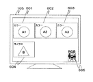
例えば、図6に示すように、内視鏡110によって取得されたカラー表示による複数の内視鏡画像601乃至603と、X線透視撮影装置109によって取得されたモノクロ表示によるX線画像604が、同一の画像表示装置105に表示した際、一つの色調整バー605によって内視鏡画像601乃至603の色調整を行なえるようにしてもよい。このように複数の画像を一度に表示する場合は、連続して撮影された画像である場合が多く、これら複数の画像で共通して色調整を行いたい場合に有効である。また、特に図示しないが、これらカラー表示による複数の内視鏡画像のうち、特定の画像のみ色調整バー605による色調整を行なえないように色調整解除手段をそれぞれの画像毎に備えてもいい。これにより、複数の画像において一度大まかな色調整をした後、前記色調整解除手段を用いて1枚ずつ色の微調整を行なうことができる。
For example, as shown in FIG. 6, a plurality of
このように、同一の医用画像撮影装置で撮影したカラー表示による異なる複数の画像に対し、一つの色調整バー605によって色調整を行なえることで操作者の利便性が向上する。
As described above, the color adjustment can be performed by the single
また、図7に示すように、図6で表示した内視鏡画像601乃至603、及びX線画像604に加え、X線CT装置によって取得され医用撮影装置表示色対応表301にて個別の色調整を可能と設定したカラー表示によるX線CT画像701が同一の画像表示装置105に表示した際、内視鏡画像601乃至603の色調整を行なう色調整バー605に加え、X線CT画像701の色調整を行なう色調整バー702を備えてもよい。
Further, as shown in FIG. 7, in addition to the
これにより、異なる種類の医用画像撮影装置で撮影したカラー表示による画像を同一の画像表示装置105に表示した際でも、各医用画像撮影装置で撮影した画像ごとに適した色調整を個別に行なうことができる。
As a result, color adjustment suitable for each image captured by each medical image capturing device can be performed individually even when an image by color display captured by a different type of medical image capturing device is displayed on the same
101 医用画像表示装置、102 キャプチャー部、103 画像メモリ部、104 出力画像生成部、105 画像表示装置、106 制御部、107 記憶装置、108 操作卓、109 X線透視撮影装置、110 内視鏡、201 レイアウト調整部、202 色調整領域設定部、203 色調整部、301 医用撮影装置表示色対応表、401,601,602,603 内視鏡画像、402,604 X線画像、403,605,702 色調整バー、404,502 スライダー、501 色相調整バー、701 X線CT画像
101 medical image display device, 102 capture unit, 103 image memory unit, 104 output image generation unit, 105 image display device, 106 control unit, 107 storage device, 108 console, 109 X-ray fluoroscopic imaging device, 110 endoscope, 201 Layout adjustment unit, 202 color adjustment area setting unit, 203 color adjustment unit, 301 medical imaging apparatus display color correspondence table, 401, 601, 602, 603 Endoscopic image, 402, 604 X-ray image, 403, 605, 702 Color adjustment bar, 404, 502 slider, 501 hue adjustment bar , 7 01 X-ray CT image
Claims (5)
前記画像表示部に表示した複数の表示画像のうち、個別の色調整を可能とする表示画像を設定する色調整領域設定部と、前記個別の色調整を行なうための色調整手段と、を備えることを特徴とする医用画像表示装置。 A medical image display comprising: an input unit that inputs images captured using a plurality of different medical image capturing devices as image data; and an image display unit that displays a plurality of images on the same screen based on the input image data. A device,
A color adjustment region setting unit that sets a display image that enables individual color adjustment among a plurality of display images displayed on the image display unit; and a color adjustment unit that performs the individual color adjustment. A medical image display device characterized by that.
Priority Applications (1)
| Application Number | Priority Date | Filing Date | Title |
|---|---|---|---|
| JP2011081514A JP5697515B2 (en) | 2011-04-01 | 2011-04-01 | Medical image display device |
Applications Claiming Priority (1)
| Application Number | Priority Date | Filing Date | Title |
|---|---|---|---|
| JP2011081514A JP5697515B2 (en) | 2011-04-01 | 2011-04-01 | Medical image display device |
Publications (3)
| Publication Number | Publication Date |
|---|---|
| JP2012213544A JP2012213544A (en) | 2012-11-08 |
| JP2012213544A5 JP2012213544A5 (en) | 2014-05-15 |
| JP5697515B2 true JP5697515B2 (en) | 2015-04-08 |
Family
ID=47266963
Family Applications (1)
| Application Number | Title | Priority Date | Filing Date |
|---|---|---|---|
| JP2011081514A Active JP5697515B2 (en) | 2011-04-01 | 2011-04-01 | Medical image display device |
Country Status (1)
| Country | Link |
|---|---|
| JP (1) | JP5697515B2 (en) |
Families Citing this family (2)
| Publication number | Priority date | Publication date | Assignee | Title |
|---|---|---|---|---|
| CN110678116B (en) * | 2017-06-05 | 2022-11-04 | 索尼公司 | Medical system and control unit |
| WO2021112988A1 (en) * | 2019-12-02 | 2021-06-10 | SG Devices LLC | Augmented reality display of surgical imaging |
Family Cites Families (5)
| Publication number | Priority date | Publication date | Assignee | Title |
|---|---|---|---|---|
| JPH0268027A (en) * | 1988-09-02 | 1990-03-07 | Hitachi Medical Corp | X-ray television apparatus for endoscopic diagnosis |
| JP3139858B2 (en) * | 1992-12-24 | 2001-03-05 | ジーイー横河メディカルシステム株式会社 | Ultrasound diagnostic equipment |
| JP3034747B2 (en) * | 1993-12-29 | 2000-04-17 | オリンパス光学工業株式会社 | Ultrasound diagnostic equipment |
| JP4631206B2 (en) * | 2001-04-27 | 2011-02-16 | コニカミノルタホールディングス株式会社 | MEDICAL IMAGE DISPLAY METHOD, MEDICAL IMAGE DISPLAY DEVICE, MEDICAL IMAGE DISPLAY PROGRAM, AND RECORDING MEDIUM |
| JPWO2005053539A1 (en) * | 2003-12-02 | 2007-12-06 | オリンパス株式会社 | Ultrasonic diagnostic equipment |
-
2011
- 2011-04-01 JP JP2011081514A patent/JP5697515B2/en active Active
Also Published As
| Publication number | Publication date |
|---|---|
| JP2012213544A (en) | 2012-11-08 |
Similar Documents
| Publication | Publication Date | Title |
|---|---|---|
| US10631712B2 (en) | Surgeon's aid for medical display | |
| JP6950707B2 (en) | Information processing equipment and methods, and programs | |
| JP5438424B2 (en) | Medical image photographing device and photographing method thereof | |
| JP5350532B2 (en) | Image processing apparatus, image display system, image processing method, and image processing program | |
| JP6397178B2 (en) | Control device, operation method of control device, and program | |
| CN109565565B (en) | Information processing apparatus, information processing method, and non-transitory computer readable medium | |
| CN110741334B (en) | Display control device, display control method, and display control program | |
| WO2014034294A1 (en) | Image display device and medical image capturing device | |
| WO2015064011A1 (en) | Imaging control device, x-ray imaging device, imaging control method, and program | |
| US10694141B2 (en) | Multi-camera system, camera, camera processing method, confirmation device, and confirmation device processing method | |
| JP6740699B2 (en) | Image analysis system | |
| JP5697515B2 (en) | Medical image display device | |
| JP5618535B2 (en) | Medical diagnostic imaging equipment | |
| JP2019004978A (en) | Surgery system and surgical image capture device | |
| WO2010122917A1 (en) | X-ray diagnostic device and method for controlling x-ray diaphragm | |
| CN110461208A (en) | Control device, external device (ED), Medical viewing system, control method, display methods and program | |
| US20220007925A1 (en) | Medical imaging systems and methods | |
| JP2020039432A (en) | Medical image switcher | |
| JP2011224086A (en) | Image processor, x-ray radiographing apparatus, image display method, image comparison method, and image display program | |
| JP2012090785A (en) | Electronic endoscope apparatus | |
| US8941758B2 (en) | Image processing apparatus and image processing method for generating a combined image | |
| US11902692B2 (en) | Video processing apparatus and video processing method | |
| JP2016016276A (en) | Dentistry x-ray imaging device | |
| JP5712055B2 (en) | Electronic endoscope system | |
| JP2012213544A5 (en) |
Legal Events
| Date | Code | Title | Description |
|---|---|---|---|
| A521 | Request for written amendment filed |
Free format text: JAPANESE INTERMEDIATE CODE: A523 Effective date: 20140331 |
|
| A621 | Written request for application examination |
Free format text: JAPANESE INTERMEDIATE CODE: A621 Effective date: 20140331 |
|
| A977 | Report on retrieval |
Free format text: JAPANESE INTERMEDIATE CODE: A971007 Effective date: 20141219 |
|
| TRDD | Decision of grant or rejection written | ||
| A01 | Written decision to grant a patent or to grant a registration (utility model) |
Free format text: JAPANESE INTERMEDIATE CODE: A01 Effective date: 20150127 |
|
| A61 | First payment of annual fees (during grant procedure) |
Free format text: JAPANESE INTERMEDIATE CODE: A61 Effective date: 20150210 |
|
| R150 | Certificate of patent or registration of utility model |
Ref document number: 5697515 Country of ref document: JP Free format text: JAPANESE INTERMEDIATE CODE: R150 |
|
| S111 | Request for change of ownership or part of ownership |
Free format text: JAPANESE INTERMEDIATE CODE: R313111 |
|
| S533 | Written request for registration of change of name |
Free format text: JAPANESE INTERMEDIATE CODE: R313533 |
|
| R350 | Written notification of registration of transfer |
Free format text: JAPANESE INTERMEDIATE CODE: R350 |
|
| S111 | Request for change of ownership or part of ownership |
Free format text: JAPANESE INTERMEDIATE CODE: R313111 |
|
| R350 | Written notification of registration of transfer |
Free format text: JAPANESE INTERMEDIATE CODE: R350 |
|
| R250 | Receipt of annual fees |
Free format text: JAPANESE INTERMEDIATE CODE: R250 |
|
| R250 | Receipt of annual fees |
Free format text: JAPANESE INTERMEDIATE CODE: R250 |
|
| R250 | Receipt of annual fees |
Free format text: JAPANESE INTERMEDIATE CODE: R250 |
|
| S111 | Request for change of ownership or part of ownership |
Free format text: JAPANESE INTERMEDIATE CODE: R313111 |
|
| R350 | Written notification of registration of transfer |
Free format text: JAPANESE INTERMEDIATE CODE: R350 |


