JP5675782B2 - Labeled probe-water-soluble carrier complex - Google Patents
Labeled probe-water-soluble carrier complex Download PDFInfo
- Publication number
- JP5675782B2 JP5675782B2 JP2012510666A JP2012510666A JP5675782B2 JP 5675782 B2 JP5675782 B2 JP 5675782B2 JP 2012510666 A JP2012510666 A JP 2012510666A JP 2012510666 A JP2012510666 A JP 2012510666A JP 5675782 B2 JP5675782 B2 JP 5675782B2
- Authority
- JP
- Japan
- Prior art keywords
- probe
- water
- biotinylated
- soluble carrier
- fab
- Prior art date
- Legal status (The legal status is an assumption and is not a legal conclusion. Google has not performed a legal analysis and makes no representation as to the accuracy of the status listed.)
- Active
Links
Images


Classifications
-
- G—PHYSICS
- G01—MEASURING; TESTING
- G01N—INVESTIGATING OR ANALYSING MATERIALS BY DETERMINING THEIR CHEMICAL OR PHYSICAL PROPERTIES
- G01N33/00—Investigating or analysing materials by specific methods not covered by groups G01N1/00 - G01N31/00
- G01N33/48—Biological material, e.g. blood, urine; Haemocytometers
- G01N33/50—Chemical analysis of biological material, e.g. blood, urine; Testing involving biospecific ligand binding methods; Immunological testing
- G01N33/58—Chemical analysis of biological material, e.g. blood, urine; Testing involving biospecific ligand binding methods; Immunological testing involving labelled substances
- G01N33/581—Chemical analysis of biological material, e.g. blood, urine; Testing involving biospecific ligand binding methods; Immunological testing involving labelled substances with enzyme label (including co-enzymes, co-factors, enzyme inhibitors or substrates)
-
- G—PHYSICS
- G01—MEASURING; TESTING
- G01N—INVESTIGATING OR ANALYSING MATERIALS BY DETERMINING THEIR CHEMICAL OR PHYSICAL PROPERTIES
- G01N33/00—Investigating or analysing materials by specific methods not covered by groups G01N1/00 - G01N31/00
- G01N33/48—Biological material, e.g. blood, urine; Haemocytometers
- G01N33/50—Chemical analysis of biological material, e.g. blood, urine; Testing involving biospecific ligand binding methods; Immunological testing
- G01N33/53—Immunoassay; Biospecific binding assay; Materials therefor
- G01N33/543—Immunoassay; Biospecific binding assay; Materials therefor with an insoluble carrier for immobilising immunochemicals
- G01N33/54353—Immunoassay; Biospecific binding assay; Materials therefor with an insoluble carrier for immobilising immunochemicals with ligand attached to the carrier via a chemical coupling agent
-
- G—PHYSICS
- G01—MEASURING; TESTING
- G01N—INVESTIGATING OR ANALYSING MATERIALS BY DETERMINING THEIR CHEMICAL OR PHYSICAL PROPERTIES
- G01N33/00—Investigating or analysing materials by specific methods not covered by groups G01N1/00 - G01N31/00
- G01N33/48—Biological material, e.g. blood, urine; Haemocytometers
- G01N33/50—Chemical analysis of biological material, e.g. blood, urine; Testing involving biospecific ligand binding methods; Immunological testing
- G01N33/53—Immunoassay; Biospecific binding assay; Materials therefor
- G01N33/531—Production of immunochemical test materials
- G01N33/532—Production of labelled immunochemicals
-
- G—PHYSICS
- G01—MEASURING; TESTING
- G01N—INVESTIGATING OR ANALYSING MATERIALS BY DETERMINING THEIR CHEMICAL OR PHYSICAL PROPERTIES
- G01N33/00—Investigating or analysing materials by specific methods not covered by groups G01N1/00 - G01N31/00
- G01N33/48—Biological material, e.g. blood, urine; Haemocytometers
- G01N33/50—Chemical analysis of biological material, e.g. blood, urine; Testing involving biospecific ligand binding methods; Immunological testing
- G01N33/58—Chemical analysis of biological material, e.g. blood, urine; Testing involving biospecific ligand binding methods; Immunological testing involving labelled substances
- G01N33/582—Chemical analysis of biological material, e.g. blood, urine; Testing involving biospecific ligand binding methods; Immunological testing involving labelled substances with fluorescent label
Description
本発明は、水溶性担体に複数のプローブを結合させ、さらに標識物を結合させた標識化プローブ−水溶性担体複合体の作製方法、当該方法によって作製される標識化プローブ−水溶性担体複合体、およびその使用方法に関するものである。この標識化プローブ−水溶性担体複合体を用いることにより、高感度で安定な検出・測定を行うことが可能となる。 The present invention relates to a method for producing a labeled probe-water-soluble carrier complex in which a plurality of probes are bound to a water-soluble carrier and a label is further bound thereto, and a labeled probe-water-soluble carrier complex produced by the method. , And its usage. By using this labeled probe-water-soluble carrier complex, highly sensitive and stable detection and measurement can be performed.
被験物質と特異的に結合するプローブと標識物との結合物を用いて、プローブを介した被験物質と標識物の結合量を指標として被験物質を検出・測定する方法において、その感度は一般にプローブおよび標識物の分子数により定まる。すなわち、1分子のプローブに対して結合する標識物の分子数は限られ、その比率が感度を定める。 In a method for detecting and measuring a test substance using a binding substance between a probe and a label that specifically binds to the test substance as an index, the sensitivity is generally measured by the probe And the number of molecules of the label. That is, the number of molecules of the label that binds to one molecule of probe is limited, and the ratio determines the sensitivity.
そこで、プローブ自体を重合化させて重合体を作製することにより分子量を大きくし、その重合体に結合する標識物の分子数を多くすることにより反応性を向上させた高感度化がなされた(特許文献1)。しかしながら、プローブの重合化を制御することが容易ではなく、実用化には至っていない。 Therefore, the sensitivity was improved by increasing the molecular weight by polymerizing the probe itself to increase the molecular weight, and increasing the number of molecules of the label bound to the polymer to improve the reactivity ( Patent Document 1). However, it is not easy to control the polymerization of the probe, and it has not been put into practical use.
また、ポリリジン、アミノデキストラン等の担体に酵素標識物およびプローブを別個に共有結合させて分子量が大きく、標識物の結合数が多い標識物プローブ複合体が提案された(特許文献2)。しかしながら、本技術により反応性は増加したものの、ブランク値での反応も増加し、検出・測定を行うときの高感度化には至っていない。 In addition, a labeled probe complex having a large molecular weight and a large number of bound labeled substances has been proposed by separately covalently binding an enzyme labeled substance and a probe to a carrier such as polylysine and aminodextran (Patent Document 2). However, although the reactivity has been increased by this technique, the reaction at the blank value has also increased, and the sensitivity has not been increased when performing detection and measurement.
さらに、ポリリジン等の担体に酵素標識物を結合させ、担体上の標識物を介してプローブを結合させた標識物−プローブ複合体(特許文献3)、デキストラン等の担体にプローブを結合させ、担体上のプローブを介して標識物を結合させた水溶性担体−プローブ複合体(特許文献4)、担体に親水性の仲介物質を結合させ、仲介物質にプローブおよび検出マーカーの標識物を結合させたプローブ複合体(特許文献5)、酵素標識物を介して2分子以上の担体を結合させた複合体にプローブを結合させたブロック化標識物プローブ(特許文献6)が提案された。 Furthermore, an enzyme label is bound to a carrier such as polylysine, a label-probe complex (Patent Document 3) in which a probe is bound via a label on the carrier, a probe is bound to a carrier such as dextran, and the carrier A water-soluble carrier-probe complex (Patent Document 4) in which a label is bound via the above probe, a hydrophilic mediator bound to the carrier, and a probe and a detection marker label bound to the mediator A probe complex (Patent Document 5) and a blocked label probe (Patent Document 6) in which a probe is bound to a complex in which two or more molecules of carriers are bound via an enzyme label have been proposed.
しかし、上記先行技術も、反応性の増加とともにブランク値での反応も増加するという技術的課題を内包しており、シグナル/ノイズ比として反応性を評価する場合、高感度かつ安定な複合体を得ることは困難であった。また、複合体形成において、担体とプローブまたは標識物の結合を最初に行うと、担体の多官能性に起因して、所望の複合体を再現性良く作製することは困難であった。 However, the above prior art also includes the technical problem that the reaction at the blank value increases as the reactivity increases. When evaluating the reactivity as a signal / noise ratio, a highly sensitive and stable complex is required. It was difficult to get. Further, when the carrier is first bound to the probe or the label in the complex formation, it is difficult to produce a desired complex with good reproducibility due to the multifunctionality of the carrier.
被験物質と特異的に結合するプローブと標識物の結合物を用いて、プローブを介した被験物質と標識物の結合量を指標として、高感度に被験物質を検出・測定するためには、1分子のプローブと標識物の結合物に多量の標識物が含まれなければならない。そのために、大分子のプローブと標識物の結合物を作製する必要がある。 In order to detect and measure a test substance with high sensitivity using a binding substance of a probe and a label that specifically binds to the test substance, using the binding amount of the test substance and the label through the probe as an index, 1 A large amount of label must be included in the conjugate of the molecular probe and label. Therefore, it is necessary to prepare a conjugate of a large molecule probe and a label.
しかしながら、大分子の結合物を形成すべく担体を使用した場合、反応性は増加するものの、ブランク値での反応も増加し、結果として高感度かつ安定な複合体を得ることは困難である。また、担体とプローブまたは標識物の結合を複合体形成の最初の段階として行うと、担体の多官能性に起因して、所望の複合体を再現性良く作製することは困難である。そこで、上記課題を解決する結合物および当該結合物を安定的に作製する方法が期待される。 However, when a carrier is used to form a large molecule conjugate, the reactivity increases, but the reaction at the blank value also increases. As a result, it is difficult to obtain a highly sensitive and stable complex. In addition, when the binding of the carrier and the probe or the label is performed as the first stage of complex formation, it is difficult to produce a desired complex with good reproducibility due to the multifunctionality of the carrier. Therefore, a combined product that solves the above-described problems and a method for stably producing the combined product are expected.
発明者等は、標識化プローブ−担体複合体の作製におけるプローブと標識物の結合において、アビジン類とビオチンとの特異的な結合を利用することに思い至り、また、アビジン類とプローブとの結合を、担体との結合に先立って行うことで、極めて高感度かつ安定な複合体を再現性良く作製し得ることを見出した。加えて、化学結合のためにプローブの化学修飾を行うときに、プローブにチオール基を導入することが、プローブの結合能に影響を与えないことを見出し、さらに、担体として水溶性担体を用いることで、高感度で安定な検出・測定に有用な標識化プローブ−水溶性担体複合体を構築した。 The inventors have come to consider using a specific binding between avidin and biotin in the binding of the probe and the label in the preparation of the labeled probe-carrier complex, and the binding between the avidin and the probe. It was found that a highly sensitive and stable complex can be produced with good reproducibility by performing the step prior to the binding to the carrier. In addition, when chemical modification of a probe for chemical bonding, it was found that introduction of a thiol group into the probe does not affect the binding ability of the probe, and a water-soluble carrier is used as the carrier. Thus, a labeled probe-water-soluble carrier complex useful for highly sensitive and stable detection and measurement was constructed.
すなわち、本発明は、以下の工程を含む、標識化プローブ−水溶性担体複合体の作製方法に関するものである。
工程1.チオール基を有し被験物質に対して結合可能なプローブと、マレイミド基を有するアビジン類を結合させてプローブ結合体とし、
工程2.次にチオール基を有する前記プローブ結合体と、マレイミド基を有する高分子の水溶性担体を結合させてプローブ結合体−水溶性担体複合体とし、
工程3.さらに前記プローブ結合体−水溶性担体複合体とビオチン化標識物を混合して、プローブ結合体−水溶性担体複合体のアビジン類とビオチン化標識物のビオチンを結合させる。That is, the present invention relates to a method for producing a labeled probe-water-soluble carrier complex including the following steps.
Step 1. A probe conjugate that binds a probe having a thiol group and capable of binding to a test substance and avidin having a maleimide group,
Step 2. Next, the probe conjugate having a thiol group and the polymer water-soluble carrier having a maleimide group are bound to form a probe conjugate-water-soluble carrier complex,
Step 3. Further, the probe conjugate-water-soluble carrier complex and the biotinylated label are mixed, and the avidin of the probe conjugate-water-soluble carrier complex and biotinylated label biotin are bound.
また、本発明は、当該工程により作製する標識化プローブ−水溶性担体複合体に関するものである。 The present invention also relates to a labeled probe-water-soluble carrier complex produced by this step.
さらに、本発明は、上記工程により作製した標識化プローブ−水溶性担体複合体を用いる測定法、免疫測定法または高感度測定法に関するものである。 Furthermore, the present invention relates to a measurement method, an immunoassay method, or a highly sensitive measurement method using a labeled probe-water-soluble carrier complex prepared by the above-described steps.
本発明において、「プローブ」とは、被験物質と相互作用する結合パートナーを意味し、非限定的な例として、抗体もしくは抗体断片、プロテインG、プロテインA、プロテインL、レクチンまたは受容体等を挙げることが出来る。また、抗体または抗体断片の非限定な例として、Fab’、F(ab’)2、FabまたはIgG等を挙げることが出来る。In the present invention, the “probe” means a binding partner that interacts with a test substance, and non-limiting examples include an antibody or antibody fragment, protein G, protein A, protein L, lectin, or receptor. I can do it. Non-limiting examples of antibodies or antibody fragments include Fab ′, F (ab ′) 2 , Fab or IgG.
また、当該抗体または抗体断片として、二種類以上の抗体または抗体断片を用いることが出来る。 Two or more types of antibodies or antibody fragments can be used as the antibody or antibody fragment.
本発明において、「アビジン類」とは、低分子の塩基性糖タンパク質のアビジン、それに類似するタンパク質またはその断片等、ビオチン化合物と安定した複合体を形成するものを意味し、当該「アビジン類」の非限定な例として、アビジンまたはストレプトアビジン等を挙げることが出来る。 In the present invention, “avidins” means those that form a stable complex with a biotin compound, such as low-molecular basic glycoprotein avidin, a protein similar thereto, or a fragment thereof, and the “avidins” As non-limiting examples, avidin or streptavidin can be mentioned.
本発明において、「水溶性担体」は、分子量が50万以上であることが好ましい。本発明の水溶性担体の非限定な例として、デキストラン、アミノデキストラン、デキストリン、クラスターデキストリン、フィコールまたはプルラン等を挙げることが出来る。 In the present invention, the “water-soluble carrier” preferably has a molecular weight of 500,000 or more. Non-limiting examples of the water-soluble carrier of the present invention include dextran, aminodextran, dextrin, cluster dextrin, ficoll or pullulan.
本発明では「ビオチン化標識物」を使用する。ここで「ビオチン」は、ビタミンB複合体の一つであり、アビジンと非常に強く結合することが知られているものである。本発明における「ビオチン化標識物」の非限定な例として、ビオチン化ルシフェラーゼ、ビオチン化アルカリホスフォターゼ、ビオチン化POD(peroxidase)、ビオチン化GOD(glucose oxidase)、ビオチン化FITC(fluorescein isothiocyanate)、ビオチン化アクリジニウム、ビオチン化アクリジニウム誘導体またはビオチン化トリス(2,2‘ビピリジル)ルテニウム(II)等を挙げることが出来る。当該「ビオチン化標識物」におけるビオチンと標識物の結合には、化学修飾等を含む既知の何れの手段を利用することが出来るが、特に遺伝子組換えを利用することが好ましい。なぜなら、化学結合のための化学修飾をしないことから、その標識物の活性を低下させないからである。 In the present invention, a “biotinylated label” is used. Here, “biotin” is one of the vitamin B complexes and is known to bind very strongly to avidin. Non-limiting examples of “biotinylated label” in the present invention include biotinylated luciferase, biotinylated alkaline phosphatase, biotinylated POD (peroxidase), biotinylated GOD (glucose oxidase), biotinylated FITC (fluorescein isothiocyanate), Examples include biotinylated acridinium, biotinylated acridinium derivatives, biotinylated tris (2,2′bipyridyl) ruthenium (II), and the like. Any known means including chemical modification or the like can be used for the binding of biotin and the labeled product in the “biotinylated labeled product”, but it is particularly preferable to use genetic recombination. This is because the chemical modification for chemical bonding is not performed, so that the activity of the label is not reduced.
本発明に係る工程1においては、まず被験物質に対して結合可能なプローブが十分なチオール基を有していない場合、当該プローブにチオール基を導入する。チオール基の導入には既知の何れの方法を用いることが出来るが、プローブが抗体または抗体断片である場合、2−メルカプトエタノール等の還元剤を使用し、内在するジスルフィド結合を還元することによりチオール基を導入することは、プローブの結合能に対する影響を最小限化出来るので、特に有利である。 In step 1 according to the present invention, when a probe capable of binding to a test substance does not have a sufficient thiol group, a thiol group is introduced into the probe. Any known method can be used to introduce a thiol group. When the probe is an antibody or antibody fragment, a reducing agent such as 2-mercaptoethanol is used to reduce the thiol group by reducing the existing disulfide bond. Introducing a group is particularly advantageous because it can minimize the effect on the binding ability of the probe.
次に、アビジン類へマレイミド基を導入する。マレイミド基の導入には、既知の何れの方法を用いることが出来、例えば、Sulfo−KMUS(同仁化学社製)といった既知のマレイミド試薬を使用することが出来る。 Next, a maleimide group is introduced into avidins. Any known method can be used for introduction of the maleimide group. For example, a known maleimide reagent such as Sulfo-KMUS (manufactured by Dojindo) can be used.
チオール基を有し被験物質に対して結合可能なプローブと、マレイミド基を有するアビジン類との結合には、既知の何れの方法を用いることが出来る。例えば、プローブおよびアビジン類をそれぞれ適切な濃度において緩衝液に溶解し、当該溶解液を反応させることで結合反応を行うことが出来る。また、以降の工程における非特異反応を回避すべく、上記結合反応完了後、2−メルカプトエタノール等のチオール試薬を用いて未反応のマレイミド基をブロックすることが出来る。ブロック後のプローブ−アビジン類結合体は、ゲル濾過等既知の方法によって精製することが出来る。 Any known method can be used for binding a probe having a thiol group and capable of binding to a test substance to avidin having a maleimide group. For example, the binding reaction can be carried out by dissolving the probe and avidin in a buffer solution at appropriate concentrations and reacting the solution. In order to avoid non-specific reactions in the subsequent steps, unreacted maleimide groups can be blocked using a thiol reagent such as 2-mercaptoethanol after completion of the above binding reaction. The blocked probe-avidin conjugate after blocking can be purified by a known method such as gel filtration.
本発明に係る工程2においては、まず工程1において作製したプローブ−アビジン類結合体にチオール基を導入する。チオール基の導入には、例えば、2−イミノチオランを使用するといった、既知の何れの方法を用いることが出来る。 In step 2 according to the present invention, a thiol group is first introduced into the probe-avidin conjugate produced in step 1. For the introduction of the thiol group, any known method such as, for example, using 2-iminothiolane can be used.
水溶性担体へのマレイミド基の導入は、既知の何れの方法を用いることが出来る。例えば、水溶性担体を酸と反応させることにより、カルボキシル基を導入し、当該酸を透析等により除去した後、エチレンジアミン等の既知のアミノ基導入試薬を用いて、カルボキシル基をアミノ基に置換する。置換後、透析等により未反応のアミノ基導入試薬を除去し、既知のマレイミド試薬を加え反応させることにより、マレイミド基を有する水溶性担体を得ることが出来る。 Any known method can be used to introduce the maleimide group into the water-soluble carrier. For example, a carboxyl group is introduced by reacting a water-soluble carrier with an acid, the acid is removed by dialysis or the like, and then the carboxyl group is replaced with an amino group using a known amino group introduction reagent such as ethylenediamine. . After substitution, an unreacted amino group introduction reagent is removed by dialysis or the like, and a known maleimide reagent is added and reacted to obtain a water-soluble carrier having a maleimide group.
チオール基を有するプローブ−アビジン類結合体と、マレイミド基を有する水溶性担体との結合には、既知の何れの方法を用いることが出来る。また、以降の工程における非特異反応を回避すべく、結合反応完了後、2−メルカプトエタノール等のチオール試薬を用いて水溶性担体に導入した未反応のマレイミド基をブロックすることが好ましい。ブロック後のプローブ結合体−水溶性担体複合体は、ゲル濾過等既知の方法によって精製することが出来る。 Any known method can be used for binding of the probe-avidin conjugate having a thiol group and the water-soluble carrier having a maleimide group. In order to avoid non-specific reactions in the subsequent steps, it is preferable to block the unreacted maleimide group introduced into the water-soluble carrier using a thiol reagent such as 2-mercaptoethanol after the completion of the binding reaction. The probe conjugate / water-soluble carrier complex after blocking can be purified by a known method such as gel filtration.
本発明に係る工程3においては、上記プローブ結合体−水溶性担体複合体をビオチン化標識物と混合することにより、プローブ結合体−水溶性担体複合体のアビジン類とビオチン化標識物のビオチンを結合させる。上記の通り、アビジン類とビオチンとの親和性は非常に強いため、例えば、プローブ結合体−水溶性担体複合体およびビオチン化標識物をそれぞれ適切な濃度に調製し、当該調製液を混合することによって、当該アビジン類とビオチンとの結合反応を行うことが出来る。 In step 3 according to the present invention, the probe conjugate-water-soluble carrier complex is mixed with a biotinylated labeled product, whereby the avidin of the probe conjugate-water soluble carrier complex and biotinylated labeled product biotin are mixed. Combine. As described above, since the affinity between avidin and biotin is very strong, for example, the probe conjugate-water-soluble carrier complex and the biotinylated label are prepared at appropriate concentrations, and the preparation is mixed. Thus, the binding reaction between the avidin and biotin can be performed.
本発明に係る工程により作製した標識化プローブ−水溶性担体複合体は、当該プローブが標的とする被験物質と高感度、かつ安定的に反応するため、既知の何れの測定法において利用することが出来る。また、当該プローブが抗体または抗体断片である場合、本発明に係る標識化プローブ−水溶性担体複合体は、既知の何れの免疫測定法において利用すること出来る。 Since the labeled probe-water-soluble carrier complex prepared by the process according to the present invention reacts with the test substance targeted by the probe with high sensitivity and stability, it can be used in any known measurement method. I can do it. When the probe is an antibody or an antibody fragment, the labeled probe-water-soluble carrier complex according to the present invention can be used in any known immunoassay method.
さらに、本発明に係る工程により作製した標識化プローブ−水溶性担体複合体は長期間保存後においても極めて安定である。37℃で1週間の加速安定性試験下においても、その経時的な安定性を保持していた。 Furthermore, the labeled probe-water-soluble carrier complex prepared by the process according to the present invention is extremely stable even after long-term storage. Even under an accelerated stability test at 37 ° C. for 1 week, the stability over time was maintained.
本発明を、一例としてビオチン化標識物にビオチン化ルシフェラーゼを、プローブとしてFab’を、アビジン類としてストレプトアビジンを、水溶性担体としてデキストラン(T2000)を各々用いて説明するが、本例に限定されることはない。 The present invention will be described using, as an example, biotinylated luciferase as a biotinylated label, Fab ′ as a probe, streptavidin as an avidin, and dextran (T2000) as a water-soluble carrier. However, the present invention is limited to this example. Never happen.
IgG抗体をペプシン消化して得たF(ab’)2に2−メルカプトエタノールを加えて還元処理した後にゲルろ過により精製し、チオール基を有するFab’を得る。2- (Mercaptoethanol) is added to F (ab ′) 2 obtained by digesting an IgG antibody with pepsin and reduced, followed by purification by gel filtration to obtain Fab ′ having a thiol group.
一方、ストレプトアビジンにマレイミド基を導入する試薬を加えて処理した後にゲルろ過により精製し、マレイミド基を導入したストレプトアビジンを得る。 On the other hand, a reagent for introducing a maleimide group is added to streptavidin for treatment, and then purified by gel filtration to obtain streptavidin having a maleimide group introduced.
上記した方法で得られたチオール基を導入したFab’およびマレイミド基を導入したストレプトアビジンを混合して反応させた後に2−メルカプトエタノールを添加して未反応のマレイミド基をブロックし、ゲルろ過により精製してFab’−ストレプトアビジン結合体を得る。 After mixing and reacting Fab ′ introduced with the thiol group obtained above and streptavidin introduced with a maleimide group, 2-mercaptoethanol was added to block the unreacted maleimide group, and gel filtration was performed. Purify to obtain the Fab′-streptavidin conjugate.
得られたFab’−ストレプトアビジン結合体に2−イミノチオランを加えて反応させた後にゲルろ過により精製し、チオール基を導入したFab’−ストレプトアビジン結合体を得る。 The resulting Fab′-streptavidin conjugate is reacted by adding 2-iminothiolane and then purified by gel filtration to obtain a Fab′-streptavidin conjugate having a thiol group introduced therein.
デキストラン(T2000)にアミノ基を導入したアミノデキストランを作製し、さらにマレイミド試薬を加えて反応させた後にゲルろ過により精製し、マレイミド基を導入したデキストラン(T2000)を得る。 An aminodextran having an amino group introduced into dextran (T2000) is prepared, further reacted with a maleimide reagent, and then purified by gel filtration to obtain dextran (T2000) having a maleimide group introduced.
上記した方法で得られたチオール基を導入したFab’−ストレプトアビジン結合体およびマレイミド基を導入したデキストラン(T2000)を混合して反応させた後に2−メルカプトエタノールを添加して未反応のマレイミド基をブロックし、ゲルろ過により精製してFab’−ストレプトアビジン結合体−デキストラン複合体を得る。 The Fab′-streptavidin conjugate introduced with the thiol group obtained by the above method and the dextran (T2000) introduced with the maleimide group were mixed and reacted, and then 2-mercaptoethanol was added to unreacted maleimide group. And purified by gel filtration to obtain the Fab′-streptavidin conjugate-dextran complex.
得られたFab’−ストレプトアビジン結合体−デキストラン(T2000)複合体とビオチン化ルシフェラーゼを混合して反応させて、ルシフェラーゼ標識Fab’−デキストラン(T2000)複合体を得る。 The obtained Fab'-streptavidin conjugate-dextran (T2000) complex and biotinylated luciferase are mixed and reacted to obtain a luciferase-labeled Fab'-dextran (T2000) complex.
得られたルシフェラーゼ標識Fab’−デキストラン(T2000)複合体を標識抗体として用い、別に用意した抗体を固定化した固相と組み合わせてサンドイッチイムノアッセイを行うと、高感度の検出・測定法が完成する。 When the obtained luciferase-labeled Fab'-dextran (T2000) complex is used as a labeled antibody and a sandwich immunoassay is performed in combination with a solid phase on which an antibody prepared separately is immobilized, a highly sensitive detection / measurement method is completed.
理論に拘束されることを意図するものではないが、本発明によって、従来と比較して、極めて高感度かつ安定な標識化プローブ−水溶性担体複合体が再現性良く得られることは、プローブと標識物の結合を、アビジン類とビオチンとの高い親和性を介して行ったこと、かつ、当該プローブとアビジン類との結合を、担体との結合に先立って行ったことに主に起因するものであると考えられる。プローブとアビジン類との結合、およびアビジン類とビオチンとの結合は、その結合に関する分子数比等を制御し易く、従って、同等の構造を有する標識化プローブ−水溶性担体複合体を再現性良く安定的に作製できるのであろう。また、このように安定的に作製された標識化プローブ−水溶性担体複合体は、ノイズを構成する非特異的反応を相対的に抑制しつつ、シグナルを構成する特異的反応のみを増加させるのに極めて適した構造を有するものと考えられる。 Although not intending to be bound by theory, the fact that a highly sensitive and stable labeled probe-water-soluble carrier complex can be obtained with high reproducibility compared with the prior art by the present invention This is mainly due to the fact that the label was bound via a high affinity between avidin and biotin, and that the probe and avidin were bound prior to binding to the carrier. It is thought that. The binding between the probe and avidin, and the binding between avidin and biotin make it easy to control the ratio of the number of molecules related to the binding. Therefore, a labeled probe-water-soluble carrier complex having an equivalent structure can be easily reproduced. It will be possible to produce it stably. In addition, the labeled probe-water-soluble carrier complex thus stably produced increases only the specific reaction constituting the signal while relatively suppressing the non-specific reaction constituting the noise. It is considered to have a structure that is extremely suitable for.
本発明を実施することにより、被験物質と特異的に結合する高感度かつ安定な標識化プローブ−水溶性担体複合体が再現性良く得られ、この標識化プローブ−水溶性担体複合体を用いることにより、高感度かつ安定な検出・測定が可能となる。 By carrying out the present invention, a highly sensitive and stable labeled probe-water-soluble carrier complex that specifically binds to the test substance can be obtained with good reproducibility, and this labeled probe-water-soluble carrier complex should be used. Therefore, highly sensitive and stable detection / measurement becomes possible.
被験物質と特異的に結合するプローブを固定化した不溶性担体と検体を反応させた後に検体を除去洗浄し、その後に標識化プローブ−水溶性担体複合体を加えて反応させ、再度除去洗浄した後に不溶性担体上の標識物の活性を測定することにより、検体中の被験物質の検出・測定を行う。 After reacting the sample with an insoluble carrier immobilized with a probe that specifically binds to the test substance, the sample is removed and washed, and then the labeled probe-water-soluble carrier complex is added and reacted, and then removed and washed again. By measuring the activity of the label on the insoluble carrier, the test substance in the sample is detected and measured.
還元処理によるチオール基を有するIgGの調製
抗HCVコア抗原マウスモノクローナルIgG抗体c11−9およびc11−14を0.1Mリン酸緩衝液(pH7.2)に溶解して、各々5mg/mLの溶液を調製した。これらのIgG溶液300μLに0.2Mの2−メルカプトエタノール(和光純薬社製)30μLを添加し、37℃で1.5時間反応させた。反応後、PD−10(GEヘルスケア社製)でゲル濾過精製し、抗体に内在するジスルフィド結合を還元して得られるチオール基を有するIgGを得た。各々のチオール基の数を定量した結果、IgG1分子あたり8.0個のチオール基の存在が確認された。 Preparation of IgG having thiol group by reduction treatment Anti-HCV core antigen mouse monoclonal IgG antibodies c11-9 and c11-14 were dissolved in 0.1 M phosphate buffer (pH 7.2), and a solution of 5 mg / mL each was prepared. Prepared. 30 μL of 0.2M 2-mercaptoethanol (Wako Pure Chemical Industries, Ltd.) was added to 300 μL of these IgG solutions, and reacted at 37 ° C. for 1.5 hours. After the reaction, gel filtration purification was performed with PD-10 (manufactured by GE Healthcare) to obtain IgG having a thiol group obtained by reducing disulfide bonds present in the antibody. As a result of quantifying the number of each thiol group, the presence of 8.0 thiol groups per IgG molecule was confirmed.
還元処理によるチオール基を有するFab’の調製
抗HCVコア抗原マウスモノクローナルIgG抗体c11−9およびc11−14を0.1M酢酸ソーダ緩衝液(pH4.5)にて10mg/mLとなるように調製した。次に、これらのIgG溶液1mLに対してペプシン0.2mgを添加し、37℃で6時間攪拌することによりIgGをペプシン消化した。ペプシン消化後、2N NaOHにて中和し、さらにSuperdex200カラム(GEヘルスケア社製)で精製することによりF(ab’)2を得た。 Preparation of Fab ′ having thiol group by reduction treatment Anti-HCV core antigen mouse monoclonal IgG antibodies c11-9 and c11-14 were prepared to a concentration of 10 mg / mL with 0.1 M sodium acetate buffer (pH 4.5). . Next, 0.2 mg of pepsin was added to 1 mL of these IgG solutions, and IgG was digested with pepsin by stirring at 37 ° C. for 6 hours. After digestion with pepsin, it was neutralized with 2N NaOH and further purified with a
各々3.0mg/mLのF(ab’)21300μLに0.2Mの2−メルカプトエタノール(和光純薬社製)130μLを添加し、37℃で1.5時間反応させた。反応後、PD−10(GEヘルスケア社製)でゲル濾過精製し、抗体に内在するジスルフィド結合を還元して得られるチオール基を有するFab’を得た。各々のチオール基の数を定量した結果、c11−9ではFab’1分子あたり3.3個、c11−14ではFab’1分子あたり3.4個のチオール基の存在が確認された。130 μL of 0.2M 2-mercaptoethanol (manufactured by Wako Pure Chemical Industries, Ltd.) was added to 1300 μL of 3.0 mg / mL F (ab ′) 2, respectively, and reacted at 37 ° C. for 1.5 hours. After the reaction, gel filtration purification was performed with PD-10 (manufactured by GE Healthcare) to obtain Fab ′ having a thiol group obtained by reducing disulfide bonds present in the antibody. As a result of quantifying the number of each thiol group, it was confirmed that c11-9 had 3.3 thiol groups per Fab ′ molecule and c11-14 had 3.4 thiol groups per Fab ′ molecule.
ストレプトアビジンへのマレイミド基の導入
20mgのストレプトアビジン(MP Bio社製)を0.1Mリン酸緩衝液(pH7.2)2mLに溶解し、ジメチルホルムアミドに6mg/mLとなるように溶解したマレイミド試薬Sulfo−KMUS(同仁化学社製)133μLを加えた。30℃で1時間反応させた後、PD−10(GEヘルスケア社製)にてゲルろ過精製し、マレイミド基を導入したストレプトアビジンを得た。マレイミド基の数を定量した結果、ストレプトアビジン1分子あたり3.7個のマレイミド基の存在が確認された。 Introduction of maleimide group into
IgG−ストレプトアビジン結合体の作製
実施例1で作製したc11−9およびc11−14のチオール基を導入したIgGを0.1Mリン酸緩衝液(pH7.2)に溶解し、各々2.5mg/mLのチオール基を導入したIgG溶液を調製した。一方、実施例3で作製したマレイミド基を導入したストレプトアビジンを0.1Mリン酸緩衝液(pH7.2)に溶解し、13.0mg/mLのマレイミド基を導入したストレプトアビジン溶液を調製した。 Preparation of IgG-Streptavidin Conjugate IgG introduced with the thiol group of c11-9 and c11-14 prepared in Example 1 was dissolved in 0.1 M phosphate buffer (pH 7.2), and each 2.5 mg / An IgG solution into which mL of thiol group was introduced was prepared. On the other hand, the streptavidin introduced with maleimide groups prepared in Example 3 was dissolved in 0.1 M phosphate buffer (pH 7.2) to prepare a streptavidin solution having 13.0 mg / mL maleimide groups introduced.
次に、各々のチオール基を導入したIgG溶液500μLに対して、マレイミド基を導入したストレプトアビジン溶液38μLを添加し、30℃で1時間反応させた。反応後、0.2Mの2−メルカプトエタノール(和光純薬社製)54μLを添加し、4℃で一晩反応させ、未反応のマレイミド基をブロックした。反応後、Superdex200カラム(GEヘルスケア社製)でゲル濾過精製し、IgG−ストレプトアビジン結合体を得た。
Next, 38 μL of a streptavidin solution introduced with a maleimide group was added to 500 μL of each IgG solution introduced with a thiol group, and reacted at 30 ° C. for 1 hour. After the reaction, 54 μL of 0.2M 2-mercaptoethanol (manufactured by Wako Pure Chemical Industries, Ltd.) was added and reacted at 4 ° C. overnight to block unreacted maleimide groups. After the reaction, gel filtration purification was performed using a
ルシフェラーゼ標識IgGの作製
実施例4で作製した10μMのc11−9およびc11−14のIgG−ストレプトアビジン結合体50μLに対して、61μMのビオチン化ルシフェラーゼ(キッコーマン社製)8.2μL加えて、25℃で1時間反応させることによりルシフェラーゼ標識IgGを作製した。 Preparation of Luciferase-Labeled IgG 8.2 μL of 61 μM biotinylated luciferase (Kikkoman) was added to 50 μL of 10 μM c11-9 and c11-14 IgG-streptavidin conjugate prepared in Example 4, and 25 ° C. The luciferase-labeled IgG was prepared by reacting for 1 hour.
Fab’−ストレプトアビジン結合体の作製
実施例2で作製したc11−9 およびc11−14の チオール基を導入したFab’を0.1Mリン酸緩衝液(pH7.2)に溶解し、各々2.6mg/mLのチオール基を導入したFab’溶液を調製した。一方、実施例3で作製したマレイミド基を導入したストレプトアビジンを0.1Mリン酸緩衝液(pH7.2)に溶解し、13.0mg/mLのマレイミド基を導入したストレプトアビジン溶液を調製した。Preparation of Fab′- Streptavidin Conjugate Fab ′ introduced with the thiol group of c11-9 and c11-14 prepared in Example 2 was dissolved in 0.1 M phosphate buffer (pH 7.2). A Fab ′ solution into which 6 mg / mL thiol group was introduced was prepared. On the other hand, the streptavidin introduced with maleimide groups prepared in Example 3 was dissolved in 0.1 M phosphate buffer (pH 7.2) to prepare a streptavidin solution having 13.0 mg / mL maleimide groups introduced.
次に、各々のチオール基を導入したFab’溶液1400μLに対して、マレイミド基を導入したストレプトアビジン溶液365μLを添加し、30℃で1時間反応させた。反応後、0.2Mの2−メルカプトエタノール(和光純薬社製)177μLを添加し、4℃で一晩反応させ、未反応のマレイミド基をブロックした。反応後、Superdex200カラム(GEヘルスケア社製)でゲル濾過精製し、Fab’−ストレプトアビジン結合体を得た。
Next, 365 μL of a streptavidin solution introduced with a maleimide group was added to 1400 μL of Fab ′ solution into which each thiol group was introduced, and reacted at 30 ° C. for 1 hour. After the reaction, 177 μL of 0.2M 2-mercaptoethanol (manufactured by Wako Pure Chemical Industries, Ltd.) was added and reacted at 4 ° C. overnight to block unreacted maleimide groups. After the reaction, gel filtration purification was performed using a
ルシフェラーゼ標識Fab’の作製
実施例6で作製した26.4μMのc11−9およびc11−14のFab’−ストレプトアビジン結合体10μLに対して、61μMのビオチン化ルシフェラーゼ(キッコーマン社製)4.3μL加えて、25℃で1時間反応させることにより、ルシフェラーゼ標識Fab’を作製した。 Preparation of Luciferase-Labeled Fab ′ 4.3 μL of 61 μM biotinylated luciferase (Kikkoman) was added to 10 μL of the 26.4 μM c11-9 and c11-14 Fab′-streptavidin conjugate prepared in Example 6. The luciferase-labeled Fab ′ was prepared by reacting at 25 ° C. for 1 hour.
アミノ基を導入したデキストランへのマレイミド基の導入
デキストラン(T2000)およびデキストラン(T500)各々2gとモノクロロ酢酸4.7gを50mLの3N NaOH溶液に溶解し、室温で70分間攪拌することによってデキストラン(T2000)およびデキストラン(T500)にカルボキシル基を導入した。次に、リン酸2水素ナトリウム0.2gを添加し、6N HClで混合液を中和することにより反応を停止させ、透析により未反応のモノクロロ酢酸を除去した。 Introduction of Maleimide Group into Dextran Introduced with Amino Group 2 g of dextran (T2000) and dextran (T500) and 4.7 g of monochloroacetic acid were dissolved in 50 mL of 3N NaOH solution, and the mixture was stirred at room temperature for 70 minutes to obtain dextran (T2000). ) And dextran (T500) were introduced with carboxyl groups. Next, 0.2 g of sodium dihydrogen phosphate was added, the mixture was neutralized with 6N HCl to stop the reaction, and unreacted monochloroacetic acid was removed by dialysis.
透析後、16.4gのエチレンジアミンと1.2gの1−エチル−3−(3−ジメチルアミノプロピル)カルボジイミドを添加し、室温で4時間攪拌することによりデキストラン(T2000)およびデキストラン(T500)に導入したカルボキシル基をアミノ基に変換した。次に、透析により未反応のエチレンジアミンとカルボジイミドを除去し、凍結乾燥することによってアミノデキストラン(T2000)およびアミノデキストラン(T500)を回収した。 After dialysis, 16.4 g of ethylenediamine and 1.2 g of 1-ethyl-3- (3-dimethylaminopropyl) carbodiimide were added and introduced into dextran (T2000) and dextran (T500) by stirring at room temperature for 4 hours. The resulting carboxyl group was converted to an amino group. Next, unreacted ethylenediamine and carbodiimide were removed by dialysis, and aminodextran (T2000) and aminodextran (T500) were recovered by lyophilization.
アミノデキストラン(T2000)およびアミノデキストラン(T500)各々1.5mgを0.1Mリン酸緩衝液(pH7.2)3mLに溶解し、ジメチルホルムアミド中に12mg/mLに溶解したマレイミド試薬Sulfo−KMUS(同仁化学社製)を30μL加え、37℃で30分間反応させた。反応後、PD−10(GEヘルスケア社製)でゲル濾過精製し、マレイミド基を導入したアミノデキストラン(T2000)およびアミノデキストラン(T500)を得た。マレイミド基の数を定量した結果、デキストラン(T2000)1分子あたり294個、デキストラン(T500)1分子あたり65個のマレイミド基の存在が確認された。 Maleimide reagent Sulfo-KMUS (Dojindo) dissolved in 3 mL of 0.1 M phosphate buffer (pH 7.2) each with 1.5 mg of aminodextran (T2000) and aminodextran (T500) and dissolved in dimethylformamide at 12 mg / mL. 30 μL of Chemical) was added and reacted at 37 ° C. for 30 minutes. After the reaction, gel filtration purification was performed with PD-10 (manufactured by GE Healthcare) to obtain aminodextran (T2000) and aminodextran (T500) into which a maleimide group was introduced. As a result of quantifying the number of maleimide groups, it was confirmed that 294 maleimide groups exist per molecule of dextran (T2000) and 65 maleimide groups per molecule of dextran (T500).
IgG−ストレプトアビジン結合体−デキストラン複合体の作製
実施例4で作製したc11−9およびc11−14のIgG−ストレプトアビジン結合体の各々を0.1Mリン酸緩衝液(pH7.2)で溶解して2.5mg/mLの溶液とし、等量混合してIgG−ストレプトアビジン結合体溶液を調製した。 Preparation of IgG-Streptavidin Conjugate-Dextran Complex Each of the c11-9 and c11-14 IgG-streptavidin conjugates prepared in Example 4 was dissolved in 0.1 M phosphate buffer (pH 7.2). A 2.5 mg / mL solution was mixed to prepare an IgG-streptavidin conjugate solution.
このIgG−ストレプトアビジン結合体溶液264μLに対して、ジメチルホルムアミドに1mg/mLとなるように溶解した2−イミノチオラン(PIERCE社製)を2μL添加し、30℃で30分間反応させた。反応後、PD−10(GEヘルスケア社製)にてゲルろ過精製し、チオール基を導入したIgG−ストレプトアビジン結合体を得た。チオール基の数を定量した結果、IgG−ストレプトアビジン結合体1分子あたり4.8個のチオール基の存在が確認された。 To 264 μL of this IgG-streptavidin conjugate solution, 2 μL of 2-iminothiolane (manufactured by PIERCE) dissolved in dimethylformamide so as to be 1 mg / mL was added and reacted at 30 ° C. for 30 minutes. After the reaction, gel filtration purification was performed with PD-10 (manufactured by GE Healthcare) to obtain an IgG-streptavidin conjugate into which a thiol group was introduced. As a result of quantifying the number of thiol groups, the presence of 4.8 thiol groups per molecule of the IgG-streptavidin conjugate was confirmed.
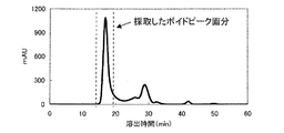
次に、実施例8で作製した0.625μMのマレイミド基を導入したアミノデキストラン(T2000)溶液40μLに8.6μMのチオール基を導入したIgG−ストレプトアビジン結合体290μLを混合し、30℃で1時間静置した。その後、0.2Mの2−メルカプトエタノール(和光純薬社製)33μLを添加し、4℃で一晩反応させ、未反応のマレイミド基をブロックした。反応後、Superdex200カラム(GEヘルスケア社製)でゲル濾過精製し、図1に示すボイドピーク画分よりIgG−ストレプトアビジン結合体−デキストラン(T2000)複合体を得た。
Next, 290 μL of an 8.6 μM thiol group-introduced IgG-streptavidin conjugate was mixed with 40 μL of an aminodextran (T2000) solution into which 0.625 μM maleimide group had been prepared. Let stand for hours. Thereafter, 33 μL of 0.2M 2-mercaptoethanol (manufactured by Wako Pure Chemical Industries, Ltd.) was added and reacted at 4 ° C. overnight to block unreacted maleimide groups. After the reaction, gel filtration purification was performed using a
IgG−ストレプトアビジン結合体−デキストラン複合体へのビオチン化ルシフェラーゼの導入
実施例9で作製したIgG−ストレプトアビジン結合体−デキストラン(T2000)複合体を、IgG−ストレプトアビジン結合体濃度として570nMになるように0.1Mリン酸緩衝液(pH7.2)を用いて調製した。このIgG−ストレプトアビジン結合体−デキストラン(T2000)複合体溶液300μLに対して、61μMのビオチン化ルシフェラーゼ(キッコーマン社製)溶液3.0μLを加え、25℃で1時間反応させることにより、ルシフェラーゼ標識IgG−デキストラン(T2000)複合体を得た。 Introduction of biotinylated luciferase into IgG-streptavidin conjugate-dextran complex The IgG-streptavidin conjugate-dextran (T2000) complex prepared in Example 9 has an IgG-streptavidin conjugate concentration of 570 nM. And 0.1 M phosphate buffer (pH 7.2). By adding 3.0 μL of a 61 μM biotinylated luciferase (Kikkoman) solution to 300 μL of this IgG-streptavidin conjugate-dextran (T2000) complex solution, and reacting at 25 ° C. for 1 hour, luciferase-labeled IgG -A dextran (T2000) complex was obtained.
Fab’−ストレプトアビジン結合体−デキストラン複合体の作製
実施例6で作製した4.0mg/mLのc11−9Fab’−ストレプトアビジン結合体420μLおよび2.6mg/mLのc11−14Fab’−ストレプトアビジン結合体646μLに対して、ジメチルホルムアミドに1mg/mLになるように溶解した2−イミノチオラン(PIERCE社製)を2μL添加し、30℃で30分間反応させた。Preparation of Fab′-Streptavidin Conjugate- Dextran Complex 4.0 mg / mL c11-9 Fab′-Streptavidin Conjugate prepared in Example 6 420 μL and 2.6 mg / mL c11-14 Fab′-Streptavidin Binding 2 μL of 2-iminothiolane (manufactured by PIERCE) dissolved in dimethylformamide so as to be 1 mg / mL was added to 646 μL of the body, and reacted at 30 ° C. for 30 minutes.
反応後、PD−10(GEヘルスケア社製)にてゲルろ過精製し、チオール基を導入した各々のFab’−ストレプトアビジン結合体を得た。チオール基の数を定量した結果、c11−9Fab’−ストレプトアビジン結合体1分子あたり4.6個、c11−14Fab’−ストレプトアビジン結合体1分子あたり3.0個のチオール基の存在が確認された。 After the reaction, gel filtration purification was performed with PD-10 (manufactured by GE Healthcare) to obtain each Fab′-streptavidin conjugate into which a thiol group was introduced. As a result of quantifying the number of thiol groups, the presence of 4.6 thiol groups per molecule of c11-9 Fab′-streptavidin conjugate and 3.0 thiol groups per molecule of c11-14 Fab′-streptavidin conjugate was confirmed. It was.
次に、実施例8で作製したマレイミド基を導入したアミノデキストラン(T2000)および(T500)とチオール基を導入したFab’−ストレプトアビジン結合体を混合し、30℃で1時間静置した。なお、マレイミド基を導入した各々のアミノデキストランとチオール基を導入した各々のFab’−ストレプトアビジン結合体は以下に示すとおりの濃度、液量で組み合わせ、合計4種類の重合体を作製した。 Next, the aminodextran (T2000) and (T500) introduced with the maleimide group prepared in Example 8 and the Fab′-streptavidin conjugate introduced with a thiol group were mixed and allowed to stand at 30 ° C. for 1 hour. Each aminodextran introduced with a maleimide group and each Fab′-streptavidin conjugate introduced with a thiol group were combined in the following concentrations and liquid amounts to prepare a total of four types of polymers.
1)c11−9およびc11−14を混合したFab’−ストレプトアビジン結合体−デキストラン(T2000)複合体
21μMのチオール基を導入したc11−9Fab’−ストレプトアビジン結合体200μL、18μMのチオール基を導入したc11−14Fab’−ストレプトアビジン結合体240μL、および0.625μMのマレイミド基を導入したアミノデキストラン(T2000)142μLを混合した。
2)c11−9Fab’−ストレプトアビジン結合体−デキストラン(T2000)複合体
21μMのチオール基を導入したc11−9Fab’−ストレプトアビジン結合体200μLおよび0.625μMのマレイミド基を導入したアミノデキストラン(T2000)71μLを混合した。 1) Fab′-streptavidin conjugate-dextran (T2000) complex in which c11-9 and c11-14 are mixed 21 μM of thiol group introduced in 21 μM c11-9 Fab′-
2) c11-9 Fab′-streptavidin conjugate-dextran (T2000) complex 200 μL of c11-9 Fab′-streptavidin conjugate introduced with 21 μM thiol group and aminodextran introduced with 0.625 μM maleimide group (T2000) 71 μL was mixed.
3)c11−14Fab’−ストレプトアビジン結合体−デキストラン(T2000)複合体
18μMのチオール基を導入したc11−14Fab’−ストレプトアビジン結合体240μLおよび0.625μMのマレイミド基を導入したアミノデキストラン(T2000)71μLを混合した。
4)c11−9およびc11−14を混合したFab’−ストレプトアビジン結合体−デキストラン(T500)複合体 3) c11-14 Fab′-streptavidin conjugate-dextran (T2000) complex 240 μL of c11-14 Fab′-streptavidin conjugate introduced with 18 μM thiol group and aminodextran introduced with 0.625 μM maleimide group (T2000) 71 μL was mixed.
4) Fab′-streptavidin conjugate-dextran (T500) complex mixed with c11-9 and c11-14
21μMのチオール基を導入したc11−9Fab’−ストレプトアビジン結合体200μL、18μMのチオール基を導入したc11−14Fab’−ストレプトアビジン結合体240μL、および2.5μMのマレイミド基を導入したアミノデキストラン(T500)116μLを混合した。 200 μL of c11-9 Fab′-streptavidin conjugate introduced with 21 μM thiol group, 240 μL of c11-14 Fab′-streptavidin conjugate introduced with 18 μM thiol group, and aminodextran (T500 introduced with 2.5 μM maleimide group) 116 μL was mixed.
反応後、0.2Mの2−メルカプトエタノール(和光純薬社製)を各々の複合体溶液の1/10量添加し、4℃で一晩反応させ、未反応のマレイミド基をブロックした。翌日、Superdex200カラム(GEヘルスケア社製)でゲル濾過精製し、図2〜5に示す各サンプルのボイドピーク画分よりFab’−ストレプトアビジン結合体−デキストラン複合体を得た。
After the reaction, 0.2M 2-mercaptoethanol (manufactured by Wako Pure Chemical Industries, Ltd.) was added in an amount of 1/10 of each complex solution and reacted at 4 ° C. overnight to block unreacted maleimide groups. The next day, gel filtration purification was performed using a
Fab’−ストレプトアビジン結合体−デキストラン複合体へのビオチン化ルシフェラーゼの導入
実施例11で作製した4種類のFab’−ストレプトアビジン結合体−デキストラン複合体を、Fab’−ストレプトアビジン結合体濃度として1.8μMになるように0.1Mリン酸緩衝液(pH7.2)にて調製した。これらのFab’−ストレプトアビジン結合体−デキストラン複合体溶液100μLに対して、61μMのビオチン化ルシフェラーゼ(キッコーマン社製)3.0μLを加え、25℃で1時間反応させることにより、以下に示す4種類のルシフェラーゼ標識Fab’−デキストラン複合体を作製した。 Introduction of Biotinylated Luciferase into Fab′-Streptavidin Conjugate-Dextran Complex Four kinds of Fab′-streptavidin conjugate-dextran complexes prepared in Example 11 were used as Fab′-streptavidin conjugate concentration of 1. It was prepared with 0.1 M phosphate buffer (pH 7.2) so as to be 8 μM. By adding 3.0 μL of 61 μM biotinylated luciferase (manufactured by Kikkoman) to 100 μL of these Fab′-streptavidin conjugate-dextran complex solutions, the following four types are obtained by reacting at 25 ° C. for 1 hour. The luciferase-labeled Fab′-dextran complex was prepared.
1)ルシフェラーゼ標識c11−9・c11−14Fab’−デキストラン(T2000)複合体
2)ルシフェラーゼ標識c11−9Fab’−デキストラン(T2000)複合体
3)ルシフェラーゼ標識c11−14Fab’−デキストラン(T2000)複合体
4)ルシフェラーゼ標識c11−9・c11−14Fab’−デキストラン(T500)複合体1) Luciferase labeled c11-9 · c11-14 Fab′-dextran (T2000) complex 2) Luciferase labeled c11-9 Fab′-dextran (T2000) complex 3) Luciferase labeled c11-14 Fab′-dextran (T2000) complex 4 ) Luciferase labeled c11-9 · c11-14 Fab′-dextran (T500) complex
ルシフェラーゼ標識抗体を用いた酵素免疫測定
WSCを用いたカルボジイミド法により、抗HCVコア抗原マウスモノクローナル抗体c11−3、c11−7、およびAOT3を磁性粒子に固定化し、0.2%抗体固相化磁性粒子を作製した。一方、正常ヒト血清およびPCRで確認された2種類のHCV陽性血清100μLと検体処理液(6M塩酸グアニジン、0.5NHCl、12.5%TritonX100、0.75%Tween20)100μLを混合し、37℃で15分間置くことによって検体の前処理を行った後、反応液(0.1Mリン酸ナトリウム、0.15M NaCl、1%BSA、0.5%カゼイン、0.05%Tween20、pH7.3)140μLと1Mトリス緩衝液20μLを混合し、この混合液160μLに前処理検体80μLを加えることにより、前処理検体を中和した。 Enzyme immunoassay using luciferase-labeled antibody Anti-HCV core antigen mouse monoclonal antibodies c11-3, c11-7, and AOT3 were immobilized on magnetic particles by the carbodiimide method using WSC, and 0.2% antibody solid-phase magnetism Particles were made. Meanwhile, 100 μL of normal human serum and two kinds of HCV positive sera confirmed by PCR and 100 μL of sample treatment solution (6 M guanidine hydrochloride, 0.5 NHCl, 12.5% Triton X100, 0.75% Tween 20) were mixed at 37 ° C. After pretreatment of the specimen by placing for 15 minutes, reaction solution (0.1 M sodium phosphate, 0.15 M NaCl, 1% BSA, 0.5% casein, 0.05
次に、中和した前処理検体240μLに0.2%抗体固相化磁性粒子を20μL加え、37℃で15分間置くことによって1次反応をおこなった。反応後、磁性粒子を洗浄液にて3回洗浄し、実施例5で作製したルシフェラーゼ標識IgGおよび実施例10で作製したルシフェラーゼ標識IgG−デキストラン(T2000)複合体を添加した。標識抗体の量は、ルシフェラーゼ濃度として18nMに希釈したものを120μL加え、攪拌後さらに37℃で15分間反応させた。反応後、磁性粒子を洗浄液にて3回洗浄し、50mMトリス緩衝液(pH8.5)100μLに再懸濁させた。 Next, a primary reaction was performed by adding 20 μL of 0.2% antibody-immobilized magnetic particles to 240 μL of the neutralized pretreated specimen and placing it at 37 ° C. for 15 minutes. After the reaction, the magnetic particles were washed three times with a washing solution, and the luciferase-labeled IgG produced in Example 5 and the luciferase-labeled IgG-dextran (T2000) complex produced in Example 10 were added. The amount of the labeled antibody was 120 μL of luciferase diluted to 18 nM, and after stirring, the mixture was further reacted at 37 ° C. for 15 minutes. After the reaction, the magnetic particles were washed three times with a washing solution and resuspended in 100 μL of 50 mM Tris buffer (pH 8.5).
磁性粒子を再懸濁したチューブに基質溶液(ルシフェリン)100μLを加え、ルーマットLB9507(ベルトールド社製)を使ってルシフェラーゼの発光を測定した。発光測定は、ルシフェリン添加0.5秒後から5秒間の発光を積算した。その結果、表1に示すとおり、ルシフェラーゼ標識IgGよりもルシフェラーゼ標識IgG−デキストラン(T2000)複合体のほうが高い発光値を示した。パネル検体MのS/N比から見積もると、およそ50〜100倍程度、反応性が向上していると推察された。 100 μL of the substrate solution (luciferin) was added to the tube in which the magnetic particles were resuspended, and luminescence of luciferase was measured using Lumat LB9507 (manufactured by Bertrand). Luminescence was measured by integrating the luminescence for 5 seconds after 0.5 seconds from the addition of luciferin. As a result, as shown in Table 1, the luciferase-labeled IgG-dextran (T2000) complex showed a higher luminescence value than the luciferase-labeled IgG. When estimated from the S / N ratio of the panel specimen M, it was estimated that the reactivity was improved by about 50 to 100 times.

ルシフェラーゼ標識抗体断片を用いた酵素免疫測定
標識抗体として実施例7および実施例12で作製したものを使用した以外は、実施例13と同じ方法で実施した。その結果、表2に示すとおり、ルシフェラーゼFab’標識よりも、ルシフェラーゼ標識Fab’−デキストラン(T2000)複合体またはルシフェラーゼ標識Fab’−デキストラン(T500)複合体のほうが高い発光値を示し、IgGの場合と同様に重合化することにより50〜100倍程度反応性が向上していることが確認された。また、c11−9またはc11−14抗体断片を個別に重合化した場合も反応性の向上は見られるが、両者を組み合わせることによりさらに反応性が向上することが確認された。 The enzyme immunoassay using the luciferase-labeled antibody fragment was carried out in the same manner as in Example 13 except that the antibody prepared in Example 7 and Example 12 was used. As a result, as shown in Table 2, the luciferase-labeled Fab′-dextran (T2000) complex or the luciferase-labeled Fab′-dextran (T500) complex showed a higher luminescence value than the luciferase Fab ′ label. It was confirmed that the reactivity was improved by about 50 to 100 times by polymerization in the same manner as in Example 1. In addition, when the c11-9 or c11-14 antibody fragment was individually polymerized, the reactivity was improved, but it was confirmed that the reactivity was further improved by combining the two.
既存のプローブ複合体作製方法を応用して作製した重合化標識抗体との性能比較Performance comparison with polymer-labeled antibody prepared by applying existing probe complex preparation method
既存のプローブ複合体作製方法を応用した重合化標識抗体の作製
国際公開2006/011543に記載の方法を応用してプローブ複合体を作製した。まず、デキストラン(T2000)44mgを秤量し、0.1Mリン酸緩衝液(pH7.0)0.8mLに溶解し、過ヨウ素酸ナトリウム溶液を0.4mL添加混合した。室温で2時間反応させた後、ゲルろ過(PD−10;GEヘルスケア社製)により余剰の過ヨウ素酸ナトリウムを除去し、ストレプトアビジン(SA)溶液とCAPS溶液(10%)を添加して室温で5時間反応させることにより、デキストランにストレプトアビジンを導入した。さらに、反応産物の安定化のため、Dimethylamine Borate(DMBA;生化学工業社製)1mgと1Mトリス溶液0.4mLを添加混合して室温で一晩反応させた。その後、ゲルろ過(Sephacryl S−300HR 1.6x30;GEヘルスケア社製)により反応産物を精製し、デキストラン−SA結合物を得た。次に、0.1Mリン酸緩衝液(pH7.0)を用いてデキストラン−SA結合物を2mg/mLになるように調製し、このデキストラン−SA結合物溶液0.5mLに対して、ジメチルホルムアミドにて10mg/mLになるように溶解したマレイミド試薬EMCS(同仁化学社製)5μLを加え、室温で1.5時間反応させた。この反応産物をPD−10(GEヘルスケア社製)にてゲルろ過精製し、未反応のEMCSを除去することによりマレイミド基を導入したデキストラン−SA結合物を得た。一方、実施例2に記載した方法によりc11−9とc11−14のFab’を作製し、各Fab’を等量混合することにより0.5mg/mLのFab’溶液を調製した。次に、2mg/mLのマレイミド基を導入したデキストラン−SA結合物0.5mLに対して、0.5mg/mL Fab’溶液を1mL添加して4℃で一晩反応させた。その後、終濃度が15mMになるように2−メルカプトエチルアミンを添加し、室温で1時間反応させることにより未反応のマレイミド基をブロックした。反応後、Sephacryl S−300HRカラム(GEヘルスケア社製)にてゲルろ過精製し、デキストラン−SA−Fab’複合体を得た。精製したデキストラン−SA−Fab’複合体に含まれるSAは、濃度既知のSA溶液を標準として、HABA試薬による発色を指標として定量した。作製したデキストランT2000−SA−Fab’重合体を、SA濃度として570nMになるように0.1Mリン酸緩衝液にて調製した。このデキストランT2000−SA−Fab’重合体溶液 300μLに対して、ビオチン化ルシフェラーゼ(キッコーマン社製)61μMを3.0μL添加し、25℃で1時間反応させることにより、既存の方法を応用した重合化標識抗体を作製した。 Production of polymerized labeled antibody by applying existing probe complex production method A probe complex was produced by applying the method described in International Publication No. 2006/011543. First, 44 mg of dextran (T2000) was weighed and dissolved in 0.8 mL of 0.1 M phosphate buffer (pH 7.0), and 0.4 mL of sodium periodate solution was added and mixed. After reacting at room temperature for 2 hours, excess sodium periodate was removed by gel filtration (PD-10; manufactured by GE Healthcare), and a streptavidin (SA) solution and a CAPS solution (10%) were added. Streptavidin was introduced into dextran by reacting at room temperature for 5 hours. Furthermore, in order to stabilize the reaction product, 1 mg of dimethylamine borate (DMBA; manufactured by Seikagaku Corporation) and 0.4 mL of 1M Tris solution were added and mixed, and reacted at room temperature overnight. Thereafter, the reaction product was purified by gel filtration (Sephaacryl S-300HR 1.6 × 30; manufactured by GE Healthcare) to obtain a dextran-SA conjugate. Next, a dextran-SA conjugate was prepared to 2 mg / mL using 0.1 M phosphate buffer (pH 7.0), and dimethylformamide was added to 0.5 mL of this dextran-SA conjugate solution. 5 μL of maleimide reagent EMCS (manufactured by Dojindo) dissolved at 10 mg / mL was added and reacted at room temperature for 1.5 hours. This reaction product was purified by gel filtration with PD-10 (manufactured by GE Healthcare), and unreacted EMCS was removed to obtain a dextran-SA conjugate having a maleimide group introduced. On the other hand, Fab ′ of c11-9 and c11-14 were prepared by the method described in Example 2, and an Fab ′ solution of 0.5 mg / mL was prepared by mixing equal amounts of each Fab ′. Next, 1 mL of 0.5 mg / mL Fab ′ solution was added to 0.5 mL of dextran-SA conjugate into which 2 mg / mL maleimide group was introduced, and reacted at 4 ° C. overnight. Thereafter, 2-mercaptoethylamine was added so that the final concentration was 15 mM, and the unreacted maleimide group was blocked by reacting at room temperature for 1 hour. After the reaction, gel filtration purification was performed using a Sephacryl S-300HR column (manufactured by GE Healthcare) to obtain a dextran-SA-Fab ′ complex. SA contained in the purified dextran-SA-Fab ′ complex was quantified using an SA solution with a known concentration as a standard and color development by the HABA reagent as an index. The prepared dextran T2000-SA-Fab ′ polymer was prepared with 0.1 M phosphate buffer so that the SA concentration was 570 nM. Polymerization applying the existing method by adding 3.0 μL of biotinylated luciferase (Kikkoman) 61 μM to 300 μL of this dextran T2000-SA-Fab ′ polymer solution and reacting at 25 ° C. for 1 hour. A labeled antibody was prepared.
ルシフェラーゼ標識抗体を用いた酵素免疫測定
WSCを用いたカルボジイミド法により、抗HCVコア抗原マウスモノクローナル抗体c11−3、c11−7、およびAOT3を磁性粒子に固定化し、0.2%抗体固相化磁性粒子を作製した。一方、正常ヒト血清および2種類のHCVコア抗原陽性血清100μLと検体処理液(6M塩酸グアニジン、0.5NHCl、12.5%TritonX100、0.75%Tween20)100μLを混合し、37℃で15分間置くことによって検体の前処理を行った後、反応液(0.1Mリン酸ナトリウム、0.15MNaCl、1%BSA、0.5%カゼイン、0.05%Tween20、pH7.3)140μLと1Mトリス緩衝液20μLを混合し、この混合液160μLに前処理検体80μLを加えることにより、前処理検体を中和した。
次に、中和した前処理検体240μLに0.2%抗体固相化磁性粒子を20μL加え、37℃で15分間置くことによって1次反応をおこなった。反応後、磁性粒子を洗浄液にて3回洗浄し、実施例5で作製したルシフェラーゼ標識IgG、実施例10で作製したルシフェラーゼ標識IgG−デキストラン(T2000)複合体、既存の方法を応用して作製した重合化標識抗体を添加した。標識抗体の量は、ルシフェラーゼ濃度として18nMに希釈したものを120μL加え、攪拌後さらに37℃で15分間反応させた。反応後、磁性粒子を洗浄液にて3回洗浄し、50mMトリス緩衝液(pH8.5)100μLに再懸濁させた。
磁性粒子を再懸濁したチューブに基質溶液(ルシフェリン)100μLを加え、ルーマットLB9507(ベルトールド社製)を使ってルシフェラーゼの発光を測定した。発光測定は、ルシフェリン添加0.5秒後から5秒間の発光を積算した。その結果、表3に示すとおりルシフェラーゼ標識IgG−デキストラン(T2000)複合体は既存の方法を応用して作製した重合化標識抗体よりも高い反応性を示した。パネル検体LおよびMのS/N比から見積もると、ルシフェラーゼ標識IgG−デキストラン(T2000)複合体は、既存の方法を応用して作製した重合化標識抗体よりも、およそ3〜5倍程度、反応性が向上していた。なお、既存の方法を応用して作製した重合化標識抗体の場合、使用する抗体量を多くしても、そのS/N比に大きな改善は見られなかった。 Enzyme immunoassay using luciferase-labeled antibody Anti-HCV core antigen mouse monoclonal antibodies c11-3, c11-7, and AOT3 were immobilized on magnetic particles by the carbodiimide method using WSC, and 0.2% antibody solid-phase magnetism Particles were made. Meanwhile, 100 μL of normal human serum and two types of HCV core antigen positive sera and 100 μL of sample treatment solution (6 M guanidine hydrochloride, 0.5 NHCl, 12.5% Triton X100, 0.75% Tween 20) are mixed and incubated at 37 ° C. for 15 minutes. After pretreatment of the specimen by placing, 140 μL of reaction solution (0.1 M sodium phosphate, 0.15 M NaCl, 1% BSA, 0.5% casein, 0.05
Next, a primary reaction was performed by adding 20 μL of 0.2% antibody-immobilized magnetic particles to 240 μL of the neutralized pretreated specimen and placing it at 37 ° C. for 15 minutes. After the reaction, the magnetic particles were washed with a washing solution three times, and the luciferase-labeled IgG prepared in Example 5, the luciferase-labeled IgG-dextran (T2000) complex prepared in Example 10 and an existing method were applied. Polymerized labeled antibody was added. The amount of the labeled antibody was 120 μL of luciferase diluted to 18 nM, and after stirring, the mixture was further reacted at 37 ° C. for 15 minutes. After the reaction, the magnetic particles were washed three times with a washing solution and resuspended in 100 μL of 50 mM Tris buffer (pH 8.5).
100 μL of the substrate solution (luciferin) was added to the tube in which the magnetic particles were resuspended, and luminescence of luciferase was measured using Lumat LB9507 (manufactured by Bertrand). Luminescence was measured by integrating the luminescence for 5 seconds after 0.5 seconds from the addition of luciferin. As a result, as shown in Table 3, the luciferase-labeled IgG-dextran (T2000) complex showed higher reactivity than the polymerized labeled antibody produced by applying the existing method. As estimated from the S / N ratios of the panel specimens L and M, the luciferase-labeled IgG-dextran (T2000) complex is approximately 3 to 5 times more reactive than the polymerization-labeled antibody produced by applying an existing method. Improved. In the case of a polymerized labeled antibody prepared by applying an existing method, even if the amount of antibody used was increased, the S / N ratio was not greatly improved.
ビオチン化PODへの応用Application to biotinylated POD
POD標識Fab’の作製
実施例6で作製した15.4μMのc11−9およびc11−14のFab’−ストレプトアビジン結合体30μLに対して、56.8μMのビオチン化POD(インビトロジェン社製)8.1μLを加えて、4℃で一晩置くことによりPOD標識Fab’を作製した。5. Production of POD-labeled Fab ′ 56.8 μM biotinylated POD (manufactured by Invitrogen) with respect to 30 μL of 15.4 μM c11-9 and c11-14 Fab′-streptavidin conjugate prepared in Example 6. POD-labeled Fab ′ was prepared by adding 1 μL and placing it overnight at 4 ° C.
Fab’−ストレプトアビジン結合体−デキストラン(T2000)複合体へのビオチン化PODの導入
実施例11で作製したc11−9およびc11−14を混合したFab’−ストレプトアビジン結合体−デキストラン(T2000)複合体を、Fab’−ストレプトアビジン結合体濃度として1.4μMになるように0.1Mリン酸緩衝液(pH7.2)にて調製した。この複合体溶液50μLに対して、56.8μMのビオチン化POD(インビトロジェン社製)1.2μLを加えて、4℃で一晩置くことによりPOD標識Fab’−デキストラン(T2000)複合体を作製した。 Introduction of biotinylated POD into Fab′-streptavidin conjugate-dextran (T2000) complex Fab′-streptavidin conjugate-dextran (T2000) complex mixed with c11-9 and c11-14 prepared in Example 11 The body was prepared with 0.1 M phosphate buffer (pH 7.2) so that the Fab′-streptavidin conjugate concentration was 1.4 μM. To 50 μL of this complex solution, 1.2 μL of 56.8 μM biotinylated POD (manufactured by Invitrogen) was added and left overnight at 4 ° C. to prepare a POD-labeled Fab′-dextran (T2000) complex. .
POD標識抗体を用いた酵素免疫測定
WSCを用いたカルボジイミド法により、抗HCVコア抗原マウスモノクローナル抗体c11−3、c11−7、およびAOT3を磁性粒子に固定化し、0.2%抗体固相化磁性粒子を作製した。一方、組み換え体HCVコア抗原(c11)を0nM、0.12nM、1.2nMになるように正常ヒト血清で希釈し、これらのサンプル100μLと検体処理液(6M塩酸グアニジン、0.5NHCl、12.5%TritonX100、0.75%Tween20)100μLを混合し、37℃で15分間置くことによってサンプルの前処理を行った。さらに、反応液(0.1Mリン酸ナトリウム、0.15MNaCl、1%BSA、0.5%カゼイン、0.05%Tween20、pH7.3)140μLと1Mトリス緩衝液20μLを混合し、この混合液160μLに前処理検体80μLを加えることにより、前処理検体を中和した。
次に、中和した前処理検体240μLに0.2%抗体固相化磁性粒子を20μL加え、37℃で15分間置くことによって1次反応をおこなった。反応後、磁性粒子を洗浄液にて3回洗浄し、上記方法により作製したPOD標識Fab’およびPOD標識Fab’−デキストラン(T2000)複合体を添加した。標識抗体の量は、POD濃度として18nMに希釈したものを120μL加え、攪拌後さらに37℃で15分間反応させた。反応後、磁性粒子を洗浄液にて3回洗浄した。
次に、磁性粒子のチューブに基質溶液(ルミノール)200μLを加え、ルーマットLB9507(ベルトールド社製)を使ってPODの発光を測定した。発光測定は、ルミノール添加12秒後から3秒間の発光を積算した。その結果、表4に示すとおりPOD標識Fab’よりもPOD標識Fab’−デキストラン(T2000)複合体のほうが高い発光値を示し、ルシフェラーゼの場合と同様に本発明によるシグナルの増幅が確認された。以上の結果から本発明はルシフェラーゼ以外の酵素を用いて実施可能であることが示された。 Enzyme immunoassay using POD labeled antibody Anti-HCV core antigen mouse monoclonal antibodies c11-3, c11-7, and AOT3 were immobilized on magnetic particles by carbodiimide method using WSC, and 0.2% antibody solid phase magnetism Particles were made. On the other hand, the recombinant HCV core antigen (c11) was diluted with normal human serum so as to be 0 nM, 0.12 nM, and 1.2 nM, and 100 μL of these samples and the sample treatment solution (6 M guanidine hydrochloride, 0.5 NHCl, 12. Samples were pretreated by mixing 100 μL of 5% Triton X100, 0.75% Tween 20) and placing at 37 ° C. for 15 minutes. Furthermore, 140 μL of the reaction solution (0.1 M sodium phosphate, 0.15 M NaCl, 1% BSA, 0.5% casein, 0.05
Next, a primary reaction was performed by adding 20 μL of 0.2% antibody-immobilized magnetic particles to 240 μL of the neutralized pretreated specimen and placing it at 37 ° C. for 15 minutes. After the reaction, the magnetic particles were washed three times with a washing solution, and POD-labeled Fab ′ and POD-labeled Fab′-dextran (T2000) complex prepared by the above method were added. As for the amount of the labeled antibody, 120 μL of POD concentration diluted to 18 nM was added, and after stirring, the mixture was further reacted at 37 ° C. for 15 minutes. After the reaction, the magnetic particles were washed with a washing solution three times.
Next, 200 μL of a substrate solution (luminol) was added to a tube of magnetic particles, and POD luminescence was measured using Lumat LB9507 (manufactured by Berthold). Luminescence was measured by integrating luminescence for 3 seconds after 12 seconds from the addition of luminol. As a result, as shown in Table 4, the POD-labeled Fab′-dextran (T2000) complex showed a higher luminescence value than the POD-labeled Fab ′, and the signal amplification according to the present invention was confirmed as in the case of luciferase. From the above results, it was shown that the present invention can be carried out using an enzyme other than luciferase.
ビオチン化FITCへの応用Application to biotinylated FITC
FITC標識Fab’の作製
実施例6で作製した15.4μMのc11−9およびc11−14のFab’−ストレプトアビジン結合体30μLに対して、160μMのビオチン化FITC(インビトロジェン社製)2.9μLを加えて、4℃で一晩置くことによりFITC標識Fab’を作製した。 Preparation of FITC-labeled Fab ′ 2.9 μL of 160 μM biotinylated FITC (manufactured by Invitrogen) was prepared on 30 μL of the 15.4 μM c11-9 and c11-14 Fab′-streptavidin conjugate prepared in Example 6. In addition, FITC-labeled Fab ′ was prepared by placing at 4 ° C. overnight.
Fab’−ストレプトアビジン結合体−デキストラン(T2000)複合体へのビオチン化FITCの導入
実施例11で作製したc11−9およびc11−14を混合したFab’−ストレプトアビジン結合体−デキストラン(T2000)複合体を、Fab’−ストレプトアビジン結合体濃度として6.3μMになるように0.1Mリン酸緩衝液(pH7.2)にて調製した。この複合体溶液20μLに対して、160μMのビオチン化FITC(インビトロジェン社製)1.0μLを加えて、4℃で一晩置くことによりFITC標識Fab’−デキストラン(T2000)複合体を作製した。 Introduction of biotinylated FITC into Fab′-streptavidin conjugate-dextran (T2000) complex Fab′-streptavidin conjugate-dextran (T2000) complex in which c11-9 and c11-14 prepared in Example 11 were mixed The body was prepared with 0.1 M phosphate buffer (pH 7.2) so that the Fab′-streptavidin conjugate concentration was 6.3 μM. To 20 μL of this complex solution, 1.0 μL of 160 μM biotinylated FITC (manufactured by Invitrogen) was added and left overnight at 4 ° C. to prepare a FITC-labeled Fab′-dextran (T2000) complex.
FITC標識抗体を用いた蛍光免疫測定
WSCを用いたカルボジイミド法により、抗HCVコア抗原マウスモノクローナル抗体c11−3、c11−7、およびAOT3を磁性粒子に固定化し、0.2%抗体固相化磁性粒子を作製した。一方、組み換え体HCVコア抗原(c11)を0nM、0.12nM、1.2nMになるように正常ヒト血清で希釈し、これらのサンプル100μLと検体処理液(6M塩酸グアニジン、0.5NHCl、12.5%TritonX100、0.75%Tween20)100μLを混合し、37℃で15分間置くことによってサンプルの前処理を行った。さらに、反応液(0.1Mリン酸ナトリウム、0.15MNaCl、1%BSA、0.5%カゼイン、0.05%Tween20、pH7.3)140μLと1Mトリス緩衝液20μLを混合し、この混合液160μLに前処理検体80μLを加えることにより、前処理検体を中和した。
次に、中和した前処理検体240μLに0.2%抗体固相化磁性粒子を20μL加え、37℃で15分間置くことによって1次反応をおこなった。反応後、磁性粒子を洗浄液にて3回洗浄し、上記方法により作製したFITC標識Fab’およびFITC標識Fab’−デキストラン(T2000)複合体を添加した。標識抗体の量は、FITC濃度として65nMに希釈したものを200μL加え、攪拌後さらに4℃で一晩反応させた。反応後、磁性粒子を洗浄液にて3回洗浄した。
次に、磁性粒子のチューブに10mM PBS(pH7.4)を200μL加え、磁性粒子を懸濁し、懸濁液を96穴白色プレート(サーモフィッシャー社製)に分注した。その後、蛍光プレートリーダーinfinite200(テカン社製)を使ってFITCの蛍光を測定した。蛍光測定の条件は、485nmの波長で励起し、535nmの蛍光を測定した。その結果、表5及び図6に示すとおりFITC標識Fab’よりもFITC標識Fab’−デキストラン(T2000)複合体のほうが高い蛍光活性を示し、ルシフェラーゼやPODの場合と同様に本発明によるシグナルの増幅が確認された。以上の結果から本発明は酵素以外の低分子標識物を用いても実施可能であることが示された。Anti-HCV core antigen mouse monoclonal antibodies c11-3, c11-7, and AOT3 were immobilized on magnetic particles by a carbodiimide method using WSC using a fluorescence immunoassay using a FITC-labeled antibody , and 0.2% antibody-immobilized magnetism Particles were made. On the other hand, the recombinant HCV core antigen (c11) was diluted with normal human serum so as to be 0 nM, 0.12 nM, and 1.2 nM, and 100 μL of these samples and the sample treatment solution (6 M guanidine hydrochloride, 0.5 NHCl, 12. Samples were pretreated by mixing 100 μL of 5% Triton X100, 0.75% Tween 20) and placing at 37 ° C. for 15 minutes. Furthermore, 140 μL of the reaction solution (0.1 M sodium phosphate, 0.15 M NaCl, 1% BSA, 0.5% casein, 0.05
Next, a primary reaction was performed by adding 20 μL of 0.2% antibody-immobilized magnetic particles to 240 μL of the neutralized pretreated specimen and placing it at 37 ° C. for 15 minutes. After the reaction, the magnetic particles were washed three times with a washing solution, and FITC-labeled Fab ′ and FITC-labeled Fab′-dextran (T2000) complex prepared by the above method were added. As the amount of the labeled antibody, 200 μL of FITC concentration diluted to 65 nM was added, and after stirring, the mixture was further reacted at 4 ° C. overnight. After the reaction, the magnetic particles were washed with a washing solution three times.
Next, 200 μL of 10 mM PBS (pH 7.4) was added to the magnetic particle tube to suspend the magnetic particles, and the suspension was dispensed into a 96-well white plate (Thermo Fisher). Thereafter, the fluorescence of FITC was measured using a fluorescence plate reader infinite 200 (manufactured by Tecan). The fluorescence measurement was performed by exciting at a wavelength of 485 nm and measuring fluorescence at 535 nm. As a result, as shown in Table 5 and FIG. 6, the FITC-labeled Fab′-dextran (T2000) complex showed a higher fluorescence activity than the FITC-labeled Fab ′, and the signal amplification according to the present invention was the same as in the case of luciferase and POD. Was confirmed. From the above results, it was shown that the present invention can be implemented using a low-molecular label other than the enzyme.
標識化プローブ−水溶性担体複合体の加速安定性
本発明に係る標識化プローブ−水溶性担体複合体の加速安定性を試験した。詳細には、実施例12で作製したルシフェラーゼ標識c11−9・c11−14Fab’−デキストラン(T2000)複合体について、4℃保存条件下における活性と、37℃、1週間での加速条件後の残存活性を測定し、比較検討した。活性の測定は実施例13と同じ方法で行った。結果は表6に示すとおりであった。 Accelerated Stability of Labeled Probe-Water-Soluble Carrier Complex The accelerated stability of the labeled probe-water-soluble carrier complex according to the present invention was tested. Specifically, for the luciferase-labeled c11-9 · c11-14 Fab′-dextran (T2000) complex prepared in Example 12, the activity under storage conditions at 4 ° C. and the remaining after acceleration conditions at 37 ° C. for 1 week Activity was measured and compared. The activity was measured in the same manner as in Example 13. The results were as shown in Table 6.
上記結果より、本発明に係る標識化プローブ−水溶性担体複合体が優れた加速安定性を示すことが実証された。即ち、本発明に係る標識化プローブ−水溶性担体複合体は、37℃で1週間保存しても、約90%程度乃至それ以上の残存活性を保持したことが観察された。また、当該加速条件に伴うバックグラウンドの上昇は観察されなかった。 From the above results, it was demonstrated that the labeled probe-water-soluble carrier complex according to the present invention exhibits excellent accelerated stability. That is, it was observed that the labeled probe-water-soluble carrier complex according to the present invention retained about 90% or more of residual activity even after storage at 37 ° C. for 1 week. In addition, an increase in background associated with the acceleration condition was not observed.
これらの結果から、本発明により、被験物質と特異的に結合する高感度かつ安定な標識化プローブ−水溶性担体複合体が得られ、この標識化プローブ−水溶性担体複合体を用いることにより、従来と比較して、より高感度かつ安定的な検出・測定が可能となることが実証された。また、本願発明が酵素以外の低分子標識物に対しても有効であることが実証された。 From these results, according to the present invention, a highly sensitive and stable labeled probe-water-soluble carrier complex that specifically binds to the test substance is obtained, and by using this labeled probe-water-soluble carrier complex, It has been demonstrated that more sensitive and stable detection and measurement is possible compared to conventional methods. In addition, it was demonstrated that the present invention is also effective for low molecular labels other than enzymes.
被験物質とプローブが特異的に結合する事を利用して、検体中の被験物質を検出・測定する方法において、本発明の標識化プローブ−水溶性担体複合物を用いることにより、従来と比較して、より高感度かつ安定な検出・測定が可能となる。 By using the labeled probe-water-soluble carrier complex of the present invention in a method for detecting and measuring a test substance in a sample by utilizing the specific binding between the test substance and the probe, it is compared with the conventional method. Therefore, more sensitive and stable detection / measurement becomes possible.
Claims (14)
工程1.チオール基を有し被験物質に対して結合可能なプローブと、マレイミド基を有するアビジン類を結合させてプローブ結合体とし、
工程2.次にチオール基を有する前記プローブ結合体と、マレイミド基を有する高分子の水溶性担体を結合させてプローブ結合体−水溶性担体複合体とし、
工程3.さらに前記プローブ結合体−水溶性担体複合体とビオチン化標識物を混合して、プローブ結合体−水溶性担体複合体のアビジン類とビオチン化標識物のビオチンを結合させる。A method for producing a labeled probe-water-soluble carrier complex, comprising the following steps.
Step 1. A probe conjugate that binds a probe having a thiol group and capable of binding to a test substance and avidin having a maleimide group,
Step 2. Next, the probe conjugate having a thiol group and the polymer water-soluble carrier having a maleimide group are bound to form a probe conjugate-water-soluble carrier complex,
Step 3. Further, the probe conjugate-water-soluble carrier complex and the biotinylated label are mixed, and the avidin of the probe conjugate-water-soluble carrier complex and biotinylated label biotin are bound.
Priority Applications (1)
| Application Number | Priority Date | Filing Date | Title |
|---|---|---|---|
| JP2012510666A JP5675782B2 (en) | 2010-04-14 | 2011-04-13 | Labeled probe-water-soluble carrier complex |
Applications Claiming Priority (4)
| Application Number | Priority Date | Filing Date | Title |
|---|---|---|---|
| JP2010092953 | 2010-04-14 | ||
| JP2010092953 | 2010-04-14 | ||
| JP2012510666A JP5675782B2 (en) | 2010-04-14 | 2011-04-13 | Labeled probe-water-soluble carrier complex |
| PCT/JP2011/059150 WO2011129357A1 (en) | 2010-04-14 | 2011-04-13 | Complex of labeled probe and water-soluble carrier |
Publications (2)
| Publication Number | Publication Date |
|---|---|
| JPWO2011129357A1 JPWO2011129357A1 (en) | 2013-07-18 |
| JP5675782B2 true JP5675782B2 (en) | 2015-02-25 |
Family
ID=44798728
Family Applications (1)
| Application Number | Title | Priority Date | Filing Date |
|---|---|---|---|
| JP2012510666A Active JP5675782B2 (en) | 2010-04-14 | 2011-04-13 | Labeled probe-water-soluble carrier complex |
Country Status (7)
| Country | Link |
|---|---|
| US (1) | US9005910B2 (en) |
| EP (1) | EP2560003B1 (en) |
| JP (1) | JP5675782B2 (en) |
| KR (1) | KR101814385B1 (en) |
| CN (1) | CN102893151B (en) |
| CA (1) | CA2796366C (en) |
| WO (1) | WO2011129357A1 (en) |
Families Citing this family (10)
| Publication number | Priority date | Publication date | Assignee | Title |
|---|---|---|---|---|
| JP6056137B2 (en) * | 2010-12-22 | 2017-01-11 | 東ソー株式会社 | antibody |
| KR101398764B1 (en) | 2013-08-29 | 2014-05-27 | 강릉원주대학교산학협력단 | Device for detecting analytes by moving the particle and method using the same |
| WO2018079314A1 (en) * | 2016-10-24 | 2018-05-03 | 東レ株式会社 | Semiconductor sensor and manufacturing method for same, and compound sensor |
| CN107236055B (en) * | 2017-05-19 | 2019-10-01 | 华东师范大学 | A kind of glucan derivative and its application |
| CN114303062A (en) * | 2019-09-02 | 2022-04-08 | 富士瑞必欧株式会社 | Method and kit for measuring lectin-binding substance, and blocked lectin used in same |
| JPWO2021045065A1 (en) * | 2019-09-02 | 2021-03-11 | ||
| CN113960306B (en) * | 2020-07-20 | 2023-04-28 | 菲鹏生物股份有限公司 | Reagent for stabilizing acridinium ester marker protein and application thereof |
| CN111830250A (en) * | 2020-08-04 | 2020-10-27 | 珠海市丽拓生物科技有限公司 | Preparation method of signal amplified enzyme-labeled secondary antibody |
| CN113777312B (en) * | 2021-09-03 | 2024-02-02 | 普十生物科技(北京)有限公司 | Preparation method of hepatitis B antibody fragment, kit and application |
| WO2023182520A1 (en) * | 2022-03-25 | 2023-09-28 | 積水メディカル株式会社 | Immunological analysis method, complex, method for producing complex, and reagent for immunological analysis |
Citations (5)
| Publication number | Priority date | Publication date | Assignee | Title |
|---|---|---|---|---|
| WO2006011543A1 (en) * | 2004-07-30 | 2006-02-02 | Advanced Life Science Institute, Inc. | Probe complex |
| WO2006070732A1 (en) * | 2004-12-28 | 2006-07-06 | Advanced Life Science Institute, Inc. | Blocked enzyme probe complex |
| JP2008022849A (en) * | 2006-07-24 | 2008-02-07 | National Institute Of Advanced Industrial & Technology | Method for producing biotin-labeled vargula hilgendorfii luciferase- streptavidin complex and method for stabilizing the same |
| JP2008122302A (en) * | 2006-11-15 | 2008-05-29 | Kikkoman Corp | Biomolecule measuring method |
| JP2010529186A (en) * | 2007-06-14 | 2010-08-26 | イノーバ・バイオサイエンシズ・リミテッド | Improved production of conjugates or production of conjugates |
Family Cites Families (11)
| Publication number | Priority date | Publication date | Assignee | Title |
|---|---|---|---|---|
| JP2619549B2 (en) | 1989-06-29 | 1997-06-11 | 日本商事株式会社 | Antigen quantification method and solid phase used for it |
| DE4310141A1 (en) | 1993-03-29 | 1994-10-06 | Boehringer Mannheim Gmbh | Homobidental trifunctional linkers |
| JPH11295313A (en) | 1998-04-09 | 1999-10-29 | Eiken Chem Co Ltd | Antibody or polymer of antibody piece and its utilization |
| JP3524401B2 (en) | 1998-09-16 | 2004-05-10 | 株式会社ニチレイ | Enzyme-antibody complex and method for producing the same |
| JP3781934B2 (en) | 1999-12-22 | 2006-06-07 | 株式会社ニチレイバイオサイエンス | Enzyme-protein complex |
| ATE487136T1 (en) * | 2000-03-28 | 2010-11-15 | Nanosphere Inc | NANOPARTICLES WITH BONDED OLIGONUCLEOTIDES AND USES THEREOF |
| JP4086277B2 (en) | 2001-12-27 | 2008-05-14 | 栄研化学株式会社 | Method for producing and using water-soluble carrier-antibody complex |
| JP2008520963A (en) * | 2004-11-16 | 2008-06-19 | シーナ・キャンサー・ダイアグノスティクス・リミテッド | How to detect an analyte in a sample |
| US20070254311A1 (en) * | 2006-04-26 | 2007-11-01 | Cardiogenics Inc. | Covalent modification and conjugation of luciferase |
| US20100304387A1 (en) * | 2007-11-28 | 2010-12-02 | Jenison Robert D | Methods and compositions for signal enhancement using multivalent interactions |
| JP2009244101A (en) | 2008-03-31 | 2009-10-22 | Canon Inc | Method for detecting test material |
-
2011
- 2011-04-13 JP JP2012510666A patent/JP5675782B2/en active Active
- 2011-04-13 KR KR1020127026688A patent/KR101814385B1/en active IP Right Grant
- 2011-04-13 CN CN201180018682.3A patent/CN102893151B/en not_active Expired - Fee Related
- 2011-04-13 CA CA2796366A patent/CA2796366C/en not_active Expired - Fee Related
- 2011-04-13 WO PCT/JP2011/059150 patent/WO2011129357A1/en active Application Filing
- 2011-04-13 US US13/640,821 patent/US9005910B2/en not_active Expired - Fee Related
- 2011-04-13 EP EP11768878.8A patent/EP2560003B1/en active Active
Patent Citations (5)
| Publication number | Priority date | Publication date | Assignee | Title |
|---|---|---|---|---|
| WO2006011543A1 (en) * | 2004-07-30 | 2006-02-02 | Advanced Life Science Institute, Inc. | Probe complex |
| WO2006070732A1 (en) * | 2004-12-28 | 2006-07-06 | Advanced Life Science Institute, Inc. | Blocked enzyme probe complex |
| JP2008022849A (en) * | 2006-07-24 | 2008-02-07 | National Institute Of Advanced Industrial & Technology | Method for producing biotin-labeled vargula hilgendorfii luciferase- streptavidin complex and method for stabilizing the same |
| JP2008122302A (en) * | 2006-11-15 | 2008-05-29 | Kikkoman Corp | Biomolecule measuring method |
| JP2010529186A (en) * | 2007-06-14 | 2010-08-26 | イノーバ・バイオサイエンシズ・リミテッド | Improved production of conjugates or production of conjugates |
Also Published As
| Publication number | Publication date |
|---|---|
| KR101814385B1 (en) | 2018-01-04 |
| US9005910B2 (en) | 2015-04-14 |
| CA2796366A1 (en) | 2011-10-20 |
| CA2796366C (en) | 2017-12-05 |
| WO2011129357A1 (en) | 2011-10-20 |
| EP2560003B1 (en) | 2018-07-04 |
| US20130230897A1 (en) | 2013-09-05 |
| KR20130081206A (en) | 2013-07-16 |
| EP2560003A1 (en) | 2013-02-20 |
| CN102893151B (en) | 2015-06-03 |
| JPWO2011129357A1 (en) | 2013-07-18 |
| CN102893151A (en) | 2013-01-23 |
| EP2560003A4 (en) | 2013-11-20 |
Similar Documents
| Publication | Publication Date | Title |
|---|---|---|
| JP5675782B2 (en) | Labeled probe-water-soluble carrier complex | |
| Chen et al. | An ultra-sensitive chemiluminescence immunosensor of carcinoembryonic antigen using HRP-functionalized mesoporous silica nanoparticles as labels | |
| Matsuya et al. | A Core− Shell-type fluorescent Nanosphere possessing reactive poly (ethylene glycol) tethered chains on the surface for Zeptomole detection of protein in time-resolved Fluorometric immunoassay | |
| CN108700584A (en) | Labeled complex and preparation method thereof, kit, application and detecting system | |
| Xie et al. | A sandwich ELISA-like detection of C-reactive protein in blood by citicoline-bovine serum albumin conjugate and aptamer-functionalized gold nanoparticles nanozyme | |
| Mahmoud et al. | Advanced procedures for labeling of antibodies with quantum dots | |
| JPH1048211A (en) | Measuring method using oligomerized receptor | |
| JP4920415B2 (en) | Probe complex | |
| TW201107752A (en) | Signal amplification microspheres, their use in one-step and multi-step analytical amplification procedures and methods for their production | |
| Peng et al. | Aptamer-barcode based immunoassay for the instantaneous derivatization chemiluminescence detection of IgE coupled to magnetic beads | |
| CN103913573A (en) | Double signal amplification ELISA detection method based on nanometer gold and graphene oxide | |
| Gao et al. | Rolling circle amplification integrated with suspension bead array for ultrasensitive multiplex immunodetection of tumor markers | |
| Yan et al. | A sandwich-hybridization assay for simultaneous determination of HIV and tuberculosis DNA targets based on signal amplification by quantum dots-PowerVision™ polymer coding nanotracers | |
| WO2002095407A1 (en) | Immunoassay method | |
| WO2019194280A1 (en) | Immunoassay method for hepatitis b virus antigen | |
| CN112067826A (en) | NT-proBNP detection kit constructed based on high-specific-activity alkaline phosphatase and application thereof | |
| WO2016041022A1 (en) | Sensor scaffold | |
| JP7361543B2 (en) | AFP-L3 measurement method, AFP-L3 measurement kit, and blocked labeled lectin used therefor | |
| WO2023124154A1 (en) | Magnetic bead coating, preparation method therefor, and test kit | |
| JP3847983B2 (en) | Reagent for immunological analysis, immunological analysis method, and kit for immunological analysis | |
| KR101273453B1 (en) | Quantitative Analysis Using Minute Tube with Accumulated Enzyme | |
| JP2019512688A (en) | Detection of anti-p53 antibody | |
| CN113049811A (en) | Nano magnetic bead coating material, preparation method thereof, detection reagent and detection kit | |
| JP5559465B2 (en) | Avidin binding carrier, method for producing the same and method for using the same | |
| Lei et al. | A simple, selective and sensitive immunoassay for determination of human chorionic gonadotrophin based on chemiluminescence resonance energy transfer |
Legal Events
| Date | Code | Title | Description |
|---|---|---|---|
| A621 | Written request for application examination |
Free format text: JAPANESE INTERMEDIATE CODE: A621 Effective date: 20131202 |
|
| TRDD | Decision of grant or rejection written | ||
| A01 | Written decision to grant a patent or to grant a registration (utility model) |
Free format text: JAPANESE INTERMEDIATE CODE: A01 Effective date: 20141216 |
|
| A61 | First payment of annual fees (during grant procedure) |
Free format text: JAPANESE INTERMEDIATE CODE: A61 Effective date: 20141224 |
|
| R150 | Certificate of patent or registration of utility model |
Ref document number: 5675782 Country of ref document: JP Free format text: JAPANESE INTERMEDIATE CODE: R150 |
|
| S531 | Written request for registration of change of domicile |
Free format text: JAPANESE INTERMEDIATE CODE: R313531 |
|
| R350 | Written notification of registration of transfer |
Free format text: JAPANESE INTERMEDIATE CODE: R350 |
|
| R250 | Receipt of annual fees |
Free format text: JAPANESE INTERMEDIATE CODE: R250 |
|
| R250 | Receipt of annual fees |
Free format text: JAPANESE INTERMEDIATE CODE: R250 |
|
| R250 | Receipt of annual fees |
Free format text: JAPANESE INTERMEDIATE CODE: R250 |
|
| S111 | Request for change of ownership or part of ownership |
Free format text: JAPANESE INTERMEDIATE CODE: R313117 |
|
| R350 | Written notification of registration of transfer |
Free format text: JAPANESE INTERMEDIATE CODE: R350 |
|
| R250 | Receipt of annual fees |
Free format text: JAPANESE INTERMEDIATE CODE: R250 |
|
| R250 | Receipt of annual fees |
Free format text: JAPANESE INTERMEDIATE CODE: R250 |
|
| R250 | Receipt of annual fees |
Free format text: JAPANESE INTERMEDIATE CODE: R250 |