JP5624930B2 - Endoscope device - Google Patents
Endoscope device Download PDFInfo
- Publication number
- JP5624930B2 JP5624930B2 JP2011082048A JP2011082048A JP5624930B2 JP 5624930 B2 JP5624930 B2 JP 5624930B2 JP 2011082048 A JP2011082048 A JP 2011082048A JP 2011082048 A JP2011082048 A JP 2011082048A JP 5624930 B2 JP5624930 B2 JP 5624930B2
- Authority
- JP
- Japan
- Prior art keywords
- light
- light source
- endoscope apparatus
- temperature
- change rate
- Prior art date
- Legal status (The legal status is an assumption and is not a legal conclusion. Google has not performed a legal analysis and makes no representation as to the accuracy of the status listed.)
- Expired - Fee Related
Links
Images
Classifications
-
- A—HUMAN NECESSITIES
- A61—MEDICAL OR VETERINARY SCIENCE; HYGIENE
- A61B—DIAGNOSIS; SURGERY; IDENTIFICATION
- A61B1/00—Instruments for performing medical examinations of the interior of cavities or tubes of the body by visual or photographical inspection, e.g. endoscopes; Illuminating arrangements therefor
- A61B1/06—Instruments for performing medical examinations of the interior of cavities or tubes of the body by visual or photographical inspection, e.g. endoscopes; Illuminating arrangements therefor with illuminating arrangements
Landscapes
- Health & Medical Sciences (AREA)
- Life Sciences & Earth Sciences (AREA)
- Surgery (AREA)
- Biophysics (AREA)
- Biomedical Technology (AREA)
- Instruments For Viewing The Inside Of Hollow Bodies (AREA)
- Nuclear Medicine, Radiotherapy & Molecular Imaging (AREA)
- Optics & Photonics (AREA)
- Pathology (AREA)
- Radiology & Medical Imaging (AREA)
- Endoscopes (AREA)
- Engineering & Computer Science (AREA)
- Physics & Mathematics (AREA)
- Heart & Thoracic Surgery (AREA)
- Medical Informatics (AREA)
- Molecular Biology (AREA)
- Animal Behavior & Ethology (AREA)
- General Health & Medical Sciences (AREA)
- Public Health (AREA)
- Veterinary Medicine (AREA)
Description
本発明は、内視鏡装置に関する。 The present invention relates to an endoscope apparatus.
一般に、内視鏡装置は、被検体内に挿入する挿入部を有する内視鏡と、この内視鏡に照明光を供給する光源装置とを備え、内視鏡と光源装置とは別体に構成されている。光源装置の発光源としては、キセノンランプやメタルハライドランプ等の白色光ランプが広く使用されるが、ランプに代えてレーザ光源を用いて照明光を生成するものがある。例えば、特許文献1の内視鏡装置においては、光源装置に搭載された半導体レーザ光源からの光を、導光部材である光ファイバを用いて内視鏡の挿入部先端まで伝送し、挿入部先端に設けた蛍光体を通して白色光を出射させる構成となっている。
In general, an endoscope apparatus includes an endoscope having an insertion portion that is inserted into a subject, and a light source device that supplies illumination light to the endoscope. The endoscope and the light source device are separated from each other. It is configured. As a light source of the light source device, a white light lamp such as a xenon lamp or a metal halide lamp is widely used. However, there is one that generates illumination light using a laser light source instead of the lamp. For example, in the endoscope device disclosed in
ところで、光源装置から内視鏡の挿入部先端までの間を単線の光ファイバ、又は少ない本数の光ファイバで照明光の伝送を行う場合、光ファイバの一部に断線が生じただけでも照明光に及ぼす光伝送損失の影響は大きくなり、照明光量を大きく低下させる。そのため、このような光ファイバの断線等の光伝送損失を検出する技術がある。例えば特許文献2には、光ファイバの出射端に配置される蛍光体がレーザ光の照射により昇温することを利用した発光装置が記載されている。この発光装置では、蛍光体の温度変化を観察することで光源からの光が蛍光体に達しているかを判断して断線を検出する。 By the way, when illuminating light is transmitted between the light source device and the distal end of the insertion portion of the endoscope using a single optical fiber or a small number of optical fibers, the illumination light is generated even if a part of the optical fiber is broken. The effect of optical transmission loss on the light intensity increases, and the amount of illumination light is greatly reduced. For this reason, there is a technique for detecting optical transmission loss such as disconnection of the optical fiber. For example, Patent Document 2 describes a light-emitting device that utilizes the fact that a phosphor disposed at an emission end of an optical fiber is heated by laser light irradiation. In this light-emitting device, the disconnection is detected by determining whether the light from the light source reaches the phosphor by observing the temperature change of the phosphor.
しかしながら、特許文献2の発光装置は、光出射端に波長変換部材を配置した導光部材と、光出射端に光拡散部材を配置した導光部材とのいずれに発生した光伝送損失かを、一つの温度センサを用いて検知できなかった。 However, the light-emitting device of Patent Document 2 indicates whether the light transmission loss occurred in either the light guide member in which the wavelength conversion member is disposed at the light exit end or the light guide member in which the light diffusion member is disposed at the light exit end. It could not be detected using one temperature sensor.
本発明は、光出射端に波長変換部材を配置した導光部材と、光出射端に光拡散部材を配置した導光部材とのいずれに発生した光伝送損失かを、一つの温度センサを用いて検知できる内視鏡装置を提供することを目的とする。 The present invention uses a single temperature sensor to determine which light transmission loss occurs between a light guide member having a wavelength conversion member disposed at a light exit end and a light guide member having a light diffusion member disposed at a light exit end. It is an object of the present invention to provide an endoscope apparatus that can detect the above.
本発明は下記構成からなる。
光伝送損失を検出する機能を備えた内視鏡装置であって、
第1の光源と、
前記第1の光源の出力光を導入して被検体内に挿入される挿入部の先端まで導光する第1の導光部材と、
前記第1の導光部材の光出射端に配置された波長変換部材と、
第2の光源と、
前記第2の光源の出力光を導入して前記挿入部の先端まで導光する第2の導光部材と、
前記第2の導光部材の光出射端に配置された光拡散部材と、
前記第1の光源及び前記第2の光源それぞれについて設定された目標光量に応じた駆動信号を生成し、前記第1の光源及び前記第2の光源を駆動する光源駆動手段と、
前記波長変換部材及び前記光拡散部材からの発熱による温度を検出する温度センサと、
前記第1の光源の駆動信号の強度に対応した前記波長変換部材の発熱による第1の温度変化率、及び前記第2の光源の駆動信号の強度に対応した前記光拡散部材の発熱による第2の温度変化率を記憶する記憶手段と、
前記温度センサにより検出された温度の第3の温度変化率を、前記第1の温度変化率、及び前記第2の温度変化率と比較し、前記第3の温度変化率が前記第1の温度変化率と一致する場合は前記第2の導光部材に光伝送損失が発生したと判定し、前記第3の温度変化率が前記第2の温度変化率と一致する場合は前記第1の導光部材に光伝送損失が発生したと判定する光伝送損失検出手段と、
を備えた内視鏡装置。
The present invention has the following configuration.
An endoscope apparatus having a function of detecting optical transmission loss,
A first light source;
A first light guide member that introduces output light of the first light source and guides it to a distal end of an insertion portion that is inserted into a subject;
A wavelength conversion member disposed at a light exit end of the first light guide member;
A second light source;
A second light guide member for introducing the output light of the second light source and guiding the light to the tip of the insertion portion;
A light diffusing member disposed at a light exit end of the second light guide member;
Light source driving means for generating a drive signal corresponding to a target light amount set for each of the first light source and the second light source, and driving the first light source and the second light source;
A temperature sensor for detecting a temperature due to heat generated from the wavelength conversion member and the light diffusion member;
The first temperature change rate due to heat generation of the wavelength conversion member corresponding to the intensity of the drive signal of the first light source, and the second due to heat generation of the light diffusion member corresponding to the intensity of the drive signal of the second light source. Storage means for storing the temperature change rate of
A third temperature change rate of the temperature detected by the temperature sensor is compared with the first temperature change rate and the second temperature change rate, and the third temperature change rate is the first temperature. When the rate of change coincides with the second light guide member, it is determined that a light transmission loss has occurred. When the rate of change of the third temperature coincides with the rate of change of the second temperature, the first light guide member is determined. An optical transmission loss detecting means for determining that an optical transmission loss has occurred in the optical member;
An endoscopic apparatus comprising:
本発明の内視鏡装置によれば、光出射端に波長変換部材を配置した導光部材と、光出射端に光拡散部材を配置した導光部材とのいずれに発生した光伝送損失かを、一つの温度センサを用いて検知できる。 According to the endoscope apparatus of the present invention, it is determined whether the light transmission loss occurs in either the light guide member in which the wavelength conversion member is disposed at the light exit end or the light guide member in which the light diffusion member is disposed at the light exit end. It can be detected using a single temperature sensor.
以下、本発明の実施の形態を図面を参照して説明する。
図1は本発明の実施形態を説明するための図で、内視鏡及び内視鏡が接続される各装置を表す内視鏡装置の構成図、図2は内視鏡装置の具体的な構成例を示す外観図である。
内視鏡装置100は、図1に示すように、内視鏡11と、制御装置13と、モニタ等の表示部15と、制御装置13に情報を入力するキーボードやマウス等の入力部17とを備えている。制御装置13は、光源装置19と、撮像画像の信号処理を行うプロセッサ21とを有して構成される。
Hereinafter, embodiments of the present invention will be described with reference to the drawings.
FIG. 1 is a diagram for explaining an embodiment of the present invention. FIG. 1 is a configuration diagram of an endoscope apparatus representing an endoscope and each apparatus to which the endoscope is connected. FIG. 2 is a specific example of the endoscope apparatus. It is an external view which shows a structural example.
As shown in FIG. 1, the
内視鏡11は、本体操作部23と、この本体操作部23に連設され被検体(体腔)内に挿入される挿入部25とを備える。本体操作部23には、ユニバーサルコード27が接続される。このユニバーサルコード27の先端は、光源装置19にライトガイドコネクタ29を介して接続され、また、ビデオコネクタ31を介してプロセッサ21に接続されている。
The
図2に示すように、内視鏡11の本体操作部23には、挿入部25の先端側で吸引、送気、送水を実施するための送気送水ボタンや、撮像時のシャッターボタン、詳細を後述する光伝送損失確認ボタン33等の各種操作ボタンが設けられる。また、本体操作部23には、これらのボタンと共に一対のアングルノブ35が併設されている。
As shown in FIG. 2, the main
挿入部25は、本体操作部側から順に軟性部37、湾曲部39、及び挿入部25の先端である内視鏡先端部41で構成される。湾曲部39は、本体操作部23のアングルノブ35を回転することによって遠隔的に湾曲操作されて、これにより内視鏡先端部41を所望の方向に向けることができる。
The
図3は内視鏡先端部41の拡大斜視図である。内視鏡先端部41には、撮像光学系の観察窓43と、照明光学系の第1照明窓45、第2照明窓47が配置されている。これら第1照明窓45、第2照明窓47は観察窓43を挟んだ両脇側に配置されている。第1照明窓45、第2照明窓47から照射される照明光による被検体からの反射光は、観察窓43を通じて撮像素子49(図1参照)で撮像される。撮像された観察画像は、プロセッサ21に接続された表示部15に表示される。
FIG. 3 is an enlarged perspective view of the
撮像光学系は、CCD(Charge Coupled Device)型イメージセンサや、CMOS(Complementary Metal Oxide Semiconductor)型イメージセンサ等の撮像素子49を有する。また、撮像光学系は、撮像素子49に観察像を結像させるレンズ等の光学部材51(図1参照)を有する。撮像素子49の受光面に結像されて取り込まれる観察像は、電気信号に変換されて信号ケーブル53を通じてプロセッサ21の撮像信号処理部55に入力され、この撮像信号処理部55で映像信号に変換される。
The imaging optical system includes an
一方、照明光学系は、図1に示すように、光源装置19と、光源装置19に接続される第1の導光部材57である第1光ファイバ59と、第2の導光部材61である第2光ファイバ63とを有する。第1光ファイバ59の光出射端には波長変換部材65が配置され、第2光ファイバ63の光出射端には光拡散部材67が配置される。光源装置19は、第1の光源69と、第2の光源71とを備える。第1の光源69は、半導体発光素子であるレーザ光源LD1と、レーザ光源LD2−Aとを備える。第2の光源71は、半導体発光素子であるレーザ光源LD2−Bを備える。光源装置19は、光源駆動手段である光源制御部75と、コンバイナ77とを有する。光源制御部75は、レーザ光源LD1、レーザ光源LD2−A、レーザ光源LD2−Bを駆動制御する。コンバイナ77は、レーザ光源LD1、レーザ光源LD2−Aからの出射光を合波して第1光ファイバ59に導入する。レーザ光源LD2−Bからの出射光は合波されることなく直接に第2光ファイバ63に導入される。
On the other hand, as shown in FIG. 1, the illumination optical system includes a
第1光ファイバ59、第2光ファイバ63は、単線の光ファイバからなる。単線の光ファイバを使用することで、挿入部25の細径化に寄与できる。
The 1st
内視鏡先端部41は、その内部に不図示の先端硬性部を備える。先端硬性部はステンレス鋼等の金属からなり、長手方向に沿って複数の貫通孔が形成されている。この先端硬性部の各貫通孔には、撮像光学系、照明光学系ユニット81、鉗子チャンネル83、送気送水ノズル85に連通する送気送水チャンネル等の各種部品が取り付けられている。
The endoscope
照明光学系ユニット81は、観察窓43を挟んで対称に配置される波長変換光学系ユニット89と、光拡散光学系ユニット91とからなる。波長変換光学系ユニット89と光拡散光学系ユニット91とはほぼ同一の構成を有し、光出射端に波長変換部材65又は光拡散部材67が設けられるかで異なる。ここでは、波長変換部材65の設けられる波長変換光学系ユニット89を図示して説明する。
The illumination
図4は照明光学系ユニット81の構成を示す分解斜視図である。波長変換光学系ユニット89は、シングルモードの第1光ファイバ59と、波長変換部材65と、波長変換部材65及び第1光ファイバ59を保持する保持部材としてのフェルール95と、波長変換部材65の外周を覆う筒状のスリーブ部材97と、スリーブ部材97の先端を封止する保護カバー99とから構成される。また、第1光ファイバ59の外周面は、不図示の保護チューブによって被覆されている。
FIG. 4 is an exploded perspective view showing the configuration of the illumination
一方、光拡散光学系ユニット91は、図4と同様に、第2光ファイバ63と、光拡散部材67と、フェルール95と、スリーブ部材97と、保護カバー99とからなる。光拡散光学系ユニット91は、波長変換光学系ユニット89と同様に、フェルール95が光拡散部材67及び第2光ファイバ63を保持する。スリーブ部材97がフェルール95の外周を覆い、かつ保護カバー99がスリーブ部材97の先端を封止する構成となっている。
On the other hand, the light diffusing
フェルール95は、円筒形状に形成され、第1光ファイバ59が挿通される挿通孔103を有する。フェルール95の先端側には、波長変換部材65を保持する蛍光体保持部105が形成されている。蛍光体保持部105は、フェルール先端面107から波長変換部材65の外形に合わせて凹となり、保護カバー99と対面する先端側が開放された凹部状に形成されている。挿通孔103は、蛍光体保持部105の基端に連続している。
The
蛍光体保持部105には、表面に反射膜109が設けられている。反射膜109は、銀、アルミ等の金属膜からなり、例えばメッキ、蒸着、スパッタ等により薄膜状に形成される。波長変換部材65は、蛍光体保持部105の内部に、反射膜109と接しつつ保持される。波長変換部材65から発する照明光は反射膜109によって反射し、効率良く利用できる。蛍光体保持部105に波長変換部材65が保持されたとき、波長変換部材65及び反射膜109の先端面がフェルール95の先端面と同一面となるように形成されている。挿通孔103は、フェルール95の中心軸に沿って形成されている。第1光ファイバ59は、先端部が挿通孔103に嵌合し、波長変換部材65の後方に保持される。
A
図5は蛍光体周辺の構成を示す要部断面図である。スリーブ部材97は、先端側から順に、保護カバー99を受ける受け部111と、フェルール95の外周面が嵌合する嵌合孔113とを有する円筒形状に形成されている。受け部111は、嵌合孔113よりも内径が大きく形成されている。受け部111は、保護カバー99の外周面に対面する受け部内周面115と、この受け部内周面115と交差し、保護カバー基端面117と対面する受け部底面119とを有する。保護カバー99が受け部111に、ガラスビーズ121を含んだ接着剤123にて接着されることで、スリーブ部材97の先端が封止される。嵌合孔113は、スリーブ部材97の中心に沿って、底面からスリーブ部材97の後端面まで連続している。
FIG. 5 is a cross-sectional view of the main part showing the configuration around the phosphor. The
保護カバー99は、波長変換部材65から出射される照明光、即ち、蛍光体を拡散しながら透過する青色レーザ光と、波長変換部材65から励起発光される緑色〜黄色の蛍光とが透過可能な材料から円板状に形成される。この保護カバー99は、例えば石英ガラスやサファイヤガラス等から形成される。
The
一方、光拡散光学系ユニット91の光拡散部材67は、レーザ光源LD2−Bから出射される照明光を拡散させながら透過させる。この光拡散部材67は、保護カバー99と同様な例えば石英ガラスやサファイヤガラス等から形成される。
On the other hand, the
図1に示すレーザ光源LD1は、中心波長445nmの青色発光の半導体レーザである。このレーザ光源LD1としては、例えばブロードエリア型のInGaN系レーザダイオードが使用できる。レーザ光源LD2−A、レーザ光源LD2−Bは、中心波長405nmの紫色発光の半導体レーザである。このレーザ光源LD1としては、例えばブロードエリア型のInGaN系レーザダイオードが使用できる。 A laser light source LD1 shown in FIG. 1 is a blue-emitting semiconductor laser having a central wavelength of 445 nm. As this laser light source LD1, for example, a broad area type InGaN laser diode can be used. The laser light source LD2-A and the laser light source LD2-B are violet-emitting semiconductor lasers having a central wavelength of 405 nm. As this laser light source LD1, for example, a broad area type InGaN laser diode can be used.
光源制御部75は、レーザ光源LD1、レーザ光源LD2−A、レーザ光源LD2−Bの出力光強度や点灯タイミング等を制御する。これらレーザ光源の出力光は、ライトガイドコネクタ29を介して、第1光ファイバ59、第2光ファイバ63に導入される。導入された出力光は、第1光ファイバ59、第2光ファイバ63によって挿入部25を通じて内視鏡先端部41まで伝送され、波長変換部材65、光拡散部材67に照射される。波長変換部材65は、レーザ光源LD1、レーザ光源LD2−Aからの出力光と、波長変換部材65により波長変換された発光光とを第1照明窓45に出射する。光拡散部材67は、レーザ光源LD2−Bからの出力光を、ほぼ透過させて第2照明窓47に出射する。第1照明窓45、第2照明窓47からは、光源制御部75によるレーザ光源LD1、レーザ光源LD2−A、レーザ光源LD2−Bの制御で、任意のタイミングで任意の強度の光出射が可能となっている。
The
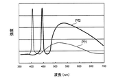
図6は出射光の分光特性を示すグラフである。波長変換部材65は、レーザ光源LD1から出射される青色レーザ光の一部を吸収して緑色〜黄色に励起発光する複数種の蛍光体(例えばYAG系蛍光体、あるいはBAM(BaMgAl10O17)等を含む蛍光体等)を含んで構成される。これら波長変換部材65により、図6に出射光の分光特性を示すように、レーザ光源LD1からの青色レーザ光と、この青色レーザ光が波長変換された緑色〜黄色の励起光とが合成されて、プロファイルPf2で示される白色光が生成される。
FIG. 6 is a graph showing the spectral characteristics of the emitted light. The
レーザ光源LD2−Aから出射される紫色レーザ光は、波長変換部材65からの波長変換光が僅かとなり、図6にプロファイルPf1で示すように、その殆どが中心波長405nmの狭帯域光として出射される。つまり、レーザ光源LD2−Aは、白色光とは異なるスペクトルの狭帯域波長光を出力する。また、LED2−Bも同様に中心波長405nmの狭帯域光として出射される。
The violet laser light emitted from the laser light source LD2-A has a small amount of wavelength-converted light from the
これにより、特定の機能を有する波長光を選択的に照射することができ、観察目的に応じた適切な波長光により所望の内視鏡観察を行うことができる。なお、狭帯域波長光の中心波長は、380nm〜480nm、好ましくは400nm〜450nmである。これにより、組織表層の毛細血管の微細構造や粘膜組織表層の微細構造模様を強調させて観察できる。 Thereby, the wavelength light which has a specific function can be selectively irradiated, and desired endoscopic observation can be performed with the appropriate wavelength light according to the observation purpose. The center wavelength of the narrow-band wavelength light is 380 nm to 480 nm, preferably 400 nm to 450 nm. Thereby, the fine structure of the capillary blood vessel on the tissue surface layer and the fine structure pattern of the mucosal tissue surface layer can be emphasized and observed.
各レーザ光源LD1、レーザ光源LD2−A、レーザ光源LD2−Bから出力されるレーザ光は、第1光ファイバ59又は第2光ファイバ63により内視鏡先端部41へ導光される。導光されたレーザ光は、第1照明窓45、又は第1照明窓45と第2照明窓47から、白色光と、紫色の狭帯域光とを任意の混合比率で選択的に出射させることができる。出射光の光強度や出射光の色味は、内視鏡制御部127が光源制御部75に所望の制御信号を出力して行う。
Laser light output from each laser light source LD1, laser light source LD2-A, and laser light source LD2-B is guided to the endoscope
この制御信号に基づいて光源制御部75が、レーザ光源LD1、レーザ光源LD2−A、レーザ光源LD2−Bを駆動する駆動信号を出力する。レーザ光源LD1、レーザ光源LD2−A、レーザ光源LD2−Bは、それぞれ入力された駆動信号に基づく光量で光出射することで調整される。つまり、光源制御部75は、設定された目標光量に応じた駆動信号をそれぞれ生成して、第1の光源69及び第2の光源71をそれぞれ駆動する。
Based on this control signal, the
波長変換部材65に、蛍光体を用いることで、任意の波長の光を容易に生成できる。また、第1の光源69からの出力光と、波長変換部材65からの蛍光とが合成されて白色光を形成するので、ブロードな波長スペクトルを有する蛍光体の蛍光を用いて白色光が生成される。これにより、照明光の演色性を向上できる。
By using a phosphor for the
ここで、本明細書でいう白色光とは、厳密に可視光の全ての波長成分を含むものに限らない。例えば、基準色であるR(赤),G(緑),B(青)等、特定の波長帯の光を含むものであればよく、緑色から赤色にかけての波長成分を含む光や、青色から緑色にかけての波長成分を含む光等も広義に含むものとする。 Here, the white light referred to in the present specification is not limited to one that strictly includes all wavelength components of visible light. For example, it is sufficient if it includes light of a specific wavelength band such as R (red), G (green), B (blue) which are reference colors, and light including wavelength components from green to red or from blue. Light including a wavelength component over green is also broadly included.
内視鏡先端部41の波長変換部材65の近傍には、波長変換部材65と光拡散部材67とからの発熱による温度を検出する1つの温度センサ129が設けられている。温度センサ129は、挿入部25の先端における波長変換部材65と光拡散部材67との間に配置された単一のセンサである。温度センサ129は、単一のセンサとなることで、簡素な構成で光伝送損失を検出できる。波長変換部材65、光拡散部材67のそれぞれに温度センサ129を設ける構成に比べ、内視鏡11の細径化が有利となる。
In the vicinity of the
温度センサ129は、レーザ光の照射による波長変換の際に発生する波長変換部材65の熱、レーザ光が透過する際の吸収によって発生する光拡散部材67の熱による温度を検出する。温度センサ129は、この検出した温度を温度検出値として温度検出信号線131を通じて内視鏡制御部127に出力する。
The
温度センサ129としては、サーミスタ、熱電対、測温抵抗体が使用可能である。また、温度センサ129は、波長変換部材65と光拡散部材67の双方にできるだけ近い位置に設けることが好ましく、波長変換部材65と光拡散部材67から互いに等しい熱伝導係数となる位置に設けることが望ましい。
As the
図1に示すように、プロセッサ21は、内視鏡制御部127と、映像信号を生成する撮像信号処理部55と、撮像信号や各種情報を保存するRAM等の記憶手段である記憶部135と、画像処理部137とを備えている。内視鏡制御部127は、撮像信号処理部55から出力される観察画像の画像データに対して、画像処理部137により適宜な画像処理を施して表示部15に映出させる。また、光源装置19の光源制御部75に制御信号を出力して、第1照明窓45、第2照明窓47から所望の光量の照明光を出射させる。この内視鏡制御部127は、図示しないLAN等のネットワークに接続されて、画像データを含む情報を配信する等、内視鏡装置100全体を制御する。
As shown in FIG. 1, the
上記構成の内視鏡装置100は、内視鏡制御部127により、内視鏡観察を行うための通常制御モードと、第1光ファイバ59、第2光ファイバ63の断線等で生じる光伝送損失を確認するための光伝送損失確認制御モードとに切り替え可能に構成されている。
In the
通常制御モードでは、レーザ光源LD1による白色照明、レーザ光源LD1とLD2−Aの同時点灯、又はレーザ光源LD1とLD2−Bによる同時点灯や個別点灯により、所望の照明光を所定光量で照射して観察画像を取得し、表示させる制御を行う。 In the normal control mode, desired illumination light is irradiated with a predetermined amount of light by white illumination by the laser light source LD1, simultaneous lighting of the laser light sources LD1 and LD2-A, simultaneous lighting or individual lighting by the laser light sources LD1 and LD2-B. Control to acquire and display an observation image.
一方、光伝送損失確認制御モードでは、レーザ光源LD1、レーザ光源LD2−A、レーザ光源LD2−Bから第1照明窓45、第2照明窓47までの間の光伝送損失を検出するための制御を行う。なお、この光伝送損失の検出には、ライトガイドコネクタ29、光源装置内の光路、及びコンバイナ77の光伝送損失も含まれる。即ち、以下に記す断線の検出とは、上記の各光伝送損失である可能性も含んでいる。
On the other hand, in the optical transmission loss confirmation control mode, control for detecting optical transmission loss between the laser light source LD1, the laser light source LD2-A, and the laser light source LD2-B to the
以下、光伝送損失確認制御モードについて説明する。
図7は光伝送損失確認制御モードを実施する手順のフローチャートである。
光伝送損失確認制御モードを実施するタイミングとしては、制御装置13の電源スイッチをオンにしたタイミング、術者が内視鏡11の本体操作部23に設けられた光伝送損失確認ボタン33を押下したタイミング、入力部17からの指示があったタイミング等、任意に設定できる。
Hereinafter, the optical transmission loss confirmation control mode will be described.
FIG. 7 is a flowchart of a procedure for implementing the optical transmission loss confirmation control mode.
The timing for executing the optical transmission loss confirmation control mode is the timing when the power switch of the
光伝送損失確認制御モードがスタートすると、内視鏡制御部127(図1参照)は、撮像素子49から出力される撮像信号の輝度情報に基づき、照明光の光量を適正化する露光制御を行う。この露光制御により、内視鏡制御部127はレーザ光源LD1、レーザ光源LD2−A、レーザ光源LD2−Bの目標光量を設定し(St1)、この目標光量にする制御信号を光源制御部75に出力する。光源制御部75は、入力された制御信号に基づいてレーザ光源LD1、レーザ光源LD2−A、レーザ光源LD2−Bの出力光強度を目標光量に制御する(St2)。
When the optical transmission loss confirmation control mode starts, the endoscope control unit 127 (see FIG. 1) performs exposure control that optimizes the amount of illumination light based on the luminance information of the imaging signal output from the
目標光量で駆動されるレーザ光源LD1、レーザ光源LD2−Aからの出力光は第1光ファイバ59に導入されて波長変換部材65に照射される。また、レーザ光源LD2−Bからの出力光は第2光ファイバ63に導入されて光拡散部材67に照射される。すると、波長変換部材65は、照射された青色レーザ光を波長変換すると共に発熱する。また、紫色レーザ光の一部が波長変換されることでも発熱する。光拡散部材67は、照射された紫色レーザ光を透過すると共にその一部を吸収して発熱する。この発熱は内視鏡先端部41で伝播され、内視鏡先端部41に収容された各部材を昇温させる。この温度変化を温度センサ129で検出する(St3)。
Output light from the laser light source LD1 and the laser light source LD2-A driven with the target light quantity is introduced into the first
このときの温度センサ129による温度検出値の時間変化の様子を図8に示した。
波長変換部材65と光拡散部材67は、第1光ファイバ59と第2光ファイバ63を介した光照射によって自身の温度が上昇するので、2つの発熱体とみなせる。その温度上昇の傾きは波長変換部材65の方が大きい。これら2つの発熱体の温度上昇傾向は、近傍の一つの温度センサ129によって検知される。温度センサ129が検知する温度上昇の傾きを、第1の温度変化率、及び第2の温度変化率と比較することで、第1光ファイバ59、第2光ファイバ63のいずれかに発生した光伝送損失が検出可能となる。
FIG. 8 shows how the temperature detection value by the
The
より具体的には、波長変換部材65は、波長445nmの励起光によって500乃至600nmの波長を蛍光発光する。これを第1の温度変化率による第1発熱特性Sq1とする。光拡散部材67は、波長405nmの励起光を特殊観察光として生体へ照射するために所定の照射角度を形成するためのものである。これを第2の温度変化率による第2発熱特性Sq2とする。第1発熱特性Sq1は、第2発熱特性Sq2よりも発熱特性の変化率(傾き)が大きい。つまり、発熱による温度変化が大きい。互いに同じ励起光が波長変換部材65と光拡散部材67に導光された場合には、第1発熱特性Sq1と第2発熱特性Sq2との変化率を合算した合算発熱特性Sqtの変化率で温度上昇する。
More specifically, the
ところが、第1光ファイバ59、第2光ファイバ63のいずれかが断線した場合、温度上昇の変化率は、第1発熱特性Sq1の傾き又は第2発熱特性Sq2の傾きに近似したものとなる。断線の発生直後では近似の値にならないが、時間の経過に伴ってSq1,Sq2の傾きに近い値となる。ここで、第1発熱特性Sq1の傾きは、ΔdT1/Δdt1で表される第1時間変化率となる。また、第2発熱特性Sq2の傾きは、ΔdT2/Δdt2で表される第2時間変化率となる。従って、合算発熱特性Sqtが、第1発熱特性Sq1又は第2発熱特性Sq2のいずれかになったことを検知すれば、第1光ファイバ59又は第2光ファイバ63のいずれかに発生した光伝送損失(断線)を、一つの温度センサ129を用いて判断できる。
However, when either the first
つまり、温度センサ129により検出した検出値時間変化率が第1時間変化率と一致する場合は、内視鏡制御部127が第2光ファイバ63に光伝送損失が発生したと判定する。検出値時間変化率が第2時間変化率と一致する場合は、内視鏡制御部127が第1光ファイバ59に光伝送損失が発生したと判定する。
That is, when the detected value time change rate detected by the
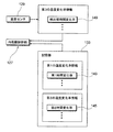
ここで、上記各情報の制御内容を図9にブロック図で示した。記憶部135は、第1の温度変化率情報である第1時間変化率143と、第2の温度変化率情報である第2時間変化率145とをデータとして格納している。また、温度センサ129は、第3の温度変化率情報である温度測定値の検出値時間変化率149を内視鏡制御部127に出力する。
Here, the control content of each information is shown in a block diagram in FIG. The
内視鏡制御部127は、温度センサ129が出力した検出値時間変化率149と、記憶部135に記憶されている第1時間変化率143及び第2時間変化率145とを比較し、第1光ファイバ59、第2光ファイバ63(図1参照)のいずれかに光伝送損失が生じたかを判断する。検出値時間変化率149は逐次変化するので、内視鏡制御部127は、所定の一定時間毎に上記比較・判定を繰り返し行う。
The
なお、上記例では時間に対する温度の変化率としているが、時間に限らず、例えば温度センサ129の温度検出タイミング毎の温度変化率や、光量指示値の変更が生じたタイミング毎の温度変化率としてもよい。
In the above example, the rate of change of temperature with respect to time is used. However, not limited to time, for example, the rate of change of temperature for each temperature detection timing of the
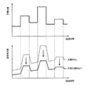
図10に観察対象に応じて逐次変化するレーザ光源の目標光量と、目標光量の変化に応じて温度センサ129からの温度検出値が変化する様子を示した。目標光量は撮像素子49から出力される撮像信号の輝度情報に応じて増減制御される。この目標光量に応じてレーザ光源を駆動する。温度センサ129からの温度検出値は、第1光ファイバ59、第2光ファイバ63に断線が生じていない場合、図中点線で示すように目標光量に対して応答遅れを有して増減する。ところが、第1光ファイバ59、第2光ファイバ63のいずれかに断線が生じた場合、図中実線で示すように温度検出値は断線が生じていない場合よりも低下する。また、応答性も遅れ、昇温の時間変化率も低下する。
FIG. 10 shows a state in which the target light amount of the laser light source that sequentially changes in accordance with the observation target and the temperature detection value from the
このように、温度検出値の変化は、断線の有無により明らかな差を生じる。このため、任意のタイミングで温度センサ129により測定しても、得られた温度検出値の変化と、予め定めた基準値(断線のない場合の解析値)とを比較することにより、光ファイバに生じる断線を随時正確に検出できる。
Thus, the change in the detected temperature value has a clear difference depending on the presence or absence of disconnection. For this reason, even if it is measured by the
断線等の光伝送損失がない場合の波長変換部材65の温度の時間変化特性は、レーザ光源LD1、レーザ光源LD2−A、レーザ光源LD2−Bの種類によって解析的に求めることができる。また、光源の出力強度、及び波長変換部材65と光拡散部材67との性状や種類に応じた吸収・発光特性等の条件によって解析的に求めることができる。
The time change characteristics of the temperature of the
そこで、本構成においては、波長変換部材65と光拡散部材67の温度の時間変化特性を解析的に求め、温度変化情報として記憶部135に予め記憶させておく。また、温度の時間変化率を正常時の基準値として記憶部135に記憶させておく。記憶させる各温度情報は、波長変換部材65及び光拡散部材自体の温度であってもよく、温度センサ129の位置での温度であってもよい。波長変換部材65及び光拡散部材67の温度である場合は、温度センサ129の温度検出値と比較する際に、必要に応じて適宜な補正処理を行えばよい。
Therefore, in this configuration, the time change characteristics of the temperatures of the
再び図7に戻り、内視鏡制御部127は、温度センサ129が測定した温度検出値の時間変化率を求め、求めた時間変化率を記憶部135に記憶された基準値と比較する(St4)。比較の結果、測定により求めた温度変化率が基準値より小さい場合、第1光ファイバ59又は第2光ファイバ63のいずれかに断線が生じたと判定する(St5)。また、基準値と同等である場合は正常と判定して光伝送損失確認制御モードを終了する。
Returning to FIG. 7 again, the
内視鏡制御部127は、断線が生じたと判定した場合に、光源制御部75へレーザ光源を消灯させる制御信号を出力し、断線が生じた側のレーザ光源を消灯させる(St6)。これにより、通常の露光制御のまま、レーザ光源が無駄に光量制御されることを防止する。
When it is determined that the disconnection has occurred, the
即ち、通常の露光制御では、撮像素子49から出力される撮像信号の輝度情報を適正輝度レベルと比較し、その過不足に応じてレーザ光源の出力が増減制御されている。断線が生じた場合は第1照明窓45、又は第2照明窓47からの照明光が不足し、撮像信号の輝度が低下する。そこで、この輝度の低下分を適正レベルにするため、レーザ光源の目標光量が増加制御される。しかし、断線が生じた場合は目標光量を増加しても照明光の不足した状態が続き、更にレーザ光源の目標光量を増加させるといった不正なロジックとなってしまう。そのため、断線検出時には、断線が生じた側のレーザ光源を消灯させることで、無駄な露光制御を防止できる。
That is, in normal exposure control, the luminance information of the image signal output from the
次に、内視鏡制御部127は、断線が発生した旨を表示部15にメッセージを表示する等して術者に通知する(St7)。表示部15への表示以外にも、例えば、アラーム音を発生させる報知や、本体操作部23や制御装置13等に設けたランプの点灯により報知を行ってもよい。
Next, the
以上説明したように、内視鏡装置100は、内視鏡制御部127が、駆動信号の立ち上がりタイミングで光伝送損失を検出する。駆動信号の立ち上がりタイミングで、温度測定値による第3の温度変化率が、第1の温度変化率及び第2の温度変化率と比較される。これにより、波長変換部材65と光拡散部材67との発熱特性を利用した、精度の高い判定が可能となる。また、内視鏡制御部127は、内視鏡11に設けた光伝送損失確認ボタン33の押下信号を受けたタイミングで光伝送損失の検出を開始してもよい。その場合、ボタンを押下した後の駆動信号の立ち上がりタイミングで光伝送損失を検出するため、任意のタイミングで光伝送損失の検出が可能となる。
As described above, in the
また、本構成では、単線の光ファイバを用いるため、光ファイバに断線が生じた場合には、照明光量が大きく低下して、光伝送損失を簡単かつ確実に検出できる。このため、内視鏡11の術者は、内視鏡検査前に光伝送損失の検査が実施し易くなる。
In addition, in this configuration, since a single optical fiber is used, when the optical fiber is disconnected, the amount of illumination light is greatly reduced, and the optical transmission loss can be detected easily and reliably. For this reason, it becomes easy for the operator of the
次に、上記内視鏡装置100の他の構成例を説明する。
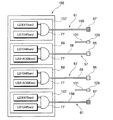
図11は第1の光源69及び第3の光源157が二対設けられた変形例に係る内視鏡先端部41の正面図、図12は図11に示す変形例に係る光源装置155の構成図である。
この変形例では、光源装置155は、第1の光源69と、第3の光源157とからなる一対の光源を二対備えている。第1の光源69は、前述同様に、レーザ光源LD1と、レーザ光源LD2−Aとを備える。一方、第3の光源157は、レーザ光源LD3と、レーザ光源LD1とを備える。レーザ光源LD3は、中心波長473nmの青色発光の半導体レーザである。レーザ光源LD3と、レーザ光源LD1からの出射光はコンバイナ77によって合波される。第3の光源157からの合波された出射光は、光出射端に光拡散部材67を配置した第2の導光部材61である第3光ファイバ159に導入される。
Next, another configuration example of the
FIG. 11 is a front view of an endoscope
In this modification, the
第1光ファイバ59に導光された照明光は第1照明窓45から出射される。第3光ファイバ159に導光された照明光は第3照明窓161から出射される。第1照明窓45,45は観察窓43を挟んだ直線165上に配置され、第3照明窓161,161は観察窓43を挟んだ直線167上に配置される。これにより、観察窓43の両脇から白色光、狭帯域光を均等に照射して、照明ムラの発生を防止している。
The illumination light guided to the first
この変形例による構成では、第3の光源157を備えることで以下の照明光による観察が可能となる。
図13に酸化ヘモグロビンと還元ヘモグロビンに対する吸光度の分光特性を示した。同図に示すように、波長405nm付近では双方の吸光度は等しく、波長445nm付近では還元ヘモグロビンが酸化ヘモグロビンよりも吸光度が高く、波長473nm付近では酸化ヘモグロビンが還元ヘモグロビンよりも吸光度が高くなっている。また、レーザ光の粘膜組織表層からの深達度は、レーザ光の波長が短い程浅くなる特性を有するので、波長が405nm、445nm、473nmの順で深くなる。
In the configuration according to this modification, observation with the following illumination light is possible by providing the third
FIG. 13 shows the spectral characteristics of absorbance with respect to oxyhemoglobin and deoxyhemoglobin. As shown in the figure, both absorbances are equal in the vicinity of a wavelength of 405 nm, reduced hemoglobin has higher absorbance than oxyhemoglobin in the vicinity of wavelength 445 nm, and oxyhemoglobin has a higher absorbance than reduced hemoglobin in the vicinity of wavelength 473 nm. Further, the depth of penetration of the laser beam from the surface of the mucosal tissue has such a characteristic that it becomes shallower as the wavelength of the laser beam becomes shorter, so that the wavelengths become deeper in the order of 405 nm, 445 nm, and 473 nm.
これらの特性を利用して、次のように観察領域の酸素飽和度と、観察領域に映出された血管深さとを求める。
(1)還元ヘモグロビンの吸光度が高い中心波長445nmのレーザ光を照射したときの、このレーザ光の戻り光成分を検出した撮像画像輝度値S1を求める。
(2)酸化ヘモグロビンの吸光度が高い中心波長473nmのレーザ光を照射したときの、このレーザ光の戻り光成分を検出した撮像画像輝度値S2を求める。
(3)吸光度の等しい中心波長405nmを照射したときの、このレーザ光の戻り光成分を検出した撮像画像輝度値を求める。
(4)S1,S2の値をそれぞれS3の値で標準化する。即ち、S1/S3、S2/S3の値を求める。
Using these characteristics, the oxygen saturation of the observation region and the blood vessel depth projected in the observation region are obtained as follows.
(1) A captured image luminance value S1 obtained by detecting a return light component of the laser beam when irradiated with a laser beam having a central wavelength of 445 nm where the absorbance of reduced hemoglobin is high is obtained.
(2) A captured image luminance value S2 obtained by detecting a return light component of the laser beam when the laser beam having a central wavelength of 473 nm with high absorbance of oxyhemoglobin is irradiated is obtained.
(3) The brightness value of the picked-up image obtained by detecting the return light component of the laser light when the central wavelength 405 nm having the same absorbance is irradiated is obtained.
(4) The values of S1 and S2 are standardized with the value of S3, respectively. That is, the values of S1 / S3 and S2 / S3 are obtained.
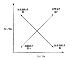
(5)図14に示すように、S1/S3の値と、S2/S3の値の大小を直交二軸で表した2次元マップを生成し、この2次元マップ上に、上記で求めたS1/S3、S2/S3の値をプロットする。2次元マップ上では、S1/S3の値が大きい程、酸素飽和度が高く、血管深さが浅いものとなり、S1/S3の値が小さい程、酸素飽和度が低く、血管深さが深くなる。また、S2/S3の値が大きい程、酸素飽和度が低く、血管深さが浅くなり、S2/S3の値が小さい程、酸素飽和度が高く、血管深さが深いものとなる。これらの関係により、観察領域における酸素飽和度の高低、血管深さの情報が求められる。 (5) As shown in FIG. 14, a two-dimensional map in which the values of S1 / S3 and the magnitude of the values of S2 / S3 are represented by two orthogonal axes is generated, and the S1 obtained above is generated on this two-dimensional map. The values of / S3 and S2 / S3 are plotted. On the two-dimensional map, the greater the value of S1 / S3, the higher the oxygen saturation and the shallower the blood vessel depth, and the smaller the value of S1 / S3, the lower the oxygen saturation and the deeper the blood vessel depth. . Also, the greater the value of S2 / S3, the lower the oxygen saturation and the shallower the blood vessel depth, and the smaller the value of S2 / S3, the higher the oxygen saturation and the deeper the blood vessel depth. Based on these relationships, information on the level of oxygen saturation and blood vessel depth in the observation region is required.
本発明は上記の実施形態に限定されるものではなく、明細書の記載、並びに周知の技術に基づいて、当業者が変更、応用することも本発明の予定するところであり、保護を求める範囲に含まれる。例えば、上記の構成では第1の導光部材57、第2の導光部材61を単線の光ファイバとしたが、第1の導光部材57、第2の導光部材61はこれに限らず、多数本の光ファイバを束ねた光ファイババンドルであっても同様に光伝送損失を検出できる。
The present invention is not limited to the above-described embodiments, and those skilled in the art can change or apply the present invention based on the description of the specification and well-known techniques. included. For example, in the above configuration, the first
以上の通り、本明細書には次の事項が開示されている。
(1) 光伝送損失を検出する機能を備えた内視鏡装置であって、
第1の光源と、
前記第1の光源の出力光を導入して被検体内に挿入される挿入部の先端まで導光する第1の導光部材と、
前記第1の導光部材の光出射端に配置された波長変換部材と、
第2の光源と、
前記第2の光源の出力光を導入して前記挿入部の先端まで導光する第2の導光部材と、
前記第2の導光部材の光出射端に配置された光拡散部材と、
前記第1の光源及び前記第2の光源それぞれについて設定された目標光量に応じた駆動信号を生成し、前記第1の光源及び前記第2の光源を駆動する光源駆動手段と、
前記波長変換部材及び前記光拡散部材からの発熱による温度を検出する温度センサと、
前記第1の光源の駆動信号の強度に対応した前記波長変換部材の発熱による第1の温度変化率、及び前記第2の光源の駆動信号の強度に対応した前記光拡散部材の発熱による第2の温度変化率を記憶する記憶手段と、
前記温度センサにより検出された温度の第3の温度変化率を、前記第1の温度変化率、及び前記第2の温度変化率と比較し、前記第3の温度変化率が前記第1の温度変化率と一致する場合は前記第2の導光部材に光伝送損失が発生したと判定し、前記第3の温度変化率が前記第2の温度変化率と一致する場合は前記第1の導光部材に光伝送損失が発生したと判定する光伝送損失検出手段と、
を備えた内視鏡装置。
この内視鏡装置によれば、波長変換部材と光拡散部材は、第1の導光部材と第2の導光部材を介した光照射によって自身の温度が上昇するので、2つの発熱体とみなせる。その温度上昇の傾きは波長変換部材の方が大きい。これら2つの発熱体の温度上昇傾向は、近傍の一つの温度センサによって検知される。温度センサが検知する温度上昇の傾きを、第1の温度変化率、及び第2の温度変化率と比較することで、第1の導光部材、第2の導光部材のいずれかに発生した光伝送損失が判断可能となる。
As described above, the following items are disclosed in this specification.
(1) An endoscope apparatus having a function of detecting optical transmission loss,
A first light source;
A first light guide member that introduces output light of the first light source and guides it to a distal end of an insertion portion that is inserted into a subject;
A wavelength conversion member disposed at a light exit end of the first light guide member;
A second light source;
A second light guide member for introducing the output light of the second light source and guiding the light to the tip of the insertion portion;
A light diffusing member disposed at a light exit end of the second light guide member;
Light source driving means for generating a drive signal corresponding to a target light amount set for each of the first light source and the second light source, and driving the first light source and the second light source;
A temperature sensor for detecting a temperature due to heat generated from the wavelength conversion member and the light diffusion member;
The first temperature change rate due to heat generation of the wavelength conversion member corresponding to the intensity of the drive signal of the first light source, and the second due to heat generation of the light diffusion member corresponding to the intensity of the drive signal of the second light source. Storage means for storing the temperature change rate of
A third temperature change rate of the temperature detected by the temperature sensor is compared with the first temperature change rate and the second temperature change rate, and the third temperature change rate is the first temperature. When the rate of change coincides with the second light guide member, it is determined that a light transmission loss has occurred. When the rate of change of the third temperature coincides with the rate of change of the second temperature, the first light guide member is determined. An optical transmission loss detecting means for determining that an optical transmission loss has occurred in the optical member;
An endoscopic apparatus comprising:
According to this endoscope apparatus, since the temperature of the wavelength conversion member and the light diffusing member rises due to light irradiation through the first light guide member and the second light guide member, It can be considered. The inclination of the temperature rise is larger in the wavelength conversion member. The temperature rising tendency of these two heating elements is detected by one temperature sensor in the vicinity. The slope of the temperature rise detected by the temperature sensor is compared with the first temperature change rate and the second temperature change rate, and is generated in either the first light guide member or the second light guide member. Optical transmission loss can be determined.
(2) (1)の内視鏡装置であって、
前記第1の温度変化率、前記第2の温度変化率、及び前記第3の温度変化率が、時間に対する温度の変化率を表すものである内視鏡装置。
この内視鏡装置によれば、時間に対する温度の変化率とすることで、確実かつ迅速に断線による変化を検出できる。
(2) The endoscope apparatus according to (1),
The endoscope apparatus in which the first temperature change rate, the second temperature change rate, and the third temperature change rate represent a temperature change rate with respect to time.
According to this endoscope apparatus, a change due to disconnection can be detected reliably and quickly by using the rate of change of temperature with respect to time.
(3) (1)又は(2)の内視鏡装置であって、
前記温度センサが、前記挿入部の先端における前記波長変換部材と前記光拡散部材との間に配置された単一のセンサである内視鏡装置。
この内視鏡装置によれば、簡素な構成で光伝送損失を検出でき、波長変換部材、光拡散部材のそれぞれに温度センサを設ける構成に比べ、内視鏡の細径化が実現する。
(3) The endoscope apparatus according to (1) or (2),
An endoscope apparatus, wherein the temperature sensor is a single sensor disposed between the wavelength conversion member and the light diffusing member at a distal end of the insertion portion.
According to this endoscope apparatus, light transmission loss can be detected with a simple configuration, and the diameter of the endoscope can be reduced as compared with a configuration in which a temperature sensor is provided for each of the wavelength conversion member and the light diffusion member.
(4) (1)〜(3)のいずれか1つの内視鏡装置であって、
前記第1の光源及び前記第2の光源がレーザ光源であり、前記第1の導光部材及び前記第2の導光部材がそれぞれ単線の光ファイバである内視鏡装置。
この内視鏡装置によれば、断線が生じた場合に照明光量が大きく低下する単線の光ファイバを使用することで、光伝送損失を簡単かつ確実に検出できる。このため、内視鏡の術者は、内視鏡検査前に光伝送損失の検査が実施し易くなる。
(4) The endoscope apparatus according to any one of (1) to (3),
An endoscope apparatus in which the first light source and the second light source are laser light sources, and each of the first light guide member and the second light guide member is a single optical fiber.
According to this endoscope apparatus, the optical transmission loss can be detected easily and reliably by using a single-line optical fiber that greatly reduces the amount of illumination light when a disconnection occurs. For this reason, it becomes easy for an endoscopic operator to perform an inspection of optical transmission loss before the endoscopic examination.
(5) (1)〜(4)のいずれか1つの内視鏡装置であって、
前記波長変換部材が前記第1の光源からの出力光で励起されて蛍光を発する蛍光体である内視鏡装置。
この内視鏡装置によれば、蛍光体を用いることで任意の波長の光を容易に検出できる。
(5) The endoscope apparatus according to any one of (1) to (4),
An endoscope apparatus, wherein the wavelength conversion member is a phosphor that emits fluorescence when excited by output light from the first light source.
According to this endoscope apparatus, light of an arbitrary wavelength can be easily detected by using a phosphor.
(6) (5)の内視鏡装置であって、
前記第1の光源からの出力光と、前記蛍光体からの蛍光とが合成されて白色光とされる内視鏡装置。
この内視鏡装置によれば、ブロードな波長スペクトルを有する蛍光体の蛍光を用いて白色光が生成されることで、演色性を向上できる。
(6) The endoscope apparatus according to (5),
An endoscope apparatus in which output light from the first light source and fluorescence from the phosphor are combined into white light.
According to this endoscope apparatus, color rendering can be improved by generating white light using fluorescence of a phosphor having a broad wavelength spectrum.
(7) (6)の内視鏡装置であって、
前記第2の光源が白色光とは異なるスペクトルの狭帯域波長光を出力する光源である内視鏡装置。
この内視鏡装置によれば、特定の機能を有する波長光を選択的に照射することができ、観察目的に応じた適切な波長光により所望の内視鏡観察を行うことができる。
(7) The endoscope apparatus according to (6),
An endoscope apparatus in which the second light source is a light source that outputs narrow-band wavelength light having a spectrum different from that of white light.
According to this endoscope apparatus, wavelength light having a specific function can be selectively irradiated, and desired endoscopic observation can be performed with light having an appropriate wavelength according to the observation purpose.
(8) (7)の内視鏡装置であって、
前記狭帯域波長光の中心波長が、380nm〜480nmである内視鏡装置。
この内視鏡装置によれば、組織表層の毛細血管の微細構造や粘膜組織表層の微細構造模様を強調させて観察できる。
(8) The endoscope apparatus according to (7),
An endoscope apparatus in which a center wavelength of the narrow-band wavelength light is 380 nm to 480 nm.
According to this endoscope apparatus, the fine structure of the capillary blood vessel on the tissue surface layer and the fine structure pattern of the mucosal tissue surface layer can be emphasized and observed.
(9) (1)〜(8)のいずれか1つの内視鏡装置であって、
前記光伝送損失検出手段が、前記駆動信号の立ち上がりタイミングで前記光伝送損失を判断する内視鏡装置。
この内視鏡装置によれば、駆動信号の立ち上がりタイミングで、温度測定値による第3の温度変化率が、第1の温度変化率及び第2の温度変化率と比較される。駆動信号の立ち下がりタイミングに比べ、波長変換部材と光拡散部材との発熱特性を利用でき、精度の高い判定が可能となる。
(9) The endoscope apparatus according to any one of (1) to (8),
An endoscope apparatus in which the optical transmission loss detection means determines the optical transmission loss at a rising timing of the drive signal.
According to this endoscope apparatus, the third temperature change rate based on the temperature measurement value is compared with the first temperature change rate and the second temperature change rate at the rising timing of the drive signal. Compared with the falling timing of the drive signal, the heat generation characteristics of the wavelength conversion member and the light diffusing member can be used, and the determination can be made with high accuracy.
(10) (9)の内視鏡装置であって、
前記光伝送損失検出手段が、前記内視鏡に設けた光伝送損失確認ボタンの押下信号を受けたタイミングで始動する内視鏡装置。
この内視鏡装置によれば、ボタン押下した後の駆動信号の立ち上がりタイミングで光伝送損失を検出するため、任意のタイミングで光伝送損失の検出が可能となる。
(10) The endoscope apparatus according to (9),
An endoscope apparatus that starts at a timing when the light transmission loss detecting means receives a press signal of a light transmission loss confirmation button provided on the endoscope.
According to this endoscope apparatus, since the optical transmission loss is detected at the rising timing of the drive signal after the button is pressed, the optical transmission loss can be detected at an arbitrary timing.
11 内視鏡
25 挿入部
33 光伝送損失確認ボタン
57 第1の導光部材
59 第1光ファイバ
61 第2の導光部材
63 第2光ファイバ
65 波長変換部材
67 光拡散部材
69 第1の光源
71 第2の光源
77 光源制御部(光源駆動手段)
127 内視鏡制御部(光伝送損失検出手段)
129 温度センサ
135 記憶部(記憶手段)
143 第1時間変化率(第1の温度変化率)
145 第2時間変化率(第2の温度変化率)
149 検出値時間変化率(第3の温度変化率)
100 内視鏡装置
LD1、LD2−A、LD2−B レーザ光源
DESCRIPTION OF
127 Endoscope control unit (light transmission loss detection means)
129
143 First time change rate (first temperature change rate)
145 Second time change rate (second temperature change rate)
149 Detection value time change rate (third temperature change rate)
100 Endoscopic devices LD1, LD2-A, LD2-B Laser light source
Claims (10)
第1の光源と、
前記第1の光源の出力光を導入して被検体内に挿入される挿入部の先端まで導光する第1の導光部材と、
前記第1の導光部材の光出射端に配置された波長変換部材と、
第2の光源と、
前記第2の光源の出力光を導入して前記挿入部の先端まで導光する第2の導光部材と、
前記第2の導光部材の光出射端に配置された光拡散部材と、
前記第1の光源及び前記第2の光源それぞれについて設定された目標光量に応じた駆動信号を生成し、前記第1の光源及び前記第2の光源を駆動する光源駆動手段と、
前記波長変換部材及び前記光拡散部材からの発熱による温度を検出する温度センサと、
前記第1の光源の駆動信号の強度に対応した前記波長変換部材の発熱による第1の温度変化率、及び前記第2の光源の駆動信号の強度に対応した前記光拡散部材の発熱による第2の温度変化率を記憶する記憶手段と、
前記温度センサにより検出された温度の第3の温度変化率を、前記第1の温度変化率、及び前記第2の温度変化率と比較し、前記第3の温度変化率が前記第1の温度変化率と一致する場合は前記第2の導光部材に光伝送損失が発生したと判定し、前記第3の温度変化率が前記第2の温度変化率と一致する場合は前記第1の導光部材に光伝送損失が発生したと判定する光伝送損失検出手段と、
を備えた内視鏡装置。 An endoscope apparatus having a function of detecting optical transmission loss,
A first light source;
A first light guide member that introduces output light of the first light source and guides it to a distal end of an insertion portion that is inserted into a subject;
A wavelength conversion member disposed at a light exit end of the first light guide member;
A second light source;
A second light guide member for introducing the output light of the second light source and guiding the light to the tip of the insertion portion;
A light diffusing member disposed at a light exit end of the second light guide member;
Light source driving means for generating a drive signal corresponding to a target light amount set for each of the first light source and the second light source, and driving the first light source and the second light source;
A temperature sensor for detecting a temperature due to heat generated from the wavelength conversion member and the light diffusion member;
The first temperature change rate due to heat generation of the wavelength conversion member corresponding to the intensity of the drive signal of the first light source, and the second due to heat generation of the light diffusion member corresponding to the intensity of the drive signal of the second light source. Storage means for storing the temperature change rate of
A third temperature change rate of the temperature detected by the temperature sensor is compared with the first temperature change rate and the second temperature change rate, and the third temperature change rate is the first temperature. When the rate of change coincides with the second light guide member, it is determined that a light transmission loss has occurred. When the rate of change of the third temperature coincides with the rate of change of the second temperature, the first light guide member is determined. An optical transmission loss detecting means for determining that an optical transmission loss has occurred in the optical member;
An endoscopic apparatus comprising:
前記第1の温度変化率、前記第2の温度変化率、及び前記第3の温度変化率が、時間に対する温度の変化率を表すものである内視鏡装置。 The endoscope apparatus according to claim 1,
The endoscope apparatus in which the first temperature change rate, the second temperature change rate, and the third temperature change rate represent a temperature change rate with respect to time.
前記温度センサが、前記挿入部の先端における前記波長変換部材と前記光拡散部材との間に配置された単一のセンサである内視鏡装置。 The endoscope apparatus according to claim 1 or 2,
An endoscope apparatus, wherein the temperature sensor is a single sensor disposed between the wavelength conversion member and the light diffusing member at a distal end of the insertion portion.
前記第1の光源及び前記第2の光源がレーザ光源であり、前記第1の導光部材及び前記第2の導光部材がそれぞれ単線の光ファイバである内視鏡装置。 The endoscope apparatus according to any one of claims 1 to 3,
An endoscope apparatus in which the first light source and the second light source are laser light sources, and each of the first light guide member and the second light guide member is a single optical fiber.
前記波長変換部材が前記第1の光源からの出力光で励起されて蛍光を発する蛍光体である内視鏡装置。 The endoscope apparatus according to any one of claims 1 to 4,
An endoscope apparatus, wherein the wavelength conversion member is a phosphor that emits fluorescence when excited by output light from the first light source.
前記第1の光源からの出力光と、前記蛍光体からの蛍光とが合成されて白色光とされる内視鏡装置。 An endoscope apparatus according to claim 5, wherein
An endoscope apparatus in which output light from the first light source and fluorescence from the phosphor are combined into white light.
前記第2の光源が白色光とは異なるスペクトルの狭帯域波長光を出力する光源である内視鏡装置。 The endoscope apparatus according to claim 6, wherein
An endoscope apparatus in which the second light source is a light source that outputs narrow-band wavelength light having a spectrum different from that of white light.
前記狭帯域波長光の中心波長が、380nm〜480nmである内視鏡装置。 The endoscope apparatus according to claim 7,
An endoscope apparatus in which a center wavelength of the narrow-band wavelength light is 380 nm to 480 nm.
前記光伝送損失検出手段が、前記駆動信号の立ち上がりタイミングで前記光伝送損失の発生を判定する内視鏡装置。 The endoscope apparatus according to any one of claims 1 to 8,
The optical transmission loss detecting means, the endoscope apparatus to determine the occurrence of the optical transmission loss at the rising edge of the drive signal.
前記光伝送損失検出手段が、前記内視鏡装置に設けた光伝送損失確認ボタンの押下信号を受けたタイミングで前記光伝送損失の発生の判定を開始する内視鏡装置。 The endoscope apparatus according to claim 9, wherein
An endoscope apparatus in which the light transmission loss detection means starts determining the occurrence of the light transmission loss at a timing when a signal for pressing a light transmission loss confirmation button provided in the endoscope apparatus is received.
Priority Applications (1)
| Application Number | Priority Date | Filing Date | Title |
|---|---|---|---|
| JP2011082048A JP5624930B2 (en) | 2011-04-01 | 2011-04-01 | Endoscope device |
Applications Claiming Priority (1)
| Application Number | Priority Date | Filing Date | Title |
|---|---|---|---|
| JP2011082048A JP5624930B2 (en) | 2011-04-01 | 2011-04-01 | Endoscope device |
Publications (3)
| Publication Number | Publication Date |
|---|---|
| JP2012213562A JP2012213562A (en) | 2012-11-08 |
| JP2012213562A5 JP2012213562A5 (en) | 2014-01-23 |
| JP5624930B2 true JP5624930B2 (en) | 2014-11-12 |
Family
ID=47266977
Family Applications (1)
| Application Number | Title | Priority Date | Filing Date |
|---|---|---|---|
| JP2011082048A Expired - Fee Related JP5624930B2 (en) | 2011-04-01 | 2011-04-01 | Endoscope device |
Country Status (1)
| Country | Link |
|---|---|
| JP (1) | JP5624930B2 (en) |
Families Citing this family (5)
| Publication number | Priority date | Publication date | Assignee | Title |
|---|---|---|---|---|
| CN106170735B (en) | 2014-03-18 | 2018-10-30 | 株式会社理光 | Light supply apparatus and image projection apparatus with light supply apparatus |
| WO2017122626A1 (en) * | 2016-01-12 | 2017-07-20 | オリンパス株式会社 | Endoscope device |
| EP3737276A4 (en) * | 2018-01-10 | 2021-10-20 | ChemImage Corporation | Time correlated source modulation for endoscopy |
| CN114206197A (en) * | 2019-08-09 | 2022-03-18 | 奥林巴斯株式会社 | Optical device, wireless endoscope, and endoscope system |
| CN114302666A (en) | 2019-08-27 | 2022-04-08 | 富士胶片株式会社 | Illumination optical system for endoscope |
Family Cites Families (6)
| Publication number | Priority date | Publication date | Assignee | Title |
|---|---|---|---|---|
| JP5103874B2 (en) * | 2006-11-15 | 2012-12-19 | 日亜化学工業株式会社 | Light emitting device |
| JP5159298B2 (en) * | 2007-12-27 | 2013-03-06 | オリンパス株式会社 | Endoscope device |
| JP5117878B2 (en) * | 2008-02-13 | 2013-01-16 | 富士フイルム株式会社 | Endoscope light source device |
| JP2010187903A (en) * | 2009-02-18 | 2010-09-02 | Fujifilm Corp | Endoscope apparatus and control method thereof |
| JP5767775B2 (en) * | 2009-07-06 | 2015-08-19 | 富士フイルム株式会社 | Endoscope device |
| JP5401205B2 (en) * | 2009-08-10 | 2014-01-29 | 富士フイルム株式会社 | Endoscope device |
-
2011
- 2011-04-01 JP JP2011082048A patent/JP5624930B2/en not_active Expired - Fee Related
Also Published As
| Publication number | Publication date |
|---|---|
| JP2012213562A (en) | 2012-11-08 |
Similar Documents
| Publication | Publication Date | Title |
|---|---|---|
| JP7603099B2 (en) | Endoscope System | |
| JP5997676B2 (en) | Endoscope light source device and endoscope system using the same | |
| US9044163B2 (en) | Endoscope apparatus | |
| JP5649747B2 (en) | Endoscope lighting unit and endoscope | |
| JP2014000301A (en) | Light source device and endoscope system | |
| US20120010465A1 (en) | Endoscope apparatus | |
| JP5624930B2 (en) | Endoscope device | |
| JP5997630B2 (en) | Light source device and endoscope system using the same | |
| JP6438062B2 (en) | Endoscope system | |
| JPWO2017046857A1 (en) | Endoscope device | |
| JP5345597B2 (en) | Temperature control device, temperature control method, light source device, and endoscope diagnosis device | |
| JP6234212B2 (en) | Endoscope light source device and endoscope system using the same | |
| WO2017104048A1 (en) | Endoscope illumination device and endoscope system | |
| JP2014121363A (en) | Light source device and endoscope system using the same | |
| JP2016179236A (en) | Endoscope apparatus | |
| JP2012139435A (en) | Electronic endoscope | |
| JP2017200601A (en) | Endoscope apparatus | |
| JP2014132918A (en) | Light source device and endoscope system using the same | |
| JP2012228443A (en) | Endoscopic device, and method of supporting operation of the same | |
| JP2013042855A (en) | Endoscopic apparatus and light source control method for the same | |
| JP2025026596A (en) | Endoscope System | |
| JP2012223460A (en) | Endoscopic device | |
| JP5455856B2 (en) | Temperature control device, temperature control method, light source device, and endoscope diagnosis device | |
| JP2023096182A (en) | Endoscope apparatus | |
| JP2012152422A (en) | Endoscope apparatus and endoscopic diagnosis apparatus |
Legal Events
| Date | Code | Title | Description |
|---|---|---|---|
| RD03 | Notification of appointment of power of attorney |
Free format text: JAPANESE INTERMEDIATE CODE: A7423 Effective date: 20121005 |
|
| A521 | Request for written amendment filed |
Free format text: JAPANESE INTERMEDIATE CODE: A523 Effective date: 20131202 |
|
| A621 | Written request for application examination |
Free format text: JAPANESE INTERMEDIATE CODE: A621 Effective date: 20131202 |
|
| A977 | Report on retrieval |
Free format text: JAPANESE INTERMEDIATE CODE: A971007 Effective date: 20140716 |
|
| TRDD | Decision of grant or rejection written | ||
| A01 | Written decision to grant a patent or to grant a registration (utility model) |
Free format text: JAPANESE INTERMEDIATE CODE: A01 Effective date: 20140902 |
|
| A61 | First payment of annual fees (during grant procedure) |
Free format text: JAPANESE INTERMEDIATE CODE: A61 Effective date: 20140929 |
|
| R150 | Certificate of patent or registration of utility model |
Ref document number: 5624930 Country of ref document: JP Free format text: JAPANESE INTERMEDIATE CODE: R150 |
|
| R250 | Receipt of annual fees |
Free format text: JAPANESE INTERMEDIATE CODE: R250 |
|
| R250 | Receipt of annual fees |
Free format text: JAPANESE INTERMEDIATE CODE: R250 |
|
| R250 | Receipt of annual fees |
Free format text: JAPANESE INTERMEDIATE CODE: R250 |
|
| R250 | Receipt of annual fees |
Free format text: JAPANESE INTERMEDIATE CODE: R250 |
|
| LAPS | Cancellation because of no payment of annual fees |


