JP5572547B2 - Improved orthopedic implant for use with precision bone surface finishing instruments - Google Patents
Improved orthopedic implant for use with precision bone surface finishing instruments Download PDFInfo
- Publication number
- JP5572547B2 JP5572547B2 JP2010515185A JP2010515185A JP5572547B2 JP 5572547 B2 JP5572547 B2 JP 5572547B2 JP 2010515185 A JP2010515185 A JP 2010515185A JP 2010515185 A JP2010515185 A JP 2010515185A JP 5572547 B2 JP5572547 B2 JP 5572547B2
- Authority
- JP
- Japan
- Prior art keywords
- bone
- fixation system
- bone fixation
- recess
- implant
- Prior art date
- Legal status (The legal status is an assumption and is not a legal conclusion. Google has not performed a legal analysis and makes no representation as to the accuracy of the status listed.)
- Expired - Fee Related
Links
- 210000000988 bone and bone Anatomy 0.000 title claims description 241
- 239000007943 implant Substances 0.000 title description 108
- 230000000399 orthopedic effect Effects 0.000 title description 12
- 239000000463 material Substances 0.000 claims description 23
- 239000012634 fragment Substances 0.000 claims description 19
- 238000002513 implantation Methods 0.000 claims description 15
- 239000000853 adhesive Substances 0.000 claims description 13
- 230000001070 adhesive effect Effects 0.000 claims description 13
- 239000003814 drug Substances 0.000 claims description 4
- 229940079593 drug Drugs 0.000 claims description 4
- 230000001737 promoting effect Effects 0.000 claims description 4
- 230000008467 tissue growth Effects 0.000 claims description 4
- 230000007547 defect Effects 0.000 claims 5
- 210000003625 skull Anatomy 0.000 description 16
- 238000000034 method Methods 0.000 description 14
- 208000010392 Bone Fractures Diseases 0.000 description 10
- 238000003754 machining Methods 0.000 description 9
- 230000008878 coupling Effects 0.000 description 8
- 238000010168 coupling process Methods 0.000 description 8
- 238000005859 coupling reaction Methods 0.000 description 8
- 230000008439 repair process Effects 0.000 description 8
- 238000005520 cutting process Methods 0.000 description 7
- 238000003780 insertion Methods 0.000 description 6
- 230000037431 insertion Effects 0.000 description 6
- 238000003801 milling Methods 0.000 description 6
- 230000004048 modification Effects 0.000 description 6
- 238000012986 modification Methods 0.000 description 6
- 238000004873 anchoring Methods 0.000 description 5
- WYTGDNHDOZPMIW-RCBQFDQVSA-N alstonine Natural products C1=CC2=C3C=CC=CC3=NC2=C2N1C[C@H]1[C@H](C)OC=C(C(=O)OC)[C@H]1C2 WYTGDNHDOZPMIW-RCBQFDQVSA-N 0.000 description 4
- 230000035876 healing Effects 0.000 description 4
- 229910001000 nickel titanium Inorganic materials 0.000 description 4
- HLXZNVUGXRDIFK-UHFFFAOYSA-N nickel titanium Chemical compound [Ti].[Ti].[Ti].[Ti].[Ti].[Ti].[Ti].[Ti].[Ti].[Ti].[Ti].[Ni].[Ni].[Ni].[Ni].[Ni].[Ni].[Ni].[Ni].[Ni].[Ni].[Ni].[Ni].[Ni].[Ni] HLXZNVUGXRDIFK-UHFFFAOYSA-N 0.000 description 4
- 231100001055 skeletal defect Toxicity 0.000 description 4
- 210000001519 tissue Anatomy 0.000 description 4
- 229920001651 Cyanoacrylate Polymers 0.000 description 3
- 239000004593 Epoxy Substances 0.000 description 3
- RTAQQCXQSZGOHL-UHFFFAOYSA-N Titanium Chemical compound [Ti] RTAQQCXQSZGOHL-UHFFFAOYSA-N 0.000 description 3
- -1 acryl Chemical group 0.000 description 3
- 239000000560 biocompatible material Substances 0.000 description 3
- 239000004568 cement Substances 0.000 description 3
- 239000004814 polyurethane Substances 0.000 description 3
- 229920002635 polyurethane Polymers 0.000 description 3
- 229920005989 resin Polymers 0.000 description 3
- 239000011347 resin Substances 0.000 description 3
- 229910052719 titanium Inorganic materials 0.000 description 3
- 239000010936 titanium Substances 0.000 description 3
- MWCLLHOVUTZFKS-UHFFFAOYSA-N Methyl cyanoacrylate Chemical compound COC(=O)C(=C)C#N MWCLLHOVUTZFKS-UHFFFAOYSA-N 0.000 description 2
- 239000004696 Poly ether ether ketone Substances 0.000 description 2
- JUPQTSLXMOCDHR-UHFFFAOYSA-N benzene-1,4-diol;bis(4-fluorophenyl)methanone Chemical compound OC1=CC=C(O)C=C1.C1=CC(F)=CC=C1C(=O)C1=CC=C(F)C=C1 JUPQTSLXMOCDHR-UHFFFAOYSA-N 0.000 description 2
- 230000008468 bone growth Effects 0.000 description 2
- 229910000389 calcium phosphate Inorganic materials 0.000 description 2
- 239000001506 calcium phosphate Substances 0.000 description 2
- 235000011010 calcium phosphates Nutrition 0.000 description 2
- 230000001054 cortical effect Effects 0.000 description 2
- 229910000701 elgiloys (Co-Cr-Ni Alloy) Inorganic materials 0.000 description 2
- 230000006870 function Effects 0.000 description 2
- 230000004927 fusion Effects 0.000 description 2
- 229910052588 hydroxylapatite Inorganic materials 0.000 description 2
- 239000011344 liquid material Substances 0.000 description 2
- 230000014759 maintenance of location Effects 0.000 description 2
- XYJRXVWERLGGKC-UHFFFAOYSA-D pentacalcium;hydroxide;triphosphate Chemical compound [OH-].[Ca+2].[Ca+2].[Ca+2].[Ca+2].[Ca+2].[O-]P([O-])([O-])=O.[O-]P([O-])([O-])=O.[O-]P([O-])([O-])=O XYJRXVWERLGGKC-UHFFFAOYSA-D 0.000 description 2
- 229920002530 polyetherether ketone Polymers 0.000 description 2
- 229920000642 polymer Polymers 0.000 description 2
- 239000010935 stainless steel Substances 0.000 description 2
- 229910001220 stainless steel Inorganic materials 0.000 description 2
- QORWJWZARLRLPR-UHFFFAOYSA-H tricalcium bis(phosphate) Chemical compound [Ca+2].[Ca+2].[Ca+2].[O-]P([O-])([O-])=O.[O-]P([O-])([O-])=O QORWJWZARLRLPR-UHFFFAOYSA-H 0.000 description 2
- XLYOFNOQVPJJNP-UHFFFAOYSA-N water Substances O XLYOFNOQVPJJNP-UHFFFAOYSA-N 0.000 description 2
- 206010011224 Cough Diseases 0.000 description 1
- 241000167880 Hirundinidae Species 0.000 description 1
- 229910000831 Steel Inorganic materials 0.000 description 1
- 206010060872 Transplant failure Diseases 0.000 description 1
- NIXOWILDQLNWCW-UHFFFAOYSA-N acrylic acid group Chemical group C(C=C)(=O)O NIXOWILDQLNWCW-UHFFFAOYSA-N 0.000 description 1
- 238000007792 addition Methods 0.000 description 1
- 210000004204 blood vessel Anatomy 0.000 description 1
- 235000013605 boiled eggs Nutrition 0.000 description 1
- 239000002639 bone cement Substances 0.000 description 1
- 238000004581 coalescence Methods 0.000 description 1
- 210000005257 cortical tissue Anatomy 0.000 description 1
- NLCKLZIHJQEMCU-UHFFFAOYSA-N cyano prop-2-enoate Chemical class C=CC(=O)OC#N NLCKLZIHJQEMCU-UHFFFAOYSA-N 0.000 description 1
- 238000001514 detection method Methods 0.000 description 1
- 238000006073 displacement reaction Methods 0.000 description 1
- 230000000694 effects Effects 0.000 description 1
- 230000007613 environmental effect Effects 0.000 description 1
- 125000003700 epoxy group Chemical group 0.000 description 1
- 210000003238 esophagus Anatomy 0.000 description 1
- 230000007717 exclusion Effects 0.000 description 1
- 210000003054 facial bone Anatomy 0.000 description 1
- 230000009477 glass transition Effects 0.000 description 1
- 238000011065 in-situ storage Methods 0.000 description 1
- 230000001788 irregular Effects 0.000 description 1
- 238000005304 joining Methods 0.000 description 1
- 238000003701 mechanical milling Methods 0.000 description 1
- 239000012528 membrane Substances 0.000 description 1
- 230000000116 mitigating effect Effects 0.000 description 1
- 239000000203 mixture Substances 0.000 description 1
- 229920000647 polyepoxide Polymers 0.000 description 1
- 230000036316 preload Effects 0.000 description 1
- 230000001172 regenerating effect Effects 0.000 description 1
- 230000001846 repelling effect Effects 0.000 description 1
- 229910001285 shape-memory alloy Inorganic materials 0.000 description 1
- 210000004872 soft tissue Anatomy 0.000 description 1
- 239000010959 steel Substances 0.000 description 1
- 238000006467 substitution reaction Methods 0.000 description 1
- 238000002054 transplantation Methods 0.000 description 1
- 238000003466 welding Methods 0.000 description 1
Images


Classifications
-
- A—HUMAN NECESSITIES
- A61—MEDICAL OR VETERINARY SCIENCE; HYGIENE
- A61B—DIAGNOSIS; SURGERY; IDENTIFICATION
- A61B17/00—Surgical instruments, devices or methods, e.g. tourniquets
- A61B17/56—Surgical instruments or methods for treatment of bones or joints; Devices specially adapted therefor
- A61B17/58—Surgical instruments or methods for treatment of bones or joints; Devices specially adapted therefor for osteosynthesis, e.g. bone plates, screws, setting implements or the like
- A61B17/68—Internal fixation devices, including fasteners and spinal fixators, even if a part thereof projects from the skin
- A61B17/80—Cortical plates, i.e. bone plates; Instruments for holding or positioning cortical plates, or for compressing bones attached to cortical plates
- A61B17/8085—Cortical plates, i.e. bone plates; Instruments for holding or positioning cortical plates, or for compressing bones attached to cortical plates with pliable or malleable elements or having a mesh-like structure, e.g. small strips
-
- A—HUMAN NECESSITIES
- A61—MEDICAL OR VETERINARY SCIENCE; HYGIENE
- A61B—DIAGNOSIS; SURGERY; IDENTIFICATION
- A61B17/00—Surgical instruments, devices or methods, e.g. tourniquets
- A61B17/56—Surgical instruments or methods for treatment of bones or joints; Devices specially adapted therefor
- A61B17/58—Surgical instruments or methods for treatment of bones or joints; Devices specially adapted therefor for osteosynthesis, e.g. bone plates, screws, setting implements or the like
- A61B17/68—Internal fixation devices, including fasteners and spinal fixators, even if a part thereof projects from the skin
- A61B17/80—Cortical plates, i.e. bone plates; Instruments for holding or positioning cortical plates, or for compressing bones attached to cortical plates
-
- A—HUMAN NECESSITIES
- A61—MEDICAL OR VETERINARY SCIENCE; HYGIENE
- A61B—DIAGNOSIS; SURGERY; IDENTIFICATION
- A61B17/00—Surgical instruments, devices or methods, e.g. tourniquets
- A61B17/56—Surgical instruments or methods for treatment of bones or joints; Devices specially adapted therefor
- A61B17/58—Surgical instruments or methods for treatment of bones or joints; Devices specially adapted therefor for osteosynthesis, e.g. bone plates, screws, setting implements or the like
- A61B17/68—Internal fixation devices, including fasteners and spinal fixators, even if a part thereof projects from the skin
-
- A—HUMAN NECESSITIES
- A61—MEDICAL OR VETERINARY SCIENCE; HYGIENE
- A61B—DIAGNOSIS; SURGERY; IDENTIFICATION
- A61B17/00—Surgical instruments, devices or methods, e.g. tourniquets
- A61B2017/00831—Material properties
- A61B2017/00898—Material properties expandable upon contact with fluid
Description
本発明は、一般的に、整形外科に関する。より詳しくは、本発明は、必要に応じて、骨格部材を支持及び修理及び再生するためのインプラントに関する。 The present invention relates generally to orthopedics. More particularly, the present invention relates to an implant for supporting and repairing and regenerating skeletal members as needed.
骨プレートを、骨の表面の上に、及び破損した部位を横切って、又はその他の補修が必要な骨格の欠陥に移植することが、当業者に知られている。また、骨ねじを用いて、骨プレートを下層にある骨に固定(例えば、アンカー)することも当業者に知られている。しかしながら、骨プレートには、骨プレートと骨ねじとに働く応力に起因して、反発する傾向がある。整形外科において、そのような移植の失敗は望ましくない。すなわち、整形外科における移植の失敗が生じる最も一般的な1つは、骨ねじが骨プレートから抜け出ることであり、患者に重篤な結果をもたらす合併症につながる。骨ねじの一般的に円筒形である形状と、これに作用する様々な力とに起因して、骨の治癒中及び治癒後には、いったん、骨ねじが骨から位置ズレを始め、又は、別な方法で足がかりを失なうと、緩んだ骨ねじが、骨及び骨プレートから抜け出続けるのを防止する手段はほとんど無く、潜在的には周囲の組織を穿刺し、例えば、頚部前方プレートの失敗の事例のように、伝えられる所では、患者は、食道の層を穿孔して排出された、頚部前方プレートのねじを飲み込み、又は咳をして吐き出す。そのような骨ねじが位置ズレした骨プレートは、さらにしっかりと移植されなくなり、追加的なねじが抜け出る機会と、完全な移植の失敗の機会とが、劇的に増加する。 It is known to those skilled in the art to implant a bone plate over the surface of the bone and across the damaged site or other skeletal defect that requires repair. It is also known to those skilled in the art to use bone screws to fix the bone plate to the underlying bone (eg anchor). However, bone plates tend to repel due to stress acting on the bone plate and bone screw. In orthopedics, such transplantation failures are undesirable. That is, one of the most common causes of graft failure in orthopedics is the escape of bone screws from the bone plate, leading to complications that have serious consequences for the patient. Due to the generally cylindrical shape of the bone screw and the various forces acting on it, the bone screw begins to be misaligned from the bone once or during the healing of the bone. If you lose your foot in the wrong way, there is little means to prevent loose bone screws from continuing to escape from the bone and bone plate, potentially puncturing the surrounding tissue, for example failure of the anterior cervical plate failure As is the case, allegedly, the patient swallows or coughs and exhales the screw in the anterior cervical plate that has been pierced through the layers of the esophagus. Bone plates with such bone screws misaligned will not be more firmly implanted, and the chances of additional screws coming out and the chances of a complete implantation failure will increase dramatically.
インプラント、例えば、骨プレート及び骨ねじなどは、そのようなインプラントが不規則な、すなわち、非円形である形状に設計されている場合には、ねじの抜け出し又はインプラントの反発に起因する失敗の見込みが少ない。加えて、インプラント、例えば、骨の下面に部分的に又は完全に移植される骨プレートは、反発に起因した故障をはるかに生じにくい。しかしながら、現在までの所、骨の表面を仕上げる技術は、フライス盤、やすり、ドリルなどの従来の機械的な骨の表面仕上げ用器具を用いて、鋭利な縁部又は精密な表面仕上げの領域を形成する困難に起因して、骨プレートなどのインプラントを、骨の表面の下に挿入することを可能にしていない。さらに、閉じ込められた外科領域において、そのような器具を取り扱うことは、外科医にとって、難しく、侵襲性である。その他の困難には、そのような器具の使用中に、粉砕又は別な方法で、局所的な骨領域について、皮質性の殻の下層にある血管化した骨を突破する可能性において、表面仕上げ用器具が、滑り易い骨の表面から、滑り落ちて、周囲の組織又は血管を損傷するリスクと、機械加工された骨表面を直接取り囲む、骨組織の強度又は一体性を大いに減少させるリスクとが含まれる。例えば、従来、機械的な器具、例えば、鋸を使用して、患者の骨にスロット又は溝部を形成することは、極めて困難であったが、それは、鋸が損傷し易く、及び/又は、手順において隣接する軟質組織を破壊し易いことに起因していた。これらの問題点は、頭蓋骨に、スロット、溝部、又はインプラント受け床を形成しようと試みるとき、さらに悪化するが、それは、頭蓋骨の相対的厚みと、頭蓋骨の下層にある繊細な組織構造とに起因する。 Implants, such as bone plates and bone screws, are probable of failure due to screw withdrawal or implant repulsion if such implants are designed in an irregular, i.e. non-circular shape. Less is. In addition, implants, such as bone plates that are partially or fully implanted in the lower surface of the bone, are much less susceptible to failure due to repulsion. To date, however, bone surface finishing techniques have been used to form sharp edges or precision surface finish areas using conventional mechanical bone surface finishing tools such as milling machines, files and drills. Due to the difficulty of doing so, it does not allow implants such as bone plates to be inserted below the surface of the bone. Furthermore, handling such instruments in a confined surgical area is difficult and invasive for the surgeon. Other difficulties include surface finishing in the use of such devices, in the possibility of breaking or otherwise breaking through the vascularized bone underlying the cortical shell for local bone regions. There is a risk that the instrument will slip off the slippery bone surface, damaging the surrounding tissue or blood vessels, and a risk of greatly reducing the strength or integrity of the bone tissue that directly surrounds the machined bone surface. included. For example, it has been extremely difficult to form slots or grooves in a patient's bone using a mechanical instrument such as a saw in the past, but it is prone to damage the saw and / or procedure. This is due to the fact that adjacent soft tissues are easily destroyed. These problems are exacerbated when attempting to form a slot, groove, or implant bed in the skull, due to the relative thickness of the skull and the delicate tissue structure underlying the skull. To do.
近年、改良された骨の表面仕上げの技術が出現し、例えば、レーザ、高周波RF、及びその他の電磁的な骨の表面仕上げ用器具、ピエゾ駆動式の表面仕上げ用器具、ピエゾ電子切断ナイフ、ウォータージェット、及びその他の精密な骨のフライス加工器具などが出現し、骨プレートなどのインプラントを、部分的に及び/又は全体的に、補修が必要な骨の表面に形成された、インプラント受入れスロット、溝部、又は床に、挿入する機会が来た。例えば、パルスレーザが開発され、これは、エネルギーパルス間に、レーザを遮断できる検出信号を送出することを可能とし、例えば、硬ゆで卵の外殻を通るが、殻の下層にある繊細な膜に損傷を与えないことができる。 In recent years, improved bone surface finishing techniques have emerged, such as lasers, radio frequency RF and other electromagnetic bone surface finishing instruments, piezo-driven surface finishing instruments, piezo electronic cutting knives, water The emergence of jets and other precision bone milling instruments, etc., implant implants such as bone plates, partially and / or entirely formed on the surface of the bone in need of repair, Opportunity to insert into the groove or floor. For example, pulsed lasers have been developed, which make it possible to send a detection signal that can shut off the laser between energy pulses, for example through the outer shell of a hard boiled egg, but a delicate membrane under the shell Can be undamaged.
そのような移植は、インプラント反発の確率を減少させる。改良された骨の表面仕上げ用の技術における追加的な利点には、非円形である縁部を有するインプラント、及び/又は、非円形である横断面のアンカー軸を有することで特徴付けられる、そのようなインプラントと共に使用するための非ねじの骨アンカーが含まれる。 Such implantation reduces the probability of implant rebound. Additional advantages in the technique for improved bone surfacing are characterized by having an implant that has a non-circular edge and / or a non-circular cross-section anchor axis, Non-threaded bone anchors for use with such implants are included.
改良された骨の表面仕上げ用の技術を利用して、小さな輪郭を有し、反発抵抗特性が高められた、整形外科のインプラントを提供する要望が存在する。 There is a need to provide an orthopedic implant having a small profile and enhanced rebound resistance characteristics utilizing improved bone surface finishing techniques.
高さが低く、ゼロ輪郭のインプラント、例えば、骨プレートが提供される。インプラントは、補修が必要な破片など、骨格の欠陥を横切って配置されるように適合している。さらに、インプラントは、インプラント反発抵抗を高める形状に提供される。 A low profile, zero contour implant, such as a bone plate, is provided. The implant is adapted to be placed across a skeletal defect, such as a piece that needs repair. Furthermore, the implant is provided in a shape that increases implant rebound resistance.
1つの実施形態においては、インプラントは、生物学的適合性のワイヤの形態である。好ましい実施形態においては、ワイヤは、骨のフライス加工又は表面仕上げ用器具、例えば、レーザ、高周波RFの表面仕上げ用器具、その他の電磁的又は機械的な表面仕上げ用器具を用いて形成された、曲線状の溝部に挿入される。内部にワイヤが移植される曲線状の溝部は、好ましくは、破片の部位に交差されており、ワイヤを溝部内に移植する際に、ワイヤは、2つの骨の破片を固定するように作用する。 In one embodiment, the implant is in the form of a biocompatible wire. In a preferred embodiment, the wire is formed using a bone milling or surface finishing tool, such as a laser, a high frequency RF surface finishing tool, or other electromagnetic or mechanical surface finishing tool, It is inserted into the curved groove. The curved groove into which the wire is implanted is preferably crossed at the site of the debris, and when the wire is implanted into the groove, the wire acts to fix the two bone fragments. .
本発明の1つの観点によれば、ワイヤは、台形である横断断面を有し、ここで、ワイヤにおける遠位側に移植される表面は、近位側の移植される表面に比べて大きな幅を有し、実質的に同一の横断面形状を有する溝部内に移植される。このように、ワイヤが外れることは、ありそうにない。さらに、ワイヤは、患者の身体中にいったん導入されると拡張可能な材料から形成されている。 According to one aspect of the present invention, the wire has a trapezoidal cross section where the distal implanted surface of the wire has a greater width than the proximal implanted surface. And implanted in a groove having substantially the same cross-sectional shape. Thus, it is unlikely that the wire will come off. Further, the wire is formed from a material that is expandable once introduced into the patient's body.
本発明の別の観点によれば、移植されたワイヤ、及び/又は、外科的に形成された溝部は、生物学的適合性の接着剤で被覆され、周囲の骨組織に対して、インプラントを固定する。接着剤は、溝部内に挿入され、及び/又は、ワイヤインプラントの周囲又は頂部に、インプラントワイヤの移植前に、移植中に、又は移植後に塗布される。変形例としては、ワイヤは、骨アンカーと共に、骨に固定される。 According to another aspect of the invention, the implanted wire and / or the surgically formed groove is coated with a biocompatible adhesive to implant the implant against the surrounding bone tissue. Fix it. The adhesive is inserted into the groove and / or applied to the periphery or top of the wire implant prior to, during or after implantation of the implant wire. As a variant, the wire is fixed to the bone together with the bone anchor.
好ましい実施形態においては、ワイヤは、約1ミリメートルの直径を有し、ワイヤが移植される溝部は、1ミリメートルの同一の深さを有し、ワイヤにおける近位側表面は、骨の上面に対して、実質的に面一(例えば、平坦)であるか又は低く、溝部の深さは、骨皮質の下にまでは延在しない。 In a preferred embodiment, the wire has a diameter of about 1 millimeter, the groove into which the wire is implanted has the same depth of 1 millimeter, and the proximal surface of the wire is relative to the upper surface of the bone. Thus, it is substantially flush (eg, flat) or low, and the depth of the groove does not extend below the bone cortex.
他の実施形態においては、ワイヤインプラントは、拡張部を具備し、例えば、刺、フィラメント、又はクリップをワイヤの軸に沿って備えている。拡張部は、ワイヤインプラントと一体的に形成される。変形例としては、拡張部は、ワイヤとは独立に形成されて、ワイヤに取り付けられる。さらに、ワイヤインプラントは、その長さの両端に、矢じりを構成されている。このように、矢じりは、破断部位を横切って、2つの骨の破片を圧縮する助けとなり、一方、ワイヤインプラントを溝部内の所定位置に固定する。従って、インプラント収容溝部の機械加工及び/又はレーザ加工は、曲線状の溝部に隣接した、1又は複数の領域を表面切断することを含み、インプラントワイヤの挿入を容易にすると共に、追加的な固定手段に適応する。 In other embodiments, the wire implant comprises an extension, eg, a barb, filament, or clip along the wire axis. The extension is formed integrally with the wire implant. As a modification, the extension is formed independently of the wire and attached to the wire. Further, the wire implant is configured with arrowheads at both ends of its length. Thus, the arrowheads help to compress the two bone fragments across the fracture site, while securing the wire implant in place within the groove. Thus, machining and / or laser machining of the implant receiving groove includes surface cutting one or more regions adjacent to the curved groove, facilitating insertion of the implant wire and additional fixation. Adapt to the means.
他の実施形態においては、インプラントは、骨プレートの形態になっている。好ましい実施形態においては、骨プレートは、2つの拡大した端部部分を有してなる形態を呈し、両者の間に配置された、中間的な結合又は橋渡し部分を備えており、それぞれの拡大した端部部分は、オプションであるねじ固定のために、オプションであるボア孔を具備している。使用に際しては、骨プレートは、破断部位又はその他の補修が必要な骨領域を横切って適用される。 In other embodiments, the implant is in the form of a bone plate. In a preferred embodiment, the bone plate takes the form of having two enlarged end portions, each having an intermediate connection or bridging portion disposed between the two enlarged portions. The end portion is provided with an optional bore hole for optional screw fastening. In use, the bone plate is applied across the fracture site or other bone area that requires repair.
本発明の別の観点によれば、骨フライス加工又は骨の表面仕上げ用器具を使用して、プレート受入れ凹部が骨に形成される。骨プレートは、好ましくは、機械加工されたプレート受入れ凹部の中に、少なくとも部分的に又は全体的に挿入される。骨プレートは、骨の破片を保持するように働き、癒合を補助するために、破断部位を横切って、互いに固定される。好ましくは、骨プレートの厚みは、機械加工されたプレート受入れ凹部又はその他の骨の表面仕上げの領域の深さに実質的に対応しており、いったん移植されたならば、骨プレートの上面が、骨の上面と実質的に面一に又はこれより低くなり、従って、ゼロ高さのインプラントが提供される。 According to another aspect of the invention, a plate receiving recess is formed in the bone using bone milling or bone surface finishing equipment. The bone plate is preferably inserted at least partially or entirely into the machined plate receiving recess. The bone plates serve to hold bone fragments and are secured together across the fracture site to assist in healing. Preferably, the thickness of the bone plate substantially corresponds to the depth of the machined plate receiving recess or other bone surface finish area, and once implanted, the upper surface of the bone plate is It is substantially flush with or below the upper surface of the bone, thus providing a zero height implant.
本発明の1つの観点によれば、プレートは、台形である横断面を有し、ここで、ワイヤにおける遠位側に移植される表面は、近位側の移植される表面に比べて大きな幅を有し、実質的に同一の横断面形状を有する凹部内に移植される。このように、プレートが外れることは、ありそうにない。さらに、プレートは、患者の身体中にいったん導入されると拡張可能な材料から形成されている。 According to one aspect of the invention, the plate has a cross-section that is trapezoidal, where the distal implanted surface of the wire has a greater width than the proximal implanted surface. And implanted in a recess having substantially the same cross-sectional shape. Thus, the plate is unlikely to come off. In addition, the plate is formed from a material that is expandable once introduced into the patient's body.
本発明の別の観点によれば、移植されたプレート、及び/又は、溝部は、生物学的適合性の接着剤で被覆され、周囲の骨組織に対して、インプラントを固定する。接着剤は、凹部内に挿入され、及び/又は、プレートの周囲又は頂部に、インプラントワイヤの移植前に、移植中に、又は移植後に塗布される。変形例としては、プレートは、骨アンカーを使用して、骨に固定される。 According to another aspect of the invention, the implanted plate and / or groove is coated with a biocompatible adhesive to secure the implant to the surrounding bone tissue. The adhesive is inserted into the recess and / or applied to the perimeter or top of the plate prior to, during or after implantation of the implant wire. Alternatively, the plate is secured to the bone using bone anchors.
以下、システムについて、添付図面を参照してさらに詳しく説明する。図面は、単に例示的に、好ましい装置の構造と、単独で又は他の特徴と組み合わせて使用されるある種の特徴とを示している。本発明は、図示の実施形態に限定されるべきではない。 Hereinafter, the system will be described in more detail with reference to the accompanying drawings. The drawings show, by way of example only, the preferred device structure and certain features used alone or in combination with other features. The present invention should not be limited to the illustrated embodiments.
本発明のある種の実施形態について、以下、添付図面を参照して説明する。一般的に、そのような実施形態は、破断部位を横切って、骨を固定するための、骨格固定システム10に関連している。当業者において一般的に理解されるように、骨格固定システム10は、頭蓋骨又は頚顔面の固定に関連して説明されるけれども、当業者は、かかるシステム及びその構成要素を、身体のその他の部分、例えば、長骨又は手、顔、足などの骨の固定に使用できることを認識することを理解されたい。 Certain embodiments of the invention will now be described with reference to the accompanying drawings. In general, such embodiments relate to a skeletal fixation system 10 for fixing bone across a fracture site. As generally understood by those skilled in the art, skeletal fixation system 10 will be described in connection with skull or cervicofacial fixation, but those skilled in the art will recognize such a system and its components as other parts of the body. It should be appreciated that it can be used to fix bones such as long bones or hands, faces, feet, etc.
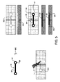
図1に示すように、骨格固定部材は、補修が必要である骨格の欠陥、例えば、破断を横切って配置されるために、生物学的適合性のワイヤの形態になっている。ワイヤ20は、好ましくは、少なくとも部分的に、骨格の欠陥を横切る頭蓋骨の中に埋設され、骨格領域を固定し、及び/又は、癒合を可能にする。好ましい実施形態においては、ワイヤ20は、曲線状の溝部200に挿入され、溝部は、骨のフライス加工、又は骨の表面仕上げ用器具、例えば、レーザ、高周波RFの表面仕上げ用器具、その他の電磁的又は機械的な表面仕上げ用器具を使用して形成される。曲線状の溝部200は、その中にワイヤ20が移植されるもので、好ましくは、その弓形の頂点にて又はその付近にて、破断部位と交差し、ワイヤ20を溝部200の中に移植する際に、ワイヤ20は、2つの骨の破片を固定するように作用し、骨の上面に対して平行に作用する力に抵抗する。
As shown in FIG. 1, the skeletal fixation member is in the form of a biocompatible wire because it is placed across a skeletal defect that needs repair, eg, a break. The
ワイヤ20は、任意の横断面形状及び/又は当業者に公知の領域を有し、それらには、限定はしないが、円筒形、直線状、台形、多角形などが含まれる。ワイヤ20が、台形の形状を有する場合には、ワイヤ20における、遠位側に移植される表面は、近位側に移植される表面に比べて大きな幅を有し、実質的に同様な横断面形状及び/又は寸法を有する、溝部200の中に移植される。このように、ワイヤ20が外れることは、ありそうにない。ワイヤ20を移植するためには、例えば、ワイヤ20を、一端に通して縛るか、いくらか力を加えて、上から下へインプラントを挿入するか、受け床の中にインプラントをスナップ嵌合させるか、完全な破断が存在する場合に実際的であるように、骨の部分を外らして、溝部200をわずかに拡大するなどである。
The
加えて、材料の選択を適切に行うことで、ワイヤ20はさらに、患者の身体又は骨組織の中にいったん導入されると、拡張可能なようになっている。代わりに、及び/又は、加えて、ワイヤ20は、薬剤を溶出し、及び/又は、組織成長促進材料でコーティングされ、例えば、BGH又はヒドロキシアパタイトが用いられる。
In addition, with proper material selection, the
代わりに、及び/又は、加えて、移植されるワイヤ20及び/又は外科的に形成された溝部200は、生物学的適合性の接着剤、例えば、骨パテ、シアノアクリレート、ポリウレタン、エポキシ、アクリール樹脂、燐酸カルシウムセメントなどで被覆され、周囲の骨組織に対して、インプラントをより確実に固定する。接着剤は、溝部200の中に挿入され、及び/又は、ワイヤ20の周囲又は上部に塗布され、これは、インプラントワイヤ20の移植前に、移植中に、又は移植後に行われることが想定される。
Alternatively and / or in addition, the implanted
ワイヤ20は、当業者に知られた任意の生物学的適合性の材料であって、特定の用途の強度及び可撓性の要求条件を満足するものから形成され、それらには、限定はしないが、ステンレス鋼、チタン、Ni−Ti(ニチノール)、エルジロイ、その他の形状記憶合金、PEEKなどのポリマー、生物学的吸収性の材料などが使用される。
The
好ましい実施形態においては、ワイヤ20は、約1ミリメートルの直径を有し、ワイヤ20が移植される溝部200は、1ミリメートルの同一の深さを有し、ワイヤ20における近位側表面120は、骨の上面に対して、実質的に面一(例えば、平坦)であるか又は低く、溝部の深さ200は、骨皮質の下にまでは延在しない。
In a preferred embodiment, the
図2に示すように、ワイヤインプラント20は、拡張部を具備し、例えば、刺、フィラメント、又はクリップをワイヤの軸に沿って備えている。拡張部22は、周囲の骨皮質組織に入る、追加的な足がかりを提供し、周囲の組織に対して、ワイヤインプラント20をよりしっかりと固定する。拡張部22は、ワイヤインプラント20と一体的に形成される。変形例としては、拡張部22は、ワイヤ20とは独立に形成されて、ワイヤに取り付けられる。そうして、拡張部22は、ワイヤインプラント20と同一の材料から形成され、又は、それらは、例えば、ニチノール、エルジロイなどの異なる材料から形成される。拡張部22は、術後に又は術中に、展開可能である。すなわち、例えば、拡張部22は、機械的に、又は、磁気的に、展開可能である。変形例としては、拡張部22は、ワイヤ20の外面に、永久的に配置される。
As shown in FIG. 2, the
図3に示すように、様々な追加的な固定手段が、周囲の骨組織に対してワイヤインプラント20の固定を助けるために想定される。1つの例においては、ワイヤインプラント20は、その長さの両端に、矢じり24を備えている。このように、矢じり24は、破断部位を横切って、2つの骨の破片を圧縮する助けとなり、一方、ワイヤインプラント20を溝部200内の所定位置に固定する。また、図3は、骨組織に対してワイヤインプラント20の固定を高めるために提供される、様々な追加的な構造を示している。
As shown in FIG. 3, various additional securing means are envisioned to assist in securing the
図示の通り、インプラント収容溝部200の機械加工及び/又はレーザ加工は、曲線状の溝部200に隣接した、1又は複数の領域210を表面切断することを含み、インプラントワイヤ20の挿入を容易にすると共に、様々に図示された追加的な固定手段に適応する。
As shown, machining and / or laser machining of the
代わりに、及び/又は、加えて、前述したように、移植されるワイヤ20及び/又は外科的に形成された溝部200は、生物学的適合性の接着剤、例えば、骨パテ、シアノアクリレート、ポリウレタン、エポキシ、アクリール樹脂、燐酸カルシウムセメントなどで被覆され、周囲の骨組織に対して、インプラントをより確実に固定する。接着剤は、溝部200の中に挿入され、及び/又は、ワイヤ20の周囲又は上部に塗布され、これは、インプラントワイヤ20の移植前に、移植中に、又は移植後に行われることが想定される。
Alternatively and / or in addition, as described above, the implanted
代わりに、及び/又は、加えて、図4に示すように、溝部200は、1又は複数の円形に機械加工された領域220を具備している。図示の通り、円形に機械加工された領域220は、曲線状の溝部の両端に配置され、円形の領域は、中空の円形凹部222を具備し、凹部は、好ましくは、曲線状の溝部の深さと同様な深さを有している。中空で円形の凹部は、好ましくは、機械加工又はレーザ加工されないことで形成された、1又は複数の円筒形である骨ペグ224を取り囲み、円筒形の骨ペグ224は、機械加工又はレーザ加工を受けていない骨の表面と面一に設けられる。曲線状の溝部200と、中空の円形凹部とは、好ましくは、中空のリング又は鳩目26を両端に配置されて有してなるワイヤインプラント20を受け入れるサイズ及び構造になっており、鳩目26は、溝部200の機械加工又はレーザ加工中に残された、円筒形の骨ペグ224を取り囲み、従って、追加的な反発抵抗をもって、移植固定するのを容易にしている。
Alternatively and / or in addition, as shown in FIG. 4, the
図示の通り、鳩目26と、対応する骨ペグ224とは、円形の形態を呈する。変形例としては、鳩目26と、対応する骨ペグ224とは、非円形の形状を呈し、例えば、正方形又は多角形の形状などであって、これらは、鋭利な縁部に起因して、円形の形態と比較して、反発に対して追加的な抵抗を提供する。代わりに、及び/又は、加えて、骨ペグ224は、非円形の形状を呈し、例えば、正方形又は多角形の形状などであり、対応する正方形又は多角形の鳩目孔26を有する円形リングと嵌合する。変形例としては、骨ペグ224は、円筒形の形状を呈し、一方、リング部材は、正方形又は多角形の外面を有し、円形の鳩目孔26を備える。
As shown in the figure, the
当業者には認識されるだろうが、機械加工された領域及び対応する鳩目26は、ワイヤ20及び/又は溝部200の長さに沿ったどこにでも形成できる。
As will be appreciated by those skilled in the art, the machined region and
図5及び図6には、特に良好に適した、頭蓋骨又は頚顔面の骨プレート30を示している。当業者には認識されるであろうけれども、骨プレート30は、同様に身体のその他の部分にも使用できる。頭蓋骨又は頚顔面の骨プレート30は、設計及び幾何学形状において、従来の頭蓋骨又は頚顔面の骨プレートと類似しており、すなわち、骨プレート30は、好ましくは、薄い輪郭を有し、小径のねじ受入れボア孔32を、薄い中間的プレート領域34に結合させて具備している。
FIGS. 5 and 6 show a skull or
すなわち、図示の通り、頭蓋骨の又は頚顔面の骨プレート30は、好ましくは、2つの拡大した端部部分36と、これらの間に配置された中間的な結合又は橋渡し部分34とを有してなる形状を呈しており、それぞれの拡大した端部部分36は、オプションであるねじ固定のために、オプションである骨孔32を具備している。そうして、頭蓋骨又は頚顔面の骨プレート30は、一般的に、バーベルの形状を呈し、2つの拡大した丸い突出部36を具備し、両端は、突出部36のいずれの幅よりも小さい寸法の幅を有する、中間のリンク部分34によって結合されている。それぞれの突出部36は、オプションであるねじ固定のために、ボア孔32を具備している。ボア孔32は、さらに、図7に示す骨ねじに適合する代わりに、図5を参照して前述したものと同様な骨ペグに嵌着するように構成される。変形例としては、突出部36は、ボア孔32をまったく備えていなくてもよい。
That is, as shown, the skull or
使用に際しては、図8に示すように、骨プレート30は、破断部位又はその他の補修が必要な骨の領域を横切るように適用される。プレート受入れ領域300は、骨フライス加工又は骨の表面仕上げ用器具、例えば、パルスレーザ、高周波RFの表面仕上げ用器具、機械的に表面仕上げを行う器具などを使用して、骨に形成される。骨プレート30は、好ましくは、少なくとも部分的に又は全体的に、機械加工されたプレート受入れ領域300に挿入される。骨プレート30は、癒合を助けるために、互いに対してしっかりと、破断部位を横切って、骨片を保持するように働く。好ましくは、前述したように、骨プレート30の厚みは、機械加工されたプレート受入れ領域300又はその他の骨の表面仕上げの領域の深さと、実質的に対応し、いったん移植されたならば、骨プレート30の上面は、骨の上面に対して実質的に面一又は下になり、従って、ゼロ高さのインプラントが提供される。
In use, as shown in FIG. 8,
さらに、前述したように、骨プレート30のインプラントは、いったん患者の身体又は骨組織の中に導入されると、適切な材料の選択によって、拡張可能になっている。また、骨プレートインプラントは、薬剤を溶出し、及び/又は、組織成長促進材料でコーティングされ、例えば、BGH又はヒドロキシアパタイトが用いられる。また、骨プレートインプラントの表面は、骨の成長を助けるテクスチャーを具備している。代わりに、及び/又は、加えて、移植された骨プレート30及び/又は機械加工されたプレート受入れ領域300は、生物学的適合性の接着剤で被覆されている。骨プレート30及び/又は骨ねじは、さらに生物学的吸収性でもよい。
Furthermore, as previously mentioned, the implant of the
図7には、様々に変化したサイズ及び形状の骨プレート30が示される。再言するが、これらのプレート30は、頭蓋骨又は頚顔面の用途に特に良好に適しているが、当業者が認識するように、骨プレート30は、身体のその他の部分にも同じく使用できる。図示の通り、骨プレート30は、一般的に、より複雑なプレート設計を有し、より複雑な破断の軽減及び/又は癒合のために、特に適している。1つの好ましい実施形態においては、ゼロ輪郭の十字形のプレート30は、一般的に、X字形を有し、結合部材34は、それぞれの結合部材34の両端にて、ボア孔32と接合され、骨アンカー又は骨ペグ310を受け入れる。十字形のプレート30は、破断部位にわたる、対応する機械加工又はレーザ加工された溝部300に、向けられ及び/又は挿入されて、2つのアンカー手段が、破断の両側に配置される。同様に、図7に示した、より複雑なプレートの設計は、骨表面に形成された対応する溝部300に移植され、複数のアンカー手段が、骨の破断部の1又は複数の側部に配置される。
FIG. 7 shows
図9に示すように、本発明の1つの観点によれば、インプラント(例えば、ワイヤ20、プレートなど)は、骨における対応する機械加工又はレーザ加工された領域300に移植され、インプラントの上面は、骨の上面に対して、実質的に面一又は下になるようにしている。変形例としては、インプラントの上面は、骨の上面に対して、わずかに下にあり(例えば、窪んでいる)、又は、骨の上面に対して、わずかに上にある。代わりに、及び/又は、加えて、生物学的適合性の接着剤、例えば、骨パテを、インプラントの頂部に適用し、補修が必要な2つの骨部分の癒合を助ける。前述したように、骨プレート30は、好ましくは、特定の用途のために設計された厚みを有し、約0.5〜約5ミリメートルの範囲であり(いくつかの場合には、例えば、およそ1ミリメートルの厚みが、有用である。)、骨の上面に外科的に形成されたインプラント受け床に、等しい深さ又はわずかに大きい深さに、受け入れられる(いくつかの場合には、例えば、約1ミリメートル。)。
As shown in FIG. 9, according to one aspect of the present invention, an implant (eg,
図10は、追加的な骨プレートの設計を示している。図示の通り、骨プレート30は、非円形であるボア孔32を具備し、インプラントを、周囲の骨組織に対して固定する。好ましくは、非円形であるボア孔32のサイズ及び構造は、軸を有するアンカーピン600を収容するようになっていて、軸は、対応する非円形である横断面によって特徴付けられ、又は、代わりに、インプラント受入れ凹部の機械加工中に残された非円形の骨ペグ310を収容することで特徴付けられる。アンカーピン600は、非円形である横断面の軸であって、円形及び円筒形のねじ付き骨アンカーと比較して、排除に対して追加的な抵抗を提供し、非円形の骨アンカーは回転しにくく、従って、周囲の骨組織から抜けにくい。
FIG. 10 shows an additional bone plate design. As shown, the
円形の横断面である、ピン、釘、ねじ、又はアンカー600が使用されるならば、最低2つのそのようなピン、釘、又はアンカー600を使用して、骨の破片が、単一のピン、釘、又はアンカー600を中心として回転するのを防ぐのが好ましい。
If a pin, nail, screw, or
骨プレート30と、骨固定ピン、釘、及びアンカー600の頭部とは、移植後に、骨の上面に対して、実質的に面一になるサイズ及び構成にすることが好ましい。変形例としては、骨プレート30は、機械加工されていない骨の表面の頂部に配置される。好ましい実施形態においては、骨ピン、釘、アンカー600などの非円形である頭部は、プレート30を通して、ボア孔の近位側部分の内部に収容され、非円形の骨ピン600の上面が、骨プレートと共に骨の上面に対して実質的に面一になる。変形例としては、非円形である骨ピン、釘、アンカー600などの上面は、骨プレート30の上面に対して上又は下に配置される。1つの実施形態においては、非円形である骨ピン、釘、アンカー600などにおける遠位側横断面領域は、非円形である骨ピン、釘、アンカー600などにおける近位側横断面領域に比べて大きくなっていて、それにより、骨ピン、釘、アンカー600などの軸の長さに沿って、わずかにテーパを提供し、骨ピン、釘、アンカー600などは、骨にスナップ嵌合し、それにより、骨ピン、釘、アンカー600などと、骨プレート30とに追加的な固定を提供する。
The
図11に示すように、骨固定要素は、ばね付勢させた、固定クリップ40の形態になっている。ばね付勢された固定クリップ40は、2つの細長い部材42を具備し、これらは、それらの長さの中央又は中央付近にて、結合部材44によって結合されている。2つの細長い部材42の間の結合は、細長い部材を付勢して、一方の細長い部材42が、他方の細長い部材42に対して、回転できるようになっている。好ましくは、なんらの外力も無いとき、2つの細長い結合部材42は、十字形又は“X”字形に配置されるような、サイズ及び構造になっている。その後、細長い部材42の一方又は両方に回転力が加わると、結合部材42は、互いに対して、回転できるようになり、2つの細長い部材42は、互いに平行な形態に整列される。
As shown in FIG. 11, the bone anchoring element is in the form of a spring-biased
使用に際しては、ばねクリップにおける一方の細長い部材42は、機械加工又はレーザ加工で形成された骨のスロット400に挿入され、次に、骨の中へ直ちに貫通され、細長い部材42は、およそ90度回転(ばね)できる。すなわち、使用に際しては、例えば、ばね付勢された固定クリップ40は、頭蓋骨又は頚顔面の用途に特に良好に適しており、例えば、頭蓋骨のフラップの癒合、又は破片を減少させて、骨片の変位を固定し、癒合が必要な2つの頭蓋骨片を横切って適用され、又は、骨の表面を仕上げることによって、例えば、破断部位の近くの両方の骨の破片を横切って、溝部400を形成させることによって減少させる。好ましくは、溝部400は、全体にわたって、又は完全に、骨を通って形成される。次に、ばね付勢された固定クリップ40は、平行な状態で、溝部400の中に導入され、そのためには、把持器具又は挿入器が使用され、遠位側の細長い部材42は、骨の底面の下に導入され、一方、近位側の細長い部材42は、骨の上面の上に配置され、結合部材44が、骨の深さにわたって設けられる。次に、インプラントから器具が解放されると、固定クリップは自動的に、その自然な状態に復帰し、2つの細長い部材42は、十字形又はX字形を呈する。十字形の状態に戻る際に、2つの細長い部材42は、機械加工された溝部400に対して、それら自体を位置決めし、インプラントの反発が禁止される。このように、ばね付勢の固定クリップ40は、従来のフラップフィックスと同様な機能を果たし、目標は、骨のフラップを周囲の骨と同一のレベルに保つことである。
In use, one
加えて、ばね付勢の固定クリップ40における細長い部材42、及び/又は、結合部材44は、刺、又は、スパイク、又はその他の表面テクスチャを具備し、骨の足がかり及び/又は骨の成長を助ける。ばねクリップ40は、溝部400の中に装填され、1ミリメートルの深さと、5ミリメートルの長さとを有する。ばねクリップは、生物学的再吸収性の材料、又は非吸収性の材料から形成され、例えば、ステンレス鋼、チタン、ニチノール、又はPEEKが用いられる。
In addition, the
図12及び図13に示すように、インプラントは、波形ブレードのインプラント50の形態であり、2つの状態、(1)予備荷重(例えば、直線状)と、(2)無荷重(例えば、波形)との構成によって特徴付けられる。このように、波形ブレードのインプラント50は、その予備荷重された(例えば、直線状)形態において、把持タイプの挿入器具を使用して、骨表面に形成された溝部又は矩形のスロット500に挿入される。溝部500内に着座したとき、波形ブレードインプラント50は、把持タイプの挿入器具から解放されて、その無荷重(例えば、波形)の構成に復帰する。無荷重の形態においては、波形ブレードインプラント50は、サイン波形を呈し、その角部と側部とは、周囲の骨組織に接触及び/又は係合して(例えば、掘り込んで)、インプラントの着座を高める。
As shown in FIGS. 12 and 13, the implant is in the form of a
波形ブレードインプラント50は、当業者に知られている任意の生物学的適合性の材料から形成され、それらには、限定はしないが、冷間加工されたチタン、冷間加工された鋼、又は任意のその他の可撓性である生物学的適合性の材料が含まれる。把持挿入器具は、プライヤタイプの器具の形態であって、直線状のスロットを有し、その中で、波形ブレードのインプラント50が、予備荷重を受ける。次に、波形ブレードのインプラント50は、プライヤタイプの器具のスロットから押し出され、同時に、骨スロット500の中に挿入される。
The
図14に示すように、インプラントは、ステープルタイプのインプラント60の形態になっている。ステープルタイプのインプラントは、複数の脚部62を有し、脚部62は、正方形の横断面領域を備えて構成されている。ステープルタイプのインプラント60における、脚部62、好ましくは、正方形の脚部は、角度的に安定しており、骨の破片が、脚部の軸線を中心として回転するのを禁止及び/又は防止する。図示の通り、ステープルタイプのインプラント60は、好ましくは、分岐した脚部62を具備し、脚部62の分岐方向は、挿入中に弾性的に屈曲して、ステープルを骨に固定する。
As shown in FIG. 14, the implant is in the form of a
代わりに、及び/又は、加えて、図15に示すように、ステープルタイプのインプラント60は、1又は複数のねじ孔64を具備し、正方形の横断面領域をもつ脚部60と組み合わせられる。ねじは、ステープルタイプのインプラントを骨の表面に保持する、追加的な固定を提供する。
Alternatively and / or in addition, as shown in FIG. 15, a staple-
前述したように、及び、図16に最良に示されるように、インプラントは、さらに、わずかなテーパ100を具備している。テーパは、インプラントの高さに沿って設けられ、インプラントにおける遠位側表面110の表面積は、インプラントの近位側表面120の表面積に比べて大きくなっている。テーパ付きのインプラントは、周囲の骨組織に対して、インプラントの保持を改善する。テーパ付きの高さインプラントは、例えば、インプラントを一端に通して縛るか、いくらか力を加えて、上から下へインプラントを挿入するか、受け床の中にインプラントをスナップ嵌合させるか、完全な破断が存在する場合に実際的であるように、骨の部分を外らして、溝部をわずかに拡大するなどして移植される。
As described above and best shown in FIG. 16, the implant further comprises a
図17は、本発明の別の観点に従った、様々な変形例によるインプラントの設計を示している。図示の通り、インプラントは、好ましくは、細くなった中間的部分と、拡大した端部部分とを具備している。インプラントは、好ましくは、骨の皮質表面に形成された、対応する受け床に受け入れられるようなサイズ及び構造になっている。そうして、インプラントは、好ましくは、いったん移植されると、骨の上面に対して、ゼロ高さ又は高さが低い輪郭になると共に、改良されたインプラントの保持をもたらす。 FIG. 17 illustrates an implant design according to various variations in accordance with another aspect of the present invention. As shown, the implant preferably comprises a narrowed intermediate portion and an enlarged end portion. The implant is preferably sized and structured to be received in a corresponding receiving bed formed in the cortical surface of the bone. Thus, once implanted, the implant preferably has a zero or low profile relative to the upper surface of the bone, resulting in improved implant retention.
図18は、非直線状(例えば、蛇状又は屈曲した)である骨の切断であって、インシトゥーにて硬化可能である、注入可能な材料によって充填されるものを示している。注入可能な材料には、限定はしないが、骨グルー、セメント、加熱された再吸収性の、又は非再吸収性のポリマーなどが含まれる。注入可能な材料は、硬化すると、前述したインプラントと同じく、同様な骨格の修理の目的に働く、成形可能なインプラントとして機能する。 FIG. 18 shows a bone cut that is non-linear (eg, serpentine or bent) and filled with an injectable material that is curable in situ. Injectable materials include, but are not limited to, bone glue, cement, heated resorbable or non-resorbable polymers, and the like. Once cured, the injectable material functions as a moldable implant that serves the same skeletal repair purposes as the previously described implant.
図19は、非直線状(例えば、蛇状又は屈曲した)である骨の切断であって、軟質の及び/又は従順な、しかし、移植後に、硬化し又は硬化しない、非液体の材料によって充填されるものを示している。硬化する材料には、限定はしないが、ガラス転移温度より高温に加熱される、ポリマー材料が含まれる。 FIG. 19 shows a bone cut that is non-linear (eg, serpentine or bent), filled with a soft and / or compliant, but non-liquid material that hardens or does not harden after implantation. Shows what will be done. Curing materials include, but are not limited to, polymeric materials that are heated above the glass transition temperature.
図20は、骨切断ツールをガイドする、ステンシル又はテンプレートのタイプの器具を示しており、骨切断ツールは、例えば、レーザ、ウォータージェット、ピエゾ電子切断ナイフ、機械的フライス機器、又はその他の骨の表面仕上げ用器具などである。 FIG. 20 shows a stencil or template type instrument that guides a bone cutting tool, such as a laser, water jet, piezo electronic cutting knife, mechanical milling device, or other bone cutting tool. For example, a surface finishing tool.
図21は、精密に骨の表面を仕上げる段階を助ける、制御された骨の除去のための数値制御式ガイドシステムの使用を示している。 FIG. 21 illustrates the use of a numerically controlled guide system for controlled bone removal that assists in precisely finishing the bone surface.
本発明及びその方法によって提供されるインプラントは、追加的な固定手段、例えば、生物学的適合性の接着剤、例えば、シアノアクリレート、ポリウレタン、エポキシ、及びアクリール樹脂を、超音波エネルギー適用と共に又はこれを伴わずに、又は骨溶接技術を利用して行われることを理解されたい。 Implants provided by the present invention and methods thereof include additional anchoring means such as biocompatible adhesives such as cyanoacrylates, polyurethanes, epoxies, and acryl resins with or without application of ultrasonic energy. It should be understood that this can be done without or using bone welding techniques.
前述した説明及び図面は、本発明の好ましい実施形態を示しているけれども、特許請求の範囲に定義された本発明の精神及び範囲から逸脱せずに、様々な追加、変更、及び置換を施せることを理解されたい。特に、当業者には明らかなように、本発明は、別の特定の形態、構造、配置、比率において、及び、他の要素、材料、及び構成要素と共に実施でき、その精神又は本質的特徴から逸脱することはない。当業者は認識するだろうが、本発明は、本発明の実施に使用される、構造、構成、比率、材料、及び構成要素その他についての多くの変更と共に使用でき、それらは、特に、本発明の原理から逸脱することなく、特定の環境及び動作条件に適合している。加えて、本願に開示された特徴は、単一にて、又は他の特徴と組み合わせて使用される。従って、ここに開示された実施形態は、すべての観点において、例示的であり、制限的ではないとみなされ、本発明の範囲は、特許請求の範囲によって定められ、前述した説明に限られるものではない。 While the foregoing description and drawings illustrate preferred embodiments of the present invention, various additions, modifications, and substitutions may be made without departing from the spirit and scope of the invention as defined in the claims. I want you to understand. In particular, it will be apparent to those skilled in the art that the present invention may be practiced in other specific forms, structures, arrangements, proportions and with other elements, materials, and components, from its spirit or essential characteristics. There is no departure. Those skilled in the art will recognize that the present invention can be used with many modifications to the structures, configurations, ratios, materials, components, etc. used in the practice of the present invention, which are particularly It is adapted to specific environmental and operating conditions without departing from the principles of In addition, the features disclosed in this application may be used alone or in combination with other features. Accordingly, the embodiments disclosed herein are considered in all respects to be illustrative and not restrictive, and the scope of the present invention is defined by the appended claims and is limited only to the foregoing description. is not.
Claims (19)
骨の破片を互いに接触させ且つ共に癒合できるように欠陥を横切る骨の破片内の非直線状の凹部の中に移植されるべく適合している可撓性ワイヤであって、この可撓性ワイヤは、骨の破片内の凹部の深さに比べて小さい又は等しい横断断面寸法を有し、可撓性ワイヤが非直線状の凹部の中に移植されるとき、可撓性ワイヤがそれぞれの骨の破片の上面と平行に作用する力に抵抗する、可撓性ワイヤ、
を備えていることを特徴とする骨固定システム。 A bone fixation system for implantation in a non-linear recess in a bone fragment separated by a defect, the bone fixation system comprising:
A flexible wire adapted to be implanted in a non-linear recess in a bone fragment that traverses a defect so that the bone fragments can contact and heal together Has a cross-sectional dimension that is small or equal to the depth of the recess in the bone fragment, and when the flexible wire is implanted in a non-linear recess, the flexible wire A flexible wire that resists forces acting parallel to the top surface of the fragments of
A bone fixation system characterized by comprising:
隣接する骨の破片を癒合できるように欠陥を横切る骨の破片内の凹部の中に移植されるべく適合した可撓性プレートを備え、可撓性プレートは、2つの突出部を中間リンク部分によって結合された両端に備え、可撓性プレートは、骨の破片内の凹部の深さに比べて小さい厚みを有し、中間リンク部分は、突出部の幅に比べて小さい幅を有し、凹部は非直線形状を有し、可撓性プレートが凹部の中に配置されるとき、可撓性プレートが骨の上面と平行に作用する力に抵抗することを特徴とする骨固定システム。 A bone fixation system for implantation into a recess in a bone fragment separated by a defect, the bone fixation system comprising:
A flexible plate adapted to be implanted in a recess in a bone fragment that traverses the defect so that adjacent bone fragments can be fused, the flexible plate having two protrusions connected by an intermediate link portion With the joined ends, the flexible plate has a thickness that is small compared to the depth of the recess in the bone fragment, the intermediate link portion has a width that is small compared to the width of the protrusion, and the recess Has a non-linear shape and resists forces acting parallel to the upper surface of the bone when the flexible plate is placed in the recess .
Applications Claiming Priority (3)
| Application Number | Priority Date | Filing Date | Title |
|---|---|---|---|
| US94725407P | 2007-06-29 | 2007-06-29 | |
| US60/947,254 | 2007-06-29 | ||
| PCT/US2008/068606 WO2009006313A1 (en) | 2007-06-29 | 2008-06-27 | Improved orthopedic implants for use with precision bone resurfacing instrumentation |
Publications (3)
| Publication Number | Publication Date |
|---|---|
| JP2010532229A JP2010532229A (en) | 2010-10-07 |
| JP2010532229A5 JP2010532229A5 (en) | 2011-08-11 |
| JP5572547B2 true JP5572547B2 (en) | 2014-08-13 |
Family
ID=39769330
Family Applications (1)
| Application Number | Title | Priority Date | Filing Date |
|---|---|---|---|
| JP2010515185A Expired - Fee Related JP5572547B2 (en) | 2007-06-29 | 2008-06-27 | Improved orthopedic implant for use with precision bone surface finishing instruments |
Country Status (11)
| Country | Link |
|---|---|
| US (1) | US20110172721A1 (en) |
| EP (1) | EP2162081A1 (en) |
| JP (1) | JP5572547B2 (en) |
| KR (1) | KR20100036275A (en) |
| CN (1) | CN101686845B (en) |
| AU (1) | AU2008269985A1 (en) |
| BR (1) | BRPI0813956A2 (en) |
| CA (1) | CA2692376A1 (en) |
| CO (1) | CO6150113A2 (en) |
| WO (1) | WO2009006313A1 (en) |
| ZA (1) | ZA200907855B (en) |
Families Citing this family (15)
| Publication number | Priority date | Publication date | Assignee | Title |
|---|---|---|---|---|
| US9848993B2 (en) | 2005-04-12 | 2017-12-26 | Nathan C. Moskowitz | Zero-profile expandable intervertebral spacer devices for distraction and spinal fusion and a universal tool for their placement and expansion |
| US20090259263A1 (en) | 2008-04-11 | 2009-10-15 | Biomet Microfixation, Inc. | Apparatus and methods of fixating bone |
| JP5335834B2 (en) * | 2011-02-15 | 2013-11-06 | 日本特殊陶業株式会社 | Artificial skull flap |
| US9445919B2 (en) | 2011-12-19 | 2016-09-20 | Warsaw Orthopedic, Inc. | Expandable interbody implant and methods of use |
| US8628578B2 (en) | 2011-12-19 | 2014-01-14 | Warsaw Orthopedic, Inc. | Expandable interbody implant and methods of use |
| US9937053B2 (en) | 2014-11-04 | 2018-04-10 | Warsaw Orthopedic, Inc. | Expandable interbody implant |
| US9907670B2 (en) | 2015-01-21 | 2018-03-06 | Warsaw Orthopedic, Inc. | Unitarily formed expandable spinal implant and method of manufacturing and implanting same |
| US10610376B2 (en) | 2015-10-16 | 2020-04-07 | Warsaw Orthopedic, Inc. | Expandable spinal implant system and method |
| US10779955B2 (en) | 2015-10-26 | 2020-09-22 | Warsaw Orthopedic, Inc. | Spinal implant system and method |
| US10188526B2 (en) | 2015-10-26 | 2019-01-29 | Warsaw Orthopedic, Inc. | Spinal implant system and method |
| US10076423B2 (en) | 2016-01-04 | 2018-09-18 | Warsaw Orthopedic, Inc. | Pivoting wedge expanding spinal implant and method of implanting same |
| US10137006B2 (en) | 2016-01-28 | 2018-11-27 | Warsaw Orthopedic, Inc. | Geared cam expandable interbody implant and method of implanting same |
| US9937054B2 (en) | 2016-01-28 | 2018-04-10 | Warsaw Orthopedic, Inc. | Expandable implant and insertion tool |
| US10238503B2 (en) | 2016-11-01 | 2019-03-26 | Warsaw Orthopedic, Inc. | Expandable spinal implant system with a biased tip and method of using same |
| DE102018121553A1 (en) * | 2018-09-04 | 2020-03-05 | Karl Leibinger Medizintechnik Gmbh & Co. Kg | Bone implant for the reconstruction of a bony defect and for guiding a marking and / or processing tool for transferring the necessary osteotomy situations |
Family Cites Families (18)
| Publication number | Priority date | Publication date | Assignee | Title |
|---|---|---|---|---|
| FI69402C (en) * | 1983-09-20 | 1986-02-10 | Materials Consultants Oy | OSTEOSYNTESANORDNING |
| US4820157A (en) * | 1985-11-22 | 1989-04-11 | Salvo Christopher A | Dental bridge |
| US4735571A (en) * | 1985-11-22 | 1988-04-05 | Salvo Christopher A | Dental splint |
| WO1994008783A1 (en) * | 1992-10-22 | 1994-04-28 | Jonathan Scharf | Ceramic reinforced dental appliances, devices and restorations |
| US5591235A (en) * | 1995-03-15 | 1997-01-07 | Kuslich; Stephen D. | Spinal fixation device |
| US5700267A (en) * | 1996-08-15 | 1997-12-23 | Kinetikos Medical Incorporated | Method for repairing bone fractures using bone-lock system |
| CN2309126Y (en) * | 1997-08-06 | 1999-03-03 | 王岩 | Head of femur memory metal support |
| US6221075B1 (en) * | 1998-03-06 | 2001-04-24 | Bionx Implants Oy | Bioabsorbable, deformable fixation plate |
| JP2000139936A (en) * | 1998-11-04 | 2000-05-23 | Hironobu Nomura | Fixing tool of bone prosthetic member to cranial bone |
| US7048542B2 (en) * | 2000-08-16 | 2006-05-23 | Medartis Ag | Dental splint |
| US20040088003A1 (en) * | 2002-09-30 | 2004-05-06 | Leung Jeffrey C. | Barbed suture in combination with surgical needle |
| US20040167572A1 (en) * | 2003-02-20 | 2004-08-26 | Roth Noah M. | Coated medical devices |
| WO2004093743A1 (en) * | 2003-04-16 | 2004-11-04 | Porex Surgical, Inc. | Craniofacial implant |
| US7794476B2 (en) * | 2003-08-08 | 2010-09-14 | Warsaw Orthopedic, Inc. | Implants formed of shape memory polymeric material for spinal fixation |
| WO2005041812A2 (en) * | 2003-10-22 | 2005-05-12 | Implant Brace, Inc. | Implantable brace for a fracture and methods |
| US20060195091A1 (en) * | 2005-02-15 | 2006-08-31 | Mcgraw J K | Percutaneous spinal stabilization device and method |
| US20070162132A1 (en) * | 2005-12-23 | 2007-07-12 | Dominique Messerli | Flexible elongated chain implant and method of supporting body tissue with same |
| US8870871B2 (en) * | 2007-01-17 | 2014-10-28 | University Of Massachusetts Lowell | Biodegradable bone plates and bonding systems |
-
2008
- 2008-06-27 BR BRPI0813956-3A2A patent/BRPI0813956A2/en not_active Application Discontinuation
- 2008-06-27 EP EP08781100A patent/EP2162081A1/en not_active Withdrawn
- 2008-06-27 CA CA002692376A patent/CA2692376A1/en not_active Abandoned
- 2008-06-27 CN CN2008800192593A patent/CN101686845B/en not_active Expired - Fee Related
- 2008-06-27 US US12/666,304 patent/US20110172721A1/en not_active Abandoned
- 2008-06-27 AU AU2008269985A patent/AU2008269985A1/en not_active Abandoned
- 2008-06-27 KR KR1020097027371A patent/KR20100036275A/en not_active Application Discontinuation
- 2008-06-27 JP JP2010515185A patent/JP5572547B2/en not_active Expired - Fee Related
- 2008-06-27 WO PCT/US2008/068606 patent/WO2009006313A1/en active Application Filing
-
2009
- 2009-11-09 ZA ZA200907855A patent/ZA200907855B/en unknown
- 2009-12-28 CO CO09148276A patent/CO6150113A2/en unknown
Also Published As
| Publication number | Publication date |
|---|---|
| CN101686845B (en) | 2013-07-10 |
| BRPI0813956A2 (en) | 2014-12-30 |
| WO2009006313A1 (en) | 2009-01-08 |
| AU2008269985A1 (en) | 2009-01-08 |
| CA2692376A1 (en) | 2009-01-08 |
| CN101686845A (en) | 2010-03-31 |
| US20110172721A1 (en) | 2011-07-14 |
| KR20100036275A (en) | 2010-04-07 |
| EP2162081A1 (en) | 2010-03-17 |
| ZA200907855B (en) | 2010-08-25 |
| CO6150113A2 (en) | 2010-04-20 |
| JP2010532229A (en) | 2010-10-07 |
Similar Documents
| Publication | Publication Date | Title |
|---|---|---|
| JP5572547B2 (en) | Improved orthopedic implant for use with precision bone surface finishing instruments | |
| EP2879602B1 (en) | Bone fixation device | |
| EP3166505B1 (en) | Bone implant with anti-rotation | |
| JP5058605B2 (en) | Bone implanter | |
| EP2967894B1 (en) | Hammertoe implant with enhanced gripping surfaces | |
| EP3311758B1 (en) | Implant and implantation device | |
| US7780701B1 (en) | Suture anchor | |
| JP6086993B2 (en) | Osteotomy implant | |
| BRPI1009207A2 (en) | spine stabilization device and method and kit for its implantation | |
| EP1917924A1 (en) | Implantation device | |
| EP1643921A1 (en) | Apparatus and method for attaching adjacent bones | |
| WO2011063250A1 (en) | Implantable devices for subchondral treatment of joint pain | |
| US10582957B2 (en) | Bone fixation implant and means of fixation | |
| US20220168026A1 (en) | Methods and devices for intracorporeal bonding of implants with thermal energy | |
| ES2918002T3 (en) | bone screw | |
| US11918263B2 (en) | Mixed materials bone screw | |
| WO2022123342A1 (en) | Sternal closure systems | |
| TWI680741B (en) | Osteotomy implant | |
| WO2020056063A1 (en) | Tendon repair implant and surgical instruments for tendon repair |
Legal Events
| Date | Code | Title | Description |
|---|---|---|---|
| A521 | Request for written amendment filed |
Free format text: JAPANESE INTERMEDIATE CODE: A523 Effective date: 20110624 |
|
| A621 | Written request for application examination |
Free format text: JAPANESE INTERMEDIATE CODE: A621 Effective date: 20110624 |
|
| A131 | Notification of reasons for refusal |
Free format text: JAPANESE INTERMEDIATE CODE: A131 Effective date: 20130107 |
|
| A601 | Written request for extension of time |
Free format text: JAPANESE INTERMEDIATE CODE: A601 Effective date: 20130305 |
|
| A602 | Written permission of extension of time |
Free format text: JAPANESE INTERMEDIATE CODE: A602 Effective date: 20130312 |
|
| A521 | Request for written amendment filed |
Free format text: JAPANESE INTERMEDIATE CODE: A523 Effective date: 20130708 |
|
| A02 | Decision of refusal |
Free format text: JAPANESE INTERMEDIATE CODE: A02 Effective date: 20131202 |
|
| A521 | Request for written amendment filed |
Free format text: JAPANESE INTERMEDIATE CODE: A523 Effective date: 20140402 |
|
| A521 | Request for written amendment filed |
Free format text: JAPANESE INTERMEDIATE CODE: A523 Effective date: 20140515 |
|
| A911 | Transfer to examiner for re-examination before appeal (zenchi) |
Free format text: JAPANESE INTERMEDIATE CODE: A911 Effective date: 20140520 |
|
| TRDD | Decision of grant or rejection written | ||
| A01 | Written decision to grant a patent or to grant a registration (utility model) |
Free format text: JAPANESE INTERMEDIATE CODE: A01 Effective date: 20140616 |
|
| A61 | First payment of annual fees (during grant procedure) |
Free format text: JAPANESE INTERMEDIATE CODE: A61 Effective date: 20140630 |
|
| R150 | Certificate of patent or registration of utility model |
Ref document number: 5572547 Country of ref document: JP Free format text: JAPANESE INTERMEDIATE CODE: R150 |
|
| R250 | Receipt of annual fees |
Free format text: JAPANESE INTERMEDIATE CODE: R250 |
|
| LAPS | Cancellation because of no payment of annual fees |