JP5503096B2 - Virus that causes respiratory tract disease in susceptible mammals - Google Patents
Virus that causes respiratory tract disease in susceptible mammals Download PDFInfo
- Publication number
- JP5503096B2 JP5503096B2 JP2002557978A JP2002557978A JP5503096B2 JP 5503096 B2 JP5503096 B2 JP 5503096B2 JP 2002557978 A JP2002557978 A JP 2002557978A JP 2002557978 A JP2002557978 A JP 2002557978A JP 5503096 B2 JP5503096 B2 JP 5503096B2
- Authority
- JP
- Japan
- Prior art keywords
- protein
- amino acid
- acid sequence
- mpv
- virus
- Prior art date
- Legal status (The legal status is an assumption and is not a legal conclusion. Google has not performed a legal analysis and makes no representation as to the accuracy of the status listed.)
- Expired - Lifetime
Links
- 241000700605 Viruses Species 0.000 title claims description 338
- 241000124008 Mammalia Species 0.000 title claims description 25
- 208000018569 Respiratory Tract disease Diseases 0.000 title 1
- 108090000623 proteins and genes Proteins 0.000 claims description 236
- 125000003275 alpha amino acid group Chemical group 0.000 claims description 174
- 102000004169 proteins and genes Human genes 0.000 claims description 140
- 241000711902 Pneumovirus Species 0.000 claims description 123
- 208000015181 infectious disease Diseases 0.000 claims description 108
- 239000012634 fragment Substances 0.000 claims description 102
- 238000000034 method Methods 0.000 claims description 90
- 239000002773 nucleotide Substances 0.000 claims description 85
- 125000003729 nucleotide group Chemical group 0.000 claims description 85
- 210000004027 cell Anatomy 0.000 claims description 66
- 150000007523 nucleic acids Chemical class 0.000 claims description 64
- 230000003612 virological effect Effects 0.000 claims description 60
- 241000351643 Metapneumovirus Species 0.000 claims description 56
- 239000000427 antigen Substances 0.000 claims description 51
- 102000036639 antigens Human genes 0.000 claims description 51
- 108091007433 antigens Proteins 0.000 claims description 51
- 108020004707 nucleic acids Proteins 0.000 claims description 48
- 102000039446 nucleic acids Human genes 0.000 claims description 48
- 108091006027 G proteins Proteins 0.000 claims description 42
- 108091000058 GTP-Binding Proteins 0.000 claims description 42
- 102000030782 GTP binding Human genes 0.000 claims description 39
- 108010068327 4-hydroxyphenylpyruvate dioxygenase Proteins 0.000 claims description 35
- 241000282414 Homo sapiens Species 0.000 claims description 35
- 101710141454 Nucleoprotein Proteins 0.000 claims description 31
- 101150039699 M2-1 gene Proteins 0.000 claims description 28
- 101001065501 Escherichia phage MS2 Lysis protein Proteins 0.000 claims description 25
- 241000711504 Paramyxoviridae Species 0.000 claims description 25
- 101710085938 Matrix protein Proteins 0.000 claims description 21
- 101710127721 Membrane protein Proteins 0.000 claims description 21
- 101710181008 P protein Proteins 0.000 claims description 21
- 101710177166 Phosphoprotein Proteins 0.000 claims description 21
- 101150103632 M2-2 gene Proteins 0.000 claims description 19
- 102100034574 P protein Human genes 0.000 claims description 19
- 241001493065 dsRNA viruses Species 0.000 claims description 19
- HEDRZPFGACZZDS-UHFFFAOYSA-N Chloroform Chemical compound ClC(Cl)Cl HEDRZPFGACZZDS-UHFFFAOYSA-N 0.000 claims description 14
- 230000000694 effects Effects 0.000 claims description 12
- 229960005486 vaccine Drugs 0.000 claims description 12
- 230000035931 haemagglutination Effects 0.000 claims description 11
- LWGJTAZLEJHCPA-UHFFFAOYSA-N n-(2-chloroethyl)-n-nitrosomorpholine-4-carboxamide Chemical compound ClCCN(N=O)C(=O)N1CCOCC1 LWGJTAZLEJHCPA-UHFFFAOYSA-N 0.000 claims description 10
- 238000009007 Diagnostic Kit Methods 0.000 claims description 8
- 239000002245 particle Substances 0.000 claims description 8
- 101001039853 Sonchus yellow net virus Matrix protein Proteins 0.000 claims description 6
- 230000015572 biosynthetic process Effects 0.000 claims description 6
- 210000004962 mammalian cell Anatomy 0.000 claims description 6
- 241000342334 Human metapneumovirus Species 0.000 claims description 5
- 239000002253 acid Substances 0.000 claims description 2
- 241001024304 Mino Species 0.000 claims 1
- 239000003937 drug carrier Substances 0.000 claims 1
- 210000005260 human cell Anatomy 0.000 claims 1
- 108700026244 Open Reading Frames Proteins 0.000 description 275
- 241001519465 Avian metapneumovirus Species 0.000 description 237
- 235000018102 proteins Nutrition 0.000 description 112
- 241000725643 Respiratory syncytial virus Species 0.000 description 106
- 150000001413 amino acids Chemical class 0.000 description 94
- 235000001014 amino acid Nutrition 0.000 description 93
- 238000003556 assay Methods 0.000 description 83
- 241000711920 Human orthopneumovirus Species 0.000 description 69
- 238000002965 ELISA Methods 0.000 description 52
- 241000700198 Cavia Species 0.000 description 46
- 238000003757 reverse transcription PCR Methods 0.000 description 41
- 210000002966 serum Anatomy 0.000 description 41
- 230000010530 Virus Neutralization Effects 0.000 description 40
- 241001465754 Metazoa Species 0.000 description 37
- 238000001514 detection method Methods 0.000 description 36
- 241000711895 Bovine orthopneumovirus Species 0.000 description 35
- 125000000539 amino acid group Chemical group 0.000 description 35
- 239000000523 sample Substances 0.000 description 34
- 238000012360 testing method Methods 0.000 description 33
- 241000271566 Aves Species 0.000 description 32
- 238000004458 analytical method Methods 0.000 description 32
- 230000000120 cytopathologic effect Effects 0.000 description 32
- 238000013081 phylogenetic analysis Methods 0.000 description 32
- 241000282412 Homo Species 0.000 description 28
- 108091028043 Nucleic acid sequence Proteins 0.000 description 24
- 241000282567 Macaca fascicularis Species 0.000 description 21
- 241000711955 Turkey rhinotracheitis virus Species 0.000 description 20
- 238000006386 neutralization reaction Methods 0.000 description 20
- 230000002441 reversible effect Effects 0.000 description 20
- 108060003393 Granulin Proteins 0.000 description 19
- 125000000151 cysteine group Chemical group N[C@@H](CS)C(=O)* 0.000 description 19
- 230000002209 hydrophobic effect Effects 0.000 description 19
- 238000002955 isolation Methods 0.000 description 19
- 108091032973 (ribonucleotides)n+m Proteins 0.000 description 18
- CSCPPACGZOOCGX-UHFFFAOYSA-N Acetone Chemical compound CC(C)=O CSCPPACGZOOCGX-UHFFFAOYSA-N 0.000 description 18
- 108090000288 Glycoproteins Proteins 0.000 description 18
- 102000003886 Glycoproteins Human genes 0.000 description 18
- 238000004113 cell culture Methods 0.000 description 18
- 108020001507 fusion proteins Proteins 0.000 description 18
- 102000037865 fusion proteins Human genes 0.000 description 18
- 230000002068 genetic effect Effects 0.000 description 18
- 101710118188 DNA-binding protein HU-alpha Proteins 0.000 description 17
- 101710158312 DNA-binding protein HU-beta Proteins 0.000 description 17
- 101710128560 Initiator protein NS1 Proteins 0.000 description 17
- 101710144127 Non-structural protein 1 Proteins 0.000 description 17
- 101710144128 Non-structural protein 2 Proteins 0.000 description 17
- 101710199667 Nuclear export protein Proteins 0.000 description 17
- 108010061100 Nucleoproteins Proteins 0.000 description 17
- 230000000890 antigenic effect Effects 0.000 description 17
- 102000011931 Nucleoproteins Human genes 0.000 description 16
- 238000012300 Sequence Analysis Methods 0.000 description 16
- 230000000405 serological effect Effects 0.000 description 16
- 230000001629 suppression Effects 0.000 description 16
- 238000010790 dilution Methods 0.000 description 15
- 239000012895 dilution Substances 0.000 description 15
- 201000010099 disease Diseases 0.000 description 15
- 208000037265 diseases, disorders, signs and symptoms Diseases 0.000 description 15
- 230000004044 response Effects 0.000 description 15
- 239000006228 supernatant Substances 0.000 description 15
- 239000013598 vector Substances 0.000 description 14
- 241000283690 Bos taurus Species 0.000 description 13
- 241000712079 Measles morbillivirus Species 0.000 description 13
- 206010057190 Respiratory tract infections Diseases 0.000 description 13
- 108020000999 Viral RNA Proteins 0.000 description 13
- 230000009260 cross reactivity Effects 0.000 description 13
- 238000003745 diagnosis Methods 0.000 description 13
- 231100000676 disease causative agent Toxicity 0.000 description 13
- 238000010166 immunofluorescence Methods 0.000 description 13
- 101150062031 L gene Proteins 0.000 description 12
- 206010035664 Pneumonia Diseases 0.000 description 12
- 208000035415 Reinfection Diseases 0.000 description 12
- 238000011081 inoculation Methods 0.000 description 12
- 239000002609 medium Substances 0.000 description 12
- 230000008520 organization Effects 0.000 description 12
- 208000023504 respiratory system disease Diseases 0.000 description 12
- 241000700199 Cavia porcellus Species 0.000 description 11
- 101150034814 F gene Proteins 0.000 description 11
- 230000004988 N-glycosylation Effects 0.000 description 11
- 108010067390 Viral Proteins Proteins 0.000 description 11
- 238000012512 characterization method Methods 0.000 description 11
- 238000006243 chemical reaction Methods 0.000 description 11
- 230000014509 gene expression Effects 0.000 description 11
- 238000004519 manufacturing process Methods 0.000 description 11
- 241000894007 species Species 0.000 description 11
- 238000007476 Maximum Likelihood Methods 0.000 description 10
- 241000711941 Murine orthopneumovirus Species 0.000 description 10
- 206010051497 Rhinotracheitis Diseases 0.000 description 10
- 108091005981 phosphorylated proteins Proteins 0.000 description 10
- 208000024891 symptom Diseases 0.000 description 10
- 238000005406 washing Methods 0.000 description 10
- 241000282693 Cercopithecidae Species 0.000 description 9
- 241000287828 Gallus gallus Species 0.000 description 9
- 108091034117 Oligonucleotide Proteins 0.000 description 9
- MTCFGRXMJLQNBG-UHFFFAOYSA-N Serine Natural products OCC(N)C(O)=O MTCFGRXMJLQNBG-UHFFFAOYSA-N 0.000 description 9
- 108091081024 Start codon Proteins 0.000 description 9
- 229930006000 Sucrose Natural products 0.000 description 9
- CZMRCDWAGMRECN-UGDNZRGBSA-N Sucrose Chemical compound O[C@H]1[C@H](O)[C@@H](CO)O[C@@]1(CO)O[C@@H]1[C@H](O)[C@@H](O)[C@H](O)[C@@H](CO)O1 CZMRCDWAGMRECN-UGDNZRGBSA-N 0.000 description 9
- 235000013330 chicken meat Nutrition 0.000 description 9
- 230000006870 function Effects 0.000 description 9
- 230000001900 immune effect Effects 0.000 description 9
- 239000000203 mixture Substances 0.000 description 9
- 244000052769 pathogen Species 0.000 description 9
- 230000001717 pathogenic effect Effects 0.000 description 9
- 239000008194 pharmaceutical composition Substances 0.000 description 9
- 238000012163 sequencing technique Methods 0.000 description 9
- 239000005720 sucrose Substances 0.000 description 9
- 241000283707 Capra Species 0.000 description 8
- 241000712003 Human respirovirus 3 Species 0.000 description 8
- 108091029795 Intergenic region Proteins 0.000 description 8
- 241000711408 Murine respirovirus Species 0.000 description 8
- 241000282341 Mustela putorius furo Species 0.000 description 8
- 241001494479 Pecora Species 0.000 description 8
- 241000282849 Ruminantia Species 0.000 description 8
- 239000003443 antiviral agent Substances 0.000 description 8
- 239000000872 buffer Substances 0.000 description 8
- 239000003153 chemical reaction reagent Substances 0.000 description 8
- 239000002299 complementary DNA Substances 0.000 description 8
- 238000011534 incubation Methods 0.000 description 8
- 229920000136 polysorbate Polymers 0.000 description 8
- 241000711404 Avian avulavirus 1 Species 0.000 description 7
- 108020004414 DNA Proteins 0.000 description 7
- 241000699666 Mus <mouse, genus> Species 0.000 description 7
- 241000282339 Mustela Species 0.000 description 7
- 239000012228 culture supernatant Substances 0.000 description 7
- 125000002924 primary amino group Chemical group [H]N([H])* 0.000 description 7
- 239000000047 product Substances 0.000 description 7
- 230000010076 replication Effects 0.000 description 7
- MTCFGRXMJLQNBG-REOHCLBHSA-N (2S)-2-Amino-3-hydroxypropansäure Chemical compound OC[C@H](N)C(O)=O MTCFGRXMJLQNBG-REOHCLBHSA-N 0.000 description 6
- PEDCQBHIVMGVHV-UHFFFAOYSA-N Glycerine Chemical compound OCC(O)CO PEDCQBHIVMGVHV-UHFFFAOYSA-N 0.000 description 6
- 101150046652 M2 gene Proteins 0.000 description 6
- 241000711502 Paramyxovirinae Species 0.000 description 6
- 108010089430 Phosphoproteins Proteins 0.000 description 6
- FAPWRFPIFSIZLT-UHFFFAOYSA-M Sodium chloride Chemical compound [Na+].[Cl-] FAPWRFPIFSIZLT-UHFFFAOYSA-M 0.000 description 6
- AYFVYJQAPQTCCC-UHFFFAOYSA-N Threonine Natural products CC(O)C(N)C(O)=O AYFVYJQAPQTCCC-UHFFFAOYSA-N 0.000 description 6
- 239000004473 Threonine Substances 0.000 description 6
- 238000003277 amino acid sequence analysis Methods 0.000 description 6
- 210000004899 c-terminal region Anatomy 0.000 description 6
- 238000002856 computational phylogenetic analysis Methods 0.000 description 6
- MHMNJMPURVTYEJ-UHFFFAOYSA-N fluorescein-5-isothiocyanate Chemical compound O1C(=O)C2=CC(N=C=S)=CC=C2C21C1=CC=C(O)C=C1OC1=CC(O)=CC=C21 MHMNJMPURVTYEJ-UHFFFAOYSA-N 0.000 description 6
- 230000004927 fusion Effects 0.000 description 6
- 239000000499 gel Substances 0.000 description 6
- 230000036039 immunity Effects 0.000 description 6
- 230000000977 initiatory effect Effects 0.000 description 6
- 238000011835 investigation Methods 0.000 description 6
- 239000000463 material Substances 0.000 description 6
- 239000011159 matrix material Substances 0.000 description 6
- 239000013642 negative control Substances 0.000 description 6
- 244000144977 poultry Species 0.000 description 6
- 235000013594 poultry meat Nutrition 0.000 description 6
- 108090000765 processed proteins & peptides Proteins 0.000 description 6
- 125000001500 prolyl group Chemical group [H]N1C([H])(C(=O)[*])C([H])([H])C([H])([H])C1([H])[H] 0.000 description 6
- 230000035945 sensitivity Effects 0.000 description 6
- 239000008399 tap water Substances 0.000 description 6
- 235000020679 tap water Nutrition 0.000 description 6
- 241000701447 unidentified baculovirus Species 0.000 description 6
- 239000013603 viral vector Substances 0.000 description 6
- 108091026890 Coding region Proteins 0.000 description 5
- 102000004190 Enzymes Human genes 0.000 description 5
- 108090000790 Enzymes Proteins 0.000 description 5
- AYFVYJQAPQTCCC-GBXIJSLDSA-N L-threonine Chemical compound C[C@@H](O)[C@H](N)C(O)=O AYFVYJQAPQTCCC-GBXIJSLDSA-N 0.000 description 5
- 241000699670 Mus sp. Species 0.000 description 5
- 241000286209 Phasianidae Species 0.000 description 5
- 102000007982 Phosphoproteins Human genes 0.000 description 5
- 241000711904 Pneumoviridae Species 0.000 description 5
- 108010076504 Protein Sorting Signals Proteins 0.000 description 5
- 206010061603 Respiratory syncytial virus infection Diseases 0.000 description 5
- 102000004142 Trypsin Human genes 0.000 description 5
- 108090000631 Trypsin Proteins 0.000 description 5
- 108700005077 Viral Genes Proteins 0.000 description 5
- 230000002238 attenuated effect Effects 0.000 description 5
- 238000003776 cleavage reaction Methods 0.000 description 5
- 238000005516 engineering process Methods 0.000 description 5
- 239000011521 glass Substances 0.000 description 5
- 238000010324 immunological assay Methods 0.000 description 5
- 238000000338 in vitro Methods 0.000 description 5
- 230000002458 infectious effect Effects 0.000 description 5
- 108020004999 messenger RNA Proteins 0.000 description 5
- 230000003472 neutralizing effect Effects 0.000 description 5
- 210000002345 respiratory system Anatomy 0.000 description 5
- 230000007017 scission Effects 0.000 description 5
- 239000012588 trypsin Substances 0.000 description 5
- 241001308709 unidentified virus Species 0.000 description 5
- QKNYBSVHEMOAJP-UHFFFAOYSA-N 2-amino-2-(hydroxymethyl)propane-1,3-diol;hydron;chloride Chemical compound Cl.OCC(N)(CO)CO QKNYBSVHEMOAJP-UHFFFAOYSA-N 0.000 description 4
- 108020004705 Codon Proteins 0.000 description 4
- 108091035707 Consensus sequence Proteins 0.000 description 4
- 101150082239 G gene Proteins 0.000 description 4
- ZDXPYRJPNDTMRX-VKHMYHEASA-N L-glutamine Chemical compound OC(=O)[C@@H](N)CCC(N)=O ZDXPYRJPNDTMRX-VKHMYHEASA-N 0.000 description 4
- 241000283216 Phocidae Species 0.000 description 4
- 238000012181 QIAquick gel extraction kit Methods 0.000 description 4
- JLCPHMBAVCMARE-UHFFFAOYSA-N [3-[[3-[[3-[[3-[[3-[[3-[[3-[[3-[[3-[[3-[[3-[[5-(2-amino-6-oxo-1H-purin-9-yl)-3-[[3-[[3-[[3-[[3-[[3-[[5-(2-amino-6-oxo-1H-purin-9-yl)-3-[[5-(2-amino-6-oxo-1H-purin-9-yl)-3-hydroxyoxolan-2-yl]methoxy-hydroxyphosphoryl]oxyoxolan-2-yl]methoxy-hydroxyphosphoryl]oxy-5-(5-methyl-2,4-dioxopyrimidin-1-yl)oxolan-2-yl]methoxy-hydroxyphosphoryl]oxy-5-(6-aminopurin-9-yl)oxolan-2-yl]methoxy-hydroxyphosphoryl]oxy-5-(6-aminopurin-9-yl)oxolan-2-yl]methoxy-hydroxyphosphoryl]oxy-5-(6-aminopurin-9-yl)oxolan-2-yl]methoxy-hydroxyphosphoryl]oxy-5-(6-aminopurin-9-yl)oxolan-2-yl]methoxy-hydroxyphosphoryl]oxyoxolan-2-yl]methoxy-hydroxyphosphoryl]oxy-5-(5-methyl-2,4-dioxopyrimidin-1-yl)oxolan-2-yl]methoxy-hydroxyphosphoryl]oxy-5-(4-amino-2-oxopyrimidin-1-yl)oxolan-2-yl]methoxy-hydroxyphosphoryl]oxy-5-(5-methyl-2,4-dioxopyrimidin-1-yl)oxolan-2-yl]methoxy-hydroxyphosphoryl]oxy-5-(5-methyl-2,4-dioxopyrimidin-1-yl)oxolan-2-yl]methoxy-hydroxyphosphoryl]oxy-5-(6-aminopurin-9-yl)oxolan-2-yl]methoxy-hydroxyphosphoryl]oxy-5-(6-aminopurin-9-yl)oxolan-2-yl]methoxy-hydroxyphosphoryl]oxy-5-(4-amino-2-oxopyrimidin-1-yl)oxolan-2-yl]methoxy-hydroxyphosphoryl]oxy-5-(4-amino-2-oxopyrimidin-1-yl)oxolan-2-yl]methoxy-hydroxyphosphoryl]oxy-5-(4-amino-2-oxopyrimidin-1-yl)oxolan-2-yl]methoxy-hydroxyphosphoryl]oxy-5-(6-aminopurin-9-yl)oxolan-2-yl]methoxy-hydroxyphosphoryl]oxy-5-(4-amino-2-oxopyrimidin-1-yl)oxolan-2-yl]methyl [5-(6-aminopurin-9-yl)-2-(hydroxymethyl)oxolan-3-yl] hydrogen phosphate Polymers Cc1cn(C2CC(OP(O)(=O)OCC3OC(CC3OP(O)(=O)OCC3OC(CC3O)n3cnc4c3nc(N)[nH]c4=O)n3cnc4c3nc(N)[nH]c4=O)C(COP(O)(=O)OC3CC(OC3COP(O)(=O)OC3CC(OC3COP(O)(=O)OC3CC(OC3COP(O)(=O)OC3CC(OC3COP(O)(=O)OC3CC(OC3COP(O)(=O)OC3CC(OC3COP(O)(=O)OC3CC(OC3COP(O)(=O)OC3CC(OC3COP(O)(=O)OC3CC(OC3COP(O)(=O)OC3CC(OC3COP(O)(=O)OC3CC(OC3COP(O)(=O)OC3CC(OC3COP(O)(=O)OC3CC(OC3COP(O)(=O)OC3CC(OC3COP(O)(=O)OC3CC(OC3COP(O)(=O)OC3CC(OC3COP(O)(=O)OC3CC(OC3CO)n3cnc4c(N)ncnc34)n3ccc(N)nc3=O)n3cnc4c(N)ncnc34)n3ccc(N)nc3=O)n3ccc(N)nc3=O)n3ccc(N)nc3=O)n3cnc4c(N)ncnc34)n3cnc4c(N)ncnc34)n3cc(C)c(=O)[nH]c3=O)n3cc(C)c(=O)[nH]c3=O)n3ccc(N)nc3=O)n3cc(C)c(=O)[nH]c3=O)n3cnc4c3nc(N)[nH]c4=O)n3cnc4c(N)ncnc34)n3cnc4c(N)ncnc34)n3cnc4c(N)ncnc34)n3cnc4c(N)ncnc34)O2)c(=O)[nH]c1=O JLCPHMBAVCMARE-UHFFFAOYSA-N 0.000 description 4
- 239000011543 agarose gel Substances 0.000 description 4
- 230000003321 amplification Effects 0.000 description 4
- 238000010171 animal model Methods 0.000 description 4
- 230000027455 binding Effects 0.000 description 4
- 230000000052 comparative effect Effects 0.000 description 4
- 230000037029 cross reaction Effects 0.000 description 4
- 230000004069 differentiation Effects 0.000 description 4
- 239000013604 expression vector Substances 0.000 description 4
- 230000028993 immune response Effects 0.000 description 4
- 230000005764 inhibitory process Effects 0.000 description 4
- 238000010369 molecular cloning Methods 0.000 description 4
- 238000003199 nucleic acid amplification method Methods 0.000 description 4
- 238000002360 preparation method Methods 0.000 description 4
- 230000008569 process Effects 0.000 description 4
- UCSJYZPVAKXKNQ-HZYVHMACSA-N streptomycin Chemical compound CN[C@H]1[C@H](O)[C@@H](O)[C@H](CO)O[C@H]1O[C@@H]1[C@](C=O)(O)[C@H](C)O[C@H]1O[C@@H]1[C@@H](NC(N)=N)[C@H](O)[C@@H](NC(N)=N)[C@H](O)[C@H]1O UCSJYZPVAKXKNQ-HZYVHMACSA-N 0.000 description 4
- 239000000758 substrate Substances 0.000 description 4
- 238000013518 transcription Methods 0.000 description 4
- 238000002255 vaccination Methods 0.000 description 4
- 230000029812 viral genome replication Effects 0.000 description 4
- 230000009385 viral infection Effects 0.000 description 4
- 101000849579 Arabidopsis thaliana 30S ribosomal protein S13, chloroplastic Proteins 0.000 description 3
- 108091003079 Bovine Serum Albumin Proteins 0.000 description 3
- 241000238631 Hexapoda Species 0.000 description 3
- 241000726041 Human respirovirus 1 Species 0.000 description 3
- 241001559187 Human rubulavirus 2 Species 0.000 description 3
- 102100034349 Integrase Human genes 0.000 description 3
- 235000019687 Lamb Nutrition 0.000 description 3
- 241000711386 Mumps virus Species 0.000 description 3
- 101150095629 NS2 gene Proteins 0.000 description 3
- 241000526636 Nipah henipavirus Species 0.000 description 3
- 108090001074 Nucleocapsid Proteins Proteins 0.000 description 3
- 241000283973 Oryctolagus cuniculus Species 0.000 description 3
- 101150084044 P gene Proteins 0.000 description 3
- 208000002606 Paramyxoviridae Infections Diseases 0.000 description 3
- 238000002123 RNA extraction Methods 0.000 description 3
- 238000010802 RNA extraction kit Methods 0.000 description 3
- 101150107578 SH gene Proteins 0.000 description 3
- 206010046306 Upper respiratory tract infection Diseases 0.000 description 3
- 208000036142 Viral infection Diseases 0.000 description 3
- 230000005875 antibody response Effects 0.000 description 3
- 210000004369 blood Anatomy 0.000 description 3
- 239000008280 blood Substances 0.000 description 3
- 235000018417 cysteine Nutrition 0.000 description 3
- XUJNEKJLAYXESH-UHFFFAOYSA-N cysteine Natural products SCC(N)C(O)=O XUJNEKJLAYXESH-UHFFFAOYSA-N 0.000 description 3
- 230000006378 damage Effects 0.000 description 3
- 230000001419 dependent effect Effects 0.000 description 3
- 238000011161 development Methods 0.000 description 3
- 230000018109 developmental process Effects 0.000 description 3
- 238000002405 diagnostic procedure Methods 0.000 description 3
- VHJLVAABSRFDPM-QWWZWVQMSA-N dithiothreitol Chemical compound SC[C@@H](O)[C@H](O)CS VHJLVAABSRFDPM-QWWZWVQMSA-N 0.000 description 3
- 210000003743 erythrocyte Anatomy 0.000 description 3
- 238000009396 hybridization Methods 0.000 description 3
- 230000003053 immunization Effects 0.000 description 3
- 238000001727 in vivo Methods 0.000 description 3
- 210000003734 kidney Anatomy 0.000 description 3
- 238000000464 low-speed centrifugation Methods 0.000 description 3
- 239000012528 membrane Substances 0.000 description 3
- 238000003752 polymerase chain reaction Methods 0.000 description 3
- 229920001184 polypeptide Polymers 0.000 description 3
- 239000002244 precipitate Substances 0.000 description 3
- 230000002265 prevention Effects 0.000 description 3
- 102000004196 processed proteins & peptides Human genes 0.000 description 3
- 230000009257 reactivity Effects 0.000 description 3
- 230000000241 respiratory effect Effects 0.000 description 3
- 239000002356 single layer Substances 0.000 description 3
- 239000011780 sodium chloride Substances 0.000 description 3
- 238000006467 substitution reaction Methods 0.000 description 3
- 238000003786 synthesis reaction Methods 0.000 description 3
- 230000035897 transcription Effects 0.000 description 3
- 238000001890 transfection Methods 0.000 description 3
- 241000712461 unidentified influenza virus Species 0.000 description 3
- 102000040650 (ribonucleotides)n+m Human genes 0.000 description 2
- JKMHFZQWWAIEOD-UHFFFAOYSA-N 2-[4-(2-hydroxyethyl)piperazin-1-yl]ethanesulfonic acid Chemical compound OCC[NH+]1CCN(CCS([O-])(=O)=O)CC1 JKMHFZQWWAIEOD-UHFFFAOYSA-N 0.000 description 2
- UAIUNKRWKOVEES-UHFFFAOYSA-N 3,3',5,5'-tetramethylbenzidine Chemical compound CC1=C(N)C(C)=CC(C=2C=C(C)C(N)=C(C)C=2)=C1 UAIUNKRWKOVEES-UHFFFAOYSA-N 0.000 description 2
- 239000004475 Arginine Substances 0.000 description 2
- 241000282472 Canis lupus familiaris Species 0.000 description 2
- 241001466804 Carnivora Species 0.000 description 2
- 206010011224 Cough Diseases 0.000 description 2
- 108010014303 DNA-directed DNA polymerase Proteins 0.000 description 2
- 102000016928 DNA-directed DNA polymerase Human genes 0.000 description 2
- RTZKZFJDLAIYFH-UHFFFAOYSA-N Diethyl ether Chemical compound CCOCC RTZKZFJDLAIYFH-UHFFFAOYSA-N 0.000 description 2
- 241000282326 Felis catus Species 0.000 description 2
- WQZGKKKJIJFFOK-GASJEMHNSA-N Glucose Natural products OC[C@H]1OC(O)[C@H](O)[C@@H](O)[C@@H]1O WQZGKKKJIJFFOK-GASJEMHNSA-N 0.000 description 2
- 101710133291 Hemagglutinin-neuraminidase Proteins 0.000 description 2
- 108010001336 Horseradish Peroxidase Proteins 0.000 description 2
- 206010061598 Immunodeficiency Diseases 0.000 description 2
- 229930182816 L-glutamine Natural products 0.000 description 2
- 108090000942 Lactalbumin Proteins 0.000 description 2
- 102000004407 Lactalbumin Human genes 0.000 description 2
- 108060004795 Methyltransferase Proteins 0.000 description 2
- 208000000112 Myalgia Diseases 0.000 description 2
- 125000001429 N-terminal alpha-amino-acid group Chemical group 0.000 description 2
- 101150033828 NS1 gene Proteins 0.000 description 2
- 102000007999 Nuclear Proteins Human genes 0.000 description 2
- 108010089610 Nuclear Proteins Proteins 0.000 description 2
- 241001137886 Ovine respiratory syncytial virus Species 0.000 description 2
- 238000002944 PCR assay Methods 0.000 description 2
- 229930182555 Penicillin Natural products 0.000 description 2
- JGSARLDLIJGVTE-MBNYWOFBSA-N Penicillin G Chemical compound N([C@H]1[C@H]2SC([C@@H](N2C1=O)C(O)=O)(C)C)C(=O)CC1=CC=CC=C1 JGSARLDLIJGVTE-MBNYWOFBSA-N 0.000 description 2
- 102000035195 Peptidases Human genes 0.000 description 2
- 108091005804 Peptidases Proteins 0.000 description 2
- 239000001888 Peptone Substances 0.000 description 2
- 108010080698 Peptones Proteins 0.000 description 2
- NBIIXXVUZAFLBC-UHFFFAOYSA-N Phosphoric acid Chemical compound OP(O)(O)=O NBIIXXVUZAFLBC-UHFFFAOYSA-N 0.000 description 2
- 239000004365 Protease Substances 0.000 description 2
- 206010037660 Pyrexia Diseases 0.000 description 2
- 238000010357 RNA editing Methods 0.000 description 2
- 230000026279 RNA modification Effects 0.000 description 2
- 230000006819 RNA synthesis Effects 0.000 description 2
- 108010092799 RNA-directed DNA polymerase Proteins 0.000 description 2
- 238000010240 RT-PCR analysis Methods 0.000 description 2
- 108010008281 Recombinant Fusion Proteins Proteins 0.000 description 2
- 102000007056 Recombinant Fusion Proteins Human genes 0.000 description 2
- 241001068295 Replication defective viruses Species 0.000 description 2
- 241000724205 Rice stripe tenuivirus Species 0.000 description 2
- 101710200413 Small hydrophobic protein Proteins 0.000 description 2
- 241000271567 Struthioniformes Species 0.000 description 2
- 241000282887 Suidae Species 0.000 description 2
- 108010006785 Taq Polymerase Proteins 0.000 description 2
- 101710157310 Tegument protein UL47 homolog Proteins 0.000 description 2
- 206010044302 Tracheitis Diseases 0.000 description 2
- 108010059722 Viral Fusion Proteins Proteins 0.000 description 2
- 206010047700 Vomiting Diseases 0.000 description 2
- 230000001154 acute effect Effects 0.000 description 2
- ODKSFYDXXFIFQN-UHFFFAOYSA-N arginine Natural products OC(=O)C(N)CCCNC(N)=N ODKSFYDXXFIFQN-UHFFFAOYSA-N 0.000 description 2
- 238000012754 cardiac puncture Methods 0.000 description 2
- 239000013592 cell lysate Substances 0.000 description 2
- 230000000295 complement effect Effects 0.000 description 2
- 239000006059 cover glass Substances 0.000 description 2
- 235000013365 dairy product Nutrition 0.000 description 2
- 230000034994 death Effects 0.000 description 2
- 230000002950 deficient Effects 0.000 description 2
- 238000009826 distribution Methods 0.000 description 2
- 238000001035 drying Methods 0.000 description 2
- 210000003527 eukaryotic cell Anatomy 0.000 description 2
- 238000002474 experimental method Methods 0.000 description 2
- 210000003608 fece Anatomy 0.000 description 2
- 239000012894 fetal calf serum Substances 0.000 description 2
- 239000012530 fluid Substances 0.000 description 2
- 239000007850 fluorescent dye Substances 0.000 description 2
- 238000009472 formulation Methods 0.000 description 2
- 125000000291 glutamic acid group Chemical group N[C@@H](CCC(O)=O)C(=O)* 0.000 description 2
- ZDXPYRJPNDTMRX-UHFFFAOYSA-N glutamine Natural products OC(=O)C(N)CCC(N)=O ZDXPYRJPNDTMRX-UHFFFAOYSA-N 0.000 description 2
- 229920001519 homopolymer Polymers 0.000 description 2
- 238000002649 immunization Methods 0.000 description 2
- 238000003018 immunoassay Methods 0.000 description 2
- 208000037797 influenza A Diseases 0.000 description 2
- 208000037798 influenza B Diseases 0.000 description 2
- 210000004072 lung Anatomy 0.000 description 2
- 239000006166 lysate Substances 0.000 description 2
- 230000034217 membrane fusion Effects 0.000 description 2
- 229940126619 mouse monoclonal antibody Drugs 0.000 description 2
- 230000035772 mutation Effects 0.000 description 2
- 238000007899 nucleic acid hybridization Methods 0.000 description 2
- 230000007170 pathology Effects 0.000 description 2
- 239000008188 pellet Substances 0.000 description 2
- 229940049954 penicillin Drugs 0.000 description 2
- 235000019319 peptone Nutrition 0.000 description 2
- 102000013415 peroxidase activity proteins Human genes 0.000 description 2
- 108040007629 peroxidase activity proteins Proteins 0.000 description 2
- 238000002823 phage display Methods 0.000 description 2
- 239000013612 plasmid Substances 0.000 description 2
- 239000013600 plasmid vector Substances 0.000 description 2
- 239000002243 precursor Substances 0.000 description 2
- 238000012545 processing Methods 0.000 description 2
- 210000001236 prokaryotic cell Anatomy 0.000 description 2
- 230000001681 protective effect Effects 0.000 description 2
- 238000011084 recovery Methods 0.000 description 2
- 230000003252 repetitive effect Effects 0.000 description 2
- 208000030925 respiratory syncytial virus infectious disease Diseases 0.000 description 2
- 238000012552 review Methods 0.000 description 2
- 150000003839 salts Chemical class 0.000 description 2
- 238000002864 sequence alignment Methods 0.000 description 2
- 239000000243 solution Substances 0.000 description 2
- 230000002269 spontaneous effect Effects 0.000 description 2
- 229960005322 streptomycin Drugs 0.000 description 2
- 239000000725 suspension Substances 0.000 description 2
- 230000017960 syncytium formation Effects 0.000 description 2
- 125000000341 threoninyl group Chemical group [H]OC([H])(C([H])([H])[H])C([H])(N([H])[H])C(*)=O 0.000 description 2
- 238000004448 titration Methods 0.000 description 2
- 230000002103 transcriptional effect Effects 0.000 description 2
- 210000003501 vero cell Anatomy 0.000 description 2
- 230000006490 viral transcription Effects 0.000 description 2
- 210000002845 virion Anatomy 0.000 description 2
- 230000008673 vomiting Effects 0.000 description 2
- 108010088577 zinc-binding protein Proteins 0.000 description 2
- 230000035502 ADME Effects 0.000 description 1
- 241000272525 Anas platyrhynchos Species 0.000 description 1
- 241000272517 Anseriformes Species 0.000 description 1
- 208000002109 Argyria Diseases 0.000 description 1
- 244000063299 Bacillus subtilis Species 0.000 description 1
- 235000014469 Bacillus subtilis Nutrition 0.000 description 1
- 241000894006 Bacteria Species 0.000 description 1
- 101900141306 Bovine respiratory syncytial virus Major surface glycoprotein G Proteins 0.000 description 1
- 241001440741 CHER virus Species 0.000 description 1
- 206010006895 Cachexia Diseases 0.000 description 1
- 241000282465 Canis Species 0.000 description 1
- BVKZGUZCCUSVTD-UHFFFAOYSA-L Carbonate Chemical compound [O-]C([O-])=O BVKZGUZCCUSVTD-UHFFFAOYSA-L 0.000 description 1
- 201000005947 Carney Complex Diseases 0.000 description 1
- 206010010741 Conjunctivitis Diseases 0.000 description 1
- 241000557626 Corvus corax Species 0.000 description 1
- 108010008286 DNA nucleotidylexotransferase Proteins 0.000 description 1
- 102100029764 DNA-directed DNA/RNA polymerase mu Human genes 0.000 description 1
- 108090000626 DNA-directed RNA polymerases Proteins 0.000 description 1
- 102000004163 DNA-directed RNA polymerases Human genes 0.000 description 1
- 241000539679 Defiwi virus Species 0.000 description 1
- 238000012286 ELISA Assay Methods 0.000 description 1
- 102000002322 Egg Proteins Human genes 0.000 description 1
- 108010000912 Egg Proteins Proteins 0.000 description 1
- 101710121417 Envelope glycoprotein Proteins 0.000 description 1
- 241000588724 Escherichia coli Species 0.000 description 1
- 241000711950 Filoviridae Species 0.000 description 1
- 241000606790 Haemophilus Species 0.000 description 1
- 101710154606 Hemagglutinin Proteins 0.000 description 1
- 241000893570 Hendra henipavirus Species 0.000 description 1
- 101000833492 Homo sapiens Jouberin Proteins 0.000 description 1
- 101000651236 Homo sapiens NCK-interacting protein with SH3 domain Proteins 0.000 description 1
- 241001500351 Influenzavirus A Species 0.000 description 1
- 241001500350 Influenzavirus B Species 0.000 description 1
- 241001500343 Influenzavirus C Species 0.000 description 1
- 102100024407 Jouberin Human genes 0.000 description 1
- WHUUTDBJXJRKMK-VKHMYHEASA-N L-glutamic acid Chemical compound OC(=O)[C@@H](N)CCC(O)=O WHUUTDBJXJRKMK-VKHMYHEASA-N 0.000 description 1
- 206010024971 Lower respiratory tract infections Diseases 0.000 description 1
- 201000005505 Measles Diseases 0.000 description 1
- 108010090054 Membrane Glycoproteins Proteins 0.000 description 1
- 102000012750 Membrane Glycoproteins Human genes 0.000 description 1
- 101710169105 Minor spike protein Proteins 0.000 description 1
- 101710081079 Minor spike protein H Proteins 0.000 description 1
- 241000711513 Mononegavirales Species 0.000 description 1
- 241001244708 Moroccan pepper virus Species 0.000 description 1
- 208000005647 Mumps Diseases 0.000 description 1
- 241001529936 Murinae Species 0.000 description 1
- 241000699660 Mus musculus Species 0.000 description 1
- 102000005348 Neuraminidase Human genes 0.000 description 1
- 108010006232 Neuraminidase Proteins 0.000 description 1
- 108091092724 Noncoding DNA Proteins 0.000 description 1
- 244000004005 Nypa fruticans Species 0.000 description 1
- 235000005305 Nypa fruticans Nutrition 0.000 description 1
- 108020005187 Oligonucleotide Probes Proteins 0.000 description 1
- 101710093908 Outer capsid protein VP4 Proteins 0.000 description 1
- 101710135467 Outer capsid protein sigma-1 Proteins 0.000 description 1
- 238000012408 PCR amplification Methods 0.000 description 1
- 229910019142 PO4 Inorganic materials 0.000 description 1
- 241000283150 Phoca vitulina Species 0.000 description 1
- 101710182846 Polyhedrin Proteins 0.000 description 1
- 102000015623 Polynucleotide Adenylyltransferase Human genes 0.000 description 1
- 108010024055 Polynucleotide adenylyltransferase Proteins 0.000 description 1
- 241000710181 Potato virus M Species 0.000 description 1
- 101710194807 Protective antigen Proteins 0.000 description 1
- 101710176177 Protein A56 Proteins 0.000 description 1
- 206010062106 Respiratory tract infection viral Diseases 0.000 description 1
- 240000004808 Saccharomyces cerevisiae Species 0.000 description 1
- 108010008038 Synthetic Vaccines Proteins 0.000 description 1
- 108091036066 Three prime untranslated region Proteins 0.000 description 1
- 239000007983 Tris buffer Substances 0.000 description 1
- 229920004890 Triton X-100 Polymers 0.000 description 1
- 239000013504 Triton X-100 Substances 0.000 description 1
- 108091005956 Type II transmembrane proteins Proteins 0.000 description 1
- 240000003864 Ulex europaeus Species 0.000 description 1
- 235000010730 Ulex europaeus Nutrition 0.000 description 1
- 108091023045 Untranslated Region Proteins 0.000 description 1
- 241000700618 Vaccinia virus Species 0.000 description 1
- 241001428384 Zamora Species 0.000 description 1
- 150000007513 acids Chemical class 0.000 description 1
- 239000000853 adhesive Substances 0.000 description 1
- 230000001070 adhesive effect Effects 0.000 description 1
- 238000007818 agglutination assay Methods 0.000 description 1
- 230000000735 allogeneic effect Effects 0.000 description 1
- 229910000147 aluminium phosphate Inorganic materials 0.000 description 1
- 230000019552 anatomical structure morphogenesis Effects 0.000 description 1
- 239000012805 animal sample Substances 0.000 description 1
- 230000003466 anti-cipated effect Effects 0.000 description 1
- 238000010420 art technique Methods 0.000 description 1
- 238000002820 assay format Methods 0.000 description 1
- 238000003149 assay kit Methods 0.000 description 1
- 108010088716 attachment protein G Proteins 0.000 description 1
- 239000011324 bead Substances 0.000 description 1
- 230000008901 benefit Effects 0.000 description 1
- 230000005540 biological transmission Effects 0.000 description 1
- 229940098773 bovine serum albumin Drugs 0.000 description 1
- 238000009395 breeding Methods 0.000 description 1
- 230000001488 breeding effect Effects 0.000 description 1
- 206010006451 bronchitis Diseases 0.000 description 1
- 230000007910 cell fusion Effects 0.000 description 1
- 239000006285 cell suspension Substances 0.000 description 1
- 238000003115 checkerboard titration Methods 0.000 description 1
- 239000007795 chemical reaction product Substances 0.000 description 1
- 239000003795 chemical substances by application Substances 0.000 description 1
- 230000002867 ciliostatic effect Effects 0.000 description 1
- 238000010367 cloning Methods 0.000 description 1
- 230000004186 co-expression Effects 0.000 description 1
- 230000005574 cross-species transmission Effects 0.000 description 1
- 238000012258 culturing Methods 0.000 description 1
- 230000001351 cycling effect Effects 0.000 description 1
- 230000002498 deadly effect Effects 0.000 description 1
- 238000012217 deletion Methods 0.000 description 1
- 230000037430 deletion Effects 0.000 description 1
- 239000005546 dideoxynucleotide Substances 0.000 description 1
- 235000013345 egg yolk Nutrition 0.000 description 1
- 210000002969 egg yolk Anatomy 0.000 description 1
- 235000013601 eggs Nutrition 0.000 description 1
- 238000001493 electron microscopy Methods 0.000 description 1
- 208000026500 emaciation Diseases 0.000 description 1
- 230000000408 embryogenic effect Effects 0.000 description 1
- 230000003511 endothelial effect Effects 0.000 description 1
- 230000002255 enzymatic effect Effects 0.000 description 1
- 210000003238 esophagus Anatomy 0.000 description 1
- 238000011156 evaluation Methods 0.000 description 1
- 230000001747 exhibiting effect Effects 0.000 description 1
- 210000002950 fibroblast Anatomy 0.000 description 1
- 238000002795 fluorescence method Methods 0.000 description 1
- 238000001943 fluorescence-activated cell sorting Methods 0.000 description 1
- 238000012252 genetic analysis Methods 0.000 description 1
- 229930195712 glutamate Natural products 0.000 description 1
- 230000013595 glycosylation Effects 0.000 description 1
- 238000006206 glycosylation reaction Methods 0.000 description 1
- 239000001963 growth medium Substances 0.000 description 1
- 208000023406 head swelling Diseases 0.000 description 1
- 230000003067 hemagglutinative effect Effects 0.000 description 1
- 244000144980 herd Species 0.000 description 1
- 244000052637 human pathogen Species 0.000 description 1
- 210000004408 hybridoma Anatomy 0.000 description 1
- 230000000521 hyperimmunizing effect Effects 0.000 description 1
- 238000003364 immunohistochemistry Methods 0.000 description 1
- 238000001114 immunoprecipitation Methods 0.000 description 1
- 238000010901 in-process video microscopy Methods 0.000 description 1
- 230000002779 inactivation Effects 0.000 description 1
- 208000037799 influenza C Diseases 0.000 description 1
- 238000003780 insertion Methods 0.000 description 1
- 230000037431 insertion Effects 0.000 description 1
- 230000000968 intestinal effect Effects 0.000 description 1
- 210000003292 kidney cell Anatomy 0.000 description 1
- 231100001231 less toxic Toxicity 0.000 description 1
- 238000002761 liquid phase assay Methods 0.000 description 1
- 229940124590 live attenuated vaccine Drugs 0.000 description 1
- 229940023012 live-attenuated vaccine Drugs 0.000 description 1
- 208000030500 lower respiratory tract disease Diseases 0.000 description 1
- MYWUZJCMWCOHBA-VIFPVBQESA-N methamphetamine Chemical compound CN[C@@H](C)CC1=CC=CC=C1 MYWUZJCMWCOHBA-VIFPVBQESA-N 0.000 description 1
- 235000013336 milk Nutrition 0.000 description 1
- 239000008267 milk Substances 0.000 description 1
- 210000004080 milk Anatomy 0.000 description 1
- 230000004048 modification Effects 0.000 description 1
- 238000012986 modification Methods 0.000 description 1
- 238000007479 molecular analysis Methods 0.000 description 1
- 238000001823 molecular biology technique Methods 0.000 description 1
- 208000010805 mumps infectious disease Diseases 0.000 description 1
- 238000007857 nested PCR Methods 0.000 description 1
- 238000011580 nude mouse model Methods 0.000 description 1
- 239000002751 oligonucleotide probe Substances 0.000 description 1
- 101150040063 orf gene Proteins 0.000 description 1
- 210000000056 organ Anatomy 0.000 description 1
- 231100000915 pathological change Toxicity 0.000 description 1
- 230000036285 pathological change Effects 0.000 description 1
- 239000012071 phase Substances 0.000 description 1
- NBIIXXVUZAFLBC-UHFFFAOYSA-K phosphate Chemical compound [O-]P([O-])([O-])=O NBIIXXVUZAFLBC-UHFFFAOYSA-K 0.000 description 1
- 239000010452 phosphate Substances 0.000 description 1
- 229920002432 poly(vinyl methyl ether) polymer Polymers 0.000 description 1
- 238000002264 polyacrylamide gel electrophoresis Methods 0.000 description 1
- 230000004481 post-translational protein modification Effects 0.000 description 1
- 230000020978 protein processing Effects 0.000 description 1
- 238000000746 purification Methods 0.000 description 1
- 230000002285 radioactive effect Effects 0.000 description 1
- 238000003127 radioimmunoassay Methods 0.000 description 1
- 239000011541 reaction mixture Substances 0.000 description 1
- 239000012070 reactive reagent Substances 0.000 description 1
- 229940124551 recombinant vaccine Drugs 0.000 description 1
- 230000009467 reduction Effects 0.000 description 1
- 238000010839 reverse transcription Methods 0.000 description 1
- 206010039083 rhinitis Diseases 0.000 description 1
- 238000012216 screening Methods 0.000 description 1
- 125000003607 serino group Chemical group [H]N([H])[C@]([H])(C(=O)[*])C(O[H])([H])[H] 0.000 description 1
- 208000026425 severe pneumonia Diseases 0.000 description 1
- 238000002741 site-directed mutagenesis Methods 0.000 description 1
- 238000002764 solid phase assay Methods 0.000 description 1
- 238000011895 specific detection Methods 0.000 description 1
- 230000007480 spreading Effects 0.000 description 1
- 238000003892 spreading Methods 0.000 description 1
- 238000010186 staining Methods 0.000 description 1
- 239000007858 starting material Substances 0.000 description 1
- 239000000126 substance Substances 0.000 description 1
- 229940031626 subunit vaccine Drugs 0.000 description 1
- 239000013589 supplement Substances 0.000 description 1
- 208000011580 syndromic disease Diseases 0.000 description 1
- 230000001225 therapeutic effect Effects 0.000 description 1
- 210000003437 trachea Anatomy 0.000 description 1
- 238000010361 transduction Methods 0.000 description 1
- 230000026683 transduction Effects 0.000 description 1
- 238000013519 translation Methods 0.000 description 1
- LENZDBCJOHFCAS-UHFFFAOYSA-N tris Chemical compound OCC(N)(CO)CO LENZDBCJOHFCAS-UHFFFAOYSA-N 0.000 description 1
- 238000005199 ultracentrifugation Methods 0.000 description 1
- 241001529453 unidentified herpesvirus Species 0.000 description 1
- 241001430294 unidentified retrovirus Species 0.000 description 1
- 244000052613 viral pathogen Species 0.000 description 1
- 210000000605 viral structure Anatomy 0.000 description 1
- 238000001262 western blot Methods 0.000 description 1
- 239000012224 working solution Substances 0.000 description 1
Images


Classifications
-
- C—CHEMISTRY; METALLURGY
- C12—BIOCHEMISTRY; BEER; SPIRITS; WINE; VINEGAR; MICROBIOLOGY; ENZYMOLOGY; MUTATION OR GENETIC ENGINEERING
- C12Q—MEASURING OR TESTING PROCESSES INVOLVING ENZYMES, NUCLEIC ACIDS OR MICROORGANISMS; COMPOSITIONS OR TEST PAPERS THEREFOR; PROCESSES OF PREPARING SUCH COMPOSITIONS; CONDITION-RESPONSIVE CONTROL IN MICROBIOLOGICAL OR ENZYMOLOGICAL PROCESSES
- C12Q1/00—Measuring or testing processes involving enzymes, nucleic acids or microorganisms; Compositions therefor; Processes of preparing such compositions
- C12Q1/70—Measuring or testing processes involving enzymes, nucleic acids or microorganisms; Compositions therefor; Processes of preparing such compositions involving virus or bacteriophage
- C12Q1/701—Specific hybridization probes
-
- A—HUMAN NECESSITIES
- A61—MEDICAL OR VETERINARY SCIENCE; HYGIENE
- A61K—PREPARATIONS FOR MEDICAL, DENTAL OR TOILETRY PURPOSES
- A61K39/00—Medicinal preparations containing antigens or antibodies
- A61K39/12—Viral antigens
-
- A—HUMAN NECESSITIES
- A61—MEDICAL OR VETERINARY SCIENCE; HYGIENE
- A61K—PREPARATIONS FOR MEDICAL, DENTAL OR TOILETRY PURPOSES
- A61K39/00—Medicinal preparations containing antigens or antibodies
- A61K39/12—Viral antigens
- A61K39/155—Paramyxoviridae, e.g. parainfluenza virus
-
- A—HUMAN NECESSITIES
- A61—MEDICAL OR VETERINARY SCIENCE; HYGIENE
- A61P—SPECIFIC THERAPEUTIC ACTIVITY OF CHEMICAL COMPOUNDS OR MEDICINAL PREPARATIONS
- A61P11/00—Drugs for disorders of the respiratory system
-
- A—HUMAN NECESSITIES
- A61—MEDICAL OR VETERINARY SCIENCE; HYGIENE
- A61P—SPECIFIC THERAPEUTIC ACTIVITY OF CHEMICAL COMPOUNDS OR MEDICINAL PREPARATIONS
- A61P31/00—Antiinfectives, i.e. antibiotics, antiseptics, chemotherapeutics
- A61P31/12—Antivirals
- A61P31/14—Antivirals for RNA viruses
-
- A—HUMAN NECESSITIES
- A61—MEDICAL OR VETERINARY SCIENCE; HYGIENE
- A61P—SPECIFIC THERAPEUTIC ACTIVITY OF CHEMICAL COMPOUNDS OR MEDICINAL PREPARATIONS
- A61P37/00—Drugs for immunological or allergic disorders
- A61P37/02—Immunomodulators
- A61P37/04—Immunostimulants
-
- C—CHEMISTRY; METALLURGY
- C07—ORGANIC CHEMISTRY
- C07K—PEPTIDES
- C07K14/00—Peptides having more than 20 amino acids; Gastrins; Somatostatins; Melanotropins; Derivatives thereof
- C07K14/005—Peptides having more than 20 amino acids; Gastrins; Somatostatins; Melanotropins; Derivatives thereof from viruses
-
- C—CHEMISTRY; METALLURGY
- C07—ORGANIC CHEMISTRY
- C07K—PEPTIDES
- C07K16/00—Immunoglobulins [IGs], e.g. monoclonal or polyclonal antibodies
- C07K16/08—Immunoglobulins [IGs], e.g. monoclonal or polyclonal antibodies against material from viruses
- C07K16/10—Immunoglobulins [IGs], e.g. monoclonal or polyclonal antibodies against material from viruses from RNA viruses
- C07K16/1027—Paramyxoviridae, e.g. respiratory syncytial virus
-
- C—CHEMISTRY; METALLURGY
- C12—BIOCHEMISTRY; BEER; SPIRITS; WINE; VINEGAR; MICROBIOLOGY; ENZYMOLOGY; MUTATION OR GENETIC ENGINEERING
- C12N—MICROORGANISMS OR ENZYMES; COMPOSITIONS THEREOF; PROPAGATING, PRESERVING, OR MAINTAINING MICROORGANISMS; MUTATION OR GENETIC ENGINEERING; CULTURE MEDIA
- C12N15/00—Mutation or genetic engineering; DNA or RNA concerning genetic engineering, vectors, e.g. plasmids, or their isolation, preparation or purification; Use of hosts therefor
- C12N15/09—Recombinant DNA-technology
- C12N15/63—Introduction of foreign genetic material using vectors; Vectors; Use of hosts therefor; Regulation of expression
- C12N15/79—Vectors or expression systems specially adapted for eukaryotic hosts
- C12N15/85—Vectors or expression systems specially adapted for eukaryotic hosts for animal cells
- C12N15/86—Viral vectors
-
- C—CHEMISTRY; METALLURGY
- C12—BIOCHEMISTRY; BEER; SPIRITS; WINE; VINEGAR; MICROBIOLOGY; ENZYMOLOGY; MUTATION OR GENETIC ENGINEERING
- C12N—MICROORGANISMS OR ENZYMES; COMPOSITIONS THEREOF; PROPAGATING, PRESERVING, OR MAINTAINING MICROORGANISMS; MUTATION OR GENETIC ENGINEERING; CULTURE MEDIA
- C12N7/00—Viruses; Bacteriophages; Compositions thereof; Preparation or purification thereof
-
- G—PHYSICS
- G01—MEASURING; TESTING
- G01N—INVESTIGATING OR ANALYSING MATERIALS BY DETERMINING THEIR CHEMICAL OR PHYSICAL PROPERTIES
- G01N33/00—Investigating or analysing materials by specific methods not covered by groups G01N1/00 - G01N31/00
- G01N33/48—Biological material, e.g. blood, urine; Haemocytometers
- G01N33/50—Chemical analysis of biological material, e.g. blood, urine; Testing involving biospecific ligand binding methods; Immunological testing
- G01N33/5005—Chemical analysis of biological material, e.g. blood, urine; Testing involving biospecific ligand binding methods; Immunological testing involving human or animal cells
- G01N33/5008—Chemical analysis of biological material, e.g. blood, urine; Testing involving biospecific ligand binding methods; Immunological testing involving human or animal cells for testing or evaluating the effect of chemical or biological compounds, e.g. drugs, cosmetics
- G01N33/502—Chemical analysis of biological material, e.g. blood, urine; Testing involving biospecific ligand binding methods; Immunological testing involving human or animal cells for testing or evaluating the effect of chemical or biological compounds, e.g. drugs, cosmetics for testing non-proliferative effects
-
- G—PHYSICS
- G01—MEASURING; TESTING
- G01N—INVESTIGATING OR ANALYSING MATERIALS BY DETERMINING THEIR CHEMICAL OR PHYSICAL PROPERTIES
- G01N33/00—Investigating or analysing materials by specific methods not covered by groups G01N1/00 - G01N31/00
- G01N33/48—Biological material, e.g. blood, urine; Haemocytometers
- G01N33/50—Chemical analysis of biological material, e.g. blood, urine; Testing involving biospecific ligand binding methods; Immunological testing
- G01N33/5005—Chemical analysis of biological material, e.g. blood, urine; Testing involving biospecific ligand binding methods; Immunological testing involving human or animal cells
- G01N33/5091—Chemical analysis of biological material, e.g. blood, urine; Testing involving biospecific ligand binding methods; Immunological testing involving human or animal cells for testing the pathological state of an organism
-
- G—PHYSICS
- G01—MEASURING; TESTING
- G01N—INVESTIGATING OR ANALYSING MATERIALS BY DETERMINING THEIR CHEMICAL OR PHYSICAL PROPERTIES
- G01N33/00—Investigating or analysing materials by specific methods not covered by groups G01N1/00 - G01N31/00
- G01N33/48—Biological material, e.g. blood, urine; Haemocytometers
- G01N33/50—Chemical analysis of biological material, e.g. blood, urine; Testing involving biospecific ligand binding methods; Immunological testing
- G01N33/53—Immunoassay; Biospecific binding assay; Materials therefor
- G01N33/569—Immunoassay; Biospecific binding assay; Materials therefor for microorganisms, e.g. protozoa, bacteria, viruses
- G01N33/56983—Viruses
-
- A—HUMAN NECESSITIES
- A61—MEDICAL OR VETERINARY SCIENCE; HYGIENE
- A61K—PREPARATIONS FOR MEDICAL, DENTAL OR TOILETRY PURPOSES
- A61K39/00—Medicinal preparations containing antigens or antibodies
- A61K2039/51—Medicinal preparations containing antigens or antibodies comprising whole cells, viruses or DNA/RNA
- A61K2039/525—Virus
- A61K2039/5254—Virus avirulent or attenuated
-
- A—HUMAN NECESSITIES
- A61—MEDICAL OR VETERINARY SCIENCE; HYGIENE
- A61K—PREPARATIONS FOR MEDICAL, DENTAL OR TOILETRY PURPOSES
- A61K39/00—Medicinal preparations containing antigens or antibodies
- A61K2039/51—Medicinal preparations containing antigens or antibodies comprising whole cells, viruses or DNA/RNA
- A61K2039/525—Virus
- A61K2039/5256—Virus expressing foreign proteins
-
- C—CHEMISTRY; METALLURGY
- C12—BIOCHEMISTRY; BEER; SPIRITS; WINE; VINEGAR; MICROBIOLOGY; ENZYMOLOGY; MUTATION OR GENETIC ENGINEERING
- C12N—MICROORGANISMS OR ENZYMES; COMPOSITIONS THEREOF; PROPAGATING, PRESERVING, OR MAINTAINING MICROORGANISMS; MUTATION OR GENETIC ENGINEERING; CULTURE MEDIA
- C12N2760/00—MICROORGANISMS OR ENZYMES; COMPOSITIONS THEREOF; PROPAGATING, PRESERVING, OR MAINTAINING MICROORGANISMS; MUTATION OR GENETIC ENGINEERING; CULTURE MEDIA ssRNA viruses negative-sense
- C12N2760/00011—Details
- C12N2760/18011—Paramyxoviridae
- C12N2760/18311—Metapneumovirus, e.g. avian pneumovirus
- C12N2760/18321—Viruses as such, e.g. new isolates, mutants or their genomic sequences
-
- C—CHEMISTRY; METALLURGY
- C12—BIOCHEMISTRY; BEER; SPIRITS; WINE; VINEGAR; MICROBIOLOGY; ENZYMOLOGY; MUTATION OR GENETIC ENGINEERING
- C12N—MICROORGANISMS OR ENZYMES; COMPOSITIONS THEREOF; PROPAGATING, PRESERVING, OR MAINTAINING MICROORGANISMS; MUTATION OR GENETIC ENGINEERING; CULTURE MEDIA
- C12N2760/00—MICROORGANISMS OR ENZYMES; COMPOSITIONS THEREOF; PROPAGATING, PRESERVING, OR MAINTAINING MICROORGANISMS; MUTATION OR GENETIC ENGINEERING; CULTURE MEDIA ssRNA viruses negative-sense
- C12N2760/00011—Details
- C12N2760/18011—Paramyxoviridae
- C12N2760/18311—Metapneumovirus, e.g. avian pneumovirus
- C12N2760/18322—New viral proteins or individual genes, new structural or functional aspects of known viral proteins or genes
-
- C—CHEMISTRY; METALLURGY
- C12—BIOCHEMISTRY; BEER; SPIRITS; WINE; VINEGAR; MICROBIOLOGY; ENZYMOLOGY; MUTATION OR GENETIC ENGINEERING
- C12N—MICROORGANISMS OR ENZYMES; COMPOSITIONS THEREOF; PROPAGATING, PRESERVING, OR MAINTAINING MICROORGANISMS; MUTATION OR GENETIC ENGINEERING; CULTURE MEDIA
- C12N2760/00—MICROORGANISMS OR ENZYMES; COMPOSITIONS THEREOF; PROPAGATING, PRESERVING, OR MAINTAINING MICROORGANISMS; MUTATION OR GENETIC ENGINEERING; CULTURE MEDIA ssRNA viruses negative-sense
- C12N2760/00011—Details
- C12N2760/18011—Paramyxoviridae
- C12N2760/18311—Metapneumovirus, e.g. avian pneumovirus
- C12N2760/18334—Use of virus or viral component as vaccine, e.g. live-attenuated or inactivated virus, VLP, viral protein
-
- C—CHEMISTRY; METALLURGY
- C12—BIOCHEMISTRY; BEER; SPIRITS; WINE; VINEGAR; MICROBIOLOGY; ENZYMOLOGY; MUTATION OR GENETIC ENGINEERING
- C12N—MICROORGANISMS OR ENZYMES; COMPOSITIONS THEREOF; PROPAGATING, PRESERVING, OR MAINTAINING MICROORGANISMS; MUTATION OR GENETIC ENGINEERING; CULTURE MEDIA
- C12N2760/00—MICROORGANISMS OR ENZYMES; COMPOSITIONS THEREOF; PROPAGATING, PRESERVING, OR MAINTAINING MICROORGANISMS; MUTATION OR GENETIC ENGINEERING; CULTURE MEDIA ssRNA viruses negative-sense
- C12N2760/00011—Details
- C12N2760/18011—Paramyxoviridae
- C12N2760/18311—Metapneumovirus, e.g. avian pneumovirus
- C12N2760/18341—Use of virus, viral particle or viral elements as a vector
- C12N2760/18343—Use of virus, viral particle or viral elements as a vector viral genome or elements thereof as genetic vector
-
- C—CHEMISTRY; METALLURGY
- C12—BIOCHEMISTRY; BEER; SPIRITS; WINE; VINEGAR; MICROBIOLOGY; ENZYMOLOGY; MUTATION OR GENETIC ENGINEERING
- C12N—MICROORGANISMS OR ENZYMES; COMPOSITIONS THEREOF; PROPAGATING, PRESERVING, OR MAINTAINING MICROORGANISMS; MUTATION OR GENETIC ENGINEERING; CULTURE MEDIA
- C12N2760/00—MICROORGANISMS OR ENZYMES; COMPOSITIONS THEREOF; PROPAGATING, PRESERVING, OR MAINTAINING MICROORGANISMS; MUTATION OR GENETIC ENGINEERING; CULTURE MEDIA ssRNA viruses negative-sense
- C12N2760/00011—Details
- C12N2760/18011—Paramyxoviridae
- C12N2760/18611—Respirovirus, e.g. Bovine, human parainfluenza 1,3
- C12N2760/18622—New viral proteins or individual genes, new structural or functional aspects of known viral proteins or genes
-
- C—CHEMISTRY; METALLURGY
- C12—BIOCHEMISTRY; BEER; SPIRITS; WINE; VINEGAR; MICROBIOLOGY; ENZYMOLOGY; MUTATION OR GENETIC ENGINEERING
- C12N—MICROORGANISMS OR ENZYMES; COMPOSITIONS THEREOF; PROPAGATING, PRESERVING, OR MAINTAINING MICROORGANISMS; MUTATION OR GENETIC ENGINEERING; CULTURE MEDIA
- C12N2840/00—Vectors comprising a special translation-regulating system
- C12N2840/20—Vectors comprising a special translation-regulating system translation of more than one cistron
- C12N2840/203—Vectors comprising a special translation-regulating system translation of more than one cistron having an IRES
-
- C—CHEMISTRY; METALLURGY
- C12—BIOCHEMISTRY; BEER; SPIRITS; WINE; VINEGAR; MICROBIOLOGY; ENZYMOLOGY; MUTATION OR GENETIC ENGINEERING
- C12Q—MEASURING OR TESTING PROCESSES INVOLVING ENZYMES, NUCLEIC ACIDS OR MICROORGANISMS; COMPOSITIONS OR TEST PAPERS THEREFOR; PROCESSES OF PREPARING SUCH COMPOSITIONS; CONDITION-RESPONSIVE CONTROL IN MICROBIOLOGICAL OR ENZYMOLOGICAL PROCESSES
- C12Q2600/00—Oligonucleotides characterized by their use
- C12Q2600/158—Expression markers
Description
発明の分野
本発明は、ウイルス学の分野に関する。
The present invention relates to the field of virology.
過去何十年もの間、哺乳動物の疾患、特にヒトにおける気道疾患(RTI)の幾つかの原因物質が同定されてきた7。哺乳動物に罹患するRTIの古典的な原因物質は、ヒト(hRSV)およびウシやヒツジなどの反芻動物(bRSVおよび/またはoRSV)に見られるニューモウイルス(Pneumovirus)属に属する呼吸器合胞体ウイルスである。ヒトRSVでは、相互交差中和アッセイ(reciprocal cross neutralization assay)、免疫学的アッセイにおけるGタンパク質の反応性、およびG遺伝子のヌクレオチド配列における違いを用いて、2つのhRSV抗原亜群が定義される。それらの亜群内では、アミノ酸(aa)配列は94%(亜群A)または98%(亜群B)の同一性を示すが、亜群間では53%のアミノ酸配列同一性しか見られない。モノクローナル抗体、RT-PCRアッセイおよびRNAse保護アッセイによれば、亜群内では更なる変異性が見られる。両者の亜群のウイルスは、世界的に分布しており、1シーズンの間存在できる。感染は、すでに免疫が存在している場合にも起こり、抗原性変異は再感染を起こすのに厳密には必要ではない。例えば、Sullender, W.M., Respiratory Syncytial Virus Genetic and Antigenic Diversity. Clinical Microbiology Reviews, 2000. 13(1):p.1-15;Collins, P.L., McIntosh, K.およびChanock, R.M., Respiratory syncytial virus. Fields virology, B.N. Knipe, Howley, P.M.編, 1996, Philadelphia: Lippencott-raven. 1313-1351;Johnson, P.R.,ら, The G glycoprotein of human respiratory syncytial viruses of subgroups A and B: extensive sequence divergence between antigenically related proteins. Proc Natl Acad Sci USA, 1987. 84(16):p.5625-9;Collins, P.L., The molecular Biology of Human Respiratory Syncytial virus (RSV) of the Genus Pneumovirus, in The Paramyxoviruses, D.W. Kingsbury,編. 1991, Plenum Press: New York. p.103-153を参照されたい。 During the past decades, several causative agents of mammalian diseases, particularly airway disease (RTI) in humans, have been identified 7 . The classic causative agent of RTI affecting mammals is a respiratory syncytial virus belonging to the genus Pneumovirus found in humans (hRSV) and ruminants such as cattle and sheep (bRSV and / or oRSV). is there. In human RSV, two hRSV antigen subgroups are defined using reciprocal cross neutralization assays, G protein reactivity in immunological assays, and differences in the nucleotide sequence of the G gene. Within those subgroups, amino acid (aa) sequences show 94% (subgroup A) or 98% (subgroup B) identity, but only 53% amino acid sequence identity is found between subgroups . Further variability is seen within subgroups according to monoclonal antibodies, RT-PCR assays and RNAse protection assays. Both subgroups of viruses are distributed worldwide and can exist for a season. Infection also occurs when immunity is already present, and antigenic mutations are not strictly necessary to cause reinfection. For example, Sullender, WM, Respiratory Syncytial Virus Genetic and Antigenic Diversity. Clinical Microbiology Reviews, 2000. 13 (1): p.1-15; Collins, PL, McIntosh, K. and Chanock, RM, Respiratory syncytial virus. Fields virology , BN Knipe, Howley, PM, 1996, Philadelphia: Lippencott-raven. 1313-1351; Johnson, PR, et al., The G glycoprotein of human respiratory syncytial viruses of subgroups A and B: extensive sequence divergence between antigenically related proteins. Natl Acad Sci USA, 1987. 84 (16): p.5625-9; Collins, PL, The Molecular Biology of Human Respiratory Syncytial virus (RSV) of the Genus Pneumovirus, in The Paramyxoviruses, DW Kingsbury, ed. 1991, Plenum See Press: New York. P.103-153.
もう1つの古典的なニューモウイルスは、マウス肺炎ウイルスであり、通常は実験用マウスだけに見られる。しかし、哺乳動物で見られる疾患の中には、依然として既知の病原体が原因であるとすることができないものがある。 Another classic pneumovirus is the mouse pneumonia virus, which is usually found only in laboratory mice. However, some diseases found in mammals still cannot be attributed to known pathogens.
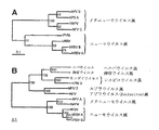
本発明は、パラミクソウイルス科(Paramyxoviridae)のニューモウイルス亜科(Pneumovirinae)に属し、系統学的にはメタニューモウイルス(Metapneumovirus)属に該当するものとして同定可能である、単離された、本質的に哺乳動物性のネガティブセンス一本鎖RNAウイルス(MPV)を提供する。このウイルスは、そのウイルスの核酸配列を決定し、その配列を系統学的解析(例えば、最尤系統樹を100のブートストラップおよび3のジャンブルを用いて作製する)において試験し、それが、鳥類の鼻気管炎の原因物質である七面鳥鼻気管炎ウイルス(TRTV)としても知られるトリ・ニューモウイルス(APV)の本質的に鳥類性のウイルス単離体に該当するというよりも、CNCM(パリ)にI-2614として寄託されているウイルス単離体に系統学的により近いものとして該当することを見出すことによって、系統学的にメタニューモウイルス属に該当するものとして同定されうる。この系統学的解析では、比較対照のアウトグループとして非MPVの核酸配列を得ることがしばしば有用であり、非常に有用なアウトグループの単離体は、例えば本明細書の図5に示すように、トリ・ニューモウイルスの血清型C(APV-C)から得ることができる。 The present invention is an isolated, essence that can be identified as belonging to the Pneumovirinae family of Paramyxoviridae and systematically identified as belonging to the genus Metapneumovirus In particular, a mammalian negative-sense single-stranded RNA virus (MPV) is provided. The virus determines the nucleic acid sequence of the virus and tests the sequence in a phylogenetic analysis (eg, creating a maximum likelihood phylogenetic tree using 100 bootstraps and 3 jumbles), which CNCM (Paris) rather than an avian virus isolate of avian pneumovirus (APV), also known as turkey rhinotracheitis virus (TRTV), the causative agent of nasal tracheitis Can be identified as phylogenetically belonging to the genus Metapneumovirus by finding it phylogenetically closer to the virus isolate deposited as I-2614. In this phylogenetic analysis, it is often useful to obtain non-MPV nucleic acid sequences as control outgroups, and very useful outgroup isolates can be obtained, for example, as shown in FIG. 5 herein. Avian pneumovirus serotype C (APV-C).
系統学的解析は、ウイルスをMPVとして同定するための便利な方法を提供するが、本明細書では、そのウイルスまたはそのウイルス由来のウイルスタンパク質もしくは核酸を同定するための、(もっと粗雑ではあるが)おそらくもっと簡単な他の方法も幾つか提供される。経験的に、MPVは、本明細書において配列または寄託により特定した単離体、ウイルスタンパク質または核酸と比較した場合の、同定しようとするウイルス、たんぱく質または核酸の相同性のパーセンテージにより同定することができる。一般に、ウイルス種、特にRNAウイルス種は、そのウイルスのクラスターがそのメンバー間で異質性(heterogeneity)を示す擬似種を構成することが多いことが知られている。したがって、各単離体は、本明細書中で提供される種々の単離体のうちの1つと、多少異なる関係(%)を有する可能性があると予期される。 Phylogenetic analysis provides a convenient way to identify a virus as an MPV, but here it is used to identify the virus or viral proteins or nucleic acids derived from the virus (although more coarsely. ) Probably some other simpler methods are also provided. Empirically, an MPV can be identified by the percentage of homology of the virus, protein or nucleic acid to be identified as compared to the isolate, viral protein or nucleic acid identified by sequence or deposit herein. it can. In general, it is known that virus species, particularly RNA virus species, often constitute pseudo-species in which the virus cluster exhibits heterogeneity among its members. Thus, it is anticipated that each isolate may have a somewhat different relationship (%) with one of the various isolates provided herein.
寄託ウイルスI-2614と比較しようとする場合に、本発明は、単離された、本質的に哺乳動物性のネガティブセンス一本鎖RNAウイルス(MPV)であって、パラミクソウイルス科のニューモウイルス亜科に属し、かつ、そのウイルスのアミノ酸配列を決定し、そのアミノ酸配列が、APV-Cと比較して、CNCM(パリ)にI-2614として寄託されているウイルス単離体と、Lタンパク質、Mタンパク質、Nタンパク質、Pタンパク質またはFタンパク質について、本明細書に記載されたパーセンテージよりも本質的に高いアミノ酸相同性のパーセンテージを有することを判定することによって、系統学的にメタニューモウイルス属に該当するものとして同定されうる上記ウイルスを提供する。あるいは、同様に、パラミクソウイルス科のニューモウイルス亜科に属する、単離された、本質的に哺乳動物性のネガティブセンス一本鎖RNAウイルス(MPV)が、そのウイルスの核酸配列を決定し、その核酸配列が、APV-Cと比較して、CNCM(パリ)にI-2614として寄託されているウイルス単離体に対して、本明細書の後記で特定されるLタンパク質、Mタンパク質、Nタンパク質、Pタンパク質またはFタンパク質をコードする核酸について本明細書中で特定されるパーセンテージよりも本質的に高い核酸同一性のパーセンテージを有することを判定することによって系統学的にメタニューモウイルス属に該当すると同定されうるものとして提供される。 When compared to the deposited virus I-2614, the present invention is an isolated, essentially mammalian negative-sense single-stranded RNA virus (MPV), which is a pneumovirus of the Paramyxoviridae family. A virus isolate that belongs to the subfamily and determines the amino acid sequence of the virus, the amino acid sequence of which is deposited as I-2614 in CNCM (Paris) compared to APV-C, and the L protein Phylogenetically, by determining that the M protein, N protein, P protein, or F protein has a percentage of amino acid homology that is essentially higher than the percentage described herein. The virus can be identified as falling within the scope of the above. Alternatively, similarly, an isolated, essentially mammalian negative-sense single-stranded RNA virus (MPV) belonging to the Pneumoviridae of the Paramyxoviridae family determines the viral nucleic acid sequence, Compared to APV-C, the nucleic acid sequence is identified as L protein, M protein, N, as specified later in this specification against the virus isolate deposited at CNCM (Paris) as I-2614. Correspondingly to the genus Metapneumovirus by determining that the nucleic acid encoding a protein, P protein or F protein has a percentage of nucleic acid identity that is essentially higher than the percentage specified herein It is then provided as something that can be identified.
ここでもまた、経験的に、MPVは、同定が必要な単離体またはその単離体のウイルスタンパク質もしくは核酸が、単離体00-1または99-1をそれぞれの比較の単離体として両者のそれぞれの群について本明細書中で特定される相同性のパーセンテージの境界に含まれる相同性のパーセンテージを有するならば、本明細書中で同定されるMPVの2つの血清学的群の一方に属する、と考えることができる。しかし、相同性のパーセンテージがそれより小さいか、あるいは更にウイルス単離体を例えばAPV-Cと区別する必要がある場合は、本明細書で特定される系統学的解析を用いることが更に好ましく推奨される。 Again, empirically, an MPV is an isolate that needs to be identified, or a viral protein or nucleic acid of that isolate, with isolate 00-1 or 99-1 as the respective comparative isolate. One of the two serological groups of MPV identified herein if it has a percentage of homology that falls within the boundaries of the percentage homology specified herein for each group of Can be thought of as belonging. However, it is more preferred to use the phylogenetic analysis specified herein if the percentage of homology is less than that, or if further virus isolates need to be distinguished from eg APV-C. Is done.
ここでもまた、相同性のパーセンテージの決定に他の単離体が選択される場合、上記のパーセンテージは多少変動する可能性があることに留意すべきである。 Again, it should be noted that the above percentages may vary somewhat if other isolates are selected to determine the percentage of homology.
このMPVが提供されると、本発明は、特に哺乳動物における疾患、更にはヒトにおける疾患、特に呼吸系疾患の診断および/または治療に用いられる診断手段および方法ならびに治療手段および方法を提供する。しかし本発明はまた、本質的に哺乳動物性のMPVと本質的に鳥類性のAPV(特にAPV-C)との遺伝学的関連(遠縁ではあるが)に基づき、トリの疾患の診断および治療に用いられる手段および方法も提供する。ウイルス学では、特定のウイルス感染の診断および/または治療は、その感染を引き起こすその特定のウイルスに最も特異的な試薬を用いて実施することが最も推奨される。この場合、これは、上記のMPV感染の診断および/または治療が、MPVに最も特異的な試薬を用いて実施されるのが好ましいことを意味する。しかし、これは、例えばもっと容易に入手可能であり当面の課題に十分に対処するという理由から、それほど特異的ではないが十分に交差反応性である試薬が代わりに用いられる可能性を決して排除するものではない。本明細書では、例えば、APVから誘導される試薬(特にAPV-Cから誘導される試薬)を用いて哺乳動物においてMPV感染のウイルス学的および/または血清学的な診断を行うことが提供され、本明細書の「詳細な説明」では、例えば、哺乳動物におけるMPV感染の十分に信頼できる血清学的診断が、トリにおいてAPV抗体を検出するように特に設計されたELISAを用いることによって達成可能であることが示されている。この目的のための特定の有用な試験としては、(例えば血清または卵黄中の)APV抗体の検出用に設計されたELISA試験であり、それの1つの商業的に入手可能なものは、SVANOVA Biotech AB(Uppsal Science Park Glunten SE-751 83 Uppsala Sweden)により製造されているAPV-Ab SVANOVIR(登録商標)として知られている。逆の状況も該当し、本明細書では、例えば、MPVから誘導される試薬を用いて、哺乳動物におけるAPV感染のウイルス学的および/または血清学的な診断を実施することが提供され、本明細書の「詳細な説明」では、例えば、鳥類におけるAPV感染の十分に信頼できる血清学的診断が、MPV抗体を検出するように設計されたELISAを用いることによって達成可能であることが示されている。抗原および抗体が「鍵と鍵穴(lock-and−key)」関係を有していることを考慮すれば、種々の抗原の検出は、十分な交差反応性を有する適切な抗体を選択することにより達成できる。勿論、そのような交差反応性に基づく場合、種々のウイルスの種々の(糖)タンパク質間に存在するアミノ酸相同性の指標に基づいて試薬(例えば抗原または抗体)を選ぶことが最も良く、その結果最も相同なタンパク質に関連する試薬が、上記の交差反応性に基づく試験で使用するのに最も有用である。 Provided with this MPV, the present invention provides diagnostic means and methods and therapeutic means and methods for use in the diagnosis and / or treatment of diseases, particularly in mammals, and even diseases in humans, particularly respiratory diseases. However, the present invention is also based on the genetic association (although distantly) of an essentially mammalian MPV and an essentially avian APV (especially APV-C) based on the diagnosis and treatment of avian diseases Also provided are the means and methods used in the above. In virology, it is most recommended that diagnosis and / or treatment of a particular viral infection be performed using reagents that are most specific for that particular virus that causes the infection. In this case, this means that the diagnosis and / or treatment of MPV infection as described above is preferably carried out using reagents that are most specific for MPV. However, this never precludes the possibility of using less specific but sufficiently cross-reactive reagents instead, for example because they are more readily available and adequately address the immediate challenges It is not a thing. Provided herein, for example, are virological and / or serological diagnosis of MPV infection in mammals using reagents derived from APV (particularly reagents derived from APV-C). In the "detailed description" herein, for example, a fully reliable serological diagnosis of MPV infection in a mammal can be achieved by using an ELISA specifically designed to detect APV antibodies in birds It is shown that. A specific useful test for this purpose is an ELISA test designed for the detection of APV antibodies (eg in serum or egg yolk), one of which is commercially available is SVANOVA Biotech Known as APV-Ab SVANOVIR®, manufactured by AB (Uppsal Science Park Glunten SE-751 83 Uppsala Sweden). The reverse situation also applies, and it is provided herein to perform a virological and / or serological diagnosis of APV infection in a mammal using, for example, reagents derived from MPV. The “detailed description” of the specification shows that, for example, a fully reliable serological diagnosis of APV infection in birds can be achieved by using an ELISA designed to detect MPV antibodies. ing. Considering that antigens and antibodies have a “lock-and-key” relationship, detection of various antigens can be achieved by selecting appropriate antibodies with sufficient cross-reactivity. Can be achieved. Of course, based on such cross-reactivity, it is best to select a reagent (eg, antigen or antibody) based on an index of amino acid homology present between different (glyco) proteins of different viruses, and as a result Reagents associated with the most homologous proteins are most useful for use in the cross-reactivity based tests described above.
核酸検出の場合、種々のウイルスの異種の核酸配列に基づいてプライマーまたはプローブを設計する(すなわち、本質的に哺乳動物性または鳥類性のメタニューモウイルス間の差異を検出するプライマーまたはプローブを設計する)のではなく、高い相同性を示すウイルス特異的核酸配列のストレッチに基づいてプライマーまたはプローブを設計または選択すればよいので、本発明の方法はさらに簡単である。一般に、核酸配列の場合、90%以上の相同性パーセンテージがあれば、ストリンジェントなハイブリダイゼーション条件を用いる診断試験においてその試験が依存する交差反応性が十分確保される。 For nucleic acid detection, primers or probes are designed based on the heterologous nucleic acid sequences of different viruses (ie, primers or probes that detect differences between essentially mammalian or avian metapneumoviruses) The method of the present invention is simpler because the primers or probes may be designed or selected based on stretches of virus-specific nucleic acid sequences that exhibit high homology. In general, for nucleic acid sequences, a homology percentage of 90% or greater ensures sufficient cross-reactivity that the test depends on in diagnostic tests that use stringent hybridization conditions.
本発明は、例えば、動物、特に哺乳動物、更に具体的にはヒトのMPV感染をウイルス学的に診断する方法であって、その動物のサンプルを本発明によるMPV特異的核酸または抗体と反応させることによって、そのサンプル中のウイルス単離体またはその成分の存在を判定することを含む上記方法、ならびに哺乳動物のMPV感染を血清学的に診断する方法であって、その哺乳動物のサンプルを本発明によるMPV特異的タンパク質様分子もしくはその断片または抗原と反応させることによって、そのサンプル中のMPVまたはその成分に対して特異的な抗体の存在を判定することを含む上記方法を提供する。また、本発明は、MPV感染を診断するための診断キットであって、本発明によるMPV、MPV特異的核酸、タンパク質様分子もしくはその断片、抗原および/または抗体を含み、さらに好ましくはそのMPV、MPV特異的核酸、タンパク質様分子もしくはその断片、抗原および/または抗体の検出手段をも含む上記キットを提供する。なおこの検出手段としては、例えば、蛍光団などの励起可能な基、または当業界で使用される酵素的検出システム(好適な診断キット様式の例としては、IF、ELISA、中和アッセイ、RT-PCRアッセイが含まれる)が挙げられる。未同定のウイルス成分またはその合成類似体(例えば核酸、タンパク質様分子もしくはその断片など)をMPV特異的なものとして同定できるか否かを判定するためには、その成分の核酸またはアミノ酸の配列を、例えばその核酸またはアミノ酸のストレッチ(好ましくは、(それぞれ)少なくとも10、更に好ましくは少なくとも25、更に好ましくは少なくとも40のヌクレオチドまたはアミノ酸からなるもの)について、例えば本明細書中で提供されるような系統学的解析を用いて既知のMPV配列および既知の非MPV配列(APV-Cが好ましく使用される)との配列相同性比較により解析すればよい。そのMPV配列または非MPV配列との関連性の程度に応じて、その成分または合成類似体が同定できる。 The present invention is a method for virologically diagnosing, for example, MPV infection in animals, in particular mammals, more particularly humans, wherein a sample of the animal is reacted with an MPV-specific nucleic acid or antibody according to the present invention A method for serologically diagnosing a mammal's MPV infection, comprising determining the presence of a virus isolate or component thereof in the sample, comprising: There is provided a method as described above comprising determining the presence of an antibody specific for MPV or a component thereof in the sample by reacting with an MPV-specific proteinaceous molecule or fragment thereof or antigen according to the invention. The present invention also provides a diagnostic kit for diagnosing MPV infection, comprising MPV, MPV-specific nucleic acid, protein-like molecule or fragment thereof, antigen and / or antibody according to the present invention, more preferably MPV, The kit is also provided comprising a MPV-specific nucleic acid, protein-like molecule or fragment thereof, antigen and / or antibody detection means. Examples of the detection means include excitable groups such as fluorophores, or enzymatic detection systems used in the art (examples of suitable diagnostic kit formats include IF, ELISA, neutralization assay, RT- PCR assays are included). To determine whether an unidentified viral component or a synthetic analog thereof (such as a nucleic acid, protein-like molecule or fragment thereof) can be identified as MPV-specific, the nucleic acid or amino acid sequence of that component is determined. For example a stretch of the nucleic acid or amino acid (preferably comprising (respectively) at least 10, more preferably at least 25, more preferably at least 40 nucleotides or amino acids), eg as provided herein What is necessary is just to analyze by sequence homology comparison with a known MPV sequence and a known non-MPV sequence (APV-C is preferably used) using phylogenetic analysis. Depending on the degree of association with the MPV or non-MPV sequence, the component or synthetic analog can be identified.
本発明はまた、哺乳動物のMPV感染をウイルス学的に診断する方法であって、その哺乳動物のサンプルをAPV(好ましくは血清型C)から誘導される交差反応性核酸またはそのAPVと反応性である交差反応性抗体と反応させることによって、そのサンプル中のウイルス単離体またはその成分の存在を判定することを含む上記方法、ならびに、哺乳動物のMPV感染を血清学的に診断する方法であって、その哺乳動物のサンプルをAPVから誘導されるタンパク質様分子またはその断片または抗原と反応させることによって、そのサンプル中のAPVとも反応する交差反応性抗体またはその成分の存在を判定することを含む上記方法を提供する。更に、本発明は、本来はAVPまたはAVP抗体の検出用に設計された診断キットの、MPV感染を診断するため、特にヒトにおけるMPV感染を検出するための使用を提供する。 The invention also provides a method for virologically diagnosing mammalian MPV infection, wherein the mammalian sample is reactive with a cross-reactive nucleic acid derived from APV (preferably serotype C) or with its APV. A method of serologically diagnosing mammalian MPV infection, as well as determining the presence of a virus isolate or component thereof in the sample by reacting with a cross-reactive antibody Determining the presence of cross-reactive antibodies or components thereof that also react with APV in the sample by reacting the mammalian sample with a protein-like molecule derived from APV or a fragment or antigen thereof. Including the above methods. Furthermore, the present invention provides the use of diagnostic kits originally designed for the detection of AVP or AVP antibodies for diagnosing MPV infection, particularly for detecting MPV infection in humans.
また、本発明は、鳥類におけるAPV感染をウイルス学的に診断する方法であって、その鳥類のサンプルをMPVから誘導される交差反応性核酸またはMPVと反応性である交差反応性抗体と反応させることによって、そのサンプル中のウイルス単離体またはその成分の存在を判定することを含む上記方法、ならびに、鳥類のAPV感染を血清学的に診断する方法であって、その鳥類のサンプルをMPVから誘導されるタンパク質様分子もしくはその断片または抗原と反応させることによって、そのサンプル中のMPVとも反応する交差反応性抗体またはその成分の存在を判定することを含む上記方法を提供する。更に、本発明は、本来はMVPまたはMVP抗体の検出用に設計された診断キットの、APV感染を診断するため、特にニワトリ、アヒルまたは七面鳥などの家禽におけるAPV感染を検出するための使用を提供する。 The present invention also relates to a method for virologically diagnosing APV infection in birds, wherein a sample of the bird is reacted with a cross-reactive nucleic acid derived from MPV or a cross-reactive antibody reactive with MPV. A method for serologically diagnosing avian APV infection, comprising determining the presence of a virus isolate or component thereof in the sample, wherein the avian sample is obtained from the MPV. There is provided a method as described above comprising determining the presence of a cross-reactive antibody or component thereof that also reacts with MPV in the sample by reacting with a derived protein-like molecule or fragment thereof or antigen. Furthermore, the present invention provides the use of diagnostic kits originally designed for the detection of MVP or MVP antibodies to diagnose APV infections, particularly for detecting APV infections in poultry such as chickens, ducks or turkeys To do.
上記で述べたように、治療の場合、見出された交差反応性を用いて同様の使用を実施することが可能であるが、これは特に、当面の状況によって、より相同性の高い方法がそれほど簡単ではない場合に実施されうる。MPV感染に対する緊急のワクチン接種のように、待つことができないワクチン接種は、例えば、より相同性の高いMPVワクチンが入手不可能な場合は、APV(好ましくはC型)の単離体から誘導されるワクチン製剤を用いて行うことが可能であり、またその逆に、APV感染に対するワクチン接種をMPVから誘導されるワクチン製剤を用いて行うことも考えられる。また、逆遺伝学的技法により、ワクチンとして有用なキメラAPV-MPVウイルス構築物の作製が可能になり、これはそれぞれの株の各々の単離体を望ましいレベルまで弱毒化する分野とは十分に異なるものである。同様の逆遺伝学的技法により、ワクチン製剤において使用するためのキメラパラミクソウイルス-メタニューモウイルス構築物(例えばRSV-MPVまたはPI3-MPV構築物)を作製することも可能になる。そうした構築物は、気道疾患を撲滅するための混合ワクチンとして特に有用である。 As mentioned above, in the case of treatment, it is possible to carry out similar uses with the found cross-reactivity, but this is especially true depending on the situation. It can be implemented when it is not so simple. Vaccinations that cannot be waited for, such as emergency vaccination against MPV infection, are derived from isolates of APV (preferably type C), for example if a more homologous MPV vaccine is not available Vaccination against APV infection can also be conceived using a vaccine formulation derived from MPV. In addition, reverse genetic techniques allow the creation of chimeric APV-MPV viral constructs useful as vaccines, which are well different from the field of attenuating each isolate of each strain to the desired level. Is. Similar reverse genetic techniques also make it possible to create chimeric paramyxovirus-metapneumovirus constructs (eg, RSV-MPV or PI3-MPV constructs) for use in vaccine formulations. Such constructs are particularly useful as combination vaccines to eradicate airway diseases.
したがって、本発明は、新規な原因物質、すなわち、パラミクソウイルス科のニューモウイルス亜科に属するが、古典的なニューモウイルスとしては同定されず、かつ、メタニューモウイルス属に属する、単離された本質的に哺乳動物性のネガティブセンス一本鎖RNAウイルス(本明細書中ではMPVとも呼ぶ)、ならびにMPV特異的成分またはその合成類似体を提供する。メタニューモウイルスに類似する哺乳動物性ウイルス、すなわち本質的にそのウイルスの天然の宿主として機能する哺乳動物から単離できるメタニューモウイルス、あるいはその哺乳動物において疾患を引き起こすメタニューモウイルスは、現在のところ、見出されていない。メタニューモウイルスは、天然の宿主または疾患の原因物質によって基本的には家禽に限定されると一般には考えられており、トリ・ニューモウイルスとしても知られている。近年、アヒルのAPV単離体が記載され(FR 2 801 607)、APV感染が天然の宿主として基本的に鳥類に限られることを更に実証している。
Thus, the present invention is a novel causative agent, ie, belonging to the Pneumovirus subfamily of the Paramyxoviridae family, but not identified as a classic pneumovirus and belonging to the Metapneumovirus genus Provided are essentially mammalian negative-sense single-stranded RNA viruses (also referred to herein as MPV), as well as MPV-specific components or synthetic analogs thereof. Mammalian viruses similar to metapneumovirus, that is, metapneumoviruses that can be isolated from mammals that essentially function as the natural host of the virus, or metapneumoviruses that cause disease in the mammal, are currently , Not found. Metapneumovirus is generally considered to be limited to poultry basically by the natural host or the causative agent of the disease, and is also known as avian pneumovirus. Recently, duck APV isolates have been described (
本発明は、ニューモウイルス属のものとは異なる遺伝子順序およびアミノ酸配列を有し、且つパラミクソウイルス科のニューモウイルス亜科に含まれるメタニューモウイルス属と近縁であり、その系統学的関連を考慮するとその属に属すると思われる、単離された哺乳動物ニューモウイルス(本明細書中ではMPVとも呼ぶ)を提供する。現在まで、メタニューモウイルスは鳥類からしか単離されていなかったが、本発明により、哺乳動物などの他の動物種においても、関連する(しかし物質的には異なる)ウイルスが同定できることが示される。ここで、本発明者らは、ヒトからのMPVの反復単離を示すが、そのような報告はAPVに関しては存在しない。更に、APVとは違って、MPVは基本的にニワトリや七面鳥において全く、または殆ど複製しないが、カニクイザルにおいては容易に複製する。哺乳動物におけるAPVの複製に関する報告は見られない。更に、MPVに対して生起させた特異的抗血清はMPVを中和するが、APV A、BまたはCに対して生起させた抗血清はそれと同程度にはMPVを中和せず、そして、このように完全な交差反応性が備わっていないことは、MPVが異なるメタニューモウイルスであることのもう1つの証拠となっている。更に、APVとMPVとはほぼ同様の遺伝子順序を有するが、MPVのGおよびSHタンパク質は、それらがアミノ酸レベルでも核酸レベルでも有意な配列相同性を示さない、という点で、APVでの知見とは大きく異なっている。APV単離体とMPV単離体、またはこれらの異なるウイルスに対する抗体同士を区別する診断アッセイは、これらのタンパク質の一方または双方に基づいて有利に開発できる(例えばIF、ELISA、中和アッセイ、RT-PCRアッセイ)。しかし、MPVのより関連するN、P、M、FおよびLタンパク質から得られる配列および/または抗原性情報、ならびにAPVのそれぞれのタンパク質との配列相同性の解析もまた、APVとMPVとを区別するのに用いることができる。例えば、MPVから得られる配列情報の系統学的解析から、MPVおよびAPVが2種の異なるウイルスであることが明らかになった。特に、系統樹は、APVおよびMPVが2種の異なるウイルスの系列であることを示している。また、本発明者らは、MPVがヒト集団において少なくとも50年間にわたって広まってきており、そのために、種間伝播は恐らく少なくとも50年前から起こっていて、ありふれた事象ではないことも示した。MPVのCPEは、tMKまたは他の細胞培養物中でhRSVもしくはhPIV-1により引き起こされるCPEとは実際には区別不能だったので、MPVは現在まで余り注目されていなかったと思われる。tMK(サル腎三次細胞、すなわち細胞培養の第三継代のMK細胞)は、初代または二代培養物と比較してコストが少なくて済むので、好ましく使用される。CPEは、古典的なパラミクソウイルス科の幾つかの場合と同様に、合胞体形成し、その後で細胞が急速な内部破壊を示し、続いて細胞が単層から剥離することを特徴とする。細胞は通常(しかしいつもではない)、初期材料から3代継代した後、接種後10〜14日でCPEを示し、これはhRSVまたはhPIV-1などの他のウイルスにより起こるCPEよりも幾分遅い。 The present invention has a gene order and amino acid sequence different from those of the genus Pneumovirus, and is closely related to the genus Metapneumovirus included in the Pneumovirus subfamily of the Paramyxoviridae family. Provided is an isolated mammalian pneumovirus (also referred to herein as MPV) that appears to belong to that genus when considered. To date, metapneumoviruses have only been isolated from birds, but the present invention shows that related (but materially different) viruses can be identified in other animal species such as mammals. . Here we show repeated isolation of MPV from humans, but no such report exists for APV. Furthermore, unlike APV, MPV basically replicates very little or not in chickens and turkeys, but easily replicates in cynomolgus monkeys. There are no reports of APV replication in mammals. Furthermore, specific antisera raised against MPV neutralize MPV, whereas antisera raised against APV A, B or C do not neutralize MPV to the same extent, and This lack of complete cross-reactivity is another evidence that MPV is a different metapneumovirus. Furthermore, although APV and MPV have approximately the same gene order, MPV G and SH proteins do not show significant sequence homology at the amino acid level or at the nucleic acid level. Are very different. Diagnostic assays that distinguish antibodies against APV and MPV isolates, or antibodies against these different viruses, can be advantageously developed based on one or both of these proteins (eg, IF, ELISA, neutralization assay, RT -PCR assay). However, analysis of the sequence and / or antigenic information obtained from the more relevant N, P, M, F and L proteins of MPV and the sequence homology of each protein of APV also distinguishes APV from MPV. Can be used to For example, phylogenetic analysis of sequence information obtained from MPV revealed that MPV and APV are two different viruses. In particular, the phylogenetic tree shows that APV and MPV are a series of two different viruses. The inventors have also shown that MPV has been widespread in the human population for at least 50 years, so interspecies transmission has probably occurred at least 50 years ago and is not a common event. Since MPV CPE was actually indistinguishable from that induced by hRSV or hPIV-1 in tMK or other cell cultures, MPV appears to have received less attention to date. tMK (monkey kidney tertiary cells, ie, MK cells in the third passage of cell culture) is preferably used because it costs less compared to primary or secondary cultures. CPE, like some cases of the classical Paramyxoviridae, is characterized by syncytia after which cells exhibit rapid internal destruction followed by cell detachment from the monolayer. Cells usually (but not always) show CPE after 3 passages from initial material, 10-14 days after inoculation, which is somewhat higher than CPE caused by other viruses such as hRSV or hPIV-1 slow.
従来、影響力の大きい疾患原因として、パラミクソウイルスは毎年世界的に多くの動物やヒトの死亡の原因となっている。パラミクソウイルス科はモノネガウイルス目(ネガティブセンス一本鎖RNAウイルス)に含まれる科を形成し、パラミクソウイルス亜科(Paramyxovirinae)とニューモウイルス亜科(Pneumovirinae)とからなる。後者の亜科は現在のところ、分類学上、ニューモウイルス属とメタニューモウイルス属とに分類される1。ヒト呼吸器合胞体ウイルス(hRSV)は、ニューモウイルス属の典型種であり、世界的に幼年期および初期小児期の下部気道感染の最も重要な単独原因である2。ニューモウイルス属の他のメンバーとしては、ウシおよびヒツジの呼吸器合胞体ウイルスおよびマウス肺炎ウイルス(PVM)が挙げられる。 Conventionally, paramyxovirus is the cause of death of many animals and humans worldwide every year as a cause of high-impact disease. The Paramyxoviridae form a family included in the order of the Mononegavirus (negative sense single-stranded RNA virus), and consists of the Paramyxovirinae and the Pneumovirinae. The latter subfamily is currently classified taxonomically as genus Pneumovirus and Metapneumovirus 1 . Human respiratory syncytial virus (hRSV) is a typical species of the genus Pneumovirus and is the most important single cause of lower respiratory tract infection in childhood and early childhood worldwide 2 . Other members of the genus Pneumovirus include bovine and sheep respiratory syncytial virus and mouse pneumonia virus (PVM).
七面鳥鼻気管炎ウイルス(TRTV)としても知られるトリ・ニューモウイルス(APV)は、鳥類の鼻気管炎、七面鳥の上部気道感染の原因物質であり3、近年になって区分されたメタニューモウイルス属の唯一のメンバーであり、上記で述べたように、これまで哺乳動物の感染と、ましてや疾患とも関係付けられていなかった。APVの血清学的亜群は、G糖タンパク質のヌクレオチド配列もしくはアミノ酸配列、およびG糖タンパク質をも認識するモノクローナル抗体を用いた中和試験に基づいて区別できる。亜群A、BおよびDの中で、G糖タンパク質は、亜群内では98.5%〜99.7%のアミノ酸配列同一性を示すが、亜群間ではたった31.2〜38%のアミノ酸同一性しか見られない。例えば、Collins, M.S., Gough, R.E.およびAlexander, D.J., Antigenic differentiation of avian pneumovirus isolates using polyclonal antisera and mouse monoclonal antibodies. Avian Pathology, 1993. 22:p.469-479;Cook, J.K.A, Jones, B.V., Ellis, M.M., Antigenic differentiation of strains of turkey rhinotracheitis virus using monoclonal antibodies. Avian Pathplogy, 1993.22:p.257-273;Bayon-Auboyer, M.H.,ら, Nucleotide sequences of the F, L and G protein genes of two non-A/non-B avian pneumoviruses (APV) reveal a novel APV subgroup. J Gen Virol, 2000.81(Pt 11):p.2723-33;Seal, B.S., Matrix Protein gene nucleotide and predicted amino acid sequence demonstrate that the first US avian pneumovirs isolate is distinct from European strains. Virus Res, 1998.58(1-2):p.45-52;Bayon-Auboyer, M.H.,ら, Comparison of F-, G- and N-based RT-PCR protocols wigh conventional virological procedures for the detection and typing of turkey rhinotrachetis virus. Arch Virol, 1999.144(6):p.1091-109;Juhasz, K.およびA.J. Easton, Extensive sequence variation in the attachment (G) protein gene of avian pneumovirus: evidence for two distinct subgroups. J Gen Virol, 1994.75(Pt 11):p.2873-80を参照されたい。 Avian pneumovirus (APV), also known as turkey rhinotracheitis virus (TRTV), is the causative agent of avian rhinotracheitis and turkey upper respiratory tract infection 3 , a recently classified metapneumovirus genus And, as mentioned above, has never been associated with mammalian infection or even disease. Serological subgroups of APV can be distinguished based on the nucleotide or amino acid sequence of the G glycoprotein and neutralization tests using monoclonal antibodies that also recognize the G glycoprotein. Among subgroups A, B and D, G glycoproteins show 98.5% -99.7% amino acid sequence identity within subgroups, but only 31.2-38% amino acid identity between subgroups Absent. For example, Collins, MS, Gough, RE and Alexander, DJ, Antigenic differentiation of avian pneumovirus isolates using polyclonal antisera and mouse monoclonal antibodies. Avian Pathology, 1993. 22: p.469-479; Cook, JKA, Jones, BV, Ellis , MM, Antigenic differentiation of strains of turkey rhinotracheitis virus using monoclonal antibodies. Avian Pathplogy, 1993.22: p.257-273; Bayon-Auboyer, MH, et al., Nucleotide sequences of the F, L and G protein genes of two non-A / non-B avian pneumoviruses (APV) reveal a novel APV subgroup.J Gen Virol, 2000.81 (Pt 11): p.2723-33; Seal, BS, Matrix Protein gene nucleotide and predicted amino acid sequence demonstrate that the first US avian pneumovirs isolate is distinct from European strains. Virus Res, 1998.58 (1-2): p.45-52; Bayon-Auboyer, MH, et al., Comparison of F-, G- and N-based RT-PCR protocols wigh conventional virological procedures for the detection and typing of turkey rhinotrachetis virus. Arch Virol, 1999.144 (6): p.1091-109; Juhasz, K. and A See J Easton, Extensive sequence variation in the attachment (G) protein gene of avian pneumovirus: evidence for two distinct subgroups. J Gen Virol, 1994.75 (Pt 11): p.2873-80.
APVのもう1つの亜群はWO00/20600に提供されており、そこでは、APVのコロラドで得られた単離体を記載しており、それをin vitro血清中和試験を用いて既知のAPVまたはTRTと比較した。まず、コロラドの単離体を、認識されたTRT単離体に対する単一特異的ポリクローナル抗血清に対して試験した。コロラドの単離体は、TRT株のいずれに対する単一特異的抗血清によっても中和されなかった。しかし、それは、亜群A株に対して作った超免疫抗血清によって中和された。この抗血清は、相同なウイルスを1:400の力価まで、コロラド単離体を1:80の力価まで中和した。次に、上記の方法を用いて、コロラドの単離体をTRTモノクローナル抗体に対して試験した。それぞれの場合において、逆中和の力価は10未満であった。コロラドの単離体に対して生起させた単一特異的抗血清は、両者の亜群のTRT株に対しても試験した。試験したTRT株はいずれも、コロラドの単離体に対する抗血清によって中和されなかった。 Another subgroup of APV is provided in WO00 / 20600, which describes an isolate obtained with Colorado of APV, which is known to be a known APV using an in vitro serum neutralization test. Or compared with TRT. First, Colorado isolates were tested against monospecific polyclonal antisera against recognized TRT isolates. The Colorado isolate was not neutralized by monospecific antisera against any of the TRT strains. However, it was neutralized by a hyperimmune antiserum raised against subgroup A strains. This antiserum neutralized the homologous virus to a titer of 1: 400 and the Colorado isolate to a titer of 1:80. The Colorado isolate was then tested against the TRT monoclonal antibody using the method described above. In each case, the reverse neutralization titer was less than 10. Monospecific antisera raised against Colorado isolates were also tested against both subgroups of TRT strains. None of the TRT strains tested were neutralized by antisera against the Colorado isolate.
APVのコロラド株は、TRTウイルスの亜群A株または亜群B株によるチャンレンジに対してSPFニワトリを保護しなかった。これらの結果から、コロラドの単離体が、Bayon-Auboyerら(J. Gen. Vir. 81:2723-2733 (2000))によっても示唆されているように、トリ・ニューモウイルスのさらなる血清型の最初の例である可能性があることが示唆される。 The Colorado strain of APV did not protect SPF chickens against challenge by subgroup A or subgroup B of the TRT virus. These results indicate that Colorado isolates were found to be further avian pneumovirus serotypes, as suggested by Bayon-Auboyer et al. (J. Gen. Vir. 81: 2723-2733 (2000)). It is suggested that this may be the first example.
1つの好ましい実施態様において、本発明は、分類学的には(これに関しては未知の哺乳動物の)メタニューモウイルスに該当し、パラミクソウイルス科のニューモウイルス亜科に含まれるニューモウイルスのものとは異なる遺伝子順序を含む、単離されたMPVを提供する。これら2種の属の分類は、主に、遺伝子の位置に基づいている。メタニューモウイルスには一般に、NS1またはNS2などの非構造タンパク質が欠けており(Randhawaら, J. Vir. 71:9849-9854 (1997)も参照されたい)、遺伝子の順序はニューモウイルスとは異なっている(RSV:’3-NS1-NS2-N-P-M-SH-G-F-M2-L-5’:APV:’3-N-P-M-F-M2-SH-G-L-5’)4,5,6。本発明により提供されるMPVまたは分類学的にそれに該当するウイルス単離体は、EM分析を行ったところ、パラミクソウイルス様の粒子であることが判明した。この分類と一致して、MPVまたは系統学的もしくは分類学的にそれに該当するウイルス単離体は、クロロホルムによる処理に対して感受性であり、tMK細胞または機能的にそれと同等の細胞において最適に培養され、大部分の細胞培養物において基本的にトリプシン依存性である。更に、典型的なCPEや、最も古典的に用いられる赤血球での赤血球凝集活性がないことから、本発明で提供されるウイルスが、(遠縁ではあるが)RSVなどの古典的なニューモウイルスと関連することが示唆される。多くのパラミクソウイルスは赤血球凝集活性を有するが、ニューモウイルスの大部分は有していない13。本発明によるMPVは、例えばAhmadianら, J. Gen. Vir. 80:2011-2016(1999)において示されているような多くの他のニューモウイルスが一般的にそうであるように、M2タンパク質をコードする核酸断片の内部に第2の重複するORF(M2-2)も含んでいる。 In one preferred embodiment, the invention is taxonomically (in this regard of an unknown mammal) metapneumovirus and is of the pneumovirus included in the Pneumovirus subfamily of the Paramyxoviridae family Provides isolated MPV containing different gene orders. The classification of these two genera is mainly based on gene location. Metapneumoviruses generally lack nonstructural proteins such as NS1 or NS2 (see also Randhawa et al., J. Vir. 71: 9849-9854 (1997)), and the gene order differs from that of pneumoviruses. (RSV: '3-NS1-NS2-NPM-SH-GF-M2-L-5': APV: '3-NPMF-M2-SH-GL-5') 4,5,6 . The MPV provided by the present invention or a virus isolate taxonomically corresponding to it was found to be paramyxovirus-like particles by EM analysis. Consistent with this classification, MPV or phylogenetically or taxonically equivalent virus isolates are sensitive to treatment with chloroform and optimally cultured in tMK cells or functionally equivalent cells. And is essentially trypsin-dependent in most cell cultures. Furthermore, because of the lack of hemagglutination activity in typical CPE and the most classically used erythrocytes, the viruses provided by the present invention are related to classic pneumoviruses such as RSV (although distantly). It is suggested to do. Many paramyxoviruses have hemagglutination activity, but do not have the majority of pneumoviruses 13 . MPVs according to the present invention may contain M2 protein, as is generally the case with many other pneumoviruses, for example as shown in Ahmadian et al., J. Gen. Vir. 80: 2011-2016 (1999). A second overlapping ORF (M2-2) is also included within the encoding nucleic acid fragment.
本発明により提供されるような別のウイルス単離体を見出すためには、サンプル(場合よっては罹患している動物またはヒトから得たもの)を、ニューモウイルス亜科のウイルスの存在について試験し、こうして得られたウイルスを、(機能性の)NS1またはNS2をコードする遺伝子の存在について試験するか、あるいは既に上記で既に述べたようにRSVなどのニューモウイルスのものとは異なる遺伝子順序を実証すればよい。更に、系統学的にMPVに該当し、したがって分類学的にMPVに該当するウイルス単離体は、ここで提供されるMPV単離体からの核酸を用いる交差ハイブリダイゼーション実験により、あるいはMPV単離体に対して特異的なモノクローナル抗体および/またはMPV単離体から特異的に誘導される抗原および/もしくは免疫原を用いる古典的な交差血清学実験で見出すことができる。 In order to find another virus isolate as provided by the present invention, a sample (optionally obtained from an affected animal or human) is tested for the presence of a virus of the Pneumovirus subfamily. Test the resulting virus for the presence of a (functional) NS1 or NS2 encoding gene or demonstrate a different gene order from that of pneumoviruses such as RSV as already mentioned above do it. In addition, virus isolates that are phylogenetically MPV and therefore taxonomically MPV can be obtained by cross-hybridization experiments using nucleic acids from the MPV isolates provided herein or by MPV isolation. It can be found in classical cross serology experiments using antigens and / or immunogens specifically derived from body-specific monoclonal antibodies and / or MPV isolates.
新たに単離されたウイルスは、本発明者らのプロトタイプMPV単離体と十分に類似する遺伝子順序および/またはアミノ酸配列を含んでいるならば、系統学的にMPVに該当し、したがって分類学的にMPVに該当し、あるいは構造的にMPVと対応し、ニューモウイルス亜科のメタニューモウイルス属と近い関連を示す。MPVと現在既知の同じ科の他のウイルスのいずれか(APVの亜型C)との間での、最大のアミノ配列相同性および個々のタンパク質レベルで構造的対応の規定は、図6に示す配列を他のウイルス(特にAPV-C)の配列と比較した場合に推定できるように、マトリックスについては87%、核タンパク質については88%、リン酸化タンパク質については68%、融合タンパク質については81%、ポリメラーゼタンパク質の一部については56〜64%である。これらの記載する最大値よりも高い相同性をそれぞれ有する個々のタンパク質またはウイルス単離体全体は、系統学的にMPVに該当し、したがって分類学的にMPVに該当しており、構造的に図6に示す配列に該当する核酸配列を含む。ここで、本発明は、寄託されたウイルスに系統学的に該当するウイルスを提供する。他のウイルスと同様に、ここで提供される各種の単離された本質的に哺乳動物性のネガティブセンス一本鎖RNAウイルス単離体の間で、ある程度の変異性が見られることに注目すべきである。系統樹において、本発明者らは、L、M、NおよびF遺伝子の一部の比較配列解析に基づいて、ウイルス単離体の少なくとも2つの遺伝的クラスターを同定した。ウイルスの核酸配列またはアミノ酸配列(ウイルス配列)におけるヌクレオチドおよびアミノ酸の違いと、RSVなどの他のニューモウイルスとの類似性に基づくと、これらのMPVの遺伝子型はMPVの亜型に該当する。MPV単離体の遺伝的クラスターの各々の中で、ヌクレオチドレベルでの同一性のパーセンテージは、Lについては94〜100、Mについては91〜100、Nについては90〜100、Fについては93〜100であり、アミノ酸レベルでの同一性のパーセンテージは、Lについては91〜100、Mについては98〜100、Nについては96〜100、Fについては98〜100であることが判った。更なる比較は、図18〜28に見ることができる。これまでに同定されたここで提供される単離された本質的に哺乳動物性のネガティブセンス一本鎖RNAウイルス(MPV単離体)のグループ全体についてのヌクレオチドレベルでの最低の同一性のパーセンテージは、LおよびMについては81であり、Nについては83であり、Fについては82であった。アミノ酸レベルでは、このパーセンテージは、LおよびNについては91であり、Mについては94であり、Fについては95であった。本発明で提供されるMPV単離体または単離されたMPVのF遺伝子のウイルス配列は、例えば、Sealら, Vir. Res. 66:139147(2000)により提供されるAPV-Cの融合遺伝子(F)のヌクレオチド配列またはアミノ酸配列に対して、それぞれ81%未満のヌクレオチド配列同一性または82%未満のアミノ酸配列同一性を示す。 A newly isolated virus falls phylogenetically as an MPV if it contains a gene sequence and / or amino acid sequence that is sufficiently similar to our prototype MPV isolate and therefore taxonomic Correspondingly to MPV, or structurally corresponding to MPV, shows close association with the Pneumovirus subfamily Metapneumovirus. The definition of maximum amino sequence homology and structural correspondence at the individual protein level between MPV and any other virus of the same family currently known (APV subtype C) is shown in FIG. 87% for the matrix, 88% for the nucleoprotein, 68% for the phosphorylated protein, 81% for the fusion protein, as deduced when comparing the sequence to that of other viruses (especially APV-C) , 56-64% for some of the polymerase proteins. Each individual protein or virus isolate with a homology higher than these stated maximum values is phylogenetically classified as MPV and therefore taxonomically classified as MPV. A nucleic acid sequence corresponding to the sequence shown in 6 is included. Here, the present invention provides a virus that corresponds phylogenetically to the deposited virus. Note that, like other viruses, there is some variability between the various isolated essentially mammalian negative-sense single-stranded RNA virus isolates provided herein. Should. In the phylogenetic tree, we identified at least two genetic clusters of virus isolates based on comparative sequence analysis of portions of the L, M, N and F genes. Based on nucleotide and amino acid differences in viral nucleic acid or amino acid sequences (viral sequences) and similarities to other pneumoviruses such as RSV, these MPV genotypes fall under the MPV subtype. Within each genetic cluster of MPV isolates, the percentage of identity at the nucleotide level is 94-100 for L, 91-100 for M, 90-100 for N, 93-93 for F. The percentage of identity at the amino acid level was found to be 91-100 for L, 98-100 for M, 96-100 for N, and 98-100 for F. A further comparison can be seen in FIGS. Minimum percentage identity at the nucleotide level for the entire group of isolated essentially mammalian negative sense single stranded RNA viruses (MPV isolates) provided heretofore identified Was 81 for L and M, 83 for N and 82 for F. At the amino acid level, this percentage was 91 for L and N, 94 for M and 95 for F. The viral sequence of the MPV isolate or isolated MPV F gene provided in the present invention can be obtained from, for example, the APV-C fusion gene provided by Seal et al., Vir. Res. 66: 139147 (2000) ( F) less than 81% nucleotide sequence identity or less than 82% amino acid sequence identity to the nucleotide or amino acid sequence of F), respectively.
また、本発明で提供されるMPV単離体または単離されたMPVのL遺伝子のウイルス配列は、例えば、Randhawaら, J. Gen. Vir. 77:3047-3051(1996)により提供されるAPV-Aのポリメラーゼ遺伝子のヌクレオチド配列またはアミノ酸配列に対して、61%未満のヌクレオチド配列同一性または63%未満のアミノ酸配列同一性を示す。 In addition, the viral sequence of the MPV isolate or isolated MPV L gene provided in the present invention is, for example, the APV provided by Randhawa et al., J. Gen. Vir. 77: 3047-3051 (1996). It shows less than 61% nucleotide sequence identity or less than 63% amino acid sequence identity to the nucleotide or amino acid sequence of the polymerase gene of -A.
世界中のMPV株の配列多様性は、他のウイルスと同様に多少高めである可能性がある。したがって、2つの可能性のある遺伝的クラスターは、9種のウイルス単離体のN、M、FおよびLのORFにおける部分ヌクレオチド配列の分析により同定される。1つのクラスター内で、90〜100%のヌクレオチド同一性が見られ、クラスター間では81〜88%の同一性が見られた。更なるウイルス単離体について得られた配列情報から、2つの遺伝子型の存在が確認された。クラスターAのプロトタイプとしてのウイルス単離体ned/00/01、およびクラスターBのプロトタイプとしてのウイルス単離体ned/99/01を交差中和アッセイで用いて、それらの遺伝子型が異なる血清型または亜群と関連するか否かを調べた。これらのデータから、単離体I-2614に対してLについては64より、M2については87より、Nについては88より、Pについては68より、Fについては81より、M-1については84より、またはM2-2については58より高いアミノ酸相同性のパーセンテージを示す本質的に哺乳動物性のウイルス単離体は、本発明で提供される単離された本質的に哺乳動物性のネガティブセンス一本鎖RNAウイルスとして分類できる、と結論づけられる。特に、一般にヌクレオチド配列レベルでは、本発明で提供されるプロトタイプMPV単離体と、LおよびMについては81、Nについては83且つ/またはFについては82、という最低の同一性のパーセンテージを有するウイルス単離体は、本発明で提供されるMPV単離体のグループのメンバーである。アミノ酸レベルでは、これらのパーセンテージは、LおよびNについては91、Mについては94、且つ/またはFについては95である。所与のウイルス単離体についてのアミノ酸配列相同性のパーセンテージが、LおよびNについては90より、Mについては93より、またはFについては94より高い場合、そのウイルス単離体は、図5に示すMPV単離体のグループと同じものである。所与のウイルス単離体についてのアミノ酸配列相同性のパーセンテージが、Lについては94より、Nについては95より、またはMおよびFについては97より高い場合、そのウイルス単離体は、図5に示す遺伝子型クラスターの1つに属すると同定できる。これらの相同性のパーセンテージ(これにより遺伝的クラスターが規定される)が、RSVの対応する遺伝子の遺伝的クラスター間で見られる相同性の程度とほぼ同じであることに注目すべきである。 The sequence diversity of MPV strains around the world may be somewhat higher as with other viruses. Thus, two possible genetic clusters are identified by analysis of partial nucleotide sequences in the N, M, F and L ORFs of nine viral isolates. Within one cluster, 90-100% nucleotide identity was seen and between clusters 81-88% identity was seen. The sequence information obtained for further virus isolates confirmed the presence of two genotypes. Virus isolates ned / 00/01 as prototypes for cluster A and virus isolates ned / 99/01 as prototypes for cluster B were used in a cross-neutralization assay to determine whether their genotypes differed Whether it was related to subgroups was examined. From these data, L for isolate I-2614 is 64 for L, 87 for M2, 88 for N, 68 for P, 81 for F and 84 for M-1. Or an essentially mammalian viral isolate exhibiting a percentage of amino acid homology greater than 58 for M2-2 is an isolated essentially mammalian negative sense provided herein. It can be concluded that it can be classified as a single-stranded RNA virus. In particular, generally at the nucleotide sequence level, a virus having the lowest percentage identity with the prototype MPV isolate provided in the present invention, 81 for L and M, 83 for N and / or 82 for F The isolate is a member of the group of MPV isolates provided in the present invention. At the amino acid level, these percentages are 91 for L and N, 94 for M, and / or 95 for F. If the percentage of amino acid sequence homology for a given viral isolate is higher than 90 for L and N, 93 for M, or 94 for F, the viral isolate is shown in FIG. Same as the group of MPV isolates shown. If the percentage of amino acid sequence homology for a given viral isolate is higher than 94 for L, 95 for N, or 97 for M and F, the viral isolate is shown in FIG. It can be identified as belonging to one of the genotype clusters shown. It should be noted that the percentage of these homology (which defines the genetic cluster) is approximately the same as the degree of homology found between the genetic clusters of the corresponding genes in RSV.
つまり、本発明は、単離された、本質的に哺乳動物性のネガティブセンス一本鎖RNAウイルス(MPV)であって、パラミクソウイルス科のニューモウイルス亜科に属するものであり、かつ、そのウイルスのゲノムの適当な断片の核酸配列を決定し、それを、最尤系統樹が100のブートストラップおよび3のジャンブルを用いて作製される系統樹解析で試験し、それが、鳥類の鼻気管炎の原因物質である七面鳥鼻気管炎ウイルス(TRTV)としても知られるトリ・ニューモウイルス(APV)のウイルス単離体に該当するというよりも、CNCM(パリ)にI-2614として寄託されているウイルス単離体に系統学的により近いものとして該当することを見出すことにより、系統学的にメタニューモウイルス属に該当するとして同定されうる上記ウイルスを提供する。 That is, the present invention is an isolated, essentially mammalian negative-sense single-stranded RNA virus (MPV) that belongs to the Pneumovirus subfamily of the Paramyxoviridae family, and Determine the nucleic acid sequence of the appropriate fragment of the viral genome and test it with a phylogenetic tree analysis in which the maximum likelihood phylogenetic tree is created using 100 bootstraps and 3 jumbles, which is the avian nasal trachea Rather than being a virus isolate of the avian pneumovirus (APV), also known as turkey rhinotracheitis virus (TRTV), the causative agent of the flame, it is deposited as I-2614 in the CNCM (Paris) Finding that it falls phylogenetically closer to a virus isolate provides the virus that can be identified phylogenetically as belonging to the genus Metapneumovirus.
そうした系統樹解析にそれぞれ有用な好適な核酸ゲノム断片は、例えば、本明細書の「詳細な説明」において開示されるRAP-PCR断片1〜10のいずれかであり、本明細書において図4または5に開示されるような種々の系統樹解析が導かれる。MPVの核タンパク質(N)、リン酸化タンパク質(P)、マトリックスタンパク質(M)および融合タンパク質(F)の遺伝子の系統樹解析を行ったところ、APV血清型C(アメリカ合衆国で主に鳥類で見られるトリ・ニューモウイルス)と、最も高い配列相同性を示すことが判明した。 Suitable nucleic acid genomic fragments each useful for such phylogenetic tree analysis are, for example, any of the RAP-PCR fragments 1-10 disclosed in the “Detailed Description” herein, where FIG. Various phylogenetic tree analyzes as disclosed in 5 are derived. A phylogenetic analysis of MPV nucleoprotein (N), phosphorylated protein (P), matrix protein (M) and fusion protein (F) genes revealed that APV serotype C (found mainly in birds in the United States) Avian pneumovirus) and the highest sequence homology.
1つの好ましい実施形態において、本発明は、単離された、本質的に哺乳動物性のネガティブセンス一本鎖RNAウイルス(MPV)であって、パラミクソウイルス科のニューモウイルス亜科に属するものであり、かつ、そのウイルスのゲノムの適当な断片の核酸配列を決定し、それを、最尤系統樹が100のブートストラップおよび3のジャンブルを用いて作製される系統樹解析で試験し、それが、鳥類の鼻気管炎の原因物質である七面鳥鼻気管炎ウイルス(TRTV)としても知られるトリ・ニューモウイルス(APV)のウイルス単離体に該当するというよりも、CNCM(パリ)にI-2614として寄託されているウイルス単離体に系統学的により近いものとして該当することを見出すことにより、系統学的にメタニューモウイルス属に該当するとして同定可能であり、但し、その適当な断片がそのウイルスのウイルスタンパク質をコードするオープンリーディングフレームを含む、上記ウイルスを提供する。 In one preferred embodiment, the present invention is an isolated, essentially mammalian negative-sense single-stranded RNA virus (MPV) that belongs to the Pneumovirus subfamily of the Paramyxoviridae family. And determine the nucleic acid sequence of the appropriate fragment of the viral genome and test it with a phylogenetic tree analysis in which the maximum likelihood phylogenetic tree is created using 100 bootstraps and 3 jumbles, Rather than being an avian pneumovirus (APV) virus isolate known as turkey rhinotracheitis virus (TRTV), the causative agent of avian rhinotracheitis, I-2614 Can be identified phylogenetically as belonging to the genus Metapneumovirus by finding that it is phylogenetically closer to the virus isolate deposited as And the virus, wherein the appropriate fragment comprises an open reading frame encoding the viral protein of the virus.
好適なオープンリーディングフレーム(ORF)は、Nタンパク質をコードするORFを含む。単離体I-2614のNタンパク質に対して分析したNタンパク質に少なくとも91%、好ましくは少なくとも95%の全体のアミノ酸同一性が見られる場合、分析したウイルス単離体は、本発明による好ましいMPV単離物を含んでいる。図示されているように、MPVのゲノム地図中の第1の遺伝子は、394アミノ酸(aa)のタンパク質をコードし、他のニューモウイルスのNタンパク質と広範にわたる相同性を示す。NのORFの長さは、APV-CのNのORFの長さと同じであり(表5)、他のパラミクソウイルスの長さ(Barrら, 1991)よりは短い。アミノ酸配列の分析から、APV-Cとは最も高い相同性(88%)を有し、他のパラミクソウイルスとは7〜11%しか有しないことが判明した(表6)。 Suitable open reading frames (ORFs) include ORFs that encode N proteins. If the analyzed N protein has an overall amino acid identity of at least 91%, preferably at least 95%, compared to the N protein of isolate I-2614, the analyzed virus isolate is a preferred MPV according to the present invention. Contains isolates. As shown, the first gene in the MPV genomic map encodes a protein of 394 amino acids (aa) and exhibits extensive homology with other pneumovirus N proteins. The length of the N ORF is the same as the length of the APV-C N ORF (Table 5) and is shorter than the length of other paramyxoviruses (Barr et al., 1991). Analysis of the amino acid sequence revealed that it had the highest homology (88%) with APV-C and only 7-11% with other paramyxoviruses (Table 6).
Barrら(1991)は、モノネガウイルス目(Mononegavirales)に属するウイルスの間で類似性を示す3つの領域:A、BおよびCを同定した(図8)。類似性はウイルス科内では最も高いが、これらの領域はウイルス科の間でも高度に保存されている。3つ全ての領域において、MPVは、APV-Cと97%、APV-Bと89%、APV-Aと92%、RSVおよびPVMと66〜73%のアミノ酸配列同一性を有することが判明した。アミノ酸残基160〜340の領域は、メタニューモウイルス間では高度に保存されており、ニューモウイルス亜科に対してはそれより多少低い程度であると思われる(Miyaharaら, 1992;Liら, 1996;Barrら, 1991)。これは、MPVがメタニューモウイルスであることと一致しており、この特定の領域はAPV Cと99%の類似性を示す。 Barr et al. (1991) identified three regions: A, B, and C that showed similarity among viruses belonging to the order of Mononegavirales (FIG. 8). Although similarity is highest within the viridae, these regions are highly conserved among viridae. In all three regions, MPV was found to have amino acid sequence identity of 97% with APV-C, 89% with APV-B, 92% with APV-A, and 66-73% with RSV and PVM. . The region of amino acid residues 160-340 is highly conserved among metapneumoviruses and appears to be somewhat less for pneumovirus subfamilies (Miyahara et al., 1992; Li et al., 1996). Barr et al., 1991). This is consistent with MPV being a metapneumovirus, and this particular region shows 99% similarity to APV C.
系統学的解析において有用なもう1つの好適なオープンリーディングフレーム(ORF)は、Pタンパク質をコードするORFを含む。分析したPタンパク質と単離体I-2614のPタンパク質との間に少なくとも70%、好ましくは少なくとも85%の全体アミノ酸同一性が見られる場合、分析したウイルス単離体は、本発明による好ましいMPV単離体を含んでいる。ゲノム地図中のこの第2のORFは、APV-CのPタンパク質と68%のアミノ酸配列相同性を有し、RSVのPタンパク質とは22〜26%しか有しない294アミノ酸のタンパク質をコードする(表6)。MPVのP遺伝子は、1個の実質的なORFを含み、それに関しては、多くの他のパラミクソウイルス由来のPと同様である(LambおよびKolakofsky, 1996;Seldmeierら, 1998を参照のこと)。APV AおよびBならびにPVMとは違って、しかしRSVおよびAPV-Cとは同様に、MPVのORFにシステイン残基が欠けている。Ling(1995)は、全てのニューモウイルス間で高い類似性がある領域(アミノ酸185〜241)がRNA合成プロセスまたはヌクレオカプシド複合体の構造的完全性の維持において役割を担っていることを示唆した。この高い類似性の領域は、特に同類置換を考慮に入れる場合、MPVにおいてもみとめられ(図9)、APV-Cとは100%、APV-AおよびBとは93%、RSVとは約81%の類似性を示す。MPVのPタンパク質のC末端は、APVについて記載されているのと同様に(Lingら, 1995)、グルタミン酸残基に富んでいる。 Another suitable open reading frame (ORF) useful in phylogenetic analysis includes an ORF encoding a P protein. If there is at least 70%, preferably at least 85% overall amino acid identity between the analyzed P protein and the P protein of isolate I-2614, the analyzed virus isolate is a preferred MPV according to the present invention. Contains isolates. This second ORF in the genomic map encodes a 294 amino acid protein that has 68% amino acid sequence homology with the APV-C P protein and only 22-26% with the RSV P protein ( Table 6). The P gene of MPV contains one substantial ORF, which is similar to P from many other paramyxoviruses (see Lamb and Kolakofsky, 1996; Seldmeier et al., 1998) . Unlike APV A and B and PVM, but similar to RSV and APV-C, the MPV ORF lacks a cysteine residue. Ling (1995) suggested that a region with high similarity between all pneumoviruses (amino acids 185-241) plays a role in maintaining the RNA synthesis process or the structural integrity of the nucleocapsid complex. This region of high similarity is also found in MPV, particularly when considering conservative substitutions (Figure 9), 100% for APV-C, 93% for APV-A and B, and about 81 for RSV. % Similarity. The C-terminus of the MPV P protein is rich in glutamic acid residues, as described for APV (Ling et al., 1995).
系統学的解析において有用であるもう1つの好適なオープンリーディングフレーム(ORF)は、Mタンパク質をコードするORFを含む。分析したMタンパク質と単離体I-2614のMタンパク質との間に少なくとも94%、好ましくは少なくとも97%の全体アミノ酸同一性が見られる場合、分析したウイルス単離体は、本発明による好ましいMPV単離体を含んでいる。ゲノム地図中のこの第3のORFは、254アミノ酸のタンパク質をコードし、これは他のニューモウイルスのMのORFと同様である。MPVのMのORFは。他のメタニューモウイルスのMのORFと全く同じ大きさであり(表5)、APVのマトリックスタンパク質と高いアミノ酸配列相同性(76〜87%)を示し、RSVおよびPVMのものとはそれより低い相同性(37〜38%)を示し、他のパラミクソウイルスのものとは10%以下の相同性を示す(表6)。Easton(1997)は、全てのニューモウイルスのマトリックスタンパク質の配列を比較し、残基14〜19において保存されたヘキサペプチドを見出した。このヘキサペプチドはMPVにおいても保存されている(図10)。RSV、PVMおよびAPVでは、Mの第1のORF内に、またはそのORFと重複して、小さな二次的なORFが同定されている(bRSVでは52アミノ酸および51アミノ酸、RSVでは75アミノ酸、PVMでは46アミノ酸、APVでは51アミノ酸)(Yuら, 1992;Eastonら, 1997;Samalら, 1991;Satakeら, 1984)。本発明者らは、MPVのMのORFの中の2つの小さなORFに注目した。54アミノ酸残基の1つの小さなORFは、第1のMのORF内に見出され、ヌクレオチド2281から始まるものであり、33アミノ酸残基の1つの小さなORFは、第1のMのORFと重複して見出され、ヌクレオチド2893から始まっている(データは示さず)。RSVおよびAPVの第2のORFと同様に、これらの第2のORFと他のニューモウイルスの第2のORFとの間に有意な相同性はなく、明確な開始シグナルまたは停止シグナルがない。更に、APVおよびRSVのこれらの第2のORFに対応するタンパク質の合成についての証拠は報告されていない。 Another suitable open reading frame (ORF) that is useful in phylogenetic analysis includes an ORF encoding an M protein. If there is at least 94%, preferably at least 97% overall amino acid identity between the analyzed M protein and the M protein of isolate I-2614, the analyzed virus isolate is a preferred MPV according to the invention. Contains isolates. This third ORF in the genomic map encodes a 254 amino acid protein, similar to the M ORF of other pneumoviruses. MPV M ORF. Exactly the same size as the M ORF of other metapneumoviruses (Table 5), showing high amino acid sequence homology (76-87%) with the matrix protein of APV, lower than that of RSV and PVM It shows homology (37-38%) and less than 10% homology with other paramyxoviruses (Table 6). Easton (1997) compared the sequences of all pneumovirus matrix proteins and found a conserved hexapeptide at residues 14-19. This hexapeptide is also conserved in MPV (FIG. 10). RSV, PVM and APV have identified small secondary ORFs within or overlapping with the first ORF of M (52 and 51 amino acids for bRSV, 75 amino acids for RSV, PVM For 46 amino acids, 51 for APV (Yu et al., 1992; Easton et al., 1997; Samal et al., 1991; Satake et al., 1984). We noticed two small ORFs in the MPV M ORF. One small ORF of 54 amino acid residues is found within the first M ORF and starts at nucleotide 2281, and one small ORF of 33 amino acid residues overlaps with the first M ORF And starts at nucleotide 2893 (data not shown). As with the RSV and APV second ORFs, there is no significant homology between these second ORFs and the other ORFs of other pneumoviruses and no clear start or stop signals. Furthermore, no evidence has been reported for the synthesis of proteins corresponding to these second ORFs of APV and RSV.
系統学的解析において有用であるもう1つの好適なオープンリーディングフレーム(ORF)は、Fタンパク質をコードするORFを含む。分析したFタンパク質と単離体I-2614のFタンパク質との間に少なくとも95%、好ましくは少なくとも97%の全体アミノ酸同一性が見られる場合、分析したウイルス単離体は、本発明による好ましいMPV単離体を含んでいる。MPVのFのORFは、MのORFに隣接して位置しており、これはメタニューモウイルス属のメンバーの特徴である。MPVのF遺伝子は539アミノ酸のタンパク質をコードし、これはAPV-CのFよりもアミノ酸残基2個分だけ長い(表5)。このアミノ酸配列の分析から、APV-Cとは81%、APV-AおよびBとは67%、ニューモウイルスFタンパク質とは33〜39%、他のパラミクソウイルスとは僅か10〜18%の相同性があることが判明した(表6)。パラミクソウイルスのFタンパク質間で保存されている特徴の1つで、MPVでも見られるものは、システイン残基の分布である(Morrison, 1988;Yuら, 1991)。メタニューモウイルスは、F1中に12個のシステイン残基(内、7個は全てのパラミクソウイルス間で保存されている)、そしてF2中に2個(内、1個は全てのパラミクソウイルス間で保存されている)を有する。MPVのFのORF内に存在する3つの可能性のあるN-結合グリコシル化部位のうち、RSVと共通するものはなく、2つ(66位および389位)はAPVと共通している。MPVの第3のユニークな可能性のあるN結合グリコシル化部位は、206位に位置する(図11)。MPVのFタンパク質については、他のパラミクソウイルスとの配列相同性は低いものの、他のパラミクソウイルス科のメンバーのFタンパク質について記載されているもの(Morrison, 1988)と一致する典型的な融合タンパク質の特徴が明らかになった。パラミクソウイルスのメンバーのFタンパク質は、不活性な前駆体(F0)として合成され、それが宿主細胞のプロテアーゼにより切断されて、アミノ末端側のF2サブユニットと大きなカルボキシ末端側のF1サブユニットを生じる。提案されている切断部位(Collinsら, 1996)は、パラミクソウイルス科の全てのメンバー間で保存されている。MPVの切断部位は、残基RQSRを含む
。2つのアルギニン(R)残基はAPVおよびRSVと共有するが、グルタミン(Q)およびセリン(S)残基は、ヒト・パラインフルエンザウイルス1型、センダイウイルスおよび麻疹ウイルスなどの他のパラミクソウイルスと共有していない(データは示さず)。F1のアミノ末端にある疎水性領域は、膜融合ドメインとして機能すると考えられ、パラミクソウイルスおよび麻疹ウイルスの間では高い配列類似性を、ニューモウイルスではそれより低い配列類似性を示す(Morrison, 1988)。これらの26の残基(137〜163位、図11)は、MPVとAPV-Cとの間では保存されており、このことは、この領域がメタニューモウイルス間で高度に保存されていることと一致する(Naylorら, 1998;Sealら, 2000)。
Another suitable open reading frame (ORF) that is useful in phylogenetic analysis includes the ORF encoding the F protein. If there is at least 95%, preferably at least 97% overall amino acid identity between the analyzed F protein and the F protein of isolate I-2614, the analyzed virus isolate is a preferred MPV according to the invention. Contains isolates. The MPV F ORF is located adjacent to the M ORF, which is characteristic of members of the metapneumovirus genus. The MPV F gene encodes a 539 amino acid protein, which is two amino acid residues longer than APV-C F (Table 5). Analysis of this amino acid sequence reveals 81% homology with APV-C, 67% with APV-A and B, 33-39% with pneumovirus F protein, and only 10-18% homology with other paramyxoviruses (Table 6). One of the features conserved among the paramyxovirus F proteins, also seen in MPV, is the distribution of cysteine residues (Morrison, 1988; Yu et al., 1991). Metapneumovirus has 12 cysteine residues in F1 (of which 7 are conserved among all paramyxoviruses) and 2 in F2 (of which 1 is all paramyxoviruses) Have been saved between). Of the three possible N-linked glycosylation sites present in the MPF F ORF, none are common to RSV and two (position 66 and 389) are common to APV. The third unique potential N-linked glycosylation site of MPV is located at position 206 (Figure 11). The MPV F protein has low sequence homology with other paramyxoviruses, but typical fusions consistent with those described for other paramyxoviridae member F proteins (Morrison, 1988) Protein characteristics were revealed. The paramyxovirus member F protein is synthesized as an inactive precursor (F0), which is cleaved by host cell proteases to produce an amino-terminal F2 subunit and a large carboxy-terminal F1 subunit. Arise. The proposed cleavage site (Collins et al., 1996) is conserved among all members of the Paramyxoviridae family. The cleavage site for MPV contains residue RQSR. Two arginine (R) residues are shared with APV and RSV, but glutamine (Q) and serine (S) residues are other paramyxoviruses such as human
APVおよび他のパラミクソウイルスのF2サブユニットについて判るように、MPVは、RSVと比較して22個のアミノ酸残基が欠失していることが判明した(107〜128位、図11)。更に、RSVおよびAPVでは、シグナルペプチドおよびアンカードメインが亜型内で保存されていることが判り、亜科間では高い多様性を示した(Plowsら, 1995;Naylorら, 1998)。MPVのF2のアミノ末端にあるシグナルペプチド(アミノ酸10〜35、図11)は、APV-Cとある程度の配列類似性を示し(26個のアミノ酸残基のうち18個が類似)、他のAPVともRSVともあまり保存されていない。F1のカルボキシ末端にある膜アンカードメインでは、ずっと高い多様性が見られるが、それでもAPV-Cとは多少の相同性が見られる。 As can be seen for the F2 subunit of APV and other paramyxoviruses, MPV was found to have 22 amino acid residues deleted compared to RSV (positions 107-128, FIG. 11). Furthermore, in RSV and APV, the signal peptide and anchor domain were found to be conserved within subtypes and showed high diversity among subfamilies (Plows et al., 1995; Naylor et al., 1998). The signal peptide at the amino terminus of MPV F2 (amino acids 10-35, Figure 11) shows some sequence similarity to APV-C (18 of the 26 amino acid residues are similar) and other APVs. Both are not well preserved with RSV. Much higher diversity is seen in the membrane anchor domain at the carboxy terminus of F1, but there is still some homology with APV-C.
系統学的解析において有用であるもう1つの好適なオープンリーディングフレーム(ORF)は、M2タンパク質をコードするORFを含む。分析したM2タンパク質と単離体I-2614のM2タンパク質との間に少なくとも85%、好ましくは少なくとも90%の全体アミノ酸同一性が見られる場合、分析したウイルス単離体は、本発明による好ましいMPV単離体を含んでいる。M2遺伝子は、ニューモウイルス亜科に固有のものであり、全てのニューモウイルスにおいて2つの重複するORFがみつかっている。第1の主要なORFはM2-1タンパク質に該当し、これはウイルスポリメラーゼのプロセッシビティ(Collinsら, 1995;Collins, 1996)およびその遺伝子間領域のリードスルー(Hardyら, 1998;Fearnsら, 1999)を増大させる。MPVのM2-1領域は、F遺伝子に隣接して位置し、187アミノ酸のタンパク質をコードし(表5)、APV-CのM2-1と最も高い(84%)相同性を示すことが判る(表6)。全てのニューモウイルスのM2-1タンパク質を比較したところ、そのタンパク質のアミノ末端側の半分において最も高度に保存されていることが判明し(Collinsら, 1990;Zamoraら, 1992;Ahmadianら, 1999)、このことは、MPVが、そのタンパク質の最初の80個のアミノ酸残基において、APV-Cと100%の類似性を示すという知見と一致している(図12A)。MPVのM2-1タンパク質は、最初の30個のアミノ酸残基の内部に位置する3個のシステイン残基を含み、それらは全てのニューモウイルス間で保存されている。そのような濃度のシステインは、亜鉛結合タンパク質ではよく見られる(Ahmadianら, 1991;Cuestaら, 2000)。 Another suitable open reading frame (ORF) that is useful in phylogenetic analysis includes the ORF encoding the M2 protein. If there is at least 85%, preferably at least 90% overall amino acid identity between the analyzed M2 protein and the M2 protein of isolate I-2614, the analyzed virus isolate is a preferred MPV according to the invention. Contains isolates. The M2 gene is unique to the pneumovirus subfamily, and two overlapping ORFs are found in all pneumoviruses. The first major ORF corresponds to the M2-1 protein, which is a viral polymerase processivity (Collins et al., 1995; Collins, 1996) and its intergenic region readthrough (Hardy et al., 1998; Fearns et al., 1999). The M2-1 region of MPV is located adjacent to the F gene, encodes a protein of 187 amino acids (Table 5), and shows the highest homology (84%) with M2-1 of APV-C. (Table 6). A comparison of all pneumovirus M2-1 proteins reveals that they are most highly conserved in the amino-terminal half of the protein (Collins et al., 1990; Zamora et al., 1992; Ahmadian et al., 1999) This is consistent with the finding that MPV shows 100% similarity to APV-C at the first 80 amino acid residues of the protein (FIG. 12A). The MPV M2-1 protein contains three cysteine residues located within the first 30 amino acid residues, which are conserved among all pneumoviruses. Such concentrations of cysteine are common in zinc-binding proteins (Ahmadian et al., 1991; Cuesta et al., 2000).
ニューモウイルスのM2-1のORFと重複する第2のORF(M2-2)は、位置においては保存されているが、配列は保存されておらず、ウイルスRNAの複製と転写の切り替えの調節に関与すると考えられる(Collinsら, 1985;Elangoら, 1985;Baybuttら, 1987;Collinsら, 1990;Lingら, 1992;Zamoraら, 1992;Alansariら, 1994;Ahmadianら, 1999;Berminghamら, 1999)。MPVでは、M2-2のORFは、M2-1のORF内のヌクレオチド512から始まり(図7)、これはAPV-Cの場合と全く同じ開始位置である。M2-2のORFの長さはAPV-CおよびMPVと同じで、77個のアミノ酸残基からなる(表5)。M2-2のORFの配列比較(図12B)から、MPVとAPV-Cの間では56%のアミノ酸配列相同性があり、MPVとAPV-AおよびBとの間では26〜27%の配列相同性しかないことが判った(表6)。 The second ORF (M2-2), which overlaps with the M2-1 ORF of pneumovirus, is conserved in position, but not sequenced, and regulates viral RNA replication and transcriptional switching. Thought to be involved (Collins et al., 1985; Elango et al., 1985; Baybutt et al., 1987; Collins et al., 1990; Ling et al., 1992; Zamora et al., 1992; Alansari et al., 1994; Ahmadian et al., 1999; Bermingham et al., 1999) . In MPV, the M2-2 ORF begins at nucleotide 512 within the M2-1 ORF (FIG. 7), which is exactly the same starting position as in APV-C. The ORF length of M2-2 is the same as APV-C and MPV and consists of 77 amino acid residues (Table 5). From the sequence comparison of the M2-2 ORF (Figure 12B), there is 56% amino acid sequence homology between MPV and APV-C, and 26-27% sequence homology between MPV and APV-A and B. It was found that there was only sex (Table 6).
系統学的解析において有用であるもう1つの好適なオープンリーディングフレーム(ORF)は、Lタンパク質をコードするORFを含む。分析したLタンパク質と単離体I-2614のLタンパク質との間に少なくとも91%、好ましくは少なくとも95%の全体アミノ酸同一性が見られる場合、分析したウイルス単離体は、本発明による好ましいMPV単離体を含んでいる。他のネガティブ鎖ウイルスと同様に、MPVゲノムのこの最後のORFは、複製および転写複合体のRNA依存性RNAポリメラーゼ成分である。MPVのL遺伝子は、2005アミノ酸のタンパク質をコードし、これはAPV-Aのタンパク質よりも残基1個分だけ長い(表5)。MPVのLタンパク質は、APV-Aとは64%、RSVとは42〜44%、他のパラミクソウイルスとは約13%の相同性を有する(表6)。Pochら(1989; 1990)は、非セグメント化ネガティブ鎖RNAウイルスのLタンパク質内の6つの保存されたドメインを同定し、その中のドメインIIIは、ポリメラーゼの機能に必要不可欠であると考えられる4つのコアポリメラーゼモチーフを含んでいた。これらのモチーフ(A、B、CおよびD)は、MPVのLタンパク質ではよく保存されている。モチーフA、BおよびCでは、MPVは全てのニューモウイルスと100%の類似性を有し、モチーフDでは、MPVはAPVと100%、RSVとは92%の類似性を有する。ドメインIII全体(LのORFのアミノ酸625〜847)では、MPVは、APVとは83%、RSVとは67〜68%、他のパラミクソウイルスとは26〜30%の同一性を有する(図15)。ニューモウイルスのLタンパク質は、ポリメラーゼモチーフに加えて、共通ATP結合モチーフK(X)21GEGAGN(X)20Kに合致する配列を含んでいる(Stec, 1991)。MPVのLのORFは、APVとほぼ同様のモチーフを含んでおり、介在残基の間隔は1個分だけ離れている:K(X)22GEGAGN(X)19K。
Another suitable open reading frame (ORF) that is useful in phylogenetic analysis includes an ORF encoding the L protein. If there is at least 91%, preferably at least 95% overall amino acid identity between the analyzed L protein and the L protein of isolate I-2614, the analyzed virus isolate is a preferred MPV according to the invention. Contains isolates. As with other negative strand viruses, this last ORF of the MPV genome is the RNA-dependent RNA polymerase component of the replication and transcription complex. The MPV L gene encodes a 2005 amino acid protein, which is one residue longer than the APV-A protein (Table 5). The MPV L protein has 64% homology with APV-A, 42-44% with RSV, and approximately 13% homology with other paramyxoviruses (Table 6). Poch et al. (1989; 1990) identified six conserved domains within the L protein of a non-segmented negative-strand RNA virus, of which domain III appears to be essential for
系統学的解析において有用である、更に好ましい好適なオープンリーディングフレーム(ORF)は、SHタンパク質をコードするORFを含む。分析したSHタンパク質と単離体I-2614のSHタンパク質との間に少なくとも30%、好ましくは少なくとも50%、更に好ましくは少なくとも75%の全体アミノ酸同一性が見られる場合、分析したウイルス単離体は、本発明による好ましいMPV単離体を含んでいる。MPVのM2に隣接して位置するこの遺伝子は、183アミノ酸のタンパク質をコードする(図7)。ヌクレオチド配列およびその推定アミノ酸配列の分析から、他のRNAウイルス遺伝子または遺伝子産物とは明確な相同性はないことが判った。MPVのSHのORFは、現時点で知られている最も長いSHのORFである(表5)。SHのORFのアミノ酸残基の組成は、APV、RSVおよびPVMのものと比較的類似しており、トレオニンおよびセリンの割合(%)が高い(MPV、APV、RSV A、RSV B、bRSVおよびPVMについて、それぞれ22%、18%、19%、20.0%、21%および28%のセリン/トレオニン含量)。MPVのSHのORFは、10個のシステイン残基を含んでいるが、APVのSHは16個のシステイン残基を含んでいる。全てのニューモウイルスは、ほぼ同数の潜在的なN-グリコシル化部位を有する(MPVは2、APVは1、RSVは2、bRSVは3、PVMは4)。 A further preferred suitable open reading frame (ORF) useful in phylogenetic analysis includes an ORF encoding an SH protein. Analyzed virus isolate if at least 30%, preferably at least 50%, more preferably at least 75% overall amino acid identity is found between the analyzed SH protein and the SH protein of isolate I-2614 Contains preferred MPV isolates according to the invention. This gene, located adjacent to M2 of MPV, encodes a protein of 183 amino acids (Figure 7). Analysis of the nucleotide sequence and its deduced amino acid sequence revealed no apparent homology with other RNA viral genes or gene products. The MPV SH ORF is the longest known SH ORF (Table 5). The composition of SH ORF amino acid residues is relatively similar to that of APV, RSV and PVM, with a high percentage of threonine and serine (MPV, APV, RSV A, RSV B, bRSV and PVM) For serine / threonine content of 22%, 18%, 19%, 20.0%, 21% and 28%, respectively). The MPV SH ORF contains 10 cysteine residues, whereas the APV SH contains 16 cysteine residues. All pneumoviruses have approximately the same number of potential N-glycosylation sites (MPV is 2, APV is 1, RSV is 2, bRSV is 3, PVM is 4).
MPVのSHタンパク質ならびにAPVおよびRSVのSHについての疎水性度プロフィールは、ほぼ同様の構造的な特徴があることが判った(図13B)。APVおよびMPVのSHのORFは、親水性のN末端(アミノ酸1〜30)、潜在的な膜貫通ドメインとしての役割を果たし得る中央の疎水性ドメイン(アミノ酸30〜53)、残基160付近の第2の疎水性ドメイン、および親水性C末端を有する。これに対し、RSVのSHは、APVおよびMPVのORFのC末端側の半分を欠いていると思われる。全てのニューモウイルスのSHタンパク質において、疎水性ドメインは塩基性アミノ酸に隣接しており、これはMPVのSHのORFにおいても見られる(アミノ酸29および54)。
The hydrophobicity profiles for MPV SH protein and APV and RSV SH were found to have almost similar structural features (FIG. 13B). The SH ORF of APV and MPV is hydrophilic N-terminal (amino acids 1-30), central hydrophobic domain (amino acids 30-53) that can serve as a potential transmembrane domain, near
系統学的解析において有用である、もう1つの更に好ましい好適なオープンリーディングフレーム(ORF)は、Gタンパク質をコードするORFを含む。分析したGタンパク質と単離体I-2614のGタンパク質との間に少なくとも30%、好ましくは少なくとも50%、更に好ましくは少なくとも75%の全体アミノ酸同一性が見られる場合、分析したウイルス単離体は、本発明による好ましいMPV単離体を含んでいる。MPVのGのORFは、SH遺伝子に隣接して位置しており、236アミノ酸のタンパク質をコードする。このORFのすぐ後に、第2の小さなORFが見られ、これは68個のアミノ酸残基(6973〜7179位)を潜在的にコードするが、開始コドンが欠けている。別のリーディングフレーム内に存在する194アミノ酸残基の第3の主要なORF(断片4、図7)は、これらのORFの両者と重複しているが、これもまた開始コドンが欠けている(ヌクレオチド6416〜7000)。この主要なORFの後ろには、同じリーディングフレーム内に第4のORFが存在し(nt7001〜7198)、65個のアミノ酸残基をコードする可能性があるが、これもまた開始コドンが欠けている。最後に、可能性のある97アミノ酸残基のORF(開始コドンが欠けている)が、第3のリーディングフレーム内に見られる(nt6444〜6737、図1)。第1のORFとは違って、それ以外のORFは、明確な遺伝子開始配列または遺伝子終結配列を持たない(後記を参照)。236アミノ酸残基のGのORFは、MPVの接着タンパク質の少なくとも一部に該当すると思われるが、更なるコード配列が、何らかのRNA編集事象により別のタンパク質または接着タンパク質の一部として発現されることも除外できない。APVおよびRSVでは、第1のGのORFの後ろに第2のORFが同定できなかったこと、しかしAPVもRSVもGの主たるORF内に第2のORFを有していることに注目すべきである。しかし、これらのORFの発現に関する証拠はなく、異なるウイルスの推定アミノ酸配列間に相同性はない(Lingら, 1992)。MPVのGにおける第2のORFは、他のGタンパク質の特徴を示さず、この別のORFが発現されるか否かについては、更なる調査が必要である。この4つのORF全てを用いたBLAST分析から、ヌクレオチドレベルでもアミ
ノ酸配列レベルでも、他の既知のウイルス遺伝子または遺伝子産物との明確な相同性は示されなかった。このことは、他のGタンパク質についてhRSV AおよびB(53%)(Johnsonら, 1987)ならびにAPV AおよびB(38%)(Juhaszら, 1994)などの低い配列相同性が見られることと一致する。MPVのORFの多くは、長さにおいても配列においてもAPVのものと類似しているが、MPVのGのORFはAPVのGのORFよりもかなり小さい(表5)。アミノ酸配列から、セリンおよびトレオニンの含量が34%であることが判明し、これはRSVでの32%やAPVでの24%よりも高い。また、GのORFは8.5%のプロリン残基も含んでおり、これはRSVでの8%やAPVでの7%より高い。APV、RSVおよびMPVのGタンパク質ではプロリン残基が並外れて豊富であることは、ムチン源である糖タンパク質(タンパク質の三次元構造の主な決定要因)においても見られている(Collinsら, 1983;Wertzら, 1985;Jentoft, 1990)。MPVのGにおける可能性のあるN結合グリコシル化部位の数は、他のニューモウイルスとほぼ同じである(MPVは5つであるが、hRSVは7つ、bRSVは5つ、APVは3〜5つ)。
Another more preferred suitable open reading frame (ORF) useful in phylogenetic analysis includes an ORF encoding a G protein. Analyzed virus isolate if there is at least 30%, preferably at least 50%, more preferably at least 75% total amino acid identity between the analyzed G protein and the G protein of isolate I-2614 Contains preferred MPV isolates according to the invention. The MPV G ORF is located adjacent to the SH gene and encodes a 236 amino acid protein. Immediately following this ORF is a second small ORF, which potentially encodes 68 amino acid residues (6973-7179) but lacks the start codon. The third major ORF of 194 amino acid residues present in another reading frame (
MPVのGの推定される疎水性度プロフィールは、他のニューモウイルスと同様の特徴を示した。アミノ末端は親水性領域を含み、その後ろに短い疎水性領域(アミノ酸33〜53)および主に親水性のカルボキシ末端と続く(図14B)。この全体的構成は、アンカー型II型膜貫通ドメインと一致し、APVおよびRSVのGタンパク質のこれらの領域に十分に対応する。MPVのGのORFは、RSVやAPV(それぞれ5個および20個)とは違って、1個のシステイン残基しか含まない。 The estimated hydrophobicity profile of MPV G showed characteristics similar to other pneumoviruses. The amino terminus includes a hydrophilic region followed by a short hydrophobic region (amino acids 33-53) and a predominantly hydrophilic carboxy terminus (FIG. 14B). This overall organization is consistent with the anchored type II transmembrane domain and corresponds well to these regions of the APV and RSV G proteins. The ORF of MPV G contains only one cysteine residue, unlike RSV and APV (5 and 20 respectively).
例えばFrancki, R.I.B., Fauquet, C.M., Kundson, D.L.およびBrown, F., Classification and nomenclature of viruses. Fifth report of the international Committee on Taxonomy of Viruses. Arch Virol, 1991. Supplement 2:p. 140-144から知られているような古典的な血清学的分析によれば、MPV単離体はまた、本発明で提供される血清型に属するものとして同定可能であり、動物の抗血清(例えば「詳細な説明」に示されているようにしてフェレットまたはモルモットから得たもの)を用いる定量的中和により決定した場合のその免疫学的特徴に基づいて定められる。そのような血清型は、他のものと交差反応しないか、あるいは両方向において16を超える同種-異種力価比を示す。中和が2種のウイルス間で一方または両方向にある程度の交差反応を示す場合は(8または16の同種-異種力価比)、DNAの実質的な生物物理学的/生化学的相違が存在する場合には、血清型は区別されるものとみなされる。中和が2種のウイルス間で一方または両方向に明らかな程度の交差反応を示す場合は(8未満の同種同士−異種同士の力価比)、検討される単離体の血清型が同一であるとみなされる。上記で述べたように、単離体I-2614(本明細書中ではMPV単離体00-1としても知られる)などの有用なプロトタイプ単離体が本発明において提供される。 For example, from Francki, RIB, Fauquet, CM, Kundson, DL and Brown, F., Classification and nomenclature of viruses.Fifth report of the international Committee on Taxonomy of Viruses.Arch Virol, 1991.Supplement 2: p.140-144 According to classical serological analysis as described, MPV isolates can also be identified as belonging to the serotypes provided in the present invention, such as animal antisera (eg “detailed description”). ) Based on its immunological characteristics as determined by quantitative neutralization using ferrets or guinea pigs as indicated in Such serotypes do not cross-react with others or exhibit a homologous / heterologous titer ratio in excess of 16 in both directions. If neutralization shows some degree of cross-reactivity in one or both directions between two viruses (8 or 16 homologous / heterogeneous titer ratio), there is substantial biophysical / biochemical difference in DNA If so, serotypes are considered distinct. If neutralization shows an apparent degree of cross-reactivity between two viruses in one or both directions (less than 8 homologous to heterologous titer ratios), the serotype of the isolate being studied is the same It is considered to be. As noted above, useful prototype isolates such as isolate I-2614 (also known herein as MPV isolate 00-1) are provided in the present invention.
ウイルスの、本発明で提供される単離された本質的に哺乳動物のネガティブセンス一本鎖RNAウイルスとしての更なる分類は、Gおよび/またはSHタンパク質に対する相同性に基づいて行うことができる。一般に、APV(トリから単離されたもの)とMPV(ヒトから単離されたもの)のN、P、M、F、M2およびLのORFにおける全体アミノ酸配列同一性は64〜88%であり、ヌクレオチド配列相同性は、APVゲノムとMPVゲノムの非コード領域間で見られ、ヒト単離体(MPV)のORFの2つと他のパラミクソウイルスのORFのいずれかとの間には本質的に明確なアミノ酸配列相同性は見られなかった。ウイルスゲノムにおけるこれらのORFのアミノ酸含量、疎水性度プロフィールおよび位置から、それらがGタンパク質およびSHタンパク質の類似体に該当することが示される。APVとMPVとの配列相同性、それらの類似のゲノム構成(3’-N-P-M-F-M2-SH-G-L-5’)、ならびに系統学的解析から、MPVを最初の哺乳動物メタニューモウイルスとして分類する提案の更なる証拠が得られる。したがって、新規なMPV単離体は、例えば、ウイルス単離、およびtMKもしくは他の細胞についての特性決定により、RT-PCRおよび/または配列解析とそれに続く系統樹解析により、そしてウイルス中和アッセイ、間接免疫蛍光アッセイ、直接免疫蛍光アッセイ、FAC分析または他の免疫学的技法などの血清学的技法により、同定される。好ましくは、これらの技法は、SHおよび/またはGタンパク質類似体に対するものである。 Further classification of the virus as an isolated essentially mammalian negative-sense single-stranded RNA virus provided in the present invention can be performed based on homology to G and / or SH proteins. In general, the overall amino acid sequence identity in the N, P, M, F, M2 and L ORFs of APV (isolated from birds) and MPV (isolated from humans) is 64-88% , Nucleotide sequence homology is found between the non-coding regions of the APV and MPV genomes and is essentially between two of the ORFs of the human isolate (MPV) and one of the other paramyxovirus ORFs. There was no clear amino acid sequence homology. The amino acid content, hydrophobicity profile and position of these ORFs in the viral genome indicate that they correspond to analogs of G and SH proteins. Classify MPV as the first mammalian metapneumovirus based on sequence homology between APV and MPV, their similar genomic organization (3'-NPMF-M2-SH-GL-5 '), and phylogenetic analysis Further proof of the proposal is obtained. Thus, novel MPV isolates can be obtained, for example, by virus isolation and characterization for tMK or other cells, by RT-PCR and / or sequence analysis followed by phylogenetic tree analysis, and virus neutralization assays, Identified by serological techniques such as indirect immunofluorescence assay, direct immunofluorescence assay, FAC analysis or other immunological techniques. Preferably, these techniques are for SH and / or G protein analogs.
例えば、本発明は、本明細書中において、本発明において提供されるMPVの更なる単離体の同定方法を提供するものであり、その方法は、本質的にMPV非感染の、または特定の病原体を含まないモルモットまたはフェレットに、プロトタイプ単離体I-2614または関連する単離体を接種することを含む(「詳細な説明」では、この動物は鼻腔内接種されるが、筋肉内または皮内接種などの他の方法で接種してもよく、他の実験動物を用いてもよい)。0日目、接種後2週間目および3週間目に、血清をその動物から採取する。その更なる単離体の免疫学的検出では、ウイルス中和(VN)アッセイおよびそれぞれの単離体I-2614に対する間接IFAで測定した場合に特異的に血清転換を起こした動物、ならびにその血清転換を起こした動物からの血清が用いられる。
For example, the present invention provides herein a method for identifying additional isolates of MPV provided in the present invention, which method is essentially MPV non-infected or specific Inoculate a guinea pig or ferret without pathogen with the prototype isolate I-2614 or related isolate (in the “detailed description”, the animal is inoculated intranasally, but intramuscularly or dermally. Other methods such as internal inoculation may be used, and other experimental animals may be used). Serum is collected from the animals on
一例として、本発明は、ヒトにおいて重篤なRTIを引き起こす可能性のあるパラミクソウイルス科の新規なメンバー、ヒトのメタニューモウイルスまたはメタニューモウイルス様ウイルス(その最終的な分類は、ウイルス分類委員会による討議を待っているところなので、MPVはここでは、例えば分類学的にAPVに相当するものとして記載している)(MPV)の特性決定を提供する。MPVによって引き起こされる疾患の臨床的所見は、咳、筋肉痛、嘔吐、発熱、細気管支炎(broncheolitis)もしくは肺炎、可能性のある結膜炎、またはそれらの組合せなどのhRSVにより引き起こされるものと本質的に類似している。hRSVに感染した小児で見られるように、特に非常に年少の小児は入院が必要になる場合がある。一例として、2001年1月19日付でCNCM(パスツール研究所、パリ)にI-2614として寄託されたMPVまたは系統学的にそれに該当するウイルス単離体が、本発明で提供される。それと共に、本発明は、図6a、6b、6cに記載の核酸配列、または構造的にそれらに該当する核酸配列に系統学的に該当する核酸またはその機能性断片を含むウイルスを提供する。特に、本発明は、最尤系統樹が100のブートストラップおよび3のジャンブルを用いて作製される系統樹解析でそれを試験した後で、それが、鳥類の鼻気管炎の原因物質である七面鳥鼻気管炎ウイルス(TRTV)としても知られるトリ・ニューモウイルス(APV)のウイルス単離体に該当するというよりも、CNCM(パリ)にI-2614として寄託されているウイルス単離体に系統学的により近いものとして該当することが見出されることを特徴とするウイルスを提供する。AVP-Cウイルス単離体を、上記の系統樹解析におけるアウトグループとして使用することが特に有用であり、それは本質的に非哺乳動物性のウイルスであるが、最も近縁である。 As an example, the present invention relates to a novel member of the Paramyxoviridae family that can cause severe RTIs in humans, human metapneumovirus or metapneumovirus-like virus (the final classification of which is the virus classification committee). MPV provides (MPV) characterization, for example, taxonomically described here as equivalent to APV, as we are awaiting discussion by the association. Clinical findings of diseases caused by MPV are essentially those caused by hRSV such as cough, myalgia, vomiting, fever, broncheolitis or pneumonia, possible conjunctivitis, or combinations thereof It is similar. As seen in children infected with hRSV, hospitalization may be necessary, especially for very young children. As an example, the present invention provides an MPV deposited phylogenetically as I-2614 or a phylogenetically equivalent virus isolate on January 19, 2001 at the CNCM (Pastur Institute, Paris). In addition, the present invention provides a virus comprising a nucleic acid sequence shown in FIGS. 6a, 6b, 6c, or a nucleic acid or a functional fragment thereof that phylogenetically corresponds to a nucleic acid sequence structurally corresponding thereto. In particular, the present invention shows that after testing it in a phylogenetic tree analysis in which the maximum likelihood phylogenetic tree was created using 100 bootstraps and 3 jumbles, it was the causative agent of avian rhinotracheitis Relative to avian pneumovirus (APV) virus isolate, also known as nasal tracheitis virus (TRTV), phylogenetic to the virus isolate deposited as I-2614 in CNCM (Paris) Provided is a virus characterized by being found to be closer to the target. It is particularly useful to use the AVP-C virus isolate as an outgroup in the above phylogenetic tree analysis, which is essentially a non-mammalian virus but is most closely related.
本発明者らは、この新規なヒト・ウイルスを、複数の知見に基づいて、ヒト・メタニューモウイルスまたはメタニューモウイルス様ウイルス(MPV)と命名することを提案する。EM分析から、パラミクソウイルス様粒子が示された。分類と一致して、MPVは、クロロホルムによる処理に対して感受性であると思われる。MPVはtMK細胞において最適に培養され、トリプシン依存性である。MPVにより引き起こされる臨床的症状、ならびに典型的なCPEおよび赤血球凝集活性がないことから、このウイルスが、hRSVに近縁であることが示唆された。多くのパラミクソウイルスが赤血球凝集活性を有するが、ニューモウイルスの多くはその活性を有していない13。 We propose to name this novel human virus as human metapneumovirus or metapneumovirus-like virus (MPV) based on multiple findings. EM analysis showed paramyxovirus-like particles. Consistent with the classification, MPV appears to be sensitive to treatment with chloroform. MPV is optimally cultured in tMK cells and is trypsin dependent. The clinical symptoms caused by MPV, as well as the lack of typical CPE and hemagglutination activity, suggested that this virus is closely related to hRSV. Many paramyxovirus has a hemagglutination activity, many pneumovirus does not have its activity 13.
一例として、本発明は、重篤なRTIに罹患している28人の小児から採取した鼻咽頭吸引物サンプルからの、これまで同定されていなかったパラミクソウイルスを提供する。これらの小児の臨床的症状は、hRSVにより引き起こされるものと非常に類似していた。患者のうち27人が5歳以下の小児であり、その半分が1〜12ヶ月齢であった。それ以外に18歳の患者がいた。全ての個体は上部RTIに罹患しており、咳、筋肉痛、嘔吐および発熱から気管支炎および重篤な肺炎までにわたる症状を示していた。これらの患者の大部分は1〜2週間にわたり入院した。 As an example, the present invention provides a previously unidentified paramyxovirus from a nasopharyngeal aspirate sample taken from 28 children suffering from severe RTI. The clinical symptoms in these children were very similar to those caused by hRSV. Twenty-seven of the patients were children under 5 years of age, half of which were 1-12 months old. There was another 18-year-old patient. All individuals suffered from upper RTI and had symptoms ranging from cough, myalgia, vomiting and fever to bronchitis and severe pneumonia. Most of these patients were hospitalized for 1-2 weeks.
これらの患者からのウイルス単離体は、陰性造影電子顕微鏡でパラミクソウイルスの形態を有していたが、既知のヒトおよび動物のパラミクソウイルスに対する特異的抗血清とは反応しなかった。それらは全て、単離体のうち2つに対して作った血清を用いた間接免疫蛍光アッセイ(IFA)により測定したところ、互いに近縁であった。これらの単離体のうち9種の配列解析を行ったところ、このウイルスがAPVとある程度関連していることが判明した。ウイルス学的データ、配列相同性ならびにゲノム構成に基づいて、本発明者らは、このウイルスがメタニューモウイルス属のメンバーであることを提案する。血清学的調査から、このウイルスは、オランダでの血清陽性率(seroprevalence)は5歳までの年齢のヒトでは100%に達しているので、比較的一般的な病原体であることが示された。更に、血清陽性率は、1958年にヒトから採取した血清では等しく高いことが判り、このことは、このウイルスが、ヒト集団の間で40年以上にもわたって広まっていることを示している。ここで、ウイルス性気道感染の診断アッセイの手段および方法または試験キットならびにワクチンまたは血清または抗体組成物、ならびにMPV感染の治療に有用な抗ウイルス剤の試験またはスクリーニングの方法を開発するための、この提案されたメタニューモウイルス属の新規メンバーの同定が提供される。 Viral isolates from these patients had a paramyxovirus form under negative contrast electron microscopy but did not react with specific antisera against known human and animal paramyxoviruses. They were all closely related to each other as measured by indirect immunofluorescence assay (IFA) using sera raised against two of the isolates. Nine of these isolates were sequenced and found to be associated with APV to some extent. Based on virological data, sequence homology as well as genomic organization, we propose that this virus is a member of the metapneumovirus genus. Serologic studies have shown that this virus is a relatively common pathogen because the seroprevalence in the Netherlands has reached 100% in humans up to 5 years of age. In addition, the seroprevalence was found to be equally high in sera collected from humans in 1958, indicating that the virus has spread among human populations for over 40 years. . Here, this is to develop means and methods or test kits for diagnostic assays of viral respiratory tract infections and vaccine or serum or antibody compositions, and methods of testing or screening for antiviral agents useful in the treatment of MPV infection. The identification of new members of the proposed metapneumovirus genus is provided.
この範囲で、本発明は、とりわけ、本発明によるウイルスから得ることができる単離された核酸もしくは組換え核酸またはそのウイルス特異的な機能性断片を提供する。特に、本発明は、MPVの核酸の同定に適するプライマーおよび/またはプローブを提供する。 To this extent, the present invention provides, inter alia, an isolated or recombinant nucleic acid or a virus-specific functional fragment thereof obtainable from a virus according to the present invention. In particular, the present invention provides primers and / or probes suitable for the identification of MPV nucleic acids.
更に、本発明は、本発明による核酸を含むベクターを提供する。まず、MPVのゲノム(の一部)を含むプラスミドベクター、MPVのゲノム(の一部)を含むウイルスベクター(例えば、限定するものではないが、他のパラミクソウイルス、ワクシニアウイルス、レトロウイルス、バキュロウイルスなど)、または他のウイルスもしくは他の病原体のゲノム(の一部)を含むMPVなどのベクターが提供される。更に、2つの重要なパラメーターに基づく、組換えネガティブ鎖ウイルスを作製するための様々な逆遺伝学的技法が記載された。まず第1に、そのようなウイルスの作製は、ネガティブセンスウイルスRNA(vRNA)ゲノムまたはその相補的なコピー(cDNA)の部分または完全長コピーの複製に基づく。このvRNAまたはcDNAは、感染ウイルスから単離してもよいし、in-vitro転写により産生させてもよいし、あるいは核酸のトランスフェクションの際に細胞内で産生させてもよい。第2に、組換えネガティブ鎖ウイルスの作製は、機能性ポリメラーゼ複合体に基づく。典型的には、ニューモウイルスのポリメラーゼ複合体は、N、P、L、そして恐らくはM2タンパク質から構成されるが、必ずしもそれらに限定されない。ポリメラーゼ複合体またはその成分は、ウイルス粒子から単離してもよいし、その成分の1種以上を発現している細胞から単離してもよいし、あるいは特定の発現ベクターのトランスフェクションにより産生させてもよい。 Furthermore, the present invention provides a vector comprising the nucleic acid according to the present invention. First, a plasmid vector containing (part of) the MPV genome, a viral vector containing (part of) the MPV genome (for example, but not limited to, other paramyxoviruses, vaccinia viruses, retroviruses, baculo) Or a vector such as MPV containing the genome of (or part of) the genome of another virus or other pathogen. In addition, various reverse genetic techniques for generating recombinant negative strand viruses based on two important parameters have been described. First of all, the production of such viruses is based on the replication of a negative sense viral RNA (vRNA) genome or a portion of its complementary copy (cDNA) or a full-length copy. This vRNA or cDNA may be isolated from the infecting virus, may be produced by in-vitro transcription, or may be produced intracellularly upon nucleic acid transfection. Second, the production of recombinant negative strand viruses is based on a functional polymerase complex. Typically, the pneumovirus polymerase complex is composed of, but not necessarily limited to, N, P, L, and possibly M2 proteins. The polymerase complex or component thereof may be isolated from viral particles, isolated from cells expressing one or more of the components, or produced by transfection with a specific expression vector. Also good.
MPVの感染性コピーは、上記のvRNA、cRNA、またはこれらのRNAを発現しているベクターが上記のポリメラーゼ複合体により複製される場合に得ることができる16,17,18,19,20,21,22。ミニレプリコン、すなわちMPVゲノムの全部または一部を含む完全長コピーを作製するための逆遺伝学的な系を作製する場合は、例えばAPV(Randhawaら, 1997)またはMPV自体から得ることができる3’末端および/または5’末端の核酸配列を使用すればよい。 Infectious copies of MPV can be obtained when the above vRNA, cRNA, or vectors expressing these RNAs are replicated by the above polymerase complex 16,17,18,19,20,21 , 22 . When creating a mini-replicon, ie a reverse genetic system to generate a full-length copy containing all or part of the MPV genome, it can be obtained from, for example, APV (Randhawa et al., 1997) or MPV itself 3 The 'terminal and / or 5' terminal nucleic acid sequence may be used.
また、本発明は、本発明による核酸またはベクターを含む宿主細胞を提供する。MPVのポリメラーゼ成分(多分、N、P、LおよびM2、しかし必ずしもそれらに限定されない)を含んでいるプラスミドまたはウイルスベクターは、適切な細胞型(細菌、昆虫細胞、真核細胞)内でその成分を発現するように原核細胞内で作製される。MPVゲノムの完全長または部分コピーを含むプラスミドまたはウイルスベクターは、ウイルス核酸をin-vitroまたはin-vivoで発現させるために原核細胞内で作製される。後者のベクターは、キメラウイルスまたはキメラウイルスタンパク質の作製のために他のウイルス配列を含んでいてもよいし、複製欠損ウイルスの作製のためにはウイルスゲノムの一部が欠失していてもよいし、弱毒ウイルスの作製のためには突然変異、欠失または挿入を含んでいてもよい。 The present invention also provides a host cell comprising a nucleic acid or vector according to the present invention. A plasmid or viral vector containing the polymerase component of MPV (possibly, but not necessarily limited to N, P, L and M2) must be contained within the appropriate cell type (bacteria, insect cell, eukaryotic cell). Is produced in prokaryotic cells to express Plasmids or viral vectors containing full length or partial copies of the MPV genome are generated in prokaryotic cells for expression of viral nucleic acids in-vitro or in-vivo. The latter vector may contain other viral sequences for the production of chimeric viruses or chimeric virus proteins, or a portion of the viral genome may be deleted for the production of replication defective viruses. However, it may contain mutations, deletions or insertions for the production of attenuated viruses.
MPVの感染性コピー(野生型、弱毒化型、複製欠損型またはキメラ型)は、上記に記載されている現時点での最新技術によりポリメラーゼ成分の共発現により作製できる。 Infectious copies of MPV (wild type, attenuated, replication deficient or chimeric) can be made by co-expression of the polymerase component by the current state-of-the-art techniques described above.
更に、1種以上の完全長または部分MPVタンパク質を一過性または安定に発現する真核細胞が使用できる。そのような細胞は、トランスフェクション(タンパク質または核酸ベクター)、感染(ウイルスベクター)またはトランスダクション(ウイルスベクター)により作製でき、上記で述べた野生型、弱毒化型、複製欠損型またはキメラ型のウイルスを相補するのに有用となり得る。 Furthermore, eukaryotic cells that transiently or stably express one or more full-length or partial MPV proteins can be used. Such cells can be produced by transfection (protein or nucleic acid vector), infection (viral vector) or transduction (viral vector), and the wild-type, attenuated, replication-deficient or chimeric viruses mentioned above. Can be useful for complementing.
キメラウイルスは、2種以上のウイルスに対して防御する組換えワクチンの作製に特に使用できる23,24,26。例えば、RSVの1種以上のタンパク質を発現するMPVウイルスベクターまたはMPVの1種以上のタンパク質を発現するRSVベクターは、双方のウイルス感染に対してそのようなベクターがワクチン接種された個体を保護すると考えられる。PI3または他のパラミクソウイルスについても同様の方法が考えられる。弱毒型または複製欠損型のウイルスは、他のウイルスについて示唆されているように25,26、生ワクチンによるワクチン接種の目的で使用できる。 Chimeric viruses can be used specifically to create recombinant vaccines that protect against two or more viruses23,24,26 . For example, an MPV viral vector that expresses one or more proteins of RSV or an RSV vector that expresses one or more proteins of MPV protects individuals vaccinated with such vectors against both viral infections. Conceivable. Similar methods can be considered for PI3 or other paramyxoviruses. Attenuated or replication-defective viruses can be used for vaccination purposes with live vaccines as suggested for other viruses 25,26 .
1つの好ましい実施形態において、本発明は、本発明による核酸によりコードされるタンパク質様分子またはメタニューモウイルス特異的ウイルスタンパク質またはそれらの機能性断片を提供する。有用なタンパク質様分子は、例えば、本発明によるウイルスに由来する遺伝子またはゲノム断片のいずれかから誘導される。ここで提供されるそのような分子またはその抗原性断片は、例えば、診断の方法もしくはキットまたは医薬組成物(例えばサブユニットワクチン)において有用である。特に有用なのは、抗原またはサブユニット免疫原として封入するためのF、SHおよび/もしくはGタンパク質またはそれらの抗原性断片であるが、不活化されているウイルス全体も使用可能である。また、系統学的解析で同定される組換え核酸断片によりコードされるタンパク質様物質も特に有用であり、勿論、系統学的解析において有用なORFの好ましい範囲に含まれるものが好ましく、特にはMPV特異的抗体をin-vivo(例えば保護目的または診断用の抗体を提供するため)またはin-vitro(例えばファージ提示法または合成抗体を作製するのに有用な別の方法による)で生起させるためのものである。 In one preferred embodiment, the present invention provides a proteinaceous molecule or metapneumovirus specific viral protein or functional fragment thereof encoded by a nucleic acid according to the present invention. Useful proteinaceous molecules are derived, for example, from either genes or genomic fragments derived from viruses according to the present invention. Such molecules or antigenic fragments thereof provided herein are useful, for example, in diagnostic methods or kits or pharmaceutical compositions (eg, subunit vaccines). Particularly useful are F, SH and / or G proteins or antigenic fragments thereof for inclusion as antigens or subunit immunogens, but whole inactivated viruses can also be used. In addition, proteinaceous substances encoded by recombinant nucleic acid fragments identified by phylogenetic analysis are particularly useful. Of course, those included in the preferred range of ORFs useful in phylogenetic analysis are preferred, especially MPV To raise specific antibodies in-vivo (eg to provide antibodies for protection or diagnostic purposes) or in-vitro (eg by phage display methods or other methods useful for making synthetic antibodies) Is.
また、本発明では抗体も提供され、それは、本発明によるタンパク質様分子またはそのMPV特異的機能性断片を含む抗原と特異的に反応する天然のポリクローナルもしくはモノクローナルもしくは合成(例えば(ファージ)ライブラリー由来の結合性分子の)抗体である。そのような抗体は、ウイルス単離体またはその成分を本発明で提供される抗体と反応させることを含む、ウイルス単離体をMPVとして同定する方法において有用である。これは、例えば、精製または非精製のMPVまたはその一部(タンパク質、ペプチド)を用い、ELISA、RIA、FACSまたは同様の抗原検出アッセイフォーマット(Current Protocols in Immunology)を用いることにより達成可能である。あるいはまた、感染細胞または細胞培養物を用いて、古典的な免疫蛍光法または免疫組織化学法を用いたウイルス抗原の同定を行うことも可能である。 The present invention also provides an antibody, which is derived from a natural polyclonal or monoclonal or synthetic (eg (phage) library) that reacts specifically with an antigen comprising a proteinaceous molecule according to the present invention or an MPV-specific functional fragment thereof. Antibody). Such antibodies are useful in methods of identifying viral isolates as MPV comprising reacting the viral isolate or component thereof with an antibody provided herein. This can be accomplished, for example, using purified or unpurified MPV or a portion thereof (protein, peptide) and using ELISA, RIA, FACS or similar antigen detection assay format (Current Protocols in Immunology). Alternatively, infected antigens or cell cultures can be used to identify viral antigens using classical immunofluorescence or immunohistochemistry.
ウイルス単離体をMPVとして同定するためのもう1つの方法は、そのウイルス単離体またはその成分を本発明によるウイルス特異的核酸と反応させることを含んでなり、特に、その哺乳動物のウイルスはヒト・ウイルスを含む。 Another method for identifying a viral isolate as MPV comprises reacting the viral isolate or component thereof with a virus-specific nucleic acid according to the invention, in particular the mammalian virus is Includes human viruses.
このようにして、本発明は、本発明の方法によって同様にパラミクソウイルス科のニューモウイルス亜科に含まれるメタニューモウイルス属に属するものとして同定されうる分類学的にネガティブセンス一本鎖RNAウイルスに該当する哺乳動物のウイルスとして同定可能なウイルス単離体を提供する。 Thus, the present invention provides a taxonomic negative sense single-stranded RNA virus that can be identified by the method of the present invention as belonging to the genus Metapneumovirus, which is also included in the Pneumovirus subfamily of the Paramyxoviridae family. Virus isolates identifiable as mammalian viruses falling under
この方法は、哺乳動物のMPV感染をウイルス学的に診断するための方法、例えば、動物のサンプルを本発明による核酸または抗体と反応させることによって、そのサンプル中のウイルス単離体またはその成分の存在を判定することを含んでなる方法において有用である。例は「詳細な説明」に更に示してあり、例えばPCR(または当業界で周知の他の増幅またはハイブリダイゼーション技術)の使用、または免疫蛍光検出(または当業界で公知の他の免疫学的技術)の使用が挙げられる。 This method is a method for virologically diagnosing a mammalian MPV infection, for example by reacting an animal sample with a nucleic acid or antibody according to the present invention to isolate the virus isolate or its components in the sample. Useful in a method comprising determining presence. Examples are further provided in the “Detailed Description”, eg, the use of PCR (or other amplification or hybridization techniques well known in the art), or immunofluorescence detection (or other immunological techniques known in the art). ).
また、本発明は、哺乳動物のMPV感染を血清学的に診断するための方法であって、その動物のサンプルを本発明によるタンパク質様分子またはその断片または抗原と反応させることによって、そのサンプル中のMPVまたはその成分に対して特異的な抗体の存在を判定することを含んでなる上記方法を提供する。 The present invention also relates to a method for serologically diagnosing mammalian MPV infection, wherein a sample of the animal is reacted with a proteinaceous molecule according to the present invention or a fragment or antigen thereof. Determining the presence of an antibody specific for the MPV or a component thereof.
本発明で提供される方法および手段は、MPV感染を診断するための診断用キットにおいて特に有用であり、これはウイルス学的または血清学的診断によるものである。そのようなキットまたはアッセイは、例えば、本発明によるウイルス、核酸、タンパク質様分子もしくはその断片、抗原および/または抗体を含む。また、医薬組成物、例えば特にヒトにおけるMPV感染の治療もしくは予防および/または気道疾患の治療もしく予防のための医薬組成物を製造するための、本発明によるウイルス、核酸、タンパク質様分子もしくはその断片、抗原および/または抗体の使用も提供される。ウイルスの弱毒化は、この目的で開発された確立されている方法により達成でき、限定するものではないが、他の種の関連ウイルスの使用、実験動物および/または組織/細胞培養物による連続継代、分子クローンの部位指定突然変異誘発、および関連ウイルス間での遺伝子または遺伝子断片の交換が挙げられる。 The methods and means provided by the present invention are particularly useful in diagnostic kits for diagnosing MPV infection, which are by virologic or serological diagnosis. Such a kit or assay includes, for example, a virus, nucleic acid, proteinaceous molecule or fragment thereof, antigen and / or antibody according to the invention. Also, a virus, nucleic acid, proteinaceous molecule or the like according to the invention for producing a pharmaceutical composition, for example a pharmaceutical composition for the treatment or prevention of MPV infection, especially in humans and / or the treatment or prevention of airway diseases. The use of fragments, antigens and / or antibodies is also provided. Virus attenuation can be achieved by established methods developed for this purpose, including but not limited to the use of other species of related viruses, continuous passage by laboratory animals and / or tissue / cell cultures. Generation, site-directed mutagenesis of molecular clones, and exchange of genes or gene fragments between related viruses.
本発明によるウイルス、核酸、タンパク質様分子もしくはその断片、抗原および/または抗体を含む医薬組成物は、例えば、本発明による医薬組成物を個体に投与することを含んでなる、MPV感染および/または気道疾患の治療または予防の方法において使用できる。これは、その個体がヒトである場合、とりわけ、そのヒトが5歳以下である場合に最も有用である。何故ならば、そのような幼児および若年の小児は、本発明で提供されるヒトMPVに最も感染しやすいからである。一般に、急性期では、患者は上部呼吸器症状に苦しみ、他の呼吸器疾患および他の疾患に罹患し易くなる。また、下部呼吸器疾患が起こることもあり、更に他の重篤な症状に懸かり易くなる。 A pharmaceutical composition comprising a virus, nucleic acid, proteinaceous molecule or fragment thereof, antigen and / or antibody according to the invention comprises, for example, MPV infection and / or comprising administering to an individual a pharmaceutical composition according to the invention. It can be used in a method for the treatment or prevention of airway diseases. This is most useful when the individual is a human, especially when the human is 5 years of age or younger. This is because such infants and young children are most susceptible to the human MPV provided in the present invention. In general, in the acute phase, patients suffer from upper respiratory symptoms and are more susceptible to other respiratory and other diseases. In addition, lower respiratory diseases may occur, and it becomes easier to get involved with other serious symptoms.
また、本発明は、気道疾患の治療に有用な抗ウイルス剤を得る方法であって、本発明によるウイルスを含む細胞培養物もしくは実験動物を樹立し、その培養物もしくは動物を候補の抗ウイルス剤で処理して、その剤がもつそのウイルスに対する効果、またはその培養物もしくは動物のその感染に及ぼす効果を判定することを含む上記方法を提供する。そのような抗ウイルス剤の一例は、本発明で提供されるMPV中和抗体またはその機能性成分を含むが、他の性質の抗ウイルス剤も同様にして得られる。本発明はまた、医薬組成物の製造、特に気道疾患(とりわけMPV感染により引き起こされる疾患)を治療するための医薬組成物の製造のための、本発明による抗ウイルス剤の使用を提供し、さらには、MPV感染または呼吸器疾患の治療または予防の方法において有用な、本発明による抗ウイルス剤を含む医薬組成物を提供し、但しこの方法はそのような医薬組成物を個体に投与することを含む。 The present invention also relates to a method for obtaining an antiviral agent useful for the treatment of airway diseases, comprising establishing a cell culture or experimental animal containing the virus according to the present invention, and using the culture or animal as a candidate antiviral agent. To determine the effect of the agent on the virus or the effect on the infection of the culture or animal. An example of such an antiviral agent includes the MPV neutralizing antibody provided in the present invention or a functional component thereof, but antiviral agents having other properties can be obtained in the same manner. The invention also provides the use of an antiviral agent according to the invention for the manufacture of a pharmaceutical composition, in particular for the manufacture of a pharmaceutical composition for the treatment of airway diseases (especially diseases caused by MPV infection), Provides a pharmaceutical composition comprising an antiviral agent according to the present invention useful in a method of treating or preventing MPV infection or respiratory disease, wherein the method comprises administering such a pharmaceutical composition to an individual. Including.
「詳細な説明」により本発明を更に説明するが、それらに限定されない。 The "detailed description" further illustrates the invention, but is not limited thereto.
図面の説明
図1Aは、「表1:単離体00-1とニューモウイルス亜科の他のメンバーのアミノ酸配列間で見られる相同性のパーセンテージ」である。パーセンテージ(×100)は、N、P、M、FおよびLにおける2種のRAP-PCR断片(L8およびL9/10)のアミノ酸配列について示してある。この分析で用いたアクセッション番号は「材料および方法」のセクションに記載されている。
DESCRIPTION OF THE FIGURES FIG. 1A is “Table 1: Percentage of homology found between the amino acid sequences of isolate 00-1 and other members of the Pneumovirus subfamily”. Percentages (× 100) are shown for the amino acid sequences of the two RAP-PCR fragments (L8 and L9 / 10) in N, P, M, F and L. The accession numbers used in this analysis are listed in the “Materials and Methods” section.
図1Bは、「表2:免疫蛍光アッセイおよびウイルス中和アッセイを用いた、年齢群で分類したヒトにおけるMPVの血清陽性率」である。 FIG. 1B is “Table 2: Seropositive rates of MPV in humans grouped by age group using immunofluorescence and virus neutralization assays”.
図2:ウイルス単離体00-1についてRAP-PCRおよびRT-PCRを用いて得られた断片の位置および大きさを含む、APVのゲノムの概略図。断片1〜10は、RAP-PCRを用いて得た。断片Aは、RAP-PCR断片1および2中のプライマーと、APVおよびRSVのリーダー配列およびトレイラー配列のアライメントに基づいて設計されたプライマーとを用いて得た6。断片Bは、RAP-PCRの断片1および2ならびにRAP-PCRの断片3中に設計されたプライマーを用いて得た。断片Cは、RAP-PCRの断片3ならびにRAP-PCRの断片4、5、6および7中に設計されたプライマーを用いて得た。
Figure 2: Schematic representation of the APV genome, including the location and size of fragments obtained using RAP-PCR and RT-PCR for virus isolate 00-1. Fragments 1-10 were obtained using RAP-PCR. Fragment A was obtained using primers in RAP-
全ての系統樹において、(図3〜5の)DNA配列はClustalWソフトウェアパッケージを用いてアラインして、最尤系統樹は、Phylip 3.5プログラムのDNA-MLソフトウェアパッケージを使用し、100のブートストラップおよび3のジャンブルを用いて作製した15。系統樹の作製に用いた先に公表されている配列は、Genbankから次のアクセッション番号で入手可能である:全ORFについて:hRSV:NC001781;bRSV:NC001989;FのORFについて:PVM、D11128;APV-A、D00850;APV-B、Y14292;APV-C、AF187152;NのORFについて:PVM、D10331;APV-A、U39295;APV-B、U39296;APV-C、AF176590;MのORFについて:PVM、U66893;APV-A、X58639;APV-B、U37586;APV-C、AF262571;PのORFについて:PVM、09649;APV-A、U22110;APV-C、AF176591。MPVの9種の異なるウイルス単離体についての系統学的解析は、アウトグループとしてAPV C株を用いて行った。 In all phylogenetic trees, the DNA sequences (of Figures 3-5) are aligned using the ClustalW software package, and the maximum likelihood phylogenetic tree uses the DNA-ML software package of the Phylip 3.5 program, with 100 bootstraps and 15 produced using the 3 jumbles. Previously published sequences used to generate phylogenetic trees are available from Genbank at the following accession numbers: for all ORFs: hRSV: NC001781; bRSV: NC001989; for F ORFs: PVM, D11128; APV-A, D00850; APV-B, Y14292; APV-C, AF187152; For N ORF: PVM, D10331; APV-A, U39295; APV-B, U39296; APV-C, AF176590; For M ORF: PVM, U66893; APV-A, X58639; APV-B, U37586; APV-C, AF262571; P ORF: PVM, 09649; APV-A, U22110; APV-C, AF176591. Phylogenetic analysis of nine different virus isolates of MPV was performed using APV C strain as an outgroup.
図中で用いた略語:hRSV:ヒトRSV;bRSV:ウシRSV;PVM:マウス肺炎ウイルス;APV-A、BおよびC:トリ・ニューモウイルスA型、B型およびC型。 Abbreviations used in the figure: hRSV: human RSV; bRSV: bovine RSV; PVM: mouse pneumonia virus; APV-A, B and C: avian pneumovirus type A, B and C.
図3。ニューモウイルス亜科のメンバーおよびウイルス単離体00-1の間でのN、P、MおよびFのORFの比較。アライメントは、ウイルス単離体00-1の完全なN、P、MおよびFタンパク質ならびにLタンパク質の一部のアミノ酸配列を示す。単離体00-1と他のウイルスとの間で異なるアミノ酸を示し、同一であるアミノ酸はピリオドで表わし、ギャップはダッシュで表わす。数字はそれらタンパク質におけるアミノ酸の位置に該当する。分析に用いたアクセッション番号は、「材料および方法」のセクションに記載されている。APV-A、BまたはC:トリ・ニューモウイルスA型、B型およびC型、b-またはhRSV:ウシまたはヒトの呼吸器合胞体ウイルス、PVM:マウス肺炎ウイルス。L8:L内に位置するRAP-PCRを用いて得られた断片8、L9/10:L内に位置するRAP-PCRを用いて得られた断片9と10との共通配列。Pのアライメントでは、APV-B配列はGenbankから入手できなかった。Lのアライメントでは、bRSV、hRSVおよびAPV-Aの配列だけが入手できた。
FIG. Comparison of N, P, M and F ORFs between members of the Pneumovirus subfamily and virus isolate 00-1. The alignment shows the complete N, P, M and F protein and part amino acid sequence of the L protein of virus isolate 00-1. Amino acids that differ between isolate 00-1 and other viruses are shown, amino acids that are identical are represented by periods and gaps are represented by dashes. The numbers correspond to the amino acid positions in these proteins. The accession numbers used for the analysis are listed in the “Materials and Methods” section. APV-A, B or C: avian pneumovirus type A, B and C, b- or hRSV: bovine or human respiratory syncytial virus, PVM: mouse pneumonia virus. L8:
図4:ニューモウイルス属のメンバーおよびウイルス単離体00-1のN、P、MおよびFのORFの系統学的解析。系統学的解析は、次の遺伝子からのウイルス配列について行った:F(パネルA)、N(パネルB)、M(パネルC)およびP(パネルD)。系統樹は、100のブートストラップおよび3のジャンブルを用いた最尤法解析に基づくものである。ヌクレオチドの変化の数を表わすスケールバーを各系統樹について示す。 Figure 4: Phylogenetic analysis of N, P, M and F ORFs of members of the genus Pneumovirus and virus isolate 00-1. Phylogenetic analysis was performed on viral sequences from the following genes: F (panel A), N (panel B), M (panel C) and P (panel D). The phylogenetic tree is based on maximum likelihood analysis using 100 bootstraps and 3 jumbles. A scale bar representing the number of nucleotide changes is shown for each phylogenetic tree.
図5:9種の主なMPV単離体とAPV-C(遺伝的に最も近縁である)とのF(パネルA)、N(パネルB)、M(パネルC)およびL(パネルD)のORFの部分の系統学的関係。系統樹は、最尤法解析に基づくものである。ヌクレオチドの変化の数を表わすスケールバーを各系統樹に示す。APV-Cのアクセッション番号:パネルA:D00850;パネルB:U39295;パネルC:X58639;パネルD:U65312。 Figure 5: F (Panel A), N (Panel B), M (Panel C) and L (Panel D) with nine major MPV isolates and APV-C (genetically related) ) Phylogenetic relationship of the ORF part. The phylogenetic tree is based on maximum likelihood analysis. A scale bar representing the number of nucleotide changes is shown in each phylogenetic tree. APV-C accession numbers: Panel A: D00850; Panel B: U39295; Panel C: X58639; Panel D: U65312.
図6A:MPV単離体00-1のゲノムの3’末端からのヌクレオチド配列およびアミノ酸配列の情報。ORFを示す。N:核タンパク質のORF;P:リン酸化タンパク質のORF;M:マトリックスタンパク質のORF;F:融合タンパク質のORF;GE:遺伝子の終点;GS:遺伝子の開始点。 FIG. 6A: Nucleotide and amino acid sequence information from the 3 'end of the genome of MPV isolate 00-1. Indicates ORF. N: ORF of nucleoprotein; P: ORF of phosphorylated protein; M: ORF of matrix protein; F: ORF of fusion protein; GE: gene end point; GS: gene start point.
図6BおよびC:MPV単離体00-1のポリメラーゼ遺伝子(L)内の断片から得られたヌクレオチド配列およびアミノ酸配列の情報。断片のL内での位置は、APV-C(アクセッション番号U65312)とのタンパク質相同性に基づくものである。翻訳された断片8(図6B)は、アミノ酸番号8〜243に位置し、断片9と10との共通配列(図6C)は、APV-CのLのORFのアミノ酸番号1358〜1464に位置する。
Figures 6B and C: Nucleotide and amino acid sequence information obtained from fragments within the polymerase gene (L) of MPV isolate 00-1. The position of the fragment within L is based on protein homology with APV-C (accession number U65312). Translated fragment 8 (FIG. 6B) is located at
図7:MPV単離体00-1のゲノム地図。開始コドンおよび停止コドンのヌクレオチド位置は、各ORFの下に示してある。LのORFを横切る二重線は、L遺伝子を短く表わしたことを示す。地図の下の3つのリーディングフレームは、第1のG ORF(ヌクレオチド(nt) 6262〜6972)、および重複した、可能性のある第2のORFを表わす。 Figure 7: Genomic map of MPV isolate 00-1. The nucleotide positions of the start and stop codons are indicated under each ORF. A double line across the L ORF indicates a short representation of the L gene. The three reading frames below the map represent the first G ORF (nucleotides (nt) 6262-6972) and the duplicated potential second ORF.
図8:MPVと他のニューモウイルスの核タンパク質の推定アミノ酸配列のアライメント。Barr(1991)によって同定された保存領域は、枠で囲んで表わし、A、BおよびCと標記した。ニューモウイルス間での保存領域(Li, 1996)は、灰色の影を付けて示す。ギャップはダッシュで示し、ピリオドはMPVと比較して同一であるアミノ酸残基の位置を示す。 Figure 8: Alignment of deduced amino acid sequences of MPV and other pneumovirus nucleoproteins. The conserved regions identified by Barr (1991) are boxed and labeled A, B and C. The conserved region between pneumoviruses (Li, 1996) is shown with a gray shadow. Gaps are indicated by dashes and periods indicate positions of amino acid residues that are identical compared to MPV.
図9: MPVのリン酸化タンパク質と他のニューモウイルスのリン酸化タンパク質のアミノ酸配列比較。高度に類似する領域(Ling, 1995)は枠で囲み、グルタミン酸に富む領域は灰色の影を付けてある。ギャップはダッシュで表わし、ピリオドはMPVと比較して同一であるアミノ酸残基の位置を示す。 Figure 9: Comparison of amino acid sequences of phosphoproteins of MPV and phosphoproteins of other pneumoviruses. Highly similar regions (Ling, 1995) are boxed, and regions rich in glutamate are shaded gray. Gaps are represented by dashes and periods indicate the position of amino acid residues that are identical compared to MPV.
図10:MPVのマトリックスタンパク質と他のニューモウイルスのマトリックスタンパク質の推定アミノ酸配列の比較。保存されたヘキサペプチド配列(Easton, 1997)は、灰色の影を付けてある。ギャップはダッシュで表わし、ピリオドはMPVと比較して同一であるアミノ酸残基の位置を示す。 Figure 10: Comparison of deduced amino acid sequences of MPV and other pneumovirus matrix proteins. The conserved hexapeptide sequence (Easton, 1997) is shaded gray. Gaps are represented by dashes and periods indicate the position of amino acid residues that are identical compared to MPV.
図11:MPVの融合タンパク質と他のニューモウイルスの融合タンパク質の推定アミノ酸配列のアライメント。保存されたシステインは枠で囲んであり、N-結合グリコシル化部位には下線を引いてあり、F0の切断部位には二重下線を引いてあり、融合ペプチド、シグナルペプチドおよび膜アンカードメインは灰色の影をつけてある。ギャップはダッシュで示し、ピリオドはMPVと比較して同一であるアミノ酸残基の位置を示す。 Figure 11: Alignment of deduced amino acid sequences of MPV fusion protein and other pneumovirus fusion proteins. The conserved cysteine is boxed, the N-linked glycosylation site is underlined, the F0 cleavage site is double underlined, and the fusion peptide, signal peptide and membrane anchor domain are gray. The shadow is attached. Gaps are indicated by dashes and periods indicate positions of amino acid residues that are identical compared to MPV.
図12:MPVのM2のORFと他のニューモウイルスのM2のORFのアミノ酸配列比較。M2-1のORFのアライメントをパネルAに示し、保存されたアミノ末端(Collins, 1990;Zamora, 1999)は灰色の影をつけてある。3つの保存されたシステイン残基は太字で印字し、#で示してある。M2-2のORFのアライメントをパネルBに示す。ギャップはダッシュで表わし、ピリオドはMPVと比較して同一であるアミノ酸残基の位置を示す。 Figure 12: Comparison of amino acid sequences of M2 ORF of MPV and M2 ORF of other pneumoviruses. The alignment of the M2-1 ORF is shown in panel A, with the conserved amino terminus (Collins, 1990; Zamora, 1999) shaded in gray. The three conserved cysteine residues are printed in bold and marked with #. Panel B shows the alignment of the M2-2 ORF. Gaps are represented by dashes and periods indicate the position of amino acid residues that are identical compared to MPV.
図13:MPVのSHのORFのアミノ酸配列解析。(A)MPVのSHのORFのアミノ酸配列であり、セリン残基およびトレオニン残基には灰色の影を付けてあり、システイン残基は太字とし、疎水性領域には二重下線が引いてある。可能性のあるN-結合グリコシル化部位には一本下線が引いてある。数字は、疎水性ドメインに隣接する塩基性アミノ酸の位置を示す。(B)MPV、APV-AおよびhRSV-BのSHタンパク質の疎水性度プロットのアライメント。17のアミノ酸からなるウィンドウで、KyteおよびDoolittle法(1982)を用いた。矢印は、強疎水性のドメインを示す。ORF内の位置はX軸に示す。 Figure 13: Amino acid sequence analysis of MPV SH ORF. (A) MPV SH ORF amino acid sequence, serine and threonine residues are shaded in gray, cysteine residues are bold, and hydrophobic regions are double underlined . Potential N-linked glycosylation sites are underlined. The numbers indicate the position of the basic amino acid adjacent to the hydrophobic domain. (B) Alignment of the hydrophobicity plots of the MP proteins of MPV, APV-A and hRSV-B. The Kyte and Doolittle method (1982) was used with a window of 17 amino acids. Arrows indicate strongly hydrophobic domains. The position in the ORF is shown on the X axis.
図14:MPVのGのORFのアミノ酸配列解析。(A)MPVのGのORFのアミノ酸配列であり、セリン残基、トレオニン残基およびプロリン残基には灰色の影を付けてあり、システイン残基は太字とし、疎水性領域には二重下線が引いてある。可能性のあるN-結合グリコシル化部位には一本下線が引いてある。(B)MPV、APV-AおよびhRSV-BのGタンパク質の疎水性度プロットのアライメント。17のアミノ酸からなるウィンドウで、KyteおよびDoolittle法(1982)を用いた。矢印は、強く疎水性の領域を示し、ORF内の位置はX軸に示す。 Figure 14: Amino acid sequence analysis of MPV G ORF. (A) MPV G ORF amino acid sequence with serine, threonine and proline residues shaded in gray, cysteine residues in bold, and hydrophobic regions underlined Is drawn. Potential N-linked glycosylation sites are underlined. (B) Alignment of G protein hydrophobicity plots of MPV, APV-A and hRSV-B. The Kyte and Doolittle method (1982) was used with a window of 17 amino acids. The arrow indicates a strongly hydrophobic region, and the position within the ORF is indicated on the X axis.
図15:MPVおよび他のパラミクソウイルスのポリメラーゼ遺伝子の保存されたドメインのアミノ酸配列の比較。ドメインIIIは、ドメインIII内の枠で囲った4つの保存されたポリメラーゼモチーフ(A、B、CおよびD)(Poch 1998, 1999)と共に示す。ギャップはダッシュで表わし、ピリオドはMPVと比較して同一であるアミノ酸残基の位置を示す。hPIV3:ヒト・パラインフルエンザウイルス3型;SV:センダイウイルス;hPIV-2:ヒト・パラインフルエンザウイルス2型;NDV:ニューカッスル病ウイルス;MV:麻疹ウイルス;nipah:ニパウイルス。
Figure 15: Comparison of amino acid sequences of conserved domains of MPV and other paramyxovirus polymerase genes. Domain III is shown with four conserved polymerase motifs (A, B, C and D) (Poch 1998, 1999) boxed within domain III. Gaps are represented by dashes and periods indicate the position of amino acid residues that are identical compared to MPV. hPIV3: human
図16:MPVおよび選択したパラミクソウイルスのM2-1およびLのORFの系統学的解析。M2-1のORFを、ニューモウイルス属の他のメンバーのM2-1のORFとアライメントし(A)、LのORFを、図15(B)の説明に記載されているニューモウイルス属のメンバーおよび選択された他のパラミクソウイルスのLのORFとアライメントした(B)。系統樹は、100のブートストラップおよび3のジャンブルを用いた最尤法解析により作製した。ヌクレオチド変化の数を表わすスケールバーを各系統樹に示す。系統樹内の番号は、共通系統樹に基づくブートストラップ値を示す。 Figure 16: Phylogenetic analysis of MPV and selected paramyxovirus M2-1 and L ORFs. Align the M2-1 ORF with the M2-1 ORF of other members of the Pneumovirus genus (A), and align the L ORF with the members of the Pneumovirus genus as described in the description of FIG. Aligned with the L ORF of other selected paramyxoviruses (B). The phylogenetic tree was created by maximum likelihood analysis using 100 bootstraps and 3 jumbles. A scale bar representing the number of nucleotide changes is shown in each phylogenetic tree. The numbers in the phylogenetic tree indicate bootstrap values based on the common phylogenetic tree.
図17:hMPV単離体00-1の非コード配列。(A)ORF間およびゲノム末端の非コード配列をポジティブセンス鎖で示している。左から右へ向かって、記載したORFの停止コドン、続いて非コード配列、記載した後続のORFの遺伝子開始シグナルおよび開始コドンを示している。数字は、hMPV地図における開始コドンおよび停止コドンの最初の位置を示す。公表されている遺伝子終結シグナルと類似性を示す配列には下線を引いてあり、UAAAAAU/A/Cと類似性を示す配列には、配列の上に線を引いて表わす。(B)hMPVのゲノム末端のヌクレオチド配列。hMPVのゲノム末端をAPVのゲノム末端と互いにアライメントした。下線を引いた領域は、RT-PCRアッセイで用いた、APVおよびRSVの3’末端および5’末端に基づくプライマー配列を表わす(Randhawaら, 1997;Minkら, 1991)。太字のイタリック体で示したヌクレオチドは、N遺伝子の遺伝子開始シグナルの一部である。Le:リーダー、Tr:トレイラー。 Figure 17: Non-coding sequence of hMPV isolate 00-1. (A) Non-coding sequences between ORFs and at the end of the genome are shown as positive sense strands. From left to right, the described ORF stop codon is followed by the non-coding sequence followed by the described ORF gene start signal and start codon. Numbers indicate the first position of start and stop codons in the hMPV map. Sequences showing similarity to published gene termination signals are underlined and sequences showing similarity to UAAAAAU / A / C are represented by a line above the sequence. (B) Nucleotide sequence at the end of the genome of hMPV. The genomic ends of hMPV were aligned with the genomic ends of APV. Underlined regions represent primer sequences based on the 3 'and 5' ends of APV and RSV used in the RT-PCR assay (Randhawa et al., 1997; Mink et al., 1991). Nucleotides shown in bold italics are part of the gene start signal for the N gene. Le: Leader, Tr: Trailer.
図18:2種のプロトタイプのhMPV単離体とAPV-AおよびAPV-Cとの比較;各種のウイルスタンパク質をコードする核酸についてのDNA類似性マトリックス。 Figure 18: Comparison of two prototype hMPV isolates with APV-A and APV-C; DNA similarity matrix for nucleic acids encoding various viral proteins.
図19:2種のプロトタイプのhMPV単離体とAPV-AおよびAPV-Cとの比較;各種のウイルスタンパク質についてのタンパク質類似性マトリックス。 Figure 19: Comparison of two prototype hMPV isolates with APV-A and APV-C; protein similarity matrix for various viral proteins.
図20:2種のプロトタイプのhMPV単離体の核タンパク質のアミノ酸アライメント。 Figure 20: Amino acid alignment of the nucleoprotein of two prototype hMPV isolates.
図21:2種のプロトタイプのhMPV単離体のリン酸化タンパク質のアミノ酸アライメント。 Figure 21: Amino acid alignment of phosphorylated proteins of two prototype hMPV isolates.
図22:2種のプロトタイプのhMPV単離体のマトリックスタンパク質のアミノ酸アライメント。 Figure 22: Amino acid alignment of matrix proteins of two prototype hMPV isolates.
図23:2種のプロトタイプのhMPV単離体の融合タンパク質のアミノ酸アライメント。 Figure 23: Amino acid alignment of fusion proteins of two prototype hMPV isolates.
図24:2種のプロトタイプのhMPV単離体のM2-1タンパク質のアミノ酸アライメント。 Figure 24: Amino acid alignment of the M2-1 protein of two prototype hMPV isolates.
図25:2種のプロトタイプのhMPV単離体のM2-2タンパク質のアミノ酸アライメント。 Figure 25: Amino acid alignment of the M2-2 protein of two prototype hMPV isolates.
図26:2種のプロトタイプのhMPV単離体の短い疎水性タンパク質のアミノ酸アライメント。 Figure 26: Amino acid alignment of short hydrophobic proteins of two prototype hMPV isolates.
図27:2種のプロトタイプのhMPV単離体の接着糖タンパク質のアミノ酸アライメント。 Figure 27: Amino acid alignment of adhesion glycoproteins of two prototype hMPV isolates.
図28:2種のプロトタイプのhMPV単離体のポリメラーゼタンパク質のN末端のアミノ酸アライメント。 Figure 28: N-terminal amino acid alignment of the polymerase protein of two prototype hMPV isolates.
図29:ned/00/01および/またはned/99/01を接種した12匹のモルモットの咽喉スワブおよび鼻スワブについてのRT-PCRアッセイの結果。 FIG. 29: RT-PCR assay results for throat and nasal swabs of 12 guinea pigs inoculated with ned / 00/01 and / or ned / 99/01.
図30A:ned/00/01に感染させ、ned/00/01(GP4、5、6)またはned/99/01(GP1、3)で再感染させたモルモットについてのned/00/01およびned/99/01に対するIgG応答。 Figure 30A: ned / 00/01 and ned for guinea pigs infected with ned / 00/01 and reinfected with ned / 00/01 (GP4, 5, 6) or ned / 99/01 (GP1, 3) IgG response to / 99/01.
図30B:ned/99/01に感染させ、ned/00/01(GP8、9)またはned/99/01(GP10、11、12)のいずれかで再感染させたモルモットについてのned/00/01およびned/99/01に対するIgG応答。 Figure 30B: ned / 00 / for guinea pigs infected with ned / 99/01 and reinfected with either ned / 00/01 (GP8, 9) or ned / 99/01 (GP10, 11, 12) IgG response to 01 and ned / 99/01.
図31:ned/00/01またはned/99/01に感染させたモルモットから採取した血清についてのned/00/01およびned/99/01 ELISAの特異性。 FIG. 31: Specificity of ned / 00/01 and ned / 99/01 ELISA for sera collected from guinea pigs infected with ned / 00/01 or ned / 99/01.
図32:3匹の同種感染(00-1/00-1)、2匹の同種感染(99-1/99-1)、2匹の異種感染(99-1/00-1)および2匹の異種感染(00-1/99-1)させたモルモットのned/00/01およびned/99/01 ELISAに対する平均IgG応答。 Figure 32: 3 homologous infections (00-1 / 00-1), 2 homologous infections (99-1 / 99-1), 2 heterologous infections (99-1 / 00-1) and 2 animals Mean IgG response to ned / 00/01 and ned / 99/01 ELISAs of guinea pigs infected with xenogeneic (00-1 / 99-1).
図33:hMPVに感染させたモルモットのAPV抑制の平均パーセンテージ。 Figure 33: Average percentage of APV suppression in guinea pigs infected with hMPV.
図34:ned/00/01およびned/99/01に感染したモルモットのned/00/01、ned/99/01およびAPV-Cに対するウイルス中和力価。 Figure 34: Virus neutralization titers against ned / 00/01, ned / 99/01 and APV-C of guinea pigs infected with ned / 00/01 and ned / 99/01.
図35:ned/00/01を(2回)接種したカニクイザルの咽喉スワブについてのRT-PCRアッセイの結果。 Figure 35: RT-PCR assay results for throat swabs of cynomolgus monkeys inoculated with ned / 00/01 (twice).
図36A(上の2枚のパネル):ned/00/01に(再)感染させた2匹のカニクイザルのned/00/01に対するIgA、IgMおよびIgG応答。 FIG. 36A (top two panels): IgA, IgM and IgG responses to ned / 00/01 of two cynomolgus monkeys (re) infected with ned / 00/01.
図36B(下の2枚のパネル):ned/00/01に感染させた2匹のカニクイザルのAPVに対するIgG応答。 FIG. 36B (bottom two panels): IgG response to APV of two cynomolgus monkeys infected with ned / 00/01.
図37:ヒト血清においてIgG抗体を検出するためのhMPV ELISAおよびAPV抑制ELISAの使用の比較。 FIG. 37: Comparison of use of hMPV ELISA and APV suppression ELISA to detect IgG antibodies in human serum.
詳細な説明
ウイルス単離および特性決定
1980年から2000年までに、本発明者らは、重篤な呼吸性疾患に罹患している患者から、28種の未同定のウイルス単離体を見出した。これら28種の未同定のウイルス単離体は、tMK細胞ではゆっくり増殖したが、VERO細胞およびA549細胞ではあまり増殖せず、MDCKまたはニワトリ胚形成線維芽細胞では全くまたは殆ど増殖できなかった。これらのウイルス単離体の多くは、tMK細胞で3代継代した後、10日目〜14日目にCPEを誘発した。このCPEは、tMKまたは他の細胞培養物においてhRSVまたはhPIVにより誘発されるものとほとんど区別できず、合胞体形成が起こった後で細胞が急速な内部破壊を示し、続いて細胞が単層から剥離することを特徴とする。細胞は通常(時には後に)、ウイルスを初期材料から3代継代した後、接種後10〜14日目にCPEを示し、これはhRSVやhPIVなどの他のウイルスにより引き起こされるCPEよりも多少遅い。
Detailed description
Virus isolation and characterization
From 1980 to 2000, we found 28 unidentified virus isolates from patients suffering from severe respiratory disease. These 28 unidentified virus isolates grew slowly in tMK cells, but not so much in VERO and A549 cells, and could not grow at all or little in MDCK or chicken embryogenic fibroblasts. Many of these virus isolates induced CPE on days 10-14 after 3 passages in tMK cells. This CPE is almost indistinguishable from that induced by hRSV or hPIV in tMK or other cell cultures, and cells show rapid internal destruction after syncytia has occurred, followed by It is characterized by peeling. Cells usually (sometimes later) show CPE 10-14 days after inoculation after 3 passages of virus from the initial material, which is somewhat slower than CPE caused by other viruses such as hRSV and hPIV .
感染tMK細胞の上清を用いてEM分析を行ったところ、13〜17nmの短いエンベロープ突起を有する150〜600nmのパラミクソウイルス様ウイルス粒子の存在が明らかになった。パラミクソウイルス科などのエンベロープウイルスの生化学的特性と一致して、標準的なクロロホルムまたはエーテルでの処理8を行ったところ、tMK細胞ではTCID50が104を超える感染度の低下が起こった。ウイルス感染tMK細胞培養物の上清は、七面鳥、ニワトリおよびモルモットの赤血球では赤血球凝集活性を示さなかった。培養の間、ウイルスの複製は、被験細胞についてトリプシン依存性のようであった。これらのウイルス学的データを組合わせて、新たに同定されたウイルスを分類学的にパラミクソウイルス科の1メンバーとして分類することができた。 EM analysis using the supernatant of infected tMK cells revealed the presence of 150-600 nm paramyxovirus-like virus particles with short envelope protrusions of 13-17 nm. Consistent with the biochemical properties of enveloped viruses, such as Paramyxoviridae, was subjected to treatment 8 with a standard chloroform or ether, in tMK cells TCID50 occurred a reduction in infection of greater than 10 4. The supernatant of virus-infected tMK cell cultures showed no hemagglutination activity on turkey, chicken and guinea pig erythrocytes. During culture, viral replication appeared to be trypsin dependent for the test cells. Combining these virological data, the newly identified virus could be taxonomically classified as a member of the Paramyxoviridae family.
パラミクソウイルス亜科9、hPIV 1〜4、センダイウイルス、サルウイルス5型、ニューカッスル病ウイルス、hRSV、麻疹ウイルス、おたふくかぜウイルス、ニパウイルス、ヘンドラウイルス、ツパイウイルスおよびマプエラウイルスに特異的なプライマーセットを用いた逆転写-ポリメラーゼ連鎖反応(RT-PCR)分析のために、未同定のウイルス単離体のうちの15種に感染させたtMK細胞からRNAを単離した。RT-PCRアッセイは、関連する可能性のあるウイルスを検出するために低ストリンジェンシーにて行い、同種のウイルスストックから単離されたRNAを対照として用いた。入手可能な対照はそれぞれのウイルス特異的プライマーと陽性に反応したが、新たに同定されたウイルス単離体はどのプライマーセットとも反応せず、このことは、そのウイルスが被験ウイルスと近縁ではないことを示した。
Paramyxovirus subfamily 9 , hPIV 1-4, Sendai virus,
ウイルス感染tMK細胞培養物の上清を2種用いて、モルモットおよびフェレットに鼻腔内接種した。接種の0日目、2週間後および3週間後に、これらの動物から血清を採取した。動物は臨床的症状を示さなかったが、ウイルス中和(VN)アッセイおよび同種ウイルスに対する間接IFAで測定したところ、全てが血清転換を起こしていた。血清は、間接IFAでは上記に記載されている既知のパラミクソウイルスおよびPVMのいずれとも反応しなかった。次に、モルモットおよびフェレットの感染前および感染後の血清を用いて、これまで未同定だったウイルス単離体をスクリーニングしたところ、そのうちの28種が、感染後血清を用いた間接IFAにより明らかに陽性を示し、それらが血清学的に近縁であるかまたは同一であることが示唆された。
Guinea pigs and ferrets were inoculated intranasally with two supernatants of virus-infected tMK cell cultures. Serum was collected from these animals on
RAP PCR
未知のウイルス単離体についての配列情報を得るために、RAP-PCR10として知られるランダムPCR増幅戦略を用いた。このために、tMK細胞を、ウイルス単離体の1つ(単離体00-1)ならびに対照としてのhPIV-1に感染させた。両者の培養物が同様のCPEレベルを示した後、培養物上清中のウイルスを連続20〜60%ショ糖勾配で精製した。勾配画分を、EMによりウイルス様粒子について調べ、約50%のショ糖を含む画分からRNAを単離したところ、その中にヌクレオカプシドが見られた。双方のウイルス画分から単離した等量のRNAをRAP-PCRに用い、その後でサンプルを3%NuSieveアガロースゲル上で並べて泳動した。次に、未同定のウイルスに特異的な20の差示的に現れるバンドをゲルから精製し、プラスミドpCR2.1(Invitrogen)にクローニングし、ベクター特異的プライマーを用いて配列決定した。これらの配列を用い、BLASTソフトウェア(www.ncbi.nlm.nih.gov/blast/)を用いてGenbankデータベースの配列に対する相同性を検索したところ、20の断片のうち10がAPV/TRTV配列に対して類似性を示した。
RAP PCR
To obtain sequence information about the unknown virus isolate, a random PCR amplification strategy known as RAP-PCR 10 was used. For this, tMK cells were infected with one of the virus isolates (isolate 00-1) as well as hPIV-1 as a control. After both cultures showed similar CPE levels, the virus in the culture supernatant was purified on a continuous 20-60% sucrose gradient. Gradient fractions were examined for virus-like particles by EM and RNA was isolated from fractions containing approximately 50% sucrose, in which nucleocapsids were found. Equal amounts of RNA isolated from both virus fractions were used for RAP-PCR, after which the samples were run side by side on a 3% NuSieve agarose gel. Next, 20 differentially appearing bands specific for the unidentified virus were purified from the gel, cloned into plasmid pCR2.1 (Invitrogen) and sequenced using vector specific primers. Using these sequences, BLAST software (www.ncbi.nlm.nih.gov/blast/) was used to search for homology to the Genbank database sequence, and 10 of the 20 fragments were compared to the APV / TRTV sequence. Showed similarities.
これら10の断片は、核タンパク質(N;断片1および2)、マトリックスタンパク質(M;断片3)、融合タンパク質(F;断片4、5、6、7)およびポリメラーゼタンパク質(L;断片8、9、10)をコードする遺伝子内に位置していた(図2)。次に、PCRプライマーを設計して、本発明者らのRAP PCR断片ならびに公表されているニューモウイルス亜科のリーダーおよびトレイラー配列6に基づいて、ウイルスゲノムの3’末端についての配列情報を完成させた。3つの断片を増幅した。そのうち、断片AはNのオープンリーディングフレーム(ORF)の3’最末端を含み、断片Bはリン酸化タンパク質(P)のORFを含み、断片CはMとFのORFのギャップに近傍であった(図2)。これら3つの断片の配列解析から、ウイルスゲノムの3’最末端にNS1およびNS2のORFが存在せず、FのORFの位置はMのORFのすぐ隣であることが判明した。このゲノム構成は、メタニューモウイルスAPVのゲノム構成と類似しており、これもまた、配列相同性と一致している。N、P、MおよびFのORFの翻訳配列全体が、平均して、ニューモウイルス属のメンバーと30〜33%、メタニューモウイルス属のメンバーと66〜68%の相同性を示した。SHおよびGのORFでは、これらの属のいずれのメンバーとも、認識できる相同性は見られなかった。Nについて見られるアミノ酸相同性は、hRSVとは約40%、APV-C(遺伝的に最も近縁である)とは88%の相同性を示し、これは例えば図3のアミノ酸配列と他のウイルスのそれぞれのNタンパク質のアミノ酸配列との比較から推定できる。Pのアミノ酸配列は、hRSVとは約25%、APV-Cとは約66〜68%の相同性を示し、MはhRSVとは約36〜39%、APV-Cとは約87〜89%の相同性を示し、FはhRSVとは約40%、APV-Cとは約81%の相同性を示し、M2-1はニューモウイルスとは約34〜36%、APV-Cとは約84〜86%の相同性を示し、M2-2はニューモウイルスとは約15〜17%、APV-Cとは56%の相同性を示し、Lにおいて得られる断片は、平均してニューモウイルスとは44%、APV-Cとは64%の相同性を示した。 These 10 fragments are nucleoprotein (N; fragments 1 and 2), matrix protein (M; fragment 3), fusion protein (F; fragments 4, 5, 6, 7) and polymerase protein (L; fragments 8, 9 , 10) (Fig. 2). Next, PCR primers were designed to complete the sequence information for the 3 'end of the viral genome based on our RAP PCR fragment and the published Pneumovirus subfamily leader and trailer sequence 6. It was. Three fragments were amplified. Of these, fragment A contained the 3 'extreme end of the open reading frame (ORF) of N, fragment B contained the ORF of phosphorylated protein (P), and fragment C was close to the gap between the M and F ORFs (Figure 2). Sequence analysis of these three fragments revealed that the NS1 and NS2 ORFs were not present at the 3 ′ extreme end of the viral genome, and the F ORF position was immediately adjacent to the M ORF. This genomic organization is similar to that of the metapneumovirus APV, which is also consistent with sequence homology. The entire N, P, M and F ORF translation sequences on average showed 30-33% homology with members of the Pneumovirus genus and 66-68% homology with members of the Metapneumovirus genus. The SH and G ORFs showed no appreciable homology with any member of these genera. The amino acid homology found for N shows about 40% homology with hRSV and 88% homology with APV-C (which is genetically most closely related), eg, the amino acid sequence of FIG. It can be deduced from a comparison with the amino acid sequence of each N protein of the virus. The amino acid sequence of P shows about 25% homology with hRSV, about 66-68% homology with APV-C, M is about 36-39% with hRSV, and about 87-89% with APV-C F shows about 40% homology with hRSV, about 81% homology with APV-C, M2-1 about 34-36% with pneumovirus, about 84% with APV-C -86% homology, M2-2 is about 15-17% with pneumovirus, 56% with APV-C, and the fragments obtained in L are on average pneumovirus It showed 44% homology with 44% and APV-C.
系統学
未同定のウイルス単離体から得たヌクレオチド配列を用いたBLAST検索から、主にニューモウイルス亜科のメンバーとの相同性が明らかになったが、タンパク質配列に基づく相同性からは、他のパラミクソウイルスともある程度の類似性があることが判った(データは示さず)。新たに同定されたウイルス単離体とニューモウイルス亜科のメンバーとの関係の指標として、これらのウイルスのN、P、MおよびFのORFに基づいて系統樹を構築した。全4つの系統樹において、新たに同定されたウイルス単離体は、APVに最も近縁であった(図4)。記載されているAPVの4種の血清型11から、アメリカ合衆国で主に鳥類において見られるメタニューモウイルスであるAPV血清型Cが、その新たに同定されたウイルスに最も近い類似性を示した。しかし、APV血清型Dでは部分配列情報しか利用できなかったことに注意すべきである。
From BLAST searches using nucleotide sequences obtained from viral isolates phylogenetic unidentified, primarily but homology to members of the subfamily Pneumovirinae revealed, the homology based on protein sequence, other It was found that there was some similarity to the paramyxovirus (data not shown). A phylogenetic tree was constructed based on the N, P, M and F ORFs of these viruses as an indicator of the relationship between newly identified virus isolates and members of the Pneumovirus subfamily. In all four phylogenetic trees, the newly identified virus isolate was most closely related to APV (Figure 4). From the four APV serotypes 11 described, APV serotype C, a metapneumovirus found mainly in birds in the United States, showed the closest similarity to the newly identified virus. However, it should be noted that only partial sequence information was available for APV serotype D.
本発明者らの種々の新たに同定されたウイルス単離体の関係を調べるために、8〜9種の単離体(Fについて8種、N、MおよびLについて9種)から得られる配列情報に基づいて系統樹を構築した。この目的のために、N、M、FおよびLのORF内の短い断片を増幅するように設計されたプライマーを使用するRT-PCRを用い、続いて直接配列決定した。予め血清学的な意味で関連していることが判っている9種のウイルス単離体(上記を参照)は、遺伝的にも近縁であることが判った。実際に、全9種の単離体は、APVに対してよりも、互いに対しての方が近縁であった。これらの系統樹に用いた配列情報は限られていたが、9種の単離体は2つのグループに分類できると思われ、単離体94-1、99-1および99-2が一方のグループに入り、他の6種の単離体(94-2;93-1;93-2;93-3;93-4;00-1)は他方のグループに入る(図5)。 Sequences from 8-9 isolates (8 for F, 9 for N, M and L) to investigate the relationship of our various newly identified virus isolates A phylogenetic tree was constructed based on the information. For this purpose, RT-PCR using primers designed to amplify short fragments within the N, M, F and L ORFs was followed by direct sequencing. Nine viral isolates (see above) that were previously known to be related in serological sense were found to be genetically related. In fact, all nine isolates were more closely related to each other than to APV. Although the sequence information used in these phylogenetic trees was limited, it appears that nine isolates could be classified into two groups, with isolates 94-1, 99-1, and 99-2 being one of the two. The other six isolates (94-2; 93-1; 93-2; 93-3; 93-4; 00-1) enter the other group (Figure 5).
血清陽性率
ヒト集団におけるこのウイルスの血清陽性率を調べるために、異なる年齢のカテゴリーのヒトからの血清を、未同定のウイルス単離体の1つに感染させたtMK細胞を用いる間接IFAにより試験した。この分析から、6ヶ月〜12ヶ月の小児の25%がそのウイルスに対する抗体を有しており、5歳までにほぼ100%の小児が血清陽性となることが判明した。間接IFAにより試験した全部で56の血清サンプルをVNアッセイにより試験した。これらのサンプルの51(91%)において、VNアッセイの結果(力価>8)は間接IFAにより得られた結果(力価>32)と一致した。IFAで陽性と判明した4種のサンプルがVN試験では陰性であり(力価<8)、一方、1種の血清がIFAでは陰性に(力価<32)、VN試験では陽性に(力価16)に反応した(表2)。
To investigate the seroprevalence of this virus in seroprevalence human population, testing different sera from age categories human, by indirect IFA using tMK cells infected with one virus isolates unidentified did. This analysis revealed that 25% of children aged 6-12 months had antibodies to the virus, and nearly 100% of children were seropositive by
1958年にヒトから採取した72の血清を用いて行ったIFA(年齢は8〜99歳)12,27では、100%の血清陽性率が示されており、ヒト集団において40年以上にもわたってこのウイルスが広まってきたことを示している。更に、これらの血清の幾つかをVNアッセイで用いて、IFAデータを確認した(表2)。 IFA (aged between 8 and 99 years) 12,27 performed with 72 sera collected from humans in 1958 showed a 100% seropositive rate, over 40 years in the human population This shows that the virus has spread. In addition, some of these sera were used in VN assays to confirm IFA data (Table 2).
N、M、PおよびF遺伝子の遺伝学的解析から、MPVが、ニューモウイルス属(平均で30%)と比較して、近年提案されたメタニューモウイルス属に対してより高い配列相同性(平均で63%)を有することが明らかになり、したがって、APV/TRTVと同様であり類似したゲノム構成であることが実証された。RSVのゲノム構成(’3-NS1-NS2-N-P-M-SH-G-F-M2-L-5’)とは違って、メタニューモウイルス(3’-N-P-M-F-M2-SH-G-L-5’)はNS1とNS2の遺伝子を欠失しており、MとLの間の遺伝子の配置が異なっている。本発明者らのウイルス単離体ではM遺伝子とF遺伝子の間にORFが欠けていること、Nの隣にNS1とNS2が欠けていること、そしてAPVとの高いアミノ酸配列相同性を示すことを理由として、ヒトから単離されたMPVを哺乳動物起源、特にヒト起源のメタニューモウイルス属の最初のメンバーとして分類することが提案される。 Genetic analysis of the N, M, P, and F genes shows that MPV has a higher sequence homology (average) to the recently proposed metapneumovirus genus compared to pneumovirus genus (average 30%) 63%), thus demonstrating a similar and similar genomic organization to APV / TRTV. Unlike the genome structure of RSV ('3-NS1-NS2-NPM-SH-GF-M2-L-5'), metapneumovirus (3'-NPMF-M2-SH-GL-5 ') is NS1 And NS2 genes are deleted, and the arrangement of genes between M and L is different. Our virus isolates lack ORF between M and F genes, lack NS1 and NS2 next to N, and show high amino acid sequence homology with APV For this reason, it is proposed to classify MPV isolated from humans as the first member of the genus Metapneumovirus of mammalian origin, particularly human origin.
系統学的解析から、配列情報が得られた9種のMPV単離体が近縁であることが明らかになった。配列情報は限られていたが、それらは実際に、トリ・メタニューモウイルスのいずれに対してよりも、お互いに対しての方が近縁であった。記載されているAPVの4種の血清型のうち、血清型Cが、N、P、MおよびF遺伝子に基づいて、MPVに最も近縁であった。しかし、血清型Dについて、GenbankからF遺伝子についての部分配列しか入手できず、血清型Bについては、M、NおよびFの配列しか入手できなかったことに注意しなければならない。本発明者らのMPV単離体は、系統樹において2つのクラスターを形成した。hRSVおよびAPVの両者については、異なる遺伝学的および血清学的亜型が記載されている。MPV単離体の2つの遺伝的クラスターが、機能的にも異なる血清学的亜群に該当するか否かは、現在のところ不明である。本発明者らの血清学的調査から、MPVがありふれたヒトの病原体であることが示された。このウイルスが重篤なRTIに罹患している小児の臨床サンプルからたびたび単離されたことは、MPVの臨床的かつ経済的なインパクトが大きいものと思われることを示す。ウイルス検出および血清学に基づいた新規な診断アッセイは、このウイルス病原体の罹患率ならびに臨床的および経済的インパクトをより詳細に分析することを可能にする。 Phylogenetic analysis revealed that nine MPV isolates from which sequence information was obtained are closely related. Although the sequence information was limited, they were actually more closely related to each other than to any of the avian metapneumoviruses. Of the four APV serotypes described, serotype C was most closely related to MPV based on N, P, M and F genes. However, it should be noted that for serotype D, only partial sequences for the F gene were available from Genbank, and for serotype B, only M, N and F sequences were available. Our MPV isolates formed two clusters in the phylogenetic tree. Different genetic and serological subtypes have been described for both hRSV and APV. It is currently unknown whether the two genetic clusters of MPV isolates fall into functionally distinct serological subgroups. Our serological investigations have shown that MPV is a common human pathogen. The frequent isolation of this virus from clinical samples of children with severe RTIs indicates that the clinical and economic impact of MPV appears to be significant. Novel diagnostic assays based on virus detection and serology allow for a more detailed analysis of the prevalence and clinical and economic impact of this viral pathogen.
IFAとVNの結果(5つのサンプル)が僅かに異なるのは、IFAではIgG血清抗体だけが検出されたが、VNアッセイでは抗体のクラスおよびサブクラスの両方が検出されるという事実によるのかもしれないし、あるいはその違いは、両者のアッセイの感度の違いによるのかもしれない。IFAではカットオフ値16が使用され、VNではカットオフ値8が使用される。 The slight difference between the IFA and VN results (5 samples) may be due to the fact that IFA detected only IgG serum antibodies, whereas the VN assay detected both antibody class and subclass. Or the difference may be due to the difference in sensitivity between the two assays. A cut-off value of 16 is used for IFA, and a cut-off value of 8 is used for VN.
一方、IFAとVNアッセイとの違いは、この新たに同定されたウイルスの異なる血清型の間で違いがある可能性を示しているのかもしれない。MPVはAPVに最も近縁であると思われるので、このヒトウイルスの起源が鳥類だったことが推測される。1958年のヒトから採取した血清サンプルの分析によれば、MPVが40年以上にわたってヒト集団に広がってきたことが明らかにされ、これは、ゾーノシス(人獣共通感染)仮説事象が1958年よりもずっと以前から起こってきたことを示している。 On the other hand, the difference between the IFA and VN assays may indicate that there may be differences between the different serotypes of this newly identified virus. Since MPV seems to be most closely related to APV, it is speculated that the origin of this human virus was birds. Analysis of serum samples collected from humans in 1958 revealed that MPV has spread to the human population for over 40 years, indicating that the zonosis hypothesis event was more than that of 1958. It shows what has happened long ago.
材料および方法
試料の採取
過去何十年もにわたって、本発明者らの研究室は、RTIに罹患している小児から鼻咽頭吸引物を集めてきており、それについてウイルスの存在についてルーチンに試験してきた。全ての鼻咽頭吸引物は、直接免疫蛍光アッセイ(DIF)により、インフルエンザウイルスA型およびB型、hRSVならびにヒト・パラインフルエンザウイルス(hPIV)1〜3型に対する蛍光標識抗体を用いて試験した。鼻咽頭吸引物は、VERO細胞、カニクイザル腎三次(tMK)細胞、ヒト肺内皮(HEL)細胞およびイヌ腎(marbin dock kidney; MDCK)細胞を含む各種の細胞系について迅速シェル-バイアル法14を用いて、ウイルス単離用に処理を行った。二代または三代の継代後に細胞変性効果(CPE)を示し、DIFで陰性であったサンプルを、間接免疫蛍光アッセイ(IFA)により、インフルエンザウイルスA型、B型およびC型、hRSV A型およびB型、麻疹ウイルス、おたふくかぜウイルス、ヒト・パラインフルエンザウイルス(hPIV)1〜4型、センダイウイルス、サルウイルス5型、ならびにニューカッスル病ウイルスに対するウイルス特異的抗体を用いて試験した。多くの場合は原因物質を同定できたが、幾つかの試料は試験したこれら全ての被験ウイルスに対して陰性であった。
Materials and methods
Sample Collection Over the past decades, our laboratory has collected nasopharyngeal aspirates from children with RTI and has routinely tested them for the presence of viruses. All nasopharyngeal aspirates were tested by direct immunofluorescence assay (DIF) using fluorescently labeled antibodies against influenza virus types A and B, hRSV and human parainfluenza virus (hPIV) types 1-3. Nasopharyngeal aspirates use rapid shell-vial method 14 for various cell lines including VERO cells, cynomolgus monkey tertiary (tMK) cells, human lung endothelial (HEL) cells and canine kidney (MDCK) cells And processed for virus isolation. Samples that showed cytopathic effect (CPE) after two or three passages and were negative for DIF were analyzed by indirect immunofluorescence assay (IFA) for influenza virus A, B and C, hRSV A and Tests were performed using virus-specific antibodies against type B, measles virus, mumps virus, human parainfluenza virus (hPIV) types 1-4, Sendai virus,
直接免疫蛍光アッセイ(DIF)
RTIに罹患している患者からの鼻咽頭吸引物サンプルを、記載されているようなDIFおよびウイルス単離14,15に用いた。サンプルは−70℃で保存した。簡単に述べると、鼻咽頭吸引物を5mlのダルベッコMEM(BioWhittaker, Walkersville, MD)で希釈し、ボルテックスミキサーで1分間にわたり十分に混合した。次に、懸濁液を840×gで10分間遠心分離した。沈殿物をマルチスポットスライド(Nutacon, Leimuiden, The Netherlands)に広げ、上清はウイルス単離に用いた。乾燥した後、細胞を、アセトン中で室温にて1分間固定した。洗浄した後、スライドを、インフルエンザA型およびB型、hRSVならびにhPIV1〜3型(Dako, Glostrup, Denmark)のような市販のFITC標識ウイルス特異的抗血清と共に、37℃で15分間インキュベートした。PBS中で3回、水道水で1回洗浄した後、スライドをグリセロール/PBS溶液(Citifluor, UKC, Canterbury, UK)中に入れ、カバーをした。スライドを、Axioscop蛍光顕微鏡(Carl Zeiss B.V, Weesp,The Netherlands)を用いて分析した。
Direct immunofluorescence assay (DIF)
Nasopharyngeal aspirate samples from patients with RTI were used for DIF and virus isolation 14,15 as described. Samples were stored at -70 ° C. Briefly, the nasopharyngeal aspirate was diluted with 5 ml Dulbecco MEM (BioWhittaker, Walkersville, MD) and mixed well with a vortex mixer for 1 minute. The suspension was then centrifuged at 840 xg for 10 minutes. The precipitate was spread on a multi-spot slide (Nutacon, Leimuiden, The Netherlands) and the supernatant was used for virus isolation. After drying, the cells were fixed in acetone for 1 minute at room temperature. After washing, the slides were incubated for 15 minutes at 37 ° C. with commercially available FITC-labeled virus specific antisera such as influenza A and B, hRSV and hPIV 1-3 (Dako, Glostrup, Denmark). After washing three times in PBS and once with tap water, the slides were placed in a glycerol / PBS solution (Citifluor, UKC, Canterbury, UK) and covered. Slides were analyzed using an Axioscop fluorescent microscope (Carl Zeiss BV, Weesp, The Netherlands).
ウイルス単離
ウイルス単離では、tMK細胞(RIVM, Bilthoven, The Netherlands)を、スライドガラス(Costar,Cambridge, UK)が入った24ウェルプレートで、10%ウシ胎仔血清(BioWhittaker, Vervier, Belgium)を補充した下記の培地を用いて培養した。インキュベートする前に、プレートをPBSで洗浄し、ハンクス塩(ICN, Costa mesa, CA)を含むイーグルMEMを加えた[そのイーグルMEMの1/2リットルに、0.26gのNaHCO3、0.025M Hepes(Biowhittaker)、2mM L-グルタミン(Biowhittaker)、100単位のペニシリン、100μgストレプトマイシン(Biowhittaker)、0.5gラクトアルブミン(Sigma-Aldrich, Zwijndrecht,The Netherlands)、1.0g D-グルコース(Merck, Amsterdam, The Netherlands)、5.0gペプトン(Oxoid, Haarlem, The Netherlands)および0.02%トリプシン(Life Technologies, Bethesda, MD)を補充した]。そのプレートに鼻咽頭吸引サンプルの上清を接種し(0.2ml/ウェルで、それぞれ3連ずつ)、続いて840×gで1時間遠心分離した。接種後、プレートを37℃で最大14日間、培地を1週間に一度交換しながらインキュベートし、また、培養物をCPEについて毎日チェックした。14日後、二代目の継代から細胞を掻き集め、14日間インキュベートした。この工程を三継代にわたり繰り返した。スライドガラスを用いて、後記に記載する間接IFAによりウイルスの存在を示した。
Virus isolation For virus isolation, tMK cells (RIVM, Bilthoven, The Netherlands) were transferred to 10% fetal calf serum (BioWhittaker, Vervier, Belgium) in 24-well plates containing glass slides (Costar, Cambridge, UK). Culturing was performed using the following supplemented medium. Prior to incubation, the plates were washed with PBS and Eagle MEM containing Hanks' salt (ICN, Costa mesa, CA) was added [1/26 liters of the Eagle MEM to 0.26 g NaHCO 3 , 0.025 M Hepes ( Biowhittaker), 2 mM L-glutamine (Biowhittaker), 100 units penicillin, 100 μg streptomycin (Biowhittaker), 0.5 g lactalbumin (Sigma-Aldrich, Zwijndrecht, The Netherlands), 1.0 g D-glucose (Merck, Amsterdam, The Netherlands) Supplemented with 5.0 g peptone (Oxoid, Haarlem, The Netherlands) and 0.02% trypsin (Life Technologies, Bethesda, MD)]. The plate was inoculated with the supernatant of the nasopharyngeal sample (0.2 ml / well, triplicate each) and then centrifuged at 840 × g for 1 hour. After inoculation, the plates were incubated at 37 ° C. for up to 14 days with medium changed once a week and the cultures were checked daily for CPE. After 14 days, cells were scraped from the second passage and incubated for 14 days. This process was repeated for three passages. The presence of the virus was shown by indirect IFA described later using a slide glass.
動物の免疫
新たに見出されたウイルスに対するフェレットおよびモルモットに特異的な抗血清を、特定の病原体を保有しない2匹のフェレットおよび2匹のモルモットに実験的に鼻腔内感染させることにより生成させて、それらの動物は別々の加圧グローブボックスに収容した。2〜3週間後、全ての動物から心臓穿刺により採血し、それらの血清を参照血清として用いた。その血清は、先に記載されている全てのウイルスについて、後記に記載されている間接IFAにより試験した。
Animal immunity A ferret and guinea pig-specific antiserum against newly found virus is generated by experimentally intranasally infecting two ferrets and two guinea pigs that do not carry a specific pathogen. The animals were housed in separate pressurized glove boxes. Two to three weeks later, blood was collected from all animals by cardiac puncture, and their sera were used as reference sera. The serum was tested by indirect IFA as described below for all viruses described above.
間接IFAによる抗原の検出
感染tMK細胞を載せたスライドについて、間接IFAを行った。PBSで洗浄した後、スライドを、ウイルス特異的抗血清と共に、37℃で30分間インキュベートした。上記で記載されているように、インフルエンザA型、B型およびC型、hPIV1〜3型ならびにhRSVに対するモノクローナル抗体をDIFで用いた。hPIV4型については、おたふくかぜウイルス、麻疹ウイルス、センダイウイルス、サルウイルス5型、ニューカッスル病ウイルスのポリクローナル抗体(RIVM)、ならびにフェレットおよびモルモットの参照血清を用いた。PBSで3回、水道水で1回洗浄した後、スライドを、最初のインキュベーションに用いた血清に対する二次抗体で染色した。ポリクローナル抗血清の二次抗体は、ヤギ-抗-フェレット(KPL, Guilford, UK, 40倍希釈したもの)、マウス-抗-ウサギ(Dako, Glostrup, Denmark, 20倍希釈したもの)、ウサギ-抗-ニワトリ(KPL, 20倍希釈したもの)、およびマウス-抗-モルモット(Dako, 20倍希釈したもの)を用いた。スライドは、DIFについて記載されているようにして処理した。
Detection of antigen by indirect IFA Indirect IFA was performed on slides carrying infected tMK cells. After washing with PBS, the slides were incubated with virus specific antiserum for 30 minutes at 37 ° C. As described above, monoclonal antibodies against influenza A, B and C, hPIV1-3 and hRSV were used in DIF. For
間接IFAによるヒトにおける抗体の検出
ウイルス特異的抗体を検出するために、感染tMK細胞をカバーガラス上で冷アセトンで固定し、PBSで洗浄し、1〜16倍の希釈度の血清サンプルで染色した。次に、サンプルを、PBS(Dako)中に80倍希釈したFITC標識ウサギ-抗ヒト抗体で染色した。スライドを上記に記載されているようにして処理した。
Detection of antibodies in humans by indirect IFA To detect virus-specific antibodies, infected tMK cells were fixed with cold acetone on a cover glass, washed with PBS, and stained with a serum sample at a dilution of 1-16. . Samples were then stained with FITC-labeled rabbit-anti-human antibody diluted 80-fold in PBS (Dako). Slides were processed as described above.
MPVのウイルス培養
上記で記載した培地中のサブコンフルエント(sub-confluent)な単層のtMK細胞に、24ウェルプレートで二代または三代継代した後でCPEを示したサンプルの上清を接種した。培養物は毎日CPEについてチェックし、週に一度培地を交換した。CPEは単離体ごとに異なっていたので、全ての培養物を、12〜14日目に間接IFAにより新規なウイルス単離体に対するフェレット抗体を用いて試験した。陽性の培養物を3回凍結融解した後、低速遠心分離により上清を清澄化し、アリコートをとり、−70℃で凍結保存した。培養上清中のウイルスの50%組織培養感染量(TCID50)を、記載16されているようにして測定した。
Viral culture of MPV Sub-confluent monolayer tMK cells in the medium described above were inoculated with the supernatant of a sample that showed CPE after being passaged two or three times in a 24-well plate . The cultures were checked daily for CPE and the medium was changed once a week. Since CPE varied from isolate to isolate, all cultures were tested with ferret antibodies against the new virus isolate by indirect IFA on days 12-14. The positive culture was thawed and thawed three times, then the supernatant was clarified by low speed centrifugation, aliquoted and stored frozen at -70 ° C. 50% tissue culture infectious dose of virus in the culture supernatant (TCID50), was measured as described 16.
ウイルス中和アッセイ
VNアッセイは、ヒトおよび動物の血清の、8倍希釈から出発する連続2倍希釈物を用いて行った。希釈した血清を、TCID50が100のウイルスと共に1時間インキュベートした後、96ウェルプレート内で増殖させたtMK細胞を接種し、その後、プレートを840×gで遠心分離した。3日後および6日後に培地を交換し、接種後8日目にMPVに対するフェレット抗体を用いてIFAを行った。VN力価は、IFAが陰性となり細胞培養物中でCPEが抑制される血清サンプルの最低希釈度として定義した。
Virus neutralization assay
VN assays were performed using serial 2-fold dilutions starting from 8-fold dilutions of human and animal sera. Diluted sera were incubated with virus with 100 TCID50 for 1 hour before inoculation with tMK cells grown in 96 well plates, after which the plates were centrifuged at 840 × g. The medium was changed after 3 days and 6 days, and IFA was performed using a ferret antibody against
ウイルスの特性決定
赤血球凝集アッセイおよびクロロホルム感受性試験を記載8,14のようにして行った。EM分析では、ウイルスは、感染細胞培養物の上清から17000×gで4℃での微量遠心分離で濃縮した後、ペレットをPBSに再懸濁し、陰性造影EMにより調べた。RAP-PCRのために、ウイルスは、感染tMK細胞上清から、60%ショ糖クッションでの超遠心分離(150000×g、4℃で2時間)により濃縮した。次に、60%ショ糖境界画分をPBSで希釈し、20〜60%の連続ショ糖勾配に上層し、これを275000×gで4℃にて16時間遠心分離した。ショ糖勾配画分を、EMおよびポリアクリルアミドゲル電気泳動、続いて銀染色により、ウイルス様粒子の存在について調べた。ヌクレオカプシドを含むと思われる約50%のショ糖画分をRNA単離およびRAP-PCRに用いた。
Viral characterization hemagglutination assay and chloroform sensitivity test were performed as described 8,14 . For EM analysis, the virus was concentrated from infected cell culture supernatants by microcentrifugation at 17000 × g at 4 ° C., and then the pellet was resuspended in PBS and examined by negative contrast EM. For RAP-PCR, virus was concentrated from the infected tMK cell supernatant by ultracentrifugation (150,000 × g, 4 ° C. for 2 hours) with a 60% sucrose cushion. Next, the 60% sucrose boundary fraction was diluted with PBS and overlaid on a 20-60% continuous sucrose gradient, which was centrifuged at 275000 × g for 16 hours at 4 ° C. Sucrose gradient fractions were examined for the presence of virus-like particles by EM and polyacrylamide gel electrophoresis followed by silver staining. Approximately 50% of the sucrose fraction, which appears to contain nucleocapsids, was used for RNA isolation and RAP-PCR.
RNAの単離
RNAは、感染細胞培養物の上清またはショ糖勾配画分から高純度RNA単離キットを用いて製造元(Roche Diagnostics, Almere, The Netherlands)の説明書に従って単離した。
RNA isolation
RNA was isolated from infected cell culture supernatants or sucrose gradient fractions using a high purity RNA isolation kit according to the manufacturer's instructions (Roche Diagnostics, Almere, The Netherlands).
RT-PCR
既知のパラミクソウイルスについてのRT-PCRアッセイのためのウイルス特異的オリゴヌクレオチド配列は、追記1に記載されている。ワンステップRT-PCRを、50mM Tris.HCl(pH 8.5)、50mM NaCl、4mM MgCl2、2mMジチオトレイトール、200μM 各dNTP、10単位の組換えRNAsin(Promega, Leiden, the Netherlands)、10単位のAMV RT(Promega, Leinden, The Netherlands)、5単位のAmplitaq Gold DNAポリメラーゼ(PE Biosystems, Nieuwerker aan de Ijssel, The Netherlands)および5μl RNAを含む50μlの反応物中で行った。サイクル条件は、42℃で45分および95℃で7分を1回、95℃で1分、42℃で2分、72℃で3分を40回繰り返し、72℃で10分を1回とした。
RT-PCR
Virus specific oligonucleotide sequences for RT-PCR assays for known paramyxoviruses are described in
RAP-PCR
RAP-PCRは、本質的に記載10のようにして行った。オリゴヌクレオチド配列は、追記2に記載されている。RT反応では、2μlのRNAを、10ng/μlのオリゴヌクレオチド、10mM ジチオトレイトール、500μmの各dNTP、25mM Tris-HCl(pH 8.3)、75mM KClおよび3mM MgCl2を含む10μlの反応物中で用いた。反応混合物を70℃で5分間、37℃で5分間インキュベートした後、200単位のSuperscript RT酵素(Life Technologies)を添加した。37℃でのインキュベーションを55分間継続し、72℃で5分間のインキュベーションにより反応を停止させた。RT混合物を希釈して、8ng/μlのオリゴヌクレオチド、300μmの各dNTP、15mM Tris-HCl(pH 8.3)、65mM KCl、3.0mM MgCl2および5単位のTaq DNAポリメラーゼ(PE Biosystems)を含む50μlのPCR反応物とした。サイクル条件は、94℃で5分、40℃で5分および72℃で1分を1回、続いて94℃で1分、56℃で2分および72℃で1分を40回繰り返し、72℃で5分を1回とした。RAP-PCRの後、15μlのRT-PCR産物を3% NuSieveアガロースゲル(FMC BioProducts, Heerhugowaard, The Netherlands)上で並べて泳動した。差示的に現れるMVP特異的断片をゲルからQiaquick Gel Extractionキット(Qiagen, Leusden, The Netherlands)を用いて精製し、pCR2.1ベクター(Invitrogen, Groningen, The Netherlands)に製造元の説明書に従ってクローニングした。
RAP-PCR
RAP-PCR was performed essentially as described 10 . The oligonucleotide sequence is described in
配列解析
ベクターpCR2.1(Invitrogen)にクローニングしたRAP-PCR産物を、M13特異的オリゴヌクレオチドを用いて配列決定した。RT-PCRにより得られたDNA断片を、アガロースゲルからQiaquick Gel Extractionキット(Qiagen, Leusden, The Netherlands)を用いて精製し、PCRに用いたのと同じオリゴヌクレオチドを用いて直接配列決定した。配列解析は、Dyenamic ETターミネーターシークエンシングキット(Amersham Pharmacia Biotech, Roosendaal, The Netherlands)およびABI373自動DNAシーケンサー(PE Biosystems)を用いて行った。全ての方法は、製造元の説明書に従って行った。
The RAP-PCR product cloned into the sequence analysis vector pCR2.1 (Invitrogen) was sequenced using M13 specific oligonucleotides. The DNA fragment obtained by RT-PCR was purified from an agarose gel using the Qiaquick Gel Extraction kit (Qiagen, Leusden, The Netherlands) and directly sequenced using the same oligonucleotides used for PCR. Sequence analysis was performed using the Dynayet ET terminator sequencing kit (Amersham Pharmacia Biotech, Roosendaal, The Netherlands) and an ABI373 automated DNA sequencer (PE Biosystems). All methods were performed according to the manufacturer's instructions.
RT-PCRによるMPVのゲノム断片の作製
RAP-PCR断片間のギャップA,BおよびCにまたがるPCR断片(図2)を作製するために、ウイルス単離体00-1から単離したRNAについて、先に記載したようなRT-PCRアッセイを用いた。次のプライマーを用いた。
Generation of genomic fragment of MPV by RT-PCR
RT-PCR assay as described above for RNA isolated from virus isolate 00-1 to generate PCR fragments spanning gaps A, B and C between RAP-PCR fragments (Figure 2) Was used. The following primers were used.
断片Aについて:リーダー中に設計されたTR1:(5’-AAAGAATTCACGAGAAAAAAACGC-3’)およびNにおいて得られるRAP-PCR断片の3’末端に設計されたN1(5’-CTGTGGTCTCTAGTCCCACTTC-3’)。 For fragment A: TR1 designed in the leader: (5'-AAAGAATTCACGAGAAAAAAACGC-3 ') and N1 designed at the 3' end of the RAP-PCR fragment obtained in N (5'-CTGTGGTCTCTAGTCCCACTTC-3 ').
断片Bについて:Nにおいて得られるRAP-PCR断片の5’末端に設計されたN2:(5’-CATGCAAGCTTATGGGGC-3’)およびMにおいて得られるRAP-PCR断片の3’末端に設計されたM1:(5’-CAGAGTGGTTATTGTCAGGGT-3’)。 For fragment B: N2 designed at the 5 ′ end of the RAP-PCR fragment obtained in N: (5′-CATGCAAGCTTATGGGGC-3 ′) and M1 designed at the 3 ′ end of the RAP-PCR fragment obtained in M: (5'-CAGAGTGGTTATTGTCAGGGT-3 ').
断片Cについて:Mにおいて得られるRAP-PCR断片の5’末端に設計されたM2:(5’-GTAGAACTAGGAGCATATG-3’)およびFにおいて得られるRAP-PCR断片の3’末端に設計されたF1:(5’-TCCCCAATGTAGATACTGCTTC-3’)。 For fragment C: M2 designed at the 5 ′ end of the RAP-PCR fragment obtained in M: (5′-GTAGAACTAGGAGCATATG-3 ′) and F1 designed at the 3 ′ end of the RAP-PCR fragment obtained in F: (5'-TCCCCAATGTAGATACTGCTTC-3 ').
断片は、前記で記載したようにして、ゲルから精製し、クローニングし、配列決定した。 Fragments were purified from gels, cloned and sequenced as described above.
MPVを診断するためのRT-PCR
9種のMPV単離体のN、M、FおよびLのORFの一部を増幅して配列決定するために、それぞれにつき、プライマーN3(5’-GCACTCAAGAGATACCCTAG-3’)およびN4(5’-AGACTTTCTGCTTTGCTGCCTG-3’)を用いて151ヌクレオチドの断片を増幅し、M3(5’-CCCTGACAATAACCACTCTG-3’)およびM4(5’-GCCAACTGATTTGGCTGAGCTC-3’)を用いて252ヌクレオチドの断片を増幅し、F7(5’-TGCACTATCTCCTCTTGGGGCTTTG-3’)およびF8(5’-TCAAAGCTGCTTGACACTGGCC-3’)を用いて221ヌクレオチドの断片を増幅し、L6(5’-CATGCCCACTATAAAAGGTCAG-3’)およびL7(5’-CA CCCCAGTCTTTCTTGAAA-3’)を用いて173ヌクレオチドの断片を増幅した。RT-PCR、ゲル精製および直接配列決定は上記で記載したようにして行った。更に、用いたプローブは次のとおりとした。
RT-PCR to diagnose MPV
Primers N3 (5'-GCACTCAAGAGATACCCTAG-3 ') and N4 (5'-) were used to amplify and sequence a portion of the N, M, F and L ORFs of nine MPV isolates, respectively. AGACTTTCTGCTTTGCTGCCTG-3 ′) was used to amplify a 151 nucleotide fragment, M3 (5′-CCCTGACAATAACCACTCTG-3 ′) and M4 (5′-GCCAACTGATTTGGCTGAGCTC-3 ′) were used to amplify a 252 nucleotide fragment, and F7 ( 5'-TGCACTATCTCCTCTTGGGGCTTTG-3 ') and F8 (5'-TCAAAGCTGCTTGACACTGGCC-3') were used to amplify a 221 nucleotide fragment, and L6 (5'-CATGCCCACTATAAAAGGTCAG-3 ') and L7 (5'-CA CCCCAGTCTTTCTTGAAA-3 ') Was used to amplify a 173 nucleotide fragment. RT-PCR, gel purification and direct sequencing were performed as described above. Furthermore, the probes used were as follows.
Mにおいて用いたプローブ:5’-TGCTTGTACTTCCCAAAG-3’
Nにおいて用いたプローブ:5’-TATTTGAACAAAAAGTGT-3’
Lにおいて用いたプローブ:5’-TGGTGTGGGATATTAACAG-3’
系統学的解析
全ての系統樹について、ClustalWソフトウェアパッケージを用いてDNA配列をアライメントし、最尤系統樹は、Phylip3.5プログラムのDNA-MLソフトウェアパッケージを使用し、100のブートストラップおよび3のジャンブルを用いて作製した15。系統樹の作製に用いた先に公表されている配列は、Genbankから次のアクセッション番号で入手できる:全ORFについて:hRSV:NC001781;bRSV:NC001989;FのORFについて:PVM、D11128;APV-A、D00850;APV-B、Y14292;APV-C、AF187152;NのORFについて:PVM、D10331;APV-A、U39295;APV-B、U39296;APV-C、AF176590;MのORFについて:PVM、U66893;APV-A、X58639;APV-B、U37586;APV-C、AF262571;PのORFについて:PVM、09649;APV-A、U22110;APV-C、AF176591。MPVの9種の異なるウイルス単離体についての系統学的解析は、アウトグループとしてAPV C株を用いて行った。
Probe used in M: 5'-TGCTTGTACTTCCCAAAG-3 '
Probe used in N: 5'-TATTTGAACAAAAAGTGT-3 '
Probe used in L: 5'-TGGTGTGGGATATTAACAG-3 '
Phylogenetic analysis For all phylogenetic trees, the DNA sequence was aligned using the ClustalW software package, and the maximum likelihood phylogenetic tree used the DNA-ML software package of the Phylip3.5 program, with 100 bootstraps and 3 jumbles 15 made using Previously published sequences used to generate phylogenetic trees are available from Genbank at the following accession numbers: For all ORFs: hRSV: NC001781; bRSV: NC001989; For F ORFs: PVM, D11128; APV- A, D00850; APV-B, Y14292; APV-C, AF187152; For ORF of N: PVM, D10331; APV-A, U39295; APV-B, U39296; APV-C, AF176590; For ORF of M: PVM, U66893; APV-A, X58639; APV-B, U37586; APV-C, AF262571; For ORF of P: PVM, 09649; APV-A, U22110; APV-C, AF176591. Phylogenetic analysis of nine different virus isolates of MPV was performed using APV C strain as an outgroup.
図中で用いた略語:hRSV:ヒトRSV;bRSV:ウシRSV;PVM:マウス肺炎ウイルス;APV-A、BおよびC:トリ・ニューモウイルスA型、B型およびC型。 Abbreviations used in the figure: hRSV: human RSV; bRSV: bovine RSV; PVM: mouse pneumonia virus; APV-A, B and C: avian pneumovirus type A, B and C.
MPVの同定方法の実施例
試料の採取
ウイルス単離体を見出すために、好ましくはヒトなどの哺乳動物、肉食動物(イヌ、ネコ、イタチ(mustellits)、アザラシなど)、ウマ、反芻動物(ウシ、ヒツジ、ヤギなど)、ブタ、ウサギ、トリ(家禽、ダチョウなど)からの鼻咽頭吸引物、咽喉および鼻のスワブ、気管支肺胞洗浄液を調べなければならない。トリの排泄腔スワブおよび糞からも調べることができる。ELISAなどの免疫学的アッセイおよびウイルス中和アッセイのためには、血清を採取しなければならない。
Examples of MPV identification methods
In order to find the virus isolates collected from the sample , preferably mammals such as humans, carnivores (dogs, cats, mustelits, seals, etc.), horses, ruminants (bovines, sheep, goats, etc.), pigs Nasopharyngeal aspirates from rabbits, birds (poultry, ostriches, etc.), throat and nasal swabs, bronchoalveolar lavage fluid should be examined. It can also be examined from bird excretory swabs and feces. Serum must be collected for immunological assays such as ELISA and virus neutralization assays.
採取したウイルス試料は、5mlのダルベッコMEM培地(BioWhittaker, Walkersville, MD)で希釈し、ボルテックスミキサーで1分間にわたり完全に混合した。次に、懸濁液を840×gで10分間遠心分離した。沈殿物は免疫蛍光技法用としてマルチスポットスライド(Nutacon, Leimuiden, The Netherlands)に広げ、上清はウイルス単離に用いた。 Collected virus samples were diluted with 5 ml Dulbecco MEM medium (BioWhittaker, Walkersville, MD) and mixed thoroughly with a vortex mixer for 1 minute. The suspension was then centrifuged at 840 xg for 10 minutes. The precipitate was spread on multi-spot slides (Nutacon, Leimuiden, The Netherlands) for immunofluorescence techniques and the supernatant was used for virus isolation.
ウイルス単離
ウイルス単離では、tMK細胞(RIVM, Bilthoven, The Netherlands)を、スライドガラス(Costar,Cambridge, UK)を含む24ウェルプレートで、10%ウシ胎児血清(BioWhittaker, Vervier, Belgium)を補充した下記の培地を用いて培養した。インキュベートする前に、プレートをPBSで洗浄し、0.52g/LのNaHCO3、0.025M Hepes(Biowhittaker)、2mM L-グルタミン(Biowhittaker)、200単位/Lのペニシリン、200μg/Lのストレプトマイシン(Biowhittaker)、1g/Lのラクトアルブミン(Sigma-Aldrich, Zwijndrecht,The Netherlands)、2.0g/LのD-グルコース(Merck, Amsterdam, The Netherlands)、10g/Lのペプトン(Oxoid, Haarlem, The Netherlands)および0.02%トリプシン(Life Technologies, Bethesda, MD)を補充したHanks'塩(ICN, Costa mesa, CA)を有するイーグルMEM(Eagle's MEM)を供給した。プレートに鼻咽頭吸引サンプルの上清を接種し(0.2ml/ウェルで3連ずつ)、続いて840×gで1時間遠心分離した。接種後、プレートを37℃最大14日間インキュベートし、培地を1週間に一度交換し、培養物をCPEについて毎日チェックした。14日後、二代目の継代から細胞を剥がし、更に14日間インキュベートした。この工程を三継代にわたり繰り返した。スライドガラスを用いて、後記に記載する間接IFAによりウイルスの存在を示した。
Virus isolation For virus isolation, tMK cells (RIVM, Bilthoven, The Netherlands) are supplemented with 10% fetal calf serum (BioWhittaker, Vervier, Belgium) in 24-well plates containing glass slides (Costar, Cambridge, UK) The following culture medium was used for cultivation. Prior to incubation, the plates were washed with PBS, 0.52 g / L NaHCO 3 , 0.025 M Hepes (Biowhittaker), 2 mM L-glutamine (Biowhittaker), 200 units / L penicillin, 200 μg / L streptomycin (Biowhittaker) 1 g / L lactalbumin (Sigma-Aldrich, Zwijndrecht, The Netherlands), 2.0 g / L D-glucose (Merck, Amsterdam, The Netherlands), 10 g / L peptone (Oxoid, Haarlem, The Netherlands) and 0.02 Eagle's MEM with Hanks' salt (ICN, Costa mesa, CA) supplemented with% trypsin (Life Technologies, Bethesda, MD) was supplied. Plates were inoculated with the nasopharyngeal aspirate sample supernatant (in triplicate at 0.2 ml / well) and then centrifuged at 840 × g for 1 hour. After inoculation, the plates were incubated for up to 14 days at 37 ° C., the medium was changed once a week and the cultures were checked daily for CPE. After 14 days, the cells were detached from the second passage and incubated for an additional 14 days. This process was repeated for three passages. The presence of the virus was shown by indirect IFA described later using a slide glass.
CPEは概して、第三代継代後、単離体によって8〜14日目に見られた。CPEは実際には、tMK細胞または他の細胞培養物においてhRSVもしくはhPIVにより引き起こされるものとは区別できなかった。しかし、hRSVは4日目付近を起点としてCPEを誘導する。CPEは、合胞体形成を特徴とし、その後で細胞は急速な内部破壊を示し、続いて細胞が単層から離脱した。幾つかの単離体では、CPEを観察することが難しく、IFAを用いてこれらの培養物中のウイルスの存在を確認した。
CPE was generally found on days 8-14 by the isolate after the third passage. CPE was in fact indistinguishable from that caused by hRSV or hPIV in tMK cells or other cell cultures. However, hRSV induces CPE starting from around
MPVのウイルス培養
上記で記載した培地中のサブコンフルエントな(sub-confluent)単層のtMK細胞に、24ウェルプレートで二代または三代継代した後でCPEを示したサンプルの上清を接種した。培養物を、CPEについて毎日チェックし、1週間に一度培地を交換した。CPEは単離体ごとに異なっていたので、全ての培養物を、12〜14日目に間接IFAにより新規なウイルス単離体に対するフェレット抗体を用いて試験した。陽性の培養物を3回凍結融解した後、低速遠心分離により上清を清澄化し、アリコートをとり、−70℃で凍結保存した。培養物上清中のウイルスの50%組織培養感染量(TCID50)を、当該分野で用いられる確立された技法16に従って測定した。
Viral culture of MPV Sub-confluent monolayers of tMK cells in the medium described above were inoculated with the supernatant of a sample that showed CPE after being passaged two or three times in a 24-well plate . The cultures were checked daily for CPE and the medium was changed once a week. Since CPE varied from isolate to isolate, all cultures were tested with ferret antibodies against the new virus isolate by indirect IFA on days 12-14. The positive culture was thawed and thawed three times, then the supernatant was clarified by low speed centrifugation, aliquoted and stored frozen at -70 ° C. The 50% tissue culture infectious dose of virus (TCID50) in the culture supernatant was measured according to established techniques 16 used in the art.
ウイルスの特性決定
血液凝集アッセイおよびクロロホルム感受性試験を、十分に確立され記載されている当該分野で用いられる技法14に従って行った。EM分析では、ウイルスを17000×gで4℃での微量遠心分離で感染細胞培養上清から濃縮した後、ペレットをPBSに再懸濁し、陰性造影EMにより調べた。
Viral characterization blood agglutination assays and chloroform sensitivity tests were performed according to well-established and described techniques 14 used in the art. For EM analysis, the virus was concentrated from the infected cell culture supernatant by microcentrifugation at 17000 × g at 4 ° C., then the pellet was resuspended in PBS and examined by negative contrast EM.
間接IFAによる抗原の検出
採取した被験体は、記載されているようにして処理し、サンプルの沈殿物をマルチスポットスライドに広げた。乾燥した後、細胞をアセトン中で室温で1分間固定した。
Detection of antigen by indirect IFA Collected subjects were processed as described and spread the sample precipitate on multi-spot slides. After drying, the cells were fixed in acetone for 1 minute at room temperature.
あるいはまた、ウイルスは、スライドガラスを含む24ウェルスライド内のtMK細胞において培養した。これらのスライドガラスをPBSで洗浄し、アセトン中で室温で1分間固定した。 Alternatively, the virus was cultured in tMK cells in 24-well slides containing glass slides. These slides were washed with PBS and fixed in acetone for 1 minute at room temperature.
PBSで洗浄した後、スライドを、PBSで1:50〜1:100に希釈したポリクローナル抗体と共に、37℃で30分間インキュベートした。本発明者らは、免疫したフェレットおよびモルモットを用いてポリクローナル抗体を得たが、これらの抗体は種々の動物において生起させることができ、ポリクローナル抗体の実施希釈度は各免疫について変えることができる。PBSで3回、水道水で1回洗浄した後、スライドを、FITC標識ヤギ-抗フェレット抗体(KPL, Guilford, UK, 40倍希釈したもの)と共に、37℃で30分間インキュベートした。PBSで3回、水道水で1回洗浄した後、スライドを、グリセロール/PBS溶液(Citifluor, UKC, Canterbury, UK)に入れ、覆った。スライドは、Axioscop蛍光顕微鏡(Carl Zeiss B.V., Weesp, the Netherlands)を用いて分析した。 After washing with PBS, slides were incubated with polyclonal antibody diluted 1:50 to 1: 100 in PBS for 30 minutes at 37 ° C. We have obtained polyclonal antibodies using immunized ferrets and guinea pigs, but these antibodies can be raised in a variety of animals, and the working dilution of polyclonal antibodies can vary for each immunization. After washing three times with PBS and once with tap water, the slides were incubated with FITC-labeled goat-anti-ferret antibody (KPL, Guilford, UK, 40-fold diluted) at 37 ° C. for 30 minutes. After washing 3 times with PBS and once with tap water, the slides were placed in a glycerol / PBS solution (Citifluor, UKC, Canterbury, UK) and covered. Slides were analyzed using an Axioscop fluorescent microscope (Carl Zeiss B.V., Weesp, the Netherlands).
間接IFAによるヒト、哺乳動物、反芻動物または他の動物における抗体の検出
ウイルス特異的抗体を検出するために、MPVを保有する感染tMK細胞をカバーガラス上でアセトンで固定し(上記で記載したとおり)、PBSで洗浄し、希釈度1〜16の血清サンプルと共に37℃で30分間インキュベートした。PBSで2回、水道水で1回洗浄した後、スライドを使用する種に対するFITC標識二次抗体(Dako)と共に37℃30分間インキュベートした。スライドは上記で記載したようにして処理した。
Detection of antibodies in humans, mammals, ruminants or other animals by indirect IFA To detect virus-specific antibodies, infected tMK cells carrying MPV were fixed with acetone on a cover glass (as described above) ), Washed with PBS, and incubated with serum samples of dilution 1-16 at 37 ° C. for 30 minutes. After washing twice with PBS and once with tap water, the slides were incubated at 37 ° C. for 30 minutes with FITC-labeled secondary antibody (Dako) for the species used. Slides were processed as described above.
抗体は蛍光色素で直接標識でき、そうすれば直接免疫蛍光アッセイが行えるだろう。FITCは、任意の蛍光色素で代用できる。 The antibody can be directly labeled with a fluorescent dye, which will allow a direct immunofluorescence assay. FITC can be substituted with any fluorescent dye.
動物の免疫
新たに見出されたウイルスに対するフェレットおよびモルモットの特異的抗血清は、2匹の特定の病原体を保有しないフェレットおよび2匹のモルモットの実験的鼻腔内感染により作製し、別々の加圧グローブボックスに収容した。2〜3週間後、動物から心臓穿刺により採血し、それらの血清を参照血清として用いた。血清は、全ての先に記載されているウイルスについて、後記に記載されているようにして間接IFAにより試験した。特異的抗体調製物の作製には、他の動物種も適しており、他の抗原調製物も使用可能である。
Animal immunity Ferret and guinea pig specific antisera against newly discovered virus were generated by experimental intranasal infection of two ferrets and two guinea pigs that do not carry a specific pathogen, and separate pressurization Housed in a glove box. Two to three weeks later, blood was collected from the animals by cardiac puncture, and their sera were used as reference sera. Serum was tested by indirect IFA for all previously described viruses as described below. Other animal species are suitable for the production of specific antibody preparations, and other antigen preparations can be used.
ウイルス中和アッセイ(VNアッセイ)
VNアッセイは、ヒトおよび動物の血清の、8倍希釈から出発する連続2倍希釈物を用いて行った。希釈した血清を、TCID50が100のウイルスと共に1時間インキュベートした後、96ウェルプレート内で増殖させたtMK細胞を接種し、その後、プレートを840×gで遠心分離した。上記で記載したのと同じ培地を使用した。3日後および6日後に培地を交換し、8日目にIFAを行った(上記を参照)。VN力価は、IFAが陰性で細胞培養物中でCPEの抑制が起こる血清サンプルの最低希釈度として定義した。
Virus neutralization assay (VN assay)
VN assays were performed using serial 2-fold dilutions starting from 8-fold dilutions of human and animal sera. Diluted sera were incubated with virus with 100 TCID50 for 1 hour before inoculation with tMK cells grown in 96 well plates, after which the plates were centrifuged at 840 × g. The same medium as described above was used. The medium was changed after 3 and 6 days, and IFA was performed on day 8 (see above). VN titer was defined as the lowest dilution of a serum sample that was negative for IFA and resulted in CPE suppression in cell culture.
RNAの単離
RNAは、感染細胞培養物の上清またはショ糖勾配画分から高純度RNA単離キットを用いて製造元(Roche Diagnostics, Almere, The Netherlands)の説明書に従って単離した。RNAは、当該分野で公知の他の手順に従って単離することもできる(Current Protocols in Molecular Biology)。
RNA isolation
RNA was isolated from infected cell culture supernatants or sucrose gradient fractions using a high purity RNA isolation kit according to the manufacturer's instructions (Roche Diagnostics, Almere, The Netherlands). RNA can also be isolated according to other procedures known in the art (Current Protocols in Molecular Biology).
RT-PCR
ワンステップRT-PCRを、50mM Tris.HCl(pH 8.5)、50mM NaCl、4mM MgCl2、2mMジチオトレイトール、200μM 各dNTP、10単位の組換えRNAsin(Promega, Leiden, the Netherlands)、10単位のAMV RT(Promega, Leiden, The Netherlands)、5単位のAmplitaq Gold DNAポリメラーゼ(PE Biosystems, Nieuwerkerk aan de Ijssel, The Netherlands)および5μl RNAを含む50μlの反応物中で行った。サイクル条件は、42℃で45分および95℃で7分を1回、95℃で1分、42℃で2分、および72℃で3分を40回繰り返し、72℃で10分を1回とした。
RT-PCR
One-step RT-PCR was performed using 50 mM Tris.HCl (pH 8.5), 50 mM NaCl, 4 mM MgCl 2 , 2 mM dithiothreitol, 200 μM each dNTP, 10 units of recombinant RNAsin (Promega, Leiden, the Netherlands), 10 units Performed in 50 μl reactions containing AMV RT (Promega, Leiden, The Netherlands), 5 units Amplitaq Gold DNA polymerase (PE Biosystems, Nieuwerkerkaan de Ijssel, The Netherlands) and 5 μl RNA. Cycle conditions were 42 ° C for 45 minutes and 95 ° C for 7 minutes once, 95 ° C for 1 minute, 42 ° C for 2 minutes, and 72 ° C for 3
診断PCRで用いたプライマー:
核タンパク質では、N3(5'-GCACTCAAGAGATACCCTAG-3')およびN4(5'-AGACTTTCTGCTTTGCTGCCTG-3')を用いて151ヌクレオチドの断片を増幅した。マトリックスタンパク質では、M3(5'-CCCTGACAATAACCACTCTG-3')およびM4(5'-GCCAACTGATTTGGCTGAGCTC-3')を用いて252ヌクレオチドの断片を増幅した。ポリメラーゼタンパク質では、L6(5'-CATGCCCACTATAAAAGGTCAG-3')およびL7(5'-CACCCCAGTCTTTCTTGAAA-3')を用いて173ヌクレオチドの断片を増幅した。他のプライマーはMPVの配列に基づいて設計でき、特定の目的には異なる緩衝液およびアッセイ条件が使用できる。
Primers used in diagnostic PCR:
For the nuclear protein, a 151 nucleotide fragment was amplified using N3 (5′-GCACTCAAGAGATACCCTAG-3 ′) and N4 (5′-AGACTTTCTGCTTTGCTGCCTG-3 ′). For the matrix protein, M3 (5′-CCCTGACAATAACCACTCTG-3 ′) and M4 (5′-GCCAACTGATTTGGCTGAGCTC-3 ′) were used to amplify a 252 nucleotide fragment. For the polymerase protein, a 173 nucleotide fragment was amplified using L6 (5′-CATGCCCACTATAAAAGGTCAG-3 ′) and L7 (5′-CACCCCAGTCTTTCTTGAAA-3 ′). Other primers can be designed based on the sequence of the MPV, and different buffers and assay conditions can be used for specific purposes.
配列解析
配列解析は、Dyenamic ETターミネーターシークエンシングキット(Amersham Pharmacia Biotech, Roosendaal, The Netherlands)およびABI373自動DNAシーケンサー(PE Biosystem)を用いて行った。全ての技法は、製造元の説明書に従って行った。PCR断片は、PCRにおいて使用したのと同じオリゴヌクレオチドを用いて直接配列決定するか、あるいは、断片をゲルからQiaquick Gel Extractionキット(Qiagen, Leusden, The Netherlands)を用いて精製し、pCR2.1ベクター(Invitrogen, Groningen, The Netherlands)に製造元の説明書に従ってクローニングし、次にM13特異的オリゴヌクレオチドを用いて配列決定した。
Sequence analysis Sequence analysis was carried out using the Dynamic ET terminator sequencing kit (Amersham Pharmacia Biotech, Roosendaal, The Netherlands) and the ABI373 automated DNA sequencer (PE Biosystem). All techniques were performed according to manufacturer's instructions. PCR fragments can be sequenced directly using the same oligonucleotides used in PCR, or the fragments can be purified from the gel using the Qiaquick Gel Extraction kit (Qiagen, Leusden, The Netherlands) and the pCR2.1 vector (Invitrogen, Groningen, The Netherlands) were cloned according to the manufacturer's instructions and then sequenced using M13 specific oligonucleotides.
ゲノムの3'末端(NS1/NS2が欠けている)の分析に用いたオリゴヌクレオチド
プライマーTR1(5'-AAAGAATTCACGAGAAAAAAACGC-3')は、公表されているhRSVおよびAPVのトレイラーおよびリーダーの配列(Randhawa(1997)により公表)に基づいて設計し、プライマーN1(5'-CTGTGGTCTCTAGTCCCACTTC-3')は、Nタンパク質において得られた配列に基づいて設計した。RT-PCRアッセイおよび配列決定は上記に記載されているようにして行った。
The oligonucleotide primer TR1 (5'-AAAGAATTCACGAGAAAAAAACGC-3 ') used for the analysis of the 3' end of the genome (lack of NS1 / NS2 ) is a published hRSV and APV trailer and leader sequence (Randhawa ( 1997)) and primer N1 (5′-CTGTGGTCTCTAGTCCCACTTC-3 ′) was designed based on the sequence obtained in the N protein. RT-PCR assays and sequencing were performed as described above.
RT-PCRにより、約500塩基対の産物が得られた。これは2種のORFの情報を含むにしては短く、これらの配列を翻訳しても、ORFは明らかにならなかった。 RT-PCR resulted in a product of approximately 500 base pairs. This was short to include information for the two ORFs, and translating these sequences did not reveal the ORF.
ELISAによるヒト、哺乳動物、反芻動物または他の動物における抗体の検出
パラミクソウイルス科において、Nタンパク質は最も豊富なタンパク質であり、このタンパク質に対する免疫応答は感染の初期に起こる。これらの理由から、MPVに対する抗体を検出するためのELISAアッセイの開発には、Nタンパク質の組換え供給源が好ましく使用される。抗体検出に適する抗原としては、MPVウイルスに暴露された、またはMPVウイルスに感染した患者のMPV特異的抗体のいずれかと結合するあらゆるMPVタンパク質が含まれる。本発明の好ましい抗原としては、MPVに暴露された患者において主に免疫応答を生じさせるものが挙げられ、したがって、それらは典型的には患者の抗体により最も容易に認識される。特に好ましい抗原としては、MPVのN、FおよびGタンパク質が挙げられる。免疫学的技法において使用される抗原は、未変性の抗原とすることができるか、あるいはそれらの修飾バージョンとすることができる。周知の分子生物学の技法を用いて、MPV抗原のアミノ酸配列を改変して、免疫学的技法において使用可能なその抗原の修飾バージョンを産生することができる。
Detection of antibodies in humans, mammals, ruminants or other animals by ELISA In the Paramyxoviridae family, the N protein is the most abundant protein and the immune response against this protein occurs early in infection. For these reasons, recombinant sources of N protein are preferably used in the development of ELISA assays to detect antibodies against MPV. Antigens suitable for antibody detection include any MPV protein that binds to either MPV-specific antibodies of patients exposed to or infected with MPV virus. Preferred antigens of the present invention include those that generate a predominantly immune response in patients exposed to MPV, and therefore they are typically most easily recognized by patient antibodies. Particularly preferred antigens include MPV N, F and G proteins. Antigens used in immunological techniques can be native antigens or modified versions thereof. Using well-known molecular biology techniques, the amino acid sequence of an MPV antigen can be altered to produce a modified version of that antigen that can be used in immunological techniques.
遺伝子のクローニング方法、発現ベクターへの、または発現ベクターからの遺伝子の操作方法、および異種宿主における遺伝子によりコードされるタンパク質の発現方法は周知であり、それらの技法を用いて、診断アッセイに使用するための組換え抗原を産生するための発現ベクター、宿主細胞、および宿主内での抗原をコードするクローン化遺伝子の発現を提供することができる。例えば、Molecular cloning, A laboratory manual and Current Protocols in Molecular Biologyを参照されたい。MPV抗原の産生には種々の発現系が使用可能である。例えば、大腸菌、枯草菌、酵母、昆虫細胞および哺乳動物細胞においてタンパク質を産生させるのに適する種々の発現ベクターが記載されており、そのいずれもが、暴露された患者において抗MPV抗体を検出するのに適するMPV抗原の産生に使用可能である。 Methods for cloning genes, methods for manipulating genes into or from expression vectors, and methods for expressing proteins encoded by genes in heterologous hosts are well known and are used in diagnostic assays. Expression vectors for producing recombinant antigens, host cells, and expression of cloned genes encoding antigens within the host can be provided. See, for example, Molecular cloning, A laboratory manual and Current Protocols in Molecular Biology. Various expression systems can be used for the production of MPV antigens. For example, various expression vectors suitable for producing proteins in E. coli, Bacillus subtilis, yeast, insect cells and mammalian cells have been described, all of which detect anti-MPV antibodies in exposed patients. Can be used to produce MPV antigens suitable for
バキュロウイルス発現系には、必要なタンパク質のプロセシングを提供するという利点があり、そのために好ましい。この系は、MPV抗原の発現を指令するのにポリヘドリンプロモーターを利用する(Matsuuraら, 1987, J. Gen. Virol. 68:1233-1250)。 The baculovirus expression system has the advantage of providing necessary protein processing and is therefore preferred. This system utilizes the polyhedrin promoter to direct the expression of MPV antigen (Matsuura et al., 1987, J. Gen. Virol. 68: 1233-1250).
組換えバキュロウイルスにより産生される抗原は、患者において抗MPV抗体を検出するための種々の免疫学的アッセイに使用できる。組換え抗原が、実際にウイルス特異的抗体を検出するための免疫学的アッセイにおいて天然ウイルスの代わりに使用できることは、十分に確立されている。このアッセイとしては、直接および間接アッセイ、サンドイッチアッセイ、固相アッセイ(特にプレートもしくはビーズを使用するものなど)、ならびに液相アッセイが挙げられる。好適なアッセイとしては、一次および二次抗体を使用するもの、ならびにプロテインAなどの抗体結合試薬を使用するものが挙げられる。更に、本発明では種々の検出方法が使用可能であり、比色、蛍光、リン光、化学発光、発光および放射性の方法が挙げられる。 Antigens produced by the recombinant baculovirus can be used in various immunological assays to detect anti-MPV antibodies in patients. It is well established that recombinant antigens can be used in place of natural viruses in immunological assays to actually detect virus-specific antibodies. This assay includes direct and indirect assays, sandwich assays, solid phase assays (especially those using plates or beads), and liquid phase assays. Suitable assays include those using primary and secondary antibodies, and those using antibody binding reagents such as protein A. Furthermore, various detection methods can be used in the present invention, including colorimetric, fluorescent, phosphorescent, chemiluminescent, luminescent and radioactive methods.
実施例1:組換えNタンパク質を用いる間接抗MPV IgG EIA
抗原として組換えNタンパク質(昆虫(Sf9)細胞内で組換えバキュロウイルスを用いて産生したもの)を使用する間接IgG EIAが実施できる。抗原調製物として、Sf9細胞を組換えバキュロウイルスに感染させ、感染後3〜7日目に収集する。細胞懸濁液をPBS(pH 7.2)で2回洗浄し、細胞密度を5.0×10⁶細胞/mlに調整し、3回凍結融解する。大きな細胞デブリを低速遠心分離(500×gで15分間)によりペレット化し、上清を集め、使用するまで−70℃で保存する。陰性対照抗原として、未感染細胞を同様に処理する。
Example 1: Indirect anti-MPV IgG EIA using recombinant N protein
Indirect IgG EIA using recombinant N protein (produced using recombinant baculovirus in insect (Sf9) cells) as an antigen can be performed. As an antigen preparation, Sf9 cells are infected with recombinant baculovirus and harvested 3-7 days after infection. The cell suspension is washed twice with PBS (pH 7.2), the cell density is adjusted to 5.0 × 10 6 cells / ml and freeze-thawed three times. Large cell debris is pelleted by low speed centrifugation (500 × g for 15 minutes), the supernatant is collected and stored at −70 ° C. until use. As a negative control antigen, uninfected cells are treated similarly.
100μlの凍結融解溶解物を1:50〜1:1000の範囲の希釈度で用いて、マイクロタイタープレートをコーティングする。未感染細胞の溶解物を2連ずつウェルに入れ、陰性対照として使う。一夜インキュベートした後、プレートをPBS/0.05% Tweenで2回洗浄する。試験血清をELISA緩衝液(2%正常ヤギ血清および0.5% ウシ血清アルブミンならびに0.1%ミルクを補充したPBS)で1:50〜1:200に希釈し、続いてウェルを37℃で1時間インキュベートする。 Coat the microtiter plate with 100 μl of freeze-thawed lysate at dilutions ranging from 1:50 to 1: 1000. Duplicate lysates of uninfected cells are placed in wells and used as negative controls. After overnight incubation, the plate is washed twice with PBS / 0.05% Tween. Test serum is diluted 1:50 to 1: 200 in ELISA buffer (PBS supplemented with 2% normal goat serum and 0.5% bovine serum albumin and 0.1% milk), followed by incubation of the wells at 37 ° C. for 1 hour. .
プレートをPBS/0.05% Tweenで2回洗浄する。西洋ワサビ・ペルオキシダーゼ標識ヤギ抗ヒト(または他の種に対するもの)IgGをELISA緩衝液で1:3000〜1:5000に希釈し、ウェルに加え、37℃で1時間インキュベートする。次に、プレートをPBS/0.05% Tweenで2回、水道水で1回洗浄し、Sigmaから入手したような酵素基質TMB(3,3',5,5'テトラメチルベンジジン)と共に室温で15分間インキュベートし、反応を100μlの2Mリン酸により停止する。比色読取は、自動マイクロタイタープレート読取装置を用いて450nmで測定する。 Wash the plate twice with PBS / 0.05% Tween. Horseradish peroxidase labeled goat anti-human (or other species) IgG is diluted 1: 3000 to 1: 5000 with ELISA buffer, added to the wells, and incubated at 37 ° C. for 1 hour. The plates are then washed twice with PBS / 0.05% Tween and once with tap water and with the enzyme substrate TMB (3,3 ′, 5,5 ′ tetramethylbenzidine) as obtained from Sigma for 15 minutes at room temperature. Incubate and stop the reaction with 100 μl 2M phosphate. Colorimetric reading is measured at 450 nm using an automated microtiter plate reader.
実施例2:組換え核タンパク質を用いる捕捉抗MPV IgM EIA
抗原として組換え核タンパク質または任意の他の組換えタンパク質を使用する捕捉IgM EIAは、先にErdmanら(1990)によりJ. Clin. Microb. 29:1466-1471に記載されているアッセイに変更を加えることにより実施できる。
Example 2: Capture anti-MPV IgM EIA using recombinant nucleoprotein
Capture IgM EIA using recombinant nucleoprotein or any other recombinant protein as an antigen is a modification of the assay previously described by Erdman et al. (1990) in J. Clin. Microb. 29: 1466-1471. It can be implemented by adding.
アフィニティ精製抗ヒトIgM捕捉抗体(または他の種に対するもの)(例えばDakoから入手したもの)を、ミクロタイタープレートのウェルに、0.1M炭酸緩衝液(pH 9.6)中で250ng/ウェルの濃度で添加する。室温で一夜インキュベートした後、プレートをPBS/0.05% Tweenで2回洗浄する。ELISA緩衝液で1:200〜1:1000に希釈した100μlの試験血清を、3連ずつウェルに添加し、37℃で1時間インキュベートする。次に、プレートをPBS/0.05% Tweenで2回洗浄する。 Add affinity purified anti-human IgM capture antibody (or other species) (eg, obtained from Dako) to wells of microtiter plate at a concentration of 250 ng / well in 0.1 M carbonate buffer (pH 9.6) To do. After overnight incubation at room temperature, the plate is washed twice with PBS / 0.05% Tween. 100 μl of test serum diluted 1: 200 to 1: 1000 in ELISA buffer is added to the wells in triplicate and incubated at 37 ° C. for 1 hour. The plate is then washed twice with PBS / 0.05% Tween.
凍結融解した(組換えウイルスに感染させた)Sf21細胞溶解物を、ELISA緩衝液で1:100〜1:500に希釈し、ウェルに添加し、37℃で2時間インキュベートする。未感染細胞溶解物は陰性対照として使用し、2連ずつウェルに入れる。次に、プレートをPBS/0.05% Tweenで3回洗浄し、ELISA緩衝液で最適希釈度にした100μlのMPVに対するポリクローナル抗体と共に37℃で1時間インキュベートする。PBS/0.05% Tweenで2回洗浄した後、プレートを西洋ワサビ・ペルオキシダーゼ標識二次抗体(例えばウサギ抗フェレット)と共にインキュベートし、プレートを37℃で20分間インキュベートする。 Freeze-thawed (infected with recombinant virus) Sf21 cell lysate is diluted 1: 100 to 1: 500 in ELISA buffer, added to the wells, and incubated at 37 ° C. for 2 hours. Uninfected cell lysate is used as a negative control and placed in duplicate in wells. The plates are then washed 3 times with PBS / 0.05% Tween and incubated for 1 hour at 37 ° C. with 100 μl of polyclonal antibody to MPV diluted to the optimal dilution with ELISA buffer. After washing twice with PBS / 0.05% Tween, the plate is incubated with a horseradish peroxidase labeled secondary antibody (eg, rabbit anti-ferret) and the plate is incubated at 37 ° C. for 20 minutes.
次に、プレートをPBS/0.05% Tweenで5回洗浄し、酵素基質TMB(3,3',5,5'テトラメチルベンジジン)(例えば「Sigma」から入手したもの)と共に室温で15分間インキュベートし、反応を100μlの2Mリン酸で停止させる。比色読取は、自動マイクロタイタープレート読取装置を用いて450nmで測定する。 The plate is then washed 5 times with PBS / 0.05% Tween and incubated for 15 minutes at room temperature with the enzyme substrate TMB (3,3 ′, 5,5 ′ tetramethylbenzidine) (eg, obtained from “Sigma”). The reaction is stopped with 100 μl 2M phosphoric acid. Colorimetric reading is measured at 450 nm using an automated microtiter plate reader.
組換え核タンパク質(または他の組換えタンパク質)およびMPVウイルス全体を用いる捕捉IgM EIAの感度を、臨床的MPVウイルス感染に罹患している患者からの急性期および回復期の血清対を用いて比較する。組換え核タンパク質捕捉EIAの特異性は、健常なヒトおよび他のパラミクソウイルス感染に罹患しているヒトからの血清被験体を試験することにより決定する。 Comparison of sensitivity of captured IgM EIA using recombinant nucleoprotein (or other recombinant protein) and whole MPV virus using acute and convalescent serum pairs from patients suffering from clinical MPV virus infection To do. The specificity of the recombinant nucleoprotein capture EIA is determined by testing serum subjects from healthy humans and humans suffering from other paramyxovirus infections.
バキュロウイルス発現により産生された組換えMPV融合タンパク質および糖タンパク質のタンパク質を用いることに関するEIAの可能性
糖タンパク質GおよびFは、MPVビリオンの2種の膜貫通エンベロープ糖タンパク質であり、主要な中和および保護抗原を表す。これらの糖タンパク質をベクターウイルス系(例えばバキュロウイルス系)で発現させると、MPV特異的抗体を検出するためのアッセイで使用する組換え抗原の供給源が得られる。更に、それらを例えば核タンパク質と組み合せて使用すると、MPVに対する抗体の検出における酵素免疫アッセイの感度がさらに増大する。
EIA's potential to use recombinant MPV fusion proteins and glycoprotein proteins produced by baculovirus expression Glycoproteins G and F are the two transmembrane envelope glycoproteins of the MPV virion and are the major neutralization And represents protective antigen. Expression of these glycoproteins in a vector virus system (eg, baculovirus system) provides a source of recombinant antigens for use in assays to detect MPV-specific antibodies. Furthermore, the use of them in combination with eg nuclear proteins further increases the sensitivity of enzyme immunoassays in the detection of antibodies against MPV.
種々の他の免疫学的アッセイ(Current Protocols in Immunology)は、本明細書で記載されているものの代替の方法として使用可能である。 Various other immunological assays (Current Protocols in Immunology) can be used as alternatives to those described herein.
ウイルス単離体を見出すためには、好ましくは(限定するものではないが)ヒト、肉食動物(イヌ、ネコ、アザラシなど)、ウマ、反芻動物(ウシ、ヒツジ、ヤギなど)、ブタ、ウサギ、トリ(家禽、ダチョウなど)からの鼻咽頭吸引物、咽喉および鼻のスワブ、気管支肺胞洗浄液を調べればよい。トリからは、排泄腔および腸管のスワブならびに糞も調べることができる。全てのサンプルについて、ウイルスを検出するために、血清学(抗体および抗原の検出など)、ウイルス単離および核酸検出技法を行うことができる。 To find virus isolates, preferably (but not limited to) humans, carnivores (dogs, cats, seals, etc.), horses, ruminants (bovines, sheep, goats, etc.), pigs, rabbits, Check nasopharyngeal aspirates from birds (poultry, ostriches, etc.), throat and nasal swabs, and bronchoalveolar lavage fluid. From birds, excretory cavities and intestinal swabs and feces can also be examined. For all samples, serology (such as antibody and antigen detection), virus isolation and nucleic acid detection techniques can be performed to detect viruses.
モノクローナル抗体は、マウス(または他の動物)を精製したMPVまたはその部分(タンパク質、ペプチド)で免疫し、次に確立されたハイブリドーマ技術(Current Protocols in Immunology)を用いることにより作製できる。あるいはまた、ファージディスプレイ技術もこの目的に使用できる(Current Protocols in Immunology)。同様に、ポリクローナル抗体は、感染しているヒトもしくは動物、または免疫したヒトもしくは動物から得ることができる(Current Protocols in Immunology)。 Monoclonal antibodies can be generated by immunizing mice (or other animals) with purified MPV or portions thereof (protein, peptide) and then using established hybridoma technology (Current Protocols in Immunology). Alternatively, phage display technology can also be used for this purpose (Current Protocols in Immunology). Similarly, polyclonal antibodies can be obtained from infected humans or animals, or immunized humans or animals (Current Protocols in Immunology).
NS1タンパク質およびNS2タンパク質の存在又は不在の検出は、種々の抗体調製物を用いるウエスタンブロッティング、IFA、免疫沈降技法を用いて行うことができる。ウイルス単離体におけるNS1遺伝子およびNS2遺伝子またはそれらの相同体の存在または不在の検出は、既知のNS1および/またはNS2遺伝子に基づいて設計されたプライマーセットを使用するPCRならびに種々の核酸ハイブリダイゼーション技法を用いて行うことができる。 Detection of the presence or absence of NS1 and NS2 proteins can be performed using Western blotting, IFA, immunoprecipitation techniques using various antibody preparations. Detection of the presence or absence of NS1 and NS2 genes or their homologues in viral isolates can be achieved by PCR using primer sets designed based on known NS1 and / or NS2 genes and various nucleic acid hybridization techniques. Can be used.
NS1遺伝子およびNS2遺伝子がウイルスゲノムの3'末端に存在するか否かを判定するために、ゲノムのこの3'末端に特異的なプライマーを用いてPCRを行うことができる。本発明者らの場合、本発明者らはウイルスゲノムの3'非翻訳領域に特異的なプライマーおよびNのORF内のプライマーを使用した。同じ目的のために、他のプライマーを設計してもよい。NS1/NS2遺伝子の不在は、PCR産物の長さおよび/またはヌクレオチド配列から明らかになる。NS1および/またはNS2遺伝子に特異的なプライマーは、ウイルスゲノムの3'末端の他の部分(例えば非翻訳領域またはN、MもしくはFのORF)に特異的なプライマーと組み合せて使用して、NS1またはNS2遺伝子の存在の陽性の同定を可能にすることができる。PCRだけでなく、分子クローニング、核酸ハイブリダイゼーションなどの種々の技法が同じ目的に使用可能である。 To determine whether NS1 and NS2 genes are present at the 3 ′ end of the viral genome, PCR can be performed using primers specific for this 3 ′ end of the genome. In our case, we used a primer specific for the 3 'untranslated region of the viral genome and a primer within the N ORF. Other primers may be designed for the same purpose. Absence of NS1 / NS2 gene becomes apparent from the length and / or nucleotide sequence of the PCR product. Primers specific for the NS1 and / or NS2 genes can be used in combination with primers specific for other parts of the 3 ′ end of the viral genome (eg, untranslated regions or N, M or F ORFs) Or it can allow positive identification of the presence of the NS2 gene. In addition to PCR, various techniques such as molecular cloning and nucleic acid hybridization can be used for the same purpose.
実施例3:MPVの各種の血清型/亜群
2つの可能性のある遺伝的クラスターが、9種のウイルス単離体のN、M、FおよびLのORFの部分ヌクレオチド配列の分析によって同定される。1つのクラスター内では90〜100%のヌクレオチド同一性が見られ、クラスター間では81〜88%の同一性が見られた。更なるウイルス単離体について得られた配列情報から、2つの遺伝子型の存在が確認された。クラスターAのプロトタイプとしてのウイルス単離体ned/00/01、およびクラスターBのプロトタイプとしてのウイルス単離体ned/99/01を交差中和アッセイで使用して、遺伝子型が異なる血清型または亜群に関連するか否かを試験した。
Example 3: Various serotypes / subgroups of MPV
Two potential genetic clusters are identified by analysis of N, M, F and L ORF partial nucleotide sequences of nine viral isolates. 90-100% nucleotide identity was seen within one cluster, and 81-88% identity was seen between clusters. The sequence information obtained for further virus isolates confirmed the presence of two genotypes. The virus isolate ned / 00/01 as a prototype for cluster A and the virus isolate ned / 99/01 as a prototype for cluster B were used in a cross-neutralization assay to produce different serotypes or subtypes. It was tested whether it was related to the group.
結果
ポリメラーゼ遺伝子内に位置するプライマーを使用するRT-PCRアッセイを用いて、本発明者らは鼻咽頭吸引サンプルから30種の更なるウイルス単離体を同定した。これらの新たな単離体のマトリックス遺伝子およびポリメラーゼ遺伝子の部分ならびに先の9種の単離体のものの配列情報を用いて、系統樹を構築した(図16)。これらの系統樹の分析から、2種の遺伝的クラスターの存在が確認された。すなわち、A群のプロトタイプウイルスとしてのウイルス単離体ned/00/00-1およびB群のプロトタイプウイルスとしてのウイルス単離体ned/99/01である。1つの群の中でのヌクレオチド配列同一性は92%を上回り、一方、クラスター間では同一性は81〜85%であった。
Results Using RT-PCR assays using primers located within the polymerase gene, we identified 30 additional virus isolates from nasopharyngeal aspirate samples. A phylogenetic tree was constructed using the matrix and polymerase gene portions of these new isolates and the sequence information of the previous nine isolates (FIG. 16). Analysis of these phylogenetic trees confirmed the existence of two genetic clusters. That is, virus isolate ned / 00 / 00-1 as a group A prototype virus and virus isolate ned / 99/01 as a group B prototype virus. Nucleotide sequence identity within one group was over 92%, while identity between clusters was 81-85%.
ウイルス単離体ned/00/01およびned/99/01を用いてフェレットに接種して、ウイルス特異的抗血清を生起させた。これらの抗血清は、両者のウイルスを用いるウイルス中和アッセイに用いた。 Virus isolates ned / 00/01 and ned / 99/01 were used to inoculate ferrets to generate virus-specific antisera. These antisera were used in virus neutralization assays using both viruses.

更に、6匹のモルモットにウイルス(ned/00/01とned/99/01)のいずれか一方を接種した。鼻咽頭吸引サンプルについてRT-PCRアッセイを行ったところ、感染後2日目から10日目までウイルスの複製を示した。感染後70日目に、モルモットを同種または異種のウイルスでチャレンジしたところ、全4種の場合について、ウイルス複製が認められた。
In addition, six guinea pigs were inoculated with either virus (ned / 00/01 or ned / 99/01). An RT-PCR assay was performed on nasopharyngeal aspirate samples and showed virus replication from
1回目のチャレンジ後の抗血清を用いたウイルス中和アッセイでは、フェレットを用いて行ったVNアッセイと本質的に同じ結果が示された(VN力価の差は16倍を上回る)。 The virus neutralization assay with antiserum after the first challenge showed essentially the same results as the VN assay performed with ferrets (VN titer difference is more than 16 times).
この実施例で示される結果から、2つの遺伝子型の存在が確認される。それらはMPVの2つの血清型に対応し、異種および同種のウイルスによる繰り返し感染の可能性を示している。 The results shown in this example confirm the presence of two genotypes. They correspond to the two serotypes of MPV, indicating the possibility of repeated infection with heterologous and homologous viruses.
実施例4:更なる配列決定
この実施例では、MPVのオープンリーディングフレーム(ORF)の配列および遺伝子間配列、ならびにゲノム末端の部分配列の更なる分析を記載する。
Example 4: Further Sequencing This example describes further analysis of MPV open reading frame (ORF) sequences and intergenic sequences, as well as partial sequences at the end of the genome.
MPVの核タンパク質(N)、リン酸化タンパク質(P)、マトリックスタンパク質(M)および融合タンパク質(F)の遺伝子の配列解析から、APV血清型C(アメリカ合衆国で主に鳥類において見られるトリ・ニューモウイルス)と最も高い程度の配列相同性があることが明らかになった。また、これらの分析から、ウイルスゲノムの3'末端において非構造タンパク質NS1およびNS2が存在せず、マトリックスタンパク質のすぐ隣に融合タンパク質が位置することも明らかになった。ここで、本発明者らは、22K(M2)タンパク質、小さな疎水性(SH)タンパク質、接着(G)タンパク質およびポリメラーゼ(L)タンパク質の遺伝子、遺伝子間領域ならびにトレイラー配列の配列を示す。ここで示す配列を、先に記載されている配列と組み合せれば、ゲノム末端の先端の12〜15ヌクレオチドを除くMPVのゲノム配列が完全なものになり、MPVのゲノム構成が確立される。MPVゲノムの配列をAPV亜型A、BおよびC、RSV亜型AおよびB、PMVならびに他のパラミクソウイルスの配列と並べて比較すれば、MPVがメタニューモウイルス属に分類されることの強力な証拠が得られる。 Sequence analysis of MPV nucleoprotein (N), phosphorylated protein (P), matrix protein (M) and fusion protein (F) genes, APV serotype C (avian pneumovirus found mainly in birds in the United States) ) And the highest degree of sequence homology. These analyzes also revealed that the nonstructural proteins NS1 and NS2 are not present at the 3 ′ end of the viral genome and that the fusion protein is located immediately adjacent to the matrix protein. Here we show the sequences of 22K (M2) protein, small hydrophobic (SH) protein, adhesion (G) protein and polymerase (L) protein genes, intergenic regions and trailer sequences. When the sequence shown here is combined with the sequence described above, the genome sequence of MPV excluding 12 to 15 nucleotides at the end of the genome end becomes complete, and the genome configuration of MPV is established. Comparing the sequence of the MPV genome side-by-side with the sequences of APV subtypes A, B and C, RSV subtypes A and B, PMV and other paramyxoviruses, the powerful ability of MPV to be classified into the genus Metapneumovirus Evidence is obtained.
結果
配列ストラテジー
MPV単離体00-1(van den Hoogenら, 2001)を、三次サル腎(tMK)細胞内で増殖させ、接種後3週間目に上清から単離されたRNAをRT-PCR分析の鋳型として用いた。プライマーは、MPV00-1について入手できる部分配列情報(van den Hoogenら, 2001)ならびにAPVおよびRSVのリーダー配列およびトレイラー配列(Randhawaら, 1997;Minkら, 1991)に基づいて設計した。初めに、先に得られた産物の間の断片(長さが500bp〜4Kbの範囲の大きさ)をRT-PCR増幅により作製し、直接配列決定した。次に、MPVゲノム全体に該当する一連の重複するRT-PCR断片(500〜800bpの範囲の大きさ)を作製することにより、ゲノム配列を確認した。全てのPCR断片について、両方の鎖を直接配列決定して、増幅および配列決定の誤差を最小限にした。このヌクレオチド配列およびアミノ酸配列を用いて、Genbankデータベースの配列との相同性をBLASTソフトウェア(www.ncbi.nih.gov.blast)を用いて検索した。タンパク質の名称は、既知のウイルス遺伝子との相同性ならびにそれらのゲノム内での位置に基づき、オープンリーディングフレーム(ORF)に対して割り当てた。この情報に基づいて、MPVのゲノム地図を構築した(図7)。MPVのゲノムの長さは13378ヌクレオチドであり、その構成はAPVのゲノム構成と類似する。以下に、本発明者らはMPVのORFおよび非コード配列と他のパラミクソウイルスのものとの間の比較を示し、重要な類似点と差異点について述べる。
result
Array strategy
MPV isolate 00-1 (van den Hoogen et al., 2001) was grown in tertiary monkey kidney (tMK) cells, and RNA isolated from the
核タンパク質(N)遺伝子
示すように、MPVのゲノム地図の最初の遺伝子は、394アミノ酸(aa)のタンパク質をコードするものであり、他のニューモウイルスのNタンパク質と広い相同性を示す。NのORFの長さは、APV-CのNのORFの長さと同じであり(表5)、他のパラミクソウイルスのもの(Barrら, 1991)よりも短い。アミノ酸配列の分析から、APV-Cと最も高い相同性(88%)を示し、他のパラミクソウイルスとは7〜11%しか示さないことが明らかになった(表6)。
As shown in the nucleoprotein (N) gene, the first gene in the MPV genome map encodes a protein of 394 amino acids (aa) and exhibits broad homology with other pneumovirus N proteins. The length of the N ORF is the same as the length of the APV-C N ORF (Table 5) and is shorter than that of other paramyxoviruses (Barr et al., 1991). Analysis of the amino acid sequence revealed the highest homology (88%) with APV-C and only 7-11% with other paramyxoviruses (Table 6).
Barrら(1991)は、他のモノネガウイルス目に属するウイルスA、BおよびCの間で類似する3つの領域を同定した(図8)。これらの領域は、類似性はウイルス科内では最も高いが、ウイルス科間では高度に保存されている。3つの領域全てにおいて、MPVは、APV-Cと97%、APV-Bと89%、APV-Aと92%、ならびにRSVおよびPVMと66〜73%のアミノ酸配列同一性があることが判明した。アミノ酸残基160〜340の間の領域は、メタニューモウイルス間で高度に保存されており、ニューモウイルス亜科ではそれより多少低いと思われる(Miyaharaら, 1992;Liら, 1996;Barrら, 1991)。これは、MPVがメタニューモウイルスであり、APV-Cと100%の類似性を示すことと一致する。 Barr et al. (1991) identified three regions similar between viruses A, B, and C belonging to the other Mononegaviruses (FIG. 8). These regions are most conserved among viridae, although the similarity is highest within the viridae. In all three regions, MPV was found to have amino acid sequence identity of 97% with APV-C, 89% with APV-B, 92% with APV-A, and 66-73% with RSV and PVM. . The region between amino acid residues 160-340 is highly conserved among metapneumoviruses and appears to be somewhat lower in the pneumovirus subfamily (Miyahara et al., 1992; Li et al., 1996; Barr et al., 1991). This is consistent with MPV being a metapneumovirus and showing 100% similarity to APV-C.
リン酸化タンパク質(P)遺伝子
ゲノム地図の第2のORFは、294アミノ酸のタンパク質をコードするものであり、このタンパク質はAPV-CのPタンパク質と68%のアミノ酸配列相同性を共有するが、RSVのPタンパク質とは22〜26%しかない(表6)。MPVのP遺伝子は1つの実質的なORFを含んでおり、それに関しては多くの他のパラミクソウイルス由来のPと類似する(LambおよびKolakofsky, 1996;Sedlmeierら, 1998に概説されている)。
The second ORF of the phosphoprotein (P) gene genome map encodes a 294 amino acid protein, which shares 68% amino acid sequence homology with the P protein of APV-C, but RSV There is only 22-26% of P protein (Table 6). The P gene of MPV contains one substantial ORF, which is similar to P from many other paramyxoviruses (reviewed in Lamb and Kolakofsky, 1996; Sedlmeier et al., 1998).
APVのAおよびBならびにPVMとは異なり、RSVおよびAPV-Cとは同様に、MPVのPのORFはシステイン残基が欠けている。Ling(1995)は、全てのニューモウイルス間で高い類似性がある領域(アミノ酸185〜241)がRNA合成プロセスまたはヌクレオカプシド複合体の構造の完全性の維持においてある役割を担っていることを示唆していた。この高い類似性のある領域はMPVにおいても、特に保存的置換を考慮に入れた場合に見られており(図9)、APV-Cと100%、APV-AおよびBと93%、ならびにRSVと約81%の類似性を示す。MPVのPタンパク質のC末端はグルタミン酸残基に富み、これはAPVについても記載されている(Lingら, 1995)。 Unlike APV A and B and PVM, like RSV and APV-C, the MPV P ORF lacks a cysteine residue. Ling (1995) suggests that a region of high similarity between all pneumoviruses (amino acids 185-141) plays a role in the RNA synthesis process or in maintaining the structural integrity of the nucleocapsid complex. It was. This highly similar region is also found in MPV, particularly when conservative substitutions are taken into account (Figure 9), APV-C and 100%, APV-A and B and 93%, and RSV. And about 81% similarity. The C-terminal of MPV P protein is rich in glutamic acid residues, which have also been described for APV (Ling et al., 1995).
マトリックス(M)タンパク質遺伝子
MPVゲノムの第3のORFは254アミノ酸のタンパク質をコードするものであり、これは他のニューモウイルスのMのORFと類似している。MPVのMのORFの大きさは他のメタニューモウイルスのMのORFと全く同じであり(表5)、APVのマトリックスタンパク質と高いアミノ酸配列相同性(78〜87%)を示し、RSVおよびPVMのものとはそれより低い相同性(37〜38%)を示し、および他のパラミクソウイルスのものとは10%以下の相同性を示す(表6)。
Matrix (M) protein gene
The third ORF of the MPV genome encodes a 254 amino acid protein, which is similar to the M ORF of other pneumoviruses. MPV M ORF size is exactly the same as other metapneumovirus M ORFs (Table 5), showing high amino acid sequence homology (78-87%) with APV matrix proteins, RSV and PVM Less homologous (37-38%) and less than 10% homology with other paramyxoviruses (Table 6).
Easton(1997)は、全てのニューモウイルスのマトリックスタンパク質の配列を比較し、残基14〜19において保存されたヘプタペプチドを見出した。このヘプタペプチドはMPVにおいても保存されている(図10)。RSV、PVMおよびAPVでは、Mの主要なORFの内部に、またはそれと重複して、小さな第2のORFが同定されている(bRSVでは52アミノ酸および51アミノ酸、RSVでは75アミノ酸、PVMでは46アミノ酸、およびAPVでは51アミノ酸)(Yuら, 1992;Eastonら, 1997;Samalら, 1991;Satakeら, 1984)。本発明者らは、MPVのMのORF内にある2つの小さなORFに注目した。一方の小さなORF(54のアミノ酸残基)は、主要なMのORF内部に見られ(断片1、図7)、ヌクレオチド2281から始まっており、もう一方の小さなORF(33のアミノ酸残基)はMの主要なORFと重複して見られ、ヌクレオチド2893から始まっていた(断片2、図7)。RSVおよびAPVの第2のORFと同様に、これらの第2のORFと他のニューモウイルスの第2のORFとの間に有意な相同性はなく、開始シグナルまたは停止シグナルが欠けていると思われる。更に、APVおよびRSVのこれらの第2のORFに相当するタンパク質の合成についての証拠は報告されていない。
Easton (1997) compared the sequences of all pneumovirus matrix proteins and found a heptapeptide conserved at residues 14-19. This heptapeptide is also conserved in MPV (FIG. 10). In RSV, PVM and APV, a small second ORF has been identified within or overlapping with M's major ORF (52 and 51 amino acids for bRSV, 75 amino acids for RSV, 46 amino acids for PVM) And 51 amino acids in APV) (Yu et al., 1992; Easton et al., 1997; Samal et al., 1991; Satake et al., 1984). We noticed two small ORFs within the M ORF of MPV. One small ORF (54 amino acid residues) is found inside the main M ORF (
融合タンパク質(F)遺伝子
MPVのFのORFはMのORFに隣接して位置しており、これはメタニューモウイルス属のメンバーの特徴である。MPVのF遺伝子は539アミノ酸のタンパク質をコードし、これはAPV-CのFよりもアミノ酸残基2個ぶんだけ長い(表5)。アミノ酸配列の分析から、APV-Cと81%、APV-AおよびBと67%、ニューモウイルスのFタンパク質と33〜39%、および他のパラミクソウイルスとは僅か10〜18%の相同性があることが明らかになった(表6)。パラミクソウイルスのFタンパク質間で保存されていて、MPVでも見られる特徴の1つは、システイン残基の分布である(Morrison, 1988;Yuら, 1991)。メタニューモウイルスは、F1において12個のシステイン残基(7個は全てのパラミクソウイルス間で保存されている)、およびF2において2個のシステイン残基(1個はすべてのパラミクソウイルス間で保存されている)を共有している。MPVのFのORF内に存在する3つの可能性のあるN結合グリコシル化部位のうち、RSVと共有するものはなく、2つ(74位および389位)がAPVと共有する。MPVの第3のユニークな可能性のあるN結合グリコシル化部位は206位に位置している(図11)。
Fusion protein (F) gene
The MPV F ORF is located adjacent to the M ORF, which is characteristic of members of the metapneumovirus genus. The MPV F gene encodes a 539 amino acid protein, which is two amino acid residues longer than F of APV-C (Table 5). Amino acid sequence analysis shows that 81% homology with APV-C, 67% with APV-A and B, 33-39% with pneumovirus F protein, and only 10-18% homology with other paramyxoviruses It became clear (Table 6). One feature that is conserved among paramyxovirus F proteins and is also found in MPV is the distribution of cysteine residues (Morrison, 1988; Yu et al., 1991). Metapneumovirus has 12 cysteine residues in F1 (7 are conserved among all paramyxoviruses) and 2 cysteine residues in F2 (1 is between all paramyxoviruses) Saved). Of the three potential N-linked glycosylation sites present in the MPV F ORF, none share with RSV and two (positions 74 and 389) share with APV. The third unique potential N-linked glycosylation site of MPV is located at position 206 (Figure 11).
他のパラミクソウイルスとは配列相同性が低いにもかかわらず、MPVのFタンパク質は、他のパラミクソウイルス科のメンバーのFタンパク質について記載されている(Morrison, 1988)ものと一致する、典型的な融合タンパク質の特徴を示した。パラミクソウイルス科のメンバーのFタンパク質は、不活性な前駆体(F0)として合成され、それが宿主細胞のプロテアーゼにより切断されて、アミノ末端F2サブユニットおよび大きなカルボキシ末端F1サブユニットを生じる。提案される切断部位(Collinsら, 1996)は、パラミクソウイルス科の全てのメンバー間で保存されている。MPVの切断部位は、残基RQSRを含む。双方のアルギニン(R)残基はAPVおよびRSVで共有しているが、グルタミン(Q)残基およびセリン(S)残基はヒト・パラインフルエンザウイルス1型、センダイウイルスおよび麻疹ウイルスなどの他のパラミクソウイルスと共有する(データは示さず)。
Despite its low sequence homology with other paramyxoviruses, the MPV F protein is consistent with that described for other paramyxoviridae member F proteins (Morrison, 1988). The characteristics of a typical fusion protein. The Paramyxoviridae member F protein is synthesized as an inactive precursor (F0), which is cleaved by host cell proteases to yield an amino-terminal F2 subunit and a large carboxy-terminal F1 subunit. The proposed cleavage site (Collins et al., 1996) is conserved among all members of the Paramyxoviridae family. The cleavage site for MPV contains residue RQSR. Both arginine (R) residues are shared by APV and RSV, but glutamine (Q) and serine (S) residues are other such as human
F1のアミノ末端にある疎水性領域は、膜融合ドメインとして機能すると考えられ、パラミクソウイルスおよび麻疹ウイルスの間で高い配列類似性を、ニューモウイルスに対してはそれより低い程度の配列類似性を示す(Morrison, 1988)。これらの26個の残基(137〜163位、図11)は、MPVとAPV-Cとの間で保存されており、これは、この領域がメタニューモウイルス間で高度に保存されていること(Naylorら, 1998;Sealら, 2000)と一致している。 The hydrophobic region at the amino terminus of F1 is thought to function as a membrane fusion domain, with high sequence similarity between paramyxovirus and measles virus and a lower degree of sequence similarity for pneumovirus. (Morrison, 1988). These 26 residues (positions 137-163, Figure 11) are conserved between MPV and APV-C, indicating that this region is highly conserved among metapneumoviruses. (Naylor et al., 1998; Seal et al., 2000).
APVおよび他のパラミクソウイルスのF2サブユニットについて見られるように、MPVはRSVと比較して22個のアミノ酸残基(107〜128位、図11)が欠失していることが明らかになった。更に、RSVおよびAPVでは、シグナルペプチドおよびアンカードメインが亜型内で保存されていることが判り、亜型間で高い多様性を示した(Plowsら, 1995;Naylorら, 1998)。F2のアミノ末端にあるMPVのシグナルペプチド(アミノ酸10〜35、図11)は、APV-Cとある程度の配列類似性を示し(26個のアミノ酸残基のうち18個が類似する)、他のAPVまたはRSVとはあまり保存されていない。F1のカルボキシ末端にある膜アンカードメインでは、ずっと高い多様性が見られるが、APV-Cとはある程度の相同性がなお見られる。 As can be seen for the F2 subunit of APV and other paramyxoviruses, MPV is found to lack 22 amino acid residues (positions 107-128, Figure 11) compared to RSV. It was. Furthermore, in RSV and APV, the signal peptide and anchor domain were found to be conserved within subtypes, indicating high diversity among subtypes (Plows et al., 1995; Naylor et al., 1998). The signal peptide of MPV at the amino terminus of F2 (amino acids 10-35, Figure 11) shows some sequence similarity with APV-C (18 of the 26 amino acid residues are similar) and other Not very conserved with APV or RSV. Much higher diversity is seen in the membrane anchor domain at the carboxy terminus of F1, but there is still some homology with APV-C.
22K(M2)タンパク質
M2遺伝子はニューモウイルス亜科に固有のものであり、全てのニューモウイルスで2つの重複するORFが見られる。第1の主要なORFはM2-1タンパク質を示し、これはウイルスポリメラーゼの処理機能(Collinsら, 1995;Collins, 1996)およびその遺伝子間領域のリードスルー(readthrough)(Hardyら, 1998;Fearnsら, 1999)を増大させる。MPVのM2-1遺伝子は、F遺伝子に隣接して位置しており、187アミノ酸のタンパク質をコードし(表5)、APV-CのM2-1と最も高い相同性(84%)を示す(表6)。全てのニューモウイルスのM2-1タンパク質の比較から、そのタンパク質のアミノ末端側の半分において最も高く保存されていることが明らかとなっており(Collinsら, 1990;Zamoraら, 1992;Ahmadianら, 1999)、これはMPVがそのタンパク質の最初の80個のアミノ酸残基においてAPV-Cと100%の類似性を示すという知見と一致する(図12A)。MPVのM2-1タンパク質は、最初の30個のアミノ酸残基内に位置する3個のシステイン残基を含んでおり、それらは全てのニューモウイルス間で保存されている。そのようなシステインの集中は、亜鉛結合タンパク質においてよく見られる(Ahmadianら, 1991;Cuestaら, 2000)。
22K (M2) protein
The M2 gene is unique to the Pneumovirus subfamily, with two overlapping ORFs found in all pneumoviruses. The first major ORF represents the M2-1 protein, which is the viral polymerase processing function (Collins et al., 1995; Collins, 1996) and the readthrough of its intergenic region (Hardy et al., 1998; Fearns et al. , 1999). The M2-1 gene of MPV is located adjacent to the F gene, encodes a protein of 187 amino acids (Table 5), and shows the highest homology (84%) with M2-1 of APV-C ( Table 6). Comparison of all pneumovirus M2-1 proteins reveals that they are most conserved in the amino-terminal half of the protein (Collins et al., 1990; Zamora et al., 1992; Ahmadian et al., 1999). ), Which is consistent with the finding that MPV shows 100% similarity to APV-C at the first 80 amino acid residues of the protein (FIG. 12A). The MPV M2-1 protein contains three cysteine residues located within the first 30 amino acid residues, which are conserved among all pneumoviruses. Such cysteine concentrations are common in zinc-binding proteins (Ahmadian et al., 1991; Cuesta et al., 2000).
第2のORF(M2-2)は、ニューモウイルスのM2-1のORFと重複し、これは位置については保存されているが、配列においては保存されておらず、ウイルスのRNAの複製と転写との間の切り替えの制御に関与すると考えられる(Collinsら, 1985;Elangoら, 1985;Baybuttら, 1989;Collinsら, 1990;Lingら, 1992;Zamoraら, 1992;Alansariら, 1994;Ahmadianら, 1999;Berminghamら, 1999)。MPVでは、M2-2のORFは、M2-1のORFのヌクレオチド512から出発しており(図7)、これはAPV-Cと全く同じ出発位置である。M2-2のORFの長さは、APV-CとMPVとは同じであり、77個のアミノ酸残基である(表5)。M2-2のORFの配列比較(図12B)から、MPVとAPV-Cとの間には64%のアミノ酸配列相同性があり、MPVとAPV-AおよびBとの間には44〜48%のアミノ酸配列相同性しかないことが明らかになった(表6)。 The second ORF (M2-2) overlaps with the pneumovirus M2-1 ORF, which is conserved in position but not in sequence, viral viral replication and transcription. (Collins et al., 1985; Elango et al., 1985; Baybutt et al., 1989; Collins et al., 1990; Ling et al., 1992; Zamora et al., 1992; Alansari et al., 1994; Ahmadian et al. 1999; Bermingham et al., 1999). In MPV, the M2-2 ORF starts at nucleotide 512 of the M2-1 ORF (FIG. 7), which is exactly the same starting position as APV-C. The length of the ORF of M2-2 is the same between APV-C and MPV, and is 77 amino acid residues (Table 5). From the sequence comparison of the M2-2 ORF (Fig. 12B), there is 64% amino acid sequence homology between MPV and APV-C, and 44-48% between MPV and APV-A and B. Only amino acid sequence homology was found (Table 6).
小さな疎水性タンパク質(SH)のORF
hMPVのM2に隣接して位置する遺伝子は、183アミノ酸のSHタンパク質をコードすると思われる(図1および7)。このORFと他のRNAウイルス遺伝子または遺伝子産物との間には認識できるような配列同一性はない。これは驚くことではない。何故ならば、ニューモウイルスのSHタンパク質間での配列類似性は一般に低いからである。hMPVの推定のSHのORFは、現在までに知られている最も長いSHのORFである(表1)。SHのORFのアミノ酸組成は、APV、RSVおよびPVMのものと比較的類似しており、トレオニン残基およびセリン残基の割合が高い(hMPV、APV、RSV A、RSV B、bRSVおよびPVMについて、それぞれ22%、18%、19%、20.0%、21%および28%)。hMPVのSHのORFは、10個のシステイン残基を含むが、APVのSHは16個のシステイン残基を含む。hMPVのSHのORFは、2個の可能性のあるN結合グリコシル化部位(アミノ酸76および121)を含むが、APVは1個であり、RSVは2個または3個であり、PVMは4個である。
ORF of small hydrophobic protein (SH)
The gene located adjacent to hMPV M2 appears to encode a 183 amino acid SH protein (FIGS. 1 and 7). There is no discernable sequence identity between this ORF and other RNA viral genes or gene products. This is not surprising. This is because the sequence similarity between pneumovirus SH proteins is generally low. The estimated SH ORF for hMPV is the longest SH ORF known to date (Table 1). The amino acid composition of the ORF of SH is relatively similar to that of APV, RSV and PVM, with a high percentage of threonine and serine residues (for hMPV, APV, RSV A, RSV B, bRSV and PVM, 22%, 18%, 19%, 20.0%, 21% and 28% respectively). The hMPV SH ORF contains 10 cysteine residues, whereas the APV SH contains 16 cysteine residues. The hMPV SH ORF contains two potential N-linked glycosylation sites (amino acids 76 and 121), but one APV, two or three RSVs, and four PVMs It is.
推定のhMPVのSHタンパク質ならびにAPVおよびRSVのSHの親水性プロフィールから、類似する特徴が明らかになった(図7B)。APVおよびhMPVのSHのORFは、親水性のN末端、中心部の疎水性ドメイン(可能性のある膜貫通ドメインとして機能する可能性がある)(hMPVではアミノ酸30〜53)、第2の疎水性ドメイン(アミノ酸151〜179)、および親水性のC末端を有する。これに対して、RSVのSHでは、APVおよびhMPVのORFのC末端部が欠けている。全てのニューモウイルスのSHタンパク質において、疎水性ドメインは塩基性のアミノ酸残基に隣接しており、これはhMPVのSHのORFでも見られる(アミノ酸29および54)。
Similar characteristics were revealed from the putative hMPV SH protein and the hydrophilic profiles of APV and RSV SH (FIG. 7B). APV and hMPV SH ORFs are hydrophilic N-terminal, central hydrophobic domain (possibly functioning as a transmembrane domain) (amino acids 30-53 in hMPV), second hydrophobic It has a sex domain (amino acids 151-179) and a hydrophilic C-terminus. In contrast, the RSV SH lacks the C-terminal part of the APV and hMPV ORFs. In all pneumovirus SH proteins, the hydrophobic domain is adjacent to a basic amino acid residue, which is also found in the SH ORF of hMPV (
接着糖タンパク質(G)のORF
hMPVの推定のGのORFは、推定のSH遺伝子に隣接して位置し、236アミノ酸のタンパク質(ヌクレオチド6262〜6972、図1)をコードする。第2の小さなORFがこのORFのすぐ後ろに見られ、68個のアミノ酸残基(ヌクレオチド6973〜7179)をコードする可能性があるが、開始コドンが欠けている。194アミノ酸残基の第2のリーディングフレームにある第3の可能性のあるORFは、これらのORFの両者と重複しているが、これもまた開始コドンが欠けている(ヌクレオチド6416〜7000)。このORFに続いて、同じリーディングフレームに65アミノ酸残基の可能性のある第4のORFがあり(ヌクレオチド7001〜7198)、これもまた開始コドンが欠けている。最後に、97アミノ酸残基の可能性のあるORF(しかし開始コドンが欠けている)が、第3のリーディングフレーム内部に見られる(ヌクレオチド6444〜6737、図1)。第1のORFとは違って、他のORFは明確な遺伝子開始配列も遺伝子終結配列も有していない(下記を参照)。236アミノ酸のGのORFはhMPVの接着タンパク質の少なくとも一部を示すと思われるが、更に別のコード配列があるRNA編集事象によって別のタンパク質または接着タンパク質の一部として発現されることは除外できない。APVおよびRSVの場合、主要なGのORFの後ろに第2のORFは同定されていないこと、しかしAPVおよびRSVの両者が、Gの主要なORFの内部に第2のORFを有していることに注目すべきである。しかし、これらのORFの発現の証拠はなく、異なるウイルスの推定されるアミノ酸配列の間に配列同一性はない(Lingら, 1992)。hMPVのGにおける第2のORFは、他のGタンパク質の特徴を示さず、更なるORFが発現されるか否かについては、更に調べる必要がある。
Adhesive glycoprotein (G) ORF
The putative G ORF of hMPV is located adjacent to the putative SH gene and encodes a 236 amino acid protein (nucleotides 6262-6972, FIG. 1). A second small ORF is found immediately behind this ORF and may encode 68 amino acid residues (nucleotides 6973-7179) but lacks the start codon. A third possible ORF in the second reading frame of 194 amino acid residues overlaps with both of these ORFs, but also lacks the start codon (nucleotides 6416-7000). This ORF is followed by a fourth ORF with a potential of 65 amino acid residues in the same reading frame (nucleotides 7001-7198), which also lacks the start codon. Finally, a possible ORF of 97 amino acid residues (but lacking the start codon) is found within the third reading frame (nucleotides 6444-6737, FIG. 1). Unlike the first ORF, the other ORFs do not have a well-defined gene termination or gene termination sequence (see below). The 236 amino acid G ORF appears to represent at least part of the hMPV adhesion protein, but it cannot be excluded that it is expressed as part of another protein or adhesion protein by an RNA editing event with additional coding sequences. . In the case of APV and RSV, a second ORF is not identified after the main G ORF, but both APV and RSV have a second ORF inside the G main ORF It should be noted. However, there is no evidence of expression of these ORFs and there is no sequence identity between the deduced amino acid sequences of different viruses (Ling et al., 1992). The second ORF in hMPV G does not show the characteristics of other G proteins and further investigation is needed to see if additional ORFs are expressed.
全てのORFを用いたBLAST分析から、ヌクレオチドレベルまたはアミノ酸配列レベルで、他の既知のウイルスの遺伝子または遺伝子産物と認識できる配列同一性がないことが判明した。これは、hRSV AおよびB(53%)(Johnsonら, 1987)ならびにAPV AおよびB(38%)(JuhaszおよびEaston, 1994)のもののような他のGタンパク質について見られる配列同一性の割合が低いことと一致する。 BLAST analysis with all ORFs revealed no recognizable sequence identity with other known viral genes or gene products at the nucleotide or amino acid sequence level. This is due to the percentage of sequence identity found for other G proteins such as those of hRSV A and B (53%) (Johnson et al., 1987) and APV A and B (38%) (Juhasz and Easton, 1994). Consistent with low.
hMPVのORFのほとんどが、長さおよび配列の両方においてAPVのものと類似するが、hMPVの236アミノ酸残基の推定のGのORFは、APVのGのORFよりもかなり短い(表1)。アミノ酸配列から、セリンおよびトレオニンの含量が34%であることが判明したが、これはRSVの32%およびAPVの24%よりもなお高い。また、推定のGのORFは、8.5%のプロリン残基も含んでおり、これはRSVの8%およびAPVの7%よりも高い。APV、RSVおよびhMPVのGタンパク質中にプロリン残基が異常に豊富であることは、ムチン起源の糖タンパク質においても見られており、それはタンパク質の三次元構造の主な決定要因である(CollinsおよびWertz, 1983;Wertzら, 1985;Jentoft, 1990)。hMPVのGのORFは5個の可能性のあるN結合グリコシル化部位を含むが、hRSVは7個、bRSVは5個、APVは3〜5個を有する。
Most of the hMPV ORFs are similar to those of APV in both length and sequence, but the predicted G ORF of 236 amino acid residues of hMPV is much shorter than the APV G ORF (Table 1). The amino acid sequence revealed a serine and threonine content of 34%, which is still higher than
hMPVのGの推定される親水性プロフィールから、他のニューモウイルスに類似した特性が示された。N末端は親水性領域を含み、続いて短い疎水性領域(hMPVではアミノ酸33〜53)および主に疎水性のC末端を含む(図8B)。この全体的な構成は、アンカー化II型の膜貫通タンパク質の構成と一致し、APVおよびRSVのGタンパク質のこれらの領域に十分に対応する。hMPVの推定のGのORFは、RSVおよびAPVとは違って(それぞれ5個および20個)、1個のシステイン残基しか含んでいない。注目すべきことに、G遺伝子内の4つの第2のORFのうち2つだけが、1個の追加のシステイン残基を含み、これらの4つの可能性のあるORFでは、セリン残基とトレオニン残基が12〜20%、プロリン残基が6〜11%であることが明らかになった。 The estimated hydrophilic profile of hMPV G showed properties similar to other pneumoviruses. The N-terminus contains a hydrophilic region followed by a short hydrophobic region (amino acids 33-53 for hMPV) and a predominantly hydrophobic C-terminus (FIG. 8B). This overall organization is consistent with that of anchored type II transmembrane proteins and corresponds well to these regions of the APV and RSV G proteins. The putative G ORF of hMPV contains only one cysteine residue, unlike RSV and APV (5 and 20 respectively). Of note, only two of the four second ORFs in the G gene contain one additional cysteine residue, and in these four possible ORFs, serine and threonine Residues were found to be 12-20% and proline residues 6-11%.
ポリメラーゼ遺伝子(L)
他のマイナス鎖ウイルスと同様に、MPVゲノムの最後のORFは、複製および転写の複合体のRNA依存性RNAポリメラーゼ成分である。MPVのL遺伝子は2005アミノ酸のタンパク質をコードし、これはAPV-Aのタンパク質よりも残基1個分だけ長い(表5)。MPVのLタンパク質は、APV-Aと64%、RSVと42〜44%、他のパラミクソウイルスと約13%の相同性を共有する(表6)。Pochら(1989;1990)は、非セグメント化マイナス鎖RNAウイルスのLタンパク質内の6つの保存されたドメインを同定し、その中で、ドメインIIIは、ポリメラーゼの機能に必要不可欠と考えられる4つのコアポリメラーゼモチーフを含んでいた。これらのモチーフ(A、B、CおよびD)は、MPVのLタンパク質において十分に保存されている。モチーフA、BおよびCでは、MPVは全てのニューモウイルスと100%の類似性を共有し、モチーフDでは、MPVはAPVと100%の類似性、およびRSVと92%の類似性を共有する。ドメインIII全体(LのORF内部のアミノ酸627〜903)では、MPVはAPVと77%の同一性、RSVと61〜62%の同一性、および他のパラミクソウイルスと23〜27%の同一性を共有する(図15)。ポリメラーゼモチーフに加えて、ニューモウイルスのLタンパク質は、共通ATP結合モチーフK(X)21GEGAGN(X)20Kとして確認される配列を含む(Stec, 1991)。MPVのLのORFは、APVと類似のモチーフを含み、中間の残基の間隔は1個だけ離れている:K(x)22GEGAGN(X)19K。
Polymerase gene (L)
Like other minus-strand viruses, the last ORF of the MPV genome is the RNA-dependent RNA polymerase component of the replication and transcription complex. The MPV L gene encodes a 2005 amino acid protein, which is one residue longer than the APV-A protein (Table 5). The MPV L protein shares 64% homology with APV-A, 42-44% with RSV, and approximately 13% homology with other paramyxoviruses (Table 6). Poch et al. (1989; 1990) identified six conserved domains within the L protein of a non-segmented negative-strand RNA virus, of which four are considered essential for polymerase function. It contained a core polymerase motif. These motifs (A, B, C and D) are well conserved in the MPV L protein. In motifs A, B and C,
系統学的解析
MPVとニューモウイルス亜科のメンバーとの関係の指標として、N、P、MおよびFのORFに基づく系統樹が先に構築されており(van den Hoogenら, 2001)、MPVとAPV-Cとが近縁関係であることが明らかとなった。MPVのSH遺伝子およびG遺伝子と他のパラミクソウイルスのものとの相同性は低いので、これらの遺伝子についての信頼性のある系統樹は構築できない。更に、ニューモウイルス属のメンバーとメタニューモウイルス属のメンバーとの間のゲノム構成は異なっているので、ゲノム配列全体に基づく系統樹を作製することは不可能である。そこで、本発明者らは、先に公表されているものに加えて、M2遺伝子およびL遺伝子の系統樹だけを構築した。これら双方の系統樹から、ニューモウイルス亜科内でAPVとMPVとが近縁関係であることが確認された(図16)。
Phylogenetic analysis
As an index of the relationship between MPV and members of the Pneumovirus subfamily, a phylogenetic tree based on ORFs of N, P, M, and F has been constructed first (van den Hoogen et al., 2001), and MPV and APV-C It became clear that there is a close relationship. Since the homology between MPV SH and G genes and those of other paramyxoviruses is low, a reliable phylogenetic tree for these genes cannot be constructed. Furthermore, since the genome organization between members of the genus Pneumovirus and members of the genus Metapneumovirus is different, it is impossible to create a phylogenetic tree based on the entire genome sequence. Therefore, the present inventors constructed only the phylogenetic trees of the M2 gene and the L gene in addition to those previously published. From both of these phylogenetic trees, it was confirmed that APV and MPV are closely related within the Pneumovirus subfamily (FIG. 16).
MPVの非コード配列
パラミクソウイルスのゲノムの遺伝子接合部は、各遺伝子の開始部および終結部に短くて高度に保存されたヌクレオチド配列を含み(遺伝子開始シグナルおよび遺伝子終結シグナル)、これらは転写の開始および終結においてある役割を担っていると思われる(Curranら, 1999)。MPVの全ての遺伝子間の遺伝子間配列の比較から、N、P、M、F、M2およびGの遺伝子開始シグナルについての共通配列:GGGACAAGU(図17A)が明らかとなったが、これはメタニューモウイルスの共通遺伝子開始シグナルと同一である(Lingら, 1992;Yuら, 1992;Liら, 1996;Bayon-Auboyerら, 2000)。MPVのSH遺伝子およびL遺伝子の遺伝子開始シグナルは、この共通配列と僅かに異なることが判った(SH:GGGAUAAAU、L:GAGACAAAU)。APVでは、またLの遺伝子開始シグナルが共通配列と異なることが判った:AGGACCAAT(APV-A)(Randhawaら, 1996)、およびGGGACCAGT(APV-D)(Bayon-Auboyerら, 2000)。
The gene junctions of the MPV non-coding sequence paramyxovirus genome contain short and highly conserved nucleotide sequences at the beginning and end of each gene (gene initiation and gene termination signals), which are transcriptional It appears to have a role in initiation and termination (Curran et al., 1999). Comparison of the intergenic sequences among all MPV genes revealed a common sequence for the N, P, M, F, M2 and G gene initiation signals: GGGACAAGU (Figure 17A), which is metapneumo. Identical to the viral common gene initiation signal (Ling et al., 1992; Yu et al., 1992; Li et al., 1996; Bayon-Auboyer et al., 2000). It was found that the gene start signals of MPV SH gene and L gene differed slightly from this consensus sequence (SH: GGGUAAAAU, L: GAGACAAAU). In APV, it was also found that the gene initiation signal of L differs from the consensus sequence: AGGACCAAT (APV-A) (Randhawa et al., 1996), and GGGACCAGT (APV-D) (Bayon-Auboyer et al., 2000).
MPVとAPVの類似する遺伝子開始配列とは異なって、APVの共通遺伝子終結配列UAGUUAAUU(Randhawaら, 1996)は、MPVの遺伝子間配列には見ることができなかった。G-L遺伝子間領域を除くほとんどの遺伝子に見られる反復配列はU AAAAA U/A/Cであったが、これは遺伝子終結シグナルとして作用する可能性がある。しかし、本発明者らはmRNAではなくウイルスRNAを配列決定したので、決定的な遺伝子終結シグナルを割り当てることができず、そのため更なる調査が必要である。ニューモウイルスの遺伝子間領域は、大きさや配列が様々に異なる(Curranら, 1999;Blumbergら, 1991;Collinsら, 1983)。MPVの遺伝子間領域は、APVおよびRSVのものと相同性を示さなかったが、大きさは10〜228ヌクレオチドの範囲である(図17B)。MPVのMおよびFのORFの間の遺伝子間領域は、第2のORFの一部を含み、これは主要なMのORF内で出発している(上記を参照)。SHとGとの間の遺伝子間領域は192ヌクレオチドを含み、3つ全てのリーディングフレーム内に多くの停止コドンが存在することに基づくと、コード能力を有するとは思われない。GとLとの間の遺伝子間領域は241ヌクレオチドを含み、もう1つのORFを含んでいる可能性がある(上記を参照)。興味深いことに、LのORFの開始点はこれらの第2のORF内に位置する。APVのL遺伝子は、その前にあるGのORF内では開始させないが、またRSVのLのORFはその前にあるM2遺伝子内で開始する。パラミクソウイルスのゲノムの3'および5'最末端において、短い遺伝子外領域はリーダー配列およびトレイラー配列と呼ばれ、リーダーのほぼ最初の12ヌクレオチドおよびトレイラーの最後の12ヌクレオチドが相補的であり、これはそれらがそれぞれ、ウイルスプロモーターの基本エレメントを含んでいるためであると思われる(Curranら, 1999;Blumbergら, 1991;Minkら, 1986)。MPVおよびAPVの3'側リーダーは双方とも、長さが41ヌクレオチドであり、両者のウイルスのヌクレオチド16と41の間の領域にはある程度の相同性が見られる(26ヌクレオチドのうち18)(図17B)。上記で述べたように、MPVのゲノム地図の最初の15ヌクレオチドは、APVゲノムに基づくプライマー配列に基づいている。MPVの5'側トレイラーの長さ(188ヌクレオチド)は、RSVの5'側トレイラーの大きさ(155ヌクレオチド)と似ており、これはAPVのもの(40ヌクレオチド)よりもかなり長い。MPVのトレイラーおよびAPVのトレイラーの先端の40ヌクレオチドのアライメントをとったところ、APVのゲノム配列に基づくプライマー配列を表す先端の12ヌクレオチドは別として、32ヌクレオチドのうち21が相同であることが判明した。本発明者らの配列解析から、NS1遺伝子およびNS2遺伝子がゲノムの3'末端に存在せず、ゲノム構成がメタニューモウイルスの構成と似ていることが明らかになった(3'-N-P-M-F-M2-SH-G-L-5')。MPV遺伝子とAPV遺伝子との間に見られる高い配列相同性は、これら2種のウイルスが近縁関係であることを更に強調する。MPVのN、P、M、F、M2-1およびM2-2遺伝子では、APV-Cに対する全体のアミノ酸相同性が79%であることが判る。事実、これらの遺伝子では、APV-CとMPVとは配列相同性を示し、それらはRSV-AおよびBまたはAPV-AおよびBなどの他の属の亜群間で見られる配列相同性と同じ範囲であった。APV-CとMPVとの間のこの近縁関係は系統学的解析でも見られ、そこでは、MPVとAPV-Cが常に同じ分岐上にあり、APV-AおよびBを含む分岐とは離れていることが明らかになった。同一のゲノム構成、配列相同性および系統学的解析は全て、MPVを哺乳動物から単離されうるメタニューモウイルス属の最初のメンバーとして分類することに適する。N、M、FおよびL遺伝子においてMPVの各種のウイルス単離体間で見られる配列の多様性は、異なる遺伝子型が存在する可能性を示したことに注目すべきである(van den Hoogenら, 2001)。MPVとAPV-Cとが近縁関係であることは、宿主の範囲では反映されない。何故ならば、MPVとは違って、APVは鳥類に感染するからである(van den Hoogenら, 2001)。この宿主範囲の違いは、高度に分岐している両者のウイルスのSHタンパク質とGタンパク質との間の違いにより決定される可能性がある。MPVのSHタンパク質およびGタンパク質は、その他のいずれのウイルスのSHタンパク質およびGタンパク質とも有意なアミノ酸配列相同性を示さなかった。アミノ酸含量および疎水性プロットはこれらのORFをSHおよびGとして規定するのに適するが、それらの機能を評価するためには実験データが必要である。そのような分析もまた、これらのSH遺伝子およびG遺伝子内の更に重複するORFの役割の解明に役立つだろう。更に、APV-CのSHおよびG遺伝子の配列解析により、MPVのSHおよびGタンパク質の機能およびそれらとAPV-Cのものとの関係における更に詳細な見解が得られるかもしれない。MPVの非コード領域は、APVのものとかなり類似していることが判った。APVおよびMPVの3'側リーダー配列および5'側トレイラー配列は高い程度の相同性を示した。遺伝子間領域の長さは必ずしもAPVとMPVとで同じではなかったが、ほとんどのORFの共通遺伝子開始シグナルは同一であることが判った。これに対して、APVの遺伝子終結シグナルは、MPVゲノムでは見られなかった。本発明者らは、ほとんどの遺伝子間領域内に反復配列(U AAAAA U/A/C)を見出したが、それらの遺伝子終結配列を正式に概説するためには、ウイルスmRNAの配列解析が必要である。3'最末端にある15ヌクレオチドおよび5'最末端にある12ヌクレオチドについての配列情報が、改変したcDNA末端の迅速増幅(RACE)手順を用いることにより得られることに注目すべきである。この技法は、他の研究者によって、関連ウイルスについても成功していることが明らかにされている(Randhawa, J.S.ら, Rescue of synthetic minireplicons establishes the absence of the NS1 and NS2 genes from avian pneumovirus. J. Virol., 71, 9849-9854(1997);Mink, M.A.ら, Nucleotide sequences of the 3'leader and 5'trailer regions of human respiratory syncytial virus genomic RNA. Virology 185, 615-24(1991))。3'vRNAリーダー配列の配列を決定するためには、ポリ-A-ポリメラーゼを用いてホモポリマーAテイルを精製vRNAに付加し、次にリーダー配列をPCRによりポリ-TプライマーおよびN遺伝子内のプライマーを用いて増幅する。5'vRNAトレイラー配列の配列を決定するためには、トレイラー配列のcDNAコピーを逆転写酵素およびL遺伝子内のプライマーを用いて作製し、次にそのcDNAをターミナルトランスフェラーゼを用いてホモポリマーdGのテーリング(tailing)を行う。次に、ポリ-CプライマーおよびL遺伝子内のプライマーを用いてトレイラー領域を増幅する。別のストラテジーとしては、vRNAをそれ自体または合成リンカーに連結し、その後、LおよびN遺伝子内のプライマーおよびリンカー特異的プライマーを用いてリーダー領域およびトレイラー領域を増幅する。5'トレイラー配列については、精製したvRNAの直接ジデオキシヌクレオチドシーケンシングも実行可能である(Randhawa, 1997)。これらの方法を用いて、本発明者らはhMPVゲノムの末端の正確な配列を分析できる。ここで提供される配列情報は、MPVおよびMPV感染に対する診断試験、ワクチンおよび抗ウイルス剤の作製に重要である。
Unlike the similar gene start sequences of MPV and APV, the APV common gene termination sequence UAGUUAAUU (Randhawa et al., 1996) was not found in the MPV intergenic sequence. The repetitive sequence found in most genes except the G-L intergenic region was U AAAAA U / A / C, which may act as a gene termination signal. However, since we have sequenced viral RNA rather than mRNA, we cannot assign a definitive gene termination signal and therefore require further investigation. Pneumovirus intergenic regions vary in size and sequence (Curran et al., 1999; Blumberg et al., 1991; Collins et al., 1983). The intergenic region of MPV showed no homology with that of APV and RSV, but the size ranged from 10 to 228 nucleotides (Figure 17B). The intergenic region between the M and F ORFs of MPV contains part of the second ORF, which starts within the main M ORF (see above). The intergenic region between SH and G contains 192 nucleotides and does not appear to have coding capacity based on the presence of many stop codons in all three reading frames. The intergenic region between G and L contains 241 nucleotides and may contain another ORF (see above). Interestingly, the starting point of the L ORF is located within these second ORFs. The APV L gene does not start in the preceding G ORF, but the RSV L ORF starts in the preceding M2 gene. At the 3 'and 5' extreme ends of the paramyxovirus genome, the short extragenic region is called the leader and trailer sequence, which is complementary to the first 12 nucleotides of the leader and the last 12 nucleotides of the trailer. Are thought to be because they each contain the basic elements of a viral promoter (Curran et al., 1999; Blumberg et al., 1991; Mink et al., 1986). The MPV and APV 3 'leaders are both 41 nucleotides in length, and there is some homology in the region between
材料および方法
配列解析
ウイルス単離体00-1は、先の記載(van den Hoogenら, 2001)のようにして、三次サル腎細胞において高力価(約10,000のTCID50/ml)まで増殖させた。ウイルスRNAは、感染細胞の上清から高純度RNA単離キットを用いて、製造元(Roch Diagnostics, Almere, The Netherlands)の説明書に従って単離した。プライマーは、APV/RSVのリーダーおよびトレイラーについて公表されている配列(Randhawaら, 1997;Minkら, 1991)に加え、先に公表されている配列(van den Hoogenら, 2001)に基づいて設計したが、それらは必要により入手可能である。RT-PCRアッセイは、ウイルスRNAを用い、ワンチューブアッセイを用いて、50mM Tris(pH 8.5)、50mM NaCl、4.5mM MgCl2、2mM DTT、1μl 順方向プライマー、1μl 逆方向プライマー、0.6mM dNTP、20単位のRNAsin(Promega, Leiden, The Netherlands)、10U AMV逆転写酵素(Promega, Leiden, The Netherlands)および5単位のTaq Polymerase(PE Applied Biosystems, Nieuwerkerk aan de IJssel, The Netherlands)を含む全容量50μlで行った。逆転写を42℃で30分間行い、続いて95℃で8分間不活性化した。cDNAは、95℃で1分、42℃で2分、72℃で3分を40サイクル、最後の伸長を72℃で10分行う間に増幅した。1%アガロースゲルで調べた後、RT-PCR産物をゲルからQiaquick Gel Extractionキット(Qiagen, Leusden, The Netherlands)を用いて精製し、Dyenamic ETターミネーターシーケンシングキット(Amersham Pharmacia Biotech, Roosendaal, the Netherlands)およびABI 373自動DNAシーケンサー(PE Applied Biosystem, Nieuwerkerk aan den IJssel, the Netherlands)を用いて、製造元の説明書に従って直接配列決定した。
Materials and methods
Sequence analysis virus isolate 00-1 was grown to high titers (approximately 10,000 TCID50 / ml) in tertiary monkey kidney cells as previously described (van den Hoogen et al., 2001). Viral RNA was isolated from the supernatant of infected cells using a high purity RNA isolation kit according to the manufacturer's instructions (Roch Diagnostics, Almere, The Netherlands). Primers were designed based on previously published sequences (van den Hoogen et al., 2001) in addition to published sequences for APV / RSV leaders and trailers (Randhawa et al., 1997; Mink et al., 1991). However, they are available on demand. The RT-PCR assay uses viral RNA, using a one-tube assay, 50 mM Tris (pH 8.5), 50 mM NaCl, 4.5 mM MgCl 2 , 2 mM DTT, 1 μl forward primer, 1 μl reverse primer, 0.6 mM dNTP, 50 μl total volume containing 20 units of RNAsin (Promega, Leiden, The Netherlands), 10 U AMV reverse transcriptase (Promega, Leiden, The Netherlands) and 5 units of Taq Polymerase (PE Applied Biosystems, Nieuwerkerk aan de IJssel, The Netherlands) I went there. Reverse transcription was performed at 42 ° C for 30 minutes followed by inactivation at 95 ° C for 8 minutes. The cDNA was amplified during 40 min cycles of 95 ° C for 1 min, 42 ° C for 2 min, 72 ° C for 3 min, and final extension at 72 ° C for 10 min. After examining on a 1% agarose gel, the RT-PCR product was purified from the gel using the Qiaquick Gel Extraction kit (Qiagen, Leusden, The Netherlands), and the Dynamic ET terminator sequencing kit (Amersham Pharmacia Biotech, Roosendaal, the Netherlands) And ABI 373 automated DNA sequencer (PE Applied Biosystem, Nieuwerkerk aan den IJssel, the Netherlands) and directly sequenced according to the manufacturer's instructions.
配列アライメントは、BioEditバージョン5.0.6.(http://jwbrown.mbio.ncsu.edu/bioedit//bioedit.html;hall, 1999)のソフトウェアパッケージで利用可能なclustalソフトウェアパッケージを用いて行った。 Sequence alignment was performed using the clustal software package available in the software package of BioEdit version 5.0.6. (Http://jwbrown.mbio.ncsu.edu/bioedit//bioedit.html;hall, 1999).
系統学的解析
系統樹を構築するために、ClustalWソフトウェアパッケージを用いてDNA配列をアライメントし、最尤系統樹はPhylip 3.5プログラムのDNA-MLソフトウェアパッケージを用い、100のブートストラップおよび3のジャンブルを用いて作製した。ブートストラップ値は、共通パッケージ(Felsenstein, 1989)を用いて作製した共通系統樹(consensus tree)についてコンピューターで算出した。
To construct a phylogenetic analysis phylogenetic tree, DNA sequences are aligned using the ClustalW software package, and the maximum likelihood phylogenetic tree uses the DNA-ML software package of the Phylip 3.5 program, with 100 bootstraps and 3 jumbles. It was made using. The bootstrap value was calculated with a computer for a consensus tree created using a common package (Felsenstein, 1989).
MPVのゲノム配列は、Genbankからアクセッション番号AF371337で入手可能である。ここで使用する全ての他の配列は、Genbankから次のアクセッション番号で入手可能である:AB046218(麻疹ウイルス、全てのORF)、NC-001796(ヒト・パラインフルエンザウイルス3型、全てのORF)、NC-001552(センダイウイルス、全てのORF)、X57559(ヒト・パラインフルエンザウイルス2型、全てのORF)、NC-002617(ニューカッスル病ウイルス、全てのORF)、NC-002728(ニパウイルス、全てのORF)、NC-001989(bRSV、全てのORF)、M11486(hRSV A、L以外の全てのORF)、NC-001803(hRSV、LのORF)、NC-001781(hRSV B、全てのORF)、D10331(PVM、NのORF)、U09649(PVM、PのORF)、U66893(PVM、MのORF)、U66893(PVM、SHのORF)、D11130(PVM、GのORF)、D11128(FのORF)。PVMのM2のORFは、Ahmadian(1999)から採用した。AF176590(APV-C、NのORF)、U39295(APV-A、NのORF)、U39296(APV-B、NのORF)、AF262571(APV-C、MのORF)、U37586(APV-B、MのORF)、X58639(APV-A、MのORF)、AF176591(APV-C、PのORF)、AF325443(APV-B、PのORF)、U22110(APV-A、PのORF)、AF187152(APV-C、FのORF)、Y14292(APV-B、FのORF)、D00850(APV-A、FのORF)、AF176592(APV-C、M2のORF)、AF35650(APV-B、M2のORF)、X63408(APV-A、M2のORF)、U65312(APV-A、LのORF)、S40185(APV-A、SHのORF)。
The genomic sequence of MPV is available from Genbank under accession number AF371337. All other sequences used here are available from Genbank with the following accession numbers: AB046218 (measles virus, all ORFs), NC-001796 (human
引用文献
Current Protocols in Molecular Biology, 第1-3巻 (1994-1998). Ausubel, F.M., Brent, R., Kinston, R.E., Moore, D.D., Seidman, J.G., Smith, J.A.およびStruhl, K.編, John Wiley and sons, Inc., USA発行.
Current Protocols in Immunology, 第1-3巻. Coligan, J.E., Kruisbeek, A.M., Margulies, D.H., Shevach, E.M.およびStrobe, W.編, John Wiley and sons, Inc., USA発行.
Sambrookら Molecular cloning, a laboratory manual, 第二版,第1-3巻, (Cold Spring Harbor Laboratory, 1989).
Fields, Virology. 1996. 第1-2巻、第三版, Fields, B.N., Knipe, D.M.およびHowley, P.M.編, Lippincott-Raven, Philadelpia, USA.
1. Pringle, C.R. Virus taxonomy at the Xith international congress of virology, Sydney, Australia 1999. Arch.Virol. 144/2, 2065-2070(1999).
2. Domachowske, J.B. & Rosenberg, H.F. Respiratory syncytial virus infection: immune response, immunopathogenesis, and treatment. Clin.Microbio.Rev. 12(2), 298-309 (1999). 概説.
3. Giraud, P., Bennejean, G., Guittet, M. & Toquin, D. Turkey rhinotracheitis in France: preliminary investigations on a ciliostatic virus. Vet.Rec. 119, 606-607 (1986).
4. Ling, R., Easton, A.J. & Pringle, C.R. Sequence analysis of the 22K, SH and G genes of turkey rhinotracheitis virus and their intergenic regions reveals a gene order different from that of other pneumoviruses. J.Gen.Virol. 73, 1709-1715 (1992).
5. Yu, Q., Davis, P.J., Li, J. & Cavanagh, D. Cloning and sequencing of the matrix protein(M) gene of turkey rhinotracheitis virus reveal a gene order different from that of respiratory syncytial virus. Virology 186, 426-434 (1992).
6. Randhawa, J.S., Marriott, A.C., Pringle, C.R. & Easton, A.J. Rescue of synthetic minireplicons establishes the absence of the NS1 and NS2 genes from avian pneumovirus. J.Virol. 71, 9849-9854 (1997).
7. Evans, A.S. In: Viral Infections of Humans. Epidemiology and control. 第3版(Evans, A.S.編) 22-28 (Plenum Publishing Corporation, New York, 1989).
8. Osterhaus, A.D.M.E., Yang, H., Spijkers, H.E.M., Groen, J., Teppema, J.S. & van Steenis, G. The isolation and partial characterization of a highly pathogenic herpesvirus from the Harbor Seal (Phoca vitulina). Arch.of Virol. 86,239-251 (1985).
9. K.B. Chuaら Nipah virus: a recently emergent deadly paramyxovirus. Science 288, 1432-1435 (2000).
10. Welsh, J., Chada, K., Dalal, S.S., Cheng, R., Ralph, D. & McClelland, M. Arbitrarily primed PCR fingerprinting of RNA. NAR. 20, 4965-4970 (1992).
11. Bayon-Auboyer, M., Arnauld, C., Toquin, D. & Eterradossi, N. Nucleotide sequences of the F, L and G protein genes of two non-A/non-B avian pneumoviruses (APV) reveal a novel APV subgroup. J. of Gen. Virol. 81, 2723-2733 (2000).
12. Mulder, J. & Masurel, N. Pre-epidemic antibody against 1957 strain of asiatic influenza in serum of older people living in The Netherlands. The Lancet, april 19, 810-814 (1958).
13. Pringle, C.R. In: The Paramyxoviruses. 第1版(D.W. Kingsbury編) 1-39 (Plenum Press, New York, 1991).
14. Rothbarth, P.H., Groen, J., Bohnen, A.M., Groot, de R., & Osterhaus, A.D.M.E. Influenza virus serology-a comparative study. J. of Virol. Methods 78, 163-169 (1999).
15. Brandenburg, A.H., Groen, J., van Steensel-Moll, H.A., Class, E.J.C., Rothbarth, P.H., Neijens, H.J. & Osterhaus, A.D.M.E. Respiratory syncytial virus specific serum antibodies in infants under six months of age: limited serological response upon infection. J.Med.Virol. 52, 97-104 (1997).
16. Lennette, D.A.ら In: Diagnostic procedures for viral, rickettsial, and chlamydial infections.第7版. (Lennette, E.H., Lennette, D.A. & Lennette, E.T.編) 3-25; 37-138; 431-463; 481-494; 539-563 (American public health association, Washington, 1995).
15. Felsenstein, J. Department of Genetics, University of Washington.
http://evolution.genetics.washington.edu./phylip.html
16. Schnellら EMBO J 13, 4195-4203, 1994
17. Collins, P.L., Hill, M.G., Camargo, E., Grosfeld, H., Chanock, R.M. & Murphy, B.R. Production of infectious human respiratory syncytial virus from cloned cDNA confirms an essential role for the transcription elongation factor from the 5' proximal open reading frame of the M2 mRNA in gene expression and provides a capability for vaccine development. PNAS 92, 11563-11567 (1995).
18. Hoffmann, E., Neumann, G., Kawakao, Y., Hobom, G. & Webster, R.G. A DNA transfection system for generation of influenza virus from eight plasmids. PNAS 97, 6108-6113 (2000).
19. Bridgen, A., Elliot, R.M. Rescue of a segmented negative-strand virus entirely from cloned complementary DNAs. PNAS 93, 15400-15404 (1996).
20. Palese, P., Zheng, H., Engelhardt, O.G., Pleschka, S. & Garcia-Sastre, A. Negative-strand RNA viruses: genetic engineering and applications. PNAS 93, 11354-11358 (1996).
21. Peeters, B.P., de Leeuw, O.S., Koch, G. & Gielkens, A.L. Rescue of Newcastle disease virus from cloned cDNA: evidence that cleavability of the fusion protein is a major determinant for virulence. J.Virol. 73, 5001-5009 (1999).
22. Durbin, A.P., Hall, S.L., Siew, J.W., Whitehead, S.S., Collins, P.L. & Murphy, B.R. Recovery of infectious human parainfluenza virus type 3 from cDNA. Virology 235, 323-332 (1997).
23. Tao, T., Durbin, A.P., Whitehead, S.S., Davoodi, F., Collins, P.L. & Murphy, B.R. Recovery of a fully viable chimeric human parainfluenza virus (PIV) type 3 in which the hemagglutinin-neuraminidase and fusion glycoproteins have been replaced by those of PIV type 1. J.Virol. 72, 2955-2961 (1998).
24. Durbin, A.P., Skiadopoulos, M.H., McAuliffe, J.M., Riggs, J.M., Surman, S.R., Collins, P.L. & Murphy, B.R. Human parainfluenza virus type 3 (PIV3) expressing the hemagglutinin protein of measles virus provides a potential method for immunization against measles virus and PIV3 in early infancy. J.Virol. 74, 6821-6831 (2000).
25. Skiadopoulos, M.H., Durbin, A.P., Tatem, J.M., Wu, S.L., Paschalis, M., Tao, T., Collins, P.L. & Murphy, B.R. Three amino acid substitutions in the L protein of the human parainfluenza virus type 3 cp45 live attenuated vaccine candidate contribute to its temperature-sensitive and attenuation phenotypes. J.Vorol. 72, 1762-1768 (1998).
26. Teng, N., Whitehead, S.S., Bermingham, A., St.Claire, M., Elkins, W.R., Murphy, B.R. & Collins, P.L. J.Virol. 74, 9317-9321 (2000).
27. Masurel, N. Relation between Hong Kong virus and former human A2 isolates and the A/EQU12 virus in human sera collected before 1957. The Lancet May 3, 907-910 (1969).
Cited references
Current Protocols in Molecular Biology, 1-3 (1994-1998). Ausubel, FM, Brent, R., Kinston, RE, Moore, DD, Seidman, JG, Smith, JA and Struhl, K., John Wiley Published by and sons, Inc., USA.
Current Protocols in Immunology, Vols. 1-3. Coligan, JE, Kruisbeek, AM, Margulies, DH, Shevach, EM and Strobe, W., John Wiley and sons, Inc., USA.
Sambrook et al. Molecular cloning, a laboratory manual, 2nd edition, volumes 1-3, (Cold Spring Harbor Laboratory, 1989).
Fields, Virology. 1996. Vols. 1-2, 3rd edition, Fields, BN, Knipe, DM and Howley, PM, Lippincott-Raven, Philadelpia, USA.
1. Pringle, CR Virus taxonomy at the Xith international congress of virology, Sydney, Australia 1999. Arch.Virol. 144/2, 2065-2070 (1999).
2. Domachowske, JB & Rosenberg, HF Respiratory syncytial virus infection: immune response, immunopathogenesis, and treatment. Clin. Microbio. Rev. 12 (2), 298-309 (1999).
3.Giraud, P., Bennejean, G., Guittet, M. & Toquin, D. Turkey rhinotracheitis in France: preliminary investigations on a ciliostatic virus.Vet.Rec. 119, 606-607 (1986).
4.Ling, R., Easton, AJ & Pringle, CR Sequence analysis of the 22K, SH and G genes of turkey rhinotracheitis virus and their intergenic regions reveals a gene order different from that of other pneumoviruses.J.Gen.Virol. 73 , 1709-1715 (1992).
5. Yu, Q., Davis, PJ, Li, J. & Cavanagh, D. Cloning and sequencing of the matrix protein (M) gene of turkey rhinotracheitis virus reveal a gene order different from that of respiratory syncytial virus.Virology 186, 426-434 (1992).
6. Randhawa, JS, Marriott, AC, Pringle, CR & Easton, AJ Rescue of synthetic minireplicons establishes the absence of the NS1 and NS2 genes from avian pneumovirus. J. Virol. 71, 9849-9854 (1997).
7. Evans, AS In: Viral Infections of Humans. Epidemiology and control. 3rd edition (Evans, AS) 22-28 (Plenum Publishing Corporation, New York, 1989).
8. Osterhaus, ADME, Yang, H., Spijkers, HEM, Groen, J., Teppema, JS & van Steenis, G. The isolation and partial characterization of a highly pathogenic herpesvirus from the Harbor Seal (Phoca vitulina). Arch. of Virol. 86,239-251 (1985).
9.KB Chua et al. Nipah virus: a recently emergent deadly paramyxovirus. Science 288, 1432-1435 (2000).
10. Welsh, J., Chada, K., Dalal, SS, Cheng, R., Ralph, D. & McClelland, M. Arbitrarily primed PCR fingerprinting of RNA.NAR. 20, 4965-4970 (1992).
11. Bayon-Auboyer, M., Arnauld, C., Toquin, D. & Eterradossi, N. Nucleotide sequences of the F, L and G protein genes of two non-A / non-B avian pneumoviruses (APV) reveal a novel APV subgroup. J. of Gen. Virol. 81, 2723-2733 (2000).
12. Mulder, J. & Masurel, N. Pre-epidemic antibody against 1957 strain of asiatic influenza in serum of older people living in The Netherlands.The Lancet, april 19, 810-814 (1958).
13. Pringle, CR In: The Paramyxoviruses. 1st Edition (DW Kingsbury) 1-39 (Plenum Press, New York, 1991).
14. Rothbarth, PH, Groen, J., Bohnen, AM, Groot, de R., & Osterhaus, ADME Influenza virus serology-a comparative study. J. of Virol. Methods 78, 163-169 (1999).
15. Brandenburg, AH, Groen, J., van Steensel-Moll, HA, Class, EJC, Rothbarth, PH, Neijens, HJ & Osterhaus, ADME Respiratory syncytial virus specific serum antibodies in infants under six months of age: limited serological response upon infection. J. Med. Virol. 52, 97-104 (1997).
16. Lennette, DA et al. In: Diagnostic procedures for viral, rickettsial, and chlamydial infections. 7th edition. (Lennette, EH, Lennette, DA & Lennette, ET) 3-25; 37-138; 431-463; 481 -494; 539-563 (American public health association, Washington, 1995).
15. Felsenstein, J. Department of Genetics, University of Washington.
http://evolution.genetics.washington.edu./phylip.html
16. Schnell et al.
17. Collins, PL, Hill, MG, Camargo, E., Grosfeld, H., Chanock, RM & Murphy, BR Production of infectious human respiratory syncytial virus from cloned cDNA confirms an essential role for the transcription elongation factor from the 5 ' proximal open reading frame of the M2 mRNA in gene expression and provides a capability for vaccine development.PNAS 92, 11563-11567 (1995).
18.Hoffmann, E., Neumann, G., Kawakao, Y., Hobom, G. & Webster, RG A DNA transfection system for generation of influenza virus from eight
19.Bridgen, A., Elliot, RM Rescue of a segmented negative-strand virus entirely from cloned
20. Palese, P., Zheng, H., Engelhardt, OG, Pleschka, S. & Garcia-Sastre, A. Negative-strand RNA viruses: genetic engineering and applications.
21. Peeters, BP, de Leeuw, OS, Koch, G. & Gielkens, AL Rescue of Newcastle disease virus from cloned cDNA: evidence that cleavability of the fusion protein is a major determinant for virulence. J. Virol. 73, 5001- 5009 (1999).
22. Durbin, AP, Hall, SL, Siew, JW, Whitehead, SS, Collins, PL & Murphy, BR Recovery of infectious human
23. Tao, T., Durbin, AP, Whitehead, SS, Davoodi, F., Collins, PL & Murphy, BR Recovery of a fully viable chimeric human parainfluenza virus (PIV)
24. Durbin, AP, Skiadopoulos, MH, McAuliffe, JM, Riggs, JM, Surman, SR, Collins, PL & Murphy, BR Human parainfluenza virus type 3 (PIV3) expressing the hemagglutinin protein of measles virus provides a potential method for immunization against measles virus and PIV3 in early infancy.J. Virol. 74, 6821-6831 (2000).
25. Skiadopoulos, MH, Durbin, AP, Tatem, JM, Wu, SL, Paschalis, M., Tao, T., Collins, PL & Murphy, BR Three amino acid substitutions in the L protein of the human
26.Teng, N., Whitehead, SS, Bermingham, A., St. Claire, M., Elkins, WR, Murphy, BR & Collins, PLJVirol. 74, 9317-9321 (2000).
27. Masurel, N. Relation between Hong Kong virus and former human A2 isolates and the A / EQU12 virus in human sera collected before 1957.The Lancet May 3, 907-910 (1969).
実施例4で使用される更なる引用文献
AHMADIAN, G., CHAMBERS, P.およびEASTON, A.J. (1996). Detection and characterisation of proteins encoded by the second ORF of the M2 gene of pneumoviruses. J Gen Virol 80, 2011-6.
ALANSARI, H.およびPOTGIETER, L.N. (1994). Molecular cloning and sequence analysis of the phosphoprotein, nucleocapsid protein, matrix protein and 22K(M2) protein of the ovine respiratory syncytial virus. J Gen Virol 75, 3597-601.
BARR, J., CHAMBERS, P., PRINGLE, C.R.およびEASTON, A.J. (1991). Sequence of the major nucleocapsid protein gene of pneumonia virus of mice: sequence comparisons suggest structural homology between nucleocapsid proteins of pneumoviruses, paramyxoviruses, rhabdoviruses and filoviruses. J Gen Virol 72, 677-85.
BAYBUTT, H.N.およびPRINGLE, C.R. (1987). Molecular cloning and sequencing of the F and 22K membrane protein genes of the RSS-2 strain of respiratory syncytial virus. J Gen Virol 68, 2789-96.
BAYON-AUBOYER, M.H., ARNAULD, C., TOQUIN, D.およびETERRADOSSI, N. (2000). Nucleotide sequences of the F, L and G protein genes of two non-A/non-B avian pneumoviruses (APV) reveal a novel APV subgroup. J Gen Virol 81, 2723-33.
BERMINGHAM, A.およびCOLLINS, P.L. (1999). The M2-2 protein of human respiratory syncytial virus is a regulatory factor involved in the balance between RNA replication and transcription. Proc. Natl. Acad. Sci. USA 96, 11259-64.
BLUMBERG, B.M., CHAN, J.およびUDEM, S.A. (1991). Function of Paramyxovirus 3' and 5' end sequences: In therory and practice. In 「the Paramyxoviruses」 (D. Kingsbury編), pp. 235-247. Plenum, New York.
COLLINS, P.L.およびWERTZ, G.W. (1983). cDNA cloning and transcriptional mapping of nine polyadenylylated RNAs encoded by the genome of human respiratory syncytial virus. Proc. Natl. Acad. Sci. USA 80, 3208-12.
COLLINS, P.L.およびWERTZ, G.W. (1985). The envelope-associated 22K protein of human respiratory syncytial virus: nucleotide SEQUENCE of the mRNA and a related polytranscript. J Virol 54, 65-71.
COLLINS, P.L., DICKENS, L.E., BUCKLER-WHITE, A., OLMSTED, R.A., SPRIGGS, M,K., CAMARGO, E.およびCOELINGH, K.V.W. (1986). Nucleotide sequences for the gene junctions of human respiratory syncytial virus reveal distinctive features of intergenic structure and gene order. Proc. Natl. Acad. Sci. USA 83, 4594-98.
COLLINS, P.L., HILL, M.G.およびJOHNSON, P.R. (1990). The two open reading frames of the 22K mRNA of human respiratory syncytial virus: sequence comparison of antigenic subgroups A and B and expression in vitro. J Gen Virol. 71, 3015-20.
COLLINS, P.L., HILL, M.G., CAMARGO, E., GROSFELD, H., CHANOCK, R.M.およびMURPHY, B.R. (1995). Production of infectious human respiratory syncytial virus from cloned cDNA confirms an essential role for the transcription elongation factor from the 5' proximal open reading frame of the M2 mRNA in gene expression and provides a capability for vaccine development. Proc Natl Acad Sci USA 92, 11563-7.
COLLINS, P.L., MCINTOSH, K.およびCHANOCK, R.M. (1996). 「Respiratory syncytial virus.」 In: Fields virology (B.N. Knipe. Howley, P.M.編) Lippencott-Raven, Philadelphia.
COOK, J.K. (2000). Avian rhinotracheitis. Rev Sci Tech 19, 602-13.
CUESTA, I., GENG, X., ASENJO, A.およびVILLANUEVA, N. (2000). Structural phosphoprotein M2-1 of the human respiratory syncytial virus is an RNA binding protein. J. Gen. Virol 74, 9858-67.
CURRAN, J.およびKOLAKOFSKY, D. (1999). Replication of paramyxoviruses. Adv. Virus Res. 50, 403-422.
EASTON, A.J.およびCHAMBERS, P. (1997). Nucleotide sequence of the genes encoding the matrix and small hydrophobic proteins of pneumonia virus of mice. Virus Res 48, 27-33.
ELANGO, N., SATAKE, M.およびVENKATESAN, S. (1985). mRNA sequence of three respiratory syncytial virus genes encoding two nonstructural proteins and a 22K structural protein. J Virol 55, 101-10.
FEARNS, R.およびCOLLINS, P.L. (1999). Role of the M2-1 transcription antitermination protein of respiratory syncytial virus in sequential transcription. J Virol 73, 5852-64.
FELSENSTEIN, J. (1989). 「PHYLIP-Phylogeny Inference Package (Version 3.2. Cladistics 5).」.
GIRAUD, P., BENNEJEAN, G., GUITTET, M.およびTOQUIN, D. (1986). Turkey rhinotracheitis in France: preliminary investigations on a ciliostatic virus. Vet Rec 119, 606-7.
Hall, T.A. (1999). BioEdit: a user-friendly biological sequence alignment editor and analysis program for Windows 95/98/NT. Nucl. Acids. Symp. Ser. 41, 95-98.
HARDY, R.W.およびWERTZ, G.W. (1998). The product of the respiratory syncytial virus M2 gene ORF1 enhances readthrough of intergenic junctions during viral transcription. J Virol 72, 520-6.
HORVATH, C.M.およびLAMB, R.A. (1992). Studies on the fusion peptide of a paramyxovirus fusion glycoprotein: roles of conserved residues in cell fusion. J Virol 66, 2443-55.
JENTOFT, N. (1990). Why are proteins O-glycosylated? Trends Biochem Sci 15, 291-4.
JOHNSON, P.R., JR., OLMSTED, R.A., PRINCE, G.A., MURPHY, B.R., ALLING, D.W., WALSH, E.E.およびCOLLINS, P.L. (1987). Antigenic relatedness between glycoproteins of human respiratory syncytial virus subgroups A and B: evaluation of the contributions of F and G glycoproteins to immunity. J Virol 61, 3136-6.
JUHASZ, K.およびEASTON, A.J. (1994). Extensive sequence variation in the attachment (G) protein gene of avian pneumovirus: evidence for two distinct subgroups. J Gen Virol 75, 2873-80.
Kyte, J.およびDoolittle, R.F. (1982). A Simple Method for Displaying the Hydrophobic Character of a Protein. J. Mol. Biol. 157, 105-142.
LAMB, R.A.およびKOLAKOFSKY, D. (1996). 「Paramyxoviridae: the viruses and their replication」. In: Fields virology (B.N. Knipe, Howley, P.M.編) Lippencott-Raven, Philadelphia.
LI, J., LING, R., RANDHAWA, J.S., SHAW, K., DAVIS, P.J., JUHASZ, K., PRINGLE, C.R., EASTON, A.J.およびCAVANAGH, D. (1996). Sequence of the nucleocapside protein gene of subgroup A and B avian pneumoviruses. Virus Res 41, 185-91.
LING, R., EASTON, A.J.およびPRINGLE, C.R. (1992). Sequence analysis of the 22K, SH and G genes of turkey rhinotracheitis virus and their intergenic regions reveals a gene order different from that of other pneumoviruses. J Gen Virol 73, 1709-15.
LING, R., DAVIS, P.J., YU, Q., WOOD, C.M., PRINGLE, C.R., CAVANAGH, D.およびEASTON, A.J. (1995). Sequence and in vitro expression of the phosphoprotein gene of avian pneumovirus. Virus Res. 36, 247-57.
MARRIOT, A.C., SMITH, J.M.およびEASTON, A. (2001). Fidelity of leader and trailer sequence usage by the respiratory syncytial virus and avian pneumovirus replication complexes. J. Virol. 75, 6265-72.
MINK, M.A., STEC, D.S.およびCOLLINS, P.L. (1991). Nucleotide sequences of the 3' leader and 5' trailer regions of human respiratory syncytial virus genomic RNA. Virology 185, 615-24.
MIYAHARA, K., KITADA, S., YOSHIMOTO, M., MATSUMURA, H., KAWANO, M., KOMADA, H., TSURUDOME, M., KUSAGAWA, S., NISHINO, M.およびITO, Y. (1992). Molecular evolution of human paramyxoviruses. Nucleotide sequence analyses of the human parainfluenza type 1 virus NP and M protein genes and construction of phylogenetic trees for all the human paramyxoviruses. Arch Virol 124, 255-68.
MORRISON, T.G. (1988). Structure, function, and intracellular processing of paramyxovirus membrane proteins. Virus Res. 10, 113-35.
NAYLOR, C.J., BRITTON, P.およびCAVANAGH, D. (1998). The ectodomains but not the transmembrane domains of the fusion proteins of subtypes A and B avian pneumovirus are conserved to a similar extent as those of human respiratory syncytial virus. J Gen Virol 79, 1393-8.
PLOWS, D.J.およびPRINGLE, C.R. (1995). Variation in the fusion glycoprotein gene of human respiratory syncytial virus subgroup A. Virus Genes 11, 37-45.
POCH, O., BLUMBERG, B.M., BOUGUELERET, LおよびTORDO, N. (1990). Sequence comparison of five polymerases (L proteins) of unsegmented negative-strand RNA viruses: theoretical assignment of functional domains. J Gen Virol 71, 1153-62.
POCH, O., SAUVAGET, I., DELARUE, M.およびTORDO, N. (1989). Identification of four conserved motifs among the RNA-dependent polymerase encoding elements. Embo J 8, 3867-74.
RANDHAWA, J.S., MARRIOTT, A.C., PRINGLE, C.R.およびEASTON, A.J. (1997). Rescue of synthetic minireplicons establishes the absence of the NS1 and NS2 genes from avian pneumovirus. J Virol 71, 9849-54.
RANDHAWA, J.S., WILSON, S.D., TOLLEY, K.P., CAVANAGH, D., PRINGLE, C.R.およびEASTON, A.J. (1996). Nucleotide sequence of the gene encoding the viral polymerase of avian pneumovirus. J Gen Virol 77, 3047-51.
SAMAL, S.K.およびZAMORA, M. (1991). Nucleotide sequence analysis of a matrix and small hydrophobic protein dicistronic mRNA of bovine respiratory syncytial virus demonstrates extensive sequence divergence of the small hydrophobic protein from that of human respiratory syncytial virus. J Gen Virol 72, 1715-20.
SATAKE, M.およびVENKATESAN, S. (1984). Nucleotide sequence of the gene encoding respiratory syncytial virus matrix protein. J Virol 50, 92-9.
SEAL, B.S., SELLERS, H.S.およびMEINERSMANN, R.J. (2000). Fusion protein predicted amino acid sequence of the first US avian pneumovirus isolate and lack of heterogeneity among other US isolates. Virus Res 66, 139-47.
SEDLMEIER, R.およびNEUBERT, W.J. (1998). The replicative complex of paramyxoviruses: structure and function. Adv. Virus Res 50, 101-39.
STEC, D.S., HILL, M.G., 3RDおよびCOLLINS, P.L. (1991). Sequence analysis of the polymerase L gene of human respiratory syncytial virus and predicted phylogeny of nonsegmented negative-strand viruses. Virology 183, 273-87.
VAN DEN HOOGEN, B.G., DE JONG, J.C., GROEN, J. KUIKEN, T., DE GROOT, R., FOUCHIER, R.A.およびOSTERHAUS, A.D. (2001). A newly discovered human pneumovirus isolated from young children with respiratory tract disease. Nat Med 7(6), 719-24.
VIRUS TAXONOMY (2000). Seventh report of the international Committee on Taxonomy of Viruses.
WERTZ, G.W., COLLINS, P.L., HUANG, Y., GRUBER, C., LEVINE, S.およびBALL, L.A. (1985). Nucleotide sequence of the G protein gene of human respiratory syncytial virus reveals an unusual type of viral membrane protein. Proc Natl Acad Sci USA 82, 4075-9.
Yu, Q., DAVIS, P.J., BARRETT, T., BINNS, M.M., BOURSNELL, M.E.およびCAVANAGH, D. (1991). Deduced amino acid sequence of the fusion glycoprotein of turkey rhinotracheitis virus has greater identity with that of human respiratory syncytial virus, a pneumovirus, than that of paramyxoviruses and morbilliviruses. J Gen Virol 72, 75-81.
Yu, Q., DAVIS, P.J., LI, J.およびCAVANAGH, D. (1992). Cloning and sequencing of the matrix protein (M) gene of turkey rhinotracheitis virus reveal a gene order different from that of respiratory syncytial virus. Virology 186, 426-34.
ZAMORA, M.およびSAMAL, S.K. (1992). Sequence analysis of M2 mRNA of bovine respiratory syncytial virus obtained from an F-M2 dicistronic mRNA suggests structural homology with that of human respiratory syncytial virus. J Gen Virol 73, 737-41.
Further citations used in Example 4
AHMADIAN, G., CHAMBERS, P. and EASTON, AJ (1996) .Detection and characterisation of proteins encoded by the second ORF of the M2 gene of pneumoviruses.
ALANSARI, H. and POTGIETER, LN (1994) .Molecular cloning and sequence analysis of the phosphoprotein, nucleocapsid protein, matrix protein and 22K (M2) protein of the ovine respiratory syncytial virus.J Gen Virol 75, 3597-601.
BARR, J., CHAMBERS, P., PRINGLE, CR and EASTON, AJ (1991) .Sequence of the major nucleocapsid protein gene of pneumonia virus of mice: sequence comparisons suggest structural homology between nucleocapsid proteins of pneumoviruses, paramyxoviruses, rhabdoviruses and filoviruses
BAYBUTT, HN and PRINGLE, CR (1987) .Molecular cloning and sequencing of the F and 22K membrane protein genes of the RSS-2 strain of respiratory syncytial virus.J Gen Virol 68, 2789-96.
BAYON-AUBOYER, MH, ARNAULD, C., TOQUIN, D. and ETERRADOSSI, N. (2000). Nucleotide sequences of the F, L and G protein genes of two non-A / non-B avian pneumoviruses (APV) reveal a novel APV subgroup. J Gen Virol 81, 2723-33.
BERMINGHAM, A. and COLLINS, PL (1999) .The M2-2 protein of human respiratory syncytial virus is a regulatory factor involved in the balance between RNA replication and transcription.Proc. Natl. Acad. Sci. USA 96, 11259-64 .
BLUMBERG, BM, CHAN, J. and UDEM, SA (1991) .Function of Paramyxovirus 3 'and 5' end sequences: In therory and practice. In "the Paramyxoviruses" (D. Kingsbury), pp. 235-247. Plenum, New York.
COLLINS, PL and WERTZ, GW (1983) .cDNA cloning and transcriptional mapping of nine polyadenylylated RNAs encoded by the genome of human respiratory syncytial virus.Proc. Natl.
COLLINS, PL and WERTZ, GW (1985) .The envelope-associated 22K protein of human respiratory syncytial virus: nucleotide SEQUENCE of the mRNA and a related polytranscript.J Virol 54, 65-71.
COLLINS, PL, DICKENS, LE, BUCKLER-WHITE, A., OLMSTED, RA, SPRIGGS, M, K., CAMARGO, E. and COELINGH, KVW (1986) .Nucleotide sequences for the gene junctions of human respiratory syncytial virus reveal distinctive features of intergenic structure and gene order.Proc. Natl. Acad. Sci. USA 83, 4594-98.
COLLINS, PL, HILL, MG and JOHNSON, PR (1990) .The two open reading frames of the 22K mRNA of human respiratory syncytial virus: sequence comparison of antigenic subgroups A and B and expression in vitro.J Gen Virol. 71, 3015 -20.
COLLINS, PL, HILL, MG, CAMARGO, E., GROSFELD, H., CHANOCK, RM and MURPHY, BR (1995). Production of infectious human respiratory syncytial virus from cloned cDNA confirms an essential role for the transcription elongation factor from the 5 'proximal open reading frame of the M2 mRNA in gene expression and provides a capability for vaccine development.Proc Natl Acad Sci USA 92, 11563-7.
COLLINS, PL, MCINTOSH, K. and CHANOCK, RM (1996). “Respiratory syncytial virus.” In: Fields virology (BN Knipe. Howley, PM) Lippencott-Raven, Philadelphia.
COOK, JK (2000). Avian rhinotracheitis.
CUESTA, I., GENG, X., ASENJO, A. and VILLANUEVA, N. (2000). Structural phosphoprotein M2-1 of the human respiratory syncytial virus is an RNA binding protein. J. Gen. Virol 74, 9858-67 .
CURRAN, J. and KOLAKOFSKY, D. (1999). Replication of paramyxoviruses. Adv. Virus Res. 50, 403-422.
EASTON, AJ and CHAMBERS, P. (1997) .Nucleotide sequence of the genes encoding the matrix and small hydrophobic proteins of pneumonia virus of mice.Virus Res 48, 27-33.
ELANGO, N., SATAKE, M. and VENKATESAN, S. (1985) .mRNA sequence of three respiratory syncytial virus genes encoding two nonstructural proteins and a 22K structural
FEARNS, R. and COLLINS, PL (1999). Role of the M2-1 transcription antitermination protein of respiratory syncytial virus in sequential transcription.J Virol 73, 5852-64.
FELSENSTEIN, J. (1989). "PHYLIP-Phylogeny Inference Package (Version 3.2. Cladistics 5)."
GIRAUD, P., BENNEJEAN, G., GUITTET, M. and TOQUIN, D. (1986). Turkey rhinotracheitis in France: preliminary investigations on a ciliostatic virus.
Hall, TA (1999). BioEdit: a user-friendly biological sequence alignment editor and analysis program for Windows 95/98 / NT. Nucl. Acids. Symp. Ser. 41, 95-98.
HARDY, RW and WERTZ, GW (1998). The product of the respiratory syncytial virus M2 gene ORF1 enhances readthrough of intergenic junctions during viral transcription.
HORVATH, CM and LAMB, RA (1992). Studies on the fusion peptide of a paramyxovirus fusion glycoprotein: roles of conserved residues in cell fusion.J Virol 66, 2443-55.
JENTOFT, N. (1990). Why are proteins O-glycosylated?
JOHNSON, PR, JR., OLMSTED, RA, PRINCE, GA, MURPHY, BR, ALLING, DW, WALSH, EE and COLLINS, PL (1987). Antigenic relatedness between glycoproteins of human respiratory syncytial virus subgroups A and B: evaluation of the contributions of F and G glycoproteins to immunity. J Virol 61, 3136-6.
JUHASZ, K. and EASTON, AJ (1994) .Extensive sequence variation in the attachment (G) protein gene of avian pneumovirus: evidence for two distinct subgroups.J Gen Virol 75, 2873-80.
Kyte, J. and Doolittle, RF (1982). A Simple Method for Displaying the Hydrophobic Character of a Protein. J. Mol. Biol. 157, 105-142.
LAMB, RA and KOLAKOFSKY, D. (1996). “Paramyxoviridae: the viruses and their replication”. In: Fields virology (BN Knipe, Howley, PM) Lippencott-Raven, Philadelphia.
LI, J., LING, R., RANDHAWA, JS, SHAW, K., DAVIS, PJ, JUHASZ, K., PRINGLE, CR, EASTON, AJ and CAVANAGH, D. (1996). Sequence of the nucleocapside protein gene of subgroup A and B avian
LING, R., EASTON, AJ and PRINGLE, CR (1992) .Sequence analysis of the 22K, SH and G genes of turkey rhinotracheitis virus and their intergenic regions reveals a gene order different from that of other pneumoviruses.J Gen Virol 73, 1709-15.
LING, R., DAVIS, PJ, YU, Q., WOOD, CM, PRINGLE, CR, CAVANAGH, D. and EASTON, AJ (1995) .Sequence and in vitro expression of the phosphoprotein gene of avian pneumovirus.Virus Res. 36, 247-57.
MARRIOT, AC, SMITH, JM and EASTON, A. (2001). Fidelity of leader and trailer sequence usage by the respiratory syncytial virus and avian pneumovirus replication complexes. J. Virol. 75, 6265-72.
MINK, MA, STEC, DS and COLLINS, PL (1991) .Nucleotide sequences of the 3 'leader and 5' trailer regions of human respiratory syncytial virus genomic RNA.
MIYAHARA, K., KITADA, S., YOSHIMOTO, M., MATSUMURA, H., KAWANO, M., KOMADA, H., TSURUDOME, M., KUSAGAWA, S., NISHINO, M. and ITO, Y. ( 1992) .Molecular evolution of human paramyxoviruses.Nucleotide sequence analyzes of the
MORRISON, TG (1988) .Structure, function, and intracellular processing of paramyxovirus membrane proteins.Virus Res. 10, 113-35.
NAYLOR, CJ, BRITTON, P. and CAVANAGH, D. (1998) .The ectodomains but not the transmembrane domains of the fusion proteins of subtypes A and B avian pneumovirus are conserved to a similar extent as those of human respiratory syncytial virus. Gen Virol 79, 1393-8.
PLOWS, DJ and PRINGLE, CR (1995). Variation in the fusion glycoprotein gene of human respiratory syncytial virus subgroup
POCH, O., BLUMBERG, BM, BOUGUELERET, L and TORDO, N. (1990). Sequence comparison of five polymerases (L proteins) of unsegmented negative-strand RNA viruses: theoretical assignment of functional domains.
POCH, O., SAUVAGET, I., DELARUE, M. and TORDO, N. (1989). Identification of four conserved motifs among the RNA-dependent polymerase encoding elements.
RANDHAWA, JS, MARRIOTT, AC, PRINGLE, CR and EASTON, AJ (1997). Rescue of synthetic minireplicons establishes the absence of the NS1 and NS2 genes from avian pneumovirus.
RANDHAWA, JS, WILSON, SD, TOLLEY, KP, CAVANAGH, D., PRINGLE, CR and EASTON, AJ (1996). Nucleotide sequence of the gene encoding the viral polymerase of avian pneumovirus.
SAMAL, SK and ZAMORA, M. (1991) .Nucleotide sequence analysis of a matrix and small hydrophobic protein dicistronic mRNA of bovine respiratory syncytial virus demonstrates extensive sequence divergence of the small hydrophobic protein from that of human respiratory syncytial virus.
SATAKE, M. and VENKATESAN, S. (1984) .Nucleotide sequence of the gene encoding respiratory syncytial virus matrix
SEAL, BS, SELLERS, HS and MEINERSMANN, RJ (2000). Fusion protein predicted amino acid sequence of the first US avian pneumovirus isolate and lack of heterogeneity among other US isolates.Virus Res 66, 139-47.
SEDLMEIER, R. and NEUBERT, WJ (1998) .The replicative complex of paramyxoviruses: structure and function.
STEC, DS, HILL, MG, 3RD and COLLINS, PL (1991). Sequence analysis of the polymerase L gene of human respiratory syncytial virus and predicted phylogeny of nonsegmented negative-
VAN DEN HOOGEN, BG, DE JONG, JC, GROEN, J. KUIKEN, T., DE GROOT, R., FOUCHIER, RA and OSTERHAUS, AD (2001) .A newly discovered human pneumovirus isolated from young children with respiratory tract disease Nat Med 7 (6), 719-24.
VIRUS TAXONOMY (2000). Seventh report of the international Committee on Taxonomy of Viruses.
WERTZ, GW, COLLINS, PL, HUANG, Y., GRUBER, C., LEVINE, S. and BALL, LA (1985) .Nucleotide sequence of the G protein gene of human respiratory syncytial virus reveals an unusual type of viral membrane protein Proc Natl Acad Sci USA 82, 4075-9.
Yu, Q., DAVIS, PJ, BARRETT, T., BINNS, MM, BOURSNELL, ME and CAVANAGH, D. (1991). Deduced amino acid sequence of the fusion glycoprotein of turkey rhinotracheitis virus has greater identity with that of human respiratory syncytial virus, a pneumovirus, than that of paramyxoviruses and morbilliviruses.
Yu, Q., DAVIS, PJ, LI, J. and CAVANAGH, D. (1992) .Cloning and sequencing of the matrix protein (M) gene of turkey rhinotracheitis virus reveal a gene order different from that of respiratory syncytial virus.Virology 186, 426-34.
ZAMORA, M. and SAMAL, SK (1992) .Sequence analysis of M2 mRNA of bovine respiratory syncytial virus obtained from an F-M2 dicistronic mRNA suggests structural homology with that of human respiratory syncytial virus.J Gen Virol 73, 737-41.
既知のパラミクソウイルスのRT-PCR検出に用いたプライマー。hPIV-1〜4、おたふくかぜ、麻疹、ツパイ、マプエラおよびヘンドラのためのプライマーは、社内で、入手可能な配列のアライメントに基づいて開発する。ニューカッスル病ウイルスのためのプライマーは、Seal, J.,J.ら;Clin. Microb., 2624-2630, 1995から採用する。ニパのためのプライマーおよび一般的なパラミクソウイルス-PCRは、Chua, K.B.ら;Science, 288, 2000年5月26日から採用する。 Primers used for RT-PCR detection of known paramyxoviruses. Primers for hPIV-1-4, mumps, measles, pineapple, mappuera and hendra are developed in-house based on available sequence alignments. Primers for Newcastle disease virus are taken from Seal, J., J. et al; Clin. Microb., 2624-2630, 1995. Primers for Nipa and general paramyxovirus-PCR are employed from Chua, K.B. et al .; Science, 288, May 26, 2000.
ウイルス プライマー タンパク質中の位置
HPIV-1 順方向 5’-TGTTGTCGAGACTATTCCAA-3’ HN
逆方向 5’-TGTTG(T/A)ACCAGTTGCAGTCT-3’
HPIV-2 順方向 5’-TGCTGCTTCTATTGAGAAACGCC-3’ N
逆方向 5’-GGTGAC/TTC(T/C)AATAGGGCCA-3’
HPIV-3 順方向 5’-CTCGAGGTTGTCAGGATATAG-3’ NH
逆方向 5’-CTTTGGGAGTTGAACACAGTT-3’
HPIV-4 順方向 5’-TCC(A/G)GTTTTAGCTGCTTACG-3’ N
逆方向 5’-AGGCAAATCTCTGGATAATGC-3’
ムンプス 順方向 5’-TCGTAAGCTCTCGTGACC-3’ SH
逆方向 5’-GGAGATCTTTCTAGAGTGAG-3’
NDV 順方向 5’-CCTTGGTGAiTCTATCCGIAG-3’ F
逆方向 5’-CTGCCACTGCTAGTTGiGATAATCC-3’
ツパイ 順方向 5’-GGGCTTCTAAGCGACCCAGATCTTG-3’ N
逆方向 5’-GAATTTCCTTATGGACAAGCTCTGTGC-3’
マプエラ 順方向 5’-GGAGCAGGAACTCCAAGACCTGGAG-3’ N
逆方向 5’-GCTCAACCTCATCACATACTAACCC-3’
ヘンドラ 順方向 5’-GAGATGGGCGGGCAAGTGCGGCAACAG-3’ N
逆方向 5’-GCCTTTGCAATCAGGATCCAAATTGGG-3’
ニパ 順方向 5’-CTGCTGCAGTTCAGGAAACATCAG-3’ N
逆方向 5’-ACCGGATGTGCTCACAGAACTG-3’
HRSV 順方向 5’-TTTGTTATAGGCATATCATTG-3’ F
逆方向 5’-TTAACCAGCAAAGTGTTA-3’
麻疹 順方向 5’-TTAGGGCAAGAGATGGTAAGG-3’ N
逆方向 5’-TTATAACAATGATGGAGGG-3’
一般的なパラミクソウイルス科
順方向 5’-CATTAAAAAGGGCACAGACGC-3’ P
逆方向 5’-TGGACATTCTCCGCAGT-3’
RAP-PCR用のプライマー:
ZF1:5’-CCCACCACCAGAGAGAAA-3’
ZF4:5’-ACCACCAGAGAGAAACCC-3’
ZF7:5’-ACCAGAGAGAAACCCACC-3’
ZF10:5’-AGAGAGAAACCCACCACC-3’
ZF13:5’-GAGAAACCCACCACCAGA-3’
ZF16:5’-AAACCCACCACCAGAGAG-3’
CS1:5’-GGAGGCAAGCGAACGCAA-3’
CS4:5’-GGCAAGCGAACGCAAGGA-3’
CS7:5’-AAGCGAACGCAAGGAGGC-3’
CS10:5’-CGAACGCAAGGAGGCAAG-3’
CS13:5’-ACGCAAGGAGGCAAGCGA-3’
CS16:5’-CAAGGAGGCAAGCGAACG-3’
うまく精製・配列決定された20の断片:
APVにおいて配列相同性があることが判った10の断片:
断片1 ZF7、335bp N遺伝子
断片2 ZF10、235bp N遺伝子
断片3 ZF10、800bp M遺伝子
断片4 CS1、1250bp F遺伝子
断片5 CS10、400bp F遺伝子
断片6 CS13、1450bp F遺伝子
断片7 CS13、750bp F遺伝子
断片8 ZF4、780bp L遺伝子(タンパク質レベル)
断片9 ZF10、330bp L遺伝子(タンパク質レベル)
断片10 ZF10、250bp L遺伝子(タンパク質レベル)
プロトタイプ単離体からの核酸のRAP-PCRに用いたプライマー:
実施例5
hMPVの2つの亜型の更なる探査
これまでに得たhMPVの各種の単離体の系統学的解析に基づいて、ウイルス単離体00-1(遺伝子型Aのプロトタイプ)および単離体99-1(遺伝子型Bのプロトタイプ)を用いて、2つの遺伝子型を同定した。
Virus primer position in protein
HPIV-1 Forward 5'-TGTTGTCGAGACTATTCCAA-3 'HN
Reverse direction 5'-TGTTG (T / A) ACCAGTTGCAGTCT-3 '
HPIV-2 Forward 5'-TGCTGCTTCTATTGAGAAACGCC-3 'N
Reverse direction 5'-GGTGAC / TTC (T / C) AATAGGGCCA-3 '
HPIV-3 Forward 5'-CTCGAGGTTGTCAGGATATAG-3 'NH
Reverse direction 5'-CTTTGGGAGTTGAACACAGTT-3 '
HPIV-4 Forward 5'-TCC (A / G) GTTTTAGCTGCTTACG-3 'N
Reverse direction 5'-AGGCAAATCTCTGGATAATGC-3 '
Mumps forward 5'-TCGTAAGCTCTCGTGACC-3 'SH
Reverse direction 5'-GGAGATCTTTCTAGAGTGAG-3 '
NDV forward 5'-CCTTGGTGAiTCTATCCGIAG-3 'F
Reverse direction 5'-CTGCCACTGCTAGTTGiGATAATCC-3 '
Tsubai forward 5'-GGGCTTCTAAGCGACCCAGATCTTG-3 'N
Reverse direction 5'-GAATTTCCTTATGGACAAGCTCTGTGC-3 '
Mapura Forward 5'-GGAGCAGGAACTCCAAGACCTGGAG-3 'N
Reverse 5'-GCTCAACCTCATCACATACTAACCC-3 '
Hendra Forward 5'-GAGATGGGCGGGCAAGTGCGGCAACAG-3 'N
Reverse direction 5'-GCCTTTGCAATCAGGATCCAAATTGGG-3 '
Nipah forward 5'-CTGCTGCAGTTCAGGAAACATCAG-3 'N
Reverse 5'-ACCGGATGTGCTCACAGAACTG-3 '
HRSV forward 5'-TTTGTTATAGGCATATCATTG-3 'F
Reverse direction 5'-TTAACCAGCAAAGTGTTA-3 '
Measles forward 5'-TTAGGGCAAGAGATGGTAAGG-3 'N
Reverse direction 5'-TTATAACAATGATGGAGGG-3 '
Common Paramyxoviridae
Forward 5'-CATTAAAAAGGGCACAGACGC-3 'P
Reverse direction 5'-TGGACATTCTCCGCAGT-3 '
Primers for RAP-PCR:
ZF1: 5'-CCCACCACCAGAGAGAAA-3 '
ZF4: 5'-ACCACCAGAGAGAAACCC-3 '
ZF7: 5'-ACCAGAGAGAAACCCACC-3 '
ZF10: 5'-AGAGAGAAACCCACCACC-3 '
ZF13: 5'-GAGAAACCCACCACCAGA-3 '
ZF16: 5'-AAACCCACCACCAGAGAG-3 '
CS1: 5'-GGAGGCAAGCGAACGCAA-3 '
CS4: 5'-GGCAAGCGAACGCAAGGA-3 '
CS7: 5'-AAGCGAACGCAAGGAGGC-3 '
CS10: 5'-CGAACGCAAGGAGGCAAG-3 '
CS13: 5'-ACGCAAGGAGGCAAGCGA-3 '
CS16: 5'-CAAGGAGGCAAGCGAACG-3 '
20 well-purified and sequenced fragments:
Ten fragments found to have sequence homology in APV:
Primers used for RAP-PCR of nucleic acids from prototype isolates:
Example 5
Further exploration of two subtypes of hMPV Based on phylogenetic analysis of various isolates of hMPV obtained so far, virus isolate 00-1 (genotype A prototype) and isolate 99 Two genotypes were identified using -1 (prototype of genotype B).
本発明者らは、これらの遺伝子型が亜型に関連し、そして両者の亜群のウイルスによる再感染は前もって存在する免疫の存在下で起こり、抗原の多様性は再感染を起こすのに厳密には必要ない可能性がある、と仮説を立てた。更に、hMPVは、トリ・ニューモウイルス(主に家禽で見られるウイルス)と近縁であると思われる。両者のウイルスのヌクレオチド配列は、SHタンパク質およびGタンパク質以外は高い相同性(%)を示す。ここで、これらのウイルスが試験において交差反応することを示すが、これは主に核タンパク質およびマトリックスタンパク質に基づくものであるが、それらは試験では異なった様式で応答し、それは接着タンパク質に基づくものである。ウイルス中和力価に違いがあることは、hMPVの2つの遺伝子型が1つのウイルスの2つの異なる血清型であることの更なる証拠となる。但し、APVは異なるウイルスである。 We have found that these genotypes are related to subtypes, and reinfection with viruses of both subgroups occurs in the presence of pre-existing immunity, and the diversity of antigens is strictly limited to cause reinfection. Hypothesized that it may not be necessary. Furthermore, hMPV appears to be closely related to avian pneumovirus (a virus found mainly in poultry). The nucleotide sequences of both viruses show high homology (%) except for SH protein and G protein. Here we show that these viruses cross-react in the test, which is mainly based on nucleoprotein and matrix proteins, but they respond in a different manner in the test, which is based on adhesion proteins It is. The difference in virus neutralization titers provides further evidence that the two genotypes of hMPV are two different serotypes of one virus. However, APV is a different virus.
2つの血清型間の交差反応およびAPVとhMPVとの交差反応
方法
hMPVについてのIgG、IgAおよびIgM抗体検出のプロトコール
hMPVについての間接IgG EIAは、マイクロタイタープレートで、本質的に先に記載(Rothbarth, P.H.ら, 1999;Influenza virus serotype-a comparative study. J. of Vir. Methods 78 (1999) 163-169)のようにして行った。
Cross reaction between two serotypes and cross reaction between APV and hMPV
Method
Protocol for IgG, IgA and IgM antibody detection for hMPV
Indirect IgG EIA for hMPV is a microtiter plate essentially as described previously (Rothbarth, PH et al., 1999; Influenza virus serotype-a comparative study. J. of Vir. Methods 78 (1999) 163-169). It was done like that.
簡単に説明すると、濃縮したhMPVを、1%のTriton X-100による処理により可溶化し、チェッカーボード滴定により最適実施希釈度を決定した後、PBS中でマイクロタイタープレートに室温で16時間コーティングした。次に、EIA緩衝液中の1:100に希釈したヒト血清サンプル100μl容量をウェルに加え、37℃で1時間インキュベートした。ヒトIgGの結合は、ヤギ抗ヒトIgGペルオキシダーゼコンジュゲート(Biosource, USA)の添加により検出した。 Briefly, concentrated hMPV was solubilized by treatment with 1% Triton X-100, the optimal working dilution was determined by checkerboard titration, and then coated on a microtiter plate in PBS for 16 hours at room temperature. . A 100 μl volume of human serum sample diluted 1: 100 in EIA buffer was then added to the wells and incubated for 1 hour at 37 ° C. Human IgG binding was detected by the addition of goat anti-human IgG peroxidase conjugate (Biosource, USA).
基質としてTMBを添加してプレートを展開し、ODを450nmで測定した。結果はODのS(シグナル)/N(ネガティブ)比として表わした。S/N比がネガティブ対照+(基準値×3倍)を上回るならば、血清はIgGについて陽性であると考える。 TMB was added as a substrate, the plate was developed, and OD was measured at 450 nm. The results were expressed as the OD S (signal) / N (negative) ratio. Serum is considered positive for IgG if the S / N ratio is above the negative control + (reference value x 3).
IgMクラスおよびIgAクラスのhMPV抗体は、本質的に先に記載(Rothbarth, P.H.ら, 1999;Influenza virus serotype-a comparative study. J. Vir. Methods 78 (1999) 163-169)のような捕捉EIAにより血清中で検出した。IgAおよびIgMの検出では、抗ヒトIgMまたはIgA特異的モノクローナル抗体でコーティングした市販のマイクロタイタープレートを使用した。血清は1:100に希釈し、37℃で1時間インキュベートした後、最適実施希釈度のhMPVを各ウェルに添加し(100μl)、37℃で1時間インキュベートした。洗浄した後、ペルオキシダーゼで標識したポリクローナル抗hMPVを添加し、プレートを37℃で1時間インキュベートした。基質としてTMBを添加することによりプレートを展開し、ODを450nmで測定した。結果はODのS(シグナル)/N(ネガティブ)比として表わした。S/N比がネガティブ対照+(基準値×3倍)を上回るならば、血清はIgGについて陽性であると考える。 IgM-class and IgA-class hMPV antibodies are essentially the same as described above (Rothbarth, PH et al., 1999; Influenza virus serotype-a comparative study. J. Vir. Methods 78 (1999) 163-169). Detected in serum. For detection of IgA and IgM, commercially available microtiter plates coated with anti-human IgM or IgA specific monoclonal antibodies were used. Serum was diluted 1: 100 and incubated at 37 ° C. for 1 hour, then the optimal working dilution of hMPV was added to each well (100 μl) and incubated at 37 ° C. for 1 hour. After washing, peroxidase labeled polyclonal anti-hMPV was added and the plate was incubated at 37 ° C. for 1 hour. Plates were developed by adding TMB as a substrate and OD was measured at 450 nm. The results were expressed as the OD S (signal) / N (negative) ratio. Serum is considered positive for IgG if the S / N ratio is above the negative control + (reference value x 3).
APV抗体は、APV抑制アッセイで検出した。APV抑制試験のプロトコールは、APV-Ab SVANOVIR(登録商標)酵素免疫アッセイ(SVANOVA Biotech AB, Uppsal Science Park Glunten SE-751 83 Uppsala Swedenにより製造されたもの)を含む。結果はODのS(シグナル)/N(ネガティブ)比として表わした。S/N比がネガティブ対照+(標準×3倍)を上回るならば、血清はIgGについて陽性であると考える。 APV antibodies were detected with an APV inhibition assay. The protocol for the APV inhibition test includes the APV-Ab SVANOVIR® enzyme immunoassay (manufactured by SVANOVA Biotech AB, Uppsal Science Park Glunten SE-751 83 Uppsala Sweden). The results were expressed as the OD S (signal) / N (negative) ratio. Serum is considered positive for IgG if the S / N ratio is above the negative control + (standard x 3 times).
1.モルモット
A. hMPVの2つの亜型によるモルモットの(再)感染
ウイルス単離体ned/00/01(亜型A)およびned/99/01(亜型B)を用いて、亜型当たり6匹のモルモットに接種した(経気道、鼻および眼)。
1. Guinea pig
A. Using guinea pig (re) infected virus isolates ned / 00/01 (subtype A) and ned / 99/01 (subtype B) with two subtypes of hMPV, 6 per subtype Guinea pigs were inoculated (respiratory tract, nose and eyes).
6匹のモルモットをhMPV00-1に感染させた(10e6,5、TCID50)
6匹のモルモットをhMPV99-1に感染させた(10e4,1、TCID50)
初回感染の54日後、モルモットに、同種または異種の亜型を接種した(10e4 TCID50)。
Six guinea pigs were infected with hMPV00-1 (10e6,5, TCID50)
Six guinea pigs were infected with hMPV99-1 (10e4,1, TCID50)
54 days after the initial infection, guinea pigs were inoculated with the same or different subtypes (10e4 TCID50).
2匹のモルモット:初回の感染は00-1;2度目の感染は99-1(異種)
3匹のモルモット:初回の感染は00-1;2度目の感染は00-1(同種)
2匹のモルモット:初回の感染は99-1;2度目の感染は00-1(異種)
3匹のモルモット:初回の感染は99-1;2度目の感染は99-1(同種)
感染後の12日間(初回の感染)または8日間(2度目の感染)にわたり咽喉および鼻のスワブを採取し、RT-PCRアッセイによりウイルスの存在について試験した。
2 guinea pigs: 00-1 for the first infection; 99-1 for the second infection (different)
3 guinea pigs: 00-1 for the first infection; 00-1 for the second infection (same species)
2 guinea pigs: 99-1 for the first infection; 00-1 for the second infection (different)
3 guinea pigs: 99-1 for the first infection; 99-1 for the second infection (same species)
Throat and nasal swabs were collected for 12 days (first infection) or 8 days (second infection) after infection and tested for the presence of the virus by RT-PCR assay.
RT-PCRアッセイの結果:図29
結果のまとめ:ウイルス単離体ned/00/01を接種したモルモットは、感染後1〜10日で上部気道に感染を示す。ned/99/01を接種したモルモットは、感染後1〜5日で上部気道に感染を示す。ned/99/01による感染は、ned/00/01による感染ほど重篤ではないと思われる。モルモットの異種ウイルスによる2度目の接種により、4匹のモルモットのうち3匹に再感染が起こり、同種ウイルスでは6匹のモルモットのうち2匹に再感染が起こる。再感染を起こした動物では、臨床的症状は全く、あるいは殆ど気付かず、再感染から防御された動物では、臨床的症状は全く見られなかった。これは、野生型ウイルスの場合でさえも初回感染の保護効果を明らかであることを実証しており、ワクチンとして異種(もちろん同種も)の単離体が、たとえ弱毒化された形態であっても、使用できる可能性を示している。
RT-PCR assay results: Figure 29
Summary of results: Guinea pigs inoculated with the virus isolate ned / 00/01 show infection in the upper respiratory tract 1-10 days after infection. Guinea pigs inoculated with ned / 99/01 show infection in the upper respiratory tract 1-5 days after infection. Infection with ned / 99/01 does not appear to be as severe as infection with ned / 00/01. A second inoculation with a guinea pig heterologous virus causes reinfection of 3 of the 4 guinea pigs, and 2 of 6 guinea pigs with the homologous virus. In animals that were reinfected, there was no or little clinical symptom, and in animals that were protected from reinfection, there were no clinical symptoms. This demonstrates that the protective effect of the initial infection is evident even in the case of wild-type virus, and heterogeneous (and of course homologous) isolates as vaccines, even in attenuated form. Also shows the possibility of use.
hMPVの両者の亜型は、モルモットに感染させることができるが、亜型B(ned/99/01)による感染は、亜型A(ned/00/01)による感染ほど重篤ではない(鼻および咽喉におけるウイルスの存在期間が短い)と思われる。これは、亜型Aが高い投薬量で与えられたためか、あるいは亜型Bの毒性が低いためであると思われる。 Both subtypes of hMPV can infect guinea pigs, but infection with subtype B (ned / 99/01) is not as severe as infection with subtype A (ned / 00/01) (nasal And the duration of the virus in the throat is short). This may be because subtype A was given at a high dosage or because subtype B was less toxic.
前もって存在する免疫の存在は、同種ウイルスによるものも異種ウイルスよるものも再感染を完全に防御しないが、ウイルスの存在期間がより短く、全ての動物がウイルス陽性にはならない、という点で、感染はそれほど顕著ではないと思われる。 The presence of pre-existing immunity does not completely protect against reinfection, whether by homologous or heterologous viruses, but is less susceptible to infection in that the duration of the virus is shorter and not all animals are virus positive. Seems not so prominent.
B. hMPVの2つの亜型に感染させたモルモットの血清学
0、52、70、80、90、110、126および160日目に、血清をモルモットから採取し、1:100の希釈度で、ned/00/01抗原およびned/99/01抗原に対する全ウイルスELISAにおいて試験した。
B. Serology of guinea pigs infected with two subtypes of hMPV
Serum was collected from guinea pigs on
図30AおよびB: IgGは、個々のモルモットについてned/00/01およびned/99/01に対して応答する。 Figures 30A and B : IgG responds to ned / 00/01 and ned / 99/01 for individual guinea pigs.
図31: ned/00/01およびned/99/01のELISAの特異性。同種再感染させたモルモットからのデータだけを用いた。 Figure 31 : Specificity of ned / 00/01 and ned / 99/01 ELISA. Only data from allogeneic reinfected guinea pigs were used.
図32: 3匹の同種(00-1/00-1)、2匹の同種(99-1/99-1)、2匹の異種(99-1/00-1)および2匹の異種(00-1/99-1)感染させたモルモットのned/00/01およびned/99/01のELISAに対する平均IgG応答。 Figure 32 : 3 homologous (00-1 / 00-1), 2 homologous (99-1 / 99-1), 2 xenogeneic (99-1 / 00-1) and 2 xenogeneic ( 00-1 / 99-1) Mean IgG response to infected guinea pig ned / 00/01 and ned / 99/01 ELISA.
結果のまとめ
上記2つの異なるELISAに応答して僅かな差異しか見られない。00-1または99-1に対する全ウイルスELISAは、それら2つの亜型を区別するのには使用できない。
Summary of results Only slight differences are seen in response to the two different ELISAs described above. Whole virus ELISA against 00-1 or 99-1 cannot be used to distinguish between the two subtypes.
C. APV抗原を保有するモルモットにおいてhMPVに対して生起させた血清の反応
感染モルモットから採取した血清を、APV抑制ELISAを用いて試験した。
C. Reaction of sera raised against hMPV in guinea pigs carrying APV antigen Serum collected from infected guinea pigs was tested using an APV suppression ELISA.
図33: hMPV感染モルモットのAPV抑制の平均割合(%)
結果のまとめ
モルモットにおいてhMPVに対して生起させた血清は、hMPV IgG ELISAにおいて反応するのと同様にして、APV抑制アッセイにおいて反応する。
Figure 33 : Average rate of APV suppression in hMPV-infected guinea pigs (%)
Summary of Results Sera raised against hMPV in guinea pigs react in the APV inhibition assay in a manner similar to that in hMPV IgG ELISA.
ned/99/01に対して生起させた血清は、APV抑制ELISAにおいて、ned/00/01に対して生起させた血清よりも低い抑制率(%)を示す。ned/99/01に感染させたモルモットは、ned/00/01の場合よりも(hMPV ELISAで見られるのと同様に)低い力価を有する可能性があるか、あるいはned/99/01とAPVとの交差反応が低い。それでも、APV-Ab抑制ELISAは、モルモットにおけるhMPV抗体の検出に使用できる。 Sera raised against ned / 99/01 show a lower inhibition rate (%) than sera raised against ned / 00/01 in the APV suppression ELISA. Guinea pigs infected with ned / 99/01 may have a lower titer (as seen in hMPV ELISA) than ned / 00/01, or ned / 99/01 Low cross-reactivity with APV. Nevertheless, APV-Ab suppression ELISA can be used to detect hMPV antibodies in guinea pigs.
D. モルモットにおいてhMPVに対して生起させた血清を用いたウイルス中和アッセイ
感染後0、52、70および80日目に採取した血清を、ned/00/01、ned/99/01およびAPV-Cを用いたウイルス(交差)中和アッセイに用いた。出発希釈度は1〜10であり、ウェル当たり100のTCID50のウイルスを用いた。中和後、ウイルスをtMK細胞に15分間接触させ、3500rpmで遠心分離し、その後、培地を新しいものと交換した。
D. Virus neutralization assay with sera raised against hMPV in guinea pigs Serums collected at 0, 52, 70 and 80 days after infection were used as ned / 00/01, ned / 99/01 and APV- Used for virus (cross) neutralization assay with C. The starting dilution was 1-10 and 100 TCID50 viruses were used per well. After neutralization, the virus was contacted with tMK cells for 15 minutes and centrifuged at 3500 rpm, after which the medium was replaced with fresh one.
APV被験体は4日間増殖させ、hMPV被験体は7日間増殖させた。細胞を80%アセトンで固定し、FITC標識したサル抗hMPVを用いてIFAを行った。染色において陰性だったウェルは、力価を中和したと考えた。各ウイルスについて、ウイルスストックの10対数滴定および実施溶液の2倍滴定を含めた。 APV subjects were grown for 4 days and hMPV subjects were grown for 7 days. Cells were fixed with 80% acetone and IFA was performed using monkey anti-hMPV labeled with FITC. Wells that were negative in staining were considered neutralized titers. For each virus, a 10 log titration of virus stock and a 2 × titration of working solution were included.
図34: ned/00/01およびned/99/01に感染させたモルモットのned/00/01、ned/99/01およびAPV-Cに対するウイルス中和力価。 Figure 34 : Virus neutralization titers against ned / 00/01, ned / 99/01 and APV-C of guinea pigs infected with ned / 00/01 and ned / 99/01.
2. カニクイザル
A. hMPVの2つの亜型によるカニクイザルの(再)感染
ウイルス単離体ned/00/01(亜型A)およびned/99/01(亜型B)(1e5 TCID50)を用いて、亜型当たり2匹のカニクイザルに接種した(経気道、鼻および眼)。初回感染の6ヶ月後、2回目として、サルにned/00/01を接種した。咽喉のスワブを感染後の14日間(初回の感染)または8日間(二回目の感染)採取し、RT-PCRアッセイによりウイルスの存在について試験した。
2. Cynomolgus monkey
A. Using cynomolgus monkey (re) infecting virus isolates ned / 00/01 (subtype A) and ned / 99/01 (subtype B) (1 e 5 TCID50) with two subtypes of hMPV , Two cynomolgus monkeys were inoculated per subtype (respiratory tract, nose and eyes). Six months after the first infection, the monkeys were inoculated with ned / 00/01 for the second time. Throat swabs were collected 14 days after infection (first infection) or 8 days (second infection) and tested for the presence of virus by RT-PCR assay.
図35: ned/00/01を(2回)接種したカニクイザルの咽喉スワブについてのRT-PCRアッセイの結果。 Figure 35 : RT-PCR assay results for throat swabs of cynomolgus monkeys inoculated with ned / 00/01 (twice).
結果のまとめ: ウイルス単離体ned/00/01を接種したカニクイザルは、感染後1〜10日で上部気道に感染を示す。臨床的症状としては、化膿性鼻炎が挙げられる。サルに同種のウイルスで二回目の接種を行ったところ、PCRにより示されるように、再感染が起こるが、臨床的症状は全く見られなかった。 Summary of results : Cynomolgus monkeys inoculated with the virus isolate ned / 00/01 show infection in the upper respiratory tract 1-10 days after infection. Clinical symptoms include purulent rhinitis. When monkeys were vaccinated a second time with the same virus, reinfection occurred as shown by PCR, but no clinical symptoms were seen.
B. hMPVに感染させたカニクイザルの採取血清についての血清学
ned/00/01を投与したカニクザルから、初回感染後の6ヶ月間にわたり血清を採取した(再感染はサル3については240日目に起こり、サル6については239日目に起こった)。
B. Serology of sera collected from cynomolgus monkeys infected with hMPV
Serum was collected from cynomolgus monkeys administered ned / 00/01 for 6 months after the initial infection (reinfection occurred on
血清を用いて、ned/00/01またはAPVに対するIgG抗体の存在、ならびにned/00/01に対するIgA抗体およびIgM抗体の存在について試験した。 Serum was used to test for the presence of IgG antibodies to ned / 00/01 or APV, and the presence of IgA and IgM antibodies to ned / 00/01.
結果:図36A
ned/00/01に(再)感染させた2匹のカニクイザルの、ned/00/01に対するIgA、IgMおよびIgGの応答。
Result: Figure 36A
Response of IgA, IgM and IgG to ned / 00/01 from two cynomolgus monkeys (re) infected with ned / 00/01.
図36B
ned/00/01に感染させた2匹のカニクイザルの、APVに対するIgGの応答。
FIG.
IgG response to APV in 2 cynomolgus monkeys infected with ned / 00/01.
結果のまとめ
2匹のカニクイザルがned/00/01にうまく感染し、ned/00/01に対する抗体の存在下ではその同種ウイルスに再感染した。IgA抗体およびIgM抗体の応答は、初回の感染後にIgM抗体を増大させたが、それは再感染後には見られない。IgA抗体は再感染後にだけ検出され、これは初回の感染後の免疫応答が即時性であることを示している。APV抑制ELISAで試験したサルにおいてhMPVに対して生起した血清は、hMPV IgG ELISAと同様の応答を示す。
Summary of results
Two cynomolgus monkeys were successfully infected with ned / 00/01 and reinfected with the same virus in the presence of antibodies to ned / 00/01. IgA and IgM antibody responses increased IgM antibody after the initial infection, but it is not seen after reinfection. IgA antibodies are detected only after reinfection, indicating that the immune response after the initial infection is immediate. Sera raised against hMPV in monkeys tested in an APV suppression ELISA show a response similar to the hMPV IgG ELISA.
考察/結論
カニクイザル内のhMPV抗体は、APV抑制ELISAにより、hMPV ELISAの場合と同様の感度で検出され、したがって、APV抑制EIAは、ヒトのサンプルをhMPV抗体の存在について試験するのに適する。
Discussion / Conclusion hMPV antibodies in cynomolgus monkeys are detected by the APV suppression ELISA with the same sensitivity as in the hMPV ELISA, and thus the APV suppression EIA is suitable for testing human samples for the presence of hMPV antibodies.
C. hMPV感染カニクイザルから採取した血清を用いたウイルス(交差)中和アッセイ
結果のまとめ: 初回感染後0日目〜229日目に採取した血清は、ned/00/01に対して低いウイルス中和力価しか示さず(0〜80)、2回目の感染後に採取した血清はned/00/01に対して高い中和力価を示す(>1280)。2回目の感染後に採取した血清だけが、ned/99/01に対して中和力価を示し(80〜640)、どの血清もAPV Cウイルスを中和しない。
C. Virus (cross) neutralization assay using sera collected from hMPV-infected cynomolgus monkeys
Summary of results : Serum collected from
ウイルス(交差)中和アッセイにおいて、APV-CとhMPVとの間で交差反応は起こらず、抗体応答をブーストした後でned/00/01とned/99/01との間に交差反応が起こる。 No cross-reaction occurs between APV-C and hMPV in virus (cross-) neutralization assay, but occurs between ned / 00/01 and ned / 99/01 after boosting antibody response .
3. ヒト
6ヶ月令未満〜20歳以上の患者の血清を、予めIFAおよびned/00/01に対するウイルス中和アッセイで試験しておいた(特許の表1を参照)。
3. Human
Sera from patients under 6 months of age to 20 years of age or older were previously tested in a virus neutralization assay against IFA and ned / 00/01 (see Patent Table 1).
ここで、これらの血清の幾つかを、ned/00/01に対するELISAでIgG、IgMおよびIgA抗体の存在について試験し、それらのサンプルをAPV抑制ELISAで試験した。 Here, some of these sera were tested for the presence of IgG, IgM and IgA antibodies by ELISA against ned / 00/01, and those samples were tested by APV suppression ELISA.
結果:図37 ヒト血清におけるIgG抗体検出のためのhMPV ELISAとAPV抑制ELISAの使用の比較。IgG HMPV試験とAPV-Ab試験との間には強い相関関連があり、したがって、APV-Ab試験は、本質的にヒトにおけるhMPVに対するIgG抗体を検出できる。 Results: FIG. 37 Comparison of the use of hMPV ELISA and APV suppression ELISA for IgG antibody detection in human serum. There is a strong correlation between the IgG HMPV test and the APV-Ab test, so the APV-Ab test is essentially capable of detecting IgG antibodies against hMPV in humans.
4. 家禽
96羽のニワトリを、APV抑制ELISAおよびned/00/01 ELISAの双方において、APVに対するIgG抗体の存在について試験した。
4. Poultry
96 chickens were tested for the presence of IgG antibodies against APV in both the APV suppression ELISA and the ned / 00/01 ELISA.
結果のまとめ: hMPV ELISAもAPV抑制ELISAも、APVに対する抗体を検出する(データは示さず)。 Summary of results : Both hMPV ELISA and APV suppression ELISA detect antibodies against APV (data not shown).
結果のまとめ
本発明者らは、ned/00/01(亜群Aのプロトタイプ)およびned/99/01(亜群Bのプロトタイプ)という、hMPVの2つの遺伝子型を見出した。
Summary of Results We found two genotypes of hMPV, ned / 00/01 (subgroup A prototype) and ned / 99/01 (subgroup B prototype).
「古典的な血清学的解析(例えば、公知のFrancki, R.I.B., Fauquet, C.M., Knudson, D.L.およびBrown, F., Classification and nomenclature of viruses. Fifth report of the international Committee on Taxonomy of Viruses. Arch Virol, 1991. 付録2:p. 140-144)によれば、動物の血清を用いた定量的中和アッセイで判定した場合に、その免疫学的な違いに基づいて2つの亜型が定義できる。2つの異なる血清型は、互いに交差反応しないか、あるいは両方向で16を上回る異種−同種力価比を示す。中和が2種のウイルス間で一方向または両方向である程度の交差反応を示す場合(同種−異種力価比が8または16)、血清型の違い(特徴)は、DNAの実質的な生物物理学的/生化学的な差異が存在するか否かであると思われる。中和が2種のウイルス間で一方向または両方向で明らかな交差反応を示す場合(同種−異種力価比が8未満)、研究対象である単離体の血清型は同一であると推定される。」
RSVの場合、(同種、異種、共に)以前より存在している免疫の存在下で再感染が起こることが知られている。モルモットおよびカニクイザルをhMPVの同種血清型および異種血清型の両者に感染させたところ、このことがhMPVについても当てはまることが明らかになった。更に、hMPVに対するIgAおよびIgMのELISAからは、IgA抗体の反応が再感染の後にだけ起こることが明らかになった。hMPVまたはAPVに対して生起させた血清は、APVおよびhMPVのELISAにおいて同じように応答する。ヌクレオチド配列の比較から、これらのウイルスが、N、P、MおよびF遺伝子について約80%のアミノ酸相同性を示すことが判る。ELISAでは、Nタンパク質およびMタンパク質が、反応する主な抗原である。ウイルス中和アッセイ(表面の糖タンパク質G、SHおよびFに対して反応することが知られる)は、2つの異なる血清間の違いを示している。APVとhMPVはELISAで交差反応するが、hMPVとAPVのヌクレオチド配列の系統学的解析、これら2種の異なるウイルスに対して生起された血清のウイルス中和力価の違い、および宿主利用の違いからも、APV-CおよびhMPVが2種の異なるウイルスであることが明らかにされる。これらの結果に基づけば、哺乳動物におけるhMPV感染が、鳥類から哺乳動物へのゾーノシス事象により起こった可能性があると推測される。しかし、これらのウイルス(すなわちGおよびSHタンパク質)は、鳥類におけるAVPの存在を考慮すると、再来(哺乳動物から鳥類への)ゾーノシス事象がまず起こらないような様式で順応してきた。
“Classical serological analysis (eg, known Francki, RIB, Fauquet, CM, Knudson, DL and Brown, F., Classification and nomenclature of viruses. Fifth report of the international Committee on Taxonomy of Viruses. Arch Virol, 1991. Appendix 2: p. 140-144), two subtypes can be defined based on their immunological differences as determined by quantitative neutralization assays using animal serum. Two different serotypes do not cross-react with each other, or show a heterologous to allogenic titer ratio in both directions greater than 16. If neutralization exhibits some degree of cross-reactivity in one or both directions between the two viruses (homogeneous -
In the case of RSV, reinfection is known to occur in the presence of previously existing immunity (both homologous and heterogeneous). Guinea pigs and cynomolgus monkeys were infected with both homologous and heterologous serotypes of hMPV and this proved to be true for hMPV. Furthermore, IgA and IgM ELISA against hMPV revealed that the IgA antibody response occurred only after reinfection. Sera raised against hMPV or APV respond similarly in APV and hMPV ELISAs. Comparison of nucleotide sequences shows that these viruses show about 80% amino acid homology for the N, P, M and F genes. In ELISA, N and M proteins are the main antigens that react. Virus neutralization assays (known to react to surface glycoproteins G, SH and F) show differences between two different sera. APV and hMPV cross-react in ELISA, but phylogenetic analysis of nucleotide sequences of hMPV and APV, differences in virus neutralization titers of sera raised against these two different viruses, and differences in host utilization Also reveals that APV-C and hMPV are two different viruses. Based on these results, it is speculated that hMPV infection in mammals may have occurred due to zonosis events from birds to mammals. However, these viruses (ie, G and SH proteins) have adapted in such a way that the zonosis event does not occur first (mammals to birds), given the presence of AVP in birds.
追記
ニューモウイルス亜科に関する背景情報
パラミクソウイルス科は2つの亜科:パラミクソウイルス亜科およびニューモウイルス亜科を含む。ニューモウイルス亜科は2つの属:ニューモウイルスおよびメタニューモウイルスからなる。ニューモウイルス属は、ヒト、ウシ、ヒツジおよびヤギの呼吸器合胞体ウイルスならびにマウス・肺炎ウイルス(PVM)を含む。メタニューモウイルス属は、トリ・ニューモウイルス(APV、TRTVとも呼ばれる)を含む。
Postscript
Background Information on the Pneumovirus Subfamily The Paramyxoviridae includes two subfamilies: the Paramyxovirus subfamily and the Pneumovirus subfamily. The pneumovirus subfamily consists of two genera: pneumovirus and metapneumovirus. The pneumovirus genus includes human, bovine, sheep and goat respiratory syncytial virus and mouse pneumonia virus (PVM). The metapneumovirus genus includes avian pneumovirus (also called APV, TRTV).
ニューモウイルス亜科における属の分類は、古典的なウイルスの特徴、遺伝子の順序および遺伝子の布置(constellation)に基づいている。ニューモウイルス属のウイルスは、パラミクソウイルス科の中でも独特であり、ゲノム(3’-NS1-NS2-N-P-M-SH-G-F-M2-L-5’)の3’末端に2個の非構造タンパク質を有する。これに対して、メタニューモウイルス属のウイルスにはNS1遺伝子およびNS2遺伝子が欠けており、MとLのコード領域の間にある遺伝子の構成が異なる(3’-N-P-M-F-M2-SH-G-L-5’)。 The genus classification in the Pneumovirus subfamily is based on classical viral characteristics, gene order and gene constellation. Viruses of the genus Pneumovirus are unique among the Paramyxoviridae, and two nonstructural proteins at the 3 'end of the genome (3'-NS1-NS2-NPM-SH-GF-M2-L-5') Have In contrast, viruses belonging to the genus Metapneumovirus lack the NS1 and NS2 genes, and the composition of the gene between the M and L coding regions is different (3'-NPMF-M2-SH-GL- Five').
パラミクソウイルス亜科の全てのメンバーは赤血球凝集活性を有しているが、この機能はニューモウイルス亜科を決定付ける特徴ではなく、RSVおよびAPVでは存在しないが、PVMには存在する。ノイラミニダーゼ活性は、パラミクソウイルス属およびルブラウイルス属(パラミクソウイルス亜科)のメンバーには存在するが、麻疹ウイルス属(パラミクソウイルス亜科)ならびにニューモウイルス属およびメタニューモウイルス属(ニューモウイルス亜科)には存在しない。 All members of the Paramyxovirus subfamily have hemagglutinating activity, but this function is not a defining feature of the Pneumovirus subfamily and is not present in RSV and APV, but is present in PVM. Neuraminidase activity is present in members of the genus Paramyxovirus and Rubravirus (Paramixovirinae), but is associated with the measles virus (Paramixovirinae) and Pneumovirus and Metapneumovirus (Pneumovirus subgenera) Does not exist.
ニューモウイルス亜科の第2の目立った特徴は、RSVによるmRNA内の選択的ORF(alternative ORFs)の利用が明らかに制限されていることである。これに対して、センダイウイルスおよび麻疹ウイルスなどのパラミクソウイルス亜科の幾つかのメンバーは、リン酸化タンパク質(P)をコードするmRNA内の選択的ORFを利用して、新規なタンパク質の合成を指令する。 The second striking feature of the Pneumovirus subfamily is that the use of alternative ORFs within mRNA by RSV is clearly limited. In contrast, several members of the Paramyxovirinae family, such as Sendai virus and measles virus, utilize a selective ORF in the mRNA encoding phosphorylated protein (P) to synthesize new proteins. Command.
ニューモウイルス亜科のGタンパク質は、パラミクソウイルス亜科のHNタンパク質またはHタンパク質と配列上の関連性も構造的な類似性も持たず、それらの鎖長のほぼ半分の大きさしかない。更に、Nタンパク質およびPタンパク質は、パラミクソウイルス亜科のNタンパク質およびPタンパク質よりも小さく、明確な配列相同性がない。多くの非セグメント化ネガティブ鎖RNAウイルスは、単一のマトリックス(M)タンパク質を有する。 The Pneumoviridae G protein has no sequence or structural similarity to the Paramyxovirinae HN protein or H protein, and is only about half the size of their chain length. Furthermore, the N and P proteins are smaller than the Paramyxovirinae N and P proteins, and there is no clear sequence homology. Many non-segmented negative strand RNA viruses have a single matrix (M) protein.
ニューモウイルス亜科のメンバーは、例外的に、2種のそうしたタンパク質(MおよびM2)を有している。このMタンパク質はパラミクソウイルス亜科のMタンパク質よりも小さく、パラミクソウイルス亜科と配列上の関連性を持たない。 Members of the Pneumovirus subfamily have, exceptionally, two such proteins (M and M2). This M protein is smaller than the Paramyxovirinae M protein and has no sequence relatedness to the Paramyxovirinae.
細胞培養物中で増殖させると、ニューモウイルス亜科のメンバーは典型的な細胞変性効果を示す。それらは特徴的な細胞の合胞体形成を誘発する。 When grown in cell culture, members of the Pneumovirus subfamily show typical cytopathic effects. They induce characteristic cell syncytium formation.
(Collinsら, 1996)
ニューモウイルス亜科、ニューモウイルス属
hRSVはニューモウイルス属の基準種であり、幼児期〜若年小児時の下部気道疾患の世界的に広がっている主な原因物質である(Selwyn, 1990)。更に、hRSVは、免疫不全の個体や老人を含む他の患者群において重要な病原体として次第に認識されてきている。また、RSVは、あらゆる年齢の入院成人の市中肺炎の重要な原因物質である(Englund, 1991;Falsey, 2000;Dowell, 1996)。RSVの2つの主な抗原型(AおよびB)が、それらのモノクローナルおよびポリクローナル抗体との反応性の違いに基づいて、核酸配列解析により同定されている(Anderson, 1985;Johnson, 1987;Sullender, 2000)。特に、Gタンパク質は、それら2つの亜型を区別するのに使用される。RSV-AとBとは、Gにおいては53%のアミノ酸配列相同性しか示さないが、他のタンパク質はそれらの亜型間でそれより高い相同性を示す(表1)(Collins, 1996)。
(Collins et al., 1996)
Pneumovirus subfamily, Pneumovirus genus
hRSV is a reference species of the pneumovirus genus and is the major causative agent spreading worldwide in lower respiratory tract diseases from infancy to early childhood (Selwyn, 1990). Furthermore, hRSV is increasingly recognized as an important pathogen in other patient groups, including immunocompromised individuals and the elderly. RSV is also an important causative agent of community-acquired pneumonia in hospitalized adults of all ages (Englund, 1991; Falsey, 2000; Dowell, 1996). Two major serotypes of RSV (A and B) have been identified by nucleic acid sequence analysis based on their reactivity differences with monoclonal and polyclonal antibodies (Anderson, 1985; Johnson, 1987; Sullender, 2000). In particular, the G protein is used to distinguish between these two subtypes. RSV-A and B show only 53% amino acid sequence homology in G, while other proteins show higher homology between their subtypes (Table 1) (Collins, 1996).
RSV感染の検出は、免疫蛍光法(DIF、IFA)、ウイルス中和アッセイおよびELISAもしくはRT-PCRアッセイにおいてモノクローナルおよびポリクローナル抗体を用いて記載されている(Rothbarth, 1988;Van Milaan, 1994;Coggins, 1998)。hRSVと近縁であるものは、ウシ(bRSV)、ヒツジ(oRSV)およびヤギRSV(oRSV)であり、中でもbRSVは最も広範に研究されてきた。hRSVとの配列相同性に基づいて、反芻動物のRSVはニューモウイルス亜科のニューモウイルス属に分類される(Collins, 1996)。反芻動物のRSV感染の診断および亜型同定は、血清学、抗原検出、ウイルス単離およびRT-PCRアッセイを組合せて使用することがベースである(Uttenthal, 1996;Valarcher, 1999;Oberst, 1993;Vilcek, 1994)。 Detection of RSV infection has been described using monoclonal and polyclonal antibodies in immunofluorescence (DIF, IFA), virus neutralization assays and ELISA or RT-PCR assays (Rothbarth, 1988; Van Milaan, 1994; Coggins, 1998). Closely related to hRSV are cattle (bRSV), sheep (oRSV) and goat RSV (oRSV), of which bRSV has been the most extensively studied. Based on sequence homology with hRSV, ruminant RSVs are classified into the Pneumovirus genus of the Pneumovirus subfamily (Collins, 1996). Diagnosis and subtyping of ruminant RSV infections is based on a combination of serology, antigen detection, virus isolation and RT-PCR assays (Uttenthal, 1996; Valarcher, 1999; Oberst, 1993; Vilcek, 1994).
bRSVの分子構成についての幾つかの解析が、ヒトおよびウシの抗血清、モノクローナル抗体およびcDNAプローブを用いて行われてきた。これらの解析から、hRSVおよびbRSVのタンパク質組成が非常に類似しており、bRSVのゲノム構成がhRSVのものと似ていることが明らかになった。bRSVでもhRSVでも、Gタンパク質およびFタンパク質は、主な中和および保護抗原になっている。Gタンパク質は、hRSVの亜型間およびhRSVとbRSVとの間で非常に異なっている(それぞれ53%および28%)(Prozzi, 1997;Lerch, 1990)。hRSVおよびbRSVの株のFタンパク質は、同等の構造特性および抗原関連性を示している。bRSVのFタンパク質はhRSVと80〜81%の相同性を示し、一方、2つのhRSVの亜型はFにおいて90%の相同性を示す(Walravens, K. 1990)。 Several analyzes on the molecular organization of bRSV have been performed using human and bovine antisera, monoclonal antibodies and cDNA probes. These analyzes revealed that the protein composition of hRSV and bRSV was very similar, and the genomic organization of bRSV was similar to that of hRSV. In both bRSV and hRSV, G and F proteins are major neutralizing and protective antigens. G proteins are very different between hRSV subtypes and between hRSV and bRSV (53% and 28%, respectively) (Prozzi, 1997; Lerch, 1990). The FRS proteins of hRSV and bRSV strains show comparable structural properties and antigenic associations. The bRSV F protein shows 80-81% homology with hRSV, while the two hRSV subtypes show 90% homology in F (Walravens, K. 1990).
hRSVおよびbRSVに特異的なモノクローナル抗体の使用に基づく研究から、bRSVには異なる抗原亜型が存在することが示唆された。亜型A、BおよびABは、Gタンパク質に特異的なモノクローナル抗体の反応パターンに基づいて区別される(Furze, 1994;Prozzi, 1997;Elvander, 1998)。bRSVの疫学は、hRSVのものと非常に類似している。若齢のウシにおける自然発生的な感染は重篤な呼吸性の徴候と結びつく場合が多く、一方、実験的感染では、一般に、僅かな病理学的変化を伴う軽度の疾患となる(Elvander, 1996)。 Studies based on the use of monoclonal antibodies specific for hRSV and bRSV suggested that bRSV has different antigenic subtypes. Subtypes A, B and AB are distinguished based on the reaction pattern of monoclonal antibodies specific for the G protein (Furze, 1994; Prozzi, 1997; Elvander, 1998). The epidemiology of bRSV is very similar to that of hRSV. Spontaneous infections in young cattle are often associated with severe respiratory signs, whereas experimental infections generally result in mild disease with slight pathological changes (Elvander, 1996 ).
また、RSVは、自然に感染したヒツジ(oRSV)(LeaMaster, 1983)およびヤギ(cRSV)(Lehmkuhl, 1980)からも単離されている。両者の株は、ウシRSVとヌクレオチド配列が96%共通しており、抗原性交差反応性である。したがって、これらのウイルスもまた、ニューモウイルス属の中に分類される。 RSV has also been isolated from naturally infected sheep (oRSV) (LeaMaster, 1983) and goat (cRSV) (Lehmkuhl, 1980). Both strains share 96% nucleotide sequence with bovine RSV and are antigenic cross-reactive. These viruses are therefore also classified within the pneumovirus genus.
ニューモウイルス亜科、ニューモウイルス属のもう1つのメンバーはマウス肺炎ウイルス(PVM)である。 Another member of the Pneumovirus subfamily, Pneumovirus genus, is mouse pneumonia virus (PVM).
PVMは、実験動物コロニー、特に無胸腺マウスを含む該コロニーにおいて一般的な病原体である。自然に獲得した感染は無症候性であると考えられるが、マウスの肺で継代すると、上部気道感染から致命的な肺炎にまで及ぶ疾患の明らかな徴候が出てくる(Richter, 1988;Weir, 1988)。 PVM is a common pathogen in laboratory animal colonies, particularly those containing athymic mice. Naturally acquired infections are thought to be asymptomatic, but passage in the lungs of mice gives clear signs of disease ranging from upper respiratory tract infection to fatal pneumonia (Richter, 1988; Weir , 1988).
PVMおよびhRSVのヌクレオカプシドタンパク質(N)とリン酸化タンパク質(P)との間の制限された血清学的交差反応性は記載されているが、どの外部タンパク質も交差反応性を示さず、これらのウイルスはウイルス中和アッセイにおいて互いに区別できる(Chambers, 1990a;Gimenez, 1984;Ling, 1989a)。PVMの糖タンパク質は、それらのグリコシル化のパターンに関して、他のパラミクソウイルスのものとは異なり、RSVのものとは類似していると思われる。しかし、それらは、プロセッシングに関しては異なっている。RSVとは違って、しかし他のパラミクソウイルスと同様に、PVMはネズミ赤血球を用いた場合に赤血球凝集活性を有し、それにGタンパク質が関与していると思われる。何故ならば、このタンパク質に対するモノクローナル抗体は赤血球凝集を抑制するからである(Ling, 1989b)。 Although limited serological cross-reactivity between nucleocapsid protein (N) and phosphorylated protein (P) of PVM and hRSV has been described, none of the external proteins show cross-reactivity, and these viruses Can be distinguished from each other in virus neutralization assays (Chambers, 1990a; Gimenez, 1984; Ling, 1989a). The glycoproteins of PVM appear to be similar to those of RSV with respect to their glycosylation patterns, unlike those of other paramyxoviruses. However, they are different in terms of processing. Unlike RSV, but like other paramyxoviruses, PVM has hemagglutination activity when murine erythrocytes are used, and appears to involve G protein. This is because monoclonal antibodies against this protein inhibit hemagglutination (Ling, 1989b).
PVMのゲノムはhRSVのものと類似しており、その3’末端に2つの非構造タンパク質を含み、同じようなゲノム構成を有する(Chambers, 1990a;Chambers, 1990b)。PVMのNS1/NS2遺伝子のヌクレオチド配列はhRSVのものとは検出可能なほど相同ではない(Chambers, 1991)。PVMのタンパク質の中には、hRSVと強い相同性を示すものもあるが(N:60%、F:38〜40%)、Gははっきり異なる(アミノ酸配列は31%長い)(Barr, 1991;Barr, 1994;Chambers, 1992)。PVMのP遺伝子は、RSVやAPVのものとは異なり、第2のORFをコードすることが報告されており、ユニークなPVMタンパク質となっている(Collins, 1996)。新規なPVMの単離体は、ウイルス単離、赤血球凝集アッセイ、ウイルス中和アッセイおよび種々の免疫学的蛍光法により同定される。 The genome of PVM is similar to that of hRSV, contains two nonstructural proteins at its 3 'end and has a similar genomic organization (Chambers, 1990a; Chambers, 1990b). The nucleotide sequence of the PVM NS1 / NS2 gene is not detectably homologous to that of hRSV (Chambers, 1991). Some PVM proteins show strong homology to hRSV (N: 60%, F: 38-40%), but G is distinct (amino acid sequence is 31% longer) (Barr, 1991; Barr, 1994; Chambers, 1992). The PVM P gene, unlike those of RSV and APV, has been reported to encode a second ORF, making it a unique PVM protein (Collins, 1996). Novel PVM isolates are identified by virus isolation, hemagglutination assays, virus neutralization assays and various immunological fluorescence methods.
メタニューモウイルス属
トリ・ニューモウイルス(APV)は、七面鳥鼻気管炎の原因物質として同定され(McDougall, 1986;Collins, 1988)、したがって、七面鳥鼻気管炎ウイルス(TRTV)と呼ばれることも多い。この疾患は七面鳥の上部気道感染であり、高い罹患率と、変動性の(しかししばしば高い)死亡率となる。雌の七面鳥では、このウイルスは実質的な産卵の低下を引き起こすこともある。同じウイルスがニワトリにも感染できるが、この種では、このウイルスの主な病原体としての役割はそれほど明確に定義されていないが、一般には繁殖ニワトリにおいて頭部膨張症候群(SHS)と関連する(Cook, 2000)。ビリオンは多面体(pleiomorphic)であるが、主に球状であり、大きさは70〜600nmであり、ヌクレオカプシドは、線状の非セグメント化ネガティブセンスRNAゲノムを含んでおり、らせん対称を示す(Collins, 1986;Giraud, 1986)。この形態は、パラミクソウイルス科のメンバーのものと類似している。APVコード化タンパク質およびRNAの解析から、この科の2つの亜科(パラミクソウイルス亜科とニューモウイルス亜科)の中で、APVはニューモウイルス亜科に最も近く類似していることが示唆された(Collins, 1988; Ling, 1988;Cavanagh, 1988)。
Metapneumovirus genus avian pneumovirus (APV) has been identified as a causative agent of turkey rhinotracheitis (McDougall, 1986; Collins, 1988) and is therefore often referred to as turkey rhinotracheitis virus (TRTV). The disease is a turkey upper respiratory tract infection with high morbidity and variable (but often high) mortality. In female turkeys, the virus can cause substantial drop in egg production. Although the same virus can infect chickens, in this species the role of the virus as the main pathogen is less well defined but is generally associated with head swelling syndrome (SHS) in breeding chickens (Cook , 2000). Virions are pleiomorphic, but mainly globular, 70-600 nm in size, and nucleocapsids contain a linear, non-segmented negative-sense RNA genome and exhibit helical symmetry (Collins, 1986; Giraud, 1986). This form is similar to that of members of the Paramyxoviridae family. Analysis of APV-encoded protein and RNA suggests that APV is most closely related to the Pneumovirus subfamily in the two subfamilies of this family (Paramixovirus subfamily and Pneumovirus subfamily) (Collins, 1988; Ling, 1988; Cavanagh, 1988).
APVは、非構造タンパク質(NS1およびNS2)を有さず、遺伝子の順序(3’-N-P-M-F-M2-SH-G-L-5’)は、RSVなどの哺乳動物のニューモウイルスのものとは異なる。したがって、APVは近年、新しいメタニューモウイルス属の基準種として分類された(Pringle, 1999)。 APV does not have nonstructural proteins (NS1 and NS2), and the gene order (3'-N-P-M-F-M2-SH-G-L-5 ') differs from that of mammalian pneumoviruses such as RSV. Therefore, APV has recently been classified as a new metapneumovirus reference species (Pringle, 1999).
中和パターン、ELISAおよびモノクローナル抗体との反応性の違いから、APVの異なる抗原型の存在が明らかになった。G遺伝子のヌクレオチド配列から、2つのウイルス亜科(AおよびB)が定義され、それらは38%のアミノ酸相同性しか示さない(Collins, 1993;Juhasz, 1994)。アメリカ合衆国コロラド州から単離されたAPV(Cook, 1999)は、亜型AおよびBのウイルスとあまり交差中和しないことが示され、配列情報に基づいて新たな亜型Cと命名された(Seal, 1998;Seal, 2000)。フランスで2種の非A/非B APVが単離され、それらは亜型A、BおよびCとは抗原性が異なることが示された。F、LおよびG遺伝子のアミノ酸配列に基づいて、これらのウイルスもまた、新たな亜型Dとして分類された(Bayon-Auboyer, 2000)。 Differences in neutralization pattern, ELISA and reactivity with monoclonal antibodies revealed the presence of different serotypes of APV. From the nucleotide sequence of the G gene, two viral subfamilies (A and B) are defined, which show only 38% amino acid homology (Collins, 1993; Juhasz, 1994). APV (Cook, 1999), isolated from Colorado, USA, was shown to be less cross-neutralizing with subtype A and B viruses and was named new subtype C based on sequence information (Seal 1998; Seal, 2000). Two non-A / non-B APVs were isolated in France and were shown to differ in antigenicity from subtypes A, B and C. Based on the amino acid sequences of the F, L and G genes, these viruses were also classified as a new subtype D (Bayon-Auboyer, 2000).
APV感染の診断は、ニワトリまたは七面鳥の気管(tracheal organ)培養物(TOC)またはVero細胞培養物におけるウイルス単離により達成できる。細胞変性効果(CPE)は通常、更に一代または二代継代した後で見られる。このCPEは、散在している病巣領域の細胞が丸く膨れて合胞体形成を引き起こすことを特徴とする(Buys, 1989)。IFおよびウイルス中和アッセイを含む幾つかの血清学的アッセイが開発されている。ELISAによるAPVに対する抗体の検出は、最も一般的に使用されている方法である(O’Loan, 1989;Gulati, 2000)。近年、APV感染の診断に、ポリメラーゼ連鎖反応(PCRI)が用いられている。食道から採取したスワブは、出発物質として使用できる(Bayon-Auboyer, 1999;Shin, 2000)。 Diagnosis of APV infection can be accomplished by virus isolation in chicken or turkey tracheal organ culture (TOC) or Vero cell culture. Cytopathic effect (CPE) is usually seen after one or two additional passages. This CPE is characterized by cells that are scattered in the focal area bulging and causing syncytia formation (Buys, 1989). Several serological assays have been developed including IF and virus neutralization assays. Detection of antibodies against APV by ELISA is the most commonly used method (O'Loan, 1989; Gulati, 2000). In recent years, the polymerase chain reaction (PCRI) has been used to diagnose APV infection. Swabs taken from the esophagus can be used as starting material (Bayon-Auboyer, 1999; Shin, 2000).
Alansari, H and Potgieter, L.N.D. 1994. Nucleotide and predicted amino acid sequence analysis of the ovine respiratory syncytial virus non-structural 1C and 1B genes and the small hydrophobic protein gene. J. Gen. Virol. 75: 401-404.
Alansari, H., Duncan R.B., Baker, J.C.およびPotgieter, L.N. 1999. Analysis of ruminant respiratory syncytial virus isolates by RNAse protection of the G glycoprotein transcripts. J. Vet. Diagn. Invest. 11:215-20.
Anderson, L.J., Hierholzer, J.C., Tsou, C., Hendry, R.M., Fernic, B.F., Stone, Y.およびMcIntosh, K. 1985. Antigenic characterization of respiratory syncytial virus strains with monoclonal antibodies. J. Inf. Dis. 151:626-633.
Barr, J., Chambers, Pringle, C.R., Easton, A.J. 1991. Sequence of the major nucleocapside protein gene of pneumonia virus of mice: sequence comparisons suggest structural homology between nucleocapside proteins of pneumoviruses, paramyxoviruses, rhabdoviruses and filoviruses. J. Gen. Virol. 72:677-685.
Barr, J., Chambers, P., Harriott, P., Pringle, C.R.およびEaston, A.J. 1994. Sequence of the phosphoprotein gene of pneumonia virus of mice: expression of multiple proteins from two overlapping rading frames. J. Virol. 68:5330-5334.
Bayon-Auboyer, M.H., Jestin, V., Toquin, D., Cherbonnel, M.およびEteradosi, N. 1999. Comparison of F-, G- and N-based RT-PCR protocols with conventional virological procedures for the detection and typing of turkey rhinotracheitis virus. Arch. Vir. 144: 1091-1109.
Bayon-Auboyer, M.H., Arnauld, C., Toquin, D.およびEterradossi, N. 2000. Nucleotide sequences of the F, L and G protein genes of two non-A/non-B avian pneumoviruses (APV) reveal a novel APC subgroup. J. Gen. Virol. 81: 2723-2733.
Buys, S.B., Du Preez, J.H.およびEls, H.J. 1989. The isolation and attenuation of a virus causing rhinotracheitis in turkeys in South Africa. Ondersteproot J. Vet. Res. 56:87-98
Cavanagh, D.およびBarrett, T. 1988. Pneumovirus-like characteristics of the mRNA and proteins of turkey rhinotracheitis virus. Virus Res. 11:241-256.
Chambers, P., Pringle, C.R.およびEaston, A.J. 1990a. Molecular cloning of pneumonia virus of mice. J. Virol. 64:1869-1872.
Chambers, P., Matthews, D.A., Pringle, C.R.およびEaston, A.J. 1990b. The nucleotide sequences of intergenic regions between nine genes of pneumonia virus of mice establish the physical order of these genes in the viral genome. Virus Res. 18:263-270.
Chambers, P., Pringles, C.R.およびEaston, A.J. 1991. Genes 1 and 2 of pneumonia virus of mice encode proteins which have little homology with the 1C and 1B proteins of human respiratory syncytial virus. J. Gen. Vir. 72:2545-2549.
Chambers, P. Pringle CR, Easton AJ. 1992. Sequence analysis of the gene encding the fusion glycoprotein of pneumonia virus of mice suggests possible conserved secondary structure elements in paramyxovirus fusion glycoproteins. J. Gen. Virol. 73:1717-1724.
Coggins, W.B., Lefkowitz, E.J.およびSullender, W.M. 1998. Genetic variability among group A and group B respiratory syncytial viruses in a children’s hospital. J. Clin. Microbiol. 36:3552-3557.
Collins, M.S.およびGough, R.E., Lister, S.A., Chettle, NおよびEddy, R. 1986. Further characterisation of a virus associated with turkey rhinotracheitis. Vet. Rec. 119:606.
Collins, M.S.およびGough, R.E. 1988. Characterisation of a virus associated with turkey rhinotracheitis. J. Gen. Virol. 69:909-916.
Collins, M.S., Gough, R.E.およびAlexander, D.J. 1993. Antigenic differentiation of avian pneumovirus isolates using polyclonal antisera and mouse monoclonal antibodies. Avian Pathology 22:469-479.
Collins, P.L, McIntosh, K., Chanock, R.M. 196. Respiratory syncytial virus. P. 1313-1351. In: B.N. Fields, D.M. KnipeおよびP.M. Howley (編). Fields virology, 第3版, 第1巻 Lippincott-Raven, Philadelphia, Pa, USA.
Cock, J.K.A., Huggins, M.B., Orbell, S.J.およびSenne, D.A. 1999. Preliminary antigenic characterization of an avian pneumovirus isolated from commercial turkeys in Colorado, USA. Avian pathol. 28:607-617.
Cook, J.K.A. 2000. Avian rhinotracheitis. Rev. Sci.tech. offint. Epiz. 19:602-613.
Dowell, S.F., Anderson, L.J., Gary, H.E., Erdman, D.D., Plouffe, J.F., File, T.M., Marston, B.J.およびBreiman, R.F. 1006. Respiratory syncytial virus is an important cause of community-acquired lower respiratory infection among hospitalized adults. J. Infect. Dis. 174:456-462.
Elvander, M. 1996. Severe respiratory disease in dairy cows caused by infection with bovine respiratory syncytial virus. Vet. Rec. 138:101-105.
Elvander, M., Vilcek, S., Baule, C., Uttenthal, A., Ballagi-Pordany, A.およびBelak, S. 1998.
Genetic and antigenic analysis of the G attachment protein of bovine respiratory syncytial virus strains. J. Gen. Virol. 79:2939-2946.
Englund, J.A., Anderson, L.J.およびRhame, F.S. 1991. Nosocomial transmission of respiratory syncytial virus in immunocompromised adults. J. Clin. Microbiol. 29: 115-119.
Falsey, A.R.およびWalsh, E.E. 2000. Respiratory syncytial virus infection in adults. Clin. Microb. Rev. 13:371-84.
Furze, J., Wertz, G., Lerch, R.およびTaylor, G. 1994. Antigenic heterogeneity of the attachment protein of bovine respiratory syncytial virus. J. Gen. Virol. 75:363-370.
Alansari, H and Potgieter, LND 1994. Nucleotide and predicted amino acid sequence analysis of the ovine respiratory syncytial virus non-structural 1C and 1B genes and the small hydrophobic protein gene.J. Gen. Virol. 75: 401-404.
Alansari, H., Duncan RB, Baker, JC and Potgieter, LN 1999. Analysis of ruminant respiratory syncytial virus isolates by RNAse protection of the G glycoprotein transcripts. J. Vet. Diagn. Invest. 11: 215-20.
Anderson, LJ, Hierholzer, JC, Tsou, C., Hendry, RM, Fernic, BF, Stone, Y. and McIntosh, K. 1985. Antigenic characterization of respiratory syncytial virus strains with monoclonal antibodies. J. Inf. Dis. 151 : 626-633.
Barr, J., Chambers, Pringle, CR, Easton, AJ 1991. Sequence of the major nucleocapside protein gene of pneumonia virus of mice: sequence comparisons suggest structural homology between nucleocapside proteins of pneumoviruses, paramyxoviruses, rhabdoviruses and filoviruses.J. Gen. Virol. 72: 677-685.
Barr, J., Chambers, P., Harriott, P., Pringle, CR and Easton, AJ 1994. Sequence of the phosphoprotein gene of pneumonia virus of mice: expression of multiple proteins from two overlapping rading frames.J. Virol. 68 : 5330-5334.
Bayon-Auboyer, MH, Jestin, V., Toquin, D., Cherbonnel, M. and Eteradosi, N. 1999. Comparison of F-, G- and N-based RT-PCR protocols with conventional virological procedures for the detection and typing of turkey rhinotracheitis virus. Arch. Vir. 144: 1091-1109.
Bayon-Auboyer, MH, Arnauld, C., Toquin, D. and Eterradossi, N. 2000. Nucleotide sequences of the F, L and G protein genes of two non-A / non-B avian pneumoviruses (APV) reveal a novel APC subgroup. J. Gen. Virol. 81: 2723-2733.
Buys, SB, Du Preez, JH and Els, HJ 1989. The isolation and attenuation of a virus causing rhinotracheitis in turkeys in South Africa. Ondersteproot J. Vet. Res. 56: 87-98
Cavanagh, D. and Barrett, T. 1988. Pneumovirus-like characteristics of the mRNA and proteins of turkey rhinotracheitis virus.Virus Res. 11: 241-256.
Chambers, P., Pringle, CR and Easton, AJ 1990a. Molecular cloning of pneumonia virus of mice. J. Virol. 64: 1869-1872.
Chambers, P., Matthews, DA, Pringle, CR and Easton, AJ 1990b.The nucleotide sequences of intergenic regions between nine genes of pneumonia virus of mice establish the physical order of these genes in the viral genome.Virus Res. 18: 263 -270.
Chambers, P., Pringles, CR and Easton, AJ 1991.
Chambers, P. Pringle CR, Easton AJ. 1992. Sequence analysis of the gene encding the fusion glycoprotein of pneumonia virus of mice suggests possible conserved secondary structure elements in paramyxovirus fusion glycoproteins. J. Gen. Virol. 73: 1717-1724.
Coggins, WB, Lefkowitz, EJ and Sullender, WM 1998. Genetic variability among group A and group B respiratory syncytial viruses in a children's hospital. J. Clin. Microbiol. 36: 3552-3557.
Collins, MS and Gough, RE, Lister, SA, Chettle, N and Eddy, R. 1986. Further characterisation of a virus associated with turkey rhinotracheitis. Vet. Rec. 119: 606.
Collins, MS and Gough, RE 1988. Characterisation of a virus associated with turkey rhinotracheitis. J. Gen. Virol. 69: 909-916.
Collins, MS, Gough, RE and Alexander, DJ 1993. Antigenic differentiation of avian pneumovirus isolates using polyclonal antisera and mouse monoclonal antibodies. Avian Pathology 22: 469-479.
Collins, PL, McIntosh, K., Chanock, RM 196. Respiratory syncytial virus. P. 1313-1351. In: BN Fields, DM Knipe and PM Howley (ed.). Fields virology, 3rd edition,
Cock, JKA, Huggins, MB, Orbell, SJ and Senne, DA 1999. Preliminary antigenic characterization of an avian pneumovirus isolated from commercial turkeys in Colorado, USA. Avian pathol. 28: 607-617.
Cook, JKA 2000. Avian rhinotracheitis. Rev. Sci.tech. Offint. Epiz. 19: 602-613.
Dowell, SF, Anderson, LJ, Gary, HE, Erdman, DD, Plouffe, JF, File, TM, Marston, BJ and Breiman, RF 1006.Respiratory syncytial virus is an important cause of community-acquired lower respiratory infection among hospitalized adults J. Infect. Dis. 174: 456-462.
Elvander, M. 1996. Severe respiratory disease in dairy cows caused by infection with bovine respiratory syncytial virus. Vet. Rec. 138: 101-105.
Elvander, M., Vilcek, S., Baule, C., Uttenthal, A., Ballagi-Pordany, A. and Belak, S. 1998.
Genetic and antigenic analysis of the G attachment protein of bovine respiratory syncytial virus strains. J. Gen. Virol. 79: 2939-2946.
Englund, JA, Anderson, LJ and Rhame, FS 1991.Nosocomial transmission of respiratory syncytial virus in immunocompromised adults.J. Clin. Microbiol. 29: 115-119.
Falsey, AR and Walsh, EE 2000. Respiratory syncytial virus infection in adults. Clin. Microb. Rev. 13: 371-84.
Furze, J., Wertz, G., Lerch, R. and Taylor, G. 1994. Antigenic heterogeneity of the attachment protein of bovine respiratory syncytial virus. J. Gen. Virol. 75: 363-370.
Gimenez, H.B., Cash, P.およびMelvin, W.T. 1984. Monoclonal antibodies to human respiratory syncytial virus and their use in comparison of different virus isolates. J. Gen. Virol. 65:963-971.
Gulati, B.R., Cameron, K.T., Seal, B.S., Goyal, S.M., Halvorson, D.A.およびNjenga, M.K. 2000.
Development of a highly sensitive and specific enzyme-linked immunosorbent assay based on recombinant matrix protein for detection of avian pneumovirus antibodies. J. Clin. Microbiol. 38:4010-4.
Johnson, P.R., Spriggs, M.K., Olimsted, R.A.およびCollins, P.L. 1987. The G glycoprotein of human respiratory syncytial virus subgroups A and B: extensive sequence divergence between antigenically related proteins. Proc. Natl. Acad. Sci. USA 84:5625-5629.
Juhasz, K.およびEaston, A.J. 1994. Extensive sequence variation in the attachment (G) protein gene of avian pneumovirus: evidence for two distinct subgroups. G. Gen. Virol. 75:2873-2880.
LeaMaster, B.R., Evermann, J.F., Mueller, M.K., Prieur, M.K.およびSchlie, J.V. 1983. Serologic stidies on naturally occurring respiratory syncytial virus and Haemophilus sommus infections in sheep. American Association of Veterinary Laboratory Diagnosticians 26:265-276.
LeHmkuhl, H.D., Smith, M.H., Cutlip, R.C. 1980. Morphogenesis and structure of caprine respiratory syncytial virus. Arch. Vir. 65:269-76.
Lerch, R.A., Anderson, KおよびWertz, G.W. 1990. Nucleotide sequence analysis and expression from recombinant vectors demonstrate that the attachment protein G of bovine respiratory syncytial virus is distinct from that of human respiratory syncytial virus. J. Virol. 64:5559-5569.
Ling, R.およびPringle, C.R. 1988. Turkey rhinotracheitis virus: in vivo and in vitro polypeptide synthesis. J. Gen. Virol. 69:917-923.
Ling, R.およびPringle, C.R. 1989a. Polypeptides of pneumonia virus of mice. I. Immunological cross-reactions and post-translational modifications. J. Gen. Virol. 70:1427-1440.
Ling, R.およびPringle, C.R. 1989b. Polypeptides of pneumonia virus of mice. II. Characterization of the glycoproteins. J. Gen. Virol. 70:1441-1452.
McDougall, J.S.およびCook, J.K.A. 1986. Turkey rhinotracheitis: preliminary investigations. Vet. Rec. 118:206-207.
Oberst, R.D., M.P. Hays, K.J. Hennessy, L.C. Stine, J.F. EvermannおよびKelling, C.L. 1993. Characteristic differences in reverse transcription polymerase chain reaction product of ovine, bovine and human respiratory syncytial viruses. J. Vet. Diagn. Investg. 5:322-328.
O’Loan, C.J., Allan, G., Baxter-Hohne, C.およびMcNulty, M.S. 1989. An improved ELISA and serum neutralisation test for the detection of turkey rhinotracheitis virus antibodies. J. Virol. Meth. 25:271-282.
Paccaud, M.F.およびJacquier, C., 1979. A respiratory syncytial virus of bovine origin. Arch. Ges. Virusforsch. 30:327-342.
Pringle, C.R. 1999 Virus taxonomy at the Xith international congress of virology. Sydney, Australia 1999. Arch. Virol. 144/2:2065-2070.
Prozzi, D., Walravens, K., Langedijk, J.P.M., Daus, F., Kramps, J.A.およびLetesson, J.J. 1997. Antigenic and molecular analysis of the variability of bovine respiratory syncytial virus G glycoprotein. J. Gen. 78:359-366.
Randhawa, j.S., Marriott, A.C., Pringle, C.R.およびA.J. Easton 1997. Rescue of synthetic minireplicons establish the absence of the NS1 and NS2 genes from avian pneumoviruses. J. Virol. 71:9849-9854.
Richter, C.B., Thigpen, J.E., Richter, C.S.およびMackenzie, J.M. 1988. Fatal pneumonia with terminal emaciation in nude mice caused by pneumonia virus of mice. Lab. Anim. Sci. 38:255-261.
Rothbarth, P.H., Habova, J.J.およびMasurel, N. 1988. Rapid diagnosis of infections caused by respiratory syncytial virus. Infection 16:252.
Seal, B.S. 1988. Matrix protein gene nucleotide and predicted amino acid sequence demonstrate that the first US avian pneumovirus isolate is distinct from European strans. Virus Res. 58:45-52.
Seal, B.S., Sellers, H.S., Meinersmann, R.J. 2000. Fusion protein predicted amino acid sequence of the first US avian pneumovirus isolate and lack of heterogeneity among other US isolates. Virus Res. 66:139-147.
Selwyn, B.J. 1990. The epidemiology of acute respiratory tract infection on young children: comparison findings from several developing countries. Rev. Infect. Dis. 12:S870-S888.
Shin, H.J., Rajashekara, G., Jirjis, F.F., Shaw, D.P., Goyal, S.M., Halvorson, D.A.およびNagaraja, K.V. 2000. Specific detection of avian pneumovirus (APV) US isolates by RT-PCR. Arch. Virol. 145:1239-1246.
Sullender, W.M. 2000. Respiratory syncytial virus genetic and antigenic diversity. Clin. Microb. Rev. 13:1-15.
Trudel, M., Nadon, F., Sinnard, C., Belanger, F., Alain, R., Seguin, C.およびLussier, G. 1989. Comparison of caprine, human and bovine strains of respiratory syncytial virus. Arch. Vir. 107:141-149.
Uttenthal, A., Jensen, N.P.B.およびBlom, J.Y. 1996. Viral aetiology of enzootic pneumonia in Danish dairy herds, diagnostic tools and epidemiology. Vet. Rec. 139, 114-117.
Valarcher, J., Bourhy, H., Gelfi, J.およびSchelcher, F. 1999. Evaluation of a nested reverse transcription-PCR assay based on the nucleoprotein gene for diagnosis of spontaneous and experimental bovine respiratory syncytial virus infections. J. Clin. Microb. 37:1858-1862.
Van Milaan, A.J., Sprenger, J.J., Rothbarth, P.H., Brandenburg, A.H., Masurel, N.およびClaas, E.C. 1994. Detection of respiratory syncytial virus by RNA-polymerase chain reaction and differentiation of subgroups with oligonucleotide probes. J. Med. Virol. 44:80-87.
Vilcek, S, Elvander, M., Ballagi-Pordany, A.およびBelak, S. 1994. Development of nested PCR assays for detection of bovine respiratory syncytial virus clinical samples. J. Clin. Microb. 32:2225-2231.
Walravens, K., Kettmann, R., Collard, A., Coppe. P.およびBurny, A., 1990. Sequence comparison between the fusion protein of human and bovine respiratory syncytial viruses. J. Gen. Virol. 71: 3009-3014.
Weir, E.C., Brownstein, D.G., Smith, A.L.およびJohnson, E.A. 1988. Respiratory disease and wasting in athymic mice infected with pneumonia virus of mice. Lab. Anim. Sci. 34:35-37.
Gimenez, HB, Cash, P. and Melvin, WT 1984. Monoclonal antibodies to human respiratory syncytial virus and their use in comparison of different virus isolates.J. Gen. Virol. 65: 963-971.
Gulati, BR, Cameron, KT, Seal, BS, Goyal, SM, Halvorson, DA and Njenga, MK 2000.
Development of a highly sensitive and specific enzyme-linked immunosorbent assay based on recombinant matrix protein for detection of avian pneumovirus antibodies. J. Clin. Microbiol. 38: 4010-4.
Johnson, PR, Spriggs, MK, Olimsted, RA and Collins, PL 1987.The G glycoprotein of human respiratory syncytial virus subgroups A and B: extensive sequence divergence between antigenically related proteins.Proc. Natl. Acad. Sci. USA 84: 5625 -5629.
Juhasz, K. and Easton, AJ 1994. Extensive sequence variation in the attachment (G) protein gene of avian pneumovirus: evidence for two distinct subgroups. G. Gen. Virol. 75: 2873-2880.
LeaMaster, BR, Evermann, JF, Mueller, MK, Prieur, MK and Schlie, JV 1983.Serologic stidies on naturally occurring respiratory syncytial virus and Haemophilus sommus infections in sheep.American Association of Veterinary Laboratory Diagnosticians 26: 265-276.
LeHmkuhl, HD, Smith, MH, Cutlip, RC 1980. Morphogenesis and structure of caprine respiratory syncytial virus. Arch. Vir. 65: 269-76.
Lerch, RA, Anderson, K and Wertz, GW 1990.Nucleotide sequence analysis and expression from recombinant vectors demonstrate that the attachment protein G of bovine respiratory syncytial virus is distinct from that of human respiratory syncytial virus.J. Virol. 64: 5559- 5569.
Ling, R. and Pringle, CR 1988. Turkey rhinotracheitis virus: in vivo and in vitro polypeptide synthesis. J. Gen. Virol. 69: 917-923.
Ling, R. and Pringle, CR 1989a. Polypeptides of pneumonia virus of mice. I. Immunological cross-reactions and post-translational modifications. J. Gen. Virol. 70: 1427-1440.
Ling, R. and Pringle, CR 1989b.Polypeptides of pneumonia virus of mice.II.Characterization of the glycoproteins.J.Gen.Virol. 70: 1441-1452.
McDougall, JS and Cook, JKA 1986. Turkey rhinotracheitis: preliminary investigations. Vet. Rec. 118: 206-207.
Oberst, RD, MP Hays, KJ Hennessy, LC Stine, JF Evermann and Kelling, CL 1993. Characteristic differences in reverse transcription polymerase chain reaction product of ovine, bovine and human respiratory syncytial viruses. J. Vet. Diagn. Investg. 5: 322-328.
O'Loan, CJ, Allan, G., Baxter-Hohne, C. and McNulty, MS 1989. An improved ELISA and serum neutralization test for the detection of turkey rhinotracheitis virus antibodies. J. Virol. Meth. 25: 271-282 .
Paccaud, MF and Jacquier, C., 1979. A respiratory syncytial virus of bovine origin. Arch. Ges. Virusforsch. 30: 327-342.
Pringle, CR 1999 Virus taxonomy at the Xith international congress of virology. Sydney, Australia 1999. Arch. Virol. 144/2: 2065-2070.
Prozzi, D., Walravens, K., Langedijk, JPM, Daus, F., Kramps, JA and Letesson, JJ 1997. Antigenic and molecular analysis of the variability of bovine respiratory syncytial virus G glycoprotein. J. Gen. 78: 359 -366.
Randhawa, jS, Marriott, AC, Pringle, CR and AJ Easton 1997. Rescue of synthetic minireplicons establish the absence of the NS1 and NS2 genes from avian pneumoviruses. J. Virol. 71: 9849-9854.
Richter, CB, Thigpen, JE, Richter, CS and Mackenzie, JM 1988. Fatal pneumonia with terminal emaciation in nude mice caused by pneumonia virus of mice. Lab. Anim. Sci. 38: 255-261.
Rothbarth, PH, Habova, JJ and Masurel, N. 1988. Rapid diagnosis of infections caused by respiratory syncytial virus. Infection 16: 252.
Seal, BS 1988. Matrix protein gene nucleotide and predicted amino acid sequence demonstrate that the first US avian pneumovirus isolate is distinct from European strans.Virus Res. 58: 45-52.
Seal, BS, Sellers, HS, Meinersmann, RJ 2000. Fusion protein predicted amino acid sequence of the first US avian pneumovirus isolate and lack of heterogeneity among other US isolates.Virus Res. 66: 139-147.
Selwyn, BJ 1990. The epidemiology of acute respiratory tract infection on young children: comparison findings from several developing countries. Rev. Infect. Dis. 12: S870-S888.
Shin, HJ, Rajashekara, G., Jirjis, FF, Shaw, DP, Goyal, SM, Halvorson, DA and Nagaraja, KV 2000. Specific detection of avian pneumovirus (APV) US isolates by RT-PCR. Arch. Virol. 145 : 1239-1246.
Sullender, WM 2000. Respiratory syncytial virus genetic and antigenic diversity. Clin. Microb. Rev. 13: 1-15.
Trudel, M., Nadon, F., Sinnard, C., Belanger, F., Alain, R., Seguin, C. and Lussier, G. 1989. Comparison of caprine, human and bovine strains of respiratory syncytial virus. Vir. 107: 141-149.
Uttenthal, A., Jensen, NPB and Blom, JY 1996. Viral aetiology of enzootic pneumonia in Danish dairy herds, diagnostic tools and epidemiology. Vet. Rec. 139, 114-117.
Valarcher, J., Bourhy, H., Gelfi, J. and Schelcher, F. 1999. Evaluation of a nested reverse transcription-PCR assay based on the nucleoprotein gene for diagnosis of spontaneous and experimental bovine respiratory syncytial virus infections. J. Clin Microb. 37: 1858-1862.
Van Milaan, AJ, Sprenger, JJ, Rothbarth, PH, Brandenburg, AH, Masurel, N. and Claas, EC 1994. Detection of respiratory syncytial virus by RNA-polymerase chain reaction and differentiation of subgroups with oligonucleotide probes.J. Med. Virol. 44: 80-87.
Vilcek, S, Elvander, M., Ballagi-Pordany, A. and Belak, S. 1994.Development of nested PCR assays for detection of bovine respiratory syncytial virus clinical samples.J. Clin. Microb. 32: 2225-2231.
Walravens, K., Kettmann, R., Collard, A., Coppe. P. and Burny, A., 1990. Sequence comparison between the fusion protein of human and bovine respiratory syncytial viruses. J. Gen. Virol. 71: 3009 -3014.
Weir, EC, Brownstein, DG, Smith, AL and Johnson, EA 1988.Respiratory disease and wasting in athymic mice infected with pneumonia virus of mice.Lab. Anim. Sci. 34: 35-37.
Claims (24)
(i) 図20に示された00-1 MPV単離体のNタンパク質のアミノ酸配列に対して少なくとも95%のアミノ酸配列同一性を有するNタンパク質;
(ii) 図21に示された00-1 MPV単離体のPタンパク質のアミノ酸配列に対して少なくとも85%のアミノ酸配列同一性を有するPタンパク質;
(iii) 図22に示された00-1 MPV単離体のMタンパク質のアミノ酸配列に対して少なくとも94%のアミノ酸配列同一性を有するMタンパク質;
(iv) 図23に示された00-1 MPV単離体のFタンパク質のアミノ酸配列に対して少なくとも95%のアミノ酸配列同一性を有するFタンパク質;
(v) 図24に示された00-1 MPV単離体のM2-1タンパク質のアミノ酸配列に対して少なくとも90%のアミノ酸配列同一性を有するM2タンパク質;
(vi) 図28に示された00-1 MPV単離体のLタンパク質のアミノ酸配列に対して少なくとも91%のアミノ酸配列同一性を有するLタンパク質;
(vii) 図26に示された00-1 MPV単離体のSHタンパク質のアミノ酸配列を有するSHタンパク質;および
(viii) 図27に示された00-1 MPV単離体のGタンパク質のアミノ酸配列を有するGタンパク質;
をコードするヌクレオチド配列を含むゲノムを有し、但し前記配列同一性はBLASTソフトウェアを用いてそれぞれのタンパク質の全長にわたって決定されるものであり、かつ、該ウイルスは(i)哺乳動物細胞に感染することができ、(ii)感染した哺乳動物細胞の合胞体形成を引き起こすことができ、(iii)150〜600nmのウイルス粒子を形成し、(iv)クロロホルム感受性であり、そして(v)該ウイルスに感染したtMK細胞は赤血球凝集活性を示さない、前記ウイルス。 An isolated human negative sense single-stranded RNA metapneumovirus comprising the following proteins:
(i) an N protein having at least 95 % amino acid sequence identity to the amino acid sequence of the N protein of the 00-1 MPV isolate shown in FIG. 20;
(ii) a P protein having at least 85% amino acid sequence identity to the amino acid sequence of the P protein of the 00-1 MPV isolate shown in FIG. 21;
(iii) an M protein having at least 94% amino acid sequence identity to the amino acid sequence of the M protein of the 00-1 MPV isolate shown in FIG. 22;
(iv) an F protein having at least 95% amino acid sequence identity to the amino acid sequence of the F protein of the 00-1 MPV isolate shown in FIG. 23;
(v) an M2 protein having at least 90% amino acid sequence identity to the amino acid sequence of the M2-1 protein of the 00-1 MPV isolate shown in FIG. 24;
(vi) an L protein having at least 91% amino acid sequence identity to the amino acid sequence of the L protein of the 00-1 MPV isolate shown in FIG. 28;
(vii) an SH protein having the amino acid sequence of the SH protein of the 00-1 MPV isolate shown in FIG. 26; and
(viii) a G protein having the amino acid sequence of the G protein of the 00-1 MPV isolate shown in FIG. 27;
It has a genome comprising a nucleotide sequence encoding, although the sequence identities are those determined over the whole length of each of the proteins using the BLAST software, and the virus infects (i) mammalian cell (Ii) can cause syncytia formation in infected mammalian cells, (iii) forms 150-600 nm virus particles, (iv) is chloroform sensitive, and (v) The virus, wherein the infected tMK cells do not show hemagglutination activity.
(i) 図20に示された00-1 MPV単離体のNタンパク質のアミノ酸配列を有するNタンパク質;
(ii) 図21に示された00-1 MPV単離体のPタンパク質のアミノ酸配列を有するPタンパク質;
(iii) 図22に示された00-1 MPV単離体のMタンパク質のアミノ酸配列を有するMタンパク質;
(iv) 図23に示された00-1 MPV単離体のFタンパク質のアミノ酸配列を有するFタンパク質;
(v) 図24に示された00-1 MPV単離体のM2-1タンパク質のアミノ酸配列を有するM2-1タンパク質;
(vi) 図25に示された00-1 MPV単離体のM2-2タンパク質のアミノ酸配列を有するM2-2タンパク質;
(vii) 図28に示された00-1 MPV単離体のLタンパク質のアミノ酸配列を有するLタンパク質;
(viii) 図26に示された00-1 MPV単離体のSHタンパク質のアミノ酸配列を有するSHタンパク質;および
(ix) 図27に示された00-1 MPV単離体のGタンパク質のアミノ酸配列を有するGタンパク質;
をコードするヌクレオチド配列を含むゲノムを有し、但し前記配列同一性はBLASTソフトウェアを用いてそれぞれのタンパク質の全長にわたって決定されるものであり、かつ、該ウイルスは(i)哺乳動物細胞に感染することができ、(ii)感染した哺乳動物細胞の合胞体形成を引き起こすことができ、(iii)150〜600nmのウイルス粒子を形成し、(iv)クロロホルム感受性であり、そして(v)該ウイルスに感染したtMK細胞は赤血球凝集活性を示さない、前記ウイルス。 An isolated human negative sense single-stranded RNA metapneumovirus comprising the following proteins:
(i) an N protein having the amino acid sequence of the N protein of the 00-1 MPV isolate shown in FIG. 20;
(ii) a P protein having the amino acid sequence of the P protein of the 00-1 MPV isolate shown in FIG. 21;
(iii) M protein having the amino acid sequence of the M protein of the 00-1 MPV isolate shown in FIG. 22;
(iv) F protein having the amino acid sequence of the F protein of the 00-1 MPV isolate shown in FIG. 23;
(v) an M2-1 protein having the amino acid sequence of the M2-1 protein of the 00-1 MPV isolate shown in FIG. 24;
(vi) M2-2 protein having the amino acid sequence of M2-2 protein of the 00-1 MPV isolate shown in FIG. 25;
(vii) an L protein having the amino acid sequence of the L protein of the 00-1 MPV isolate shown in FIG. 28;
(viii) an SH protein having the amino acid sequence of the SH protein of the 00-1 MPV isolate shown in FIG. 26; and
(ix) a G protein having the amino acid sequence of the G protein of the 00-1 MPV isolate shown in FIG. 27;
It has a genome comprising a nucleotide sequence encoding, although the sequence identities are those determined over the whole length of each of the proteins using the BLAST software, and the virus infects (i) mammalian cell (Ii) can cause syncytia formation in infected mammalian cells, (iii) forms 150-600 nm virus particles, (iv) is chloroform sensitive, and (v) The virus, wherein the infected tMK cells do not show hemagglutination activity.
(i) 図20に示された00-1 MPV単離体のNタンパク質のアミノ酸配列と同一のアミノ酸配列;
(ii) 図21に示された00-1 MPV単離体のPタンパク質のアミノ酸配列と同一のアミノ酸配列;
(iii) 図22に示された00-1 MPV単離体のMタンパク質のアミノ酸配列と同一のミノ酸配列;
(iv) 図23に示された00-1 MPV単離体のFタンパク質のアミノ酸配列と同一のアミノ酸配列;
(v) 図24に示された00-1 MPV単離体のM2-1タンパク質のアミノ酸配列と同一のアミノ酸配列;
(vi) 図25に示された00-1 MPV単離体のM2-2タンパク質のアミノ酸配列と同一のアミノ酸配列;
(vii) 図28に示された00-1 MPV単離体のLタンパク質のアミノ酸配列と同一のアミノ酸配列;
(viii) 図26に示された00-1 MPV単離体のSHタンパク質のアミノ酸配列と同一のアミノ酸配列;または
(ix) 図27に示された00-1 MPV単離体のGタンパク質のアミノ酸配列と同一のアミノ酸配列。 An isolated protein comprising the following amino acid sequence:
(i) an amino acid sequence identical to the amino acid sequence of the N protein of the 00-1 MPV isolate shown in FIG.
(ii) an amino acid sequence identical to the amino acid sequence of the P protein of the 00-1 MPV isolate shown in FIG. 21;
(iii) a mino acid sequence identical to the amino acid sequence of the M protein of the 00-1 MPV isolate shown in FIG. 22;
(iv) an amino acid sequence identical to the amino acid sequence of the F protein of the 00-1 MPV isolate shown in FIG. 23;
(v) an amino acid sequence identical to the amino acid sequence of the M2-1 protein of the 00-1 MPV isolate shown in FIG. 24;
(vi) an amino acid sequence identical to the amino acid sequence of the M2-2 protein of the 00-1 MPV isolate shown in FIG. 25;
(vii) an amino acid sequence identical to the amino acid sequence of the L protein of the 00-1 MPV isolate shown in FIG. 28;
(viii) an amino acid sequence identical to the amino acid sequence of the SH protein of the 00-1 MPV isolate shown in FIG. 26; or
(ix) Amino acid sequence identical to the amino acid sequence of the G protein of the 00-1 MPV isolate shown in FIG.
(i) 図26に示された00-1 MPV単離体のSHタンパク質のアミノ酸配列を有するSHタンパク質;および
(ii) 図27に示された00-1 MPV単離体のGタンパク質のアミノ酸配列を有するGタンパク質。 Monoclonal antibodies that specifically bind to any of the following proteins but do not specifically bind to APV-C :
(i) an SH protein having the amino acid sequence of the SH protein of the 00-1 MPV isolate shown in FIG. 26; and
(ii) A G protein having the amino acid sequence of the G protein of the 00-1 MPV isolate shown in FIG .
(i) 図20に示された00-1 MPV単離体のNタンパク質のアミノ酸配列を有するNタンパク質;
(ii) 図22に示された00-1 MPV単離体のMタンパク質のアミノ酸配列を有するMタンパク質;
(iii) 図23に示された00-1 MPV単離体のFタンパク質のアミノ酸配列を有するFタンパク質;
(iv) 図26に示された00-1 MPV単離体のSHタンパク質のアミノ酸配列を有するSHタンパク質;および
(v) 図27に示された00-1 MPV単離体のGタンパク質のアミノ酸配列を有するGタンパク質
からなる群より選択されるタンパク質に特異的に結合するモノクローナル抗体と、哺乳動物から取得したサンプルとを接触させることを含む、該サンプル中の哺乳動物メタニューモウイルスの検出方法。 Less than:
(i) an N protein having the amino acid sequence of the N protein of the 00-1 MPV isolate shown in FIG. 20;
(ii) an M protein having the amino acid sequence of the M protein of the 00-1 MPV isolate shown in FIG. 22;
(iii) F protein having the amino acid sequence of the F protein of the 00-1 MPV isolate shown in FIG. 23;
(iv) an SH protein having the amino acid sequence of the SH protein of the 00-1 MPV isolate shown in FIG. 26; and
(v) G protein having the amino acid sequence of the G protein of the 00-1 MPV isolate shown in FIG.
A method for detecting a mammalian metapneumovirus in a sample, comprising contacting a monoclonal antibody that specifically binds to a protein selected from the group consisting of and a sample obtained from a mammal.
(i) 図20に示された00-1 MPV単離体のNタンパク質のアミノ酸配列と同一のアミノ酸配列;
(ii) 図22に示された00-1 MPV単離体のMタンパク質のアミノ酸配列と同一のアミノ酸配列;
(iii) 図23に示された00-1 MPV単離体のFタンパク質のアミノ酸配列と同一のアミノ酸配列;
(iv) 図26に示された00-1 MPV単離体のSHタンパク質のアミノ酸配列と同一のアミノ酸配列;および
(v) 図27に示された00-1 MPV単離体のGタンパク質のアミノ酸配列と同一のアミノ酸配列
からなる群より選択されるアミノ酸配列を含むタンパク質、または請求項1または2に記載のウイルスと、該哺乳動物由来のサンプルとを反応させることにより、該サンプル中のMPVもしくはその成分に対して特異的な抗体の存在を検出することを含む、前記方法。 A method for serologically detecting mammalian MPV infection, comprising:
(i) an amino acid sequence identical to the amino acid sequence of the N protein of the 00-1 MPV isolate shown in FIG.
(ii) an amino acid sequence identical to the amino acid sequence of the M protein of the 00-1 MPV isolate shown in FIG. 22;
(iii) an amino acid sequence identical to the amino acid sequence of the F protein of the 00-1 MPV isolate shown in FIG. 23;
(iv) an amino acid sequence identical to the amino acid sequence of the SH protein of the 00-1 MPV isolate shown in FIG. 26; and
(v) Amino acid sequence identical to the amino acid sequence of the G protein of the 00-1 MPV isolate shown in FIG.
A protein comprising an amino acid sequence selected from the group consisting of : or a virus according to claim 1 or 2 and a sample derived from the mammal, thereby reacting specifically with MPV or a component thereof in the sample Detecting the presence of a specific antibody.
(i) 図20に示された00-1 MPV単離体のNタンパク質のアミノ酸配列と同一のアミノ酸配列;
(ii) 図22に示された00-1 MPV単離体のMタンパク質のアミノ酸配列と同一のアミノ酸配列;
(iii) 図23に示された00-1 MPV単離体のFタンパク質のアミノ酸配列と同一のアミノ酸配列;
(iv) 図26に示された00-1 MPV単離体のSHタンパク質のアミノ酸配列と同一のアミノ酸配列;および
(v) 図27に示された00-1 MPV単離体のGタンパク質のアミノ酸配列と同一のアミノ酸配列
からなる群より選択されるアミノ酸配列を含むタンパク質を含む抗原。 Less than:
(i) an amino acid sequence identical to the amino acid sequence of the N protein of the 00-1 MPV isolate shown in FIG.
(ii) an amino acid sequence identical to the amino acid sequence of the M protein of the 00-1 MPV isolate shown in FIG. 22;
(iii) an amino acid sequence identical to the amino acid sequence of the F protein of the 00-1 MPV isolate shown in FIG. 23;
(iv) an amino acid sequence identical to the amino acid sequence of the SH protein of the 00-1 MPV isolate shown in FIG. 26; and
(v) Amino acid sequence identical to the amino acid sequence of the G protein of the 00-1 MPV isolate shown in FIG.
An antigen comprising a protein comprising an amino acid sequence selected from the group consisting of:
(i) 図20に示された00-1 MPV単離体のNタンパク質のアミノ酸配列と同一のアミノ酸配列;
(ii) 図22に示された00-1 MPV単離体のMタンパク質のアミノ酸配列と同一のアミノ酸配列;
(iii) 図23に示された00-1 MPV単離体のFタンパク質のアミノ酸配列と同一のアミノ酸配列;
(iv) 図26に示された00-1 MPV単離体のSHタンパク質のアミノ酸配列と同一のアミノ酸配列;および
(v) 図27に示された00-1 MPV単離体のGタンパク質のアミノ酸配列と同一のアミノ酸配列
からなる群より選択されるアミノ酸配列を含むタンパク質、該タンパク質に特異的に結合するモノクローナル抗体、請求項1もしくは2に記載のウイルス、または請求項13もしくは14に記載の抗原を含む、哺乳動物から取得したサンプルにおいてMPV感染を診断するための診断用キット。 Less than:
(i) an amino acid sequence identical to the amino acid sequence of the N protein of the 00-1 MPV isolate shown in FIG.
(ii) an amino acid sequence identical to the amino acid sequence of the M protein of the 00-1 MPV isolate shown in FIG. 22;
(iii) an amino acid sequence identical to the amino acid sequence of the F protein of the 00-1 MPV isolate shown in FIG. 23;
(iv) an amino acid sequence identical to the amino acid sequence of the SH protein of the 00-1 MPV isolate shown in FIG. 26; and
(v) Amino acid sequence identical to the amino acid sequence of the G protein of the 00-1 MPV isolate shown in FIG.
From a mammal comprising a protein comprising an amino acid sequence selected from the group consisting of: a monoclonal antibody that specifically binds to the protein, a virus according to claim 1 or 2 , or an antigen according to claim 13 or 14. A diagnostic kit for diagnosing MPV infection in the obtained sample .
(i) 図20に示された00-1 MPV単離体のNタンパク質のアミノ酸配列;
(ii) 図21に示された00-1 MPV単離体のPタンパク質のアミノ酸配列;
(iii) 図22に示された00-1 MPV単離体のMタンパク質のアミノ酸配列;
(iv) 図23に示された00-1 MPV単離体のFタンパク質のアミノ酸配列;
(v) 図24に示された00-1 MPV単離体のM2-1タンパク質のアミノ酸配列;
(vi) 図25に示された00-1 MPV単離体のM2-2タンパク質のアミノ酸配列;
(vii) 図28に示された00-1 MPV単離体のLタンパク質のアミノ酸配列;
(viii) 図26に示された00-1 MPV単離体のSHタンパク質のアミノ酸配列;または
(ix) 図27に示された00-1 MPV単離体のGタンパク質のアミノ酸配列。 An isolated nucleic acid encoding a protein comprising the following amino acid sequence:
(i) the amino acid sequence of the N protein of the 00-1 MPV isolate shown in FIG. 20;
(ii) the amino acid sequence of the P protein of the 00-1 MPV isolate shown in FIG. 21;
(iii) the amino acid sequence of the M protein of the 00-1 MPV isolate shown in FIG. 22;
(iv) the amino acid sequence of the F protein of the 00-1 MPV isolate shown in FIG. 23;
(v) the amino acid sequence of the M2-1 protein of the 00-1 MPV isolate shown in FIG. 24;
(vi) the amino acid sequence of the M2-2 protein of the 00-1 MPV isolate shown in FIG. 25;
(vii) the amino acid sequence of the L protein of the 00-1 MPV isolate shown in FIG. 28;
(viii) the amino acid sequence of the SH protein of the 00-1 MPV isolate shown in FIG. 26; or
(ix) The amino acid sequence of the G protein of the 00-1 MPV isolate shown in FIG.
Applications Claiming Priority (5)
| Application Number | Priority Date | Filing Date | Title |
|---|---|---|---|
| EP01200213.5 | 2001-01-19 | ||
| EP01200213 | 2001-01-19 | ||
| EP01203985 | 2001-10-18 | ||
| EP01203985.5 | 2001-10-18 | ||
| PCT/NL2002/000040 WO2002057302A2 (en) | 2001-01-19 | 2002-01-18 | A virus causing respiratory tract illness in susceptible mammals |
Related Child Applications (1)
| Application Number | Title | Priority Date | Filing Date |
|---|---|---|---|
| JP2011050982A Division JP2011177173A (en) | 2001-01-19 | 2011-03-09 | Virus causing airway disease in sensitive mammal |
Publications (3)
| Publication Number | Publication Date |
|---|---|
| JP2004531220A JP2004531220A (en) | 2004-10-14 |
| JP2004531220A5 JP2004531220A5 (en) | 2005-12-22 |
| JP5503096B2 true JP5503096B2 (en) | 2014-05-28 |
Family
ID=26076820
Family Applications (3)
| Application Number | Title | Priority Date | Filing Date |
|---|---|---|---|
| JP2002557978A Expired - Lifetime JP5503096B2 (en) | 2001-01-19 | 2002-01-18 | Virus that causes respiratory tract disease in susceptible mammals |
| JP2011050982A Pending JP2011177173A (en) | 2001-01-19 | 2011-03-09 | Virus causing airway disease in sensitive mammal |
| JP2013142224A Expired - Lifetime JP5813060B2 (en) | 2001-01-19 | 2013-07-08 | Virus that causes respiratory tract disease in susceptible mammals |
Family Applications After (2)
| Application Number | Title | Priority Date | Filing Date |
|---|---|---|---|
| JP2011050982A Pending JP2011177173A (en) | 2001-01-19 | 2011-03-09 | Virus causing airway disease in sensitive mammal |
| JP2013142224A Expired - Lifetime JP5813060B2 (en) | 2001-01-19 | 2013-07-08 | Virus that causes respiratory tract disease in susceptible mammals |
Country Status (22)
| Country | Link |
|---|---|
| US (10) | US8927206B2 (en) |
| EP (1) | EP1351981B1 (en) |
| JP (3) | JP5503096B2 (en) |
| KR (4) | KR101105984B1 (en) |
| CN (2) | CN101921732A (en) |
| AU (1) | AU2002228471B2 (en) |
| BR (1) | BR0206591A (en) |
| CA (1) | CA2435180C (en) |
| DK (1) | DK1351981T3 (en) |
| EA (1) | EA006879B1 (en) |
| ES (1) | ES2393468T3 (en) |
| HK (1) | HK1068898A1 (en) |
| HU (1) | HUP0500244A3 (en) |
| IL (1) | IL157003A0 (en) |
| MX (1) | MXPA03006430A (en) |
| NO (1) | NO335551B1 (en) |
| NZ (2) | NZ555567A (en) |
| PL (1) | PL213926B1 (en) |
| SG (1) | SG149684A1 (en) |
| UA (1) | UA93981C2 (en) |
| WO (1) | WO2002057302A2 (en) |
| ZA (1) | ZA200305655B (en) |
Cited By (2)
| Publication number | Priority date | Publication date | Assignee | Title |
|---|---|---|---|---|
| US9334543B2 (en) | 2001-01-19 | 2016-05-10 | Erasmus University Medical Center Rotterdam | Virus causing respiratory tract illness in susceptible mammals |
| US11220718B2 (en) | 2002-02-21 | 2022-01-11 | Erasmus University Medical Center Rotterdam | Metapneumovirus strains and their use in vaccine formulations and as vectors for expression of antigenic sequences |
Families Citing this family (55)
| Publication number | Priority date | Publication date | Assignee | Title |
|---|---|---|---|---|
| US20060094105A1 (en) | 1998-04-24 | 2006-05-04 | University Hospitals Of Cleveland | Mixed cell diagnostic systems for detection of respiratory, herpes and enteric viruses |
| US20030232061A1 (en) * | 2001-10-18 | 2003-12-18 | Fouchier Ronaldus Adrianus Maria | Recombinant parainfluenza virus expression systems and vaccines comprising heterologous antigens derived from metapneumovirus |
| US8715922B2 (en) * | 2001-01-19 | 2014-05-06 | ViroNovative | Virus causing respiratory tract illness in susceptible mammals |
| DE10221836A1 (en) * | 2002-05-16 | 2003-12-04 | Lohmann Animal Health Gmbh | Attenuation of metapneumovirus |
| TW200501985A (en) * | 2002-07-25 | 2005-01-16 | Medimmune Inc | Methods of treating and preventing RSV, hMPV, and PIV using anti-RSV, anti-hMPV, and anti-PIV antibodies |
| AU2003297602A1 (en) * | 2002-12-02 | 2004-06-23 | University Of Maryland, College Park | Avian pneumovirus genes, recombinant avian pneumoviruses and methods of making |
| WO2004057021A2 (en) * | 2002-12-19 | 2004-07-08 | UNIVERSITé LAVAL | Molecular methods and compositions for detecting and quantifying respiratory viruses |
| AU2004262624B2 (en) * | 2003-02-28 | 2009-11-19 | The Government Of The United States Of America As Represented By The Secretary Of The Department Of Health And Human Services | Recombinant human metapneumovirus and its use |
| WO2004085650A1 (en) | 2003-03-24 | 2004-10-07 | The University Of Hong Kong | A high-troughput diagnostic assay for the human virus causing severe acute respiratory syndrome (sars) |
| DE602004027362D1 (en) * | 2003-03-28 | 2010-07-08 | Medimmune Llc | COMPOSITIONS AND METHODS WITH THE RESPIRATORY SYNCYTIAL VIRUS SUB-GROUP B STAMM 9320 |
| KR20110094361A (en) * | 2003-04-11 | 2011-08-23 | 메디뮨 엘엘씨 | Recombinant il-9 antibodies and uses thereof |
| WO2005027825A2 (en) * | 2003-04-25 | 2005-03-31 | Medimmune Vaccines, Inc. | Recombinant parainfluenza virus expression systems and vaccines comprising heterologous antigens derived from metapneumovirus |
| EP2494987A1 (en) | 2003-04-25 | 2012-09-05 | MedImmune Vaccines, Inc. | Metapneumovirus strains and their use in vaccine formulations and as vectors for expression of antigenic sequences and methods for propagating virus |
| EP1473037A1 (en) * | 2003-05-02 | 2004-11-03 | Vironovative B.V. | Treatment of hPMV infections with Ribavirin |
| AU2004274717A1 (en) * | 2003-09-22 | 2005-03-31 | Inserm (Institut National De La Sante Et De La Recherche Medicale) | A method for detecting Nipah virus and method for providing immunoprotection against henipaviruses |
| EP1755673B1 (en) | 2004-04-12 | 2014-07-23 | MedImmune, LLC | Anti-il-9 antibody formulations and uses thereof |
| JP4546212B2 (en) * | 2004-10-21 | 2010-09-15 | 三菱化学メディエンス株式会社 | Antibody measurement method using cells expressing antigenic protein |
| US20060216700A1 (en) * | 2005-03-10 | 2006-09-28 | Medimmune Vaccines, Inc. | Metapneumovirus strains and their use in vaccine formulations and as vectors for expression of antigenic sequences and methods for propagating virus |
| US8492097B2 (en) | 2006-04-24 | 2013-07-23 | Diagnostic Hybrids, Inc. | Compositions and methods for human metapneumovirus monoclonal antibodies |
| US20090017517A1 (en) * | 2007-07-13 | 2009-01-15 | Medimmune, Llc | Preparation of Negative-Stranded RNA Viruses by Electroporation |
| WO2009062532A1 (en) * | 2007-11-16 | 2009-05-22 | Vironovative Bv | Live attenuated metapneumovirus strains and their use in vaccine formulations and chimeric metapneumovirus strains |
| US20090186050A1 (en) * | 2007-11-16 | 2009-07-23 | Fouchier Ron A M | Live attenuated metapneumovirus strains and their use in vaccine formulations and chimeric metapneumovirus strains |
| WO2010048615A2 (en) | 2008-10-24 | 2010-04-29 | The Government Of The United States Of America As Represented By The Secretary, Department Of Health & Human Services, Center For Disease Control And Prevention | Human ebola virus species and compositions and methods thereof |
| KR101073991B1 (en) * | 2009-10-27 | 2011-10-21 | 대한민국 | Avian metapneumovirus type B SC1509 strain isolated from chicken in Korea and the use thereof |
| CN101880732B (en) * | 2010-06-29 | 2012-10-10 | 中国药品生物制品检定所 | Simian parainfluenza virus 5 RT-PCR (Reverse Transcription-Polymerase Chain Reaction) detection method and kit |
| CA2807552A1 (en) | 2010-08-06 | 2012-02-09 | Moderna Therapeutics, Inc. | Engineered nucleic acids and methods of use thereof |
| EP4108671A1 (en) | 2010-10-01 | 2022-12-28 | ModernaTX, Inc. | Modified nucleosides, nucleotides, and nucleic acids, and uses thereof |
| AU2012236099A1 (en) | 2011-03-31 | 2013-10-03 | Moderna Therapeutics, Inc. | Delivery and formulation of engineered nucleic acids |
| US9464124B2 (en) | 2011-09-12 | 2016-10-11 | Moderna Therapeutics, Inc. | Engineered nucleic acids and methods of use thereof |
| CN103974724B (en) | 2011-10-03 | 2019-08-30 | 现代泰克斯公司 | Nucleosides, nucleotide and nucleic acid of modification and application thereof |
| CA2859387A1 (en) | 2011-12-16 | 2013-06-20 | Moderna Therapeutics, Inc. | Modified nucleoside, nucleotide, and nucleic acid compositions |
| EP2828293B1 (en) | 2012-03-20 | 2017-10-25 | Humabs Biomed S.A. | Antibodies that neutralize rsv, mpv and pvm and uses thereof |
| US9283287B2 (en) | 2012-04-02 | 2016-03-15 | Moderna Therapeutics, Inc. | Modified polynucleotides for the production of nuclear proteins |
| AU2013243951A1 (en) | 2012-04-02 | 2014-10-30 | Moderna Therapeutics, Inc. | Modified polynucleotides for the production of secreted proteins |
| US9572897B2 (en) | 2012-04-02 | 2017-02-21 | Modernatx, Inc. | Modified polynucleotides for the production of cytoplasmic and cytoskeletal proteins |
| US9878056B2 (en) | 2012-04-02 | 2018-01-30 | Modernatx, Inc. | Modified polynucleotides for the production of cosmetic proteins and peptides |
| EA039682B1 (en) * | 2012-06-04 | 2022-02-28 | Хумабс Биомед Са | Antibodies that neutralize rsv, mpv and pvm and uses thereof |
| CN102676701B (en) * | 2012-06-11 | 2013-08-14 | 广东温氏食品集团股份有限公司 | Universal RT-PCR (Reverse Transcription Polymerase Chain Reaction) detection primer and detection method for avian pneumovirus |
| JP6144355B2 (en) | 2012-11-26 | 2017-06-07 | モデルナティエックス インコーポレイテッドModernaTX,Inc. | Chemically modified mRNA |
| RU2015129655A (en) | 2013-01-28 | 2017-03-07 | Эвек Инк. | HUMAN ANTI-BODY SPECIFIC TO HUMAN MATAPNEVMOVIRUS OR AN ANTIGEN-BINDING Fragment of the INDICATED ANTIBODY |
| US8980864B2 (en) | 2013-03-15 | 2015-03-17 | Moderna Therapeutics, Inc. | Compositions and methods of altering cholesterol levels |
| CN103360472B (en) * | 2013-07-30 | 2015-02-25 | 北京市农林科学院 | Acian Metapneumovirus antibody ELISA detection kit |
| WO2015048744A2 (en) | 2013-09-30 | 2015-04-02 | Moderna Therapeutics, Inc. | Polynucleotides encoding immune modulating polypeptides |
| CL2013002829A1 (en) * | 2013-10-01 | 2014-04-04 | Univ Pontificia Catolica Chile | Immunogenic formulation containing live recombinant bcg expressing metapneumovirus (hmpv) antigens in a suspension that is prepared from a lyophilisate without the need for adjuvant; its pharmaceutical use; hmpv vaccine |
| EA201690675A1 (en) | 2013-10-03 | 2016-08-31 | Модерна Терапьютикс, Инк. | POLYNUCLEOTES ENCODING THE RECEPTOR OF LOW DENSITY LIPOPROTEINS |
| EP3116538A1 (en) * | 2014-03-10 | 2017-01-18 | Fondation The Ark | Respiratory syncytial virus (rsv) replication inhibitors |
| EP3718565B1 (en) | 2015-10-22 | 2022-04-27 | ModernaTX, Inc. | Respiratory virus vaccines |
| DE102016215118A1 (en) | 2016-08-12 | 2018-02-15 | Anicon Labor Gmbh | Infectious Bronchitis (IB) Virus Variants and Related Compositions, Uses and Methods |
| WO2018076342A1 (en) * | 2016-10-31 | 2018-05-03 | Wuhan Sanli Biotechnology Co., Ltd. | Respiratory syncytial virus vaccine |
| WO2018107088A2 (en) | 2016-12-08 | 2018-06-14 | Modernatx, Inc. | Respiratory virus nucleic acid vaccines |
| EP3595676A4 (en) | 2017-03-17 | 2021-05-05 | Modernatx, Inc. | Zoonotic disease rna vaccines |
| BR112020021652A2 (en) * | 2018-04-23 | 2021-02-02 | The United States Of America, As Represented By The Secretary, Department Of Health And Human Services | chimeric vectors |
| US11351242B1 (en) | 2019-02-12 | 2022-06-07 | Modernatx, Inc. | HMPV/hPIV3 mRNA vaccine composition |
| EP4351592A1 (en) | 2021-06-09 | 2024-04-17 | Universität Duisburg-Essen | Method for immortalising vesicle-secreting cells |
| WO2023004307A1 (en) * | 2021-07-19 | 2023-01-26 | Epivax, Inc. | T cell epitopes and related compositions useful in the prevention and treatment of respiratory syncytial virus infection |
Family Cites Families (65)
| Publication number | Priority date | Publication date | Assignee | Title |
|---|---|---|---|---|
| US4946778A (en) | 1987-09-21 | 1990-08-07 | Genex Corporation | Single polypeptide chain binding molecules |
| DE68915165T2 (en) | 1988-04-22 | 1994-09-08 | Upjohn Co | IMMUNOGENIC SECTIONS OF HUMAN PARAIN INFLUENCE VIRUS TYPE 3 CONTAINING CHIMERIC GLYCOPROTEINS. |
| US5137819A (en) | 1988-07-08 | 1992-08-11 | University Of British Columbia | Cellulose binding fusion proteins for immobilization and purification of polypeptides |
| US5166057A (en) | 1989-08-28 | 1992-11-24 | The Mount Sinai School Of Medicine Of The City University Of New York | Recombinant negative strand rna virus expression-systems |
| US5854037A (en) | 1989-08-28 | 1998-12-29 | The Mount Sinai School Of Medicine Of The City University Of New York | Recombinant negative strand RNA virus expression systems and vaccines |
| US5824307A (en) | 1991-12-23 | 1998-10-20 | Medimmune, Inc. | Human-murine chimeric antibodies against respiratory syncytial virus |
| GB9200117D0 (en) | 1992-01-06 | 1992-02-26 | Connaught Lab | Production of recombinant chimeric proteins for vaccine use |
| EP0756639B1 (en) | 1994-04-19 | 1998-12-30 | Stichting Centraal Laboratorium van de Bloedtransfusiedienst van het Nederlandse Rode Kruis | Methods for the discrimination between syncytium inducing and non syncytium inducing variants of the human immunodeficiency virus and primers to be used in the methods |
| SE504386C2 (en) | 1994-05-30 | 1997-01-27 | Bengt Wilhelm Toernqvist | Bottom construction on a displacement hull |
| PT702085E (en) | 1994-07-18 | 2004-04-30 | Karl Klaus Conzelmann | NON-SEGMENTED CHEMICAL RNA VIRUS INFECTIOUS RECOMBINANT |
| US7153510B1 (en) | 1995-05-04 | 2006-12-26 | Yale University | Recombinant vesiculoviruses and their uses |
| DE69510207T3 (en) | 1995-08-09 | 2007-02-15 | Schweiz. Serum- & Impfinstitut Bern | Process for producing infectious minus-stranded RNA viruses |
| WO1997012032A1 (en) | 1995-09-27 | 1997-04-03 | The Government Of The United States Of America, As Represented By The Department Of Health And Human Services | Production of infectious respiratory syncytial virus from cloned nucleotide sequences |
| US5869036A (en) | 1995-12-08 | 1999-02-09 | St. Louis University | Live attenuated vaccines based on CP45 HPIV-3 strain and method to ensure attenuation in such vaccine |
| ES2109189B1 (en) | 1996-03-14 | 1998-05-16 | Iberica Cyanamid | VECTORS BASED ON RECOMBINANT DEFECTIVE VIRAL GENOMES AND THEIR USE IN THE FORMULATION OF VACCINES. |
| US6180398B1 (en) | 1996-07-12 | 2001-01-30 | Virogeneitics Corporation | Two-step immunization procedure against the pyramyxoviridae family of viruses using recombinant virus and subunit protein preparation |
| AU3799797A (en) | 1996-07-15 | 1998-02-09 | Government Of The United States Of America, As Represented By The Secretary Of The Department Of Health And Human Services, The | Production of attenuated respiratory syncytial virus vaccines from cloned nucleotide sequences |
| BR9712138A (en) | 1996-09-27 | 2000-01-18 | American Cyanamid Co | Isolated RNA virus, vaccine, process to immunize an individual to induce protection against a non-segmented, negative, single-stranded RNA virus of the mononegaviral order and to produce RNA virus, isolated nucleic acid molecule and composition. |
| US5817494A (en) | 1997-05-21 | 1998-10-06 | Incyte Pharmaceuticals, Inc. | Ubiquitin conjugation proteins |
| DE69842165D1 (en) | 1997-05-23 | 2011-04-21 | Us Of America As Represented By The Secretary Of The Dept Of Health And Human Services | PREPARATION OF ATTENUATED PARAIN FLUENZA VIRUS VACCINES FROM CLONED NUCLEOTIDE SEQUENCES |
| US7192593B2 (en) | 1997-05-23 | 2007-03-20 | The United States Of America, Represented By The Secretary, Department Of Health And Human Services | Use of recombinant parainfluenza viruses (PIVs) as vectors to protect against infection and disease caused by PIV and other human pathogens |
| ATE361973T1 (en) | 1997-07-11 | 2007-06-15 | Univ Yale | RHABDOVIRUS WITH A GENETICALLY MODIFIED CASE |
| EP1015594A1 (en) | 1997-09-19 | 2000-07-05 | American Cyanamid Company | Attenuated respiratory syncytial viruses |
| US6146642A (en) * | 1998-09-14 | 2000-11-14 | Mount Sinai School Of Medicine, Of The City University Of New York | Recombinant new castle disease virus RNA expression systems and vaccines |
| WO2000020600A1 (en) * | 1998-10-05 | 2000-04-13 | Akzo Nobel N.V. | Avian pneumovirus vaccine and diagnostic agent |
| CN1330767C (en) | 1999-05-18 | 2007-08-08 | 株式会社载体研究所 | Paramyxorividae virus vector defective in envelope gene |
| CN101392239A (en) | 1999-07-09 | 2009-03-25 | 美国政府健康及人类服务部 | Attenuated human-bovine chimeric parainfluenza virus (PIV) vaccines |
| ES2190972T3 (en) | 1999-07-29 | 2003-09-01 | Hatebur Umformmaschinen Ag | DEVICE FOR THE GENERATION OF A ELEVATION AND DESCENT MOVEMENT. |
| FR2801607B1 (en) | 1999-11-26 | 2001-12-28 | Merial Sas | DUCK TIRES AND CORRESPONDING VACCINES |
| EP1103610A1 (en) | 1999-11-26 | 2001-05-30 | Introgene B.V. | Production of vaccines from immortalised mammalian cell lines |
| IL144831A0 (en) | 1999-12-10 | 2002-06-30 | Us Health | USE OF RECOMBINANT PARAINFLUENZA VIRUSES (PIVs) AS VECTORS TO PROTECT AGAINST INFECTION AND DISEASE CAUSED BY PIV AND OTHER HUMAN PATHOGENS |
| US6764685B1 (en) | 2000-03-21 | 2004-07-20 | Medimmune Vaccines, Inc. | Recombinant parainfluenza virus expression systems and vaccines |
| US6387586B1 (en) | 2000-11-06 | 2002-05-14 | Eastman Kodak Company | High contrast visually adaptive radiographic film and imaging assembly for thoracic imaging |
| DE60129860T2 (en) | 2000-11-28 | 2008-04-24 | MedImmune Vaccines, Inc., Mountain View | RECOMBINANT RSV VIRUS EXPRESSION SYSTEMS AND VACCINATES |
| US6632459B2 (en) | 2000-12-11 | 2003-10-14 | Nutricia N.V. | Chlorogenic acid and an analog thereof for immune system stimulation |
| US20030232061A1 (en) | 2001-10-18 | 2003-12-18 | Fouchier Ronaldus Adrianus Maria | Recombinant parainfluenza virus expression systems and vaccines comprising heterologous antigens derived from metapneumovirus |
| US8715922B2 (en) | 2001-01-19 | 2014-05-06 | ViroNovative | Virus causing respiratory tract illness in susceptible mammals |
| NZ555567A (en) * | 2001-01-19 | 2008-07-31 | Vironovative Bv | A virus causing respiratory tract illness in susceptible mammals |
| AU2002364121C1 (en) | 2001-11-21 | 2010-02-18 | The United States Of America As Represented By The Secretary, Department Of Health And Human Services | Recovery of recombinant human parainfluenza virus type 1 (HPIVI) from cDNA |
| TWI405851B (en) | 2002-02-21 | 2013-08-21 | Medimmune Llc | Metapneumovirus strains as vaccine formulations and as vectors for expression of heterologous antigenic sequences |
| DE10221836A1 (en) | 2002-05-16 | 2003-12-04 | Lohmann Animal Health Gmbh | Attenuation of metapneumovirus |
| TW200501985A (en) | 2002-07-25 | 2005-01-16 | Medimmune Inc | Methods of treating and preventing RSV, hMPV, and PIV using anti-RSV, anti-hMPV, and anti-PIV antibodies |
| US6605283B1 (en) * | 2002-11-01 | 2003-08-12 | The United States Of America As Represented By The Secretary Of Agriculture | Nucleotide sequence for the Avian Metapneumovirus (Colorado) attachment glycoprotein gene |
| WO2004057021A2 (en) | 2002-12-19 | 2004-07-08 | UNIVERSITé LAVAL | Molecular methods and compositions for detecting and quantifying respiratory viruses |
| AU2004262624B2 (en) | 2003-02-28 | 2009-11-19 | The Government Of The United States Of America As Represented By The Secretary Of The Department Of Health And Human Services | Recombinant human metapneumovirus and its use |
| CN1757063A (en) | 2003-03-05 | 2006-04-05 | 皇家飞利浦电子股份有限公司 | Apparatus and method of determining a power parameter for writing/erasing information on an optical medium |
| CA2521856A1 (en) | 2003-04-08 | 2004-10-21 | Coronovative B.V. | Severe acute respiratory syndrome (sars) causing coronavirus |
| EP2494987A1 (en) | 2003-04-25 | 2012-09-05 | MedImmune Vaccines, Inc. | Metapneumovirus strains and their use in vaccine formulations and as vectors for expression of antigenic sequences and methods for propagating virus |
| WO2005027825A2 (en) | 2003-04-25 | 2005-03-31 | Medimmune Vaccines, Inc. | Recombinant parainfluenza virus expression systems and vaccines comprising heterologous antigens derived from metapneumovirus |
| US20040229219A1 (en) | 2003-04-30 | 2004-11-18 | Gallaher William R. | Method of inhibiting human metapneumovirus and human coronavirus in the prevention and treatment of severe acute respiratory syndrome (SARS) |
| EP1533370A1 (en) | 2003-11-18 | 2005-05-25 | ViroNovative B.V. | Novel atypical pneumonia-causing virus |
| EP1548748A1 (en) | 2003-12-17 | 2005-06-29 | Interuniversitaire Microelectronica Centrum vzw ( IMEC) | A method for making probes for atomic force microscopy |
| WO2005080991A1 (en) | 2004-02-20 | 2005-09-01 | Erasmus Universiteit Rotterdam | Method to detect antigen-specific cytolytic activity |
| US8318423B2 (en) | 2004-07-06 | 2012-11-27 | Focus Diagnostics, Inc. | Methods and compositions for detecting rhinoviruses |
| EP1812564A2 (en) | 2004-11-11 | 2007-08-01 | Solvay Pharmaceuticals B.V. | Defective influenza virus particles |
| KR101272487B1 (en) | 2004-12-24 | 2013-06-07 | 에라스무스 유니버시티 메디컬 센터 로테르담 | Rescue of influenza virus |
| US20060216700A1 (en) | 2005-03-10 | 2006-09-28 | Medimmune Vaccines, Inc. | Metapneumovirus strains and their use in vaccine formulations and as vectors for expression of antigenic sequences and methods for propagating virus |
| CA2603940A1 (en) | 2005-04-08 | 2006-10-19 | Nancy Ulbrandt | Antibodies against mammalian metapneumovirus |
| EP1748447B1 (en) | 2005-07-28 | 2008-10-22 | Interuniversitair Microelektronica Centrum ( Imec) | Dual tip atomic force microscopy probe and method for producing such a probe |
| EP1925318A1 (en) | 2006-11-20 | 2008-05-28 | Paul-Ehrlich-Institut | Recombinant modified vaccinia virus Ankara (MVA)-based vaccine for the avian flu |
| US20090186050A1 (en) | 2007-11-16 | 2009-07-23 | Fouchier Ron A M | Live attenuated metapneumovirus strains and their use in vaccine formulations and chimeric metapneumovirus strains |
| WO2012011868A1 (en) | 2010-07-23 | 2012-01-26 | Osterhaus A D M E | Influenza vaccine |
| JP5923723B2 (en) | 2011-06-02 | 2016-05-25 | パナソニックIpマネジメント株式会社 | Person attribute estimation system, person attribute estimation apparatus, and person attribute estimation method |
| US10781426B2 (en) | 2012-09-23 | 2020-09-22 | Erasmus University Medical Center Rotterdam | Human Betacoronavirus lineage C and identification of N-terminal dipeptidyl peptidase as its virus receptor |
| CN109402065B (en) * | 2018-08-08 | 2021-07-30 | 温氏食品集团股份有限公司 | Muscovy duck subtype C avian metapneumovirus attenuated strain and preparation and application thereof |
-
2002
- 2002-01-18 NZ NZ555567A patent/NZ555567A/en not_active IP Right Cessation
- 2002-01-18 CN CN2009102062092A patent/CN101921732A/en active Pending
- 2002-01-18 ES ES02710551T patent/ES2393468T3/en not_active Expired - Lifetime
- 2002-01-18 KR KR1020107005359A patent/KR101105984B1/en active IP Right Grant
- 2002-01-18 US US10/466,811 patent/US8927206B2/en active Active
- 2002-01-18 NZ NZ527221A patent/NZ527221A/en not_active IP Right Cessation
- 2002-01-18 HU HU0500244A patent/HUP0500244A3/en unknown
- 2002-01-18 KR KR10-2003-7009602A patent/KR20040002853A/en not_active Application Discontinuation
- 2002-01-18 BR BR0206591-6A patent/BR0206591A/en not_active Application Discontinuation
- 2002-01-18 DK DK02710551.9T patent/DK1351981T3/en active
- 2002-01-18 PL PL367826A patent/PL213926B1/en unknown
- 2002-01-18 EA EA200300810A patent/EA006879B1/en not_active IP Right Cessation
- 2002-01-18 UA UA2003087814A patent/UA93981C2/en unknown
- 2002-01-18 MX MXPA03006430A patent/MXPA03006430A/en active IP Right Grant
- 2002-01-18 KR KR1020117020851A patent/KR101412317B1/en active IP Right Grant
- 2002-01-18 AU AU2002228471A patent/AU2002228471B2/en not_active Expired
- 2002-01-18 WO PCT/NL2002/000040 patent/WO2002057302A2/en active Application Filing
- 2002-01-18 CN CN028067630A patent/CN1524088B/en not_active Expired - Fee Related
- 2002-01-18 IL IL15700302A patent/IL157003A0/en active IP Right Grant
- 2002-01-18 KR KR1020087031511A patent/KR100964413B1/en active IP Right Grant
- 2002-01-18 EP EP02710551A patent/EP1351981B1/en not_active Revoked
- 2002-01-18 JP JP2002557978A patent/JP5503096B2/en not_active Expired - Lifetime
- 2002-01-18 CA CA2435180A patent/CA2435180C/en not_active Expired - Lifetime
- 2002-01-18 SG SG200504408-6A patent/SG149684A1/en unknown
-
2003
- 2003-02-21 US US10/371,122 patent/US7531342B2/en not_active Expired - Lifetime
- 2003-07-18 NO NO20033272A patent/NO335551B1/en not_active IP Right Cessation
- 2003-07-22 ZA ZA200305655A patent/ZA200305655B/en unknown
-
2005
- 2005-02-18 HK HK05101405.2A patent/HK1068898A1/en not_active IP Right Cessation
-
2008
- 2008-12-31 US US12/319,152 patent/US8722341B2/en not_active Expired - Lifetime
-
2011
- 2011-03-09 JP JP2011050982A patent/JP2011177173A/en active Pending
-
2013
- 2013-07-08 JP JP2013142224A patent/JP5813060B2/en not_active Expired - Lifetime
-
2014
- 2014-05-05 US US14/270,237 patent/US9376726B2/en not_active Expired - Lifetime
- 2014-11-25 US US14/553,957 patent/US9334543B2/en not_active Expired - Lifetime
-
2016
- 2016-05-05 US US15/147,653 patent/US9593386B2/en not_active Expired - Lifetime
-
2017
- 2017-03-08 US US15/453,764 patent/US9803252B2/en not_active Expired - Lifetime
- 2017-10-27 US US15/796,500 patent/US10167524B2/en not_active Expired - Fee Related
-
2018
- 2018-12-28 US US16/235,030 patent/US10519517B2/en not_active Expired - Fee Related
-
2019
- 2019-12-30 US US16/730,429 patent/US11162148B2/en not_active Expired - Lifetime
Cited By (6)
| Publication number | Priority date | Publication date | Assignee | Title |
|---|---|---|---|---|
| US9334543B2 (en) | 2001-01-19 | 2016-05-10 | Erasmus University Medical Center Rotterdam | Virus causing respiratory tract illness in susceptible mammals |
| US9376726B2 (en) | 2001-01-19 | 2016-06-28 | Erasmus University Medical Center Rotterdam | Metapneumovirus strains and their use in vaccine formulations and as vectors for expression of antigenic sequences |
| US9593386B2 (en) | 2001-01-19 | 2017-03-14 | Erasmus Universiteit Medical Center Rotterdam | Virus causing respiratory tract illness in susceptible mammals |
| US10519517B2 (en) | 2001-01-19 | 2019-12-31 | Vironovative Bv | Virus causing respiratory tract illness in susceptible mammals |
| US11162148B2 (en) | 2001-01-19 | 2021-11-02 | Erasmus University Medical Center Rotterdam | Virus causing respiratory tract illness in susceptible mammals |
| US11220718B2 (en) | 2002-02-21 | 2022-01-11 | Erasmus University Medical Center Rotterdam | Metapneumovirus strains and their use in vaccine formulations and as vectors for expression of antigenic sequences |
Also Published As
Similar Documents
| Publication | Publication Date | Title |
|---|---|---|
| JP5813060B2 (en) | Virus that causes respiratory tract disease in susceptible mammals | |
| TWI405851B (en) | Metapneumovirus strains as vaccine formulations and as vectors for expression of heterologous antigenic sequences | |
| AU2002228471A1 (en) | A virus causing respiratory tract illness in susceptible mammals | |
| US8715922B2 (en) | Virus causing respiratory tract illness in susceptible mammals | |
| AU2008202111B2 (en) | A virus causing respiratory tract illness in susceptible mammals | |
| DE et al. | Patent 2435180 Summary |
Legal Events
| Date | Code | Title | Description |
|---|---|---|---|
| A521 | Request for written amendment filed |
Free format text: JAPANESE INTERMEDIATE CODE: A523 Effective date: 20050117 |
|
| A621 | Written request for application examination |
Free format text: JAPANESE INTERMEDIATE CODE: A621 Effective date: 20050117 |
|
| A131 | Notification of reasons for refusal |
Free format text: JAPANESE INTERMEDIATE CODE: A131 Effective date: 20080304 |
|
| A601 | Written request for extension of time |
Free format text: JAPANESE INTERMEDIATE CODE: A601 Effective date: 20080528 |
|
| A602 | Written permission of extension of time |
Free format text: JAPANESE INTERMEDIATE CODE: A602 Effective date: 20080604 |
|
| A521 | Request for written amendment filed |
Free format text: JAPANESE INTERMEDIATE CODE: A523 Effective date: 20080904 |
|
| A131 | Notification of reasons for refusal |
Free format text: JAPANESE INTERMEDIATE CODE: A131 Effective date: 20090714 |
|
| A601 | Written request for extension of time |
Free format text: JAPANESE INTERMEDIATE CODE: A601 Effective date: 20090924 |
|
| A602 | Written permission of extension of time |
Free format text: JAPANESE INTERMEDIATE CODE: A602 Effective date: 20091001 |
|
| A521 | Request for written amendment filed |
Free format text: JAPANESE INTERMEDIATE CODE: A523 Effective date: 20100114 |
|
| A02 | Decision of refusal |
Free format text: JAPANESE INTERMEDIATE CODE: A02 Effective date: 20101109 |
|
| A521 | Request for written amendment filed |
Free format text: JAPANESE INTERMEDIATE CODE: A523 Effective date: 20110309 |
|
| A911 | Transfer to examiner for re-examination before appeal (zenchi) |
Free format text: JAPANESE INTERMEDIATE CODE: A911 Effective date: 20110519 |
|
| A912 | Re-examination (zenchi) completed and case transferred to appeal board |
Free format text: JAPANESE INTERMEDIATE CODE: A912 Effective date: 20110805 |
|
| A601 | Written request for extension of time |
Free format text: JAPANESE INTERMEDIATE CODE: A601 Effective date: 20121108 |
|
| A602 | Written permission of extension of time |
Free format text: JAPANESE INTERMEDIATE CODE: A602 Effective date: 20121113 |
|
| A521 | Request for written amendment filed |
Free format text: JAPANESE INTERMEDIATE CODE: A523 Effective date: 20140203 |
|
| A61 | First payment of annual fees (during grant procedure) |
Free format text: JAPANESE INTERMEDIATE CODE: A61 Effective date: 20140314 |
|
| R150 | Certificate of patent or registration of utility model |
Ref document number: 5503096 Country of ref document: JP Free format text: JAPANESE INTERMEDIATE CODE: R150 |
|
| S111 | Request for change of ownership or part of ownership |
Free format text: JAPANESE INTERMEDIATE CODE: R313113 |
|
| R350 | Written notification of registration of transfer |
Free format text: JAPANESE INTERMEDIATE CODE: R350 |
|
| R250 | Receipt of annual fees |
Free format text: JAPANESE INTERMEDIATE CODE: R250 |
|
| R250 | Receipt of annual fees |
Free format text: JAPANESE INTERMEDIATE CODE: R250 |
|
| R250 | Receipt of annual fees |
Free format text: JAPANESE INTERMEDIATE CODE: R250 |
|
| R250 | Receipt of annual fees |
Free format text: JAPANESE INTERMEDIATE CODE: R250 |
|
| R250 | Receipt of annual fees |
Free format text: JAPANESE INTERMEDIATE CODE: R250 |
|
| EXPY | Cancellation because of completion of term |