JP5457671B2 - M-CSF specific monoclonal antibody and use thereof - Google Patents
M-CSF specific monoclonal antibody and use thereof Download PDFInfo
- Publication number
- JP5457671B2 JP5457671B2 JP2008524163A JP2008524163A JP5457671B2 JP 5457671 B2 JP5457671 B2 JP 5457671B2 JP 2008524163 A JP2008524163 A JP 2008524163A JP 2008524163 A JP2008524163 A JP 2008524163A JP 5457671 B2 JP5457671 B2 JP 5457671B2
- Authority
- JP
- Japan
- Prior art keywords
- antibody
- antibodies
- cancer
- csf
- human
- Prior art date
- Legal status (The legal status is an assumption and is not a legal conclusion. Google has not performed a legal analysis and makes no representation as to the accuracy of the status listed.)
- Active
Links
- 108010046938 Macrophage Colony-Stimulating Factor Proteins 0.000 title claims description 199
- 102000007651 Macrophage Colony-Stimulating Factor Human genes 0.000 title claims description 199
- 239000003814 drug Substances 0.000 claims description 106
- 208000037265 diseases, disorders, signs and symptoms Diseases 0.000 claims description 74
- 201000010099 disease Diseases 0.000 claims description 66
- 229940124597 therapeutic agent Drugs 0.000 claims description 49
- 239000000203 mixture Substances 0.000 claims description 41
- 230000000010 osteolytic effect Effects 0.000 claims description 30
- 238000000034 method Methods 0.000 description 167
- 210000004027 cell Anatomy 0.000 description 163
- 230000027455 binding Effects 0.000 description 120
- 206010028980 Neoplasm Diseases 0.000 description 115
- 235000001014 amino acid Nutrition 0.000 description 91
- 108090000623 proteins and genes Proteins 0.000 description 88
- 125000003275 alpha amino acid group Chemical group 0.000 description 85
- 229940024606 amino acid Drugs 0.000 description 85
- 150000001413 amino acids Chemical class 0.000 description 84
- 201000011510 cancer Diseases 0.000 description 84
- 241000699666 Mus <mouse, genus> Species 0.000 description 78
- 238000011282 treatment Methods 0.000 description 61
- 108010047041 Complementarity Determining Regions Proteins 0.000 description 60
- 239000000427 antigen Substances 0.000 description 60
- 108090000765 processed proteins & peptides Proteins 0.000 description 58
- 108091007433 antigens Proteins 0.000 description 57
- 102000036639 antigens Human genes 0.000 description 57
- 230000000694 effects Effects 0.000 description 57
- 210000002997 osteoclast Anatomy 0.000 description 52
- 206010027476 Metastases Diseases 0.000 description 51
- 102000004169 proteins and genes Human genes 0.000 description 51
- 235000018102 proteins Nutrition 0.000 description 50
- 239000012634 fragment Substances 0.000 description 49
- 102000004196 processed proteins & peptides Human genes 0.000 description 47
- 229940079593 drug Drugs 0.000 description 46
- 150000007523 nucleic acids Chemical class 0.000 description 45
- 238000006467 substitution reaction Methods 0.000 description 44
- 230000009401 metastasis Effects 0.000 description 42
- 210000000988 bone and bone Anatomy 0.000 description 41
- 229920001184 polypeptide Polymers 0.000 description 41
- 102000008394 Immunoglobulin Fragments Human genes 0.000 description 38
- 108010021625 Immunoglobulin Fragments Proteins 0.000 description 38
- 102100023726 Melanocortin receptor 3 Human genes 0.000 description 37
- 102000039446 nucleic acids Human genes 0.000 description 37
- 108020004707 nucleic acids Proteins 0.000 description 37
- 108060003951 Immunoglobulin Proteins 0.000 description 36
- -1 tilundronate Chemical compound 0.000 description 36
- 102000018358 immunoglobulin Human genes 0.000 description 35
- 238000004519 manufacturing process Methods 0.000 description 35
- 125000000539 amino acid group Chemical group 0.000 description 34
- 230000004048 modification Effects 0.000 description 32
- 238000012986 modification Methods 0.000 description 32
- 239000013598 vector Substances 0.000 description 32
- 230000037396 body weight Effects 0.000 description 31
- 241001465754 Metazoa Species 0.000 description 30
- 238000006243 chemical reaction Methods 0.000 description 27
- 230000014509 gene expression Effects 0.000 description 27
- 102000005962 receptors Human genes 0.000 description 26
- 108020003175 receptors Proteins 0.000 description 26
- 238000003556 assay Methods 0.000 description 25
- 239000000651 prodrug Substances 0.000 description 25
- 229940002612 prodrug Drugs 0.000 description 25
- 239000003053 toxin Substances 0.000 description 25
- 230000035772 mutation Effects 0.000 description 23
- 239000002246 antineoplastic agent Substances 0.000 description 22
- 240000004808 Saccharomyces cerevisiae Species 0.000 description 21
- 235000014680 Saccharomyces cerevisiae Nutrition 0.000 description 21
- 150000001875 compounds Chemical class 0.000 description 21
- 230000006870 function Effects 0.000 description 21
- 210000004408 hybridoma Anatomy 0.000 description 21
- 210000000963 osteoblast Anatomy 0.000 description 21
- 210000004881 tumor cell Anatomy 0.000 description 21
- 108020004414 DNA Proteins 0.000 description 20
- 241000283984 Rodentia Species 0.000 description 20
- 206010065687 Bone loss Diseases 0.000 description 19
- 231100000765 toxin Toxicity 0.000 description 19
- 108700012359 toxins Proteins 0.000 description 19
- NFGXHKASABOEEW-UHFFFAOYSA-N 1-methylethyl 11-methoxy-3,7,11-trimethyl-2,4-dodecadienoate Chemical group COC(C)(C)CCCC(C)CC=CC(C)=CC(=O)OC(C)C NFGXHKASABOEEW-UHFFFAOYSA-N 0.000 description 18
- 102000004190 Enzymes Human genes 0.000 description 18
- 108090000790 Enzymes Proteins 0.000 description 18
- WHUUTDBJXJRKMK-VKHMYHEASA-N L-glutamic acid Chemical compound OC(=O)[C@@H](N)CCC(O)=O WHUUTDBJXJRKMK-VKHMYHEASA-N 0.000 description 18
- 208000003076 Osteolysis Diseases 0.000 description 18
- 102100024568 Tumor necrosis factor ligand superfamily member 11 Human genes 0.000 description 18
- 239000003795 chemical substances by application Substances 0.000 description 18
- 229940088598 enzyme Drugs 0.000 description 18
- 230000001965 increasing effect Effects 0.000 description 18
- 239000002502 liposome Substances 0.000 description 18
- 208000029791 lytic metastatic bone lesion Diseases 0.000 description 18
- 208000037819 metastatic cancer Diseases 0.000 description 18
- 208000011575 metastatic malignant neoplasm Diseases 0.000 description 18
- 230000001225 therapeutic effect Effects 0.000 description 18
- 108010025832 RANK Ligand Proteins 0.000 description 17
- 239000005557 antagonist Substances 0.000 description 17
- 230000004071 biological effect Effects 0.000 description 17
- 230000010072 bone remodeling Effects 0.000 description 17
- 150000001720 carbohydrates Chemical group 0.000 description 17
- 229940122361 Bisphosphonate Drugs 0.000 description 16
- 206010006187 Breast cancer Diseases 0.000 description 16
- 208000026310 Breast neoplasm Diseases 0.000 description 16
- 150000004663 bisphosphonates Chemical group 0.000 description 16
- 239000012636 effector Substances 0.000 description 16
- 238000005516 engineering process Methods 0.000 description 16
- 239000002773 nucleotide Substances 0.000 description 16
- 125000003729 nucleotide group Chemical group 0.000 description 16
- 230000008569 process Effects 0.000 description 16
- 238000010367 cloning Methods 0.000 description 15
- 108020001507 fusion proteins Proteins 0.000 description 15
- 239000000126 substance Substances 0.000 description 15
- 238000002560 therapeutic procedure Methods 0.000 description 15
- 206010009944 Colon cancer Diseases 0.000 description 14
- 241001529936 Murinae Species 0.000 description 14
- 102000037865 fusion proteins Human genes 0.000 description 14
- 229940127121 immunoconjugate Drugs 0.000 description 14
- 230000005847 immunogenicity Effects 0.000 description 14
- 238000001727 in vivo Methods 0.000 description 14
- 230000002829 reductive effect Effects 0.000 description 14
- 229920002307 Dextran Polymers 0.000 description 13
- 206010035226 Plasma cell myeloma Diseases 0.000 description 13
- 108010076504 Protein Sorting Signals Proteins 0.000 description 13
- 230000015572 biosynthetic process Effects 0.000 description 13
- 229940127089 cytotoxic agent Drugs 0.000 description 13
- 238000000338 in vitro Methods 0.000 description 13
- 238000003780 insertion Methods 0.000 description 13
- 230000037431 insertion Effects 0.000 description 13
- 239000008194 pharmaceutical composition Substances 0.000 description 13
- 108010087819 Fc receptors Proteins 0.000 description 12
- 102000009109 Fc receptors Human genes 0.000 description 12
- 108091034117 Oligonucleotide Proteins 0.000 description 12
- 230000010056 antibody-dependent cellular cytotoxicity Effects 0.000 description 12
- 125000003178 carboxy group Chemical group [H]OC(*)=O 0.000 description 12
- 239000002738 chelating agent Substances 0.000 description 12
- 239000002299 complementary DNA Substances 0.000 description 12
- 239000003623 enhancer Substances 0.000 description 12
- 229940126619 mouse monoclonal antibody Drugs 0.000 description 12
- 239000000047 product Substances 0.000 description 12
- 238000012360 testing method Methods 0.000 description 12
- 241000894006 Bacteria Species 0.000 description 11
- 241000124008 Mammalia Species 0.000 description 11
- 125000000151 cysteine group Chemical class N[C@@H](CS)C(=O)* 0.000 description 11
- 239000013604 expression vector Substances 0.000 description 11
- 210000004602 germ cell Anatomy 0.000 description 11
- 230000012010 growth Effects 0.000 description 11
- 238000002347 injection Methods 0.000 description 11
- 239000007924 injection Substances 0.000 description 11
- 239000003550 marker Substances 0.000 description 11
- 239000012528 membrane Substances 0.000 description 11
- 210000004379 membrane Anatomy 0.000 description 11
- 230000001582 osteoblastic effect Effects 0.000 description 11
- 238000002823 phage display Methods 0.000 description 11
- 239000013612 plasmid Substances 0.000 description 11
- 229920000642 polymer Polymers 0.000 description 11
- 239000000243 solution Substances 0.000 description 11
- 238000010561 standard procedure Methods 0.000 description 11
- 108091028043 Nucleic acid sequence Proteins 0.000 description 10
- 150000001412 amines Chemical class 0.000 description 10
- 239000003153 chemical reaction reagent Substances 0.000 description 10
- 230000000295 complement effect Effects 0.000 description 10
- 230000007423 decrease Effects 0.000 description 10
- 238000009472 formulation Methods 0.000 description 10
- 230000001976 improved effect Effects 0.000 description 10
- 230000007246 mechanism Effects 0.000 description 10
- 230000003472 neutralizing effect Effects 0.000 description 10
- 210000002966 serum Anatomy 0.000 description 10
- 241000894007 species Species 0.000 description 10
- 241000588724 Escherichia coli Species 0.000 description 9
- ONIBWKKTOPOVIA-BYPYZUCNSA-N L-Proline Chemical compound OC(=O)[C@@H]1CCCN1 ONIBWKKTOPOVIA-BYPYZUCNSA-N 0.000 description 9
- 239000002202 Polyethylene glycol Substances 0.000 description 9
- ONIBWKKTOPOVIA-UHFFFAOYSA-N Proline Natural products OC(=O)C1CCCN1 ONIBWKKTOPOVIA-UHFFFAOYSA-N 0.000 description 9
- 206010060862 Prostate cancer Diseases 0.000 description 9
- 208000000236 Prostatic Neoplasms Diseases 0.000 description 9
- 241000700605 Viruses Species 0.000 description 9
- 239000002585 base Substances 0.000 description 9
- 238000004113 cell culture Methods 0.000 description 9
- 230000008859 change Effects 0.000 description 9
- 230000021615 conjugation Effects 0.000 description 9
- 230000004927 fusion Effects 0.000 description 9
- 230000003993 interaction Effects 0.000 description 9
- 239000002609 medium Substances 0.000 description 9
- 229920001223 polyethylene glycol Polymers 0.000 description 9
- 238000002360 preparation method Methods 0.000 description 9
- 230000002265 prevention Effects 0.000 description 9
- 238000007634 remodeling Methods 0.000 description 9
- 238000012216 screening Methods 0.000 description 9
- YBJHBAHKTGYVGT-ZKWXMUAHSA-N (+)-Biotin Chemical compound N1C(=O)N[C@@H]2[C@H](CCCCC(=O)O)SC[C@@H]21 YBJHBAHKTGYVGT-ZKWXMUAHSA-N 0.000 description 8
- MTCFGRXMJLQNBG-REOHCLBHSA-N (2S)-2-Amino-3-hydroxypropansäure Chemical compound OC[C@H](N)C(O)=O MTCFGRXMJLQNBG-REOHCLBHSA-N 0.000 description 8
- 206010058467 Lung neoplasm malignant Diseases 0.000 description 8
- 206010025323 Lymphomas Diseases 0.000 description 8
- 241000829100 Macaca mulatta polyomavirus 1 Species 0.000 description 8
- 241000699660 Mus musculus Species 0.000 description 8
- FAPWRFPIFSIZLT-UHFFFAOYSA-M Sodium chloride Chemical compound [Na+].[Cl-] FAPWRFPIFSIZLT-UHFFFAOYSA-M 0.000 description 8
- 208000024770 Thyroid neoplasm Diseases 0.000 description 8
- 125000003277 amino group Chemical group 0.000 description 8
- 239000000872 buffer Substances 0.000 description 8
- 238000012217 deletion Methods 0.000 description 8
- 230000037430 deletion Effects 0.000 description 8
- 239000001963 growth medium Substances 0.000 description 8
- 208000032839 leukemia Diseases 0.000 description 8
- 239000003446 ligand Substances 0.000 description 8
- 201000007270 liver cancer Diseases 0.000 description 8
- 208000014018 liver neoplasm Diseases 0.000 description 8
- 201000005202 lung cancer Diseases 0.000 description 8
- 208000020816 lung neoplasm Diseases 0.000 description 8
- 210000004962 mammalian cell Anatomy 0.000 description 8
- 230000001404 mediated effect Effects 0.000 description 8
- 201000000050 myeloid neoplasm Diseases 0.000 description 8
- 239000002243 precursor Substances 0.000 description 8
- 230000006798 recombination Effects 0.000 description 8
- 238000005215 recombination Methods 0.000 description 8
- 230000008685 targeting Effects 0.000 description 8
- 201000002510 thyroid cancer Diseases 0.000 description 8
- 210000001519 tissue Anatomy 0.000 description 8
- 206010008342 Cervix carcinoma Diseases 0.000 description 7
- 108091026890 Coding region Proteins 0.000 description 7
- 102100024746 Dihydrofolate reductase Human genes 0.000 description 7
- 208000008839 Kidney Neoplasms Diseases 0.000 description 7
- 208000001132 Osteoporosis Diseases 0.000 description 7
- 206010061535 Ovarian neoplasm Diseases 0.000 description 7
- 206010038389 Renal cancer Diseases 0.000 description 7
- MTCFGRXMJLQNBG-UHFFFAOYSA-N Serine Natural products OCC(N)C(O)=O MTCFGRXMJLQNBG-UHFFFAOYSA-N 0.000 description 7
- 208000006105 Uterine Cervical Neoplasms Diseases 0.000 description 7
- 201000010881 cervical cancer Diseases 0.000 description 7
- ACSIXWWBWUQEHA-UHFFFAOYSA-N clodronic acid Chemical compound OP(O)(=O)C(Cl)(Cl)P(O)(O)=O ACSIXWWBWUQEHA-UHFFFAOYSA-N 0.000 description 7
- 229960002286 clodronic acid Drugs 0.000 description 7
- 231100000433 cytotoxic Toxicity 0.000 description 7
- 231100000599 cytotoxic agent Toxicity 0.000 description 7
- 230000001472 cytotoxic effect Effects 0.000 description 7
- 230000006378 damage Effects 0.000 description 7
- 108020001096 dihydrofolate reductase Proteins 0.000 description 7
- 208000035475 disorder Diseases 0.000 description 7
- 230000013595 glycosylation Effects 0.000 description 7
- 238000006206 glycosylation reaction Methods 0.000 description 7
- 230000028993 immune response Effects 0.000 description 7
- 230000001939 inductive effect Effects 0.000 description 7
- 201000010982 kidney cancer Diseases 0.000 description 7
- 125000003588 lysine group Chemical group [H]N([H])C([H])([H])C([H])([H])C([H])([H])C([H])([H])C([H])(N([H])[H])C(*)=O 0.000 description 7
- 108020004999 messenger RNA Proteins 0.000 description 7
- 206010061289 metastatic neoplasm Diseases 0.000 description 7
- 238000003752 polymerase chain reaction Methods 0.000 description 7
- 230000004044 response Effects 0.000 description 7
- 208000024891 symptom Diseases 0.000 description 7
- 238000001890 transfection Methods 0.000 description 7
- 230000009261 transgenic effect Effects 0.000 description 7
- IJGRMHOSHXDMSA-UHFFFAOYSA-N Atomic nitrogen Chemical compound N#N IJGRMHOSHXDMSA-UHFFFAOYSA-N 0.000 description 6
- 206010005003 Bladder cancer Diseases 0.000 description 6
- 208000001333 Colorectal Neoplasms Diseases 0.000 description 6
- AOJJSUZBOXZQNB-TZSSRYMLSA-N Doxorubicin Chemical compound O([C@H]1C[C@@](O)(CC=2C(O)=C3C(=O)C=4C=CC=C(C=4C(=O)C3=C(O)C=21)OC)C(=O)CO)[C@H]1C[C@H](N)[C@H](O)[C@H](C)O1 AOJJSUZBOXZQNB-TZSSRYMLSA-N 0.000 description 6
- 241000196324 Embryophyta Species 0.000 description 6
- 206010014733 Endometrial cancer Diseases 0.000 description 6
- 206010014759 Endometrial neoplasm Diseases 0.000 description 6
- 206010017993 Gastrointestinal neoplasms Diseases 0.000 description 6
- WHUUTDBJXJRKMK-UHFFFAOYSA-N Glutamic acid Natural products OC(=O)C(N)CCC(O)=O WHUUTDBJXJRKMK-UHFFFAOYSA-N 0.000 description 6
- DHMQDGOQFOQNFH-UHFFFAOYSA-N Glycine Chemical compound NCC(O)=O DHMQDGOQFOQNFH-UHFFFAOYSA-N 0.000 description 6
- 241000282412 Homo Species 0.000 description 6
- 108700005091 Immunoglobulin Genes Proteins 0.000 description 6
- 241000699670 Mus sp. Species 0.000 description 6
- 206010061902 Pancreatic neoplasm Diseases 0.000 description 6
- 206010039491 Sarcoma Diseases 0.000 description 6
- 208000007097 Urinary Bladder Neoplasms Diseases 0.000 description 6
- 239000000654 additive Substances 0.000 description 6
- 208000009956 adenocarcinoma Diseases 0.000 description 6
- 238000009175 antibody therapy Methods 0.000 description 6
- 238000013459 approach Methods 0.000 description 6
- 210000003719 b-lymphocyte Anatomy 0.000 description 6
- 235000014633 carbohydrates Nutrition 0.000 description 6
- 238000004587 chromatography analysis Methods 0.000 description 6
- 208000029742 colonic neoplasm Diseases 0.000 description 6
- 238000007796 conventional method Methods 0.000 description 6
- 235000018417 cysteine Nutrition 0.000 description 6
- 239000002254 cytotoxic agent Substances 0.000 description 6
- 238000011161 development Methods 0.000 description 6
- 230000018109 developmental process Effects 0.000 description 6
- 230000004069 differentiation Effects 0.000 description 6
- 239000000539 dimer Substances 0.000 description 6
- 230000005670 electromagnetic radiation Effects 0.000 description 6
- 201000010536 head and neck cancer Diseases 0.000 description 6
- 208000014829 head and neck neoplasm Diseases 0.000 description 6
- 238000013537 high throughput screening Methods 0.000 description 6
- 230000002163 immunogen Effects 0.000 description 6
- 229940121354 immunomodulator Drugs 0.000 description 6
- 239000003112 inhibitor Substances 0.000 description 6
- NOESYZHRGYRDHS-UHFFFAOYSA-N insulin Chemical compound N1C(=O)C(NC(=O)C(CCC(N)=O)NC(=O)C(CCC(O)=O)NC(=O)C(C(C)C)NC(=O)C(NC(=O)CN)C(C)CC)CSSCC(C(NC(CO)C(=O)NC(CC(C)C)C(=O)NC(CC=2C=CC(O)=CC=2)C(=O)NC(CCC(N)=O)C(=O)NC(CC(C)C)C(=O)NC(CCC(O)=O)C(=O)NC(CC(N)=O)C(=O)NC(CC=2C=CC(O)=CC=2)C(=O)NC(CSSCC(NC(=O)C(C(C)C)NC(=O)C(CC(C)C)NC(=O)C(CC=2C=CC(O)=CC=2)NC(=O)C(CC(C)C)NC(=O)C(C)NC(=O)C(CCC(O)=O)NC(=O)C(C(C)C)NC(=O)C(CC(C)C)NC(=O)C(CC=2NC=NC=2)NC(=O)C(CO)NC(=O)CNC2=O)C(=O)NCC(=O)NC(CCC(O)=O)C(=O)NC(CCCNC(N)=N)C(=O)NCC(=O)NC(CC=3C=CC=CC=3)C(=O)NC(CC=3C=CC=CC=3)C(=O)NC(CC=3C=CC(O)=CC=3)C(=O)NC(C(C)O)C(=O)N3C(CCC3)C(=O)NC(CCCCN)C(=O)NC(C)C(O)=O)C(=O)NC(CC(N)=O)C(O)=O)=O)NC(=O)C(C(C)CC)NC(=O)C(CO)NC(=O)C(C(C)O)NC(=O)C1CSSCC2NC(=O)C(CC(C)C)NC(=O)C(NC(=O)C(CCC(N)=O)NC(=O)C(CC(N)=O)NC(=O)C(NC(=O)C(N)CC=1C=CC=CC=1)C(C)C)CC1=CN=CN1 NOESYZHRGYRDHS-UHFFFAOYSA-N 0.000 description 6
- 230000003902 lesion Effects 0.000 description 6
- 208000015486 malignant pancreatic neoplasm Diseases 0.000 description 6
- 201000002528 pancreatic cancer Diseases 0.000 description 6
- 208000008443 pancreatic carcinoma Diseases 0.000 description 6
- 239000000546 pharmaceutical excipient Substances 0.000 description 6
- 230000012846 protein folding Effects 0.000 description 6
- 238000000746 purification Methods 0.000 description 6
- 230000005855 radiation Effects 0.000 description 6
- 230000010076 replication Effects 0.000 description 6
- 230000028327 secretion Effects 0.000 description 6
- 235000002639 sodium chloride Nutrition 0.000 description 6
- 239000002904 solvent Substances 0.000 description 6
- 206010041823 squamous cell carcinoma Diseases 0.000 description 6
- 125000003396 thiol group Chemical group [H]S* 0.000 description 6
- 230000035897 transcription Effects 0.000 description 6
- 238000013518 transcription Methods 0.000 description 6
- 201000005112 urinary bladder cancer Diseases 0.000 description 6
- OGSPWJRAVKPPFI-UHFFFAOYSA-N Alendronic Acid Chemical compound NCCCC(O)(P(O)(O)=O)P(O)(O)=O OGSPWJRAVKPPFI-UHFFFAOYSA-N 0.000 description 5
- ZOXJGFHDIHLPTG-UHFFFAOYSA-N Boron Chemical compound [B] ZOXJGFHDIHLPTG-UHFFFAOYSA-N 0.000 description 5
- 208000003174 Brain Neoplasms Diseases 0.000 description 5
- 102000004127 Cytokines Human genes 0.000 description 5
- 108090000695 Cytokines Proteins 0.000 description 5
- 208000000461 Esophageal Neoplasms Diseases 0.000 description 5
- DBVJJBKOTRCVKF-UHFFFAOYSA-N Etidronic acid Chemical compound OP(=O)(O)C(O)(C)P(O)(O)=O DBVJJBKOTRCVKF-UHFFFAOYSA-N 0.000 description 5
- 102000018233 Fibroblast Growth Factor Human genes 0.000 description 5
- 108050007372 Fibroblast Growth Factor Proteins 0.000 description 5
- 208000022072 Gallbladder Neoplasms Diseases 0.000 description 5
- HTTJABKRGRZYRN-UHFFFAOYSA-N Heparin Chemical compound OC1C(NC(=O)C)C(O)OC(COS(O)(=O)=O)C1OC1C(OS(O)(=O)=O)C(O)C(OC2C(C(OS(O)(=O)=O)C(OC3C(C(O)C(O)C(O3)C(O)=O)OS(O)(=O)=O)C(CO)O2)NS(O)(=O)=O)C(C(O)=O)O1 HTTJABKRGRZYRN-UHFFFAOYSA-N 0.000 description 5
- 208000037147 Hypercalcaemia Diseases 0.000 description 5
- MPBVHIBUJCELCL-UHFFFAOYSA-N Ibandronate Chemical compound CCCCCN(C)CCC(O)(P(O)(O)=O)P(O)(O)=O MPBVHIBUJCELCL-UHFFFAOYSA-N 0.000 description 5
- 108010054477 Immunoglobulin Fab Fragments Proteins 0.000 description 5
- 102000001706 Immunoglobulin Fab Fragments Human genes 0.000 description 5
- 208000005016 Intestinal Neoplasms Diseases 0.000 description 5
- ROHFNLRQFUQHCH-YFKPBYRVSA-N L-leucine Chemical compound CC(C)C[C@H](N)C(O)=O ROHFNLRQFUQHCH-YFKPBYRVSA-N 0.000 description 5
- KDXKERNSBIXSRK-YFKPBYRVSA-N L-lysine Chemical compound NCCCC[C@H](N)C(O)=O KDXKERNSBIXSRK-YFKPBYRVSA-N 0.000 description 5
- FBOZXECLQNJBKD-ZDUSSCGKSA-N L-methotrexate Chemical compound C=1N=C2N=C(N)N=C(N)C2=NC=1CN(C)C1=CC=C(C(=O)N[C@@H](CCC(O)=O)C(O)=O)C=C1 FBOZXECLQNJBKD-ZDUSSCGKSA-N 0.000 description 5
- AYFVYJQAPQTCCC-GBXIJSLDSA-N L-threonine Chemical compound C[C@@H](O)[C@H](N)C(O)=O AYFVYJQAPQTCCC-GBXIJSLDSA-N 0.000 description 5
- QIVBCDIJIAJPQS-VIFPVBQESA-N L-tryptophane Chemical compound C1=CC=C2C(C[C@H](N)C(O)=O)=CNC2=C1 QIVBCDIJIAJPQS-VIFPVBQESA-N 0.000 description 5
- KDXKERNSBIXSRK-UHFFFAOYSA-N Lysine Natural products NCCCCC(N)C(O)=O KDXKERNSBIXSRK-UHFFFAOYSA-N 0.000 description 5
- 206010027452 Metastases to bone Diseases 0.000 description 5
- 208000034578 Multiple myelomas Diseases 0.000 description 5
- NWIBSHFKIJFRCO-WUDYKRTCSA-N Mytomycin Chemical compound C1N2C(C(C(C)=C(N)C3=O)=O)=C3[C@@H](COC(N)=O)[C@@]2(OC)[C@@H]2[C@H]1N2 NWIBSHFKIJFRCO-WUDYKRTCSA-N 0.000 description 5
- 206010029260 Neuroblastoma Diseases 0.000 description 5
- 206010030155 Oesophageal carcinoma Diseases 0.000 description 5
- 206010033128 Ovarian cancer Diseases 0.000 description 5
- 208000015634 Rectal Neoplasms Diseases 0.000 description 5
- 208000000453 Skin Neoplasms Diseases 0.000 description 5
- 208000005718 Stomach Neoplasms Diseases 0.000 description 5
- QIVBCDIJIAJPQS-UHFFFAOYSA-N Tryptophan Natural products C1=CC=C2C(CC(N)C(O)=O)=CNC2=C1 QIVBCDIJIAJPQS-UHFFFAOYSA-N 0.000 description 5
- 239000002671 adjuvant Substances 0.000 description 5
- 235000004279 alanine Nutrition 0.000 description 5
- 229940062527 alendronate Drugs 0.000 description 5
- 239000003242 anti bacterial agent Substances 0.000 description 5
- 230000001580 bacterial effect Effects 0.000 description 5
- 208000026900 bile duct neoplasm Diseases 0.000 description 5
- 229910052796 boron Inorganic materials 0.000 description 5
- 239000000969 carrier Substances 0.000 description 5
- 229940044683 chemotherapy drug Drugs 0.000 description 5
- HVYWMOMLDIMFJA-DPAQBDIFSA-N cholesterol Chemical compound C1C=C2C[C@@H](O)CC[C@]2(C)[C@@H]2[C@@H]1[C@@H]1CC[C@H]([C@H](C)CCCC(C)C)[C@@]1(C)CC2 HVYWMOMLDIMFJA-DPAQBDIFSA-N 0.000 description 5
- 238000003776 cleavage reaction Methods 0.000 description 5
- 238000004132 cross linking Methods 0.000 description 5
- 208000035250 cutaneous malignant susceptibility to 1 melanoma Diseases 0.000 description 5
- 230000003247 decreasing effect Effects 0.000 description 5
- 230000007812 deficiency Effects 0.000 description 5
- 150000004985 diamines Chemical class 0.000 description 5
- MWRBNPKJOOWZPW-CLFAGFIQSA-N dioleoyl phosphatidylethanolamine Chemical compound CCCCCCCC\C=C/CCCCCCCC(=O)OCC(COP(O)(=O)OCCN)OC(=O)CCCCCCC\C=C/CCCCCCCC MWRBNPKJOOWZPW-CLFAGFIQSA-N 0.000 description 5
- 230000002255 enzymatic effect Effects 0.000 description 5
- 201000004101 esophageal cancer Diseases 0.000 description 5
- 229940009626 etidronate Drugs 0.000 description 5
- 210000005002 female reproductive tract Anatomy 0.000 description 5
- 229940126864 fibroblast growth factor Drugs 0.000 description 5
- 206010017758 gastric cancer Diseases 0.000 description 5
- 238000001415 gene therapy Methods 0.000 description 5
- 239000003862 glucocorticoid Substances 0.000 description 5
- 229960002989 glutamic acid Drugs 0.000 description 5
- 230000036541 health Effects 0.000 description 5
- 229920000669 heparin Polymers 0.000 description 5
- 229960002897 heparin Drugs 0.000 description 5
- 229940088597 hormone Drugs 0.000 description 5
- 239000005556 hormone Substances 0.000 description 5
- 230000000148 hypercalcaemia Effects 0.000 description 5
- 208000030915 hypercalcemia disease Diseases 0.000 description 5
- 229940015872 ibandronate Drugs 0.000 description 5
- 230000003053 immunization Effects 0.000 description 5
- 238000002649 immunization Methods 0.000 description 5
- 230000016784 immunoglobulin production Effects 0.000 description 5
- 239000002955 immunomodulating agent Substances 0.000 description 5
- 239000007943 implant Substances 0.000 description 5
- 201000002313 intestinal cancer Diseases 0.000 description 5
- 230000036210 malignancy Effects 0.000 description 5
- 239000000463 material Substances 0.000 description 5
- 201000001441 melanoma Diseases 0.000 description 5
- 230000001394 metastastic effect Effects 0.000 description 5
- TWXDDNPPQUTEOV-FVGYRXGTSA-N methamphetamine hydrochloride Chemical compound Cl.CN[C@@H](C)CC1=CC=CC=C1 TWXDDNPPQUTEOV-FVGYRXGTSA-N 0.000 description 5
- 229960000485 methotrexate Drugs 0.000 description 5
- 239000000178 monomer Substances 0.000 description 5
- 238000002703 mutagenesis Methods 0.000 description 5
- 231100000350 mutagenesis Toxicity 0.000 description 5
- 229910052757 nitrogen Inorganic materials 0.000 description 5
- 238000011580 nude mouse model Methods 0.000 description 5
- 201000008968 osteosarcoma Diseases 0.000 description 5
- 239000000843 powder Substances 0.000 description 5
- 230000035755 proliferation Effects 0.000 description 5
- 206010038038 rectal cancer Diseases 0.000 description 5
- 201000001275 rectum cancer Diseases 0.000 description 5
- 230000007017 scission Effects 0.000 description 5
- 230000035945 sensitivity Effects 0.000 description 5
- 201000000849 skin cancer Diseases 0.000 description 5
- 239000011780 sodium chloride Substances 0.000 description 5
- 239000003381 stabilizer Substances 0.000 description 5
- 230000000638 stimulation Effects 0.000 description 5
- 201000011549 stomach cancer Diseases 0.000 description 5
- 239000000758 substrate Substances 0.000 description 5
- 235000000346 sugar Nutrition 0.000 description 5
- 230000005030 transcription termination Effects 0.000 description 5
- 238000011830 transgenic mouse model Methods 0.000 description 5
- 206010046885 vaginal cancer Diseases 0.000 description 5
- 208000013139 vaginal neoplasm Diseases 0.000 description 5
- XLYOFNOQVPJJNP-UHFFFAOYSA-N water Substances O XLYOFNOQVPJJNP-UHFFFAOYSA-N 0.000 description 5
- 102000002260 Alkaline Phosphatase Human genes 0.000 description 4
- 108020004774 Alkaline Phosphatase Proteins 0.000 description 4
- 239000004475 Arginine Substances 0.000 description 4
- 206010003445 Ascites Diseases 0.000 description 4
- 206010004593 Bile duct cancer Diseases 0.000 description 4
- 208000017667 Chronic Disease Diseases 0.000 description 4
- 208000013725 Chronic Kidney Disease-Mineral and Bone disease Diseases 0.000 description 4
- 108020004705 Codon Proteins 0.000 description 4
- 206010058314 Dysplasia Diseases 0.000 description 4
- KCXVZYZYPLLWCC-UHFFFAOYSA-N EDTA Chemical compound OC(=O)CN(CC(O)=O)CCN(CC(O)=O)CC(O)=O KCXVZYZYPLLWCC-UHFFFAOYSA-N 0.000 description 4
- 238000002965 ELISA Methods 0.000 description 4
- 208000017701 Endocrine disease Diseases 0.000 description 4
- LFQSCWFLJHTTHZ-UHFFFAOYSA-N Ethanol Chemical compound CCO LFQSCWFLJHTTHZ-UHFFFAOYSA-N 0.000 description 4
- 108700028146 Genetic Enhancer Elements Proteins 0.000 description 4
- 241000238631 Hexapoda Species 0.000 description 4
- 206010020365 Homocystinuria Diseases 0.000 description 4
- 206010020707 Hyperparathyroidism primary Diseases 0.000 description 4
- 206010020850 Hyperthyroidism Diseases 0.000 description 4
- 206010058359 Hypogonadism Diseases 0.000 description 4
- 108010091358 Hypoxanthine Phosphoribosyltransferase Proteins 0.000 description 4
- 108010067060 Immunoglobulin Variable Region Proteins 0.000 description 4
- 102000017727 Immunoglobulin Variable Region Human genes 0.000 description 4
- 208000026350 Inborn Genetic disease Diseases 0.000 description 4
- QNAYBMKLOCPYGJ-REOHCLBHSA-N L-alanine Chemical compound C[C@H](N)C(O)=O QNAYBMKLOCPYGJ-REOHCLBHSA-N 0.000 description 4
- ODKSFYDXXFIFQN-BYPYZUCNSA-P L-argininium(2+) Chemical compound NC(=[NH2+])NCCC[C@H]([NH3+])C(O)=O ODKSFYDXXFIFQN-BYPYZUCNSA-P 0.000 description 4
- CKLJMWTZIZZHCS-REOHCLBHSA-N L-aspartic acid Chemical compound OC(=O)[C@@H](N)CC(O)=O CKLJMWTZIZZHCS-REOHCLBHSA-N 0.000 description 4
- ROHFNLRQFUQHCH-UHFFFAOYSA-N Leucine Natural products CC(C)CC(N)C(O)=O ROHFNLRQFUQHCH-UHFFFAOYSA-N 0.000 description 4
- 206010025476 Malabsorption Diseases 0.000 description 4
- 208000004155 Malabsorption Syndromes Diseases 0.000 description 4
- 208000002720 Malnutrition Diseases 0.000 description 4
- 208000030136 Marchiafava-Bignami Disease Diseases 0.000 description 4
- 208000029725 Metabolic bone disease Diseases 0.000 description 4
- 108090000157 Metallothionein Proteins 0.000 description 4
- 108091007491 NSP3 Papain-like protease domains Proteins 0.000 description 4
- 208000010191 Osteitis Deformans Diseases 0.000 description 4
- 206010031240 Osteodystrophy Diseases 0.000 description 4
- 229910019142 PO4 Inorganic materials 0.000 description 4
- 208000027868 Paget disease Diseases 0.000 description 4
- 201000000981 Primary Hyperparathyroidism Diseases 0.000 description 4
- 108020004511 Recombinant DNA Proteins 0.000 description 4
- 108010039491 Ricin Proteins 0.000 description 4
- 150000004753 Schiff bases Chemical class 0.000 description 4
- 208000005770 Secondary Hyperparathyroidism Diseases 0.000 description 4
- NKANXQFJJICGDU-QPLCGJKRSA-N Tamoxifen Chemical compound C=1C=CC=CC=1C(/CC)=C(C=1C=CC(OCCN(C)C)=CC=1)/C1=CC=CC=C1 NKANXQFJJICGDU-QPLCGJKRSA-N 0.000 description 4
- IQFYYKKMVGJFEH-XLPZGREQSA-N Thymidine Chemical compound O=C1NC(=O)C(C)=CN1[C@@H]1O[C@H](CO)[C@@H](O)C1 IQFYYKKMVGJFEH-XLPZGREQSA-N 0.000 description 4
- 102100032236 Tumor necrosis factor receptor superfamily member 11B Human genes 0.000 description 4
- 206010047623 Vitamin C deficiency Diseases 0.000 description 4
- HIHOWBSBBDRPDW-PTHRTHQKSA-N [(3s,8s,9s,10r,13r,14s,17r)-10,13-dimethyl-17-[(2r)-6-methylheptan-2-yl]-2,3,4,7,8,9,11,12,14,15,16,17-dodecahydro-1h-cyclopenta[a]phenanthren-3-yl] n-[2-(dimethylamino)ethyl]carbamate Chemical compound C1C=C2C[C@@H](OC(=O)NCCN(C)C)CC[C@]2(C)[C@@H]2[C@@H]1[C@@H]1CC[C@H]([C@H](C)CCCC(C)C)[C@@]1(C)CC2 HIHOWBSBBDRPDW-PTHRTHQKSA-N 0.000 description 4
- 230000002159 abnormal effect Effects 0.000 description 4
- 238000001042 affinity chromatography Methods 0.000 description 4
- 229940088710 antibiotic agent Drugs 0.000 description 4
- 229940041181 antineoplastic drug Drugs 0.000 description 4
- ODKSFYDXXFIFQN-UHFFFAOYSA-N arginine Natural products OC(=O)C(N)CCCNC(N)=N ODKSFYDXXFIFQN-UHFFFAOYSA-N 0.000 description 4
- 235000009697 arginine Nutrition 0.000 description 4
- 206010003246 arthritis Diseases 0.000 description 4
- 210000000013 bile duct Anatomy 0.000 description 4
- 229960002685 biotin Drugs 0.000 description 4
- 235000020958 biotin Nutrition 0.000 description 4
- 239000011616 biotin Substances 0.000 description 4
- 210000001185 bone marrow Anatomy 0.000 description 4
- 238000002512 chemotherapy Methods 0.000 description 4
- 208000006990 cholangiocarcinoma Diseases 0.000 description 4
- 208000020832 chronic kidney disease Diseases 0.000 description 4
- 208000022831 chronic renal failure syndrome Diseases 0.000 description 4
- 238000003501 co-culture Methods 0.000 description 4
- 229920001577 copolymer Polymers 0.000 description 4
- 230000002950 deficient Effects 0.000 description 4
- 238000001212 derivatisation Methods 0.000 description 4
- 238000013461 design Methods 0.000 description 4
- 238000012377 drug delivery Methods 0.000 description 4
- 201000010175 gallbladder cancer Diseases 0.000 description 4
- 208000016361 genetic disease Diseases 0.000 description 4
- 235000013922 glutamic acid Nutrition 0.000 description 4
- 239000004220 glutamic acid Substances 0.000 description 4
- 125000000291 glutamic acid group Chemical group N[C@@H](CCC(O)=O)C(=O)* 0.000 description 4
- 239000003102 growth factor Substances 0.000 description 4
- 229940072221 immunoglobulins Drugs 0.000 description 4
- 238000011534 incubation Methods 0.000 description 4
- 230000002401 inhibitory effect Effects 0.000 description 4
- 238000002955 isolation Methods 0.000 description 4
- 210000003292 kidney cell Anatomy 0.000 description 4
- 125000005647 linker group Chemical group 0.000 description 4
- 150000002632 lipids Chemical class 0.000 description 4
- 208000019423 liver disease Diseases 0.000 description 4
- 210000004698 lymphocyte Anatomy 0.000 description 4
- 230000002101 lytic effect Effects 0.000 description 4
- 230000003211 malignant effect Effects 0.000 description 4
- 230000001071 malnutrition Effects 0.000 description 4
- 235000000824 malnutrition Nutrition 0.000 description 4
- 208000027202 mammary Paget disease Diseases 0.000 description 4
- 239000003094 microcapsule Substances 0.000 description 4
- 208000015380 nutritional deficiency disease Diseases 0.000 description 4
- 239000003921 oil Substances 0.000 description 4
- 208000005368 osteomalacia Diseases 0.000 description 4
- 239000002245 particle Substances 0.000 description 4
- 208000028169 periodontal disease Diseases 0.000 description 4
- 239000010452 phosphate Substances 0.000 description 4
- NBIIXXVUZAFLBC-UHFFFAOYSA-K phosphate Chemical compound [O-]P([O-])([O-])=O NBIIXXVUZAFLBC-UHFFFAOYSA-K 0.000 description 4
- 239000002953 phosphate buffered saline Substances 0.000 description 4
- 238000003127 radioimmunoassay Methods 0.000 description 4
- 238000001959 radiotherapy Methods 0.000 description 4
- 230000008707 rearrangement Effects 0.000 description 4
- 230000009467 reduction Effects 0.000 description 4
- 201000006409 renal osteodystrophy Diseases 0.000 description 4
- 230000000717 retained effect Effects 0.000 description 4
- 206010039073 rheumatoid arthritis Diseases 0.000 description 4
- 208000007442 rickets Diseases 0.000 description 4
- 208000010233 scurvy Diseases 0.000 description 4
- 238000012163 sequencing technique Methods 0.000 description 4
- 238000001356 surgical procedure Methods 0.000 description 4
- 230000009466 transformation Effects 0.000 description 4
- 230000004614 tumor growth Effects 0.000 description 4
- 241000701161 unidentified adenovirus Species 0.000 description 4
- 241001430294 unidentified retrovirus Species 0.000 description 4
- WOVKYSAHUYNSMH-RRKCRQDMSA-N 5-bromodeoxyuridine Chemical compound C1[C@H](O)[C@@H](CO)O[C@H]1N1C(=O)NC(=O)C(Br)=C1 WOVKYSAHUYNSMH-RRKCRQDMSA-N 0.000 description 3
- QTBSBXVTEAMEQO-UHFFFAOYSA-N Acetic acid Chemical compound CC(O)=O QTBSBXVTEAMEQO-UHFFFAOYSA-N 0.000 description 3
- 108010088751 Albumins Proteins 0.000 description 3
- 102000009027 Albumins Human genes 0.000 description 3
- 108090001008 Avidin Proteins 0.000 description 3
- UHOVQNZJYSORNB-UHFFFAOYSA-N Benzene Chemical compound C1=CC=CC=C1 UHOVQNZJYSORNB-UHFFFAOYSA-N 0.000 description 3
- 108010007726 Bone Morphogenetic Proteins Proteins 0.000 description 3
- 102000007350 Bone Morphogenetic Proteins Human genes 0.000 description 3
- 241000282693 Cercopithecidae Species 0.000 description 3
- LZZYPRNAOMGNLH-UHFFFAOYSA-M Cetrimonium bromide Chemical compound [Br-].CCCCCCCCCCCCCCCC[N+](C)(C)C LZZYPRNAOMGNLH-UHFFFAOYSA-M 0.000 description 3
- 239000004971 Cross linker Substances 0.000 description 3
- GHASVSINZRGABV-UHFFFAOYSA-N Fluorouracil Chemical compound FC1=CNC(=O)NC1=O GHASVSINZRGABV-UHFFFAOYSA-N 0.000 description 3
- 241000233866 Fungi Species 0.000 description 3
- WQZGKKKJIJFFOK-GASJEMHNSA-N Glucose Natural products OC[C@H]1OC(O)[C@H](O)[C@@H](O)[C@@H]1O WQZGKKKJIJFFOK-GASJEMHNSA-N 0.000 description 3
- SXRSQZLOMIGNAQ-UHFFFAOYSA-N Glutaraldehyde Chemical compound O=CCCCC=O SXRSQZLOMIGNAQ-UHFFFAOYSA-N 0.000 description 3
- PEDCQBHIVMGVHV-UHFFFAOYSA-N Glycerine Chemical compound OCC(O)CO PEDCQBHIVMGVHV-UHFFFAOYSA-N 0.000 description 3
- JZNWSCPGTDBMEW-UHFFFAOYSA-N Glycerophosphorylethanolamin Natural products NCCOP(O)(=O)OCC(O)CO JZNWSCPGTDBMEW-UHFFFAOYSA-N 0.000 description 3
- 102100029098 Hypoxanthine-guanine phosphoribosyltransferase Human genes 0.000 description 3
- 108010073807 IgG Receptors Proteins 0.000 description 3
- 102000004877 Insulin Human genes 0.000 description 3
- 108090001061 Insulin Proteins 0.000 description 3
- 108010002350 Interleukin-2 Proteins 0.000 description 3
- 102100020873 Interleukin-2 Human genes 0.000 description 3
- 108090001007 Interleukin-8 Proteins 0.000 description 3
- DCXYFEDJOCDNAF-REOHCLBHSA-N L-asparagine Chemical compound OC(=O)[C@@H](N)CC(N)=O DCXYFEDJOCDNAF-REOHCLBHSA-N 0.000 description 3
- ZDXPYRJPNDTMRX-VKHMYHEASA-N L-glutamine Chemical compound OC(=O)[C@@H](N)CCC(N)=O ZDXPYRJPNDTMRX-VKHMYHEASA-N 0.000 description 3
- AGPKZVBTJJNPAG-WHFBIAKZSA-N L-isoleucine Chemical compound CC[C@H](C)[C@H](N)C(O)=O AGPKZVBTJJNPAG-WHFBIAKZSA-N 0.000 description 3
- OUYCCCASQSFEME-QMMMGPOBSA-N L-tyrosine Chemical compound OC(=O)[C@@H](N)CC1=CC=C(O)C=C1 OUYCCCASQSFEME-QMMMGPOBSA-N 0.000 description 3
- 108010000817 Leuprolide Proteins 0.000 description 3
- 102100029185 Low affinity immunoglobulin gamma Fc region receptor III-B Human genes 0.000 description 3
- 239000004472 Lysine Substances 0.000 description 3
- 101710127797 Macrophage colony-stimulating factor 1 Proteins 0.000 description 3
- 102100028123 Macrophage colony-stimulating factor 1 Human genes 0.000 description 3
- 108010052285 Membrane Proteins Proteins 0.000 description 3
- 102000003792 Metallothionein Human genes 0.000 description 3
- 229930193140 Neomycin Natural products 0.000 description 3
- 230000004989 O-glycosylation Effects 0.000 description 3
- 241000906034 Orthops Species 0.000 description 3
- 206010031149 Osteitis Diseases 0.000 description 3
- 108010038512 Platelet-Derived Growth Factor Proteins 0.000 description 3
- 102000010780 Platelet-Derived Growth Factor Human genes 0.000 description 3
- 229920002873 Polyethylenimine Polymers 0.000 description 3
- 108010039918 Polylysine Proteins 0.000 description 3
- 239000004365 Protease Substances 0.000 description 3
- 239000012980 RPMI-1640 medium Substances 0.000 description 3
- 239000002262 Schiff base Substances 0.000 description 3
- 241000607720 Serratia Species 0.000 description 3
- 101150006914 TRP1 gene Proteins 0.000 description 3
- 239000004473 Threonine Substances 0.000 description 3
- 108060008682 Tumor Necrosis Factor Proteins 0.000 description 3
- 102000000852 Tumor Necrosis Factor-alpha Human genes 0.000 description 3
- JLCPHMBAVCMARE-UHFFFAOYSA-N [3-[[3-[[3-[[3-[[3-[[3-[[3-[[3-[[3-[[3-[[3-[[5-(2-amino-6-oxo-1H-purin-9-yl)-3-[[3-[[3-[[3-[[3-[[3-[[5-(2-amino-6-oxo-1H-purin-9-yl)-3-[[5-(2-amino-6-oxo-1H-purin-9-yl)-3-hydroxyoxolan-2-yl]methoxy-hydroxyphosphoryl]oxyoxolan-2-yl]methoxy-hydroxyphosphoryl]oxy-5-(5-methyl-2,4-dioxopyrimidin-1-yl)oxolan-2-yl]methoxy-hydroxyphosphoryl]oxy-5-(6-aminopurin-9-yl)oxolan-2-yl]methoxy-hydroxyphosphoryl]oxy-5-(6-aminopurin-9-yl)oxolan-2-yl]methoxy-hydroxyphosphoryl]oxy-5-(6-aminopurin-9-yl)oxolan-2-yl]methoxy-hydroxyphosphoryl]oxy-5-(6-aminopurin-9-yl)oxolan-2-yl]methoxy-hydroxyphosphoryl]oxyoxolan-2-yl]methoxy-hydroxyphosphoryl]oxy-5-(5-methyl-2,4-dioxopyrimidin-1-yl)oxolan-2-yl]methoxy-hydroxyphosphoryl]oxy-5-(4-amino-2-oxopyrimidin-1-yl)oxolan-2-yl]methoxy-hydroxyphosphoryl]oxy-5-(5-methyl-2,4-dioxopyrimidin-1-yl)oxolan-2-yl]methoxy-hydroxyphosphoryl]oxy-5-(5-methyl-2,4-dioxopyrimidin-1-yl)oxolan-2-yl]methoxy-hydroxyphosphoryl]oxy-5-(6-aminopurin-9-yl)oxolan-2-yl]methoxy-hydroxyphosphoryl]oxy-5-(6-aminopurin-9-yl)oxolan-2-yl]methoxy-hydroxyphosphoryl]oxy-5-(4-amino-2-oxopyrimidin-1-yl)oxolan-2-yl]methoxy-hydroxyphosphoryl]oxy-5-(4-amino-2-oxopyrimidin-1-yl)oxolan-2-yl]methoxy-hydroxyphosphoryl]oxy-5-(4-amino-2-oxopyrimidin-1-yl)oxolan-2-yl]methoxy-hydroxyphosphoryl]oxy-5-(6-aminopurin-9-yl)oxolan-2-yl]methoxy-hydroxyphosphoryl]oxy-5-(4-amino-2-oxopyrimidin-1-yl)oxolan-2-yl]methyl [5-(6-aminopurin-9-yl)-2-(hydroxymethyl)oxolan-3-yl] hydrogen phosphate Polymers Cc1cn(C2CC(OP(O)(=O)OCC3OC(CC3OP(O)(=O)OCC3OC(CC3O)n3cnc4c3nc(N)[nH]c4=O)n3cnc4c3nc(N)[nH]c4=O)C(COP(O)(=O)OC3CC(OC3COP(O)(=O)OC3CC(OC3COP(O)(=O)OC3CC(OC3COP(O)(=O)OC3CC(OC3COP(O)(=O)OC3CC(OC3COP(O)(=O)OC3CC(OC3COP(O)(=O)OC3CC(OC3COP(O)(=O)OC3CC(OC3COP(O)(=O)OC3CC(OC3COP(O)(=O)OC3CC(OC3COP(O)(=O)OC3CC(OC3COP(O)(=O)OC3CC(OC3COP(O)(=O)OC3CC(OC3COP(O)(=O)OC3CC(OC3COP(O)(=O)OC3CC(OC3COP(O)(=O)OC3CC(OC3COP(O)(=O)OC3CC(OC3CO)n3cnc4c(N)ncnc34)n3ccc(N)nc3=O)n3cnc4c(N)ncnc34)n3ccc(N)nc3=O)n3ccc(N)nc3=O)n3ccc(N)nc3=O)n3cnc4c(N)ncnc34)n3cnc4c(N)ncnc34)n3cc(C)c(=O)[nH]c3=O)n3cc(C)c(=O)[nH]c3=O)n3ccc(N)nc3=O)n3cc(C)c(=O)[nH]c3=O)n3cnc4c3nc(N)[nH]c4=O)n3cnc4c(N)ncnc34)n3cnc4c(N)ncnc34)n3cnc4c(N)ncnc34)n3cnc4c(N)ncnc34)O2)c(=O)[nH]c1=O JLCPHMBAVCMARE-UHFFFAOYSA-N 0.000 description 3
- 239000002253 acid Substances 0.000 description 3
- 230000003213 activating effect Effects 0.000 description 3
- 230000004913 activation Effects 0.000 description 3
- 230000002411 adverse Effects 0.000 description 3
- 125000003295 alanine group Chemical group N[C@@H](C)C(=O)* 0.000 description 3
- 150000001299 aldehydes Chemical class 0.000 description 3
- 229940100198 alkylating agent Drugs 0.000 description 3
- 239000002168 alkylating agent Substances 0.000 description 3
- WQZGKKKJIJFFOK-PHYPRBDBSA-N alpha-D-galactose Chemical group OC[C@H]1O[C@H](O)[C@H](O)[C@@H](O)[C@H]1O WQZGKKKJIJFFOK-PHYPRBDBSA-N 0.000 description 3
- 238000010171 animal model Methods 0.000 description 3
- 230000000340 anti-metabolite Effects 0.000 description 3
- 230000000259 anti-tumor effect Effects 0.000 description 3
- 229940100197 antimetabolite Drugs 0.000 description 3
- 239000002256 antimetabolite Substances 0.000 description 3
- 125000000613 asparagine group Chemical group N[C@@H](CC(N)=O)C(=O)* 0.000 description 3
- 230000008901 benefit Effects 0.000 description 3
- WQZGKKKJIJFFOK-VFUOTHLCSA-N beta-D-glucose Chemical compound OC[C@H]1O[C@@H](O)[C@H](O)[C@@H](O)[C@@H]1O WQZGKKKJIJFFOK-VFUOTHLCSA-N 0.000 description 3
- 230000001588 bifunctional effect Effects 0.000 description 3
- 230000003115 biocidal effect Effects 0.000 description 3
- 210000000601 blood cell Anatomy 0.000 description 3
- 238000006664 bond formation reaction Methods 0.000 description 3
- 229940112869 bone morphogenetic protein Drugs 0.000 description 3
- 230000010261 cell growth Effects 0.000 description 3
- 230000001413 cellular effect Effects 0.000 description 3
- 238000007385 chemical modification Methods 0.000 description 3
- 238000009833 condensation Methods 0.000 description 3
- 230000005494 condensation Effects 0.000 description 3
- 239000013078 crystal Substances 0.000 description 3
- 238000012258 culturing Methods 0.000 description 3
- 230000003013 cytotoxicity Effects 0.000 description 3
- 231100000135 cytotoxicity Toxicity 0.000 description 3
- 239000003085 diluting agent Substances 0.000 description 3
- 239000002552 dosage form Substances 0.000 description 3
- 239000003937 drug carrier Substances 0.000 description 3
- 231100000655 enterotoxin Toxicity 0.000 description 3
- 150000002148 esters Chemical class 0.000 description 3
- 210000003527 eukaryotic cell Anatomy 0.000 description 3
- 230000001747 exhibiting effect Effects 0.000 description 3
- 229960002949 fluorouracil Drugs 0.000 description 3
- 239000000499 gel Substances 0.000 description 3
- BRZYSWJRSDMWLG-CAXSIQPQSA-N geneticin Natural products O1C[C@@](O)(C)[C@H](NC)[C@@H](O)[C@H]1O[C@@H]1[C@@H](O)[C@H](O[C@@H]2[C@@H]([C@@H](O)[C@H](O)[C@@H](C(C)O)O2)N)[C@@H](N)C[C@H]1N BRZYSWJRSDMWLG-CAXSIQPQSA-N 0.000 description 3
- 201000003617 glucocorticoid-induced osteoporosis Diseases 0.000 description 3
- ZDXPYRJPNDTMRX-UHFFFAOYSA-N glutamine Natural products OC(=O)C(N)CCC(N)=O ZDXPYRJPNDTMRX-UHFFFAOYSA-N 0.000 description 3
- 150000004676 glycans Chemical class 0.000 description 3
- 125000000487 histidyl group Chemical group [H]N([H])C(C(=O)O*)C([H])([H])C1=C([H])N([H])C([H])=N1 0.000 description 3
- 125000002887 hydroxy group Chemical group [H]O* 0.000 description 3
- 238000010348 incorporation Methods 0.000 description 3
- 229940125396 insulin Drugs 0.000 description 3
- 230000009545 invasion Effects 0.000 description 3
- 210000003734 kidney Anatomy 0.000 description 3
- GFIJNRVAKGFPGQ-LIJARHBVSA-N leuprolide Chemical compound CCNC(=O)[C@@H]1CCCN1C(=O)[C@H](CCCNC(N)=N)NC(=O)[C@H](CC(C)C)NC(=O)[C@@H](CC(C)C)NC(=O)[C@@H](NC(=O)[C@H](CO)NC(=O)[C@H](CC=1C2=CC=CC=C2NC=1)NC(=O)[C@H](CC=1N=CNC=1)NC(=O)[C@H]1NC(=O)CC1)CC1=CC=C(O)C=C1 GFIJNRVAKGFPGQ-LIJARHBVSA-N 0.000 description 3
- 229960004338 leuprorelin Drugs 0.000 description 3
- 210000004185 liver Anatomy 0.000 description 3
- 238000011068 loading method Methods 0.000 description 3
- 230000007774 longterm Effects 0.000 description 3
- 210000003141 lower extremity Anatomy 0.000 description 3
- 235000018977 lysine Nutrition 0.000 description 3
- 238000012423 maintenance Methods 0.000 description 3
- 239000011159 matrix material Substances 0.000 description 3
- 125000001360 methionine group Chemical group N[C@@H](CCSC)C(=O)* 0.000 description 3
- 229960004857 mitomycin Drugs 0.000 description 3
- 238000010369 molecular cloning Methods 0.000 description 3
- 210000005088 multinucleated cell Anatomy 0.000 description 3
- 210000001721 multinucleated osteoclast Anatomy 0.000 description 3
- SMBBZHGTZJNSRQ-UHFFFAOYSA-N n'-(6,6-dichlorohexyl)methanediimine Chemical compound ClC(Cl)CCCCCN=C=N SMBBZHGTZJNSRQ-UHFFFAOYSA-N 0.000 description 3
- UNAFTICPPXVTTN-UHFFFAOYSA-N n-dodecyldodecan-1-amine;hydrobromide Chemical compound [Br-].CCCCCCCCCCCC[NH2+]CCCCCCCCCCCC UNAFTICPPXVTTN-UHFFFAOYSA-N 0.000 description 3
- 229960004927 neomycin Drugs 0.000 description 3
- 230000007935 neutral effect Effects 0.000 description 3
- 231100000065 noncytotoxic Toxicity 0.000 description 3
- 230000002020 noncytotoxic effect Effects 0.000 description 3
- 230000001599 osteoclastic effect Effects 0.000 description 3
- 210000001672 ovary Anatomy 0.000 description 3
- 230000004962 physiological condition Effects 0.000 description 3
- 229920000768 polyamine Polymers 0.000 description 3
- 229920000656 polylysine Polymers 0.000 description 3
- 102000040430 polynucleotide Human genes 0.000 description 3
- 108091033319 polynucleotide Proteins 0.000 description 3
- 239000002157 polynucleotide Substances 0.000 description 3
- 229920001282 polysaccharide Polymers 0.000 description 3
- 239000005017 polysaccharide Substances 0.000 description 3
- 125000002924 primary amino group Chemical group [H]N([H])* 0.000 description 3
- 230000000069 prophylactic effect Effects 0.000 description 3
- 210000002307 prostate Anatomy 0.000 description 3
- 239000002534 radiation-sensitizing agent Substances 0.000 description 3
- 230000001105 regulatory effect Effects 0.000 description 3
- 230000003362 replicative effect Effects 0.000 description 3
- 238000012552 review Methods 0.000 description 3
- 239000000523 sample Substances 0.000 description 3
- 239000007787 solid Substances 0.000 description 3
- 125000006850 spacer group Chemical group 0.000 description 3
- 210000002536 stromal cell Anatomy 0.000 description 3
- 238000007920 subcutaneous administration Methods 0.000 description 3
- 150000008163 sugars Chemical class 0.000 description 3
- 239000004094 surface-active agent Substances 0.000 description 3
- 230000004083 survival effect Effects 0.000 description 3
- 230000002195 synergetic effect Effects 0.000 description 3
- 238000003786 synthesis reaction Methods 0.000 description 3
- 229940037128 systemic glucocorticoids Drugs 0.000 description 3
- 238000011541 total hip replacement Methods 0.000 description 3
- 238000012546 transfer Methods 0.000 description 3
- 125000001493 tyrosinyl group Chemical group [H]OC1=C([H])C([H])=C(C([H])=C1[H])C([H])([H])C([H])(N([H])[H])C(*)=O 0.000 description 3
- 230000003612 virological effect Effects 0.000 description 3
- OPCHFPHZPIURNA-MFERNQICSA-N (2s)-2,5-bis(3-aminopropylamino)-n-[2-(dioctadecylamino)acetyl]pentanamide Chemical compound CCCCCCCCCCCCCCCCCCN(CC(=O)NC(=O)[C@H](CCCNCCCN)NCCCN)CCCCCCCCCCCCCCCCCC OPCHFPHZPIURNA-MFERNQICSA-N 0.000 description 2
- 108091032973 (ribonucleotides)n+m Proteins 0.000 description 2
- VSNHCAURESNICA-NJFSPNSNSA-N 1-oxidanylurea Chemical compound N[14C](=O)NO VSNHCAURESNICA-NJFSPNSNSA-N 0.000 description 2
- GZCWLCBFPRFLKL-UHFFFAOYSA-N 1-prop-2-ynoxypropan-2-ol Chemical compound CC(O)COCC#C GZCWLCBFPRFLKL-UHFFFAOYSA-N 0.000 description 2
- OWEGMIWEEQEYGQ-UHFFFAOYSA-N 100676-05-9 Natural products OC1C(O)C(O)C(CO)OC1OCC1C(O)C(O)C(O)C(OC2C(OC(O)C(O)C2O)CO)O1 OWEGMIWEEQEYGQ-UHFFFAOYSA-N 0.000 description 2
- LSBDFXRDZJMBSC-UHFFFAOYSA-N 2-phenylacetamide Chemical class NC(=O)CC1=CC=CC=C1 LSBDFXRDZJMBSC-UHFFFAOYSA-N 0.000 description 2
- OSJPPGNTCRNQQC-UWTATZPHSA-N 3-phospho-D-glyceric acid Chemical compound OC(=O)[C@H](O)COP(O)(O)=O OSJPPGNTCRNQQC-UWTATZPHSA-N 0.000 description 2
- GANZODCWZFAEGN-UHFFFAOYSA-N 5-mercapto-2-nitro-benzoic acid Chemical compound OC(=O)C1=CC(S)=CC=C1[N+]([O-])=O GANZODCWZFAEGN-UHFFFAOYSA-N 0.000 description 2
- STQGQHZAVUOBTE-UHFFFAOYSA-N 7-Cyan-hept-2t-en-4,6-diinsaeure Natural products C1=2C(O)=C3C(=O)C=4C(OC)=CC=CC=4C(=O)C3=C(O)C=2CC(O)(C(C)=O)CC1OC1CC(N)C(O)C(C)O1 STQGQHZAVUOBTE-UHFFFAOYSA-N 0.000 description 2
- 102000013563 Acid Phosphatase Human genes 0.000 description 2
- 108010051457 Acid Phosphatase Proteins 0.000 description 2
- 229920000936 Agarose Polymers 0.000 description 2
- 101710154825 Aminoglycoside 3'-phosphotransferase Proteins 0.000 description 2
- CIWBSHSKHKDKBQ-JLAZNSOCSA-N Ascorbic acid Chemical compound OC[C@H](O)[C@H]1OC(=O)C(O)=C1O CIWBSHSKHKDKBQ-JLAZNSOCSA-N 0.000 description 2
- WOVKYSAHUYNSMH-UHFFFAOYSA-N BROMODEOXYURIDINE Natural products C1C(O)C(CO)OC1N1C(=O)NC(=O)C(Br)=C1 WOVKYSAHUYNSMH-UHFFFAOYSA-N 0.000 description 2
- 241000194108 Bacillus licheniformis Species 0.000 description 2
- VGGGPCQERPFHOB-MCIONIFRSA-N Bestatin Chemical compound CC(C)C[C@H](C(O)=O)NC(=O)[C@@H](O)[C@H](N)CC1=CC=CC=C1 VGGGPCQERPFHOB-MCIONIFRSA-N 0.000 description 2
- DWRXFEITVBNRMK-UHFFFAOYSA-N Beta-D-1-Arabinofuranosylthymine Natural products O=C1NC(=O)C(C)=CN1C1C(O)C(O)C(CO)O1 DWRXFEITVBNRMK-UHFFFAOYSA-N 0.000 description 2
- 102100023995 Beta-nerve growth factor Human genes 0.000 description 2
- 108010006654 Bleomycin Proteins 0.000 description 2
- 241000255789 Bombyx mori Species 0.000 description 2
- 208000006386 Bone Resorption Diseases 0.000 description 2
- 206010061728 Bone lesion Diseases 0.000 description 2
- 241000701822 Bovine papillomavirus Species 0.000 description 2
- 108010001789 Calcitonin Receptors Proteins 0.000 description 2
- 102100038520 Calcitonin receptor Human genes 0.000 description 2
- UXVMQQNJUSDDNG-UHFFFAOYSA-L Calcium chloride Chemical compound [Cl-].[Cl-].[Ca+2] UXVMQQNJUSDDNG-UHFFFAOYSA-L 0.000 description 2
- 241000282472 Canis lupus familiaris Species 0.000 description 2
- 241000283707 Capra Species 0.000 description 2
- CURLTUGMZLYLDI-UHFFFAOYSA-N Carbon dioxide Chemical compound O=C=O CURLTUGMZLYLDI-UHFFFAOYSA-N 0.000 description 2
- 108010006303 Carboxypeptidases Proteins 0.000 description 2
- 102000005367 Carboxypeptidases Human genes 0.000 description 2
- DLGOEMSEDOSKAD-UHFFFAOYSA-N Carmustine Chemical compound ClCCNC(=O)N(N=O)CCCl DLGOEMSEDOSKAD-UHFFFAOYSA-N 0.000 description 2
- 108010084457 Cathepsins Proteins 0.000 description 2
- 102000005600 Cathepsins Human genes 0.000 description 2
- 102000007644 Colony-Stimulating Factors Human genes 0.000 description 2
- 108010071942 Colony-Stimulating Factors Proteins 0.000 description 2
- 108091035707 Consensus sequence Proteins 0.000 description 2
- 241000699800 Cricetinae Species 0.000 description 2
- 241000699802 Cricetulus griseus Species 0.000 description 2
- UHDGCWIWMRVCDJ-CCXZUQQUSA-N Cytarabine Chemical compound O=C1N=C(N)C=CN1[C@H]1[C@@H](O)[C@H](O)[C@@H](CO)O1 UHDGCWIWMRVCDJ-CCXZUQQUSA-N 0.000 description 2
- 241000701022 Cytomegalovirus Species 0.000 description 2
- 101150097493 D gene Proteins 0.000 description 2
- 150000008574 D-amino acids Chemical class 0.000 description 2
- FBPFZTCFMRRESA-JGWLITMVSA-N D-glucitol Polymers OC[C@H](O)[C@@H](O)[C@H](O)[C@H](O)CO FBPFZTCFMRRESA-JGWLITMVSA-N 0.000 description 2
- SRBFZHDQGSBBOR-IOVATXLUSA-N D-xylopyranose Chemical compound O[C@@H]1COC(O)[C@H](O)[C@H]1O SRBFZHDQGSBBOR-IOVATXLUSA-N 0.000 description 2
- 101710150351 DNA polymerase processivity factor Proteins 0.000 description 2
- 108010008532 Deoxyribonuclease I Proteins 0.000 description 2
- 102000007260 Deoxyribonuclease I Human genes 0.000 description 2
- 241000702421 Dependoparvovirus Species 0.000 description 2
- QSJXEFYPDANLFS-UHFFFAOYSA-N Diacetyl Chemical compound CC(=O)C(C)=O QSJXEFYPDANLFS-UHFFFAOYSA-N 0.000 description 2
- 102100030074 Dickkopf-related protein 1 Human genes 0.000 description 2
- 101710099518 Dickkopf-related protein 1 Proteins 0.000 description 2
- BWGNESOTFCXPMA-UHFFFAOYSA-N Dihydrogen disulfide Chemical compound SS BWGNESOTFCXPMA-UHFFFAOYSA-N 0.000 description 2
- 108090000204 Dipeptidase 1 Proteins 0.000 description 2
- 108010053187 Diphtheria Toxin Proteins 0.000 description 2
- 102000016607 Diphtheria Toxin Human genes 0.000 description 2
- 241000255925 Diptera Species 0.000 description 2
- 239000006144 Dulbecco’s modified Eagle's medium Substances 0.000 description 2
- 241000206602 Eukaryota Species 0.000 description 2
- 241000724791 Filamentous phage Species 0.000 description 2
- 108700004714 Gelonium multiflorum GEL Proteins 0.000 description 2
- 102100031132 Glucose-6-phosphate isomerase Human genes 0.000 description 2
- 108010070600 Glucose-6-phosphate isomerase Proteins 0.000 description 2
- 239000004471 Glycine Substances 0.000 description 2
- 244000068988 Glycine max Species 0.000 description 2
- 235000010469 Glycine max Nutrition 0.000 description 2
- 108010031186 Glycoside Hydrolases Proteins 0.000 description 2
- 102000005744 Glycoside Hydrolases Human genes 0.000 description 2
- 239000000579 Gonadotropin-Releasing Hormone Substances 0.000 description 2
- 102000004269 Granulocyte Colony-Stimulating Factor Human genes 0.000 description 2
- 108010017080 Granulocyte Colony-Stimulating Factor Proteins 0.000 description 2
- 239000000095 Growth Hormone-Releasing Hormone Substances 0.000 description 2
- 102000038461 Growth Hormone-Releasing Hormone Human genes 0.000 description 2
- 208000002250 Hematologic Neoplasms Diseases 0.000 description 2
- 102100021866 Hepatocyte growth factor Human genes 0.000 description 2
- 208000009889 Herpes Simplex Diseases 0.000 description 2
- 101000599951 Homo sapiens Insulin-like growth factor I Proteins 0.000 description 2
- 101001076292 Homo sapiens Insulin-like growth factor II Proteins 0.000 description 2
- 102000009490 IgG Receptors Human genes 0.000 description 2
- 102100037852 Insulin-like growth factor I Human genes 0.000 description 2
- 102100025947 Insulin-like growth factor II Human genes 0.000 description 2
- 102100022337 Integrin alpha-V Human genes 0.000 description 2
- 102000003996 Interferon-beta Human genes 0.000 description 2
- 108090000467 Interferon-beta Proteins 0.000 description 2
- 102000014150 Interferons Human genes 0.000 description 2
- 108010050904 Interferons Proteins 0.000 description 2
- 244000285963 Kluyveromyces fragilis Species 0.000 description 2
- ZQISRDCJNBUVMM-UHFFFAOYSA-N L-Histidinol Natural products OCC(N)CC1=CN=CN1 ZQISRDCJNBUVMM-UHFFFAOYSA-N 0.000 description 2
- HNDVDQJCIGZPNO-YFKPBYRVSA-N L-histidine Chemical compound OC(=O)[C@@H](N)CC1=CN=CN1 HNDVDQJCIGZPNO-YFKPBYRVSA-N 0.000 description 2
- ZQISRDCJNBUVMM-YFKPBYRVSA-N L-histidinol Chemical compound OC[C@@H](N)CC1=CNC=N1 ZQISRDCJNBUVMM-YFKPBYRVSA-N 0.000 description 2
- COLNVLDHVKWLRT-QMMMGPOBSA-N L-phenylalanine Chemical compound OC(=O)[C@@H](N)CC1=CC=CC=C1 COLNVLDHVKWLRT-QMMMGPOBSA-N 0.000 description 2
- 102100029204 Low affinity immunoglobulin gamma Fc region receptor II-a Human genes 0.000 description 2
- GUBGYTABKSRVRQ-PICCSMPSSA-N Maltose Natural products O[C@@H]1[C@@H](O)[C@H](O)[C@@H](CO)O[C@@H]1O[C@@H]1[C@@H](CO)OC(O)[C@H](O)[C@H]1O GUBGYTABKSRVRQ-PICCSMPSSA-N 0.000 description 2
- 241000872931 Myoporum sandwicense Species 0.000 description 2
- OVRNDRQMDRJTHS-CBQIKETKSA-N N-Acetyl-D-Galactosamine Chemical compound CC(=O)N[C@H]1[C@@H](O)O[C@H](CO)[C@H](O)[C@@H]1O OVRNDRQMDRJTHS-CBQIKETKSA-N 0.000 description 2
- LRHPLDYGYMQRHN-UHFFFAOYSA-N N-Butanol Chemical compound CCCCO LRHPLDYGYMQRHN-UHFFFAOYSA-N 0.000 description 2
- MBLBDJOUHNCFQT-UHFFFAOYSA-N N-acetyl-D-galactosamine Natural products CC(=O)NC(C=O)C(O)C(O)C(O)CO MBLBDJOUHNCFQT-UHFFFAOYSA-N 0.000 description 2
- 230000004988 N-glycosylation Effects 0.000 description 2
- 125000000729 N-terminal amino-acid group Chemical group 0.000 description 2
- 108010025020 Nerve Growth Factor Proteins 0.000 description 2
- 108010006232 Neuraminidase Proteins 0.000 description 2
- 102000005348 Neuraminidase Human genes 0.000 description 2
- PXHVJJICTQNCMI-UHFFFAOYSA-N Nickel Chemical compound [Ni] PXHVJJICTQNCMI-UHFFFAOYSA-N 0.000 description 2
- 108020005187 Oligonucleotide Probes Proteins 0.000 description 2
- 206010031243 Osteogenesis imperfecta Diseases 0.000 description 2
- 108010035042 Osteoprotegerin Proteins 0.000 description 2
- 108700020797 Parathyroid Hormone-Related Proteins 0.000 description 2
- 102000043299 Parathyroid hormone-related Human genes 0.000 description 2
- 108091005804 Peptidases Proteins 0.000 description 2
- 102000035195 Peptidases Human genes 0.000 description 2
- ISWSIDIOOBJBQZ-UHFFFAOYSA-N Phenol Chemical compound OC1=CC=CC=C1 ISWSIDIOOBJBQZ-UHFFFAOYSA-N 0.000 description 2
- 108091000080 Phosphotransferase Proteins 0.000 description 2
- 229920000805 Polyaspartic acid Polymers 0.000 description 2
- WCUXLLCKKVVCTQ-UHFFFAOYSA-M Potassium chloride Chemical compound [Cl-].[K+] WCUXLLCKKVVCTQ-UHFFFAOYSA-M 0.000 description 2
- 241000589516 Pseudomonas Species 0.000 description 2
- RADKZDMFGJYCBB-UHFFFAOYSA-N Pyridoxal Chemical compound CC1=NC=C(CO)C(C=O)=C1O RADKZDMFGJYCBB-UHFFFAOYSA-N 0.000 description 2
- 108020005091 Replication Origin Proteins 0.000 description 2
- 102000006382 Ribonucleases Human genes 0.000 description 2
- 108010083644 Ribonucleases Proteins 0.000 description 2
- 229920002684 Sepharose Polymers 0.000 description 2
- VYPSYNLAJGMNEJ-UHFFFAOYSA-N Silicium dioxide Chemical compound O=[Si]=O VYPSYNLAJGMNEJ-UHFFFAOYSA-N 0.000 description 2
- 241000700584 Simplexvirus Species 0.000 description 2
- 101710142969 Somatoliberin Proteins 0.000 description 2
- 101000857870 Squalus acanthias Gonadoliberin Proteins 0.000 description 2
- 108010090804 Streptavidin Proteins 0.000 description 2
- QAOWNCQODCNURD-UHFFFAOYSA-L Sulfate Chemical compound [O-]S([O-])(=O)=O QAOWNCQODCNURD-UHFFFAOYSA-L 0.000 description 2
- 102000019197 Superoxide Dismutase Human genes 0.000 description 2
- 108010012715 Superoxide dismutase Proteins 0.000 description 2
- 108700005078 Synthetic Genes Proteins 0.000 description 2
- 230000005867 T cell response Effects 0.000 description 2
- 102000007591 Tartrate-Resistant Acid Phosphatase Human genes 0.000 description 2
- 108010032050 Tartrate-Resistant Acid Phosphatase Proteins 0.000 description 2
- 108010055044 Tetanus Toxin Proteins 0.000 description 2
- NYTOUQBROMCLBJ-UHFFFAOYSA-N Tetranitromethane Chemical compound [O-][N+](=O)C([N+]([O-])=O)([N+]([O-])=O)[N+]([O-])=O NYTOUQBROMCLBJ-UHFFFAOYSA-N 0.000 description 2
- AYFVYJQAPQTCCC-UHFFFAOYSA-N Threonine Natural products CC(O)C(N)C(O)=O AYFVYJQAPQTCCC-UHFFFAOYSA-N 0.000 description 2
- 102000006601 Thymidine Kinase Human genes 0.000 description 2
- 108020004440 Thymidine kinase Proteins 0.000 description 2
- 108700009124 Transcription Initiation Site Proteins 0.000 description 2
- 241000223259 Trichoderma Species 0.000 description 2
- 208000035896 Twin-reversed arterial perfusion sequence Diseases 0.000 description 2
- 101150117115 V gene Proteins 0.000 description 2
- KZSNJWFQEVHDMF-UHFFFAOYSA-N Valine Chemical compound CC(C)C(N)C(O)=O KZSNJWFQEVHDMF-UHFFFAOYSA-N 0.000 description 2
- 241000251539 Vertebrata <Metazoa> Species 0.000 description 2
- 229940122803 Vinca alkaloid Drugs 0.000 description 2
- 108010048673 Vitronectin Receptors Proteins 0.000 description 2
- IXKSXJFAGXLQOQ-XISFHERQSA-N WHWLQLKPGQPMY Chemical compound C([C@@H](C(=O)N[C@@H](CC=1C2=CC=CC=C2NC=1)C(=O)N[C@@H](CC(C)C)C(=O)N[C@@H](CCC(N)=O)C(=O)N[C@@H](CC(C)C)C(=O)N1CCC[C@H]1C(=O)NCC(=O)N[C@@H](CCC(N)=O)C(=O)N[C@@H](CC(O)=O)C(=O)N1CCC[C@H]1C(=O)N[C@@H](CCSC)C(=O)N[C@@H](CC=1C=CC(O)=CC=1)C(O)=O)NC(=O)[C@@H](N)CC=1C2=CC=CC=C2NC=1)C1=CNC=N1 IXKSXJFAGXLQOQ-XISFHERQSA-N 0.000 description 2
- YRKCREAYFQTBPV-UHFFFAOYSA-N acetylacetone Chemical compound CC(=O)CC(C)=O YRKCREAYFQTBPV-UHFFFAOYSA-N 0.000 description 2
- RJURFGZVJUQBHK-UHFFFAOYSA-N actinomycin D Natural products CC1OC(=O)C(C(C)C)N(C)C(=O)CN(C)C(=O)C2CCCN2C(=O)C(C(C)C)NC(=O)C1NC(=O)C1=C(N)C(=O)C(C)=C2OC(C(C)=CC=C3C(=O)NC4C(=O)NC(C(N5CCCC5C(=O)N(C)CC(=O)N(C)C(C(C)C)C(=O)OC4C)=O)C(C)C)=C3N=C21 RJURFGZVJUQBHK-UHFFFAOYSA-N 0.000 description 2
- 239000013543 active substance Substances 0.000 description 2
- OIRDTQYFTABQOQ-KQYNXXCUSA-N adenosine Chemical compound C1=NC=2C(N)=NC=NC=2N1[C@@H]1O[C@H](CO)[C@@H](O)[C@H]1O OIRDTQYFTABQOQ-KQYNXXCUSA-N 0.000 description 2
- 239000000443 aerosol Substances 0.000 description 2
- 238000012867 alanine scanning Methods 0.000 description 2
- SHGAZHPCJJPHSC-YCNIQYBTSA-N all-trans-retinoic acid Chemical compound OC(=O)\C=C(/C)\C=C\C=C(/C)\C=C\C1=C(C)CCCC1(C)C SHGAZHPCJJPHSC-YCNIQYBTSA-N 0.000 description 2
- 230000003321 amplification Effects 0.000 description 2
- 238000004458 analytical method Methods 0.000 description 2
- 239000002870 angiogenesis inducing agent Substances 0.000 description 2
- 150000001450 anions Chemical class 0.000 description 2
- 210000000628 antibody-producing cell Anatomy 0.000 description 2
- 238000010913 antigen-directed enzyme pro-drug therapy Methods 0.000 description 2
- 230000000890 antigenic effect Effects 0.000 description 2
- 239000007864 aqueous solution Substances 0.000 description 2
- PYMYPHUHKUWMLA-UHFFFAOYSA-N arabinose Natural products OCC(O)C(O)C(O)C=O PYMYPHUHKUWMLA-UHFFFAOYSA-N 0.000 description 2
- 125000000637 arginyl group Chemical group N[C@@H](CCCNC(N)=N)C(=O)* 0.000 description 2
- 125000003118 aryl group Chemical group 0.000 description 2
- VSRXQHXAPYXROS-UHFFFAOYSA-N azanide;cyclobutane-1,1-dicarboxylic acid;platinum(2+) Chemical compound [NH2-].[NH2-].[Pt+2].OC(=O)C1(C(O)=O)CCC1 VSRXQHXAPYXROS-UHFFFAOYSA-N 0.000 description 2
- 239000011324 bead Substances 0.000 description 2
- MDKCFLQDBWCQCV-UHFFFAOYSA-N benzyl isothiocyanate Chemical compound S=C=NCC1=CC=CC=C1 MDKCFLQDBWCQCV-UHFFFAOYSA-N 0.000 description 2
- SRBFZHDQGSBBOR-UHFFFAOYSA-N beta-D-Pyranose-Lyxose Natural products OC1COC(O)C(O)C1O SRBFZHDQGSBBOR-UHFFFAOYSA-N 0.000 description 2
- IQFYYKKMVGJFEH-UHFFFAOYSA-N beta-L-thymidine Natural products O=C1NC(=O)C(C)=CN1C1OC(CO)C(O)C1 IQFYYKKMVGJFEH-UHFFFAOYSA-N 0.000 description 2
- 102000006635 beta-lactamase Human genes 0.000 description 2
- GUBGYTABKSRVRQ-QUYVBRFLSA-N beta-maltose Chemical compound OC[C@H]1O[C@H](O[C@H]2[C@H](O)[C@@H](O)[C@H](O)O[C@@H]2CO)[C@H](O)[C@@H](O)[C@@H]1O GUBGYTABKSRVRQ-QUYVBRFLSA-N 0.000 description 2
- 229960001561 bleomycin Drugs 0.000 description 2
- OYVAGSVQBOHSSS-UAPAGMARSA-O bleomycin A2 Chemical compound N([C@H](C(=O)N[C@H](C)[C@@H](O)[C@H](C)C(=O)N[C@@H]([C@H](O)C)C(=O)NCCC=1SC=C(N=1)C=1SC=C(N=1)C(=O)NCCC[S+](C)C)[C@@H](O[C@H]1[C@H]([C@@H](O)[C@H](O)[C@H](CO)O1)O[C@@H]1[C@H]([C@@H](OC(N)=O)[C@H](O)[C@@H](CO)O1)O)C=1N=CNC=1)C(=O)C1=NC([C@H](CC(N)=O)NC[C@H](N)C(N)=O)=NC(N)=C1C OYVAGSVQBOHSSS-UAPAGMARSA-O 0.000 description 2
- 210000004369 blood Anatomy 0.000 description 2
- 239000008280 blood Substances 0.000 description 2
- 230000024279 bone resorption Effects 0.000 description 2
- 229950004398 broxuridine Drugs 0.000 description 2
- DQXBYHZEEUGOBF-UHFFFAOYSA-N but-3-enoic acid;ethene Chemical compound C=C.OC(=O)CC=C DQXBYHZEEUGOBF-UHFFFAOYSA-N 0.000 description 2
- 239000001110 calcium chloride Substances 0.000 description 2
- 229910001628 calcium chloride Inorganic materials 0.000 description 2
- 235000011148 calcium chloride Nutrition 0.000 description 2
- 239000001506 calcium phosphate Substances 0.000 description 2
- 229910000389 calcium phosphate Inorganic materials 0.000 description 2
- 235000011010 calcium phosphates Nutrition 0.000 description 2
- 239000012830 cancer therapeutic Substances 0.000 description 2
- 150000001718 carbodiimides Chemical group 0.000 description 2
- 229910002091 carbon monoxide Inorganic materials 0.000 description 2
- 229960004562 carboplatin Drugs 0.000 description 2
- YCIMNLLNPGFGHC-UHFFFAOYSA-N catechol Chemical compound OC1=CC=CC=C1O YCIMNLLNPGFGHC-UHFFFAOYSA-N 0.000 description 2
- 230000024245 cell differentiation Effects 0.000 description 2
- 230000006037 cell lysis Effects 0.000 description 2
- 230000004663 cell proliferation Effects 0.000 description 2
- 238000012054 celltiter-glo Methods 0.000 description 2
- 239000003638 chemical reducing agent Substances 0.000 description 2
- 210000004978 chinese hamster ovary cell Anatomy 0.000 description 2
- 235000012000 cholesterol Nutrition 0.000 description 2
- 210000000349 chromosome Anatomy 0.000 description 2
- DQLATGHUWYMOKM-UHFFFAOYSA-L cisplatin Chemical compound N[Pt](N)(Cl)Cl DQLATGHUWYMOKM-UHFFFAOYSA-L 0.000 description 2
- 229960004316 cisplatin Drugs 0.000 description 2
- 238000000975 co-precipitation Methods 0.000 description 2
- 238000012875 competitive assay Methods 0.000 description 2
- 238000004590 computer program Methods 0.000 description 2
- 108091036078 conserved sequence Proteins 0.000 description 2
- 230000008878 coupling Effects 0.000 description 2
- 238000010168 coupling process Methods 0.000 description 2
- 238000005859 coupling reaction Methods 0.000 description 2
- 239000003431 cross linking reagent Substances 0.000 description 2
- 210000004748 cultured cell Anatomy 0.000 description 2
- XUJNEKJLAYXESH-UHFFFAOYSA-N cysteine Natural products SCC(N)C(O)=O XUJNEKJLAYXESH-UHFFFAOYSA-N 0.000 description 2
- 230000009089 cytolysis Effects 0.000 description 2
- 230000001086 cytosolic effect Effects 0.000 description 2
- STQGQHZAVUOBTE-VGBVRHCVSA-N daunorubicin Chemical compound O([C@H]1C[C@@](O)(CC=2C(O)=C3C(=O)C=4C=CC=C(C=4C(=O)C3=C(O)C=21)OC)C(C)=O)[C@H]1C[C@H](N)[C@H](O)[C@H](C)O1 STQGQHZAVUOBTE-VGBVRHCVSA-N 0.000 description 2
- 230000022811 deglycosylation Effects 0.000 description 2
- 230000000593 degrading effect Effects 0.000 description 2
- 239000003405 delayed action preparation Substances 0.000 description 2
- 238000000502 dialysis Methods 0.000 description 2
- 235000014113 dietary fatty acids Nutrition 0.000 description 2
- 208000024558 digestive system cancer Diseases 0.000 description 2
- XBDQKXXYIPTUBI-UHFFFAOYSA-N dimethylselenoniopropionate Natural products CCC(O)=O XBDQKXXYIPTUBI-UHFFFAOYSA-N 0.000 description 2
- LOKCTEFSRHRXRJ-UHFFFAOYSA-I dipotassium trisodium dihydrogen phosphate hydrogen phosphate dichloride Chemical compound P(=O)(O)(O)[O-].[K+].P(=O)(O)([O-])[O-].[Na+].[Na+].[Cl-].[K+].[Cl-].[Na+] LOKCTEFSRHRXRJ-UHFFFAOYSA-I 0.000 description 2
- 208000037765 diseases and disorders Diseases 0.000 description 2
- 238000010494 dissociation reaction Methods 0.000 description 2
- 230000005593 dissociations Effects 0.000 description 2
- 238000004090 dissolution Methods 0.000 description 2
- 238000009826 distribution Methods 0.000 description 2
- 150000004662 dithiols Chemical class 0.000 description 2
- 229960004679 doxorubicin Drugs 0.000 description 2
- 238000009510 drug design Methods 0.000 description 2
- 230000002708 enhancing effect Effects 0.000 description 2
- 125000001495 ethyl group Chemical group [H]C([H])([H])C([H])([H])* 0.000 description 2
- 239000005038 ethylene vinyl acetate Substances 0.000 description 2
- 238000002474 experimental method Methods 0.000 description 2
- 229930195729 fatty acid Natural products 0.000 description 2
- 239000000194 fatty acid Substances 0.000 description 2
- 150000004665 fatty acids Chemical class 0.000 description 2
- 230000003176 fibrotic effect Effects 0.000 description 2
- 201000010103 fibrous dysplasia Diseases 0.000 description 2
- ODKNJVUHOIMIIZ-RRKCRQDMSA-N floxuridine Chemical compound C1[C@H](O)[C@@H](CO)O[C@H]1N1C(=O)NC(=O)C(F)=C1 ODKNJVUHOIMIIZ-RRKCRQDMSA-N 0.000 description 2
- XRECTZIEBJDKEO-UHFFFAOYSA-N flucytosine Chemical compound NC1=NC(=O)NC=C1F XRECTZIEBJDKEO-UHFFFAOYSA-N 0.000 description 2
- 239000012530 fluid Substances 0.000 description 2
- 239000007850 fluorescent dye Substances 0.000 description 2
- 238000005194 fractionation Methods 0.000 description 2
- 230000002538 fungal effect Effects 0.000 description 2
- 229930182830 galactose Natural products 0.000 description 2
- 201000010231 gastrointestinal system cancer Diseases 0.000 description 2
- 239000011521 glass Substances 0.000 description 2
- 239000008103 glucose Substances 0.000 description 2
- 125000000404 glutamine group Chemical group N[C@@H](CCC(N)=O)C(=O)* 0.000 description 2
- 230000002414 glycolytic effect Effects 0.000 description 2
- XLXSAKCOAKORKW-AQJXLSMYSA-N gonadorelin Chemical compound C([C@@H](C(=O)NCC(=O)N[C@@H](CC(C)C)C(=O)N[C@@H](CCCNC(N)=N)C(=O)N1[C@@H](CCC1)C(=O)NCC(N)=O)NC(=O)[C@H](CO)NC(=O)[C@H](CC=1C2=CC=CC=C2NC=1)NC(=O)[C@H](CC=1N=CNC=1)NC(=O)[C@H]1NC(=O)CC1)C1=CC=C(O)C=C1 XLXSAKCOAKORKW-AQJXLSMYSA-N 0.000 description 2
- 229940035638 gonadotropin-releasing hormone Drugs 0.000 description 2
- 210000003958 hematopoietic stem cell Anatomy 0.000 description 2
- 206010073071 hepatocellular carcinoma Diseases 0.000 description 2
- 231100000844 hepatocellular carcinoma Toxicity 0.000 description 2
- 239000000833 heterodimer Substances 0.000 description 2
- HNDVDQJCIGZPNO-UHFFFAOYSA-N histidine Natural products OC(=O)C(N)CC1=CN=CN1 HNDVDQJCIGZPNO-UHFFFAOYSA-N 0.000 description 2
- 235000014304 histidine Nutrition 0.000 description 2
- 239000000710 homodimer Substances 0.000 description 2
- 239000000017 hydrogel Substances 0.000 description 2
- 229920001477 hydrophilic polymer Polymers 0.000 description 2
- 229910052588 hydroxylapatite Inorganic materials 0.000 description 2
- FDGQSTZJBFJUBT-UHFFFAOYSA-N hypoxanthine Chemical compound O=C1NC=NC2=C1NC=N2 FDGQSTZJBFJUBT-UHFFFAOYSA-N 0.000 description 2
- 230000002998 immunogenetic effect Effects 0.000 description 2
- 238000003364 immunohistochemistry Methods 0.000 description 2
- 230000002584 immunomodulator Effects 0.000 description 2
- 238000001114 immunoprecipitation Methods 0.000 description 2
- 238000009169 immunotherapy Methods 0.000 description 2
- 238000000099 in vitro assay Methods 0.000 description 2
- 238000011065 in-situ storage Methods 0.000 description 2
- 239000000411 inducer Substances 0.000 description 2
- 230000006698 induction Effects 0.000 description 2
- 238000001802 infusion Methods 0.000 description 2
- 230000005764 inhibitory process Effects 0.000 description 2
- 238000011081 inoculation Methods 0.000 description 2
- 229940068935 insulin-like growth factor 2 Drugs 0.000 description 2
- 229940047124 interferons Drugs 0.000 description 2
- 230000003834 intracellular effect Effects 0.000 description 2
- 238000007912 intraperitoneal administration Methods 0.000 description 2
- 238000001990 intravenous administration Methods 0.000 description 2
- UWKQSNNFCGGAFS-XIFFEERXSA-N irinotecan Chemical compound C1=C2C(CC)=C3CN(C(C4=C([C@@](C(=O)OC4)(O)CC)C=4)=O)C=4C3=NC2=CC=C1OC(=O)N(CC1)CCC1N1CCCCC1 UWKQSNNFCGGAFS-XIFFEERXSA-N 0.000 description 2
- 238000005304 joining Methods 0.000 description 2
- 229940043355 kinase inhibitor Drugs 0.000 description 2
- 150000002611 lead compounds Chemical class 0.000 description 2
- 230000000670 limiting effect Effects 0.000 description 2
- 210000005229 liver cell Anatomy 0.000 description 2
- RLSSMJSEOOYNOY-UHFFFAOYSA-N m-cresol Chemical compound CC1=CC=CC(O)=C1 RLSSMJSEOOYNOY-UHFFFAOYSA-N 0.000 description 2
- 210000002540 macrophage Anatomy 0.000 description 2
- LXCFILQKKLGQFO-UHFFFAOYSA-N methylparaben Chemical compound COC(=O)C1=CC=C(O)C=C1 LXCFILQKKLGQFO-UHFFFAOYSA-N 0.000 description 2
- 239000000693 micelle Substances 0.000 description 2
- HPNSFSBZBAHARI-UHFFFAOYSA-N micophenolic acid Natural products OC1=C(CC=C(C)CCC(O)=O)C(OC)=C(C)C2=C1C(=O)OC2 HPNSFSBZBAHARI-UHFFFAOYSA-N 0.000 description 2
- 239000004005 microsphere Substances 0.000 description 2
- 239000003607 modifier Substances 0.000 description 2
- 210000001616 monocyte Anatomy 0.000 description 2
- 229960000951 mycophenolic acid Drugs 0.000 description 2
- HPNSFSBZBAHARI-RUDMXATFSA-N mycophenolic acid Chemical compound OC1=C(C\C=C(/C)CCC(O)=O)C(OC)=C(C)C2=C1C(=O)OC2 HPNSFSBZBAHARI-RUDMXATFSA-N 0.000 description 2
- 229940053128 nerve growth factor Drugs 0.000 description 2
- 231100000252 nontoxic Toxicity 0.000 description 2
- 230000003000 nontoxic effect Effects 0.000 description 2
- 238000003199 nucleic acid amplification method Methods 0.000 description 2
- 210000004940 nucleus Anatomy 0.000 description 2
- 235000015097 nutrients Nutrition 0.000 description 2
- 239000002751 oligonucleotide probe Substances 0.000 description 2
- 239000007800 oxidant agent Substances 0.000 description 2
- 230000003647 oxidation Effects 0.000 description 2
- 238000007254 oxidation reaction Methods 0.000 description 2
- WRUUGTRCQOWXEG-UHFFFAOYSA-N pamidronate Chemical compound NCCC(O)(P(O)(O)=O)P(O)(O)=O WRUUGTRCQOWXEG-UHFFFAOYSA-N 0.000 description 2
- 238000004091 panning Methods 0.000 description 2
- 230000007170 pathology Effects 0.000 description 2
- XYJRXVWERLGGKC-UHFFFAOYSA-D pentacalcium;hydroxide;triphosphate Chemical compound [OH-].[Ca+2].[Ca+2].[Ca+2].[Ca+2].[Ca+2].[O-]P([O-])([O-])=O.[O-]P([O-])([O-])=O.[O-]P([O-])([O-])=O XYJRXVWERLGGKC-UHFFFAOYSA-D 0.000 description 2
- AQIXEPGDORPWBJ-UHFFFAOYSA-N pentan-3-ol Chemical compound CCC(O)CC AQIXEPGDORPWBJ-UHFFFAOYSA-N 0.000 description 2
- 238000010647 peptide synthesis reaction Methods 0.000 description 2
- 210000001322 periplasm Anatomy 0.000 description 2
- OJUGVDODNPJEEC-UHFFFAOYSA-N phenylglyoxal Chemical compound O=CC(=O)C1=CC=CC=C1 OJUGVDODNPJEEC-UHFFFAOYSA-N 0.000 description 2
- WTJKGGKOPKCXLL-RRHRGVEJSA-N phosphatidylcholine Chemical compound CCCCCCCCCCCCCCCC(=O)OC[C@H](COP([O-])(=O)OCC[N+](C)(C)C)OC(=O)CCCCCCCC=CCCCCCCCC WTJKGGKOPKCXLL-RRHRGVEJSA-N 0.000 description 2
- 150000008104 phosphatidylethanolamines Chemical class 0.000 description 2
- 150000003904 phospholipids Chemical class 0.000 description 2
- 102000020233 phosphotransferase Human genes 0.000 description 2
- 239000003757 phosphotransferase inhibitor Substances 0.000 description 2
- IEQIEDJGQAUEQZ-UHFFFAOYSA-N phthalocyanine Chemical compound N1C(N=C2C3=CC=CC=C3C(N=C3C4=CC=CC=C4C(=N4)N3)=N2)=C(C=CC=C2)C2=C1N=C1C2=CC=CC=C2C4=N1 IEQIEDJGQAUEQZ-UHFFFAOYSA-N 0.000 description 2
- 108700028325 pokeweed antiviral Proteins 0.000 description 2
- 229920001200 poly(ethylene-vinyl acetate) Polymers 0.000 description 2
- 229920002401 polyacrylamide Polymers 0.000 description 2
- 230000008488 polyadenylation Effects 0.000 description 2
- 108010064470 polyaspartate Proteins 0.000 description 2
- 239000011148 porous material Substances 0.000 description 2
- SCVFZCLFOSHCOH-UHFFFAOYSA-M potassium acetate Chemical compound [K+].CC([O-])=O SCVFZCLFOSHCOH-UHFFFAOYSA-M 0.000 description 2
- 239000002244 precipitate Substances 0.000 description 2
- 238000001556 precipitation Methods 0.000 description 2
- 239000003755 preservative agent Substances 0.000 description 2
- QELSKZZBTMNZEB-UHFFFAOYSA-N propylparaben Chemical compound CCCOC(=O)C1=CC=C(O)C=C1 QELSKZZBTMNZEB-UHFFFAOYSA-N 0.000 description 2
- RXWNCPJZOCPEPQ-NVWDDTSBSA-N puromycin Chemical compound C1=CC(OC)=CC=C1C[C@H](N)C(=O)N[C@H]1[C@@H](O)[C@H](N2C3=NC=NC(=C3N=C2)N(C)C)O[C@@H]1CO RXWNCPJZOCPEPQ-NVWDDTSBSA-N 0.000 description 2
- NGVDGCNFYWLIFO-UHFFFAOYSA-N pyridoxal 5'-phosphate Chemical compound CC1=NC=C(COP(O)(O)=O)C(C=O)=C1O NGVDGCNFYWLIFO-UHFFFAOYSA-N 0.000 description 2
- 238000011160 research Methods 0.000 description 2
- 229920005989 resin Polymers 0.000 description 2
- 239000011347 resin Substances 0.000 description 2
- GHMLBKRAJCXXBS-UHFFFAOYSA-N resorcinol Chemical compound OC1=CC=CC(O)=C1 GHMLBKRAJCXXBS-UHFFFAOYSA-N 0.000 description 2
- 230000003248 secreting effect Effects 0.000 description 2
- 125000003607 serino group Chemical group [H]N([H])[C@]([H])(C(=O)[*])C(O[H])([H])[H] 0.000 description 2
- 238000004513 sizing Methods 0.000 description 2
- 238000002415 sodium dodecyl sulfate polyacrylamide gel electrophoresis Methods 0.000 description 2
- ATHGHQPFGPMSJY-UHFFFAOYSA-N spermidine Chemical compound NCCCCNCCCN ATHGHQPFGPMSJY-UHFFFAOYSA-N 0.000 description 2
- PFNFFQXMRSDOHW-UHFFFAOYSA-N spermine Chemical compound NCCCNCCCCNCCCN PFNFFQXMRSDOHW-UHFFFAOYSA-N 0.000 description 2
- 210000000952 spleen Anatomy 0.000 description 2
- 210000004989 spleen cell Anatomy 0.000 description 2
- 239000007921 spray Substances 0.000 description 2
- 230000006641 stabilisation Effects 0.000 description 2
- 238000011105 stabilization Methods 0.000 description 2
- 238000010254 subcutaneous injection Methods 0.000 description 2
- 239000007929 subcutaneous injection Substances 0.000 description 2
- KDYFGRWQOYBRFD-UHFFFAOYSA-N succinic acid Chemical compound OC(=O)CCC(O)=O KDYFGRWQOYBRFD-UHFFFAOYSA-N 0.000 description 2
- 239000013589 supplement Substances 0.000 description 2
- 230000001629 suppression Effects 0.000 description 2
- 239000000375 suspending agent Substances 0.000 description 2
- 239000000725 suspension Substances 0.000 description 2
- 238000013268 sustained release Methods 0.000 description 2
- 239000012730 sustained-release form Substances 0.000 description 2
- 229960001603 tamoxifen Drugs 0.000 description 2
- 229940118376 tetanus toxin Drugs 0.000 description 2
- 125000000341 threoninyl group Chemical group [H]OC([H])(C([H])([H])[H])C([H])(N([H])[H])C(*)=O 0.000 description 2
- 229940104230 thymidine Drugs 0.000 description 2
- 210000002303 tibia Anatomy 0.000 description 2
- 231100000331 toxic Toxicity 0.000 description 2
- 230000002588 toxic effect Effects 0.000 description 2
- 230000001988 toxicity Effects 0.000 description 2
- 231100000419 toxicity Toxicity 0.000 description 2
- QORWJWZARLRLPR-UHFFFAOYSA-H tricalcium bis(phosphate) Chemical compound [Ca+2].[Ca+2].[Ca+2].[O-]P([O-])([O-])=O.[O-]P([O-])([O-])=O QORWJWZARLRLPR-UHFFFAOYSA-H 0.000 description 2
- OUYCCCASQSFEME-UHFFFAOYSA-N tyrosine Natural products OC(=O)C(N)CC1=CC=C(O)C=C1 OUYCCCASQSFEME-UHFFFAOYSA-N 0.000 description 2
- 229950009811 ubenimex Drugs 0.000 description 2
- 238000011144 upstream manufacturing Methods 0.000 description 2
- VBEQCZHXXJYVRD-GACYYNSASA-N uroanthelone Chemical compound C([C@@H](C(=O)N[C@H](C(=O)N[C@@H](CS)C(=O)N[C@@H](CC(N)=O)C(=O)N[C@@H](CS)C(=O)N[C@H](C(=O)N[C@@H]([C@@H](C)CC)C(=O)NCC(=O)N[C@@H](CC=1C=CC(O)=CC=1)C(=O)N[C@@H](CO)C(=O)NCC(=O)N[C@@H](CC(O)=O)C(=O)N[C@@H](CCCNC(N)=N)C(=O)N[C@@H](CS)C(=O)N[C@@H](CCC(N)=O)C(=O)N[C@@H]([C@@H](C)O)C(=O)N[C@@H](CCCNC(N)=N)C(=O)N[C@@H](CC(O)=O)C(=O)N[C@@H](CC(C)C)C(=O)N[C@@H](CCCNC(N)=N)C(=O)N[C@@H](CC=1C2=CC=CC=C2NC=1)C(=O)N[C@@H](CC=1C2=CC=CC=C2NC=1)C(=O)N[C@@H](CCC(O)=O)C(=O)N[C@@H](CC(C)C)C(=O)N[C@@H](CCCNC(N)=N)C(O)=O)C(C)C)[C@@H](C)O)NC(=O)[C@H](CO)NC(=O)[C@H](CC(O)=O)NC(=O)[C@H](CC(C)C)NC(=O)[C@H](CO)NC(=O)[C@H](CCC(O)=O)NC(=O)[C@@H](NC(=O)[C@H](CC=1NC=NC=1)NC(=O)[C@H](CCSC)NC(=O)[C@H](CS)NC(=O)[C@@H](NC(=O)CNC(=O)CNC(=O)[C@H](CC(N)=O)NC(=O)[C@H](CC(C)C)NC(=O)[C@H](CS)NC(=O)[C@H](CC=1C=CC(O)=CC=1)NC(=O)CNC(=O)[C@H](CC(O)=O)NC(=O)[C@H](CC=1C=CC(O)=CC=1)NC(=O)[C@H](CO)NC(=O)[C@H](CO)NC(=O)[C@H]1N(CCC1)C(=O)[C@H](CS)NC(=O)CNC(=O)[C@H]1N(CCC1)C(=O)[C@H](CC=1C=CC(O)=CC=1)NC(=O)[C@H](CO)NC(=O)[C@@H](N)CC(N)=O)C(C)C)[C@@H](C)CC)C1=CC=C(O)C=C1 VBEQCZHXXJYVRD-GACYYNSASA-N 0.000 description 2
- 239000003981 vehicle Substances 0.000 description 2
- 239000013603 viral vector Substances 0.000 description 2
- 229960002555 zidovudine Drugs 0.000 description 2
- HBOMLICNUCNMMY-XLPZGREQSA-N zidovudine Chemical compound O=C1NC(=O)C(C)=CN1[C@@H]1O[C@H](CO)[C@@H](N=[N+]=[N-])C1 HBOMLICNUCNMMY-XLPZGREQSA-N 0.000 description 2
- XRASPMIURGNCCH-UHFFFAOYSA-N zoledronic acid Chemical compound OP(=O)(O)C(P(O)(O)=O)(O)CN1C=CN=C1 XRASPMIURGNCCH-UHFFFAOYSA-N 0.000 description 2
- 229940002005 zometa Drugs 0.000 description 2
- 150000003952 β-lactams Chemical class 0.000 description 2
- HDTRYLNUVZCQOY-UHFFFAOYSA-N α-D-glucopyranosyl-α-D-glucopyranoside Natural products OC1C(O)C(O)C(CO)OC1OC1C(O)C(O)C(O)C(CO)O1 HDTRYLNUVZCQOY-UHFFFAOYSA-N 0.000 description 1
- DIGQNXIGRZPYDK-WKSCXVIASA-N (2R)-6-amino-2-[[2-[[(2S)-2-[[2-[[(2R)-2-[[(2S)-2-[[(2R,3S)-2-[[2-[[(2S)-2-[[2-[[(2S)-2-[[(2S)-2-[[(2R)-2-[[(2S,3S)-2-[[(2R)-2-[[(2S)-2-[[(2S)-2-[[(2S)-2-[[2-[[(2S)-2-[[(2R)-2-[[2-[[2-[[2-[(2-amino-1-hydroxyethylidene)amino]-3-carboxy-1-hydroxypropylidene]amino]-1-hydroxy-3-sulfanylpropylidene]amino]-1-hydroxyethylidene]amino]-1-hydroxy-3-sulfanylpropylidene]amino]-1,3-dihydroxypropylidene]amino]-1-hydroxyethylidene]amino]-1-hydroxypropylidene]amino]-1,3-dihydroxypropylidene]amino]-1,3-dihydroxypropylidene]amino]-1-hydroxy-3-sulfanylpropylidene]amino]-1,3-dihydroxybutylidene]amino]-1-hydroxy-3-sulfanylpropylidene]amino]-1-hydroxypropylidene]amino]-1,3-dihydroxypropylidene]amino]-1-hydroxyethylidene]amino]-1,5-dihydroxy-5-iminopentylidene]amino]-1-hydroxy-3-sulfanylpropylidene]amino]-1,3-dihydroxybutylidene]amino]-1-hydroxy-3-sulfanylpropylidene]amino]-1,3-dihydroxypropylidene]amino]-1-hydroxyethylidene]amino]-1-hydroxy-3-sulfanylpropylidene]amino]-1-hydroxyethylidene]amino]hexanoic acid Chemical compound C[C@@H]([C@@H](C(=N[C@@H](CS)C(=N[C@@H](C)C(=N[C@@H](CO)C(=NCC(=N[C@@H](CCC(=N)O)C(=NC(CS)C(=N[C@H]([C@H](C)O)C(=N[C@H](CS)C(=N[C@H](CO)C(=NCC(=N[C@H](CS)C(=NCC(=N[C@H](CCCCN)C(=O)O)O)O)O)O)O)O)O)O)O)O)O)O)O)N=C([C@H](CS)N=C([C@H](CO)N=C([C@H](CO)N=C([C@H](C)N=C(CN=C([C@H](CO)N=C([C@H](CS)N=C(CN=C(C(CS)N=C(C(CC(=O)O)N=C(CN)O)O)O)O)O)O)O)O)O)O)O)O DIGQNXIGRZPYDK-WKSCXVIASA-N 0.000 description 1
- WDQLRUYAYXDIFW-RWKIJVEZSA-N (2r,3r,4s,5r,6r)-4-[(2s,3r,4s,5r,6r)-3,5-dihydroxy-4-[(2r,3r,4s,5s,6r)-3,4,5-trihydroxy-6-(hydroxymethyl)oxan-2-yl]oxy-6-[[(2r,3r,4s,5s,6r)-3,4,5-trihydroxy-6-(hydroxymethyl)oxan-2-yl]oxymethyl]oxan-2-yl]oxy-6-(hydroxymethyl)oxane-2,3,5-triol Chemical compound O[C@@H]1[C@@H](CO)O[C@@H](O)[C@H](O)[C@H]1O[C@H]1[C@H](O)[C@@H](O[C@H]2[C@@H]([C@@H](O)[C@H](O)[C@@H](CO)O2)O)[C@H](O)[C@@H](CO[C@H]2[C@@H]([C@@H](O)[C@H](O)[C@@H](CO)O2)O)O1 WDQLRUYAYXDIFW-RWKIJVEZSA-N 0.000 description 1
- KYBXNPIASYUWLN-WUCPZUCCSA-N (2s)-5-hydroxypyrrolidine-2-carboxylic acid Chemical compound OC1CC[C@@H](C(O)=O)N1 KYBXNPIASYUWLN-WUCPZUCCSA-N 0.000 description 1
- MZOFCQQQCNRIBI-VMXHOPILSA-N (3s)-4-[[(2s)-1-[[(2s)-1-[[(1s)-1-carboxy-2-hydroxyethyl]amino]-4-methyl-1-oxopentan-2-yl]amino]-5-(diaminomethylideneamino)-1-oxopentan-2-yl]amino]-3-[[2-[[(2s)-2,6-diaminohexanoyl]amino]acetyl]amino]-4-oxobutanoic acid Chemical compound OC[C@@H](C(O)=O)NC(=O)[C@H](CC(C)C)NC(=O)[C@H](CCCN=C(N)N)NC(=O)[C@H](CC(O)=O)NC(=O)CNC(=O)[C@@H](N)CCCCN MZOFCQQQCNRIBI-VMXHOPILSA-N 0.000 description 1
- FDKXTQMXEQVLRF-ZHACJKMWSA-N (E)-dacarbazine Chemical compound CN(C)\N=N\c1[nH]cnc1C(N)=O FDKXTQMXEQVLRF-ZHACJKMWSA-N 0.000 description 1
- 102000040650 (ribonucleotides)n+m Human genes 0.000 description 1
- NWUYHJFMYQTDRP-UHFFFAOYSA-N 1,2-bis(ethenyl)benzene;1-ethenyl-2-ethylbenzene;styrene Chemical compound C=CC1=CC=CC=C1.CCC1=CC=CC=C1C=C.C=CC1=CC=CC=C1C=C NWUYHJFMYQTDRP-UHFFFAOYSA-N 0.000 description 1
- UYBWIEGTWASWSR-UHFFFAOYSA-N 1,3-diaminopropan-2-ol Chemical compound NCC(O)CN UYBWIEGTWASWSR-UHFFFAOYSA-N 0.000 description 1
- VILFTWLXLYIEMV-UHFFFAOYSA-N 1,5-difluoro-2,4-dinitrobenzene Chemical compound [O-][N+](=O)C1=CC([N+]([O-])=O)=C(F)C=C1F VILFTWLXLYIEMV-UHFFFAOYSA-N 0.000 description 1
- WVWOOAYQYLJEFD-UHFFFAOYSA-N 1-(2-nitroimidazol-1-yl)-3-piperidin-1-ylpropan-2-ol Chemical compound C1=CN=C([N+]([O-])=O)N1CC(O)CN1CCCCC1 WVWOOAYQYLJEFD-UHFFFAOYSA-N 0.000 description 1
- RUAUPNFNQOGIFF-UHFFFAOYSA-N 1-(4-tert-butyl-2,5-dimethoxyphenyl)propan-2-amine Chemical compound COC1=CC(C(C)(C)C)=C(OC)C=C1CC(C)N RUAUPNFNQOGIFF-UHFFFAOYSA-N 0.000 description 1
- OEWYWFJWBZNJJG-UHFFFAOYSA-N 1-(aziridin-1-yl)-3-(2-nitroimidazol-1-yl)propan-2-ol Chemical compound C1=CN=C([N+]([O-])=O)N1CC(O)CN1CC1 OEWYWFJWBZNJJG-UHFFFAOYSA-N 0.000 description 1
- YAMUFBLWGFFICM-PTGWMXDISA-N 1-O-oleoyl-sn-glycero-3-phosphocholine Chemical compound CCCCCCCC\C=C/CCCCCCCC(=O)OC[C@@H](O)COP([O-])(=O)OCC[N+](C)(C)C YAMUFBLWGFFICM-PTGWMXDISA-N 0.000 description 1
- RYCNUMLMNKHWPZ-SNVBAGLBSA-N 1-acetyl-sn-glycero-3-phosphocholine Chemical compound CC(=O)OC[C@@H](O)COP([O-])(=O)OCC[N+](C)(C)C RYCNUMLMNKHWPZ-SNVBAGLBSA-N 0.000 description 1
- 125000003287 1H-imidazol-4-ylmethyl group Chemical group [H]N1C([H])=NC(C([H])([H])[*])=C1[H] 0.000 description 1
- YBBNVCVOACOHIG-UHFFFAOYSA-N 2,2-diamino-1,4-bis(4-azidophenyl)-3-butylbutane-1,4-dione Chemical compound C=1C=C(N=[N+]=[N-])C=CC=1C(=O)C(N)(N)C(CCCC)C(=O)C1=CC=C(N=[N+]=[N-])C=C1 YBBNVCVOACOHIG-UHFFFAOYSA-N 0.000 description 1
- LDGWQMRUWMSZIU-LQDDAWAPSA-M 2,3-bis[(z)-octadec-9-enoxy]propyl-trimethylazanium;chloride Chemical compound [Cl-].CCCCCCCC\C=C/CCCCCCCCOCC(C[N+](C)(C)C)OCCCCCCCC\C=C/CCCCCCCC LDGWQMRUWMSZIU-LQDDAWAPSA-M 0.000 description 1
- KSXTUUUQYQYKCR-LQDDAWAPSA-M 2,3-bis[[(z)-octadec-9-enoyl]oxy]propyl-trimethylazanium;chloride Chemical compound [Cl-].CCCCCCCC\C=C/CCCCCCCC(=O)OCC(C[N+](C)(C)C)OC(=O)CCCCCCC\C=C/CCCCCCCC KSXTUUUQYQYKCR-LQDDAWAPSA-M 0.000 description 1
- NHJVRSWLHSJWIN-UHFFFAOYSA-N 2,4,6-trinitrobenzenesulfonic acid Chemical compound OS(=O)(=O)C1=C([N+]([O-])=O)C=C([N+]([O-])=O)C=C1[N+]([O-])=O NHJVRSWLHSJWIN-UHFFFAOYSA-N 0.000 description 1
- FALRKNHUBBKYCC-UHFFFAOYSA-N 2-(chloromethyl)pyridine-3-carbonitrile Chemical compound ClCC1=NC=CC=C1C#N FALRKNHUBBKYCC-UHFFFAOYSA-N 0.000 description 1
- BHANCCMWYDZQOR-UHFFFAOYSA-N 2-(methyldisulfanyl)pyridine Chemical compound CSSC1=CC=CC=N1 BHANCCMWYDZQOR-UHFFFAOYSA-N 0.000 description 1
- GSCHIGXDTVYEEM-UHFFFAOYSA-N 2-[2-[[3-[6-[[4,5-dihydroxy-3-[3,4,5-trihydroxy-6-(hydroxymethyl)oxan-2-yl]oxyoxan-2-yl]oxymethyl]-3,4-dihydroxy-5-[3,4,5-trihydroxy-6-[(3,4,5-trihydroxyoxan-2-yl)oxymethyl]oxan-2-yl]oxyoxan-2-yl]oxy-4,5-dihydroxy-6-[4,5,6-trihydroxy-2-(hydroxymethyl)oxan Chemical compound OC1C(O)C(O)C(CO)OC1OC1C(OCC2C(C(O)C(O)C(OC3C(OC(O)C(O)C3O)CO)O2)OC2C(C(O)C(OC3C(C(O)C(O)C(COC4C(C(O)C(O)CO4)O)O3)O)C(COC3C(C(O)C(O)CO3)OC3C(C(O)C(O)C(CO)O3)O)O2)O)OCC(O)C1O GSCHIGXDTVYEEM-UHFFFAOYSA-N 0.000 description 1
- JKMHFZQWWAIEOD-UHFFFAOYSA-N 2-[4-(2-hydroxyethyl)piperazin-1-yl]ethanesulfonic acid Chemical compound OCC[NH+]1CCN(CCS([O-])(=O)=O)CC1 JKMHFZQWWAIEOD-UHFFFAOYSA-N 0.000 description 1
- YIYCUMYWGOOSNU-FMZZOXHWSA-N 2-[[(2s)-1-[(2s,3s)-2-[[(2s,3r)-2-[[2-[[(2s)-2-amino-4-methylpentanoyl]amino]acetyl]amino]-3-hydroxybutanoyl]amino]-3-methylpentanoyl]pyrrolidine-2-carbonyl]amino]acetic acid Chemical compound CC(C)C[C@H](N)C(=O)NCC(=O)N[C@@H]([C@@H](C)O)C(=O)N[C@@H]([C@@H](C)CC)C(=O)N1CCC[C@H]1C(=O)NCC(O)=O YIYCUMYWGOOSNU-FMZZOXHWSA-N 0.000 description 1
- FZDFGHZZPBUTGP-UHFFFAOYSA-N 2-[[2-[bis(carboxymethyl)amino]-3-(4-isothiocyanatophenyl)propyl]-[2-[bis(carboxymethyl)amino]propyl]amino]acetic acid Chemical compound OC(=O)CN(CC(O)=O)C(C)CN(CC(O)=O)CC(N(CC(O)=O)CC(O)=O)CC1=CC=C(N=C=S)C=C1 FZDFGHZZPBUTGP-UHFFFAOYSA-N 0.000 description 1
- 125000000979 2-amino-2-oxoethyl group Chemical group [H]C([*])([H])C(=O)N([H])[H] 0.000 description 1
- FKJSFKCZZIXQIP-UHFFFAOYSA-N 2-bromo-1-(4-bromophenyl)ethanone Chemical compound BrCC(=O)C1=CC=C(Br)C=C1 FKJSFKCZZIXQIP-UHFFFAOYSA-N 0.000 description 1
- JQPFYXFVUKHERX-UHFFFAOYSA-N 2-hydroxy-2-cyclohexen-1-one Natural products OC1=CCCCC1=O JQPFYXFVUKHERX-UHFFFAOYSA-N 0.000 description 1
- XBBVURRQGJPTHH-UHFFFAOYSA-N 2-hydroxyacetic acid;2-hydroxypropanoic acid Chemical compound OCC(O)=O.CC(O)C(O)=O XBBVURRQGJPTHH-UHFFFAOYSA-N 0.000 description 1
- AOPRXJXHLWYPQR-UHFFFAOYSA-N 2-phenoxyacetamide Chemical class NC(=O)COC1=CC=CC=C1 AOPRXJXHLWYPQR-UHFFFAOYSA-N 0.000 description 1
- MHIITNFQDPFSES-UHFFFAOYSA-N 25,26,27,28-tetrazahexacyclo[16.6.1.13,6.18,11.113,16.019,24]octacosa-1(25),2,4,6,8(27),9,11,13,15,17,19,21,23-tridecaene Chemical class N1C(C=C2C3=CC=CC=C3C(C=C3NC(=C4)C=C3)=N2)=CC=C1C=C1C=CC4=N1 MHIITNFQDPFSES-UHFFFAOYSA-N 0.000 description 1
- NFJMWGFHZFBGJQ-LQDDAWAPSA-M 3,3-bis[[(z)-octadec-9-enoyl]oxy]propyl-trimethylazanium;bromide Chemical compound [Br-].CCCCCCCC\C=C/CCCCCCCC(=O)OC(CC[N+](C)(C)C)OC(=O)CCCCCCC\C=C/CCCCCCCC NFJMWGFHZFBGJQ-LQDDAWAPSA-M 0.000 description 1
- NDMPLJNOPCLANR-UHFFFAOYSA-N 3,4-dihydroxy-15-(4-hydroxy-18-methoxycarbonyl-5,18-seco-ibogamin-18-yl)-16-methoxy-1-methyl-6,7-didehydro-aspidospermidine-3-carboxylic acid methyl ester Natural products C1C(CC)(O)CC(CC2(C(=O)OC)C=3C(=CC4=C(C56C(C(C(O)C7(CC)C=CCN(C67)CC5)(O)C(=O)OC)N4C)C=3)OC)CN1CCC1=C2NC2=CC=CC=C12 NDMPLJNOPCLANR-UHFFFAOYSA-N 0.000 description 1
- VJINKBZUJYGZGP-UHFFFAOYSA-N 3-(1-aminopropylideneamino)propyl-trimethylazanium Chemical compound CCC(N)=NCCC[N+](C)(C)C VJINKBZUJYGZGP-UHFFFAOYSA-N 0.000 description 1
- NUGLIYXAARVRPQ-UHFFFAOYSA-N 3-(2-nitroimidazol-1-yl)propane-1,2-diol Chemical compound OCC(O)CN1C=CN=C1[N+]([O-])=O NUGLIYXAARVRPQ-UHFFFAOYSA-N 0.000 description 1
- RKEBXTALJSALNU-LDCXZXNSSA-N 3-[(3R,21S,22S)-16-ethenyl-11-ethyl-4-hydroxy-3-methoxycarbonyl-12,17,21,26-tetramethyl-7,23,24,25-tetrazahexacyclo[18.2.1.15,8.110,13.115,18.02,6]hexacosa-1,4,6,8(26),9,11,13(25),14,16,18(24),19-undecaen-22-yl]propanoic acid Chemical compound CCC1=C(C2=NC1=CC3=C(C4=C([C@@H](C(=C5[C@H]([C@@H](C(=CC6=NC(=C2)C(=C6C)C=C)N5)C)CCC(=O)O)C4=N3)C(=O)OC)O)C)C RKEBXTALJSALNU-LDCXZXNSSA-N 0.000 description 1
- ONZQYZKCUHFORE-UHFFFAOYSA-N 3-bromo-1,1,1-trifluoropropan-2-one Chemical compound FC(F)(F)C(=O)CBr ONZQYZKCUHFORE-UHFFFAOYSA-N 0.000 description 1
- QHSXWDVVFHXHHB-UHFFFAOYSA-N 3-nitro-2-[(3-nitropyridin-2-yl)disulfanyl]pyridine Chemical compound [O-][N+](=O)C1=CC=CN=C1SSC1=NC=CC=C1[N+]([O-])=O QHSXWDVVFHXHHB-UHFFFAOYSA-N 0.000 description 1
- CYDQOEWLBCCFJZ-UHFFFAOYSA-N 4-(4-fluorophenyl)oxane-4-carboxylic acid Chemical compound C=1C=C(F)C=CC=1C1(C(=O)O)CCOCC1 CYDQOEWLBCCFJZ-UHFFFAOYSA-N 0.000 description 1
- KISUPFXQEHWGAR-RRKCRQDMSA-N 4-amino-5-bromo-1-[(2r,4s,5r)-4-hydroxy-5-(hydroxymethyl)oxolan-2-yl]pyrimidin-2-one Chemical compound C1=C(Br)C(N)=NC(=O)N1[C@@H]1O[C@H](CO)[C@@H](O)C1 KISUPFXQEHWGAR-RRKCRQDMSA-N 0.000 description 1
- TVZGACDUOSZQKY-LBPRGKRZSA-N 4-aminofolic acid Chemical compound C1=NC2=NC(N)=NC(N)=C2N=C1CNC1=CC=C(C(=O)N[C@@H](CCC(O)=O)C(O)=O)C=C1 TVZGACDUOSZQKY-LBPRGKRZSA-N 0.000 description 1
- 101710169336 5'-deoxyadenosine deaminase Proteins 0.000 description 1
- SQDAZGGFXASXDW-UHFFFAOYSA-N 5-bromo-2-(trifluoromethoxy)pyridine Chemical compound FC(F)(F)OC1=CC=C(Br)C=N1 SQDAZGGFXASXDW-UHFFFAOYSA-N 0.000 description 1
- DHMYGZIEILLVNR-UHFFFAOYSA-N 5-fluoro-1-(oxolan-2-yl)pyrimidine-2,4-dione;1h-pyrimidine-2,4-dione Chemical compound O=C1C=CNC(=O)N1.O=C1NC(=O)C(F)=CN1C1OCCC1 DHMYGZIEILLVNR-UHFFFAOYSA-N 0.000 description 1
- FHIDNBAQOFJWCA-UAKXSSHOSA-N 5-fluorouridine Chemical compound O[C@@H]1[C@H](O)[C@@H](CO)O[C@H]1N1C(=O)NC(=O)C(F)=C1 FHIDNBAQOFJWCA-UAKXSSHOSA-N 0.000 description 1
- 229940117976 5-hydroxylysine Drugs 0.000 description 1
- NKGPJODWTZCHGF-KQYNXXCUSA-N 6-thioinosinic acid Chemical compound O[C@@H]1[C@H](O)[C@@H](CO)O[C@H]1N1C2=NC=NC(S)=C2N=C1 NKGPJODWTZCHGF-KQYNXXCUSA-N 0.000 description 1
- VVIAGPKUTFNRDU-UHFFFAOYSA-N 6S-folinic acid Natural products C1NC=2NC(N)=NC(=O)C=2N(C=O)C1CNC1=CC=C(C(=O)NC(CCC(O)=O)C(O)=O)C=C1 VVIAGPKUTFNRDU-UHFFFAOYSA-N 0.000 description 1
- 108010066676 Abrin Proteins 0.000 description 1
- 241000238876 Acari Species 0.000 description 1
- 206010069754 Acquired gene mutation Diseases 0.000 description 1
- 108010085238 Actins Proteins 0.000 description 1
- 102000007469 Actins Human genes 0.000 description 1
- 102100036664 Adenosine deaminase Human genes 0.000 description 1
- 101710187573 Alcohol dehydrogenase 2 Proteins 0.000 description 1
- 101710133776 Alcohol dehydrogenase class-3 Proteins 0.000 description 1
- 208000031873 Animal Disease Models Diseases 0.000 description 1
- MXPOCMVWFLDDLZ-NSCUHMNNSA-N Apaziquone Chemical compound CN1C(\C=C\CO)=C(CO)C(C2=O)=C1C(=O)C=C2N1CC1 MXPOCMVWFLDDLZ-NSCUHMNNSA-N 0.000 description 1
- 101100107610 Arabidopsis thaliana ABCF4 gene Proteins 0.000 description 1
- 208000002109 Argyria Diseases 0.000 description 1
- 102000009133 Arylsulfatases Human genes 0.000 description 1
- DCXYFEDJOCDNAF-UHFFFAOYSA-N Asparagine Natural products OC(=O)C(N)CC(N)=O DCXYFEDJOCDNAF-UHFFFAOYSA-N 0.000 description 1
- 102100022108 Aspartyl/asparaginyl beta-hydroxylase Human genes 0.000 description 1
- 241000228212 Aspergillus Species 0.000 description 1
- 241000351920 Aspergillus nidulans Species 0.000 description 1
- 241000228245 Aspergillus niger Species 0.000 description 1
- 240000006439 Aspergillus oryzae Species 0.000 description 1
- 235000002247 Aspergillus oryzae Nutrition 0.000 description 1
- 101100136076 Aspergillus oryzae (strain ATCC 42149 / RIB 40) pel1 gene Proteins 0.000 description 1
- 102100022717 Atypical chemokine receptor 1 Human genes 0.000 description 1
- 241001203868 Autographa californica Species 0.000 description 1
- 241000271566 Aves Species 0.000 description 1
- 241000713842 Avian sarcoma virus Species 0.000 description 1
- 102100038080 B-cell receptor CD22 Human genes 0.000 description 1
- 241000193830 Bacillus <bacterium> Species 0.000 description 1
- 244000063299 Bacillus subtilis Species 0.000 description 1
- 235000014469 Bacillus subtilis Nutrition 0.000 description 1
- VGGGPCQERPFHOB-UHFFFAOYSA-N Bestatin Natural products CC(C)CC(C(O)=O)NC(=O)C(O)C(N)CC1=CC=CC=C1 VGGGPCQERPFHOB-UHFFFAOYSA-N 0.000 description 1
- 102000015735 Beta-catenin Human genes 0.000 description 1
- 108060000903 Beta-catenin Proteins 0.000 description 1
- 102100026189 Beta-galactosidase Human genes 0.000 description 1
- 108010049931 Bone Morphogenetic Protein 2 Proteins 0.000 description 1
- 206010005949 Bone cancer Diseases 0.000 description 1
- 208000020084 Bone disease Diseases 0.000 description 1
- 102100024506 Bone morphogenetic protein 2 Human genes 0.000 description 1
- 208000018084 Bone neoplasm Diseases 0.000 description 1
- 241000283690 Bos taurus Species 0.000 description 1
- 108091003079 Bovine Serum Albumin Proteins 0.000 description 1
- COVZYZSDYWQREU-UHFFFAOYSA-N Busulfan Chemical compound CS(=O)(=O)OCCCCOS(C)(=O)=O COVZYZSDYWQREU-UHFFFAOYSA-N 0.000 description 1
- 125000001433 C-terminal amino-acid group Chemical group 0.000 description 1
- 239000002126 C01EB10 - Adenosine Substances 0.000 description 1
- 108700012439 CA9 Proteins 0.000 description 1
- UJKPHYRXOLRVJJ-MLSVHJFASA-N CC(O)C1=C(C)/C2=C/C3=N/C(=C\C4=C(CCC(O)=O)C(C)=C(N4)/C=C4\N=C(\C=C\1/N\2)C(C)=C4C(C)O)/C(CCC(O)=O)=C3C Chemical class CC(O)C1=C(C)/C2=C/C3=N/C(=C\C4=C(CCC(O)=O)C(C)=C(N4)/C=C4\N=C(\C=C\1/N\2)C(C)=C4C(C)O)/C(CCC(O)=O)=C3C UJKPHYRXOLRVJJ-MLSVHJFASA-N 0.000 description 1
- 102100032937 CD40 ligand Human genes 0.000 description 1
- OYPRJOBELJOOCE-UHFFFAOYSA-N Calcium Chemical compound [Ca] OYPRJOBELJOOCE-UHFFFAOYSA-N 0.000 description 1
- 241000222120 Candida <Saccharomycetales> Species 0.000 description 1
- 241000282465 Canis Species 0.000 description 1
- 108090000565 Capsid Proteins Proteins 0.000 description 1
- 239000004215 Carbon black (E152) Substances 0.000 description 1
- 102100024633 Carbonic anhydrase 2 Human genes 0.000 description 1
- 101710167917 Carbonic anhydrase 2 Proteins 0.000 description 1
- 102100024423 Carbonic anhydrase 9 Human genes 0.000 description 1
- 102100025475 Carcinoembryonic antigen-related cell adhesion molecule 5 Human genes 0.000 description 1
- 208000005623 Carcinogenesis Diseases 0.000 description 1
- 201000009030 Carcinoma Diseases 0.000 description 1
- AOCCBINRVIKJHY-UHFFFAOYSA-N Carmofur Chemical compound CCCCCCNC(=O)N1C=C(F)C(=O)NC1=O AOCCBINRVIKJHY-UHFFFAOYSA-N 0.000 description 1
- 102100023321 Ceruloplasmin Human genes 0.000 description 1
- 241000282552 Chlorocebus aethiops Species 0.000 description 1
- 229920001287 Chondroitin sulfate Polymers 0.000 description 1
- RURLVUZRUFHCJO-UHFFFAOYSA-N Chromomycin A3 Natural products COC(C1Cc2cc3cc(OC4CC(OC(=O)C)C(OC5CC(O)C(OC)C(C)O5)C(C)O4)c(C)c(O)c3c(O)c2C(=O)C1OC6CC(OC7CC(C)(O)C(OC(=O)C)C(C)O7)C(O)C(C)O6)C(=O)C(O)C(C)O RURLVUZRUFHCJO-UHFFFAOYSA-N 0.000 description 1
- 108090000746 Chymosin Proteins 0.000 description 1
- KRKNYBCHXYNGOX-UHFFFAOYSA-K Citrate Chemical compound [O-]C(=O)CC(O)(CC([O-])=O)C([O-])=O KRKNYBCHXYNGOX-UHFFFAOYSA-K 0.000 description 1
- 108010003384 Colony-Stimulating Factor Receptors Proteins 0.000 description 1
- 102000004626 Colony-Stimulating Factor Receptors Human genes 0.000 description 1
- 102000000989 Complement System Proteins Human genes 0.000 description 1
- 108010069112 Complement System Proteins Proteins 0.000 description 1
- 108020004635 Complementary DNA Proteins 0.000 description 1
- 238000011537 Coomassie blue staining Methods 0.000 description 1
- 102000015775 Core Binding Factor Alpha 1 Subunit Human genes 0.000 description 1
- 108010024682 Core Binding Factor Alpha 1 Subunit Proteins 0.000 description 1
- 108010072732 Core Binding Factors Proteins 0.000 description 1
- 102000006990 Core Binding Factors Human genes 0.000 description 1
- 229920000742 Cotton Polymers 0.000 description 1
- CMSMOCZEIVJLDB-UHFFFAOYSA-N Cyclophosphamide Chemical compound ClCCN(CCCl)P1(=O)NCCCO1 CMSMOCZEIVJLDB-UHFFFAOYSA-N 0.000 description 1
- 102000000311 Cytosine Deaminase Human genes 0.000 description 1
- 108010080611 Cytosine Deaminase Proteins 0.000 description 1
- 101710112752 Cytotoxin Proteins 0.000 description 1
- FBPFZTCFMRRESA-FSIIMWSLSA-N D-Glucitol Natural products OC[C@H](O)[C@H](O)[C@@H](O)[C@H](O)CO FBPFZTCFMRRESA-FSIIMWSLSA-N 0.000 description 1
- FBPFZTCFMRRESA-KVTDHHQDSA-N D-Mannitol Chemical compound OC[C@@H](O)[C@@H](O)[C@H](O)[C@H](O)CO FBPFZTCFMRRESA-KVTDHHQDSA-N 0.000 description 1
- QNAYBMKLOCPYGJ-UWTATZPHSA-N D-alanine Chemical compound C[C@@H](N)C(O)=O QNAYBMKLOCPYGJ-UWTATZPHSA-N 0.000 description 1
- 125000000030 D-alanine group Chemical group [H]N([H])[C@](C([H])([H])[H])(C(=O)[*])[H] 0.000 description 1
- QNAYBMKLOCPYGJ-UHFFFAOYSA-N D-alpha-Ala Natural products CC([NH3+])C([O-])=O QNAYBMKLOCPYGJ-UHFFFAOYSA-N 0.000 description 1
- WQZGKKKJIJFFOK-QTVWNMPRSA-N D-mannopyranose Chemical compound OC[C@H]1OC(O)[C@@H](O)[C@@H](O)[C@@H]1O WQZGKKKJIJFFOK-QTVWNMPRSA-N 0.000 description 1
- 102000012410 DNA Ligases Human genes 0.000 description 1
- 108010061982 DNA Ligases Proteins 0.000 description 1
- 230000006820 DNA synthesis Effects 0.000 description 1
- 102000016928 DNA-directed DNA polymerase Human genes 0.000 description 1
- 108010014303 DNA-directed DNA polymerase Proteins 0.000 description 1
- 108010092160 Dactinomycin Proteins 0.000 description 1
- WEAHRLBPCANXCN-UHFFFAOYSA-N Daunomycin Natural products CCC1(O)CC(OC2CC(N)C(O)C(C)O2)c3cc4C(=O)c5c(OC)cccc5C(=O)c4c(O)c3C1 WEAHRLBPCANXCN-UHFFFAOYSA-N 0.000 description 1
- 101710088194 Dehydrogenase Proteins 0.000 description 1
- 239000004375 Dextrin Substances 0.000 description 1
- 229920001353 Dextrin Polymers 0.000 description 1
- RPNUMPOLZDHAAY-UHFFFAOYSA-N Diethylenetriamine Chemical compound NCCNCCN RPNUMPOLZDHAAY-UHFFFAOYSA-N 0.000 description 1
- 206010061818 Disease progression Diseases 0.000 description 1
- 241000255601 Drosophila melanogaster Species 0.000 description 1
- 206010059866 Drug resistance Diseases 0.000 description 1
- 229940118365 Endothelin receptor antagonist Drugs 0.000 description 1
- SAMRUMKYXPVKPA-VFKOLLTISA-N Enocitabine Chemical compound O=C1N=C(NC(=O)CCCCCCCCCCCCCCCCCCCCC)C=CN1[C@H]1[C@@H](O)[C@H](O)[C@@H](CO)O1 SAMRUMKYXPVKPA-VFKOLLTISA-N 0.000 description 1
- 241000588914 Enterobacter Species 0.000 description 1
- 241000588921 Enterobacteriaceae Species 0.000 description 1
- 101710146739 Enterotoxin Proteins 0.000 description 1
- 108010055196 EphA2 Receptor Proteins 0.000 description 1
- 102100030340 Ephrin type-A receptor 2 Human genes 0.000 description 1
- 102400001368 Epidermal growth factor Human genes 0.000 description 1
- 101800003838 Epidermal growth factor Proteins 0.000 description 1
- YQYJSBFKSSDGFO-UHFFFAOYSA-N Epihygromycin Natural products OC1C(O)C(C(=O)C)OC1OC(C(=C1)O)=CC=C1C=C(C)C(=O)NC1C(O)C(O)C2OCOC2C1O YQYJSBFKSSDGFO-UHFFFAOYSA-N 0.000 description 1
- OBMLHUPNRURLOK-XGRAFVIBSA-N Epitiostanol Chemical compound C1[C@@H]2S[C@@H]2C[C@]2(C)[C@H]3CC[C@](C)([C@H](CC4)O)[C@@H]4[C@@H]3CC[C@H]21 OBMLHUPNRURLOK-XGRAFVIBSA-N 0.000 description 1
- 241000283086 Equidae Species 0.000 description 1
- 241000588698 Erwinia Species 0.000 description 1
- 241001522878 Escherichia coli B Species 0.000 description 1
- 241001302584 Escherichia coli str. K-12 substr. W3110 Species 0.000 description 1
- 101000759376 Escherichia phage Mu Tail sheath protein Proteins 0.000 description 1
- 229930189413 Esperamicin Natural products 0.000 description 1
- PIICEJLVQHRZGT-UHFFFAOYSA-N Ethylenediamine Chemical compound NCCN PIICEJLVQHRZGT-UHFFFAOYSA-N 0.000 description 1
- 101710082714 Exotoxin A Proteins 0.000 description 1
- 108010021468 Fc gamma receptor IIA Proteins 0.000 description 1
- 108010021472 Fc gamma receptor IIB Proteins 0.000 description 1
- 108010008177 Fd immunoglobulins Proteins 0.000 description 1
- 241000282326 Felis catus Species 0.000 description 1
- MPJKWIXIYCLVCU-UHFFFAOYSA-N Folinic acid Natural products NC1=NC2=C(N(C=O)C(CNc3ccc(cc3)C(=O)NC(CCC(=O)O)CC(=O)O)CN2)C(=O)N1 MPJKWIXIYCLVCU-UHFFFAOYSA-N 0.000 description 1
- 206010017076 Fracture Diseases 0.000 description 1
- 108010015133 Galactose oxidase Proteins 0.000 description 1
- 108010010803 Gelatin Proteins 0.000 description 1
- 206010060891 General symptom Diseases 0.000 description 1
- 108700039691 Genetic Promoter Regions Proteins 0.000 description 1
- 229930182566 Gentamicin Natural products 0.000 description 1
- CEAZRRDELHUEMR-URQXQFDESA-N Gentamicin Chemical compound O1[C@H](C(C)NC)CC[C@@H](N)[C@H]1O[C@H]1[C@H](O)[C@@H](O[C@@H]2[C@@H]([C@@H](NC)[C@@](C)(O)CO2)O)[C@H](N)C[C@@H]1N CEAZRRDELHUEMR-URQXQFDESA-N 0.000 description 1
- 102000006395 Globulins Human genes 0.000 description 1
- 108010044091 Globulins Proteins 0.000 description 1
- 108010073178 Glucan 1,4-alpha-Glucosidase Proteins 0.000 description 1
- 102100022624 Glucoamylase Human genes 0.000 description 1
- 102000030595 Glucokinase Human genes 0.000 description 1
- 108010021582 Glucokinase Proteins 0.000 description 1
- 102000003886 Glycoproteins Human genes 0.000 description 1
- 108090000288 Glycoproteins Proteins 0.000 description 1
- 108010069236 Goserelin Proteins 0.000 description 1
- BLCLNMBMMGCOAS-URPVMXJPSA-N Goserelin Chemical compound C([C@@H](C(=O)N[C@H](COC(C)(C)C)C(=O)N[C@@H](CC(C)C)C(=O)N[C@@H](CCCN=C(N)N)C(=O)N1[C@@H](CCC1)C(=O)NNC(N)=O)NC(=O)[C@H](CO)NC(=O)[C@H](CC=1C2=CC=CC=C2NC=1)NC(=O)[C@H](CC=1NC=NC=1)NC(=O)[C@H]1NC(=O)CC1)C1=CC=C(O)C=C1 BLCLNMBMMGCOAS-URPVMXJPSA-N 0.000 description 1
- 241000219146 Gossypium Species 0.000 description 1
- 108010009202 Growth Factor Receptors Proteins 0.000 description 1
- 102000009465 Growth Factor Receptors Human genes 0.000 description 1
- HVLSXIKZNLPZJJ-TXZCQADKSA-N HA peptide Chemical compound C([C@@H](C(=O)N[C@@H](CC(O)=O)C(=O)N[C@@H](C(C)C)C(=O)N1[C@@H](CCC1)C(=O)N[C@@H](CC(O)=O)C(=O)N[C@@H](CC=1C=CC(O)=CC=1)C(=O)N[C@@H](C)C(O)=O)NC(=O)[C@H]1N(CCC1)C(=O)[C@@H](N)CC=1C=CC(O)=CC=1)C1=CC=C(O)C=C1 HVLSXIKZNLPZJJ-TXZCQADKSA-N 0.000 description 1
- 239000007995 HEPES buffer Substances 0.000 description 1
- 101100082540 Haemophilus influenzae (strain ATCC 51907 / DSM 11121 / KW20 / Rd) pcp gene Proteins 0.000 description 1
- 241000700721 Hepatitis B virus Species 0.000 description 1
- 108090000100 Hepatocyte Growth Factor Proteins 0.000 description 1
- 229920000209 Hexadimethrine bromide Polymers 0.000 description 1
- 102000005548 Hexokinase Human genes 0.000 description 1
- 108700040460 Hexokinases Proteins 0.000 description 1
- 102100026122 High affinity immunoglobulin gamma Fc receptor I Human genes 0.000 description 1
- 101710182312 High affinity immunoglobulin gamma Fc receptor I Proteins 0.000 description 1
- 101000690301 Homo sapiens Aldo-keto reductase family 1 member C4 Proteins 0.000 description 1
- 101000901030 Homo sapiens Aspartyl/asparaginyl beta-hydroxylase Proteins 0.000 description 1
- 101000678879 Homo sapiens Atypical chemokine receptor 1 Proteins 0.000 description 1
- 101000884305 Homo sapiens B-cell receptor CD22 Proteins 0.000 description 1
- 101100005713 Homo sapiens CD4 gene Proteins 0.000 description 1
- 101000868215 Homo sapiens CD40 ligand Proteins 0.000 description 1
- 101000914324 Homo sapiens Carcinoembryonic antigen-related cell adhesion molecule 5 Proteins 0.000 description 1
- 101000914321 Homo sapiens Carcinoembryonic antigen-related cell adhesion molecule 7 Proteins 0.000 description 1
- 101000917826 Homo sapiens Low affinity immunoglobulin gamma Fc region receptor II-a Proteins 0.000 description 1
- 101000917824 Homo sapiens Low affinity immunoglobulin gamma Fc region receptor II-b Proteins 0.000 description 1
- 101000917858 Homo sapiens Low affinity immunoglobulin gamma Fc region receptor III-A Proteins 0.000 description 1
- 101000917839 Homo sapiens Low affinity immunoglobulin gamma Fc region receptor III-B Proteins 0.000 description 1
- 101001116548 Homo sapiens Protein CBFA2T1 Proteins 0.000 description 1
- 101001012157 Homo sapiens Receptor tyrosine-protein kinase erbB-2 Proteins 0.000 description 1
- 101000834948 Homo sapiens Tomoregulin-2 Proteins 0.000 description 1
- 108010001336 Horseradish Peroxidase Proteins 0.000 description 1
- 108091006905 Human Serum Albumin Proteins 0.000 description 1
- 102000008100 Human Serum Albumin Human genes 0.000 description 1
- 241000701109 Human adenovirus 2 Species 0.000 description 1
- 241000701024 Human betaherpesvirus 5 Species 0.000 description 1
- 206010020751 Hypersensitivity Diseases 0.000 description 1
- UGQMRVRMYYASKQ-UHFFFAOYSA-N Hypoxanthine nucleoside Natural products OC1C(O)C(CO)OC1N1C(NC=NC2=O)=C2N=C1 UGQMRVRMYYASKQ-UHFFFAOYSA-N 0.000 description 1
- XQFRJNBWHJMXHO-RRKCRQDMSA-N IDUR Chemical compound C1[C@H](O)[C@@H](CO)O[C@H]1N1C(=O)NC(=O)C(I)=C1 XQFRJNBWHJMXHO-RRKCRQDMSA-N 0.000 description 1
- 102000026633 IL6 Human genes 0.000 description 1
- 108091058560 IL8 Proteins 0.000 description 1
- DGAQECJNVWCQMB-PUAWFVPOSA-M Ilexoside XXIX Chemical compound C[C@@H]1CC[C@@]2(CC[C@@]3(C(=CC[C@H]4[C@]3(CC[C@@H]5[C@@]4(CC[C@@H](C5(C)C)OS(=O)(=O)[O-])C)C)[C@@H]2[C@]1(C)O)C)C(=O)O[C@H]6[C@@H]([C@H]([C@@H]([C@H](O6)CO)O)O)O.[Na+] DGAQECJNVWCQMB-PUAWFVPOSA-M 0.000 description 1
- 206010061598 Immunodeficiency Diseases 0.000 description 1
- 108010091135 Immunoglobulin Fc Fragments Proteins 0.000 description 1
- 102000018071 Immunoglobulin Fc Fragments Human genes 0.000 description 1
- 102000006496 Immunoglobulin Heavy Chains Human genes 0.000 description 1
- 108010019476 Immunoglobulin Heavy Chains Proteins 0.000 description 1
- 102000013463 Immunoglobulin Light Chains Human genes 0.000 description 1
- 108010065825 Immunoglobulin Light Chains Proteins 0.000 description 1
- 102000016844 Immunoglobulin-like domains Human genes 0.000 description 1
- 108050006430 Immunoglobulin-like domains Proteins 0.000 description 1
- 238000012695 Interfacial polymerization Methods 0.000 description 1
- 102000006992 Interferon-alpha Human genes 0.000 description 1
- 108010047761 Interferon-alpha Proteins 0.000 description 1
- 102100039064 Interleukin-3 Human genes 0.000 description 1
- 108010002386 Interleukin-3 Proteins 0.000 description 1
- 108090001005 Interleukin-6 Proteins 0.000 description 1
- 108010063738 Interleukins Proteins 0.000 description 1
- 102000015696 Interleukins Human genes 0.000 description 1
- 208000004404 Intractable Pain Diseases 0.000 description 1
- 244000017020 Ipomoea batatas Species 0.000 description 1
- 235000002678 Ipomoea batatas Nutrition 0.000 description 1
- 241000588748 Klebsiella Species 0.000 description 1
- 241001138401 Kluyveromyces lactis Species 0.000 description 1
- 241000235058 Komagataella pastoris Species 0.000 description 1
- 108010092694 L-Selectin Proteins 0.000 description 1
- 229930195714 L-glutamate Natural products 0.000 description 1
- FFEARJCKVFRZRR-BYPYZUCNSA-N L-methionine Chemical compound CSCC[C@H](N)C(O)=O FFEARJCKVFRZRR-BYPYZUCNSA-N 0.000 description 1
- LRQKBLKVPFOOQJ-YFKPBYRVSA-N L-norleucine Chemical compound CCCC[C@H]([NH3+])C([O-])=O LRQKBLKVPFOOQJ-YFKPBYRVSA-N 0.000 description 1
- 102000016551 L-selectin Human genes 0.000 description 1
- 125000000510 L-tryptophano group Chemical group [H]C1=C([H])C([H])=C2N([H])C([H])=C(C([H])([H])[C@@]([H])(C(O[H])=O)N([H])[*])C2=C1[H] 0.000 description 1
- KZSNJWFQEVHDMF-BYPYZUCNSA-N L-valine Chemical compound CC(C)[C@H](N)C(O)=O KZSNJWFQEVHDMF-BYPYZUCNSA-N 0.000 description 1
- GUBGYTABKSRVRQ-QKKXKWKRSA-N Lactose Natural products OC[C@H]1O[C@@H](O[C@H]2[C@H](O)[C@@H](O)C(O)O[C@@H]2CO)[C@H](O)[C@@H](O)[C@H]1O GUBGYTABKSRVRQ-QKKXKWKRSA-N 0.000 description 1
- 241000209499 Lemna Species 0.000 description 1
- 244000207740 Lemna minor Species 0.000 description 1
- 235000006439 Lemna minor Nutrition 0.000 description 1
- 229920001491 Lentinan Polymers 0.000 description 1
- 102000003960 Ligases Human genes 0.000 description 1
- 108090000364 Ligases Proteins 0.000 description 1
- 108090001030 Lipoproteins Proteins 0.000 description 1
- 102000004895 Lipoproteins Human genes 0.000 description 1
- 101710122625 Low affinity immunoglobulin gamma Fc region receptor II Proteins 0.000 description 1
- 102100029205 Low affinity immunoglobulin gamma Fc region receptor II-b Human genes 0.000 description 1
- 101710177649 Low affinity immunoglobulin gamma Fc region receptor III Proteins 0.000 description 1
- 101710099301 Low affinity immunoglobulin gamma Fc region receptor III-A Proteins 0.000 description 1
- 235000007688 Lycopersicon esculentum Nutrition 0.000 description 1
- 102000004083 Lymphotoxin-alpha Human genes 0.000 description 1
- 108090000542 Lymphotoxin-alpha Proteins 0.000 description 1
- 101001133631 Lysinibacillus sphaericus Penicillin acylase Proteins 0.000 description 1
- 102000043131 MHC class II family Human genes 0.000 description 1
- 108091054438 MHC class II family Proteins 0.000 description 1
- 229940124761 MMP inhibitor Drugs 0.000 description 1
- 241000282553 Macaca Species 0.000 description 1
- 239000004907 Macro-emulsion Substances 0.000 description 1
- 102100028198 Macrophage colony-stimulating factor 1 receptor Human genes 0.000 description 1
- 101710150918 Macrophage colony-stimulating factor 1 receptor Proteins 0.000 description 1
- FYYHWMGAXLPEAU-UHFFFAOYSA-N Magnesium Chemical compound [Mg] FYYHWMGAXLPEAU-UHFFFAOYSA-N 0.000 description 1
- 229930195725 Mannitol Natural products 0.000 description 1
- 102000018697 Membrane Proteins Human genes 0.000 description 1
- XADCESSVHJOZHK-UHFFFAOYSA-N Meperidine Chemical compound C=1C=CC=CC=1C1(C(=O)OCC)CCN(C)CC1 XADCESSVHJOZHK-UHFFFAOYSA-N 0.000 description 1
- 229930192392 Mitomycin Natural products 0.000 description 1
- 101100370002 Mus musculus Tnfsf14 gene Proteins 0.000 description 1
- 102100038895 Myc proto-oncogene protein Human genes 0.000 description 1
- 101710135898 Myc proto-oncogene protein Proteins 0.000 description 1
- 108010045503 Myeloma Proteins Proteins 0.000 description 1
- 102000005717 Myeloma Proteins Human genes 0.000 description 1
- NQTADLQHYWFPDB-UHFFFAOYSA-N N-Hydroxysuccinimide Chemical compound ON1C(=O)CCC1=O NQTADLQHYWFPDB-UHFFFAOYSA-N 0.000 description 1
- OVRNDRQMDRJTHS-UHFFFAOYSA-N N-acelyl-D-glucosamine Natural products CC(=O)NC1C(O)OC(CO)C(O)C1O OVRNDRQMDRJTHS-UHFFFAOYSA-N 0.000 description 1
- OVRNDRQMDRJTHS-FMDGEEDCSA-N N-acetyl-beta-D-glucosamine Chemical compound CC(=O)N[C@H]1[C@H](O)O[C@H](CO)[C@@H](O)[C@@H]1O OVRNDRQMDRJTHS-FMDGEEDCSA-N 0.000 description 1
- MBLBDJOUHNCFQT-LXGUWJNJSA-N N-acetylglucosamine Natural products CC(=O)N[C@@H](C=O)[C@@H](O)[C@H](O)[C@H](O)CO MBLBDJOUHNCFQT-LXGUWJNJSA-N 0.000 description 1
- ZDZOTLJHXYCWBA-VCVYQWHSSA-N N-debenzoyl-N-(tert-butoxycarbonyl)-10-deacetyltaxol Chemical compound O([C@H]1[C@H]2[C@@](C([C@H](O)C3=C(C)[C@@H](OC(=O)[C@H](O)[C@@H](NC(=O)OC(C)(C)C)C=4C=CC=CC=4)C[C@]1(O)C3(C)C)=O)(C)[C@@H](O)C[C@H]1OC[C@]12OC(=O)C)C(=O)C1=CC=CC=C1 ZDZOTLJHXYCWBA-VCVYQWHSSA-N 0.000 description 1
- QAADZYUXQLUXFX-UHFFFAOYSA-N N-phenylmethylthioformamide Natural products S=CNCC1=CC=CC=C1 QAADZYUXQLUXFX-UHFFFAOYSA-N 0.000 description 1
- 108010057466 NF-kappa B Proteins 0.000 description 1
- 102000003945 NF-kappa B Human genes 0.000 description 1
- 241000588652 Neisseria gonorrhoeae Species 0.000 description 1
- 241000244206 Nematoda Species 0.000 description 1
- 102000048850 Neoplasm Genes Human genes 0.000 description 1
- 108700019961 Neoplasm Genes Proteins 0.000 description 1
- 206010029174 Nerve compression Diseases 0.000 description 1
- 244000061176 Nicotiana tabacum Species 0.000 description 1
- 235000002637 Nicotiana tabacum Nutrition 0.000 description 1
- REYJJPSVUYRZGE-UHFFFAOYSA-N Octadecylamine Chemical compound CCCCCCCCCCCCCCCCCCN REYJJPSVUYRZGE-UHFFFAOYSA-N 0.000 description 1
- 108700026244 Open Reading Frames Proteins 0.000 description 1
- 102100021079 Ornithine decarboxylase Human genes 0.000 description 1
- 108700005126 Ornithine decarboxylases Proteins 0.000 description 1
- 241000566512 Orthodes Species 0.000 description 1
- 241000283973 Oryctolagus cuniculus Species 0.000 description 1
- 229930012538 Paclitaxel Natural products 0.000 description 1
- 108010067372 Pancreatic elastase Proteins 0.000 description 1
- 102000016387 Pancreatic elastase Human genes 0.000 description 1
- 108090000526 Papain Proteins 0.000 description 1
- 108010073038 Penicillin Amidase Proteins 0.000 description 1
- 101710123388 Penicillin G acylase Proteins 0.000 description 1
- 108010087702 Penicillinase Proteins 0.000 description 1
- 108090000284 Pepsin A Proteins 0.000 description 1
- 102000057297 Pepsin A Human genes 0.000 description 1
- 241001325166 Phacelia congesta Species 0.000 description 1
- 102000012288 Phosphopyruvate Hydratase Human genes 0.000 description 1
- 108010022181 Phosphopyruvate Hydratase Proteins 0.000 description 1
- 108010020346 Polyglutamic Acid Proteins 0.000 description 1
- 108010021757 Polynucleotide 5'-Hydroxyl-Kinase Proteins 0.000 description 1
- 102000008422 Polynucleotide 5'-hydroxyl-kinase Human genes 0.000 description 1
- 241001505332 Polyomavirus sp. Species 0.000 description 1
- 235000001855 Portulaca oleracea Nutrition 0.000 description 1
- 241000288906 Primates Species 0.000 description 1
- 102000001253 Protein Kinase Human genes 0.000 description 1
- 108090000412 Protein-Tyrosine Kinases Proteins 0.000 description 1
- 102000004022 Protein-Tyrosine Kinases Human genes 0.000 description 1
- 241000588769 Proteus <enterobacteria> Species 0.000 description 1
- 241000589517 Pseudomonas aeruginosa Species 0.000 description 1
- 101000762949 Pseudomonas aeruginosa (strain ATCC 15692 / DSM 22644 / CIP 104116 / JCM 14847 / LMG 12228 / 1C / PRS 101 / PAO1) Exotoxin A Proteins 0.000 description 1
- 101100084022 Pseudomonas aeruginosa (strain ATCC 15692 / DSM 22644 / CIP 104116 / JCM 14847 / LMG 12228 / 1C / PRS 101 / PAO1) lapA gene Proteins 0.000 description 1
- 206010056342 Pulmonary mass Diseases 0.000 description 1
- 108010011939 Pyruvate Decarboxylase Proteins 0.000 description 1
- 102000013009 Pyruvate Kinase Human genes 0.000 description 1
- 108020005115 Pyruvate Kinase Proteins 0.000 description 1
- 102000004879 Racemases and epimerases Human genes 0.000 description 1
- 108090001066 Racemases and epimerases Proteins 0.000 description 1
- 241000700159 Rattus Species 0.000 description 1
- 241000700157 Rattus norvegicus Species 0.000 description 1
- 102100030086 Receptor tyrosine-protein kinase erbB-2 Human genes 0.000 description 1
- 241000714474 Rous sarcoma virus Species 0.000 description 1
- 238000011579 SCID mouse model Methods 0.000 description 1
- 241000235070 Saccharomyces Species 0.000 description 1
- 101100068078 Saccharomyces cerevisiae (strain ATCC 204508 / S288c) GCN4 gene Proteins 0.000 description 1
- 241000235343 Saccharomycetales Species 0.000 description 1
- 208000004337 Salivary Gland Neoplasms Diseases 0.000 description 1
- 206010061934 Salivary gland cancer Diseases 0.000 description 1
- 241000607142 Salmonella Species 0.000 description 1
- 241000293869 Salmonella enterica subsp. enterica serovar Typhimurium Species 0.000 description 1
- 108010084592 Saporins Proteins 0.000 description 1
- 240000005499 Sasa Species 0.000 description 1
- 229920002305 Schizophyllan Polymers 0.000 description 1
- 241000235347 Schizosaccharomyces pombe Species 0.000 description 1
- 239000012506 Sephacryl® Substances 0.000 description 1
- 108010071390 Serum Albumin Proteins 0.000 description 1
- 102000007562 Serum Albumin Human genes 0.000 description 1
- 241000607768 Shigella Species 0.000 description 1
- 206010041067 Small cell lung cancer Diseases 0.000 description 1
- VMHLLURERBWHNL-UHFFFAOYSA-M Sodium acetate Chemical compound [Na+].CC([O-])=O VMHLLURERBWHNL-UHFFFAOYSA-M 0.000 description 1
- 240000003768 Solanum lycopersicum Species 0.000 description 1
- 244000061456 Solanum tuberosum Species 0.000 description 1
- 235000002595 Solanum tuberosum Nutrition 0.000 description 1
- 102000005157 Somatostatin Human genes 0.000 description 1
- 108010056088 Somatostatin Proteins 0.000 description 1
- FBPFZTCFMRRESA-NQAPHZHOSA-N Sorbitol Polymers OCC(O)[C@H](O)[C@@H](O)[C@H](O)CO FBPFZTCFMRRESA-NQAPHZHOSA-N 0.000 description 1
- 108010043267 Sp7 Transcription Factor Proteins 0.000 description 1
- 241000187747 Streptomyces Species 0.000 description 1
- 108090000787 Subtilisin Proteins 0.000 description 1
- 229930006000 Sucrose Natural products 0.000 description 1
- CZMRCDWAGMRECN-UGDNZRGBSA-N Sucrose Chemical compound O[C@H]1[C@H](O)[C@@H](CO)O[C@@]1(CO)O[C@@H]1[C@H](O)[C@@H](O)[C@H](O)[C@@H](CO)O1 CZMRCDWAGMRECN-UGDNZRGBSA-N 0.000 description 1
- 108091008874 T cell receptors Proteins 0.000 description 1
- 102000016266 T-Cell Antigen Receptors Human genes 0.000 description 1
- 210000001744 T-lymphocyte Anatomy 0.000 description 1
- 108700012920 TNF Proteins 0.000 description 1
- 239000004098 Tetracycline Substances 0.000 description 1
- 108090001109 Thermolysin Proteins 0.000 description 1
- FOCVUCIESVLUNU-UHFFFAOYSA-N Thiotepa Chemical compound C1CN1P(N1CC1)(=S)N1CC1 FOCVUCIESVLUNU-UHFFFAOYSA-N 0.000 description 1
- 108091036066 Three prime untranslated region Proteins 0.000 description 1
- 108010078233 Thymalfasin Proteins 0.000 description 1
- 102400000800 Thymosin alpha-1 Human genes 0.000 description 1
- 108010034949 Thyroglobulin Proteins 0.000 description 1
- 102000009843 Thyroglobulin Human genes 0.000 description 1
- ATJFFYVFTNAWJD-UHFFFAOYSA-N Tin Chemical compound [Sn] ATJFFYVFTNAWJD-UHFFFAOYSA-N 0.000 description 1
- 241001149964 Tolypocladium Species 0.000 description 1
- 102100026160 Tomoregulin-2 Human genes 0.000 description 1
- 101710120037 Toxin CcdB Proteins 0.000 description 1
- 102100032317 Transcription factor Sp7 Human genes 0.000 description 1
- 101710150448 Transcriptional regulator Myc Proteins 0.000 description 1
- 102000004338 Transferrin Human genes 0.000 description 1
- 108090000901 Transferrin Proteins 0.000 description 1
- 102000004887 Transforming Growth Factor beta Human genes 0.000 description 1
- 108090001012 Transforming Growth Factor beta Proteins 0.000 description 1
- 102000011117 Transforming Growth Factor beta2 Human genes 0.000 description 1
- 101800000304 Transforming growth factor beta-2 Proteins 0.000 description 1
- 102100023935 Transmembrane glycoprotein NMB Human genes 0.000 description 1
- HDTRYLNUVZCQOY-WSWWMNSNSA-N Trehalose Natural products O[C@@H]1[C@@H](O)[C@@H](O)[C@@H](CO)O[C@@H]1O[C@@H]1[C@H](O)[C@@H](O)[C@@H](O)[C@@H](CO)O1 HDTRYLNUVZCQOY-WSWWMNSNSA-N 0.000 description 1
- 102000005924 Triose-Phosphate Isomerase Human genes 0.000 description 1
- 108700015934 Triose-phosphate isomerases Proteins 0.000 description 1
- 239000007983 Tris buffer Substances 0.000 description 1
- 101710162629 Trypsin inhibitor Proteins 0.000 description 1
- 229940122618 Trypsin inhibitor Drugs 0.000 description 1
- 101710097161 Tumor necrosis factor ligand superfamily member 11 Proteins 0.000 description 1
- 101150050575 URA3 gene Proteins 0.000 description 1
- 102000005789 Vascular Endothelial Growth Factors Human genes 0.000 description 1
- 108010019530 Vascular Endothelial Growth Factors Proteins 0.000 description 1
- JXLYSJRDGCGARV-WWYNWVTFSA-N Vinblastine Natural products O=C(O[C@H]1[C@](O)(C(=O)OC)[C@@H]2N(C)c3c(cc(c(OC)c3)[C@]3(C(=O)OC)c4[nH]c5c(c4CCN4C[C@](O)(CC)C[C@H](C3)C4)cccc5)[C@@]32[C@H]2[C@@]1(CC)C=CCN2CC3)C JXLYSJRDGCGARV-WWYNWVTFSA-N 0.000 description 1
- 108020005202 Viral DNA Proteins 0.000 description 1
- 206010047741 Vulval cancer Diseases 0.000 description 1
- 208000004354 Vulvar Neoplasms Diseases 0.000 description 1
- 241000235013 Yarrowia Species 0.000 description 1
- 240000008042 Zea mays Species 0.000 description 1
- 235000005824 Zea mays ssp. parviglumis Nutrition 0.000 description 1
- 235000002017 Zea mays subsp mays Nutrition 0.000 description 1
- HCHKCACWOHOZIP-UHFFFAOYSA-N Zinc Chemical compound [Zn] HCHKCACWOHOZIP-UHFFFAOYSA-N 0.000 description 1
- CWRILEGKIAOYKP-SSDOTTSWSA-M [(2r)-3-acetyloxy-2-hydroxypropyl] 2-aminoethyl phosphate Chemical compound CC(=O)OC[C@@H](O)COP([O-])(=O)OCCN CWRILEGKIAOYKP-SSDOTTSWSA-M 0.000 description 1
- DFPAKSUCGFBDDF-ZQBYOMGUSA-N [14c]-nicotinamide Chemical compound N[14C](=O)C1=CC=CN=C1 DFPAKSUCGFBDDF-ZQBYOMGUSA-N 0.000 description 1
- HMNZFMSWFCAGGW-XPWSMXQVSA-N [3-[hydroxy(2-hydroxyethoxy)phosphoryl]oxy-2-[(e)-octadec-9-enoyl]oxypropyl] (e)-octadec-9-enoate Chemical compound CCCCCCCC\C=C\CCCCCCCC(=O)OCC(COP(O)(=O)OCCO)OC(=O)CCCCCCC\C=C\CCCCCCCC HMNZFMSWFCAGGW-XPWSMXQVSA-N 0.000 description 1
- 230000021736 acetylation Effects 0.000 description 1
- 238000006640 acetylation reaction Methods 0.000 description 1
- 239000003929 acidic solution Substances 0.000 description 1
- USZYSDMBJDPRIF-SVEJIMAYSA-N aclacinomycin A Chemical compound O([C@H]1[C@@H](O)C[C@@H](O[C@H]1C)O[C@H]1[C@H](C[C@@H](O[C@H]1C)O[C@H]1C[C@]([C@@H](C2=CC=3C(=O)C4=CC=CC(O)=C4C(=O)C=3C(O)=C21)C(=O)OC)(O)CC)N(C)C)[C@H]1CCC(=O)[C@H](C)O1 USZYSDMBJDPRIF-SVEJIMAYSA-N 0.000 description 1
- 229960004176 aclarubicin Drugs 0.000 description 1
- RJURFGZVJUQBHK-IIXSONLDSA-N actinomycin D Chemical compound C[C@H]1OC(=O)[C@H](C(C)C)N(C)C(=O)CN(C)C(=O)[C@@H]2CCCN2C(=O)[C@@H](C(C)C)NC(=O)[C@H]1NC(=O)C1=C(N)C(=O)C(C)=C2OC(C(C)=CC=C3C(=O)N[C@@H]4C(=O)N[C@@H](C(N5CCC[C@H]5C(=O)N(C)CC(=O)N(C)[C@@H](C(C)C)C(=O)O[C@@H]4C)=O)C(C)C)=C3N=C21 RJURFGZVJUQBHK-IIXSONLDSA-N 0.000 description 1
- 239000012190 activator Substances 0.000 description 1
- 239000004480 active ingredient Substances 0.000 description 1
- 229960005305 adenosine Drugs 0.000 description 1
- 239000003470 adrenal cortex hormone Substances 0.000 description 1
- 229940009456 adriamycin Drugs 0.000 description 1
- 239000007801 affinity label Substances 0.000 description 1
- 230000009824 affinity maturation Effects 0.000 description 1
- 238000001261 affinity purification Methods 0.000 description 1
- 230000004931 aggregating effect Effects 0.000 description 1
- 230000002776 aggregation Effects 0.000 description 1
- 238000004220 aggregation Methods 0.000 description 1
- 239000003570 air Substances 0.000 description 1
- 125000003172 aldehyde group Chemical group 0.000 description 1
- 108700025316 aldesleukin Proteins 0.000 description 1
- 229960005310 aldesleukin Drugs 0.000 description 1
- 125000000217 alkyl group Chemical group 0.000 description 1
- 208000026935 allergic disease Diseases 0.000 description 1
- HDTRYLNUVZCQOY-LIZSDCNHSA-N alpha,alpha-trehalose Chemical compound O[C@@H]1[C@@H](O)[C@H](O)[C@@H](CO)O[C@@H]1O[C@@H]1[C@H](O)[C@@H](O)[C@H](O)[C@@H](CO)O1 HDTRYLNUVZCQOY-LIZSDCNHSA-N 0.000 description 1
- 108010026331 alpha-Fetoproteins Proteins 0.000 description 1
- 102000013529 alpha-Fetoproteins Human genes 0.000 description 1
- 229940037003 alum Drugs 0.000 description 1
- 210000001132 alveolar macrophage Anatomy 0.000 description 1
- 230000009435 amidation Effects 0.000 description 1
- 238000007112 amidation reaction Methods 0.000 description 1
- 125000003368 amide group Chemical group 0.000 description 1
- 229940126574 aminoglycoside antibiotic Drugs 0.000 description 1
- 239000002647 aminoglycoside antibiotic agent Substances 0.000 description 1
- 229960003896 aminopterin Drugs 0.000 description 1
- 238000012870 ammonium sulfate precipitation Methods 0.000 description 1
- 210000004381 amniotic fluid Anatomy 0.000 description 1
- 229960000723 ampicillin Drugs 0.000 description 1
- AVKUERGKIZMTKX-NJBDSQKTSA-N ampicillin Chemical compound C1([C@@H](N)C(=O)N[C@H]2[C@H]3SC([C@@H](N3C2=O)C(O)=O)(C)C)=CC=CC=C1 AVKUERGKIZMTKX-NJBDSQKTSA-N 0.000 description 1
- BBDAGFIXKZCXAH-CCXZUQQUSA-N ancitabine Chemical compound N=C1C=CN2[C@@H]3O[C@H](CO)[C@@H](O)[C@@H]3OC2=N1 BBDAGFIXKZCXAH-CCXZUQQUSA-N 0.000 description 1
- 229950000242 ancitabine Drugs 0.000 description 1
- 239000003098 androgen Substances 0.000 description 1
- 230000033115 angiogenesis Effects 0.000 description 1
- 238000011558 animal model by disease Methods 0.000 description 1
- 239000003957 anion exchange resin Substances 0.000 description 1
- 238000000137 annealing Methods 0.000 description 1
- 230000002280 anti-androgenic effect Effects 0.000 description 1
- 230000001772 anti-angiogenic effect Effects 0.000 description 1
- 230000001093 anti-cancer Effects 0.000 description 1
- 230000001064 anti-interferon Effects 0.000 description 1
- 230000000169 anti-osteoclastic effect Effects 0.000 description 1
- 239000000051 antiandrogen Substances 0.000 description 1
- 230000005875 antibody response Effects 0.000 description 1
- 239000004599 antimicrobial Substances 0.000 description 1
- 229940034982 antineoplastic agent Drugs 0.000 description 1
- 239000003963 antioxidant agent Substances 0.000 description 1
- 235000006708 antioxidants Nutrition 0.000 description 1
- 210000000709 aorta Anatomy 0.000 description 1
- 239000008365 aqueous carrier Substances 0.000 description 1
- PYMYPHUHKUWMLA-WDCZJNDASA-N arabinose Chemical compound OC[C@@H](O)[C@@H](O)[C@H](O)C=O PYMYPHUHKUWMLA-WDCZJNDASA-N 0.000 description 1
- 229960005070 ascorbic acid Drugs 0.000 description 1
- 235000010323 ascorbic acid Nutrition 0.000 description 1
- 239000011668 ascorbic acid Substances 0.000 description 1
- 235000009582 asparagine Nutrition 0.000 description 1
- 229960001230 asparagine Drugs 0.000 description 1
- 229940009098 aspartate Drugs 0.000 description 1
- 108010044540 auristatin Proteins 0.000 description 1
- 238000011888 autopsy Methods 0.000 description 1
- 230000003385 bacteriostatic effect Effects 0.000 description 1
- 239000007640 basal medium Substances 0.000 description 1
- 210000002469 basement membrane Anatomy 0.000 description 1
- OGBUMNBNEWYMNJ-UHFFFAOYSA-N batilol Chemical class CCCCCCCCCCCCCCCCCCOCC(O)CO OGBUMNBNEWYMNJ-UHFFFAOYSA-N 0.000 description 1
- 229960000686 benzalkonium chloride Drugs 0.000 description 1
- UREZNYTWGJKWBI-UHFFFAOYSA-M benzethonium chloride Chemical compound [Cl-].C1=CC(C(C)(C)CC(C)(C)C)=CC=C1OCCOCC[N+](C)(C)CC1=CC=CC=C1 UREZNYTWGJKWBI-UHFFFAOYSA-M 0.000 description 1
- 229960001950 benzethonium chloride Drugs 0.000 description 1
- MMIMIFULGMZVPO-UHFFFAOYSA-N benzyl 3-bromo-2,6-dinitro-5-phenylmethoxybenzoate Chemical compound [O-][N+](=O)C1=C(C(=O)OCC=2C=CC=CC=2)C([N+](=O)[O-])=C(Br)C=C1OCC1=CC=CC=C1 MMIMIFULGMZVPO-UHFFFAOYSA-N 0.000 description 1
- CADWTSSKOVRVJC-UHFFFAOYSA-N benzyl(dimethyl)azanium;chloride Chemical compound [Cl-].C[NH+](C)CC1=CC=CC=C1 CADWTSSKOVRVJC-UHFFFAOYSA-N 0.000 description 1
- JGSARLDLIJGVTE-MBNYWOFBSA-M benzylpenicillin(1-) Chemical compound N([C@H]1[C@H]2SC([C@@H](N2C1=O)C([O-])=O)(C)C)C(=O)CC1=CC=CC=C1 JGSARLDLIJGVTE-MBNYWOFBSA-M 0.000 description 1
- 108010051210 beta-Fructofuranosidase Proteins 0.000 description 1
- 108010005774 beta-Galactosidase Proteins 0.000 description 1
- 238000002306 biochemical method Methods 0.000 description 1
- 229920002988 biodegradable polymer Polymers 0.000 description 1
- 239000004621 biodegradable polymer Substances 0.000 description 1
- 230000033228 biological regulation Effects 0.000 description 1
- 238000005460 biophysical method Methods 0.000 description 1
- HUTDDBSSHVOYJR-UHFFFAOYSA-H bis[(2-oxo-1,3,2$l^{5},4$l^{2}-dioxaphosphaplumbetan-2-yl)oxy]lead Chemical compound [Pb+2].[Pb+2].[Pb+2].[O-]P([O-])([O-])=O.[O-]P([O-])([O-])=O HUTDDBSSHVOYJR-UHFFFAOYSA-H 0.000 description 1
- 201000000053 blastoma Diseases 0.000 description 1
- 230000000903 blocking effect Effects 0.000 description 1
- 230000036770 blood supply Effects 0.000 description 1
- 238000009835 boiling Methods 0.000 description 1
- 210000002798 bone marrow cell Anatomy 0.000 description 1
- 210000004271 bone marrow stromal cell Anatomy 0.000 description 1
- 210000002805 bone matrix Anatomy 0.000 description 1
- 230000004097 bone metabolism Effects 0.000 description 1
- 108010006025 bovine growth hormone Proteins 0.000 description 1
- 229940098773 bovine serum albumin Drugs 0.000 description 1
- 235000008429 bread Nutrition 0.000 description 1
- 210000000481 breast Anatomy 0.000 description 1
- 229960002092 busulfan Drugs 0.000 description 1
- 210000004899 c-terminal region Anatomy 0.000 description 1
- 239000011575 calcium Substances 0.000 description 1
- 229910052791 calcium Inorganic materials 0.000 description 1
- 244000309466 calf Species 0.000 description 1
- 230000036952 cancer formation Effects 0.000 description 1
- 239000001569 carbon dioxide Substances 0.000 description 1
- 229910002092 carbon dioxide Inorganic materials 0.000 description 1
- 125000002915 carbonyl group Chemical group [*:2]C([*:1])=O 0.000 description 1
- 150000007942 carboxylates Chemical class 0.000 description 1
- 150000001244 carboxylic acid anhydrides Chemical class 0.000 description 1
- 150000001732 carboxylic acid derivatives Chemical group 0.000 description 1
- 231100000504 carcinogenesis Toxicity 0.000 description 1
- XREUEWVEMYWFFA-CSKJXFQVSA-N carminomycin Chemical compound C1[C@H](N)[C@H](O)[C@H](C)O[C@H]1O[C@@H]1C2=C(O)C(C(=O)C3=C(O)C=CC=C3C3=O)=C3C(O)=C2C[C@@](O)(C(C)=O)C1 XREUEWVEMYWFFA-CSKJXFQVSA-N 0.000 description 1
- 229930188550 carminomycin Natural products 0.000 description 1
- XREUEWVEMYWFFA-UHFFFAOYSA-N carminomycin I Natural products C1C(N)C(O)C(C)OC1OC1C2=C(O)C(C(=O)C3=C(O)C=CC=C3C3=O)=C3C(O)=C2CC(O)(C(C)=O)C1 XREUEWVEMYWFFA-UHFFFAOYSA-N 0.000 description 1
- 229960003261 carmofur Drugs 0.000 description 1
- 229960005243 carmustine Drugs 0.000 description 1
- 229950001725 carubicin Drugs 0.000 description 1
- 238000012219 cassette mutagenesis Methods 0.000 description 1
- 238000006555 catalytic reaction Methods 0.000 description 1
- 239000003729 cation exchange resin Substances 0.000 description 1
- 125000002091 cationic group Chemical group 0.000 description 1
- 101150014721 cdr gene Proteins 0.000 description 1
- 239000006143 cell culture medium Substances 0.000 description 1
- 230000032823 cell division Effects 0.000 description 1
- 230000007910 cell fusion Effects 0.000 description 1
- 230000022534 cell killing Effects 0.000 description 1
- 238000011965 cell line development Methods 0.000 description 1
- 238000001516 cell proliferation assay Methods 0.000 description 1
- 230000030570 cellular localization Effects 0.000 description 1
- 230000036755 cellular response Effects 0.000 description 1
- 238000005119 centrifugation Methods 0.000 description 1
- 230000009920 chelation Effects 0.000 description 1
- 238000012412 chemical coupling Methods 0.000 description 1
- 239000007795 chemical reaction product Substances 0.000 description 1
- VIMWCINSBRXAQH-UHFFFAOYSA-M chloro-(2-hydroxy-5-nitrophenyl)mercury Chemical compound OC1=CC=C([N+]([O-])=O)C=C1[Hg]Cl VIMWCINSBRXAQH-UHFFFAOYSA-M 0.000 description 1
- VXIVSQZSERGHQP-UHFFFAOYSA-N chloroacetamide Chemical compound NC(=O)CCl VXIVSQZSERGHQP-UHFFFAOYSA-N 0.000 description 1
- FOCAUTSVDIKZOP-UHFFFAOYSA-N chloroacetic acid Chemical compound OC(=O)CCl FOCAUTSVDIKZOP-UHFFFAOYSA-N 0.000 description 1
- 229940106681 chloroacetic acid Drugs 0.000 description 1
- ATNHDLDRLWWWCB-AENOIHSZSA-M chlorophyll a Chemical compound C1([C@@H](C(=O)OC)C(=O)C2=C3C)=C2N2C3=CC(C(CC)=C3C)=[N+]4C3=CC3=C(C=C)C(C)=C5N3[Mg-2]42[N+]2=C1[C@@H](CCC(=O)OC\C=C(/C)CCC[C@H](C)CCC[C@H](C)CCCC(C)C)[C@H](C)C2=C5 ATNHDLDRLWWWCB-AENOIHSZSA-M 0.000 description 1
- 229930002868 chlorophyll a Natural products 0.000 description 1
- 229940059329 chondroitin sulfate Drugs 0.000 description 1
- 238000011098 chromatofocusing Methods 0.000 description 1
- ZYVSOIYQKUDENJ-WKSBCEQHSA-N chromomycin A3 Chemical compound O([C@@H]1C[C@@H](O[C@H](C)[C@@H]1OC(C)=O)OC=1C=C2C=C3C[C@H]([C@@H](C(=O)C3=C(O)C2=C(O)C=1C)O[C@@H]1O[C@H](C)[C@@H](O)[C@H](O[C@@H]2O[C@H](C)[C@@H](O)[C@H](O[C@@H]3O[C@@H](C)[C@H](OC(C)=O)[C@@](C)(O)C3)C2)C1)[C@H](OC)C(=O)[C@@H](O)[C@@H](C)O)[C@@H]1C[C@@H](O)[C@@H](OC)[C@@H](C)O1 ZYVSOIYQKUDENJ-WKSBCEQHSA-N 0.000 description 1
- 239000013611 chromosomal DNA Substances 0.000 description 1
- 230000002759 chromosomal effect Effects 0.000 description 1
- 229940080701 chymosin Drugs 0.000 description 1
- 239000004927 clay Substances 0.000 description 1
- 238000005354 coacervation Methods 0.000 description 1
- 238000002648 combination therapy Methods 0.000 description 1
- 230000009137 competitive binding Effects 0.000 description 1
- 230000002860 competitive effect Effects 0.000 description 1
- 239000008139 complexing agent Substances 0.000 description 1
- 238000005094 computer simulation Methods 0.000 description 1
- 239000003636 conditioned culture medium Substances 0.000 description 1
- 230000001268 conjugating effect Effects 0.000 description 1
- 238000010276 construction Methods 0.000 description 1
- 239000000356 contaminant Substances 0.000 description 1
- 238000013270 controlled release Methods 0.000 description 1
- 235000005822 corn Nutrition 0.000 description 1
- RKHQGWMMUURILY-UHRZLXHJSA-N cortivazol Chemical compound C([C@H]1[C@@H]2C[C@H]([C@]([C@@]2(C)C[C@H](O)[C@@H]1[C@@]1(C)C2)(O)C(=O)COC(C)=O)C)=C(C)C1=CC1=C2C=NN1C1=CC=CC=C1 RKHQGWMMUURILY-UHRZLXHJSA-N 0.000 description 1
- 239000007822 coupling agent Substances 0.000 description 1
- ATDGTVJJHBUTRL-UHFFFAOYSA-N cyanogen bromide Chemical compound BrC#N ATDGTVJJHBUTRL-UHFFFAOYSA-N 0.000 description 1
- OILAIQUEIWYQPH-UHFFFAOYSA-N cyclohexane-1,2-dione Chemical compound O=C1CCCCC1=O OILAIQUEIWYQPH-UHFFFAOYSA-N 0.000 description 1
- HPXRVTGHNJAIIH-UHFFFAOYSA-N cyclohexanol Chemical compound OC1CCCCC1 HPXRVTGHNJAIIH-UHFFFAOYSA-N 0.000 description 1
- 229960004397 cyclophosphamide Drugs 0.000 description 1
- UFULAYFCSOUIOV-UHFFFAOYSA-N cysteamine Chemical compound NCCS UFULAYFCSOUIOV-UHFFFAOYSA-N 0.000 description 1
- 229960000684 cytarabine Drugs 0.000 description 1
- 230000003229 cytophilic effect Effects 0.000 description 1
- 230000001120 cytoprotective effect Effects 0.000 description 1
- 238000011393 cytotoxic chemotherapy Methods 0.000 description 1
- 239000002619 cytotoxin Substances 0.000 description 1
- 229960003901 dacarbazine Drugs 0.000 description 1
- 229960000640 dactinomycin Drugs 0.000 description 1
- 229960000975 daunorubicin Drugs 0.000 description 1
- 230000003111 delayed effect Effects 0.000 description 1
- 238000002716 delivery method Methods 0.000 description 1
- YSMODUONRAFBET-UHFFFAOYSA-N delta-DL-hydroxylysine Natural products NCC(O)CCC(N)C(O)=O YSMODUONRAFBET-UHFFFAOYSA-N 0.000 description 1
- 210000004268 dentin Anatomy 0.000 description 1
- 239000005547 deoxyribonucleotide Substances 0.000 description 1
- 125000002637 deoxyribonucleotide group Chemical group 0.000 description 1
- 230000001419 dependent effect Effects 0.000 description 1
- 238000009795 derivation Methods 0.000 description 1
- 238000001514 detection method Methods 0.000 description 1
- 230000006866 deterioration Effects 0.000 description 1
- 235000019425 dextrin Nutrition 0.000 description 1
- 239000008121 dextrose Substances 0.000 description 1
- 238000003745 diagnosis Methods 0.000 description 1
- 239000012954 diazonium Substances 0.000 description 1
- 150000005690 diesters Chemical class 0.000 description 1
- 235000005911 diet Nutrition 0.000 description 1
- 230000037213 diet Effects 0.000 description 1
- FFYPMLJYZAEMQB-UHFFFAOYSA-N diethyl pyrocarbonate Chemical compound CCOC(=O)OC(=O)OCC FFYPMLJYZAEMQB-UHFFFAOYSA-N 0.000 description 1
- 230000029087 digestion Effects 0.000 description 1
- 125000005442 diisocyanate group Chemical group 0.000 description 1
- 238000010790 dilution Methods 0.000 description 1
- 239000012895 dilution Substances 0.000 description 1
- 238000006471 dimerization reaction Methods 0.000 description 1
- JKNIOHXBRYZCTM-UHFFFAOYSA-N dimethyl hexanediimidate;hydrochloride Chemical compound Cl.COC(=N)CCCCC(=N)OC JKNIOHXBRYZCTM-UHFFFAOYSA-N 0.000 description 1
- OGQYPPBGSLZBEG-UHFFFAOYSA-N dimethyl(dioctadecyl)azanium Chemical compound CCCCCCCCCCCCCCCCCC[N+](C)(C)CCCCCCCCCCCCCCCCCC OGQYPPBGSLZBEG-UHFFFAOYSA-N 0.000 description 1
- 150000002016 disaccharides Chemical class 0.000 description 1
- 230000005750 disease progression Effects 0.000 description 1
- 239000002270 dispersing agent Substances 0.000 description 1
- 239000006185 dispersion Substances 0.000 description 1
- 125000002228 disulfide group Chemical group 0.000 description 1
- 229960003668 docetaxel Drugs 0.000 description 1
- AMRJKAQTDDKMCE-UHFFFAOYSA-N dolastatin Chemical compound CC(C)C(N(C)C)C(=O)NC(C(C)C)C(=O)N(C)C(C(C)C)C(OC)CC(=O)N1CCCC1C(OC)C(C)C(=O)NC(C=1SC=CN=1)CC1=CC=CC=C1 AMRJKAQTDDKMCE-UHFFFAOYSA-N 0.000 description 1
- 229930188854 dolastatin Natural products 0.000 description 1
- 231100000673 dose–response relationship Toxicity 0.000 description 1
- 239000000890 drug combination Substances 0.000 description 1
- 238000007877 drug screening Methods 0.000 description 1
- 238000002651 drug therapy Methods 0.000 description 1
- 230000009977 dual effect Effects 0.000 description 1
- 238000004520 electroporation Methods 0.000 description 1
- 201000008184 embryoma Diseases 0.000 description 1
- 238000004836 empirical method Methods 0.000 description 1
- 239000000839 emulsion Substances 0.000 description 1
- 208000030172 endocrine system disease Diseases 0.000 description 1
- 230000012202 endocytosis Effects 0.000 description 1
- 239000002308 endothelin receptor antagonist Substances 0.000 description 1
- 229950011487 enocitabine Drugs 0.000 description 1
- 239000000147 enterotoxin Substances 0.000 description 1
- 230000007613 environmental effect Effects 0.000 description 1
- 229940116977 epidermal growth factor Drugs 0.000 description 1
- 229950002973 epitiostanol Drugs 0.000 description 1
- YSMODUONRAFBET-UHNVWZDZSA-N erythro-5-hydroxy-L-lysine Chemical compound NC[C@H](O)CC[C@H](N)C(O)=O YSMODUONRAFBET-UHNVWZDZSA-N 0.000 description 1
- LJQQFQHBKUKHIS-WJHRIEJJSA-N esperamicin Chemical compound O1CC(NC(C)C)C(OC)CC1OC1C(O)C(NOC2OC(C)C(SC)C(O)C2)C(C)OC1OC1C(\C2=C/CSSSC)=C(NC(=O)OC)C(=O)C(OC3OC(C)C(O)C(OC(=O)C=4C(=CC(OC)=C(OC)C=4)NC(=O)C(=C)OC)C3)C2(O)C#C\C=C/C#C1 LJQQFQHBKUKHIS-WJHRIEJJSA-N 0.000 description 1
- 229960001842 estramustine Drugs 0.000 description 1
- FRPJXPJMRWBBIH-RBRWEJTLSA-N estramustine Chemical compound ClCCN(CCCl)C(=O)OC1=CC=C2[C@H]3CC[C@](C)([C@H](CC4)O)[C@@H]4[C@@H]3CCC2=C1 FRPJXPJMRWBBIH-RBRWEJTLSA-N 0.000 description 1
- 229940011871 estrogen Drugs 0.000 description 1
- 239000000262 estrogen Substances 0.000 description 1
- WCDWBPCFGJXFJZ-UHFFFAOYSA-N etanidazole Chemical compound OCCNC(=O)CN1C=CN=C1[N+]([O-])=O WCDWBPCFGJXFJZ-UHFFFAOYSA-N 0.000 description 1
- 229950006566 etanidazole Drugs 0.000 description 1
- 238000012869 ethanol precipitation Methods 0.000 description 1
- 125000001301 ethoxy group Chemical group [H]C([H])([H])C([H])([H])O* 0.000 description 1
- VJJPUSNTGOMMGY-MRVIYFEKSA-N etoposide Chemical compound COC1=C(O)C(OC)=CC([C@@H]2C3=CC=4OCOC=4C=C3[C@@H](O[C@H]3[C@@H]([C@@H](O)[C@@H]4O[C@H](C)OC[C@H]4O3)O)[C@@H]3[C@@H]2C(OC3)=O)=C1 VJJPUSNTGOMMGY-MRVIYFEKSA-N 0.000 description 1
- 238000001704 evaporation Methods 0.000 description 1
- 230000029142 excretion Effects 0.000 description 1
- 210000003722 extracellular fluid Anatomy 0.000 description 1
- 210000002950 fibroblast Anatomy 0.000 description 1
- 238000001914 filtration Methods 0.000 description 1
- 229960004413 flucytosine Drugs 0.000 description 1
- GNBHRKFJIUUOQI-UHFFFAOYSA-N fluorescein Chemical compound O1C(=O)C2=CC=CC=C2C21C1=CC=C(O)C=C1OC1=CC(O)=CC=C21 GNBHRKFJIUUOQI-UHFFFAOYSA-N 0.000 description 1
- MHMNJMPURVTYEJ-UHFFFAOYSA-N fluorescein-5-isothiocyanate Chemical compound O1C(=O)C2=CC(N=C=S)=CC=C2C21C1=CC=C(O)C=C1OC1=CC(O)=CC=C21 MHMNJMPURVTYEJ-UHFFFAOYSA-N 0.000 description 1
- 150000002222 fluorine compounds Chemical class 0.000 description 1
- 108700024553 fms Genes Proteins 0.000 description 1
- 239000006260 foam Substances 0.000 description 1
- VVIAGPKUTFNRDU-ABLWVSNPSA-N folinic acid Chemical compound C1NC=2NC(N)=NC(=O)C=2N(C=O)C1CNC1=CC=C(C(=O)N[C@@H](CCC(O)=O)C(O)=O)C=C1 VVIAGPKUTFNRDU-ABLWVSNPSA-N 0.000 description 1
- 235000008191 folinic acid Nutrition 0.000 description 1
- 239000011672 folinic acid Substances 0.000 description 1
- 238000004108 freeze drying Methods 0.000 description 1
- 230000005714 functional activity Effects 0.000 description 1
- 210000000232 gallbladder Anatomy 0.000 description 1
- 201000007487 gallbladder carcinoma Diseases 0.000 description 1
- 108010074605 gamma-Globulins Proteins 0.000 description 1
- 229960002963 ganciclovir Drugs 0.000 description 1
- IRSCQMHQWWYFCW-UHFFFAOYSA-N ganciclovir Chemical compound O=C1NC(N)=NC2=C1N=CN2COC(CO)CO IRSCQMHQWWYFCW-UHFFFAOYSA-N 0.000 description 1
- 238000001502 gel electrophoresis Methods 0.000 description 1
- 239000008273 gelatin Substances 0.000 description 1
- 229920000159 gelatin Polymers 0.000 description 1
- 235000019322 gelatine Nutrition 0.000 description 1
- 235000011852 gelatine desserts Nutrition 0.000 description 1
- 229960005277 gemcitabine Drugs 0.000 description 1
- SDUQYLNIPVEERB-QPPQHZFASA-N gemcitabine Chemical compound O=C1N=C(N)C=CN1[C@H]1C(F)(F)[C@H](O)[C@@H](CO)O1 SDUQYLNIPVEERB-QPPQHZFASA-N 0.000 description 1
- 229940020967 gemzar Drugs 0.000 description 1
- 238000007429 general method Methods 0.000 description 1
- 238000010353 genetic engineering Methods 0.000 description 1
- 229960002518 gentamicin Drugs 0.000 description 1
- 208000005017 glioblastoma Diseases 0.000 description 1
- 108060003196 globin Proteins 0.000 description 1
- 102000018146 globin Human genes 0.000 description 1
- 150000002303 glucose derivatives Polymers 0.000 description 1
- 150000002304 glucoses Polymers 0.000 description 1
- 125000002791 glucosyl group Polymers C1([C@H](O)[C@@H](O)[C@H](O)[C@H](O1)CO)* 0.000 description 1
- 108020004445 glyceraldehyde-3-phosphate dehydrogenase Proteins 0.000 description 1
- 102000006602 glyceraldehyde-3-phosphate dehydrogenase Human genes 0.000 description 1
- 150000002314 glycerols Polymers 0.000 description 1
- 230000034659 glycolysis Effects 0.000 description 1
- 229930182470 glycoside Natural products 0.000 description 1
- 150000002338 glycosides Chemical class 0.000 description 1
- 229960002913 goserelin Drugs 0.000 description 1
- 239000008187 granular material Substances 0.000 description 1
- ZRALSGWEFCBTJO-UHFFFAOYSA-N guanidine group Chemical group NC(=N)N ZRALSGWEFCBTJO-UHFFFAOYSA-N 0.000 description 1
- 229940093915 gynecological organic acid Drugs 0.000 description 1
- 210000002443 helper t lymphocyte Anatomy 0.000 description 1
- 230000002440 hepatic effect Effects 0.000 description 1
- 230000003054 hormonal effect Effects 0.000 description 1
- 238000001794 hormone therapy Methods 0.000 description 1
- 102000054751 human RUNX1T1 Human genes 0.000 description 1
- 229930195733 hydrocarbon Natural products 0.000 description 1
- 150000002430 hydrocarbons Chemical class 0.000 description 1
- 229920001600 hydrophobic polymer Polymers 0.000 description 1
- XLYOFNOQVPJJNP-UHFFFAOYSA-M hydroxide Chemical compound [OH-] XLYOFNOQVPJJNP-UHFFFAOYSA-M 0.000 description 1
- 125000002349 hydroxyamino group Chemical group [H]ON([H])[*] 0.000 description 1
- 230000033444 hydroxylation Effects 0.000 description 1
- 238000005805 hydroxylation reaction Methods 0.000 description 1
- 229920003063 hydroxymethyl cellulose Polymers 0.000 description 1
- 229940031574 hydroxymethyl cellulose Drugs 0.000 description 1
- 230000009610 hypersensitivity Effects 0.000 description 1
- HOMGKSMUEGBAAB-UHFFFAOYSA-N ifosfamide Chemical compound ClCCNP1(=O)OCCCN1CCCl HOMGKSMUEGBAAB-UHFFFAOYSA-N 0.000 description 1
- 229960001101 ifosfamide Drugs 0.000 description 1
- 150000002463 imidates Chemical class 0.000 description 1
- RAXXELZNTBOGNW-UHFFFAOYSA-N imidazole Substances C1=CNC=N1 RAXXELZNTBOGNW-UHFFFAOYSA-N 0.000 description 1
- 150000002466 imines Chemical group 0.000 description 1
- 230000003100 immobilizing effect Effects 0.000 description 1
- 210000002865 immune cell Anatomy 0.000 description 1
- 230000001900 immune effect Effects 0.000 description 1
- 239000012642 immune effector Substances 0.000 description 1
- 210000000987 immune system Anatomy 0.000 description 1
- 230000002637 immunotoxin Effects 0.000 description 1
- 239000002596 immunotoxin Substances 0.000 description 1
- 229940051026 immunotoxin Drugs 0.000 description 1
- 231100000608 immunotoxin Toxicity 0.000 description 1
- 230000006872 improvement Effects 0.000 description 1
- 230000002779 inactivation Effects 0.000 description 1
- 230000006882 induction of apoptosis Effects 0.000 description 1
- 208000015181 infectious disease Diseases 0.000 description 1
- 230000028709 inflammatory response Effects 0.000 description 1
- 108091008042 inhibitory receptors Proteins 0.000 description 1
- 238000011283 initial treatment period Methods 0.000 description 1
- 150000002484 inorganic compounds Chemical class 0.000 description 1
- 229910010272 inorganic material Inorganic materials 0.000 description 1
- 229910052500 inorganic mineral Inorganic materials 0.000 description 1
- 239000013546 insoluble monolayer Substances 0.000 description 1
- 230000010354 integration Effects 0.000 description 1
- 102000029719 integrin binding proteins Human genes 0.000 description 1
- 108091009291 integrin binding proteins Proteins 0.000 description 1
- 230000008611 intercellular interaction Effects 0.000 description 1
- 229960001388 interferon-beta Drugs 0.000 description 1
- 229940076264 interleukin-3 Drugs 0.000 description 1
- 229940047122 interleukins Drugs 0.000 description 1
- 238000001361 intraarterial administration Methods 0.000 description 1
- 230000004068 intracellular signaling Effects 0.000 description 1
- 238000007918 intramuscular administration Methods 0.000 description 1
- 238000010253 intravenous injection Methods 0.000 description 1
- 239000001573 invertase Substances 0.000 description 1
- 235000011073 invertase Nutrition 0.000 description 1
- 238000005342 ion exchange Methods 0.000 description 1
- 229960004768 irinotecan Drugs 0.000 description 1
- 239000007951 isotonicity adjuster Substances 0.000 description 1
- 229960000318 kanamycin Drugs 0.000 description 1
- 229930027917 kanamycin Natural products 0.000 description 1
- SBUJHOSQTJFQJX-NOAMYHISSA-N kanamycin Chemical compound O[C@@H]1[C@@H](O)[C@H](O)[C@@H](CN)O[C@@H]1O[C@H]1[C@H](O)[C@@H](O[C@@H]2[C@@H]([C@@H](N)[C@H](O)[C@@H](CO)O2)O)[C@H](N)C[C@@H]1N SBUJHOSQTJFQJX-NOAMYHISSA-N 0.000 description 1
- 229930182823 kanamycin A Natural products 0.000 description 1
- 150000002576 ketones Chemical class 0.000 description 1
- 108010045069 keyhole-limpet hemocyanin Proteins 0.000 description 1
- 238000002372 labelling Methods 0.000 description 1
- 239000008101 lactose Substances 0.000 description 1
- 239000010410 layer Substances 0.000 description 1
- 210000005240 left ventricle Anatomy 0.000 description 1
- 229940115286 lentinan Drugs 0.000 description 1
- 229940029872 leucine / phenylalanine Drugs 0.000 description 1
- 229960001691 leucovorin Drugs 0.000 description 1
- 210000000265 leukocyte Anatomy 0.000 description 1
- RGLRXNKKBLIBQS-XNHQSDQCSA-N leuprolide acetate Chemical compound CC(O)=O.CCNC(=O)[C@@H]1CCCN1C(=O)[C@H](CCCNC(N)=N)NC(=O)[C@H](CC(C)C)NC(=O)[C@@H](CC(C)C)NC(=O)[C@@H](NC(=O)[C@H](CO)NC(=O)[C@H](CC=1C2=CC=CC=C2NC=1)NC(=O)[C@H](CC=1N=CNC=1)NC(=O)[C@H]1NC(=O)CC1)CC1=CC=C(O)C=C1 RGLRXNKKBLIBQS-XNHQSDQCSA-N 0.000 description 1
- 239000007788 liquid Substances 0.000 description 1
- 101150074251 lpp gene Proteins 0.000 description 1
- 210000004072 lung Anatomy 0.000 description 1
- 210000005265 lung cell Anatomy 0.000 description 1
- 210000002751 lymph Anatomy 0.000 description 1
- 239000012931 lyophilized formulation Substances 0.000 description 1
- 239000008176 lyophilized powder Substances 0.000 description 1
- 239000012139 lysis buffer Substances 0.000 description 1
- 230000002132 lysosomal effect Effects 0.000 description 1
- 230000007434 lytic lesion Effects 0.000 description 1
- 230000012976 mRNA stabilization Effects 0.000 description 1
- 239000011777 magnesium Substances 0.000 description 1
- 229910052749 magnesium Inorganic materials 0.000 description 1
- UEGPKNKPLBYCNK-UHFFFAOYSA-L magnesium acetate Chemical compound [Mg+2].CC([O-])=O.CC([O-])=O UEGPKNKPLBYCNK-UHFFFAOYSA-L 0.000 description 1
- 239000011654 magnesium acetate Substances 0.000 description 1
- 235000011285 magnesium acetate Nutrition 0.000 description 1
- 229940069446 magnesium acetate Drugs 0.000 description 1
- 230000005291 magnetic effect Effects 0.000 description 1
- 239000000594 mannitol Substances 0.000 description 1
- 235000010355 mannitol Nutrition 0.000 description 1
- 238000013507 mapping Methods 0.000 description 1
- 229960004961 mechlorethamine Drugs 0.000 description 1
- HAWPXGHAZFHHAD-UHFFFAOYSA-N mechlorethamine Chemical class ClCCN(C)CCCl HAWPXGHAZFHHAD-UHFFFAOYSA-N 0.000 description 1
- 229960001924 melphalan Drugs 0.000 description 1
- SGDBTWWWUNNDEQ-LBPRGKRZSA-N melphalan Chemical compound OC(=O)[C@@H](N)CC1=CC=C(N(CCCl)CCCl)C=C1 SGDBTWWWUNNDEQ-LBPRGKRZSA-N 0.000 description 1
- 229960003151 mercaptamine Drugs 0.000 description 1
- GLVAUDGFNGKCSF-UHFFFAOYSA-N mercaptopurine Chemical class S=C1NC=NC2=C1NC=N2 GLVAUDGFNGKCSF-UHFFFAOYSA-N 0.000 description 1
- 230000002503 metabolic effect Effects 0.000 description 1
- 230000004060 metabolic process Effects 0.000 description 1
- 230000037323 metabolic rate Effects 0.000 description 1
- 239000002207 metabolite Substances 0.000 description 1
- 238000002705 metabolomic analysis Methods 0.000 description 1
- 230000001431 metabolomic effect Effects 0.000 description 1
- 229910052751 metal Inorganic materials 0.000 description 1
- 239000002184 metal Substances 0.000 description 1
- 229930182817 methionine Natural products 0.000 description 1
- RMAHPRNLQIRHIJ-UHFFFAOYSA-N methyl carbamimidate Chemical compound COC(N)=N RMAHPRNLQIRHIJ-UHFFFAOYSA-N 0.000 description 1
- 239000004292 methyl p-hydroxybenzoate Substances 0.000 description 1
- 235000010270 methyl p-hydroxybenzoate Nutrition 0.000 description 1
- NEGQCMNHXHSFGU-UHFFFAOYSA-N methyl pyridine-2-carboximidate Chemical compound COC(=N)C1=CC=CC=N1 NEGQCMNHXHSFGU-UHFFFAOYSA-N 0.000 description 1
- 230000011987 methylation Effects 0.000 description 1
- 238000007069 methylation reaction Methods 0.000 description 1
- 229960002216 methylparaben Drugs 0.000 description 1
- 229960000282 metronidazole Drugs 0.000 description 1
- VAOCPAMSLUNLGC-UHFFFAOYSA-N metronidazole Chemical compound CC1=NC=C([N+]([O-])=O)N1CCO VAOCPAMSLUNLGC-UHFFFAOYSA-N 0.000 description 1
- 239000004530 micro-emulsion Substances 0.000 description 1
- 230000000813 microbial effect Effects 0.000 description 1
- 238000009629 microbiological culture Methods 0.000 description 1
- 244000005700 microbiome Species 0.000 description 1
- 239000011859 microparticle Substances 0.000 description 1
- 230000005012 migration Effects 0.000 description 1
- 238000013508 migration Methods 0.000 description 1
- 235000010755 mineral Nutrition 0.000 description 1
- 239000011707 mineral Substances 0.000 description 1
- 208000027361 mineral metabolism disease Diseases 0.000 description 1
- 231100000324 minimal toxicity Toxicity 0.000 description 1
- OBBCSXFCDPPXOL-UHFFFAOYSA-N misonidazole Chemical compound COCC(O)CN1C=CN=C1[N+]([O-])=O OBBCSXFCDPPXOL-UHFFFAOYSA-N 0.000 description 1
- 229950010514 misonidazole Drugs 0.000 description 1
- 210000003470 mitochondria Anatomy 0.000 description 1
- 230000011278 mitosis Effects 0.000 description 1
- KKZJGLLVHKMTCM-UHFFFAOYSA-N mitoxantrone Chemical compound O=C1C2=C(O)C=CC(O)=C2C(=O)C2=C1C(NCCNCCO)=CC=C2NCCNCCO KKZJGLLVHKMTCM-UHFFFAOYSA-N 0.000 description 1
- 229960001156 mitoxantrone Drugs 0.000 description 1
- 238000000302 molecular modelling Methods 0.000 description 1
- 210000005087 mononuclear cell Anatomy 0.000 description 1
- 150000002772 monosaccharides Chemical class 0.000 description 1
- 125000004312 morpholin-2-yl group Chemical group [H]N1C([H])([H])C([H])([H])OC([H])(*)C1([H])[H] 0.000 description 1
- 108700021654 myb Genes Proteins 0.000 description 1
- 208000025113 myeloid leukemia Diseases 0.000 description 1
- GNOLWGAJQVLBSM-UHFFFAOYSA-N n,n,5,7-tetramethyl-1,2,3,4-tetrahydronaphthalen-1-amine Chemical compound C1=C(C)C=C2C(N(C)C)CCCC2=C1C GNOLWGAJQVLBSM-UHFFFAOYSA-N 0.000 description 1
- ZFRKLDBGVWUUEE-UHFFFAOYSA-N n,n-dimethyl-1-phenylmethanamine;hydrate Chemical compound [OH-].C[NH+](C)CC1=CC=CC=C1 ZFRKLDBGVWUUEE-UHFFFAOYSA-N 0.000 description 1
- 229950006780 n-acetylglucosamine Drugs 0.000 description 1
- 239000002088 nanocapsule Substances 0.000 description 1
- 239000002105 nanoparticle Substances 0.000 description 1
- LKKPNUDVOYAOBB-UHFFFAOYSA-N naphthalocyanine Chemical compound N1C(N=C2C3=CC4=CC=CC=C4C=C3C(N=C3C4=CC5=CC=CC=C5C=C4C(=N4)N3)=N2)=C(C=C2C(C=CC=C2)=C2)C2=C1N=C1C2=CC3=CC=CC=C3C=C2C4=N1 LKKPNUDVOYAOBB-UHFFFAOYSA-N 0.000 description 1
- 230000017095 negative regulation of cell growth Effects 0.000 description 1
- 230000009826 neoplastic cell growth Effects 0.000 description 1
- 230000001613 neoplastic effect Effects 0.000 description 1
- 201000011519 neuroendocrine tumor Diseases 0.000 description 1
- 238000006386 neutralization reaction Methods 0.000 description 1
- 210000000440 neutrophil Anatomy 0.000 description 1
- 229910052759 nickel Inorganic materials 0.000 description 1
- MDJFHRLTPRPZLY-UHFFFAOYSA-N nimorazole Chemical compound [O-][N+](=O)C1=CN=CN1CCN1CCOCC1 MDJFHRLTPRPZLY-UHFFFAOYSA-N 0.000 description 1
- 229960004918 nimorazole Drugs 0.000 description 1
- VFEDRRNHLBGPNN-UHFFFAOYSA-N nimustine Chemical compound CC1=NC=C(CNC(=O)N(CCCl)N=O)C(N)=N1 VFEDRRNHLBGPNN-UHFFFAOYSA-N 0.000 description 1
- 229960001420 nimustine Drugs 0.000 description 1
- FEMOMIGRRWSMCU-UHFFFAOYSA-N ninhydrin Chemical compound C1=CC=C2C(=O)C(O)(O)C(=O)C2=C1 FEMOMIGRRWSMCU-UHFFFAOYSA-N 0.000 description 1
- OSTGTTZJOCZWJG-UHFFFAOYSA-N nitrosourea Chemical compound NC(=O)N=NO OSTGTTZJOCZWJG-UHFFFAOYSA-N 0.000 description 1
- 208000002154 non-small cell lung carcinoma Diseases 0.000 description 1
- 239000002736 nonionic surfactant Substances 0.000 description 1
- XXUPLYBCNPLTIW-UHFFFAOYSA-N octadec-7-ynoic acid Chemical compound CCCCCCCCCCC#CCCCCCC(O)=O XXUPLYBCNPLTIW-UHFFFAOYSA-N 0.000 description 1
- 238000011275 oncology therapy Methods 0.000 description 1
- 230000003287 optical effect Effects 0.000 description 1
- 210000000056 organ Anatomy 0.000 description 1
- 150000007524 organic acids Chemical class 0.000 description 1
- 235000005985 organic acids Nutrition 0.000 description 1
- 230000011164 ossification Effects 0.000 description 1
- 210000005009 osteogenic cell Anatomy 0.000 description 1
- 230000002188 osteogenic effect Effects 0.000 description 1
- 229940043515 other immunoglobulins in atc Drugs 0.000 description 1
- 230000001590 oxidative effect Effects 0.000 description 1
- 125000005702 oxyalkylene group Chemical group 0.000 description 1
- YFZOUMNUDGGHIW-UHFFFAOYSA-M p-chloromercuribenzoic acid Chemical compound OC(=O)C1=CC=C([Hg]Cl)C=C1 YFZOUMNUDGGHIW-UHFFFAOYSA-M 0.000 description 1
- 239000003002 pH adjusting agent Substances 0.000 description 1
- 238000004806 packaging method and process Methods 0.000 description 1
- 229960001592 paclitaxel Drugs 0.000 description 1
- 229940055729 papain Drugs 0.000 description 1
- 235000019834 papain Nutrition 0.000 description 1
- 230000005298 paramagnetic effect Effects 0.000 description 1
- 230000036961 partial effect Effects 0.000 description 1
- 108010087558 pectate lyase Proteins 0.000 description 1
- 101150040383 pel2 gene Proteins 0.000 description 1
- 101150050446 pelB gene Proteins 0.000 description 1
- 239000008188 pellet Substances 0.000 description 1
- 230000000149 penetrating effect Effects 0.000 description 1
- 230000035515 penetration Effects 0.000 description 1
- 229950009506 penicillinase Drugs 0.000 description 1
- 229940111202 pepsin Drugs 0.000 description 1
- 125000001151 peptidyl group Chemical group 0.000 description 1
- 210000003819 peripheral blood mononuclear cell Anatomy 0.000 description 1
- 229960000482 pethidine Drugs 0.000 description 1
- 229960003742 phenol Drugs 0.000 description 1
- COLNVLDHVKWLRT-UHFFFAOYSA-N phenylalanine Natural products OC(=O)C(N)CC1=CC=CC=C1 COLNVLDHVKWLRT-UHFFFAOYSA-N 0.000 description 1
- 101150009573 phoA gene Proteins 0.000 description 1
- HMFAQQIORZDPJG-UHFFFAOYSA-N phosphono 2-chloroacetate Chemical compound OP(O)(=O)OC(=O)CCl HMFAQQIORZDPJG-UHFFFAOYSA-N 0.000 description 1
- 239000011574 phosphorus Substances 0.000 description 1
- 229910052698 phosphorus Inorganic materials 0.000 description 1
- 230000026731 phosphorylation Effects 0.000 description 1
- 238000006366 phosphorylation reaction Methods 0.000 description 1
- 230000000865 phosphorylative effect Effects 0.000 description 1
- 238000002428 photodynamic therapy Methods 0.000 description 1
- 230000001766 physiological effect Effects 0.000 description 1
- 229950010456 pimonidazole Drugs 0.000 description 1
- 229920003023 plastic Polymers 0.000 description 1
- 239000004033 plastic Substances 0.000 description 1
- 229920001983 poloxamer Polymers 0.000 description 1
- 229920001483 poly(ethyl methacrylate) polymer Polymers 0.000 description 1
- 229920000747 poly(lactic acid) Polymers 0.000 description 1
- 229920001583 poly(oxyethylated polyols) Polymers 0.000 description 1
- 108010054442 polyalanine Proteins 0.000 description 1
- 229920000728 polyester Polymers 0.000 description 1
- 229920002643 polyglutamic acid Polymers 0.000 description 1
- 229920002704 polyhistidine Polymers 0.000 description 1
- 229920002338 polyhydroxyethylmethacrylate Polymers 0.000 description 1
- 229920001451 polypropylene glycol Polymers 0.000 description 1
- 229920001296 polysiloxane Polymers 0.000 description 1
- 229920000136 polysorbate Polymers 0.000 description 1
- 229920002451 polyvinyl alcohol Polymers 0.000 description 1
- 229920000036 polyvinylpyrrolidone Polymers 0.000 description 1
- 239000001267 polyvinylpyrrolidone Substances 0.000 description 1
- 235000013855 polyvinylpyrrolidone Nutrition 0.000 description 1
- 238000010837 poor prognosis Methods 0.000 description 1
- 230000002980 postoperative effect Effects 0.000 description 1
- 230000001323 posttranslational effect Effects 0.000 description 1
- 235000011056 potassium acetate Nutrition 0.000 description 1
- 239000001103 potassium chloride Substances 0.000 description 1
- 235000011164 potassium chloride Nutrition 0.000 description 1
- 235000012015 potatoes Nutrition 0.000 description 1
- 244000144977 poultry Species 0.000 description 1
- 230000002335 preservative effect Effects 0.000 description 1
- 229960001586 procarbazine hydrochloride Drugs 0.000 description 1
- 238000012545 processing Methods 0.000 description 1
- 239000003380 propellant Substances 0.000 description 1
- 235000019260 propionic acid Nutrition 0.000 description 1
- 125000001436 propyl group Chemical group [H]C([*])([H])C([H])([H])C([H])([H])[H] 0.000 description 1
- 239000004405 propyl p-hydroxybenzoate Substances 0.000 description 1
- 235000010232 propyl p-hydroxybenzoate Nutrition 0.000 description 1
- AOHJOMMDDJHIJH-UHFFFAOYSA-N propylenediamine Chemical compound CC(N)CN AOHJOMMDDJHIJH-UHFFFAOYSA-N 0.000 description 1
- 229960003415 propylparaben Drugs 0.000 description 1
- 235000019419 proteases Nutrition 0.000 description 1
- 238000000159 protein binding assay Methods 0.000 description 1
- 230000013777 protein digestion Effects 0.000 description 1
- 108060006633 protein kinase Proteins 0.000 description 1
- 230000020978 protein processing Effects 0.000 description 1
- 238000001742 protein purification Methods 0.000 description 1
- 230000007026 protein scission Effects 0.000 description 1
- 238000000734 protein sequencing Methods 0.000 description 1
- 230000012743 protein tagging Effects 0.000 description 1
- 238000009163 protein therapy Methods 0.000 description 1
- 230000017854 proteolysis Effects 0.000 description 1
- 230000005180 public health Effects 0.000 description 1
- 150000003212 purines Chemical class 0.000 description 1
- 229950010131 puromycin Drugs 0.000 description 1
- 229960003581 pyridoxal Drugs 0.000 description 1
- 235000008164 pyridoxal Nutrition 0.000 description 1
- 239000011674 pyridoxal Substances 0.000 description 1
- 235000007682 pyridoxal 5'-phosphate Nutrition 0.000 description 1
- 239000011589 pyridoxal 5'-phosphate Substances 0.000 description 1
- 229960001327 pyridoxal phosphate Drugs 0.000 description 1
- 150000003230 pyrimidines Chemical class 0.000 description 1
- 125000001453 quaternary ammonium group Chemical group 0.000 description 1
- 150000003242 quaternary ammonium salts Chemical class 0.000 description 1
- IUVKMZGDUIUOCP-BTNSXGMBSA-N quinbolone Chemical compound O([C@H]1CC[C@H]2[C@H]3[C@@H]([C@]4(C=CC(=O)C=C4CC3)C)CC[C@@]21C)C1=CCCC1 IUVKMZGDUIUOCP-BTNSXGMBSA-N 0.000 description 1
- 230000002285 radioactive effect Effects 0.000 description 1
- 238000002601 radiography Methods 0.000 description 1
- 238000002708 random mutagenesis Methods 0.000 description 1
- 108700015048 receptor decoy activity proteins Proteins 0.000 description 1
- 230000010837 receptor-mediated endocytosis Effects 0.000 description 1
- 238000010188 recombinant method Methods 0.000 description 1
- 230000007115 recruitment Effects 0.000 description 1
- 230000037425 regulation of transcription Effects 0.000 description 1
- 230000033458 reproduction Effects 0.000 description 1
- 230000001177 retroviral effect Effects 0.000 description 1
- 238000010839 reverse transcription Methods 0.000 description 1
- 238000004007 reversed phase HPLC Methods 0.000 description 1
- 230000002441 reversible effect Effects 0.000 description 1
- PYWVYCXTNDRMGF-UHFFFAOYSA-N rhodamine B Chemical compound [Cl-].C=12C=CC(=[N+](CC)CC)C=C2OC2=CC(N(CC)CC)=CC=C2C=1C1=CC=CC=C1C(O)=O PYWVYCXTNDRMGF-UHFFFAOYSA-N 0.000 description 1
- XMVJITFPVVRMHC-UHFFFAOYSA-N roxarsone Chemical group OC1=CC=C([As](O)(O)=O)C=C1[N+]([O-])=O XMVJITFPVVRMHC-UHFFFAOYSA-N 0.000 description 1
- 150000003839 salts Chemical class 0.000 description 1
- 238000013391 scatchard analysis Methods 0.000 description 1
- 150000003335 secondary amines Chemical class 0.000 description 1
- 208000011581 secondary neoplasm Diseases 0.000 description 1
- 238000002864 sequence alignment Methods 0.000 description 1
- 210000000717 sertoli cell Anatomy 0.000 description 1
- 230000035939 shock Effects 0.000 description 1
- 239000000377 silicon dioxide Substances 0.000 description 1
- 238000002741 site-directed mutagenesis Methods 0.000 description 1
- 208000000587 small cell lung carcinoma Diseases 0.000 description 1
- 229940126586 small molecule drug Drugs 0.000 description 1
- 150000003384 small molecules Chemical class 0.000 description 1
- 239000011734 sodium Substances 0.000 description 1
- 229910052708 sodium Inorganic materials 0.000 description 1
- 239000001632 sodium acetate Substances 0.000 description 1
- 235000017281 sodium acetate Nutrition 0.000 description 1
- PTLRDCMBXHILCL-UHFFFAOYSA-M sodium arsenite Chemical compound [Na+].[O-][As]=O PTLRDCMBXHILCL-UHFFFAOYSA-M 0.000 description 1
- IHQKEDIOMGYHEB-UHFFFAOYSA-M sodium dimethylarsinate Chemical compound [Na+].C[As](C)([O-])=O IHQKEDIOMGYHEB-UHFFFAOYSA-M 0.000 description 1
- 239000001540 sodium lactate Substances 0.000 description 1
- 235000011088 sodium lactate Nutrition 0.000 description 1
- 229940005581 sodium lactate Drugs 0.000 description 1
- 210000004872 soft tissue Anatomy 0.000 description 1
- 230000037439 somatic mutation Effects 0.000 description 1
- NHXLMOGPVYXJNR-ATOGVRKGSA-N somatostatin Chemical compound C([C@H]1C(=O)N[C@H](C(N[C@@H](CO)C(=O)N[C@@H](CSSC[C@@H](C(=O)N[C@@H](CCCCN)C(=O)N[C@@H](CC(N)=O)C(=O)N[C@@H](CC=2C=CC=CC=2)C(=O)N[C@@H](CC=2C=CC=CC=2)C(=O)N[C@@H](CC=2C3=CC=CC=C3NC=2)C(=O)N[C@@H](CCCCN)C(=O)N[C@H](C(=O)N1)[C@@H](C)O)NC(=O)CNC(=O)[C@H](C)N)C(O)=O)=O)[C@H](O)C)C1=CC=CC=C1 NHXLMOGPVYXJNR-ATOGVRKGSA-N 0.000 description 1
- 229960000553 somatostatin Drugs 0.000 description 1
- 239000000600 sorbitol Substances 0.000 description 1
- 230000009870 specific binding Effects 0.000 description 1
- 230000003595 spectral effect Effects 0.000 description 1
- 229940063673 spermidine Drugs 0.000 description 1
- 229940063675 spermine Drugs 0.000 description 1
- 238000009987 spinning Methods 0.000 description 1
- 102000009076 src-Family Kinases Human genes 0.000 description 1
- 108010087686 src-Family Kinases Proteins 0.000 description 1
- SFVFIFLLYFPGHH-UHFFFAOYSA-M stearalkonium chloride Chemical compound [Cl-].CCCCCCCCCCCCCCCCCC[N+](C)(C)CC1=CC=CC=C1 SFVFIFLLYFPGHH-UHFFFAOYSA-M 0.000 description 1
- 210000000130 stem cell Anatomy 0.000 description 1
- 238000011146 sterile filtration Methods 0.000 description 1
- 239000008174 sterile solution Substances 0.000 description 1
- 239000008223 sterile water Substances 0.000 description 1
- 230000001954 sterilising effect Effects 0.000 description 1
- 238000004659 sterilization and disinfection Methods 0.000 description 1
- 238000003860 storage Methods 0.000 description 1
- 239000001384 succinic acid Substances 0.000 description 1
- 229940014800 succinic anhydride Drugs 0.000 description 1
- 239000005720 sucrose Substances 0.000 description 1
- 108060007951 sulfatase Proteins 0.000 description 1
- 239000006228 supernatant Substances 0.000 description 1
- 238000004114 suspension culture Methods 0.000 description 1
- 239000011885 synergistic combination Substances 0.000 description 1
- RCINICONZNJXQF-MZXODVADSA-N taxol Chemical compound O([C@@H]1[C@@]2(C[C@@H](C(C)=C(C2(C)C)[C@H](C([C@]2(C)[C@@H](O)C[C@H]3OC[C@]3([C@H]21)OC(C)=O)=O)OC(=O)C)OC(=O)[C@H](O)[C@@H](NC(=O)C=1C=CC=CC=1)C=1C=CC=CC=1)O)C(=O)C1=CC=CC=C1 RCINICONZNJXQF-MZXODVADSA-N 0.000 description 1
- RCINICONZNJXQF-XAZOAEDWSA-N taxol® Chemical compound O([C@@H]1[C@@]2(CC(C(C)=C(C2(C)C)[C@H](C([C@]2(C)[C@@H](O)C[C@H]3OC[C@]3(C21)OC(C)=O)=O)OC(=O)C)OC(=O)[C@H](O)[C@@H](NC(=O)C=1C=CC=CC=1)C=1C=CC=CC=1)O)C(=O)C1=CC=CC=C1 RCINICONZNJXQF-XAZOAEDWSA-N 0.000 description 1
- 229960001674 tegafur Drugs 0.000 description 1
- WFWLQNSHRPWKFK-ZCFIWIBFSA-N tegafur Chemical compound O=C1NC(=O)C(F)=CN1[C@@H]1OCCC1 WFWLQNSHRPWKFK-ZCFIWIBFSA-N 0.000 description 1
- 229940061532 tegafur / uracil Drugs 0.000 description 1
- NRUKOCRGYNPUPR-QBPJDGROSA-N teniposide Chemical compound COC1=C(O)C(OC)=CC([C@@H]2C3=CC=4OCOC=4C=C3[C@@H](O[C@H]3[C@@H]([C@@H](O)[C@@H]4O[C@@H](OC[C@H]4O3)C=3SC=CC=3)O)[C@@H]3[C@@H]2C(OC3)=O)=C1 NRUKOCRGYNPUPR-QBPJDGROSA-N 0.000 description 1
- 229960001278 teniposide Drugs 0.000 description 1
- 229960002180 tetracycline Drugs 0.000 description 1
- 229930101283 tetracycline Natural products 0.000 description 1
- 235000019364 tetracycline Nutrition 0.000 description 1
- 150000003522 tetracyclines Chemical class 0.000 description 1
- ZRKFYGHZFMAOKI-QMGMOQQFSA-N tgfbeta Chemical compound C([C@H](NC(=O)[C@H](C(C)C)NC(=O)CNC(=O)[C@H](CCC(O)=O)NC(=O)[C@H](CCCNC(N)=N)NC(=O)[C@H](CC(N)=O)NC(=O)[C@H](CC(C)C)NC(=O)[C@H]([C@@H](C)O)NC(=O)[C@H](CCC(O)=O)NC(=O)[C@H]([C@@H](C)O)NC(=O)[C@H](CC(C)C)NC(=O)CNC(=O)[C@H](C)NC(=O)[C@H](CO)NC(=O)[C@H](CCC(N)=O)NC(=O)[C@@H](NC(=O)[C@H](C)NC(=O)[C@H](C)NC(=O)[C@@H](NC(=O)[C@H](CC(C)C)NC(=O)[C@@H](N)CCSC)C(C)C)[C@@H](C)CC)C(=O)N[C@@H]([C@@H](C)O)C(=O)N[C@@H](C(C)C)C(=O)N[C@@H](CC=1C=CC=CC=1)C(=O)N[C@@H](C)C(=O)N1[C@@H](CCC1)C(=O)N[C@@H]([C@@H](C)O)C(=O)N[C@@H](CC(N)=O)C(=O)N[C@@H](CCC(O)=O)C(=O)N[C@@H](C)C(=O)N[C@@H](CC=1C=CC=CC=1)C(=O)N[C@@H](CCCNC(N)=N)C(=O)N[C@@H](C)C(=O)N[C@@H](CC(C)C)C(=O)N1[C@@H](CCC1)C(=O)N1[C@@H](CCC1)C(=O)N[C@@H](CCCNC(N)=N)C(=O)N[C@@H](CCC(O)=O)C(=O)N[C@@H](CCCNC(N)=N)C(=O)N[C@@H](CO)C(=O)N[C@@H](CCCNC(N)=N)C(=O)N[C@@H](CC(C)C)C(=O)N[C@@H](CC(C)C)C(O)=O)C1=CC=C(O)C=C1 ZRKFYGHZFMAOKI-QMGMOQQFSA-N 0.000 description 1
- 229940126585 therapeutic drug Drugs 0.000 description 1
- 238000011285 therapeutic regimen Methods 0.000 description 1
- CNHYKKNIIGEXAY-UHFFFAOYSA-N thiolan-2-imine Chemical compound N=C1CCCS1 CNHYKKNIIGEXAY-UHFFFAOYSA-N 0.000 description 1
- RYYWUUFWQRZTIU-UHFFFAOYSA-K thiophosphate Chemical compound [O-]P([O-])([O-])=S RYYWUUFWQRZTIU-UHFFFAOYSA-K 0.000 description 1
- 229960001196 thiotepa Drugs 0.000 description 1
- NZVYCXVTEHPMHE-ZSUJOUNUSA-N thymalfasin Chemical compound CC(=O)N[C@@H](CO)C(=O)N[C@@H](CC(O)=O)C(=O)N[C@@H](C)C(=O)N[C@@H](C)C(=O)N[C@@H](C(C)C)C(=O)N[C@@H](CC(O)=O)C(=O)N[C@@H]([C@@H](C)O)C(=O)N[C@@H](CO)C(=O)N[C@@H](CO)C(=O)N[C@@H](CCC(O)=O)C(=O)N[C@@H]([C@@H](C)CC)C(=O)N[C@@H]([C@@H](C)O)C(=O)N[C@@H]([C@@H](C)O)C(=O)N[C@@H](CCCCN)C(=O)N[C@@H](CC(O)=O)C(=O)N[C@@H](CC(C)C)C(=O)N[C@@H](CCCCN)C(=O)N[C@@H](CCC(O)=O)C(=O)N[C@@H](CCCCN)C(=O)N[C@@H](CCCCN)C(=O)N[C@@H](CCC(O)=O)C(=O)N[C@@H](C(C)C)C(=O)N[C@@H](C(C)C)C(=O)N[C@@H](CCC(O)=O)C(=O)N[C@@H](CCC(O)=O)C(=O)N[C@@H](C)C(=O)N[C@@H](CCC(O)=O)C(=O)N[C@@H](CC(N)=O)C(O)=O NZVYCXVTEHPMHE-ZSUJOUNUSA-N 0.000 description 1
- 229960004231 thymalfasin Drugs 0.000 description 1
- 229960002175 thyroglobulin Drugs 0.000 description 1
- QVMPZNRFXAKISM-UHFFFAOYSA-N tirapazamine Chemical compound C1=CC=C2[N+]([O-])=NC(=N)N(O)C2=C1 QVMPZNRFXAKISM-UHFFFAOYSA-N 0.000 description 1
- 238000011200 topical administration Methods 0.000 description 1
- 239000011573 trace mineral Substances 0.000 description 1
- 235000013619 trace mineral Nutrition 0.000 description 1
- 238000010361 transduction Methods 0.000 description 1
- 230000026683 transduction Effects 0.000 description 1
- 239000012581 transferrin Substances 0.000 description 1
- 229940072041 transforming growth factor beta 2 Drugs 0.000 description 1
- 238000013519 translation Methods 0.000 description 1
- 108091007466 transmembrane glycoproteins Proteins 0.000 description 1
- 229960001727 tretinoin Drugs 0.000 description 1
- 150000003626 triacylglycerols Chemical class 0.000 description 1
- 150000005671 trienes Chemical class 0.000 description 1
- ITMCEJHCFYSIIV-UHFFFAOYSA-N triflic acid Chemical compound OS(=O)(=O)C(F)(F)F ITMCEJHCFYSIIV-UHFFFAOYSA-N 0.000 description 1
- GETQZCLCWQTVFV-UHFFFAOYSA-N trimethylamine Chemical compound CN(C)C GETQZCLCWQTVFV-UHFFFAOYSA-N 0.000 description 1
- LENZDBCJOHFCAS-UHFFFAOYSA-N tris Chemical compound OCC(N)(CO)CO LENZDBCJOHFCAS-UHFFFAOYSA-N 0.000 description 1
- 239000002753 trypsin inhibitor Substances 0.000 description 1
- 208000025421 tumor of uterus Diseases 0.000 description 1
- 208000029729 tumor suppressor gene on chromosome 11 Diseases 0.000 description 1
- 108010087967 type I signal peptidase Proteins 0.000 description 1
- 238000000108 ultra-filtration Methods 0.000 description 1
- 241000701447 unidentified baculovirus Species 0.000 description 1
- 206010046766 uterine cancer Diseases 0.000 description 1
- 210000003934 vacuole Anatomy 0.000 description 1
- 239000004474 valine Substances 0.000 description 1
- 229910052720 vanadium Inorganic materials 0.000 description 1
- 210000003462 vein Anatomy 0.000 description 1
- 229960003048 vinblastine Drugs 0.000 description 1
- JXLYSJRDGCGARV-XQKSVPLYSA-N vincaleukoblastine Chemical compound C([C@@H](C[C@]1(C(=O)OC)C=2C(=CC3=C([C@]45[C@H]([C@@]([C@H](OC(C)=O)[C@]6(CC)C=CCN([C@H]56)CC4)(O)C(=O)OC)N3C)C=2)OC)C[C@@](C2)(O)CC)N2CCC2=C1NC1=CC=CC=C21 JXLYSJRDGCGARV-XQKSVPLYSA-N 0.000 description 1
- 229960004528 vincristine Drugs 0.000 description 1
- OGWKCGZFUXNPDA-XQKSVPLYSA-N vincristine Chemical compound C([N@]1C[C@@H](C[C@]2(C(=O)OC)C=3C(=CC4=C([C@]56[C@H]([C@@]([C@H](OC(C)=O)[C@]7(CC)C=CCN([C@H]67)CC5)(O)C(=O)OC)N4C=O)C=3)OC)C[C@@](C1)(O)CC)CC1=C2NC2=CC=CC=C12 OGWKCGZFUXNPDA-XQKSVPLYSA-N 0.000 description 1
- OGWKCGZFUXNPDA-UHFFFAOYSA-N vincristine Natural products C1C(CC)(O)CC(CC2(C(=O)OC)C=3C(=CC4=C(C56C(C(C(OC(C)=O)C7(CC)C=CCN(C67)CC5)(O)C(=O)OC)N4C=O)C=3)OC)CN1CCC1=C2NC2=CC=CC=C12 OGWKCGZFUXNPDA-UHFFFAOYSA-N 0.000 description 1
- 229960004355 vindesine Drugs 0.000 description 1
- UGGWPQSBPIFKDZ-KOTLKJBCSA-N vindesine Chemical compound C([C@@H](C[C@]1(C(=O)OC)C=2C(=CC3=C([C@]45[C@H]([C@@]([C@H](O)[C@]6(CC)C=CCN([C@H]56)CC4)(O)C(N)=O)N3C)C=2)OC)C[C@@](C2)(O)CC)N2CCC2=C1N=C1[C]2C=CC=C1 UGGWPQSBPIFKDZ-KOTLKJBCSA-N 0.000 description 1
- GBABOYUKABKIAF-GHYRFKGUSA-N vinorelbine Chemical compound C1N(CC=2C3=CC=CC=C3NC=22)CC(CC)=C[C@H]1C[C@]2(C(=O)OC)C1=CC([C@]23[C@H]([C@]([C@H](OC(C)=O)[C@]4(CC)C=CCN([C@H]34)CC2)(O)C(=O)OC)N2C)=C2C=C1OC GBABOYUKABKIAF-GHYRFKGUSA-N 0.000 description 1
- 229960002066 vinorelbine Drugs 0.000 description 1
- 201000005102 vulva cancer Diseases 0.000 description 1
- 238000005406 washing Methods 0.000 description 1
- 229920003169 water-soluble polymer Polymers 0.000 description 1
- 238000001262 western blot Methods 0.000 description 1
- 239000000080 wetting agent Substances 0.000 description 1
- 229910052725 zinc Inorganic materials 0.000 description 1
- 239000011701 zinc Substances 0.000 description 1
Images


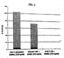


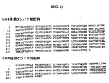
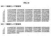


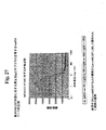
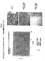


Classifications
-
- C—CHEMISTRY; METALLURGY
- C07—ORGANIC CHEMISTRY
- C07K—PEPTIDES
- C07K16/00—Immunoglobulins [IGs], e.g. monoclonal or polyclonal antibodies
- C07K16/18—Immunoglobulins [IGs], e.g. monoclonal or polyclonal antibodies against material from animals or humans
- C07K16/24—Immunoglobulins [IGs], e.g. monoclonal or polyclonal antibodies against material from animals or humans against cytokines, lymphokines or interferons
- C07K16/243—Colony Stimulating Factors
-
- A—HUMAN NECESSITIES
- A61—MEDICAL OR VETERINARY SCIENCE; HYGIENE
- A61P—SPECIFIC THERAPEUTIC ACTIVITY OF CHEMICAL COMPOUNDS OR MEDICINAL PREPARATIONS
- A61P19/00—Drugs for skeletal disorders
-
- A—HUMAN NECESSITIES
- A61—MEDICAL OR VETERINARY SCIENCE; HYGIENE
- A61P—SPECIFIC THERAPEUTIC ACTIVITY OF CHEMICAL COMPOUNDS OR MEDICINAL PREPARATIONS
- A61P19/00—Drugs for skeletal disorders
- A61P19/08—Drugs for skeletal disorders for bone diseases, e.g. rachitism, Paget's disease
-
- A—HUMAN NECESSITIES
- A61—MEDICAL OR VETERINARY SCIENCE; HYGIENE
- A61P—SPECIFIC THERAPEUTIC ACTIVITY OF CHEMICAL COMPOUNDS OR MEDICINAL PREPARATIONS
- A61P35/00—Antineoplastic agents
- A61P35/04—Antineoplastic agents specific for metastasis
-
- A—HUMAN NECESSITIES
- A61—MEDICAL OR VETERINARY SCIENCE; HYGIENE
- A61P—SPECIFIC THERAPEUTIC ACTIVITY OF CHEMICAL COMPOUNDS OR MEDICINAL PREPARATIONS
- A61P43/00—Drugs for specific purposes, not provided for in groups A61P1/00-A61P41/00
-
- A—HUMAN NECESSITIES
- A61—MEDICAL OR VETERINARY SCIENCE; HYGIENE
- A61K—PREPARATIONS FOR MEDICAL, DENTAL OR TOILETRY PURPOSES
- A61K39/00—Medicinal preparations containing antigens or antibodies
- A61K2039/505—Medicinal preparations containing antigens or antibodies comprising antibodies
-
- C—CHEMISTRY; METALLURGY
- C07—ORGANIC CHEMISTRY
- C07K—PEPTIDES
- C07K2317/00—Immunoglobulins specific features
- C07K2317/30—Immunoglobulins specific features characterized by aspects of specificity or valency
- C07K2317/34—Identification of a linear epitope shorter than 20 amino acid residues or of a conformational epitope defined by amino acid residues
-
- C—CHEMISTRY; METALLURGY
- C07—ORGANIC CHEMISTRY
- C07K—PEPTIDES
- C07K2317/00—Immunoglobulins specific features
- C07K2317/70—Immunoglobulins specific features characterized by effect upon binding to a cell or to an antigen
- C07K2317/73—Inducing cell death, e.g. apoptosis, necrosis or inhibition of cell proliferation
Description
本発明は、M−CSF特異的抗体の被験体への投与による、骨溶解、癌転移、および癌転移に関連する骨量減少または再造形を防止および治療する方法に関する。 The present invention relates to methods for preventing and treating osteolysis, cancer metastasis, and bone loss or remodeling associated with cancer metastasis by administration of M-CSF specific antibodies to a subject.
癌転移は、癌患者の術後または治療後再発の主な原因である。治療開発に関する取り組みが集中的に行われているにもかかわらず、癌転移は、実質的には依然として治療抵抗性を示す。骨は、種々のヒト癌型(例えば、乳癌、肺癌、前立腺癌、および甲状腺癌)の最も一般的な転移部位の1つである。骨溶解性骨転移の発生は、難治性疼痛、骨折に対する高い感受性、神経圧迫、および高カルシウム血症に起因する重篤な病的状態を引き起こす。これらの臨床的問題の重要性にもかかわらず、癌転移または癌転移に関連する骨再造形に利用可能な治療はほとんどない。 Cancer metastasis is a major cause of post-operative or post-treatment recurrence in cancer patients. Despite intensive treatment development efforts, cancer metastasis remains substantially resistant to treatment. Bone is one of the most common metastatic sites of various human cancer types (eg, breast cancer, lung cancer, prostate cancer, and thyroid cancer). The development of osteolytic bone metastases causes severe morbidity resulting from refractory pain, high sensitivity to fractures, nerve compression, and hypercalcemia. Despite the importance of these clinical problems, few treatments are available for cancer metastasis or bone remodeling associated with cancer metastasis.
骨芽細胞(骨形成細胞)と破骨細胞(骨溶解細胞)との間の活性の均衡により、一般に、転移性骨病変の表現型が決定され、骨芽細胞および破骨細胞の両方は、直接または間接的に、骨転移に関与している。相対骨形成(骨芽細胞性)表現型を有する骨転移は、癌細胞による相当な骨芽細胞の刺激または破骨細胞の阻害(または両方)の結果であるのに対して、相対的骨溶解(骨溶解性)表現型を有する転移は癌細胞による相当な骨芽細胞機能の阻害または破骨細胞機能の刺激(または両方)を反映する。前立腺癌(そのほとんどが腺癌である)からの転移は、ほぼ常に骨に病変を形成し、この病変は骨溶解性よりも骨芽細胞性であり、対照的に、腎臓癌、肺癌、または乳癌からの骨転移は、より多くの場合は骨溶解性である。しかし、前立腺の比較的一般的ではない神経内分泌腫瘍からの転移も骨溶解性病変を発症する。さらに、患者は、骨芽細胞病変および溶解性病変を有する疾患の混合形態を示し得る。 The balance of activity between osteoblasts (osteogenic cells) and osteoclasts (osteolytic cells) generally determines the phenotype of metastatic bone lesions, both osteoblasts and osteoclasts are It is directly or indirectly involved in bone metastasis. Bone metastasis with a relative osteogenic (osteoblastic) phenotype is the result of relative osteolysis, whereas cancer cells are the result of substantial osteoblast stimulation or osteoclast inhibition (or both). Metastasis with the (osteolytic) phenotype reflects substantial inhibition of osteoblast function or stimulation of osteoclast function (or both) by cancer cells. Metastases from prostate cancer (most of which are adenocarcinoma) almost always form lesions in the bone, which are more osteoblastic than osteolytic, in contrast to kidney cancer, lung cancer, or Bone metastases from breast cancer are more often osteolytic. However, metastases from the less common neuroendocrine tumors of the prostate also develop osteolytic lesions. In addition, patients may exhibit a mixed form of disease with osteoblastic lesions and lytic lesions.
破骨細胞は骨の再吸着を媒介し、破骨細胞は、造血細胞から分化した多核細胞である。破骨細胞は、不完全な細胞分裂よりもむしろ、骨髄中の造血幹細胞由来の単核細胞前駆体の融合によって形成されるということが一般に認められている(非特許文献1;非特許文献2;非特許文献3;非特許文献4;非特許文献5;非特許文献6;非特許文献7)。これらは、単球マクロファージ系列細胞と共通の幹細胞を有する(非特許文献8,非特許文献9)。破骨細胞前駆体の成熟多核破骨細胞への分化には、異なる因子(ホルモン刺激および局所刺激が含まれる)が必要であり(非特許文献10;非特許文献11;非特許文献7;非特許文献12)、生きている骨および骨細胞は、破骨細胞の発達で重要な役割を果たすことが示されている(非特許文献13)。骨芽細胞または骨髄間質細胞はまた、破骨細胞の分化に必要である。破骨細胞形成を補助するこれらの細胞によって産生される因子の1つは、マクロファージコロニー刺激因子M−CSFである(非特許文献14;非特許文献15)。NF−κB活性化受容体リガンド(RANKL(TRANCE、ODF、およびOPGLとしても公知))は別のシグナルであり(非特許文献4)、これにより、骨芽細胞/間質細胞は、破骨細胞および破骨細胞前駆体に存在する受容体RANK(TRANCER)を介して破骨細胞の形成および吸収を刺激する(非特許文献16;非特許文献17;非特許文献18;非特許文献19;非特許文献20;非特許文献21)。骨芽細胞はまた、オステオプロテジェリン(OPG、OCIFとしても公知)と呼ばれる骨芽細胞形成を強く阻害するタンパク質を分泌する。このタンパク質は、RANKLのデコイ受容体として作用し、それにより、RANKおよびRANKLを介した破骨細胞と骨芽細胞との間の正のシグナルを阻害する。
Osteoclasts mediate bone resorption, and osteoclasts are multinucleated cells differentiated from hematopoietic cells. It is generally accepted that osteoclasts are formed by fusion of hematopoietic stem cell-derived mononuclear cell precursors in the bone marrow rather than incomplete cell division (Non-patent
破骨細胞は、無機物および有機物の骨基質の両方の溶解を担う(非特許文献22)。破骨細胞は、特殊化した膜領域およびいくつかの膜ならびに細胞質マーカー(酒石酸耐性酸性ホスファターゼ(TRAP)(Andersonら、1979)、炭酸脱水酵素II(非特許文献23)、カルシトニン受容体(非特許文献24)、およびビトロネクチン受容体(非特許文献25)など)を有する固有の極性形態を発現する最終分化細胞である。多核破骨細胞は、通常、10個未満の核を含むが、直径が10μmと100μmとの間の100個までの核を含み得る(非特許文献26)。これにより、光学顕微鏡によって比較的容易に同定される。多核破骨細胞は、活性状態の場合に比較的空胞が存在し、多くのミトコンドリアも含み、高い代謝率を示す(非特許文献27)。破骨細胞は骨溶解性骨代謝および他の骨溶解性疾患(例えば、骨粗鬆症など)で主な役割を果たすので、破骨細胞の刺激および機能を防止するための新規の薬剤および方法が当該分野で必要である。
Osteoclasts are responsible for the lysis of both inorganic and organic bone matrix (22). Osteoclasts consist of specialized membrane regions and several membranes and cytoplasmic markers (tartrate-resistant acid phosphatase (TRAP) (Anderson et al., 1979), carbonic anhydrase II (Non-patent Document 23), calcitonin receptor (Non-patent document Reference 24), and terminally differentiated cells that express unique polar forms with vitronectin receptors (Non-patent
したがって、骨溶解または癌転移(骨芽細胞性骨転移と骨溶解性骨転移との混合、および破骨細胞活性の増加に関連する骨疾患が含まれる)の防止または治療のための新規の薬剤および方法を同定することが当該分野で要求されている。
発明の概要
本発明の材料および方法は、当該分野における上記要求および他の関連する要求を満たす。本発明の1つの実施形態では、図4、14、および15にそれぞれ示すアミノ酸配列を有するマウスモノクローナル抗体RX1、MC1、またはMC3のいずれか1つと同一のM−CSFのエピトープに特異的に結合する非マウスモノクローナル抗体(機能的フラグメントが含まれる)を提供する。関連する実施形態では、好ましくは少なくとも10−7、10−8、または10−9、またはそれを超える結合親和性を保持するポリクローナル抗体、モノクローナル抗体(ヒト操作(Human Engineered(商標))抗体が含まれる)、ヒト化抗体、ヒト抗体、キメラ抗体、Fab、F(ab’)2、Fv、ScFv、またはSCA抗体フラグメント、ダイアボディ(diabody)、線状抗体(linear antibody)、またはこれら抗体のいずれか1つのムテインからなる群から選択される上記抗体を提供する。図4に示すアミノ酸配列を有するモノクローナル抗体RX1、MC1、および/またはMC3とM−CSFへの結合とを75%を超えて競合する非マウスモノクローナル抗体(機能的フラグメントが含まれる)も意図する。
SUMMARY OF THE INVENTION The materials and methods of the present invention meet the above and other related needs in the art. In one embodiment of the invention, it specifically binds to the same epitope of M-CSF as any one of the murine monoclonal antibodies RX1, MC1, or MC3 having the amino acid sequences shown in FIGS. 4, 14, and 15, respectively. Non-mouse monoclonal antibodies (including functional fragments) are provided. In related embodiments, preferably a polyclonal antibody, monoclonal antibody (human engineered ™ antibody) that retains a binding affinity of at least 10 −7 , 10 −8 , or 10 −9 or higher is included. Humanized antibody, human antibody, chimeric antibody, Fab, F (ab ′) 2 , Fv, ScFv, or SCA antibody fragment, diabody, linear antibody, or any of these antibodies Or an antibody selected from the group consisting of one mutein. Also contemplated are non-mouse monoclonal antibodies (including functional fragments) that compete for more than 75% of monoclonal antibodies RX1, MC1, and / or MC3 having the amino acid sequence shown in FIG. 4 and binding to M-CSF.
別の実施形態では、非マウスモノクローナル抗体(機能的フラグメントが含まれる)を提供し、非マウスモノクローナル抗体またはその機能的フラグメントは図12のアミノ酸98〜105の少なくとも4、5、6、7、または8個の連続する残基を含むM−CSFのエピトープに結合している。 In another embodiment, a non-mouse monoclonal antibody (including functional fragments) is provided, wherein the non-mouse monoclonal antibody or functional fragment thereof is at least 4, 5, 6, 7, or amino acids 98-105 of FIG. It binds to an epitope of M-CSF containing 8 consecutive residues.
別の実施形態では、本発明は、非マウスモノクローナル抗体(機能的フラグメントが含まれる)を提供し、非マウスモノクローナル抗体またはその機能的フラグメントは図12のアミノ酸65〜73または138〜144の少なくとも4、5、6、7、または8個の連続する残基を含むM−CSFのエピトープに結合している。 In another embodiment, the invention provides a non-mouse monoclonal antibody, including a functional fragment, wherein the non-mouse monoclonal antibody or functional fragment thereof is at least 4 of amino acids 65-73 or 138-144 of FIG. It binds to an epitope of M-CSF that contains 5, 6, 7, or 8 consecutive residues.
さらに別の実施形態では、図12のアミノ酸98〜105を含むM−CSFのエピトープに結合する上記抗体またはフラグメントを提供する。関連する実施形態では、図4AのCDR3を含む上記抗体を提供する。別の実施形態では、図4Aに示すマウス抗体RX1の少なくとも1、2、3、4、5または6個のCDRを含む抗体を提供する。マウス抗体RX1の少なくとも1、2、3、4、または5個のCDRを含むこのような抗体はまた、図16A〜Bに示す抗体5H4の任意の6個のCDRのうちの少なくとも1、2、3、4、または5個のCDRを含み得る。あるいは、マウス抗体RX1の少なくとも1、2、3、4、または5個のCDRを含む抗体はまた、図16A〜Bに示す抗体MC1の任意の6個のCDRのうちの少なくとも1、2、3、4、または5個のCDRを含み得る。さらに別の実施形態では、上記抗体はまた、図16A〜Bに示す抗体MC3の任意の6個のCDRのうちの少なくとも1、2、3、4、または5個のCDRを含み得る。関連する実施形態では、マウス抗体RX1の少なくとも1、2、3、4、または5個のCDRを含む抗体は、図16A〜Bに示す抗体MC1の任意の6個のCDRのうちの少なくとも1、2、3、4、または5個のCDRを含み得ることを提供する。さらに別の関連する実施形態では、上記抗体では、コンセンサスCDRの1つまたは複数の残基は、マウス抗体RX1、5H4、MC1、またはMC3の任意のCDRの対応する残基に置換される。抗体中の1つまたは複数のアミノ酸が変異しているにもかかわらず、例えば、CDR中の保存的置換および/または低リスクおよび中程度のリスクの残基の保存的または非保存的変化によって所望の結合親和性を保持することができる。 In yet another embodiment, the antibody or fragment is provided that binds to an epitope of M-CSF comprising amino acids 98-105 of FIG. In a related embodiment, the above antibody comprising the CDR3 of FIG. 4A is provided. In another embodiment, an antibody comprising at least 1, 2, 3, 4, 5 or 6 CDRs of the murine antibody RX1 shown in FIG. 4A is provided. Such an antibody comprising at least 1, 2, 3, 4, or 5 CDRs of murine antibody RX1 is also at least 1, 2, of any 6 CDRs of antibody 5H4 shown in FIGS. It may contain 3, 4, or 5 CDRs. Alternatively, an antibody comprising at least 1, 2, 3, 4, or 5 CDRs of murine antibody RX1 is also at least 1, 2, 3, of any 6 CDRs of antibody MC1 shown in FIGS. It may contain 4, or 5 CDRs. In yet another embodiment, the antibody can also comprise at least 1, 2, 3, 4, or 5 of any 6 CDRs of antibody MC3 shown in FIGS. In a related embodiment, the antibody comprising at least 1, 2, 3, 4, or 5 CDRs of mouse antibody RX1 is at least one of any 6 CDRs of antibody MC1 shown in FIGS. It is provided that it can contain 2, 3, 4, or 5 CDRs. In yet another related embodiment, in the antibody, one or more residues of the consensus CDR are replaced with the corresponding residues of any CDR of the murine antibody RX1, 5H4, MC1, or MC3. Despite one or more amino acids in the antibody being mutated, for example, by conservative substitutions in the CDRs and / or conservative or non-conservative changes in low and moderate risk residues The binding affinity of can be retained.
本発明の別の実施形態では、図4A、13、14、または15に示すアミノ酸配列に少なくとも60、65、70、75、80、85、90、91、92、93、94、95、96、97、98、または99%相同な可変重鎖アミノ酸配列を含む上記抗体の変異型(variant)を提供する。関連する実施形態では、抗体は、図4A、13、14、または15に示すアミノ酸配列に少なくとも60、65、70、75、80、85、90、91、92、93、94、95、96、97、98、または99%相同な可変軽鎖アミノ酸配列を含む。 In another embodiment of the invention, the amino acid sequence shown in FIG. 4A, 13, 14, or 15 has at least 60, 65, 70, 75, 80, 85, 90, 91, 92, 93, 94, 95, 96, Variants of the above antibodies comprising variable heavy chain amino acid sequences that are 97, 98, or 99% homologous are provided. In related embodiments, the antibody has at least 60, 65, 70, 75, 80, 85, 90, 91, 92, 93, 94, 95, 96, amino acid sequence as shown in FIG. 4A, 13, 14, or 15. Contains variable light chain amino acid sequences that are 97, 98, or 99% homologous.
さらに別の実施形態では、抗体は、ヒト抗体配列の定常領域および1つまたは複数の重鎖および軽鎖の可変フレームワーク領域を含む。関連する実施形態では、抗体は、ヒトIgG1、IgG2、IgG3、またはIgG4の修飾または未修飾定常領域を含む。好ましい実施形態では、定常領域は、ヒトIgG1またはIgG4であり、これらを、一定の性質を増強または減少させるように任意選択的に修飾することができる。IgG1の場合、定常領域、特にヒンジまたはCH2領域の修飾により、エフェクター機能(ADCCおよび/またはCDC活性が含まれる)を増加または減少させることができる。他の実施形態では、IgG2定常領域を、抗体−抗原凝集体形成を減少させるように修飾する。IgG4の場合、定常領域、特にヒンジ領域の修飾により、半抗体(half−antibody)の形成を減少させることができる。 In yet another embodiment, the antibody comprises a constant region of a human antibody sequence and one or more heavy and light chain variable framework regions. In related embodiments, the antibody comprises a modified or unmodified constant region of human IgG1, IgG2, IgG3, or IgG4. In preferred embodiments, the constant regions are human IgG1 or IgG4, which can be optionally modified to enhance or decrease certain properties. In the case of IgG1, effector function (including ADCC and / or CDC activity) can be increased or decreased by modification of the constant region, particularly the hinge or CH2 region. In other embodiments, the IgG2 constant region is modified to reduce antibody-antigen aggregate formation. In the case of IgG4, modification of the constant region, particularly the hinge region, can reduce the formation of half-antibodies.
本発明のさらに別の実施形態では、上記抗体は、Kabat,NCBI Ig Blast,http://www.ncbi.nlm.nih.gov/igblast/showGermline.cgi、Kabat Database http://www.bioinf.org.uk/abs/seqtest.html、FTP site for Kabat Release 5.0(1992)ftp://ftp.ncbi.nih.gov/repository/kabat/Rel5.0/、ImMunoGeneTics database(Montpellier France)http://imgt.cnusc.fr:8104/、V−Base http://www.mrc−cpe.cam.ac.uk/LIST.php?menu=901、Zurich University http://www.unizh.ch/〜antibody/Sequences/index.html、The Therapeutic Antibody Human Homology Project(TAHHP)http://www.path.cam.ac.uk/〜mrc7/humanisation/TAHHP.html、Protein Sequence and Structure Analysis of Antibody Domains http://how.to/AnalyseAntibodies/、Humanization by design http://people.cryst.bbk.ac.uk/〜ubcg07s/、Antibody Resources http://www.antibodyresource.com/educational.html、Antibody Engineering(by TT Wu),Humana Press中のヒトコンセンサス配列、ヒト生殖系列配列、ヒトコンセンサス生殖系列配列、またはヒト抗体配列のいずれか1つに由来するか、これらに基づくか、これらの一部を含む。 In yet another embodiment of the invention, the antibody is Kabat, NCBI Ig Blast, http: // www. ncbi. nlm. nih. gov / igblast / showGermine. cgi, Kabat Database http: // www. bioinf. org. uk / abs / seqtest. html, FTP site for Kabat Release 5.0 (1992) ftp: // ftp. ncbi. nih. gov / repository / kabat / Rel5.0 /, ImMunoGeneTics database (Montpellier France) http: // imgt. cnusc. fr: 8104 /, V-Base http: // www. mrc-cpe. cam. ac. uk / LIST. php? menu = 901, Zurich University http: // www. unith. ch / ~ antibody / Sequences / index. html, The Therapeutic Antibiotic Human Homology Project (TAHHP) http: // www. path. cam. ac. uk / ~ mrc7 / humanization / TAHHP. html, Protein Sequence and Structure Analysis of Antibody Domains http: // how. to / Analyze Antibodies /, Humanization by design http: // people. cryst. bbk. ac. uk / ~ ubcg07s /, Antibody Resources http: // www. antibodyresource. com / educational. derived from or based on any one of human consensus sequences, human germline sequences, human consensus germline sequences, or human antibody sequences in html, Antibody Engineering (by TT Wu), Humana Press Includes some.
本発明の好ましい態様では、上記抗体は、ヒト操作(商標)抗体である。例えば、ヒト操作(商標)抗体配列は、図23〜24に示す配列のいずれか1つである。他のヒト操作(商標)抗体またはその変異型を意図する。 In a preferred embodiment of the invention, the antibody is a human engineered antibody. For example, the human engineered (trademark) antibody sequence is any one of the sequences shown in FIGS. Other human engineered antibodies or variants thereof are contemplated.
例えば、1つの実施形態では、上記RX1ベースの抗体であって、重鎖可変領域がアミノ酸配列 For example, in one embodiment, the RX1-based antibody, wherein the heavy chain variable region is an amino acid sequence

本発明のさらに別の実施形態では、重鎖可変領域がアミノ酸配列 In yet another embodiment of the invention, the heavy chain variable region is an amino acid sequence.
本発明の別の実施形態では、軽鎖可変領域がアミノ酸配列 In another embodiment of the invention, the light chain variable region is an amino acid sequence.
本発明の別の実施形態では、軽鎖可変領域がアミノ酸配列 In another embodiment of the invention, the light chain variable region is an amino acid sequence.
本発明のさらに別の実施形態では、軽鎖可変領域がアミノ酸配列 In yet another embodiment of the invention, the light chain variable region is an amino acid sequence.
本発明の別の実施形態では、少なくとも1つのXが図4Aに示すアミノ酸配列内の相当するアミノ酸である上記抗体を提供する。関連する実施形態では、少なくとも1つのXが図4Aに示すアミノ酸配列内の相当するアミノ酸の保存的置換(表1に従う)である抗体を提供する。さらに別の関連する実施形態では、少なくとも1つのXが図4Aに示すアミノ酸配列内の相当するアミノ酸の非保存的置換(表1に従う)である抗体を提供する。本発明のさらに別の実施形態では、少なくとも1つのXがヒト抗体配列内の相当するアミノ酸である抗体を提供する。 In another embodiment of the invention, the antibody is provided wherein at least one X is the corresponding amino acid within the amino acid sequence shown in FIG. 4A. In a related embodiment, an antibody is provided wherein at least one X is a conservative substitution (according to Table 1) of the corresponding amino acid within the amino acid sequence shown in FIG. 4A. In yet another related embodiment, an antibody is provided wherein at least one X is a non-conservative substitution (according to Table 1) of the corresponding amino acid within the amino acid sequence shown in FIG. 4A. In yet another embodiment of the invention, an antibody is provided wherein at least one X is a corresponding amino acid within a human antibody sequence.
上記ヒト操作(商標)抗体は、Kabat,NCBI Ig Blast,http://www.ncbi.nlm.nih.gov/igblast/showGermline.cgi、Kabat Database http://www.bioinf.org.uk/abs/seqtest.html、FTP site for Kabat Release 5.0(1992)ftp://ftp.ncbi.nih.gov/repository/kabat/Rel5.0/、ImMunoGeneTics database(Montpellier France)http://imgt.cnusc.fr:8104/、V−Base http://www.mrc−cpe.cam.ac.uk/LIST.php?menu=901、Zurich University http://www.unizh.ch/〜antibody/Sequences/index.html、The Therapeutic Antibody Human Homology Project(TAHHP)http://www.path.cam.ac.uk/〜mrc7/humanisation/TAHHP.html、Protein Sequence and Structure Analysis of Antibody Domains http://how.to/AnalyseAntibodies/、Humanization by design http://people.cryst.bbk.ac.uk/〜ubcg07s/、Antibody Resources http://www.antibodyresource.com/educational.html、Antibody Engineering(by TT Wu),Humana Press中のヒト抗体コンセンサス配列、ヒト生殖系列配列、ヒトコンセンサス生殖系列配列、またはヒト抗体配列のいずれか1つに由来するか、これらに基づくか、これらの一部を含む。 The human engineered antibody is available from Kabat, NCBI Ig Blast, http: // www. ncbi. nlm. nih. gov / igblast / showGermine. cgi, Kabat Database http: // www. bioinf. org. uk / abs / seqtest. html, FTP site for Kabat Release 5.0 (1992) ftp: // ftp. ncbi. nih. gov / repository / kabat / Rel5.0 /, ImMunoGeneTics database (Montpellier France) http: // imgt. cnusc. fr: 8104 /, V-Base http: // www. mrc-cpe. cam. ac. uk / LIST. php? menu = 901, Zurich University http: // www. unith. ch / ~ antibody / Sequences / index. html, The Therapeutic Antibiotic Human Homology Project (TAHHP) http: // www. path. cam. ac. uk / ~ mrc7 / humanization / TAHHP. html, Protein Sequence and Structure Analysis of Antibody Domains http: // how. to / Analyze Antibodies /, Humanization by design http: // people. cryst. bbk. ac. uk / ~ ubcg07s /, Antibody Resources http: // www. antibodyresource. com / educational. derived from, based on, or based on any one of human antibody consensus sequences, human germline sequences, human consensus germline sequences, or human antibody sequences in html, Antibody Engineering (by TT Wu), Humana Press Including a part of
本発明の別の実施形態では、ヒト操作(商標)抗体配列が、図23〜24または29〜30に示す配列の1つである上記抗体を提供する。別の実施形態では、抗体は、図19Bに示す重鎖アミノ酸配列の1つと少なくとも60、65、70、75、80、85、90、91、92、93、94、95、96、97、98、または99%同一の可変重鎖アミノ酸配列を含む。さらに別の実施形態では、抗体は、図20B〜22Bのいずれかに示す軽鎖アミノ酸配列の1つと少なくとも60、65、70、75、80、85、90、91、92、93、94、95、96、97、98、または99%同一の可変軽鎖アミノ酸配列を含む。 In another embodiment of the invention, there is provided the above antibody, wherein the human engineered antibody sequence is one of the sequences shown in FIGS. 23-24 or 29-30. In another embodiment, the antibody has at least 60, 65, 70, 75, 80, 85, 90, 91, 92, 93, 94, 95, 96, 97, 98 with one of the heavy chain amino acid sequences shown in FIG. 19B. Or a variable heavy chain amino acid sequence that is 99% identical. In yet another embodiment, the antibody has at least 60, 65, 70, 75, 80, 85, 90, 91, 92, 93, 94, 95 and one of the light chain amino acid sequences shown in any of FIGS. 96, 97, 98, or 99% identical variable light chain amino acid sequences.
さらに別の実施形態では、図24C〜24Eの1つに示す配列を有する5H4、MC1、またはMC3などの上記抗体は、低リスク、中程度のリスク、および高リスクの残基を同定するために図24C〜24Eに示すKabatナンバリングを使用したStudnickaらに付与された米国特許第5,766,886号および本明細書中の実施例5に示す方法に従ってヒト操作(商標)する。関連する実施形態では、必要に応じて、ヒト基準免疫グロブリン配列と同一の残基となるように重鎖、軽鎖、または両方のいずれかの低リスク残基の全てを修飾した上記抗体を提供する。同様に、本発明の別の実施形態は、必要に応じて、ヒト基準免疫グロブリン配列と同一の残基となるように重鎖、軽鎖、または両方のいずれかの低リスク+中程度のリスクの残基の全てを修飾した上記抗体を提供する。低リスク残基の全てを修飾した重鎖を、低リスク残基および中程度のリスクの残基の全てを修飾した軽鎖と組み合わせることができ、その逆も同様である。同様に、上記ヒト操作(商標)軽鎖または重鎖を、ヒト化抗体またはキメラ抗体の軽鎖または重鎖と組み合わせることができる。 In yet another embodiment, the antibody, such as 5H4, MC1, or MC3, having the sequence shown in one of FIGS. 24C-24E, is used to identify low risk, moderate risk, and high risk residues. Human manipulation (TM) is performed according to the method set forth in US Pat. No. 5,766,886 granted to Studnicka et al. Using the Kabat numbering shown in FIGS. 24C-24E and Example 5 herein. In related embodiments, the above-described antibodies are provided, wherein all of the low-risk residues, either heavy chain, light chain, or both, are optionally modified to be identical residues to the human reference immunoglobulin sequence. To do. Similarly, another embodiment of the present invention provides for the low risk plus moderate risk of either heavy chain, light chain, or both, to be the same residue as the human reference immunoglobulin sequence, as appropriate. The above antibody is provided in which all of the residues are modified. A heavy chain modified with all of the low risk residues can be combined with a light chain modified with all of the low and moderate risk residues, and vice versa. Similarly, the human engineered light chain or heavy chain can be combined with the light chain or heavy chain of a humanized or chimeric antibody.
さらに別の実施形態では、抗体は、Studnickaの方法に従ってヒト操作(商標)したすぐ上に記載の重鎖アミノ酸配列の1つと60、65、70、75、80、85、90、91、92、93、94、95、96、97、98、または99%同一の可変重鎖アミノ酸配列を含む。さらに別の実施形態では、抗体は、Studnickaの方法に従ってヒト操作(商標)したすぐ上に記載の軽鎖アミノ酸配列の1つと60、65、70、75、80、85、90、91、92、93、94、95、96、97、98、または99%同一の可変軽鎖アミ酸配列を含む。 In yet another embodiment, the antibody is one of the heavy chain amino acid sequences set forth immediately above human engineered ™ according to Studnika's method and 60, 65, 70, 75, 80, 85, 90, 91, 92, Contains variable heavy chain amino acid sequences that are 93, 94, 95, 96, 97, 98, or 99% identical. In yet another embodiment, the antibody is one of the light chain amino acid sequences set forth immediately above human engineered (TM) according to Studnika's method and 60, 65, 70, 75, 80, 85, 90, 91, 92, Contains variable light chain amino acid sequences that are 93, 94, 95, 96, 97, 98, or 99% identical.
さらに別の実施形態では、上記の重鎖および上記の軽鎖を含む抗体を提供する。 In yet another embodiment, an antibody comprising the above heavy chain and the above light chain is provided.
本発明のさらに別の実施形態では、上記抗体の親和性Kdは少なくとも10−7である。関連する実施形態では、抗体の親和性Kdは少なくとも10−9である。 In yet another embodiment of the invention, the affinity Kd of the antibody is at least 10 −7 . In a related embodiment, the antibody has an affinity Kd of at least 10 −9 .
本発明の別の実施形態では、ポリクローナル抗体(ヒト操作(商標)抗体が含まれる)、ヒト化抗体、ヒト抗体、キメラ抗体、Fab、F(ab’)2、Fv、ScFv、またはSCA抗体フラグメント、ダイアボディ、線状抗体、またはこれら抗体のいずれか1つのムテインである上記抗体を提供する。関連する実施形態では、モノクローナル抗体は、単離抗体である。 In another embodiment of the invention, polyclonal antibodies (including human engineered antibodies), humanized antibodies, human antibodies, chimeric antibodies, Fab, F (ab ′) 2 , Fv, ScFv, or SCA antibody fragments A diabody, a linear antibody, or a mutein of any one of these antibodies. In a related embodiment, the monoclonal antibody is an isolated antibody.
本発明のさらに別の実施形態では、上記抗体の軽鎖をコードする核酸配列を含む単離核酸を提供する。関連する実施形態では、単離核酸は、図4A、13、14、または15に示す重鎖ヌクレオチド配列と少なくとも60、65、70、75、80、85、90、91、92、93、94、95、96、97、98、または99%同一の重鎖核酸配列を含む。さらに別の関連する実施形態では、単離核酸は、図4A、13、14、または15に示す軽鎖ヌクレオチド配列と少なくとも60、65、70、75、80、85、90、91、92、93、94、95、96、97、98、または99%同一の軽鎖核酸配列を含む。 In yet another embodiment of the invention, an isolated nucleic acid comprising a nucleic acid sequence encoding the light chain of the antibody is provided. In related embodiments, the isolated nucleic acid comprises a heavy chain nucleotide sequence shown in FIG. 4A, 13, 14, or 15 and at least 60, 65, 70, 75, 80, 85, 90, 91, 92, 93, 94, Contains heavy chain nucleic acid sequences that are 95, 96, 97, 98, or 99% identical. In yet another related embodiment, the isolated nucleic acid comprises a light chain nucleotide sequence shown in FIG. 4A, 13, 14, or 15 and at least 60, 65, 70, 75, 80, 85, 90, 91, 92, 93. 94, 95, 96, 97, 98, or 99% identical light chain nucleic acid sequences.
別の実施形態では、上記単離核酸を含むベクターを提供する。関連する実施形態では、単離核酸が調節制御配列(regulatory control sequence)に作動可能に連結された上記ベクターを提供する。さらに別の実施形態では、上記ベクターを含む宿主細胞を提供する。 In another embodiment, a vector comprising the isolated nucleic acid is provided. In a related embodiment, the above vector is provided wherein the isolated nucleic acid is operably linked to a regulatory control sequence. In yet another embodiment, a host cell comprising the above vector is provided.
本発明で多数の方法を意図する。例えば、単離核酸を発現して抗体を産生するように上記宿主細胞を培養する工程を含む、上記抗体の産生方法を提供する。関連する実施形態では、本方法は、宿主細胞培養物から抗体を回収する工程をさらに含む。関連する実施形態では、上記方法によって産生された単離抗体を提供する。 A number of methods are contemplated by the present invention. For example, a method for producing the antibody is provided, comprising the step of culturing the host cell to express an isolated nucleic acid to produce the antibody. In related embodiments, the method further comprises recovering the antibody from the host cell culture. In a related embodiment, an isolated antibody produced by the above method is provided.
本発明によって上記抗体を分泌するハイブリドーマも提供する。さらに、毒素に接合した上記抗体を提供する。 The present invention also provides a hybridoma that secretes the antibody. Further provided is the above antibody conjugated to a toxin.
本発明の別の実施形態では、上記抗体のいずれか1つおよび薬学的に許容可能なキャリア、賦形剤、または希釈剤を含む薬学的組成物を提供する。関連する実施形態では、薬学的組成物は、第2の治療薬をさらに含む。さらに別の関連する実施形態では、第2の治療薬が癌化学療法薬である薬学的組成物を提供する。さらに別の関連する実施形態では、第2の治療薬がビスホスホネートである薬学的組成物を提供する。別の実施形態では、第2の治療薬は別の抗体である。 In another embodiment of the invention, a pharmaceutical composition is provided comprising any one of the above antibodies and a pharmaceutically acceptable carrier, excipient or diluent. In a related embodiment, the pharmaceutical composition further comprises a second therapeutic agent. In yet another related embodiment, a pharmaceutical composition is provided wherein the second therapeutic agent is a cancer chemotherapeutic agent. In yet another related embodiment, a pharmaceutical composition is provided wherein the second therapeutic agent is a bisphosphonate. In another embodiment, the second therapeutic agent is another antibody.
本発明の抗体は、疾患および障害の治療のための多数の望ましい特徴を有することが意図される。本発明の1つの実施形態では、抗体が疾患に関連する骨量減少または再造形の重症度を有効に軽減する、骨溶解を生じるか、これに寄与するか、これに起因する疾患を罹患した被験体の防止のためのM−CSFに結合する上記抗体のいずれか1つを提供する。同様に、抗体が疾患に関連する骨量減少または再造形の重症度を有効に軽減する、骨溶解を生じるか、これに寄与するか、これに起因する疾患を罹患した被験体の治療のためのM−CSFに結合する上記抗体のいずれか1つを提供する。 The antibodies of the present invention are intended to have a number of desirable characteristics for the treatment of diseases and disorders. In one embodiment of the invention, the antibody effectively reduces or reduces the severity of bone loss or remodeling associated with the disease, resulting in, contributing to or contributing to the disease resulting from osteolysis. Any one of the above antibodies that binds to M-CSF for prevention of a subject is provided. Similarly, for the treatment of subjects suffering from, or contributing to, osteolysis that effectively reduces the severity of bone loss or remodeling associated with the disease. Any one of the above antibodies that binds to M-CSF.
多数の疾患および障害は、本発明の抗体ベースの治療に影響を受けることが意図される。本発明の1つの実施形態では、疾患が、比較的増加した破骨細胞活性または骨再造形に必要な破骨細胞/骨芽細胞過程(破骨細胞活性の増加および骨芽細胞活性の増加の両方に関連する)に関連する骨溶解性疾患(代謝性骨疾患、内分泌障害(高コルチコイド症、性腺機能低下症、原発性または続発性副甲状腺機能亢進症、甲状腺機能亢進症)、高カルシウム血症、欠乏状態(くる病/骨軟化症、壊血病、栄養失調症)、慢性疾患(吸収不良症候群、慢性腎不全(腎性骨異栄養症)、慢性肝臓疾患(肝臓性骨異栄養症))、薬物(糖質コルチコイド(糖質コルチコイド誘導性骨粗鬆症)、ヘパリン、アルコール)、および遺伝性疾患(骨形成不全症、ホモシスチン尿症)、癌、骨粗鬆症、人工器官周辺の骨量減少(例えば、人工器官を使用した骨または関節中の外来インプラントの置換(人工股関節全置換術が含まれる)による)、関節炎および関節リウマチに関連する骨の炎症、歯周疾患、線維性異形成、および/またはパジェット病が含まれる)からなる群から選択される上記抗体を提供する。 A number of diseases and disorders are contemplated to be affected by the antibody-based treatment of the present invention. In one embodiment of the invention, the disease is a relatively increased osteoclast activity or an osteoclast / osteoblast process required for bone remodeling (increased osteoclast activity and increased osteoblast activity). Osteolytic diseases (metabolic bone disease, endocrine disorders (hypercorticoid, hypogonadism, primary or secondary hyperparathyroidism, hyperthyroidism)), hypercalcemia , Deficiency (rickets / osteomalacia, scurvy, malnutrition), chronic diseases (malabsorption syndrome, chronic renal failure (renal osteodystrophy), chronic liver diseases (hepatic osteodystrophy) )), Drugs (glucocorticoids (glucocorticoid-induced osteoporosis), heparin, alcohol), and genetic disorders (osteogenesis dysplasia, homocystinuria), cancer, osteoporosis, bone loss around the prosthesis (eg Use a prosthesis Includes replacement of foreign implants in bone or joints (including total hip replacement), bone inflammation associated with arthritis and rheumatoid arthritis, periodontal disease, fibrotic dysplasia, and / or Paget's disease The antibody is selected from the group consisting of:
関連する実施形態では、骨転移性癌の防止または治療のためのM−CSFに結合する上記の抗体を提供し、転移性癌は、乳癌、肺癌、腎臓癌、多発性骨髄腫、甲状腺癌、前立腺癌、腺癌、血球悪性疾患(白血病およびリンパ腫が含まれる)、頭頸部癌、消化管癌(食道癌、胃癌、結腸癌、腸癌、結腸直腸癌、直腸癌、膵臓癌、肝臓癌、胆管または胆嚢の癌が含まれる)、女性生殖管の悪性疾患(卵巣癌、子宮内膜癌、膣癌、および子宮頸癌が含まれる)、膀胱癌、脳腫瘍(神経芽細胞腫が含まれる)、肉腫、骨肉種、および皮膚癌(悪性黒色腫または扁平上皮癌が含まれる)である。 In a related embodiment, the aforementioned antibody that binds to M-CSF for the prevention or treatment of bone metastatic cancer is provided, wherein the metastatic cancer is breast cancer, lung cancer, kidney cancer, multiple myeloma, thyroid cancer, Prostate cancer, adenocarcinoma, blood cell malignancies (including leukemia and lymphoma), head and neck cancer, gastrointestinal cancer (esophageal cancer, stomach cancer, colon cancer, intestinal cancer, colorectal cancer, rectal cancer, pancreatic cancer, liver cancer, Bile duct or gallbladder cancer), female reproductive tract malignancies (including ovarian cancer, endometrial cancer, vaginal cancer, and cervical cancer), bladder cancer, brain tumor (including neuroblastoma) Sarcoma, osteosarcoma, and skin cancer (including malignant melanoma or squamous cell carcinoma).
本発明の別の実施形態では、転移性腫瘍細胞培地、破骨細胞、および候補抗体を接触させる工程と、破骨細胞の形成、増殖、および/または分化の減少が検出された場合にM−CSF特異的抗体として候補抗体を同定する工程とを含む、M−CSF特異的抗体のスクリーニング方法を提供する。同様に、転移性腫瘍細胞培地が腫瘍細胞を含む上記方法を提供する。 In another embodiment of the invention, contacting the metastatic tumor cell medium, osteoclasts, and the candidate antibody, and detecting a decrease in osteoclast formation, proliferation, and / or differentiation when M- is detected. Identifying a candidate antibody as a CSF-specific antibody, and a method for screening an M-CSF-specific antibody. Similarly, provided is the above method, wherein the metastatic tumor cell medium comprises tumor cells.
関連する実施形態では、候補抗体がM−CSFに結合するかどうかを決定する工程をさらに含む上記方法を提供する。同様に、本発明の別の実施形態は、候補抗体がM−CSFとその受容体M−CSFRとの間の相互作用を阻害するかどうかを決定する工程をさらに含む上記方法を提供する。 In a related embodiment, the aforementioned method is provided further comprising the step of determining whether the candidate antibody binds to M-CSF. Similarly, another embodiment of the present invention provides the above method, further comprising the step of determining whether the candidate antibody inhibits the interaction between M-CSF and its receptor M-CSFR.
本発明の別の実施形態では、(a)図12のアミノ酸98〜105の少なくとも4個の連続する残基を含むM−CSFのエピトープへの候補抗体の結合を検出する工程と、(b)候補抗体がインビトロまたはインビボで骨への転移性癌を防止または治療する能力をアッセイする工程とを含む、骨への転移性癌を防止または治療することができるM−CSF特異的抗体を同定する方法を提供する。 In another embodiment of the invention, (a) detecting binding of a candidate antibody to an epitope of M-CSF comprising at least 4 consecutive residues of amino acids 98-105 of FIG. 12, and (b) Identifying an M-CSF specific antibody capable of preventing or treating metastatic cancer to bone, comprising assaying the ability of the candidate antibody to prevent or treat metastatic cancer to bone in vitro or in vivo Provide a method.
本発明の別の実施形態では、(a)(5H4またはMC3によって認識されるM−CSFエピトープに対応する)図12のアミノ酸65〜73または138〜144の少なくとも4個の連続する残基を含むM−CSFのエピトープへの候補抗体の結合を検出する工程と、(b)候補抗体がインビトロまたはインビボで骨への転移性癌を防止または治療する能力をアッセイする工程とを含む、骨への転移性癌を防止または治療することができるM−CSF特異的抗体を同定する方法を提供する。 In another embodiment of the invention, (a) comprises at least 4 consecutive residues of amino acids 65-73 or 138-144 of FIG. 12 (corresponding to the M-CSF epitope recognized by 5H4 or MC3) Detecting binding of the candidate antibody to an epitope of M-CSF, and (b) assaying the ability of the candidate antibody to prevent or treat metastatic cancer to bone in vitro or in vivo. Methods are provided for identifying M-CSF specific antibodies capable of preventing or treating metastatic cancer.
本発明のさらに別の実施形態では、図4Aに示すアミノ酸配列のCDR内のアミノ酸を変化させる工程と、少なくとも10−7の親和性KaでM−CSFに結合する抗体を選択する工程とを含む、図12のアミノ酸98〜105を含むM−CSFのエピトープに結合する抗体のCDRを変化させる方法を提供する。別の実施形態では、アミノ酸配列 In yet another embodiment of the invention, the method comprises the steps of changing amino acids in the CDR of the amino acid sequence shown in FIG. 4A and selecting an antibody that binds to M-CSF with an affinity Ka of at least 10 −7. 12 provides a method of changing the CDR of an antibody that binds to an epitope of M-CSF comprising amino acids 98-105 of FIG. In another embodiment, the amino acid sequence
関連する実施形態では、アミノ酸配列 In a related embodiment, the amino acid sequence
本発明のさらに別の実施形態では、図13、14、および15の1つに示すアミノ酸配列のCDR内のアミノ酸を変化させる工程と、少なくとも10−7の親和性KaでM−CSFに結合する抗体を選択する工程とを含む(5H4またはMC3によって認識されるM−CSFエピトープに相当する)図12のアミノ酸65〜73または138〜144を含むM−CSFのエピトープに結合する抗体のCDRを変化させる方法を提供する。別の実施形態では、Studnickaらに付与された米国特許第5,766,886号および本明細書中の実施例5に示す方法に従い、且つ図24C〜24Eに示すKabatナンバリングに従って上記配列を変化させる工程と、変化したアミノ酸配列を含む抗体を、(5H4またはMC3によって認識されるM−CSFエピトープに相当する)図12のアミノ酸65〜73または138〜144を含むM−CSFのエピトープへの結合について試験する工程とを含む、図13、14、および15の1つに示す重鎖アミノ酸配列の60%までを系統的に変化させる方法を提供する。関連する実施形態では、低リスク残基の全てを修飾する。同様に、本発明の別の実施形態では、低リスクの残基および中程度のリスクの残基の全てを修飾する。さらに別の実施形態では、プロリンを除く低リスクの残基および中程度のリスクの残基を修飾する。 In yet another embodiment of the invention, the step of changing amino acids in the CDRs of the amino acid sequence shown in one of FIGS. 13, 14, and 15 and binding to M-CSF with an affinity Ka of at least 10 −7. Changing the CDR of the antibody that binds to the epitope of M-CSF comprising amino acids 65-73 or 138-144 of FIG. 12 (corresponding to the M-CSF epitope recognized by 5H4 or MC3) Provide a way to make it happen. In another embodiment, the sequence is varied according to the method shown in US Pat. No. 5,766,886 to Studnikka et al. And Example 5 herein, and according to the Kabat numbering shown in FIGS. 24C-24E. For binding to an epitope of M-CSF comprising amino acids 65-73 or 138-144 of FIG. 12 (corresponding to the M-CSF epitope recognized by 5H4 or MC3) Providing a method of systematically changing up to 60% of the heavy chain amino acid sequence shown in one of FIGS. 13, 14, and 15. In related embodiments, all of the low risk residues are modified. Similarly, in another embodiment of the invention, all of the low and moderate risk residues are modified. In yet another embodiment, low-risk residues and moderate-risk residues except proline are modified.
本発明の別の実施形態では、上記過程によって設計されたCDRを有する抗体を発現する方法を提供する。別の実施形態では、MCSFに結合する抗体を含む薬学的組成物であって、抗体が上記方法を使用して作製される、薬学的組成物を提供する。 In another embodiment of the present invention, a method of expressing an antibody having CDRs designed by the above process is provided. In another embodiment, a pharmaceutical composition comprising an antibody that binds to MCSF is provided, wherein the antibody is made using the methods described above.
本発明のさらに別の実施形態では、治療有効量の上記抗体のいずれか1つを、骨溶解を生じるか、これに寄与するか、これに起因する疾患を罹患した被験体に投与する工程と、それにより、疾患に関連する骨量減少または再造形を防止または軽減することとを含む、骨量減少または再造形を防止または軽減する方法を提供する。関連する実施形態では、治療有効量の上記抗体のいずれか1つを被験体に投与する工程と、それにより、疾患に関連する骨量減少または再造形の重症度が軽減することとを含む、骨溶解を生じるか、これに寄与するか、これに起因する疾患を罹患した被験体を治療する方法を提供する。 In yet another embodiment of the invention, administering a therapeutically effective amount of any one of the above-described antibodies to a subject that develops, contributes to, or suffers from a disease resulting from osteolysis, and Thereby providing a method for preventing or reducing bone loss or remodeling, including preventing or reducing bone loss or remodeling associated with disease. In related embodiments, comprising administering to the subject a therapeutically effective amount of any one of the above antibodies, thereby reducing the severity of bone loss or remodeling associated with the disease. Methods are provided for treating a subject suffering from or contributing to, or resulting from, osteolysis.
関連する実施形態では、疾患が、比較的増加した破骨細胞活性または骨再造形に必要な破骨細胞/骨芽細胞過程(破骨細胞活性の増加および骨芽細胞活性の増加の両方に関連する)に関連する骨溶解性疾患(代謝性骨疾患、内分泌障害(高コルチコイド症、性腺機能低下症、原発性または続発性副甲状腺機能亢進症、甲状腺機能亢進症)、高カルシウム血症、欠乏状態(くる病/骨軟化症、壊血病、栄養失調症)、慢性疾患(吸収不良症候群、慢性腎不全(腎性骨異栄養症)、慢性肝臓疾患(肝臓性骨異栄養症))、薬物(糖質コルチコイド(糖質コルチコイド誘導性骨粗鬆症)、ヘパリン、アルコール)、および遺伝性疾患(骨形成不全症、ホモシスチン尿症)、癌、骨粗鬆症、人工器官周辺の骨量減少(例えば、人工器官を使用した骨または関節中の外来インプラントの置換(人工股関節全置換術が含まれる)による)、関節炎および関節リウマチに関連する骨の炎症、歯周疾患、線維性異形成、および/またはパジェット病が含まれる)からなる群から選択される上記方法を提供する。 In a related embodiment, the disease is associated with a relatively increased osteoclast activity or an osteoclast / osteoblast process required for bone remodeling (both increased osteoclast activity and increased osteoblast activity). Osteolytic diseases (metabolic bone disease, endocrine disorders (hypercorticoid, hypogonadism, primary or secondary hyperparathyroidism, hyperthyroidism), hypercalcemia, deficiency Condition (rickets / osteomalacia, scurvy, malnutrition), chronic diseases (malabsorption syndrome, chronic renal failure (renal osteodystrophy), chronic liver disease (liver osteodystrophy)), Drugs (glucocorticoids (glucocorticoid-induced osteoporosis), heparin, alcohol), and genetic disorders (osseous dysplasia, homocystinuria), cancer, osteoporosis, bone loss around the prosthesis (eg, prosthesis) Using bone (Includes replacement of foreign implants in the joint (including total hip replacement), bone inflammation associated with arthritis and rheumatoid arthritis, periodontal disease, fibrous dysplasia, and / or Paget's disease) The above method is selected from the group consisting of:
さらに別の実施形態では、治療有効量の上記抗体のいずれか1つを、転移性癌を罹患した被験体に投与する工程を含む、転移性癌を防止または治療する方法を提供する。関連する実施形態では、転移性癌が、乳癌、肺癌、腎臓癌、多発性骨髄腫、甲状腺癌、前立腺癌、腺癌、血球悪性疾患(白血病およびリンパ腫が含まれる)、頭頸部癌、消化管癌(食道癌、胃癌、結腸癌、腸癌、結腸直腸癌、直腸癌、膵臓癌、肝臓癌、胆管または胆嚢の癌が含まれる)、女性生殖管の悪性疾患(卵巣癌、子宮内膜癌、膣癌、および子宮頸癌が含まれる)、膀胱癌、脳腫瘍(神経芽細胞腫が含まれる)、肉腫、骨肉種、および皮膚癌(悪性黒色腫または扁平上皮癌が含まれる)である方法を提供する。 In yet another embodiment, a method of preventing or treating metastatic cancer comprising the step of administering a therapeutically effective amount of any one of the above antibodies to a subject suffering from metastatic cancer is provided. In related embodiments, the metastatic cancer is breast cancer, lung cancer, kidney cancer, multiple myeloma, thyroid cancer, prostate cancer, adenocarcinoma, hematologic malignancies (including leukemia and lymphoma), head and neck cancer, gastrointestinal tract Cancer (includes esophageal cancer, stomach cancer, colon cancer, intestinal cancer, colorectal cancer, rectal cancer, pancreatic cancer, liver cancer, bile duct or gallbladder cancer), malignant diseases of female reproductive tract (ovarian cancer, endometrial cancer) , Vaginal cancer, and cervical cancer), bladder cancer, brain tumor (including neuroblastoma), sarcoma, osteosarcoma, and skin cancer (including malignant melanoma or squamous cell carcinoma) I will provide a.
本発明のさらに別の実施形態では、治療有効量の上記抗体のいずれか1つを必要とする被験体に投与する工程を含む、骨量減少または腫瘍成長を防止する方法を提供する。 In yet another embodiment of the invention, a method is provided for preventing bone loss or tumor growth comprising administering to a subject in need thereof a therapeutically effective amount of any one of the above antibodies.
本明細書中に記載の治療方法のいずれか1つには、第2の治療薬を投与する工程を含み得る。さらに別の関連する実施形態では、第2の治療薬が、癌化学療法薬またはビスホスホネートである方法を提供する。さらに別の関連する実施形態では、ビスホネートが、ゼレドロネート(zeledronate)、パルミドロネート、クロドロネート、エチドロネート、チルンドロネート(tilundronate)、アレンドロネート、またはイバンドロネートである方法を提供する。さらに別の関連する実施形態では、治療薬が細胞傷害性化学療法薬である上記方法を提供する。別の実施形態では、被験体へのビスホスホネート治療を排除する上記方法を提供する。 Any one of the treatment methods described herein can include the step of administering a second therapeutic agent. In yet another related embodiment, a method is provided wherein the second therapeutic agent is a cancer chemotherapeutic agent or bisphosphonate. In yet another related embodiment, a method is provided wherein the bisphonate is zeledronate, palmidronate, clodronate, etidronate, tilundronate, alendronate, or ibandronate. In yet another related embodiment, the aforementioned method is provided wherein the therapeutic agent is a cytotoxic chemotherapeutic agent. In another embodiment, the above method is provided that eliminates bisphosphonate therapy to a subject.
さらに別の関連する実施形態では、抗体が治療効果を達成するために必要な第2の治療薬の投薬量を減少させるのに有効である、上記方法を提供する。別の実施形態では、第2の治療薬は、非M−CSFコロニー刺激因子(例えば、G−CSF)、抗RANKL抗体、または可溶性RANKL受容体である。 In yet another related embodiment, the aforementioned method is provided wherein the antibody is effective to reduce the dosage of a second therapeutic agent needed to achieve a therapeutic effect. In another embodiment, the second therapeutic agent is a non-M-CSF colony stimulating factor (eg, G-CSF), an anti-RANKL antibody, or a soluble RANKL receptor.
本発明の別の実施形態では、被験体が哺乳動物である上記方法を提供する。関連する実施形態では、哺乳動物はヒトである。 In another embodiment of the invention, the above method is provided wherein the subject is a mammal. In a related embodiment, the mammal is a human.
別の実施形態では、抗体がM−CSFとその受容体(M−CSFR)との間の相互作用を阻害する、上記方法を提供する。別の関連する実施形態では、抗体は、腫瘍細胞によって誘導された破骨細胞の増殖および/または分化を阻害する。さらに別の実施形態では、約1.0μg/kg体重〜約100mg/kg体重、2μg/kg体重〜30mg/kg体重、0.1mg/kg体重〜30mg/kg体重、または0.1mg/kg体重〜10mg/kg体重の用量の抗体を投与する、上記方法を提供する。 In another embodiment, the aforementioned method is provided wherein the antibody inhibits the interaction between M-CSF and its receptor (M-CSFR). In another related embodiment, the antibody inhibits osteoclast proliferation and / or differentiation induced by tumor cells. In yet another embodiment, about 1.0 μg / kg body weight to about 100 mg / kg body weight, 2 μg / kg body weight to 30 mg / kg body weight, 0.1 mg / kg body weight to 30 mg / kg body weight, or 0.1 mg / kg body weight. There is provided the above method, wherein a dose of 10 mg / kg body weight of antibody is administered.
本発明の別の実施形態では、骨溶解または破骨細胞性疾患の症状を示す患者の骨量減少または再造形を防止または軽減するための薬物の製造および骨溶解または破骨細胞性疾患を生じるか、これに寄与するか、これに起因する疾患を罹患した患者の治療のための薬物の製造における本発明の抗体の使用を意図する。疾患が、比較的増加した破骨細胞活性または骨再造形に必要な破骨細胞/骨芽細胞過程に関連する骨溶解性疾患(代謝性骨疾患、内分泌障害(高コルチコイド症、性腺機能低下症、原発性または続発性副甲状腺機能亢進症、甲状腺機能亢進症が含まれる)、高カルシウム血症、欠乏状態(くる病/骨軟化症、壊血病、栄養失調症が含まれる)、慢性疾患(吸収不良症候群、慢性腎不全(腎性骨異栄養症が含まれる)、慢性肝臓疾患(肝臓性骨異栄養症が含まれる)が含まれる)、薬物(糖質コルチコイド(糖質コルチコイド誘導性骨粗鬆症)、ヘパリン、アルコールが含まれる)、および遺伝性疾患(骨形成不全症、ホモシスチン尿症が含まれる)、癌、骨粗鬆症、人工器官周辺の骨量減少(例えば、人工器官を使用した骨または関節中の外来インプラントの置換(人工股関節全置換術が含まれる)による)、関節炎および関節リウマチに関連する骨の炎症、歯周疾患、線維性異形成、ならびに/またはパジェット病)からなる群から選択される上記使用をさらに意図する。 In another embodiment of the invention, the manufacture of a drug to prevent or reduce bone loss or remodeling in a patient exhibiting symptoms of osteolysis or osteoclastic disease and resulting in osteolysis or osteoclastic disease Contemplated is the use of an antibody of the invention in the manufacture of a medicament for the treatment of a patient suffering from, contributing to or resulting from a disease resulting therefrom. The disease is a relatively increased osteoclast activity or osteolytic disease related to the osteoclast / osteoblast process necessary for bone remodeling (metabolic bone disease, endocrine disorders (hypercorticoidosis, hypogonadism) , Primary or secondary hyperparathyroidism, hyperthyroidism), hypercalcemia, deficiency (including rickets / osteomalacia, scurvy, malnutrition), chronic diseases (Including malabsorption syndrome, chronic renal failure (including renal osteodystrophy), chronic liver disease (including liver osteodystrophy)), drugs (glucocorticoid (glucocorticoid-induced) Osteoporosis), including heparin, alcohol), and genetic disorders (including osteogenesis imperfecta, homocystinuria), cancer, osteoporosis, bone loss around the prosthesis (eg, bones using prosthesis or Outside in the joint Selected from the group consisting of implant replacement (including total hip replacement), bone inflammation associated with arthritis and rheumatoid arthritis, periodontal disease, fibrous dysplasia, and / or Paget's disease) Further intended for use.
別の実施形態では、転移性癌患者における転移性癌の防止または治療のための薬物の製造における本発明の抗体の使用を意図する。関連する実施形態では、転移性癌は、乳癌、肺癌、腎臓癌、多発性骨髄腫、甲状腺癌、前立腺癌、腺癌、血球悪性疾患(白血病およびリンパ腫が含まれる)、頭頸部癌、消化管癌(食道癌、胃癌、結腸癌、腸癌、結腸直腸癌、直腸癌、膵臓癌、肝臓癌、胆管または胆嚢の癌が含まれる)、女性生殖管の悪性疾患(卵巣癌、子宮内膜癌、膣癌、および子宮頸癌が含まれる)、膀胱癌、脳腫瘍(神経芽細胞腫が含まれる)、肉腫、骨肉種、または皮膚癌(悪性黒色腫または扁平上皮癌が含まれる)である。 Another embodiment contemplates the use of the antibodies of the invention in the manufacture of a medicament for the prevention or treatment of metastatic cancer in patients with metastatic cancer. In related embodiments, the metastatic cancer is breast cancer, lung cancer, kidney cancer, multiple myeloma, thyroid cancer, prostate cancer, adenocarcinoma, hematologic malignancies (including leukemia and lymphoma), head and neck cancer, gastrointestinal tract Cancer (includes esophageal cancer, stomach cancer, colon cancer, intestinal cancer, colorectal cancer, rectal cancer, pancreatic cancer, liver cancer, bile duct or gallbladder cancer), malignant diseases of female reproductive tract (ovarian cancer, endometrial cancer) Vaginal cancer, and cervical cancer), bladder cancer, brain tumor (including neuroblastoma), sarcoma, osteosarcoma, or skin cancer (including malignant melanoma or squamous cell carcinoma).
さらに別の実施形態では、癌患者の治療のための薬物の製造における本発明の抗体の使用を意図する。 In yet another embodiment, the use of the antibodies of the invention in the manufacture of a medicament for the treatment of cancer patients is contemplated.
上記使用のいずれかにおいて、薬物を、第2の治療薬を使用した治療と調和させる。関連する実施形態では、第2の治療薬は、癌化学療法薬である。関連する実施形態では、第2の治療薬は、非M−CSFコロニー刺激因子、抗RANKL抗体、可溶性RANEX受容体、またはビスホスホネートである。関連する実施形態では、ビスホネートは、ゼレドロネート、パルミドロネート、クロドロネート、エチドロネート、チルンドロネート、アレンドロネート、またはイバンドロネートである。 In any of the above uses, the drug is harmonized with treatment using a second therapeutic agent. In a related embodiment, the second therapeutic agent is a cancer chemotherapeutic agent. In related embodiments, the second therapeutic agent is a non-M-CSF colony stimulating factor, an anti-RANKL antibody, a soluble RANEX receptor, or a bisphosphonate. In related embodiments, the bisphonate is zeledronate, palmidronate, clodronate, etidronate, tilundronate, alendronate, or ibandronate.
さらに別の実施形態では、被験体へのビスホスホネート治療を排除し、そして/または患者は第2の治療薬で前処置されている、上記使用のいずれかを意図する。関連する実施形態では、第2の治療薬は、癌化学療法薬、非M−CSFコロニー刺激因子、抗RANKL抗体、可溶性RANKL受容体、またはビスホスホネートである。さらに別の関連する実施形態では、ビスホネートは、ゼレドロネート、パルミドロネート、クロドロネート、エチドロネート、チルンドロネート、アレンドロネート、またはイバンドロネートである。さらに別の関連する実施形態では、患者へのビスホスホネート治療を排除する。 In yet another embodiment, any of the above uses is contemplated wherein bisphosphonate therapy is excluded from the subject and / or the patient is pre-treated with a second therapeutic agent. In related embodiments, the second therapeutic agent is a cancer chemotherapeutic agent, a non-M-CSF colony stimulating factor, an anti-RANKL antibody, a soluble RANKL receptor, or a bisphosphonate. In yet another related embodiment, the bisphonate is zeledronate, palmidronate, clodronate, etidronate, tilundronate, alendronate, or ibandronate. In yet another related embodiment, bisphosphonate treatment for the patient is eliminated.
本発明の別の実施形態では、薬物を第2の治療薬を使用した治療と調和させた、骨溶解の症状を示す患者の治療薬の調製のための本発明の抗体の相乗的組み合わせ(synergistic combination)の使用を意図する。関連する実施形態では、第2の治療薬は、癌化学療法薬、非M−CSFコロニー刺激因子、抗RANKL抗体、可溶性RANKL受容体、またはビスホスホネートである。関連する実施形態では、ビスホネートは、ゼレドロネート、パルミドロネート、クロドロネート、エチドロネート、チルンドロネート、アレンドロネート、またはイバンドロネートである。さらに別の実施形態では、患者へのビスホスホネート治療を排除する。 In another embodiment of the invention, the synergistic combination of the antibodies of the invention for the preparation of a therapeutic agent for a patient exhibiting osteolytic symptoms, wherein the drug is coordinated with treatment using a second therapeutic agent. intended for use). In related embodiments, the second therapeutic agent is a cancer chemotherapeutic agent, a non-M-CSF colony stimulating factor, an anti-RANKL antibody, a soluble RANKL receptor, or a bisphosphonate. In related embodiments, the bisphonate is zeledronate, palmidronate, clodronate, etidronate, tilundronate, alendronate, or ibandronate. In yet another embodiment, bisphosphonate treatment for the patient is eliminated.
薬物中の抗体の量が、治療効果を達成するのに必要な第2の治療薬の投薬量を減少させるのに有効な用量である、上記使用のいずれかの実施形態を意図する。癌に関連する骨量減少または再造形に関連する上記実施形態のいずれかでは、薬物中の抗体の量が、腫瘍細胞によって誘導された破骨細胞の増殖および/または分化を阻害するのに有効であることが好ましい。 Embodiments of any of the above uses are contemplated where the amount of antibody in the drug is a dose effective to reduce the dosage of the second therapeutic agent required to achieve a therapeutic effect. In any of the above embodiments associated with cancer-related bone loss or remodeling, the amount of antibody in the drug is effective to inhibit osteoclast proliferation and / or differentiation induced by tumor cells. It is preferable that
上記薬物のいずれかにおける抗体の量は、約1.0μg/kg体重〜約100mg/kg体重または2μg/kg体重〜30mg/kg体重の用量であり得る。関連する実施形態では、薬物中の抗体の量は、約0.1mg/kg体重〜30mg/kg体重の用量である。さらに別の実施形態では、薬物中の抗体の量は、約0.1mg/kg体重〜10mg/kg体重の用量である。 The amount of antibody in any of the above drugs can be a dose of about 1.0 μg / kg body weight to about 100 mg / kg body weight or 2 μg / kg body weight to 30 mg / kg body weight. In a related embodiment, the amount of antibody in the drug is a dose of about 0.1 mg / kg body weight to 30 mg / kg body weight. In yet another embodiment, the amount of antibody in the drug is a dose of about 0.1 mg / kg body weight to 10 mg / kg body weight.
キットも本発明で意図する。1つの実施形態では、バイアルまたはボトルなどの容器に包装されているか、シリンジに予め充填された治療有効量の本発明の抗体を含み、容器に添付されているか容器と共に包装されたラベルをさらに含み、ラベルには、容器の内容物が記載され、骨量減少を防止または軽減するための容器の内容物の使用に関する指示および/または説明が備えられているキットを提供する。別の実施形態では、バイアルまたはボトルなどの容器に包装された治療有効量の本発明の抗体を含み、容器に添付されているか容器と共に包装されたラベルをさらに含み、ラベルには、容器の内容物が記載され、指示および/または説明が備えられているキットを提供する。容器は、任意選択的に、第2の治療薬を含む容器および/または凍結乾燥抗体の再構成に適切な溶液を含む容器をさらに含む。ラベルには、例えば、骨溶解または骨再造形を生じるか、これに寄与する疾患を防止または軽減するか、防止または治療するための、罹患患者への容器の内容物の使用に関する説明を備えることができる。関連する実施形態では、疾患が、比較的増加した破骨細胞活性または骨再造形に必要な破骨細胞/骨芽細胞過程に関連する骨溶解性疾患(代謝性骨疾患、内分泌障害(高コルチコイド症、性腺機能低下症、原発性または続発性副甲状腺機能亢進症、甲状腺機能亢進症が含まれる)、高カルシウム血症、欠乏状態(くる病/骨軟化症、壊血病、栄養失調症が含まれる)、慢性疾患(吸収不良症候群、慢性腎不全(腎性骨異栄養症が含まれる)、慢性肝臓疾患(肝臓性骨異栄養症が含まれる)が含まれる)、薬物(糖質コルチコイド(糖質コルチコイド誘導性骨粗鬆症)、ヘパリン、アルコールが含まれる)、遺伝性疾患(骨形成不全症、ホモシスチン尿症が含まれる)、癌、骨粗鬆症、人工器官周辺の骨量減少(例えば、人工器官を使用した骨または関節中の外来インプラントの置換(人工股関節全置換術が含まれる)による)、関節炎および関節リウマチに関連する骨の炎症、歯周疾患、線維性異形成、および/またはパジェット病)からなる群から選択される、キットを提供する。骨への転移性癌に関連するこのような骨溶解性疾患は、乳癌、肺癌、腎臓癌、多発性骨髄腫、甲状腺癌、前立腺癌、腺癌、血球悪性疾患(白血病およびリンパ腫が含まれる)、頭頸部癌、消化管癌(食道癌、胃癌、結腸癌、腸癌、結腸直腸癌、直腸癌、膵臓癌、肝臓癌、胆管または胆嚢の癌が含まれる)、女性生殖管の悪性疾患(卵巣癌、子宮内膜癌、膣癌、および子宮頸癌が含まれる)、膀胱癌、脳腫瘍(神経芽細胞腫が含まれる)、肉腫、骨肉種、または皮膚癌(悪性黒色腫または扁平上皮癌が含まれる)に起因し得る。 Kits are also contemplated by the present invention. In one embodiment, a therapeutically effective amount of the antibody of the invention packaged in a container such as a vial or bottle or prefilled in a syringe, further comprising a label attached to or packaged with the container. The label describes the contents of the container and provides a kit with instructions and / or instructions regarding the use of the contents of the container to prevent or reduce bone loss. In another embodiment, a label comprising a therapeutically effective amount of an antibody of the present invention packaged in a container, such as a vial or bottle, further comprising a label attached to or packaged with the container, the label comprising the contents of the container A kit is provided wherein the article is described and provided with instructions and / or instructions. The container optionally further comprises a container containing a second therapeutic agent and / or a container containing a solution suitable for reconstitution of lyophilized antibody. The label shall be provided with instructions regarding the use of the contents of the container to the affected patient, for example to prevent or reduce, prevent or treat diseases that cause or contribute to osteolysis or bone remodeling. Can do. In related embodiments, the disease is an osteolytic disease (metabolic bone disease, endocrine disorder (high corticoid) associated with an osteoclast / osteoblast process required for relatively increased osteoclast activity or bone remodeling. , Hypogonadism, primary or secondary hyperparathyroidism, hyperthyroidism), hypercalcemia, deficiency (rickets / osteomalacia, scurvy, malnutrition) Included), chronic diseases (including malabsorption syndrome, chronic renal failure (including renal osteodystrophy), chronic liver diseases (including liver osteodystrophy)), drugs (glucocorticoids) (Including glucocorticoid-induced osteoporosis), heparin, alcohol), genetic diseases (including osteogenesis imperfecta, homocystinuria), cancer, osteoporosis, bone loss around the prosthesis (eg, prosthesis) Using bone Or bone replacement related to arthritis and rheumatoid arthritis (periodontal disease, fibrotic dysplasia, and / or Paget's disease)) A kit is provided, selected from the group. Such osteolytic diseases associated with metastatic cancer to bone include breast cancer, lung cancer, kidney cancer, multiple myeloma, thyroid cancer, prostate cancer, adenocarcinoma, blood cell malignancies (including leukemia and lymphoma) , Head and neck cancer, gastrointestinal cancer (including esophageal cancer, stomach cancer, colon cancer, intestinal cancer, colorectal cancer, rectal cancer, pancreatic cancer, liver cancer, cancer of the bile duct or gallbladder), malignant diseases of female reproductive tract ( Ovarian cancer, endometrial cancer, vaginal cancer, and cervical cancer), bladder cancer, brain tumor (including neuroblastoma), sarcoma, osteosarcoma, or skin cancer (malignant melanoma or squamous cell carcinoma) May be included).
さらに別の実施形態では、バイアルまたはボトルなどの容器に包装されているか、シリンジに予め充填された治療有効量の本発明の抗体を含み、容器に添付されているか容器と共に包装されたラベルをさらに含み、ラベルには、容器の内容物が記載され、骨量減少癌を治療するための容器の内容物の使用に関する指示および/または説明が備えられているキットを提供する。 In yet another embodiment, a label comprising a therapeutically effective amount of an antibody of the present invention packaged in a container such as a vial or bottle or pre-filled in a syringe, further attached to the container or packaged with the container. The label includes the contents of the container and provides a kit with instructions and / or instructions regarding the use of the contents of the container to treat bone loss cancer.
別の実施形態では、キットは、第2の治療薬をさらに含む。関連する実施形態では、第2の治療薬は、癌化学療法薬、非M−CSFコロニー刺激因子、抗RANKL抗体、可溶性RANEX受容体、またはビスホスホネートである。関連する実施形態では、ビスホネートは、ゼレドロネート、パルミドロネート、クロドロネート、エチドロネート、チルンドロネート、アレンドロネート、またはイバンドロネートである。さらに別の実施形態では、キットは、ビスホスホネート治療を排除した患者を治療するための説明を含む。 In another embodiment, the kit further comprises a second therapeutic agent. In related embodiments, the second therapeutic agent is a cancer chemotherapeutic agent, a non-M-CSF colony stimulating factor, an anti-RANKL antibody, a soluble RANEX receptor, or a bisphosphonate. In related embodiments, the bisphonate is zeledronate, palmidronate, clodronate, etidronate, tilundronate, alendronate, or ibandronate. In yet another embodiment, the kit includes instructions for treating a patient who has excluded bisphosphonate therapy.
別の実施形態では、治療効果を達成するのに必要な第2の治療薬の投薬量を減少させるのに有効な用量の抗M−CSF抗体を含む上記キットを提供する。別の実施形態では、キットは、第2の治療薬と相乗する用量の抗M−CSF抗体を含む。さらに別の実施形態では、キットは、腫瘍細胞によって誘導された破骨細胞の増殖および/または分化を阻害するのに有効な用量の抗体を含む。 In another embodiment, the kit is provided comprising a dose of an anti-M-CSF antibody effective to reduce the dosage of a second therapeutic agent needed to achieve a therapeutic effect. In another embodiment, the kit includes a dose of an anti-M-CSF antibody that is synergistic with the second therapeutic agent. In yet another embodiment, the kit comprises a dose of an antibody effective to inhibit osteoclast proliferation and / or differentiation induced by tumor cells.
さらに別の実施形態では、上記キットは、約1.0μg/kg体重〜約100mg/kg体重または2μg/kg体重〜30mg/kg体重の用量の抗体を含む。別の実施形態では、キットは、約0.1mg/kg体重〜30mg/kg体重の用量の抗体を含む。さらに別の実施形態では、キットは、約0.1mg/kg体重〜10mg/kg体重の用量の抗体を含む。 In yet another embodiment, the kit comprises a dose of antibody of about 1.0 μg / kg body weight to about 100 mg / kg body weight or 2 μg / kg body weight to 30 mg / kg body weight. In another embodiment, the kit comprises a dose of the antibody of about 0.1 mg / kg body weight to 30 mg / kg body weight. In yet another embodiment, the kit comprises a dose of about 0.1 mg / kg body weight to 10 mg / kg body weight of antibody.
1つまたは複数の上記抗体を含む薬物および薬物を手術または放射線療法と組み合わせて使用すべきであることが記載された説明書を含む包装、バイアル、予め充填したシリンジまたは容器を含む骨量減少を防止する方法を提供する。別の実施形態では、上記抗体のいずれか1つを被験体に投与する工程および手術または放射線療法で被験体を処置する工程を含む骨への転移性癌を防止または治療する方法を提供する。別の実施形態では、上記抗体のいずれか1つを投与する工程を含む、表面に膜結合M−CSFを発現する腫瘍細胞をターゲティングする方法であって、抗体を放射性核種または他の毒素と接合することを含む方法を提供する。さらに別の実施形態では、治療有効量の上記抗体のいずれか1つを投与する工程を含む、癌を罹患した被験体を治療する方法を提供する。 Bone loss including packaging, vials, pre-filled syringes or containers containing instructions describing that drugs containing one or more of the above antibodies and drugs should be used in combination with surgery or radiation therapy Provide a way to prevent. In another embodiment, a method of preventing or treating metastatic cancer to bone comprising administering any one of the above antibodies to a subject and treating the subject with surgery or radiation therapy is provided. In another embodiment, a method of targeting tumor cells expressing membrane-bound M-CSF on a surface comprising administering any one of the above antibodies, wherein the antibody is conjugated with a radionuclide or other toxin Providing a method comprising: In yet another embodiment, a method of treating a subject afflicted with cancer comprising the step of administering a therapeutically effective amount of any one of the above antibodies is provided.
本発明のさらに別の実施形態では、骨溶解を生じるか、これに寄与する疾患を罹患した被験体に被験体の細胞によって産生されたM−CSFを中和するのに有効な量の上記抗体のいずれか1つを投与する工程を含む、骨再造形を防止する方法であって、量が癌細胞によって産生されたM−CSFを中和するのに有効な量を超える方法を提供する。関連する実施形態では、被験体に被験体の細胞によって産生されたM−CSFを中和するのに有効な量の上記抗体のいずれか1つを投与する工程を含む、骨溶解を生じるか、これに寄与する疾患を罹患した被験体を治療する方法であって、量が癌細胞によって産生されたM−CSFを中和するのに有効な量を超える方法を提供する。 In yet another embodiment of the invention, an amount of the antibody effective to neutralize M-CSF produced by a subject's cells in a subject suffering from a disease that produces or contributes to osteolysis A method of preventing bone remodeling comprising administering any one of the above, wherein the amount exceeds the amount effective to neutralize M-CSF produced by cancer cells. In a related embodiment, the subject produces osteolysis, comprising administering to the subject an amount of any one of the above antibodies effective to neutralize M-CSF produced by the subject's cells, A method of treating a subject suffering from a disease that contributes to this, wherein the amount exceeds the amount effective to neutralize M-CSF produced by cancer cells, is provided.
本発明の1つの実施形態では、抗体RX1、5H4、MC1、および/もしくはMC3、非マウスRX1、5H4、MC1、および/もしくはMC3由来抗体、またはRX1、5H4、MC1、および/もしくはMC3競合抗体、ならびに癌治療薬を含む薬学的組成物を提供する。本発明の別の実施形態では、抗体RX1、5H4、MC1、および/もしくはMC3、非マウスRX1、5H4、MC1、および/もしくはMC3由来抗体、またはRX1、5H4、MC1、および/もしくはMC3競合抗体、ならびに薬物を手術または放射線療法と組み合わせて使用すべきであることが記載された説明書を含む包装、バイアル、または容器を提供する。 In one embodiment of the invention, antibodies RX1, 5H4, MC1, and / or MC3, non-mouse RX1, 5H4, MC1, and / or MC3-derived antibodies, or RX1, 5H4, MC1, and / or MC3 competitor antibodies, As well as pharmaceutical compositions comprising cancer therapeutics. In another embodiment of the invention, antibodies RX1, 5H4, MC1, and / or MC3, non-mouse RX1, 5H4, MC1, and / or MC3-derived antibodies, or RX1, 5H4, MC1, and / or MC3 competitor antibodies, A package, vial, or container is also provided that includes instructions describing that the drug should be used in combination with surgery or radiation therapy.
本発明のさらに別の実施形態では、癌を含む細胞がM−CSFを分泌せず、上記抗体のいずれか1つを投与する工程を含む、癌を罹患した被験体を治療する方法を提供する。 In yet another embodiment of the invention, a method of treating a subject suffering from cancer is provided, wherein the cell containing cancer does not secrete M-CSF and comprises administering any one of the above antibodies. .
詳細な説明
転移能力は、癌の明確な特徴である。転移は、癌細胞の身体の他の部分への拡大およびこの拡大によって得られた容態をいう。転移は、複雑な多工程の過程であり、細胞の遺伝物質の変化、変化した細胞の原発性腫瘍への無秩序の増殖、原発性腫瘍のための新規の血液供給、原発性腫瘍由来の細胞による循環系の侵入、原発性腫瘍の小集団の身体の他の部分への分散、およびこの部位中における続発性腫瘍の成長を含む。
DETAILED DESCRIPTION The ability to metastasize is a clear feature of cancer. Metastasis refers to the expansion of cancer cells to other parts of the body and the condition obtained by this expansion. Metastasis is a complex multi-step process, with changes in cellular genetic material, disordered growth of altered cells to the primary tumor, new blood supply for the primary tumor, cells from the primary tumor Including the invasion of the circulatory system, the distribution of primary tumor subpopulations to other parts of the body, and the growth of secondary tumors in this site.
骨は、ヒトの乳癌、肺癌、前立腺癌、および甲状腺癌、ならびに他の癌における最も一般的な転移部位の1つであり、剖検において60%もの癌患者が骨転移していることが見出されている。骨溶解性骨転移は、他の器官への転移で認められない固有の破骨細胞性骨吸収工程を示す。癌転移に関連する骨量減少は、破骨細胞(鉱質化組織を吸収する能力を有する多核巨細胞)によって媒介され、破骨細胞は腫瘍生成物によって活性化されるようである。 Bone is one of the most common metastatic sites in human breast cancer, lung cancer, prostate cancer, and thyroid cancer, and other cancers, and at autopsy, as many as 60% of cancer patients have found bone metastasis Has been. Osteolytic bone metastasis represents an intrinsic osteoclast bone resorption process that is not observed in metastasis to other organs. Bone loss associated with cancer metastasis is mediated by osteoclasts (multinucleated giant cells capable of resorbing mineralized tissue), and osteoclasts appear to be activated by tumor products.
マクロファージコロニー刺激因子(M−CSF)としても公知のコロニー刺激因子(CSF−1)は、破骨細胞形成に極めて重要であることが見出されている。さらに、M−CSFは、他の可溶性因子と調和して成熟破骨細胞の破骨細胞機能、その移動、およびその生存を調整し、破骨細胞および線維芽細胞によって得られる細胞−細胞相互作用を調整することが示されている(Fixe and Praloran,Cytokine 10:3−7,1998;Martinら、Critical Rev.in Eukaryotic Gene Expression 8:107−23(1998))。 Colony stimulating factor (CSF-1), also known as macrophage colony stimulating factor (M-CSF), has been found to be extremely important for osteoclast formation. Furthermore, M-CSF regulates the osteoclast function, its migration, and its survival of mature osteoclasts in harmony with other soluble factors, and cell-cell interactions obtained by osteoclasts and fibroblasts. (Fixe and Plaroran, Cytokine 10: 3-7, 1998; Martin et al., Critical Rev. in Eukaryotic Gene Expression 8: 107-23 (1998)).
全長ヒトM−CSFmRNAは、554アミノ酸の前駆タンパク質をコードする。選択的mRNAスプライシングおよび差分翻訳後タンパク質処理にもかかわらず、M−CSFを、糖タンパク質またはプロテオグリカンを含むコンドロイチン硫酸として循環中に分泌するか、M−CSF産生細胞の表面に膜貫通糖タンパク質として発現することができる。細菌が発現したヒトM−CSFのアミノ末端150アミノ酸(全てのインビトロ生物活性に必要な最小の配列)の三次元構造は、このタンパク質が4つのαヘリックス束および逆平行βシートからなる各単量体を有するジスルフィド結合二量体であることを示す(Panditら、Science 258:1358−62(1992))。3つの異なるM−CSF種を、選択的mRNAスプライシングによって産生する。3つのポリペプチド前駆体は、256アミノ酸のM−CSFα、554アミノ酸のM−CSFβ、および438アミノ酸のM−CSFγである。M−CSFβは、膜結合形態で生じない分泌タンパク質である。M−CSFαは、タンパク質切断によってゆっくり放出される内在性膜タンパク質として発現する。M−CSFαは、図10に示す配列のアミノ酸191〜197で切断される。M−CSFの膜結合形態は、付近の細胞上の受容体と相互作用し、したがって、特異的細胞−細胞接触を媒介することができる。用語「M−CSF」はまた、図12のアミノ酸36〜438を含み得る。
Full-length human M-CSF mRNA encodes a 554 amino acid precursor protein. Despite alternative mRNA splicing and differential post-translational protein processing, M-CSF is secreted into the circulation as a chondroitin sulfate containing glycoprotein or proteoglycan or expressed as a transmembrane glycoprotein on the surface of M-CSF producing cells can do. The three-dimensional structure of the
種々のM−CSF形態は、標的細胞上でその受容体M−CSFR(CSF−1Rとしても公知)への結合によって機能する。M−CSFRは、5つの細胞外免疫グロブリン様ドメイン、膜貫通ドメイン、および細胞内断続性(interrupted)Src関連チロシンキナーゼドメインを有する膜貫通分子である。M−CSFRは、c−fms癌原遺伝子によってコードされる。M−CSFRの細胞外ドメインへのM−CSFの結合により、受容体が二量体化して細胞質キナーゼドメインを活性化し、それにより、他の細胞タンパク質が自己リン酸化およびリン酸化する(Hamilton J.A.,J Leukoc Biol.,62(2):145−55(1997);Hamilton J,A.,Immuno Today.,18(7):313−7(1997)。 Various M-CSF forms function by binding to its receptor M-CSFR (also known as CSF-1R) on target cells. M-CSFR is a transmembrane molecule with five extracellular immunoglobulin-like domains, a transmembrane domain, and an intracellular Src-related tyrosine kinase domain. M-CSFR is encoded by the c-fms proto-oncogene. Binding of M-CSF to the extracellular domain of M-CSFR causes the receptor to dimerize and activate the cytoplasmic kinase domain, thereby autophosphorylating and phosphorylating other cellular proteins (Hamilton J. et al. A., J Leukoc Biol., 62 (2): 145-55 (1997); Hamilton J, A., Immuno Today., 18 (7): 313-7 (1997).
リン酸化細胞タンパク質は、細胞応答を生じる生物学的事象(有糸分裂、サイトカインの分泌、膜ラフリング、およびその受容体の転写の調節)のカスケードを含む(Fixe and Praloran,Cytokine 10:32−37(1998))。 Phosphorylated cellular proteins comprise a cascade of biological events that produce cellular responses (mitosis, cytokine secretion, membrane roughing, and regulation of transcription of its receptors) (Fixe and Plaroran, Cytokine 10: 32-37). (1998)).
M−CSFは、間質細胞、骨芽細胞、および他の細胞中に発現する。乳房腫瘍細胞、子宮腫瘍細胞、および卵巣腫瘍細胞中にも発現する。これらの腫瘍中の発現範囲は、高悪性度および予後不良と相関する(Kacinski Ann.Med.27:79−85(1995);Smithら、Clin.Cancer Res.1:313−25(1995))。乳癌では、M−CSF発現は、腺管内(前浸潤)癌と対照的に、浸潤性腫瘍細胞中に広がっている(Schollら、J.Natl.Cancer Inst.86:120−6(1994))。さらに、M−CSFは、乳腺腫瘍の悪性腫瘍への進行を促進することが示されている(Linら、J.Exp.Med.93:727−39(2001))。乳癌および卵巣癌について、M−CSFの産生は、マクロファージの腫瘍への動員を担うようである。前立腺中に生じる癌には、再吸収とは対照的に新規の骨が形成される骨芽細胞性病変が含まれる。さらに、前立腺転移はしばしば混合型であり、骨溶解性病変および骨芽細胞性病変の両方からなる。臨床的に、患者によっては、しばしば骨溶解期が骨芽細胞期に先行する。骨溶解期により、しばしば、骨芽細胞成長因子が放出され、骨芽細胞過程の「誘発」を促し得る。最近、いくつかの研究により、骨粗鬆症および他の骨溶解疾患を防止/治療することが最も一般的に公知のビスホスホネートが骨芽細胞性転移物の成長を有効に防止または阻害することができることが示されている。本発明の1つの実施形態では、抗M−CSF抗体を使用して、混合型疾患または骨芽細胞性疾患のいずれかを治療する。したがって、本発明は、癌、癌転移、および癌転移に関連する骨再造形を治療または防止するための組成物および方法を提供する。 M-CSF is expressed in stromal cells, osteoblasts, and other cells. It is also expressed in breast tumor cells, uterine tumor cells, and ovarian tumor cells. The range of expression in these tumors correlates with high grade and poor prognosis (Kacinski Ann. Med. 27: 79-85 (1995); Smith et al., Clin. Cancer Res. 1: 313-25 (1995)) . In breast cancer, M-CSF expression is spread in invasive tumor cells as opposed to intraductal (pre-invasive) cancer (Scholl et al., J. Natl. Cancer Inst. 86: 120-6 (1994)). . Furthermore, M-CSF has been shown to promote the progression of breast tumors to malignant tumors (Lin et al., J. Exp. Med. 93: 727-39 (2001)). For breast and ovarian cancers, the production of M-CSF appears to be responsible for the recruitment of macrophages to tumors. Cancers that arise in the prostate include osteoblastic lesions in which new bone is formed as opposed to resorption. In addition, prostate metastases are often mixed and consist of both osteolytic and osteoblastic lesions. Clinically, in some patients, the osteolysis phase often precedes the osteoblast phase. The osteolytic phase often releases osteoblast growth factors, which can facilitate “induction” of the osteoblast process. Recently, several studies have shown that bisphosphonates most commonly known to prevent / treat osteoporosis and other osteolytic diseases can effectively prevent or inhibit the growth of osteoblastic metastases. Has been. In one embodiment of the invention, anti-M-CSF antibodies are used to treat either mixed disease or osteoblastic disease. Accordingly, the present invention provides compositions and methods for treating or preventing cancer, cancer metastasis, and bone remodeling associated with cancer metastasis.
好ましい抗M−CSF抗体マウスRX1を、Studnickaらのヒト操作(商標)法に基づいて、ヒトの免疫原性が低下するように修飾した。好ましい実施形態では、8〜12個の重鎖可変領域の表面露出アミノ酸残基および16〜19個の軽鎖領域中の表面露出残基を、ヒト環境に関してその免疫原性が減少しながら抗原結合またはタンパク質折り畳みのいずれかに悪影響を及ぼす可能性が低いと判断された位置のヒト残基に対して修飾した。修飾重鎖および/または軽鎖可変領域を含む合成遺伝子を構築し、ヒトγ重鎖および/またはκ軽鎖定常領域に連結した。任意のヒト重鎖および軽鎖定常領域を、ヒト操作(商標)抗体可変領域と組み合わせて使用することができる。ヒト重鎖および軽鎖の遺伝子を哺乳動物細胞に導入し、得られた組換え免疫グロブリン産物を特徴づけた。5H4、MC1、またはMC3などの他の例示的な抗M−CSF抗体を、同様に、ヒト操作(商標)する。 A preferred anti-M-CSF antibody mouse RX1 was modified to reduce human immunogenicity based on the Studnikka et al. Human engineering (TM) method. In a preferred embodiment, surface binding amino acid residues of 8-12 heavy chain variable regions and surface exposed residues in 16-19 light chain regions bind antigen while reducing their immunogenicity with respect to the human environment. Alternatively, modifications were made to human residues at positions judged to be less likely to adversely affect either protein folding. Synthetic genes containing modified heavy and / or light chain variable regions were constructed and linked to human gamma heavy chain and / or kappa light chain constant regions. Any human heavy and light chain constant region can be used in combination with a human engineered antibody variable region. Human heavy and light chain genes were introduced into mammalian cells and the resulting recombinant immunoglobulin products were characterized. Other exemplary anti-M-CSF antibodies, such as 5H4, MC1, or MC3, are similarly human engineered.
用語「RX1由来抗体」には、以下のいずれか1つが含まれる。 The term “RX1-derived antibody” includes any one of the following:
1)図4に示すアミノ酸配列を有するマウス抗体RX1のアミノ酸変異型(相同性決定のための類似のアミノ酸を考慮して、図4に示すアミノ酸配列と少なくとも60、65、70、75、80、85、90、91、92、93、94、95、96、97、98、または99%相同な重鎖可変アミノ酸配列および/または図4に示すアミノ酸配列と少なくとも60、65、70、75、80、85、90、91、92、93、94、95、96、97、98、または99%相同な軽鎖可変アミノ酸配列を含む変異型が含まれる)、
2)図4に示すアミノ酸配列を有するマウス抗体RX1の1つまたは複数の相補性決定領域(CDR)、好ましくは、RX1重鎖の少なくともCDR3、好ましくは、2つまたはそれを超えるか、3つまたはそれを超えるか、4つまたはそれを超えるか、5つまたはそれを超えるか、6つ全てのCDRを含むM−CSF結合ポリペプチド(マウス抗体RX1を除く)、
3)図19B〜22Bに示す重鎖および軽鎖のアミノ酸配列を有するヒト操作(商標)抗体または図19B〜22Bの元のヒト操作(商標)重鎖または軽鎖と少なくとも60%のアミノ酸配列が同一、より好ましくは、少なくとも80%、より好ましくは少なくとも85%、より好ましくは少なくとも90%、最も好ましくは少なくとも95%(例えば、65%、70%、75%、80%、81%、82%、83%、84%、85%、86%、87%、88%、89%、90%、91%、92%、93%、94%、95%、96%、97%、98%、99%、および100%が含まれる)同一の重鎖または軽鎖を含むその変異型、
4)図19B〜22Bのヒト操作(商標)抗体の1つまたは複数のCDRの高リスク残基、好ましくは、2つまたはそれを超えるか、3つまたはそれを超えるか、4つまたはそれを超えるか、5つまたはそれを超えるか、6つ全てのCDRの高リスク残基を含むM−CSF結合ポリペプチド(マウス抗体RX1を除く)(ヒト操作(商標)RX1抗体の例示的なCDRには、配列番号18、21、24、32、36およびQASQSIGTSIH(配列番号138)が含まれる)、
5)図4Bに示す高リスクアミノ酸残基を保持し、図4Bに示す低リスクまたは中程度のリスクの残基において1つまたは複数の変化を含み、
例えば、図4Bに示す低リスク残基における1つまたは複数の変化および中程度のリスクの残基における保存的置換を含むか、
例えば、図4Bに示す中程度のリスクおよび高リスクのアミノ酸残基を保持し、また、低リスクの残基において1つまたは複数の変化を含む
ヒト操作(商標)抗体または変異型
(ここで、変化には、挿入、欠失、または置換が含まれ、保存的置換であり得るか、ヒト軽鎖または重鎖配列、ヒト生殖系列軽鎖または重鎖配列、コンセンサスヒト軽鎖または重鎖配列、またはコンセンサスヒト生殖系列軽鎖または重鎖配列と配列がより近く、M−CSFへの結合能力が保持された操作抗体を生じ得る)。このような抗体は、好ましくは、M−CSFと、少なくとも10−7、10−8、または10−9またはそれより高い親和性で結合し、好ましくは、M−CSFの所望の生物活性(例えば、M−CSFの破骨細胞生成誘導活性)を中和する。
1) An amino acid variant of mouse antibody RX1 having the amino acid sequence shown in FIG. 4 (in consideration of similar amino acids for determining homology, at least 60, 65, 70, 75, 80, 85, 90, 91, 92, 93, 94, 95, 96, 97, 98, or 99% homologous heavy chain variable amino acid sequence and / or the amino acid sequence shown in FIG. 4 and at least 60, 65, 70, 75, 80 85, 90, 91, 92, 93, 94, 95, 96, 97, 98, or 99% homologous light chain variable amino acid sequence variants)
2) One or more complementarity determining regions (CDRs) of mouse antibody RX1 having the amino acid sequence shown in FIG. 4, preferably at least CDR3, preferably two or more or three of the RX1 heavy chain Or M-CSF binding polypeptide (excluding murine antibody RX1) comprising more than, 4 or more, 5 or more, or all 6 CDRs,
3) A human engineered antibody having the heavy and light chain amino acid sequences shown in FIGS. 19B-22B or the original human engineered heavy or light chain of FIGS. 19B-22B and at least 60% amino acid sequence Identical, more preferably at least 80%, more preferably at least 85%, more preferably at least 90%, most preferably at least 95% (eg 65%, 70%, 75%, 80%, 81%, 82% 83%, 84%, 85%, 86%, 87%, 88%, 89%, 90%, 91%, 92%, 93%, 94%, 95%, 96%, 97%, 98%, 99 And variants thereof comprising the same heavy or light chain),
4) High-risk residues of one or more CDRs of the human engineered antibody of FIGS. 19B-22B, preferably two or more, three or more, four or more M-CSF binding polypeptides (excluding mouse antibody RX1) that contain more than, 5 or more, or all 6 high-risk residues of CDRs (in the exemplary CDR of human engineered RX1 antibody) Includes SEQ ID NO: 18, 21, 24, 32, 36 and QASQSIGSIH (SEQ ID NO: 138)),
5) retains the high risk amino acid residues shown in FIG. 4B and includes one or more changes in the low risk or moderate risk residues shown in FIG. 4B;
For example, including one or more changes in low risk residues shown in FIG. 4B and conservative substitutions in moderate risk residues,
For example, a human engineered antibody or variant that retains the moderate and high risk amino acid residues shown in FIG. 4B and that contains one or more changes in the low risk residues, where Changes include insertions, deletions or substitutions and can be conservative substitutions, human light or heavy chain sequences, human germline light or heavy chain sequences, consensus human light or heavy chain sequences, Or a consensus human germline light or heavy chain sequence that is closer in sequence and may result in engineered antibodies that retain the ability to bind M-CSF). Such antibodies preferably bind to M-CSF with an affinity of at least 10 −7 , 10 −8 , or 10 −9 or higher, preferably the desired biological activity of M-CSF (eg, , The osteoclast production-inducing activity of M-CSF) is neutralized.
同様に、用語「MC3由来抗体」には、以下のいずれか1つが含まれる。 Similarly, the term “MC3-derived antibody” includes any one of the following:
1)図15に示すアミノ酸配列を有するマウス抗体MC3のアミノ酸変異型(相同性決定のための類似のアミノ酸を考慮して、図15に示すアミノ酸配列と少なくとも60、65、70、75、80、85、90、91、92、93、94、95、96、97、98、または99%相同な重鎖可変アミノ酸配列および/または図15に示すアミノ酸配列と少なくとも60、65、70、75、80、85、90、91、92、93、94、95、96、97、98、または99%相同な軽鎖可変アミノ酸配列を含む変異型が含まれる)、
2)図15に示すアミノ酸配列を有するマウス抗体MC3の1つまたは複数の相補性決定領域(CDR)、好ましくは、MC3重鎖の少なくともCDR3、好ましくは、2つまたはそれを超えるか、3つまたはそれを超えるか、4つまたはそれを超えるか、5つまたはそれを超えるか、6つ全てのCDRを含むM−CSF結合ポリペプチド(マウス抗体RX1を任意選択的に含むか除く)、
3)低リスク、中程度のリスク、および高リスクの残基を同定するための図24C〜24Eに示すKabatナンバリングを使用して、Studnickaらに付与された米国特許第5,766,886号および本明細書中の実施例5に示す方法にしたがったマウス配列の変化によって生成されたヒト操作(商標)抗体。このような抗体は、少なくとも1つの以下の重鎖および少なくとも1つの以下の軽鎖を含む:(a)必要に応じて、ヒト基準免疫グロブリン配列と同一の残基となるように低リスク残基の全てが修飾されている重鎖、または(b)必要に応じて、ヒト基準免疫グロブリン配列と同一の残基となるように低リスクおよび中程度のリスクの残基の全てが修飾されている重鎖、(c)必要に応じて、ヒト基準免疫グロブリン配列と同一の残基となるように低リスク残基の全てが修飾されている軽鎖、または(b)必要に応じて、ヒト基準免疫グロブリン配列と同一の残基となるように低リスクおよび中程度のリスクの残基の全てが修飾されている軽鎖。
1) Amino acid variant of mouse antibody MC3 having the amino acid sequence shown in FIG. 15 (in consideration of similar amino acids for determining homology, at least 60, 65, 70, 75, 80, 85, 90, 91, 92, 93, 94, 95, 96, 97, 98, or 99% homologous heavy chain variable amino acid sequence and / or the amino acid sequence shown in FIG. 15 and at least 60, 65, 70, 75, 80 85, 90, 91, 92, 93, 94, 95, 96, 97, 98, or 99% homologous light chain variable amino acid sequence variants)
2) One or more complementarity determining regions (CDRs) of mouse antibody MC3 having the amino acid sequence shown in FIG. 15, preferably at least CDR3, preferably two or more or three of the MC3 heavy chain Or M-CSF binding polypeptide comprising more than 4, 4 or more, 5 or more, or all 6 CDRs (optionally including or excluding mouse antibody RX1),
3) US Pat. No. 5,766,886 to Studnikka et al. Using the Kabat numbering shown in FIGS. 24C-24E to identify low risk, moderate risk and high risk residues and Human engineered antibody produced by a change in mouse sequence according to the method shown in Example 5 herein. Such antibodies comprise at least one of the following heavy chains and at least one of the following light chains: (a) optionally low risk residues to be the same residues as the human reference immunoglobulin sequence A heavy chain that is all modified, or (b) if necessary, all of the low and medium risk residues are modified to be the same residues as the human reference immunoglobulin sequence. Heavy chain, (c) a light chain where all of the low-risk residues have been modified to be the same residues as the human reference immunoglobulin sequence, or (b) an optional human reference A light chain in which all low and moderate risk residues have been modified to be identical residues to the immunoglobulin sequence.
4)元のヒト操作(商標)重鎖または軽鎖と少なくとも60%のアミノ酸配列が同一、より好ましくは、少なくとも80%、より好ましくは少なくとも85%、より好ましくは少なくとも90%、最も好ましくは少なくとも95%(例えば、65%、70%、75%、80%、81%、82%、83%、84%、85%、86%、87%、88%、89%、90%、91%、92%、93%、94%、95%、96%、97%、98%、99%、および100%が含まれる)同一の重鎖または軽鎖を含む前記段落(3)中の上記抗体の変異型、
5)図15のマウスMC3抗体抗体の1つまたは複数のCDRの高リスク残基、好ましくは、2つまたはそれを超えるか、3つまたはそれを超えるか、4つまたはそれを超えるか、5つまたはそれを超えるか、6つ全てのCDRの高リスク残基を含むM−CSF結合ポリペプチド(マウス抗体RX1を任意選択的に含むか除く)、
6)マウスMC3抗体の高リスクアミノ酸残基を保持し、低リスクまたは中程度のリスクの残基において1つまたは複数の変化を含み、
例えば、低リスク残基における1つまたは複数の変化および中程度のリスクの残基における保存的置換を含むか、
例えば、中程度のリスクおよび高リスクのアミノ酸残基を保持し、また、低リスクの残基において1つまたは複数の変化を含む
ヒト操作(商標)抗体または変異型
(ここで、変化には、挿入、欠失、または置換が含まれ、保存的置換であり得るか、ヒト軽鎖または重鎖配列、ヒト生殖系列軽鎖または重鎖配列、コンセンサスヒト軽鎖または重鎖配列、またはコンセンサスヒト生殖系列軽鎖または重鎖配列と配列がより近く、M−CSFへの結合能力が保持された操作抗体を生じ得る)。このような抗体は、好ましくは、M−CSFと、少なくとも10−7、10−8、または10−9またはそれより高い親和性で結合し、好ましくは、M−CSFの所望の生物活性(例えば、M−CSFの破骨細胞生成誘導活性)を中和する。
4) at least 60% amino acid sequence identical to the original human engineered heavy or light chain, more preferably at least 80%, more preferably at least 85%, more preferably at least 90%, most preferably at least 95% (e.g. 65%, 70%, 75%, 80%, 81%, 82%, 83%, 84%, 85%, 86%, 87%, 88%, 89%, 90%, 91%, (Including 92%, 93%, 94%, 95%, 96%, 97%, 98%, 99%, and 100%) of the antibody in paragraph (3) above comprising the same heavy or light chain Variant,
5) High risk residues of one or more CDRs of the murine MC3 antibody antibody of FIG. 15, preferably 2 or more, 3 or more, 4 or more, 5 An M-CSF binding polypeptide (optionally including or excluding the murine antibody RX1) that includes one or more, or high risk residues of all six CDRs,
6) retains the high risk amino acid residues of the murine MC3 antibody and contains one or more changes in low or moderate risk residues;
Including, for example, one or more changes at low risk residues and conservative substitutions at moderate risk residues,
For example, a human engineered antibody or variant that retains moderate and high risk amino acid residues and contains one or more changes in low risk residues (where the changes include Insertions, deletions or substitutions are included and may be conservative substitutions, human light or heavy chain sequences, human germline light or heavy chain sequences, consensus human light or heavy chain sequences, or consensus human reproduction Can produce engineered antibodies that are closer in sequence to the sequence light or heavy chain sequence and retain the ability to bind M-CSF). Such antibodies preferably bind to M-CSF with an affinity of at least 10 −7 , 10 −8 , or 10 −9 or higher, preferably the desired biological activity of M-CSF (eg, , The osteoclast production-inducing activity of M-CSF) is neutralized.
用語「5H4由来抗体」または「MC1由来抗体」は、同様に、上記説明に従って定義される。 The term “5H4-derived antibody” or “MC1-derived antibody” is similarly defined according to the above description.
本明細書中に詳述するように、RX1、5H4、MC1、またはMC3由来の抗体などの抗M−CSF抗体(ヒト操作(商標)抗体または変異型が含まれる)は、アイソタイプが異なり得る(IgG、IgA、IgM、またはIgEなど)。IgGクラスの抗体には、異なる定常領域が含まれ得る。例えば、IgG2を、IgG1またはIgG4定常領域を示すように修飾することができる。好ましい実施形態では、本発明は、修飾または未修飾のIgG1またはIgG4定常領域を含むヒト操作(商標)抗体または変異型を提供する。IgG1の場合、定常領域、特にヒンジ領域またはCH2領域の修飾により、エフェクター機能(ADCCおよび/またはCDC活性が含まれる)を増加または減少させることができる。他の実施形態では、IgG2定常領域を、抗体−抗原凝集体形成を減少させるように修飾する。IgG4の場合、定常領域、特にヒンジ領域の修飾により、半抗体の形成を減少することができる。特定の例示的実施形態では、IgG4ヒンジ配列Cys−Pro−Ser−CysのIgG1ヒンジ配列Cys−Pro−Pro−Cysへの変異を提供する。 As detailed herein, anti-M-CSF antibodies (including human engineered antibodies or variants), such as antibodies from RX1, 5H4, MC1, or MC3, may differ in isotype ( IgG, IgA, IgM, or IgE). IgG class antibodies may contain different constant regions. For example, IgG2 can be modified to show an IgG1 or IgG4 constant region. In a preferred embodiment, the present invention provides a human engineered antibody or variant comprising a modified or unmodified IgG1 or IgG4 constant region. In the case of IgG1, effector function (including ADCC and / or CDC activity) can be increased or decreased by modification of the constant region, particularly the hinge region or CH2 region. In other embodiments, the IgG2 constant region is modified to reduce antibody-antigen aggregate formation. In the case of IgG4, modification of the constant region, particularly the hinge region, can reduce half-antibody formation. In certain exemplary embodiments, a mutation of the IgG4 hinge sequence Cys-Pro-Ser-Cys to the IgG1 hinge sequence Cys-Pro-Pro-Cys is provided.
IgG1またはIgG4の定常領域を含むヒト操作(商標)抗体は、IgG2定常領域を含むヒト操作(商標)抗体と比較して改良された性質を有することを本明細書中に示す。IgG1またはIgG4のFc領域の選択により、結合親和性、MCSF中和活性、および抗破骨細胞活性が改善された。さらに、IgG1またはIgG4のFc領域の選択により、親マウス抗体によって形成された抗原−抗体複合体により厳密に類似した抗原−抗体複合体が得られた。 It is shown herein that a human engineered antibody comprising an IgGl or IgG4 constant region has improved properties compared to a human engineered antibody comprising an IgG2 constant region. Selection of IgG1 or IgG4 Fc regions improved binding affinity, MCSF neutralizing activity, and anti-osteoclast activity. Furthermore, selection of the IgG1 or IgG4 Fc region resulted in an antigen-antibody complex that was more closely similar to the antigen-antibody complex formed by the parent mouse antibody.
したがって、ヒンジ領域の可動性は、二量体抗原MCSFへの抗体の結合および抗体の中和活性に顕著に影響を及ぼすようである。本発明は、一般に、修飾または未修飾のIgG1またはIgG4定常領域、特にヒンジおよびCH2ドメイン、好ましくは少なくともヒンジドメインを含む重鎖を含む抗体の調製によって結合親和性が改善され、そして/または二量体抗原からの抗体の解離が遅延することを意図する。 Thus, the mobility of the hinge region appears to significantly affect the binding of the antibody to the dimeric antigen MCSF and the neutralizing activity of the antibody. The present invention generally provides improved binding affinity and / or dimerization by preparing an antibody comprising a modified or unmodified IgG1 or IgG4 constant region, particularly a hinge and CH2 domain, preferably a heavy chain comprising at least the hinge domain. It is intended to delay the dissociation of the antibody from the body antigen.
用語「RX1競合抗体」には、以下が含まれる:
1)図4に示す完全な軽鎖配列および重鎖配列を有するマウスRX1と同一のM−CSFのエピトープに結合する非マウスまたは非げっ歯類モノクローナル抗体、
2)図12のM−CSFのアミノ酸98〜105の少なくとも4個の連続するアミノ酸に結合する非マウスまたは非げっ歯類モノクローナル抗体、
3)図4に示す完全な配列を有するマウス抗体RX1とM−CSFへの結合を、75%を超えるか、80%を超えるか、81%、82%、83%、84%、85%、86%、87%、88%、89%、90%、91%、92%、93%、94%、または95%を超えて競合する非マウスまたは非げっ歯類モノクローナル抗体。このような抗体は、好ましくは、M−CSFと、少なくとも10−7、10−8、または10−9またはそれより高い親和性で結合し、好ましくは、M−CSFの所望の生物活性(例えば、M−CSFの破骨細胞生成誘導活性)を中和する。
The term “RX1 competing antibody” includes the following:
1) a non-mouse or non-rodent monoclonal antibody that binds to the same M-CSF epitope as mouse RX1 having the complete light and heavy chain sequences shown in FIG.
2) a non-mouse or non-rodent monoclonal antibody that binds to at least four consecutive amino acids of amino acids 98-105 of M-CSF of FIG. 12,
3) More than 75%, more than 80%, 81%, 82%, 83%, 84%, 85% binding of mouse antibody RX1 having the complete sequence shown in FIG. 4 to M-CSF Non-mouse or non-rodent monoclonal antibody that competes in excess of 86%, 87%, 88%, 89%, 90%, 91%, 92%, 93%, 94%, or 95%. Such antibodies preferably bind to M-CSF with an affinity of at least 10 −7 , 10 −8 , or 10 −9 or higher, preferably the desired biological activity of M-CSF (eg, , The osteoclast production-inducing activity of M-CSF) is neutralized.
用語「MC1競合抗体」、「MC3競合抗体」、または「5H4競合抗体」を、それぞれ、図13、14、または15に示す完全な軽鎖配列および重鎖配列を有するマウス5H4、MC1、またはMC3抗体ならびに抗体によって結合したM−CSFのエピトープ(例えば、図12のアミノ酸65〜73または138〜144)(5H4またはMC3によって認識されるM−CSFエピトープに対応する)に関して同様に定義する。 The terms “MC1 competing antibody”, “MC3 competing antibody”, or “5H4 competing antibody” are referred to as mouse 5H4, MC1, or MC3 having the complete light chain and heavy chain sequences shown in FIG. 13, 14, or 15, respectively. Similar definitions are made for antibodies and epitopes of M-CSF bound by antibodies (eg, amino acids 65-73 or 138-144 of FIG. 12) (corresponding to M-CSF epitopes recognized by 5H4 or MC3).
任意選択的に、出願日以前に公的に開示されているか出願日以前に提出された出願に開示の任意のキメラ抗体、ヒト抗体、またはヒト化M−CSF抗体を、本発明の範囲から除外する。 Optionally, any chimeric antibody, human antibody, or humanized M-CSF antibody that is publicly disclosed prior to the filing date or disclosed in an application filed prior to the filing date is excluded from the scope of the present invention. To do.
「非げっ歯類」モノクローナル抗体は、げっ歯類ハイブリドーマによって生成された完全にインタクトなげっ歯類モノクローナル抗体ではない本明細書中に広く定義した任意の抗体である。したがって、非げっ歯類抗体には、特に、げっ歯類抗体の変異型、げっ歯類抗体のフラグメント、線状抗体、キメラ抗体、ヒト化抗体、ヒト操作(商標)抗体、およびヒト抗体(トランスジェニック動物から産生されたかファージディスプレイテクノロジーによって産生されたヒト抗体が含まれる)が含まれるが、これらに限定されない。同様に、非マウス抗体には、マウス抗体の変異型、マウス抗体のフラグメント、線状抗体、キメラ抗体、ヒト化抗体、ヒト操作(商標)抗体、およびヒト抗体が含まれるが、これらに限定されない。 A “non-rodent” monoclonal antibody is any antibody broadly defined herein that is not a fully intact rodent monoclonal antibody produced by a rodent hybridoma. Thus, non-rodent antibodies include, inter alia, rodent antibody variants, rodent antibody fragments, linear antibodies, chimeric antibodies, humanized antibodies, human engineered antibodies, and human antibodies (trans Including, but not limited to, human antibodies produced from transgenic animals or produced by phage display technology). Similarly, non-mouse antibodies include, but are not limited to, murine antibody variants, mouse antibody fragments, linear antibodies, chimeric antibodies, humanized antibodies, human engineered (trademark) antibodies, and human antibodies. .
本明細書中で使用される、「腫瘍」は、悪性または良性のいずれかにかかわらない全ての新生物性細胞成長および増殖ならびに全ての前癌性および癌性の細胞および組織をいう。 As used herein, “tumor” refers to all neoplastic cell growth and proliferation, whether malignant or benign, and all precancerous and cancerous cells and tissues.
用語「癌」および「癌性」は、典型的には無秩序の細胞成長によって特徴づけられる哺乳動物における生理学的容態をいい、または説明する。癌の例には、癌腫、リンパ腫、芽細胞腫、肉腫、および白血病が含まれるが、これらに限定されない。このような癌のより特定の例には、乳癌、前立腺癌、結腸癌、扁平上皮癌、小細胞肺癌、非小細胞肺癌、消化管癌、膵臓癌、膠芽細胞腫、子宮頸癌、卵巣癌、肝臓癌、膀胱癌、肝細胞癌、結腸直腸癌、子宮内膜癌、唾液腺癌、腎臓癌、肝臓癌、外陰癌、甲状腺癌、肝臓癌、および種々の頭頸部癌型が含まれる。 The terms “cancer” and “cancerous” refer to or describe the physiological condition in mammals that is typically characterized by unregulated cell growth. Examples of cancer include, but are not limited to, carcinoma, lymphoma, blastoma, sarcoma, and leukemia. More specific examples of such cancers include breast cancer, prostate cancer, colon cancer, squamous cell carcinoma, small cell lung cancer, non-small cell lung cancer, gastrointestinal cancer, pancreatic cancer, glioblastoma, cervical cancer, ovary Includes cancer, liver cancer, bladder cancer, hepatocellular carcinoma, colorectal cancer, endometrial cancer, salivary gland cancer, kidney cancer, liver cancer, vulvar cancer, thyroid cancer, liver cancer, and various head and neck cancer types.
「処置」は、障害の発症を防止するか病状を変化させることを意図して行われる介入である。したがって、「処置」は、治療上の処置および予防または防止手段をいう。処置を必要とする者には、すでに障害を罹患している者および障害を防止すべき者が含まれる。腫瘍(例えば、癌)の処置では、治療薬は、腫瘍細胞の病状を直接軽減するか、腫瘍細胞を他の治療薬による処置(例えば、照射および/または化学療法)に対してより感受性を高めることができる。疾患の臨床的症状、生化学的症状、放射線学的症状、または自覚症状(骨芽細胞性病変または骨溶解など)を罹患した患者の処置には、このような症状のいくらかまたは全ての緩和または疾患素因の軽減が含まれ得る。癌の「病状」には、患者の健康を危うくする全ての現象が含まれる。これには、異常または制御不可能な細胞成長、転移、周辺細胞の正常な機能の妨害、サイトカインまたは他の分泌生成物の異常なレベルでの放出、炎症反応または免疫応答の抑制または悪化などが含まれるが、これらに限定されない。したがって、処置後に、腫瘍サイズの減少、腫瘍成長速度の減少、既存の腫瘍細胞または転移細胞の破壊、および/または転移のサイズまたは数の減少として改善が認められる。 “Treatment” is an intervention performed with the intention of preventing the onset of a disorder or changing the condition. “Treatment” thus refers to therapeutic treatment and prophylactic or preventative measures. Those in need of treatment include those already suffering from the disorder and those who should be prevented. In the treatment of a tumor (eg, cancer), the therapeutic agent directly reduces the pathology of the tumor cell or makes the tumor cell more sensitive to treatment with other therapeutic agents (eg, irradiation and / or chemotherapy). be able to. Treatment of patients suffering from clinical, biochemical, radiological, or subjective symptoms of disease (such as osteoblastic lesions or osteolysis) may involve some or all relief of such symptoms or Reduction of disease predisposition may be included. The “pathology” of cancer includes all phenomena that compromise the health of the patient. This includes abnormal or uncontrollable cell growth, metastasis, disruption of normal functioning of surrounding cells, abnormal levels of release of cytokines or other secreted products, suppression or deterioration of inflammatory or immune responses, etc. Including, but not limited to. Thus, improvements are observed after treatment as a decrease in tumor size, a decrease in tumor growth rate, destruction of existing tumor cells or metastatic cells, and / or a decrease in the size or number of metastases.
処置を目的とする「哺乳動物」は、哺乳動物(ヒト、家畜、および動物園、競技用、またはペットとしての動物(イヌ、ウマ、ネコ、ウシなど)が含まれる)に分類される任意の動物をいう。好ましくは、哺乳動物はヒトである。 A “mammal” for purposes of treatment is any animal classified as a mammal (including humans, farm animals, and animals in the zoo, competition, or pet (including dogs, horses, cats, cows, etc.)) Say. Preferably the mammal is a human.
本明細書中で使用される、句「転移性癌」を、身体の他の領域、特に骨に拡大する可能性がある癌と定義する。種々の癌が骨に転移し得るが、最も一般的な転移性癌は、乳癌、肺癌、腎臓癌、多発性骨髄腫、甲状腺癌、および前立腺癌である。例として、骨に転移する可能性がある他の癌には、腺癌、血球悪性疾患(白血病およびリンパ腫が含まれる)、頭頸部癌、消化管癌(食道癌、胃癌、結腸癌、腸癌、結腸直腸癌、直腸癌、膵臓癌、肝臓癌、胆管または胆嚢の癌が含まれる)、女性生殖管の悪性疾患(卵巣癌、子宮内膜癌、膣癌、および子宮頸癌が含まれる)、膀胱癌、脳腫瘍(神経芽細胞腫が含まれる)、肉腫、骨肉種、および皮膚癌(悪性黒色腫および扁平上皮癌が含まれる)が含まれるが、これらに限定されない。本発明は、特に、骨における腫瘍誘導性骨溶解性病変または混合型骨芽細胞性/骨溶解性病変の防止および治療を意図する。 As used herein, the phrase “metastatic cancer” is defined as cancer that may spread to other areas of the body, particularly bone. Although various cancers can metastasize to the bone, the most common metastatic cancers are breast cancer, lung cancer, kidney cancer, multiple myeloma, thyroid cancer, and prostate cancer. For example, other cancers that can metastasize to bone include adenocarcinoma, blood cell malignancies (including leukemia and lymphoma), head and neck cancer, gastrointestinal cancer (esophageal cancer, stomach cancer, colon cancer, intestinal cancer) , Colorectal cancer, rectal cancer, pancreatic cancer, liver cancer, bile duct or gallbladder cancer), female reproductive tract malignancies (including ovarian cancer, endometrial cancer, vaginal cancer, and cervical cancer) , Bladder cancer, brain tumor (including neuroblastoma), sarcoma, osteosarcoma, and skin cancer (including malignant melanoma and squamous cell carcinoma). The present invention is particularly intended for the prevention and treatment of tumor-induced osteolytic lesions or mixed osteoblastic / osteolytic lesions in bone.
本明細書中で使用される、句「薬学的に有効な量」は、本発明の実施形態に適切と考えられ、所望の治療レジメンにしたがって投与した場合に所望の治療または予防効果または応答(このような疾患のいくらかまたは全ての症状の緩和または疾患素因の減少が含まれる)を誘発する治療的または予防的M−CSF抗体の量をいうことを意味する。 As used herein, the phrase “pharmaceutically effective amount” is considered appropriate for embodiments of the present invention, and is intended for a desired therapeutic or prophylactic effect or response (when administered according to a desired therapeutic regimen). It is meant to refer to the amount of therapeutic or prophylactic M-CSF antibody that induces alleviation of some or all symptoms of such disease or reduction in disease predisposition.
本明細書中で使用される、「ヒトM−CSF」は、Kawasakiら、Science 230:291(1985)、Cerrettiら、Molecular Immunology,25:761(1988)、またはLadnerら、EMBO Journal 6:2693(1987)(それぞれ、本明細書中で参考により援用される)に記載の成熟ヒトM−CSFα、M−CSFβ、またはM−CSFγポリペプチドと実質的に同一のアミノ酸配列を有するヒトポリペプチドをいう。このような専門用語は、上記のように成熟M−CSFが異なるアミノ酸配列を有し、M−CSFの活性形態がジスルフィド結合した二量体であることの理解を反映し、したがって、用語「M−CSF」が生物活性形態をいう場合、二量体形態を意図する。「M−CSF二量体」は、二量体化された2つのM−CSFポリペプチド単量体をいい、ホモ二量体(2つの同型のM−CSF単量体からなる)およびヘテロ二量体(2つの異なる単量体からなる)の両方が含まれる。米国特許第4,929,700号(本明細書中で参考により援用される)に記載のように、M−CSF単量体を、インビトロでM−CSF二量体に変換することができる。 As used herein, “human M-CSF” refers to Kawasaki et al., Science 230: 291 (1985), Cerretti et al., Molecular Immunology, 25: 761 (1988), or Ladner et al., EMBO Journal 6: 2693. (1987) (each incorporated herein by reference) a human polypeptide having an amino acid sequence substantially identical to a mature human M-CSFα, M-CSFβ, or M-CSFγ polypeptide. Say. Such terminology reflects the understanding that mature M-CSF has a different amino acid sequence as described above and the active form of M-CSF is a disulfide-linked dimer, and thus the term “M When “CSF” refers to a biologically active form, the dimeric form is intended. “M-CSF dimer” refers to two dimerized M-CSF polypeptide monomers, a homodimer (consisting of two homomorphic M-CSF monomers) and a heterodimer. Both mers (consisting of two different monomers) are included. M-CSF monomers can be converted to M-CSF dimers in vitro as described in US Pat. No. 4,929,700 (incorporated herein by reference).
抗MCSF抗体
本発明は、M−CSF特異的抗体(RX1、5H4、MC1、および/またはMC3など)、M−CSF特異的抗体(RX1、5H4、MC1、および/またはMC3など)を含む薬学的処方物、薬学的処方物を調製する方法、および薬学的処方物および化合物を使用して患者を治療する方法を提供する。用語「抗体」を、最も広い意味で使用し、完全にアセンブリされた抗体、モノクローナル抗体、ポリクローナル抗体、多重特異性抗体(例えば、二重特異性抗体)、抗原に結合することができる抗体フラグメント(例えば、Fab’、F’(ab)2、Fv、単鎖抗体、ダイアボディ)、および上記を含む組換えペプチド(所望の生物活性を示す場合に限る)が含まれる。
Anti-MCCSF antibodies The present invention relates to pharmaceuticals comprising M-CSF specific antibodies (such as RX1, 5H4, MC1, and / or MC3), M-CSF specific antibodies (such as RX1, 5H4, MC1, and / or MC3) Formulations, methods of preparing pharmaceutical formulations, and methods of treating patients using the pharmaceutical formulations and compounds are provided. The term “antibody” is used in its broadest sense and is a fully assembled antibody, monoclonal antibody, polyclonal antibody, multispecific antibody (eg, bispecific antibody), antibody fragment capable of binding to an antigen ( For example, Fab ′, F ′ (ab) 2, Fv, single chain antibody, diabody), and recombinant peptides comprising the above (only if exhibiting the desired biological activity) are included.
本明細書中で使用される、用語「モノクローナル抗体」は、実質的に均一な抗体集団(すなわち、集団を含む個々の抗体は、少量で存在し得る天然に存在する変異を有する可能性があること以外は同一である)から得た抗体をいう。モノクローナル抗体は、高度に特異的であり、1つの抗原部位に指向する。さらに、典型的には異なる決定基(エピトープ)に指向する異なる抗体を含む従来の(ポリクローナル)抗体調製物と対照的に、各モノクローナル抗体は、抗原における1つの決定基に指向する。その特異性に加えて、モノクローナル抗体は、均一な培養物によって合成され、異なる特異性および特徴を有する他の免疫グロブリンが夾雑していないと言う点で有利である。 As used herein, the term “monoclonal antibody” refers to a substantially homogeneous antibody population (ie, individual antibodies comprising the population may have naturally occurring mutations that may be present in small amounts). Is the same as the above). Monoclonal antibodies are highly specific and are directed to one antigenic site. Furthermore, in contrast to conventional (polyclonal) antibody preparations that typically include different antibodies directed against different determinants (epitopes), each monoclonal antibody is directed against one determinant in the antigen. In addition to its specificity, monoclonal antibodies are advantageous in that they are synthesized by uniform cultures and are not contaminated with other immunoglobulins with different specificities and characteristics.
修飾語「モノクローナル」は、実質的に均一な抗体集団から得られるという特徴を示し、任意の特定の方法によって抗体が産生される必要がないと解釈される。例えば、本発明で使用されるモノクローナル抗体を、Kohlerら、Nature,256:495[19751に最初に記載のハイブリドーマ法によって作製することができるか、組換えDNA法(例えば、米国特許第4,816,567号を参照のこと)によって作製することができる。「モノクローナル抗体」を、例えば、Clacksonら、Nature,352:624628[1991]およびMarksら、J.Mol.Biol.,222:581−597(1991)に記載の技術を使用して、ファージ抗体ライブラリーから単離することもできる。 The modifier “monoclonal” indicates the characteristic of being obtained from a substantially homogeneous antibody population, and is to be construed as not requiring that the antibody be produced by any particular method. For example, the monoclonal antibodies used in the present invention can be made by the hybridoma method first described in Kohler et al., Nature, 256: 495 [19751 or by recombinant DNA methods (eg, US Pat. No. 567). “Monoclonal antibodies” are described, for example, in Clackson et al., Nature, 352: 624628 [1991] and Marks et al. Mol. Biol. 222: 581-597 (1991) can also be used to isolate from a phage antibody library.
その重鎖の定常ドメインのアミノ酸配列に応じて、免疫グロブリンを異なるクラスに割り当てることができる。5つの主なクラス(IgA、IgD、IgE、IgG、およびIgM)が存在し、これらのいくつかを、サブクラスまたはアイソタイプ(例えば、IgG1、IgG2、IgG3、IgG4、IgA1、およびIgA2)にさらに分類することができる。異なる免疫グロブリンクラスに対応する重鎖定常ドメインは、それぞれ、α、δ、ε、γ、およびμと呼ばれる。異なる免疫グロブリンクラスのサブユニット構造および三次元立体配置は周知である。異なるアイソタイプは、異なるエフェクター機能を有する(例えば、IgG1およびIgG3アイソタイプはADCC活性を有する)。 Depending on the amino acid sequence of the constant domain of their heavy chains, immunoglobulins can be assigned to different classes. There are five main classes (IgA, IgD, IgE, IgG, and IgM), some of which are further classified into subclasses or isotypes (eg, IgG1, IgG2, IgG3, IgG4, IgA1, and IgA2) be able to. The heavy chain constant domains that correspond to the different immunoglobulin classes are called α, δ, ε, γ, and μ, respectively. The subunit structures and three-dimensional configurations of different immunoglobulin classes are well known. Different isotypes have different effector functions (eg, IgG1 and IgG3 isotypes have ADCC activity).
「抗体フラグメント」は、インタクトな全長抗体の一部、好ましくは、インタクトな抗体の抗原結合領域または可変領域を含む。抗体フラグメントの例には、Fab、Fab’、F(ab’)2、およびFvフラグメント、ダイアボディ、線状抗体(Zapataら、Protein Eng.,8(10):1057−1062(1995))、単鎖抗体分子、および抗体フラグメントから形成された多重特異性抗体が含まれる。抗体のパパイン消化により、それぞれ1つの抗原結合部位を有する「Fab」フラグメントと呼ばれる2つ同一の抗原結合フラグメントおよび残存する「Fc」フラグメント(その名称は、35を容易に結晶化する能力を反映する)が産生される。ペプシン処理により、抗体のVHドメインおよびVLドメインを含む2つの「単鎖Fv」または「sFv」抗体フラグメントを有するF(ab’)2フラグメントが得られ、これらのドメインは、1つのポリペプチド鎖中に存在する。好ましくは、Fvポリペプチドは、さらに、VHドメインとVLドメインとの間に、Fvが抗原結合に望ましい構造を形成することができるポリペプチドリンカーを含む。sFvの概説については、Pluckthun in The Pharmacology of Monoclonal Antibodies,vol.113,Rosenburg and Moore eds.,Springer−Verlag,New York,pp.269−315(1994)を参照のこと。 “Antibody fragments” comprise a portion of an intact full length antibody, preferably the antigen binding or variable region of the intact antibody. Examples of antibody fragments include Fab, Fab ′, F (ab ′) 2, and Fv fragments, diabodies, linear antibodies (Zapata et al., Protein Eng., 8 (10): 1057-1062 (1995)), Single chain antibody molecules and multispecific antibodies formed from antibody fragments are included. By antibody papain digestion, two identical antigen-binding fragments, called “Fab” fragments, each with one antigen-binding site and the remaining “Fc” fragment (the name reflects the ability to crystallize 35 easily ) Is produced. Pepsin treatment yields an F (ab ′) 2 fragment with two “single-chain Fv” or “sFv” antibody fragments containing the VH and VL domains of the antibody, these domains in one polypeptide chain Exists. Preferably, the Fv polypeptide further comprises a polypeptide linker between the VH and VL domains that allows Fv to form the desired structure for antigen binding. For a review of sFv, see Plugthun in The Pharmacology of Monoclonal Antibodies, vol. 113, Rosenburg and Moore eds. Springer-Verlag, New York, pp. 269-315 (1994).
用語「超可変」領域は、抗原結合を担う抗体のアミノ酸残基をいう。超可変領域は、相補性決定領域またはCDR由来のアミノ酸残基(すなわち、Kabatら、Sequences of Proteins of Immunological Interest,5th Ed.Public Health Service,National Institutes of Health,Bethesda,Md.(1991)に記載の軽鎖可変ドメイン中の残基24〜34(L1)、50〜56(L2)、および89〜97(L3)、ならびに重鎖可変ドメイン中の31〜35(H1)、50〜65(H2)、および95〜102(H3))および/または超可変ループ由来の残基(すなわち、[Chothiaら、J.Mol.Biol.196:901−917(1987)]に記載の軽鎖可変ドメイン中の残基26〜32(L1)、50〜52(L2)、および91〜96(L3)ならびに重鎖可変ドメイン中の26〜32(H1)、53〜55(H2)、および96〜101(H3))を含む。 The term “hypervariable” region refers to the amino acid residues of an antibody which are responsible for antigen binding. The hypervariable region is a complementarity-determining region or an amino acid residue derived from a CDR (ie, described in Kabat et al., Sequences of Proteins of Immunological Institute, 5th Ed. Public Health Service, National Institutes of Health 91, M Residues 34-34 (L1), 50-56 (L2), and 89-97 (L3) in the light chain variable domain and 31-35 (H1), 50-65 (H2) in the heavy chain variable domain ), And residues from 95-102 (H3)) and / or hypervariable loops (ie, [Chothia et al., J. Mol. Biol. 196: 901-917 (1987)]). Residues 26-32 (L1), 50-52 (L2), and 91-96 (L3) in In and 26-32 (H1), 53-55 (H2), and 96- in the heavy chain variable domain 101 (H3)).
「フレームワーク」またはER残基は、超可変領域残基以外の可変ドメイン残基である。 “Framework” or ER residues are those variable domain residues other than the hypervariable region residues.
用語「ダイアボディ」は、2つの抗原結合部位を有する小抗体フラグメントをいい、このフラグメントは、同一のポリペプチド鎖中に軽鎖可変ドメイン(VL)に連結した重鎖可変ドメイン(VH)(VH VL)を含む。短すぎて同一鎖上の2つのドメイン間で対合できないリンカーの使用により、ドメインを別の鎖の相補性ドメインと強制的に対合させて2つの抗原結合部位を作製する。ダイアボディは、例えば、欧州特許第404,097号、WO93/11161号、および30 Hollingerら、Proc.Natl.Acad.Sci.USA,90:6444−6448(1993)により完全に記載されている。 The term “diabody” refers to a small antibody fragment having two antigen binding sites, which fragment is a heavy chain variable domain (VH) (VH) linked to a light chain variable domain (VL) in the same polypeptide chain. VL). The use of a linker that is too short to allow pairing between two domains on the same chain forces the domain to pair with the complementary domain of another chain to create two antigen binding sites. Diabodies are described in, for example, EP 404,097, WO 93/11161, and 30 Hollinger et al., Proc. Natl. Acad. Sci. USA, 90: 6444-6448 (1993).
いくつかの実施形態では、多重特異性(例えば、二重特異性)モノクローナル抗体(少なくとも2つの異なるエピトープに対して結合特異性を有するモノクローナル抗体、ヒト抗体、ヒト化抗体、ヒト操作(商標)抗体、または変異抗M−CSF抗体が含まれる)を生成することが望ましいかもしれない。例示的な二重特異性抗体は、M−CSFの2つの異なるエピトープに結合することができる。あるいは、抗M−CSFアームを、白血球上の誘発分子(triggering molecule)(T細胞受容体分子(例えば、CD2またはCD3)など)またはIgGのFc受容体(FcγRI(CD64)、FcγRII(CD32)、およびFcγRIII(CD16)など)に結合するアームと組み合わせて、M−CSF発現細胞に対する細胞防御機構に集中することができる。二重特異性抗体を使用して、第2の治療薬(例えば、細胞傷害薬)をM−CSFを発現する細胞に局在化することもできる。これらの抗体は、M−CSF結合アームおよび細胞傷害薬(例えば、サポリン、抗インターフェロン60、ビンカアルカロイド、リシンA鎖、メトトレキサート、または放射性同位体ハプテン)に結合するアームを有する。二重特異性抗体を、全長抗体または抗体フラグメント(例えば、F(ab’)2二重特異性抗体)として調製することができる。
In some embodiments, multispecific (eg, bispecific) monoclonal antibodies (monoclonal antibodies, human antibodies, humanized antibodies, human engineered antibodies with binding specificities for at least two different epitopes) Or mutated anti-M-CSF antibodies) may be desirable. Exemplary bispecific antibodies can bind to two different epitopes of M-CSF. Alternatively, anti-M-CSF arms can be linked to triggering molecules on leukocytes (such as T cell receptor molecules (eg, CD2 or CD3)) or IgG Fc receptors (FcγRI (CD64), FcγRII (CD32), And arms that bind to FcγRIII (CD16), etc.) can be focused on the cytoprotective mechanism against M-CSF expressing cells. Bispecific antibodies can also be used to localize a second therapeutic agent (eg, a cytotoxic agent) to cells that express M-CSF. These antibodies have an M-CSF binding arm and an arm that binds to a cytotoxic agent (eg, saporin,
二重特異性抗体の別の作製アプローチによれば、抗体分子対間の界面を、組換え細胞培養物から回収されるヘテロ二量体の比率を最大にするように操作することができる。好ましい界面は、抗体定常ドメインのCH3ドメインの少なくとも一部を含む。この方法では、第1の抗体分子の界面由来の1つまたは複数の小アミノ酸側鎖を、より大きな側鎖(例えば、チロシンまたはトリプトファン)と置換する。巨大なアミノ酸側鎖をより小さな側鎖(例えば、アラニンまたはトレオニン)と置換することによって、巨大側鎖と同一または類似のサイズの補整的「空洞」を第2の抗体分子の界面に作製する。これにより、ホモ二量体などの望ましくない他の最終生成物を超えるヘテロ二量体の収量が増加する機構が得られる。1996年9月6日のWO96/27011号を参照のこと。
According to another production approach for bispecific antibodies, the interface between antibody molecule pairs can be engineered to maximize the proportion of heterodimers recovered from recombinant cell culture. A preferred interface comprises at least part of the
二重特異性抗体には、架橋抗体または「ヘテロ接合体(heteroconjugate)」抗体が含まれる。例えば、ヘテロ接合体中の抗体の一方をアビジンにカップリングし、他方をビオチンにカップリングすることができる。ヘテロ接合体抗体を、任意の従来の架橋法を使用して作製することができる。適切な架橋剤は当該分野で周知であり、多数の架橋技術と共に米国特許第4,676,980号に開示されている。 Bispecific antibodies include cross-linked antibodies or “heteroconjugate” antibodies. For example, one of the antibodies in the heterozygote can be coupled to avidin and the other can be coupled to biotin. Heterozygous antibodies can be made using any conventional cross-linking method. Suitable crosslinkers are well known in the art and are disclosed in US Pat. No. 4,676,980, along with a number of crosslinking techniques.
抗体フラグメントから二重特異性抗体を生成する技術も文献に記載されている。例えば、二重特異性抗体を、化学結合を使用して調製することができる。Brennanら、Science 229:81(1985)は、インタクトな抗体をタンパク質分解によって切断してF(ab’)2フラグメントを生成する手順を記載している。これらのフラグメントを、ジチオール錯化剤である亜砒酸ナトリウムの存在下で還元して、近接ジチオールを安定化し、分子間ジスルフィド形成を防止する。次いで、生成されたFab’フラグメントを、チオニトロベンゾエート(TNB)誘導体に変換する。次いで、Fab’−TNB誘導体の一方をメルカプトエチルアミンでの還元によってFab’−チオールに再変換し、等モル量の他方のFab’−TNB誘導体と混合して二重特異性抗体を形成する。産生された二重特異性抗体を、酵素の選択的固定のための薬剤として使用することができる。なおさらなる実施形態では、大腸菌から直接回収したFab’−SHフラグメントをインビトロで化学的にカップリングして、二重特異性抗体を形成することができる(Shalabyら、J.Exp.Med.175:217−225(1992))。 Techniques for generating bispecific antibodies from antibody fragments have also been described in the literature. For example, bispecific antibodies can be prepared using chemical linkage. Brennan et al., Science 229: 81 (1985) describes a procedure in which intact antibodies are cleaved by proteolysis to produce F (ab ′) 2 fragments. These fragments are reduced in the presence of the dithiol complexing agent sodium arsenite to stabilize vicinal dithiols and prevent intermolecular disulfide formation. The produced Fab ′ fragment is then converted to a thionitrobenzoate (TNB) derivative. One of the Fab′-TNB derivatives is then reconverted to Fab′-thiol by reduction with mercaptoethylamine and mixed with an equimolar amount of the other Fab′-TNB derivative to form a bispecific antibody. The produced bispecific antibody can be used as a drug for selective immobilization of enzymes. In still further embodiments, Fab′-SH fragments directly recovered from E. coli can be chemically coupled in vitro to form bispecific antibodies (Shalaby et al., J. Exp. Med. 175: 217-225 (1992)).
Shalabyら、J.Exp.Med.175:217−225(1992)は、完全にヒト化された二重特異性抗体F(ab’)2分子の産生を記載している。各Fab’フラグメントを大腸菌から個別に分泌し、インビトロでの特異的化学結合(directed chemical coupling)に供して、二重特異性抗体を形成した。このようにして形成された二重特異性抗体は、HER2受容体を過剰発現する細胞および正常なヒトT細胞に結合し、ヒト乳房腫瘍標的に対するヒト細胞傷害性リンパ球の溶解活動を誘発することができた。 Shalaby et al. Exp. Med. 175: 217-225 (1992) describes the production of two molecules of the fully humanized bispecific antibody F (ab ′). Each Fab ′ fragment was secreted individually from E. coli and subjected to in vitro specific chemical coupling to form bispecific antibodies. The bispecific antibody thus formed binds to HER2 receptor overexpressing cells and normal human T cells and induces lytic activity of human cytotoxic lymphocytes against human breast tumor targets I was able to.
組換え細胞培養物から直接的に二重特異性抗体フラグメントを作製および単離するための種々の技術も記載されている。例えば、ロイシンジッパーを使用して、二重特異性抗体が産生されている。(Kostelnyら、J.Immunol.148(5):1547−1553(1992))。FosおよびJunタンパク質由来のロイシンジッパーペプチドを、遺伝子融合によって2つの異なる抗体のFab’部分に連結した。抗体ホモ二量体のヒンジ領域を還元して単量体を形成し、次いで、再酸化して抗体ヘテロ二量体を形成した。この方法を、抗体ホモ二量体の産生のために利用することもできる。Hollingerら、Proc.Natl.Acad.Sci.USA 90:6444−6448(1993)に記載の「ダイアボディ」テクノロジーは、二重特異性抗体フラグメントの別の作製機構を示している。 Various techniques for making and isolating bispecific antibody fragments directly from recombinant cell culture have also been described. For example, bispecific antibodies have been produced using leucine zippers. (Koselny et al., J. Immunol. 148 (5): 1547-1553 (1992)). Leucine zipper peptides from Fos and Jun proteins were linked to the Fab 'portions of two different antibodies by gene fusion. The hinge region of the antibody homodimer was reduced to form a monomer and then reoxidized to form an antibody heterodimer. This method can also be utilized for the production of antibody homodimers. Hollinger et al., Proc. Natl. Acad. Sci. USA 90: 6444-6448 (1993) describes another mechanism for making bispecific antibody fragments.
フラグメントは、短すぎて同一鎖上の2つのドメイン間で対合できないリンカーの使用によって軽鎖可変領域(VL)に連結した重鎖可変領域(VH)を含む。したがって、一方のフラグメントのVHドメインおよびVLドメインを、別のフラグメントの相補的VLドメインおよびVHドメインと強制的に対合させ、それにより、2つの抗原結合部位を形成する。単鎖Fv(sFv)二量体の使用による二重特異性抗体フラグメントの別の作製ストラテジーも報告されている。Gruberら、J.Immunol.152:5368(1994)を参照のこと。 The fragment includes a heavy chain variable region (V H ) linked to a light chain variable region (V L ) by use of a linker that is too short to allow pairing between two domains on the same chain. Accordingly, the V H and V L domains of one fragment are forced to pair with the complementary V L and V H domains of another fragment, thereby forming two antigen-binding sites. Another strategy for making bispecific antibody fragments by using single chain Fv (sFv) dimers has also been reported. Gruber et al. Immunol. 152: 5368 (1994).
あるいは、二重特異性抗体は、Zapataら、Protein Eng.8(10):1057−1062(1995)に記載のように産生された「線状抗体」であり得る。簡潔に述べれば、これらの抗体は、抗原結合領域対を形成する1つのタンデムFdセグメント(VH−CH1−VH−CH1)を含む。線状抗体は、二重特異性または単一特異性であり得る。 Alternatively, bispecific antibodies can be obtained from Zapata et al., Protein Eng. 8 (10): 1057-1062 (1995) and may be a “linear antibody” produced. Briefly, these antibodies contain one tandem Fd segment (V H -C H 1 -V H -C H 1) that forms an antigen binding region pair. Linear antibodies can be bispecific or monospecific.
2つを超える結合価を有する抗体も意図する。例えば、三重特異性抗体を調製することができる(Tuttら、J.Immunol.147:60(1991))。 Antibodies with more than two valencies are also contemplated. For example, trispecific antibodies can be prepared (Tutt et al., J. Immunol. 147: 60 (1991)).
一定の実施形態では、モノクローナル抗体、ヒト抗体、ヒト化抗体、ヒト操作(商標)抗体、または変異抗M−CSF抗体は、抗体フラグメント(RX1、5H4、MC1、またはMC3抗体フラグメントなど)である。抗体フラグメントの産生のための種々の技術が開発されている。伝統的には、これらのフラグメントを、インタクトな抗体のタンパク質消化によって誘導した(例えば、Morimotoら、Journal of Biochemical and Biophysical Methods 24:107−117(1992)およびBrennanら、Science 229:81(1985)を参照のこと)。しかし、現在、これらのフラグメントを、組換え宿主細胞によって直接産生することができる。Betterら、Science 240:1041−1043(1988)は、細菌からの機能的抗体フラグメントの分泌を開示している(例えば、Betterら、Skerraら、Science 240:1038−1041(1988)を参照のこと)。例えば、Fab’−SHフラグメントを大腸菌から直接回収し、化学的にカップリングしてF(ab’)2フラグメントを形成することができる(Carterら、Bio/Technology 10:163−167(1992))。別の実施形態では、F(ab’)2分子のアセンブリを促進するためのロイシンジッパーGCN4を使用してF(ab’)2を形成する。別のアプローチによれば、Fv、Fab、またはF(ab’)2フラグメントを、組換え宿主細胞培養物から直接単離することができる。抗体フラグメントの他の産生技術が当業者に明らかである。 In certain embodiments, the monoclonal antibody, human antibody, humanized antibody, human engineered antibody, or mutant anti-M-CSF antibody is an antibody fragment (such as an RX1, 5H4, MC1, or MC3 antibody fragment). Various techniques have been developed for the production of antibody fragments. Traditionally, these fragments were derived by protein digestion of intact antibodies (eg, Morimoto et al., Journal of Biochemical and Biophysical Methods 24: 107-117 (1992) and Brennan et al., Science 229: 81 (1985)). checking). Currently, however, these fragments can be produced directly by recombinant host cells. Better et al., Science 240: 1041-1043 (1988) discloses the secretion of functional antibody fragments from bacteria (see, eg, Better et al., Skerra et al., Science 240: 1038-1041 (1988)). ). For example, Fab′-SH fragments can be directly recovered from E. coli and chemically coupled to form F (ab ′) 2 fragments (Carter et al., Bio / Technology 10: 163-167 (1992)). . In another embodiment, F (ab ′) 2 is formed using leucine zipper GCN4 to facilitate assembly of F (ab ′) 2 molecules. According to another approach, Fv, Fab, or F (ab ′) 2 fragments can be isolated directly from recombinant host cell culture. Other techniques for producing antibody fragments will be apparent to those skilled in the art.
「単離」抗体は、同定され、その天然環境の成分から分離および回収された抗体である。天然環境の夾雑成分は、抗体の診断または治療での使用を妨害するであろう物質であり、酵素、ホルモン、および他のタンパク質または非タンパク質の溶質が含まれ得る。好ましい実施形態では、抗体を、(1)ローリー法によって決定される抗体の95重量%超、最も好ましくは、99重量%超まで、(2)回転カップ(spinning cup)シークエネーターの使用によってN末端または内部アミノ酸配列の少なくとも15残基を得るのに十分な程度まで、または(3)クーマシーブルー染色、好ましくは銀染色を使用した還元条件または非還元条件下でのSDS−PAGEによる均一な程度まで精製する。抗体の天然環境の少なくとも1つの成分が存在しないので、単離抗体には、組換え細胞内にインサイチュで抗体が含まれる。しかし、通常、単離抗体を、少なくとも1つの精製工程によって調製する。 An “isolated” antibody is an antibody that has been identified and separated and recovered from a component of its natural environment. Contaminant components of the natural environment are substances that would interfere with the use of the antibody in diagnosis or therapy, and may include enzymes, hormones, and other protein or non-protein solutes. In a preferred embodiment, the antibody is (1) greater than 95%, most preferably greater than 99% by weight of the antibody as determined by the Raleigh method, and (2) N-terminal by use of a spinning cup sequenator. Or to a degree sufficient to obtain at least 15 residues of the internal amino acid sequence, or (3) uniform degree by SDS-PAGE under reducing or non-reducing conditions using Coomassie blue staining, preferably silver staining Purify until. Isolated antibody includes the antibody in situ within recombinant cells since at least one component of the antibody's natural environment will not be present. Ordinarily, however, isolated antibody will be prepared by at least one purification step.
抗体の構造および生成の詳細な説明については、Roth,D.B.,and Craig,NL.,Cell,94:411−414(1998)および米国特許第6,255,458号(その全体が本明細書中で参考により援用される)を参照のこと。簡潔に述べれば、重鎖および軽鎖免疫グロブリン遺伝子をコードするDNAの生成過程により、主にB細胞が得られる。種々の免疫グロブリン遺伝子セグメントの再配置および連結前に、V、D、J、および定常(C)遺伝子セグメントは、一般に、1つの染色体上に比較的近接して見出される。B細胞分化中、V、D、J(または軽鎖遺伝子の場合、VおよびJのみ)遺伝子セグメントを組み換えて、機能的に再配置された重鎖および軽鎖免疫グロブリン遺伝子を形成する。この遺伝子セグメントの再配置過程は、逐次的なようである。第1に、重鎖D−J連結部を作製し、その後、重鎖V−DJ連結部および軽鎖V−J連結部を作製する。 For a detailed description of antibody structure and production, see Roth, D. et al. B. , And Craig, NL. , Cell, 94: 411-414 (1998) and US Pat. No. 6,255,458, which is hereby incorporated by reference in its entirety. Briefly, B cells are mainly obtained by the process of generating DNA encoding heavy and light chain immunoglobulin genes. Prior to rearrangement and ligation of the various immunoglobulin gene segments, the V, D, J, and constant (C) gene segments are generally found relatively close together on one chromosome. During B cell differentiation, the V, D, J (or only V and J for light chain genes) gene segments are recombined to form functionally rearranged heavy and light chain immunoglobulin genes. This gene segment rearrangement process appears to be sequential. First, a heavy chain DJ connection is made, and then a heavy chain V-DJ connection and a light chain VJ connection are made.
機能的重鎖および軽鎖可変領域を形成するための可変領域遺伝子セグメントの組換えは、コンピテントなV、D、およびJセグメントに組換え的に隣接する組換えシグナル配列(RSS)によって媒介される。組換えに必要且つ十分なRSSは、二分子対称七量体、ATリッチ九量体、および12または23塩基対の介在スペーサー領域を含む。これらのシグナルは、D−J(またはV−J)組換えを行う異なる遺伝子座および種で保存されており、機能的に相互交換可能である。Oettinger,ら(1990),Science,248,1517−1523およびその参考文献を参照のこと。七量体は、配列CACAGTGまたはそのアナログを含み、その後に非保存配列のスペーサー、次いで、配列ACAAAAACCまたはそのアナログを有する九量体を含む。これらの配列は、Jまたは各VおよびD遺伝子セグメントの下流側で見出される。生殖系列DおよびJセグメントの直前に2つの組換えシグナル配列が再度存在する(第1の九量体およびその後の非保存配列によって分離された七量体が再度存在する)。VL、VH、またはDセグメント後の七量体および九量体配列は、組み換えられたJL、D、またはJHセグメントに先行する七量体および九量体配列と相補的である。七量体配列と九量体配列との間のスペーサーは、12塩基対長または22塩基対長と24塩基対長との間のいずれかである。 Recombination of variable region gene segments to form functional heavy and light chain variable regions is mediated by a recombination signal sequence (RSS) that is recombinantly adjacent to competent V, D, and J segments. The The RSS necessary and sufficient for recombination contains a bimolecular symmetrical heptamer, an AT-rich nonamer, and an intervening spacer region of 12 or 23 base pairs. These signals are conserved at different loci and species that undergo DJ (or VJ) recombination and are functionally interchangeable. See Oettinger, et al. (1990), Science, 248, 1517-1523 and references therein. The heptamer comprises the sequence CACATGTG or an analog thereof, followed by a non-conserved sequence spacer, then a nonamer having the sequence ACAAAAACC or an analogue thereof. These sequences are found downstream of J or each V and D gene segment. Two recombination signal sequences are again present immediately before the germline D and J segments (there is a heptamer separated by a first nonamer followed by a non-conserved sequence). The heptamer and nonamer sequences after the V L , V H , or D segment are complementary to the heptamer and nonamer sequences preceding the recombined J L , D, or J H segment. The spacer between the heptamer sequence and the nonamer sequence is either 12 base pairs long or between 22 and 24 base pairs long.
V、D、およびJセグメントの再配置に加えて、軽鎖中のVおよびJセグメントが連結し、重鎖のDおよびJセグメントが連結する位置での可変領域の組換えによって免疫グロブリンの重鎖および軽鎖の一次レパートリーにさらなる多様性が生じる。典型的には、V遺伝子セグメントの最後のコドンおよびJセグメントの最初のコドン内にこのような軽鎖の変化が生じる。DセグメントとJHセグメントとの間の重鎖染色体上に連結の類似の不正確さ(imprecision)が生じ、この不正確さは10ヌクレオチドにもわたって拡大し得る。さらに、いくつかのヌクレオチドを、ゲノムDNAによってコードされないD遺伝子セグメントとJH遺伝子セグメントとの間およびVH遺伝子セグメントとD遺伝子セグメントとの間に挿入することができる。これらのヌクレオチドの付加は、N領域の多様性として公知である。 In addition to the rearrangement of the V, D, and J segments, the immunoglobulin heavy chain by recombination of the variable region at the position where the V and J segments in the light chain are joined and the heavy chain D and J segments are joined. Further diversity occurs in the primary repertoire of light and light chains. Typically, such light chain changes occur within the last codon of the V gene segment and the first codon of the J segment. A similar implication of ligation occurs on the heavy chain chromosome between the D and JH segments, and this inaccuracy can extend over 10 nucleotides. In addition, some nucleotides can be inserted between the D and JH gene segments that are not encoded by the genomic DNA and between the VH and D gene segments. The addition of these nucleotides is known as N region diversity.
可変領域の遺伝子セグメント中のこのような再配置およびこのような連結時に生じ得る可変領域の組換えの正味の効果は、一次抗体レパートリーの産生である。 The net effect of such rearrangements in the variable region gene segments and variable region recombination that may occur upon such ligation is the production of the primary antibody repertoire.
「Fv」は、完全な抗原認識および結合部位を含む最小の抗体フラグメントである。この領域は、強固に非共有結合した1つの重鎖可変領域および1つの軽鎖可変領域からなる。この立体配置では、各可変ドメインの3つのCDRが相互作用して、VH VI二量体の表面の抗原結合部位が定義される。集合的に、6つのCDRが抗体に抗原結合特異性を付与する。しかし、1つの可変ドメイン(または抗原に特異的なたった3つのCDRを含むFvの半分)でさえも、全結合部位よりも親和性が低いが、抗原を認識して結合する能力を有する。 “Fv” is the minimum antibody fragment which contains a complete antigen recognition and binding site. This region consists of one heavy chain variable region and one light chain variable region that are tightly non-covalently linked. In this configuration, the three CDRs of each variable domain interact to define the antigen binding site on the surface of the VH VI dimer. Collectively, the six CDRs confer antigen binding specificity to the antibody. However, even one variable domain (or half of an Fv containing only three CDRs specific for the antigen) has the ability to recognize and bind antigen, although with lower affinity than the entire binding site.
Fabフラグメントはまた、軽鎖の定常ドメインおよび重鎖の第1の定常ドメイン(CH1)を含む。Fabフラグメントは、重鎖CH1ドメインのカルボキシル末端にいくつかの残基(抗体ヒンジ領域由来の1つまたは複数のシステインが含まれる)が付加されていることがFab’フラグメントとの違いである。Fab’−SHは、定常ドメインのシステイン残基が遊離チオール基を有するFab’についての本明細書中での名称である。その間にヒンジシステインを有するFabフラグメント対としてF(ab’)2抗体フラグメントが最初に産生された。 The Fab fragment also contains the constant domain of the light chain and the first constant domain (CH1) of the heavy chain. The Fab fragment differs from the Fab 'fragment in that several residues (including one or more cysteines from the antibody hinge region) are added to the carboxyl terminus of the heavy chain CH1 domain. Fab'-SH is the name herein for Fab 'in which the cysteine residue of the constant domain has a free thiol group. In the meantime, F (ab ') 2 antibody fragments were first produced as Fab fragment pairs with hinge cysteines between them.
「中和抗体」は、結合する標的抗原のエフェクター機能を排除するか有意に減少させることができる抗体分子を意味する。したがって、「中和」抗標的抗体は、エフェクター機能(酵素活性、リガンド結合、または細胞内シグナル伝達など)を排除するか有意に減少させることができる。 By “neutralizing antibody” is meant an antibody molecule that can eliminate or significantly reduce the effector function of the target antigen to which it binds. Thus, “neutralizing” anti-target antibodies can eliminate or significantly reduce effector function (such as enzyme activity, ligand binding, or intracellular signaling).
本明細書中に提供されるように、骨溶解性疾患(すなわち、破骨細胞活性の増加に関連する疾患)または癌転移および/または癌転移に関連する骨再形成を治療するための組成物および方法は、所望の効果を達成するために、1つまたは複数の抗体を単独で使用するか、他の治療薬と組み合わせて使用することができる。本発明の抗体を、環境抗原との直接的な接触または抗原での免疫化のいずれかの結果として抗体を産生する動物から単離することができる。あるいは、当該分野で周知の抗体発現系の1つを使用した組換えDNA法によって抗体を産生することができる(例えば、Harlow and Lane,Antibodies:A Laboratory Manual,Cold Spring Harbor Laboratory(1988)を参照のこと)。このような抗体には、組換えIgG、免疫グロブリン由来配列を有するキメラ融合タンパク質、またはヒト操作(商標)抗体が含まれ得、これら全てを本発明の癌転移および/または癌転移に関連する骨再形成の治療のために使用することができる。インタクトな全長分子に加えて、用語「抗体」はまた、そのフラグメント(例えば、scFv、Fv、Fd、Fab、Fab’、およびF(ab)’2フラグメントなど)またはインタクトな分子の多量体もしくは凝集体、および/またはM−CSF(またはM−CSFR)に結合するフラグメントをいう。これらの抗体フラグメントは抗原に結合し、例えば、ガラクトース残基の組み込みによってクリアランスおよび取り込みを促進する構造の特徴を示すように誘導することができる。 Compositions for treating osteolytic diseases (ie, diseases associated with increased osteoclast activity) or cancer metastasis and / or bone remodeling associated with cancer metastasis, as provided herein. And methods can be used with one or more antibodies alone or in combination with other therapeutic agents to achieve the desired effect. The antibodies of the invention can be isolated from animals that produce antibodies as a result of either direct contact with environmental antigens or immunization with antigens. Alternatively, antibodies can be produced by recombinant DNA methods using one of the antibody expression systems well known in the art (see, eg, Harlow and Lane, Antibodies: A Laboratory Manual, Cold Spring Harbor Laboratory (1988)). ) Such antibodies can include recombinant IgG, chimeric fusion proteins having immunoglobulin-derived sequences, or human engineered (TM) antibodies, all of which are associated with cancer metastasis and / or bone metastases associated with cancer metastasis of the present invention. Can be used for the treatment of remodeling. In addition to an intact full-length molecule, the term “antibody” also includes fragments (eg, scFv, Fv, Fd, Fab, Fab ′, and F (ab) ′ 2 fragments) or multimers or aggregates of intact molecules. Aggregates and / or fragments that bind to M-CSF (or M-CSFR). These antibody fragments bind to the antigen and can be induced to exhibit structural features that facilitate clearance and uptake, for example, by incorporation of galactose residues.
本発明の1つの実施形態では、M−CSFモノクローナル抗体を、Halenbeckらに付与された米国特許第5,491,065号(1997)(本明細書中で参考により援用される)に本質的に記載のように調製することができる。例示的なM−CSFモノクローナル抗体には、生物活性の中和を伴う組換えまたは未変性の二量体M−CSFに関連する見かけ上の高次構造エピトープに結合するものが含まれる。これらの抗体は、M−CSFの生物学的に不活性な形態(単量体M−CSFおよび化学的に誘導された二量体M−CSFが含まれる)と実質的に反応しない。 In one embodiment of the invention, the M-CSF monoclonal antibody is essentially contained in US Pat. No. 5,491,065 (1997), which is incorporated herein by reference, to Halenbeck et al. Can be prepared as described. Exemplary M-CSF monoclonal antibodies include those that bind to apparent conformational epitopes associated with recombinant or native dimeric M-CSF with neutralization of biological activity. These antibodies do not substantially react with biologically inactive forms of M-CSF, including monomeric M-CSF and chemically derived dimeric M-CSF.
本発明の他の実施形態では、ヒト操作(商標)抗M−CSFモノクローナル抗体を提供する。句「ヒト操作(商標)抗体」は、非ヒト抗体、典型的にマウスモノクローナル抗体由来の抗体をいう。あるいは、ヒト操作(商標)抗体は、親非ヒト抗体の抗原結合性を保持するか実質的に保持するが、ヒトに投与した場合に親抗体と比較して免疫原性が減少したキメラ抗体に由来し得る。本明細書中で使用される、句「キメラ抗体」は、典型的には、異なる種に由来する2つの異なる抗体由来の配列を含む抗体をいう(例えば、米国特許第4,816,567号を参照のこと)。最も典型的には、キメラ抗体は、ヒト抗体およびマウス抗体のフラグメント、一般に、ヒト定常領域およびマウス可変領域を含む。 In another embodiment of the invention, a human engineered anti-M-CSF monoclonal antibody is provided. The phrase “human engineered antibody” refers to an antibody derived from a non-human antibody, typically a mouse monoclonal antibody. Alternatively, a human engineered (TM) antibody is a chimeric antibody that retains or substantially retains the antigen binding properties of the parent non-human antibody, but is less immunogenic when administered to a human than the parent antibody. Can come from. As used herein, the phrase “chimeric antibody” typically refers to an antibody comprising sequences from two different antibodies derived from different species (eg, US Pat. No. 4,816,567). checking). Most typically, chimeric antibodies comprise human and murine antibody fragments, generally human constant and murine variable regions.
句「相補性決定領域」または用語「CDR」は、共に未変性の免疫グロブリン結合部位の天然のFv領域の結合親和性および特異性を定義するアミノ酸配列をいう(例えば、Chothiaら、J.Mol.Biol.196:901 917(1987);Kabatら、U.S.Dept.of Health and Human Services NIH Publication No.91 3242(1991)を参照のこと)。句「定常領域」は、エフェクター機能を付与する抗体分子の一部をいう。本発明では、マウス定常領域を、好ましくは、ヒト定常領域に置換する。本発明の抗体の定常領域は、ヒト免疫グロブリンに由来する。重鎖定常領域を、以下の5つのアイソタイプから選択することができる:α、δ、ε、γ、またはμ。 The phrase “complementarity determining region” or the term “CDR” refers to an amino acid sequence that together defines the binding affinity and specificity of the native Fv region of a native immunoglobulin binding site (eg, Chothia et al., J. Mol). Biol. 196: 901 917 (1987); see Kabat et al., US Dept. of Health and Human Services NIH Publication No. 91 3242 (1991)). The phrase “constant region” refers to the part of an antibody molecule that confers effector functions. In the present invention, the mouse constant region is preferably replaced with a human constant region. The constant region of the antibody of the present invention is derived from a human immunoglobulin. The heavy chain constant region can be selected from the following five isotypes: α, δ, ε, γ, or μ.
本発明の抗体は、抗体が抗原と約106M−1以上、好ましくは約107M−1以上、より好ましくは約108M−1以上、最も好ましくは約109M−1、1010M−1、1011M−1、または1012M−1以上のKaで結合する場合に免疫特異的または特異的に結合すると言われる。抗M−CSF抗体は、異なる天然に存在するM−CSF形態(宿主/被験体の組織によって発現される形態および腫瘍によって発現される形態が含まれる)に結合することができる。本明細書中に開示のモノクローナル抗体(RX1、5H4、MC1、またはMC3抗体など)は、M−CSFに対する親和性を有し、少なくとも10−4M、好ましくは少なくとも約10−7M〜約10−8M、より好ましくは少なくとも約10−8M、10−10M、10−11M、または10−12Mの解離平衡定数(Kd)によって特徴づけられる。このような親和性を、従来技術(平衡透析、製造者によって概説された一般的手順を使用したBIAcore 2000装置の使用、125I標識M−CSFを使用した放射免疫アッセイ、または当業者に公知の別の方法など)を使用して容易に決定することができる。アフィニティデータを、例えば、Scatchardら、Ann N.Y.Acad.Sci,51:660(1949)の方法によって分析することができる。したがって、好ましいM−CSF抗体はM−CSFに対して高い特異性を示し、実質的により低い親和性で他の分子に結合することが明らかである。好ましい抗体は、適切な動物疾患モデルで試験した場合、図4のマウスRX1がM−CSFに結合するのと類似の親和性でM−CSFに結合し、免疫原性が低く、所望の生物活性を示す。他の例示的な抗体は、それぞれ、図13、14、または15のマウス5H4、MC1、またはMC3がM−CSFに結合するのと類似の親和性でM−CSFに結合する。 The antibody of the present invention has an antibody of about 10 6 M −1 or more, preferably about 10 7 M −1 or more, more preferably about 10 8 M −1 or more, most preferably about 10 9 M −1 or more. It is said to bind immunospecifically or specifically when it binds with a Ka of 10 M −1 , 10 11 M −1 , or 10 12 M −1 or higher. The anti-M-CSF antibody can bind to different naturally occurring M-CSF forms, including forms expressed by host / subject tissues and forms expressed by tumors. The monoclonal antibodies disclosed herein (such as RX1, 5H4, MC1, or MC3 antibodies) have an affinity for M-CSF and are at least 10 −4 M, preferably at least about 10 −7 M to about 10 Characterized by a dissociation equilibrium constant (Kd) of −8 M, more preferably at least about 10 −8 M, 10 −10 M, 10 −11 M, or 10 −12 M. Such affinity can be determined using conventional techniques (equilibrium dialysis, use of BIAcore 2000 equipment using general procedures outlined by the manufacturer, radioimmunoassay using 125 I-labeled M-CSF, or known to those skilled in the art. Other methods) can be easily determined. Affinity data can be obtained, for example, from Scatchard et al., Ann N .; Y. Acad. Sci, 51: 660 (1949). Thus, it is clear that preferred M-CSF antibodies exhibit high specificity for M-CSF and bind to other molecules with substantially lower affinity. Preferred antibodies bind to M-CSF with similar affinity that mouse RX1 in FIG. 4 binds to M-CSF when tested in an appropriate animal disease model, have low immunogenicity, and have the desired biological activity. Indicates. Other exemplary antibodies bind to M-CSF with similar affinity as mouse 5H4, MC1, or MC3 of FIGS. 13, 14, or 15, respectively, binds to M-CSF.
抗体産生のために使用すべき抗原は、例えば、所望のエピトープを保持し、任意選択的に、エピトープがその未変性高次構造中に提示可能な別のポリペプチドに融合したインタクトなM−CSFまたはM−CSFのフラグメントであり得る。あるいは、その細胞表面にM−CSFを発現する細胞を使用して、抗体を生成することができる。このような細胞は、M−CSFを発現するように形質転換することができるか、M−CSFを発現する他の天然に存在する細胞であり得る。抗体生成に有用な他のM−CSF形態は、当業者に明らかである。 The antigen to be used for antibody production is, for example, intact M-CSF, which retains the desired epitope and optionally is fused to another polypeptide that can be presented in its native conformation. Or it may be a fragment of M-CSF. Alternatively, cells expressing M-CSF on the cell surface can be used to produce antibodies. Such cells can be transformed to express M-CSF or can be other naturally occurring cells that express M-CSF. Other M-CSF forms useful for antibody production will be apparent to those skilled in the art.
ポリクローナル抗体
ポリクローナル抗体は、好ましくは、関連抗原およびアジュバントの複数回の皮下(sc)または腹腔内(ip)注射によって動物中に惹起される。二価性薬剤(bifunctional agent)または誘導剤(例えば、マレイミドベンゾイルスルホスクシンイミドエステル(システイン残基による抱合)、N−ヒドロキシスクシンイミド(リジン残基による)、グルタルアルデヒド、無水コハク酸、または当該分野で公知の他の薬剤)を使用した、免疫化される種において免疫原性を示すタンパク質(例えば、キーホールリンペットヘモシアニン、血清アルブミン、ウシサイログロブリン、またはダイズトリプシンインヒビター)への関連抗原の接合によって改良された抗体応答を得ることができる。
Polyclonal antibodies Polyclonal antibodies are preferably raised in animals by multiple subcutaneous (sc) or intraperitoneal (ip) injections of the relevant antigen and adjuvant. Bifunctional agent or inducer (eg, maleimidobenzoylsulfosuccinimide ester (conjugated with cysteine residue), N-hydroxysuccinimide (with lysine residue), glutaraldehyde, succinic anhydride, or known in the art Improved by conjugating the relevant antigen to a protein (eg, keyhole limpet hemocyanin, serum albumin, bovine thyroglobulin, or soybean trypsin inhibitor) that is immunogenic in the species to be immunized Antibody responses can be obtained.
動物を、例えば、100μgまたは5μgのタンパク質または接合体(それぞれ、ウサギまたはマウスについて)と3体積のフロイント完全アジュバントとの組み合わせおよびこの溶液の複数の部位への皮内注射によって、抗原、免疫原性接合体、または誘導体に対して免疫化する。1ヶ月後、動物に、複数の部位への皮下注射によって元のペプチドまたは接合体の量の1/5〜{画分(1/10)}を含むフロイント完全アジュバントで追加免疫する。追加免疫注射の7〜14日後、動物を採血し、抗体力価について血清をアッセイする。力価がプラトーになるまで動物を追加免疫する。好ましくは、動物を、同一抗原であるが、異なるタンパク質に接合しているか、及び/または異なる架橋試薬によって接合した接合体で追加免疫する。接合体を、組換え細胞培養物中でタンパク質融合物として作製することもできる。また、ミョウバンなどの凝集剤を適切に使用して、免疫応答を増強する。 Animals are treated with antigen, immunogenicity, for example, by combining 100 μg or 5 μg protein or conjugate (for rabbits or mice, respectively) with 3 volumes of Freund's complete adjuvant and intradermal injection of this solution at multiple sites. Immunize against a conjugate, or derivative. One month later, the animals are boosted with Freund's complete adjuvant containing 1/5 to {fraction (1/10)} of the original peptide or conjugate amount by subcutaneous injection at multiple sites. Seven to 14 days after the booster injection, animals are bled and the serum is assayed for antibody titer. The animals are boosted until the titer reaches a plateau. Preferably, the animals are boosted with conjugates that are the same antigen but are conjugated to different proteins and / or conjugated with different cross-linking reagents. Conjugates can also be made as protein fusions in recombinant cell culture. In addition, an aggregating agent such as alum is appropriately used to enhance the immune response.
モノクローナル抗体
モノクローナル抗体を、Kohlerら、Nature,256:495(1975)によって最初に記載されたハイブリドーマ法を使用して作製することができるか、組換えDNA法によって作製することができる。
Monoclonal antibodies Monoclonal antibodies can be made using the hybridoma method originally described by Kohler et al., Nature, 256: 495 (1975) or can be made by recombinant DNA methods.
ハイブリドーマ法では、マウスまたは他の適切な宿主動物(ハムスターまたはマカクザルなど)を、本明細書中に記載のように免疫化し、免疫化に使用されるタンパク質に特異的に結合する抗体を産生するか産生することができるリンパ球を誘発する。あるいは、リンパ球を、インビトロで免疫化することができる。次いで、リンパ球を、適切な融合剤(ポリエチレングリコールなど)を使用して骨髄腫細胞と融合してハイブリドーマ細胞を形成する(Goding,Monoclonal Antibodies:Principles and Practice,pp.59−103(Academic Press,1986))。 In the hybridoma method, a mouse or other suitable host animal (such as a hamster or macaque monkey) is immunized as described herein to produce antibodies that specifically bind to the protein used for immunization. Inducing lymphocytes that can be produced. Alternatively, lymphocytes can be immunized in vitro. The lymphocytes are then fused with myeloma cells using an appropriate fusion agent (such as polyethylene glycol) to form hybridoma cells (Goding, Monoclonal Antibodies: Principles and Practices, pp. 59-103 (Academic Press, 1986)).
このようにした調製されたハイブリドーマ細胞を、好ましくは、融合していない親骨髄腫細胞の成長または生存を阻害する1つまたは複数の物質を含む適切な培養培地中に播種し、成長させる。例えば、親骨髄腫細胞が酵素ヒポキサンチングアニンホスホリボシルトランスフェラーゼ(HGPRTまたはHPRT)を欠く場合、ハイブリドーマ用の培養培地は、典型的には、ヒポキサンチン、アミノプレテリン、およびチミジンを含み(HAT培地)、これらの物質がHGPRT欠損細胞の成長を防止する。 The hybridoma cells thus prepared are preferably seeded and grown in a suitable culture medium containing one or more substances that inhibit the growth or survival of the unfused parent myeloma cells. For example, if the parent myeloma cell lacks the enzyme hypoxanthine guanine phosphoribosyltransferase (HGPRT or HPRT), the culture medium for the hybridoma typically contains hypoxanthine, aminopreterin, and thymidine (HAT medium). These substances prevent the growth of HGPRT-deficient cells.
好ましい骨髄腫細胞は、効率よく融合し、選択された抗体産生細胞による抗体の安定な高レベル産生を補助し、培地に感受性を示す骨髄腫細胞である。ヒトモノクローナル抗体産生のためのヒト骨髄腫細胞株およびマウス−ヒトヘテロ骨髄腫(heteromyeloma)細胞も記載されている(Kozbor,J.Immunol.,133:3001(1984);Brodeurら、Monoclonal Antibody Production Techniques and Applications,pp.51−63(Marcel Dekker,Inc.,New York,1987))。例示的なマウス骨髄腫株には、SaIk Institute Cell Distribution Center,San Diego,Calif.USAから利用可能なMOP−21およびM.C.−11マウス腫瘍ならびにAmerican Type Culture Collection,Rockville,Md.USAから利用可能なSP−2またはX63−Ag8−653細胞由来のものが含まれる。 Preferred myeloma cells are myeloma cells that fuse efficiently, support stable high level production of antibodies by selected antibody-producing cells, and are sensitive to media. Human myeloma cell lines and mouse-human heteromyeloma cells for the production of human monoclonal antibodies have also been described (Kozbor, J. Immunol., 133: 3001 (1984); Brodeur et al., Monoclonal Antibody Production Technologies. Applications, pp. 51-63 (Marcel Dekker, Inc., New York, 1987)). Exemplary mouse myeloma lines include SaIk Institute Cell Distribution Center, San Diego, Calif. MOP-21 and M.M. C. -11 mouse tumor as well as American Type Culture Collection, Rockville, Md. Those derived from SP-2 or X63-Ag8-653 cells available from USA are included.
ハイブリドーマ細胞が成長している培養培地を、抗原に対して指向するモノクローナル抗体の産生についてアッセイする。好ましくは、ハイブリドーマ細胞によって産生されたモノクローナル抗体の結合特異性を、免疫沈降またはインビトロ結合アッセイ(放射免疫アッセイ(RIA)または酵素結合免疫吸着アッセイ(ELISA)など)によって決定する。モノクローナル抗体の結合親和性を、例えば、Scatchard分析(Munsonら、Anal.Biochem.,107:220(1980))によって決定することができる。 Culture medium in which hybridoma cells are growing is assayed for production of monoclonal antibodies directed against the antigen. Preferably, the binding specificity of monoclonal antibodies produced by hybridoma cells is determined by immunoprecipitation or in vitro binding assays such as radioimmunoassay (RIA) or enzyme-linked immunosorbent assay (ELISA). The binding affinity of a monoclonal antibody can be determined, for example, by Scatchard analysis (Munson et al., Anal. Biochem., 107: 220 (1980)).
所望の特異性、親和性、および/または活性の抗体を産生するハイブリドーマ細胞の同定後、クローンを、限界希釈手順によってサブクローニングし、標準的な方法によって成長させることができる(Goding,Monoclonal Antibodies:Principles and Practice,pp.59−103(Academic Press,1986))。この目的に適切な培養培地には、例えば、D−MEM培地またはRPMI−1640培地が含まれる。さらに、ハイブリドーマ細胞を、動物中の腹水腫瘍としてインビボで成長させることができる。サブクローンによって分泌されたモノクローナル抗体を、従来の免疫グロブリン精製手順(例えば、プロテインA−Sepharose、ヒドロキシアパタイトクロマトグラフィ、ゲル電気泳動、透析、またはアフィニティクロマトグラフィなど)によって培養培地、腹水、または血清から適切に分離する。 After identification of hybridoma cells producing antibodies of the desired specificity, affinity, and / or activity, clones can be subcloned by limiting dilution procedures and grown by standard methods (Goding, Monoclonal Antibodies: Principles). and Practice, pp. 59-103 (Academic Press, 1986)). Suitable culture media for this purpose include, for example, D-MEM or RPMI-1640 medium. In addition, hybridoma cells can be grown in vivo as ascites tumors in animals. Monoclonal antibodies secreted by the subclone are appropriately removed from the culture medium, ascites fluid, or serum by conventional immunoglobulin purification procedures (eg, protein A-Sepharose, hydroxyapatite chromatography, gel electrophoresis, dialysis, or affinity chromatography). To separate.
抗体の組換え産生
従来の手順(例えば、モノクローナル抗体の重鎖および軽鎖をコードする遺伝子に特異的に結合することができるオリゴヌクレオチドプローブの使用による)を使用して、モノクローナル抗体をコードするDNAを、ハイブリドーマ細胞から単離および分離することができる。配列決定には、一般に、目的の遺伝子またはcDNAの少なくとも一部の単離が必要である。通常、これには、モノクローナル抗体をコードするDNA、好ましくはmRNA(すなわち、cDNA)のクローニングが必要である。標準的技術を使用してクローニングを行う(例えば、Sambrookら(1989)Molecular Cloning:A Laboratory Guide,Vols 1−3,Cold Spring Harbor Press(本明細書中で参考により援用される)を参照のこと)。例えば、cDNAライブラリーを、ポリA+mRNA、好ましくは膜結合mRNAの逆転写によって構築し、ヒト免疫グロブリンポリペプチド遺伝子配列に特異的なプローブを使用してライブラリーをスクリーニングすることができる。しかし、好ましい実施形態では、ポリメラーゼ連鎖反応(PCR)を使用して、目的の免疫グロブリン遺伝子セグメント(例えば、軽鎖可変セグメント)をコードするcDNA(または全長cDNAの一部)を増幅する。増幅した配列を、任意の適切なベクター(例えば、発現ベクター、ミニ遺伝子ベクター、またはファージディスプレイベクター)に容易にクローン化することができる。目的の免疫グロブリンポリペプチドのいくつかの部分の配列を決定することが可能である限り、使用される特定のクローニング法は重要でないと認識されるであろう。本明細書中で使用される、「単離」核酸分子または「単離」核酸配列は、(1)通常は天然の核酸供給源中で会合している少なくとも1つの夾雑核酸分子から同定および分離されているか、(2)目的の核酸配列を決定することができ、単離されたと見なされるようにバックグラウンド核酸からクローン化、増幅、タグ化されているか、そうでなければ区別される核酸分子である。単離核酸分子は、天然で見出される以外の形態または状態である。したがって、単離核酸分子は、天然細胞中で存在する核酸分子と区別される。しかし、単離核酸分子には、例えば、核酸分子が天然細胞と異なる染色体位置に存在する抗体を通常発現する細胞中に含まれる核酸分子が含まれる。
Recombinant production of antibodies DNA encoding monoclonal antibodies using conventional procedures (eg, by use of oligonucleotide probes capable of specifically binding to the genes encoding heavy and light chains of monoclonal antibodies) Can be isolated and separated from hybridoma cells. Sequencing generally requires the isolation of at least a portion of the gene or cDNA of interest. This usually requires cloning of the DNA encoding the monoclonal antibody, preferably mRNA (ie, cDNA). Perform cloning using standard techniques (see, eg, Sambrook et al. (1989) Molecular Cloning: A Laboratory Guide, Vols 1-3, Cold Spring Harbor Press (incorporated herein by reference)). ). For example, a cDNA library can be constructed by reverse transcription of poly A + mRNA, preferably membrane-bound mRNA, and the library screened using probes specific for human immunoglobulin polypeptide gene sequences. However, in a preferred embodiment, polymerase chain reaction (PCR) is used to amplify cDNA (or part of the full length cDNA) encoding the immunoglobulin gene segment of interest (eg, light chain variable segment). The amplified sequence can be easily cloned into any appropriate vector (eg, an expression vector, minigene vector, or phage display vector). It will be appreciated that the particular cloning method used is not critical as long as it is possible to sequence several portions of the immunoglobulin polypeptide of interest. As used herein, an “isolated” nucleic acid molecule or “isolated” nucleic acid sequence is (1) identified and separated from at least one contaminating nucleic acid molecule normally associated in a natural nucleic acid source. Or (2) a nucleic acid molecule that is capable of determining the nucleic acid sequence of interest and that has been cloned, amplified, tagged, or otherwise distinguished from background nucleic acid as deemed isolated It is. An isolated nucleic acid molecule is in a form or state other than that found in nature. Isolated nucleic acid molecules are therefore distinguished from nucleic acid molecules present in natural cells. However, an isolated nucleic acid molecule includes, for example, a nucleic acid molecule contained in a cell that normally expresses an antibody in which the nucleic acid molecule is present at a different chromosomal location than the natural cell.
クローニングおよび配列決定のために使用されるRNAの1つは、トランスジェニックマウスからB細胞を得ることおよび不死細胞へのB細胞の融合によって産生されたハイブリドーマである。ハイブリドーマを用いる利点は、ハイブリドーマを容易にスクリーニングし、目的のモノクローナル抗体を産生するハイブリドーマを選択することができることである。あるいは、RNAを、免疫化動物のB細胞(または全脾臓)から単離することができる。ハイブリドーマ以外の供給源を使用する場合、特異的結合特性を有する免疫グロブリンまたは免疫グロブリンポリペプチドをコードする配列をスクリーニングすることが望ましいかもしれない。1つのこのようなスクリーニング方法は、ファージディスプレイテクノロジーの使用である。ファージディスプレイは、例えば、Dowerら、WO91/17271号、McCaffertyら、WO92/01047号、およびCaton and Koprowski,Proc.Natl.Acad.Sci.USA,87:6450−6454(1990)(それぞれ、本明細書中で参考により援用される)に記載されている。ファージディスプレイテクノロジーを使用する1つの実施形態では、免疫化トランスジェニックマウス由来のcDNA(例えば、総脾臓cDNA)を単離し、ポリメラーゼ連鎖反応を使用して、免疫グロブリンポリペプチドの一部(例えば、CDR領域)をコードするcDNA配列を増幅し、増幅した配列をファージベクターに挿入する。目的のペプチド(例えば、所望の結合特性を有する可変領域のペプチド)をコードするcDNAを、パニングなどの標準的技術によって同定する。 One of the RNAs used for cloning and sequencing is a hybridoma produced by obtaining B cells from transgenic mice and fusion of B cells to immortal cells. The advantage of using a hybridoma is that the hybridoma can be easily screened and a hybridoma that produces the target monoclonal antibody can be selected. Alternatively, RNA can be isolated from B cells (or whole spleen) of immunized animals. When using sources other than hybridomas, it may be desirable to screen for sequences encoding immunoglobulins or immunoglobulin polypeptides that have specific binding properties. One such screening method is the use of phage display technology. Phage display is described, for example, in Dower et al., WO 91/17271, McCafferty et al., WO 92/01047, and Caton and Korowski, Proc. Natl. Acad. Sci. USA, 87: 6450-6454 (1990), each incorporated herein by reference. In one embodiment using phage display technology, cDNA (eg, total spleen cDNA) from an immunized transgenic mouse is isolated and a portion of an immunoglobulin polypeptide (eg, a CDR) using polymerase chain reaction. A cDNA sequence encoding (region) is amplified, and the amplified sequence is inserted into a phage vector. A cDNA encoding a peptide of interest (eg, a variable region peptide having the desired binding characteristics) is identified by standard techniques such as panning.
次いで、増幅またはクローン化された核酸配列を決定する。典型的には、免疫グロブリンポリペプチドの全可変領域をコードする配列を決定するが、時折、可変領域の一部のみ(例えば、CDRコード部分)を配列決定することが適切であろう。典型的には、部分配列は、少なくとも30塩基長、より頻繁には、可変領域の長さの少なくとも約1/3または少なくとも約1/2をコードする塩基(based)を配列決定する。 The amplified or cloned nucleic acid sequence is then determined. Typically, the sequence encoding the entire variable region of an immunoglobulin polypeptide is determined, but occasionally it may be appropriate to sequence only a portion of the variable region (eg, the CDR coding portion). Typically, the subsequences are sequenced at least 30 bases in length, more often bases that encode at least about 1/3 or at least about 1/2 of the length of the variable region.
cDNAライブラリーから単離したクローンを配列決定するか、PCRを使用した場合、増幅配列のサブクローニング後に配列決定するか、増幅セグメントの直接PCR配列決定によって配列決定することができる。配列決定を、標準的な技術(例えば、Sambrookら(1989)Molecular Cloning:A Laboratory Guide,Vols 1−3,Cold Spring Harbor PressおよびSanger,F.ら(1977)Proc.Natl.Acad.Sci.USA 74:5463−5467(本明細書中で参考により援用される)を参照のこと)を使用して行う。クローン化した核酸の配列とヒト免疫グロブリン遺伝子およびcDNAの配列との比較により、当業者は、配列決定した領域に応じて、(i)ハイブリドーマ免疫グロブリンポリペプチド(重鎖のアイソタイプが含まれる)の生殖系列セグメントの使用、および(ii)重鎖および軽鎖の可変領域の配列(N領域の付加および体細胞変異過程に起因する配列が含まれる)を容易に決定することができる。免疫グロブリン遺伝子配列の1つの情報源は、National Center for Biotechnology Information,National Library of Medicine,National Institutes of Health,Bethesda,Mdである。 Clones isolated from a cDNA library can be sequenced or, if PCR is used, can be sequenced after subcloning of the amplified sequence or by direct PCR sequencing of the amplified segment. Sequencing can be performed using standard techniques (see, eg, Sambrook et al. (1989) Molecular Cloning: A Laboratory Guide, Vols 1-3, Cold Spring Harbor Press and Sanger, F. et al. (1977) Proc. Natl. Aci. Sci. 74: 5463-5467 (see hereby incorporated by reference). By comparing the sequence of the cloned nucleic acid with the sequence of human immunoglobulin genes and cDNAs, one of skill in the art can determine (i) the hybridoma immunoglobulin polypeptide (including heavy chain isotypes), depending on the sequenced region. Use of germline segments and (ii) heavy and light chain variable region sequences, including sequences resulting from N region addition and somatic mutation processes, can be readily determined. One source of immunoglobulin gene sequences is National Center for Biotechnology Information, National Library of Medicine, National Institutes of Health, Bethesda, Md.
一旦単離されると、DNAを発現ベクターに配置し、次いで、別の方法で免疫グロブリンタンパク質を産生しない大腸菌細胞、サルCOS細胞、ヒト胚腎臓293細胞(例えば、293E細胞)、チャイニーズハムスター卵巣(CHO)細胞、または骨髄腫細胞などの宿主細胞にトランスフェクションして、組換え宿主細胞中にモノクローナル抗体を合成することができる。抗体の組換え産生は、当該分野で周知である。
Once isolated, the DNA is placed into an expression vector and then E. coli cells, monkey COS cells, human
発現調節配列は、特定の宿主生物中で作動可能に連結されたコード配列の発現に必要なDNA配列をいう。原核生物に適切な調節配列には、例えば、プロモーター、任意選択的に、オペレーター配列、およびリボゾーム結合部位が含まれる。真核細胞は、プロモーター、ポリアデニル化シグナル、およびエンハンサーを使用することが公知である。 An expression control sequence refers to a DNA sequence necessary for the expression of a coding sequence operably linked in a particular host organism. The control sequences that are suitable for prokaryotes, for example, include a promoter, optionally an operator sequence, and a ribosome binding site. Eukaryotic cells are known to use promoters, polyadenylation signals, and enhancers.
核酸は、別の核酸配列と機能的関係に置かれた場合に作動可能に連結されている。例えば、プレ配列または分泌リーダーのDNAは、ポリペプチドの分泌に関与する前タンパク質として発現する場合にポリペプチドのDNAと作動可能に連結されており、プロモーターまたはエンハンサーは、配列の転写に影響を及ぼす場合にコード配列と作動可能に連結されており、または、リボゾーム結合部位は、翻訳を容易にするために配置された場合にコード配列と作動可能に連結されている。一般に、「作動可能に連結された」は、連結されるDNA配列が連続しており、分泌リーダーの場合、連続し、且つリーディングフェーズ(reading phase)中に存在することを意味する。しかし、エンハンサーは、連続する必要はない。都合のよい制限部位でのライゲーションによって連結する。このような部位が存在しない場合、従来の作業にしたがって、合成オリゴヌクレオチドアダプターまたはリンカーを使用する。 A nucleic acid is operably linked when placed in a functional relationship with another nucleic acid sequence. For example, the presequence or secretory leader DNA is operably linked to the polypeptide DNA when expressed as a preprotein involved in the secretion of the polypeptide, and the promoter or enhancer affects the transcription of the sequence. In some cases, it is operably linked to a coding sequence, or a ribosome binding site is operably linked to a coding sequence when positioned to facilitate translation. In general, “operably linked” means that the DNA sequences to be linked are contiguous and, in the case of a secretory leader, are contiguous and present in the reading phase. However, enhancers do not have to be contiguous. Ligate by ligation at convenient restriction sites. If such sites do not exist, synthetic oligonucleotide adapters or linkers are used according to conventional work.
細胞、細胞株、および細胞培養物を、しばしば、交換可能に使用し、本明細書中の全てのこのような指定には子孫が含まれる。形質転換体および形質転換細胞には、導入数と無関係に本発明の初代細胞およびこれに由来する培養物が含まれる。全ての子孫は意図的または偶然の変異によってDNAの内容が正確に同一でなくてよいとも理解される。最初に形質転換された細胞でスクリーニングしたものと同一の機能または生物活性を有する変異子孫が含まれる。異なる指定を意図する場合、文脈から明らかである。 Cells, cell lines, and cell cultures are often used interchangeably and all such designations herein include progeny. Transformants and transformed cells include the primary cells of the present invention and cultures derived therefrom regardless of the number of cells introduced. It is also understood that all progeny may not have exactly the same DNA content due to intentional or accidental mutation. Mutant progeny that have the same function or biological activity as screened on the originally transformed cell are included. If a different designation is intended, it will be clear from the context.
別の実施形態では、目的の免疫グロブリンのアミノ酸配列を、直接タンパク質配列決定によって決定することができる。適切なコードヌクレオチド配列を、普遍コドン表にしたがって設計することができる。 In another embodiment, the amino acid sequence of the immunoglobulin of interest can be determined by direct protein sequencing. Appropriate coding nucleotide sequences can be designed according to the universal codon table.
所望の抗体のアミノ酸配列変異型を、コードDNAの適切なヌクレオチドの変化の導入またはペプチド合成によって調製することができる。このような変異型には、例えば、抗体のアミノ酸配列内の残基の欠失および/または挿入および/または置換が含まれる。最終構築物が所望の特徴を有する場合、欠失、挿入、および置換の任意の組み合わせによって最終構築物に到達する。アミノ酸の変化により、モノクローナル抗体、ヒト抗体、ヒト化抗体、ヒト操作(商標)抗体、または変異抗体を変化させることもできる(グリコシル化部位の数または位置の変化など)。 Amino acid sequence variants of the desired antibody can be prepared by introducing appropriate nucleotide changes in the coding DNA or by peptide synthesis. Such variants include, for example, deletions and / or insertions and / or substitutions of residues within the amino acid sequence of the antibody. If the final construct has the desired characteristics, the final construct is reached by any combination of deletions, insertions, and substitutions. Amino acid changes can also alter monoclonal antibodies, human antibodies, humanized antibodies, human engineered antibodies, or mutated antibodies (such as changes in the number or position of glycosylation sites).
抗体のアミノ酸配列変異型をコードする核酸分子を、当該分野で公知の種々の方法によって調製する。これらの方法には、天然供給源(天然に存在するアミノ酸配列変異型の場合)からの単離または抗体のより早期に調製された変異型または非変異バージョンのオリゴヌクレオチド媒介性(または部位特異的)変異誘発、PCR変異誘発、およびカセット変異誘発による調製が含まれるが、これらに限定されない。 Nucleic acid molecules encoding amino acid sequence variants of the antibody are prepared by a variety of methods known in the art. These methods include isolation from natural sources (in the case of naturally occurring amino acid sequence variants) or oligonucleotide-mediated (or site-specific) of earlier prepared mutated or non-mutated versions of antibodies. ) Including, but not limited to, mutagenesis, PCR mutagenesis, and preparation by cassette mutagenesis.
本発明はまた、宿主細胞によって認識される調節配列と任意選択的に作動可能に連結された本発明の抗体をコードする単離核酸、核酸を含むベクターおよび宿主細胞、ならびに抗体産生のための組換え技術(核酸が発現されるように宿主細胞を培養する工程と、任意選択的に、宿主細胞培養物または培養培地から抗体を回収する工程とを含み得る)を提供する。 The invention also provides an isolated nucleic acid encoding an antibody of the invention, optionally operably linked to a regulatory sequence recognized by the host cell, a vector and host cell comprising the nucleic acid, and a set for antibody production. Replacement techniques are provided, which may include culturing host cells so that the nucleic acid is expressed, and optionally recovering the antibody from the host cell culture or culture medium.
抗体の組換え産生のために、抗体をコードする核酸を単離し、さらなるクローニング(DNAの増幅)または発現のための複製可能なベクターに挿入する。モノクローナル抗体をコードするDNAを容易に単離し、従来の手順を使用して(抗体の重鎖および軽鎖をコードする遺伝子に特異的に結合することができるオリゴヌクレオチドプローブの使用によって)配列決定する。多数のベクターが利用可能である。ベクター成分には、一般に、1つまたは複数の以下が含まれるが、これらに限定されない:シグナル配列、複製起点、1つまたは複数の選択マーカー遺伝子、エンハンサーエレメント、プロモーター、および転写終結配列。 For recombinant production of the antibody, the nucleic acid encoding the antibody is isolated and inserted into a replicable vector for further cloning (amplification of DNA) or expression. DNA encoding the monoclonal antibody is readily isolated and sequenced (by use of oligonucleotide probes capable of specifically binding to the genes encoding the antibody heavy and light chains) using conventional procedures. . A large number of vectors are available. Vector components generally include, but are not limited to, one or more of the following: a signal sequence, an origin of replication, one or more selectable marker genes, an enhancer element, a promoter, and a transcription termination sequence.
(1)シグナル配列成分
本発明の抗体を、組換えによって直接産生するだけでなく、異種ポリペプチド(好ましくは、成熟タンパク質またはポリペプチドのN末端に特異的切断部位を有するシグナル配列または他のポリペプチドである)との融合ポリペプチドとして産生することができる。選択されたシグナル配列は、好ましくは、宿主細胞によって認識および処理(すなわち、シグナルペプチダーゼによって切断される)されるシグナル配列である。原核生物宿主細胞が未変性抗体シグナル配列を認識および処理しない場合、シグナル配列を、例えば、ペクチン酸リアーゼ(例えば、pelB)、アルカリホスファターゼ、ペニシリナーゼ、lpp、または熱安定性エンテロトキシンIIリーダーの群から選択されたシグナル配列と置換することができる。酵母分泌のために、未変性シグナル配列を、例えば、酵母インベルターゼリーダー、α因子リーダー(サッカロミセスおよびクリベロマイセスα因子リーダーが含まれる)、酸性ホスファターゼリーダー、C.アルビカンスのグルコアミラーゼリーダー、またはWO90/13646号に記載のシグナルと置換することができる。哺乳動物細胞発現では、哺乳動物シグナル配列およびウイルス分泌リーダー(例えば、単純ヘルペスgDシグナル)を利用可能である。
(1) Signal sequence component In addition to producing the antibody of the present invention directly by recombination, a heterologous polypeptide (preferably a signal sequence having a specific cleavage site at the N-terminus of the mature protein or polypeptide or other As a fusion polypeptide. The selected signal sequence is preferably a signal sequence that is recognized and processed (ie, cleaved by a signal peptidase) by the host cell. If the prokaryotic host cell does not recognize and process the native antibody signal sequence, the signal sequence is selected from the group of, for example, pectate lyase (eg, pelB), alkaline phosphatase, penicillinase, lpp, or thermostable enterotoxin II leader Can be substituted for the signal sequence. For yeast secretion, native signal sequences can be selected from, for example, yeast invertase leader, alpha factor leader (including Saccharomyces and Krivellomyces alpha factor leader), acid phosphatase leader, C.I. It can be substituted for the albicans glucoamylase leader or the signal described in WO 90/13646. For mammalian cell expression, mammalian signal sequences and viral secretion leaders (eg, herpes simplex gD signal) are available.
このような前駆体領域のDNAを、抗体をコードするDNAの読み取り枠内にライゲーションする。 Such precursor region DNA is ligated into an open reading frame of DNA encoding the antibody.
(2)複製起点成分
発現ベクターおよびクローニングベクターの両方は、1つまたは複数の選択された宿主細胞中でベクターを複製することができる核酸配列を含む。一般に、クローニングベクターでは、この配列は、宿主染色体DNAから独立してベクターを複製することができる配列であり、複製起点または自律複製配列が含まれる。このような配列は、種々の細菌、酵母、およびウイルスで周知である。プラスミドpBR322由来の複製起点は、ほとんどのグラム陰性細菌に適切であり、2μプラスミド起点は酵母に適切であり、種々のウイルス起点は哺乳動物細胞におけるベクターのクローニングに適切である。一般に、複製起点成分は、哺乳動物発現ベクターに必要ない(初期プロモーターを含むので、典型的には、SV40起点のみを使用することができる)。
(2) Origin of replication component Both expression and cloning vectors contain a nucleic acid sequence capable of replicating the vector in one or more selected host cells. In general, in cloning vectors, this sequence is a sequence capable of replicating the vector independently of host chromosomal DNA, and includes origins of replication or autonomously replicating sequences. Such sequences are well known for a variety of bacteria, yeast, and viruses. The origin of replication from plasmid pBR322 is appropriate for most gram-negative bacteria, the 2μ plasmid origin is appropriate for yeast, and the various viral origins are suitable for vector cloning in mammalian cells. In general, an origin of replication component is not required for mammalian expression vectors (typically only the SV40 origin can be used because it contains the early promoter).
(3)選択マーカー成分
発現ベクターおよびクローニングベクターは、選択マーカーとも呼ばれる選択遺伝子を含み得る。典型的な選択遺伝子は、(a)抗生物質または他の毒素(例えば、アンピシリン、ネオマイシン、メトトレキサート、テトラサイクリン、G418、ゲネチシン(geneticin)、ヒスチジノール、またはミコフェノール酸)に対する耐性を付与し、(b)栄養要求性欠損を補完し、(c)天然培地から利用できない極めて重要な栄養素(例えば、バチルスのD−アラニンラセマーゼをコードする遺伝子)を供給するタンパク質をコードする。
(3) Selection marker component Expression and cloning vectors may contain a selection gene, also called a selectable marker. Typical selection genes confer resistance to (a) antibiotics or other toxins such as ampicillin, neomycin, methotrexate, tetracycline, G418, geneticin, histidinol, or mycophenolic acid (b) It encodes a protein that complements the auxotrophic deficiency and supplies (c) a critical nutrient that is not available from natural media (eg, a gene encoding Bacillus D-alanine racemase).
選択スキームの1つの例は、宿主細胞の成長を停止させる薬物を使用する。異種遺伝子で首尾よく形質転換された細胞は、薬物耐性を付与するタンパク質を産生し、それにより、選択レジメンを生き抜く。このような優性選択(dominant selection)の例は、薬物メトトレキサート、ネオマイシン、ヒスチジノール、ピューロマイシン、ミコフェノール酸、およびハイグロマイシンを使用する。 One example of a selection scheme uses drugs that stop the growth of host cells. Cells successfully transformed with a heterologous gene produce a protein conferring drug resistance, thereby surviving the selection regimen. Examples of such dominant selection use the drugs methotrexate, neomycin, histidinol, puromycin, mycophenolic acid, and hygromycin.
哺乳動物のための適切な選択マーカーの別の例は、抗体コード核酸を取り込む能力のある細胞を同定することができる選択マーカー(DHFR、チミジンキナーゼ、メタロチオネイン−IおよびII、好ましくは、メタロチオネイン遺伝子、アデノシンデアミナーゼ、オルニチンデカルボキシラーゼなど)である。 Another example of a suitable selectable marker for mammals is a selectable marker (DHFR, thymidine kinase, metallothionein-I and II, preferably a metallothionein gene, which can identify cells capable of taking up antibody-encoding nucleic acids, Adenosine deaminase, ornithine decarboxylase, etc.).
例えば、DHFR選択遺伝子で形質転換された細胞を、全形質転換体のメトトレキサート(Mtx)(DHFRの競合性アンタゴニスト)を含む培養培地中での培養によって最初に同定する。野生型DHFRを使用する場合に適切な宿主細胞は、DHFR活性が欠損したチャイニーズハムスター卵巣(CHO)細胞株である。 For example, cells transformed with the DHFR selection gene are first identified by culturing in a culture medium containing all transformants methotrexate (Mtx), a competitive antagonist of DHFR. A suitable host cell when using wild type DHFR is a Chinese hamster ovary (CHO) cell line deficient in DHFR activity.
あるいは、本発明の抗体、野生型DHFRタンパク質、およびアミノグリコシド3’−ホスホトランスフェラーゼ(APH)などの別の選択マーカーで形質転換されたか同時形質転換された宿主細胞(特に、内因性DHFRを含む野生型宿主)を、アミノグリコシド抗生物質(例えば、カナマイシン、ネオマイシン、またはG148)などの選択マーカーのための選択剤を含む培地中での細胞成長によって選択することができる。米国特許第4,965,199号を参照のこと。
Alternatively, a host cell transformed or co-transformed with another selectable marker such as an antibody of the invention, wild type DHFR protein, and
酵母に適切な選択遺伝子は、酵母プラスミドYRp7中に存在するtrp1遺伝子である(Stinchcombら、Nature,282:39(1979))。trp1遺伝子により、トリプトファン中での成長能力を欠く酵母の変異株(例えば、ATCC番号44076またはPEP4−1)のための選択マーカーが得られる(Jones,Genetics,85:12(1977))。次いで、酵母宿主細胞ゲノム中にtrp1損傷が存在することにより、トリプトファンの不在下での成長による形質転換の検出に有効な環境が得られる。同様に、Leu2欠損酵母株(ATCC 20,622または38,626)を、Leu2遺伝子を保有する公知のプラスミドによって補足する。Ura3欠損酵母株を、ura3遺伝子を保有するプラスミドによって補足する。 A suitable selection gene for yeast is the trp1 gene present in the yeast plasmid YRp7 (Stinchcomb et al., Nature, 282: 39 (1979)). The trp1 gene provides a selectable marker for mutants of yeast lacking the ability to grow in tryptophan (eg, ATCC No. 44076 or PEP4-1) (Jones, Genetics, 85:12 (1977)). The presence of trp1 damage in the yeast host cell genome then provides an effective environment for detecting transformation by growth in the absence of tryptophan. Similarly, a Leu2 deficient yeast strain (ATCC 20,622 or 38,626) is supplemented with a known plasmid carrying the Leu2 gene. Ura3-deficient yeast strains are supplemented with a plasmid carrying the ura3 gene.
さらに、1.6μm;の環状プラスミドpKD1由来のベクターを、クリベロマイセス酵母の形質転換のために使用することができる。あるいは、K.ラクチスについての組換え仔牛キモシンの大量産生のための発現系が報告されている。Van den Berg,Bio/Technology,8:135(1990)。産業用クリベロマイセス株の成熟組換えヒト血清アルブミンの分泌のための安定な多コピー発現ベクターも開示されている。Fleerら、Bio/Technology,9:968−975(1991)。 In addition, a 1.6 μm; vector derived from the circular plasmid pKD1 can be used for transformation of Kriveromyces yeasts. Alternatively, K. An expression system for mass production of recombinant calf chymosin for lactis has been reported. Van den Berg, Bio / Technology, 8: 135 (1990). A stable multicopy expression vector for secretion of mature recombinant human serum albumin of industrial Kriveromyces strains is also disclosed. Freeer et al., Bio / Technology, 9: 968-975 (1991).
(4)プロモーター成分
発現ベクターおよびクローニングベクターは、通常、宿主生物によって認識され、抗体コード核酸に作動可能に連結されたプロモーターを含む。原核生物宿主との使用に適切なプロモーターには、アラビノース(例えば、araB)プロモーターphoAプロモーター、β−ラクタマーゼおよびラクトースプロモーター系、アルカリホスファターゼ、トリプトファン(trp)プロモーター系、ならびにtacプロモーターなどのハイブリッドプロモーターが含まれる。しかし、他の公知の細菌プロモーターが適切である。細菌系で用いるプロモーターはまた、本発明の抗体をコードするDNAに作動可能に連結されたシャイン−ダルカルノ(S.D.)配列を含むであろう。
(4) Promoter component Expression and cloning vectors usually contain a promoter that is recognized by the host organism and is operably linked to the antibody-encoding nucleic acid. Suitable promoters for use with prokaryotic hosts include arabinose (eg, araB) promoter phoA promoter, β-lactamase and lactose promoter systems, alkaline phosphatase, tryptophan (trp) promoter system, and hybrid promoters such as the tac promoter. It is. However, other known bacterial promoters are suitable. Promoters used in bacterial systems will also contain a Shine-Dalcarno (SD) sequence operably linked to the DNA encoding the antibody of the present invention.
真核生物のためのプロモーター配列は公知である。実質的に全ての真核生物遺伝子が、転写開始部位から約25〜30塩基上流に存在するATリッチ領域を有する。多くの遺伝子の転写開始部位から70〜80塩基上流に見出される別の配列は、CNCAAT領域(Nは任意のヌクレオチドであり得る)である。ほとんどの真核生物遺伝子の3’末端は、AATAAA配列であり、これは、コード配列の3’末端へのポリAテールの付加のためのシグナルであり得る。これらの全配列を、真核生物発現ベクターに適切に挿入する。 Promoter sequences for eukaryotes are known. Virtually all eukaryotic genes have an AT-rich region present approximately 25-30 bases upstream from the transcription start site. Another sequence found 70 to 80 bases upstream from the transcription start site of many genes is the CNCAAT region, where N can be any nucleotide. The 3 'end of most eukaryotic genes is an AATAAA sequence, which can be a signal for the addition of a poly A tail to the 3' end of the coding sequence. All these sequences are inserted appropriately into eukaryotic expression vectors.
酵母宿主との使用に適切な促進配列の例には、3−ホスホグリセリン酸キナーゼまたは他の糖分解酵素(エノラーゼ、グリセルアルデヒド−3−リン酸デヒドロゲナーゼ、ヘキソキナーゼ、ピルビン酸デカルボキシラーゼ、ホスホフルクトキナーゼ、グルコース−6−リン酸イソメラーゼ、3−ホスホグリセリン酸ムターゼ、ピルビン酸キナーゼ、トリオースリン酸イソメラーゼ、ホスホグルコースイソメラーゼ、おいよびグルコキナーゼなど)のプロモーターが含まれる。 Examples of facilitating sequences suitable for use with yeast hosts include 3-phosphoglycerate kinase or other glycolytic enzymes (enolase, glyceraldehyde-3-phosphate dehydrogenase, hexokinase, pyruvate decarboxylase, phosphofructo Promoters of kinase, glucose-6-phosphate isomerase, 3-phosphoglycerate mutase, pyruvate kinase, triose phosphate isomerase, phosphoglucose isomerase, glucokinase and the like).
成長条件によって調節される転写のさらなる利点を有する誘導プロモーターである他の酵母プロモーターは、アルコールデヒドロゲナーゼ2、イソシトクロムC、酸性ホスファターゼ、窒素代謝に関連する分解酵素、メタロチオネイン、グリセルアルデヒド−3−リン酸デヒドロゲナーゼ、ならびにマルトースおよびガラクトース利用を担う酵素のプロモーター領域である。酵母発現での使用に適切なベクターおよびプロモーターは、欧州特許第73,657号にさらに記載されている。酵母エンハンサーを、酵母プロモーターと共に使用することも有利である。
Other yeast promoters that are inducible promoters with the additional advantage of transcription regulated by growth conditions are
哺乳動物宿主細胞中のベクターからの抗体転写を、例えば、ウイルス(エーベルソン白血病ウイルス、ポリオーマウイルス、家禽ポックスウイルス、アデノウイルス(アデノウイルス2など)、ウシ乳頭腫ウイルス、トリ肉腫ウイルス、最も好ましくはサイトメガロウイルス、レトロウイルス、B型肝炎ウイルス、サルウイルス40(SV40)など)のゲノムから得たプロモーター、異種哺乳動物プロモーター(例えば、アクチンプロモーターまたは免疫グロブリンプロモーター)、熱ショックプロモーター(このようなプロモーターが宿主細胞系と適合する場合)によって調節する。 Antibody transcription from vectors in mammalian host cells can be performed, for example, with viruses (Ebelson leukemia virus, polyoma virus, poultry pox virus, adenovirus (such as adenovirus 2), bovine papilloma virus, avian sarcoma virus, most preferably A promoter derived from the genome of cytomegalovirus, retrovirus, hepatitis B virus, simian virus 40 (SV40), heterologous mammalian promoter (eg, actin promoter or immunoglobulin promoter), heat shock promoter (such promoter) To be compatible with the host cell system).
SV40ウイルス複製起点も含むSV40制限フラグメントとしてSV40ウイルスの初期および後期プロモーターを得ることが都合がよい。HindIIIE制限フラグメントとしてヒトサイトメガロウイルスの最初期プロモーターを得ることが都合がよい。ベクターとしてウシ乳頭腫ウイルスを使用した哺乳動物宿主におけるDNAの発現系は、米国特許第4,419,446号に開示されている。この系の修飾は、米国特許第4,601,978号に記載されている。単純ヘルペスウイルス由来のチミジンキナーゼプロモーターの調節下でのマウス細胞中のヒトβ−インターフェロンcDNAの発現については、Reyesら、Nature 297:598−601(1982)を参照のこと。あるいは、ラウス肉腫ウイルスの長末端反復をプロモーターとして使用することができる。 It is convenient to obtain the early and late promoters of the SV40 virus as an SV40 restriction fragment that also contains the SV40 viral origin of replication. It is convenient to obtain the early promoter of human cytomegalovirus as a HindIIIE restriction fragment. A system for expressing DNA in mammalian hosts using the bovine papilloma virus as a vector is disclosed in US Pat. No. 4,419,446. Modifications to this system are described in US Pat. No. 4,601,978. For the expression of human β-interferon cDNA in mouse cells under the control of the thymidine kinase promoter from herpes simplex virus, see Reyes et al., Nature 297: 598-601 (1982). Alternatively, the rous sarcoma virus long terminal repeat can be used as a promoter.
(5)エンハンサーエレメント成分
高等真核生物による本発明の抗体をコードするDNAの転写を、しばしば、ベクターへのエンハンサー配列の挿入によって増加する。現在、哺乳動物遺伝子(グロビン、エラスターゼ、アルブミン、α−フェトプロテイン、およびインスリン)由来の多数のエンハンサー配列が公知である。しかし、典型的には、真核細胞ウイルス由来のエンハンサーを使用する。例として、複製起点の後期側(bp100〜270)のSV40エンハンサー、サイトメガロウイルス初期プロモーターエンハンサー、複製起点の後期側のポリオーマエンハンサー、およびアデノウイルスエンハンサーが挙げられる。真核生物プロモーターの活性かのための増強エレメントについては、Yaniv,Nature 297:17−18(1982)も参照のこと。エンハンサーを、ベクター中にて抗体コード配列の5’位または3’位でスプライシングすることができるが、好ましくは、プロモーターの5’側の部位に存在する。
(5) Enhancer element component Transcription of a DNA encoding the antibody of the present invention by higher eukaryotes is often increased by insertion of an enhancer sequence into the vector. A number of enhancer sequences are now known from mammalian genes (globin, elastase, albumin, α-fetoprotein, and insulin). Typically, however, one will use an enhancer from a eukaryotic cell virus. Examples include the late replication origin (bp 100-270) SV40 enhancer, the cytomegalovirus early promoter enhancer, the late polyoma enhancer of the replication origin, and the adenovirus enhancer. See also Yaniv, Nature 297: 17-18 (1982) on enhancing elements for eukaryotic promoter activity. Enhancers can be spliced in the vector at the 5 ′ or 3 ′ position of the antibody coding sequence, but are preferably present at the 5 ′ site of the promoter.
(6)転写終結成分
真核生物宿主細胞(酵母、真菌、昆虫、植物、動物、ヒト、または有核細胞)中で使用される発現ベクターはまた、転写終結およびmRNAの安定化に必要な配列を含む。このような配列は、一般に、真核生物またはウイルスDNAまたはcDNAの5’、時折、3’非翻訳領域から利用可能である。これらの領域は、抗体をコードするmRNAの非翻訳部分中のポリアデニル化フラグメントとして転写されるヌクレオチドセグメントを含む。有用な転写終結成分の1つは、ウシ成長ホルモンポリアデニル化領域である。WO94/11026号およびこれに開示の発現ベクターを参照のこと。別の転写終結成分は、マウス免疫グロブリン軽鎖転写終結因子である。
(6) Transcription termination component Expression vectors used in eukaryotic host cells (yeast, fungi, insects, plants, animals, humans or nucleated cells) are also sequences necessary for transcription termination and mRNA stabilization. including. Such sequences are generally available from the 5 ′, sometimes 3 ′ untranslated region of eukaryotic or viral DNA or cDNA. These regions contain nucleotide segments transcribed as polyadenylated fragments in the untranslated portion of the mRNA encoding the antibody. One useful transcription termination component is the bovine growth hormone polyadenylation region. See WO94 / 11026 and the expression vector disclosed therein. Another transcription termination component is a mouse immunoglobulin light chain transcription terminator.
(7)宿主細胞の選択および形質転換
本明細書中のベクター中でのDNAのクローニングおよび発現に適切な宿主細胞は、上記の原核生物、酵母、または高等真核細胞である。この目的に適切な原核生物には、グラム陰性生物またはグラム陽性生物などの真正細菌(例えば、腸内細菌科(大腸菌類(例えば、大腸菌、エンテロバクター属、エルウィニア属,クレブシエラ属、プロテウス属、サルモネラ属(例えば、ネズミチフス菌)、セラチア属(例えば、セラチア・マルセスカンス)、および赤痢菌属など)、ならびに桿菌(枯草菌およびバチルス・リケニホルミス(例えば、1989年4月12日公開のDD266,710に開示のバチルス・リケニホルミス41P)など)、シュードモナス(緑膿菌など)、およびストレプトミセス)が含まれる。1つの好ましい大腸菌クローニング宿主は、大腸菌294(ATCC 31,446)であるが、大腸菌B、大腸菌X1776(ATCC 31,537)、および大腸菌W3110(ATCC27,325)も適切である。これらの例は、本発明を制限するよりもむしろ例示である。
(7) Host cell selection and transformation Suitable host cells for cloning and expression of DNA in the vectors herein are the prokaryotes, yeast, or higher eukaryotic cells described above. Prokaryotes suitable for this purpose include eubacteria such as Gram-negative or Gram-positive organisms (eg, Enterobacteriaceae (eg, Escherichia coli, Enterobacter, Erwinia, Klebsiella, Proteus, Salmonella). Genus (eg, Salmonella typhimurium), Serratia (eg, Serratia marcescans), and Shigella, etc., and Neisseria gonorrhoeae (eg, Bacillus subtilis and Bacillus licheniformis (eg, disclosed in DD 266,710 published April 12, 1989) B. licheniformis 41P)), Pseudomonas (such as Pseudomonas aeruginosa), and Streptomyces) One preferred E. coli cloning host is E. coli 294 (ATCC 31,446), while E. coli B, E. coli X1776. (ATCC 31,537), and E. coli W3110 (ATCC 27,325) are also suitable. These examples are illustrative rather than limiting the present invention.
原核生物に加えて、糸状菌または酵母などの真核微生物は、抗体コードベクターに適切なクローニング宿主または発現宿主である。出芽酵母(すなわち、一般的なパン酵母)は、下等真核宿主微生物の間で最も一般的に使用されている。しかし、一般に、多数の他の属、種、および株が本明細書中で利用可能且つ有用である(分裂酵母、クリベロマイセス宿主(クリベロマイセス・ラクチス、K.フラギリス(K.fragilis)(ATCC 12,424)、K.ブルガリクス(K.bulgaricus)(ATCC 16,045)、K.ウィケラミイ(K.wickeramii)(ATCC 24,178)、K.ワルチイ(K.waltii)(ATCC 56,500)、K.ドロソフィラルム(K.drosophilarum)(ATCC 36,906)、K.サーモトレランス、およびK.マルキシアヌスなど)、ヤロウイア属(欧州特許第402,226号)、ピキア・パストリス(欧州特許第183,070号)、カンジダ属、トリコデルマ・リーゼイ(Trichoderma reesia)(欧州特許第244,234号)、アカパンカビ、シュワニオミセス属(Schwanniomyces)(シュワニオミセス・オキシデンタリス(Schwanniomyces occidentalisなど)、ならびに糸状菌(例えば、アカパンカビ属、アオカビ属、トリポカジウム属(Tolypocladium)、およびアスペルギルス属の宿主(アスペルギルス・ニデュランスおよびクロコウジカビなど)など)など)。 In addition to prokaryotes, eukaryotic microbes such as filamentous fungi or yeast are suitable cloning or expression hosts for antibody-encoding vectors. Budding yeast (ie, common baker's yeast) is most commonly used among lower eukaryotic host microorganisms. However, in general, many other genera, species, and strains are available and useful herein (fission yeast, Kliveromyces host (K. fragilis) (ATCC 12,424). ), K. bulgaricus (ATCC 16,045), K. wickeramii (ATCC 24,178), K. walti (ATCC 56,500), K. Drosophilarum (ATCC 36,906), K. thermotolerance, K. marxianus, etc., Yarrowia (European Patent No. 402,226), Pichia Pastoris (European Patent No. 183,070), Candida, Trichoderma Lee Trichoderma reesia (European Patent No. 244,234), red bread mold, Schwaniomyces (Schwaniomyces occidentalis, etc.), and filamentous fungi (eg, genus Akabika) Such as Tolypocladium and Aspergillus hosts (such as Aspergillus nidulans and Aspergillus niger)).
グリコシル化抗体の発現に適切な宿主細胞は、多細胞生物に由来する。無脊椎動物細胞の例として、植物細胞および昆虫細胞が挙げられる。多数のバキュロウイルス株および変異型ならびにヨトウガ(毛虫)、ネッタイシマカ(蚊)、ヒトスジシマカ(蚊)、キイロショウジョウバエ(ショウジョウバエ)、およびカイコなどの宿主由来の対応する承認された昆虫宿主細胞が同定されている。トランスフェクションのための種々のウイルス株(オートグラファ・カリフォルニカ(Autographa californica)NPVのL−1変異型およびカイコNPVのBm−5株)が公的に利用可能であり、このようなウイルスを、特にヨトウガ細胞のトランスフェクションのための本発明のウイルスとして使用することができる。 Suitable host cells for the expression of glycosylated antibody are derived from multicellular organisms. Examples of invertebrate cells include plant cells and insect cells. Numerous baculovirus strains and variants have been identified and corresponding approved insect host cells from hosts such as mites, caterpillars, mosquitoes, mosquitoes, Drosophila melanogaster, and silkworms . Various virus strains for transfection are publicly available (L-1 mutant of Autographa californica NPV and Bm-5 strain of silkworm NPV), and such viruses are In particular, it can be used as a virus of the present invention for transfection of sweet potato cells.
ワタ、トウモロコシ、ジャガイモ、ダイズ、ツクバネアサガオ、トマト、タバコ、アオウキクサ(lemna)、および他の植物細胞の植物細胞培養物も宿主として使用することができる。 Plant cell cultures of cotton, corn, potatoes, soybeans, Aspergillus oryzae, tomatoes, tobacco, duckweed (lemna), and other plant cells can also be used as hosts.
しかし、脊椎動物細胞に最も関心が寄せられており、培養(組織培養)における脊椎動物細胞の増殖は日常的手順となりつつある。有用な哺乳動物宿主細胞株の例として、チャイニーズハムスター卵巣細胞(CHOK1細胞(ATCC CCL61)、DXB−11、DG−44、およびチャイニーズハムスター卵巣細胞/−DHFR(CHO,Urlaubら、Proc.Natl.Acad.Sci.USA 77:4216(1980))が含まれる)、SV40によって形質転換されたサル腎臓CV1株(COS−7、ATCC CRL 1651)、ヒト胚腎臓株(293細胞または浮遊培養での成長のためにサブクローニングされた293細胞(Grahamら、J Gen Virol.36:59(1977))、ベビーハムスター腎臓細胞(BHK,ATCC CCL 10)、マウスセルトリ細胞(TM4,Mather,Biol.Reprod.23:243−251(1980))、サル腎臓細胞(CV1 ATCC CCL 70)、アフリカミドリザル腎臓細胞(VERO−76,ATCC CRL−1587)、ヒト子宮頸癌細胞(HELA,ATCC CCL 2)、イヌ腎臓細胞(MDCK5 ATCC CCL 34)、バッファローラット肝臓細胞(BRL 3A,ATCC CRL 1442)、ヒト肺細胞(W138,ATCC CCL 75)、ヒト肝臓細胞(Hep G2,HB 8065)、マウス乳腺腫瘍(MMT 060562,ATCC CCL51)、TRI細胞(Matherら、Annals N.Y Acad.Sci.383:44−68(1982))、MRC5細胞、FS4細胞、およびヒト肝細胞癌株(Hep G2)である。 However, interest has been greatest in vertebrate cells, and propagation of vertebrate cells in culture (tissue culture) is becoming a routine procedure. Examples of useful mammalian host cell lines include Chinese hamster ovary cells (CHOK1 cells (ATCC CCL61), DXB-11, DG-44, and Chinese hamster ovary cells / -DHFR (CHO, Urlaub et al., Proc. Natl. Acad). USA Sci. USA 77: 4216 (1980)), SV40 transformed monkey kidney CV1 strain (COS-7, ATCC CRL 1651), human embryonic kidney strain (293 cells or growth in suspension culture) 293 cells (Graham et al., J Gen Virol. 36:59 (1977)), baby hamster kidney cells (BHK, ATCC CCL 10), mouse Sertoli cells (TM4, Mother, Biol. Reprod. 23: 243-251 (1980)), monkey kidney cells (CV1 ATCC CCL 70), African green monkey kidney cells (VERO-76, ATCC CRL-1587), human cervical cancer cells (HELA, ATCC CCL 2), canine kidney cells ( MDCK5 ATCC CCL 34), buffalo rat liver cells (BRL 3A, ATCC CRL 1442), human lung cells (W138, ATCC CCL 75), human liver cells (Hep G2, HB 8065), mouse mammary tumor (MMT 060562, ATCC CCL51) ), TRI cells (Mother et al., Anals NY Acad. Sci. 383: 44-68 (1982)), MRC5 cells, FS4 cells, and human hepatocellular carcinoma line (Hep G2).
宿主細胞を、抗体産生のための上記発現ベクターまたはクローニングベクターで形質転換またはトランスフェクションし、プロモーターの誘導、形質転換体の選択、または所望の配列をコードする遺伝子の増幅に適切なように改変した従来の栄養培地中で培養する。さらに、選択マーカーによって分離された転写単位の複数のコピーを有する新規のベクターおよびトランスフェクションした細胞株は、M−CSFをターゲティングする抗体の発現に特に有用且つ好ましい。 Host cells were transformed or transfected with the above expression or cloning vectors for antibody production and modified as appropriate for induction of promoters, selection of transformants, or amplification of genes encoding desired sequences. Incubate in conventional nutrient medium. Furthermore, novel vectors and transfected cell lines having multiple copies of transcription units separated by a selectable marker are particularly useful and preferred for the expression of antibodies that target M-CSF.
(8)縮細胞の培養
本発明の抗体を産生するために使用される宿主細胞を、種々の培地中で培養することができる。ハムF10(Sigma)、最小基礎培地(MEM,Sigma)、RPMI−1640(Sigma)、およびダルベッコ改変イーグル培地(DMEM,Sigma)は、宿主細胞の培養に適切である。さらに、Hamら、Meth.Enz.58:44(1979),Barnesら、Anal.Biochem.102:255(1980)、米国特許第4,767,704号、同第4,657,866号、同第4,927,762号、同第4,560,655号、または同第5,122,469号、WO90103430号、WO87/00195号、または米国再発行特許第30,985号に記載の任意の培地を、宿主細胞のための培養培地として使用することができる。これらの任意の培地に、必要に応じて、ホルモンおよび/または他の成長因子(インスリン、トランスフェリン、または上皮成長因子など)、塩(塩化ナトリウム、カルシウム、マグネシウム、およびリン酸など)、緩衝液(HEPESなど)、ヌクレオチド(アデノシンおよびチミジンなど)、抗生物質(ゲンタマイシン(商標)薬など)、微量元素(通常はマイクロモルレンジの最終濃度で存在する無機化合物として定義)、およびグルコースまたは等価なエネルギー源を補足することができる。任意の他の必要な補助栄養剤を、当業者に公知と考えられる適切な濃度で含めることもできる。温度およびpHなどの培養条件は、発現のために選択した宿主細胞と共に使用した培養条件であり、当業者に明らかである。
(8) Culture of contracted cells The host cells used to produce the antibodies of the present invention can be cultured in various media. Ham F10 (Sigma), minimal basal medium (MEM, Sigma), RPMI-1640 (Sigma), and Dulbecco's modified Eagle medium (DMEM, Sigma) are suitable for host cell culture. Furthermore, Ham et al., Meth. Enz. 58:44 (1979), Barnes et al., Anal. Biochem. 102: 255 (1980), U.S. Pat. Nos. 4,767,704, 4,657,866, 4,927,762, 4,560,655, or 5,122. , 469, WO90103430, WO87 / 00195, or US Reissue Patent 30,985, can be used as a culture medium for host cells. In any of these media, hormones and / or other growth factors (such as insulin, transferrin, or epidermal growth factor), salts (such as sodium chloride, calcium, magnesium, and phosphate), buffers (such as HEPES, etc.), nucleotides (such as adenosine and thymidine), antibiotics (such as gentamicin ™ drugs), trace elements (usually defined as inorganic compounds present at final concentrations in the micromolar range), and glucose or equivalent energy sources Can be supplemented. Any other necessary supplements may also be included at appropriate concentrations that would be known to those skilled in the art. Culture conditions such as temperature and pH are those used with the host cell selected for expression and will be apparent to those skilled in the art.
(9)抗体の精製
組換え技術を使用する場合、抗体を、細胞膜周辺腔に細胞内に産生するか、培地(微生物培養物が含まれる)に直接分泌することができる。抗体を細胞内に産生する場合、最初の工程として、粒子状デブリ(宿主細胞または溶解フラグメントのいずれか)を、例えば、遠心分離または限外濾過によって除去する。Betterら、Science 240:1041−1043(1988);ICSU Short Reports 10:105(1990)およびProc.Natl.Acad.Sci.USA 90:457−461(1993)は、大腸菌の細胞膜周辺腔に分泌された抗体の単離手順を記載している(Carterら、Bio/Technology 10:163−167(1992)も参照のこと)。
(9) Purification of antibody When using recombinant techniques, the antibody can be produced intracellularly in the periplasmic space or secreted directly into the medium (including microbial cultures). If the antibody is produced intracellularly, as a first step, particulate debris (either host cells or lysed fragments) is removed, for example, by centrifugation or ultrafiltration. Better et al., Science 240: 1041-1043 (1988); ICSU Short Reports 10: 105 (1990) and Proc. Natl. Acad. Sci. USA 90: 457-461 (1993) describes a procedure for isolation of antibodies secreted into the periplasmic space of E. coli (see also Carter et al., Bio / Technology 10: 163-167 (1992)). .
微生物細胞または哺乳動物細胞から調製した抗体組成物を、例えば、陰イオンヒドロキシアパタイトクロマトグラフィまたはトリ(avian)交換クロマトグラフィ、およびアフィニティクロマトグラフィを使用して精製することができ、アフィニティクロマトグラフィが好ましい精製技術である。アフィニティリガンドとしてのプロテインAの適切性は、抗体中に存在する任意の免疫グロブリンFcドメインの種およびアイソタイプに依存する。プロテインAを使用して、ヒトγ1、γ2、またはγ4重鎖に基づいた抗体を精製することができる(Lindmarkら、J.Immunol.Meth.62:1−13(1983))。全てのマウスアイソタイプおよびヒトγ3にはプロテインGが推奨される(Gussら、EMBO J.5:15671575(1986))。アフィニティリガンドが付着するマトリックスは、ほとんどの場合アガロースであるが、他のマトリックスが利用可能である。多孔性ガラス(controlled pore glass)またはポリ(スチレンジビニル)ベンゼンなどの機械的に安定なマトリックスによって、アガロースよりも速い流速および短い処理時間が可能である。抗体がCH3ドメインを含む場合、Bakerbond ABX(商標)樹脂(J.T.Baker,Phillipsburg,NJ.)は精製に有用である。イオン交換カラムでの分画、エタノール沈殿、逆相HPLC、シリカでのクロマトグラフィ、陰イオンおよび陽イオン交換樹脂(ポリアスパラギン酸カラムなど)でのヘパリンSEPHAROSE(商標)クロマトグラフィ、クロマトフォーカシング、SDS−PAGE、および硫酸アンモニウム沈殿などの他のタンパク質精製技術も、回収されるべき抗体に応じて利用可能である。
Antibody compositions prepared from microbial or mammalian cells can be purified using, for example, anion hydroxyapatite chromatography or avian exchange chromatography, and affinity chromatography, with affinity chromatography being the preferred purification technique . The suitability of protein A as an affinity ligand depends on the species and isotype of any immunoglobulin Fc domain that is present in the antibody. Protein A can be used to purify antibodies based on human γ1, γ2, or γ4 heavy chains (Lindmark et al., J. Immunol. Meth. 62: 1-13 (1983)). Protein G is recommended for all mouse isotypes and human γ3 (Guss et al., EMBO J. 5: 156671575 (1986)). The matrix to which the affinity ligand is attached is most often agarose, but other matrices are available. Mechanically stable matrices such as controllable pore glass or poly (styrenedivinyl) benzene allow for faster flow rates and shorter processing times than agarose. Where the antibody contains a
キメラ抗体およびヒト化抗体
キメラ抗体およびヒト化抗体は親マウスモノクローナル抗体よりもヒトにおいて免疫原性が低いので、これらを、過敏症リスクのはるかに低いヒト治療に使用することができる。したがって、これらの抗体は、ヒトへのインビボ投与を含む治療への応用で好ましい場合がある。
Chimeric and humanized antibodies Since chimeric and humanized antibodies are less immunogenic in humans than the parent mouse monoclonal antibody, they can be used for human therapy with a much lower risk of hypersensitivity. Accordingly, these antibodies may be preferred for therapeutic applications including in vivo administration to humans.
マウスモノクローナル抗体の可変Igドメインがヒト定常Igドメインに融合したキメラモノクローナル抗体を、当該分野で公知の標準的手順を使用して生成することができる(Morrison,S.L.,ら(1984)Chimeric Human Antibody Molecules;Mouse Antigen Binding Domains with Human Constant Region Domains,Proc.Natl.Acad.Sci.USA 81,6841−6855およびBoulianne,G.L.,ら、Nature 312,643−646(1984)を参照のこと)。いくつかのキメラモノクローナル抗体でヒトでの免疫原性の低さが証明されているにもかかわらず、マウス可変Igドメインは依然として有意にヒト抗マウス応答を示し得る。 Chimeric monoclonal antibodies in which the variable Ig domain of a mouse monoclonal antibody is fused to a human constant Ig domain can be generated using standard procedures known in the art (Morrison, SL, et al. (1984) Chimeric. Human Antigen Molecules; see Mouse Antigen Binding Domains with Human Constant Region Domains, Proc. Natl. Acad. Sci. about). Despite the proven low immunogenicity in humans with some chimeric monoclonal antibodies, mouse variable Ig domains can still exhibit a significant human anti-mouse response.
ヒト化抗体を、種々の方法(例えば、(1)ヒトフレームワークへの非ヒト相補性決定領域(CDR)および定常領域のグラフティング(当該分野で「CDRグラフティング」によるヒト化と呼ばれる過程)、または(2)全非ヒト可変ドメインを移植するが表面残基の置換によってヒト様表面を「覆うこと(cloaking)」(当該分野で「ベニア化(veneering)」と呼ばれる過程)が含まれる)によって得ることができる。本発明では、ヒト化抗体には、「ヒト化」抗体および「ベニア化」抗体の両方が含まれるであろう。これらの方法は、例えば、Jonesら、Nature 321:522 525(1986);Morrisonら、Proc.Natl.Acad.Sci.,U.S.A.,81:6851 6855(1984);Morrison and Oi,Adv.Immunol.,44:65 92(1988);Verhoeyerら、Science 239:1534 1536(1988);Padlan,Molec.Immun.28:489 498(1991);Padlan,Molec.Immunol.31(3):169 217(1994);およびKettleborough,CA.ら、Protein Eng.4(7):773 83(1991)(それぞれ、本明細書中で参考により援用される)に開示されている。 Humanized antibodies can be obtained in a variety of ways (eg, (1) non-human complementarity determining regions (CDR) and constant region grafting to the human framework (a process referred to in the art as humanization by “CDR grafting”)). Or (2) transplanting all non-human variable domains but “cloaking” a human-like surface by substitution of surface residues (including a process called “veneering” in the art) Can be obtained by: In the present invention, humanized antibodies will include both “humanized” and “veneered” antibodies. These methods are described, for example, in Jones et al., Nature 321: 522 525 (1986); Morrison et al., Proc. Natl. Acad. Sci. , U. S. A. , 81: 6851 6855 (1984); Morrison and Oi, Adv. Immunol. 44:65 92 (1988); Verhoeyer et al., Science 239: 1534 1536 (1988); Padlan, Molec. Immun. 28: 489 498 (1991); Padlan, Molec. Immunol. 31 (3): 169 217 (1994); and Kettleborough, CA. Et al., Protein Eng. 4 (7): 773 83 (1991), each incorporated herein by reference.
特に、ヒトへの単独または接合体としての繰り返しインビボ投与におけるげっ歯類抗体は、げっ歯類抗体に対してレシピエントが免疫応答(いわゆるHAMA(ヒト抗マウス抗体)応答)を引き起こす。HAMA応答は、反復投与が必要な場合、医薬品の有効性を制限し得る。抗体の免疫原性を、ポリエチレングリコールなどの親水性ポリマーでの抗体の化学修飾またはよりヒトに類似した抗体結合構造を作製するための遺伝子操作方法によって減少させることができる。例えば、CEAに結合するげっ歯類抗体の可変ドメインの遺伝子配列をヒト骨髄腫タンパク質の可変ドメインと置換し、それにより、組換えキメラ抗体を産生することができる。これらの手順は、欧州特許第194276号、欧州特許第0120694号、欧州特許第0125023号、欧州特許第0171496号、欧州特許第0173494号、およびWO86/01533号に詳述されている。あるいは、げっ歯類抗体のCDRの遺伝子配列を単離または合成し、相同なヒト抗体遺伝子の対応する配列領域と置換して、元のげっ歯類抗体の特異性を有するヒト抗体を産生することができる。これらの手順は、欧州特許第023940号、WO90/07861号、およびWO91/09967号に記載されている。あるいは、げっ歯類抗体の可変ドメインの多数の表面残基を、相同なヒト抗体上に通常見出される残基に変化させて、残基の表面「ベニア」を有するげっ歯類抗体を作製することができ、それにより、ヒトの体内によって自己として認識される。このアプローチは、Padlanら(1991)Mol.Immunol.28,489によって証明されている。 In particular, rodent antibodies in repeated in vivo administration to humans alone or as conjugates cause an immune response (so-called HAMA (human anti-mouse antibody) response) by the recipient against the rodent antibody. The HAMA response can limit the effectiveness of the drug when repeated administration is required. The immunogenicity of an antibody can be reduced by chemical modification of the antibody with a hydrophilic polymer such as polyethylene glycol or genetic engineering methods to create a more human-like antibody binding structure. For example, the variable domain gene sequence of a rodent antibody that binds to CEA can be replaced with the variable domain of a human myeloma protein, thereby producing a recombinant chimeric antibody. These procedures are described in detail in European Patent No. 194276, European Patent No. 0120694, European Patent No. 0125023, European Patent No. 071496, European Patent No. 0173494, and WO86 / 01533. Alternatively, the CDR gene sequence of a rodent antibody is isolated or synthesized and replaced with the corresponding sequence region of a homologous human antibody gene to produce a human antibody having the specificity of the original rodent antibody. Can do. These procedures are described in EP 023940, WO 90/07861, and WO 91/09967. Alternatively, a number of surface residues in the variable domain of a rodent antibody are changed to residues normally found on homologous human antibodies to create a rodent antibody having a surface “veneer” of residues. Can be recognized as self by the human body. This approach is described in Padlan et al. (1991) Mol. Immunol. 28,489.
CDRグラフティングは、マウス重鎖および軽鎖の可変Igドメイン由来の6つのCDRの1つまたは複数をヒト可変Igドメインの適切な4つのフレームワーク領域に導入する工程を含む。この技術(Riechmann,L.,ら、Nature 332,323(1988))は、主に抗原と接触するCDRループを支持する足場として保存フレームワーク領域(FR1〜FR4)を使用する。しかし、CDRグラフティングの欠点は、フレームワーク領域のアミノ酸配列が抗原結合に寄与し、CDRループのアミノ酸が2つのIg可変領域の会合に影響を及ぼし得るので、元のマウス抗体よりも結合親和性が実質的に低いヒト化抗体を生じ得ることである。ヒト化モノクローナル抗体の親和性を維持するために、CDRグラフティング技術を、元のマウス抗体のフレームワーク領域と最も密接に類似するヒトフレームワーク領域の選択および抗原結合部位のコンピュータモデリングによって支援されたフレームワークまたはCDR内の1つのアミノ酸の部位特異的変異誘発によって改良することができる(例えば、Co,M.S.,ら(1994),J.Immunol.152,2968−2976)。 CDR grafting involves introducing one or more of the six CDRs from the murine heavy and light chain variable Ig domains into the appropriate four framework regions of the human variable Ig domain. This technique (Riechmann, L., et al., Nature 332, 323 (1988)) uses a conserved framework region (FR1-FR4) as a scaffold that supports CDR loops that primarily contact antigen. However, the disadvantage of CDR grafting is that the amino acid sequence of the framework region contributes to antigen binding and the amino acids of the CDR loop can affect the association of the two Ig variable regions, so that the binding affinity is higher than the original mouse antibody. Can produce substantially lower humanized antibodies. To maintain the affinity of the humanized monoclonal antibody, CDR grafting technology was assisted by selection of the human framework region that most closely resembled the framework region of the original mouse antibody and computer modeling of the antigen binding site. It can be improved by site-directed mutagenesis of one amino acid within the framework or CDR (eg, Co, MS, et al. (1994), J. Immunol. 152, 2968-2976).
1つの抗体のヒト化方法は、非ヒト重鎖および軽鎖配列をヒト重鎖および軽鎖配列と整列させる工程と、このようなアラインメントに基づいて非ヒトフレームワークをヒトフレームワークと置換する工程と、ヒト化配列の高次構造を予想するために分子モデリングを行う工程と、親抗体の高次構造と比較する工程とを含む。この過程の後に、ヒト化配列モデルの予想される高次構造が親非ヒト抗体の非ヒトCDRの高次構造に極めて近似するまでCDRの構造を妨げるCDR領域中の残基を繰り返し逆変異させる。このよなヒト化抗体を、例えば、アシュウェル受容体を介した取り込みおよびクリアランスを容易にするためにさらに誘導体化することができる(例えば、米国特許第5,530,101号および同第5,585,089号(これら特許は本明細書中で参考により援用される)を参照のこと)。 One antibody humanization method includes aligning non-human heavy and light chain sequences with human heavy and light chain sequences and replacing the non-human framework with a human framework based on such an alignment. And performing molecular modeling to predict the higher order structure of the humanized sequence and comparing to the higher order structure of the parent antibody. Following this process, residues in the CDR region that repeatedly interfere with the structure of the CDR are repeatedly back mutated until the predicted conformation of the humanized sequence model closely approximates the conformation of the non-human CDR of the parent non-human antibody. . Such humanized antibodies can be further derivatized, eg, to facilitate uptake and clearance via the Ashwell receptor (eg, US Pat. Nos. 5,530,101 and 5, 585,089 (see these patents are hereby incorporated by reference).
合理的設計によるマウスモノクローナル抗体の多数のヒト化が報告されている(例えば、2002年7月11日公開の20020091240号、WO92/11018号、および米国特許第5,693,762号、米国特許第5,766,866号を参照のこと)。 Numerous humanizations of mouse monoclonal antibodies with rational design have been reported (eg, 20020091240, WO92 / 11018, published on July 11, 2002, and US Pat. No. 5,693,762, US Pat. No. 5,766,866).
アミノ酸配列の変異型
Cunningham and Wells Science,244:1081−1085(1989)に記載のように、変異誘発に好ましい位置に存在する抗体の一定の残基または領域の有用な同定方法は、「アラニンスキャニング変異誘発」と呼ばれる。ここに、標的残基の残基または基(例えば、arg、asp、his、lys、およびgluなどの荷電残基)を同定し、アミノ酸の抗原との相互作用に影響を及ぼすために中性または負電荷のアミノ酸(最も好ましくは、アラニンまたはポリアラニン)に置換する。次いで、置換に対する機能的感受性が証明されたアミノ酸位置を、置換部位でのさらなるまたは他の変異型の導入によってさらに精巧にする。したがって、アミノ酸配列変異型の導入部位を予め決定するが、変異の性質自体は予め決定する必要はない。例えば、所与の部位での変異の能力を分析するために、標的コドンまたは領域でalaスキャニングまたはランダム変異誘発を行い、発現した抗体変異型を所望の活性についてスクリーニングする。
Amino acid sequence variants As described in Cunningham and Wells Science, 244: 1081-1085 (1989), a useful method for identifying certain residues or regions of an antibody present at a preferred position for mutagenesis is described in “Alanine Scanning”. It is called “mutagenesis”. Here, the residue or group of the target residue (e.g., charged residues such as arg, asp, his, lys, and glu) is identified and neutral or in order to affect the interaction of amino acids with the antigen Substitution with a negatively charged amino acid (most preferably alanine or polyalanine). The amino acid positions that have proven functional sensitivity to substitution are then further refined by the introduction of further or other variants at the substitution site. Therefore, although the introduction site of the amino acid sequence variant is determined in advance, the nature of the mutation itself need not be determined in advance. For example, to analyze the ability of a mutation at a given site, ala scanning or random mutagenesis is performed at the target codon or region and the expressed antibody variants are screened for the desired activity.
アミノ酸配列の挿入は、1つの残基から100個またはそれを超える残基を含むポリペプチドまでの長さの範囲のアミノおよび/またはカルボキシル末端融合ならびに1つまたは複数のアミノ酸残基の配列内挿入を含む。末端挿入の例には、N末端メチオニル残基を有する抗体またはエピトープタグまたはサルベージ受容体エピトープに融合した抗体(抗体フラグメントが含まれる)が含まれる。抗体分子の他の挿入変異型には、例えば、N末端またはC末端での抗体の血清半減期が増加するポリペプチドへの融合が含まれる。 Amino acid sequence insertions are amino and / or carboxyl terminal fusions ranging in length from one residue to a polypeptide containing 100 or more residues and intrasequence insertion of one or more amino acid residues including. Examples of terminal insertions include antibodies having an N-terminal methionyl residue or antibodies (including antibody fragments) fused to an epitope tag or salvage receptor epitope. Other insertional variants of the antibody molecule include, for example, fusion to a polypeptide that increases the serum half-life of the antibody at the N-terminus or C-terminus.
用語「エピトープタグ化」は、エピトープタグに融合した抗体をいう。エピトープタグポリペプチドは、抗体を作製することができるエピトープを得るのに十分であるが、抗体の活性を妨害しないように十分に短い残基を有する。エピトープタグは、好ましくは、エピトープに対する抗体が他のエピトープと実質的に交差反応しないように十分に固有である。適切なタグポリペプチドは、一般に、少なくとも6個のアミノ酸残基、通常、約8〜50個のアミノ酸残基(好ましくは、約9〜309個の残基)を有する。例には、flu HAタグポリペプチドおよびその抗体12CA5(Fieldら、Mol.Cell.Biol.8:2159−2165(1988))、c−mycタグおよびこれに対する8F9、3C7、6E10、G4、B7、および9E10抗体(Evanら、Mol.Cell.Biol.5(12):3610−3616(1985))、および単純ヘルペスウイルス糖タンパク質D(gD)タグおよびその抗体(Paborskyら、Protein Engineering 3(6):547−553(1990))が含まれる。他の例示的なタグは、ニッケルキレート化を使用して標識した化合物を単離可能な一般に6つのヒスチジン残基の周囲のポリヒスチジン配列である。当該分野で周知であり、且つ日常的に使用されている他の標識およびタグ(FLAG(登録商標)タグ(Eastman Kodak,Rochester,NY)が本発明に含まれる。 The term “epitope tagged” refers to an antibody fused to an epitope tag. An epitope tag polypeptide is sufficient to obtain an epitope from which an antibody can be generated, but has residues short enough so as not to interfere with the activity of the antibody. The epitope tag is preferably sufficiently unique so that antibodies to the epitope do not substantially cross-react with other epitopes. Suitable tag polypeptides generally have at least 6 amino acid residues, usually about 8-50 amino acid residues (preferably about 9-309 residues). Examples include the flu HA tag polypeptide and its antibody 12CA5 (Field et al., Mol. Cell. Biol. 8: 2159-2165 (1988)), the c-myc tag and its corresponding 8F9, 3C7, 6E10, G4, B7, And 9E10 antibody (Evan et al., Mol. Cell. Biol. 5 (12): 3610-3616 (1985)), and herpes simplex virus glycoprotein D (gD) tag and its antibody (Paborsky et al., Protein Engineering 3 (6) : 547-553 (1990)). Another exemplary tag is a polyhistidine sequence, generally around six histidine residues that can be used to isolate labeled compounds using nickel chelation. Other labels and tags well known in the art and used routinely (FLAG® tags (Eastman Kodak, Rochester, NY) are included in the present invention.
本明細書中で使用される、用語「サルベージ受容体結合エピトープ」は、インビボでのIgG分子の血清半減期の増加を担うIgG分子(例えば、IgG1、IgG2、IgG3、またはIgG4)のFc領域のエピトープをいう。 As used herein, the term “salvage receptor binding epitope” refers to an IgG molecule (eg, IgG 1 , IgG 2 , IgG 3 , or IgG 4 ) that is responsible for increasing the serum half-life of an IgG molecule in vivo. The epitope of Fc region.
別の型の変異型は、アミノ酸置換変異型である。これらの変異型は、取り出された抗体分子中に少なくとも1つのアミノ酸残基およびその場所に挿入された異なる残基を有する。任意の超可変領域、CDR領域、またはフレームワーク領域内の置換変異誘発を意図する。保存的置換を表1に示す。ほとんどの保存的置換は、「好ましい置換」の見出し下に見出される。このような置換によって生物活性が全く変化しない場合、表1で「模範的置換」と命名しているか、アミノ酸クラスに関して以下にさらに示すより実質的な変化を導入し、産物をスクリーニングすることができる。 Another type of variant is an amino acid substitution variant. These variants have at least one amino acid residue and a different residue inserted in its place in the removed antibody molecule. Substitution mutagenesis within any hypervariable, CDR, or framework region is contemplated. Conservative substitutions are shown in Table 1. Most conservative substitutions are found under the heading of “preferred substitutions”. If such substitution does not change the biological activity at all, it may be named “exemplary substitution” in Table 1 or introduce a more substantial change as described below with respect to amino acid class and the product can be screened. .
(1)疎水性:ノルロイシン、met、ala、val、leu、ile;
(2)中性親水性:cys、ser、thr;
(3)酸性:asp、glu;
(4)塩基性:asn、gin、his、lys、arg;
(5)鎖の配向に影響を及ぼす残基:gly、pro;および
(6)芳香族:trp、tyr、phe。
(1) Hydrophobicity: norleucine, met, ala, val, leu, ile;
(2) Neutral hydrophilicity: cys, ser, thr;
(3) Acidity: asp, glu;
(4) Basic: asn, gin, his, lys, arg;
(5) Residues that affect chain orientation: gly, pro; and (6) Aromatics: trp, tyr, phe.
保存的置換は、アミノ酸のそのクラスの別のメンバーとの置換を含む。非保存的置換は、これらのクラスの1つのメンバーの別のクラスのメンバーとの置換を含む。 A conservative substitution includes a substitution of an amino acid with another member of that class. Non-conservative substitutions involve the replacement of one member of these classes with a member of another class.
モノクローナル抗体、ヒト抗体、ヒト化抗体、ヒト操作(商標)抗体、または変異抗体の適切な高次構造の維持に関与しない任意のシステイン残基を、一般に、セリンと置換して、分子の酸化安定性を改善し、異常な架橋を防止することもできる。逆に、システイン結合を抗体に付加して、その安定性を改善することができる(特に、抗体がFvフラグメントなどの抗体フラグメントである場合)。 Any cysteine residue that is not involved in maintaining the proper conformation of a monoclonal, human, humanized, human engineered antibody, or mutant antibody is generally replaced with serine to oxidatively stabilize the molecule It is possible to improve the property and prevent abnormal crosslinking. Conversely, cysteine bonds can be added to an antibody to improve its stability (particularly where the antibody is an antibody fragment such as an Fv fragment).
親和性変異は、親抗体のCDR内に置換を有する抗体変異型の調製およびスクリーニングならびに、親抗体と比較して結合親和性などの生物学的性質が改良された変異体の選択を含む。このような置換変異型の従来の生成方法は、ファージディスプレイを使用した親和性成熟(affinity maturation)である。簡潔に述べれば、超可変領域部位(例えば、6〜7部位)を変異させて、各部位に全ての可能なアミノ置換を生成する。このようにして生成された抗体変異型は、各粒子内にパッケージングされたM13の遺伝子III産物への融合物として線状ファージ粒子から1価様式で表示される。次いで、ファージディスプレイされた変異型を、その生物活性(例えば、結合親和性)についてスクリーニングする。 Affinity mutations include the preparation and screening of antibody variants having substitutions within the CDRs of the parent antibody and the selection of variants with improved biological properties such as binding affinity compared to the parent antibody. A conventional method for generating such substitutional variants is affinity maturation using phage display. Briefly, hypervariable region sites (eg, 6-7 sites) are mutated to generate all possible amino substitutions at each site. The antibody variants thus generated are displayed in a monovalent manner from the filamentous phage particle as a fusion to the gene III product of M13 packaged within each particle. The phage-displayed variant is then screened for its biological activity (eg, binding affinity).
アラニンスキャニング変異誘発を実施して、抗原結合に有意に寄与する超可変領域の残基を同定することができる。あるいは、またはさらに、抗体と抗原との間の接触点を同定するために、抗原−抗体複合体の結晶構造を分析することが有利であり得る。このような接触残基および隣接残基は、本明細書中に詳述された技術による置換の候補である。一旦このような変異型が生成されると、変異型のパネルを、本明細書中に記載のスクリーニングに供し、1つまたは複数の関連アッセイにおいて優れた性質を有する抗体をさらなる開発のために選択することができる。 Alanine scanning mutagenesis can be performed to identify hypervariable region residues that contribute significantly to antigen binding. Alternatively, or additionally, it may be advantageous to analyze the crystal structure of the antigen-antibody complex to identify contact points between the antibody and the antigen. Such contact residues and adjacent residues are candidates for substitution according to the techniques detailed herein. Once such variants are generated, a panel of variants is subjected to the screening described herein and antibodies with superior properties in one or more related assays are selected for further development. can do.
親抗体と比較して改変されたグリコシル化パターン(例えば、抗体中に見出される1つまたは複数の炭水化物部分の欠失および/または抗体中に存在しない1つまたは複数のグリコシル化部位の付加)を有する抗体変異型を産生することもできる。 An altered glycosylation pattern compared to the parent antibody (eg, deletion of one or more carbohydrate moieties found in the antibody and / or addition of one or more glycosylation sites not present in the antibody) Antibody variants can also be produced.
抗体のグリコシル化は、典型的には、N結合またはO結合である。N結合は、アスパラギン残基の側鎖への炭水化物部分の付着をいう。トリペプチド配列であるアスパラギン−X−セリンおよびアスパラギン−X−トレオニン(Xはプロリン以外の任意のアミノ酸である)は、アスパラギン側鎖への炭水化物部分の酵素的付着のための認識配列である。ポリペプチド中のこれらのトリペプチド配列のいずれかの存在により、潜在的なグリコシル化部位が作製される。したがって、N結合グリコシル化部位を、これらのトリペプチド配列を含むようなアミノ酸配列の変化によって抗体に付加することができる。O結合グリコシル化は、糖であるN−アセチルガラクトサミン、ガラクトース、またはキシロースのうちの1つのヒドロキシアミノ酸、最も一般的にはセリンまたはトレオニンへの付着をいうが、5−ヒドロキシプロリンまたは5−ヒドロキシリジンも使用することができる。元の抗体の配列への1つまたは複数のセリンまたはトレオニン残基の挿入または置換によって、O結合グリコシル化部位を抗体に付加することができる。例として、図4Aの41〜43位(NGS)にRX1のアミノ酸を保持することができる。あるいは、アミノ酸41および42(NG)のみを保持することができる。 Antibody glycosylation is typically N-linked or O-linked. N-linked refers to the attachment of a carbohydrate moiety to the side chain of an asparagine residue. The tripeptide sequences asparagine-X-serine and asparagine-X-threonine (X is any amino acid other than proline) are recognition sequences for enzymatic attachment of the carbohydrate moiety to the asparagine side chain. The presence of any of these tripeptide sequences in the polypeptide creates a potential glycosylation site. Thus, N-linked glycosylation sites can be added to the antibody by altering the amino acid sequence to include these tripeptide sequences. O-linked glycosylation refers to attachment to one hydroxy amino acid of the sugars N-acetylgalactosamine, galactose, or xylose, most commonly serine or threonine, but 5-hydroxyproline or 5-hydroxylysine. Can also be used. O-linked glycosylation sites can be added to the antibody by insertion or substitution of one or more serine or threonine residues into the original antibody sequence. As an example, the amino acid of RX1 can be retained at positions 41 to 43 (NGS) in FIG. 4A. Alternatively, only amino acids 41 and 42 (NG) can be retained.
通常、ヒト操作(商標)抗体のアミノ酸変異型は、元のヒト操作(商標)抗体の重鎖または軽鎖のいずれか(例えば、図19B〜22Bのいずれかなど)のアミノ酸配列と少なくとも60%のアミノ酸配列が同一、より好ましくは少なくとも80%、より好ましくは少なくとも85%、より好ましくは少なくとも90%、最も好ましくは少なくとも95%(例えば、80%、81%、82%、83%、84%、85%、86%、87%、88%、89%、90%、91%、92%、93%、94%、95%、96%、97%、98%、99%,および100%が含まれる)のアミノ酸配列が同一のアミノ酸配列を有する。この配列に関する同一性または相同性を、本明細書中で、必要に応じて、最大配列同一率を達成するための、配列同一性の一部としていかなる保存的置換(表1に定義)も考慮しない配列の整列およびギャップの導入後に、ヒト操作(商標)残基と同一の候補配列中のアミノ酸残基の比率と定義する。抗体配列のN末端、C末端が存在しないこと、または内部伸長、欠失、または挿入は、配列の同一性または相同性に影響を及ぼすと解釈されるべきである。したがって、配列同一性を、2つのポリペプチドのアミノ酸位置の類似性を比較するために一般的に使用される標準的方法によって決定することができる。BLASTまたはFASTAなどのコンピュータプログラムを使用して、2つのポリペプチドを、(一方または両方の配列の全長または一方または両方の配列の予め決定した部分に沿って)その各アミノ酸が至適に適合するように整列させる。プログラムはデフォルトオープニングペナルティおよびデフォルトギャップペナルティを提供し、PAM 250(標準的なスコアリング行列、Dayhoffら、in Atlas of Protein Sequence and Structure,vol.5,supp.3(1978))を、コンピュータプログラムと組み合わせて使用することができる。例えば、次いで、同一率を、以下のように計算することができる:総同一適合数×100/適合スパン内のより長い配列の長さおよび2つの配列を整列させるためのより長い配列に導入されたギャップ数の和。 Typically, an amino acid variant of a human engineered antibody is at least 60% of the amino acid sequence of either the heavy or light chain of the original human engineered antibody (eg, any of FIGS. 19B-22B). Have the same amino acid sequence, more preferably at least 80%, more preferably at least 85%, more preferably at least 90%, most preferably at least 95% (eg, 80%, 81%, 82%, 83%, 84% 85%, 86%, 87%, 88%, 89%, 90%, 91%, 92%, 93%, 94%, 95%, 96%, 97%, 98%, 99%, and 100% Included) have the same amino acid sequence. The identity or homology for this sequence is considered herein, where appropriate, any conservative substitutions (defined in Table 1) as part of sequence identity to achieve maximum sequence identity. Defined as the ratio of amino acid residues in the candidate sequence that are identical to the human engineered (TM) residues after sequence alignment and the introduction of gaps. The absence of the N-terminus, C-terminus, or internal extension, deletion, or insertion of an antibody sequence should be construed as affecting sequence identity or homology. Thus, sequence identity can be determined by standard methods commonly used to compare the similarity of amino acid positions of two polypeptides. Using a computer program such as BLAST or FASTA, two polypeptides are optimally matched to each other (along the full length of one or both sequences or along a predetermined portion of one or both sequences). Align like so. The program provides default opening penalties and default gap penalties, and PAM 250 (standard scoring matrix, Dayhoff et al., In Atlas of Protein Sequence and Structure, vol. 5, sup. 3 (1978)), a computer program and Can be used in combination. For example, the percent identity can then be calculated as follows: Total number of identical matches x 100 / length of longer sequence within the span of fit and longer sequence to align the two sequences Sum of gap numbers.
抗体の他の修飾を意図する。例えば、癌治療において抗体の有効性を増強するためにエフェクター効果に関して本発明の抗体を修飾することが望ましいかもしれない。例えば、システイン残基をFc領域に中に導入し、それにより、この領域中に鎖間ジスルフィド結合を形成させることができる。このようにして生成されたホモ二量体抗体は、内在化能力が改良され、そして/または補体媒介性細胞死滅および抗体依存性細胞傷害性(ADCC)が増加し得る。Caronら、J.Exp Med.176:1191−1195(1992)およびShopes,B.J.Immunol.148:2918−2922(1992)を参照のこと。Wolffら、Cancer Research 53:2560−2565(1993)に記載のように、生物活性が増強されたホモ二量体抗体を、ヘテロ二官能性架橋剤を使用して調製することもできる。あるいは、二重Fc領域を有する抗体を操作し、それにより、補体溶解およびADCC能を増強することができる。Stevensonら、Anti−Cancer Drug Design 3:219−230(1989)を参照のこと。さらに、CDR内の配列が抗体をMHCクラスIIに結合させて望ましくないヘルパーT細胞応答を誘発し得ることが示されている。保存的置換により、抗体の結合能力が保持されるにもかかわらず、その望ましくないT細胞応答を誘発する能力を喪失させることができる。マウス可変領域がヒトγ1、γ2、γ3、およびγ4定常領域と連結したキメラ抗体を記載したSteplewskiら、Proc Natl Acad Sci U S A.1988;85(13):4852−6(その全体が本明細書中で参考により援用される)も参照のこと。 Other modifications of the antibody are contemplated. For example, it may be desirable to modify the antibodies of the invention with respect to effector effects in order to enhance the effectiveness of the antibodies in cancer therapy. For example, cysteine residues can be introduced into the Fc region, thereby forming an interchain disulfide bond in this region. The homodimeric antibody thus generated may have improved internalization capability and / or increased complement-mediated cell killing and antibody dependent cellular cytotoxicity (ADCC). Caron et al. Exp Med. 176: 1191-1195 (1992) and Shopes, B .; J. et al. Immunol. 148: 2918-2922 (1992). Homodimeric antibodies with enhanced biological activity can also be prepared using heterobifunctional crosslinkers, as described by Wolff et al., Cancer Research 53: 2560-2565 (1993). Alternatively, an antibody with a dual Fc region can be engineered thereby enhancing complement lysis and ADCC ability. See Stevenson et al., Anti-Cancer Drug Design 3: 219-230 (1989). Furthermore, it has been shown that sequences within CDRs can bind antibodies to MHC class II and elicit unwanted helper T cell responses. Conservative substitutions can lose their ability to elicit undesired T cell responses despite retaining the ability of the antibody to bind. Steplewski et al., Proc Natl Acad Sci USA described a chimeric antibody in which the mouse variable region was linked to human γ1, γ2, γ3, and γ4 constant regions. 1988; 85 (13): 4852-6 (incorporated herein by reference in its entirety).
本発明の一定の実施形態では、例えば、組織腫瘍の浸透を増加させるために、インタクトな抗体よりもむしろ抗体フラグメントを使用することが望ましいかもしれない。この場合、その血清半減期を増加させるために抗体フラグメントを修飾することが望ましいかもしれない(例えば、半減期を増加させるためのPEGまたは他の水溶性ポリマー(多糖ポリマーが含まれる)などの分子の抗体フラグメントへの付加)。例えば、抗体フラグメントへのサルベージ受容体結合エピトープの組み込み(例えば、抗体フラグメント中の適切な領域の変異またはエピトープのペプチドタグへの組み込みおよびその後のDNA合成またはペプチド合成によるいずれかの末端または中ほどでの抗体フラグメントの融合)によってこれを行うことができる(例えば、WO96/32478号を参照のこと)。 In certain embodiments of the invention, it may be desirable to use antibody fragments rather than intact antibodies, for example, to increase tissue tumor penetration. In this case, it may be desirable to modify the antibody fragment to increase its serum half-life (eg, molecules such as PEG or other water-soluble polymers (including polysaccharide polymers) to increase half-life) To the antibody fragment). For example, incorporation of a salvage receptor binding epitope into an antibody fragment (eg, mutation of the appropriate region in the antibody fragment or incorporation of the epitope into a peptide tag and subsequent DNA synthesis or peptide synthesis at either end or in the middle This can be done by fusion of antibody fragments of (see, for example, WO 96/32478).
サルベージ受容体結合エピトープは、好ましくは、Fcドメインの1つまたは2つのループ由来の任意の1つまたは複数のアミノ酸残基を抗体フラグメントの類似の位置に移入する領域を構成する。さらにより好ましくは、Fcドメインの1つまたは2つのループ由来の3つまたはそれを超える残基を移入する。さらにより好ましくは、(例えば、IgGの)Fc領域のCH2ドメインからエピトープを採取し、抗体のCH1、CH3、またはVH領域または1つを超えるこのような領域に移入する。あるいは、Fc領域のCH2ドメインからエピトープを採取し、抗体のCL領域、VL領域、またはその両方に移入する。Fc変異型およびそのサルベージ受容体との相互作用を記載している国際出願WO97/34631号およびWO96/32478号も参照のこと。 The salvage receptor binding epitope preferably constitutes a region that transfers any one or more amino acid residues from one or two loops of the Fc domain to similar positions in an antibody fragment. Even more preferably, three or more residues from one or two loops of the Fc domain are imported. Even more preferably, the epitope is taken from the CH2 domain of the Fc region (eg of IgG) and transferred to the CH1, CH3, or VH region of the antibody or more than one such region. Alternatively, epitopes are taken from the CH2 domain of the Fc region and transferred to the antibody CL region, VL region, or both. See also International Applications WO 97/34631 and WO 96/32478 which describe Fc variants and their interaction with salvage receptors.
したがって、本発明の抗体は、ヒトFc部分、ヒトコンセンサスFc部分、またはFcサルベージ受容体と相互作用する能力を保持するその変異型(ジスルフィド結合に関与するシステインが修飾または除去された変異型および/またはN末端にmetが付加され、そして/またはN末端20アミノ酸の1つまたは複数が除去された変異型、および/または補体と相互作用する領域(C1q結合部位)が除去された変異型、および/またはADCC部位が除去された変異型が含まれる)を含み得る(例えば、Molec.Immunol.29(5):633−9(1992)を参照のこと)。
Accordingly, the antibodies of the present invention may be human Fc portions, human consensus Fc portions, or variants thereof that retain the ability to interact with Fc salvage receptors (variants in which cysteines involved in disulfide bonds have been modified or removed and / or Or a variant in which met is added to the N-terminus and / or one or more of the N-
以前の研究により、主にIgG残基233〜239から構成されるより下側のヒンジ領域に対するFcRについてのヒトおよびマウスIgG上の結合部位がマッピングされた。他の研究では、さらに広いセグメント(例えば、ヒトFc受容体IのGly316〜Lys338、ヒトFc受容体IIIのLys274〜Arg301およびTyr407〜Arg416)が提案されるか、下側のヒンジの外側にいくつかの特異的残基(例えば、マウスFc受容体と相互作用するマウスIgG2bのAsn297およびGlu318)が見出された。ヒトFc受容体IIIAを有するヒトIgG1 Fcフラグメントの3.2Å結晶構造の報告は、Fc受容体IIIAへの結合に関与するIgG1残基Leu234〜Ser239、Asp265〜Glu269、Asn297〜Thr299、およびAla327〜Ile332を描写していた。結晶構造に基づいて、下側のヒンジ(Leu234〜Gly237)、IgG CH2ドメインループFG(残基 326〜330)、およびBC(残基265〜271)がFc受容体IIAへの結合で役割を果たし得ることが示唆されている。Shieldsら、J.Biol.Chem.,276(9):6591−6604(2001)(その全体が本明細書中で参考により援用される)を参照のこと。Fc受容体結合部位内の残基の変異により、エフェクター機能が変化し得る(ADCCもしくはCDC活性の変化または半減期の変化など)。上記のように、潜在的変異として、1つまたは複数の残基の挿入、欠失、または置換(アラニンとの置換、保存的置換、非保存的置換、または異なるIgGサブクラス由来の同一の位置での対応するアミノ酸残基との置換(例えば、IgG1残基のその位置の対応するIgG2残基との置換)が含まれる)が挙げられる。 Previous studies have mapped the binding sites on human and mouse IgG for FcR to the lower hinge region composed primarily of IgG residues 233-239. Other studies have proposed wider segments (eg, Gly316-Lys338 for human Fc receptor I, Lys274-Arg301 and Tyr407-Arg416 for human Fc receptor III) or some outside the lower hinge Specific residues (eg, mouse IgG2b Asn297 and Glu318 that interact with the mouse Fc receptor) were found. A report of the 3.2 Å crystal structure of a human IgG1 Fc fragment with human Fc receptor IIIA is shown in IgG1 residues Leu234-Ser239, Asp265-Glu269, Asn297-Thr299, and Ala327-Ile332 involved in binding to Fc receptor IIIA. Was described. Based on the crystal structure, the lower hinge (Leu234-Gly237), IgG CH2 domain loop FG (residues 326-330), and BC (residues 265-271) play a role in binding to Fc receptor IIA It has been suggested to obtain. Shields et al. Biol. Chem. 276 (9): 6591-6604 (2001), which is incorporated herein by reference in its entirety. Mutation of residues within the Fc receptor binding site can alter effector function (such as altered ADCC or CDC activity or altered half-life). As above, potential mutations include insertion, deletion, or substitution of one or more residues (substitution with alanine, conservative substitution, non-conservative substitution, or the same position from different IgG subclasses. Substitution with the corresponding amino acid residue (for example, substitution of the IgG1 residue with the corresponding IgG2 residue at that position).
Shieldsらは、全ヒトFc受容体への結合に関与するIgG1残基がヒンジに近接するCH2ドメイン中に存在すると報告しており、その位置を以下のように2つのカテゴリーに分類している:1)全FcRと直接相互作用し得る残基には、Leu234〜Pro238、Ala327、およびPro329(および場合によってAsp265)が含まれる。2)炭水化物の性質または位置に影響を及ぼす位置には、Asp265およびAsn297が含まれる。Fc受容体IIへの結合に影響を及ぼすさらなるIgG1残基は以下である:(最も大きな影響)Arg255、Thr256、Glu258、Ser267、Asp270、Glu272、Asp280、Arg292、Ser298、および(より低い影響)His268、Asn276、His285、Asn286、Lys290、Gln295、Arg301、Thr307、Leu309、Asn315、Lys322、Lys326、Pro331、Ser337、Ala339、Ala378、およびLys414。A327Q、A327S、P329A、D265A、およびD270Aによって結合が減少した。全FcRについて上記で同定された残基に加えて、Fc受容体IIIAへの結合を40%またはそれを超えて減少させたさらなるIgG1残基は以下である:Ser239、Ser267(Glyのみ)、His268、Glu293、Gln295、Tyr296、Arg301、Val303、Lys338、およびAsp376。FcRIIIAへの結合が改良された変異型には、T256A、K290A、S298A、E333A、K334A、およびA339Tが含まれる。Lys414は、FcRIIAおよびFcRILBの結合を40%減少させ、Arg416はFcRILAおよびFcRIIIAの結合を30%減少させ、Gln419は、FcRIIAへの結合を30%おおよびFcRIIBへの結合を40%減少させ、Lys360はFcRIIIAへの結合を23%改善した。Prestaら、Biochem.Soc.Trans.(2001)30,487−490も参照のこと。 Shields et al. Report that the IgG1 residue involved in binding to all human Fc receptors is present in the CH2 domain adjacent to the hinge, and categorizes its position into two categories as follows: 1) Residues that can interact directly with all FcRs include Leu234-Pro238, Ala327, and Pro329 (and optionally Asp265). 2) Positions that affect carbohydrate properties or position include Asp265 and Asn297. Additional IgG1 residues that affect binding to Fc receptor II are: (most significant effect) Arg255, Thr256, Glu258, Ser267, Asp270, Glu272, Asp280, Arg292, Ser298, and (lower effect) His268 , Asn276, His285, Asn286, Lys290, Gln295, Arg301, Thr307, Leu309, Asn315, Lys322, Lys326, Pro331, Ser337, Ala339, Ala378, and Lys414. Binding was reduced by A327Q, A327S, P329A, D265A, and D270A. In addition to the residues identified above for all FcRs, additional IgG1 residues that reduced binding to Fc receptor IIIA by 40% or more are: Ser239, Ser267 (Gly only), His268. , Glu293, Gln295, Tyr296, Arg301, Val303, Lys338, and Asp376. Variants with improved binding to FcRIIIA include T256A, K290A, S298A, E333A, K334A, and A339T. Lys414 reduced FcRIIA and FcRILB binding by 40%, Arg416 reduced FcRILA and FcRIIIA binding by 30%, Gln419 reduced FcRIIA binding by 30% and FcRIIB binding by 40%, Lys360 Improved the binding to FcRIIIA by 23%. Presta et al., Biochem. Soc. Trans. (2001) 30, 487-490.
例えば、米国特許第6,194,551号(その全体が本明細書中で参考として援用される)は、ヒトIgG Fc領域のアミノ酸329、331、または322位(Kabatナンバリングを使用)に変異を有するエフェクター機能が変化した変異型を記載しており、そのいくつかは、C1q結合またはCDC活性が減少していた。別の例として、米国特許第6,737,056号(その全体が本明細書中で参考として援用される)は、ヒトIgG Fc領域のアミノ酸238、239、248、249、252、254、255、256、258、265、267、268、269、270、272、276、278、280、283、285、286、289、290、292、294、295、296、298、301、303、305、307、309、312、315、320、322、324、326、327、329、330、331、333、334、335、337、338、340、360、373、376、378、382、388、389、398、414、416、419、430、434、435、437、438、または439位(Kabatナンバリングを使用)に変異を含むエフェクターまたはFc−γ受容体結合が変化した変異型を記載しており、そのいくつかは、ADCCまたはCDC活性の減少に関連する受容体結合プロフィールを示す。これらのうち、アミノ酸238、265、269、270、327、または329位の変異がFcRIへの結合を減少させることが述べられており、アミノ酸238、265、269、270、292、294、295、298、303、324、327、329、333、335、338、373、376、414、416、419、435、438、または439位の変異がFcRIIへの結合を減少させることが述べられており、アミノ酸238、239、248、249、252、254、265、268、269、270、272、278、289、293、294、295、296、301、303、322、327、329、338、340、373、376、382、388、389、416、434、435、または437位の変異がFcRIIIへの結合を減少させることが述べられている。
For example, US Pat. No. 6,194,551 (incorporated herein by reference in its entirety) mutates amino acids 329, 331, or 322 (using Kabat numbering) of the human IgG Fc region. Variants with altered effector function have been described, some of which have decreased C1q binding or CDC activity. As another example, US Pat. No. 6,737,056 (incorporated herein by reference in its entirety) describes
米国特許第5,624,821号(その全体が本明細書中で参考として援用される)は、マウス抗体のC1q結合活性を重鎖のアミノ酸残基318、320、または322の変異によって変化させることができ、残基297(Asn)の置換によって溶解活性が除去されると報告している。
US Pat. No. 5,624,821, which is incorporated herein by reference in its entirety, alters the C1q binding activity of mouse antibodies by mutations in
米国特許出願公開第20040132101号(その全体が本明細書中で参考として援用される)は、アミノ酸240、244、245、247、262、263、266、299、313、325、328、もしくは332位(Kabatナンバリングを使用)または234、235、239、240、241、243、244、245、247、262、263、264、265、266、267、269、296、297、298、299、313、325、327、328、329、330、もしくは332位(Kabatナンバリングを使用)が変異した変異型を記載しており、234、235、239、240、241、243、244、245、247、262、263、264、265、266、267、269、296、297、298、299、313、325、327、328、329、330、または332位での変異により、ADCC活性が減少するかFcγ受容体への結合が減少し得る。
US Patent Application Publication No. 20040132101 (incorporated herein by reference in its entirety) is
Chappelら、Proc Natl Acad Sci USA.1991;88(20):9036−40(その全体が本明細書中で参考として援用される)は、IgG1の細胞親和活性がその重鎖CH2ドメインの固有の特性であると報告している。IgG1のアミノ酸残基234〜237のいずれの1つの点変異により、その活性が有意に低下するか消失した。IgG2およびIgG4への全IgG1残基234〜237(LLGG)の置換には、全結合活性を回復させる必要があった。全ELLGGP配列(残基233〜238)を含むIgG2抗体は、野生型IgG1より活性が高いことが認められた。 Chappel et al., Proc Natl Acad Sci USA. 1991; 88 (20): 9036-40, which is incorporated herein by reference in its entirety, reports that the cytophilic activity of IgG1 is an intrinsic property of its heavy chain CH2 domain. Any one point mutation of amino acid residues 234 to 237 of IgG1 significantly reduced or eliminated its activity. Replacement of all IgG1 residues 234-237 (LLGG) to IgG2 and IgG4 required restoration of total binding activity. An IgG2 antibody containing the entire ELLGGP sequence (residues 233-238) was found to be more active than wild type IgG1.
Isaacsら、J Immunol.1998;161(8):3862−9(その全体が本明細書中で参考として援用される)は、FcγR結合に極めて重要なモチーフ内の変異(グルタミン233からプロリンへの変異、ロイシン/フェニルアラニン234からバリンへの変異、およびロイシン235からアラニンへの変異)によって標的細胞の枯渇が完全に防止されると報告している。グルタミン酸318からアラニンへの変異により、マウスIgG2bのエフェクター機能が消失し、ヒトIgG4の効力も減少した。
Isaacs et al., J Immunol. 1998; 161 (8): 3862-9 (incorporated herein by reference in its entirety) is a mutation within the motif critical for FcγR binding (mutation from glutamine 233 to proline, leucine / phenylalanine 234). To valine, and
Armourら、Mol Immunol.2003;40(9):585−93(その全体が本明細書中で参考として援用される)は、野生型IgG1よりも少なくとも1/10の効率で活性化受容体(FcγRIIa)と相互作用するが、その阻害受容体(FcγRIIb)への結合は1/4しか減少しないIgG1変異型を同定した。アミノ酸233〜236の残基および/またはアミノ酸327、330、および331位を変異した。WO99/58572号(その全体が本明細書中で参考として援用される)も参照のこと。
Armour et al., Mol Immunol. 2003; 40 (9): 585-93, which is incorporated herein by reference in its entirety, interacts with the activating receptor (FcγRIIa) with at least 1/10 the efficiency of wild-type IgG1. However, an IgG1 variant was identified that reduced its binding to the inhibitory receptor (FcγRIIb) by only ¼. Residues at amino acids 233-236 and / or
Xuら、J Biol Chem.1994;269(5):3469−74(その全体が本明細書中で参考として援用される)は、IgG1 Pro331lのSerへの変異によってC1q結合が減少し、溶解活性が実質的に消失することを示している。対照的に、IgG4中のSer331のProへの置換により、IgG4 Pro331変異型に部分的溶解活性(40%)が付与された。 Xu et al., J Biol Chem. 1994; 269 (5): 3469-74 (incorporated herein by reference in its entirety) shows that mutation of IgG1 Pro331l to Ser reduces C1q binding and substantially eliminates lytic activity. Is shown. In contrast, replacement of Ser331 with IgG in IgG4 gave partial lytic activity (40%) to the IgG4 Pro331 variant.
Schuurmanら、Mol Immunol.2001;38(1):1−8(その全体が本明細書中で参考として援用される)は、重鎖間結合形成に関与するヒンジシステインの1つ(Cys226)のセリンへの変異によってより安定な重鎖間結合が得られることを報告している。IgG4ヒンジ配列Cys−Pro−Ser−CysのIgG1ヒンジ配列Cys−Pro−Pro−Cysへの変異はまた、重鎖間の共有結合性相互作用を顕著に安定化する。 Schuurman et al., Mol Immunol. 2001; 38 (1): 1-8 (incorporated herein by reference in its entirety) is more due to mutation of one of the hinge cysteines involved in inter-heavy chain bond formation (Cys226) to serine. It has been reported that stable heavy chain bonds can be obtained. Mutation of the IgG4 hinge sequence Cys-Pro-Ser-Cys to the IgG1 hinge sequence Cys-Pro-Pro-Cys also significantly stabilizes the covalent interaction between the heavy chains.
Angalら、Mol Immunol.1993;30(1):105−8(その全体が本明細書中で参考として援用される)は、IgG4中のアミノ酸241位のセリンのプロリン(IgG1およびIgG2中のその位置で見出される)への変異によって均一な抗体が産生され、元のキメラIgG4と比較して半減期が延長し、組織分布が改善されることを報告している。 Angal et al., Mol Immunol. 1993; 30 (1): 105-8 (incorporated herein by reference in its entirety) to serine proline at amino acid position 241 in IgG4 (found at that position in IgG1 and IgG2). It has been reported that a uniform antibody is produced by this mutation, the half-life is extended compared with the original chimeric IgG4, and the tissue distribution is improved.
ヒトおよびヒト操作(商標)抗体
ヒト操作(商標)
抗体可変ドメインのヒト操作(商標)は、Studnicka(例えば、Studnickaらの米国特許第5,766,886号、Studnickaら、Protein Engineering 7:805−814(1994)を参照のこと)によって、抗体分子の結合活性を維持しながら免疫原性を減少させる方法として記載されている。この方法によれば、各可変領域のアミノ酸に置換リスクを割り当てている。アミノ酸置換は、以下の3つのリスクカテゴリーの1つによって区別される:(1)低リスクの変化は、免疫原性が減少する可能性が最も高く、抗原結合が破壊される機会が最も低い変化である;(2)中程度のリスクの変化は、免疫原性がさらに減少するが、抗原結合またはタンパク質折り畳みに影響を及ぼす機会がより多い変化である;(3)高リスクの残基は、抗体構造の結合または維持に重要であり、抗原結合またはタンパク質折り畳みが影響を受けるリスクが最も高い残基である。プロリンの三次元構造における役割のために、プロリンの修飾は、この位置が典型的には低リスクの位置である場合でさえも、一般に、少なくとも中程度のリスクの変化であると見なされる。
Human and human engineered (TM) antibodies Human engineered (TM)
Human manipulations (TM) of antibody variable domains are described by the antibody molecule by Studnika (see, eg, Studnika et al., US Pat. No. 5,766,886, Studnika et al., Protein Engineering 7: 805-814 (1994)). It is described as a method for reducing immunogenicity while maintaining the binding activity of According to this method, substitution risk is assigned to amino acids in each variable region. Amino acid substitutions are distinguished by one of the following three risk categories: (1) Low-risk changes are most likely to reduce immunogenicity and have the least chance of antigen-binding disruption (2) A moderate risk change is a change that further reduces immunogenicity but has a greater chance of affecting antigen binding or protein folding; (3) high-risk residues are Residues that are important for binding or maintaining antibody structure and are most at risk of affecting antigen binding or protein folding. Because of its role in the three-dimensional structure of proline, modification of proline is generally considered to be at least moderate risk change, even when this position is typically a low risk position.
げっ歯類抗体の軽鎖および重鎖の可変領域を以下のようにヒト操作(商標)して、抗原結合またはタンパク質折り畳みのいずれかに悪影響を及ぼす可能性が低いが、ヒト環境において免疫原性を減少させる可能性が高いと判断される位置のヒトアミノ酸を置換する。「低リスク」の位置に存在し、且つ本方法の修飾の候補であるアミノ酸残基を、げっ歯類可変領域のアミノ酸配列とヒト可変領域の配列との整列によって同定する。任意のヒト可変領域(各VHもしくはVL配列、ヒトコンセンサスVHもしくはVL配列、または個別またはコンセンサスなヒト生殖系列配列が含まれる)を使用することができる。多数の低リスク位置または全ての低リスク位置のアミノ酸残基を変化させることができる。例えば、整列されたマウスおよびヒトのアミノ酸残基が異なる各低リスク位置で、げっ歯類残基をヒト残基に置換するアミノ酸修飾を導入する。あるいは、全ての低リスク位置および多数の中程度のリスクの位置のアミノ酸残基を変化させることができる。理想的には、免疫原性を最小にするために、全ての低リスクおよび中程度のリスクの位置を、げっ歯類配列からヒト配列に変化させる。 The rodent antibody light and heavy chain variable regions are less likely to be adversely affected by either antigen binding or protein folding by human manipulation as follows, but are immunogenic in the human environment Substitute human amino acids at positions that are likely to decrease Amino acid residues that are in the “low risk” position and that are candidates for modification of the method are identified by alignment of the amino acid sequence of the rodent variable region with the sequence of the human variable region. Any human variable region (including individual VH or VL sequences, human consensus VH or VL sequences, or individual or consensus human germline sequences) can be used. Numerous low risk positions or all low risk position amino acid residues can be altered. For example, amino acid modifications are introduced that replace rodent residues with human residues at each low risk position where the aligned mouse and human amino acid residues differ. Alternatively, amino acid residues at all low risk positions and a number of moderate risk positions can be changed. Ideally, all low and moderate risk positions are changed from rodent sequences to human sequences to minimize immunogenicity.
修飾重鎖および/または軽鎖可変領域を含む合成遺伝子を構築し、ヒトγ重鎖および/またはκ軽鎖の定常領域に連結する。任意のヒト重鎖および軽鎖の定常領域を、ヒト操作(商標)抗体可変領域(IgA(任意のサブクラス、IgA1またはIgA2など)、IgD、IgE、IgG(任意のサブクラス、IgG1、IgG2、IgG3、またはIgG4など)、またはIgMが含まれる)と組み合わせて使用することができる。ヒト重鎖および軽鎖の遺伝子を哺乳動物細胞などの宿主細胞に導入し、得られた組換え免疫グロブリン産物を特徴づける。 A synthetic gene comprising a modified heavy chain and / or light chain variable region is constructed and linked to the constant region of the human gamma heavy chain and / or kappa light chain. Any human heavy and light chain constant regions can be combined with human engineered antibody variable regions (IgA (any subclass, such as IgA1 or IgA2), IgD, IgE, IgG (any subclass, IgG1, IgG2, IgG3, Or IgG4), or IgM is included). Human heavy and light chain genes are introduced into host cells, such as mammalian cells, and the resulting recombinant immunoglobulin product is characterized.
トランスジェニック動物由来のヒト抗体
内因性免疫グロブリンが産生されず、ヒト免疫グロブリン遺伝子座を含むように操作されたトランスジェニック動物を使用して、M−CSFに対するヒト抗体産生することもできる。例えば、WO98/24893号は、内因性重鎖および軽鎖の遺伝子座の不活化のために動物が機能的な内因性免疫グロブリンを産生しないヒトIg遺伝子座を有するトランスジェニック動物を開示する。WO91/741号はまた、抗体が霊長類定常領域および/または可変領域を有し、内因性免疫グロブリンコード遺伝子座が置換または不活化された、免疫原に対する免疫応答を増加させることができるトランスジェニック非霊長類哺乳動物宿主を開示している。WO96/30498号は、定常領域または可変領域の全部または一部を置換して修飾抗体分子を形成するなどのために哺乳動物中の免疫グロブリン遺伝子座を修飾するためのCre/Lox系の使用を開示している。WO94/02602号は、不活化内因性Ig遺伝子座および機能的ヒトIg遺伝子座を有する非ヒト哺乳動物宿主を開示している。米国特許第5,939,598は、マウスが内因性重鎖を欠き、1つまたは複数の異種定常領域を含む外因性免疫グロブリン遺伝子座を発現するトランスジェニックマウスの作製方法を開示している。
Human antibodies derived from transgenic animals Transgenic animals that do not produce endogenous immunoglobulins and that have been engineered to contain human immunoglobulin loci can also be used to produce human antibodies to M-CSF. For example, WO 98/24893 discloses a transgenic animal with a human Ig locus where the animal does not produce functional endogenous immunoglobulin due to inactivation of the endogenous heavy and light chain loci. WO 91/741 also describes transgenics in which the antibody has a primate constant region and / or variable region, and an endogenous immune globulin coding locus has been replaced or inactivated to increase the immune response to the immunogen. Non-primate mammalian hosts are disclosed. WO 96/30498 describes the use of the Cre / Lox system to modify immunoglobulin loci in mammals, such as by substituting all or part of the constant region or variable region to form modified antibody molecules. Disclosure. WO 94/02602 discloses a non-human mammalian host having an inactivated endogenous Ig locus and a functional human Ig locus. US Pat. No. 5,939,598 discloses a method for making a transgenic mouse in which the mouse lacks an endogenous heavy chain and expresses an exogenous immunoglobulin locus that includes one or more heterologous constant regions.
上記のトランスジェニック動物を使用して、選択された抗原分子に対する免疫応答を生じさせ、抗体産生細胞を動物から取り出し、これを使用してヒトモノクローナル抗体を分泌するハイブリドーマを産生することができる。免疫化プロトコールおよびアジュバントなどは当該分野で公知であり、例えば、WO96/33735号に記載のトランスジェニックマウスの免疫化で使用する。この公報は、種々の抗原分子(IL6、IL8、TNFa、ヒトCD4、Lセレクチン、gp39、および破傷風毒素が含まれる)に対するモノクローナル抗体を開示している。モノクローナル抗体を、対応するタンパク質の生物活性または生理学的効果を阻害または中和する能力について試験することができる。WO96/33735号は、IL−8で免疫化したトランスジェニックマウスの免疫細胞由来のIL−8に対するモノクローナル抗体が好中球機能を阻害したことを開示している。トランスジェニック動物を免疫化するために使用した抗原に特異性を有するヒトモノクローナル抗体は、WO96/34096号および米国特許出願番号20030194404号および米国特許出願番号20030031667号にも開示されている。 The transgenic animals described above can be used to generate an immune response against a selected antigen molecule, remove antibody-producing cells from the animal and use them to produce hybridomas that secrete human monoclonal antibodies. Immunization protocols and adjuvants are known in the art, and are used, for example, for immunization of transgenic mice described in WO 96/33735. This publication discloses monoclonal antibodies against various antigen molecules, including IL6, IL8, TNFa, human CD4, L selectin, gp39, and tetanus toxin. Monoclonal antibodies can be tested for the ability to inhibit or neutralize the biological activity or physiological effect of the corresponding protein. WO 96/33735 discloses that monoclonal antibodies against IL-8 derived from immune cells of transgenic mice immunized with IL-8 inhibited neutrophil function. Human monoclonal antibodies having specificity for the antigens used to immunize transgenic animals are also disclosed in WO 96/34096 and US Patent Application No. 20030194404 and US Patent Application No. 20030131667.
Jakobovitsら、Proc.Natl.Acad.Sci.USA,90:2551(1993);Jakobovitsら、Nature,362:255−258(1993);Bruggermarmら、Year in Immune,7:33(1993);および米国特許第5,591,669号、米国特許第5,589,369号、米国特許第5,545,807号、および米国特許出願番号20020199213号も参照のこと。米国特許出願番号20030092125号は、所望のエピトープに対する動物の免疫応答を偏らせる(bias)方法を記載している。インビトロ活性化B細胞によってヒト抗体を生成することもできる(米国特許第5,567,610号および同第5,229,275号を参照のこと)。 Jakobovits et al., Proc. Natl. Acad. Sci. USA, 90: 2551 (1993); Jakobovits et al., Nature, 362: 255-258 (1993); Bruggerarm et al., Year in Immune, 7:33 (1993); and US Pat. See also US Pat. No. 5,589,369, US Pat. No. 5,545,807, and US Patent Application No. 20020199213. US Patent Application No. 20030092125 describes a method for biasing an animal's immune response to a desired epitope. Human antibodies can also be generated by in vitro activated B cells (see US Pat. Nos. 5,567,610 and 5,229,275).
ファージディスプレイテクノロジー由来のヒト抗体
組換え抗体遺伝子のレパートリーの作製テクノロジーの開発および糸状バクテリオファージの表面上へのコードされた抗体フラグメントの表示により、ヒト抗体の直接的作製手段が得られた。ファージテクノロジーによって産生された抗体を、細菌中に抗原結合フラグメント(通常、FvまたはFabフラグメント)として産生し、それにより、エフェクター機能を欠く。エフェクター機能を、以下の2つのストラテジーのうちの1つによって導入することができる。フラグメントを、例えば、哺乳動物細胞中での完全な抗体またはエフェクター機能を誘発することができる第2の結合部位を有する二重特異性抗体フラグメントのいずれかに操作することができる。
Human antibodies derived from phage display technology Development of technology to generate a repertoire of recombinant antibody genes and display of encoded antibody fragments on the surface of filamentous bacteriophages has provided a means for direct production of human antibodies. Antibodies produced by phage technology are produced in bacteria as antigen-binding fragments (usually Fv or Fab fragments), thereby lacking effector function. Effector functions can be introduced by one of the following two strategies. The fragment can be engineered into, for example, either a complete antibody in a mammalian cell or a bispecific antibody fragment having a second binding site capable of eliciting effector function.
典型的には、抗体のFdフラグメント(VH−CH1)および軽鎖(VL−CL)を、PCRによって個別にクローニングし、組み合わせファージディスプレイライブラリーで無作為に組換え、次いで、特定の抗原への結合について選択することができる。Fabフラグメントをファージ表面上で発現させる(すなわち、これをコードする遺伝子に物理的に連結する)。したがって、抗原結合によるFabの選択により、Fabコード配列を同時選択し、その後に増幅することができる。数ラウンドの抗原結合および再増幅(パニングと呼ばれる手順)により、抗原に特異的なFabを富化し、最終的に単離する。 Typically, antibody Fd fragments (V H -C H 1) and light chains (V L -C L ) are individually cloned by PCR, recombined randomly in a combined phage display library, and then One can select for binding to a particular antigen. The Fab fragment is expressed on the phage surface (ie physically linked to the gene encoding it). Thus, Fab coding sequences can be co-selected and subsequently amplified by selection of Fabs by antigen binding. The antigen specific Fab is enriched and finally isolated by several rounds of antigen binding and reamplification (a procedure called panning).
1994年に、「誘導型選択(guided selection)」と呼ばれる抗体のヒト化アプローチが記載されている。誘導型選択は、マウスモノクローナル抗体のヒト化のためのファージディスプレイ技術の能力(power)を使用する(Jespers,L.S.,ら、Bio/Technology 12,899−903(1994)を参照のこと)。これのために、マウスモノクローナル抗体のFdフラグメントを、ヒト軽鎖ライブラリーと組み合わせて表示し、次いで、得られたハイブリッドFabライブラリーを、抗原を使用して選択することができる。それにより、マウスFdフラグメントが、選択を誘導するためのテンプレートを与える。その後、選択されたヒト軽鎖を、ヒトFdフラグメントライブラリーと組み合わせる。得られたライブラリーの選択により、ヒトFabが完全に得られる。
In 1994, an antibody humanization approach called “guided selection” was described. Inducible selection uses the power of phage display technology for humanization of mouse monoclonal antibodies (see Jespers, LS, et al., Bio /
ファージディスプレイライブラリーからのヒト抗体の誘導のための種々の手順が記載されている(例えば、Hoogenboomら、J.Mol.Biol.,227:381(1991);Marksら、J.Mol.Biol,222:581−597(1991);米国特許第5,565,332号および同第5,573,905号;Clackson,T.,and Wells,J.A.,TIBTECH 12,173−184(1994)を参照のこと)。特に、ファージディスプレイライブラリー由来の抗体のインビトロでの選択および進化は、強力なツールとなりつつある(Burton,D.R.,and Barbas III,C.F.,Adv.Immunol.57,191−280(1994);およびWinter,G.,ら、Annu.Rev.Immunol.12,433−455(1994);米国特許出願番号20020004215号およびWO92/01047;203年10月9日公開の米国特許出願番号20030190317号および米国特許第6,054,287号;米国特許第5,877,293号を参照のこと)。
Various procedures for the derivation of human antibodies from phage display libraries have been described (eg, Hoogenboom et al., J. Mol. Biol., 227: 381 (1991); Marks et al., J. Mol. Biol, 222: 581-597 (1991); U.S. Patent Nos. 5,565,332 and 5,573,905; Clackson, T., and Wells, JA,
Watkins,”Screening of Phage−Expressed Antibody Libraries by Capture Lift,”Methods in Molecular Biology,Antibody Phage Display:Methods and Protocols 178:187−193および2003年3月6日公開の米国特許出願番号200120030044772号は、キャプチャーリフト(個体支持体上の候補結合分子の免疫化を含む方法)によるファージ発現抗体または他の結合分子のスクリーニング方法を記載されている。
Watkins, “Screening of Phage-Expressed Antibody Library-by-Capture Lift,
抗体産物を、本明細書中の「スクリーニング方法」という表題の項に記載の活性または当該分野で公知の任意の適切なアッセイを使用して、本発明の治療方法における活性および安定性をスクリーニングすることができる。 Antibody products are screened for activity and stability in the therapeutic methods of the invention using the activity described in the section entitled “Screening Methods” herein, or any suitable assay known in the art. be able to.
他の共有結合性修飾
抗体の共有結合性修飾も本発明の範囲内に含まれる。妥当な場合、共有結合性修飾を、抗体の化学合成または酵素的もしくは化学的切断によって作製することができる。抗体の他の共有結合性修飾型を、抗体のターゲティングされたアミノ酸残基と選択された側鎖またはN末端もしくはC末端残基と反応することができる有機誘導剤との反応によって分子に導入する。
Other Covalent Modifications Covalent modifications of antibodies are also included within the scope of the present invention. Where appropriate, covalent modifications can be made by chemical synthesis or enzymatic or chemical cleavage of antibodies. Other covalently modified forms of the antibody are introduced into the molecule by reaction of the targeted amino acid residues of the antibody with selected side chains or organic inducers that can react with the N-terminal or C-terminal residues. .
システイニル残基を、最も一般的には、α−ハロアセテート(および対応するアミン)(クロロ酢酸またはクロロアセトアミドなど)と反応させて、カルボキシル誘導体またはカルボキシアミドメチル誘導体を得る。システイニル残基を、ブロモトリフルオロアセトン、α−ブロモ−β−(5−イミドゾイル)プロピオン酸、リン酸クロロアセチル、N−アルキルマレイミド、3−ニトロ−2−ピリジルジスルフィド、メチル2−ピリジルジスルフィド、p−クロロマーキュリベンゾエート、2−クロロマーキュリ−4−ニトロフェノール、またはクロロ−7−ニトロベンゾ−オキサ−1,3−ジアゾールとの反応によっても誘導する。 Cysteinyl residues are most commonly reacted with α-haloacetates (and corresponding amines) (such as chloroacetic acid or chloroacetamide) to give carboxyl derivatives or carboxyamidomethyl derivatives. Cysteinyl residues can be converted to bromotrifluoroacetone, α-bromo-β- (5-imidozoyl) propionic acid, chloroacetyl phosphate, N-alkylmaleimide, 3-nitro-2-pyridyl disulfide, methyl 2-pyridyl disulfide, p Derived also by reaction with chloromercuribenzoate, 2-chloromercuri-4-nitrophenol, or chloro-7-nitrobenzo-oxa-1,3-diazole.
ヒスチジル残基を、pH5.5〜7.0でのジエチルピロカーボネートとの反応によって誘導する。なぜなら、この薬剤はヒスチジル側鎖に比較的特異性を示すからである。パラブロモフェナシルブロミドも有用である。この反応を、好ましくは、pH6.0の0.1Mカコジル酸ナトリウム中で行う。 Histidyl residues are derived by reaction with diethylpyrocarbonate at pH 5.5-7.0. This is because this drug is relatively specific for the histidyl side chain. Parabromophenacyl bromide is also useful. This reaction is preferably carried out in 0.1 M sodium cacodylate at pH 6.0.
リジニル残基およびアミノ末端残基を、コハク酸または他の無水カルボン酸と反応させる。これらの薬剤での誘導体化は、リジニル残基の電荷を逆にする効果がある。α−アミノ含有残基の誘導体化のための他の適切な試薬には、メチルピコリニミデートなどのイミドエステル、リン酸ピリドキサール、ピリドキサール、クロロボロヒドリド、トリニトロベンゼンスルホン酸、O−メチルイソ尿素、2,4−ペンタンジオン、および繰りオキシレートとのトランスアミナーゼ触媒反応が含まれる。 Lysinyl residues and amino terminal residues are reacted with succinic acid or other carboxylic anhydrides. Derivatization with these agents has the effect of reversing the charge of the lysinyl residue. Other suitable reagents for derivatization of α-amino containing residues include imide esters such as methylpicolinimidate, pyridoxal phosphate, pyridoxal, chloroborohydride, trinitrobenzene sulfonic acid, O-methylisourea, 2 , 4-pentanedione, and transaminase-catalyzed reaction with oxylate.
アルギニル基を、1つまたはいくつかの従来の試薬(特に、フェニルグリオキサール、2,3−ブタンジオン、1,2−シクロヘキサンジオン、およびニンヒドリン)との反応によって修飾する。アルギニン残基の誘導体化には、グアニジン官能基のpKaが高いので、アルカリ条件下で反応を行う必要がある。さらに、これらの試薬は、リジン基およびアルギニンεアミノ基と反応し得る。 Arginyl groups are modified by reaction with one or several conventional reagents, particularly phenylglyoxal, 2,3-butanedione, 1,2-cyclohexanedione, and ninhydrin. Derivatization of arginine residues because of the high pK a of the guanidine functional group, it is necessary to carry out the reaction under alkaline conditions. In addition, these reagents can react with lysine groups and arginine epsilon amino groups.
特に興味深いのは、芳香族ジアゾニウム化合物またはテトラニトロメタンとの反応によるチロシル残基へのスペクトル標識の導入を使用して、チロシル残基の特異的修飾を行うことができる。最も一般的には、N−アセチルイミジゾール(N−acetylimidizole)およびテトラニトロメタンを使用して、O−アセチルチロシル種および3−ニトロ誘導体をそれぞれ形成する。チロシル残基を、125Iまたは131Iを使用してヨード化して、放射免疫アッセイで使用するための標識タンパク質を調製する。 Of particular interest can be the specific modification of tyrosyl residues using the introduction of spectral labels into tyrosyl residues by reaction with aromatic diazonium compounds or tetranitromethane. Most commonly, N-acetylimidizole and tetranitromethane are used to form O-acetyl tyrosyl species and 3-nitro derivatives, respectively. Tyrosyl residues are iodinated using 125 I or 131 I to prepare labeled proteins for use in radioimmunoassay.
カルボキシル側鎖(アスパルチルまたはグルタミル)を、カルボジイミド(R−N=C=N−R’)(式中、RおよびR’は異なるアルキル基である)(たとえば1−シクロヘキシル−3−(2−モルホリニル−4−エチル)カルボジイミドまたは1−エチル−3−(4−アゾニア−4,4−ジメチルペンチル)カルボジイミドなど)との反応によって選択的に修飾する。さらに、アスパルチル残基およびグルタミル残基を、アンモニウムイオンとの反応によってアスパラギニル残基およびグルタミニル残基に変換する。 Carboxyl side chain (aspartyl or glutamyl) is replaced with carbodiimide (RN = C = N-R '), where R and R' are different alkyl groups (eg 1-cyclohexyl-3- (2-morpholinyl) -4-ethyl) carbodiimide or 1-ethyl-3- (4-azonia-4,4-dimethylpentyl) carbodiimide, etc.) to selectively modify. Furthermore, aspartyl and glutamyl residues are converted to asparaginyl and glutaminyl residues by reaction with ammonium ions.
グルタミニル残基およびアスパラギニル残基を、頻繁に、対応するグルタミル残基およびアスパルチル残基にそれぞれ脱アミド化する。これらの残基を、中性または塩基性の条件下で脱アミド化する。これらの残基の脱アミド化形態は、本発明の範囲内である。 Glutaminyl and asparaginyl residues are frequently deamidated to the corresponding glutamyl and aspartyl residues, respectively. These residues are deamidated under neutral or basic conditions. Deamidated forms of these residues are within the scope of the present invention.
他の修飾には、プロリンおよびリジンのヒドロキシル化、セリル残基またはトレオニル残基の水酸基のリン酸化、リジン、アルギニン、およびヒスチジン側鎖のメチル化(T.E.Creighton,Proteins:Structure and Molecular Properties,W.H.Freeman & Co.,San Francisco,pp.79−86(1983))、N−末端アミンのアセチル化、および任意のC末端カルボキシル基のアミド化が含まれる。 Other modifications include hydroxylation of proline and lysine, phosphorylation of the hydroxyl group of seryl or threonyl residues, methylation of lysine, arginine, and histidine side chains (TE Creighton, Proteins: Structure and Molecular Properties). , WH Freeman & Co., San Francisco, pp. 79-86 (1983)), acetylation of N-terminal amines, and amidation of any C-terminal carboxyl group.
別の共有結合性修飾は、グリコシドの抗体への化学的または酵素的カップリングを含む。これらの手順は、NまたはO結合グリコシル化のためのグリコシル化能力を有する宿主細胞中での抗体の酸性が必要でないと言う点で有利である。使用したカップリング様式に依存して、糖を、(a)アルギニンおよびヒスチジン、(b)遊離カルボキシル基、(c)遊離スルフヒドリル基(システインのスルフヒドリル基など)、(d)遊離水酸基(セリンの水酸基など)、(e)芳香族残基(フェニルアラニン、チロシン、またはトリプトファンの芳香族基など)、または(f)グルタミンのアミド基に結合することができる。これらの方法は、1987年9月11日公開のWO87/05330号およびAplin and Wriston,CRC Crit.Rev.Biochem.,pp.259−306(1981)に記載されている。 Another covalent modification involves chemical or enzymatic coupling of glycosides to the antibody. These procedures are advantageous in that they do not require the acidity of the antibody in host cells that have glycosylation capabilities for N- or O-linked glycosylation. Depending on the coupling mode used, the sugar can be converted to (a) arginine and histidine, (b) free carboxyl group, (c) free sulfhydryl group (such as sulfhydryl group of cysteine), (d) free hydroxyl group (hydroxyl group of serine). Etc.), (e) an aromatic residue (such as an aromatic group of phenylalanine, tyrosine, or tryptophan), or (f) an amide group of glutamine. These methods are described in WO 87/05330 published September 11, 1987 and in Aplin and Wriston, CRC Crit. Rev. Biochem. , Pp. 259-306 (1981).
抗体上に存在する任意のカルボン酸部分の除去を、化学的または酵素的に行うことができる。化学的脱グリコシル化には、化合物トリフルオロメタンスルホン酸または等価な化合物への抗体の曝露が必要である。この処理により、架橋糖(N−アセチルグルコサミンまたはN−アセチルガラクトサミン)以外のほとんどまたは全ての糖が切断される一方で、抗体はインタクトなままである。化学的脱グリコシル化は、Hakimuddin,ら、Arch.Biochem.Biophys.259:52(1987)およびEdgeら、Anal.Biochem.,118:131(1981)に記載されている。Thotakuraら、Meth.Enzymol.138:350(1987)に記載のように、抗体上の炭水化物部分の酵素的切断を、種々のエンドグリコシダーゼおよびエクソグリコシダーゼの使用によって行うことができる。 Removal of any carboxylic acid moiety present on the antibody can be done chemically or enzymatically. Chemical deglycosylation requires exposure of the antibody to the compound trifluoromethanesulfonic acid or an equivalent compound. This treatment cleaves most or all sugars except the cross-linked sugar (N-acetylglucosamine or N-acetylgalactosamine), while the antibody remains intact. Chemical deglycosylation is described by Hakimuddin, et al., Arch. Biochem. Biophys. 259: 52 (1987) and Edge et al., Anal. Biochem. 118: 131 (1981). Shotakura et al., Meth. Enzymol. 138: 350 (1987), enzymatic cleavage of carbohydrate moieties on antibodies can be accomplished by the use of various endoglycosidases and exoglycosidases.
抗体の別の共有結合性修飾型は、種々の非タンパク質ポリマー(例えば、ポリエチレングリコール、ポリプロピレングリコール、ポリオキシエチル化ポリオール、ポリオキシエチル化ソルビトール、ポリオキシエチル化グルコース、ポリオキシエチル化グリセロール、ポリオキシアルキレン、または多糖ポリマー(デキストランなど))の1つへの抗体の連結を含む。このような方法は当該分野で公知である。例えば、米国特許第4,640,835号、同第4,496,689号、同第4,301,144号、同第4,670,417号、同第4,791,192号、同第4,179,337号、同第4,766,106号、同第4,179,337号、同第4,495,285号、同第4,609,546号、または欧州特許第315456号を参照のこと。 Other covalently modified forms of antibodies include various non-protein polymers such as polyethylene glycol, polypropylene glycol, polyoxyethylated polyol, polyoxyethylated sorbitol, polyoxyethylated glucose, polyoxyethylated glycerol, poly Conjugation of the antibody to one of oxyalkylene, or a polysaccharide polymer (such as dextran). Such methods are known in the art. For example, U.S. Pat. Nos. 4,640,835, 4,496,689, 4,301,144, 4,670,417, 4,791,192, 4,179,337, 4,766,106, 4,179,337, 4,495,285, 4,609,546, or European Patent 315456 See
遺伝子療法
適切な細胞への治療抗体の送達を、当該分野で公知の任意の適切なアプローチの使用によるエキソビボ、インサイチュ、またはインビボでの遺伝子療法(物理的DNA移入法(例えば、リポソームまたは化学的処理)の使用またはウイルスベクター(例えば、アデノウイルス、アデノ随伴ウイルス、またはレトロウイルス)の使用が含まれる)によって行うことができる。例えば、インビボ療法のために、所望の抗体をコードする核酸を、単独またはベクター、リポソーム、または沈殿物を被験体に直接注射することができ、いくつかの実施形態では、抗体化合物の発現が望まれる部位に注射することができる。エキソビボ治療のために、被験体の細胞を取り出し、これらの細胞に核酸を導入し、修飾された細胞を被験体に直接戻すか、例えば、多孔質膜内にカプセル化して患者に移植する。例えば、米国特許第4,892,538号および同第5,283,187号を参照のこと。生細胞への核酸の導入に利用可能な種々の技術が存在する。技術は、核酸がインビトロで培養細胞に移入されるか、インビボで意図する宿主の細胞に移入するかどうかに応じて変化する。インビトロでの哺乳動物細胞への核酸の移入に適切な技術には、リポソーム、エレクトロポレーション、細胞融合、DEAE−デキストラン、およびリン酸カルシウム沈殿の使用が含まれる。核酸のエキソビボ送達のために一般的に使用されるベクターは、レトロウイルスである。
Gene therapy The delivery of therapeutic antibodies to the appropriate cells can be achieved by ex vivo, in situ, or in vivo gene therapy (physical DNA transfer methods (eg, liposomes or chemical treatments) using any suitable approach known in the art. ) Or viral vectors (eg, using adenoviruses, adeno-associated viruses, or retroviruses). For example, for in vivo therapy, the nucleic acid encoding the desired antibody can be injected directly into the subject, alone or in vector, liposome, or precipitate, and in some embodiments, expression of the antibody compound is desired. Can be injected at the site. For ex vivo treatment, the cells of the subject are removed, nucleic acids are introduced into these cells, and the modified cells are returned directly to the subject or encapsulated within, for example, a porous membrane and implanted into the patient. See, for example, U.S. Pat. Nos. 4,892,538 and 5,283,187. There are a variety of techniques available for introducing nucleic acids into living cells. The technique will vary depending on whether the nucleic acid is transferred to cultured cells in vitro or to the intended host cells in vivo. Suitable techniques for transferring nucleic acids into mammalian cells in vitro include the use of liposomes, electroporation, cell fusion, DEAE-dextran, and calcium phosphate precipitation. A commonly used vector for ex vivo delivery of nucleic acids is a retrovirus.
他のインビボ核酸移入技術には、ウイルスベクター(アデノウイルス、単純ヘルペスIウイルス、またはアデノ随伴ウイルスなど)でのトランスフェクションおよび脂質ベースの系が含まれる。核酸およびトランスフェクション剤を、任意選択的に、微粒子に会合する。例示的なトランスフェクション剤には、リン酸カルシウム共沈または塩化カルシウム供沈、DEAE−デキストラン媒介トランスフェクション、第四級アンモニウム両親媒性物質DOTMA(GIBCO−BRLからリポフェクチンとして市販されている(ジオレオイルオキシプロピル)トリメチルアンモニウムブロミド)(Felgnerら(1987)Proc.Natl.Acad.Sci.USA 84,7413−7417;Maloneら(1989)Proc.Natl Acad.Sci.USA 86 6077−6081)、ペンダントトリメチルアンモニウムヘッドを有する親油性グルタミン酸ジエステル(Itoら(1990)Biochem.Biophys.Acta 1023,124−132)、カチオン性脂質であるジオクタデシルアミドグリシルスペルミン(DOGS,Transfectam,Promega)およびジパルミトイルホスファチジルエタノールアミルステルミン(DPPES)などの代謝性親脂質(J.P.Behr(1986)Tetrahedron Lett.27,5861−5864;J.P.Behrら(1989)Proc.Natl.Acad.Sci.USA 86,6982−6986)、代謝性第四級アンモニウム塩(DOTB、N−(1−[2,3−ジオレオイルオキシ]プロピル)−N,N,N−トリメチルアンモニウムメチルスルフェート(DOTAP)(Boehringer Mannheim)、ポリエチレンイミン(PEI)、ジオレオイルエステル、ChoTB、ChoSC、DOSC)(Leventisら(1990)Biochim.Inter.22,235−241)、3β[N−(N’,N’−ジメチルアミノエタン)−カルバモイル]コレステロール(DC−Chol)、ジオレオイルホスファチジルエタノールアミン(DOPE)/3β[N−(N’,N’−ジメチルアミノエタン)−カルバモイル]コレステロールDC−Cholの1:1混合物(Gaoら、(1991)Biochim.Biophys.Acta 1065,8−14)、スペルミン、スペルミジン、リポポリアミン(Behrら、Bioconjugate Chem,1994,5:382−389)、親油性ポリリジン(LPLL)(Zhouら、(1991)Biochim.Biophys.Acta 939,8−18)、過剰量のホスファチジルコリン/コレステロールを含む[[(1,1,3,3−テトラメチルブチル)クレ−ゾキシ]エトキシ]エチル]ジメチルベンジルアンモニウムヒドロキシド(DEBDAヒドロキシド)(Ballasら、(1988)Biochim.Biophys.Acta 939,8−18)、セチルトリメチルアンモニウムブロミド(CTAB)/DOPE混合物(Pinnaduwageら(1989)Biochim.Biophys.Acta 985,33−37)、グルタミン酸のDOPE、CTAB、DEBDA、ジドデシルアンモニウムブロミド(DDAB)、およびステアリルアミンとの親油性ジエステル(TMAG)のホスファチジルエタノールアミンとの混合物(Roseら、(1991)Biotechnique 10,520−525)、DDAB/DOPE(TransfectACE,GIBCO BRL)、およびオリゴガラクトース含有脂質が含まれる。導入効率を増加させる例示的なトランスフェクションエンハンサーには、例えば、DEAE−デキストラン、ポリブレン、リソソーム分解性ペプチド(Ohmori N Iら、Biochem Biophys Res Commun Jun.27,1997;235(3):726−9)、コンドロイタン(chondroitan)ベースのプロテオグリカン、硫酸化プロテオグリカン、ポリエチレンイミン、ポリリジン(Pollard Hら、J Biol Chem,1998 273(13):7507−11)、インテグリン結合タンパク質CYGGRGDTP、直鎖デキストランノナサッカリド、グリセロール、オリゴヌクレオチドの3’末端ヌクレオシド間結合でつながれたコレステリル基(Letsinger,R.L.1989 Proc Natl Acad Sci USA 86:(17):6553−6)、リソホスファチド、リソホスファチジルコリン、リソホスファチジルエタノールアミン、および1−オレオイルリソホスファチジルコリンが含まれる。 Other in vivo nucleic acid transfer techniques include transfection with viral vectors (such as adenovirus, herpes simplex I virus, or adeno-associated virus) and lipid-based systems. Nucleic acids and transfection agents are optionally associated with the microparticles. Exemplary transfection agents include calcium phosphate coprecipitation or calcium chloride precipitation, DEAE-dextran mediated transfection, quaternary ammonium amphiphile DOTMA (commercially available as lipofectin from GIBCO-BRL (dioleoyloxy Propyl) trimethylammonium bromide) (Felner et al. (1987) Proc. Natl. Acad. Sci. USA 84, 7413-7417; Malone et al. (1989) Proc. Natl Acad. Sci. USA 86 6077-6081), pendant trimethylammonium head. Lipophilic glutamic acid diester having (Ito et al. (1990) Biochem. Biophys. Acta 1023, 124-132), cationic fat Metabolites such as dioctadecylamidoglycylspermine (DOGS, Transfectam, Promega) and dipalmitoylphosphatidylethanolamylsthermine (DPPES) (JP P. Behr (1986) Tetrahedron Lett. 27, 5861-5864). JP Behr et al. (1989) Proc. Natl. Acad. Sci. USA 86, 6982-6986), metabolic quaternary ammonium salts (DOTB, N- (1- [2,3-dioleoyloxy) ] Propyl) -N, N, N-trimethylammonium methyl sulfate (DOTAP) (Boehringer Mannheim), polyethyleneimine (PEI), dioleoyl ester, ChoTB, ChoSC, DOSC (Leventis et al. (1990) Biochim. Inter. 22, 235-241), 3β [N- (N ′, N′-dimethylaminoethane) -carbamoyl] cholesterol (DC-Chol), dioleoylphosphatidylethanolamine (DOPE). ) / 3β [N- (N ′, N′-dimethylaminoethane) -carbamoyl] cholesterol DC-Chol 1: 1 mixture (Gao et al. (1991) Biochim. Biophys. Acta 1065, 8-14), spermine, Spermidine, lipopolyamine (Behr et al., Bioconjugate Chem, 1994, 5: 382-389), lipophilic polylysine (LPLL) (Zhou et al. (1991) Biochim. Biophys. Acta 939, 8-18), An amount of phosphatidylcholine / cholesterol [[(1,1,3,3-tetramethylbutyl) clay - Zokishi] ethoxy] ethyl] dimethylbenzyl ammonium hydroxide (DEBDA hydroxide) (Ballas et al., (1988) Biochim. Biophys. Acta 939,8-18), cetyltrimethylammonium bromide (CTAB) / DOPE mixture (Pinduwage et al. (1989) Biochim. Biophys. Acta 985, 33-37), DOPE of glutamic acid, CTAB, DEBDA, didodecyl ammonium bromide (DDAB) ), And a mixture of lipophilic diester (TMAG) with stearylamine with phosphatidylethanolamine (Rose et al. (1991) Biotechnique 10, 520-525), DDAB / DOPE (TransfectACE, GIBCO BRL), and oligogalactose-containing lipids Is included. Exemplary transfection enhancers that increase transduction efficiency include, for example, DEAE-dextran, polybrene, lysosomal degrading peptides (Ohmori NI et al., Biochem Biophys Res Commun Jun. 27, 1997; 235 (3): 726-9 ), Chondroitan-based proteoglycan, sulfated proteoglycan, polyethyleneimine, polylysine (Pollard H et al., J Biol Chem, 1998 273 (13): 7507-11), integrin binding protein CYGGRGTP, linear dextran nonasaccharide, glycerol , A cholesteryl group (Letsinger, RL, L.) linked by an internucleoside linkage at the 3 ′ end of the oligonucleotide. 989 Proc Natl Acad Sci USA 86: (17): 6553-6), Risohosufachido, lysophosphatidylcholine includes lysophosphatidylethanolamine, and 1-oleoyl lysophosphatidylcholine.
場合によっては、標的細胞に核酸含有ベクターを向かわせる薬剤と共に核酸を送達することが望ましいかもしれない。このような「ターゲティング」分子には、標的細胞上の細胞表面膜タンパク質または標的細胞上の受容体のリガンドに特異的な抗体が含まれる。リポソームを使用する場合、エンドサイトーシスに関連する細胞表面膜タンパク質に結合するタンパク質を、ターゲティングのために使用し、そして/または取り込みを容易にすることができる。このようなタンパク質の例には、特定の細胞型に向性を示すカプシドタンパク質およびそのフラグメント、循環で内在化されるタンパク質の抗体、および細胞内局在をターゲティングして細胞内半減期を増強するタンパク質が含まれる。他の実施形態では、受容体媒介性エンドサイトーシスを使用することができる。このような方法は、例えば、Wuら、1987またはWagnerら、1990に記載されている。現在公知の遺伝子作製プロトコールおよび遺伝子治療プロトコールの概説については、Anderson 1992を参照のこと。WO93/25673号およびその引例も参照のこと。遺伝子治療テクノロジーのさらなる概説については、Friedmann,Science,244:1275−1281(1989);Anderson,Nature,supplement to vol.392,no 6679,pp.25−30(1998);Verma,Scientific American:68−84(1990);およびMiller,Nature,357:455460(1992)を参照のこと。 In some cases, it may be desirable to deliver the nucleic acid with an agent that directs the nucleic acid-containing vector to target cells. Such “targeting” molecules include antibodies specific for cell surface membrane proteins on target cells or ligands for receptors on target cells. When using liposomes, proteins that bind to cell surface membrane proteins associated with endocytosis can be used for targeting and / or facilitate uptake. Examples of such proteins include capsid proteins and fragments thereof that are tropic for specific cell types, antibodies of proteins internalized in the circulation, and targeting intracellular localization to enhance intracellular half-life Contains protein. In other embodiments, receptor-mediated endocytosis can be used. Such methods are described, for example, in Wu et al., 1987 or Wagner et al., 1990. For a review of currently known gene production protocols and gene therapy protocols, see Anderson 1992. See also WO 93/25673 and its references. For a further review of gene therapy technology, see Friedmann, Science, 244: 1275-1281 (1989); Anderson, Nature, supplement to vol. 392, no 6679, pp. 25-30 (1998); Verma, Scientific American: 68-84 (1990); and Miller, Nature, 357: 455460 (1992).
スクリーニング法
有効な治療法は、有意な毒性を欠く有効な薬剤の同定に依存する。当該分野で公知の方法によって、抗体を結合親和性についてスクリーニングすることができる。例えば、ゲルシフトアッセイ、ウェスタンブロット、放射性標識競合アッセイ、クロマトグラフィによる同時分画、共沈、架橋、およびELISAなどを使用することができ、これらは、例えば、Current Protocols in Molecular Biology(1999)John Wiley & Sons,NY(その全体が本明細書中で参考として援用される)に記載されている。
Screening Methods Effective therapies depend on the identification of effective drugs that lack significant toxicity. Antibodies can be screened for binding affinity by methods known in the art. For example, gel shift assays, Western blots, radiolabel competition assays, chromatographic co-fractionation, coprecipitation, cross-linking, and ELISA can be used, such as, for example, Current Protocols in Molecular Biology (1999) John Wiley & Sons, NY (incorporated herein by reference in its entirety).
M−CSF上の所望のエピトープに結合する抗体(例えば、M−CSFへのRX1、5H4、MC1、および/またはMC3の結合を遮断する抗体)を最初にスクリーニングするために、Antibodies,A Laboratory Manual,Cold Spring Harbor Laboratory,Ed Harlow and David Lane(1988)などに記載の日常的な架橋アッセイを行うことができる。本発明のM−CSF特異的抗体へのM−CSFの結合を阻害する能力によって未知の抗体を特徴づける日常的競合結合アッセイも使用することができる。インタクトなM−CSF、そのフラグメント、または線状エピトープ(図12のM−CSFのアミノ酸98〜105または図12のアミノ酸65〜73または138〜144(5H4またはMC3によって認識されるM−CSFエピトープに対応)によって示されるものなど)を使用することができる。エピトープマッピングは、Champeら、J.Biol.Chem.270:1388−1394(1995)に記載されている。 To initially screen for antibodies that bind to the desired epitope on M-CSF (eg, antibodies that block RX1, 5H4, MC1, and / or MC3 binding to M-CSF), Antibodies, A Laboratory Manual , Cold Spring Harbor Laboratory, Ed Harlow and David Lane (1988), etc. can be performed. Routine competitive binding assays that characterize unknown antibodies by their ability to inhibit M-CSF binding to M-CSF specific antibodies of the invention can also be used. Intact M-CSF, a fragment thereof, or a linear epitope (M-CSF amino acids 98-105 in FIG. 12 or amino acids 65-73 or 138-144 in FIG. 12 (on the M-CSF epitope recognized by 5H4 or MC3) Etc.) can be used. Epitope mapping is described in Champe et al. Biol. Chem. 270: 1388-1394 (1995).
次に抗体を破骨細胞生成に及ぼす影響について試験し、動物に投与することがさらに意図される。癌転移に関連する骨再造形の防止または治療に潜在的に有用な化合物を、種々のアッセイを使用してスクリーニングすることができる。例えば、培養細胞系中の候補アンタゴニストを最初に特徴づけて、誘導された破骨細胞生成においてM−CSFを中和する能力を決定することができる。このような系には、マウス頭蓋冠骨芽細胞および脾臓細胞の共培養(Sudaら、Modulation of osteoclast differentiation.Endocr.Rev.13:66 80,1992;Martin and Udagawa,Trends Endocrinol.Metab.9:6−12,1998)、マウス間質細胞株(例えば、MC3T3−G2/PA6およびST2)およびマウス脾臓細胞の共培養(Udagawaら、Endocrinology 125:1805 13,1989)、およびST2細胞および骨髄細胞、末梢血単核球または肺胞マクロファージの共培養(Udagawaら、Proc.Natl.Acad.Sci.USA 87:72604,1990;Sasakiら、Cancer Res.58:462 7,1998;Mancinoら、J.Surg.Res.100:18−24,2001)が含まれ得る。任意のM−CSFアンタゴニストの不在下で、このような共培養で形成された多核細胞は、酒石酸耐性酸性ホスファターゼ(TRAP、破骨細胞のマーカー酵素)活性、カルシトニン受容体、p60C−STC、ビトロネクチン受容体、ならびに骨および象牙スライス(dentine slice)上で吸収窩(resorption pit)を形成する能力などの破骨細胞の主な基準を満たす。有効なM−CSFアンタゴニストの存在は、このような多核細胞の形成を阻害する。 It is further contemplated that the antibodies are then tested for effects on osteoclast production and administered to animals. Compounds potentially useful for preventing or treating bone remodeling associated with cancer metastasis can be screened using various assays. For example, candidate antagonists in cultured cell lines can be first characterized to determine their ability to neutralize M-CSF in induced osteoclastogenesis. Such systems include co-culture of mouse calvarial osteoblasts and spleen cells (Suda et al., Modulation of osteoclastation. Endocr. Rev. 13:66 80, 1992; Martin and Udagawa, Trends Endocrinol. Et al. 6-12, 1998), mouse stromal cell lines (eg, MC3T3-G2 / PA6 and ST2) and mouse spleen cells (Udagawa et al., Endocrinology 125: 1805 13, 1989), and ST2 cells and bone marrow cells, Co-culture of peripheral blood mononuclear cells or alveolar macrophages (Udagawa et al., Proc. Natl. Acad. Sci. USA 87: 72604, 1990; Sasa i et, Cancer Res.58: 462 7,1998; Mancino et al., J.Surg.Res.100: 18-24,2001) may be included. In the absence of any M-CSF antagonist, multinucleated cells formed in such co-cultures are tartrate-resistant acid phosphatase (TRAP, osteoclast marker enzyme) activity, calcitonin receptor, p60C-STC, vitronectin receptor It meets the main criteria of osteoclasts such as the body and the ability to form a resorption pit on bone and dentine slices. The presence of an effective M-CSF antagonist inhibits the formation of such multinucleated cells.
上記共培養系では、破骨細胞生成の阻害における候補M−CSF抗体の能力を、無間質細胞系または無破骨細胞系でアッセイすることができる。破骨細胞生成に必要なM−CSFを、同時培養した転移性癌細胞(例えば、MDA231)またはこれらの癌細胞由来の馴化培地(Mancinoら、J.Surg.Res.0:18−24,2001)、または精製M−CSFの付加によって得ることができる。 In the above co-culture system, the ability of a candidate M-CSF antibody in inhibiting osteoclast production can be assayed in an stromal cell-free or osteoclast cell system. M-CSF required for osteoclast generation is co-cultured metastatic cancer cells (eg, MDA231) or conditioned media derived from these cancer cells (Mancino et al., J. Surg. Res. 0: 18-24, 2001). ), Or by addition of purified M-CSF.
癌転移に関連する骨再造形の防止または治療における所与のM−CSF抗体の有効性も、当業者によく知られている任意の動物骨転移モデル系で使用することができる。このようなモデル系には、骨髄腔(Ingall,Proc.Soc.Exp.Biol.Med.,117:819−22,1964;Falasko,Clin.Orthop.169:20 7,1982)、ラット副大動脈(Powlesら、Br.J.Cancer 28:31621,1973)、マウス外側尾静脈(lateral tail vein)、またはマウス左心室(Auguelloら、Cancer Res.48:6876 81,1988)への腫瘍細胞の直接注射が含まれる。有効なM−CSFアンタゴニストの不在下で、注射した腫瘍細胞から形成された骨溶解性骨転移を、放射線写真(骨溶解性骨病変領域)または組織学または免疫組織化学(骨および軟組織)によって決定することができる。Sasakiら、Cancer Res.55:3551 7,1995;Yonedaら、J.Clin.Invest.99:2509 17,1997.Clohisy and Ramnaraine,Orthop Res.16:660 6,1998.Yinら、J.Clin.Invest.103:197 206,1999。有効なM−CSF抗体の存在下で、骨溶解性骨転移を防止または阻害して、転移をより少数および/または小さくすることができる。 The effectiveness of a given M-CSF antibody in preventing or treating bone remodeling associated with cancer metastasis can also be used in any animal bone metastasis model system well known to those skilled in the art. Such model systems include the bone marrow cavity (Ingal, Proc. Soc. Exp. Biol. Med., 117: 819-22, 1964; Falasko, Clin. Orthod. 169: 207, 1982), rat accessory aorta ( Direct injection of tumor cells into the Paules et al., Br. J. Cancer 28: 31621, 1973), mouse lateral tail vein, or mouse left ventricle (Auguello et al., Cancer Res. 48: 6876 81, 1988). Is included. Osteolytic bone metastases formed from injected tumor cells in the absence of an effective M-CSF antagonist were determined by radiography (osteolytic bone lesion area) or histology or immunohistochemistry (bone and soft tissue) can do. Sasaki et al., Cancer Res. 55: 3551 7, 1995; Yoneda et al., J. Biol. Clin. Invest. 99: 2509 17, 1997. Clohisy and Ramnarine, Orthop Res. 16: 660 6,1998. Yin et al. Clin. Invest. 103: 197 206, 1999. In the presence of effective M-CSF antibodies, osteolytic bone metastasis can be prevented or inhibited, resulting in fewer and / or smaller metastases.
本発明のM−CSF抗体はまた、癌転移の防止または治療で有用であり得る。Fildermanら、Cancer Res 52:36616,1992に記載のように、癌転移の防止または治療における候補M−CSF抗体の有効性を、ヒト羊水基底膜侵入モデルを使用してスクリーニングすることができる。さらに、種々の癌型の転移のための任意の動物モデル系を使用することもできる。このようなモデル系には、Wengerら、Clin.Exp.Metastasis 19:169 73,2002;Yiら、Cancer Res.62:917 23,2002;Tsutsumiら、Cancer Lett 169:77−85,2001;Tsingotjidouら、Anticancer Res.21:971 8,2001;Wakabayashiら、Oncology 59:75 80,2000;Gulp and Kogerman,Front Biosci.3:D672 83,1998;Rungeら、Invest Radiol.32:212 7;Shiodaら、J.Surg.Oncol.64:122 6,1997;Maら、Invest Ophthalmol Vis Sci.37:2293 301,1996;Kuruppuら、J Gastroenterol Hepatol.11:26 32,1996に記載のモデル系が含まれるが、これらに限定されない。有効なM−CSF抗体の存在下で、癌転移を防止または阻害して、転移をより少なく、および/または小さくすることができる。 The M-CSF antibodies of the present invention may also be useful in the prevention or treatment of cancer metastasis. The efficacy of candidate M-CSF antibodies in the prevention or treatment of cancer metastasis can be screened using a human amniotic fluid basement membrane invasion model, as described in Fielder et al., Cancer Res 52: 36616, 1992. In addition, any animal model system for metastasis of various cancer types can be used. Such model systems include Wenger et al., Clin. Exp. Metastasis 19: 169 73, 2002; Yi et al., Cancer Res. 62: 917 23, 2002; Tsutsuumi et al., Cancer Lett 169: 77-85, 2001; Tsingotjidou et al., Anticancer Res. 21: 971 8,2001; Wakabayashi et al., Oncology 59:75 80,2000; Gulp and Kogerman, Front Biosci. 3: D672 83, 1998; Runge et al. Invest Radiol. 32: 2127; Shioda et al. Surg. Oncol. 64: 122 6, 1997; Ma et al. Invest Ophthalmol Vis Sci. 37: 2293 301, 1996; Kuruppu et al., J Gastroenterol Hepatol. 11:26 32, 1996, but is not limited thereto. In the presence of effective M-CSF antibodies, cancer metastasis can be prevented or inhibited, resulting in fewer and / or smaller metastases.
特定のM−CSF抗体またはM−CSF抗体の組み合わせの生物活性を、適切な動物モデルを使用してインビボで評価することができる。例えば、ヒトリンパ腫細胞を免疫無防備動物に導入した異種リンパ腫癌モデル(ヌードマウスまたはSCIDマウスなど)。腫瘍形成、および腫瘍の退行または転移などを測定するアッセイを使用して有効性を予想することができる。 The biological activity of a particular M-CSF antibody or M-CSF antibody combination can be assessed in vivo using an appropriate animal model. For example, a heterogeneous lymphoma cancer model (such as nude mice or SCID mice) in which human lymphoma cells have been introduced into immunocompromised animals. Effectiveness can be predicted using assays that measure tumorigenesis and tumor regression or metastasis.
インビトロアッセイの1つの変形形態では、本発明は、(a)固定化M−CSFを候補抗体と接触させる工程と、(b)候補抗体のM−CSFへの結合を検出する工程とを含む方法を提供する。別の実施形態では、候補抗体を固定化し、M−CSFの結合を検出する。当該分野で周知の任意の方法(支持体、ビーズ、またはクロマトグラフ樹脂への共有結合、抗体結合などの非共有結合性抗親和性相互作用、または固定化化合物がビオチン部分を含むストレプトアビジン/ビオチン結合の使用が含まれる)を使用して固定化を行う。(i)固定化されていない化合物上の放射性標識、(ii)非固定化化合物上の蛍光標識、(iii)非固定化化合物に免疫原性を示す抗体、(iv)固定化化合物が付着した蛍光支持体を励起する非固定化化合物上の標識、ならびに当該分野で周知且つ日常的に実施される他の技術を使用して、結合を検出することができる。 In one variation of the in vitro assay, the invention comprises a method comprising: (a) contacting the immobilized M-CSF with a candidate antibody; and (b) detecting binding of the candidate antibody to M-CSF. I will provide a. In another embodiment, the candidate antibody is immobilized and M-CSF binding is detected. Any method known in the art (covalent binding to a support, bead or chromatographic resin, non-covalent anti-affinity interaction such as antibody binding, or streptavidin / biotin where the immobilizing compound contains a biotin moiety (Including the use of binding). (I) a radioactive label on a non-immobilized compound, (ii) a fluorescent label on a non-immobilized compound, (iii) an immunogenic antibody to the non-immobilized compound, (iv) an immobilized compound attached Binding can be detected using labels on non-immobilized compounds that excite the fluorescent support, as well as other techniques well known and routinely practiced in the art.
M−CSFの活性または発現を調整する(すなわち、増加、減少、または遮断)抗体を、推定モジュレーターのM−CSFを発現する細胞とのインキュベーションおよびM−CSFの活性または発現に及ぼす推定モジュレーターの影響の決定によって同定することができる。M−CSFポリペプチドまたはポリヌクレオチドの活性を調整する抗体の選択性を、M−CSFポリペプチドまたはポリヌクレオチドに及ぼす影響または他の関連化合物に及ぼす影響の比較によって評価することができる。選択的モジュレーターとして、例えば、M−CSFポリペプチドまたはM−CSFポリペプチドをコードする核酸に特異的に結合する抗体および他のタンパク質、ペプチド、または有機分子が挙げられる。M−CSF活性のモジュレーターは、M−CSFポリペプチドの正常または異常な活性が関与する疾患および生理学的容態の治療で治療的に有用であろう。 Effects of putative modulators on M-CSF activity or expression (ie, increase, decrease, or block) antibody incubation with putative modulators M-CSF expressing cells and activity or expression of M-CSF Can be identified. The selectivity of an antibody that modulates the activity of an M-CSF polypeptide or polynucleotide can be assessed by comparison of its effect on M-CSF polypeptide or polynucleotide or on other related compounds. Selective modulators include, for example, antibodies and other proteins, peptides, or organic molecules that specifically bind to an M-CSF polypeptide or a nucleic acid encoding an M-CSF polypeptide. Modulators of M-CSF activity will be therapeutically useful in the treatment of diseases and physiological conditions involving normal or abnormal activity of M-CSF polypeptides.
本発明はまた、M−CSFポリペプチドの生物活性と相互作用するか阻害する(すなわち、酵素活性、結合活性などを阻害する)抗体を同定するための高処理スクリーニング(HTS)アッセイを理解している。HTSアッセイにより、有効な様式で大量の化合物をスクリーニング可能である。細胞ベースのHTS系は、M−CSFポリペプチドとその結合パートナーとの間の相互作用を調査することを意図する。HTSアッセイを、所望の性質を有する「ヒット」または「リード化合物」が同定されるように設計し、このアッセイは、所望の性質が改良されるように修飾を設計することができる。「ヒット」または「リード化合物」の化学修飾は、しばしば、「ヒット」とM−CSFポリペプチドとの間の同定可能な構造/活性関係に基づく。 The present invention also understands high-throughput screening (HTS) assays for identifying antibodies that interact with or inhibit the biological activity of M-CSF polypeptides (ie, inhibit enzyme activity, binding activity, etc.). Yes. HTS assays can screen large quantities of compounds in an effective manner. The cell-based HTS system is intended to investigate the interaction between the M-CSF polypeptide and its binding partner. The HTS assay can be designed to identify “hits” or “lead compounds” having the desired properties, and the assay can be designed to modify the desired properties. Chemical modification of a “hit” or “lead compound” is often based on an identifiable structure / activity relationship between the “hit” and the M-CSF polypeptide.
本発明の別の態様は、M−CSFを抗体と接触させる工程と、抗体がM−CSF活性を改変するかどうかを決定する工程とを含むM−CSF活性を調整する(すなわち、減少させる)抗体を同定する方法に関する。試験抗体の存在下での活性を、試験抗体の不在下での活性と比較する。試験抗体を含むサンプルの活性が試験抗体を欠くサンプルの活性よりも低い場合、抗体は活性を阻害している。 Another aspect of the invention modulates (ie, reduces) M-CSF activity comprising contacting M-CSF with an antibody and determining whether the antibody modifies M-CSF activity. It relates to a method for identifying antibodies. The activity in the presence of the test antibody is compared to the activity in the absence of the test antibody. If the activity of the sample containing the test antibody is lower than the activity of the sample lacking the test antibody, the antibody is inhibiting the activity.
種々の異種系を、当業者に周知の組換えポリペプチドの機能的発現に利用可能である。このような系には、細菌(Strosberg,ら、Trends in Pharmacological Sciences(1992)13:95−98)、酵母(Pausch,Trends in Biotechnology(1997)15:487−494)、数種の昆虫細胞(Vanden Broeck,Int.Rev.Cytology(1996)164:189−268)、両生類細胞(Jayawickremeら、Current Opinion in Biotechnology(1997)8:629−634)、いくつかの哺乳動物細胞(CHO、HEK293、COSなど、Gerhardt,ら、Eur.J.Pharmacology(1997)334:1−23を参照のこと)が含まれる。これらの例は、他の可能な細胞発現系(線虫から得た細胞株(PCT出願WO98/37177号)が含まれる)の使用を排除しない。 A variety of heterologous systems are available for functional expression of recombinant polypeptides well known to those skilled in the art. Such systems include bacteria (Strosberg, et al., Trends in Pharmaceutical Sciences (1992) 13: 95-98), yeast (Pausch, Trends in Biotechnology (1997) 15: 487-494), several insect cells ( Vanden Broeck, Int. Rev. Cytology (1996) 164: 189-268), amphibian cells (Jaywickreme et al., Current Opinion in Biotechnology (1997) 8: 629-634), several mammalian cells (CHO, HEK293, SEK) Gerhardt, et al., Eur. J. Pharmacology (1997) 334: 1-23). These examples do not preclude the use of other possible cell expression systems, including cell lines derived from nematodes (PCT application WO 98/37177).
本発明の1つの実施形態では、M−CSF活性を調整する抗体をスクリーニングする方法は、試験抗体をM−CSFポリペプチドと接触させる工程と、抗体とM−CSFとの間の複合体の存在をアッセイする工程とを含む。このようなアッセイでは、典型的には、リガンドを標識する。適切なインキュベーション後、結合形態で存在する遊離リガンドを分離し、遊離標識または非結合標識の量は、特定の抗体がM−CSFまたはM−CSFポリペプチドに結合する能力の基準である。 In one embodiment of the invention, a method of screening for an antibody that modulates M-CSF activity comprises contacting a test antibody with an M-CSF polypeptide and the presence of a complex between the antibody and M-CSF. Assaying. In such assays, the ligand is typically labeled. After appropriate incubation, free ligand present in bound form is separated, and the amount of free or unbound label is a measure of the ability of a particular antibody to bind to M-CSF or M-CSF polypeptide.
本発明の別の実施形態では、M−CSFポリペプチドに対して適切な結合親和性を有する抗体フラグメントまたはCDRの高処理スクリーニングを使用する。簡潔に述べれば、多数の異なる小ペプチドの試験化合物を固体基質上で合成する。ペプチド試験抗体を、M−CSFポリペプチドと接触させ、洗浄する。次いで、結合したM−CSFポリペプチドを、当該分野で周知の方法によって検出する。本発明の精製抗体を、上記薬物スクリーニング技術で使用するためのプレート上に直接コーティングすることもできる。さらに、非中和抗体を使用して、固体支持体上でタンパク質を補足し、これを固定することができる。 In another embodiment of the invention, high-throughput screening of antibody fragments or CDRs with appropriate binding affinity for M-CSF polypeptide is used. Briefly, a number of different small peptide test compounds are synthesized on a solid substrate. Peptide test antibodies are contacted with M-CSF polypeptide and washed. The bound M-CSF polypeptide is then detected by methods well known in the art. The purified antibody of the present invention can also be directly coated on a plate for use in the above drug screening technique. In addition, non-neutralizing antibodies can be used to capture and immobilize the protein on the solid support.
併用療法
動物モデルで有効な1つを超えるM−CSF抗体が同定されると、2つまたはそれを超えるM−CSF抗体を混合して、骨量減少または癌転移および/または癌転移に関連する骨再造形に対する有効性をさらに改善することがさらに有利であり得る。1つまたは複数のM−CSF抗体を含む組成物を、骨量減少または癌転移および/または癌転移に関連する骨再造形を罹患しているか罹患しやすいヒトまたは哺乳動物に投与することができる。薬剤がその治療効果を発揮する間に重複する限り、2つの治療薬の併存的(concurrent)投与は薬剤を同時または同経路で投与する必要ない。異なる日数または週数での投与である場合、同時投与または連続的投与を意図する。
Combination Therapy Once more than one M-CSF antibody is identified that is effective in an animal model, two or more M-CSF antibodies can be mixed and associated with bone loss or cancer metastasis and / or cancer metastasis It may be further advantageous to further improve the effectiveness against bone remodeling. A composition comprising one or more M-CSF antibodies can be administered to a human or mammal suffering from or susceptible to bone loss or bone remodeling associated with cancer metastasis and / or cancer metastasis. . Concurrent administration of two therapeutic agents need not be administered simultaneously or by the same route, as long as the agents overlap while exerting their therapeutic effects. When administered on different days or weeks, simultaneous or sequential administration is contemplated.
M−CSF抗体療法が全ての癌病期に有用であり得るが、抗体療法は、進行癌または転移癌で特に適切であり得る。抗体療法と化学療法または放射線療法との組み合わせは、化学療法を受けてない患者で好ましい場合があるのに対して、抗体療法を使用した治療は、1つまたは複数の化学療法薬を受けた患者に適応することができる。さらに、抗体療法はまた、特に化学療法薬の毒性に十分に耐えられない患者において付随する化学療法薬の使用量を減少させることができる。 While M-CSF antibody therapy may be useful for all cancer stages, antibody therapy may be particularly appropriate for advanced or metastatic cancer. A combination of antibody therapy and chemotherapy or radiation therapy may be preferred in patients who have not received chemotherapy, whereas treatment using antibody therapy has received one or more chemotherapy drugs Can adapt to. In addition, antibody therapy can also reduce the associated use of chemotherapeutic drugs, particularly in patients who cannot tolerate the toxicity of chemotherapeutic drugs sufficiently.
本発明の方法は、単一の抗M−CSF抗体または異なる抗体の組み合わせ(すなわち、「カクテル」)の投与を意図する。このような抗体カクテルは、異なるエフェクター機構を活用するか、細胞傷害性抗体を免疫エフェクター機能性に依存する抗体と直接組み合わせる抗体を含むので、一定の利点を有し得る。このような組み合わせ抗体は、相乗的治療効果を示し得る。 The methods of the invention contemplate administration of a single anti-M-CSF antibody or a combination of different antibodies (ie, a “cocktail”). Such antibody cocktails may have certain advantages because they include antibodies that exploit different effector mechanisms or combine cytotoxic antibodies directly with antibodies that depend on immune effector functionality. Such combination antibodies may exhibit a synergistic therapeutic effect.
RX1またはRX1抗体のヒト操作(商標)誘導体の他の治療薬との組み合わせは、破骨細胞性疾患および/または腫瘍成長もしくは転移を経験した患者に影響を及ぼし得る。例えば、骨溶解性疾患を罹患した患者の治療薬の製造において、抗RANKL抗体、可溶性RANKL受容体、他のRANKLインヒビター、またはビスホスホネート(例えば、アレディア、ゾメタ、クロドロネート)を使用した治療と調和させたRX1抗体を使用することができる。あるいは、骨溶解性疾患を罹患した患者の治療薬の製造において、RX1抗体またはRX1抗体のヒト操作誘導体を使用した治療と調和させた抗RANKL抗体またはビスホスホネートを使用することができる。組み合わせは、治療患者において相乗効果も有し得る。RX1抗体および他の治療薬を、同時に投与する必要はない。RX1またはヒト操作変異型および他の治療薬を、1日以内、1週間以内、2週間以内、4週間以内、2ヶ月以内、3ヶ月以内、6ヶ月以内、1年以内、または2年以内に互いに投与することができる。 Combinations of RX1 or human engineered derivatives of RX1 antibodies with other therapeutic agents can affect patients who have experienced osteoclastic disease and / or tumor growth or metastasis. For example, in the manufacture of therapeutics for patients with osteolytic diseases, coordinated with treatment using anti-RANKL antibodies, soluble RANKL receptors, other RANKL inhibitors, or bisphosphonates (eg, Aredia, Zometa, clodronate) RX1 antibodies can be used. Alternatively, anti-RANKL antibodies or bisphosphonates harmonized with treatment using RX1 antibodies or human engineered derivatives of RX1 antibodies can be used in the manufacture of therapeutics for patients with osteolytic diseases. The combination may also have a synergistic effect in the treated patient. The RX1 antibody and other therapeutic agent need not be administered simultaneously. Within one day, within one week, within two weeks, within four weeks, within two months, within two months, within three months, within six months, within one year, or within two years of RX1 or human engineered variants and other therapeutic agents Can be administered to each other.
本発明はまた、骨溶解性疾患を罹患した患者の治療薬の製造におけるRX1抗体またはRX1抗体のヒト操作(商標)誘導体の使用を意図し、この治療薬を、抗RANKL抗体またはビスホスホネートで前処置した患者に使用する。「前処置」は、患者が、RX1またはRX1のヒト操作(商標)変異型での処置の2年前、1年前、6ヶ月前、3ヶ月前、2ヶ月前、1ヶ月前、2週間前、1週間前、または少なくとも1日前に処置することを意味する。 The present invention also contemplates the use of RX1 antibody or a human engineered derivative of RX1 antibody in the manufacture of a therapeutic for a patient suffering from an osteolytic disease, wherein the therapeutic is pretreated with an anti-RANKL antibody or bisphosphonate. Used for patients who have “Pretreatment” means that the patient is 2 years, 1 year, 6 months, 3 months, 2 months, 1 month, 2 weeks before treatment with RX1 or a human engineered variant of RX1 Meaning to be treated one week before, at least one day ago.
RX1抗体またはヒト操作(商標)変異型を、他の癌治療薬と組み合わせて使用することができる。例えば、癌疾患を罹患した患者の治療薬の製造において、他の治療薬および/または手順(種々の化学療法薬、アンドロゲン遮断薬、および免疫変調物質(immune modulator)(例えば、IL−2、GM−CSF、SLC)、ビスホスホネート(例えば、アレディア、ゾメタ、クロドロネート)、手術、照射、細胞傷害性化学療法、ホルモン療法(例えば、タモキシフェン、抗アンドロゲン療法)、抗体療法(例えば、RANKL/RANK中和抗体、PTHrP中和抗体、抗Her2抗体、抗CD20抗体、抗CD40抗体、CD22抗体、VEGF抗体、IGFR−1抗体、EphA2抗体、HAAH抗体、TMEFF2抗体、CAIX抗体)、治療タンパク質療法(例えば、可溶性RANKL受容体、OPGインヒビター、PDGFインヒビター、およびMMPインヒビター)、小分子薬物療法(例えば、Src−キナーゼインヒビター)、成長因子受容体のキナーゼインヒビター(すなわち、RANKLインヒビター)、オリゴヌクレオチド療法(例えば、RANKL、RANK、またはPTHrPアンチセンス)、遺伝子療法(例えば、RANKLまたはRANKインヒビター)、ペプチド療法(例えば、RANKLのムテイン)、ならびに本明細書中に記載のタンパク質、ペプチド、化合物、および小分子が含まれるが、これらに限定されない)を使用した治療と調和させて、RX1抗体またはヒト操作(商標)変異型を使用することができる。混合型骨溶解性/骨芽細胞性病変の治療には、RX1抗体および骨芽細胞生成を阻害する別の治療薬(例えば、骨形成タンパク質(BMP)アンタゴニスト(BMP−2アンタゴニストなど)、形質転換成長因子−β1(TGFβ1)アンタゴニスト、形質転換成長因子−β2(TGFβ2)アンタゴニスト、インスリン様成長因子1(IGF1)アンタゴニスト、インスリン様成長因子2(IGF2)アンタゴニスト、線維芽細胞成長因子(FGF)アンタゴニスト、血小板由来成長因子(PDGF)アンタゴニスト、WNTアンタゴニスト(DICKKOPF−1(DKK−1)など)、β−カテニンアンタゴニスト、オステリックスアンタゴニスト、コア結合因子1(RUNX2)アンタゴニスト、およびエンドセリン受容体アンタゴニスト(アントラセンタン(atrasentan)など)が含まれ得る。 RX1 antibodies or human engineered variants can be used in combination with other cancer therapeutics. For example, in the manufacture of therapeutics for patients with cancer diseases, other therapeutics and / or procedures (various chemotherapeutics, androgen blockers, and immunomodulators (eg, IL-2, GM) -CSF, SLC), bisphosphonates (eg Aredia, Zometa, clodronate), surgery, irradiation, cytotoxic chemotherapy, hormone therapy (eg tamoxifen, antiandrogen therapy), antibody therapy (eg RANKL / RANK neutralizing antibody) , PTHrP neutralizing antibody, anti-Her2 antibody, anti-CD20 antibody, anti-CD40 antibody, CD22 antibody, VEGF antibody, IGFR-1 antibody, EphA2 antibody, HAAH antibody, TMEFF2 antibody, CAIX antibody), therapeutic protein therapy (eg soluble RANKL Receptor, OPG inhibitor -, PDGF inhibitors, and MMP inhibitors), small molecule drug therapy (eg, Src-kinase inhibitors), kinase inhibitors of growth factor receptors (ie, RANKL inhibitors), oligonucleotide therapy (eg, RANKL, RANK, or PTHrP anti-antigen) Sense), gene therapy (eg, RANKL or RANK inhibitor), peptide therapy (eg, RANKL muteins), and proteins, peptides, compounds, and small molecules described herein, including, but not limited to RX1 antibodies or human engineered (TM) variants can be used in concert with treatment using For the treatment of mixed osteolytic / osteoblastic lesions, RX1 antibody and another therapeutic agent that inhibits osteoblast production (eg, bone morphogenetic protein (BMP) antagonist (such as BMP-2 antagonist), transformation) Growth factor-β1 (TGFβ1) antagonist, transforming growth factor-β2 (TGFβ2) antagonist, insulin-like growth factor 1 (IGF1) antagonist, insulin-like growth factor 2 (IGF2) antagonist, fibroblast growth factor (FGF) antagonist, Platelet-derived growth factor (PDGF) antagonists, WNT antagonists (such as DICKKOPF-1 (DKK-1)), β-catenin antagonists, osterix antagonists, core binding factor 1 (RUNX2) antagonists, and endothelin receptor antagonists (Such as an anthracentan).
RX1およびヒト操作(商標)変異型を、上記治療薬で前処置した患者の治療薬の製造において使用することができる。 RX1 and human engineered variants can be used in the manufacture of therapeutics for patients pre-treated with the above therapeutics.
細胞傷害薬は、細胞の機能を阻害または防止し、そして/または細胞破壊生じさせる物質をいう。この用語は、放射性同位体(例えば、I131、I125、Y90、およびRe186)、化学療法薬、および毒素(細菌、真菌、植物、または動物起源の酵素活性毒素および合成毒素またはそのフラグメントなど)が含まれることが意図される。非細胞傷害薬は、細胞の機能を阻害または防止せず、そして/または細胞破壊生じさせない物質をいう。非細胞傷害薬には、細胞傷害性に活性化することができる薬剤が含まれ得る。非細胞傷害薬には、ビーズ、リポソーム、マトリックス、または粒子が含まれ得る(例えば、米国特許出願2003/0028071号および同2003/0032995号(本明細書中で参考として援用される)を参照のこと)。このような薬剤を、本発明の抗体と接合、カップリング、連結、または会合することができる。 A cytotoxic agent refers to a substance that inhibits or prevents the function of cells and / or causes destruction of cells. The term includes radioisotopes (eg, I 131 , I 125 , Y 90 , and Re 186 ), chemotherapeutic drugs, and toxins (enzymatically active toxins and synthetic toxins or fragments thereof of bacterial, fungal, plant, or animal origin). Etc.) are intended to be included. Non-cytotoxic drugs refer to substances that do not inhibit or prevent the function of cells and / or do not cause cell destruction. Non-cytotoxic drugs can include drugs that can be cytotoxically activated. Non-cytotoxic drugs can include beads, liposomes, matrices, or particles (see, eg, US Patent Applications 2003/0028071 and 2003/0032995, incorporated herein by reference). about). Such agents can be conjugated, coupled, linked or associated with the antibodies of the invention.
癌化学療法薬には、アルキル化剤(カルボプラチンおよびシスプラチン、ナイトロジェンマスタードアルキル化剤、ニトロソ尿素アルキル化剤(カルムスチン(BCNU)など)など)、代謝拮抗物質(メトトレキサートなど)、フォリン酸、プリンアナログ代謝拮抗物質、メルカプトプリン、ピリミジンアナログ代謝拮抗物質(フルオロウラシル(5−FU)およびゲムシタビン(ジェムザール(登録商標))など)、ホルモン抗新生物薬(ゴセレリン、ロイプロリド、およびタモキシフェンなど)、天然抗新生物薬(アルデスロイキン、インターロイキン−2、ドセタキセル、エトポシド(VP−16)、インターフェロンα、パクリタキセル(タキソール(登録商標))、およびトレチノイン(ATRA)など);抗生物質天然抗新生物薬(ブレオマイシン、ダクチノマイシン、ダウノルビシン、ドキソルビシン、ダウノマイシン、およびマイトマイシン(マイトマイシンCが含まれる)など)およびビンカアルカロイド天然抗新生物薬(ビンブラスチン、ビンクリスチン、ビンデシンなど)、ヒドロキシ尿素、アセグラトン、アドリアマイシン、イフォスファミド、エノシタビン、エピチオスタノール、アクラルビシン、アンシタビン、ニムスチン、塩酸プロカルバジン、カルボコン、カルボプラチン、カルモフール、クロモマイシンA3、抗腫瘍性多糖類、抗腫瘍性血小板因子、シクロホスファミド(サイトトキシン(登録商標))、シゾフィラン、シタラビン(シトシンアラビノシド)、ダカルバジン、チオイノシン、チオテパ、テガフール、ドラスタチン、ドラスタチンアナログ(オーリスタチン(auristatin)など)、CPT−11(イリノテカン)、ミトザントロン(mitozantrone)、ビノレルビン、テニポシド、アミノプテリン、カルミノマイシン(carminomycin)、エスペラミシン(例えば、米国特許第4、675、187号を参照のこと)、ネオカルチノスタチン、OK−432、ブレオマイシン、フルツロン、ブロクスウリジン、ブスルファン、ホンバン、ぺプロマイシン、ベスタチン(ウベニメクス(登録商標))、インターフェロン−β、メピチオスタン、ミトブロニトール、メルファラン、ラミニンペプチド、レンチナン、カワラタケ抽出物、テガフール/ウラシル、エストラムスチン(エストロゲン/メクロレタミン)が含まれるが、これらに限定されない。 Cancer chemotherapeutic drugs include alkylating agents (carboplatin and cisplatin, nitrogen mustard alkylating agents, nitrosourea alkylating agents (such as carmustine (BCNU))), antimetabolites (such as methotrexate), folinic acid, purine analogs Antimetabolites, mercaptopurines, pyrimidine analog antimetabolites (such as fluorouracil (5-FU) and gemcitabine (Gemzar®)), hormone anti-neoplastic agents (such as goserelin, leuprolide, and tamoxifen), natural antineoplastic Drugs (such as aldesleukin, interleukin-2, docetaxel, etoposide (VP-16), interferon alpha, paclitaxel (Taxol®), and tretinoin (ATRA)); antibiotic natural angiogenesis Drugs (such as bleomycin, dactinomycin, daunorubicin, doxorubicin, daunomycin, and mitomycin (including mitomycin C)) and vinca alkaloid natural antineoplastic drugs (such as vinblastine, vincristine, vindesine), hydroxyurea, acegraton, adriamycin, ifosfamide , Enocitabine, Epithiostanol, Aclarubicin, Ancitabine, Nimustine, Procarbazine hydrochloride, Carbocon, Carboplatin, Carmofur, Chromomycin A3, Antitumor polysaccharide, Antitumor platelet factor, Cyclophosphamide (Cytotoxin (registered trademark)) , Schizophyllan, cytarabine (cytosine arabinoside), dacarbazine, thioinosine, thiotepa, tegafur, dolastatin, drastachi Analogs (such as auristatin), CPT-11 (irinotecan), mitozantrone, vinorelbine, teniposide, aminopterin, carminomycin, esperamicin (eg, US Pat. No. 4,675,187). See also), neocartinostatin, OK-432, bleomycin, fluturon, broxuridine, busulfan, hongban, pepromycin, bestatin (Ubenimex®), interferon-β, mepithiostan, mitoblonitol, melphalan, Includes, but is not limited to laminin peptide, lentinan, Kawaratake extract, tegafur / uracil, estramustine (estrogens / mechloretamine) It is not.
さらに、癌患者の治療薬として使用されるさらなる薬剤には、EPO、G−CSF、ガンシクロビル、抗生物質、ロイプロリド、メペリジン、ジドブジン(AZT)、インターロイキン1〜18(変異体およびアナログが含まれる)、インターフェロンまたはサイトカイン(インターフェロンα、βおよびγなど)、ホルモン(黄体形成ホルモン形成ホルモン(LHRH)およびアナログならびに性腺刺激ホルモン放出ホルモン(GnRH)など)、成長因子(形質転換成長因子−β(TGF−β)、線維芽細胞成長因子(FGF)、神経成長因子(NGF)、成長ホルモン放出因子(GHRF)、上皮成長因子(EGF)、線維芽細胞成長因子相同因子(FGFHF)、肝細胞成長因子(HGF)、およびインスリン成長因子(IGF)など)、腫瘍壊死因子−αおよびβ(TNF−αおよびβ)、浸潤阻害因子−2(IIF−2)、骨形成タンパク質1〜7(BMP1〜7)、ソマトスタチン、サイモシン−α−1、γ−グロブリン、スーパーオキシドジスムターゼ(SOD)、補体因子、抗血管形成因子、抗原性物質、およびプロドラッグが含まれる。 In addition, additional agents used as therapeutics for cancer patients include EPO, G-CSF, ganciclovir, antibiotics, leuprolide, meperidine, zidovudine (AZT), interleukins 1-18 (including variants and analogs) , Interferons or cytokines (such as interferons α, β and γ), hormones (such as luteinizing hormone-forming hormone (LHRH) and analogs and gonadotropin releasing hormone (GnRH)), growth factors (transforming growth factor-β (TGF- β), fibroblast growth factor (FGF), nerve growth factor (NGF), growth hormone releasing factor (GHRF), epidermal growth factor (EGF), fibroblast growth factor homologous factor (FGFHF), hepatocyte growth factor ( HGF), and insulin growth factor (IGF)) , Tumor necrosis factor-α and β (TNF-α and β), invasion inhibitory factor-2 (IIF-2), bone morphogenetic protein 1-7 (BMP1-7), somatostatin, thymosin-α-1, γ-globulin , Superoxide dismutase (SOD), complement factors, anti-angiogenic factors, antigenic substances, and prodrugs.
プロドラッグは、親薬物と比較して腫瘍細胞に対して細胞傷害性が低いか細胞傷害性が無く、酵素的に活性化するか、より活性の高い親形態に変換することができる薬学的に活性な物質の前駆体または誘導体をいう。例えば、Wilman,”Prodrugs in Cancer Chemotherapy”Biochemical Society Transactions,14,pp.375−382,615th Meeting Belfast(1986)およびStellaら、”Prodrugs:A Chemical Approach to Targeted Drug Delivery,”Directed Drug Delivery,Borchardtら、(ed.),pp.247−267,Humana Press(1985)を参照のこと。プロドラッグには、リン酸塩含有プロドラッグ、チオリン酸塩含有プロドラッグ、硫酸塩含有プロドラッグ、、ペプチド含有プロドラッグ、D−アミノ酸改変プロドラッグ、グリコシル化プロドラッグ、β−ラクタム含有プロドラッグ、任意選択的に置換されたフェノキシアセトアミド含有プロドラッグ、または任意選択的に置換されたフェニルアセトアミド含有プロドラッグ、5−フルオロシトシンおよびより活性の高い細胞傷害性遊離薬に変換することができる他の5−フルオロウリジンプロドラッグが含まれるが、これらに限定されない。本明細書中で使用するためのプロドラッグ形態に誘導体化することができる細胞傷害薬の例には、上記の化学療法薬が含まれるが、これらに限定されない。 Prodrugs are pharmacologically less active or less cytotoxic to tumor cells compared to the parent drug and can be enzymatically activated or converted to a more active parent form. A precursor or derivative of an active substance. See, for example, Wilman, “Prodrugs in Cancer Chemotherapy” Biochemical Society Transactions, 14, pp. 375-382, 615th Meeting Belfast (1986) and Stella et al., "Prodrugs: A Chemical Approach to Targeted Drug Delivery," Directed Drug Delivery, Borchard. 247-267, Humana Press (1985). Prodrugs include phosphate-containing prodrugs, thiophosphate-containing prodrugs, sulfate-containing prodrugs, peptide-containing prodrugs, D-amino acid modified prodrugs, glycosylated prodrugs, β-lactam-containing prodrugs, Optional substituted phenoxyacetamide-containing prodrugs, or optionally substituted phenylacetamide-containing prodrugs, 5-fluorocytosine, and other 5 that can be converted to more active cytotoxic release agents -Includes but is not limited to fluorouridine prodrugs. Examples of cytotoxic agents that can be derivatized into prodrug forms for use herein include, but are not limited to, the chemotherapeutic agents described above.
投与および調製
本発明の方法の実施において使用される抗M−CSF抗体を、所望の送達法に適切なキャリアを含む薬学的組成物に処方することができる。適切なキャリアには、抗M−CSF抗体と組み合わせた場合に所望の抗体活性を維持し、被験体の免疫系と反応しない任意の物質が含まれる。例には、多数の標準的な薬学的キャリア(滅菌リン酸緩衝化生理食塩水および静菌水など)のいずれかが含まれるが、これらに限定されない。種々の水性キャリア(例えば、水、緩衝液、0.4%生理食塩水、および0.3%グリシンなど)を使用することができ、穏やかな化学修飾などに供された安定性を増強する他のタンパク質(アルブミン、リポタンパク質、グロブリンなど)を含み得る。
Administration and Preparation The anti-M-CSF antibodies used in the practice of the methods of the invention can be formulated into pharmaceutical compositions comprising a carrier suitable for the desired delivery method. Suitable carriers include any substance that maintains the desired antibody activity when combined with an anti-M-CSF antibody and does not react with the subject's immune system. Examples include, but are not limited to, any of a number of standard pharmaceutical carriers such as sterile phosphate buffered saline and bacteriostatic water. Various aqueous carriers (eg, water, buffer, 0.4% saline, and 0.3% glycine, etc.) can be used, and others that enhance stability subjected to mild chemical modifications, etc. Other proteins (albumin, lipoprotein, globulin, etc.).
保存用の抗体治療処方物を、所望の純度を有する抗体と任意選択的な生理学的に許容可能なキャリア、賦形剤、または安定剤(Remington’s Pharmaceutical Sciences 16th edition,Osol,A.Ed.(1980))との混合によって、凍結乾燥処方物または水溶液の形態で調製する。許容可能なキャリア、賦形剤、または安定剤は、使用される投薬量および濃度でレシピエントに無毒であり、これらには、リン酸、クエン酸、および他の有機酸などの緩衝液、抗酸化剤(アスコルビン酸およびメチオニンが含まれる)、防腐剤(オクタデシルジメチルベンジルアンモニウムクロリド、塩化ヘキサメトニウム、塩化ベンザルコニウム、塩化ベンゼトニウム、フェノール、ブチルアルコール、またはベンジルアルコール、アルキルパラベン(メチルパラベンまたはプロピルパラベンなど)、カテコール、レゾルシノール、シクロヘキサノール、3−ペンタノール、およびm−クレゾールなど)、低分子量(約10残基未満)のポリペプチド、タンパク質(血清アルブミン、ゼラチン、または免疫グロブリンなど)、親水性ポリマー(ポリビニルピロリドンなど)、アミノ酸(グリシン、グルタミン酸、アスパラギン、ヒスチジン、アルギニン、またはリジンなど)、単糖類、二糖類、または他の炭水化物(グルコース、マンノース、またはデキストリンなど)、キレート剤(EDTAなど)、糖(スクロース、マンニトール、トレハロース、またはソルビトールなど)、塩形成対イオン(ナトリウムなど)、金属複合体(例えば、Zn−タンパク質複合体)、および/または非イオン性界面活性剤(TWEEN(商標)、PLURONICS(商標)、またはポリエチレングリコール(PEG)など)が含まれる。 Preservative antibody therapeutic formulations can be prepared by combining an antibody with the desired purity and an optional physiologically acceptable carrier, excipient, or stabilizer (Remington's Pharmaceutical Sciences 16th edition, Osol, A. Ed. (1980)) in the form of a lyophilized formulation or an aqueous solution. Acceptable carriers, excipients, or stabilizers are nontoxic to recipients at the dosages and concentrations used, and include buffers, antimicrobials such as phosphate, citrate, and other organic acids. Oxidizing agents (including ascorbic acid and methionine), preservatives (octadecyldimethylbenzylammonium chloride, hexamethonium chloride, benzalkonium chloride, benzethonium chloride, phenol, butyl alcohol, or benzyl alcohol, alkyl parabens (methyl paraben or propyl paraben) ), Catechol, resorcinol, cyclohexanol, 3-pentanol, and m-cresol), low molecular weight (less than about 10 residues) polypeptide, protein (such as serum albumin, gelatin, or immunoglobulin), hydrophilic Polymer (Such as polyvinylpyrrolidone), amino acids (such as glycine, glutamic acid, asparagine, histidine, arginine, or lysine), monosaccharides, disaccharides, or other carbohydrates (such as glucose, mannose, or dextrin), chelating agents (such as EDTA), Sugars (such as sucrose, mannitol, trehalose, or sorbitol), salt-forming counterions (such as sodium), metal complexes (eg, Zn-protein complexes), and / or nonionic surfactants (TWEEN ™), PLURONICS ™, or polyethylene glycol (PEG), etc.).
本明細書中の処方物はまた、必要に応じて、処置される特定の適応症のための1つを超える化合物(好ましくは、互いに悪影響を及ぼさない補助的活性を有する化合物)を含み得る。このような分子は、意図する目的に有効な量で組み合わせて存在することが適切である。 The formulations herein may also contain, if desired, more than one compound for the particular indication being treated, preferably compounds with ancillary activity that do not adversely affect each other. Such molecules are suitably present in combination in amounts that are effective for the purpose intended.
有効成分を、例えば、コアセルベーション技術または界面重合によって調製したマイクロカプセル(例えば、それぞれ、ヒドロキシメチルセルロースもしくはゼラチン−マイクロカプセルおよびポリ(エチルメタクリレート)マイクロカプセル)、コロイド状薬物送達系(例えば、リポソーム、アルブミンミクロスフェア、マイクロエマルション、ナノ粒子、およびナノカプセル)、またはマクロエマルションに捕捉することもできる。このような技術は、Remington’s Pharmaceutical Sciences 16th edition,Osol,A.Ed.(1980)に開示されている。 The active ingredients are, for example, microcapsules prepared by coacervation techniques or interfacial polymerization (eg hydroxymethylcellulose or gelatin-microcapsules and poly (ethyl methacrylate) microcapsules, respectively), colloidal drug delivery systems (eg liposomes, Albumin microspheres, microemulsions, nanoparticles, and nanocapsules), or macroemulsions. Such techniques are described in Remington's Pharmaceutical Sciences 16th edition, Osol, A. et al. Ed. (1980).
インビボ投与のために使用される処方物は、無菌でなければならない。これは、滅菌濾過膜での濾過によって行われる。 Formulations used for in vivo administration must be sterile. This is done by filtration through a sterile filtration membrane.
抗体を、任意の適切な手段(非経口、皮下、腹腔内、肺内、および鼻腔内、局所治療が望ましい場合、病変内への投与が含まれる)によって投与する。非経口注入には、静脈内、動脈内、筋肉内、皮内、または皮下への投与が含まれる。さらに、抗体を、特に漸減用量の抗体のパルス注入によって適切に投与する。好ましくは、注射、最も好ましくは静脈内注射または皮下注射によって投与する。他の投与方法(局所、特に、所望の部位付近に配置したカテーテルによる経皮、経粘膜、直腸、経口、または局所投与)が意図される。 The antibody is administered by any suitable means, including parenteral, subcutaneous, intraperitoneal, intrapulmonary, and intranasal, including intralesional administration if local treatment is desired. Parenteral injection includes intravenous, intraarterial, intramuscular, intradermal, or subcutaneous administration. In addition, the antibody is suitably administered, particularly by pulse infusion of decreasing doses of antibody. Preferably, administration is by injection, most preferably intravenous or subcutaneous injection. Other methods of administration are contemplated (local, particularly transdermal, transmucosal, rectal, oral, or topical administration with a catheter placed near the desired site).
鼻腔内投与のために、薬学的処方物および薬物は、適切な溶媒および任意選択的に他の化合物(安定剤、抗菌薬、抗酸化剤、pH調整剤、界面活性剤、生物学的利用能調整剤(bioavailability modifier)、およびこれらの組み合わせなどであるが、これらに限定されない)噴霧またはエアゾールであり得る。エアゾール処方物のための噴射剤には、圧縮空気、窒素、二酸化炭素、または炭化水素ベースの低沸点溶媒が含まれる。 For intranasal administration, pharmaceutical formulations and drugs may be formulated with appropriate solvents and optionally other compounds (stabilizers, antibacterial agents, antioxidants, pH adjusting agents, surfactants, bioavailability It may be a spray or an aerosol, including but not limited to bioavailability modifiers and combinations thereof. Propellants for aerosol formulations include compressed air, nitrogen, carbon dioxide, or hydrocarbon based low boiling solvents.
注射用投薬形態には、一般に、適切な分散剤または湿潤剤および懸濁化剤を使用して調製することができる水性懸濁液または油性懸濁液が含まれる。注射用形態は、溶媒または希釈剤を使用して調製される液相または懸濁液の形態であり得る。許容可能な溶媒またはビヒクルには、滅菌水、リンゲル液、または等張性生理食塩水が含まれる。あるいは、溶媒または懸濁剤として滅菌油を使用することができる。好ましくは、油または脂肪酸は、不揮発性であり、天然油、合成油、脂肪酸、モノグリセリド、ジグリセリド、またはトリグリセリドが含まれる。 Injectable dosage forms generally include aqueous or oily suspensions which can be prepared using suitable dispersing or wetting agents and suspending agents. Injectable forms may be in the form of solutions or suspensions, prepared using solvents or diluents. Acceptable solvents or vehicles include sterile water, Ringer's solution, or isotonic saline. Alternatively, sterile oil can be used as a solvent or suspending agent. Preferably, the oil or fatty acid is non-volatile and includes natural oils, synthetic oils, fatty acids, monoglycerides, diglycerides, or triglycerides.
注射のために、薬学的処方物および/または薬物は、上記の適切な溶液での再構成に適切な粉末であり得る。これらの例として、凍結乾燥粉末、回転乾燥粉末、または噴霧乾燥粉末、非晶質粉末、顆粒、沈殿物、または粒子が挙げられるが、これらに限定されない。注射のために、処方物は、任意選択的に、安定剤、pH調整剤、界面活性剤、生物学的利用能調整剤、およびこれらの組み合わせを含み得る。 For injection, the pharmaceutical formulation and / or drug may be a powder suitable for reconstitution with an appropriate solution as described above. Examples of these include, but are not limited to, lyophilized powder, rotary dried powder, or spray dried powder, amorphous powder, granules, precipitates, or particles. For injection, the formulation may optionally contain stabilizers, pH adjusters, surfactants, bioavailability adjusters, and combinations thereof.
徐放性調製物を調製することができる。徐放性調製物の適切な例には、抗体を含み、成型物(shaped article)(例えば、フィルムまたはマイクロカプセル)である固体疎水性ポリマーの半透性マトリックスが含まれる。徐放性マトリックスの例には、ポリエステル、ヒドロゲル(例えば、ポリ(2−ヒドロキシエチル−メタクリレート)またはポリ(ビニルアルコール))、ポリラクチド(米国特許第3,773,919号)、L−グルタミン酸とエチル−L−グルタメートとのコポリマー、非分解性エチレンビニルアセテート、分解性乳酸−グリコール酸コポリマー(Lupron Depot(商標)(乳酸−グリコール酸コポリマーおよび酢酸ロイプロリドから構成される注射用ミクロスフェア)など)、およびポリ−D−(−)−3−ヒドロキシ酪酸などが含まれる。エチレン酢酸ビニルおよび乳酸−グリコール酸などのポリマーが100日にわたって分子を放出することができる一方で、一定のヒドロゲルは、タンパク質をより短い期間放出する。カプセル化抗体を体内に長期間保持する場合、37℃の水分への曝露の結果としてカプセル化抗体が変性または凝集し、それにより、生物活性が喪失し、免疫原性が変化する可能性があり得る。関連する機構に応じて、安定化するための妥当なストラテジーを考案することができる。例えば、凝集機構がチオ−ジスルフィド交換による分子間S−S結合形成であることが発見された場合、スルフヒドリル残基の修飾、酸性溶液からの凍結乾燥、適切な添加物を使用した含水量の調節、および特定のポリマーマトリックス組成物の開発によって安定化することができる。当該分野で公知の他のストラテジーを使用することができる。 Sustained release preparations can be prepared. Suitable examples of sustained release preparations include semipermeable matrices of solid hydrophobic polymers that contain antibodies and are shaped articles (eg, films or microcapsules). Examples of sustained release matrices include polyesters, hydrogels (eg, poly (2-hydroxyethyl-methacrylate) or poly (vinyl alcohol)), polylactides (US Pat. No. 3,773,919), L-glutamic acid and ethyl. -Copolymers with L-glutamate, non-degradable ethylene vinyl acetate, degradable lactic acid-glycolic acid copolymers (such as Lupron Depot ™ (injectable microspheres composed of lactic acid-glycolic acid copolymer and leuprolide acetate)), and Poly-D-(-)-3-hydroxybutyric acid and the like are included. While polymers such as ethylene vinyl acetate and lactic acid-glycolic acid can release molecules over 100 days, certain hydrogels release proteins for shorter periods of time. If the encapsulated antibody is kept in the body for a long period of time, the encapsulated antibody may denature or aggregate as a result of exposure to moisture at 37 ° C, thereby losing biological activity and altering immunogenicity. obtain. Depending on the mechanism involved, a reasonable strategy for stabilization can be devised. For example, if it is discovered that the aggregation mechanism is intermolecular SS bond formation by thio-disulfide exchange, modification of sulfhydryl residues, lyophilization from acidic solutions, and adjustment of water content using appropriate additives , And the development of specific polymer matrix compositions. Other strategies known in the art can be used.
本明細書中に記載のように、本発明の処方物を、短期作用性、急速放出性、長期作用性、または徐放性となるように設計することができる。したがって、制御放出または遅延放出のための薬学的処方物を処方することもできる。 As described herein, the formulations of the present invention can be designed to be short acting, rapid release, long acting, or sustained release. Thus, pharmaceutical formulations for controlled release or delayed release can also be formulated.
本発明の組成物はまた、例えば、ミセル、リポソーム、または他のカプセル化形態を含み得るか、長期保存および/または送達効果を得るための長期放出形態で投与することができる。したがって、薬学的処方物および薬物を、ペレットまたは注射筒に圧縮し、デポー注射またはステントなどのインプラント(implant)として筋肉内または皮下に移植することができる。このようなインプラントは、シリコーンおよび生分解性ポリマーなどの公知の不活性材料を使用することができる。 The compositions of the present invention can also include, for example, micelles, liposomes, or other encapsulated forms, or can be administered in a long-term release form for long-term storage and / or delivery effects. Thus, pharmaceutical formulations and drugs can be compressed into pellets or syringes and implanted intramuscularly or subcutaneously as depot injections or implants such as stents. Such implants can use known inert materials such as silicones and biodegradable polymers.
上記の代表的投薬形態に加えて、薬学的に許容可能な賦形剤およびキャリアは、当業者に一般に公知であり、したがって、本発明に含まれる。このような賦形剤およびキャリアは、例えば、”Remingtons Pharmaceutical Sciences” Mack Pub.Co.,New Jersey(1991)(本明細書中で参考として援用される)に記載されている。 In addition to the representative dosage forms described above, pharmaceutically acceptable excipients and carriers are generally known to those skilled in the art and are thus included in the invention. Such excipients and carriers are described, for example, in “Remingtons Pharmaceutical Sciences” Mack Pub. Co. , New Jersey (1991) (incorporated herein by reference).
特定の投薬量を、疾患、被験体の年齢、体重、一般的な健康状態、遺伝子型、性別、および食事、投与間隔、投与経路、排泄率、および薬物の組み合わせに応じて調整することができる。有効量を含む任意の上記投薬形態は、十分に日常的実験の範囲内であり、したがって、十分に本発明の範囲内である。 Specific dosages can be adjusted according to disease, subject age, weight, general health, genotype, gender, and diet, dosing interval, route of administration, excretion rate, and drug combination . Any of the above dosage forms containing an effective amount are well within the scope of routine experimentation and are thus well within the scope of the present invention.
しばしば、他の天然に存在する免疫グロブリンや他の生体分子を実質的に含まない骨溶解性疾患(骨への癌転移およびこのような疾患に関連する骨再造形が含まれる)の治療薬として有用なM−CSF抗体を調製する。好ましいM−CSF抗体はまた、骨溶解性疾患を罹患しているか罹患しやすい哺乳動物に投与した場合に最小の毒性を示す。 Often as a treatment for osteolytic diseases that are substantially free of other naturally occurring immunoglobulins and other biomolecules, including cancer metastasis to bone and bone remodeling associated with such diseases Useful M-CSF antibodies are prepared. Preferred M-CSF antibodies also exhibit minimal toxicity when administered to a mammal suffering from or susceptible to osteolytic disease.
本発明の組成物を、従来の周知の滅菌技術によって滅菌することができる。得られた溶液を、使用するために包装するか、滅菌条件下で濾過して凍結乾燥し、凍結乾燥調製物を、投与前に滅菌溶液と組み合わせる。組成物は、適切な生理学的条件に必要な薬学的に許容可能な補助剤(pH調整剤および緩衝剤、および等張化剤(例えば、酢酸ナトリウム、乳酸ナトリウム、塩化ナトリウム、塩化カリウム、塩化カルシウム)、および安定剤(例えば、120%マルトースなど)など)を含み得る。 The compositions of the present invention can be sterilized by conventional, well-known sterilization techniques. The resulting solution is packaged for use or filtered under sterile conditions and lyophilized, and the lyophilized preparation is combined with a sterile solution prior to administration. The composition contains pharmaceutically acceptable adjuvants (pH adjusters and buffers, and isotonic agents (eg, sodium acetate, sodium lactate, sodium chloride, potassium chloride, calcium chloride) required for the appropriate physiological conditions. ), And stabilizers (such as 120% maltose, etc.).
本発明のM−CSF抗体を、リポソームを介して投与することもできる。リポソームは、種々の型の脂質および/またはリン脂質および/または薬物(本明細書中に開示の抗体、および、任意選択的に、化学療法薬など)の送達に有用な界面活性剤から構成される小胞である。リポソームとして、乳濁液、フォーム、ミセル、不溶性単層、リン脂質分散液、およびラメラ層が挙げられ、M−CSF抗体を特定の組織にターゲティングし、組成物の半減期を増加させるためのビヒクルとしての機能を果たし得る。米国特許第4,837,028号および同第5,019,369号(これらの特許は、本明細書中で参考として援用される)に記載のように、リポソームの調製に種々の方法が利用可能である。 The M-CSF antibody of the present invention can also be administered via a liposome. Liposomes are composed of surfactants useful for the delivery of various types of lipids and / or phospholipids and / or drugs (such as the antibodies disclosed herein, and optionally chemotherapeutic agents). Vesicles. Liposomes include emulsions, foams, micelles, insoluble monolayers, phospholipid dispersions, and lamellar layers, and vehicles for targeting M-CSF antibodies to specific tissues and increasing the half-life of the composition Can serve as a Various methods are utilized for the preparation of liposomes, as described in US Pat. Nos. 4,837,028 and 5,019,369, which are hereby incorporated by reference. Is possible.
抗体を含むリポソームを、Epsteinら、Proc.Natl.Acad.Sci.USA 82:3688(1985);Hwangら、Proc.Natl Acad.Sci.USA 77:4030(1980);ならびに米国特許第4,485,045号および同第4,544,545号などに記載の当該分野で公知の方法によって調製する。循環時間が増加したリポソームは、米国特許第5,013,556号に記載されている。特に有用なリポソームを、ホスファチジルコリン、コレステロール、およびPEG由来ホスファチジルエタノールアミン(PEG−PE)を含む脂質組成物を使用した逆相蒸発法によって生成することができる。リポソームを所定の孔サイズのフィルターから押し出し、所望の直径のリポソームを得る。Martinら、J.Biol.Chem.257:286−288(1982)に記載のように、本発明の抗体のFab’フラグメントを、ジスルフィド交換反応によってリポソームに接合することができる。化学療法薬(ドキソルビシンなど)を、任意選択的にリポソーム内に含める(例えば、Gabizonら、J.National Cancer Inst.81(19):1484(1989)を参照のこと)。 Liposomes containing antibodies are described in Epstein et al., Proc. Natl. Acad. Sci. USA 82: 3688 (1985); Hwang et al., Proc. Natl Acad. Sci. USA 77: 4030 (1980); and U.S. Pat. Nos. 4,485,045 and 4,544,545, and the like. Liposomes with increased circulation time are described in US Pat. No. 5,013,556. Particularly useful liposomes can be generated by the reverse phase evaporation method using a lipid composition comprising phosphatidylcholine, cholesterol, and PEG-derived phosphatidylethanolamine (PEG-PE). Liposomes are extruded from a filter having a predetermined pore size to obtain liposomes having a desired diameter. Martin et al. Biol. Chem. 257: 286-288 (1982), Fab 'fragments of antibodies of the invention can be conjugated to liposomes by a disulfide exchange reaction. A chemotherapeutic agent (such as doxorubicin) is optionally included within the liposome (see, eg, Gabizon et al., J. National Cancer Inst. 81 (19): 1484 (1989)).
これらの組成物中のM−CSF抗体の濃度は、広範囲にわたって変化し得る(すなわち、約10重量%未満(通常、少なくとも約25重量%)から75重量%または90重量%まで)。この濃度を、選択された特定の投与様式にしたがって、主に液量、粘度などによって選択する。経口、局所、および非経口に投与可能な組成物の実際の調製方法は当業者に公知であるか明らかであり、例えば、Remington’s Pharmaceutical Science,19th ed.,Mack Publishing Co.,Easton,PA(1995)(本明細書中で参考として援用される)に詳述されている。 The concentration of M-CSF antibody in these compositions can vary over a wide range (ie, less than about 10% (usually at least about 25%) to 75% or 90% by weight). This concentration is selected primarily by liquid volume, viscosity, etc., according to the particular mode of administration selected. Actual methods of preparing compositions that can be administered orally, topically, and parenterally are known or apparent to those of skill in the art, see, eg, Remington's Pharmaceutical Science, 19th ed. , Mack Publishing Co. , Easton, PA (1995) (incorporated herein by reference).
患者の疾患の治療のための本発明の組成物の有効量を、当該分野で周知の標準的な経験的方法によって決定することができる。例えば、Cenciら、J Clin.Invest.1055:1279−87,2000に記載のように、所与の投薬量のM−CSF抗体で処置した被験体由来の血清のインビボ中和活性を、インビトロで血清がマウス単球(高レベルのM−CSF受容体を発現するCD11b+細胞、CD11細胞のサブセット)のM−CSF誘導性増殖および生存を遮断する能力を決定するアッセイを使用して評価することができる。 An effective amount of a composition of the invention for treatment of a patient's disease can be determined by standard empirical methods well known in the art. See, for example, Cenci et al., J Clin. Invest. 1055: 1279-87,2000, demonstrated in vivo neutralizing activity of serum from subjects treated with a given dosage of M-CSF antibody, in which serum was in vitro treated with mouse monocytes (high levels of M -CD11b + cells expressing CSF receptor, a subset of CD11 cells) can be assessed using assays that determine the ability to block M-CSF-induced proliferation and survival.
本発明の組成物を、骨溶解性疾患(骨への癌転移およびこのような疾患に関連する骨再造形が含まれる)を既に罹患しているか、罹患する傾向があるか、リスクのある哺乳動物に、疾患を防止するか、疾患の進行を少なくとも部分的に停止させるのに十分な量で投与する。これを達成するのに適切な量を、「治療有効用量」と定義する。M−CSF抗体の有効量は治療を受ける患者の疾患の重症度、体重、および一般的症状に依存して変化し、一般に、約1.0μg/kg体重から約100mg/kg体重までの範囲である。例示的用量は、適用あたり約10μg/kg体重から約30mg/kgまで、約0.1mg/kg体重から約20mg/kgまで、または約1mg/kg体重から約10mg/kgまでの範囲であり得る。抗体を、体表面積(例えば、4.5g/m2)に従って投与することもできる。抗体の単回投与での他の例示的用量には、全部で8g(体重80kgまたは体表面積1.8m2と仮定して)が含まれる。 Compositions of the present invention can be used to treat, or tend to suffer from, or at risk for osteolytic diseases, including cancer metastasis to bone and bone remodeling associated with such diseases. The animal is administered in an amount sufficient to prevent the disease or at least partially stop disease progression. An amount adequate to accomplish this is defined as "therapeutically effective dose". The effective amount of M-CSF antibody varies depending on the severity of the disease, the body weight, and the general symptoms of the patient being treated, and generally ranges from about 1.0 μg / kg body weight to about 100 mg / kg body weight. is there. Exemplary doses can range from about 10 μg / kg body weight to about 30 mg / kg, from about 0.1 mg / kg body weight to about 20 mg / kg, or from about 1 mg / kg body weight to about 10 mg / kg per application. . The antibody can also be administered according to body surface area (eg, 4.5 g / m 2 ). Other exemplary doses with a single dose of antibody include a total of 8 g (assuming 80 kg body weight or 1.8 m 2 body surface area).
当該分野で公知の任意の手段によって投与することができる。例えば、抗体を、1つまたは複数の個別のボーラス投与、例えば、5、10、15、30、60、90、120分またはそれ以上にわたる短期または長期注入によって投与することができる。最初の治療期間後、患者の応答を維持することが必要な場合、患者の応答および治療耐性に応じて、例えば、毎週、2週間毎、3週間毎、4週間毎、毎月、2ヶ月毎、3ヶ月毎、または6ヶ月毎に維持量を投与することができる。所望の疾患症状の抑制が起こるまでより頻繁な投与が必要であり得、必要に応じて投薬量を調整することができる。この治療の進行を、従来の技術およびアッセイによって容易にモニタリングする。所定の期間治療を行うか、疾患が進行するか死亡するまで、多年にわたって慢性的に継続することができる。 Administration can be by any means known in the art. For example, the antibody can be administered by one or more individual bolus administrations, eg, short or long term infusions over 5, 10, 15, 30, 60, 90, 120 minutes or more. If it is necessary to maintain the patient's response after the initial treatment period, depending on the patient's response and treatment tolerance, for example, every week, every two weeks, every three weeks, every four weeks, every month, every two months, A maintenance dose can be administered every 3 months or every 6 months. More frequent administration may be necessary until the desired disease symptom suppression occurs, and the dosage can be adjusted as needed. The progress of this therapy is easily monitored by conventional techniques and assays. It can continue chronically for many years until treatment is given for a predetermined period of time, or the disease progresses or dies.
治療を行う医師によって選択された投与レベルおよびパターンを使用して、組成物を単回投与または複数回投与することができる。疾患の防止または治療のために、抗体の適切な投薬量は、上記定義の治療される疾患の型、疾患の重症度および経過、抗体を防止目的または治療目的のいずれのために投与するのか、以前の治療、患者の病歴および抗体に対する応答、ならびに担当医の判断に依存する。抗体を、1回または一連の治療にわたって患者に適切に投与する。 The composition can be administered in a single dose or multiple doses using dosage levels and patterns selected by the treating physician. For the prevention or treatment of a disease, the appropriate dosage of the antibody is the type of disease to be treated, the severity and course of the disease as defined above, whether the antibody is administered for prevention or treatment purposes, Depends on previous treatment, patient history and response to antibodies, and the judgment of the attending physician. The antibody is suitably administered to the patient at one time or over a series of treatments.
いずれにしても、処方物により、骨溶解性疾患(骨への癌転移およびこのような疾患に関連する骨再造形が含まれる)を有効に防止するか重症度を最小にするのに十分な量のM−CSF抗体が長期間得られるべきである。本発明の組成物を、単独で投与するか、このような疾患の治療のための当該分野で公知の他の治療薬と組み合わせた補助療法として投与することができる。 In any case, the formulation is sufficient to effectively prevent or minimize the severity of osteolytic diseases, including cancer metastasis to bone and bone remodeling associated with such diseases An amount of M-CSF antibody should be obtained over time. The compositions of the invention can be administered alone or as an adjunct therapy in combination with other therapeutic agents known in the art for the treatment of such diseases.
抗体組成物を、良好な医療行為と一致する様式で処方、服用、および投与する。この文脈における考慮要因には、治療を受ける特定の障害、治療を受ける特定の哺乳動物、各患者の臨床症状、障害の原因、薬剤の送達部位、投与方法、投与レジメン、および医師に公知の他の要因が含まれる。投与すべき抗体の治療有効量は、このような検討材料によって左右され、M−CSF媒介性疾患、容態、または障害を防止、改善、または治療するのに必要な最少量である。このような量は、好ましくは、宿主に有毒であるか、宿主が感染に対して有意により感受性を示す量よりも低い。 The antibody composition is formulated, taken, and administered in a manner consistent with good medical practice. Factors to consider in this context include the specific disorder being treated, the particular mammal being treated, the clinical symptoms of each patient, the cause of the disorder, the site of drug delivery, the method of administration, the dosage regimen, and others known to the physician. This factor is included. The therapeutically effective amount of antibody to be administered will depend on such considerations, and is the minimum amount necessary to prevent, ameliorate, or treat M-CSF mediated disease, condition, or disorder. Such an amount is preferably below the amount that is toxic to the host or that makes the host significantly more susceptible to infection.
抗体を、問題の障害を防止または治療するために現在使用されている1つまたは複数の薬剤と配合する必要はないが、任意選択的に配合する。このような他の薬剤の有効量は、処方物中に存在する抗体の量、疾患、容態、障害、または処置の型、および上記で考察された他の要因に依存する。これらを、一般に、上記と同一の投薬量および投与経路または前記で使用した投薬量の約1〜99%で使用する。 The antibody need not be formulated with one or more agents currently used to prevent or treat the disorder in question, but is optionally formulated. The effective amount of such other agents depends on the amount of antibody present in the formulation, the type of disease, condition, disorder, or treatment, and other factors discussed above. These are generally used at the same dosage and route of administration as above, or about 1-99% of the dosage used above.
本発明の別の実施形態では、骨溶解性疾患(骨への癌転移およびこのような疾患に関連する骨再造形が含まれる)の治療に有用な材料を含む製品を提供する。製品は、容器およびラベルを含む。適切な容器には、例えば、ボトル、バイアル、シリンジ、および試験管が含まれる。容器を、ガラスまたはプラスチックなどの種々の材料から形成することができる。容器は、容態の治療に有効な組成物が保持されており、滅菌アクセスポートを有し得る(例えば、容器は、皮下注射針によって突き刺すことができるストッパーを有する静脈注射用溶液のバッグまたはバイアルであり得る)。溶媒中の活性成分(active agent)は、本発明の抗体である。容器上または容器に添付したラベルは、組成物を最適な容態の治療のために使用することを示す。製品は、第2の治療薬(本明細書中で考察されているか当該分野で公知の骨溶解性疾患のための任意の第2の治療薬が含まれる)を含む第2の容器をさらに含み得る。製品は、薬学的に許容可能な緩衝液(凍結乾燥した抗体処方物を再構成するためのリン酸緩衝化生理食塩水、リンゲル液、またはデキストロース容器など)を含む別の容器をさらに含み得る。製品は、商業的または使用者の立場から望ましい他の物質(他の緩衝液、希釈剤、フィルター、ニードル、シリンジ、および使用説明書を含む添付文書が含まれる)さらに含み得る。 In another embodiment of the invention, a product is provided that includes materials useful for the treatment of osteolytic diseases, including cancer metastasis to bone and bone remodeling associated with such diseases. The product includes a container and a label. Suitable containers include, for example, bottles, vials, syringes, and test tubes. The container can be formed from a variety of materials such as glass or plastic. The container holds a composition effective to treat the condition and may have a sterile access port (eg, the container may be a bag or vial of intravenous solution with a stopper that can be pierced by a hypodermic needle. possible). The active agent in the solvent is the antibody of the present invention. A label on or affixed to the container indicates that the composition is to be used for optimal condition treatment. The product further comprises a second container containing a second therapeutic agent (which includes any second therapeutic agent for osteolytic diseases discussed herein or known in the art). obtain. The product may further comprise another container containing a pharmaceutically acceptable buffer, such as a phosphate buffered saline, Ringer's solution, or dextrose container for reconstitution of the lyophilized antibody formulation. The product may further include other materials desirable from a commercial or user standpoint, including other buffers, diluents, filters, needles, syringes, and package inserts including instructions for use.
免疫療法
癌患者の治療で有用な抗M−CSF抗体には、腫瘍に対して強い免疫応答を引き起こすことができる抗M−CSF抗体および細胞傷害性を指示することができる抗M−CSF抗体が含まれる。これに関して、抗M−CSF抗体は、補体媒介性細胞傷害性または抗体依存性細胞傷害性(ADCC)機構のいずれかによって腫瘍細胞溶解を誘発することができる。これらの機構は共にエフェクター細胞のFc受容体部位または補体タンパク質との相互作用のための免疫グロブリン分子のインタクトなFc部分が必要である。さらに、腫瘍成長に対して直接的な生物学的効果を発揮する抗M−CSF抗体は、本発明の実施に有用である。このような直接的細胞傷害性抗体が作用することができる潜在的機構として、細胞成長の阻害、細胞分化の調整、腫瘍血管新生因子プロフィールの調整、およびアポトーシスの誘導が挙げられる。特定の抗M−CSF抗体が抗腫瘍効果を発揮する機構を、当該分野で一般的に公知のADCC、ADMMC、および補体媒介性細胞溶解などを決定するために設計された多数のインビトロアッセイを使用して評価することができる。
Immunotherapy Anti-M-CSF antibodies useful in the treatment of cancer patients include anti-M-CSF antibodies that can elicit a strong immune response against tumors and anti-M-CSF antibodies that can direct cytotoxicity. included. In this regard, anti-M-CSF antibodies can induce tumor cell lysis by either complement-mediated cytotoxicity or antibody-dependent cytotoxicity (ADCC) mechanisms. Both of these mechanisms require the intact Fc portion of an immunoglobulin molecule for interaction with the Fc receptor site or complement protein of effector cells. Furthermore, anti-M-CSF antibodies that exert a direct biological effect on tumor growth are useful in the practice of the present invention. Potential mechanisms by which such direct cytotoxic antibodies can act include inhibition of cell growth, modulation of cell differentiation, modulation of tumor angiogenic factor profiles, and induction of apoptosis. Numerous in vitro assays designed to determine the mechanism by which a particular anti-M-CSF antibody exerts an anti-tumor effect, such as ADCC, ADMMC, and complement-mediated cell lysis commonly known in the art. Can be used and evaluated.
1つの実施形態では、M−CSFの分泌形態よりもM−CSFの膜結合形態(M−CSFα)に対して高い親和性を有する抗体を使用して免疫療法を行う。例えば、M−CSFの切断部位または切断部位周囲または膜近傍のM−CSFαの一部に特異的に結合する抗体を調製することができる。このような抗体はまた、M−CSFαの可溶性活性部分の切断または放出を有利に阻害することもできる。 In one embodiment, immunotherapy is performed using an antibody that has a higher affinity for the membrane-bound form of M-CSF (M-CSFα) than for the secreted form of M-CSF. For example, it is possible to prepare an antibody that specifically binds to a part of M-CSFα at or around the cleavage site of M-CSF or in the vicinity of the membrane. Such antibodies can also advantageously inhibit cleavage or release of the soluble active portion of M-CSFα.
抗M−CSF抗体を、その「裸の(naked)」形態または非接合形態で投与することができるか、またはこれらに接合した治療薬を有し得る。1つの実施形態では、抗M−CSF抗体を、放射線増感剤として使用する。このような実施形態では、抗M−CSF抗体を、放射線増感剤に接合する。本明細書中で使用される、用語「放射線増感剤」は、電磁波放射線に対して放射線増感される細胞の感度を増加させ、そして/または電磁波放射線で治療可能な疾患の治療を促進するために治療有効量で動物に投与される分子、好ましくは低分子量の分子と定義する。電磁波放射線で治療可能な疾患には、新生物疾患、良性および悪性の腫瘍、および癌細胞が含まれる。 The anti-M-CSF antibody can be administered in its “naked” or unconjugated form, or can have a therapeutic agent conjugated to them. In one embodiment, anti-M-CSF antibody is used as a radiosensitizer. In such embodiments, the anti-M-CSF antibody is conjugated to a radiosensitizer. As used herein, the term “radiosensitizer” increases the sensitivity of cells that are radiosensitized to electromagnetic radiation and / or facilitates treatment of diseases treatable with electromagnetic radiation. Therefore, it is defined as a molecule that is administered to an animal in a therapeutically effective amount, preferably a low molecular weight molecule. Diseases that can be treated with electromagnetic radiation include neoplastic diseases, benign and malignant tumors, and cancer cells.
本明細書中で使用される、用語「電磁波放射線」および「放射線」には、10−20〜100mの波長を有する放射線が含まれるが、これらに限定されない。本発明の好ましい実施形態は、γ線放射(10−20〜10−13m)、X線放射(10−12〜10−9m)、紫外光(10nm〜400nm)、可視光(400nm〜700nm)、赤外線(700nm〜1.0mm)、およびマイクロ波放射(1mm〜30cm)を使用する。 As used herein, the terms “electromagnetic radiation” and “radiation” include, but are not limited to, radiation having a wavelength of 10 −20 to 100 m. Preferred embodiments of the present invention include gamma radiation (10 −20 to 10 −13 m), X-ray radiation (10 −12 to 10 −9 m), ultraviolet light (10 nm to 400 nm), visible light (400 nm to 700 nm). ), Infrared (700 nm to 1.0 mm), and microwave radiation (1 mm to 30 cm).
放射線増感剤は、電磁波放射線の毒作用に対する癌細胞の感受性を増加させることが公知である。多くの癌治療プロトコールは、現在、X線の電磁波放射線によって活性化される放射線増感剤を使用している。X線活性化放射線増感剤の例には、以下が含まれるが、これらに限定されない:メトロニダゾール、ミソニダゾール、デスメチルミソニダゾール、ピモニダゾール(pimonidazole)、エタニダゾール、ニモラゾール、マイトマイシンC、RSU1069、SR4233、EO9、RB6145、ニコチンアミド、5−ブロモデオキシウリジン(BUdR)、5−ヨードデオキシウリジン(IUdR)、ブロモデオキシシチジン、フルオロデオキシウリジン(FUdR)、ヒドロキシ尿素、シスプラチン、ならびにこれらの治療有効アナログおよび誘導体。 Radiosensitizers are known to increase the sensitivity of cancer cells to the toxic effects of electromagnetic radiation. Many cancer treatment protocols currently use radiosensitizers activated by X-ray electromagnetic radiation. Examples of X-ray activated radiosensitizers include, but are not limited to: metronidazole, misonidazole, desmethyl misonidazole, pimonidazole, etanidazole, nimorazole, mitomycin C, RSU 1069, SR 4233, EO9, RB6145, nicotinamide, 5-bromodeoxyuridine (BUdR), 5-iododeoxyuridine (IUdR), bromodeoxycytidine, fluorodeoxyuridine (FUdR), hydroxyurea, cisplatin, and therapeutically effective analogs and derivatives thereof.
癌の光力学療法(PDT)は、放射線増感剤の放射線アクチベーターとして可視光を使用する。光力学的放射線増感剤の例には、以下が含まれるが、これらに限定されない:ヘマトポルフィリン誘導体、フォトフリン(登録商標)、ベンゾポルフィリン誘導体、NPe6、スズエチオポルフィリン(SnET2)、フェノボルビド−a(pheoborbide−a)、細菌クロロフィル−a、ナフタロシアニン、フタロシアニン、亜鉛フタロシアニン、およびこれらの治療有効アナログおよび誘導体。 Cancer photodynamic therapy (PDT) uses visible light as a radiation activator for radiosensitizers. Examples of photodynamic radiosensitizers include, but are not limited to: hematoporphyrin derivatives, photofurin®, benzoporphyrin derivatives, NPe6, tin etioporphyrin (SnET2), phenoborbide-a (Pheophorbide-a), bacterial chlorophyll-a, naphthalocyanine, phthalocyanine, zinc phthalocyanine, and therapeutically effective analogs and derivatives thereof.
別の実施形態では、抗体を、腫瘍のプレターゲティングで使用するための受容体(ストレプトアビジンなど)に接合することができ、この抗体−受容体接合体を患者に投与し、その後に透徹薬(clearing agent)(例えば、放射線核種)に接合したリガンド(例えば、アビジン)を投与する。 In another embodiment, the antibody can be conjugated to a receptor (such as streptavidin) for use in tumor pre-targeting, and the antibody-receptor conjugate is administered to a patient followed by a penetrating drug ( A ligand (eg, avidin) conjugated to a clearing agent (eg, radionuclide) is administered.
本発明は、さらに、検出可能に標識された形態での上記抗体を提供する。抗体を、放射性同位体、親和性標識(ビオチン、アビジンなど)、酵素標識(西洋ワサビペルオキシダーゼ、アルカリホスファターゼなど)、蛍光標識、発光標識、または生物発光標識(FITCまたはローダミンなど)、および常磁性原子などの使用によって検出可能に標識することができる。このような標識の手順は当該分野で周知であり、例えば、(Sternberger,L.A.ら、J.Histochem.Cytochem.18:315(1970);Bayer,E.A.ら、Meth.Enzym.62:308(1979);Engval,E.ら、Immunol.109:129(1972);Goding,J.W.J.Immunol.Meth.13:215(1976))を参照のこと。 The present invention further provides the above antibodies in detectably labeled form. Antibodies can be labeled with radioisotopes, affinity labels (such as biotin and avidin), enzyme labels (such as horseradish peroxidase, alkaline phosphatase), fluorescent labels, luminescent labels, or bioluminescent labels (such as FITC or rhodamine), and paramagnetic atoms. Can be detectably labeled. Such labeling procedures are well known in the art and are described, for example, in (Sternberger, LA. Et al., J. Histochem. Cytochem. 18: 315 (1970); Bayer, EA et al., Meth. Enzym. 62: 308 (1979); Engval, E. et al., Immunol. 109: 129 (1972); Goding, JWJ. Immunol. Meth. 13: 215 (1976)).
「標識」は、抗体に直接または間接的に接合する検出可能な化合物または組成物をいう。標識自体を、単独で検出することができるか(例えば、放射性同位体標識または蛍光標識)、酵素標識の場合、検出可能な基質化合物または組成物の化学的変化を触媒することができる。あるいは、標識は、単独で検出することができないが、検出可能な別の薬剤(例えば、同位体タグまたはビオチン−アビジンなどの結合パートナーの1つ)に結合するエレメントであり得る。したがって、抗体は、その単離を容易にする標識またはタグを含み得る。本発明の抗体を同定する方法は、標識またはタグとの相互作用によってM−CSF/抗体を単離する工程を含む。 “Label” refers to a detectable compound or composition that is conjugated directly or indirectly to an antibody. The label itself can be detected alone (eg, a radioisotope label or a fluorescent label), or in the case of an enzyme label, it can catalyze a chemical change in the detectable substrate compound or composition. Alternatively, the label may be an element that cannot be detected alone but binds to another detectable agent (eg, one of the binding partners such as an isotope tag or biotin-avidin). Thus, an antibody can include a label or tag that facilitates its isolation. The method of identifying an antibody of the present invention comprises isolating M-CSF / antibody by interaction with a label or tag.
例示的治療免疫接合体は、細胞傷害薬(化学療法薬、毒素(例えば、細菌、真菌、植物、または動物起源の酵素活性毒素またはそのフラグメント)、または放射性同位体(すなわち、放射性接合体(radioconjugate))など)に接合した本明細書中に記載の抗体を含む。融合タンパク質は、以下により詳細に記載されている。 Exemplary therapeutic immunoconjugates are cytotoxic agents (chemotherapeutic agents, toxins (eg, enzymatically active toxins or fragments thereof of bacterial, fungal, plant, or animal origin), or radioisotopes (ie, radioconjugates). )) Etc.) and the antibodies described herein are conjugated. Fusion proteins are described in more detail below.
免疫接合体の産生は、米国特許第6,306,393号に記載されている。免疫接合体を、治療薬の抗体成分への直接的接合によって調製することができる。一般的技術は、Shihら、Int.J.Cancer 41:832−839(1988);Shihら、Int.J.Cancer 46:1101−1106(1990);およびShihらの米国特許第5,057,313号に記載されている。一般的方法は、酸化炭水化物部分を有する抗体成分を少なくとも1つの遊離アミン官能基を有し、且つ複数の薬物、毒素、キレート剤、ホウ素添加物(boron addend)、または他の治療薬を負荷したキャリアポリマーとの反応を含む。この反応により、最初のシッフ塩基(イミン)結合が得られ、これを、第二級アミンへの還元によって安定化して最終接合体を形成することができる。 The production of immunoconjugates is described in US Pat. No. 6,306,393. Immunoconjugates can be prepared by direct conjugation of therapeutic agents to antibody components. General techniques are described in Shih et al., Int. J. et al. Cancer 41: 832-839 (1988); Shih et al., Int. J. et al. Cancer 46: 1101-1106 (1990); and Shih et al., US Pat. No. 5,057,313. General methods have antibody components with oxidized carbohydrate moieties having at least one free amine function and loaded with multiple drugs, toxins, chelators, boron addends, or other therapeutic agents Includes reaction with carrier polymer. This reaction yields an initial Schiff base (imine) bond that can be stabilized by reduction to a secondary amine to form the final conjugate.
キャリアポリマーは、好ましくは、アミノデキストランまたは少なくとも50アミノ酸残基のポリペプチドであるが、他の実質的に等価なポリマーキャリアを使用することもできる。好ましくは、容易な投与および治療で使用するための有効なターゲティングのための最終免疫接合体は、水溶液(哺乳動物血清など)に可溶である。したがって、キャリアポリマーの溶解機能は、最終免疫接合体の血清溶解性を増強する。特に、アミノデキストランが好ましい。 The carrier polymer is preferably aminodextran or a polypeptide of at least 50 amino acid residues, although other substantially equivalent polymer carriers can be used. Preferably, the final immunoconjugate for effective targeting for use in easy administration and therapy is soluble in aqueous solution (such as mammalian serum). Thus, the dissolution function of the carrier polymer enhances the serum solubility of the final immunoconjugate. In particular, aminodextran is preferable.
アミノデキストランキャリアを使用した免疫接合体の調製過程は、デキストランポリマー、有利には、平均分子量が約10,000〜100,000のデキストランから開始する。デキストランを酸化剤と反応させて、その炭水化物環の一部の酸化の調節に影響を及ぼし、それにより、アルデヒド基が生成される。従来の手順によってNaIO4などの解糖化学試薬を使用して都合よく酸化させる。 The process of preparing an immunoconjugate using an aminodextran carrier starts with a dextran polymer, advantageously a dextran having an average molecular weight of about 10,000 to 100,000. Dextran is reacted with an oxidant to affect the regulation of the oxidation of a portion of its carbohydrate ring, thereby producing an aldehyde group. Conveniently oxidized using a glycolytic chemical reagent such as NaIO 4 by conventional procedures.
次いで、酸化デキストランを、ポリアミン、好ましくはジアミン、より好ましくは、モノヒドロキシジアミンまたはポリヒドロキシジアミンと反応させる。適切なアミンには、エチレンジアミンまたはプロピレンジアミンなど、ポリメチレンジアミンまたはジエチレントリアミンなど、ポリアミン、または1,3−ジアミノ−2−ヒドロキシプロパンなど、およびヒドロキシル化ジアミンまたはポリアミンなどが含まれる。デキストランのアルデヒド基に反応性を示す過剰なアミンを使用して、アルデヒド感応基のシッフ塩基基への完全な変換を確実にする。 The oxidized dextran is then reacted with a polyamine, preferably a diamine, more preferably a monohydroxydiamine or polyhydroxydiamine. Suitable amines include ethylene diamine or propylene diamine, polymethylene diamine or diethylene triamine, polyamine, or 1,3-diamino-2-hydroxypropane, and hydroxylated diamine or polyamine. An excess of amine reactive to the dextran aldehyde group is used to ensure complete conversion of the aldehyde sensitive group to a Schiff base group.
NaBH4またはNaBH3CNなどの還元剤を使用して、得られたシッフ塩基中間体の還元的安定化を行う。得られた付加物を、従来のサイジングカラムに通して架橋デキストランを除去することによって精製することができる。 Reductive stabilization of the resulting Schiff base intermediate is performed using a reducing agent such as NaBH 4 or NaBH 3 CN. The resulting adduct can be purified by passing through a conventional sizing column to remove cross-linked dextran.
アミン官能基を導入するためにデキストランを誘導体化する他の従来の方法(例えば、臭化シアンとの反応およびその後のジアミンとの反応)を使用することもできる。 Other conventional methods of derivatizing dextran to introduce amine functionality (eg, reaction with cyanogen bromide and subsequent reaction with diamine) can also be used.
次いで、アミノデキストランを、例えば、ジクロロヘキシルカルボジイミド(DCC)またはその水溶性変異型を使用して従来の手段によって調製した活性化形態の負荷すべき特定の薬物、毒素、キレート剤、免疫調節薬、ホウ素添加物、または他の治療薬の誘導体(好ましくは、カルボキシル活性化誘導体)と反応させて、中間体付加物を形成する。 The aminodextran is then loaded with the specific drug, toxin, chelator, immunomodulator to be loaded in an activated form prepared by conventional means, for example using dichlorohexylcarbodiimide (DCC) or its water-soluble variants, Reaction with boron additives or other therapeutic derivatives (preferably carboxyl-activated derivatives) forms intermediate adducts.
あるいは、ヤマゴボウ抗ウイルスタンパク質またはリシンA鎖などのポリペプチド毒素を、グルタルアルデヒド縮合またはアミノデキストランにおけるアミンとの活性化カルボキシル基の反応によってアミノデキストランにカップリングすることができる。 Alternatively, polypeptide toxins such as pokeweed antiviral protein or ricin A chain can be coupled to aminodextran by glutaraldehyde condensation or reaction of an activated carboxyl group with an amine in aminodextran.
放射性金属または磁気共鳴エンハンサーのためのキレート剤は、当該分野で周知である。典型的なキレート剤は、エチレンジアミン四酢酸(EDTA)およびジエチレントリアミノ五酢酸(DTPA)の誘導体である。これらのキレート剤は、典型的には、キレート剤をキャリアに結合することができる側鎖上の基を有する。このような基には、例えば、DTPAまたはEDTAをキャリアのアミン基にカップリングすることができるベンジルイソチオシアネートが含まれる。あるいは、キレート剤上のカルボキシル基またはアミン基を、全て周知の手段により、活性化によってキャリアにカップリングするか、誘導体化後にカップリングすることができる。 Chelating agents for radiometals or magnetic resonance enhancers are well known in the art. Typical chelating agents are derivatives of ethylenediaminetetraacetic acid (EDTA) and diethylenetriaminopentaacetic acid (DTPA). These chelating agents typically have groups on the side chains that can attach the chelating agent to a carrier. Such groups include, for example, benzyl isothiocyanate, which can couple DTPA or EDTA to the amine group of the carrier. Alternatively, the carboxyl group or amine group on the chelating agent can be coupled to the carrier by activation or coupled after derivatization, all by well known means.
カルボランなどのホウ素添加物を、従来の方法によって抗体成分に結合することができる。例えば、当該分野で周知のように、ペンダント側鎖上のカルボキシル基を使用してカルボランを調製することができる。このようなキャリア(例えば、デキストラン)へのカルボランの結合を、カルボランのカルボキシル基活性化およびキャリア上のアミンとの縮合によって行い、中間接合体を生成することができる。次いで、下記のように、このような中間接合体を抗体成分に結合させて、治療に有用な免疫接合体を生成する。 Boron additives such as carborane can be bound to antibody components by conventional methods. For example, carboranes can be prepared using carboxyl groups on the pendant side chains, as is well known in the art. Binding of carborane to such a carrier (eg, dextran) can be performed by carborane carboxyl group activation and condensation with an amine on the carrier to produce an intermediate conjugate. Such an intermediate conjugate is then coupled to the antibody component as described below to produce an immunoconjugate useful for therapy.
アミノデキストランの代わりにポリペプチドキャリアを使用することができるが、ポリペプチドキャリアは、鎖中に少なくとも50アミノ酸残基、好ましくは100〜5000アミン酸残基を有するべきである。少なくともいくつかのアミノ酸残基は、リジン残基、グルタミン酸残基、またはアスパラギン酸残基であるべきである。リジン残基のペンダントアミンならびにグルタミンおよびアスパラギン酸のペンダントカルボキシレートは、薬物、毒素、免疫調節薬、キレート剤、ホウ素添加物、または他の治療薬への結合に都合がよい。適切なポリペプチドキャリアの例には、得られた負荷キャリアおよび免疫接合体に望ましい溶解性を付与するためのポリリジン、ポリグルタミン酸、ポリアスパラギン酸、そのコポリマー、これらのアミノ酸および他のアミノ酸(例えば、セリン)の混合ポリマーが含まれる。 A polypeptide carrier can be used in place of aminodextran, but the polypeptide carrier should have at least 50 amino acid residues in the chain, preferably 100-5000 amino acid residues. At least some of the amino acid residues should be lysine residues, glutamic acid residues, or aspartic acid residues. Pendant amines of lysine residues and pendant carboxylates of glutamine and aspartate are convenient for conjugation to drugs, toxins, immunomodulators, chelators, boron additives, or other therapeutic agents. Examples of suitable polypeptide carriers include polylysine, polyglutamic acid, polyaspartic acid, copolymers thereof, these amino acids and other amino acids (e.g., to impart the desired solubility to the resulting load carrier and immunoconjugate. Serine) mixed polymers are included.
中間接合体の抗体成分との接合を、抗体成分の炭水化物部分の酸化および薬物、毒素、キレート剤、免疫調節薬、ホウ素添加物、または他の治療薬の負荷後の得られたアルデヒド(およびケトン)カルボニルとキャリア上に残存するアミン基との反応によって行う。あるいは、中間接合体を、治療薬の負荷後に中間接合体中に導入したアミン基を介して酸化抗体成分に結合することができる。NaIO4または他の解糖試薬を使用して化学的に酸化するか、例えば、ノイラミニダーゼおよびガラクトースオキシダーゼを使用して酵素的に酸化することが都合がよい。アミノデキストランキャリアの場合、アミノデキストランの全てのアミンを治療薬の負荷に使用するわけではない。アミノデキストランの残存アミンを、酸化抗体成分と縮合して、シッフ塩基付加物を形成し、次いで、通常、ホウ化水素還元剤を使用して還元的に安定化する。 The conjugation of the intermediate conjugate with the antibody component can be achieved by oxidation of the carbohydrate portion of the antibody component and the resulting aldehyde (and ketone) after loading with drugs, toxins, chelators, immunomodulators, boron additives, or other therapeutic agents. ) By reaction of carbonyl with amine groups remaining on the carrier. Alternatively, the intermediate conjugate can be bound to the oxidized antibody component via an amine group introduced into the intermediate conjugate after loading with the therapeutic agent. It is convenient to oxidize chemically using NaIO 4 or other glycolysis reagents, or enzymatically using, for example, neuraminidase and galactose oxidase. In the case of an aminodextran carrier, not all amines of aminodextran are used for therapeutic drug loading. The remaining amine of aminodextran is condensed with the oxidized antibody component to form a Schiff base adduct, which is then reductively stabilized, usually using a borohydride reducing agent.
類似の手順を使用して、本発明にしたがって他の免疫接合体を生成する。負荷したポリペプチドキャリアは、好ましくは、抗体成分の酸化炭水化物部分との縮合のための残存する遊離リジン残基を有する。ポリペプチドキャリア上のカルボキシルを、必要に応じて、例えば、DCCでの活性化および過剰なジアミンとの反応によってアミンに変換することができる。 Similar procedures are used to generate other immunoconjugates according to the present invention. The loaded polypeptide carrier preferably has a free lysine residue remaining for condensation with the oxidized carbohydrate portion of the antibody component. The carboxyl on the polypeptide carrier can be converted to an amine as required, for example, by activation with DCC and reaction with excess diamine.
最終免疫接合体を、従来の技術(sephacryl S−300によるサイジングクロマトグラフィまたは1つもしくは複数のCD84Hyエピトープを使用したアフィニティクロマトグラフィなど)を使用して精製する。 The final immunoconjugate is purified using conventional techniques, such as sizing chromatography with sephacryl S-300 or affinity chromatography using one or more CD84Hy epitopes.
あるいは、抗体成分の治療薬との直接接合によって免疫接合体を調製することができる。一般的手順は、治療薬を酸化抗体成分に直接結合すること以外は間接的接合方法に類似する。例えば、半減期を延長するために、抗体の炭水化物部分を、ポリエチレングリコールに結合することができる。 Alternatively, immunoconjugates can be prepared by direct conjugation of antibody components to therapeutic agents. The general procedure is similar to the indirect conjugation method except that the therapeutic agent is directly attached to the oxidized antibody component. For example, the carbohydrate portion of an antibody can be conjugated to polyethylene glycol to increase half life.
本明細書中に記載のキレート剤を他の治療薬と置換することができると認識される。当業者は、過剰に実験することなく接合スキームを開発することができるであろう。 It will be appreciated that the chelating agents described herein can be replaced with other therapeutic agents. One skilled in the art will be able to develop a bonding scheme without undue experimentation.
さらなる説明として、治療薬を、ジスルフィド結合形成を介して還元抗体成分のヒンジ領域に結合することができる。例えば、破傷風毒素ペプチドを1つのシステイン残基を使用して構築し、これを使用して、抗体成分にペプチドを結合することができる。代替として、このようなペプチドを、N−スクシニル3−(2−ピリジルジチオ)プロピロネート(SPDP)などのヘテロ二官能性架橋剤を使用して抗体成分に結合することができる。Yuら、Int.J.Cancer 56:244(1994)。このような一般的接合技術は、当該分野で周知である。例えば、Wong,Chemistry Of Protein Conjugation and Cross−Linking(CRC Press 1991);Upeslacisら、”Modification of Antibodies by Chemical Methods,”in Monoclonal Antibodies:Principles and Applications,Birchら(eds.),pages 187−230(Wiley−Liss,Inc.1995);Price,”Production and Characterization of Synthetic Peptide−Derived Antibodies,”in Monoclonal Antibodies:Production,Enineering and Clinical Application,Ritterら(eds.),pages 60−84(Cambridge University Press 1995)を参照のこと。 As a further illustration, the therapeutic agent can be attached to the hinge region of the reduced antibody component via disulfide bond formation. For example, a tetanus toxin peptide can be constructed using one cysteine residue and used to attach the peptide to the antibody component. Alternatively, such peptides can be conjugated to antibody components using heterobifunctional crosslinkers such as N-succinyl 3- (2-pyridyldithio) propyronate (SPDP). Yu et al., Int. J. et al. Cancer 56: 244 (1994). Such general joining techniques are well known in the art. For example, Wong, Chemistry Of Protein Conjugation and Cross-Linking (CRC Press 1991), Upeslacis et al., “Modification of Antibiotics in Aim. Wiley-Liss, Inc. 1995); Price, “Production and Characteristic of Synthetic Peptide—Derived Antibodies,” in Monoclonal Antibodies. Production, Enineering and Clinical Application, Ritter et al. (Eds.), Pages 60-84 (Cambridge University Press 1995) See.
抗体と細胞傷害薬との接合体を、種々の二官能性タンパク質カップリング剤(N−スクシニミジル−3−(2−ピリジルジチオール)プロピオネート(SPDP)、イミノチオラン(IT)、イミドエステルの二官能性誘導体(塩酸ジメチルアジピミデートなど)、活性エステル(ジスクシニミジル基質など)、アルデヒド(グルタルアルデヒドなど)、ビス−アジド化合物(ビス(p−アジドベンゾイル)ヘキサンジアミンなど)、ビス−イミダゾニウム誘導体(ビス−(p−ジアゾニウムベンゾイル)−エチレンジアミンなど)、ジイソシアネート(トリエン2,6−ジイソシアネートなど)、およびビス活性フッ素化合物(1,5−ジフルオロ−2,4−ジニトロベンゼンなど)など)を使用して作製する。例えば、リシン免疫毒素を、Vitettaら、Science 238:1098(1987)に記載のように調製することができる。14C標識1−イソチオシアナトベンジル−3−メチルジエチレントリアミン五酢酸(MX−DTPA)は、抗体への放射性核種の接合のための例示的キレート剤である(例えば、WO94/11026号を参照のこと)。
Conjugates of antibodies and cytotoxic agents can be synthesized by using various bifunctional protein coupling agents (N-succinimidyl-3- (2-pyridyldithiol) propionate (SPDP), iminothiolane (IT), bifunctional derivatives of imidoester (Such as dimethyl adipimidate hydrochloride), active ester (such as disuccinimidyl substrate), aldehyde (such as glutaraldehyde), bis-azide compound (such as bis (p-azidobenzoyl) hexanediamine), bis-imidazolium derivative (bis- (P-diazonium benzoyl) -ethylenediamine etc.), diisocyanate (
上記のように、抗体のFc領域中の炭水化物部分を使用して、治療薬を接合することができる。しかし、抗体フラグメントを免疫接合体の抗体成分として使用する場合にFc領域が存在し得る。それにもかかわらず、炭水化物部分を抗体の軽鎖可変領域または抗体フラグメントに導入することが可能である。例えば、Leungら、J.Immunol.154:5919(1995);Hansenらの米国特許第5,443,953号を参照のこと。次いで、操作炭水化物部分を、治療薬に結合する。 As described above, the carbohydrate moiety in the Fc region of an antibody can be used to conjugate a therapeutic agent. However, an Fc region may be present when an antibody fragment is used as the antibody component of an immunoconjugate. Nevertheless, it is possible to introduce a carbohydrate moiety into the light chain variable region or antibody fragment of an antibody. For example, Leung et al. Immunol. 154: 5919 (1995); Hansen et al., US Pat. No. 5,443,953. The engineered carbohydrate moiety is then coupled to the therapeutic agent.
さらに、当業者は、接合方法の多数の可能な変形形態を認識している。例えば、炭水化物部分を使用してポリエチレングリコールに結合し、血液、リンパ、または他の細胞外液中のインタクトな抗体またはその抗原結合フラグメントの半減期を延長することができる。さらに、炭水化物部分および遊離スルフヒドリル基に治療薬を結合させることによって「2価免疫接合体」を構築することが可能である。このような遊離スルフヒドリル基を、抗体成分のヒンジ領域中に配置することができる。 Furthermore, those skilled in the art are aware of many possible variations of the joining method. For example, a carbohydrate moiety can be used to bind to polyethylene glycol to extend the half-life of an intact antibody or antigen-binding fragment thereof in blood, lymph, or other extracellular fluid. In addition, “bivalent immunoconjugates” can be constructed by attaching therapeutic agents to carbohydrate moieties and free sulfhydryl groups. Such free sulfhydryl groups can be placed in the hinge region of the antibody component.
抗M−CSF抗体融合タンパク質
本発明は、1つまたは複数の抗M−CSF抗体部分および別のポリペプチド(免疫調節薬または毒素部分など)を含む融合タンパク質の使用を意図する。抗体融合タンパク質の作製方法は、当該分野で周知である。例えば、米国特許第6,306,393号を参照のこと。インターロイキン−2部分を含む抗体融合タンパク質は、Boletiら、Ann.Oncol.6:945(1995)、Nicoletら、Cancer Gene Ther.2:161(1995),Beckerら、Proc.Nat’l Acad.Sci.USA 93:7826(1996)、Hankら、Clin.Cancer Res.2:1951(1996)、およびHuら、Cancer Res.56:4998(1996)に記載されている。さらに、Yangら、Hum.Antibodies Hybridomas 6:129(1995)は、F(ab’)2フラグメントおよび腫瘍壊死因子α部分を含む融合タンパク質を記載している。
Anti-M-CSF Antibody Fusion Protein The present invention contemplates the use of a fusion protein comprising one or more anti-M-CSF antibody moieties and another polypeptide (such as an immunomodulator or toxin moiety). Methods for producing antibody fusion proteins are well known in the art. See, for example, US Pat. No. 6,306,393. Antibody fusion proteins comprising an interleukin-2 moiety are described in Boleti et al., Ann. Oncol. 6: 945 (1995), Nicolet et al., Cancer Gene Ther. 2: 161 (1995), Becker et al., Proc. Nat'l Acad. Sci. USA 93: 7826 (1996), Hank et al., Clin. Cancer Res. 2: 1951 (1996), and Hu et al., Cancer Res. 56: 4998 (1996). Furthermore, Yang et al., Hum. Antibodies Hybridomas 6: 129 (1995) describes a fusion protein comprising an F (ab ′) 2 fragment and a tumor necrosis factor α moiety.
組換え分子が1つまたは複数の抗体成分および毒素または化学療法薬を含む抗体−毒素融合タンパク質の作製方法も当業者に公知である。例えば、抗体−シュードモナス外毒素A融合タンパク質は、Chaudharyら、Nature 339:394(1989),Brinkmannら、Proc.Nat’l Acad.Sci.USA 88:8616(1991),Batraら、Proc.Nat’l Acad.Sci.USA 89:5867(1992),Friedmanら、J.Immunol.150:3054(1993),Welsら、Int.J.Can.60:137(1995),Fominayaら、J.Biol.Chem.271:10560(1996),Kuanら、Biochemistry 35:2872(1996)、およびSchmidtら、Int.J.Can.65:538(1996)に記載されている。ジフテリア毒素部分を含む抗体−毒素融合タンパク質は、Kreitmanら、Leukemia 7:553(1993),Nichollsら、J.Biol.Chem.268:5302(1993),Thompsonら、J.Biol.Chem.270:28037(1995)、およびValleraら、Blood 88:2342(1996)に記載されている。Deonarainら、Tumor Targeting 1:177(1995)は、RNアーゼ部分を有する抗体−毒素融合タンパク質を記載している一方で、Linardouら、Cell Biophys.24−25:243(1994)は、DNアーゼI成分を含む抗体−毒素融合タンパク質を生成している。Wangら、Abstracts of the 209th ACS National Meeting,Anaheim,Calif,Apr.2−6,1995,Part 1,BIOT005の抗体−毒素融合タンパク質中の毒素部分としてゲロニンを使用した。さらなる例として、Dohlstenら、Proc.Nat’l Acad.Sci.USA 91:8945(1994)は、ブドウ球菌エンテロトキシンAを含む抗体−毒素融合タンパク質を報告している。
Methods of making antibody-toxin fusion proteins wherein the recombinant molecule includes one or more antibody components and a toxin or chemotherapeutic agent are also known to those skilled in the art. For example, antibody-Pseudomonas exotoxin A fusion proteins are described in Chaudhary et al., Nature 339: 394 (1989), Brinkmann et al., Proc. Nat'l Acad. Sci. USA 88: 8616 (1991), Batra et al., Proc. Nat'l Acad. Sci. USA 89: 5867 (1992), Friedman et al., J. Biol. Immunol. 150: 3054 (1993), Wels et al., Int. J. et al. Can. 60: 137 (1995), Fominaya et al., J. MoI. Biol. Chem. 271: 10560 (1996), Kuan et al., Biochemistry 35: 2872 (1996), and Schmidt et al., Int. J. et al. Can. 65: 538 (1996). Antibody-toxin fusion proteins containing a diphtheria toxin moiety are described in Kreitman et al., Leukemia 7: 553 (1993), Nichols et al. Biol. Chem. 268: 5302 (1993), Thompson et al. Biol. Chem. 270: 28037 (1995), and Vallera et al., Blood 88: 2342 (1996). Deonarain et al., Tumor Targeting 1: 177 (1995) describes an antibody-toxin fusion protein with an RNase moiety, while Linardou et al., Cell Biophys. 24-25: 243 (1994) produces an antibody-toxin fusion protein containing a DNase I component. Wang et al., Abstracts of the 209th ACS National Meeting, Anaheim, Calif, Apr. Gelonin was used as the toxin moiety in the antibody-toxin fusion protein of 2-6, 1995,
このような接合体の調製で適切に使用される毒素の例は、リシン、アブリン、リボヌクレアーゼ、DNアーゼI、ブドウ球菌エンテロトキシンA、ヤマゴボウ抗ウイルスタンパク質、ゲロニン、ジフテリア毒素、シュードモナス外毒素、およびシュードモナス内毒素である。例えば、Pastanら、Cell 47:641(1986)およびGoldenberg,CA−A Cancer Journal for Clinicians 44:43(1994)を参照のこと。他の適切な毒素は、当業者に公知である。 Examples of toxins suitably used in the preparation of such conjugates are ricin, abrin, ribonuclease, DNase I, staphylococcal enterotoxin A, pokeweed antiviral protein, gelonin, diphtheria toxin, Pseudomonas exotoxin, and Pseudomonas endo It is a toxin. See, for example, Pastan et al., Cell 47: 641 (1986) and Goldenberg, CA-A Cancer Journal for Clinicians 44:43 (1994). Other suitable toxins are known to those skilled in the art.
本発明の抗体を、プロドラッグに変換されるプロドラッグ活性化酵素(例えば、ペプチジル化学療法薬、WO81/01145号を参照のこと)への抗体の接合によってADEPT中で使用することもできる。例えば、WO88/07378号および米国特許第4,975,278号を参照のこと。 The antibodies of the present invention can also be used in ADEPT by conjugation of the antibody to a prodrug activating enzyme (eg, peptidyl chemotherapeutic agent, see WO81 / 01145) that is converted to a prodrug. See, for example, WO 88/07378 and US Pat. No. 4,975,278.
ADEPTに有用な免疫接合体の酵素生物には、そのより活性な細胞傷害性形態に変化するような方法で、プロドラッグに対して作用することができる任意の酵素が含まれる。 Immunoconjugate enzyme organisms useful for ADEPT include any enzyme capable of acting on a prodrug in such a way as to change to its more active cytotoxic form.
本発明の方法で有用な酵素には、リン酸塩含有プロドラッグの遊離薬への変換に有用なアルカリホスファターゼ、硫酸塩含有プロドラッグの遊離薬への変換に有用なアリールスルファターゼ、非毒性5−フルオロシトシンの抗癌薬5−フルオロウラシルへの変換に有用なシトシンデアミナーゼ、ペプチド含有プロドラッグの遊離薬への変換に有用なプロテアーゼ(セラチアプロテアーゼ、サーモリシン、サブチリシン、カルボキシペプチダーゼ、およびカテプシン(カテプシンBおよびLなど)など)、D−アミノ酸置換基を含むプロドラッグへの変換に有用なD−アラニルカルボキシペプチダーゼ、グリコシル化プロドラッグの遊離薬への変換に有用な炭水化物切断酵素(β−ガラクトシダーゼおよびノイラミニダーゼなど)、β−ラクタムを使用して誘導体化した薬物の遊離薬への変換に有用なβ−ラクタマーゼ、それぞれフェノキシアセチルまたはフェニルアセチル基を使用してアミン窒素で誘導体化した薬物の遊離薬への変換に有用なペニシリンアミダーゼ(ペニシリンVアミダーゼまたはペニシリンGアミダーゼなど)が含まれるが、これらに限定されない。あるいは、酵素活性を有する抗体もアブザイムとして公知であり、これを使用して、本発明のプロドラッグを遊離活性薬に変換することができる(例えば、Massey,Nature 328:457−458(1987)を参照のこと)。抗体−アブザイム接合体を、腫瘍細胞集団へのアブザイムの送達について本明細書中に記載のように調製することができる。 Enzymes useful in the methods of the present invention include alkaline phosphatase useful for converting phosphate-containing prodrugs to free drugs, aryl sulfatase useful for converting sulfate-containing prodrugs to free drugs, non-toxic 5- Cytosine deaminase useful for the conversion of fluorocytosine to the anticancer drug 5-fluorouracil, proteases useful for the conversion of peptide-containing prodrugs to free drugs (Serratia protease, thermolysin, subtilisin, carboxypeptidase, and cathepsins (cathepsins B and L Etc.), D-alanyl carboxypeptidase useful for conversion to prodrugs containing D-amino acid substituents, carbohydrate cleaving enzymes (β-galactosidase and neuraminidase, etc. useful for conversion of glycosylated prodrugs to free drugs, etc. ), Β-lactam Β-lactamase, useful for the conversion of drugs derivatized using phenoxylin, and penicillin amidase useful for the conversion of drugs derivatized with amine nitrogen using phenoxyacetyl or phenylacetyl groups, respectively, to the free drug (Such as, but not limited to, penicillin V amidase or penicillin G amidase). Alternatively, antibodies with enzymatic activity are also known as abzymes and can be used to convert the prodrugs of the present invention into free active drugs (see, for example, Massey, Nature 328: 457-458 (1987)). See Antibody-abzyme conjugates can be prepared as described herein for delivery of abzymes to tumor cell populations.
本発明の酵素を、当該分野で周知の技術(上記で考察のヘテロ二官能性架橋試薬の使用など)によって抗体に共有結合することができる。あるいは、本発明の抗体の少なくとも機能活性部分に連結した本発明の抗体の少なくとも抗原結合領域を含む融合タンパク質を、当該分野で周知の組換えDNA技術を使用して構築することができる(例えば、Neubergerら、Nature 312:604−608(1984))。 The enzyme of the invention can be covalently attached to the antibody by techniques well known in the art (such as the use of heterobifunctional cross-linking reagents discussed above). Alternatively, a fusion protein comprising at least the antigen binding region of an antibody of the present invention linked to at least a functionally active portion of the antibody of the present invention can be constructed using recombinant DNA techniques well known in the art (eg, Neuberger et al., Nature 312: 604-608 (1984)).
非治療用途
本発明の抗体を、M−CSFのアフィニティ精製として、またはM−CSFタンパク質の診断アッセイ(例えば、特定の細胞、組織、または血清中での発現の検出)で使用することができる。抗体を、インビボ診断アッセイのために使用することもできる。一般に、これらの目的のために、メンエキシンチオグラフィを使用して局在化することができるように、抗体を放射線核種(111I、99Tc、14C、131I、125I、3H、32P、または35Sなど)で標識する。
Non-Therapeutic Applications The antibodies of the present invention can be used as affinity purification of M-CSF or in diagnostic assays for M-CSF protein (eg, detection of expression in specific cells, tissues, or serum). The antibodies can also be used for in vivo diagnostic assays. In general, for these purposes, antibodies can be radionuclide ( 111 I, 99 Tc, 14 C, 131 I, 125 I, 3 H so that they can be localized using menexin thiography. , 32 P, or 35 S).
本発明の抗体を、任意の抗体のアッセイ方法(競合結合アッセイ、ELISAなどの直接および間接的サンドイッチアッセイ、および免疫沈降アッセイ)で使用することができる。Zola,Monoclonal Antibodies:A Manual of Techniques,pp.147−158(CRC Press,Inc.1987)。免疫組織化学において当該分野で公知の方法を使用して腫瘍サンプルを標識するために抗体を使用することもできる。 The antibodies of the invention can be used in any antibody assay method (competitive binding assays, direct and indirect sandwich assays such as ELISA, and immunoprecipitation assays). Zola, Monoclonal Antibodies: A Manual of Technologies, pp. 147-158 (CRC Press, Inc. 1987). Antibodies can also be used to label tumor samples using methods known in the art in immunohistochemistry.
便宜上、本発明の抗体を、キット中で提供することができる(すなわち、診断アッセイ実施のための説明書と共に包装された所定量の試薬の組み合わせ)。抗体を酵素で標識する場合、キットは、酵素によって必要とされる基質および補因子(例えば、検出可能な発色団またはフルオロフォアが得られる基質前駆体)を含む。さらに、緩衝液(すなわち、ブロッキング緩衝液または溶解緩衝液)などの他の添加物を含めることができる。相対量の種々の試薬を、アッセイの感度を実質的に最適にする試薬溶液中の濃度が得られるように広範に変化させることができる。特に、通常は凍結乾燥された乾燥粉末試薬(溶解時に適切な濃度の試薬溶液が得られる賦形剤が含まれる)を提供することができる。 For convenience, the antibodies of the invention can be provided in a kit (ie, a combination of a predetermined amount of reagents packaged with instructions for performing a diagnostic assay). If the antibody is labeled with an enzyme, the kit contains the substrate and cofactor required by the enzyme (eg, a substrate precursor that yields a detectable chromophore or fluorophore). In addition, other additives such as buffers (ie blocking buffer or lysis buffer) can be included. The relative amounts of the various reagents can be varied widely to obtain a concentration in the reagent solution that substantially optimizes the sensitivity of the assay. In particular, it is possible to provide dry powder reagents that are usually lyophilized (including excipients that provide a reagent solution of an appropriate concentration upon dissolution).
本発明を以下の実施例によって説明するが、本発明を制限することを決して意図していない。 The invention is illustrated by the following examples, which are in no way intended to limit the invention.
実施例1
本実施例は、M−CSF抗体であるRX1および5A1が種特異的であり、抗体RX1、MC1、およびMC3がヒトM−CSF活性を中和することを示す。RX1は、市販の抗体であり、本願の出願日の1年以上前から利用可能であった。例示的な販売者は、マウス抗ヒトM−CSFモノクローナル抗体クローン116、692、および21(Anogen)、抗ヒトM−CSF抗体クローン21113.131、26730、および26786(R & D Systems,Inc.)、ならびに抗ヒトM−CSF抗体クローンM16(Antigenix America,Inc.)が含まれるが、これらに限定されない。
Example 1
This example shows that the M-CSF antibodies RX1 and 5A1 are species specific and the antibodies RX1, MC1, and MC3 neutralize human M-CSF activity. RX1 is a commercially available antibody and has been available for more than one year before the filing date of the present application. Exemplary vendors are mouse anti-human M-CSF monoclonal antibody clones 116, 692, and 21 (Agenen), anti-human M-CSF antibody clones 21113.131, 26730, and 26786 (R & D Systems, Inc.). As well as anti-human M-CSF antibody clone M16 (Antigenix America, Inc.).
RX1および5A1の中和活性を試験するために、M−NFS−60細胞株の増殖アッセイを使用した(ATCC in Rockville,MD,USAから利用可能なAmerican Type Culture Collection Accession No.CRL−1838、Cas−Br−MuLV野生マウスエコトロピックレトロウイルスで誘導された骨髄性白血病由来であり、インターロイキン3およびM−CSFの両方に応答し、レトロウイルスの組み込みに起因する短縮c−myb癌原遺伝子を含む)。M−NFS−60の増殖には、用量依存様式で活性なM−CSFが必要である。アッセイでは、M−NFS−60細胞を洗浄し、10%FBS、3000U/mlのM−CSF、および1%Pen/Strepを含むRPMI1640培地中にプレートした。ヒトまたはマウスに特異的な組換えヒトM−CSF(最終濃度10ng/ml)を、種々の濃度の抗体と、インキュベーター中、5%CO2にて37℃で1時間インキュベートした。インキュベーション後、混合物を、96ウェルプレート中のM−NFS−60培地に添加した。ウェルあたりの総アッセイ体積は100μlであり、10ng/ml M−CSFを含み、抗体濃度を図5に示した。細胞を、5%CO2下にて37℃で72時間インキュベートし、その後、細胞数をCellTiter Gloアッセイ(Promega)によって定量した。上記アッセイを、抗体MC3およびMC1のために繰り返した。
To test the neutralizing activity of RX1 and 5A1, a proliferation assay of the M-NFS-60 cell line was used (American Type Culture Collection Accession No. CRL-1838, Cass available from ATCC in Rockville, MD, USA). -Derived from Br-MuLV wild mouse ecotropic retrovirus-derived myeloid leukemia, which responds to both
図5に示すように、M−CSF抗体であるRX1および5A1は種特異的である。細胞増殖を、CellTiter Gloアッセイからの蛍光の読み取りとして示し、この読み取りは、細胞数に比例する。RX1および5A1の種特異的中和活性を、そのヒトまたはマウスM−CSFのいずれかの存在下でM−NFS−60を阻害する能力によって示す。最後に、図5Bに示すように、抗体MC3およびMC1はまた、ヒトM−CSF活性の有効なインヒビターである。 As shown in FIG. 5, the M-CSF antibodies RX1 and 5A1 are species specific. Cell proliferation is shown as a fluorescence reading from the CellTiter Glo assay, which is proportional to cell number. Species specific neutralizing activity of RX1 and 5A1 is shown by its ability to inhibit M-NFS-60 in the presence of either human or mouse M-CSF. Finally, as shown in FIG. 5B, antibodies MC3 and MC1 are also effective inhibitors of human M-CSF activity.
実施例2
本実施例は、抗体RX1が5mg/kgの用量で、ヒト異種移植片モデルで骨溶解を阻害することを示す。4〜7週齢で平均体重が約20gであるの雌ヌードマウスを、本研究に使用した。10μl生理食塩水中に懸濁した腫瘍細胞(MDA−MB−231、3×105)を、骨髄腔の右脛骨に注射した。ベースライン画像を得て、注射に起因する骨折をチェックするために腫瘍接種から1日後に後肢のX線写真を撮影した。マウスを、群あたり10匹のマウスで、5mg/kgのPBSおよびRX1を1週間に1回6週間i.p.注射する治療群に無作為に分類した。研究終了後、後肢のX線写真を再度撮影し、骨損傷についてベースラインと比較した。腫瘍に起因する骨損傷の程度を、図6に示すように定義した。RX1 5mg/kgの処置群は、腫瘍に起因する障害からの骨の統計的に有意な防御を示した。
Example 2
This example shows that antibody RX1 inhibits osteolysis in a human xenograft model at a dose of 5 mg / kg. Female nude mice, 4-7 weeks old and having an average weight of about 20 g, were used in this study. Tumor cells (MDA-MB-231, 3 × 10 5 ) suspended in 10 μl saline were injected into the right tibia of the bone marrow cavity. Baseline images were obtained and radiographs of the hind limbs were taken one day after tumor inoculation to check for fractures resulting from the injection. Mice were 10 mice per group with 5 mg / kg PBS and RX1 once a week for 6 weeks. p. Randomly classified into treatment groups to inject. At the end of the study, radiographs of the hind limbs were taken again and compared to baseline for bone damage. The extent of bone damage due to the tumor was defined as shown in FIG. The
実施例3
本実施例は、抗体RX1をヒト乳癌MDA−MB−231保有ヌードマウスに5mg/kgの濃度で投与した場合に転移数が減少することを示す。
Example 3
This example shows that the number of metastases decreases when antibody RX1 is administered to nude mice bearing human breast cancer MDA-MB-231 at a concentration of 5 mg / kg.
4〜7週齢で平均体重が約20gである雌ヌードマウスを本研究に使用した。10μl生理食塩水中に懸濁した腫瘍細胞(MDA−MB−231、3×10⁵)を、骨髄腔の右脛骨に注射した。ベースライン画像を得て、注射に起因する骨折をチェックするために腫瘍接種から1日後に後肢のX線写真を撮影した。マウスを、5mg/kgのPBSおよびRX1を1週間に1回6週間i.p.注射する治療群に無作為に分類した。研究終了後、各治療群の肺を採取し、転移性肺小結節の計数のためにブアン液中で固定した。 Female nude mice, 4-7 weeks old and having an average weight of about 20 g, were used in this study. Tumor cells (MDA-MB-231, 3 × 10 5 ) suspended in 10 μl saline were injected into the right tibia of the bone marrow cavity. Baseline images were obtained and radiographs of the hind limbs were taken one day after tumor inoculation to check for fractures resulting from the injection. Mice were treated with 5 mg / kg PBS and RX1 once a week for 6 weeks. p. Randomly classified into treatment groups to inject. At the end of the study, lungs from each treatment group were collected and fixed in Buan's solution for counting metastatic lung nodules.
図7に示すように、抗体RX1を5mg/kgの用量でヒト乳癌MDA−MB−231保有ヌードマウスに投与した場合に転移数が減少する。 As shown in FIG. 7, the number of metastases decreases when antibody RX1 is administered to nude mice bearing human breast cancer MDA-MB-231 at a dose of 5 mg / kg.
実施例4
本実施例は、RX1抗体のヒト化手順を示す。5H4、MC1、およびMC3を、類似の手順を使用してヒト化する。
Example 4
This example shows the humanization procedure for RX1 antibody. 5H4, MC1, and MC3 are humanized using a similar procedure.
ヒト化RX1軽鎖および重鎖の遺伝子設計
マウスRX1のヌクレオチドおよびアミノ酸配列を図4Bに示す。National Biomedical Foundation Protein Identification Resourceまたは類似のデータベースを使用して同定したヒト抗体の配列を使用して、ヒト化抗体のフレームワークを得る。ヒト化抗重鎖配列を選択するために、マウスRX1重鎖配列を、ヒト抗体重鎖配列と整列させる。各位置では、ヒト抗体アミノ酸をヒト化抗体について選択し、位置が以下に定義の4つのカテゴリーのいずれか1つに分類されない場合、マウスRX1アミノ酸を選択する:
(1)この位置は、Kabat,J.Immunol.,125,961−969(1980)に定義の相補性決定領域(CDR)内に含まれるか;
(2)ヒト抗体アミノ酸は、この位置でヒト重鎖には稀であるのに対して、マウスRX1アミノ酸はこの位置のヒト重鎖と共通であるか;
(3)位置は、マウスRX1重鎖のアミノ酸配列中のCDRに直接隣接しているか;または
(4)マウスRX1抗体の三次元モデリングにより、アミノ酸が抗原結合領域に物理的に近いことが示唆される。
Humanized RX1 Light and Heavy Chain Gene Design The nucleotide and amino acid sequence of mouse RX1 is shown in FIG. 4B. Human antibody sequences identified using the National Biomedical Foundation Protein Identification Resource or similar database are used to obtain a humanized antibody framework. In order to select a humanized anti-heavy chain sequence, the mouse RX1 heavy chain sequence is aligned with the human antibody heavy chain sequence. For each position, a human antibody amino acid is selected for the humanized antibody, and if the position does not fall into any one of the four categories defined below, the mouse RX1 amino acid is selected:
(1) This position is determined by Kabat, J. et al. Immunol. , 125, 961-969 (1980) within the complementarity determining region (CDR) defined;
(2) human antibody amino acids are rare in the human heavy chain at this position, whereas mouse RX1 amino acids are common with the human heavy chain at this position;
(3) The position is directly adjacent to the CDR in the amino acid sequence of mouse RX1 heavy chain; or (4) Three-dimensional modeling of the mouse RX1 antibody suggests that the amino acid is physically close to the antigen binding region. The
ヒト化抗軽鎖配列を選択するために、マウスRX1軽鎖配列をヒト抗体軽鎖配列と整列させる。各位置では、ヒト化抗体のためにヒト抗体アミノ酸を選択し、位置が上記カテゴリーの1つに分類されない場合、以下を繰り返す:
(1)CDR;
(2)ヒト抗体よりも典型的なマウスRX1アミノ酸;
(3)CDRへの隣接;または
(4)結合領域に対する可能な三次元近接性。
In order to select a humanized anti-light chain sequence, the mouse RX1 light chain sequence is aligned with the human antibody light chain sequence. For each position, select human antibody amino acids for the humanized antibody, and if the position is not classified in one of the above categories, repeat the following:
(1) CDR;
(2) a mouse RX1 amino acid that is more typical than a human antibody;
(3) Adjacent to the CDR; or (4) Possible three-dimensional proximity to the binding region.
重鎖および軽鎖の遺伝子の実際のヌクレオチド配列を、以下のように選択する:
(1)ヌクレオチド配列は、上記のように選択したアミノ酸配列をコードする;
(2)コード配列の5’のヌクレオチド配列は、リーダー(シグナル)配列をコードする。これらのリーダー配列を、典型的な抗体として選択した;
(3)コード配列の3’のヌクレオチド配列は、マウスRX1配列の一部であるマウス軽鎖J5セグメントおよびマウス重鎖J2セグメントが後に続く配列である。スプライスドナーシグナルを含むので、これらの配列が含まれる;そして
(4)各末端で、配列は、XbaI部位で切断され、ベクターのXbaI部位にクローニングするXbaI部位である。
The actual nucleotide sequences of the heavy and light chain genes are selected as follows:
(1) the nucleotide sequence encodes the amino acid sequence selected as described above;
(2) The 5 ′ nucleotide sequence of the coding sequence encodes the leader (signal) sequence. These leader sequences were selected as typical antibodies;
(3) The 3 ′ nucleotide sequence of the coding sequence is a sequence followed by a mouse light chain J5 segment and a mouse heavy chain J2 segment that are part of the mouse RX1 sequence. These sequences are included because they contain a splice donor signal; and (4) At each end, the sequence is an XbaI site that is cleaved at the XbaI site and cloned into the XbaI site of the vector.
ヒト化軽鎖および重鎖遺伝子の構築
重鎖を合成するために、Applied Biosystems 380BDNA合成機を使用して4つのポリヌクレオチドを合成する。これらのオリゴヌクレオチドのうちの2つは、重鎖の各鎖の一部であり、各オリゴヌクレオチドはアニーリングするために約20ヌクレオチドまで次の1つと重複している。まとめると、オリゴヌクレオチドは全ヒト重鎖可変領域をカバーし、各末端にXbaI部位で切断するためのいくつかの過剰なヌクレオチドを有する。オリゴヌクレオチドは、ポリアクリルアミドから精製する。
Construction of humanized light and heavy chain genes To synthesize the heavy chain, four polynucleotides are synthesized using an Applied Biosystems 380B DNA synthesizer. Two of these oligonucleotides are part of each strand of the heavy chain, and each oligonucleotide overlaps with the next one up to about 20 nucleotides for annealing. In summary, the oligonucleotide covers the entire human heavy chain variable region and has several extra nucleotides at each end to cleave at the XbaI site. Oligonucleotides are purified from polyacrylamide.
各オリゴヌクレオチドを、標準的な手順によってATPおよびT4ポリヌクレオチドキナーゼを使用してリン酸化する(Maniatisら、Molecular Cloning:A Laboratory Manual,2nd ed.,Cold Spring Harbor Laboratory,Cold Spring Harbor,N.Y.(1989))。リン酸化オリゴヌクレオチドをアニーリングするために、これらを、40μlのTA(33mM Tris酢酸、pH7.9、66mM酢酸カリウム、10mM酢酸マグネシウム)中にそれぞれ約3.75μMの濃度で互いに懸濁し、95℃で4分間加熱し、4℃にゆっくり冷却する。各オリゴヌクレオチドの反対の鎖の合成によってオリゴヌクレオチドから完全な遺伝子を合成するために、以下の成分を添加し、最終体積を100μlにする。 Each oligonucleotide is phosphorylated using ATP and T4 polynucleotide kinase by standard procedures (Maniatis et al., Molecular Cloning: A Laboratory Manual, 2nd ed., Cold Spring Harbor Laboratory, Cold Spring Harbor, N.). (1989)). To anneal the phosphorylated oligonucleotides, they were suspended from each other in 40 μl TA (33 mM Tris acetic acid, pH 7.9, 66 mM potassium acetate, 10 mM magnesium acetate) at a concentration of approximately 3.75 μM, respectively, at 95 ° C. Heat for 4 minutes and cool slowly to 4 ° C. In order to synthesize the complete gene from the oligonucleotide by synthesis of the opposite strand of each oligonucleotide, the following components are added to a final volume of 100 μl.
10μl アニーリングしたオリゴヌクレオチド
0.16mM 各デオキシリボヌクレオチド
0.5mM ATP
0.5mM DTT
100μg/ml BSA
3.5μg/ml T4g43タンパク質(DNAポリメラーゼ)
25μg/ml T4g44/62タンパク質(ポリメラーゼアクセサリータンパク質)
25μg/ml 45タンパク質(ポリメラーゼアクセサリータンパク質)
混合物を、37℃で30分間インキュベートする。10μのT4DNAリガーゼを添加し、再度37℃で30分間インキュベートした。ポリメラーゼおよびリガーゼを、70℃で15分間の反応物のインキュベーションによって活性化する。XbaIで遺伝子を消化するために、200μg/mlのBSA、1mMのDTT、43μlの水、および5μlの50μのXbaIを含む50μlの2×TAを反応物に添加する。反応物を37℃で3時間インキュベートし、ゲルで精製する。ゲルからXbaIフラグメントを精製し、標準的な方法によってプラスミドpUC19のXbaI部位にクローニングする。プラスミドを標準的な技術を使用して精製し、ジデオキシ法を使用して配列決定する。
10 μl annealed oligonucleotide 0.16 mM each deoxyribonucleotide 0.5 mM ATP
0.5 mM DTT
100 μg / ml BSA
3.5 μg / ml T4g43 protein (DNA polymerase)
25 μg / ml T4g44 / 62 protein (polymerase accessory protein)
25 μg /
The mixture is incubated for 30 minutes at 37 ° C. 10 μT4 DNA ligase was added and incubated again at 37 ° C. for 30 minutes. Polymerase and ligase are activated by incubation of the reaction at 70 ° C. for 15 minutes. To digest the gene with XbaI, 50
ヒト化軽鎖および重鎖を発現するためのプラスミドを、挿入されたpUC19プラスミドからの軽鎖および重鎖のXbaIフラグメントの単離およびその後の適切な宿主細胞にトランスフェクションした場合に高レベルの完全な重鎖を発現する適切な発現ベクターのXbaI部位への挿入によって構築する。 Plasmids for expressing the humanized light and heavy chains were found to have high levels of completeness when the light and heavy chain XbaI fragments were isolated from the inserted pUC19 plasmid and subsequently transfected into appropriate host cells. An appropriate expression vector expressing a heavy chain is constructed by insertion into the XbaI site.
ヒト化抗体の合成および親和性
発現ベクターを、マウスSp2/0細胞にトランスフェクションし、プラスミドに組み込んだ細胞を、標準的方法によって発現ベクターに付与された選択マーカーに基づいて選択する。M−CSFに結合する抗体を分泌するこれらの細胞を検証するために、細胞由来の上清を、M−CSFを発現することが公知の細胞とインキュベートする。洗浄後、細胞を、フルオレセイン接合ヤギ抗ヒト抗体とインキュベートし、洗浄し、FACSANサイトフルオロメーターによって蛍光について分析する。
Synthesis and Affinity of Humanized Antibodies Expression vectors are transfected into mouse Sp2 / 0 cells and cells that have been incorporated into the plasmid are selected based on the selectable marker attached to the expression vector by standard methods. To verify those cells that secrete antibodies that bind to M-CSF, the cell-derived supernatant is incubated with cells known to express M-CSF. After washing, the cells are incubated with fluorescein-conjugated goat anti-human antibody, washed and analyzed for fluorescence by a FACSAN cytofluorometer.
次の実験のために、ヒト化抗体を産生する細胞をマウスに注射し、得られた腹水を回収する。ヒト化抗体を、標準的技術にしたがってAffigel−10支持体(Bio−Rad Laboratories,Inc.,Richmond,Calif.)にて調製したヤギ抗ヒト免疫グロブリン抗体のアフィニティカラムの通過によって腹水から実質的に均一に精製する。元のマウスRX1抗体と比較してヒト化抗体の親和性を決定するために、当該分野で公知の技術にしたがって、競合結合実験を行う。 For subsequent experiments, mice producing humanized antibodies are injected into mice and the resulting ascites collected. Humanized antibodies are substantially isolated from ascites fluid by passage through an affinity column of goat anti-human immunoglobulin antibodies prepared on Affigel-10 supports (Bio-Rad Laboratories, Inc., Richmond, Calif.) According to standard techniques. Purify uniformly. In order to determine the affinity of the humanized antibody compared to the original mouse RX1 antibody, competitive binding experiments are performed according to techniques known in the art.
実施例5
本実施例は、ヒト操作(商標)RX1抗体のクローニングおよび発現、ならびにこのような抗体の精製および結合活性の試験を記載する。ヒト操作(商標)5H4、MC1、およびMC3抗体を、類似の手順を使用して調製する。
Example 5
This example describes the cloning and expression of a human engineered RX1 antibody and the testing of the purification and binding activity of such an antibody. Human engineered ™ 5H4, MC1, and MC3 antibodies are prepared using similar procedures.
ヒト操作(商標)配列の設計
抗体可変ドメインのヒト操作(商標)は、抗体分子の結合活性を維持しながら免疫原性を減少させる方法としてStudnicka(例えば、Studnickaら、米国特許第5,766,886号;Studnickaら、Protein Engineering 7:805−814(1994))に記載されている。本方法によれば、各可変領域のアミノ酸に置換リスクを割り当てた。アミノ酸置換を、以下の3つのリスクカテゴリーのうちの1つによって区別する:(1)免疫原性を減少させる可能性が最も低く、且つ抗原結合が破壊される機会が最も少ない低リスクの変化;(2)免疫原性をさらに減少させるであろうが、抗原結合またはタンパク質折り畳みに影響を及ぼす機会がより多い中程度のリスク変化;(3)高リスク残基は、結合または抗体構造の維持に重要であり、且つ抗原結合またはタンパク質折り畳みに影響を及ぼすリスクが最も高い残基である。プロリンの三次元構造における役割のために、プロリンの修飾は、この位置が典型的には低リスクの位置である場合でさえも、一般に、少なくとも中程度のリスクの変化であると見なされる。実質的な変化が好ましいが、挿入および欠失も可能である。図4Bおよび4Cは、それぞれ高リスク、中程度のリスク、または低リスクの変化として分類したマウスRX1軽鎖および重鎖の各アミノ酸残基についてのリスク割り当てを示す。
Design of Human Engineered (TM) Sequences Human engineered (TM) of antibody variable domains can be used as a method of reducing immunogenicity while maintaining the binding activity of antibody molecules (e.g., Studnikka et al., U.S. Pat. 886; Studnicka et al., Protein Engineering 7: 805-814 (1994)). According to this method, a substitution risk was assigned to each variable region amino acid. Amino acid substitutions are distinguished by one of the following three risk categories: (1) Low risk changes that are least likely to reduce immunogenicity and have the least chance of disrupting antigen binding; (2) Moderate risk changes that will further reduce immunogenicity but have more chances of affecting antigen binding or protein folding; (3) High risk residues are in the binding or maintenance of antibody structure It is the residue that is important and has the highest risk of affecting antigen binding or protein folding. Because of its role in the three-dimensional structure of proline, modification of proline is generally considered to be at least moderate risk change, even when this position is typically a low risk position. Substantial changes are preferred, but insertions and deletions are possible. FIGS. 4B and 4C show the risk assignment for each amino acid residue of mouse RX1 light and heavy chains classified as high risk, medium risk, or low risk changes, respectively.
マウスRX1抗体の軽鎖および重鎖の可変領域を、この方法を使用してヒト操作(商標)した。この方法による低リスクの位置での修飾の候補であるアミノ酸残基を、マウス可変領域のアミノ酸配列とヒト可変領域の配列との整列によって同定した。任意のヒト可変領域を使用することができ、これには、各VHもしくはVL配列またはヒトコンセンサスVHもしくはVL配列が含まれる。多数の低リスクの位置または全ての低リスクの位置のアミノ酸残基を変化させることができる。図19A〜B中のヒト操作「低リスク」重鎖配列のために、ヒトコンセンサスVh2(Kabatに基づく)をテンプレートとして使用し、マウスおよびヒトアミノ酸残基が低リスク位置で異なる各位置のために、マウス残基をヒト残基置換したアミノ酸修飾を導入した。図20A〜B中のヒト操作(商標)「低リスク」鎖配列のために、ヒトコンセンサスκ3(Kabatに基づく)をテンプレートとして使用し、マウスおよびヒトアミノ酸残基が低リスク位置で異なる各位置のために、マウス残基をヒト残基置換したアミノ酸修飾を導入した。全部で軽鎖に対して16のアミノ酸低リスク修飾を行い、重鎖に対して8つの低リスク修飾を行った。 The light and heavy chain variable regions of the murine RX1 antibody were human engineered (TM) using this method. Amino acid residues that are candidates for modification at low risk positions by this method were identified by alignment of the amino acid sequence of the mouse variable region with the sequence of the human variable region. Any human variable region can be used, including each VH or VL sequence or human consensus VH or VL sequence. Numerous low risk positions or all low risk amino acid residues can be changed. For the human engineered “low risk” heavy chain sequence in FIGS. 19A-B, human consensus Vh2 (based on Kabat) was used as a template for each position where mouse and human amino acid residues differ at low risk positions. An amino acid modification in which a mouse residue was substituted with a human residue was introduced. For the human engineered “low-risk” chain sequence in FIGS. 20A-B, human consensus κ3 (based on Kabat) was used as a template for each position where mouse and human amino acid residues differ at low-risk positions. Therefore, amino acid modifications in which mouse residues were substituted with human residues were introduced. A total of 16 amino acid low risk modifications were made to the light chain and 8 low risk modifications were made to the heavy chain.
同様に、全ての低リスク位置および中程度のリスク位置での本方法による修飾の候補であるアミノ酸残基を、マウス可変領域のアミノ酸配列とヒト可変領域の配列との整列によって同定した。多数の低リスク位置もしくは中程度のリスクの位置または全ての低リスク位置もしくは中程度のリスクの位置のアミノ酸残基を変化させることができる。図19A〜B中のヒト操作(商標)重鎖配列のために、ヒトコンセンサスVh2(Kabatに基づく)をテンプレートとして使用し、マウスおよびヒトアミノ酸残基が低リスク位置および中程度のリスクの位置で異なる各位置のために、マウス残基をヒト残基置換したアミノ酸修飾を導入した。図20A〜B中のヒト操作(商標)軽鎖配列のために、ヒトコンセンサスκ3(Kabatに基づく)をテンプレートとして使用し、マウスおよびヒトアミノ酸残基が低リスク位置または中程度のリスクの位置で異なる各位置のために、マウス残基をヒト残基置換したアミノ酸修飾を導入した。全部で軽鎖に対して19の低リスクおよび中程度のリスクのアミノ酸修飾を行い、重鎖に対して12の低リスクおよび中程度のリスクの修飾を行った。 Similarly, amino acid residues that are candidates for modification by this method at all low and moderate risk positions were identified by alignment of the amino acid sequence of the mouse variable region with the sequence of the human variable region. Amino acid residues at a number of low risk positions or moderate risk positions or all low risk positions or moderate risk positions can be changed. For the human engineered heavy chain sequences in FIGS. 19A-B, human consensus Vh2 (based on Kabat) was used as a template, with mouse and human amino acid residues at low and moderate risk positions. For each different position, an amino acid modification was introduced in which the mouse residue was replaced with a human residue. For the human engineered ™ light chain sequence in FIGS. 20A-B, human consensus kappa 3 (based on Kabat) was used as a template, with mouse and human amino acid residues at low or moderate risk positions. For each different position, an amino acid modification was introduced in which the mouse residue was replaced with a human residue. A total of 19 low and moderate risk amino acid modifications were made to the light chain, and 12 low and medium risk modifications were made to the heavy chain.
図21A〜Bに示すように、54位での修飾をマウスに戻す「別の低リスク」軽鎖配列も調製した。図21A〜Bに示すように、54〜56位の修飾をマウスに戻す「別の低リスク+中程度のリスク」軽鎖配列も調製した。 As shown in FIGS. 21A-B, an “alternative low risk” light chain sequence was also prepared that returned the modification at position 54 to the mouse. As shown in FIGS. 21A-B, “another low-risk + moderate-risk” light chain sequence was also prepared that returned the modification at positions 54-56 to the mouse.
最後に、図22A〜Bに示すように、テンプレートとしてヒト生殖系列VK6亜群2−1−(1)A14を使用して、ヒト操作(商標)「低リスク+中程度のリスク」軽鎖V領域配列も産生した。 Finally, as shown in FIGS. 22A-B, human engineered “low risk + moderate risk” light chain V using human germline VK6 subgroup 2-1- (1) A14 as template. A region sequence was also produced.
本発明は、グリコシル化部位を示す図4Aの残存アミノ酸41〜43(NGS)も意図する。あるいは、2つのアミノ酸41〜43(例えば、NG)の1つのみを保持することができる。 The present invention also contemplates the remaining amino acids 41-43 (NGS) of FIG. 4A that show glycosylation sites. Alternatively, only one of the two amino acids 41-43 (eg NG) can be retained.
恒久的細胞株開発のための発現ベクターの調製
抗体由来シグナル配列と共に上記重鎖および軽鎖のV領域配列をそれぞれコードするDNAフラグメントを、合成的ヌクレオチド合成を使用して構築した。上記の軽鎖V領域アミノ酸配列をそれぞれコードするDNAを、ヒトκ軽鎖定常領域を含むベクターpMXP10に挿入した。上記の重鎖V領域アミノ酸配列をそれぞれコードするDNAを、ヒトγ−2重鎖定常領域を含むベクターpMXP6に挿入した。図29A、29b、および30に示す配列を有するヒトγ−1(cDNA)およびγ−4(ゲノムおよびcDNA)定常領域に融合した重鎖V領域アミノ酸配列を含むさらなるベクターを構築した。これら全てのベクターは、hCMVプロモーターおよびマウスκ軽鎖3’非翻訳領域ならびにG418またはヒスチジノール耐性トランスフェクタントの選択のためのneoまたはhisなどの選択マーカー遺伝子を含む。軽鎖および重鎖ベクターを、表2および3にそれぞれ記載する。
Preparation of Expression Vectors for Permanent Cell Line Development DNA fragments encoding the heavy chain and light chain V region sequences, respectively, along with antibody-derived signal sequences were constructed using synthetic nucleotide synthesis. DNAs encoding the above light chain V region amino acid sequences were inserted into the vector pMXP10 containing the human kappa light chain constant region. DNAs encoding the above heavy chain V region amino acid sequences were inserted into the vector pMXP6 containing the human γ-2 heavy chain constant region. Additional vectors were constructed containing heavy chain V region amino acid sequences fused to human γ-1 (cDNA) and γ-4 (genomic and cDNA) constant regions having the sequences shown in FIGS. 29A, 29b, and 30. All these vectors contain a hCMV promoter and mouse kappa
上記の軽鎖および重鎖の遺伝子のいずれかを含むベクターも一過性トランスフェクションのために構築した。これらのベクターは、neoまたはhis遺伝子のかわりにベクターがエプスタイン−バーウイルス核抗原を発現するHEK293細胞中の複製のためのエプスタイン−バーウイルスoriPを含むこと以外は上記の恒久的トランスフェクションのためのベクターと類似している。一過性トランスフェクションのためのベクターを、表5および6に記載する。
上記のエプスタイン−バーウイルス由来のoriPおよび軽鎖および重鎖の遺伝子をそれぞれ含む個別のベクターを、HEK293E細胞に一過性にトランスフェクションした。一過性にトランスフェクションした細胞を、10日間までインキュベートし、その後、上清を回収し、プロテインAクロマトグラフィを使用して抗体を精製した。293E細胞の一過性トランスフェクションによって産生された抗体を、以下の表7に記載する。
軽鎖遺伝子および重鎖遺伝子の1つの各コピーを共に含む上記ベクター(表4)を、Ex−Cell302適合CHO−K1細胞にトランスフェクションする。Ex−Cell302培地中での浮遊培養に適合したCHO−K1細胞を、典型的には、40μgの線状化ベクターを使用してエレクトロポレーションを行う。あるいは、線状化DNAを、線状ポリエチレンイミン(PEI)と複合体化し、トランスフェクションに使用することができる。細胞を、1%FBSおよびG418を補足したEx−Cell302培地を含む96ウェルプレート中にプレートする。96ウェルプレート中でクローンをスクリーニングし、各トランスフェクション由来の約10%のクローンを、Ex−Cell302培地を含む24ウェルプレートに移す。
7日間および14日間成長させた培養物について、Ex−Cell302培地を含む24ウェルプレート中で産生性試験を行い、この時点で、培養上清をIgGについての免疫グロブリンELISAアッセイによって分泌抗体レベルを試験する。 Cultures grown for 7 and 14 days were tested for productivity in 24-well plates containing Ex-Cell 302 medium, at which time the culture supernatant was tested for secreted antibody levels by an immunoglobulin ELISA assay for IgG. To do.
上部のクローンを、Ex−Cell302培地を含む震盪フラスコに移す。細胞を浮遊培養での成長に適合させるとすぐに、これらのクローンを含むEx−Cell302培地を使用して震盪フラスコ試験を行う。細胞を、25mlの培地を含む125mlのエルレンマイヤーフラスコで10日間まで成長させる。フラスコを、ガス交換のためにインキュベーション期間の少なくとも1日おきに開け、インキュベーション期間の終了時に培養培地中の免疫グロブリンポリペプチドレベルを、IgGELISAによって決定する。2つまたは3つの多単位転写ベクターでの同一細胞株の複数の連続的トランスフェクションにより、免疫グロブリン産生レベルが、好ましくは300μg/mlまたはそれを超えてさらに増加するクローンおよび細胞株が得られる。 The top clone is transferred to a shake flask containing Ex-Cell 302 medium. As soon as the cells are adapted for growth in suspension culture, a shake flask test is performed using Ex-Cell 302 medium containing these clones. Cells are grown for up to 10 days in a 125 ml Erlenmeyer flask containing 25 ml of medium. Flasks are opened for gas exchange at least every other day of the incubation period, and immunoglobulin polypeptide levels in the culture medium are determined by IgG ELISA at the end of the incubation period. Multiple sequential transfections of the same cell line with two or three multi-unit transcription vectors results in clones and cell lines that further increase the level of immunoglobulin production, preferably 300 μg / ml or more.
精製
本発明によるベクターおよび全株からの免疫グロブリンポリペプチドの精製過程を設計することができる。当該分野で周知の方法によれば、当該分野で周知の方法にしたがって、細胞を、終了後の濾過によって取り出す。濾過物を、プロテインAカラムにロードする(必要に応じて、複数回通過させる)。カラムを洗浄し、次いで、発現および分泌した免疫グロブリンポリペプチドを、カラムから溶離する。抗体生成物の調製のために、プロテインAのプールを、ウイルス不活化工程として低pH(最短で30分間および最長で1時間pH3にする)に保持する。次いで、吸着陽イオン交換工程を使用して、生成物をさらに精製する。吸着分離カラムからの溶離物を、ウイルス保持フィルターに通して、潜在的なウイルス粒子のさらなる清浄化を行う。濾過物を、生成物が結合しない陰イオン交換カラムへの通過によってさらに精製する。最後に、ダイアフィルトレーションによってフォームレーション緩衝液(formulation buffer)に生成物を移すことによって精製過程を終了する。保持液(retentate)を、少なくとも1mg/mLのタンパク質濃度に調整し、安定剤を添加する。
Purification Processes for purification of immunoglobulin polypeptides from vectors and whole strains according to the present invention can be designed. According to methods well known in the art, cells are removed by filtration after termination according to methods well known in the art. The filtrate is loaded onto a protein A column (passed multiple times as needed). The column is washed and the expressed and secreted immunoglobulin polypeptide is then eluted from the column. For the preparation of the antibody product, the protein A pool is kept at a low pH (minimum 30 minutes and maximum 1 hour pH 3) as a virus inactivation step. The product is then further purified using an adsorption cation exchange step. The eluate from the adsorption separation column is passed through a virus retention filter for further cleaning of potential virus particles. The filtrate is further purified by passing through an anion exchange column where the product is not bound. Finally, the purification process is terminated by transferring the product to the formation buffer by diafiltration. The retentate is adjusted to a protein concentration of at least 1 mg / mL and a stabilizer is added.
結合活性
組換えヒト操作(商標)抗体のMCSF結合活性を評価する。プロテインAカラムの通過およびその後のA280での濃度の決定によって、震盪フラスコ培養上清からタンパク質を精製する。上記の実施例1および下記の実施例13に記載のように、結合アッセイを行う。マイクロプレートに固定するために、ImmulonIIプレートに、PBSコーティング液中で予め希釈したsM−CSF抗原でプレコーティングする。0.25〜20μg/mlの範囲の種々の試験濃度のM−CSFを50μl/ウェルで添加し、4℃で一晩インキュベートする。次いで、プレートを、PBS−0.05%Tweenで3回洗浄する。PBS−0.05%Tween1%BSA中への添加およびその後の37℃でのインキュベーションによってブロッキングする。免疫グロブリンポリペプチドの希釈物を、PBS−0.05%Tween1%BSA溶液中に調製する。2倍または3倍連続希釈物を調製し、二連または三連で添加する(100μl/ウェル)。37℃で90分のインキュベーション後、マイクロプレートを、PBS−0.05%Tweenで3回洗浄する。シグナル発生のために、ペルオキシダーゼに接合したヤギ抗ヒトIgG(γまたはFc特異的)二次抗体を各ウェルに添加し、37℃で60分間インキュベートし、その後、0.4mg/mlのOPDを含むクエン酸緩衝液+0.012%H2O2を添加する。室温で5〜10分後、100μlの1M H2SO4の添加によってアッセイを停止させ、490nmでプレートを読み取る。ヤギ抗ヒトIgG(γ特異的)およびヤギ抗ヒトIgG(Fc特異的)抗体の両方を使用した。
Binding activity The MCSF binding activity of the recombinant human engineered antibody is evaluated. The concentration determined in the pass and subsequent A 280 protein A column to purify the shake flask culture supernatant proteins. Binding assays are performed as described in Example 1 above and Example 13 below. For immobilization on the microplate, Immulon II plates are precoated with sM-CSF antigen prediluted in PBS coating solution. Various test concentrations of M-CSF ranging from 0.25-20 μg / ml are added at 50 μl / well and incubated overnight at 4 ° C. The plate is then washed 3 times with PBS-0.05% Tween. Block by addition in PBS-0.05
実施例6
以下の実施例は、RX−1由来抗体またはRX1競合抗体などのM−CSF特異的抗体(修飾または未修飾のIgG1またはIgG4定常領域を有するRX1ヒト操作(商標)抗体が含まれる)を使用したヒトの治療手順を設計する。MC1由来抗体もしくはMC3由来抗体またはMC1競合抗体もしくはMC3競合抗体もこの手順に従い得る。予想される有効な投与範囲は、2μg/kg〜10mg/kgである。この評価は、実験データによって立証された以下の論理的証拠に基づく。
Example 6
The following examples used M-CSF specific antibodies (including RX1 human engineered ™ antibodies with modified or unmodified IgG1 or IgG4 constant regions) such as RX-1-derived antibodies or RX1-competing antibodies Design human treatment procedures. MC1-derived antibodies or MC3-derived antibodies or MC1-competing antibodies or MC3-competing antibodies can also follow this procedure. The expected effective dose range is 2 μg / kg to 10 mg / kg. This evaluation is based on the following logical evidence, which is verified by experimental data.
ヒト血漿(健常者および乳癌患者の両方)中の測定されたM−CSFレベルは、約1ng/mlである。M−CSF中和抗体RX1のEC50の測定値は、1ng/mlのヒトM−CSFに対して2μg/mlである。したがって、ヒト血漿中の有効な抗体濃度は、そのEC50の10〜50,000倍であると予想される(すなわち、ヒト血漿中に20ng/ml〜100μg/mlの抗体)。PK研究に基づいて、ヒト患者においてこの濃度を達成するために、20ng/ml〜100μg/mlの血漿中の抗体濃度を達成するために2μg/kg〜10mg/kgの投与が必要である。 The measured M-CSF level in human plasma (both healthy and breast cancer patients) is about 1 ng / ml. The measured EC 50 of M-CSF neutralizing antibody RX1 is 2 μg / ml for 1 ng / ml human M-CSF. Thus, an effective antibody concentration in human plasma is expected to be 10 to 50,000 times its EC 50 (ie, 20 ng / ml to 100 μg / ml antibody in human plasma). Based on PK studies, to achieve this concentration in human patients, administration of 2 μg / kg to 10 mg / kg is necessary to achieve antibody concentrations in plasma of 20 ng / ml to 100 μg / ml.
実施例7
本実施例は、皮下モデルにおける抗M−CSFモノクローナル抗体の抗癌活性の評価のための手順を設定する。上記の実施例2は、抗M−CSFモノクローナル抗体治療により、骨髄中の腫瘍成長によって誘導された骨量減少が有意に阻害されることを示した。本研究の目的は、抗体が軟組織中の腫瘍成長を阻害することもできるかどうかを評価することである。
Example 7
This example sets up a procedure for evaluating the anti-cancer activity of anti-M-CSF monoclonal antibodies in a subcutaneous model. Example 2 above showed that anti-M-CSF monoclonal antibody treatment significantly inhibited bone loss induced by tumor growth in bone marrow. The purpose of this study is to evaluate whether antibodies can also inhibit tumor growth in soft tissue.
10週齢で平均体重が約20gの雌マウスを本研究で使用する。研究開始の少なくとも7日前に、マウスに馴化期間を与える。0日目に、ヌードマウスの右側に、ルイス肺癌細胞またはCT26癌細胞を5×10⁶細胞/マウス/100μlで皮下注射する。腫瘍体積が100〜200mm3に到達した時(通常、腫瘍接種から1週間後)、マウスを群あたり10匹ずつ以下の5つの群に無作為に分ける。
Female mice that are 10 weeks old and have an average weight of about 20 g are used in this study. Mice are allowed a habituation period at least 7 days before the start of the study. On
1)PBS
2)RX1
3)5A1
4)mIgG1+rIgG1アイソタイプAbコントロール
5)5A1+RX1
マウスを、10mpkの指定した抗体で1週間に1回を4週間腹腔内処置する。腫瘍体積が2000mm3に到達した時、研究を終了した。あるいは、以下の状況のいずれかに遭遇した場合にも動物を安楽死させる:腫瘍表面の潰瘍形成が総腫瘍表面積の30%を超えた場合、体重が有意に減少した場合(20%未満)、脱水症、および瀕死の状態。全マウスから全血を採取し、潜在的代理マーカーとして単球集団を分析する。腫瘍の幅および長さを測定して、腫瘍体積を計算する。上記実験の結果として腫瘍成長が阻害されると予想される。
1) PBS
2) RX1
3) 5A1
4) mIgG1 + rIgG1 isotype Ab control 5) 5A1 + RX1
Mice are treated intraperitoneally once a week for 4 weeks with the specified antibody at 10 mpk. The study was terminated when the tumor volume reached 2000 mm 3 . Alternatively, animals are also euthanized if any of the following situations are encountered: if tumor surface ulceration exceeds 30% of the total tumor surface area, weight is significantly reduced (less than 20%), Dehydration and drowning. Whole blood is collected from all mice and monocyte population is analyzed as a potential surrogate marker. Tumor volume is calculated by measuring the width and length of the tumor. Tumor growth is expected to be inhibited as a result of the above experiment.
実施例8
以下の実施例は、癌転移に関連する重症骨溶解性疾患の治療および防止のための併用療法の評価手順を設定する。
Example 8
The following examples set forth a combination therapy evaluation procedure for the treatment and prevention of severe osteolytic disease associated with cancer metastasis.
実験設計。実施例2または3に記載の研究を、以下の例外を除き、本質的に記載のように繰り返す。以下の治療群で設計した抗体または抗体の組み合わせに加えて、動物に以下のさらなる処置の1つを施す。 Experimental design. The study described in Example 2 or 3 is repeated essentially as described with the following exceptions. In addition to the antibody or antibody combination designed in the following therapeutic groups, the animals receive one of the following additional treatments.
1.ビスホスホネート(例えば、アレディア、ゾメタ、クロドロネート)、
2.手術
3.照射
4.化学療法
5.ホルモン療法(例えば、タモキシフェン、抗アンドロゲン療法)
6.抗体療法(例えば、RANKL/RANK中和抗体、PTHrP中和抗体)
7.治療タンパク質両方(例えば、可溶性RANKL受容体、OPG、ならびにPDGFおよびMMPインヒビター)
8.小分子薬物療法(例えば、Src−キナーゼインヒビター)
9.オリゴヌクレオチド療法(例えば、RANKL、RANK、またはPTHrPアンチセンス)
10.遺伝子療法(例えば、RANKLまたはRANKインヒビター)
11.ペプチド療法(例えば、RANKLのムテイン)
処置群は以下である。上記のさらなる処置を、「+療法X」として以下に示す。
1. Bisphosphonates (eg Aredia, zometa, clodronate),
2.
6). Antibody therapy (eg, RANKL / RANK neutralizing antibody, PTHrP neutralizing antibody)
7). Both therapeutic proteins (eg, soluble RANKL receptor, OPG, and PDGF and MMP inhibitors)
8). Small molecule drug therapy (eg, Src-kinase inhibitors)
9. Oligonucleotide therapy (eg, RANKL, RANK, or PTHrP antisense)
10. Gene therapy (eg, RANKL or RANK inhibitor)
11. Peptide therapy (eg, RANKL muteins)
The treatment groups are as follows. The further treatment described above is indicated below as “+ therapy X”.
1.PBSのみ
2.療法Xのみでの処置
3.ラットIgG1アイソタイプコントロール
4.マウスIG1アイソタイプコントロール
5.RX1抗ヒトMCSFのみ
6.5A1ラットIgG1抗マウスMCSFのみ
7.ラットIgG1とマウスIgG1アイソタイプコントロールとの組み合わせ
8.RX1と5A1との組み合わせ
9.ラットIgG1アイソタイプコントロール+療法X
10.マウスIG1アイソタイプコントロール+療法X
11.RX1抗ヒトMCSF+療法X
12.5A1ラットIgG1抗マウスMCSF+療法X
13.ラットIgG1とマウスIgG1アイソタイプコントロールとの組み合わせ+療法X
14.RX1と5A1との組み合わせ+療法X
用量:0.1〜30mg/kgの各抗体を、各動物への投与に使用する。好ましい用量は10mg/kgである。投与経路は、IV、IP、SCであり得る。好ましい経路はIPである。上記の実施例6に記載のように、腫瘍細胞注射の翌日から処置を開始する。
1. PBS only 2. Treatment with therapy X only 3. Rat
10. Mouse IG1 isotype control + therapy X
11. RX1 anti-human MCSF + therapy X
12.5A1 rat IgG1 anti-mouse MCSF + therapy X
13. Combination of rat IgG1 and mouse IgG1 isotype control + therapy X
14 Combination of RX1 and 5A1 + Therapy X
Dose: 0.1-30 mg / kg of each antibody is used for administration to each animal. A preferred dose is 10 mg / kg. The route of administration can be IV, IP, SC. The preferred route is IP. As described in Example 6 above, treatment begins on the day after tumor cell injection.
測定。種々の処置群間の骨溶解の重症度を評価するために、各マウスに、腫瘍細胞注射の翌日にベースラインFaxitron画像を撮影した。研究終了時(8週間)にもFaxitron画像を撮影する。腫瘍細胞はルシフェラーゼを安定して発現するので、Xenogenシステムを使用して腫瘍成長を同時に測定する。癌転移に関連する重症骨溶解疾患の治療および防止のための併用療法は抗体療法のみと比較して改良されていると予想される。 Measurement. In order to assess the severity of osteolysis between the various treatment groups, each mouse was taken a baseline Faxitron image the day after tumor cell injection. A Faxitron image is also taken at the end of the study (8 weeks). Since tumor cells stably express luciferase, tumor growth is measured simultaneously using the Xenogen system. Combination therapy for the treatment and prevention of severe osteolytic disease associated with cancer metastasis is expected to be improved compared to antibody therapy alone.
実施例9
以下の実施例は、例えば、蛍光標示式細胞分取器を使用した乳癌細胞(細胞株MDA231)または多発性骨髄腫癌細胞(細胞株ARH77)に結合するM−CSF特異的抗体の能力の評価のためのプロトコールを提供する。
Example 9
The following examples assess, for example, the ability of M-CSF specific antibodies to bind to breast cancer cells (cell line MDA231) or multiple myeloma cancer cells (cell line ARH77) using a fluorescence activated cell sorter. Provides a protocol for
細胞を、最初に、PBS(Ca2+、Mg2+無し)で2回洗浄した。各10cmのプレートに、2mlの3mM EDTAを添加し、細胞が円形になり、且つ皿から分離し始めるまでプレートを37℃で2〜3分間インキュベートした。次に、10mlの緩衝液A(PBS+5%FBS)を添加し、混合した。その時点で細胞をペレット化し、PBS+5%FBS中に約5×10⁶細胞/mlに再懸濁し、細胞を、100μl/サンプルで細管に入れた。 Cells were initially washed twice with PBS (Ca 2+ , no Mg 2+ ). To each 10 cm plate, 2 ml of 3 mM EDTA was added and the plates were incubated at 37 ° C. for 2-3 minutes until the cells became circular and began to separate from the dish. Next, 10 ml of Buffer A (PBS + 5% FBS) was added and mixed. At that point the cells were pelleted and resuspended to approximately 5 × 10 6 cells / ml in PBS + 5% FBS and the cells were placed in tubules at 100 μl / sample.
この時点で、0.1〜10μg/mlの一次抗体(表示の濃度のM−CSF抗体またはコントロール抗体で使用)を添加した。必要に応じて、5%FBS/PBS中で希釈した。次いで、混合物を、4℃で30分間インキュベートした。インキュベーション期間後、細胞を、400gで5分間の遠心分離によって3回洗浄し、細胞をPBS中に再懸濁した。 At this point, 0.1-10 μg / ml primary antibody (used with the indicated concentrations of M-CSF antibody or control antibody) was added. Diluted in 5% FBS / PBS as needed. The mixture was then incubated for 30 minutes at 4 ° C. After the incubation period, the cells were washed three times by centrifugation at 400 g for 5 minutes and the cells were resuspended in PBS.
FITCまたはPE標識抗IgG抗体(0.25μg/サンプル)を、1%BSA/PBSにて適切な希釈率に希釈し、細胞をこの溶液に再懸濁し、4℃で30分間インキュベートした。次に、細胞を、上記のように3回洗浄した。細胞洗浄後、細胞を、0.5ml/サンプルPI−PBSで再懸濁した(必要に応じて、死細胞を生細胞と区別するため)。その後の分析のために、細胞を固定することもできる(細胞を0.1%ホルムアルデヒドで固定した場合、細胞は約3日間持続する)。次に、標準的な手順を使用した蛍光標示式FACSにおいて細胞を分析した。 FITC or PE labeled anti-IgG antibody (0.25 μg / sample) was diluted to the appropriate dilution with 1% BSA / PBS and the cells were resuspended in this solution and incubated at 4 ° C. for 30 minutes. The cells were then washed 3 times as described above. After cell washing, the cells were resuspended with 0.5 ml / sample PI-PBS (to distinguish dead cells from live cells, if necessary). The cells can also be fixed for subsequent analysis (if the cells are fixed with 0.1% formaldehyde, the cells will last about 3 days). Cells were then analyzed in a fluorescence activated FACS using standard procedures.
図8Aおよび8Bに示すように、表示の種々の抗体濃度のMCSF特異的抗体RX1を、乳癌細胞株MDA231または多発性骨髄腫癌細胞株ARH77に結合させた。 As shown in FIGS. 8A and 8B, the indicated antibody concentrations of MCSF specific antibody RX1 were conjugated to breast cancer cell line MDA231 or multiple myeloma cancer cell line ARH77.
実施例10
以下の実施例は、M−CSFが多数の癌細胞表面上に蔓延していることを示す。M−CSFの免疫組織化学的染色を、以下のようにM−CSF特異的抗体RX1を使用して行った。
Example 10
The following examples show that M-CSF is prevalent on many cancer cell surfaces. Immunohistochemical staining of M-CSF was performed using M-CSF specific antibody RX1 as follows.
最初は、スライドをオーブン中にて50〜60℃で1時間加熱し、2〜3分間冷却した。以下の脱蝋および再水和パラメーターを使用した。 Initially, the slides were heated in an oven at 50-60 ° C. for 1 hour and cooled for 2-3 minutes. The following dewaxing and rehydration parameters were used.
a.キシレン 3×5分間
b.100%アルコール試薬 2×5分間
c.95%アルコール試薬 2×4分間
d.75%アルコール試薬 2×3分間
e.50%アルコール試薬 1×3分間
g.dIH2O 2〜3回の短時間のリンス
ペルオキシドブロッキング工程前に、1×Biogenex Citra Plusを使用して抗原の取り出しを行った。溶液を、最初に電子レンジにて全出力で沸騰させた。一旦溶液が沸騰すると、電子レンジを迅速に出力レベル2にさらに13分間設定し、処理前に冷却した。ペルオキシドブロッキング工程を、以下のように行った。スライドを、3%H2O2(30%の25ml〜250mlのdIH2O)に浸漬し、室温に10分間置いた。次に、スライドをdIH2Oで2回洗浄し、1×PBS2×2分間で洗浄した。
a. Xylene 3 x 5 minutes b. 100% alcohol reagent 2 x 5 minutes c. 95
アビジン/ビオチンブロッキング手順を以下のように行った。スライドを金属性の棚に平らに置いた。組織周囲にBluePAPペン(疎水性スライドマーカー)を使用した。次に、2滴のZymedアビジン(試薬A)(組織被覆に十分な量)を添加し、スライドを室温で10分間インキュベートした。インキュベーション後、スライドを以下のように洗浄した。 The avidin / biotin blocking procedure was performed as follows. The slide was placed flat on a metal shelf. A BluePAP pen (hydrophobic slide marker) was used around the tissue. Next, 2 drops of Zymed Avidin (Reagent A) (amount sufficient for tissue coating) was added and the slides were incubated at room temperature for 10 minutes. After incubation, the slides were washed as follows.
1×PBSでの2×3分間の洗浄
室温で10分間の2滴のZymedビオチン(試薬B)の添加
1×PBSでの2×3分間の洗浄
タンパク質ブロッキング手順を以下のように行った。第1に、10%血清(最終濃度2%)の二次抗体種を添加した。次に、BioGenex PowerBlockを、dIH2Oで1倍に希釈した。スライドのラックを、PowerBlock中に室温で8分間浸漬させ、スライドを1×PBSで2回リンスした。
Washing with 1 × PBS for 2 × 3 minutes Addition of 2 drops of Zymed biotin (reagent B) for 10 minutes at room temperature Washing with 2 × 3 minutes with 1 × PBS The protein blocking procedure was performed as follows. First, 10% serum (
一次抗体(RX1)の添加のために、スライドを金属性のラックに平らに置いた。各切片を覆うように抗体を添加し(約350μl)、抗体を、組織を擦ることなく(必要に応じて)ピペットチップで広げた。次いで、スライドを、室温で1時間インキュベートした。インキュベーション後、スライドを、1×PBSでそれぞれ3〜5分間3回洗浄した。この時点で、BioGenex Multi−Linkを切片に適用し、室温で10〜11分間インキュベートした。次いで、切片を、毎回3分間洗浄した。 The slides were laid flat on a metal rack for the addition of the primary antibody (RX1). Antibody was added to cover each section (approximately 350 μl) and the antibody was spread with a pipette tip without rubbing the tissue (if necessary). The slide was then incubated for 1 hour at room temperature. After incubation, the slides were washed 3 times with 1 × PBS for 3-5 minutes each. At this point, BioGenex Multi-Link was applied to the sections and incubated at room temperature for 10-11 minutes. The sections were then washed for 3 minutes each time.
BioGenexHRP標識の切片への適用によって標識を行い、次いで、室温で10〜11分間インキュベートし、1×PBS3×3分間で洗浄した。次に、切片にBioGenex H2O2基質を添加し(2.5mlH2O2毎に1滴のAEC)、室温で10分間インキュベートした。次いで、切片をdIH2Oで数回リンスした。対比染色工程を、以下のように行った。切片をヘマトキシリンを用いて室温で1分間染色した。次に、切片をH2Oで2回洗浄し、次いで、1×PBSで1分間インキュベートした。次いで、切片をH2Oで十分にリンスして、PBSを除去した。切片を、切片への1滴のBioGenex SuperMountの適用によってマウントし、次いで、室温で一晩風乾した。
Labeling was performed by application of BioGenex HRP labeling to sections, then incubated at room temperature for 10-11 minutes and washed with 1 ×
図9に示すように、M−CSFは、多数の癌細胞表面上に蔓延している。表示の癌細胞型の切片を、以下のようにスコアリングした。 As shown in FIG. 9, M-CSF is prevalent on the surface of many cancer cells. The indicated cancer cell type sections were scored as follows.
0 染色されず
1 染色がバックグラウンドと類似していた
2 陽性であるが、弱い染色
3 陽性且つ有意な染色
4 陽性且つ強い染色
実施例11
以下の実施例は、抗体MC1およびMC3の産生手順を示す。MC1およびMC3は、ヒトM−CSF抗体を中和し、ヒトM−CSFに結合する2つのモノクローナルマウス抗体である。これらの抗体のアミノ酸配列を、それぞれ、図14および15に示す。これらは、a)組換えヒトM−CSFでBalb Cマウスを免疫化する工程、b)ELISA形式においてヒトM−CSFに結合する抗体を産生する陽性クローンをスクリーニングする工程、c)陽性クローンをサブクローニングして安定なハイブリドーマクローンを生成する工程、d)細胞培養物をスケールアップして大量の抗体を産生する工程、e)前述の実施例に記載のようにアフィニティ分析、細胞結合、および中和活性アッセイにおいて抗体を精製および特徴づける工程を含む一連の工程によって同定した。
0 Not stained 1 Staining was similar to
The following examples show the production procedure of antibodies MC1 and MC3. MC1 and MC3 are two monoclonal mouse antibodies that neutralize human M-CSF antibody and bind to human M-CSF. The amino acid sequences of these antibodies are shown in FIGS. 14 and 15, respectively. These include: a) immunizing Balb C mice with recombinant human M-CSF, b) screening positive clones that produce antibodies that bind to human M-CSF in an ELISA format, c) subcloning positive clones Generating stable hybridoma clones, d) scaling up cell cultures to produce large amounts of antibodies, e) affinity analysis, cell binding, and neutralizing activity as described in the previous examples. The antibody was identified by a series of steps including purification and characterization of the antibody in the assay.
図16Aおよび16Bは、抗体RX1、5H4、MC1、およびMC3のそれぞれ重鎖および軽鎖のCDRのアラインメントを示す。 FIGS. 16A and 16B show the alignment of the CDRs of the heavy and light chains of antibodies RX1, 5H4, MC1, and MC3, respectively.
ヒト化およびヒト操作(商標)異形(version)を、上記実施例に記載のように生成する。 Humanized and human engineered TM versions are generated as described in the examples above.
実施例12
本実施例は、M−CSF抗体RX1および5H4ならびにそのフラグメントが異なる中和活性を有することを示す。以下の実施例はまた、抗体RX1、5H4、およびMC3がM−CSFに対して種々の親和性を有することを示す。本実施例は、上記のインタクトな抗体の親和性が上記抗体のFabフラグメントと比較して高いことを証明する。
Example 12
This example shows that M-CSF antibodies RX1 and 5H4 and fragments thereof have different neutralizing activities. The following examples also show that antibodies RX1, 5H4, and MC3 have different affinities for M-CSF. This example demonstrates that the affinity of the intact antibody is high compared to the Fab fragment of the antibody.
インタクトなRX1および5H4対RX1および5H4のFabフラグメントの中和活性を、種々の濃度の抗体の存在下でのM−CSF依存性細胞増殖の測定にによって決定した。化学発光色素によって細胞増殖を決定した。図17に示すように、インタクトなRX1は最も高い効力を有する一方で、RX1のFabフラグメントはその効力が失われ、5H4および5H4Fabフラグメントのように挙動する。 The neutralizing activity of intact RX1 and 5H4 vs. RX1 and 5H4 Fab fragments was determined by measuring M-CSF dependent cell proliferation in the presence of various concentrations of antibody. Cell proliferation was determined by chemiluminescent dye. As shown in FIG. 17, intact RX1 has the highest potency while the Fab fragment of RX1 loses its potency and behaves like 5H4 and 5H4 Fab fragments.
上記抗体の結合性を、Biacore分析を使用して分析した。M−CSFに対するRX1、5H4、およびMC3の相対親和性を決定するために、ウサギ抗マウスFcを、アミンカップリングによってCM5バイオセンサチップ上に固定した。次いで、1.5μg/mlの上記抗体を、抗マウスFc/CM5バイオセンサチップ上に2μl/mlで3分間捕捉した。MCSFを、種々の濃度(Rmax約15)で修飾バイオセンサ表面上に流した。抗体および抗原を、0.01M HEPES(pH7.4)、0.15M NaCl、3mM EDTA、0.005%サーファクタントP20(HBS−EP)中で希釈した。全実験を、25℃で行った。速度定数および親和性定数を、1:1相互作用モデル/全体的適合(global fit)を使用したBiaevaluationソフトウェア(Biacore)を使用して決定した。以下の表8に示すように、RX1は、5H4およびMC3と比較して、M−CSFと最も高い親和性で結合する。 The binding of the antibody was analyzed using Biacore analysis. To determine the relative affinity of RX1, 5H4, and MC3 for M-CSF, rabbit anti-mouse Fc was immobilized on a CM5 biosensor chip by amine coupling. 1.5 μg / ml of the antibody was then captured on an anti-mouse Fc / CM5 biosensor chip at 2 μl / ml for 3 minutes. MCSF was flowed over the modified biosensor surface at various concentrations (Rmax about 15). Antibodies and antigens were diluted in 0.01 M HEPES (pH 7.4), 0.15 M NaCl, 3 mM EDTA, 0.005% surfactant P20 (HBS-EP). All experiments were performed at 25 ° C. Rate constants and affinity constants were determined using Biavaluation software (Biacore) using a 1: 1 interaction model / global fit. As shown in Table 8 below, RX1 binds with the highest affinity to M-CSF compared to 5H4 and MC3.
実施例13
以下の実施例は、抗体RX1、5H4、およびMC3によって認識されるM−CSF上の線状エピトープ(すなわち、アミノ酸配列)を明らかにする。
Example 13
The following example reveals a linear epitope (ie amino acid sequence) on M-CSF recognized by antibodies RX1, 5H4, and MC3.
最初に、抗体RX1、5H4、およびMC3がM−CSF内の線状エピトープまたは高次構造エピトープを認識するかどうかを決定するために、エピトープマッピングストラテジーを設計した。したがって、抗M−CSF抗体を、還元条件下および非還元条件下で0.1μgのM−CSFに対して試験した。M−CSFの非還元形態のみが各抗体によって認識され、認識されたエピトープが事実上不連続であることが示唆された。 Initially, an epitope mapping strategy was designed to determine whether antibodies RX1, 5H4, and MC3 recognize linear or conformational epitopes within M-CSF. Therefore, anti-M-CSF antibodies were tested against 0.1 μg M-CSF under reducing and non-reducing conditions. Only the non-reduced form of M-CSF was recognized by each antibody, suggesting that the recognized epitope was virtually discontinuous.
次に、各抗体についてのM−CSFの線状エピトープを決定した。具体的には、目的のM−CSFフラグメント配列(1アミノ酸オフセットを使用して合成した10量体の重複ペプチド)をセルロース膜支持体上に負荷したSPOT膜(Sigma Genosys)を調製した。次いで、これらの膜を上記抗体で探索し、反応性SPOTを同定した。次いで、ペプチド配列を膜上のその対応する位置によって同定し、陽性反応ペプチド内の重複アミノ酸をエピトープと同定した。図18に示すように、RX1は5H4およびMC3よりも異なる線状エピトープに結合し、これを、M−CSF上の異なる位置にマッピングする。RX1は、RFRDNTPN(配列番号120)またはRFRDNTAN(配列番号121)(図12のM−CSFのアミノ酸98〜105)によって示される線状エピトープに結合する。5H4は、ITFEFVDQE(配列番号122)(図12のM−CSFのアミノ酸65〜73)に結合する。MC3は、(1)ITFEFVDQE(配列番号122)(図12のM−CSFのアミノ酸65〜73)および(2)FYETPLQ(配列番号123)(図12のM−CSFのアミノ酸138〜144)によって示される2つの線状エピトープに結合する。 Next, the linear epitope of M-CSF for each antibody was determined. Specifically, a SPOT membrane (Sigma Genosys) in which a target M-CSF fragment sequence (a 10-mer overlapping peptide synthesized using a 1 amino acid offset) was loaded on a cellulose membrane support was prepared. These membranes were then probed with the above antibodies to identify reactive SPOTs. The peptide sequence was then identified by its corresponding position on the membrane and overlapping amino acids in the positive reaction peptide were identified as epitopes. As shown in FIG. 18, RX1 binds to a different linear epitope than 5H4 and MC3, which maps to different locations on M-CSF. RX1 binds to a linear epitope indicated by RFRDNTPN (SEQ ID NO: 120) or RFRDNTAN (SEQ ID NO: 121) (amino acids 98-105 of M-CSF in FIG. 12). 5H4 binds to ITFEFVDQE (SEQ ID NO: 122) (amino acids 65-73 of M-CSF in FIG. 12). MC3 is indicated by (1) ITFEFVDQE (SEQ ID NO: 122) (amino acids 65-73 of M-CSF in FIG. 12) and (2) FYETPLQ (SEQ ID NO: 123) (amino acids 138-144 of M-CSF in FIG. 12). Binds to two linear epitopes.
実施例14
上記の実施例5に記載のように調製したRX1抗体のヒト操作(商標)異形の結合親和性を決定した。本実施例は、異なるIgGサブクラス定常領域を有するヒト操作(商標)RX1抗体がインビトロで異なる親和性にてM−CSFに結合することを示す。Biacore分析によるインタクトな抗体の結合親和性の相対的相違を決定するために、M−CSFを、アミンカップリングによってCM5バイオセンサチップ上に固定した。0.05μg/ml M−CSFを含む10mM酢酸ナトリウム(pH4.0)を、1μl/分で5分間注入して、RL=6〜12RUを達成した。試験抗体またはFabフラグメントを、100mMから1.5nMまでの種々の濃度にて2倍希釈で修飾バイオセンサ上に流した。試験抗体を、0.01M HEPES(pH7.4)、0.15M NaCL、3mM EDTA、0.005%サーファクタントP20(HBS−EP)中で希釈し、全実験を25℃で行った。各濃度点および緩衝液ブランクを三連で実施し、3分間の会合および8分間の解離に関するデータを収集した。速度定数および親和性定数を、1:1相互作用モデル/全体的適合を使用したBiaevaluationソフトウェアを使用して決定した。以下の表10に示すように、heRX1−1.G1およびheRX1−1.G4は、マウスRX1−M−CSF結合親和性と最も密接に類似する親和性でM−CSFに結合する。
Example 14
The binding affinity of the human engineered ™ variant of the RX1 antibody prepared as described in Example 5 above was determined. This example demonstrates that human engineered RX1 antibodies with different IgG subclass constant regions bind to M-CSF with different affinities in vitro. To determine the relative difference in intact antibody binding affinity by Biacore analysis, M-CSF was immobilized on a CM5 biosensor chip by amine coupling. 10 mM sodium acetate (pH 4.0) containing 0.05 μg / ml M-CSF was injected at 1 μl / min for 5 minutes to achieve RL = 6-12RU. Test antibodies or Fab fragments were run on the modified biosensor at 2-fold dilutions at various concentrations from 100 mM to 1.5 nM. Test antibodies were diluted in 0.01 M HEPES (pH 7.4), 0.15 M NaCl, 3 mM EDTA, 0.005% surfactant P20 (HBS-EP) and all experiments were performed at 25 ° C. Each concentration point and buffer blank was performed in triplicate, and data was collected for 3 minutes of association and 8 minutes of dissociation. Rate constants and affinity constants were determined using Biavaluation software using a 1: 1 interaction model / global fit. As shown in Table 10 below, heRX1-1. G1 and heRX1-1. G4 binds to M-CSF with an affinity that most closely resembles mouse RX1-M-CSF binding affinity.
本実施例は、異なるIgGサブクラス定常領域を有するヒト操作(商標)RX1抗体がインビボで異なる中和活性を有することを示す。M−CSF抗体の中和活性を試験するために、M−NFS−60細胞株の増殖アッセイを使用した(ATCC in Rockville,MD,USAから利用可能なAmerican Type Culture Collection Accession No.CRL−1838、Cas−Br−MuLV野生マウスエコトロピックレトロウイルスで誘導された骨髄性白血病由来であり、この細胞株はインターロイキン3およびM−CSFの両方に応答し、レトロウイルスの組み込みに起因する短縮c−myb癌原遺伝子を含む)。M−NFS−60の増殖には、用量依存様式で活性なM−CSFが必要である。アッセイでは、M−NFS−60細胞を洗浄し、10%FBSおよび1%Pen/Strepを含むRPMI1640培地中にプレートした。組換えヒトM−CSF(最終濃度10ng/ml、3000U/mlのM−CSF活性に等価)を、1μg/mlから0.5ng/ml(連続2倍希釈)までの範囲の種々の濃度の抗体と、インキュベーター中5%CO2にて37℃で1時間インキュベートした。インキュベーション後、混合物を、96ウェルマイクロタイタープレート中のM−NFS−60培地に添加した。ウェルあたりの総アッセイ体積は100μlであり、10ng/ml M−CSF(示した抗体濃度)を含む。細胞を、5%CO2下にて37℃で72時間インキュベートし、その後、細胞数をCellTiter Gloアッセイ(Promega)によって定量した。各抗体を、翌日に繰り返して三連で試験した(抗体あたり全部で6回アッセイする)。各抗体のIC50を、カーブフィッティングによって分析した。
This example shows that human engineered RX1 antibodies with different IgG subclass constant regions have different neutralizing activities in vivo. To test the neutralizing activity of M-CSF antibody, a proliferation assay of M-NFS-60 cell line was used (American Type Culture Collection No. CRL-1838, available from ATCC in Rockville, MD, USA). Derived from myeloid leukemia induced by Cas-Br-MuLV wild mouse ecotropic retrovirus, this cell line responds to both
ヒトMCSFを含む血清、MDA231馴化培地(M−CSFを含む)、カニクイザルMCSFを含む血清、およびカニクイザル組換えMCSFを使用してアッセイを繰り返した。結果を、細胞数に比例するCellTiter Gloアッセイからの蛍光の読み取りとして、図25(組換えMCSF)、26(ヒトMCSFを含む血清)、および27(MDA231馴化培地)に示す。抗体の中和活性を、M−NFS−60細胞増殖の阻害として示す。 The assay was repeated using serum containing human MCSF, MDA231 conditioned medium (containing M-CSF), serum containing cynomolgus MCSF, and cynomolgus monkey recombinant MCSF. Results are shown in FIGS. 25 (recombinant MCSF), 26 (serum containing human MCSF), and 27 (MDA231 conditioned medium) as fluorescence readings from the CellTiter Glo assay proportional to cell number. Antibody neutralization activity is shown as inhibition of M-NFS-60 cell proliferation.
これらの結果は、heRX1−1−IgG1およびheRX1−1−IgG4のIC50は組換えマウス親RX1抗体とほぼ同一である一方で、heRX1−1−IgG2のIC50は約2〜4倍であったことを示す。 These results show that the IC50 of heRX1-1-IgG1 and heRX1-1-IgG4 is almost the same as that of the recombinant mouse parent RX1 antibody, while the IC50 of heRX1-1-IgG2 was about 2 to 4 times. Indicates.
以下の表12は、上記の実施例5に記載のように調製した種々のIgG2構築物の相対的IC50(IC50の減少倍率に関して)を示す。これらの構築物のうち、heRX1−1.G2およびheRX1−10.G2は最も小さいIC50の減少を示した。 Table 12 below shows the relative IC50s (in terms of fold reduction in IC50) for various IgG2 constructs prepared as described in Example 5 above. Of these constructs, heRX1-1. G2 and heRX1-10. G2 showed the smallest decrease in IC50.
本実施例は、異なるIgGサブクラス定常領域を有するヒト操作(商標)RX1抗体によってインビボ破骨細胞生成アッセイで異なるTRAP活性が得られることを示す。
This example demonstrates that human engineered RX1 antibodies with different IgG subclass constant regions provide different TRAP activity in an in vivo osteoclastogenesis assay.
ヒト骨髄CD34+細胞(Biowhittakerカタログ番号2M−101 A、3×10⁵細胞/バイアル)を、本明細書中に記載の実験条件下で破骨細胞に分化するように誘導した。1日目に、CD34+細胞を、1つの凍結バイアルから10mlの培地(10%FCS、1×Pen Strep、および1×ファンギゾンを含むAlphaEME)に解凍した。細胞を1回洗浄し、2mlの培地中に再懸濁し、100μl/ウェルで96ウェルプレートに入れた。2日目に、元の培地を除去することなく、各ウェルに50μlの4×CSF−1を30ng/mlの最終濃度まで添加し、50μlの4×RANKL(sRANKL、Chemiconカタログ番号GF091、10μg/パッケージ)を100ng/mlの最終濃度まで添加した。7日目に、50μlの5×RANKLを100ng/mlの最終濃度まで各ウェルに添加した。17日目に、細胞をTRAP活性について染色し(TRAP染色のための白血球酸ホスファターゼキット、Sigmaカタログ番号387−A)、顕微鏡下で試験した。
Human bone marrow CD34 + cells (Biowhittaker catalog number 2M-101 A, 3 × 10 5 cells / vial) were induced to differentiate into osteoclasts under the experimental conditions described herein. On
アッセイ2日目にM−CSF中和抗体を添加する。図28に示すように、抗体は、用量依存様式で破骨細胞分化を阻害する。破骨細胞分化アッセイにおける抗体の阻害活性を、アッセイ17日目の生きている破骨細胞の欠乏として示す。
Add M-CSF neutralizing antibody on the second day of the assay. As shown in FIG. 28, the antibody inhibits osteoclast differentiation in a dose dependent manner. The inhibitory activity of the antibodies in the osteoclast differentiation assay is shown as a lack of live osteoclasts on
実施例17
異なるIgGサブクラス定常領域を有するヒト操作(商標)RX1抗体を、さらに特徴づけた。
Example 17
Human engineered ™ RX1 antibodies with different IgG subclass constant regions were further characterized.
MCSFを使用して形成した種々のヒト操作(商標)RX1抗体、抗原−抗体複合体を、緩衝液中での等モル比でのM−CSFと抗体との組み合わせ、その後のサイズ排除クロマトグラフィまたは光散乱を使用した抗原−抗体複合体のサイズの分析によって研究した。結果は、マウス親RX1が約20kDaのMCSFとの1:1複合体を形成するようであることを示した。heRX1−9.G2または1−10.G2抗体は、約400kDaのMCSFと2:1または2:2複合体を形成するようであった。heRX1−1.G2は、2×10⁶Daを超えるMCSFと巨大な格子凝集体を形成するようであった。IgG1およびIgG4構築物は、マウス親RX1に類似の小複合体を形成した。 Various human engineered RX1 antibodies, antigen-antibody complexes formed using MCSF were combined with M-CSF and antibody in equimolar ratio in buffer followed by size exclusion chromatography or light It was studied by analysis of the size of the antigen-antibody complex using scattering. The results showed that the mouse parent RX1 appeared to form a 1: 1 complex with about 20 kDa MCSF. heRX1-9. G2 or 1-10. The G2 antibody appeared to form a 2: 1 or 2: 2 complex with about 400 kDa MCSF. heRX1-1. G2 appeared to form huge lattice aggregates with MCSF over 2 × 10 6 Da. The IgG1 and IgG4 constructs formed a small complex similar to the mouse parent RX1.
heRX1−1.G4の変性還元および非還元SDS−PAGEは、予想通り、IgG4異形が半抗体を形成するようであることを示した。 heRX1-1. Denatured reduced and non-reduced SDS-PAGE of G4 showed that, as expected, the IgG4 variant appeared to form a half antibody.
実施例18
本実施例は、インビトロでの破骨細胞生成に及ぼすヒト操作(商標)抗体または他の抗M−CSF抗体の影響を示す。一次ヒト破骨細胞前駆体(Cambrex BioScience Walkersville,Inc.カタログ番号2T−110)を、30ng/mlヒトM−CSFおよび100ng/ml RANKLを含む0.2ml/ウェルの細胞培養培地に10,000細胞/ウェルで播種した。細胞プレーティングと同日に、試験抗M−CSF抗体を表示の濃度でウェルに添加した。細胞を、5%CO2の加湿大気中にて37℃でインキュベートした。7日目に、破骨細胞を、位相差顕微鏡法によって通常は巨大な多核細胞として同定することができる。栄養補給しながらさらに1週間培養を継続することができ、その間に、破骨細胞のサイズが増加し続ける。培養終了後、細胞を、酒石酸耐性酸ホスファターゼについて染色し、その陽性染色は、破骨細胞の存在を示す。各ウェル中の破骨細胞数を同定し、光学顕微鏡下で計数し、顕微鏡の視野の観察による平均破骨細胞としてグラフに示すことができる。
Example 18
This example demonstrates the effect of a human engineered antibody or other anti-M-CSF antibody on osteoclast production in vitro. Primary human osteoclast precursor (Cambrex BioScience Walkersville, Inc. Catalog No. 2T-110) is 10,000 cells in 0.2 ml / well cell culture medium containing 30 ng / ml human M-CSF and 100 ng / ml RANKL. / Seeded in well. On the same day as cell plating, test anti-M-CSF antibodies were added to the wells at the indicated concentrations. Cells were incubated at 37 ° C. in a humidified atmosphere of 5% CO 2 . On
マウス抗MCSF抗体5H4の試験結果を、図31に示す。 The test results of mouse anti-MCSF antibody 5H4 are shown in FIG.
実施例19
以下の実施例は、抗M−CSF抗体が混合型骨芽細胞/破骨細胞性骨病変の影響を軽減する能力を評価するための脛骨内モデルを設計する。
Example 19
The following example designs an intra-tibial model to assess the ability of anti-M-CSF antibodies to reduce the effects of mixed osteoblast / osteoclastic bone lesions.
4〜5週齢で平均体重が15〜18gの雄SCIDマウスを使用する。同定のためにマウスにチップを埋め込み、試験開始日前に7日間の馴化期間を与える。細胞移植の30分前に、マウスにフルニキシン(2.5mpk)をs.c.注射する。次いで、マウスをイソフルラン吸入によって麻酔し、その右後肢を70%エタノールで清浄化し、50または100mlのマイクロシリンジを使用して、腫瘍細胞(2.5×10⁵/10μl生理食塩水)を右脛骨の骨髄腔に注射した。全部で20匹のマウスに接種する。 Male SCID mice 4-5 weeks old with an average body weight of 15-18 g are used. Mice are implanted with chips for identification and given a 7 day acclimation period prior to the start of the study. Thirty minutes prior to cell transplantation, mice received flunixin (2.5 mpk) s. c. Inject. Mice were then anesthetized by isoflurane inhalation, the right hind leg was cleaned with 70% ethanol, using a microsyringe of 50 or 100 ml, tumor cells (2.5 × 10 5 / 10μl saline) the right tibia The bone marrow cavity was injected. A total of 20 mice are inoculated.
腫瘍細胞注射の翌日に、Faxitron装置によってマウスの画像を撮影してベースライン測定値を確立し、注射が骨に損傷を与えなかったことを確実にする。処置群(処置を2日目から開始する)を以下に示す。 The day after tumor cell injection, mice are imaged with a Faxitron device to establish baseline measurements to ensure that the injection did not damage the bone. Treatment groups (treatment begins on day 2) are shown below.
−マウス/ヒトIgGアイソタイプコントロール:それぞれ10mpk、i.p.、1週間に1回を5週間
−抗MCSF(ラット抗マウスIgG1 5A1およびマウス抗ヒトIgG1 5H4):それぞれ10mpk、i.p.、1週間に1回を5週間
骨損傷(コントロール群の第1のマウスの骨損傷が第3期に到達するまでFaxitron分析によって1週間目から1回/週で評価し、その時点でFaxitron分析を1週間に2回行う)をモニタリングし、1週間毎に体重を計測する。研究終了まで、マウスの窮迫の臨床的徴候を毎日観察する。重篤な臨床的徴候(15%を超える体重減少、猫背の姿勢、右脚に加重できないこと、非移動性、触ると冷たい、脚の腫脹)を示すか、第4期に到達したマウスを安楽死させる。研究終了時に、最終画像化、研究における全20匹の動物からの心臓穿刺による採血、脛骨採取(組織学のための各群由来の3つの右脛骨、3つの正常な左側の脛骨)を行う。
-Mouse / human IgG isotype control: 10 mpk, i. p. Once a week for 5 weeks-anti-MCSF (rat anti-mouse IgG1 5A1 and mouse anti-human IgG1 5H4): 10 mpk, i. p. Once a week for 5 weeks Bone injury (Faxtron analysis is evaluated once a week from the first week until the bone injury of the first mouse in the control group reaches the third stage, and then the Faxitron analysis is performed. Is performed twice a week) and the body weight is measured every week. Until the end of the study, the mice are observed daily for clinical signs of distress. Relieve mice who have severe clinical signs (over 15% weight loss, stooped posture, inability to weight right leg, non-mobility, cold to touch, swollen leg) or have reached
実施例20
本実施例は、抗M−CSF抗体の存在下で前立腺周囲の骨量減少を評価するためのインビボ手順を設定する。磨耗屑、特にポリエチレン粒子の生成および生物学的影響は、関節全置換の寿命に及ぼす影響の理由から医師の大きな懸念である。粒子状デブリは、破骨細胞の分化および成熟の促進および骨芽細胞機能の阻害によって骨の正常なホメオスタシス機構を妨害する多数の要因を誘導する。正味の影響は、人工器官周辺の骨床(bone bed)を脆弱化する骨量減少である(Goodmanら、J.Biomed.Mater.Res.,65A:454−461(2003))。この手順では、薬物試験室(drug test chamber)(DTC)を、ウサギの四肢内の骨に外科的に埋め込み、骨内で組み合わせる。その後、薬物を含むか含まないポリエチレン粒子を腔内に注入し、骨組織を定期的に取り出して、骨形成を評価する。
Example 20
This example sets up an in vivo procedure for assessing bone loss around the prostate in the presence of anti-M-CSF antibodies. The generation and biological effects of wear debris, especially polyethylene particles, are a major concern for physicians because of the effect on total joint replacement life. Particulate debris induces a number of factors that interfere with the normal homeostasis mechanism of bone by promoting osteoclast differentiation and maturation and inhibiting osteoblast function. The net effect is bone loss that weakens the bone bed around the prosthesis (Goodman et al., J. Biomed. Mater. Res., 65A: 454-461 (2003)). In this procedure, a drug test chamber (DTC) is surgically implanted into the bone in the rabbit limb and combined within the bone. Thereafter, polyethylene particles with or without drug are injected into the cavity and bone tissue is removed periodically to assess bone formation.
薬物試験室(DTC)
DTCは、骨採取室(bone harvest chamber)に類似のウサギの近位内側脛骨骨幹端中の皮質レベルで埋め込まれるチタンデバイスである。長さ1mmで直径0.25mmの拡散毛細管を介して1×1×5mmの内部成長管(ingrowth canal)に連結した液体リザーバとして作用する10μLの腔を有するDTCの可撤性コアを提供する。連続注入のために、2つの管をリザーバに接続する。一方の管を、皮下浸透圧ミニポンプ(Alza Corp,Palo Alto,CA)に接続する。他方の管を、皮下組織に開き、それにより、リザーバの排液管として機能する。これにより、リザーバを介して液体がゆっくりと連続的に流れ、拡散毛細管に沿って濃度勾配が作製される。リザーバが皮下組織に排液するので、拡散毛細管を介した溶媒の骨への浸出は最小である。したがって、内部成長組織を、溶媒の流れを妨害することなく溶液で局所的に治療することができる。各治療後、1cmの小さな切開により、室を曝露して分解し、骨内でインタクトな外側のオッセオインテグレーティッドシリンダーをそのままにした。内部コアを一過性に取り出し、室中の組織を露出させ、その後の分析のために採取した。次いで、室部分を、次の治療のために再度組み立てる。同時に、別の小切開により、ミニポンプおよび管類(注入すべき新規の溶液を含む)を置換する。
Drug testing room (DTC)
The DTC is a titanium device that is implanted at the cortical level in the proximal medial tibia metaphysis of a rabbit similar to a bone harvest chamber. A DTC removable core having a 10 μL cavity acting as a liquid reservoir connected to a 1 × 1 × 5 mm ingrowth canal through a 1 mm long and 0.25 mm diameter diffusion capillary. Connect two tubes to the reservoir for continuous infusion. One tube is connected to a subcutaneous osmotic minipump (Alza Corp, Palo Alto, CA). The other tube opens into the subcutaneous tissue, thereby functioning as a reservoir drain. This allows the liquid to flow slowly and continuously through the reservoir, creating a concentration gradient along the diffusion capillary. Since the reservoir drains into the subcutaneous tissue, leaching of the solvent into the bone through the diffusion capillary is minimal. Thus, ingrowth tissue can be locally treated with the solution without interfering with the solvent flow. After each treatment, the chamber was exposed and disintegrated with a small incision of 1 cm, leaving the intact osseointegrated cylinder intact in the bone. The inner core was removed transiently to expose the tissue in the chamber and collected for subsequent analysis. The chamber portion is then reassembled for the next treatment. At the same time, another small incision replaces the minipump and tubing (including the new solution to be infused).
外科的手順
前に記載のように、一般的な麻酔下で好ましくは6〜12月齢で体重が3.5〜4.5kgの成熟雄ニュージーランドシロウサギにDTCを最初に埋め込む。感染予防としてセファロスポリン抗生物質を1回投与する。オッセオインテグレーション(osseointegration)の6週間後、室の内容物を採取し、破棄する。次いで、DTCをポリビニル管類を介してAlzet浸透圧拡散ポンプ(モデル番号2004、Alza,Palo Alto,CA)に接続する。拡散ポンプは、200μLの液体を含み、送達速度は0.25μL/時である。キャリア溶液のみ(すなわち、M−CSF抗体、希釈剤、ヘパリン硫酸、およびBSAなどを含まない処方溶液)を、3週間注入し、その後に採取する。
Surgical Procedure As described previously, DTC is first implanted in a mature male New Zealand white rabbit, preferably 6-12 months old and weighing 3.5-4.5 kg, under general anesthesia. Administer cephalosporin antibiotic once to prevent infection. After 6 weeks of osseointegration, the room contents are collected and discarded. The DTC is then connected to an Alzet osmotic diffusion pump (model number 2004, Alza, Palo Alto, CA) via polyvinyl tubing. The diffusion pump contains 200 μL of liquid and the delivery rate is 0.25 μL / hour. Carrier solution alone (i.e., prescription solution without M-CSF antibody, diluent, heparin sulfate, BSA, etc.) is injected for 3 weeks and then collected.
全ウサギにM−CSF抗体治療を連続的に行う(表1を参照のこと)。抗M−CSF抗体を、ポリエチレン粒子の不在下で、0、0.5、5、50、または500ng/日の投薬量で3週間最初に注入する。各処置後に室の内容物を採取し、拡散ポンプおよび管類を、適切な溶液を含む別のものと交換する。次いで、低用量(5.8×1011粒子/mL)または高用量(1.7×1012粒子/mL)の超高分子量のポリエチレン粒子を、DTC中に入れ、50ng/日の抗M−CSF抗体を3週間注入するか注入しない。各処置後に室の内容物を採取し、新規の拡散ポンプおよび適切な溶液を含む管類をウサギに配置する。 All rabbits receive M-CSF antibody therapy continuously (see Table 1). Anti-M-CSF antibody is first injected for 3 weeks at a dosage of 0, 0.5, 5, 50, or 500 ng / day in the absence of polyethylene particles. The contents of the chamber are collected after each procedure, and the diffusion pump and tubing are replaced with another containing the appropriate solution. A low dose (5.8 × 10 11 particles / mL) or a high dose (1.7 × 10 12 particles / mL) of ultra high molecular weight polyethylene particles was then placed in the DTC and 50 ng / day anti-M− Inject or do not inject CSF antibody for 3 weeks. The chamber contents are collected after each procedure and a new diffusion pump and tubing containing the appropriate solution is placed in the rabbit.
組織の処理および免疫組織化学
採取した試料を、最適な切断培地(cutting medium)(OCT)に入れ、液化窒素中で瞬間冷凍し、処理まで−70℃で保存する。6μmの連続切片を、クリオスタット(Cambridge Instruments,Buffalo,NY)を使用して切断し、顕微鏡用スライド上にマウントする。一般的な形態の特徴について、試料の中心由来の5つまたはそれを超える切片を、ヘマトキシリンおよびエオシンで染色する。上記の中心切片の両側の連続切片を、モノクローナル抗体を使用した免疫ペルオキシダーゼ染色のために使用し、ビトロネクチン受容体を発現する細胞(破骨細胞様細胞)を同定する。
Tissue Processing and Immunohistochemistry Collected samples are placed in an optimal cutting medium (OCT), snap frozen in liquefied nitrogen and stored at -70 ° C until processing. 6 μm serial sections are cut using a cryostat (Cambridge Instruments, Buffalo, NY) and mounted on a microscope slide. For general morphological features, five or more sections from the center of the sample are stained with hematoxylin and eosin. Serial sections on both sides of the central section are used for immunoperoxidase staining using monoclonal antibodies to identify cells expressing osteonectin receptors (osteoclast-like cells).
凍結切片を、4℃の無水アセトン中で固定する。スライドをアセトンで10分間固定し、風乾し、アセトンでさらに10分間処置する。次いで、スライドを、45分間風乾し、密封容器に入れ、処理まで−70℃で保存する。解凍後、切片を、ビトロネクチン受容体を示す破骨細胞様細胞を同定するためのモノクローナル抗体(CD51、インテグリンαVβ3に対するマウス抗ヒトモノクローナル抗体、Chemicon,Temecula,CA)と室温でインキュベートする。このモノクローナル抗体(破骨細胞に特異的ではないが)は、破骨細胞様細胞を同定するために以前に使用されており、酒石酸耐性酸性ホスファターゼ染色と強く相関する。さらに、CD51陽性細胞は、より濃縮された染色を示し、酒石酸耐性酸性ホスファターゼで染色した細胞よりも容易に計数される。次いで、切片を、ビオチン化ウサギ抗マウス免疫グロブリンおよびABC溶液(Vector,Burlingame,CA)とインキュベートする。各工程後に切片をリン酸緩衝化生理食塩水で洗浄する。陽性染色細胞を、ジアミノベンジジン、0.01%H2O2、0.3%アジ化ナトリウムを含む0.05M Tris緩衝液(pH7.6)で視覚化する。スライドを、トルイジンブルーで対比染色し、脱水し、マウントする。 The frozen sections are fixed in anhydrous acetone at 4 ° C. Slides are fixed with acetone for 10 minutes, air dried and treated with acetone for an additional 10 minutes. The slides are then air dried for 45 minutes, placed in a sealed container and stored at -70 ° C until processing. After thawing, the sections are incubated at room temperature with a monoclonal antibody (CD51, a mouse anti-human monoclonal antibody against integrin αVβ3, Chemicon, Temecula, Calif.) To identify osteoclast-like cells exhibiting vitronectin receptors. This monoclonal antibody (although not specific for osteoclasts) has been used previously to identify osteoclast-like cells and correlates strongly with tartrate-resistant acid phosphatase staining. In addition, CD51 positive cells show more concentrated staining and are more easily counted than cells stained with tartrate resistant acid phosphatase. The sections are then incubated with biotinylated rabbit anti-mouse immunoglobulin and ABC solution (Vector, Burlingame, CA). The sections are washed with phosphate buffered saline after each step. Positively stained cells are visualized with 0.05 M Tris buffer (pH 7.6) containing diaminobenzidine, 0.01% H 2 O 2 , 0.3% sodium azide. Slides are counterstained with toluidine blue, dehydrated and mounted.
評価および統計的分析
試料の全長をスライド上で視覚化することができるヘマトキシリンおよびエオシンで染色した複数の(最少=5)連続する長手方向の切片の組織形態学的分析を行う。これらの切片を、細胞成分、間質、および存在する骨の型について40〜400倍の範囲の倍率を使用して定性的に試験する。MacIntosh Quadraコンピュータ(Apple,Cupertino,CA)およびリアルタイムビデオ光学顕微鏡システムと連動させたNIH Imageを使用したコンピュータ化双方向性画像分析システムを使用して、形態計測分析を行う。ヘマトキシリンおよびエオシンで染色した試料の中心由来の1つの全切片を、100倍の倍率を使用した骨内部成長についての形態計測分析に使用する。全切片上の測定領域を、コンピュータおよび組織学画像を含むテレビ画面に接続したマウスパッドを使用して手作業で輪郭を描く。CD51陽性および陰性細胞を、免疫組織化学によって処理した適切な切片を使用して、400倍の倍率で計数する。全測定を、処置を知らずに盲検で行う。
Evaluation and Statistical Analysis Histomorphological analysis of multiple (minimum = 5) consecutive longitudinal sections stained with hematoxylin and eosin, where the full length of the sample can be visualized on the slide. These sections are examined qualitatively using magnifications ranging from 40-400 times for cellular components, stroma, and the type of bone present. Morphometric analysis is performed using a MacInto Quadra computer (Apple, Cupertino, CA) and a computerized interactive image analysis system using NIH Image in conjunction with a real-time video optical microscope system. One whole section from the center of the sample stained with hematoxylin and eosin is used for morphometric analysis for bone ingrowth using a 100 × magnification. The measurement area on all sections is manually outlined using a mouse pad connected to a television screen containing a computer and histology images. CD51 positive and negative cells are counted at 400-fold magnification using appropriate sections processed by immunohistochemistry. All measurements are performed blind without knowing the treatment.
組織形態学的分析を行って総組織領域、総骨領域、総領域で割った骨領域の比を決定し、比率、全切片中の骨に隣接するビトロネクチン−受容体陽性細胞の総数、および全切片中の骨から離れたビトロネクチン−受容体陽性細胞の総数として示す。 A histomorphometric analysis is performed to determine the ratio of total tissue area, total bone area, bone area divided by total area, and the ratio, total number of vitronectin-receptor positive cells adjacent to bone in all sections, and total Shown as the total number of vitronectin-receptor positive cells away from bone in the section.
StatViewソフトウェアプログラム(Abacus Concepts,Berkeley,CA)を使用して統計的分析を行う。骨内部成長率の評価のためにパラメトリック統計学(連続的3週間処置を比較するための分散分析が含まれる)を使用する。事後検定(post hoc test)は、同一動物で行った異なる処置を比較する対応のあるt検定を使用することができる。細胞計数データを、クラスカル・ワリス検定および符合付順位検定を使用して分析する。0.05の有意レベルを使用する。 Statistical analysis is performed using the StatView software program (Abacus Concepts, Berkeley, CA). Parametric statistics (including analysis of variance to compare consecutive 3 week treatments) are used for assessment of bone ingrowth rate. A post hoc test can use a paired t-test comparing different treatments performed on the same animal. Cell count data is analyzed using Kruskal-Wallis test and signed rank test. A significance level of 0.05 is used.
上記手順を使用して、骨形成が及ぼす抗M−CSF抗体の影響を容易に分析することができる。抗M−CSF抗体の添加によって正味の骨形成が増加し、それにより、例えば、無セメントインプラントのオッセオインテグレーションの増強手段が得られると予想される。 Using the above procedure, the effects of anti-M-CSF antibodies on bone formation can be easily analyzed. It is expected that the addition of anti-M-CSF antibody will increase net bone formation, thereby providing a means of enhancing osseointegration of, for example, cementless implants.
本明細書中で言及され、そして/または出願データシートに列挙された上記米国特許、米国特許公開公報、米国特許出願、外国特許、外国特許出願、および非特許刊行物は、その全体が本明細書中で参考として援用される。 The above-mentioned U.S. patents, U.S. patent publications, U.S. patent applications, foreign patents, foreign patent applications, and non-patent publications mentioned herein and / or listed in the application data sheet are hereby incorporated by reference in their entirety. It is incorporated by reference in the book.
上記から、例証を目的として本発明の特定の実施形態を本明細書中に記載しているが、本発明の精神および範囲から逸脱することなく種々の修飾を行うことができると認識される。 From the foregoing, it will be appreciated that, although specific embodiments of the invention have been described herein for purposes of illustration, various modifications can be made without departing from the spirit and scope of the invention.
Claims (4)
The composition of claim 1 further comprising a second therapeutic agent.
Applications Claiming Priority (5)
| Application Number | Priority Date | Filing Date | Title |
|---|---|---|---|
| US70331405P | 2005-07-28 | 2005-07-28 | |
| US70321805P | 2005-07-28 | 2005-07-28 | |
| US60/703,314 | 2005-07-28 | ||
| US60/703,218 | 2005-07-28 | ||
| PCT/US2006/029279 WO2007016285A2 (en) | 2005-07-28 | 2006-07-27 | M-csf specific monoclonal antibody and uses thereof |
Related Child Applications (1)
| Application Number | Title | Priority Date | Filing Date |
|---|---|---|---|
| JP2012269512A Division JP2013049725A (en) | 2005-07-28 | 2012-12-10 | M-csf-specific monoclonal antibody, and use thereof |
Publications (3)
| Publication Number | Publication Date |
|---|---|
| JP2009502952A JP2009502952A (en) | 2009-01-29 |
| JP2009502952A5 JP2009502952A5 (en) | 2009-08-06 |
| JP5457671B2 true JP5457671B2 (en) | 2014-04-02 |
Family
ID=37517268
Family Applications (3)
| Application Number | Title | Priority Date | Filing Date |
|---|---|---|---|
| JP2008524163A Active JP5457671B2 (en) | 2005-07-28 | 2006-07-27 | M-CSF specific monoclonal antibody and use thereof |
| JP2012269512A Withdrawn JP2013049725A (en) | 2005-07-28 | 2012-12-10 | M-csf-specific monoclonal antibody, and use thereof |
| JP2014208618A Withdrawn JP2015013900A (en) | 2005-07-28 | 2014-10-10 | M-csf-specific monoclonal antibodies and uses thereof |
Family Applications After (2)
| Application Number | Title | Priority Date | Filing Date |
|---|---|---|---|
| JP2012269512A Withdrawn JP2013049725A (en) | 2005-07-28 | 2012-12-10 | M-csf-specific monoclonal antibody, and use thereof |
| JP2014208618A Withdrawn JP2015013900A (en) | 2005-07-28 | 2014-10-10 | M-csf-specific monoclonal antibodies and uses thereof |
Country Status (5)
| Country | Link |
|---|---|
| US (1) | US8652469B2 (en) |
| EP (2) | EP2311876A3 (en) |
| JP (3) | JP5457671B2 (en) |
| HK (1) | HK1111177A1 (en) |
| WO (1) | WO2007016285A2 (en) |
Families Citing this family (20)
| Publication number | Priority date | Publication date | Assignee | Title |
|---|---|---|---|---|
| AR045563A1 (en) | 2003-09-10 | 2005-11-02 | Warner Lambert Co | ANTIBODIES DIRECTED TO M-CSF |
| DK1704166T3 (en) | 2004-01-07 | 2015-06-01 | Novartis Vaccines & Diagnostic | M-CSF-SPECIFIC MONOCLONAL ANTIBODY AND APPLICATIONS THEREOF |
| CA2636149A1 (en) * | 2006-01-05 | 2007-07-19 | Novartis Ag | Methods for preventing and treating cancer metastasis and bone loss associated with cancer metastasis |
| EP2014681A1 (en) | 2007-07-12 | 2009-01-14 | Pierre Fabre Medicament | Novel antibodies inhibiting c-met dimerization, and uses thereof |
| WO2010064090A1 (en) * | 2008-12-02 | 2010-06-10 | Pierre Fabre Medicament | Process for the modulation of the antagonistic activity of a monoclonal antibody |
| AR074439A1 (en) * | 2008-12-02 | 2011-01-19 | Pf Medicament | ANTI-CMET ANTIBODY (C-MET RECEIVER) |
| US9469691B2 (en) | 2008-12-02 | 2016-10-18 | Pierre Fabre Medicament | Anti-cMET antibody |
| AR074438A1 (en) * | 2008-12-02 | 2011-01-19 | Pf Medicament | PROCESS FOR THE MODULATION OF ANTAGONIST ACTIVITY OF A MONOCLONAL ANTIBODY |
| US8545839B2 (en) | 2008-12-02 | 2013-10-01 | Pierre Fabre Medicament | Anti-c-Met antibody |
| DK2734547T3 (en) | 2011-07-18 | 2017-04-03 | Univ Melbourne | USE OF C-FMS ANTIBODIES |
| US9610084B2 (en) | 2012-09-12 | 2017-04-04 | Peter Michael Sutherland Walker | Method and apparatus for hip replacements |
| KR102073748B1 (en) | 2013-01-31 | 2020-02-05 | 한미약품 주식회사 | Recombinant yeast transformant and process for preparing immunoglobulin Fc fragment employing the same |
| MX2015011199A (en) * | 2013-02-28 | 2015-12-16 | Univ Edinburgh | Csf1 therapeutics. |
| JP2016516092A (en) | 2013-04-12 | 2016-06-02 | モルフォシス・アー・ゲー | Antibodies targeting M-CSF |
| KR102599293B1 (en) | 2015-06-04 | 2023-11-07 | 니혼 메디피직스 가부시키가이샤 | Imaging diagnostic agent for early bone metastasis of cancer |
| WO2017149538A1 (en) | 2016-03-01 | 2017-09-08 | Yissum Research Development Company Of The Hebrew University Of Jerusalem Ltd. | Antibodies specific to human poliovirus receptor (pvr) |
| IL247369B (en) | 2016-08-18 | 2018-08-30 | B G Negev Tech And Applications Ltd | Modified m-csf polypeptides and use thereof |
| WO2018169922A2 (en) * | 2017-03-13 | 2018-09-20 | Kite Pharma, Inc. | Chimeric antigen receptors for melanoma and uses thereof |
| WO2018183366A1 (en) | 2017-03-28 | 2018-10-04 | Syndax Pharmaceuticals, Inc. | Combination therapies of csf-1r or csf-1 antibodies and a t-cell engaging therapy |
| KR20240042244A (en) | 2017-05-19 | 2024-04-01 | 신닥스 파마슈티컬스, 인크. | Combination therapies |
Family Cites Families (95)
| Publication number | Priority date | Publication date | Assignee | Title |
|---|---|---|---|---|
| US30985A (en) * | 1860-12-18 | Thomas l | ||
| US3773919A (en) * | 1969-10-23 | 1973-11-20 | Du Pont | Polylactide-drug mixtures |
| US4179337A (en) * | 1973-07-20 | 1979-12-18 | Davis Frank F | Non-immunogenic polypeptides |
| JPS6023084B2 (en) * | 1979-07-11 | 1985-06-05 | 味の素株式会社 | blood substitute |
| WO1981001145A1 (en) | 1979-10-18 | 1981-04-30 | Univ Illinois | Hydrolytic enzyme-activatible pro-drugs |
| US4419446A (en) * | 1980-12-31 | 1983-12-06 | The United States Of America As Represented By The Department Of Health And Human Services | Recombinant DNA process utilizing a papilloma virus DNA as a vector |
| US4485045A (en) * | 1981-07-06 | 1984-11-27 | Research Corporation | Synthetic phosphatidyl cholines useful in forming liposomes |
| NZ201705A (en) | 1981-08-31 | 1986-03-14 | Genentech Inc | Recombinant dna method for production of hepatitis b surface antigen in yeast |
| JPS5896026A (en) * | 1981-10-30 | 1983-06-07 | Nippon Chemiphar Co Ltd | Novel urokinase derivative, its preparation and thrombolytic agent containing the same |
| US4640835A (en) * | 1981-10-30 | 1987-02-03 | Nippon Chemiphar Company, Ltd. | Plasminogen activator derivatives |
| DE3380726D1 (en) * | 1982-06-24 | 1989-11-23 | Japan Chem Res | Long-acting composition |
| US4601978A (en) * | 1982-11-24 | 1986-07-22 | The Regents Of The University Of California | Mammalian metallothionein promoter system |
| US4560655A (en) * | 1982-12-16 | 1985-12-24 | Immunex Corporation | Serum-free cell culture medium and process for making same |
| US4657866A (en) * | 1982-12-21 | 1987-04-14 | Sudhir Kumar | Serum-free, synthetic, completely chemically defined tissue culture media |
| GB8308235D0 (en) | 1983-03-25 | 1983-05-05 | Celltech Ltd | Polypeptides |
| US4816567A (en) | 1983-04-08 | 1989-03-28 | Genentech, Inc. | Recombinant immunoglobin preparations |
| US4675187A (en) * | 1983-05-16 | 1987-06-23 | Bristol-Myers Company | BBM-1675, a new antibiotic complex |
| US4544545A (en) * | 1983-06-20 | 1985-10-01 | Trustees University Of Massachusetts | Liposomes containing modified cholesterol for organ targeting |
| US4767704A (en) * | 1983-10-07 | 1988-08-30 | Columbia University In The City Of New York | Protein-free culture medium |
| US4496689A (en) * | 1983-12-27 | 1985-01-29 | Miles Laboratories, Inc. | Covalently attached complex of alpha-1-proteinase inhibitor with a water soluble polymer |
| US4965199A (en) * | 1984-04-20 | 1990-10-23 | Genentech, Inc. | Preparation of functional human factor VIII in mammalian cells using methotrexate based selection |
| JPS6147500A (en) | 1984-08-15 | 1986-03-07 | Res Dev Corp Of Japan | Chimera monoclonal antibody and its preparation |
| EP0173494A3 (en) | 1984-08-27 | 1987-11-25 | The Board Of Trustees Of The Leland Stanford Junior University | Chimeric receptors by dna splicing and expression |
| GB8422238D0 (en) | 1984-09-03 | 1984-10-10 | Neuberger M S | Chimeric proteins |
| US5019369A (en) * | 1984-10-22 | 1991-05-28 | Vestar, Inc. | Method of targeting tumors in humans |
| DE3675588D1 (en) * | 1985-06-19 | 1990-12-20 | Ajinomoto Kk | HAEMOGLOBIN TIED TO A POLY (ALKENYLENE OXIDE). |
| US4766106A (en) * | 1985-06-26 | 1988-08-23 | Cetus Corporation | Solubilization of proteins for pharmaceutical compositions using polymer conjugation |
| GB8516415D0 (en) | 1985-06-28 | 1985-07-31 | Celltech Ltd | Culture of animal cells |
| US4676980A (en) * | 1985-09-23 | 1987-06-30 | The United States Of America As Represented By The Secretary Of The Department Of Health And Human Services | Target specific cross-linked heteroantibodies |
| US5057313A (en) * | 1986-02-25 | 1991-10-15 | The Center For Molecular Medicine And Immunology | Diagnostic and therapeutic antibody conjugates |
| US4927762A (en) * | 1986-04-01 | 1990-05-22 | Cell Enterprises, Inc. | Cell culture medium with antioxidant |
| US4791192A (en) * | 1986-06-26 | 1988-12-13 | Takeda Chemical Industries, Ltd. | Chemically modified protein with polyethyleneglycol |
| US5567610A (en) * | 1986-09-04 | 1996-10-22 | Bioinvent International Ab | Method of producing human monoclonal antibodies and kit therefor |
| US4837028A (en) * | 1986-12-24 | 1989-06-06 | Liposome Technology, Inc. | Liposomes with enhanced circulation time |
| GB8705477D0 (en) | 1987-03-09 | 1987-04-15 | Carlton Med Prod | Drug delivery systems |
| EP0307434B2 (en) * | 1987-03-18 | 1998-07-29 | Scotgen Biopharmaceuticals, Inc. | Altered antibodies |
| US4929700A (en) * | 1987-04-16 | 1990-05-29 | Cetus Corporation | Production of purified, biologically active, bacterially produced recombinant human CSF-1 |
| US4975278A (en) * | 1988-02-26 | 1990-12-04 | Bristol-Myers Company | Antibody-enzyme conjugates in combination with prodrugs for the delivery of cytotoxic agents to tumor cells |
| EP0315456B1 (en) | 1987-11-05 | 1994-06-01 | Hybritech Incorporated | Polysaccharide-modified immunoglobulins having reduced immunogenic potential or improved pharmacokinetics |
| US5283187A (en) * | 1987-11-17 | 1994-02-01 | Brown University Research Foundation | Cell culture-containing tubular capsule produced by co-extrusion |
| US4892538A (en) * | 1987-11-17 | 1990-01-09 | Brown University Research Foundation | In vivo delivery of neurotransmitters by implanted, encapsulated cells |
| GB8823869D0 (en) * | 1988-10-12 | 1988-11-16 | Medical Res Council | Production of antibodies |
| US5175384A (en) * | 1988-12-05 | 1992-12-29 | Genpharm International | Transgenic mice depleted in mature t-cells and methods for making transgenic mice |
| US5530101A (en) * | 1988-12-28 | 1996-06-25 | Protein Design Labs, Inc. | Humanized immunoglobulins |
| IL162181A (en) | 1988-12-28 | 2006-04-10 | Pdl Biopharma Inc | A method of producing humanized immunoglubulin, and polynucleotides encoding the same |
| DE69028095T2 (en) * | 1989-02-10 | 1997-02-06 | Cetus Oncology Corp | M-CSF MONOCLONAL ANTIBODIES THAT DETECT A NEUTRALIZING CONFORMAL EPITOP |
| FR2646437B1 (en) | 1989-04-28 | 1991-08-30 | Transgene Sa | NOVEL DNA SEQUENCES, THEIR APPLICATION AS A SEQUENCE ENCODING A SIGNAL PEPTIDE FOR THE SECRETION OF MATURE PROTEINS BY RECOMBINANT YEASTS, EXPRESSION CASSETTES, PROCESSED YEASTS AND PROCESS FOR PREPARING THE SAME |
| DE3920358A1 (en) | 1989-06-22 | 1991-01-17 | Behringwerke Ag | BISPECIFIC AND OLIGO-SPECIFIC, MONO- AND OLIGOVALENT ANTI-BODY CONSTRUCTS, THEIR PRODUCTION AND USE |
| US5013556A (en) * | 1989-10-20 | 1991-05-07 | Liposome Technology, Inc. | Liposomes with enhanced circulation time |
| GB8928874D0 (en) | 1989-12-21 | 1990-02-28 | Celltech Ltd | Humanised antibodies |
| US5229275A (en) * | 1990-04-26 | 1993-07-20 | Akzo N.V. | In-vitro method for producing antigen-specific human monoclonal antibodies |
| US5427908A (en) | 1990-05-01 | 1995-06-27 | Affymax Technologies N.V. | Recombinant library screening methods |
| GB9014932D0 (en) * | 1990-07-05 | 1990-08-22 | Celltech Ltd | Recombinant dna product and method |
| GB9015198D0 (en) | 1990-07-10 | 1990-08-29 | Brien Caroline J O | Binding substance |
| US6255458B1 (en) * | 1990-08-29 | 2001-07-03 | Genpharm International | High affinity human antibodies and human antibodies against digoxin |
| US5122469A (en) * | 1990-10-03 | 1992-06-16 | Genentech, Inc. | Method for culturing Chinese hamster ovary cells to improve production of recombinant proteins |
| ES2136092T3 (en) * | 1991-09-23 | 1999-11-16 | Medical Res Council | PROCEDURES FOR THE PRODUCTION OF HUMANIZED ANTIBODIES. |
| JPH07501451A (en) | 1991-11-25 | 1995-02-16 | エンゾン・インコーポレイテッド | Multivalent antigen binding protein |
| ES2202310T3 (en) * | 1991-12-13 | 2004-04-01 | Xoma Corporation | METHODS AND MATERIALS FOR THE PREPARATION OF VARIABLE DOMAINS OF MODIFIED ANTIBODIES AND THEIR THERAPEUTIC USES. |
| ATE244763T1 (en) * | 1992-02-11 | 2003-07-15 | Cell Genesys Inc | ACHIEVEMENT OF HOMOZYGOTE THROUGH TARGETED GENETIC EVENTS |
| SE470179B (en) * | 1992-02-20 | 1993-11-29 | Metpump Ab | Pumping device for pumping molten metal |
| US5573905A (en) * | 1992-03-30 | 1996-11-12 | The Scripps Research Institute | Encoded combinatorial chemical libraries |
| IL105914A0 (en) | 1992-06-04 | 1993-10-20 | Univ California | Methods and compositions for in vivo gene therapy |
| ATE381614T1 (en) | 1992-07-24 | 2008-01-15 | Amgen Fremont Inc | FORMATION OF XENOGENE ANTIBODIES |
| CA2149329C (en) | 1992-11-13 | 2008-07-15 | Darrell R. Anderson | Therapeutic application of chimeric and radiolabeled antibodies to human b lymphocyte restricted differentiation antigen for treatment of b cell lymphoma |
| US5807549A (en) * | 1993-05-21 | 1998-09-15 | Research Corporation Technologies, Inc. | Lymphocyte chemoattractant factor and uses thereof |
| US5443953A (en) * | 1993-12-08 | 1995-08-22 | Immunomedics, Inc. | Preparation and use of immunoconjugates |
| US6054287A (en) * | 1994-05-27 | 2000-04-25 | Methodist Hospital Of Indiana, Inc. | Cell-type-specific methods and devices for the low temperature preservation of the cells of an animal species |
| US5731168A (en) | 1995-03-01 | 1998-03-24 | Genentech, Inc. | Method for making heteromultimeric polypeptides |
| US6130364A (en) | 1995-03-29 | 2000-10-10 | Abgenix, Inc. | Production of antibodies using Cre-mediated site-specific recombination |
| US6096871A (en) | 1995-04-14 | 2000-08-01 | Genentech, Inc. | Polypeptides altered to contain an epitope from the Fc region of an IgG molecule for increased half-life |
| ES2304786T3 (en) | 1995-04-27 | 2008-10-16 | Amgen Fremont Inc. | ANTI-IL-8 HUMAN ANTIBODIES, DERIVED FROM IMMUNIZED XENORATONES. |
| EP0823941A4 (en) | 1995-04-28 | 2001-09-19 | Abgenix Inc | Human antibodies derived from immunized xenomice |
| DE69731289D1 (en) | 1996-03-18 | 2004-11-25 | Univ Texas | IMMUNGLOBULIN-LIKE DOMAIN WITH INCREASED HALF-VALUE TIMES |
| GB9712818D0 (en) * | 1996-07-08 | 1997-08-20 | Cambridge Antibody Tech | Labelling and selection of specific binding molecules |
| DK1500329T3 (en) | 1996-12-03 | 2012-07-09 | Amgen Fremont Inc | Human antibodies that specifically bind TNF-alpha |
| US6017757A (en) | 1997-02-20 | 2000-01-25 | Mississippi State University | Isolated viable nematode intestinal cells |
| US6306393B1 (en) * | 1997-03-24 | 2001-10-23 | Immunomedics, Inc. | Immunotherapy of B-cell malignancies using anti-CD22 antibodies |
| US20020032315A1 (en) * | 1997-08-06 | 2002-03-14 | Manuel Baca | Anti-vegf antibodies |
| US20030044772A1 (en) * | 1997-08-04 | 2003-03-06 | Applied Molecular Evolution [Formerly Ixsys] | Methods for identifying ligand specific binding molecules |
| US6194551B1 (en) * | 1998-04-02 | 2001-02-27 | Genentech, Inc. | Polypeptide variants |
| US20020029391A1 (en) * | 1998-04-15 | 2002-03-07 | Claude Geoffrey Davis | Epitope-driven human antibody production and gene expression profiling |
| GB9809951D0 (en) | 1998-05-08 | 1998-07-08 | Univ Cambridge Tech | Binding molecules |
| MXPA01005515A (en) * | 1998-12-01 | 2003-07-14 | Protein Design Labs Inc | Humanized antibodies to gamma-interferon. |
| US6737056B1 (en) * | 1999-01-15 | 2004-05-18 | Genentech, Inc. | Polypeptide variants with altered effector function |
| AU783063B2 (en) | 1999-10-28 | 2005-09-22 | Seyedhossein Aharinejad | Use of CSF-1 inhibitors |
| US7560534B2 (en) * | 2000-05-08 | 2009-07-14 | Celldex Research Corporation | Molecular conjugates comprising human monoclonal antibodies to dendritic cells |
| JP3523245B1 (en) * | 2000-11-30 | 2004-04-26 | メダレックス,インコーポレーテッド | Transgenic chromosome-introduced rodents for the production of human antibodies |
| US6997863B2 (en) * | 2001-07-25 | 2006-02-14 | Triton Biosystems, Inc. | Thermotherapy via targeted delivery of nanoscale magnetic particles |
| US7074175B2 (en) * | 2001-07-25 | 2006-07-11 | Erik Schroeder Handy | Thermotherapy via targeted delivery of nanoscale magnetic particles |
| US20040132101A1 (en) * | 2002-09-27 | 2004-07-08 | Xencor | Optimized Fc variants and methods for their generation |
| ES2327830T3 (en) * | 2002-03-29 | 2009-11-04 | Schering Corporation | ANTI-INTERLEUQUINA-5 HUMAN MONOCLONAL ANTIBODIES AND METHODS AND COMPOSITIONS CONTAINING THEM. |
| CN1787837A (en) | 2002-11-15 | 2006-06-14 | 希龙公司 | Methods for preventing and treating cancer metastasis and bone loss associated with cancer metastasis |
| AR045563A1 (en) * | 2003-09-10 | 2005-11-02 | Warner Lambert Co | ANTIBODIES DIRECTED TO M-CSF |
| DK1704166T3 (en) * | 2004-01-07 | 2015-06-01 | Novartis Vaccines & Diagnostic | M-CSF-SPECIFIC MONOCLONAL ANTIBODY AND APPLICATIONS THEREOF |
-
2006
- 2006-07-27 JP JP2008524163A patent/JP5457671B2/en active Active
- 2006-07-27 EP EP10177847A patent/EP2311876A3/en not_active Withdrawn
- 2006-07-27 US US11/996,905 patent/US8652469B2/en active Active
- 2006-07-27 EP EP06788709.1A patent/EP1913027B1/en active Active
- 2006-07-27 WO PCT/US2006/029279 patent/WO2007016285A2/en active Application Filing
-
2008
- 2008-05-30 HK HK08106086.4A patent/HK1111177A1/en unknown
-
2012
- 2012-12-10 JP JP2012269512A patent/JP2013049725A/en not_active Withdrawn
-
2014
- 2014-10-10 JP JP2014208618A patent/JP2015013900A/en not_active Withdrawn
Also Published As
| Publication number | Publication date |
|---|---|
| HK1111177A1 (en) | 2008-08-01 |
| EP1913027B1 (en) | 2015-03-04 |
| WO2007016285A3 (en) | 2007-04-19 |
| JP2013049725A (en) | 2013-03-14 |
| EP2311876A2 (en) | 2011-04-20 |
| EP2311876A3 (en) | 2011-04-27 |
| JP2015013900A (en) | 2015-01-22 |
| US20100092464A1 (en) | 2010-04-15 |
| JP2009502952A (en) | 2009-01-29 |
| US8652469B2 (en) | 2014-02-18 |
| WO2007016285A2 (en) | 2007-02-08 |
| EP1913027A2 (en) | 2008-04-23 |
Similar Documents
| Publication | Publication Date | Title |
|---|---|---|
| US11034759B2 (en) | Humanized M-CSF specific monoclonal antibodies, kits, and uses thereof | |
| JP5457671B2 (en) | M-CSF specific monoclonal antibody and use thereof | |
| TWI454480B (en) | Prlr-specific antibody and uses thereof | |
| JP2008500806A5 (en) | ||
| JP2010500006A (en) | EphB3-specific antibodies and uses thereof | |
| ZA200606531B (en) | M-CSF-specific monoclonal antibody and uses thereof |
Legal Events
| Date | Code | Title | Description |
|---|---|---|---|
| A521 | Request for written amendment filed |
Free format text: JAPANESE INTERMEDIATE CODE: A523 Effective date: 20090617 |
|
| A621 | Written request for application examination |
Free format text: JAPANESE INTERMEDIATE CODE: A621 Effective date: 20090617 |
|
| A521 | Request for written amendment filed |
Free format text: JAPANESE INTERMEDIATE CODE: A523 Effective date: 20100604 |
|
| A131 | Notification of reasons for refusal |
Free format text: JAPANESE INTERMEDIATE CODE: A131 Effective date: 20111202 |
|
| A521 | Request for written amendment filed |
Free format text: JAPANESE INTERMEDIATE CODE: A523 Effective date: 20120222 |
|
| A131 | Notification of reasons for refusal |
Free format text: JAPANESE INTERMEDIATE CODE: A131 Effective date: 20120810 |
|
| A601 | Written request for extension of time |
Free format text: JAPANESE INTERMEDIATE CODE: A601 Effective date: 20121109 |
|
| A602 | Written permission of extension of time |
Free format text: JAPANESE INTERMEDIATE CODE: A602 Effective date: 20121116 |
|
| A521 | Request for written amendment filed |
Free format text: JAPANESE INTERMEDIATE CODE: A523 Effective date: 20121210 |
|
| A02 | Decision of refusal |
Free format text: JAPANESE INTERMEDIATE CODE: A02 Effective date: 20130726 |
|
| A521 | Request for written amendment filed |
Free format text: JAPANESE INTERMEDIATE CODE: A523 Effective date: 20131122 |
|
| A911 | Transfer to examiner for re-examination before appeal (zenchi) |
Free format text: JAPANESE INTERMEDIATE CODE: A911 Effective date: 20131128 |
|
| TRDD | Decision of grant or rejection written | ||
| A01 | Written decision to grant a patent or to grant a registration (utility model) |
Free format text: JAPANESE INTERMEDIATE CODE: A01 Effective date: 20131218 |
|
| A61 | First payment of annual fees (during grant procedure) |
Free format text: JAPANESE INTERMEDIATE CODE: A61 Effective date: 20140110 |
|
| R150 | Certificate of patent or registration of utility model |
Ref document number: 5457671 Country of ref document: JP Free format text: JAPANESE INTERMEDIATE CODE: R150 Free format text: JAPANESE INTERMEDIATE CODE: R150 |
|
| R250 | Receipt of annual fees |
Free format text: JAPANESE INTERMEDIATE CODE: R250 |
|
| R250 | Receipt of annual fees |
Free format text: JAPANESE INTERMEDIATE CODE: R250 |
|
| R250 | Receipt of annual fees |
Free format text: JAPANESE INTERMEDIATE CODE: R250 |
|
| R250 | Receipt of annual fees |
Free format text: JAPANESE INTERMEDIATE CODE: R250 |
|
| R250 | Receipt of annual fees |
Free format text: JAPANESE INTERMEDIATE CODE: R250 |
|
| R250 | Receipt of annual fees |
Free format text: JAPANESE INTERMEDIATE CODE: R250 |
|
| R250 | Receipt of annual fees |
Free format text: JAPANESE INTERMEDIATE CODE: R250 |
|
| R250 | Receipt of annual fees |
Free format text: JAPANESE INTERMEDIATE CODE: R250 |