JP5344818B2 - Cough monitoring system and method - Google Patents
Cough monitoring system and method Download PDFInfo
- Publication number
- JP5344818B2 JP5344818B2 JP2007518293A JP2007518293A JP5344818B2 JP 5344818 B2 JP5344818 B2 JP 5344818B2 JP 2007518293 A JP2007518293 A JP 2007518293A JP 2007518293 A JP2007518293 A JP 2007518293A JP 5344818 B2 JP5344818 B2 JP 5344818B2
- Authority
- JP
- Japan
- Prior art keywords
- cough
- data
- event
- signal
- sound
- Prior art date
- Legal status (The legal status is an assumption and is not a legal conclusion. Google has not performed a legal analysis and makes no representation as to the accuracy of the status listed.)
- Expired - Fee Related
Links
Images


Classifications
-
- A—HUMAN NECESSITIES
- A61—MEDICAL OR VETERINARY SCIENCE; HYGIENE
- A61B—DIAGNOSIS; SURGERY; IDENTIFICATION
- A61B5/00—Measuring for diagnostic purposes; Identification of persons
- A61B5/48—Other medical applications
- A61B5/4806—Sleep evaluation
- A61B5/4809—Sleep detection, i.e. determining whether a subject is asleep or not
-
- A—HUMAN NECESSITIES
- A61—MEDICAL OR VETERINARY SCIENCE; HYGIENE
- A61B—DIAGNOSIS; SURGERY; IDENTIFICATION
- A61B5/00—Measuring for diagnostic purposes; Identification of persons
- A61B5/08—Detecting, measuring or recording devices for evaluating the respiratory organs
- A61B5/0823—Detecting or evaluating cough events
-
- A—HUMAN NECESSITIES
- A61—MEDICAL OR VETERINARY SCIENCE; HYGIENE
- A61B—DIAGNOSIS; SURGERY; IDENTIFICATION
- A61B5/00—Measuring for diagnostic purposes; Identification of persons
- A61B5/08—Detecting, measuring or recording devices for evaluating the respiratory organs
- A61B5/087—Measuring breath flow
- A61B5/0871—Peak expiratory flowmeters
-
- A—HUMAN NECESSITIES
- A61—MEDICAL OR VETERINARY SCIENCE; HYGIENE
- A61B—DIAGNOSIS; SURGERY; IDENTIFICATION
- A61B5/00—Measuring for diagnostic purposes; Identification of persons
- A61B5/103—Detecting, measuring or recording devices for testing the shape, pattern, colour, size or movement of the body or parts thereof, for diagnostic purposes
- A61B5/11—Measuring movement of the entire body or parts thereof, e.g. head or hand tremor, mobility of a limb
- A61B5/113—Measuring movement of the entire body or parts thereof, e.g. head or hand tremor, mobility of a limb occurring during breathing
-
- A—HUMAN NECESSITIES
- A61—MEDICAL OR VETERINARY SCIENCE; HYGIENE
- A61B—DIAGNOSIS; SURGERY; IDENTIFICATION
- A61B5/00—Measuring for diagnostic purposes; Identification of persons
- A61B5/24—Detecting, measuring or recording bioelectric or biomagnetic signals of the body or parts thereof
- A61B5/316—Modalities, i.e. specific diagnostic methods
- A61B5/369—Electroencephalography [EEG]
-
- A—HUMAN NECESSITIES
- A61—MEDICAL OR VETERINARY SCIENCE; HYGIENE
- A61B—DIAGNOSIS; SURGERY; IDENTIFICATION
- A61B5/00—Measuring for diagnostic purposes; Identification of persons
- A61B5/24—Detecting, measuring or recording bioelectric or biomagnetic signals of the body or parts thereof
- A61B5/316—Modalities, i.e. specific diagnostic methods
- A61B5/398—Electrooculography [EOG], e.g. detecting nystagmus; Electroretinography [ERG]
-
- A—HUMAN NECESSITIES
- A61—MEDICAL OR VETERINARY SCIENCE; HYGIENE
- A61B—DIAGNOSIS; SURGERY; IDENTIFICATION
- A61B5/00—Measuring for diagnostic purposes; Identification of persons
- A61B5/48—Other medical applications
- A61B5/4806—Sleep evaluation
- A61B5/4812—Detecting sleep stages or cycles
-
- A—HUMAN NECESSITIES
- A61—MEDICAL OR VETERINARY SCIENCE; HYGIENE
- A61B—DIAGNOSIS; SURGERY; IDENTIFICATION
- A61B7/00—Instruments for auscultation
- A61B7/003—Detecting lung or respiration noise
-
- A—HUMAN NECESSITIES
- A61—MEDICAL OR VETERINARY SCIENCE; HYGIENE
- A61B—DIAGNOSIS; SURGERY; IDENTIFICATION
- A61B5/00—Measuring for diagnostic purposes; Identification of persons
- A61B5/103—Detecting, measuring or recording devices for testing the shape, pattern, colour, size or movement of the body or parts thereof, for diagnostic purposes
- A61B5/107—Measuring physical dimensions, e.g. size of the entire body or parts thereof
-
- A—HUMAN NECESSITIES
- A61—MEDICAL OR VETERINARY SCIENCE; HYGIENE
- A61B—DIAGNOSIS; SURGERY; IDENTIFICATION
- A61B5/00—Measuring for diagnostic purposes; Identification of persons
- A61B5/24—Detecting, measuring or recording bioelectric or biomagnetic signals of the body or parts thereof
- A61B5/316—Modalities, i.e. specific diagnostic methods
- A61B5/389—Electromyography [EMG]
Landscapes
- Health & Medical Sciences (AREA)
- Life Sciences & Earth Sciences (AREA)
- Surgery (AREA)
- Veterinary Medicine (AREA)
- Public Health (AREA)
- General Health & Medical Sciences (AREA)
- Animal Behavior & Ethology (AREA)
- Engineering & Computer Science (AREA)
- Biomedical Technology (AREA)
- Heart & Thoracic Surgery (AREA)
- Medical Informatics (AREA)
- Molecular Biology (AREA)
- Pathology (AREA)
- Biophysics (AREA)
- Physics & Mathematics (AREA)
- Pulmonology (AREA)
- Physiology (AREA)
- Anesthesiology (AREA)
- Ophthalmology & Optometry (AREA)
- Psychiatry (AREA)
- Psychology (AREA)
- Dentistry (AREA)
- Oral & Maxillofacial Surgery (AREA)
- Measurement Of The Respiration, Hearing Ability, Form, And Blood Characteristics Of Living Organisms (AREA)
Abstract
Description
本発明は、とりわけ家庭環境において眠っている被検体をリアルタイムで生理学的に監視するシステムおよび方法を提供するものであり、特に、睡眠中の咳頻度と脳波覚醒を監視するシステムおよび方法を提供する。本発明はまた、覚醒時の被検体および/または外来患者被検体を監視するのにも有用である。 The present invention provides a system and method for physiologically monitoring in real time a subject sleeping, particularly in a home environment, and in particular, provides a system and method for monitoring cough frequency and EEG arousal during sleep. . The present invention is also useful for monitoring awake and / or outpatient subjects.
慢性閉塞性肺疾患(COPD)患者(およびそれ以外の患者)は、機能レベルと不快レベルの両方で生活の質に深刻な衝撃を与える恐れのあると言って、咳の酷さを訴えることがよくある。病気の進行と治療で咳を理解することで、より目標を明確に絞った治療を行うことができるとともに、患者の病歴をよりよく理解することができるようになることが期待される。しかし、真の咳頻度と咳の概日分布は比較的未知のままであり、その原因としては、多数の技術的理由から、「実世界環境」で咳を客観的に量で示すことは困難だったことがある。それ以外のお決まりの手順で咳を客観的に量で示すことは、研究者と被検体の両方にとって困難であるうえに、時間を要するものであった。 Patients with chronic obstructive pulmonary disease (COPD) (and others) may complain of coughing, saying they can have a serious impact on quality of life at both functional and discomfort levels Often. Understanding cough through disease progression and treatment is expected to provide a more targeted treatment and a better understanding of the patient's medical history. However, the true cough frequency and circadian distribution of cough remain relatively unknown, and it is difficult to objectively indicate cough in the “real world environment” due to a number of technical reasons. Have been. It was difficult and time consuming for both the researcher and the subject to objectively present cough in an otherwise routine manner.
更に、当該技術には、咳に関する客観的かつ量的なデータと、脳波覚醒を伴う睡眠中の咳についてのそのようなデータを提供する、持運び可能な使い易い監視方法およびシステムが存在していない。本件発明者の知るところでは、これまでのどの携帯型装置も、咳を認識する能力や咳の頻度を監視する能力、或いは、咳と脳波の同時データを提供する能力をよく示すには至っていない。昼間の咳と夜間の咳を査定する多数の携帯型装置が報告されているが、夜間の咳を、脳電図(EEG)によって詳らかにされるような眠りの構造に咳が与える影響と一緒に査定したという報告は未だ誰も聞いたことがない。コックス(Cox)ほか著、「咳強度を客観的に査定する筋電図法およびクエン酸に対する用量反応曲線に基づくコデインの効果の査定法(An Electromyographic Method of Objectively Assessing Cough Intensity and Use of the Method to Assess Effects of Codeine on the Dose-Response Curve to Citric Acid)」(臨床薬理学ブリティッシュジャーナル(British Journal of Clinical Pharmacology)第18号、377頁〜382頁、1984年刊)、ミュニアード(Munyard)ほか著、「新型着装携行式咳記録装置(A New Device for Ambulatory Cough Recording)」(小児肺病学(Pediatric Pulmonology)18号、178頁〜186頁、1994年刊)、サブライ(Subburaj)ほか著、「咳音の記録分析法(Methods of Recording and Analyzing Cough Sounds)」(肺病薬理学(Pulmonary Pharmacology)9号、269頁〜279頁、1996年刊)などを参照のこと。 In addition, there is a portable and easy-to-use monitoring method and system that provides objective and quantitative data on cough and such data on sleep cough with brainwave arousal. Absent. As far as the inventor knows, none of the portable devices so far has shown enough ability to recognize cough, monitor cough frequency, or provide simultaneous cough and brainwave data. . Numerous portable devices have been reported to assess daytime and nighttime coughs, but together with the effects of coughing on sleep structures as detailed by electroencephalograms (EEG) No one has ever heard of a report of an assessment. Cox et al., “An Electromyographic Method of Objectively Assessing Cough Intensity and Use of the Method to Assess. "Effects of Codeine on the Dose-Response Curve to Citric Acid" (British Journal of Clinical Pharmacology, 18, 377-382, published in 1984), Munyard et al. “A New Device for Ambulatory Cough Recording” (Pediatric Pulmonology No. 18, 178-186, 1994), Subburaj et al., “Recording analysis of cough sounds” See Methods of Recording and Analyzing Cough Sounds (Pulmonary Pharmacology No. 9, pp. 269-279, 1996).
当該技術におけるかなりの混乱の原因は、咳と睡眠を監視する客観的方法とシステムが上述のように無いことであった。一方、睡眠が咳を抑えるという報告は以前からあった。スー(Hsu)ほか著、「しつこい咳を患う患者における咳頻度:24時間着装携行式記録装置を使った査定(Coughing Frequency in Patients with Persistent Cough: Assessment Using a 24 Hour Ambulatory Recorder)」(欧州呼吸ジャーナル(European Respiratory Journal)7号、1246頁〜1253頁、1994年刊)などを参照のこと。各地のEEG実験室による研究報告によると、段階3および段階4の眠り(熟睡)では咳はほぼ完全に消失し、咳に付随して夜中に目覚めるとは思われないことが分かった。パワー(Power)ほか著、「慢性気管支炎と気腫を患う患者の夜間の咳(Nocturnal Cough in Patients with Chronic Bronchitis and Emphysema)」(米国呼吸疾患レビュー(American Review of Respiratory Disease)130号、999頁〜1001頁、1984年刊)などを参照のこと。他方で、次のような報告もあり、夜間の咳と喘息に付随する喘鳴(ぜーぜー音)が眠りの質を損なうことがあるのが分かっている。セルビー(Selby)ほかの研究(「夜間喘息に吸入式サルメテロールまたは経口セオフィリンは効くか(Inhaled Salmeterol or Oral Theophylline in Nocturnal Asthema?)」(米国呼吸&臨床治療医学ジャーナル(American Journal of Respiratory & Critical Care Medicine)155号、104頁〜108頁、1997年刊)では、患者は50μgサルメテロール投薬を受けたか、または、個別に用量滴定による持続放出経口セオフィリン投与を受けたか、いずれかであった。サルメテロール治療後は、患者の生活の質は向上したと報告があった。この著者は、夜中に目覚める回数が減ったことを観察したが、この覚醒が気道閉鎖のせいなのか、それとも、咳のせいなのかについては示していない。眠りの構造が治療前と治療後で変化したようには思われなかった。
The source of considerable confusion in the art was the lack of an objective method and system for monitoring cough and sleep as described above. On the other hand, there have been reports that sleep suppresses coughing. Hsu et al., “Coughing Frequency in Patients with Persistent Cough: Assessment Using a 24 Hour Ambulatory Recorder” (European Respiratory Journal) (European Respiratory Journal) No. 7, pp. 1246-1253, published in 1994). Research reports from local EEG laboratories have found that cough disappears almost completely in
他方で、別な研究者による報告では、多数の睡眠障害や肺疾患を患う患者の睡眠と、或る程度の高齢者の睡眠は、頻繁な短い覚醒で中断されることが分かっている。このような覚醒は一過性のものであり、大抵は、或る条件下では1分間あたり1回程度の頻度で再発しながらも、行動認識を生じることは無い。覚醒刺激は多様な疾病ごとに様々に異なっており、或る事例(すなわち、咳、無呼吸、脚運動、傷み)では識別することができるが、別な事例(すなわち、高齢者の「正常な」眠り、不眠症)では、個人特有のものである。睡眠中のEEGデータで明らかなのは、患者が咳をするために覚醒したことである。従って、一晩が経過する間、複数回の咳の発作で何度も覚醒すれば、最終的に眠りの質の全般に障ることになりかねない。重要な事実は、睡眠時間が短くなることよりはむしろ、眠りが途切れ途切れになってしまう点である。睡眠時間が短くなることに関して、ここで明らかなのは、睡眠が途切れ途切れになることで日中に眠気が強くなったり、それ以外の心身に有害な影響が生じることである。 On the other hand, reports from other researchers have shown that sleep in patients with numerous sleep disorders and lung diseases and some elderly sleep are interrupted by frequent short awakenings. Such arousal is transient, and in most cases, it does not cause action recognition even if it recurs at a frequency of about once per minute under certain conditions. Arousal stimuli are different for different diseases and can be identified in one case (ie cough, apnea, leg movement, injury), but another case (ie “normal” “Sleeping, insomnia” is personal. EEG data during sleep is evident when the patient is awake to cough. Therefore, if you wake up multiple times with multiple coughing attacks over the course of one night, it may ultimately impair the overall quality of sleep. The important fact is that sleep is interrupted rather than shortened sleep time. With regard to the shortening of sleep time, it is clear here that sleep is interrupted and sleepiness becomes stronger during the day and other harmful effects on the mind and body.
このように、客観的かつ量化した咳と睡眠の監視方法および監視システムが無いことが原因で当該技術に混乱をきたしており、COPD、喘息、それらに類似した諸症状の管理を阻害してきた。よって、このような方法およびシステムは、医学研究と医療実践に利益をもたらす。 Thus, the lack of an objective and quantified cough and sleep monitoring method and monitoring system has led to confusion in the art and has hindered the management of COPD, asthma and similar symptoms. Thus, such methods and systems benefit medical research and practice.
本件では多数の引例が挙げられており、それらの開示内容全部はそれぞれの全体が引例に挙げることにより、あらゆる意味で本件の一部をなすものとする。更に、このような引例のいずれも、これより前述の部分でどのような特徴づけがなされていたかとは無関係に、本件の特許請求の範囲に記載されている要旨の発明に先んじて優るものであると認められるものではない。 There are numerous references in this case, and all of their disclosures are hereby incorporated by reference in their entirety to form part of this case. Furthermore, none of these references is superior to the claimed invention in the claims of this application, regardless of what characterization has been made in the foregoing part. It is not recognized as being.
本発明の目的は、覚醒時の被検体と睡眠中の被検体における客観的かつ量化された咳監視法およびそのシステムである。これ以外の方法とシステムは、EEGデータを処理することによっても、咳を原因とする睡眠妨害を監視する。本発明はCOPD(慢性閉塞性肺疾患)、喘息、および、これらに類似する諸症状(例えば、嚢胞性線維症(CF)など)の管理を支援し、また、医療研究を促進する。 An object of the present invention is an objective and quantified cough monitoring method and system for an awake subject and a sleeping subject. Other methods and systems also monitor sleep disturbances caused by coughing by processing EEG data. The present invention supports the management of COPD (chronic obstructive pulmonary disease), asthma, and similar symptoms such as cystic fibrosis (CF) and promotes medical research.
本発明のシステムおよび方法は、被検体を監視し、呼吸データおよび脳電図(EEG)データを収集する。この呼吸データは、とりわけ、咳の発生を客観的に認識するために処理される。制御された研究環境では、同時ビデオ記録処理で観察下に置かれている被検体に本発明の方法を適用することにより、99%もの精度が実証されている。同様の精度は、覚醒時と睡眠中の両方の「実生活」状況で達成され、証明される。EEGデータは、とりわけ、定常の睡眠ポリグラフ上でマニュアル式に識別することができるものに類似した短い覚醒を反映した(目覚めている状態を示唆する)、突然の周波数変化を認識することを目的として処理される。筋電図(EMG)データを或る実施形態で利用することができる場合は、そのような覚醒を確証するのに、EMG振幅が短期間上昇を根拠とすることができる。このような覚醒は短時間かつ一過性であるため、標準的な20秒間ないし30秒間の睡眠段階勘定システムを読み解く際の不確かさの原因となることがあり、或いは、完全に見逃されてしまうことになりかねない。例えば、ボンネット(Bonnet)ほか著、「脳波覚醒:計算上の規則と具体例−米国睡眠障害教会の睡眠障害図解対策委員会からの予備報告書(EEG Arousals: Scoring Rules and Examples - A Preliminary Report from the Sleep Disorders Atlas Task Force of the American Sleep Disorders Association)」(睡眠(Sleep)15号、173頁〜184頁、1992年刊)などを参照のこと。
The systems and methods of the present invention monitor a subject and collect respiratory data and electroencephalogram (EEG) data. This breathing data is processed in particular to objectively recognize the occurrence of cough. In a controlled research environment, as much as 99% accuracy has been demonstrated by applying the method of the present invention to a subject placed under observation in a simultaneous video recording process. Similar accuracy is achieved and proven in “real life” situations both during waking and during sleep. The EEG data is specifically aimed at recognizing sudden frequency changes, reflecting short arousals similar to those that can be identified manually on a steady polysomnogram (indicating awake state) It is processed. If electromyogram (EMG) data is available in certain embodiments, EMG amplitudes can be based on short-term increases to confirm such arousal. Such awakenings are short-lived and transient, which can cause uncertainties in reading the standard 20- to 30-second sleep stage billing system or are completely missed That could be a problem. For example, Bonnet et al., “EEG Arousals: Scoring Rules and Examples-A Preliminary Report from ESL Arousals: Scoring Rules and Examples-A Preliminary Report from See "The Sleep Disorders Atlas Task Force of the American Sleep Disorders Association" (
次に、処理された監視データを相互に組合わせて、咳覚醒指数(CAI)や咳障害指数(CDI)などの、新たな臨床的に相関的な結果変数を決めるようにするのが好ましい。このCAIは1時間の睡眠中でEEG覚醒を伴う夜間の咳の回数を反映している。夜間の咳がEEG覚醒を伴わない場合は、咳障害指数(CDI)に計数され、この指数は、覚醒を伴わない1時間の睡眠中の咳の回数と定義される。このような新たな指数は個々の患者の医療管理のためであるとともに、薬理学的化合物の鎮咳プロファイルおよび/または応咳プロファイルを理解するといったような、医学研究のためでもある。 The processed monitoring data is then preferably combined with each other to determine new clinically relevant outcome variables such as cough alertness index (CAI) and cough disorder index (CDI). This CAI reflects the number of coughs at night with EEG awakening during an hour of sleep. If nocturnal cough is not accompanied by EEG arousal, it is counted in the Cough Disorder Index (CDI), which is defined as the number of coughs during one hour of sleep without arousal. Such new indices are not only for medical management of individual patients but also for medical research such as understanding the antitussive and / or antitussive profiles of pharmacological compounds.
より詳細には、本発明は睡眠中の被検体を監視する方法を提供するにあたり、呼吸およびEEGデータを記録すること、呼吸データから咳の発生を認識すること、EEGデータから一過性EEG覚醒の発生を認識すること、認識された事象が認識されたEEG覚醒と関連して発生している場合に、咳−覚醒事象を検出することなどの手段を利用する。この方法は、更に、睡眠中の単位期間あたりの咳−覚醒事象の回数として咳覚醒指数を判定する。本発明はまた、呼吸信号およびEEG信号のセンサーを備えている衣類を着用している睡眠中の被検体を監視するシステムと、本発明の方法を実施するための衣類とデータ通信状態にあるコンピュータシステムとを提供する。本発明はまた、本発明の方法を実施する指令がコード化されて記憶されているコンピュータが読取れる媒体を含むプログラム製品を提供する。更に各種の実施形態は咳覚醒指数を使用する方法、咳を患っている患者を治療するにあたり、患者の咳覚醒指数を判定し、薬剤を投与して、患者の咳覚醒指数が選択された限度内に収まるように図る手段をとる方法、および、治療薬を評価するにあたり、被検体に治療薬を投与し、被検体の咳覚醒指数を監視する手段をとる方法を提供する。 More particularly, the present invention provides a method for monitoring a sleeping subject by recording respiration and EEG data, recognizing cough occurrences from respiration data, and transient EEG arousal from EEG data. Means such as recognizing the occurrence of a cough-wake event when the recognized event is occurring in conjunction with a recognized EEG awakening are utilized. The method further determines the cough alertness index as the number of cough-wake events per unit period during sleep. The present invention also provides a system for monitoring a sleeping subject wearing a garment comprising sensors for respiratory and EEG signals, and a computer in data communication with the garment for performing the method of the present invention. System. The present invention also provides a program product comprising a computer readable medium on which instructions for performing the method of the present invention are encoded and stored. Further, various embodiments provide a method for using a cough alertness index, a method for treating a patient suffering from cough, determining a patient's cough alertness index, administering a drug, and selecting the patient's cough alertness index The present invention provides a method for taking a means to fit within, and a method for taking a means for administering a therapeutic agent to a subject and monitoring the cough alertness index of the subject in evaluating the therapeutic agent.
本発明は次の実施形態を含んでいる。第1の実施形態では、本発明は被検体の咳を監視する、コンピュータにより実現される方法を含んでおり、かかる方法は、被検体から入手された一回呼吸気量(VT)データを処理して、呼吸のピークから次のピークまでの振幅が閾値を超過した場合の呼吸事象を認識するようにし、被検体から入手された音声データを処理して、音包絡線が閾値を超過した場合の音事象を認識するようにし、認識された各呼吸事象を処理して、一時的に音事象と重複しているか否かを判定し、咳の吐息−吸気パターン特性を有しているか否かを更に判定し、音事象と重複する呼吸事象と特徴的な吐息−吸気パターンを有している呼吸事象とを、咳事象として選択する。 The present invention includes the following embodiments. In a first embodiment, the present invention includes a computer-implemented method for monitoring a subject's cough, which comprises taking tidal volume (V T ) data obtained from the subject. Processed to recognize respiratory events when the amplitude from the peak of breathing to the next peak exceeds the threshold, processed the audio data obtained from the subject, and the sound envelope exceeded the threshold The sound event is recognized, each recognized respiratory event is processed to determine whether it temporarily overlaps with the sound event, and whether it has cough breath-inspiration pattern characteristics The respiratory event that overlaps the sound event and the respiratory event having a characteristic exhalation-inspiration pattern are selected as cough events.
本実施形態の選択された局面は、被検体の喉と接触させたセンサー、または、被検体の喉の極めて近位のセンサーから音声データを入手する工程と、被検体から入手された加速計データを処理して、被検体の運動を認識し、咳の間、被検体の動きが全く認識されない場合は選択された咳事象を維持し、そうではなく、咳の間、被検体の動きが認識された場合は咳事象を放棄する工程とを含んでいる。 Selected aspects of this embodiment include: obtaining audio data from a sensor in contact with the subject's throat or a sensor very proximal to the subject's throat; and accelerometer data obtained from the subject. To recognize the subject's movement and maintain the selected cough event if no subject movement is recognized during the cough, but otherwise recognize the subject movement during the cough Abandoning the cough event if done.
第2の実施形態では、本発明は被検体の咳を監視する、コンピュータにより実現される方法を含んでおり、かかる方法は、被検体から入手された呼吸データおよび音声データを処理して、咳事象を認識するようにし、被検体から入手されたEEGデータを処理して、一過性覚醒事象を認識するようにし、認識された咳事象が認識されたEEG覚醒事象に関連して発生した場合には、咳−覚醒(CA)事象を検出する。 In a second embodiment, the present invention includes a computer-implemented method for monitoring a subject's cough, which processes respiratory data and audio data obtained from the subject to produce cough. The event is recognized, the EEG data obtained from the subject is processed to recognize a transient wake event, and the recognized cough event occurs in relation to the recognized EEG wake event Detect cough-wake (CA) events.
本実施形態の選択された局面は、被検体から入手された加速計データを処理して、被検体の動きを認識し、咳の間、被検体の動きが認識されない場合は、選択された咳事象を維持し、そうではなく、咳の間、被検体の動きが認識された場合は、咳事象を放棄し、更に、選択された期間にわたりCA指数(CAI)を、選択された期間中のCA事象の回数として判定し、選択された期間にわたる複数のCAIは被検体の睡眠期間にわたっている。 The selected aspect of the present embodiment processes the accelerometer data obtained from the subject to recognize the subject's movement and if the subject's movement is not recognized during the cough, the selected cough If the subject's movement is recognized during coughing, otherwise the event is abandoned, and the coughing event is abandoned, and the CA index (CAI) over the selected period is Determined as the number of CA events, multiple CAIs over a selected period span the subject's sleep period.
第3の実施形態では、本発明は被検体の咳を監視する、コンピュータにより実現される方法を含んでおり、かかる方法は、一回呼吸気量(VT)データおよび音声データを処理して、咳を認識してから、更に、咳事象ごとに処理して、咳事象の深度の、静かな呼吸の期間中の平均吐気量に対する割合を判定する。次に、本実施形態の選択された局面は、前述の割合が嚢胞性線維症の特徴を示していると判断される範囲に入っている場合には嚢胞性線維症として分類し、前述の割合が感染後の咳の特徴を示していると判断される範囲に入っている場合には感染後の咳として分類し、その場合、感染後の咳の範囲は嚢胞性線維症の咳の範囲よりも狭く、或いは、前述の範囲がCOPD咳の特徴を示していると判断される範囲に入っている場合には慢性閉塞性肺疾患(COPD)の咳として分類し、COPDの咳の範囲は感染後の咳の範囲よりも狭い。 In a third embodiment, the present invention includes a computer-implemented method for monitoring a subject's cough, which processes tidal volume (V T ) data and audio data. Once cough is recognized, it is further processed for each cough event to determine the ratio of cough event depth to average nausea during quiet breathing. Next, the selected aspect of the present embodiment is classified as cystic fibrosis when the above-mentioned ratio falls within a range that is judged to indicate the characteristics of cystic fibrosis. Is classified as a post-infection cough if it falls within the range considered to be characteristic of post-infection cough, in which case the range of cough after infection is more than that of cystic fibrosis If it is narrower, or if the above-mentioned range falls within the range judged to be characteristic of COPD cough, it is classified as cough of chronic obstructive pulmonary disease (COPD), and the range of COPD cough is infection Narrower than the range of later cough.
第4の実施形態では、本発明は、被検体に由来する呼吸信号、音声信号、および、EEG信号を発信するセンサーを装備した監視用衣類を装着している睡眠中の被検体を監視するシステムと、指令がコード化されて記憶されているコンピュータが読取れる記憶媒体を含んでいるコンピュータシステムとから構成されており、指令は、センサー信号を受信し、呼吸信号および音声信号を処理して咳事象を認識し、EEG信号を処理して一過性覚醒事象を認識し、認識された咳事象が認識されたEEG覚醒事象に関連して発生した場合には咳−覚醒(CA)事象を検出し、複数の選択された期間にわたるCA指数(CAI)を選択された期間中のCA事象の回数と判定するよう指示するものである。 In a fourth embodiment, the present invention relates to a system for monitoring a sleeping subject wearing a monitoring garment equipped with a sensor that transmits a respiratory signal, an audio signal, and an EEG signal derived from the subject. And a computer system including a computer readable storage medium in which the instructions are encoded and stored. The instructions receive sensor signals and process respiratory and audio signals to cough. Recognize events, process EEG signals to recognize transient wakefulness events, and detect cough-wakefulness (CA) events when recognized cough events occur in relation to recognized EEG wakefulness events The CA index (CAI) over a plurality of selected time periods is determined to be the number of CA events during the selected time period.
本実施形態の選択された局面は、加速計信号を処理して被検体の動きを認識し、咳の間、被検体の動きが認識されない場合は選択された咳事象を維持し、そうではなく、咳の間、被検体の動きが認識されない場合は咳事象を放棄し、音声信号を発信するセンサーは被検体の喉に接触して、または、被検体の喉の極めて近位に置かれる。 Selected aspects of this embodiment process the accelerometer signal to recognize subject movement and maintain the selected cough event during cough if subject movement is not recognized, otherwise During cough, if the subject's movement is not recognized, the cough event is abandoned and a sensor that emits an audio signal is placed in contact with the subject's throat or very close to the subject's throat.
本発明はまた、本発明の各種の方法をそれぞれの局面全部について実施する指令がコード化されて記憶されているコンピュータが読取れる記憶媒体を有しているプログラム製品を含む。本発明のまた別な応用例は、医学的問題と薬学的問題を解決することを目標とした方法を含んでいる。例えば、1つのそのような方法は、被検体の咳を治療することを目標としており、選択された期間にわたる被検体の咳障害指数(CDI)を選択された期間中の咳事象の回数と判定し、その場合、そのような咳事象は特許請求の範囲の請求項1の方法によって判定され、また、鎮咳治療薬を被検体に投与して、CDIが選択された限度内に収まるようにする。
The present invention also includes a program product having a computer readable storage medium in which instructions for performing the various methods of the present invention for all aspects are encoded and stored. Another application of the present invention includes methods aimed at solving medical and pharmaceutical problems. For example, one such method is aimed at treating a subject's cough and determines the subject's cough disorder index (CDI) over the selected period as the number of cough events during the selected period. In such a case, such cough events are determined by the method of
かかる方法のまた別な例は、睡眠中の咳を原因とする被検体の睡眠障害を治療することを目的としており、被検体が睡眠中である選択された期間にわたる咳覚醒指数(CAI)を、睡眠中の選択された期間の咳−覚醒事象の回数と判定し、その場合、そのような咳−覚醒事象は特許請求の範囲の請求項1の方法によって判定され、また、鎮咳治療薬を被検体に投与して、CAIが選択された限度内に収まるようにする。かかる方法の更に別な例は、被検体の治療薬を査定することを目的としており、選択された期間にわたる被検体の先行咳障害指数(CDI)を判定し、治療薬を被検体に投与し、更に選択された期間にわたる被検体の後続CDIを判定し、先行CDIを後続CDIと比較して、被検体の咳に対する治療薬の効果を判定する。
Another example of such a method is aimed at treating a subject's sleep disorder caused by a cough during sleep, and the cough alertness index (CAI) over a selected period of time during which the subject is sleeping. Determining the number of cough-wake events during a selected period of sleep, wherein such cough-wake events are determined by the method of
本発明はまた、このような方法のまた別な局面とまた別な実施形態を含んでおり、それらは後段の説明、図面、特許請求の範囲から明らかになる。 The invention also includes other aspects and embodiments of such methods, which will become apparent from the following description, drawings, and claims.
本発明の特殊な実施形態は後段の詳細な説明および添付図面から分かるが、記載されている多様な実施形態は添付の特許請求の範囲の各請求項にも記載されている。 While particular embodiments of the invention can be seen from the following detailed description and the accompanying drawings, the various embodiments described are also set forth in the appended claims.
本発明は、本発明の好ましい実施形態の後段の詳細な説明、本発明の特殊な実施形態の具体例、および、添付の図面を参照することで、より十分に理解することができる。 The present invention may be better understood with reference to the following detailed description of the preferred embodiments of the invention, specific examples of specific embodiments of the invention, and the accompanying drawings.
<A1 好ましい実施形態の詳細な説明>
本発明のシステムおよび方法の好ましい実施形態を以下に説明する。後段で、また、本明細書全体で、見出しを使っているのは明瞭にし、便利よくするためにすぎない。
<Detailed Description of A1 Preferred Embodiment>
Preferred embodiments of the system and method of the present invention are described below. The use of headings later and throughout the present specification is for clarity and convenience only.
<A2 本発明のシステムおよび方法>
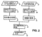
図2は一般に、本発明の方法を例示している。簡単に説明すると、これらの方法は2種類の別個の連続する生理学的データを処理および結合して、新規な咳覚醒指数を判定する。被検体の呼吸データ、音声データ、動きデータ31は、事前処理33の後で、客観的かつ自動的な処置35で使用されて、被検体の咳の発生を検出する。被検体の脳電図(EEG)および眼電図(EOG)(一般に、選択された筋電図(EMG)データ)37は、事前処理39の後で、客観的かつ自動的な処置41で使用されて、被検体の覚醒の発生を検出する。認識された咳と覚醒の発生は互いに関連づけされて43、判定済みの咳覚醒指数および咳障害指数45が出力される。このような工程と付随するシステムは、後段でより詳細に説明される。
<A2 System and Method of the Present Invention>
FIG. 2 generally illustrates the method of the present invention. Briefly, these methods process and combine two separate sequential physiological data to determine a new cough alertness index. The subject's breathing data, audio data, and
<A2−2 咳事象の認識>
本発明により処理されるデータは、図1に例示されている衣服またはシャツ1のような、装着可能な監視用装置によって得ることができ、かかる衣類は十分に快適かつ突出していないため、被検体の眠りは(実質的に)妨害されることはない。このような衣服は、必要な被検体監視データを収集するためのセンサーで具現化され、または、そのようなセンサーに一体的に含まれて、一晩中もの間の家庭内の設定で睡眠中の生理学的記録を行えるようにし、かつ/または、無制限な出歩ける設定で日中に生理学的記録を行えるようにする。
<A2-2 Recognition of cough events>
Data processed by the present invention can be obtained by a wearable monitoring device, such as the garment or
この衣服は、本発明のデータを提供するために使用される監視用器具の好ましい一例である。この衣類は、本発明を制限することはなく、他の実施形態においても、本発明によって処理されるデータは、当該技術で周知の他のセンサー技術によって収集され、また、そのようなセンサーを監視下にある被検体上に配置および配列することにより回収することができる。しかしながら、ただ簡明にするために、後段の説明は、監視用衣服とそれに付随するシステム構成要素の好ましい実施形態に大いに関連づけられている。 This garment is a preferred example of a monitoring instrument used to provide the data of the present invention. This garment does not limit the present invention, and in other embodiments the data processed by the present invention is collected by and monitored by other sensor technologies known in the art. It can be recovered by placing and arranging on the underlying subject. However, for the sake of brevity, the following description is largely related to the preferred embodiment of the monitoring garment and the accompanying system components.
呼吸信号、音声信号、および、動き信号31はそれぞれに、誘導体積変動記録(IP)呼吸センサー帯5、7(または、それ以外のセンサー種類で、呼吸速度情報と呼吸量情報を供与するもの)、シャツの内側にあるものとして例示されている具体的な加速計11のような、体姿勢と体の動きを検知する1個以上の加速計など、更には、喉マイク14のような、咳の音を検出する1個以上のマイクから入手される。衣服1(ここでは、「シャツ」とも呼ばれる)は、被検体の体に従って伸び縮みするのに十分に抱き包むように適合する伸縮自在な素材から作られており、そこに埋設されたIPセンサー帯(呼吸測定のための、呼吸誘導体積変動記録帯すなわちRIP帯として周知である)が被検体の上体の断面領域または周囲を測定することができるようになっている。RIP帯は1個でよいが、RIP帯を2個使うのが好ましい。すなわち、胸郭のレベルの帯5と腹部のレベルの帯7である。IP技術の詳細(および、当該技術で周知の代替センサー技術の詳細)は、後段の見出しA3部分とそこに含まれる引例に記載されている。
Respiration signal, audio signal, and
EMG信号およびEOG信号37はEEGセンサーおよびEOGセンサーから入手されるが、その具体例として、単体バイポーラ(頭頂骨に配置される)EEGセンサー15および単体リードEOGセンサー13などがある。EEGセンサーおよびEOGセンサーは、例えば、導電性コネクタ17などによってシャツ1と電気連絡状態にあるのが好ましい。本発明については任意であるが、また別なセンサーがシャツに接触され、または、シャツと電気連絡状態に置かれてもよいが、例えば、パルス酸素計、カプノグラフ、EEG電極(参照番号9a、9bで例示されている)などがある。病院、医院、または、研究室では、上記以外の信号が広範な生理学的センサーから得られる。
The EMG signal and the
好ましい衣類1と局在的に関連して、データケーブル2により(または、短波ラジオリンクにより)局所データ記録装置3が動作可能に衣類の被検体センサーに接続されている。データ記録装置3は通院用途では好ましく、小型かつ軽量で、ベルトに装着したり、ポケットに入れたり、シャツ1に埋め込むことができるようになっているのが好ましい。この装置は、十分な医学的明細とオフライン解析に備えた満足のゆく正確さと精度でセンサーデータを記憶し、ディジタルダイアリを実現するためのタッチスクリーン(または、それ以外のユーザー入力設備)を備えており、ディジタルダイアリーのデータは解析コンピュータに転送されて、センサー読取り値と相互に関連づけられる。
Locally associated with the
本発明の方法は、コンピュータ21などのような解析コンピュータで実行される解析ソフトウエアによって実現される。解析は信号記録(オンライン)と同時に行われるか、または、後で(オフラインで)行われる。オフライン解析については、センサーデータはデータ記録装置3から解析コンピュータ21に転送され、コンパクトフラッシュ(登録商標)カードのようなメモリカード19に保存される。これに代わる例として、データは、携帯電話技術などを利用したデータ伝達などの無線リンクによって転送することができる。本発明の方法を実現するこのような解析ソフトウエアの全部または一部は、光ディスク23のようなコンピュータが読取ることのできる媒体のプログラム製品として利用できるようにしてもよい。睡眠監視処理については、衣類1を担体とするセンサーがデータ解析保存システム21に直接接続される。代替例として、睡眠中に使用するためのデータ記録装置は、携帯性を減じるという犠牲を払う代わりに、上記以外の能力を付加して処理を行うようにしてもよい。
The method of the present invention is realized by analysis software executed by an analysis computer such as the
再度、図2を参照すると、センサーデータを初期ディジタル化して処理する工程33は、アナログセンサー信号の必要なディジタル化として、ディジタル信号をフィルタ処理してノイズと偽信号を除去する処理を含んでいる。呼吸信号を更に処理する工程は、1個以上のRIP帯からの信号を較正および結合して、呼吸速度信号および一回呼吸気量(VT)信号に変換すること(更に任意で、それでも残存している偽信号を除去するように処理する)と、それぞれを解析して指標と傾向を判定することを含んでいる。これらの処理工程の詳細を後段で説明する。
Referring again to FIG. 2, the
マイクデータを更に処理する工程は、低周波数音声成分とそれぞれの一時的変動性を識別する処理を含むが、これらは咳の特徴を示す音事象を認識するために結合される。好ましい実施形態では、低周波数音声成分が所定回数、所定の閾値を超過した場合は、咳と予測される事象であると識別される。このような閾値と回数は個々の被検体の変化を反映するために調節されるのが好ましい。 Further processing of the microphone data includes identifying low frequency speech components and their respective temporal variability, which are combined to recognize sound events indicative of cough characteristics. In a preferred embodiment, if the low frequency audio component exceeds a predetermined threshold a predetermined number of times, it is identified as an event that is predicted to be a cough. Such thresholds and times are preferably adjusted to reflect changes in individual subjects.
次に、客観的な、コンピュータにより実施される処理工程35は、咳と予測される事象を認識するために、事前処理された呼吸信号、音声信号、および、動き信号を結合し、較正する。このような事象は、データが個々の激しい吐息を示して、一部が閉じた声門に向けて1呼吸のうちに発生している場合に認識される。特に、咳と予測されるものは、高い吐息流を伴って実質的に(一時的に)局在判定された指標吐息流を超過した呼吸信号によって示される。更に、咳は一部が閉じた声門に向けた吐息であるから、或る期間にわたって実質的に一定である低周波数成分を含む音事象に関連していることが多い。処理済みのマイクデータからこのような特性を含む音を観察することでも、咳と予測されるものが分かる。咳は、特徴的な呼吸事象の発生により認識され、咳と予測されるものは、特徴的な呼吸事象と特徴的な音事象の同時発生によって認識される。 Next, an objective, computer-implemented processing step 35 combines and calibrates the preprocessed respiratory signal, audio signal, and motion signal to recognize the expected cough event. Such an event is recognized when the data indicates an individual sigh and is occurring in one breath towards a partially closed glottis. In particular, what is predicted to be a cough is indicated by a respiratory signal that exceeds the index breath flow that is substantially (temporarily) localized with high breath flow. In addition, since cough is a sigh that is directed partially to the glottis, it is often associated with a sound event that includes a low frequency component that is substantially constant over a period of time. By observing a sound including such characteristics from the processed microphone data, it is possible to understand what is predicted to be cough. A cough is recognized by the occurrence of a characteristic respiratory event, and what is predicted to be a cough is recognized by the simultaneous occurrence of a characteristic respiratory event and a characteristic sound event.
図3は、このような方法に従って検出された咳と予測されるものを例示している。同図は、8個の同時発生痕跡を含んでおり、上から下の順に、一回呼吸気量(VT)信号、胸郭RIP帯(RC)信号、腹部RIP帯(AB)信号(VTはRC信号とAB信号の組合せである)、心電図(ECG)信号、マイクロフォン(MIC)信号、認識された複数音事象(EVT)(MIC信号から認識される)の同時発生、認識された咳(CGH)の同時発生、加速計(ACC)信号である。どの痕跡についても、時間は左から右に向かって経過する。VT信号はRC信号とAB信号の較正された組合せであり、EVT信号はMIC信号に由来する複数音事象の同時発生を示している。 FIG. 3 illustrates what is predicted to be a cough detected according to such a method. The figure includes eight simultaneous traces. From the top to the bottom, the tidal volume (V T ) signal, the rib cage RIP band (RC) signal, the abdominal RIP band (AB) signal (V T Is a combination of RC and AB signals), electrocardiogram (ECG) signal, microphone (MIC) signal, recognized multiple sound events (EVT) (recognized from MIC signal), recognized cough ( CGH), accelerometer (ACC) signal. For any trace, time elapses from left to right. V T signal is calibrated combination of RC signal and AB signal, EVT signal indicates the simultaneous occurrence of multiple sound events from MIC signal.
図3は、3回認識された咳と予測されるもの51c、53c、55cを示している。51cは、EVT51bがVT信号の大きな負の傾き51aによって示される高い吐息流と同時発生していることを根拠に咳と認識される。同様に、咳53cおよび咳55cはEVT53bおよびEVT55bがVT信号の大きな負の傾き53aおよび55aとそれぞれ同時発生しているのを示している。EVT57a、EVT57b、EVT57cは咳と認識されないが、それは、VT信号の大きな負の傾きに一致していないからである。最後に、EVT59aは、VT信号の吸気(正の傾き)59aと対応しているので、咳ではない。 FIG. 3 shows the predicted coughs 51c, 53c, 55c recognized three times. 51c is, EVT51b is recognized as cough grounds that they are high exhaled air flow and concurrent indicated by the large negative slope 51a of the V T signal. Similarly, cough 53c and cough 55c show that the EVT53b and EVT55b are respectively concurrent and large negative slope 53a and 55a of the V T signal. EVT57a, EVT57b, but EVT57c is not recognized as coughing, it is because not coincide with the large negative slope of the V T signal. Finally, EVT59a, since it corresponds to the intake (positive slope) 59a of the V T signal, not a cough.
<A2−3 EEG覚醒の認識>
図2をもう一度参照すると、処理工程37で受信されたEEG信号およびEOG信号(これに代えて、選択されたEMG信号)が処理工程39で事前処理されてから、工程41で一過性覚醒を認識するために使用される。好ましいEEGセンサー位置は、EEG技術ではありふれた位置決め表記を使って規定されるが、参照符号C4/A1またはC3/A1の中央バイポーラ設置位置と、O1/A2、O2/A1またはOZ/A1、もしくは、OZ/A2などの任意のバイポーラ後頭部基準設置位置がある。好ましいバイポーラEOG電極設置位置はLOC/A1および/またはROC/A2である。代替の実施形態では、EOG信号は顎下信号またはそれ以外EMG信号で補足され、或いは、それらと置換されてもよい。
<A2-3 Recognition of EEG awakening>
Referring back to FIG. 2, the EEG signal and EOG signal received in processing step 37 (alternatively, the selected EMG signal) are pre-processed in processing step 39, and then transient arousal is detected in step 41. Used to recognize. Preferred EEG sensor positions are defined using common positioning notations in EEG technology, but with a central bipolar installation position of C4 / A1 or C3 / A1, and O1 / A2, O2 / A1 or OZ / A1, or , There is an arbitrary bipolar occipital reference installation position such as OZ / A2. The preferred bipolar EOG electrode location is LOC / A1 and / or ROC / A2. In alternative embodiments, the EOG signal may be supplemented with or replaced by a submandibular signal or otherwise an EMG signal.
受信された信号は、次いで、工程39でディジタル化され、再処理される。通例、約50 Hzを超過するEEG信号およびEOG信号はそれほど興味の対象とはならず、適切な信号デジタル化は100/毎秒(ナイキスト周波数)であり、ディジタル化は150/毎秒またはそれ以上であるのがより好ましく、200/毎秒またはそれ以上であるのが更により好ましい。次に、ディジタル化された信号は低域フィルタで濾過され、約50 Hz以上などのあまり重要ではない高周波数成分が除去される。最後に、標準的なEEG周波数帯、すなわち、アルファ帯、ベータ帯、シータ帯、デルタ帯などに準じているのが好ましいが、信号周波数成分と時間の関係を反映している分光写真タイプの出力を供与するように、信号が処理される。この処理は、例えば、時間窓を設けた帯域通過フィルタバンク、または、多元分解能ウエーブレット分解によって行うことができるが、この場合、フィルタの通過帯域またはウエーブレット分解能はEEG周波数帯に従って選択される。 The received signal is then digitized at step 39 and reprocessed. Typically, EEG and EOG signals above about 50 Hz are not of great interest, proper signal digitization is 100 / sec (Nyquist frequency), and digitization is 150 / sec or more Is more preferred, and even more preferred is 200 / second or more. The digitized signal is then filtered with a low pass filter to remove less important high frequency components, such as above about 50 Hz. Finally, it is preferable to follow the standard EEG frequency band, ie alpha band, beta band, theta band, delta band, etc., but spectrograph type output reflecting the relationship between signal frequency components and time The signal is processed to provide This process can be performed, for example, by a bandpass filter bank with a time window or multi-resolution wavelet decomposition, in which case the passband or wavelet resolution of the filter is selected according to the EEG frequency band.
次に、EEG覚醒の標準定義から得られる規則を好ましくは不随意的に適用することにより、工程41で、中心部の微分EEGまたは後頭部の微分EEGのいずれかから導出される分光写真型出力から、覚醒が認識される。例えば、ボンネットほか著、「脳波覚醒:計算上の規則と具体例−米国睡眠障害教会の睡眠障害図解対策委員会からの予備報告書」(睡眠15号、173頁〜184頁、1992年刊)などを参照のこと。好ましいルールの認識によると、EEG周波数が3秒以上持続して16 Hz(例えば、シータ周波数、アルファ周波数、および/または、ベータ周波数など)よりも高い帯域で、ただし、スピンドルなしに突然シフトしたことを分光写真が表している場合は、覚醒であるとする。生理学に対立するものとして、3秒という基準は主として方法論的であるため、これ以外の持続時間を使用して、各種の状況でEEG周波数シフトを信頼をもって認識することができるようにしてもよい。
Next, the rules derived from the standard definition of EEG arousal are applied, preferably involuntarily, at step 41 from the spectroscopic output derived from either the central differential EEG or the occipital differential EEG. Awakening is recognized. For example, Bonnet et al., EEG Awakening: Computational Rules and Specific Examples-Preliminary Report from the Sleep Disorders Illustrative Committee of the American Sleep Disorders Church (
この規則は或る二次的規則に従って限定することができる。覚醒は睡眠を途絶する周期的現象であると考えられているので、二次的規則の1つは、被検体がどの睡眠段階であれ10秒以上眠っている場合に覚醒と認識されるものとし、更に、どの段階であれ10秒以上の眠りが先行する覚醒と今回の覚醒との間に挟まっている場合に、二回目(今回)の覚醒が認識されるものとする、というものである。大まかに言って、10秒という期間が選択されているのは、10秒に満たない間隔にわたり睡眠状態か覚醒状態かを判定することは信頼性が劣るからである。しかし、独立した覚醒と勘定するのに必要な最小量の介在する睡眠はバックグラウンドEEGで決まり、状況によって変動することがある。 This rule can be limited according to certain secondary rules. Since awakening is considered to be a periodic phenomenon that disrupts sleep, one of the secondary rules is that a subject is recognized as awake if he sleeps for more than 10 seconds at any sleep stage. Furthermore, when a sleep of 10 seconds or more is sandwiched between the preceding awakening and the current awakening at any stage, the second (this time) awakening is recognized. Broadly speaking, the period of 10 seconds is selected because it is less reliable to determine whether it is a sleep state or an awake state over an interval of less than 10 seconds. However, the minimum amount of intervening sleep required to account for independent wakefulness is determined by background EEG and may vary from situation to situation.
また別な二次的規則は、EEG特性に従って睡眠を周知の分類法を使ってレム(REM: Rapid-Eye-Movement)睡眠またはノンレム睡眠(NREM: non-REM)睡眠に分類する(ノンレム睡眠は更に副次的分類により睡眠段階1、2、3、4に分類される)。ノンレム睡眠では、覚醒は、EEG特性のみに基づいて認識される。しかし、アルファまたはシータEEG活性のバーストはレム睡眠ではよくあることであるうえに、生理学的覚醒を反映していないことがあるため、レム睡眠から覚醒であると信頼をもって勘定に入れるのに追加要件として、EOG(またはEMG)振幅が増大することを加えるのが好ましい。しかし、EMG振幅の変動のみを基準として覚醒と見なすことはできない。本質的に、レム睡眠が認識された場合、覚醒と認識するには、そのようなEOG振幅またはEMG振幅の増大が必要となる。
Another secondary rule is to classify sleep into REM (Rapid-Eye-Movement) sleep or non-REM (NREM) sleep according to EEG characteristics using well-known classification methods (non-REM sleep is Furthermore, it is classified into
覚醒を認識する際に有用となる別な規則は、ボンネットほかなどの先行技術文献に記載されている別な状況から得ることができる。 Another rule useful in recognizing arousal can be obtained from other situations described in prior art documents such as bonnet et al.
<A2−4 咳−覚醒事象の認識>
図2を再度参照すると、工程35において認識された咳が工程41で認識されたEEG覚醒と関連して検出されるような睡眠中は、工程43で咳−覚醒事象が認識される。咳が覚醒中に発生した場合、咳と覚醒は関連があり、また、咳が覚醒を含む時間窓の範囲内で発生した場合、咳と覚醒は関連がある。好ましい時間窓は覚醒の前に先行し、約30秒の長さを保つ(または、約1分までの長さを保つ)。これ以外の好ましい時間窓は、覚醒の後に続く約30秒である(または、約1分)。これ以外の好適な時間窓は、個々の被検体ごとに判定される。覚醒期間中または覚醒に関連する時間窓の期間中に咳が発生していない場合、咳が覚醒と関連づけられることはない。
<A2-4 Recognition of cough-wakefulness event>
Referring back to FIG. 2, a cough-wake event is recognized at
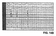
図4は具体的な咳−覚醒事象を例示している。同図は、11個の同時発生痕跡を含んでおり、上から下の順に、EEG信号(EEG)、EOG信号(EOG)、認識された覚醒(ARS)、一回呼吸気量(VT)信号、胸郭RIP帯(RC)信号、腹部RIP帯(AB)信号、高周波数フィルタ処理済みVT信号(HFB)、マイクロフォン(MIC)信号、認識された複数音事象(EVT)の同時発生、認識された咳(CGH)の同時発生、加速計(ACC)信号である。どの痕跡についても、時間は左から右に向かって経過する。咳61dは、強迫的な吐息61bが音事象61cと同時発生したと認識される。また、咳61dはEEG覚醒61aと関連して(ここでは、EEG覚醒61aの最中に)発生しているので、咳−覚醒事象が認識される。強迫的な吐息61bは、低周波数成分が除去された後のHFB信号の61b'では一層顕著である。被検体に由来する加速計データを監視するのも好ましい。高域通過フィルタでこのデータを濾過処理することで、被検体の動きに関する情報が供与され、低域通過フィルタで処理されたデータは被検体の姿勢に関する情報を供与する。動きの変動および/または姿勢の変動はセンサー信号に偽信号を生じることがあるので、動きの変動および/または姿勢の変動に関与する咳および/または覚醒は放棄するのが有利である。 FIG. 4 illustrates a specific cough-wake event. The figure includes 11 simultaneous traces, from top to bottom, EEG signal (EEG), EOG signal (EOG), recognized arousal (ARS), tidal volume (V T ) signal, thorax RIP band (RC) signal, abdominal RIP band (AB) signal, the high-frequency filtered V T signal (HFB), concurrent microphone (MIC) signal, recognized multiple sound event (EVT), recognition Co-occurring cough (CGH), accelerometer (ACC) signal. For any trace, time elapses from left to right. The cough 61d is recognized as a compulsive exhalation 61b occurring simultaneously with the sound event 61c. In addition, since the cough 61d occurs in association with the EEG awakening 61a (here, during the EEG awakening 61a), the cough-wakening event is recognized. Compulsive exhalation 61b is more prominent in 61b 'of the HFB signal after the low frequency component is removed. It is also preferable to monitor accelerometer data derived from the subject. By filtering this data with the high-pass filter, information on the movement of the subject is provided, and the data processed by the low-pass filter provides information on the posture of the subject. It is advantageous to abandon cough and / or wakefulness involved in motion variation and / or posture variation, since motion variation and / or posture variation can cause false signals in the sensor signal.
次に、咳覚醒指数(CAI)は、睡眠中の1時間あたり(または、それ以外の適切な期間あたり)の咳−覚醒事象(咳と覚醒に関与する)の回数と判定される。咳障害指数(CDI)は、咳−覚醒事象の一部ではない(すなわち、EEG覚醒と関連している)睡眠中の1時間あたり(または、それ以外の適切な期間あたり)の咳の回数と判定される。CAIとCDIの和は、1時間あたりの咳の合計数である。 The cough alertness index (CAI) is then determined as the number of cough-wake events (involved in cough and alertness) per hour during sleep (or per other appropriate period). The cough disorder index (CDI) is the number of coughs per hour during sleep (or per any other suitable period) that are not part of a cough-wake event (ie, associated with EEG alertness) Determined. The sum of CAI and CDI is the total number of coughs per hour.
このような指数は、監視下にある被検体および監視中の担当人員が使う出力である。例えば、監視下にある被検体は、CAIが容認できる閾値より低くなるように、または、容認できる範囲内に収まるように薬物服用量を調節することで、被検体の睡眠構造の異常を適切に緩和することができる。医療監視人員は、薬物開発、薬物試験、または、薬物評価の途中で被検査集団のCAIおよびCDIを監視することができる。 Such an index is the output used by the subject being monitored and the person in charge being monitored. For example, a subject under monitoring may properly adjust the subject's sleep structure abnormalities by adjusting the drug dose so that the CAI is below an acceptable threshold or within an acceptable range. Can be relaxed. Medical monitoring personnel can monitor the CAI and CDI of the tested population during drug development, drug testing, or drug evaluation.
<A2−5 咳覚醒指数の具体例>
本発明のシステムおよび方法、睡眠障害の特性、および、CAIの臨床的重要性は、以下の測定によって確かめられる。
<Specific example of A2-5 cough alertness index>
The system and method of the present invention, the characteristics of sleep disorders, and the clinical significance of CAI are confirmed by the following measurements.
中程度から重症までのCOPDを患っている10人の患者が、米国カリフォルニア州ヴェンチユラに居所を置くヴィヴォメトリクス・インコーポレティッド(VivoMetrics, Inc.)からライフシャツ(LifeShirt:登録商標)監視システムを利用しながら個々の正常な日常活動を実施しながら、各自の家庭で監視下に置かれた。ライフシャツシステムは、上述の好ましい監視用衣類とデータ記録装置を実装していた。特に、監視用衣類はRC RIP帯センサーおよびAB RIP帯センサーと、修正型の四肢用II型ECGセンサーと、姿勢と動きのフィルタ装備した加速計センサーと、咳音を識別するために甲状軟骨の接触マイクセンサーとを備えていた。睡眠中は、関与するEEGセンサーおよびEOGセンサーからのデータも記録された。この生理学的監視データは、上述の好ましい方法によっても処理された。更に、ビデオ(オーディオ付き)テープ記録術を利用して、好ましい自動咳認識を検証した。感度0.78、特異性1.0、および、精度0.99が観察された。 Ten patients with moderate to severe COPD use a LifeShirt® surveillance system from VivoMetrics, Inc., located in Ventura, California, USA While undertaking individual normal daily activities, they were placed under supervision in their homes. The life shirt system was equipped with the preferred monitoring garment and data recording device described above. In particular, surveillance clothing includes RC RIP and AB RIP sensors, a modified limb type II ECG sensor, an accelerometer sensor equipped with posture and movement filters, and thyroid cartilage to identify cough sounds. It had a contact microphone sensor. During sleep, data from involved EEG and EOG sensors were also recorded. This physiological monitoring data was also processed by the preferred method described above. In addition, video (with audio) tape recording was utilized to verify preferred automatic cough recognition. A sensitivity of 0.78, a specificity of 1.0, and an accuracy of 0.99 were observed.
このような測定の結果が以下のものを含む。第1に、図5は、丸一日のうち1時間あたりの平均咳周波数を2日分、例示している。咳周波数は、両日とも相互に似たような概日パターンを追従しており、午前8時頃と午後2時から午後4時までの期間に咳周波数のピークがあることを特徴とする。夜間の咳は、早朝を除き、ほぼ夜通し顕著な頻度で発生した。このような夜間の咳の数は、その一例が図4に示されているが、EEG覚醒中に発生するか、または、覚醒に関与する認知できる時間窓の範囲内で発生し、従って、患者のCAIの一因となった(他の咳はCDIに計上された)。 The results of such measurements include: First, FIG. 5 exemplifies the average cough frequency per hour for the entire day. The cough frequency follows a circadian pattern similar to each other on both days, and is characterized by cough frequency peaks in the period from about 8 am and from 2 pm to 4 pm. Nighttime cough occurred with a noticeable frequency almost overnight, except early morning. The number of such nighttime coughs, an example of which is shown in FIG. 4, occurs during EEG arousal, or occurs within the perceivable time window involved in arousal, and thus patients Contributed to CAI (other coughs were counted in CDI).
次に、睡眠は、記録されたEEG信号を評価するために先に説明した規則を利用してノンレム睡眠(段階1から段階4)とレム睡眠に段階分けされて、睡眠段階ごとの期間中の咳の回数が確かめられた。図6は次のような測定値を例示している。すなわち、点描暗色棒線はCOPD患者の各睡眠段階中の咳の平均回数を示しており、黒塗り矩形はCOPD患者が各睡眠段階に費やした平均持続時間を示しており、更に、白抜き矩形(REF)は、正常で健康な、上記患者と同じ年齢の管理者が各睡眠段階に費やした平均時間を示している。これらの値は全て平均値であり、平均値の標準偏差は誤差バー(Tとして表記されている)によってうまく示されている。この図は、COPD患者がノンレム睡眠とレム睡眠のそれぞれの段階3と段階4の両方を通して均一に分布する咳を経験したことを示している。しかし、ノンレム睡眠の段階1の間、咳は幾分か増大し、ノンレム睡眠の段階2の間、ひときわ多数回の咳が発生した。従って、夜間の咳は浅い眠りの段階で最も頻繁に発生し、そのため、このようなCOPD患者は段階1の睡眠における正常なパーセント時間よりも高いパーセント時間を費やしている。
Next, sleep is staged into non-REM sleep (
従って、夜間の咳はこのようなCOPD患者がより深い眠りの段階へと自然に進んでゆくのを妨げ、段階1の眠りと段階2の眠りに法外なパーセント時間が費やされるという睡眠妨害の構造の主因となっている。このような睡眠妨害は日中の作業性能に逆影響を及ぼしがちであり、生活の質を低下させ、それ以上の問題を引き起こす可能性がある。これで、咳の影響を受けやすい被検体の夜間の咳を監視および治療する重要性が確認される。
Thus, a night cough prevents these COPD patients from going naturally to deeper sleep stages, and sleep disturbances that spend extraordinary percentage time on
更に、先に説明された方法に従って監視下のCOPDに対してCAIが判定され、各患者のCAIは、患者の予測されるピーク吐息のパーセント流量と相互に関連があった。予測された吐息のパーセント流量は、患者のFEV1の、患者と年齢が一致する正常な管理人について予測されたFEV1に対するパーセント割合であるが、COPDを監視する際に有用となる気道閉鎖の分かっている測定値である。図7Aはこのような比較の結果を例示している。すなわち、予測されたピーク吐息のパーセント流量をCAIと二変数適合させると、有意水準0.05で相関関係強度が0.64であるのが分かる。この相関関係は、観察された肺機能と睡眠の質とを結びつける臨床医学的変数としてのCAIの有用性を確認するものである。 In addition, CAI was determined for supervised COPD according to the methods described above, and each patient's CAI correlated with the patient's predicted peak inspiratory percent flow. The predicted percent exhalation flow is the percentage of the patient's FEV 1 compared to the FEV 1 predicted for a normal caretaker who is age-matched with the patient, but is useful for monitoring COPD. This is a known measurement. FIG. 7A illustrates the result of such a comparison. That is, when the predicted percent flow of peak exhalation is fitted to CAI with two variables, it can be seen that the correlation strength is 0.64 at a significance level of 0.05. This correlation confirms the usefulness of CAI as a clinical medical variable linking observed lung function and sleep quality.
重要なのは、本発明の測定値と指数がコンピュータが実行する方法によって客観的に判定されるという点である。このような測定値は患者への問診と回収された回答に依存してはいない。これに比べて、咳と睡眠障害の先行技術の判定は、患者の回答の回収や報告という、いずれも信頼できないと分かっているものに依存してきたのであった。図7Bは、予測されたピーク吐息のパーセント流量を咳の指数と対比させた、明らかに信頼できない比較の別な例を示している。咳発生を長期記録したものが無いまま使用された咳指数はカプサイシンに対する感度であったが、カプサイシンは唐辛子の活性成分であるとともに味覚上の辛さの尺度で使用される咳誘発性刺激原である。予測されるピーク吐息のパーセント流量と強い相関関係にある客観的に判定された咳覚醒指数を示している図7Aと比較して、図7Bは、COPDと喘息のいずれについても、予測されるピーク吐息のパーセント流量とこの咳指数との間に観察できる相関関係を示していない。 Importantly, the measurements and indices of the present invention are objectively determined by a computer-implemented method. Such measurements are independent of patient interviews and collected responses. In comparison, prior art determinations of cough and sleep disorders have relied on patient responses being collected and reported, both of which are known to be unreliable. FIG. 7B shows another example of a clearly unreliable comparison comparing the predicted peak sigh percent flow with the cough index. The cough index used without any long-term record of cough occurrence was sensitivity to capsaicin, but capsaicin is an active ingredient in chili and a cough-inducing irritant used on the measure of taste hotness. is there. FIG. 7B shows the predicted peak for both COPD and asthma as compared to FIG. 7A, which shows an objectively determined cough alertness index that is strongly correlated with the percent flow of predicted peak breathing. There is no observable correlation between the percent flow of sigh and this cough index.
<A3−1 好ましいシステムおよび方法>
この項は、先に説明されたシステムおよび方法の更に別な詳細を説明するものである。
<A3-1 Preferred System and Method>
This section describes further details of the systems and methods described above.
<A3−2 好ましいシステム>
呼吸データは、被検体の胸郭の経時変化する断面領域を反映しているのが好ましく、また、被検体の腹部の経時変化する断面領域を反映しているのが有利である。確立された生理学的モデル(例えば、呼吸量の2成分モデルなど)を照合しながらの信号処理技術とパターン認識技術は、生理学的事象が発生した生理学的諸機能と回数の証拠すなわち測定値を与えることができる。例えば、呼吸率、一回呼吸気量の証拠、心拍血液量の証拠、呼吸が落着きを取り戻す発生回数などを入手することが好ましい。
<A3-2 Preferred system>
The respiratory data preferably reflects a cross-sectional area of the subject's thorax that changes with time, and advantageously reflects a cross-sectional area of the subject's abdomen that changes with time. Signal processing and pattern recognition techniques while matching established physiological models (eg, two-component models of respiratory volume) provide evidence or measurements of the physiological functions and times that a physiological event has occurred be able to. For example, it is preferable to obtain the respiration rate, the tidal volume evidence, the heart rate blood volume evidence, the number of occurrences of respiration calmness, and the like.
そのような測定値についての1つの好ましいセンサー技術は、誘導体積変動記録(IP)である。この技術は、心臓機能と呼吸機能に関する信頼できる半定量データおよび定量データを供与するように、臨床的に既に正式に認められている。簡単に述べると、IPは監視下にある被検体の喉、腹部、および、それ以外の肉体構造について多用なレベルで設置されている導電ループ(一般に、センサー帯として構成されている)のインダクタンスを測定する。このような経時変化するループインダクタンス測定値は、それらのループによって包絡されている経時変化する断面積を主として反映している。 One preferred sensor technology for such measurements is induced volume variation recording (IP). This technique has already been formally approved clinically to provide reliable semi-quantitative and quantitative data on cardiac and respiratory functions. In short, IP is the inductance of a conductive loop (typically configured as a sensor band) that is installed at various levels for the throat, abdomen, and other body structures of the subject being monitored. taking measurement. Such time-varying loop inductance measurements primarily reflect the time-varying cross-sectional area enveloped by the loops.
しかしながら、本発明はIPベースのセンサーに限定されるものではなく、代替のセンサー技術を採用することができる。代替のセンサー技術があるとすれば、それは、IPベースのセンサーと同様に、喉、腹部、または、それ以外の肉体構造を通る1つ以上の階層で、断面積、周囲長さ、または、それぞれの幾何学的均等物(例えば、応力や歪など)を反映した測定を行うものである。それらについての信号は、IPセンサー信号のために既に開発済みの方法によって処理することができる。例えば、代替のセンサーは、目下開発中であるか、または、これから開発される予定の糸の技術と繊維の技術に基づくものである。すなわち、センサーは応力依存抵抗を示す導電性の糸の抵抗を測定することができ、衣類または帯に組み入れ、または、局所歪みが周方向全体の歪を反映するように織られた繊維の局所歪みを、センサーは光学手段または電気手段により測定することができる。また別な具体例としては、代替のセンサーは、肉体構造を通して幾何学的パラメータ(距離など)を測定するのにエネルギー放射(超音波、電場、磁場、または、電磁場のような)を利用してもよい。 However, the present invention is not limited to IP-based sensors and alternative sensor technologies can be employed. If there is an alternative sensor technology, it can be cross-sectional area, perimeter length, or respectively, in one or more layers through the throat, abdomen, or other body structure, similar to IP-based sensors. The measurement reflecting the geometrical equivalent (for example, stress or strain) is performed. The signals about them can be processed by methods already developed for IP sensor signals. For example, alternative sensors are currently in development or are based on yarn technology and fiber technology that will be developed in the future. That is, the sensor can measure the resistance of a conductive yarn that exhibits stress-dependent resistance, and is incorporated into a garment or band, or the local strain of a fiber woven so that the local strain reflects the strain in the entire circumferential direction. The sensor can measure the optical or electrical means. As another example, alternative sensors utilize energy radiation (such as ultrasound, electric, magnetic, or electromagnetic fields) to measure geometric parameters (such as distance) through the body structure. Also good.
それ以外のセンサーは、必要に応じて、また、入手できれば、本発明に組み入れてもよい。そのようなセンサーとしては、例えば、化学物質(CO2やCH4などに晒すセンサー)、生物危害(多種の放射線、多種の微生物など)のセンサー、および、それ以外のセンサーがある。IPベースの装着可能なセンサーと衣類の詳細は、センサー特許と衣類特許、および/または、心臓機能特許に開示されている。 Other sensors may be incorporated into the present invention as needed and available. Examples of such sensors include chemical substances (sensors exposed to CO 2 , CH 4, and the like), biological hazard sensors (various types of radiation, various types of microorganisms, and the like), and other sensors. Details of IP-based wearable sensors and garments are disclosed in sensor and garment patents and / or cardiac function patents.
生理学的センサーが、多種多様な衣類を着用した監視下の被検体に配置されるのが好ましいが、そのような衣類の具体例として、帯、部分シャツ、シャツ、部分ボディースーツ、全身用ボディースーツなどの、突出部のない、快適な、窮屈ではない繊維の衣類がある。本発明は多様な上述のような衣類とセンサーの配置を含んでおり、その細部は主として、生理学的監視の種類と程度で決まる。このような衣類は睡眠できるような設計で、かつ/または、それほど邪魔にならずに通院活動を行えるような設計であるのが好ましい。
好ましいIP技術、IP技術の衣類への配置、IP技術による処理と解釈、および、或る緊密に提携するセンサー技術の詳細は、本出願の現在の譲受人に目下譲渡されている次の米国特許および特許出願(IP特許)に見ることができる。このような特許および出願はいずれも、それぞれの全体が引例に挙げることで、あらゆる意味で本件の一部をなすものとする。IP技術と繊維および衣類へのIP技術の配置を開示している米国特許(「センサー特許および衣類特許)には、例えば、米国特許第6,551,252号、第6,341,504号、第6,047,203号、第5,331,968号、第5,301,678号、および、1989年2月28日交付の第4,807,640号(伸縮自在なIPトランスデューサー)がある。
Physiological sensors are preferably placed on monitored subjects wearing a wide variety of clothing, examples of such clothing include belts, partial shirts, shirts, partial body suits, and full body body suits. There are comfortable, non-buckled fiber garments, such as no protrusions. The present invention includes various garment and sensor arrangements as described above, the details of which depend primarily on the type and degree of physiological monitoring. Such garments are preferably designed to sleep and / or to be able to perform out-of-hospital activities without significant disruption.
Details of preferred IP technology, placement of IP technology in clothing, processing and interpretation by IP technology, and some closely affiliated sensor technology are the following US patents currently assigned to the current assignee of this application: And in patent applications (IP patents). All such patents and applications are hereby incorporated by reference in their entirety to form part of this case. US patents ("sensor patents and garment patents") that disclose IP technology and the placement of IP technology in textiles and garments include, for example, US Pat. Nos. 6,551,252, 6,341,504, 6,047,203, 5,331,968, There are 5,301,678 and 4,807,640 (stretchable IP transducer) issued February 28, 1989.
IP信号処理を開示している米国特許(データ処理およびデータ翻訳特許)は、例えば、第6,413,225号、第6,015,388号、第5,159,935号、第4,860,766号、第4,834,109号、第4,815,473号、第4,777,962号、第4,648,407号、第4,373,534号、および、第4,308,872号がある。同様の米国特許出願としては、コイル(Coyle)ほかによる2004年4月9日出願の出願番号第(未定)号で、現在の弁護士訴訟事件番号10684-035-999号と、出願番号第10/457,097号がある。IP信号を処理して心臓機能の測定値を得ることを開示している米国特許(心臓機能特許)は、具体例として、第5,588,425号、第5,178,151号、第5,040,540号、第4,986,277号、第4,456,015号、および、第4,452,252号と、米国特許出願第10/107,078号がある。
US patents (data processing and data translation patents) that disclose IP signal processing are, for example, 6,413,225, 6,015,388, 5,159,935, 4,860,766, 4,834,109, 4,815,473, 4,777,962, There are 4,648,407, 4,373,534, and 4,308,872. Similar US patent applications include Application No. (undecided) filed April 9, 2004 by Coyle et al., Current attorney case number 10684-035-999, and
<A3−3 咳事象認識方法>
一般に、このような方法にとりかかるのに、AB信号、RC信号、および、VT信号を含む入力呼吸パラメータから呼吸事象候補を認識し、更に任意で、音声入力から音事象候補を認識するという手段を利用する。次に、呼吸事象候補とこれに関与する音事象候補の同時発生的組合せから、咳事象が検出される。咳の種類の酷さは、呼吸事象パラメータと音事象パラメータの各値によって弁別することができる。
<A3-3 Cough event recognition method>
In general, to get started in this way, AB signal, RC signal, and recognizes the respiratory event candidate from an input respiratory parameters including V T signal, further optionally, means of recognizing a sound event candidate from the speech input Is used. A cough event is then detected from the concurrent combination of the candidate respiratory event and the sound event candidate involved. The severity of the type of cough can be distinguished by the values of the respiratory event parameter and the sound event parameter.
<<咳認識の第1の方法>>
咳検出の第1の好ましい方法は呼吸データのみを使用し、従って、音声データが利用できない場合に有利となる。第1の咳検出方法によると、咳は、0.25秒から3秒の範囲にわたる所定の閾値よりも長い吐息期間を有する真の呼吸と認識されなければならない。有用かつ好ましい閾値は約1秒であり、これは個人ごとに違っていてもよい。次に、このような基準に合致している真の呼吸は、それぞれのピーク吐息流(PEF)が、最初の2分間の窓に基づいて定まるような、連続する中位基本水準PEF値の所定の閾値よりも高い場合には、咳と認識される。好ましいPEF閾値は、連続する中位基本水準PEF値の100%から1000%かそれより高い割合までの間にあり、大半の被検体にとって、約250%よりも高いPEF閾値は結果として、適切な咳認識を生む。閾値は、過去の監視データを利用して特定の被検体ごとに個別的に扱うことができる。
<< First method of cough recognition >>
The first preferred method of cough detection uses only respiratory data and is therefore advantageous when voice data is not available. According to the first cough detection method, the cough must be recognized as a true breath with an exhalation period longer than a predetermined threshold ranging from 0.25 seconds to 3 seconds. A useful and preferred threshold is about 1 second, which may vary from individual to individual. Second, true breaths that meet these criteria are defined by a continuous mid-basal level PEF value such that each peak exhalation flow (PEF) is determined based on the first two-minute window. Is higher than the threshold value, it is recognized as cough. The preferred PEF threshold is between 100% and 1000% or higher of continuous mid-basal level PEF values, and for most subjects, a PEF threshold higher than about 250% results in an appropriate Produces cough recognition. The threshold can be individually handled for each specific subject using past monitoring data.
図8は、咳94と咳98を含む現実の被検体データを例示している。PEFは、短く素早い吐息であると、dV/dt(dVt/dtと標識を付されている)曲線から判定されるが、ここでは、同じ2回の咳96と咳102が短く鋭い吐息として容易に視認される。この例では、咳96のPEFは連続する中位PEF基本水準の約400%であるが、咳102については、PEFは基本水準の約380%である。
FIG. 8 illustrates actual subject data including cough 94 and
<<咳認識の第2の方法>>
咳検出の第2の方法は咳検出の支援策として音声入力を組み入れており、音声データが利用できない場合に好ましい。この項目と添付の図面では、入力データと得られたデータは、次のような略表記で言及されることが多い。
RC 胸郭(RC)測定値(入力データ)
AB 腹部(AB)測定値(入力データ)
VT 一回呼吸気量(RC測定値とAB測定値から先に説明したようにして
抽出された方法入力データ)
HFB 高周波数帯域通過フィルタ処理されたVT(抽出データ)
LFB 低周波数帯域通過フィルタ処理されたVT(抽出データ)
FAB 高周波数帯域通過フィルタ処理されたAB(抽出データ)
MIC 喉のマイクから記録されたマイク音声信号(入力データ)
SE マイク音声信号包絡線(得られたデータ)
PITCH 選択された時間間隔のうちの最重要音声ピッチレベル(抽出データ)
PITCHm 選択された時間間隔のうちの平均音声ピッチレベル(抽出データ)
EVT 音事象と持続時間検出装置(方法工程)
CGH 咳マーカー(検出された咳が存在することを示す方法出力データ)
<< Second method of cough recognition >>
The second method of cough detection incorporates voice input as a cough detection support measure and is preferable when voice data is not available. In this item and the accompanying drawings, input data and obtained data are often referred to by the following abbreviations.
RC Thoracic (RC) measurement (input data)
AB Abdominal (AB) measurement (input data)
V T tidal volume (and from RC measured value and AB measurements as previously described
Extracted method input data)
HFB High frequency bandpass filtered V T (extracted data)
LFB Low frequency bandpass filtered V T (extracted data)
FAB High-frequency bandpass filtered AB (extracted data)
MIC Microphone audio signal (input data) recorded from the throat microphone
SE microphone audio signal envelope (obtained data)
PITCH Most important voice pitch level of the selected time interval (extracted data)
PITCHm Average voice pitch level (extracted data) during the selected time interval
EVT sound event and duration detection device (method process)
CGH cough marker (method output data indicating the presence of detected cough)
簡単に説明すると、一回呼吸気量VTはまず、フィルタを通されて、高周波数成分と低周波数成分に分けられる。AB信号もフィルタを通されて、高周波成分を選別する。これらは更に、任意で、高周波数ノイズと低周波数移動偽信号を更に制限するように設計されている。フィルタ処理語の信号はそのピークから次のピークまでの出力振幅、すなわち、呼吸振幅(最大吐息と最大吸気の差)が所定の閾値Tを超過している場合は、呼吸信号と音声信号の両方がより詳細に検査されて、咳のように見える事象の存在を検出する。閾値を超過していない場合は、咳事象が存在する見込みはない。 Briefly, the tidal volume VT is first filtered and divided into a high frequency component and a low frequency component. AB signals are also filtered to filter out high frequency components. They are also optionally designed to further limit high frequency noise and low frequency mobile spurious signals. The signal of the filtered word is the output amplitude from the peak to the next peak, that is, if the respiratory amplitude (difference between maximum exhalation and maximum inspiration) exceeds a predetermined threshold T, both the respiration signal and the audio signal Are examined in more detail to detect the presence of an event that looks like a cough. If the threshold is not exceeded, no cough event is likely to exist.
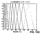
音声信号(例えば、喉のマイクからの)は発話認識フロントエンドで処理されて、音事象が言葉で表明された発話か言葉で表明されていない発話かを判定する。この判定で重要なのは、抽出信号PITCHmであり、この信号は選択された帯域mにおける有限持続期間にわたるピッチの値の平均である。この平均レベルは、被検体が話をしている、または、会話に関与している場合にはかなり上昇し、咳の場合には上昇しない。ピッチ値の算定は、ケプストラム係数またはメル周波数ケプストラム係数(MFCC)に存在しているピークから次のピークまでの出力を測定することによって実施される。また別な重要な抽出信号がPITCH信号である。音声信号処理に由来する出力はパルスであり、図11のEVT痕跡によって例示されている通りであるが、そのタイミングと持続時間は、入力音声データ中に検出された重要な音事象のタイミングおよび持続時間に等しい。 The audio signal (eg, from the throat microphone) is processed by the speech recognition front end to determine if the sound event is an utterance expressed in words or not expressed in words. Important in this determination is the extracted signal PITCHm, which is the average of the pitch values over a finite duration in the selected band m. This average level rises considerably when the subject is speaking or engaged in conversation and does not rise in the case of cough. The calculation of the pitch value is performed by measuring the output from the peak existing in the cepstrum coefficient or the mel frequency cepstrum coefficient (MFCC) to the next peak. Another important extraction signal is the PITCH signal. The output resulting from the audio signal processing is a pulse, as illustrated by the EVT trace of FIG. 11, whose timing and duration are the timing and duration of important sound events detected in the input audio data. Equal to time.
音事象が存在しない場合は、咳は検出されない。音事象が存在している場合、その持続時間は、フィルタ処理後のどの呼吸信号が咳特性検出装置に供与されるべきかを決める。音事象の持続時間が比較的長い場合(すなわち、中位重要性の音事象よりも長く)、例えば、>=600 msec.の場合、低周波数帯域通過フィルタ処理後の呼吸データLFBが咳特性検出装置によって解析される。音声持続期間が比較的短い場合(すなわち、中位重要性の音事象よりも短く)、例えば、<=600 msec.の場合、高周波数帯域通過呼吸データHFBが解析される。この信号選択は移動および動き偽信号を適切にフィルタで除去し、咳特性がより明確に検出されるように誘導することが分かっている。 If no sound event is present, no cough is detected. If a sound event is present, its duration determines which filtered respiratory signal is to be provided to the cough characteristic detector. If the duration of the sound event is relatively long (ie longer than the sound event of medium importance), for example> = 600 msec., The respiratory data LFB after low frequency bandpass filtering will detect cough characteristics Analyzed by the device. If the speech duration is relatively short (ie shorter than a medium importance sound event), for example <= 600 msec., The high frequency bandpass breathing data HFB is analyzed. This signal selection has been found to properly filter out movement and motion spurious signals and induce cough characteristics to be detected more clearly.
図9は、咳検出の第2の方法の詳細を例示している。一回呼吸気量の痕跡VTは、RC帯およびAB帯の線形的に重み付けされた和であると先に判定されているが、これがまず 2個の互いに平行なFIR帯域通過フィルタを通過させられて、ピーク出力(フィルタ処理された信号の最大値によって反映されるような)が測定されて、ピーク出力が閾値Tを超過した場合には、起こり得る咳事象の存在を判定する。入力呼吸信号のフィルタは有限衝撃反応(FIR)設計のものであるのが好ましいが、無限衝撃反応(IIR)フィルタで、位相シフトまたは時間遅延が最小限であるものを使用してもよい。ここでは、呼吸信号位相は十分に平静で、対応する音声信号と一時的に一致したままになるようにしなければならない。
FIG. 9 illustrates details of the second method of cough detection. The tidal volume trace VT has previously been determined to be a linearly weighted sum of the RC and AB bands, which are first passed through two parallel FIR bandpass filters. The peak output (as reflected by the maximum value of the filtered signal) is measured to determine the presence of a possible cough event if the peak output exceeds a threshold T. The input respiratory signal filter is preferably of a finite impact response (FIR) design, but an infinite impact response (IIR) filter with minimal phase shift or time delay may be used. Here, the respiratory signal phase must be sufficiently calm and remain in temporary agreement with the corresponding audio signal.
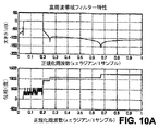
高周波数帯フィルタについては図10Aに例示され、低周波数帯フィルタについては図10Bに例示されている、十分に鋭い周波数特性と平坦な位相特性を達成するのに好ましいとして、1024のフィルタ長が判定された。表1はこれら好ましいそれぞれのフィルタのパラメータをリスト化しており、胸郭および腹部(RCおよびAB)から得られた十分な呼吸運動を維持しながら、発生し得る被検体の肉体運動を表記の限度までフィルタで選別するようにパラメータが選択されている。

次に、ピークから次のピークまでの出力が測定され、閾値と比較される。ピークから次のピークまでの出力は正特性信号の最大値と負特性信号の最小値との差になるように取るのが好ましい。所定の閾値に合致している場合は、咳事象の候補はフィルタ処理された呼吸信号に存在している可能性があると考えられる。この閾値に合致していない場合は、咳事象は存在している可能性があるとは見なされず、信号のこの部分をそれ以上処理することは実施されない。信号LFB、信号HFB、信号FABはこの判定を行うために測定される。信号FABはABフィルタ処理痕から残存しているフィルタ残差であり、RCおよびABが互いに異相であるとともにVTに差の効果を発揮して帯域で真の作用力を減じている場合には、有利である。 Next, the output from one peak to the next is measured and compared to a threshold value. The output from the peak to the next peak is preferably taken so as to be the difference between the maximum value of the positive characteristic signal and the minimum value of the negative characteristic signal. If the predetermined threshold is met, the cough event candidate is likely to be present in the filtered respiratory signal. If this threshold is not met, a cough event is not considered possible and no further processing of this portion of the signal is performed. Signal LFB, signal HFB, and signal FAB are measured to make this determination. Signal FAB is a filter residuals remaining from AB filtering traces, if they reduce the real action force in the band to exhibit the effect of the difference in V T with RC and AB are heterophasic each other Is advantageous.
閾値Tは、正常な呼吸がそれ以上の検査に回されることがないように選択されるのが好ましい。閾値は中位数、平均値、または、それ以外の、被検体の現在の呼吸測定値であればよい。これに代わる例として、固定された閾値が使用される。一般に、休息中または睡眠中の被検体には約200 mlの吐息量が相応である。相応な吐息量が広い範囲にわたる特定の被検体集団には固定された閾値が選択されるのが好ましく、そのような特定の一被検体にも固定された閾値が選択されるのがより好ましい。閾値は、被検体の現在の吐息量の百分率として選択されてもよい。 The threshold T is preferably selected so that normal breathing is not diverted to further examinations. The threshold may be a median number, an average value, or any other current respiratory measurement of the subject. As an alternative example, a fixed threshold is used. In general, a breathing volume of about 200 ml is adequate for a resting or sleeping subject. It is preferable that a fixed threshold value is selected for a specific subject group having a corresponding range of breathing volume over a wide range, and it is more preferable that a fixed threshold value is also selected for such one specific subject. The threshold value may be selected as a percentage of the current exhalation volume of the subject.
次の工程は入力マイク信号(MIC)を処理する。図11は、拡大尺度で、具体的な音声包絡線、痕跡SE、具体的なマイク入力からの抽出、痕跡MICを例示している。音声包絡線は全ての呼吸帯と同じサンプル周波数まで標本化されるのが好ましく、50 Hzであるのが好ましいが、フィルタ残差の効果と呼吸信号の派生物(これも50 Hzで標本化されるのが好ましい)の効果を最小限に抑える。この標本化はマイクの信号の流れからサンプル30個ごとに平均化する処理を含んでいるが、これはまず、1500 Hzで標本化されて、50 Hzの音声包絡線を与える。同図はまた、所定の音事象、痕跡EVT、付随する高周波フィルタ処理VT信号、痕跡HFBを例示している。 The next step processes the input microphone signal (MIC). FIG. 11 illustrates a specific speech envelope, a trace SE, a specific extraction from a microphone input, and a trace MIC on an enlarged scale. The voice envelope is preferably sampled to the same sample frequency as all breath zones, preferably 50 Hz, but the effects of filter residuals and derivatives of the breathing signal (also sampled at 50 Hz) Is preferable). This sampling involves the averaging of every 30 samples from the microphone signal flow, which is first sampled at 1500 Hz to give a 50 Hz speech envelope. The figure also predetermined sound event traces EVT, accompanying the high frequency filtering V T signal, illustrate traces HFB.
次に、音声包絡線信号は音事象検出と持続期間判断のために処理される。選択された多数の較正バックグラウンドノイズ閾値であると判定された閾値を音包絡線が通過すると、音事象の開始が認識される。ノイズ閾値は局所または長期マイク記録(240時間までが使用された)に基づいて較正されるのが好ましい。この信号は、+1と−1の間の変量に応じて定められ、音声包絡線信号スケールのレベル30(任意の単位)を表している。有利な事象閾値はノイズ閾値の2倍になることが分かっており、すなわち、値60になると分かっている。音声包絡線がノイズ閾値(ここでは、値60)より低いレベルまで低下した場合、音事象は終了する。喉マイクの使用はバックグラウンドノイズを最小限に抑える。音事象は、振幅10(任意の単位)のパルスおよび音事象の長さに等しい持続時間として、EVT痕跡に跡を残される。音事象が検出されない場合、咳が存在しているようには見えず、信号のこの部分の処理が終了する。 The audio envelope signal is then processed for sound event detection and duration determination. The start of a sound event is recognized when the sound envelope passes a threshold determined to be a number of selected calibration background noise thresholds. The noise threshold is preferably calibrated based on local or long-term microphone recording (up to 240 hours used). This signal is determined according to a variable between +1 and -1, and represents level 30 (arbitrary unit) of the voice envelope signal scale. The advantageous event threshold has been found to be twice the noise threshold, ie a value of 60. If the voice envelope falls to a level below the noise threshold (here, value 60), the sound event ends. Use of a throat microphone minimizes background noise. Sound events are left in the EVT signature as pulses of amplitude 10 (arbitrary units) and duration equal to the length of the sound event. If no sound event is detected, it does not appear that a cough is present and processing of this part of the signal ends.
咳特性は、処理された呼吸信号と処理された音声信号を組合せることによって見つけられる。起こり得る音事象が起こり得る呼吸事象と一致する場合は、このような信号の1つは音の持続時間に従って選択されてから、更に咳特性を検出するために解析される。重要な音咳事象の持続時間を判定してしまってから、LFB信号またはHFB信号のいずれかが咳特性の存在を求めて更に解析される。周波数帯を選択して解析するために、音事象の持続時間が測定される。短い音事象の持続時間にわたり、すなわち、約600 ms(個々に調節されるのが好ましい)より短い事象の間、HFB信号が解析されるが、これは、短縮された音事象時間によって明らかになるような短い咳ほど、高い呼吸周波数成分を示す可能性がある(吐息をより短い時間に出すために)からである。逆に、より長い持続時間の音事象ほど、低い周波数信号の呼吸信号を含む可能性があるため、LFB信号は更に咳特性を検出するために選択される。 Cough characteristics are found by combining the processed respiratory signal and the processed audio signal. If a possible sound event matches a possible breathing event, one such signal is selected according to the duration of the sound and then analyzed to further detect cough characteristics. Once the duration of an important sound cough event has been determined, either the LFB signal or the HFB signal is further analyzed for the presence of cough characteristics. In order to select and analyze the frequency band, the duration of the sound event is measured. The HFB signal is analyzed over the duration of the short sound event, ie, for events shorter than about 600 ms (preferably individually adjusted), which is manifested by the shortened sound event time This is because such a short cough may show a higher respiratory frequency component (to give exhalation in a shorter time). Conversely, LFB signals are further selected to detect cough characteristics because longer duration sound events may include lower frequency respiratory signals.
典型的な咳特性が図11のHFB痕跡に示されている。咳特性は、咳事象として分類される音事象と関連して発生するHFB痕跡またはLFB痕跡のいずれか一方または両方の鋭い呼吸が後に続く、鋭い吐息(ピークが高い吐息流に対応する)を示すのが好ましい。HFB痕跡またはLFB痕跡の最も低いサンプル値は、関連する音事象の中央領域に近接して配置されるのが好ましい。中央領域は、音事象の開始から起算した音事象持続時間の33%よりも長く、事象の終わりから起算した事象持続時間の33%よりも短い時間と定義される。更に、この最小値はT値を超過しなければならず、この値は特定の被検体についての平均呼吸量(平静で落ち着いた状態の呼吸と識別された期間中に測定される)に基づいて選択および較正される。 Typical cough characteristics are shown in the HFB trace of FIG. Cough characteristics indicate sharp exhalation (corresponding to high-peak exhalation flow) followed by sharp breathing of either or both HFB and / or LFB traces associated with sound events classified as cough events Is preferred. The lowest sample value of the HFB or LFB signature is preferably located close to the central region of the associated sound event. The central region is defined as a time longer than 33% of the sound event duration calculated from the start of the sound event and shorter than 33% of the event duration calculated from the end of the event. In addition, this minimum value must exceed the T value, which is based on the average respiratory volume for a particular subject (measured during the period identified as calm and calm breathing). Selected and calibrated.
更に、最小値の両側のHFB痕跡またはLFB痕跡の傾斜(および、そのような傾斜の勾配)は次のような制限範囲内にあるのが好ましい。第一に、各サンプル[x(n)-x(n-1)]の間の差は、特性の中央より前では負であり、中央より後で終了部より前では正となるべきである。次に、この特性は、中央サンプルすなわち最小値の両側に同じような傾斜を有して、適度に対称的であるべきである。中央サンプルすなわち最小値の両側の傾斜の終端点は、減少し始める前に信号が最大振幅に達する点である。このような終端点は、事象の終端の後、または、事象の終端の前の事象持続時間の50%よりも長い期間を超過するべきではない。このようなきつい制約を適用することにより、咳のような事象を誤って咳と検出する可能性が減ることが分かっている。これに代わる例として、閾値は、ピーク吐息量とそれに続くピーク吸気量によって超過されていなければならないと指定されてもよい。 Further, the slope of the HFB or LFB trace on both sides of the minimum value (and the slope of such a slope) is preferably within the following limits. First, the difference between each sample [x (n) -x (n-1)] should be negative before the center of the characteristic and positive after the center and before the end . This characteristic should then be reasonably symmetric with a similar slope on either side of the central sample or minimum. The end point of the slope on either side of the center sample or minimum is the point where the signal reaches maximum amplitude before it begins to decrease. Such end points should not exceed a period longer than 50% of the event duration after the end of the event or before the end of the event. By applying such tight constraints, it has been found that the possibility of erroneously detecting a cough-like event as a cough is reduced. As an alternative example, the threshold may be specified as having to be exceeded by a peak inspiratory volume followed by a peak inspiratory volume.
咳特性検出装置が咳がありそうにないと判定した場合、信号のこの部分をそれ以上処理するのが終わる。一実施形態では、咳特性が検出された場合、咳が存在しそうだということが最終的に出力される。しかしながら、好ましい実施形態では、咳音を発話音から分離するために、音声信号が更に分析される。更に分析することで、入力音波形を小さくまとまったパラメータ表示に変換し(周波数対時間の表示形態に変換するのが好ましい)、咳音が発話音から区別できるようにするが、咳音は一般に周波数が低く、発話音は周波数が高い。従って、周波数関連の閾値は小さくまとまった表示に規定されて、閾値より低い信号が咳音である可能性が高いとしてもよい。咳と思われるものPをピッチが超過した場合は、その事象は咳である可能性があるとは見なされない。ピッチ判定が満足のゆくものであれば(Pより小さい場合)、この実例は咳が存在しそうだと出力する。 If the cough characteristic detector determines that cough is unlikely, further processing of this portion of the signal ends. In one embodiment, if cough characteristics are detected, it is finally output that a cough is likely to exist. However, in a preferred embodiment, the audio signal is further analyzed to separate cough sounds from speech sounds. Further analysis will convert the input sound waveform into a small parameter display (preferably converted to a frequency vs. time display form), allowing cough sounds to be distinguished from utterance sounds, The frequency is low and the speech sound has a high frequency. Therefore, the frequency-related threshold value may be defined as a small grouped display, and a signal lower than the threshold value may be highly likely to be a cough sound. If the pitch exceeds what appears to be cough P, the event is not considered a possible cough. If the pitch determination is satisfactory (less than P), this example outputs that cough is likely to exist.
上記の検査を以下に要約する。関連する音事象が咳音を含んでいないで、かつ/または、発話音を含んでいると判定された場合は、咳の呼吸特性を示す事象候補は咳であるとは見なされない。逆に、関連する呼吸事象が咳特性を示していない場合、咳の音特性を示している事象候補は咳であると見なされない。ピッチに依存する代替の検査は、咳−発話閾値より高い信号出力が存在していたとしても、咳−発話閾値より低い信号出力が増大している場合には、音事象を咳と容認する。PITCH値が或る閾値(1.5ないし2のメル周波数閾値)より高い場合、事象候補は真の咳であるとは見なされない。PITCH値がこの閾値よりもちょっと低いだけでも、PITCHm値がこの閾値より高い場合は、事象候補は咳と見なされるが、この場合、PITCHmは所定期間内の全てのPITCH値の平均である。このようなPITCH値の平均がこの閾値より高い場合、この事象の前後に発話が存在していたこと、よって、この事象が多分、発話であることを意味する。 The above tests are summarized below. If it is determined that the associated sound event does not include a cough sound and / or includes a speech sound, the event candidate exhibiting the cough breathing characteristics is not considered a cough. Conversely, if the associated respiratory event does not exhibit cough characteristics, the candidate event that exhibits cough sound characteristics is not considered a cough. An alternative test that relies on pitch accepts a sound event as cough if there is an increase in signal output below the cough-speech threshold, even if there is a signal output above the cough-speech threshold. If the PITCH value is higher than a certain threshold (1.5-2 Mel frequency threshold), the event candidate is not considered a true cough. Even if the PITCH value is just below this threshold, if the PITCHm value is above this threshold, the event candidate is considered a cough, but in this case, PITCHm is the average of all PITCH values within a given period. If the average of such PITCH values is higher than this threshold, it means that there was an utterance before and after this event, and thus this event is probably an utterance.
このような別なテストについて、発話音声信号の特性は10 msec.の時間増分にわたって停滞していると見なされ、よって、音声信号のピッチはそのような持続時間の上述のような区分にわたって解析される。発話信号の停滞部の一例が図13A(時間の単位はmsec.)に示されている。より長い持続期間にわたると発話信号特性は確かに変化して、異なる可聴音が発生していることを反映するようになるが、短時間の分光分析は音声信号をそのように特徴づける周知の方法である。 For such another test, the characteristics of the spoken speech signal are considered stagnant over a 10 msec. Time increment, so the pitch of the speech signal is analyzed over the above-mentioned segment of such duration. The An example of the stagnation part of the speech signal is shown in FIG. 13A (the unit of time is msec.). Over longer durations, the speech signal characteristics will certainly change to reflect the occurrence of different audible sounds, but short-term spectroscopic analysis is a well-known method of characterizing speech signals as such It is.
音声信号のピッチ特性をパラメータを使って抽出および表示する幾つかの技術が公知であり、例えば、線形予測コード化(Linear Prediction Coding:LPC)、メル周波数ケプストラム係数(MFCC)、などがある。MFCCは好ましい方法であることが分かっている。一般に、MFCCは人間の耳に重要な帯域幅の周知の変形例に基づいているため、このような係数はメル周波数スケールで表現され、1000 Hzよりも低い周波数では線形で、1000 Hzより高い周波数では対数である。このようなフィルタは音声学的に重要な発話特性を捕らえる。 Several techniques for extracting and displaying pitch characteristics of an audio signal using parameters are known, such as linear prediction coding (LPC), mel frequency cepstrum coefficient (MFCC), and the like. MFCC has proven to be a preferred method. In general, MFCC is based on well-known variations of bandwidth important to the human ear, so such coefficients are expressed on the Mel frequency scale, linear at frequencies below 1000 Hz, and frequencies above 1000 Hz Then it is logarithmic. Such a filter captures phonetically important speech characteristics.
<<MFCC判定>>
図12は、MFCCを算定する好ましいプロセスのフローチャートであり、この項目で説明する。1500 Hzで標本化された音声入力を処理し、標本化周波数は発話成分と咳成分を分解するように選択される。このプロセスの第1工程であるフレームブロック化工程は、連続する音声入力信号をN個の標本のフレームにブロック化し、互いに隣接し合うフレームはM個の標本で分離される(M < N)。第1フレームは第1のN個の標本から構成されている。第2のフレームは、第1フレームの後のM個の標本で始まり、N個ないしM個の標本分だけ第1フレームと重なり合う。同様に、第3フレームは第1フレームの後の2M個の標本で始まり(または、第2フレームの後のM個の標本)、N個ないし2M個の標本分だけ第1フレームと重なり合う。このプロセスは、音声信号全体が1個以上のフレームにブロック化されてしまうまで続く。好ましいブロック化のパラメータNおよびMはN=64(〜40 msec.の窓生成に等しく、高速二乗根(radix-2)FFTを容易にする)およびM=32を満たす。
<< MFCC judgment >>
FIG. 12 is a flowchart of a preferred process for calculating the MFCC and is described in this section. Processing a speech input sampled at 1500 Hz, the sampling frequency is selected to decompose speech and cough components. The first step of this process, the frame blocking step, blocks a continuous speech input signal into N sample frames and separates adjacent frames into M samples (M <N). The first frame is composed of the first N samples. The second frame starts with M samples after the first frame and overlaps the first frame by N to M samples. Similarly, the third frame begins with 2M samples after the first frame (or M samples after the second frame) and overlaps the first frame by N to 2M samples. This process continues until the entire audio signal has been blocked into one or more frames. The preferred blocking parameters N and M satisfy N = 64 (equal to ~ 40 msec. Window generation, facilitating fast square root (radix-2) FFT) and M = 32.
ウインドウ化工程は個々のフレームに窓を生成し、フレーム境界部の信号非連続性を最小限に抑える。連続する平滑な窓を利用して、各フレームの最初と最後で信号をゼロに先細らせることにより、分光歪みは最小限に抑えることができる。窓が次のように定義されている場合、すなわち、
![]()
![]()
![]()
![]()
![]()
![]()
次の処理工程は高速フーリエ変換であり、これは、N個のサンプルからなる各フレームを時間ドメインから周波数ドメインへ変換する。FFTは離散フーリエ変換(DFT)を実現する周知のアルゴリズムであり、これは、N個の標本からなる組{xn}について次のように定義される:
![]()
![]()
![]()
![]()
![]()
![]()
![]()
![]()
![]()
![]()
この次の工程はメル周波数ラッピングである。精神物理学研究は、音の周波数成分の人間の認識は線形スケールを踏襲していないことを示している。従って、現実の周波数fが単位Hzで測定されるトーンごとに、主観的ピッチがメルスケールと呼ばれる尺度で測定されるが、これは、1000 Hzより下では線形周波数開きを示し、1000 Hzより上では対数開きを示す。基準点として、知覚できる可聴閾値より40 dB高い、1kHzトーンのピッチは、1000 メルと規定される。よって、次の近似式が所与の周波数f(単位Hz)についてメル数を算定する:
![]()
![]()
主観的な音声スペクトルを広くシミュレートするのに、フィルタバンクを使うが、フィルタは図13Cに例示されているようなメルスケールで均一な開きが設けられている。フィルタバンクは三角形帯域通過周波数反応を示すのが好ましく、開きのみならず帯域幅も一定のメル周波数間隔によって決まる。メルフィルタ処理された入力信号S(ω) のメルフィルタ処理されたスペクトルは、従って、S(ω) が入力である場合は、このようなフィルタの出力からなる。メルスペクトル係数の数Kは、通例は、18から24の間で選択される。フィルタバンクは周波数ドメインに付与され、よって、その値は単純に、図13Cの三角形の窓係数を図13Bの時間周波数スペクトル倍したものになる。この方法で、低い標本率のせいで、入力信号周波数スペクトルにK=10メルのスケールフィルタバンクを付与するのが好ましいことが分かっている。 A filter bank is used to widely simulate the subjective speech spectrum, but the filter is provided with a uniform opening on the mel scale as illustrated in FIG. 13C. The filter bank preferably exhibits a triangular bandpass frequency response, and not only the opening but also the bandwidth is determined by a constant Mel frequency interval. The mel-filtered spectrum of the mel-filtered input signal S (ω) thus consists of the output of such a filter if S (ω) is the input. The number K of mel spectral coefficients is usually chosen between 18 and 24. The filter bank is assigned to the frequency domain, so its value is simply the triangular window coefficient of FIG. 13C multiplied by the time frequency spectrum of FIG. 13B. In this manner, it has been found preferable to apply a K = 10 mel scale filter bank to the input signal frequency spectrum due to the low sample rate.
ケプストラム判定の最終工程では、対数メルスペクトルが時間的に逆変換された結果、メル周波数ケプストラム係数(MFCC)を生じる。発話スペクトルのケプストラム表示は、所与のフレーム解析を目的として、信号の局所スペクトル特性の表示を供与する。メルスペクトル係数(および、それぞれの対数も)実数であるため、離散余弦変換(DCT)を利用して、時間ドメインに変換される。よって、最終工程の結果であるメル出力スペクトル係数が
![]()
![]()
![]()
![]()
![]()
![]()
![]()
![]()
図13Dは、図13Aから図13Cに既に提示されている発話信号についてのケプストラム出力を例示している。咳と言葉で表されていない発話音は、1.5ないし2のメル周波数閾値より低いレベルまで低下するのが一般的であることが分かっている。言葉で表された発話が具体例の信号に存在していることが明らかである理由は、信号出力がこの閾値より高いレベルでより高いピッチで存在するからである。PITCHm信号は、メル周波数スペクトルの単なる平均値、または、累乗偏頗された平均などとして得られる。PITCH信号は、離散余弦変換の結果として生じた最大(重要な、または、総メル出力の5%、10%、または、20%を含む)メル周波数ケプストラム係数として得られる。 FIG. 13D illustrates the cepstrum output for the speech signal already presented in FIGS. 13A-13C. It has been found that speech sounds that are not verbally expressed as cough typically fall to a level below the 1.5 to 2 Mel frequency threshold. It is clear that the verbal utterance is present in the example signal because the signal output is present at a higher pitch at a level above this threshold. The PITCHm signal is obtained as a simple average value of the Mel frequency spectrum or a power-biased average. The PITCH signal is obtained as the maximum (including 5%, 10%, or 20% of the total mel output) mel frequency cepstrum coefficient that results from the discrete cosine transform.
<<咳の酷さと分類>>
任意で、検出された咳は酷さと種類について解析される。咳の酷さの事象は、帯域通過フィルタ処理された肺活量データ、すなわち、LFB信号とHFB信号の特別な特性を抽出することにより解析される。これらの特性は、咳特性の深度または振幅と、咳特性の最後の部分の反射吸気を含んでいる。咳の病理学的原因の弁別を可能にする手段として、平静かつ落ち着いた状態の呼吸と識別された期間に1被検体あたりについて算定された平均吐息量に関する咳の深度の割合がある。これにより、咳の酷さは個々の較正に基づいて判定することができるようになり、よって、肺疾患を判定するのを助けることができる。更に、このような手段としては、咳事象の期間中の吐息量と吸気量の両方の変化率を含む。また別な手段は咳の各区分を解析し、咳事象の互いに異なる間隔の量の変化率を比較する。
<<< cough severity and classification >>>
Optionally, the detected cough is analyzed for severity and type. The cough severity event is analyzed by extracting bandpass filtered spirometry data, ie, special characteristics of the LFB and HFB signals. These characteristics include the depth or amplitude of the cough characteristic and the reflex inspiration of the last part of the cough characteristic. As a means to enable discrimination of pathological causes of cough, there is a ratio of cough depth to average breath volume calculated per subject during a period identified as calm and calm breathing. This allows cough severity to be determined based on individual calibrations and thus can help determine lung disease. Further, such means include the rate of change of both exhalation and inspiration during the cough event. Another means is to analyze each segment of cough and compare the rate of change of the amount of different intervals of cough events.
似たような事例で、このような信号(咳量)の振幅とそれぞれの傾斜(空気流量)を結合して、1種類の咳を別な種類の咳と分類する診断基準にすることができる。このような基準は、例えば、咳の異なる深度、咳事象の最後の反射吸気活動を反映する。フィルタ処理されていないVTの咳特性の発現は、特に酷い咳を更なる認証である。このようなより簡単な酷さの基準を利用して、CF咳は、多に比較して酷くなる可能性があるという理由から認識され、COPD咳は、それほど酷くなりそうにないからという理由で、認識される。PIC咳は中くらいの酷さになる可能性がある。フィルタ処理されていない一回呼吸気量痕跡VT中に存在する咳特性は、極めて酷い咳を伴う。 In similar cases, the amplitude of such a signal (cough volume) and the respective slope (air flow rate) can be combined into a diagnostic criterion that classifies one type of cough as another type of cough. . Such a criterion reflects, for example, the different depths of cough, the last reflex inspiratory activity of the cough event. Expression of cough characteristic of V T unfiltered is particularly further authenticate the terrible cough. Using these simpler severity criteria, CF cough is recognized because it can be more severe than COPD cough because COPD cough is not likely to be so severe. Recognized. PIC cough can be moderately severe. Cough characteristics present in the tidal volume trace V T unfiltered involves very severe cough.
<A3−4 咳の具体例>
多様なタイプの咳特性とそれぞれを弁別するための好ましい基準を、ここで、図14A、図14B、図15A、図15B、図16A、図16Bに関連づけて説明する。慢性閉鎖性肺疾患(COPD)は一般に、進行性傾向で悪化する呼吸機能に向かう1群の肺疾患についての言及である。肺へ送る空気流を進行性傾向で阻害するCOPDの2つのよくある原因は、気管支炎と気腫である。慢性気管支炎では、気道が遮断され、炎症を起こし、気管支の粘膜生成腺が肥大し、過剰量の粘膜が肺に分泌される。よって、この形態のCOPDは、過剰な粘膜を取除くために咳をする必要を増大させてしまう。
<Specific example of A3-4 cough>
Various types of cough characteristics and preferred criteria for distinguishing each will now be described in connection with FIGS. 14A, 14B, 15A, 15B, 16A, and 16B. Chronic obstructive pulmonary disease (COPD) is generally a reference to a group of pulmonary diseases that tend to respiratory function that worsens with a progressive tendency. Two common causes of COPD that obstruct airflow to the lung with a progressive tendency are bronchitis and emphysema. In chronic bronchitis, the airways are blocked, inflamed, the bronchial mucosal glands enlarge, and excessive amounts of mucosa are secreted into the lungs. Thus, this form of COPD increases the need to cough to remove excess mucosa.
図14Aと図14Bは、ソフトウエアアプリケーションに実装され、かつ、視聴覚記録によって確認されるような、本発明のシステムおよび方法によって同定されたCOPD咳を例示している。HFB痕跡とLFB痕跡は、図14Aの真の咳が鋭い(短期間に大量の空気流の)吐息の後に鋭い吸気が続くことを特徴とすることを例示している。更に、音事象は、低ピッチを示しているうえに咳音を含んでいることが極めてありそうな特徴を示した喉マイク入力から検出された。図14Bは、異なるCOPD被検体に由来する幾つかの咳ではない事象と1つの真の咳事象を例示している。咳ではない事象は、呼吸咳のしるし(LFB信号およびHFB信号における鋭い吸気と吐息)を伴うことがない、低ピッチの音事象であるのが分かる。他方で、真の咳事象は、適切な特性を示す関連音事象と関連呼吸事象を特徴とする。 14A and 14B illustrate a COPD cough identified by the system and method of the present invention as implemented in a software application and confirmed by audiovisual recording. The HFB and LFB traces illustrate that the true cough of FIG. 14A is characterized by a sharp inspiration followed by a sharp (a large amount of airflow in a short period of time). In addition, sound events were detected from throat microphone inputs that showed low pitch and very likely to contain coughing. FIG. 14B illustrates several non-cough events and one true cough event from different COPD subjects. It can be seen that the non-cough event is a low-pitch sound event that does not involve respiratory cough indications (sharp inspiration and exhalation in LFB and HFB signals). On the other hand, a true cough event is characterized by an associated sound event and an associated respiratory event that exhibit the appropriate characteristics.
嚢胞性線維症(CF)は生命を脅かす多数系統症状であり、主として、肺と消化系を脅かす。嚢胞性線維症は気道を閉鎖する粘性の粘液を分泌するに至り、頻繁に咳をすることで気道から粘液を取除こうとする必要が生じる。咳は粘膜を緩和し、呼吸を楽にすることが多い。図15Aと図15Bは2人の嚢胞性線維症患者の咳を例示している。関連する痕跡、とりわけ、HFB痕跡およびLFB痕跡を吟味すれば、このような咳はCOPD由来の咳よりも酷く、大きな振幅および/または強い気流を含んでいることが明らかとなる。更に、振幅は、咳特性がフィルタ処理されていない一回呼吸気量(VT)痕跡において容易に識別されるのに十分な大きさである。 Cystic fibrosis (CF) is a life-threatening multisystem symptom, primarily threatening the lungs and digestive system. Cystic fibrosis leads to the secretion of viscous mucus that closes the airways, requiring frequent coughing to remove mucus from the airways. Cough often relaxes the mucous membranes and makes breathing easier. FIG. 15A and FIG. 15B illustrate the cough of two cystic fibrosis patients. Examination of the associated traces, especially the HFB and LFB traces, reveals that such coughs are more severe than COPD-derived coughs and contain large amplitudes and / or strong airflow. In addition, the amplitude is large enough that the cough characteristic is easily identified in the unfiltered tidal volume (V T ) signature.
感染後の咳(PIC)は、上位気道のウイルス感染後、極めてありふれている。このような感染症は、粘液分泌が増大するのとは無関係、持続性炎症のせいで咳を誘発することがある。図16Aと図16Bは PIC 咳の2つの具体例を例示している。このような咳は、CF咳とCOPD咳の中間ぐらいの酷さのものであるのが分かる。 Cough after infection (PIC) is very common after viral infection of the upper respiratory tract. Such infections may induce cough due to persistent inflammation, regardless of increased mucus secretion. 16A and 16B illustrate two specific examples of PIC cough. It can be seen that such cough is as severe as between CF and COPD cough.
本件に記載され、権利請求されている発明は、ここに開示されている好ましい実施形態によって範囲が限定されるべきではなく、それは、このような実施形態は本発明の幾つかの局面の例示にすぎないと解釈されるべきだからである。均等な実施形態はいずれも、本発明の範囲に入るものと解釈される。実際、本件に例示および説明されているものに加えて、本発明の多様な修正が前掲の詳細な説明から当業者には明らかとなる。そのような修正例も、添付の特許請求の範囲の範囲に入るものと解釈される。 The invention described and claimed herein should not be limited in scope by the preferred embodiments disclosed herein, as such embodiments are illustrative of some aspects of the invention. It is because it should be interpreted that it is only too much. Any equivalent embodiments are intended to be within the scope of this invention. Indeed, various modifications of the invention in addition to those exemplified and described herein will become apparent to those skilled in the art from the foregoing detailed description. Such modifications are also intended to fall within the scope of the appended claims.
本明細書では多数の引例を挙げたが、それらの開示内容全部のそれぞれ全体が、引例に挙げることによりあらゆる意味で本明細書の一部をなすものである。更に、このような引例のいずれも、前段でどのように特徴づけられたかとは無関係に、本件の特許請求の範囲に記載されている要旨の発明に先んじて優るものと認められるものではない。 Although numerous references have been given herein, the entire disclosures of each of which are incorporated herein by reference in their entirety. Further, none of these references is considered to be prior to the invention of the gist described in the claims of the present invention, regardless of how it was characterized in the preceding paragraph.
Claims (7)
プロセッサと、
前記プロセッサに以下の(1)〜(5)のステップを実行させるプログラムが保存された読み取り可能コンピュータメモリと、
を含むことを特徴とするコンピュータシステム:
(1)一回呼吸気量(VT)データ及び音声データを含むセンサ信号データを受信するステップであって、前記V T データは、胸郭の寸法を反映している胸郭寸法(RC)データと腹部の寸法を反映している腹部寸法(AB)データとを組み合わせた波形データである、ステップ;
(2)受信した音包路線が選択された閾値を越えたとき、前記受信した音声データの中に音事象を認識するステップ;
(3)前記一回呼吸気量(VT)データを高周波数帯域通過フィルターで濾過する処理して高周波数帯域通過フィルター処理されたVTデータ(HFB)を得るステップ;
(4)前記一回呼吸気量(VT)データを低周波数帯域通過フィルターで濾過する処理して低周波数帯域通過フィルター処理されたVTデータ(LFB)を得るステップ;
(5)音事象の持続時間を測定し、該持続時間が所定の時間以下である場合にHFBを選択し、該持続時間が所定の時間を超える場合にLFBを選択し、選択されたデータに対して以下の処理(a)および(b)を行うステップ
(a)選択されたVTデータの振幅が、既に測定された過去のVTデータの振幅に依存して選択された閾値を越えたとき、前記選択されたVTデータの中に呼吸事象を認識する処理;および
(b)認識した呼吸事象が咳の吐息−吸気パターン特性を有し、認識した音事象が一時的に呼吸事象と重複しているとき、咳事象を認識する処理。 A computer system for recognizing cough events in a sensor signal,
A processor;
A readable computer memory storing a program for causing the processor to execute the following steps (1) to (5):
A computer system characterized by including:
(1) receiving sensor signal data including tidal volume (V T ) data and audio data , wherein the V T data includes thoracic dimension (RC) data reflecting a thoracic dimension; Waveform data combining abdominal dimension (AB) data reflecting abdominal dimensions ; step ;
(2) when the sound follicles routes received has exceeded a selected threshold, recognizes the sound event in the audio data said receiving step;
(3) A step of filtering the tidal volume (V T ) data with a high frequency band pass filter to obtain a high frequency band pass filtered V T data (HFB);
(4) A step of filtering the tidal volume (V T ) data with a low frequency band pass filter to obtain a low frequency band pass filtered V T data (LFB);
(5) Measure the duration of the sound event, select HFB if the duration is less than or equal to the predetermined time , select LFB if the duration exceeds the predetermined time, and select the selected data the amplitude of the following processes (a) and (b) performing a (a) selected V T data is for has already exceeded the measured threshold is selected depending on the amplitude of the past V T data when, processing of recognizing respiratory events in the selected V T data were; sigh and (b) recognized respiratory event cough - has an intake pattern characteristics, it recognized sound event and temporarily respiratory event The process of recognizing cough events when they overlap.
Applications Claiming Priority (3)
| Application Number | Priority Date | Filing Date | Title |
|---|---|---|---|
| US58252004P | 2004-06-24 | 2004-06-24 | |
| US60/582,520 | 2004-06-24 | ||
| PCT/US2005/022378 WO2006002338A2 (en) | 2004-06-24 | 2005-06-24 | Systems and methods for monitoring cough |
Publications (3)
| Publication Number | Publication Date |
|---|---|
| JP2008506423A JP2008506423A (en) | 2008-03-06 |
| JP2008506423A5 JP2008506423A5 (en) | 2013-08-22 |
| JP5344818B2 true JP5344818B2 (en) | 2013-11-20 |
Family
ID=35782355
Family Applications (1)
| Application Number | Title | Priority Date | Filing Date |
|---|---|---|---|
| JP2007518293A Expired - Fee Related JP5344818B2 (en) | 2004-06-24 | 2005-06-24 | Cough monitoring system and method |
Country Status (5)
| Country | Link |
|---|---|
| EP (1) | EP1781171A4 (en) |
| JP (1) | JP5344818B2 (en) |
| AU (1) | AU2005258237A1 (en) |
| CA (1) | CA2578684C (en) |
| WO (1) | WO2006002338A2 (en) |
Families Citing this family (19)
| Publication number | Priority date | Publication date | Assignee | Title |
|---|---|---|---|---|
| EP2324761A3 (en) | 2000-04-17 | 2014-06-18 | Adidas AG | Systems and methods for ambulatory monitoring of physiological signals |
| US9492084B2 (en) | 2004-06-18 | 2016-11-15 | Adidas Ag | Systems and methods for monitoring subjects in potential physiological distress |
| US9504410B2 (en) | 2005-09-21 | 2016-11-29 | Adidas Ag | Band-like garment for physiological monitoring |
| JP2007125360A (en) * | 2005-10-05 | 2007-05-24 | Konica Minolta Medical & Graphic Inc | Cough detecting device |
| US8762733B2 (en) * | 2006-01-30 | 2014-06-24 | Adidas Ag | System and method for identity confirmation using physiologic biometrics to determine a physiologic fingerprint |
| US8177724B2 (en) | 2006-06-08 | 2012-05-15 | Adidas Ag | System and method for snore detection and confirmation |
| US8121692B2 (en) | 2006-08-30 | 2012-02-21 | Cardiac Pacemakers, Inc. | Method and apparatus for neural stimulation with respiratory feedback |
| US8050765B2 (en) | 2006-08-30 | 2011-11-01 | Cardiac Pacemakers, Inc. | Method and apparatus for controlling neural stimulation during disordered breathing |
| US9833184B2 (en) | 2006-10-27 | 2017-12-05 | Adidas Ag | Identification of emotional states using physiological responses |
| EP2350898B1 (en) * | 2008-09-24 | 2020-05-20 | ResMed Sensor Technologies Limited | Contactless and minimal-contact monitoring of quality of life parameters for assessment and intervention |
| US9545222B2 (en) * | 2009-09-01 | 2017-01-17 | Adidas Ag | Garment with noninvasive method and system for monitoring physiological characteristics and athletic performance |
| US9326705B2 (en) | 2009-09-01 | 2016-05-03 | Adidas Ag | Method and system for monitoring physiological and athletic performance characteristics of a subject |
| JP5765338B2 (en) * | 2010-06-10 | 2015-08-19 | 富士通株式会社 | Voice processing apparatus and method of operating voice processing apparatus |
| CN101894551B (en) * | 2010-07-02 | 2012-05-09 | 华南理工大学 | Device for automatically identifying cough |
| US10098569B2 (en) | 2012-03-29 | 2018-10-16 | The University Of Queensland | Method and apparatus for processing patient sounds |
| US10321871B2 (en) | 2015-08-28 | 2019-06-18 | Awarables Inc. | Determining sleep stages and sleep events using sensor data |
| US10582890B2 (en) | 2015-08-28 | 2020-03-10 | Awarables Inc. | Visualizing, scoring, recording, and analyzing sleep data and hypnograms |
| US20200015709A1 (en) * | 2017-02-01 | 2020-01-16 | ResApp Health Limited | Methods and apparatus for cough detection in background noise environments |
| US20220061694A1 (en) * | 2020-09-02 | 2022-03-03 | Hill-Rom Services Pte. Ltd. | Lung health sensing through voice analysis |
Family Cites Families (11)
| Publication number | Priority date | Publication date | Assignee | Title |
|---|---|---|---|---|
| GB1596298A (en) * | 1977-04-07 | 1981-08-26 | Morgan Ltd P K | Method of and apparatus for detecting or measuring changes in the cross-sectional area of a non-magnetic object |
| US4240444A (en) * | 1978-07-31 | 1980-12-23 | Snyder Peter E | Apparatus for sensing coughs |
| US5197490A (en) * | 1990-04-19 | 1993-03-30 | Cta Bio Services, Inc. | Information processing system for counting coughs or evaluating other activities of a patient |
| US5353793A (en) * | 1991-11-25 | 1994-10-11 | Oishi-Kogyo Company | Sensor apparatus |
| AUPN627395A0 (en) * | 1995-10-31 | 1995-11-23 | Compumedics Sleep Pty Ltd | Integrated diagnostic and therapeutic device for gas delivery to patient |
| US6168568B1 (en) * | 1996-10-04 | 2001-01-02 | Karmel Medical Acoustic Technologies Ltd. | Phonopneumograph system |
| US6436057B1 (en) * | 1999-04-22 | 2002-08-20 | The United States Of America As Represented By The Department Of Health And Human Services, Centers For Disease Control And Prevention | Method and apparatus for cough sound analysis |
| AU2002246880B2 (en) * | 2000-12-29 | 2006-12-07 | Watermark Medical, Inc. | Sleep apnea risk evaluation |
| US6881192B1 (en) * | 2002-06-12 | 2005-04-19 | Pacesetter, Inc. | Measurement of sleep apnea duration and evaluation of response therapies using duration metrics |
| US20040249299A1 (en) * | 2003-06-06 | 2004-12-09 | Cobb Jeffrey Lane | Methods and systems for analysis of physiological signals |
| US7104962B2 (en) * | 2003-06-24 | 2006-09-12 | Buxco Electronics, Inc. | Cough/sneeze analyzer and method |
-
2005
- 2005-06-24 AU AU2005258237A patent/AU2005258237A1/en not_active Abandoned
- 2005-06-24 EP EP05763148A patent/EP1781171A4/en not_active Withdrawn
- 2005-06-24 WO PCT/US2005/022378 patent/WO2006002338A2/en not_active Application Discontinuation
- 2005-06-24 CA CA2578684A patent/CA2578684C/en not_active Expired - Fee Related
- 2005-06-24 JP JP2007518293A patent/JP5344818B2/en not_active Expired - Fee Related
Also Published As
| Publication number | Publication date |
|---|---|
| JP2008506423A (en) | 2008-03-06 |
| WO2006002338A2 (en) | 2006-01-05 |
| AU2005258237A2 (en) | 2006-01-05 |
| AU2005258237A1 (en) | 2006-01-05 |
| EP1781171A4 (en) | 2009-10-21 |
| EP1781171A2 (en) | 2007-05-09 |
| CA2578684A1 (en) | 2006-01-05 |
| CA2578684C (en) | 2017-07-25 |
| WO2006002338A3 (en) | 2009-04-09 |
Similar Documents
| Publication | Publication Date | Title |
|---|---|---|
| JP5344818B2 (en) | Cough monitoring system and method | |
| US7207948B2 (en) | Systems and methods for monitoring cough | |
| US7727161B2 (en) | Systems and methods for monitoring cough | |
| EP2621336B1 (en) | Apparatus and method for diagnosing obstructive sleep apnea | |
| EP1622512B1 (en) | Systems and methods for respiratory event detection | |
| US7593767B1 (en) | Ambulatory sleepiness and apnea propensity evaluation system | |
| US6363270B1 (en) | Monitoring the occurrence of apneic and hypopneic arousals | |
| US6893405B2 (en) | Analysis of Sleep Apnea | |
| US20100030085A1 (en) | System for monitoring and analysing cardiorespiratory signals and snoring | |
| US20080243014A1 (en) | Breathing sound analysis for detection of sleep apnea/popnea events | |
| US10004452B2 (en) | System and methods for estimating respiratory airflow | |
| Penzel et al. | Physics and applications for tracheal sound recordings in sleep disorders | |
| Rühle et al. | Monitoring at home | |
| AU2011203044C1 (en) | Systems and methods for respiratory event detection | |
| RU2564902C1 (en) | Method of diagnosing obstructive sleep apnoea/hypopnoea syndrome | |
| Hajipour et al. | Expiratory Breathing Sounds Characteristics During Wakefulness and Sleep in Mild and Severe Apneic Groups | |
| Doukas et al. | MORFEAS: A non-invasive system for automated sleep apnea detection utilizing snore sound analysis | |
| Mickelson | The use of sleep tests for suspected sleep disordered breathing | |
| AU3819002A (en) | Monitoring the occurence of apneic and hypopneic arousals | |
| AU1635000A (en) | Monitoring the occurence of apneic and hypopneic arousals |
Legal Events
| Date | Code | Title | Description |
|---|---|---|---|
| A521 | Written amendment |
Free format text: JAPANESE INTERMEDIATE CODE: A523 Effective date: 20080623 |
|
| A621 | Written request for application examination |
Free format text: JAPANESE INTERMEDIATE CODE: A621 Effective date: 20080623 |
|
| A131 | Notification of reasons for refusal |
Free format text: JAPANESE INTERMEDIATE CODE: A131 Effective date: 20110131 |
|
| A711 | Notification of change in applicant |
Free format text: JAPANESE INTERMEDIATE CODE: A711 Effective date: 20110204 |
|
| A601 | Written request for extension of time |
Free format text: JAPANESE INTERMEDIATE CODE: A601 Effective date: 20110427 |
|
| A602 | Written permission of extension of time |
Free format text: JAPANESE INTERMEDIATE CODE: A602 Effective date: 20110510 |
|
| A521 | Written amendment |
Free format text: JAPANESE INTERMEDIATE CODE: A523 Effective date: 20110726 |
|
| A131 | Notification of reasons for refusal |
Free format text: JAPANESE INTERMEDIATE CODE: A131 Effective date: 20120515 |
|
| A601 | Written request for extension of time |
Free format text: JAPANESE INTERMEDIATE CODE: A601 Effective date: 20120809 |
|
| A602 | Written permission of extension of time |
Free format text: JAPANESE INTERMEDIATE CODE: A602 Effective date: 20120816 |
|
| A521 | Written amendment |
Free format text: JAPANESE INTERMEDIATE CODE: A523 Effective date: 20121115 |
|
| A131 | Notification of reasons for refusal |
Free format text: JAPANESE INTERMEDIATE CODE: A131 Effective date: 20130108 |
|
| RD03 | Notification of appointment of power of attorney |
Free format text: JAPANESE INTERMEDIATE CODE: A7423 Effective date: 20130215 |
|
| A601 | Written request for extension of time |
Free format text: JAPANESE INTERMEDIATE CODE: A601 Effective date: 20130408 |
|
| A602 | Written permission of extension of time |
Free format text: JAPANESE INTERMEDIATE CODE: A602 Effective date: 20130415 |
|
| A524 | Written submission of copy of amendment under section 19 (pct) |
Free format text: JAPANESE INTERMEDIATE CODE: A524 Effective date: 20130705 |
|
| TRDD | Decision of grant or rejection written | ||
| A01 | Written decision to grant a patent or to grant a registration (utility model) |
Free format text: JAPANESE INTERMEDIATE CODE: A01 Effective date: 20130730 |
|
| A61 | First payment of annual fees (during grant procedure) |
Free format text: JAPANESE INTERMEDIATE CODE: A61 Effective date: 20130813 |
|
| R150 | Certificate of patent or registration of utility model |
Free format text: JAPANESE INTERMEDIATE CODE: R150 |
|
| LAPS | Cancellation because of no payment of annual fees |