JP5314667B2 - Diagnosis and / or prognosis of bladder cancer - Google Patents
Diagnosis and / or prognosis of bladder cancer Download PDFInfo
- Publication number
- JP5314667B2 JP5314667B2 JP2010500299A JP2010500299A JP5314667B2 JP 5314667 B2 JP5314667 B2 JP 5314667B2 JP 2010500299 A JP2010500299 A JP 2010500299A JP 2010500299 A JP2010500299 A JP 2010500299A JP 5314667 B2 JP5314667 B2 JP 5314667B2
- Authority
- JP
- Japan
- Prior art keywords
- bladder
- genes
- samples
- gene
- tumor
- Prior art date
- Legal status (The legal status is an assumption and is not a legal conclusion. Google has not performed a legal analysis and makes no representation as to the accuracy of the status listed.)
- Expired - Fee Related
Links
- 208000007097 Urinary Bladder Neoplasms Diseases 0.000 title claims abstract description 145
- 206010005003 Bladder cancer Diseases 0.000 title claims abstract description 112
- 201000005112 urinary bladder cancer Diseases 0.000 title claims abstract description 100
- 238000003745 diagnosis Methods 0.000 title claims abstract description 30
- 238000004393 prognosis Methods 0.000 title claims abstract description 19
- 108090000623 proteins and genes Proteins 0.000 claims abstract description 226
- 230000014509 gene expression Effects 0.000 claims abstract description 87
- 239000000523 sample Substances 0.000 claims abstract description 87
- 239000012530 fluid Substances 0.000 claims abstract description 74
- 238000000034 method Methods 0.000 claims abstract description 56
- 238000001514 detection method Methods 0.000 claims abstract description 44
- 238000011002 quantification Methods 0.000 claims abstract description 37
- 238000000338 in vitro Methods 0.000 claims abstract description 13
- 238000011529 RT qPCR Methods 0.000 claims description 33
- 102100032700 Keratin, type I cytoskeletal 20 Human genes 0.000 claims description 32
- 102100025947 Insulin-like growth factor II Human genes 0.000 claims description 31
- 101001076292 Homo sapiens Insulin-like growth factor II Proteins 0.000 claims description 30
- 101000775052 Homo sapiens Protein AHNAK2 Proteins 0.000 claims description 30
- 102100024202 Protein phosphatase 1 regulatory subunit 14D Human genes 0.000 claims description 30
- 210000002700 urine Anatomy 0.000 claims description 30
- 102100021752 Corticoliberin Human genes 0.000 claims description 29
- 102100031838 Protein AHNAK2 Human genes 0.000 claims description 28
- 101000994460 Homo sapiens Keratin, type I cytoskeletal 20 Proteins 0.000 claims description 27
- 101000688355 Homo sapiens Protein phosphatase 1 regulatory subunit 14D Proteins 0.000 claims description 27
- 102100037765 Periostin Human genes 0.000 claims description 27
- 101001139112 Homo sapiens Krueppel-like factor 9 Proteins 0.000 claims description 26
- 102100020684 Krueppel-like factor 9 Human genes 0.000 claims description 26
- 102100028117 Annexin A10 Human genes 0.000 claims description 25
- 101000768069 Homo sapiens Annexin A10 Proteins 0.000 claims description 25
- 101001095308 Homo sapiens Periostin Proteins 0.000 claims description 25
- 102000012985 SLC1A6 Human genes 0.000 claims description 25
- 102100032215 Cathepsin E Human genes 0.000 claims description 24
- 101000866308 Homo sapiens Excitatory amino acid transporter 4 Proteins 0.000 claims description 24
- 102100022443 CXADR-like membrane protein Human genes 0.000 claims description 23
- 101000869031 Homo sapiens Cathepsin E Proteins 0.000 claims description 22
- 101000583797 Homo sapiens Protein MCM10 homolog Proteins 0.000 claims description 19
- 101001005719 Homo sapiens Melanoma-associated antigen 3 Proteins 0.000 claims description 7
- 102100025082 Melanoma-associated antigen 3 Human genes 0.000 claims description 7
- XXQGYGJZNMSSFD-UHFFFAOYSA-N 2-[2-(dimethylcarbamoyl)phenoxy]acetic acid Chemical compound CN(C)C(=O)C1=CC=CC=C1OCC(O)=O XXQGYGJZNMSSFD-UHFFFAOYSA-N 0.000 claims 3
- 101000895481 Homo sapiens Corticoliberin Proteins 0.000 claims 3
- 230000002068 genetic effect Effects 0.000 abstract description 7
- 201000010099 disease Diseases 0.000 abstract description 5
- 208000037265 diseases, disorders, signs and symptoms Diseases 0.000 abstract description 5
- 206010028980 Neoplasm Diseases 0.000 description 175
- 210000003932 urinary bladder Anatomy 0.000 description 131
- 238000002493 microarray Methods 0.000 description 56
- 238000004458 analytical method Methods 0.000 description 49
- 108091032973 (ribonucleotides)n+m Proteins 0.000 description 46
- 210000001519 tissue Anatomy 0.000 description 34
- 201000011510 cancer Diseases 0.000 description 28
- 239000000055 Corticotropin-Releasing Hormone Substances 0.000 description 26
- 108010022152 Corticotropin-Releasing Hormone Proteins 0.000 description 25
- 229940041967 corticotropin-releasing hormone Drugs 0.000 description 25
- KLVRDXBAMSPYKH-RKYZNNDCSA-N corticotropin-releasing hormone (human) Chemical compound C([C@@H](C(=O)N[C@@H](CO)C(=O)N[C@@H](CC(N)=O)C(=O)N[C@@H](CCCNC(N)=N)C(=O)N[C@@H](CCCCN)C(=O)N[C@@H](CC(C)C)C(=O)N[C@@H](CCSC)C(=O)N[C@@H](CCC(O)=O)C(=O)N[C@H](C(=O)N[C@@H]([C@@H](C)CC)C(N)=O)[C@@H](C)CC)NC(=O)[C@H](C)NC(=O)[C@H](CCC(N)=O)NC(=O)[C@H](CCC(N)=O)NC(=O)[C@H](C)NC(=O)[C@H](CC(C)C)NC(=O)[C@H](CCC(N)=O)NC(=O)[C@H](CCC(O)=O)NC(=O)[C@H](C)NC(=O)[C@H](CCCNC(N)=N)NC(=O)[C@H](C)NC(=O)[C@H](CCSC)NC(=O)[C@H](CCC(O)=O)NC(=O)[C@H](CC(C)C)NC(=O)[C@@H](NC(=O)[C@H](CCC(O)=O)NC(=O)[C@H](CCCNC(N)=N)NC(=O)[C@H](CC(C)C)NC(=O)[C@H](CC(C)C)NC(=O)[C@H](CC=1N=CNC=1)NC(=O)[C@H](CC=1C=CC=CC=1)NC(=O)[C@@H](NC(=O)[C@H](CC(C)C)NC(=O)[C@H](CC(O)=O)NC(=O)[C@H](CC(C)C)NC(=O)[C@H](CO)NC(=O)[C@@H](NC(=O)[C@H]1N(CCC1)C(=O)[C@H]1N(CCC1)C(=O)[C@H](CCC(O)=O)NC(=O)[C@H](CCC(O)=O)NC(=O)[C@@H](N)CO)[C@@H](C)CC)C(C)C)C(C)C)C1=CNC=N1 KLVRDXBAMSPYKH-RKYZNNDCSA-N 0.000 description 25
- 210000004877 mucosa Anatomy 0.000 description 25
- 101000901723 Homo sapiens CXADR-like membrane protein Proteins 0.000 description 24
- 108010017842 Telomerase Proteins 0.000 description 24
- 102100032938 Telomerase reverse transcriptase Human genes 0.000 description 21
- 239000003550 marker Substances 0.000 description 21
- -1 MAGEA3 Proteins 0.000 description 18
- 206010044412 transitional cell carcinoma Diseases 0.000 description 18
- 230000035945 sensitivity Effects 0.000 description 17
- 230000003211 malignant effect Effects 0.000 description 16
- 239000002299 complementary DNA Substances 0.000 description 13
- 230000015556 catabolic process Effects 0.000 description 12
- 238000006731 degradation reaction Methods 0.000 description 12
- 238000013459 approach Methods 0.000 description 10
- 238000003491 array Methods 0.000 description 10
- 238000002405 diagnostic procedure Methods 0.000 description 10
- 230000002485 urinary effect Effects 0.000 description 10
- 210000004027 cell Anatomy 0.000 description 9
- 239000013068 control sample Substances 0.000 description 8
- 238000003633 gene expression assay Methods 0.000 description 8
- 238000002474 experimental method Methods 0.000 description 7
- 108020004999 messenger RNA Proteins 0.000 description 7
- 230000001575 pathological effect Effects 0.000 description 7
- 108020004414 DNA Proteins 0.000 description 6
- 101000655352 Homo sapiens Telomerase reverse transcriptase Proteins 0.000 description 6
- 102100030962 Protein MCM10 homolog Human genes 0.000 description 6
- 238000003556 assay Methods 0.000 description 6
- 230000008859 change Effects 0.000 description 6
- 210000003205 muscle Anatomy 0.000 description 6
- 230000008569 process Effects 0.000 description 6
- 239000000538 analytical sample Substances 0.000 description 5
- 230000006399 behavior Effects 0.000 description 5
- 230000000694 effects Effects 0.000 description 5
- 230000036210 malignancy Effects 0.000 description 5
- 238000000018 DNA microarray Methods 0.000 description 4
- 101100137243 Homo sapiens POSTN gene Proteins 0.000 description 4
- 101150023743 KLF9 gene Proteins 0.000 description 4
- 102100036961 Nuclear mitotic apparatus protein 1 Human genes 0.000 description 4
- 101150117667 PPP1R14D gene Proteins 0.000 description 4
- 238000002123 RNA extraction Methods 0.000 description 4
- 108010040002 Tumor Suppressor Proteins Proteins 0.000 description 4
- 102000001742 Tumor Suppressor Proteins Human genes 0.000 description 4
- 238000011161 development Methods 0.000 description 4
- 230000018109 developmental process Effects 0.000 description 4
- 238000010195 expression analysis Methods 0.000 description 4
- 230000006872 improvement Effects 0.000 description 4
- 230000009545 invasion Effects 0.000 description 4
- 238000002372 labelling Methods 0.000 description 4
- 239000011159 matrix material Substances 0.000 description 4
- 238000005259 measurement Methods 0.000 description 4
- 108010036112 nuclear matrix protein 22 Proteins 0.000 description 4
- 230000007170 pathology Effects 0.000 description 4
- 230000037361 pathway Effects 0.000 description 4
- 238000012360 testing method Methods 0.000 description 4
- 102000040650 (ribonucleotides)n+m Human genes 0.000 description 3
- 102100021943 C-C motif chemokine 2 Human genes 0.000 description 3
- 238000009007 Diagnostic Kit Methods 0.000 description 3
- 108010049003 Fibrinogen Proteins 0.000 description 3
- 102000008946 Fibrinogen Human genes 0.000 description 3
- 101000897480 Homo sapiens C-C motif chemokine 2 Proteins 0.000 description 3
- 108090001117 Insulin-Like Growth Factor II Proteins 0.000 description 3
- 206010060862 Prostate cancer Diseases 0.000 description 3
- 208000000236 Prostatic Neoplasms Diseases 0.000 description 3
- 210000003484 anatomy Anatomy 0.000 description 3
- 230000001413 cellular effect Effects 0.000 description 3
- 238000006243 chemical reaction Methods 0.000 description 3
- 238000012790 confirmation Methods 0.000 description 3
- 238000002574 cystoscopy Methods 0.000 description 3
- 238000009826 distribution Methods 0.000 description 3
- 210000000981 epithelium Anatomy 0.000 description 3
- 238000011156 evaluation Methods 0.000 description 3
- 229940012952 fibrinogen Drugs 0.000 description 3
- 230000036541 health Effects 0.000 description 3
- 108020004707 nucleic acids Proteins 0.000 description 3
- 102000039446 nucleic acids Human genes 0.000 description 3
- 150000007523 nucleic acids Chemical class 0.000 description 3
- 238000003752 polymerase chain reaction Methods 0.000 description 3
- 239000000047 product Substances 0.000 description 3
- 210000002307 prostate Anatomy 0.000 description 3
- 230000000391 smoking effect Effects 0.000 description 3
- 238000001356 surgical procedure Methods 0.000 description 3
- 230000025366 tissue development Effects 0.000 description 3
- 238000005406 washing Methods 0.000 description 3
- 208000009458 Carcinoma in Situ Diseases 0.000 description 2
- 101150023635 Ctse gene Proteins 0.000 description 2
- 108700029231 Developmental Genes Proteins 0.000 description 2
- BWGVNKXGVNDBDI-UHFFFAOYSA-N Fibrin monomer Chemical compound CNC(=O)CNC(=O)CN BWGVNKXGVNDBDI-UHFFFAOYSA-N 0.000 description 2
- 102100028953 Gelsolin Human genes 0.000 description 2
- 102000001554 Hemoglobins Human genes 0.000 description 2
- 108010054147 Hemoglobins Proteins 0.000 description 2
- 101001059150 Homo sapiens Gelsolin Proteins 0.000 description 2
- 101150002416 Igf2 gene Proteins 0.000 description 2
- 102000048143 Insulin-Like Growth Factor II Human genes 0.000 description 2
- 108010066370 Keratin-20 Proteins 0.000 description 2
- 101150082851 Krt20 gene Proteins 0.000 description 2
- 101150067233 MAGEA3 gene Proteins 0.000 description 2
- 241000208125 Nicotiana Species 0.000 description 2
- 235000002637 Nicotiana tabacum Nutrition 0.000 description 2
- 101710199268 Periostin Proteins 0.000 description 2
- 101150045248 Slc1a6 gene Proteins 0.000 description 2
- 101150047500 TERT gene Proteins 0.000 description 2
- 238000002835 absorbance Methods 0.000 description 2
- 230000003321 amplification Effects 0.000 description 2
- 239000000427 antigen Substances 0.000 description 2
- 108091007433 antigens Proteins 0.000 description 2
- 102000036639 antigens Human genes 0.000 description 2
- 230000009286 beneficial effect Effects 0.000 description 2
- 230000008901 benefit Effects 0.000 description 2
- 235000013361 beverage Nutrition 0.000 description 2
- 239000012472 biological sample Substances 0.000 description 2
- 210000004369 blood Anatomy 0.000 description 2
- 239000008280 blood Substances 0.000 description 2
- 210000000481 breast Anatomy 0.000 description 2
- 210000000349 chromosome Anatomy 0.000 description 2
- 210000002808 connective tissue Anatomy 0.000 description 2
- 239000000356 contaminant Substances 0.000 description 2
- 230000034994 death Effects 0.000 description 2
- 231100000517 death Toxicity 0.000 description 2
- 238000012217 deletion Methods 0.000 description 2
- 230000037430 deletion Effects 0.000 description 2
- 238000001962 electrophoresis Methods 0.000 description 2
- 102000057877 human IGF2 Human genes 0.000 description 2
- 206010020718 hyperplasia Diseases 0.000 description 2
- 230000000977 initiatory effect Effects 0.000 description 2
- 239000000203 mixture Substances 0.000 description 2
- 238000007479 molecular analysis Methods 0.000 description 2
- 238000001823 molecular biology technique Methods 0.000 description 2
- 238000012544 monitoring process Methods 0.000 description 2
- 238000003199 nucleic acid amplification method Methods 0.000 description 2
- 238000010827 pathological analysis Methods 0.000 description 2
- 210000005259 peripheral blood Anatomy 0.000 description 2
- 239000011886 peripheral blood Substances 0.000 description 2
- 238000012545 processing Methods 0.000 description 2
- 102000004169 proteins and genes Human genes 0.000 description 2
- 238000003908 quality control method Methods 0.000 description 2
- 238000011160 research Methods 0.000 description 2
- 238000002271 resection Methods 0.000 description 2
- 238000010839 reverse transcription Methods 0.000 description 2
- 238000012552 review Methods 0.000 description 2
- 108020004418 ribosomal RNA Proteins 0.000 description 2
- 208000017572 squamous cell neoplasm Diseases 0.000 description 2
- 238000003860 storage Methods 0.000 description 2
- 108010057210 telomerase RNA Proteins 0.000 description 2
- 230000002103 transcriptional effect Effects 0.000 description 2
- 230000007704 transition Effects 0.000 description 2
- 239000000439 tumor marker Substances 0.000 description 2
- 208000023747 urothelial carcinoma Diseases 0.000 description 2
- AQQSXKSWTNWXKR-UHFFFAOYSA-N 2-(2-phenylphenanthro[9,10-d]imidazol-3-yl)acetic acid Chemical compound C1(=CC=CC=C1)C1=NC2=C(N1CC(=O)O)C1=CC=CC=C1C=1C=CC=CC=12 AQQSXKSWTNWXKR-UHFFFAOYSA-N 0.000 description 1
- 101150034014 48 gene Proteins 0.000 description 1
- 102100022987 Angiogenin Human genes 0.000 description 1
- 102000017620 Annexin A10 Human genes 0.000 description 1
- 108050005848 Annexin A10 Proteins 0.000 description 1
- 108700016171 Aspartate ammonia-lyases Proteins 0.000 description 1
- 102100026031 Beta-glucuronidase Human genes 0.000 description 1
- 206010006187 Breast cancer Diseases 0.000 description 1
- 208000026310 Breast neoplasm Diseases 0.000 description 1
- 101710155857 C-C motif chemokine 2 Proteins 0.000 description 1
- 201000009030 Carcinoma Diseases 0.000 description 1
- 102000004178 Cathepsin E Human genes 0.000 description 1
- 108090000611 Cathepsin E Proteins 0.000 description 1
- 102000000018 Chemokine CCL2 Human genes 0.000 description 1
- RWSOTUBLDIXVET-UHFFFAOYSA-N Dihydrogen sulfide Chemical compound S RWSOTUBLDIXVET-UHFFFAOYSA-N 0.000 description 1
- 206010058314 Dysplasia Diseases 0.000 description 1
- KCXVZYZYPLLWCC-UHFFFAOYSA-N EDTA Chemical compound OC(=O)CN(CC(O)=O)CCN(CC(O)=O)CC(O)=O KCXVZYZYPLLWCC-UHFFFAOYSA-N 0.000 description 1
- 102100031559 Excitatory amino acid transporter 4 Human genes 0.000 description 1
- 108010073385 Fibrin Proteins 0.000 description 1
- 102000009123 Fibrin Human genes 0.000 description 1
- 108010058861 Fibrin Fibrinogen Degradation Products Proteins 0.000 description 1
- 208000034826 Genetic Predisposition to Disease Diseases 0.000 description 1
- 102000034575 Glutamate transporters Human genes 0.000 description 1
- 108091006151 Glutamate transporters Proteins 0.000 description 1
- 101150001754 Gusb gene Proteins 0.000 description 1
- 241000282412 Homo Species 0.000 description 1
- 101100055111 Homo sapiens AHNAK2 gene Proteins 0.000 description 1
- 101000933465 Homo sapiens Beta-glucuronidase Proteins 0.000 description 1
- 101000914324 Homo sapiens Carcinoembryonic antigen-related cell adhesion molecule 5 Proteins 0.000 description 1
- 101000914321 Homo sapiens Carcinoembryonic antigen-related cell adhesion molecule 7 Proteins 0.000 description 1
- 101000614436 Homo sapiens Keratin, type I cytoskeletal 14 Proteins 0.000 description 1
- 101000938676 Homo sapiens Liver carboxylesterase 1 Proteins 0.000 description 1
- 101001005722 Homo sapiens Melanoma-associated antigen 6 Proteins 0.000 description 1
- 101001000104 Homo sapiens Myosin-11 Proteins 0.000 description 1
- 101001067833 Homo sapiens Peptidyl-prolyl cis-trans isomerase A Proteins 0.000 description 1
- 101000617725 Homo sapiens Pregnancy-specific beta-1-glycoprotein 2 Proteins 0.000 description 1
- 101000931462 Homo sapiens Protein FosB Proteins 0.000 description 1
- 101000688348 Homo sapiens Protein phosphatase 1 regulatory subunit 14C Proteins 0.000 description 1
- 101000575639 Homo sapiens Ribonucleoside-diphosphate reductase subunit M2 Proteins 0.000 description 1
- 101000629631 Homo sapiens Sorbin and SH3 domain-containing protein 1 Proteins 0.000 description 1
- 101000880098 Homo sapiens Sushi repeat-containing protein SRPX Proteins 0.000 description 1
- 101000891367 Homo sapiens Transcobalamin-1 Proteins 0.000 description 1
- 208000026350 Inborn Genetic disease Diseases 0.000 description 1
- 108090000723 Insulin-Like Growth Factor I Proteins 0.000 description 1
- 102100034343 Integrase Human genes 0.000 description 1
- 108010064593 Intercellular Adhesion Molecule-1 Proteins 0.000 description 1
- 102000015271 Intercellular Adhesion Molecule-1 Human genes 0.000 description 1
- 102100040445 Keratin, type I cytoskeletal 14 Human genes 0.000 description 1
- 101710183663 Keratin, type I cytoskeletal 20 Proteins 0.000 description 1
- 102100030817 Liver carboxylesterase 1 Human genes 0.000 description 1
- 102100025075 Melanoma-associated antigen 6 Human genes 0.000 description 1
- 206010054949 Metaplasia Diseases 0.000 description 1
- 206010027476 Metastases Diseases 0.000 description 1
- 102100036639 Myosin-11 Human genes 0.000 description 1
- 238000000636 Northern blotting Methods 0.000 description 1
- 101710195305 Nuclear transition protein 2 Proteins 0.000 description 1
- 108091034117 Oligonucleotide Proteins 0.000 description 1
- 108700020796 Oncogene Proteins 0.000 description 1
- 102000035195 Peptidases Human genes 0.000 description 1
- 108091005804 Peptidases Proteins 0.000 description 1
- 102100034539 Peptidyl-prolyl cis-trans isomerase A Human genes 0.000 description 1
- 102000004160 Phosphoric Monoester Hydrolases Human genes 0.000 description 1
- 108090000608 Phosphoric Monoester Hydrolases Proteins 0.000 description 1
- 102100022019 Pregnancy-specific beta-1-glycoprotein 2 Human genes 0.000 description 1
- 239000004365 Protease Substances 0.000 description 1
- 102100020847 Protein FosB Human genes 0.000 description 1
- 101710081976 Protein phosphatase 1 regulatory subunit 14D Proteins 0.000 description 1
- 108010092799 RNA-directed DNA polymerase Proteins 0.000 description 1
- 101100397619 Rattus norvegicus Krt20 gene Proteins 0.000 description 1
- 102100026006 Ribonucleoside-diphosphate reductase subunit M2 Human genes 0.000 description 1
- 101710102802 Runt-related transcription factor 2 Proteins 0.000 description 1
- 102000037054 SLC-Transporter Human genes 0.000 description 1
- 108091006207 SLC-Transporter Proteins 0.000 description 1
- 102000013275 Somatomedins Human genes 0.000 description 1
- 102100026834 Sorbin and SH3 domain-containing protein 1 Human genes 0.000 description 1
- 102100037352 Sushi repeat-containing protein SRPX Human genes 0.000 description 1
- 102100040396 Transcobalamin-1 Human genes 0.000 description 1
- 108091023040 Transcription factor Proteins 0.000 description 1
- 102000040945 Transcription factor Human genes 0.000 description 1
- 206010064390 Tumour invasion Diseases 0.000 description 1
- 102000012349 Uroplakins Human genes 0.000 description 1
- 108010061861 Uroplakins Proteins 0.000 description 1
- JLCPHMBAVCMARE-UHFFFAOYSA-N [3-[[3-[[3-[[3-[[3-[[3-[[3-[[3-[[3-[[3-[[3-[[5-(2-amino-6-oxo-1H-purin-9-yl)-3-[[3-[[3-[[3-[[3-[[3-[[5-(2-amino-6-oxo-1H-purin-9-yl)-3-[[5-(2-amino-6-oxo-1H-purin-9-yl)-3-hydroxyoxolan-2-yl]methoxy-hydroxyphosphoryl]oxyoxolan-2-yl]methoxy-hydroxyphosphoryl]oxy-5-(5-methyl-2,4-dioxopyrimidin-1-yl)oxolan-2-yl]methoxy-hydroxyphosphoryl]oxy-5-(6-aminopurin-9-yl)oxolan-2-yl]methoxy-hydroxyphosphoryl]oxy-5-(6-aminopurin-9-yl)oxolan-2-yl]methoxy-hydroxyphosphoryl]oxy-5-(6-aminopurin-9-yl)oxolan-2-yl]methoxy-hydroxyphosphoryl]oxy-5-(6-aminopurin-9-yl)oxolan-2-yl]methoxy-hydroxyphosphoryl]oxyoxolan-2-yl]methoxy-hydroxyphosphoryl]oxy-5-(5-methyl-2,4-dioxopyrimidin-1-yl)oxolan-2-yl]methoxy-hydroxyphosphoryl]oxy-5-(4-amino-2-oxopyrimidin-1-yl)oxolan-2-yl]methoxy-hydroxyphosphoryl]oxy-5-(5-methyl-2,4-dioxopyrimidin-1-yl)oxolan-2-yl]methoxy-hydroxyphosphoryl]oxy-5-(5-methyl-2,4-dioxopyrimidin-1-yl)oxolan-2-yl]methoxy-hydroxyphosphoryl]oxy-5-(6-aminopurin-9-yl)oxolan-2-yl]methoxy-hydroxyphosphoryl]oxy-5-(6-aminopurin-9-yl)oxolan-2-yl]methoxy-hydroxyphosphoryl]oxy-5-(4-amino-2-oxopyrimidin-1-yl)oxolan-2-yl]methoxy-hydroxyphosphoryl]oxy-5-(4-amino-2-oxopyrimidin-1-yl)oxolan-2-yl]methoxy-hydroxyphosphoryl]oxy-5-(4-amino-2-oxopyrimidin-1-yl)oxolan-2-yl]methoxy-hydroxyphosphoryl]oxy-5-(6-aminopurin-9-yl)oxolan-2-yl]methoxy-hydroxyphosphoryl]oxy-5-(4-amino-2-oxopyrimidin-1-yl)oxolan-2-yl]methyl [5-(6-aminopurin-9-yl)-2-(hydroxymethyl)oxolan-3-yl] hydrogen phosphate Polymers Cc1cn(C2CC(OP(O)(=O)OCC3OC(CC3OP(O)(=O)OCC3OC(CC3O)n3cnc4c3nc(N)[nH]c4=O)n3cnc4c3nc(N)[nH]c4=O)C(COP(O)(=O)OC3CC(OC3COP(O)(=O)OC3CC(OC3COP(O)(=O)OC3CC(OC3COP(O)(=O)OC3CC(OC3COP(O)(=O)OC3CC(OC3COP(O)(=O)OC3CC(OC3COP(O)(=O)OC3CC(OC3COP(O)(=O)OC3CC(OC3COP(O)(=O)OC3CC(OC3COP(O)(=O)OC3CC(OC3COP(O)(=O)OC3CC(OC3COP(O)(=O)OC3CC(OC3COP(O)(=O)OC3CC(OC3COP(O)(=O)OC3CC(OC3COP(O)(=O)OC3CC(OC3COP(O)(=O)OC3CC(OC3COP(O)(=O)OC3CC(OC3CO)n3cnc4c(N)ncnc34)n3ccc(N)nc3=O)n3cnc4c(N)ncnc34)n3ccc(N)nc3=O)n3ccc(N)nc3=O)n3ccc(N)nc3=O)n3cnc4c(N)ncnc34)n3cnc4c(N)ncnc34)n3cc(C)c(=O)[nH]c3=O)n3cc(C)c(=O)[nH]c3=O)n3ccc(N)nc3=O)n3cc(C)c(=O)[nH]c3=O)n3cnc4c3nc(N)[nH]c4=O)n3cnc4c(N)ncnc34)n3cnc4c(N)ncnc34)n3cnc4c(N)ncnc34)n3cnc4c(N)ncnc34)O2)c(=O)[nH]c1=O JLCPHMBAVCMARE-UHFFFAOYSA-N 0.000 description 1
- 210000003815 abdominal wall Anatomy 0.000 description 1
- 208000009956 adenocarcinoma Diseases 0.000 description 1
- 230000002411 adverse Effects 0.000 description 1
- 230000004075 alteration Effects 0.000 description 1
- 108010072788 angiogenin Proteins 0.000 description 1
- 238000013528 artificial neural network Methods 0.000 description 1
- 201000001531 bladder carcinoma Diseases 0.000 description 1
- 238000010804 cDNA synthesis Methods 0.000 description 1
- 238000004422 calculation algorithm Methods 0.000 description 1
- 230000009400 cancer invasion Effects 0.000 description 1
- 231100000357 carcinogen Toxicity 0.000 description 1
- 239000003183 carcinogenic agent Substances 0.000 description 1
- 230000021164 cell adhesion Effects 0.000 description 1
- 238000007635 classification algorithm Methods 0.000 description 1
- 238000013145 classification model Methods 0.000 description 1
- 238000009535 clinical urine test Methods 0.000 description 1
- 238000004590 computer program Methods 0.000 description 1
- 238000010924 continuous production Methods 0.000 description 1
- 238000007796 conventional method Methods 0.000 description 1
- 238000012937 correction Methods 0.000 description 1
- 238000009799 cystectomy Methods 0.000 description 1
- 238000007405 data analysis Methods 0.000 description 1
- 230000003247 decreasing effect Effects 0.000 description 1
- 239000007857 degradation product Substances 0.000 description 1
- 238000012631 diagnostic technique Methods 0.000 description 1
- 230000037213 diet Effects 0.000 description 1
- 235000005911 diet Nutrition 0.000 description 1
- 230000009274 differential gene expression Effects 0.000 description 1
- 238000005516 engineering process Methods 0.000 description 1
- 230000007613 environmental effect Effects 0.000 description 1
- 210000002919 epithelial cell Anatomy 0.000 description 1
- 229950003499 fibrin Drugs 0.000 description 1
- 239000000208 fibrin degradation product Substances 0.000 description 1
- 238000000684 flow cytometry Methods 0.000 description 1
- 238000002073 fluorescence micrograph Methods 0.000 description 1
- 239000007850 fluorescent dye Substances 0.000 description 1
- 235000013305 food Nutrition 0.000 description 1
- 108091008053 gene clusters Proteins 0.000 description 1
- 208000016361 genetic disease Diseases 0.000 description 1
- 210000004392 genitalia Anatomy 0.000 description 1
- 239000011521 glass Substances 0.000 description 1
- 230000012010 growth Effects 0.000 description 1
- 238000009396 hybridization Methods 0.000 description 1
- 210000003016 hypothalamus Anatomy 0.000 description 1
- 210000000987 immune system Anatomy 0.000 description 1
- 201000004933 in situ carcinoma Diseases 0.000 description 1
- 238000007901 in situ hybridization Methods 0.000 description 1
- 238000011065 in-situ storage Methods 0.000 description 1
- 238000011534 incubation Methods 0.000 description 1
- 229940068935 insulin-like growth factor 2 Drugs 0.000 description 1
- 210000003963 intermediate filament Anatomy 0.000 description 1
- 230000003834 intracellular effect Effects 0.000 description 1
- 230000010189 intracellular transport Effects 0.000 description 1
- 210000003734 kidney Anatomy 0.000 description 1
- 210000004698 lymphocyte Anatomy 0.000 description 1
- 238000012423 maintenance Methods 0.000 description 1
- 201000001441 melanoma Diseases 0.000 description 1
- 230000015689 metaplastic ossification Effects 0.000 description 1
- 230000001394 metastastic effect Effects 0.000 description 1
- 206010061289 metastatic neoplasm Diseases 0.000 description 1
- 229940101532 meted Drugs 0.000 description 1
- 238000010208 microarray analysis Methods 0.000 description 1
- 230000027939 micturition Effects 0.000 description 1
- 210000004400 mucous membrane Anatomy 0.000 description 1
- 238000013188 needle biopsy Methods 0.000 description 1
- 239000002547 new drug Substances 0.000 description 1
- 239000002773 nucleotide Substances 0.000 description 1
- 125000003729 nucleotide group Chemical group 0.000 description 1
- 230000008520 organization Effects 0.000 description 1
- 230000002018 overexpression Effects 0.000 description 1
- 239000008188 pellet Substances 0.000 description 1
- 210000003899 penis Anatomy 0.000 description 1
- 238000002360 preparation method Methods 0.000 description 1
- 230000000750 progressive effect Effects 0.000 description 1
- 238000001303 quality assessment method Methods 0.000 description 1
- 238000004445 quantitative analysis Methods 0.000 description 1
- 238000011470 radical surgery Methods 0.000 description 1
- 230000031539 regulation of cell division Effects 0.000 description 1
- 230000010076 replication Effects 0.000 description 1
- 230000004044 response Effects 0.000 description 1
- 230000003938 response to stress Effects 0.000 description 1
- 238000005070 sampling Methods 0.000 description 1
- 239000013049 sediment Substances 0.000 description 1
- 230000019491 signal transduction Effects 0.000 description 1
- 230000002269 spontaneous effect Effects 0.000 description 1
- 238000011477 surgical intervention Methods 0.000 description 1
- 230000009897 systematic effect Effects 0.000 description 1
- 230000001225 therapeutic effect Effects 0.000 description 1
- 208000010570 urinary bladder carcinoma Diseases 0.000 description 1
- 208000029584 urinary system neoplasm Diseases 0.000 description 1
- 210000001635 urinary tract Anatomy 0.000 description 1
- 230000000007 visual effect Effects 0.000 description 1
- XLYOFNOQVPJJNP-UHFFFAOYSA-N water Substances O XLYOFNOQVPJJNP-UHFFFAOYSA-N 0.000 description 1
Images


Classifications
-
- G—PHYSICS
- G01—MEASURING; TESTING
- G01N—INVESTIGATING OR ANALYSING MATERIALS BY DETERMINING THEIR CHEMICAL OR PHYSICAL PROPERTIES
- G01N33/00—Investigating or analysing materials by specific methods not covered by groups G01N1/00 - G01N31/00
- G01N33/48—Biological material, e.g. blood, urine; Haemocytometers
- G01N33/50—Chemical analysis of biological material, e.g. blood, urine; Testing involving biospecific ligand binding methods; Immunological testing
- G01N33/53—Immunoassay; Biospecific binding assay; Materials therefor
- G01N33/574—Immunoassay; Biospecific binding assay; Materials therefor for cancer
- G01N33/57407—Specifically defined cancers
-
- C—CHEMISTRY; METALLURGY
- C12—BIOCHEMISTRY; BEER; SPIRITS; WINE; VINEGAR; MICROBIOLOGY; ENZYMOLOGY; MUTATION OR GENETIC ENGINEERING
- C12Q—MEASURING OR TESTING PROCESSES INVOLVING ENZYMES, NUCLEIC ACIDS OR MICROORGANISMS; COMPOSITIONS OR TEST PAPERS THEREFOR; PROCESSES OF PREPARING SUCH COMPOSITIONS; CONDITION-RESPONSIVE CONTROL IN MICROBIOLOGICAL OR ENZYMOLOGICAL PROCESSES
- C12Q1/00—Measuring or testing processes involving enzymes, nucleic acids or microorganisms; Compositions therefor; Processes of preparing such compositions
- C12Q1/68—Measuring or testing processes involving enzymes, nucleic acids or microorganisms; Compositions therefor; Processes of preparing such compositions involving nucleic acids
- C12Q1/6876—Nucleic acid products used in the analysis of nucleic acids, e.g. primers or probes
- C12Q1/6883—Nucleic acid products used in the analysis of nucleic acids, e.g. primers or probes for diseases caused by alterations of genetic material
- C12Q1/6886—Nucleic acid products used in the analysis of nucleic acids, e.g. primers or probes for diseases caused by alterations of genetic material for cancer
-
- G—PHYSICS
- G01—MEASURING; TESTING
- G01N—INVESTIGATING OR ANALYSING MATERIALS BY DETERMINING THEIR CHEMICAL OR PHYSICAL PROPERTIES
- G01N33/00—Investigating or analysing materials by specific methods not covered by groups G01N1/00 - G01N31/00
- G01N33/48—Biological material, e.g. blood, urine; Haemocytometers
- G01N33/50—Chemical analysis of biological material, e.g. blood, urine; Testing involving biospecific ligand binding methods; Immunological testing
- G01N33/53—Immunoassay; Biospecific binding assay; Materials therefor
- G01N33/574—Immunoassay; Biospecific binding assay; Materials therefor for cancer
-
- C—CHEMISTRY; METALLURGY
- C12—BIOCHEMISTRY; BEER; SPIRITS; WINE; VINEGAR; MICROBIOLOGY; ENZYMOLOGY; MUTATION OR GENETIC ENGINEERING
- C12Q—MEASURING OR TESTING PROCESSES INVOLVING ENZYMES, NUCLEIC ACIDS OR MICROORGANISMS; COMPOSITIONS OR TEST PAPERS THEREFOR; PROCESSES OF PREPARING SUCH COMPOSITIONS; CONDITION-RESPONSIVE CONTROL IN MICROBIOLOGICAL OR ENZYMOLOGICAL PROCESSES
- C12Q2600/00—Oligonucleotides characterized by their use
- C12Q2600/118—Prognosis of disease development
Landscapes
- Health & Medical Sciences (AREA)
- Life Sciences & Earth Sciences (AREA)
- Chemical & Material Sciences (AREA)
- Engineering & Computer Science (AREA)
- Immunology (AREA)
- Organic Chemistry (AREA)
- Pathology (AREA)
- Molecular Biology (AREA)
- Analytical Chemistry (AREA)
- Proteomics, Peptides & Aminoacids (AREA)
- Zoology (AREA)
- Biomedical Technology (AREA)
- Physics & Mathematics (AREA)
- Biotechnology (AREA)
- Microbiology (AREA)
- Genetics & Genomics (AREA)
- Oncology (AREA)
- Wood Science & Technology (AREA)
- Hospice & Palliative Care (AREA)
- Biochemistry (AREA)
- Urology & Nephrology (AREA)
- Hematology (AREA)
- General Health & Medical Sciences (AREA)
- General Physics & Mathematics (AREA)
- Cell Biology (AREA)
- General Engineering & Computer Science (AREA)
- Bioinformatics & Cheminformatics (AREA)
- Food Science & Technology (AREA)
- Medicinal Chemistry (AREA)
- Biophysics (AREA)
- Measuring Or Testing Involving Enzymes Or Micro-Organisms (AREA)
- Investigating Or Analysing Biological Materials (AREA)
- Magnetic Resonance Imaging Apparatus (AREA)
- Medicines Containing Antibodies Or Antigens For Use As Internal Diagnostic Agents (AREA)
- Measuring Pulse, Heart Rate, Blood Pressure Or Blood Flow (AREA)
- Medicines That Contain Protein Lipid Enzymes And Other Medicines (AREA)
- External Artificial Organs (AREA)
Abstract
Description
発明の分野
本発明の応用分野は、健康管理分野、主として「腫瘍泌尿器学」および「分子生物学」の分野にある。本発明は、特に膀胱癌の診断および予後方法を目的とするものである。
The field of application of the invention is in the field of health care, mainly in the fields of “tumor urology” and “molecular biology”. The present invention is particularly aimed at methods for diagnosing and prognosing bladder cancer.
膀胱癌(bladder cancerまたはvesical cancer)は、前立腺癌に次いで頻発する尿生殖路の腫瘍である[Jemal A, Thomas A, Murray T, Thun M. Cancer statistics, 2002. カリフォルニア Cancer J Clin 2002; 52:23-47]。包括的には、それは癌による全死亡数の男性および女性ではそれぞれ3および1%である。絶対値では、これは、約95,000名の男性および約35,000名の女性がこの疾患により毎年死亡することを意味している。発生数と死亡数との比率は、それぞれの国の発展の程度によって異なる。極端な例としては、北米では、この比は0.2に近いが、サハラ砂漠以南の地域では0.6まで増加するということができる[Edwards BK, Brown ML, Wingo PA et al. 「癌の現状についての国家への年間報告、1975-2002、癌治療の人口を基準とした傾向特集(Annual repon to the nation on the status of cancer, 1975-2002年, featuring population-based trends in cancer treatment)」. J Nati Cancer Inst 2005; 97:1407-27; Pisani P, Parkin DM, Bray F, Ferlay J. 「1990年における25種類の癌による世界中の死亡数の推定(Estimates of the worldwide mortality from 25 cancers in 1990)」. Int J Cancer 1999; 83: 18-29]。 Bladder cancer (bladder cancer or vesical cancer) is a tumor of the genitourinary tract that occurs frequently after prostate cancer [Jemal A, Thomas A, Murray T, Thun M. Cancer statistics, 2002. California Cancer J Clin 2002; 52: 23-47]. Overall, it is 3 and 1%, respectively, in men and women with all cancer deaths. In absolute terms, this means that about 95,000 men and about 35,000 women die annually from the disease. The ratio between the number of occurrences and deaths varies according to the degree of development in each country. As an extreme example, this ratio is close to 0.2 in North America, but increases to 0.6 in sub-Saharan Africa [Edwards BK, Brown ML, Wingo PA et al. `` Annual repon to the nation on the status of cancer, 1975-2002, featuring population-based trends in cancer treatment ''. Cancer Inst 2005; 97: 1407-27; Pisani P, Parkin DM, Bray F, Ferlay J. “Estimates of the worldwide mortality from 25 cancers in 1990” Int J Cancer 1999; 83: 18-29].
他の腫瘍とは異なり、家族性の遺伝学的疾病素質因子は現在のところはほとんど検出されていない。対照的に、膀胱腫瘍に強く関係した幾つかの環境因子が検出されている。疾患に対する関係だけでなく人口における発生率による最も重要な要因の1つは、喫煙である。喫煙者は、膀胱腫瘍に罹る危険性が非喫煙者の3倍であることが観察されている。実際に、膀胱腫瘍の3分の1は、喫煙に関係している。不幸なことには、タバコに含まれる発癌性物質は、未だに明確に同定されていない[Burch JD, Rohan TE, Howe GR et al. 「タバコ暴露の源および種類による膀胱癌の危険性: 症例対照研究(Risk of bladder cancer by source and type of tobaceo exposure: a case-コントロール study)」. Int J Cancer 1989,44:622-28; Zeegers MP, Kellen E, Buntinx F, van den Brandt PA.「喫煙、飲料消費、食物および膀胱癌の間の関連性: 系統的文献総説(The association between smoking, beverage consumption, diet and bladder cancer: a systematic literature review)」. World J Urol 2004; 21:392-401]。 Unlike other tumors, familial genetic predisposition factors are currently rarely detected. In contrast, several environmental factors that are strongly associated with bladder tumors have been detected. One of the most important factors not only due to disease, but also due to the incidence in the population is smoking. Smokers have been observed to be three times as likely to have a bladder tumor as non-smokers. In fact, one third of bladder tumors are related to smoking. Unfortunately, the carcinogens in tobacco have not yet been clearly identified [Burch JD, Rohan TE, Howe GR et al. "Bladder cancer risk due to source and type of tobacco exposure: case control Research (Risk of bladder cancer by source and type of tobaceo exposure: a case-control study). '' Int J Cancer 1989, 44: 622-28; Zeegers MP, Kellen E, Buntinx F, van den Brandt PA. Association between beverage consumption, food and bladder cancer: The association between smoking, beverage consumption, diet and bladder cancer: a systematic literature review. "World J Urol 2004; 21: 392-401].
様々な種類の疾患を、膀胱に細胞レベルで見出すことができる。特に、上皮過形成、尿路化生およびブルン細胞巣のような良性変化がある。対照的に、形成異常は、正常上皮と癌の中間くらいの疾患に相当する。最後に、様々な種類の尿路上皮癌は膀胱に見出され、これは腺癌、扁平上皮腫瘍および移行上皮癌(TCC)に分類することができる。 Various types of diseases can be found at the cellular level in the bladder. In particular, there are benign changes such as epithelial hyperplasia, urinary metaplasia, and Brunn cell nest. In contrast, dysplasia corresponds to a disease about halfway between normal epithelium and cancer. Finally, various types of urothelial cancer are found in the bladder, which can be classified as adenocarcinoma, squamous cell tumor and transitional cell carcinoma (TCC).
90%を上回る膀胱腫瘍はTCCである。その診断時には、約75%は表在性腫瘍であり、20%は筋肉層を侵襲しており(浸潤性または侵襲性TCC)、5%は既に転位している。表在性の症例の中、約20%は1回の外科的介入によって治癒し、50〜70%は手術後に1回以上再発するが、浸潤性腫瘍にはならない。表在性腫瘍の10〜30%は、浸潤性腫瘍になる。これらの腫瘍は5年後の死亡率が50%である急速進行性の予後の難しい腫瘍であり、転移症例では、2年後の死亡率は100%である[Sanchez-Carbayo M, Socci ND, Charytonowicz E et al. 「cDNAマイクロアレーを用いる膀胱癌の分子プロファイリング: 組織発生および生物学的表現型の定義(Molecular profiling of bladder cancer using cDNAマイクロアレー: defining histogenesis and biological phenotypes)」. Cancer Res 2002,62:6973-80; Adshead JM, Kessling AM, Ogden CW. 「膀胱の移行上皮癌における遺伝学的開始、進行および予後マーカー: 構造および転写変化の概要および発育遺伝子の役割(Genetic initiation, progression and prognostic markers in transitional cell carcinoma of the bladder: a summary of the structural and transcriptional changes, and the role of developmental遺伝子)」. Br J Urol 1998,82:503-12; Babaian RJ, Johnson DE, Llamas L, Ay ala HG. 「膀胱の移行上皮癌の転移(Metastases from transitional cell carcinoma of urinary bladder)」. Urology 1980; 16:142-44]。 Over 90% of bladder tumors are TCC. At the time of diagnosis, about 75% are superficial tumors, 20% have invaded the muscle layer (invasive or invasive TCC), and 5% have already translocated. Of the superficial cases, about 20% are cured with a single surgical intervention, and 50-70% recur more than once after surgery, but do not become invasive tumors. 10-30% of superficial tumors become invasive tumors. These tumors are rapidly progressive, difficult prognostic tumors with a mortality rate of 50% after 5 years, and in metastatic cases, the mortality rate is 2% after 2 years [Sanchez-Carbayo M, Socci ND, Charytonowicz E et al. “Molecular profiling of bladder cancer using cDNA microarray: defining histogenesis and biological phenotypes”. Cancer Res 2002, 62: 6973-80; Adshead JM, Kessling AM, Ogden CW. “Genetic initiation, progression and prognostic markers in transitional cell carcinoma of the bladder: an overview of structural and transcriptional changes and the role of developmental genes” markers in transitional cell carcinoma of the bladder: a summary of the structural and transcriptional changes, and the role of developmental genes). '' Br J Urol 1998,82: 503-12; Babaian RJ, Johnson DE, Llamas L, Ay ala HG "Bladder transition Transition of Kawagan (Metastases from transitional cell carcinoma of urinary bladder) "Urology 1980; 16:. 142-44].
表在性および侵襲性TCCの遺伝子経路は、関連してはいるが、全く異なるものであると思われる。表在性腫瘍における最も通常の進行は過形成、異型性および最終的には低悪性乳頭状TCCであると思われる。侵襲性腫瘍では、異型性から形成異常へと進行した後、イン・シテュー腫瘍(Tis)へと変化し、最後に浸潤性腫瘍となるのが最も一般的である[Knowles MA. 「現在なし得ること: 膀胱癌の分子病理学(What we could do now: molecular pathology of bladder cancer)」. Mol Pathol 2001; 54:215-21]。 The superficial and invasive TCC gene pathways, although related, appear to be quite different. The most common progression in superficial tumors appears to be hyperplasia, atypia, and ultimately low malignant papillary TCC. In invasive tumors, it is most common to progress from atypical to dysplastic, then to in situ tumors (Tis), and finally to invasive tumors [Knowles MA. That: Molecular pathology of bladder cancer ”. Mol Pathol 2001; 54: 215-21].
現行の診断システムは、尿細胞診(尿中の扁平上皮細胞由来)と膀胱鏡検査法による膀胱の直接観察との組合せに基づいている。後者は、実際的には腫瘍の主要な診断および追跡手法である。これは経尿道経路によって行われ、従ってこれは侵襲性であり患者にとってかなり不快な手法である。この手法の感受性および特異性は極めて高いと思われていたが、実際的手法(蛍光膀胱鏡検査法)における改良法は、この方法が恐らくはさほどでもなくかつ表在性腫瘍で見られる再発は目に見えない部分を完全に切除しきれなかったことによることを示している[Jones JS. 「膀胱癌サーベイランスのためのDNAに基づく分子細胞学(DNA-based molecular cytology for bladder cancer surveillance)」. Urology 2006; 67:35-45]。尿細胞診もまた、高悪性腫瘍に高感受性および特異性を有する非侵襲性診断手法である。しかしながら、この手法は低悪性腫瘍の検出に限界を示している[Bastacky S, lbrahim S, Wilczynski SP, Murphy WM. 「日常的実施における尿細胞診の精度(The accuracy of urinary cytology in daily practice)」. Cancer 1999; 87:118-28]。さらに、細胞学の解釈は観察者によって大きく異なっているので、特に低悪性腫瘍では観察者間の差である可能性がある。 Current diagnostic systems are based on a combination of urine cytology (derived from urinary squamous cells) and direct observation of the bladder by cystoscopy. The latter is actually the main diagnostic and tracking technique for tumors. This is done by the transurethral route, so this is an invasive and rather uncomfortable procedure for the patient. Although this approach seemed very sensitive and specific, an improved approach to a practical approach (fluorescent cystoscopy) is probably not modest and the relapse seen in superficial tumors is not noticeable. [Jones JS. “DNA-based molecular cytology for bladder cancer surveillance”. Urology. 2006; 67: 35-45]. Urine cytology is also a non-invasive diagnostic technique that is highly sensitive and specific to highly malignant tumors. However, this approach has shown limitations in detecting low-grade malignancies [Bastacky S, lbrahim S, Wilczynski SP, Murphy WM. “The accuracy of urinary cytology in daily practice” Cancer 1999; 87: 118-28]. Furthermore, since the interpretation of cytology varies greatly from observer to observer, it may be a difference between observers, especially in low malignant tumors.
これら総ての限界から、さらに信頼性ある非侵襲性膀胱癌マーカーが求められてきた。膀胱TCCに高感受性および特異性を有する非侵襲性マーカーを見出すことができれば、臨床活動に大きな助けとなるであろう。実際に、幾つかの研究では、尿における新規な腫瘍マーカー、例えば、膀胱腫瘍抗原NMP22の試験[Wiener HG, Mian C, Haitel A, Pycha A, Schatzl G, Marberger M. 「尿に結合した診断試験は膀胱癌の処理において膀胱鏡検査法に代わりうるか(Can urine bound diagnostic tests replace cystoscopy in the management of bladder cancer?)」 J Urol 1998, 159:1876-80; Soloway MS, Briggman V, Carpinito GA et al. 「外科処理後の尿路の不顕性出血または急速再発性移行上皮癌の検出における新規腫瘍マーカーNMP22の使用(Use of a new tumor marker, urinary NMP22, in the detection of occult or rapidly recurring transitional cell carcinoma of the urinary tract following surgical treatment)」. J Urol 1996; 156:363-67]、フィブリン分解産物[Schmetter BS, Habicht KK, Lamm DL et al. 「膀胱癌の検出および監視のためのフィブリン/フィブリノーゲン分解産物の多中心試験評価(A multicenter trial evaluation of the fibrin/fibrinogen degradation producis test for detection and monitoring of bladder cancer)」. J Urol 1997; 158:801-5.]、テロメラーゼ[Takihana Y, Tsuchida T, Fukasawa M, Araki I, Tanabe N, Takeda M. 「膀胱癌における臨床病理学的パラメーターについてのマーカーとしてのヒトテロメラーゼ逆転写酵素mRNAおよびヒトテロメラーゼRNA成分mRNA発現のリアル・タイム定量分析(Real-time quantitative analysis for human telomerase reverse transcriptase mRNA and human telomerase RNA component mRNA expressions as markers for clinicopathologic parameters in urinary bladder cancer)」, Int J Urol 2006, 13:401-8]、蛍光原位置ハイブリダイゼーションに基づく試験[Halling KC, King W, Sokolova IA et al. 「尿での尿路上皮癌の検出を目的とするBTAスタット、ヘモグロビン尿検査、テロメラーゼおよびVysis UroVysionアッセイの比較(A comparison of BTA stat, hemoglobin dipstick, telomerase and Vysis UroVysion assays for the detection of urothelial carcinoma in urine)」. J Urol 2002, 167:2001-6]、またはフローサイトメトリー[Takahashi C, Miyagawa I, Kumano S, Oshimura M. 「針生験による前立腺癌におけるテロメラーゼ活性の検出(Detection of telomerase activity in prostate cancer by needle biopsy)」. Eur Urol 1997;32:494-98; Trott PA, Edwards L. 「膀胱癌の診断における膀胱洗浄液および尿細胞診の比較(Comparison of bladder washings and urine cytology in the diagnosis of bladder cancer)」. J Urol 1973, 110:664-66]が報告されており、それらのほとんどは尿細胞診より高い感受性を有するが、後者は未だに最も特異性が高い[Bassi P, De M, V, De Lisa A et al. 「膀胱癌の非侵襲性診断試験: 文献の総説(Non-invasive diagnostic tests for bladder cancer: a review of the literature)」. Urol Int 2005; 75: 193-200]。 From all these limitations, more reliable non-invasive bladder cancer markers have been sought. Finding non-invasive markers with high sensitivity and specificity for bladder TCC will greatly help clinical activity. In fact, some studies have tested novel tumor markers in urine, such as the bladder tumor antigen NMP22 [Wiener HG, Mian C, Haitel A, Pycha A, Schatzl G, Marberger M. Can urine bound diagnostic tests replace cystoscopy in the management of bladder cancer? '' J Urol 1998, 159: 1876-80; Soloway MS, Briggman V, Carpinito GA et al “Use of a new tumor marker, urinary NMP22, in the detection of occult or rapidly recurring transitional cell. J Urol 1996; 156: 363-67], fibrin degradation products [Schmetter BS, Habicht KK, Lamm DL et al. "Fibrin / fibrinogen for detection and monitoring of bladder cancer." Multi-center test evaluation of degradation products (A multicenter trial evaluation of the fibrin / fibrinogen degradation producis test for detection and monitoring of bladder cancer). '' J Urol 1997; 158: 801-5.], telomerase [Takihana Y, Tsuchida T, Fukasawa M, Araki I, Tanabe N, Takeda M. “Real-time quantitative analysis for human telomerase reverse transcriptase mRNA and human telomerase reverse transcriptase mRNA and human telomerase RNA component mRNA expression as markers for clinicopathological parameters telomerase RNA component mRNA expressions as markers for clinicopathologic parameters in urinary bladder cancer), Int J Urol 2006, 13: 401-8], a study based on fluorescence in situ hybridization [Halling KC, King W, Sokolova IA et al. Comparison of BTA stat, hemoglobin urine test, telomerase and Vysis UroVysion assay for detection of urothelial carcinoma in urine A stat, hemoglobin dipstick, telomerase and Vysis UroVysion assays for the detection of urothelial carcinoma in urine). "J Urol 2002, 167: 2001-6], or flow cytometry [Takahashi C, Miyagawa I, Kumano S, Oshimura M. “Detection of telomerase activity in prostate cancer by needle biopsy”. Eur Urol 1997; 32: 494-98; Trott PA, Edwards L. “Bladder lavage fluid in bladder cancer diagnosis and "Comparison of bladder washings and urine cytology in the diagnosis of bladder cancer". J Urol 1973, 110: 664-66], most of which are more sensitive than urine cytology However, the latter is still the most specific [Bassi P, De M, V, De Lisa A et al. “Non-invasive diagnostic tests for bladder cancer: a review of the literature) ". Urol Int 2005; 75: 193-200].
多数の極めて様々な遺伝子疾患が尿路腫瘍に見出されていることが知られているので、現行の傾向は分析試料において癌の存在を示すことができる(DNA、RNAまたはタンパク質レベルでの)遺伝子マーカーを探索することである。さらに、これらの同じマーカーを有する患者の腫瘍の攻撃性を区別することができれば、さらに一層個人化した有効な治療を行うことができるので極めて興味深い。最後に、これらのマーカーの幾つかは、癌と闘う新薬を開発するための可能な治療ターゲットとすることができた。 Current trends can indicate the presence of cancer in the analytical sample (at the DNA, RNA or protein level) since a large number of very diverse genetic diseases are known to be found in urinary tract tumors Search for genetic markers. Furthermore, it would be extremely interesting to be able to distinguish the aggressiveness of tumors in patients with these same markers, as this would allow for even more personalized and effective treatment. Finally, some of these markers could be potential therapeutic targets for developing new drugs to fight cancer.
最近まで、遺伝子発現パターンを分析する能力は、実験当たり数個の遺伝子に限定されていた。DNAマイクロアレーのような新規な手法は、このシナリオを完全に変化させた。最近では、数千の遺伝子を一度の分析で分析することができる[Duggan DJ, Bittner M, Chen Y, Meltzer P, Trent JM. 「cDNAマイクロアレーを用いる発現プロファイリング(Expression profiling using cDNAマイクロアレー)」. Nat Genet 1999,21:10-14; Granjeaud S, Bertucci F, Jordan BR. 「発現プロファイリング: 多くの外観をしたDNAアレー(Expression profiling: DNA arrays in many guises)」. Bioessays 1999;21:781-90]。従って、膀胱腫瘍を含むあらゆる種類の腫瘍の厖大な発現結果が文献に現れ始めたが[Sanchez-Carbayo M, Socci ND, Charytonowicz E et al. 「cDNAマイクロアレーを用いる膀胱癌の分子プロファイリング: 組織発生および生物学的表現型(Molecular profiling of bladder cancer using cDNAマイクロアレー: defining histogenesis and biological phenotypes)」. Cancer Res 2002,62:6973-80; Ramaswamy S, Tamayo P, Rifkin R et al. 「腫瘍遺伝子発現用法指示を用いる多数のクラスの癌診断(Multiclass cancer diagnosis using tumor遺伝子 expression signatures)」. Proc Nati Acad Sci 米国 200198:15149-54; Sanchez-Carbayo M, Socci ND, Lozano JJ et al. 「cDNAマイクロアレーを用いる膀胱癌進行における遺伝子発見(Gene discovery in bladder cancer progression using cDNAマイクロアレー)」. Am J Pathol 2003; 163:505-16; Sanchez-Carbayo M, Capodieci P, Cordon-Cardo C. 「膀胱癌におけるKiSS-1の腫瘍サプレッサーの役割: KiSS-1発現の喪失は膀胱癌の進行および臨床的結果と関係している(Tumor suppressor role of KiSS-1 in bladder cancer: loss of KiSS-1 expression /s associated with bladder cancer progression and clinical outcome)」. Am J Pathol 2003; 162:609-17; Dyrskjot L, Thykjaer T, Kruhoffer M et al. 「マイクロアレーを用いる別個のクラスの膀胱癌の同定(Identifying distinct classes of bladder carcinoma usingマイクロアレー)」. Nat Genet 2003,33:90-96]、結果のほとんどは完全には公表されていない。しかしながら、これまでのところ、特異的膀胱癌マーカーを用いて行ってきた研究は1個または極めて少数の遺伝子に焦点が当てられてきた[Olsburgh J, Harnden P, Weeks R et al. 「正常なヒト組織および局所的に進行した膀胱癌におけるウロプラキン遺伝子発現(Uroplakin遺伝子 expression in 正常 human tissues and locally advanced bladder cancer)」. J Pathol 2003, 199:41-49; Fichera E, Liang S, Xu Z, Guo N, Mineo R, Fujita-Yamaguchi Y. 「ヒトIGF-IIについての定量的逆転写およびポリメラーゼ連鎖反応分析法により、乳、膀胱および前立腺癌組織のIGF-II mRNAレベルを直接比較することができる(A quantitative reverse transcription and polymerase chain reaction assay for human IGF-II allows direct comparison of IGF-II mRNA levels in cancerous breast, bladder, and prostate tissues)」. Growth Horm IGF Res 2000, 10:61-70; Simoneau M, Aboulkassim T0, LaRue H, Rousseau F, Fradet Y. 「膀胱癌における染色体9q上の4個の腫瘍サプレッサー遺伝子座: 9q22.3および9q31における2個の新規候補領域の証拠(Four tumor suppressor loci on 染色体9q in bladder cancer: evidence for two novel candidate regions at 9q22.3 and 9q31)」. Oncogene 1999;18:157-63]。 Until recently, the ability to analyze gene expression patterns was limited to a few genes per experiment. New approaches such as DNA microarrays have completely changed this scenario. Recently, thousands of genes can be analyzed in one analysis [Duggan DJ, Bittner M, Chen Y, Meltzer P, Trent JM. "Expression profiling using cDNA microarray". Nat Genet 1999, 21: 10-14; Granjeaud S, Bertucci F, Jordan BR. “Expression profiling: DNA arrays in many guises”. Bioessays 1999; 21: 781- 90]. Thus, although extensive expression results for all types of tumors, including bladder tumors, have begun to appear in the literature [Sanchez-Carbayo M, Socci ND, Charytonowicz E et al. “Molecular profiling of bladder cancer using cDNA microarrays: tissue development `` Molecular profiling of bladder cancer using cDNA microarray: defining histogenesis and biological phenotypes ''. Cancer Res 2002,62: 6973-80; Ramaswamy S, Tamayo P, Rifkin R et al. Multiclass cancer diagnosis using tumor gene expression signatures ”. Proc Nati Acad Sci USA 200198: 15149-54; Sanchez-Carbayo M, Socci ND, Lozano JJ et al.“ CDNA microarray ” Gene discovery in bladder cancer progression using cDNA microarray ”. Am J Pathol 2003; 163: 505-16; Sanchez-Carbayo M, Capodieci P, Cordon-Cardo C.“ In bladder cancer progression ” KiSS -1 tumor suppressor role: loss of KiSS-1 expression / s associated with bladder cancer progression and clinical outcome (Tumor suppressor role of KiSS-1 in bladder cancer: loss of KiSS-1 expression / s associated with "Bladder cancer progression and clinical outcome)". Am J Pathol 2003; 162: 609-17; Dyrskjot L, Thykjaer T, Kruhoffer M et al. "Identifying distinct classes of bladder using microarrays" carcinoma using microarray) ". Nat Genet 2003, 33: 90-96], most of the results are not fully published. However, so far, studies conducted using specific bladder cancer markers have focused on one or very few genes [Olsburgh J, Harnden P, Weeks R et al. Uroplakin gene expression in normal tissues and locally advanced bladder cancer ". J Pathol 2003, 199: 41-49; Fichera E, Liang S, Xu Z, Guo N , Mineo R, Fujita-Yamaguchi Y. “Quantitative reverse transcription and polymerase chain reaction analysis for human IGF-II can directly compare IGF-II mRNA levels in breast, bladder and prostate cancer tissues (A Quantitative reverse transcription and polymerase chain reaction assay for human IGF-II allows direct comparison of IGF-II mRNA levels in cancerous breast, bladder, and prostate tissues). '' Growth Horm IGF Res 2000, 10: 61-70; Simoneau M, Aboulkassim T0, LaRue H, Rou sseau F, Fradet Y. "Four tumor suppressor loci on chromosome 9q in bladder cancer: evidence for four tumor suppressor loci on chromosome 9q in bladder cancer: 9q22.3 and 9q31 two novel candidate regions at 9q22.3 and 9q31) ". Oncogene 1999; 18: 157-63].
これらの腫瘍の性状が極めて異質であれば、単一のマーカーで総てまたはほとんどの癌を同定できるとは思われない。従って、ほとんどの腫瘍を特定することができるようにするには、最良のマーカーの幾つかをいずれかの種類の範囲に結合させることが肝要と思われる。 If these tumors are very heterogeneous, it seems unlikely that all or most cancers can be identified with a single marker. Thus, in order to be able to identify most tumors, it seems important to couple some of the best markers to either type of range.
その上、日常的診断方法を開発する上で最も苦痛のない代替法は、尿路組織の直接分析であるが、この後者の方法は患者の生活の品質を低下させかつ健康管理のための経済的負担が極めて高くなるため、先に述べたように、上記の方法が侵襲的ではないことは極めて興味深いことである。 In addition, the least painful alternative to developing routine diagnostic methods is the direct analysis of urinary tract tissue, but this latter method reduces the quality of life of the patient and is an economic for health care. It is very interesting that the above method is not invasive, as mentioned above, because the burden on the work is very high.
膀胱上皮全体、従って腫瘍塊と接触している膀胱液(bladder fluid)(尿または膀胱洗浄液)は、分析を行う試料を得るための容易で非侵襲性の手段であることを考慮すれば、腫瘍マーカーを検出するための良好な代替物であると思われる。従って、多数の研究は、膀胱TCCの非侵襲性診断法の探索において尿中の腫瘍マーカーの研究に焦点が当てられてきた。実際に、この目的を有する様々なテストが市販されている(NMP22, UroVysion, ImmunoCyt, Accu-Dxなど)。 Given that the bladder fluid in contact with the entire bladder epithelium and hence the tumor mass (urine or bladder lavage fluid) is an easy and non-invasive means to obtain the sample to be analyzed, It appears to be a good alternative for detecting markers. Thus, many studies have focused on the study of urinary tumor markers in the search for non-invasive diagnostic methods for bladder TCC. In fact, various tests with this purpose are commercially available (NMP22, UroVysion, ImmunoCyt, Accu-Dx, etc.).
市販されていない1つの代替手段は、膀胱癌マーカーの遺伝子発現を測定することによる尿試料中の膀胱TCCの検出である。実際に、1個または数個のマーカー遺伝子を用いて行ったものではあるが、この方法の有用性を示唆している幾つかの研究がある[Parekattil SJ, Fisher HA, Kogan BA. 「膀胱癌検出のための尿中核マトリックスプロテイン-22、単球化学誘引プロテイン-1および尿中細胞間接着分子-1の組合せを用いる神経ネットワーク(Neural network using combined urine nuclear matrix protein-22, monocyte chemoattractant protein-1 and urinary intercellular adhesion molecule-1 to detect bladder cancer)」. J Urol 2003; 169:917-20; Eissa S, Kenawy G, Swellam M, El Fadle AA, Abd El-Aal AA, El Ahmady O. 「膀胱癌の診断手段としての排尿試料中のサイトケラチン20 RNAおよびアンギオゲニンの比較(Comparison of cytokeratin 20 RNA and angiogenin in voided urine samples as diagnostic tools for bladder carcinoma)」. Clin Biochem 2004,37:803-10; Larsson PC, Beheshti B, Sampson HA, Jewett MA, Shipman R. 「膀胱癌における尿細胞沈澱の対立形質欠失フィンガープリンティング(Allelic deletion fingerprinting of urine cell sediments in bladder cancer)」. Mol Diagn 2001;6:181-88]。
One alternative that is not commercially available is the detection of bladder TCC in urine samples by measuring gene expression of bladder cancer markers. In fact, there have been several studies suggesting the usefulness of this method, although it was done with one or several marker genes [Parekattil SJ, Fisher HA, Kogan BA. Neural network using combined urine nuclear matrix protein-22, monocyte chemoattractant protein-1 urinary intercellular adhesion molecule-1 to detect bladder cancer). J Urol 2003; 169: 917-20; Eissa S, Kenawy G, Swellam M, El Fadle AA, Abd El-Aal AA, El Ahmady O. Comparison of
これらの必要に応え、本発明者らは、鋭意研究の後、14種類の膀胱腫瘍マーカー遺伝子を同定し、それにより、膀胱液から抽出したRNAの定量的リアル・タイムPCRを用いた上記遺伝子の遺伝子発現の検出および定量に基づく膀胱癌の診断および予後方法、およびそれに続く「警告システム」を用いたコンピューターとの組み合わせを見出した。 In response to these needs, the present inventors identified 14 types of bladder tumor marker genes after intensive research, thereby enabling the above genes to be analyzed using quantitative real-time PCR of RNA extracted from bladder fluid. A bladder cancer diagnosis and prognosis method based on detection and quantification of gene expression was found, followed by a combination with a computer using a “warning system”.
発明の目的
本発明の目的は、膀胱癌の遺伝子マーカーとして作用するある種の遺伝子および/またはそれらの組合せの遺伝子発現の膀胱液における検出および定量に基づくイン・ビトロの非侵襲的な膀胱癌の診断および予後方法に関する。
Objects of the invention The object of the present invention is to detect in vitro non-invasive bladder cancer based on the detection and quantification of gene expression of certain genes and / or combinations thereof acting as bladder cancer genetic markers in bladder fluid. It relates to diagnosis and prognosis methods.
同様に、本発明の目的は、膀胱癌の診断および/または予後遺伝子マーカーとしての上記遺伝子の使用である。 Similarly, an object of the present invention is the use of the above gene as a diagnostic and / or prognostic gene marker for bladder cancer.
さらに、本発明の別の目的は、膀胱癌の遺伝子マーカーとしての上記遺伝子の使用に基づく膀胱癌の診断および予後キットに関する。 Furthermore, another object of the present invention relates to a bladder cancer diagnosis and prognosis kit based on the use of the above gene as a genetic marker for bladder cancer.
発明の説明
本発明の主要な目的は、膀胱癌の遺伝子マーカーとして作用するある種の遺伝子の検出および定量に基づくイン・ビトロの非侵襲的な膀胱癌の診断および/または予後方法を開発することにある。
DESCRIPTION OF THE INVENTION The primary objective of the present invention is to develop in vitro non-invasive bladder cancer diagnosis and / or prognosis methods based on the detection and quantification of certain genes that act as genetic markers for bladder cancer. It is in.
この方法を行うために、出発点は、ある種の遺伝子および/またはそれらの組合せの発現パターンの検出および定量について行う被験者から得た膀胱液試料である。得られた結果と、膀胱液における上記遺伝子についての標準参照値とを比較することによって、診断および/または予後を確立する。 To perform this method, the starting point is a bladder fluid sample obtained from a subject for the detection and quantification of the expression pattern of certain genes and / or combinations thereof. Diagnosis and / or prognosis is established by comparing the results obtained with standard reference values for the genes in bladder fluid.
本発明で用いられる「被験者」という用語は、ヒトに関するものである。 The term “subject” as used in the present invention relates to humans.
被験者から得た膀胱液試料は、尿または膀胱洗浄液試料であってよく、任意の通常の方法によって得ることができる。 The bladder fluid sample obtained from the subject can be a urine or bladder lavage sample and can be obtained by any conventional method.
本発明において、膀胱癌の診断方法は、腫瘍とコントロール試料(健康な個体由来)の間の示差発現遺伝子を検出して定量することができる方法と理解される(診断遺伝子)。 In the present invention, a bladder cancer diagnosis method is understood as a method capable of detecting and quantifying a differentially expressed gene between a tumor and a control sample (from a healthy individual) (diagnostic gene).
予後方法は、様々な種類の腫瘍における示差発現遺伝子を検出することができるものに関し(予後遺伝子)、これにより侵襲性(aggressiveness)により腫瘍を分類し、それぞれの症例における治療を個人的なものとすることができる。 Prognostic methods are those that can detect differentially expressed genes in various types of tumors (prognostic genes), thereby classifying tumors by aggressiveness and treating treatment in each case as personal. can do.
様々な種類の移行上皮癌(TCC)の腫瘍分類は、現在は病理解剖学実験室における巨視的および微視的観察に基づいている。それらの分類は、腫瘍の深さおよび細胞の微視的外観に基づく多少標準化した観察によって決定される。最近の分子研究は、表在性腫瘍および浸潤性腫瘍を主として区別する実際上2個の示差遺伝子プロフィールがあることを示していると思われる。 Tumor classification of various types of transitional cell carcinoma (TCC) is now based on macroscopic and microscopic observations in the pathological anatomy laboratory. Their classification is determined by somewhat standardized observations based on tumor depth and the microscopic appearance of the cells. Recent molecular studies appear to indicate that there are actually two differential gene profiles that primarily distinguish between superficial and invasive tumors.
表在性膀胱腫瘍は、従って、Ta、TisおよびT1と呼ばれる。Ta癌は、非侵襲性であるかまたは上皮に閉じ込められている外方増殖性の癌である。Tisは原位置癌(扁平上皮腫瘍)であり、T1は上皮下結合組織に侵襲するまたは粘膜固有層を侵襲する腫瘍である。 Superficial bladder tumors are therefore called Ta, Tis and T1. Ta cancer is an exoproliferative cancer that is non-invasive or trapped in the epithelium. Tis is an in situ cancer (squamous cell tumor) and T1 is a tumor that invades the subepithelial connective tissue or the lamina propria.
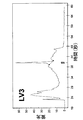
本発明において、HGという略号は、高悪性腫瘍を決定するのに用いられ、LGは低悪性腫瘍を決定するのに用いられる。 In the present invention, the abbreviation HG is used to determine high malignancy and LG is used to determine low malignancy.
TaおよびT1癌は、経尿道的切除(TUR)によって摘出することができる。高悪性(HG)TisおよびT1粘膜に閉じ込められた表在性癌であるが、高悪性腫瘍であるため、分子生物学の手法を用いて、それらが高悪性で侵襲の可能性を有する臨床的経験によって説明されてきた。 Ta and T1 cancers can be removed by transurethral resection (TUR). Superficial cancers confined to high malignant (HG) Tis and T1 mucosa, but because they are high malignant tumors, they are highly malignant and clinically invasive using molecular biology techniques Has been explained by experience.
その上、浸潤性膀胱癌はT2、T3およびT4に分類される。例えば、T2は、膀胱筋肉層を侵襲する腫瘍に関する。この種類は、さらに表面の筋肉層または内部の層を侵襲するT2aと深部筋肉層またはまたは外側の層を侵襲するT2bとに分類される。T3は、筋肉層を越えて侵襲するまたは膀胱周囲脂肪を侵襲する腫瘍に関する。この種類は、さらに微視的侵襲を有するT3aと巨視的侵襲を有するT3bとに分類される。さらに、T4は、膀胱に隣接する腫瘍侵襲構造に関しており、これはさらに前立腺、子宮または膣侵襲を有するT4aと骨盤壁または腹壁侵襲を有するT4bとに分類される。 Moreover, invasive bladder cancer is classified as T2, T3, and T4. For example, T2 relates to a tumor that invades the bladder muscle layer. This type is further classified into T2a that invades the surface muscle layer or inner layer and T2b that invades the deep muscle layer or outer layer. T3 relates to tumors that invade across the muscle layer or invade peri-bladder fat. This type is further classified into T3a having microscopic invasion and T3b having macroscopic invasion. Furthermore, T4 relates to a tumor invasion structure adjacent to the bladder, which is further classified into T4a with prostate, uterine or vaginal invasion and T4b with pelvic or abdominal wall invasion.
遺伝子の遺伝子発現の検出および定量は、本発明の目的に適する任意の非侵襲的な分子生物学の手法、例えば発現マイクロアレー、定量的リアル・タイムPCR、ノーザンブロット分析、通常のPCRなどによって行うことができる。 Detection and quantification of gene expression of genes is performed by any non-invasive molecular biology technique suitable for the purposes of the present invention, such as expression microarrays, quantitative real-time PCR, Northern blot analysis, conventional PCR, etc. be able to.
具体的には、DNAアレーの使用により極めて多数の遺伝子の発現結果を得て、それぞれの実験において数千の遺伝子を試験することができる。この手法の使用には、良好な品質を有する多量の(非分解)RNAを必要とする。 Specifically, the use of a DNA array can result in the expression of a very large number of genes, and thousands of genes can be tested in each experiment. The use of this technique requires a large amount of (non-degraded) RNA with good quality.
定量的リアル・タイムPCRの手法(qRT-PCR)は、診断および/または予後遺伝子を検出し手定量する目的で本発明において好ましく用いられる。この手法は、かなりの分解度のRNAを用いることができる上に、一層精確であり、最終結果に影響を与えない。特定の態様によれば、用いられるハイブリダイゼーションプローブはTaqmanプローブである。 The quantitative real-time PCR method (qRT-PCR) is preferably used in the present invention for the purpose of detecting and manually quantifying a diagnostic and / or prognostic gene. This approach can use RNA with a significant degree of degradation and is more accurate and does not affect the final result. According to a particular embodiment, the hybridization probe used is a Taqman probe.
膀胱液試料における遺伝子発現の検出および定量で得られる結果と、健康な被験者の試料における上記遺伝子についての標準参照値とを比較する。マーカー遺伝子の増減は、一般に分析の被験者に対応する試料の分析により得られる結果を平行して分析されるコントロール試料の結果と比較することによって評価される。それぞれの試料の分類の最終的決定はマーカー遺伝子の発現値に基づく「警告システム」によって行われ、観察値のいずれかがコントロール試料で期待されるものと比較して極めて有意な変動を示すときには、最終分類が腫瘍分類である確率は「警告を与えられた」遺伝子には無関係に増加する。 The results obtained from the detection and quantification of gene expression in a bladder fluid sample are compared with the standard reference values for the genes in healthy subject samples. The increase or decrease of the marker gene is generally assessed by comparing the results obtained by analyzing the sample corresponding to the subject to be analyzed with the results of the control sample analyzed in parallel. The final determination of the classification of each sample is made by a “warning system” based on the expression value of the marker gene, and when any of the observed values show a very significant variation compared to what is expected in the control sample, The probability that the final classification is a tumor classification increases regardless of the “warned” gene.
さらに具体的には、本発明の主要態様によれば、非侵襲的な膀胱癌の診断および/または予後方法は、被験者から膀胱液試料を集めてANXA10、C14orf78、CTSE、CRH、IGF2、KLF9、KRT20、MAGEA3、POSTN、PPP1R14D、SLC1A6、TERT、ASAMおよびMCM10遺伝子の組合せの発現パターンを上記試料において検出および定量することを含んでなる。得られた結果と、膀胱液における上記遺伝子についての標準参照値とを比較する。 More specifically, according to the main aspect of the present invention, a non-invasive method for diagnosing and / or prognosing bladder cancer is to collect a bladder fluid sample from a subject and use ANXA10, C14orf78, CTSE, CRH, IGF2, KLF9, Detecting and quantifying the expression pattern of the combination of KRT20, MAGEA3, POSTN, PPP1R14D, SLC1A6, TERT, ASAM and MCM10 genes in the sample. The results obtained are compared with standard reference values for the genes in bladder fluid.
膀胱液試料は、特に容易に得られるという条件では、好ましくは尿である。しかしながら、膀胱洗浄液が日常的方法では用いられることがあり、一層良好な品質のRNAが得られる。 The bladder fluid sample is preferably urine, provided that it is particularly easily obtained. However, bladder lavage fluid may be used in routine methods, resulting in better quality RNA.
4q32.3にあるANXA10(アネキシンA10)遺伝子(ANX14とも呼ばれる)は、細胞分裂の調節および様々なシグナル導入経路に関与しているが、その正確な機能は未だ決定されていない。 The ANXA10 (Annexin A10) gene (also called ANX14) at 4q32.3 is involved in the regulation of cell division and various signal transduction pathways, but its exact function has not yet been determined.
C14orf78(染色体14オープンリーディングフレーム78)遺伝子(AHNAK2またはKIAA2019とも呼ばれる)は、14q32.33に位置している。その機能は、未だ決定されていない。 The C14orf78 (chromosome 14 open reading frame 78) gene (also called AHNAK2 or KIAA2019) is located at 14q32.33. Its function has not yet been determined.
CTSE(カテプシンE)遺伝子(カリフォルニアTEとも呼ばれる)は1q31に位置しており、細胞内プロテアーゼをコードする。 The CTSE (cathepsin E) gene (also called California TE) is located at 1q31 and encodes an intracellular protease.
8q13に位置しているCRH(コルチコトロピン放出ホルモン)遺伝子(CRFとも呼ばれる)は、ストレスに応じて視床下部に分泌されるコルチコトロピン放出ホルモンをコードする。 The CRH (corticotropin releasing hormone) gene (also called CRF) located at 8q13 encodes a corticotropin releasing hormone that is secreted into the hypothalamus in response to stress.
11p15.5に位置しているIGF2(インスリン様増殖因子2 (ソマトメジンA))(11orf43、FLJ22066、FLJ44734、INSIGFとも呼ばれる)は、インスリン様増殖因子をコードする。 IGF2, located at 11p15.5 (insulin-like growth factor 2 (somatomedin A)) (also called 11orf43, FLJ22066, FLJ44734, INSIGF) encodes an insulin-like growth factor.
KLF9 (クルッペル様因子9)遺伝子(BTEB1とも呼ばれる)は、転写因子をコードする。 The KLF9 (Kruppel-like factor 9) gene (also called BTEB1) encodes a transcription factor.
17q21.2に位置しているKRT20(ケラチン20)遺伝子(K20、CK20、KRT21、MGC35423とも呼ばれる)は、上皮細胞に構造および一体性を与える働きをする中間フィラメントのタンパク質形成部分をコードする。 The KRT20 (keratin 20) gene located at 17q21.2 (also called K20, CK20, KRT21, MGC35423) encodes the protein-forming part of the intermediate filament that serves to confer structure and integrity to epithelial cells.
MAGEA3 (黒色腫抗原ファミリーA, 3)遺伝子(HIP8、HYPD、MAGE3、MAGEA6、MGC14613とも呼ばれる)は、Xq28に位置している。その機能は未知である。 The MAGEA3 (melanoma antigen family A, 3) gene (also called HIP8, HYPD, MAGE3, MAGEA6, MGC14613) is located at Xq28. Its function is unknown.
POSTN(ペリオスチン、骨芽細胞特異因子)遺伝子(PN、OSF-2、PDLPOSTN、MGC119510、MGC119511、ペリオスチン、RP11-412K4.1とも呼ばれる)は、13q13.3に位置しており、細胞移動性に関係した機能を有する。 The POSTN (periostin, osteoblast specific factor) gene (also called PN, OSF-2, PDLPOSTN, MGC119510, MGC119511, periostin, RP11-412K4.1) is located at 13q13.3 and is related to cell mobility It has the function.
PPP1R14D(タンパク質ホスファターゼ1、調節(インヒビター)サブユニット14D)遺伝子(GBPI-1、FLJ20251、MGC119014、MGC119016、CPI17様とも呼ばれる)は15q15.1に位置しており、ホスファターゼをコードする。
The PPP1R14D (
19p13.12に位置しているSLC1A6(溶質キャリヤーファミリー1(高親和性アスパルターゼ/グルタメート輸送体)、メンバー6)遺伝子(EAAT4、MGC33092、MGC43671とも呼ばれる)は、細胞内輸送に関与している。 The SLC1A6 (solute carrier family 1 (high affinity aspartase / glutamate transporter), member 6) gene (also called EAAT4, MGC33092, MGC43671) located at 19p13.12 is involved in intracellular transport.
5p15.33に位置しているTERT(テロメラーゼ逆転写酵素)遺伝子(TP2、TRT、EST2、TCS1、hEST2とも呼ばれる)は、逆転写酵素活性を有するテロメアのポリメラーゼをコードする。 The TERT (telomerase reverse transcriptase) gene (also called TP2, TRT, EST2, TCS1, hEST2) located at 5p15.33 encodes a telomeric polymerase with reverse transcriptase activity.
11q24.1に位置しているASAM(脂肪細胞特異的接着分子)(ASAM、AカリフォルニアM、CLMP、FLJ22415とも呼ばれる)は、細胞接着に関与している。 ASAM (adipocyte specific adhesion molecule) located at 11q24.1 (also called ASAM, A California M, CLMP, FLJ22415) is involved in cell adhesion.
さらに、10p13に位置しているMCM10(ミニ染色体維持欠失10(S. cerevisiae))遺伝子(CNA43、PRO2249、MGC126776とも呼ばれる)は、ゲノム複製の開始に関与するタンパク質をコードする。 In addition, the MCM10 (minichromosome maintenance deletion 10 (S. cerevisiae)) gene (also called CNA43, PRO2249, MGC126776) located at 10p13 encodes a protein involved in the initiation of genomic replication.
本発明の別の態様によれば、イン・ビトロの非侵襲的な膀胱癌の診断および/または予後方法は、被験者から膀胱液試料を集めて、ANXA10、CTSE、CRH、IGF2、KRT20、MAGEA3、SLC1A6、TERTおよびMCM10遺伝子の組合せの発現パターンの上記試料における検出および定量を行うことを含んでなる。得られた結果と、膀胱液における上記遺伝子についての標準参照値とを比較する。 According to another aspect of the present invention, an in vitro non-invasive bladder cancer diagnosis and / or prognosis method collects a bladder fluid sample from a subject, and ANXA10, CTSE, CRH, IGF2, KRT20, MAGEA3, Detecting and quantifying the expression pattern of the combination of SLC1A6, TERT and MCM10 genes in the sample. The results obtained are compared with standard reference values for the genes in bladder fluid.
本発明の別の態様によれば、イン・ビトロの非侵襲的な膀胱癌の診断および/または予後方法は、被験者から膀胱液試料を集めて、C14orf78、KLF9、POSTN、PPP1R14D、ASAMおよびそれらの組合せから選択される遺伝子の発現の上記膀胱液試料における検出および定量を行うことを含んでなる。得られた結果と、膀胱液における上記遺伝子についての標準参照値とを比較する。 According to another aspect of the invention, an in vitro non-invasive bladder cancer diagnosis and / or prognosis method collects bladder fluid samples from a subject to obtain C14orf78, KLF9, POSTN, PPP1R14D, ASAM and their Detecting and quantifying the expression of genes selected from the combination in the bladder fluid sample. The results obtained are compared with standard reference values for the genes in bladder fluid.
例えば、本発明の一特定態様によれば、C14orf78遺伝子発現の個々の検出および定量に基づく上記診断および予後方法が意図される。 For example, according to one particular embodiment of the present invention, the above diagnostic and prognostic methods based on individual detection and quantification of C14orf78 gene expression are contemplated.
本発明の別の特定態様によれば、KLF9遺伝子発現の個々の検出および定量に基づく上記診断および予後方法が意図される。 According to another particular embodiment of the invention, the above diagnostic and prognostic methods based on individual detection and quantification of KLF9 gene expression are contemplated.
本発明の別の特定態様によれば、POSTN遺伝子発現の個々の検出および定量に基づく上記診断および/または予後方法が意図される。 According to another particular embodiment of the invention, the above diagnostic and / or prognostic method based on individual detection and quantification of POSTN gene expression is contemplated.
別の特定態様によれば、PPPIR14D遺伝子発現の個々の検出および定量に基づく上記診断および/または予後方法が意図される。 According to another particular embodiment, the above diagnostic and / or prognostic method based on individual detection and quantification of PPPIR14D gene expression is contemplated.
本発明の別の特定態様によれば、ASAM遺伝子発現の個々の検出および定量に基づく上記診断および/または予後方法が意図される。 According to another particular embodiment of the invention, the above diagnostic and / or prognostic method based on individual detection and quantification of ASAM gene expression is contemplated.
本発明の別の特定態様によれば、C14orf78、KLF9、POSTN、PPP1R14D、ASAMおよびそれらの組合せから選択された遺伝子、およびさらにANXA10、CTSE、CRH、IGF2、KRT20、MAGEA3、SLC1A6、TERTおよびMCM10から選択された少なくとも1種類の遺伝子の検出および定量に基づくイン・ビトロの非侵襲的な膀胱癌の診断および/または予後方法が意図される。 According to another specific embodiment of the invention, genes selected from C14orf78, KLF9, POSTN, PPP1R14D, ASAM and combinations thereof, and further from ANXA10, CTSE, CRH, IGF2, KRT20, MAGEA3, SLC1A6, TERT and MCM10 In vitro non-invasive bladder cancer diagnosis and / or prognostic methods based on the detection and quantification of at least one selected gene are contemplated.
本発明の別の態様によれば、被験者から膀胱液試料を集めて、上記方法準じてANXA10、C14orf78、CTSE、CRH、IGF2、KLF9、KRT20、MAGEA3、POSTN、PPP1R14D、SLC1A6およびTERT遺伝子の組合せの発現パターンの検出および定量を行うこと含んでなる膀胱癌の診断に焦点を当てたイン・ビトロの非侵襲的方法が意図される。得られた結果と、膀胱液における上記遺伝子についての標準参照値とを比較する。 According to another aspect of the present invention, a bladder fluid sample is collected from a subject, and the combination of ANXA10, C14orf78, CTSE, CRH, IGF2, KLF9, KRT20, MAGEA3, POSTN, PPP1R14D, SLC1A6 and TERT gene according to the above method In vitro, non-invasive methods focused on the diagnosis of bladder cancer comprising performing expression pattern detection and quantification are contemplated. The results obtained are compared with standard reference values for the genes in bladder fluid.
本発明の別の態様によれば、イン・ビトロの非侵襲性の膀胱癌診断法は、被験者から膀胱液試料を集めて、ANXA10、CTSE、CRH、IGF2、KRT20、MAGEA3、SLC1A6およびTERT遺伝子の組合せの発現パターンの検出および定量を行うことを含んでなる。得られた結果と、膀胱液における上記遺伝子についての標準参照値とを比較する。 According to another aspect of the present invention, an in vitro non-invasive bladder cancer diagnostic method collects a bladder fluid sample from a subject and produces ANXA10, CTSE, CRH, IGF2, KRT20, MAGEA3, SLC1A6 and TERT genes. Detecting and quantifying the expression pattern of the combination. The results obtained are compared with standard reference values for the genes in bladder fluid.
本発明の別の態様によれば、イン・ビトロの非侵襲性の膀胱癌の診断法は、被験者から膀胱液試料を集めて、C14orf78、KLF9、POSTN、PPP1R14Dおよびそれらの組合せから選択される遺伝子発現の検出および定量を行うことを含んでなる。得られた結果と、膀胱液における上記遺伝子についての標準参照値とを比較する。 According to another aspect of the present invention, a method for in vitro non-invasive bladder cancer diagnosis comprises collecting a bladder fluid sample from a subject and selecting from C14orf78, KLF9, POSTN, PPP1R14D and combinations thereof Performing expression detection and quantification. The results obtained are compared with standard reference values for the genes in bladder fluid.
本発明の特定態様によれば、上記診断法はC14orf78遺伝子の検出および定量に基づいている。
本発明の別の特定態様によれば、上記診断法はKLF9遺伝子の検出および定量に基づいている。
According to a particular embodiment of the invention, the diagnostic method is based on the detection and quantification of the C14orf78 gene.
According to another particular embodiment of the invention, the diagnostic method is based on the detection and quantification of the KLF9 gene.
本発明の別の特定態様によれば、上記診断法はPOSTN遺伝子の検出および定量に基づいている。 According to another particular embodiment of the invention, the diagnostic method is based on the detection and quantification of the POSTN gene.
本発明の別の特定態様によれば、上記診断法はPPPIR14D遺伝子の検出および定量に基づいている。 According to another particular embodiment of the invention, the diagnostic method is based on the detection and quantification of the PPPIR14D gene.
本発明の別の特定態様によれば、上記診断法はC14orf78、KLF9、POSTN、PPP1R14Dおよびそれらの組合せから選択される遺伝子、およびさらにANXA10、CTSE、CRH、IGF2、KRT20、MAGEA3、SLC1A6およびTERTから選択される少なくとも1種類の遺伝子の発現の検出および定量に基づいている。 According to another particular embodiment of the invention, said diagnostic method comprises a gene selected from C14orf78, KLF9, POSTN, PPP1R14D and combinations thereof, and further from ANXA10, CTSE, CRH, IGF2, KRT20, MAGEA3, SLC1A6 and TERT Based on detection and quantification of expression of at least one selected gene.
本発明の別の態様によれば、被験者から膀胱液試料を集めて、ASAMおよびMCM10遺伝子の組合せの発現パターンの検出および定量を行うことを含んでなるイン・ビトロの非侵襲的な膀胱癌の予後方法が意図される。得られた結果と、膀胱液における上記遺伝子についての標準参照値とを比較する。 According to another aspect of the invention, in vitro non-invasive bladder cancer comprising collecting bladder fluid samples from a subject and detecting and quantifying the expression pattern of the combination of ASAM and MCM10 genes. Prognostic methods are intended. The results obtained are compared with standard reference values for the genes in bladder fluid.
本発明の別の態様によれば、被験者から膀胱液試料を集めて、ASAM遺伝子の発現を検出および定量することを含んでなるイン・ビトロの非侵襲的な膀胱癌の予後方法が提供される。得られた結果を、膀胱液における上記遺伝子についての標準参照値と比較する。 According to another aspect of the invention, there is provided an in vitro non-invasive bladder cancer prognosis method comprising collecting bladder fluid samples from a subject and detecting and quantifying expression of the ASAM gene. . The results obtained are compared with standard reference values for the above genes in bladder fluid.
本発明の別の態様によれば、膀胱癌診断および/または予後マーカーとしてのANXA10、C14orf78、CTSE、CRH、IGF2、KLF9、KRT20、MAGEA3、POSTN、PPP1R14D、SLC1A6、TERT、ASAMおよびMCM10遺伝子の組合せの使用が意図される。 According to another aspect of the present invention, a combination of ANXA10, C14orf78, CTSE, CRH, IGF2, KLF9, KRT20, MAGEA3, POSTN, PPP1R14D, SLC1A6, TERT, ASAM and MCM10 genes as bladder cancer diagnostic and / or prognostic markers Is intended for use.
本発明の別の態様においては、膀胱癌診断および/または予後マーカーとしてのANXA10、CTSE、CRH、IGF2、KRT20、MAGEA3、SLC1A6、TERTおよびMCM10遺伝子の組合せの使用に焦点が当てられている。 In another aspect of the invention, the focus is on the use of a combination of ANXA10, CTSE, CRH, IGF2, KRT20, MAGEA3, SLC1A6, TERT and MCM10 genes as bladder cancer diagnostic and / or prognostic markers.
本発明の別の態様によれば、膀胱癌診断および/または予後マーカーとしてのC14orf78、KLF9、POSTN、PPP1R14D、ASAMおよびそれらの組合せから選択される遺伝子の使用が意図される。 Another aspect of the invention contemplates the use of genes selected from C14orf78, KLF9, POSTN, PPP1R14D, ASAM and combinations thereof as bladder cancer diagnostic and / or prognostic markers.
本発明の特定態様によれば、膀胱癌の診断および/または予後マーカーとしてのC14orf78遺伝子の使用が意図される。 According to a particular embodiment of the invention, the use of the C14orf78 gene as a bladder cancer diagnosis and / or prognostic marker is contemplated.
本発明の別の特定態様によれば、膀胱癌診断および/または予後マーカーとしてのKLF9遺伝子の使用が意図される。 According to another particular embodiment of the invention, the use of the KLF9 gene as a bladder cancer diagnosis and / or prognostic marker is contemplated.
本発明の別の特定態様によれば、膀胱癌診断および/または予後マーカーとしてのPOSTN遺伝子の使用が意図される。 According to another particular embodiment of the invention, the use of the POSTN gene as a bladder cancer diagnosis and / or prognostic marker is contemplated.
本発明の別の特定態様によれば、膀胱癌診断および/または予後マーカーとしてのPPP1R14D遺伝子の使用が意図される。 According to another particular embodiment of the invention, the use of the PPP1R14D gene as a bladder cancer diagnosis and / or prognostic marker is contemplated.
本発明の別の特定態様によれば、膀胱癌診断および/または予後マーカーとしてのASAM遺伝子の使用が意図される。 According to another particular embodiment of the invention, the use of the ASAM gene as a bladder cancer diagnosis and / or prognostic marker is contemplated.
本発明の別の態様においては、ANXA10、CTSE、CRH、IGF2、KRT20、MAGEA3、SLC1A6、TERTおよびMCM10から選択される少なくとも1種類の遺伝子と組み合わせた、C14orf78、KLF9、POSTN、PPP1R14D、ASAMおよびそれらの組合せから選択される遺伝子の、膀胱癌診断および/または予後マーカーとしての使用に焦点が当てられる。 In another embodiment of the present invention, C14orf78, KLF9, POSTN, PPP1R14D, ASAM and them in combination with at least one gene selected from ANXA10, CTSE, CRH, IGF2, KRT20, MAGEA3, SLC1A6, TERT and MCM10 The focus is on the use of genes selected from these combinations as bladder cancer diagnostics and / or prognostic markers.
本発明の別の態様は、ANXA10、C14orf78、CTSE、CRH、IGF2、KLF9、KRT20、MAGEA3、POSTN、PPP1R14D、SLC1A6およびTERT遺伝子の組合せの膀胱癌の診断マーカーとしての使用に関する。 Another aspect of the present invention relates to the use of a combination of ANXA10, C14orf78, CTSE, CRH, IGF2, KLF9, KRT20, MAGEA3, POSTN, PPP1R14D, SLC1A6 and TERT gene as a diagnostic marker for bladder cancer.
本発明の別の態様においては、ANXA10、CTSE、CRH、IGF2、KRT20、MAGEA3、SLC1A6およびTERT遺伝子の膀胱癌の診断マーカーとしての使用に焦点が当てられる。 In another aspect of the invention, the focus is on the use of ANXA10, CTSE, CRH, IGF2, KRT20, MAGEA3, SLC1A6 and TERT genes as diagnostic markers for bladder cancer.
本発明の別の態様は、C14orf78、KLF9、POSTN、PPP1R14Dおよびそれらの組合せから選択される遺伝子の膀胱癌の診断マーカーとしての使用に関する。 Another aspect of the present invention relates to the use of a gene selected from C14orf78, KLF9, POSTN, PPP1R14D and combinations thereof as a diagnostic marker for bladder cancer.
本発明の特定態様によれば、C14orf78遺伝子の膀胱癌の診断マーカーとしての使用が意図される。 According to a particular embodiment of the invention, the use of the C14orf78 gene as a diagnostic marker for bladder cancer is contemplated.
同様に、本発明の別の特定態様によれば、KLF9遺伝子の膀胱癌の診断マーカーとしての使用が意図される。 Similarly, according to another particular embodiment of the invention, the use of the KLF9 gene as a diagnostic marker for bladder cancer is contemplated.
本発明の別の特定態様によれば、POSTN遺伝子の膀胱癌の診断マーカーとしての使用が意図される。 According to another specific embodiment of the invention, the use of the POSTN gene as a diagnostic marker for bladder cancer is contemplated.
本発明の別の特定態様によれば、PPP1R14D遺伝子の膀胱癌の診断マーカーとしての使用が意図される。 According to another particular embodiment of the invention, the use of the PPP1R14D gene as a diagnostic marker for bladder cancer is contemplated.
本発明の別の態様によれば、ANXA10、CTSE、CRH、IGF2、KRT20、MAGEA3、SLC1A6およびTERTから選択される少なくとも1種類の遺伝子と組み合わせた、C14orf78、KLF9、POSTN、PPP1R14Dおよびそれらの組合せから選択される遺伝子の膀胱癌の診断マーカーとしての使用が意図される。 According to another aspect of the invention, from C14orf78, KLF9, POSTN, PPP1R14D and combinations thereof in combination with at least one gene selected from ANXA10, CTSE, CRH, IGF2, KRT20, MAGEA3, SLC1A6 and TERT The use of the selected gene as a diagnostic marker for bladder cancer is contemplated.
本発明の別の態様によれば、ASAMおよびMCM10遺伝子の組合せの膀胱癌の予後マーカーとしての使用が意図される。 According to another aspect of the invention, the use of a combination of ASAM and MCM10 genes as a prognostic marker for bladder cancer is contemplated.
本発明の別の態様によれば、ASAM遺伝子の膀胱癌の予後マーカーとしての使用が意図される。 According to another aspect of the invention, the use of the ASAM gene as a prognostic marker for bladder cancer is contemplated.
本発明の別の態様は、ANXA10、C14orf78、CTSE、CRH、IGF2、KLF9、KRT20、MAGEA3、POSTN、PPP1R14D、 SLC1A6、TERT、ASAMおよびMCM10遺伝子の組合せの発現パターンの検出および定量に適するプローブの組を含んでなる膀胱癌の診断および/または予後キットに関する。 Another aspect of the present invention is a set of probes suitable for detection and quantification of expression patterns of combinations of ANXA10, C14orf78, CTSE, CRH, IGF2, KLF9, KRT20, MAGEA3, POSTN, PPP1R14D, SLC1A6, TERT, ASAM and MCM10 genes A bladder cancer diagnosis and / or prognosis kit comprising:
本発明の別の態様によれば、膀胱癌の診断および/または予後キットは、ANXA10、CTSE、CRH、IGF2、KRT20、MAGEA3、SLC1A6、TERTおよびMCM10遺伝子の組合せの発現パターンの検出および定量に適するプローブの組を含んでなる。 According to another aspect of the present invention, a bladder cancer diagnosis and / or prognosis kit is suitable for detection and quantification of expression patterns of a combination of ANXA10, CTSE, CRH, IGF2, KRT20, MAGEA3, SLC1A6, TERT and MCM10 genes A set of probes.
本発明の別の態様によれば、C14orf78、KLF9、POSTN、PPP1R14D、ASAMおよびそれらの組合せから選択される遺伝子の検出および定量に適するプローブの組に基づく膀胱癌の診断および/または予後キットが意図される。 According to another aspect of the invention, a bladder cancer diagnosis and / or prognosis kit based on a set of probes suitable for the detection and quantification of genes selected from C14orf78, KLF9, POSTN, PPP1R14D, ASAM and combinations thereof is contemplated. Is done.
本発明の特定態様によれば、C14orf78、KLF9、POSTN、PPP1R14D、ASAMおよびそれらの組合せから選択される遺伝子の検出および定量のためのプローブの組に基づく上記膀胱癌の診断および/または予後キットは、さらにANXA10、CTSE、CRH、IGF2、KRT20、MAGEA3、SLC1A6、TERTおよびMCM10から選択される遺伝子の検出および定量に適するプローブの組を含んでなる。 According to a particular embodiment of the present invention, the bladder cancer diagnosis and / or prognosis kit based on a set of probes for detection and quantification of a gene selected from C14orf78, KLF9, POSTN, PPP1R14D, ASAM and combinations thereof, And a set of probes suitable for detection and quantification of a gene selected from ANXA10, CTSE, CRH, IGF2, KRT20, MAGEA3, SLC1A6, TERT and MCM10.
本発明の別の態様によれば、ANXA10、C14orf78、CTSE、CRH、IGF2、KLF9、KRT20、MAGEA3、POSTN、PPP1R14D、SLC1A6およびTERT遺伝子の組合せの発現パターンの検出および定量に適するプローブの組に基づく膀胱癌の診断キットが意図される。 According to another aspect of the invention, based on a set of probes suitable for the detection and quantification of expression patterns of combinations of ANXA10, C14orf78, CTSE, CRH, IGF2, KLF9, KRT20, MAGEA3, POSTN, PPP1R14D, SLC1A6 and TERT genes A diagnostic kit for bladder cancer is contemplated.
本発明の別の態様によれば、膀胱癌の診断キットは、ANXA10、CTSE、CRH、IGF2、KRT20、MAGEA3、SLC1A6およびTERT遺伝子の組合せの発現パターンの検出および定量に適するプローブの組を含んでなる。 According to another aspect of the present invention, a bladder cancer diagnostic kit comprises a set of probes suitable for detection and quantification of expression patterns of combinations of ANXA10, CTSE, CRH, IGF2, KRT20, MAGEA3, SLC1A6 and TERT genes Become.
本発明の別の態様によれば、C14orf78、KLF9、POSTN、PPP1R14Dおよびそれらの組合せから選択される遺伝子の検出および定量に適するプローブの組に基づく癌の診断キットが意図される。 According to another aspect of the present invention, a cancer diagnostic kit based on a set of probes suitable for the detection and quantification of genes selected from C14orf78, KLF9, POSTN, PPP1R14D and combinations thereof is contemplated.
本発明の別の態様によれば、C14orf78、KLF9、POSTN、PPP1R14Dおよびそれらの組合せから選択される遺伝子の検出および定量に適するプローブの組に基づく上記キットは、さらにANXA10、CTSE、CRH、IGF2、KRT20、MAGEA3、SLC1A6およびTERTから選択される少なくとも1種類の遺伝子の検出および定量に適するプローブを含んでなる。 According to another aspect of the present invention, the kit based on a set of probes suitable for detection and quantification of a gene selected from C14orf78, KLF9, POSTN, PPP1R14D and combinations thereof further comprises ANXA10, CTSE, CRH, IGF2, It comprises a probe suitable for the detection and quantification of at least one gene selected from KRT20, MAGEA3, SLC1A6 and TERT.
本発明の別の態様によれば、ASAMおよびMCM10遺伝子の組合せの発現パターンの検出および定量に適するプローブの組に基づく膀胱癌の予後キットが意図される。 According to another aspect of the present invention, a bladder cancer prognostic kit based on a set of probes suitable for detection and quantification of the expression pattern of a combination of ASAM and MCM10 genes is contemplated.
本発明の別の態様によれば、この予後キットは、ASAM遺伝子の検出および定量に適するプローブの組に基づいている。 According to another aspect of the invention, this prognostic kit is based on a set of probes suitable for the detection and quantification of the ASAM gene.
表1に、膀胱癌の診断および/または予後遺伝子マーカーとして同定された14個の遺伝子を示す。ASAMおよびMCM10遺伝子は、予後のための2個の特異的遺伝子である。
本発明の制限ではなく例示に有用な幾つかの実施例を、下記に挙げる。
Table 1 shows 14 genes identified as bladder cancer diagnostic and / or prognostic genetic markers. The ASAM and MCM10 genes are two specific genes for prognosis.
Some examples useful for illustration rather than limitation of the invention are listed below.
実施例1 膀胱液試料における分解の重要性の決定
本発明の最終目的を行うには、膀胱液(尿および/または膀胱洗浄液)から得られるRNAの品質が一般的に低い場合には、最初に遺伝子発現プロフィールに対する様々なRNA分解レベルの衝撃を知る必要があった。膀胱液から得られた遺伝子発現プロフィールが対応する腫瘍で得られるものと調和する場合にも、測定しなければならなかった。
Example 1 Determining the Importance of Degradation in a Bladder Fluid Sample To accomplish the final objective of the present invention, if the quality of RNA obtained from bladder fluid (urine and / or bladder lavage fluid) is generally low, It was necessary to know the impact of various RNA degradation levels on gene expression profiles. It also had to be measured if the gene expression profile obtained from the bladder fluid matched that obtained in the corresponding tumor.
1. 試料およびRNA製剤の選択
高悪性(G3)pT2として診断された1名の同一患者の腫瘍組織(T)および膀胱洗浄液(BW)試料を選択した[Lopez-Beltran A, Sauter G, Gasser T, Hartmann A, Schmitz-Drager BJ, Helpap B, Ayala HG, Tamboni P, Knowles MA, Sidransky D, Cordon-Cardo C, Jones PA, Cairns P, Simon R, Amin MB, Tyczynsky JE. 「泌尿器系の腫瘍(Tumours of the Urinary System)」: Eble JN, Sauter G, Epstein JI, Sesterhenn IA(監修), 「泌尿器系および男性器の腫瘍の病理学および遺伝学(Pathology and Genetics of Tumours of the Urinary System and Male Genital Organs)」. World Health Organization Classification of Tumours. リヨン: IARC Press; 2004: 89-157; Sobin LH, Wittekind CH. 「悪性腫瘍のTNM分類(TNM Classification of Malignant Tumours)」. International Union Against Cancer., 第6版. ニューヨーク: Jonh Wiley & Sons; 2002]。両試料(T0およびBW0)のRNAを、TRIzol(Invitrogen, カールスバード, カリフォルニア, 米国)を用いて供給業者の指示に従って抽出した。次いで、両RNA(T0およびBW0)の一部を80℃で15(T1およびBW1)、30(T2およびBW2)および60(T3およびBW3)分間インキュベーションすることによって分解し、水を塩基緩衝液の代わりに用いたことを除き、Xiang CC, Chen M, Ma L et al. 「マイクロアレー研究用の小さな組織試料からの分解したRNAを増幅する新規方法(A new strategy to amplify degraded RNA from small tissue samples for microarray studies)」. Nucleic Acids Res 2003; 31:53に記載の方法によって3つの分解レベルを得た。
1. Selection of samples and RNA preparations Tumor tissue (T) and bladder lavage fluid (BW) samples from one identical patient diagnosed as highly malignant (G3) pT2 were selected [Lopez-Beltran A, Sauter G, Gasser T , Hartmann A, Schmitz-Drager BJ, Helpap B, Ayala HG, Tamboni P, Knowles MA, Sidransky D, Cordon-Cardo C, Jones PA, Cairns P, Simon R, Amin MB, Tyczynsky JE. Tumours of the Urinary System '': Eble JN, Sauter G, Epstein JI, Sesterhenn IA (supervised), `` Pathology and Genetics of Tumours of the Urinary System and Male Genital World Health Organization Classification of Tumours. Lyon: IARC Press; 2004: 89-157; Sobin LH, Wittekind CH. "TNM Classification of Malignant Tumours". International Union Against Cancer. 6th edition. New York: Jonh Wiley &Sons; 2002]. RNA from both samples (T0 and BW0) was extracted using TRIzol (Invitrogen, Carlsbad, California, USA) according to the supplier's instructions. A portion of both RNAs (T0 and BW0) was then degraded by incubation at 80 ° C. for 15 (T1 and BW1), 30 (T2 and BW2) and 60 (T3 and BW3) minutes, and water was removed from the base buffer. Xiang CC, Chen M, Ma L et al. “A new strategy to amplify degraded RNA from small tissue samples. for microarray studies) ”. Three degradation levels were obtained by the method described in Nucleic Acids Res 2003; 31:53.
膀胱病状の証拠のない4名の患者から健康な膀胱粘膜試料(コントロール試料)も集め、RNAを上記試料と同様の方法で得て、4個のRNAを等モル比で混合した(C0)。 Healthy bladder mucosa samples (control samples) were also collected from 4 patients without evidence of bladder pathology, RNA was obtained in the same manner as the above samples, and 4 RNAs were mixed in equimolar ratio (C0).
完全および分解RNAのそれぞれ1μlをAgilent 2100 Bioanalyzerで分析して、それぞれのRNAの品質を決定した(Imbeaud S, Graudens E, Boulanger V et al. 「マイクロキャピラリー電気泳動トレースの使用者の影響を受けない選別器を用いるRNA品質査定の標準化に向けて(Towards standardization of RNA quality assessment using user-independent classifiers of microcapillary electrophoresis traces)」. Nucleic Acids Res 2005; 33:e56)(図1A-H)。図1(J)は、これらのバンドの分解の進行が見られる分析試料のそれぞれのリボソームRNA(28Sおよび18S)のバンドを有するゲルを示す。 1 μl of each complete and degraded RNA was analyzed on an Agilent 2100 Bioanalyzer to determine the quality of each RNA (Imbeaud S, Graudens E, Boulanger V et al. “Insensitive to Microcapillary Electrophoretic Trace User Towards standardization of RNA quality assessment using user-independent classifiers of microcapillary electrophoresis traces ”. Nucleic Acids Res 2005; 33: e56) (FIGS. 1A-H). FIG. 1 (J) shows a gel with bands for each ribosomal RNA (28S and 18S) of the analytical sample where progress of degradation of these bands is seen.
さらに、36の膀胱腫瘍洗浄液(8個の低悪性(LG)pTa、5個の高悪性(HG)pTa、3個のpT1 LG、5個のpT1 HG、4個のpTis、9個のpT2 HGおよび2個のpT4 HG)および膀胱病状のない患者の14個のコントロール膀胱洗浄液を集め、RNAを上記の場合と同様にして抽出した。 In addition, 36 bladder tumor lavage fluids (8 low-grade (LG) pTa, 5 high-grade (HG) pTa, 3 pT1 LG, 5 pT1 HG, 4 pTis, 9 pT2 HG And 2 pT4 HG) and 14 control bladder lavage fluids of patients without bladder pathology were collected and RNA was extracted as above.
2. イン・ビトロRNA増幅およびラベリング
完全なRNA(T0およびBW0)および分解RNA(T1、T2、T3、BW1、BW2およびBW3)5μgを、T3プロモーター配列の3'における付加によって修飾した9ヌクレオチドのランダム配列(T3N9)を有するプライマーを用いて増幅した(Xiang CC, Chen M, Ma L et al. 「マイクロアレー研究のための小さな組織試料由来の分解RNAを増幅する新規方法(A new strategy to amplify degraded RNA from small tissue samples for microarray studies)」. Nucleic Acids Res 2003; 31:e53)。プローブは、直接ラベリング法によって合成した(Richter A, Schwager C, Hentze S, Ansorge W, Hentze MW, Muckenthaler M. 「DNAマイクロアレーによる発現分析に用いた蛍光タグDNAラベリング法の比較(Comparison of fluorescent tag DNA labeling methods used for expression analysis by DNA microarrays)」. Biotechniques 2002; 33:620-8, 630)。
2. In vitro RNA amplification and labeling Nine nucleotides of 5 μg of complete RNA (T0 and BW0) and degraded RNA (T1, T2, T3, BW1, BW2 and BW3) modified by
3. アレープロセシングおよびデーター分析
Oncochip-v2グラスヒトcDNAマイクロアレー(http://qrupos.cnio.es/qenomica/arravs/reports/ochip_v2.xls)を用いて、腫瘍および膀胱洗浄液(T0、T1、T2、T3、BW0、BW1、BW2およびBW3)の両方のRNAの4個の次第に分解した部分のそれぞれを健康な膀胱試料由来のRNAのプール(C0)と共に同時ハイブリダイズした。蛍光画像はG2565BA Microarray Scanner System (Agilent, Technologies, バルトブロン, 独国)を用いて得て、TIFF画像はR統計学的環境下(http://www.r-project.org)でSpotプログラム(http://experimental.act.cmis.csiro.au/Spot)を用いて定量した。マイクロアレーのそれぞれの点の最終強度測定は、以前に示唆されている方法(http://www.stat.berkeley.edu/users/terrv/zarrav/Html/imaqe.html)(GEOデーターベースにおける一般にアクセス可能なデーター; GSE3192)により計算した。最後に、総ての品質基準に従う1111個の有効なクローンを得て、それぞれのアレーの100個の最も示差的に発現したクローンを選択してアレー間で比較し、それらの間の共通な遺伝子の割合を計算した。高い割合の共通の示差発現遺伝子が、腫瘍組織アレー間(85-91%)および膀胱洗浄液アレー間(78-93%)で検出され(図2a)、RNAの分解は実質的に遺伝子発現プロフィールに影響しないことを示唆していた。
3. Array processing and data analysis
Using Oncochip-v2 glass human cDNA microarray (http://qrupos.cnio.es/qenomica/arravs/reports/ochip_v2.xls), tumor and bladder lavage fluid (T0, T1, T2, T3, BW0, BW1, BW2 And each of the four progressively degraded portions of both RNAs of BW3) were cohybridized with a pool of RNA (C0) from healthy bladder samples. Fluorescence images were obtained using the G2565BA Microarray Scanner System (Agilent, Technologies, Baltbron, Germany), and TIFF images were obtained in the R program (http://www.r-project.org) under the Spot program (http://www.r-project.org). </experimental.act.cmis.csiro.au/Spot). The final intensity measurement for each point of the microarray is based on the previously suggested method (http://www.stat.berkeley.edu/users/terrv/zarrav/Html/imaqe.html) (generally in the GEO database Accessible data; GSE3192). Finally, we obtain 1111 valid clones according to all quality standards, select the 100 most differentially expressed clones of each array and compare them between arrays, the common gene between them The percentage of was calculated. A high proportion of common differentially expressed genes was detected between tumor tissue arrays (85-91%) and between bladder lavage fluid arrays (78-93%) (Figure 2a), and RNA degradation was essentially in the gene expression profile. It suggested that there was no effect.
マイクロアレーに含まれる総てのクローンの未管理クラスターも、UPGMA(無荷重平均距離法(Unweighted Pair-Group Meted with Arithmetic Mean))およびピアソン補正を用いて行った。このクラスターは、分解レベルが異なる(例えば、BW0およびBW1)RNAとハイブリダイズした2個のアレー間で同定された共通な遺伝子の割合は、同じアレーのダイ・スワップ(DS)による複製(例えば、BW0対BW0-DS)間の割合より高いことがあり(図2b)、これは、遺伝子発現プロフィールは、部分的に分解したRNAで作業するときには実質的に変化しないという結論を強化するものであった。 Unmanaged clusters of all clones contained in the microarray were also performed using UPGMA (Unweighted Pair-Group Meted with Arithmetic Mean) and Pearson correction. This cluster shows that the percentage of common genes identified between two arrays hybridized to RNA with different degradation levels (e.g., BW0 and BW1) is replicated by die swap (DS) of the same array (e.g., (Figure 2b), which reinforces the conclusion that gene expression profiles do not change substantially when working with partially degraded RNA. It was.
膀胱洗浄液試料から得た遺伝子発現プロフィールが対応する腫瘍で得られたものと調和するかどうかを決定するため、腫瘍組織アレーと膀胱洗浄液アレーとの間で共通の示差発現遺伝子の割合を比較した。腫瘍と膀胱洗浄液との間では高い類似性が得られ(52-60%)、この類似性はRNAの分解条件とは無関係であった。 In order to determine whether the gene expression profiles obtained from bladder lavage samples are consistent with those obtained in the corresponding tumor, the proportion of differentially expressed genes between tumor tissue arrays and bladder lavage arrays was compared. High similarity was obtained between tumor and bladder lavage fluid (52-60%), and this similarity was independent of RNA degradation conditions.
結論として、このデーターは、部分分解した膀胱洗浄液RNAをマイクロアレーを用いる遺伝子発現研究に用いることができ、このRNAは腫瘍の遺伝子発現を反映していることを示唆した。 In conclusion, this data suggested that partially degraded bladder washing RNA could be used for gene expression studies using microarrays, and that this RNA reflected tumor gene expression.
4. 定量的リアル・タイムRT-PCR(qRT-PCR)
特定の患者のマイクロアレーで得られた結果が一層長いコホートまで補外することができることを確認するため、文献に準じた膀胱癌過程に関するアレーで示差発現した4個の遺伝子(KRT20、IGF2、GSNおよびCCL2)をqRT-PCRによって分析した。この確認には、36個の追加の膀胱腫瘍洗浄液と14個のコントロール膀胱洗浄液を用いた。
4. Quantitative real-time RT-PCR (qRT-PCR)
In order to confirm that the results obtained in a specific patient microarray can be extrapolated to a longer cohort, four differentially expressed genes (KRT20, IGF2, GSN) And CCL2) were analyzed by qRT-PCR. For this confirmation, 36 additional bladder tumor lavage fluids and 14 control bladder lavage fluids were used.
cDNAは、反応の最終容積を50μlに減少したことを除き、High-Capacity cDNA Archive Kit(Applied Biosystems, フォスター・シティー, 米国)を用いて供給業者の指示に従ってRNA 1μgから合成した。GUSB遺伝子を、内因性コントロールとして用いた。PCRは、反応の容積を20μlに減少したことを除き、ABI PRISM 7000 SDS (Applied Biosystems, フォスター・シティー, 米国)中でAssays-on-Demand(商品名)Gene Expression Productsを用いて行った。 cDNA was synthesized from 1 μg RNA using the High-Capacity cDNA Archive Kit (Applied Biosystems, Foster City, USA) according to the supplier's instructions, except that the final volume of the reaction was reduced to 50 μl. The GUSB gene was used as an endogenous control. PCR was performed using Assays-on-Demand (trade name) Gene Expression Products in ABI PRISM 7000 SDS (Applied Biosystems, Foster City, USA) except that the reaction volume was reduced to 20 μl.
ΔΔCt法(ABI PRISM 7700 Sequence Detection System User Bulletin #2: 遺伝子発現の定量 P/N 4303859)を用いて、14個のコントロール試料の発現の平均値に対するそれぞれの遺伝子の発現の相対量を計算した。コントロールにおける参照値を確立するため、膀胱に病状のない患者の14コントロール膀胱洗浄液の発現の算術平均を得た。 Using the ΔΔCt method (ABI PRISM 7700 Sequence Detection System User Bulletin # 2: Quantification of gene expression P / N 4303859), the relative amount of expression of each gene relative to the average value of expression of 14 control samples was calculated. In order to establish a reference value in the control, the arithmetic mean of the expression of 14 control bladder lavage fluids was obtained for patients with no bladder condition.
底が2の対数として表される2つの比較条件(この場合には、それぞれの指定遺伝子に対する内因性コントロール)の間の割合または除法(比)として定義されlog2ratioとして表されるこの分析の結果は、KRT20について試料の81%、GNSについて89%、IGF2について64%およびCCL2について89%においてマイクロアレーの結果を確かめた(図3)。単独の患者の分析から得られたマイクロアレーデーターおよび36名の患者のコホートの分析から得られたqRT-PCRにより、マイクロアレーで得られた遺伝子発現プロフィールは単独患者の分析によるものではないことが確かめられた。 The result of this analysis, defined as the ratio or division (ratio) between the two comparison conditions (in this case, the endogenous control for each specified gene) expressed as the logarithm of 2, is the log2ratio The microarray results were confirmed in 81% of the samples for KRT20, 89% for GNS, 64% for IGF2, and 89% for CCL2 (FIG. 3). The microarray data obtained from the analysis of a single patient and the qRT-PCR from the analysis of a cohort of 36 patients indicate that the gene expression profile obtained in the microarray is not due to the analysis of a single patient It was confirmed.
5. 実施例1の結論
膀胱洗浄液RNAを用いて、cDNAマイクロアレーおよびqRT-PCRの両方による対応する膀胱腫瘍の遺伝子発現プロフィールを推定することが可能である。
5. Conclusions of Example 1 Using bladder lavage RNA, it is possible to estimate the corresponding bladder tumor gene expression profile by both cDNA microarray and qRT-PCR.
実施例2. 予測モデルの候補遺伝子の初期測定
膀胱液試料の遺伝子発現プロフィールの測定が可能であることを知れば、次の目的はできるだけ大規模な特定遺伝子発現データーを得ることであった。少数の試料でできる限り多量の遺伝子を分析することによって出発し、連続的工程でさらに大規模なシリーズの試料においてますます小さくかつ一層選択された量の遺伝子を連続的に分析した。
Example 2. Initial Measurement of Predictive Model Candidate Genes Knowing that it is possible to measure the gene expression profile of bladder fluid samples, the next objective was to obtain as much specific gene expression data as possible. Starting by analyzing as many genes as possible with a small number of samples, successive smaller and more selected amounts of genes were continuously analyzed in a larger series of samples in a continuous process.
このプロトコルは、この過程の総ての決定的工程において極めて厳格な品質管理の確率も包含した。これは、Fundacio Puigvertチームの外科医により手術室で所望な特徴を有する生物学的試料の取得、試料の適当な条件での保管および保存、試料の解剖学的-病理学的分析、および実験室装置による分子処理を包含した。 The protocol also included very strict quality control probabilities in all critical steps of the process. This includes the acquisition of biological samples with the desired characteristics in the operating room by surgeons from the Fundacio Puigvert team, storage and storage of the samples in appropriate conditions, anatomical-pathological analysis of the samples, and laboratory equipment The molecular treatment by was included.
1.生物学的試料の取得および選択
組織試料は低温鉗子(腫瘍試料)または直接鋏(コントロール試料)を用いて手術室で得た。得られた組織の一部を、次にRNA抽出の目的で処理するまで直ちに-80℃で冷凍し、残りの部分はその解剖学的-病理学的分析のために病理解剖部門に送った。RNA抽出のため、組織を機械的にホモジナイズし、RNAをTRIzol (Invitrogen, カールスバード, カリフォルニア, 米国)のプロトコルに従って抽出した。最後に、RNAを分光光度法により260 nmの吸光度を測定することによって定量した。
1. Obtaining biological samples and selecting tissue samples were obtained in the operating room using cryo forceps (tumor samples) or direct scissors (control samples). A portion of the resulting tissue was immediately frozen at −80 ° C. until processed for the purpose of RNA extraction, and the remaining portion was sent to the pathological anatomy department for its anatomical-pathological analysis. For RNA extraction, tissues were mechanically homogenized and RNA was extracted according to the protocol of TRIzol (Invitrogen, Carlsbad, California, USA). Finally, RNA was quantified by measuring absorbance at 260 nm spectrophotometrically.
2. 検討試料の群
表在性腫瘍と侵襲性腫瘍が異なる遺伝子プロフィールを有すると思われるときには、最も端的な腫瘍群(低悪性の表在性腫瘍対浸潤性腫瘍)を比較することにした。さらに、不明瞭な臨床挙動を有する種類の腫瘍の分子プロフィールを見出すことも決定したが、それらは表在性であるが高度の細胞収差を有し(高悪性T1, pT1 HGとして分類)、多くの場合に(約50%)浸潤性腫瘍となるからである。このようにして、4つの検討群を定義した。
群1: 膀胱粘膜のみを侵襲する低悪性表在性腫瘍試料(LG)(病理学的にはpTa LGとして分類)。
群2: 上皮下結合組織を侵襲する高悪性表在性腫瘍試料(HG)(病理学的にはpT1 HGとして分類)。
群3: 浸潤性の高悪性腫瘍試料(病理学的にはpT2として分類)。
群4: 健康な膀胱粘膜試料(コントロール)。
2. When the superficial and invasive tumors in the study sample appear to have different gene profiles, we decided to compare the most extreme tumor groups (low malignant superficial versus invasive tumors). In addition, it was decided to find molecular profiles of tumor types with ambiguous clinical behavior, but they were superficial but had high cellular aberrations (classified as high malignant T1, pT1 HG), many In this case (about 50%), it becomes an invasive tumor. In this way, four study groups were defined.
Group 1: Low malignant superficial tumor sample (LG) that invades only the bladder mucosa (pathologically classified as pTa LG).
Group 2: High malignant superficial tumor sample (HG) invading the subepithelial connective tissue (pathologically classified as pT1 HG).
Group 3: Infiltrating high malignant tumor sample (pathologically classified as pT2).
Group 4: Healthy bladder mucosa sample (control).
かなり高い生物学的変動を少なくするため、1種類の同一腫瘍、すなわち同一の解剖学的-病理学的分類を有する試料をプールした。従って、それぞれの群について4-5個の腫瘍試料を3回プールした。pTa LG、pT1 HG、pT2 HGおよびコントロール。 In order to reduce fairly high biological variability, samples with one identical tumor, ie with the same anatomical-pathological classification, were pooled. Therefore, 4-5 tumor samples for each group were pooled 3 times. pTa LG, pT1 HG, pT2 HG and control.
3. Affvmetrixマイクロアレー
cDNAに基づくマイクロアレーのプラットフォームは以前に検討されてきたが、さらに多数の遺伝子発現結果を得ることができるオリゴヌクレオチドに基づく他の市販プラットフォームがあることが文献から知られていた。最後に、このプラットフォームについての公的データーベースで利用可能な多量のデーターがあり、実質的に総ての文献がその結果の高品質を記載し、ほとんどのヒト遺伝子の遺伝子発現を測定できる新規マイクロアレー(U133 plus 2.0)が市販されたときには、Affymetrixプラットフォーム(http://www.affymetrix.com/index.affx)を用いることにした。
3. Affvmetrix microarray
Although cDNA-based microarray platforms have been previously investigated, it has been known from the literature that there are other commercial platforms based on oligonucleotides that can yield even more gene expression results. Finally, there is a large amount of data available in the public database for this platform, and virtually all the literature describes the high quality of the results and is a novel micro that can measure gene expression of most human genes When the array (U133 plus 2.0) was marketed, the Affymetrix platform (http://www.affymetrix.com/index.affx) was used.
Affymetrixマイクロアレーは専門的会社(Progenika)がハイブリダイズして走査し、生の発現データー(またはセルファイル)をRMA(Robust Multi array Analysis)アルゴリズムを用いてR統計学的環境下で直接分析した。 Affymetrix microarrays were hybridized and scanned by a professional company (Progenika) and raw expression data (or cell files) were directly analyzed under R statistical environment using RMA (Robust Multi array Analysis) algorithm.
4. Affymetrixマイクロアレー分析
それぞれのクローンについての標準化した発現データーを得たならば、選択した様々な試料が未管理クラスターを行うことによって互いにクラスター形成する方法を研究することにした(図4)。後者では、総てのコントロールが互いにクラスター形成し、腫瘍から明らかに識別されることを観察することができ、これは腫瘍とコントロールとの間に多数の示差発現遺伝子(診断遺伝子)があることを示していた。さらに、浸潤性腫瘍(pT2_1、pT2_2およびpT2_3)および高悪性の表在性腫瘍(pT1 HG_1、pT1 HG_2およびpT1 HG_3)の3つのプールは互いにクラスター形成し、低悪性の表在性腫瘍の3つのプール(pT1 LG_1、pT1 LG_2、pT1 LG 3)から識別されることも観察され、これによりいずれの経路のマーカー遺伝子(予後遺伝子)を配置することもできる。
4. Affymetrix microarray analysis Once we have obtained standardized expression data for each clone, we decided to study how the various selected samples clustered together by performing unmanaged clusters (Figure 4). In the latter, it can be observed that all controls cluster together and are clearly distinguished from the tumor, indicating that there are a number of differentially expressed genes (diagnostic genes) between the tumor and the control. Was showing. In addition, the three pools of invasive tumors (pT2_1, pT2_2 and pT2_3) and high malignant superficial tumors (pT1 HG_1, pT1 HG_2 and pT1 HG_3) are clustered together, and the three pools of low malignant superficial tumors It is also observed to be discriminated from the pool (pT1 LG_1, pT1 LG_2, pT1 LG3), whereby a marker gene (prognostic gene) of any pathway can be placed.
様々な群を比較し最良の示差発現遺伝子を得るためのランキングシステムとして、最低平均値を有する群の最大強度値と最高平均値を有する群の最小強度値の対数尺度での比を用いることにした。この測定は、ある群の任意の複製を他の群の任意の複製と比較することによって得ることができるミニマム・フォールド・チェンジ(minimum fold change)と同等である。得られた最終結果は、腫瘍とコントロールとの間に十分有意な発現差を有する文字通り数千の遺伝子であった。 Use a logarithmic scale ratio of the maximum intensity value of the group with the lowest average value and the minimum intensity value of the group with the highest average value as a ranking system to compare the various groups and obtain the best differentially expressed genes. did. This measurement is equivalent to a minimum fold change that can be obtained by comparing any replica of one group with any replica of another group. The end result obtained was literally thousands of genes with a sufficiently significant difference in expression between tumor and control.
5. 定量的リアル・タイムPCRによるマイクロアレーの結果の確認
遺伝子を多少示差発現するものから配列したならば、完全に独立し文献によれば遙かに正確な手法である定量的リアル・タイムPCR(qRT-PCR)を用いて得られた結果を実証することにした。特に示差的に発現した遺伝子10個をこの手法を実証するために選択し、それらの遺伝子発現を(両方の手法の結果を比較できるようにするために)マイクロアレーでハイブリダイズした正確に同じプールを用いて、および(実際のqRT-PCR法の再製可能性を検討できるようにするために)それぞれのプールの個々の試料を用いてqRT-PCRによって定量した(図5)。回帰係数0.978がマイクロアレーとqRT-PCRとの比較で得られ、これは2つの手法の間の極めて良好な再製可能性を示していた。qRT-PCRによって得られた個々の試料の算術平均と同じ手法によるプールでのそれらの発現を比較したところ、回帰係数は0.995となり、これによりqRT-PCRによる定量が優れた技術的品質を有するという事実の文献データーが確認された。
5. Confirmation of microarray results by quantitative real-time PCR Quantitative real-time PCR is a completely independent and much more accurate method according to the literature if the genes are sequenced from those that are somewhat differentially expressed. We decided to demonstrate the results obtained using (qRT-PCR). Specifically 10 differentially expressed genes were selected to demonstrate this approach, and their gene expression was hybridized in a microarray (to be able to compare the results of both approaches) And quantified by qRT-PCR using individual samples from each pool (to be able to examine the reproducibility of the actual qRT-PCR method) (Figure 5). A regression coefficient of 0.978 was obtained by comparing the microarray and qRT-PCR, which showed very good reproducibility between the two approaches. Comparing their expression in the pool with the same technique as the arithmetic mean of the individual samples obtained by qRT-PCR, the regression coefficient is 0.995, which indicates that quantification by qRT-PCR has excellent technical quality The factual literature data was confirmed.
6. 実施例2の結論
小さな遺伝子群で2種類の完全に独立した手法を用いる遺伝子発現の定量によって観察された結果を考慮すれば、マイクロアレーによって観察された発現は比較的大きな候補遺伝子の群を次にさらに大規模かつ特異的分析について定義する目的で使用するのに十分な信頼性があると思われると推定することができる。
6. Conclusion of Example 2 Considering the results observed by quantifying gene expression using two completely independent methods with small gene groups, the expression observed by the microarray is a relatively large group of candidate genes. Can then be assumed to be sufficiently reliable to be used for the purpose of defining for a larger and specific analysis.
平行して、マイクロアレーは遺伝子発現の定量に適しているが、qRT-PCRは最終結果に悪影響を及ぼすことなく試料における一層高いRNA分解レベルを可能にすることに加えて、さらに正確であると結論された。 In parallel, microarrays are suitable for quantifying gene expression, but qRT-PCR is more accurate in addition to allowing higher levels of RNA degradation in the sample without adversely affecting the final results. It was concluded.
実施例3. 候補遺伝子の最終選択
この研究の最終目的は、膀胱TCCに関係しかつその発現時に診断および予後腫瘍情報が得られる小さな遺伝子の群を選択することであった。その目的のため、DNAマイクロアレーおよび定量的リアル・タイムPCRの2つの手法を用いることができることが実証された。マイクロアレー法によれば、数千の遺伝子がそれぞれの実験で試験され、qRT-PCR法によるよりも一層良好な品質を有する一層多量のRNAが実験を行う上で必要とされる。さらに、後者は一層正確な手法であり、目的とする遺伝子の正確な数を定量することができる。従って、この研究の次の工程ではqRT-PCRに基づくTaqMan Low Density Arrays(TLDA)法を用いることにした。
Example 3. Final Selection of Candidate Genes The ultimate goal of this study was to select a small group of genes that are associated with bladder TCC and that provide diagnostic and prognostic tumor information when expressed. To that end, it has been demonstrated that two approaches can be used: DNA microarrays and quantitative real-time PCR. According to the microarray method, thousands of genes are tested in each experiment, and larger amounts of RNA with better quality than with the qRT-PCR method are required to perform the experiment. Furthermore, the latter is a more accurate method, and the exact number of the target gene can be quantified. Therefore, we decided to use TaqMan Low Density Arrays (TLDA) method based on qRT-PCR in the next step of this study.
1. TaqMan Low Density Arrays(TLDA)カードに対する384個の遺伝子の選択
TLDAは、最大384の遺伝子に対する凍結乾燥プライマーおよびTaqManプローブを含むマイクロ流体カードである(1枚の同じカードで384の遺伝子から1つの同じ試料で48の遺伝子および8試料までを分析することができる様々なTLDA配置がある)。従って、組織RNAとハイブリダイズしたAffymetrixマイクロアレーにより上記で行った実験から、384遺伝子のサブグループを選択しなければならない。腫瘍とコントロールの間で最も示差的に発現した遺伝子(診断遺伝子)および3つの腫瘍群pTa LG、pT1 HGおよびpT2 HGの間で示差発現した遺伝子(予後遺伝子)も選択した。
1. Selection of 384 genes for TaqMan Low Density Arrays (TLDA) card
TLDA is a microfluidic card containing lyophilized primers and TaqMan probes for up to 384 genes (A single card can analyze from 384 genes to 48 genes and 8 samples in one and the same sample There are various TLDA arrangements). Therefore, a subgroup of 384 genes must be selected from the experiments performed above with Affymetrix microarrays hybridized with tissue RNA. The gene most differentially expressed between tumor and control (diagnostic gene) and the gene differentially expressed between the three tumor groups pTa LG, pT1 HG and pT2 HG (prognostic gene) were also selected.
さらに、本研究の目的の1つが膀胱液を処理することであったので、本研究のこの工程での目的は、これまでに行ってきたような組織RNAを用いずに膀胱液(尿または膀胱洗浄液)を直接用いてこれら384の遺伝子を検討できるようにすることであった。 Furthermore, because one of the purposes of this study was to treat bladder fluid, the goal of this study in this process was to use bladder fluid (urine or bladder without using tissue RNA as has been done so far. It was to be able to examine these 384 genes using washing solution directly.
2. 膀胱洗浄液および尿の収集および処理
膀胱洗浄液試料は、膀胱腫瘍の切除前または膀胱切開の前に手術中にバルボタージによって採取した。尿試料は、患者が手術に入る前の自発的排尿によって収集した。膀胱洗浄液試料および尿試料はいずれも、採取直後に氷中に入れて実験室に輸送した。試料を1/25容量の0.5M EDTA, pH8.0と混合して、1000 x gで10分間遠心分離した。細胞ペレットをTRIzol(Invitrogen, カールスバード, カリフォルニア, 米国)1mlに再懸濁し、RNA抽出まで-80℃で冷凍した。
2. Bladder lavage fluid and urine collection and processing Bladder lavage fluid samples were collected by Barbotage during surgery prior to excision of the bladder tumor or prior to cystotomy. Urine samples were collected by spontaneous urination before the patient entered surgery. Both bladder lavage samples and urine samples were placed in ice immediately after collection and transported to the laboratory. Samples were mixed with 1/25 volume of 0.5M EDTA, pH 8.0 and centrifuged at 1000 xg for 10 minutes. The cell pellet was resuspended in 1 ml TRIzol (Invitrogen, Carlsbad, California, USA) and frozen at -80 ° C. until RNA extraction.
425個の膀胱腫瘍洗浄液試料、30個のコントロール膀胱洗浄液試料、43個の腫瘍尿試料および158個のコントロール尿試料を採取して保管した。 425 bladder tumor lavage samples, 30 control bladder lavage fluid samples, 43 tumor urine samples and 158 control urine samples were collected and stored.
3. RNA抽出およびcDNA合成
RNAをTRIzol(Invitrogen, カールスバード, カリフォルニア, 米国)のプロトコルに従って抽出し、分光光度法により260 nmの吸光度を測定することによって定量した。
3. RNA extraction and cDNA synthesis
RNA was extracted according to the protocol of TRIzol (Invitrogen, Carlsbad, California, USA) and quantified by measuring absorbance at 260 nm spectrophotometrically.
cDNAはHigh-Capacity cDNA Archive Kit(Applied Biosystems, フォスター・シティー, 米国)を用いて、反応の最終容積を50μlに減少したことを除いて供給業者の指示に従ってRNA 1μgから合成した。 cDNA was synthesized from 1 μg RNA using the High-Capacity cDNA Archive Kit (Applied Biosystems, Foster City, USA) according to the supplier's instructions except that the final volume of the reaction was reduced to 50 μl.
4. 「TaqMan遺伝子発現産物」の選択および定量的リアル・タイムRT-PCR(qRT-PCR)
目的の遺伝子が知られている場合には、プライマーおよび蛍光プローブ(TaqMan Assays-on-Demand(商品名)Gene Expression Products)を選択し、Applied Biosystemsウェブ(http://www.appliedbiosystems.com/)でqRT-PCRによって遺伝子発現を定量した。
4. Selection of “TaqMan gene expression product” and quantitative real-time RT-PCR (qRT-PCR)
If the gene of interest is known, select primers and fluorescent probes (TaqMan Assays-on-Demand (trade name) Gene Expression Products) and click on Applied Biosystems web (http://www.appliedbiosystems.com/) The gene expression was quantified by qRT-PCR.
診断遺伝子および予後遺伝子、および内因性コントロール遺伝子に対応する384のアッセイを含むマイクロ流体カード(TaqMan Low Density Array, TLDA)を配列した(図8)。このTLDA配置により、カード当たり単一試料を分析することができる。図8の表は、遺伝子名および記号、並びに遺伝子の示差発現が見られたAffymetrixクローンを示している。TaqMan Low Density Arrayマイクロ流体カードについて選択されたTaqMan Gene Expression Assay (http://www.appliedbiosystems.com/)の名称も定義される。この分析名はまた、qRT-PCRで増幅される遺伝子領域を示している。最後に、このアッセイで増幅される主要な転写体の1つ(Ref SeqまたはGene Bank mRNA)が示されている。 A microfluidic card (TaqMan Low Density Array, TLDA) containing 384 assays corresponding to diagnostic and prognostic genes and endogenous control genes was arranged (FIG. 8). This TLDA configuration allows a single sample per card to be analyzed. The table in FIG. 8 shows gene names and symbols, and Affymetrix clones in which differential gene expression was observed. The name of the TaqMan Gene Expression Assay (http://www.appliedbiosystems.com/) selected for the TaqMan Low Density Array microfluidic card is also defined. This analytical name also indicates the gene region amplified by qRT-PCR. Finally, one of the major transcripts (Ref Seq or Gene Bank mRNA) amplified in this assay is shown.
PCRは、ABI PRISM 9700 HD SDS (Applied Biosystems, フォスター・シティー, 米国)で供給業者の指示に従って行った。 PCR was performed on ABI PRISM 9700 HD SDS (Applied Biosystems, Foster City, USA) according to the supplier's instructions.
全部で60の試料を、384遺伝子TLDAによって分析した:
− 39個の膀胱腫瘍洗浄液試料
− 15個のコントロール膀胱洗浄液試料
− 3個の腫瘍尿試料
3個の末梢血試料; これは、Affymetrixマイクロアレーに基づく上記分析において、筋肉組織混入物が純粋と思われる膀胱粘膜試料で観察されかつ免疫系による膀胱液試料に混入物があることを伺わせる徴候があるときに行った。従って、3個のリンパ球試料を分析して、血中で高発現する遺伝子を比較によって除去できるようにすることにした(血液が膀胱腫瘍の患者からの膀胱液試料における一定の混入物であるとき)。
A total of 60 samples were analyzed by 384 gene TLDA:
-39 bladder tumor lavage samples-15 control bladder lavage samples-3 tumor urine samples
3 peripheral blood samples; this indicates that in the above analysis based on the Affymetrix microarray, muscle tissue contaminants are observed in bladder mucosa samples that appear to be pure and that the bladder fluid samples by the immune system are contaminated Went when there were signs. Therefore, three lymphocyte samples were analyzed so that genes highly expressed in the blood could be removed by comparison (blood is a constant contaminant in bladder fluid samples from patients with bladder tumors When).
5. 384遺伝子TLDAの分析
総てのPCRを行ったならば、それぞれの遺伝子に最も適する閾値レベルおよびベースラインレベルを確立し、Ct(サイクル閾値)または生発現データーをSDS 2.1プログラム(Applied Biosystems)によって計算した。
5. Analysis of 384-gene TLDA Once all PCRs have been performed, establish the best threshold and baseline levels for each gene, and use Ct (cycle threshold) or live expression data for the SDS 2.1 program (Applied Biosystems) Calculated by
次いで、それぞれの遺伝子の相対発現測定値またはデルタ(delta)Ct(ターゲット遺伝子のCt - 内因性コントロール、この場合にはGUSBのCt)を計算し、個々の試料が未管理クラスターによって互いにクラスター形成するのを(ユークリッド距離およびUPGMAを用いて)検討した(図6)。このクラスターで観察された第一分類レベルは、末梢血からの3個の試料と膀胱液(膀胱洗浄液および尿)試料との間の区別である。さらに、膀胱液はクラスターの上部で互いにクラスター形成し(B155-RV_T2high試料からB288-RV1_TaG2high試料まで)かつ腫瘍膀胱液試料によってのみ形成される試料の群と、腫瘍とコントロール膀胱液の混合物によって形成されるクラスターの下部における別の試料の群(B71-RV_TaG2lowCIS試料からB109-RV_T2high試料まで)とにサブクラスター形成される。上部クラスター内では、高悪性および低悪性腫瘍を識別することもでき、一方下部クラスターでは、ほとんどコントロール試料のみによるクラスタリングとコントロールと腫瘍の混合物による別のクラスタリングがある。プールでの組織の分析から個々の試料の膀胱液へ変化したことを考慮しなければならないので、クラスターによる識別力のこの損失は比較的予想可能であった。 Then calculate the relative expression measure or delta Ct of each gene (Ct-endogenous control of the target gene, in this case Gt Ct), and individual samples cluster together by an unmanaged cluster Were examined (using Euclidean distance and UPGMA) (Figure 6). The first classification level observed in this cluster is a distinction between three samples from peripheral blood and bladder fluid (bladder lavage fluid and urine) samples. In addition, bladder fluid clusters with each other at the top of the cluster (from B155-RV_T2high sample to B288-RV1_TaG2high sample) and is formed by a mixture of tumor and control bladder fluids formed only by tumor bladder fluid samples Are sub-clustered into another group of samples (from B71-RV_TaG2lowCIS to B109-RV_T2high samples) at the bottom of the cluster. Within the upper cluster, high and low malignant tumors can also be distinguished, while in the lower cluster there is mostly clustering with only the control sample and another clustering with a mixture of control and tumor. This loss of discriminatory power by the cluster was relatively predictable, as it had to be taken into account that the analysis of the tissue in the pool had changed to individual sample bladder fluid.
この分析の目的は、試料の数が多い次の工程で検討される遺伝子を384から96に減少させることである。最良の遺伝子を選択する過程について、上記統計学的パラメーター(ミニマム・フォールド・チェンジ)だけでなく、2つの比較群のメジアンの対数尺度比(メジアン・フォールド・チェンジ)および異なる強度値の遺伝子による個別の手動分析などの様々なパラメーターを考慮した。これにより、384個の遺伝子の初期群を次の工程の実験に必要な96個の遺伝子に減少することができた(図9)。 The purpose of this analysis is to reduce the genes examined in the next step with a large number of samples from 384 to 96. For the process of selecting the best gene, not only the above statistical parameters (minimum fold change), but also the logarithmic scale ratio of the two comparison groups (median fold change) and individual genes with different intensity values Various parameters such as manual analysis were considered. As a result, the initial group of 384 genes could be reduced to 96 genes necessary for the next step experiment (FIG. 9).
実施例4. 診断/予後力を増加するための遺伝子の第二の選択
この工程の検討において、目的は腫瘍とコントロール試料との識別力を増加することであった。その目的のため、一層多数の膀胱液試料を分析し、可能ならばこの診断および予後システムの初期プロトタイプが基づいている遺伝子の数の少なくとも半数だけ減少させることを目的とした。
Example 4. Second selection of genes to increase diagnostic / prognostic power In this step study, the objective was to increase the discriminatory power between tumors and control samples. To that end, the aim was to analyze a larger number of bladder fluid samples and possibly reduce by at least half the number of genes on which the initial prototype of this diagnostic and prognostic system is based.
1. 分析試料および96遺伝子TLDA
96アッセイを含むマイクロ流体カード(TaqMan Low Density Arrays)(図9)を、このTLDA配置によりカード当たり4試料を分析できることを除き、384個の遺伝子のものと同様に配置して処理した。
1. Analytical sample and 96 gene TLDA
A microfluidic card (TaqMan Low Density Arrays) containing 96 assays (Figure 9) was placed and processed similarly to that of 384 genes, except that this TLDA placement allowed analysis of 4 samples per card.
全部で80の試料を96遺伝子TLDAによって分析した:
− 42個の腫瘍膀胱洗浄液試料
− 8個のコントロール膀胱洗浄液試料
− 15個の腫瘍尿試料
− 15個のコントロール尿試料。
A total of 80 samples were analyzed by 96 gene TLDA:
-42 tumor bladder lavage samples-8 control bladder lavage samples
-15 tumor urine samples-15 control urine samples.
2. 96遺伝子TLDAの分析
前の実験工程で用いた手法(実施例3)がこの実施例のものと同じであり、この工程で分析される遺伝子が前に分析した384個の遺伝子TLDAに既に含まれている場合には、実施例3の60個の試料を新たな80個の試料と共に抽出して加えた(総試料数= 140)。
2. The method used in the experimental process before the analysis of 96-gene TLDA (Example 3) is the same as in this example, and the genes analyzed in this process are already included in the 384 gene TLDA analyzed previously. If included, 60 samples of Example 3 were extracted and added with a new 80 samples (total number of samples = 140).
最初の分析は、96遺伝子の発現を有する140試料の未管理クラスターによって行い、2つの明確に識別された大きな群を観察することができた(図7)。第一群(図7B)では、総ての試料は例外なく腫瘍試料である。対照的に、第二群(図7A)では、試料のほとんどはコントロールであるが、いくらかの腫瘍試料があり、遺伝子プロフィールは標準的試料と区別することができない。この結果から抽出される結論は、ほとんどの腫瘍はコントロール試料から差別化される特徴的遺伝子プロフィールを有するが、幾つかの場合には一般的プロフィールは標準的試料と識別されず、従って検出することができなかった。実施例3と同じ効果が観察されたが、試料の識別能力は一層高かった。 The first analysis was performed with an unmanaged cluster of 140 samples with 96 gene expression, and two clearly distinguished large groups could be observed (FIG. 7). In the first group (FIG. 7B), all samples are tumor samples without exception. In contrast, in the second group (FIG. 7A), most of the samples are controls, but there are some tumor samples and the gene profile cannot be distinguished from the standard samples. The conclusions drawn from this result are that most tumors have a characteristic gene profile that is differentiated from the control sample, but in some cases the general profile is not distinguished from the standard sample and is therefore detected I could not. The same effect as in Example 3 was observed, but the sample discrimination ability was much higher.
クラスターから観察されるデーターに基づきかつ他の分類アルゴリズム(識別線形分析、k-ニアレスト・ネイバー(nearest neighbor)(KNN)など)を用いようとする適当な予備分析では、コントロール試料に関する幾つかの腫瘍の識別の問題が持続していることが観察された。この新規作業仮説は、特異的遺伝子群を用いる識別測定の包括的計算のための任意のシステムは同じ問題を有していることであった。これは、腫瘍が高度に不均質であるため、それらのほとんどのプロフィールを認識することは比較的容易であり、これはほとんどが同様の変化であるが、それらは少数の経路を変化させたものであるので、それらの分析の目的で選択される遺伝子の包括的挙動がコントロール試料と区別されない少数の例が常にあった。 Based on the data observed from the cluster and appropriate preliminary analysis using other classification algorithms (such as discriminative linear analysis, k-nearest neighbor (KNN)), several tumors for the control sample It was observed that the identification problem persisted. The new working hypothesis was that any system for comprehensive computation of discriminant measurements using specific gene clusters had the same problem. This is because it is relatively easy to recognize most of their profiles because the tumors are highly heterogeneous, which are mostly similar changes, but they have changed a few pathways As such, there have always been a few examples where the global behavior of the genes selected for their analytical purposes is not distinguished from the control samples.
大多数および少数の腫瘍を両方とも検出するため、コントロール試料が変化する値の範囲を確立して信頼区間を加えることによって「警告システム」を設定して、他の遺伝子で観察された発現値とは関係なくより高い(または低発現遺伝子の場合にはより低い)発現は腫瘍を示唆する点を決定することができるようにした。このシステムの利点は、腫瘍は健康な試料と同様の一般的な発現プロフィールを有するが、警告遺伝子の1つが誘導されると、試料は腫瘍試料であることを断言できる点である。 To detect both the majority and a small number of tumors, we set up a “warning system” by establishing a range of values for the control sample to vary and adding a confidence interval to match the expression values observed with other genes Regardless of the higher (or lower in the case of low-expressing genes) expression, it was possible to determine the point that suggests a tumor. The advantage of this system is that the tumor has a general expression profile similar to a healthy sample, but if one of the warning genes is induced, it can be asserted that the sample is a tumor sample.
このシステムの開発における第一工程は、コントロールの発現範囲とそれらの信頼区間の推定であった。コントロール値が誤って範囲を変化させる技術的過誤を持たないことは極めて重要であるので、最小品質レベルを持たないコントロールを排除することにした。この品質測定を計算するため、総ての遺伝子の相対的定量のための(その幾何平均を計算することによる)内因性コントロールとしても有用な3個の遺伝子(GUSB、18SおよびPPIA)を用いた。それぞれの遺伝子の分布の個々の挙動を分析することによって、正規分布に対する十分な適合度が満たされていることを実証することはできず、従ってその分散に基づく信頼区間を設定することはできなかった。代わりに、様々なストリンジェンシーレベルを有する任意および固定の信頼区間を設定することにした(閾値点として腫瘍に一層類似した発現値を有するコントロール値を2倍、4倍または8倍用いることにした)。 The first step in the development of this system was the estimation of the control expression ranges and their confidence intervals. Since it is extremely important that the control values have no technical error that erroneously changes the range, it was decided to eliminate controls that did not have a minimum quality level. To calculate this quality measure, we used three genes (GUSB, 18S and PPIA) that were also useful as endogenous controls for the relative quantification of all genes (by calculating their geometric mean). . By analyzing the individual behavior of the distribution of each gene, it is not possible to demonstrate that a good fit to the normal distribution is met, and therefore it is not possible to establish a confidence interval based on its variance It was. Instead, we decided to set arbitrary and fixed confidence intervals with varying stringency levels (we decided to use control values with expression values more similar to the tumor as threshold points, 2x, 4x or 8x) ).
それぞれの遺伝子の閾値点を決定したならば、この総ての情報を腫瘍試料に対して96遺伝子を有するマトリックスにまとめた。閾値レベルを超過しなかった値を0とし、超過した値を1とした(それぞれのストリンジェンシーレベルについて)。(プロフィールを少なくとも48に減少させるための)最良の遺伝子を選択するため、1) この遺伝子が高い数値の腫瘍を検出することができ(値1の一層高い和を有するものの探索)、2) この検出は(少数経路の最大値を検出することができるようにするための)他の警告遺伝子からできる限り独立していることの2つの特性を考慮した。 Once the threshold points for each gene were determined, all this information was compiled into a matrix with 96 genes for the tumor sample. The value that did not exceed the threshold level was 0, and the value that exceeded the threshold level was 1 (for each stringency level). To select the best gene (to reduce the profile to at least 48), 1) this gene can detect high numbers of tumors (search for those with a higher sum of values 1), 2) this The detection considered two characteristics of being as independent as possible from other warning genes (to be able to detect the maximum of minority pathways).
その結果、目的遺伝子の数は48未満まで減少させたが、(これまでに分析したコントロール試料の数が少ないため)コントロールの幾つかの区間は完全には正確ではないので、次の工程でこれらの分析のこの数を維持することにした。 As a result, the number of genes of interest was reduced to less than 48, but some sections of the control were not completely accurate (because of the small number of control samples analyzed so far), so in the next step Decided to maintain this number of analyses.
48の選択された遺伝子の遺伝子プロフィールから新たな試料を分析する方法を自動化するため(図10)、qRT-PCRから得られたCts結果から出発して、診断予測を行うことができるコンピュータープログラムを作成した。このプログラムは(区間のストリンジェンシーによって)様々なパラメーターファイルを用いることができるので、感受性(SN)および特異性(SP)値は変化する。最小ストリンジェントパラメーターファイル(閾値点は、腫瘍に最も近接しているコントロールの2倍である)を用いて、SN=100%およびSP=100%を得た。第二のパラメーターファイル(閾値点は、腫瘍に最も近接しているコントロールの4倍である)の場合には、SN=98.96%およびSP=100%を得た。最後の場合(閾値点は、腫瘍に最も近接しているコントロールの8倍である)には、SN=97.93%およびSP=100%を得た。これらの結果はパラメーターファイルの生成に用いた同一試料で得たので、過剰適合が起こる可能性があり、新規な試料を用いる後の実験では評価が必要になることを示していることは重要である。 To automate the method of analyzing new samples from the gene profile of 48 selected genes (Figure 10), a computer program that can make diagnostic predictions starting from Cts results obtained from qRT-PCR Created. Since this program can use different parameter files (depending on the stringency of the interval), the sensitivity (SN) and specificity (SP) values vary. A minimal stringent parameter file (threshold point is twice the control closest to the tumor) was used to obtain SN = 100% and SP = 100%. In the case of the second parameter file (threshold point is 4 times the control closest to the tumor), SN = 98.96% and SP = 100% were obtained. In the last case (threshold point is 8 times the control closest to the tumor), SN = 97.93% and SP = 100% were obtained. Since these results were obtained with the same sample used to generate the parameter file, it is important to show that overfitting can occur and that subsequent experiments with new samples will require evaluation. is there.
実施例5. 最終的診断モデルの開発
本研究のこの工程での目的は、腫瘍予測モデルを試験して改良すること、並びに予測を行うのに用いた遺伝子の数を最小値まで減少させることであった。
Example 5. Development of a final diagnostic model The purpose of this study at this stage is to test and improve the tumor prediction model and to reduce the number of genes used to make the prediction to a minimum. there were.
この工程に対して、腫瘍およびコントロール試料の群をさらに増幅する必要があった。440の新規試料を実施例3 (60試料)および実施例4 (80試料)のデーターに加えられている48遺伝子を用いるマイクロ流体カードによって分析した。 For this step, it was necessary to further amplify the tumor and control sample groups. 440 new samples were analyzed by microfluidic card using 48 genes added to the data of Example 3 (60 samples) and Example 4 (80 samples).
試料における最小品質管理を行ったならば、上記の定性的警告モデルによって分析した。得られた結果(SN=0.81およびSP=0.81)は実施例4の最終モデルで得られた結果と若干異なっており、多くの過剰トレーニングを有すると思われたので、これの改良を試みることにした。 Once minimal quality control was performed on the sample, it was analyzed by the qualitative warning model described above. The results obtained (SN = 0.81 and SP = 0.81) were slightly different from those obtained in the final model of Example 4 and seemed to have a lot of overtraining, so we tried to improve this. did.
遺伝子のそれぞれの離散化頻度ヒトスグラムの観察から、腫瘍およびコントロール試料がどのように分布したかを観察することができた。サンプリングが極めて増加してしまったという事実により、分布間のオーバーラッピングはかなり減少した。極めて低頻度ではあるが、幾つかのコントロール症例は腫瘍に極めて類似した発現レベルを有することも観察することができた。 From the observation of the individualized frequency human sgrams of each gene, it was possible to observe how the tumor and control samples were distributed. Due to the fact that sampling has increased significantly, the overlap between distributions has been significantly reduced. It was also possible to observe that, albeit very infrequently, some control cases had expression levels very similar to the tumor.
概念的レベルでは、開発された定性的警告システムは遺伝子発現の細胞挙動に対する良好な近似であると考えられてはいたが、遺伝子のそれぞれの重要性を定量することが不可能であることはその予測力に対する重大な制限となった。 At the conceptual level, the developed qualitative warning system was thought to be a good approximation to the cellular behavior of gene expression, but it is impossible to quantify the importance of each gene. It became a serious limitation on predictive power.
同じ警告コンセプトに基づいて、バイエスの条件付確率法則を用いることによって可能な定量的モデルの開発を試みることにした。 Based on the same warning concept, we decided to try to develop a quantitative model possible by using Bayesian conditional probability law.
分析試料の数は十分に大きいので、発現値が与えられれば、試料は腫瘍またはコントロールであるという確率は、観察された発現頻度から推定することができる。 Since the number of analytical samples is large enough, given an expression value, the probability that the sample is a tumor or control can be estimated from the observed expression frequency.
バイエス法則に基づくモデルの利点の1つは、これがそれぞれのセンサー遺伝子に独立して応用することができることである。経験的確率が与えられて、これを再度次の遺伝子に対する先験的確率として用いることができれば、観察された遺伝子発現により腫瘍であることの先験的確率が変化するであろう。実際に、異なる遺伝子間の独立は、暗黙の中に仮定されている。 One advantage of a model based on Bayesian law is that it can be applied independently to each sensor gene. If an empirical probability is given and can be used again as an a priori probability for the next gene, the a priori probability of being a tumor will change due to the observed gene expression. In fact, independence between different genes is implicitly assumed.
モデルを適用することができた試料の最終的な数は、308個の腫瘍と156個のコントロールであった。 The final number of samples to which the model could be applied was 308 tumors and 156 controls.
このモデルを48個の遺伝子に相互作用的に適用すると、上記定性モデルの予測力が有意に改良されたが(SN=0.86およびSP=0.92)、頻度ヒトスグラムを検討することによって、多くの遺伝子は最終モデルに有意な情報を提供するとは思われないことが観察された。従って、試料の診断上法の最大量を捕捉するのに十分かつ必要な遺伝子のサブグループを選択することが提案された。 Applying this model interactively to 48 genes significantly improved the predictive power of the qualitative model (SN = 0.86 and SP = 0.92), but by examining frequency human sgrams, many genes It was observed that it did not seem to provide significant information to the final model. Therefore, it was proposed to select a subgroup of genes sufficient and necessary to capture the maximum diagnostic amount of the sample.
定量的モデルの使用により極めて興味深い遺伝子の選択を行う明確な方法はなかった。古い定性的モデルで、最も有益でかつまた両者間に一層高い独立性を有する遺伝子を選択することができた。新たな定量的モデルと比較して定性的モデルで検出された最良の遺伝子(CTSE、MAGEA3、CRH、SLC1A6、PPP1R14D、IGF2、C14orf78およびKLF9)を用いたところ、結果に重要な改良を示した(SN=0.89およびSP=0.96)。 There was no clear way to select a very interesting gene by using a quantitative model. With the old qualitative model, we were able to select the genes that are most beneficial and also have greater independence between them. Using the best genes (CTSE, MAGEA3, CRH, SLC1A6, PPP1R14D, IGF2, C14orf78 and KLF9) detected in the qualitative model compared to the new quantitative model showed significant improvements in the results ( SN = 0.89 and SP = 0.96).
いずれの場合でも、他の近似を試みることにした。48遺伝子の頻度ヒトスグラムの目視分析から、また互いに極めて大きく変動したヒトスグラムにより(この事実はそれらの間の一層高い独立性を示していることが期待される)明らかに最も有益なサブグループ(ANXA10、CRH、IGF2、KRT20、MAGEA3、POSTN、SLC1A6およびTERT)が選択された。得られた結果はまた、48遺伝子の分析に対して有意な改良を示した(SN=0.90およびSP=0.96)。 In either case, we decided to try other approximations. From the visual analysis of the 48-gene frequency human sgrams and the human sgrams that fluctuated significantly from one another (this fact is expected to show a higher degree of independence between them) clearly the most beneficial subgroup (ANXA10, CRH, IGF2, KRT20, MAGEA3, POSTN, SLC1A6 and TERT) were selected. The results obtained also showed a significant improvement over the analysis of 48 genes (SN = 0.90 and SP = 0.96).
最後に、定性的モデルによって得た遺伝子のサブグループと目視によって選択された遺伝子は両方とも、初期の定量的モデルに対して改良を示したので、2近似値の遺伝子(ANXA10、C14orf78、CTSE、CRH、IGF2、KLF9、KRT20、MAGEA3、POSTN、PPP1R14D、SLC1A6およびTERT)を組み合わせることにした。組合せ保てての結果は、それらのいずれよりも若干良好であった(SN=0.91およびSP=0.96)。 Finally, both the subgroup of genes obtained by the qualitative model and the visually selected genes showed improvement over the initial quantitative model, so two approximate genes (ANXA10, C14orf78, CTSE, CRH, IGF2, KLF9, KRT20, MAGEA3, POSTN, PPP1R14D, SLC1A6 and TERT) were combined. The combined results were slightly better than either of them (SN = 0.91 and SP = 0.96).
モデルが得られたならば、不完全に分類された腫瘍およびコントロールの両方における任意の共通パターンがあるかどうかを検討することにした。コントロールの場合には、泌尿器系(主として、前立腺、腎臓および陰茎)と接触した腫瘍を有する試料の有意な存在を検出することができた。これらの種類の試料は膀胱腫瘍と共通の発現パターンを有すると思われるので、予測モデルを混乱させる可能性がある。従って、泌尿器系と接触する可能性がある腫瘍を有する総ての症例をコントロール試料から除外することにした。 Once the model was obtained, it was decided to consider whether there was any common pattern in both poorly classified tumors and controls. In the case of controls, it was possible to detect a significant presence of samples with tumors in contact with the urinary system (mainly prostate, kidney and penis). Because these types of samples appear to have a common expression pattern with bladder tumors, they can disrupt the prediction model. Therefore, all cases with tumors that could come into contact with the urinary system were excluded from the control samples.
コントロール試料の数は156から126に減少したが、308の腫瘍試料もあった。この新たな母集団について48遺伝子を有する定量的モデルを用いることによって重要な改良が観察された(SN=0.90およびSP=0.93)。8個の最も独立な遺伝子の場合には、SN=0.91およびSP=0.97を得た。最も興味深いヒトスグラムを有するサブグループでは、SN=0.91およびSP=0.97を得た。最後に、組み合わせた遺伝子のサブグループでは、SN=0.93およびSP=0.97を得た。それぞれの上記で選択したサブグループを用いてデーターを再度計算することによって、モデルの力はコントロールのこれらの種類を除外することによって増加したことが一般に分かる。 The number of control samples decreased from 156 to 126, but there were also 308 tumor samples. Significant improvements were observed by using a quantitative model with 48 genes for this new population (SN = 0.90 and SP = 0.93). In the case of the 8 most independent genes, SN = 0.91 and SP = 0.97 were obtained. In the subgroup with the most interesting human sgram, SN = 0.91 and SP = 0.97 were obtained. Finally, in the combined gene subgroup, SN = 0.93 and SP = 0.97 were obtained. It can generally be seen that by recalculating the data with each of the above selected subgroups, the power of the model was increased by excluding these types of controls.
不完全に分類された腫瘍の検討に関しては、この群に含まれる膀胱切除の数の有意な増加が検出された。腫瘍塊は膀胱の上皮壁から部分的にまたは完全に物理的に除去されているので、根治的手術に時間的に極めて接近して頻繁に行われる従来の経尿道的切除(TUR)は観察される分子プロフィールを変化させる可能性があると思われる。この検討では、データーは結論的なものではないので、膀胱切開の症例は除外されなかったが、新規な母集団の分析ではこれらの種類の試料を含まないことが推奨される。 Regarding the examination of incompletely classified tumors, a significant increase in the number of cystectomy included in this group was detected. Because the tumor mass has been partially or completely physically removed from the epithelial wall of the bladder, conventional transurethral resection (TUR), which is frequently performed in close proximity to radical surgery, is observed. May change the molecular profile. In this study, data were not conclusive, so cystotomy cases were not excluded, but it is recommended that new population analyzes do not include these types of samples.
実施例6. 最終予後モデルの開発
とくに重要な関心は腫瘍予測(診断予測)であったが、様々な種類の腫瘍の分類(予後予測)にも興味があり、この項の主要な目的である。この分類により、それぞれの症例における治療をさらに個人化することができた。
Example 6. Development of the final prognostic model Of particular interest was tumor prediction (diagnostic prediction), but also interested in the classification of various types of tumors (prognosis prediction), which is the main purpose of this section . This classification allowed further personalization of treatment in each case.
腫瘍分類は、現在は、病理解剖学実験室における巨視的および微視的観察に基づいている。それらの分類は、腫瘍の深さおよび細胞の微視的外観に基づいて多かれ少なかれ標準化した観察によって決定される。最近の分子研究は、実際に表在性腫瘍と浸潤性腫瘍を主に識別する2個の示差的遺伝子プロフィールがあることを示しているように思われる。 Tumor classification is currently based on macroscopic and microscopic observations in a pathological anatomy laboratory. Their classification is determined by more or less standardized observations based on tumor depth and the microscopic appearance of the cells. Recent molecular studies seem to show that there are actually two differential gene profiles that mainly distinguish between superficial and invasive tumors.
予後分類モデルを行うためには、異なる群の腫瘍群を正確に分離しなければならない。解剖学的病理学的観察は、これらの試料の分子レベルでの挙動との調和を保証しないので、この分類からだけ予後モデルを誘導することは良好な考えとは思われなかった。解剖学的病理学的(AP)等級を考慮したことに加えて、未管理クラスター(試料を主に2つの大きな群に分離する)による分類システムの使用を選択した。 In order to perform a prognostic classification model, different groups of tumor groups must be accurately separated. Since anatomical pathological observations do not guarantee harmony with the molecular behavior of these samples, it was not considered a good idea to derive a prognostic model solely from this classification. In addition to considering the anatomical pathological (AP) grade, we chose to use a classification system with unmanaged clusters (which mainly separate the samples into two large groups).
有効な表在性腫瘍試料の群としては、それらに対応する群におけるクラスターに準じておよびAPに準じて互いにクラスター形成する必要があり、それらは低悪性Ta、T1腫瘍でなければならず、関連した上皮内癌(cis)はない。浸潤性腫瘍は対応する群のクラスターに属していなければならず、APによれば、それらは高悪性のT1、T2、T3またはT4腫瘍およびCISの存在を有する任意の腫瘍でなければならない。 A group of valid superficial tumor samples should cluster with each other according to the cluster in their corresponding group and according to the AP, they must be low malignant Ta, T1 tumors and related There is no carcinoma in situ (cis). Infiltrating tumors must belong to the corresponding group of clusters, and according to AP, they must be highly malignant T1, T2, T3 or T4 tumors and any tumor with the presence of CIS.
表在性腫瘍として定義される試料の群では、308の腫瘍の129が分類された。浸潤性腫瘍として定義される群では、308の腫瘍の100が分類された。最後に、79個の腫瘍試料は、解剖学的病理学的分類とそれらの分子プロフィールとの間に相違点があるか、または2つの主要なクラスター群内では明らかに定義されなかった。 In the group of samples defined as superficial tumors, 129 of 308 tumors were classified. In the group defined as invasive tumors, 100 of 308 tumors were classified. Finally, 79 tumor samples differed between anatomical pathological classifications and their molecular profiles or were not clearly defined within the two major cluster groups.
表在性腫瘍と浸潤性腫瘍を識別するモデルの作成に用いた方法論は、診断モデルを得る目的で実施例5で用いたのと正確に同じであった。 The methodology used to create the model that discriminates superficial and invasive tumors was exactly the same as that used in Example 5 for the purpose of obtaining a diagnostic model.
バイエス法則を48個の遺伝子を用いて適用すると、良好な分類が得られた(SN=0.97およびSP=0.96)。 Applying the Bayesian law with 48 genes, good classification was obtained (SN = 0.97 and SP = 0.96).
診断に関する遺伝子は、頻度ヒトスグラムを分析することによって大部分は予後遺伝子と一致することを観察することができた。しかしながら、診断には適さず予後には適する幾つかの遺伝子(MCM 10およびASAM)があるので、これら2個の遺伝子を12個の上記で選択した遺伝子に加えた。14個の遺伝子で生成するモデルは、ほぼ完全に作業することが分かった(SN=0.99およびSP=1.00)。表1は、遺伝子記号およびTaqMan Low Density Arrayマイクロ流体カードに対して選択されたTaqMan Gene Expression Assayの名称を示す14個の遺伝子を含んでいる。
It was possible to observe that the genes related to diagnosis were mostly consistent with prognostic genes by analyzing frequency human sgrams. However, since there are several genes (
表1Table 1

Claims (5)
a. 被験者由来の膀胱液試料におけるANXA10、C14orf78、CTSE、CRH、IGF2、KLF9、KRT20、MAGEA3、POSTN、PPP1R14D、SLC1A6、TERT、ASAMおよびMCM10遺伝子の組合せの発現パターンを検出して定量し、
b. 工程a)で得た結果と、膀胱液における前記遺伝子についての標準参照値とを比較すること
を含んでなる、方法。 A method for detecting bladder cancer cells , comprising:
a . Detect and quantify the expression pattern of the ANXA10, C14orf78, CTSE, CRH, IGF2, KLF9, KRT20, MAGEA3, POSTN, PPP1R14D, SLC1A6, TERT, ASAM and MCM10 genes in bladder fluid samples from subjects ,
b . A method comprising comparing the result obtained in step a ) with a standard reference value for said gene in bladder fluid.
Applications Claiming Priority (3)
| Application Number | Priority Date | Filing Date | Title |
|---|---|---|---|
| ES200700727A ES2304306B1 (en) | 2007-03-20 | 2007-03-20 | METHOD OF DIAGNOSIS AND / OR FORECAST OF CANCER VESICAL. |
| ESP200700727 | 2007-03-20 | ||
| PCT/ES2007/000330 WO2008113870A1 (en) | 2007-03-20 | 2007-06-05 | Method for the diagnosis and/or prognosis of cancer of the bladder |
Publications (2)
| Publication Number | Publication Date |
|---|---|
| JP2010521981A JP2010521981A (en) | 2010-07-01 |
| JP5314667B2 true JP5314667B2 (en) | 2013-10-16 |
Family
ID=39758544
Family Applications (1)
| Application Number | Title | Priority Date | Filing Date |
|---|---|---|---|
| JP2010500299A Expired - Fee Related JP5314667B2 (en) | 2007-03-20 | 2007-06-05 | Diagnosis and / or prognosis of bladder cancer |
Country Status (13)
| Country | Link |
|---|---|
| US (1) | US9097715B2 (en) |
| EP (1) | EP2138848B1 (en) |
| JP (1) | JP5314667B2 (en) |
| CN (1) | CN101730848A (en) |
| AT (1) | ATE487140T1 (en) |
| CA (1) | CA2693847C (en) |
| DE (1) | DE602007010360D1 (en) |
| DK (1) | DK2138848T3 (en) |
| ES (2) | ES2304306B1 (en) |
| PL (1) | PL2138848T3 (en) |
| PT (1) | PT2138848E (en) |
| SI (1) | SI2138848T1 (en) |
| WO (1) | WO2008113870A1 (en) |
Families Citing this family (25)
| Publication number | Priority date | Publication date | Assignee | Title |
|---|---|---|---|---|
| AU2009335000B2 (en) | 2008-12-30 | 2014-11-13 | Children's Medical Center Corporation | Method of predicting acute appendicitis |
| US20120053253A1 (en) * | 2010-07-07 | 2012-03-01 | Myriad Genetics, Incorporated | Gene signatures for cancer prognosis |
| CA2805035A1 (en) * | 2010-07-22 | 2012-01-26 | Glaxosmithkline Biologicals S.A. | Novel antigen binding proteins |
| WO2012095872A1 (en) * | 2011-01-13 | 2012-07-19 | Decode Genetics Ehf | Genetic variants as markers for use in urinary bladder cancer risk assessment, diagnosis, prognosis and treatment |
| EP2723898A4 (en) * | 2011-06-22 | 2015-09-30 | Oncocyte Corp | Methods and compositions for the treatment and diagnosis of bladder cancer |
| US9249467B2 (en) * | 2011-09-16 | 2016-02-02 | Steven Goodison | Bladder cancer detection composition, kit and associated methods |
| WO2013040758A1 (en) * | 2011-09-20 | 2013-03-28 | 深圳华大基因科技有限公司 | Gene related to bladder transitional cell carcinoma susceptibility and prediction method and system thereof |
| US20140323342A1 (en) * | 2011-11-15 | 2014-10-30 | Oncocyte Corporation | Methods and Compositions for the Treatment and Diagnosis of Bladder Cancer |
| US9290814B2 (en) | 2011-12-30 | 2016-03-22 | Abbott Molecular Inc. | Materials and methods for diagnosis of bladder cancer and monitoring recurrence thereof |
| US10202653B2 (en) | 2012-04-20 | 2019-02-12 | Cepheid | Compositions and methods for detecting markers associated with bladder cancer |
| EP2762574A1 (en) | 2013-01-31 | 2014-08-06 | Fina Biotech, S.L. | Non-invasive diagnostic method for diagnosing bladder cancer |
| CA2903104A1 (en) | 2013-03-06 | 2014-09-12 | Cepheid | Methods of detecting bladder cancer |
| WO2015013233A2 (en) * | 2013-07-23 | 2015-01-29 | Oncocyte Corp | Methods and compositions for the treatment and diagnosis of bladder cancer |
| US10304188B1 (en) | 2015-03-27 | 2019-05-28 | Caleb J. Kumar | Apparatus and method for automated cell analysis |
| US11519916B2 (en) | 2015-06-08 | 2022-12-06 | Arquer Diagnostics Limited | Methods for analysing a urine sample |
| US11391744B2 (en) | 2015-06-08 | 2022-07-19 | Arquer Diagnostic Limited | Methods and kits |
| US20180230545A1 (en) * | 2015-08-06 | 2018-08-16 | Stichting Katholieke Universiteit | Method for the prediction of progression of bladder cancer |
| CN105368959B (en) * | 2015-12-15 | 2019-01-04 | 甘肃中天羊业股份有限公司 | Molecular labeling and its application of the ANXA10 gene as sheep immune character |
| WO2018218737A1 (en) * | 2017-06-01 | 2018-12-06 | 立森印迹诊断技术(无锡)有限公司 | Graded models of imprinted genes in bladder tumours and system composed of same |
| WO2018219342A1 (en) * | 2017-06-01 | 2018-12-06 | 立森印迹诊断技术有限公司 | Imprinted gene graded model and diagnostic method and application |
| CN108414756A (en) * | 2017-10-25 | 2018-08-17 | 深圳市罗湖区人民医院 | Preparation method that is a kind of while detecting the protein chip of four bladder carcinoma markers in urine |
| CN108414755A (en) * | 2017-10-25 | 2018-08-17 | 深圳市罗湖区人民医院 | Protein chip that is a kind of while detecting four bladder carcinoma markers in urine |
| CN108866194B (en) * | 2018-08-16 | 2021-03-26 | 杭州可帮基因科技有限公司 | Gene group for detecting bladder cancer and application thereof |
| CN109797221A (en) * | 2019-03-13 | 2019-05-24 | 上海市第十人民医院 | A kind of biomarker combination and its application for Myometrial involvement bladder cancer progress molecule parting and/or prognosis prediction |
| CN112378708B (en) * | 2020-11-04 | 2024-03-26 | 中国人民解放军陆军装甲兵学院 | Equipment assessment device based on Bayes method under small sample condition |
Family Cites Families (1)
| Publication number | Priority date | Publication date | Assignee | Title |
|---|---|---|---|---|
| IT1318504B1 (en) * | 2000-05-08 | 2003-08-27 | Talent Srl | METHOD AND EQUIPMENT FOR EARLY DIAGNOSIS OF BLADDER TUMORS ON URINE SAMPLES. |
-
2007
- 2007-03-20 ES ES200700727A patent/ES2304306B1/en not_active Expired - Fee Related
- 2007-06-05 ES ES07788578T patent/ES2357784T3/en active Active
- 2007-06-05 WO PCT/ES2007/000330 patent/WO2008113870A1/en active Application Filing
- 2007-06-05 CA CA2693847A patent/CA2693847C/en active Active
- 2007-06-05 DE DE602007010360T patent/DE602007010360D1/en active Active
- 2007-06-05 SI SI200730488T patent/SI2138848T1/en unknown
- 2007-06-05 CN CN200780053065A patent/CN101730848A/en active Pending
- 2007-06-05 PL PL07788578T patent/PL2138848T3/en unknown
- 2007-06-05 US US12/532,139 patent/US9097715B2/en active Active
- 2007-06-05 PT PT07788578T patent/PT2138848E/en unknown
- 2007-06-05 DK DK07788578.8T patent/DK2138848T3/en active
- 2007-06-05 JP JP2010500299A patent/JP5314667B2/en not_active Expired - Fee Related
- 2007-06-05 AT AT07788578T patent/ATE487140T1/en active
- 2007-06-05 EP EP07788578A patent/EP2138848B1/en active Active
Also Published As
| Publication number | Publication date |
|---|---|
| JP2010521981A (en) | 2010-07-01 |
| CN101730848A (en) | 2010-06-09 |
| CA2693847C (en) | 2016-08-02 |
| ES2304306A1 (en) | 2008-10-01 |
| ES2357784T3 (en) | 2011-04-29 |
| SI2138848T1 (en) | 2011-04-29 |
| EP2138848B1 (en) | 2010-11-03 |
| PL2138848T3 (en) | 2011-11-30 |
| ES2304306B1 (en) | 2009-07-07 |
| EP2138848A4 (en) | 2010-03-31 |
| EP2138848A1 (en) | 2009-12-30 |
| PT2138848E (en) | 2011-02-08 |
| ATE487140T1 (en) | 2010-11-15 |
| DE602007010360D1 (en) | 2010-12-16 |
| WO2008113870A1 (en) | 2008-09-25 |
| DK2138848T3 (en) | 2011-02-21 |
| CA2693847A1 (en) | 2008-09-25 |
| US9097715B2 (en) | 2015-08-04 |
| US20100086932A1 (en) | 2010-04-08 |
Similar Documents
| Publication | Publication Date | Title |
|---|---|---|
| JP5314667B2 (en) | Diagnosis and / or prognosis of bladder cancer | |
| Vachani et al. | A 10-gene classifier for distinguishing head and neck squamous cell carcinoma and lung squamous cell carcinoma | |
| EP1934615B1 (en) | Methods and materials for identifying the origin of a carcinoma of unknown primary origin | |
| EP1756303B1 (en) | Diagnostic tool for diagnosing benign versus malignant thyroid lesions | |
| US20170073758A1 (en) | Methods and materials for identifying the origin of a carcinoma of unknown primary origin | |
| US20070099197A1 (en) | Methods of prognosis of prostate cancer | |
| EP1526186B1 (en) | Colorectal cancer prognostics | |
| EP2121988B1 (en) | Prostate cancer survival and recurrence | |
| WO2014118334A1 (en) | Non-invasive diagnostic method for diagnosing bladder cancer | |
| US11111546B2 (en) | 3.4 KB mitochondrial DNA deletion for use in the detection of cancer | |
| US8153370B2 (en) | RNA from cytology samples to diagnose disease | |
| CN114574584B (en) | Tumor detection markers and application thereof | |
| US20180051342A1 (en) | Prostate cancer survival and recurrence | |
| WO2013002750A2 (en) | Determining tumor origin | |
| Alexandrescu et al. | Melanoma-specific marker expression in skin biopsy tissues as a tool to facilitate melanoma diagnosis | |
| SG185254A1 (en) | 3.4 kb mitochondrial dna deletion for use in the detection of cancer | |
| CN117120631A (en) | Follicular thyroid cancer specific markers |
Legal Events
| Date | Code | Title | Description |
|---|---|---|---|
| A621 | Written request for application examination |
Free format text: JAPANESE INTERMEDIATE CODE: A621 Effective date: 20100507 |
|
| A131 | Notification of reasons for refusal |
Free format text: JAPANESE INTERMEDIATE CODE: A131 Effective date: 20120706 |
|
| A601 | Written request for extension of time |
Free format text: JAPANESE INTERMEDIATE CODE: A601 Effective date: 20121009 |
|
| A602 | Written permission of extension of time |
Free format text: JAPANESE INTERMEDIATE CODE: A602 Effective date: 20121016 |
|
| A521 | Request for written amendment filed |
Free format text: JAPANESE INTERMEDIATE CODE: A523 Effective date: 20130104 |
|
| TRDD | Decision of grant or rejection written | ||
| A01 | Written decision to grant a patent or to grant a registration (utility model) |
Free format text: JAPANESE INTERMEDIATE CODE: A01 Effective date: 20130607 |
|
| A61 | First payment of annual fees (during grant procedure) |
Free format text: JAPANESE INTERMEDIATE CODE: A61 Effective date: 20130705 |
|
| R150 | Certificate of patent or registration of utility model |
Free format text: JAPANESE INTERMEDIATE CODE: R150 Ref document number: 5314667 Country of ref document: JP Free format text: JAPANESE INTERMEDIATE CODE: R150 |
|
| R250 | Receipt of annual fees |
Free format text: JAPANESE INTERMEDIATE CODE: R250 |
|
| R250 | Receipt of annual fees |
Free format text: JAPANESE INTERMEDIATE CODE: R250 |
|
| R250 | Receipt of annual fees |
Free format text: JAPANESE INTERMEDIATE CODE: R250 |
|
| R250 | Receipt of annual fees |
Free format text: JAPANESE INTERMEDIATE CODE: R250 |
|
| R250 | Receipt of annual fees |
Free format text: JAPANESE INTERMEDIATE CODE: R250 |
|
| R250 | Receipt of annual fees |
Free format text: JAPANESE INTERMEDIATE CODE: R250 |
|
| R250 | Receipt of annual fees |
Free format text: JAPANESE INTERMEDIATE CODE: R250 |
|
| LAPS | Cancellation because of no payment of annual fees |