JP5299902B2 - Chimeric Fcγ receptor and ADCC activity measurement method using the receptor - Google Patents
Chimeric Fcγ receptor and ADCC activity measurement method using the receptor Download PDFInfo
- Publication number
- JP5299902B2 JP5299902B2 JP2008556119A JP2008556119A JP5299902B2 JP 5299902 B2 JP5299902 B2 JP 5299902B2 JP 2008556119 A JP2008556119 A JP 2008556119A JP 2008556119 A JP2008556119 A JP 2008556119A JP 5299902 B2 JP5299902 B2 JP 5299902B2
- Authority
- JP
- Japan
- Prior art keywords
- mouse
- antibody
- human
- cells
- cell
- Prior art date
- Legal status (The legal status is an assumption and is not a legal conclusion. Google has not performed a legal analysis and makes no representation as to the accuracy of the status listed.)
- Active
Links
Images


Classifications
-
- C—CHEMISTRY; METALLURGY
- C07—ORGANIC CHEMISTRY
- C07K—PEPTIDES
- C07K16/00—Immunoglobulins [IGs], e.g. monoclonal or polyclonal antibodies
- C07K16/18—Immunoglobulins [IGs], e.g. monoclonal or polyclonal antibodies against material from animals or humans
-
- G—PHYSICS
- G01—MEASURING; TESTING
- G01N—INVESTIGATING OR ANALYSING MATERIALS BY DETERMINING THEIR CHEMICAL OR PHYSICAL PROPERTIES
- G01N33/00—Investigating or analysing materials by specific methods not covered by groups G01N1/00 - G01N31/00
- G01N33/48—Biological material, e.g. blood, urine; Haemocytometers
- G01N33/50—Chemical analysis of biological material, e.g. blood, urine; Testing involving biospecific ligand binding methods; Immunological testing
- G01N33/53—Immunoassay; Biospecific binding assay; Materials therefor
- G01N33/566—Immunoassay; Biospecific binding assay; Materials therefor using specific carrier or receptor proteins as ligand binding reagents where possible specific carrier or receptor proteins are classified with their target compounds
-
- C—CHEMISTRY; METALLURGY
- C07—ORGANIC CHEMISTRY
- C07K—PEPTIDES
- C07K14/00—Peptides having more than 20 amino acids; Gastrins; Somatostatins; Melanotropins; Derivatives thereof
- C07K14/435—Peptides having more than 20 amino acids; Gastrins; Somatostatins; Melanotropins; Derivatives thereof from animals; from humans
- C07K14/705—Receptors; Cell surface antigens; Cell surface determinants
- C07K14/70503—Immunoglobulin superfamily
- C07K14/70535—Fc-receptors, e.g. CD16, CD32, CD64 (CD2314/705F)
-
- G—PHYSICS
- G01—MEASURING; TESTING
- G01N—INVESTIGATING OR ANALYSING MATERIALS BY DETERMINING THEIR CHEMICAL OR PHYSICAL PROPERTIES
- G01N33/00—Investigating or analysing materials by specific methods not covered by groups G01N1/00 - G01N31/00
- G01N33/48—Biological material, e.g. blood, urine; Haemocytometers
- G01N33/50—Chemical analysis of biological material, e.g. blood, urine; Testing involving biospecific ligand binding methods; Immunological testing
- G01N33/5005—Chemical analysis of biological material, e.g. blood, urine; Testing involving biospecific ligand binding methods; Immunological testing involving human or animal cells
- G01N33/5008—Chemical analysis of biological material, e.g. blood, urine; Testing involving biospecific ligand binding methods; Immunological testing involving human or animal cells for testing or evaluating the effect of chemical or biological compounds, e.g. drugs, cosmetics
- G01N33/5014—Chemical analysis of biological material, e.g. blood, urine; Testing involving biospecific ligand binding methods; Immunological testing involving human or animal cells for testing or evaluating the effect of chemical or biological compounds, e.g. drugs, cosmetics for testing toxicity
-
- C—CHEMISTRY; METALLURGY
- C07—ORGANIC CHEMISTRY
- C07K—PEPTIDES
- C07K2317/00—Immunoglobulins specific features
- C07K2317/20—Immunoglobulins specific features characterized by taxonomic origin
- C07K2317/24—Immunoglobulins specific features characterized by taxonomic origin containing regions, domains or residues from different species, e.g. chimeric, humanized or veneered
-
- C—CHEMISTRY; METALLURGY
- C07—ORGANIC CHEMISTRY
- C07K—PEPTIDES
- C07K2317/00—Immunoglobulins specific features
- C07K2317/40—Immunoglobulins specific features characterized by post-translational modification
- C07K2317/41—Glycosylation, sialylation, or fucosylation
-
- C—CHEMISTRY; METALLURGY
- C07—ORGANIC CHEMISTRY
- C07K—PEPTIDES
- C07K2317/00—Immunoglobulins specific features
- C07K2317/50—Immunoglobulins specific features characterized by immunoglobulin fragments
- C07K2317/52—Constant or Fc region; Isotype
-
- C—CHEMISTRY; METALLURGY
- C07—ORGANIC CHEMISTRY
- C07K—PEPTIDES
- C07K2317/00—Immunoglobulins specific features
- C07K2317/70—Immunoglobulins specific features characterized by effect upon binding to a cell or to an antigen
- C07K2317/73—Inducing cell death, e.g. apoptosis, necrosis or inhibition of cell proliferation
- C07K2317/732—Antibody-dependent cellular cytotoxicity [ADCC]
-
- C—CHEMISTRY; METALLURGY
- C07—ORGANIC CHEMISTRY
- C07K—PEPTIDES
- C07K2319/00—Fusion polypeptide
-
- G—PHYSICS
- G01—MEASURING; TESTING
- G01N—INVESTIGATING OR ANALYSING MATERIALS BY DETERMINING THEIR CHEMICAL OR PHYSICAL PROPERTIES
- G01N2500/00—Screening for compounds of potential therapeutic value
- G01N2500/04—Screening involving studying the effect of compounds C directly on molecule A (e.g. C are potential ligands for a receptor A, or potential substrates for an enzyme A)
-
- G—PHYSICS
- G01—MEASURING; TESTING
- G01N—INVESTIGATING OR ANALYSING MATERIALS BY DETERMINING THEIR CHEMICAL OR PHYSICAL PROPERTIES
- G01N2500/00—Screening for compounds of potential therapeutic value
- G01N2500/10—Screening for compounds of potential therapeutic value involving cells
Abstract
Description
本発明はヒトFcγレセプターまたはヒトγ鎖とマウスFcγレセプターとのキメラFcγレセプターに関する。 The present invention relates to a human Fcγ receptor or a chimeric Fcγ receptor comprising a human γ chain and a mouse Fcγ receptor.
抗体依存性細胞傷害活性(antibody-dependent cell-mediated cytotoxicity, ADCC)を薬効メカニズムとする抗体医薬品を開発する場合、ADCC活性が強いクローンを選択することが重要である。ADCC活性を評価するには、目的とする抗原を発現する細胞(標的細胞)と、その標的細胞を殺傷するエフェクター細胞が用いられる。エフェクター細胞はFcγレセプター(FcγR)を介して、標的細胞に結合した抗体のFc領域を認識する。FcγRから伝達されるシグナルにより、エフェクター細胞が標的細胞を殺傷する。FcγRはその細胞膜貫通領域においてγ鎖と呼ばれる分子と結合し、γ鎖を介してADCCシグナルを伝達する(非特許文献1−3)。ADCCを引き起こすFcγRとしては、マウスではFcγR3およびFcγR4、ヒトではFcγR3が知られている。ヒトFcγRとマウスFcγRの膜貫通領域のアミノ酸配列を比較すると、ヒトFcγR3とマウスFcγR3では5/21アミノ酸が異なり、ヒトFcγR3とマウスFcγR4では7/21アミノ酸が異なる。またヒトγ鎖とマウスγ鎖を比較すると、膜貫通領域では1/21アミノ酸で配列が異なる(非特許文献4)。 When developing antibody drugs that have antibody-dependent cell-mediated cytotoxicity (ADCC) as a medicinal mechanism, it is important to select clones with strong ADCC activity. To evaluate ADCC activity, cells that express the target antigen (target cells) and effector cells that kill the target cells are used. Effector cells recognize the Fc region of the antibody bound to the target cell via the Fcγ receptor (FcγR). Signals transmitted from FcγR kill target cells by effector cells. FcγR binds to a molecule called a γ chain in its transmembrane region and transmits an ADCC signal through the γ chain (Non-patent Documents 1-3). As FcγR causing ADCC, FcγR3 and FcγR4 are known in mice, and FcγR3 is known in humans. Comparing the amino acid sequences of the transmembrane regions of human FcγR and mouse FcγR, human FcγR3 and mouse FcγR3 differ by 5/21 amino acids, and human FcγR3 and mouse FcγR4 differ by 7/21 amino acids. When human γ chain and mouse γ chain are compared, the transmembrane region has a sequence difference of 1/21 amino acid (Non-patent Document 4).
ヒト抗体のADCC活性を測定する場合はエフェクター細胞としてヒトNK細胞が用いられる。ヒトNK細胞はヒト末梢血単核球(peripheral blood mononuclear cell, PBMC)からNK Cell アイソレーションキットII (ミルテニーバイオテク株式会社)を用いて精製することができる。あるいはPBMCをそのままエフェクター細胞として用いることもできる。PBMCは購入することも可能であり(Cambrex Corporation)、ボランティアから採血した新鮮な末梢血から調製してもいい。しかしながら、これらの細胞をエフェクター細胞として用いる場合、ロット間差や調製に手間がかかることが欠点として挙げられる。 When measuring ADCC activity of a human antibody, human NK cells are used as effector cells. Human NK cells can be purified from human peripheral blood mononuclear cells (PBMC) using NK Cell Isolation Kit II (Miltenyi Biotech Co., Ltd.). Alternatively, PBMC can be used as an effector cell as it is. PBMC can also be purchased (Cambrex Corporation) and can be prepared from fresh peripheral blood collected from volunteers. However, when these cells are used as effector cells, there are disadvantages in that lot-to-lot differences and preparation are troublesome.
これらの欠点を回避するため、ヒト抗体のADCC活性を測定する場合にはエフェクター細胞としてヒトNK細胞株を用いる系が開発されている。ヒトNK細胞株NK92(ATCC)はヒトFcγRを発現しないが、ヒトγ鎖を発現する(非特許文献5)。そこで、ヒトFcγR3をヒトNK細胞株NK92で強制発現することによりADCC活性を誘導することが可能となった(非特許文献6、7)。その結果、調製の手間が大幅に軽減され、ロット間差が少なく正確な測定が可能となった。又、ヒトFcγR3の細胞外領域とヒトγ鎖の膜貫通領域および細胞内領域を融合したキメラ分子がヒト抗体に関してADCC活性を示すことが報告されている(非特許文献8)。 In order to avoid these drawbacks, a system using a human NK cell line as an effector cell has been developed when measuring ADCC activity of a human antibody. Human NK cell line NK92 (ATCC) does not express human FcγR but expresses human γ chain (Non-patent Document 5). Thus, it has become possible to induce ADCC activity by forcibly expressing human FcγR3 in the human NK cell line NK92 (Non-patent Documents 6 and 7). As a result, the labor of preparation was greatly reduced, and accurate measurement was possible with little difference between lots. Moreover, it has been reported that a chimeric molecule in which the extracellular region of human FcγR3, the transmembrane region of the human γ chain and the intracellular region are fused exhibits ADCC activity with respect to human antibodies (Non-patent Document 8).
一方、マウス抗体のADCC活性を測定する場合はエフェクター細胞としてマウス脾臓細胞が用いられる(非特許文献9、10)。マウス脾臓細胞を調製するには、マウスから脾臓を摘出し、赤血球を溶血し、interleukin 2でNK細胞を活性化させる必要がある。しかしこのようにして調製した脾臓細胞は抗体非依存的に標的細胞を殺傷する活性(natural killer活性)が強いため、標的細胞によってはADCC活性が測定できないことがある。またエフェクター細胞の調製に労力が必要である。 On the other hand, when measuring ADCC activity of a mouse antibody, mouse spleen cells are used as effector cells (Non-Patent Documents 9 and 10). In order to prepare mouse spleen cells, it is necessary to remove the spleen from the mouse, lyse erythrocytes, and activate NK cells with interleukin 2. However, since the spleen cells prepared in this way have a strong activity of killing target cells (natural killer activity) independent of antibodies, ADCC activity may not be measured depending on the target cells. In addition, efforts are required to prepare effector cells.
ヒトNK細胞株を用いたヒト抗体のADCC活性測定系が開発されているのに対し、マウスNK細胞株は一般的に知られていない為、マウス抗体のADCC活性をNK細胞株を用いて簡便に測定する系はこれまでに確立されていない。又、上述のようにヒトとマウスではFcγレセプター及びγ鎖の配列が異なることから、マウスFcγRをそのままヒトNK92細胞に発現させても、ヒトFcγRと同等の強さでヒトγ鎖と結合できないと考えられる。
従って、マウス抗体のADCC活性を測定するためには上述のようにマウス脾臓細胞を調製する方法か、抗体のFc部分をヒト抗体のものと入れ替えたキメラ抗体を作製して測定するというように非常に手間のかかる方法で測定せざるを得なかった。While a human antibody ADCC activity measurement system using human NK cell lines has been developed, mouse NK cell lines are not generally known. Therefore, it is easy to use mouse antibody ADCC activity using NK cell lines. No measurement system has been established so far. In addition, since the Fcγ receptor and γ chain sequences differ between human and mouse as described above, even if mouse FcγR is expressed as it is in human NK92 cells, it cannot bind to human γ chain with the same strength as human FcγR. Conceivable.
Therefore, in order to measure the ADCC activity of a mouse antibody, the method of preparing mouse spleen cells as described above or the production and measurement of a chimeric antibody in which the Fc part of the antibody is replaced with that of a human antibody Therefore, it was unavoidable to make a measurement by a time-consuming method.
なお、本出願の発明に関連する先行技術文献情報を以下に示す。
本発明はマウスFcγレセプター細胞外領域とヒトFcγレセプター膜貫通領域を含むキメラレセプター、またはマウスFcγレセプター細胞外領域とヒトγ鎖膜貫通領域を含むキメラレセプターを提供することを課題とする。又、本発明は該キメラレセプターを用いたマウス抗体のADCC活性を測定する為の方法を提供することを課題とする。さらに、本発明は該キメラレセプターを用いたADCC活性を有するマウス抗体のスクリーニング方法を提供することを課題とする。 An object of the present invention is to provide a chimeric receptor containing a mouse Fcγ receptor extracellular region and a human Fcγ receptor transmembrane region, or a chimeric receptor containing a mouse Fcγ receptor extracellular region and a human γ chain transmembrane region. Another object of the present invention is to provide a method for measuring ADCC activity of a mouse antibody using the chimeric receptor. Furthermore, an object of the present invention is to provide a method for screening a mouse antibody having ADCC activity using the chimeric receptor.
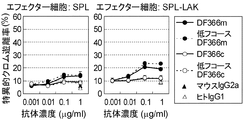
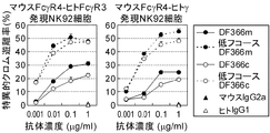
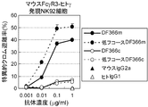
本発明者らは、上記の課題を解決するために、マウスFcγR3あるいはマウスFcγR4の細胞外領域と、ヒトγ鎖あるいはヒトFcγR3の膜貫通領域−細胞内領域を融合したキメラ分子を作製し、ヒトNK92細胞に発現させた。その結果、マウスFcγR3-ヒトγ鎖、マウスFcγR3-ヒトFcγR3、マウスFcγR4-ヒトγ鎖、マウスFcγR4-ヒトFcγR3のいずれの組み合わせでもADCC活性を誘導しうることが見出された。以上の知見により、本発明者らは、マウスFcγレセプターとヒトFcγレセプターのキメラレセプター、又はマウスFcγレセプターとヒトγ鎖のキメラレセプターを用いることにより、マウス抗体のADCC活性を測定することが可能であることを見出した。さらに、本発明者らは、マウスFcγレセプターとヒトFcγレセプター、又はマウスFcγレセプターとヒトγ鎖のキメラレセプターを用いることにより、ADCC活性を有するマウス抗体のスクリーニングが可能であることを見出した。 In order to solve the above-mentioned problems, the present inventors have produced a chimeric molecule in which the extracellular region of mouse FcγR3 or mouse FcγR4 and the human γ chain or the transmembrane region-intracellular region of human FcγR3 are fused. It was expressed in NK92 cells. As a result, it was found that any combination of mouse FcγR3-human γ chain, mouse FcγR3-human FcγR3, mouse FcγR4-human γ chain and mouse FcγR4-human FcγR3 can induce ADCC activity. Based on the above knowledge, the present inventors can measure the ADCC activity of a mouse antibody by using a chimeric receptor of mouse Fcγ receptor and human Fcγ receptor, or a chimeric receptor of mouse Fcγ receptor and human γ chain. I found out. Furthermore, the present inventors have found that a mouse antibody having ADCC activity can be screened by using a mouse Fcγ receptor and a human Fcγ receptor, or a chimeric receptor of a mouse Fcγ receptor and a human γ chain.
すなわち本願は以下の(1)から(20)を提供するものである。
(1)マウスFcγレセプター細胞外領域及びヒトγ鎖膜貫通領域を含むキメラタンパク質。
(2)ヒトγ鎖細胞内領域をさらに含む(1)のキメラタンパク質。
(3)マウスFcγレセプター細胞外領域及びヒトFcγレセプター膜貫通領域を含むキメラタンパク質。
(4)ヒトFcγレセプター細胞内領域をさらに含む(3)のキメラタンパク質。
(5)ヒトFcγレセプターがヒトFcγレセプター3である(3)または(4)のキメラタンパク質。
(6)マウスFcγレセプターがマウスFcγレセプター3である(1)から(5)いずれかのキメラタンパク質。
(7)マウスFcγレセプターがマウスFcγレセプター4である(1)から(5)いずれかのキメラタンパク質。
(8)(1)から(7)いずれかのキメラタンパク質をコードする遺伝子。
(9)(8)の遺伝子を含むベクター。
(10)(1)から(7)いずれかのキメラタンパク質を発現する細胞。
(11)細胞がNK細胞である(10)の細胞。
(12)細胞がヒト由来の細胞である(10)または(11)の細胞。
(13)以下の工程を含む、抗体の細胞障害活性の測定方法。
(a)被検抗体と該被検抗体が結合する抗原を発現する細胞を接触させる工程、
(b)(a)の被検抗体と(10)から(12)いずれかの細胞を接触させる工程、
(c)被検抗体の細胞障害活性を測定する工程。
(14)被検抗体がマウス由来抗体である(13)の測定方法。
(15)以下の工程を含む細胞障害活性を有する抗体のスクリーニング方法。
(a)被検抗体と該被検抗体が結合する抗原を発現する細胞を接触させる工程、
(b)(a)の被検抗体と(10)から(12)いずれかの細胞を接触させる工程、
(c)被検抗体の細胞障害活性を測定する工程、
(d)細胞障害活性を有する抗体を選択する工程。
(16)被検抗体がマウス由来抗体である(15)のスクリーニング方法。
(17)細胞障害活性を測定する為の(1)から(7)いずれかのキメラタンパク質の使用。
(18)細胞障害活性を測定する為の(10)から(12)いずれかの細胞の使用。
(19)細胞障害活性を有する抗体のスクリーニングの為の(1)から(7)いずれかのキメラタンパク質の使用。
(20)細胞障害活性を有する抗体のスクリーニングの為の(10)から(12)いずれかの細胞の使用。That is, this application provides the following (1) to (20).
(1) A chimeric protein comprising a mouse Fcγ receptor extracellular region and a human γ chain transmembrane region.
(2) The chimeric protein according to (1), further comprising a human γ-chain intracellular region.
(3) A chimeric protein comprising a mouse Fcγ receptor extracellular region and a human Fcγ receptor transmembrane region.
(4) The chimeric protein according to (3), further comprising a human Fcγ receptor intracellular region.
(5) The chimeric protein according to (3) or (4), wherein the human Fcγ receptor is human Fcγ receptor 3.
(6) The chimeric protein according to any one of (1) to (5), wherein the mouse Fcγ receptor is mouse Fcγ receptor 3.
(7) The chimeric protein according to any one of (1) to (5), wherein the mouse Fcγ receptor is mouse Fcγ receptor 4.
(8) A gene encoding any of the chimeric proteins (1) to (7).
(9) A vector containing the gene of (8).
(10) A cell that expresses any one of the chimeric proteins (1) to (7).
(11) The cell according to (10), wherein the cell is an NK cell.
(12) The cell according to (10) or (11), wherein the cell is a human-derived cell.
(13) A method for measuring the cytotoxic activity of an antibody, comprising the following steps.
(A) contacting a test antibody and a cell expressing an antigen to which the test antibody binds,
(B) contacting the test antibody of (a) with any of the cells from (10) to (12);
(C) A step of measuring the cytotoxic activity of the test antibody.
(14) The measuring method according to (13), wherein the test antibody is a mouse-derived antibody.
(15) A screening method for an antibody having cytotoxic activity comprising the following steps.
(A) contacting a test antibody and a cell expressing an antigen to which the test antibody binds,
(B) contacting the test antibody of (a) with any of the cells from (10) to (12);
(C) measuring the cytotoxic activity of the test antibody,
(D) A step of selecting an antibody having cytotoxic activity.
(16) The screening method according to (15), wherein the test antibody is a mouse-derived antibody.
(17) Use of the chimeric protein according to any one of (1) to (7) for measuring cytotoxic activity.
(18) Use of any cell of (10) to (12) for measuring cytotoxic activity.
(19) Use of any of the chimeric proteins (1) to (7) for screening for an antibody having cytotoxic activity.
(20) Use of any cell of (10) to (12) for screening for an antibody having cytotoxic activity.
〔発明の実施の形態〕
本発明はマウスFcγレセプターの細胞外領域およびヒトFcγレセプターの膜貫通領域を含むキメラレセプターを提供する。さらに、本発明はマウスFcγレセプターの細胞外領域およびヒトγ鎖の膜貫通領域を含むキメラレセプターを提供する。[Embodiment of the Invention]
The present invention provides a chimeric receptor comprising the extracellular region of mouse Fcγ receptor and the transmembrane region of human Fcγ receptor. Furthermore, the present invention provides a chimeric receptor comprising the extracellular region of mouse Fcγ receptor and the transmembrane region of human γ chain.
本発明のキメラレセプターは、細胞外領域にマウス抗体のFc領域が結合した場合、細胞内にシグナルを伝達する活性を有するレセプターであることが好ましい。
本発明で用いられるマウスFcγレセプターは特に限定されず、どのようなマウスFcγレセプターを用いてもよいが、好ましくはマウスFcγレセプター3(FcγR3)またはマウスFcγレセプター4(FcγR4)である。
マウスFcγR3とマウスFcγR4の発現分布を比較すると、マウスFcγR3は主にNK細胞に、マウスFcγR4はマクロファージと好中球に発現する(Immunity 2005, 23, 41)。またマウスFcγR3はマウスIgG1、マウスIgG2aおよびマウスIgG2bに結合するのに対し、マウスFcγR4はマウスIgG1には結合しない(Immunity 2005, 23, 41, Science 2005, 310, 1510)。従って、種々のマウス抗体のADCC活性を評価する際には、マウスIgG1でも測定できるマウスFcγR3が好ましい。The chimeric receptor of the present invention is preferably a receptor having an activity of transmitting a signal into the cell when the Fc region of a mouse antibody is bound to the extracellular region.
The mouse Fcγ receptor used in the present invention is not particularly limited, and any mouse Fcγ receptor may be used, but mouse Fcγ receptor 3 (FcγR3) or mouse Fcγ receptor 4 (FcγR4) is preferable.
Comparing the expression distribution of mouse FcγR3 and mouse FcγR4, mouse FcγR3 is expressed mainly in NK cells, and mouse FcγR4 is expressed in macrophages and neutrophils (Immunity 2005, 23, 41). Mouse FcγR3 binds to mouse IgG1, mouse IgG2a and mouse IgG2b, whereas mouse FcγR4 does not bind to mouse IgG1 (Immunity 2005, 23, 41, Science 2005, 310, 1510). Therefore, when evaluating ADCC activity of various mouse antibodies, mouse FcγR3 that can also be measured with mouse IgG1 is preferred.
マウスFcγレセプターをコードするDNAの塩基配列およびアミノ酸配列は公知の配列を用いることができる。マウスFcγレセプター3及びマウスFcγレセプター4をコードするDNAの塩基配列、アミノ酸配列としては、例えば、配列番号:1(マウスFcγレセプター3塩基配列)、配列番号:2(マウスFcγレセプター3アミノ酸配列)、配列番号:3(マウスFcγレセプター4塩基配列)、配列番号:4(マウスFcγレセプター4アミノ酸配列)に記載の配列を用いることができる。配列番号:2に記載のアミノ酸配列のうち、マウスFcγレセプター3細胞外領域はアミノ酸番号31から212であり、配列番号:4に記載のアミノ酸配列のうち、マウスFcγレセプター4細胞外領域はアミノ酸番号19から201である。 Known sequences can be used for the base sequence and amino acid sequence of DNA encoding mouse Fcγ receptor. Examples of the nucleotide sequence and amino acid sequence of DNA encoding mouse Fcγ receptor 3 and mouse Fcγ receptor 4 include, for example, SEQ ID NO: 1 (mouse Fcγ receptor 3 nucleotide sequence), SEQ ID NO: 2 (mouse Fcγ receptor 3 amino acid sequence), The sequences described in SEQ ID NO: 3 (mouse Fcγ receptor 4-base sequence) and SEQ ID NO: 4 (mouse Fcγ receptor 4-amino acid sequence) can be used. Of the amino acid sequence shown in SEQ ID NO: 2, the mouse Fcγ receptor 3 extracellular region is amino acid numbers 31 to 212. Of the amino acid sequence shown in SEQ ID NO: 4, the mouse Fcγ receptor 4 extracellular region is amino acid number. 19 to 201.
受容体の細胞外領域は、細胞外領域全体であってもよいし、その一部であってもよいが、抗体のFc領域への結合活性などの活性を適切に反映できる点で細胞外領域全体を用いることが好ましい。受容体の細胞外領域の一部を用いる場合、抗体のFc領域への結合活性を維持していることが好ましい。本発明のキメラレセプターに用いられるマウスFcγレセプター細胞外領域は、抗体のFc領域への結合能を有する限り、アミノ酸の置換、欠失、挿入、付加があってもよい。あるタンパク質と機能的に同等なタンパク質を調整するための、当業者によく知られた方法としては、タンパク質に変異を導入する方法が知られている。例えば、当業者であれば、部位特異的変異誘発法(Hashimoto-Gotoh, T. et al. (1995) Gene 152, 271-275、Zoller, MJ, and Smith, M. (1983) Methods Enzymol. 100, 468-500、Kramer, W. et al. (1984) Nucleic Acids Res. 12, 9441-9456、Kramer W, and Fritz HJ (1987) Methods. Enzymol. 154, 350-367、Kunkel, TA (1985) Proc Natl Acad Sci USA. 82, 488-492、Kunkel (1988) Methods Enzymol. 85, 2763-2766)などを用いて作製することができる。このような変異体における、変異するアミノ酸数は、通常、50アミノ酸以内であり、好ましくは30アミノ酸以内であり、さらに好ましくは20アミノ酸以内であり、さらに好ましくは10アミノ酸以内であり、さらに好ましくは5アミノ酸以内である。 The extracellular region of the receptor may be the whole extracellular region or a part thereof, but the extracellular region is capable of appropriately reflecting the activity such as the binding activity of the antibody to the Fc region. It is preferable to use the whole. When using a part of the extracellular region of the receptor, it is preferable to maintain the binding activity of the antibody to the Fc region. The mouse Fcγ receptor extracellular region used for the chimeric receptor of the present invention may have amino acid substitutions, deletions, insertions and additions as long as it has the ability to bind to the Fc region of an antibody. As a method well known to those skilled in the art for preparing a protein functionally equivalent to a certain protein, a method of introducing a mutation into the protein is known. For example, those skilled in the art will recognize site-directed mutagenesis (Hashimoto-Gotoh, T. et al. (1995) Gene 152, 271-275, Zoller, MJ, and Smith, M. (1983) Methods Enzymol. 100 , 468-500, Kramer, W. et al. (1984) Nucleic Acids Res. 12, 9441-9456, Kramer W, and Fritz HJ (1987) Methods. Enzymol. 154, 350-367, Kunkel, TA (1985) Proc Natl Acad Sci USA. 82, 488-492, Kunkel (1988) Methods Enzymol. 85, 2763-2766) and the like. In such mutants, the number of amino acids to be mutated is usually within 50 amino acids, preferably within 30 amino acids, more preferably within 20 amino acids, still more preferably within 10 amino acids, still more preferably. Within 5 amino acids.
変異するアミノ酸残基においては、アミノ酸側鎖の性質が保存されている別のアミノ酸に変異されることが好ましい。例えば、アミノ酸側鎖の性質としては、疎水性アミノ酸(A、I、L、M、F、P、W、Y、V)、親水性アミノ酸(R、D、N、C、E、Q、G、H、K、S、T)、脂肪族側鎖を有するアミノ酸(G、A、V、L、I、P)、水酸基含有側鎖を有するアミノ酸(S、T、Y)、硫黄原子含有側鎖を有するアミノ酸(C、M)、カルボン酸及びアミド含有側鎖を有するアミノ酸(D、N、E、Q)、塩基含有側鎖を有するアミノ酸(R、K、H)、芳香族含有側鎖を有するアミノ酸(H、F、Y、W)を挙げることができる(括弧内はいずれもアミノ酸の一文字表記を表す)。 The amino acid residue to be mutated is preferably mutated to another amino acid that preserves the properties of the amino acid side chain. For example, amino acid side chain properties include hydrophobic amino acids (A, I, L, M, F, P, W, Y, V), hydrophilic amino acids (R, D, N, C, E, Q, G , H, K, S, T), amino acids having aliphatic side chains (G, A, V, L, I, P), amino acids having hydroxyl group-containing side chains (S, T, Y), sulfur atom-containing side Amino acids with chains (C, M), amino acids with carboxylic acid and amide-containing side chains (D, N, E, Q), amino acids with base-containing side chains (R, K, H), aromatic-containing side chains (H, F, Y, W) can be mentioned (all parentheses indicate single letter amino acids).
あるアミノ酸配列に対する1又は複数個のアミノ酸残基の欠失、付加及び/又は他のアミノ酸による置換により修飾されたアミノ酸配列を有するポリペプチドがその生物学的活性を維持することはすでに知られている(Mark, D. F. et al., Proc. Natl. Acad. Sci. USA (1984) 81, 5662-5666、 Zoller, M. J. & Smith, M. Nucleic Acids Research (1982) 10, 6487-6500、 Wang, A. et al., Science 224, 1431-1433、 Dalbadie-McFarland, G. et al., Proc. Natl. Acad. Sci. USA (1982) 79, 6409-6413)。 It is already known that a polypeptide having an amino acid sequence modified by deletion, addition and / or substitution by one or more amino acid residues to a certain amino acid sequence maintains its biological activity. (Mark, DF et al., Proc. Natl. Acad. Sci. USA (1984) 81, 5662-5666, Zoller, MJ & Smith, M. Nucleic Acids Research (1982) 10, 6487-6500, Wang, A et al., Science 224, 1431-1433, Dalbadie-McFarland, G. et al., Proc. Natl. Acad. Sci. USA (1982) 79, 6409-6413).
本発明のキメラレセプターに用いられるマウスFcγレセプター細胞外領域は、抗体のFc領域への結合能を有する限り、マウスFcγレセプター(例えば、マウスFcγレセプター3、マウスFcγレセプター4など)と高い相同性を有するポリペプチドでもよい。本発明において高い相同性を有するポリペプチドとは、通常、70%以上、好ましくは80%以上、さらに好ましくは90%以上、さらに好ましくは95%以上の同一性を有するポリペプチドをいう。ポリペプチドの相同性を決定するには、文献(Wilbur, W. J. and Lipman, D. J. Proc. Natl. Acad. Sci. USA (1983) 80, 726-730)に記載のアルゴリズムにしたがえばよい。 The mouse Fcγ receptor extracellular region used for the chimeric receptor of the present invention has high homology with mouse Fcγ receptors (eg, mouse Fcγ receptor 3, mouse Fcγ receptor 4 etc.) as long as it has the ability to bind to the Fc region of the antibody. It may be a polypeptide. In the present invention, a polypeptide having high homology generally means a polypeptide having 70% or more, preferably 80% or more, more preferably 90% or more, more preferably 95% or more. To determine the homology of a polypeptide, the algorithm described in the literature (Wilbur, W. J. and Lipman, D. J. Proc. Natl. Acad. Sci. USA (1983) 80, 726-730) may be used.
また、マウスFcγレセプターと相同性の高いポリペプチドをコードするDNAを単離するためには、通常ストリンジェントな条件下でハイブリダイゼーション反応を行なってもよい。ストリンジェントなハイブリダイゼーション条件は当業者であれば、適宜選択することができる。一例を示せば、25%ホルムアミド、より厳しい条件では50%ホルムアミド、4×SSC、50mM Hepes pH7.0、10×デンハルト溶液、20μg/ml変性サケ精子DNAを含むハイブリダイゼーション溶液中、42℃で一晩プレハイブリダイゼーションを行った後、標識したプローブを添加し、42℃で一晩保温することによりハイブリダイゼーションを行う。その後の洗浄における洗浄液および温度条件は、「1xSSC、0.1% SDS、37℃」程度で、より厳しい条件としては「0.5xSSC、0.1% SDS、42℃」程度で、さらに厳しい条件としては「0.2xSSC、0.1% SDS、65℃」程度で実施することができる。このようにハイブリダイゼーションの洗浄の条件が厳しくなるほどプローブ配列と高い相同性を有するDNAの単離を期待しうる。但し、上記SSC、SDSおよび温度の条件の組み合わせは例示であり、当業者であれば、ハイブリダイゼーションのストリンジェンシーを決定する上記若しくは他の要素(例えば、プローブ濃度、プローブの長さ、ハイブリダイゼーション反応時間など)を適宜組み合わせることにより、上記と同様のストリンジェンシーを実現することが可能である。 In order to isolate DNA encoding a polypeptide having high homology with mouse Fcγ receptor, a hybridization reaction may be usually performed under stringent conditions. Those skilled in the art can appropriately select stringent hybridization conditions. An example is 25% formamide, 50% formamide under more severe conditions, 4 × SSC, 50 mM Hepes pH 7.0, 10 × Denhardt's solution, 20 μg / ml denatured in a hybridization solution containing denatured salmon sperm DNA at 42 ° C. After overnight prehybridization, a labeled probe is added and hybridization is performed by incubating overnight at 42 ° C. The cleaning solution and temperature conditions for the subsequent cleaning are about 1xSSC, 0.1% SDS, 37 ° C, more severe conditions are about 0.5xSSC, 0.1% SDS, 42 ° C, and more severe conditions are about 0.2xSSC. , 0.1% SDS, about 65 ° C. ”. Thus, isolation of DNA having high homology with the probe sequence can be expected as the conditions for washing hybridization are more severe. However, combinations of the above SSC, SDS, and temperature conditions are exemplary, and those skilled in the art will understand the above or other factors that determine the stringency of hybridization (eg, probe concentration, probe length, hybridization reaction). It is possible to achieve the same stringency as above by appropriately combining the time and the like.
単離されたDNAの相同性は、アミノ酸配列全体で、少なくとも50%以上、さらに好ましくは70%以上、さらに好ましくは90%以上(例えば、95%、96%、97%、98%、99%以上)の配列の同一性を有する。配列の相同性は、BLASTN(核酸レベル)やBLASTX(アミノ酸レベル)のプログラム(Altschul et al. J. Mol. Biol., 215: 403-410, 1990)を利用して決定することができる。該プログラムは、Karlin及びAltschulによるアルゴリズムBLAST (Proc. Natl. Acad. Sci. USA, 87:2264-2268, 1990, Proc. Natl. Acad. Sci. USA, 90: 5873-5877, 1993) に基づいている。BLASTNによって塩基配列を解析する場合には、パラメーターは例えばscore = 100、wordlength =12とする。また、BLASTXによってアミノ酸配列を解析する場合には、パラメーターは例えばscore = 50、wordlength = 3とする。また、Gapped BLASTプログラムを用いて、アミノ酸配列を解析する場合は、Altschulら(Nucleic Acids Res. 25: 3389-3402, 1997)に記載されているように行うことができる。BLASTとGapped BLASTプログラムを用いる場合には、各プログラムのデフォルトパラメーターを用いる。これらの解析方法の具体的な手法は公知である。
また、マウスFcγレセプターをコードするDNA(配列番号:1、3)の配列情報を基に合成したプライマーを用いる遺伝子増幅法、例えば、ポリメラーゼ連鎖反応(PCR)法を利用して、マウスFcγレセプターと相同性の高いポリペプチドをコードするDNAを単離することも可能である。The homology of the isolated DNA is at least 50% or more, more preferably 70% or more, more preferably 90% or more (for example, 95%, 96%, 97%, 98%, 99%) in the entire amino acid sequence. And the like). Sequence homology can be determined using BLASTN (nucleic acid level) and BLASTX (amino acid level) programs (Altschul et al. J. Mol. Biol., 215: 403-410, 1990). The program is based on the algorithm BLAST by Karlin and Altschul (Proc. Natl. Acad. Sci. USA, 87: 2264-2268, 1990, Proc. Natl. Acad. Sci. USA, 90: 5873-5877, 1993). Yes. When analyzing a base sequence by BLASTN, parameters are set to score = 100 and wordlength = 12, for example. Further, when the amino acid sequence is analyzed by BLASTX, the parameters are, for example, score = 50 and wordlength = 3. When the amino acid sequence is analyzed using the Gapped BLAST program, it can be performed as described in Altschul et al. (Nucleic Acids Res. 25: 3389-3402, 1997). When using BLAST and Gapped BLAST programs, the default parameters of each program are used. Specific methods of these analysis methods are known.
In addition, a gene amplification method using primers synthesized based on the sequence information of DNA encoding the mouse Fcγ receptor (SEQ ID NOs: 1, 3), for example, polymerase chain reaction (PCR) method, It is also possible to isolate DNA encoding a polypeptide having high homology.
本発明のキメラレセプターにおいては、膜貫通領域としてヒトFcγレセプター膜貫通領域またはヒトγ鎖膜貫通領域が用いられる。
本発明で用いられるヒトFcγレセプターは特に限定されず、どのようなヒトFcγレセプターでもよいが、好ましくはヒトFcγレセプター3である。ヒトFcγレセプターをコードするDNAの塩基配列およびアミノ酸配列は公知の配列を用いることができる。ヒトFcγレセプター3をコードするDNAの塩基配列、アミノ酸配列としては、例えば、配列番号:5(ヒトFcγレセプター3塩基配列)、配列番号:6(ヒトFcγレセプター3アミノ酸配列)の配列を用いることができる。配列番号:6に記載のアミノ酸配列のうち、膜貫通領域はアミノ酸番号207から229である。
また、ヒトγ鎖をコードするDNAの塩基配列、アミノ酸配列は公知であり、例えば、配列番号:7(ヒトγ鎖塩基配列)、配列番号:8(ヒトγ鎖アミノ酸配列)の配列を用いることができる。配列番号:8に記載のアミノ酸配列のうち、膜貫通領域はアミノ酸番号24から44である。In the chimeric receptor of the present invention, a human Fcγ receptor transmembrane region or a human γ chain transmembrane region is used as the transmembrane region.
The human Fcγ receptor used in the present invention is not particularly limited, and any human Fcγ receptor may be used, but human Fcγ receptor 3 is preferable. Known sequences can be used for the base sequence and amino acid sequence of DNA encoding the human Fcγ receptor. As the base sequence and amino acid sequence of DNA encoding human Fcγ receptor 3, for example, the sequence of SEQ ID NO: 5 (human Fcγ receptor 3 base sequence) and SEQ ID NO: 6 (human Fcγ receptor 3 amino acid sequence) can be used. it can. Of the amino acid sequence set forth in SEQ ID NO: 6, the transmembrane region is amino acid numbers 207 to 229.
Further, the base sequence and amino acid sequence of DNA encoding human γ chain are known. For example, the sequence of SEQ ID NO: 7 (human γ chain base sequence) and SEQ ID NO: 8 (human γ chain amino acid sequence) should be used. Can do. Of the amino acid sequence set forth in SEQ ID NO: 8, the transmembrane region is amino acid numbers 24-44.
受容体の膜貫通領域は膜貫通領域全体であってもよいし、その一部であってもよいが、シグナル伝達活性などの活性を適切に反映できる点で膜貫通領域全体を用いることが好ましい。膜貫通領域の一部を用いる場合、シグナル伝達活性を維持していることが好ましい。本発明のキメラレセプターに用いられる膜貫通領域は、アミノ酸の置換、欠失、挿入、付加があってもよい。又、本発明に用いられる膜貫通領域は、これらと相同性の高いポリペプチドであってもよい。アミノ酸の置換、欠失、挿入、付加および相同性の高いポリペプチドについては上述に記載のとおりである。アミノ酸が置換、欠失、挿入及び/又は付加されたポリペプチド、および相同性の高いポリペプチドは膜貫通領域のシグナル伝達活性を維持していることが好ましい。 The transmembrane region of the receptor may be the entire transmembrane region or a part thereof, but it is preferable to use the entire transmembrane region in that the activity such as signal transduction activity can be appropriately reflected. . When a part of the transmembrane region is used, it is preferable to maintain the signal transduction activity. The transmembrane region used in the chimeric receptor of the present invention may have amino acid substitution, deletion, insertion, and addition. Further, the transmembrane region used in the present invention may be a polypeptide having high homology with these. Amino acid substitutions, deletions, insertions, additions and highly homologous polypeptides are as described above. It is preferable that a polypeptide in which an amino acid is substituted, deleted, inserted and / or added, and a highly homologous polypeptide maintain the signal transduction activity of the transmembrane region.
本発明のマウスFcγレセプター細胞外領域とヒトFcγレセプター膜貫通領域を含むキメラレセプターまたはマウスFcγレセプター細胞外領域とヒトγ鎖膜貫通領域を含むキメラレセプターは、さらに細胞内領域を含むことが好ましい。
本発明のキメラレセプターに用いられる細胞内領域は特に限定されず、どのような細胞内領域でもよいが、膜貫通領域としてヒトFcγレセプター膜貫通領域が用いられる場合は細胞内領域としてヒトFcγレセプター細胞内領域が用いられることが好ましく、膜貫通領域としてヒトγ鎖が用いられる場合は細胞内領域としてヒトγ鎖細胞内領域が用いられることが好ましい。
ヒトFcγレセプター3細胞内領域は、例えば、配列番号:6に記載のアミノ酸配列のうち、アミノ酸番号230から254の領域を用いることができる。ヒトγ鎖の細胞内領域は、例えば、配列番号:8に記載のアミノ酸配列のうちアミノ酸番号45から86の領域を用いることができる。The chimeric receptor containing the mouse Fcγ receptor extracellular region and the human Fcγ receptor transmembrane region or the chimeric receptor containing the mouse Fcγ receptor extracellular region and the human γ chain transmembrane region of the present invention preferably further contains an intracellular region.
The intracellular region used for the chimeric receptor of the present invention is not particularly limited, and any intracellular region may be used. When a human Fcγ receptor transmembrane region is used as the transmembrane region, the human Fcγ receptor cell is used as the intracellular region. The inner region is preferably used, and when the human γ chain is used as the transmembrane region, the human γ chain intracellular region is preferably used as the intracellular region.
As the human Fcγ receptor 3 intracellular region, for example, the region of amino acid numbers 230 to 254 in the amino acid sequence of SEQ ID NO: 6 can be used. As the intracellular region of the human γ chain, for example, the region of amino acid numbers 45 to 86 in the amino acid sequence shown in SEQ ID NO: 8 can be used.
受容体の細胞内領域は細胞内領域全体であってもよいし、その一部であってもよいが、シグナル伝達活性などの活性を適切に反映できる点で細胞内領域全体を用いることが好ましい。細胞内領域の一部を用いる場合、シグナル伝達活性を維持していることが好ましい。本発明のキメラレセプターに用いられる細胞内領域は、アミノ酸の置換、欠失、挿入、付加があってもよい。又、本発明に用いられる細胞内領域は、これらと相同性の高いポリペプチドであってもよい。、アミノ酸の置換、欠失、挿入、付加および相同性の高いポリペプチドについては上述のとおりである。アミノ酸が置換、欠失、挿入及び/又は付加されたポリペプチド、および相同性の高いポリペプチドは細胞内領域のシグナル伝達活性を維持していることが好ましい。 The intracellular region of the receptor may be the entire intracellular region or a part thereof, but it is preferable to use the entire intracellular region from the viewpoint of appropriately reflecting the activity such as signal transduction activity. . When a part of the intracellular region is used, it is preferable to maintain the signal transduction activity. The intracellular region used in the chimeric receptor of the present invention may have amino acid substitution, deletion, insertion, and addition. The intracellular region used in the present invention may be a polypeptide having high homology with these. Polypeptides having high amino acid substitution, deletion, insertion, addition and homology are as described above. It is preferable that a polypeptide in which amino acids are substituted, deleted, inserted and / or added, and a polypeptide having high homology maintain the signal transduction activity in the intracellular region.
本発明のキメラレセプターの好ましい態様として、以下の(a)から(f)のキメラレセプターを挙げることができる。
(a) 配列番号:10のアミノ酸配列を有するキメラレセプター(マウスFcγR3細胞外領域と、ヒトFcγR3膜貫通領域および細胞内領域を含むキメラレセプター;マウスFcγR3−ヒトFcγR3)。
(b) 配列番号:12のアミノ酸配列を有するキメラレセプター(マウスFcγR3細胞外領域と、ヒトγ鎖膜貫通領域および細胞内領域を含むキメラレセプター;マウスFcγR3−ヒトγ鎖)。
(c) 配列番号:14のアミノ酸配列を有するキメラレセプター(マウスFcγR4細胞外領域と、ヒトFcγR3膜貫通領域および細胞内領域を含むキメラレセプター;マウスFcγR4−ヒトFcγR3)。
(d) 配列番号:16のアミノ酸配列を有するキメラレセプター(マウスFcγR4細胞外領域と、ヒトγ鎖膜貫通領域および細胞内領域を含むキメラレセプター;マウスFcγR4−ヒトγ鎖)。
(e) 上記(a)から(d)のキメラレセプターにおいて、1又は複数のアミノ酸配列が置換、欠失、付加および/または挿入されたレセプターであって、(a)から(d)のキメラレセプターと同等の活性を有するキメラレセプター。
(f) 上記(a)から(d)のキメラレセプターのアミノ酸配列と高い相同性を有するレセプターであって、(a)から(d)のキメラレセプターと同等の活性を有するキメラレセプター。
アミノ酸の置換、欠失、付加、挿入、および高い相同性については上述のとおりである。Preferred embodiments of the chimeric receptor of the present invention include the following chimeric receptors (a) to (f).
(a) A chimeric receptor having the amino acid sequence of SEQ ID NO: 10 (a chimeric receptor comprising a mouse FcγR3 extracellular region, a human FcγR3 transmembrane region and an intracellular region; mouse FcγR3-human FcγR3).
(b) A chimeric receptor having the amino acid sequence of SEQ ID NO: 12 (a chimeric receptor comprising a mouse FcγR3 extracellular region, a human γ-chain transmembrane region and an intracellular region; mouse FcγR3-human γ chain).
(c) A chimeric receptor having the amino acid sequence of SEQ ID NO: 14 (a chimeric receptor comprising a mouse FcγR4 extracellular region, a human FcγR3 transmembrane region and an intracellular region; mouse FcγR4-human FcγR3).
(d) A chimeric receptor having the amino acid sequence of SEQ ID NO: 16 (a chimeric receptor comprising a mouse FcγR4 extracellular region, a human γ-chain transmembrane region and an intracellular region; mouse FcγR4-human γ chain).
(e) The chimeric receptor of (a) to (d) above, wherein one or a plurality of amino acid sequences are substituted, deleted, added and / or inserted, and the chimeric receptor of (a) to (d) Chimeric receptor having the same activity as
(f) A chimeric receptor having high homology with the amino acid sequence of the chimeric receptor of (a) to (d) above, and having an activity equivalent to that of the chimeric receptor of (a) to (d).
Amino acid substitutions, deletions, additions, insertions, and high homology are as described above.
本発明において、(a)から(d)のキメラレセプターと「同等の活性」を有するとは、同等の生物学的活性や生化学的活性を有することをいう。本発明のキメラレセプターの生物学的活性や生化学的活性としては、マウス抗体のFc領域への結合能やADCCシグナル伝達能を挙げることができる。
本発明のキメラレセプターのマウス抗体のFc領域への結合活性は当業者に公知の方法で測定することが可能であり、例えば、ELISA、BIACORE、ウェスタンブロットなどの方法により測定することが可能である。In the present invention, “having the same activity” as the chimeric receptors (a) to (d) means having the same biological activity or biochemical activity. Examples of the biological activity and biochemical activity of the chimeric receptor of the present invention include the ability of a mouse antibody to bind to the Fc region and ADCC signal transduction ability.
The binding activity of the chimeric receptor of the present invention to the Fc region of a mouse antibody can be measured by methods known to those skilled in the art, for example, by ELISA, BIACORE, Western blot, etc. .
本発明のキメラレセプターがADCCシグナルを伝達するか否かは当業者に公知の方法で確認することが可能であり、例えば、キメラレセプターを発現するヒトNK細胞(ヒトNK92細胞など)をエフェクター細胞として使用し、該NK細胞と標的細胞上に発現した抗原に結合したマウス抗体とを接触させ、ADCC活性を測定することにより確認することができる。より具体的には、後述の方法、または本発明の実施例に記載の方法で確認することが可能である。 Whether or not the chimeric receptor of the present invention transmits an ADCC signal can be confirmed by a method known to those skilled in the art. For example, human NK cells (such as human NK92 cells) expressing the chimeric receptor are used as effector cells. This can be confirmed by contacting the NK cell with a mouse antibody bound to an antigen expressed on the target cell and measuring ADCC activity. More specifically, it can be confirmed by the method described later or the method described in the examples of the present invention.
本発明のキメラレセプターをコードするDNA、及び該DNAの転写産物RNAもまた、本発明に含まれる。本発明のキメラレセプターをコードするDNAは当業者に公知の方法により調整することができる。例えば、本発明で細胞外領域、膜貫通領域として用いられるレセプターを発現している細胞よりcDNAライブラリを作製し、既知のDNA配列の一部をプローブにしてハイブリダイゼーションを行なうことにより、細胞外領域や膜貫通領域をコードするDNAを調整できる。それぞれ調整されたDNAを結合することにより本発明のキメラレセプターをコードするDNAを調整することが可能である。 DNA encoding the chimeric receptor of the present invention, and a transcript RNA of the DNA are also included in the present invention. The DNA encoding the chimeric receptor of the present invention can be prepared by methods known to those skilled in the art. For example, by preparing a cDNA library from a cell expressing a receptor used as an extracellular region or a transmembrane region in the present invention and performing hybridization using a part of a known DNA sequence as a probe, the extracellular region And DNA encoding the transmembrane region can be adjusted. It is possible to prepare a DNA encoding the chimeric receptor of the present invention by binding each prepared DNA.
本発明のキメラレセプターをコードするDNAの塩基配列の例としては、配列番号:9(マウスFcγR3−ヒトFcγR3)、配列番号:11(マウスFcγR3−ヒトγ鎖)、配列番号:13(マウスFcγR4−ヒトFcγR3)、配列番号:15(マウスFcγR4−ヒトγ鎖)に記載の塩基配列を挙げることができる。 Examples of the base sequence of the DNA encoding the chimeric receptor of the present invention include SEQ ID NO: 9 (mouse FcγR3-human FcγR3), SEQ ID NO: 11 (mouse FcγR3-human γ chain), SEQ ID NO: 13 (mouse FcγR4-). Human FcγR3), SEQ ID NO: 15 (mouse FcγR4-human γ chain).
調整された本発明のキメラレセプターをコードするDNAをベクターDNAと連結する。さらに、これより組換えベクターを作製し、大腸菌等に導入してコロニーを選択して所望の組換えベクターを調整することができる。DNAを保持するためのベクターDNAには、公知のもの(例えばpUC19、pBluescriptなど)を用いることができる。また、大腸菌は公知のもの(例えばDH5α、JM109など)を用いることができる。目的とするDNAの塩基配列は公知の方法、例えばジデオキシヌクレオチドチェインターミネーション法により確認することができる。又、自動塩基配列決定装置などを用いることもできる。 The prepared DNA encoding the chimeric receptor of the present invention is ligated to vector DNA. Furthermore, a recombinant vector can be prepared from this, introduced into Escherichia coli, etc., and colonies can be selected to prepare a desired recombinant vector. As the vector DNA for retaining the DNA, known ones (for example, pUC19, pBluescript, etc.) can be used. Known Escherichia coli (eg, DH5α, JM109, etc.) can be used. The base sequence of the target DNA can be confirmed by a known method such as the dideoxynucleotide chain termination method. An automatic base sequence determination device or the like can also be used.
また、本発明のDNAにおいては、発現に使用する宿主のコドン使用頻度を考慮して、より発現効率の高い塩基配列を設計することもできる(Grantham, R. et al., Nucleic Acids Research (1981) 9, r43-74)。また、本発明のDNAは市販のキットや公知の方法によって改変することができる。改変としては、例えば、制限酵素により消化、合成オリゴヌクレオチドや適当なDNAフラグメントの挿入、リンカーの付加、開始コドン及び/又は終止コドンの挿入、などが挙げられる。 In addition, in the DNA of the present invention, a base sequence with higher expression efficiency can be designed in consideration of the codon usage frequency of the host used for expression (Grantham, R. et al., Nucleic Acids Research (1981). ) 9, r43-74). The DNA of the present invention can be modified by a commercially available kit or a known method. Examples of the modification include digestion with a restriction enzyme, insertion of a synthetic oligonucleotide or an appropriate DNA fragment, addition of a linker, insertion of a start codon and / or a stop codon, and the like.
本発明のキメラレセプター発現のためにはプロモーターなどの発現制御領域のもとでキメラレセプターをコードするDNAを含む発現ベクターを作製する。この発現ベクターにより宿主細胞を形質転換して細胞にキメラレセプターを発現させる。ベクターにはエンハンサーなどが含まれていてもよい。 In order to express the chimeric receptor of the present invention, an expression vector containing DNA encoding the chimeric receptor is prepared under an expression control region such as a promoter. A host cell is transformed with this expression vector to express the chimeric receptor in the cell. An enhancer etc. may be contained in the vector.
宿主細胞での発現のために有用なプロモーターとしては、例えば、サイトメガロウイルス、レトロウイルス、ポリオーマウイルス、アデノウイルス、SV40などのウイルスプロモーターや、哺乳動物細胞由来のプロモーターなどを挙げることができる。
宿主細胞中への遺伝子導入のため、発現ベクターには選択マーカー遺伝子を含むことができる。
宿主細胞中への遺伝子導入は、リン酸カルシウム法、リポフェクション法、エレクトロポレーション法などの公知の方法により行なうことができる。Examples of promoters useful for expression in host cells include viral promoters such as cytomegalovirus, retrovirus, polyoma virus, adenovirus, SV40, and mammalian cell-derived promoters.
For gene transfer into the host cell, the expression vector can include a selectable marker gene.
The gene can be introduced into the host cell by a known method such as a calcium phosphate method, a lipofection method, or an electroporation method.
本発明は、本発明のキメラレセプターをコードするDNAを含有するベクターを提供する。本発明で用いられるベクターは特に限定されず、いかなるベクターでもよい。ベクターは当業者が適宜選択することができ、例えば、pCOS1(WO98/13388)、pME18S(Med. Immunol. 20, 27-32 (1990))、pEF-BOS(Nucleic Acids Res. 18, 5322 (1990))、pCDM8(Nature 329, 840-842 (1987))、pRSV-neo、pcDNAI/Amp(Invitrogen)、pcDNAI、pAMoERC3Sc、pCDM8(Nature 329, 840 (1987))、pAGE107(Cytotechnology 3, 133 (1990))、pREP4(Invitrogen)、pAGE103(J. Biochem. 101, 1307 (1987))、pAMoA、pAS3-3、pCAGGS(Gene 108, 193-200 (1991))、pBK-CMV、pcDNA3.1(Invitrogen)、pZeoSV(Stratagene)などを用いることができる。 The present invention provides a vector containing DNA encoding the chimeric receptor of the present invention. The vector used in the present invention is not particularly limited, and any vector may be used. Vectors can be appropriately selected by those skilled in the art.For example, pCOS1 (WO98 / 13388), pME18S (Med. Immunol. 20, 27-32 (1990)), pEF-BOS (Nucleic Acids Res. 18, 5322 (1990) )), PCDM8 (Nature 329, 840-842 (1987)), pRSV-neo, pcDNAI / Amp (Invitrogen), pcDNAI, pAMoERC3Sc, pCDM8 (Nature 329, 840 (1987)), pAGE107 (Cytotechnology 3, 133 (1990) )), PREP4 (Invitrogen), pAGE103 (J. Biochem. 101, 1307 (1987)), pAMoA, pAS3-3, pCAGGS (Gene 108, 193-200 (1991)), pBK-CMV, pcDNA3.1 (Invitrogen) ), PZeoSV (Stratagene), etc. can be used.
本発明は、本発明のキメラレセプターを発現する細胞に関する。本発明のキメラレセプターを発現する細胞は、当業者に公知の方法で作製することが可能であり、例えば、本発明の上記ベクターを細胞に導入することにより作製することが可能である。本発明で用いられる細胞は特に限定されず、いかなる細胞を用いてもよいが、エフェクター細胞が好ましく、好ましくはNK細胞であり、特に好ましくはNK92細胞である。又、本発明で用いられる細胞はヒト由来細胞であることが好ましく、特にヒト由来NK細胞が好ましい。ヒトNK細胞は公知のヒトNK細胞を用いることもできるし、ヒトNK細胞を作製して用いることもできる。本発明で用いられる細胞はヒトγ鎖を発現する細胞でもヒトγ鎖を発現しない細胞でもよいが、ヒトγ鎖を発現する細胞であることが好ましい。ヒトγ鎖を発現しない細胞やヒトγ鎖の発現量が少ない細胞の場合、ヒトγ鎖をコードする遺伝子を導入して強制的にヒトγ鎖を発現させてもよい。 The present invention relates to cells that express the chimeric receptor of the present invention. Cells expressing the chimeric receptor of the present invention can be prepared by methods known to those skilled in the art. For example, the cells can be prepared by introducing the vector of the present invention into cells. The cells used in the present invention are not particularly limited, and any cells may be used, but effector cells are preferable, preferably NK cells, and particularly preferably NK92 cells. Moreover, the cell used in the present invention is preferably a human-derived cell, and particularly preferably a human-derived NK cell. As human NK cells, known human NK cells can be used, or human NK cells can be prepared and used. The cell used in the present invention may be a cell expressing a human γ chain or a cell not expressing a human γ chain, but is preferably a cell expressing a human γ chain. In the case of cells that do not express human γ chain or cells that express a small amount of human γ chain, a gene encoding human γ chain may be introduced to forcibly express human γ chain.
本発明は、本発明のキメラレセプターを用いた抗体の細胞障害活性の測定方法を提供する。
細胞障害活性の測定は、本発明のキメラレセプターを用いて、通常の細胞障害活性の測定と同様に行なうことが可能である。
例えば、以下の工程を含む方法により行なうことができる。
(a)被検抗体と該被検抗体が結合する抗原を発現する細胞を接触させる工程、
(b)(a)の被検抗体と本発明のキメラレセプターを発現する細胞を接触させる工程、
(c)被検抗体の細胞障害活性を測定する工程。The present invention provides a method for measuring the cytotoxic activity of an antibody using the chimeric receptor of the present invention.
Cytotoxic activity can be measured using the chimeric receptor of the present invention in the same manner as normal measurement of cytotoxic activity.
For example, it can be performed by a method including the following steps.
(A) contacting a test antibody and a cell expressing an antigen to which the test antibody binds,
(B) contacting the test antibody of (a) with a cell expressing the chimeric receptor of the present invention;
(C) A step of measuring the cytotoxic activity of the test antibody.
本発明において測定とは、定量的または定性的な測定を含む。定性的な測定としては、例えば、単に被検抗体が細胞障害活性を有するか否かの測定、被検抗体が一定レベル以上の細胞障害活性を有するか否かの測定、被検抗体の細胞障害活性をコントロール抗体(陽性コントロール、陰性コントロール、など)と比較する測定などを挙げることができる。一方、定量的な検出としては、例えば、被検抗体の細胞障害活性の絶対値や相対値の測定、被検抗体の医薬品としての有用性の測定などを挙げることができる。 In the present invention, the measurement includes quantitative or qualitative measurement. The qualitative measurement includes, for example, simply measuring whether or not the test antibody has cytotoxic activity, measuring whether or not the test antibody has a certain level of cytotoxic activity, or cytotoxicity of the test antibody. A measurement that compares the activity with a control antibody (positive control, negative control, etc.) can be mentioned. On the other hand, examples of quantitative detection include measurement of the absolute value and relative value of the cytotoxic activity of the test antibody, and measurement of the usefulness of the test antibody as a pharmaceutical.
又、本発明は、本発明のキメラレセプターを用いた、細胞障害活性を有する抗体のスクリーニング方法を提供する。
具体的には、以下の工程を含む方法により細胞障害活性を有する抗体のスクリーニングを行なうことが可能である。
(a)被検抗体と該被検抗体が結合する抗原を発現する細胞を接触させる工程、
(b)(a)の被検抗体と本発明のキメラレセプターを発現する細胞を接触させる工程、
(c)被検抗体の細胞障害活性を測定する工程、
(d)細胞障害活性を有する抗体を選択する工程。The present invention also provides a method for screening an antibody having cytotoxic activity using the chimeric receptor of the present invention.
Specifically, an antibody having cytotoxic activity can be screened by a method including the following steps.
(A) contacting a test antibody and a cell expressing an antigen to which the test antibody binds,
(B) contacting the test antibody of (a) with a cell expressing the chimeric receptor of the present invention;
(C) measuring the cytotoxic activity of the test antibody,
(D) A step of selecting an antibody having cytotoxic activity.
本発明のスクリーニング方法は、細胞障害活性を有しているか否か不明な被検抗体から細胞障害活性を有する抗体のスクリーニング、細胞障害活性を有している被検抗体から活性の高い抗体のスクリーニング、活性を有する被検抗体または活性を有するか否か不明な被検抗体から医薬品として有用な抗体のスクリーニング、など如何なるスクリーニング方法でもよい。
本発明の方法において、測定される細胞障害活性は、通常、抗体依存性細胞性細胞障害活性(ADCC活性)である。
本発明の方法で用いられる被検抗体は特に限定されず、いかなる抗体を用いてもよいが、通常、本発明のキメラレセプターの細胞外領域と結合可能な領域を有する。被検抗体の好ましい例として、マウス抗体またはマウス抗体由来のFc領域を有する抗体を挙げることができる。被検抗体のFc領域のアミノ酸配列は改変されていてもよく、又、抗体の糖鎖が改変されていてもよい。The screening method of the present invention comprises screening an antibody having cytotoxic activity from a test antibody whose cytotoxicity is unknown or not, screening an antibody having high activity from a test antibody having cytotoxic activity. Any screening method may be used such as screening of a test antibody having activity or screening of an antibody useful as a pharmaceutical from a test antibody with unknown activity.
In the method of the present invention, the measured cytotoxic activity is usually antibody-dependent cellular cytotoxic activity (ADCC activity).
The test antibody used in the method of the present invention is not particularly limited, and any antibody may be used. Usually, the antibody has a region capable of binding to the extracellular region of the chimeric receptor of the present invention. Preferred examples of the test antibody include a mouse antibody or an antibody having an Fc region derived from a mouse antibody. The amino acid sequence of the Fc region of the test antibody may be modified, or the sugar chain of the antibody may be modified.
被検抗体が結合する抗原は特に限定されないが、膜タンパク質であることが好ましい。膜タンパク質の例としては、受容体、輸送タンパク質、イオンチャンネル、細胞膜抗原などを挙げることができる。
本発明において、被検抗体が結合する抗原の好ましい例として、疾患に関連した抗原を挙げることができる。疾患に関連した抗原とは、特定の疾患において発現が確認されている抗原であり、好ましくは正常状態と比較して特定の疾患において発現量が増加している抗原である。疾患に関連した抗原の例として、癌において高発現しているタンパク質や自己免疫疾患において高発現しているタンパク質などを挙げることができる。The antigen to which the test antibody binds is not particularly limited, but is preferably a membrane protein. Examples of membrane proteins include receptors, transport proteins, ion channels, cell membrane antigens and the like.
In the present invention, a preferred example of an antigen to which a test antibody binds is an antigen associated with a disease. The antigen related to a disease is an antigen whose expression is confirmed in a specific disease, and preferably an antigen whose expression level is increased in a specific disease as compared with a normal state. Examples of diseases-related antigens include proteins that are highly expressed in cancer and proteins that are highly expressed in autoimmune diseases.
抗原を発現する細胞は如何なる細胞でもよく、被検抗体が結合する抗原をもともと発現する細胞でもよいし、該抗原をコードする遺伝子が導入されて抗原が強制的に発現される細胞でもよい。本発明の方法で用いられる抗原の好ましい例としては、癌細胞、B細胞などの自己免疫疾患関連細胞などを挙げることができる。
本発明の方法においては、通常、まず被検抗体と該被検抗体が結合する抗原を発現する細胞を接触させ、その後、抗原を発現する細胞に結合した被検抗体に本発明のキメラレセプターを発現する細胞を接触させる。しかしながら、被検抗体、抗原を発現する細胞、本発明のキメラレセプターを発現する細胞を接触させる順序は上述の順序に限定されず、被検抗体、抗原を発現する細胞および本発明のキメラレセプターを発現する細胞を同時に接触させてもよいし、被検抗体と本発明のキメラレセプターを発現する細胞を接触させた後に抗原を発現する細胞を接触させてもよい。The cell that expresses the antigen may be any cell, may be a cell that originally expresses the antigen to which the test antibody binds, or may be a cell in which a gene encoding the antigen is introduced and the antigen is forcibly expressed. Preferable examples of the antigen used in the method of the present invention include autoimmune disease-related cells such as cancer cells and B cells.
In the method of the present invention, usually, first, a test antibody and a cell expressing an antigen to which the test antibody binds are contacted, and then the chimeric receptor of the present invention is attached to the test antibody bound to the cell expressing the antigen. Contact the expressing cells. However, the order in which the test antibody, the cell expressing the antigen, and the cell expressing the chimeric receptor of the present invention are brought into contact with each other is not limited to the order described above, and the test antibody, the cell expressing the antigen and the chimeric receptor of the present invention Cells that express the antigen may be contacted simultaneously, or cells that express the antigen may be contacted after the test antibody and cells expressing the chimeric receptor of the present invention are contacted.
抗体がADCC活性を有するか否かは公知の方法により測定することができる(例えば、Current protocols in Immunology, Chapter7. Immunologic studies in humans, Editor, John E, Coligan et al., John Wiley & Sons, Inc.,(1993)等)。
具体的には、以下の方法により行なうことが可能である。
まずエフェクター細胞、標的細胞の調製が実施される。
(1)エフェクター細胞の調製
本発明のキメラレセプター発現細胞を細胞濃度5×10⁶/mlに調製することによって、エフェクター細胞が調製できる。
(2)標的細胞の調製
被検抗体が結合する抗原を発現する細胞を0.2 mCiの51Cr-クロム酸ナトリウム(GEヘルスケアバイオサイエンス社製)とともに、10% FBS含有RPMI1640培地中で37℃にて1時間培養することにより該標的細胞を放射性標識できる。放射性標識後、細胞を10% FBS含有RPMI1640培地にて3回洗浄し、細胞濃度を2×10⁵/mlに調製することによって、該標的細胞が調製できる。Whether or not an antibody has ADCC activity can be measured by a known method (for example, Current protocols in Immunology, Chapter 7. Immunologic studies in humans, Editor, John E, Coligan et al., John Wiley & Sons, Inc. ., (1993) etc.).
Specifically, it can be performed by the following method.
First, effector cells and target cells are prepared.
(1) Preparation of effector cells Effector cells can be prepared by preparing the chimeric receptor-expressing cells of the present invention at a cell concentration of 5 × 10 6 / ml.
(2) Preparation of target cells Cells expressing an antigen to which the test antibody binds were combined with 0.2 mCi of 51Cr-sodium chromate (GE Healthcare Biosciences) at 37 ° C in RPMI1640 medium containing 10% FBS. The target cells can be radiolabeled by culturing for 1 hour. After radiolabeling, the target cells can be prepared by washing the cells three times with 10% FBS-containing RPMI1640 medium and adjusting the cell concentration to 2 × 10 5 / ml.
ADCC活性の強度は下記に述べる方法により測定できる。96ウェルU底プレート(Becton Dickinson社製)に、標的細胞と、被検抗体を50 μlずつ加え、氷上にて15分間反応させる。その後、エフェクター細胞として本発明のキメラレセプターを発現する細胞を100 μl加え、炭酸ガスインキュベーター内で4時間培養する。抗体の終濃度は0または10μg/mlとする。培養後、100μlの上清を回収し、ガンマカウンター(COBRAII AUTO-GAMMA、MODEL D5005、Packard Instrument Company社製)で放射活性を測定する。細胞傷害活性(%)は得られた値を使用して(A-C) / (B-C) x 100の計算式に基づいて計算できる。Aは各試料における放射活性(cpm)、Bは1% NP-40(nacalai tesque社製)を加えた試料における放射活性(cpm)、Cは標的細胞のみを含む試料の放射活性(cpm)を示す。
本発明のスクリーニング方法により選択される細胞障害活性を有する抗体は、各種疾患の治療や予防のための医薬として用いることが可能である。例えば、癌や自己免疫疾患などの疾患に対する治療剤、予防剤として用いることが可能である。The intensity of ADCC activity can be measured by the method described below. 50 μl each of target cells and test antibody are added to a 96-well U-bottom plate (Becton Dickinson) and allowed to react on ice for 15 minutes. Thereafter, 100 μl of cells expressing the chimeric receptor of the present invention are added as effector cells, and cultured in a carbon dioxide incubator for 4 hours. The final antibody concentration is 0 or 10 μg / ml. After the culture, 100 μl of the supernatant is collected, and the radioactivity is measured with a gamma counter (COBRAII AUTO-GAMMA, MODEL D5005, manufactured by Packard Instrument Company). Cytotoxic activity (%) can be calculated based on the formula (AC) / (BC) × 100 using the obtained value. A is the radioactivity (cpm) in each sample, B is the radioactivity (cpm) in the sample with 1% NP-40 (manufactured by nacalai tesque), C is the radioactivity (cpm) of the sample containing only the target cells Show.
The antibody having cytotoxic activity selected by the screening method of the present invention can be used as a medicament for treating or preventing various diseases. For example, it can be used as a therapeutic agent or preventive agent for diseases such as cancer and autoimmune diseases.
本発明はさらに、本発明のキメラレセプターを用いた細胞障害活性を有する抗体の製造方法を提供する。具体的には以下の工程を含む方法により製造することが可能である。
(a)被検抗体と該被検抗体が結合する抗原を発現する細胞を接触させる工程、
(b)(a)の被検抗体と本発明のキメラレセプターを発現する細胞を接触させる工程、
(c)被検抗体の細胞障害活性を測定する工程、
(d)細胞障害活性を有する抗体を選択する工程、
(e)選択された抗体をコードする遺伝子を含む発現ベクターを作製する工程、
(f)(e)のベクターを宿主細胞に形質転換する工程、
(g)(f)の宿主細胞を培養する工程、
(h)(g)で培養した宿主細胞から抗体を回収する工程。
選択された抗体をコードする遺伝子は、選択された抗体と全領域において同一のアミノ酸配列を有する抗体をコードする遺伝子でもよいし、選択された抗体と一部の領域において同一のアミノ酸配列を有する抗体をコードする遺伝子でもよい。選択された抗体と一部の領域において同一のアミノ酸配列を有する抗体の好ましい例としては、選択された抗体と同一の可変領域を有する抗体または選択された抗体と同一の相補鎖決定領域(complementarity determining region;CDR)を有する抗体を挙げることができる。可変領域またはCDR以外の領域を他の抗体由来の配列に置換する方法は公知である(欧州特許公開EP 239400 、国際公開WO 96/02576参照)。The present invention further provides a method for producing an antibody having cytotoxic activity using the chimeric receptor of the present invention. Specifically, it can be produced by a method including the following steps.
(A) contacting a test antibody and a cell expressing an antigen to which the test antibody binds,
(B) contacting the test antibody of (a) with a cell expressing the chimeric receptor of the present invention;
(C) measuring the cytotoxic activity of the test antibody,
(D) selecting an antibody having cytotoxic activity;
(E) producing an expression vector containing a gene encoding the selected antibody;
(F) transforming the vector of (e) into a host cell;
(G) culturing the host cell of (f),
(H) A step of recovering the antibody from the host cell cultured in (g).
The gene encoding the selected antibody may be a gene encoding an antibody having the same amino acid sequence in the entire region as the selected antibody, or an antibody having the same amino acid sequence in a partial region as the selected antibody It may be a gene encoding. Preferred examples of an antibody having the same amino acid sequence in a partial region as the selected antibody include an antibody having the same variable region as the selected antibody or a complementary chain determining region (complementarity determining region identical to the selected antibody). region; CDR). Methods for substituting variable regions or regions other than CDRs with sequences derived from other antibodies are known (see European Patent Publication EP 239400 and International Publication WO 96/02576).
これら抗体をヒトや哺乳動物の医薬として使用する場合には、それ自体を直接患者に投与する以外に、公知の製剤学的方法により製剤化して投与を行なうことも可能である。例えば、必要に応じて、錠剤、カプセル剤などとして経口的に、あるいは水もしくはそれ以外の薬学的に許容し得る液との無菌性溶液、又は懸濁液剤の注射剤の形で非経口的に使用できる。例えば、薬理学上許容される担体もしくは媒体、具体的には、滅菌水や生理食塩水、植物油、乳化剤、懸濁剤、界面活性剤、安定剤、香味剤、ベヒクル、防腐剤、結合剤などと適宜組み合わせて、一般に認められた製薬実施に要求される単位容量形態で混和することによって製剤化することが考えられる。これら製剤における有効成分は指示された範囲の適当な容量が得られるようにするものである。錠剤、カプセル剤に混和することができる添加剤としては、例えばゼラチン、コーンスターチ、トラガントガム、アラビアゴムのような結合剤、結晶性セルロースのような賦形剤、コーンスターチ、ゼラチン、アルギン酸のような膨化剤、ステアリン酸マグネシウムのような潤滑剤、ショ糖、乳糖又はサッカリンのような甘味剤、ペパーミント、アカモノ油又はチェリーのような香味剤などが用いられる。調剤単位形態がカプセルである場合には、上記の材料にさらに油脂のような液状担体を含有することができる。注射のための無菌組成物は注射用蒸留水のようなベヒクルを用いて通常の製剤実施に従って処方することができる。 When these antibodies are used as pharmaceuticals for humans or mammals, they can be formulated and administered by a known pharmaceutical method in addition to directly administering them to patients. For example, if necessary, orally as a tablet, capsule or the like or parenterally in the form of a sterile solution with water or other pharmaceutically acceptable liquid, or an injection in a suspension. Can be used. For example, a pharmacologically acceptable carrier or medium, such as sterile water or physiological saline, vegetable oil, emulsifier, suspension, surfactant, stabilizer, flavoring agent, vehicle, preservative, binder, etc. It is conceivable to prepare a pharmaceutical preparation by mixing in a unit volume form that is generally required for pharmaceutical practice in combination. The active ingredient in these formulations is such that an appropriate volume in the indicated range is obtained. Examples of additives that can be mixed in tablets and capsules include binders such as gelatin, corn starch, gum tragacanth and gum arabic, excipients such as crystalline cellulose, swelling agents such as corn starch, gelatin and alginic acid And lubricants such as magnesium stearate, sweeteners such as sucrose, lactose or saccharin, flavoring agents such as peppermint, red mono oil or cherry. When the dispensing unit form is a capsule, the above material can further contain a liquid carrier such as fats and oils. Sterile compositions for injection can be formulated according to normal pharmaceutical practice using a vehicle such as distilled water for injection.
注射用の水溶液としては、例えば生理食塩水、ブドウ糖やその他の補助薬を含む等張液、例えばD-ソルビトール、D-マンノース、D-マンニトール、塩化ナトリウムが挙げられ、適当な溶解補助剤、例えばアルコール、具体的にはエタノール、ポリアルコール、例えばプロピレングリコール、ポリエチレングリコール、非イオン性界面活性剤、例えばポリソルベート80、HCO-50などと併用してもよい。
Aqueous solutions for injection include, for example, isotonic solutions containing physiological saline, glucose and other adjuvants such as D-sorbitol, D-mannose, D-mannitol and sodium chloride, and suitable solubilizers such as You may use together with alcohol, specifically ethanol, polyalcohol, such as propylene glycol, polyethylene glycol, nonionic surfactants, such as
油性液としてはゴマ油、大豆油などが挙げられ、溶解補助剤として安息香酸ベンジル、ベンジルアルコールと併用してもよい。また、緩衝剤、例えばリン酸塩緩衝液、酢酸ナトリウム緩衝液、無痛化剤、例えば塩酸プロカイン、安定剤、例えばベンジルアルコール、フェノール、酸化防止剤などと配合してもよい、調整された注射液は通常、適当なアンプルに充填される。
患者への投与は、例えば動脈内注射、静脈内注射、皮下注射などのほか、鼻腔内的、経気管支的、筋肉内、経皮的、又は経口的に当業者に公知の方法により行ないうる。
なお本明細書において引用された全ての先行技術文献は、参照として本明細書に組み入れられる。Examples of the oily liquid include sesame oil and soybean oil, which may be used in combination with benzyl benzoate or benzyl alcohol as a solubilizer. Also prepared injection solutions which may be formulated with buffers such as phosphate buffer, sodium acetate buffer, soothing agents such as procaine hydrochloride, stabilizers such as benzyl alcohol, phenol, antioxidants and the like Is usually filled into a suitable ampoule.
Administration to a patient can be performed, for example, by intraarterial injection, intravenous injection, subcutaneous injection, or the like, intranasally, transbronchially, intramuscularly, transdermally, or orally by methods known to those skilled in the art.
It should be noted that all prior art documents cited in the present specification are incorporated herein by reference.
以下、本発明を実施例によりさらに具体的に説明するが本発明はこれら実施例に制限されるものではない。
〔実施例1〕FcγR発現NK92細胞株の樹立
1−1)マウスFcγR4発現ベクターの構築
Mouse spleen cDNA (Clontech社)を鋳型とし、制限酵素EcoRI配列を含むセンスプライマー(mFcR4-EcoRI-F、配列番号:17)、制限酵素NotI配列を含むアンチセンスプライマー(mFcR4-NotI-R、配列番号:18)を用いたPCRによりマウスFcγR4遺伝子を増幅し、制限酵素EcoRIおよびNotIで処理した後、哺乳細胞発現用プラスミド(pMCDN)のEcoRI-NotIサイトにクローニングした(pMCDN/mFcR4)。pMCDNはマウスCMVプロモーター(Accession No. U68299)による制御下で誘導発現が可能で、ネオマイシン耐性遺伝子とDHFR遺伝子が組み込まれたベクターである。クローニングした塩基配列はABI3730 DNAシーケンサーを用いたシークエンシングによって確認した。配列番号:3にマウスFcγR4の塩基配列を、配列番号:4にマウスFcγR4のアミノ酸配列を示す。得られた配列は公知の配列(NM_144559)と比較して422番目の塩基がCからTに変わっていたため、141番目のアミノ酸がセリンからロイシンに変化していた。EXAMPLES Hereinafter, the present invention will be described more specifically with reference to examples, but the present invention is not limited to these examples.
[Example 1] Establishment of FcγR-expressing NK92 cell line 1-1) Construction of mouse FcγR4 expression vector
Using a mouse spleen cDNA (Clontech) as a template, a sense primer (mFcR4-EcoRI-F, SEQ ID NO: 17) containing a restriction enzyme EcoRI sequence, an antisense primer (mFcR4-NotI-R, sequence number) containing a restriction enzyme NotI sequence : The mouse FcγR4 gene was amplified by PCR using 18), treated with restriction enzymes EcoRI and NotI, and then cloned into the EcoRI-NotI site of a mammalian cell expression plasmid (pMCDN) (pMCDN / mFcR4). pMCDN can be inducibly expressed under the control of the mouse CMV promoter (Accession No. U68299), and is a vector incorporating a neomycin resistance gene and a DHFR gene. The cloned nucleotide sequence was confirmed by sequencing using an ABI3730 DNA sequencer. SEQ ID NO: 3 shows the base sequence of mouse FcγR4, and SEQ ID NO: 4 shows the amino acid sequence of mouse FcγR4. In the obtained sequence, the base at position 422 was changed from C to T as compared with the known sequence (NM_144559), so that the amino acid at position 141 was changed from serine to leucine.
1−2)マウスFcγR3発現ベクターの構築
Mouse spleen cDNA (Clontech社)を鋳型とし、制限酵素EcoRI配列を含むセンスプライマー (mFcR3-EcoRI-F、配列番号:19)、制限酵素NotI配列を含むアンチセンスプライマー (mFcR3-NotI-R、配列番号:20)を用いたPCRによりマウスFcγR3遺伝子を増幅し、制限酵素EcoRIおよびNotIで処理した後、プラスミドpMCDNのEcoRI-NotIサイトにクローニングした(pMCDN/mFcR3)。クローニングした塩基配列はABI3730 DNAシーケンサーを用いたシークエンシングによって確認した。配列番号:1にマウスFcγR3の塩基配列を、配列番号:2にマウスFcγR3のアミノ酸配列を示す。1-2) Construction of mouse FcγR3 expression vector
Using a mouse spleen cDNA (Clontech) as a template, a sense primer (mFcR3-EcoRI-F, SEQ ID NO: 19) containing the restriction enzyme EcoRI sequence, an antisense primer (mFcR3-NotI-R, SEQ ID NO: containing the restriction enzyme NotI sequence) : 20), the mouse FcγR3 gene was amplified by PCR, treated with restriction enzymes EcoRI and NotI, and then cloned into the EcoRI-NotI site of plasmid pMCDN (pMCDN / mFcR3). The cloned nucleotide sequence was confirmed by sequencing using an ABI3730 DNA sequencer. SEQ ID NO: 1 shows the base sequence of mouse FcγR3, and SEQ ID NO: 2 shows the amino acid sequence of mouse FcγR3.
1−3)マウスFcγR4-ヒトFcγR3キメラ発現ベクターの構築
マウスFcγR4遺伝子が組み込まれたプラスミドpMCDN/mFcR4を鋳型とし、センスプライマー (mFcR4-EcoRI-F)、アンチセンスプライマー (m4h3-mR、配列番号:21)を用いたPCRによりマウスFcγR4の細胞外領域を増幅した。次にヒトFcγR3遺伝子(塩基配列: 配列番号:5、アミノ酸配列: 配列番号:6)が組み込まれたプラスミドpMCDN (pMCDN/hFcR3)を鋳型とし、センスプライマー (m4h3-hF、配列番号:22)、アンチセンスプライマー (ベクタープライマー: pMCM-R1、配列番号:23)を用いたPCRによりヒトFcγR3の膜貫通領域および細胞内領域を増幅した。これらの増幅産物を等量ずつ混合した後、mFcR4-EcoRI-FプライマーおよびpMCM-R1プライマーで再び増幅し、制限酵素EcoRIおよびNotIで処理し、プラスミドpMCDNのEcoRI-NotIサイトに組み込むことによりマウスFcγR4-ヒトFcγR3キメラ(マウスFcγR4-ヒトFcγR3)発現ベクターを構築した(pMCDN/mFcR4-hFcR3)。クローニングした塩基配列はABI3730 DNAシーケンサーを用いたシークエンシングによって確認した。配列番号:13にマウスFcγR4-ヒトFcγR3の塩基配列を、配列番号:14にマウスFcγR4-ヒトFcγR3のアミノ酸配列を示す。1-3) Construction of mouse FcγR4-human FcγR3 chimeric expression vector Using plasmid pMCDN / mFcR4 in which mouse FcγR4 gene is incorporated as a template, sense primer (mFcR4-EcoRI-F), antisense primer (m4h3-mR, SEQ ID NO: The extracellular region of mouse FcγR4 was amplified by PCR using 21). Next, using a plasmid pMCDN (pMCDN / hFcR3) in which a human FcγR3 gene (base sequence: SEQ ID NO: 5, amino acid sequence: SEQ ID NO: 6) is incorporated as a template, a sense primer (m4h3-hF, SEQ ID NO: 22), The transmembrane region and intracellular region of human FcγR3 were amplified by PCR using an antisense primer (vector primer: pMCM-R1, SEQ ID NO: 23). These amplification products were mixed in equal amounts, then amplified again with mFcR4-EcoRI-F primer and pMCM-R1 primer, treated with restriction enzymes EcoRI and NotI, and incorporated into the EcoRI-NotI site of plasmid pMCDN to obtain mouse FcγR4. A human FcγR3 chimeric (mouse FcγR4-human FcγR3) expression vector was constructed (pMCDN / mFcR4-hFcR3). The cloned nucleotide sequence was confirmed by sequencing using an ABI3730 DNA sequencer. SEQ ID NO: 13 shows the nucleotide sequence of mouse FcγR4-human FcγR3, and SEQ ID NO: 14 shows the amino acid sequence of mouse FcγR4-human FcγR3.
1−4)マウスFcγR4-ヒトγ鎖キメラ発現ベクターの構築
マウスFcγR4遺伝子が組み込まれたプラスミドpMCDN/mFcR4を鋳型とし、センスプライマー (mFcR4-EcoRI-F)、アンチセンスプライマー (m4hG-mR、配列番号:24)を用いたPCRによりマウスFcγR4の細胞外領域を増幅した。次にhuman spleen cDNA (Clontech社)を鋳型とし、センスプライマー (m4hG-hF、配列番号:25)、アンチセンスプライマー (m4hG-hR、配列番号:26)を用いたPCRによりヒトγ鎖(塩基配列: 配列番号:7、アミノ酸配列: 配列番号:8)の細胞外領域の2アミノ酸、膜貫通領域および細胞内領域を増幅した。これらの増幅産物を等量ずつ混合した後、mFcR4-EcoRI-Fプライマーおよびm4hG-hRプライマーで再び増幅し、制限酵素EcoRIで処理し、プラスミドpMCDNのEcoRI-EcoRVサイトに組み込むことによりマウスFcγR4-ヒトγ鎖キメラ(マウスFcγR4-ヒトγ)発現ベクターを構築した(pMCDN/mFcR4-hG)。クローニングした塩基配列はABI3730 DNAシーケンサーを用いたシークエンシングによって確認した。配列番号:15にマウスFcγR4-ヒトγの塩基配列を、配列番号:16にマウスFcγR4-ヒトγのアミノ酸配列を示す。1-4) Construction of mouse FcγR4-human γ chain chimeric expression vector Using plasmid pMCDN / mFcR4 in which mouse FcγR4 gene is incorporated as a template, sense primer (mFcR4-EcoRI-F), antisense primer (m4hG-mR, SEQ ID NO: : The extracellular region of mouse FcγR4 was amplified by PCR using 24). Next, using human spleen cDNA (Clontech) as a template, PCR using a sense primer (m4hG-hF, SEQ ID NO: 25) and an antisense primer (m4hG-hR, SEQ ID NO: 26) was performed. : SEQ ID NO: 7, Amino acid sequence: SEQ ID NO: 8) Two amino acids in the extracellular region, transmembrane region and intracellular region were amplified. These amplification products were mixed in equal amounts, then amplified again with mFcR4-EcoRI-F primer and m4hG-hR primer, treated with restriction enzyme EcoRI, and incorporated into the EcoRI-EcoRV site of plasmid pMCDN to obtain mouse FcγR4-human. A γ chain chimera (mouse FcγR4-human γ) expression vector was constructed (pMCDN / mFcR4-hG). The cloned nucleotide sequence was confirmed by sequencing using an ABI3730 DNA sequencer. SEQ ID NO: 15 shows the nucleotide sequence of mouse FcγR4-human γ, and SEQ ID NO: 16 shows the amino acid sequence of mouse FcγR4-human γ.
1−5)マウスFcγR3-ヒトFcγR3キメラ発現ベクターの構築
マウスFcγR3遺伝子が組み込まれたプラスミドpMCDN/mFcR3を鋳型とし、センスプライマー(mFcR3-EcoRI-F)、アンチセンスプライマー(m3h3-mR、配列番号:27)を用いたPCRによりマウスFcγR3の細胞外領域を増幅した。次にマウスFcγR4-ヒトFcγR3遺伝子が組み込まれたプラスミドpMCDN/mFcR4-hFcR3を鋳型とし、センスプライマー(m3h3-hF、配列番号:28)、アンチセンスプライマー(pMCM-R1)を用いたPCRによりヒトFcγR3の膜貫通領域および細胞内領域を増幅した。これらの増幅産物を等量ずつ混合した後、mFcR3-EcoRI-FプライマーおよびpMCM-R1プライマーで再び増幅し、制限酵素EcoRIおよびNotIで処理し、プラスミドpMCDNのEcoRI-NotIサイトに組み込むことによりマウスFcγR3-ヒトFcγR3キメラ(マウスFcγR3-ヒトFcγR3)発現ベクターを構築した(pMCDN/mFcR3-hFcR3)。クローニングした塩基配列はABI3730 DNAシーケンサーを用いたシークエンシングによって確認した。配列番号:9にマウスFcγR3-ヒトFcγR3の塩基配列を、配列番号:10にマウスFcγR3-ヒトFcγR3のアミノ酸配列を示す。1-5) Construction of mouse FcγR3-human FcγR3 chimeric expression vector Using plasmid pMCDN / mFcR3 in which mouse FcγR3 gene is incorporated as a template, sense primer (mFcR3-EcoRI-F), antisense primer (m3h3-mR, SEQ ID NO: The extracellular region of mouse FcγR3 was amplified by PCR using 27). Next, by using the plasmid pMCDN / mFcR4-hFcR3 in which the mouse FcγR4-human FcγR3 gene is incorporated as a template, PCR using the sense primer (m3h3-hF, SEQ ID NO: 28) and the antisense primer (pMCM-R1) is performed by PCR. The transmembrane and intracellular regions of were amplified. These amplification products were mixed in equal amounts, then amplified again with mFcR3-EcoRI-F primer and pMCM-R1 primer, treated with restriction enzymes EcoRI and NotI, and incorporated into the EcoRI-NotI site of plasmid pMCDN to incorporate mouse FcγR3. A human FcγR3 chimeric (mouse FcγR3-human FcγR3) expression vector was constructed (pMCDN / mFcR3-hFcR3). The cloned nucleotide sequence was confirmed by sequencing using an ABI3730 DNA sequencer. SEQ ID NO: 9 shows the nucleotide sequence of mouse FcγR3-human FcγR3, and SEQ ID NO: 10 shows the amino acid sequence of mouse FcγR3-human FcγR3.
1−6)マウスFcγR3-ヒトγ鎖キメラ発現ベクターの構築
マウスFcγR3遺伝子が組み込まれたプラスミドpMCDN/mFcR3を鋳型とし、センスプライマー(mFcR3-EcoRI-F)、アンチセンスプライマー(m3hG-mR、配列番号:29)を用いたPCRによりマウスFcγR3の細胞外領域を増幅した。次にマウスFcγR4-ヒトγ遺伝子が組み込まれたプラスミドpMCDN/mFcR4-hGを鋳型とし、センスプライマー(m3hG-hF、配列番号:30)、アンチセンスプライマー(pMCM-R1)を用いたPCRによりヒトγ鎖の細胞外領域の2アミノ酸、膜貫通領域および細胞内領域を増幅した。これらの増幅産物を等量ずつ混合した後、mFcR3-EcoRI-FプライマーおよびpMCM-R1プライマーで再び増幅し、制限酵素EcoRIおよびNotIで処理し、プラスミドpMCDNのEcoRI-NotIサイトに組み込むことによりマウスFcγR3-ヒトγ鎖キメラ(マウスFcγR3-ヒトγ)発現ベクターを構築した(pMCDN/mFcR3-hG)。配列番号:11にマウスFcγR3-ヒトγの塩基配列を、配列番号:12にマウスFcγR3-ヒトγのアミノ酸配列を示す。1-6) Construction of mouse FcγR3-human γ chain chimeric expression vector Using plasmid pMCDN / mFcR3 in which mouse FcγR3 gene is incorporated as a template, sense primer (mFcR3-EcoRI-F), antisense primer (m3hG-mR, SEQ ID NO: : The extracellular region of mouse FcγR3 was amplified by PCR using 29). Next, by using the plasmid pMCDN / mFcR4-hG in which the mouse FcγR4-human γ gene was incorporated as a template, PCR using a sense primer (m3hG-hF, SEQ ID NO: 30) and an antisense primer (pMCM-R1) was performed. Two amino acids in the extracellular region of the chain, the transmembrane region and the intracellular region were amplified. These amplification products were mixed in equal amounts, then amplified again with mFcR3-EcoRI-F primer and pMCM-R1 primer, treated with restriction enzymes EcoRI and NotI, and incorporated into the EcoRI-NotI site of plasmid pMCDN to incorporate mouse FcγR3. A human γ chain chimera (mouse FcγR3-human γ) expression vector was constructed (pMCDN / mFcR3-hG). SEQ ID NO: 11 shows the nucleotide sequence of mouse FcγR3-human γ, and SEQ ID NO: 12 shows the amino acid sequence of mouse FcγR3-human γ.
1−7)FcγR発現ベクターのNK92細胞株への導入
プラスミドpMCDN/mFcR4-hFcR3、pMCDN/mFcR4-hG、pMCDN/mFcR3-hFcR3、pMCDN/mFcR3-hG、およびpMCDN/hFcR3を制限酵素PvuIで消化した後、NK92細胞株(ATCCより購入)へエレクトロポレーションにより導入し、500 μg/ml Geneticin (Invitrogen社)での選抜により、マウスFcγR4-ヒトFcγR3、マウスFcγR4-ヒトγ、マウスFcγR3-ヒトFcγR3、マウスFcγR3-ヒトγ、およびヒトFcγR3を定常的に発現するNK92細胞株を樹立した。これらのNK92細胞株の培養には500 μg/ml Geneticin、penicillin/streptomycin (Invitrogen社)、100 U/ml recombinant human interleukin-2 (Peprotech社)、10% fetal bovine serum (FBS、Invitrogen社)、10% horse serum (Invitrogen社)、0.11 mM 2-mercaptoethanol (Invitrogen社)、0.2 mM inositol (Sigma社)、0.02 mM folic acid (Sigma社)を含むAlpha minimum essential medium without ribonucleosides and deoxyribonucleosides with L-glutamine培地(Invitrogen社)を用いた。1-7) Introduction of FcγR expression vector into NK92 cell line Plasmids pMCDN / mFcR4-hFcR3, pMCDN / mFcR4-hG, pMCDN / mFcR3-hFcR3, pMCDN / mFcR3-hG, and pMCDN / hFcR3 were digested with the restriction enzyme PvuI After that, by introducing into NK92 cell line (purchased from ATCC) by electroporation, by selection with 500 μg / ml Geneticin (Invitrogen), mouse FcγR4-human FcγR3, mouse FcγR4-human γ, mouse FcγR3-human FcγR3, NK92 cell lines that constantly express mouse FcγR3-human γ and human FcγR3 were established. These NK92 cell lines were cultured at 500 μg / ml Geneticin, penicillin / streptomycin (Invitrogen), 100 U / ml recombinant human interleukin-2 (Peprotech), 10% fetal bovine serum (FBS, Invitrogen), 10 Alpha minimum essential medium without ribonucleosides and deoxyribonucleosides with L-glutamine medium containing% horse serum (Invitrogen), 0.11 mM 2-mercaptoethanol (Invitrogen), 0.2 mM inositol (Sigma), 0.02 mM folic acid (Sigma) ( Invitrogen) was used.
〔実施例2〕抗ヒトdesmoglein3抗体の作製
2−1)ヒトdesmoglein3発現細胞株の樹立
ヒトdesmoglein3 (DSG3)遺伝子(塩基配列: 配列番号:31、アミノ酸配列: 配列番号:32)が組み込まれた哺乳細胞発現用プラスミド(pMCN/DSG3)を制限酵素PvuIで消化した後、CHO細胞株DG44 (Invitrogen社)へエレクトロポレーションにより導入し、500 μg/mlのGeneticin での選抜により、DSG3を定常的に発現するCHO細胞株(DSG3-DG44)を樹立した。pMCNは、mouse CMVプロモータ(ACCESSION No. U68299)による制御下で誘導発現が可能で、ネオマイシン耐性遺伝子が組み込まれたベクターである。DSG3-DG44細胞の培養には500 μg/ml Geneticin、HT supplement (Invitrogen社)、penicillin/streptomycinを含むCHO-S-SFM II培地(Invitrogen社)を用いた。[Example 2] Preparation of anti-human desmoglein3 antibody 2-1) Establishment of cell line expressing human desmoglein3 A mammal incorporating a human desmoglein3 (DSG3) gene (base sequence: SEQ ID NO: 31, amino acid sequence: SEQ ID NO: 32) After digesting the cell expression plasmid (pMCN / DSG3) with the restriction enzyme PvuI, it was introduced into the CHO cell line DG44 (Invitrogen) by electroporation, and DSG3 was routinely selected by selection with 500 μg / ml Geneticin. An expressing CHO cell line (DSG3-DG44) was established. pMCN is a vector that can be inducibly expressed under the control of the mouse CMV promoter (ACCESSION No. U68299) and incorporates a neomycin resistance gene. For culturing DSG3-DG44 cells, CHO-S-SFM II medium (Invitrogen) containing 500 μg / ml Geneticin, HT supplement (Invitrogen) and penicillin / streptomycin was used.
2−2)可溶型ヒトdesmoglein3/マウスIgG2a-Fc融合タンパク質の作製
抗DSG3抗体作製のための免疫抗原として可溶型ヒトdesmoglein3/マウスIgG2a-Fc融合タンパク質(DSG3-Fc)を作製した。まずプラスミドpMCDNに、DSG3細胞外領域(Met1-Leu616)とマウスIgG2a定常領域を、マウスIgG2a定常領域のヒンジ部位の制限酵素CpoI配列で連結した遺伝子(DSG3-Fc、塩基配列: 配列番号:33、アミノ酸配列: 配列番号:34)をクローニングした(pMCDN/DSG3-Fc)。プラスミドpMCDN/DSG3-FcをDG44細胞へエレクトロポレーションにより導入し、500 μg/ml Geneticinで選抜することにより、DSG3-Fcを定常的に発現するCHO細胞株(DSG3-Fc-DG44)を樹立した。次に、このDSG3-Fc-DG44の培養上清からDSG3-Fcを精製した。培養上清をHi Trap ProteinG HPカラム(Cat. No. 17-0404-01、GEヘルスケアバイオサイエンス社)にアプライし、結合バッファー(20mMリン酸ナトリウム、pH 7.0)にて洗浄後、溶出バッファー(0.1M グリシン-HCl、pH 2.7)で溶出した。溶出液は中和バッファー(1M Tris-HCl、pH 9.0)を加えたチューブに溶出することにより直ちに中和した。この溶出液をSuperdex 200 HR 10/30 (GEヘルスケアバイオサイエンス社)によるゲルろ過に供し、溶媒をPBSに置換した。精製したDSG3-FcはDC protein assay kit (BIO-RAD社)を用いて、該キットに添付されたウシIgGを標準試料とした濃度に換算し定量した。2-2) Preparation of soluble human desmoglein3 / mouse IgG2a-Fc fusion protein Soluble human desmoglein3 / mouse IgG2a-Fc fusion protein (DSG3-Fc) was prepared as an immunizing antigen for the production of anti-DSG3 antibody. First, a gene (DSG3-Fc, base sequence: SEQ ID NO: 33) in which a DSG3 extracellular region (Met1-Leu616) and a mouse IgG2a constant region are linked to a plasmid pMCDN with a restriction enzyme CpoI sequence at the hinge region of the mouse IgG2a constant region. The amino acid sequence: SEQ ID NO: 34) was cloned (pMCDN / DSG3-Fc). The plasmid pMCDN / DSG3-Fc was introduced into DG44 cells by electroporation and selected with 500 μg / ml Geneticin to establish a CHO cell line (DSG3-Fc-DG44) that constantly expresses DSG3-Fc. . Next, DSG3-Fc was purified from the culture supernatant of DSG3-Fc-DG44. The culture supernatant was applied to a Hi Trap ProteinG HP column (Cat. No. 17-0404-01, GE Healthcare Bioscience), washed with a binding buffer (20 mM sodium phosphate, pH 7.0), and then an elution buffer ( Elute with 0.1 M glycine-HCl, pH 2.7). The eluate was immediately neutralized by eluting into a tube to which neutralization buffer (1M Tris-HCl, pH 9.0) was added. This eluate was subjected to gel filtration using Superdex 200
2−3)抗DSG3抗体の作製
MRL/MpJUmmCrj-lpr/lprマウス(7-8週令、日本チャールス・リバーより購入)を免疫動物として用いた。初回免疫では100 μgのDSG3-Fcをフロイント完全アジュバント(Becton Dickinson社)を用いてエマルジョン化し、皮下に投与した。追加免疫として2週間後に50 μgのDSG3-Fcをフロイント不完全アジュバント(Becton Dickinson社)を用いてエマルジョン化し、皮下に投与した。以降1週間間隔で追加免疫を3回行った。最終免疫として50 μgのDSG3-Fcを尾静脈に投与した。最終免疫の4日後に脾臓細胞を摘出し、マウスミエローマ細胞株P3-X63Ag8U1 (ATCCより購入)と2:1になるように混合し、PEG1500 (ロシュ・ダイアグノスティックス社)を加える事により細胞融合を実施した。続いてRPMI1640培地(Invitrogen社)を加えた後に、遠心分離で上清を除くことによりPEG1500を除去した。10%FBSを含むRPMI1640培地にて懸濁した融合細胞を100 μl/wellとなるように96穴プレートに播種した。翌日、100 μl/wellの10%FBS、1 x HAT media supplement (Sigma社)、0.5 x BM-Condimed H1 Hybridoma cloning supplement (ロシュ・ダイアグノスティックス社)を含むRPMI1640培地(HAT培地)を添加した。2日後および3日後にそれぞれ培養液の半分をHAT培地に置き換え、7日後の培養上清をスクリーニングに供した。スクリーニングは、DSG3-DG44細胞への結合を検出するフローサイトメトリーによって実施した。このスクリーニングにより得られた陽性クローンを限界希釈法によりモノクローン化することにより、DSG3に特異的に結合するモノクローナル抗体であるDF366を産生するハイブリドーマを樹立した。このハイブリドーマをultra-low IgG FBS (Invitrogen社)をFBSの代わりに加えたHAT培地にて培養し、その培養上清から、Hi Trap ProteinG HPカラムを用いてDF366抗体を精製した。PD-10カラム(GEヘルスケアバイオサイエンス社)を用いて溶媒をPBSに置換した。精製したDF366抗体の濃度はDC protein assay kitを用いて定量した。DF366抗体のアイソタイプがマウスIgG1κであることをIsostrip (ロシュ・ダイアグノスティックス社)を用いた解析により確認した。2-3) Preparation of anti-DSG3 antibody
MRL / MpJUmmCrj-lpr / lpr mice (7-8 weeks old, purchased from Charles River, Japan) were used as immunized animals. In the first immunization, 100 μg of DSG3-Fc was emulsified with Freund's complete adjuvant (Becton Dickinson) and administered subcutaneously. As booster immunization, 2 weeks later, 50 μg of DSG3-Fc was emulsified with Freund's incomplete adjuvant (Becton Dickinson) and administered subcutaneously. Thereafter, booster immunization was performed 3 times at weekly intervals. As final immunization, 50 μg of DSG3-Fc was administered into the tail vein. Four days after the final immunization, spleen cells were removed, mixed with mouse myeloma cell line P3-X63Ag8U1 (purchased from ATCC) at a ratio of 2: 1, and cells were added by adding PEG1500 (Roche Diagnostics). Fusion was performed. Subsequently, RPMI1640 medium (Invitrogen) was added, and PEG1500 was removed by removing the supernatant by centrifugation. The fused cells suspended in RPMI1640 medium containing 10% FBS were seeded in a 96-well plate at 100 μl / well. The next day, RPMI1640 medium (HAT medium) containing 100 μl / well of 10% FBS, 1 x HAT media supplement (Sigma) and 0.5 x BM-Condimed H1 Hybridoma cloning supplement (Roche Diagnostics) was added. . Half of the culture broth was replaced with HAT medium after 2 days and 3 days, respectively, and the culture supernatant after 7 days was subjected to screening. Screening was performed by flow cytometry to detect binding to DSG3-DG44 cells. A positive clone obtained by this screening was monocloned by the limiting dilution method, thereby establishing a hybridoma producing DF366, which is a monoclonal antibody that specifically binds to DSG3. This hybridoma was cultured in a HAT medium supplemented with ultra-low IgG FBS (Invitrogen) instead of FBS, and DF366 antibody was purified from the culture supernatant using a Hi Trap Protein G HP column. The solvent was replaced with PBS using a PD-10 column (GE Healthcare Bioscience). The concentration of the purified DF366 antibody was quantified using a DC protein assay kit. It was confirmed by analysis using Isostrip (Roche Diagnostics) that the DF366 antibody isotype was mouse IgG1κ.
DF366抗体のDSG3-DG44細胞に対する結合をフローサイトメトリーにより測定した。5x10⁴個のDSG3-DG44を3 μg/mlのDF366抗体と氷上で30分間反応させ、洗浄した。次に、二次抗体としてFITC標識抗マウスIgG抗体(Beckman Coulter社)と氷上にて30分間反応させ、洗浄し、フローサイトメトリーに供した。フローサイトメーターはFACS Calibur (Becton Dickinson社)を用いた。DF366抗体はDSG3-DG44細胞に結合したが、親株であるDG44細胞には結合しなかった。従ってDF366抗体がDSG3に特異的に結合することが確認された。The binding of DF366 antibody to DSG3-DG44 cells was measured by flow cytometry. 5 × 10 4 DSG3-DG44 were reacted with 3 μg / ml DF366 antibody for 30 minutes on ice and washed. Next, FITC-labeled anti-mouse IgG antibody (Beckman Coulter) was reacted as a secondary antibody on ice for 30 minutes, washed, and subjected to flow cytometry. A FACS Calibur (Becton Dickinson) was used as the flow cytometer. The DF366 antibody bound to DSG3-DG44 cells, but not to the parental DG44 cells. Therefore, it was confirmed that the DF366 antibody specifically binds to DSG3.
2−4)ヒトIgG1キメラDF366抗体(DF366c)の作製
DF366抗体を産生するハイブリドーマから、DF366抗体のH鎖可変領域遺伝子(塩基配列:配列番号:35、アミノ酸配列: 配列番号:36)およびL鎖可変領域遺伝子(塩基配列: 配列番号:37、アミノ酸配列: 配列番号:38)をクローニングした。次にこれらの遺伝子をヒトIgG1のH鎖定常領域遺伝子およびL鎖(κ鎖)定常領域遺伝子の塩基配列にインフレームで連結した。まず、H鎖可変領域遺伝子の5’末端塩基配列とコザック配列と制限酵素EcoRI配列を有するプライマー、および3’末端塩基配列の相補配列と制限酵素NheI配列を有するアンチセンスプライマーを用いてPCRを実施した。次に、L鎖可変領域遺伝子の5’末端塩基配列とコザック配列と制限酵素BamHI配列を有するプライマー、および3’末端塩基配列の相補配列と制限酵素BsiWI配列を有するアンチセンスプライマーを用いてPCRを実施した。得られた増幅産物を制限酵素EcoRIおよびNheI、またはBamHIおよびBsiWIで処理し、ヒトIgG1キメラ抗体発現プラスミド(pMCDN/G1k)のEcoRI-NheIサイト、またはBamHI-BsiWIサイトに組み込んだ(pMCDN/G1k-DF366)。pMCDN/G1kは、プラスミドpMCDNにヒトIgG1のH鎖定常領域遺伝子(塩基配列: 配列番号:39、アミノ酸配列: 配列番号:40)およびL鎖(κ鎖)定常領域遺伝子(塩基配列: 配列番号:41、アミノ酸配列: 配列番号:42)がクローニングされている。制限酵素NheI配列によりマウスH鎖可変領域とヒトH鎖定常領域が連結され、制限酵素BsiWI配列によりマウスL鎖可変領域とヒトL鎖定常領域が連結される。2-4) Preparation of human IgG1 chimeric DF366 antibody (DF366c)
From the hybridoma producing the DF366 antibody, the H chain variable region gene (base sequence: SEQ ID NO: 35, amino acid sequence: SEQ ID NO: 36) and L chain variable region gene (base sequence: SEQ ID NO: 37, amino acid sequence) of the DF366 antibody : SEQ ID NO: 38) was cloned. Next, these genes were linked in frame to the base sequences of the H chain constant region gene and the L chain (κ chain) constant region gene of human IgG1. First, PCR was performed using a primer having the 5′-end base sequence, Kozak sequence, and restriction enzyme EcoRI sequence of the heavy chain variable region gene, and an antisense primer having a complementary sequence of the 3′-end base sequence and the restriction enzyme NheI sequence. did. Next, PCR was performed using a primer having a 5′-end base sequence, a Kozak sequence and a restriction enzyme BamHI sequence of the L chain variable region gene, and an antisense primer having a complementary sequence of the 3′-end base sequence and a restriction enzyme BsiWI sequence. Carried out. The obtained amplification product was treated with restriction enzymes EcoRI and NheI, or BamHI and BsiWI, and incorporated into the EcoRI-NheI site or BamHI-BsiWI site of the human IgG1 chimeric antibody expression plasmid (pMCDN / G1k) (pMCDN / G1k- DF366). pMCDN / G1k is a human IgG1 H chain constant region gene (base sequence: SEQ ID NO: 39, amino acid sequence: SEQ ID NO: 40) and L chain (κ chain) constant region gene (base sequence: SEQ ID NO: 41, amino acid sequence: SEQ ID NO: 42) has been cloned. The mouse H chain variable region and human H chain constant region are linked by the restriction enzyme NheI sequence, and the mouse L chain variable region and human L chain constant region are linked by the restriction enzyme BsiWI sequence.
プラスミドpMCDN/G1k-DF366をDG44細胞へエレクトロポレーションにより導入した。500 μg/ml Geneticinでの選抜により、ヒトIgG1キメラDF366抗体(DF366c)を定常的に発現するCHO細胞(DF366c-DG44)を樹立した。次に、このDF366c-DG44の培養上清よりHi Trap rProtein Aカラム(GEヘルスケアバイオサイエンス社)を用いてDF366c抗体を精製した。PD-10カラムを用いて溶媒をPBSに置換した。精製したDF366c抗体の濃度はDC protein assay kitを用いて定量した。DF366c抗体のH鎖遺伝子全長の塩基配列を配列番号:43、アミノ酸配列を配列番号:44、L鎖遺伝子全長の塩基配列を配列番号:45、アミノ酸配列を配列番号:46に示す。
DF366c抗体のDSG3-DG44細胞およびDG44細胞に対する結合をフローサイトメトリーにより測定した。3x10⁴個の細胞を10 μg/mlのDF366c抗体と氷上で1時間反応させ、洗浄した。次に、二次抗体としてFITC標識抗ヒトIgG抗体(Beckman Coulter社)と氷上にて1時間反応させ、洗浄し、フローサイトメトリーに供した。フローサイトメーターはFACS Calibur (Becton Dickinson社)を用いた。DF366c抗体はDSG3-DG44細胞に結合したが、親株であるDG44細胞には結合しなかった。従ってDF366c抗体がDSG3に特異的に結合することが確認された。Plasmid pMCDN / G1k-DF366 was introduced into DG44 cells by electroporation. CHO cells (DF366c-DG44) that constantly express human IgG1 chimeric DF366 antibody (DF366c) were established by selection with 500 μg / ml Geneticin. Next, the DF366c antibody was purified from the culture supernatant of DF366c-DG44 using a Hi Trap rProtein A column (GE Healthcare Bioscience). The solvent was replaced with PBS using a PD-10 column. The concentration of the purified DF366c antibody was quantified using a DC protein assay kit. The full length base sequence of the DF366c antibody is shown in SEQ ID NO: 43, the amino acid sequence is shown in SEQ ID NO: 44, the full length of the L chain gene is shown in SEQ ID NO: 45, and the amino acid sequence is shown in SEQ ID NO: 46.
The binding of DF366c antibody to DSG3-DG44 cells and DG44 cells was measured by flow cytometry. 3 × 10 4 cells were reacted with 10 μg / ml DF366c antibody for 1 hour on ice and washed. Next, FITC-labeled anti-human IgG antibody (Beckman Coulter) was reacted as a secondary antibody for 1 hour on ice, washed, and subjected to flow cytometry. A FACS Calibur (Becton Dickinson) was used as the flow cytometer. The DF366c antibody bound to DSG3-DG44 cells, but not to the parental DG44 cells. Therefore, it was confirmed that the DF366c antibody specifically binds to DSG3.
2−5)マウスIgG2aキメラDF366抗体(DF366m)の作製
DF366抗体のH鎖可変領域遺伝子の塩基配列をマウスIgG2aのH鎖定常領域遺伝子の塩基配列にインフレームで連結した。まず、H鎖可変領域遺伝子の5’末端塩基配列とコザック配列と制限酵素EcoRI配列を有するプライマー、および3’末端塩基配列の相補配列にc残基を付加したアンチセンスプライマーを用いてPCRを実施した。得られた増幅産物を制限酵素EcoRIで処理し、マウスIgG2aキメラH鎖発現プラスミド(pMCD/G2a)のEcoRI-NruIサイトに組み込むことによりマウスIgG2aキメラDF366抗体H鎖発現ベクターを構築した(pMCD/G2a-DF366)。pMCD/G2aは、哺乳細胞発現用プラスミドpMCDにマウスIgG2aのH鎖定常領域遺伝子(塩基配列: 配列番号:47、アミノ酸配列: 配列番号:48)がクローニングされており、H鎖可変領域にH鎖定常領域の制限酵素NruI配列が連結される。pMCDベクターは、mouse CMVプロモータ(ACCESSION No. U68299)による制御下で誘導発現が可能で、DHFR遺伝子が組み込まれたベクターである。2-5) Preparation of mouse IgG2a chimeric DF366 antibody (DF366m)
The base sequence of the heavy chain variable region gene of DF366 antibody was linked in frame to the base sequence of the heavy chain constant region gene of mouse IgG2a. First, PCR was performed using a primer having the 5′-end base sequence, Kozak sequence and restriction enzyme EcoRI sequence of the heavy chain variable region gene, and an antisense primer in which c residue was added to the complementary sequence of the 3′-end base sequence. did. The obtained amplification product was treated with the restriction enzyme EcoRI and incorporated into the EcoRI-NruI site of the mouse IgG2a chimeric H chain expression plasmid (pMCD / G2a) to construct a mouse IgG2a chimeric DF366 antibody H chain expression vector (pMCD / G2a -DF366). pMCD / G2a has a mouse IgG2a heavy chain constant region gene (base sequence: SEQ ID NO: 47, amino acid sequence: SEQ ID NO: 48) cloned in a mammalian cell expression plasmid pMCD, and the heavy chain constant region is defined in the heavy chain variable region. The normal region restriction enzyme NruI sequence is ligated. The pMCD vector can be inducibly expressed under the control of the mouse CMV promoter (ACCESSION No. U68299) and is a vector in which the DHFR gene is integrated.
DF366抗体のL鎖可変領域遺伝子の塩基配列をマウスIgG2aのL鎖(κ鎖)定常領域遺伝子の塩基配列にインフレームで連結した。まず、L鎖可変領域遺伝子の5’末端塩基配列とコザック配列と制限酵素EcoRI配列を有するプライマー、および3’末端塩基配列の相補配列にgcccg残基を付加したアンチセンスプライマーを用いてPCRを実施した。得られた増幅産物を制限酵素EcoRIで処理し、マウスIgG2aキメラL鎖(κ鎖)発現プラスミド(pMCN/k)のEcoRI-NruIサイトに組み込むことによりマウスIgG2aキメラDF366抗体L鎖発現ベクターを構築した(pMCN/k-DF366)。pMCN/kは、プラスミドpMCNにマウスIgG2aのL鎖(κ鎖)定常領域遺伝子(塩基配列: 配列番号:49、アミノ酸配列: 配列番号:50)がクローニングされており、L鎖可変領域にL鎖(κ鎖)定常領域の制限酵素NruI配列が連結される。 The base sequence of the L chain variable region gene of DF366 antibody was linked in frame to the base sequence of the L chain (κ chain) constant region gene of mouse IgG2a. First, PCR was performed using a primer with the 5 'end base sequence, Kozak sequence and restriction enzyme EcoRI sequence of the L chain variable region gene, and an antisense primer with a gcccg residue added to the complementary sequence of the 3' end base sequence. did. The obtained amplification product was treated with the restriction enzyme EcoRI and incorporated into the EcoRI-NruI site of the mouse IgG2a chimeric L chain (κ chain) expression plasmid (pMCN / k) to construct a mouse IgG2a chimeric DF366 antibody L chain expression vector (pMCN / k-DF366). pMCN / k has a mouse IgG2a L chain (κ chain) constant region gene (base sequence: SEQ ID NO: 49, amino acid sequence: SEQ ID NO: 50) cloned in the plasmid pMCN, and the L chain in the L chain variable region. The restriction enzyme NruI sequence of the (κ chain) constant region is linked.
プラスミドpMCD/G2a-DF366およびプラスミドpMCN/k-DF366をDG44細胞へエレクトロポレーションにより導入した。500 μg/ml Geneticinおよび核酸(HT supplement)不含培地での選抜により、マウスIgG2aキメラDF366抗体(DF366m)を定常的に発現するCHO細胞(DF366m-DG44)を樹立した。次に、このDF366m-DG44の培養上清よりHi Trap ProteinG HPカラムを用いてDF366m抗体を精製した。PD-10カラムを用いて溶媒をPBSに置換した。精製したDF366m抗体の濃度はDC Protein Assay kitを用いて定量した。DF366m抗体は、フローサイトメトリー解析によりDF366c抗体と同様にDSG3に特異的に結合することを確認した。なお二次抗体にはFITC標識抗マウスIgG抗体(Beckman Coulter社)を用いた。DF366m抗体のH鎖遺伝子全長の塩基配列を配列番号:51、アミノ酸配列を配列番号:52、L鎖遺伝子全長の塩基配列を配列番号:53、アミノ酸配列を配列番号:54に示す。 Plasmid pMCD / G2a-DF366 and plasmid pMCN / k-DF366 were introduced into DG44 cells by electroporation. CHO cells (DF366m-DG44) that constantly express the mouse IgG2a chimeric DF366 antibody (DF366m) were established by selection in a medium not containing 500 μg / ml Geneticin and nucleic acid (HT supplement). Next, the DF366m antibody was purified from the culture supernatant of DF366m-DG44 using a Hi Trap ProteinG HP column. The solvent was replaced with PBS using a PD-10 column. The concentration of the purified DF366m antibody was quantified using a DC Protein Assay kit. The DF366m antibody was confirmed to specifically bind to DSG3 by the flow cytometry analysis in the same manner as the DF366c antibody. As the secondary antibody, FITC-labeled anti-mouse IgG antibody (Beckman Coulter) was used. The base sequence of the full length H chain gene of the DF366m antibody is shown in SEQ ID NO: 51, the amino acid sequence is shown in SEQ ID NO: 52, the base sequence of the full length of the L chain gene is shown in SEQ ID NO: 53, and the amino acid sequence is shown in SEQ ID NO: 54.
2−6)低フコースDF366抗体の作製
プラスミドpMCDN/G1k-DF366をフコーストランスポーターノックアウトCHO細胞株(FTPKO-DXB11細胞、国際公開公報WO2006/067913、国際公開公報WO2006/067847)へエレクトロポレーションにより導入した。500 μg/ml Geneticinでの選抜により、低フコースヒトIgG1キメラDF366抗体(低フコースDF366c)を定常的に発現するCHO細胞(低フコースDF366c-DXB11)を樹立した。次に、この低フコースDF366c-DXB11の培養上清よりHi Trap rProtein Aカラムを用いて低フコースDF366c抗体を精製した。PD-10カラムを用いて溶媒をPBSに置換し、DC Protein Assay kitにより抗体濃度を定量した。
プラスミドpMCD/G2a-DF366およびプラスミドpMCN/k-DF366をFTPKO-DXB11細胞へエレクトロポレーションにより導入した。500 μg/ml Geneticinおよび核酸(HT supplement)不含培地での選抜により、低フコースマウスIgG2aキメラDF366抗体(低フコースDF366m)を定常的に発現するCHO細胞(低フコースDF366m-DXB11)を樹立した。次に、この低フコースDF366m-DXB11の培養上清よりHi Trap ProteinG HPカラムを用いて低フコースDF366m抗体を精製した。PD-10カラムを用いて溶媒をPBSに置換し、DC Protein Assay kitにより抗体濃度を定量した。2-6) Preparation of low-fucose DF366 antibody Plasmid pMCDN / G1k-DF366 was introduced into a fucose transporter knockout CHO cell line (FTPKO-DXB11 cell, International Publication WO2006 / 067913, International Publication WO2006 / 067847) by electroporation did. CHO cells (low fucose DF366c-DXB11) that constantly express low-fucose human IgG1 chimeric DF366 antibody (low-fucose DF366c) were established by selection with 500 μg / ml Geneticin. Next, the low-fucose DF366c antibody was purified from the culture supernatant of this low-fucose DF366c-DXB11 using a Hi Trap rProtein A column. The solvent was replaced with PBS using a PD-10 column, and the antibody concentration was quantified using a DC Protein Assay kit.
Plasmid pMCD / G2a-DF366 and plasmid pMCN / k-DF366 were introduced into FTPKO-DXB11 cells by electroporation. CHO cells (low fucose DF366m-DXB11) that constantly express low-fucose mouse IgG2a chimeric DF366 antibody (low-fucose DF366m) were established by selection in a medium not containing 500 μg / ml Geneticin and nucleic acid (HT supplement). Next, the low fucose DF366m antibody was purified from the culture supernatant of this low fucose DF366m-DXB11 using a Hi Trap Protein G HP column. The solvent was replaced with PBS using a PD-10 column, and the antibody concentration was quantified using a DC Protein Assay kit.
〔実施例3〕抗DSG3抗体のADCC活性の測定
3−1)標的細胞株の樹立
ヒトdesmoglein3 (DSG3)遺伝子(塩基配列: 配列番号:31、アミノ酸配列: 配列番号:32)が組み込まれた哺乳細胞発現用プラスミド(pMCDN/DSG3)を制限酵素PvuIで消化した後Ba/F3細胞(理化学研究所バイオリソースセンターより購入)へエレクトロポレーションにより導入し、500 μg/ml Geneticinでの選抜により、DSG3を定常的に発現するBa/F3細胞株(DSG3-Ba/F3)を樹立した。DSG3-Ba/F3細胞の培養には500 μg/ml Geneticin、penicillin/streptomycin、recombinant mouse interleukin-3 (R&D Systems社)、10% FBSを含むRPMI1640培地(Invitrogen社)を用いた。
DSG3の発現はフローサイトメトリーにて確認した(図1)。すなわち、DSG3-Ba/F3細胞を10 μg/mlの抗DSG3モノクローナル抗体(R&D Systems社)もしくは陰性対照抗体(マウスIgG2a、Becton Dickinson社)と氷上にて1時間反応後、洗浄した。続いて二次抗体(FITC標識抗マウスIg抗体、Becton Dickinson社)と氷上にて1時間反応させ、洗浄後、フローサイトメーター(FACS Calibur、Becton Dickinson社)にて測定した。[Example 3] Measurement of ADCC activity of anti-DSG3 antibody 3-1) Establishment of target cell line Mammals incorporating human desmoglein3 (DSG3) gene (base sequence: SEQ ID NO: 31, amino acid sequence: SEQ ID NO: 32) After digesting the cell expression plasmid (pMCDN / DSG3) with the restriction enzyme PvuI, it was introduced into Ba / F3 cells (purchased from RIKEN BioResource Center) by electroporation, and DSG3 was selected by selection with 500 μg / ml Geneticin. A constantly expressing Ba / F3 cell line (DSG3-Ba / F3) was established. For the culture of DSG3-Ba / F3 cells, RPMI1640 medium (Invitrogen) containing 500 μg / ml Geneticin, penicillin / streptomycin, recombinant mouse interleukin-3 (R & D Systems) and 10% FBS was used.
The expression of DSG3 was confirmed by flow cytometry (FIG. 1). That is, DSG3-Ba / F3 cells were washed with 10 μg / ml anti-DSG3 monoclonal antibody (R & D Systems) or negative control antibody (mouse IgG2a, Becton Dickinson) on ice for 1 hour and then washed. Subsequently, the mixture was reacted with a secondary antibody (FITC-labeled anti-mouse Ig antibody, Becton Dickinson) for 1 hour on ice, washed, and then measured with a flow cytometer (FACS Calibur, Becton Dickinson).
3−2)ADCC活性の測定方法
実験には、penicillin/streptomycin、10% FBSを含むRPMI1640培地(RPMI培地)を用いた。DSG3-Ba/F3細胞株1 x 106個を3.7 MBqのChromium-51 (GEヘルスケアバイオサイエンス社)を含む約200 μlのRPMI培地に懸濁した後に、5%炭酸ガスインキュベーター中で37℃にて1時間培養した。洗浄後、細胞密度を2 x 10⁵ cells/mlに調製し、96ウェルU底プレートに50 μlずつ分注した。次に、抗体溶液を各ウェルに50 μlずつ添加した。室温で15分間静置後、エフェクター細胞(後述)を100 μlずつ添加した。プレートは5%炭酸ガスインキュベーター中で37℃にて4-6時間静置した。その後、各ウェルから上清を100 μlずつ回収し、ガンマカウンター (1480 WIZARD 3"、Wallac社)にて放射活性を測定した。下式に基づいて特異的クロム遊離率を算出した。
特異的クロム遊離率(%) = (A-C)×100/(B-C)
Aは各ウェルにおける放射活性(cpm)、Bは50 μlの細胞と150 μlの2% Nonidet P-40溶液(Code No. 252-23、ナカライテスク社)を添加したウェルにおける放射活性(cpm)の平均値、Cは50 μlの細胞と150 μlのRPMI培地を添加したウェルにおける放射活性(cpm)の平均値を示す。測定はduplicateにて行い、特異的クロム遊離率の平均値及び標準偏差を算出した。3-2) Measuring method of ADCC activity In the experiment, RPMI1640 medium (RPMI medium) containing penicillin / streptomycin and 10% FBS was used. 6 x DSG3-Ba / F3 cell lines were suspended in approximately 200 μl of RPMI medium containing 3.7 MBq of Chromium-51 (GE Healthcare Bioscience), and then 37 ° C in a 5% carbon dioxide incubator. For 1 hour. After washing, the cell density was adjusted to 2 × 10 5 cells / ml, and 50 μl was dispensed into a 96-well U-bottom plate. Next, 50 μl of antibody solution was added to each well. After standing at room temperature for 15 minutes, 100 μl of effector cells (described later) were added. The plate was allowed to stand at 37 ° C. for 4-6 hours in a 5% carbon dioxide incubator. Thereafter, 100 μl of the supernatant was collected from each well and the radioactivity was measured with a gamma counter (1480 WIZARD 3 ”, Wallac). The specific chromium release rate was calculated based on the following formula.
Specific chromium release rate (%) = (AC) x 100 / (BC)
A is radioactivity (cpm) in each well, B is radioactivity (cpm) in wells with 50 μl of cells and 150 μl of 2% Nonidet P-40 solution (Code No. 252-23, Nacalai Tesque) , C represents the average value of radioactivity (cpm) in wells to which 50 μl of cells and 150 μl of RPMI medium were added. The measurement was performed in duplicate, and the average value and standard deviation of the specific chromium release rate were calculated.
3−3)マウス脾臓細胞をエフェクター細胞としたADCC活性の測定
エフェクター細胞として、C3Hマウス(日本チャールス・リバー社)から調製した脾臓細胞に50 ng/mlリコンビナントヒトinterleukin-2 (Peprotech社)を添加した細胞(SPL)、もしくは脾臓細胞を50 ng/mlリコンビナントヒトinterleukin-2存在下で4日間培養した細胞(SPL-LAK)を用いた。3−2)に従いADCC活性を測定した。ウェルあたりのエフェクター細胞数は5 x 105個(SPL)もしくは2 x 10⁵個(SPL-LAK)とし、ADCC誘導時間は6時間とした。陰性対照抗体としてマウスIgG2a (Becton Dickinson社)およびヒトIgG1 (Serotec社)を用いた。
DF366m抗体および低フコースDF366m抗体では弱いADCC活性が測定されたが、DF366c抗体および低フコースDF366c抗体ではほとんどADCC活性は測定されなかった(図2)。3-3) Measurement of ADCC activity using mouse spleen cells as effector cells As effector cells, 50 ng / ml recombinant human interleukin-2 (Peprotech) was added to spleen cells prepared from C3H mice (Charles River Japan). Cells (SPL) or cells cultured for 4 days in the presence of 50 ng / ml recombinant human interleukin-2 (SPL-LAK) were used. ADCC activity was measured according to 3-2). The number of effector cells per well was 5 × 10 5 (SPL) or 2 × 10 5 (SPL-LAK), and the ADCC induction time was 6 hours. Mouse IgG2a (Becton Dickinson) and human IgG1 (Serotec) were used as negative control antibodies.
Weak ADCC activity was measured with the DF366m antibody and low-fucose DF366m antibody, but almost no ADCC activity was measured with the DF366c antibody and low-fucose DF366c antibody (FIG. 2).
3−4)FcγR発現NK92細胞株をエフェクター細胞としたADCC活性の測定
エフェクター細胞として、FcγR発現NK92細胞株(実施例1、マウスFcγR4-ヒトFcγR3、マウスFcγR4-ヒトγ、マウスFcγR3-ヒトFcγR3、マウスFcγR3-ヒトγ、およびヒトFcγR3発現NK92細胞)を用いた。3−2)に従いADCC活性を測定した。ウェルあたりのエフェクター細胞数は5 x 10⁴個、ADCC誘導時間は4時間とした。陰性対照抗体としてマウスIgG2a (Becton Dickinson社)およびヒトIgG1 (Serotec社)を用いた。
マウスFcγR4-ヒトFcγR3、マウスFcγR4-ヒトγ発現NK92細胞を用いた場合はDF366m抗体、DF366c抗体ともにADCC活性が測定され、低フコースDF366m抗体、低フコースDF366c抗体ではADCC活性がさらに顕著に増加していた(図3)。
マウスFcγR3-ヒトγ発現NK92細胞を用いた場合はDF366m抗体で強いADCC活性が測定され、低フコースDF366m抗体ではADCC活性がさらに増加していた。DF366c抗体および低フコースDF366c抗体では弱いADCC活性が測定された(図4)。
マウスFcγR3-ヒトFcγR3発現NK92細胞ではADCC活性が弱かったが、これはFcγRの発現量が少なかったことが原因として推察された(図5)。
ヒトFcγR3発現NK92細胞を用いた場合はDF366c抗体で強いADCC活性が測定され、低フコースDF366c抗体ではADCC活性がさらに顕著に増加していた。DF366m抗体ではADCC活性が弱かったのに対し、低フコースDF366m抗体では強いADCC活性が測定された(図6)。
以上の結果から、マウス脾臓細胞では弱いADCC活性しか測定できない場合でも、キメラFcγR発現NK92細胞を用いることにより、強いADCC活性を測定できることが示された。特に、マウスFcγR4発現NK92細胞では低フコース抗体によるADCC活性増強が明確に測定された。また、ヒトFcγR3発現NK92細胞ではマウス抗体のADCC活性が十分に測定できず、マウス抗体のADCC活性を測定するためにはキメラFcγR発現NK92細胞が優れていることが示された。3-4) Measurement of ADCC activity using FcγR-expressing NK92 cell line as effector cells As effector cells, FcγR-expressing NK92 cell lines (Example 1, mouse FcγR4-human FcγR3, mouse FcγR4-human γ, mouse FcγR3-human FcγR3, Mouse FcγR3-human γ and human FcγR3-expressing NK92 cells) were used. ADCC activity was measured according to 3-2). The number of effector cells per well was 5 × 10 4 cells, and the ADCC induction time was 4 hours. Mouse IgG2a (Becton Dickinson) and human IgG1 (Serotec) were used as negative control antibodies.
When mouse FcγR4-human FcγR3 and mouse FcγR4-human γ-expressing NK92 cells were used, ADCC activity was measured for both DF366m antibody and DF366c antibody. (FIG. 3).
When mouse FcγR3-human γ-expressing NK92 cells were used, strong ADCC activity was measured with the DF366m antibody, and ADCC activity was further increased with the low fucose DF366m antibody. Weak ADCC activity was measured with the DF366c antibody and the low fucose DF366c antibody (FIG. 4).
In mouse FcγR3-human FcγR3-expressing NK92 cells, the ADCC activity was weak, which was presumed to be due to the low expression level of FcγR (FIG. 5).
When human FcγR3-expressing NK92 cells were used, strong ADCC activity was measured with the DF366c antibody, and ADCC activity was further significantly increased with the low fucose DF366c antibody. The ADCF activity was weak with the DF366m antibody, whereas the strong ADCC activity was measured with the low fucose DF366m antibody (FIG. 6).
From the above results, it was shown that strong ADCC activity can be measured by using chimeric FcγR-expressing NK92 cells even when only weak ADCC activity can be measured in mouse spleen cells. In particular, enhancement of ADCC activity by low fucose antibody was clearly measured in mouse FcγR4-expressing NK92 cells. Moreover, human FcγR3-expressing NK92 cells could not sufficiently measure the ADCC activity of mouse antibodies, indicating that chimeric FcγR-expressing NK92 cells are superior for measuring the ADCC activity of mouse antibodies.
〔実施例4〕抗claudin3抗体のADCC活性の測定
抗claudin3モノクローナル抗体のADCC活性を測定した(図7)。乳癌細胞株MCF7 (ATCCから購入)を標的細胞とし、測定方法は〔実施例3〕に従った。用いた抗体は、CDN16 (マウスIgG2b、H鎖アミノ酸配列:配列番号55、L鎖アミノ酸配列:配列番号56)、CDN27 (マウスIgG2a、H鎖アミノ酸配列:配列番号57、L鎖アミノ酸配列:配列番号58)、CDN35 (マウスIgG2a、H鎖アミノ酸配列:配列番号59、L鎖アミノ酸配列:配列番号60)である。エフェクター細胞としてSPL (4.5 x 10⁵個/ウェル、ADCC誘導時間6時間)を用いた場合、ほとんどADCC活性が測定できなかった。マウスFcγR4-ヒトFcγR3、マウスFcγR4-ヒトγ発現NK92細胞(5 x 104個/ウェル、ADCC誘導時間4時間)を用いた場合、顕著なADCC活性が測定された。中でもCDN35が強いADCC活性を示した。
以上の結果から、マウス脾臓細胞ではADCC活性が十分に測定できない場合でも、キメラFcγR発現NK92細胞を用いることによりADCC活性の測定が可能となり、各抗体のADCC活性を正確に比較できることが示された。[Example 4] Measurement of ADCC activity of anti-claudin3 antibody The ADCC activity of the anti-claudin3 monoclonal antibody was measured (Fig. 7). The breast cancer cell line MCF7 (purchased from ATCC) was used as a target cell, and the measurement method followed [Example 3]. The antibodies used were CDN16 (mouse IgG2b, H chain amino acid sequence: SEQ ID NO: 55, L chain amino acid sequence: SEQ ID NO: 56), CDN27 (mouse IgG2a, H chain amino acid sequence: SEQ ID NO: 57, L chain amino acid sequence: SEQ ID NO: 58), CDN35 (mouse IgG2a, H chain amino acid sequence: SEQ ID NO: 59, L chain amino acid sequence: SEQ ID NO: 60). When SPL (4.5 × 10 5 cells / well, ADCC induction time 6 hours) was used as effector cells, almost no ADCC activity could be measured. When mouse FcγR4-human FcγR3 and mouse FcγR4-human γ-expressing NK92 cells (5 × 10 4 cells / well, ADCC induction time 4 hours) were used, significant ADCC activity was measured. Among them, CDN35 showed strong ADCC activity.
From the above results, even when ADCC activity could not be measured sufficiently in mouse spleen cells, it was shown that ADCC activity can be measured by using chimeric FcγR-expressing NK92 cells, and the ADCC activity of each antibody can be accurately compared .
本発明のキメラタンパク質をエフェクター細胞に発現させることにより、マウス抗体のADCC活性を測定するためのエフェクター細胞調製の手間が大幅に軽減され、ロット間差が少なく正確な測定が可能となった。本発明のキメラタンパク質を発現させたエフェクター細胞を用いることで、癌や自己免疫疾患などの疾患に対する治療や予防のための、細胞障害活性を有する抗体を効率的にスクリーニングすることが可能である。 By expressing the chimeric protein of the present invention in effector cells, the labor of preparing effector cells for measuring the ADCC activity of mouse antibodies is greatly reduced, and accurate measurement is possible with little difference between lots. By using effector cells expressing the chimeric protein of the present invention, it is possible to efficiently screen for antibodies having cytotoxic activity for the treatment and prevention of diseases such as cancer and autoimmune diseases.
Claims (14)
(a) 被検抗体と該被検抗体が結合する抗原を発現する細胞を接触させる工程、
(b) (a)の被検抗体と請求項4から6のいずれかに記載の細胞を接触させる工程、
(c) 被検抗体のADCC活性を測定する工程。 A method for measuring ADCC activity of an antibody, comprising the following steps.
(a) contacting the test antibody with a cell expressing an antigen to which the test antibody binds,
(b) a step of contacting the test antibody of (a) with the cell according to any one of claims 4 to 6,
(c) A step of measuring the ADCC activity of the test antibody.
(a) 被検抗体と該被検抗体が結合する抗原を発現する細胞を接触させる工程、
(b) (a)の被検抗体と請求項4から6のいずれかに記載の細胞を接触させる工程、
(c) 被検抗体のADCC活性を測定する工程、
(d) ADCC活性を有する抗体を選択する工程。 A screening method for an antibody having ADCC activity, comprising the following steps.
(a) contacting the test antibody with a cell expressing an antigen to which the test antibody binds,
(b) a step of contacting the test antibody of (a) with the cell according to any one of claims 4 to 6,
(c) measuring the ADCC activity of the test antibody,
(d) A step of selecting an antibody having ADCC activity.
Priority Applications (1)
| Application Number | Priority Date | Filing Date | Title |
|---|---|---|---|
| JP2008556119A JP5299902B2 (en) | 2007-01-30 | 2008-01-30 | Chimeric Fcγ receptor and ADCC activity measurement method using the receptor |
Applications Claiming Priority (4)
| Application Number | Priority Date | Filing Date | Title |
|---|---|---|---|
| JP2007020155 | 2007-01-30 | ||
| JP2007020155 | 2007-01-30 | ||
| JP2008556119A JP5299902B2 (en) | 2007-01-30 | 2008-01-30 | Chimeric Fcγ receptor and ADCC activity measurement method using the receptor |
| PCT/JP2008/051333 WO2008093688A1 (en) | 2007-01-30 | 2008-01-30 | CHIMERIC Fcγ RECEPTOR AND METHOD FOR DETERMINATION OF ADCC ACTIVITY BY USING THE RECEPTOR |
Publications (2)
| Publication Number | Publication Date |
|---|---|
| JPWO2008093688A1 JPWO2008093688A1 (en) | 2010-05-20 |
| JP5299902B2 true JP5299902B2 (en) | 2013-09-25 |
Family
ID=39673997
Family Applications (1)
| Application Number | Title | Priority Date | Filing Date |
|---|---|---|---|
| JP2008556119A Active JP5299902B2 (en) | 2007-01-30 | 2008-01-30 | Chimeric Fcγ receptor and ADCC activity measurement method using the receptor |
Country Status (6)
| Country | Link |
|---|---|
| US (2) | US8946385B2 (en) |
| EP (1) | EP2123754B1 (en) |
| JP (1) | JP5299902B2 (en) |
| AT (1) | ATE504647T1 (en) |
| DE (1) | DE602008006041D1 (en) |
| WO (1) | WO2008093688A1 (en) |
Families Citing this family (20)
| Publication number | Priority date | Publication date | Assignee | Title |
|---|---|---|---|---|
| JP5632582B2 (en) * | 2006-12-14 | 2014-11-26 | 中外製薬株式会社 | Anti-Claudin3 monoclonal antibody and cancer treatment and diagnosis using same |
| WO2008093688A1 (en) | 2007-01-30 | 2008-08-07 | Forerunner Pharma Research Co., Ltd. | CHIMERIC Fcγ RECEPTOR AND METHOD FOR DETERMINATION OF ADCC ACTIVITY BY USING THE RECEPTOR |
| US8846870B2 (en) | 2008-12-22 | 2014-09-30 | Chugai Seiyaku Kabushiki Kaisha | Anti-HS6ST2 antibodies and uses thereof |
| US9139647B2 (en) | 2008-12-25 | 2015-09-22 | Forerunner Pharma Research Co., Ltd. | Diagnosis and treatment of cancer using anti-TM4SF20 antibody |
| SG172427A1 (en) | 2008-12-26 | 2011-07-28 | Univ Tokyo | Diagnosis and treatment of cancer using anti-lgr7 antibody |
| US9127071B2 (en) | 2010-01-29 | 2015-09-08 | Chugai Seiyaku Kabushiki Kaisha | Anti-DLL3 antibody |
| EP2700652B1 (en) | 2011-04-18 | 2018-12-19 | The University of Tokyo | Diagnosis and treatment of cancer using anti-itm2a antibody |
| JP6280031B2 (en) | 2012-03-29 | 2018-02-14 | 中外製薬株式会社 | Anti-LAMP5 antibody and use thereof |
| EP3279315A3 (en) * | 2012-05-25 | 2018-02-28 | Cellectis | Use of pre t alpha or functional variant thereof for expanding tcr alpha deficient t cells |
| BR112016015881A2 (en) | 2014-01-08 | 2017-09-19 | 1Globe Biomedical Co Ltd | BRAND NEW ADCC TECHNOLOGY BASED ON SYNTHETIC BIOLOGY |
| US9474255B2 (en) * | 2014-04-08 | 2016-10-25 | Regeneron Pharmaceuticals, Inc. | Non-human animals having humanized Fc-gamma receptors |
| KR20240013282A (en) | 2015-06-10 | 2024-01-30 | 이뮤너티바이오, 인크. | Modified nk-92 cells for treating cancer |
| EP3579870A4 (en) | 2017-02-07 | 2020-12-30 | Seattle Children's Hospital (DBA Seattle Children's Research Institute) | Phospholipid ether (ple) car t cell tumor targeting (ctct) agents |
| CN110582288A (en) | 2017-02-28 | 2019-12-17 | 恩多塞特公司 | Compositions and methods for CAR T cell therapy |
| CN112292138A (en) | 2018-01-22 | 2021-01-29 | 西雅图儿童医院(Dba西雅图儿童研究所) | Methods of use of CAR T cells |
| US11623960B2 (en) | 2018-03-20 | 2023-04-11 | National Health Research Institutes | Dual-function antibodies targeting VEGFR2 and VEGFR3 |
| AU2019242586A1 (en) | 2018-03-26 | 2020-10-01 | Regeneron Pharmaceuticals, Inc. | Humanized rodents for testing therapeutic agents |
| US11661459B2 (en) | 2020-12-03 | 2023-05-30 | Century Therapeutics, Inc. | Artificial cell death polypeptide for chimeric antigen receptor and uses thereof |
| US11883432B2 (en) | 2020-12-18 | 2024-01-30 | Century Therapeutics, Inc. | Chimeric antigen receptor system with adaptable receptor specificity |
| WO2023114847A2 (en) * | 2021-12-14 | 2023-06-22 | Dana-Farber Cancer Institute, Inc. | Compositions and methods for treating disease |
Citations (1)
| Publication number | Priority date | Publication date | Assignee | Title |
|---|---|---|---|---|
| JP2004147565A (en) * | 2002-10-30 | 2004-05-27 | Japan Science & Technology Agency | Immortalized natural killer cell strain |
Family Cites Families (5)
| Publication number | Priority date | Publication date | Assignee | Title |
|---|---|---|---|---|
| GB8607679D0 (en) | 1986-03-27 | 1986-04-30 | Winter G P | Recombinant dna product |
| CA2194907A1 (en) | 1994-07-13 | 1996-02-01 | Kouji Matsushima | Reshaped human antibody against human interleukin-8 |
| KR100847441B1 (en) | 1996-09-26 | 2008-07-21 | 츄가이 세이야꾸 가부시키가이샤 | Antibody against Human Parathormone related Peptides |
| EP1829961A4 (en) | 2004-12-22 | 2008-06-04 | Chugai Pharmaceutical Co Ltd | Method of preparing antibody by use of cell having its fucose transporter function inhibited |
| WO2008093688A1 (en) | 2007-01-30 | 2008-08-07 | Forerunner Pharma Research Co., Ltd. | CHIMERIC Fcγ RECEPTOR AND METHOD FOR DETERMINATION OF ADCC ACTIVITY BY USING THE RECEPTOR |
-
2008
- 2008-01-30 WO PCT/JP2008/051333 patent/WO2008093688A1/en active Application Filing
- 2008-01-30 US US12/525,031 patent/US8946385B2/en active Active
- 2008-01-30 AT AT08704115T patent/ATE504647T1/en not_active IP Right Cessation
- 2008-01-30 EP EP08704115A patent/EP2123754B1/en active Active
- 2008-01-30 JP JP2008556119A patent/JP5299902B2/en active Active
- 2008-01-30 DE DE602008006041T patent/DE602008006041D1/en active Active
-
2014
- 2014-12-01 US US14/556,684 patent/US9733245B2/en active Active
Patent Citations (1)
| Publication number | Priority date | Publication date | Assignee | Title |
|---|---|---|---|---|
| JP2004147565A (en) * | 2002-10-30 | 2004-05-27 | Japan Science & Technology Agency | Immortalized natural killer cell strain |
Non-Patent Citations (2)
| Title |
|---|
| JPN6012061772; Blood,2006 Jun 15,107(12),p.4669-77 * |
| JPN6012061773; J.Biol.Chem.,2004 Dec 24,279(52),p.53907-14 * |
Also Published As
| Publication number | Publication date |
|---|---|
| EP2123754A1 (en) | 2009-11-25 |
| DE602008006041D1 (en) | 2011-05-19 |
| EP2123754A4 (en) | 2010-01-27 |
| WO2008093688A1 (en) | 2008-08-07 |
| US20100035280A1 (en) | 2010-02-11 |
| US9733245B2 (en) | 2017-08-15 |
| ATE504647T1 (en) | 2011-04-15 |
| US8946385B2 (en) | 2015-02-03 |
| EP2123754B1 (en) | 2011-04-06 |
| JPWO2008093688A1 (en) | 2010-05-20 |
| US20150079610A1 (en) | 2015-03-19 |
Similar Documents
| Publication | Publication Date | Title |
|---|---|---|
| JP5299902B2 (en) | Chimeric Fcγ receptor and ADCC activity measurement method using the receptor | |
| US20190270826A1 (en) | Anti-il1rap antibodies, bispecific antigen binding molecules that bind il1rap and cd3, and uses thereof | |
| EP2581113B1 (en) | Anti-tim-3 antibody | |
| JP6280031B2 (en) | Anti-LAMP5 antibody and use thereof | |
| JP2021184721A (en) | Anti-bcma antibodies, bispecific antigen binding molecules that bind bcma and cd3, and uses thereof | |
| DK2374883T3 (en) | Anti-cd4 antibody | |
| US9296823B2 (en) | Diagnosis and treatment of cancer using anti-GPR49 antibody | |
| US10239946B2 (en) | Cell line overexpressing human CD303 antigen | |
| TW201902929A (en) | A structure for specifically identifying Glypicin 3 (GLYPICAN 3) and its use | |
| JPWO2009051201A1 (en) | Anti-BST2 antibody | |
| US20240026017A1 (en) | Anti-human ccr1 monoclonal antibody | |
| KR20230051617A (en) | Materials and methods for binding siglec-3/cd33 | |
| JP2019048770A (en) | Antihuman ccr1 monoclonal antibody | |
| CN116917320A (en) | Murine cross-reactive human CCR8 binding agents | |
| EA040933B1 (en) | MONOCLONAL ANTIBODY AGAINST A UNIQUE SIALOGLYCOSYLATED TUMOR-ASSOCIATED CD43 EPITOPE | |
| AU2014201680A1 (en) | Diagnosis and treatment of cancer using anti-GPR49 antibody |
Legal Events
| Date | Code | Title | Description |
|---|---|---|---|
| A621 | Written request for application examination |
Free format text: JAPANESE INTERMEDIATE CODE: A621 Effective date: 20110128 |
|
| A131 | Notification of reasons for refusal |
Free format text: JAPANESE INTERMEDIATE CODE: A131 Effective date: 20121128 |
|
| A521 | Request for written amendment filed |
Free format text: JAPANESE INTERMEDIATE CODE: A523 Effective date: 20130128 |
|
| A131 | Notification of reasons for refusal |
Free format text: JAPANESE INTERMEDIATE CODE: A131 Effective date: 20130218 |
|
| A521 | Request for written amendment filed |
Free format text: JAPANESE INTERMEDIATE CODE: A523 Effective date: 20130416 |
|
| TRDD | Decision of grant or rejection written | ||
| A01 | Written decision to grant a patent or to grant a registration (utility model) |
Free format text: JAPANESE INTERMEDIATE CODE: A01 Effective date: 20130513 |
|
| A711 | Notification of change in applicant |
Free format text: JAPANESE INTERMEDIATE CODE: A711 Effective date: 20130522 |
|
| A521 | Request for written amendment filed |
Free format text: JAPANESE INTERMEDIATE CODE: A821 Effective date: 20130522 |
|
| A61 | First payment of annual fees (during grant procedure) |
Free format text: JAPANESE INTERMEDIATE CODE: A61 Effective date: 20130611 |
|
| R150 | Certificate of patent or registration of utility model |
Free format text: JAPANESE INTERMEDIATE CODE: R150 Ref document number: 5299902 Country of ref document: JP Free format text: JAPANESE INTERMEDIATE CODE: R150 |
|
| R250 | Receipt of annual fees |
Free format text: JAPANESE INTERMEDIATE CODE: R250 |
|
| R250 | Receipt of annual fees |
Free format text: JAPANESE INTERMEDIATE CODE: R250 |
|
| R250 | Receipt of annual fees |
Free format text: JAPANESE INTERMEDIATE CODE: R250 |
|
| R250 | Receipt of annual fees |
Free format text: JAPANESE INTERMEDIATE CODE: R250 |
|
| R250 | Receipt of annual fees |
Free format text: JAPANESE INTERMEDIATE CODE: R250 |
|
| R250 | Receipt of annual fees |
Free format text: JAPANESE INTERMEDIATE CODE: R250 |
|
| R250 | Receipt of annual fees |
Free format text: JAPANESE INTERMEDIATE CODE: R250 |
|
| R250 | Receipt of annual fees |
Free format text: JAPANESE INTERMEDIATE CODE: R250 |