JP5285067B2 - Stimulation catheter - Google Patents
Stimulation catheter Download PDFInfo
- Publication number
- JP5285067B2 JP5285067B2 JP2010511305A JP2010511305A JP5285067B2 JP 5285067 B2 JP5285067 B2 JP 5285067B2 JP 2010511305 A JP2010511305 A JP 2010511305A JP 2010511305 A JP2010511305 A JP 2010511305A JP 5285067 B2 JP5285067 B2 JP 5285067B2
- Authority
- JP
- Japan
- Prior art keywords
- catheter
- fluid
- tube
- catheter body
- conductive
- Prior art date
- Legal status (The legal status is an assumption and is not a legal conclusion. Google has not performed a legal analysis and makes no representation as to the accuracy of the status listed.)
- Active
Links
- 230000000638 stimulation Effects 0.000 title claims description 51
- 239000012530 fluid Substances 0.000 claims abstract description 232
- 210000003484 anatomy Anatomy 0.000 claims abstract description 54
- 238000004891 communication Methods 0.000 claims abstract description 20
- 238000002347 injection Methods 0.000 claims description 64
- 239000007924 injection Substances 0.000 claims description 64
- 230000004044 response Effects 0.000 claims description 12
- 238000004804 winding Methods 0.000 claims description 11
- 239000007788 liquid Substances 0.000 claims description 9
- 229910001220 stainless steel Inorganic materials 0.000 claims description 4
- 239000010935 stainless steel Substances 0.000 claims description 3
- 238000001802 infusion Methods 0.000 abstract description 52
- 239000000126 substance Substances 0.000 abstract description 3
- 239000012528 membrane Substances 0.000 description 79
- 239000000463 material Substances 0.000 description 42
- 210000005036 nerve Anatomy 0.000 description 31
- 239000003814 drug Substances 0.000 description 29
- 229940079593 drug Drugs 0.000 description 29
- 238000000034 method Methods 0.000 description 28
- 210000001519 tissue Anatomy 0.000 description 25
- 208000027418 Wounds and injury Diseases 0.000 description 20
- 239000000853 adhesive Substances 0.000 description 19
- 230000001070 adhesive effect Effects 0.000 description 19
- 206010052428 Wound Diseases 0.000 description 18
- 239000011148 porous material Substances 0.000 description 15
- 230000007383 nerve stimulation Effects 0.000 description 14
- 230000008901 benefit Effects 0.000 description 12
- 238000004519 manufacturing process Methods 0.000 description 10
- 238000007789 sealing Methods 0.000 description 8
- WABPQHHGFIMREM-UHFFFAOYSA-N lead(0) Chemical compound [Pb] WABPQHHGFIMREM-UHFFFAOYSA-N 0.000 description 7
- 210000000578 peripheral nerve Anatomy 0.000 description 7
- 239000004677 Nylon Substances 0.000 description 6
- 238000013461 design Methods 0.000 description 6
- 229920001778 nylon Polymers 0.000 description 6
- 241000894006 Bacteria Species 0.000 description 5
- 230000008569 process Effects 0.000 description 5
- 229920006395 saturated elastomer Polymers 0.000 description 5
- 238000000926 separation method Methods 0.000 description 5
- 239000004809 Teflon Substances 0.000 description 4
- 229920006362 Teflon® Polymers 0.000 description 4
- 239000004020 conductor Substances 0.000 description 4
- 238000003780 insertion Methods 0.000 description 4
- 230000037431 insertion Effects 0.000 description 4
- 230000007246 mechanism Effects 0.000 description 4
- 230000001537 neural effect Effects 0.000 description 4
- -1 polyethylene Polymers 0.000 description 4
- 238000009736 wetting Methods 0.000 description 4
- 238000012377 drug delivery Methods 0.000 description 3
- 210000003205 muscle Anatomy 0.000 description 3
- 230000004043 responsiveness Effects 0.000 description 3
- 239000007787 solid Substances 0.000 description 3
- 241000270728 Alligator Species 0.000 description 2
- 239000004952 Polyamide Substances 0.000 description 2
- 239000004695 Polyether sulfone Substances 0.000 description 2
- 239000004698 Polyethylene Substances 0.000 description 2
- 239000004642 Polyimide Substances 0.000 description 2
- 239000004743 Polypropylene Substances 0.000 description 2
- 230000003444 anaesthetic effect Effects 0.000 description 2
- 229940035676 analgesics Drugs 0.000 description 2
- 239000000730 antalgic agent Substances 0.000 description 2
- 230000015572 biosynthetic process Effects 0.000 description 2
- 230000008602 contraction Effects 0.000 description 2
- 230000006378 damage Effects 0.000 description 2
- 239000003822 epoxy resin Substances 0.000 description 2
- 238000001125 extrusion Methods 0.000 description 2
- 229920001903 high density polyethylene Polymers 0.000 description 2
- 239000004700 high-density polyethylene Substances 0.000 description 2
- 239000012510 hollow fiber Substances 0.000 description 2
- 208000014674 injury Diseases 0.000 description 2
- 239000011810 insulating material Substances 0.000 description 2
- 238000002386 leaching Methods 0.000 description 2
- 230000004118 muscle contraction Effects 0.000 description 2
- 229920002492 poly(sulfone) Polymers 0.000 description 2
- 229920002647 polyamide Polymers 0.000 description 2
- 229920000515 polycarbonate Polymers 0.000 description 2
- 239000004417 polycarbonate Substances 0.000 description 2
- 229920000647 polyepoxide Polymers 0.000 description 2
- 229920006393 polyether sulfone Polymers 0.000 description 2
- 229920000573 polyethylene Polymers 0.000 description 2
- 229920001721 polyimide Polymers 0.000 description 2
- 229920001155 polypropylene Polymers 0.000 description 2
- 229920002981 polyvinylidene fluoride Polymers 0.000 description 2
- 230000004936 stimulating effect Effects 0.000 description 2
- 208000007101 Muscle Cramp Diseases 0.000 description 1
- 208000005392 Spasm Diseases 0.000 description 1
- 208000002847 Surgical Wound Diseases 0.000 description 1
- 238000013459 approach Methods 0.000 description 1
- 239000011230 binding agent Substances 0.000 description 1
- 230000008859 change Effects 0.000 description 1
- 238000003486 chemical etching Methods 0.000 description 1
- 230000007547 defect Effects 0.000 description 1
- 238000009826 distribution Methods 0.000 description 1
- 238000005553 drilling Methods 0.000 description 1
- 238000001647 drug administration Methods 0.000 description 1
- 230000000694 effects Effects 0.000 description 1
- 238000005516 engineering process Methods 0.000 description 1
- 238000002594 fluoroscopy Methods 0.000 description 1
- 239000012634 fragment Substances 0.000 description 1
- 239000003292 glue Substances 0.000 description 1
- 238000003384 imaging method Methods 0.000 description 1
- 239000007943 implant Substances 0.000 description 1
- 238000009434 installation Methods 0.000 description 1
- 238000001361 intraarterial administration Methods 0.000 description 1
- 238000007913 intrathecal administration Methods 0.000 description 1
- 238000000608 laser ablation Methods 0.000 description 1
- 230000003902 lesion Effects 0.000 description 1
- 238000007726 management method Methods 0.000 description 1
- 238000012986 modification Methods 0.000 description 1
- 230000004048 modification Effects 0.000 description 1
- 210000001640 nerve ending Anatomy 0.000 description 1
- 239000003176 neuroleptic agent Substances 0.000 description 1
- 230000002093 peripheral effect Effects 0.000 description 1
- 238000003825 pressing Methods 0.000 description 1
- 230000008439 repair process Effects 0.000 description 1
- 238000007920 subcutaneous administration Methods 0.000 description 1
- 239000000758 substrate Substances 0.000 description 1
- 230000001988 toxicity Effects 0.000 description 1
- 231100000419 toxicity Toxicity 0.000 description 1
Images
Classifications
-
- A—HUMAN NECESSITIES
- A61—MEDICAL OR VETERINARY SCIENCE; HYGIENE
- A61M—DEVICES FOR INTRODUCING MEDIA INTO, OR ONTO, THE BODY; DEVICES FOR TRANSDUCING BODY MEDIA OR FOR TAKING MEDIA FROM THE BODY; DEVICES FOR PRODUCING OR ENDING SLEEP OR STUPOR
- A61M25/00—Catheters; Hollow probes
- A61M25/0067—Catheters; Hollow probes characterised by the distal end, e.g. tips
- A61M25/0074—Dynamic characteristics of the catheter tip, e.g. openable, closable, expandable or deformable
-
- A—HUMAN NECESSITIES
- A61—MEDICAL OR VETERINARY SCIENCE; HYGIENE
- A61M—DEVICES FOR INTRODUCING MEDIA INTO, OR ONTO, THE BODY; DEVICES FOR TRANSDUCING BODY MEDIA OR FOR TAKING MEDIA FROM THE BODY; DEVICES FOR PRODUCING OR ENDING SLEEP OR STUPOR
- A61M25/00—Catheters; Hollow probes
- A61M25/0067—Catheters; Hollow probes characterised by the distal end, e.g. tips
- A61M25/0082—Catheter tip comprising a tool
-
- A—HUMAN NECESSITIES
- A61—MEDICAL OR VETERINARY SCIENCE; HYGIENE
- A61N—ELECTROTHERAPY; MAGNETOTHERAPY; RADIATION THERAPY; ULTRASOUND THERAPY
- A61N1/00—Electrotherapy; Circuits therefor
- A61N1/02—Details
- A61N1/04—Electrodes
- A61N1/05—Electrodes for implantation or insertion into the body, e.g. heart electrode
- A61N1/0551—Spinal or peripheral nerve electrodes
-
- A—HUMAN NECESSITIES
- A61—MEDICAL OR VETERINARY SCIENCE; HYGIENE
- A61B—DIAGNOSIS; SURGERY; IDENTIFICATION
- A61B5/00—Measuring for diagnostic purposes; Identification of persons
- A61B5/48—Other medical applications
- A61B5/4887—Locating particular structures in or on the body
- A61B5/4893—Nerves
-
- A—HUMAN NECESSITIES
- A61—MEDICAL OR VETERINARY SCIENCE; HYGIENE
- A61M—DEVICES FOR INTRODUCING MEDIA INTO, OR ONTO, THE BODY; DEVICES FOR TRANSDUCING BODY MEDIA OR FOR TAKING MEDIA FROM THE BODY; DEVICES FOR PRODUCING OR ENDING SLEEP OR STUPOR
- A61M25/00—Catheters; Hollow probes
- A61M25/0043—Catheters; Hollow probes characterised by structural features
- A61M2025/0057—Catheters delivering medicament other than through a conventional lumen, e.g. porous walls or hydrogel coatings
-
- A—HUMAN NECESSITIES
- A61—MEDICAL OR VETERINARY SCIENCE; HYGIENE
- A61M—DEVICES FOR INTRODUCING MEDIA INTO, OR ONTO, THE BODY; DEVICES FOR TRANSDUCING BODY MEDIA OR FOR TAKING MEDIA FROM THE BODY; DEVICES FOR PRODUCING OR ENDING SLEEP OR STUPOR
- A61M25/00—Catheters; Hollow probes
- A61M25/0043—Catheters; Hollow probes characterised by structural features
- A61M25/0045—Catheters; Hollow probes characterised by structural features multi-layered, e.g. coated
-
- A—HUMAN NECESSITIES
- A61—MEDICAL OR VETERINARY SCIENCE; HYGIENE
- A61M—DEVICES FOR INTRODUCING MEDIA INTO, OR ONTO, THE BODY; DEVICES FOR TRANSDUCING BODY MEDIA OR FOR TAKING MEDIA FROM THE BODY; DEVICES FOR PRODUCING OR ENDING SLEEP OR STUPOR
- A61M25/00—Catheters; Hollow probes
- A61M25/0067—Catheters; Hollow probes characterised by the distal end, e.g. tips
- A61M25/0068—Static characteristics of the catheter tip, e.g. shape, atraumatic tip, curved tip or tip structure
- A61M25/0069—Tip not integral with tube
-
- A—HUMAN NECESSITIES
- A61—MEDICAL OR VETERINARY SCIENCE; HYGIENE
- A61M—DEVICES FOR INTRODUCING MEDIA INTO, OR ONTO, THE BODY; DEVICES FOR TRANSDUCING BODY MEDIA OR FOR TAKING MEDIA FROM THE BODY; DEVICES FOR PRODUCING OR ENDING SLEEP OR STUPOR
- A61M25/00—Catheters; Hollow probes
- A61M25/0067—Catheters; Hollow probes characterised by the distal end, e.g. tips
- A61M25/0068—Static characteristics of the catheter tip, e.g. shape, atraumatic tip, curved tip or tip structure
- A61M25/007—Side holes, e.g. their profiles or arrangements; Provisions to keep side holes unblocked
Landscapes
- Health & Medical Sciences (AREA)
- Life Sciences & Earth Sciences (AREA)
- Animal Behavior & Ethology (AREA)
- Veterinary Medicine (AREA)
- Public Health (AREA)
- Engineering & Computer Science (AREA)
- Biomedical Technology (AREA)
- Heart & Thoracic Surgery (AREA)
- General Health & Medical Sciences (AREA)
- Anesthesiology (AREA)
- Hematology (AREA)
- Pulmonology (AREA)
- Biophysics (AREA)
- Neurology (AREA)
- Neurosurgery (AREA)
- Orthopedic Medicine & Surgery (AREA)
- Cardiology (AREA)
- Nuclear Medicine, Radiotherapy & Molecular Imaging (AREA)
- Radiology & Medical Imaging (AREA)
- Media Introduction/Drainage Providing Device (AREA)
- Infusion, Injection, And Reservoir Apparatuses (AREA)
Abstract
Description
<発明の背景>
本発明は、全体として、カテーテルに関するものであり、特に、標的神経叢を突き止め、カテーテルの注入区間の全体にわたって均一に流体薬物を送達するカテーテルに関するものである。
<Background of the invention>
The present invention relates generally to catheters, and more particularly to catheters that locate a target plexus and deliver fluid drugs uniformly throughout the infusion section of the catheter.
<関連技術の説明>
当該分野では、人体などの解剖学的組織内に流体薬物を送達するための注入カテーテルがよく知られている。このようなカテーテルは、大抵の場合、解剖学的構造の何らかの領域に挿入される中空可撓管を含む。管は、通常は、流体を中に流すことができる1本又は2本以上の軸方向の内腔を内包している。カテーテル管の基端は、カテーテル管に流体を導入することができる流体源に接続することができる。流体は、管の基端に印加される圧力を受けて、いずれかの内腔の中を流れる。流体を管から出すために、各内腔には、管の末端付近における注入区間に沿って、1つ又は2つ以上の出口穴が設けられるのが普通である。このような出口穴は、中空管の側壁に穴を穿つことによって形成される。
<Description of related technologies>
In the art, infusion catheters for delivering fluid drugs into anatomical tissues such as the human body are well known. Such catheters often include a hollow flexible tube that is inserted into some area of the anatomy. The tube typically contains one or more axial lumens through which fluid can flow. The proximal end of the catheter tube can be connected to a fluid source that can introduce fluid into the catheter tube. The fluid flows through any of the lumens under pressure applied to the proximal end of the tube. For exiting the fluid from the tube, each lumen is typically provided with one or more outlet holes along the infusion section near the end of the tube. Such an outlet hole is formed by making a hole in the side wall of the hollow tube.
特定の医学的条件下では、創傷部位内の複数の箇所に流体薬物を送達することが好都合とされる。例えば、鎮痛薬を必要とする創傷のなかには、単一の神経幹ではなく多くの神経終末に通じている可能性のあるものがある。このような傷の一例は、外科的切開痕である。上述のように、カテーテル管から流体薬物を出て行かせるために、複数の出口穴を設けることが知られている。薬物送達箇所の位置を制御するために、出口穴は、カテーテル管に沿って様々な軸方向位置及び周方向位置に設けることができる。この構成を有するカテーテルの一例が、Eldorに属する米国特許第5,800,407号に開示されている。また、場合によっては、このような薬物を比較的低速で送達することができるように、その流体を低圧力下で送達することが望ましい。例えば、鎮痛薬のなかには、毒性及びその他の副作用を回避するために、ゆっくり送達しなければならないものがある。更に、多くの場合は、流体薬物を創傷部位全体に均等に分布させられるように、カテーテルの注入区間の全体にわたって実質的に均一な速度でその薬物を投与することが望ましい。 Under certain medical conditions, it may be advantageous to deliver the fluid drug to multiple locations within the wound site. For example, some wounds that require analgesics may lead to many nerve endings rather than a single nerve trunk. An example of such a wound is a surgical incision mark. As mentioned above, it is known to provide a plurality of outlet holes to allow fluid drug to exit the catheter tube. To control the location of the drug delivery site, the exit holes can be provided at various axial and circumferential positions along the catheter tube. An example of a catheter having this configuration is disclosed in US Pat. No. 5,800,407 belonging to Eldor. Also, in some cases, it is desirable to deliver the fluid under low pressure so that such drugs can be delivered at a relatively slow rate. For example, some analgesics must be delivered slowly to avoid toxicity and other side effects. Further, in many cases, it is desirable to administer the drug at a substantially uniform rate throughout the infusion section of the catheter so that the fluid drug is evenly distributed throughout the wound site.
あいにく、Eldorによって教示されているカテーテルなど、複数の出口穴を伴う先行技術によるカテーテルの限界は、低圧力下で流体薬物が送達される際に、流体がカテーテル管の注入区間の基端に最も近い(1つ又は2つ以上の)出口穴のみを通って出て行く傾向があることにある。これは、管内を流れる流体にとって、流れ抵抗が最小の出口穴を通って出て行く方が容易だからである。内腔内において流体がたどる流路が長いほど、その流体が受ける流れ抵抗及び圧力降下は大きくなる。最も基端寄りの穴では、流れ抵抗及び圧力降下が最小である。したがって、流体は、主に、これらの出口穴を通ってカテーテル管から出て行く傾向がある。その結果、流体薬物は、創傷部位内のほんの小領域のみに送達されることになる。流体が最も基端寄りの出口穴のみを通って流れる望ましくない傾向は、穴のサイズ、出口穴の合計数、及び流速に依存する。穴のサイズ又は穴の数が増大するにつれて、流体は、最も基端寄りの穴のみを通って出て行く傾向が更に強くなる。反対に、流速が増大するにつれて、流体は、そのような傾向が弱くなる。 Unfortunately, the limitations of prior art catheters with multiple outlet holes, such as the catheter taught by Eldor, are that fluid is most likely to be proximal to the infusion section of the catheter tube when fluid drug is delivered under low pressure. There is a tendency to exit only through close (one or more) exit holes. This is because it is easier for the fluid flowing in the tube to exit through the exit hole with the least flow resistance. The longer the flow path the fluid follows in the lumen, the greater the flow resistance and pressure drop experienced by that fluid. In the most proximal hole, flow resistance and pressure drop are minimal. Thus, the fluid tends to exit the catheter tube primarily through these outlet holes. As a result, fluid medication will be delivered to only a small area within the wound site. The undesirable tendency for fluid to flow through only the most proximal outlet holes depends on the size of the holes, the total number of outlet holes, and the flow rate. As the hole size or number of holes increases, the fluid becomes more prone to exit only through the most proximal holes. Conversely, as the flow rate increases, the fluid becomes less prone to such a tendency.
流体がカテーテルの最も基端寄りの穴のみを通って出て行く望ましくない傾向は、場合によっては、流体の流速又は圧力を増大させ、流体をカテーテルのより多くの出口穴を通って流れさせることによって、克服することができる。実際、もし流速又は圧力が十分に高ければ、流体は、全ての出口穴を通って流れる。しかしながら、ときには、薬物を比較的低速で、すなわち低圧力で送達することが望ましいことがある。また、たとえ高圧力で
の流体送達が許容可能である又は望ましい場合でも、先行技術によるカテーテルは、カテーテルの注入区間に沿って均一な流体送達を提供することができない。正しくは、注入区間の基端に近い出口穴を通る流速は、末端に近い出口穴を通る流速より大きい傾向がある。これは、より基端寄りの穴を通り抜ける流体ほど、より小さい流れ抵抗及び圧力降下を受けるからである。反対に、より末端寄りの穴を通って流れる流体ほど、より大きい流れ抵抗及び圧力降下を受け、その結果、より低い流速で出て行く。穴がより末端寄りであるほど、流体が出て行く流速は低くなる。その結果、創傷部位全体に薬物が不均等に分布されることになる。
An undesirable tendency for fluid to exit only through the most proximal hole in the catheter is to increase the fluid flow rate or pressure in some cases, causing fluid to flow through more outlet holes in the catheter. Can be overcome. In fact, if the flow rate or pressure is high enough, fluid will flow through all outlet holes. However, sometimes it may be desirable to deliver the drug at a relatively slow rate, ie, low pressure. Also, even if fluid delivery at high pressure is acceptable or desirable, prior art catheters cannot provide uniform fluid delivery along the catheter infusion section. Correctly, the flow rate through the exit hole near the proximal end of the injection section tends to be greater than the flow rate through the exit hole near the distal end. This is because the fluid that passes through the more proximal hole experiences less flow resistance and pressure drop. Conversely, the fluid flowing through the more distal holes will experience greater flow resistance and pressure drop, resulting in lower flow rates. The closer the hole is to the end, the lower the flow rate at which the fluid exits. As a result, the drug is unevenly distributed throughout the wound site.
別の既知のタイプの注入カテーテルでは、カテーテル管内に幾つかの内腔が提供される。各内腔に、管の壁に穴を穿つことによって1つの出口穴が設けられる。出口穴は、カテーテル管の注入区間に沿って、異なる軸方向位置に設けられる。このようにすれば、流体薬物は、創傷部位内の幾つかの位置に送達されるであろう。この構成は、流体の分布を向上させる一方で、幾つかの欠点を有する。欠点の1つは、上記と同じ理由により、より末端寄りの出口穴ほど大きな流れ抵抗を示すゆえに、出口穴を通る流体の流速が均等でないことである。もう1つの欠点は、カテーテル管の直径が小さいゆえに、内腔の数、ひいては流体出口穴の数が制限されることである。その結果、流体は、創傷部位内の非常に限られた数の位置にしか送達されえない。更にもう1つの欠点は、内腔の基端を複雑なマニホールドに取り付けなければならないことであり、これは、カテーテルの製造コストを増大させる。 Another known type of infusion catheter provides several lumens within the catheter tube. Each lumen is provided with one outlet hole by drilling a hole in the tube wall. Outlet holes are provided at different axial positions along the injection section of the catheter tube. In this way, the fluid drug will be delivered to several locations within the wound site. While this arrangement improves fluid distribution, it has several drawbacks. One drawback is that for the same reason as described above, the flow rate of fluid through the outlet holes is not uniform because the more distal outlet holes exhibit greater flow resistance. Another disadvantage is that the small diameter of the catheter tube limits the number of lumens and thus the number of fluid outlet holes. As a result, fluid can only be delivered to a very limited number of locations within the wound site. Yet another disadvantage is that the proximal end of the lumen must be attached to a complex manifold, which increases the manufacturing cost of the catheter.
カテーテルの注入区間の全体にわたってより均一に流体薬物を投与することを可能にするカテーテルの一例が、Wangに属する米国特許第5,425,723号によって例示されている。Wangは、外側の管と、該外側の管内に同心状に閉じ込められる内側の管と、該内側の管内の中心内腔と、を含む注入カテーテルを開示している。内側の管と外側の管との間に環状の通路が形成されるように、内側の管は、外側の管よりも小さい直径を有する。外側の管は、均等に間隔を空けた複数の出口穴を有し、カテーテルの注入区間を画定する。使用において、中心内腔内を流れる流体は、内側の管の側壁に戦略的に位置決めされた側穴を通り抜ける。具体的には、より多くの流体をより末端寄りの穴に通らせるために、隣り合った側穴間の間隔を内側の管の長さに沿って減少させる。流体は、次いで、外側の管の壁の出口穴を通って出て行く前に、環状の通路内を長手方向に流れる。環状の通路内において、流体は、外側の管内にある最も近い出口穴の場所に応じて末端方向又は基端方向に流れることができる。この構成は、カテーテルから出る流体の流速をより均一にするために提供されている。 An example of a catheter that allows for more uniform fluid drug administration throughout the catheter infusion section is illustrated by US Pat. No. 5,425,723 to Wang. Wang discloses an infusion catheter that includes an outer tube, an inner tube concentrically confined within the outer tube, and a central lumen within the inner tube. The inner tube has a smaller diameter than the outer tube so that an annular passage is formed between the inner tube and the outer tube. The outer tube has a plurality of evenly spaced outlet holes to define the infusion section of the catheter. In use, fluid flowing in the central lumen passes through side holes strategically positioned on the side wall of the inner tube. Specifically, to allow more fluid to pass through the more distal holes, the spacing between adjacent side holes is reduced along the length of the inner tube. The fluid then flows longitudinally through the annular passage before exiting through the outlet hole in the outer tube wall. Within the annular passage, fluid can flow distally or proximally depending on the location of the nearest outlet hole in the outer tube. This configuration is provided to make the flow rate of the fluid exiting the catheter more uniform.
あいにく、Wangによるカテーテルは、比較的高圧の流体送達に対してのみ効果的である。比較的低圧力の流体送達に使用されるときは、Wangによって開示されているカテーテルは、流体を均一に投与することができない。むしろ、流体は、カテーテルの注入区間の基端に最も近いところにある内側の管及び外側の管の側穴を通って出て行く傾向がある。なぜならば、これらの穴は、最小の流れ抵抗を示すからである。たとえ高圧力の流体送達の場合でも、この設計には幾つかの限界がある。限界の1つは、同心状の管の設計が比較的複雑で尚且つ製造が困難なことにある。いずれの管も、解剖学的組織に通らせる操縦性を可能にするために十分に可撓性でなければならず、そのうえで、環状の通路は、その中に流体を均一に流れさせるために、開いた状態を維持しなくてはならない。もう1つの限界は、管の注入区間が湾曲すると、環状の通路が妨げられる可能性があることにある。カテーテルの湾曲は、環状の通路を変形させ、更には内側の管と外側の管とを接触させる可能性がある。これは、環状の通路の長手方向断面内における流体圧力を不均等にし、流体の送達を不均一にする結果となる。更に、Wangによるカテーテルなど特定のクラスのカテーテルは、高流体圧力すなわち高流速の場合にのみ流体送達を均一にできると認識されている。しかしながら、このクラスに属する注入カテーテルであって、比較的単純で尚且つ製
造が容易な設計を有するとともに、湾曲又はそれ以外の物理的変形を受けても均一な流体送達を維持することができるような、カテーテルが必要とされている。
Unfortunately, the Wang catheter is only effective for relatively high pressure fluid delivery. When used for relatively low pressure fluid delivery, the catheter disclosed by Wang cannot administer fluid uniformly. Rather, the fluid tends to exit through the inner tube and the outer tube side holes closest to the proximal end of the catheter injection section. This is because these holes exhibit minimal flow resistance. There are several limitations to this design, even for high pressure fluid delivery. One of the limitations is that the concentric tube design is relatively complex and difficult to manufacture. Both tubes must be sufficiently flexible to allow maneuverability to pass through the anatomy, in addition, the annular passage allows the fluid to flow uniformly therein You must keep it open. Another limitation is that if the tube injection section is curved, the annular passage may be obstructed. Catheter curvature can deform the annular passage and even contact the inner and outer tubes. This results in non-uniform fluid pressure within the longitudinal cross section of the annular passage and non-uniform fluid delivery. Furthermore, it has been recognized that certain classes of catheters, such as those from Wang, can only deliver fluids at high fluid pressures or flow rates. However, infusion catheters belonging to this class have a relatively simple and easy-to-manufacture design and can maintain uniform fluid delivery even when subjected to curvature or other physical deformations. There is a need for a catheter.
注入カテーテルの正確な位置付けは、損傷部位に麻酔薬を投与する際の重要な段階である。神経叢は、非常に傷つきやすく、カテーテルによる不慮の強引な接触によって損傷されたときの修復及び再建が非常に困難である。これゆえに、そして神経遮断薬物を送達するカテーテルの有効性がカテーテルの近接的設置に依存するゆえに、注入カテーテルを神経に隣接して正確に設置することが重要である。しかしながら、カテーテルの正確な位置決めは、標的神経叢を突き止めるための手段をカテーテルに一体的に有さない従来のカテーテル設計では、非常に困難である。 Accurate positioning of the infusion catheter is an important step in administering an anesthetic at the site of injury. The plexus is very fragile and is very difficult to repair and reconstruct when damaged by accidental forceful contact with a catheter. Therefore, and because the effectiveness of a catheter to deliver a neuroleptic drug depends on the proximity placement of the catheter, it is important to place the infusion catheter accurately adjacent to the nerve. However, accurate positioning of the catheter is very difficult with conventional catheter designs that do not have an integral means for locating the target plexus.
したがって、高流速の流体送達及び低流速の流体送達の両方にとって有効な、比較的単純で尚且つ製造が容易な設計において、神経叢を正確に突き止めて、その注入区間に沿って流体薬物を均一に送達するための、一体型の注入カテーテルが必要とされている。 Thus, in a relatively simple yet easy-to-manufacture design that is effective for both high and low flow rate fluid delivery, it accurately locates the plexus and distributes the fluid drug uniformly along its infusion section. There is a need for an integral infusion catheter for delivery to the hospital.
<実施形態の要約>
したがって、一部の実施形態では、上述された限界の一部又は全部を克服するように構成され、尚且つ解剖学的領域の創傷部位に流体薬物を送達するための改善された装置を提供するように構成された、カテーテルが開示される。
<Summary of Embodiment>
Accordingly, some embodiments provide an improved device configured to overcome some or all of the limitations described above and yet deliver a fluid drug to a wound site in an anatomical region. A catheter configured as described is disclosed.
一実施形態にしたがって、解剖学的領域の全体に均一に流体を送達するためのカテーテルであって、多孔質膜で作成された細長い管状部材を含むカテーテルが提供される。膜は、人の皮膚などの解剖学的領域を取り巻く皮下層に挿入されるサイズにすることができる。膜は、圧力下で管状部材の開口端に導入された流体が、管状部材の長さに沿って実質的に均一な速度で管状部材の側壁を通って流れるように、構成することができる。一部の実施形態は、また、解剖学的領域の全体に均一に流体を送達する方法であって、細長い管状部材を解剖学的領域に挿入する段階と、圧力下で管状部材の開口端に流体を導入する段階と、を含む方法を提供する。 According to one embodiment, a catheter for delivering fluid uniformly throughout an anatomical region is provided that includes an elongate tubular member made of a porous membrane. The membrane can be sized to be inserted into the subcutaneous layer surrounding an anatomical region such as the human skin. The membrane can be configured such that fluid introduced under pressure at the open end of the tubular member flows through the side wall of the tubular member at a substantially uniform rate along the length of the tubular member. Some embodiments also provide a method of delivering fluid uniformly throughout an anatomical region, the step of inserting an elongate tubular member into the anatomical region and applying pressure to the open end of the tubular member. Introducing a fluid.
本開示の別の一実施形態は、解剖学的領域の全体に均一に流体を送達するためのカテーテル及び方法を提供する。カテーテルは、細長いサポートと、該サポートに巻き付けられた多孔質膜とを含む。サポートは、サポートと膜との間に1本又は2本以上の内腔が形成されるように構成することができる。或いは、サポートは、複数の穴を有する管状部材であることが可能である。方法は、上述のカテーテルを解剖学的領域に挿入する段階と、圧力下で少なくとも1本の内腔の基端に流体を導入する段階とを含む。流体は、実質的に均一な速度で膜を通り抜けて解剖学的領域に入ることができる。本開示は、更に、このカテーテルを製造する方法であって、細長いサポートを形成する段階と、該サポートとの間に1本又は2本以上の内腔が形成されるように多孔質膜をサポートに巻き付ける段階と、を含む方法を提供する。 Another embodiment of the present disclosure provides a catheter and method for delivering fluid uniformly throughout an anatomical region. The catheter includes an elongated support and a porous membrane wrapped around the support. The support can be configured such that one or more lumens are formed between the support and the membrane. Alternatively, the support can be a tubular member having a plurality of holes. The method includes inserting the catheter described above into an anatomical region and introducing fluid under pressure into a proximal end of at least one lumen. The fluid can enter the anatomical region through the membrane at a substantially uniform rate. The present disclosure further provides a method of manufacturing the catheter, the step of forming an elongate support and the support of the porous membrane such that one or more lumens are formed between the support. And a method comprising the steps of:
本開示の別の一実施形態は、解剖学的領域の全体に均一に流体を送達するためのカテーテル及び方法を提供する。カテーテルは、長さに沿って複数の出口穴を含む細長い管と、該管内に同心状に閉じ込められる管状の多孔質膜とを含む。管及び膜は、内腔を画定する。方法は、上記のカテーテルを解剖学的領域に挿入する段階と、流体が実質的に均一な速度で膜及び出口穴を通り抜けて解剖学的領域に入ることができるように、圧力下で内腔の基端に流体を導入する段階とを含む。本開示は、更に、このカテーテルを製造する方法であって、細長い管を形成する段階と、管の長さに沿って複数の出口穴を設ける段階と、管状の多孔質膜を形成する段階と、管及び膜が内腔を画定するように、管状の多孔質膜を管内に同心状に閉じ込める段階と、を含む方法を提供する。 Another embodiment of the present disclosure provides a catheter and method for delivering fluid uniformly throughout an anatomical region. The catheter includes an elongated tube including a plurality of outlet holes along its length and a tubular porous membrane concentrically confined within the tube. The tube and membrane define a lumen. The method includes inserting the catheter described above into an anatomical region and a lumen under pressure so that fluid can enter the anatomical region through the membrane and outlet holes at a substantially uniform rate. Introducing a fluid to the proximal end of the substrate. The present disclosure further provides a method for manufacturing the catheter, the method comprising forming an elongated tube, providing a plurality of outlet holes along the length of the tube, and forming a tubular porous membrane. Concentrically confining the tubular porous membrane within the tube such that the tube and membrane define a lumen.
本開示の別の一実施形態は、解剖学的領域の全体に均一に流体を送達するための装置及び方法を提供する。装置は、単純で尚且つ製造が容易であることができ、長さに沿って複数の出口穴を有する細長いカテーテルを含む。出口穴は、流量制限オリフィスとして機能してよい。或いは、流量制限オリフィスは、カテーテル内又はカテーテル近くのその他の場所に設けることができる。出口穴は、最大の出口穴が最小の出口穴よりも遠く末端寄りにあるように、カテーテルの長さに沿ってサイズを徐々に増大させてよい。或いは、穴は、レーザドリルによって開けることができ、おおよそ同じサイズであることが可能である。圧力下でカテーテル内を流れる流体は、実質的に全ての出口穴を実質的に等しい速度で通って流れることが好ましい。方法は、カテーテルを解剖学的領域に挿入する段階と、圧力下でカテーテルの基端に流体を導入する段階とを含む。流体は、出口穴を通って流れて解剖学的領域に入ることができ、好ましくは、実質的に全ての出口穴を実質的に等しい速度で通って流れる。本開示は、更に、この装置を製造する方法であって、細長いカテーテルを形成する段階と、カテーテルの長さに沿って基端から末端にかけて徐々にサイズが増大するように複数の出口穴をカテーテルの長さに沿って設ける段階と、を含む方法を提供する。 Another embodiment of the present disclosure provides an apparatus and method for delivering fluid uniformly throughout an anatomical region. The device can be simple and easy to manufacture and includes an elongate catheter having a plurality of outlet holes along its length. The outlet hole may function as a flow restriction orifice. Alternatively, the flow restricting orifice may be provided elsewhere in or near the catheter. The outlet holes may be gradually increased in size along the length of the catheter so that the largest outlet hole is farther distal than the smallest outlet hole. Alternatively, the holes can be drilled with a laser drill and can be approximately the same size. The fluid flowing in the catheter under pressure preferably flows through substantially all outlet holes at a substantially equal velocity. The method includes inserting the catheter into the anatomical region and introducing fluid under pressure into the proximal end of the catheter. The fluid can flow through the exit holes and enter the anatomical region, and preferably flows through substantially all of the exit holes at a substantially equal velocity. The present disclosure further provides a method of manufacturing the device, comprising forming an elongate catheter and a plurality of outlet holes in the catheter such that the size gradually increases from the proximal end to the distal end along the length of the catheter. Providing along the length of the method.
本開示の別の一実施形態は、解剖学的領域に流体薬物を送達するためのカテーテル及び方法を提供する。カテーテルは、管と、該管の末端に取り付けられた「滲出式」管状コイルバネと、該バネの末端を閉じるストップとを含む。管及びバネは、それぞれ、中心内腔の一部分を形成する。バネは、バネ内にある投与限界圧力を下回る流体が巻きの間を半径方向に流れることによって内腔から出て行くことのないように互いに接触している隣り合った巻きを有する。バネは、流体圧力が投与限界圧力以上になると延伸する特性を有しており、これは、流体が巻きの間を半径方向に流れる、すなわちバネを通って「滲出」することによって内腔から投与されることを可能にする。或いは、流体は、バネの巻き内の欠陥を通って滲出することができる。流体は、バネの一部分の長さ及び周の全体にわたって実質的に均一に投与されることが可能である。使用において、流体は、管の開口基端に導入され、バネに流れ込むことを可能にされ、そしてバネを通って滲出するように投与限界圧力以上の圧力にされる。 Another embodiment of the present disclosure provides a catheter and method for delivering a fluid drug to an anatomical region. The catheter includes a tube, a “wetting” tubular coil spring attached to the end of the tube, and a stop that closes the end of the spring. The tube and spring each form part of the central lumen. The spring has adjacent turns that are in contact with each other so that fluid below the threshold dose pressure in the spring does not exit the lumen by flowing radially between the turns. The spring has the property of stretching when the fluid pressure is above the dosing limit pressure, which is that the fluid flows radially between the turns, i.e., `` exudes '' through the spring and is dispensed from the lumen. Allows to be done. Alternatively, fluid can leach through defects in the spring winding. The fluid can be dispensed substantially uniformly throughout the length and circumference of a portion of the spring. In use, fluid is introduced into the open proximal end of the tube, allowed to flow into the spring, and brought to a pressure above the dosing limit pressure to ooze through the spring.
本開示の別の一実施形態は、解剖学的領域に流体薬物を送達するためのカテーテル及び方法を提供する。カテーテルは、末端を閉じられた管と、該管内に同心状に閉じ込められる上述の「滲出式」管状コイルバネとを含む。管の長さに沿って、側壁に複数の出口穴を設けることができ、これは、管の注入区間を画定する。バネは、管及びバネの中に内腔が画定されるように、注入区間内に閉じ込めることができる。使用において、流体は、管の基端に導入され、バネに流れ込むことを可能にされ、そしてバネを通って滲出し次いで管の出口穴を通って流れることによって内腔から投与されることが可能であるようにバネの投与限界圧力以上の圧力にされる。 Another embodiment of the present disclosure provides a catheter and method for delivering a fluid drug to an anatomical region. The catheter includes a tube that is closed at the end and the aforementioned “exudation” tubular coil spring concentrically confined within the tube. A plurality of outlet holes can be provided in the sidewall along the length of the tube, which defines the injection section of the tube. The spring can be confined within the injection section such that a lumen is defined in the tube and spring. In use, fluid is introduced into the proximal end of the tube, allowed to flow into the spring, and can be dispensed from the lumen by leaching through the spring and then flowing through the outlet hole of the tube. The pressure is set to be equal to or higher than the administration limit pressure of the spring.
本開示の別の一実施形態は、細長い管と、該管内に位置決めされる固形の可撓部材と、を含むカテーテルを提供する。管は、閉じられた末端と、管の側壁内の複数の出口穴とを有する。出口穴は、管の長さに沿って設けられ、カテーテルの注入区間を画定する。管は、解剖学的領域に挿入されるサイズにすることができる。部材は、管内に位置決めすることができ、管と部材との間に環状の空間を形成することができるようなサイズにすることができる。部材は、多孔質材料で形成することができる。カテーテルは、管の基端に導入された流体が注入区間の全体にわたって実質的に均一な速度で出口穴を通って流れるように構成することができる。 Another embodiment of the present disclosure provides a catheter that includes an elongated tube and a solid flexible member positioned within the tube. The tube has a closed end and a plurality of outlet holes in the side wall of the tube. An outlet hole is provided along the length of the tube and defines the infusion section of the catheter. The tube can be sized to be inserted into the anatomical region. The member can be positioned within the tube and can be sized such that an annular space can be formed between the tube and the member. The member can be formed of a porous material. The catheter can be configured such that fluid introduced at the proximal end of the tube flows through the outlet hole at a substantially uniform rate throughout the infusion section.
別の一実施形態は、側壁内に複数の出口スロットを有する細長い管を含むカテーテルを提供する。スロットは、管の長さに沿って設けられ、これは、カテーテルの注入区間を画定する。出口スロットは、管の長手方向軸に対して概ね平行な向きに設けられる。管は、
その中を流れる流体が実質的に全ての出口穴を実質的に等しい速度で通って流れるように構成することができる。随意の一態様では、スロットは、注入区間の基端から末端にかけて長さを増大させる。
Another embodiment provides a catheter that includes an elongated tube having a plurality of outlet slots in the sidewall. A slot is provided along the length of the tube, which defines the infusion section of the catheter. The outlet slot is provided in a direction generally parallel to the longitudinal axis of the tube. Tube
The fluid flowing therein may be configured to flow through substantially all outlet holes at substantially equal speed. In an optional aspect, the slot increases in length from the proximal end to the distal end of the injection section.
本開示の別の一連の実施形態は、細長い管と、該管内に位置決めされる固形の可撓部材と、カテーテル管によって支えられる1つ又は2つ以上の導電性素子と、を含むカテーテルを提供する。導電性素子は、米国特許第5,830,151号に記載されているような、周辺に位置することができる神経刺激装置に対し、又は米国特許第5,853,373号に記載されているような、カテーテルに対して一体的な神経刺激装置に対し、取り外し式に接続することができる。米国特許第5,830,151号及び米国特許第5,853,373号の開示は、あたかも本明細書に全文を記載されているかのように、参照によって本明細書に組み込まれる。これらのいずれの構成においても、神経刺激装置からの電気的刺激(本明細書において使用される刺激という用語は、電流、パルス、又は信号を含むがこれらに限定されない)は、カテーテルの内腔を通って導電性の構成要素に伝送されることが可能である。 Another set of embodiments of the present disclosure provides a catheter that includes an elongated tube, a solid flexible member positioned within the tube, and one or more conductive elements supported by the catheter tube. To do. The conductive element is described for a neurostimulator that can be located in the periphery, as described in US Pat. No. 5,830,151, or in US Pat. No. 5,853,373. Such a neural stimulation device integral with the catheter can be removably connected. The disclosures of US Pat. No. 5,830,151 and US Pat. No. 5,853,373 are hereby incorporated by reference as if set forth in full herein. In any of these configurations, electrical stimulation from the neurostimulator (the term stimulation as used herein includes, but is not limited to, current, pulse, or signal) causes the lumen of the catheter to Can be transmitted through to the conductive component.
導電性のアース線も、カテーテル管によって支えることができる。アース線は、神経刺激装置に対して取り外し式に取り付けることもできる。もしカテーテル内に導電性アース線が含まれない場合は、標的神経の場所に近い患者の外部の体上にアースパッチを位置決めすることができる。そして、アース線によって、アースパッチを外部の神経刺激装置に接続することができる。後ほど詳述されるように、1つ又は2つ以上の導電性素子が体の神経にごく接近しているときに、その導電性素子を通じて神経刺激装置によって伝送される電気的刺激は、好ましいことに、その神経に関連した1つ又は2つ以上の筋肉を電気的刺激の大きさに相応して収縮させる、又はその他の何らかの応答を起こさせる。もし電気的パルスの大きさが一定に留まる場合は、観測可能であろう収縮の大きさは、対応する神経に対する導電性素子の接近の程度に依存するので、筋肉の収縮又はその他の応答の大きさは、導電性素子が神経にごく接近しているときに最大になる。また、当業者にならば、神経刺激装置及び本開示の刺激カテーテルを使用するためのその他の技術が容易に明らかである。 A conductive ground wire can also be supported by the catheter tube. The ground wire can also be removably attached to the neurostimulator. If no conductive ground wire is included in the catheter, a ground patch can be positioned on the patient's external body near the target nerve location. Then, the ground patch can be connected to an external nerve stimulation device by the ground wire. As will be described in detail later, electrical stimulation transmitted by a nerve stimulator through a conductive element when one or more conductive elements are in close proximity to a nerve of the body is preferred. In addition, one or more muscles associated with the nerve are contracted according to the magnitude of the electrical stimulus or cause some other response. If the magnitude of the electrical pulse remains constant, the magnitude of the contraction that would be observable depends on the degree of proximity of the conductive element to the corresponding nerve, so the magnitude of muscle contraction or other response. This is maximized when the conductive element is very close to the nerve. It will also be readily apparent to those skilled in the art other techniques for using the neurostimulator and the stimulation catheter of the present disclosure.
したがって、この構成では、カテーテルのユーザは、カテーテルによって標的神経を正確に突き止める尚且つ/又は標的神経に接近した位置でカテーテルを体の組織に埋め込むことができ、これは、ユーザが神経に対してカテーテル管をより正確に位置決めすることを可能にする。コンタクト線、先端、及び管は、カテーテル内に一体的に形成されるので、カテーテルのユーザは、上述されたような標的神経を突き止める段階と同時に管を位置決めすることができる。したがって、一部の実施形態では、本開示の刺激カテーテルは、設置作業の効率及び正確さを向上させることができる。更には、神経刺激パルス又は神経刺激信号を伝送するために、例えば硬くて尖った送達用の針の末端ではなく導電性素子を使用することができるので、針の末端による不慮の接触によって神経が損傷されるリスクを低減させることができる。 Thus, in this configuration, the catheter user can accurately locate the target nerve with the catheter and / or implant the catheter into body tissue at a location close to the target nerve, which allows the user to Allows more accurate positioning of the catheter tube. Because the contact wire, tip, and tube are integrally formed within the catheter, the catheter user can position the tube simultaneously with locating the target nerve as described above. Thus, in some embodiments, the stimulation catheter of the present disclosure can improve the efficiency and accuracy of installation operations. Furthermore, in order to transmit a nerve stimulation pulse or nerve stimulation signal, for example, a conductive element can be used rather than a hard and pointed needle tip for delivery, so that inadvertent contact with the needle tip causes the nerve to The risk of being damaged can be reduced.
本開示の別の一連の実施形態は、解剖学的領域に流体を送達するための装置であって、カテーテル本体と、コイル部材と、1つ又は2つ以上の導電性素子と、を含むカテーテルを含むことができる装置を提供する。一部の実施形態では、カテーテル本体は、その中の内腔と、実質的に閉じられた末端と、流体がカテーテル本体を通り抜けることを可能にするように構成される注入区間とを含むことができる。一部の実施形態では、注入区間は、カテーテル本体の長さ未満であることが可能な長さを画定することができる。一部の実施形態は、コイル部材は、カテーテル本体の内腔内に位置決めすることができ、隣り合った巻きを含むことができる。コイル部材は、第1の端と、第2の端と、それらの間の内腔とを画定することができる。一部の実施形態では、コイル部材の第2の端は、コイル部材の
第1の端よりもカテーテル本体の末端の近くに位置決めすることができる。一部の実施形態では、1つ又は2つ以上の導電性素子は、カテーテル本体によって支えることができ、カテーテル本体を取り巻く患者の組織に電気的刺激を提供するためにカテーテル本体の周辺に位置している1つ又は2つ以上の電気的刺激源に電気的に通じていることが可能である。
Another series of embodiments of the present disclosure is an apparatus for delivering fluid to an anatomical region, comprising a catheter body, a coil member, and one or more conductive elements. An apparatus that can include: In some embodiments, the catheter body includes a lumen therein, a substantially closed end, and an infusion section configured to allow fluid to pass through the catheter body. it can. In some embodiments, the infusion section can define a length that can be less than the length of the catheter body. In some embodiments, the coil member can be positioned within the lumen of the catheter body and can include adjacent turns. The coil member can define a first end, a second end, and a lumen therebetween. In some embodiments, the second end of the coil member can be positioned closer to the distal end of the catheter body than the first end of the coil member. In some embodiments, one or more conductive elements can be supported by the catheter body and are located around the catheter body to provide electrical stimulation to the patient's tissue surrounding the catheter body. It is possible to be in electrical communication with one or more electrical stimulation sources.
一部の実施形態では、コイル部材は、弛緩状態にあるときに、コイル部材の少なくとも一部分において隣り合った巻きの外側表面の少なくとも一部分が互いに接触することができるように構成することができる。更に、コイル部材は、弛緩状態にあるときに、注入区間の長さ以上の長さを有することができる。一部の実施形態では、コイル部材は、電気的刺激を伝達可能であってよく、1つ又は2つ以上の導電性素子の少なくとも1つは、コイル部材に通じていることが可能である。 In some embodiments, the coil member can be configured such that when in a relaxed state, at least a portion of the outer surfaces of adjacent turns in at least a portion of the coil member can contact each other. Further, the coil member can have a length that is greater than or equal to the length of the injection section when in the relaxed state. In some embodiments, the coil member may be capable of transmitting electrical stimulation and at least one of the one or more conductive elements may be in communication with the coil member.
本開示の別の一連の実施形態は、解剖学的領域に流体を送達するためのカテーテルを提供する。一部の実施形態では、カテーテルは、その中の内腔と、実質的に閉じられた末端と、流体がカテーテル本体を制御された形態で通り抜けることを可能にするように構成される注入区間と、を含むカテーテル本体を含むことができる。注入区間は、カテーテル本体の長さ未満の長さを画定することができる。一部の実施形態では、カテーテルは、カテーテル本体によって支えられ、カテーテル本体の外部に位置している対象に第1の電気的刺激を伝送するように構成される第1の導電性素子を含むことができる。一部の実施形態では、カテーテルは、更に、カテーテル本体によって支えられ、カテーテル本体の外部に位置している対象に第2の電気的刺激を伝送するように構成される第2の導電性素子を含むことができる。一部の実施形態では、第1及び第2の導電性素子は、それぞれ、カテーテルの外部の電気的刺激源に接続可能であるように構成することができる。一部の実施形態では、第1の導電性素子は、カテーテル本体上において第2の導電性素子の位置と異なる位置に位置していることが可能である。 Another series of embodiments of the present disclosure provides a catheter for delivering fluid to an anatomical region. In some embodiments, the catheter has a lumen therein, a substantially closed end, and an infusion section configured to allow fluid to pass through the catheter body in a controlled manner. , Including a catheter body. The infusion section can define a length that is less than the length of the catheter body. In some embodiments, the catheter includes a first conductive element supported by the catheter body and configured to transmit a first electrical stimulus to a subject located outside the catheter body. Can do. In some embodiments, the catheter further comprises a second conductive element supported by the catheter body and configured to transmit a second electrical stimulus to a subject located outside the catheter body. Can be included. In some embodiments, the first and second conductive elements can each be configured to be connectable to an electrical stimulation source external to the catheter. In some embodiments, the first conductive element can be located at a different location on the catheter body than the position of the second conductive element.
一部の実施形態では、患者の体内の所望の解剖学的領域内にカテーテルを位置決めするための方法が提供される。一部の実施形態では、方法は、カテーテル本体と、コイル部材と、1つ又は2つ以上の導電性素子と、を含むことができるカテーテルを提供する段階を含むことができる。一部の実施形態では、カテーテル本体は、その中の内腔と、実質的に閉じられた末端と、流体がカテーテル本体を通り抜けることを可能にするように構成される注入区間とを含むことができる。一部の実施形態では、注入区間は、カテーテル本体の長さ未満の長さを画定することができる。一部の実施形態は、コイル部材は、カテーテル本体の内腔内に位置決めすることができ、隣り合った巻きを含むことができる。一部の実施形態では、コイル部材は、第1の端と、第2の端と、それらの間の内腔とを画定することができ、コイル部材の第2の端は、コイル部材の第1の端よりもカテーテル本体の末端の近くに位置決めすることができる。 In some embodiments, a method is provided for positioning a catheter within a desired anatomical region within a patient's body. In some embodiments, the method can include providing a catheter that can include a catheter body, a coil member, and one or more conductive elements. In some embodiments, the catheter body includes a lumen therein, a substantially closed end, and an infusion section configured to allow fluid to pass through the catheter body. it can. In some embodiments, the infusion section can define a length that is less than the length of the catheter body. In some embodiments, the coil member can be positioned within the lumen of the catheter body and can include adjacent turns. In some embodiments, the coil member can define a first end, a second end, and a lumen therebetween, wherein the second end of the coil member is the first end of the coil member. It can be positioned closer to the distal end of the catheter body than one end.
一部の実施形態では、1つ又は2つ以上の導電性素子は、カテーテル本体の外部に位置している対象に電気的刺激を伝送するために、カテーテル本体によって支えることができる。一部の実施形態では、コイル部材は、弛緩状態にあるときにコイル部材の少なくとも一部分において隣り合った巻きの外側表面の少なくとも一部分が互いに接触していることが可能であるように構成することができる。一部の実施形態では、コイル部材は、弛緩状態にあるときに、注入区間の長さ以上の長さを有することができる。一部の実施形態では、コイル部材は、電気的刺激を伝達可能であってよく、1つ又は2つ以上の導電性素子の少なくとも1つは、コイル部材に通じていることが可能である。 In some embodiments, one or more conductive elements can be supported by the catheter body to transmit electrical stimulation to a subject located outside the catheter body. In some embodiments, the coil member can be configured such that when in a relaxed state, at least a portion of the outer surfaces of adjacent turns in at least a portion of the coil member can be in contact with each other. it can. In some embodiments, the coil member can have a length that is greater than or equal to the length of the infusion section when in the relaxed state. In some embodiments, the coil member may be capable of transmitting electrical stimulation and at least one of the one or more conductive elements may be in communication with the coil member.
一部の実施形態では、患者の体内の所望の解剖学的領域内にカテーテルを位置決めするための方法は、更に、所望の解剖学的領域の概ね近くにカテーテル本体を位置決めする段
階と、カテーテル本体を取り巻く組織に1つ又は2つ以上の導電性素子の少なくとも1つを通じて電気的刺激を伝送し、患者の体からの応答を観測する段階と、カテーテル本体を取り巻く組織に電気的刺激が印加されているときに観測可能な応答に応じ、もし必要であれば、カテーテル本体が所望の位置にくるまでカテーテル本体を位置決めしなおす段階と、カテーテル本体の注入区間を通じて物質を送達する段階とを含むことができる。
In some embodiments, a method for positioning a catheter within a desired anatomical region within a patient's body further includes positioning the catheter body generally near the desired anatomical region; and Transmitting electrical stimulation through at least one of the one or more conductive elements to the tissue surrounding and observing a response from the patient's body; and applying electrical stimulation to the tissue surrounding the catheter body Repositioning the catheter body until the catheter body is in a desired position and delivering the substance through the injection section of the catheter body, if necessary, depending on the observable response when Can do.
一部の実施形態では、患者の体内の所望の解剖学的領域内にカテーテルを位置決めするための別の方法が提供される。一部の実施形態では、方法は、カテーテルを提供する段階を含むことができる。このカテーテルは、一部の実施形態では、その中の内腔と、実質的に閉じられた末端と、流体がカテーテル本体を制御された形態で通り抜けることを可能にするように構成される注入区間と、を含むカテーテル本体を含むことができ、注入区間は、カテーテル本体の長さ未満であることが可能な長さを画定する。一部の実施形態では、カテーテルは、更に、カテーテル本体によって支えられ、カテーテル本体の外部に位置している対象に第1の電気的刺激を伝送するように構成される第1の導電性素子を含むことができる。一部の実施形態では、カテーテルは、更に、カテーテル本体によって支えられ、カテーテル本体の外部に位置している対象に第2の電気的刺激を伝送するように構成される第2の導電性素子を含むことができる。一部の実施形態では、第1及び第2の導電性素子は、それぞれ、カテーテルの外部の電気的刺激源に接続可能であるように構成することができる。一部の実施形態では、第1の導電性素子は、カテーテル本体上において第2の導電性素子の位置と異なる位置に位置していることが可能である。 In some embodiments, another method is provided for positioning a catheter within a desired anatomical region within a patient's body. In some embodiments, the method can include providing a catheter. The catheter, in some embodiments, is a lumen therein, a substantially closed end, and an infusion section configured to allow fluid to pass through the catheter body in a controlled manner. And the infusion section defines a length that can be less than the length of the catheter body. In some embodiments, the catheter further comprises a first conductive element supported by the catheter body and configured to transmit a first electrical stimulus to a subject located outside the catheter body. Can be included. In some embodiments, the catheter further comprises a second conductive element supported by the catheter body and configured to transmit a second electrical stimulus to a subject located outside the catheter body. Can be included. In some embodiments, the first and second conductive elements can each be configured to be connectable to an electrical stimulation source external to the catheter. In some embodiments, the first conductive element can be located at a different location on the catheter body than the position of the second conductive element.
一部の実施形態では、患者の体内の所望の解剖学的領域内にカテーテルを位置決めするための方法は、更に、所望の解剖学的領域の概ね近くにカテーテル本体を位置決めする段階と、カテーテル本体を取り巻く組織に第1の導電性素子を通じて電気的刺激を伝送し、患者の体からの応答を観測する段階と、カテーテル本体を取り巻く組織に第2の導電性素子を通じて電気的刺激を伝送し、患者の体からの応答を観測する段階とを含むことができる。一部の実施形態では、患者の体内の所望の解剖学的領域内にカテーテルを位置決めするための方法は、更に、第1の導電性素子を通じて伝送された電気的刺激の結果としての患者の体からの応答を、第2の導電性素子を通じて伝送された電気的刺激の結果としての患者の体からの応答と比較する段階を含むことができる。一部の実施形態では、患者の体内の所望の解剖学的領域内にカテーテルを位置決めするための方法は、更に、第1の導電性素子を通じて伝送された電気的刺激の結果としての患者の体からの応答と、第2の導電性素子を通じて伝送された電気的刺激の結果としての患者の体からの応答との比較に基づいて、もし必要であれば、カテーテル本体が所望の位置にくるまでカテーテル本体を位置決めしなおす段階を含むことができる。 In some embodiments, a method for positioning a catheter within a desired anatomical region within a patient's body further includes positioning the catheter body generally near the desired anatomical region; and Transmitting electrical stimulation through the first conductive element to the tissue surrounding the patient, observing a response from the patient's body, transmitting electrical stimulation through the second conductive element to the tissue surrounding the catheter body, Observing a response from the patient's body. In some embodiments, the method for positioning a catheter within a desired anatomical region within a patient's body further includes the patient's body as a result of electrical stimulation transmitted through the first conductive element. And comparing the response from the response from the patient's body as a result of electrical stimulation transmitted through the second conductive element. In some embodiments, the method for positioning a catheter within a desired anatomical region within a patient's body further includes the patient's body as a result of electrical stimulation transmitted through the first conductive element. On the basis of the response from the patient's body as a result of electrical stimulation transmitted through the second conductive element, if necessary, until the catheter body is in the desired position. Repositioning the catheter body can be included.
本発明、及びそれによって達成される先行技術と比べた利点を取りまとめる目的で、本明細書では、本発明の特定の目的及び利点が上述されている。もちろん、本発明のいずれの特定の実施形態にしたがう場合も、このような目的又は利点の必ずしも全てを達成できるとは限らないことが理解される。したがって、例えば、当業者ならば、本明細書で教示される1つ又は一連の利点を、本明細書で教示又は示唆することができるその他の目的又は利点を必ずしも達成することなく達成する又は最適化する方式で、本発明を具現化又は実施できることがわかる。 For purposes of summarizing the invention and the advantages achieved therewith over the prior art, certain objectives and advantages of the invention have been described herein above. Of course, it will be understood that not necessarily all such objects or advantages may be achieved in accordance with any particular embodiment of the invention. Thus, for example, a person skilled in the art will achieve or optimally achieve one or a series of advantages taught herein without necessarily achieving the other objects or advantages that can be taught or suggested herein. It can be seen that the present invention can be embodied or implemented in the manner of
これらの実施形態は、全て、本明細書で開示される発明の範囲内であることを意図している。当業者ならば、本開示のこれらの及びその他の実施形態が、添付の図面を参照にした好ましい実施形態の以下の詳細な説明から容易に明らかになる。本発明は、開示されるどの特定の(1つ又は2つ以上の)好ましい実施形態にも限定されない。 All of these embodiments are intended to be within the scope of the invention disclosed herein. These and other embodiments of the present disclosure will be readily apparent to those skilled in the art from the following detailed description of preferred embodiments with reference to the accompanying drawings. The invention is not limited to any particular (one or more) preferred embodiments disclosed.
次に、以下の図面を参照にして、本発明の特定の実施形態が詳細に議論される。これら
の図面は、例示のみを目的として提供され、本発明は、図面に示されている事柄に限定されない。
Specific embodiments of the invention will now be discussed in detail with reference to the following drawings. These drawings are provided for illustrative purposes only, and the present invention is not limited to what is shown in the drawings.
<好適な実施形態の詳細な説明>
図1〜4は、本開示の一実施形態にしたがった注入カテーテル20を示している。カテーテル20は、可撓性サポート22(図2〜4)と、非多孔質膜24と、多孔質膜26とを含むことが好ましい。膜24、26は、後ほど詳述されるように、膜24、26の内側表面とサポート22の表面との間に複数の軸方向内腔を形成するために、サポート22に巻き付けられる。図1に示されているように、非多孔質膜24は、カテーテル20の非注入区間28を画定し、サポート22を、その基端から地点30にかけて覆うことが好ましい。同様に、多孔質膜26は、カテーテル20の注入区間32を画定し、サポート22を、地点30からサポート22の末端にかけて覆うことが好ましい。或いは、カテーテル20は、非多孔質膜24を伴わないように構成することができる。この構成では、多孔質膜26は、サポート22の全長を覆うので、サポート22の全長が、カテーテル20の注入区間に対応している。注入区間は、任意の所望の長さを有することができる。カテーテル20の基端は、液状薬物などの流体36を内包する流体供給34に接続することができる。カテーテル20の末端は、カテーテル20内の軸方向内腔の終点を画定するキャップ48(図4)を含むことができる。
<Detailed Description of Preferred Embodiment>
1-4 illustrate an
使用において、カテーテル20は、人体などの解剖学的組織内の損傷部位に直接に流体薬物を送達するために、その解剖学的組織に挿入することができる。特に、カテーテル20は、カテーテル20の注入区間32に対応する概ね線形の創傷部位断片の全体に薬物を送達するように設計することができる。したがって、カテーテルは、注入区間32が損傷部位内に位置決めされるように挿入することができる。周知の方法を使用すると、医師又は看護師は、カテーテルの軸方向誘導線腔44内に位置決めされた軸方向誘導線46の補助によって、カテーテル20を挿入することができる。カテーテルが所望どおりに位置決めされると、誘導線46は、単純に、カテーテル20の基端から引き抜かれる。或いは、カテーテル20は、誘導線も誘導線腔も伴わないように構成することができる。
In use, the
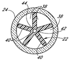
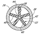
図2及び図3は、サポート22の好ましい一構成を示している。サポート22の表面は、図に示されているように、複数のリブ40などの割り込みを含む。割り込みは、膜24、26がサポート22に巻き付けられたときに、これらの膜によって、流体36を中を流すことができる複数の軸方向内腔38の壁の一部分が形成されるように構成される。好ましい一構成では、複数のリブ40は、サポート22の共通の軸方向中心部分42から半径方向に広がる。リブ40は、また、サポート22の長さに沿って長手方向に、好ましくはサポート22の全長に沿って延びる。図2に示されている非注入区間28では、リブ40の外縁に、非多孔質膜24をきつく巻き付けることができる。その結果、軸方向の内腔38が、非多孔質膜24の内側表面とサポート22の外側表面との間に形成される。同様に、図3に示されている注入区間32では、多孔質膜26の内側表面とサポート22の外側表面との間に軸方向の内腔38が形成されるように、リブ40の外縁に、多孔質膜26をきつく巻き付けることができる。
2 and 3 show a preferred configuration of the
カテーテル20の代替の一実施形態では、サポート20の全長にわたって多孔質膜26を巻き付けて、非多孔質膜24に置き換えることができる。この実施形態では、サポート22の全長が、注入区間32に対応している。別の代替の一実施形態にしたがうと、サポート22は、注入区間32内にのみ延びていてよく、流体供給34からサポート22の基端に到る範囲には、管を提供することができる。この実施形態では、好ましい実施形態の、非多孔質膜24と、非注入区間28内に延びているサポート22部分とを、管で置き換えている。言い換えると、管が、非注入区間28を画定している。
In an alternative embodiment of the
好ましい構成では、リブ40の数は、軸方向内腔38の数に等しい。図2及び図3には
、5つのリブ40及び軸方向内腔38が示されているが、カテーテル20内に複数の内腔を用意する、可撓性を維持する、及びもし所望であれば内腔の流体独立性を維持するという目標を十分に考慮して、任意の適切な数のリブ40及び内腔38を提供することができる。ここで、複数の軸方向内腔について記述するために使用されるときの「流体独立性」や「流体分離」などの用語は、単純に、それらの内腔が互いに流体的に通じていないことを意味する。膜24、26は、医療用グレードグルー又はエポキシ樹脂などの任意の適切な接着剤を用いてリブ40の外縁に沿って接着されることが好ましい。これは、解剖学的構造に対するカテーテルの挿入時又は取り出し時に生じることがある膜24、26の滑りを阻止する。より好ましくは、膜は、各リブ40の外縁の全長に沿って接着される。或いは、膜は、異質な物質によってサポートに固定されることなくサポートに巻き付けることができる。膜及びサポートは、当業者に知られているその他の手段によって互いに固定することも可能である。これは、内腔38の流体独立性を維持する。もし所望であれば、サポート22の軸方向中心部分42内に、軸方向誘導線腔44を設けることができる。誘導線腔44は、上述のように、そして当業者に容易に理解されるように、解剖学的構造へのカテーテル20の挿入を補助するために使用できる誘導線46を受け入れるように適応される。
In the preferred configuration, the number of
図4に示されているように、カテーテル20は、サポート22の末端に固定される端部分すなわちキャップ48を含むことが好ましい。端部分48は、サポート22に対して一体的に形成することができる、又はサポート22に対して接着接合することができる。好ましくは、端部分48の基端は、円形であり、示されているように、端部分48の基端の外側表面がサポート22のリブ40の外縁に合わさるような直径を有する。多孔質膜26は、端部分48の基端に巻き付けられる。膜26は、内腔38内の流体36が、膜26の壁を通り抜けることなくカテーテル20から出て行くことがないように、端部分48に接着することができる。端部分48は、流体がカテーテル20の末端を通って軸方向に流れることがないように遮断する。しかしながら、端部分48は、もし所望であれば、カテーテル20の末端から幾らかの流体を軸方向に投与可能にするために、随意として、多孔質材料で形成することができる。端部分48の末端は、カテーテル20をより容易に解剖学的領域に挿入可能にするために、ドーム状であることが可能である。
As shown in FIG. 4, the
サポート22は、可撓性、軽量性、強度、滑らかさ、及び解剖学的組織に対する非反応性すなわち安全性という目標を十分に考慮して、様々な材料で形成することができる。サポート22に適した材料は、ナイロン、ポリアミド、テフロン(登録商標、以下同じ)、及び当業者に知られているその他の材料を含む。多孔質膜26は、スポンジ状若しくは泡状の材料、又は中空ファイバであることが可能である。膜26は、可撓性であること、及び解剖学的組織に対して非反応性であるという目標を十分に考慮して、様々な適切な材料で形成することができる。膜26は、カテーテル20の注入区間32の表面積に沿って流体を実質的に均一に投与可能にする多孔性を有するとともに、膜の壁を通るバクテリアの流れを制限する十分に小さい平均細孔サイズを有することが好ましい。膜26に適した材料には、ポリエチレン、ポリスルホン、ポリエーテルスルホン、ポリプロピレン、ポリ二フッ化ビニリデン、ポリカーボネート、ナイロン、又は高密度のポリエチレンがある。これらの材料は、生体適合性であることが可能である。多孔質膜26は、流体薬物が膜26を通り抜ける際に、不要なバクテリアを除外することができる。最小のバクテリアは、0.23ミクロン未満のいかなる穴も通り抜けできないことが知られている。したがって、バクテリアが多孔質膜26を横断しないように阻止するには、膜26の平均細孔サイズすなわち細孔径が0.23ミクロン未満であればよい。多孔質膜26の平均細孔サイズすなわち細孔径は、約0.1〜1.2ミクロンの範囲内であってよく、より好ましくは約0.3〜1ミクロンの範囲内であってよく、よりいっそう好ましくは約0.8ミクロンであってよい。
The
上記のように、カテーテル20の基端は、流体供給34に接続することができる。カテーテル20は、各軸方向内腔38が流体的に独立しているように構成することができる。言い換えると、内腔38は、互いに流体的に通じていないと考えられる。カテーテル20は、流体36が各内腔38内を流れるように、1つの流体供給34に接続することができる。或いは、カテーテル20は、幾つかの異なる流体が別々に内腔38内を流れることができるように、複数の別々の流体供給に接続することができる。この構成にしたがうと、各内腔38は、解剖学的構造に送達することができる異なる流体の合計数が内腔38の数に等しくなるように、それぞれ別々の流体供給に接続することができる。或いは、流体内腔は、流体的に独立している必要はない。例えば、膜26は、サポート22の全長に沿ってサポート22に固定されなくてもよく、そうして、流体36が内腔38間を移動することを可能にできる。
As described above, the proximal end of the
動作において、カテーテル20は、注入区間32に隣接する解剖学的構造の部位に直接流体を送達する。流体源34からの流体36は、カテーテル20の基端において軸方向内腔38に導入される。流体36は、先ず、非注入区間28を通って流れる。注入区間32に初めて到達すると、流体36は、多孔質膜26に染み込む。より多くの流体36が注入区間32に入るにつれて、流体36は、膜26及び注入区間32の全体が流体で飽和されるまで膜26の壁内を長手方向に拡散する。この時点で、流体36は、膜26を通り抜けはじめ、そうして、カテーテル20から出て解剖学的構造に入る。そのうえ、流体36は、膜26の特性ゆえに、多孔質膜26の全表面積を実質的に均一な速度で通り抜けることができる。したがって、流体は、解剖学的構造の創傷部位の概ね線形の断片全体に、実質的に等しい速度で送達される。更に、この利点は、低圧力及び高圧力のいずれの流体送達においても得られる。
In operation, the
図5及び図6は、本開示の代替の一実施形態にしたがったカテーテル50を示している。この実施形態にしたがうと、カテーテル50は、外側の細長い管52(本明細書ではカテーテル本体とも称される)と、内側の細長い管状多孔質膜54とを含む。管状の膜54は、外側の管52内に同心円状に閉じ込めることができる。より好ましくは、管52は、管52の内のり寸法と膜54の外のり寸法との間に比較的きつい締まり嵌めが実現されるように、管状の膜54をきつく取り巻いて支える。管52には、好ましくはその全周にわたって複数の流体出口56が設けられる。出口穴56を含む管52の部分は、カテーテル50の注入区間を画定する。管状の膜54は、注入区間の長さに沿って設けられるだけでよいが、もっと長いことも可能である。随意として、管52の末端の先端58内に、軸方向の出口穴を設けることができる。また、当業者によって理解されるように、解剖学的構造へのカテーテル50の挿入を補助するために、誘導線及び/又は誘導線内腔を設けることができる。
5 and 6 illustrate a
管52は、解剖学的組織に対する非反応性、可撓性、軽量性、強度、滑らかさ、及び安全性という目標を十分に考慮して、ナイロン、ポリイミド、テフロン、及び当業者に知られているその他の材料などの様々な任意の適切な材料で形成することができる。好ましい一構成では、管52は、それぞれ0.019インチ(約0.483mm)及び0.031インチ(約0.787mm)の内径及び外径を有する20ゲージのカテーテル管であってよい。管52の出口穴56は、直径が約0.015インチ(約0.381mm)で、管52に沿って等間隔の軸方向位置に設けられることが好ましい。穴56は、どの穴も、その前の穴の角度位置から管52の長手方向軸に相対的に約120度だけ角度的にずれるように配置されることが好ましい。隣り合った出口穴56どうしの間の軸方向の分離は、約0.125〜0.25インチ(約3.18〜6.35mm)の範囲内であってよく、より好ましくは約16分の3インチ(約4.76mm)であってよい。また、注入区間は、任意の所望の長さを有することができる。この構成は、創傷部位の概ね線形の断片全体に、徹底的に均一に流体を送達する結果となる。出口穴56は、もちろん、様々な任意の代替の
配置で設けることができる。
管状多孔質膜54は、スポンジ状若しくは泡状の材料、又は中空ファイバであることが可能である。管状の膜54は、バクテリアを除外するために、0.23ミクロン未満の平均細孔サイズすなわち細孔径を有してよい。細孔径は、約0.1〜1.2ミクロンの範囲内であってよく、より好ましくは約0.3〜1ミクロンの範囲内であってよく、よりいっそう好ましくは約0.8ミクロンであってよい。管状の膜54は、解剖学的組織に対する非反応性、可撓性の維持、管52のサイズ制約内での嵌め込み、及び結果として管52の全ての出口穴56を通して流体を実質的に均一に投与させる多孔性という目標を十分に考慮して、様々な任意の適切な材料で形成することができる。膜54に適した材料には、ポリエチレン、ポリスルホン、ポリエーテルスルホン、ポリプロピレン、ポリ二フッ化ビニリデン、ポリカーボネート、ナイロン、又は高密度のポリエチレンがある。管状の膜54の好ましい内径及び外径は、それぞれ0.010インチ(約0.254mm)及び0.018インチ(約0.457mm)である。誘導線46が提供される場合は、誘導線は、直径が約0.005インチ(約0.127mm)のステンレス鋼線材であることが可能である。管52は、エポキシ樹脂又は当業者に知られているその他の手段によって、膜54に固定することができる。或いは、膜54は、膜54を管52内に固定するためのその他の材料を使用せずに、締まり嵌めによって管52に接触させることができる。
The tubular
動作において、カテーテル50は、カテーテル50の注入区間に隣接する解剖学的組織の領域に流体を送達する。流体は、注入区間に流れ込むにつれて、先ず、管状の多孔質膜54に染み込む。より多くの流体が注入区間に入るにつれて、流体は、管状の膜54の壁内を長手方向に拡散する。膜54及びその中の管状の空間が飽和されると、流体は、膜54を通り抜け、管52の出口穴56を通って流れることによってカテーテル50から出て行く。そのうえ、流体は、膜54の表面積全体にわたって実質的に均一に膜を通り抜けることができ、その結果、実質的に全ての出口穴56を、実質的に均一な流れが通ることになる。したがって、流体は、解剖学的構造の創傷部位全体に、実質的に等しい速度で送達される。更に、この利点は、低圧力及び高圧力のいずれの流体送達においても得られる。
In operation, the
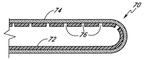
図7は、本開示の別の一実施形態にしたがったカテーテル70を示している。カテーテル70は、管の側壁に複数の出口穴76を有する管72と、該管72を同心円状に閉じ込める管状の多孔質膜74とを含む。カテーテル70は、図5及び図6にあわせて上述されたカテーテル50と同様に動作する。使用において、流体薬物は、出口穴76を通り抜け、次いで、多孔質膜74に染み込みはじめる。流体は、膜が飽和されるまで膜の壁内を長手方向に拡散する。その後、流体は、膜の壁を後にして解剖学的構造に入る。流体は、膜74の表面積全体にわたって実質的に均一な速度で解剖学的構造に送達されえる。先の実施形態と同様に、この利点は、低圧力及び高圧力のいずれの流体送達においても得られる。
FIG. 7 illustrates a
図8は、本開示の別の一実施形態にしたがったカテーテル60を示している。カテーテル60は、解剖学的組織内の領域に比較的高流速で流体を送達するのに、より適している。カテーテル60は、サイズを増していく複数の出口穴64を有する管62を含む。具体的には、より末端寄りの出口穴ほど、より基端寄りの出口穴よりも大きい直径を有する。管62上における出口穴64の位置は、カテーテル60の注入区間の長さを画定する。注入区間は、任意の所望の長さを有することができる。カテーテル60の基端は、流体供給に接続されており、解剖学的構造へのカテーテル60の挿入を補助するために、誘導線及び/又は誘導線内腔を設けられてもよい。
FIG. 8 illustrates a
上述のように、高圧力又は低圧力の流体送達において、カテーテル管の末端に近い出口穴は、管の基端に近い出口穴と比べると、全体的に流れ抵抗が増大している。また、より
末端寄りの穴を通って流れる流体ほど大きい圧力降下を受ける。その結果、全体的に、より基端寄りの穴を通る流体ほど流速が大きくなり、不均一な流体送達がなされる結果になる。これに対して、カテーテル60は、比較的高流速の条件下において、実質的に全ての出口穴64を通して実質的に均一な流体送達を提供することができる。これは、より末端寄りの穴ほどサイズを大きくすることによって、流れ抵抗及び圧力降下の増大が打ち消されるからである。言い換えると、より末端寄りの穴ほどより基端寄りの穴よりも大きいので、より末端寄りの穴を通る流体の流速は、それらの穴がより基端寄りの穴と同サイズである場合よりも大きくなると考えられる。穴64は、徐々にサイズを大きくして形成することができ、これは、結果として、実質的に均一な流体送達をもたらすことができる。また、出口穴は、図12の実施形態に関連して後述されるように、組み合わさることによって流量制限オリフィスを形成するサイズにすることができる。
As mentioned above, in high pressure or low pressure fluid delivery, the exit hole near the distal end of the catheter tube has an overall increased flow resistance compared to the exit hole near the proximal end of the tube. In addition, the fluid flowing through the hole closer to the end receives a larger pressure drop. As a result, overall, the fluid passing through the more proximal hole has a higher flow rate, resulting in uneven fluid delivery. In contrast, the
先行技術によるカテーテルと比べて、カテーテル60は、単純で尚且つ製造が容易であることが可能である。必要なのは、管62に複数の出口穴64を開けることだけである。更に、カテーテル60は、操作性を維持しつつ、先行技術のカテーテルよりも大きな湾曲に耐えることができる。Wangカテーテルなどの先行技術のカテーテルと異なり、管62は、もしいくぶん湾曲したとしても、依然として比較的均一に流体を送達することができる。これは、管62が、比較的面積の大きい単一の内腔を有するからである。管62がいくぶん湾曲したときに、内腔内を流れる流体は、不均一な流体送達をもたらす可能性がある閉塞及びその結果としての圧力変化をあまり受けない。
Compared to prior art catheters, the
カテーテル60の管62は、解剖学的組織に対する非反応性、可撓性、軽量性、強度、滑らかさ、及び安全性という目標を十分に考慮して、種々様々な任意の材料で形成することができる。適した材料は、ナイロン、ポリアミド、テフロン、及び当業者に知られているその他の材料を含む。注入区間は、任意の所望の長さを有することができるが、約0.5〜20インチ(約1.27〜50.8cm)の長さであってよく、より好ましくは約10インチ(約25.4cm)の長さであってよい。出口穴64の直径は、注入区間の基端における約0.0002インチ(約0.00508mm)から注入区間の末端における約0.01インチ(約0.254mm)に及ぶことが好ましい。最大の、すなわち最も末端寄りの出口穴64は、管62の末端から約0.25インチ(約6.35mm)であってよい。好ましい構成では、隣り合った穴64どうしの軸方向の分離は、約0.125〜0.25インチ(約3.18〜6.35mm)の範囲内であってよく、より好ましくは約16分の3インチ(約4.76mm)であってよい。随意として、穴64は、図5の実施形態のように、隣り合った穴どうしが約120度だけ角度的にずれるように設けることができる。もちろん、出口穴64を数多く設けすぎるのは、管62が弱くなる可能性があり望ましくない。
The
図9、図10A、及び図10Bは、本開示の別の一実施形態にしたがったカテーテル80を示している。カテーテル80は、管82と、「滲出式」管状コイルバネ84と、ストップ86とを含む。管及びバネがそれぞれ中心内腔の一部分を画定するように、バネ84の基端は、管82の末端に取り付けられる。好ましくはドーム状のストップ86が、バネ84の末端に取り付けられ、バネ84の末端を閉じる。管82よりも末端寄りのバネ84の部分が、カテーテル80の注入区間である。図10Aに示されている非延伸状態において、バネ84は、バネ内にある投与限界圧力を下回る流体が巻きの間を半径方向に流れることによって内腔から出ることのないように、隣り合った巻きどうしが接触している。バネ84は、図10Bに示されているように、流体圧力がバネの投与限界圧力以上になると長手方向に延伸する特性を有しており、これは、流体が「滲出」する、すなわち巻きの間を半径方向に外向きに漏出することによって内腔から投与されることを可能にする。或いは、バネは、流体がバネの巻きの間を通って滲出することを可能にするために、長くなるのではなく半径方向に延伸することができる。更に、バネは、当業者に理解されるように
、滲出を可能にするために、長手方向及び半径方向の両方に延伸することができる。バネの巻きの間の流体は、管82よりも末端寄りのバネの部分すなわち注入区間の長さ及び周の全体にわたって実質的に均一に投与することができる。カテーテル80は、高流速及び低流速の両方の流体送達に使用することができる。
9, 10A, and 10B illustrate a
使用において、カテーテル80は、流体薬物の送達を所望される領域にバネ84がくるように、解剖学的領域に挿入される。バネは、最初、図10Aに示されているように、非延伸状態にある。流体は、カテーテル80の管82の基端に導入され、バネ84に流れ込み、ストップ86に達するまでバネ84を通って流れる。流体は、管82の基端に導入され続ける間に、バネ84の内側に蓄積する。バネ84が流体で満たされると、流体圧力が更に急速に上昇する。流体は、バネの巻きに対して半径方向外向きに作用する力を付与する。圧力が蓄積するにつれて、外向きの力は大きくなる。流体圧力が投与限界圧力まで上昇すると、外向きの力は、図10Bに示されているように、バネが長手方向に延伸するようにバネの巻きどうしを僅かに分離させる。或いは、巻きは、上述のように半径方向に分離することができる。流体は、すると、分離された巻きの間を通って流れ、カテーテル80から投与される。そのうえ、投与は、カテーテル80の注入区間全体にわたって均一であることが可能である。流体が管82に導入され続ける間、バネ84は、流体を解剖学的構造内の所望の領域に投与し続けるために延伸状態にとどまる。流体の導入が一時的に停止すると、バネ84内の流体圧力は、投与限界圧力未満に降下するであろう。もしそうであれば、バネは圧縮するので、巻きどうしは再び隣接しあい、流体は投与されなくなる。
In use, the
幾つかのバネタイプが、この発明の目的を達成する。適切なバネタイプは、容易に購入することができる316L又は402Lである。好ましい一構成では、バネ84は、その長さに沿って、1インチ(約2.54cm)あたり約200の巻きを有する。この構成では、バネは、流体を漏出させることなく高度な湾曲に耐えることができ、重度の湾曲のみが、隣り合った巻きどうしを分離させる。したがって、バネ84は、流体を漏出させて解剖学的構造内の一領域のみに投与することなく解剖学的領域内において大きく撓むことができる。バネ84は、カテーテル80の注入区間の長さを画定するために、任意の所望の長さを有することができる。バネは、強度、可撓性、及び安全性という目標を十分に考慮して、様々な材料で形成することができる。好ましい材料は、ステンレス鋼である。好ましい構成では、バネの内径及び外径は、それぞれ約0.02インチ(約0.508mm)及び約0.03インチ(約0.762mm)であり、バネ線は、約0.005インチ(約0.127mm)の直径を有する。バネ84の基端は、管82の末端内に同心円状に閉じ込めることができる。バネは、例えばUV接着剤、埋め込み用材料、又はその他の結合剤を使用して管82の内壁に接着することができる。或いは、バネは、管82内にはんだ付けする、又は基端側にプラグを使用して管82にきつく嵌め込むことができる。
Several spring types accomplish the purpose of this invention. A suitable spring type is 316L or 402L, which can be easily purchased. In one preferred configuration, the
管82及びストップ86は、可撓性、軽量性、強度、滑らかさ、及び安全性という目標を十分に考慮して、様々な任意の材料で形成することができる。適切な材料は、ナイロン、ポリイミド、テフロン、及び当業者に知られているその他の材料を含む。
The
図11は、本開示の別の一実施形態にしたがったカテーテル90を示している。カテーテル90は、末端を閉じられた管92と、管及びバネの中に内腔が画定されるように管92内に同心円状に閉じ込められる「滲出式」管状コイルバネ94とを含む。管92の長さに沿って、その側壁に、複数の出口穴96が設けられる。このような出口穴96を含む管92の長さは、カテーテル90の注入区間を画定する。出口穴96は、注入区間の壁全体にわたって設けられることが好ましい。注入区間は、任意の所望の長さを有することができる。好ましい構成では、隣り合った出口穴96どうしの間の軸方向の間隔は、約0.125〜0.25インチ(約3.18〜6.35mm)の範囲内であってよく、より好ましくは約16分の3インチ(約4.76mm)であってよい。隣り合った穴96どうしは、
約120度角度的に隔てられていることが好ましい。バネ94は、カテーテルの注入区間内に閉じ込めることができ、図9、図10A、及び図10Bの実施形態のバネ84と同様に構成することができる。バネ94は、注入部分より長いことが可能であり、全ての出口穴96がバネ94に隣接するように位置決めすることができる。この構成では、流体は、バネの巻きの間を流れることなく内腔から出て行くことのないように阻止される。管の末端を閉じるために、管に、ストップを取り付けることができる。或いは、管92は、閉じられた末端を伴うように形成することができる。カテーテル90は、高流速又は低流速の流体送達に使用することができる。
FIG. 11 illustrates a catheter 90 according to another embodiment of the present disclosure. Catheter 90 includes a
Preferably, they are angularly separated by about 120 degrees. The
使用において、カテーテル90は、流体薬物の送達を所望される領域に注入区間がくるように、解剖学的領域に挿入される。流体は、カテーテル90の管92の基端に導入され、管92の閉じられた末端に到達するまでバネ94を通って流れる。流体は、管92の基端に導入され続ける間に、バネ94の内側に蓄積する。最終的に、バネ94が流体で満たされると、流体圧力が上昇し、図9、図10A、及び図10Bの実施形態に関連して上述されたように、バネの巻きを通って流体が滲出する。そのうえ、流体は、バネ94の長さ及び周の全体にわたって実質的に均一にバネの巻きを通って流れる。流体は、次いで、注入区間の出口穴96を通って流れることによって、管92から出て行く。出口穴は、流体が実質的に等しい速度で出口穴を通って流れるように、等サイズであることが可能であり、その結果、解剖学的構造の所望の領域全体に、総じて均一に流体を投与することが可能になる。流体がカテーテル90に導入され続ける間、バネ94は、カテーテルから流体を投与し続けるために延伸状態にとどまる。流体の導入が一時的に停止すると、バネ94内の流体圧力は、投与限界圧力未満に降下するであろう。もしそうであれば、バネは圧縮するので、巻きどうしは再び隣接しあい、流体は投与されなくなる。
In use, the catheter 90 is inserted into the anatomical region so that the infusion section is at the region where fluid drug delivery is desired. Fluid is introduced into the proximal end of the
好ましい構成では、バネ94及び管92は、バネの全長に沿って接触しているので、バネを通って滲出する流体は、注入区間の穴96を通って流れるように強いられる。好ましくは、バネ94の一端を、管92の内壁に取り付けることによって、バネのもう一端を、バネの延伸とともに移動可能にする。バネは、例えばUV接着剤、埋め込み用材料、又はその他の接合材料によって、管92に接着することができる。或いは、バネの端は、管92の内壁にはんだ付けすることができる。管92は、任意の適切な材料で形成することができる。管92の内壁は、バネがより自由に延伸及び圧縮することができるように、滑らかであることが好ましい。
In a preferred configuration, the
図12は、本開示の別の一実施形態にしたがったカテーテル100を示している。このカテーテル100は、複数の出口穴104を内壁に有する末端を閉じられた管102を含む。出口穴104を有する管102部分は、カテーテル100の注入区間を画定する。出口穴104は、開口の合計面積がカテーテルの任意のその他の流量制限断面すなわちオリフィスの面積よりも小さいようなサイズにされる。したがって、出口穴104は、カテーテル100の流量制限器である。使用において、カテーテルは、実質的に全ての出口穴104を通して流体を投与することができる。管102の基端に導入された流体は、管の閉じられた末端に到達するまで管を通って流れる。この時点で、流体は、カテーテルの注入区間内に蓄積する。穴のサイズが小さいゆえに、流体は、穴104を通って流れることのないように実質的に阻止される。最終的には、カテーテルの注入区間は、流体で満たされる。流体が管102の基端に導入され続ける間に、流体圧力が蓄積しはじめる。とある時点で、圧力は、出口穴104に流体を通り抜けさせるのに十分な高さになる。そのうえ、流体は、実質的に全ての出口穴104を通って流れる。
FIG. 12 illustrates a
この好ましい構成では、出口穴104は、実質的に全ての穴を通して実質的に等しい速度で流体が投与されるように、全て等サイズである。穴104は、非常に小さい穴径を実現するために、レーザで開けられることが好ましい。出口穴104の好ましい直径は、約
0.0002インチ(約0.00508mm)、すなわち約5ミクロンである。管102内には、数々の出口穴104を設けることができる。穴は、解剖学的領域の全体に、より均一に流体を送達するために、カテーテル100の注入区間の全周にわたって形成することができる。隣り合った出口穴104どうしの間の軸方向の分離は、約0.125〜0.25インチ(約3.18〜6.35mm)の範囲内であってよく、より好ましくは約16分の3インチ(約4.76mm)であってよい。カテーテル100は、高流速又は低流速の流体送達に使用することができる。管102は、当業者に知られている前述の様々な任意の材料で形成することができる。
In this preferred configuration, the outlet holes 104 are all equally sized so that fluid is dispensed at substantially equal rates through substantially all holes. The
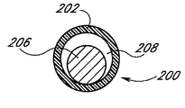
図13は、本開示の別の一実施形態にしたがったカテーテル200を示している。カテーテル200は、上述された実施形態のように、カテーテルの注入区間に沿って複数の出口穴204を有する末端を閉じられた管202を含む。穴204は、管202の全周にわたって設けられることが望ましい。管202内には、多孔質材料で形成された細長い部材206が閉じ込められる。好ましくは、部材206は、概ね円筒形状であり、尚且つ固形である。好ましくは、部材206は、部材206の外側表面と管202の内側表面との間に環状の空間208が形成されるように、管204内に位置決めされる。好ましくは、部材206は、管202の末端210からカテーテルの注入区間よりも基端寄りの地点へと後方に延びる。或いは、部材206は、注入区間の一部分に沿ってのみ延びてよい。一部の実施形態では、部材206は、管202に対して概ね同心円状であることが可能である。しかしながら、実施形態によっては、非同心円状の設計によって本発明の利点が実現される。好ましくは、部材206は、患者の体内へのカテーテル200の設置を補助するために、可撓性の材料で製造される。
FIG. 13 illustrates a
動作において、管202内を流れる流体薬物は、多孔質部材206を飽和状態にし、環状の領域208に流れ込む。部材206が飽和状態になると、部材206内の流体は、領域208に流れ込み、出口穴204を通ってカテーテル200から出て行く。流体圧力は、環状の領域208全体にわたって均一であることが可能なので、流体は、全ての穴204を実質的に均一に通って流れることができる。環状の領域208には、幾つかの利点がある。1つの利点は、出口穴204を通る流れの均一性を最適化する傾向があることである。また、部材206は、液体で飽和されると膨張する傾向がある多孔質材料で形成することができる。もしそうであれば、部材206は、管202を圧迫することなく環状の領域208内に膨張することが好ましい。これは、創傷箇所内に薬物を不均等に流出させる可能性がある管202の内部表面における高圧領域の形成を抑制する。或いは、部材206は、膨張して管202に接触してもなお本開示の目標を達成するものであってよい。
In operation, fluid drug flowing in the
部材206は、好ましくは0.1〜50ミクロンの範囲内の、より好ましくは約0.45ミクロンの、平均細孔サイズを有する多孔質材料で形成される。環状の領域208の半径方向の幅Wは、0〜約0.005ミクロンの範囲内であってよく、より好ましくは約0.003ミクロンであってよい。部材206は、多孔性、可撓性、強度、及び耐久性という目標を十分に考慮して、様々な任意の材料で形成することができる。好ましい材料は、メンテック(Mentek)である。
部材206は、接着剤の使用によって、管202内に固定することができる。図13に示されているような一実施形態では、管202の末端の内部表面との間に接合を形成するために、部材206の末端に接着剤が塗布される。好ましくは、接着剤は、カテーテル200の注入区間の基端又はその近くに塗布される。また、接着剤は、部材206の任意の長手方向位置においてその周囲に塗布されて、管202の内部表面との間にリング状の接合を形成することができる。例えば、図13の実施形態では、カテーテル200の注入区間のすぐ基端寄りにリング状の接合214が提供される。その他の構成も可能である。例えば、図14は、接合216を形成するために部材206の末端に、そしてリング状の接
合218を形成するために注入区間の概ね中心に接着剤を塗布される一実施形態を示している。図15は、接合220を形成するために部材206の末端のみに接着剤を塗布される一実施形態を示している。図16は、リング状の接合222を形成するために注入区間の中心のみに接着剤を塗布される一実施形態を示している。当業者ならば、本明細書の教示内容から、接着剤が様々な任意の構成で塗布可能であることを理解することができる。したがって、例えば、カテーテルの末端における接着剤(すなわち図13、図14、及び15における212、216、及び220)は、不要である。
現時点における本発明の最良の形態では、好ましくは2つの接合が、1つはカテーテルの最も基端寄りの穴に、もう1つはカテーテルの最も末端寄りの穴に盛り込まれる。各接合は、後述のように、接着剤によって形成される。 In the best mode of the invention at the present time, preferably two junctions are placed in one of the most proximal holes in the catheter and the other in the most distal hole of the catheter. Each joint is formed by an adhesive as will be described later.
リング状の接合214は、部材206が管202内にあるときに出口穴204の1つに液体形態の接着剤を流し込むことによって形成することができる。総じて高い粘度を有する接着剤は、部材206の本体に流れ込むのではなく、部材206の周囲を流れる傾向がある。当業者に理解されるように、接着剤は、こうして管202との間にリング状の接合を形成する。また、接着剤は、流し込まれた出口穴204を塞ぐ。様々に異なるタイプの任意の接着剤が許容可能であり、好ましい一接着剤はロックタイト(Loctite)である。
The ring-shaped joint 214 can be formed by pouring a liquid form of adhesive into one of the outlet holes 204 when the
上述のように、部材206は、管202に対して同心円状であることが可能である。図17は、部材206を管202内に同心円状に閉じ込められたカテーテル200の断面を示している。或いは、部材206は、図18に示されているように、管202に隣接して位置決めすることができる。図18の構成は、部材206を管202内において中心合わせする必要がないので、図17の構成よりも製造が容易であることが可能である。
As described above, the
当業者ならば、本明細書の教示内容から、部材206が任意の所望の長さであってよいこと、そしてカテーテル200の注入区間の任意の所望の長さに沿って延びてよいことを理解できる。例えば、部材206は、管202の末端まで延びでいる必要はない。更に、部材206の基端は、注入区間の基端よりも末端寄り又は基端寄りであることが可能である。
Those skilled in the art will understand from the teachings herein that the
上述の実施形態の任意のカテーテルが使用されるときに、カテーテルは、最初、カテーテル管内に空気を有している可能性がある。例えば、図13に示されているカテーテル200は、多孔質材料の部材206の内側に空気を有している可能性がある。カテーテルへの液体薬物の導入は、出口穴から空気を流出させる。しかしながら、これには、数時間かかると考えられる。もし空気が内側にある状態で、カテーテルが患者に挿入され、液体薬物がカテーテルに導入されると、患者の創傷箇所は、空気がカテーテル管から追い出されるまでほとんど又は全く薬物を受け取ることができない可能性がある。したがって、使用前にカテーテルから空気を確実に追い出すためには、患者にカテーテルを挿入する前に、カテーテルに液体薬物を通すことが好ましい。更に、図19を参照すると、当該分野で知られているような空気フィルタ224を、カテーテル200の注入区間226よりも基端寄りでカテーテルに挿入することができる。フィルタ224は、望ましくない空気がカテーテル200の注入区間226に入らないように阻止する。
When any catheter of the above-described embodiment is used, the catheter may initially have air in the catheter tube. For example, the
図20及び図21は、細長い出口穴すなわちスロットを有するカテーテル管を示している。これらのカテーテル管は、図示され上述されているカテーテル管の代わりに使用することができる。図20は、管230の長手方向に細長く延びている出口穴すなわちスロット232を有する。スロット232は、カテーテルの注入区間に沿って、管230の全周にわたって設けられることが好ましい。より小さい出口穴と比較すると、細長いスロット232は、流体が受ける流れインピーダンスを低減させることによって、カテーテルから
出る流体の流速を増大させる傾向がある。好ましくは、スロット232は、当業者に容易に理解されるように、カテーテル200の構造的完全性を損なうことのないようにカテーテル本体に長手方向の向きに設けることができる。
20 and 21 show a catheter tube having an elongated exit hole or slot. These catheter tubes can be used in place of the catheter tubes shown and described above. FIG. 20 has an exit hole or slot 232 that is elongated in the longitudinal direction of the
図21は、管の長さに沿って末端方向に長さを増大させる出口穴すなわちスロット236を有する管234を示している。図示されている実施形態では、管234の注入区間の基端に近いスロットは、注入区間の末端に近いスロットよりも短い。図8の実施形態のように、カテーテル管234は、比較的高流速の条件下において、実質的に全ての出口スロット236を通して実質的に均一な流体送達を提供することができる。これは、より末端寄りのスロットほどサイズを大きくすることによって、流れ抵抗及び圧力降下の増大が打ち消されるからである。言い換えると、より末端寄りのスロットほどより基端寄りのスロットよりも大きいので、より末端寄りのスロットを通る流体の流速は、それらのスロットがより基端寄りのスロットと同サイズである場合よりも大きくなると考えられる。スロット236は、徐々にサイズを大きくして形成することができ、これは、結果として、実質的に均一な流体送達をもたらす。更に、細長いスロットは、図20の実施形態のように、総じて出口流速をより高くする結果になる。
FIG. 21 shows a
管の末端を閉じるために、管にストップを取り付けることができる。或いは、管302は、閉じられた末端を伴うように形成することができる。カテーテル300は、高流速又は低流速の流体送達に使用することができる。
A stop can be attached to the tube to close the end of the tube. Alternatively, the
図22〜28は、カテーテル本体によって支えられ、標的神経を突き止めて注入カテーテルの位置決めを促進するために周辺の神経刺激装置(不図示)と連絡しあうことができる導電性素子(導電性の線又は帯)を有する本開示のカテーテルの幾つかの実施形態を示している。一部の実施形態では、導電性素子は、カテーテル本体に対して一体的に形成することができる。図22〜26に示されている実施形態に使用される神経刺激装置は、あたかも本明細書に全文を記載されているかのように参照によって本明細書に組み込まれる米国特許第5,830,151号に記載されている装置のように、当該分野で通常使用されている種類のものであることが可能である。更に、図22〜26に示されている任意の実施形態の導電性素子は、上述された任意のカテーテル実施形態とともに使用することができる。 FIGS. 22-28 show conductive elements (conductive lines) supported by the catheter body and capable of communicating with a peripheral nerve stimulator (not shown) to locate the target nerve and facilitate positioning of the infusion catheter. Or embodiment of the catheter of the present disclosure having a band. In some embodiments, the conductive element can be integrally formed with the catheter body. The neurostimulator used in the embodiment shown in FIGS. 22-26 is described in US Pat. No. 5,830,151, which is incorporated herein by reference as if set forth in full herein. Can be of the type normally used in the art, such as the device described in the issue. Furthermore, the conductive elements of any embodiment shown in FIGS. 22-26 can be used with any of the catheter embodiments described above.
図22及び図23は、管302と、管及びバネの中に内腔が画定されるように管302内に同心円状に閉じ込められる「滲出式」管状コイルバネ304などのコイル部材と、を含むカテーテル300を示している。カテーテル300は、上記の図11に示されているカテーテル90と同様に構成することができる。このように、管302の長さに沿って、その側壁に、複数の出口穴306が設けられる。このような出口穴306を含む管302の長さは、カテーテル300の注入区間を画定する。出口穴306は、注入区間の壁全体にわたって設けられることが好ましい。注入区間は、任意の所望の長さを有することができる。バネ304は、カテーテル300の注入区間内に閉じ込めることができ、図9、図10A、及び図10Bの実施形態のバネ84と同様に構成される。(弛緩状態にある)バネ304は、注入部分より長いことが可能であり、全ての出口穴306がバネ304に隣接するように位置決めすることができる。この構成では、流体は、バネの巻きの間を流れることなく内腔から出て行くことのないように実質的に阻止することができる。したがって、図22〜23に示されている構成の管302及びバネ304は、図11に示されているカテーテル90の管98及びバネ94と同様に構成される。しかしながら、管302及びカテーテル300は、流体を所望の部位に均一に拡散させるために、上述の任意の形態又はその他の形態で構成することができる。流体は、麻酔薬などの薬物であることが可能である。
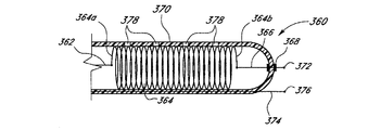
FIGS. 22 and 23 include a
図22及び図23に示されている構成では、カテーテル300は、導電性バネ304の基端304aに取り付け可能な導電性リード線308を有する。リード線308は、上述されたタイプのカテーテル内腔を通り、ワニ口クリップ又は同様に構成されたその他の接続装置の使用によって、好ましくは周辺に位置している神経刺激装置に対して取り外し式に取り付けられる。バネ304の末端304bには、コンタクト線310を取り付け可能であり、該コンタクト線310は、管302の末端の中心部分に位置している封止された又は実質的に封止された開口312に通される。考えられるその他の封止メカニズムのなかでも、個別のシール部材を用いること、或いは開口312を、いかなる追加の封止素子を伴わずともコンタクト線310を緊密に囲うようなサイズにすることが可能である。なお、封止を必要とするであろう開口を管302の端に存在させないように、コンタクト線310を管302に対して一体的に形成する、ひいては埋め込むことができることに留意せよ。一部の実施形態では、バネ304は、開口312に対するコンタクト線310の位置を安定化させるのに役立つことができる。バネ304は、導電性であることが可能であるので、刺激神経装置によってリード線308を通じてバネの基端304aに供給された電気的パルスは、コンタクト線310に伝送することが可能である。
In the configuration shown in FIGS. 22 and 23, the
バネ304、リード線308、及びコンタクト線310は、1本の導電性の線で一体的に形成することができる。管302は、リード線308、バネ304、及びコンタクト線310を通じて伝送される電気的パルスが、コンタクト線310を介する場合を除いて管302に隣接するいかなる体の組織にも伝わらないように、電気的に絶縁された材料で作成することができる。コンタクト線310は、コンタクト線310に隣接して位置する体の組織内への電気的パルスの漏洩を阻止するために、コンタクト線310の先端314を除いてコンタクト線310の長さに沿った全ての地点を絶縁性材料でコーティングすることができる。神経刺激装置用のアースを体内に提供するために、カテーテル管の末端から、導電性のアース線を突き出させてもよい。図22及び図23に示されている構成では、カテーテル300は、アース線を突き出させていない。この構成では、標的神経の場所に近い外部の患者の体上に、アースパッチ(不図示)などの接地装置を位置決めすることが好ましいであろう。アースパッチは、アース線によって、外部の神経刺激装置に接続される。アース線は、神経刺激装置に対して取り外し式に取り付けられることも好ましいであろう。所望の位置に管が位置決めされた後は、神経刺激装置をリード線308から切り離すことができる。
The
このような構成では、電気的刺激を、周辺の神経刺激装置から、カテーテル管302内に位置決め可能なコンタクト線310の先端314に伝送することができる。周辺の神経刺激装置は、リード線308、バネ304、及びコンタクト線310を通じて間欠電気的刺激を導電性の先端314に伝送することが好ましい。電気的パルスは、先端314を通じて、該先端が接触している又はごく接近している体の組織又は神経に伝送することができる。上述されたアースパッチ又はアース線は、神経刺激装置のための電気回路を完成する。体のそれぞれの筋肉に関連付けられた神経を、神経刺激装置を通じて電気的に刺激すると、神経に関連付けられた筋肉は、先端314が神経に接触して又は神経に接近して位置決めされているときに神経刺激装置によって供給される電気的パルスに応答して収縮する。筋肉の痙攣又は収縮の大きさは、電気的パルスの電圧と、神経に対する先端314の接近の程度とに依存する。したがって、もし一定の電圧が印加される場合は、本明細書に説明されている装置のユーザは、電気的パルスの結果生じる筋肉収縮の大きさによって、標的神経までの先端314の距離を検出することができる。カテーテル300が所望の位置に位置決めされた後は、先端314が封止開口312に隣接する又は封止開口312の内側にくるように、また、全ての出口穴306がバネ304に隣接して位置決めされた状態にとどまるように、リード線308を内腔内において十分な遠さまで引き戻すことによって、先端314及びコンタクト線310を引き込むことができる。
In such a configuration, electrical stimulation can be transmitted from the peripheral nerve stimulation device to the tip 314 of the
代替の一実施形態のカテーテル320が、図24に示されている。この構成では、リード線322は、バネ324の基端324aに取り付け可能である。コンタクト線326もまた、バネ324の基端324aに取り付け可能であり、管330の末端にある封止開口326に通される。この構成では、カテーテル320、コンタクト線326、及び先端322は、コンタクト線326がバネ324の末端346bに取り付けられないゆえに、カテーテル320が体に埋め込まれた後に先端328が体の組織に接触する結果として生じえるバネ324の末端寄り部分の巻きの動き及びひいてはそれらの巻きの間を通して生じる流体薬物の滲出が、コンタクト線326によってバネ324に及ぼされるいかなる軸方向力にも影響されない、という点を除き、上述されたカテーテル200の関連の構成要素と同様に動作する。代替の別の一実施形態では、コンタクト線326は、バネ324の中間部分に取り付け可能である。
An
カテーテルの別の一実施形態が、図25に示されている。この構成では、カテーテル340は、管346の壁内の開口344を通って管346の末端にある開口348から管346を出ることができるコンタクト線342を含むように、構成することができる。開口348は、封止されていても、封止されていなくてもよい。また、カテーテル340は、コンタクト線342を通すための開口を有さなくてもよく、その代わり、カテーテル340の壁に開口がないように、コンタクト線342をカテーテル340の壁内に一体的に形成してよい、すなわちコンタクト線342をカテーテル340の壁内に埋め込むことができる。例えば、カテーテル340及びコンタクト線342は、同時押し出しプロセスによって一体にされてよい。管346は、上述された管の実施形態を非限定的に含む任意の適切な構成であることが可能である。一部の実施形態では、コンタクト線342は、先端350にある又は先端350の近くにあるその末端において終結することができる。コンタクト線342は、好ましくは周辺に位置している神経刺激装置と直接連絡しあうことができ、電気的に絶縁された材料で作成することができる管346の壁によって、バネ352及び体の組織から絶縁することができる。一部の実施形態では、コンタクト線342の末端部分のみが露出されている。この構成では、カテーテル340、コンタクト線342、及び先端350は、上述されたカテーテル300と同様に動作する。更に、管が体内に埋め込まれた後にコンタクト線342による不慮の接触によって神経叢が損傷されることのないように、コンタクト線342は、カテーテル340が神経叢に隣接して最適に位置決めされた後に、コンタクト線342を引き込んで、先端350を封止開口348に隣接して又はその内側に位置決めすることができるように、開口344内において、好ましくは制御可能に軸方向に滑ることができる。
Another embodiment of the catheter is shown in FIG. In this configuration, the
カテーテルの別の一実施形態が、図26に示されている。この構成では、カテーテル360は、導電性コイル部材すなわちバネ364の基端364aに一体的に又はその他の形で接続可能な導電性リード線362を有することができる。リード線362は、上述されたタイプのカテーテル内腔を通り、上述のように、ワニ口クリップ又は同様に構成されたその他の接続装置の使用によって、好ましくは周辺に位置している神経刺激装置すなわち神経刺激生成器に対して取り外し式に取り付けられる。バネ364の末端364bには、コンタクト線366を取り付け可能であり、該コンタクト線366は、管370の末端の中心部分に位置している封止開口368に通される。バネ364は、開口368に対するコンタクト線366の位置を安定化させるのに役立つ。バネ364は、導電性であることが可能であるので、刺激神経装置によってリード線362を通じてバネの基端364aに供給された電気的パルスは、コンタクト線366に伝送することができる。バネ364、リード線362、及びコンタクト線366は、1本の導電性の線一体的に形成することができる。管370は、リード線362、バネ364、及びコンタクト線366を通じて伝送される電気的パルスが、コンタクト線366を介する場合を除いて管370に隣接するいかなる体の組織にも伝わらないように、電気的に絶縁された材料で作成することができる。コンタクト線366は、コンタクト線366に隣接して位置する体の組織内への電気
的パルスの漏洩を阻止するために、コンタクト線366の先端372を除いてコンタクト線366の長さに沿った全ての地点を絶縁性材料でコーティングすることができる。
Another embodiment of the catheter is shown in FIG. In this configuration, the
神経刺激装置用のアースを体内に提供するために、カテーテル管370の末端から、カテーテル360の壁に埋め込み可能な導電性のアース線374を突き出させることもできる。アース線374は、先端376を有してよく、外部の神経刺激装置に接続することができる。アース線374は、神経刺激装置に対して取り外し式に取り付けることができる。所望の位置に管が位置決めされた後は、神経刺激装置をリード線362及びアース線374から切り離すことができる。便宜上、リード線362及びアース線374として説明されてはいるものの、線362、374は、相まって電気回路の一部分を形成することができ、線362、374のいずれかは、神経刺激装置の正端子又は負端子のいずれかに接続することができる。また、図示されている線374と同様に、線362、374の両方がカテーテル管270内にあってもよい。
A
このような構成では、電気的刺激を、周辺の神経刺激装置から、カテーテル管370内に位置決め可能なコンタクト線366の先端372に伝送することができる。周辺の神経刺激装置は、リード線362、バネ364、及びコンタクト線366を通じて間欠電気的刺激を導電性の先端372に伝送することが好ましい。電気的パルスは、先端372を通じて、該先端が接触している又はごく接近している体の組織又は神経に伝送することができる。アース線374は、神経刺激装置のための電気回路を完成するので、電気的パルスは、先端372に接近して位置している体の組織及びアース線374を通って移動する。標的神経に対するカテーテル360の接近の程度を決定するためのカテーテル360及び神経刺激装置の動作は、図22〜23に示されているカテーテル300に関連して上述された動作と同様であることが可能である。カテーテル360が所望の位置に位置決めされた後は、先端372が封止開口368に隣接する又は封止開口368の内側にくるように、また、全ての出口穴378がバネ364に隣接して位置決めされた状態にとどまるように、リード線362を内腔内において十分な遠さまで引き戻すことによって、先端368及びコンタクト線366を引き込むことができる。
In such a configuration, electrical stimulation can be transmitted from the peripheral nerve stimulation device to the
図27Aは、別の一実施形態のカテーテル400の断面図である。図27Bは、図27Aに示されている実施形態のカテーテル400の、曲線27B−27Bによって定められた範囲の拡大断面図である。一部の実施形態では、カテーテル400は、本明細書で開示されているその他の任意のカテーテルの構成要素、特徴、材料、サイズ、形状、詳細、又は構成の任意を含むことができる。図示されている実施形態のように、一部の実施形態では、カテーテル400は、上述された任意の管、バネ、又は開口と同様に、管402、バネ部材404、及び管402に形成された開口406を含むことができる。図示されている実施形態のように、一部の実施形態では、カテーテル400は、管402の壁内の開口422を通ることができる導電性の線420を含むように、構成することができる。しかしながら、カテーテル400は、線420を通すための開口を有する必要はなく、一部の実施形態では、カテーテル400の壁に既存の開口がないように、導電性線420を管402の壁に対して一体的に形成する又は管402の壁に埋め込むことができる。このようなカテーテルを構成するために考えられる手法の1つは、同時押し出しプロセスを用いる手法であり、これは、カテーテル管402の押し出しの最中にカテーテル管402に線420を埋め込むと考えられる。図示されている実施形態では、線420は、管402の末端402bの近くで終結することができる。線420は、好ましくは周辺に位置している神経刺激装置と直接連絡しあうことができ、管402の壁によってバネ404及び体の組織から絶縁することができる。したがって、一部の実施形態では、管402は、電気的に絶縁された材料で作成することができる。
FIG. 27A is a cross-sectional view of another embodiment of a
管402は、上述された任意の実施形態の管の構成を非限定的に含む任意の適切な構成
であることが可能である。図示されている実施形態のように、一部の実施形態では、カテーテル400は、線420が管402の壁内に実質完全に封じ込まれ、ゆえに線420のどの部分も管402から突き出さないように、構成することができる。図示されている実施形態では、線420は、管420の末端402bの近くで終結することができる。管402の一部分の外側表面上における管402上の任意の所望の位置に、導電性の帯412を位置決めし、導電性の線420に電気的に通じるように構成することができる。図示されている実施形態のように、一部の実施形態では、導電性の帯412は、およそ0.05インチ(約1.27mm)以下からおよそ0.2インチ(約5.08mm)以上までの間、又はおよそ0.1インチ(約2.54mm)からおよそ0.15インチ(約3.81mm)までの間であることが可能な幅(図27Bにおいて「W」として表されている)を形成する導電性材料の環状の帯であることが可能である。一部の実施形態では、帯412の厚さは、およそ0.001インチ(約0.0254mm)以下、又はおよそ0.001インチ(約0.0254mm)とおよそ0.05インチ(約1.27mm)以上までの間、又はおよそ0.005インチ(約0.127mm)からおよそ0.015インチ(約0.381mm)までの間であることが可能である。しかしながら、帯412のサイズは、上記の寸法に限定されない。帯412は、上記の寸法範囲内又は上記の寸法範囲から外れた任意の適切な又は所望の厚さを形成することができる。
The
一部の実施形態では、帯412の内側表面412a上の一部分に、突出428を形成することができ、カテーテル400は、帯412が管402に組み付けられたときに、突出428が導電性の線420と導電性の帯412との間に電気的接続を提供することができるように、構成することができる。一部の実施形態では、突出428は、帯412の内側表面412aの一部分上に形成される円錐状の突出であることが可能である。一部の実施形態では、突出428は、帯412の内側表面412aの一部分上に形成される環状の隆起であることが可能である。
In some embodiments, a
一部の実施形態では、管402内における導電性の線420と帯412との間に、突出428を通せる開口(不図示)を形成することができる。一部の実施形態では、突出428は、帯412が管402の外側表面の周囲に位置決めされたときに、管402の壁を突き抜けて導電性の線420と接触するように構成することができる。また、一部の実施形態では、管402は、帯412が管402に組み付けられたときに、帯412の内側表面412aが導電性の線420に直接接触することができるように、凹所を形成される、環状の通路を形成される、又はその他の形態に構成することができる。例えば、環状の通路が用いられる場合は、帯412の内側表面412aの突出428ではない部分が導電性の線420と接触して突出428を不要にしてよい。導電性の線420に対するアクセスを提供するための凹所、環状の通路、又はその他の構造若しくはメカニズムは、カテーテル管402と線420との組み立て品の形成中又は形成後に作成することができる。例えば、凹所、環状の通路、又はアクセス用のその他の構造若しくは機構は、適切な材料除去プロセス(例えば化学的エッチング、レーザアブレーション、機械的な材料除去など)によって作成することができる。そのうえ、線420に対するアクセスが得られたら、線420の端部分をカテーテル管402から半径方向に突き出せるように、線420を管402に対して操作する(例えば凹所、環状の通路、又はアクセス用のその他の構造若しくは機構から引き抜く)ことができる。一部の実施形態では、導電性の帯412は、ステンレス鋼又はその他の任意の適切な導電性材料で形成することができる。また、一部の実施形態では、帯412は、蛍光透視又はその他の任意の適切な若しくは所望の撮像技術の最中に医療実践者が帯412の位置を、ひいては管402’の位置を決定することを可能にできるような、放射線不透過性の材料で形成することができる。
In some embodiments, an opening (not shown) through which the
帯412は、任意の所望の又は適切な手段によって管402に組み付けることができる。例えば、一部の実施形態では、帯402は、管402の外側表面の周囲の所望の位置に
位置決めされ、次いで、かしめプロセスなどによって直径を低減させられる。これは、好ましいことに、帯402を管402の外側の周囲にきつく嵌め、管402上の所望の位置にとどまらせる結果になる。また、一部の実施形態では、帯412を組み付ける前に、管402を引き伸ばして管402の断面サイズを減少させることができる。帯412が管402に対して所望の位置に位置決めされたら、管402の断面サイズが弛緩サイズまで膨張するように、管402を弛緩させることができる。これは、好ましいことに、帯412をきつく嵌める、すなわち締まり嵌めする結果になる。また、管402が突出428を含む実施形態では、帯412は、管402に組み付けられたときに、突出428が導電性の線421に合わさって接触するように、管402上に位置決めされることが好ましい。
The
一部の実施形態では、帯412は、導電性の線420に電気的に通じていることが可能であるので、電気的刺激が線420に提供されたときに、カテーテル400の外側を取り巻く組織に電気的刺激を提供することが好ましい。一部の実施形態では、帯412は、カテーテル400の外側表面から放射状配列の形で電気的刺激を提供するように構成することができる。一部の実施形態では、帯412の一部を、その部分がカテーテル400を取り巻く組織に電気的刺激を伝送しないように絶縁性すなわち遮蔽性の材料で覆うことができる。
In some embodiments, the
図28Aは、別の一実施形態のカテーテル400’の断面図である。図28Bは、図28Aに示されている実施形態のカテーテルの、曲線28B−28Bによって定められた範囲の拡大断面図である。一部の実施形態では、カテーテル400’は、上述されたカテーテル400を非限定的に含む、本明細書で開示されているその他の任意のカテーテルの構成要素、特徴、材料、サイズ、形状、詳細、又は構成の任意を含むことができる。図示されている実施形態のように、一部の実施形態では、カテーテル400’は、上述された任意の管、バネ、又は開口と同様に、管402’、バネ部材404、及び管402’に形成された開口406を含むことができる。
FIG. 28A is a cross-sectional view of another embodiment of a catheter 400 '. FIG. 28B is an enlarged cross-sectional view of the embodiment of the catheter shown in FIG. 28A in the range defined by
一部の実施形態では、カテーテル400’は、上述のカテーテル400と同じであってよく、更に、以下で説明される特徴及び構成要素を含むことができる。図28A及び図28Bを参照すると、第1の導電性の線420と、上述のように管402の末端402bの近くに位置決めされている第1の導電性の帯412とを含むほかに、カテーテル400’は、管402’の壁の開口418を通ることができるとともに管402’の基端402a’の近くに位置決めされた第2の帯410に電気的に通じている第2の導電性の線416を含むように構成することができる。一部の実施形態では、カテーテル400’は、線416を通すための開口を有する必要はなく、一部の実施形態では、カテーテル400’の壁に既存の開口がないように、第2の導電性の線416を管402’の壁に対して一体的に形成する又は管402’の壁に埋め込むことができる。一部の実施形態では、帯410、412は、図28A及び図28Bに示されている位置のほかに、管402、402’上の任意の所望の位置に位置決めすることができる。
In some embodiments, the catheter 400 'can be the same as the
管402’は、上述された任意の実施形態の管の構成を非限定的に含む任意の適切な構成であることが可能である。図示されている実施形態のように、一部の実施形態では、カテーテル400’は、線416が管402’の壁内に実質完全に封じ込まれ、ゆえに線416のどの部分も管402’から突き出さないように、構成することができる。管402’の一部分の外側表面上に、第2の導電性の帯410を位置決めし、第2の導電性の線416に電気的に通じるように構成することができる。図28A及び図28Bに示されているように、図示されている実施形態では、線416は、第2の帯410の所望の位置をちょうど超えたところで終結することができる。図示されている実施形態のように、一部の実施形態では、第2の導電性の帯410は、上述された第1の導電性の帯412と比較してサイズ、形状、材料、及びその他の形態が同様な導電性材料の環状の帯であることが可
能である。また、一部の実施形態では、カテーテル400’は、第1の導電性の帯412及びカテーテル400に関連して上述された手段を非限定的に含む任意の適切な手段によって第2の導電性の帯410を管402’に組み付けることができるように、構成することができる。
The tube 402 'can be any suitable configuration, including but not limited to the configuration of any of the embodiments described above. As in the illustrated embodiment, in some embodiments, the
一部の実施形態では、第2の帯410の内側表面410a上の一部分に、突出426を形成することができ、カテーテル400’は、第2の帯410が管402’に組み付けられたときに、突出426が第2の導電性の線416と第2の導電性の帯410との間に電気的接続を提供することができるように、構成することができる。一部の実施形態では、突出426は、第2の帯410の内側表面410aの一部分上に形成される円錐状の突出であること可能である。一部の実施形態では、突出426は、第2の帯410の内側表面410aの一部分上に形成される環状の隆起であることが可能である。
In some embodiments, a
一部の実施形態では、管402’内における第2の導電性の線416と第2の帯410との間に、突出426を通せる開口(不図示)を形成することができる。一部の実施形態では、突出426は、第2の帯410が管402’の外側表面の周囲に位置決めされたときに、管402’の壁を突き抜けて第2の導電性の線416と接触するように、構成することができる。また、一部の実施形態(不図示)では、管402’は、第2の帯410が管402’上に位置決めされたときに、第2の帯410の内側表面410aの突出426ではない部分が第2の導電性の線416に直接接触して突出426を不要にできるように、凹所を形成される、環状の通路を形成される、又はその他の形態で構成することができる。
In some embodiments, an opening (not shown) through which the
図示されている実施形態では、カテーテル400’は、第1の導電性の帯412が第1の導電性の線420には電気的に通じているが第2の導電性の線416には電気的に通じていないように構成することができる。同様に、図示されている実施形態では、カテーテル400’は、第2の導電性の帯410が第2の導電性の線416には電気的に通じているが第1の導電性の線420には電気的に通じていないように構成することができる。線416は、好ましくは周辺に位置している神経刺激装置すなわち神経刺激生成器と直接連絡することができ、管402’の壁によってバネ404及び体の組織から絶縁することができる。したがって、一部の実施形態では、管402’は、電気的に絶縁された材料で作成することができる。
In the illustrated embodiment, the
また、一部の実施形態では、第1の導電性の線420は、第2の導電性の線416に対して電気的に絶縁されている、すなわち独立していることが可能である。この構成では、手術者は、第2の導電性の帯410に提供可能な電気的刺激に対して独立的に第1の導電性の帯412に電気的刺激を提供することができ、逆もまた同様である。カテーテル400’を取り巻く組織に第1の導電性の帯412又は第2の導電性の帯410のいずれかを通して電気的刺激を独立的に提供することによって、医療実践者すなわち手術者は、第1の導電性の帯412又は第2の導電性の帯410のいずれかによって提供される神経刺激の結果を解析し、神経叢すなわち所望の場所に相対的な管402’又は管402’の注入区間の場所をより正確に決定することができる。例えば、上記の2つの導電性の帯410、412の使用によって、医療実践者は、神経刺激の結果を比較して、基端402a’又は末端402b’のいずれが神経又は神経叢により近いかを決定することができる。医療実践者は、次いで、管402’の所望の位置が実現されるまで、管402’の位置を変更して刺激プロセスを全体的に又は部分的に繰り返すことができる。
Also, in some embodiments, the first
しかしながら、一部の実施形態では、第1の帯412が第2の帯410に電気的に通じるように、第1の導電性の線420を第2の導電性の線416に電気的に通じさせることができる。この構成では、医療実践者すなわち手術者は、第1の帯412及び第2の帯4
10を通じて同時に電気的刺激を提供することができる。一部の実施形態では、第1の導電性の帯412及び第2の導電性の帯410は、任意のその他の適切な形状を形成することができる。更に、カテーテル400’は、1つ又は2つ以上の更なる導電性の帯又は導電性要素がカテーテル管402’に組み付けられるように、構成することができる。
However, in some embodiments, the first
10 can simultaneously provide electrical stimulation. In some embodiments, the first
当業者に理解されるように、上記の全ての実施形態のカテーテルには、開示されている(1本又は2本以上の)内腔の中又は隣りに独立した誘導線内腔を提供することができる。 As will be appreciated by those skilled in the art, the catheters of all of the above embodiments provide an independent guide wire lumen in or adjacent to the disclosed lumen (s). Can do.
当業者に容易に理解されるように、本明細書に記載されている任意の実施形態のカテーテルは、末梢神経ブロック、髄腔内注入、硬膜外注入、血管内注入、動脈内注入、及び関節内注入、並びに創傷箇所の疼痛管理を非限定的に含む、様々な用途に使用することができる。 As will be readily appreciated by those skilled in the art, the catheters of any embodiment described herein include peripheral nerve block, intrathecal injection, epidural injection, intravascular injection, intraarterial injection, and It can be used for a variety of applications including, but not limited to, intra-articular injection as well as pain management at the wound site.
また、本明細書で開示されている任意のカテーテルは、注入ポンプに接続される又は固定されるように設計された独立カテーテルではなく、注入ポンプから発する流体ラインに一体的であることが可能である。 Also, any catheter disclosed herein can be integral to the fluid line emanating from the infusion pump, rather than an independent catheter designed to be connected or secured to the infusion pump. is there.
本発明は、特定の好ましい実施形態及び実施例との関連で開示されてきたが、当業者ならば、本開示が、具体的に開示されている実施形態の範囲を超えて、本発明のその他の代替の実施形態及び/又は用途並びにそれらの自明の変更形態及び等価形態にまで及ぶことを理解できる。したがって、本明細書で開示されている本開示の範囲は、上記に開示されている特定の実施形態によって限定されることは望ましくなく、以下の特許請求の範囲を偏見なく読むことによってのみ決定されることが望ましい。 Although the present invention has been disclosed in the context of certain preferred embodiments and examples, those skilled in the art will recognize that the present disclosure goes beyond the scope of the specifically disclosed embodiments and that It can be understood that this extends to alternative embodiments and / or applications of the present invention, as well as obvious modifications and equivalents thereof. Accordingly, the scope of the disclosure disclosed herein is not to be limited by the specific embodiments disclosed above, but is only determined by reading the following claims without prejudice. It is desirable.
Claims (11)
外壁の内面で唯一の内腔を画定し、実質的に閉じられた遠位端、及び流体が前記内腔から出ることができるように前記遠位端側に設けられた複数の開口により画定された注液区間を含むカテーテル本体を備えるカテーテルと、
前記カテーテル本体の前記内腔内に位置決めされたコイル部材であって、互いに隣り合った巻き部分を含み、第1の端、第2の端、及び前記巻き部分の内側のコイル内腔を画定し、前記コイル部材の前記第2の端は、前記コイル部材の前記第1の端よりも前記カテーテル本体の前記遠位端の近くに位置決めされ、前記コイル内腔内に流体がある場合には、流体圧力が閾値圧力未満のときに互いに隣り合った前記巻き部分が互いに接触して流体が前記コイル内腔から出られない第1状態又は流体圧力が前記閾値圧力以上のときに流体が前記巻き部分の間から前記コイル内腔の外に出ることが可能な第2状態をとり、前記流体が前記カテーテル本体の注液区間の開口から出る際に前記コイル内腔を通るように設けられたコイル部材と、
前記カテーテル本体によって支持され、前記カテーテル本体の周囲に位置する患者の組織に電気的刺激を提供するための電気的刺激源とそれぞれ電気的に連絡した2つ以上の導電性素子とを備え、
前記コイル部材は、前記第1状態にあるときに、前記第1及び第2の端の間の長さが前記注液区間の長さ以上であり、前記注液区間が前記第1及び第2の端の間に位置するようにされ、
前記コイル部材は、電気的刺激を伝達可能であり、
前記2つ以上の導電性素子は、前記コイル部材の第2の端側に接続され、前記カテーテル本体の遠位端から該遠位端で封止されて外側に延びるようにされた導電線を含む導電性素子、及び前記カテーテル本体の壁に埋め込まれて前記カテーテル本体の遠位端側へと延び前記カテーテル本体の外部と電気的に連絡可能にされた導電線を含む導電性素子を含み、
前記装置は、患者の体内に挿入された後、前記2つ以上の導電性素子に電気的刺激を伝達させて、患者の体からの応答を観察することにより位置決めされることを特徴とする装置。 An apparatus for delivering fluid to an anatomical region,
The inner surface of the outer wall defines a unique lumen and is defined by a substantially closed distal end and a plurality of openings provided on the distal end side to allow fluid to exit the lumen. A catheter comprising a catheter body including a liquid injection section;
A coil member positioned within the lumen of the catheter body, the coil member including winding portions adjacent to each other , and defining a first end, a second end, and a coil lumen inside the winding portion. The second end of the coil member is positioned closer to the distal end of the catheter body than the first end of the coil member and there is fluid in the coil lumen; When the fluid pressure is less than the threshold pressure, the winding portions adjacent to each other come into contact with each other, and the fluid does not exit the coil lumen. takes a second state which can go out of the coil lumen from between, said fluid said catheter body instilling co disposed so as to pass through the coil lumen upon exiting from the opening of the section yl Members,
Supported by said catheter body, and an electrical stimulation source and two or more conductive elements in electrical communication respectively for providing electrical stimulation to the patient's tissue located around said catheter body,
When the coil member is in the first state, the length between the first and second ends is not less than the length of the liquid injection section , and the liquid injection section is the first and second sections. Is located between the ends of the
The coil member is capable of transmitting electrical stimulation;
The two or more conductive elements are connected to a second end side of the coil member , and have a conductive wire sealed from the distal end of the catheter body and extended outward. And a conductive element including a conductive wire embedded in a wall of the catheter body and extending to a distal end side of the catheter body to be in electrical communication with the outside of the catheter body,
After being inserted into a patient's body, the device is positioned by transmitting electrical stimulation to the two or more conductive elements and observing a response from the patient's body .
前記カテーテル本体は、細長い管状の形状を画定することを特徴とする装置。 The apparatus of claim 1, comprising:
The catheter body, and wherein the defining a narrow elongate tubular shape.
前記2つ以上の導電性素子の少なくとも1つは、前記カテーテル本体の外側表面の一部分を取り巻く導電性の帯であることを特徴とする装置。 The apparatus according to claim 1 or 2 , wherein
At least one of the previous SL two or more conductive elements, and wherein the a strip of electrically conductive surrounding the portion of the outer surface of the catheter body.
前記2つ以上の導電性素子は、前記カテーテル本体の外側表面の第1の部分を取り巻く第1の導電性の帯と、前記カテーテル本体の前記外側表面の第2の部分を取り巻く第2の導電性の帯とを含み、前記カテーテル本体の前記外側表面の前記第1の部分は、前記カテーテル本体の前記外側表面の前記第2の部分と別個の部分であることを特徴とする装置。 The apparatus according to claim 1 or 2 , wherein
Before SL two or more conductive elements, said the first conductive band surrounding the first portion of the outer surface of the catheter body, said second surrounding a second portion of the outer surface of the catheter body and a strip of electrically conductive, said first portion of said outer surface of said catheter body, and wherein the a second portion and a separate portion of the outer surface of the catheter body.
前記カテーテルは、前記第2の導電性の帯を通じて電気的刺激を提供することなく前記第1の導電性の帯を通じて電気的刺激が提供可能であるように、又は前記第1の導電性の帯を通じて電気的刺激を提供することなく前記第2の導電性の帯を通じて電気的刺激が提供可能であるように構成されることを特徴とする装置。 The apparatus according to claim 4 , comprising:
The catheter can provide electrical stimulation through the first conductive band without providing electrical stimulation through the second conductive band, or the first conductive band. An apparatus configured to be able to provide electrical stimulation through the second conductive band without providing electrical stimulation through the second conductive band.
前記コイル部材は、電気的刺激を伝達可能であり、前記2つ以上の導電性素子は、第1及び第2の導電性素子を含み、前記第1の導電性素子は、前記コイル部材の前記第1の端に電気的に連絡し、電気的刺激生成装置と連絡しあうように構成され、前記第2の導電性素子は、前記コイル部材の前記第2の端に電気的に連絡し、前記カテーテル本体から突出するように構成されることを特徴とする装置。 The apparatus according to claim 1 or 2 , wherein
The coil member is able to transmit electrical stimulation, before Symbol two or more conductive element includes a first and second conductive elements, the first conductive element, of the coil member The second conductive element is configured to be in electrical communication with the first end and in communication with an electrical stimulation generator, and the second conductive element is in electrical communication with the second end of the coil member. An apparatus configured to protrude from the catheter body.
前記第2の導電性素子は、前記カテーテル本体の前記遠位端から突出していることを特徴とする装置。 The apparatus according to claim 6 , comprising:
The second conductive element, and wherein the projecting from the distal end of the catheter body.
前記2つ以上の導電性素子の少なくとも1つは、ステンレス鋼を含むことを特徴とする装置。 A device according to any one of claims 1 to 7 ,
At least one of the previous SL two or more conductive elements, a device characterized in that it comprises a stainless steel.
外壁の内面で唯一の内腔を画定し、実質的に閉じられた遠位端、及び流体が制御された形態で前記内腔から出ることができるように前記遠位端側に設けられた複数の開口により画定された注液区間を含むカテーテル本体と、
前記カテーテル本体の前記内腔内に位置決めされたコイル部材であって、互いに隣り合った巻き部分を含み、第1の端、第2の端、及び前記巻き部分の内側のコイル内腔を画定し、前記コイル部材の前記第2の端は、前記コイル部材の前記第1の端よりも前記カテーテル本体の前記遠位端の近くに位置決めされ、前記コイル内腔内に流体がある場合には、流体圧力が閾値圧力未満のときに互いに隣り合った前記巻き部分が互いに接触して流体が前記コイル内腔から出られない第1状態又は流体圧力が前記閾値圧力以上のときに流体が前記巻き部分の間から前記コイル内腔の外に出ることが可能な第2状態をとり、前記流体が前記カテーテル本体の注液区間の開口から出る際に前記コイル内腔を通るように設けられたコイル部材と、
前記カテーテル本体によって支持され、前記カテーテル本体の外部に位置する対象に第1の電気的刺激を伝送するように構成され、前記カテーテル本体の外壁に埋め込まれて前記カテーテル本体の遠位端側へと延びる導電線を含む第1の導電性素子と、
前記カテーテル本体によって支持され、前記カテーテル本体の外部に位置する対象に第2の電気的刺激を伝送するように構成され、前記カテーテル本体の外壁に埋め込まれて前記カテーテル本体の遠位端側へと延びる導電線を含む第2の導電性素子とを備え、
前記第1の導電性素子及び前記第2の導電性素子は、それぞれ、前記カテーテルの外部の電気的刺激源に接続可能であるように構成され、
前記第1の導電性素子は、前記カテーテル本体において前記第2の導電性素子の位置と異なる位置に位置し、
前記カテーテルは、患者の体内に挿入された後、前記第1及び第2の導電性素子に電気的刺激を伝達させて、患者の体からの応答を観察することにより位置決めされることを特徴とするカテーテル。 A catheter for delivering fluid to an anatomical region,
Defining a sole lumen in the inner surface of the outer wall, substantially closed distal end, and a fluid is provided on the distal end side so as to be able to exit from the lumen in controlled form a catheter body comprising between pouring ku defined by a plurality of openings,
A coil member positioned within the lumen of the catheter body, the coil member including winding portions adjacent to each other, and defining a first end, a second end, and a coil lumen inside the winding portion. The second end of the coil member is positioned closer to the distal end of the catheter body than the first end of the coil member and there is fluid in the coil lumen; When the fluid pressure is less than the threshold pressure, the winding portions adjacent to each other come into contact with each other, and the fluid does not exit the coil lumen. A coil member provided in a second state in which the fluid can exit from the space between the coil lumen and the fluid through the coil lumen when the fluid exits from the opening of the injection section of the catheter body. When,
Configured to transmit a first electrical stimulus to a subject supported by the catheter body and located outside the catheter body, and embedded in an outer wall of the catheter body toward the distal end of the catheter body A first conductive element including a conductive line extending through ;
Configured to transmit a second electrical stimulus to a subject supported by the catheter body and located outside the catheter body, embedded in an outer wall of the catheter body and toward a distal end of the catheter body and a second conductive element including a conductive wire extending,
The first conductive element and the second conductive element are each configured to be connectable to an electrical stimulation source external to the catheter;
It said first conductive element is located at a position different from the position of Oite the second conductive element to the catheter the body,
The catheter is positioned by being inserted into a patient's body and then transmitting electrical stimulation to the first and second conductive elements and observing a response from the patient's body. catheter.
前記カテーテルは、前記第2の導電性素子を通じて電気的刺激を伝送することなく前記第1の導電性素子を通じて電気的刺激が伝送可能であるように、又は前記第1の導電性素子を通じて電気的刺激を伝送することなく前記第2の導電性素子を通じて電気的刺激が伝送可能であるように構成されたことを特徴とするカテーテル。 The catheter according to claim 9 , wherein
The catheter can transmit electrical stimulation through the first conductive element without transmitting electrical stimulation through the second conductive element, or can be electrically transmitted through the first conductive element. A catheter configured to transmit an electrical stimulus through the second conductive element without transmitting a stimulus.
前記注液区間は、前記第1の導電性素子と前記第2の導電性素子との間に位置していることを特徴とするカテーテル。 The catheter according to claim 9 , wherein
It said pouring section, catheter and being located between the first conductive element and the second conductive element.
Applications Claiming Priority (3)
| Application Number | Priority Date | Filing Date | Title |
|---|---|---|---|
| US94193207P | 2007-06-04 | 2007-06-04 | |
| US60/941,932 | 2007-06-04 | ||
| PCT/US2008/065807 WO2008151250A1 (en) | 2007-06-04 | 2008-06-04 | Stimulating catheter |
Publications (3)
| Publication Number | Publication Date |
|---|---|
| JP2010528776A JP2010528776A (en) | 2010-08-26 |
| JP2010528776A5 JP2010528776A5 (en) | 2011-06-30 |
| JP5285067B2 true JP5285067B2 (en) | 2013-09-11 |
Family
ID=39768633
Family Applications (1)
| Application Number | Title | Priority Date | Filing Date |
|---|---|---|---|
| JP2010511305A Active JP5285067B2 (en) | 2007-06-04 | 2008-06-04 | Stimulation catheter |
Country Status (6)
| Country | Link |
|---|---|
| US (1) | US8005538B2 (en) |
| EP (1) | EP2152154B1 (en) |
| JP (1) | JP5285067B2 (en) |
| AU (1) | AU2008261095B2 (en) |
| CA (2) | CA2992612C (en) |
| WO (1) | WO2008151250A1 (en) |
Families Citing this family (9)
| Publication number | Priority date | Publication date | Assignee | Title |
|---|---|---|---|---|
| US20090254062A1 (en) * | 2008-04-03 | 2009-10-08 | Mcglothlin Mark W | Infusion catheters with slit valves and of simplified construction |
| US9539415B2 (en) * | 2011-12-28 | 2017-01-10 | Custom Medical Applications, Inc. | Catheters including bend indicators, catheter assemblies including such catheters and related methods |
| WO2014005007A1 (en) | 2012-06-28 | 2014-01-03 | Volcano Corporation | Side-loading connectors for use with intravascular devices and associated systems and methods |
| CN104288892B (en) * | 2014-10-13 | 2016-10-19 | 南通伊诺精密塑胶导管有限公司 | A kind of high flow capacity anti-folding guiding catheter structure |
| GB2532044A (en) * | 2014-11-06 | 2016-05-11 | Phagenesis Ltd | Catheter for recovery of dysphagia |
| CA3091601A1 (en) * | 2019-09-18 | 2021-03-18 | Heraeus Medical Gmbh | Device for temporarily, locally applying fluids |
| EP3795196B1 (en) * | 2019-09-18 | 2022-05-11 | Heraeus Medical GmbH | Device for temporary local application of fluids |
| US11992681B2 (en) | 2020-11-20 | 2024-05-28 | Phagenesis Limited | Devices, systems, and methods for treating disease using electrical stimulation |
| US20220313981A1 (en) * | 2021-04-06 | 2022-10-06 | Phagenesis Limited | Pharyngeal electrical stimulation devices, systems, and methods |
Family Cites Families (17)
| Publication number | Priority date | Publication date | Assignee | Title |
|---|---|---|---|---|
| US3642004A (en) | 1970-01-05 | 1972-02-15 | Life Support Equipment Corp | Urethral valve |
| US5613950A (en) * | 1988-07-22 | 1997-03-25 | Yoon; Inbae | Multifunctional manipulating instrument for various surgical procedures |
| US5425723A (en) | 1993-12-30 | 1995-06-20 | Boston Scientific Corporation | Infusion catheter with uniform distribution of fluids |
| CA2229391C (en) | 1995-04-10 | 2005-09-27 | Admir Hadzic | Peripheral nerve stimulation device for unassisted nerve blockade |
| US5688265A (en) | 1995-08-30 | 1997-11-18 | Aaron Medical Industries, Inc. | Battery powered cautery assembly |
| US5800407A (en) | 1995-12-21 | 1998-09-01 | Eldor; Joseph | Multiple hole epidural catheter |
| US6361532B1 (en) | 1996-05-01 | 2002-03-26 | Bovie Medical Corporation | Electrosurgical pencil |
| US5853373A (en) | 1996-08-05 | 1998-12-29 | Becton, Dickinson And Company | Bi-level charge pulse apparatus to facilitate nerve location during peripheral nerve block procedures |
| US5976110A (en) | 1998-01-14 | 1999-11-02 | Duke University | Catheter system for administration of continuous peripheral nerve anesthetic |
| US7004923B2 (en) | 1999-07-19 | 2006-02-28 | I-Flow Corporation | Catheter for uniform delivery of medication |
| US6350253B1 (en) * | 1999-07-19 | 2002-02-26 | I-Flow Corporation | Catheter for uniform delivery of medication |
| US6456874B1 (en) * | 2000-03-13 | 2002-09-24 | Arrow International Inc. | Instrument for delivery of anaesthetic drug |
| US7357794B2 (en) * | 2002-01-17 | 2008-04-15 | Medtronic Vascular, Inc. | Devices, systems and methods for acute or chronic delivery of substances or apparatus to extravascular treatment sites |
| DE20107778U1 (en) | 2001-05-08 | 2001-10-11 | B. Braun Melsungen Ag, 34212 Melsungen | Puncture cannula |
| US6978180B2 (en) * | 2003-01-03 | 2005-12-20 | Advanced Neuromodulation Systems, Inc. | System and method for stimulation of a person's brain stem |
| JP4881737B2 (en) * | 2003-05-12 | 2012-02-22 | キンバリー クラーク ワールドワイド インコーポレイテッド | Catheter for uniform drug delivery |
| US7608275B2 (en) * | 2005-07-22 | 2009-10-27 | The Foundry, Llc | Systems and methods for delivery of a therapeutic agent |
-
2008
- 2008-06-04 JP JP2010511305A patent/JP5285067B2/en active Active
- 2008-06-04 AU AU2008261095A patent/AU2008261095B2/en active Active
- 2008-06-04 EP EP08770124.9A patent/EP2152154B1/en active Active
- 2008-06-04 US US12/133,269 patent/US8005538B2/en active Active
- 2008-06-04 CA CA2992612A patent/CA2992612C/en active Active
- 2008-06-04 CA CA2689387A patent/CA2689387C/en active Active
- 2008-06-04 WO PCT/US2008/065807 patent/WO2008151250A1/en active Application Filing
Also Published As
| Publication number | Publication date |
|---|---|
| CA2689387C (en) | 2018-03-27 |
| US20080300530A1 (en) | 2008-12-04 |
| US8005538B2 (en) | 2011-08-23 |
| CA2689387A1 (en) | 2008-12-11 |
| CA2992612C (en) | 2020-03-24 |
| AU2008261095A1 (en) | 2008-12-11 |
| EP2152154A1 (en) | 2010-02-17 |
| JP2010528776A (en) | 2010-08-26 |
| AU2008261095A2 (en) | 2010-01-28 |
| WO2008151250A1 (en) | 2008-12-11 |
| AU2008261095B2 (en) | 2013-08-15 |
| EP2152154B1 (en) | 2018-08-01 |
| CA2992612A1 (en) | 2008-12-11 |
Similar Documents
| Publication | Publication Date | Title |
|---|---|---|
| JP5285067B2 (en) | Stimulation catheter | |
| US20210346640A1 (en) | Continuous anesthesia nerve conduction apparatus, system and method thereof | |
| US20170265746A1 (en) | Ultrasound monitored continuous anesthesia apparatus and method of using the same | |
| EP1574231B1 (en) | Anesthesia conduction catheter for delivery of electrical stimulus | |
| CA2890727C (en) | Continuous anesthesia nerve conduction apparatus, system and method thereof | |
| EP1644068B1 (en) | Catheter for uniform delivery of medication | |
| US10898679B2 (en) | Over-the-needle catheter insert | |
| KR20080003398A (en) | Adjustable infusion catheter | |
| BRPI0615022A2 (en) | antimicrobial catater | |
| US20040064129A1 (en) | Catheter for uniform delivery of medication | |
| EP3233158B1 (en) | Multi-catheter infusion system and method thereof |
Legal Events
| Date | Code | Title | Description |
|---|---|---|---|
| A521 | Request for written amendment filed |
Free format text: JAPANESE INTERMEDIATE CODE: A523 Effective date: 20110516 |
|
| A621 | Written request for application examination |
Free format text: JAPANESE INTERMEDIATE CODE: A621 Effective date: 20110516 |
|
| RD02 | Notification of acceptance of power of attorney |
Free format text: JAPANESE INTERMEDIATE CODE: A7422 Effective date: 20110805 |
|
| A711 | Notification of change in applicant |
Free format text: JAPANESE INTERMEDIATE CODE: A711 Effective date: 20110808 |
|
| RD02 | Notification of acceptance of power of attorney |
Free format text: JAPANESE INTERMEDIATE CODE: A7422 Effective date: 20120217 |
|
| A131 | Notification of reasons for refusal |
Free format text: JAPANESE INTERMEDIATE CODE: A131 Effective date: 20120911 |
|
| A521 | Request for written amendment filed |
Free format text: JAPANESE INTERMEDIATE CODE: A523 Effective date: 20121018 |
|
| TRDD | Decision of grant or rejection written | ||
| A01 | Written decision to grant a patent or to grant a registration (utility model) |
Free format text: JAPANESE INTERMEDIATE CODE: A01 Effective date: 20130507 |
|
| A61 | First payment of annual fees (during grant procedure) |
Free format text: JAPANESE INTERMEDIATE CODE: A61 Effective date: 20130530 |
|
| R150 | Certificate of patent or registration of utility model |
Ref document number: 5285067 Country of ref document: JP Free format text: JAPANESE INTERMEDIATE CODE: R150 |
|
| S111 | Request for change of ownership or part of ownership |
Free format text: JAPANESE INTERMEDIATE CODE: R313113 |
|
| R350 | Written notification of registration of transfer |
Free format text: JAPANESE INTERMEDIATE CODE: R350 |
|
| R250 | Receipt of annual fees |
Free format text: JAPANESE INTERMEDIATE CODE: R250 |
|
| R250 | Receipt of annual fees |
Free format text: JAPANESE INTERMEDIATE CODE: R250 |
|
| R250 | Receipt of annual fees |
Free format text: JAPANESE INTERMEDIATE CODE: R250 |
|
| R250 | Receipt of annual fees |
Free format text: JAPANESE INTERMEDIATE CODE: R250 |
|
| R250 | Receipt of annual fees |
Free format text: JAPANESE INTERMEDIATE CODE: R250 |
|
| R250 | Receipt of annual fees |
Free format text: JAPANESE INTERMEDIATE CODE: R250 |
|
| R250 | Receipt of annual fees |
Free format text: JAPANESE INTERMEDIATE CODE: R250 |
|
| R250 | Receipt of annual fees |
Free format text: JAPANESE INTERMEDIATE CODE: R250 |
|
| R250 | Receipt of annual fees |
Free format text: JAPANESE INTERMEDIATE CODE: R250 |

