JP5207355B2 - Method for detecting nucleic acid having target nucleotide sequence, probe set, and method for identifying nucleic acid - Google Patents
Method for detecting nucleic acid having target nucleotide sequence, probe set, and method for identifying nucleic acid Download PDFInfo
- Publication number
- JP5207355B2 JP5207355B2 JP2008063247A JP2008063247A JP5207355B2 JP 5207355 B2 JP5207355 B2 JP 5207355B2 JP 2008063247 A JP2008063247 A JP 2008063247A JP 2008063247 A JP2008063247 A JP 2008063247A JP 5207355 B2 JP5207355 B2 JP 5207355B2
- Authority
- JP
- Japan
- Prior art keywords
- probe
- nucleic acid
- nucleotide sequence
- target nucleotide
- upstream
- Prior art date
- Legal status (The legal status is an assumption and is not a legal conclusion. Google has not performed a legal analysis and makes no representation as to the accuracy of the status listed.)
- Expired - Fee Related
Links
Images


Classifications
-
- C—CHEMISTRY; METALLURGY
- C12—BIOCHEMISTRY; BEER; SPIRITS; WINE; VINEGAR; MICROBIOLOGY; ENZYMOLOGY; MUTATION OR GENETIC ENGINEERING
- C12Q—MEASURING OR TESTING PROCESSES INVOLVING ENZYMES, NUCLEIC ACIDS OR MICROORGANISMS; COMPOSITIONS OR TEST PAPERS THEREFOR; PROCESSES OF PREPARING SUCH COMPOSITIONS; CONDITION-RESPONSIVE CONTROL IN MICROBIOLOGICAL OR ENZYMOLOGICAL PROCESSES
- C12Q1/00—Measuring or testing processes involving enzymes, nucleic acids or microorganisms; Compositions therefor; Processes of preparing such compositions
- C12Q1/68—Measuring or testing processes involving enzymes, nucleic acids or microorganisms; Compositions therefor; Processes of preparing such compositions involving nucleic acids
- C12Q1/6813—Hybridisation assays
- C12Q1/6816—Hybridisation assays characterised by the detection means
- C12Q1/6818—Hybridisation assays characterised by the detection means involving interaction of two or more labels, e.g. resonant energy transfer
Description
本発明は、PCR(polymerase chain reaction、ポリメラーゼ連鎖反応)と光応答性塩基による光連結反応とを組み合せた標的ヌクレオチド配列を有する核酸の検出方法、該検出方法に好適なプローブセット、及び該検出方法を用いた核酸の識別方法に関する。 The present invention relates to a method for detecting a nucleic acid having a target nucleotide sequence in which PCR (polymerase chain reaction) and a photoligation reaction with a photoresponsive base are combined, a probe set suitable for the detection method, and the detection method The present invention relates to a nucleic acid identification method using
近年、細胞や微生物等の生物の遺伝子や、特定のヌクレオチド配列を有する核酸を検出し解析する技術の開発が盛んに行われている。例えば、一塩基多型(SNP)等の遺伝子多型の解析等を行う場合には、解析対象である核酸サンプルは通常微量であるため、核酸サンプル中の核酸を増幅しつつ、ヌクレオチド配列を解析することが一般的である。 In recent years, techniques for detecting and analyzing genes of organisms such as cells and microorganisms and nucleic acids having specific nucleotide sequences have been actively developed. For example, when analyzing genetic polymorphisms such as single nucleotide polymorphisms (SNPs), the nucleic acid sample to be analyzed is usually a very small amount, so the nucleotide sequence is analyzed while amplifying the nucleic acid in the nucleic acid sample. It is common to do.
汎用されている核酸増幅方法として、PCR法がある。PCR法は、核酸サンプル中の核酸を鋳型とし、解析の目的である標的ヌクレオチド配列を挟んだ2種類のプライマーを起点として、DNAポリメラーゼによるDNAポリメラーゼ反応を繰り返すことにより、標的ヌクレオチド配列を有する核酸を増幅する方法である。PCR法は基本的に、(i)二本鎖核酸を一本鎖化する変性工程、(ii)鋳型となる一本鎖核酸とプライマーをハイブリダイズさせるアニーリング工程、及び(iii)DNAポリメラーゼにより、プライマーを起点としてヌクレオチド鎖を伸長させる伸長工程の3つの工程を1サイクルとし、該サイクルを25〜30回繰り返すことにより特定の標的ヌクレオチド配列を有する核酸を増幅していく方法である。 There is a PCR method as a widely used nucleic acid amplification method. The PCR method uses a nucleic acid in a nucleic acid sample as a template and repeats a DNA polymerase reaction with a DNA polymerase starting from two types of primers sandwiching the target nucleotide sequence to be analyzed. It is a method of amplification. The PCR method basically includes (i) a denaturation step for single-stranded double-stranded nucleic acid, (ii) an annealing step for hybridizing a single-stranded nucleic acid serving as a template with a primer, and (iii) a DNA polymerase. This is a method of amplifying a nucleic acid having a specific target nucleotide sequence by repeating the cycle 25-30 times, with three steps of the extension step of extending the nucleotide chain starting from the primer as one cycle.
また、標的ヌクレオチド配列を有する核酸の検出には、PCR法の他にも、例えば、該標的ヌクレオチド配列を有する核酸と特異的にハイブリダイズすることができる人工合成したヌクレオチド鎖からなるプローブを用いて、ハイブリダイゼーションの有無により、核酸サンプル中の標的ヌクレオチド配列を有する核酸を検出するハイブリダイゼーション法が汎用されている。ハイブリダイゼーション法による標的ヌクレオチド配列を有する核酸の検出方法として、例えば、(1)エクサイプレックス蛍光又はエキシマー蛍光を用いたハイブリダイゼーションに基づく標的ヌクレオチド配列を有する核酸の識別法等がある(例えば、特許文献1参照)。該方法では、まず、標的ヌクレオチド配列部分と完全に相補的なヌクレオチド配列を有する2種類以上からなる非放射性のヌクレオチドプローブを用意する。各プローブの5’又は3’末端には、各プローブが該標的ヌクレオチド配列とハイブリダイズした際に、プローブが隣接し、エキシマー等を形成できる適当な空間的配置をとれるよう発色基分子を標識する。これにより、標的ヌクレオチド配列を有する核酸とプローブとが完全に相補的となり、ハイブリダイズした場合にのみエキシマー蛍光等が誘起され、極めて高い認識性を有することができる。該方法においては、各プローブに標識する分子は、極めて広い種類の分子から選択することができ、また、検出感度においてもバックグラウンドノイズを著しく減少させることが可能である。 For detection of a nucleic acid having a target nucleotide sequence, in addition to the PCR method, for example, a probe comprising an artificially synthesized nucleotide chain that can specifically hybridize with the nucleic acid having the target nucleotide sequence is used. A hybridization method for detecting a nucleic acid having a target nucleotide sequence in a nucleic acid sample based on the presence or absence of hybridization is widely used. Examples of a method for detecting a nucleic acid having a target nucleotide sequence by a hybridization method include (1) a method for identifying a nucleic acid having a target nucleotide sequence based on hybridization using exciplex fluorescence or excimer fluorescence (for example, patents) Reference 1). In this method, first, two or more kinds of non-radioactive nucleotide probes having a nucleotide sequence completely complementary to a target nucleotide sequence portion are prepared. The 5 'or 3' end of each probe is labeled with a chromophoric group molecule so that, when each probe is hybridized with the target nucleotide sequence, the probe is adjacent and can take an appropriate spatial arrangement that can form an excimer or the like. . As a result, the nucleic acid having the target nucleotide sequence and the probe are completely complementary, and excimer fluorescence or the like is induced only when they are hybridized, and can have extremely high recognizability. In this method, the molecule to be labeled on each probe can be selected from a very wide variety of molecules, and the background noise can be significantly reduced in detection sensitivity.
その他、PCRによる核酸増幅とハイブリダイゼーション法を組み合わせた方法として、例えば、(2)PCRとLDR(ligase detection reaction、リガーゼ検出法)とを組み合わせたPCR−LDR法がある(例えば、特許文献1参照。)。該(2)方法は、PCRにより増幅した核酸を鋳型として、該鋳型に隣り合ってハイブリダイズする2種類のヌクレオチドプローブを、リガーゼを用いて連結し、連結反応の有無を検出することにより、標的ヌクレオチド配列を有する核酸を識別し検出する方法である。
上記(1)の方法は、プローブを用いて、解析対象である標的ヌクレオチド配列を直接解析する方法であり、SNP等の、標的ヌクレオチド配列とそれ以外の非標的ヌクレオチド配列が1ヌクレオチド程度しか異ならない場合であっても、精度よく標的ヌクレオチド配列を検出することができる。しかしながら、ハイブリダイゼーション法のみによる方法では、標的ヌクレオチド配列の検出感度は必ずしも十分ではないという問題がある。また、標的ヌクレオチド配列の識別には、通常、ハイブリダイゼーション条件を厳密に制御するシステムが必要であり、高度な温度制御と巧みなプローブの配列設計が要求されるため、特に多種類の標的ヌクレオチド配列を有する核酸を同時に高精度に識別検出することは極めて困難であった。 The method (1) is a method for directly analyzing a target nucleotide sequence to be analyzed using a probe, and the target nucleotide sequence such as SNP and other non-target nucleotide sequences differ from each other by about 1 nucleotide. Even in this case, the target nucleotide sequence can be detected with high accuracy. However, the method using only the hybridization method has a problem that the detection sensitivity of the target nucleotide sequence is not always sufficient. In addition, identification of target nucleotide sequences usually requires a system that strictly controls hybridization conditions, and requires sophisticated temperature control and skillful probe sequence design. It was extremely difficult to simultaneously identify and detect nucleic acids having a high accuracy.
一方、上記(2)の方法は、PCRとLDRを組み合わせており、標的ヌクレオチド配列の検出感度は良好であるが、PCRとLDRを同一の反応系で実行した場合には、リガーゼにより連結されるべきプローブが、PCRのプライマーとして消費されてしまう結果、プローブ本来の機能を果たせなくなるばかりか、標的ヌクレオチド配列を有する核酸領域のPCRによる増幅効率の減少にもつながる。また、プローブにDNAポリメラーゼ活性を阻害するように修飾を施した場合には、該修飾によりリガーゼの活性も阻害されてしまうため、好ましくない。このため、該方法により標的ヌクレオチド配列を有する核酸の検出を効率的に実行するためには、第一反応をPCR、第二反応をLDRとする2段階反応とし、PCR後には、LDR反応においてDNAポリメラーゼ活性を抑制するために、PCR後の反応溶液に対して、失活処理、希釈処理、又は反応溶液置換等を行った後に、第二反応であるLDRを行うことが必要である。このように、2段階反応とすることにより、操作が煩雑になり、装置やシステムの簡素化にも限界が生じるという問題がある。 On the other hand, the method (2) combines PCR and LDR, and the detection sensitivity of the target nucleotide sequence is good. However, when PCR and LDR are carried out in the same reaction system, they are linked by ligase. As a result of the consumption of the target probe as a primer for PCR, not only the function of the probe can be performed, but also the amplification efficiency of the nucleic acid region having the target nucleotide sequence by PCR is reduced. In addition, when the probe is modified so as to inhibit the DNA polymerase activity, the modification also inhibits the ligase activity, which is not preferable. For this reason, in order to efficiently detect a nucleic acid having a target nucleotide sequence by this method, the first reaction is a two-step reaction in which the PCR and the second reaction are LDRs. In order to suppress the polymerase activity, it is necessary to perform LDR which is the second reaction after inactivation treatment, dilution treatment, reaction solution replacement, or the like is performed on the reaction solution after PCR. Thus, there are problems that the two-step reaction makes the operation complicated and limits the simplification of the apparatus and system.
また、酵素は、溶液組成、濃度、温度の影響を受け易く、通常、PCRに用いられる耐熱性DNAポリメラーゼとリガーゼでは、最適な反応条件が異なる。このため、PCRとLDRを同一反応系にて実行させるためには、どちらかの酵素にとって不十分な反応条件にて実行させるか、もしくは両酵素にとって妥協的な条件にて反応を実行させることとなり、反応の効率や精度の向上においても限界があるという問題もある。 Enzymes are easily affected by the solution composition, concentration, and temperature, and optimal reaction conditions differ between thermostable DNA polymerase and ligase that are usually used for PCR. For this reason, in order to execute PCR and LDR in the same reaction system, either the reaction is performed under insufficient reaction conditions for either enzyme, or the reaction is performed under a compromise condition for both enzymes. There is also a problem that there is a limit in improving the efficiency and accuracy of the reaction.
本発明は、PCR法とハイブリダイゼーション法を組み合わせた方法であって、PCR後にDNAポリメラーゼ活性の抑制処理を要さず、簡便に、かつ感度及び精度良く標的ヌクレオチド配列を有する核酸を検出し得る方法、該方法に好適に用いられるプローブセット、及び該検出方法を用いた核酸の識別方法を提供することを目的とする。 The present invention is a method that combines a PCR method and a hybridization method, and does not require a DNA polymerase activity suppression treatment after PCR, and can detect a nucleic acid having a target nucleotide sequence in a simple, sensitive and accurate manner. Another object of the present invention is to provide a probe set suitably used in the method and a nucleic acid identification method using the detection method.
本発明者らは、上記課題を解決すべく鋭意研究した結果、PCR−LDR法において、PCRにより増幅した核酸を鋳型として、該鋳型に隣り合ってハイブリダイズする2種類のプローブを連結させる方法として、LDRに代えて、光連結(Photon ligation)反応を用いることにより、PCRの反応系と同一の反応系においてプローブ同士を連結させることができること、及び、該プローブの3’末端を、DNAポリメラーゼによるヌクレオチド鎖の伸長を阻害するように修飾することにより、該プローブが、PCRのプライマーとして消費されることを効果的に抑制し得ることを見出し、本発明を完成させた。 As a result of diligent research to solve the above problems, the present inventors, as a method of linking two types of probes that hybridize next to the template, using the nucleic acid amplified by PCR as a template in the PCR-LDR method. By using a photoligation reaction instead of LDR, the probes can be linked in the same reaction system as the PCR reaction system, and the 3 ′ end of the probe is bound by DNA polymerase. The inventors have found that the probe can be effectively suppressed from being consumed as a primer for PCR by modifying it so as to inhibit the elongation of the nucleotide chain, and the present invention has been completed.
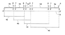
(1)本発明は、核酸サンプル中の1又は2以上の標的ヌクレオチド配列を有する核酸を検出する方法であって、(a)核酸サンプル中の核酸を鋳型とし、1又は2以上の標的ヌクレオチド配列を含むヌクレオチド配列を増幅し得るプライマーを用いて、PCR(polymerase chain reaction)を行うことにより、1種又は2種以上の二本鎖核酸を増幅する工程と、(b)前記工程(a)において得られた二本鎖核酸を一本鎖化し、一本鎖核酸を得る工程と、(c)標的ヌクレオチド配列ごとに、下記の(I)〜(III)を充足する上流側プローブ及び下流側プローブを準備する工程と、(d)前記工程(b)において得られた一本鎖核酸に対して、前記工程(c)において準備した上流側プローブ及び下流側プローブをハイブリダイズさせることにより、一本鎖核酸とプローブとの複合体を形成する工程と、(e)前記工程(d)により形成された複合体に光を照射する工程と、(f)前記工程(e)の後に、前記複合体中の一本鎖核酸を、前記上流側プローブ、前記下流側プローブ、及びこれらが連結されたプローブから解離させる工程と、(g)前記工程(f)の後に、前記工程(e)において連結されたプローブを検出する工程と、(h)前記工程(g)において、連結されたプローブが検出された場合には、前記核酸サンプル中に前記標的ヌクレオチド配列を有する核酸が含有されており、連結されたプローブが検出されなかった場合には、前記核酸サンプル中に前記標的ヌクレオチド配列を有する核酸が含有されていないと判断する工程と、を有することを特徴とする、標的ヌクレオチド配列を有する核酸の検出方法を提供する。
(I)標的ヌクレオチド配列を上流側標的ヌクレオチド配列と下流側標的ヌクレオチド配列に2分割し、前記上流側プローブは、前記上流側標的ヌクレオチド配列を有する核酸とハイブリダイズし得るプローブであり、前記下流側プローブは、前記下流側標的ヌクレオチド配列を有する核酸とハイブリダイズし得るプローブであること。
(II)前記上流側プローブ及び前記下流側プローブは、3’末端がPCR阻害物質により修飾されていること。
(III)前記上流側プローブの5’末端が光応答性物質により修飾されていること。
(2)本発明は、前記工程(a)におけるPCRが、多段階PCRであることを特徴とする、前記(1)記載の標的ヌクレオチド配列を有する核酸の検出方法を提供するものである。
(3)本発明は、前記上流側プローブ及び前記下流側プローブが、ヌクレオチド、ヌクレオチドアナログ、及びこれらの修飾体からなる群より選択される1以上がリン酸ジエステル結合により連結したものであることを特徴とする前記(1)又は(2)記載の標的ヌクレオチド配列を有する核酸の検出方法を提供するものである。
(4)本発明は、前記上流側プローブ及び前記下流側プローブが、ヌクレオチド、ヌクレオチドアナログ、及びこれらの修飾体からなる群より選択される1以上がリン酸ジエステル結合により、6〜100分子連結したものであることを特徴とする前記(1)又は(2)記載の標的ヌクレオチド配列を有する核酸の検出方法を提供するものである。
(5)本発明は、前記光応答性物質は、光照射によって開裂する共役二重結合部位を有することを特徴とする前記(1)〜(4)のいずれかに記載の標的ヌクレオチド配列を有する核酸の検出方法を提供するものである。
(6)本発明は、前記光応答性物質は、カルボキシビニル基、ビニル基、ベンジルトリアゾールビニル基、フェニルトリアゾールビニル基、メトキシフェニルトリアゾールビニル基、シアノフェニルトリアゾールビニル基、及びナフチルトリアゾールビニル基からなる群より選択される1以上を有することを特徴とする前記(1)〜(4)のいずれかに記載の標的ヌクレオチド配列を有する核酸の検出方法を提供するものである。
(7)本発明は、前記工程(e)において照射される光が、紫外光であることを特徴とする前記(1)〜(6)のいずれかに記載の標的ヌクレオチド配列を有する核酸の検出方法を提供するものである。
(8)本発明は、前記工程(e)において照射される光が、320〜400nmの波長の光の照射であることを特徴とする前記(1)〜(6)のいずれかに記載の標的ヌクレオチド配列を有する核酸の検出方法を提供するものである。
(9)本発明は、前記上流側プローブの5’末端が、カルボキシビニル基により修飾されたヌクレオチド又はヌクレオチドアナログであることを特徴とする前記(1)〜(8)のいずれかに記載の標的ヌクレオチド配列を有する核酸の検出方法を提供するものである。
(10)本発明は、前記工程(e)において、前記複合体を形成するプローブのTm値以下の温度において光を照射することを特徴とする前記(1)〜(9)のいずれかに記載の標的ヌクレオチド配列を有する核酸の検出方法を提供するものである。
(11)本発明は、前記工程(e)において、前記複合体を形成するプローブのTm値−5℃以上Tm値以下の温度において光を照射することを特徴とする前記(1)〜(9)のいずれかに記載の標的ヌクレオチド配列を有する核酸の検出方法を提供するものである。
(12)本発明は、前記上流側プローブの3’末端及び/又は前記下流側プローブの3’末端が、アミノ基により修飾されたヌクレオチド、リン酸基により修飾されたヌクレオチド、及びダイデオキシヌクレオチドからなる群より選択される1であることを特徴とする前記(1)〜(11)のいずれかに記載の標的ヌクレオチド配列を有する核酸の検出方法を提供するものである。
(13)本発明は、前記上流側プローブの5’末端及び/又は前記下流側プローブの5’末端が、5’→3’エキソヌクレアーゼ活性阻害物質により修飾されていることを特徴とする前記(1)〜(12)のいずれかに記載の標的ヌクレオチド配列を有する核酸の検出方法を提供するものである。
(14)本発明は、前記上流側プローブの5’末端及び/又は前記下流側プローブの5’末端が、メチル基により2−O−メチル化修飾されたヌクレオチド又はヌクレオチドアナログであることを特徴とする前記(1)〜(12)のいずれかに記載の標的ヌクレオチド配列を有する核酸の検出方法を提供するものである。
(15)本発明は、前記上流側プローブ及び/又は前記下流側プローブの、5’末端と5’末端から2番目のヌクレオチド又はヌクレオチドアナログを連結するリン酸ジエステル結合中のリン酸基が、ホスホロチオエート化されていることを特徴とする前記(1)〜(12)のいずれかに記載の標的ヌクレオチド配列を有する核酸の検出方法を提供するものである。
(16)本発明は、前記上流側プローブ及び/又は前記下流側プローブが、標識分子により標識されていることを特徴とする前記(1)〜(15)のいずれかに記載の標的ヌクレオチド配列を有する核酸の検出方法を提供するものである。
(17)本発明は、前記上流側プローブを標識する標識分子と、前記下流側プローブを標識する標識分子とが、異なる種類の発色基分子であり、前記工程(g)における連結されたプローブの検出が、前記上流側プローブを標識する標識分子と前記下流側プローブを標識する標識分子のうち、いずれか一方をドナー分子とし、他方をアクセプター分子とする蛍光共鳴エネルギー転移による、蛍光の発光又は消光の検出であることを特徴とする前記(16)記載の標的ヌクレオチド配列を有する核酸の検出方法を提供するものである。
(18)本発明は、前記上流側プローブを標識する標識分子と、前記下流側プローブを標識する標識分子とのうち、いずれか一方が発色基分子、他方が発色抑制分子であり、
前記工程(g)における連結されたプローブの検出が、前記発色基分子の消光の検出であることを特徴とする前記(16)記載の標的ヌクレオチド配列を有する核酸の検出方法を提供するものである。
(19)本発明は、前記ドナー分子がAlexa Fluor(登録商標、インビトロジェン社製)488又はATTO 488であり、前記アクセプター分子がAlexa Fluor(登録商標)594又はROX(Carboxy-X-rhodamine)であることを特徴とする前記(17)記載の標的ヌクレオチド配列を有する核酸の検出方法を提供するものである。
(20)本発明は、前記発色基分子がAlexa Fluor(登録商標)488、ATTO 488、Alexa Fluor(登録商標)594、及びROX(Carboxy-X-rhodamine)からなる群より選択される1であり、前記発色抑制分子がBHQ(登録商標、Black hole quencher)−1又はBHQ(登録商標)−2であることを特徴とする前記(18)記載の標的ヌクレオチド配列を有する核酸の検出方法を提供するものである。
(21)本発明は、前記標識分子が、前記上流側プローブ及び/又は前記下流側プローブの、5’末端又は3’末端から19分子以内のヌクレオチド又はヌクレオチドアナログに標識されていることを特徴とする前記(16)〜(20)のいずれかに記載の標的ヌクレオチド配列を有する核酸の検出方法を提供するものである。
(22)本発明は、前記上流側プローブにおける標識分子により標識されているヌクレオチド又はヌクレオチドアナログから5’末端までの分子数と、前記下流側プローブにおける標識分子により標識されているヌクレオチド又はヌクレオチドアナログから3’末端までの分子数との和が20以下であることを特徴とする前記(16)〜(20)のいずれかに記載の標的ヌクレオチド配列を有する核酸の検出方法を提供するものである。
(23)本発明は、前記PCRに用いられるプライマーが、ヌクレオチド及びヌクレオチドアナログからなる群より選択される1以上がリン酸ジエステル結合により連結したものであることを特徴とする前記(1)〜(22)のいずれかに記載の標的ヌクレオチド配列を有する核酸の検出方法を提供するものである。
(24)本発明は、前記PCRに用いられるプライマーが、ヌクレオチド及びヌクレオチドアナログからなる群より選択される1以上がリン酸ジエステル結合により、10〜100分子連結したものであることを特徴とする前記(1)〜(22)のいずれかに記載の標的ヌクレオチド配列を有する核酸の検出方法を提供するものである。
(25)本発明は、前記工程(b)において、前記工程(a)において得られた二本鎖核酸の一本鎖化を、前記二本鎖核酸に熱エネルギーを加えることにより行うことを特徴とする前記(1)〜(24)のいずれかに記載の標的ヌクレオチド配列を有する核酸の検出方法を提供するものである。
(26)本発明は、前記工程(b)において、前記工程(a)において得られた二本鎖核酸の一本鎖化を、前記二本鎖核酸を80〜105℃に加熱することにより行うことを特徴とする前記(1)〜(24)のいずれかに記載の標的ヌクレオチド配列を有する核酸の検出方法を提供するものである。
(27)本発明は、前記工程(b)において、前記工程(a)において得られた二本鎖核酸の一本鎖化を、前記二本鎖核酸に、水酸化ナトリウム、ホルムアルデヒド、及び尿素からなる群より選択される1以上の化学物質を添加することにより行うことを特徴とする前記(1)〜(24)のいずれかに記載の標的ヌクレオチド配列を有する核酸の検出方法を提供するものである。
(28)本発明は、前記工程(b)において、前記工程(a)において得られた二本鎖核酸の一本鎖化を、前記二本鎖核酸に、標的ヌクレオチド配列を有する一本鎖核酸を添加することにより行うことを特徴とする前記(1)〜(24)のいずれかに記載の標的ヌクレオチド配列を有する核酸の検出方法を提供するものである。
(29)本発明は、前記工程(f)において、前記複合体を37〜105℃に加熱することにより、一本鎖核酸を、前記上流側プローブ、前記下流側プローブ、及びこれらが連結されたプローブから解離させることを特徴とする前記(1)〜(28)のいずれかに記載の標的ヌクレオチド配列を有する核酸の検出方法を提供するものである。
(30)本発明は、1又は2以上の、標的ヌクレオチド配列を有する核酸を検出するためのプローブのセットであって、標的ヌクレオチド配列の種類ごとに下記の(I)〜(III)を充足する上流側プローブ及び下流側プローブを有することを特徴とするプローブセットを提供するものである。
(I)標的ヌクレオチド配列を上流側標的ヌクレオチド配列と下流側標的ヌクレオチド配列に2分割し、前記上流側プローブは、前記上流側標的ヌクレオチド配列を有する核酸とハイブリダイズし得るプローブであり、前記下流側プローブは、前記下流側標的ヌクレオチド配列を有する核酸とハイブリダイズし得るプローブであること。
(II)前記上流側プローブ及び前記下流側プローブは、3’末端がPCR阻害物質により修飾されていること。
(III)前記上流側プローブの5’末端が光応答性物質により修飾されていること。
(31)本発明は、前記上流側プローブ及び前記下流側プローブが、ヌクレオチド、ヌクレオチドアナログ、及びこれらの修飾体からなる群より選択される1以上がリン酸ジエステル結合により連結したものであることを特徴とする前記(30)記載のプローブセットを提供するものである。
(32)本発明は、前記上流側プローブ及び前記下流側プローブが、ヌクレオチド、ヌクレオチドアナログ、及びこれらの修飾体からなる群より選択される1以上がリン酸ジエステル結合により、6〜100分子連結したものであることを特徴とする前記(30)記載のプローブセットを提供するものである。
(33)本発明は、前記光応答性物質は、光反応官能基として、光照射によって開裂する共役二重結合部位を有することを特徴とする前記(30)〜(32)のいずれかに記載のプローブセットを提供するものである。
(34)本発明は、前記光応答性物質は、カルボキシビニル基、ビニル基、ベンジルトリアゾールビニル基、フェニルトリアゾールビニル基、メトキシフェニルトリアゾールビニル基、シアノフェニルトリアゾールビニル基、及びナフチルトリアゾールビニル基からなる群より選択される1以上を有することを特徴とする前記(30)〜(32)のいずれかに記載のプローブセットを提供するものである。
(35)本発明は、前記上流側プローブの5’末端が、カルボキシビニル基により修飾されたヌクレオチド又はヌクレオチドアナログであることを特徴とする前記(30)〜(34)のいずれかに記載のプローブセットを提供するものである。
(36)本発明は、前記上流側プローブの3’末端及び/又は前記下流側プローブの3’末端が、アミノ基により修飾されたヌクレオチド、リン酸基により修飾されたヌクレオチド、及びダイデオキシヌクレオチドからなる群より選択される1であることを特徴とする前記(30)〜(35)のいずれかに記載のプローブセットを提供するものである。
(37)本発明は、前記上流側プローブの5’末端又は前記下流側プローブの5’末端が、5’→3’エキソヌクレアーゼ活性阻害物質により修飾されていることを特徴とする前記(30)〜(36)のいずれかに記載のプローブセットを提供するものである。
(38)本発明は、前記上流側プローブの5’末端及び/又は前記下流側プローブの5’末端が、メチル基により2−O−メチル化修飾されたヌクレオチド又はヌクレオチドアナログであることを特徴とする前記(30)〜(36)のいずれかに記載のプローブセットを提供するものである。
(39)本発明は、前記上流側プローブ及び/又は前記下流側プローブの、5’末端と5’末端から2番目のヌクレオチド又はヌクレオチドアナログを連結するリン酸ジエステル結合中のリン酸基が、ホスホロチオエート化されていることを特徴とする前記(30)〜(36)のいずれかに記載のプローブセットを提供するものである。
(40)本発明は、前記上流側プローブ及び/又は前記下流側プローブが、標識分子により標識されていることを特徴とする前記(30)〜(39)のいずれかに記載のプローブセットを提供するものである。
(41)本発明は、前記上流側プローブを標識する標識分子と、前記下流側プローブを標識する標識分子とが、異なる種類の発色基分子であることを特徴とする前記(40)記載のプローブセットを提供するものである。
(42)本発明は、前記上流側プローブを標識する標識分子と、前記下流側プローブを標識する標識分子とのうち、いずれか一方が発色基分子、他方が発色抑制分子であることを特徴とする前記(40)記載のプローブセットを提供するものである。
(43)本発明は、前記上流側プローブを標識する標識分子と前記下流側プローブを標識する標識分子のいずれか一方がAlexa Fluor(登録商標、インビトロジェン社製)488又はATTO 488であり、他方がAlexa Fluor(登録商標)594又はROX(Carboxy−X−rhodamine)であることを特徴とする前記(41)記載のプローブセットを提供するものである。
(44)本発明は、前記発色基分子がAlexa Fluor(登録商標)488、ATTO 488、Alexa Fluor(登録商標)594、及びROX(Carboxy−X−rhodamine)からなる群より選択される1であり、前記発色抑制分子がBHQ(登録商標、Black hole quencher)−1又はBHQ(登録商標)−2であることを特徴とする前記(42)記載のプローブセットを提供するものである。
(45)本発明は、前記標識分子が、前記上流側プローブ及び/又は前記下流側プローブの、5’末端又は3’末端から19分子以内のヌクレオチド又はヌクレオチドアナログに標識されていることを特徴とする前記(40)〜(44)のいずれかに記載のプローブセットを提供するものである。
(46)本発明は、前記上流側プローブにおける標識分子により標識されているヌクレオチド又はヌクレオチドアナログから5’末端までの分子数と、前記下流側プローブにおける標識分子により標識されているヌクレオチド又はヌクレオチドアナログから3’末端までの分子数との和が20以下であることを特徴とする前記(40)〜(44)のいずれかに記載のプローブセットを提供するものである。
(47)本発明は、標的ヌクレオチド配列に、1又は2以上のヌクレオチドの置換、挿入、又は欠失からなるミスマッチ部位を有する非標的ヌクレオチド配列を有する核酸と、標的ヌクレオチド配列を有する核酸とを識別する方法であって、(a”)核酸サンプル中の核酸を鋳型とし、標的ヌクレオチド配列を含むヌクレオチド配列を増幅し得るプライマーを用いて、PCRを行うことにより、二本鎖核酸を増幅する工程と、(b”)前記工程(a”)において得られた二本鎖核酸を一本鎖化し、一本鎖核酸を得る工程と、(c”)下記の(I)〜(III)を充足する上流側プローブ及び下流側プローブを準備する工程と、(d”)前記工程(b”)において得られた一本鎖核酸に対して、前記工程(c”)において準備した上流側プローブ及び下流側プローブをハイブリダイズさせることにより、一本鎖核酸とプローブとの複合体を形成する工程と、(e”)前記工程(d”)により形成された複合体に光を照射する工程と、(f”)前記工程(e”)の後に、前記複合体中の一本鎖核酸を、前記上流側プローブ、前記下流側プローブ、及びこれらが連結されたプローブから解離させる工程と、(g”)前記工程(f”)の後に、前記工程(e”)において連結されたプローブを検出する工程と、(h”)前記工程(g”)において、連結されたプローブが検出された場合には、前記核酸サンプル中に前記標的ヌクレオチド配列を有する核酸が含有されており、連結されたプローブが検出されなかった場合には、前記核酸サンプル中に前記非標的ヌクレオチド配列を有する核酸が含有されていると判断する工程と、を有することを特徴とする、核酸の識別方法を提供するものである。
(I)標的ヌクレオチド配列を上流側標的ヌクレオチド配列と下流側標的ヌクレオチド配列に2分割し、前記上流側プローブは、前記上流側標的ヌクレオチド配列を有する核酸とハイブリダイズし得るプローブであり、前記下流側プローブは、前記下流側標的ヌクレオチド配列を有する核酸とハイブリダイズし得るプローブであること。
(II)前記上流側プローブ及び前記下流側プローブは、3’末端がPCR阻害物質により修飾されていること。
(III)前記上流側プローブの5’末端が光応答性物質により修飾されていること。
(48)本発明は、前記ミスマッチ部位が、前記上流側標的ヌクレオチド配列の3’末端から10分子以内又は前記下流側標的ヌクレオチド配列の5’末端から10分子以内にあることを特徴とする前記(47)記載の核酸の識別方法を提供するものである。
(49)本発明は、前記ミスマッチ部位は、1又は2箇所以上の、1のヌクレオチド又は2以上の連続したヌクレオチドが、置換、挿入、又は欠失していることを特徴とする前記(47)又は(48)記載の核酸の識別方法を提供するものである。
(50)本発明は、前記標的ヌクレオチド配列が遺伝子多型のうちの一多型に相同的なヌクレオチド配列であり、前記非標的ヌクレオチド配列が前記遺伝子多型の他の一多型に相同的なヌクレオチド配列であり、前記ミスマッチ部位が前記遺伝子多型の多型部位であり、前記工程(h”)が、(h”2)前記工程(g”)において検出された連結されたプローブ量に基づき、前記核酸サンプル中の核酸が、前記標的ヌクレオチド配列を有するアレルのホモ接合体、前記非標的ヌクレオチド配列を有するアレルのホモ接合体、及び前記標的ヌクレオチド配列を有するアレルと前記非標的ヌクレオチド配列を有するアレルのヘテロ接合体のいずれであるかを識別する工程、であることを特徴とする前記(47)〜(49)のいずれかに記載の核酸の識別方法を提供するものである。
(51)本発明は、核酸サンプル中の1又は2以上の標的ヌクレオチド配列を有する核酸を検出する方法であって、(a’)核酸サンプル中の核酸を鋳型とし、1又は2以上の標的ヌクレオチド配列を含むヌクレオチド配列を増幅し得るプライマーを用いて、非対称PCR(Asymmetrical polymerase chain reaction)を行うことにより、1種又は2種以上の一本鎖核酸を増幅する工程と、(c’)標的ヌクレオチド配列ごとに、下記の(I)〜(III)を充足する上流側プローブ及び下流側プローブを準備する工程と、(d’)前記工程(a’)において得られた一本鎖核酸、前記上流側プローブ、及び前記下流側プローブをハイブリダイズさせることにより、一本鎖核酸とプローブとの複合体を形成する工程と、(e’)前記工程(d’)により形成された複合体に光を照射する工程と、(f’)前記工程(e’)の後に、前記複合体中の一本鎖核酸を、前記上流側プローブ、前記下流側プローブ、及びこれらが連結されたプローブから解離させる工程と、(g’)前記工程(f’)の後に、前記工程(e’)において連結されたプローブを検出する工程と、(h’)前記工程(g’)において、連結されたプローブが検出された場合には、前記核酸サンプル中に前記標的ヌクレオチド配列を有する核酸が含有されており、連結されたプローブが検出されなかった場合には、前記核酸サンプル中に前記標的ヌクレオチド配列を有する核酸が含有されていないと判断する工程と、を有することを特徴とする、標的ヌクレオチド配列を有する核酸の検出方法を提供するものである。
(I)標的ヌクレオチド配列を上流側標的ヌクレオチド配列と下流側標的ヌクレオチド配列に2分割し、前記上流側プローブは、前記上流側標的ヌクレオチド配列を有する核酸とハイブリダイズし得るプローブであり、前記下流側プローブは、前記下流側標的ヌクレオチド配列を有する核酸とハイブリダイズし得るプローブであること。
(II)前記上流側プローブ及び前記下流側プローブは、3’末端がPCR阻害物質により修飾されていること。
(III)前記上流側プローブの5’末端が光応答性物質により修飾されていること。
(52)本発明は、前記工程(a’)が、(a’1)核酸サンプル中の核酸を鋳型とし、1段階又は多段階のPCRを行い、核酸を増幅する工程と、(a’2)前記工程(a’1)において増幅された核酸を鋳型とし、非対称PCRを行うことにより、、1又は2以上の標的ヌクレオチド配列を含む領域が一本鎖核酸である1種又は2種以上の核酸を増幅する工程と、を有する工程であることを特徴とする前記(51)記載の核酸の検出方法を提供するものである。
(53)本発明は、前記工程(a’)が、(a’3)核酸サンプル中の核酸を鋳型とし、PCRと非対称PCRを同時に行うことにより、1又は2以上の標的ヌクレオチド配列を含む領域が一本鎖核酸である1種又は2種以上の核酸を増幅する工程、であることを特徴とする前記(51)記載の標的ヌクレオチド配列を有する核酸の検出方法を提供するものである。
(54)本発明は、前記工程(a’)が、(a’4)核酸サンプル中の核酸を鋳型とし、1又は2以上の標的ヌクレオチド配列を含むヌクレオチド配列を増幅し得る3種類のプライマーを用いて、PCRを行うことにより、1又は2以上の標的ヌクレオチド配列を含む領域が一本鎖核酸である1種又は2種以上のループ状構造を有する核酸を増幅する工程
であり、前記3種類のプライマーが、1又は2以上の標的ヌクレオチド配列を含むヌクレオチド配列の5’側末端領域と相同的なヌクレオチド配列を有するフォワードプライマーと、1又は2以上の標的ヌクレオチド配列を含むヌクレオチド配列の3’側末端領域と相補的なヌクレオチド配列を有するリバースプライマーと、3’末端側に前記フォワードプライマーよりも3’側であって、標的ヌクレオチド配列よりも5’側にある領域と相同的なヌクレオチド配列を有するループ形成用プライマーとであり、前記ループ形成用プライマーの5’末端には、当該プライマーを起点としてPCRにより伸長されるヌクレオチド配列領域のうち、標的ヌクレオチド配列よりも3’末端側であり、かつ前記リバースプライマーと相補的なヌクレオチド配列よりも5’末端側にあるヌクレオチド配列領域と、ハイブリダイズし得るヌクレオチド配列が付加されていることを特徴とする前記(51)記載の核酸の検出方法を提供するものである。
(55)本発明は、核酸サンプル中の1又は2以上の標的ヌクレオチド配列を有する核酸を検出する方法であって、(1)標的ヌクレオチド配列ごとに、下記の(I)〜(III)を充足する上流側プローブ及び下流側プローブを準備する工程と、(2)核酸サンプル中の核酸を鋳型とし、1又は2以上の標的ヌクレオチド配列を含むヌクレオチド配列を増幅し得るプライマーを準備する工程と、(3)前記核酸サンプル、前記工程(1)において準備した上流側プローブ及び下流側プローブ、前記工程(2)において準備したプライマー、並びに耐熱性DNAポリメラーゼを有する反応溶液を調製する工程と、(4)前記工程(3)において調製した反応溶液中の二本鎖核酸を、一本鎖核酸に変性させる工程と、(5)前記工程(4)において得られた一本鎖核酸を鋳型として、前記プライマー用いて、DNAポリメラーゼによる伸長反応を行う工程と、(6)前記工程(5)の後、反応溶液中の二本鎖核酸を、一本鎖核酸に変性させる工程と、(7)前記工程(6)において得られた一本鎖核酸に対して、反応溶液中の上流側プローブ及び下流側プローブをハイブリダイズさせることにより、一本鎖核酸とプローブとの複合体を形成する工程と、(8)前記工程(7)により形成された複合体に光を照射する工程と、(9)前記工程(8)の後に、前記複合体中の一本鎖核酸を、前記上流側プローブ、前記下流側プローブ、及びこれらが連結されたプローブから解離させる工程と、(10)前記工程(9)の後に、前記工程(8)において連結されたプローブを検出する工程と、(11)前記工程(10)の後、前記工程(4)〜(10)を繰り返す工程と、を有し、前記工程(10)及び(11)において、連結されたプローブが検出された場合には、前記核酸サンプル中に前記標的ヌクレオチド配列を有する核酸が含有されており、連結されたプローブが検出されなかった場合には、前記核酸サンプル中に前記標的ヌクレオチド配列を有する核酸が含有されていないと判断することを特徴とする、標的ヌクレオチド配列を有する核酸の検出方法を提供するものである。
(I)標的ヌクレオチド配列を上流側標的ヌクレオチド配列と下流側標的ヌクレオチド配列に2分割し、前記上流側プローブは、前記上流側標的ヌクレオチド配列を有する核酸とハイブリダイズし得るプローブであり、前記下流側プローブは、前記下流側標的ヌクレオチド配列を有する核酸とハイブリダイズし得るプローブであること。
(II)前記上流側プローブ及び前記下流側プローブは、3’末端がPCR阻害物質により修飾されていること。
(III)前記上流側プローブの5’末端が光応答性物質により修飾されていること。
(1) The present invention is a method for detecting a nucleic acid having one or more target nucleotide sequences in a nucleic acid sample, and (a) one or more target nucleotide sequences using the nucleic acid in the nucleic acid sample as a template. A step of amplifying one or more double-stranded nucleic acids by performing PCR (polymerase chain reaction) using a primer capable of amplifying a nucleotide sequence comprising: (b) in the step (a) A step of obtaining a single-stranded nucleic acid from the obtained double-stranded nucleic acid, and (c) an upstream probe and a downstream probe satisfying the following (I) to (III) for each target nucleotide sequence: And (d) the upstream probe and the downstream probe prepared in the step (c) with respect to the single-stranded nucleic acid obtained in the step (b). (E) irradiating light to the complex formed by the step (d), (f) the step of forming a complex of the single-stranded nucleic acid and the probe by hybridizing After step (e), the step of dissociating the single-stranded nucleic acid in the complex from the upstream probe, the downstream probe, and the probe to which they are linked, and (g) the step (f) And (h) detecting the linked probe in the step (e); and (h) if the linked probe is detected in the step (g), the target nucleotide sequence is detected in the nucleic acid sample. The step of determining that the nucleic acid having the target nucleotide sequence is not contained in the nucleic acid sample when the nucleic acid sample is contained and no linked probe is detected. A method for detecting a nucleic acid having a target nucleotide sequence is provided.
(I) The target nucleotide sequence is divided into an upstream target nucleotide sequence and a downstream target nucleotide sequence, and the upstream probe is a probe capable of hybridizing with a nucleic acid having the upstream target nucleotide sequence, and the downstream side The probe is a probe capable of hybridizing with a nucleic acid having the downstream target nucleotide sequence.
(II) The 3 ′ end of the upstream probe and the downstream probe is modified with a PCR inhibitor.
(III) 5 ′ end of the upstream probe But It must be modified with a photoresponsive substance.
(2) The present invention provides a method for detecting a nucleic acid having a target nucleotide sequence according to (1) above, wherein the PCR in the step (a) is a multi-step PCR.
(3) In the present invention, the upstream probe and the downstream probe are ones in which one or more selected from the group consisting of nucleotides, nucleotide analogs, and modifications thereof are linked by a phosphodiester bond. The present invention provides a method for detecting a nucleic acid having a target nucleotide sequence according to (1) or (2) above.
(4) In the present invention, the upstream probe and the downstream probe are linked by 6 to 100 molecules by a phosphodiester bond at least one selected from the group consisting of nucleotides, nucleotide analogs, and modifications thereof. The present invention provides a method for detecting a nucleic acid having a target nucleotide sequence as described in (1) or (2) above.
(5) The present invention has the target nucleotide sequence according to any one of (1) to (4) above, wherein the photoresponsive substance has a conjugated double bond site that is cleaved by light irradiation. A method for detecting a nucleic acid is provided.
(6) In the present invention, the photoresponsive substance includes a carboxyvinyl group, a vinyl group, a benzyltriazole vinyl group, a phenyltriazole vinyl group, a methoxyphenyltriazole vinyl group, a cyanophenyltriazole vinyl group, and a naphthyltriazole vinyl group. The method for detecting a nucleic acid having a target nucleotide sequence according to any one of the above (1) to (4), comprising one or more selected from the group.
(7) The present invention provides the detection of a nucleic acid having a target nucleotide sequence according to any one of (1) to (6), wherein the light irradiated in the step (e) is ultraviolet light. A method is provided.
(8) The target according to any one of (1) to (6), wherein the light irradiated in the step (e) is irradiation with light having a wavelength of 320 to 400 nm. A method for detecting a nucleic acid having a nucleotide sequence is provided.
( 9 The target nucleotide sequence according to any one of (1) to (8), wherein the 5 ′ end of the upstream probe is a nucleotide or nucleotide analog modified with a carboxyvinyl group. The present invention provides a method for detecting a nucleic acid having
( 10 In the step (e), the present invention irradiates light at a temperature not higher than the Tm value of the probe forming the complex. 9 The method of detecting the nucleic acid which has the target nucleotide sequence in any one of (1) is provided.
( 11 ) In the step (e), the present invention irradiates light at a temperature of Tm value −5 ° C. or more and Tm value or less of the probe forming the complex. 9 The method of detecting the nucleic acid which has the target nucleotide sequence in any one of (1) is provided.
( 12 The present invention is a group consisting of nucleotides modified at the 3 ′ end of the upstream probe and / or the 3 ′ end of the downstream probe with an amino group, nucleotides modified with a phosphate group, and dideoxynucleotides. (1) to (1) above, wherein 11 The method of detecting the nucleic acid which has the target nucleotide sequence in any one of (1) is provided.
( 13 The present invention is characterized in that the 5 ′ end of the upstream probe and / or the 5 ′ end of the downstream probe is modified with a 5 ′ → 3 ′ exonuclease activity inhibitor (1) ~ ( 12 The method of detecting the nucleic acid which has the target nucleotide sequence in any one of (1) is provided.
( 14 The present invention is characterized in that the 5 ′ end of the upstream probe and / or the 5 ′ end of the downstream probe is a nucleotide or nucleotide analog that is 2-O-methylated modified with a methyl group. (1) to ( 12 The method of detecting the nucleic acid which has the target nucleotide sequence in any one of (1) is provided.
( 15 In the present invention, the phosphate group in the phosphodiester bond linking the second nucleotide or nucleotide analog from the 5 ′ end and the 5 ′ end of the upstream probe and / or the downstream probe is phosphorothioated. (1) to (1) above, 12 The method of detecting the nucleic acid which has the target nucleotide sequence in any one of (1) is provided.
( 16 In the present invention, the upstream probe and / or the downstream probe is labeled with a labeling molecule. 15 The method of detecting the nucleic acid which has the target nucleotide sequence in any one of (1) is provided.
( 17 ) In the present invention, the label molecule for labeling the upstream probe and the label molecule for labeling the downstream probe are different types of chromophore groups, and the detection of the linked probe in the step (g) is possible. Detecting fluorescence emission or quenching by fluorescence resonance energy transfer using one of a label molecule for labeling the upstream probe and a label molecule for labeling the downstream probe as a donor molecule and the other as an acceptor molecule Said () 16 The present invention provides a method for detecting a nucleic acid having the target nucleotide sequence described above.
( 18 ) The present invention is a labeling molecule that labels the upstream probe and a labeling molecule that labels the downstream probe, one of which is a chromophoric group molecule and the other is a chromogenic inhibitor molecule,
The detection of the linked probe in the step (g) is detection of quenching of the chromophoric molecule, 16 The present invention provides a method for detecting a nucleic acid having the target nucleotide sequence described above.
( 19 ) According to the present invention, the donor molecule is Alexa Fluor (registered trademark, manufactured by Invitrogen) 488 or
( 20 In the present invention, the chromophore molecule is 1 selected from the group consisting of Alexa Fluor (registered trademark) 488,
( 21 The present invention is characterized in that the labeled molecule is labeled with a nucleotide or nucleotide analog within 19 molecules from the 5 ′ end or 3 ′ end of the upstream probe and / or the downstream probe. ( 16 ) ~ ( 20 The method of detecting the nucleic acid which has the target nucleotide sequence in any one of (1) is provided.
( 22 The present invention relates to the number of molecules from the nucleotide or nucleotide analog labeled with the labeled molecule in the upstream probe to the 5 ′ end, and the nucleotide or nucleotide analog labeled with the labeled molecule in the
( 23 In the present invention, the primer used in the PCR is one in which at least one selected from the group consisting of nucleotides and nucleotide analogs is linked by a phosphodiester bond. 22 The method of detecting the nucleic acid which has the target nucleotide sequence in any one of (1) is provided.
( 24 The present invention is characterized in that the primer used in the PCR is one or more selected from the group consisting of nucleotides and nucleotide analogs linked by 10 to 100 molecules by phosphodiester bonds (1) ) ~ ( 22 The method of detecting the nucleic acid which has the target nucleotide sequence in any one of (1) is provided.
( 25 ) The present invention is characterized in that in the step (b), the double-stranded nucleic acid obtained in the step (a) is converted into a single strand by applying thermal energy to the double-stranded nucleic acid. (1) to ( 24 The method of detecting the nucleic acid which has the target nucleotide sequence in any one of (1) is provided.
( 26 ) In the step (b), the present invention performs the single-stranded single-stranded nucleic acid obtained in the step (a) by heating the double-stranded nucleic acid to 80 to 105 ° C. Characteristic (1) to ( 24 The method of detecting the nucleic acid which has the target nucleotide sequence in any one of (1) is provided.
( 27 ) In the step (b), the present invention comprises the step of converting the double-stranded nucleic acid obtained in the step (a) into a single strand, wherein the double-stranded nucleic acid comprises sodium hydroxide, formaldehyde, and urea. It is performed by adding one or more chemical substances selected from the above (1) to (1) 24 The method of detecting the nucleic acid which has the target nucleotide sequence in any one of (1) is provided.
( 28 ) In the present invention, in the step (b), the double-stranded nucleic acid obtained in the step (a) is converted into a single strand, and a single-stranded nucleic acid having a target nucleotide sequence is added to the double-stranded nucleic acid. (1) to (), 24 The method of detecting the nucleic acid which has the target nucleotide sequence in any one of (1) is provided.
( 29 ) In the present invention, in the step (f), the complex is heated to 37 to 105 ° C., whereby the single-stranded nucleic acid is separated from the upstream probe, the downstream probe, and the probe to which they are linked. (1) to (1) characterized by being dissociated 28 The method of detecting the nucleic acid which has the target nucleotide sequence in any one of (1) is provided.
( 30 ) The present invention is a set of probes for detecting a nucleic acid having one or more target nucleotide sequences, the upstream side satisfying the following (I) to (III) for each type of target nucleotide sequence A probe set having a probe and a downstream probe is provided.
(I) The target nucleotide sequence is divided into an upstream target nucleotide sequence and a downstream target nucleotide sequence, and the upstream probe is a probe capable of hybridizing with a nucleic acid having the upstream target nucleotide sequence, and the downstream side The probe is a probe capable of hybridizing with a nucleic acid having the downstream target nucleotide sequence.
(II) The 3 ′ end of the upstream probe and the downstream probe is modified with a PCR inhibitor.
(III) 5 ′ end of the upstream probe But It must be modified with a photoresponsive substance.
( 31 ) The present invention is characterized in that the upstream probe and the downstream probe are ones selected from the group consisting of nucleotides, nucleotide analogs, and modifications thereof linked by a phosphodiester bond. Said ( 30 The probe set described above is provided.
( 32 ) In the present invention, the upstream probe and the downstream probe are linked by 6 to 100 molecules of one or more selected from the group consisting of nucleotides, nucleotide analogs, and modifications thereof by phosphodiester bonds. The above (characterized by 30 The probe set described above is provided.
( 33 In the present invention, the photoresponsive substance has a conjugated double bond site that is cleaved by light irradiation as a photoreactive functional group. 30 ) ~ ( 32 The probe set according to any one of 1) is provided.
( 34 In the present invention, the photoresponsive substance is selected from the group consisting of carboxyvinyl group, vinyl group, benzyltriazole vinyl group, phenyltriazole vinyl group, methoxyphenyltriazole vinyl group, cyanophenyltriazole vinyl group, and naphthyltriazole vinyl group. Said (1) having one or more selected 30 ) ~ ( 32 The probe set according to any one of 1) is provided.
( 35 The present invention is characterized in that the 5 ′ end of the upstream probe is a nucleotide or nucleotide analog modified with a carboxyvinyl group ( 30 ) ~ ( 34 The probe set according to any one of 1) is provided.
( 36 The present invention is a group consisting of nucleotides modified at the 3 ′ end of the upstream probe and / or the 3 ′ end of the downstream probe with an amino group, nucleotides modified with a phosphate group, and dideoxynucleotides. (1) selected from the above, 30 ) ~ ( 35 The probe set according to any one of 1) is provided.
( 37 The present invention is characterized in that the 5 ′ end of the upstream probe or the 5 ′ end of the downstream probe is modified with a 5 ′ → 3 ′ exonuclease activity inhibitor ( 30 ) ~ ( 36 The probe set according to any one of 1) is provided.
( 38 The present invention is characterized in that the 5 ′ end of the upstream probe and / or the 5 ′ end of the downstream probe is a nucleotide or nucleotide analog that is 2-O-methylated modified with a methyl group. ( 30 ) ~ ( 36 The probe set according to any one of 1) is provided.
( 39 In the present invention, the phosphate group in the phosphodiester bond linking the second nucleotide or nucleotide analog from the 5 ′ end and the 5 ′ end of the upstream probe and / or the downstream probe is phosphorothioated. Said () 30 ) ~ ( 36 The probe set according to any one of 1) is provided.
( 40 The present invention is characterized in that the upstream probe and / or the downstream probe is labeled with a labeling molecule. 30 ) ~ ( 39 The probe set according to any one of 1) is provided.
( 41 The present invention is characterized in that the labeling molecule for labeling the upstream probe and the labeling molecule for labeling the downstream probe are different types of chromogenic group molecules ( 40 The probe set described above is provided.
( 42 The present invention is characterized in that one of the labeling molecule for labeling the upstream probe and the labeling molecule for labeling the downstream probe is a chromophoric group molecule and the other is a chromogenic inhibitor molecule. ( 40 The probe set described above is provided.
( 43 In the present invention, either one of the labeled molecule for labeling the upstream probe and the labeled molecule for labeling the downstream probe is Alexa Fluor (registered trademark, manufactured by Invitrogen) 488 or
( 44 ) According to the present invention, the chromophore group is 1 selected from the group consisting of Alexa Fluor (registered trademark) 488,
( 45 The present invention is characterized in that the labeled molecule is labeled with a nucleotide or nucleotide analog within 19 molecules from the 5 ′ end or 3 ′ end of the upstream probe and / or the downstream probe. ( 40 ) ~ ( 44 The probe set according to any one of 1) is provided.
( 46 The present invention relates to the number of molecules from the nucleotide or nucleotide analog labeled with the labeled molecule in the upstream probe to the 5 ′ end, and the nucleotide or nucleotide analog labeled with the labeled molecule in the
( 47 The present invention relates to a method for discriminating between a nucleic acid having a non-target nucleotide sequence having a mismatch site consisting of substitution, insertion or deletion of one or more nucleotides in the target nucleotide sequence and a nucleic acid having the target nucleotide sequence. And (a ″) a step of amplifying a double-stranded nucleic acid by performing PCR using a nucleic acid in a nucleic acid sample as a template and a primer capable of amplifying a nucleotide sequence including a target nucleotide sequence; b ″) a step of obtaining a single-stranded nucleic acid by making the double-stranded nucleic acid obtained in the step (a ″) into a single strand; and (c ″) an upstream side satisfying the following (I) to (III): A step of preparing a probe and a downstream probe; and (d ″) an upstream probe prepared in the step (c ″) for the single-stranded nucleic acid obtained in the step (b ″), and A step of forming a complex of the single-stranded nucleic acid and the probe by hybridizing the flow-side probe; and (e ″) irradiating the complex formed by the step (d ″) with light, (F ″) after the step (e ″), dissociating the single-stranded nucleic acid in the complex from the upstream probe, the downstream probe, and the probe to which they are linked; and (g ″) ) After the step (f ″), detecting the probe linked in the step (e ″); and (h ″) in the case where the linked probe is detected in the step (g ″). When the nucleic acid sample contains the nucleic acid having the target nucleotide sequence and no linked probe is detected, the nucleic acid sample contains the nucleic acid having the non-target nucleotide sequence. Characterized in that it and a step of determining to have been, there is provided a method for identifying nucleic acids.
(I) The target nucleotide sequence is divided into an upstream target nucleotide sequence and a downstream target nucleotide sequence, and the upstream probe is a probe capable of hybridizing with a nucleic acid having the upstream target nucleotide sequence, and the downstream side The probe is a probe capable of hybridizing with a nucleic acid having the downstream target nucleotide sequence.
(II) The 3 ′ end of the upstream probe and the downstream probe is modified with a PCR inhibitor.
(III) 5 ′ end of the upstream probe But It must be modified with a photoresponsive substance.
( 48 The present invention is characterized in that the mismatch site is within 10 molecules from the 3 ′ end of the upstream target nucleotide sequence or within 10 molecules from the 5 ′ end of the downstream target nucleotide sequence. 47 The identification method of the nucleic acid of description is provided.
( 49 In the present invention, the mismatch site is characterized in that one, two or more, one nucleotide or two or more consecutive nucleotides are substituted, inserted, or deleted ( 47 Or ( 48 The identification method of the nucleic acid of description is provided.
( 50 ) In the present invention, the target nucleotide sequence is a nucleotide sequence homologous to one polymorphism of the gene polymorphism, and the non-target nucleotide sequence is a nucleotide sequence homologous to the other polymorphism of the gene polymorphism. The mismatch site is a polymorphic site of the gene polymorphism, and the step (h ″) is based on the amount of linked probes detected in (h ″ 2) the step (g ″), A nucleic acid in a nucleic acid sample is a homozygote of an allele having the target nucleotide sequence, a homozygote of an allele having the non-target nucleotide sequence, and an allele having the target nucleotide sequence and an allele having the non-target nucleotide sequence. The step of identifying which of the heterozygotes, 47 ) ~ ( 49 The method for identifying a nucleic acid according to any one of 1) is provided.
( 51 ) The present invention is a method for detecting a nucleic acid having one or more target nucleotide sequences in a nucleic acid sample, wherein (a ′) the nucleic acid in the nucleic acid sample is used as a template and one or more target nucleotide sequences are detected. Amplifying one or more single-stranded nucleic acids by performing asymmetric PCR (Asymmetric Polymerase chain reaction) using a primer capable of amplifying the nucleotide sequence, and (c ′) for each target nucleotide sequence A step of preparing an upstream probe and a downstream probe satisfying the following (I) to (III), (d ′) the single-stranded nucleic acid obtained in the step (a ′), and the upstream probe And forming a complex of the single-stranded nucleic acid and the probe by hybridizing the downstream probe; (E ′) irradiating the complex formed by the step (d ′) with light; (f ′) after the step (e ′), the single-stranded nucleic acid in the complex is A step of dissociating from the upstream probe, the downstream probe, and a probe to which they are linked; and (g ′) a step of detecting the probe linked in the step (e ′) after the step (f ′). (H ′) When a linked probe is detected in the step (g ′), the nucleic acid sample contains a nucleic acid having the target nucleotide sequence, and the linked probe is detected. If not, a step of determining that the nucleic acid sample does not contain a nucleic acid having the target nucleotide sequence is provided. Than is.
(I) The target nucleotide sequence is divided into an upstream target nucleotide sequence and a downstream target nucleotide sequence, and the upstream probe is a probe capable of hybridizing with a nucleic acid having the upstream target nucleotide sequence, and the downstream side The probe is a probe capable of hybridizing with a nucleic acid having the downstream target nucleotide sequence.
(II) The 3 ′ end of the upstream probe and the downstream probe is modified with a PCR inhibitor.
(III) 5 ′ end of the upstream probe But It must be modified with a photoresponsive substance.
( 52 ) In the present invention, the step (a ′) comprises (a′1) performing a one-step or multi-step PCR using the nucleic acid in the nucleic acid sample as a template, and amplifying the nucleic acid; By performing asymmetric PCR using the nucleic acid amplified in step (a′1) as a template, one or more nucleic acids in which the region containing one or more target nucleotide sequences is a single-stranded nucleic acid are obtained. A step of amplifying, wherein 51 The method for detecting the nucleic acid described above is provided.
( 53 ) In the present invention, when the step (a ′) comprises (a′3) a nucleic acid in a nucleic acid sample as a template and PCR and asymmetric PCR are simultaneously performed, one region containing one or more target nucleotide sequences is obtained. A step of amplifying one or more nucleic acids that are single-stranded nucleic acids, 51 The present invention provides a method for detecting a nucleic acid having the target nucleotide sequence described above.
( 54 ) In the present invention, the step (a ′) uses (a′4) three types of primers capable of amplifying a nucleotide sequence containing one or more target nucleotide sequences using the nucleic acid in the nucleic acid sample as a template. , And amplifying a nucleic acid having one or two or more loop-shaped structures in which a region containing one or more target nucleotide sequences is a single-stranded nucleic acid by performing PCR
The three kinds of primers include a forward primer having a nucleotide sequence homologous to the 5 ′ terminal region of the nucleotide sequence including one or more target nucleotide sequences, and one or more target nucleotide sequences. A reverse primer having a nucleotide sequence complementary to the 3 ′ terminal region of the nucleotide sequence, and a region homologous to the
( 55 ) The present invention is a method for detecting a nucleic acid having one or more target nucleotide sequences in a nucleic acid sample, wherein (1) an upstream satisfying the following (I) to (III) for each target nucleotide sequence: A step of preparing a side probe and a downstream probe, (2) a step of preparing a primer capable of amplifying a nucleotide sequence containing one or more target nucleotide sequences using a nucleic acid in a nucleic acid sample as a template, and (3) A step of preparing a reaction solution having the nucleic acid sample, the upstream probe and the downstream probe prepared in the step (1), the primer prepared in the step (2), and a heat-resistant DNA polymerase; and (4) the step A step of denaturing the double-stranded nucleic acid in the reaction solution prepared in (3) into a single-stranded nucleic acid; and (5) obtained in the step (4). A step of performing an extension reaction with DNA polymerase using the single-stranded nucleic acid as a template and the primer, and (6) after the step (5), the double-stranded nucleic acid in the reaction solution is converted into a single-stranded nucleic acid. And (7) the single-stranded nucleic acid and the probe by hybridizing the upstream probe and the downstream probe in the reaction solution to the single-stranded nucleic acid obtained in the step (6). (8) a step of irradiating the composite formed by the step (7) with light, and (9) one of the composites after the step (8). A step of dissociating the nucleic acid from the upstream probe, the downstream probe, and the probe to which they are linked, and (10) detecting the probe linked in the step (8) after the step (9). And (1 ) After the step (10), the steps (4) to (10) are repeated, and in the steps (10) and (11), when a linked probe is detected, If the nucleic acid sample contains a nucleic acid having the target nucleotide sequence and no linked probe is detected, it is determined that the nucleic acid sample does not contain a nucleic acid having the target nucleotide sequence. The present invention provides a method for detecting a nucleic acid having a target nucleotide sequence.
(I) The target nucleotide sequence is divided into an upstream target nucleotide sequence and a downstream target nucleotide sequence, and the upstream probe is a probe capable of hybridizing with a nucleic acid having the upstream target nucleotide sequence, and the downstream side The probe is a probe capable of hybridizing with a nucleic acid having the downstream target nucleotide sequence.
(II) The 3 ′ end of the upstream probe and the downstream probe is modified with a PCR inhibitor.
(III) 5 ′ end of the upstream probe But It must be modified with a photoresponsive substance.
本発明の標的ヌクレオチド配列を有する核酸の検出方法及び核酸の識別方法は、PCR法とハイブリダイゼーション法を組み合わせた方法であるため、より高精度かつ高感度に標的ヌクレオチド配列を識別し検出することができる。また、LDRに代えて、光連結反応を用いていることにより、従来のPCR−LDR法とは異なり、一つの反応系でPCRとプローブ連結反応を同時に進行させることも可能となり、ヌクレオチド配列の識別や検出の精度のみならず、反応効率の向上にも資する。さらに、反応系を簡素化することが可能となるため、本発明を実施するシステムや装置において、装置の小型化、装備の単純化、しいては製品のコストダウン等の多種のメリットを生みだすことができる。さらに、反応の開始・停止は光照射の有無で制御できるため、任意のタイミングで連結反応を誘導することができ、反応条件をより厳密に制御することが可能となる。
また、本発明のプローブセットを用いることにより、従来法よりも簡便かつ高精度に、標的ヌクレオチド配列を有する核酸を識別し検出することが可能となる。
Since the method for detecting a nucleic acid having a target nucleotide sequence and the method for identifying a nucleic acid according to the present invention are a combination of a PCR method and a hybridization method, the target nucleotide sequence can be identified and detected with higher accuracy and higher sensitivity. it can. Also, by using a photoligation reaction instead of LDR, unlike the conventional PCR-LDR method, it is possible to simultaneously proceed with PCR and probe ligation reaction in a single reaction system, thereby identifying nucleotide sequences. As well as the accuracy of detection, it also contributes to improving the reaction efficiency. Furthermore, since the reaction system can be simplified, the system and apparatus for carrying out the present invention can produce various merits such as downsizing of the apparatus, simplification of equipment, and cost reduction of the product. Can do. Furthermore, since the start / stop of the reaction can be controlled by the presence or absence of light irradiation, the ligation reaction can be induced at an arbitrary timing, and the reaction conditions can be more strictly controlled.
Further, by using the probe set of the present invention, it becomes possible to identify and detect a nucleic acid having a target nucleotide sequence more easily and with higher accuracy than the conventional method.
本発明において、核酸とは、DNA又はRNAであれば特に限定されるものではなく、天然のものであってもよく、合成されたものであってもよい。天然の核酸として、例えば、生物から回収されたゲノムDNA、mRNA、tRNA、rRNA、hnRNA等がある。また、合成された核酸として、β−シアノエチルホスフォロアミダイト法DNA固相合成法等の公知の化学的合成法により合成されたDNAや、PCR等の公知の核酸増幅法により合成された核酸、逆転写反応により合成されたcDNA等がある。 In the present invention, the nucleic acid is not particularly limited as long as it is DNA or RNA, and may be natural or synthesized. Examples of natural nucleic acids include genomic DNA, mRNA, tRNA, rRNA, hnRNA and the like recovered from an organism. Moreover, as a synthesized nucleic acid, DNA synthesized by a known chemical synthesis method such as β-cyanoethyl phosphoramidite method DNA solid phase synthesis method, a nucleic acid synthesized by a known nucleic acid amplification method such as PCR, inversion There are cDNAs synthesized by photoreaction.
本発明において核酸サンプルとは、核酸を含有するサンプルであれば、特に限定されるものではないが、動物、植物、微生物、培養細胞等から核酸を抽出したサンプルであることが好ましい。なお、動物等からの核酸の抽出は、フェノール/クロロホルム法等の公知の手法により行うことができる。 In the present invention, the nucleic acid sample is not particularly limited as long as it is a sample containing nucleic acid, but is preferably a sample obtained by extracting nucleic acid from animals, plants, microorganisms, cultured cells and the like. Extraction of nucleic acids from animals and the like can be performed by a known method such as a phenol / chloroform method.
本発明において標的ヌクレオチド配列は、PCR法やハイブリダイゼーション法等により検出が可能な程度に塩基配列が明らかになっているものであれば、特に限定されるものではない。本発明の標的ヌクレオチド配列を有する核酸の検出方法や核酸の識別方法においては、標的ヌクレオチド配列として、遺伝子上に存在する特定の領域のヌクレオチド配列であることが好ましく、遺伝子多型を含む領域のヌクレオチド配列であることがより好ましい。本発明の標的ヌクレオチド配列を有する核酸の検出方法や核酸の識別方法は、標的ヌクレオチド配列を有する核酸の識別・検出能が高く、相同性の高いヌクレオチド配列同士を高精度に識別し得るためである。 In the present invention, the target nucleotide sequence is not particularly limited as long as the nucleotide sequence has been clarified to such an extent that it can be detected by a PCR method or a hybridization method. In the method for detecting a nucleic acid having a target nucleotide sequence and the method for identifying a nucleic acid of the present invention, the target nucleotide sequence is preferably a nucleotide sequence of a specific region present on the gene, and the nucleotide of the region containing the gene polymorphism More preferably, it is an array. This is because the method for detecting a nucleic acid having a target nucleotide sequence and the method for identifying a nucleic acid according to the present invention have a high ability to identify and detect a nucleic acid having a target nucleotide sequence and can identify highly homologous nucleotide sequences with high accuracy. .
ここで、遺伝子多型とは、ある生物種の集団内において、個体ごとに遺伝子のヌクレオチド配列が異なるものを意味する。遺伝子多型としては、例えば、一塩基多型(SNP)、マイクロサテライト多型等がある。本発明の標的ヌクレオチド配列を有する核酸の検出方法や核酸の識別方法において、標的ヌクレオチド配列が含む遺伝子多型として、SNPであることが好ましい。 Here, the gene polymorphism means one having a different nucleotide sequence of a gene for each individual within a population of a certain biological species. Examples of gene polymorphism include single nucleotide polymorphism (SNP) and microsatellite polymorphism. In the method for detecting a nucleic acid having a target nucleotide sequence of the present invention and the method for identifying a nucleic acid, the gene polymorphism contained in the target nucleotide sequence is preferably an SNP.
その他、本発明における標的ヌクレオチド配列としては、遺伝子多型のように先天的な多型のみならず、後天的な遺伝子変異等を含む領域のヌクレオチド配列であってもよい。後天的な遺伝子変異として、紫外線等の物理的刺激や発がん性物質等による化学的刺激により誘発される体細胞変異等が挙げられる。後述するように、本発明においては、標的ヌクレオチド配列を有する核酸の検出を、連結されたプローブ量を測定することにより検出するため、定量的な解析が可能となり得るため、体細胞変異等を定量的に解析する場合にも用いることができる。 In addition, the target nucleotide sequence in the present invention may be a nucleotide sequence of a region containing not only a congenital polymorphism such as a gene polymorphism but also an acquired genetic mutation. Acquired genetic mutations include somatic mutations induced by physical stimuli such as ultraviolet rays or chemical stimuli such as carcinogenic substances. As will be described later, in the present invention, detection of a nucleic acid having a target nucleotide sequence is detected by measuring the amount of linked probes, so that quantitative analysis may be possible, so that somatic mutations and the like are quantified. It can also be used for analysis.
本発明においてヌクレオチドアナログとは、非天然のヌクレオチドであり、天然のヌクレオチドであるデオキシリボヌクレオチド(DNA)やリボヌクレオチド(RNA)と同様の機能を有するものをいう。すなわち、ヌクレオチドアナログは、ヌクレオチドと同様にリン酸ジエステル結合により鎖を形成することができ、かつ、ヌクレオチドアナログを用いて形成されたプライマーやプローブは、ヌクレオチドのみを用いて形成されたプライマーやプローブと同様に、PCRやハイブリダイゼーションに用いることができる。このようなヌクレオチドアナログとして、例えば、PNA(ポリアミドヌクレオチド誘導体)、LNA(BNA)、ENA(2‘−O,4’−C−Ethylene−bridged nucleic acids)、及びこれらの複合体等がある。ここで、PNAは、DNAやRNAのリン酸と5炭糖からなる主鎖をポリアミド鎖に置換したものである。また、LNA(BNA)は、リボヌクレオシドの2’部位の酸素原子と4’部位の炭素原子がメチレンを介して結合している2つの環状構造を持つ化合物である。 In the present invention, the nucleotide analog is a non-natural nucleotide and has the same function as deoxyribonucleotide (DNA) or ribonucleotide (RNA), which are natural nucleotides. That is, nucleotide analogs can form chains by phosphodiester bonds in the same way as nucleotides, and primers and probes formed using nucleotide analogs are primers and probes formed using only nucleotides. Similarly, it can be used for PCR and hybridization. Examples of such nucleotide analogs include PNA (polyamide nucleotide derivatives), LNA (BNA), ENA (2'-O, 4'-C-Ethylene-bridged nucleic acids), and complexes thereof. Here, PNA is obtained by substituting a main chain composed of phosphoric acid and pentose of DNA or RNA with a polyamide chain. LNA (BNA) is a compound having two cyclic structures in which an oxygen atom at the 2 'site of a ribonucleoside and a carbon atom at the 4' site are bonded via methylene.
本発明の標的ヌクレオチド配列を有する核酸の検出方法(以下、本発明の検出方法ということがある。)は、核酸サンプル中の1又は2以上の標的ヌクレオチド配列を有する核酸を検出する方法であって、(a)核酸サンプル中の核酸を鋳型とし、1又は2以上の標的ヌクレオチド配列を含むヌクレオチド配列を増幅し得るプライマーを用いて、PCRを行うことにより、1種又は2種以上の二本鎖核酸を増幅する工程と、(b)前記工程(a)において得られた二本鎖核酸を一本鎖化し、一本鎖核酸を得る工程と、(c)標的ヌクレオチド配列ごとに、下記の(I)〜(III)を充足する上流側プローブ及び下流側プローブを準備する工程と、(d)前記工程(b)において得られた一本鎖核酸に対して、前記工程(c)において準備した上流側プローブ及び下流側プローブをハイブリダイズさせることにより、一本鎖核酸とプローブとの複合体を形成する工程と、(e)前記工程(d)により形成された複合体に光を照射する工程と、(f)前記工程(e)の後に、前記複合体中の一本鎖核酸を、前記上流側プローブ、前記下流側プローブ、及びこれらが連結されたプローブから解離させる工程と、(g)前記工程(f)の後に、前記工程(e)において連結されたプローブを検出する工程と、(h)前記工程(g)において、連結されたプローブが検出された場合には、前記核酸サンプル中に前記標的ヌクレオチド配列を有する核酸が含有されており、連結されたプローブが検出されなかった場合には、前記核酸サンプル中に前記標的ヌクレオチド配列を有する核酸が含有されていないと判断する工程と、を有することを特徴とする。
(I)標的ヌクレオチド配列を上流側標的ヌクレオチド配列と下流側標的ヌクレオチド配列に2分割し、前記上流側プローブは、前記上流側標的ヌクレオチド配列を有する核酸とハイブリダイズし得るプローブであり、前記下流側プローブは、前記下流側標的ヌクレオチド配列を有する核酸とハイブリダイズし得るプローブであること。
(II)前記上流側プローブ及び前記下流側プローブは、3’末端がPCR阻害物質により修飾されていること。
(III)前記上流側プローブの5’末端及び/又は前記下流側プローブの3’末端が光応答性物質により修飾されていること。
The method for detecting a nucleic acid having a target nucleotide sequence of the present invention (hereinafter sometimes referred to as the detection method of the present invention) is a method for detecting a nucleic acid having one or more target nucleotide sequences in a nucleic acid sample. (A) One or two or more double strands by performing PCR using a nucleic acid in a nucleic acid sample as a template and a primer capable of amplifying a nucleotide sequence containing one or more target nucleotide sequences A step of amplifying a nucleic acid, (b) a step of obtaining a single-stranded nucleic acid by making the double-stranded nucleic acid obtained in the step (a) into a single strand, and (c) for each target nucleotide sequence, the following ( A step of preparing an upstream probe and a downstream probe satisfying I) to (III), and (d) a preparation of the single-stranded nucleic acid obtained in the step (b) in the step (c). A step of forming a complex of the single-stranded nucleic acid and the probe by hybridizing the upstream probe and the downstream probe, and (e) a step of irradiating the complex formed by the step (d) with light. And (f) after the step (e), dissociating the single-stranded nucleic acid in the complex from the upstream probe, the downstream probe, and the probe to which they are linked, and (g) After the step (f), a step of detecting the linked probe in the step (e); and (h) in the step (g), when a linked probe is detected, Contains a nucleic acid having the target nucleotide sequence, and if a linked probe is not detected, the nucleic acid sample has a nucleic acid having the target nucleotide sequence. Is characterized by having a, a step of determining that no.
(I) The target nucleotide sequence is divided into an upstream target nucleotide sequence and a downstream target nucleotide sequence, and the upstream probe is a probe capable of hybridizing with a nucleic acid having the upstream target nucleotide sequence, and the downstream side The probe is a probe capable of hybridizing with a nucleic acid having the downstream target nucleotide sequence.
(II) The 3 ′ end of the upstream probe and the downstream probe is modified with a PCR inhibitor.
(III) The 5 ′ end of the upstream probe and / or the 3 ′ end of the downstream probe are modified with a photoresponsive substance.
本発明の検出方法は、主に、工程(a)〜(b)のPCRによる増幅段階と、工程(c)〜(h)の光連結反応を用いたハイブリダイゼーションによる検出段階の2段階に分けられる。以下、工程ごとに説明する。 The detection method of the present invention is mainly divided into two steps, an amplification step by PCR in steps (a) to (b) and a detection step by hybridization using a photoligation reaction in steps (c) to (h). It is done. Hereinafter, it demonstrates for every process.
まず、工程(a)において、核酸サンプル中の核酸を鋳型とし、1又は2以上の標的ヌクレオチド配列を含むヌクレオチド配列を増幅し得るプライマーを用いて、PCRを行うことにより、1種又は2種以上の二本鎖核酸を増幅する。核酸サンプルに含まれている全核酸のうち、標的ヌクレオチド配列を含むことが期待される領域のみを増幅することにより、核酸サンプルとして、標的ヌクレオチドを有する核酸が微量にしか存在していない遺伝子サンプル等を用いた場合であっても、高感度に標的ヌクレオチド配列を有する核酸を検出することができる。ここで、鋳型とする核酸サンプル中の核酸は、DNA又はRNAのいずれでもあってよく、一本鎖でもあっても二本鎖であってもよい。なお、核酸サンプル中の核酸がRNAである場合には、逆転写反応により得られたcDNAを鋳型とすることができる。 First, in step (a), one or two or more kinds are obtained by performing PCR using a nucleic acid in a nucleic acid sample as a template and a primer capable of amplifying a nucleotide sequence containing one or more target nucleotide sequences. A double-stranded nucleic acid is amplified. A gene sample in which only a small amount of nucleic acid having a target nucleotide exists as a nucleic acid sample by amplifying only the region expected to contain the target nucleotide sequence among all the nucleic acids contained in the nucleic acid sample Even in the case where is used, a nucleic acid having a target nucleotide sequence can be detected with high sensitivity. Here, the nucleic acid in the nucleic acid sample used as a template may be either DNA or RNA, and may be single-stranded or double-stranded. When the nucleic acid in the nucleic acid sample is RNA, cDNA obtained by reverse transcription reaction can be used as a template.
工程(a)において用いられるプライマーは、1又は2以上の標的ヌクレオチド配列を含むヌクレオチド配列(増幅用ヌクレオチド配列)をPCRにより増幅し得るプライマーであり、増幅用ヌクレオチド配列を挟む2種類のプライマーである。例えば、増幅用ヌクレオチド配列の上流端領域と相同的なヌクレオチド配列を有するフォワードプライマーと、増幅用ヌクレオチド配列の下流端領域と相補的なヌクレオチド配列を有するリバースプライマーとからなる2種類のプライマーであってもよい。なお、PCRにおいて用いられる該2種類のプライマーの各濃度は、PCR産物として二本鎖核酸を得ることができる濃度比であれば特に限定されるものではないが、等濃度で用いることが好ましい。 The primers used in the step (a) are primers that can amplify a nucleotide sequence (amplification nucleotide sequence) containing one or more target nucleotide sequences by PCR, and are two kinds of primers sandwiching the amplification nucleotide sequence. . For example, two types of primers consisting of a forward primer having a nucleotide sequence homologous to the upstream end region of the amplification nucleotide sequence and a reverse primer having a nucleotide sequence complementary to the downstream end region of the amplification nucleotide sequence, Also good. The concentration of the two kinds of primers used in PCR is not particularly limited as long as it is a concentration ratio that can obtain a double-stranded nucleic acid as a PCR product, but is preferably used at an equal concentration.
増幅用ヌクレオチド配列、すなわち、PCRにより増幅される領域は、少なくとも1の標的ヌクレオチド配列を含む領域であればよく、2以上の標的ヌクレオチド配列を含む領域を一のPCRにより増幅してもよい。つまり、PCRにより得られる二本鎖核酸は、1の標的ヌクレオチド配列を含む核酸であってもよく、2以上の標的ヌクレオチド配列を含む核酸であってもよい。 The nucleotide sequence for amplification, that is, the region amplified by PCR may be a region containing at least one target nucleotide sequence, and a region containing two or more target nucleotide sequences may be amplified by one PCR. That is, the double-stranded nucleic acid obtained by PCR may be a nucleic acid containing one target nucleotide sequence or a nucleic acid containing two or more target nucleotide sequences.
また、工程(a)においてPCRにより得られる二本鎖核酸は、1種類であってもよく、複数種類であってもよい。ここで、PCRにより得られる二本鎖核酸が複数種類であるとは、PCRにより増幅される領域が、核酸サンプル中の核酸の1の領域ではなく、2以上の領域であることを意味する。例えば、工程(a)におけるPCRをマルチプレックスPCRとすることにより、複数種類の二本鎖核酸(PCR産物)を得ることができる。また、二本鎖核酸の種類ごとに別個独立にPCRを行い、得られたPCR産物を混合してもよい。 Further, the double-stranded nucleic acid obtained by PCR in the step (a) may be one kind or plural kinds. Here, the plural types of double-stranded nucleic acid obtained by PCR means that the region amplified by PCR is not one region of nucleic acid in the nucleic acid sample but two or more regions. For example, by setting the PCR in the step (a) to multiplex PCR, a plurality of types of double-stranded nucleic acids (PCR products) can be obtained. Alternatively, PCR may be performed independently for each type of double-stranded nucleic acid, and the resulting PCR products may be mixed.
このように、2以上の標的ヌクレオチド配列を含むと期待し得る核酸領域をまとめて増幅することにより、2以上の標的ヌクレオチド配列の検出、すなわち多重解析における作業工程を減少させることができる。 Thus, by amplifying together nucleic acid regions that can be expected to contain two or more target nucleotide sequences, it is possible to reduce the number of work steps in detection of two or more target nucleotide sequences, that is, multiplex analysis.
工程(a)におけるPCRは、多段階PCRであってもよい。ここで、多段階PCRとは、前段階目のPCR後のPCR溶液に、前段階目のPCRとは異なるプライマーを添加して、後段階目のPCRを行うことを意味する。後段階目のPCRは、前段階目のPCR産物を鋳型として用いるものであってもよく、核酸サンプル中に元々含まれていた核酸を鋳型として用いるものであってもよい。工程(a)におけるPCR増幅を複数回繰り返すことにより、後のハイブリダイゼーションによる検出段階において、より高い検出シグナルを得ることができる。 The PCR in step (a) may be multi-stage PCR. Here, the multi-stage PCR means that a primer different from the previous-stage PCR is added to the PCR solution after the previous-stage PCR to perform the latter-stage PCR. The PCR in the subsequent stage may be performed using the PCR product in the previous stage as a template, or may be performed using the nucleic acid originally contained in the nucleic acid sample as the template. By repeating the PCR amplification in step (a) a plurality of times, a higher detection signal can be obtained in the subsequent detection step by hybridization.
次に、工程(b)として、工程(a)において得られた二本鎖核酸を一本鎖化し、一本鎖核酸を得る。一本鎖化することにより、工程(a)において増幅した標的ヌクレオチド配列を含むヌクレオチド配列を有する核酸を、後の工程のハイブリダイゼーションにおける鋳型として用いることができるようになる。 Next, as the step (b), the double-stranded nucleic acid obtained in the step (a) is made into a single strand to obtain a single-stranded nucleic acid. By forming a single strand, a nucleic acid having a nucleotide sequence including the target nucleotide sequence amplified in step (a) can be used as a template in the subsequent hybridization.
工程(b)において、二本鎖核酸を一本鎖化する方法は、特に限定されるものではなく、公知の手法で行うことができる。例えば、二本鎖核酸に熱エネルギーを加えることや、塩基同士の水素結合を弱めるような化学物質を添加すること等により、二本鎖核酸を一本鎖化することができる。具体的には、例えば、PCR後のPCR溶液を、80〜105℃に加熱することや、該PCR溶液に、水酸化ナトリウム、ホルムアルデヒド、及び尿素等を添加することにより、PCR溶液中の二本鎖核酸を一本鎖化することができる。その他、該PCR溶液に、一本鎖化させたい領域のヌクレオチ配列、すなわち標的ヌクレオチド配列と相同的な配列を有する一本鎖核酸を添加することによっても、一本鎖化することができる。 In the step (b), the method for making a double-stranded nucleic acid into a single strand is not particularly limited, and can be performed by a known method. For example, a double-stranded nucleic acid can be made into a single strand by adding thermal energy to the double-stranded nucleic acid or adding a chemical substance that weakens the hydrogen bond between bases. Specifically, for example, by heating the PCR solution after PCR to 80 to 105 ° C. or adding sodium hydroxide, formaldehyde, urea or the like to the PCR solution, A strand nucleic acid can be made into a single strand. Alternatively, a single-stranded nucleic acid having a sequence homologous to the nucleotide sequence of the region to be single-stranded, that is, a target nucleotide sequence, can also be made single-stranded.
二本鎖核酸を一本鎖化するために添加される一本鎖核酸は、ヌクレオチド又はヌクレオチドアナログがリン酸ジエステル結合することにより形成されたものであることが好ましい。特に、ヌクレオチドアナログとして、LNA(BNA)、ENA、PNA等のTm値が比較的高いヌクレオチドアナログを構成分子として形成された一本鎖核酸であることが好ましい。 The single-stranded nucleic acid added to make the double-stranded nucleic acid into a single strand is preferably formed by phosphodiester bonding of nucleotides or nucleotide analogs. In particular, the nucleotide analog is preferably a single-stranded nucleic acid formed using a nucleotide analog having a relatively high Tm value such as LNA (BNA), ENA, or PNA as a constituent molecule.
本発明の検出方法においては、工程(b)における一本鎖化は、熱エネルギーを加える方法であることが好ましく、PCR後のPCR溶液を、80〜105℃に加熱することがより好ましく、95〜100℃に加熱することがさらに好ましく、95℃に加熱することが特に好ましい。PCR溶液の組成を変更する必要がないため、後の工程に対する影響を抑えることができるためである。 In the detection method of the present invention, the single strand formation in the step (b) is preferably a method of applying thermal energy, more preferably, the PCR solution after PCR is heated to 80 to 105 ° C., 95 It is more preferable to heat to -100 ° C, and it is particularly preferable to heat to 95 ° C. This is because it is not necessary to change the composition of the PCR solution, and the influence on the subsequent steps can be suppressed.
工程(a)におけるPCRを、非対称PCRとすることにより、1又は2以上の標的ヌクレオチド配列を含むヌクレオチド配列を有する一本鎖核酸を増幅させることができる。具体的には、核酸サンプル中の核酸を鋳型とし、1又は2以上の標的ヌクレオチド配列を含むヌクレオチド配列を増幅し得るプライマーを用いて、非対称PCRを行うことにより、1種又は2種以上の一本鎖核酸を増幅する。このようにして得られた一本鎖核酸は、一本鎖化することなく、後の工程のハイブリダイゼーションにおける鋳型として用いることができる。 By setting the PCR in step (a) to asymmetric PCR, a single-stranded nucleic acid having a nucleotide sequence containing one or more target nucleotide sequences can be amplified. Specifically, by using a nucleic acid in a nucleic acid sample as a template and a primer capable of amplifying a nucleotide sequence containing one or more target nucleotide sequences, asymmetric PCR is carried out to perform one or more types of one or more types. Amplify double-stranded nucleic acid. The single-stranded nucleic acid thus obtained can be used as a template in the subsequent hybridization without forming a single-stranded nucleic acid.
本発明において、非対称PCRとは、前記二本鎖核酸を得る場合とは異なり、PCRに用いられる増幅用ヌクレオチド配列を挟む2種類のプライマーのうち、1種類のプライマーのみを用いて行うPCRや、1種類のプライマーを他方のプライマーよりも高濃度に用いて行うPCRを意味する。鋳型である二本鎖核酸を構成する二本の鎖が非対称的に増幅される結果、より高濃度に添加したプライマーを起点として伸長される鎖が、一本鎖核酸の状態で得ることができる。ここで、1種類のプライマーのみを用いてPCRを行った場合には、一本鎖核酸は増幅されるものの、指数関数的な増幅は行われないが、プライマーの量比を変えてPCRを行った場合には、通常のPCRのように二本鎖核酸が指数関数的に増幅しつつ、一本鎖核酸も増幅される。 In the present invention, asymmetric PCR is different from the case where the double-stranded nucleic acid is obtained, PCR performed using only one kind of primers among two kinds of primers sandwiching the nucleotide sequence for amplification used in PCR, It means PCR performed using one kind of primer at a higher concentration than the other primer. As a result of asymmetric amplification of the two strands that constitute the double-stranded nucleic acid that is the template, a single strand nucleic acid can be obtained in the form of a strand that is extended from a primer added at a higher concentration. . Here, when PCR is performed using only one kind of primer, single-stranded nucleic acid is amplified, but exponential amplification is not performed, but PCR is performed by changing the quantity ratio of primers. In such a case, the single-stranded nucleic acid is also amplified while the double-stranded nucleic acid is amplified exponentially as in normal PCR.
その他、一部分に一本鎖のループ構造を有するPCR産物を得る方法を用いることにより、標的ヌクレオチド配列領域を一本鎖核酸として得ることができる(例えば、特開2005−102502号公報参照。)。具体的には、3種類のプライマー、すなわち、通常のPCRに用いられる前述のフォワードプライマーとリバースプライマーの2種類のプライマーに加えて、5’末端には、当該プライマーを起点としてPCRにより伸長されるヌクレオチド配列領域のうち、標的ヌクレオチド配列よりも3’末端側であり、かつ前記リバースプライマーと相補的なヌクレオチド配列よりも5’末端側にあるヌクレオチド配列領域と、ハイブリダイズし得るヌクレオチド配列が付加されており、3’末端側に前記フォワードプライマーよりも3’側であって、標的ヌクレオチド配列よりも5’側にある領域と相同的なヌクレオチド配列を有するループ形成用プライマーとを用いてPCRを行うことにより、一本鎖のループ構造部分に標的ヌクレオチド配列領域を有するPCR産物を得ることができる。該方法により得られたPCR産物も、非対称PCR産物と同様に、一本鎖化することなく、後の工程のハイブリダイゼーションにおける鋳型として用いることができる。 In addition, a target nucleotide sequence region can be obtained as a single-stranded nucleic acid by using a method for obtaining a PCR product having a single-stranded loop structure in part (see, for example, JP-A-2005-102502). Specifically, in addition to the three types of primers, ie, the above-mentioned two types of primers, the forward primer and the reverse primer used in normal PCR, the 5 ′ end is extended by PCR starting from the primer. Among the nucleotide sequence regions, a nucleotide sequence capable of hybridizing is added to the nucleotide sequence region that is 3 ′ end side of the target nucleotide sequence and 5 ′ end side of the nucleotide sequence complementary to the reverse primer. PCR is performed using a loop-forming primer having a nucleotide sequence that is homologous to a region 3 'to the 3' end and 5 'to the target nucleotide sequence at the 3' end. The target nucleotide sequence region in the single-stranded loop structure PCR products with can be obtained. The PCR product obtained by this method can also be used as a template in the subsequent hybridization without forming a single strand, similarly to the asymmetric PCR product.
なお、PCR産物として二本鎖核酸を得るPCRと、一本鎖核酸を得るPCRとを組み合わせて行ってもよい。例えば、1段階のPCR又は多段階PCRを行った後に、非対称PCRを行ってもよい。 In addition, you may carry out combining PCR which obtains a double-stranded nucleic acid as a PCR product, and PCR which obtains a single-stranded nucleic acid. For example, asymmetric PCR may be performed after performing one-step PCR or multi-step PCR.
また、工程(a)におけるPCRは、PCRと非対称PCRを同時に行うものであってもよい。例えば、第一段階目のPCRを通常のPCRとし、第二段階目のPCRを非対称PCRとして、第二段階目の非対称PCRにより目的の増幅ヌクレオチド配列を有する一本鎖核酸を増幅させるNested PCRを行う場合に、第一段階目のPCRを行うためのプライマーと、第二段階目の非対称PCRを行うためのプライマーとを、予め反応溶液中に入れておくことにより、第一段階目のPCRと第二段階目の非対称PCRを同時に行うことができる。
なお、PCRや非対称PCRを同時に行うとは、同一の反応溶液中で、同一の反応サイクル(変性工程、アニール工程、伸長工程)で反応を行うことを意味する。
The PCR in step (a) may be performed simultaneously with PCR and asymmetric PCR. For example, a first-stage PCR is a normal PCR, a second-stage PCR is an asymmetric PCR, and a second-stage asymmetric PCR is used to amplify a single-stranded nucleic acid having a target amplified nucleotide sequence. In the case of performing the first stage PCR, the primer for performing the first stage PCR and the primer for performing the second stage asymmetric PCR are previously placed in the reaction solution. The second stage asymmetric PCR can be performed simultaneously.
Note that performing PCR and asymmetric PCR at the same time means performing a reaction in the same reaction solution in the same reaction cycle (denaturation step, annealing step, extension step).
PCRに用いられるこれらのプライマーは、標的ヌクレオチド配列を含むヌクレオチド配列の配列情報、PCRに用いるDNAポリメラーゼの種類等を考慮して、常法により設計し合成することができる。 These primers used for PCR can be designed and synthesized by a conventional method in consideration of the sequence information of the nucleotide sequence including the target nucleotide sequence, the type of DNA polymerase used for PCR, and the like.
本発明においては、PCRに用いられるこれらのプライマーのTm値が、上流側プローブや下流側プローブのTm値よりも高くなるように設計することが好ましい。プローブのTm値よりもプライマーのTm値を高くすることにより、プローブの存在下でプライマーを用いてPCRを行う場合にも、PCRに対するプローブの影響を抑えることができる。 In the present invention, it is preferable to design such that the Tm values of these primers used for PCR are higher than the Tm values of the upstream probe and the downstream probe. By making the Tm value of the primer higher than the Tm value of the probe, the influence of the probe on the PCR can be suppressed even when PCR is performed using the primer in the presence of the probe.
PCRに用いられるこれらのプライマーは、ヌクレオチド及びヌクレオチドアナログからなる群より選択される1以上がリン酸ジエステル結合により連結したものである。プライマーの長さは、プライマーのTm値、増幅ヌクレオチド配列の種類等を考慮して適宜決定することができるが、10〜100分子連結したものであることが好ましい。 These primers used for PCR are those in which one or more selected from the group consisting of nucleotides and nucleotide analogs are linked by a phosphodiester bond. The length of the primer can be appropriately determined in consideration of the Tm value of the primer, the type of the amplified nucleotide sequence, and the like, but it is preferably 10 to 100 linked molecules.
本発明の検出方法においては、PCRは、通常用いられる試薬を通常用いられる量で用いて、通常使用されるプロトコールに基づき、常法により行うことができる。なお、PCRに用いられるDNAポリメラーゼは、通常PCRにおいて用いられるDNAポリメラーゼであれば特に限定されるものではないが、耐熱性ポリメラーゼであることが好ましい。PCRの変性工程では、通常90℃以上の高温処理が行われるためである。 In the detection method of the present invention, PCR can be performed by a conventional method based on a commonly used protocol using a commonly used reagent in a commonly used amount. The DNA polymerase used in PCR is not particularly limited as long as it is a DNA polymerase usually used in PCR, but is preferably a heat-resistant polymerase. This is because a high-temperature treatment of 90 ° C. or higher is usually performed in the PCR denaturation step.
また、5’→3’エキソヌクレアーゼ活性を有するDNAポリメラーゼを用いることもできる。通常のPCRと同様に、PCRの精度を向上させることができるためである。但し、上流側プローブや下流側プローブの5’末端が、後述するように5’→3’エキソヌクレアーゼ活性阻害物質により修飾されていない場合等、5’→3’エキソヌクレアーゼ活性により上流側プローブ等が損なわれるおそれがある場合には、5’→3’エキソヌクレアーゼ活性を有さないDNAポリメラーゼを用いることが好ましい。 A DNA polymerase having 5 '→ 3' exonuclease activity can also be used. This is because the accuracy of PCR can be improved as in normal PCR. However, when the 5 ′ end of the upstream probe or the downstream probe is not modified with a 5 ′ → 3 ′ exonuclease activity inhibitor as described later, the upstream probe etc. due to the 5 ′ → 3 ′ exonuclease activity In the case where there is a risk of damage, it is preferable to use a DNA polymerase that does not have 5 ′ → 3 ′ exonuclease activity.
その他、例えば、標的ヌクレオチド配列中に、遺伝子多型部位等の特に高精度に識別して検出したい標的ヌクレオチドが存在する場合であって、上流側プローブや下流側プローブ中の該標的ヌクレオチドとハイブリダイズするヌクレオチドが、各プローブの3’末端領域に存在する場合には、該プローブの3’末端を後述するように3’→5’エキソヌクレアーゼ活性阻害物質により修飾しておくことが好ましく、3’→5’エキソヌクレアーゼ活性阻害物質により修飾されていない場合には、3’→5’エキソヌクレアーゼ活性を有さないDNAポリメラーゼを用いることが好ましい。3’→5’エキソヌクレアーゼ活性により、各プローブ中の該標的ヌクレオチドとハイブリダイズするヌクレオチドが、削られてしまうおそれがあるためである。 In addition, for example, when the target nucleotide sequence contains a target nucleotide to be detected with particularly high accuracy, such as a gene polymorphism site, and hybridizes with the target nucleotide in the upstream probe or the downstream probe. When the nucleotide to be present is present in the 3 ′ terminal region of each probe, the 3 ′ terminal of the probe is preferably modified with a 3 ′ → 5 ′ exonuclease activity inhibitor as described later. → When not modified with a 5 ′ exonuclease activity inhibitor, it is preferable to use a DNA polymerase having no 3 ′ → 5 ′ exonuclease activity. This is because the 3 ′ → 5 ′ exonuclease activity may cause nucleotides that hybridize with the target nucleotide in each probe to be deleted.
また、本発明の検出方法においては、工程(c)として、標的ヌクレオチド配列ごとに、上記の3要件(I)〜(III)を充足する上流側プローブ及び下流側プローブを準備する。なお、工程(c)は、工程(d)の前であればいつ行ってもよく、工程(a)及び(b)とは独立して行うことができる。例えば、工程(a)の前に予め工程(c)を行ってもよい。 In the detection method of the present invention, as step (c), an upstream probe and a downstream probe satisfying the above three requirements (I) to (III) are prepared for each target nucleotide sequence. The step (c) may be performed at any time before the step (d), and can be performed independently of the steps (a) and (b). For example, step (c) may be performed in advance before step (a).
上流側プローブ及び下流側プローブは、ヌクレオチド、ヌクレオチドアナログ、及びこれらの修飾体からなる群より選択される1以上がリン酸ジエステル結合により連結したものである。ここで、修飾体としては、例えば、修飾デオキシリボヌクレオチド、修飾リボヌクレオチド、修飾ホスフェート−糖−骨格オリゴヌクレオチド、修飾PNA、修飾LNA(BNA)、修飾ENA等がある。ここで、ヌクレオチドやヌクレオチドアナログの修飾に用いられる物質は、本発明の効果を損なわない限り、特に限定されるものではなく、ヌクレオチド等の修飾に通常用いられる物質を用いることができる。修飾ヌクレオチドや修飾ヌクレオチドアナログとして、例えば、アミノ基、カルボキシビニル基、リン酸基、メチル基等の官能基により修飾されたヌクレオチド等、メチル基により2−O−メチル化修飾されたヌクレオチド等、ホスホロチオエート化修飾されたヌクレオチド等、蛍光物質等の後述する標識分子により修飾されたヌクレオチド等がある。 In the upstream probe and the downstream probe, one or more selected from the group consisting of nucleotides, nucleotide analogs, and modifications thereof are linked by a phosphodiester bond. Here, examples of the modified form include modified deoxyribonucleotide, modified ribonucleotide, modified phosphate-sugar-backbone oligonucleotide, modified PNA, modified LNA (BNA), and modified ENA. Here, the substance used for modification of nucleotides and nucleotide analogs is not particularly limited as long as the effects of the present invention are not impaired, and substances usually used for modification of nucleotides and the like can be used. Examples of modified nucleotides and modified nucleotide analogs include nucleotides modified with functional groups such as amino groups, carboxyvinyl groups, phosphate groups, and methyl groups, nucleotides modified with 2-O-methylation with methyl groups, and phosphorothioates And nucleotides modified with a labeling molecule such as a fluorescent substance, which will be described later.
上流側プローブ及び下流側プローブの長さは、特に限定されるものではなく、標的ヌクレオチド配列の長さや種類、Tm値等を考慮して適宜決定することができる。本発明における上流側プローブ又は下流側プローブとしては、ヌクレオチド、ヌクレオチドアナログ、及びこれらの修飾体(以下、ヌクレオチド等ということがある。)が、6〜100分子連結したものであることが好ましい。特に、プローブのTm値が、核酸増幅工段階であるPCRにおいて用いられるプライマーのTm値よりも低くなるように、長さを調整することが好ましい。 The lengths of the upstream probe and the downstream probe are not particularly limited, and can be appropriately determined in consideration of the length and type of the target nucleotide sequence, the Tm value, and the like. As the upstream probe or the downstream probe in the present invention, 6 to 100 molecules of nucleotides, nucleotide analogs, and modified products thereof (hereinafter sometimes referred to as nucleotides) are preferably linked. In particular, it is preferable to adjust the length so that the Tm value of the probe is lower than the Tm value of the primer used in PCR, which is the stage of nucleic acid amplification.
標的ヌクレオチド配列を上流側標的ヌクレオチド配列と下流側標的ヌクレオチド配列に2分割した場合に、前記上流側プローブは、前記上流側標的ヌクレオチド配列を有する核酸とハイブリダイズし得るプローブであり、前記下流側プローブは、前記下流側標的ヌクレオチド配列を有する核酸とハイブリダイズし得るプローブであることを要する(要件(I))。なお、本発明においては、上流とは、ヌクレオチド配列やプローブ、プライマー等の5’側を意味し、下流とは、3’側を意味する。 When the target nucleotide sequence is divided into an upstream target nucleotide sequence and a downstream target nucleotide sequence, the upstream probe is a probe that can hybridize with a nucleic acid having the upstream target nucleotide sequence, and the downstream probe Requires a probe that can hybridize with a nucleic acid having the downstream target nucleotide sequence (requirement (I)). In the present invention, upstream means the 5 'side of nucleotide sequences, probes, primers, etc., and downstream means the 3' side.
図1は、標的ヌクレオチド配列1、上流側プローブ2、及び下流側プローブ3の関係を模式的に示した図である。上流側プローブ2は、標的ヌクレオチド配列1の上流側標的ヌクレオチド配列1aにハイブリダイズし得るプローブであり、下流側プローブ3は、下流側標的ヌクレオチド配列1bにハイブリダイズし得るプローブである。つまり、標的ヌクレオチド配列1を有する核酸を鋳型とし、各プローブがハイブリダイズした場合には、上流側プローブ2の5’末端と、下流側プローブ3の3’末端が隣接することになる。
FIG. 1 is a diagram schematically showing the relationship among a
また、前記上流側プローブ及び前記下流側プローブは、3’末端がPCR阻害物質により修飾されているプローブであることを要する(要件(II))。DNAポリメラーゼによる伸長反応は、ヌクレオチドの5’側から3’側へと進行するため、各プローブの3’末端をPCR阻害物質により修飾することにより、DNAポリメラーゼ活性が存在する場合であっても、各プローブはプライマーとして消費され難い。このため、DNAポリメラーゼの失活処理をすることなく、PCR溶液をそのまま工程(d)以降の処理に用いることができる。 Further, the upstream probe and the downstream probe are required to be probes whose 3 ′ ends are modified with a PCR inhibitor (requirement (II)). Since the elongation reaction by DNA polymerase proceeds from the 5 ′ side to the 3 ′ side of the nucleotide, even if DNA polymerase activity is present by modifying the 3 ′ end of each probe with a PCR inhibitor, Each probe is hardly consumed as a primer. For this reason, the PCR solution can be used as it is for the processes after the step (d) without performing the DNA polymerase inactivation process.
ここで、各プローブにおいて、3’末端とは、各プローブの3’末端を構成するヌクレオチド等を、5’末端とは、各プローブの5’末端を構成するヌクレオチド等を、それぞれ意味する。 Here, in each probe, the 3 'end means a nucleotide constituting the 3' end of each probe, and the 5 'end means a nucleotide constituting the 5' end of each probe.
各プローブの3’末端を修飾するPCR阻害物質は、該物質により修飾された3’末端が、DNAポリメラーゼ活性による伸長反応の起点となることができない物質であれば、特に限定されるものではなく、DNAポリメラーゼ活性阻害作用を有する公知の物質を適宜用いることができる。本発明においては、特に、上流側プローブの3’末端や下流側プローブの3’末端が、アミノ基により修飾されたヌクレオチド、リン酸基により修飾されたヌクレオチド、及びダイデオキシヌクレオチドからなる群より選択される1であることが好ましい。 The PCR inhibitor that modifies the 3 ′ end of each probe is not particularly limited as long as the 3 ′ end modified by the substance cannot be the starting point of an extension reaction due to DNA polymerase activity. Any known substance having an inhibitory action on DNA polymerase activity can be used as appropriate. In the present invention, in particular, the 3 ′ end of the upstream probe and the 3 ′ end of the downstream probe are selected from the group consisting of nucleotides modified with amino groups, nucleotides modified with phosphate groups, and dideoxynucleotides. 1 is preferred.
さらに、前記上流側プローブの5’末端及び/又は前記下流側プローブの3’末端が光応答性物質により修飾されていることを要する(要件(III))。図1に示すように、ハイブリダイズした場合には、上流側プローブと下流側プローブは、上流側プローブの5’末端と下流側プローブの3’末端において隣接するため、少なくともいずれか一方のプローブ末端が光応答性物質により修飾されていることにより、光連結反応により両プローブを連結させることができる。なお、上流側プローブの5’末端と下流側プローブの3’末端の両末端が光応答性物質により修飾されていてもよい。なお、本発明においては、下流側プローブの3’末端ではなく、上流側プローブの5’末端が光応答性物質により修飾されていることが好ましい。 Furthermore, it is necessary that the 5 'end of the upstream probe and / or the 3' end of the downstream probe is modified with a photoresponsive substance (requirement (III)). As shown in FIG. 1, when hybridized, the upstream probe and the downstream probe are adjacent to each other at the 5 ′ end of the upstream probe and the 3 ′ end of the downstream probe. Is modified with a photoresponsive substance, whereby both probes can be linked by a photoligation reaction. Note that both ends of the 5 ′ end of the upstream probe and the 3 ′ end of the downstream probe may be modified with a photoresponsive substance. In the present invention, it is preferable that the 5 ′ end of the upstream probe is modified with a photoresponsive substance instead of the 3 ′ end of the downstream probe.
本発明において、光応答性物質とは、光エネルギーにより分子構造が変化し、隣接する分子を互いに連結し得る光反応官能基を有する物質を意味する。上流側プローブの5’末端や下流側プローブの3’末端を修飾する光応答性物質は、このような光反応官能基を有する物質であれば、特に限定されるものではないが、光照射によって開裂する共役二重結合部位を有する物質であることが好ましい。例えば、光照射により、光応答性物質に存在する共役二重結合部位が開裂することにより、隣接する分子、具体的には隣接するプローブの末端の共役二重結合部位の開裂を促される。これらの開裂された共役二重結部位同士により環状結合が形成される結果、隣接するプローブ同士が連結される。 In the present invention, the photoresponsive substance means a substance having a photoreactive functional group whose molecular structure is changed by light energy and capable of connecting adjacent molecules to each other. The photoresponsive substance that modifies the 5 ′ end of the upstream probe and the 3 ′ end of the downstream probe is not particularly limited as long as it has such a photoreactive functional group. A substance having a conjugated double bond site to be cleaved is preferable. For example, by irradiating with light, a conjugated double bond site existing in the photoresponsive substance is cleaved, thereby promoting cleavage of an adjacent molecule, specifically, a conjugated double bond site at the end of an adjacent probe. As a result of the formation of a cyclic bond between these cleaved conjugated double bonds, adjacent probes are linked together.
本発明における上流側プローブの5’末端や下流側プローブの3’末端を修飾する光応答性物質としては、紫外光、好ましくは320〜400nm、より好ましくは365nm付近の光に応答し得る物質であることが好ましい。このような波長の光の照射によって開裂する共役二重結合部位を有する物質としては、例えば、カルボキシビニル基、ビニル基、ベンジルトリアゾールビニル(BTV)基、フェニルトリアゾールビニル(PTV)基、メトキシフェニルトリアゾールビニル(MPTV)基、シアノフェニルトリアゾールビニル(CPTV)基、及びナフチルトリアゾールビニル(NPTV基)等を有する物質等がある。特に、上流側プローブの5’末端が、カルボキシビニル基により修飾されたヌクレオチド等であることが好ましい。具体的には、カルボキシビニルデオキシアデノシン、カルボキシビニルデオキシシチジン、カルボキシビニルデオキシグアノシン、カルボキシビニルデオキシウリジン等がある。 In the present invention, the photoresponsive substance for modifying the 5 ′ end of the upstream probe and the 3 ′ end of the downstream probe is a substance that can respond to ultraviolet light, preferably 320 to 400 nm, more preferably around 365 nm. Preferably there is. Examples of the substance having a conjugated double bond site that is cleaved by irradiation with light having such a wavelength include a carboxyvinyl group, a vinyl group, a benzyltriazole vinyl (BTV) group, a phenyltriazole vinyl (PTV) group, and a methoxyphenyltriazole. Examples include substances having a vinyl (MPTV) group, a cyanophenyltriazole vinyl (CPTV) group, and a naphthyltriazole vinyl (NPTV group). In particular, the 5 ′ end of the upstream probe is preferably a nucleotide or the like modified with a carboxyvinyl group. Specific examples include carboxyvinyl deoxyadenosine, carboxyvinyl deoxycytidine, carboxyvinyl deoxyguanosine, carboxyvinyl deoxyuridine and the like.
上流側プローブの5’末端や下流側プローブの5’末端は、5’→3’エキソヌクレアーゼ活性阻害物質により修飾されていることが好ましい。各プローブの5’末端が5’→3’エキソヌクレアーゼ活性阻害物質により修飾されていることにより、工程(a)におけるPCRにおいて5’→3’エキソヌクレアーゼ活性を有するDNAポリメラーゼを用いた場合であっても、工程(d)以降の工程における該活性の影響を効果的に抑制することができる。各プローブの5’末端のヌクレオチド等と5’末端から2番目のヌクレオチド等との間に位置するリン酸が5’→3’エキソヌクレアーゼ活性阻害物質により修飾されていることがより好ましい。 The 5 ′ end of the upstream probe and the 5 ′ end of the downstream probe are preferably modified with a 5 ′ → 3 ′ exonuclease activity inhibitor. Since the 5 ′ end of each probe is modified with a 5 ′ → 3 ′ exonuclease activity inhibitor, a DNA polymerase having 5 ′ → 3 ′ exonuclease activity was used in the PCR in step (a). However, the influence of the activity in the steps after step (d) can be effectively suppressed. More preferably, the phosphate located between the 5 'terminal nucleotide and the like and the second nucleotide from the 5' terminal of each probe is modified with a 5 'to 3' exonuclease activity inhibitor.
このような5’→3’エキソヌクレアーゼ活性阻害物質による修飾として、本発明においては、特に、上流側プローブの5’末端や下流側プローブの5’末端がメチル基により2−O−メチル化修飾されたヌクレオチド等や、ホスホロチオエート化修飾されたヌクレオチド等であることが好ましい。ここで、プローブの5’末端が2−O−メチル化修飾されたヌクレオチド等であるとは、各プローブの5’末端のヌクレオチド等の炭糖の2位がメチル化(2−O−メチル化)されていることを意味する。また、プローブの5’末端がホスホロチオエート化修飾されたヌクレオチド等であるとは、各プローブの5’末端と5’末端から2番目のヌクレオチド等を連結するリン酸ジエステル結合中のリン酸基がホスホロチオエート化されていることを意味する。 As modification with such a 5 ′ → 3 ′ exonuclease activity inhibitor, in the present invention, in particular, the 5 ′ end of the upstream probe and the 5 ′ end of the downstream probe are modified by 2-O-methylation with a methyl group. It is preferably a nucleotide that has been modified, a phosphorothioated nucleotide, or the like. Here, the 5 ′ end of the probe is 2-O-methylated nucleotide or the like means that the 2-position of the sugar such as the 5 ′ end nucleotide of each probe is methylated (2-O-methylated). ) Means. Further, the 5 ′ end of the probe is a phosphorothioate-modified nucleotide or the like means that the phosphate group in the phosphodiester bond linking the 5 ′ end and the second nucleotide from the 5 ′ end of each probe is phosphorothioate. Means that
その他、上流側プローブの3’末端や下流側プローブの3’末端は、3’→5’エキソヌクレアーゼ活性阻害物質により修飾されていてもよい。本発明において、各プローブの3’末端の3’→5’エキソヌクレアーゼ活性阻害物質により修飾されたヌクレオチドとしては、2−O−メチル化修飾されたヌクレオチド等や、ホスホロチオエート化修飾されたヌクレオチド等が挙げられる。ここで、プローブの3’末端が2−O−メチル化修飾されたヌクレオチド等であるとは、各プローブの3’末端のヌクレオチド等の炭糖の2位がメチル化(2−O−メチル化)されていることを意味する。また、プローブの3’末端がホスホロチオエート化修飾されたヌクレオチド等であるとは、各プローブの3’末端と3’末端から2番目のヌクレオチド等を連結するリン酸ジエステル結合中のリン酸基がホスホロチオエート化されていることを意味する。 In addition, the 3 ′ end of the upstream probe and the 3 ′ end of the downstream probe may be modified with a 3 ′ → 5 ′ exonuclease activity inhibitor. In the present invention, nucleotides modified with a 3 ′ → 5 ′ exonuclease activity inhibitor at the 3 ′ end of each probe include nucleotides modified with 2-O-methylation, nucleotides modified with phosphorothioate, and the like. Can be mentioned. Here, the 3 ′ end of the probe is a 2-O-methylated nucleotide or the like means that the 2-position of the sugar such as the nucleotide at the 3 ′ end of each probe is methylated (2-O-methylated). ) Means. In addition, the 3 'end of the probe is a phosphorothioate-modified nucleotide or the like means that the phosphate group in the phosphodiester bond linking the 3' end and the second nucleotide from the 3 'end of each probe is phosphorothioate. Means that
標的ヌクレオチド配列が2以上ある場合には、標的ヌクレオチド配列ごとに上流側プローブと下流側プローブを準備する。ここで、標的ヌクレオチド配列は、互いに別個独立のものであってもよく、互いに重複するものであってもよい。また、遺伝子のセンス鎖とアンチセンス鎖の一方にのみ複数の標的ヌクレオチド配列を有していてもよく、センス鎖とアンチセンス鎖の両方に標的ヌクレオチド配列を有するものであってもよい。 When there are two or more target nucleotide sequences, an upstream probe and a downstream probe are prepared for each target nucleotide sequence. Here, the target nucleotide sequences may be independent from each other or may overlap each other. Moreover, it may have a plurality of target nucleotide sequences only in one of the sense strand and the antisense strand of the gene, or may have a target nucleotide sequence in both the sense strand and the antisense strand.
2以上の標的ヌクレオチド配列が互いに重複する場合には、例えば、図2に示すように、1のプローブが、ある標的ヌクレオチド配列に対する上流側プローブであると同時に、別の標的ヌクレオチド配列に対する下流側プローブであってもよい。図2においては、プローブ5aは、標的ヌクレオチド配列4aに対する下流側プローブであり、プローブ5bは、標的ヌクレオチド配列4aに対する上流側プローブであると同時に、標的ヌクレオチド配列4bに対する下流側プローブである。プローブ5cは、標的ヌクレオチド配列4bに対する上流側プローブであると同時に、標的ヌクレオチド配列4cに対する下流側プローブである。プローブ5dは、標的ヌクレオチド配列4cに対する上流側プローブであると同時に、標的ヌクレオチド配列4dに対する下流側プローブである。プローブ5eは、標的ヌクレオチド配列4dに対する上流側プローブである。各プローブの3’末端をPCR阻害物質6により修飾し、各上流側プローブ(プローブ5b〜5e)の5’末端を光応答性物質7により修飾することにより、上記要件(I)〜(III)を充足することができる。
When two or more target nucleotide sequences overlap each other, for example, as shown in FIG. 2, one probe is an upstream probe for one target nucleotide sequence, and at the same time, a downstream probe for another target nucleotide sequence It may be. In FIG. 2, the
ヌクレオチド配列は、基本的に、ATGCの4種のヌクレオチドの組み合わせからなるものにすぎないため、標的ヌクレオチド配列を有する核酸を検出するためには、該標的ヌクレオチド配列を、該標的ヌクレオチド配列と相同性が高い該標的ヌクレオチド配列以外のヌクレオチド配列から高精度に識別して検出する必要がある。一般的には、標的ヌクレオチド配列が有しており、かつ該標的ヌクレオチド配列と相同性が高い他のヌクレオチド配列にはない特徴的な1又は2以上のヌクレオチド(以下、標的ヌクレオチドということがある。)を指標として、標的ヌクレオチド配列を有する核酸を他のヌクレオチド配列を有する核酸から識別して検出することができる。 Since the nucleotide sequence basically consists only of a combination of the four nucleotides of ATGC, in order to detect a nucleic acid having a target nucleotide sequence, the target nucleotide sequence is homologous to the target nucleotide sequence. Therefore, it is necessary to distinguish and detect from a nucleotide sequence other than the target nucleotide sequence having high accuracy. In general, the target nucleotide sequence has one or two or more characteristic nucleotides (hereinafter referred to as target nucleotides) that are not found in other nucleotide sequences having high homology with the target nucleotide sequence. ) As an index, a nucleic acid having a target nucleotide sequence can be distinguished from a nucleic acid having another nucleotide sequence and detected.
ここで、標的ヌクレオチドを含む領域のヌクレオチド配列情報が公知である場合には、標的ヌクレオチド配列は、任意に設定することができる。例えば、図3のように、遺伝子のある領域に、2つの標的ヌクレオチド9が存在していた場合に、図3(a)のように、2つの標的ヌクレオチドが含まれる比較的長い領域を標的ヌクレオチド配列10A+Bとしてもよく、図3(b)のように、標的ヌクレオチドごとに別個独立した標的ヌクレオチド配列10Aと標的ヌクレオチド配列10Bとしてもよく、図3(c)のように、2つの標的ヌクレオチド配列が互いに重複するように設定してもよい。どのように標的ヌクレオチド配列を設定するかは、標的ヌクレオチド配列に依存して決定される上流側プローブや下流側プローブのTm値や、標的ヌクレオチドにハイブリダイズするヌクレオチドが各プローブ中のどこに配置されることになるか等を考慮して、適宜決定することができる。なお、標的ヌクレオチド9にハイブリダイズするヌクレオチドが、後述する塩基識別可能領域内となるように各プローブを設計することが好ましく、このように各プローブの設計を可能とするように標的ヌクレオチド配列を設定することが好ましい。
Here, when the nucleotide sequence information of the region including the target nucleotide is known, the target nucleotide sequence can be arbitrarily set. For example, as shown in FIG. 3, when two
例えば、図3(c)のように2の標的ヌクレオチド配列103’ と105’を決定した場合には、図4に示すように、標的ヌクレオチド配列103’の下流側プローブであるプローブ11a、標的ヌクレオチド配列103’の上流側プローブでありかつ標的ヌクレオチド配列105’の下流側プローブであるプローブ11b、標的ヌクレオチド配列105’の上流側プローブであるプローブ11cを設計することができる。この場合、プローブ11aとプローブ11bとプローブ11cの3つのプローブの連結体が検出された場合には、核酸サンプル中に、標的ヌクレオチド配列103’と標的ヌクレオチド配列105’の両方を有する核酸が存在することが分かる。一方、いずれのプローブ連結体も検出されない場合には、核酸サンプル中に、標的ヌクレオチド配列103’を有する核酸と標的ヌクレオチド配列105’を有する核酸のいずれも存在していないことが分かる。その他、例えば、プローブ11aとプローブ11bのプローブ連結体と、プローブ11bとプローブ11cのプローブ連結体は検出されたものの、プローブ11aとプローブ11bとプローブ11cの3つのプローブの連結体が検出されなかった場合には、核酸サンプル中の核酸においては、標的ヌクレオチド配列103’と標的ヌクレオチド配列105’が重複していないことが分かる。つまり、本発明の検出方法を応用することにより、染色体転座等の遺伝子変異の有無を検出し得ることが期待できる。
For example, when two
次に、工程(d)として、工程(b)において得られた一本鎖核酸、又は非対象PCR等により得られた一本鎖核酸に対して、工程(c)において準備した上流側プローブ及び下流側プローブをハイブリダイズさせることにより、一本鎖核酸とプローブとの複合体(以下、一本鎖核酸−プローブ複合体ということがある。)を形成する。具体的には、一本鎖化処理後又はPCR後の反応溶液に、上流側プローブ及び下流側プローブを添加し、混合することにより、該反応溶液中の一本鎖核酸の標的ヌクレオチド配列領域に、各プローブをハイブリダイズさせ、一本鎖核酸−プローブ複合体を形成することができる。既に述べたように、各プローブはDNAポリメラーゼ活性の影響を受けないよう修飾されているため、一本鎖化処理後又はPCR後の反応溶液は、失活処理等を行うことなく用いることができる。 Next, as the step (d), the upstream probe prepared in the step (c) and the single-stranded nucleic acid obtained in the step (b) or the single-stranded nucleic acid obtained by non-target PCR and the like By hybridizing the downstream probe, a complex of the single-stranded nucleic acid and the probe (hereinafter sometimes referred to as a single-stranded nucleic acid-probe complex) is formed. Specifically, an upstream probe and a downstream probe are added to the reaction solution after the single-stranded treatment or after the PCR, and mixed to obtain the target nucleotide sequence region of the single-stranded nucleic acid in the reaction solution. Each probe can be hybridized to form a single-stranded nucleic acid-probe complex. As described above, since each probe is modified so as not to be affected by the DNA polymerase activity, the reaction solution after the single-stranded treatment or after the PCR can be used without performing the deactivation treatment or the like. .
本発明の検出方法においては、工程(d)は、一本鎖化処理後又はPCR後の反応溶液に、上流側プローブ及び下流側プローブを添加してもよく、PCR前の反応溶液に予め上流側プローブ及び下流側プローブを添加しておいてもよい。PCR前の反応溶液に予め上流側プローブ及び下流側プローブを添加しておくことにより、一本鎖化処理後又はPCR後に速やかに一本鎖核酸−プローブ複合体を形成することができ、また、操作工程を少なくすることができる。 In the detection method of the present invention, in the step (d), the upstream probe and the downstream probe may be added to the reaction solution after the single-stranded treatment or after PCR, and upstream in advance to the reaction solution before PCR. A side probe and a downstream probe may be added. By adding an upstream probe and a downstream probe to the reaction solution before PCR in advance, a single-stranded nucleic acid-probe complex can be formed quickly after single-stranded treatment or after PCR, The operation process can be reduced.
さらに、工程(e)として、工程(d)により形成された一本鎖核酸−プローブ複合体に光を照射した後、工程(f)として、該一本鎖核酸−プローブ複合体中の一本鎖核酸を、前記上流側プローブ、前記下流側プローブ、及びこれらが連結されたプローブから解離させる。一本鎖核酸が標的ヌクレオチド配列を有する場合には、該標的ヌクレオチド配列に対応する上流側プローブと下流側プローブが互いに隣接してハイブリダイズして一本鎖核酸−プローブ複合体が形成されている。このため、光を照射することにより、光連結反応が誘導される結果、隣接するプローブ同士が連結される。 Furthermore, as step (e), after irradiating the single-stranded nucleic acid-probe complex formed in step (d) with light, as step (f), one strand in the single-stranded nucleic acid-probe complex The strand nucleic acid is dissociated from the upstream probe, the downstream probe, and the probe to which they are linked. When a single-stranded nucleic acid has a target nucleotide sequence, an upstream probe and a downstream probe corresponding to the target nucleotide sequence are hybridized adjacent to each other to form a single-stranded nucleic acid-probe complex. . For this reason, by irradiating light, a photoligation reaction is induced, and as a result, adjacent probes are linked.
工程(e)において照射される光の波長は、各プローブに修飾されている光応答性物質の種類に応じて適宜決定することができる。より十分な光エネルギーを与えることができるため、照射される光は、紫外光であることが好ましく、320〜400nmの波長の光であることがより好ましく、365nm付近の波長の光であることがさらに好ましい。 The wavelength of the light irradiated in the step (e) can be appropriately determined according to the type of photoresponsive substance modified in each probe. Since sufficient light energy can be given, the irradiated light is preferably ultraviolet light, more preferably light having a wavelength of 320 to 400 nm, and light having a wavelength near 365 nm. Further preferred.
光連結反応の反応効率は、連結反応に必要とされる波長域の光の照度と照射時間との積に相関する。このため、工程(e)において照射される光の照度や照射時間を調整することにより、光連結反応効率を良好に維持することができる。また、光によって誘導される反応であるため、光を照射するタイミングを調整することにより、連結反応を任意のタイミングで実施することができる。さらに、光連結反応は、従来の酵素を用いた連結法に比べ、温度による影響を受け難い。このため、例えば0〜80℃といった幅広い温度域で反応を行うことができる。また、反応溶液が所望の反応温度に達した時点で、単に光を照射することにより光連結反応を行うことができるため、従来の酵素法に比べて非常に厳密な反応温度制御も可能である。 The reaction efficiency of the photoligation reaction correlates with the product of the illuminance of light in the wavelength region required for the ligation reaction and the irradiation time. For this reason, the photoligation reaction efficiency can be favorably maintained by adjusting the illuminance and irradiation time of the light irradiated in the step (e). In addition, since the reaction is induced by light, the coupling reaction can be performed at an arbitrary timing by adjusting the timing of light irradiation. Furthermore, the photoligation reaction is less susceptible to temperature than the conventional ligation method using an enzyme. For this reason, reaction can be performed in a wide temperature range, for example, 0-80 degreeC. In addition, since the photoligation reaction can be performed by simply irradiating light when the reaction solution reaches a desired reaction temperature, the reaction temperature can be controlled more strictly than conventional enzyme methods. .
特に、工程(e)においては、光を照射する温度を適宜調整することにより、上流側プローブや下流側プローブの、標的ヌクレオチド配列に対する特異性を高めることができ、標的ヌクレオチド配列を有する核酸の検出精度を向上させることができる。本発明の検出方法においては、一本鎖核酸−プローブ複合体に、該一本鎖核酸−プローブ複合体を形成するプローブのTm値以下の温度において光を照射することが好ましい。より好ましくは、(Tm値−20℃)以上Tm値以下の温度であり、さらに好ましくは(Tm値−10℃)以上Tm値以下の温度であり、さらに好ましくは(Tm値−5℃)以上Tm値以下の温度である。 In particular, in the step (e), the specificity of the upstream probe and the downstream probe with respect to the target nucleotide sequence can be increased by appropriately adjusting the temperature at which light is irradiated, and detection of a nucleic acid having the target nucleotide sequence Accuracy can be improved. In the detection method of the present invention, it is preferable that the single-stranded nucleic acid-probe complex is irradiated with light at a temperature equal to or lower than the Tm value of the probe forming the single-stranded nucleic acid-probe complex. More preferably, the temperature is (Tm value −20 ° C.) or more and Tm value or less, more preferably (Tm value −10 ° C.) or more and Tm value or less, and further preferably (Tm value −5 ° C.) or more. The temperature is equal to or lower than the Tm value.
なお、「一本鎖核酸−プローブ複合体を形成するプローブのTm値」とは、該一本鎖核酸−プローブ複合体を形成する複数のプローブのTm値の中で、最も低いTm値を意味する。例えば、一の一本鎖核酸中に一の標的ヌクレオチド配列領域のみが存在している場合には、一本鎖核酸−プローブ複合体は、該一本鎖核酸と、1の上流側プローブと1の下流側プローブとから形成される。したがって、この場合には、「一本鎖核酸−プローブ複合体を形成するプローブのTm値」とは、該上流側プローブのTm値と該下流側プローブのTm値のうちいずれか低い方である。また、図2や図4に示すように標的ヌクレオチド配列が重複している場合等、複数の標的ヌクレオチド配列に対する上流側プローブと下流側プローブとが互いに連結する場合には、これらの複数のプローブのTm値のうち、最も低いTm値が「一本鎖核酸−プローブ複合体を形成するプローブのTm値」となる。 The “Tm value of a probe forming a single-stranded nucleic acid-probe complex” means the lowest Tm value among the Tm values of a plurality of probes forming the single-stranded nucleic acid-probe complex. To do. For example, when only one target nucleotide sequence region is present in one single-stranded nucleic acid, the single-stranded nucleic acid-probe complex includes the single-stranded nucleic acid, one upstream probe, and one And a downstream probe. Therefore, in this case, the “Tm value of the probe forming the single-stranded nucleic acid-probe complex” is the lower of the Tm value of the upstream probe and the Tm value of the downstream probe. . Further, when the upstream probes and the downstream probes for a plurality of target nucleotide sequences are linked to each other, such as when the target nucleotide sequences overlap as shown in FIG. 2 or FIG. Among the Tm values, the lowest Tm value is the “Tm value of a probe forming a single-stranded nucleic acid-probe complex”.
このように、標的ヌクレオチド配列を有する核酸の検出精度が、光連結反応時の温度に影響を受ける理由は明らかではないが、各プローブのハイブリダイゼーション効率が温度に影響を受けるためではないかと推察される。例えば、光を照射する温度が、各プローブのTm値よりも高い場合には、各プローブと標的ヌクレオチド配列を有する一本鎖核酸とのハイブリダイゼーション効率が低下するため、光連結反応効率も低下してプローブ連結体が形成されず、核酸サンプルが標的ヌクレオチド配列を有する核酸を含んでいた場合であっても、該核酸サンプル中には標的ヌクレオチド配列を有する核酸は含まれていないと判断される恐れが高い。一方で、光を照射する温度が、各プローブのTm値よりもはるかに低い場合には、各プローブのヌクレオチド配列識別能が低下し、標的ヌクレオチド配列以外のヌクレオチド配列に対してもハイブリダイズしやすくなるため、核酸サンプルが標的ヌクレオチド配列を有する核酸を含んでいない場合であっても、該核酸サンプル中には標的ヌクレオチド配列を有する核酸は含まれていると判断される恐れが高い。 As described above, it is not clear why the detection accuracy of the nucleic acid having the target nucleotide sequence is affected by the temperature during the photoligation reaction, but it is assumed that the hybridization efficiency of each probe is affected by the temperature. The For example, when the light irradiation temperature is higher than the Tm value of each probe, the efficiency of photoligation reaction also decreases because the hybridization efficiency between each probe and the single-stranded nucleic acid having the target nucleotide sequence decreases. Even if a probe conjugate is not formed and the nucleic acid sample contains a nucleic acid having a target nucleotide sequence, it may be determined that the nucleic acid sample does not contain a nucleic acid having a target nucleotide sequence. Is expensive. On the other hand, when the temperature at which light is irradiated is much lower than the Tm value of each probe, the nucleotide sequence discrimination ability of each probe is lowered, and it is easy to hybridize to nucleotide sequences other than the target nucleotide sequence. Therefore, even when the nucleic acid sample does not contain a nucleic acid having a target nucleotide sequence, there is a high possibility that the nucleic acid sample is judged to contain a nucleic acid having a target nucleotide sequence.
工程(f)において、該一本鎖核酸−プローブ複合体中の一本鎖核酸を、前記上流側プローブ、前記下流側プローブ、及びこれらが連結されたプローブから解離させる方法は、特に限定されるものではなく、二本鎖核酸を一本鎖化する方法において挙げられた方法と同様の方法により行うことができる。特に、該一本鎖核酸−プローブ複合体を37〜105℃に加熱することが好ましく、60〜95℃に加熱することがより好ましい。 In the step (f), the method for dissociating the single-stranded nucleic acid in the single-stranded nucleic acid-probe complex from the upstream probe, the downstream probe, and the probe to which they are linked is particularly limited. It can be carried out by the same method as mentioned in the method of single-stranded double-stranded nucleic acid. In particular, the single-stranded nucleic acid-probe complex is preferably heated to 37 to 105 ° C, more preferably 60 to 95 ° C.
その後、工程(g)として、工程(e)において連結されたプローブ(プローブ連結体)を検出し、工程(h)として、プローブ連結体が検出された場合には、前記核酸サンプル中に前記標的ヌクレオチド配列を有する核酸が含有されており、プローブ連結体が検出されなかった場合には、前記核酸サンプル中に前記標的ヌクレオチド配列を有する核酸が含有されていないと判断することにより、核酸サンプル中の標的ヌクレオチド配列を有する核酸を検出することができる。 Thereafter, as the step (g), the probe (probe conjugate) linked in the step (e) is detected, and when the probe conjugate is detected as the step (h), the target is included in the nucleic acid sample. When a nucleic acid having a nucleotide sequence is contained and no probe conjugate is detected, it is determined that the nucleic acid having the target nucleotide sequence is not contained in the nucleic acid sample. Nucleic acids having the target nucleotide sequence can be detected.
工程(g)におけるプローブ連結体の検出方法は、特に限定されるものではなく、PCR産物等の核酸を検出する場合に通常行われる方法を用いて行うことができる。また、プローブ連結体の検出を容易にするために、工程(c)において準備した各プローブは、予め適当な標識分子によって標識されていることが好ましい。このような標識分子は、一本鎖核酸−プローブ複合体の形成を阻害しない限り、特に限定されるものではなく、ヌクレオチド等を標識する場合に通常用いられる分子を使用することができる。 The detection method of the probe conjugate in the step (g) is not particularly limited, and can be performed using a method that is usually performed when detecting a nucleic acid such as a PCR product. In order to facilitate detection of the probe conjugate, each probe prepared in step (c) is preferably labeled with an appropriate label molecule in advance. Such a labeled molecule is not particularly limited as long as it does not inhibit the formation of a single-stranded nucleic acid-probe complex, and a molecule usually used for labeling nucleotides and the like can be used.
例えば、未連結の各プローブよりもプローブ連結体のほうが、分子量が大きいことを利用して、電気泳動法、カラムクロマトグラフィー法等により、各プローブを標識した標識分子を指標として、プローブ連結体を検出することができる。 For example, by utilizing the fact that the probe conjugate has a higher molecular weight than the unlinked probes, the probe conjugate can be prepared by electrophoresis or column chromatography using the labeled molecules labeled with each probe as an index. Can be detected.
各プローブは、一の標識分子により標識されていてもよく、同一種類の複数の標識分子により標識されていてもよく、複数種類の標識分子により標識されていてもよい。また、上流側プローブと下流側プローブは同一種類の標識分子により標識されていてもよく、異なる種類の標識分子により標識されていてもよい。その他、上流側プローブと下流側プローブのいずれか一方のみに標識分子を標識し、他方は非標識のプローブとしてもよい。例えば、複数の標的ヌクレオチド配列を検出する多重解析を行う場合には、一の標的ヌクレオチド配列対応する上流側プローブと下流側プローブを同一種類の標識分子により標識し、かつ標的ヌクレオチド配列の種類ごとに異なる種類の標識分子を用いて標識することにより、多重解析を容易に行うこともできる。 Each probe may be labeled with one label molecule, may be labeled with a plurality of label molecules of the same type, or may be labeled with a plurality of types of label molecules. Further, the upstream probe and the downstream probe may be labeled with the same type of label molecule, or may be labeled with different types of label molecules. In addition, only one of the upstream probe and the downstream probe may be labeled with a labeled molecule, and the other may be an unlabeled probe. For example, when performing multiplex analysis for detecting a plurality of target nucleotide sequences, the upstream probe and the downstream probe corresponding to one target nucleotide sequence are labeled with the same type of labeled molecule, and each type of target nucleotide sequence is Multiple labeling can be easily performed by labeling with different types of labeling molecules.
本発明においては、上流側プローブと下流側プローブを標識し得る標識分子としては、通常核酸の標識に用いられ得る分子であれば、特に限定されるものではない。このような標識分子として、例えば、発色基分子、発色抑制分子、蛍光分子、色素、燐光発光性分子、放射活性物質、化学発光性分子、酵素、抗原、重金属、ユーロピウム錯体、磁性分子、及び電気化学的検出分子等がある。ここで、発色基分子とは、少なくとも1の発色基を含み、他の発色基分子又は発色抑制分子と、空間的に適切な位置にて接近することにより、蛍光を発光又は消光することができる分子を意味する。また、発色抑制分子とは、空間的に適切な位置に配置されている他の発色基分子の発色を抑制することができる分子を意味する。なお、各標識分子のプローブへの標識は、一般的な有機合成方法等を用いることにより行うことができる。 In the present invention, the labeling molecule that can label the upstream probe and the downstream probe is not particularly limited as long as it is a molecule that can be generally used for labeling a nucleic acid. Examples of such labeling molecules include chromophore molecules, chromogenic inhibitors, fluorescent molecules, dyes, phosphorescent molecules, radioactive substances, chemiluminescent molecules, enzymes, antigens, heavy metals, europium complexes, magnetic molecules, and electric molecules. There are chemical detection molecules. Here, the chromogenic group molecule includes at least one chromogenic group, and can emit or quench fluorescence by approaching another chromogenic group molecule or chromogenic inhibitor molecule at a spatially appropriate position. Means a molecule. The color-depressing molecule means a molecule that can suppress the color development of other color-developing group molecules arranged at spatially appropriate positions. The labeling of each labeled molecule to the probe can be performed by using a general organic synthesis method or the like.
未連結のプローブとプローブ連結体を区別して検出する方法として、大きさによる区別以外に、上流側プローブを標識する標識分子と下流側プローブを標識する標識分子との相互反応を検出する方法がある。このように、標識分子の相互反応を利用する場合には、上流側プローブを標識する標識分子と下流側プローブを標識する標識分子の空間的な距離が適当であることが必要である。このため、標識分子は、上流側プローブや下流側プローブにおいて、5’末端又は3’末端から19分子以内のヌクレオチド又はヌクレオチドアナログに標識されていることが好ましく、5’末端又は3’末端から6分子以内のヌクレオチド又はヌクレオチドアナログに標識されていることがより好ましい。また、上流側プローブにおける標識分子により標識されているヌクレオチド又はヌクレオチドアナログから5’末端までの分子数と、下流側プローブにおける標識分子により標識されているヌクレオチド又はヌクレオチドアナログから3’末端までの分子数との和が20以下であることが好ましく、12以下であることがより好ましい。 As a method for distinguishing and detecting an unlinked probe and a probe conjugate, there is a method for detecting an interaction between a labeled molecule for labeling an upstream probe and a labeled molecule for labeling a downstream probe, in addition to the size discrimination. . Thus, when utilizing the interaction of the labeled molecules, it is necessary that the spatial distance between the labeled molecule that labels the upstream probe and the labeled molecule that labels the downstream probe is appropriate. For this reason, the labeled molecule is preferably labeled with nucleotides or nucleotide analogs within 19 molecules from the 5 ′ end or 3 ′ end in the upstream probe or downstream probe, and 6 from the 5 ′ end or 3 ′ end. More preferably, it is labeled with nucleotides or nucleotide analogs within the molecule. The number of molecules from the nucleotide or nucleotide analog labeled with the labeled molecule in the upstream probe to the 5 ′ end and the number of molecules labeled from the nucleotide or nucleotide analog in the downstream probe to the 3 ′ end Is preferably 20 or less, and more preferably 12 or less.
このような標識分子の相互反応を利用する検出方法として、例えば、上流側プローブと下流側プローブを同一種類の発色基分子により標識することにより、上流側プローブと下流側プローブを連結させた場合に、上流側プローブを標識する発色基分子と下流側プローブを標識する発色基分子とによってエクサイプレックス又はエキシマーが形成される結果、発光されるエクサイプレックス蛍光又はエキシマー蛍光を検出することにより、上流側プローブと下流側プローブのプローブ連結体を検出することが可能となる(例えば、特許文献1参照。)。エクサイプレックス又はエキシマーは、各発色基分子が空間的に近接することにより形成されるため、一本鎖核酸に正しくハイブリダイズし、光照射により連結されたプローブのみを検出することができる。このため、未連結のプローブが過剰に共存する場合であっても、発色基分子単量体自体に由来する蛍光に干渉されることなく、プローブ連結体を検出することができる。 As a detection method using such interaction of labeled molecules, for example, when the upstream probe and the downstream probe are linked by labeling the upstream probe and the downstream probe with the same type of chromophore molecule, for example. By detecting the exciplex fluorescence or excimer fluorescence emitted as a result of the formation of an exciplex or excimer by the chromophore molecule that labels the upstream probe and the chromophore molecule that labels the downstream probe, It becomes possible to detect a probe assembly of a side probe and a downstream probe (see, for example, Patent Document 1). The exciplex or excimer is formed by the spatial proximity of each chromophore molecule, so that only a probe that is correctly hybridized to a single-stranded nucleic acid and linked by light irradiation can be detected. For this reason, even when an unlinked probe coexists excessively, the probe linked body can be detected without being interfered with the fluorescence derived from the chromophore group monomer itself.
上流側プローブと下流側プローブを標識する発色基分子としては、上記定義を充足するものであれば特に限定されるものではないが、エクサイプレックス又はエキシマーを形成しやすい分子であることが好ましい。このような発色基分子としては、例えば、ピレン、ナフタレン、アントラセン、ペリレン、スチルベン、ベンゼン、トルエン、フェニルアントラセン、ジフェニルアントラセン、ベンツピレン、ベンツアントラセン、テトラセン、フェナントレン、ペンタセン、トリフェニレン、及びクリセン等の、芳香族性の発色基を含む分子であることが好ましく、ピレン骨格を有する発色基を含む分子であることがより好ましい。 The coloring group molecule for labeling the upstream probe and the downstream probe is not particularly limited as long as the above definition is satisfied, but is preferably a molecule that easily forms an exciplex or excimer. Examples of such coloring group molecules include aromatics such as pyrene, naphthalene, anthracene, perylene, stilbene, benzene, toluene, phenylanthracene, diphenylanthracene, benzpyrene, benzanthracene, tetracene, phenanthrene, pentacene, triphenylene, and chrysene. Preferably, it is a molecule containing a chromogenic group, more preferably a molecule containing a chromophore having a pyrene skeleton.
また、上流側プローブと下流側プローブを異なる種類の発色基分子により標識することにより、上流側プローブを標識する標識分子と下流側プローブを標識する標識分子のうち、いずれか一方をドナー分子とし、他方をアクセプター分子とする蛍光共鳴エネルギー転移(FRET;Fluorescence Resonance Energy Transfer)を起こすことができ、この際に生じる蛍光の発光又は消光を検出することによっても、上流側プローブと下流側プローブのプローブ連結体を検出することができる。FRETも、エクサイプレックス又はエキシマーの形成と同様に、ドナー分子とアクセプター分子とが空間的に近接することにより起こるため、光照射により連結されたプローブのみを検出することが可能となる。 In addition, by labeling the upstream probe and the downstream probe with different types of chromophore groups, one of the label molecule for labeling the upstream probe and the label molecule for labeling the downstream probe is a donor molecule, Fluorescence resonance energy transfer (FRET) using the other as an acceptor molecule can occur, and by detecting the emission or quenching of fluorescence generated at this time, the probe connection between the upstream probe and the downstream probe can be performed. The body can be detected. Similar to the formation of exciplex or excimer, FRET also occurs when the donor molecule and the acceptor molecule are spatially close to each other, so that only the probe linked by light irradiation can be detected.
ドナー分子やアクセプター分子として機能し得る発色基分子は、FRET法を行う場合に通常用いられている発色基分子を適宜使用することができる。本発明の検出方法においては、ドナー分子がAlexa Fluor(登録商標)488(インビトロジェン社製)又はATTO 488(ATTO-TEC GmbH社製)であり、前記アクセプター分子がAlexa Fluor(登録商標)594(インビトロジェン社製)又はROX(Carboxy-X-rhodamine)であることが好ましい。 As the chromophoric group molecule that can function as a donor molecule or an acceptor molecule, a chromophoric group molecule that is usually used in the FRET method can be appropriately used. In the detection method of the present invention, the donor molecule is Alexa Fluor (registered trademark) 488 (manufactured by Invitrogen) or ATTO 488 (manufactured by ATTO-TEC GmbH), and the acceptor molecule is Alexa Fluor (registered trademark) 594 (Invitrogen). And ROX (Carboxy-X-rhodamine).
例えば、図4に示すように、標的ヌクレオチド配列及び各プローブを設計した場合に、プローブ11aに低波長で励起する発色基分子を標識し、プローブ11bに中波長で励起する発色基分子を標識し、プローブ11cに高波長で励起する発色基分子を標識しておく。これらのプローブを用いて鋳型となる一本鎖核酸にハイブリダイズさせ、光連結反応を行った場合に、核酸サンプル中に標的ヌクレオチド配列10Aと標的ヌクレオチド配列10Bの両方を有する、図4に示す核酸が存在していた場合には、プローブ11aとプローブ11bとプローブ11cの3つのプローブの連結体が形成されるため、低波長の励起光を照射すると、各プローブの標識に用いた3種類の発色基分子が連鎖的にFRETを起こす結果、プローブ11cを標識した高波長で励起する発色基分子の発光が検出される。
For example, as shown in FIG. 4, when the target nucleotide sequence and each probe are designed, the
その他、上流側プローブを標識する標識分子と、前記下流側プローブを標識する標識分子とのうち、いずれか一方が発色基分子、他方が発色抑制分子とすることにより、上流側プローブと下流側プローブを連結させた場合に、FRETとは異なる熱エネルギー放出により発色基分子の発光を消光させることができる。したがって、発色基分子の発光の変化を測定し、生じた消光を検出することにより、上流側プローブと下流側プローブのプローブ連結体を検出することが可能となる。この発色基分子の消光も、エクサイプレックス又はエキシマーの形成と同様に、発色基分子と発色抑制分子とが空間的に近接することにより起こるため、光照射により連結されたプローブのみを検出することが可能となる。 In addition, an upstream probe and a downstream probe can be formed by using one of a label molecule for labeling an upstream probe and a label molecule for labeling the downstream probe as a chromogenic group molecule and the other as a chromogenic inhibitor molecule. , The luminescence of the chromophoric molecule can be quenched by releasing thermal energy different from that of FRET. Therefore, it is possible to detect the probe linked body of the upstream probe and the downstream probe by measuring the change in luminescence of the chromophore group molecule and detecting the generated quenching. This quenching of the chromophore group is caused by the spatial proximity of the chromophore molecule and the chromogenic inhibitor molecule, similar to the formation of the exciplex or excimer, so only the probe linked by light irradiation is detected. Is possible.
該検出方法では、2以上の標的ヌクレオチド配列を有する核酸を検出する場合であって、各2以上の標的ヌクレオチド配列の上流側プローブに発色基分子を、各下流側プローブに発色抑制分子を、それぞれ標識する場合に、各発色基分子と各発色抑制分子は標的ヌクレオチド配列ごとに異なる種類としてもよいが、発色基分子は、標的ヌクレオチド配列ごとに異なる種類とし、発色抑制分子は、使用する全ての発色基分子の発色波長をカバーし得る1の種類の発色抑制分子を共通して使用することもできる。例えば、標的ヌクレオチド配列Aの上流側プローブに発色基分子Aを標識し、標的ヌクレオチド配列Bの上流側プローブに発色基分子Bを標識し、標的ヌクレオチド配列Aと標的ヌクレオチド配列Bの双方の下流側プローブに、発色基分子Aと発色基分子Bの両方の発色波長を抑制し得る同一の発色抑制分子Cを標識し、これらのプローブを一の反応溶液中に混在させることにより、各発色分子の発色波長をそれぞれ検出して、反応溶液中の標的ヌクレオチド配列Aを有する核酸の有無と、標的ヌクレオチド配列Bを有する核酸の有無を、一の反応溶液中で解析することができる。 In the detection method, a nucleic acid having two or more target nucleotide sequences is detected, and a chromophoric group molecule is used as an upstream probe and a chromogenic inhibitor molecule is used as a downstream probe of each of two or more target nucleotide sequences. When labeling, each chromophoric group molecule and each chromogenic inhibitor molecule may be of a different type for each target nucleotide sequence, but the chromophore group molecule is of a different type for each target nucleotide sequence, One type of color-depressing molecule that can cover the color-developing wavelength of the color-developing group molecule can also be used in common. For example, the chromophore molecule A is labeled on the upstream probe of the target nucleotide sequence A, the chromophore molecule B is labeled on the upstream probe of the target nucleotide sequence B, and downstream of both the target nucleotide sequence A and the target nucleotide sequence B By labeling the probe with the same coloring suppression molecule C that can suppress the coloring wavelength of both the coloring group molecule A and the coloring group molecule B, and mixing these probes in one reaction solution, Each color development wavelength is detected, and the presence / absence of the nucleic acid having the target nucleotide sequence A and the presence / absence of the nucleic acid having the target nucleotide sequence B in the reaction solution can be analyzed in one reaction solution.
発色基分子の消光の検出により連結されたプローブを検出する場合に用いられる発色基分子や発色抑制分子としては、近接させた場合に熱エネルギーの放出が生じる組み合わせの分子であれば特に限定されるものではないが、発色基分子がAlexa Fluor(登録商標)488(インビトロジェン社製)、ATTO 488(ATTO-TEC GmbH社製)、Alexa Fluor(登録商標)594(インビトロジェン社製)、及びROX(Carboxy-X-rhodamine)からなる群より選択される1であり、発色抑制分子がBHQ(登録商標、Black hole quencher)−1又はBHQ(登録商標)−2であることが好ましい。 The chromophoric group molecule or the chromogenic inhibitor molecule used when detecting the linked probe by detecting the quenching of the chromophoric group molecule is particularly limited as long as it is a combination molecule that generates thermal energy when brought close to each other. Although not, the chromophore group molecules are Alexa Fluor (registered trademark) 488 (manufactured by Invitrogen), ATTO 488 (manufactured by ATTO-TEC GmbH), Alexa Fluor (registered trademark) 594 (manufactured by Invitrogen), and ROX (Carboxy). -X-rhodamine), and the color-suppressing molecule is preferably BHQ (registered trademark, Black hole quencher) -1 or BHQ (registered trademark) -2.
その他、本発明の検出方法においては、上記のように、全ての核酸増幅段階が終了した後に光連結反応による検出段階を行ってもよく、核酸増幅段階と検出段階を交互に行ってもよい。つまり、PCRは、変性工程、アニーリング工程、及び伸長工程の3つの工程を1サイクルとし、該サイクルを繰り返すことにより標的ヌクレオチド配列を有する核酸を増幅していくが、このPCRのサイクルごとに、光連結反応を行い、プローブ連結体を検出してもよい。このように、1サイクルを終了する度に検出反応を行い、増幅された標的ヌクレオチド配列を有する核酸を経時的に検出することにより、リアルタイムPCRのように、半定量的な検出が可能となる。 In addition, in the detection method of the present invention, as described above, the detection step by the photoligation reaction may be performed after all the nucleic acid amplification steps are completed, or the nucleic acid amplification step and the detection step may be performed alternately. That is, in PCR, a nucleic acid having a target nucleotide sequence is amplified by repeating three cycles of a denaturation step, an annealing step, and an extension step, and this cycle is repeated. A ligation reaction may be performed to detect the probe conjugate. In this way, semi-quantitative detection is possible as in real-time PCR by performing a detection reaction every time one cycle is completed and detecting a nucleic acid having an amplified target nucleotide sequence over time.
また、核酸増幅段階と検出段階を交互に行う場合において、核酸増幅段階におけるPCRは、変性工程、アニーリング工程、及び伸長工程の3つの工程からなるサイクルを繰り返すものであれば、特に限定されるものではなく、通常のPCRであってもよく、非対称PCRであってもよく、一本鎖のループ構造部分に標的ヌクレオチド配列領域を有するPCR産物を得ることができるPCRであってもよい。 In addition, when the nucleic acid amplification step and the detection step are alternately performed, the PCR in the nucleic acid amplification step is particularly limited as long as it repeats a cycle consisting of three steps of a denaturation step, an annealing step, and an extension step. Instead, it may be a normal PCR, an asymmetric PCR, or a PCR capable of obtaining a PCR product having a target nucleotide sequence region in a single-stranded loop structure part.
具体的には、核酸サンプル中の1又は2以上の標的ヌクレオチド配列を有する核酸を検出する方法であって、下記の工程(1)〜(11)を有し、工程(10)及び(11)において、連結されたプローブが検出された場合には、前記核酸サンプル中に前記標的ヌクレオチド配列を有する核酸が含有されており、連結されたプローブが検出されなかった場合には、前記核酸サンプル中に前記標的ヌクレオチド配列を有する核酸が含有されていないと判断することを特徴とする、標的ヌクレオチド配列を有する核酸の検出方法であってもよい。
(1)標的ヌクレオチド配列ごとに、上記要件(I)〜(III)を充足する上流側プローブ及び下流側プローブを準備する工程と、
(2)核酸サンプル中の核酸を鋳型とし、1又は2以上の標的ヌクレオチド配列を含むヌクレオチド配列を増幅し得るプライマーを準備する工程と、
(3)前記核酸サンプル、前記工程(1)において準備した上流側プローブ及び下流側プローブ、前記工程(2)において準備したプライマー、並びに耐熱性DNAポリメラーゼを有する反応溶液を調製する工程と、
(4)前記工程(3)において調製した反応溶液中の二本鎖核酸を、一本鎖核酸に変性させる工程と、
(5)前記工程(4)において得られた一本鎖核酸を鋳型として、前記プライマー用いて、DNAポリメラーゼによる伸長反応を行う工程と、
(6)前記工程(5)の後、反応溶液中の二本鎖核酸を、一本鎖核酸に変性させる工程と、
(7)前記工程(6)において得られた一本鎖核酸に対して、反応溶液中の上流側プローブ及び下流側プローブをハイブリダイズさせることにより、一本鎖核酸とプローブとの複合体を形成する工程と、
(8)前記工程(7)により形成された複合体に光を照射する工程と、
(9)前記工程(8)の後に、前記複合体中の一本鎖核酸を、前記上流側プローブ、前記下流側プローブ、及びこれらが連結されたプローブから解離させる工程と、
(10)前記工程(9)の後に、前記工程(8)において連結されたプローブを検出する工程と、
(11)前記工程(10)の後、前記工程(4)〜(10)を繰り返す工程。
Specifically, a method for detecting a nucleic acid having one or more target nucleotide sequences in a nucleic acid sample, comprising the following steps (1) to (11), wherein steps (10) and (11) The nucleic acid sample contains a nucleic acid having the target nucleotide sequence, and when the linked probe is not detected, the nucleic acid sample contains It may be judged that the nucleic acid having the target nucleotide sequence is not contained, and the method may be a method for detecting a nucleic acid having a target nucleotide sequence.
(1) For each target nucleotide sequence, preparing an upstream probe and a downstream probe that satisfy the above requirements (I) to (III);
(2) preparing a primer capable of amplifying a nucleotide sequence containing one or more target nucleotide sequences using a nucleic acid in a nucleic acid sample as a template;
(3) preparing a reaction solution having the nucleic acid sample, the upstream probe and the downstream probe prepared in the step (1), the primer prepared in the step (2), and a heat-resistant DNA polymerase;
(4) a step of denaturing the double-stranded nucleic acid in the reaction solution prepared in the step (3) into a single-stranded nucleic acid;
(5) a step of performing an extension reaction with DNA polymerase using the single-stranded nucleic acid obtained in the step (4) as a template and the primer;
(6) After the step (5), a step of denaturing the double-stranded nucleic acid in the reaction solution into a single-stranded nucleic acid;
(7) The single-stranded nucleic acid obtained in the step (6) is hybridized with the upstream probe and the downstream probe in the reaction solution to form a complex of the single-stranded nucleic acid and the probe. And a process of
(8) irradiating the composite formed by the step (7) with light;
(9) after the step (8), dissociating the single-stranded nucleic acid in the complex from the upstream probe, the downstream probe, and the probe to which they are linked;
(10) After the step (9), detecting the probe linked in the step (8);
(11) A step of repeating the steps (4) to (10) after the step (10).
工程(1)は、前記工程(b)と同様である。
また、工程(2)において準備されるプライマーは、前記工程(a)においてPCRに用いるプライマーと同様である。
そして、工程(3)において、核酸サンプルと、前工程において準備した上流側プローブ、下流側プローブ、プライマー、耐熱性DNAポリメラーゼを有する反応溶液を調製する。ここで調製される反応溶液は、前記工程(a)においてPCRを行う反応溶液に、上流側プローブと下流側プローブとを添加したものであり、DNAポリメラーゼのみならず、ヌクレオチド、塩類等のPCRのために必要な試薬を適量含むものである。該反応溶液に添加される、工程(2)において準備された1又は2以上の標的ヌクレオチド配列を含むヌクレオチド配列を増幅し得るプライマーは、通常のPCRと同様に増幅ヌクレオチド配列を挟む2種類のプライマーであってもよく、このうちの1種類のみであってもよく、前記工程(a’4)において用いられるような3種類のプライマーであってもよい。
Step (1) is the same as step (b).
The primer prepared in step (2) is the same as the primer used for PCR in step (a).
In step (3), a nucleic acid sample and a reaction solution having an upstream probe, a downstream probe, a primer, and a heat-resistant DNA polymerase prepared in the previous step are prepared. The reaction solution prepared here is obtained by adding an upstream probe and a downstream probe to the reaction solution for performing PCR in the step (a), and not only DNA polymerase but also PCR of nucleotides, salts, etc. Therefore, it contains an appropriate amount of the necessary reagents. Primers capable of amplifying the nucleotide sequence containing one or more target nucleotide sequences prepared in step (2), which are added to the reaction solution, are two kinds of primers sandwiching the amplified nucleotide sequence in the same manner as in ordinary PCR. Or only one of them, or three kinds of primers used in the step (a′4).
次に、工程(4)として、工程(3)において調製した反応溶液中の二本鎖核酸を、一本鎖核酸に変性させ、工程(5)として、変性により得られた一本鎖核酸を鋳型として、前記プライマー用いて、DNAポリメラーゼによる伸長反応を行う。この工程(4)が、PCRの1サイクルにおける変性工程に相当し、工程(5)が、アニール工程及び伸長工程に相当する。 Next, in step (4), the double-stranded nucleic acid in the reaction solution prepared in step (3) is denatured to single-stranded nucleic acid, and in step (5), the single-stranded nucleic acid obtained by denaturation is Using the primer as a template, an extension reaction with DNA polymerase is performed. This step (4) corresponds to a denaturation step in one cycle of PCR, and step (5) corresponds to an annealing step and an extension step.
その後、工程(6)として、反応溶液中の二本鎖核酸を、一本鎖核酸に変性させる。該工程は、前記工程(b)に相当する工程であり、変性方法は、工程(b)と同様に行うことができる。また、工程(3)〜(4)におけるPCRが非対称PCR等であって、増幅された標的ヌクレオチド配列を有する核酸が一本鎖核酸である場合には、省略することができる。 Thereafter, as step (6), the double-stranded nucleic acid in the reaction solution is denatured into a single-stranded nucleic acid. This step is a step corresponding to the step (b), and the modification method can be performed in the same manner as in the step (b). Further, when the PCR in the steps (3) to (4) is asymmetric PCR or the like and the nucleic acid having the amplified target nucleotide sequence is a single-stranded nucleic acid, it can be omitted.
さらに工程(7)〜(10)により、PCRにより増幅された一本鎖核酸を鋳型として、上流側プローブと下流側プローブをハイブリダイズさせた後、光連結反応を行い、得られたプローブ連結体を検出する。具体的には、工程(7)は前記工程(d)と、工程(8)は前記工程(e)と、工程(9)は前記工程(f)と、工程(10)は前記工程(g)と、それぞれ同様に行うことができる。 Furthermore, after hybridizing an upstream probe and a downstream probe using a single-stranded nucleic acid amplified by PCR as a template in steps (7) to (10), a photoligation reaction is performed, and the resulting probe conjugate is obtained. Is detected. Specifically, the step (7) is the step (d), the step (8) is the step (e), the step (9) is the step (f), and the step (10) is the step (g). ) And can be performed in the same manner.
なお、このようにPCRの各サイクル終了時に光連結反応を行い、得られたプローブ連結体を検出する方法は、リアルタイムPCRに通常用いられている装置に、光連結反応に適した波長の光を照射し得る装置を組み込んだ装置を用いることにより、簡便に実施することができる。 In this way, the method of performing the photoligation reaction at the end of each cycle of PCR and detecting the obtained probe ligation is performed by applying light having a wavelength suitable for the photoligation reaction to an apparatus usually used for real-time PCR. By using a device incorporating a device that can irradiate, it can be carried out easily.
本発明の検出方法を利用することにより、標的ヌクレオチド配列に、1又は2以上のヌクレオチドの置換、挿入、又は欠失からなるミスマッチ部位を有する非標的ヌクレオチド配列を有する核酸と、標的ヌクレオチド配列を有する核酸とを識別することができる。具体的には、本発明の核酸の識別方法は、標的ヌクレオチド配列に、1又は2以上のヌクレオチドの置換、挿入、又は欠失からなるミスマッチ部位を有する非標的ヌクレオチド配列を有する核酸と、標的ヌクレオチド配列を有する核酸とを識別する方法であって、(a”)核酸サンプル中の核酸を鋳型とし、標的ヌクレオチド配列を含むヌクレオチド配列を増幅し得るプライマーを用いて、PCRを行うことにより、二本鎖核酸を増幅する工程と、(b”)前記工程(a”)において得られた二本鎖核酸を一本鎖化し、一本鎖核酸を得る工程と、(c”)上記の(I)〜(III)を充足する上流側プローブ及び下流側プローブを準備する工程と、(d”)前記工程(b”)において得られた一本鎖核酸に対して、前記工程(c”)において準備した上流側プローブ及び下流側プローブをハイブリダイズさせることにより、一本鎖核酸−プローブ複合体を形成する工程と、(e”)前記工程(d”)により形成された一本鎖核酸−プローブ複合体に光を照射する工程と、(f”)前記工程(e”)の後に、前記一本鎖核酸−プローブ複合体中の一本鎖核酸を、前記上流側プローブ、前記下流側プローブ、及びこれらが連結されたプローブから解離させる工程と、(g”)前記工程(f”)の後に、前記工程(e”)において連結されたプローブを検出する工程と、(h”)前記工程(g”)において、連結されたプローブが検出された場合には、前記核酸サンプル中に前記標的ヌクレオチド配列を有する核酸が含有されており、連結されたプローブが検出されなかった場合には、前記核酸サンプル中に前記非標的ヌクレオチド配列を有する核酸が含有されていると判断する工程と、を有することを特徴とする。 By using the detection method of the present invention, the target nucleotide sequence has a nucleic acid having a non-target nucleotide sequence having a mismatch site consisting of substitution, insertion or deletion of one or more nucleotides, and the target nucleotide sequence. It can be distinguished from a nucleic acid. Specifically, in the method for identifying a nucleic acid of the present invention, a nucleic acid having a non-target nucleotide sequence having a mismatch site consisting of substitution, insertion or deletion of one or more nucleotides in the target nucleotide sequence, and the target nucleotide A method for discriminating a nucleic acid having a sequence, comprising: (a ″) performing PCR using a primer that can amplify a nucleotide sequence including a target nucleotide sequence using a nucleic acid in a nucleic acid sample as a template, A step of amplifying a strand nucleic acid, (b ″) a step of obtaining a single strand nucleic acid by making the double strand nucleic acid obtained in the step (a ″) into a single strand, and (c ″) the above (I) A step of preparing an upstream probe and a downstream probe satisfying (III), and (d ″) the single-stranded nucleic acid obtained in the step (b ″), in the step (c ″) A step of forming a single-stranded nucleic acid-probe complex by hybridizing the prepared upstream probe and downstream probe, and (e ″) a single-stranded nucleic acid-probe formed by the step (d ″). Irradiating the complex with light; and (f ″) after the step (e ″), the single-stranded nucleic acid in the single-stranded nucleic acid-probe complex is converted into the upstream probe, the downstream probe, And a step of dissociating them from the linked probes, (g ″) a step of detecting the probes linked in the step (e ″) after the step (f ″), and (h ″) the step ( g ″), when a linked probe is detected, the nucleic acid sample contains a nucleic acid having the target nucleotide sequence, and when a linked probe is not detected, A step of determining the nucleic acid is contained with the non-target nucleotide sequence in a nucleic acid sample, and having a.
本発明の核酸の識別方法において、工程(a”)〜(g”)は、本発明の検出方法の工程(a)〜(g)と同様である。特に工程(a”)におけるPCRは、本発明の検出方法の工程(a)と同様に非対象PCRであってもよい。工程(a”)におけるPCRを非対称PCRとする場合には、工程(b)と同様に工程(b”)は省略してもよい。 In the nucleic acid identification method of the present invention, steps (a ″) to (g ″) are the same as steps (a) to (g) of the detection method of the present invention. In particular, the PCR in the step (a ″) may be a non-target PCR as in the step (a) of the detection method of the present invention. When the PCR in the step (a ″) is an asymmetric PCR, the step (a) As in b), step (b ″) may be omitted.
本発明の核酸の識別方法において、標的ヌクレオチド配列と非標的ヌクレオチド配列との相違部位であるミスマッチ部位は、1又は2以上のヌクレオチドが置換、挿入、又は欠失されている部位である。例えば、1又は2箇所以上の、1のヌクレオチド又は2以上の連続したヌクレオチドが置換、挿入、又は欠失されている部位であってよい。つまり、2以上のヌクレオチドが置換等している場合には、ミスマッチ部位は、一箇所であってもよく、複数箇所にあってもよい。具体的には、一箇所の2以上の連続するヌクレオチドが置換等されていてもよく、1又は2以上の連続するヌクレオチドが置換等されている箇所が複数存在していてもよい。 In the nucleic acid identification method of the present invention, a mismatch site, which is a difference between a target nucleotide sequence and a non-target nucleotide sequence, is a site where one or more nucleotides are substituted, inserted, or deleted. For example, it may be a site where one, two or more, one nucleotide or two or more consecutive nucleotides are substituted, inserted, or deleted. That is, when two or more nucleotides are substituted, the mismatch site may be at one location or at multiple locations. Specifically, two or more consecutive nucleotides at one place may be substituted, and there may be a plurality of places where one or two or more consecutive nucleotides are substituted.
標的ヌクレオチド配列において、ミスマッチ部位は、上流側プローブと下流側プローブの連結部位の近隣に存在していることが好ましい。なお、本発明においては、上流側プローブと下流側プローブの連結部位の近隣10分子を、「塩基識別可能領域」という。ミスマッチ部位は塩基識別可能領域にあることが好ましい。該領域にあることにより、たった一塩基のみ相違する標的ヌクレオチド配列同士をより高精度に識別することができるためである。本発明の核酸の識別方法においては、ミスマッチ部位が塩基識別可能領域にあること、すなわち、ミスマッチ部位が、上流側標的ヌクレオチド配列の3’末端から10分子以内又は下流側標的ヌクレオチド配列の5’末端から10分子以内にあることが好ましい。ミスマッチ部位は、特に、上流側標的ヌクレオチド配列の3’末端から3分子以内又は下流側標的ヌクレオチド配列の5’末端から3分子以内にあることがより好ましい。 In the target nucleotide sequence, the mismatch site is preferably present in the vicinity of the connection site between the upstream probe and the downstream probe. In the present invention, 10 molecules in the vicinity of the connection site between the upstream probe and the downstream probe are referred to as “base distinguishable region”. The mismatch site is preferably in the base distinguishable region. This is because, by being in this region, target nucleotide sequences that differ by only one base can be identified with higher accuracy. In the nucleic acid identification method of the present invention, the mismatch site is in the base-identifiable region, that is, the mismatch site is within 10 molecules from the 3 ′ end of the upstream target nucleotide sequence or the 5 ′ end of the downstream target nucleotide sequence. To within 10 molecules. In particular, the mismatch site is more preferably within 3 molecules from the 3 'end of the upstream target nucleotide sequence or within 3 molecules from the 5' end of the downstream target nucleotide sequence.
なお、標的ヌクレオチド配列を上流側標的ヌクレオチド配列と下流側標的ヌクレオチド配列に2分割する際に、ミスマッチ部位が該上流側標的ヌクレオチド配列の3’末端から10分子以内、又は該下流側標的ヌクレオチド配列の5’末端から10分子以内となるように、標的ヌクレオチド配列を2分割することにより、ミスマッチ部位が上流側プローブや下流側プローブの塩基識別可能領域に有るようにすることができる。また、解析対象である核酸のヌクレオチド配列が予め開示されている場合には、標的ヌクレオチド配列は任意に決定することができるため、ミスマッチ部位が標的ヌクレオチド配列の5’末端領域や3’末端領域に偏りすぎないように、予め標的ヌクレオチド配列を決定しておくことも好ましい。 When the target nucleotide sequence is divided into an upstream target nucleotide sequence and a downstream target nucleotide sequence, the mismatch site is within 10 molecules from the 3 ′ end of the upstream target nucleotide sequence, or the downstream target nucleotide sequence By dividing the target nucleotide sequence into two so as to be within 10 molecules from the 5 ′ end, the mismatch site can be located in the base distinguishable region of the upstream probe or the downstream probe. In addition, when the nucleotide sequence of the nucleic acid to be analyzed is disclosed in advance, the target nucleotide sequence can be arbitrarily determined, so that the mismatch site is located in the 5 ′ end region or 3 ′ end region of the target nucleotide sequence. It is also preferable to determine the target nucleotide sequence in advance so as not to be too biased.
図5は本発明の核酸の識別方法の一態様を模式的に示した図である。標的ヌクレオチド配列と非標的ヌクレオチド配列は、ミスマッチ部位(図中の「変異部位」に相当)以外は相同的なヌクレオチド配列を有しており、相同性が高い。このため、上流側プローブと下流側プローブは、非標的ヌクレオチド配列に対しても、ミスマッチ部位以外の領域でハイブリダイズし得る場合が多い。この場合であっても、ミスマッチ部位が連結部位近隣、特に塩基識別可能領域に有る場合には、連結部位近隣において、少なくともいずれか一方のプローブが一本鎖核酸と完全にハイブリダイズしていないため、図5に示すように、上流側プローブと下流側プローブが光連結反応による連結のために十分なほど近接することができず、プローブ連結体の形成が阻害される。この結果、連結されたプローブが検出された場合には、核酸サンプル中に標的ヌクレオチド配列を有する核酸が含有されており、連結されたプローブが検出されなかった場合には、核酸サンプル中に非標的ヌクレオチド配列を有する核酸が含有されていると判断することができ、核酸サンプル中の核酸が、標的ヌクレオチド配列を有する核酸であるか、又は非標的ヌクレオチド配列を有する核酸であるかを識別することができる。 FIG. 5 is a diagram schematically showing one embodiment of the nucleic acid identification method of the present invention. The target nucleotide sequence and the non-target nucleotide sequence have a homologous nucleotide sequence except for a mismatch site (corresponding to “mutation site” in the figure), and are highly homologous. For this reason, the upstream probe and the downstream probe can often hybridize to a non-target nucleotide sequence in a region other than the mismatch site. Even in this case, if the mismatch site is in the vicinity of the linking site, particularly in the base-identifiable region, at least one of the probes is not completely hybridized with the single-stranded nucleic acid in the vicinity of the linking site. As shown in FIG. 5, the upstream probe and the downstream probe cannot be brought close enough to be linked by the photoligation reaction, and the formation of the probe linked body is inhibited. As a result, when a linked probe is detected, the nucleic acid sample contains a nucleic acid having a target nucleotide sequence. When a linked probe is not detected, a non-target is detected in the nucleic acid sample. It can be determined that a nucleic acid having a nucleotide sequence is contained, and identifying whether the nucleic acid in the nucleic acid sample is a nucleic acid having a target nucleotide sequence or a nucleic acid having a non-target nucleotide sequence it can.
本発明の核酸の識別方法は、特に遺伝子多型のタイピングに好適に用いることができる。工程(g”)において検出された連結されたプローブ量は、核酸サンプル中に存在していた標的ヌクレオチド配列を有する核酸の量と相関するため、連結されたプローブ量から、核酸サンプル中の標的ヌクレオチド配列を有する核酸と、非標的ヌクレオチド配列を有する核酸との相対的な量比を算出し得るためである。 The method for identifying a nucleic acid of the present invention can be suitably used particularly for genetic polymorphism typing. Since the amount of linked probe detected in step (g ″) correlates with the amount of nucleic acid having the target nucleotide sequence that was present in the nucleic acid sample, from the amount of linked probe, the target nucleotide in the nucleic acid sample This is because the relative quantitative ratio between the nucleic acid having the sequence and the nucleic acid having the non-target nucleotide sequence can be calculated.
具体的には、ミスマッチ部位が遺伝子多型の多型部位とし、標的ヌクレオチド配列が遺伝子多型のうちの一多型に相同的なヌクレオチド配列とし、非標的ヌクレオチド配列が該遺伝子多型の他の一多型に相同的なヌクレオチド配列として、工程(a”)〜(g”)を行った後、工程(g”)において検出されたプローブ連結体量に基づき、前記核酸サンプル中の核酸が、前記標的ヌクレオチド配列を有するアレルのホモ接合体、前記非標的ヌクレオチド配列を有するアレルのホモ接合体、及び前記標的ヌクレオチド配列を有するアレルと前記非標的ヌクレオチド配列を有するアレルのヘテロ接合体のいずれであるかを識別することができる。 Specifically, the mismatch site is a polymorphic site of the gene polymorphism, the target nucleotide sequence is a nucleotide sequence homologous to one of the gene polymorphisms, and the non-target nucleotide sequence is another gene polymorphism. After performing steps (a ″) to (g ″) as a nucleotide sequence homologous to one polymorphism, the nucleic acid in the nucleic acid sample is based on the amount of probe conjugate detected in step (g ″). Any of a homozygote of an allele having the target nucleotide sequence, a homozygote of an allele having the non-target nucleotide sequence, and a heterozygote of an allele having the target nucleotide sequence and an allele having the non-target nucleotide sequence Can be identified.
例えば、A/GタイプのSNPをタイピングする場合において、Aアレルに相同的なヌクレオチド配列を標的ヌクレオチド配列とし、Gアレルに相同的なヌクレオチド配列を非標的ヌクレオチド配列として、本発明の核酸の識別方法を用いて検出対象である核酸サンプルのタイピングを行った場合に、検出されたプローブ連結体量が、Aアレルのホモ接合体を核酸サンプルとして用いた場合のプローブ連結体量とほぼ同程度であれば、検出対象核酸サンプルはAアレルのホモ接合体であり、Gアレルのホモ接合体を核酸サンプルとして用いた場合のプローブ連結体量とほぼ同程度であれば、検出対象核酸サンプルはGアレルのホモ接合体であり、Aアレルのホモ接合体を核酸サンプルとして用いた場合のプローブ連結体量とGアレルのホモ接合体を核酸サンプルとして用いた場合のプローブ連結体量との中間的な量であれば、AアレルとGアレルのヘテロ接合体であるとタイピングすることができる。 For example, when typing an A / G type SNP, the nucleotide sequence homologous to the A allele is used as a target nucleotide sequence, and the nucleotide sequence homologous to the G allele is used as a non-target nucleotide sequence, and the nucleic acid identification method of the present invention When the nucleic acid sample to be detected is typed using the probe, the detected amount of probe conjugate should be approximately the same as the amount of probe conjugate when the homozygous A allele is used as the nucleic acid sample. For example, the nucleic acid sample to be detected is a homozygote of the A allele, and the nucleic acid sample to be detected is approximately equal to the amount of probe conjugate when the homozygote of the G allele is used as the nucleic acid sample. Homozygote, the amount of probe ligation and the homozygous G allele when a homozygote of A allele is used as a nucleic acid sample If an intermediate amount of the probe coupling body weight in the case of using the body as a nucleic acid sample may be typing with a heterozygous A allele and the G allele.
その他、本発明の検出方法を利用することにより、配列情報が未知のヌクレオチド配列を決定することもできる。例えば、配列情報が不明であるヌクレオチド配列領域において、該領域のヌクレオチド配列として推定されるヌクレオチド配列を標的ヌクレオチド配列とし、該標的ヌクレオチド配列を有する核酸が検出された場合には、該領域のヌクレオチド配列が、該標的ヌクレオチド配列と同一の配列であると決定することができる。 In addition, nucleotide sequences with unknown sequence information can also be determined by using the detection method of the present invention. For example, in a nucleotide sequence region whose sequence information is unknown, when a nucleotide sequence estimated as the nucleotide sequence of the region is a target nucleotide sequence and a nucleic acid having the target nucleotide sequence is detected, the nucleotide sequence of the region Can be determined to be the same sequence as the target nucleotide sequence.
本発明のプローブセットは、本発明の検出方法や、本発明の核酸の識別方法において用いることができる上流側プローブと下流側プローブをセットとしたものである。標的ヌクレオチド配列が2以上ある場合には、一の標的ヌクレオチド配列に対する上流側プローブと下流側プローブのみを一のプローブセットとしてもよく、全ての標的ヌクレオチド配列に対する上流側プローブと下流側プローブを一のプローブセットとしてもよい。 The probe set of the present invention is a set of an upstream probe and a downstream probe that can be used in the detection method of the present invention and the nucleic acid identification method of the present invention. When there are two or more target nucleotide sequences, only one upstream probe and one downstream probe for one target nucleotide sequence may be used as one probe set, and one upstream probe and one downstream probe for all target nucleotide sequences may be used as one probe set. It is good also as a probe set.
次に実施例を示して本発明をさらに詳細に説明するが、本発明は以下の実施例に限定されるものではない。 EXAMPLES Next, although an Example is shown and this invention is demonstrated further in detail, this invention is not limited to a following example.
[参考例1]各種蛍光色素の紫外線耐性の検討
本発明における上流側プローブ又は下流側プローブの標識(修飾)に用いられる蛍光色素の紫外線耐性について検討した。
具体的には、200μLのマイクロチューブ(Applied Biosystems社製)に、表1に示した各種蛍光色素修飾プローブを、終濃度1.0pmol/μLに、また、10×PCRバッファー(タカラバイオ社製)を終濃度1×(説明書に記載の標準濃度)になるように添加し、純水でメスアップして全量40μLのサンプルを調製した。なお、表1中、「p」は3’ 末端ヌクレオチドのリン酸基修飾を、「cv−U」は5−カルボキシビニル-デオキシウリジンを、「Alexa Fluor 568」、「Alexa Fluor 594」、「Alexa Fluor 488」、「ATTO 550」、「ATTO 565」、「ATTO 465」、「ATTO 488」、「Hilyte Fluor 555」、「Hilyte Fluor 488」、「TAMRA(Tetramethylrhodamine)」、「ROX」は各種蛍光色素の種類と修飾部位を、それぞれ示す。例えば、「wL14−Ale568」は、配列番号3のヌクレオチド配列を有するプローブであって、3’末端がリン酸基で修飾されており、5’末端から10番目のヌクレオチドがAlexa Fluor 568で修飾されているプローブである。また、「wL25−Ale594」は、配列番号5のヌクレオチド配列を有するプローブであって、3’末端がリン酸基で修飾されており、5’末端から21番目のヌクレオチドがAlexa Fluor 594で修飾されているプローブであり、「mL25−Ale594」は、配列番号6のヌクレオチド配列を有するプローブであって、3’末端がリン酸基で修飾されており、5’末端から21番目のヌクレオチドがAlexa Fluor 594で修飾されているプローブである。また、「R12−Ale488」は、配列番号7のヌクレオチド配列を有するプローブであって、3’末端がリン酸基で修飾されており、5’末端ヌクレオチドが5−カルボキシビニル-デオキシウリジンであり、さらに5’末端から6番目のヌクレオチドがAlexa Fluor 488で修飾されているプローブである。「R21−At488」は、配列番号8のヌクレオチド配列を有するプローブであって、3’末端がリン酸基で修飾されており、5’末端ヌクレオチドが5−カルボキシビニル-デオキシウリジンであり、さらに5’末端から6番目のヌクレオチドがAlexa Fluor 488で修飾されているプローブである。
[Reference Example 1] Examination of UV resistance of various fluorescent dyes The UV resistance of fluorescent dyes used for labeling (modification) of upstream probes or downstream probes in the present invention was examined.
Specifically, various fluorescent dye-modified probes shown in Table 1 are added to a 200 μL microtube (Applied Biosystems) at a final concentration of 1.0 pmol / μL, and a 10 × PCR buffer (Takara Bio). Was added to a final concentration of 1 × (standard concentration described in the instruction manual), and the volume was made up with pure water to prepare a sample with a total volume of 40 μL. In Table 1, “p” represents the phosphate group modification of the 3 ′ terminal nucleotide, “cv-U” represents 5-carboxyvinyl-deoxyuridine, “Alexa Fluor 568”, “Alexa Fluor 594”, “Alexa”. “

チューブに透明キャップを装着した後、紫外線照射器(オムロン社製、UV−LED)及び紫外線照射コントローラ(オムロン社製、ZUV−H30M)を使用して、透明キャップを通して、365nmの紫外線(強度:約900mW/cm2)を室温下にて、0分、30分間、60分間照射し、その際の蛍光強度劣化との関係を調べた。蛍光強度測定は、リアルタイムPCR7300(Applied Biosystems社製)にて計測した。その際、下流側プローブ(長波長励起のAcceptor色素)は、励起光465nm、蛍光580〜610nmのフィルタを、また、上流側プローブ(低波長励起のDonor色素)は励起光465nm、蛍光520〜550nmのフィルタを、それぞれ用いて計測した。 After mounting a transparent cap on the tube, using an ultraviolet irradiator (OMRON, UV-LED) and an ultraviolet irradiation controller (OMRON, ZUV-H30M), 365 nm ultraviolet light (intensity: approx. 900 mW / cm 2 ) was irradiated at room temperature for 0 minutes, 30 minutes, and 60 minutes, and the relationship with the fluorescence intensity deterioration at that time was examined. The fluorescence intensity was measured with a real-time PCR 7300 (manufactured by Applied Biosystems). At that time, the downstream probe (Acceptor dye for long wavelength excitation) is a filter with excitation light of 465 nm and fluorescence 580 to 610 nm, and the upstream probe (Donor dye for low wavelength excitation) is excitation light 465 nm and fluorescence 520 to 550 nm. Each of these filters was measured.
各種蛍光色素修飾プローブの紫外線照射後における蛍光強度変化を図6〜図16に示す。
この結果、wL14−Ale 594、wL14−Hil 555、wL14−Roxの3種については、紫外線照射によっても蛍光強度に大きな変化は見られなかった。
一方で、R12−Ale 488、R12−Ato 488、wL14−Ato 550の3種については、照射時間が長くなるほど蛍光強度に若干の減少が認められた。
また、wL14−Ato 565、wL14−TAMRAの2種については、照射時間が長くなるほど見かけ上、蛍光強度が増加する傾向を示し、R12−Ato 465、R12−Hil 488、wL14−Ale 568の3種については、照射時間が長くなるについて、蛍光強度は激減する結果となった。
以上のことから、上流側プローブとしては、R12−Ale 488、R12−Ato 488の2種、また、下流側プローブとしては、wL14−Ale 594、wL14−Hil 555、wL14−Rox、wL14−Ato 550の4種が紫外線に対して比較的強い耐性を有することが明らかとなった。
Changes in fluorescence intensity of various fluorescent dye-modified probes after ultraviolet irradiation are shown in FIGS.
As a result, no significant change in the fluorescence intensity was observed for the three types of wL14-Ale 594, wL14-Hil 555, and wL14-Rox even by ultraviolet irradiation.
On the other hand, for the three types of R12-
Moreover, about two types, wL14-Ato 565 and wL14-TAMRA, the fluorescence intensity tends to increase as the irradiation time becomes longer, and three types, R12-
From the above, as upstream probes, two types of R12-
[参考例2]各種蛍光色素修飾プローブにおけるFRET効果
表1に示した各種蛍光色素修飾プローブのうち、低波長励起色素と長波長励起色素とを組み合わせ、鋳型DNA(wT39、配列番号10)とハイブリダイゼーションさせた際のFRET効果における蛍光強度比較を行った。
具体的には、表1に記載の各種蛍光修飾プローブの上流側プローブと下流側プローブを、それぞれ終濃度1.0pmol/μL、鋳型DNA(wT39)を終濃度1.0pmol/μL、10×PCRバッファーを1×の濃度として、dNTP Mixture(2.5mM each)を終濃度0.25mMとなるように添加し、純水でメスアップして全量40μLのサンプルを調製した。また、ネガティブコントロールとしては、鋳型DNA(wT39)を添加しないサンプルを調製した。
蛍光強度測定にはリアルタイムPCR7300を使用し、サンプルを20.0℃から80.3℃まで昇温させる間の蛍光強度を計測した。その際、励起光は465nm、蛍光は580〜610nmのフィルタにて測光した。
鋳型存在下におけるFERT光と、鋳型非存在下におけるFRET光(ネガティブコントロール)の蛍光強度比を算出した。図17〜23は、算出された各種蛍光色素の強度比を重ねてプロットした図である。
この結果、表1記載の蛍光修飾プローブのうち、下流側プローブのwL14−Hil 555及びwL14−TAMRAを除いた全ての色素の組み合わせにおいて、顕著なFRETが認められた。中でも本検出器では、上流側プローブのR12−Ale488及びR12−465が高いFRET効果を引き出すことが分かった。
[Reference Example 2] FRET effect in various fluorescent dye-modified probes Of the various fluorescent dye-modified probes shown in Table 1, a combination of a low-wavelength excitation dye and a long-wavelength excitation dye combined with template DNA (wT39, SEQ ID NO: 10) and high The fluorescence intensity in the FRET effect at the time of hybridization was compared.
Specifically, the upstream probe and the downstream probe of the various fluorescent modification probes listed in Table 1 are each a final concentration of 1.0 pmol / μL, and template DNA (wT39) is a final concentration of 1.0 pmol / μL, 10 × PCR. The buffer was set at a concentration of 1 ×, dNTP Mixture (2.5 mM etch) was added to a final concentration of 0.25 mM, and the volume was increased with pure water to prepare a total volume of 40 μL of sample. As a negative control, a sample to which no template DNA (wT39) was added was prepared.
Real-time PCR7300 was used for the fluorescence intensity measurement, and the fluorescence intensity was measured while the sample was heated from 20.0 ° C. to 80.3 ° C. At that time, the excitation light was measured with a filter of 465 nm and the fluorescence was measured with a filter of 580 to 610 nm.
The fluorescence intensity ratio between the FERT light in the presence of the template and the FRET light (negative control) in the absence of the template was calculated. 17 to 23 are diagrams in which the calculated intensity ratios of various fluorescent dyes are superimposed and plotted.
As a result, remarkable FRET was recognized in all the combinations of the dyes except for the downstream probes wL14-Hil 555 and wL14-TAMRA among the fluorescent modified probes shown in Table 1. In particular, in this detector, it was found that the upstream probes R12-Ale488 and R12-465 elicit a high FRET effect.
[参考例3]各種蛍光色素修飾プローブの光連結反応後のFRET効果
表1に示した各種蛍光色素修飾プローブのうち、低波長励起色素と長波長励起色素とを組み合わせ、鋳型DNA(wT39)とをハイブリダイゼーションさせ、さらに光連結反応を行った際のFRET強度について比較した。
具体的には、200μLチューブに、表1に記載の上流側プローブ及び下流側プローブの各種組み合わせた蛍光修飾プローブをそれぞれ終濃度1.0pmol/μL、鋳型DNA(wT39)を終濃度1.0pmol/μL、10×PCRバッファーを1×の濃度として、dNTP Mixture(2.5mM each)を終濃度0.25mMとなるように添加し、純水でメスアップして全量40μLのサンプルを調製した。また、ネガティブコントロールには、鋳型DNA(wT39)を添加しないサンプルとして調製した。なお、R12−Ato 465及びR12−Hil 488は、紫外線耐性が低かったことから(参考例1の結果)、本実験では用いなかった。
上記調製サンプルの入った容器に透明キャップを装着し、紫外線照射器(オムロン社製、UV−LED)及び紫外線照射コントローラ(オムロン社製、ZUV−H30M)を使用して、透明キャップを通して、365nmの紫外線(強度:約900mW/cm2)を0℃下にて、10分間照射し、プローブ間の光連結反応を試みた。
FRET強度測定にはリアルタイムPCR7300を使用し、励起光は465nm、蛍光は580〜610nmのフィルタを用いて、サンプルを20.1℃から77.3℃まで昇温させた際の蛍光を計測した。
鋳型存在下における各種蛍光色素修飾プローブの光連結反応後のFERT光と、鋳型非存在下におけるFRET光(ネガティブコントロール)の蛍光強度比を算出した。図24〜30は、算出された各種蛍光色素の強度比を重ねてプロットした図である。
連結反応後の上・下流側プローブの連結体のTm値(55℃付近)よりも高い温度領域(各図24〜30の点線枠内)でのFRETは、単に上流側プローブと下流側プローブの隣接により生じたFRETではなく、連結されたプローブのみによるFRETである。点線枠内のFRET強度比の結果より、wL14−Hil 555とwL14−TAMRAを除く蛍光修飾プローブでは、光連結反応による顕著なFRET効果が認められた。また、この結果から、プローブに修飾されている蛍光色素の位置が、光連結反応を阻害する位置ではないことが分かると同時に、光連結反応が蛍光色素のFRET反応を阻害しないことも確認できた。さらに、上流側プローブの3’末端にリン酸基が修飾されてもプローブ間の光連結反応が起こることが明らかとなった。なお、通常の酵素(リガーゼ)反応では連結反応は阻害される。
[Reference Example 3] FRET effect after photoligation reaction of various fluorescent dye-modified probes Among the various fluorescent dye-modified probes shown in Table 1, a combination of a low-wavelength excitation dye and a long-wavelength excitation dye, and a template DNA (wT39) Were compared, and the FRET intensity was further compared when a photoligation reaction was performed.
Specifically, in a 200 μL tube, a fluorescent modified probe in which various combinations of the upstream probe and the downstream probe shown in Table 1 are combined, respectively, at a final concentration of 1.0 pmol / μL, and template DNA (wT39) at a final concentration of 1.0 pmol / μL, 10 × PCR buffer was added at a concentration of 1 ×, dNTP Mixture (2.5 mM etch) was added to a final concentration of 0.25 mM, and the volume was increased with pure water to prepare a total volume of 40 μL of sample. As a negative control, a sample to which no template DNA (wT39) was added was prepared. R12-
A transparent cap is attached to the container containing the above prepared sample, and 365 nm is passed through the transparent cap using an ultraviolet irradiator (Omron, UV-LED) and an ultraviolet irradiation controller (Omron, ZUV-H30M). Ultraviolet rays (intensity: about 900 mW / cm 2 ) were irradiated at 0 ° C. for 10 minutes to attempt a photoligation reaction between the probes.
For the FRET intensity measurement, real-time PCR 7300 was used, and the fluorescence was measured when the sample was heated from 20.1 ° C. to 77.3 ° C. using a filter with excitation light of 465 nm and fluorescence of 580 to 610 nm.
The fluorescence intensity ratio between the FERT light after the photoligation reaction of various fluorescent dye-modified probes in the presence of the template and the FRET light (negative control) in the absence of the template was calculated. 24 to 30 are diagrams in which the calculated intensity ratios of various fluorescent dyes are superimposed and plotted.
FRET in the temperature region (within the dotted frame in each of FIGS. 24 to 30) higher than the Tm value (near 55 ° C.) of the connected body of the upstream and downstream probes after the ligation reaction is simply the upstream probe and the downstream probe. It is not a FRET caused by the adjacency but a FRET by only the linked probe. From the result of the FRET intensity ratio in the dotted line frame, a remarkable FRET effect due to the photoligation reaction was recognized in the fluorescence modified probes excluding wL14-Hil 555 and wL14-TAMRA. In addition, from this result, it was found that the position of the fluorescent dye modified with the probe is not a position that inhibits the photoligation reaction, and at the same time, it was also confirmed that the photoligation reaction did not inhibit the FRET reaction of the fluorescent dye. . Furthermore, it has been clarified that the photoligation reaction between the probes occurs even if the phosphate group is modified at the 3 ′ end of the upstream probe. In addition, the ligation reaction is inhibited in a normal enzyme (ligase) reaction.
[参考例4]光連結反応の最適反応温度の検討1
光連結反応におけるヌクレオチド識別特異性と温度との関係について検討した。
具体的には、200μLのマイクロチューブの中に、表1記載の下流側プローブwL14−TAMRA又はmL14−TAMRAを終濃度が各5pmol/μLになるように、表1記載の上流側プローブR12を終濃度が各5pmol/μLになるように、表1の鋳型DNAであるwT39又はmT39(配列番号11)を終濃度が5pmol/μLになるよう加え、10×PCRバッファーを1×の濃度として、dNTP Mixture(2.5mM each)を終濃度0.25mMとなるよう試薬を添加して、反応溶液の全量が40μLとなるよう純水でメスアップした。サンプルは、wL14とR12とwT39(フルマッチ)、mL14とR12とmT39(フルマッチ)、mL14とR12とwT39(シングルヌクレオチドミスマッチ)、wL14とR12とmT39(シングルヌクレオチドミスマッチ)の4通りの組合せとなるように調製した。マイクロチューブに透明キャップを装着した後、各サンプルをそれぞれ0℃、10℃、18℃、26℃、31℃、37℃、48℃、60℃となるようサーマルサイクラーにて温調制御し、紫外線照射器(オムロン社製、UV−LED)及び紫外線照射コントローラ(オムロン社製、ZUV−H30M)を使用して、透明キャップを通して、365nmの紫外線を約900mW/cm2の強度で10分間照射した。
上記の条件で紫外線照射したサンプルについて下記条件にてHPLC分析を行った。各反応温度におけるプローブ連結体量の測定結果を図31に示す。
溶離液[A]:100mM 酢酸トリエチルアミン水溶液
溶離液[B]:50% 100mM酢酸トリエチルアミン水溶液 / 50%アセトニトリル
グラジエント[B]: 0min,10%→22min,45.5%
インジェクション: 20μL
検出波長: 254nm
流速: 1mL/min
カラム温度: 60℃
カラム / 充填材:ChemcoPak Chemcobond 5-ODS-H (Lot# G7A62)
[Reference Example 4] Examination of optimum reaction temperature for
The relationship between nucleotide discrimination specificity and temperature in the photoligation reaction was investigated.
Specifically, the upstream probe R12 described in Table 1 is terminated in a 200 μL microtube so that the downstream probe wL14-TAMRA or mL14-TAMRA described in Table 1 has a final concentration of 5 pmol / μL. The template DNA of Table 1 wT39 or mT39 (SEQ ID NO: 11) is added to a final concentration of 5 pmol / μL so that the concentration is 5 pmol / μL, and the dNTP is prepared with 10 × PCR buffer at a concentration of 1 ×. Reagent was added to Mixture (2.5 mM etch) to a final concentration of 0.25 mM, and the volume of the reaction solution was increased to 40 μL with pure water. Samples will be four combinations of wL14, R12 and wT39 (full match), mL14 and R12 and mT39 (full match), mL14 and R12 and wT39 (single nucleotide mismatch), wL14, R12 and mT39 (single nucleotide mismatch) Prepared. After attaching a transparent cap to the microtube, the temperature of each sample is controlled by a thermal cycler to 0 ° C, 10 ° C, 18 ° C, 26 ° C, 31 ° C, 37 ° C, 48 ° C, 60 ° C, and ultraviolet rays Using an irradiator (manufactured by OMRON Corporation, UV-LED) and an ultraviolet irradiation controller (manufactured by OMRON Corporation, ZUV-H30M), 365 nm ultraviolet rays were irradiated for 10 minutes at an intensity of about 900 mW / cm 2 through a transparent cap.
The sample subjected to ultraviolet irradiation under the above conditions was subjected to HPLC analysis under the following conditions. The measurement result of the amount of probe conjugates at each reaction temperature is shown in FIG.
Eluent [A]: 100 mM acetic acid triethylamine aqueous solution
Eluent [B]: 50% 100 mM aqueous triethylamine acetate / 50% acetonitrile gradient [B]: 0 min, 10% → 22 min, 45.5%
Injection: 20μL
Detection wavelength: 254 nm
Flow rate: 1mL / min
Column temperature: 60 ° C
Column / Packing: ChemcoPak Chemcobond 5-ODS-H (Lot # G7A62)
この結果、光連結反応温度が高温になるに従って、光連結反応の効率低下が認められた。また、48℃以上では光連結反応産物の生成を認めることはできなかった。一方、0℃下における光連結反応では、シングルヌクレオチドミスマッチ配列であっても比較的高い光連結反応産物が認められたが、反応温度が高くなるにつれて、その非特異的な連結反応産物は減少する傾向にあり、37℃下での光連結反応が最も高い精度(高いS/N比)にてヌクレオチドミスマッチを識別できていることが分かった。 As a result, the efficiency of the photoligation reaction was reduced as the photoligation reaction temperature increased. Moreover, the production | generation of the photoligation reaction product was not recognized above 48 degreeC. On the other hand, in the photoligation reaction at 0 ° C., a relatively high photoligation reaction product was observed even with a single nucleotide mismatch sequence, but the non-specific ligation reaction product decreased as the reaction temperature increased. It was found that the photoligation reaction at 37 ° C. could identify nucleotide mismatches with the highest accuracy (high S / N ratio).
[参考例5]光連結反応の最適反応温度の検討2
下流側プローブとしてwL25−TAMRA又はmL25−TAMRAを、上流側プローブとしてR21を、鋳型DNAとしてwT50(配列番号12)又はmT50(配列番号13)をそれぞれ用いた以外は、参考例4と同様にして、各反応温度におけるプローブ連結体量を測定した。測定結果を図32に示す。
この結果、参考例4と同様に、光連結反応温度が高温になるに従って、光連結反応の効率低下が認められた。また、0℃下における光連結反応では、シングルヌクレオチドミスマッチ配列であっても比較的高い光連結反応産物が認められたが、反応温度が高くなるにつれて、その非特異的な連結反応産物は減少する傾向にあり、60℃下での光連結反応が最も高い精度(高いS/N比)にてヌクレオチドミスマッチを識別できていることが分かった。
本実験結果を参考例4の結果と比較すると、ヌクレオチドミスマッチを高精度に識別できる至適反応温度は、プローブが長鎖となる本実験では高くなっていた(37℃→48℃)。以上より、光連結反応によるヌクレオチド配列の識別特異性は、プローブの長さに依存する、すなわち、プローブのTm値にも深く関与していることが示唆された。
[Reference Example 5] Investigation of optimum reaction temperature for
The same as Reference Example 4 except that wL25-TAMRA or mL25-TAMRA was used as the downstream probe, R21 was used as the upstream probe, and wT50 (SEQ ID NO: 12) or mT50 (SEQ ID NO: 13) was used as the template DNA. The amount of probe conjugate at each reaction temperature was measured. The measurement results are shown in FIG.
As a result, as in Reference Example 4, the efficiency of the photoligation reaction was reduced as the photoligation reaction temperature increased. In the photoligation reaction at 0 ° C., a relatively high photoligation reaction product was observed even with a single nucleotide mismatch sequence, but the nonspecific ligation reaction product decreased as the reaction temperature increased. It was found that the photoligation reaction at 60 ° C. was able to identify nucleotide mismatches with the highest accuracy (high S / N ratio).
Comparing the results of this experiment with the results of Reference Example 4, the optimum reaction temperature at which nucleotide mismatches can be identified with high accuracy was higher in this experiment in which the probe was a long chain (37 ° C. → 48 ° C.). From the above, it was suggested that the identification specificity of the nucleotide sequence by the photoligation reaction depends on the length of the probe, that is, is deeply involved in the Tm value of the probe.
[参考例6]オリゴDNAとプローブとの複合体を形成するプローブのTm値の測定1
参考例4で用いたオリゴDNAと標識分子のみが異なるプローブを用いて、オリゴDNAとプローブとの複合体を形成するプローブのTm値を測定した。
具体的には、200μLのマイクロチューブの中に、表1記載の下流側プローブwL14−Ale594又はmL14−Ale594を終濃度が各0.5pmol/μLになるように、表1記載の上流側プローブR12−Ato488を終濃度が各0.5pmol/μLになるように、表1記載の鋳型DNAであるwT39又はmT39を終濃度が各0.5pmol/μLになるよう加え、10×PCRバッファーを1×の濃度として、dNTP Mixture(2.5mM each)を終濃度0.25mMとなるよう試薬を添加して、反応溶液の全量が40μLとなるよう純水でメスアップした。サンプルは、wL14とR12とwT39(フルマッチ)、mL14とR12とmT39(フルマッチ)、mL14とR12とwT39(シングルヌクレオチドミスマッチ)、wL14とR12とmT39(シングルヌクレオチドミスマッチ)の4通りの組合せとなるように調製した。また、ネガティブコントロールとしては、鋳型DNAを添加しないサンプルを調製した。
蛍光強度測定にはリアルタイムPCR7300を使用し、サンプルを15.0℃から91.9℃まで昇温させる間の蛍光強度を計測した。その際、励起光は465nm、蛍光は580〜610nmのフィルタにて測光した。
図33は、上記実験より得られた融解曲線である。この結果、本実験で使用したプローブの上記4通りの組合せにおけるTm値は、34〜37℃の範囲であることが示された。
[Reference Example 6] Measurement of Tm value of probe forming complex of oligo DNA and
Tm values of the probes forming a complex of the oligo DNA and the probe were measured using a probe that differs from the oligo DNA used in Reference Example 4 only in the labeled molecule.
Specifically, the downstream probe wL14-Ale594 or mL14-Ale594 described in Table 1 is placed in a 200 μL microtube so that the final concentration is 0.5 pmol / μL for each upstream probe R12 described in Table 1. -Add Ato488 so that the final concentration is 0.5 pmol / μL each, and add wT39 or mT39, which is the template DNA described in Table 1, so that the final concentration is 0.5 pmol / μL each. The reagent was added to a final concentration of 0.25 mM dNTP Mixture (2.5 mM etch), and the volume of the reaction solution was made up with pure water so that the total volume of the reaction solution was 40 μL. Samples will be four combinations of wL14, R12 and wT39 (full match), mL14 and R12 and mT39 (full match), mL14 and R12 and wT39 (single nucleotide mismatch), wL14, R12 and mT39 (single nucleotide mismatch) Prepared. As a negative control, a sample to which no template DNA was added was prepared.
The fluorescence intensity was measured using real-time PCR 7300, and the fluorescence intensity was measured while the sample was heated from 15.0 ° C. to 91.9 ° C. At that time, the excitation light was measured with a filter of 465 nm and the fluorescence was measured with a filter of 580 to 610 nm.
FIG. 33 is a melting curve obtained from the above experiment. As a result, it was shown that the Tm values in the above four combinations of probes used in this experiment were in the range of 34 to 37 ° C.
[参考例7]オリゴDNAとプローブとの複合体を形成するプローブのTm値の測定2
参考例5で用いたオリゴDNAと標識分子のみが異なるプローブを用いて、オリゴDNAとプローブとの複合体を形成するプローブのTm値を測定した。
下流側プローブとしてwL25−Ale594又はmL25−Ale594を、上流側プローブとしてR21−Ato488を、鋳型DNAとしてwT50又はmT50をそれぞれ用いた以外は、参考例6と同様にして、Tm値の測定を行った。
図34は、上記実験より得られた融解曲線である。この結果、本実験で使用したプローブの上記4通りの組合せにおけるTm値は、55〜58℃の範囲であることが示された。
[Reference Example 7] Measurement of Tm value of probe forming complex of oligo DNA and
Tm values of the probes forming a complex of the oligo DNA and the probe were measured using a probe that differs only in the labeled molecule from the oligo DNA used in Reference Example 5.
Tm values were measured in the same manner as in Reference Example 6 except that wL25-Ale594 or mL25-Ale594 was used as the downstream probe, R21-Ato488 was used as the upstream probe, and wT50 or mT50 was used as the template DNA. .
FIG. 34 is a melting curve obtained from the above experiment. As a result, it was shown that the Tm values in the above four combinations of probes used in this experiment were in the range of 55 to 58 ° C.
参考例4及び5において得られた各プローブのヌクレオチド配列の識別特異性と光連結反応温度との関係に関する結果と、参考例6及び7において得られた各プローブのTm値の結果から、光連結反応温度を、鋳型となる一本鎖核酸とプローブとの複合体を形成するプローブのTm値−5℃以上Tm値以下の範囲にすることにより、各プローブのヌクレオチド配列の識別能(各プローブのヌクレオチド配列に対する特異性)が向上し、標的ヌクレオチド配列を有する核酸を、高精度に検出し得ることが明らかである。 From the results regarding the relationship between the nucleotide sequence identification specificity of each probe obtained in Reference Examples 4 and 5 and the photoligation reaction temperature, and the results of the Tm values of each probe obtained in Reference Examples 6 and 7, photoligation was performed. By setting the reaction temperature within the range of Tm value −5 ° C. or more and Tm value of the probe that forms a complex of the single-stranded nucleic acid as a template and the probe, the nucleotide sequence discrimination ability of each probe (for each probe) It is clear that the nucleic acid having the target nucleotide sequence can be detected with high accuracy by improving the specificity to the nucleotide sequence.
[参考例8]オリゴDNAを鋳型とした光連結反応のヌクレオチド識別特異性評価
光連結反応におけるヌクレオチド識別特異性と定量性について検討した。
具体的には、200μLのマイクロチューブの中に、表1記載の下流側プローブwL14又はmL14を終濃度が各0.5pmol/μLになるように、また、表1記載の上流側プローブR12を終濃度が各0.5pmol/μLになるように、表1記載の鋳型DNAwT39又はmT39を終濃度が0.5pmol/μL、あるいはwT39とmT39を混合し、それぞれの終濃度が0.25pmol/μLになるよう加え、10×PCRバッファーを1×の濃度として、dNTP Mixture(2.5mM each)を終濃度0.25mMとなるよう試薬を添加して、反応溶液の全量が40μLとなるよう純水でメスアップした。サンプルは、wL14とR12とwT39(マッチ)、mL14とR12とmT39(マッチ)、mL14とR12とwT39(シングルヌクレオチドミスマッチ)、wL14とR12とmT39(シングルヌクレオチドミスマッチ)、mL14とR12とwT39とmT39(ハーフマッチ)、wL14とR12とwT39とmT39(ハーフマッチ)、mL14とR12(ネガティブコントロール)、wL14とR12(ネガティブコントロール)の全8通りの組合せとなるように調製した。マイクロチューブに透明キャップを装着した後、反応温度が37℃となるようサーマルサイクラーにて温調制御し、紫外線照射器(オムロン社製、UV−LED)及び紫外線照射コントローラ(オムロン社製、ZUV−H30M)を使用して、透明キャップを通して、365nmの紫外線を約900mW/cm2の強度で10分間照射した。
FRET強度測定にはリアルタイムPCR7300を使用し、励起光は465nm、蛍光は610nmのフィルタを用いて、サンプルを21.2℃から92.5℃まで昇温させた際の蛍光を計測した。図33〜40は、Acceptor色素のFRET値をDonor色素のFRET値で除した値を重ねてプロットした図である。
図33から図40において、光連結反応したプローブのTm値(55℃付近)よりも高い温度領域での蛍光強度比を比較すると、ターゲットが含まれないネガティブコントロールの蛍光強度比に対して、ミスマッチ、ハーフマッチ、マッチの順に蛍光強度比は高くなることが分かった。このことから、光連結反応がターゲットのヌクレオチド配列を識別検出できていることが明らかとなった。また同時に、ハーフマッチがミスマッチとマッチのほぼ中間値となる蛍光強度比を示していることから、本光連結反応は、定量性も有していることが示唆される。よって、検出対象である核酸が、野生型アレルホモ体、変異型アレルと野生型アレルのへテロ体、野生型アレルのホモ体のいずれのタイプを呈する場合であっても、本発明の方法を用いることにより、容易にその多型を識別できることが明らかである。
[Reference Example 8] Nucleotide discrimination specificity evaluation of photoligation reaction using oligo DNA as a template The nucleotide discrimination specificity and quantitativeness in photoligation reaction were examined.
Specifically, in the 200 μL microtube, the downstream probe wL14 or mL14 shown in Table 1 is terminated so that the final concentration is 0.5 pmol / μL, and the upstream probe R12 shown in Table 1 is terminated. The template DNA wT39 or mT39 described in Table 1 was mixed with a final concentration of 0.5 pmol / μL, or wT39 and mT39 were mixed so that each concentration was 0.5 pmol / μL, and each final concentration was 0.25 pmol / μL. The reagent is added so that the concentration of 10 × PCR buffer is 1 × and the final concentration of dNTP Mixture (2.5 mM reach) is 0.25 mM, and pure water is added so that the total amount of the reaction solution becomes 40 μL. The female was up. Samples were wL14 and R12 and wT39 (match), mL14 and R12 and mT39 (match), mL14 and R12 and wT39 (single nucleotide mismatch), wL14 and R12 and mT39 (single nucleotide mismatch), mL14 and R12 and wT39 and mT39. (Half match), wL14, R12, wT39 and mT39 (half match), mL14 and R12 (negative control), and wL14 and R12 (negative control). After attaching a transparent cap to the microtube, the temperature is controlled by a thermal cycler so that the reaction temperature becomes 37 ° C., and an ultraviolet irradiator (OMRON, UV-LED) and an ultraviolet irradiation controller (OMRON, ZUV- H30M) was used to irradiate 365 nm UV light through a transparent cap at an intensity of about 900 mW / cm 2 for 10 minutes.
For the FRET intensity measurement, real-time PCR 7300 was used, and the fluorescence was measured when the sample was heated from 21.2 ° C. to 92.5 ° C. using a filter with excitation light of 465 nm and fluorescence of 610 nm. FIGS. 33 to 40 are graphs in which values obtained by dividing the FRET value of the Acceptor dye by the FRET value of the Donor dye are superimposed.
33 to 40, when comparing the fluorescence intensity ratio in the temperature region higher than the Tm value (around 55 ° C.) of the probe subjected to the photoligation reaction, the mismatch is compared with the fluorescence intensity ratio of the negative control not including the target. It was found that the fluorescence intensity ratio increased in the order of half match and match. This revealed that the photoligation reaction was able to identify and detect the target nucleotide sequence. At the same time, the half-match shows a fluorescence intensity ratio that is approximately an intermediate value between the mismatch and the match, suggesting that the present photoligation reaction also has a quantitative property. Therefore, the method of the present invention is used regardless of whether the nucleic acid to be detected is a wild type allele homozygote, a mutant allele or a wild type allele, or a wild type allele homozygote. It is clear that the polymorphism can be easily identified.
[実施例1]
CYP2C9遺伝子のヒトSNP(A/C)の、3タイプ(AA、AC、CC)のゲノムDNAをそれぞれPCR増幅し、合成された二本鎖核酸を鋳型として、上流側及び下流側プローブを用いて光連結反応を行い、遺伝子型を識別した。方法の詳細を以下に示す。
増幅ヌクレオチド配列領域内に3種類の多型をそれぞれ持つヒトゲノムDNA(AA:ワイルド、AC:ミュータントへテロ、CC:ミュータントホモ)を鋳型として、表1記載のF24プライマーとR22プライマーを用いて、PCR反応にて増幅ヌクレオチド配列を有する二本鎖核酸を増幅した。PCR反応には、ホットスタート式Taqポリメラーゼを5unit、10×PCRバッファーを1×の濃度として、dNTP Mixture(2.5mM each)を終濃度0.25mMとなるよう添加して、反応溶液の全量が40μLとなるよう純水でメスアップした。PCR温調サイクルは、95℃3分間を1サイクル、次に95℃30秒間−58℃30秒間−72℃30秒間を40サイクル、その後72℃5分間とした。次に、得られたPCR産物から18μLを分取し、表1記載の下流側プローブwL25−Ale 594(ワイルド検出用)又はmL25−Ale 594(ミュータント検出用)と、表1記載の上流側プローブR21−Ato488を各々0.5pmol/μLになるように加え、全量を20μLとする光連結反応準備溶液を調製した。また、ネガティブコントロール溶液は、PCR産物の代わりに水で置き換えたものを準備した。
サンプルをLightCycler−350S専用キャピラリに装填し、LightCycler−350Sにて、95℃1分、次いで、20℃/secの速度で40℃まで温度降下し、PCR産物である二本鎖核酸と各種プローブセットとのハイブリダイゼーション反応を促した。その後直ちに、LightCycler−350Sからキャピラリを取り出し、紫外線照射器(オムロン社製、UV−LED)及び紫外線照射コントローラ(オムロン社製、ZUV−H30M)を使用して、キャピラリの外側から内部のサンプル溶液に向けて、室温下で365nmの紫外線を10分間照射した。
上記のサンプル溶液(キャピラリ)を再び、LightCycler−350Sにセットし、95℃にてサンプル溶液のFRET解析を行った。F2(640nm)、F1(530nm)のフィルタで測定した蛍光強度値の比、F2/F1の値を算出した。図41及び図42は、各種プローブセットにおいて検出されたFRET強度比から、ネガティブコントロールのFRET強度比を差し引いた値と、各種ゲノムDNAのタイプとの関係について示した図である。
図41、図42はともに、検出用である下流側プローブとゲノムDNAのタイプが一致する場合(マッチ)には、FRET強度比が高くなり、一致しない場合(ミスマッチ)には、そのFRET強度比は小さくなり、半分のみ一致するヘテロ接合体である場合(ハーフマッチ)では、タイプが一致する場合と一致しない場合の中間のFRET強度比を示した。これらの結果から、本発明の核酸識別方法により、SNP等の遺伝子多型をタイピングし得ることが明らかである。
[Example 1]
Three types (AA, AC, CC) of genomic DNA of human SNP (A / C) of CYP2C9 gene are each PCR amplified, and the synthesized double-stranded nucleic acid is used as a template and upstream and downstream probes are used. A photoligation reaction was performed to identify the genotype. Details of the method are given below.
PCR was carried out using F24 and R22 primers listed in Table 1 using human genomic DNA (AA: wild, AC: mutant hetero, CC: mutant homo) each having three polymorphisms in the amplified nucleotide sequence region as a template. A double-stranded nucleic acid having an amplified nucleotide sequence was amplified by the reaction. In the PCR reaction, hot start Taq polymerase was added at a concentration of 5 units, 10 × PCR buffer at 1 ×, and dNTP Mixture (2.5 mM etch) to a final concentration of 0.25 mM, and the total amount of the reaction solution was The volume was made up with pure water to 40 μL. The PCR temperature control cycle was 95 ° C for 3 minutes for 1 cycle, then 95 ° C for 30 seconds-58 ° C for 30 seconds-72 ° C for 30 seconds for 40 cycles, and then 72 ° C for 5 minutes. Next, 18 μL was collected from the obtained PCR product, and the downstream probe wL25-Ale 594 (for wild detection) or mL25-Ale 594 (for mutant detection) described in Table 1 and the upstream probe described in Table 1 were collected. R21-Ato488 was added so that each might be 0.5 pmol / microliter, and the photoligation reaction preparation solution which makes a
The sample is loaded into a LightCycler-350S dedicated capillary, and the temperature is lowered to 40 ° C at 95 ° C for 1 minute and then at a rate of 20 ° C / sec using the LightCycler-350S. The hybridization reaction was promoted. Immediately thereafter, the capillary is taken out from the LightCycler-350S, and the sample solution from the outside of the capillary is applied to the internal sample solution using an ultraviolet irradiator (OMRON, UV-LED) and an ultraviolet irradiation controller (OMRON, ZUV-H30M). Then, ultraviolet rays of 365 nm were irradiated for 10 minutes at room temperature.
The sample solution (capillary) was again set on the LightCycler-350S, and the sample solution was subjected to FRET analysis at 95 ° C. The ratio of fluorescence intensity values measured with F2 (640 nm) and F1 (530 nm) filters, the value of F2 / F1, was calculated. 41 and 42 are diagrams showing the relationship between the value obtained by subtracting the FRET intensity ratio of the negative control from the FRET intensity ratio detected in various probe sets and the types of various genomic DNAs.
Both FIG. 41 and FIG. 42 show that the FRET intensity ratio is high when the downstream probe for detection and the type of genomic DNA match (match), and the FRET intensity ratio when the type does not match (mismatch). In the case of heterozygotes that match only half (half match), an intermediate FRET intensity ratio between the case where the types match and the case where they do not match is shown. From these results, it is clear that gene polymorphisms such as SNPs can be typed by the nucleic acid identification method of the present invention.
現在、臨床分野や研究分野においては、遺伝子検査の安定性、コストダウン、簡素化党が求められているが、従来法では、検査反応に酵素を用いるということで、保存性や安定性の面で問題解決に限界があり、それに伴い、コストダウンや検査反応の簡素改良も困難となっている。本発明の標的ヌクレオチド配列を有する核酸の検出方法及び核酸の識別方法は、光連結反応を用いることにより、酵素を用いずに変異を識別することが可能となるため、安定性及びコスト面での改善、また、反応系の簡素化も容易となり、今後産業価値の高い技術として広く活用されることが期待できる。 Currently, in the clinical and research fields, stability, cost reduction, and simplification of genetic testing are required, but the conventional method uses an enzyme for the test reaction, so that the preservation and stability aspects However, there is a limit to problem solving, and along with this, it is difficult to reduce costs and improve test reactions. The method for detecting a nucleic acid having a target nucleotide sequence and the method for identifying a nucleic acid according to the present invention can identify a mutation without using an enzyme by using a photoligation reaction. Improvement and simplification of the reaction system will be facilitated, and it can be expected to be widely used as a technology with high industrial value in the future.
[参考例9]紫外線照射に伴うBHQ2のクエンチング能力減衰評価
BHQ2の紫外線耐性、すなわち、BHQ2のクエンチング能力に対する紫外線照射の影響を、経時的に紫外線を照射したBHQ2による、蛍光色素の消光度合いについて解析し、評価した。
具体的には、200μLのマイクロチューブの中に、表1記載の下流側プローブwL25−Ale488又はwL25−Ale594を終濃度が各1pmol/μLになるように、表1記載の上流側プローブR23−BHQ2を終濃度が1pmol/μLになるように、表1の鋳型DNAであるwT50を終濃度が1pmol/μLになるよう加え、10×PCRバッファーを1×の濃度として、dNTP Mixture(2.5mM each)を終濃度0.25mMとなるよう試薬を添加して、反応溶液の全量が40μLとなるよう純水でメスアップした。なお、ポジティブコントロールとして、R23−BHQ2に代えて純水を添加した反応溶液を調製した。
これらのチューブに透明キャップを装着した後、紫外線照射器(オムロン社製、UV−LED)及び紫外線照射コントローラ(オムロン社製、ZUV−H30M)を使用して、透明キャップを通して、365nmの紫外線(強度:約900mW/cm2)を室温下にて、0分、30分間、60分間照射し、各下流側プローブを標識している蛍光色素の蛍光強度を測定した。なお、蛍光強度測定は、リアルタイムPCR7300を用いて行った。
図45及び図46は、各種蛍光色素修飾プローブの紫外線照射後における蛍光強度変化を示した図である。また、図中、「PC」は、紫外線照射0分のポジティブコントロールを示している。
[Reference Example 9] Evaluation of attenuation of quenching ability of BHQ2 due to UV irradiation The influence of UV irradiation on the UV resistance of BHQ2, that is, the quenching ability of BHQ2, was determined by the degree of quenching of the fluorescent dye by BHQ2 irradiated with UV over time. Were analyzed and evaluated.
Specifically, the upstream probe R23-BHQ2 described in Table 1 is placed in a 200 μL microtube so that the downstream probe wL25-Ale488 or wL25-Ale594 described in Table 1 has a final concentration of 1 pmol / μL. Was added to the final concentration of 1 pmol / μL so that the final concentration was 1 pmol / μL, and 10 × PCR buffer was used at a concentration of 1 × to prepare dNTP Mixture (2.5 mM etch). ) Was added to a final concentration of 0.25 mM, and the volume of the reaction solution was made up with pure water so that the total amount of the reaction solution was 40 μL. As a positive control, a reaction solution to which pure water was added instead of R23-BHQ2 was prepared.
After attaching a transparent cap to these tubes, an ultraviolet ray irradiator (Omron, UV-LED) and an ultraviolet irradiation controller (Omron, ZUV-H30M) were used, and 365 nm ultraviolet (intensity) was passed through the transparent cap. : About 900 mW / cm 2 ) at room temperature for 0 minutes, 30 minutes, and 60 minutes, and the fluorescence intensity of the fluorescent dye labeling each downstream probe was measured. In addition, the fluorescence intensity measurement was performed using real-time PCR7300.
45 and 46 are graphs showing changes in fluorescence intensity after irradiation of various fluorescent dye-modified probes with ultraviolet rays. In the figure, “PC” indicates positive control of 0 minutes of ultraviolet irradiation.
表2は、測定された蛍光強度に基づき、BHQ2のクエンチング能力を評価した表である。表中の「PC」は図45及び46と同様である。具体的には、紫外線照射0分間のサンプルにおけるBHQ2のクエンチング能力を100%とし、紫外線照射0分のポジティブコントロールにおけるBHQ2のクエンチング能力を0%とし、紫外線照射30分間と60分間の各サンプルにおけるBHQ2のクエンチング能力を算定した。
表2の結果から、いずれの蛍光色素であっても、紫外線照射30分間では、BHQ2のクエンチング能力はおよそ13%程度の減衰しか認められなかった。したがって、これらの結果から、少なくとも30分間程度の紫外線照射による光連結反応であれば、BHQ2のクエンチング能力を活かした蛍光検出によるプローブ連結体の検出が可能であることが明らかである。
Table 2 is a table in which the quenching ability of BHQ2 was evaluated based on the measured fluorescence intensity. “PC” in the table is the same as in FIGS. Specifically, the quenching ability of BHQ2 in a sample with 0 minutes of UV irradiation was set to 100%, the quenching ability of BHQ2 in a positive control with 0 minutes of UV irradiation was set to 0%, and each sample with 30 minutes and 60 minutes of UV irradiation was used. The quenching ability of BHQ2 was calculated.
From the results shown in Table 2, for any fluorescent dye, the quenching ability of BHQ2 was only about 13% attenuation after 30 minutes of UV irradiation. Therefore, it is clear from these results that the probe conjugate can be detected by fluorescence detection utilizing the quenching ability of BHQ2 as long as it is a photoligation reaction by ultraviolet irradiation for at least about 30 minutes.
1…標的ヌクレオチド配列、1a…標的ヌクレオチド配列1の上流側標的ヌクレオチド配列、1b…標的ヌクレオチド配列1の下流側標的ヌクレオチド配列、2…上流側プローブ、3…下流側プローブ、4a〜4d…標的ヌクレオチド配列、5a〜5e…プローブ、6…PCR阻害物質、7…光応答性物質、8…標的ヌクレオチド配列を含むヌクレオチド配列、9…標的ヌクレオチド、10…標的ヌクレオチド配列、11a〜11c…プローブ。
DESCRIPTION OF
Claims (55)
(a)核酸サンプル中の核酸を鋳型とし、1又は2以上の標的ヌクレオチド配列を含むヌクレオチド配列を増幅し得るプライマーを用いて、PCR(polymerase chain reaction)を行うことにより、1種又は2種以上の二本鎖核酸を増幅する工程と、
(b)前記工程(a)において得られた二本鎖核酸を一本鎖化し、一本鎖核酸を得る工程と、
(c)標的ヌクレオチド配列ごとに、下記の(I)〜(III)を充足する上流側プローブ及び下流側プローブを準備する工程と、
(d)前記工程(b)において得られた一本鎖核酸に対して、前記工程(c)において準備した上流側プローブ及び下流側プローブをハイブリダイズさせることにより、一本鎖核酸とプローブとの複合体を形成する工程と、
(e)前記工程(d)により形成された複合体に光を照射する工程と、
(f)前記工程(e)の後に、前記複合体中の一本鎖核酸を、前記上流側プローブ、前記下流側プローブ、及びこれらが連結されたプローブから解離させる工程と、
(g)前記工程(f)の後に、前記工程(e)において連結されたプローブを検出する工程と、
(h)前記工程(g)において、連結されたプローブが検出された場合には、前記核酸サンプル中に前記標的ヌクレオチド配列を有する核酸が含有されており、連結されたプローブが検出されなかった場合には、前記核酸サンプル中に前記標的ヌクレオチド配列を有する核酸が含有されていないと判断する工程と、
を有することを特徴とする、標的ヌクレオチド配列を有する核酸の検出方法。
(I)標的ヌクレオチド配列を上流側標的ヌクレオチド配列と下流側標的ヌクレオチド配列に2分割し、前記上流側プローブは、前記上流側標的ヌクレオチド配列を有する核酸とハイブリダイズし得るプローブであり、前記下流側プローブは、前記下流側標的ヌクレオチド配列を有する核酸とハイブリダイズし得るプローブであること。
(II)前記上流側プローブ及び前記下流側プローブは、3’末端がPCR阻害物質により修飾されていること。
(III)前記上流側プローブの5’末端が光応答性物質により修飾されていること。 A method for detecting a nucleic acid having one or more target nucleotide sequences in a nucleic acid sample, comprising:
(A) By performing PCR (polymerase chain reaction) using a nucleic acid in a nucleic acid sample as a template and a primer capable of amplifying a nucleotide sequence including one or more target nucleotide sequences, one or more types A step of amplifying the double-stranded nucleic acid of
(B) single-stranded the double-stranded nucleic acid obtained in the step (a) to obtain a single-stranded nucleic acid;
(C) preparing an upstream probe and a downstream probe satisfying the following (I) to (III) for each target nucleotide sequence;
(D) The single-stranded nucleic acid obtained in the step (b) is hybridized with the upstream probe and the downstream probe prepared in the step (c), so that the single-stranded nucleic acid and the probe Forming a complex;
(E) irradiating the composite formed in the step (d) with light;
(F) after the step (e), dissociating the single-stranded nucleic acid in the complex from the upstream probe, the downstream probe, and the probe to which they are linked;
(G) after the step (f), detecting the probe linked in the step (e);
(H) When a linked probe is detected in the step (g), a nucleic acid having the target nucleotide sequence is contained in the nucleic acid sample, and a linked probe is not detected Determining that the nucleic acid sample does not contain a nucleic acid having the target nucleotide sequence; and
A method for detecting a nucleic acid having a target nucleotide sequence, comprising:
(I) The target nucleotide sequence is divided into an upstream target nucleotide sequence and a downstream target nucleotide sequence, and the upstream probe is a probe capable of hybridizing with a nucleic acid having the upstream target nucleotide sequence, and the downstream side The probe is a probe capable of hybridizing with a nucleic acid having the downstream target nucleotide sequence.
(II) The 3 ′ end of the upstream probe and the downstream probe is modified with a PCR inhibitor.
(III) the 5 'end of the upstream probe is modified by photoresponsive material.
前記工程(g)における連結されたプローブの検出が、前記上流側プローブを標識する標識分子と前記下流側プローブを標識する標識分子のうち、いずれか一方をドナー分子とし、他方をアクセプター分子とする蛍光共鳴エネルギー転移による、蛍光の発光又は消光の検出であることを特徴とする請求項16記載の標的ヌクレオチド配列を有する核酸の検出方法。 The label molecule for labeling the upstream probe and the label molecule for labeling the downstream probe are different types of chromophore molecules,
In the detection of the linked probe in the step (g), one of the label molecule for labeling the upstream probe and the label molecule for labeling the downstream probe is used as a donor molecule, and the other is used as an acceptor molecule. 17. The method for detecting a nucleic acid having a target nucleotide sequence according to claim 16, wherein the method detects fluorescence emission or quenching by fluorescence resonance energy transfer.
前記工程(g)における連結されたプローブの検出が、前記発色基分子の消光の検出であることを特徴とする請求項16記載の標的ヌクレオチド配列を有する核酸の検出方法。 Among the labeling molecule for labeling the upstream probe and the labeling molecule for labeling the downstream probe, either one is a coloring group molecule, the other is a coloring suppression molecule,
The method for detecting a nucleic acid having a target nucleotide sequence according to claim 16 , wherein the detection of the linked probe in the step (g) is detection of quenching of the chromophore group molecule.
標的ヌクレオチド配列の種類ごとに下記の(I)〜(III)を充足する上流側プローブ及び下流側プローブを有することを特徴とするプローブセット。
(I)標的ヌクレオチド配列を上流側標的ヌクレオチド配列と下流側標的ヌクレオチド配列に2分割し、前記上流側プローブは、前記上流側標的ヌクレオチド配列を有する核酸とハイブリダイズし得るプローブであり、前記下流側プローブは、前記下流側標的ヌクレオチド配列を有する核酸とハイブリダイズし得るプローブであること。
(II)前記上流側プローブ及び前記下流側プローブは、3’末端がPCR阻害物質により修飾されていること。
(III)前記上流側プローブの5’末端が光応答性物質により修飾されていること。 A set of probes for detecting one or more nucleic acids having a target nucleotide sequence comprising:
A probe set comprising an upstream probe and a downstream probe satisfying the following (I) to (III) for each type of target nucleotide sequence:
(I) The target nucleotide sequence is divided into an upstream target nucleotide sequence and a downstream target nucleotide sequence, and the upstream probe is a probe capable of hybridizing with a nucleic acid having the upstream target nucleotide sequence, and the downstream side The probe is a probe capable of hybridizing with a nucleic acid having the downstream target nucleotide sequence.
(II) The 3 ′ end of the upstream probe and the downstream probe is modified with a PCR inhibitor.
(III) the 5 'end of the upstream probe is modified by photoresponsive material.
(a”)核酸サンプル中の核酸を鋳型とし、標的ヌクレオチド配列を含むヌクレオチド配列を増幅し得るプライマーを用いて、PCRを行うことにより、二本鎖核酸を増幅する工程と、
(b”)前記工程(a”)において得られた二本鎖核酸を一本鎖化し、一本鎖核酸を得る工程と、
(c”)下記の(I)〜(III)を充足する上流側プローブ及び下流側プローブを準備する工程と、
(d”)前記工程(b”)において得られた一本鎖核酸に対して、前記工程(c”)において準備した上流側プローブ及び下流側プローブをハイブリダイズさせることにより、一本鎖核酸とプローブとの複合体を形成する工程と、
(e”)前記工程(d”)により形成された複合体に光を照射する工程と、
(f”)前記工程(e”)の後に、前記複合体中の一本鎖核酸を、前記上流側プローブ、前記下流側プローブ、及びこれらが連結されたプローブから解離させる工程と、
(g”)前記工程(f”)の後に、前記工程(e”)において連結されたプローブを検出する工程と、
(h”)前記工程(g”)において、連結されたプローブが検出された場合には、前記核酸サンプル中に前記標的ヌクレオチド配列を有する核酸が含有されており、連結されたプローブが検出されなかった場合には、前記核酸サンプル中に前記非標的ヌクレオチド配列を有する核酸が含有されていると判断する工程と、
を有することを特徴とする、核酸の識別方法。
(I)標的ヌクレオチド配列を上流側標的ヌクレオチド配列と下流側標的ヌクレオチド配列に2分割し、前記上流側プローブは、前記上流側標的ヌクレオチド配列を有する核酸とハイブリダイズし得るプローブであり、前記下流側プローブは、前記下流側標的ヌクレオチド配列を有する核酸とハイブリダイズし得るプローブであること。
(II)前記上流側プローブ及び前記下流側プローブは、3’末端がPCR阻害物質により修飾されていること。
(III)前記上流側プローブの5’末端が光応答性物質により修飾されていること。 A method for discriminating between a nucleic acid having a non-target nucleotide sequence having a mismatch site consisting of substitution, insertion or deletion of one or more nucleotides in a target nucleotide sequence, and a nucleic acid having a target nucleotide sequence,
(A ″) a step of amplifying a double-stranded nucleic acid by performing PCR using a nucleic acid in a nucleic acid sample as a template and a primer capable of amplifying a nucleotide sequence including a target nucleotide sequence;
(B ″) a step of single-stranded the double-stranded nucleic acid obtained in the step (a ″) to obtain a single-stranded nucleic acid;
(C ″) preparing an upstream probe and a downstream probe that satisfy the following (I) to (III):
(D ″) The single-stranded nucleic acid obtained in the step (b ″) is hybridized with the upstream probe and the downstream probe prepared in the step (c ″), whereby a single-stranded nucleic acid and Forming a complex with the probe;
(E ″) irradiating the composite formed in the step (d ″) with light;
(F ″) after the step (e ″), dissociating the single-stranded nucleic acid in the complex from the upstream probe, the downstream probe, and the probe to which they are linked;
(G ″) after the step (f ″), detecting the probe linked in the step (e ″);
(H ″) When a linked probe is detected in the step (g ″), the nucleic acid sample contains a nucleic acid having the target nucleotide sequence, and the linked probe is not detected. In the case where the nucleic acid sample contains a nucleic acid having the non-target nucleotide sequence,
A method for identifying a nucleic acid, comprising:
(I) The target nucleotide sequence is divided into an upstream target nucleotide sequence and a downstream target nucleotide sequence, and the upstream probe is a probe capable of hybridizing with a nucleic acid having the upstream target nucleotide sequence, and the downstream side The probe is a probe capable of hybridizing with a nucleic acid having the downstream target nucleotide sequence.
(II) The 3 ′ end of the upstream probe and the downstream probe is modified with a PCR inhibitor.
(III) the 5 'end of the upstream probe is modified by photoresponsive material.
(h”2)前記工程(g”)において検出された連結されたプローブ量に基づき、前記核酸サンプル中の核酸が、前記標的ヌクレオチド配列を有するアレルのホモ接合体、前記非標的ヌクレオチド配列を有するアレルのホモ接合体、及び前記標的ヌクレオチド配列を有するアレルと前記非標的ヌクレオチド配列を有するアレルのヘテロ接合体のいずれであるかを識別する工程、
であることを特徴とする請求項47〜49のいずれかに記載の核酸の識別方法。 The target nucleotide sequence is a nucleotide sequence homologous to one of the gene polymorphisms, the non-target nucleotide sequence is a nucleotide sequence homologous to the other polymorphism of the gene polymorphism, and the mismatch The site is a polymorphic site of the gene polymorphism, and the step (h ″) comprises:
(H ″ 2) Based on the amount of linked probes detected in the step (g ″), the nucleic acid in the nucleic acid sample has a homozygous allele having the target nucleotide sequence and the non-target nucleotide sequence. Identifying homozygotes of alleles and alleles having the target nucleotide sequence and heterozygotes of alleles having the non-target nucleotide sequence;
The method for identifying a nucleic acid according to any one of claims 47 to 49 , wherein:
(a’)核酸サンプル中の核酸を鋳型とし、1又は2以上の標的ヌクレオチド配列を含むヌクレオチド配列を増幅し得るプライマーを用いて、非対称PCR(Asymmetrical polymerase chain reaction)を行うことにより、1種又は2種以上の一本鎖核酸を増幅する工程と、
(c’)標的ヌクレオチド配列ごとに、下記の(I)〜(III)を充足する上流側プローブ及び下流側プローブを準備する工程と、
(d’)前記工程(a’)において得られた一本鎖核酸、前記上流側プローブ、及び前記下流側プローブをハイブリダイズさせることにより、一本鎖核酸とプローブとの複合体を形成する工程と、
(e’)前記工程(d’)により形成された複合体に光を照射する工程と、
(f’)前記工程(e’)の後に、前記複合体中の一本鎖核酸を、前記上流側プローブ、前記下流側プローブ、及びこれらが連結されたプローブから解離させる工程と、
(g’)前記工程(f’)の後に、前記工程(e’)において連結されたプローブを検出する工程と、
(h’)前記工程(g’)において、連結されたプローブが検出された場合には、前記核酸サンプル中に前記標的ヌクレオチド配列を有する核酸が含有されており、連結されたプローブが検出されなかった場合には、前記核酸サンプル中に前記標的ヌクレオチド配列を有する核酸が含有されていないと判断する工程と、
を有することを特徴とする、標的ヌクレオチド配列を有する核酸の検出方法。
(I)標的ヌクレオチド配列を上流側標的ヌクレオチド配列と下流側標的ヌクレオチド配列に2分割し、前記上流側プローブは、前記上流側標的ヌクレオチド配列を有する核酸とハイブリダイズし得るプローブであり、前記下流側プローブは、前記下流側標的ヌクレオチド配列を有する核酸とハイブリダイズし得るプローブであること。
(II)前記上流側プローブ及び前記下流側プローブは、3’末端がPCR阻害物質により修飾されていること。
(III)前記上流側プローブの5’末端が光応答性物質により修飾されていること。 A method for detecting a nucleic acid having one or more target nucleotide sequences in a nucleic acid sample, comprising:
(A ′) By using a nucleic acid in a nucleic acid sample as a template and using a primer capable of amplifying a nucleotide sequence including one or more target nucleotide sequences, one or more types are obtained by performing asymmetric PCR (Asymmetric Polymerase chain reaction) Amplifying two or more single-stranded nucleic acids;
(C ′) preparing an upstream probe and a downstream probe satisfying the following (I) to (III) for each target nucleotide sequence;
(D ′) a step of forming a complex of the single-stranded nucleic acid and the probe by hybridizing the single-stranded nucleic acid obtained in the step (a ′), the upstream probe, and the downstream probe. When,
(E ′) irradiating the composite formed by the step (d ′) with light;
(F ′) after the step (e ′), dissociating the single-stranded nucleic acid in the complex from the upstream probe, the downstream probe, and the probe to which they are linked;
(G ′) after the step (f ′), detecting the probe linked in the step (e ′);
(H ′) In the step (g ′), when a linked probe is detected, the nucleic acid sample contains a nucleic acid having the target nucleotide sequence, and the linked probe is not detected. In the case where the nucleic acid sample does not contain a nucleic acid having the target nucleotide sequence, and
A method for detecting a nucleic acid having a target nucleotide sequence, comprising:
(I) The target nucleotide sequence is divided into an upstream target nucleotide sequence and a downstream target nucleotide sequence, and the upstream probe is a probe capable of hybridizing with a nucleic acid having the upstream target nucleotide sequence, and the downstream side The probe is a probe capable of hybridizing with a nucleic acid having the downstream target nucleotide sequence.
(II) The 3 ′ end of the upstream probe and the downstream probe is modified with a PCR inhibitor.
(III) the 5 'end of the upstream probe is modified by photoresponsive material.
(a’1)核酸サンプル中の核酸を鋳型とし、1段階又は多段階のPCRを行い、核酸を増幅する工程と、
(a’2)前記工程(a’1)において増幅された核酸を鋳型とし、非対称PCRを行うことにより、1又は2以上の標的ヌクレオチド配列を含む領域が一本鎖核酸である1種又は2種以上の核酸を増幅する工程と、
を有する工程であることを特徴とする請求項51記載の標的ヌクレオチド配列を有する核酸の検出方法。 The step (a ′)
(A′1) performing a one-step or multi-step PCR using a nucleic acid in a nucleic acid sample as a template, and amplifying the nucleic acid;
(A′2) One or two regions in which a region containing one or more target nucleotide sequences is a single-stranded nucleic acid by performing asymmetric PCR using the nucleic acid amplified in the step (a′1) as a template Amplifying more than one species of nucleic acid;
52. The method for detecting a nucleic acid having a target nucleotide sequence according to claim 51, wherein the method comprises:
(a’3)核酸サンプル中の核酸を鋳型とし、PCRと非対称PCRを同時に行うことにより、1又は2以上の標的ヌクレオチド配列を含む領域が一本鎖核酸である1種又は2種以上の核酸を増幅する工程、
であることを特徴とする請求項51記載の標的ヌクレオチド配列を有する核酸の検出方法。 The step (a ′)
(A′3) One or more nucleic acids in which a region containing one or more target nucleotide sequences is a single-stranded nucleic acid by simultaneously performing PCR and asymmetric PCR using the nucleic acid in the nucleic acid sample as a template Amplifying the process,
52. The method for detecting a nucleic acid having a target nucleotide sequence according to claim 51, wherein:
(a’4)核酸サンプル中の核酸を鋳型とし、1又は2以上の標的ヌクレオチド配列を含むヌクレオチド配列を増幅し得る3種類のプライマーを用いて、PCRを行うことにより、1又は2以上の標的ヌクレオチド配列を含む領域が一本鎖核酸である1種又は2種以上のループ状構造を有する核酸を増幅する工程、
であり、
前記3種類のプライマーが、1又は2以上の標的ヌクレオチド配列を含むヌクレオチド配列の5’側末端領域と相同的なヌクレオチド配列を有するフォワードプライマーと、1又は2以上の標的ヌクレオチド配列を含むヌクレオチド配列の3’側末端領域と相補的なヌクレオチド配列を有するリバースプライマーと、3’末端側に前記フォワードプライマーよりも3’側であって、標的ヌクレオチド配列よりも5’側にある領域と相同的なヌクレオチド配列を有するループ形成用プライマーとであり、
前記ループ形成用プライマーの5’末端には、当該プライマーを起点としてPCRにより伸長されるヌクレオチド配列領域のうち、標的ヌクレオチド配列よりも3’末端側であり、かつ前記リバースプライマーと相補的なヌクレオチド配列よりも5’末端側にあるヌクレオチド配列領域と、ハイブリダイズし得るヌクレオチド配列が付加されていることを特徴とする請求項51記載の標的ヌクレオチド配列を有する核酸の検出方法。 The step (a ′)
(A′4) One or two or more targets can be obtained by performing PCR using the nucleic acid in the nucleic acid sample as a template and three types of primers capable of amplifying a nucleotide sequence including one or more target nucleotide sequences. Amplifying a nucleic acid having one or two or more loop-shaped structures in which the region containing the nucleotide sequence is a single-stranded nucleic acid;
And
A forward primer having a nucleotide sequence homologous to the 5 ′ terminal region of a nucleotide sequence comprising one or more target nucleotide sequences, and a nucleotide sequence comprising one or more target nucleotide sequences. A reverse primer having a nucleotide sequence complementary to the 3 ′ end region, and a nucleotide homologous to the region 3 ′ to the 3 ′ end and 3 ′ to the forward primer and 5 ′ to the target nucleotide sequence A loop-forming primer having a sequence;
At the 5 ′ end of the primer for loop formation, a nucleotide sequence that is 3 ′ end side of the target nucleotide sequence and complementary to the reverse primer in the nucleotide sequence region extended by PCR starting from the primer 52. The method for detecting a nucleic acid having a target nucleotide sequence according to claim 51 , wherein a nucleotide sequence region that is capable of hybridizing with a nucleotide sequence region located on the 5 ′ end side of the target nucleotide sequence is added.
(1)標的ヌクレオチド配列ごとに、下記の(I)〜(III)を充足する上流側プローブ及び下流側プローブを準備する工程と、
(2)核酸サンプル中の核酸を鋳型とし、1又は2以上の標的ヌクレオチド配列を含むヌクレオチド配列を増幅し得るプライマーを準備する工程と、
(3)前記核酸サンプル、前記工程(1)において準備した上流側プローブ及び下流側プローブ、前記工程(2)において準備したプライマー、並びに耐熱性DNAポリメラーゼを有する反応溶液を調製する工程と、
(4)前記工程(3)において調製した反応溶液中の二本鎖核酸を、一本鎖核酸に変性させる工程と、
(5)前記工程(4)において得られた一本鎖核酸を鋳型として、前記プライマー用いて、DNAポリメラーゼによる伸長反応を行う工程と、
(6)前記工程(5)の後、反応溶液中の二本鎖核酸を、一本鎖核酸に変性させる工程と、
(7)前記工程(6)において得られた一本鎖核酸に対して、反応溶液中の上流側プローブ及び下流側プローブをハイブリダイズさせることにより、一本鎖核酸とプローブとの複合体を形成する工程と、
(8)前記工程(7)により形成された複合体に光を照射する工程と、
(9)前記工程(8)の後に、前記複合体中の一本鎖核酸を、前記上流側プローブ、前記下流側プローブ、及びこれらが連結されたプローブから解離させる工程と、
(10)前記工程(9)の後に、前記工程(8)において連結されたプローブを検出する工程と、
(11)前記工程(10)の後、前記工程(4)〜(10)を繰り返す工程と、
を有し、
前記工程(10)及び(11)において、連結されたプローブが検出された場合には、前記核酸サンプル中に前記標的ヌクレオチド配列を有する核酸が含有されており、連結されたプローブが検出されなかった場合には、前記核酸サンプル中に前記標的ヌクレオチド配列を有する核酸が含有されていないと判断することを特徴とする、標的ヌクレオチド配列を有する核酸の検出方法。
(I)標的ヌクレオチド配列を上流側標的ヌクレオチド配列と下流側標的ヌクレオチド配列に2分割し、前記上流側プローブは、前記上流側標的ヌクレオチド配列を有する核酸とハイブリダイズし得るプローブであり、前記下流側プローブは、前記下流側標的ヌクレオチド配列を有する核酸とハイブリダイズし得るプローブであること。
(II)前記上流側プローブ及び前記下流側プローブは、3’末端がPCR阻害物質により修飾されていること。
(III)前記上流側プローブの5’末端が光応答性物質により修飾されていること。 A method for detecting a nucleic acid having one or more target nucleotide sequences in a nucleic acid sample, comprising:
(1) preparing an upstream probe and a downstream probe satisfying the following (I) to (III) for each target nucleotide sequence;
(2) preparing a primer capable of amplifying a nucleotide sequence containing one or more target nucleotide sequences using a nucleic acid in a nucleic acid sample as a template;
(3) preparing a reaction solution having the nucleic acid sample, the upstream probe and the downstream probe prepared in the step (1), the primer prepared in the step (2), and a heat-resistant DNA polymerase;
(4) a step of denaturing the double-stranded nucleic acid in the reaction solution prepared in the step (3) into a single-stranded nucleic acid;
(5) a step of performing an extension reaction with DNA polymerase using the single-stranded nucleic acid obtained in the step (4) as a template and the primer;
(6) After the step (5), a step of denaturing the double-stranded nucleic acid in the reaction solution into a single-stranded nucleic acid;
(7) The single-stranded nucleic acid obtained in the step (6) is hybridized with the upstream probe and the downstream probe in the reaction solution to form a complex of the single-stranded nucleic acid and the probe. And a process of
(8) irradiating the composite formed by the step (7) with light;
(9) after the step (8), dissociating the single-stranded nucleic acid in the complex from the upstream probe, the downstream probe, and the probe to which they are linked;
(10) After the step (9), detecting the probe linked in the step (8);
(11) After the step (10), a step of repeating the steps (4) to (10);
Have
In the steps (10) and (11), when a linked probe was detected, the nucleic acid sample contained a nucleic acid having the target nucleotide sequence, and the linked probe was not detected. In this case, the method for detecting a nucleic acid having a target nucleotide sequence, wherein the nucleic acid sample is judged not to contain a nucleic acid having the target nucleotide sequence.
(I) The target nucleotide sequence is divided into an upstream target nucleotide sequence and a downstream target nucleotide sequence, and the upstream probe is a probe capable of hybridizing with a nucleic acid having the upstream target nucleotide sequence, and the downstream side The probe is a probe capable of hybridizing with a nucleic acid having the downstream target nucleotide sequence.
(II) The 3 ′ end of the upstream probe and the downstream probe is modified with a PCR inhibitor.
(III) the 5 'end of the upstream probe is modified by photoresponsive material.
Priority Applications (2)
| Application Number | Priority Date | Filing Date | Title |
|---|---|---|---|
| JP2008063247A JP5207355B2 (en) | 2008-03-12 | 2008-03-12 | Method for detecting nucleic acid having target nucleotide sequence, probe set, and method for identifying nucleic acid |
| PCT/JP2009/054696 WO2009113593A1 (en) | 2008-03-12 | 2009-03-11 | Method of detecting nucleic acid having target nucleotide sequence, probe set and method of distinguishing nucleic acids |
Applications Claiming Priority (1)
| Application Number | Priority Date | Filing Date | Title |
|---|---|---|---|
| JP2008063247A JP5207355B2 (en) | 2008-03-12 | 2008-03-12 | Method for detecting nucleic acid having target nucleotide sequence, probe set, and method for identifying nucleic acid |
Publications (2)
| Publication Number | Publication Date |
|---|---|
| JP2009213445A JP2009213445A (en) | 2009-09-24 |
| JP5207355B2 true JP5207355B2 (en) | 2013-06-12 |
Family
ID=41065254
Family Applications (1)
| Application Number | Title | Priority Date | Filing Date |
|---|---|---|---|
| JP2008063247A Expired - Fee Related JP5207355B2 (en) | 2008-03-12 | 2008-03-12 | Method for detecting nucleic acid having target nucleotide sequence, probe set, and method for identifying nucleic acid |
Country Status (2)
| Country | Link |
|---|---|
| JP (1) | JP5207355B2 (en) |
| WO (1) | WO2009113593A1 (en) |
Families Citing this family (6)
| Publication number | Priority date | Publication date | Assignee | Title |
|---|---|---|---|---|
| JP5633133B2 (en) * | 2009-09-30 | 2014-12-03 | 凸版印刷株式会社 | Genetic analyzer |
| DK2521556T3 (en) | 2010-01-08 | 2018-08-13 | Ionis Pharmaceuticals Inc | MODULATION OF ANGIOPOIETIN-LIKE 3 EXPRESSION |
| WO2012033190A1 (en) | 2010-09-10 | 2012-03-15 | 三菱化学メディエンス株式会社 | Method for inhibiting nucleic acid amplification using light and highly sensitive method for selective nucleic acid amplification |
| JP5757909B2 (en) * | 2011-05-02 | 2015-08-05 | アークレイ株式会社 | Polymorph detection probe, polymorph detection method, efficacy determination method, and polymorph detection reagent kit |
| JP5830286B2 (en) | 2011-07-12 | 2015-12-09 | アークレイ株式会社 | Nucleic acid amplification detection method and kit |
| CN113755562B (en) * | 2021-10-20 | 2022-07-12 | 赛宁(苏州)生物科技有限公司 | Detection method of PCR inhibitor residue of biological consumable |
Family Cites Families (2)
| Publication number | Priority date | Publication date | Assignee | Title |
|---|---|---|---|---|
| JP4180020B2 (en) * | 2004-06-09 | 2008-11-12 | 独立行政法人科学技術振興機構 | Deoxyuridine derivatives and photoresponsive nucleotides |
| EP1956089B1 (en) * | 2005-11-17 | 2012-02-08 | Japan Science and Technology Agency | Method for detecting target nucleic acid with specific base sequence and set of nucleic acids for detection |
-
2008
- 2008-03-12 JP JP2008063247A patent/JP5207355B2/en not_active Expired - Fee Related
-
2009
- 2009-03-11 WO PCT/JP2009/054696 patent/WO2009113593A1/en active Application Filing
Also Published As
| Publication number | Publication date |
|---|---|
| JP2009213445A (en) | 2009-09-24 |
| WO2009113593A1 (en) | 2009-09-17 |
Similar Documents
| Publication | Publication Date | Title |
|---|---|---|
| US9587272B2 (en) | Probe based nucleic acid detection | |
| CN108774639B (en) | Directional polymerization fluorescent probe PCR | |
| AU2012374566B2 (en) | Polymerase chain reaction detection system using oligonucleotides comprising a phosphorothioate group | |
| CN100588953C (en) | Method for detecting mononucleotide polymorphism with biochip | |
| JP4190562B2 (en) | Gene sequence inspection | |
| JP5207355B2 (en) | Method for detecting nucleic acid having target nucleotide sequence, probe set, and method for identifying nucleic acid | |
| JP7175326B2 (en) | Target nucleic acid amplification method and target nucleic acid amplification composition | |
| US20200172958A1 (en) | Multiplex probes | |
| KR20130099092A (en) | Method for inhibiting nucleic acid amplification using light and highly sensitive method for selective nucleic acid amplification | |
| US20180371549A1 (en) | Compositions, methods, and kits for analyzing dna methylation | |
| JP4942508B2 (en) | Probe for detecting abl gene mutation and use thereof | |
| CN103103283A (en) | Detection method for single nucleotide polymorphism | |
| JP5597939B2 (en) | Method for detecting a target nucleotide sequence using a partially competitive probe | |
| CN104838017B (en) | The improvement calibration that high-resolution is unwind | |
| JP2007075023A (en) | Method and kit for detecting genetic polymorphism using fluorescence resonance energy transfer method | |
| US20110136105A1 (en) | Methods of quantifying nucleic acids | |
| US20110104762A1 (en) | Detection probe acting by molecular recognition | |
| US20190276878A1 (en) | Single labeled fluorescent smart probe for in vitro detection | |
| KR101414714B1 (en) | Method of detecting a target nucleic acid by using a competitive amplification | |
| CN114250286A (en) | Combination for nucleic acid detection, kit and application thereof | |
| WO2004003199A1 (en) | Novel method of acquiring nucleic acid or gene |
Legal Events
| Date | Code | Title | Description |
|---|---|---|---|
| A621 | Written request for application examination |
Free format text: JAPANESE INTERMEDIATE CODE: A621 Effective date: 20110204 |
|
| A521 | Written amendment |
Free format text: JAPANESE INTERMEDIATE CODE: A821 Effective date: 20110204 |
|
| A131 | Notification of reasons for refusal |
Free format text: JAPANESE INTERMEDIATE CODE: A131 Effective date: 20121016 |
|
| A521 | Written amendment |
Free format text: JAPANESE INTERMEDIATE CODE: A523 Effective date: 20121217 |
|
| TRDD | Decision of grant or rejection written | ||
| A01 | Written decision to grant a patent or to grant a registration (utility model) |
Free format text: JAPANESE INTERMEDIATE CODE: A01 Effective date: 20130115 |
|
| A61 | First payment of annual fees (during grant procedure) |
Free format text: JAPANESE INTERMEDIATE CODE: A61 Effective date: 20130214 |
|
| FPAY | Renewal fee payment (event date is renewal date of database) |
Free format text: PAYMENT UNTIL: 20160301 Year of fee payment: 3 |
|
| R150 | Certificate of patent or registration of utility model |
Ref document number: 5207355 Country of ref document: JP Free format text: JAPANESE INTERMEDIATE CODE: R150 Free format text: JAPANESE INTERMEDIATE CODE: R150 |
|
| S111 | Request for change of ownership or part of ownership |
Free format text: JAPANESE INTERMEDIATE CODE: R313117 |
|
| R350 | Written notification of registration of transfer |
Free format text: JAPANESE INTERMEDIATE CODE: R350 |
|
| R250 | Receipt of annual fees |
Free format text: JAPANESE INTERMEDIATE CODE: R250 |
|
| R250 | Receipt of annual fees |
Free format text: JAPANESE INTERMEDIATE CODE: R250 |
|
| R250 | Receipt of annual fees |
Free format text: JAPANESE INTERMEDIATE CODE: R250 |
|
| R250 | Receipt of annual fees |
Free format text: JAPANESE INTERMEDIATE CODE: R250 |
|
| LAPS | Cancellation because of no payment of annual fees |