JP5205346B2 - HIV-1 amplification and detection reagents - Google Patents
HIV-1 amplification and detection reagents Download PDFInfo
- Publication number
- JP5205346B2 JP5205346B2 JP2009180578A JP2009180578A JP5205346B2 JP 5205346 B2 JP5205346 B2 JP 5205346B2 JP 2009180578 A JP2009180578 A JP 2009180578A JP 2009180578 A JP2009180578 A JP 2009180578A JP 5205346 B2 JP5205346 B2 JP 5205346B2
- Authority
- JP
- Japan
- Prior art keywords
- seq
- hiv
- primer
- probe
- amplification
- Prior art date
- Legal status (The legal status is an assumption and is not a legal conclusion. Google has not performed a legal analysis and makes no representation as to the accuracy of the status listed.)
- Expired - Fee Related
Links
Images


Classifications
-
- C—CHEMISTRY; METALLURGY
- C12—BIOCHEMISTRY; BEER; SPIRITS; WINE; VINEGAR; MICROBIOLOGY; ENZYMOLOGY; MUTATION OR GENETIC ENGINEERING
- C12Q—MEASURING OR TESTING PROCESSES INVOLVING ENZYMES, NUCLEIC ACIDS OR MICROORGANISMS; COMPOSITIONS OR TEST PAPERS THEREFOR; PROCESSES OF PREPARING SUCH COMPOSITIONS; CONDITION-RESPONSIVE CONTROL IN MICROBIOLOGICAL OR ENZYMOLOGICAL PROCESSES
- C12Q1/00—Measuring or testing processes involving enzymes, nucleic acids or microorganisms; Compositions therefor; Processes of preparing such compositions
- C12Q1/70—Measuring or testing processes involving enzymes, nucleic acids or microorganisms; Compositions therefor; Processes of preparing such compositions involving virus or bacteriophage
- C12Q1/701—Specific hybridization probes
- C12Q1/702—Specific hybridization probes for retroviruses
- C12Q1/703—Viruses associated with AIDS
Landscapes
- Health & Medical Sciences (AREA)
- Chemical & Material Sciences (AREA)
- Life Sciences & Earth Sciences (AREA)
- Virology (AREA)
- Organic Chemistry (AREA)
- Wood Science & Technology (AREA)
- Immunology (AREA)
- Zoology (AREA)
- Engineering & Computer Science (AREA)
- Proteomics, Peptides & Aminoacids (AREA)
- Analytical Chemistry (AREA)
- Biotechnology (AREA)
- Microbiology (AREA)
- Molecular Biology (AREA)
- Physics & Mathematics (AREA)
- AIDS & HIV (AREA)
- Biophysics (AREA)
- Biochemistry (AREA)
- Bioinformatics & Cheminformatics (AREA)
- General Engineering & Computer Science (AREA)
- General Health & Medical Sciences (AREA)
- Genetics & Genomics (AREA)
- Measuring Or Testing Involving Enzymes Or Micro-Organisms (AREA)
Description
本発明はヒト免疫不全ウイルス1型(HIV−1)に関する。特に、本発明はHIV−1核酸配列の増幅・検出方法に関する。 The present invention relates to human immunodeficiency virus type 1 (HIV-1). In particular, the present invention relates to a method for amplifying and detecting HIV-1 nucleic acid sequences.
世界中から集めたHIV株の分子キャラクタリゼーションにより広範囲の遺伝的多様性が明らかとなった。ウイルスゲノム配列の系統分析に基づいて、HIVはM、N及びOの3つの別個の群に分類されている。群MウイルスがHIVの大部分を占め、配列ダイバージェンスに基づいて更にサブタイプA、B、C、D、F、G、H、J及びKと称される9つの区別できるクレードに細分されている(D.L.Robertsonら,「ヒトレトロウイルス及びAIDS 1999−核酸及びアミノ酸配列の収集及び分析(Human Restroviruses and AIDS 1999- A Compilation and Analysis of Nucleic Acid and Amino Acid Sequences)」,C.Kuikenら編,p.492−505(1999))。群M単離物の系統パターンは、共通の祖先から分岐している、互いにおおよそ等距離離れているサブタイプを有する星形系統である。ウイルスエンベロープ(env)遺伝子アミノ酸配列のサブタイプ内ダイバージェンス度は最高20%であり、サブタイプ間ダイバージェンス度は25〜30%である(P.M.Sharpら,AIDS,8:S27−S42(1994))。 Molecular characterization of HIV strains collected from around the world revealed a wide range of genetic diversity. Based on phylogenetic analysis of viral genome sequences, HIV has been classified into three distinct groups: M, N and O. Group M viruses account for the majority of HIV and are further subdivided into nine distinct clades called subtypes A, B, C, D, F, G, H, J and K based on sequence divergence. (D. L. Robertson et al., “Human Retroviruses and AIDS 1999-Collection and Analysis of Nucleic Acids and Amino Acid Sequences”, edited by C. Kuiken et al. , P. 492-505 (1999)). The phylogenetic pattern of Group M isolates is a star system with subtypes that are branched from a common ancestor and are approximately equidistant from each other. Viral envelope (env) gene amino acid sequence has an intrasubtype divergence of up to 20% and an intersubtype divergence of 25-30% (PM Sharp et al., AIDS, 8: S27-S42 (1994). )).
1990年に、異常HIV−1株(ANT70)がカメルーン人患者から単離されたと報告された(R.DeLeysら,J.Virol.,64:1207−1216(1990))。入手し得る配列情報に基づいて、このウイルス株は他のHIV−1配列とは全く異なるようであった。類似のウイルス(MVP−5180)が別のカメルーン人患者から単離された(L.Gurtlerら,J.Virol.,68:1581−1585(1994))。完全ゲノム配列分析から、これらのウイルスは群M株と同一の全ゲノム構造を共有しているが、これらの配列は群M単離物と比較してenv遺伝子内のヌクレオチドホモロジーが〜50%しかなく高度に分岐していた(L.Gurtlerら,J.Virol.,68:1581−1585(1994))。群M株との遺伝子ダイバージェンス度により、これらの単離物は群O(域外値)ウイルスと称された。最近、群M及び群O株から系統的に等距離にあるHIV−1ウイルスがカメルーンで同定された。前記ウイルスはN群と称されている(F.Simonら,Nat.Med.,4:1032−1037(1998))。 In 1990, an abnormal HIV-1 strain (ANT70) was reported to have been isolated from a Cameroonian patient (R. DeLeys et al., J. Virol., 64: 1207-1216 (1990)). Based on available sequence information, this virus strain appeared to be quite different from other HIV-1 sequences. A similar virus (MVP-5180) was isolated from another Cameroonian patient (L. Gultler et al., J. Virol., 68: 1581-1585 (1994)). From complete genome sequence analysis, these viruses share the same whole genome structure as group M strains, but these sequences only have -50% nucleotide homology in the env gene compared to group M isolates. And highly branched (L. Gultler et al., J. Virol., 68: 1581-1585 (1994)). Due to the degree of gene divergence with group M strains, these isolates were referred to as group O (outlier) viruses. Recently, an HIV-1 virus systematically equidistant from group M and group O strains has been identified in Cameroon. The viruses are referred to as Group N (F. Simon et al., Nat. Med., 4: 1032-1037 (1998)).
先天的にエラー頻発型の逆転写酵素、高いウイルス負荷及びインビボ選択圧すべてがHIV−1の遺伝的多様性の一因である。多様性の別の原因として、5’末端に結合している2つのゲノムRNA転写物がビリオンに包膜されているHIV複製サイクルの副産物が挙げられる。細胞に2つ以上のHIV−1株を同時に感染させると、ヘテロ接合型ビリオンが生じ得る。ビリオンに感染後、逆転写酵素は2つのRNA転写物間で前後に切り替えられ、組換えウイルスが生じ得る(W.S.Hu及びH.M.Temin,Scinece,250:1227−1233(1990))。この組換え能力により、遺伝子変化が迅速かつ劇的に生ずる機会が生ずる。天然に存在するサブタイプ間組換えウイルスは最初Sabinoらにより同定され、彼らは2人の疫学的に関連する患者で見つけたB/Fモザイクをキャラクタライズした(E.C.Sabinoら,J.Virol.,68:6340−6346(1994))。複数のサブタイプが同時循環している地域では、サブタイプ間組換え体がHIV−1感染者の20%以上を占める恐れがある(M.Cornelissenら,J.Virol.,70:8209−8212(1996))。現在までに公表されたウイルス組換え体の大部分は群Mサブタイプ間モザイクであるが、群M及び群O遺伝子セグメントからなる群間組換えウイルスも同定されている(M.Peetersら,J.of Virol.,73:7368−7375(1999))。 Innately error-prone reverse transcriptase, high viral load, and in vivo selective pressure all contribute to HIV-1 genetic diversity. Another source of diversity is a byproduct of the HIV replication cycle, in which two genomic RNA transcripts bound to the 5 'end are encapsulated in virions. Infecting cells with two or more HIV-1 strains simultaneously can result in heterozygous virions. After infection with virions, reverse transcriptase can be switched back and forth between the two RNA transcripts, resulting in recombinant viruses (WS Hu and HM Temin, Scinece, 250: 1227-1233 (1990)). ). This ability to recombine creates opportunities for genetic changes to occur quickly and dramatically. Naturally occurring intersubtype recombinant viruses were first identified by Sabino et al., Which characterized the B / F mosaics found in two epidemiologically relevant patients (EC Sabino et al., J. Virol). 68: 6340-6346 (1994)). In areas where multiple subtypes are circulating simultaneously, intersubtype recombinants may occupy over 20% of HIV-1 infected individuals (M. Cornelissen et al., J. Virol., 70: 8209-8212 ( 1996)). While most of the virus recombinants published to date are mosaics between group M subtypes, intergroup recombinant viruses consisting of group M and group O gene segments have also been identified (M. Peters et al., J. Biol. of Virol., 73: 7368-7375 (1999)).
完全長ゲノムのキャラクタリゼーションにより、群Mの2つの既に確認されているサブタイプのリファレンス株は実際にはサブタイプ間組換えウイルスであったことが明らかとなった。現在までに配列決定された“サブタイプE”株の代表例はすべてサブタイプA由来のgag及びRNA依存性DNAポリメラーゼ(pol)遺伝子から構成され、そのenv遺伝子はサブタイプEから誘導されている(F.Gaoら,J.Virol.,70:7013−7029(1996))。以前サブタイプI株として確認されていたHIV−1株はサブタイプA、G及びI由来のサブゲノムセグメントからなるトリプルモザイクであることが分かった(G.Nasioulasら,AIDS Res.Hum.Retroviruses,15:745−758(1999))。流行が確認されている前記組換え株は循環組換え体形態(Circulating Recombinant Forms)(CRF;D.L.Robertsonら,「ヒトレトロウイルス及びAIDS 1999−核酸及びアミノ酸配列の収集及び分析(Human Restroviruses and AIDS 1999- A Compilation and Analysis of Nucleic Acid and Amino Acid Sequences)」,C.Kuikenら編,p.492−505(1999))として分類されてきた。 Characterization of the full-length genome revealed that the two already identified subtype reference strains in group M were actually intersubtype recombinant viruses. All representative examples of “subtype E” strains sequenced to date are composed of gag derived from subtype A and an RNA-dependent DNA polymerase (pol) gene, and the env gene is derived from subtype E. (F. Gao et al., J. Virol., 70: 7013-7029 (1996)). The HIV-1 strain, previously identified as a subtype I strain, was found to be a triple mosaic consisting of subgenomic segments from subtypes A, G and I (G. Naisoulas et al., AIDS Res. Hum. Retroviruses, 15: 745-758 (1999)). The recombinant strains that have been confirmed to be endemic are Circulating Recombinant Forms (CRF; DL Robertson et al., “Human Retrovirus and AIDS 1999-Nucleic Acid and Amino Acid Sequence Collection and Analysis” and AIDS 1999-A Compilation and Analysis of Nucleic Acid and Amino Acid Sequences) ”, edited by C. Kuiken et al., p. 492-505 (1999)).
CRF株の出現の可能性は既に文献に記載されている。CRF01_AEと称されるサブタイプE株はタイにおけるHIV−1の主形態である。カリーニングラードでは、注射薬使用者でA/B組換えウイルス(CRF03_AB)が大発生することが最近文献に記載されている(K.Liitsola,AIDS,12:1907−1919(1999))。独自で複雑なモザイクパターンを有するA/Gサブタイプ間組換え体(CRF02_AG)がナイジェリア、ジブチ及び西中央アフリカ領域で同定された(J.K.Carrら,Virology,247:22−31(1998))。 The possibility of the emergence of CRF strains has already been described in the literature. Subtype E strain called CRF01_AE is the main form of HIV-1 in Thailand. In Kaliningrad, it has recently been described in the literature that an A / B recombinant virus (CRF03_AB) is outbreaked in injection users (K. Liitsola, AIDS, 12: 1907-1919 (1999)). A / G intersubtype recombinant (CRF02_AG) with a unique and complex mosaic pattern has been identified in Nigeria, Djibouti and West Central Africa regions (JK Carr et al., Virology, 247: 22-31 (1998)). ).
HIV−1群、サブタイプ及びCRFの全分布は地理的地域毎にかなり異なり、常に変化している。サブタイプBは北アメリカ及び西ヨーロッパにおいて優勢であるが(F.E.McCutchan,AIDS,14(補遺3):S31−S44(2000))、非サブタイプB感染者の数がヨーロッパでも米国でも増えつつある。フランスでは、1985〜1995年の10年間で非Bウイルスの罹患率は約4%から20%以上に上昇した(F.Barinら,AIDS,11:1503−1508(1997))。非B反応性検体が調べたすべての地域で発見された。驚くことに、ほぼ全ての群Mサブタイプ及び群O感染がパリの1つの病院で報告された(F.Simonら,AIDS Res.Hum.Retroviruses,15:1427−1433(1996))。最近感染した24人のドイツ人患者の分析から、該患者の33%が非Bウイルスに感染していたことが判明した。この中にはサブタイプA、E及びCが含まれていた(U.Dietrichら,AIDS,11:1532−1533(1997))。ベルギーでは、サブタイプA、C、D、E、F、G及びH感染が検出され、これらは全HIV−1感染者の30%以上を占めていた(L.Heyndrichxら,AIDS Res.Hum.Retroviruses,14:1291−1296(1998))。サブタイプA、D、E、F及び群Oを含めた非サブタイプB感染者の数が米国でも上昇しつつある(P.J.Weidleら,J.Inferct.Dis.,181:470−475(2000))。よって、サブタイプBが伝統的に最も優勢の地域ではウイルス異質性も増えつつある。 The overall distribution of HIV-1 groups, subtypes and CRFs varies considerably from geographical region to region and is constantly changing. Subtype B is prevalent in North America and Western Europe (FE McCutchan, AIDS, 14 (Appendix 3): S31-S44 (2000)), but the number of non-subtype B infected individuals is both in Europe and the United States. It is increasing. In France, the prevalence of non-B viruses increased from about 4% to over 20% over the 10 years of 1985-1995 (F. Barin et al., AIDS, 11: 1503-1508 (1997)). Non-B-reactive specimens were found in all areas examined. Surprisingly, almost all group M subtypes and group O infections have been reported in one hospital in Paris (F. Simon et al., AIDS Res. Hum. Retroviruses, 15: 1427-1433 (1996)). Analysis of 24 recently infected German patients revealed that 33% of the patients were infected with non-B virus. This included subtypes A, E and C (U. Dietrich et al., AIDS, 11: 1532-1533 (1997)). In Belgium, subtypes A, C, D, E, F, G and H infections were detected, which accounted for over 30% of all HIV-1 infected individuals (L. Heyndrichx et al., AIDS Res. Hum. Retroviruses, 14: 1291-1296 (1998)). The number of non-subtype B infected individuals, including subtypes A, D, E, F and group O, is also rising in the United States (PJ Weidle et al., J. Infect. Dis., 181: 470-475). (2000)). Thus, virus heterogeneity is also increasing in regions where subtype B is traditionally the most prevalent.
血漿中のビリオン随伴RNAを定量することがHIV−1感染患者の臨床的管理及び追跡のための十分確立された方法となってきた。逆転写酵素結合ポリメラーゼ連鎖反応(RT−PCR)、核酸配列ベース増幅(NASBA)及び分岐DNA(bDNA)を含めた各種の核酸ベース方法がHIV−1ウイルスRNAの検出及び定量のために開発された(J.Mulderら,J.Clin.Microbiol.,32:292−300(1994);T.Kievitsら,J.Virol.Methods,35:273−286(1991);D.Kernら,J.Clin.Microbiol.,34:3196−3202(1996);P.Swansonら,J.Virol.Methods,89:97−108(2000))。これらの方法はすべてオリゴヌクレオチドの標的配列へのハイブリダイゼーションを利用している。プライマー/プローブと標的配列がミスマッチであると、標的配列の増幅及び/または検出の効率がゼロになったり低減する恐れがある。よって、プライマー及び/またはプローブ配列の選択は前記アッセイの性能において重要な役割を果たす。 Quantifying virion-associated RNA in plasma has become a well-established method for clinical management and follow-up of HIV-1 infected patients. Various nucleic acid-based methods have been developed for detection and quantification of HIV-1 viral RNA, including reverse transcriptase-linked polymerase chain reaction (RT-PCR), nucleic acid sequence-based amplification (NASBA) and branched DNA (bDNA) (J. Mulder et al., J. Clin. Microbiol., 32: 292-300 (1994); T. Kievits et al., J. Virol. Methods, 35: 273-286 (1991); D. Kern et al., J. Clin. Microbiol., 34: 3196-3202 (1996); P. Swanson et al., J. Virol. Methods, 89: 97-108 (2000)). All of these methods utilize hybridization of the oligonucleotide to the target sequence. If the primer / probe and the target sequence are mismatched, the efficiency of amplification and / or detection of the target sequence may become zero or reduced. Thus, the selection of primer and / or probe sequences plays an important role in the performance of the assay.
第1代の核酸ベース試験は、主に米国及び西ヨーロッパに共通するHIV−1サブタイプBから誘導される配列情報に基づいて開発された。第1代Amplicor HIV−1モニター(バージョン1.0)アッセイにより増幅効率に対するHIV−1遺伝的多様性の影響がすぐに明らかとなり始めた。なぜならば、群MサブタイプA、E、F、G及び群O臨床検体及びウイルス単離物を全く検出できなかったり、過少にしか定量できなかったからである(J.Loussert−Ajakaら,Lancet,346:912−913(1995);J.Costeら,J.Med.Virol.,50:293−302(1996);P.Swansonら,J.Virol.Methods,89:97−108(2000))。HIV−1遺伝的多様性に起因するミスマッチがNASBA HIV−1 RNA QT試験による群MサブタイプA、G、H、J及び群O検体の定量に悪影響を及ぼすことも判明した(J.Costeら,J.Med.Virol.,50:293−302(1996);A−M.Vandammeら,J.Acquired Immune Defic.Syndr.Hum.Retrovirol.,13:127−139(1996);Z.Debyserら,AIDS Res.Hum.Retroviruses,14:453−459(1998))。Amplicor HIV−1 Monitor及びNASBA HIV RNA QT試験はいずれも遺伝的に分岐なサブタイプB検体を過少にしか定量しないように、サブタイプ間多様性も上記アッセイに影響を及ぼす(A.Alaeusら,AIDS,11:859−865(1997);E.Gobbersら,J.Virol.Methods,66:293−301(1997))。アッセイ性能に対するHIV−1遺伝的多様性の影響はRT−PCR、NASBA及びbDNAアッセイの第2代バージョンでも認められた(M.Segondyら,J.Clin.Microbiol.,36:3372−3374(1997);A.Holguinら,Eur.J.Clin.Microbiol.Infect.Dis.,18:256−259(1999))。現在のAmplicor Monitor 1.5試験は群Mサブタイプに対して著しい改善を示しているが、群O検体を検出できないかまたは信頼できる程度に定量できない(P.Swansonら,J.Virol.Methods,89:97−108(2000))。gagベースのNASBA及びbDNAアッセイも群O検体を検出できないかまたは過少にしか定量できない(E.Gobbersら,J.Virol.Methods,66:293−301(1997);P.Swansonら,J.Clin.Micro.,39:862−870(2001))。 The first nucleic acid-based test was developed based on sequence information derived from HIV-1 subtype B, which is common in the United States and Western Europe. The first Amplicor HIV-1 monitor (version 1.0) assay began to immediately reveal the impact of HIV-1 genetic diversity on amplification efficiency. This is because group M subtypes A, E, F, G and group O clinical specimens and virus isolates could not be detected at all, or could be quantified insignificantly (J. Lousert-Anaka et al., Lancet, 346: 912-913 (1995); J. Coste et al., J. Med. Virol., 50: 293-302 (1996); P. Swanson et al., J. Virol. Methods, 89: 97-108 (2000)). . It has also been found that mismatches due to HIV-1 genetic diversity adversely affect the quantification of group M subtypes A, G, H, J and group O specimens by the NASBA HIV-1 RNA QT test (J. Coste et al. , J. Med. Virol., 50: 293-302 (1996); A.M.Vandam et al., J. Acquired Immune Defect.Syndr.Hum.Retrovirol., 13: 127-139 (1996); , AIDS Res.Hum.Retroviruses, 14: 453-359 (1998)). As both the Amplicor HIV-1 Monitor and the NASBA HIV RNA QT test underestimate genetically diverged subtype B specimens, intersubtype diversity also affects the assay (A. Alaeus et al., AIDS, 11: 859-865 (1997); E. Gobbers et al., J. Virol. Methods, 66: 293-301 (1997)). The impact of HIV-1 genetic diversity on assay performance was also observed in second generation versions of RT-PCR, NASBA and bDNA assays (M. Segondy et al., J. Clin. Microbiol., 36: 3372-3374 (1997). A. Holguin et al., Eur. J. Clin. Microbiol. Infect. Dis., 18: 256-259 (1999)). The current Amplicor Monitor 1.5 test shows a significant improvement over the Group M subtype, but no Group O specimens can be detected or reliably quantified (P. Swanson et al., J. Virol. Methods, 89: 97-108 (2000)). The gag-based NASBA and bDNA assays can also detect or only under-quantify group O specimens (E. Gobbers et al., J. Virol. Methods, 66: 293-301 (1997); P. Swanson et al., J. Clin. Micro., 39: 862-870 (2001)).
HIV−1群及びサブタイプの地理的分布は常に変化しており、HIV−1の組換え体形態の数が増えているために、血漿中のHIV−1 RNAレベルをモニターするために使用されるアッセイがHIV−1変異体すべてを検出できることが重要となってきた。理想的には、HIV−1ウイルスRNAを定量するために使用されるアッセイはすべての感染を高信頼性で確実に定量できるように群及びサブタイプと無関係に機能しなければならない。 The geographical distribution of HIV-1 groups and subtypes is constantly changing and is used to monitor HIV-1 RNA levels in plasma due to the increasing number of recombinant forms of HIV-1. It has become important that these assays can detect all HIV-1 variants. Ideally, the assay used to quantify HIV-1 viral RNA should function independently of group and subtype so that all infections can be reliably and reliably quantified.
最初に高変異性HIV−1ゲノムの各種群及びサブタイプとハイブリダイスし得るプライマーセットを見つけることが更に困難になるのは、各種ゲノムと比較することにより選択したプライマーが必ずしも所期標的を増幅するために有効でないためである。Q.Heら,BioTechniques,17(1):82−86(1994)に記載されているように、当業者は特定標的配列にハイブリダイズするプライマーセットから十分な増幅産物を得るのに説明できない困難さを経験している。このことが、所与標的配列のためのプライマーセットを設計する際に直面する問題であり、既に困難なHIV−1標的のためのプライマヘの選択を複雑にしている。 First, it becomes more difficult to find a primer set that can hybridize with various groups and subtypes of the highly mutated HIV-1 genome. The primer selected by comparison with various genomes does not necessarily amplify the intended target. It is because it is not effective to do. Q. As described in He et al., BioTechniques, 17 (1): 82-86 (1994), those skilled in the art have unexplained difficulties to obtain sufficient amplification products from primer sets that hybridize to specific target sequences. Have experienced. This is a problem encountered when designing a primer set for a given target sequence, complicating the selection of primers for already difficult HIV-1 targets.
従って、HIV−1群M、N及びO、並びに前記群内ないし前記群に由来する各種サブタイプを含めたHIV−1変異体を特異的且つ高感度で増幅・検出するためのプライマーセット及び試薬が要望されている。 Therefore, a primer set and a reagent for amplifying and detecting HIV-1 mutants including HIV-1 groups M, N, and O, and various subtypes derived from or within the group with specific and high sensitivity Is desired.
本発明は、CRF及び群間組換え体を含めたHIV−1群M、N及びO株をすべて増幅・検出するために有用な試薬を提供する。特に、前記試薬は上記したHIV−1変異体を特異的且つ高感度で検出するために核酸増幅方法に従って使用され得るプライマーセットの形態を有する。本発明で提供するプライマーセットは、HIV−1標的配列を増幅させるために1対のプライマーを使用する公知の核酸増幅方法に従って使用され得る。プローブ配列も提供する。前記プローブ配列を各種プライマーセットと組合せて、HIV−1標的配列を増幅・検出するために使用され得るオリゴヌクレオチド、すなわち“オリゴ”セットを形成し得る。 The present invention provides reagents useful for amplifying and detecting all HIV-1 group M, N and O strains including CRF and intergroup recombinants. In particular, the reagent has the form of a primer set that can be used in accordance with a nucleic acid amplification method in order to detect the above HIV-1 variant specifically and with high sensitivity. The primer set provided in the present invention can be used in accordance with a known nucleic acid amplification method using a pair of primers to amplify an HIV-1 target sequence. A probe sequence is also provided. The probe sequences can be combined with various primer sets to form oligonucleotides or “oligo” sets that can be used to amplify and detect HIV-1 target sequences.
HIV−1を検出するために使用され得る本発明のプライマーセットは、本明細書中、プライマーセット1(配列番号1及び配列番号2)、プライマーセット2(配列番号3及び配列番号2)、プライマーセット3(配列番号4及び配列番号2)、プライマーセット4(配列番号5及び配列番号2)、プライマーセット5(配列番号6及び配列番号2)、プライマーセット6(配列番号7及び配列番号2)、プライマーセット7(配列番号8及び配列番号2)、プライマーセット8(配列番号9及び配列番号10)、プライマーセット9(配列番号9及び配列番号11)、プライマーセット10(配列番号1及び配列番号12)、プライマーセット11(配列番号13及び配列番号14)、プライマーセット12(配列番号13及び配列番号15)、プライマーセット13(配列番号13及び配列番号2)、プライマーセット14(配列番号9及び配列番号12)、プライマーセット15(配列番号1及び配列番号11)、プライマーセット16(配列番号16及び配列番号12)、プライマーセット17(配列番号16及び配列番号17)、プライマーセット18(配列番号3及び配列番号12)、プライマーセット19(配列番号3及び配列番号18)、プライマーセット20(配列番号19及び配列番号18)、プライマーセット21(配列番号13及び配列番号17)、プライマーセット22(配列番号13及び配列番号20)、プライマーセット23(配列番号21及び配列番号18)、プライマーセット24(配列番号21及び配列番号14)、プライマーセット25(配列番号21及び配列番号20)、プライマーセット26(配列番号4及び配列番号20)、プライマーセット27(配列番号5及び配列番号15)、プライマーセット28(配列番号21及び配列番号22)、プライマーセット29(配列番号21及び配列番号23)、プライマーセット30(配列番号5及び配列番号23)、プライマーセット31(配列番号28及び配列番号29)、プライマーセット32(配列番号28及び配列番号30)、プライマーセット33(配列番号28及び配列番号31)、プライマーセット38(配列番号37及び配列番号32)、プライマーセット39(配列番号37及び配列番号33)、プライマーセット40(配列番号38及び配列番号29)、プライマーセット41(配列番号38及び配列番号30)、プライマーセット42(配列番号4及び配列番号22)、プライマーセット43(配列番号4及び配列番号40)、プライマーセット44(配列番号34及び配列番号22)、プライマーセット45(配列番号34及び配列番号40)、プライマーセット46(配列番号24及び配列番号25)及びプライマーセット47(配列番号26及び配列番号27)と称される。 The primer set of the present invention that can be used to detect HIV-1 is referred to herein as primer set 1 (SEQ ID NO: 1 and SEQ ID NO: 2), primer set 2 (SEQ ID NO: 3 and SEQ ID NO: 2), primer Set 3 (SEQ ID NO: 4 and SEQ ID NO: 2), primer set 4 (SEQ ID NO: 5 and SEQ ID NO: 2), primer set 5 (SEQ ID NO: 6 and SEQ ID NO: 2), primer set 6 (SEQ ID NO: 7 and SEQ ID NO: 2) , Primer set 7 (SEQ ID NO: 8 and SEQ ID NO: 2), primer set 8 (SEQ ID NO: 9 and SEQ ID NO: 10), primer set 9 (SEQ ID NO: 9 and SEQ ID NO: 11), primer set 10 (SEQ ID NO: 1 and SEQ ID NO: 12), primer set 11 (SEQ ID NO: 13 and SEQ ID NO: 14), primer set 12 (SEQ ID NO: 13 and SEQ ID NO: 5), primer set 13 (SEQ ID NO: 13 and SEQ ID NO: 2), primer set 14 (SEQ ID NO: 9 and SEQ ID NO: 12), primer set 15 (SEQ ID NO: 1 and SEQ ID NO: 11), primer set 16 (SEQ ID NO: 16 and SEQ ID NO: 12), primer set 17 (SEQ ID NO: 16 and SEQ ID NO: 17), primer set 18 (SEQ ID NO: 3 and SEQ ID NO: 12), primer set 19 (SEQ ID NO: 3 and SEQ ID NO: 18), primer set 20 (SEQ ID NO: 19 and SEQ ID NO: 18), primer set 21 (SEQ ID NO: 13 and SEQ ID NO: 17), primer set 22 (SEQ ID NO: 13 and SEQ ID NO: 20), primer set 23 (SEQ ID NO: 21 and SEQ ID NO: 18), primer set 24 ( SEQ ID NO: 21 and SEQ ID NO: 14), primer set 25 SEQ ID NO: 21 and SEQ ID NO: 20), primer set 26 (SEQ ID NO: 4 and SEQ ID NO: 20), primer set 27 (SEQ ID NO: 5 and SEQ ID NO: 15), primer set 28 (SEQ ID NO: 21 and SEQ ID NO: 22), primer set 29 (SEQ ID NO: 21 and SEQ ID NO: 23), primer set 30 (SEQ ID NO: 5 and SEQ ID NO: 23), primer set 31 (SEQ ID NO: 28 and SEQ ID NO: 29), primer set 32 (SEQ ID NO: 28 and SEQ ID NO: 30), Primer set 33 (SEQ ID NO: 28 and SEQ ID NO: 31), primer set 38 (SEQ ID NO: 37 and SEQ ID NO: 32), primer set 39 (SEQ ID NO: 37 and SEQ ID NO: 33), primer set 40 (SEQ ID NO: 38 and SEQ ID NO: 29) ), Primer set 41 (SEQ ID NO: 38 and SEQ ID NO: 30), primer set 42 (SEQ ID NO: 4 and SEQ ID NO: 22), primer set 43 (SEQ ID NO: 4 and SEQ ID NO: 40), primer set 44 (SEQ ID NO: 34 and SEQ ID NO: 22), primer set 45 (SEQ ID NO: 34 and SEQ ID NO: 40), primer set 46 (SEQ ID NO: 24 and SEQ ID NO: 25) and primer set 47 (SEQ ID NO: 26 and SEQ ID NO: 27).
本発明で提供される(増幅されていてもされていなくてもよい)HIV−1標的配列を検出するために使用され得るプローブ配列は、本明細書中、配列番号41、配列番号42、配列番号43、配列番号44、配列番号45、配列番号47、配列番号48、配列番号49、配列番号50、配列番号51、配列番号52、配列番号53、配列番号55、配列番号57、配列番号58、配列番号59、配列番号60、配列番号61、配列番号62、配列番号63、配列番号64及び配列番号65と称される。 The probe sequences that can be used to detect the HIV-1 target sequence provided in the present invention (which may or may not be amplified) are: SEQ ID NO: 41, SEQ ID NO: 42, SEQ ID NO: 43, SEQ ID NO: 44, SEQ ID NO: 45, SEQ ID NO: 47, SEQ ID NO: 48, SEQ ID NO: 49, SEQ ID NO: 50, SEQ ID NO: 51, SEQ ID NO: 52, SEQ ID NO: 53, SEQ ID NO: 55, SEQ ID NO: 57, SEQ ID NO: 58 , SEQ ID NO: 59, SEQ ID NO: 60, SEQ ID NO: 61, SEQ ID NO: 62, SEQ ID NO: 63, SEQ ID NO: 64 and SEQ ID NO: 65.
HIV−1標的配列を増幅・検出するために使用され得るオリゴセットは、本明細書中、オリゴセット1(配列番号28、配列番号29及び配列番号41)、オリゴセット2(配列番号28、配列番号29及び配列番号42)、オリゴセット3(配列番号28、配列番号29及び配列番号43)、オリゴセット4(配列番号28、配列番号29及び配列番号44)、オリゴセット5(配列番号28、配列番号29及び配列番号45)、オリゴセット7(配列番号28、配列番号29及び配列番号47)、オリゴセット8(配列番号28、配列番号29及び配列番号48)、オリゴセット9(配列番号28、配列番号29及び配列番号49)、オリゴセット10(配列番号28、配列番号29及び配列番号50)、オリゴセット11(配列番号28、配列番号29及び配列番号51)、オリゴセット12(配列番号28、配列番号29及び配列番号52)、オリゴセット13(配列番号28、配列番号29及び配列番号53)、オリゴセット14(配列番号28、配列番号30及び配列番号41)、オリゴセット15(配列番号28、配列番号30及び配列番号42)、オリゴセット16(配列番号28、配列番号30及び配列番号43)、オリゴセット17(配列番号28、配列番号30及び配列番号44)、オリゴセット18(配列番号28、配列番号30及び配列番号45)、オリゴセット20(配列番号28、配列番号30及び配列番号47)、オリゴセット21(配列番号28、配列番号30及び配列番号48)、オリゴセット22(配列番号28、配列番号30及び配列番号49)、オリゴセット23(配列番号28、配列番号30及び配列番号50)、オリゴセット24(配列番号28、配列番号30及び配列番号51)、オリゴセット25(配列番号28、配列番号30及び配列番号52)、オリゴセット26(配列番号28、配列番号30及び配列番号53)、オリゴセット27(配列番号28、配列番号31及び配列番号41)、オリゴセット28(配列番号28、配列番号31及び配列番号42)、オリゴセット29(配列番号28、配列番号31及び配列番号43)、オリゴセット30(配列番号28、配列番号31及び配列番号44)、オリゴセット31(配列番号28、配列番号31及び配列番号45)、オリゴセット33(配列番号28、配列番号31及び配列番号47)、オリゴセット34(配列番号28、配列番号31及び配列番号48)、オリゴセット35(配列番号37、配列番号32及び配列番号55)、オリゴセット36(配列番号37、配列番号33及び配列番号55)、オリゴセット38(配列番号37、配列番号33及び配列番号57)、オリゴセット39(配列番号38、配列番号29及び配列番号50)、オリゴセット40(配列番号38、配列番号29及び配列番号51)、オリゴセット41(配列番号38、配列番号29及び配列番号52)、オリゴセット42(配列番号38、配列番号29及び配列番号53)、オリゴセット43(配列番号38、配列番号30及び配列番号50)、オリゴセット44(配列番号38、配列番号30及び配列番号51)、オリゴセット45(配列番号38、配列番号30及び配列番号52)、オリゴセット46(配列番号38、配列番号30及び配列番号53)、オリゴセット47(配列番号28、配列番号30及び配列番号58)、オリゴセット48(配列番号28、配列番号30及び配列番号59)、オリゴセット49(配列番号28、配列番号30及び配列番号60)、オリゴセット50(配列番号28、配列番号30及び配列番号61)、オリゴセット51(配列番号28、配列番号30及び配列番号62)、オリゴセット52(配列番号28、配列番号30及び配列番号63)、オリゴセット53(配列番号28、配列番号30及び配列番号64)及びオリゴセット54(配列番号28、配列番号30及び配列番号65)と称される。 The oligo sets that can be used to amplify and detect HIV-1 target sequences are referred to herein as oligo set 1 (SEQ ID NO: 28, SEQ ID NO: 29 and SEQ ID NO: 41), oligo set 2 (SEQ ID NO: 28, sequence No. 29 and SEQ ID NO: 42), oligo set 3 (SEQ ID NO: 28, SEQ ID NO: 29 and SEQ ID NO: 43), oligo set 4 (SEQ ID NO: 28, SEQ ID NO: 29 and SEQ ID NO: 44), oligo set 5 (SEQ ID NO: 28, SEQ ID NO: 29 and SEQ ID NO: 45), oligo set 7 (SEQ ID NO: 28, SEQ ID NO: 29 and SEQ ID NO: 47), oligo set 8 (SEQ ID NO: 28, SEQ ID NO: 29 and SEQ ID NO: 48), oligo set 9 (SEQ ID NO: 28) , SEQ ID NO: 29 and SEQ ID NO: 49), oligo set 10 (SEQ ID NO: 28, SEQ ID NO: 29 and SEQ ID NO: 50), oligo set 11 (SEQ ID NO: 8, SEQ ID NO: 29 and SEQ ID NO: 51), oligo set 12 (SEQ ID NO: 28, SEQ ID NO: 29 and SEQ ID NO: 52), oligo set 13 (SEQ ID NO: 28, SEQ ID NO: 29 and SEQ ID NO: 53), oligo set 14 (sequence) 28, SEQ ID NO: 30 and SEQ ID NO: 41), oligo set 15 (SEQ ID NO: 28, SEQ ID NO: 30 and SEQ ID NO: 42), oligo set 16 (SEQ ID NO: 28, SEQ ID NO: 30 and SEQ ID NO: 43), oligo set 17 ( SEQ ID NO: 28, SEQ ID NO: 30 and SEQ ID NO: 44), oligo set 18 (SEQ ID NO: 28, SEQ ID NO: 30 and SEQ ID NO: 45), oligo set 20 (SEQ ID NO: 28, SEQ ID NO: 30 and SEQ ID NO: 47), oligo set 21 (SEQ ID NO: 28, SEQ ID NO: 30 and SEQ ID NO: 48), Oligoset 22 (SEQ ID NO: 28, SEQ ID NO: 30 and sequence) 49), oligo set 23 (SEQ ID NO: 28, SEQ ID NO: 30 and SEQ ID NO: 50), oligo set 24 (SEQ ID NO: 28, SEQ ID NO: 30 and SEQ ID NO: 51), oligo set 25 (SEQ ID NO: 28, SEQ ID NO: 30 and SEQ ID NO: 52), oligo set 26 (SEQ ID NO: 28, SEQ ID NO: 30 and SEQ ID NO: 53), oligo set 27 (SEQ ID NO: 28, SEQ ID NO: 31 and SEQ ID NO: 41), oligo set 28 (SEQ ID NO: 28, SEQ ID NO: 31) And SEQ ID NO: 42), oligo set 29 (SEQ ID NO: 28, SEQ ID NO: 31 and SEQ ID NO: 43), oligo set 30 (SEQ ID NO: 28, SEQ ID NO: 31 and SEQ ID NO: 44), oligo set 31 (SEQ ID NO: 28, SEQ ID NO: 31 and SEQ ID NO: 45), oligo set 33 (SEQ ID NO: 28, SEQ ID NO: 31 and SEQ ID NO: 47), oligo set 34 (SEQ ID NO: 28, SEQ ID NO: 31 and SEQ ID NO: 48), oligo set 35 (SEQ ID NO: 37, SEQ ID NO: 32 and SEQ ID NO: 55), oligo set 36 (SEQ ID NO: 37, SEQ ID NO: 33 and SEQ ID NO: 55), oligo set 38 (SEQ ID NO: 37, SEQ ID NO: 33 and SEQ ID NO: 57), oligo set 39 (SEQ ID NO: 38, SEQ ID NO: 29 and SEQ ID NO: 50), oligo set 40 (SEQ ID NO: 38, SEQ ID NO: 29 and SEQ ID NO: 51), oligo Set 41 (SEQ ID NO: 38, SEQ ID NO: 29 and SEQ ID NO: 52), oligo set 42 (SEQ ID NO: 38, SEQ ID NO: 29 and SEQ ID NO: 53), oligo set 43 (SEQ ID NO: 38, SEQ ID NO: 30 and SEQ ID NO: 50), Oligo set 44 (SEQ ID NO: 38, SEQ ID NO: 30 and SEQ ID NO: 51), Oligo set 45 (SEQ ID NO: 38, SEQ ID NO: 3) And SEQ ID NO: 52), oligo set 46 (SEQ ID NO: 38, SEQ ID NO: 30 and SEQ ID NO: 53), oligo set 47 (SEQ ID NO: 28, SEQ ID NO: 30 and SEQ ID NO: 58), oligo set 48 (SEQ ID NO: 28, SEQ ID NO: 30 and SEQ ID NO: 59), oligo set 49 (SEQ ID NO: 28, SEQ ID NO: 30 and SEQ ID NO: 60), oligo set 50 (SEQ ID NO: 28, SEQ ID NO: 30 and SEQ ID NO: 61), oligo set 51 (SEQ ID NO: 28, sequence) 30 and SEQ ID NO: 62), oligo set 52 (SEQ ID NO: 28, SEQ ID NO: 30 and SEQ ID NO: 63), oligo set 53 (SEQ ID NO: 28, SEQ ID NO: 30 and SEQ ID NO: 64) and oligo set 54 (SEQ ID NO: 28, SEQ ID NO: 30 and SEQ ID NO: 65).
試験サンプル中のHIV−1を増幅・検出する方法も提供する。通常、前記方法は試験サンプルを増幅試薬及び上記プライマーセットと接触させて反応混合物を形成することを含む。次いで、前記反応混合物を増幅条件下に置いて増幅産物を形成し、よってHIV−1標的配列を増幅させる。増幅産物は各種検出方法を用いて検出され得る。しかしながら、増幅産物/プローブハイブリッドを形成し、試験サンプル中のHIV−1の存在の指標として検出することが好ましい。 A method for amplifying and detecting HIV-1 in a test sample is also provided. Typically, the method includes contacting a test sample with an amplification reagent and the primer set to form a reaction mixture. The reaction mixture is then placed under amplification conditions to form an amplification product and thus amplify the HIV-1 target sequence. Amplification products can be detected using various detection methods. However, it is preferred that an amplification product / probe hybrid is formed and detected as an indicator of the presence of HIV-1 in the test sample.
(発明の詳細説明)
本発明により提供されるプライマーセットは、試験サンプル中のHIV−1標的配列を増幅させるために使用され得る2つのオリゴヌクレオチドプライマーから構成される。本明細書中、「試験サンプル」はHIV−1標的配列を含んでいるかまたは含んでいると疑われるサンプルを指す。試験サンプルは生物学的ソース(例えば、血液、精液、眼房液、脳脊髄液、乳、腹水液、滑液、腹膜液、羊水、セット織、発酵ブロス、細胞培養物等)であるかまたは前記ソースから誘導され得る。試験サンプルは(i)前記ソースから入手したまま直接、または(ii)サンプルの特性を修飾するための予備処理後使用され得る。よって、使用する前に、試験サンプルを例えば血液から血漿を作成したり、細胞またはウイルス粒子を破壊したり、固体材料から液体を作成したり、粘性流体を希釈したり、液体を濾過したり、液体を蒸留したり、液体を濃縮したり、干渉成分を不活化したり、試薬を添加したり、核酸を精製したりして予備処理され得る。
(Detailed description of the invention)
The primer set provided by the present invention is composed of two oligonucleotide primers that can be used to amplify HIV-1 target sequences in a test sample. As used herein, “test sample” refers to a sample that contains or is suspected of containing an HIV-1 target sequence. The test sample is a biological source (eg, blood, semen, aqueous humor, cerebrospinal fluid, milk, ascites fluid, synovial fluid, peritoneal fluid, amniotic fluid, set tissue, fermentation broth, cell culture, etc.) It can be derived from the source. The test sample can be used (i) directly as obtained from the source or (ii) after pretreatment to modify the properties of the sample. Thus, before use, the test sample can be made, for example, plasma from blood, destroy cells or virus particles, make liquid from solid material, dilute viscous fluid, filter liquid, It can be pretreated by distilling the liquid, concentrating the liquid, inactivating the interference components, adding reagents, or purifying the nucleic acid.
本明細書中、「標的配列」は本発明のプライマーセットを用いて増幅及び/または検出される核酸配列を意味する。更に、標的配列はときどき一本鎖と称されるが、標的配列が実際には二本鎖であり得ることを当業者は認識している。よって、標的が二本鎖の場合には、本発明のプライマー配列は標的配列の両鎖を増幅する。 As used herein, “target sequence” means a nucleic acid sequence that is amplified and / or detected using the primer set of the present invention. Furthermore, although the target sequence is sometimes referred to as single stranded, one skilled in the art recognizes that the target sequence may actually be double stranded. Thus, when the target is double stranded, the primer sequence of the present invention amplifies both strands of the target sequence.
好ましくは、HIV−1標的配列を増幅させるために使用され得るプライマーセットはデオキシリボ核酸(DNA)またはリボ核酸(RNA)からなる。前記プライマーセットは、標的配列を増幅させるために2つのオリゴヌクレオチドを用いる核酸増幅方法に従って使用され得る。例えば、プライマーセットは公知の核酸増幅反応、例えば、参照により本明細書に含まれるとする米国特許第5,399,491号明細書に記載されているTMAのようなNASBAまたは類似の反応、いずれも参照により本明細書に含まれるとする米国特許第4,683,195号明細書及び同第4,683,202号明細書に記載されているPCRに従って使用され得る。更に、HIVゲノムのRNA特性にてらして、プライマーセットはいずれも参照により本明細書に含まれるとする米国特許第5,322,770号明細書及び同第5,310,652号明細書に記載されている“RT−PCR”フォーマットに従って使用され得る。簡単に説明すると、RT−PCRフォーマットによりRNA標的配列からDNA鎖を転写する方法が提供される。RNA標的から転写したコピーDNA鎖は通常“cDNA”と称され、このcDNAはその後上記方法により増幅するための鋳型として使用され得る。cDNAの作成方法はPCRのような他の増幅方法を取り巻く多くのハイブリダイゼーション及び伸長原則を共有しているが、使用する酵素は逆転写活性を有していなければならない。逆転写活性を有する酵素及びRT−PCR法は公知であり、よって詳細な検討は省く。更に、cDNAの合成方法も公知であり、例えば参照により本明細書に含まれるとする1995年2月22日に出願された本出願人の米国特許出願第08/356,287号明細書に記載されている。一般的に、試験サンプル中のHIV−1標的配列の増幅方法は通常試験サンプルをプライマーセット及び増幅試薬と接触させて反応混合物を形成するステップ及び前記反応混合物を増幅条件下に置いて前記標的配列を増幅させるステップを含む。 Preferably, the primer set that can be used to amplify the HIV-1 target sequence consists of deoxyribonucleic acid (DNA) or ribonucleic acid (RNA). The primer set can be used according to a nucleic acid amplification method that uses two oligonucleotides to amplify a target sequence. For example, the primer set may be a known nucleic acid amplification reaction, such as a NASBA or similar reaction such as TMA described in US Pat. No. 5,399,491, which is hereby incorporated by reference. Can also be used according to the PCR described in US Pat. Nos. 4,683,195 and 4,683,202, which are also incorporated herein by reference. Further, in view of RNA characteristics of the HIV genome, the primer sets are described in US Pat. Nos. 5,322,770 and 5,310,652, both of which are incorporated herein by reference. Can be used according to the "RT-PCR" format that is being used. Briefly, a method is provided for transcribing a DNA strand from an RNA target sequence by RT-PCR format. The copy DNA strand transcribed from the RNA target is usually referred to as “cDNA”, which can then be used as a template for amplification by the above methods. Although cDNA production methods share many hybridization and extension principles surrounding other amplification methods such as PCR, the enzyme used must have reverse transcription activity. Enzymes having reverse transcription activity and RT-PCR methods are known, and thus detailed examination is omitted. In addition, methods for synthesizing cDNA are also known and are described, for example, in the applicant's US patent application Ser. No. 08 / 356,287, filed Feb. 22, 1995, which is hereby incorporated by reference. Has been. In general, a method for amplifying an HIV-1 target sequence in a test sample typically comprises contacting the test sample with a primer set and an amplification reagent to form a reaction mixture and placing the reaction mixture under amplification conditions to produce the target sequence. Amplifying.
本明細書中、「増幅反応試薬」は、核酸増幅反応に使用することが公知の試薬を意味し、この中には1つまたは複数の試薬;逆転写酵素及び/またはポリメラーゼ活性またはエキソヌクレアーゼ活性を有する1つまたは複数の酵素;酵素コファクター、例えばマグネシウムまたはマンガン;塩;ニコチンアミドアデニンジヌクレオチド(NAD);デオキシヌクレオシドトリホスフェート(dNTP)、例えばデオキシアデノシントリホスフェート、デオキシグアノシントリホスフェート、デオキシシトジントリホスフェート及びチミジントリホスフェートが含まれるが、これらに限定されない。使用する正確な増幅試薬は、使用する特定の増幅反応に基づく当業者の設計事項である。 As used herein, “amplification reaction reagent” means a reagent known to be used in nucleic acid amplification reactions, and includes one or more reagents; reverse transcriptase and / or polymerase activity or exonuclease activity. Enzyme cofactor, such as magnesium or manganese; salt; nicotinamide adenine dinucleotide (NAD); deoxynucleoside triphosphate (dNTP), such as deoxyadenosine triphosphate, deoxyguanosine triphosphate, deoxycyto These include, but are not limited to, gin triphosphate and thymidine triphosphate. The exact amplification reagent used is a matter of design for those skilled in the art based on the particular amplification reaction used.
「増幅条件」は、通常プライマー配列のアニーリング及び伸長を促進する条件として定義され、当業者に公知であり、選択した増幅反応に基づく当業者の設計事項である。例えば、PCRの場合、増幅条件は熱サイクリングと称される2つ以上の温度間で様々に反応混合物をサイクリングさせることを含み得る。典型的には、PCR反応は所望通り増幅させるために20〜50回サイクリングさせる。所謂“等温度”増幅反応を使用する場合には、異なる温度間をサイクリングさせることなく増幅が生じ、反応混合物が形成される結果として増幅産物が生成されるが、反応を開始するために初期温度を高く設定しなければならないこともある。 “Amplification conditions” are usually defined as conditions that promote the annealing and extension of primer sequences and are known to those skilled in the art and are a matter of design for those skilled in the art based on the selected amplification reaction. For example, in the case of PCR, amplification conditions can include cycling the reaction mixture variously between two or more temperatures referred to as thermal cycling. Typically, the PCR reaction is cycled 20-50 times to amplify as desired. When using a so-called “isothermal” amplification reaction, amplification occurs without cycling between different temperatures, resulting in the formation of a reaction mixture, which produces an amplification product, but the initial temperature is used to initiate the reaction. May have to be set high.
HIV−1標的配列を増幅させるために使用され得るプライマーセットが以下の表1、表3及び表7にリストされている。表中、正プライマーを対の上段、逆プライマーを対の下段に示す。これらのプライマーセットの多くは、該プライマーを用いて生成される増幅産物がゲルまたは以下に詳記する他の手段を用いて検出され得るような感度のよい方法でHIV−1標的配列を増幅させることが判明した。表1、表3及び表7にリストしたプライマーセットは、好ましくはサンプル1mlあたり100,000コピーのHIV−1核酸、より好ましくはサンプル1mlあたり10,000コピーのHIV−1核酸、最も好ましくはサンプル1mlあたり1,500コピーのHIV−1核酸から検出し得る増幅産物を産生するほど高感度である。 Primer sets that can be used to amplify HIV-1 target sequences are listed in Tables 1, 3 and 7 below. In the table, the forward primer is shown in the upper part of the pair, and the reverse primer is shown in the lower part of the pair. Many of these primer sets amplify HIV-1 target sequences in a sensitive manner such that amplification products generated using the primers can be detected using gels or other means detailed below. It has been found. The primer sets listed in Table 1, Table 3 and Table 7 are preferably 100,000 copies of HIV-1 nucleic acid per ml of sample, more preferably 10,000 copies of HIV-1 nucleic acid per ml of sample, most preferably the sample. It is sensitive enough to produce an amplification product that can be detected from 1,500 copies of HIV-1 nucleic acid per ml.
本発明のプライマーセットを用いて産生した増幅産物は当業界で公知の各種検出方法を用いて検出され得る。例えば、増幅産物はアガロースゲル電気泳動にかけ、臭化エチジウム染色により可視化し、紫外(UV)光に露出させるか、またHIV−同一性を確認するために増幅産物の配列を分析することにより検出され得る。 The amplification product produced using the primer set of the present invention can be detected using various detection methods known in the art. For example, amplification products can be detected by agarose gel electrophoresis, visualized by ethidium bromide staining, exposed to ultraviolet (UV) light, or by analyzing the sequence of the amplification products to confirm HIV-identity. obtain.
或いは、増幅産物はプローブとのオリゴヌクレオチドハイブリダイゼーションによっても検出され得る。プローブ配列は通常10〜50ヌクレオチド長、より典型的には15〜40ヌクレオチド長であり、プライマー配列と同様にプローブ配列も核酸である。よって、プローブはDNA、RNAまたは核酸アナログ(例えば、非荷電核酸アナログ)から構成され得、この中には国際特許出願公開第92/20702号パンフレットに開示されているペプチド核酸(PNA)、または米国特許第5,185,144号明細書、同第5,034,506号明細書及び同第5,142,047号明細書に記載されているモルホリノアナログが含まれるが、これらに限定されない。なお、上記特許文献はいずれも参照により本明細書に含まれるとする。前記配列は通常現在利用可能な各種方法を用いて合成され得る。例えば、DNA配列は慣用のヌクレオチドホスホルアミデート化学及びカリフォルニア州フォスターシティーに所在のApplied Biosystems,Inc.、デラウェア州ウィルミントンに所在のDuPont、またはマサチューセッツ州ベッドフォードに所在のMilligenから入手し得る装置を用いて合成され得る。また、所望ならば、プローブをいずれも参照により本明細書に含まれるとする米国特許第5,464,746号明細書、同第5,424,414号明細書及び同第4,948,882号明細書に記載されているような当業界で公知の方法を用いて標識してもよい。また、プローブは通常プライマー配列間の標的配列とハイブリダイズする。換言すると、プローブ配列は通常いずれのプライマーとも共伸長性でない。 Alternatively, the amplification product can be detected by oligonucleotide hybridization with a probe. The probe sequence is usually 10-50 nucleotides in length, more typically 15-40 nucleotides in length, and the probe sequence is a nucleic acid as well as the primer sequence. Thus, probes can be composed of DNA, RNA or nucleic acid analogs (eg, uncharged nucleic acid analogs), including peptide nucleic acids (PNA) disclosed in WO 92/20702, or the United States The morpholino analogs described in Patent Nos. 5,185,144, 5,034,506 and 5,142,047 are included, but are not limited thereto. The above patent documents are all included in the present specification by reference. The sequence can be synthesized using various methods that are currently available. For example, DNA sequences can be obtained using conventional nucleotide phosphoramidate chemistry and Applied Biosystems, Inc., located in Foster City, California. Can be synthesized using equipment available from DuPont, Wilmington, Delaware, or Milligen, Bedford, Massachusetts. Also, if desired, US Pat. Nos. 5,464,746, 5,424,414, and 4,948,882, all of which are incorporated herein by reference. Labeling may be performed using methods known in the art as described in the specification. The probe usually hybridizes with the target sequence between the primer sequences. In other words, the probe sequence is usually not co-extensible with any primer.
本明細書中、「標識」は検出し得る性質ないし特徴を有する分子または部分を意味する。標識は、例えば放射性同位体、発蛍光団、化学発光団、酵素、コロイド粒子、蛍光微粒子等のように直接検出され得る。或いは、標識は例えば特異的結合メンバーのように間接的に検出され得る。直接検出可能な標識では、該標識を検出し得るように基質、トリガー試薬、光等のような追加成分が必要な場合がある。間接的に検出可能な標識を使用するときには、該標識は通常“コンジュゲート”と組合せて使用される。通常、コンジュゲートは間接的に検出可能な標識に結合またはカップリングさせた特異的結合メンバーである。コンジュゲートを合成するためのカップリング化学は当業界で公知であり、この例には特異的結合メンバーの特異的結合性または標識の検出可能性を損なわない任意の化学的手段及び/または物理的手段が含まれ得る。本明細書中、「特異的結合メンバー」は、1つの分子がもう一方の分子に例えば化学的または物理的手段により特異的に結合する2種の分子という結合対のメンバーを意味する。抗原/抗体特異的結合対に加えて、他の特異的結合対にはアビジンとビオチン;ハプテンとこのハプテンに対して特異的な抗体;相補性ヌクレオチド配列;酵素コファクターまたは基質と酵素;等が含まれるが、これらに限定されない。 As used herein, “label” means a molecule or moiety having a detectable property or characteristic. Labels can be detected directly, such as radioisotopes, fluorophores, chemiluminescent groups, enzymes, colloidal particles, fluorescent microparticles, and the like. Alternatively, the label can be detected indirectly, such as a specific binding member. Directly detectable labels may require additional components such as a substrate, trigger reagent, light, etc. so that the label can be detected. When using an indirectly detectable label, the label is usually used in combination with a “conjugate”. Usually, the conjugate is a specific binding member bound or coupled to an indirectly detectable label. Coupling chemistry for synthesizing conjugates is known in the art, and examples of this include any chemical means and / or physical that does not impair the specific binding properties of the specific binding member or the detectability of the label. Means may be included. As used herein, “specific binding member” means a member of a binding pair of two molecules that specifically bind one molecule to the other, eg, by chemical or physical means. In addition to antigen / antibody specific binding pairs, other specific binding pairs include avidin and biotin; a hapten and an antibody specific for this hapten; a complementary nucleotide sequence; an enzyme cofactor or substrate and enzyme; Including, but not limited to.
プローブ配列は、増幅産物を検出するために各種の均一または不均一方法を用いて使用され得る。通常、前記方法はプローブが増幅産物の鎖にハイブリダイズして増幅産物/プローブハイブリッドを形成するステップを含む。次いで、前記ハイブリッドはプライマー及び/またはプローブ上の標識を用いて検出され得る。増幅産物を検出するための均一検出プラットフォームの例には、標的配列の存在下でシグナルを発するプローブに結合したFRET(蛍光共鳴エネルギートランスファー)標識の使用が含まれる。核酸配列を均一に検出するために使用され得る方法の例として、参照により本明細書に含まれるとする米国特許第5,210,015号明細書に記載されている所謂TaqManアッセイ及び参照により本明細書に含まれるとする米国特許第5,925,517号明細書に記載されているMolecular Beaconアッセイが挙げられる。均一検出方法によれば、増幅反応の産物が該産物が形成されたときかまたは所謂リアルタイムに検出され得る。その結果、反応混合物が増幅条件下にあるときに増幅産物/プローブが形成、検出される。 The probe sequence can be used using a variety of homogeneous or heterogeneous methods to detect amplification products. Typically, the method includes the step of the probe hybridizing to the amplification product strand to form an amplification product / probe hybrid. The hybrid can then be detected using a label on the primer and / or probe. An example of a homogeneous detection platform for detecting amplification products includes the use of a FRET (fluorescence resonance energy transfer) label coupled to a probe that emits a signal in the presence of a target sequence. Examples of methods that can be used to uniformly detect nucleic acid sequences include the so-called TaqMan assay described in US Pat. No. 5,210,015, which is hereby incorporated by reference, and the present reference. And the Molecular Beacon assay described in US Pat. No. 5,925,517, which is included in the specification. According to the homogeneous detection method, the product of the amplification reaction can be detected when the product is formed or in so-called real time. As a result, amplification products / probes are formed and detected when the reaction mixture is under amplification conditions.
不均一検出フォーマットでは、反応に使用した他の材料から増幅配列を分離するために通常捕捉試薬を使用する。典型的な捕捉試薬は、同一または異なる結合メンバーに対して特異的な1つ以上の特異的結合メンバーを被覆した固体支持材料である。本明細書中、「固体支持材料」は、不溶性であるかまたはその後の反応により不溶性となる任意の材料を指す。よって、固体支持材料はラテックス、プラスチック、誘導体化プラスチック、磁気または非磁気金属、ガラスもしくはシリコーン表面、試験管表面、微量滴定ウェル、シート、ビーズ、微量粒子、チップ、及び当業者に公知の他の構成であり得る。増幅産物/プローブハイブリッドを不均一的に検出するのを助けるために、微量粒子のような固体材料に結合させた結合パートナーに対して特異的な第1結合メンバーでプローブを標識してもよい。また、上記コンジュゲートに対して特異的な第2結合メンバーでプライマーを標識してもよい。次いで、プローブに結合した増幅産物は、反応混合物を上記固体支持体と接触させた後その固体支持体を反応混合物から除去することにより残りの反応混合物から分離され得る。次いで、固体支持体に結合した増幅産物/プローブハイブリッドをコンジュゲートと接触させて、固体支持体上のハイブリッドの存在を検出することができる。 In a heterogeneous detection format, a capture reagent is usually used to separate the amplified sequence from other materials used in the reaction. A typical capture reagent is a solid support material coated with one or more specific binding members specific for the same or different binding members. As used herein, “solid support material” refers to any material that is insoluble or becomes insoluble by subsequent reaction. Thus, the solid support material can be latex, plastic, derivatized plastic, magnetic or non-magnetic metal, glass or silicone surface, test tube surface, microtiter wells, sheets, beads, microparticles, chips, and other known to those skilled in the art It can be a configuration. To aid in heterogeneous detection of amplification product / probe hybrids, the probe may be labeled with a first binding member specific for a binding partner bound to a solid material, such as a trace particle. The primer may also be labeled with a second binding member specific for the conjugate. The amplification product bound to the probe can then be separated from the remaining reaction mixture by contacting the reaction mixture with the solid support and then removing the solid support from the reaction mixture. The amplification product / probe hybrid bound to the solid support can then be contacted with the conjugate to detect the presence of the hybrid on the solid support.
均一または不均一的に検出するかに関係なく、試験サンプル中の標的配列の検出方法は通常試験サンプルを本発明のプライマーセット及び増幅試薬と接触させて反応混合物を形成するステップを含む。次いで、前記反応混合物を上記したように増幅条件下に置いて増幅産物を形成する。次いで、増幅産物を試験サンプル中の標的配列の存在の指標として検出する。上記したように、反応産物はゲル電気泳動、均一反応または不均一方法を用いて検出され得る。従って、反応産物は、増幅条件下に置かれている間にTaqMan ProbesまたはMolecular Beaconsを用いるような均一方法を用いて反応混合物中で検出され得る。或いは、標的配列の増幅が完了後増幅産物を不均一方法またはゲルを用いて検出され得る。 Regardless of whether it is uniformly or non-uniformly detected, a method for detecting a target sequence in a test sample usually comprises contacting the test sample with the primer set and amplification reagent of the present invention to form a reaction mixture. The reaction mixture is then placed under amplification conditions as described above to form an amplification product. The amplification product is then detected as an indication of the presence of the target sequence in the test sample. As described above, reaction products can be detected using gel electrophoresis, homogeneous reaction, or heterogeneous methods. Accordingly, reaction products can be detected in the reaction mixture using homogeneous methods such as using TaqMan Probes or Molecular Beacons while being subjected to amplification conditions. Alternatively, the amplified product can be detected using heterogeneous methods or gels after amplification of the target sequence is complete.
また、本発明は試験サンプル中のHIV−1標的配列を増幅・検出するために有用なオリゴヌクレオチドセットを提供する。前記オリゴヌクレオチドセット、すなわち“オリゴセット”は上記したように使用され得るプライマーセット及び分子ビーコンプローブからなる。更に、前記オリゴセットは適当な容器に収容されていてもよく、試験サンプル中のHIV−1を検出するためのキットを提供すべく(同様に適当な容器に収容されている)増幅試薬のような追加試薬と組合わせてもよい。 The present invention also provides a set of oligonucleotides useful for amplifying and detecting HIV-1 target sequences in a test sample. The oligonucleotide set, or “oligoset”, consists of a primer set and a molecular beacon probe that can be used as described above. In addition, the oligo set may be contained in a suitable container, such as an amplification reagent (also contained in a suitable container) to provide a kit for detecting HIV-1 in a test sample. May be combined with other additional reagents.
分子ビーコンを用いて検出する場合、プローブ配列を修飾し、蛍光検出標識及び蛍光消光基で標識する。前記配列のプローブ部分を用いてプライマー配列より生じた配列とハイブリダイズさせ、通常はプライマー配列を含まない配列とハイブリダイズさせる。このフォーマットでは、それぞれが異なる発蛍光団で標識されている複数のビーコンを用いてプローブすることも可能である。コピー配列/分子ビーコンハイブリッドを形成する際、異なる分子ビーコンからの別の標識を使用して増幅産物中で予想され得る僅かな配列の変化を分離、検出することができる。増幅配列の変化が予想される状況の例には、増幅反応(例えば、HIV−1特異的及びHIV−2特異的)での複数のプライマーセットの使用、内部コントロール配列の初期標的配列への付加及び共増幅、または複数のHIVサブタイプを増幅するために1つのプライマーセットの可能性が含まれる。なぜならば、前記プライマーセットはそうするように設計されており、その後サブタイプ特異的分子ビーコン配列により区別され得るからである。検出は、当業者に公知のように蛍光検出のために利用し得る装置を用いて実施される。 When detecting using a molecular beacon, the probe sequence is modified and labeled with a fluorescence detection label and a fluorescence quenching group. The probe portion of the sequence is used to hybridize with a sequence generated from the primer sequence, and usually hybridize with a sequence not containing the primer sequence. In this format, it is also possible to probe with multiple beacons, each labeled with a different fluorophore. In forming copy sequence / molecular beacon hybrids, other labels from different molecular beacons can be used to isolate and detect minor sequence changes that can be expected in the amplification product. Examples of situations where changes in the amplification sequence are expected include the use of multiple primer sets in amplification reactions (eg, HIV-1 specific and HIV-2 specific), addition of internal control sequences to the initial target sequence And the possibility of co-amplification, or one primer set to amplify multiple HIV subtypes is included. This is because the primer set is designed to do so and can then be distinguished by subtype-specific molecular beacon sequences. Detection is performed using equipment that can be utilized for fluorescence detection as is known to those skilled in the art.
以下、本発明を更に説明するために実施例を提示するが、これらの実施例は本発明を限定するものではない。 Examples are provided below to further illustrate the present invention, but these examples do not limit the present invention.
下記実施例は、HIV−1の各種サブタイプの本発明のプライマーセットを用いる増幅・検出を説明する。プライマーセットからなるDNA配列を、配列番号1、配列番号2、配列番号3、配列番号4、配列番号5、配列番号6、配列番号7、配列番号8、配列番号9、配列番号10、配列番号11、配列番号12、配列番号13、配列番号14、配列番号15、配列番号16、配列番号17、配列番号18、配列番号19、配列番号20、配列番号21、配列番号22、配列番号23、配列番号24、配列番号25、配列番号26、配列番号27、配列番号28、配列番号29、配列番号30、配列番号31、配列番号32、配列番号33、配列番号34、配列番号35、配列番号37、配列番号38及び配列番号40として同定する。 The following examples illustrate amplification and detection using the primer sets of the present invention for various subtypes of HIV-1. The DNA sequence comprising the primer set is represented by SEQ ID NO: 1, SEQ ID NO: 2, SEQ ID NO: 3, SEQ ID NO: 4, SEQ ID NO: 5, SEQ ID NO: 6, SEQ ID NO: 7, SEQ ID NO: 8, SEQ ID NO: 9, SEQ ID NO: 10, SEQ ID NO: 11, SEQ ID NO: 12, SEQ ID NO: 13, SEQ ID NO: 14, SEQ ID NO: 15, SEQ ID NO: 16, SEQ ID NO: 17, SEQ ID NO: 18, SEQ ID NO: 19, SEQ ID NO: 20, SEQ ID NO: 21, SEQ ID NO: 22, SEQ ID NO: 23, Sequence number 24, Sequence number 25, Sequence number 26, Sequence number 27, Sequence number 28, Sequence number 29, Sequence number 30, Sequence number 31, Sequence number 32, Sequence number 33, Sequence number 34, Sequence number 35, Sequence number 37, SEQ ID NO: 38 and SEQ ID NO: 40.
実施例で使用するプローブ配列を、配列番号41、配列番号42、配列番号43、配列番号44、配列番号45、配列番号46、配列番号47、配列番号48、配列番号49、配列番号50、配列番号51、配列番号52、配列番号53、配列番号54、配列番号55、配列番号56、配列番号57、配列番号58、配列番号59、配列番号60、配列番号61、配列番号62、配列番号63、配列番号64及び配列番号65として同定する。 The probe sequences used in the examples are SEQ ID NO: 41, SEQ ID NO: 42, SEQ ID NO: 43, SEQ ID NO: 44, SEQ ID NO: 45, SEQ ID NO: 46, SEQ ID NO: 47, SEQ ID NO: 48, SEQ ID NO: 49, SEQ ID NO: 50, SEQ ID NO: 50. SEQ ID NO: 51, SEQ ID NO: 52, SEQ ID NO: 53, SEQ ID NO: 54, SEQ ID NO: 55, SEQ ID NO: 56, SEQ ID NO: 57, SEQ ID NO: 58, SEQ ID NO: 60, SEQ ID NO: 61, SEQ ID NO: 62, SEQ ID NO: 63 , Identified as SEQ ID NO: 64 and SEQ ID NO: 65.
(実施例1)
オリゴヌクレオチドプライマーの作成
公知のHIV−1群M株、HIV−1群O株、またはHIV−1群M及び群O株すべてをRT−PCRにより増幅させるためにオリゴヌクレオチドプライマーを設計した。これらのプライマーは、配列番号1、配列番号2、配列番号3、配列番号4、配列番号5、配列番号6、配列番号7、配列番号8、配列番号9、配列番号10、配列番号11、配列番号12、配列番号13、配列番号14、配列番号15、配列番号16、配列番号17、配列番号18、配列番号19、配列番号20、配列番号21、配列番号22、配列番号23、配列番号24、配列番号25、配列番号26、配列番号27、配列番号28、配列番号29、配列番号30、配列番号31、配列番号32、配列番号33、配列番号34、配列番号35、配列番号37、配列番号38及び配列番号40であった。プライマー配列を標準オリゴヌクレオチド合成法により合成した。
Example 1
Preparation of oligonucleotide primers Oligonucleotide primers were designed to amplify all known HIV-1 group M strains, HIV-1 group O strains, or HIV-1 group M and group O strains by RT-PCR. These primers are SEQ ID NO: 1, SEQ ID NO: 2, SEQ ID NO: 3, SEQ ID NO: 4, SEQ ID NO: 5, SEQ ID NO: 6, SEQ ID NO: 7, SEQ ID NO: 8, SEQ ID NO: 9, SEQ ID NO: 10, SEQ ID NO: 11, SEQ ID NO: SEQ ID NO: 12, SEQ ID NO: 13, SEQ ID NO: 14, SEQ ID NO: 15, SEQ ID NO: 16, SEQ ID NO: 17, SEQ ID NO: 18, SEQ ID NO: 19, SEQ ID NO: 20, SEQ ID NO: 21, SEQ ID NO: 22, SEQ ID NO: 23, SEQ ID NO: 24 SEQ ID NO: 25, SEQ ID NO: 26, SEQ ID NO: 27, SEQ ID NO: 28, SEQ ID NO: 29, SEQ ID NO: 30, SEQ ID NO: 31, SEQ ID NO: 32, SEQ ID NO: 33, SEQ ID NO: 34, SEQ ID NO: 35, SEQ ID NO: 37, SEQ ID NO: No. 38 and SEQ ID No. 40. Primer sequences were synthesized by standard oligonucleotide synthesis methods.
(実施例2)
単離物のキャラクタリゼーション
本発明のオリゴヌクレオチドプライマーセット(表1)がHIV−1変異株を検出・増幅できたかを調べるために、群M(最も優勢なサブタイプを含む)、CRF及び群Oウイルス単離物のパネルを使用して性能を試験した。HIV−1単離物は幾つかのソースから入手した。12個の群M単離物はメリーランド州ベセズダに所在のWalter Reed Army Institute of Research(WRAIR)から入手した。群O単離物の1個は提携研究開発契約(Collaborative Research and Development Agreement)に基づいてジョージア州アトランタに所在のCenters for Disease Control and Preventionから入手した。もう一方の群O単離物はジョージア州アトランタに所在のSerologicals,Inc.から受領した。単離物からの無細胞ウイルスストックはメリーランド州ロックビルに所在のSRA Technologiesにより作成した。ウイルス単離物を配列分析及び系統分析によりキャラクタライズして、HIV−1群/サブタイプ分類を指定した。
(Example 2)
Isolation Characterization To examine whether the oligonucleotide primer set of the present invention (Table 1) was able to detect and amplify HIV-1 mutants, group M (including the most dominant subtype), CRF and group O A panel of virus isolates was used to test performance. HIV-1 isolates were obtained from several sources. Twelve Group M isolates were obtained from Walter Reed Army Institute of Research (WRAIR), Bethesda, Maryland. One of the Group O isolates was obtained from the Centers for Disease Control and Prevention located in Atlanta, Georgia under a Collaborative Research and Development Agreement. The other group O isolate is available from Serologicals, Inc., located in Atlanta, Georgia. Received from. Cell-free virus stock from the isolate was generated by SRA Technologies located in Rockville, Maryland. Viral isolates were characterized by sequence analysis and phylogenetic analysis to assign HIV-1 group / subtype classification.
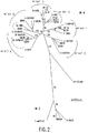
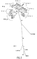
HIV−1ゲノムの3つの領域、すなわちgap p24(399ヌクリオチド)、polインテグラーゼ(864ヌクリオチド)及びenv gp41免疫優性領域(IDR;369ヌクリオチド)を配列分析のための標的とした。ウイルスストックをHIV−1血清陰性ヒト血漿で希釈した。200〜400μlの血漿から全核酸をQIAamp Bloodキット(カリフォルニア州バレンシアに所在のQiagen Inc.)を用いて抽出した。上記した3つの領域をRT−PCR増幅するためのプライマー及び条件は公知である(Brennanら,AIDS,11:1823−1832(1997);Brennanら,AIDS Res.Hum.Retroviruses,13:901−904(1997);Hackettら,AIDS Res.Hum.Retroviruses,13:1155−1158(1997);Swansonら,J.Virol.Methods,89:97−108(2000))。増幅産物をQIAquick PCR精製キット(Qiagen Inc.)を用いて精製した。精製PCR産物の両鎖をABIモデル377自動シーケンサー(カリフォルニア州フォスターシティーに所在のPE Applied Biosystems)及びABI Prism Big Dye Terminator Cycle Sequencingキット(PE Applied Biosystems)を用いて直接配列決定した。ヌクレオチド配列を、すべての群サブタイプ及び群Oを表す樹立リファレンス株からのヌクレオチド配列とアラインさせ、Lasergene 99(ウィスコンシン州マディソンに所在のDNASTAR,Inc.)を用いて分析した。Phylipソフトウェアパッケージ(バージョン3.5c;ワシントン州シアトルに所在のワシントン大学のJ.Felsenstein)を系統分析のために使用した。進化距離をDNADIST(キムラ2パラメーター法)を用いて推定し、系統学的再構成は近隣−結合法(neighbot-joining method)(NEIGHBOR)を用いて推定した。分岐パターンの再現性はSEQBOOTを用いてブーツストラップ分析(100サンプリング)により調べた。3つすべての領域は14個すべてのウイルス単離物からうまくRT−PCR増幅され、配列決定された。系統分析の結果を図1〜3に示し、表2に要約する。 Three regions of the HIV-1 genome were targeted for sequence analysis: gap p24 (399 nucleotides), pol integrase (864 nucleotides) and the env gp41 immunodominant region (IDR; 369 nucleotides). The virus stock was diluted with HIV-1 seronegative human plasma. Total nucleic acid was extracted from 200-400 μl of plasma using the QIAamp Blood kit (Qiagen Inc., Valencia, Calif.). Primers and conditions for RT-PCR amplification of the three regions described above are known (Brennan et al., AIDS, 11: 1833-1832 (1997); Brennan et al., AIDS Res. Hum. Retroviruses, 13: 901-904. (1997); Hackett et al., AIDS Res.Hum.Retroviruses, 13: 1155-1158 (1997); Swanson et al., J. Virol. Methods, 89: 97-108 (2000)). The amplification product was purified using the QIAquick PCR purification kit (Qiagen Inc.). Both strands of the purified PCR product were sequenced directly using an ABI model 377 automated sequencer (PE Applied Biosystems located in Foster City, Calif.) And an ABI Prism Big Dye Terminator Cycle Sequencing Kit (PE Applied Biosystems). Nucleotide sequences were aligned with nucleotide sequences from established reference strains representing all group subtypes and group O and analyzed using Lasergene 99 (DNASTAR, Inc., Madison, Wis.). The Phylip software package (version 3.5c; J. Felstainin, University of Washington, Seattle, WA) was used for phylogenetic analysis. Evolutionary distance was estimated using DNADIST (Kimura two parameter method), and phylogenetic reconstruction was estimated using the neighbor-joining method (NEIGHBOR). The reproducibility of the branch pattern was examined by bootstrap analysis (100 sampling) using SEQBOOT. All three regions were successfully RT-PCR amplified and sequenced from all 14 virus isolates. The results of the phylogenetic analysis are shown in FIGS. 1-3 and summarized in Table 2.
試験のために選択した単離物パネルは地理的な広範囲にわたり、2つのCRF株を含め群Mの最も広く分布しているサブタイプ及び群Oが含まれている。これらの単離物はpolインテグラーゼ遺伝子の系統分析の結果に基づいて選択した。なぜならば、本発明のプライマーはHIV−1のpolインテグラーゼ遺伝子を標的するように設計されているからである。14個の単離物のうち、polインテグラーゼ領域の分析(図2、表2)が示すように群Mサブタイプあたり2つの単離物及び2つの群O単離物が均一に分布している。上記したように、CRF01_AEはpolインテグラーゼ領域においてサブタイプAであり、よってサブタイプCRF01_AE単離物の検出はサブタイプA及びCRF01_AEを検出するプライマーセットの能力を立証している。このパネルの試験から、本発明のプライマーはHIV−1の遺伝学的に多様の株を検出するために有効である。 The isolate panel selected for testing spans a wide geographical area and includes the most widely distributed subtypes of Group M and Group O, including the two CRF strains. These isolates were selected based on the results of phylogenetic analysis of the pol integrase gene. This is because the primer of the present invention is designed to target the pol integrase gene of HIV-1. Of the 14 isolates, 2 isolates and 2 group O isolates were evenly distributed per group M subtype as shown by analysis of the pol integrase region (Figure 2, Table 2). Yes. As noted above, CRF01_AE is subtype A in the pol integrase region, and thus detection of subtype CRF01_AE isolates demonstrates the ability of the primer set to detect subtype A and CRF01_AE. From the tests in this panel, the primers of the present invention are effective for detecting genetically diverse strains of HIV-1.
(実施例3)
HIVサブタイプ検出
表1のプライマーセットがHIV−1群Mサブタイプ及びCRF株及び群O単離物をそれぞれ検出するかを確かめるために、実施例2でキャラクタライズしたウイルス単離物を用いて表1のプライマーセットを試験した。
(Example 3)
HIV subtype detection In order to ascertain whether the primer sets in Table 1 detect HIV-1 group M subtype and CRF strain and group O isolate, respectively, using the virus isolate characterized in Example 2 The primer sets in Table 1 were tested.
PCR増幅産物の分析を容易とするために、精製HIV−1 RNA希釈物を逆転写し、完全polインテグラーゼ遺伝子を表3にリストした群M(polI8及びpolI5)または群O(O−polI8及びO−polI5)特異的プライマーを用いてPCR増幅した。次いで、別の反応で本発明のHIV−1プライマーセット(表1)を用いて第2回増幅を実施した。 To facilitate analysis of PCR amplification products, purified HIV-1 RNA dilutions were reverse transcribed and complete pol integrase genes listed in Table 3 were group M (pol I8 and pol I5) or group O (O-pol I8 and O -PolI5) PCR amplification using specific primers. The second round of amplification was then performed in a separate reaction using the HIV-1 primer set of the present invention (Table 1).
RT−PCRをPerkin Elmer Gene Amp RNA PCRキットの成分を製造業者の指示に従って用いて実施した。PCR緩衝液II、5mMのMgCl2、2.5U/反応物の濃度のMuLV逆転写酵素、それぞれ1.0mMの濃度のdNTP類(dATP、dGTP、dTTP及びdCTP)、1U/反応物の濃度のRNaseインヒビター及び1μMのプライマーを含む反応物(総容量20μl)中で3μlのサンプルを用いて核酸を逆転写することにより相補性DNA(cDNA)を合成した。反応混合物を逆転写し、Perkin−Elmer 9600サーマルサイクラーにおいて増幅した。RT反応混合物をまず42℃で40分間インキュベートした後99℃で5分間インキュベートした。 RT-PCR was performed using components of the Perkin Elmer Gene Amp RNA PCR kit according to the manufacturer's instructions. PCR buffer II, 5 mM MgCl 2 , 2.5 U / reactant concentration of MuLV reverse transcriptase, 1.0 mM each of dNTPs (dATP, dGTP, dTTP and dCTP), 1 U / reactant concentration Complementary DNA (cDNA) was synthesized by reverse transcribing nucleic acid with 3 μl of sample in a reaction (total volume 20 μl) containing RNase inhibitor and 1 μM primer. The reaction mixture was reverse transcribed and amplified in a Perkin-Elmer 9600 thermal cycler. The RT reaction mixture was first incubated at 42 ° C. for 40 minutes and then at 99 ° C. for 5 minutes.
第1回PCR増幅を、100μlの総反応容量につき20μlのcDNA反応物に対して追加試薬を添加して実施した。反応物は最終濃度で5mMのMgCl2、2.5U/反応物のAmplitaq DNAポリメラーゼ、0.5μMの正プライマー及び0.5μMの逆プライマーを含んでいた。次いで、反応を次のようにサイクリングした:95℃×1分間の初期変成、94℃×30秒間,45℃×30秒間及び72℃×90秒間を1サイクルとして40サイクル、その後72℃×10分間の最終インキュベーション。 The first round of PCR amplification was performed by adding additional reagents to 20 μl of the cDNA reaction per 100 μl total reaction volume. The reaction contained 5 mM MgCl 2 at a final concentration, 2.5 U / reactant Amplitaq DNA polymerase, 0.5 μM forward primer and 0.5 μM reverse primer. The reaction was then cycled as follows: initial transformation at 95 ° C. × 1 minute, 40 cycles of 94 ° C. × 30 seconds, 45 ° C. × 30 seconds and 72 ° C. × 90 seconds, followed by 72 ° C. × 10 minutes. Final incubation of.
次いで、完全長polインテグラーゼ増幅産物を鋳型として用いて、本発明のHIV−1プライマーセット(表1)の性能を試験した。各プライマーセットを、5μlの初期PCR反応物、PCR緩衝液、それぞれ0.2mMの濃度のdNTP類(dATP、dGTP、dTTP及びdCTP)、2.5U/反応物のAmplitaq DNAポリメラーゼ及び0.5μMの各プライマーを含む反応物100μl中で個別に試験した。反応混合物をPerkin−Elmer 9600または9700サーマルサイクラーにおいて増幅させた。反応混合物を次のようにサイクリングした:95℃×1分間の初期変成、94℃×30秒間,50℃または55℃×30秒間及び72℃×90秒間を1サイクルとして40サイクル、その後72℃×10分間の最終インキュベーション。アガロースゲル電気泳動までサンプルを4℃に保持した。 The performance of the HIV-1 primer set of the present invention (Table 1) was then tested using the full-length pol integrase amplification product as a template. Each primer set consists of 5 μl of initial PCR reaction, PCR buffer, dNTPs (dATP, dGTP, dTTP and dCTP) at a concentration of 0.2 mM, 2.5 U / reactant Amplitaq DNA polymerase and 0.5 μM Individually tested in 100 μl of reaction containing each primer. The reaction mixture was amplified in a Perkin-Elmer 9600 or 9700 thermal cycler. The reaction mixture was cycled as follows: 95 ° C. × 1 minute initial denaturation, 94 ° C. × 30 seconds, 50 ° C. or 55 ° C. × 30 seconds and 72 ° C. × 90 seconds, 40 cycles, then 72 ° C. × Final incubation for 10 minutes. Samples were kept at 4 ° C. until agarose gel electrophoresis.
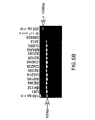
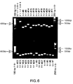
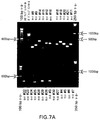
反応産物をアガロースゲル電気泳動により検出した。各100μlの反応物から、5μlを分子量マーカーと一緒にアガロースゲル上に流して、断片の長さを測定した。図4は、各プライマーセットに関してインテグラーゼ遺伝子に対して予想される断片サイズ及び位置を概略的に示す。臭化エチジウムで染色後UV光に曝すことにより断片を可視化した。前記データの代表的サンプリングを図5〜7に示す。図5は、各単離物のプライマーセット#1、#2、#12及び#13を用いる増幅を示す。図6及び7は、群MサブタイプCRF02_AG単離物DJ263及び群O単離物3012の2つの特定単離物について残りのプライマーセットを用いる増幅を示す。本実験で実施した試験データを表4に要約し、HIV−1プライマーセット#1〜30によるHIV−1群MサブタイプA〜G及び群Oの検出を示す。 The reaction product was detected by agarose gel electrophoresis. From each 100 μl reaction, 5 μl was run on an agarose gel with molecular weight markers to determine the length of the fragments. FIG. 4 schematically shows the expected fragment size and position for the integrase gene for each primer set. Fragments were visualized by exposure to UV light after staining with ethidium bromide. A representative sampling of the data is shown in FIGS. FIG. 5 shows amplification using primer sets # 1, # 2, # 12 and # 13 for each isolate. FIGS. 6 and 7 show amplification using the remaining primer sets for two specific isolates, group M subtype CRF02_AG isolate DJ263 and group O isolate 3012. FIG. The test data carried out in this experiment is summarized in Table 4 and shows the detection of HIV-1 group M subtypes AG and group O with HIV-1 primer set # 1-30.
幾つかの断片に関して、反応物の残りの部分を精製し、配列決定して、所期産物の増幅を確認した。精製は、QIAamp PCR精製キットまたはQIAquickゲル抽出キット(Qiagen Inc.)を製造業者の指示に従って用いて実施した。精製PCR断片を本発明の対応プライマー、ABI Prism Big Dyeターミネーターサイクルシーケンシクング反応キット(カリフォルニア州フォスターシティーに所在のPE Applied Biosystems)、AmpliTaq DNAポリメラーゼ及びABIモデル377自動シーケンサー(PE Applied Biosystems)を用いて直接配列決定した。表5は配列決定結果を要約し、予想断片が増幅されたことを示している。 For some fragments, the remainder of the reaction was purified and sequenced to confirm amplification of the desired product. Purification was performed using the QIAamp PCR purification kit or the QIAquick gel extraction kit (Qiagen Inc.) according to the manufacturer's instructions. Purified PCR fragments were prepared using the corresponding primers of the present invention, ABI Prism Big Dye Terminator Cycle Sequencing Reaction Kit (PE Applied Biosystems, Foster City, Calif.), AmpliTaq DNA Polymerase and ABI Model 377 Automatic Sequencer (PE Applied Biosystems). Sequenced directly. Table 5 summarizes the sequencing results and shows that the expected fragment was amplified.
HIV−1プライマーセット#1〜30は、遺伝的に分岐した群O単離物を含めて試験したすべてのHIV−1サブタイプ(表4)をうまく検出した。各プライマーセットに関して、予想される長さを有する断片がアガロースゲル分析に基づいて検出された。配列決定した断片に関して、各増幅断片が標的単離物のインテグラーゼ配列との比較に基づいて予想される断片であったことが配列分析から確認された。 HIV-1 primer sets # 1-30 successfully detected all HIV-1 subtypes tested (Table 4), including genetically branched group O isolates. For each primer set, fragments with the expected length were detected based on agarose gel analysis. For the sequenced fragments, sequence analysis confirmed that each amplified fragment was the expected fragment based on comparison with the integrase sequence of the target isolate.
(実施例4)
HIV−1感受性
実施例2及び3に記載されている2つのウイルス単離物を使用して、表1のプライマーセットの感受性を評価した。前記単離物を脱繊維素HIV−1血清陰性ヒト血漿で希釈した後Abbott LCx HIV定量RNAアッセイ(イリノイ州アボットパークに所在のAbbott Laboratories)を用いて試験してウイルス負荷を測定した。次いで、希釈したサンプルを実施例3に記載されているプライマーセット1〜30を用いて試験した。試験のために選択した2つの単離物のうち1つは、1585コピー/mlの群M単離物のUG274(サブタイプD)、1つは1479コピー/mlの群O単離物の3012であった。
Example 4
HIV-1 Sensitivity Two virus isolates described in Examples 2 and 3 were used to assess the sensitivity of the primer sets in Table 1. The isolate was diluted with defibrinated HIV-1 seronegative human plasma and then tested using the Abbott LCx HIV quantitative RNA assay (Abbott Laboratories, Abbot Park, Ill.) To determine viral load. The diluted samples were then tested using primer sets 1-30 as described in Example 3. One of the two isolates selected for testing was UG274 (subtype D) of 1585 copies / ml group M isolate and 3012 of
感受性試験の結果を表6に示す。すべてのプライマーセットが両方の単離物をうまく検出した。1つのプライマーセット#1では、群O単離物を検出するためにPCR条件を僅かに修正しなければならなかった。群O単離物をプライマーセット#1を用いて最初に試験したところ、混合結果を示した。第1の試験は、アガロースゲルで予想される断片長さの非常に弱いバンドを示した。しかしながら、試験を繰り返してもバンドは示さなかった。サイクリング中のアニーリング温度を50℃から45℃に下げることによりPCR条件を僅かに修正すると、結果が改善され、アガロースゲル電気泳動により明瞭なバンドが検出された。実施例3に記載されている条件はすべての単離物に対してプライマーセットを通常スクリーニングするために設計した。プライマーセット#1の結果は、最適感受性のために増幅条件が各プライマーセットに対して特異的に最適化し得ることを示している。
The results of the sensitivity test are shown in Table 6. All primer sets successfully detected both isolates. For one
上記結果は、本発明のプライマーセットがHIV群M及び群Oを試験した最低濃度の1500コピー/mlという感受性で検出することを示している。 The above results show that the primer set of the present invention detects with a sensitivity of 1500 copies / ml, the lowest concentration tested for HIV group M and group O.
(実施例5)
HIV−1 M及びO増幅産物の分子ビーコンプローブを用いる検出
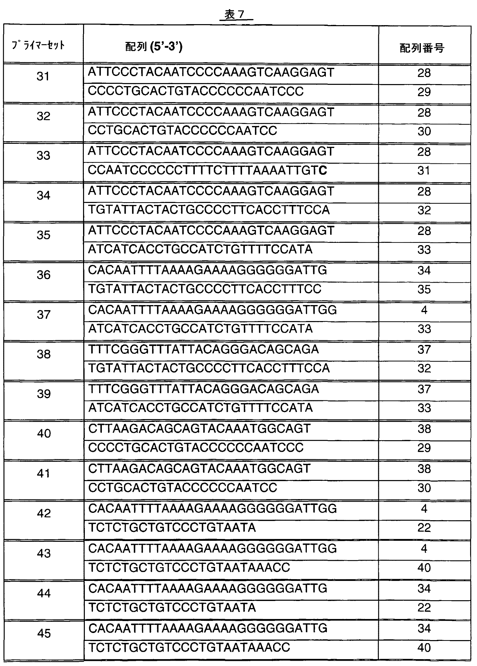
分子ビーコンプローブを用いて検出した増幅産物を作成するために使用したプライマーは、配列番号28、配列番号29、配列番号30、配列番号31、配列番号32、配列番号33、配列番号34、配列番号35、配列番号4、配列番号37、配列番号38、配列番号22及び配列番号40であった。プライマー配列は標準オリゴヌクレオチド合成法を用いて合成した。HIVを検出するために、プライマー配列を下表7に示すようにプライマーセットで一緒に使用した。表中、正プライマーを対の上段、逆プライマーを対の下段に示す。
(Example 5)
Detection using molecular beacon probes of HIV-1 M and O amplification products Primers used to generate amplification products detected using molecular beacon probes are SEQ ID NO: 28, SEQ ID NO: 29, SEQ ID NO: 30, SEQ ID NO: 31. SEQ ID NO: 32, SEQ ID NO: 33, SEQ ID NO: 34, SEQ ID NO: 35, SEQ ID NO: 4, SEQ ID NO: 37, SEQ ID NO: 38, SEQ ID NO: 22 and SEQ ID NO: 40. Primer sequences were synthesized using standard oligonucleotide synthesis methods. To detect HIV, the primer sequences were used together in a primer set as shown in Table 7 below. In the table, the forward primer is shown at the top of the pair and the reverse primer is shown at the bottom of the pair.
(実施例6)
HIV分子ビーコンプローブの作成
増幅したHIVインテグラーゼ標的配列とオリゴヌクレオチドハイブリダイゼーションによりハイブリダイズするために分子ビーコンプローブを設計した。これらのプローブは、配列番号41、配列番号42、配列番号43、配列番号44、配列番号45、配列番号46、配列番号47、配列番号48、配列番号49、配列番号50、配列番号51、配列番号52、配列番号53、配列番号54、配列番号55、配列番号56、配列番号57、配列番号58、配列番号59、配列番号60、配列番号61、配列番号62、配列番号63、配列番号64及び配列番号65であった。プローブ配列は標準オリゴヌクレオチド合成法を用いて合成し、援用により本明細書に含まれるとする米国特許第5,464,746号明細書に記載されている標準シアノエチルホスホルアミデートカップリング化学を用いて5’末端を蛍光6−カルボキシフルオレセイン(6−FAM)、3’末端をC6−NH−DABCYLで標識した。使用したHIV分子ビーコンプローブ配列を下表8に示す。
(Example 6)
Generation of HIV molecular beacon probes Molecular beacon probes were designed to hybridize with amplified HIV integrase target sequences by oligonucleotide hybridization. These probes are SEQ ID NO: 41, SEQ ID NO: 42, SEQ ID NO: 43, SEQ ID NO: 44, SEQ ID NO: 45, SEQ ID NO: 46, SEQ ID NO: 47, SEQ ID NO: 48, SEQ ID NO: 49, SEQ ID NO: 50, SEQ ID NO: 51, SEQ ID NO: 51. SEQ ID NO: 52, SEQ ID NO: 53, SEQ ID NO: 54, SEQ ID NO: 55, SEQ ID NO: 56, SEQ ID NO: 57, SEQ ID NO: 58, SEQ ID NO: 59, SEQ ID NO: 60, SEQ ID NO: 61, SEQ ID NO: 62, SEQ ID NO: 63, SEQ ID NO: 64 And SEQ ID NO: 65. The probe sequence is synthesized using standard oligonucleotide synthesis methods and uses the standard cyanoethyl phosphoramidate coupling chemistry described in US Pat. No. 5,464,746, which is hereby incorporated by reference. Used, the 5 ′ end was labeled with fluorescent 6-carboxyfluorescein (6-FAM) and the 3 ′ end was labeled with C6-NH-DABCYL. The HIV molecular beacon probe sequences used are shown in Table 8 below.
上表8において、大文字はHIVに特異的な配列を表し、小文字は分子ビーコンプローブのステムを作成するために使用したランダム配列を表し、囲んだ領域はこのステムを形成する配列である。 In Table 8 above, uppercase letters represent sequences specific for HIV, lowercase letters represent random sequences used to create the stems of molecular beacon probes, and the enclosed region is the sequence that forms this stem.
プローブA(配列番号41)、プローブB(配列番号42)、プローブC(配列番号43)、プローブD(配列番号44)、プローブE(配列番号45)、プローブF(配列番号46)、プローブG(配列番号47)及びプローブH(配列番号48)はプライマーセット31、32、33、34及び35と一緒に使用され得る。プローブI(配列番号49)はプライマーセット31、32、34及び35と一緒に使用され得る。プローブJ(配列番号50)、プローブK(配列番号51)、プローブL(配列番号52)及びプローブM(配列番号53)はプライマーセット31、32、34、35、40及び41と一緒に使用され得る。プローブO(配列番号54)はプライマーセット34、35、36及び37と一緒に使用され得る。プローブP(配列番号55)はプライマーセット34、35、36、37、38及び39と一緒に使用され得る。プローブQ(配列番号56)及びプローブR(配列番号57)はプライマーセット35、37及び39と一緒に使用され得る。 Probe A (SEQ ID NO: 41), Probe B (SEQ ID NO: 42), Probe C (SEQ ID NO: 43), Probe D (SEQ ID NO: 44), Probe E (SEQ ID NO: 45), Probe F (SEQ ID NO: 46), Probe G (SEQ ID NO: 47) and probe H (SEQ ID NO: 48) can be used with primer sets 31, 32, 33, 34 and 35. Probe I (SEQ ID NO: 49) can be used with primer sets 31, 32, 34 and 35. Probe J (SEQ ID NO: 50), probe K (SEQ ID NO: 51), probe L (SEQ ID NO: 52) and probe M (SEQ ID NO: 53) are used together with primer sets 31, 32, 34, 35, 40 and 41. obtain. Probe O (SEQ ID NO: 54) can be used with primer sets 34, 35, 36 and 37. Probe P (SEQ ID NO: 55) can be used with primer sets 34, 35, 36, 37, 38 and 39. Probe Q (SEQ ID NO: 56) and probe R (SEQ ID NO: 57) can be used with primer sets 35, 37 and 39.
(実施例7)
分子ビーコンプローブを用いるプライマーセットの感受性
実施例5で作成した表7に示すプライマーセットの性能を、HIV RNAサンプルの希釈物(K.Abravayaら,J.Clin.Microbiol.,38:716−723(2000))を実施例6で作成した表8に示す特定の分子ビーコンプローブで試験して評価した。精製HIV RNAを100,000コピー/ml、10,000コピー/ml、1,000コピー/ml、100コピー/ml及び25コピー/mlに希釈した後、いろいろなプライマーセット/プローブの組合せを用いる別々の反応で逆転写し、PCR増幅し、検出した。HIV RNAを含まないネガティブコントロールもそれぞれのプライマーセット/プローブの組合せに含めた。RT−PCRを、130nMの適切な正プライマー、478nMの適切な逆プライマー、81nMの適切なHIV分子ビーコンプローブ、4.38mMのMnCl2、0.375mMの各dNTP(dATP、dGTP、dTTp及びdCTP)、13単位の組換えサーマス・サーモフィルス(Thermus thermophilus)ポリメラーゼ、Bicine緩衝液及びHIV RNA希釈物またはネガティブコントロールを含む反応混合物100μl中で実施した。
(Example 7)
Sensitivity of primer set using molecular beacon probe The performance of the primer set shown in Table 7 prepared in Example 5 was compared to the dilution of HIV RNA sample (K. Abravaya et al., J. Clin. Microbiol., 38: 716-723 ( 2000)) were tested and evaluated with the specific molecular beacon probes shown in Table 8 prepared in Example 6. Separate dilutions using various primer set / probe combinations after dilution of purified HIV RNA to 100,000 copies / ml, 10,000 copies / ml, 1,000 copies / ml, 100 copies / ml and 25 copies / ml The reaction was reverse transcribed, PCR amplified and detected. A negative control without HIV RNA was also included in each primer set / probe combination. RT-PCR was performed using 130 nM appropriate forward primer, 478 nM appropriate reverse primer, 81 nM appropriate HIV molecular beacon probe, 4.38 mM MnCl 2 , 0.375 mM each dNTP (dATP, dGTP, dTTp and dCTP). The reaction mixture was run in 100 μl, containing 13 units of recombinant Thermus thermophilus polymerase, Bicine buffer and HIV RNA dilution or negative control.
反応混合物を逆転写し、Perkin−Elmer 9700サーマルサイクラーにおいて増幅させた。まず、反応混合物を59℃で30分間インキュベートしてRNAを逆転写した後、95℃×30秒間、54℃×30秒間及び72℃×30秒間を4サイクル実施した。次いで、90℃×30秒間、59℃×30秒間及び72℃×30秒間を36〜40サイクル実施して増幅させた。反応混合物を熱サイクルにかけた後、混合物を94℃に5秒間上昇させ、温度を45℃に15秒間下げ、更に25℃に10秒間下げることによりプローブオリゴハイブリダイゼーションを実施した。反応産物が検出されるまでサンプルを25℃で維持した。反応産物をCytofluor(マサチューセッツ州フレーミングハムに所在のPerceptive)またはBioTek 600(カリフォルニア州フォスターシティーに所在のApplied Biosystems)のような蛍光リーダーを用いて検出した。蛍光単位で表示した結果を下表9に示す。 The reaction mixture was reverse transcribed and amplified in a Perkin-Elmer 9700 thermal cycler. First, the reaction mixture was incubated at 59 ° C. for 30 minutes to reverse-transcribe RNA, and then subjected to 4 cycles of 95 ° C. × 30 seconds, 54 ° C. × 30 seconds, and 72 ° C. × 30 seconds. Subsequently, amplification was performed by performing 36 to 40 cycles of 90 ° C. × 30 seconds, 59 ° C. × 30 seconds, and 72 ° C. × 30 seconds. After subjecting the reaction mixture to thermal cycling, probe oligo hybridization was performed by raising the mixture to 94 ° C. for 5 seconds, lowering the temperature to 45 ° C. for 15 seconds, and then to 25 ° C. for 10 seconds. Samples were kept at 25 ° C. until reaction products were detected. Reaction products were detected using a fluorescence reader such as Cytofluor (Perceptive, Framingham, Mass.) Or BioTek 600 (Applied Biosystems, Foster City, Calif.). The results displayed in fluorescence units are shown in Table 9 below.
プライマーセット31、32及び38は最良の性能を与え、ネガティブコントロール(0コピー/ml)と簡単に判別される25コピー/mlのHIVを検出した。プライマーセット33、39、40及び41は1000〜10,000コピー/mlのHIVを検出した。プライマーセット34、35、36及び37はうまく働かなかったが、これは使用したプローブのせいではなかった。なぜならは、プローブAまたはPは他のプライマーセットと併用したときには良好な結果を示したからである。 Primer sets 31, 32 and 38 gave the best performance and detected 25 copies / ml of HIV, which was easily distinguished from the negative control (0 copies / ml). Primer sets 33, 39, 40 and 41 detected 1000-10,000 copies / ml of HIV. Primer sets 34, 35, 36 and 37 did not work well, but this was not due to the probe used. This is because probe A or P showed good results when used in combination with other primer sets.
プライマーセット42、43、44及び45をプローブアニーリングステップを省いた以外は上記したように試験した。ゲルをRT−PCR産物と一緒に流すことにより結果を分析した。10,000コピー/mlのHIVを用いて予想されるバンドが観察された。 Primer sets 42, 43, 44 and 45 were tested as described above except that the probe annealing step was omitted. Results were analyzed by running the gel with the RT-PCR product. The expected band was observed with 10,000 copies / ml of HIV.
(実施例8)
分子ビーコンプローブの感受性
実施例6で作成した表8に示す分子ビーコンプローブA〜Mの性能を、実施例7のHIV RNA希釈物を実施例5で作成したプライマーセット32を用いて試験することにより評価した。また、プローブOをプライマーセット35と併用し、プローブP、Q及びRをプライマーセット39と併用した。結果を下表10に示す。
(Example 8)
Molecular Beacon Probe Sensitivity By testing the performance of the molecular beacon probes A to M shown in Table 8 prepared in Example 6 using the primer set 32 prepared in Example 5 from the HIV RNA dilution of Example 7. evaluated. Probe O was used in combination with primer set 35, and probes P, Q, and R were used in combination with primer set 39. The results are shown in Table 10 below.
プローブA、B、E、H及びIは25コピー/mlのHIVを検出し、この量はネガティブコントロール(0コピー/ml)と判別できた。プローブJ、K、M、P及びRは100〜1000コピー/mlのHIVを検出した。プローブF、O及びQはあまり有効でなく、プローブC、D、G及びLはネガティブコントロールでより高いバックグランウンド値を示した。プローブC、D、E及びFで観察された性能の違いは驚くものであった。なぜならば、これらのプローブは同じHIV結合配列を有していたからである。しかしながら、これらのプローブはステム配列の組成にしか違いがなかった。プローブJ、K及びLも、HIV結合配列中の塩基の違いは1つしかなく、ステム配列セット成は同一またはほぼ同一である点で驚くことであった。プローブJ及びKは約1000コピー/mlのHIVを検出したのに対して、プローブLはJまたはKよりも高いバックグラウンド値を与えた。 Probes A, B, E, H and I detected 25 copies / ml of HIV, and this amount was distinguishable from the negative control (0 copies / ml). Probes J, K, M, P and R detected 100-1000 copies / ml of HIV. Probes F, O and Q were less effective and probes C, D, G and L showed higher background values in the negative control. The difference in performance observed with probes C, D, E and F was surprising. This is because these probes had the same HIV binding sequence. However, these probes differed only in the composition of the stem sequence. Probes J, K and L were also surprising in that there was only one base difference in the HIV binding sequence and the stem sequence set was identical or nearly identical. Probes J and K detected about 1000 copies / ml of HIV, while probe L gave a higher background value than J or K.
(実施例9)
インドールまたはイノシン置換基を有する分子ビーコンプローブの感受性
プローブA、B、E、H及びIを325個の公知HIV−1配列に対して分析した。最も一般的なミスマッチを有する部位を同定し、プローブをそのミスマッチ位置にユニバーサル塩基を含むように修飾した。プローブ内のミスマッチ位置を表11に示すようにニトロインドールまたはイノシンで置換した。これらの修飾プロープは実施例6のように合成した。前記修飾プローブの配列を下表11に示す。
Example 9
Sensitive probes A, B, E, H and I of molecular beacon probes with indole or inosine substituents were analyzed against 325 known HIV-1 sequences. The site with the most common mismatch was identified and the probe was modified to include a universal base at that mismatch location. The mismatch position in the probe was substituted with nitroindole or inosine as shown in Table 11. These modified probes were synthesized as in Example 6. The sequences of the modified probes are shown in Table 11 below.
インドールまたはイノシン分子ビーコンプローブの性能を、上記実施例7のようにHIV RNAの希釈物を実施例5で作成したプライマーセット32を用いて試験することにより評価した。結果を下表12に示す。インドールまたはイノシン置換基を含むプローブはすべて25コピー/mlのHIVを検出し、この量はネガティブコントロール(0コピー/ml)と判別できた。 The performance of the indole or inosine molecular beacon probe was evaluated by testing a dilution of HIV RNA using primer set 32 prepared in Example 5 as in Example 7 above. The results are shown in Table 12 below. All probes containing indole or inosine substituents detected 25 copies / ml of HIV, which could be discriminated from the negative control (0 copies / ml).
(実施例10)
各種HIVサブタイプの分子ビーコンプローブを用いる検出
各種HIVサブタイプを入手し、RNAをK.Abravayaら,J.Clin.Microbiol.,38:716−723(2000)及びJ.Johnsonら,J.Virol.Methods,95:81−92(2001)に記載されているように単離した。各種サブタイプから単離したHIV RNAを約1000コピー/mlに希釈し、プライマーセット32+プローブA、プライマーセット32+プローブHまたはプライマーセット38+プローブPを用いてRT−PCR及びプローブオリゴハイブリダイゼーションを実施して実施例7に記載されているように試験した。プライマーセットは実施例7に記載されており、蛍光単位で表示した。下表13から分かるように、すべてのHIVサブタイプが試験した3つのプライマー/プローブセットで検出された。
(Example 10)
Detection using various beacon probes of various HIV subtypes. Abravaya et al., J. MoI. Clin. Microbiol. 38: 716-723 (2000) and J. MoI. Johnson et al., J. MoI. Virol. Isolated as described in Methods, 95: 81-92 (2001). HIV RNA isolated from various subtypes was diluted to about 1000 copies / ml, and RT-PCR and probe oligo hybridization were performed using primer set 32 + probe A, primer set 32 + probe H or primer set 38 + probe P. Were tested as described in Example 7. Primer sets are described in Example 7 and are expressed in fluorescence units. As can be seen from Table 13 below, all HIV subtypes were detected with the three primer / probe sets tested.
RNA転写物を、K.Abravayaら,J.Clin.Microbiol.,38:716−723(2000)及びJ.Johnsonら,J.Virol.Methods,95:81−92(2001)に記載されているようにHIVサブタイプA、C、D、CRF01_AE及びFのクローンから作成した。転写物を約10,000コピー/mlに希釈し、プライマーセット32+プロープAまたはプローブHを用いて実施例8に記載されているように増幅し、検出した。表14の結果は、試験したすべてのHIVサブタイプが使用した2つのプライマー/プローブセットで検出されたことを示している。 RNA transcripts were obtained from K. Abravaya et al., J. MoI. Clin. Microbiol. 38: 716-723 (2000) and J. MoI. Johnson et al., J. MoI. Virol. Methods, 95: 81-92 (2001), from HIV subtype A, C, D, CRF01_AE and F clones. Transcripts were diluted to approximately 10,000 copies / ml, amplified and detected as described in Example 8 using primer set 32 + probe A or probe H. The results in Table 14 indicate that all tested HIV subtypes were detected with the two primer / probe sets used.
(実施例11)
各種HIVサブタイプの分子ビーコンプローブを用いる定量
実施例10に記載されている各種HIVサブタイプから単離したRNAを約1000(すなわち、3log)コピー/mlに希釈し、プライマーセット32+プローブAまたはプライマーセット32+プローブHを用いて実施例7に記載されているように試験した。定量結果を得るために、RT−PCR反応混合物には0.1μMのInternal Control転写物配列に対して特異的な分子ビーコンプローブ
(Example 11)
Quantification using molecular beacon probes of various HIV subtypes RNA isolated from the various HIV subtypes described in Example 10 is diluted to approximately 1000 (ie, 3 log) copies / ml, primer set 32 + probe A or primer Tested as described in Example 7 using set 32 + probe H. To obtain quantitative results, the RT-PCR reaction mixture contains a molecular beacon probe specific for the 0.1 μM Internal Control transcript sequence
![]()
![]()
各種HIVサブタイプからのRNA単離物のこの方法による定量結果を表15に示す。結果はlogコピー/mlで表示する。サブタイプA単離物327を除いて、試験したすべての単離物が約3logコピー/mlで定量された。この単離物をプライマーセット31を用いても試験した。このプライマーセット31は逆プライマーが3塩基長いこと(表7参照;5’末端に2つの追加ヌクレオチド、3’末端に1つの追加ヌクレオチド)を除いてプライマー32と同一であった。シグナルは2倍高かった(データ示さず)。 The quantification results of this method for RNA isolates from various HIV subtypes are shown in Table 15. Results are displayed in log copies / ml. With the exception of subtype A isolate 327, all isolates tested were quantified at approximately 3 log copies / ml. This isolate was also tested using primer set 31. This primer set 31 was identical to primer 32 except that the reverse primer was 3 bases longer (see Table 7; 2 additional nucleotides at the 5 'end and 1 additional nucleotide at the 3' end). The signal was twice as high (data not shown).
また、実施例10に記載されているようにサブタイプA、C、D、CRF01_AE及びFを代表とするHIV−1単離物のクローンから作成したRNA転写物を約10,000コピー/mlに希釈し、プライマーセット32+プローブAまたはプローブHを用いて実施例11に上記したように定量した。表16の結果は、ほとんどすべてのHIV転写物(K.Abravayaら,J.Clin.Microbiol.,38:716−723(2000)参照)が4logコピー/mlで定量されたことを示している。 In addition, RNA transcripts made from clones of HIV-1 isolates typified by subtypes A, C, D, CRF01_AE and F as described in Example 10 were approximately 10,000 copies / ml. Diluted and quantified as described above in Example 11 using primer set 32 + probe A or probe H. The results in Table 16 indicate that almost all HIV transcripts (see K. Abravaya et al., J. Clin. Microbiol., 38: 716-723 (2000)) were quantified at 4 log copies / ml.
本発明を特定実施例を参照して詳細に説明してきたが、本発明の趣旨及び範囲を逸脱することなく前記実施例に各種の変更び改変を加えることができることは当業者に自明である。 Although the invention has been described in detail with reference to specific embodiments, it will be apparent to those skilled in the art that various changes and modifications can be made to the embodiments without departing from the spirit and scope of the invention.
Claims (5)
b)前記反応混合物を増幅条件下に置いて増幅産物を形成するステップ、
c)増幅産物及び配列番号58のヌクレオチド配列で表されるヌクレオチドからなるプローブとハイブリッドを形成するステップ、及び
d)前記ハイブリッドを試験サンプル中のHIV−1の存在の指標として検出するステップ
を含むことを特徴とする試験サンプル中のHIV−1の存在の検出方法。 a) contacting a test sample with an amplification reagent to form a reaction mixture;
b) subjecting the reaction mixture to amplification conditions to form an amplification product;
c) forming a hybrid with the amplification product and a probe consisting of the nucleotide represented by the nucleotide sequence of SEQ ID NO: 58 ; and d) detecting the hybrid as an indicator of the presence of HIV-1 in the test sample. A method for detecting the presence of HIV-1 in a test sample characterized by
Applications Claiming Priority (2)
| Application Number | Priority Date | Filing Date | Title |
|---|---|---|---|
| US09/945,943 | 2001-09-04 | ||
| US09/945,943 US6852491B2 (en) | 2001-09-04 | 2001-09-04 | Amplification and detection reagents for HIV-1 |
Related Parent Applications (1)
| Application Number | Title | Priority Date | Filing Date |
|---|---|---|---|
| JP2003525582A Division JP4413610B2 (en) | 2001-09-04 | 2002-07-12 | HIV-1 amplification and detection reagents |
Related Child Applications (1)
| Application Number | Title | Priority Date | Filing Date |
|---|---|---|---|
| JP2012246079A Division JP2013063072A (en) | 2001-09-04 | 2012-11-08 | Amplification and detection reagent for hiv-1 |
Publications (2)
| Publication Number | Publication Date |
|---|---|
| JP2009291202A JP2009291202A (en) | 2009-12-17 |
| JP5205346B2 true JP5205346B2 (en) | 2013-06-05 |
Family
ID=25483738
Family Applications (3)
| Application Number | Title | Priority Date | Filing Date |
|---|---|---|---|
| JP2003525582A Expired - Fee Related JP4413610B2 (en) | 2001-09-04 | 2002-07-12 | HIV-1 amplification and detection reagents |
| JP2009180578A Expired - Fee Related JP5205346B2 (en) | 2001-09-04 | 2009-08-03 | HIV-1 amplification and detection reagents |
| JP2012246079A Withdrawn JP2013063072A (en) | 2001-09-04 | 2012-11-08 | Amplification and detection reagent for hiv-1 |
Family Applications Before (1)
| Application Number | Title | Priority Date | Filing Date |
|---|---|---|---|
| JP2003525582A Expired - Fee Related JP4413610B2 (en) | 2001-09-04 | 2002-07-12 | HIV-1 amplification and detection reagents |
Family Applications After (1)
| Application Number | Title | Priority Date | Filing Date |
|---|---|---|---|
| JP2012246079A Withdrawn JP2013063072A (en) | 2001-09-04 | 2012-11-08 | Amplification and detection reagent for hiv-1 |
Country Status (6)
| Country | Link |
|---|---|
| US (1) | US6852491B2 (en) |
| EP (3) | EP2385144A1 (en) |
| JP (3) | JP4413610B2 (en) |
| CA (2) | CA2459354C (en) |
| ES (1) | ES2458556T3 (en) |
| WO (1) | WO2003020878A2 (en) |
Families Citing this family (27)
| Publication number | Priority date | Publication date | Assignee | Title |
|---|---|---|---|---|
| AU3219400A (en) * | 1999-02-03 | 2000-08-25 | Government Of The United States Of America, As Represented By The Secretary Of The Department Of Health And Human Services, The | Methods and reagents for molecular detection of hiv-1 groups m, n and |
| CN1910289B (en) * | 2004-01-07 | 2012-05-23 | 日立化成研究中心公司 | Primers and probes for HIV detection |
| EP1602736A1 (en) * | 2004-06-04 | 2005-12-07 | Bio-Rad Pasteur | HIV type and subtype detection |
| ES2610781T3 (en) | 2004-09-14 | 2017-05-03 | Argos Therapeutics, Inc. | Strain independent pathogen amplification and vaccines for these |
| EP2975139B1 (en) | 2004-09-30 | 2019-11-06 | Gen-Probe Incorporated | Assay for detecting and quantifying hiv-1 |
| AU2011253599B2 (en) * | 2004-09-30 | 2013-12-12 | Gen-Probe Incorporated | Assay for detecting and quantifying HIV-1 |
| US8673551B2 (en) * | 2005-12-07 | 2014-03-18 | Speedx Pty Ltd. | Methods, plasmid vectors and primers for assessing HIV viral fitness |
| US20090061413A1 (en) * | 2006-02-27 | 2009-03-05 | Salinas Frank G | Isothermal screening of hiv-1 related nucleic acids |
| US8338101B2 (en) * | 2007-01-23 | 2012-12-25 | Virco Bvba | Method for designing a drug regime for HIV-infected patients |
| WO2009017743A2 (en) | 2007-07-30 | 2009-02-05 | Argos Therapeutics, Inc. | Improved primers and probes for the amplification and detection of hiv gag, rev and nef polynucleotides |
| CA2639416C (en) * | 2007-09-11 | 2019-12-31 | F. Hoffmann-La Roche Ag | Diagnostic test for susceptibility to b-raf kinase inhibitors |
| WO2010013232A1 (en) * | 2008-07-29 | 2010-02-04 | Applied Materials Israel Ltd. | Mapping variations of a surface |
| US8691504B2 (en) * | 2009-05-26 | 2014-04-08 | Xiamen University | Method for detecting variations in nucleic acid sequences |
| BR112012027151A8 (en) * | 2010-04-23 | 2018-06-26 | Genomic Vision | method for detecting or identifying an infectious or genomic viral polynucleotide sequence in a mammalian cell, tissue or biological fluid, method for detecting or tracking viral presence, viral replication or viral genomic rearrangements in a mammalian cell, method for evaluating efficacy of an antiviral treatment, probe set, kit to perform molecular combing and biological materials. |
| EP2729583B1 (en) | 2011-07-05 | 2019-05-22 | The Govt. Of The U.S.A. As Represented By The Secretary Of The Department Of Health & Human Services | Hiv-1 genotyping assay for global surveillance of hiv-1 drug resistance |
| WO2013116039A1 (en) * | 2012-01-31 | 2013-08-08 | Advanced Liquid Logic Inc | Amplification primers and probes for detection of hiv-1 |
| BR112014019168B1 (en) | 2012-02-03 | 2021-10-05 | California Institute Of Technology | METHOD CAPABLE OF DETECTING IN A NON-DEGENERATED PRESENCE OR ABSENCE OF ANALYTES IN A SINGLE SAMPLE VOLUME AND METHOD OF DETECTION OF THE PRESENCE OR ABSENCE OF EACH ANALYTE AMONG A PLURALITY OF ANALYTES |
| CN104662172B (en) | 2012-08-03 | 2018-07-06 | 加州理工学院 | Multiplexing and quantification in PCR with reduced hardware and requirements |
| WO2014037712A2 (en) * | 2012-09-04 | 2014-03-13 | Genome Research Limited | Hiv-1 detection |
| WO2015118491A1 (en) * | 2014-02-06 | 2015-08-13 | University Of The Witwatersrand, Johannesburg | Method for detecting hiv |
| US11028443B2 (en) | 2015-08-31 | 2021-06-08 | Showa Denko Materials Co., Ltd. | Molecular methods for assessing urothelial disease |
| US10066263B2 (en) | 2016-06-17 | 2018-09-04 | California Institute Of Technology | Nucleic acid reactions and related methods and compositions |
| CN112424377A (en) | 2018-04-17 | 2021-02-26 | 克罗玛科德公司 | Method and system for multiplex analysis |
| US12203129B2 (en) | 2018-07-03 | 2025-01-21 | ChromaCode, Inc. | Formulations and signal encoding and decoding methods for massively multiplexed biochemical assays |
| WO2020169705A1 (en) * | 2019-02-20 | 2020-08-27 | Biomedrex Ab | Virus therapy |
| KR102191513B1 (en) | 2020-02-13 | 2020-12-15 | 주식회사 에스씨엘헬스케어 | A Primer set for HIV-1 drug resistance analysis, a kit comprising the primer set, and analysis method using the same |
| CN112795699A (en) * | 2021-02-09 | 2021-05-14 | 南方医科大学 | Universal primer, probe and detection method for detecting different subtypes of HIV-1 virus by RT-RAA fluorescence method |
Family Cites Families (30)
| Publication number | Priority date | Publication date | Assignee | Title |
|---|---|---|---|---|
| US4948882A (en) | 1983-02-22 | 1990-08-14 | Syngene, Inc. | Single-stranded labelled oligonucleotides, reactive monomers and methods of synthesis |
| US5185444A (en) | 1985-03-15 | 1993-02-09 | Anti-Gene Deveopment Group | Uncharged morpolino-based polymers having phosphorous containing chiral intersubunit linkages |
| AU5661386A (en) | 1985-03-15 | 1986-10-13 | Stirchak, E. | Stereoregular polynucleotide-binding polymers |
| US5034506A (en) | 1985-03-15 | 1991-07-23 | Anti-Gene Development Group | Uncharged morpholino-based polymers having achiral intersubunit linkages |
| US4683195A (en) | 1986-01-30 | 1987-07-28 | Cetus Corporation | Process for amplifying, detecting, and/or-cloning nucleic acid sequences |
| US4683202A (en) | 1985-03-28 | 1987-07-28 | Cetus Corporation | Process for amplifying nucleic acid sequences |
| US5310652A (en) | 1986-08-22 | 1994-05-10 | Hoffman-La Roche Inc. | Reverse transcription with thermostable DNA polymerase-high temperature reverse transcription |
| US5322770A (en) | 1989-12-22 | 1994-06-21 | Hoffman-Laroche Inc. | Reverse transcription with thermostable DNA polymerases - high temperature reverse transcription |
| US6004826A (en) * | 1988-07-20 | 1999-12-21 | David Segev | Repair-mediated process for amplifying and detecting nucleic acid sequences |
| CA2003383A1 (en) * | 1988-11-23 | 1990-05-23 | Sushil G. Devare | Synthetic dna derived recombinant hiv antigens |
| EP0388602B1 (en) * | 1989-02-03 | 1994-11-09 | Abbott Laboratories | Monoclonal antibody for differentiating HIV-2 from HIV-1 seropositive individuals |
| EP1715064B1 (en) * | 1989-06-02 | 2009-02-25 | Institut Pasteur | Nucleotide sequences derived from the genomes of the retroviruses HIV-1, HIV-2 and SIV, and their application to the amplification of pol sequences in these viral genomes and to the in vitro diagnosis of infections resulting from these viruses |
| CA2020958C (en) | 1989-07-11 | 2005-01-11 | Daniel L. Kacian | Nucleic acid sequence amplification methods |
| US5210015A (en) | 1990-08-06 | 1993-05-11 | Hoffman-La Roche Inc. | Homogeneous assay system using the nuclease activity of a nucleic acid polymerase |
| DK51092D0 (en) | 1991-05-24 | 1992-04-15 | Ole Buchardt | OLIGONUCLEOTIDE ANALOGUE DESCRIBED BY PEN, MONOMERIC SYNTHONES AND PROCEDURES FOR PREPARING THEREOF, AND APPLICATIONS THEREOF |
| US5424414A (en) | 1991-12-17 | 1995-06-13 | Abbott Laboratories | Haptens, tracers, immunogens and antibodies for 3-phenyl-1-adamantaneacetic acids |
| US5464746A (en) | 1991-12-17 | 1995-11-07 | Abbott Laboratories | Haptens, tracers, immunogens and antibodies for carbazole and dibenzofuran derivatives |
| AU4387193A (en) | 1992-05-29 | 1993-12-30 | Abbott Laboratories | Ligase chain reaction starting with rna sequences |
| EP2423322A1 (en) * | 1993-11-12 | 2012-02-29 | PHRI Properties, Inc. | Hybridization probes for nucleic acid detection, universal stems, methods and kits |
| US5925517A (en) | 1993-11-12 | 1999-07-20 | The Public Health Research Institute Of The City Of New York, Inc. | Detectably labeled dual conformation oligonucleotide probes, assays and kits |
| US5733781A (en) * | 1994-07-19 | 1998-03-31 | Gen-Probe Incorporated | Oligonucleotides and methods for inhibiting propagation of human immunodeficiency virus |
| US5962665A (en) * | 1997-06-16 | 1999-10-05 | Abbott Laboratories | Nucleic acid primers and probes for detecting HIV-1 and HIV-2 |
| US6001558A (en) * | 1997-06-25 | 1999-12-14 | Ortho Clinical Diagnostics, Inc. | Amplification and detection of HIV-1 and/or HIV 2 |
| US6391544B1 (en) * | 1998-05-15 | 2002-05-21 | Abbott Laboratories | Method for using unequal primer concentrations for generating nucleic acid amplification products |
| DE19850186A1 (en) * | 1998-10-30 | 2000-05-25 | Roche Diagnostics Gmbh | New primers and probes for the detection of HIV |
| AU3219400A (en) * | 1999-02-03 | 2000-08-25 | Government Of The United States Of America, As Represented By The Secretary Of The Department Of Health And Human Services, The | Methods and reagents for molecular detection of hiv-1 groups m, n and |
| US6300075B1 (en) * | 1999-02-03 | 2001-10-09 | Ortho-Clinical Diagnostics, Inc | Enhancement of the specificity of nucleic acid amplification by carrier nucleic acid |
| WO2001004361A2 (en) * | 1999-07-09 | 2001-01-18 | Gen-Probe Incorporated | Detection of hiv-1 by nucleic acid amplification |
| US6294338B1 (en) * | 1999-07-23 | 2001-09-25 | Gen-Probe Incorporated | Polynucleotide amplification method |
| US6936414B2 (en) * | 1999-12-22 | 2005-08-30 | Abbott Laboratories | Nucleic acid isolation method and kit |
-
2001
- 2001-09-04 US US09/945,943 patent/US6852491B2/en not_active Expired - Fee Related
-
2002
- 2002-07-12 ES ES11154847.5T patent/ES2458556T3/en not_active Expired - Lifetime
- 2002-07-12 EP EP11154836A patent/EP2385144A1/en not_active Withdrawn
- 2002-07-12 WO PCT/US2002/022230 patent/WO2003020878A2/en not_active Ceased
- 2002-07-12 CA CA2459354A patent/CA2459354C/en not_active Expired - Fee Related
- 2002-07-12 CA CA2736621A patent/CA2736621A1/en not_active Abandoned
- 2002-07-12 EP EP02750006A patent/EP1430069A4/en not_active Withdrawn
- 2002-07-12 JP JP2003525582A patent/JP4413610B2/en not_active Expired - Fee Related
- 2002-07-12 EP EP11154847.5A patent/EP2390259B1/en not_active Expired - Lifetime
-
2009
- 2009-08-03 JP JP2009180578A patent/JP5205346B2/en not_active Expired - Fee Related
-
2012
- 2012-11-08 JP JP2012246079A patent/JP2013063072A/en not_active Withdrawn
Also Published As
| Publication number | Publication date |
|---|---|
| HK1164326A1 (en) | 2012-09-21 |
| WO2003020878A2 (en) | 2003-03-13 |
| ES2458556T3 (en) | 2014-05-06 |
| JP2005507655A (en) | 2005-03-24 |
| EP1430069A2 (en) | 2004-06-23 |
| CA2459354A1 (en) | 2003-03-13 |
| JP2013063072A (en) | 2013-04-11 |
| EP2390259A2 (en) | 2011-11-30 |
| EP1430069A4 (en) | 2007-04-04 |
| US20030148280A1 (en) | 2003-08-07 |
| US6852491B2 (en) | 2005-02-08 |
| JP2009291202A (en) | 2009-12-17 |
| EP2385144A1 (en) | 2011-11-09 |
| EP2390259B1 (en) | 2014-03-05 |
| CA2459354C (en) | 2011-06-14 |
| WO2003020878A3 (en) | 2004-01-29 |
| JP4413610B2 (en) | 2010-02-10 |
| EP2390259A3 (en) | 2012-05-02 |
| CA2736621A1 (en) | 2003-03-13 |
Similar Documents
| Publication | Publication Date | Title |
|---|---|---|
| JP5205346B2 (en) | HIV-1 amplification and detection reagents | |
| JP3689333B2 (en) | Nucleic acid sequences that can be used as primers and probes to amplify and detect all subtypes of HIV-1 | |
| JP4271944B2 (en) | Universal multi-mutant detection system | |
| AU2002307359A1 (en) | Universal multi-variant detection system | |
| HK1163754A (en) | Amplification and detection reagents for hiv-1 | |
| HK1164326B (en) | Amplification and detection reagents for hiv-1 |
Legal Events
| Date | Code | Title | Description |
|---|---|---|---|
| A131 | Notification of reasons for refusal |
Free format text: JAPANESE INTERMEDIATE CODE: A131 Effective date: 20120515 |
|
| A601 | Written request for extension of time |
Free format text: JAPANESE INTERMEDIATE CODE: A601 Effective date: 20120808 |
|
| A602 | Written permission of extension of time |
Free format text: JAPANESE INTERMEDIATE CODE: A602 Effective date: 20120813 |
|
| A521 | Written amendment |
Free format text: JAPANESE INTERMEDIATE CODE: A523 Effective date: 20121108 |
|
| TRDD | Decision of grant or rejection written | ||
| A01 | Written decision to grant a patent or to grant a registration (utility model) |
Free format text: JAPANESE INTERMEDIATE CODE: A01 Effective date: 20130205 |
|
| A61 | First payment of annual fees (during grant procedure) |
Free format text: JAPANESE INTERMEDIATE CODE: A61 Effective date: 20130218 |
|
| R150 | Certificate of patent or registration of utility model |
Free format text: JAPANESE INTERMEDIATE CODE: R150 |
|
| FPAY | Renewal fee payment (event date is renewal date of database) |
Free format text: PAYMENT UNTIL: 20160222 Year of fee payment: 3 |
|
| S111 | Request for change of ownership or part of ownership |
Free format text: JAPANESE INTERMEDIATE CODE: R313113 |
|
| R350 | Written notification of registration of transfer |
Free format text: JAPANESE INTERMEDIATE CODE: R350 |
|
| LAPS | Cancellation because of no payment of annual fees |