JP5117852B2 - Prognosis method and kit for breast cancer - Google Patents
Prognosis method and kit for breast cancer Download PDFInfo
- Publication number
- JP5117852B2 JP5117852B2 JP2007525333A JP2007525333A JP5117852B2 JP 5117852 B2 JP5117852 B2 JP 5117852B2 JP 2007525333 A JP2007525333 A JP 2007525333A JP 2007525333 A JP2007525333 A JP 2007525333A JP 5117852 B2 JP5117852 B2 JP 5117852B2
- Authority
- JP
- Japan
- Prior art keywords
- marker
- expression
- prognosis
- expression level
- molecular
- Prior art date
- Legal status (The legal status is an assumption and is not a legal conclusion. Google has not performed a legal analysis and makes no representation as to the accuracy of the status listed.)
- Expired - Fee Related
Links
- 238000000034 method Methods 0.000 title claims abstract description 65
- 208000026310 Breast neoplasm Diseases 0.000 title claims abstract description 61
- 206010006187 Breast cancer Diseases 0.000 title claims abstract description 60
- 238000004393 prognosis Methods 0.000 title claims abstract description 40
- 230000014509 gene expression Effects 0.000 claims abstract description 92
- 206010028980 Neoplasm Diseases 0.000 claims abstract description 40
- 230000004083 survival effect Effects 0.000 claims abstract description 34
- 201000011510 cancer Diseases 0.000 claims abstract description 33
- 108090000623 proteins and genes Proteins 0.000 claims description 74
- 239000000523 sample Substances 0.000 claims description 49
- -1 MET Proteins 0.000 claims description 36
- 239000003147 molecular marker Substances 0.000 claims description 26
- 239000003550 marker Substances 0.000 claims description 24
- 108091032973 (ribonucleotides)n+m Proteins 0.000 claims description 9
- 239000003153 chemical reaction reagent Substances 0.000 claims description 9
- 102000004169 proteins and genes Human genes 0.000 claims description 8
- 238000002493 microarray Methods 0.000 claims description 7
- 238000004458 analytical method Methods 0.000 claims description 6
- 210000000481 breast Anatomy 0.000 claims description 6
- 101000667821 Homo sapiens Rho-related GTP-binding protein RhoE Proteins 0.000 claims description 5
- 101000997835 Homo sapiens Tyrosine-protein kinase JAK1 Proteins 0.000 claims description 5
- 102100039640 Rho-related GTP-binding protein RhoE Human genes 0.000 claims description 5
- 102100029563 Somatostatin Human genes 0.000 claims description 5
- 102100033438 Tyrosine-protein kinase JAK1 Human genes 0.000 claims description 5
- 102000015735 Beta-catenin Human genes 0.000 claims description 4
- 108060000903 Beta-catenin Proteins 0.000 claims description 4
- 102100040836 Claudin-1 Human genes 0.000 claims description 4
- 102100023580 Cyclic AMP-dependent transcription factor ATF-4 Human genes 0.000 claims description 4
- 102100021337 Gap junction alpha-1 protein Human genes 0.000 claims description 4
- 101000905743 Homo sapiens Cyclic AMP-dependent transcription factor ATF-4 Proteins 0.000 claims description 4
- 101001044883 Homo sapiens Interleukin-22 receptor subunit alpha-1 Proteins 0.000 claims description 4
- 101001064779 Homo sapiens Plexin domain-containing protein 2 Proteins 0.000 claims description 4
- 101000617130 Homo sapiens Stromal cell-derived factor 1 Proteins 0.000 claims description 4
- 101000626153 Homo sapiens Tensin-3 Proteins 0.000 claims description 4
- 101000836148 Homo sapiens Transforming acidic coiled-coil-containing protein 2 Proteins 0.000 claims description 4
- 102100022723 Interleukin-22 receptor subunit alpha-1 Human genes 0.000 claims description 4
- 102100031889 Plexin domain-containing protein 2 Human genes 0.000 claims description 4
- 102100023087 Protein S100-A4 Human genes 0.000 claims description 4
- 108010085149 S100 Calcium-Binding Protein A4 Proteins 0.000 claims description 4
- 102100021669 Stromal cell-derived factor 1 Human genes 0.000 claims description 4
- 102100024548 Tensin-3 Human genes 0.000 claims description 4
- 102100024991 Tetraspanin-12 Human genes 0.000 claims description 4
- 102100027044 Transforming acidic coiled-coil-containing protein 2 Human genes 0.000 claims description 4
- 239000000126 substance Substances 0.000 claims description 4
- 102000004373 Actin-related protein 2 Human genes 0.000 claims description 3
- 108090000963 Actin-related protein 2 Proteins 0.000 claims description 3
- 108090000654 Bone morphogenetic protein 1 Proteins 0.000 claims description 3
- 102000004152 Bone morphogenetic protein 1 Human genes 0.000 claims description 3
- 102100028726 Bone morphogenetic protein 10 Human genes 0.000 claims description 3
- 101150042405 CCN1 gene Proteins 0.000 claims description 3
- 102100032912 CD44 antigen Human genes 0.000 claims description 3
- 102000007590 Calpain Human genes 0.000 claims description 3
- 108010032088 Calpain Proteins 0.000 claims description 3
- 108090000600 Claudin-1 Proteins 0.000 claims description 3
- 102000006312 Cyclin D2 Human genes 0.000 claims description 3
- 108010058544 Cyclin D2 Proteins 0.000 claims description 3
- 108010070600 Glucose-6-phosphate isomerase Proteins 0.000 claims description 3
- 102100022123 Hepatocyte nuclear factor 1-beta Human genes 0.000 claims description 3
- 101000695367 Homo sapiens Bone morphogenetic protein 10 Proteins 0.000 claims description 3
- 101000868273 Homo sapiens CD44 antigen Proteins 0.000 claims description 3
- 101001045758 Homo sapiens Hepatocyte nuclear factor 1-beta Proteins 0.000 claims description 3
- 101000990912 Homo sapiens Matrilysin Proteins 0.000 claims description 3
- 101000710013 Homo sapiens Reversion-inducing cysteine-rich protein with Kazal motifs Proteins 0.000 claims description 3
- 101000632994 Homo sapiens Somatostatin Proteins 0.000 claims description 3
- 101000628497 Homo sapiens StAR-related lipid transfer protein 3 Proteins 0.000 claims description 3
- 101000759882 Homo sapiens Tetraspanin-12 Proteins 0.000 claims description 3
- 101000596772 Homo sapiens Transcription factor 7-like 1 Proteins 0.000 claims description 3
- 101000666382 Homo sapiens Transcription factor E2-alpha Proteins 0.000 claims description 3
- 102100030417 Matrilysin Human genes 0.000 claims description 3
- 101100323108 Mus musculus Amot gene Proteins 0.000 claims description 3
- 102100035486 Nectin-4 Human genes 0.000 claims description 3
- 101710043865 Nectin-4 Proteins 0.000 claims description 3
- 101150040424 PTTG1 gene Proteins 0.000 claims description 3
- 108700020797 Parathyroid Hormone-Related Proteins 0.000 claims description 3
- 102000043299 Parathyroid hormone-related Human genes 0.000 claims description 3
- 102100030908 Protein shisa-5 Human genes 0.000 claims description 3
- 101710205495 Protein shisa-5 Proteins 0.000 claims description 3
- 102100034634 Reversion-inducing cysteine-rich protein with Kazal motifs Human genes 0.000 claims description 3
- 101150013910 Rock2 gene Proteins 0.000 claims description 3
- 102100026719 StAR-related lipid transfer protein 3 Human genes 0.000 claims description 3
- 102100038313 Transcription factor E2-alpha Human genes 0.000 claims description 3
- 102100032452 Transmembrane protease serine 6 Human genes 0.000 claims description 3
- 108010047374 matriptase 2 Proteins 0.000 claims description 3
- 102100032412 Basigin Human genes 0.000 claims description 2
- 108010064528 Basigin Proteins 0.000 claims description 2
- 101100477279 Drosophila melanogaster Semp1 gene Proteins 0.000 claims description 2
- 101000785517 Homo sapiens Tight junction protein ZO-3 Proteins 0.000 claims description 2
- 102100022127 Radixin Human genes 0.000 claims description 2
- 102100026640 Tight junction protein ZO-3 Human genes 0.000 claims description 2
- 239000013068 control sample Substances 0.000 claims description 2
- 108020004999 messenger RNA Proteins 0.000 claims description 2
- 108010048484 radixin Proteins 0.000 claims description 2
- 241000124008 Mammalia Species 0.000 claims 3
- 101000642347 Homo sapiens Splicing factor 45 Proteins 0.000 claims 2
- 102100036374 Splicing factor 45 Human genes 0.000 claims 2
- 101000749331 Homo sapiens Claudin-1 Proteins 0.000 claims 1
- 201000010099 disease Diseases 0.000 abstract description 14
- 208000037265 diseases, disorders, signs and symptoms Diseases 0.000 abstract description 14
- 230000002068 genetic effect Effects 0.000 abstract description 9
- 230000001394 metastastic effect Effects 0.000 abstract description 4
- 206010061289 metastatic neoplasm Diseases 0.000 abstract description 2
- 210000001519 tissue Anatomy 0.000 description 33
- 206010027476 Metastases Diseases 0.000 description 14
- 230000009401 metastasis Effects 0.000 description 12
- 208000037819 metastatic cancer Diseases 0.000 description 11
- 208000011575 metastatic malignant neoplasm Diseases 0.000 description 11
- 238000011282 treatment Methods 0.000 description 7
- OKKJLVBELUTLKV-UHFFFAOYSA-N Methanol Chemical compound OC OKKJLVBELUTLKV-UHFFFAOYSA-N 0.000 description 6
- 238000006243 chemical reaction Methods 0.000 description 5
- 238000001356 surgical procedure Methods 0.000 description 5
- 206010061819 Disease recurrence Diseases 0.000 description 4
- 238000001514 detection method Methods 0.000 description 4
- 238000011160 research Methods 0.000 description 4
- 108020004414 DNA Proteins 0.000 description 3
- 101150075823 KISS1 gene Proteins 0.000 description 3
- 108090000848 Ubiquitin Proteins 0.000 description 3
- 102000044159 Ubiquitin Human genes 0.000 description 3
- 230000003321 amplification Effects 0.000 description 3
- 210000004027 cell Anatomy 0.000 description 3
- 238000003199 nucleic acid amplification method Methods 0.000 description 3
- 238000012360 testing method Methods 0.000 description 3
- 230000035899 viability Effects 0.000 description 3
- 238000005406 washing Methods 0.000 description 3
- YBJHBAHKTGYVGT-ZKWXMUAHSA-N (+)-Biotin Chemical compound N1C(=O)N[C@@H]2[C@H](CCCCC(=O)O)SC[C@@H]21 YBJHBAHKTGYVGT-ZKWXMUAHSA-N 0.000 description 2
- CSCPPACGZOOCGX-UHFFFAOYSA-N Acetone Chemical compound CC(C)=O CSCPPACGZOOCGX-UHFFFAOYSA-N 0.000 description 2
- 102100022900 Actin, cytoplasmic 1 Human genes 0.000 description 2
- 108010085238 Actins Proteins 0.000 description 2
- 108010075348 Activated-Leukocyte Cell Adhesion Molecule Proteins 0.000 description 2
- 102100024210 CD166 antigen Human genes 0.000 description 2
- 101000941138 Homo sapiens Small subunit processome component 20 homolog Proteins 0.000 description 2
- 101001017896 Homo sapiens U6 snRNA-associated Sm-like protein LSm1 Proteins 0.000 description 2
- 102100023181 Neurogenic locus notch homolog protein 1 Human genes 0.000 description 2
- 108700037638 Neurogenic locus notch homolog protein 1 Proteins 0.000 description 2
- 108700037064 Neurogenic locus notch homolog protein 2 Proteins 0.000 description 2
- 102100025246 Neurogenic locus notch homolog protein 2 Human genes 0.000 description 2
- 108010070047 Notch Receptors Proteins 0.000 description 2
- 102000005650 Notch Receptors Human genes 0.000 description 2
- 108010016731 PPAR gamma Proteins 0.000 description 2
- 108010067372 Pancreatic elastase Proteins 0.000 description 2
- 102000016387 Pancreatic elastase Human genes 0.000 description 2
- 102100038825 Peroxisome proliferator-activated receptor gamma Human genes 0.000 description 2
- 238000002123 RNA extraction Methods 0.000 description 2
- 238000011529 RT qPCR Methods 0.000 description 2
- 102100031321 Small subunit processome component 20 homolog Human genes 0.000 description 2
- 241000251131 Sphyrna Species 0.000 description 2
- 108700019146 Transgenes Proteins 0.000 description 2
- 108060008539 Transglutaminase Proteins 0.000 description 2
- 102100033314 U6 snRNA-associated Sm-like protein LSm1 Human genes 0.000 description 2
- 239000012190 activator Substances 0.000 description 2
- 238000011256 aggressive treatment Methods 0.000 description 2
- 231100000504 carcinogenesis Toxicity 0.000 description 2
- 239000002299 complementary DNA Substances 0.000 description 2
- 230000000694 effects Effects 0.000 description 2
- 210000002950 fibroblast Anatomy 0.000 description 2
- 238000003364 immunohistochemistry Methods 0.000 description 2
- 230000002401 inhibitory effect Effects 0.000 description 2
- 239000000203 mixture Substances 0.000 description 2
- 238000010837 poor prognosis Methods 0.000 description 2
- 230000001177 retroviral effect Effects 0.000 description 2
- 239000000243 solution Substances 0.000 description 2
- 238000010186 staining Methods 0.000 description 2
- 238000007619 statistical method Methods 0.000 description 2
- 102000003601 transglutaminase Human genes 0.000 description 2
- 238000007473 univariate analysis Methods 0.000 description 2
- WZUVPPKBWHMQCE-XJKSGUPXSA-N (+)-haematoxylin Chemical compound C12=CC(O)=C(O)C=C2C[C@]2(O)[C@H]1C1=CC=C(O)C(O)=C1OC2 WZUVPPKBWHMQCE-XJKSGUPXSA-N 0.000 description 1
- KMICWMXRUOETFR-UHFFFAOYSA-N 1-[4-[2-(diethylamino)ethoxy]phenyl]-3-phenylpropan-1-one Chemical compound C1=CC(OCCN(CC)CC)=CC=C1C(=O)CCC1=CC=CC=C1 KMICWMXRUOETFR-UHFFFAOYSA-N 0.000 description 1
- 229920000936 Agarose Polymers 0.000 description 1
- 102100040356 Angio-associated migratory cell protein Human genes 0.000 description 1
- 102000043902 Angiomotin Human genes 0.000 description 1
- 108700020509 Angiomotin Proteins 0.000 description 1
- 102000015427 Angiotensins Human genes 0.000 description 1
- 108010064733 Angiotensins Proteins 0.000 description 1
- 101000942941 Arabidopsis thaliana DNA ligase 6 Proteins 0.000 description 1
- 108090001008 Avidin Proteins 0.000 description 1
- 101150082778 BMP10 gene Proteins 0.000 description 1
- 102100022544 Bone morphogenetic protein 7 Human genes 0.000 description 1
- 102100025423 Bone morphogenetic protein receptor type-1A Human genes 0.000 description 1
- 102000039854 CCN family Human genes 0.000 description 1
- 108091068251 CCN family Proteins 0.000 description 1
- 102100025215 CCN family member 5 Human genes 0.000 description 1
- 101150005734 CREB1 gene Proteins 0.000 description 1
- 208000005623 Carcinogenesis Diseases 0.000 description 1
- 102100035654 Cathepsin S Human genes 0.000 description 1
- 108090000613 Cathepsin S Proteins 0.000 description 1
- 102000016736 Cyclin Human genes 0.000 description 1
- 108050006400 Cyclin Proteins 0.000 description 1
- 102100031237 Cystatin-A Human genes 0.000 description 1
- 102100031480 Dual specificity mitogen-activated protein kinase kinase 1 Human genes 0.000 description 1
- 102100032045 E3 ubiquitin-protein ligase AMFR Human genes 0.000 description 1
- 102100029951 Estrogen receptor beta Human genes 0.000 description 1
- 241000237858 Gastropoda Species 0.000 description 1
- 102100040892 Growth/differentiation factor 2 Human genes 0.000 description 1
- WZUVPPKBWHMQCE-UHFFFAOYSA-N Haematoxylin Natural products C12=CC(O)=C(O)C=C2CC2(O)C1C1=CC=C(O)C(O)=C1OC2 WZUVPPKBWHMQCE-UHFFFAOYSA-N 0.000 description 1
- 101000964180 Homo sapiens Angio-associated migratory cell protein Proteins 0.000 description 1
- 101000899361 Homo sapiens Bone morphogenetic protein 7 Proteins 0.000 description 1
- 101000934638 Homo sapiens Bone morphogenetic protein receptor type-1A Proteins 0.000 description 1
- 101000934220 Homo sapiens CCN family member 5 Proteins 0.000 description 1
- 101000921786 Homo sapiens Cystatin-A Proteins 0.000 description 1
- 101001014196 Homo sapiens Dual specificity mitogen-activated protein kinase kinase 1 Proteins 0.000 description 1
- 101000776154 Homo sapiens E3 ubiquitin-protein ligase AMFR Proteins 0.000 description 1
- 101001010910 Homo sapiens Estrogen receptor beta Proteins 0.000 description 1
- 101000893585 Homo sapiens Growth/differentiation factor 2 Proteins 0.000 description 1
- 101000840577 Homo sapiens Insulin-like growth factor-binding protein 7 Proteins 0.000 description 1
- 101000853009 Homo sapiens Interleukin-24 Proteins 0.000 description 1
- 101001043809 Homo sapiens Interleukin-7 receptor subunit alpha Proteins 0.000 description 1
- 101000998011 Homo sapiens Keratin, type I cytoskeletal 19 Proteins 0.000 description 1
- 101001054921 Homo sapiens Lymphatic vessel endothelial hyaluronic acid receptor 1 Proteins 0.000 description 1
- 101000582631 Homo sapiens Menin Proteins 0.000 description 1
- 101001128156 Homo sapiens Nanos homolog 3 Proteins 0.000 description 1
- 101001124309 Homo sapiens Nitric oxide synthase, endothelial Proteins 0.000 description 1
- 101001047090 Homo sapiens Potassium voltage-gated channel subfamily H member 2 Proteins 0.000 description 1
- 101001087372 Homo sapiens Securin Proteins 0.000 description 1
- 101000643391 Homo sapiens Serine/arginine-rich splicing factor 11 Proteins 0.000 description 1
- 101000648528 Homo sapiens Transmembrane protein 50A Proteins 0.000 description 1
- 108060003951 Immunoglobulin Proteins 0.000 description 1
- 102100029228 Insulin-like growth factor-binding protein 7 Human genes 0.000 description 1
- 102000003815 Interleukin-11 Human genes 0.000 description 1
- 108090000177 Interleukin-11 Proteins 0.000 description 1
- 102100036671 Interleukin-24 Human genes 0.000 description 1
- 102100021593 Interleukin-7 receptor subunit alpha Human genes 0.000 description 1
- 102100033420 Keratin, type I cytoskeletal 19 Human genes 0.000 description 1
- 238000012313 Kruskal-Wallis test Methods 0.000 description 1
- QIVBCDIJIAJPQS-VIFPVBQESA-N L-tryptophane Chemical compound C1=CC=C2C(C[C@H](N)C(O)=O)=CNC2=C1 QIVBCDIJIAJPQS-VIFPVBQESA-N 0.000 description 1
- 108010028275 Leukocyte Elastase Proteins 0.000 description 1
- 102000016799 Leukocyte elastase Human genes 0.000 description 1
- 102100026849 Lymphatic vessel endothelial hyaluronic acid receptor 1 Human genes 0.000 description 1
- 238000000585 Mann–Whitney U test Methods 0.000 description 1
- 102100027240 Membrane-associated guanylate kinase, WW and PDZ domain-containing protein 1 Human genes 0.000 description 1
- 101710137278 Membrane-associated guanylate kinase, WW and PDZ domain-containing protein 1 Proteins 0.000 description 1
- 101100165563 Mus musculus Bmp8a gene Proteins 0.000 description 1
- 101100477443 Mus musculus Sfrp2 gene Proteins 0.000 description 1
- 102100031893 Nanos homolog 3 Human genes 0.000 description 1
- CTQNGGLPUBDAKN-UHFFFAOYSA-N O-Xylene Chemical compound CC1=CC=CC=C1C CTQNGGLPUBDAKN-UHFFFAOYSA-N 0.000 description 1
- 108020005187 Oligonucleotide Probes Proteins 0.000 description 1
- 241000283973 Oryctolagus cuniculus Species 0.000 description 1
- 102100035846 Pigment epithelium-derived factor Human genes 0.000 description 1
- 102100022807 Potassium voltage-gated channel subfamily H member 2 Human genes 0.000 description 1
- 102100032446 Protein S100-A7 Human genes 0.000 description 1
- 238000010802 RNA extraction kit Methods 0.000 description 1
- 101150061177 ROCK1 gene Proteins 0.000 description 1
- 102000042463 Rho family Human genes 0.000 description 1
- 108091078243 Rho family Proteins 0.000 description 1
- 108010005256 S100 Calcium Binding Protein A7 Proteins 0.000 description 1
- 102100031029 SWI/SNF-related matrix-associated actin-dependent regulator of chromatin subfamily E member 1 Human genes 0.000 description 1
- 101710089108 SWI/SNF-related matrix-associated actin-dependent regulator of chromatin subfamily E member 1 Proteins 0.000 description 1
- 102100033004 Securin Human genes 0.000 description 1
- 102100028770 Transmembrane protein 50A Human genes 0.000 description 1
- 102100038388 Vasoactive intestinal polypeptide receptor 1 Human genes 0.000 description 1
- 101710137655 Vasoactive intestinal polypeptide receptor 1 Proteins 0.000 description 1
- 102000003970 Vinculin Human genes 0.000 description 1
- 108090000384 Vinculin Proteins 0.000 description 1
- 108010036639 WW Domain-Containing Oxidoreductase Proteins 0.000 description 1
- 102000012163 WW Domain-Containing Oxidoreductase Human genes 0.000 description 1
- 108010062653 Wiskott-Aldrich Syndrome Protein Family Proteins 0.000 description 1
- 102100037103 Wiskott-Aldrich syndrome protein family member 2 Human genes 0.000 description 1
- SXEHKFHPFVVDIR-UHFFFAOYSA-N [4-(4-hydrazinylphenyl)phenyl]hydrazine Chemical compound C1=CC(NN)=CC=C1C1=CC=C(NN)C=C1 SXEHKFHPFVVDIR-UHFFFAOYSA-N 0.000 description 1
- 210000004102 animal cell Anatomy 0.000 description 1
- 230000000692 anti-sense effect Effects 0.000 description 1
- 229960000074 biopharmaceutical Drugs 0.000 description 1
- 230000015572 biosynthetic process Effects 0.000 description 1
- 229960002685 biotin Drugs 0.000 description 1
- 235000020958 biotin Nutrition 0.000 description 1
- 239000011616 biotin Substances 0.000 description 1
- QKSKPIVNLNLAAV-UHFFFAOYSA-N bis(2-chloroethyl) sulfide Chemical compound ClCCSCCCl QKSKPIVNLNLAAV-UHFFFAOYSA-N 0.000 description 1
- 230000000903 blocking effect Effects 0.000 description 1
- 101150006966 bmp3 gene Proteins 0.000 description 1
- 101150067309 bmp4 gene Proteins 0.000 description 1
- 238000010804 cDNA synthesis Methods 0.000 description 1
- 230000036952 cancer formation Effects 0.000 description 1
- 238000004113 cell culture Methods 0.000 description 1
- 230000017455 cell-cell adhesion Effects 0.000 description 1
- 230000023549 cell-cell signaling Effects 0.000 description 1
- 230000008859 change Effects 0.000 description 1
- 238000002512 chemotherapy Methods 0.000 description 1
- 230000035606 childbirth Effects 0.000 description 1
- 238000003759 clinical diagnosis Methods 0.000 description 1
- 230000000295 complement effect Effects 0.000 description 1
- 238000012303 cytoplasmic staining Methods 0.000 description 1
- 230000006735 deficit Effects 0.000 description 1
- 230000001419 dependent effect Effects 0.000 description 1
- 238000003745 diagnosis Methods 0.000 description 1
- 229940079593 drug Drugs 0.000 description 1
- 239000003814 drug Substances 0.000 description 1
- 238000013399 early diagnosis Methods 0.000 description 1
- 210000002889 endothelial cell Anatomy 0.000 description 1
- 230000001744 histochemical effect Effects 0.000 description 1
- 238000001794 hormone therapy Methods 0.000 description 1
- 102000018358 immunoglobulin Human genes 0.000 description 1
- 230000006872 improvement Effects 0.000 description 1
- 230000001939 inductive effect Effects 0.000 description 1
- 239000000543 intermediate Substances 0.000 description 1
- 210000001165 lymph node Anatomy 0.000 description 1
- 238000009607 mammography Methods 0.000 description 1
- 230000007246 mechanism Effects 0.000 description 1
- 230000002175 menstrual effect Effects 0.000 description 1
- 238000012986 modification Methods 0.000 description 1
- 230000004048 modification Effects 0.000 description 1
- 102000039446 nucleic acids Human genes 0.000 description 1
- 108020004707 nucleic acids Proteins 0.000 description 1
- 150000007523 nucleic acids Chemical class 0.000 description 1
- 239000002751 oligonucleotide probe Substances 0.000 description 1
- 230000003287 optical effect Effects 0.000 description 1
- 210000000056 organ Anatomy 0.000 description 1
- 108090000102 pigment epithelium-derived factor Proteins 0.000 description 1
- 238000002360 preparation method Methods 0.000 description 1
- 238000012545 processing Methods 0.000 description 1
- 108010027883 protein kinase C eta Proteins 0.000 description 1
- 229940030793 psoriasin Drugs 0.000 description 1
- JFINOWIINSTUNY-UHFFFAOYSA-N pyrrolidin-3-ylmethanesulfonamide Chemical compound NS(=O)(=O)CC1CCNC1 JFINOWIINSTUNY-UHFFFAOYSA-N 0.000 description 1
- 238000004445 quantitative analysis Methods 0.000 description 1
- 230000005855 radiation Effects 0.000 description 1
- 238000002601 radiography Methods 0.000 description 1
- 238000003753 real-time PCR Methods 0.000 description 1
- 238000011897 real-time detection Methods 0.000 description 1
- 230000004044 response Effects 0.000 description 1
- 238000010839 reverse transcription Methods 0.000 description 1
- 102000011843 rho-Specific Guanine Nucleotide Dissociation Inhibitors Human genes 0.000 description 1
- 108010036036 rho-Specific Guanine Nucleotide Dissociation Inhibitors Proteins 0.000 description 1
- 238000003786 synthesis reaction Methods 0.000 description 1
- 238000002560 therapeutic procedure Methods 0.000 description 1
- 231100000331 toxic Toxicity 0.000 description 1
- 230000002588 toxic effect Effects 0.000 description 1
- 238000012546 transfer Methods 0.000 description 1
- 238000011269 treatment regimen Methods 0.000 description 1
- 210000003606 umbilical vein Anatomy 0.000 description 1
- 239000011534 wash buffer Substances 0.000 description 1
- 239000008096 xylene Substances 0.000 description 1
Images


Classifications
-
- C—CHEMISTRY; METALLURGY
- C12—BIOCHEMISTRY; BEER; SPIRITS; WINE; VINEGAR; MICROBIOLOGY; ENZYMOLOGY; MUTATION OR GENETIC ENGINEERING
- C12Q—MEASURING OR TESTING PROCESSES INVOLVING ENZYMES, NUCLEIC ACIDS OR MICROORGANISMS; COMPOSITIONS OR TEST PAPERS THEREFOR; PROCESSES OF PREPARING SUCH COMPOSITIONS; CONDITION-RESPONSIVE CONTROL IN MICROBIOLOGICAL OR ENZYMOLOGICAL PROCESSES
- C12Q1/00—Measuring or testing processes involving enzymes, nucleic acids or microorganisms; Compositions therefor; Processes of preparing such compositions
- C12Q1/68—Measuring or testing processes involving enzymes, nucleic acids or microorganisms; Compositions therefor; Processes of preparing such compositions involving nucleic acids
- C12Q1/6876—Nucleic acid products used in the analysis of nucleic acids, e.g. primers or probes
- C12Q1/6883—Nucleic acid products used in the analysis of nucleic acids, e.g. primers or probes for diseases caused by alterations of genetic material
- C12Q1/6886—Nucleic acid products used in the analysis of nucleic acids, e.g. primers or probes for diseases caused by alterations of genetic material for cancer
-
- C—CHEMISTRY; METALLURGY
- C12—BIOCHEMISTRY; BEER; SPIRITS; WINE; VINEGAR; MICROBIOLOGY; ENZYMOLOGY; MUTATION OR GENETIC ENGINEERING
- C12Q—MEASURING OR TESTING PROCESSES INVOLVING ENZYMES, NUCLEIC ACIDS OR MICROORGANISMS; COMPOSITIONS OR TEST PAPERS THEREFOR; PROCESSES OF PREPARING SUCH COMPOSITIONS; CONDITION-RESPONSIVE CONTROL IN MICROBIOLOGICAL OR ENZYMOLOGICAL PROCESSES
- C12Q2600/00—Oligonucleotides characterized by their use
- C12Q2600/118—Prognosis of disease development
Landscapes
- Chemical & Material Sciences (AREA)
- Life Sciences & Earth Sciences (AREA)
- Health & Medical Sciences (AREA)
- Proteomics, Peptides & Aminoacids (AREA)
- Organic Chemistry (AREA)
- Engineering & Computer Science (AREA)
- Zoology (AREA)
- Immunology (AREA)
- Wood Science & Technology (AREA)
- Pathology (AREA)
- Genetics & Genomics (AREA)
- Analytical Chemistry (AREA)
- Microbiology (AREA)
- General Engineering & Computer Science (AREA)
- Biophysics (AREA)
- Molecular Biology (AREA)
- Physics & Mathematics (AREA)
- Oncology (AREA)
- Hospice & Palliative Care (AREA)
- Biochemistry (AREA)
- Bioinformatics & Cheminformatics (AREA)
- Biotechnology (AREA)
- General Health & Medical Sciences (AREA)
- Measuring Or Testing Involving Enzymes Or Micro-Organisms (AREA)
- Peptides Or Proteins (AREA)
- Apparatus Associated With Microorganisms And Enzymes (AREA)
- Other Investigation Or Analysis Of Materials By Electrical Means (AREA)
- Investigating Or Analysing Biological Materials (AREA)
- Medicines That Contain Protein Lipid Enzymes And Other Medicines (AREA)
- Prostheses (AREA)
Abstract
Description
本発明は、乳癌の予後診断のための方法およびキット、またはその一部に関する。特に、本方法は、乳癌患者の生存可能性および/または乳癌の治療中またはすでに治療を受けた患者における疾病の再発の可能性および/または癌の転移特性を示す遺伝子発現パターンを同定することを包含する。 The present invention relates to methods and kits for breast cancer prognosis or parts thereof. In particular, the method identifies gene expression patterns that indicate the survival potential of breast cancer patients and / or the likelihood of disease recurrence and / or cancer metastatic properties during or already treated for breast cancer. Include.
乳癌は、英国、米国およびデンマークにおいて最も一般的に見られる女性の癌である。また、工業化された世界において、女性に影響を与える最も一般的な癌の形態でもある。乳癌の発生率は徐々に増加してきており、米国においては、癌による二番目に多い死亡原因ともなっている。実際のところ、1997年には、米国においては推定181,000件の新しい報告がされており、毎年、40,000人が乳癌で死亡していると推定される。この状態に対抗するために行われてきた世界的な努力にも拘わらず、早期の検出と新しい治療法により生存率はここ20〜30年のあいだに辛うじて向上してきているが、乳癌の発生率には僅かな変化しか見られない。 Breast cancer is the most common female cancer found in the UK, USA and Denmark. It is also the most common form of cancer that affects women in the industrialized world. The incidence of breast cancer is increasing gradually and is the second most common cause of cancer death in the United States. In fact, in 1997 there were an estimated 181,000 new reports in the United States, and it is estimated that 40,000 people die from breast cancer each year. Despite the global efforts that have been made to combat this condition, early detection and new therapies have marginally improved survival over the last 20-30 years, but the incidence of breast cancer There is only a slight change.
殆どの乳癌について腫瘍発生機構はあまり知られていないが、女性を進行性乳癌にかかりやすくする要因は多く存在する。そうした要因としては、出産歴、月経状態、腫瘍の異型度、ER状態、腫瘍の大きさおよび診断および手術のときのリンパ節の障害が挙げられる。さらに、予後は、マンモグラフィまたは他のX線撮影法を用いて、様々な程度まで判定することができる。しかしながら、マンモグラムに危険を伴わないわけではなく、検査中に用いられる放射線のイオン化特性により、乳腫瘍が誘起されることもある。加えて、このような方法は費用が高く、また、技術者によって結果の解釈が異なることもある。たとえば、ある研究では、放射線技師のグループによって解釈された一連のマンモグラムの約3分の1において、大きな臨床的な意見の相違が認められた。さらに、多くの女性が、マンモグラムを受けることを痛い経験として感じている。 Although the mechanisms of tumorigenesis are not well known for most breast cancers, there are many factors that make women more susceptible to advanced breast cancer. Such factors include childbirth history, menstrual status, tumor grade, ER status, tumor size and lymph node impairment during diagnosis and surgery. In addition, prognosis can be determined to varying degrees using mammography or other radiography. However, mammograms are not without risk and breast tumors may be induced by the ionization properties of the radiation used during the examination. In addition, such methods are expensive and the interpretation of results may vary from engineer to engineer. For example, one study found significant clinical disagreement in about one third of a series of mammograms interpreted by a group of radiologists. In addition, many women feel having a mammogram as a painful experience.
疾病の予後診断の臨床的実施は、その後に行うべき治療を決定することから、重要である。正確な予後診断により、癌専門医は、たとえば、ホルモン療法または化学療法のいずれを行った方がよいかを選んだり、癌の最も侵攻性の高い場合にのみ手術を勧めることができるようになる。 Clinical implementation of disease prognosis is important because it determines the treatment to be performed subsequently. Accurate prognosis allows oncologists to choose, for example, whether to take hormone therapy or chemotherapy, or recommend surgery only when the cancer is the most aggressive.
しかしながら、非常に早期の段階の疾病を示す患者 (patient)の数は増えてきているため、乳癌においては、早期診断が通常的な特徴となってきている。そのため、癌の結果を評価する従来の方法がより困難となってきており、患者の反応の良し悪しには、癌の種類だけではなく、治療のタイミングも鍵となることは、ますます明白になってきている。たとえば、多くの患者が、現在、しばしば毒性のある副作用を引き起こす不要な治療を受けていたり、癌が実際は予測よりも進んでいるにもかかわらず、温存治療の方針が示されていたりする。したがって、適切かつ正確な予後診断を早期に行うことが極めて肝要であると考えられる。 However, since the number of patients with very early stage disease is increasing, early diagnosis has become a normal feature in breast cancer. As a result, traditional methods for assessing cancer outcomes have become more difficult, and it is increasingly clear that patient response is not only dependent on the type of cancer but also the timing of treatment. It has become to. For example, many patients are currently receiving unnecessary treatments that often cause toxic side effects, or preserving treatment strategies are shown even though cancer is actually more advanced than expected. Therefore, it is considered extremely important to make an appropriate and accurate prognosis early.
これまで、臨床情報だけに基づいた予後診断のための満足な予測セットは同定されていなかった。その結果、研究の方向性は、癌を診断および予後診断できることよりも、分子サインの考察に変わってきた。WO02/103320は、その発現が臨床予後診断に関連づけられた何千もの遺伝的マーカーを開示しており、それらの遺伝マーカーは、予後の良い患者と予後の良くない患者とを見分けるために用いることができる。発現の判定方法には、患者から採取した組織の試験試料の発現パターンを、良好な予後を示す患者から採取した組織の試料の発現パターン、および既知の予後の良くない患者から採取した試料の発現パターンと比較することと、試験試料が、これらの試料のいずれに、より類似しているかを判定することとが含まれる。 To date, no satisfactory prediction set has been identified for prognosis based solely on clinical information. As a result, the direction of research has shifted to molecular signature considerations rather than being able to diagnose and prognose cancer. WO 02/103320 discloses thousands of genetic markers whose expression has been linked to clinical prognosis, and these genetic markers should be used to distinguish between patients with good prognosis and patients with poor prognosis Can do. Expression methods include the expression pattern of a tissue test sample collected from a patient, the expression pattern of a tissue sample collected from a patient with a good prognosis, and the expression of a sample collected from a patient with a known poor prognosis Comparing with the pattern and determining which of these samples the test sample is more similar to.
この方法は、予後診断の伝統的な臨床方法よりも進歩したものではあるが、多くの欠点も有している。たとえば、何百もの遺伝子マーカーを解析するには相当の時間がかかり、本質的に実用的ではない。さらに、良好な予後マーカーのいくつかと不良な予後マーカーのいくつかを発現する試料が、一方または他方のいずれの選択肢の予後診断を与えるかが明確でない。このような方法の複雑さ故に、他方の群よりも一方の群に属する遺伝子を多く発現するために患者が間違った予後群に入れられてしまうという意味で不正確となるか、または、明確な答えを提供することができないことがある。前述したように、適切かつ有効な治療のためには、早期かつ正確な予後診断が極めて重要である。したがって、より簡単で、より明確な分子サインが必要であることは明らかである。 Although this method is an improvement over traditional clinical methods of prognosis, it also has many drawbacks. For example, analyzing hundreds of genetic markers takes considerable time and is not practical in nature. Furthermore, it is unclear whether samples that express some of the good prognostic markers and some of the poor prognostic markers will provide a prognosis of one or the other option. Because of the complexity of such methods, it is inaccurate in the sense that patients are put into the wrong prognostic group because they express more genes belonging to one group than the other, or You may not be able to provide an answer. As described above, early and accurate prognosis is extremely important for appropriate and effective treatment. Thus, it is clear that a simpler and clearer molecular signature is needed.
よって、我々は、比較的容易に実施でき、効率的に実行でき、疾病の起こりそうな結果を正確に示す、所与の乳癌の予後を判定するための方法を開発した。我々の方法は、少ないが高い指示性を有し、したがって所与の癌の起こりうる結果を判定する際に特に正確なマーカー試料を使用する。さらに、我々の方法は、次の3つの要素に分割することができる。第1の要素は、乳癌の診察に来た個体の生存可能性を予測することであり、第2の要素は、乳癌の診察に来た個体における癌の再発の可能性を予測することであり、第3の要素は、癌の転移特性を予測することである。当業者には明らかであるように、第2の要素は、乳癌の診察に来た患者(patient)の無発症生存可能性を示し、第3の要素は、疾病の侵攻性を示す。まとめると、我々は、所与の乳癌の予後の判定するときに関連性を有する複数の分子サインを同定した。各分子サインは、その発現レベルが穏やかな予後(後述)の患者からの組織に関して高いか低いかのいずれかであり、所与の結果を示す、複数の遺伝的マーカーからなる。これに加えて、我々は、どの遺伝マーカーが所与の疾病の結果に対して最良の指標となるか、すなわち、分子サインの予測能力に最も寄与するかを同定するために、各分子サインについて分析した。このマーカーのサブセットは、正確な分子サインとして、まとめて知られる。 Thus, we have developed a method for determining the prognosis of a given breast cancer that is relatively easy to implement, can be performed efficiently, and accurately indicates the likely outcome of the disease. Our method has a low but high indication and therefore uses a particularly accurate marker sample in determining the possible outcome of a given cancer. Furthermore, our method can be divided into the following three elements. The first element is to predict the survivability of an individual who has come to a breast cancer examination, and the second element is to predict the likelihood of a cancer recurrence in an individual who has come to a breast cancer examination. The third factor is to predict the metastatic properties of cancer. As will be apparent to those skilled in the art, the second factor indicates the disease-free survival potential of patients who have come to a breast cancer examination, and the third factor indicates the aggressiveness of the disease. In summary, we have identified multiple molecular signatures that are relevant when determining the prognosis of a given breast cancer. Each molecular signature consists of a plurality of genetic markers whose expression levels are either high or low for tissues from patients with a mild prognosis (see below) and indicate a given outcome. In addition to this, we identify for each molecular signature to identify which genetic marker is the best indicator for the outcome of a given disease, i.e. it contributes most to the predictive ability of the molecular signature. analyzed. This subset of markers is collectively known as an accurate molecular signature.
たとえば、高発現が低生存率と相関する2組の分子マーカーからなる第1の分子サインが提供され、第1の組は、本願においては、第1の一次分子サイン[組(A)](AMF、ATF4、Cyr61、ER、マトリプターゼ(Matriptase)2、MET、MLN64、MMP7、ネクチン(Nectin)4、PAR1A、Psoriason、Pttg1、Rho−C、Scotin、SDF1、SEMP1、SPF45、SST1、ST15、TACC2、TBD10、TCF2、TEM6、TEM7R、ZO−3)と総称される、低生存率の最も統計的に有意な指標である分子マーカーを含み、第2の組は、前記のものに加えて、第1の二次分子サイン[組(B)](バシジン(Basigin)、ベータカテニン(Beta−catenin)、BMP1、BMP10、カルパイン・ラージ(Calpain large)、CD44、CX43、サイクリン(cyclin)D2、EHMS、FAK、FAP、GIRK、HAVR1、アイソトポ(Isotopo)3、JAK1、LOX12、NET−2、PAR1A2、PTHrP、Rho−G、S100A4、SPARC、TCF3、VECAD、Vilip、Wave2)と総称される分子マーカーのうちの少なくとも1つを含む。 For example, a first molecular signature consisting of two sets of molecular markers whose high expression correlates with low survival is provided, the first set being in this application the first primary molecular signature [set (A)] ( AMF, ATF4, Cyr61, ER, Matriptase 2, MET, MLN64, MMP7, Nectin 4, PAR1A, Psoriason, Pttg1, Rho-C, Scotin, SDF1, SMP1, STF15, SST1, STF15, SST1, STF1 , TBD10, TCF2, TEM6, TEM7R, ZO-3), the molecular markers that are the most statistically significant indicators of low survival, and the second set includes 1 secondary molecule signature [tuple (B)] (basigin, betacatenin (Bet -Catenin), BMP1, BMP10, Calpain large, CD44, CX43, cyclin D2, EHMS, FAK, FAP, GIRK, HAVR1, Isotopo3, JAK1, LOX12, ET-2, NET-2 PAR1A2, PTHrP, Rho-G, S100A4, SPARC, TCF3, VECAD, Vilip, Wave2).
上記および下記を参照願う。マーカーは、www.NCBI.LM.NIH.govデータベース上で完全な素性が入手できる命名済タンパク質が参照されるか、または当業者に周知である。 See above and below. Markers are available at www. NCBI. LM. NIH. Reference is made to named proteins for which complete identity is available on the gov database or is well known to those skilled in the art.
本文中で用いる高または低発現とは、穏やかな予後を示すと推定される患者、すなわち標準予後指数であるノッティンガム予後指数(NPI)が3.4〜5.4である患者における同一マーカーの発現レベルのことをいうが、ここで、NPI=0.2×腫瘍サイズ+腫瘍グレード+結節状態であり、NPI(低)は、3.4未満であり、86%の患者が15年間生き延び、NPI(中)は、3.4〜5.4であり、42%の患者が15年間生き延び、NPI(高)は、5.4を超えるものであり、13%の患者しか15年間生き延びられない。 As used herein, high or low expression refers to expression of the same marker in patients presumed to have a mild prognosis, ie, patients with a standard prognostic index, Nottingham Prognostic Index (NPI) of 3.4 to 5.4. Refers to the level, where NPI = 0.2 × tumor size + tumor grade + nodular state, NPI (low) is less than 3.4, 86% of patients survived 15 years, NPI (Medium) is 3.4-5.4, 42% of patients survive for 15 years, NPI (high) is greater than 5.4, and only 13% of patients survive for 15 years.
さらに、低発現が低生存率と相関する2組の分子マーカーからなる第2の分子サインが提供され、その第1の組は、本願においては、第2の一次分子サイン[組(C)](ARP2、Atf−3、HuR、MEN1、パラセリン(Paracellin)、PTP−RKラディキシン(Radixin)、RHO8/gdiG−Ratio)と総称される低生存率の最も統計的に有意な指標である分子マーカーを含み、第2の組は、前記のものに加えて、第2の二次分子サイン[組(D)](aMOT、Atf−1、Claudin−1、IL22R、Rock2、Veg1)と総称される分子マーカーのうちの少なくとも1つを含む。 Further provided is a second molecular signature consisting of two sets of molecular markers whose low expression correlates with low viability, the first set being in this application the second primary molecular signature [set (C)] (ARP2, Atf-3, HuR, MEN1, Paraserine, PTP-RK radixin, RHO8 / gdiG-Ratio) and molecular markers that are the most statistically significant indicators of low survival rate In addition to the above, the second set includes molecules that are collectively referred to as the second secondary molecular signature [set (D)] (aMOT, Atf-1, Claudin-1, IL22R, Rock2, Veg1). Including at least one of the markers.
さらに、高発現が低い癌無発症生存率と相関する2組の分子マーカーからなる第3の分子サインが提供され、その第1の組は、本願においては、第3の一次分子サイン[組(E)](AAMP、AMFR、Bmp8、BMP9、ベータカテニン(Beta−catenin)、CAR、Creb12、DRIM、EHMS、エンドムスチン(Endomuscin)2、FAK、FAP、アイソトポ(Isotopo)1、Kiss1/ck19、Notch1、PAR1A、Par1A2、PLC−デルタ(delta)、Psoriasin、PTTG1、RhoC、Rock1、SDF1、SST1、ST15、TEM6、TEM7R)と総称される、低い癌無発症生存率の最も統計的に有意な指標である分子マーカーを含み、第2の組は、前記のものに加えて、第3の二次分子サイン[組(F)](アンジオテンシン(Angiotensin)2R1、ATF4、Bmp10、CASM、カセプシン(cathepsin)S、CX43、エラスターゼ(Elastase)PMN、GIRK、HAVR1、HIN、アイソトポ(Isotopo)3、Kiss1、LOX12、NOS3、PMSA、S100A4、SEMP1、TACC2、ユビキチン(Ubiquitin)、WISP2)と総称される分子マーカーのうちの少なくとも1つを含む。 In addition, a third molecular signature consisting of two sets of molecular markers that correlate with low cancer-free survival with high expression is provided, the first set being the third primary molecular signature [set ( E)] (AAMP, AMFR, Bmp8, BMP9, Beta-catenin, CAR, Creb12, DRIM, EHMS, Endomuscin 2, FAK, FAP, Isotopo1, Kiss1 / ck19, Notch1, PAR1A, Par1A2, PLC-delta (Delta), Psoriasin, PTTG1, RhoC, Rock1, SDF1, SST1, ST15, TEM6, TEM7R) are the most statistically significant indicators of low cancer-free survival Including a molecular marker, a second In addition to the above, the third secondary molecular signature [Teg (F)] (Angiotensin 2R1, ATF4, Bmp10, CASM, Cathepsin S, CX43, Elastase PMN, GIRK , HAVR1, HIN, Isotopo3, Kiss1, LOX12, NOS3, PMSA, S100A4, Semp1, TACC2, ubiquitin, and WISP2).
さらに、低発現が低い癌無発症生存率と相関する2組の分子マーカーからなる第4の分子サインが提供され、その第1の組は、本願においては、第4の一次分子サイン[組(G)](Bmp3、IL22R、IL24、JAK1、PTP−RK、Rho8/GdiG、Snail、WASP)と総称される、低い癌無発症生存率の最も統計的に有意な指標である分子マーカーを含み、第2の組は、前記のものに加えて、第4の二次分子サイン[組(H)](ATF3、Bmp4、BMPR1A、MEN1、パラセリン(Paracellin))と総称される、分子マーカーのうちの少なくとも1つを含む。 In addition, a fourth molecular signature consisting of two sets of molecular markers that correlate with low cancer-free survival is provided, the first set being the fourth primary molecular signature [set ( G)] (Bmp3, IL22R, IL24, JAK1, PTP-RK, Rho8 / GdiG, Snail, WASP), including molecular markers that are the most statistically significant indicators of low cancer-free survival, The second set includes, in addition to those described above, a fourth secondary molecular signature [set (H)] (ATF3, Bmp4, BMPR1A, MEN1, Paracellin). Including at least one.
さらに、高発現が転移性癌と相関する2組の分子マーカーからなる第5の分子サインが提供され、その第1の組は、本願においては、第5の一次分子サイン[組(I)](BAF57、BNDF、CAR1、CASM、カセプシン(Cathepsin)−L、Creb1/2、CXCR10、DRIM、HERG、IL7R、IL−11、Kiss1、MKK1、PMNエラスターゼ(elastase)、PTTP1、SDF5、TACC2、ユビキチン(Ubiquitin)、VIPR1、VUDP)と総称される、転移性癌の最も統計的に有意な指標である分子マーカーを含み、第2の組は、前記のものに加えて、第5の二次分子サイン[組(J)](アンジオモチン(Angiomotin)、BMP7、サイクリン(cyclin)D1、DNAリガーゼ(ligase)−1、IGFBP7、LYVE1、NET2、RHO8、SRBC、Stath4、トランスグルタミナーゼ(TGAse)−3、ビンクリン(Vinculin)、WAVE2)と総称される、分子マーカーのうちの少なくとも1つを含む。 Further provided is a fifth molecular signature consisting of two sets of molecular markers whose high expression correlates with metastatic cancer, the first set being the fifth primary molecular signature [set (I)] in the present application. (BAF57, BNDF, CAR1, CASM, Catthepsin-L, Creb1 / 2, CXCR10, DRIM, HERG, IL7R, IL-11, Kiss1, MKK1, PMN elastase (elastase), PTTP1, SDF5, TACC2, ubiquitin ( Ubiquitin), VIPR1, VUDP), including molecular markers that are the most statistically significant indicators of metastatic cancer, and the second set includes, in addition to the above, a fifth secondary molecular signature [Combination (J)] (Angiomotin, BMP7, cyclin (cycl) n) at least of molecular markers collectively referred to as D1, DNA ligase-1, IGFBP7, LYVE1, NET2, RHO8, SRBC, Stat4, transglutaminase (TGAse) -3, vinculin, WAVE2) Contains one.
最後に、さらに、低発現が転移性癌と相関する2組の分子マーカーからなる第6の分子サインが提供され、第1の組は、本願においては、第6の一次分子サイン[組(K)](パラセリン(Paracellin))と総称される、転移性癌の最も統計的に有意な指標である分子マーカーを含み、第2の組は、前記のものに加えて、第6の二次分子サイン[組(L)](ALCAM、Eplin、ERベータ(beta)、Glypic3、JAK1、MAGI−1、PEDF、PKC−イータ(eta)、Stathlin、WWOX)と総称される分子マーカーのうちの少なくとも1つを含む。 Finally, there is further provided a sixth molecular signature consisting of two sets of molecular markers whose low expression correlates with metastatic cancer, the first set being the sixth primary molecular signature [set (K )]], Which includes molecular markers, collectively referred to as (Paracellin), which are the most statistically significant indicators of metastatic cancer, the second set being in addition to the above, a sixth secondary molecule At least one of molecular markers collectively referred to as a signature [set (L)] (ALCAM, Epilin, ER beta (beta), Glypic3, JAK1, MAGI-1, PEDF, PKC-eta (eta), Statlin, WWOX) Including one.
したがって、我々は、乳癌の予後診断における用途を有する、12組の(6組の一次と6組の二次)分子マーカーからなる少なくとも6つの分子サインを決定した。これらのサインの解明には、10年以上の研究が伴い、この間に我々は数百の乳癌組織の試料とさらに数百単位の遺伝的分子マーカーを体系的かつ注意深く調べてきた。しかしながら、この大きな労力を要する作業を終えたところで、我々は驚くべきことに、所与の乳癌組織の試料に対する正確な予後診断を行うためには、少数の遺伝子しか調べる必要はないことを見いだした。さらに驚くべきことに、我々の分子サインの予測結果に最も寄与する分子マーカーを同定することにより、この数をさらに減らすことができた。たとえば、転移性癌に関連する分子サインの場合、20または21遺伝子しか調べる必要がない。このことは、我々の方法が迅速な適用性をもち、臨床状況において迅速かつ常套的に実施可能であることを意味する。実際、我々が提案するのは、我々の方法が乳癌患者の標準的な治療形態の一部を成すことにより、関係する癌専門医が早期段階において特定の疾病の結果を判断し、それに応じた治療を行えるようにすることである。このように、たとえば、低生存率または結節転移の指標となるサインを示す個体の場合(すなわち、癌が広がっている可能性のある)、迅速かつ攻撃的な形の治療を処方する必要がある。同様に、個体が低い無病生存率の指標となるサインを示す場合、したがって、疾病の再発の可能性がより高い場合には、より頻繁な追跡訪問と検査が必要となる場合もある。反対に、個体が、転移が無いことの指標となるサインを示す場合には、癌専門医は、侵襲性と攻撃性のより少ない治療を処方して、患者を、不要な苦痛と望ましくない副作用から救うようにすることができる。したがって、我々の方法は、個体が各々の遺伝的構成に合わせられた治療を受けられるようにするだけでなく、必要な状態にある者だけに攻撃的な治療を処方することにより、治療中の患者の生活の質を改善することもできる。 We therefore determined at least six molecular signatures consisting of 12 sets (6 sets of primary and 6 sets of secondary) molecular markers that have use in the prognosis of breast cancer. The elucidation of these signatures has involved more than a decade of research, during which we have systematically and carefully examined hundreds of breast cancer tissue samples and hundreds of additional genetic molecular markers. However, after completing this labor-intensive task, we have surprisingly found that only a few genes need to be examined in order to make an accurate prognosis for a given breast cancer tissue sample. . Even more surprising, this number could be further reduced by identifying the molecular markers that contributed most to our molecular signature prediction results. For example, in the case of molecular signatures associated with metastatic cancer, only 20 or 21 genes need to be examined. This means that our method has rapid applicability and can be performed quickly and routinely in clinical situations. In fact, we suggest that our method forms part of the standard form of treatment for breast cancer patients so that relevant oncologists can determine the outcome of a particular disease at an early stage and treat it accordingly. It is to be able to do. Thus, for example, individuals who show signs of low survival or nodular metastasis (ie, the cancer may have spread), a rapid and aggressive form of treatment needs to be prescribed . Similarly, more frequent follow-up visits and tests may be required if an individual shows a sign that is indicative of low disease-free survival and, therefore, if the likelihood of disease recurrence is higher. Conversely, if an individual shows a sign that is indicative of the absence of metastasis, the oncologist will prescribe less invasive and aggressive treatment to remove the patient from unnecessary pain and unwanted side effects. You can save it. Thus, our method not only allows individuals to receive treatment tailored to their genetic makeup, but also prescribes aggressive treatment only to those in need, It can also improve the patient's quality of life.
したがって、本発明の1つの態様において、哺乳類乳癌の予後の判定方法が提供され、該方法は、
(a)組(A)内の分子マーカーをコードする遺伝子の発現レベルを測定するために、個体からの乳癌組織の試料を検査することと、
(b)該マーカーについて高レベルの発現が測定される場合、
(c)組織の試料を採取した個体の生存可能性が低いと結論づけることを含む。
Accordingly, in one aspect of the present invention, a method for determining the prognosis of mammalian breast cancer is provided, the method comprising:
(A) examining a sample of breast cancer tissue from an individual to determine the expression level of a gene encoding a molecular marker in set (A);
(B) when high level expression is measured for the marker,
(C) conclude that the individual from whom the tissue sample was taken has a low probability of survival.
本発明のさらなる好ましい実施形態において、前記方法は、その(a)の部分において、当該遺伝子が高い発現レベルを有するかどうかを判定するために、組(B)内の少なくとも1つの分子マーカーをコードする遺伝子の発現レベルを測定することと、および/または当該遺伝子が抑制発現されているかを判定するために、組(C)内の分子マーカーをコードする遺伝子の発現レベルを測定することと、および/または、当該遺伝子が抑制発現されているかを判定するために、組(D)内の少なくとも1つの分子マーカーをコードする遺伝子の発現レベルを測定することと、上記発現パターンが確認された場合に、当該個体の生存可能性が低いと結論づけることをさらに含む。 In a further preferred embodiment of the invention, the method encodes at least one molecular marker in set (B) in order to determine whether in part (a) the gene has a high expression level. Measuring the expression level of a gene that encodes a molecular marker in the set (C) to determine whether the gene is to be expressed in a repressed manner, and / or In order to determine whether the gene is suppressed or expressed, when the expression level of the gene encoding at least one molecular marker in the group (D) is measured and the expression pattern is confirmed Further comprising concluding that the individual is less likely to survive.
本発明のさらなる態様において、哺乳類乳癌の予後の判定方法が提供され、該方法は、
(a)組(C)内の分子マーカーをコードする遺伝子の発現レベルを測定するために、個体からの乳癌組織の試料を検査することと、
(b)該マーカーについて低レベルの発現が測定される場合、
(c)組織の試料を採取した個体の生存可能性が低いと結論づけることを含む。
In a further aspect of the invention, a method for determining the prognosis of mammalian breast cancer is provided, the method comprising:
(A) examining a sample of breast cancer tissue from an individual to determine the expression level of a gene encoding a molecular marker in set (C);
(B) when low level expression is measured for the marker,
(C) conclude that the individual from whom the tissue sample was taken has a low probability of survival.
本発明のさらなる好ましい実施形態において、前記方法は、その(a)の部分において、当該遺伝子が抑制発現されているか否かを判定するために、組(D)内の少なくとも1つの分子マーカーをコードする遺伝子の発現レベルを測定することと、されている場合、当該個体の生存可能性が低いと結論づけることを、付加的にまたは代替的に含む。 In a further preferred embodiment of the present invention, the method encodes at least one molecular marker in set (D) to determine whether or not the gene is repressed in part (a). Measuring the expression level of the gene to be, and if so, concluding that the individual is less likely to survive.
本発明の本態様のさらなる好ましい実施形態において、前記方法は、その(a)の部分において、当該遺伝子が過剰発現されているか否かを判定するために、組(A)内の遺伝子および/または組(B)内の少なくとも1つの遺伝子の発現レベルを測定することと、されている場合、当該個体の生存可能性が低いと結論づけることをさらに含む。 In a further preferred embodiment of this aspect of the invention, the method comprises in part (a) a gene in the set (A) and / or to determine whether the gene is overexpressed Further comprising measuring the expression level of at least one gene in set (B) and, if so, concluding that the individual is less likely to survive.
本発明のさらなる態様において、哺乳類乳癌の予後の判定方法が提供され、該方法は、
(a)組(E)内の分子マーカーをコードする遺伝子の発現レベルを測定するために、個体からの乳癌組織の試料を検査することと、
(b)該マーカーについて高レベルの発現が測定される場合、
(c)組織の試料を採取した個体の癌再発可能性が高いと結論づけることを含む。
In a further aspect of the invention, a method for determining the prognosis of mammalian breast cancer is provided, the method comprising:
(A) examining a sample of breast cancer tissue from an individual to determine the expression level of a gene encoding a molecular marker in set (E);
(B) when high level expression is measured for the marker,
(C) including concluding that the individual from whom the tissue sample was taken has a high likelihood of cancer recurrence.
本文中で用いる癌再発という用語は、胸部内での局所的または遠隔部位における癌の再発、または転移を含む。 As used herein, the term cancer recurrence includes cancer recurrence or metastasis at a local or distant site within the breast.
本発明のさらなる好ましい実施形態において、前記方法は、その(a)の部分において、当該遺伝子が高い発現レベルを有するかどうかを判定するために、組(F)内の少なくとも1つの分子マーカーをコードする遺伝子の発現レベルを測定することと、および/または当該遺伝子が抑制発現されているかを判定するために、組(G)内の分子マーカーをコードする遺伝子の発現レベルを測定すること、および/または、当該遺伝子が抑制発現されているかを判定するために、組(H)内の少なくとも1つの分子マーカーをコードする遺伝子の発現レベルを測定することと、上記発現パターンが確認された場合に、当該個体の癌再発可能性が高いと結論づけることをさらに含む。 In a further preferred embodiment of the invention, the method encodes at least one molecular marker in set (F) to determine whether in part (a) the gene has a high expression level. Measuring the expression level of a gene that encodes a molecular marker in the set (G), and / or determining the expression level of the gene to Alternatively, in order to determine whether the gene is suppressed, the level of expression of the gene encoding at least one molecular marker in the set (H) is measured, and when the expression pattern is confirmed, It further includes concluding that the individual has a high likelihood of cancer recurrence.
本発明のさらなる態様において、哺乳類乳癌の予後の判定方法が提供され、該方法は、
(a)組(G)内の分子マーカーをコードする遺伝子の発現レベルを測定するために、個体からの乳癌組織の試料を検査することと、
(b)該マーカーについて低レベルの発現が測定される場合、
(c)組織の試料を採取した個体の癌再発可能性が高いと結論づけることを含む。
In a further aspect of the invention, a method for determining the prognosis of mammalian breast cancer is provided, the method comprising:
(A) examining a sample of breast cancer tissue from an individual to determine the expression level of a gene encoding a molecular marker in set (G);
(B) when low level expression is measured for the marker,
(C) including concluding that the individual from whom the tissue sample was taken has a high likelihood of cancer recurrence.
本発明のさらなる好ましい実施形態において、前記方法は、その(a)の部分において、当該遺伝子が抑制発現されているか否かを判定するために、組(H)内の少なくとも1つの分子マーカーをコードする遺伝子の発現レベルを測定することと、されている場合、当該個体の癌再発可能性が高いと結論づけることを、付加的にまたは代替的に含む。 In a further preferred embodiment of the present invention, the method encodes at least one molecular marker in the set (H) in order to determine whether or not the gene is repressed in part (a). Measuring the expression level of the gene to be treated, and if so, concluding that the individual has a high likelihood of cancer recurrence.
本発明の本態様のさらなる好ましい実施形態において、前記方法は、その(a)の部分において、当該遺伝子が過剰発現されているか否かを判定するために、組(E)内の遺伝子および/または組(F)内の少なくとも1つの遺伝子の発現レベルを測定することと、されている場合、当該個体の癌再発可能性が高いと結論づけることをさらに含む。 In a further preferred embodiment of this aspect of the invention, the method comprises in part (a) a gene in set (E) and / or to determine whether the gene is overexpressed Measuring the expression level of at least one gene in the set (F) and, if so, concluding that the individual has a high likelihood of cancer recurrence.
本発明のさらなる態様において、哺乳類乳癌の予後の判定方法が提供され、該方法は、
(a)組(I)内の分子マーカーをコードする遺伝子の発現レベルを測定するために、個体からの乳癌組織の試料を検査することと、
(b)該マーカーについて高レベルの発現が測定される場合、
(c)組織の試料を採取した個体が転移型の癌を有すると結論づけることを含む。
In a further aspect of the invention, a method for determining the prognosis of mammalian breast cancer is provided, the method comprising:
(A) examining a sample of breast cancer tissue from an individual to determine the expression level of a gene encoding a molecular marker in set (I);
(B) when high level expression is measured for the marker,
(C) conclude that the individual from whom the tissue sample was taken has metastatic cancer.
本発明のさらなる好ましい実施形態において、前記方法は、その(a)の部分において、当該遺伝子が高い発現レベルを有するかどうかを判定するために、組(J)内の少なくとも1つの分子マーカーをコードする遺伝子の発現レベルを測定することと、および/または当該遺伝子が抑制発現されているかを判定するために、組(K)内の分子マーカーをコードする遺伝子の発現レベルを測定することと、および/または、当該遺伝子が抑制発現されているかを判定するために、組(L)内の少なくとも1つの分子マーカーをコードする遺伝子の発現レベルを測定することと、上記発現パターンが確認された場合に、当該個体が転移型の癌を有すると結論づけることをさらに含む。 In a further preferred embodiment of the invention, the method encodes at least one molecular marker in set (J) to determine whether in part (a) the gene has a high expression level. Measuring the expression level of a gene that encodes a molecular marker in the set (K), and / or determining the expression level of the gene to In order to determine whether the gene is suppressed or expressed, when the expression level of the gene encoding at least one molecular marker in the set (L) is measured and the expression pattern is confirmed Further comprising concluding that the individual has a metastatic cancer.
本発明のさらなる態様において、哺乳類乳癌の予後の判定方法が提供され、該方法は、
(a)組(K)内の分子マーカーをコードする遺伝子の発現レベルを測定するために、個体からの乳癌組織の試料を検査することと、
(b)該マーカーについて低レベルの発現が測定される場合、
(c)組織の試料を採取した個体が転移型の癌を有すると結論づけることを含む。
In a further aspect of the invention, a method for determining the prognosis of mammalian breast cancer is provided, the method comprising:
(A) examining a sample of breast cancer tissue from an individual to determine the expression level of a gene encoding a molecular marker in set (K);
(B) when low level expression is measured for the marker,
(C) conclude that the individual from whom the tissue sample was taken has metastatic cancer.
本発明のさらなる好ましい実施形態において、前記方法は、その(a)の部分において、当該遺伝子が抑制発現されているか否かを判定するために、組(L)内の少なくとも1つの分子マーカーをコードする遺伝子の発現レベルを測定することと、されている場合、当該個体が転移型の癌を有すると結論づけることを付加的にまたは代替的に含む。 In a further preferred embodiment of the present invention, the method encodes at least one molecular marker in the set (L) in order to determine whether or not the gene is repressed in part (a). Measuring the expression level of the gene to be, and if so, concluding that the individual has a metastatic cancer.
本発明の本態様のさらなる好ましい実施形態において、前記方法は、その(a)の部分において、当該遺伝子が過剰発現されているか否かを判定するために、組(I)内の遺伝子および/または組(J)内の少なくとも1つの遺伝子の発現レベルを測定することと、されている場合、当該個体が転移型の癌を有すると結論づけることをさらに含む。 In a further preferred embodiment of this aspect of the invention, the method comprises in part (a), in order to determine whether the gene is overexpressed and / or Further comprising measuring the expression level of at least one gene in set (J) and, if so, concluding that the individual has a metastatic cancer.
本発明のさらなる態様において、前記すべての方法の任意の組み合わせが提供される。 In a further aspect of the invention, any combination of all the above methods is provided.
本発明の上記方法のそれぞれにおいて、分析は、理想的にはヒト乳癌組織、さらに好ましくは女性のヒト乳癌組織に対して行われる。 In each of the above methods of the invention, the analysis is ideally performed on human breast cancer tissue, more preferably female human breast cancer tissue.
本発明の上記方法のそれぞれにおいて、分析される組織の試料は、理想的にはRNAの存在、好ましくは全RNA、さらにより好ましくはmRNAの量について分析される。RNA含量を測定するために利用できる技術は周知であり、実際のところ、臨床診断分野の当業者によって常套的に実施されているものであることが当業者には明らかである。 In each of the above methods of the invention, the tissue sample to be analyzed is ideally analyzed for the presence of RNA, preferably total RNA, and even more preferably the amount of mRNA. It will be apparent to those skilled in the art that the techniques available for measuring RNA content are well known and, in fact, are routinely practiced by those skilled in the clinical diagnostics field.
本発明の代替実施形態において、本方法は、分子マーカーのそれぞれによってコードされるタンパク質を分析することを包含し、したがって、典型的には、限定されるものではないが、当該タンパク質に結合する物質を使用して該タンパク質を同定することを包含する。一般的な物質は抗体であり、特に理想的には、有益には適切なタグによって標識され、結合した抗体の存在を判定できるようにしたモノクローナル抗体である。タンパク質同定のための分析の技術は、当業者によって周知であり、実際のところ、臨床診断の分野の従事者によって日常的に使用されているものである。 In an alternative embodiment of the invention, the method includes analyzing a protein encoded by each of the molecular markers, and thus typically, but not limited to, a substance that binds to the protein. Identifying the protein using. A common substance is an antibody, particularly ideally a monoclonal antibody that is advantageously labeled with an appropriate tag so that the presence of bound antibody can be determined. Analytical techniques for protein identification are well known by those skilled in the art and, in fact, are those routinely used by those in the field of clinical diagnosis.
さらに、本発明の方法は、選択されたマーカーをその同定前に増幅することを含んでもよく、この場合、典型的には、増幅は、該マーカーの存在と、増幅度を考慮した量とを決定する前に該マーカーを増幅させるために、関心のある分子マーカーに特異的なオリゴヌクレオチドプローブを用いたPCR反応によって行われる。 Further, the methods of the invention may include amplifying a selected marker prior to its identification, where amplification typically involves the presence of the marker and an amount that takes into account the degree of amplification. To amplify the marker prior to determination, it is performed by a PCR reaction using an oligonucleotide probe specific for the molecular marker of interest.
本発明を行うさらなる好ましい方法において、所与の分子マーカーの発現レベルは、対照試料を考慮して決定され、対照試料は、癌を有さない乳房組織の試料であるか、ここで同定されるように穏やかな予後を示す患者に由来する試料である。より理想的には、乳房組織のこの試料は、この疾病を示さない個体から採取される。あるいは、対照は、正常な個体における各関連遺伝子の発現の、認識されている標準である。 In a further preferred method of carrying out the invention, the expression level of a given molecular marker is determined in view of a control sample, which is or is identified here as a sample of breast tissue without cancer. Thus, it is a sample derived from a patient showing a mild prognosis. More ideally, this sample of breast tissue is taken from an individual who does not exhibit this disease. Alternatively, the control is a recognized standard of expression of each related gene in normal individuals.
遺伝子発現のレベルは、Jiangら2003aまたはParrおよびJiang、2004に開示される方法を用いて、リアルタイム定量的PCRによって測定してもよい。 The level of gene expression may be measured by real-time quantitative PCR using the methods disclosed in Jiang et al. 2003a or Parr and Jiang, 2004.
生存可能性は、患者が今後10年間に生存するであろう可能性を意味する。再発可能性は、癌が10年以内に再発する可能性を意味する。転移型癌とは、その癌が元の器官または組織から、体の他の部位に広がるであろうことを意味する。 Viability means the likelihood that the patient will survive in the next 10 years. The likelihood of recurrence means the likelihood that the cancer will recur within 10 years. Metastatic cancer means that the cancer will spread from the original organ or tissue to other parts of the body.
本発明のさらなる態様によれば、前記方法の任意の1つ以上を実施するキットが提供され、前記キットは、
(a)前記方法において記載される分子マーカーの少なくとも1組を検出する複数のプローブと、
(b)随意で、前記プローブの使用に関する試薬および説明書とを含む。
According to a further aspect of the invention, there is provided a kit for performing any one or more of the methods, wherein the kit comprises:
(A) a plurality of probes for detecting at least one set of molecular markers described in the method;
(B) optionally including reagents and instructions for use of the probe.
本発明のさらなる好ましい態様において、哺乳類の乳癌の予後を判定するキットが提供され、該キットは、
(a)組(A)内の各遺伝子からの少なくとも1つの転写産物を同定する複数のプローブと、
(b)随意で、前記各遺伝子の発現レベルを測定するか、または決定方法を示す試薬および説明書とを含む。
In a further preferred embodiment of the present invention, a kit for determining the prognosis of mammalian breast cancer is provided, the kit comprising:
(A) a plurality of probes identifying at least one transcript from each gene in set (A);
(B) optionally including reagents and instructions that measure or determine the expression level of each gene.
本発明のさらなる実施形態において、前記キットは、
(a)組(C)内の各遺伝子からの少なくとも1つの転写産物、および/または組(B)または(D)内の少なくとも1つの遺伝子からの少なくとも1つの転写産物を同定する複数のプローブと、
(b)随意で、前記各遺伝子の発現レベルを測定するか、または決定方法を示す試薬および説明書とをさらに含む。
In a further embodiment of the invention the kit is
A plurality of probes identifying at least one transcript from each gene in set (C) and / or at least one transcript from at least one gene in set (B) or (D); ,
(B) optionally further comprising reagents and instructions indicating the method of measuring or determining the expression level of each gene.
本発明のさらなる好ましい態様において、哺乳類の乳癌の予後を判定するキットが提供され、該キットは、
(a)組(E)内の各遺伝子からの少なくとも1つの転写産物を同定する複数のプローブと、
(b)随意で、前記各遺伝子の発現レベルを測定するか、または決定方法を示す試薬および説明書とを含む。
In a further preferred embodiment of the present invention, a kit for determining the prognosis of mammalian breast cancer is provided, the kit comprising:
(A) a plurality of probes identifying at least one transcript from each gene in set (E);
(B) optionally including reagents and instructions that measure or determine the expression level of each gene.
本発明のさらなる実施形態において、前記キットは、
(a)組(G)内の各遺伝子からの少なくとも1つの転写産物、および/または、組(F)または(H)内の少なくとも1つの遺伝子からの少なくとも1つの転写産物を同定する複数のプローブと、
(b)随意で、前記各遺伝子の発現レベルを測定するか、または決定方法を示す試薬および説明書とをさらに含む。
In a further embodiment of the invention the kit is
(A) a plurality of probes identifying at least one transcript from each gene in set (G) and / or at least one transcript from at least one gene in set (F) or (H) When,
(B) optionally further comprising reagents and instructions indicating the method of measuring or determining the expression level of each gene.
本発明のさらなる好ましい態様において、哺乳類の乳癌の予後を判定するキットが提供され、該キットは、
(a)組(I)内の各遺伝子からの少なくとも1つの転写産物を同定する複数のプローブと、
(b)随意で、前記各遺伝子の発現レベルを測定するか、または決定方法を示す試薬および説明書とを含む。
In a further preferred embodiment of the present invention, a kit for determining the prognosis of mammalian breast cancer is provided, the kit comprising:
(A) a plurality of probes identifying at least one transcript from each gene in set (I);
(B) optionally including reagents and instructions that measure or determine the expression level of each gene.
本発明のさらなる実施形態において、前記キットは、
(a)組(K)内の各遺伝子からの少なくとも1つの転写産物、および/または、組(J)または(L)内の少なくとも1つの遺伝子からの少なくとも1つの転写産物を同定する複数のプローブと、
(b)随意で、前記各遺伝子の発現レベルを測定するか、または決定方法を示す試薬および説明書とをさらに含む。
In a further embodiment of the invention the kit is
(A) a plurality of probes identifying at least one transcript from each gene in set (K) and / or at least one transcript from at least one gene in set (J) or (L) When,
(B) optionally further comprising reagents and instructions indicating the method of measuring or determining the expression level of each gene.
本発明のさらなる態様によれば、前記分子マーカーを同定するために、前記プローブの組の任意の選択された組み合わせを含むキットが提供される。 According to a further aspect of the invention, there is provided a kit comprising any selected combination of the probe set to identify the molecular marker.
本発明のさらなる態様によれば、前記分子マーカーの組の任意の1つ以上の発現レベルを同定するために、前記プローブの組の任意の1つ以上を含むマイクロアレイが提供される。 According to a further aspect of the invention, there is provided a microarray comprising any one or more of the set of probes to identify the expression level of any one or more of the set of molecular markers.
本発明の別の態様において、生存可能性、および/または、乳癌の再発、および/または、患者内の癌の転移性を判定するキットが提供され、該キットは、
(a)上記方法に記載の分子マーカーの少なくとも1組を同定するための複数のプローブを含む少なくとも1つのマイクロアレイと、随意で、
(b)前記マーカーの正常な発現レベルを示す内部標準において、同じ分子マーカーの組を同定するための複数のプローブを含む第2のマイクロアレイとを含む。
In another aspect of the invention, there is provided a kit for determining viability and / or recurrence of breast cancer and / or metastasis of cancer within a patient, the kit comprising:
(A) at least one microarray comprising a plurality of probes for identifying at least one set of molecular markers described in the method, and optionally
(B) a second microarray comprising a plurality of probes for identifying the same set of molecular markers in an internal standard indicating a normal expression level of said marker.
本発明は、上述のようなマイクロアレイまたはプローブ組をも提供する。 The present invention also provides a microarray or probe set as described above.
本発明は、表1〜3および図1〜4を参照して、以下の実施例によって説明する。 The invention is illustrated by the following examples with reference to Tables 1 to 3 and FIGS.
組織と細胞
乳房腫瘍組織および関連する正常組織は、外科手術後に迅速に回収し、使用まで冷凍した。これは、地元の倫理委員会の承認の下に、主に1991〜1994年の間に行い、限られた数のものについては、1995〜1996年の間に行った。本解析は、2004年6月における10年間の追跡の中央値(median)に基づいている。本研究では、乳癌組織(n=120)と正常なバックグラウンド組織(n=32)とを用いた。ヒト乳癌細胞系列MCF−7および MDA MB 231、ヒト線維芽細胞系列MRC−5は、the European Collection of Animal Cell Cultures(ECACC, Salisbury, England)から購入した。ヒト臍静脈内皮細胞(HUVEC)は、TCS Biologicals(Oxford, England)より購入した。外科手術の最中および後の病状についての情報、臨床情報、患者の臨床結果は、外科手術の直後または追跡のときに得た。
Tissues and cells Breast tumor tissue and associated normal tissue were quickly collected after surgery and frozen until use. This was done mainly between 1991 and 1994 with the approval of the local ethics committee, and between 1995 and 1996 for a limited number. This analysis is based on the median of 10 years of follow-up in June 2004. In this study, breast cancer tissue (n = 120) and normal background tissue (n = 32) were used. Human breast cancer cell lines MCF-7 and MDA MB 231 and human fibroblast cell line MRC-5 were purchased from the European Collection of Animal Cell Cultures (ECACC, Salisbury, England). Human umbilical vein endothelial cells (HUVEC) were purchased from TCS Biologicals (Oxford, England). Information about the condition during and after surgery, clinical information, and patient clinical results were obtained immediately after surgery or at follow-up.
組織処理
乳房組織は凍結切片化した。切片は、常套的組織学に供する部分と、免疫組織化学に供する部分と、RNAの調製に供する部分との3つの部分に分割した。
Tissue processing The breast tissue was cryosectioned. The sections were divided into three parts: a part subjected to conventional histology, a part subjected to immunohistochemistry, and a part subjected to RNA preparation.
細胞および組織からのRNA抽出およびcDNA合成
組織の凍結切片は、5〜10μmの厚みに裁断し、免疫組織化学および常套的組織学のために保存した(Jiang et al, 2003a)。さらに15〜20枚の切片を手持ち式ホモジナイザーを用いて氷冷RNA抽出溶液中でホモジナイズした。RNAの濃度は、UV分光光度計を用いて決定した。逆転写は、AbGene(商標)によって供給されるアンカードオリゴdtプライマー付のRTキットを用いて、1μgの全RNAを用いて、96ウェルプレート中で行った。cDNAの質は、β−アクチンプライマーを用いて確認した。RNA抽出キットおよびRTキットは、AbGene Ltd(Surry, England, UK)より得た。PCRプライマーは、Beacon Designer(California, USA)を用いて設計し、合成はInvitrogen(商標)Ltd(Paisley, Scotland, UK)で行われた。分子生物学グレードアガロースおよびDNAラダーは、Invitrogenより得た。常套的なPCRおよび定量的PCR用のマスターミックスはAbGeneより得た。
RNA extraction and cDNA synthesis from cells and tissues Frozen sections of tissues were cut to 5-10 μm thickness and stored for immunohistochemistry and routine histology (Jiang et al, 2003a). Further, 15 to 20 sections were homogenized in an ice-cold RNA extraction solution using a hand-held homogenizer. The concentration of RNA was determined using a UV spectrophotometer. Reverse transcription was performed in 96-well plates using 1 μg of total RNA using an RT kit with an uncarded oligo dt primer supplied by AbGene ™. The quality of the cDNA was confirmed using β-actin primer. RNA extraction kit and RT kit were obtained from AbGene Ltd (Surry, England, UK). PCR primers were designed using Beacon Designer (California, USA) and synthesis was performed at Invitrogen ™ Ltd (Paisley, Scotland, UK). Molecular biology grade agarose and DNA ladder were obtained from Invitrogen. Master mixes for conventional and quantitative PCR were obtained from AbGene.
遺伝的マーカーの定量分析
上記調製したcDNAのCCNファミリーメンバーの転写産物のレベルは、以前に報告された方法(Jiang et al, 2003aおよび2003b)に修正を加えたAmplifuor(商標)技術(Nazarenko et al, 1997)に基づいて、リアルタイム定量的PCRを用いて決定した。すなわち、Beacon Designerソフトウェア(バージョン2、California, USA)を用いてPCRプライマー対を設計した。プライマーの1つに対して、(我々の研究室では常套的にアンチセンスプライマーに対して)、ユニバーサルZプローブ(Nazarenko et al, 1997)(Intergen Inc., England, UK)に相補的なZ配列(5’actgaacctgaccgtaca’3)として知られる付加的配列を加えた。βアクチン検出用のTaqman(商標)検出キットは、Perkin−Elmer(商標)より購入した。
Quantitative analysis of genetic markers Transcript levels of CCN family members of the above-prepared cDNA were measured using the Amplifur ™ technology (Nazarenko et al.) With modifications to previously reported methods (Jiang et al, 2003a and 2003b). , 1997) using real-time quantitative PCR. That is, PCR primer pairs were designed using Beacon Designer software (version 2, California, USA). For one of the primers (usually for antisense primers in our lab), a Z sequence complementary to the universal Z probe (Nazarenko et al, 1997) (Intergen Inc., England, UK) An additional sequence known as (5'actgaacctgaccgtcaca'3) was added. Taqman ™ detection kit for β-actin detection was purchased from Perkin-Elmer ™.
反応は、ホットスタートQ−マスターミックス(Abgene)、10pmolの特異的順方向プライマー、1pmolのZ配列を有した逆方向プライマー、10pmolのFAM−標識プローブ(Intergen Inc.)、および約50ng(RT反応における出発RNAから計算)RNAからのcDNAを用いて行った。反応は、96反応のリアルタイム検出を可能にする光学ユニットを備えたIcyclerIQ(商標)(Bio−Rad(商標))を用いて、94℃12時間、94℃15秒間と55℃40秒間と72℃20秒間からなるサイクルを50サイクル(Jiang et al, 2003b、2003c、ParrおよびJiang、2004)の条件で行った。転写産物のレベルは、試料を用いて同時に増幅させた内部標準(Jiang et al, 2003a)から得た。結果は、等量のRNAに基づいた転写産物の濃度、または標的/CK19比の二通りの方法で示す。 The reaction consisted of a hot start Q-master mix (Abgene), 10 pmol specific forward primer, 1 pmol reverse primer with Z sequence, 10 pmol FAM-labeled probe (Intergen Inc.), and about 50 ng (RT reaction) (Calculated from the starting RNA in Figure 2). The reaction was performed at 94 ° C. for 12 hours, 94 ° C. for 15 seconds, 55 ° C. for 40 seconds, and 72 ° C. using an Icycler IQ ™ (Bio-Rad ™) equipped with an optical unit allowing real-time detection of 96 reactions. A cycle of 20 seconds was performed under conditions of 50 cycles (Jiang et al, 2003b, 2003c, Parr and Jiang, 2004). Transcript levels were obtained from an internal standard (Jiang et al, 2003a) that was simultaneously amplified with samples. Results are shown in two ways: the concentration of transcripts based on equal amounts of RNA, or the target / CK19 ratio.
分子の適時免役組織化学的染色
乳房腫瘍およびバックグラウンド組織の凍結切片は、クリオスタットを用いて6μmの厚みに裁断した(Jiang et al, 2003c)。切片をスーパーフロストプラス顕微鏡スライド上に載せ、風乾し、50%アセトンと50%メタノールの混合物中で固定した。次に切片を「Optimax」洗浄バッファ中に5〜10分間放置して再水和させた。切片を0.6%BSAブロッキング溶液中で20分間インキュベートし、一次抗体を用いて検証した。よく洗浄した後、切片を二次ビオチン化抗体(マルチリンクブタ抗ヤギ/マウス/ウサギ免疫グロブリン、Dako Inc.)中で30分間インキュベートした。洗浄に続いて、Avidin Biotin Complex(Vector Laboratories)を切片に塗布し、よく洗浄した。Diaminobenzidine chromogen(Vector Labs)を切片に加えて、5分間暗中でインキュベートした。その後切片をGill’s Haematoxylin中で対比染色して、キシレン中で洗浄する前にメタノールのグレードを上げながら再水和させて、カバースリップを被せた。それぞれのタンパク質の細胞質染色は、我々が以前に開示(Davies et al, 2000、King et al,2004)したOptimas6.0ソフトウェアを用いて定量化し、本願では相対染色強度として示す。
Timely immune histochemical staining of molecules Cryosections of breast tumors and background tissues were cut to a thickness of 6 μm using cryostat (Jiang et al, 2003c). Sections were mounted on Super Frost Plus microscope slides, air dried, and fixed in a mixture of 50% acetone and 50% methanol. The sections were then left to rehydrate in the “Optimax” wash buffer for 5-10 minutes. Sections were incubated for 20 minutes in 0.6% BSA blocking solution and verified with primary antibody. After extensive washing, the sections were incubated for 30 minutes in secondary biotinylated antibody (Multilink porcine anti-goat / mouse / rabbit immunoglobulin, Dako Inc.). Following washing, Avidin Biotin Complex (Vector Laboratories) was applied to the sections and washed thoroughly. Diaminobenzidine chromogen (Vector Labs) was added to the sections and incubated for 5 minutes in the dark. The sections were then counterstained in Gill's Haematoxylin, rehydrated with increasing grade of methanol and covered with a coverslip before washing in xylene. Cytoplasmic staining of each protein is quantified using the Optimas 6.0 software we previously disclosed (Davies et al, 2000, King et al, 2004) and is shown here as relative staining intensity.
統計的解析
統計的解析は、Mann−WhitneyUテストとKruskal−Wallisテストとを用いて行った。生存率解析は、カプランマイヤー生存曲線と単変量解析(SPSS11)を用いて行った。
Statistical analysis Statistical analysis was performed using the Mann-Whitney U test and the Kruskal-Wallis test. Survival analysis was performed using Kaplan-Meier survival curves and univariate analysis (SPSS11).
結果
スクリーニング対象分子
我々は453の分子について、10年間の追跡を含む完全な臨床情報に対しての定量化を行った。生存率および疾病の再発の発生に対する解析に続いて、我々は3つのサイン、すなわち生存分子サインおよび発症予測分子サインおよび転移性分子サインを開発した。
Results Screened molecules We quantified 453 molecules against complete clinical information including 10 years of follow-up. Following analysis for survival and the occurrence of disease recurrence, we developed three signatures: a survival molecular signature, a predictive molecular signature, and a metastatic molecular signature.
生存の分子サイン
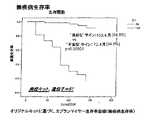
表1に示すように、51個の分子が低生存率との正相関を有し、14個の分子が低生存率との逆相関を有することが判明した。図1は、本願で「良好なサイン」(表1の左列の分子に高い発現が見られず、表1の右列の分子に抑制発現が見られない)と称されるものを有する個体の92.2%が、148.9ヶ月まで生き延びると予測されるが、本願で「不良なサイン」(表1の左列の分子に高い発現が見られ、表1の右列の分子に抑制発現が見られる)と呼ぶものを有する個体の8.3%しか、40ヶ月まで生き延びられないと予測されることを示す。この結果は、p値が0.00001未満であり、統計的に有意である。
Molecular signature of survival As shown in Table 1, it was found that 51 molecules had a positive correlation with low survival and 14 molecules had an inverse correlation with low survival. FIG. 1 shows an individual having what is referred to herein as a “good signature” (no high expression is seen in the molecules in the left column of Table 1 and no inhibitory expression is seen in the molecules in the right column of Table 1). 92.2% are expected to survive up to 148.9 months, but in this application “bad sign” (higher expression was seen in the molecules in the left column of Table 1; It shows that only 8.3% of individuals with what is called expression is expected to survive until 40 months. This result is statistically significant with a p-value less than 0.00001.
カプランマイヤー生存曲線と単変量解析を用いて、統計的精度に最も寄与する分子を同定することにより、我々は分子サインを絞り込んだ(表1において*で識別される)。我々は、低生存率と正の相関性を有する25個と、低生存率と負の相関性を有する8個との合計33個の一次分子マーカーが、統計的有意性の大部分を占めることを発見した。図2は、第1の一次および第2の一次分子サインを用いた予測生存曲線を示しており、良好なサインを有する個体の93.2%が、149.69ヶ月まで生き延びると予測され、不良なサインを有する個体のわずか14.4%しか、52.3ヶ月まで生き延びないと予測される(p<0.000001)。 By using Kaplan-Meier survival curves and univariate analysis to identify the molecules that contribute the most to statistical accuracy, we narrowed down the molecular signature (identified by * in Table 1). We find that a total of 33 primary molecular markers, 25 with a positive correlation with low survival and 8 with a negative correlation with low survival, account for the majority of statistical significance. I found FIG. 2 shows a predicted survival curve using the first primary and second primary molecular signatures, with 93.2% of individuals with good signs predicted to survive up to 149.69 months, poor Only 14.4% of individuals with an unsigned sign are expected to survive until 52.3 months (p <0.000001).
無発症予測分子サイン
表2に示すように、48個の分子が発症の発生(再発および転移)との正相関を有し、13個の分子が発症の発生との逆相関を有することが判明した。図3は、本願で「良好なサイン」(表2の左列の分子に高い発現が見られず、表2の右列の分子に抑制発現が見られない)と称されるものを有する個体のうちの94.5%が、150.4ヶ月まで疾病の再発がない(すなわち、無疾病生存)と予測されるが、一方で、本願で「不良なサイン」(表2の左列の分子に高い発現が見られ、表2の右列の分子に抑制発現が見られる)と称されるものを有する個体のうち34.5%だけしか、72.4ヶ月まで無疾病で生きられないと予測されることを示す(p<0.00001)。
Non-predicted molecular signature As shown in Table 2, 48 molecules were found to have a positive correlation with the onset of occurrence (recurrence and metastasis) and 13 molecules had an inverse correlation to the occurrence of onset did. FIG. 3 shows an individual having what is referred to in the present application as a “good signature” (no high expression is seen in the molecules in the left column of Table 2, and no suppressed expression is seen in the molecules in the right column of Table 2). Of these, 94.5% are predicted to have no recurrence of disease until 150.4 months (ie disease free survival), while in the present application “bad signs” (molecules in the left column of Table 2) And only 34.5% of individuals with the expression of) are able to survive disease-free until 72.4 months. It indicates that it is predicted (p <0.00001).
上記生存率サインと同様に、我々は本サインについても絞り込みを行い、再発と正の相関性を有する28個と、再発と負の相関性を有する8個との合計36個の一次分子マーカー(表2において*で識別)が、統計的有意性の大部分を占めることを発見した。図4は、本願で「良好なサイン」と称されるものを有する個体のうちの91.7%が、148.4ヶ月まで疾病の再発がないと予測されるが、本願で「不良なサイン」と称されるものを有する個体のわずか5.88%しか、44.2ヶ月まで無疾病で生きられないと予測されることを示す。(p<0.000001)。
結節転移の分子サイン
表3に示すように、37個の分子が結節転移との正相関を有し、10個の分子が結節転移との逆相関を有することが判明した。これらの37個の分子の組み合わせでは、不良なサインを示した(すなわち、表3の左列の分子に高い発現が見られ、表3の右列の分子に抑制発現が見られる)腫瘍の91%が、結節転移に進展した。さらに、良好なサインを示した(すなわち、表3の左列の分子に高い発現が見られず、表3の右列の分子に抑制発現が見られない)腫瘍の88.9%に、一切の結節転移が認められなかった。(p=0.00024)。
Similar to the above survival sign, we narrowed down this sign as well, and a total of 36 primary molecular markers (28 with positive correlation with recurrence and 8 with negative correlation with recurrence) ( It was found that (identified by * in Table 2) accounted for the majority of statistical significance. FIG. 4 shows that 91.7% of individuals with what are referred to herein as “good signs” are predicted to have no disease recurrence until 148.4 months. Only 5.88% of individuals with what is referred to as "are predicted to be disease-free until 44.2 months. (P <0.000001).
Molecular signature of nodal metastasis As shown in Table 3, it was found that 37 molecules had a positive correlation with nodal metastasis and 10 molecules had an inverse correlation with nodal metastasis. The combination of these 37 molecules showed a poor signature (ie, high expression was seen in the molecules in the left column of Table 3, and suppressed expression was seen in the molecules in the right column of Table 3). % Progressed to nodular metastasis. Furthermore, 88.9% of the tumors that showed good signs (ie, no high expression was seen in the molecules in the left column of Table 3, and no inhibitory expression was seen in the molecules in the right column of Table 3) No nodal metastasis was observed. (P = 0.00024).
上述のように、我々は組み合わせを絞り込むことによりこのサインを修正し、結節転移と正の相関性を有する20組と、結節転移と負の相関性を有する1組との合計21組の一次遺伝子(表3において*で識別)が、よく予測していたことを発見した。不良なサインを有する腫瘍の89.1%が結節転移を有し、良好なサインを有する腫瘍の86.8%が結節転移を全く有しなかった(p=0.000025)。 As mentioned above, we correct this signature by narrowing down the combinations, and a total of 21 primary genes, 20 pairs with a positive correlation with nodal metastasis and one with a negative correlation with nodal metastasis. (Identified by * in Table 3) found what was well predicted. 89.1% of tumors with poor signs had nodular metastases and 86.8% of tumors with good signs had no nodal metastases (p = 0.000025).
参考文献
Daviesら、2000:Davies G,Jiang WG,Mason MD.「ヒトプロテアーゼ癌における細胞間付着とシグナリング中間体(Cell−cell adhesion and signalling intermediates in human prostate cancer)」Journal of Urology,2000,163,985〜992頁。
Jiangら、2003a:Jiang WG,Watkins G,Lane J,Douglas−Jones A,Cunnick GH,Mokbel M,Mansel RE.「Rhoファミリーの予後診断価値および乳癌におけるrho−GDI(Prognostic value of Rho family and and rho−GDIs in breast cancer)」Clinical Cancer Research,2003,9(17),6432〜6440頁。
Jiangら、2003b:Jiang WG,Douglas−Jones A,およびMansel RE.「ヒト乳癌におけるPPAR−ガンマの発現レベルと、そのコアクチベータ(PPAR−GCA)(Level of expression of PPAR−gamma and its co−activator (PPAR−GCA) in human breast cancer)」International Journal of Cancer,2003,106,752〜757頁。
Jiangら、2003c:Jiang WG,Grimshaw D,Lane J,Martin TA,Parr C,Davies G,Laterra J,およびMansel RE.「cMETへのレトロウィルスハンマーヘッド導入遺伝子および線維芽細胞によって誘導される乳房腫瘍のHGF/SF阻害増殖(Retroviral hammerhead transgenes to cMET and HGF/SF inhibited growth of breast tumour,induced by fibroblasts)」Clinical Cancer Research,2003,9,4274〜4281頁。
Kingら、2004:King JAC,Ofori−Acquah AF,Stevens T,Al−Mehdi AB,Fodstad O,Jiang WG.「ヒト乳癌におけるALCAMの予後診断価値(Prognostic value of ALCAM in human breast cancer)」Breast Cancer Research,2004,R478〜487頁。
Nazarenkoら、1997:Nazarenko IA,Bhatnagar SK,Hohman RJ.「エネルギー転移に基づいたDNAの増幅および検出のための閉管方式(A closed tube format for amplification and detection of DNA based on energy transfer)」Nucleic Acids Res 1997;25:2516〜21頁。
ParrおよびJiang 2004:Parr C and Jiang WG.「ヒト乳癌におけるノッチ受容体、ノッチ−1およびノッチ−2(The Notch receptors、Notch−1 andNotch−2,in human breast cancers)」International Journal of Molecular Medicine,2004 Nov;14(5):779〜786頁。
References Davies et al., 2000: Davies G, Jiang WG, Mason MD. “Cell-cell adhesion and signaling intermediates in human prosthesis”, Journal of Urology, 2000, 163, 985-992.
Jiang et al., 2003a: Jiang WG, Watkins G, Lane J, Douglas-Jones A, Cunnick GH, Mokbel M, Mansel RE. “Prognostic value of the Rho family and rho-GDI in breast cancer”, Clinical Cancer Research, 2003, 9 (17), 6432-6440.
Jiang et al., 2003b: Jiang WG, Douglas-Jones A, and Mansel RE. "PPAR-gamma expression level in human breast cancer and its co-activator (PPAR-GCA) (PPAR-gamma and its co-activator (PPAR-GCA) in human breast cancer)" International Journal of Japan 3 106, 752-757.
Jiang et al., 2003c: Jiang WG, Grimshow D, Lane J, Martin TA, Parr C, Davies G, Laterra J, and Mansel RE. “Retroviral hammerhead transgenes and HGF / SF inhibited growth of breast cancer, inducible growth of breast cancer, induced by the retroviral hammerhead transgene to cMET and fibroblasts” 2003, 9, 4274-4281.
King et al., 2004: King JAC, Fori-Acquah AF, Stevens T, Al-Mehdi AB, Fodstad O, Jiang WG. "Prognostic value of ALCAM in human breast cancer", Breast Cancer Research, 2004, pp. 478-487.
Nazarenko et al., 1997: Nazarenko IA, Bhatnagar SK, Hohman RJ. “A closed tube for amplification and detection of DNA based on energy transfer”, Nucleic Acids Res 1997; 25: 2516-21.
Parr and Jiang 2004: Parr C and Jiang WG. “The Notch Receptors in Human Breast Cancer, Notch-1 and Notch-2 (The Notch Receptors, Notch-1 and Notch-2, in Human Breast Cancers)” International Journal of Molecular Medicine, 2004 Nov; 14 (5): 7 (78): 7. page.

Claims (19)
(a)第1の分子マーカーのセットであるAMF、ATF4、Cyr61、ER、マトリプターゼ2、MET、MLN64、MMP7、ネクチン4、PAR1A、Psoriason、Pttg1、Rho−C、Scotin、SDF1、SEMP1、SPF45、SST1、ST15、TACC2、TBD10、TCF2、TEM6、TEM7R、およびZO−3をコードする遺伝子の発現レベルと、第2の分子マーカーのセットであるARP2、Atf−3、HuR、MEN1、パラセリン、PTP−RKおよびラディキシンをコードする遺伝子の発現レベル並びにgdiGをコードする遺伝子の発現レベルに対するRHO8をコードする遺伝子の発現レベルの比を測定するために、個体からの乳癌組織の試料を検査することと、
(b)第1の分子マーカーのセットについて穏やかな予後であると推定される患者における同一マーカーの発現レベルよりも高レベルの発現が測定される場合、且つ第2の分子マーカーのセットについて穏やかな予後であると推定される患者における同一マーカーの発現レベルよりも低レベルの発現が測定される場合、
(c)前記組織の試料を採取した前記個体が52.3ヶ月以上生存できるのが14.4%以下の確率であると予測することとを含む方法。A method to help determine the prognosis of breast cancer in mammals,
(A) AMF, ATF4, Cyr61, ER, Matriptase 2, MET, MLN64, MMP7, Nectin 4, PAR1A, Psoriason, Pttg1, Rho-C, Scotin, SDF1, SEMP1, SPF45 , SST1, ST15, TACC2, TBD10, TCF2, TEM6, TEM7R, and the expression level of the gene encoding ZO-3 and the second set of molecular markers ARP2, Atf-3, HuR, MEN1, paraserine, PTP to measure the ratio of the expression level of the gene encoding the RHO8 for -RK and Radikishi of genes encoding down expression levels and gene expression levels encoding GdiG, and examining a sample of breast cancer tissue from an individual ,
(B) If a higher level of expression is measured than the expression level of the same marker in a patient presumed to have a mild prognosis for the first set of molecular markers, and mild for the second set of molecular markers If a lower level of expression is measured than the expression level of the same marker in a patient with prognosis,
(C) predicting that there is a probability of 14.4% or less that the individual who has collected the tissue sample can survive for 52.3 months or more.
第1の分子マーカーのセットおよび第3の分子マーカーセットについて穏やかな予後であると推定される患者における同一マーカーの発現レベルよりも高レベルの発現が見られ、且つ第2の分子マーカーのセット第4の分子マーカーセットについて穏やかな予後であると推定される患者における同一マーカーの発現レベルよりも低レベルの発現が測定される場合、前記組織の試料を採取した前記個体が40ヶ月以上生存できるのが8.3%以下の確率であると予測することをさらに含む、請求項1に記載の方法。Part (a) is the third set of molecular markers, Basigin, Betacatenin, BMP1, BMP10, Calpain Large, CD44, CX43, Cyclin D2, EHMS, FAK, FAP, GIRK, HAVR1, Isotopo3 , JAK1, LOX12, NET-2, PAR1A2, PTHrP, Rho-G, S100A4, SPARC, TCF3, VECAD, Vilip, and Wave2 expression levels and a fourth set of molecular markers, aMOT, Atf -1, Claudin-1, IL22R, Rock2, and the expression level of genes encoding Veg1,
Expression higher than the expression level of the same marker in a patient presumed to have a mild prognosis for the first set of molecular markers and the third set of molecular markers, and the second set of molecular markers If the expression level is lower than the expression level of the same marker in a patient estimated to have a mild prognosis for a set of 4 molecular markers, the individual taking the tissue sample can survive for more than 40 months The method of claim 1, further comprising predicting a probability of less than or equal to 8.3%.
(a)AMF、ATF4、Cyr61、ER、マトリプターゼ2、MET、MLN64、MMP7、ネクチン4、PAR1A、Psoriason、Pttg1、Rho−C、Scotin、SDF1、SEMP1、SPF45、SST1、ST15、TACC2、TBD10、TCF2、TEM6、TEM7R、およびZO−3からなる第1のマーカー組内の各遺伝子からの少なくとも1つの転写産物を同定する複数のプローブと、
(b)ARP2、Atf−3、HuR、MEN1、パラセリン、PTP−RK、ラディキシン、RHO8およびgdiGからなる第2のマーカー組内の各遺伝子からの少なくとも1つの転写産物を同定する複数のプローブと、を含むキット。A kit for determining the prognosis of mammalian breast cancer,
(A) AMF, ATF4, Cyr61, ER, Matriptase 2, MET, MLN64, MMP7, Nectin4, PAR1A, Psorison, Pttg1, Rho-C, Scotin, SDF1, Semp1, SPF45, SST1, ST15, TACC2, TBD10, A plurality of probes identifying at least one transcript from each gene in the first marker set consisting of TCF2, TEM6, TEM7R, and ZO-3;
(B) a plurality of probes identifying at least one transcript from each gene in the second marker set consisting of ARP2, Atf-3, HuR, MEN1, paraserine, PTP-RK, radixin , RHO8 and gdiG; Including kit.
(a)バシジン、ベータカテニン、BMP1、BMP10、カルパインラージ、CD44、CX43、サイクリンD2、EHMS、FAK、FAP、GIRK、HAVR1、アイソトポ3、JAK1、LOX12、NET−2、PAR1A2、PTHrP、Rho−G、S100A4、SPARC、TCF3、VECAD、Vilip、およびWave2からなる第3のマーカー組内の全遺伝子からの少なくとも1つの転写産物を同定する複数のプローブと、
(b)aMOT、Atf−1、Claudin−1、IL22R、Rock2およびVeg1からなる第4のマーカー組内の全遺伝子からの少なくとも1つの転写産物を同定する複数のプローブと、をさらに含む請求項12に記載のキット。The kit is
(A) Basidin, beta-catenin, BMP1, BMP10, calpain large, CD44, CX43, cyclin D2, EHMS, FAK, FAP, GIRK, HAVR1, isotopo3, JAK1, LOX12, NET-2, PAR1A2, PTHrP, Rho-G A plurality of probes identifying at least one transcript from all genes in a third marker set consisting of S100A4, SPARC, TCF3, VECAD, Villip, and Wave2, and
And (b) a plurality of probes that identify at least one transcript from all genes in the fourth marker set consisting of aMOT, Atf-1, Claudin-1, IL22R, Rock2 and Veg1. The kit according to 1.
請求項1または2に記載の分子マーカーを含む分子マーカーの組を同定するための少なくとも1組のプローブからなる少なくとも1つのマイクロアレイを含むキット。A kit for determining a patient's chance of survival in breast cancer,
A kit comprising at least one microarray comprising at least one set of probes for identifying a set of molecular markers comprising the molecular marker according to claim 1 or 2.
Applications Claiming Priority (5)
| Application Number | Priority Date | Filing Date | Title |
|---|---|---|---|
| GB0417740.8 | 2004-08-10 | ||
| GB0417740A GB0417740D0 (en) | 2004-08-10 | 2004-08-10 | Methods and kit for the prognosis of breast cancer |
| GB0426777.9 | 2004-12-07 | ||
| GB0426777A GB0426777D0 (en) | 2004-12-07 | 2004-12-07 | Methods and kit for the prognosis of breast cancer |
| PCT/GB2005/002971 WO2006016110A1 (en) | 2004-08-10 | 2005-07-27 | Methods and kit for the prognosis of breast cancer |
Related Child Applications (1)
| Application Number | Title | Priority Date | Filing Date |
|---|---|---|---|
| JP2011198276A Division JP5634360B2 (en) | 2004-08-10 | 2011-09-12 | Prognosis method and kit for breast cancer |
Publications (2)
| Publication Number | Publication Date |
|---|---|
| JP2008508895A JP2008508895A (en) | 2008-03-27 |
| JP5117852B2 true JP5117852B2 (en) | 2013-01-16 |
Family
ID=35044601
Family Applications (2)
| Application Number | Title | Priority Date | Filing Date |
|---|---|---|---|
| JP2007525333A Expired - Fee Related JP5117852B2 (en) | 2004-08-10 | 2005-07-27 | Prognosis method and kit for breast cancer |
| JP2011198276A Expired - Fee Related JP5634360B2 (en) | 2004-08-10 | 2011-09-12 | Prognosis method and kit for breast cancer |
Family Applications After (1)
| Application Number | Title | Priority Date | Filing Date |
|---|---|---|---|
| JP2011198276A Expired - Fee Related JP5634360B2 (en) | 2004-08-10 | 2011-09-12 | Prognosis method and kit for breast cancer |
Country Status (15)
| Country | Link |
|---|---|
| US (1) | US20100035240A1 (en) |
| EP (1) | EP1781814B3 (en) |
| JP (2) | JP5117852B2 (en) |
| CN (1) | CN102443627B (en) |
| AT (1) | ATE454474T1 (en) |
| AU (1) | AU2005271113B2 (en) |
| CA (1) | CA2576113A1 (en) |
| DE (1) | DE602005018789D1 (en) |
| DK (1) | DK1781814T3 (en) |
| ES (1) | ES2339132T7 (en) |
| GB (1) | GB2430741A (en) |
| HK (1) | HK1103422A1 (en) |
| MX (1) | MX2007001640A (en) |
| PT (1) | PT1781814E (en) |
| WO (1) | WO2006016110A1 (en) |
Families Citing this family (37)
| Publication number | Priority date | Publication date | Assignee | Title |
|---|---|---|---|---|
| US8497292B2 (en) | 2005-12-28 | 2013-07-30 | Translational Therapeutics, Inc. | Translational dysfunction based therapeutics |
| KR100786759B1 (en) * | 2006-07-07 | 2007-12-18 | 김현기 | -1 prognostic marker for breast cancer and composition for inducing obesity comprising hccr-1 |
| CN1919874B (en) * | 2006-09-18 | 2010-09-08 | 中国人民解放军军事医学科学院生物工程研究所 | Antibody molecule ATF-Fc fusion albumen of antiurokinase type fibrinolysin activator receptor and use thereof |
| NZ576149A (en) * | 2006-09-29 | 2012-06-29 | Translational Therapeutics Inc | Eif4e regulon-based diagnostics |
| WO2008138868A2 (en) * | 2007-05-09 | 2008-11-20 | Traslational Cancer Drugs Pharma, S.L. | In vitro diagnosis and/or prognosis of cancer by the analysis of gtpase protein expression |
| WO2008150512A2 (en) * | 2007-06-04 | 2008-12-11 | University Of Louisville Research Foundation, Inc. | Methods for identifying an increased likelihood of recurrence of breast cancer |
| ES2338843B1 (en) * | 2008-07-02 | 2011-01-24 | Centro De Investigaciones Energeticas, Medioambientales Y Tecnologicas | GENOMIC FOOTPRINT OF CANCER OF MAMA. |
| WO2010009171A2 (en) * | 2008-07-14 | 2010-01-21 | Wyeth | Src activation for determining cancer prognosis and as a target for cancer therapy |
| WO2010054379A2 (en) * | 2008-11-10 | 2010-05-14 | The United States Of America, As Represensted By The Secretary, Department Of Health And Human Services | Gene signature for predicting prognosis of patients with solid tumors |
| GB0821787D0 (en) * | 2008-12-01 | 2009-01-07 | Univ Ulster | A genomic-based method of stratifying breast cancer patients |
| US20120183543A1 (en) * | 2009-05-08 | 2012-07-19 | Novartis Ag | Diagnostic biomarkers for fibrotic disorders |
| KR101421326B1 (en) * | 2010-03-30 | 2014-07-21 | 서울대학교산학협력단 | Composition for predicting prognosis of breast cancer and kit comprising the same |
| JP2014515821A (en) * | 2011-03-27 | 2014-07-03 | オンコステム ダイアグノスティックス(モーリシャス)プライベート リミテッド | Markers, methods and kits for identifying tumor cells |
| CA2840946A1 (en) * | 2011-06-22 | 2012-12-27 | Oncocyte Corporation | Methods and compositions for the treatment and diagnosis of cancer |
| WO2013033609A2 (en) * | 2011-08-31 | 2013-03-07 | Oncocyte Corporation | Methods and compositions for the treatment and diagnosis of cancer |
| WO2013110053A1 (en) | 2012-01-20 | 2013-07-25 | The Ohio State University | Breast cancer biomarker signatures for invasiveness and prognosis |
| FR2988479B1 (en) * | 2012-03-23 | 2014-12-19 | Hospices Civils Lyon | METHOD FOR DETERMINING THE SUSCEPTIBILITY TO NOSOCOMIAL INFECTIONS |
| KR101672531B1 (en) * | 2013-04-18 | 2016-11-17 | 주식회사 젠큐릭스 | Genetic markers for prognosing or predicting early stage breast cancer and uses thereof |
| CN103235141A (en) * | 2013-04-27 | 2013-08-07 | 上海交通大学医学院附属新华医院 | Application of ATF (activating transcription factor) 3 to predicting bladder cancer metastasis and judging prognosis |
| KR101548830B1 (en) * | 2013-12-30 | 2015-08-31 | 가천대학교 산학협력단 | Cmposition for predicting breast cancer prognosis using marker for breast cancer stem cell screened by culture method of stem cell |
| WO2015105190A1 (en) * | 2014-01-10 | 2015-07-16 | 学校法人順天堂 | Method for assessing lymph node metastatic potential of endometrial cancer |
| KR102307278B1 (en) * | 2014-04-29 | 2021-09-30 | 노파르티스 아게 | Novel vertebrate cells and methods for recombinantly expressing a polypeptide of interest |
| CN105986024B (en) * | 2015-02-27 | 2019-10-25 | 复旦大学附属肿瘤医院 | One group of gene and its application for triple negative breast cancer prognosis |
| CN104725488A (en) * | 2015-04-06 | 2015-06-24 | 苏州普罗达生物科技有限公司 | Tight junction protein inhibitor polypeptide and application thereof |
| CN104725487A (en) * | 2015-04-06 | 2015-06-24 | 苏州普罗达生物科技有限公司 | Tight junction protein inhibitor polypeptide and application thereof |
| CN106295244B (en) * | 2015-06-05 | 2019-09-17 | 上海交通大学医学院附属瑞金医院 | Screening method of tumor diagnosis marker, breast cancer lung metastasis related gene obtained by method and application of breast cancer lung metastasis related gene |
| SG11201707815RA (en) | 2015-06-05 | 2017-12-28 | Novartis Ag | Antibodies targeting bone morphogenetic protein 9 (bmp9) and methods therefor |
| WO2017007031A1 (en) * | 2015-07-09 | 2017-01-12 | 学校法人順天堂 | Method for evaluating lymph node metastatic potential of endometrial cancer |
| CN106337081B (en) * | 2016-02-02 | 2020-01-21 | 中国医学科学院肿瘤医院 | Correlation of SNP locus rs1054135 of FABP4 gene and triple negative breast cancer prognosis |
| WO2018074865A2 (en) * | 2016-10-21 | 2018-04-26 | 서울대학교병원 | Composition and method for breast cancer prognosis prediction |
| EP3364190A1 (en) * | 2017-02-20 | 2018-08-22 | Panka Cancer Research AG | Method of detecting cancer or cancer cells |
| JPWO2018159808A1 (en) * | 2017-03-03 | 2019-12-26 | 国立研究開発法人国立がん研究センター | Pharmaceutical composition comprising an antibody drug conjugate of an anti-IL-7R antibody and an antibody drug conjugate of an anti-IL-7R antibody and a cytotoxic agent for use in treating cancer or inflammation |
| CN108504726B (en) * | 2018-03-28 | 2021-10-26 | 华南农业大学 | Method for carrying out PCR pretreatment by adopting AMF single spore trace DNA |
| WO2020008097A2 (en) * | 2018-07-03 | 2020-01-09 | Universidad De Granada | Method for obtaining data that can be used for the prediction or prognosis of overall survival and relapse-free survival from cancer, and composition that can modulate the activity of atf4 for the treatment of cancer |
| CN109620832B (en) * | 2019-01-14 | 2021-01-01 | 大连大学 | Application of glycocholic acid in preparation of anti-hyperplasia of mammary glands medicine |
| KR102280672B1 (en) * | 2019-12-20 | 2021-07-23 | 주식회사 베르티스 | A Composition for Diagnosing Cancer |
| CN114758773A (en) * | 2022-05-25 | 2022-07-15 | 四川大学华西医院 | Bladder cancer immunotherapy biomarker, immune risk model and application of immune risk model |
Family Cites Families (12)
| Publication number | Priority date | Publication date | Assignee | Title |
|---|---|---|---|---|
| US5759791A (en) * | 1989-01-17 | 1998-06-02 | The Johns Hopkins University | Cancer related antigen |
| CA2164498C (en) * | 1993-07-09 | 2007-09-11 | Paul A. Price | Assay for ykl-40 as a marker for degradation of mammalian connective tissue matrices |
| ATE256740T1 (en) * | 1997-02-06 | 2004-01-15 | Inst Genetics Llc | HUMAN SDF-5 PROTEIN AND RELATED COMPOSITIONS |
| WO1999038500A2 (en) * | 1998-01-30 | 1999-08-05 | The Brigham And Women's Hospital, Inc. | The human calcium-sensing receptor in the detection and treatment of cancer |
| JP4892131B2 (en) * | 1998-11-13 | 2012-03-07 | ストライカー コーポレイション | How to relieve cancer symptoms |
| JP2005503760A (en) * | 2001-01-24 | 2005-02-10 | プロテイン デザイン ラブス, インコーポレイテッド | Breast cancer diagnosis method, composition and breast cancer modulator screening method |
| US20030064384A1 (en) * | 2001-04-02 | 2003-04-03 | Mien-Chie Hung | Beta-catenin is a strong and independent prognostic factor for cancer |
| US20040086504A1 (en) * | 2001-06-21 | 2004-05-06 | Deepak Sampath | Cyr61 as a target for treatment and diagnosis of breast cancer |
| AU2002232563A1 (en) * | 2001-12-05 | 2003-06-23 | Genzyme Corporation | Compounds for therapy and diagnosis and methods for using same |
| WO2003054165A2 (en) * | 2001-12-20 | 2003-07-03 | The Regents Of The University Of California | Genetic markers and methods for the diagnosis, treatment and prevention of tumor metastasis |
| US20030186248A1 (en) * | 2002-03-29 | 2003-10-02 | Erlander Mark G. | Interpreting cytological specimens via molecular histological signatures |
| US20040231909A1 (en) * | 2003-01-15 | 2004-11-25 | Tai-Yang Luh | Motorized vehicle having forward and backward differential structure |
-
2005
- 2005-07-27 CA CA002576113A patent/CA2576113A1/en not_active Abandoned
- 2005-07-27 AU AU2005271113A patent/AU2005271113B2/en not_active Ceased
- 2005-07-27 DE DE602005018789T patent/DE602005018789D1/en active Active
- 2005-07-27 AT AT05763127T patent/ATE454474T1/en not_active IP Right Cessation
- 2005-07-27 MX MX2007001640A patent/MX2007001640A/en active IP Right Grant
- 2005-07-27 US US11/573,487 patent/US20100035240A1/en not_active Abandoned
- 2005-07-27 JP JP2007525333A patent/JP5117852B2/en not_active Expired - Fee Related
- 2005-07-27 EP EP05763127A patent/EP1781814B3/en not_active Not-in-force
- 2005-07-27 ES ES05763127T patent/ES2339132T7/en active Active
- 2005-07-27 WO PCT/GB2005/002971 patent/WO2006016110A1/en active Application Filing
- 2005-07-27 DK DK05763127.7T patent/DK1781814T3/en active
- 2005-07-27 CN CN201110315370.0A patent/CN102443627B/en not_active Expired - Fee Related
- 2005-07-27 PT PT05763127T patent/PT1781814E/en unknown
-
2007
- 2007-01-30 GB GB0701676A patent/GB2430741A/en not_active Withdrawn
- 2007-10-12 HK HK07111076.7A patent/HK1103422A1/en not_active IP Right Cessation
-
2011
- 2011-09-12 JP JP2011198276A patent/JP5634360B2/en not_active Expired - Fee Related
Also Published As
| Publication number | Publication date |
|---|---|
| US20100035240A1 (en) | 2010-02-11 |
| DK1781814T3 (en) | 2010-05-03 |
| EP1781814B3 (en) | 2011-08-31 |
| JP2008508895A (en) | 2008-03-27 |
| AU2005271113B2 (en) | 2010-12-09 |
| ES2339132T3 (en) | 2010-05-17 |
| AU2005271113A1 (en) | 2006-02-16 |
| DE602005018789D1 (en) | 2011-05-26 |
| MX2007001640A (en) | 2007-07-25 |
| ATE454474T1 (en) | 2010-01-15 |
| GB2430741A (en) | 2007-04-04 |
| ES2339132T7 (en) | 2012-03-16 |
| CA2576113A1 (en) | 2006-02-16 |
| GB0701676D0 (en) | 2007-03-14 |
| JP5634360B2 (en) | 2014-12-03 |
| CN102443627B (en) | 2014-10-29 |
| WO2006016110A1 (en) | 2006-02-16 |
| HK1103422A1 (en) | 2007-12-21 |
| EP1781814B1 (en) | 2010-01-06 |
| WO2006016110A8 (en) | 2006-08-24 |
| JP2012024095A (en) | 2012-02-09 |
| PT1781814E (en) | 2010-04-12 |
| EP1781814A1 (en) | 2007-05-09 |
| CN102443627A (en) | 2012-05-09 |
Similar Documents
| Publication | Publication Date | Title |
|---|---|---|
| JP5117852B2 (en) | Prognosis method and kit for breast cancer | |
| JP6246845B2 (en) | Methods for quantifying prostate cancer prognosis using gene expression | |
| JP6743056B2 (en) | Prostate Cancer Prognosis Judgment Method | |
| CN101014720B (en) | Methods and kit for the prognosis of breast cancer | |
| Lee et al. | Long noncoding RNAs as putative biomarkers for prostate cancer detection | |
| RU2654587C2 (en) | Method for predicting breast cancer recurrent during endocrine treatment | |
| KR20120065959A (en) | Markers for predicting gastric cancer prognostication and method for predicting gastric cancer prognostication using the same | |
| WO2009157204A1 (en) | Diagnosis/therapeutic strategy for gynecological cancer by utilizing micro-rna as biomarker | |
| JP2012529918A (en) | Method and apparatus for assessing infertility and / or egg quality | |
| JP2019522478A (en) | Compositions and methods for diagnosis of lung cancer using gene expression profiles | |
| CN110331207A (en) | Adenocarcinoma of lung biomarker and related application | |
| BR112020012280A2 (en) | compositions and methods for diagnosing lung cancers using gene expression profiles | |
| JPWO2015105190A1 (en) | Evaluation method for lymph node metastasis of endometrial cancer | |
| EP2083087A1 (en) | Method for determining tongue cancer | |
| KR101346038B1 (en) | Composition or Kit and Method for predicting prognosis of liver cancer | |
| US20160138116A1 (en) | Methods for monitoring treatment response and relapse in breast cancer | |
| TW202129011A (en) | Kit for in vitro testing panel of genes in pap smear samples for endometriosis and method of non-invasively and qualitatively determining severity of endometriosis using the kit | |
| US20030198961A1 (en) | Determining cancer aggressiveness | |
| CN116121369A (en) | Kidney cancer related biomarker and application and detection method thereof | |
| WO2012056033A1 (en) | Method for the analysis of a biological sample of a patient |
Legal Events
| Date | Code | Title | Description |
|---|---|---|---|
| A711 | Notification of change in applicant |
Free format text: JAPANESE INTERMEDIATE CODE: A711 Effective date: 20080318 |
|
| A521 | Request for written amendment filed |
Free format text: JAPANESE INTERMEDIATE CODE: A821 Effective date: 20080318 |
|
| A621 | Written request for application examination |
Free format text: JAPANESE INTERMEDIATE CODE: A621 Effective date: 20080722 |
|
| A131 | Notification of reasons for refusal |
Free format text: JAPANESE INTERMEDIATE CODE: A131 Effective date: 20110510 |
|
| A601 | Written request for extension of time |
Free format text: JAPANESE INTERMEDIATE CODE: A601 Effective date: 20110804 |
|
| A602 | Written permission of extension of time |
Free format text: JAPANESE INTERMEDIATE CODE: A602 Effective date: 20110811 |
|
| A521 | Request for written amendment filed |
Free format text: JAPANESE INTERMEDIATE CODE: A523 Effective date: 20110912 |
|
| A131 | Notification of reasons for refusal |
Free format text: JAPANESE INTERMEDIATE CODE: A131 Effective date: 20111122 |
|
| A601 | Written request for extension of time |
Free format text: JAPANESE INTERMEDIATE CODE: A601 Effective date: 20120220 |
|
| A602 | Written permission of extension of time |
Free format text: JAPANESE INTERMEDIATE CODE: A602 Effective date: 20120227 |
|
| A601 | Written request for extension of time |
Free format text: JAPANESE INTERMEDIATE CODE: A601 Effective date: 20120321 |
|
| A602 | Written permission of extension of time |
Free format text: JAPANESE INTERMEDIATE CODE: A602 Effective date: 20120328 |
|
| A601 | Written request for extension of time |
Free format text: JAPANESE INTERMEDIATE CODE: A601 Effective date: 20120419 |
|
| A602 | Written permission of extension of time |
Free format text: JAPANESE INTERMEDIATE CODE: A602 Effective date: 20120426 |
|
| A521 | Request for written amendment filed |
Free format text: JAPANESE INTERMEDIATE CODE: A523 Effective date: 20120522 |
|
| RD03 | Notification of appointment of power of attorney |
Free format text: JAPANESE INTERMEDIATE CODE: A7423 Effective date: 20120813 |
|
| A131 | Notification of reasons for refusal |
Free format text: JAPANESE INTERMEDIATE CODE: A131 Effective date: 20120821 |
|
| A521 | Request for written amendment filed |
Free format text: JAPANESE INTERMEDIATE CODE: A523 Effective date: 20120823 |
|
| A521 | Request for written amendment filed |
Free format text: JAPANESE INTERMEDIATE CODE: A821 Effective date: 20120813 |
|
| TRDD | Decision of grant or rejection written | ||
| A01 | Written decision to grant a patent or to grant a registration (utility model) |
Free format text: JAPANESE INTERMEDIATE CODE: A01 Effective date: 20120918 |
|
| A01 | Written decision to grant a patent or to grant a registration (utility model) |
Free format text: JAPANESE INTERMEDIATE CODE: A01 |
|
| A61 | First payment of annual fees (during grant procedure) |
Free format text: JAPANESE INTERMEDIATE CODE: A61 Effective date: 20121018 |
|
| R150 | Certificate of patent or registration of utility model |
Free format text: JAPANESE INTERMEDIATE CODE: R150 Ref document number: 5117852 Country of ref document: JP Free format text: JAPANESE INTERMEDIATE CODE: R150 |
|
| FPAY | Renewal fee payment (event date is renewal date of database) |
Free format text: PAYMENT UNTIL: 20151026 Year of fee payment: 3 |
|
| S111 | Request for change of ownership or part of ownership |
Free format text: JAPANESE INTERMEDIATE CODE: R313113 |
|
| S531 | Written request for registration of change of domicile |
Free format text: JAPANESE INTERMEDIATE CODE: R313531 |
|
| R350 | Written notification of registration of transfer |
Free format text: JAPANESE INTERMEDIATE CODE: R350 |
|
| R250 | Receipt of annual fees |
Free format text: JAPANESE INTERMEDIATE CODE: R250 |
|
| S111 | Request for change of ownership or part of ownership |
Free format text: JAPANESE INTERMEDIATE CODE: R313113 |
|
| R350 | Written notification of registration of transfer |
Free format text: JAPANESE INTERMEDIATE CODE: R350 |
|
| R250 | Receipt of annual fees |
Free format text: JAPANESE INTERMEDIATE CODE: R250 |
|
| R250 | Receipt of annual fees |
Free format text: JAPANESE INTERMEDIATE CODE: R250 |
|
| R250 | Receipt of annual fees |
Free format text: JAPANESE INTERMEDIATE CODE: R250 |
|
| R250 | Receipt of annual fees |
Free format text: JAPANESE INTERMEDIATE CODE: R250 |
|
| R250 | Receipt of annual fees |
Free format text: JAPANESE INTERMEDIATE CODE: R250 |
|
| LAPS | Cancellation because of no payment of annual fees |