JP5109029B2 - Bio-remodelable graft - Google Patents
Bio-remodelable graft Download PDFInfo
- Publication number
- JP5109029B2 JP5109029B2 JP2006552348A JP2006552348A JP5109029B2 JP 5109029 B2 JP5109029 B2 JP 5109029B2 JP 2006552348 A JP2006552348 A JP 2006552348A JP 2006552348 A JP2006552348 A JP 2006552348A JP 5109029 B2 JP5109029 B2 JP 5109029B2
- Authority
- JP
- Japan
- Prior art keywords
- graft
- bioremodelable
- layer
- expandable member
- collagen matrix
- Prior art date
- Legal status (The legal status is an assumption and is not a legal conclusion. Google has not performed a legal analysis and makes no representation as to the accuracy of the status listed.)
- Active
Links
Images


Classifications
-
- A—HUMAN NECESSITIES
- A61—MEDICAL OR VETERINARY SCIENCE; HYGIENE
- A61L—METHODS OR APPARATUS FOR STERILISING MATERIALS OR OBJECTS IN GENERAL; DISINFECTION, STERILISATION OR DEODORISATION OF AIR; CHEMICAL ASPECTS OF BANDAGES, DRESSINGS, ABSORBENT PADS OR SURGICAL ARTICLES; MATERIALS FOR BANDAGES, DRESSINGS, ABSORBENT PADS OR SURGICAL ARTICLES
- A61L27/00—Materials for grafts or prostheses or for coating grafts or prostheses
- A61L27/50—Materials characterised by their function or physical properties, e.g. injectable or lubricating compositions, shape-memory materials, surface modified materials
- A61L27/507—Materials characterised by their function or physical properties, e.g. injectable or lubricating compositions, shape-memory materials, surface modified materials for artificial blood vessels
-
- A—HUMAN NECESSITIES
- A61—MEDICAL OR VETERINARY SCIENCE; HYGIENE
- A61L—METHODS OR APPARATUS FOR STERILISING MATERIALS OR OBJECTS IN GENERAL; DISINFECTION, STERILISATION OR DEODORISATION OF AIR; CHEMICAL ASPECTS OF BANDAGES, DRESSINGS, ABSORBENT PADS OR SURGICAL ARTICLES; MATERIALS FOR BANDAGES, DRESSINGS, ABSORBENT PADS OR SURGICAL ARTICLES
- A61L27/00—Materials for grafts or prostheses or for coating grafts or prostheses
- A61L27/36—Materials for grafts or prostheses or for coating grafts or prostheses containing ingredients of undetermined constitution or reaction products thereof, e.g. transplant tissue, natural bone, extracellular matrix
- A61L27/3604—Materials for grafts or prostheses or for coating grafts or prostheses containing ingredients of undetermined constitution or reaction products thereof, e.g. transplant tissue, natural bone, extracellular matrix characterised by the human or animal origin of the biological material, e.g. hair, fascia, fish scales, silk, shellac, pericardium, pleura, renal tissue, amniotic membrane, parenchymal tissue, fetal tissue, muscle tissue, fat tissue, enamel
- A61L27/3629—Intestinal tissue, e.g. small intestinal submucosa
-
- A—HUMAN NECESSITIES
- A61—MEDICAL OR VETERINARY SCIENCE; HYGIENE
- A61L—METHODS OR APPARATUS FOR STERILISING MATERIALS OR OBJECTS IN GENERAL; DISINFECTION, STERILISATION OR DEODORISATION OF AIR; CHEMICAL ASPECTS OF BANDAGES, DRESSINGS, ABSORBENT PADS OR SURGICAL ARTICLES; MATERIALS FOR BANDAGES, DRESSINGS, ABSORBENT PADS OR SURGICAL ARTICLES
- A61L27/00—Materials for grafts or prostheses or for coating grafts or prostheses
- A61L27/36—Materials for grafts or prostheses or for coating grafts or prostheses containing ingredients of undetermined constitution or reaction products thereof, e.g. transplant tissue, natural bone, extracellular matrix
- A61L27/3604—Materials for grafts or prostheses or for coating grafts or prostheses containing ingredients of undetermined constitution or reaction products thereof, e.g. transplant tissue, natural bone, extracellular matrix characterised by the human or animal origin of the biological material, e.g. hair, fascia, fish scales, silk, shellac, pericardium, pleura, renal tissue, amniotic membrane, parenchymal tissue, fetal tissue, muscle tissue, fat tissue, enamel
- A61L27/3633—Extracellular matrix [ECM]
-
- A—HUMAN NECESSITIES
- A61—MEDICAL OR VETERINARY SCIENCE; HYGIENE
- A61L—METHODS OR APPARATUS FOR STERILISING MATERIALS OR OBJECTS IN GENERAL; DISINFECTION, STERILISATION OR DEODORISATION OF AIR; CHEMICAL ASPECTS OF BANDAGES, DRESSINGS, ABSORBENT PADS OR SURGICAL ARTICLES; MATERIALS FOR BANDAGES, DRESSINGS, ABSORBENT PADS OR SURGICAL ARTICLES
- A61L27/00—Materials for grafts or prostheses or for coating grafts or prostheses
- A61L27/50—Materials characterised by their function or physical properties, e.g. injectable or lubricating compositions, shape-memory materials, surface modified materials
- A61L27/58—Materials at least partially resorbable by the body
Abstract
Description
本発明は一般に医療装置に関する。詳しくは、外科用グラフト、ステントグラフト、ステントグラフト材料、および外科用グラフトおよびステントグラフトを作製する方法に関する。これらグラフトおよびステントグラフトは、本来は腹部大動脈瘤の移植およびステント移植で用いられるが、脈管構造の他の領域でステント移植、動脈瘤除去、バイパスなどのために使用することができる。 The present invention relates generally to medical devices. In particular, it relates to surgical grafts, stent grafts, stent graft materials, and surgical grafts and methods for making stent grafts. These grafts and stent grafts are primarily used for abdominal aortic aneurysm implantation and stent implantation, but can be used for stent implantation, aneurysm removal, bypassing, etc. in other areas of the vasculature.
発明の背景
脈管構造のグラフト材料には一般には2つの主要なタイプ、ダクロン(登録商標)織布および延伸ポリテトラフルオロエチレン(ePTFE)がある。これらの材料は共に合成材料であり身体には僅かに耐性があるだけである。ステントグラフトの場合には、これらは、接着箇所でグラフト材料の磨耗を引き起こす、一般にステントとして知られる金属製バネ拡張部材と組み合わされるため磨耗する。加えて、これら合成グラフトは比較的厚く、導入器被覆を通しての体内への導入のために折り畳まれるとき非常に大きな外皮が必要となり、経皮導入が非常に困難になる。
BACKGROUND OF THE INVENTION There are generally two main types of vascular graft materials, Dacron® woven fabric and expanded polytetrafluoroethylene (ePTFE). Both of these materials are synthetic materials and are only slightly resistant to the body. In the case of stent grafts, these wear because they are combined with a metal spring expansion member, commonly known as a stent, which causes wear of the graft material at the point of attachment. In addition, these synthetic grafts are relatively thick and require a very large skin when folded for introduction into the body through an introducer covering, making transdermal introduction very difficult.
発明の概要
本発明の鋳造生体再造形可能(bioremodelable)グラフトの例示的な実施形態において、上記の問題は解決され、技術進歩が実現される。このグラフトは、利点として、好ましくは細胞外コラーゲン基質材料などの、これにほぼ接触する組織または細胞を再造形させる生体再造形可能物質の鋳造管状構造物を備えている。
SUMMARY OF THE INVENTION In the exemplary embodiment of the cast bioremodelable graft of the present invention, the above problems are solved and technical advances are realized. The graft advantageously comprises a cast tubular structure of a bioremodelable material that remodels the tissue or cells that are in close contact with it, preferably an extracellular collagen matrix material.
本発明のグラフト材料は、好ましくは、とりわけ、成長因子、再吸収性材料、細胞外コラーゲン間トリックス材料、合成材料および結合剤材料のうちの少なくとも1つを含む生体再造形可能物質である。好ましくは、生体再造形可能物質は細胞外コラーゲン基質材料であり、特に、小腸粘膜下組織(SIS)組織、コラーゲン、または組織の再造形を誘発するために例えば成長因子を有する他の天然の材料である。通常のシート状のSISは、その厚さおよび任意の材料片での有孔率が広い範囲で様々に異なる。SIS材料を通常生じるシート状で用いる代わりに、SISを切断して断片とするか、または細断もしくは粉砕して小サイズの細片または粒子にすることもできる。これらの小片または細片を次に、所望のグラフトにとって適切な形状およびサイズの心金へと均一に吹き付けるか、形成するかまたは鋳造することができる。可鍛性のある水和化された断片が型上に鋳造されるか、または張子紙のように塗布される。鋳造物を乾燥または硬化させた後、型を除去して、鋳造管状構造物が得られる。SIS粒子を、結合剤材料を伴ってまたは伴わないで心金上に吹き付けるかまたは好ましくは鋳造して、得られる構造物の物理的強度を向上させることができる。適切なサイズおよび強度のZステントなどの拡張可能部材を、SIS材料を心金上に形成しているときにこれに埋め込むことができる。次にこのステントを埋め込んだSISに、真空袋詰め、テープ巻き付けまたは他の適切な方法で圧力をかけて、硬化および結合している間にSIS粒子をまとめて密に圧縮させ、これにより均一の厚さおよび有孔率を持つ材料を製造する。加えて、拡張可能部材、好ましくはZステントはグラフトの壁構造内に十分に組み込まれるため、ステントをグラフト材料に保持させるのに縫合を必要としない。縫合材料が除かれることにより、ステントグラフトの折り畳み状態での輪郭が大幅に減少し、このため経皮配置にとってより適切となり、またステント支柱とグラフト材料との間の移動または「擦れ」の領域を削除することによってグラフト材料の磨耗
の可能性が減少する。
The graft material of the present invention is preferably a bioremodelable substance comprising at least one of growth factors, resorbable materials, extracellular intercollagenous trick materials, synthetic materials and binder materials, among others. Preferably, the bioremodelable substance is an extracellular collagen matrix material, in particular small intestine submucosa (SIS) tissue, collagen, or other natural material having, for example, growth factors to induce tissue remodeling It is. Ordinary sheet-like SIS varies widely in its thickness and porosity in any piece of material. Instead of using the SIS material in the usual sheet form, the SIS can be cut into pieces, or shredded or crushed into small sized pieces or particles. These pieces or strips can then be uniformly sprayed, formed or cast into a mandrel of the appropriate shape and size for the desired graft. A malleable hydrated piece is cast on a mold or applied like a paper. After the casting is dried or cured, the mold is removed to obtain a cast tubular structure. SIS particles can be sprayed or preferably cast on a mandrel with or without a binder material to improve the physical strength of the resulting structure. An expandable member, such as an appropriately sized and strong Z-stent, can be embedded in the SIS material as it is being formed on the mandrel. The SIS in which the stent is embedded is then pressured by vacuum bagging, tape wrapping or other suitable method to collectively compress the SIS particles together during curing and bonding, thereby producing a uniform Produces materials with thickness and porosity. In addition, the expandable member, preferably the Z-stent, is fully incorporated within the graft wall structure and does not require stitching to hold the stent to the graft material. The removal of the suture material significantly reduces the folded contour of the stent graft, making it more suitable for percutaneous placement and eliminating the area of movement or “rubbing” between the stent strut and the graft material. This reduces the possibility of wear of the graft material.
本発明は、身体によって受容されて最終的には天然の組織へと再造形され、また拡張フレーム部材をその構造物へと組み込むことで経皮導入において輪郭または折り畳み状態でのサイズが大幅に減少するグラフト材料に関する。 The present invention is received by the body and eventually reshaped into natural tissue, and the expansion frame member is incorporated into the structure to greatly reduce the size in the contoured or folded state during percutaneous introduction. The present invention relates to a graft material.
別の実施形態では、SIS材料を粉砕または細断して微細な繊維質粒子またはストランドとし、これらを次に紡いで微細なヤーンまたは糸とすることができる。この糸を次に織って、現存の織り込みダクロングラフトに類似する方法で所望のグラフト形態とすることができる。細いニチノールワイヤまたは他の弾力性材料を一定の間隔で横糸および/または縦糸へと織り込んで、グラフト「本体」に、追加のステントをグラフトに接着させることなく拡張しまたその形状を維持する能力を与えることができる。 In another embodiment, the SIS material can be ground or chopped into fine fibrous particles or strands, which can then be spun into fine yarns or yarns. This yarn can then be woven into the desired graft form in a manner similar to existing woven Dacron grafts. The ability to weave thin nitinol wires or other elastic materials into weft and / or warp yarns at regular intervals to expand and maintain the shape of the graft “body” without adhering additional stents to the graft. Can be given.
生体再造形可能物質は、これにほぼ接触する組織または細胞を再造形させる成長因子を有する天然または合成の物質であり得る。好ましくは、生体再造形可能物質はまた再吸収性材料を含み、また生体再造形可能材料の断片または粒子を相互接続させる結合剤材料も含み得る。鋳造生体再造形可能および拡張可能グラフトのより好適な実施形態では、グラフトはまた、好ましくは生体再造形可能物質内に配置された拡張可能部材を含み、鋳造管状構造物への更なる接着手段を必要としない。加えて、生体再造形可能物質の第2層を第1層に塗布して、拡張可能部材を2つの層の間に配置することができる。好ましくは、生体再造形可能物質は、SISなどの細胞外コラーゲン基質材料の断片または粒子を含み、これらは次に真空プレスされてより薄い壁厚または輪郭を得るという利点がある。いかなる結合剤材料は好ましくは再吸収性材料であり、好ましくは、とりわけ、生体分解性ポリマー、コラーゲン、フィブリン、フィブロネクチンおよび多糖類のうちの少なくとも1つを含む。 The bioremodelable material can be a natural or synthetic material that has growth factors that remodel tissue or cells that are in close contact therewith. Preferably, the bioremodelable material also includes a resorbable material and may include a binder material that interconnects fragments or particles of the bioremodelable material. In a more preferred embodiment of the cast bioremodelable and expandable graft, the graft also includes an expandable member, preferably disposed within the bioremodelable material, for further adhesion to the cast tubular structure. do not need. In addition, a second layer of bioremodelable material can be applied to the first layer and the expandable member can be placed between the two layers. Preferably, the bioremodelable material comprises fragments or particles of extracellular collagen matrix material such as SIS, which then have the advantage of being vacuum pressed to obtain a thinner wall thickness or contour. Any binder material is preferably a resorbable material and preferably comprises, among other things, at least one of a biodegradable polymer, collagen, fibrin, fibronectin and polysaccharide.
詳細な説明
図1は、生体再造形可能物質34の鋳造管状構造物33内に配置されたZステントなどの選択的な拡張可能部材18を有する、本発明の例示的な鋳造生体再造形可能グラフト32の図を示す。管状構造物は、長さ方向に貫通する内腔35、および内表面36と外表面37とを有する。管状構造物33はまた、生体再造形可能物質の第1層38と、選択的に第1層上に配置される生体再造形可能物質の第2層とを含み、これらの間にステント18などの拡張可能部材を有する。拡張可能部材18は鋳造グラフトを圧縮状態と拡張状態との間で拡張させ、好ましくは侵襲性が最小限の送達システム内に配置される腔内の実施形態で利用される。鋳造グラフトはまた、グラフトを脈管または導管の端部に縫合またはかすがい留めにする開口外科処置において脈管構造でまたは他の導管系内で利用することができる。生体再造形可能物質は、典型的には成長因子または他のたんぱく質などを含み、これらに接触する組織または細胞が再造形するように誘発する生体適合性物質である。
DETAILED DESCRIPTION FIG. 1 illustrates an exemplary cast bioremodelable graft of the present invention having a selectively
図1Aは、鋳造二又生体再造形可能グラフトを鋳造または形成するための二又の型10の別の例示的実施形態を示す。特に、心金または型10は主本体部11と、主本体部から延びる取り外し可能な両側性および対側性脚部12および13とを含む。心金または型の各部分は金属またはプラスチック材料管から作製するか、またはいかなる特定のサイズおよび形状にも融通がきく適切な寸法をもつ金型から鋳造することができる。二又の心金または型として述べたが、好ましくは円形断面をもつ単一の細長い部材の型を用いて、その上に管状構造物グラフトを鋳造または形成することもできる。
FIG. 1A shows another exemplary embodiment of a bifurcated
図1Bは、図1Aの心金または型10を、その上に鋳造、塗布または形成された、液状または可鍛性の生体再造形可能物質14の第1層と共に示す。この例示的な二又の実施形
態では、生体再造形可能物質は型上に鋳造または塗布されて二又グラフト22の主本体部15と主本体部から遠方に延びる両側性および対側性脚部16および17とを確立する。この生体再造形可能物質の第1層は型に吹き付けるか、または型を液状の生体再造形可能物質内に浸すことによって塗布することができ、このようにして心金上に第1層を定着させる。本発明の1つの特定の形態では、この生体再造形可能物質14の単一層を乾燥または硬化させて生体再造形可能グラフト22を形成する。この単一層グラフトの厚さまたは断面を小さくするために、グラフトを室温で脱水させて硬い管状構造物を形成することができる。またグラフトを真空チャンバー内で冷凍および脱水させて凍結乾燥材料構造体を形成することもできる。もしくは、単一層グラフトにポリテトラフルオロエチレンまたは拡張ポリテトラフルオロエチレンなどのポリマー材料の帯をきつく巻き付けて包み、次に真空プレスして、グラフトの壁厚を減らすことができる。この真空プレス技術は、管状構造体および型を適切な室温で真空チャンバー内に配置して生体再造形可能物質を脱水することを包含する。乾燥、硬化または真空プレスの後、二又心金または型10をグラフトの内腔から取り除き、これにより、グラフトの一部断面部分に示すように、主本体腔部23ならびに両側性および対側性腔部24および25を有するグラフト22が作製される。
FIG. 1B shows the mandrel or
本発明の生体再造形可能物質は、直接にまたは近似に接触する組織または細胞を再造形することができる多くの様々な材料または物質を含み得る。生体再造形可能物質は、とりわけ、接触する組織または細胞を再造成または再生させる成長因子を含む。本発明におけるように、鋳造グラフトを、例えば、動物または人の患者の大動脈内に外科的に配置してそこの病的組織に取って代わることができる。二又グラフト22は、例えば開口外科処置中に大動脈および腸管動脈の健全な組織に縫い合わせることができる。この後グラフトの生体再造形可能物質は大動脈および腸管動脈を再造形して、好ましくは組織が再造形された後再吸収される。
The bioremodelable materials of the present invention can include many different materials or substances that can remodel tissue or cells that are in direct or close contact. Bioremodelable materials include, among other things, growth factors that remodel or regenerate the tissues or cells that come into contact. As in the present invention, the cast graft can be surgically placed within the aorta of, for example, an animal or a human patient to replace the diseased tissue therein. The bifurcated
本発明によれば、生体再造形可能物質は、好ましくは、これに接触する天然の組織または細胞の再造成中またはその後患者によって再吸収される再吸収性材料を含む。1つの好適な実施形態では、生体再造形可能物質は、細胞外コラーゲン基質材料および、特に、小腸粘膜下組織(SIS)材料を含む。このSIS材料は、例えば、ブタから得られ、別の材料の組織または細胞内にまたはこれに対して配置するために殺菌される。このSIS材料はCook Biotech, West Lafayette, Indianaから市販されている。 According to the present invention, the bioremodelable material preferably comprises a resorbable material that is resorbed by the patient during or after remodeling of the natural tissue or cells that contact it. In one preferred embodiment, the bioremodelable material comprises extracellular collagen matrix material and, in particular, small intestine submucosa (SIS) material. This SIS material is obtained, for example, from pigs and sterilized for placement in or against tissue or cells of another material. This SIS material is commercially available from Cook Biotech, West Lafayette, Indiana.
再構成されたまたは天然由来のコラーゲン材料もまた本発明において用いることができる。少なくとも生体再吸収性であるこのような材料は、生体再造形可能であり細胞浸潤および内殖を促進させて特定の利点を提供する材料と共に、本発明において利点を提供する。ここでは生体再造形可能材料は、脈管の腔内で細胞の成長を促進するために用いられ得る。これは処置の完了後、生体的プロセスを通して脈管の開存性が再確立されることに対する防衛の助けとなる。 Reconstituted or naturally derived collagen material can also be used in the present invention. Such materials that are at least bioresorbable provide advantages in the present invention, along with materials that are bioremodelable and promote cell infiltration and ingrowth to provide certain advantages. Here, the bio-remodelable material can be used to promote cell growth within the vessel lumen. This helps defend against the re-establishment of vascular patency through biological processes after treatment is complete.
適切な生体再造形可能材料は、バイオトロピック(biotropic)特性を有するコラーゲン細胞外基質材料(ECM)によって提供され得る。例えば、適切なコラーゲン材料としては、粘膜下組織、副腎膜、皮膚コラーゲン、硬膜、心膜、漿膜、腹膜または、肝基底膜を含む基底膜層などのECMを含む。これらの目的のための適切な粘膜下組織材料としては、例えば、小腸粘膜下組織を含む腸粘膜下組織、胃粘膜下組織、膀胱粘膜下組織および子宮粘膜下組織が含まれる。調製および使用時、使用する粘膜下組織材料および他のECMは、選択的に、成長因子または他の元組織にとって天然である生体活性成分を保持し得る。例えば、粘膜下組織または他のECMは、基本的な繊維芽細胞成長因子(FGF−2)、形質転換成長因子ベータ(TGF−beta)、上皮細胞増殖因子(EGF)および/または血小板由来増殖因子(PDGF)などの1つ以上の成長因子を含み得る
。同様に、本発明で用いられる粘膜下組織または他のECMは、ヘパリン、ヘパリン硫酸、ヒアルロン酸、フィブロネクチンなどの他の生体材料を含み得る。従って、一般に、粘膜下組織または他のECM材料は、直接または間接に、細胞形態学における変化、増殖、成長、タンパク質または遺伝子発現などの細胞反応を誘発する生体活性成分を含み得る。
A suitable bioremodelable material can be provided by a collagen extracellular matrix material (ECM) having biotropic properties. For example, suitable collagen materials include ECM such as submucosa, adrenal membranes, skin collagen, dura mater, pericardium, serosa, peritoneum, or basement membrane layer including liver basement membrane. Suitable submucosal materials for these purposes include, for example, intestinal submucosa, including small intestine submucosa, gastric submucosa, urinary bladder submucosa and uterine submucosa. During preparation and use, the submucosal material and other ECMs used may optionally retain bioactive ingredients that are natural for growth factors or other source tissues. For example, submucosal tissue or other ECMs may include basic fibroblast growth factor (FGF-2), transforming growth factor beta (TGF-beta), epidermal growth factor (EGF) and / or platelet derived growth factor. One or more growth factors such as (PDGF) may be included. Similarly, the submucosa or other ECM used in the present invention may include other biomaterials such as heparin, heparin sulfate, hyaluronic acid, fibronectin. Thus, in general, submucosa or other ECM material may include bioactive components that directly or indirectly induce cellular responses such as changes in cell morphology, proliferation, growth, protein or gene expression.
さらに、このような天然の生体活性成分を含むことに加えてまたはこれに代えて、組み換え技術または他の方法によって化学合成により作製されるものなどの非天然の生体活性成分を粘膜下組織に組み込んでもよい。これら非天然の生体活性成分は、ECM組織内で天然に生じるものに対応する天然由来のまたは組み換えにより作製されたタンパク質であるが、恐らくは異なる種(例えば、ブタなどの他の動物からのコラーゲンECMに与えられるヒトタンパク質)であり得る。非天然の生体活性成分はまた薬剤物質であってもよい。閉鎖装置内におよび/またはその上に組み込まれ得る例示的な薬剤物質としては、例えば、抗生物質および、トロンビン、フィブリノゲンのような凝血因子などの血栓促進物質などがある。これらの物質を処置直前に製造前ステップとして(例えば、材料をセファゾリンなどの適切な抗生物質を含有する溶液中に浸漬させることによって)、または患者内で閉鎖装置を展開中またはその後に、装置にあてがうとよい。 In addition, in addition to or in place of such natural bioactive ingredients, non-natural bioactive ingredients such as those produced by chemical synthesis by recombinant techniques or other methods are incorporated into the submucosa. But you can. These non-natural bioactive ingredients are naturally occurring or recombinantly produced proteins corresponding to those naturally occurring in ECM tissue, but probably different species (eg collagen ECM from other animals such as pigs). A human protein). The non-natural bioactive ingredient may also be a drug substance. Exemplary drug substances that can be incorporated into and / or on the closure device include, for example, antibiotics and thrombus-promoting substances such as clotting factors such as thrombin and fibrinogen. These substances can be applied to the device as a pre-manufacturing step immediately prior to treatment (eg, by immersing the material in a solution containing a suitable antibiotic such as cefazolin), or during or after deployment of the closure device in the patient. It is good to apply.
本発明で用いる粘膜下組織または他のECM組織は、好ましくは、例えばCookらの米国特許第6,206,931号に記載されているように、高純度に精製される。従って、好適なECM材料は、グラム当たりの内毒素単位(EU)が約12より小さい、より好ましくはグラム当たり約5EUより小さい、最も好ましくはグラム当たり約1EUより小さい内毒素レベルを示す。別の利点としては、粘膜下組織または他のECM材料は、グラム当たりのコロニー形成単位(CFU)が約1より小さい、より好ましくはグラム当たり約0.5CFUより小さいバイオバーデンを有し得る。真菌レベルも望ましくは同様に低く、例えば、グラム当たり約1CFUより小さく、より好ましくはグラム当たり約0.5CFUより小さい。核酸レベルは好ましくは約5μg/mgより小さく、より好ましくは約2μg/mgより小さい。そしてウィルスレベルは、好ましくはグラム当たりのプラーク形成単位(PFU)が約50より小さく、より好ましくはグラム当たり約5PFUより小さい。米国特許第6,206,931号に教示された粘膜下組織または他のECM組織のこれらのおよびさらに他の特性は、本発明で使用される粘膜下組織の特性となり得る。 The submucosa or other ECM tissue used in the present invention is preferably purified to high purity as described, for example, in US Pat. No. 6,206,931 to Cook et al. Thus, suitable ECM materials exhibit endotoxin units (EU) per gram of less than about 12, more preferably less than about 5 EU per gram, and most preferably less than about 1 EU per gram. As another advantage, the submucosa or other ECM material may have a bioburden with less than about 1 colony forming unit (CFU) per gram, more preferably less than about 0.5 CFU per gram. Fungal levels are also desirably low, for example, less than about 1 CFU per gram, more preferably less than about 0.5 CFU per gram. The nucleic acid level is preferably less than about 5 μg / mg, more preferably less than about 2 μg / mg. And the virus level is preferably less than about 50 plaque forming units (PFU) per gram, more preferably less than about 5 PFU per gram. These and other properties of submucosa or other ECM tissue taught in US Pat. No. 6,206,931 can be properties of the submucosa used in the present invention.
本発明により作製されるグラフトまたはステントグラフトは、脈管の開存性を維持しまた動脈瘤を排除して破裂を防ぐ一方で、最終的には接触する組織のタイプへと再造形される。この特定の応用例では、ECM材料は、脈管の病的部位が健全な脈管構造組織に取って代わるように、脈管内皮となる。再造形されたECM材料は、グラフト材料内に元々埋め込まれているステントまたは補剛ワイヤなどの拡張可能部材にわたって内皮組織の保護層を形成して、グラフト部分を通って流れる血液が、血栓または血流乱流を引き起こし得る外来または合成の材料と接触しないようにされる。さらに、ステントであっても補剛ワイヤであっても拡張可能部材は、その本来の機能を果たした後、新しく形成された内皮を支持および強度を与え続ける。 A graft or stent graft made in accordance with the present invention is ultimately reshaped into the type of tissue it contacts while maintaining vascular patency and eliminating an aneurysm to prevent rupture. In this particular application, the ECM material becomes the vascular endothelium such that the vascular pathological site replaces healthy vasculature tissue. The reshaped ECM material forms a protective layer of endothelial tissue over an expandable member such as a stent or stiffening wire that is originally embedded in the graft material so that blood flowing through the graft portion can be thrombus or blood. Avoid contact with foreign or synthetic materials that can cause turbulent flow. Furthermore, the expandable member, whether it is a stent or a stiffening wire, continues to provide support and strength to the newly formed endothelium after performing its original function.
好適であり最良の形態の実施形態として、生体再吸収材料は、SIS材料断片の張子紙様の層またはコーティングであり得る。SIS断片は水和されて二又型10に塗布され、個々の断片は互いに直接接触し重なって管状構造物を形成する。断片は所望の厚さを形成するように塗布され得る。生体再造形可能物質はまた、好ましくは、SIS断片を互いにより良好に付着または接着させる結合剤材料を含む。結合剤材料は好ましくは再吸収性材料を含み、とりわけ、生体分解性ポリマー、コラーゲン、フィブリン、フィブロネクチンおよび多糖類のうちの少なくとも1つを含む。生体分解性ポリマーは、ポリ乳酸、グリコール酸、ポリカプロラクトン、ポリウレタンウレアおよびポリヒドロキシアルカン酸のうちの少なくとも1つからなる。コラーゲンは、とりわけ、精製コラーゲン、非精製コラー
ゲン、架橋コラーゲンおよび非架橋コラーゲンのうちの少なくとも1つからなる。多糖類は、グリコーゲン、キトサンおよびグルコースのうちの少なくとも1つからなる。この結合剤材料は、好ましくは、細胞外コラーゲン基質材料、特に、SIS材料断片を相互接続させる。
As a preferred and best mode embodiment, the bioresorbable material may be a tensioned paper-like layer or coating of SIS material pieces. The SIS pieces are hydrated and applied to the bifurcated
図4は、図1Bの本発明のグラフト22の主本体部15の端面図である。図6は、図4のグラフト22の主本体部15のSIS生体再造形可能物質層14の、ライン6−6に沿った一部および拡大断面図である。層14は結合剤材料41によって相互接続されるSIS材料粒子40を含む。図7は、図4のグラフト22の主本体部15の生体再造形可能物質層14の、ライン7−7に沿った拡大および一部断面図である。この好適であり最良の形態の実施形態では、層は、水和され互いに相互接続されているSIS材料断片42を含む。加えて、結合剤材料41がSIS断片の相互接続を助けるために利用される。これらの断片は面積が数平方センチメートル以上に達することができ、張子紙によく似たように心金または型に塗布される。これらSIS材料断片は典型的には、これらを可鍛性とし心金または型上に容易に形成され得るように水和される。
4 is an end view of the
本発明の別の実施形態では,細胞外コラーゲン基質材料は粉砕されるかまたは小さな粒子または微粒子へと形成されて結合剤材料と混ぜ合わされ、次に型10上に直接塗布または鋳造され得る。また他の繊維をこの生体再造形可能物質と混ぜ合わせて単一層グラフトにさらに強度を与え得ることも考慮される。細胞外コラーゲン基質材料のサイズ、数および濃度は、接触する組織または細胞の所望の再造形が得られるように選択される。また好ましくは、結合剤材料は所望の組織の再造形が行われた後、患者によって再吸収され得る。
In another embodiment of the present invention, the extracellular collagen matrix material can be crushed or formed into small particles or microparticles, mixed with the binder material, and then applied or cast directly onto the
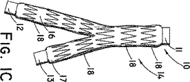
図1Cは、図1Bの本発明に、生体再造形可能物質14の第1層の少なくとも上、中、下またはその近辺に周知の市販のGianturco Zステントなどの1つ以上の拡張可能部材18が配置された別の例示的実施形態を示す。単一層の生体再造形可能物質と共に、これらの拡張可能部材またはZステントはグラフトの長さに沿ってあてがわれ、好ましくは、単一層のグラフト内に覆われるかまたは含有されて、例えば、最小侵襲性腔内外科技術を用いて患者内に位置決めされると、ステントは拡張される。しかし、図示するように、複数の拡張可能部材Zステント18が主本体部15ならびに脚部16および17のグラフトの長さに沿って長さ方向に配置される。各Zステントは単一ワイヤ片により作製することができる。一連のZステントはカニューレ管からレーザカットされ、相互に接続する断片が円筒状の主ループセグメント間に長さ方向の間隔を確立する。前述のように、これらの拡張可能部材は、結合剤材料を伴うまたは伴わないSIS断片であっても、内部に細胞外コラーゲン基質材料の粒子を含有した結合剤材料であっても、いかなる生体再造形可能物質にもあてがうことができる。拡張可能部材はまた、単に、腔内送達システムでの圧縮状態からの開放後に生体再造形可能グラフトを拡張させることができるステンレススチール、ニチロールまたはその他の弾力性材料などの弾力性材料リングまたは輪を含むこともできる。
FIG. 1C illustrates that the present invention of FIG. 1B includes one or more
図4は、本発明のグラフト22の主本体部15の端面図である。主本体部は、鋳造生体再造形可能物質14の壁または層をステント18の断面支柱と共に含む。この特定の実施形態では、ステントは生体再造形可能物質の単一層塗布内に含まれる。もしくは、ステントは第1層を覆って配置され、その生体再造形可能物質の第2層を拡張可能部材ステントを覆って塗布して、ステントを単一層壁内に完全に閉じ込めるようにすることができる。
FIG. 4 is an end view of the
図5は、生体再造形可能物質の第1層14が心金または型に塗布される、グラフト22の主本体部15の別の実施形態を示す。1つ以上の拡張可能部材ステント18が第1層の外表面に配置され、次に生体再造形可能物質の第2層19がこれを覆って塗布される。本
発明の最良の形態の実施形態は、SIS材料断片の2つの層をこれらの間に配置される拡張可能ステント18と共に用いることを考慮する。第1層が経験する乾燥または硬化の量に依っては、多層構造物は、界面43が区別し難いので、単一層構造物と同じに見えるかもしれない。これは、繰り返すが、第2のまたは複数の層を元の層に塗布する前に元の層が受ける乾燥または硬化の量に依存し得る。
FIG. 5 shows another embodiment of the
図1Dは、図1Cの本発明の拡張可能グラフト22を、拡張可能部材ステント18を覆って塗布または鋳造された生体再造形物質の第2層またはコーティング19と共に示す。この結果、拡張可能部材18は全体が生体再造形物質の1つ以上の層内にはめられるまたは封じ込まれ。これにより拡張可能部材をグラフト材料に結合させる縫合または他の接着手段を使用する必要がないという利点がある。前述のように、この多層構造物は、好ましくは室温で乾燥、硬化または脱水させることができる。管状構造物の他の処理方法では、多層グラフトを凍結させて次に真空チャンバー内で脱水させて凍結乾燥材料グラフトを作製することもできる。
FIG. 1D shows the
図1Eは、図1Dの本発明の多層グラフト22を、心金または型10上に置かれたままの多層グラフトの外表面の周りにきつく巻かれた拡張可能ポリテトラフルオロエチレンなどのポリマーの帯28と共に示す。帯28を巻くことによって、生体再造形物質層の厚さがより良好に制御され、その長さに沿って均一な厚さが維持される。1つ以上の帯28をグラフトの長さ全体に沿って貼り付けた後、ステントグラフトを脱水させ、好ましくは室温で真空プレスして、グラフトの鋳造生体再造形可能層を硬化および/または固化する。
FIG. 1E shows a strip of polymer, such as expandable polytetrafluoroethylene, tightly wound around the outer surface of the multi-layer graft as placed on the mandrel or
図1Fは、図1Eの本発明の多層グラフト22を、型10をグラフトの内腔23〜25から除去した状態で示す。次にこの自己拡張多層グラフトは送達システムに搭載され、例えば外側被覆を用いて縮径状態へと圧縮される。管の主本体部の近位端は、同側性および対側性脚部の遠位端と共に、送達システム内に配置されるのに先立って所望の長さに切断される。図2は、例えば、グラフトの全長にわたって長さ方向に配置されるニチノールワイヤ輪29を備えた多層グラフト27の本発明の別の例示的実施形態を示す。多層生体再造形可能グラフトとして示しているが、ワイヤ輪は、生体再造形可能物質が心金または型上に鋳造または塗布されるとき生体再造形可能物質内に配置され得る拡張可能部材である。この構成は単層のみを考慮しているが、好適な多層グラフトであれば拡張可能部材の配置および位置決めをより制御することができる。
FIG. 1F shows the inventive
図3は、織り込み生体再造形可能グラフトの本発明の別の実施形態であって、例えばSISなどの生体再造形可能物質の糸30が、グラフトの円周方向および長さ方向の支持および自己拡張のために、横糸および縦糸部材を形成するニチノール、ステンレススチールまたは他の弾力性材料ワイヤ31などの拡張可能部材と共に織り込まれている。この織り込み構成は単独でグラフトとして、または前述のような鋳造管状構造物との組み合わせで用いることができる。
FIG. 3 is another embodiment of the present invention of a woven bio-remodelable graft, wherein a
前述の結合剤材料を含めて、生体再造形可能物質として数多くの材料が利用され得ることが考慮される。また、この特定の構成においては、鋳造の通常の辞書上の意味が適用される。鋳造の1つのこのような定義は、McGraw−Hill Dictionary
of Scientific and Technical Terms第5版、版権1994年、322頁から得ることができる。ここでは鋳造(cast)は、液体またはプラスチック物質を金型内で冷却させることによって厚い形状へと形成することであると定義されている。また鋳造物は、鋳造可能物質を金型または型に入れてこれを固化させることによって形成されるあらゆる物体を含むことが理解される。この特定の適用例では、鋳造はまた液状または可鍛性の生体再造形可能物質を、単一のまたは二又の内腔形状の本明細書で述べた心金または型の表面に塗布することも考慮する。鋳造はまた、1つ以上の
層をこの心金または型に塗布するか、またはグラフトを金型およびその中に含まれる別の型内で成形して、単一のまたは複数の内腔を作製することも包含する。また、結合材料を伴ったまたは伴わない生体再造形可能物質は繊維または他の補強材料を含むことができることも考慮される。生体再造形可能物質は、上述の心金または型に塗布して乾燥または硬化させることができるゲル、ドウまたは他の液状の、柔軟な、または可鍛性の物質であり得る。
It is contemplated that many materials can be utilized as bioremodelable materials, including the binder materials described above. Also, in this particular configuration, the usual dictionary meaning of casting applies. One such definition of casting is McGraw-Hill Dictionary.
of Scientific and Technical Terms 5th Edition, Copyright 1994, p.322. Here, casting is defined as forming a thick shape by cooling a liquid or plastic material in a mold. It is also understood that a cast includes any object formed by placing a castable material in a mold or mold and solidifying it. In this particular application, casting also applies a liquid or malleable bioremodelable material to the surface of a mandrel or mold described herein in a single or bifurcated lumen shape. Also consider. Casting also applies one or more layers to the mandrel or mold, or molds the graft in the mold and another mold contained therein to create a single or multiple lumens To include. It is also contemplated that the bioremodelable material with or without a binding material can include fibers or other reinforcing materials. The bioremodelable material can be a gel, dough or other liquid, flexible, or malleable material that can be applied to the core or mold described above and dried or cured.
本発明において参考として援用するために、以下の特許が、あらゆる形態の生体再造形可能物質をさらに詳細に説明するものとして包含される。これらの参考文献としては、米国特許第4,902,508号Tissue Graft Composition、第4,956,178号Tissue Graft Composition、第5,275,826号Fluidized Intestinal Submucosa and its Use as an Injectable Tissue Graft、第5,281,422号Graft For Promoting Autogenous Tissue Growth、第5,352,463号Tissue Graft for Surgical Reconstruction of a Collagenous Meniscus And Method
Therefor、第5,372,821号Graft for Promoting Autogenous Tissue Growth、第5,445,833号Tendon or Ligament Graft for Promoting Autogenous Tissue Growth、第5,516,533号Fluidized Intestinal Submucosa and its Use as an Injectable Tissue Graft、第5,573,784号Graft for Promoting Autogenous Tissue Growth、第5,641,518号Method of Repairing Bone Tissue、第5,645,860号Tissue Graft and Method for Urinary Urothelium
Reconstruction Replacement、第5,695,998号Submucosa as a Growth Substrate for Islet Cells、第5,711,969号Large Area Submucosal Tissue Graft Constructs、第5,753,267号Method for Enhancing Functional Properties of Submucosal Tissue Graft Constructs、第5,755,791号Perforated Submucosal Tissue Graft Constructs、第5,762,966号Tissue Graft and Method for Urinary Urothelium Reconstruction Replacement、第5,866,414号Submucosa Gel as a Growth Substrate for Cells、第5,885,619号Large Area Submucosal Tissue Graft Constructs and Method for Making the Same、第5,955,110号Multilayered Submucosal Graft Constructs and Method for Making Same、第5,968,096号Method of Repairing Perforated submucosal Tissue Graft Constructs、第5,997,575号Perforated Submucosal Tissue Graft Constructs、第6,087,157号Device and Method of
Analyzing Tumor Cell Invasion of an Extracellular Matrix、第6,096,347号Myocardial Graft Constructs、第6,126,686号Artificial Vascular Valves、第6,187,039号Tubular Submucosal Graft Constructs、第6,241,981号Composition and Method for Repairing Neurological Tissue、第6,264,992号Submucosa as a Growth Substrate for Cells、第6,331,319号Galactosidase Modified Submucosal Tissue、第6,375,989号Submucosa Extracts、第6,206,931号Graft Prosthesis Materials、第6,358,284号Tubular Grafts from Purified Submucosa、第5,554,389号Urinary Bladder Submucosa Derived Tissue Graft、第6,099,567号Stomach Submucosa Derived Tissue Graftが含まれる。加えて、以下の米国および世界知的所有権機構の特許または公告番号およびそれらの適切な発行または公告日は、それらの全体が本明細書において参考として援用される。これらの追加の米国および世界知的所有権機構の公告物としては、米国特許第6,666,892号Multi-formed Collagenous Biomaterial Medical Device 2003-12-23、US 20030051735A1, Vessel Closure Member, Delivery Apparatus, and Method of Inserting the Member 2003-03-20、WO 03092546A2, Sling for Supporting Tissue 2003-11-13、WO 03092471A2, Cell-Seeded Extracellular Matrix Grafts 2003-11-13、WO 03088844A1, Apparatus and Method for Producing a Reinforced Surgical Staple Line 2003-10-30、WO 03035125A3, Medical Graft Device with Meshed Structure 2003-05-01、WO 03035125A2, Medical Graft Device with Meshed Structure 2003-05-01、WO 03009764A1, Vessel Closure Member and Delivery
Apparatus 2003-02-06、WO 03002168A1, Porous Sponge Matrix Medical Devices and Methods 2003-01-09、WO 03002165A1, Graft Prosthesis Devices Containing Renal Capsule Collagen 2003-01-09、WO 0156500A, Implantable Vascular Device 2001-08-09、WO
0154625A1, Stent Valves and Uses of Same 2001-08-02、WO 0110355A1, Tubular Graf
t Construct 2001-02-15、WO 0032253A1, Radiopaque Implantable Collagenous Biomaterial Device 2000-06-08、WO 0032250A1, A Multi-formed Collagenous Biomaterial Medical Device 2000-06-08およびWO 0032112A1, Embolization Device 2000-06-08がある。上記の参考文献のすべてが本明細書において参考として援用され、あらゆる結合材料を含む鋳造生体再造形可能物質の上述の実施形態および記述のいずれに対しても詳細な説明および支持を与えるものとして引用され得る。また、生体再造形可能物質は上記の参考文献において記載されているように架橋されて、生体再造形可能物質に近接する組織の再造形量を制御することができることも考慮される。
The following patents are included to further illustrate all forms of bioremodelable materials for incorporation by reference in the present invention. These references include U.S. Patent No. 4,902,508 Tissue Graft Composition, 4,956,178 Tissue Graft Composition, No. Issue Tissue Graft for Surgical Reconstruction of a Collagenous Meniscus And Method
Therefor, No. 5,372,821 Graft for Promoting Autogenous Tissue Growth, No. 5,445,833 Tendon or Ligament Graft for Promoting Autogenous Tissue Growth, No. 5,516,533 Fluidized Intestinal Submucosa and its Use as an Injectable Tissue Graft, No. 5,573,784 Graft for Promoting Autogenous Tissue Growth No. 5,641,518 Method of Repairing Bone Tissue, No. 5,645,860 Tissue Graft and Method for Urinary Urothelium
Reconstruction Replacement, No. 5,695,998 Submucosa as a Growth Substrate for Islet Cells, No. 5,711,969 Large Area Submucosal Tissue Graft Constructs, No. 5,753,267 Method for Enhancing Functional Properties of Submucosal Tissue Graft Constructs, No. 5,755,791, Perforated Submuco762 No. Tissue Graft and Method for Urinary Urothelium Reconstruction Replacement, No. 5,866,414 Submucosa Gel as a Growth Substrate for Cells, No. 5,885,619 Large Area Submucosal Tissue Graft Constructs and Method for Making the Same, No. 5,955,110 Multilayered Submucosal Graft Constructs and Method for Making Same, No. 5,968,096 Method of Repairing Perforated submucosal Tissue Graft Constructs, No. 5,997,575 Perforated Submucosal Tissue Graft Constructs, No. 6,087,157 Device and Method of
Analyzing Tumor Cell Invasion of an Extracellular Matrix, No. 6,096,347 Myocardial Graft Constructs, No. 6,126,686 Artificial Vascular Valves, No. 6,187,039 Tubular Submucosal Graft Constructs, No. 6,241,981 Composition and Method for Repairing Neurological Tissue, No. for Cells, No. 6,331,319 Galactosidase Modified Submucosal Tissue, No. 6,375,989 Submucosa Extracts, No. 6,206,931 Graft Prosthesis Materials, No. 6,358,284 Tubular Grafts from Purified Submucosa, No. 5,554,389 Urinary Bladder Submu Includes Graft. In addition, the following US and World Intellectual Property Organization patents or publication numbers and their appropriate issuance or publication dates are hereby incorporated by reference in their entirety. These additional U.S. and World Intellectual Property Organization publications include U.S. Pat.No. 6,666,892, Multi-formed Collagenous Biomaterial Medical Device 2003-12-23, US 20030051735A1, Vessel Closure Member, Delivery Apparatus, and Method of Inserting. the Member 2003-03-20, WO 03092546A2, Sling for Supporting Tissue 2003-11-13, WO 03092471A2, Cell-Seeded Extracellular Matrix Grafts 2003-11-13, WO 03088844A1, Apparatus and Method for Producing a Reinforced Surgical Staple Line 2003 -10-30, WO 03035125A3, Medical Graft Device with Meshed Structure 2003-05-01, WO 03035125A2, Medical Graft Device with Meshed Structure 2003-05-01, WO 03009764A1, Vessel Closure Member and Delivery
Apparatus 2003-02-06, WO 03002168A1, Porous Sponge Matrix Medical Devices and Methods 2003-01-09, WO 03002165A1, Graft Prosthesis Devices Containing Renal Capsule Collagen 2003-01-09, WO 0156500A, Implantable Vascular Device 2001-08-09 , WO
0154625A1, Stent Valves and Uses of Same 2001-08-02, WO 0110355A1, Tubular Graf
t Construct 2001-02-15, WO 0032253A1, Radiopaque Implantable Collagenous Biomaterial Device 2000-06-08, WO 0032250A1, A Multi-formed Collagenous Biomaterial Medical Device 2000-06-08 and WO 0032112A1, Embolization Device 2000-06-08 is there. All of the above references are incorporated herein by reference and are cited as providing detailed description and support for any of the above-described embodiments and descriptions of cast bioremodelable materials including any binding materials. Can be done. It is also contemplated that the bioremodelable material can be cross-linked as described in the above references to control the amount of tissue remodeling in proximity to the bioremodelable material.
Claims (15)
に配置される、請求項1から6のいずれかに記載のグラフト。The graft according to any of claims 1 to 6, wherein the expandable member is disposed between a first layer and a second layer of a piece of extracellular collagen matrix material.
コラーゲン基質材料を切断、細断または粉砕することにより、断片、細片または粒子形態の細胞外コラーゲン基質材料を提供するステップと、
前記細胞外コラーゲン基質材料の断片、細片または粒子を型に塗布するステップと、
結合材料中で前記細胞外コラーゲン基質材料の断片、細片または粒子を互いに結合させて生体再造形可能物質の少なくとも1つの層を形成するステップと、
を包含する方法。A method of making a graft according to any of claims 1 to 11, comprising
Cutting the collagen matrix material by shredding or grinding, and providing a fragment, an extracellular collagen matrix materials of the strip or particle form,
A step of applying a fragment of said extracellular collagen matrix material, debris or particles child into a mold,
Fragment of the extracellular collagen matrix material bonded material, forming at least one layer of bound debris or particles together bioremodelable substance,
Including the method.
Applications Claiming Priority (3)
| Application Number | Priority Date | Filing Date | Title |
|---|---|---|---|
| US54292204P | 2004-02-09 | 2004-02-09 | |
| US60/542,922 | 2004-02-09 | ||
| PCT/US2005/003968 WO2005077432A2 (en) | 2004-02-09 | 2005-02-09 | Cast bioremodelable graft |
Publications (2)
| Publication Number | Publication Date |
|---|---|
| JP2007521884A JP2007521884A (en) | 2007-08-09 |
| JP5109029B2 true JP5109029B2 (en) | 2012-12-26 |
Family
ID=34860352
Family Applications (1)
| Application Number | Title | Priority Date | Filing Date |
|---|---|---|---|
| JP2006552348A Active JP5109029B2 (en) | 2004-02-09 | 2005-02-09 | Bio-remodelable graft |
Country Status (7)
| Country | Link |
|---|---|
| US (1) | US8808352B2 (en) |
| EP (1) | EP1713525B1 (en) |
| JP (1) | JP5109029B2 (en) |
| AT (1) | ATE471170T1 (en) |
| AU (1) | AU2005212335B2 (en) |
| DE (1) | DE602005021855D1 (en) |
| WO (1) | WO2005077432A2 (en) |
Families Citing this family (30)
| Publication number | Priority date | Publication date | Assignee | Title |
|---|---|---|---|---|
| US8038708B2 (en) * | 2001-02-05 | 2011-10-18 | Cook Medical Technologies Llc | Implantable device with remodelable material and covering material |
| US20060206139A1 (en) * | 2005-01-19 | 2006-09-14 | Tekulve Kurt J | Vascular occlusion device |
| US9138445B2 (en) * | 2005-03-09 | 2015-09-22 | Cook Biotech Incorporated | Medical graft materials with adherent extracellular matrix fibrous mass |
| AU2006275881B2 (en) * | 2005-07-27 | 2012-04-12 | Cook Medical Technologies Llc | Stent/graft device and method for open surgical placement |
| GB0517085D0 (en) * | 2005-08-19 | 2005-09-28 | Angiomed Ag | Polymer prosthesis |
| WO2007134134A2 (en) * | 2006-05-09 | 2007-11-22 | Lifecell Corporation | Reinforced biological tissue |
| US20070282421A1 (en) * | 2006-05-31 | 2007-12-06 | Parker Fred T | Stent Assembly for Protecting the Interior Surface of a Vessel |
| US8790414B2 (en) * | 2006-11-10 | 2014-07-29 | Cook Biotech Incorporated | Graft for hysterotomy closure |
| EP2107895A1 (en) * | 2007-01-29 | 2009-10-14 | Cook Incorporated | Medical prosthesis and method of production |
| US8177834B2 (en) | 2007-03-12 | 2012-05-15 | Cook Medical Technologies Llc | Woven fabric with shape memory element strands |
| US8597342B2 (en) * | 2007-08-24 | 2013-12-03 | Cook Medical Technologies Llc | Textile graft for in situ fenestration |
| JP2011500283A (en) | 2007-10-26 | 2011-01-06 | クック クリティカル ケア インコーポレーテッド | Vascular conduit and delivery system installed in open surgery |
| US8834552B2 (en) * | 2007-12-27 | 2014-09-16 | Cook Medical Technologies Llc | Stent graft having floating yarns |
| US8956378B2 (en) * | 2008-02-29 | 2015-02-17 | Cook Biotech Incorporated | Coated embolization device |
| US9295757B2 (en) | 2008-06-10 | 2016-03-29 | Cook Biotech Incorporated | Quilted implantable graft |
| US10688219B2 (en) * | 2008-06-10 | 2020-06-23 | Cook Biotech Incorporated | Quilted implantable graft |
| US8353943B2 (en) * | 2008-08-29 | 2013-01-15 | Cook Medical Technologies Llc | Variable weave graft with metal strand reinforcement for in situ fenestration |
| US9427304B2 (en) * | 2008-10-27 | 2016-08-30 | St. Jude Medical, Cardiology Division, Inc. | Multi-layer device with gap for treating a target site and associated method |
| JP5693475B2 (en) * | 2009-03-04 | 2015-04-01 | ペイタント・ソリューションズ・インコーポレイテッドPeytant Solutions, Inc. | Stent modified with a material comprising amnion tissue and corresponding method |
| WO2011130536A2 (en) * | 2010-04-14 | 2011-10-20 | Northwestern University | Triple balloon occlusion and infusion catheter |
| JP5947790B2 (en) * | 2010-05-25 | 2016-07-06 | クック・バイオテック・インコーポレイテッドCook Biotech Incorporated | Methods, substrates, and systems useful for cell seeding of medical implants |
| US8540619B2 (en) * | 2010-07-14 | 2013-09-24 | Atex Technologies, Inc. | Fabric cutting system and method |
| US9751156B2 (en) | 2010-07-14 | 2017-09-05 | Atex Technologies Inc. | Fabric cutting system |
| US20140100648A1 (en) * | 2012-10-08 | 2014-04-10 | Robert G. Matheny | Multi-Layer Vascular Prosthesis |
| JP6563900B2 (en) * | 2013-04-13 | 2019-08-21 | ソリナス メディカル インコーポレイテッドSolinas Medical,Inc. | Self-closing device, apparatus, manufacturing method and delivery method thereof |
| US20150283308A1 (en) * | 2014-04-03 | 2015-10-08 | Cook Biotech, Incorporated | Endoluminal device and method of implanting same |
| WO2016014259A1 (en) * | 2014-07-22 | 2016-01-28 | Cormatrix Cardiovascular, Inc. | Reinforced vascular prostheses |
| US9694105B2 (en) * | 2014-12-08 | 2017-07-04 | Cormatrix Cardiovascular, Inc. | Vascular casted prostheses and methods of forming same for treating biological tissue |
| EP3274006B1 (en) * | 2015-03-23 | 2020-11-11 | Cormatrix Cardiovascular, Inc. | Vascular casted prostheses and methods of forming same for treating biological tissue |
| US11826490B1 (en) | 2020-12-29 | 2023-11-28 | Acell, Inc. | Extracellular matrix sheet devices with improved mechanical properties and method of making |
Family Cites Families (22)
| Publication number | Priority date | Publication date | Assignee | Title |
|---|---|---|---|---|
| US4787900A (en) * | 1982-04-19 | 1988-11-29 | Massachusetts Institute Of Technology | Process for forming multilayer bioreplaceable blood vessel prosthesis |
| CA2183962A1 (en) * | 1994-03-22 | 1995-09-28 | Paul D. Kemp | Three-dimensional bioremodelable collagen fabrics |
| US6475232B1 (en) * | 1996-12-10 | 2002-11-05 | Purdue Research Foundation | Stent with reduced thrombogenicity |
| US5693085A (en) * | 1994-04-29 | 1997-12-02 | Scimed Life Systems, Inc. | Stent with collagen |
| US6015429A (en) * | 1994-09-08 | 2000-01-18 | Gore Enterprise Holdings, Inc. | Procedures for introducing stents and stent-grafts |
| US5755791A (en) * | 1996-04-05 | 1998-05-26 | Purdue Research Foundation | Perforated submucosal tissue graft constructs |
| AU732726B2 (en) * | 1996-12-10 | 2001-04-26 | Purdue Research Foundation | Biomaterial derived from vertebrate liver tissue |
| AU5520898A (en) * | 1996-12-10 | 1998-07-03 | Purdue Research Foundation | Stent with reduced thrombogenicity |
| GB2329840C (en) * | 1997-10-03 | 2007-10-05 | Johnson & Johnson Medical | Biopolymer sponge tubes |
| DE69940466D1 (en) * | 1998-06-05 | 2009-04-09 | Organogenesis Inc | BIOLOGICALLY MODELED IMPLANTABLE PROSTHESIS |
| US6187036B1 (en) * | 1998-12-11 | 2001-02-13 | Endologix, Inc. | Endoluminal vascular prosthesis |
| US20050171594A1 (en) * | 1998-12-31 | 2005-08-04 | Angiotech International Ag | Stent grafts with bioactive coatings |
| US20030229393A1 (en) * | 2001-03-15 | 2003-12-11 | Kutryk Michael J. B. | Medical device with coating that promotes cell adherence and differentiation |
| US6254632B1 (en) * | 2000-09-28 | 2001-07-03 | Advanced Cardiovascular Systems, Inc. | Implantable medical device having protruding surface structures for drug delivery and cover attachment |
| US20050027307A1 (en) * | 2001-07-16 | 2005-02-03 | Schwartz Herbert Eugene | Unitary surgical device and method |
| JP2003126125A (en) * | 2001-10-24 | 2003-05-07 | Katsuko Sakai | Artificial blood vessel and method of preparing it |
| US8518096B2 (en) * | 2002-09-03 | 2013-08-27 | Lifeshield Sciences Llc | Elephant trunk thoracic endograft and delivery system |
| US20040059409A1 (en) * | 2002-09-24 | 2004-03-25 | Stenzel Eric B. | Method of applying coatings to a medical device |
| EP1673037B1 (en) * | 2003-10-10 | 2009-08-26 | The Cleveland Clinic Foundation | Endoluminal prosthesis with interconnectable modules |
| US8734501B2 (en) * | 2003-10-10 | 2014-05-27 | Cook Medical Technologies Llc | Composite stent graft |
| US9078780B2 (en) * | 2003-11-08 | 2015-07-14 | Cook Medical Technologies Llc | Balloon flareable branch vessel prosthesis and method |
| WO2007002260A2 (en) * | 2005-06-21 | 2007-01-04 | Cook Incorporated | Implantable graft to close a fistula |
-
2005
- 2005-02-09 EP EP05713125A patent/EP1713525B1/en active Active
- 2005-02-09 WO PCT/US2005/003968 patent/WO2005077432A2/en not_active Application Discontinuation
- 2005-02-09 DE DE602005021855T patent/DE602005021855D1/de active Active
- 2005-02-09 AT AT05713125T patent/ATE471170T1/en not_active IP Right Cessation
- 2005-02-09 JP JP2006552348A patent/JP5109029B2/en active Active
- 2005-02-09 AU AU2005212335A patent/AU2005212335B2/en active Active
- 2005-02-09 US US11/054,043 patent/US8808352B2/en active Active
Also Published As
| Publication number | Publication date |
|---|---|
| US8808352B2 (en) | 2014-08-19 |
| ATE471170T1 (en) | 2010-07-15 |
| EP1713525A2 (en) | 2006-10-25 |
| AU2005212335A1 (en) | 2005-08-25 |
| US20050187604A1 (en) | 2005-08-25 |
| AU2005212335B2 (en) | 2010-01-28 |
| WO2005077432A2 (en) | 2005-08-25 |
| DE602005021855D1 (en) | 2010-07-29 |
| WO2005077432A3 (en) | 2006-03-30 |
| JP2007521884A (en) | 2007-08-09 |
| EP1713525B1 (en) | 2010-06-16 |
Similar Documents
| Publication | Publication Date | Title |
|---|---|---|
| JP5109029B2 (en) | Bio-remodelable graft | |
| US20240009356A1 (en) | Methods for forming stents modified with material comprising amnion tissue | |
| US7887576B2 (en) | Endoluminal device with extracellular matrix material and methods | |
| EP1729677B1 (en) | Graft material and stent graft comprising extracellular collagen matrix and method of preparation | |
| US6334872B1 (en) | Method for treating diseased or damaged organs | |
| WO2005115275A1 (en) | Endoluminal device with extracellular matrix material and methods | |
| EP1364627A1 (en) | Bioremodelable collagen graft prosthesis | |
| CN102137634A (en) | Hernia patch with removable resilient element | |
| WO1998024385A1 (en) | Artificial blood vessel | |
| US20080046070A1 (en) | Chemically treated extracellular matrices for affecting the cellular response | |
| Kato et al. | Experimental assessment of newly devised transcatheter stent-graft for aortic dissection | |
| González-Pérez et al. | Biohybrid elastin-like venous valve with potential for in situ tissue engineering | |
| Guidoin et al. | Biostability of vascular prostheses | |
| Guidoin et al. | Vascular prostheses for open surgery | |
| JP2015503373A (en) | Structure with fibers that are adhesively bonded to each other at each location | |
| Tizian | A new microvascular substitute (inside diameter: 1 mm): Evaluation and early patency rates | |
| CA2352767C (en) | Method for vocal cord reconstruction | |
| Guidoin et al. | Biotextiles as medical implants: 15. Vascular prostheses for open surgery | |
| WO2011114218A2 (en) | Endograft with cultured stem cells |
Legal Events
| Date | Code | Title | Description |
|---|---|---|---|
| A621 | Written request for application examination |
Free format text: JAPANESE INTERMEDIATE CODE: A621 Effective date: 20080208 |
|
| A521 | Request for written amendment filed |
Free format text: JAPANESE INTERMEDIATE CODE: A523 Effective date: 20110120 |
|
| A131 | Notification of reasons for refusal |
Free format text: JAPANESE INTERMEDIATE CODE: A131 Effective date: 20110524 |
|
| A521 | Request for written amendment filed |
Free format text: JAPANESE INTERMEDIATE CODE: A523 Effective date: 20110802 |
|
| A131 | Notification of reasons for refusal |
Free format text: JAPANESE INTERMEDIATE CODE: A131 Effective date: 20120403 |
|
| A521 | Request for written amendment filed |
Free format text: JAPANESE INTERMEDIATE CODE: A523 Effective date: 20120605 |
|
| TRDD | Decision of grant or rejection written | ||
| A01 | Written decision to grant a patent or to grant a registration (utility model) |
Free format text: JAPANESE INTERMEDIATE CODE: A01 Effective date: 20120626 |
|
| A601 | Written request for extension of time |
Free format text: JAPANESE INTERMEDIATE CODE: A601 Effective date: 20120724 |
|
| A602 | Written permission of extension of time |
Free format text: JAPANESE INTERMEDIATE CODE: A602 Effective date: 20120801 |
|
| A711 | Notification of change in applicant |
Free format text: JAPANESE INTERMEDIATE CODE: A711 Effective date: 20120821 |
|
| A61 | First payment of annual fees (during grant procedure) |
Free format text: JAPANESE INTERMEDIATE CODE: A61 Effective date: 20120822 |
|
| A521 | Request for written amendment filed |
Free format text: JAPANESE INTERMEDIATE CODE: A821 Effective date: 20120821 |
|
| FPAY | Renewal fee payment (event date is renewal date of database) |
Free format text: PAYMENT UNTIL: 20151019 Year of fee payment: 3 |
|
| R150 | Certificate of patent or registration of utility model |
Ref document number: 5109029 Country of ref document: JP Free format text: JAPANESE INTERMEDIATE CODE: R150 Free format text: JAPANESE INTERMEDIATE CODE: R150 |
|
| R250 | Receipt of annual fees |
Free format text: JAPANESE INTERMEDIATE CODE: R250 |
|
| R250 | Receipt of annual fees |
Free format text: JAPANESE INTERMEDIATE CODE: R250 |
|
| R250 | Receipt of annual fees |
Free format text: JAPANESE INTERMEDIATE CODE: R250 |
|
| R250 | Receipt of annual fees |
Free format text: JAPANESE INTERMEDIATE CODE: R250 |
|
| R250 | Receipt of annual fees |
Free format text: JAPANESE INTERMEDIATE CODE: R250 |
|
| R250 | Receipt of annual fees |
Free format text: JAPANESE INTERMEDIATE CODE: R250 |
|
| R250 | Receipt of annual fees |
Free format text: JAPANESE INTERMEDIATE CODE: R250 |
|
| R250 | Receipt of annual fees |
Free format text: JAPANESE INTERMEDIATE CODE: R250 |