JP5102773B2 - Methods for improving stem cell homing and engraftment - Google Patents
Methods for improving stem cell homing and engraftment Download PDFInfo
- Publication number
- JP5102773B2 JP5102773B2 JP2008542940A JP2008542940A JP5102773B2 JP 5102773 B2 JP5102773 B2 JP 5102773B2 JP 2008542940 A JP2008542940 A JP 2008542940A JP 2008542940 A JP2008542940 A JP 2008542940A JP 5102773 B2 JP5102773 B2 JP 5102773B2
- Authority
- JP
- Japan
- Prior art keywords
- cells
- cell
- hematopoietic
- nicotinamide
- stem
- Prior art date
- Legal status (The legal status is an assumption and is not a legal conclusion. Google has not performed a legal analysis and makes no representation as to the accuracy of the status listed.)
- Active
Links
Images


Classifications
-
- C—CHEMISTRY; METALLURGY
- C12—BIOCHEMISTRY; BEER; SPIRITS; WINE; VINEGAR; MICROBIOLOGY; ENZYMOLOGY; MUTATION OR GENETIC ENGINEERING
- C12N—MICROORGANISMS OR ENZYMES; COMPOSITIONS THEREOF; PROPAGATING, PRESERVING, OR MAINTAINING MICROORGANISMS; MUTATION OR GENETIC ENGINEERING; CULTURE MEDIA
- C12N5/00—Undifferentiated human, animal or plant cells, e.g. cell lines; Tissues; Cultivation or maintenance thereof; Culture media therefor
- C12N5/06—Animal cells or tissues; Human cells or tissues
- C12N5/0602—Vertebrate cells
- C12N5/0634—Cells from the blood or the immune system
- C12N5/0647—Haematopoietic stem cells; Uncommitted or multipotent progenitors
-
- A—HUMAN NECESSITIES
- A61—MEDICAL OR VETERINARY SCIENCE; HYGIENE
- A61P—SPECIFIC THERAPEUTIC ACTIVITY OF CHEMICAL COMPOUNDS OR MEDICINAL PREPARATIONS
- A61P43/00—Drugs for specific purposes, not provided for in groups A61P1/00-A61P41/00
-
- C—CHEMISTRY; METALLURGY
- C12—BIOCHEMISTRY; BEER; SPIRITS; WINE; VINEGAR; MICROBIOLOGY; ENZYMOLOGY; MUTATION OR GENETIC ENGINEERING
- C12N—MICROORGANISMS OR ENZYMES; COMPOSITIONS THEREOF; PROPAGATING, PRESERVING, OR MAINTAINING MICROORGANISMS; MUTATION OR GENETIC ENGINEERING; CULTURE MEDIA
- C12N5/00—Undifferentiated human, animal or plant cells, e.g. cell lines; Tissues; Cultivation or maintenance thereof; Culture media therefor
- C12N5/06—Animal cells or tissues; Human cells or tissues
- C12N5/0602—Vertebrate cells
-
- A—HUMAN NECESSITIES
- A61—MEDICAL OR VETERINARY SCIENCE; HYGIENE
- A61K—PREPARATIONS FOR MEDICAL, DENTAL OR TOILETRY PURPOSES
- A61K48/00—Medicinal preparations containing genetic material which is inserted into cells of the living body to treat genetic diseases; Gene therapy
Description
本発明は、移植された細胞のホーミング効率、保持効率および生着効率を改善するための方法に関連する。 The present invention relates to a method for improving the homing efficiency, retention efficiency and engraftment efficiency of transplanted cells.
骨髄移植(BMT)は、骨髄から得られた多能性造血細胞を患者に移植する臨床的手法である。BMTは、悪性腫瘍、重症複合免疫不全症(SCID)、先天的または遺伝的に明らかにされた造血異常、貧血、再生不良性貧血、白血病および大理石骨病をはじめとするいくつかの血液学的障害における優れた治療である。 Bone marrow transplantation (BMT) is a clinical procedure in which pluripotent hematopoietic cells obtained from bone marrow are transplanted into a patient. BMT has several hematological conditions including malignant tumors, severe combined immunodeficiency disease (SCID), congenital or genetically manifested hematopoietic abnormalities, anemia, aplastic anemia, leukemia and marble bone disease. Excellent treatment for disability.
原始的または多能性造血幹細胞は通常、骨髄に存在しているが、臍血は、移植可能な造血幹細胞/前駆細胞の別の機能的な供給源である(Gluckman,E.他、1989、N.Engl.J.Med.、321:1174)。これらの原始的造血細胞のすべてがその表面CD34抗原によって特定され得る。造血幹細胞は、2つの主要な経路の一方に沿って、すなわち、リンパ球系幹細胞または骨髄系幹細胞のいずれかに分化する。両者はさらに、それぞれのタイプの成熟した血液細胞のための前駆細胞に分化する。これらの前駆細胞は自己複製のための能力を失っており、また、定められた細胞系譜への分化が決定している。従って、リンパ球系幹細胞はT前駆細胞またはB前駆細胞に分化し、骨髄系幹細胞は、赤血球、好中球、好酸球、好塩基球、単球、マスト細胞および血小板のための前駆細胞に分化する。 Primitive or pluripotent hematopoietic stem cells are usually present in the bone marrow, but umbilical blood is another functional source of transplantable hematopoietic stem / progenitor cells (Gluckman, E. et al., 1989, N. Engl. J. Med., 321: 1174). All of these primitive hematopoietic cells can be identified by their surface CD34 antigen. Hematopoietic stem cells differentiate along one of two major pathways, namely either lymphoid stem cells or myeloid stem cells. Both further differentiate into progenitor cells for each type of mature blood cell. These progenitor cells have lost the ability for self-renewal and have been determined to differentiate into a defined cell lineage. Thus, lymphoid stem cells differentiate into T or B precursor cells, and myeloid stem cells become progenitor cells for red blood cells, neutrophils, eosinophils, basophils, monocytes, mast cells and platelets. Differentiate.
定常状態条件のもとでは、造血幹細胞および造血前駆細胞の大部分が骨髄に存在しており、これらの細胞のごく僅かが末梢血において検出可能であるにすぎない。しかしながら、幹細胞は骨髄抑制剤および/または特定の造血増殖因子による処理によって末梢血内に動員させることができる。様々な研究により、宿主に注入された末梢血幹細胞(PBSC)が、骨髄由来の幹細胞および前駆細胞と比較したとき、生着について潜在的能力が高いことを示すことが明らかにされている。従って、化学療法、造血増殖因子、または、これらの様式の組合せによって動員されたPBSCが現在、自家移植および非自家移植の両方の状況で使用される[Anderlini,P.およびKorbling,M.(1997)、Stem.Cells、15、9〜17]。非自家移植の場合、幹細胞のドナーは健康な個体であり、幹細胞を血流内に動員するための手法は、最小限の不快を伴って達成しなければならない。この場合、造血増殖因子による幹細胞の動員は、抗成長性薬物(すなわち、シクロホスファミド)による動員よりも好まれる。 Under steady state conditions, the majority of hematopoietic stem cells and hematopoietic progenitor cells are present in the bone marrow, and only a few of these cells are detectable in peripheral blood. However, stem cells can be mobilized into the peripheral blood by treatment with myelosuppressants and / or certain hematopoietic growth factors. Various studies have shown that peripheral blood stem cells (PBSC) injected into the host show a high potential for engraftment when compared to bone marrow derived stem and progenitor cells. Thus, PBSC mobilized by chemotherapy, hematopoietic growth factors, or combinations of these modalities are currently used in both autologous and non-autologous situations [Anderlini, P. et al. And Korbling, M .; (1997), Stem. Cells, 15, 9-17]. For non-autologous transplants, stem cell donors are healthy individuals, and techniques for mobilizing stem cells into the bloodstream must be accomplished with minimal discomfort. In this case, mobilization of stem cells by hematopoietic growth factors is preferred over mobilization by anti-growth drugs (ie cyclophosphamide).
幹細胞および前駆細胞に加えて、より分化した細胞を、細胞機能不全または細胞死によって特徴づけられる特定の器官または組織の疾患または状態を治療するために、移植のために使用することができる。多くのそのような疾患について、現在の医学的治療または外科的手法は不十分であるか、または存在しないかのいずれかである。細胞治療が、これらの状態のための回復治療を提供するために、存在している細胞に取って代わり得るか、または、存在している細胞を強化することができる。移植のために好適である例示的な細胞タイプには、神経組織由来の細胞、肝細胞、筋細胞、網膜細胞、内分泌細胞、メラノサイト、ケラチノサイトおよび軟骨細胞が挙げられる。移植された細胞の生着により、組織機能が再確立に成功し得ることが、動物モデルおよびヒト研究の両方で示されている。従って、ニューロンを、例えば、パーキンソン病および他の神経変性疾患のために移植することができる。筋肉細胞(例えば、筋芽細胞など)を、例えば、虚血性の心臓の筋障害を治療するために移植することができる。小島細胞を、糖尿病、および/または、インスリンおよびグルカゴンに関連する他の疾患または状態を治療する移植することができる。分化した血液細胞(例えば、リンパ球および樹状細胞など)はまた、例えば、NK細胞による養子免疫治療のために移植することができる。 In addition to stem and progenitor cells, more differentiated cells can be used for transplantation to treat diseases or conditions of specific organs or tissues characterized by cell dysfunction or cell death. For many such diseases, current medical treatment or surgical procedures are either inadequate or nonexistent. Cell therapy can replace existing cells or can enhance existing cells to provide a recovery therapy for these conditions. Exemplary cell types that are suitable for transplantation include cells derived from neural tissue, hepatocytes, muscle cells, retinal cells, endocrine cells, melanocytes, keratinocytes and chondrocytes. It has been shown in both animal models and human studies that transplanted cell engraftment can successfully re-establish tissue function. Thus, neurons can be transplanted for Parkinson's disease and other neurodegenerative diseases, for example. Muscle cells (eg, myoblasts, etc.) can be transplanted to treat, for example, ischemic cardiac myopathy. Islet cells can be transplanted to treat diabetes and / or other diseases or conditions associated with insulin and glucagon. Differentiated blood cells (such as lymphocytes and dendritic cells) can also be transplanted, for example, for adoptive immunotherapy with NK cells.
しかしながら、様々な研究では、移植された細胞(例えば、肝細胞および神経細胞など)の大部分が移植後に身体から除かれ、目標の器官または組織に局在化しないことが示されている(De Roos他、Transplantation、1997、63:513〜18;Gagandeep他、Gene Therapy、1999、6:729〜36)。移植された細胞のホーミング、保持および生着を改善するための様々な努力(例えば、移植前におけるConAによる肝細胞の治療(Ito他、Muscle Nerve、1998、21:291〜7)など)はこれまで、わずかに効果的であるにすぎない。従って、様々な努力が、移植のためのより多くの数の細胞を提供するために、新しく調製された細胞をプールおよび貯蔵するための方法に向けられている(例えば、米国特許第6713245号および同第6821779号(Koopmans他)を参照のこと)。 However, various studies have shown that the majority of transplanted cells (eg, hepatocytes and neurons) are removed from the body after transplantation and do not localize to the target organ or tissue (De Roos et al., Transplantation, 1997, 63: 513-18; Gandeep et al., Gene Therapy, 1999, 6: 729-36). Various efforts to improve homing, retention and engraftment of transplanted cells (eg treatment of hepatocytes with ConA prior to transplantation (Ito et al., Muscle Nerve, 1998, 21: 291-7) etc.) Until, it is only slightly effective. Accordingly, various efforts are directed to methods for pooling and storing freshly prepared cells to provide a greater number of cells for transplantation (see, eg, US Pat. No. 6,713,245 and No. 6821777 (Koopmans et al.)).
移植後、細胞はその目標組織に向かって遊走するであろう。化学誘因物質(例えば、特定のサイトカイン(CXCL1〜CXCL16およびCCL1〜CCL27)など)が、細胞をその目的に向かわせることを助ける。ストローマ細胞由来因子1α(SDF−1α)(これはCXCL12とも呼ばれる)が、造血幹細胞および神経幹細胞を含めて、CD34+細胞の強力な化学誘因物質であり(Aiuti、J.Exp.Med.、1997、185:111〜120)、発達期間中(McGrath、Dev.Biol.、1999、213:442〜456)および成人期の期間中(Imai、Br.J.Haematol.、1999、106:905〜911)において多くの組織で広く発現する。この因子はまた、非幹細胞(例えば、Tリンパ球など)を化学的に誘引する。CXCケモカイン受容体4(CXCR4)はSDF−1αについての同族受容体であり、幹細胞上に発現する。近年の研究では、SDF−1α/CXCR4が、障害時における幹細胞の分子的プログラムおよびホーミングを活性化する経路として関係している(Jaime Imitola他、Proc Natl Acad Sci USA、2004(December 28)、101(52):18117〜18122)。 After transplantation, the cells will migrate towards their target tissue. A chemoattractant, such as certain cytokines (CXCL1-CXCL16 and CCL1-CCL27), helps direct the cell to its purpose. Stromal cell-derived factor 1α (SDF-1α) (also referred to as CXCL12) is a potent chemoattractant of CD34 + cells, including hematopoietic and neural stem cells (Aitui, J. Exp. Med., 1997). 185: 111-120), during development (McGrath, Dev. Biol., 1999, 213: 442-456) and during adulthood (Imai, Br. J. Haematol., 1999, 106: 905-911). ) Is widely expressed in many tissues. This factor also chemically attracts non-stem cells such as T lymphocytes. CXC chemokine receptor 4 (CXCR4) is a cognate receptor for SDF-1α and is expressed on stem cells. In recent studies, SDF-1α / CXCR4 has been implicated as a pathway that activates the molecular program and homing of stem cells at the time of injury (Jaime Imitola et al., Proc Natl Acad Sci USA, 2004 (December 28), 101 (52): 18117-18122).
CD26/ジペプチジルペプチダーゼIV(DPPIV)(ジペプチド(例えば、SDF−1αなど)をプロリンまたはアラニンの後でポリペプチド鎖のN末端から切断する膜結合した細胞外ペプチダーゼ)は、造血細胞および他の細胞におけるその発現が分化および活性化によって調節される系譜非特異的な抗原である。ケモカインのタンパク質分解的切断が、細胞がケモカインによって誘因および/または活性化され得ることに関して関係している(Baggiolini,M.、1998、Nature、392:565)。 CD26 / dipeptidyl peptidase IV (DPPIV), a membrane-bound extracellular peptidase that cleaves a dipeptide (eg, SDF-1α, etc.) from the N-terminus of the polypeptide chain after proline or alanine, is a hematopoietic cell and other cells A lineage non-specific antigen whose expression in is regulated by differentiation and activation. Proteolytic cleavage of chemokines is implicated in that cells can be triggered and / or activated by chemokines (Baggiolini, M., 1998, Nature, 392: 565).
いくつかの機能的研究では、CD26/DPPIVがT細胞および造血細胞の遊走および動員において果たす役割が間接的に示される[Shioda他(1998)、Proc.Natl.Acad.Sci.USA、95:6331]。CD34+細胞に対する内因性CD26/DPPIV活性の阻害は、SDF−1αに対するこれらの細胞の走化性応答を高めることが示された(Christopherson KW 2nd他、Science、2004(Aug 13)、305(5686):1000〜1003;Christopherson KW 2nd、J Immunol、2002(Dec 15)、169(12):7000〜7008)。その一方で、DPPIVを伴うSDF−1αのN末端短縮化によってでは、CD34+臍血細胞は遊走を誘導しない。 Several functional studies indirectly show the role CD26 / DPPIV plays in T cell and hematopoietic cell migration and mobilization [Shioda et al. (1998), Proc. Natl. Acad. Sci. USA, 95: 6331]. Inhibition of endogenous CD26 / DPPIV activity on CD34 + cells has been shown to enhance the chemotactic response of these cells to SDF-1α (Christopherson KW 2nd et al., Science, 2004 (Aug 13), 305 (5686). ): 1000-1003; Christopherson KW 2nd, J Immunol, 2002 (Dec 15), 169 (12): 7000-7008). On the other hand, CD34 + umbilical blood cells do not induce migration by shortening the N-terminus of SDF-1α with DPPIV.
ニコチンアミド(NA)(ナイアシン(ビタミンB3)のアミド形態)は、モノADPリボシルトランスフェラーゼ活性およびポリADPリボシルトランスフェラーゼ活性を有するNAD(+)依存性酵素の塩基交換基質および強力な阻害剤である。ADPリボシル化は多様な生物学的プロセスの修飾に関係する(Corda D、Di Girolamo M.2003;22(9):1953−1958;Rankin PW他、J Biol Chem.1989;264:4312−4317;Banasik M.他、J Biol Chem.1992;267:1569−1575;Ueda K,Hayaishi O、Annu Rev Biochem.1985;54:73−100;Smith S.Trends Biochem Sci.2001;26:174−179;Virag L,Szabo C.Pharm.Reviews.2002;54:375−429)。 Nicotinamide (NA) (amide form of niacin (vitamin B3)) is a base exchange substrate and a potent inhibitor of NAD (+)-dependent enzymes with mono-ADP ribosyl transferase activity and poly-ADP ribosyl transferase activity. ADP ribosylation is involved in the modification of various biological processes (Corda D, Di Girolamo M. 2003; 22 (9): 1953-1958; Rankin PW et al., J Biol Chem. 1989; 264: 4312-4317; Banasik M. et al., J Biol Chem. 1992; 267: 1569-1575; Ueda K, Hayashi O, Annu Rev Biochem.1985; 54: 73-100; Smith S. Trends Biochem Sci.2001; Virag L, Szabo C. Pharm. Reviews. 2002; 54: 375-429).
モノADPリボシル化反応またはポリADPリボシル化反応に関わる内因性ADPリボシルトランスフェラーゼは、細胞のシグナル伝達に関与する様々な分子(例えば、コアヒストン(de la Cruz X、Lois S他、Bioessays、2005、27(2):164〜75)、ヘテロ三量体GTP結合(G)タンパク質のαサブユニット、小さいGTPaseのRho、モノマーアクチンおよび伸長因子2(EF−2)など)を修飾する。これらの翻訳後修飾は、これらのタンパク質によって調節される細胞機能の活性化または不活性化を引き起こす(Lupi R他、J Biol Chem、2000、275:9418〜9424;Lupi R他、Biochem J.、2002、367:1〜7;Yau L他、Eur.J.Biochem.、2003、270:101〜110)。 Endogenous ADP ribosyltransferases involved in mono-ADP ribosylation reactions or poly-ADP ribosylation reactions are various molecules involved in cellular signal transduction (eg, core histones (de la Cruz X, Lois S et al., Bioessays, 2005, 27 ( 2): 164-75), heterotrimeric GTP binding (G) protein α subunit, small GTPase Rho, monomeric actin and elongation factor 2 (EF-2), etc.). These post-translational modifications cause activation or inactivation of cellular functions regulated by these proteins (Lupi R et al., J Biol Chem, 2000, 275: 9418-9424; Lupi R et al., Biochem J., 2002, 367: 1-7; Yau L et al., Eur. J. Biochem., 2003, 270: 101-110).
米国特許出願公開第2004/0247574号は、骨髄への幹細胞ホーミングを改善すること、および、動員されたドナー幹細胞の数を増大させることの両方によって幹細胞移植物の生着効率を改善するためのCD26阻害剤の使用を教示する。この特許出願はCD26の表面発現のダウンレギュレーションを教示せず、むしろ、CD26の触媒活性のダウンレギュレーションを教示する。具体的には、米国特許出願公開第2004/0247574号は、CD26の表面発現をダウンレギュレーションするためのニコチンアミドの使用を教示していない。 US Patent Application Publication No. 2004/0247574 is a CD26 for improving stem cell transplant engraftment efficiency by both improving stem cell homing to the bone marrow and increasing the number of mobilized donor stem cells. Teaches the use of inhibitors. This patent application does not teach the down-regulation of CD26 surface expression, but rather the down-regulation of the catalytic activity of CD26. Specifically, US Patent Application Publication No. 2004/0247574 does not teach the use of nicotinamide to down-regulate surface expression of CD26.
PCT出願IL03/00064は、幹細胞および前駆細胞をエクスビボ拡大する際の分化を阻害するための、ニコチンアミド、および、CD38の他の阻害剤の使用を開示する。しかしながら、PCT IL03/00064は、細胞のホーミング、保持および生着を高めるためのニコチンアミドの投与、または、3日以下の短い間隔についての幹細胞および前駆細胞へのニコチンアミドの投与、非幹細胞集団および非前駆細胞集団(すなわち、分化決定された細胞集団)へのニコチンアミドの投与、または、細胞増殖のための条件を与えることなくニコチンアミドを投与することを教示していない。 PCT application IL03 / 00064 discloses the use of nicotinamide and other inhibitors of CD38 to inhibit differentiation in ex vivo expansion of stem and progenitor cells. However, PCT IL03 / 00064 is administered nicotinamide to enhance cell homing, retention and engraftment, or administration of nicotinamide to stem and progenitor cells for short intervals of 3 days or less, non-stem cell populations and It does not teach the administration of nicotinamide to a non-progenitor cell population (ie, a cell population determined to be differentiated) or nicotinamide without providing conditions for cell proliferation.
従って、現在利用可能な治療において記載される欠点を克服し、移植された細胞の細胞遊走能、細胞保持能および細胞ホーミング能を高めるための組成物および方法を提供することが、本発明の目的である。 Accordingly, it is an object of the present invention to provide compositions and methods for overcoming the drawbacks described in currently available therapies and enhancing the cell migration, cell retention and cell homing capabilities of transplanted cells. It is.
本発明の1つの態様によれば、細胞生着能を高める方法が提供され、この場合、この方法は、細胞集団を、細胞ホーミング能および細胞生着能を高めるために十分な期間にわたって所定量のニコチンアミドにエクスビボまたはインビトロで供することを含み、下記の少なくとも1つによってさらに特徴づけられる:
(i)細胞集団が造血幹細胞および/または造血前駆細胞の集団であり、期間が、幹細胞の拡大のためには不十分であるように選択されるか、あるいは、幹細胞および/または前駆細胞の拡大のためには不十分な条件のもとで選択される;
(ii)ニコチンアミドの量および期間が、幹細胞および/または前駆細胞の拡大のためではなく、細胞集団の細胞によるCD26の発現をダウンレギュレーションするために十分であるように選択される;
(iii)細胞集団が、造血細胞、造血幹細胞、単核細胞、初期肝臓前駆細胞、分化決定された前駆細胞、非造血幹細胞および非造血前駆細胞、または、胚性幹細胞および胚性前駆細胞を含まない;
(iv)供することは栄養物の非存在下で行われる;
(v)供することはサイトカインの非存在下で行われる;
(vi)供することはFLT−3リガンドの非存在下で行われる;
(vii)供することは幹細胞因子(SCF)の非存在下で行われる;
(viii)供することは顆粒球コロニー刺激因子(GCSF)の非存在下で行われる;
(ix)供することは初期作用サイトカインの非存在下で行われる;
(x)供することは後期作用サイトカインの非存在下で行われる。
In accordance with one aspect of the present invention, a method is provided for enhancing cell engraftment, wherein the method comprises subjecting a cell population to a predetermined amount over a period of time sufficient to enhance cell homing and cell engraftment. Subjecting the nicotinamide to ex vivo or in vitro, further characterized by at least one of the following:
(I) the cell population is a population of hematopoietic stem cells and / or hematopoietic progenitor cells and the time period is selected to be insufficient for expansion of the stem cells or expansion of the stem cells and / or progenitor cells Selected under insufficient conditions for
(Ii) the amount and duration of nicotinamide is selected to be sufficient to down-regulate the expression of CD26 by cells of the cell population and not for expansion of stem and / or progenitor cells;
(Iii) the cell population includes hematopoietic cells, hematopoietic stem cells, mononuclear cells, early liver progenitor cells, differentiated progenitor cells, non-hematopoietic stem cells and non-hematopoietic progenitor cells, or embryonic stem cells and embryonic progenitor cells Absent;
(Iv) the serving is performed in the absence of nutrients;
(V) providing is performed in the absence of cytokines;
(Vi) providing is performed in the absence of FLT-3 ligand;
(Vii) providing is performed in the absence of stem cell factor (SCF);
(Viii) providing is performed in the absence of granulocyte colony stimulating factor (GCSF);
(Ix) providing is performed in the absence of early acting cytokines;
(X) The feeding is performed in the absence of late acting cytokines.
本発明の別の態様によれば、細胞を対象に移植する方法が提供され、この場合、この方法は、(a)そのような細胞を含む細胞集団を、細胞におけるホーミングおよび生着を高めるために十分な期間にわたって所定量のニコチンアミドにエクスビボで供すること、ただし、この方法は、下記の少なくとも1つによってさらに特徴づけられる:
(i)細胞集団が造血幹細胞および/または造血前駆細胞の集団であり、期間が、幹細胞の拡大のためには不十分であるように選択されるか、あるいは、幹細胞および/または前駆細胞の拡大のためには不十分な条件のもとで選択される;
(ii)ニコチンアミドの量および期間が、幹細胞および/または前駆細胞の拡大のためではなく、細胞集団の細胞によるCD26の発現をダウンレギュレーションするために十分であるように選択される;
(iii)細胞集団が、造血細胞、造血幹細胞、単核細胞、初期肝臓前駆細胞、分化決定された前駆細胞、非造血幹細胞および非造血前駆細胞、または、胚性幹細胞および胚性前駆細胞を含まない;
(iv)供することは栄養物の非存在下で行われる;
(v)供することはサイトカインの非存在下で行われる;
(vi)供することはFLT−3リガンドの非存在下で供行われる;
(vii)供することは幹細胞因子(SCF)の非存在下で行われる;
(viii)供することは顆粒球コロニー刺激因子(GCSF)の非存在下で行われる;
(ix)供することは初期作用サイトカインの非存在下で行われる;
(x)供することは後期作用サイトカインの非存在下で行われる;
および、続いて、
(b)細胞をその必要性のある対象に移植すること
を含む。
According to another aspect of the present invention, there is provided a method of transplanting cells into a subject, wherein the method comprises (a) enhancing a cell population comprising such cells to homing and engraftment in the cells. Subjecting a predetermined amount of nicotinamide to an ex vivo for a sufficient period of time, provided that the method is further characterized by at least one of the following:
(I) the cell population is a population of hematopoietic stem cells and / or hematopoietic progenitor cells and the time period is selected to be insufficient for expansion of the stem cells or expansion of the stem cells and / or progenitor cells Selected under insufficient conditions for
(Ii) the amount and duration of nicotinamide is selected to be sufficient to down-regulate the expression of CD26 by cells of the cell population and not for expansion of stem and / or progenitor cells;
(Iii) the cell population includes hematopoietic cells, hematopoietic stem cells, mononuclear cells, early liver progenitor cells, differentiated progenitor cells, non-hematopoietic stem cells and non-hematopoietic progenitor cells, or embryonic stem cells and embryonic progenitor cells Absent;
(Iv) the serving is performed in the absence of nutrients;
(V) providing is performed in the absence of cytokines;
(Vi) providing is performed in the absence of FLT-3 ligand;
(Vii) providing is performed in the absence of stem cell factor (SCF);
(Viii) providing is performed in the absence of granulocyte colony stimulating factor (GCSF);
(Ix) providing is performed in the absence of early acting cytokines;
(X) providing is performed in the absence of late acting cytokines;
And then
(B) transplanting the cells to a subject in need thereof.
記載された好ましい実施形態におけるさらなる特徴によれば、対象はヒト対象である。 According to still further features in the described preferred embodiments, the subject is a human subject.
記載された好ましい実施形態におけるさらなる特徴によれば、ニコチンアミドが、ニコチンアミド、ニコチンアミド類似体、ニコチンアミド代謝物、ニコチンアミド類似体代謝物、および、それらの誘導体からなる群から選択される。 According to still further features in the described preferred embodiments, the nicotinamide is selected from the group consisting of nicotinamide, nicotinamide analogs, nicotinamide metabolites, nicotinamide analog metabolites, and derivatives thereof.
記載された好ましい実施形態におけるさらなる特徴によれば、細胞集団が、筋肉、皮膚、骨、リンパ器官、膵臓、肝臓、胆嚢、腎臓、消化管器官、気道器官、生殖器官、尿路器官、血液関連器官、胸腺、脾臓、ならびに、神経系器官からなる群から選択される器官に由来する。 According to still further features in the described preferred embodiments the cell population is muscle, skin, bone, lymphoid organ, pancreas, liver, gallbladder, kidney, gastrointestinal organ, airway organ, reproductive organ, urinary tract, blood related Derived from an organ selected from the group consisting of organs, thymus, spleen, and nervous system organs.
記載された好ましい実施形態におけるさらなる特徴によれば、細胞集団は、造血細胞、単核細胞、初期肝臓前駆細胞、分化決定された前駆細胞、非造血幹細胞および非造血前駆細胞、または、胚性幹細胞および胚性前駆細胞を含まない。 According to still further features in the described preferred embodiments, the cell population is a hematopoietic cell, a mononuclear cell, an early liver progenitor cell, a differentiated progenitor cell, a non-hematopoietic stem cell and a non-hematopoietic progenitor cell, or an embryonic stem cell And no embryonic progenitor cells.
記載された好ましい実施形態におけるさらなる特徴によれば、細胞集団は幹細胞を含む。 According to still further features in the described preferred embodiments the cell population comprises stem cells.
記載された好ましい実施形態におけるさらなる特徴によれば、幹細胞が、造血細胞、臍帯血細胞、動員された末梢血細胞、骨髄細胞、ならびに、胚性幹細胞および/または胚性前駆細胞からなる群から選択される供給源に由来する。 According to still further features in the described preferred embodiments, the stem cells are selected from the group consisting of hematopoietic cells, umbilical cord blood cells, mobilized peripheral blood cells, bone marrow cells, and embryonic stem cells and / or embryonic progenitor cells. Derived from the source.
記載された好ましい実施形態におけるさらなる特徴によれば、幹細胞が骨髄または末梢血に由来する。 According to still further features in the described preferred embodiments, the stem cells are derived from bone marrow or peripheral blood.
記載された好ましい実施形態におけるさらなる特徴によれば、幹細胞が新生児の臍帯血に由来する。 According to still further features in the described preferred embodiments the stem cells are derived from neonatal umbilical cord blood.
記載された好ましい実施形態におけるさらなる特徴によれば、幹細胞が単核細胞分画物に由来する。 According to still further features in the described preferred embodiments, the stem cells are derived from a mononuclear cell fraction.
記載された好ましい実施形態におけるさらなる特徴によれば、幹細胞は造血幹細胞について濃縮される。 According to still further features in the described preferred embodiments, the stem cells are enriched for hematopoietic stem cells.
記載された好ましい実施形態におけるさらなる特徴によれば、幹細胞の生着能を高める方法、および、移植する方法は、造血幹細胞について濃縮された細胞集団を、エクスビボで供する工程の前に、または、エクスビボで供する工程と同時に、または、エクスビボで供する工程の後で選択する工程をさらに含む。 According to still further features in the described preferred embodiments, the method of enhancing stem cell engraftment and the method of transplanting is performed prior to the step of subjecting the cell population enriched for hematopoietic stem cells ex vivo or ex vivo. And a step of selecting after the step of providing ex vivo or after the step of providing ex vivo.
記載された好ましい実施形態におけるさらなる特徴によれば、選択する工程がCD34により行われる。 According to still further features in the described preferred embodiments the selecting step is performed by CD34.
記載された好ましい実施形態におけるさらなる特徴によれば、幹細胞の生着能を高める方法、および、移植する方法は、初期造血幹細胞について濃縮された細胞集団を、エクスビボで供する工程の前に、または、エクスビボで供する工程と同時に、または、エクスビボで供する工程の後で選択する工程をさらに含む。 According to still further features in the described preferred embodiments, the method for enhancing stem cell engraftment and the method for transplantation is performed prior to the step of subjecting the population of cells enriched for early hematopoietic stem cells ex vivo, or The method further includes selecting at the same time as the ex-vivo step or after the ex-vivo step.
記載された好ましい実施形態におけるさらなる特徴によれば、選択する工程がCD133により行われる。 According to still further features in the described preferred embodiments, the selecting step is performed by CD133.
記載された好ましい実施形態におけるさらなる特徴によれば、選択する工程がCD34/CD38により行われる。 According to still further features in the described preferred embodiments the selecting step is performed by CD34 / CD38.
記載された好ましい実施形態におけるさらなる特徴によれば、期間が1週間〜18週間の間である。 According to still further features in the described preferred embodiments the period is between 1 week and 18 weeks.
記載された好ましい実施形態におけるさらなる特徴によれば、期間が1日〜7日の間である。 According to still further features in the described preferred embodiments the period is between 1 and 7 days.
記載された好ましい実施形態におけるさらなる特徴によれば、期間が2日〜4日の間である。 According to still further features in the described preferred embodiments the period is between 2 and 4 days.
記載された好ましい実施形態におけるさらなる特徴によれば、期間が12時間〜30時間の間である。 According to still further features in the described preferred embodiments the period is between 12 hours and 30 hours.
記載された好ましい実施形態におけるさらなる特徴によれば、期間が72時間を越えない。 According to still further features in the described preferred embodiments the duration does not exceed 72 hours.
記載された好ましい実施形態におけるさらなる特徴によれば、細胞集団が造血幹細胞および造血前駆細胞の集団であり、期間が、幹細胞の拡大のためには不十分であるように選択される。 According to still further features in the described preferred embodiments, the cell population is a population of hematopoietic stem cells and hematopoietic progenitor cells, and the time period is selected to be insufficient for stem cell expansion.
記載された好ましい実施形態におけるさらなる特徴によれば、細胞集団が造血幹細胞および造血前駆細胞の集団であり、供することが、幹細胞の拡大のためには不十分である条件のもとで行われる。 According to still further features in the described preferred embodiments, the cell population is a population of hematopoietic stem cells and hematopoietic progenitor cells and is provided under conditions where providing is insufficient for expansion of the stem cells.
記載された好ましい実施形態におけるさらなる特徴によれば、幹細胞の拡大のためには不十分である条件が、栄養物の非存在、後期作用サイトカインの非存在、および、初期作用サイトカインの非存在からなる群から選択される。 According to still further features in the described preferred embodiments the conditions that are insufficient for stem cell expansion consist of absence of nutrients, absence of late acting cytokines, and absence of early acting cytokines Selected from the group.
記載された好ましい実施形態におけるさらなる特徴によれば、期間が、細胞におけるCD26の発現をダウンレギュレーションするためには十分であるが、細胞増殖のためには不十分である。 According to still further features in the described preferred embodiments, the time period is sufficient to down regulate the expression of CD26 in the cell, but insufficient for cell proliferation.
記載された好ましい実施形態におけるさらなる特徴によれば、ニコチンアミドの濃度が0.01mg/ml〜60mg/mlである。 According to still further features in the described preferred embodiments the concentration of nicotinamide is from 0.01 mg / ml to 60 mg / ml.
記載された好ましい実施形態におけるさらなる特徴によれば、ニコチンアミドの効果的な量が1.0mg/kg体重〜40mg/kg体重である。 According to still further features in the described preferred embodiments the effective amount of nicotinamide is 1.0 mg / kg body weight to 40 mg / kg body weight.
記載された好ましい実施形態におけるさらなる特徴によれば、ニコチンアミドの効果的な量が10mg/kg体重〜20mg/kg体重である。 According to still further features in the described preferred embodiments the effective amount of nicotinamide is between 10 mg / kg body weight and 20 mg / kg body weight.
本発明のさらに別の態様によれば、上記方法による高まったホーミングおよび/または生着によって特徴づけられる細胞を含む細胞集団が提供される。 According to yet another aspect of the present invention, there is provided a cell population comprising cells characterized by increased homing and / or engraftment by the above method.
本発明のさらなる態様によれば、そのような細胞集団を有効成分として含み、かつ、医薬的に許容され得るキャリアを含む医薬組成物が提供される。 According to a further aspect of the present invention, there is provided a pharmaceutical composition comprising such a cell population as an active ingredient and comprising a pharmaceutically acceptable carrier.
本発明のさらなる態様によれば、幹細胞の生着および/またはホーミングを改善するために特定される医薬品を製造するためのニコチンアミドの使用が提供される。 According to a further aspect of the invention there is provided the use of nicotinamide for the manufacture of a medicament identified to improve stem cell engraftment and / or homing.
本発明は、幹細胞の動員および遊走を幹細胞移植前および幹細胞移植後の両方で高めるための方法を提供することによって、現在知られている形態の欠点に対処することに成功している。 The present invention has succeeded in addressing the shortcomings of currently known forms by providing a method for enhancing stem cell mobilization and migration both before and after stem cell transplantation.
別途定義されない限り、本明細書中で使用されるすべての技術的用語および科学的用語は、本発明が属する技術分野の当業者によって一般に理解されるのと同じ意味を有する。本明細書中に記載される方法および材料と類似または同等の方法および材料を本発明の実施または試験において使用することができるが、好適な方法および材料が下記に記載される。矛盾する場合には、定義を含めて、本特許明細書が優先する。加えて、材料、方法および実施例は例示にすぎず、限定であることは意図されない。 Unless defined otherwise, all technical and scientific terms used herein have the same meaning as commonly understood by one of ordinary skill in the art to which this invention belongs. Although methods and materials similar or equivalent to those described herein can be used in the practice or testing of the present invention, suitable methods and materials are described below. In case of conflict, the patent specification, including definitions, will control. In addition, the materials, methods, and examples are illustrative only and not intended to be limiting.
図面の簡単な記述
本明細書では本発明を単に例示し図面を参照して説明する。特に詳細に図面を参照して、示されている詳細が例示として本発明の好ましい実施態様を例示考察することだけを目的としており、本発明の原理や概念の側面の最も有用でかつ容易に理解される説明であると考えられるものを提供するために提示していることを強調するものである。この点について、本発明を基本的に理解するのに必要である以上に詳細に本発明の構造の詳細は示さないが、図面について行う説明によって本発明のいくつもの形態を実施する方法は当業者には明らかになるであろう。
BRIEF DESCRIPTION OF THE DRAWINGS The present invention is described herein by way of example only and with reference to the drawings. The details shown are only intended to illustrate the preferred embodiments of the present invention by way of example, with particular reference to the drawings in detail, and the most useful and easily understood aspects of the principles and concepts of the present invention. It is emphasized that it is presented to provide what is believed to be the explanation given. In this regard, details of the structure of the present invention are not shown in more detail than is necessary for a basic understanding of the present invention, but those skilled in the art will understand how to implement several forms of the present invention by way of the description given with reference to the drawings. Will become clear.
図1a〜図1iは、NOD/SCIDマウスの骨髄への造血幹細胞のホーミングに対するニコチンアミドの影響のフローサイトメトリー分析のグラフ表示である。非培養の単核細胞、または、サイトカインおよびニコチンアミドを含む3週間における拡大後の子孫全体(サイトカイン+NA)、または、サイトカインだけを含む3週間にわたる拡大後の子孫全体(サイトカイン)をCFSEにより標識し、非致死線量が照射されたNOD/SCIDマウスに注入した(5x104個のCD34+細胞、および、ニコチンアミドを含む3週間にわたる拡大の後、または、含まない3週間にわたる拡大の後での5x104個のCD34+細胞の子孫すべてを含有する非培養群については10x106細胞/マウス;180x104個のCD34+細胞を含有する20x106細胞/マウス)。図1aは、移植後におけるCFSE+/CD34+細胞のホーミングを示すレシピエントの骨髄細胞のフローサイトメトリーの結果のヒストグラムである。図1bは、移植後におけるCFSE+細胞全体のホーミングを示すレシピエントの骨髄細胞のフローサイトメトリーの結果のヒストグラムである。ヒト細胞のホーミングが、分析された100000個のBM細胞あたりの陽性事象(サイトメトリー)の数として表される。それぞれの棒が3つの独立した実験の平均±SEを表す(実験群あたり6匹〜7匹のマウス)。非注入マウスから得られた骨髄細胞の代表的なフローサイトメトリー分析(図1c)、および、非培養細胞が注入されたマウスから得られた骨髄細胞の代表的なフローサイトメトリー分析(図1dおよび図1g)、サイトカインとともに培養された細胞の代表的なフローサイトメトリー分析(図1eおよび図1h)、ならびに、サイトカインおよびニコチンアミドとともに培養された細胞の代表的なフローサイトメトリー分析(図1fおよび図1i)が示される。BMにホーミングするヒト細胞全体が、低い側方散乱(y軸)およびCFSE発現のlog蛍光分布(x軸)に基づいてゲート処理される(領域R2を参照のこと)(図1c〜図1f)。CFSEの明るい蛍光は、標識されたヒト細胞を少なくとも1logによって非標識のマウス細胞から分離するために十分であった。R2にゲート処理された細胞を、その後、CFSE(x軸)およびCD34−APC(y軸)について分析した(図1g〜図1i)。上側および下側の右側象限はヒト細胞全体を表し、一方、上側の右側象限は、BMにホーミングするヒトCD34+細胞を表す。 FIGS. 1 a-1 i are graphical representations of flow cytometric analysis of the effect of nicotinamide on hematopoietic stem cell homing to the bone marrow of NOD / SCID mice. Uncultured mononuclear cells or whole expanded progeny in 3 weeks containing cytokines and nicotinamide (cytokine + NA) or whole expanded progeny over 3 weeks containing only cytokines (cytokine) are labeled with CFSE. NOD / SCID mice irradiated with a non-lethal dose (5 × 10 4 CD34 + cells and 5 × 10 after 3 weeks expansion with or without nicotinamide) 20x10 6 cells / mouse containing 180X10 four of CD34 + cells); 10x10 6 cells / mouse for four uncultured group containing all descendants of CD34 + cells. FIG. 1a is a histogram of flow cytometry results of recipient bone marrow cells showing homing of CFSE + / CD34 + cells after transplantation. FIG. 1b is a histogram of flow cytometry results of recipient bone marrow cells showing homing of the entire CFSE + cells after transplantation. Human cell homing is expressed as the number of positive events (cytometry) per 100,000 BM cells analyzed. Each bar represents the mean ± SE of 3 independent experiments (6-7 mice per experimental group). Representative flow cytometric analysis of bone marrow cells obtained from non-injected mice (FIG. 1c) and representative flow cytometric analysis of bone marrow cells obtained from mice injected with non-cultured cells (FIG. 1d and FIG. 1g), representative flow cytometric analysis of cells cultured with cytokines (FIGS. 1e and 1h), and representative flow cytometric analysis of cells cultured with cytokines and nicotinamide (FIG. 1f and FIG. 1i) is shown. Whole human cells homing to BM are gated based on low side scatter (y axis) and log fluorescence distribution of CFSE expression (x axis) (see region R2) (FIGS. 1c-1f) . The bright fluorescence of CFSE was sufficient to separate labeled human cells from unlabeled mouse cells by at least 1 log. Cells gated to R2 were then analyzed for CFSE (x axis) and CD34-APC (y axis) (FIGS. 1g-1i). The upper and lower right quadrants represent whole human cells, while the upper right quadrant represents human CD34 + cells homing to BM.
図2は、造血細胞のインビトロ遊走に対するニコチンアミドの影響を示すヒストグラムである。3週間の培養の前(非培養)、または、サイトカインおよびニコチンアミドを含む3週間の培養の後(サイトカイン+NA)、または、サイトカインだけを含む3週間の培養の後(サイトカイン)のいずれかにおける精製CD34+細胞のCXCL12(100ng/ml)誘導によるトランスウェル遊走(n=3、*p<0.02、**p<0.05)を、本明細書中下記に記載されるように測定した。ニコチンアミドの存在下で培養された細胞の高まった遊走に留意すること。 FIG. 2 is a histogram showing the effect of nicotinamide on in vitro migration of hematopoietic cells. Purification either before 3 weeks of culture (non-culture), after 3 weeks of culture containing cytokines and nicotinamide (cytokine + NA), or after 3 weeks of culture containing only cytokines (cytokine) Transwell migration (n = 3, * p <0.02, ** p <0.05) induced by CXCL12 (100 ng / ml) induction of CD34 + cells was measured as described herein below. Note the increased migration of cells cultured in the presence of nicotinamide.
図3は、剪断流のもとでの固定化接着分子への細胞のVLA4媒介による結合に対するニコチンアミドの影響を示すグラフである。本明細書中上記の図1および図2で記載されるように培養されたCD34+細胞を、ポリスチレン上に10μlのドットとして吸着させた固定化VCAM−1に対する、剪断ストレスのもとでの捕獲および阻止について分析した。細胞沈降事象および阻止をビデオ写真によって灌流(剪断ストレス)下で分析した。アッセイされた細胞集団は、培養前の細胞(非培養、白丸)、図2で記載されるような、サイトカインとともに培養された細胞(培養、黒丸)、ならびに、サイトカインおよびニコチンアミドとともに培養された細胞(培養+NA、黒三角)であった。接着分子媒介の結合に対するニコチンアミドの著しい一貫した影響に留意すること。 FIG. 3 is a graph showing the effect of nicotinamide on VLA4-mediated binding of cells to immobilized adhesion molecules under shear flow. Capture under shear stress on immobilized VCAM-1 adsorbed as 10 μl dots on polystyrene and cultured as described in FIGS. 1 and 2 hereinabove. Analyzed for inhibition. Cell sedimentation events and inhibition were analyzed under perfusion (shear stress) by video photography. The cell populations assayed were pre-cultured cells (uncultured, open circles), cells cultured with cytokines (cultured, filled circles) as described in FIG. 2, and cells cultured with cytokines and nicotinamide (Culture + NA, black triangle). Note the significant consistent effect of nicotinamide on adhesion molecule-mediated binding.
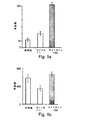
図4a〜図4fは、NOD/SCIDマウスに移植されたヒト造血細胞のホーミングおよび生着に対するニコチンアミドの影響のグラフ表示である。図4aおよび図4bは移植前の細胞集団におけるヒト(CD45+)細胞の割合を示す:非培養CD34+細胞(非培養、灰色の楕円)、サイトカインだけへの3週間の暴露の後の培養物の子孫全体(サイトカイン単独、黒塗りの楕円)、または、サイトカインおよびニコチンアミドへの3週間の暴露の後の培養物の子孫全体(サイトカイン+NA、矢印)。移植後4週間での生着率をNOD/SCIDの骨髄におけるヒトCD45細胞のフローサイトメトリー(y軸)によって求めた。SCID再増殖細胞(SRC)の数を、それぞれの用量における生着頻度をプロットすることによって計算した。得られた曲線は、非培養CD34+細胞(図4c)、サイトカインを含む培養物(図4d)、または、サイトカインおよびニコチンアミドを含む培養物(図4e)におけるSRCの推定頻度を示す。それぞれのボックスに示された数字は、最尤推定量を使用するSRCの計算された頻度を示す。図4fは、FACSによって測定されたときの、ニコチンアミドとともに3週間培養された12x103個のCD34+細胞の子孫が移植された代表的なマウスにおける生着したヒト細胞の免疫表現型を示す。マウスの骨髄細胞を、FITCコンジュゲート化抗CD45(ヒト)、および、示されるようなヒト分化マーカーに対する抗体により二重標識した。二重の陽性細胞の割合がそれぞれの象限に示される。ニコチンアミド治療されたマウスにおける生着が、サイトカインだけにより治療されたマウスと比較したとき、7倍を越えて高まることに留意すること(図4d)。 FIGS. 4a-4f are graphical representations of the effect of nicotinamide on homing and engraftment of human hematopoietic cells transplanted into NOD / SCID mice. Figures 4a and 4b show the percentage of human (CD45 +) cells in the pre-transplant cell population: uncultured CD34 + cells (uncultured, gray ellipse), culture after 3 weeks exposure to cytokine alone Whole progeny (cytokine alone, black ellipses) or whole culture progeny after 3 weeks exposure to cytokines and nicotinamide (cytokine + NA, arrows). The engraftment rate 4 weeks after transplantation was determined by flow cytometry (y-axis) of human CD45 cells in NOD / SCID bone marrow. The number of SCID repopulating cells (SRC) was calculated by plotting the engraftment frequency at each dose. The resulting curve shows the estimated frequency of SRC in uncultured CD34 + cells (FIG. 4c), cultures containing cytokines (FIG. 4d), or cultures containing cytokines and nicotinamide (FIG. 4e). The numbers shown in each box indicate the calculated frequency of SRC using the maximum likelihood estimator. FIG. 4f shows the engrafted human cell immunophenotype in a representative mouse transplanted with 12 × 10 3 CD34 + cell progeny cultured for 3 weeks with nicotinamide as measured by FACS. Murine bone marrow cells were double labeled with FITC-conjugated anti-CD45 (human) and antibodies against human differentiation markers as indicated. The percentage of double positive cells is shown in each quadrant. Note that engraftment in nicotinamide-treated mice is increased by more than 7-fold when compared to mice treated with cytokines alone (FIG. 4d).
図5a〜図5cは、分化促進条件のもとで培養された細胞の生着能に対するニコチンアミド(NA)の影響のグラフ表示である。培養を、精製された臍血由来のCD34+細胞を用いて、SCF、TPO、IL−6およびFLT3(それぞれ、50ng/ml)ならびにIL−3(20ng/ml)が補充され、10mMのニコチンアミドを含む培地(サイトカイン+NA、矢印)、または、ニコチンアミドを含まない培地(サイトカイン、楕円)において開始した。3週間後、細胞を集め、示されたようにSCIDマウスに移植した。マウスには、1.25〜5x104個のCD34+細胞、または、拡大後のその子孫を移植した。マウスを4週間後に屠殺し、骨髄細胞をCD34+細胞(ヒト前駆細胞)およびCD45+(ヒト)細胞の存在についてFACSによって分析した。マウスは、ヒト(CD45+)細胞の数が骨髄集団の0.5%以上を構成したときには生着したとして記録した。図5aは、移植された細胞の用量(5.0X104個、2.5X104個および1.25X104個の細胞)のそれぞれについて、移植されたマウスの総数あたりの生着陽性マウスの数を示す。図5bおよび図5cは、1.25X104個のCD34+細胞により開始された培養に由来する細胞が移植されたマウスの骨髄における総ヒト(CD45+)細胞の割合(図5b)およびヒト前駆細胞(CD45+CD34+)細胞の割合(図5c)を示す。総ヒト細胞、および、ニコチンアミド処理から生じたヒト前駆細胞の両方の高まった生着に留意すること。 Figures 5a to 5c are graphical representations of the effect of nicotinamide (NA) on the engraftability of cells cultured under differentiation promoting conditions. Cultures were supplemented with SCF, TPO, IL-6 and FLT3 (50 ng / ml, respectively) and IL-3 (20 ng / ml) using purified umbilical cord-derived CD34 + cells and supplemented with 10 mM nicotinamide. Start with medium containing (cytokine + NA, arrow) or medium without nicotinamide (cytokine, oval). Three weeks later, cells were collected and transplanted into SCID mice as indicated. Mice were transplanted with 1.25-5 × 10 4 CD34 + cells or their expanded progeny. Mice were sacrificed after 4 weeks and bone marrow cells were analyzed by FACS for the presence of CD34 + cells (human progenitor cells) and CD45 + (human) cells. Mice were scored as engrafted when the number of human (CD45 +) cells comprised 0.5% or more of the bone marrow population. FIG. 5a shows the number of engraftment-positive mice per total number of transplanted mice for each transplanted cell dose (5.0 × 10 4 , 2.5 × 10 4 and 1.25 × 10 4 cells). Show. FIGS. 5b and 5c show the percentage of total human (CD45 +) cells in the bone marrow of mice transplanted with cells derived from cultures initiated with 1.25 × 10 4 CD34 + cells (FIG. 5b) and human progenitor cells (CD45 + CD34 +). ) Shows the percentage of cells (Fig. 5c). Note the increased engraftment of both total human cells and human progenitor cells resulting from nicotinamide treatment.
本発明は、移植可能な細胞のホーミングおよび生着を改善するための方法の発明である。 The present invention is an invention of a method for improving homing and engraftment of transplantable cells.
本発明の原理および作用が、付随する説明を参照してより十分に理解することができる。 The principles and operation of the present invention may be better understood with reference to the accompanying description.
本発明の少なくとも1つの実施形態を詳しく説明する前に、本発明は、その適用において、下記の説明において示される細部、または、実施例によって例示される細部に限定されないことを理解しなければならない。本発明は他の実施形態が可能であり、または、様々な方法で実施または実行されることができる。また、本明細書中で用いられる表現法および用語法は記述のためであって、限定であると見なしてはならないことを理解しなければならない Before describing at least one embodiment of the present invention in detail, it should be understood that the present invention is not limited in its application to the details set forth in the following description or the details illustrated by the examples. . The invention is capable of other embodiments or of being practiced or carried out in various ways. It should also be understood that the terminology and terminology used herein is for the purpose of description and should not be considered limiting.
成功した血液移植および骨髄移植(自家および同種の両方)は、髄腔にホーミングすることができ、かつ、すべてがそろった造血細胞系譜を、時宜を得た様式で再生することができる造血幹細胞の十分な数を注入することを必要とする。骨髄から血液内への幹細胞の呼び寄せは動員と呼ばれるか、または、より一般には幹細胞動員と呼ばれる。幹細胞動員および/または幹細胞ホーミングの強化が、成功した幹細胞移植をもたらすことは十分に明らかにされている。 Successful blood transplantation and bone marrow transplantation (both autologous and allogeneic) can be homing to the medullary canal, and all hematopoietic cell lineages can be regenerated in a timely manner. It is necessary to inject a sufficient number. The recruitment of stem cells from the bone marrow into the blood is called mobilization, or more commonly called stem cell mobilization. It has been well documented that enhanced stem cell mobilization and / or stem cell homing results in successful stem cell transplantation.
SDF−αは造血幹細胞および造血前駆細胞(HSC/HPC)を化学的に誘引しており、骨髄からのHSC/HPCの動員において、また同様に、幹細胞ホーミングにおいて非常に重要な役割を果たすと考えられる。 SDF-α chemically attracts hematopoietic stem cells and hematopoietic progenitor cells (HSC / HPC) and appears to play a very important role in mobilizing HSC / HPC from the bone marrow and also in stem cell homing It is done.
CD26は、プロリン残基又はアラニン残基のいずれかを最後から2番目の位置に有するポリペプチドからN末端のジペプチドを切断することができるジペプチジルペプチダーゼIV(DPPIV)活性をその細胞外ドメインに有することが知られている、広範囲に分布する110kDaの細胞表面の糖タンパク質である。CD26は、SDFの活性を、後者をその2位のプロリンにおいて切断することによって阻害し、それにより、その幹細胞動員/ホーミング機能を阻害する。 CD26 has dipeptidyl peptidase IV (DPPIV) activity in its extracellular domain capable of cleaving the N-terminal dipeptide from a polypeptide having either a proline residue or an alanine residue in the penultimate position. It is known to be a widely distributed 110 kDa cell surface glycoprotein. CD26 inhibits the activity of SDF by cleaving the latter at its 2-position proline, thereby inhibiting its stem cell recruitment / homing function.
米国特許出願公開第2004/0247547号は、骨髄への幹細胞ホーミングを改善すること、および、動員されたドナー幹細胞の数を増大させることの両方によって幹細胞移植物の生着効率を改善するためのCD26阻害剤の使用を開示する。この特許出願は、CD26の表面発現をダウンレギュレーションすることを教示せず、むしろ、CD26の触媒活性をダウンレギュレーションすることを教示する。具体的には、米国特許出願公開第2004/0247574号は、CD26の表面発現をダウンレギュレーションするためのニコチンアミドの使用を教示していない。 US Patent Application Publication No. 2004/0247547 is a CD26 for improving stem cell transplant engraftment efficiency by both improving stem cell homing to the bone marrow and increasing the number of recruited donor stem cells. Disclose the use of inhibitors. This patent application does not teach down-regulating CD26 surface expression, but rather down-regulating the catalytic activity of CD26. Specifically, US Patent Application Publication No. 2004/0247574 does not teach the use of nicotinamide to down-regulate surface expression of CD26.
本発明を実施に移しているとき、本発明者は、ニコチンアミドが、CD26の細胞表面での発現をダウンレギュレーションし、接着分子およびインテグリン分子の発現および機能を高め、誘導された移植可能な細胞の遊走を増大させ、かつ、移植可能な細胞のホーミングおよび生着をインビボで著しく改善するために首尾良く使用できることを発見した。 As the invention is put into practice, the inventor found that nicotinamide down-regulated the expression of CD26 on the cell surface, increased the expression and function of adhesion and integrin molecules, and induced transplantable cells. Has been found to be successfully used to increase the migration of cells and to significantly improve homing and engraftment of transplantable cells in vivo.
本明細書中下記および下記の実施例の節において明らかにされるように、本発明者は、ニコチンアミドとの細胞の短期間のインキュベーションが、CD26の発現をダウンレギュレーションするために十分であることを示した。CD26を発現するCD34+細胞およびAC133+細胞における著しい減少が、ニコチンアミドとのわずかに20時間のインキュベーションの後で認められた。 As demonstrated herein below and in the Examples section below, the inventors have shown that short-term incubation of cells with nicotinamide is sufficient to down-regulate CD26 expression. showed that. A significant decrease in CD34 + and AC133 + cells expressing CD26 was observed after only 20 hours of incubation with nicotinamide.
より重要なことに、ニコチンアミドは、細胞結合および細胞阻止のプロセスにとって非常に重要な分子の機能性を高めることにおいて効果的であることが示された。実際、ニコチンアミド暴露後におけるインビトロでの細胞遊走能の評価では、ニコチンアミドが、ニコチンアミドとともに培養された移植可能な細胞において、CXCL12誘導可能な遊走、ならびに、VCAM−1におけるVLA−4媒介の結合および保持をともに高めたことが示された(下記の実施例3を参照のこと)。細胞遊走のプロセスは広範囲の様々な細胞において「認識」ペア(例えば、VLA−4およびVCAM−1など)によって媒介されるので、この驚くべき発見は、ニコチンアミド、ならびに、ニコチンアミドの誘導体および類似体が、多様な起源および様々な分化段階の細胞について結合および保持(これらは、移植された細胞がホーミングに成功し、生着し、かつ、宿主組織を再増殖(repopulate)することができることにとって非常に重要である)を高めることにおいて効果的であり得ることを示している。 More importantly, nicotinamide has been shown to be effective in enhancing the functionality of molecules critical to cell binding and cell arrest processes. Indeed, in the evaluation of in vitro cell migration capacity after nicotinamide exposure, nicotinamide is CXCL12-induced migration in transplantable cells cultured with nicotinamide, as well as VLA-4 mediated in VCAM-1. It was shown that both binding and retention were enhanced (see Example 3 below). Since the process of cell migration is mediated by “recognition” pairs (eg, VLA-4 and VCAM-1 etc.) in a wide variety of cells, this surprising discovery is made with nicotinamide and derivatives and analogs of nicotinamide The body binds and retains cells of various origins and various stages of differentiation (this is because the transplanted cells are able to successfully home and engraft and repopulate host tissues It is very important to show that it can be effective.
ニコチンアミド処理された細胞を用いた実際のインビボ移植実験では、細胞生着能および細胞ホーミング能に対するニコチンアミドの影響についての確実な証拠がもたらされた。下記の実施例2では、単核細胞をNOD/SCIDマウスへの移植に先だってニコチンアミドにさらすことにより、骨髄へのホーミングが、ニコチンアミドを含むことなく培養された同一の細胞よりも6倍増大したことが示される。その上さらに、下記の実施例4では、細胞の明らかに最適でない用量において、コントロールの培養細胞はNOD/SCIDマウスにおける骨髄の再増殖を引き起こすことができなかった一方で、ニコチンアミド処理された細胞の移植は大きい程度の生着および成功した再増殖をもたらしたことが示される。 Actual in vivo transplantation experiments using nicotinamide-treated cells provided solid evidence for the effect of nicotinamide on cell engraftment and cell homing capacity. In Example 2 below, exposure of mononuclear cells to nicotinamide prior to transplantation into NOD / SCID mice increased homing to the bone marrow by a factor of 6 over identical cells cultured without nicotinamide. Is shown. Furthermore, in Example 4 below, control cultured cells failed to cause bone marrow regrowth in NOD / SCID mice at clearly suboptimal doses of cells, whereas nicotinamide treated cells It has been shown that transplantation of a large amount resulted in a large degree of engraftment and successful regrowth.
まとめると、これらの結果から、細胞ホーミングおよび細胞生着におけるニコチンアミドについての新規な役割、ならびに、細胞移植におけるそのようなものとしての新規な役割が示唆される。 Taken together, these results suggest a novel role for nicotinamide in cell homing and cell engraftment and as such in cell transplantation.
従って、本発明の1つの態様によれば、細胞ホーミング能および細胞生着能を高める方法が提供され、この場合、この方法は、細胞集団を、細胞ホーミング能および細胞生着能を高めるために十分な期間にわたって所定量のニコチンアミドにエクスビボまたはインビトロで供することを含む。 Thus, according to one aspect of the present invention there is provided a method for enhancing cell homing and cell engraftment, wherein the method is used to increase a cell population to enhance cell homing and cell engraftment. Subjecting a predetermined amount of nicotinamide to ex vivo or in vitro for a sufficient period of time.
本明細書中で使用される表現「細胞生着能を高める」は、標的組織への改善されたホーミング、改善された接着、および、低下した拒絶などから生じ得る、細胞移植の効率、特性または迅速さにおける改善を示す。細胞生着能を評価するための方法には、例えば、本明細書中下記に詳しく記載されるように、細胞遊走技術および他のインビトロ技術、ならびに、実際のインビボ移植から得られた組織および器官の組織学評価、免疫学的評価および/または放射線学的評価が含まれる。幹細胞の自己複製能を長期コロニー形成(LTC−CFUc)によってインビトロで求めることができ、または、SCID−Huマウスモデルにおけるインビボ生着によって求めることができる。SCID−Huマウスモデルでは、ヒト胎児の胸腺組織および肝臓組織またはヒト胎児のBM組織が移植されたC.B.17scid/scid(SCID)マウスが用いられ、このモデルは、推定されるヒト造血幹細胞を評価するための適切なモデルを提供する。ヒト胎児組織によるSCIDマウスの再構成のために、このモデルは、ヒト起源の造血性微小環境における幹細胞の増殖(この場合には、増殖するためのヒト造血幹細胞)および機能をもたらす。マウスは典型的には放射線照射され、その後、幹細胞が移植片に送達され、再構成が、再増殖した器官のFACSおよび免疫組織化学をはじめとする多数の方法によって測定される(Humeau L.他、Blood(1997)、90:3496;また、下記の材料および実験方法を参照のこと)。 As used herein, the phrase “enhance cell engraftment” refers to the efficiency, characteristics, or characteristics of cell transplantation that may result from improved homing to a target tissue, improved adhesion, reduced rejection, etc. Shows improvement in speed. Methods for assessing cell engraftment include, for example, cell migration techniques and other in vitro techniques, and tissues and organs obtained from actual in vivo transplantation, as described in detail herein below. Histological evaluation, immunological evaluation and / or radiological evaluation. The ability of stem cells to self-replicate can be determined in vitro by long-term colony formation (LTC-CFUc) or by in vivo engraftment in a SCID-Hu mouse model. In the SCID-Hu mouse model, human fetal thymus and liver tissue or human fetal BM tissue was transplanted. B. 17 scid / scid (SCID) mice are used and this model provides a suitable model for evaluating putative human hematopoietic stem cells. For reconstitution of SCID mice with human fetal tissue, this model provides stem cell proliferation (in this case, human hematopoietic stem cells to proliferate) and function in a hematopoietic microenvironment of human origin. Mice are typically irradiated, after which stem cells are delivered to the graft and reconstitution is measured by a number of methods including FACS and immunohistochemistry of repopulated organs (Humeau L. et al. , Blood (1997), 90: 3496; see also materials and experimental methods below).
本明細書中で使用される用語「エクスビボ」は、細胞が、生きている生物から取り出され、その生物の体外で(例えば、試験管において)増殖させられるプロセスを示す。 The term “ex vivo” as used herein refers to the process by which cells are removed from a living organism and grown outside the organism (eg, in a test tube).
本明細書中で使用される用語「インビトロ」は、研究室で維持される1つ以上の細胞株(例えば、NTera2神経細胞、胚性細胞株など)に由来する細胞が生物の体外で操作されるプロセスを示す。そのような細胞株は、多くの場合、不死化された細胞である。 As used herein, the term “in vitro” refers to cells that are derived from one or more cell lines maintained in the laboratory (eg, NTera2 neurons, embryonic cell lines, etc.) that are manipulated outside the organism. Process. Such cell lines are often immortalized cells.
本明細書中で使用される表現「細胞集団」は、移植のために好適である細胞集団を含む細胞の均一または不均一な単離された集団を示す。好ましい実施形態において、本発明のこの態様の細胞集団の少なくとも一部がCD26またはVLA−4を細胞表面に発現する。 The expression “cell population” as used herein refers to a homogeneous or heterogeneous isolated population of cells, including cell populations that are suitable for transplantation. In preferred embodiments, at least a portion of the cell population of this aspect of the invention expresses CD26 or VLA-4 on the cell surface.
本明細書中で使用される表現「幹細胞」は、細胞塊を組織または身体において生じさせることに関わる最も初期の再生可能な細胞集団、および、幾分かより分化し、それにもかかわらず、分化決定されておらず、最も初期の再生可能な細胞集団の一部となるために容易に逆戻りすることができる初期の前駆細胞の両方を示す。 As used herein, the expression “stem cell” refers to the earliest renewable cell population involved in generating a cell mass in a tissue or body, and somewhat more differentiated, nevertheless Both early progenitor cells that have not been determined and can be easily reversed to become part of the earliest renewable cell population are shown.
本明細書中で使用される表現の「非幹」細胞、「非前駆」細胞および「分化決定された」細胞は、再生可能な細胞集団の一部となるために逆戻りする能力を一般にはもはや保持していない様々な分化段階にある細胞を示す。幹細胞、前駆細胞、および、非幹性で、前駆細胞でない分化決定された細胞をエクスビボ培養する様々な方法が、細胞培養の技術分野では広く知られている。この目的のために、例えば、Freshneyによる書籍「Culture of Animal Cells−A Manual of Basic Technique」(Wiley−Liss、N.Y.(1994)、第3版)(その教示は本明細書により参考として組み込まれる)を参照のこと。 As used herein, the expressions “non-stem” cells, “non-progenitor” cells and “differentiated” cells generally no longer have the ability to reverse to become part of a reproducible cell population. Cells in various stages of differentiation that are not retained are shown. Various methods of ex vivo culturing stem cells, progenitor cells, and non-stem, non-progenitor differentiated cells are widely known in the cell culture art. To this end, for example, the book “Culture of Animal Cells-A Manual of Basic Technique” by Freshney (Wiley-Liss, NY (1994), 3rd edition), the teachings of which are hereby incorporated by reference. See incorporated).
本発明の細胞集団は自家ドナーまたは非自家ドナー(同種または異種)に由来し得る。 The cell populations of the invention can be derived from autologous donors or non-autologous donors (homologous or xenogeneic).
好ましい実施形態において、移植用の細胞は幹細胞および/または前駆細胞であり、幹細胞集団の供給源は、CD34+細胞または他の造血幹細胞について濃縮されていない未分画の単核細胞調製物である。別の実施形態において、幹細胞は、当該技術分野で知られている幹細胞マーカー(例えば、CD34+、CD34+/CD38−、CD133+、CD34+/Lin−および他の幹細胞マーカーなど)によって特定される。さらに別の実施形態において、幹細胞集団の供給源は、幹細胞マーカーに従った選択によって造血幹細胞について濃縮されている幹細胞である。 In preferred embodiments, the cells for transplantation are stem cells and / or progenitor cells, and the source of the stem cell population is an unfractionated mononuclear cell preparation that is not enriched for CD34 + cells or other hematopoietic stem cells. In another embodiment, stem cells are identified by stem cell markers known in the art, such as CD34 +, CD34 + / CD38−, CD133 +, CD34 + / Lin− and other stem cell markers. In yet another embodiment, the source of the stem cell population is stem cells that have been enriched for hematopoietic stem cells by selection according to stem cell markers.
例えば、本発明の幹細胞は、造血細胞、臍帯血細胞、および、動員された末梢血細胞からなる群から選択される供給源に由来し得る。 For example, the stem cells of the present invention can be derived from a source selected from the group consisting of hematopoietic cells, umbilical cord blood cells, and mobilized peripheral blood cells.
本明細書中で使用される「ニコチンアミド」は、ニコチンアミド、ならびに、ニコチンアミド、その類似体、および、ニコチンアミドまたはニコチンアミド類似体の代謝物に由来する産物(例えば、NAD、NADHおよびNADPHなど)を示す。 As used herein, “nicotinamide” refers to nicotinamide and products derived from nicotinamide, its analogs, and nicotinamide or metabolites of nicotinamide analogs (eg, NAD, NADH and NADPH). Etc.)
本明細書中で使用される表現「ニコチンアミド類似体」は、ニコチンアミドと類似して作用することが知られている任意の分子を示す。ニコチンアミド類似体の代表的な例には、限定されないが、ベンズアミド、ニコチンチオアミド(ニコチンアミドのチオール類似体)、ニコチン酸およびα−アミノ−3−インドールプロピオン酸が挙げられる。 As used herein, the expression “nicotinamide analog” refers to any molecule known to act similarly to nicotinamide. Representative examples of nicotinamide analogs include, but are not limited to, benzamide, nicotine thioamide (a thiol analog of nicotinamide), nicotinic acid and α-amino-3-indolepropionic acid.
本明細書中で使用される用語「対象」は哺乳動物対象を示し、好ましくは、ヒト対象を示す。 As used herein, the term “subject” refers to a mammalian subject, preferably a human subject.
表現「ニコチンアミド誘導体またはニコチンアミド類似体誘導体」は、ニコチンアミドそのものまたはニコチンアミドの類似体の任意の構造的誘導体を示す。そのような誘導体の例には、限定されないが、置換されたベンズアミド、置換されたニコチンアミドおよびニコチンチオアミド、ならびに、N−置換されたニコチンアミドおよびニコチンチオアミドが挙げられる。 The expression “nicotinamide derivative or nicotinamide analogue derivative” refers to any structural derivative of nicotinamide itself or an analogue of nicotinamide. Examples of such derivatives include, but are not limited to, substituted benzamides, substituted nicotinamides and nicotine thioamides, and N-substituted nicotinamides and nicotine thioamides.
加えて、または、代替として、幹細胞の動員を、当該技術分野では広く知られている動員剤を使用して幹細胞移植のための細胞を集める前に行うことができる。一般に、動員プロセスは、膜結合した幹細胞因子(SCF)の脱落および放出、前駆細胞の増殖、ならびに、接着分子の活性化および/または分解をもたらす化学療法、および、サイトカイン(例えば、顆粒球コロニー刺激因子(G−CSF)など)による反復した刺激による好中球および破骨細胞のストレス誘導による活性化によって開始される。本発明に従って使用することができる動員剤には、DNA損傷剤、1つだけの化学療法剤(例えば、シクロホスファミド)、混合化学療法方式[例えば、イホスファミド、カルボプラチンおよびエトポシド(ICE)、ならびに、メチルプレドニゾロン、ara−cおよびシスプラチン(ESHAP)]、サイトカイン(例えば、G−CSF、GM−CSF、SCF、FLT−3リガンドなど)、および、ケモカイン(例えば、IL−8、MIP−1α、GroβおよびSDF−1など)が含まれるが、これらに限定されない。投与様式、ならびに、動員を達成するために必要とされる時間枠、および、動員される細胞のタイプは、使用された分子に依存する。例えば、G−CSFは通常、単独で、または、治療の後で、5μg/kg〜10μg/kgの用量として5日〜10日にわたって毎日投与される。動員方式の調節が医師によって行われ、また、Cottker−Fox他(2003)、Hematology、419〜437において総説される。 Additionally or alternatively, stem cell mobilization can be performed prior to collecting cells for stem cell transplantation using mobilization agents well known in the art. In general, the mobilization process involves the detachment and release of membrane-bound stem cell factor (SCF), proliferation of progenitor cells, and chemotherapy that results in activation and / or degradation of adhesion molecules and cytokines (eg, granulocyte colony stimulation) Initiated by stress-induced activation of neutrophils and osteoclasts by repeated stimulation with factors (such as G-CSF). Mobilizing agents that can be used in accordance with the present invention include DNA damaging agents, only one chemotherapeutic agent (eg, cyclophosphamide), mixed chemotherapy regimens [eg, ifosfamide, carboplatin and etoposide (ICE), and , Methylprednisolone, ara-c and cisplatin (ESHAP)], cytokines (eg, G-CSF, GM-CSF, SCF, FLT-3 ligand, etc.), and chemokines (eg, IL-8, MIP-1α, Groβ) And SDF-1), but is not limited to these. The mode of administration, as well as the time frame required to achieve mobilization, and the type of cell mobilized will depend on the molecule used. For example, G-CSF is usually administered daily for 5-10 days, alone or after treatment as a dose of 5 [mu] g / kg to 10 [mu] g / kg. Modulation of the mobilization scheme is performed by physicians and is reviewed in Cotker-Fox et al. (2003) Hematology, 419-437.
移植のための細胞を調製する様々な方法が当該技術分野では広く知られている。非幹細胞を調製するために、細胞を、個々の細胞を組織の結合している細胞外マトリックスから解離することによってドナー組織から得ることができる。特定の領域からの組織が、無菌手順を使用して取り出され、細胞が、酵素(例えば、トリプシン、コラゲナーゼおよびDNAseなど)による処理を含めて、当該技術分野で知られている任意の方法を使用して、または、鈍器などを用いた物理的な解離方法を使用することによって解離させられる。 Various methods for preparing cells for transplantation are widely known in the art. To prepare non-stem cells, cells can be obtained from donor tissue by dissociating individual cells from the tissue-bound extracellular matrix. Tissue from a particular area is removed using aseptic procedures and cells are used in any manner known in the art, including treatment with enzymes (eg, trypsin, collagenase and DNAse). Or by using a physical dissociation method using a blunt device or the like.
移植のために調製された細胞は生理学的溶液において維持されるか、あるいは、懸濁状態で、または、固定された基体の表面で培養される。細胞を支持することができる好適な培養培地には、HEM、DMEM、RPMIおよびF−12などが含まれる。要求されるならば、培地は、細胞の代謝のために要求される補充物(例えば、グルタミンおよび他のアミノ酸、ビタミン、ミネラルおよび有用なタンパク質(例えば、トランスフェリンなど)など)を含有することができる。培地はまた、酵母、細菌および真菌による汚染を防止するための抗生物質(例えば、ペニシリン、ストレプトマイシンおよびゲンタマイシンなど)を含有することができる。細胞が培養されることになるならば、条件は生理学的な条件(好ましくは、約6〜約8のpHおよび約30℃〜約40℃の温度)に近づけなければならない。培養培地は、場合により、少なくとも1つの増殖誘導性の増殖因子(例えば、EGF、アンフィレグリン、酸性線維芽細胞増殖因子(aFGFまたはFGF−1)、塩基性線維芽細胞増殖因子(bFGFまたはFGF−2)、トランスフォーミング増殖因子α(TGF−α)、サイトカイン(例えば、G−CSF、GM−CSF、SCF、FLT−3リガンドなど)、および/または、ケモカイン(例えば、IL−8、MIP−1α、GroβおよびSDF−1など)、ならびに、それらの組合せにより補充され得る。増殖誘導性の増殖因子に加えて、他の増殖因子を培養培地に加えることができ、これらには、NGF、血小板由来増殖因子(PDGF)および甲状腺刺激ホルモン放出ホルモン(TRH)などが含まれる。 Cells prepared for transplantation are maintained in a physiological solution or cultured in suspension or on the surface of a fixed substrate. Suitable culture media that can support the cells include HEM, DMEM, RPMI, F-12, and the like. If required, the medium can contain supplements required for cellular metabolism, such as glutamine and other amino acids, vitamins, minerals and useful proteins such as transferrin. . The medium can also contain antibiotics such as penicillin, streptomycin and gentamicin to prevent contamination by yeast, bacteria and fungi. If the cells are to be cultured, the conditions should approach physiological conditions (preferably a pH of about 6 to about 8 and a temperature of about 30 ° C. to about 40 ° C.). The culture medium is optionally at least one growth-inducing growth factor (eg, EGF, amphiregulin, acidic fibroblast growth factor (aFGF or FGF-1), basic fibroblast growth factor (bFGF or FGF). -2), transforming growth factor α (TGF-α), cytokines (eg, G-CSF, GM-CSF, SCF, FLT-3 ligand, etc.) and / or chemokines (eg, IL-8, MIP- 1α, Groβ and SDF-1), and combinations thereof, in addition to growth-inducing growth factors, other growth factors can be added to the culture medium, including NGF, platelets Derived growth factor (PDGF) and thyrotropin releasing hormone (TRH) are included.
特定の細胞タイプの選択および濃縮を、形態学的手段、物理的手段、免疫組織学的手段(FACS)または他の手段によって行うことができる(例えば、肝臓組織からの肝細胞の分離、グリア細胞からのニューロンの分離、または、膵臓組織からの小島細胞の単離など)。新鮮な細胞調製物または培養された細胞調製物は、当該技術分野で知られている任意の方法によって必要とされるまで凍結保存することができる。細胞は、特定の凍結保護剤を含有する等張性の溶液(好ましくは、細胞培養培地)に懸濁することができる。そのような凍結保護剤には、ジメチルスルホキシド(DMSO)およびグリセロールが含まれる。細胞を移植のために調製および保存するためのさらなる方法が当該技術分野では知られており、また、例えば、Handbook of Transplantation(KipshidzeおよびSerruys編、London、UK、2004)に詳しく開示される。 Selection and enrichment of specific cell types can be done by morphological, physical, immunohistological (FACS) or other means (eg, separation of hepatocytes from liver tissue, glial cells Isolation of neurons from the pancreas, or isolation of islet cells from pancreatic tissue). Fresh or cultured cell preparations can be stored frozen until required by any method known in the art. The cells can be suspended in an isotonic solution (preferably a cell culture medium) containing a specific cryoprotectant. Such cryoprotectants include dimethyl sulfoxide (DMSO) and glycerol. Additional methods for preparing and storing cells for transplantation are known in the art, and are disclosed in detail, for example, in Handbook of Transplantation (Edited by Khiphideze and Serruys, London, UK, 2004).
幹細胞を調製する様々な方法が当該技術分野では広く知られており、一般には、1つ以上の幹細胞マーカー(例えば、CD34、CD133など)を発現するか、あるいは、分化した細胞のマーカーを有さない細胞を選択することである。選択は通常、FACSによるか、または、免疫磁石分離であるが、核酸方法(例えば、PCRなど)によってもまた可能である(本明細書中下記における材料および実験方法を参照のこと)。胚性幹細胞およびその回収方法が当該技術分野では広く知られており、例えば、Trounson AO(Reprod Fertil Dev(2001)、13:523)、Roach ML(Methods Mol Biol(2002)、185:1)、および、Smith AG(Annu Rev Cell Dev Biol(2001)、17:435)に記載される。成体幹細胞は、成体の組織に由来する幹細胞であり、これもまた、当該技術分野では広く知られている。成体幹細胞を単離する方法、または、成体幹細胞について濃縮する方法が、例えば、下記に記載される:Miraglia,S.他(1997)Blood 90:5013,Uchida,N.他(2000)Proc.Natl.Acad.Sci.USA 97:14720,Simmons,P.J.他(1991)Blood 78:55,Prockop DJ(Cytotherapy(2001)3:393),Bohmer RM(Fetal Diagn Ther(2002)17:83)およびRowley SD他(Bone Marrow Transplant(1998)21:1253),Stem Cell Biology Daniel R.Marshak(編者)Richard L.Gardner(編者),出版社:Cold Spring Harbor Laboratory Press,(2001)およびHematopoietic Stem Cell Transplantation.Anthony D.Ho(編者)Richard Champlin(編者),出版社:Marcel Dekker(2000)。 Various methods of preparing stem cells are widely known in the art and generally express one or more stem cell markers (eg, CD34, CD133, etc.) or have markers for differentiated cells. Is to select no cells. Selection is usually by FACS or by immunomagnet separation, but can also be by nucleic acid methods (eg PCR etc.) (see materials and experimental methods herein below). Embryonic stem cells and methods for their recovery are widely known in the art, such as Trounson AO (Reprod Fertil Dev (2001), 13: 523), Roach ML (Methods Mol Biol (2002), 185: 1), And Smith AG (Annu Rev Cell Dev Biol (2001), 17: 435). Adult stem cells are stem cells derived from adult tissue, which are also widely known in the art. Methods for isolating or enriching for adult stem cells are described, for example, in the following: Miraglia, S .; Et al. (1997) Blood 90: 5013, Uchida, N .; Et al. (2000) Proc. Natl. Acad. Sci. USA 97: 14720, Simons, P.A. J. et al. (1991) Blood 78:55, Prochop DJ (Cytotherapy (2001) 3: 393), Bohmer RM (Fetal Diagnostic Ther (2002) 17:83) and Rowley SD et al. (Bone Marrow Transplant: 1998, 1251). Stem Cell Biology Daniel R. Marshak (editor) Richard L. Gardner (editor), Publisher: Cold Spring Harbor Laboratory Press, (2001) and Hematopoietic Stem Cell Transplantation. Anthony D. Ho (editor) Richard Champlin (editor), publisher: Marcel Dekker (2000).
ニコチンアミドは広範囲の様々な細胞タイプにおいて生着能およびホーミング能を高め得ることが理解される。例えば、ニコチンアミドはCD26の表面発現をダウンレギュレーションすることができ、VLA−4、CXCR−2または他の接着分子を発現する任意の細胞タイプからのVLA−4、CXCR−2または他の接着分子の機能性を高めることができ、また、これらの分子は多様な起源の細胞集団において広く発現されるので、本発明のこの態様の細胞集団は、選択された細胞タイプのより均一な集団だけでなく、非選択の細胞集団(例えば、組織からの粗細胞調製物、あるいは、単核の幹細胞および/または前駆細胞など)を含むことができる。 It is understood that nicotinamide can enhance engraftment and homing capacity in a wide variety of cell types. For example, nicotinamide can down-regulate CD26 surface expression and VLA-4, CXCR-2 or other adhesion molecules from any cell type that expresses VLA-4, CXCR-2 or other adhesion molecules Since these molecules are widely expressed in cell populations of diverse origins, the cell population of this aspect of the invention can be expressed only in a more homogeneous population of the selected cell type. Rather, non-selected cell populations (eg, crude cell preparations from tissues, or mononuclear stem cells and / or progenitor cells) can be included.
本明細書中で使用される表現「造血性単核細胞」は、血液サンプルに存在する白血球のレパートリー全体を示し、通常、造血性の分化決定された細胞の大きい割合と、造血幹細胞および造血前駆細胞の小さい割合とを含む造血性単核細胞を示す。健康なヒトにおいて、白血球は、造血系譜の分化決定された細胞および分化した細胞の混合物(典型的には、単核細胞の99%超が、系譜決定された細胞である)を含み、これには、例えば、系譜決定された前駆細胞のCD34+CD33+(骨髄性の分化決定された細胞)、CD34+CD3+(リンパ系の分化決定された細胞)、CD34+CD41+(巨核球性の分化決定された細胞)、および、分化した細胞、すなわち、CD34−CD33+(骨髄球、例えば、顆粒球および単球など)、CD34−CD3+、CD34−CD19+細胞(それぞれ、T細胞およびB細胞)、CD34−CD41+(巨核球)、ならびに、造血幹細胞および初期前駆細胞(例えば、CD34+の系譜陰性(Lin−)、CD34−の系譜陰性、CD34+CD38−など)(典型的には、1%未満)が含まれる。 As used herein, the expression “hematopoietic mononuclear cell” refers to the entire repertoire of leukocytes present in a blood sample, usually a large percentage of hematopoietic differentiation determined cells, and hematopoietic stem cells and hematopoietic progenitors. Shown are hematopoietic mononuclear cells containing a small percentage of cells. In healthy humans, leukocytes include hematopoietic lineage-differentiated cells and a mixture of differentiated cells (typically more than 99% of mononuclear cells are lineage-determined cells) CD34 + CD33 + (myeloid differentiation-determined cells), CD34 + CD3 + (lymphoid differentiation-determined cells), CD34 + CD41 + (megakaryocytic cells) Differentiated cells), and differentiated cells, ie, CD34 − CD33 + (such as myeloid cells, eg, granulocytes and monocytes), CD34 − CD3 + , CD34 − CD19 + cells (T cells and B, respectively). cells), CD34 - CD41 + (megakaryocytes), and hematopoietic stem cells and early progenitor cells (e.g., CD34 + lineage negative (Lin -), CD3 - Lineage negative, CD34 + CD38 -, etc.) (typically, less than 1%) are included.
造血性単核細胞は、典型的には、血液サンプルをFicoll−Hypaque層に加え、密度クッション遠心分離後、Ficoll−Hypaqueと、血清との間に存在する境界を集めることによって血液サンプルから得られ、この場合、そのような境界層は本質的にはすべてが、血液サンプルに存在する白血球からなる。 Hematopoietic mononuclear cells are typically obtained from a blood sample by adding the blood sample to a Ficoll-Hypaque layer and, after density cushion centrifugation, collecting the existing boundary between Ficoll-Hypaque and serum. In this case, such a boundary layer essentially consists entirely of white blood cells present in the blood sample.
現在、造血幹細胞は、上記で記載されたような示差的な密度遠心分離によって得られる造血性単核細胞のさらなる濃縮によって得ることができる。このさらなる濃縮プロセスは、典型的には、免疫分離(例えば、免疫磁石分離またはFACSなど)によって行われ、造血幹細胞について濃縮される細胞分画物をもたらす(造血幹細胞の濃縮の詳細な説明については、本明細書中下記の、材料および実験手順の節を参照のこと)。 Currently, hematopoietic stem cells can be obtained by further enrichment of hematopoietic mononuclear cells obtained by differential density centrifugation as described above. This further enrichment process is typically performed by immunoseparation (eg, immunomagnet separation or FACS), resulting in a cell fraction that is enriched for hematopoietic stem cells (for a detailed description of hematopoietic stem cell enrichment). See the Materials and Experimental Procedures section below).
用いられた細胞の起源およびその組成にかかわらず、細胞が得られると、細胞は、移植後の細胞の生着およびホーミングを高めるために十分な期間にわたって所定量のニコチンアミドに供される(所定量のニコチンアミドと接触させられる)。そのような期間は、必要に応じて、短時間であり得るか、または、より長い時間であり得る。1つの好ましい実施形態において、接触させることが、CD26の表面発現をダウンレギュレーションするために十分な期間にわたって行われる。別の好ましい実施形態において、細胞が造血幹細胞であり、接触させることが、幹細胞の増殖(これは拡大とも呼ばれる)のためには不十分であり、一方、CD26の表面発現をダウンレギュレーションするためには十分な期間にわたって行われる。さらに別の実施形態において、接触させることが、VLA−4、CXCR2、ならびに、他の接着分子および/またはインテグリン分子の機能性を増大させるために十分な期間にわたって行われる。 Regardless of the origin of the cell used and its composition, once obtained, the cell is subjected to a predetermined amount of nicotinamide for a period sufficient to enhance cell engraftment and homing after transplantation (where Contact with a certain amount of nicotinamide). Such a period can be short or longer, as required. In one preferred embodiment, the contacting is performed for a period of time sufficient to down-regulate CD26 surface expression. In another preferred embodiment, the cells are hematopoietic stem cells and contacting is insufficient for stem cell proliferation (also referred to as expansion), while down-regulating surface expression of CD26 Is carried out for a sufficient period of time. In yet another embodiment, the contacting is performed for a period of time sufficient to increase the functionality of VLA-4, CXCR2, and other adhesion and / or integrin molecules.
タンパク質の細胞表面での発現を明らかにする様々な方法が当該技術分野では広く知られている。例には、免疫学的方法(例えば、FACS分析(実施例の節を参照のこと)など)、ならびに、生化学的方法(細胞表面の標識化、例えば、放射能、蛍光、アビジン−ビオチン)が挙げられる。 Various methods for revealing protein expression on the cell surface are widely known in the art. Examples include immunological methods such as FACS analysis (see Examples section), as well as biochemical methods such as cell surface labeling such as radioactivity, fluorescence, avidin-biotin Is mentioned.
細胞増殖をアッセイする様々な方法が当該技術分野では広く知られている(例えば、MTT、チミジン取り込み、FACSなど)。細胞倍加速度もまた文献に由来し得ることが理解される。 Various methods for assaying cell proliferation are widely known in the art (eg, MTT, thymidine incorporation, FACS, etc.). It is understood that cell double acceleration can also be derived from the literature.
細胞タイプおよびその意図された使用に依存して、細胞は、長期間の接触のために、すなわち、1週間以上の期間にわたってニコチンアミドにエクスビボで供することができ、また、細胞は、移植のための使用に先だって、ニコチンアミドと接触させて保存することさえできる。さらに、特定の実施形態によれば、短期間の暴露が望ましい。長期間接触させることが、1週間〜18週間(好ましくは3週間〜9週間、より好ましくは2週間〜5週間、最も好ましくは2週間〜3週間)の間で可能である。短期間接触させることが、1週間〜2週間(好ましくは1週間以下、より好ましくは1日〜5日の間)にわたって可能である。 Depending on the cell type and its intended use, the cells can be subjected to nicotinamide ex vivo for long-term contact, i.e. over a period of one week or more, and the cells can be used for transplantation. Prior to use, it can even be stored in contact with nicotinamide. Furthermore, according to certain embodiments, short-term exposure is desirable. Long term contact is possible between 1 week and 18 weeks (preferably between 3 weeks and 9 weeks, more preferably between 2 weeks and 5 weeks, most preferably between 2 weeks and 3 weeks). Short term contact is possible for 1 to 2 weeks (preferably 1 week or less, more preferably between 1 to 5 days).
本発明を実施に移しているとき、ニコチンアミドへの造血幹細胞の20時間の暴露が、細胞ホーミングおよび細胞生着にとって非常に重要であるCD26の発現における低下をもたらすためには十分であるが、幹細胞の拡大または増殖を行わせるためには不十分であったことが発見された。従って、本発明の1つの実施形態によれば、細胞は、数日を超えない期間にわたって、好ましくは30時間の期間にわたって、より好ましくは1時間〜30時間の期間にわたって、一層より好ましくは5時間〜30時間の期間にわたって、一層より好ましくは10時間〜30時間の期間にわたってニコチンアミドにエクスビボで供される。別の好ましい実施形態において、細胞は幹細胞および/または前駆細胞であり、ニコチンアミドに対する暴露継続期間は、幹細胞の拡大のためには不十分であるように選択されるか、または、幹細胞の拡大のためには不十分な条件(例えば、サイトカインの非存在、栄養分の非存在、最適でない温度など)のもとで選択される。 As the invention is put into practice, 20 hours of exposure of hematopoietic stem cells to nicotinamide is sufficient to cause a decrease in the expression of CD26, which is critical for cell homing and cell engraftment, It was discovered that this was insufficient to allow stem cell expansion or proliferation. Thus, according to one embodiment of the invention, the cells are left over a period not exceeding several days, preferably over a period of 30 hours, more preferably over a period of 1 hour to 30 hours, even more preferably 5 hours. The nicotinamide is provided ex vivo over a period of ˜30 hours, even more preferably over a period of 10 hours to 30 hours. In another preferred embodiment, the cell is a stem cell and / or progenitor cell and the duration of exposure to nicotinamide is selected to be insufficient for stem cell expansion, or of stem cell expansion It is selected under conditions that are insufficient for this (eg, absence of cytokines, absence of nutrients, suboptimal temperature, etc.).
ニコチンアミドは、好ましくは、0.01mg/ml〜60mg/ml(好ましくは1mg/ml〜40mg/ml、より好ましくは5mg/ml〜30mg/ml、最も好ましくは10mg/ml〜20mg/ml)の最終濃度で与えられる。培養培地および培地補充物の選択は細胞およびその意図された使用に依存する。 Nicotinamide is preferably 0.01 mg / ml to 60 mg / ml (preferably 1 mg / ml to 40 mg / ml, more preferably 5 mg / ml to 30 mg / ml, most preferably 10 mg / ml to 20 mg / ml). Given at final concentration. The choice of culture medium and medium supplement depends on the cell and its intended use.
1つの好ましい実施形態において、ニコチンアミドに供された細胞は、所定の期間にわたるニコチンアミドへの暴露の後、さらなるエクスビボ拡大を伴うことなく、その必要性のある対象への移植のために使用することができる。ニコチンアミドへの暴露は、ニコチンアミドへのエクスビボ暴露の前、および、好ましくは、生着のためのその使用の直前を含めて、当該技術分野で広く知られているさらなるエクスビボ処理(例えば、拡大、選択、遺伝子改変など)を受けている細胞に対して行われ得ることが理解される。 In one preferred embodiment, cells subjected to nicotinamide are used for transplantation into a subject in need thereof without further ex vivo expansion after exposure to nicotinamide for a predetermined period of time. be able to. Exposure to nicotinamide may include additional ex vivo treatments widely known in the art, including prior to ex vivo exposure to nicotinamide, and preferably just prior to its use for engraftment (eg, expansion) It is understood that this can be done on cells undergoing selection, genetic modification, etc.).
ニコチンアミドにさらされた後、細胞は、その後、その必要性のある対象に移植(投与)することができる。下記には、本発明の教示に従って取り組むことができるいくつかの臨床的適用がまとめられる。 After exposure to nicotinamide, the cells can then be transplanted (administered) to a subject in need thereof. The following summarizes some clinical applications that can be addressed in accordance with the teachings of the present invention.
造血細胞移植:造血細胞の移植が様々な遺伝性疾患または悪性疾患のための優れた治療になってきている。初期の移植手法では、骨髄(BM)集団全体が利用されたが、現在では、幹細胞(CD34+細胞)について濃縮された、より明確にされた集団が使用されている[Van Epps、Blood Cells、20:411(1994)]。骨髄に加えて、そのような細胞は他の供給源(例えば、末梢血(PB)および新生児臍帯血(CB)など)に由来し得る[Emerson、Blood、87:3082(1996)]。BMと比較したとき、PB細胞を用いた移植は汎血球減少症の期間を短くし、感染および出血の危険性を低下させる[Brugger、N Engl J Med、333:283、1995;Williams、Blood、87:1687(1996);Zimmerman、J Heamatotherapy、5:247(1996)]。 Hematopoietic cell transplantation: Transplantation of hematopoietic cells has become an excellent treatment for various inherited or malignant diseases. Early transplantation techniques utilized the entire bone marrow (BM) population, but now a more defined population enriched for stem cells (CD34 + cells) is used [Van Epps, Blood Cells, 20: 411 (1994)]. In addition to bone marrow, such cells can be derived from other sources such as peripheral blood (PB) and neonatal umbilical cord blood (CB) [Emerson, Blood, 87: 3082 (1996)]. Transplantation with PB cells shortens the period of pancytopenia and reduces the risk of infection and bleeding when compared to BM [Brugger, N Engl J Med, 333: 283, 1995; Williams, Blood, 87: 1687 (1996); Zimmerman, J Hematotherapy, 5: 247 (1996)].
PBを移植のために使用することのさらなる利点がその入手性である。PB移植についての律速要因は、循環する多能性幹細胞/前駆細胞の数が少ないことである。 A further advantage of using PB for transplantation is its availability. The rate limiting factor for PB transplantation is the low number of circulating pluripotent stem / progenitor cells.
十分なPB由来幹細胞を移植のために得るために、これらの細胞は、化学療法およびサイトカインを用いた治療による骨髄から循環へのその動員の後での反復した白血球搬出によって「集められる」[Brugger、N Engl J Med、333:283、1995;Williams、Blood、87:1687(1996)]。そのような治療は、正常なドナーには明らかに適していない。 In order to obtain sufficient PB-derived stem cells for transplantation, these cells are “collected” by repeated leukapheresis after their mobilization from the bone marrow to circulation by treatment with chemotherapy and cytokines [Brugger N Engl J Med, 333: 283, 1995; Williams, Blood, 87: 1687 (1996)]. Such treatment is clearly not suitable for normal donors.
エクスビボ拡大された幹細胞を移植のために使用することは下記の利点を有する[Koller、Blood、82:378(1993);Lebkowski、Blood Cells、20:404(1994)]: The use of ex vivo expanded stem cells for transplantation has the following advantages [Koller, Blood, 82: 378 (1993); Lebkowski, Blood Cells, 20: 404 (1994)]:
そのような使用は、成体造血系を再構築するために要求される血液量を少なくし、また、動員および白血球搬出の必要性を取り除くことができる[Brugger、N Engl J Med、333:283、1995]。 Such use reduces the amount of blood required to reconstruct the adult hematopoietic system and can eliminate the need for mobilization and leukocyte export [Brugger, N Engl J Med, 333: 283, 1995].
そのような使用は、少数のPB細胞またはCB細胞の貯蔵を可能性のある将来の使用のために可能にする。 Such use allows the storage of a small number of PB or CB cells for possible future use.
悪性腫瘍を有するレシピエントの自家移植の場合、自家注入における混入する腫瘍細胞が、多くの場合、疾患の再発の一因となる[Brugger、N Engl J Med、333:283、1995]。CD34+幹細胞を選択および拡大することにより、最終的な移植物における腫瘍細胞の負荷量が少なくなる。 In the case of autologous transplantation of recipients with malignant tumors, contaminating tumor cells in autoinjection often contribute to disease recurrence [Brugger, N Engl J Med, 333: 283, 1995]. Selecting and expanding CD34 + stem cells reduces the burden of tumor cells in the final implant.
培養物はTリンパ球の著しい枯渇をもたらし、これは、移植片対宿主病を低下させるために同種移植片の状況では有用であり得る。 The culture results in significant depletion of T lymphocytes, which can be useful in allograft situations to reduce graft versus host disease.
臨床研究では、少数のPB CD34+細胞に由来するエクスビボ拡大された細胞の移植により、造血が、高用量の化学療法により治療されたレシピエントにおいて回復され得ることが示されるが、その結果は、これらの培養細胞の長期にわたるインビボ造血能についての確固たる結論を未だ可能にしていない[Brugger、N Engl J Med、333:283、1995;Williams、Blood、87:1687(1996)]。 Clinical studies show that transplantation of ex vivo expanded cells from a small number of PB CD34 + cells can restore hematopoiesis in recipients treated with high dose chemotherapy, A firm conclusion about the long-term in vivo hematopoietic potential of these cultured cells has not yet been possible [Brugger, N Engl J Med, 333: 283, 1995; Williams, Blood, 87: 1687 (1996)].
成功した移植のためには、長期間にわたる生着だけでなく、血球減少症期の持続期間を短くすることが非常に重要である。中間および後期の前駆細胞を移植物に含めることにより、ドナー由来の成熟した細胞の産生を加速することができ、それにより、血球減少症期を短くすることができる。従って、エクスビボ拡大された細胞が、造血の短期間の回復および長期間の修復を最適化するために、本明細書中上記で記載されたようにニコチンアミドに供された幹細胞および/または前駆細胞に加えて、より分化した細胞を含むことが重要である。拡大された中間および後期の分化決定された細胞(特に、好中球系譜および巨核球系譜に分化決定された細胞)を、拡大された幹細胞および/または前駆細胞と一緒に含むことは、この目的に役立つに違いない[Sandstrom、Blood、86:958(1995)]。 For successful transplantation, it is very important to shorten the duration of the cytopenias as well as long-term engraftment. Inclusion of intermediate and late progenitor cells can accelerate the production of mature cells from the donor, thereby shortening the cytopenic phase. Thus, ex vivo expanded cells are subjected to stem and / or progenitor cells that have been subjected to nicotinamide as described hereinabove to optimize short-term recovery and long-term repair of hematopoiesis In addition, it is important to include more differentiated cells. It is for this purpose to include expanded intermediate and late differentiated cells (especially cells that have been differentiated into neutrophil and megakaryocyte lineages) together with expanded stem and / or progenitor cells. [Sandstrom, Blood, 86: 958 (1995)].
そのような培養物は、造血を、骨髄が完全に除かれたレシピエントで修復することにおいて、同様にまた、従来の放射線療法または化学療法の後でのレシピエントの骨髄の回復を短くするための支援策を提供することにおいて有用であり得る。 Such cultures are also useful in repairing hematopoiesis in recipients from which the bone marrow has been completely removed, as well as to shorten the recovery of the recipient's bone marrow after conventional radiation therapy or chemotherapy. It may be useful in providing support measures.
組織再生:本発明の幹細胞集団は、組織再生を促進させるために使用することができる。幹細胞の移植は、再生医療、再建手術、組織工学、新しい組織を再生すること、および、病的器官または傷害器官を本来のように治癒することにおける利益のために非常に有望である(総説については、Czyz他、Biol Chem、2003、384:1391〜40;Sylvester他、Arch Surg、2004、139:93〜99を参照のこと)。さらに、ニューロンおよび支持するグリア細胞がハンチングトン病の治療における移植のために使用されており(米国特許第6524865号(Freed他))、また、膵臓の小島細胞がI型糖尿病およびII型糖尿病のための移植のために使用されつつある(例えば、米国特許第6326201号(Fung他)および同第7045349号(Benedict他)を参照のこと)。筋肉および筋肉由来細胞が臨床的使用について研究中であり、傷害を受けた心臓組織、骨組織および関節構造体に移植されたときには有望な結果を示している(米国特許第6866842号(Chancellor他)を参照のこと)。従って、本発明の1つの態様によれば、生着または移植のための細胞は、筋肉、皮膚、骨、リンパ器官、膵臓、肝臓、胆嚢、腎臓、消化管器官、気道器官、生殖器官、尿路器官、血液関連器官、胸腺、脾臓および神経系器官からなる群から選択される器官に由来することができる。本発明の方法によって埋め込みのために調製することができる細胞の例には、初代培養物、ならびに、確立された細胞株が挙げられる。これらの例には、膵臓小島細胞、ヒト包皮線維芽細胞、ベータ細胞インスリノーマ、NT2細胞、胚性細胞、胚性幹細胞、肝細胞、ドーパミンを分泌する腹側中脳細胞、神経芽細胞様細胞、副腎髄質細胞、T細胞、および、これらの組合せなどが挙げられるが、これらに限定されない。この部分的な列挙から理解され得るように、すべてのタイプの細胞(皮膚、神経、血液、器官、筋肉、腺、骨、消化系、生殖系および免疫系の細胞を含む)、ならびに、すべての起源種の細胞をこの方法によって首尾良く調製することができる。 Tissue regeneration: The stem cell population of the present invention can be used to promote tissue regeneration. Stem cell transplantation is very promising because of the benefits in regenerative medicine, reconstructive surgery, tissue engineering, renewing new tissues, and healing pathological or injured organs as they should (reviewed) (See Czyz et al., Biol Chem, 2003, 384: 1391-40; Sylvester et al., Arch Surg, 2004, 139: 93-99). In addition, neurons and supporting glial cells have been used for transplantation in the treatment of Huntington's disease (US Pat. No. 6,524,865 (Freed et al.)) And pancreatic islet cells have been used for type I and type II diabetes. (See, eg, US Pat. Nos. 6,326,201 (Fung et al.) And 7045349 (Benedict et al.)). Muscles and muscle-derived cells are under investigation for clinical use and have shown promising results when transplanted into injured heart tissue, bone tissue and joint structures (US Pat. No. 6,866,842 (Chancellor et al.)). checking). Thus, according to one aspect of the present invention, cells for engraftment or transplantation are muscle, skin, bone, lymphoid organ, pancreas, liver, gallbladder, kidney, gastrointestinal organ, respiratory tract organ, reproductive organ, urine. It can be derived from an organ selected from the group consisting of tract organs, blood-related organs, thymus, spleen and nervous system organs. Examples of cells that can be prepared for implantation by the methods of the present invention include primary cultures as well as established cell lines. Examples of these include pancreatic islet cells, human foreskin fibroblasts, beta cell insulinomas, NT2 cells, embryonic cells, embryonic stem cells, hepatocytes, ventral mesencephalic cells secreting dopamine, neuroblast-like cells, Examples include, but are not limited to, adrenal medullary cells, T cells, and combinations thereof. As can be understood from this partial enumeration, all types of cells (including skin, nerves, blood, organs, muscles, glands, bones, digestive, reproductive and immune system cells), and all Cells of origin species can be successfully prepared by this method.
近年の報告では、移植または輸注された幹細胞が、幹細胞が由来した組織とは異なる非相同的組織において再生を高めることができることが明らかにされている。例えば、梗塞した心筋における高まった筋発生および血管形成が骨髄幹細胞注入後に認められている(Tse他、Lancet、2003、361:47〜79;Jackson他、J Clin Invest、2001、107:1395〜402;Orlic他、Nature、2001、410:701〜5;Lee他、Cell Cycle、2005、4:861〜64;Nagaya他、Am J Heart Circ Phys、2004、287:H2670〜76)。他の研究では、骨髄幹細胞、内皮幹細胞および骨格筋幹細胞が虚血性腎障害において有益であることが示されている(Togel他、AJP Renal Phys、2005、289:F31〜F42、および、Arriero他、AMJ Ren Phys、2004、287:F621〜27)。 Recent reports have revealed that transplanted or infused stem cells can enhance regeneration in a heterologous tissue different from the tissue from which the stem cells were derived. For example, increased myogenesis and angiogenesis in infarcted myocardium has been observed after bone marrow stem cell infusion (Tse et al., Lancet, 2003, 361: 47-79; Jackson et al., J Clin Invest, 2001, 107: 1395-402). Oric et al., Nature, 2001, 410: 701-5; Lee et al., Cell Cycle, 2005, 4: 861-64; Nagaya et al., Am J Heart Circ Phys, 2004, 287: H2670-76). Other studies have shown that bone marrow stem cells, endothelial stem cells and skeletal muscle stem cells are beneficial in ischemic kidney injury (Togel et al., AJP Renal Phys, 2005, 289: F31-F42, and Arriero et al., AMJ Ren Phys, 2004, 287: F621-27).
遺伝子治療:成功した長期間の遺伝子治療のためには、導入遺伝子がそのゲノムに安定的に組み込まれている高頻度の遺伝子改変された細胞が必須要件である。例えば、BM組織において、細胞の大部分が、細胞周期に沿って循環する前駆細胞である一方で、幹細胞は細胞集団のほんの小さい割合を構成するだけであり、また、幹細胞のほとんどが、休止している非循環の状態にある。ウイルスに基づくベクター(例えば、レトロウイルスベクター)は活発な細胞分裂を宿主ゲノム内への導入遺伝子の組み込みのために必要とする。従って、新鮮な幹細胞への遺伝子移入は非常に非効率的である。選択された細胞集団をエクスビボで貯蔵および処理することができること、ならびに、それらのホーミング能および生着能を高めることができることは、遺伝子改変された細胞の移植を首尾良く使用することの増大した可能性を提供する[Palmiter、Proc Natl Acad Sci USA、91(4):1219〜1223(1994)]。 Gene therapy: For successful long-term gene therapy, a high frequency of genetically modified cells in which the transgene is stably integrated into its genome is an essential requirement. For example, in BM tissue, the majority of cells are progenitor cells that circulate along the cell cycle, while stem cells make up only a small percentage of the cell population, and most of the stem cells rest. Is in an acyclic state. Virus-based vectors (eg, retroviral vectors) require active cell division for integration of the transgene into the host genome. Therefore, gene transfer to fresh stem cells is very inefficient. The ability to store and process selected cell populations ex vivo and to increase their homing and engraftment potential has increased the potential for successful use of genetically modified cell transplantation [Palmiter, Proc Natl Acad Sci USA, 91 (4): 1219-1223 (1994)].
養子免疫治療:エクスビボ拡大された明確なリンパ系サブ集団が、様々な悪性腫瘍、免疫不全症、ウイルス疾患および遺伝的疾患の養子免疫療法のために研究および使用されている[Freedman、Nature Medicine、2:46(1996);Heslop、Nature Medicine、2:551(1996);Protti、Cancer Res、56:1210(1996)]。 Adoptive immunotherapy: Ex vivo expanded distinct lymphoid subpopulations have been studied and used for adoptive immunotherapy of various malignancies, immunodeficiencies, viral diseases and genetic disorders [Freedman, Nature Medicine, 2:46 (1996); Heslop, Nature Medicine, 2: 551 (1996); Protti, Cancer Res, 56: 1210 (1996)].
この治療では、要求される免疫応答が高められるか、または、不完全な機能が置き換えられる。この方法は、非常に多数の自家キラーT細胞、また同様に、同種のエクスビボ拡大された非特異的なキラーT細胞、および、続いて、エクスビボ拡大された特異的な腫瘍浸潤性リンパ球を使用して、Rosenberg他によって臨床的に開拓された[Rosenberg、J Natl Cancer Inst、85:622、1993]。 This treatment increases the required immune response or replaces incomplete function. This method uses a large number of autologous killer T cells, as well as allogeneic ex vivo expanded non-specific killer T cells, followed by ex vivo expanded specific tumor infiltrating lymphocytes And was pioneered clinically by Rosenberg et al. [Rosenberg, J Natl Cancer Inst, 85: 622, 1993].
機能的に活性な抗原提示細胞を同様に、サイトカインにより支持された培養において、CD34+のPB細胞の出発集団から成長させることができる。これらの細胞は可溶性のタンパク質抗原を自家T細胞にインビトロで提示することができ、従って、高用量の化学療法の後での微小残存病変の免疫治療のための新しい見込みをもたらす。抗原提示する樹状細胞のエクスビボ拡大が同様に研究されており、これは、現在提案されている技術のさらなる有望な適用である[Bernhard、Cancer Res、10:99(1995);Fisch、Eur J Immunol、26:595(1996);Siena、Expt Hematol、23:1463(1996)]。 Functionally active antigen-presenting cells can also be grown from a starting population of CD34 + PB cells in a culture supported by cytokines. These cells are able to present soluble protein antigens to autologous T cells in vitro, thus providing new promise for immunotherapy of minimal residual disease after high dose chemotherapy. Ex vivo expansion of antigen presenting dendritic cells has been studied as well, which is a further promising application of the currently proposed technology [Bernhard, Cancer Res, 10:99 (1995); Fisch, Eur J Immunol, 26: 595 (1996); Siena, Exp Hematol, 23: 1463 (1996)].
エクスビボ適用についてのさらなる例:
分化した非幹性細胞、ならびに、ニコチンアミドによる幹細胞および前駆細胞の治療のさらなる適用には、皮膚再生、肝臓再生、筋肉再生、骨粗鬆症における適用のための骨成長の刺激、ならびに、関節障害および関節炎障害を治療するための軟骨細胞/軟骨芽細胞および/または滑液細胞の移植が含まれる。
Further examples of ex vivo application:
Additional applications for treatment of differentiated non-stem cells and stem and progenitor cells with nicotinamide include skin regeneration, liver regeneration, muscle regeneration, stimulation of bone growth for applications in osteoporosis, and joint disorders and arthritis Includes transplantation of chondrocytes / chondroblasts and / or synovial cells to treat the disorder.
本発明の1つの態様によれば、細胞集団を本明細書中上記で記載された特徴に従ってニコチンアミドとエクスビボで接触させることは、幹細胞または非幹細胞の集団を、そのような細胞をその必要性のある対象の器官に埋め込むためにエクスビボまたはインビボで調製するために利用することができる。 In accordance with one aspect of the present invention, contacting a cell population ex vivo with nicotinamide according to the features described hereinabove results in a population of stem cells or non-stem cells being in need of such cells. Can be used to prepare ex vivo or in vivo for implantation in certain subject organs.
本発明の細胞は、器官内への直接的な注入、血流内への注入、腹腔内注入などの手段によって移植することができる。好適な移植方法は、所望される器官への埋め込まれた細胞のホーミングおよび生着を、所望される器官特異的な遺伝子またはマーカーの発現、ならびに、対象の由来する器官の機能をモニターすることによって決定することができる。例えば、膵臓では、正常血糖の維持、インスリンおよび/またはCペプチドの分泌が、本明細書中下記に開示されるような細胞置換治療の後における糖尿病宿主動物に対する機能修復の尺度であり得る。例えば、肝臓では、アルブミン合成をモニターすることができる。 The cells of the present invention can be transplanted by means such as direct injection into an organ, injection into the bloodstream, and intraperitoneal injection. A preferred transplantation method is to monitor the homing and engraftment of implanted cells in the desired organ, the expression of the desired organ-specific gene or marker, and the function of the organ from which the subject is derived. Can be determined. For example, in the pancreas, maintenance of normoglycemia, insulin and / or C peptide secretion can be a measure of functional repair for diabetic host animals after cell replacement therapy as disclosed herein below. For example, in the liver, albumin synthesis can be monitored.
本発明の細胞集団は、それ自体、本発明の細胞集団を含有する培養培地と一緒に与えることができ、また、培養培地から単離することができ、また、医薬的に許容され得るキャリア、ならびに、細胞の生着および/または器官の機能を促進し得るさらなる薬剤(例えば、免疫抑制剤、抗体、増殖因子)と組み合わせることができる。従って、本発明の細胞集団は、医薬的に許容され得るキャリアまたは希釈剤(例えば、無菌の生理的食塩水および水性緩衝溶液など)において投与することができる。そのようなキャリアおよび希釈剤の使用は当該技術分野では広く知られている。 The cell population of the present invention can itself be given together with a culture medium containing the cell population of the present invention, and can be isolated from the culture medium, and a pharmaceutically acceptable carrier, As well as additional agents (eg, immunosuppressants, antibodies, growth factors) that can promote cell engraftment and / or organ function. Thus, the cell populations of the invention can be administered in pharmaceutically acceptable carriers or diluents such as sterile physiological saline and aqueous buffer solutions. The use of such carriers and diluents is well known in the art.
本発明の組成物は、所望されるならば、有効成分(例えば、細胞)を含有する1つまたは複数の単位投薬形態物を含有し得るパックまたはディスペンサーデバイス(例えば、FDA承認キットなど)で提供され得る。パックは、例えば、ブリスターパックのような、金属ホイルまたはプラスチックホイルを含むことができる。パックまたはディスペンサーデバイスには、投与のための説明書が付随し得る。パックまたはディスペンサーデバイスはまた、医薬品の製造、使用または販売を規制する政府当局によって定められた形式で、容器に関連した通知によって適応させることがあり、この場合、そのような通知は、組成物の形態、あるいはヒトまたは動物への投与の当局による承認を反映する。そのような通知は、例えば、処方薬物について米国食品医薬品局によって承認されたラベル書きであり得るか、または、承認された製品添付文書であり得る。医薬的に適合し得るキャリアに配合された本発明の調製物を含む組成物もまた、上でさらに詳述されたように、適応される状態を処置するために調製され、適切な容器に入れられ、かつ標識され得る。 Compositions of the invention are provided in packs or dispenser devices (eg, FDA approved kits, etc.) that may contain one or more unit dosage forms containing the active ingredients (eg, cells), if desired. Can be done. The pack can include a metal foil or plastic foil, such as a blister pack, for example. The pack or dispenser device can be accompanied by instructions for administration. The pack or dispenser device may also be adapted by a notice associated with the container in a manner prescribed by the government authorities that regulate the manufacture, use or sale of the pharmaceutical, in which case such notice may be Reflects the form or approval by the authorities for administration to humans or animals. Such notice may be, for example, a label approved by the US Food and Drug Administration for a prescription drug, or may be an approved product package insert. Compositions comprising the preparations of the invention formulated in a pharmaceutically compatible carrier are also prepared and treated in appropriate containers to treat the indicated condition, as further detailed above. And can be labeled.
本発明の方法に従って調製された細胞は、それ自体で対象に投与されることができ、または、好適なキャリアまたは賦形剤と混合される医薬組成物で投与されることができる。 The cells prepared according to the methods of the invention can be administered to the subject per se or can be administered in a pharmaceutical composition mixed with a suitable carrier or excipient.
本明細書中で使用される「医薬組成物」は、本明細書中に記載の有効成分の1つまたは複数と、他の化学的成分(例えば、生理学的に好適なキャリアおよび賦形剤など)との調製物を示す。医薬組成物の目的は、生物に対する化合物の投与を容易にすることである。 As used herein, a “pharmaceutical composition” refers to one or more of the active ingredients described herein and other chemical ingredients such as physiologically suitable carriers and excipients, etc. ). The purpose of a pharmaceutical composition is to facilitate administration of a compound to an organism.
以降、交換可能に使用されうる表現「生理学的に許容され得るキャリア」および表現「医薬的に許容され得るキャリア」は、生物に対する著しい刺激を生じさせず、投与された化合物の生物学的な活性および性質を阻害しないキャリアまたは希釈剤を示す。アジュバントはこれらの表現に含まれる。 The expressions “physiologically acceptable carrier” and the expression “pharmaceutically acceptable carrier” that can be used interchangeably hereinafter do not cause significant irritation to the organism and the biological activity of the administered compound. And a carrier or diluent that does not interfere with the properties. Adjuvant is included in these expressions.
本明細書中において、用語「賦形剤」は、有効成分の投与をさらに容易にするために医薬組成物に添加される不活性な物質を示す。賦形剤の非限定的な例には、炭酸カルシウム、リン酸カルシウム、様々な糖およびデンプン、セルロース誘導体、ゼラチン、植物油およびポリエチレングリコールが含まれる。 As used herein, the term “excipient” refers to an inert substance added to a pharmaceutical composition to further facilitate administration of an active ingredient. Non-limiting examples of excipients include calcium carbonate, calcium phosphate, various sugars and starches, cellulose derivatives, gelatin, vegetable oils and polyethylene glycols.
薬物の配合および投与のための様々な技術が“Remington’s Pharmaceutical Sciences”(Mack Publishing Co.、Easton、PA)に見出され得る(これは参考として本明細書中に全体が組み込まれる)。 Various techniques for drug formulation and administration can be found in “Remington's Pharmaceutical Sciences” (Mack Publishing Co., Easton, PA), which is incorporated herein by reference in its entirety.
好適な投与経路には、例えば、経口送達、直腸送達、経粘膜送達、特に経鼻送達、腸管送達または非経口送達(これには、筋肉内注射、皮下注射および髄内注射、ならびに、クモ膜下注射、直接的な脳室内注射、静脈内注射、腹腔内注射、鼻内注射または眼内注射が含まれる)が含まれ得る。 Suitable routes of administration include, for example, oral delivery, rectal delivery, transmucosal delivery, particularly nasal delivery, intestinal delivery or parenteral delivery (including intramuscular, subcutaneous and intramedullary injection, and arachnoid membrane) Subinjection, direct intraventricular injection, intravenous injection, intraperitoneal injection, intranasal injection or intraocular injection).
あるいは、例えば、患者の組織領域に直接的に医薬組成物の注射をすることによって、全身的な方法よりも局所的に医薬組成物を投与し得る。 Alternatively, the pharmaceutical composition may be administered locally rather than systemically, for example, by injecting the pharmaceutical composition directly into the patient's tissue region.
本発明に従って使用される医薬組成物は、医薬品として使用され得る調製物への有効成分の加工を容易にする賦形剤および補助剤を含む1つまたは複数の生理学的に許容され得るキャリアを使用して従来の様式で配合することできる。適正な配合は、選ばれた投与経路に依存する。 The pharmaceutical composition used in accordance with the present invention uses one or more physiologically acceptable carriers comprising excipients and adjuvants that facilitate the processing of the active ingredients into preparations that can be used as pharmaceuticals. And can be formulated in a conventional manner. Proper formulation is dependent upon the route of administration chosen.
注射の場合、医薬組成物の有効成分は、水溶液において、好ましくは生理学的に適合し得る緩衝液(例えば、ハンクス溶液、リンゲル溶液、または生理学的な生理的食塩緩衝液など)において配合することができる。経粘膜投与の場合、浸透されるバリヤーに対して適切な浸透剤が配合において使用される。そのような浸透剤はこの分野では一般に知られている。 For injection, the active ingredients of the pharmaceutical composition may be formulated in aqueous solutions, preferably in physiologically compatible buffers such as Hanks's solution, Ringer's solution, or physiological saline buffer. it can. For transmucosal administration, penetrants appropriate to the barrier to be permeated are used in the formulation. Such penetrants are generally known in the art.
本発明に関連した使用のために好適な医薬組成物には、有効成分が、その意図された目的を達成するために効果的な量で含有される組成物が含まれる。より具体的には、「治療効果的な量」は、治療されている対象の病状(例えば、虚血)を予防、緩和あるいは改善するために効果的であるか、または、治療されている対象の生存を延ばすために効果的である、有効成分(例えば、核酸構築物)の量を意味する。 Pharmaceutical compositions suitable for use in connection with the present invention include compositions in which the active ingredient is contained in an amount effective to achieve its intended purpose. More specifically, a “therapeutically effective amount” is a subject that is effective or is being treated to prevent, alleviate or ameliorate the condition (eg, ischemia) of the subject being treated. Means the amount of active ingredient (eg, nucleic acid construct) that is effective to prolong the survival of
治療効果的な量の決定は、特に本明細書中に提供される詳細な開示を考慮して、十分に当業者の能力の範囲内である。 Determination of a therapeutically effective amount is well within the capability of those skilled in the art, especially in light of the detailed disclosure provided herein.
本発明の方法において使用される任意の調製物について、治療効果的な量または用量は、最初はインビトロでアッセイおよび細胞培養アッセイから推定することができる。例えば、用量は所望の濃度または滴定量を得るために動物モデルにおいて配合することが可能であり、そのような情報は、ヒトにおける有用な用量をより正確に決定するために使用することができる。 For any preparation used in the methods of the invention, the therapeutically effective amount or dose can be estimated initially from in vitro and cell culture assays. For example, doses can be formulated in animal models to obtain a desired concentration or titer, and such information can be used to more accurately determine useful doses in humans.
本明細書中に記載される有効成分の毒性および治療効力は、細胞培養または実験動物における、インビトロで標準的な薬学的手法によって、明らかにすることができる。これらのインビトロでの細胞培養アッセイおよび動物研究から得られたデータは、ヒトにおける使用に対する投薬量範囲を決定するために使用することができる。投薬量は、用いられる投薬形態および利用される投与経路に依存して変化し得る。正確な配合、投与経路および投薬量は、患者の状態を考慮して個々の医師によって選ぶことができる(Fingl他、(1975)「The Pharmacological Basis of Therapeutics」,Ch.1p.1参照)。 Toxicity and therapeutic efficacy of the active ingredients described herein can be determined by standard pharmaceutical techniques in vitro in cell cultures or experimental animals. Data obtained from these in vitro cell culture assays and animal studies can be used to determine dosage ranges for use in humans. The dosage can vary depending on the dosage form employed and the route of administration utilized. The exact formulation, route of administration, and dosage can be chosen by the individual physician in view of the patient's condition (see Fingl et al. (1975) “The Pharmaceutical Basis of Therapeutics”, Ch. 1p. 1).
処置される状態の重篤度および応答性に依存して、投薬は、単回または複数回投与で行うことができ、この場合、処置期間は、数日から数週間まで、または治療が達成されるまで、または疾患状態の軽減が達成されるまで続く。 Depending on the severity and responsiveness of the condition being treated, dosing can be done in single or multiple doses, in which case the treatment period can be from a few days to a few weeks, or therapy is achieved. Or until relief of the disease state is achieved.
投与される組成物の量は、当然のことではあるが、処置されている患者、苦痛の重篤度、投与様式、処方医の判断などに依存する。 The amount of composition administered will, of course, depend on the patient being treated, the severity of the affliction, the manner of administration, the judgment of the prescribing physician, and the like.
本発明のさらなる目的、利点および新規な特徴が、限定であることが意図されない下記の実施例を検討したとき、当業者には明らかになる。加えて、本明細書中上記に描かれるような、また、下記の請求項の節において特許請求されるような本発明の様々な実施形態および態様のそれぞれは、実験的裏付けが下記の実施例において見出される。 Further objects, advantages and novel features of the present invention will become apparent to those skilled in the art upon review of the following examples, which are not intended to be limiting. In addition, each of the various embodiments and aspects of the invention as described hereinabove and as claimed in the claims section below, is experimentally supported by the following examples. Found in
次に下記の実施例が参照されるが、下記の実施例は、上記の説明と一緒に、本発明を非限定様式で例示する。 Reference is now made to the following examples, which together with the above description, illustrate the invention in a non limiting fashion.
本願で使用される用語と、本発明で利用される実験方法には、分子生化学、微生物学および組み換えDNAの技法が広く含まれている。これらの技法は文献に詳細に説明されている。例えば以下の諸文献を参照されたい:「Molecular Cloning:A laboratory Manual」Sambrook他(1989);Ausubel,R.M.編「Current Protocols in Molecular Biology」I〜III巻(1994)、Ausubel他著;「Current Protocols in Molecular Biology」John Wiley and Sons,米国メリーランド州バルチモア(1989);Perbal著「A Practical Guide to Molecular Cloning」John Wiley & Sons,米国ニューヨーク(1988);Watson他、「Recombinant DNA」Scientific American Books、米国ニューヨーク;Birren他編「Genome Analysis:A Laboratory Manual Series」1〜4巻、Cold Spring Harbor Laboratory Press、米国ニューヨーク(1998);米国特許の4666828号、4683202号、4801531号、5192659号および5272057号に記載される方法;Cellis,J.E.編「Cell Biology:A Laboratory Handbook」I〜III巻(1994);Coligan,J.E.編「Current Protocols in Immunology」I〜III巻(1994);Stites他編「Basic and Clinical Immunology」(第8版)、Appleton & Lange、米国コネティカット州ノーウォーク(1994);MishellとShiigi編「Selected Methods in Cellular Immunology」、W.H. Freeman and Co.、米国ニューヨーク(1980);また利用可能な免疫検定法は、例えば以下の特許と科学文献に広範囲にわたって記載されている:米国特許の3791932号、3839153号、3850752号、3850578号、3853987号、3867517号、3879262号、3901654号、3935074号、3984533号、3996345号、4034074号、4098876号、4879219号、5011771号および5281521号;Gait,M.J.編「Oligonucleotide Synthesis」(1984);Hames,B.D.およびHiggins S.J.編「Nucleic Acid Hybridization」(1985);Hames,B.D.およびHiggins S.J.編「Transcription and Translation」(1984);Freshney,R.I.編「Animal Cell Culture」(1986);「Immobilized Cells and Enzymes」IRL Press(1986);「A Practical Guide to Molecular Cloning」Perbal,B.著(1984)および「Methods in Enzymology」1〜317巻、Academic Press;「PCR Protocols:A Guide To Methods And Applications」、Academic Press、米国カリフォルニア州サンディエゴ(1990);Marshak他、「Strategies for Protein Purification and Characterization−A Laboratory Course Manual」、CSHL Press、(1996);なおこれらの文献類は、あたかも本願に完全に記載されているように援用するものである。その他の一般的な文献は、本明細書を通じて提供される。それらの文献に記載の方法は当業技術界で周知であると考えられ、読者の便宜のために提供される。それらの文献に含まれるすべての情報は本願に援用するものである。 The terms used in the present application and the experimental methods utilized in the present invention broadly include molecular biochemistry, microbiology and recombinant DNA techniques. These techniques are explained fully in the literature. See, for example, the following publications: “Molecular Cloning: A laboratory Manual” Sambrook et al. (1989); Ausubel, R .; M.M. Ed. "Current Protocols in Molecular Biology" I-III (1994), Ausubel et al .; "Current Protocols in Molecular Biology" John Wiley and Sons, Balticmore, Maryland, USA (1989) "John Wiley & Sons, New York, USA (1988); Watson et al.," Recombinant DNA ", Scientific American Books, New York, USA; Birenren et al.," Genome Analysis: A Laboratories 4 ". Cold Spring Harbor Laboratory Press, New York, USA (1998); methods described in US Pat. Nos. 4,666,828, 4,683,202, 4,801,531, 5,192,659, and 5,272,057; E. Ed. “Cell Biology: A Laboratory Handbook” Volumes I-III (1994); Coligan, J. et al. E. Ed. “Current Protocols in Immunology” Volumes I-III (1994); Stites et al., “Basic and Clinical Immunology” (8th edition), Appleton & Lange, United States, Norwalk, Connecticut (1994); in Cellular Immunology ", W.C. H. Freeman and Co. New York (1980); available immunoassays are also extensively described in, for example, the following patents and scientific literature: US Pat. Nos. 3,791,932, 3839153, 3850752, 3850578, 3853987, 3867517. No. 3879262, 3901654, 3935074, 3984533, 3996345, 4034074, 4098876, 4879219, 5011771 and 5281521; Gait, M. et al. J. et al. Ed. "Oligonucleotide Synthesis" (1984); Hames, B .; D. And Higgins S. J. et al. Hen “Nucleic Acid Hybridization” (1985); Hames, B .; D. And Higgins S. J. et al. Ed. “Transscription and Translation” (1984); Freshney, R .; I. Ed. “Animal Cell Culture” (1986); “Immobilized Cells and Enzymes” IRL Press (1986); “A Practical Guide to Molecular Cloning” Perbal, B .; (1984) and “Methods in Enzymology”, Volumes 1-317, Academic Press; “PCR Protocols: A Guide To Methods And Applications, Academic Press, Academic Press, Sadego, CA; Characterization-A Laboratory Course Manual ", CSHL Press, (1996); these references are incorporated as if fully described herein. Other general literature is provided throughout this specification. The methods described in those documents are considered well known in the art and are provided for the convenience of the reader. All the information contained in those documents is incorporated herein by reference.
材料および実験手順
臍帯血サンプル
細胞を正常な満期分娩の後の臍帯血から得た(インフォームドコンセントを得た)。サンプルを分娩後24時間以内にRubinstein他[Rubinstein他、Proc Natl Acad Sci USA、1995、92(22):10119〜10122]に従って集め、凍結した。使用前に、細胞を、2.5%のヒト血清アルブミン(HAS、Bayer Corp.、Elkhart、IN、米国)を含有するデキストラン緩衝液(Sigma、St.Louis、MO、米国)において解凍し、Ficolll−Hypaqueグラジエント(1.077g/mL;Sigma)に重ね、800xgで30分間遠心分離した。境界層の単核細胞を集め、0.5%のHSAを含有するリン酸塩緩衝化生理的食塩水(PBS、Biological Industries)で3回洗浄した。CD34+細胞を精製するために、単核細胞分画物を、「MiniMACS CD34前駆細胞単離キット」(Miltenyi Biotec Bergisch、Gladbach、ドイツ)を製造者の説明書に従って使用して2回の免疫磁石ビーズ分離に供した。このようにして得られたCD34+集団の純度は、フローサイトメトリーによって評価されたとき、95%〜98%であった。
Materials and Experimental Procedures Umbilical cord blood samples Cells were obtained from umbilical cord blood after normal full-term delivery (informed consent was obtained). Samples were collected and frozen within 24 hours after delivery according to Rubinstein et al. [Rubistein et al., Proc Natl Acad Sci USA, 1995, 92 (22): 10119-10122]. Prior to use, the cells are thawed in dextran buffer (Sigma, St. Louis, MO, USA) containing 2.5% human serum albumin (HAS, Bayer Corp., Elkhart, IN, USA) and Ficollll -Overlaid on Hyperpaque gradient (1.077 g / mL; Sigma) and centrifuged at 800 xg for 30 minutes. Boundary layer mononuclear cells were collected and washed 3 times with phosphate buffered saline (PBS, Biological Industries) containing 0.5% HSA. To purify CD34 + cells, the mononuclear cell fraction was extracted into two immunomagnet beads using the “MiniMACS CD34 progenitor cell isolation kit” (Miltenyi Biotec Bergisch, Gladbach, Germany) according to the manufacturer's instructions. Used for separation. The purity of the CD34 + population obtained in this way was 95% to 98% as assessed by flow cytometry.
CD34+細胞のFACS分析
CD26について陽性である前駆細胞の割合を、CD34+細胞を、Becton Dickinsonから購入した抗CD26 FITCにより染色することによって求めた。
FACS analysis of CD34 + cells The percentage of progenitor cells positive for CD26 was determined by staining CD34 + cells with anti-CD26 FITC purchased from Becton Dickinson.
エクスビボ拡大
精製されたCD34+細胞を、MEMα培地、10%ウシ胎児血清(FCS)およびサイトカイン(トロンボポイエチン(TPO)、インターロイキン−6(IL−6)、FLT−3リガンドおよび幹細胞因子(SCF)、それぞれが50ng/mlの最終濃度である)(Perpo Tech,Inc.、Rocky Hill、NJ)において、5mMのニコチンアミド(NA)(Sigma Aldrich、Milwaukee、WI)とともに、または、ニコチンアミドを含むことなく、1x10⁴細胞/mlで培養バッグ(American Fluoroseal Co.、Gaithersburg、MD)において培養し、空気における5%CO2の加湿雰囲気において37℃でインキュベーションした。NAの様々な濃度(1mM〜10mM)を用いて行われた予備的研究では、これら4つのサイトカインとの組合せにおいて、5mMが最適なNA濃度であったことが示された(データは示されず)。第3週まで、培養物には、同じ体積の新鮮な培地を毎週与え、その後、毎週、半分に減らした。細胞計数、コロニー形成ユニット(CFUc)アッセイおよび免疫表現型分析を本明細書中下記に記載されるように行った。
Ex vivo expansion Purified CD34 + cells were transformed into MEMα medium, 10% fetal calf serum (FCS) and cytokines (thrombopoietin (TPO), interleukin-6 (IL-6), FLT-3 ligand and stem cell factor (SCF)) , Each at a final concentration of 50 ng / ml) (Perpo Tech, Inc., Rocky Hill, NJ) with or including nicotinamide with 5 mM nicotinamide (NA) (Sigma Aldrich, Milwaukee, WI) Rather, it was cultured in culture bags (American Fluoroseal Co., Gaithersburg, MD) at 1 × 10 4 cells / ml and incubated at 37 ° C. in a humidified atmosphere of 5% CO 2 in air. Preliminary studies conducted with various concentrations of NA (1 mM to 10 mM) showed that 5 mM was the optimal NA concentration in combination with these four cytokines (data not shown) . Up to
CD34+細胞の免疫表現型決定
MiniMACSで再単離されたCD34+細胞を、1%BSAを含有するPBSにより洗浄し、CD34+Lin−細胞を決定するために、PEコンジュゲート化抗CD34抗体、および、CXCR4に対するFITCコンジュゲート化抗体、VLA−4に対するFITCコンジュゲート化抗体(Chemicon Intnl,Inc.、Temecula、CA、米国)、LFA−1に対するFITCコンジュゲート化抗体(IQ product)、CD38に対するFITCコンジュゲート化抗体、または、様々な分化抗原(CD38、CD33、CD14、CD15、CD3、CD61、CD19)に対するFITCコンジュゲート化抗体の混合物により(4℃で30分間)二重染色した。(CD34、CD38およびCD61に対する抗体をDAKO Glostrup,Group(Carpenteria、CA、米国)から購入し、一方、それ以外の抗体をBecton Dickinson and Co(San Jose、CA、米国)から購入した)。その後、細胞を上記緩衝液で洗浄し、FACScalibur(登録商標)フローサイトメーター(Becton Dickinson and Co.、San Jose、CA)を使用して分析した。104個の細胞の放射を、対数増幅を使用して測定し、CellQuestソフトウエア(Becton Dickinson and Co、San Jose、CA、米国)を使用して分析した。FACS分析の結果がCD34+細胞の百分率として示される。CD34+CD38−細胞およびCD34+Lin−細胞の絶対数を培養におけるCD34+細胞の総数から計算した。
Immunophenotyping of CD34 + cells CD34 + cells re-isolated with MiniMACS were washed with PBS containing 1% BSA to determine PE34 + Lin- cells, against PE conjugated anti-CD34 antibody and CXCR4 FITC conjugated antibody, FITC conjugated antibody against VLA-4 (Chemicon Intnl, Inc., Temecula, CA, USA), FITC conjugated antibody against LFA-1 (IQ product), FITC conjugated antibody against CD38 Or double-stained with a mixture of FITC-conjugated antibodies against various differentiation antigens (CD38, CD33, CD14, CD15, CD3, CD61, CD19) (30 min at 4 ° C.). (Antibodies against CD34, CD38 and CD61 were purchased from DAKO Glostrup, Group (Carpenteria, CA, USA), while other antibodies were purchased from Becton Dickinson and Co (San Jose, CA, USA)). Cells were then washed with the above buffer and analyzed using a FACScalibur® flow cytometer (Becton Dickinson and Co., San Jose, Calif.). The emission of 10 4 cells was measured using logarithmic amplification and analyzed using CellQuest software (Becton Dickinson and Co, San Jose, CA, USA). The results of FACS analysis are shown as percentage of CD34 + cells. The absolute number of CD34 + CD38− cells and CD34 + Lin− cells was calculated from the total number of CD34 + cells in culture.
CFSE標識化
非培養細胞または培養細胞を洗浄し、血清非含有培地に107細胞/mL未満で再懸濁した。CFSE(Molecular Probes,Inc.、Eugene、OR、米国)を5μg/mlの最終濃度で加え、細胞を37℃で10分間インキュベーションした。色素の取り込みを10%FCSの添加によって停止させた。標識化の後、細胞を、10%FCSが補充されたPBSで3回洗浄し、蛍光強度についてフローサイトメトリーによって分析し、その後、非致死量の線量が照射されたNOD/SCIDマウスに静脈内注入した(マウスあたり1千万個〜2千万個の細胞)。
CFSE-labeled non-cultured cells or cultured cells were washed and resuspended in serum-free medium at less than 10 7 cells / mL. CFSE (Molecular Probes, Inc., Eugene, OR, USA) was added at a final concentration of 5 μg / ml and the cells were incubated at 37 ° C. for 10 minutes. Dye uptake was stopped by the addition of 10% FCS. After labeling, the cells were washed 3 times with PBS supplemented with 10% FCS and analyzed by flow cytometry for fluorescence intensity and then intravenously administered to NOD / SCID mice irradiated with a non-lethal dose. Injection (10 to 20 million cells per mouse).
インビトロ遊走アッセイ
100ng/mlのCXCL12(R&D Systems)を含有するRPMI+10%FCS(0.6ml)をCoster24ウェル「トランスウェル」培養プレート(Corning,Inc、Corning、NY)の下部チャンバーに入れた。100μlの培地における細胞(2x10⁵個)を上部チャンバーに多孔性メンブラン(細孔サイズ、5μm)の上に導入した。4時間後、細胞を両方のチャンバーから集め、フローサイトメトリー(FACSsort、Becton Dickinson and Co、San Jose、CA、米国)によって計数した。自発的遊走のコントロール遊走を、CXCL12を下部チャンバーに伴うことなく行った。
In Vitro Migration Assay RPMI + 10% FCS (0.6 ml) containing 100 ng / ml CXCL12 (R & D Systems) was placed in the lower chamber of a Coster 24 well “Transwell” culture plate (Corning, Inc, Corning, NY). Cells (2 × 10 5 cells) in 100 μl medium were introduced into the upper chamber above the porous membrane (pore size, 5 μm). After 4 hours, cells were collected from both chambers and counted by flow cytometry (FACSsort, Becton Dickinson and Co, San Jose, CA, USA). Control migration of spontaneous migration was performed without CXCL12 accompanying the lower chamber.
ホーミングのインビボ分析
NOD/SCIDマウス(8週齢〜10週齢)(Harlan Ltd.、イスラエル)に、非致死量の線量を(67cGy/分において375cGyで)照射し、24時間後、CFSE標識の培養CB細胞または非培養CB細胞のいずれかを、尾静脈を介して接種した。マウスを注入後24時間で屠殺し、骨髄サンプルを、その大腿骨および脛骨を4℃でIMDMにより流すことによって得た。ヒト細胞のホーミングを、標識されていないマウス細胞のバックグラウンドを上回るCFSE染色細胞の視覚化によりフローサイトメトリーによって検出した。CFSEの明るい蛍光は、標識されたヒト細胞を標識されていないマウス細胞から少なくとも1logによって分離するために十分であった。ヒト前駆細胞のホーミングを定量化するために、骨髄細胞をAPCコンジュゲート化抗ヒトCD34モノクローナル抗体により染色し、CFSE+CD34+細胞(ヒト前駆細胞)を計数した。それぞれのサンプルについて、100000個の事象を記録し、分析した。
In vivo analysis of homing NOD / SCID mice (8-10 weeks old) (Harlan Ltd., Israel) were irradiated with a non-lethal dose (at 375 cGy at 67 cGy / min) and after 24 hours CFSE labeled Either cultured or uncultured CB cells were inoculated via the tail vein. Mice were sacrificed 24 hours after injection and bone marrow samples were obtained by flushing their femurs and tibias with IMDM at 4 ° C. Human cell homing was detected by flow cytometry by visualization of CFSE-stained cells above the background of unlabeled mouse cells. The bright fluorescence of CFSE was sufficient to separate labeled human cells from unlabeled mouse cells by at least 1 log. To quantify homing of human progenitor cells, bone marrow cells were stained with APC-conjugated anti-human CD34 monoclonal antibody and CFSE + CD34 + cells (human progenitor cells) were counted. For each sample, 100,000 events were recorded and analyzed.
NOD/SCIDマウスへのヒトCD34+細胞の移植
NOD/SCIDマウスを無菌の内部換気ケージ(Techniplast、Bugugiatte、イタリア)において飼育および維持した。8週齢のマウスに、上記で記載されたように、非致死量の線量を照射した。その後、マウスに、新鮮な精製されたCB由来CD34+細胞、または、培養で3週間後のその子孫全体を、尾静脈を介して接種した。ドナーの変動性を避けるために、数ユニットに由来するCB由来のCD34+細胞をプールし、拡大培養ならびに群への注入のために使用した。マウスを第4週で屠殺し、骨髄サンプルを、その大腿骨および脛骨を4℃でIMDMにより流すことによって得た。NOD/SCID骨髄細胞のフローサイトメトリー分析を、ヒト細胞の生着を特定するために、ヒト白血球分化抗原に対するモノクローナル抗体を使用して、本明細書中上記で記載されたように行った。
Transplantation of human CD34 + cells into NOD / SCID mice NOD / SCID mice were housed and maintained in sterile internal ventilation cages (Techniplast, Bugigate, Italy). Eight week old mice were irradiated with a non-lethal dose of dose as described above. The mice were then inoculated via the tail vein with freshly purified CB-derived CD34 + cells or their entire offspring after 3 weeks in culture. To avoid donor variability, CB-derived CD34 + cells from several units were pooled and used for expansion cultures as well as group injections. Mice were sacrificed at 4 weeks and bone marrow samples were obtained by flushing their femur and tibia with IMDM at 4 ° C. Flow cytometric analysis of NOD / SCID bone marrow cells was performed as described hereinabove using monoclonal antibodies against human leukocyte differentiation antigen to identify human cell engraftment.
SCID再増殖細胞(SRC)の定量化
SRCの頻度を、限界希釈分析、および、ポアソン統計学を以前に記載されたような一ヒットモデルに適用することによって定量化した。マウスは、その骨髄細胞の0.5%がヒトCD45を発現したならば、生着が陽性であるとして記録した。SRCの頻度、および、個々の集団の間での統計学的比較を、L−Calcソフトウエア(StemCell Technologies、Vancouver、BC)を使用して最尤推定量によって計算した。
Quantification of SCID repopulating cells (SRC) The frequency of SRC was quantified by limiting dilution analysis and applying Poisson statistics to a one-hit model as previously described. Mice were scored as engraftment positive if 0.5% of their bone marrow cells expressed human CD45. The frequency of SRC and statistical comparisons between individual populations were calculated by maximum likelihood estimator using L-Calc software (StemCell Technologies, Vancouver, BC).
剪断流実験
可溶性の精製された7ドメインのヒトVCAM−1(sVCAM−1)を固定量のキャリア(2μg/mlのHSA)と被覆培地(20mMの重炭酸ナトリウムにより緩衝化されたPBS、pH8.5)において混合し、単独で、あるいは、示された量の無傷のケモカインまたは熱不活性化されたケモカインとともに、37℃で2時間、ポリスチレンプレート(Becton Dickinson and Co、San Jose、CA、米国)における10μlのスポットとして吸着させた。プレートを洗浄し、HSA(20mg/ml)によりブロッキング処理した。VCAM−1部位密度を、125I標識された抗VCAM−1 mAb(4B9)を使用して評価した。非培養細胞、および、ニコチンアミドを含む培養、または、ニコチンアミドを含まない培養の後の細胞の細胞単層と、VCAM−1/ケモカインにより被覆された基体とを、フローチャンバーの下部表面(260μmのすき間)として組み立て、結合培地により徹底的に洗浄した。フローチャンバーを倒立型位相差顕微鏡(Diaphot300;Nikon Europe BV、Badhoevedorp、オランダ)のステージに載せた。すべてのフロー実験を37℃で行った。細胞を、自動化されたシリンジポンプにより生じる所望の流速でチャンバーに通して106細胞/mlで灌流した。細胞灌流の全継続期間を長積算LIS−700CCDビデオカメラ(Applitech Rigicam、イスラエル)およびTime Lapse SVHS−Videoレコーダー(AG−6730;Panasonic、日本)によりビデオテープに記録した。接着性基体とのすべての細胞相互作用を、0.9mmの場経路に沿った個々の細胞の動きを1分間にわたって手作業で追うことによって求めた。VCAM−1保有表面との細胞相互作用は95%超であり、4つのインテグリンに依存していた。それぞれの実験において、すべての事象を、基体のすぐ近くを流れる一定の細胞集団に対して正規化した。束縛(tether)のそれぞれのカテゴリーの頻度をユニットの百分率(事象x細胞−1x10²)で表した;0.5dyn/cm2、1dyn/cm2および1.5dyn/cm2で測定された1%ユニットは、平均±範囲またはSDとして表されたとき、1.5x10−3事象x細胞−1mm−1s−1、3x10−3事象x細胞−1mm−1s−1および4.5x10−3事象x細胞−1mm−1s−1の束縛率に対応する。
Shear flow experiment Soluble purified 7-domain human VCAM-1 (sVCAM-1) was fixed in a fixed amount of carrier (2 μg / ml HSA) and coated medium (PBS buffered with 20 mM sodium bicarbonate, pH 8. 5) mixed in a polystyrene plate (Becton Dickinson and Co, San Jose, CA, USA) alone or with the indicated amount of intact or heat-inactivated chemokine for 2 hours at 37 ° C. Adsorbed as 10 μl spots. Plates were washed and blocked with HSA (20 mg / ml). VCAM-1 site density was assessed using 125 I-labeled anti-VCAM-1 mAb (4B9). A cell monolayer of uncultured cells and cells with or without nicotinamide culture and a substrate coated with VCAM-1 / chemokine are placed on the lower surface of the flow chamber (260 μm And was thoroughly washed with binding medium. The flow chamber was placed on the stage of an inverted phase contrast microscope (Diaphot 300; Nikon Europe BV, Badhoevedorp, The Netherlands). All flow experiments were performed at 37 ° C. Cells were perfused at 10 6 cells / ml through the chamber at the desired flow rate generated by an automated syringe pump. The entire duration of cell perfusion was recorded on videotape with a long integration LIS-700 CCD video camera (Applitech Rigicam, Israel) and a Time Lapse SVHS-Video recorder (AG-6730; Panasonic, Japan). All cell interactions with the adherent substrate were determined by manually following the movement of individual cells along the 0.9 mm field path for 1 minute. Cell interaction with the VCAM-1 bearing surface was greater than 95% and was dependent on four integrins. In each experiment, all events were normalized to a constant cell population flowing in the immediate vicinity of the substrate. 1 measured at 0.5dyn / cm 2, 1dyn / cm 2 and 1.5dyn / cm 2; the binding each frequency category expressed as a percentage of units (event x cell -1 x10 2) of (tether) % Units, when expressed as mean ± range or SD, are 1.5 × 10 −3 event x cell- 1 mm −1 s −1 , 3 × 10 −3 event x cell −1 mm −1 s −1 and 4.5 × 10 Corresponds to a binding rate of −3 events x cells− 1 mm −1 s −1 .
統計学
ノンパラメトリックのウィルコクソンランク検定を、研究群の間における違いを検定するために適用した。適用された検定のすべてが両側であり、5%以下のp値を、統計学的に有意であると見なした。データを、SASソフトウエア(SAS Institute、Cary、NC)を使用して分析した。
Statistics The nonparametric Wilcoxon rank test was applied to test for differences between study groups. All of the applied tests were two-sided and a p-value of 5% or less was considered statistically significant. Data was analyzed using SAS software (SAS Institute, Cary, NC).
実験結果
実施例1
ニコチンアミドはCD34+細胞上でのCD26/ジペプチジルペプチダーゼIVの発現をダウンレギュレーションする
HSC CD26の膜発現に対する、ニコチンアミドとの短期間のインキュベーションの影響を、FACS分析によって検討した。
Experimental results Example 1
Nicotinamide down-regulates the expression of CD26 / dipeptidyl peptidase IV on CD34 + cells The effect of short-term incubation with nicotinamide on membrane expression of HSC CD26 was examined by FACS analysis.
新たに精製されたCD34+細胞をCD26の発現についてFACS分析し(T−0)、その後、ニコチンアミド(5mM)の存在下/非存在下で20時間インキュベーションし(T−20時間)、再びFACS分析した。下記の表1にまとめられる二連の実験で明らかにされるように、ニコチンアミドの存在下での20時間のインキュベーション(T−20)の後において、CD26の発現が、ニコチンアミドの非存在下で20時間維持された細胞、ならびに、新たに精製されたCD34+細胞(T−0)と比較したとき、ニコチンアミドの存在によって著しく低下した(2倍〜3倍の低下)。

実施例2
ニコチンアミドは培養細胞の骨髄ホーミングを増大させる
培養細胞の低下した生着効率は、少なくとも部分的には、非培養細胞と比較して、そのホーミング能力における欠如に起因すると考えられている(Szilvassy,S.J.他、Blood、2000、95:2829〜37)。培養細胞のホーミングに対するニコチンアミドの影響を評価するために、NOD/SCIDマウスに、5x104個のCD34+細胞(0.5%のCD34+細胞)を含有する10x106個の非培養の単核細胞(MNC)を移植したか、あるいは、ニコチンアミドとともに、または、ニコチンアミドを含まずに、サイトカインを含む培養で3週間後の全子孫を移植した(それぞれの移植が180x104個のCD34+細胞を含有する)。移植前に、細胞をCFSEにより標識した。移植後24時間で、レシピエントマウスのマウス骨髄にホーミングしたCFSE標識されている細胞全体およびCFSE標識されているCD34+細胞をFACSによって定量した。
Example 2
Nicotinamide increases bone marrow homing of cultured cells Decreased engraftment efficiency of cultured cells is believed to be due, at least in part, to their lack of homing capacity compared to uncultured cells (Silvassy, S. J. et al., Blood, 2000, 95: 2829-37). To assess the effect of nicotinamide on cultured cell homing, NOD / SCID mice received 10 × 10 6 uncultured mononuclear cells (5 × 10 4 CD34 + cells (0.5% CD34 + cells)). MNC) or transplanted all progeny after 3 weeks in culture with cytokines with or without nicotinamide (each transplant contains 180 × 10 4 CD34 + cells) ). Prior to transplantation, cells were labeled with CFSE. At 24 hours post-transplantation, total CFSE-labeled cells and CFSE-labeled CD34 + cells homed to mouse bone marrow of recipient mice were quantified by FACS.
同じ数の細胞およびCD34+細胞が両方の培養群から移植されたとしても、ニコチンアミド処理されたCD34+細胞のホーミングは、非培養のCD34+細胞のホーミングと比較して6倍高く、一方、ニコチンアミドへの暴露を伴わないCD34+細胞のホーミングはほんの2倍高かったにすぎない(n=21、p<0.05)(図1a)。培養細胞(MNC)のホーミングは、ニコチンアミド非治療の細胞と比較して、ニコチンアミド処理の細胞に関して2倍高く、非培養のMNCのホーミングと類似していた(n=21、p<0.05)(図1b)。図1c〜図1iは、非培養細胞または培養細胞が移植された代表的なマウスのFACS分析ドットプロットを示す。 Even if the same number of cells and CD34 + cells were transplanted from both culture groups, the homing of nicotinamide-treated CD34 + cells was 6 times higher compared to the homing of uncultured CD34 + cells, whereas to nicotinamide The homing of CD34 + cells without exposure was only twice as high (n = 21, p <0.05) (FIG. 1a). The homing of cultured cells (MNC) was twice as high for nicotinamide-treated cells compared to nicotinamide-untreated cells and was similar to homing of uncultured MNC (n = 21, p <0. 05) (FIG. 1b). FIGS. 1 c-1 i show FACS analysis dot plots of representative mice transplanted with uncultured or cultured cells.
実施例3
ニコチンアミドはケモカイン受容体および接着分子の機能性を増大させる
ケモカイン分子および接着分子における変化は、発現または機能性のいずれかであっても、培養されたCD34+細胞におけるホーミング欠如を引き起こすことが示唆されており、これは、細胞が特異的な「ドッキング」リガンドに結合することが、循環から標的組織への細胞の効率的な通過のために非常に重要であるからである(Foguenne,J.他、Haematologica、2005、90:445〜51)。これは、様々な細胞タイプ(筋肉細胞、リンパ球、好酸球など)にわたるインテグリン分子および接着分子(例えば、VLA−4およびLFA−1など)の広範囲の分布を考慮すると特に重要である。細胞のホーミングおよび生着のニコチンアミド媒介による強化におけるそのような接着分子および関連した分子の役割を明らかにするために、インビトロ遊走、および、接着分子の超遅発活性化抗原(VLA−4)の機能性に対するニコチンアミドの影響を調べた。
Example 3
Nicotinamide increases chemokine receptor and adhesion molecule functionality suggests that changes in chemokine and adhesion molecules, whether expressed or functional, cause a lack of homing in cultured CD34 + cells This is because it is very important for cells to bind to a specific “docking” ligand for efficient passage of the cells from the circulation to the target tissue (Foguenne, J. et al. Haematologica, 2005, 90: 445-51). This is particularly important in view of the wide distribution of integrin and adhesion molecules (eg, VLA-4 and LFA-1) across various cell types (muscle cells, lymphocytes, eosinophils, etc.). To elucidate the role of such adhesion molecules and related molecules in nicotinamide-mediated enhancement of cell homing and engraftment, in vitro migration and very late activation antigen (VLA-4) of adhesion molecules We investigated the effect of nicotinamide on the functionality of.
トランスウェル遊走アッセイを使用して、非培養の造血細胞および培養された造血細胞のCXCL12誘導による遊走を、インテグリン分子および接着分子の機能に対するニコチンアミドの影響を評価して調べた。CXCL12は、処理されたCD34+細胞および非処理のCD34+細胞の両方の遊走を強力に刺激した(図2d)。しかしながら、CXCL12誘導による遊走は、ニコチンアミドとともに培養された細胞(サイトカイン+NA)の方が、ニコチンアミドを含むことなく培養された細胞(p>0.02)または非培養の細胞(p=0.05)と比較して著しく高かった(図2)。これらの結果から、NAによるCD34+細胞の処理は、そのリガンドCXCL12に対するCXCR4の応答性を潜在的に増大させることができ、これにより、ニコチンアミド処理された細胞の高まった生着能およびホーミング能をもたらすことが示唆される。 Using a transwell migration assay, CXCL12-induced migration of uncultured and cultured hematopoietic cells was examined by evaluating the effect of nicotinamide on the function of integrin and adhesion molecules. CXCL12 strongly stimulated migration of both treated and untreated CD34 + cells (FIG. 2d). However, CXCL12-induced migration is observed when cells cultured with nicotinamide (cytokine + NA) are cultured without nicotinamide (p> 0.02) or uncultured cells (p = 0.0). 05) (Fig. 2). From these results, treatment of CD34 + cells with NA can potentially increase the responsiveness of CXCR4 to its ligand CXCL12, thereby increasing the engraftment and homing capacity of nicotinamide-treated cells. It is suggested to bring.
接着分子に対する細胞結合の機能的性質を、剪断流分析を使用して調べたとき、VCAMにおけるVLA−4媒介による結合および保持に対するニコチンアミドの強い影響が明らかにされた。図3は、ニコチンアミドにより処理された細胞において明らかである剪断ストレスによる除去に対して抵抗性である最初に沈降した細胞の割合が著しく高まったことを示す。 When the functional properties of cell binding to adhesion molecules were examined using shear flow analysis, a strong impact of nicotinamide on VLA-4 mediated binding and retention in VCAM was revealed. FIG. 3 shows that the proportion of initially settled cells that are resistant to removal by shear stress, which is evident in cells treated with nicotinamide, is significantly increased.
従って、図2および図3における結果から、移植前における細胞のニコチンアミド処理が、非培養の細胞またはサイトカイン単独で培養された細胞と比較したとき、増大した初期捕捉および固定化VCAM−1に対する結合、ならびに、増大した流れのもとでの保持によって示されるように、これらの細胞における接着分子およびサイトカイン関連分子の機能を増大させ、細胞遊走を高め、従って、細胞の移植可能性を高めることが明らかにされる。 Accordingly, the results in FIGS. 2 and 3 show that nicotinamide treatment of cells prior to transplantation increased initial binding and binding to immobilized VCAM-1 when compared to uncultured cells or cells cultured with cytokine alone. And increase the function of adhesion and cytokine-related molecules in these cells, as indicated by retention under increased flow, and increase cell migration, and therefore increase cell transplantability. Will be revealed.
実施例4
NAはサイトカイン培養細胞のSCID再増殖能力を増大させた
ニコチンアミド処理を、NOD/SCIDマウスの再増殖によって、移植された細胞のホーミングおよび生着を高めることができることについて調べた。再増殖能力を評価するために、NOD/SCIDマウスに、最適でない移植、および、マウスの一部におけるその後の非生着を達成するために意図された用量の範囲にわたる非培養のCD34+細胞(n=12)、または、3週間の拡大をサイトカインとともに行った後のその子孫(n=12)、または、3週間の拡大をサイトカイン+NAとともに行った後のその子孫(n=13)を移植した。ヒト細胞の生着を移植後4週間で評価した。マウスは、レシピエントの骨髄細胞0.5%がCD45抗原を発現したならば(CD45+ならば)、生着が陽性であるとして記録した。図4aに示されるように、3x103個のCD34+細胞の移植は非培養細胞において生着をもたらさなかった。同様に、サイトカインのみと培養された3x103個のCB CD34+細胞の子孫もまた、生着することができなかった。しかしながら、培養におけるニコチンアミドの存在はマウスにおいて3x10³個のCB CD34+細胞の50%の生着をもたらした。6x10³個の細胞の用量範囲では(図4b)、新鮮なCB CD34+細胞がマウスのわずかに16.7%において生着しただけであり、これに対して、サイトカインとともに培養された6x10³個のCD34+細胞の子孫はマウスの33.3%において生着した。対照的に、同じ用量範囲で、サイトカインおよびニコチンアミドとともに培養された細胞の子孫はマウスの100%において生着した(図4b)。
Example 4
NA Increased SCID Reproliferation Capability of Cytokine Culture Cells Nicotinamide treatment was examined to be able to enhance homing and engraftment of transplanted cells by repopulation of NOD / SCID mice. To assess regrowth capacity, NOD / SCID mice were given non-cultured CD34 + cells (n over a range of doses intended to achieve suboptimal transplantation and subsequent non-engraftment in some of the mice. = 12), or its progeny after 3 weeks of expansion with cytokines (n = 12), or its progeny after 3 weeks of expansion with cytokine + NA (n = 13). Human cell engraftment was evaluated 4 weeks after transplantation. Mice were scored as positive for engraftment if 0.5% of recipient bone marrow cells expressed CD45 antigen (if CD45 +). As shown in FIG. 4a, transplantation of 3 × 10 3 CD34 + cells did not result in engraftment in uncultured cells. Similarly, the progeny of 3 × 10 3 CB CD34 + cells cultured with cytokines alone could not engraft. However, the presence of nicotinamide in culture resulted in 50% engraftment of 3 × 10 3 CB CD34 + cells in mice. At a dose range of 6 × 10 3 cells (FIG. 4b), fresh CB CD34 + cells only engrafted in only 16.7% of mice, compared to 6 × 10 3 cultured with cytokines. CD34 + cell progeny were engrafted in 33.3% of mice. In contrast, progeny of cells cultured with cytokines and nicotinamide at the same dose range engrafted in 100% of mice (FIG. 4b).
SCID再増殖細胞(SRC)の頻度を、本明細書中上記で記載されるような最尤推定量を使用して計算した(図4c〜図4e)。非培養CD34+細胞におけるSRCの頻度は36756個の細胞中1個であった(95%の信頼区間[CI]、1/113366〜1/11917)(図4c)。サイトカインだけとともに培養された細胞におけるSRC頻度は19982において1であり(CI、1/47972〜1/8323)(図4d)、ニコチンアミドおよびサイトカインの存在下で培養された細胞におけるSRC頻度は著しく高く、2620個の細胞中1個であった(CI、1/5127〜1/1339)(図4e)。従って、ニコチンアミドを含む培養条件は、非培養細胞の14倍の数のSRCを支持し、サイトカイン単独で培養された細胞の7.6倍のSRCを支持した。図4fは、NOD/SCIDマウスにおいて生着した、NA処理された培養細胞のインビボ多系譜分化を明らかにする。 The frequency of SCID repopulating cells (SRC) was calculated using the maximum likelihood estimator as described hereinabove (FIGS. 4c-4e). The frequency of SRC in uncultured CD34 + cells was 1 in 36756 cells (95% confidence interval [CI], 1/1113366 to 1/111917) (FIG. 4c). The SRC frequency in cells cultured with only cytokines is 1 in 19982 (CI, 1/497972 to 1/8323) (FIG. 4d), and the SRC frequency in cells cultured in the presence of nicotinamide and cytokines is significantly higher. 1 in 2620 cells (CI, 1/5127 to 1/1339) (FIG. 4e). Thus, culture conditions containing nicotinamide supported 14 times as many SRCs as uncultured cells and 7.6 times SRCs over cells cultured with cytokine alone. FIG. 4f demonstrates in vivo multilineage differentiation of NA treated cultured cells engrafted in NOD / SCID mice.
IL−3処理された細胞におけるホーミングおよび生着に対するニコチンアミドの影響:IL−3は、移植された細胞の分化を促進させること、および、移植された細胞のSCID再増殖能力を弱めることが報告されている。ニコチンアミドが、サイトカインにさらされた細胞の生着能を調節するかを調べるために、IL−3とともに培養されたCB由来のCD34+細胞に対するニコチンアミドの影響を評価した。移植実験では、ニコチンアミドによる処理が実際に、IL−3補充された培養物のSCID再増殖能を増大させたことが示された。図5aは、1.25〜5X10⁴個の細胞を注入した後における生着の割合を示す。図5b〜図5cは、この実験で評価された最も低い細胞用量(1.25X10⁴個の細胞)を移植した後における総ヒト細胞(図5b)および前駆細胞(図5c)の生着を示す。結果は、ヒト(CD45+)細胞が、ニコチンアミド処理の細胞が移植された5匹のマウスのうちの5匹の骨髄に存在するが、サイトカイン単独で処理された細胞が移植された5匹のマウスのうちのわずかに2匹にしか存在しないことを示す。移植後4週間でのヒト前駆細胞(CD45+CD34+)の生着は、ニコチンアミドとともに培養された細胞が移植されたマウスにおいてのみ認められただけであった。 Effects of nicotinamide on homing and engraftment in IL-3 treated cells: IL-3 reports that it promotes the differentiation of transplanted cells and weakens the SCID regrowth ability of transplanted cells Has been. To investigate whether nicotinamide modulates the engraftment ability of cells exposed to cytokines, the effect of nicotinamide on CB-derived CD34 + cells cultured with IL-3 was evaluated. Transplantation experiments showed that treatment with nicotinamide actually increased the SCID regrowth potential of cultures supplemented with IL-3. FIG. 5a shows the percentage of engraftment after injecting 1.25-5 × 10 4 cells. FIGS. 5b-5c show engraftment of total human cells (FIG. 5b) and progenitor cells (FIG. 5c) after transplantation with the lowest cell dose evaluated in this experiment (1.25 × 10 4 cells). . The results show that human (CD45 +) cells are present in 5 bone marrows of 5 mice transplanted with nicotinamide-treated cells, but 5 mice transplanted with cells treated with cytokine alone. It is shown that only 2 of them are present. Engraftment of human progenitor cells (CD45 + CD34 +) at 4 weeks after transplantation was only observed in mice transplanted with cells cultured with nicotinamide.
本明細書中上記にもたらされる結果は、ニコチンアミドへの細胞の暴露が、細胞生着および細胞ホーミングにとって非常に重要である接着分子およびインテグリン分子の発現および機能を高め、また、細胞遊走能を増大させることができ、また、移植された細胞の優れた生着およびホーミングを明瞭に提供することを明瞭に示している。従って、ニコチンアミドを、高まったホーミング能および生着能を有する移植用の細胞調製物を提供するために使用することができる。 The results provided hereinabove indicate that exposure of cells to nicotinamide increases the expression and function of adhesion and integrin molecules, which are critical for cell engraftment and cell homing, and also increases cell migration ability. It clearly shows that it can be increased and clearly provides superior engraftment and homing of the transplanted cells. Thus, nicotinamide can be used to provide a cell preparation for transplantation with enhanced homing and engraftment capabilities.
明確にするため別個の実施態様で説明されている本発明の特定の特徴は単一の実施態様に組み合わせて提供することもできることは分かるであろう。逆に、簡潔にするため単一の実施態様で説明されている本発明の各種の特徴は別個にまたは適切なサブコンビネーションで提供することもできる。 It will be appreciated that certain features of the invention described in separate embodiments for clarity may also be provided in combination in a single embodiment. Conversely, the various features of the invention described in a single embodiment for the sake of brevity can also be provided separately or in appropriate subcombinations.
本発明はその特定の実施態様によって説明してきたが、多くの別法、変更および変形があることは当業者には明らかであることは明白である。従って、本発明は、本願の請求項の精神と広い範囲の中に入るこのような別法、変更および変形すべてを包含するものである。本願で挙げた刊行物、特許および特許願ならびにGenBankアクセッション番号はすべて、個々の刊行物、特許もしくは特許願またはGenBankアクセッション番号が各々あたかも具体的にかつ個々に引用提示されているのと同程度に、全体を本明細書に援用するものである。さらに、本願で引用または確認したことは本発明の先行技術として利用できるという自白とみなすべきではない。 While the invention has been described in terms of specific embodiments thereof, it will be apparent to those skilled in the art that there are many alternatives, modifications, and variations. Accordingly, the present invention is intended to embrace all such alternatives, modifications and variations that fall within the spirit and broad scope of the appended claims. All publications, patents and patent applications and GenBank accession numbers mentioned in this application are the same as if each individual publication, patent or patent application or GenBank accession number was specifically and individually cited. To the extent they are incorporated herein in their entirety. Furthermore, citation or confirmation in this application should not be considered as a confession that it can be used as prior art to the present invention.
参考として本明細書中に全体が組み込まれるPCT出願IL03/00064の教示は、本発明および添付の請求項の範囲から除外されることが意図される。 The teachings of PCT application IL03 / 00064, which are hereby incorporated by reference in their entirety, are intended to be excluded from the scope of the present invention and the appended claims.
Claims (13)
(a)造血細胞を含む細胞集団を請求項1〜10のいずれかに記載の方法によってニコチンアミドにエクスビボで供すること、および、続いて、
(b)細胞を単離すること
を含む方法。A method of preparing a cell for transplantation into a subject comprising:
(A) subjecting ex vivo nicotinamide by a method according to the cell population comprising hematopoietic cells to one of claims 1 to 10, Contact and, subsequently,
(B) A method comprising isolating cells.
Applications Claiming Priority (3)
| Application Number | Priority Date | Filing Date | Title |
|---|---|---|---|
| US28900405A | 2005-11-29 | 2005-11-29 | |
| US11/289,004 | 2005-11-29 | ||
| PCT/IL2006/001381 WO2007063545A2 (en) | 2005-11-29 | 2006-11-29 | Methods of improving stem cell homing and engraftment |
Publications (3)
| Publication Number | Publication Date |
|---|---|
| JP2009517078A JP2009517078A (en) | 2009-04-30 |
| JP2009517078A5 JP2009517078A5 (en) | 2009-11-26 |
| JP5102773B2 true JP5102773B2 (en) | 2012-12-19 |
Family
ID=37895790
Family Applications (1)
| Application Number | Title | Priority Date | Filing Date |
|---|---|---|---|
| JP2008542940A Active JP5102773B2 (en) | 2005-11-29 | 2006-11-29 | Methods for improving stem cell homing and engraftment |
Country Status (9)
| Country | Link |
|---|---|
| EP (2) | EP2390312A1 (en) |
| JP (1) | JP5102773B2 (en) |
| KR (1) | KR20080072748A (en) |
| AU (1) | AU2006321172B2 (en) |
| BR (1) | BRPI0620523A2 (en) |
| CA (1) | CA2632288A1 (en) |
| ES (1) | ES2409128T3 (en) |
| WO (1) | WO2007063545A2 (en) |
| ZA (1) | ZA200805210B (en) |
Families Citing this family (10)
| Publication number | Priority date | Publication date | Assignee | Title |
|---|---|---|---|---|
| IL142094A0 (en) | 1998-09-29 | 2002-03-10 | Gamida Cell Ltd | Methods of controlling proliferation and differentiation of stem and progenitor cells |
| US8846393B2 (en) | 2005-11-29 | 2014-09-30 | Gamida-Cell Ltd. | Methods of improving stem cell homing and engraftment |
| JP5763894B2 (en) * | 2009-07-01 | 2015-08-12 | タカラバイオ株式会社 | Cell separation method |
| US20130011376A1 (en) * | 2009-12-29 | 2013-01-10 | Gamida Cell Ltd. | Methods for Enhancing Natural Killer Cell Proliferation and Activity |
| WO2012121451A1 (en) * | 2011-03-08 | 2012-09-13 | 연세대학교 산학협력단 | Method for inducing in vivo migration of stem cell |
| JP6348848B2 (en) | 2012-02-13 | 2018-06-27 | ガミダ セル リミテッド | Proliferation of mesenchymal stem cells |
| US9175266B2 (en) | 2012-07-23 | 2015-11-03 | Gamida Cell Ltd. | Enhancement of natural killer (NK) cell proliferation and activity |
| US9567569B2 (en) | 2012-07-23 | 2017-02-14 | Gamida Cell Ltd. | Methods of culturing and expanding mesenchymal stem cells |
| US11730771B2 (en) | 2017-05-16 | 2023-08-22 | Gamida Cell Ltd. | Selection and use of umbilical cord cell fractions suitable for transplantation |
| BR112019024077A2 (en) | 2017-05-16 | 2020-06-02 | Gamida-Cell Ltd. | METHODS FOR SELECTING A CRYOPRESERVED UMBILICAL CORD BLOOD UNIT FOR EX-VIVO TRANSPLANTATION AND EXPANSION AND FOR PREPARING A SINGLE UMBILICAL CORD BLOOD UNIT, AND, MANUFACTURING ARTICLE. |
Family Cites Families (32)
| Publication number | Priority date | Publication date | Assignee | Title |
|---|---|---|---|---|
| NL154600B (en) | 1971-02-10 | 1977-09-15 | Organon Nv | METHOD FOR THE DETERMINATION AND DETERMINATION OF SPECIFIC BINDING PROTEINS AND THEIR CORRESPONDING BINDABLE SUBSTANCES. |
| NL154598B (en) | 1970-11-10 | 1977-09-15 | Organon Nv | PROCEDURE FOR DETERMINING AND DETERMINING LOW MOLECULAR COMPOUNDS AND PROTEINS THAT CAN SPECIFICALLY BIND THESE COMPOUNDS AND TEST PACKAGING. |
| NL154599B (en) | 1970-12-28 | 1977-09-15 | Organon Nv | PROCEDURE FOR DETERMINING AND DETERMINING SPECIFIC BINDING PROTEINS AND THEIR CORRESPONDING BINDABLE SUBSTANCES, AND TEST PACKAGING. |
| US3901654A (en) | 1971-06-21 | 1975-08-26 | Biological Developments | Receptor assays of biologically active compounds employing biologically specific receptors |
| US3853987A (en) | 1971-09-01 | 1974-12-10 | W Dreyer | Immunological reagent and radioimmuno assay |
| US3867517A (en) | 1971-12-21 | 1975-02-18 | Abbott Lab | Direct radioimmunoassay for antigens and their antibodies |
| NL171930C (en) | 1972-05-11 | 1983-06-01 | Akzo Nv | METHOD FOR DETERMINING AND DETERMINING BITES AND TEST PACKAGING. |
| US3850578A (en) | 1973-03-12 | 1974-11-26 | H Mcconnell | Process for assaying for biologically active molecules |
| US3935074A (en) | 1973-12-17 | 1976-01-27 | Syva Company | Antibody steric hindrance immunoassay with two antibodies |
| US3996345A (en) | 1974-08-12 | 1976-12-07 | Syva Company | Fluorescence quenching with immunological pairs in immunoassays |
| US4034074A (en) | 1974-09-19 | 1977-07-05 | The Board Of Trustees Of Leland Stanford Junior University | Universal reagent 2-site immunoradiometric assay using labelled anti (IgG) |
| US3984533A (en) | 1975-11-13 | 1976-10-05 | General Electric Company | Electrophoretic method of detecting antigen-antibody reaction |
| US4098876A (en) | 1976-10-26 | 1978-07-04 | Corning Glass Works | Reverse sandwich immunoassay |
| US4879219A (en) | 1980-09-19 | 1989-11-07 | General Hospital Corporation | Immunoassay utilizing monoclonal high affinity IgM antibodies |
| US5011771A (en) | 1984-04-12 | 1991-04-30 | The General Hospital Corporation | Multiepitopic immunometric assay |
| US4666828A (en) | 1984-08-15 | 1987-05-19 | The General Hospital Corporation | Test for Huntington's disease |
| US4683202A (en) | 1985-03-28 | 1987-07-28 | Cetus Corporation | Process for amplifying nucleic acid sequences |
| US4801531A (en) | 1985-04-17 | 1989-01-31 | Biotechnology Research Partners, Ltd. | Apo AI/CIII genomic polymorphisms predictive of atherosclerosis |
| US5272057A (en) | 1988-10-14 | 1993-12-21 | Georgetown University | Method of detecting a predisposition to cancer by the use of restriction fragment length polymorphism of the gene for human poly (ADP-ribose) polymerase |
| US5192659A (en) | 1989-08-25 | 1993-03-09 | Genetype Ag | Intron sequence analysis method for detection of adjacent and remote locus alleles as haplotypes |
| US5281521A (en) | 1992-07-20 | 1994-01-25 | The Trustees Of The University Of Pennsylvania | Modified avidin-biotin technique |
| CA2223027A1 (en) | 1995-06-07 | 1996-12-19 | Igen, Inc. | Electrochemiluminescent enzyme immunoassay |
| US6866842B1 (en) | 1998-05-01 | 2005-03-15 | University Of Pittsburgh | Muscle-derived cells (MDCs) for treating muscle-or bone-related injury or dysfunction |
| US6713245B2 (en) | 1998-07-06 | 2004-03-30 | Diacrin, Inc. | Methods for storing neural cells such that they are suitable for transplantation |
| EP1175487A2 (en) | 1999-02-10 | 2002-01-30 | Curis, Inc. | Pancreatic progenitor cells, methods and uses related thereto |
| US7045349B2 (en) | 2001-01-23 | 2006-05-16 | Benedict Daniel J | Method of islet isolation using process control |
| IL152904A0 (en) * | 2002-01-24 | 2003-06-24 | Gamida Cell Ltd | Utilization of retinoid and vitamin d receptor antagonists for expansion of renewable stem cell populations |
| CA2479679A1 (en) * | 2002-03-18 | 2003-09-25 | Gamida-Cell Ltd. | Methods of inducing differentiation in ex vivo expanded stem cells |
| AU2003250519A1 (en) * | 2002-08-19 | 2004-03-03 | Gamida-Cell Ltd. | Ex-vivo expansion of hematopoietic stem cell populations in mononuclear cell cultures |
| US20050054103A1 (en) * | 2003-03-07 | 2005-03-10 | Tony Peled | Expansion of renewable stem cell populations using modulators of PI 3-kinase |
| US20040247574A1 (en) | 2003-05-27 | 2004-12-09 | Christopherson Kent W. | Methods for enhancing stem cell engraftment during transplantation |
| IL161903A0 (en) * | 2003-07-17 | 2005-11-20 | Gamida Cell Ltd | Ex vivo progenitor and stem cell expansion for usein the treatment of disease of endodermally- deri ved organs |
-
2006
- 2006-11-29 WO PCT/IL2006/001381 patent/WO2007063545A2/en active Application Filing
- 2006-11-29 AU AU2006321172A patent/AU2006321172B2/en active Active
- 2006-11-29 CA CA002632288A patent/CA2632288A1/en not_active Abandoned
- 2006-11-29 BR BRPI0620523-2A patent/BRPI0620523A2/en not_active IP Right Cessation
- 2006-11-29 EP EP11177996A patent/EP2390312A1/en not_active Withdrawn
- 2006-11-29 JP JP2008542940A patent/JP5102773B2/en active Active
- 2006-11-29 EP EP06821601A patent/EP1974012B1/en active Active
- 2006-11-29 ES ES06821601T patent/ES2409128T3/en active Active
- 2006-11-29 KR KR1020087015455A patent/KR20080072748A/en not_active Application Discontinuation
-
2008
- 2008-06-17 ZA ZA200805210A patent/ZA200805210B/en unknown
Also Published As
| Publication number | Publication date |
|---|---|
| ZA200805210B (en) | 2009-09-30 |
| EP1974012B1 (en) | 2013-02-27 |
| BRPI0620523A2 (en) | 2011-11-16 |
| ES2409128T3 (en) | 2013-06-25 |
| EP2390312A1 (en) | 2011-11-30 |
| WO2007063545A2 (en) | 2007-06-07 |
| EP1974012A2 (en) | 2008-10-01 |
| KR20080072748A (en) | 2008-08-06 |
| CA2632288A1 (en) | 2007-06-07 |
| AU2006321172B2 (en) | 2012-02-23 |
| WO2007063545A3 (en) | 2007-07-19 |
| AU2006321172A1 (en) | 2007-06-07 |
| JP2009517078A (en) | 2009-04-30 |
Similar Documents
| Publication | Publication Date | Title |
|---|---|---|
| JP5102773B2 (en) | Methods for improving stem cell homing and engraftment | |
| US8846393B2 (en) | Methods of improving stem cell homing and engraftment | |
| Dexter et al. | Stromal cells in haemopoiesis | |
| US8986744B2 (en) | Stem cell populations and methods of use | |
| DiGiusto et al. | Hematopoietic potential of cryopreserved and ex vivo manipulated umbilical cord blood progenitor cells evaluated in vitro and in vivo | |
| US8747905B2 (en) | Therapeutic use of CD31 expressing cells | |
| Davis et al. | Porcine brain microvascular endothelial cells support the in vitro expansion of human primitive hematopoietic bone marrow progenitor cells with a high replating potential: requirement for cell-to-cell interactions and colony-stimulating factors | |
| JP2002516085A (en) | Compositions and uses that affect hematopoietic stem cell populations in mammals | |
| WO2019189115A1 (en) | Cd3-negative cell population expressing chemokine receptor and cell adhesion molecule, use thereof, and method for producing same | |
| US20120141434A1 (en) | Methods for Selecting Expanded Stem Cell Populations | |
| Zimran et al. | Ex vivo expansion of hematopoietic stem cells: Finally transitioning from the lab to the clinic | |
| Bonde et al. | Recent advances in hematopoietic stem cell biology | |
| Schlechta et al. | Ex-vivo expanded umbilical cord blood stem cells retain capacity for myocardial regeneration | |
| US6821513B1 (en) | Method for enhancing hematopoiesis | |
| EP1751661B1 (en) | Method for selectively expanding, selecting and enriching stem/progenitor cell populations | |
| US8802434B2 (en) | Biological cell culture, cell culture media and therapeutic use of biological cells | |
| JP2006521325A (en) | Novel use of chemokine receptor agonists for stem cell transplantation | |
| CN111918963B (en) | CD3 negative cell population expressing chemokine receptor and cell adhesion molecule, uses thereof and preparation method thereof | |
| Kauser et al. | Bone marrow-derived progenitors | |
| Koury et al. | Wintrobe’s clinical hematology | |
| Case | The cytokine-mediated expansion of human umbilical cord blood stem and progenitor cells | |
| JP2004503215A (en) | Method for enriching quiescent cells in a hematopoietic cell population | |
| Su | Ex vivo expansion of hematopoietic stem and progenitor cells from umbilical cord blood: Cytokine combinations, platelet-derived growth factor and stromal cell support |
Legal Events
| Date | Code | Title | Description |
|---|---|---|---|
| A521 | Request for written amendment filed |
Free format text: JAPANESE INTERMEDIATE CODE: A523 Effective date: 20091006 |
|
| A621 | Written request for application examination |
Free format text: JAPANESE INTERMEDIATE CODE: A621 Effective date: 20091006 |
|
| A131 | Notification of reasons for refusal |
Free format text: JAPANESE INTERMEDIATE CODE: A131 Effective date: 20120511 |
|
| A521 | Request for written amendment filed |
Free format text: JAPANESE INTERMEDIATE CODE: A523 Effective date: 20120809 |
|
| TRDD | Decision of grant or rejection written | ||
| A01 | Written decision to grant a patent or to grant a registration (utility model) |
Free format text: JAPANESE INTERMEDIATE CODE: A01 Effective date: 20120904 |
|
| A01 | Written decision to grant a patent or to grant a registration (utility model) |
Free format text: JAPANESE INTERMEDIATE CODE: A01 |
|
| A61 | First payment of annual fees (during grant procedure) |
Free format text: JAPANESE INTERMEDIATE CODE: A61 Effective date: 20120928 |
|
| FPAY | Renewal fee payment (event date is renewal date of database) |
Free format text: PAYMENT UNTIL: 20151005 Year of fee payment: 3 |
|
| R150 | Certificate of patent or registration of utility model |
Free format text: JAPANESE INTERMEDIATE CODE: R150 Ref document number: 5102773 Country of ref document: JP Free format text: JAPANESE INTERMEDIATE CODE: R150 |
|
| R250 | Receipt of annual fees |
Free format text: JAPANESE INTERMEDIATE CODE: R250 |
|
| R250 | Receipt of annual fees |
Free format text: JAPANESE INTERMEDIATE CODE: R250 |
|
| R250 | Receipt of annual fees |
Free format text: JAPANESE INTERMEDIATE CODE: R250 |
|
| R250 | Receipt of annual fees |
Free format text: JAPANESE INTERMEDIATE CODE: R250 |
|
| R250 | Receipt of annual fees |
Free format text: JAPANESE INTERMEDIATE CODE: R250 |
|
| R250 | Receipt of annual fees |
Free format text: JAPANESE INTERMEDIATE CODE: R250 |
|
| R250 | Receipt of annual fees |
Free format text: JAPANESE INTERMEDIATE CODE: R250 |
|
| R250 | Receipt of annual fees |
Free format text: JAPANESE INTERMEDIATE CODE: R250 |
|
| R250 | Receipt of annual fees |
Free format text: JAPANESE INTERMEDIATE CODE: R250 |