JP5080981B2 - Device to replace the intervertebral disc - Google Patents
Device to replace the intervertebral disc Download PDFInfo
- Publication number
- JP5080981B2 JP5080981B2 JP2007545558A JP2007545558A JP5080981B2 JP 5080981 B2 JP5080981 B2 JP 5080981B2 JP 2007545558 A JP2007545558 A JP 2007545558A JP 2007545558 A JP2007545558 A JP 2007545558A JP 5080981 B2 JP5080981 B2 JP 5080981B2
- Authority
- JP
- Japan
- Prior art keywords
- rail
- vertebra
- protrusion
- extending
- extends
- Prior art date
- Legal status (The legal status is an assumption and is not a legal conclusion. Google has not performed a legal analysis and makes no representation as to the accuracy of the status listed.)
- Expired - Fee Related
Links
- 230000014759 maintenance of location Effects 0.000 claims description 17
- 210000000988 bone and bone Anatomy 0.000 claims description 3
- 238000003780 insertion Methods 0.000 description 80
- 230000037431 insertion Effects 0.000 description 80
- 239000011162 core material Substances 0.000 description 68
- 239000003550 marker Substances 0.000 description 31
- 230000007246 mechanism Effects 0.000 description 26
- 239000003381 stabilizer Substances 0.000 description 15
- 238000000926 separation method Methods 0.000 description 12
- 229920000642 polymer Polymers 0.000 description 10
- 230000006641 stabilisation Effects 0.000 description 10
- 238000011105 stabilization Methods 0.000 description 10
- 230000007704 transition Effects 0.000 description 9
- 239000011324 bead Substances 0.000 description 8
- 238000003384 imaging method Methods 0.000 description 8
- 210000001519 tissue Anatomy 0.000 description 8
- 230000000087 stabilizing effect Effects 0.000 description 7
- 230000008859 change Effects 0.000 description 5
- 229910001069 Ti alloy Inorganic materials 0.000 description 4
- 239000000463 material Substances 0.000 description 4
- 238000000034 method Methods 0.000 description 4
- 239000000560 biocompatible material Substances 0.000 description 3
- 229910052751 metal Inorganic materials 0.000 description 3
- 239000002184 metal Substances 0.000 description 3
- 125000006850 spacer group Chemical group 0.000 description 3
- XUIMIQQOPSSXEZ-UHFFFAOYSA-N Silicon Chemical group [Si] XUIMIQQOPSSXEZ-UHFFFAOYSA-N 0.000 description 2
- 230000008878 coupling Effects 0.000 description 2
- 238000010168 coupling process Methods 0.000 description 2
- 238000005859 coupling reaction Methods 0.000 description 2
- 238000005516 engineering process Methods 0.000 description 2
- 239000007943 implant Substances 0.000 description 2
- 230000002093 peripheral effect Effects 0.000 description 2
- 229910052710 silicon Inorganic materials 0.000 description 2
- 239000010703 silicon Substances 0.000 description 2
- 239000004970 Chain extender Substances 0.000 description 1
- JOYRKODLDBILNP-UHFFFAOYSA-N Ethyl urethane Chemical compound CCOC(N)=O JOYRKODLDBILNP-UHFFFAOYSA-N 0.000 description 1
- 125000001931 aliphatic group Chemical group 0.000 description 1
- 230000015572 biosynthetic process Effects 0.000 description 1
- 210000000845 cartilage Anatomy 0.000 description 1
- 230000006835 compression Effects 0.000 description 1
- 238000007906 compression Methods 0.000 description 1
- 229920001577 copolymer Polymers 0.000 description 1
- 238000010586 diagram Methods 0.000 description 1
- 239000004205 dimethyl polysiloxane Substances 0.000 description 1
- 229920001971 elastomer Polymers 0.000 description 1
- 239000000806 elastomer Substances 0.000 description 1
- 125000003827 glycol group Chemical group 0.000 description 1
- 238000002513 implantation Methods 0.000 description 1
- 239000000203 mixture Substances 0.000 description 1
- 238000012986 modification Methods 0.000 description 1
- 230000004048 modification Effects 0.000 description 1
- 229920000435 poly(dimethylsiloxane) Polymers 0.000 description 1
- 229920000515 polycarbonate Polymers 0.000 description 1
- 239000004417 polycarbonate Substances 0.000 description 1
- 229920001692 polycarbonate urethane Polymers 0.000 description 1
- -1 polydimethylsiloxane Polymers 0.000 description 1
- 229920001296 polysiloxane Polymers 0.000 description 1
- 229920002379 silicone rubber Polymers 0.000 description 1
- 238000001356 surgical procedure Methods 0.000 description 1
- 238000003786 synthesis reaction Methods 0.000 description 1
- 229920002803 thermoplastic polyurethane Polymers 0.000 description 1
- XLYOFNOQVPJJNP-UHFFFAOYSA-N water Substances O XLYOFNOQVPJJNP-UHFFFAOYSA-N 0.000 description 1
- 238000003466 welding Methods 0.000 description 1
Images


Classifications
-
- A—HUMAN NECESSITIES
- A61—MEDICAL OR VETERINARY SCIENCE; HYGIENE
- A61B—DIAGNOSIS; SURGERY; IDENTIFICATION
- A61B17/00—Surgical instruments, devices or methods, e.g. tourniquets
- A61B17/56—Surgical instruments or methods for treatment of bones or joints; Devices specially adapted therefor
-
- A—HUMAN NECESSITIES
- A61—MEDICAL OR VETERINARY SCIENCE; HYGIENE
- A61F—FILTERS IMPLANTABLE INTO BLOOD VESSELS; PROSTHESES; DEVICES PROVIDING PATENCY TO, OR PREVENTING COLLAPSING OF, TUBULAR STRUCTURES OF THE BODY, e.g. STENTS; ORTHOPAEDIC, NURSING OR CONTRACEPTIVE DEVICES; FOMENTATION; TREATMENT OR PROTECTION OF EYES OR EARS; BANDAGES, DRESSINGS OR ABSORBENT PADS; FIRST-AID KITS
- A61F2/00—Filters implantable into blood vessels; Prostheses, i.e. artificial substitutes or replacements for parts of the body; Appliances for connecting them with the body; Devices providing patency to, or preventing collapsing of, tubular structures of the body, e.g. stents
- A61F2/02—Prostheses implantable into the body
- A61F2/30—Joints
- A61F2/44—Joints for the spine, e.g. vertebrae, spinal discs
- A61F2/442—Intervertebral or spinal discs, e.g. resilient
-
- A—HUMAN NECESSITIES
- A61—MEDICAL OR VETERINARY SCIENCE; HYGIENE
- A61B—DIAGNOSIS; SURGERY; IDENTIFICATION
- A61B17/00—Surgical instruments, devices or methods, e.g. tourniquets
- A61B17/16—Bone cutting, breaking or removal means other than saws, e.g. Osteoclasts; Drills or chisels for bones; Trepans
- A61B17/1662—Bone cutting, breaking or removal means other than saws, e.g. Osteoclasts; Drills or chisels for bones; Trepans for particular parts of the body
- A61B17/1671—Bone cutting, breaking or removal means other than saws, e.g. Osteoclasts; Drills or chisels for bones; Trepans for particular parts of the body for the spine
-
- A—HUMAN NECESSITIES
- A61—MEDICAL OR VETERINARY SCIENCE; HYGIENE
- A61B—DIAGNOSIS; SURGERY; IDENTIFICATION
- A61B17/00—Surgical instruments, devices or methods, e.g. tourniquets
- A61B17/16—Bone cutting, breaking or removal means other than saws, e.g. Osteoclasts; Drills or chisels for bones; Trepans
- A61B17/17—Guides or aligning means for drills, mills, pins or wires
- A61B17/1739—Guides or aligning means for drills, mills, pins or wires specially adapted for particular parts of the body
- A61B17/1757—Guides or aligning means for drills, mills, pins or wires specially adapted for particular parts of the body for the spine
-
- A—HUMAN NECESSITIES
- A61—MEDICAL OR VETERINARY SCIENCE; HYGIENE
- A61B—DIAGNOSIS; SURGERY; IDENTIFICATION
- A61B17/00—Surgical instruments, devices or methods, e.g. tourniquets
- A61B17/56—Surgical instruments or methods for treatment of bones or joints; Devices specially adapted therefor
- A61B17/58—Surgical instruments or methods for treatment of bones or joints; Devices specially adapted therefor for osteosynthesis, e.g. bone plates, screws, setting implements or the like
- A61B17/68—Internal fixation devices, including fasteners and spinal fixators, even if a part thereof projects from the skin
- A61B17/70—Spinal positioners or stabilisers ; Bone stabilisers comprising fluid filler in an implant
-
- A—HUMAN NECESSITIES
- A61—MEDICAL OR VETERINARY SCIENCE; HYGIENE
- A61B—DIAGNOSIS; SURGERY; IDENTIFICATION
- A61B17/00—Surgical instruments, devices or methods, e.g. tourniquets
- A61B17/56—Surgical instruments or methods for treatment of bones or joints; Devices specially adapted therefor
- A61B17/58—Surgical instruments or methods for treatment of bones or joints; Devices specially adapted therefor for osteosynthesis, e.g. bone plates, screws, setting implements or the like
- A61B17/88—Osteosynthesis instruments; Methods or means for implanting or extracting internal or external fixation devices
-
- A—HUMAN NECESSITIES
- A61—MEDICAL OR VETERINARY SCIENCE; HYGIENE
- A61F—FILTERS IMPLANTABLE INTO BLOOD VESSELS; PROSTHESES; DEVICES PROVIDING PATENCY TO, OR PREVENTING COLLAPSING OF, TUBULAR STRUCTURES OF THE BODY, e.g. STENTS; ORTHOPAEDIC, NURSING OR CONTRACEPTIVE DEVICES; FOMENTATION; TREATMENT OR PROTECTION OF EYES OR EARS; BANDAGES, DRESSINGS OR ABSORBENT PADS; FIRST-AID KITS
- A61F2/00—Filters implantable into blood vessels; Prostheses, i.e. artificial substitutes or replacements for parts of the body; Appliances for connecting them with the body; Devices providing patency to, or preventing collapsing of, tubular structures of the body, e.g. stents
- A61F2/02—Prostheses implantable into the body
- A61F2/30—Joints
- A61F2/46—Special tools or methods for implanting or extracting artificial joints, accessories, bone grafts or substitutes, or particular adaptations therefor
- A61F2/4603—Special tools or methods for implanting or extracting artificial joints, accessories, bone grafts or substitutes, or particular adaptations therefor for insertion or extraction of endoprosthetic joints or of accessories thereof
- A61F2/4611—Special tools or methods for implanting or extracting artificial joints, accessories, bone grafts or substitutes, or particular adaptations therefor for insertion or extraction of endoprosthetic joints or of accessories thereof of spinal prostheses
-
- A—HUMAN NECESSITIES
- A61—MEDICAL OR VETERINARY SCIENCE; HYGIENE
- A61F—FILTERS IMPLANTABLE INTO BLOOD VESSELS; PROSTHESES; DEVICES PROVIDING PATENCY TO, OR PREVENTING COLLAPSING OF, TUBULAR STRUCTURES OF THE BODY, e.g. STENTS; ORTHOPAEDIC, NURSING OR CONTRACEPTIVE DEVICES; FOMENTATION; TREATMENT OR PROTECTION OF EYES OR EARS; BANDAGES, DRESSINGS OR ABSORBENT PADS; FIRST-AID KITS
- A61F2/00—Filters implantable into blood vessels; Prostheses, i.e. artificial substitutes or replacements for parts of the body; Appliances for connecting them with the body; Devices providing patency to, or preventing collapsing of, tubular structures of the body, e.g. stents
- A61F2/02—Prostheses implantable into the body
- A61F2/30—Joints
- A61F2/46—Special tools or methods for implanting or extracting artificial joints, accessories, bone grafts or substitutes, or particular adaptations therefor
- A61F2/4684—Trial or dummy prostheses
-
- A—HUMAN NECESSITIES
- A61—MEDICAL OR VETERINARY SCIENCE; HYGIENE
- A61F—FILTERS IMPLANTABLE INTO BLOOD VESSELS; PROSTHESES; DEVICES PROVIDING PATENCY TO, OR PREVENTING COLLAPSING OF, TUBULAR STRUCTURES OF THE BODY, e.g. STENTS; ORTHOPAEDIC, NURSING OR CONTRACEPTIVE DEVICES; FOMENTATION; TREATMENT OR PROTECTION OF EYES OR EARS; BANDAGES, DRESSINGS OR ABSORBENT PADS; FIRST-AID KITS
- A61F2/00—Filters implantable into blood vessels; Prostheses, i.e. artificial substitutes or replacements for parts of the body; Appliances for connecting them with the body; Devices providing patency to, or preventing collapsing of, tubular structures of the body, e.g. stents
- A61F2/02—Prostheses implantable into the body
- A61F2/30—Joints
- A61F2002/30001—Additional features of subject-matter classified in A61F2/28, A61F2/30 and subgroups thereof
- A61F2002/30316—The prosthesis having different structural features at different locations within the same prosthesis; Connections between prosthetic parts; Special structural features of bone or joint prostheses not otherwise provided for
- A61F2002/30329—Connections or couplings between prosthetic parts, e.g. between modular parts; Connecting elements
- A61F2002/30331—Connections or couplings between prosthetic parts, e.g. between modular parts; Connecting elements made by longitudinally pushing a protrusion into a complementarily-shaped recess, e.g. held by friction fit
- A61F2002/30332—Conically- or frustoconically-shaped protrusion and recess
-
- A—HUMAN NECESSITIES
- A61—MEDICAL OR VETERINARY SCIENCE; HYGIENE
- A61F—FILTERS IMPLANTABLE INTO BLOOD VESSELS; PROSTHESES; DEVICES PROVIDING PATENCY TO, OR PREVENTING COLLAPSING OF, TUBULAR STRUCTURES OF THE BODY, e.g. STENTS; ORTHOPAEDIC, NURSING OR CONTRACEPTIVE DEVICES; FOMENTATION; TREATMENT OR PROTECTION OF EYES OR EARS; BANDAGES, DRESSINGS OR ABSORBENT PADS; FIRST-AID KITS
- A61F2/00—Filters implantable into blood vessels; Prostheses, i.e. artificial substitutes or replacements for parts of the body; Appliances for connecting them with the body; Devices providing patency to, or preventing collapsing of, tubular structures of the body, e.g. stents
- A61F2/02—Prostheses implantable into the body
- A61F2/30—Joints
- A61F2002/30001—Additional features of subject-matter classified in A61F2/28, A61F2/30 and subgroups thereof
- A61F2002/30316—The prosthesis having different structural features at different locations within the same prosthesis; Connections between prosthetic parts; Special structural features of bone or joint prostheses not otherwise provided for
- A61F2002/30329—Connections or couplings between prosthetic parts, e.g. between modular parts; Connecting elements
- A61F2002/30476—Connections or couplings between prosthetic parts, e.g. between modular parts; Connecting elements locked by an additional locking mechanism
- A61F2002/30507—Connections or couplings between prosthetic parts, e.g. between modular parts; Connecting elements locked by an additional locking mechanism using a threaded locking member, e.g. a locking screw or a set screw
-
- A—HUMAN NECESSITIES
- A61—MEDICAL OR VETERINARY SCIENCE; HYGIENE
- A61F—FILTERS IMPLANTABLE INTO BLOOD VESSELS; PROSTHESES; DEVICES PROVIDING PATENCY TO, OR PREVENTING COLLAPSING OF, TUBULAR STRUCTURES OF THE BODY, e.g. STENTS; ORTHOPAEDIC, NURSING OR CONTRACEPTIVE DEVICES; FOMENTATION; TREATMENT OR PROTECTION OF EYES OR EARS; BANDAGES, DRESSINGS OR ABSORBENT PADS; FIRST-AID KITS
- A61F2/00—Filters implantable into blood vessels; Prostheses, i.e. artificial substitutes or replacements for parts of the body; Appliances for connecting them with the body; Devices providing patency to, or preventing collapsing of, tubular structures of the body, e.g. stents
- A61F2/02—Prostheses implantable into the body
- A61F2/30—Joints
- A61F2002/30001—Additional features of subject-matter classified in A61F2/28, A61F2/30 and subgroups thereof
- A61F2002/30316—The prosthesis having different structural features at different locations within the same prosthesis; Connections between prosthetic parts; Special structural features of bone or joint prostheses not otherwise provided for
- A61F2002/30535—Special structural features of bone or joint prostheses not otherwise provided for
- A61F2002/30563—Special structural features of bone or joint prostheses not otherwise provided for having elastic means or damping means, different from springs, e.g. including an elastomeric core or shock absorbers
-
- A—HUMAN NECESSITIES
- A61—MEDICAL OR VETERINARY SCIENCE; HYGIENE
- A61F—FILTERS IMPLANTABLE INTO BLOOD VESSELS; PROSTHESES; DEVICES PROVIDING PATENCY TO, OR PREVENTING COLLAPSING OF, TUBULAR STRUCTURES OF THE BODY, e.g. STENTS; ORTHOPAEDIC, NURSING OR CONTRACEPTIVE DEVICES; FOMENTATION; TREATMENT OR PROTECTION OF EYES OR EARS; BANDAGES, DRESSINGS OR ABSORBENT PADS; FIRST-AID KITS
- A61F2/00—Filters implantable into blood vessels; Prostheses, i.e. artificial substitutes or replacements for parts of the body; Appliances for connecting them with the body; Devices providing patency to, or preventing collapsing of, tubular structures of the body, e.g. stents
- A61F2/02—Prostheses implantable into the body
- A61F2/30—Joints
- A61F2002/30001—Additional features of subject-matter classified in A61F2/28, A61F2/30 and subgroups thereof
- A61F2002/30316—The prosthesis having different structural features at different locations within the same prosthesis; Connections between prosthetic parts; Special structural features of bone or joint prostheses not otherwise provided for
- A61F2002/30535—Special structural features of bone or joint prostheses not otherwise provided for
- A61F2002/30574—Special structural features of bone or joint prostheses not otherwise provided for with an integral complete or partial collar or flange
-
- A—HUMAN NECESSITIES
- A61—MEDICAL OR VETERINARY SCIENCE; HYGIENE
- A61F—FILTERS IMPLANTABLE INTO BLOOD VESSELS; PROSTHESES; DEVICES PROVIDING PATENCY TO, OR PREVENTING COLLAPSING OF, TUBULAR STRUCTURES OF THE BODY, e.g. STENTS; ORTHOPAEDIC, NURSING OR CONTRACEPTIVE DEVICES; FOMENTATION; TREATMENT OR PROTECTION OF EYES OR EARS; BANDAGES, DRESSINGS OR ABSORBENT PADS; FIRST-AID KITS
- A61F2/00—Filters implantable into blood vessels; Prostheses, i.e. artificial substitutes or replacements for parts of the body; Appliances for connecting them with the body; Devices providing patency to, or preventing collapsing of, tubular structures of the body, e.g. stents
- A61F2/02—Prostheses implantable into the body
- A61F2/30—Joints
- A61F2002/30001—Additional features of subject-matter classified in A61F2/28, A61F2/30 and subgroups thereof
- A61F2002/30316—The prosthesis having different structural features at different locations within the same prosthesis; Connections between prosthetic parts; Special structural features of bone or joint prostheses not otherwise provided for
- A61F2002/30535—Special structural features of bone or joint prostheses not otherwise provided for
- A61F2002/30579—Special structural features of bone or joint prostheses not otherwise provided for with mechanically expandable devices, e.g. fixation devices
-
- A—HUMAN NECESSITIES
- A61—MEDICAL OR VETERINARY SCIENCE; HYGIENE
- A61F—FILTERS IMPLANTABLE INTO BLOOD VESSELS; PROSTHESES; DEVICES PROVIDING PATENCY TO, OR PREVENTING COLLAPSING OF, TUBULAR STRUCTURES OF THE BODY, e.g. STENTS; ORTHOPAEDIC, NURSING OR CONTRACEPTIVE DEVICES; FOMENTATION; TREATMENT OR PROTECTION OF EYES OR EARS; BANDAGES, DRESSINGS OR ABSORBENT PADS; FIRST-AID KITS
- A61F2/00—Filters implantable into blood vessels; Prostheses, i.e. artificial substitutes or replacements for parts of the body; Appliances for connecting them with the body; Devices providing patency to, or preventing collapsing of, tubular structures of the body, e.g. stents
- A61F2/02—Prostheses implantable into the body
- A61F2/30—Joints
- A61F2/30767—Special external or bone-contacting surface, e.g. coating for improving bone ingrowth
- A61F2/30771—Special external or bone-contacting surface, e.g. coating for improving bone ingrowth applied in original prostheses, e.g. holes or grooves
- A61F2002/30841—Sharp anchoring protrusions for impaction into the bone, e.g. sharp pins, spikes
-
- A—HUMAN NECESSITIES
- A61—MEDICAL OR VETERINARY SCIENCE; HYGIENE
- A61F—FILTERS IMPLANTABLE INTO BLOOD VESSELS; PROSTHESES; DEVICES PROVIDING PATENCY TO, OR PREVENTING COLLAPSING OF, TUBULAR STRUCTURES OF THE BODY, e.g. STENTS; ORTHOPAEDIC, NURSING OR CONTRACEPTIVE DEVICES; FOMENTATION; TREATMENT OR PROTECTION OF EYES OR EARS; BANDAGES, DRESSINGS OR ABSORBENT PADS; FIRST-AID KITS
- A61F2/00—Filters implantable into blood vessels; Prostheses, i.e. artificial substitutes or replacements for parts of the body; Appliances for connecting them with the body; Devices providing patency to, or preventing collapsing of, tubular structures of the body, e.g. stents
- A61F2/02—Prostheses implantable into the body
- A61F2/30—Joints
- A61F2/30767—Special external or bone-contacting surface, e.g. coating for improving bone ingrowth
- A61F2/30771—Special external or bone-contacting surface, e.g. coating for improving bone ingrowth applied in original prostheses, e.g. holes or grooves
- A61F2002/30878—Special external or bone-contacting surface, e.g. coating for improving bone ingrowth applied in original prostheses, e.g. holes or grooves with non-sharp protrusions, for instance contacting the bone for anchoring, e.g. keels, pegs, pins, posts, shanks, stems, struts
- A61F2002/30884—Fins or wings, e.g. longitudinal wings for preventing rotation within the bone cavity
-
- A—HUMAN NECESSITIES
- A61—MEDICAL OR VETERINARY SCIENCE; HYGIENE
- A61F—FILTERS IMPLANTABLE INTO BLOOD VESSELS; PROSTHESES; DEVICES PROVIDING PATENCY TO, OR PREVENTING COLLAPSING OF, TUBULAR STRUCTURES OF THE BODY, e.g. STENTS; ORTHOPAEDIC, NURSING OR CONTRACEPTIVE DEVICES; FOMENTATION; TREATMENT OR PROTECTION OF EYES OR EARS; BANDAGES, DRESSINGS OR ABSORBENT PADS; FIRST-AID KITS
- A61F2/00—Filters implantable into blood vessels; Prostheses, i.e. artificial substitutes or replacements for parts of the body; Appliances for connecting them with the body; Devices providing patency to, or preventing collapsing of, tubular structures of the body, e.g. stents
- A61F2/02—Prostheses implantable into the body
- A61F2/30—Joints
- A61F2/46—Special tools or methods for implanting or extracting artificial joints, accessories, bone grafts or substitutes, or particular adaptations therefor
- A61F2/4603—Special tools or methods for implanting or extracting artificial joints, accessories, bone grafts or substitutes, or particular adaptations therefor for insertion or extraction of endoprosthetic joints or of accessories thereof
- A61F2002/4622—Special tools or methods for implanting or extracting artificial joints, accessories, bone grafts or substitutes, or particular adaptations therefor for insertion or extraction of endoprosthetic joints or of accessories thereof having the shape of a forceps or a clamp
-
- A—HUMAN NECESSITIES
- A61—MEDICAL OR VETERINARY SCIENCE; HYGIENE
- A61F—FILTERS IMPLANTABLE INTO BLOOD VESSELS; PROSTHESES; DEVICES PROVIDING PATENCY TO, OR PREVENTING COLLAPSING OF, TUBULAR STRUCTURES OF THE BODY, e.g. STENTS; ORTHOPAEDIC, NURSING OR CONTRACEPTIVE DEVICES; FOMENTATION; TREATMENT OR PROTECTION OF EYES OR EARS; BANDAGES, DRESSINGS OR ABSORBENT PADS; FIRST-AID KITS
- A61F2/00—Filters implantable into blood vessels; Prostheses, i.e. artificial substitutes or replacements for parts of the body; Appliances for connecting them with the body; Devices providing patency to, or preventing collapsing of, tubular structures of the body, e.g. stents
- A61F2/02—Prostheses implantable into the body
- A61F2/30—Joints
- A61F2/46—Special tools or methods for implanting or extracting artificial joints, accessories, bone grafts or substitutes, or particular adaptations therefor
- A61F2/4603—Special tools or methods for implanting or extracting artificial joints, accessories, bone grafts or substitutes, or particular adaptations therefor for insertion or extraction of endoprosthetic joints or of accessories thereof
- A61F2002/4625—Special tools or methods for implanting or extracting artificial joints, accessories, bone grafts or substitutes, or particular adaptations therefor for insertion or extraction of endoprosthetic joints or of accessories thereof with relative movement between parts of the instrument during use
- A61F2002/4627—Special tools or methods for implanting or extracting artificial joints, accessories, bone grafts or substitutes, or particular adaptations therefor for insertion or extraction of endoprosthetic joints or of accessories thereof with relative movement between parts of the instrument during use with linear motion along or rotating motion about the instrument axis or the implantation direction, e.g. telescopic, along a guiding rod, screwing inside the instrument
-
- A—HUMAN NECESSITIES
- A61—MEDICAL OR VETERINARY SCIENCE; HYGIENE
- A61F—FILTERS IMPLANTABLE INTO BLOOD VESSELS; PROSTHESES; DEVICES PROVIDING PATENCY TO, OR PREVENTING COLLAPSING OF, TUBULAR STRUCTURES OF THE BODY, e.g. STENTS; ORTHOPAEDIC, NURSING OR CONTRACEPTIVE DEVICES; FOMENTATION; TREATMENT OR PROTECTION OF EYES OR EARS; BANDAGES, DRESSINGS OR ABSORBENT PADS; FIRST-AID KITS
- A61F2/00—Filters implantable into blood vessels; Prostheses, i.e. artificial substitutes or replacements for parts of the body; Appliances for connecting them with the body; Devices providing patency to, or preventing collapsing of, tubular structures of the body, e.g. stents
- A61F2/02—Prostheses implantable into the body
- A61F2/30—Joints
- A61F2/46—Special tools or methods for implanting or extracting artificial joints, accessories, bone grafts or substitutes, or particular adaptations therefor
- A61F2/4603—Special tools or methods for implanting or extracting artificial joints, accessories, bone grafts or substitutes, or particular adaptations therefor for insertion or extraction of endoprosthetic joints or of accessories thereof
- A61F2002/4625—Special tools or methods for implanting or extracting artificial joints, accessories, bone grafts or substitutes, or particular adaptations therefor for insertion or extraction of endoprosthetic joints or of accessories thereof with relative movement between parts of the instrument during use
- A61F2002/4628—Special tools or methods for implanting or extracting artificial joints, accessories, bone grafts or substitutes, or particular adaptations therefor for insertion or extraction of endoprosthetic joints or of accessories thereof with relative movement between parts of the instrument during use with linear motion along or rotating motion about an axis transverse to the instrument axis or to the implantation direction, e.g. clamping
-
- A—HUMAN NECESSITIES
- A61—MEDICAL OR VETERINARY SCIENCE; HYGIENE
- A61F—FILTERS IMPLANTABLE INTO BLOOD VESSELS; PROSTHESES; DEVICES PROVIDING PATENCY TO, OR PREVENTING COLLAPSING OF, TUBULAR STRUCTURES OF THE BODY, e.g. STENTS; ORTHOPAEDIC, NURSING OR CONTRACEPTIVE DEVICES; FOMENTATION; TREATMENT OR PROTECTION OF EYES OR EARS; BANDAGES, DRESSINGS OR ABSORBENT PADS; FIRST-AID KITS
- A61F2/00—Filters implantable into blood vessels; Prostheses, i.e. artificial substitutes or replacements for parts of the body; Appliances for connecting them with the body; Devices providing patency to, or preventing collapsing of, tubular structures of the body, e.g. stents
- A61F2/02—Prostheses implantable into the body
- A61F2/30—Joints
- A61F2/46—Special tools or methods for implanting or extracting artificial joints, accessories, bone grafts or substitutes, or particular adaptations therefor
- A61F2002/4687—Mechanical guides for implantation instruments
-
- A—HUMAN NECESSITIES
- A61—MEDICAL OR VETERINARY SCIENCE; HYGIENE
- A61F—FILTERS IMPLANTABLE INTO BLOOD VESSELS; PROSTHESES; DEVICES PROVIDING PATENCY TO, OR PREVENTING COLLAPSING OF, TUBULAR STRUCTURES OF THE BODY, e.g. STENTS; ORTHOPAEDIC, NURSING OR CONTRACEPTIVE DEVICES; FOMENTATION; TREATMENT OR PROTECTION OF EYES OR EARS; BANDAGES, DRESSINGS OR ABSORBENT PADS; FIRST-AID KITS
- A61F2220/00—Fixations or connections for prostheses classified in groups A61F2/00 - A61F2/26 or A61F2/82 or A61F9/00 or A61F11/00 or subgroups thereof
- A61F2220/0025—Connections or couplings between prosthetic parts, e.g. between modular parts; Connecting elements
- A61F2220/0033—Connections or couplings between prosthetic parts, e.g. between modular parts; Connecting elements made by longitudinally pushing a protrusion into a complementary-shaped recess, e.g. held by friction fit
Description
本出願は、その主題を参照として本明細書に援用する、2004年12月6日出願の米国仮特許出願第60/633,620号に関する。
本発明は、脊柱の椎間板を置換する方法および装置に関し、より詳細には、外科用器具の挿入を案内する案内組立体、および脊柱の椎骨を係合させるように外側表面から延びる突起を有する装置を使用して、脊柱内の椎間板を置換する方法に関する。
This application is related to US Provisional Patent Application No. 60 / 633,620, filed Dec. 6, 2004, the subject matter of which is incorporated herein by reference.
The present invention relates to a method and apparatus for replacing a spinal disc, and more particularly, a guide assembly for guiding the insertion of a surgical instrument, and a device having a protrusion extending from an outer surface to engage a spine vertebra. Is used to replace the intervertebral disc in the spinal column.
脊柱の第1の椎骨と第2の椎骨との間の人工装具または人工椎間板と椎間板を置換することが知られている。脊柱の第1および第2の椎骨を準備する外科用器具を適切に位置合わせすることは難しい。移植片または人工椎間板は、第1および第2の椎骨を係合させる突起を備えることができる。 It is known to replace a prosthesis between a first vertebra of a spine and a second vertebra or an intervertebral disc and an intervertebral disc. It is difficult to properly align the surgical instruments that prepare the first and second vertebrae of the spine. The implant or artificial disc can include a protrusion that engages the first and second vertebrae.
脊柱の第1の椎骨と第2の椎骨との間の椎間板を置換する方法は、脊柱上の基準点を測定する工程を含んでいる。マーカーは、基準点で脊柱の椎骨に連結されている。マーカーは、第1の椎骨と第2の椎骨との間で準備して第1の椎骨と第2の椎骨との間で装置を受けるように、第1の椎骨と第2の椎骨との間に外科用器具の挿入を案内する案内組立体に係合している。装置は、第1椎骨と第2の椎骨との間に挿入される。 A method for replacing an intervertebral disc between a first vertebra and a second vertebra of a spine includes measuring a reference point on the spine. The marker is connected to the vertebra of the spinal column at a reference point. A marker is provided between the first vertebra and the second vertebra such that the marker is prepared between the first vertebra and the second vertebra to receive the device between the first vertebra and the second vertebra. And a guide assembly for guiding the insertion of the surgical instrument. The device is inserted between the first vertebra and the second vertebra.
椎間板を置換する装置は、第1の端部と、対向する第2の端部と、第1の端部と第2の端部の間に延びる第1および第2の横側部とを有する。外側表面は、脊柱の第1の椎骨と係合可能である。装置の一態様によると、外側表面から延びるレールは、装置を第1の椎骨に連結するように第1の椎骨と係合可能である。レールは、第1の端部に隣接する外側表面から第1の距離で延びる。レールは、第2の端部に隣接する外側表面から第2の距離で延びる。第2の距離は、第1の距離より大きい。 An apparatus for replacing an intervertebral disc has a first end, an opposing second end, and first and second lateral sides extending between the first end and the second end. . The outer surface is engageable with the first vertebra of the spinal column. According to one aspect of the device, a rail extending from the outer surface is engageable with the first vertebra to connect the device to the first vertebra. The rail extends at a first distance from the outer surface adjacent to the first end. The rail extends a second distance from the outer surface adjacent to the second end. The second distance is greater than the first distance.
装置の別の態様によると、外側表面から延びる第1の突起は、装置を第1の椎骨に連結するように、第1の椎骨に係合可能である。第1の突起は、外側表面から第1の距離で延びる。外側表面から延びる第2の突起は、装置を第1の椎骨に連結するように第1の椎骨と係合可能である。第2の突起は、第1の突起より第2の距離により近くに配置されている。第2の突起は、第1の距離より大きい第2の距離だけ外側表面から延びる。装置の別の態様によると、第1および第2のリブは外側表面から延びる。第1のリブは、第1および第2の端部に対してほぼ横に延びる。第2のリブは、第1のリブに対してほぼ横に延びる。 According to another aspect of the device, a first protrusion extending from the outer surface is engageable with the first vertebra to couple the device to the first vertebra. The first protrusion extends a first distance from the outer surface. A second protrusion extending from the outer surface is engageable with the first vertebra to couple the device to the first vertebra. The second protrusion is disposed closer to the second distance than the first protrusion. The second protrusion extends from the outer surface by a second distance that is greater than the first distance. According to another aspect of the apparatus, the first and second ribs extend from the outer surface. The first rib extends substantially transverse to the first and second ends. The second rib extends substantially laterally with respect to the first rib.
本発明の前述および他の特徴は、添付の図面を参照して本発明の以下の詳細な説明を考慮し、本発明の当業者に明らかになるだろう。
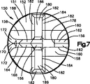
本発明は、人間の脊柱内の損傷を受けたまたは劣化した椎間板を置換する装置、移植片、または人工装具に関する。図1〜図4は、脊柱内の損傷を受けたまたは劣化した椎間板を置換する装置、移植片または人工装具10を示している。装置10(図6)は、人間の脊柱16の隣接する上下椎骨12と14の間の椎間板を置換するのに使用される。装置10(図2)は、第1の遠位または後側端部18と、対向する第2の近接または前側端部20とを有する。装置10は、椎骨12と14の間に挿入することができ、第1の端部18は脊柱16の後部側に隣接して配置され、第2の端部20は脊柱の前部側に隣接して配置されている。装置10は、第1の端部18と第2の端部20の間に延びる第1および第2の横側部22、24を備える。
The foregoing and other features of the present invention will become apparent to those skilled in the art upon consideration of the following detailed description of the invention with reference to the accompanying drawings.
The present invention relates to a device, graft, or prosthesis for replacing a damaged or degraded disc in the human spinal column. 1-4 show a device, implant or
装置10(図1〜図10)は、人工椎間板26と、隣接する椎骨12、14に椎間板26を連結させるのを助ける取付部材150とを備えることができる。取付部材150はまた、椎骨12、14に対して椎間板26を位置決めするのを助けることができる。取付部材150は、装置10を椎骨12と14の間に挿入する前に椎間板26に連結することができる。また、取付部材150は椎骨の間に椎間板26を挿入する前に、椎骨12、14に連結させることができる。
The device 10 (FIGS. 1-10) can include an
装置10(図1)は、上部または第1の保持装置30と、下部または第2の保持装置80と、保持装置間に介在し、これらに付着される弾力性のある芯130とを備える。上部および下部保持装置30、80は互いに同一であり、取付部材150を備える。装置10は、水平方向に延びる平面A(図4)周りで対照である。「上部」および「下部」という用語は本明細書では、参照する目的で2つの同一の保持装置を区別するために、図9に示すように人間の身体内にある場合に装置10の方向に関して使用される。
The device 10 (FIG. 1) comprises an upper or
上部保持装置(図1〜図3)は、上部または第1の保持リングまたは部材31と、取付部材150とを備える。人工椎間板26は上部保持部材31を備える。上部保持部材31は剛性があり、生体適合性金属またはポリマーなどの任意の所望の生体適合性材料でできている。上部保持部材31はチタン合金で作ることもできると考えられる。また、取付部材150は、上部保持部材31と一体に形成することができると考えられる。
The upper holding device (FIGS. 1 to 3) includes an upper or first holding ring or
上部保持部材31(図4)は、椎骨12と係合可能な外側表面32を有する。上部保持部材31の内側凹状表面34は、弾力性のある芯130に固定または結合される。内側凹状表面34は、任意の所望の方法で芯130に固定して結合することができる。内側表面34は、上部保持部材31を芯130に連結するのを助けるように、内側表面上で焼結されるビード(図示せず)、または内側表面上にエッチングされる組織(図示せず)を有することができると考えられる。
The upper retaining member 31 (FIG. 4) has an
複数のレールまたはリブ36(図1〜図3および図5)は、第1および第2の横側部22に隣接して外側表面32から延びる。複数のレールまたはリブ38は、椎間板26の中心部に隣接して外側表面32から延びる。上部保持部材31は図では4つのレール36、38を有するが、上部保持部材はいかなる数のレール36、38を有することができると考えられる。レール36、38は、装置10を椎骨12と14の間で定位置に保持するのを助けるように、椎骨12に係合している。外側表面32は、椎骨12と14の間に装置10をさらに保持するように、外側表面上で焼結されたビード(図示せず)と、外側表面上でエッチングされた組織(図示せず)とを有することができる。レール36、38は、椎骨12と14の間の装置10の簡単な挿入を可能にするための、ビードまたは組織を有することができない。外側表面32(図2)は、ビードまたは組織を備えていない、椎間板26の遠位または後側端部18に隣接する領域40を備えることができる。領域40は、レールまたはリブ36、38の遠位または後側端部に隣接して配置することができる。領域40により、椎骨12と14の間の装置10の簡単な挿入を可能にすることができる。領域40は、矩形などの任意の所望の形状をしていてもよいと考えられる。
A plurality of rails or ribs 36 (FIGS. 1-3 and 5) extend from the
レール36、38(図1〜図4)は、第1の端部に向かって椎間板26の第2の端部20から互いにほぼ平行に延びる。レール36、38は任意の所望の方向に延びていてもよいと考えられる。レール36、38が延びる方向は、椎間板26の挿入の方向によって決まる。
各レール36は、外側表面32から延びる複数の突起42を備える。レール36は図では5つの突起42を備えるが、いかなる所望の数の突起を有することができると考えられる。レール36は、椎間板26の後側端部18から前側端部20までテーパ状になっている。したがって、後側端部18のより近くに配置された突起42は外側表面32から第1の距離で延びており、前側端部20のより近くに配置された各隣接する突起42は、外側表面から第1の距離より大きい第2の距離で延びる。
Each
各レール38は、外側表面32から延びる複数の突起44を備える。レール38は図では6つの突起44を備えるが、レール38はいかなる所望の数の突起を有することができることが考えられる。レール38は、椎間板26の後側端部18から前側端部20までテーパ状になっている。したがって、後側端部18のより近くに配置された突起44は、外側表面32から第1の距離で延びており、前側端部20のより近くに配置された各隣接した突起44は外側表面から第1の距離より大きい第2の距離で延びる。
Each
椎間板26の横側部22、24に隣接したレール36の突起42(図3)は、レール38の突起44が表面から延びる距離よりも大きい距離だけ外側表面32から延びる。したがって、椎間板26の横側部22、24に隣接したリブ38の突起42は、リブ38の各隣接する突起44より大きい距離だけ椎骨12内に延びることができる。突起42、44は、外側表面32から任意の所望の距離で延びることができることが考えられる。
The protrusions 42 (FIG. 3) of the
突起42、44は実質的に同様である。したがって、1つの突起42のみを詳細に説明する。突起42(図1および図2)は、上側または外側、および装置10の第1の端部18に向かって面する第1または後側表面46を有する。第1の表面46は、第1の角度で表面32から延びる。突起42の第2のまたは前側表面48は、上側または外側、および装置10の第2の端部20に向かって面している。第2の表面48は第2の角度で表面32から延びる。第2の表面48が表面32から延びる第2の角度は、第1の表面46が表面32から延びる第1の角度より直角に近い。突起42は、上側または外側、および横側部22に向かって面する第1の横表面50を有する。第1の横表面は、第3の角度で表面32から延びる。第2の横表面52は、上側または外側、および横側面24に向かって面している。横表面52は、第3の角度と同じである第4の角度で表面32から延びる。表面46、48、50、52は任意の所望の角度で表面32から延びていてもよいと考えられる。突起42はまた、表面32とほぼ平行に延びる上側表面54を備える。各表面46、48、50、52は台形の形状をしている。第1および第2の横表面50、52は、第1の表面46および第2の表面48を横切る。突起42、44は任意の所望の構成を有することができることが考えられる。
The
軸方向に延びる開口56(図4〜図5)は、上部保持部材31の外側表面32および内側表面34を通して延びる。取付部材150は、取付部材を上部保持部材31に連結させるように、開口56内に延びる。上部保持部材31は、開口56を少なくとも部分的に画定する円錐切頭形表面57を有する。開口56の上部は第1の直径を有し、開口の下部は第1の直径より小さい第2の直径を有する。開口56はレール38間の中央に配置される。開口56は図では円形であるが、開口は任意の所望の形状を有することができることが考えられる。
An axially extending opening 56 (FIGS. 4-5) extends through the
フランジ部58(図1〜図5)は、椎間板26の前側端部20で上部保持部材31から延びる。フランジ部58は凹部60を有する。凹部60は、底部表面62、および底部表面62から上向きに延びる側部表面64、66によって画定される。楕円形のスロット68は、フランジ部58の底部表面62を通して延びる。スロット68は、レール38が延びる方向を横切る方向に延びる。
The flange portion 58 (FIGS. 1 to 5) extends from the upper holding
上部保持部材31の内側凹状表面34(図4)は、弾力性のある芯130に固定または結合されている。上部保持部材31は、下部保持装置80に向かって延びる周面フランジ部70を備える。フランジ70は芯130を取り囲む。フランジ70は、芯130に面する径方向内側表面72を有する。表面72は、凹状表面34から径方向外側、および下部保持装置80に向かって延びる。フランジ70上の表面72は、芯130に連結されない。したがって、フランジ70は芯130に対して移動することができる。
The inner concave surface 34 (FIG. 4) of the upper holding
表面72は、所定の負荷が装置10に加えられるまで、芯130から間隔を置いて配置することができると考えられる。上部および下部保持装置30、80を互いに対して移動させる負荷が装置10に加えられたときに、芯130はフランジ70上の表面72に向かって偏向することができる。所定の負荷が装置10に加えられると、芯130はフランジ70上の表面72と係合するように偏向することができる。芯130がフランジ70と係合すると、芯は硬くなる。というのは、芯のさらなる偏向がフランジによって抑えられるからである。
It is contemplated that the
フランジ70の表面72は任意の所望の構成を有していてもよい。表面72は、第2の部分より芯130の近くに延びる第1の部分を有することができ、それによって芯は表面72の第2の部分と係合する前に、表面72の第1の部分と係合する。したがって、芯130は、異なる負荷が装置10に加えられて異なる負荷での芯の剛性が変化するので、表面72の異なる部分に係合することができる。
The
保持部材31は、凹状内側表面34から開口56まで延び、所定の負荷が装置10に加えられるまで芯130から間隔を置いて配置される内側表面(図示せず)を有することができると考えられる。所定の負荷が装置10に加えられると、芯130は、凹状表面34から開口56まで延びる内側表面(図示せず)と係合するように偏向する。芯130が凹状表面34から開口56まで延びる内側表面と係合すると、芯は硬くなる。というのは、芯のさらなる偏向が保持部材31によって抑えられるからである。
It is contemplated that the retaining
下部保持装置(図1および図2〜図5)は、上部保持装置30と構成が同一である。下部保持装置80は、下部または第2の保持部材またはリング81、および取付部材150とを備える。椎間板26は下部保持部材81を備える。取付部材150は、下部保持部材81と一体的に形成することができると考えられる。下部保持部材81は、上部保持部材31と同一である。したがって、下部保持部材81は詳細に説明しない。下部保持部材81は剛性があり、チタン合金などの、上部保持部材31と同じ材料でできている。
The lower holding device (FIGS. 1 and 2 to 5) has the same configuration as the
下部保持部材81(図4)は、椎骨14と係合可能な外側表面82を有する。下部保持部材81の内側凹状表面84は、弾力性のある芯130に固定または結合される。内側表面84は、下部保持部材81を芯130に連結するのを助けるように、内側表面上で焼結されるビード(図示せず)、または内側表面上にエッチングされる組織(図示せず)を有することができると考えられる。
The lower retaining member 81 (FIG. 4) has an
複数のレールまたはリブ86(図1および図3)は、第1および第2の横側部22、24に隣接して外側表面82から延びる。複数のレールまたはリブ88は、椎間板26の中心部に隣接して外側表面82から延びる。下部保持部材81は、任意の所望の数のリブ86、88を有することができる。レール86、88は、装置10を椎骨12と14との間で定位置に保持するのを助けるように、椎骨14に係合している。外側表面82は、椎骨12と14との間に装置10をさらに保持するように、外側表面上で焼結されたビード(図示せず)と、外側表面上でエッチングされた組織(図示せず)とを有することができる。レール86、88は、椎骨12と14との間の装置10の簡単な挿入を可能にするための、ビードまたは組織を有することができない。外側表面82は、ビードまたは組織を備えていない、椎間板26の第1の端部18に隣接した、上部保持部材31上の領域40と同様の領域(図示せず)を備えることができる。領域は、レールまたはリブ86、88の遠位または後側端部に隣接して配置することができる。領域は、矩形などの任意の所望の形状を有していてもよいと考えられる。
A plurality of rails or ribs 86 (FIGS. 1 and 3) extend from the
レール86、88は、第1の端部18に向かって椎間板26の第2の端部20から互いにほぼ平行に延びる。レール86、88は任意の所望の方向に延びていてもよいと考えられる。レール86、88が延びる方向は、椎間板26の挿入の方向によって決まる。
The
各レール86は、外側表面82から延びる複数の突起92を備える。レール86は、任意の所望の数の突起92を有することができる。レール86は、椎間板26の第1の端部18から第2の端部20までテーパ状になっている。したがって、後側端部18のより近くに配置された突起92は外側表面82から第1の距離で延びており、前側端部20のより近くに配置された各隣接する突起92は、外側表面から第1の距離より大きい第2の距離で延びる。
Each
各レール88は、外側表面82から延びる複数の突起94を備える。レール88は、任意の所望の数の突起94を有することができる。レール88は、椎間板26の後側端部18から前側端部20までテーパ状になっている。したがって、後側端部18のより近くに配置された突起94は、外側表面82から第1の距離で延びており、前側端部20のより近くに配置された各隣接した突起94は外側表面から第1の距離より大きい第2の距離で延びる。
Each
椎間板26の横側部22、24に隣接したレール86の突起92(図3)は、レール86の突起92に隣接するレール88の突起94が表面82から延びる距離よりも大きい距離だけ外側表面82から延びる。したがって、椎間板26の横側部22、24に隣接したレール86の突起92は、レール88の突起94より大きい距離だけ椎骨14内に延びることができる。突起92、94は、外側表面82から任意の所望の距離で延びることができる。
The protrusions 92 (FIG. 3) of the
レール86、88の突起92、94は、リブ36、38の突起42、44と実質的に同様である。したがって、突起92、94をより詳細に説明しない。各突起92、94(図4)は、下側または外側、および装置の第1の端部18に向かって面する第1のまたは後側表面96を有する。第1の表面96は、第1の角度で表面82から延びる。第2のまたは前側表面98は、下側または外側、および装置10の第2の端部20に向かって面している。第2の表面98は、第2の角度で表面82から延びる。第2の表面98が表面82から延びる第2の角度は、第1の表面96が表面82から延びる第1の角度より直角に近い。各突起92、94(図3)は、第1の横表面100と、第3および第4の角度で表面82から延びる第2の横表面とを有する。第1および第2の横表面100、102は、下側または外側、および装置10の横側部22、24に向かって面している。表面96、98、および第1および第2の横表面は任意の所望の角度で表面82から延びることができると考えられる。突起92、94はまた、表面82とほぼ平行に延びる下側表面103を備える。
The
各表面96、98、100、102は台形の形状をしている。横表面100、102は、第1の表面96および第2の表面98を横切る。突起92、94は任意の所望の構成を有することができると考えられる。
Each
軸方向に延びる開口104(図4)は、下部保持部材81の外側表面82および内側表面84を通して延びる。取付部材150は、取付部材を下部保持部材81に連結させるように、開口104内に延びる。下部保持部材81は、開口104を少なくとも部分的に画定する円錐切頭形表面106を有する。開口104の下部は第1の直径を有し、開口の上部は第1の直径より小さい第2の直径を有する。開口104はレール88間の中央に配置される。開口104は円形であると記載されているが、開口は任意の所望の形状を有することができることが考えられる。
An axially extending opening 104 (FIG. 4) extends through the
フランジ部108(図1、および図3〜図5)は、椎間板26の前側端部20で下部保持部材81から延びる。フランジ部108は凹部110を有する。凹部110は、上側表面112、および上側表面112から下向きに延びる側部表面114、116によって画定される。楕円形のスロット118は、フランジ部108の上側表面112を通して延びる。スロット118は、レール88が延びる方向を横切る方向に延びる。
The flange portion 108 (FIGS. 1 and 3 to 5) extends from the lower holding
下部保持部材81の内側凹状表面84(図4)は、弾力性のある芯130に固定または結合されている。下部保持部材81は、上部保持装置30に向かって延びる周面フランジ部120を備える。フランジ120は芯130を取り囲む。フランジ120は、芯130に面する径方向内側表面122を有する。表面122は、凹状表面84から径方向外側、および上部保持装置30に向かって延びる。フランジ120上の表面122は、芯130に連結されない。したがって、フランジ122は芯130に対して移動することができる。
The inner concave surface 84 (FIG. 4) of the lower holding
表面122は、所定の負荷が装置10に加えられるまで、芯130から間隔を置いて配置することができると考えられる。上部および下部保持装置30、80を互いに対して移動させる負荷が装置10に加えられたときに、芯130はフランジ120上の表面122に向かって偏向することができる。所定の負荷が装置10に加えられると、芯130はフランジ120上の表面122と係合するように偏向することができる。芯130がフランジ120と係合すると、芯は硬くなる。というのは、芯のさらなる偏向がフランジ120によって抑えられるからである。
It is contemplated that the
フランジ120の表面122は任意の所望の構成を有していてもよい。表面122は、第2の部分より芯130の近くに延びる第1の部分を有することができ、それによって芯は表面122の第2の部分と係合する前に、表面122の第1の部分と係合する。したがって、芯130は、異なる負荷が装置10に加えられて異なる負荷での芯の剛性が変化するので、表面122の異なる部分に係合することができる。また、所定の負荷が装置10に加えられたときに、下部保持部材81上のフランジ120は上部保持部材31上のフランジ70と係合することができると考えられる。
The
保持部材81は、凹状内側表面84から開口104まで延び、所定の負荷が装置10に加えられるまで芯130から間隔を置いて配置される内側表面(図示せず)を有することができると考えられる。所定の負荷が装置10に加えられると、芯130は、凹状表面84から開口104まで延びる内側表面(図示せず)と係合するように偏向する。芯130が凹状表面84から開口104まで延びる内側表面と係合すると、芯は硬くなる。というのは、芯のさらなる偏向が保持部材81によって抑えられるからである。
It is contemplated that the retaining
弾力性のある芯130は一体的であり、カリフォルニア州、Berkleyにあるthe Polymer Technology Groupにより製造されたウレタンシリコン混合物でできていてもよい。弾力性のある芯130は、任意の所望の方法で上部および下部保持部材31、81に接着または結合することができる。弾力性のある芯130は、上部保持部材31と下部保持部材81の間で挿入成形、トランスファ成形、または射出成形することができることが考えられる。芯130は、芯用の材料を開口56または104の1つを通して上部および下部保持部材に射出することによって、上部保持部材31と下部保持部材81の間で成形することができる。
The
弾力性のある芯130は、カリフォルニア州、Berkleyにあるthe Polymer Technology Groupにより製造されたCarboSil(商標)の名前のシリコンポリカーボネートウレタン共重合体であるポリマーでできていてもよい。弾力性のある芯130は、ポリジメチルシシロキサンが脂肪族、ヒドロキシル基末端ポリカーボネートオリゴマーを有するポリマー軟質部分に組み込まれる間に、多工程バルク合成により作成される。硬質部分は、低分子量グリコール鎖増量剤を有する芳香族ジイソシアネートからなる。共重合体鎖は、シリコンで終端する。
The
弾力性のある芯130の材料は、シリコンエラストマーの生体適合性および生体安定性を熱可塑性ウレタンエラストマーの処理性および強靭性と組み合わせる。弾力性のある芯130の材料は、患者の身体との均衡に到達する際にかなり軟化する比較的高い硬質部分内容物を有する。関連する均衡は、約37℃での身体との熱均衡、および身体に移植した後にポリマーによって取り込まれる均衡水および溶質を必要とする。弾力性のある芯130の材料は、室温と比較して37℃で小さな係数を有する。したがって、より高いデュロメータポリマーをその生体安定性に使用することができる。というのは、人間の身体の状態により所望の範囲の圧縮剛性に対するポリマーの係数が小さくなるからである。
The
弾力性のある芯130は楔形をしている。上部保持部材31は、椎間板26の近接側18に隣接して第1の距離だけ下部保持部材81から間隔を置いて配置されている。上部保持部材31は、椎間板26の前部側20に隣接して第1の距離より大きい第2の距離だけ下部保持部材81から間隔を置いて配置されている。上部保持部材31は、任意の所望の距離だけ下部保持部材81から間隔を置いて配置することができることが考えられる。
The
芯130は、上側または第1の凹状表面132を有する。上側凹状表面132は、上部保持部材31の凹状内側表面34に固定される。下側または第2の凸状表面134は、下部保持部材81の凹状内側表面84に固定される。
The
芯130は、径方向外側表面136を備える。移行表面138は、径方向外側表面136と上下側表面132、134の間に延びる。径方向外側表面136は、所定の負荷が装置10に加えられるまで、上部および下部保持部材31、81上でフランジ70、120から間隔を置いて配置することができる。
The
周表面136および移行表面138は、任意の所望の構成を有することができる。表面136、138は第2の部分よりフランジ70、120の近くに延びる第1の部分を有することができ、それによって第1の部分は第2の部分の前でフランジに係合する。したがって、表面136、138の異なる部分は、異なる負荷が装置10に加えられて異なる負荷で芯130の剛性を変化させるときに、フランジ70、120と係合することができる。
The
各保持装置30、80(図1〜図7)は、椎間板26を椎骨12、14に連結させるのを助けるように取付部材150を備える。取付部材150は、椎骨12と14との間に椎間板26を位置決めするのを助けることができる。取付部材150(図6)は、取付部材が椎間板26に連結された場合に、保持部材31、81内の開口56、104内に延びる。取付部材150は、装置10を椎骨12と14との間に挿入する前に、椎間板26に連結することができる。椎間板26はまた、取付部材150を椎骨に連結した後に、椎骨12と14との間に挿入することができると考えられる。レール38、88は、椎間板26の開口56、104の両側で、椎骨12と14との間の所望の位置に椎間板を案内するように取付部材150に係合することができる。
Each
取付部材150の第1の実施形態は、図1〜図4および図6〜図8に示されている。取付部材150は、互いに同一である。したがって、1つの取付部材150のみを詳細に説明する。取付部材150(図6〜図8)は剛性があり、生体適合性金属またはポリマーなどの任意の所望の生体適合性材料でできている。取付部材150は、チタン合金でできていてもよいと考えられる。
A first embodiment of the mounting
取付部材150は、ほぼ円形の本体151を有する。取付部材150の本体151は、取付部材を椎間板26内の開口56、104内に摺動させることができる任意の所望の構成をしていてもよいと考えられる。取付部材150の本体151は、椎骨に面する外側表面152を有する。取付部材150の内側凹状表面154(図4)は、弾力性のある芯130に面している。上部保持装置30の取付部材150の内側凹状表面154は、芯130の上側表面132に面している。下部保持装置80の取付部材150の内側凹状表面154は、芯130の下側表面134に面している。
The
弾力性のある芯130(図9〜図10)は、上部および下部保持装置30、80を互いに対して移動させるように負荷が装置10に加えられると、凹状表面154に向かって偏向する。芯130は、図10に示すように、上部および下部保持部材31、81の開口56、104内に偏向し、脊柱16に所定の負荷が加えられると、凹状表面154と係合する。芯130が取付部材150の表面154と係合すると、弾力性のある芯は硬くなる。というのは、保持装置30、80の芯のさらなる偏向が抑えられるからである。取付部材150は、芯130と取付部材の間から気体が逃げることを可能にする軸方向に延びる開口を有することができると考えられる。
The resilient core 130 (FIGS. 9-10) deflects toward the
取付部材150の表面154は、任意の所望の構成をしていてもよい。芯130は、異なる負荷が装置10に加えられて異なる負荷での芯130の剛性が変化するので、表面154の異なる部分と係合することができる。また、保持装置30の取付部材150の表面154は、保持装置80の取付部材150の表面154とは異なる構成をしていてもよいと考えられる。
The
取付部材150の中心レールまたはリブ156(図3および図6〜図8)は、本体151の表面152から延びており、椎骨と係合可能である。リブ156は、取付部材の近接または前部側に向かって取付部材150の遠位または後部側から延びる長さを有する。したがって、取付部材150が保持部材31または81に連結される場合、レールまたはリブ156の長さは、第1および第2の端部18、20とほぼ横方向に、またレール36、38または86、88とほぼ平行に延びる。
A central rail or rib 156 (FIGS. 3 and 6-8) of the mounting
リブ156は、表面152から延びるアーチ状横表面158を備える。平らな横表面160は、アーチ状表面158から上向きに延びる。上側表面162は、平らな表面160に対してある角度で延びる。上側表面162は、リブ156の頂点を形成するように、互いに対してある角度で延びる。
リブ156の遠位または後側部164(図6〜図8)は、取付部材150の表面152に向かってリブの中心部166の頂点からテーパ状になっている。後側部164は、互いに対してある角度で、また平らな横表面160に対してある角度で延びる上側表面168を備える。リブ156の後側部164は、三角形の形状をしている後側表面170を備える。1対の移行表面172は、後側表面170、上側表面168、およびアーチ状横表面158の間に延びる。レール156の中心部166は、骨内部成長を行うU字形凹部178を備える。凹部178は、リブ156の頂点からアーチ状表面158まで延びる。
The distal or rear side 164 (FIGS. 6-8) of the
1対の横レールまたはリブ180は、リブ156の中心部166から横方向に延びる。レール180は、レール156が表面152から延びる距離より小さい距離だけ本体151の表面152から延びる。したがって、レール156はリブ180よりさらに椎骨内に延びることができる。各レール180は、取付部材150の横側部にレール156の中心部166から延びる距離を有する。したがって、レールまたはリブ180の長さは、取付部材150が保持部材31または81に連結された場合に、レール156、36、38、86、88に対してほぼ横に延びる。レール180の長さは、取付部材150が保持部材31または81に連結された場合に、レール156、36、38、86、88に対してほぼ垂直に延びる。
A pair of lateral rails or
各リブ180は、表面152から延びるアーチ状表面182を備える。前側表面184は、アーチ状表面182、および取付部材150の表面152にほぼ垂直に延びる。後側表面186は、前側表面184に対してある角度で延びる。
Each
取付部材150の本体151は、径方向外側表面190を有する。丸みを帯びた移行表面192は、径方向外側表面190から凹状表面154まで延びる。取付部材150は、外側表面190に隣接した第1の直径、および第1の直径より小さな移行表面192に隣接した第2の直径を有する。取付部材150の径方向外側表面190および/または移行表面192は、椎間板を椎骨間に挿入しているときに、取付部材および椎骨12、14に対して第1の後側方向への椎間板26の移動を案内するように、保持部材31、81上でレール38、88と係合することができる。
The
取付部材150上の径方向外側表面190は、取付部材が椎間板26の開口56、104内にある場合に、上部および下部保持部材31、81上で円錐切頭形表面57、106と係合する。表面57、106との表面190の係合により、取付部材150と椎間板26の間に締まり嵌めが作り出される。したがって、取付部材150が椎間板26に固定取り付けされ、椎間板は取付部材に対して移動しないようにされる。
A radially
径方向外側表面190は2つの凹部196を有し、そのうちの1つを図6および図8に示す。凹部196は、互いに対して180°に配置される。取付部材190は2つの凹部196を有すると記載されているが、取付部材150は任意の所望の数の凹部を有することができると考えられる。
The radially
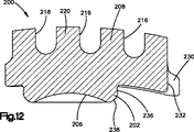
椎間板26で使用する取付部材200の第2の実施形態が、図11および図12に示されている。取付部材200(図11)は剛性があり、生体適合性金属またはポリマーなどの任意の所望の生体適合性材料でできている。取付部材200は、チタン合金でできていてもよいと考えられる。また、取付部材200は保持部材31、81と一体に形成することができると考えられる。
A second embodiment of an
取付部材200は、ほぼ円形の基部202を有する。基部202は、取付部材200を椎間板に連結させるように、椎間板26の開口56、104の1つ内に延びる。基部202は、基部を椎間板26の開口56、104内に摺動させることができる任意の所望の構成をしていてもよいと考えられる。
The
取付部材200(図11)は、椎骨に面する外側表面204を有する。取付部材200の内側凹状表面206は、弾力性のある芯130に面している。上部保持装置30の取付部材200の内側凹状表面206は、芯130の上側表面132に面している。下部保持装置80の取付部材200の内側凹状表面206は、芯130の下側表面134に面している。
The attachment member 200 (FIG. 11) has an
弾力性のある芯130は、上部および下部保持装置30、80を互いに対して移動させるように負荷が装置10に加えられると、凹状表面206に向かって偏向する。芯130は、上部および下部保持部材31、81の開口56、104内に偏向し、脊柱16に所定の負荷が加えられると、凹状表面206と係合する。芯130が取付部材200の表面206と係合すると、弾力性のある芯は硬くなる。というのは、保持装置30、80の芯へのさらなる偏向が抑えられるからである。取付部材200は、芯130と取付部材の間から気体が逃げることを可能にする軸方向に延びる開口を有することができると考えられる。
The
取付部材200の表面206は、任意の所望の構成をしていてもよい。芯130は、異なる負荷が装置10に加えられて異なる負荷での芯130の剛性が変化するので、表面206の異なる部分と係合することができる。また、保持装置30の取付部材200の表面206は、保持装置80の取付部材200の表面206とは異なる構成をしていてもよいと考えられる。
The
取付部材200の中心レールまたはリブ208は、表面204から延びており、椎骨と係合可能である。レール208は、取付部材の近接または前部側に向かって取付部材200の遠位または後部側から延びる長さを有する。したがって、取付部材200が保持部材31または81に連結される場合、レールまたはリブ208の長さは、第1および第2の端部18、20とほぼ横方向に、またレール36、38または86、88とほぼ平行に延びる。
A central rail or
レールまたはリブ208は、表面204から延びるアーチ状横表面210を備える。平らな横表面212は、アーチ状表面210から上向きに延びる。上側表面214は、平らな表面212に対してある角度で延びる。上側表面214は、リブ208の頂点を形成するように、互いに対してある角度で延びる。
The rail or
リブ208は、骨内部成長を行う複数のU字形凹部216を備える。凹部216は、リブ208の頂点からアーチ状表面210まで延びる。取付部材200は図ではU字形凹部216を有するが、取付部材は任意の所望の数の凹部を有することができると考えられる。
The
1つが図11に示されている、1対の横リブ218は、リブ208の中心部220から横方向に延びる。リブ218は、リブ208が表面204から延びる距離より小さい距離だけ表面204から延びる。したがって、リブ208はリブ218より遠くに椎骨内に延びることができる。各レールまたはリブ218は、リブ208の中心部220から取付部材200の横側部まで延びる長さを有する。したがって、レールまたはリブ218の長さは、取付部材200が保持部材31または81に連結される場合に、レール208、36、38、86、88に対してほぼ横方向に延びる。レール218の長さは、取付部材200が保持部材31または81に連結される場合に、レール156、36、38、86、88に対してほぼ垂直に延びる。
A pair of
各リブ218は、表面204から延びるアーチ状表面222を備える。前側表面224はアーチ状表面222から延びる。後側表面226はアーチ状表面222から延びる。後側表面226はまた、前側表面224に対してある角度で延びる。
Each
フランジ230は、取付部材200の前側部から延びる。フランジ230は、リブ208の対向する横側部から延びる。フランジ230は、取付部材200の前側部で凹部232を画定する。
The
基部202(図11および図12)は、径方向外側表面236を有する。丸みを帯びた移行表面238は、径方向外側表面236から凹状表面206まで延びる。基部202は、外側表面236に隣接した第1の直径、および第1の直径より小さな移行表面238に隣接した第2の直径を有する。取付部材200の径方向外側表面236および/または移行表面238は、椎間板を椎骨間に挿入しているときに、取付部材および椎骨12、14に対して第1の後側方向への椎間板26の移動を案内するように、保持部材31、81上でレール38、88と係合することができる。
Base 202 (FIGS. 11 and 12) has a radially
取付部材200上の径方向外側表面236は、取付部材が椎間板26の開口56、104内にある場合に、上部および下部保持部材31、81上で円錐切頭形表面57、106と係合する。表面57、106との表面236の係合により、取付部材200と椎間板26の間に締まり嵌めが作り出される。したがって、取付部材200が椎間板26に固定取り付けされ、椎間板は取付部材に対して移動しないようにされる。
A radially
径方向外側表面236は2つの凹部240を有し、そのうちの1つを図11に示す。凹部240は、互いに対して180°に配置される。取付部材200は2つの凹部240を有すると記載されているが、取付部材200は任意の所望の数の凹部を有することができると考えられる。
The radially
椎骨12と14の間に装置10を挿入する前に、試験選別機またはスペーサ300(図13および図14)が椎骨の間に挿入される。試験選別機またはスペーサ300は、装置10に対して所望の位置、足跡、楔角度、および椎間板高さを決めるのに使用される。試験選別機300はまた、椎骨間の装置10に対して所望の位置を決めるのに使用される。装置10に対する所望の位置が、試験選別機またはスペーサ300を使用して決められると、試験選別機は椎骨12と14の間の外科用器具の挿入を案内するのに使用する、脊柱上の基準点を決めるのに使用することができる。異なる試験選別機300は、所望の足跡、楔角度、および椎間板高さを有する試験選別機が椎骨間に位置決めされるまで、椎骨12と14の間に挿入し、その間から取り除くことができる。所望の足跡、楔角度、および椎間板高さを有する試験選別機300が挿入されると、装置10の寸法および形状を決めることができる。
Prior to inserting the
試験選別機300は、装置10と同様の形状を有し、楔形状をしていてもよい。試験選別機300は、第1の、遠位または後側端部302と、反対側の第2の、近接または前側端部304とを有する。試験選別機300はまた、第1の端部302と第2の端部304の間に延びる第1および第2の横側部306、308を備える。試験選別機300の上側表面310は椎骨12と係合可能であり、下側表面312は椎骨14と係合可能である。上側および下側表面310、312は、椎骨12、14と係合する複数の突起314を有する。突起314は、任意の所望の構成を有することができる。
The
試験選別機300(図13)は、第1の横側部306と第2の横側部308の間に延びる円筒形通路320を備える。試験選別機300は、第1の端部302と第2の端部304の間に延びる中心円筒形通路322を備える。通路322は、試験選別機300の楔角度を二股に分ける。選別機300はまた、第1の端部302と第2の端部304の間に延びる2つの横円筒形通路324、326を備える。通路322、324、326は互いにほぼ平行に延びる。通路322、324、326は通路320を横切る。通路320、322、324、326は任意の所望の構成を有することができると考えられる。通路320、322、324、326は、試験選別機300が椎骨12と14の間に適切に位置決めされたかどうかを判断するように、蛍光透視鏡などの所望の画像化システムを使用して見ることができる。試験選別機300が椎骨12と14の間に適切に位置決めされると、通路320、322、324、326は、画像化システムによって作り出された画像内に円として見える可能性がある。試験選別機が適切に位置決めされないと、通路320、322、324、326は画像内に楕円として見える可能性がある。通路322は、前側部304に隣接して配置されたねじ付き部328を有する。
The test sorter 300 (FIG. 13) includes a
挿入ロッド330は、通路322のねじ付き部328とねじ係合可能である。挿入ロッド330は、椎骨12と14の間に試験選別機300を挿入するのに使用することができる。挿入ロッド330は、選別機300を椎骨12と14の間で移動させるように、木槌またはハンマで打つことができる。挿入ロッド330は、通路322のねじ付き部328とねじ係合する第1または遠位ねじ付き端部332を有する。第1のねじ付き端部332は第1の直径を有する。中心部334は、第1の直径より大きな第2の直径を有する。径方向に延びる表面(図示せず)は、第1の端部332と中心部334の間に延びる。径方向に延びる表面は、挿入ロッド330の試験選別機内へのさらなる挿入を防ぐため、試験選別機300と係合可能である。挿入ロッド330の第2の端部338は、任意の所望の方法でハンドル(図示せず)と連結可能である。
The
試験選別機300を椎骨12と14の間に位置決めした後に、案内組立体351の第1の実施形態のマーカー350(図15)は、椎骨12、14の1つなどの脊柱の任意の所望の椎骨に連結されている。マーカー350は、脊柱16の中線などの所望の椎骨上の所望の基準点で椎骨12、14の1つの連結されている。マーカー350は、第1の直径の第1の端部354を有するシャフト352を備える。シャフト352の中心部356は、第1の直径より大きな第2の直径を有する。径方向に延びる表面358は、第1の端部354から中心部356まで延びる。シャフト352の第2の端部360は、任意の所望の方法でハンドル(図示せず)と連結可能である。
After positioning the
中心部356は、径方向に延びる1対の位置決め部材362を備える。位置決め部材362は全く反対である。第1の端部354は、椎骨12、14の1つに挿入するための尖った端部364を有する。端部354はまた、径方向に延びる表面358に隣接して配置された直径方向に延びる開口366を備える。開口366は、安定化部材370をシャフト352に連結させるようにピン(図示せず)を受ける。
The
安定化部材370は、軸方向に延びる開口374を有する本体372を備える。本体372内の径方向に延びる開口(図示せず)は、軸方向に延びる開口374を横切る。シャフト352の第1の端部354は開口374を通して延びており、それによって肩部358は本体372に係合する。ピン(図示せず)は、シャフトを安定化部材370に連結させるように、本体372の径方向に延びる開口(図示せず)を通して、シャフト352の開口366内に延びる。シャフト352は、溶接などの任意の所望の方法で、安定化部材370に連結させることができる。
Stabilizing
安定化部材370は、本体372から延びる1対の安定化シャフト378を備える。安定化シャフト378は本体372から延びており、それによってシャフト352が安定化部材370に連結されている場合に、安定化シャフトおよびシャフト352はトライデントを形成する。安定化シャフト378は、マーカー350を椎骨内に維持するのを助けるように、椎骨12、14の1つに挿入される。
案内機構または部材390(図15および図17)は、所望の基準点で椎骨12、14の1つにマーカー350を連結させるのに使用される。案内機構または部材390は、対向する端部392、394を有する。案内機構または部材390の第1のまたは下側部396は、第1の直径を有する通路398を有する。通路398の直径は、挿入ロッド330の直径より少し大きい。第2のまたは上側部400は、第1の直径より小さい第2の直径を有する通路402を有する。通路402の直径は、マーカー350のシャフト352の中心部356の直径より少し大きい。中心通路404は、案内部材390を通して延びており、通路398と402の間に配置されている。通路398、402、404は、対向する端部392、394の間で互いにほぼ平行に延びる。
A guide mechanism or member 390 (FIGS. 15 and 17) is used to connect the
中心通路404は、ハンドル(図示せず)とねじ係合するねじ付き部405を備える。案内機構390は、通路404を部分的に画定する肩部406を備える。ハンドル(図示せず)は、肩部406の1つと係合可能な肩部を備える。ハンドルは、案内機構390を取り扱うのに使用され、任意の所望の方法で案内機構と連結することができる。
The
案内機構390の端部392は、凹部408を備える。端部394は凹部410を備える。凹部408、410は、通路398、402、404を横切り、下部396から上部400まで延びる。凹部408、410は、シャフト352上で径方向に延びる位置決め部材362を受けることができる。したがって、第1および第2の端部392、394はほぼ同様である。
The
マーカー350のシャフト352は、シャフト上の径方向に延びる位置決め部材362が凹部408内に受けられるまで、案内機構390内に通路402を通して挿入される。ハンドル(図示せず)は、ねじ付き部405の1つにねじ連結することができる。案内機構390はその後、選別機が椎骨12と14の間にある場合に試験選別機300から延びる挿入ロッド330の上で入れ子状になっており、それによって挿入ロッドは案内機構の通路398内に延びる。案内機構390が挿入ロッド330に対して軸方向に移動されると、シャフト352の第1の端部354は、所望の基準点で、椎骨12などの椎骨12、14の1つに挿入される。安定化部材370の安定化シャフト378はまた、1つの椎骨内に挿入される。マーカー350のシャフト352は、挿入ロッド330と平行に延びる。マーカー350を椎骨12、14の1つに連結した後、案内部材390はマーカーから取り外され、試験選別機300は椎骨12と14の間から取り外される。
The
複数の案内機構390は、手術中に提供することができる。各案内機構390は、通路398と402の間に異なる間隙を有する。したがって、外科医は脊柱16の所望の椎骨上の所望の基準点でマーカー350を挿入するように、適当な案内機構390を選択することができる。
A plurality of
マーカー350を脊柱16の所望の椎骨に連結させ、試験選別機300を椎骨12と14の間から取り除いた後に、椎骨は装置10を受けるように切断することができる。第1のカッター450(図18〜図20)は、装置10上のレール36、38、86、88を受けるように椎骨内に溝を切り込むように、椎骨12と14の間に挿入させることができる。第1のカッター450は、装置10と同様の形状をしており、楔形の形状をしていてもよい。第1のカッター450は、第1の、遠位または後側端部452と、反対側の第2の、近接または前側端部454とを有する。カッター450はまた、第1の端部452と第2の端部454の間に延びる第1および第2の横側部456、458を備える。カッター450の上側表面460は椎骨12と係合することができ、下側表面462は椎骨14と係合することができる。
After the
複数の列464の歯466が、横側部456、458に隣接して上側および下側表面460、462から延びる。歯466は、装置10のリブ36、86を受けるように椎骨12、14内に溝を切り込む。複数の列467の歯468は、カッター450の中心部に隣接して上側および下側表面460、462から延びる。歯468は、装置10のリブ38、88を受けるように、椎骨12、14内に溝を切り込む。列467の歯468は、列464の歯466が上側および下側表面から延びる距離より小さい距離だけ上側および下側表面460、462から延びる。したがって、列464の歯466は、列467の歯468より椎骨12、14内に深い溝を切り込むことができる。列464、467の歯466、468は、カッター450の第1の端部と第2の端部の間の距離の半分にほぼ等しい第1の距離だけ、第1の端部452に向かって第2の端部454から延びる。
A plurality of
カッター450は、第1の横側部456と第2の横側部458の間に延びる円筒形通路470を備える。カッター450は、第1の端部452と第2の端部454の間に延びる中心円筒形通路472を備える。カッター450はまた、第1の端部452と第2の端部454の間に延びる2つの横円筒形通路474、476を備える。通路472、474、476は互いにほぼ平行に延びる。通路472、474、476は通路470を横切る。通路470、472、474、476は任意の所望の構成を有することができると考えられる。通路470、472、474、476は、カッター450が椎骨12と14の間に適切に位置決めされたかどうかを判断するように、蛍光透視鏡などの画像化システムを使用して見ることができる。カッター450が椎骨12と14の間に適切に位置決めされると、通路470、472、474、476は、画像化システムによって作り出された画像内に円として見える可能性がある。カッター450が適切に位置決めされないと、通路470、472、474、476は画像内に楕円として見える可能性がある。通路472は、前側部454に隣接して配置されたねじ付き部478を有する。
The
挿入ロッド330は、通路472のねじ付き部478とねじ係合可能である。挿入ロッド330は、椎骨12と14の間にカッター450を挿入するのに使用することができる。挿入ロッド330は、案内部材390の通路398内に挿入される。案内部材390はその後、凹部408がシャフト352上で径方向に延びる位置決め部材362を受けるまで、マーカー350のシャフト352上で入れ子状になっている。したがって、カッター450は椎骨12、14と所望のように位置合わせされている。挿入ロッド330は、カッター450を椎骨12と14の間で移動させ、歯466、468が椎骨12、14内に溝を切り込むように、木槌またはハンマで打つことができる。
The
カッター450が椎骨12、14内に溝を切り込んだ後に、カッター450は椎骨間から取り除かれ、案内部材390はマーカー350から取り除かれる。第2のカッター490(図21〜図22)は、椎骨内にさらに溝を切り込んで装置10でリブ36、38、86、88を受けるように、椎骨12と14の間に挿入することができる。第2のカッター490は、第1のカッター450と実質的に同様である。第2のカッター490は、装置10と同様の形状をしており、楔形状をしていてもよい。第2のカッター490は、第1の、遠位または後側端部492と、反対側の第2の、近接または前側端部494とを有する。カッター490はまた、第1および第2の横側部496、498を備える。カッター490の上側表面500は椎骨12と係合することができ、下側表面502は椎骨14と係合することができる。
After
複数の列504の歯506は、横側部496、498に隣接して上側および下側表面500、502から延びる。歯506は、装置10のリブ36、86を受けるように椎骨12、14内に溝を切り込む。複数の列507の歯508は、カッター490の中心部に隣接して上側および下側表面500、502から延びる。歯508は、装置10のリブ38、88を受けるように、椎骨12、14内に溝を切り込む。列507の歯508は、列504の歯506が上側および下側表面から延びる距離より小さい距離だけ上側および下側表面500、502から延びる。したがって、列504の歯506は、列507の歯508より椎骨12、14内に深い溝を切り込むことができる。列504、507の歯506、508は、後側部492と前側部494の間の距離の半分より大きい第2の距離だけ、第2の端部494から第1の端部492まで延びる。したがって、列504、507の歯506、508は、第1のカッター450上の列464、467の歯466、468が延びる第1の距離より大きい距離で延びる。
The
カッター490は、第1の横側部496と第2の横側部498の間に延びる円筒形通路510を備える。カッター490は、第1の端部492と第2の端部494の間に延びる中心円筒形通路512を備える。カッター490はまた、第1の端部492と第2の端部494の間に延びる2つの横円筒形通路514、516を備える。通路512、514、516は互いにほぼ平行に延びる。通路512、514、516は通路510を横切る。通路510、512、514、516は任意の所望の構成を有することができると考えられる。通路510、512、514、516は、カッター490が椎骨12と14の間に適切に位置決めされたかどうかを判断するように、蛍光透視鏡などの画像化システムを使用して見ることができる。カッター490が椎骨12と14の間に適切に位置決めされると、通路510、512、514、516は、画像化システムによって作り出された画像内に円として見える可能性がある。カッター490が適切に位置決めされないと、通路510、512、514、516は画像内に楕円として見える可能性がある。通路512は、前側部494に隣接して配置されたねじ付き部518を有する。
The
挿入ロッド330は、通路512のねじ付き部518とねじ係合可能である。挿入ロッド330は、椎骨12と14の間にカッター490を挿入するのに使用することができる。挿入ロッド330は、案内機構390の通路398内に挿入される。案内機構390はその後、凹部408がシャフト352上で径方向に延びる位置決め部材362を受けるまで、マーカー350のシャフト352上で入れ子状になっている。したがって、カッター490は椎骨12、14と所望のように位置合わせされている。挿入ロッド330は、カッター490を椎骨12と14の間で移動させ、歯506、508が椎骨12、14内に溝を切り込むように、木槌またはハンマで打つことができる。
装置10を椎骨12と14の間に挿入する、または取付部材150を椎骨12、14に連結した後に椎間板26を椎骨間に挿入する、挿入器具550(図24〜図25)が、図24および25に示されている。器具550(図24)は、共通のはさみと、互いに旋回可能に連結された1対の脚部552、554とを有する。器具550は、脚部552、554上に1対の顎部558によって形成された把持端部556を備える。顎部558(図25)は、互いに向かって延びる楕円形突起560を備える。突起560は、椎骨12と14の間に挿入するように椎間板を把持するために、椎間板26の開口68、118内に挿入される。
An insertion instrument 550 (FIGS. 24-25) that inserts the
脚部552(図24)は、顎部558の反対側に拡大端部562を有する。拡大端部562は、必要に応じて、椎骨12と14の間に椎間板26を駆動させるように木槌で打つことができる。脚部554は、顎部558に対向して湾曲ハンドル564を有する。ハンドル564は、器具550を操作するように外科医によって簡単に把持される。
Leg 552 (FIG. 24) has an
係止機構570は、突起560を椎間板26の開口68、118に挿入した後に、顎部558が互いに離れるように旋回するのを防ぐ。係止機構570は、脚部554から延びる取付部574に旋回可能に連結されたロッド572を備える。ロッド572は、脚部552の開口578を通して延びるねじ付き端部576を有する。ナット582は、顎部558が互いに離れるように旋回するのを防ぐように、ロッド572の端部576にねじ係合し、脚部552に係合する。
The
装置10が挿入器具550で椎骨12と14の間に挿入された後、突き固め部材(タンピング部材)600(図26)を使用して、さらに装置を椎骨12と14の間に位置決めすることができる。突き固め部材600は、本体602を備える。突き固め部材600の第1のまたは後側部606は、装置10の第2の端部20と係合可能である。部材600の第2のまたは前側部608は、装置10から離れて面している。
After the
部材600の第1の側部606は、装置10の前側部20の輪郭に一致する輪郭を有する。後側部606は、上部および下部保持部材31、81と係合可能であるアーチ状表面612を有する。矩形突起614は、アーチ状表面612から延びる。突起614は、装置10上に部材600を位置決めするのを助けるように、上部保持部材31と下部保持部材81の間に延びる。凹部618は、突起614の間でアーチ状表面612内に中心に配置されている。凹部618は、保持部材31、81上でフランジ部58、108を受ける。開口622は部材600を通して延び、凹部618を横切る。開口622は、挿入ロッド330などの部材とねじ係合するようにねじ切りすることができる。
The
部材600は、装置10と係合して配置され、それによってアーチ状表面612は上部および下部保持部材31、81と係合し、突起614は上部保持部材31と下部保持部材81の間に延びる。挿入ロッド330は、装置10を椎骨間に最初に挿入した後に、椎骨12と14の間に装置10をさらに位置決めするように、木槌またはハンマで打つことができる。
The
装置10を椎骨12と14の間に挿入する場合、椎骨に隣接した前部空間は腹膜後または経腹膜方法を使用して露出される。椎骨12と14の間の空間はそらされ、椎骨間の椎間板は切除される。椎間板が切除された後、軟骨性端板を椎骨12、14から取り除くことができる。椎骨12、14は所望の通りに彫ることができる。適当な寸法の装置10は、試験選別機300を使用して測定される。試験選別機300は、椎骨12と14の間に挿入されて、切除した椎間板を置換するのに必要な所望の足跡、楔角度、および椎間板高さを測定する。所望の足跡、楔角度、および椎間板高さは、試験選別機300の通路320、322、324、326が所望の方向に延びるかどうかを測定するように、蛍光透視鏡を使用して確認される。
When the
マーカー350は、試験選別機300を使用して、椎骨12、14の1つに連結される。中線基準は、試験選別機300を使用して確立される。マーカー350は、脊柱16の中線に対して基準点を維持するように、椎骨に連結される。
溝は、第1および第2のカッター450、490を使用して椎骨12、14内に切り込むことができる。カッター450、490は、案内組立体151を使用して、椎骨12と14の間に順次挿入される。適当な寸法の案内機構390を使用することができる。したがって、カッター450、490は所望の位置で椎骨12と14内に溝を切り込むことができる。カッター450、490の1つだけを使用することができると考えられる。また、カッター450、490を使用しなくてもよいことが考えられる。
Grooves can be cut into
溝を椎骨12、14内に切り込んだ後、カッター450、490は椎骨間から取り除かれ、マーカー150を椎骨12から取り除くことができる。挿入器具550は装置10に連結される。装置10はその後、椎骨12と14の間に挿入される。装置10の挿入中、取付部材150上のリブ156、180または取付部材200上のリブ208、218は、椎骨12、14内に切り込む。
After the grooves are cut into the
椎間板26を椎骨間に挿入する前に、取付部材150または200を椎骨12、14に連結することができると考えられる。取付部材150上の表面190、192、または取付部材200上の表面236、238は椎間板26の挿入を案内することができる。椎間板26上のリブ38、88は、椎間板26の開口56、104への取付部材150または200の挿入を案内することができる。
It is contemplated that the
椎間板26は椎骨12と14の間の所望の位置に配置した後、器具550は椎間板から取り除かれる。突き固め部材600は、保持部材31、81、および保持部材間の突起614と係合してアーチ状表面612で位置決めすることができる。挿入ロッド330は、椎骨12、14に対して装置10をさらに位置決めするように、木槌またはハンマで打つことができる。
After the
椎間板26上のリブ36、38、86、88は、取付部材150または200が椎間板26の開口56、104内に挿入されると、椎骨12、14と係合する。取付部材150または200、およびリブ36、38、86、88は、椎骨12と14の間の定位置に装置10を保持する。
The
装置10が脊柱16内で使用される場合、上部保持装置30は椎骨12に固定される。リブ36、38および取付部材150上のリブ156、180または取付部材200上のリブ208、218は、上部保持装置30と椎骨12の間の相対移動に抵抗する。下部保持装置80は椎骨14に固定される。リブ86、88および取付部材150上のリブ156、180または取付部材200上のリブ208、218は、下部保持装置80と椎骨14の間の相対移動に抵抗する。
When
図10に示すように、脊柱16が圧縮されている場合などの、上部および下部保持装置30、80が互いに対して移動する場合、弾力性のある芯130は取付部材150上の凹状表面154、または取付部材200上の凹状表面206に向かって偏向する。したがって、芯130は装置10に比較的長い耐用年数を与えるように、上部保持装置30と下部保持装置80の間の相対移動の際に、芯の中の応力を少なくするようにエネルギーを消費する。弾力性のある芯130は、所定の負荷が加えられた場合に、取付部材150の表面154、または取付部材200の表面206と係合するように偏向することができる。したがって、芯130は表面154または206と係合する場合に硬くなる。というのは、芯のさらなる偏向が抑えられるからである。
As shown in FIG. 10, when the upper and
椎間板26は、取付部材150または200を使用することなく、椎骨12と14の間に挿入することができると考えられる。椎間板26が取付部材150または200なしで使用される場合、保持装置30、80の保持部材31、81は、取付部材150の内側凹状表面154と同様の内側凹状表面を備えると考えられる。芯130は、保持部材31、81上の内側凹状表面から間隔を置いて配置され、所定の負荷が装置10に加えられた場合に、内側凹状表面と係合するように偏向するだろう。
It is contemplated that the
案内組立体851の第2の実施形態が、図27〜図28に示されている。案内組立体851は、案内組立体351のシャフト352および安定化部材370とほぼ同様であるシャフト852および安定化部材870を有するマーカー850を備える。したがって、シャフト852および安定化部材870は詳細には説明しない。
A second embodiment of the
案内機構890は、マーカー850を椎骨12、14の1つに連結させるのに使用される。案内機構890は、第1のまたは下側案内部または部材892と、第2のまたは上側案内部または部材894とを備える。案内部材892、894は互いに対して移動可能である。
下側案内部材892(図28)は、第1の直径を有する円筒形通路898を備えた本体896を有する。通路898の直径は、挿入ロッド330の直径より少し大きい。1対の案内部900は本体896から上向きに延びる。案内部900は通路898に対して横に延びる。案内部900の1つは、上側案内部材894を下側案内部材892に連結させるスロット902を有する。
The lower guide member 892 (FIG. 28) has a
ファスナ906は、座金908および案内部900のスロット902を通して延びる。ファスナ906は、下側および上側案内部材892、894を相互連結させるように、上側案内部材894の開口910とねじ係合する。開口910は、上側案内部材894の本体914から延びる突起912内に延びる。
上側案内部材894の本体914は、そのうちの1つが図28に示される、下側案内部材892の案内部900を受ける凹部916を備える。上側案内部材894は案内部900間で受けられる。上側案内部材894は、下側案内部材892の本体896に向かって、またそこから離れるように移動可能である。
The
上側案内部894の本体914は、下側案内部材892の通路898の第1の直径より小さい第2の直径を有する円筒形通路918を有する。第2の直径は、マーカー850のシャフト852の直径より少し大きい。本体914は、本体の上側表面を通して延びるスロット920を備える。スロット920は通路918を横切る。
The
マーカー850のシャフト852は、シャフト852上の径方向に延びる位置決め部材862がスロット920内に受けられるまで、上側案内部894の通路918を通して挿入することができる。下側案内部892はその後、試験選別機300から延びる挿入ロッド330の上で入れ子状になっており、それによって挿入ロッドは下側案内部の通路898内に延びる。案内部892、894が挿入ロッド330に対して軸方向に移動されると、シャフト852の端部854および安定化部材870の安定化シャフト878は、椎骨12などの椎骨12、14の1つに挿入される。マーカー850が椎骨に連結された後、案内機構890はマーカーから取り除かれ、試験選別機300は椎骨12と14の間から取り除かれる。案内部892、894は、通路898、918を互いに平行に維持しながら、互いに対して移動する。上側および下側案内部材894、892は、互いに対して移動させて、マーカー850を脊柱の所望の椎骨上の所望の位置に位置決めすることができる。ファスナ906は、上側案内部892と下側案内部894の間の相対移動を防ぐように、案内部900を上側案内部894に締め付けるように締めることができる。
The
マーカー850を椎骨12に連結し、試験選別機300を椎骨12と14の間から取り除いた後に、椎骨は装置10を受けるように切り込むことができる。カッター450、490は、挿入ロッド330に順次連結することができる。挿入ロッド330は、下側案内部892の通路898を通して配置される。上側案内部894はその後、マーカー850のシャフト852上で入れ子状になっており、カッター450、490は椎骨12、14内に溝を順次切り込むことができる。
After the
取付部材150または200は、椎間板26を椎骨12と14の間に挿入する前に、椎骨12、14に連結することができると考えられる。取付部材150または200は、作動装置1002および挿入部材1004を備えた外科用装置を使用して、椎骨12、14に連結することができる(図29〜図32)。取付部材150または200の1つは部材1004に連結され、作動装置1002は取付部材を椎骨12、14の1つに連結させるように部材を移動する。作動装置1002(図29)は、部材1004が連結された、ドイツ、SolingenのFriedrich GmbHによって製造されたモジュール伸延器であってもよい。作動装置1002は当業界で知られており、詳細に説明しない。
It is contemplated that the
作動装置1002は、1対の作動ハンドル1006と、部材1004に連結可能である1対の分離装置1008とを備える。ハンドル1006は、結合システム1010によって分離装置1008に連結される。互いに向かうハンドル1006の移動の際、結合システム1010は、分離装置1008を互いから離れるように移動させる。作動装置1002はまた、互いに対して所望の距離で分離装置1008を係止する係止機構1012を備える。
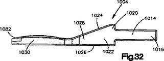
挿入部材1004(図30〜図32)は、分離装置1008の1つに連結可能である。部材1004は、作動装置1002の分離装置1008の1つの開口(図示せず)に挿入可能な連結端部1014を備える。端部1014は、1対の突起1016を備える。突起1016(図31)は、互いにほぼ平行に延びており、その間に経路1018を画定する。端部1014は、知られている方法で、部材1004を作動装置に連結させるように、作動装置1002の分離装置1008の開口(図示せず)に挿入される。部材1004は、知られている方法で分離装置1008から取り除くことができる。部材1004の端部1014は、部材を所望の作動装置に連結させるための任意の所望の構成を有することができると考えられる。
The insertion member 1004 (FIGS. 30-32) is connectable to one of the
突起1016(図30〜図32)は、部材1004の中心本体1022の第1の端部1020から延びる。中心本体1022は、上側表面1024と、中心本体の第1の端部1020から第2の端部1028まで延びる下側表面1026とを備える。突起1016は、上側表面1024に対してある角度で、下側表面1026にほぼ平行に延びる。突起1016は、上側表面1024および下側表面1026に対して任意の所定の角度で延びることができると考えられる。
The protrusion 1016 (FIGS. 30-32) extends from the
部材1004の挿入端部1030(図30〜図32)は、本体1022の第2の端部1028から延びる。挿入端部1030は、本体1022の上側表面1024に対してある角度で、突起1016とほぼ平行に延びる。挿入端部1030は、上側表面1024および突起1016に対して任意の所望の角度で延びることができると考えられる。
The insertion end 1030 (FIGS. 30-32) of the
挿入端部1030(図31)は、取付部材150または200を受ける凹部1034を備える。凹部1034は、そこを通して取付部材150または200を凹部に挿入し、凹部から取り除くことができる、開口端部1036を備えたほぼU字形状をしている。凹部1034は、開口端部1036から延びる第1の側壁1038および第2の側壁(図示せず)によって画定される。第1の側壁1038および第2の側壁は、後部壁面1042によって相互連結される。底部壁面1044は、第1の側壁1038、第2の側壁、および後部壁面1042にほぼ垂直に延びる。
The insertion end 1030 (FIG. 31) includes a
後部壁面1042は、本体1022に向かって延びる切り欠き1048を有する。溝1050は、第1の側壁1038および後部壁面1042の一部に形成されている。溝1050は、開口端部1036に隣接したところから切り欠き1048まで延びる。後部壁面1042の別の部分、および第2の側壁(図示せず)に形成された溝(図示せず)は、切り欠き1048から開口端部1038に隣接したところまで延びる。
The
第1の円形開口1056は底部壁面1044を通して延びており、凹部1034内に中心に配置されている。開口1056は、必要に応じて凹部1034から取付部材150または200を取り除くことを可能にする。第2のより小さな円形開口1058は、底部壁面1044を通して延び、切り欠き1048内に配置されている。
The first
ばね部材1060(図33)は、取付部材150または200を凹部内に保持するように、凹部1034内で受けられる。ばね部材160はほぼU字形をしており、基部1066から延びる1対の腕部1062、1064を備える。突起1068は、腕部1062、1064とは反対方向に基部1066から延びる。突起1068は、ばね部材1060を部材1004に連結するようにピン(図示せず)を受ける円形開口1070を有する。ピン(図示せず)は、ばね部材を部材1004に連結させるように、ばね部材1060の開口1070を通して部材1004の開口1058内に延びる。
The spring member 1060 (FIG. 33) is received in the
腕部1062は、取付部材を部材1004内に保持するように、取付部材150または200と係合する。腕部1062は、径方向内側に延びる突起1076を有する。突起1076は、取付部材を部材1004内に保持するように、取付部材150または200の凹部196または240の1つに延びる。腕部1064は、径方向内側に延びる突起1078を有する。突起1078は、取付部材を部材1004内に保持するように、取付部材150または200の凹部196または240の1つに延びる。
The
ばね部材1060は、開口端部1036を通して凹部1034内に挿入される。腕部1062、1064は、ばね1060が凹部1034内に挿入されているときに、第1の側壁1038の溝1050および第2の側壁(図示せず)の溝(図示せず)内に延びる。腕部1062、1064は互いに向かって移動する。腕部1062、1064が溝1050および第2の側壁(図示せず)の溝(図示せず)に隣接している場合、腕部は互いから離れるように移動する。
The
ばね1060が凹部1034内に挿入されると、腕部1062は溝1050内に延び、腕部1064は第2の側壁(図示せず)の溝(図示せず)内に延びる。ばね部材1060の突起1068の開口1070は、挿入端部1030の開口1058と位置合わせされている。ピン(図示せず)は、ばね部材を凹部1034内に保持するように、ばね部材1060の開口1070を通して、開口1058内に延びる。
When the
ばね1060の腕部1062と1064の間の取付部材150または200の挿入の際、腕部は凹部196または240が突起1076、1078と位置合わせされるまで、互いから離れるように径方向外側に移動する。凹部196または240が突起1076、1078と位置合わせされると、腕部1062、1064は取付部材150または200を挿入端部1030内に保持するように、凹部内で互いに向かって移動する。取付部材150または200は、ばね部材1060によって加えられる保持力を克服することによって、凹部1034から取り除くことができる。
Upon insertion of mounting
部材1004の挿入端部1030は凹部1082を備える。凹部1082は、凹部1034の開口端部1036の両側に配置されている。凹部1082は、椎骨12、14の1つの所望の深さへの取付部材150または200の挿入を保証するのに使用される。
The
案内部材1100(図30および図34)は、椎骨12、14内への取付部材150または200の挿入を案内するのに使用することができる。案内部材1100は、装置10と同様の形状をしており、楔形状をしていてもよい。案内部材1100は、第1の、遠位または後側端部1102と、反対側の第2の、近接または前側端部1104とを有する。案内部材1100はまた、第1の端部1102と第2の端部1104の間に延びる第1および第2の横側部1106、1108を備える。案内部材1100の上側表面1110は椎骨12と係合可能であり、下側表面1112は椎骨14と係合可能である。上側および下側表面1110、1112はそれぞれ、カッター450および/または490によって椎骨12、14内に切り込まれた溝と摺動可能に係合する複数のレール1114を有する。リブ1114は、第2の端部1104から第1の端部1102まで延びる。レール1114は、椎骨12、14内に溝を切り込むことができると考えられる。
Guide member 1100 (FIGS. 30 and 34) can be used to guide the insertion of
案内部材1100(図34)は、第1の端部1102に向かって第2の端部1104から延びる凹部1120を備える。案内部材1100の端部壁面1122および側壁1124は、凹部1120を画定する。端部壁面1122および側壁1124は、椎骨12、14に対する挿入部材の移動を案内するように、挿入部材1004と係合可能である。2つが図30および図34に示された4つのフランジ1128は、端部壁面1122および側壁1124から凹部1120内に延びる。フランジ1128は、椎骨12、14の1つ内への取付部材150または200のさらなる挿入を防ぐため、挿入部材1004上の凹部1082内に延び、挿入部材1004と係合する。
The guide member 1100 (FIG. 34) includes a
案内部材1100(図30)は、第1の横側部1106から凹部1120内に延びる通路1132を備える。通路1134は、第2の横側部1108から凹部1120内に延びる。通路1132、1134は同軸である。案内部材1100は、後側部1102から凹部1120内に延びる中心円筒形通路1136を備える。案内部材1100はまた、第1の端部1102と第2の端部1104の間に延びる2つの横円筒形通路1138を備える。通路1136、1138は互いにほぼ平行に延びる。通路1138は、通路1132、1134を横切る。通路1132、1134、1136、1138は任意の所望の構成を有することができることが考えられる。通路1132、1134、1136、1138は、案内部材1100が椎骨12と14の間に適切に位置決めされたかどうかを判断するように、蛍光透視鏡などの所望の画像化システムを使用して見ることができる。案内部材1100が椎骨12と14の間に適切に位置決めされると、通路1132、1134、1136、1138は、画像化システムによって作り出された画像内に円として見える可能性がある。案内部材1100が適切に位置決めされないと、通路1132、1134、1136、1138は画像内に楕円として見える可能性がある。通路1136は、挿入ロッド330を受けるようにねじ切りすることができる。
The guide member 1100 (FIG. 30) includes a
挿入ロッド330は、通路1136とねじ係合可能である。挿入ロッド330は、椎骨12と14の間に案内部材1100を挿入するのに使用することができる。挿入ロッド330は、案内組立体351の通路398、または案内組立体851の通路898内に挿入することができる。案内機構390、または案内機構890はその後、凹部408またはスロット920がシャフト352または852上で径方向に延びる位置決め部材362または862を受けるまで、マーカー350または850のシャフト352または852上で入れ子状になっている。したがって、案内部材1100は椎骨12、14と所望のように位置合わせされている。挿入ロッド330は、案内部材1100を椎骨12と14の間で移動させるように、木槌またはハンマで打つことができる。
The
脊柱安定装置1200(図30および図35)は、取付部材150または200の挿入中の、椎骨間の相対移動を防ぐように、椎骨12、14と係合可能である。脊柱安定装置1200は、中心通路1204を画定するほぼ四角形の本体1202を備える。上側取付シャフト1206は、本体1202の上壁部1208の中心部から延びる。上側取付シャフト1206は、脊柱安定装置1200を椎骨12に連結させるように、椎骨12と係合可能である。下側取付シャフト1210は、本体1202の下壁部1212の横外側部から延びる。下側取付シャフト1210は、脊柱安定装置1200を下側椎骨14と連結させるように、椎骨14と係合可能である。したがって、脊柱安定装置1200は、椎骨と連結された場合に、椎骨12と14の間の相対移動を防ぐ。
The spinal stabilizer 1200 (FIGS. 30 and 35) is engageable with the
脊柱安定装置1200は、本体1202の側壁1218の開口1216を備える。開口1216は、脊柱安定装置が椎骨12、14に連結された場合に、案内部材1100の通路1138と位置合わせされる。脊柱安定装置1200の開口1216、および案内部材1100の通路1138は、位置決めロッド1230(図30)を受ける。各ロッド1230は、第1の直径を有する第1の軸方向端部1232を備える。各ロッド1230は、第1の直径より大きい第2の直径を有する第2の軸方向端部1234を備える。径方向に延びる表面1236は、第1の軸方向端部1232と第2の軸方向端部1234の間に延びる。第1の軸方向端部1232は、径方向に延びる表面1236が脊柱安定装置と係合するまで、脊柱安定装置1200の開口1216および案内部材1100の通路1138内に挿入される。
The
部材1250(図30)は、ロッド1230の第2の端部1234を受ける開口1252を有する。部材1250は、作動装置1002の分離装置1008の1つと連結可能である。部材1250は、作動装置1002の分離装置1008の1つに開口(図示せず)内に挿入可能である連結端部1254を備える。端部1254は1対の突起1256を備える。突起1256は、互いにほぼ平行に延びており、その間に経路1258を画定する。端部1254は、知られている方法で部材1250を作動装置に連結させるように、作動装置1002の分離装置1008の開口(図示せず)に挿入される。部材1250は、知られている方法で、分離装置1008から取り除くことができる。部材1250の端部1254は、部材を所望の作動装置に連結させるように任意の所望の構成を有することができると考えられる。
Member 1250 (FIG. 30) has an
突起1256は、部材1250の本体1262の第1の側部1260から延びる。本体1262は、上端部1264を備えたほぼU字形をしている。開口1252は、本体1262の上端部1264を通して延びる。
The
取付部材150または200の1つが椎骨12、14の1つに連結されると、案内部材1100は椎骨間に挿入される。ロッド1230は、脊柱安定装置1200の開口1216を通して配置される。脊柱安定装置1200は、案内部材1100の通路1138内に延びるロッド1230で椎骨12、14に連結される。挿入部材1004および部材1250は、作動装置1002の分離装置1008に連結される。取付部材150または200は、挿入部材1004に連結される。挿入部材1004は、脊柱安定装置1200の開口1204を通して案内部材1100の凹部1120内に配置される。挿入部材1004は、案内部材の凹部1120内に挿入され、それによって挿入部材は凹部1120を画定する端部壁面1122と係合する。挿入部材1004が脊柱案内装置1200の開口1212を通して挿入されると、部材1250はロッド1230の上に配置され、それによってロッドは部材1250の開口1252を通して延びる。したがって、部材1250は椎骨12、14に対して移動することができない。
When one of the
挿入部材1004は、取付部材150または200を椎骨12に連結させるように、作動装置1002によって部材1250から離れるように移動される。脊柱安定装置1200は、椎骨12と14の間の相対移動を防ぐ。取付部材150または200が椎骨12に連結された後、作動装置1002は旋回させることができ、他の取付部材150または200は椎骨14に連結することができる。
The
本発明の上記説明は、様々な変形形態、変更形態、および応用例が可能であり、これは頭記の特許請求の範囲の同等物の意味および範囲内であると解釈することを意図していることを理解されたい。現時点で開示されている実施形態は、全ての面において制限的なものではなく例示的なものであると考えられる。本発明の範囲は、前述の説明ではなく頭記の特許請求の範囲によって示され、その同等物の意味および範囲内にある全ての変更はこれに含まれることを意図している。 The above description of the invention is capable of various variations, modifications, and applications, which are intended to be construed within the meaning and scope of the equivalents of the appended claims. I want you to understand. The embodiments disclosed at the present time are considered to be illustrative rather than restrictive in all aspects. The scope of the invention is indicated by the appended claims rather than by the foregoing description, and all changes that come within the meaning and range of equivalents are intended to be embraced therein.
Claims (47)
第1の端部と、
対向する第2の端部と、
前記第1の端部と第2の端部との間に延びる第1の横側部および第2の横側部と、
前記脊柱の第1の椎骨と係合可能である外側表面と、
前記装置を前記第1の椎骨に連結させるように、前記第1の椎骨と係合可能である前記外側表面から延びる第1のレールであって、当該第1のレールは、前記第1の端部に隣接して前記外側表面から第1の距離で延び、また前記第1のレールは、前記第2の端部に隣接して前記外側表面から第2の距離で延び、前記第2の距離は前記第1の距離より大きい、第1のレールと、
前記外側表面から延び、前記第1のレールに対してほぼ横に延びる長さを有する第2のレールと、
第1の表面および第2の表面を有する弾力性のある芯と、
前記弾力性のある芯の前記第1の表面に連結される第1の保持装置であって、当該第1の保持装置は、前記脊柱の前記第1の椎骨と係合可能である前記外側表面、および前記弾力性のある芯の前記第1の表面に面する内側表面を有する、第1の保持装置と、
前記弾力性のある芯の前記第2の表面に連結される第2の保持装置であって、当該第2の保持装置は、前記脊柱の前記第2の椎骨と係合可能である前記外側表面、および前記弾力性のある芯の前記第2の表面に面する内側表面を有する、第2の保持装置と、を備えた装置。An apparatus for replacing an intervertebral disc in the spinal column, wherein the intervertebral disc is disposed between a first vertebra and a second vertebra;
A first end;
An opposing second end;
A first lateral side and a second lateral side extending between the first end and the second end;
An outer surface engageable with a first vertebra of the spinal column;
A first rail extending from the outer surface that is engageable with the first vertebra to couple the device to the first vertebra, the first rail being the first end. A first distance extending from the outer surface adjacent the portion and the first rail extends a second distance from the outer surface adjacent to the second end; and the second distance. Is a first rail greater than the first distance;
A second rail extending from the outer surface and having a length extending generally transverse to the first rail;
A resilient core having a first surface and a second surface;
A first retention device coupled to the first surface of the resilient core, the first retention device being engageable with the first vertebra of the spinal column; And a first holding device having an inner surface facing the first surface of the resilient core;
A second retention device coupled to the second surface of the resilient core, the second retention device being engageable with the second vertebra of the spinal column And a second holding device having an inner surface facing the second surface of the resilient core.
第1の端部と、
対向する第2の端部と、
前記第1の端部と前記第2の端部との間に延びる、第1の横側部および第2の横側部と、
前記脊柱の第1の椎骨と係合可能である外側表面と、
前記外側表面から延び、前記装置を前記第1の椎骨に連結させるように、前記第1の椎骨と係合可能である、第1の突起であって、前記第1の突起は前記外側表面から第1の距離で延びる、第1の突起と、
前記外側表面から延び、前記装置を前記第1の椎骨に連結させるように、前記第1の椎骨と係合可能である、第2の突起であって、当該第2の突起は、前記第1の突起よりも前記第2の端部に対してより近くに配置され、前記第2の突起は、前記第1の距離よりも大きい第2の距離で前記外側表面から延びる、第2の突起と、
第1のレールであって、前記第1および第2の端部に対してほぼ横に延びる長さを有する前記第1のレールと、
第2のレールであって、前記第1のレールに対してほぼ横に延びる長さを有する前記第2のレールと、
第1の表面および第2の表面を有する弾力性のある芯と、
前記弾力性のある芯の前記第1の表面に連結される第1の保持装置であって、当該第1の保持装置は、前記脊柱の前記第1の椎骨と係合可能である前記外側表面、および前記弾力性のある芯の前記第1の表面に面する内側表面を有する、第1の保持装置と、
前記弾力性のある芯の前記第2の表面に連結される第2の保持装置であって、当該第2の保持装置は、前記脊柱の前記第2の椎骨と係合可能である前記外側表面、および前記弾力性のある芯の前記第2の表面に面する内側表面を有する、第2の保持装置と、を備えた装置。An apparatus for replacing an intervertebral disc in the spinal column, wherein the intervertebral disc is disposed between a first vertebra and a second vertebra;
A first end;
An opposing second end;
A first lateral side and a second lateral side extending between the first end and the second end;
An outer surface engageable with a first vertebra of the spinal column;
A first protrusion extending from the outer surface and engageable with the first vertebra to couple the device to the first vertebra, the first protrusion from the outer surface A first protrusion extending at a first distance;
A second protrusion extending from the outer surface and engageable with the first vertebra to couple the device to the first vertebra, the second protrusion being the first protrusion A second protrusion disposed closer to the second end than the second protrusion, the second protrusion extending from the outer surface at a second distance greater than the first distance; ,
A first rail having a length extending substantially transverse to the first and second ends ;
A second rail, the second rail having a length extending substantially transverse to the first rail;
A resilient core having a first surface and a second surface;
A first retention device coupled to the first surface of the resilient core, the first retention device being engageable with the first vertebra of the spinal column; And a first holding device having an inner surface facing the first surface of the resilient core;
A second retention device coupled to the second surface of the resilient core, the second retention device being engageable with the second vertebra of the spinal column. And a second holding device having an inner surface facing the second surface of the resilient core.
第1の端部と、
対向する第2の端部と、
前記第1の端部と第2の端部の間に延びる第1の横側部および第2の横側部と、
前記脊柱の第1の椎骨と係合可能である外側表面と、
前記外側表面から延びる第1のレールであって、前記第1のレールは、前記第1および第2の端部に対してほぼ横に延びる長さを有し、前記第1のレールは、外側表面から延びる複数の突起を備える前記第1のレールと、
前記外側表面から延びる第2のレールであって、前記第2のレールは、前記第1のレールに対してほぼ横に延びる長さを有する前記第2のレールと、
第1の表面および第2の表面を有する弾力性のある芯と、
前記弾力性のある芯の前記第1の表面に連結される第1の保持装置であって、当該第1の保持装置は、前記脊柱の前記第1の椎骨と係合可能である前記外側表面、および前記弾力性のある芯の前記第1の表面に面する内側表面を有する、第1の保持装置と、
前記弾力性のある芯の前記第2の表面に連結される第2の保持装置であって、当該第2の保持装置は、前記脊柱の前記第2の椎骨と係合可能である前記外側表面、および前記弾力性のある芯の前記第2の表面に面する内側表面を有する、第2の保持装置と、を備えた装置。An apparatus for replacing an intervertebral disc in the spinal column, wherein the intervertebral disc is disposed between a first vertebra and a second vertebra;
A first end;
An opposing second end;
A first lateral side and a second lateral side extending between the first end and the second end;
An outer surface engageable with a first vertebra of the spinal column;
A first rail extending from the outer surface , wherein the first rail has a length extending generally transverse to the first and second ends, the first rail being an outer side The first rail comprising a plurality of protrusions extending from a surface;
A second rail extending from the outer surface, the second rail having a length extending substantially transverse to the first rail;
A resilient core having a first surface and a second surface;
A first retention device coupled to the first surface of the resilient core, the first retention device being engageable with the first vertebra of the spinal column; And a first holding device having an inner surface facing the first surface of the resilient core;
A second retention device coupled to the second surface of the resilient core, the second retention device being engageable with the second vertebra of the spinal column And a second holding device having an inner surface facing the second surface of the resilient core.
Applications Claiming Priority (3)
| Application Number | Priority Date | Filing Date | Title |
|---|---|---|---|
| US63362004P | 2004-12-06 | 2004-12-06 | |
| US60/633,620 | 2004-12-06 | ||
| PCT/US2005/044088 WO2006062960A2 (en) | 2004-12-06 | 2005-12-06 | Method and apparatus for replacing a spinal disc |
Publications (3)
| Publication Number | Publication Date |
|---|---|
| JP2008522737A JP2008522737A (en) | 2008-07-03 |
| JP2008522737A5 JP2008522737A5 (en) | 2009-01-15 |
| JP5080981B2 true JP5080981B2 (en) | 2012-11-21 |
Family
ID=36578484
Family Applications (1)
| Application Number | Title | Priority Date | Filing Date |
|---|---|---|---|
| JP2007545558A Expired - Fee Related JP5080981B2 (en) | 2004-12-06 | 2005-12-06 | Device to replace the intervertebral disc |
Country Status (8)
| Country | Link |
|---|---|
| US (1) | US7850697B2 (en) |
| EP (1) | EP1824430A4 (en) |
| JP (1) | JP5080981B2 (en) |
| KR (1) | KR100939507B1 (en) |
| CN (1) | CN101111208B (en) |
| AU (2) | AU2005314224B2 (en) |
| CA (1) | CA2591326C (en) |
| WO (1) | WO2006062960A2 (en) |
Families Citing this family (61)
| Publication number | Priority date | Publication date | Assignee | Title |
|---|---|---|---|---|
| ATE388677T1 (en) | 1999-07-02 | 2008-03-15 | Spine Solutions Inc | INTERVERBARY IMPLANT |
| DE59914213D1 (en) | 1999-09-14 | 2007-04-05 | Spine Solutions Inc | INSERT INSTRUMENT FOR A INTERMEDIATE IMPLANT |
| FR2824261B1 (en) | 2001-05-04 | 2004-05-28 | Ldr Medical | INTERVERTEBRAL DISC PROSTHESIS AND IMPLEMENTATION METHOD AND TOOLS |
| FR2831049B1 (en) | 2001-10-18 | 2004-08-13 | Ldr Medical | PLATE FOR OSTEOSYNTHESIS DEVICE AND PRE-ASSEMBLY METHOD |
| FR2831048B1 (en) | 2001-10-18 | 2004-09-17 | Ldr Medical | PROGRESSIVE APPROACH OSTEOSYNTHESIS DEVICE AND PRE-ASSEMBLY PROCESS |
| CA2451147C (en) * | 2002-04-05 | 2013-07-30 | Nippon Steel Corporation | Pearlitic steel rail excellent in wear resistance and ductility and method for producing the same |
| US8038713B2 (en) | 2002-04-23 | 2011-10-18 | Spinecore, Inc. | Two-component artificial disc replacements |
| US20080027548A9 (en) | 2002-04-12 | 2008-01-31 | Ferree Bret A | Spacerless artificial disc replacements |
| US6706068B2 (en) | 2002-04-23 | 2004-03-16 | Bret A. Ferree | Artificial disc replacements with natural kinematics |
| FR2846550B1 (en) | 2002-11-05 | 2006-01-13 | Ldr Medical | INTERVERTEBRAL DISC PROSTHESIS |
| US7204852B2 (en) | 2002-12-13 | 2007-04-17 | Spine Solutions, Inc. | Intervertebral implant, insertion tool and method of inserting same |
| US6908484B2 (en) | 2003-03-06 | 2005-06-21 | Spinecore, Inc. | Cervical disc replacement |
| US7491204B2 (en) | 2003-04-28 | 2009-02-17 | Spine Solutions, Inc. | Instruments and method for preparing an intervertebral space for receiving an artificial disc implant |
| US7803162B2 (en) | 2003-07-21 | 2010-09-28 | Spine Solutions, Inc. | Instruments and method for inserting an intervertebral implant |
| US9844442B2 (en) | 2003-07-31 | 2017-12-19 | Globus Medical, Inc. | Prosthetic spinal disc replacement and methods thereof |
| FR2865629B1 (en) | 2004-02-04 | 2007-01-26 | Ldr Medical | INTERVERTEBRAL DISC PROSTHESIS |
| ES2547532T3 (en) | 2004-02-04 | 2015-10-07 | Ldr Medical | Intervertebral disc prosthesis |
| FR2869528B1 (en) | 2004-04-28 | 2007-02-02 | Ldr Medical | INTERVERTEBRAL DISC PROSTHESIS |
| US7547309B2 (en) * | 2004-09-23 | 2009-06-16 | Spine Solutions, Inc. | Distractor for lumbar insertion instrument |
| ATE524121T1 (en) | 2004-11-24 | 2011-09-15 | Abdou Samy | DEVICES FOR PLACING AN ORTHOPEDIC INTERVERTEBRAL IMPLANT |
| FR2879436B1 (en) | 2004-12-22 | 2007-03-09 | Ldr Medical | INTERVERTEBRAL DISC PROSTHESIS |
| FR2887762B1 (en) | 2005-06-29 | 2007-10-12 | Ldr Medical Soc Par Actions Si | INTERVERTEBRAL DISC PROSTHESIS INSERTION INSTRUMENTATION BETWEEN VERTEBRATES |
| FR2891135B1 (en) | 2005-09-23 | 2008-09-12 | Ldr Medical Sarl | INTERVERTEBRAL DISC PROSTHESIS |
| US20070118134A1 (en) * | 2005-10-17 | 2007-05-24 | Jeffery Thramann | Surgical Tools and method to facilitate spinal surgery |
| US8251902B2 (en) | 2005-10-17 | 2012-08-28 | Lanx, Inc. | Pedicle guided retractor system |
| FR2893838B1 (en) * | 2005-11-30 | 2008-08-08 | Ldr Medical Soc Par Actions Si | PROSTHESIS OF INTERVERTEBRAL DISC AND INSTRUMENTATION OF INSERTION OF THE PROSTHESIS BETWEEN VERTEBRATES |
| US20070203580A1 (en) * | 2006-02-24 | 2007-08-30 | Paonan Biotech Co., Ltd. | Intervertebral filling |
| US20110022177A1 (en) * | 2006-02-24 | 2011-01-27 | Paonan Biotech Co., Ltd. | Intervertebral filling for cervical vertebrae |
| US20070213826A1 (en) * | 2006-03-08 | 2007-09-13 | Seaspine, Inc. | Intervertebral spacer and insertion tool providing multiple angles of insertion |
| US8409290B2 (en) * | 2006-03-08 | 2013-04-02 | Seaspine, Inc. | Interbody device for spinal applications |
| US20080071379A1 (en) * | 2006-05-10 | 2008-03-20 | Mark Rydell | Intervertebral disc replacement |
| AU2007276755A1 (en) | 2006-07-24 | 2008-01-31 | Spine Solutions, Inc. | Intervertebral implant with keel |
| CA2659024A1 (en) | 2006-07-31 | 2008-02-07 | Synthes (Usa) | Drilling/milling guide and keel cut preparation system |
| AU2007297546B9 (en) | 2006-09-21 | 2013-06-20 | Spinecore, Inc. | Intervertebral disc implants and tooling |
| US8029569B2 (en) * | 2006-11-20 | 2011-10-04 | International Spinal Innovations, Llc | Implantable spinal disk |
| US8465546B2 (en) | 2007-02-16 | 2013-06-18 | Ldr Medical | Intervertebral disc prosthesis insertion assemblies |
| FR2916956B1 (en) | 2007-06-08 | 2012-12-14 | Ldr Medical | INTERSOMATIC CAGE, INTERVERTEBRAL PROSTHESIS, ANCHORING DEVICE AND IMPLANTATION INSTRUMENTATION |
| US20090177195A1 (en) * | 2008-01-07 | 2009-07-09 | Spinal Kinetics, Inc. | Alignable Tools for Preparing an Intervertebral Site for Implanting a Prosthetic Intervertebral Disc and a Method for Using Those Tools |
| FR2932374B1 (en) * | 2008-06-13 | 2010-06-04 | Spinevision | DEVICE FOR ESTABLISHING AN ANATOMICAL MARK OF AN INTERVERTEBRAL DISC. |
| JP5681639B2 (en) | 2008-12-22 | 2015-03-11 | ジンテス ゲゼルシャフト ミット ベシュレンクテル ハフツング | Orthopedic implant with flexible keel |
| US8764806B2 (en) | 2009-12-07 | 2014-07-01 | Samy Abdou | Devices and methods for minimally invasive spinal stabilization and instrumentation |
| US8449616B2 (en) * | 2011-03-15 | 2013-05-28 | Axiomed Spine Corporation | Apparatus for replacing a damaged spinal disc |
| US8845728B1 (en) | 2011-09-23 | 2014-09-30 | Samy Abdou | Spinal fixation devices and methods of use |
| US20130226240A1 (en) | 2012-02-22 | 2013-08-29 | Samy Abdou | Spinous process fixation devices and methods of use |
| US20130261746A1 (en) * | 2012-03-28 | 2013-10-03 | Linares Medical Devices, Llc | Implantable inter-vertebral disk having upper and lower layers of a metal exhibiting bone fusing characteristics and which sandwich therebetween a soft plastic cushioning disc for providing dynamic properties mimicking that of a natural inter-vertebral disc |
| US20130261747A1 (en) * | 2012-03-30 | 2013-10-03 | Christophe Geisert | ALIF Spinal Implant |
| US9198767B2 (en) | 2012-08-28 | 2015-12-01 | Samy Abdou | Devices and methods for spinal stabilization and instrumentation |
| US9320617B2 (en) | 2012-10-22 | 2016-04-26 | Cogent Spine, LLC | Devices and methods for spinal stabilization and instrumentation |
| EP3154479A4 (en) * | 2014-06-12 | 2017-07-05 | Globus Medical, Inc. | Prosthetic spinal disc replacement and methods thereof |
| EP3103417B1 (en) * | 2015-06-10 | 2018-01-31 | Biedermann Technologies GmbH & Co. KG | Intervertebral implant and system of an intervertebral implant and an instrument for inserting the intervertebral implant |
| US10857003B1 (en) | 2015-10-14 | 2020-12-08 | Samy Abdou | Devices and methods for vertebral stabilization |
| US10617531B2 (en) | 2015-10-26 | 2020-04-14 | K2M, Inc. | Cervical disc and instrumentation |
| EP3448318A4 (en) | 2016-04-26 | 2019-12-04 | K2M, Inc. | Orthopedic implant with integrated core |
| US10744000B1 (en) | 2016-10-25 | 2020-08-18 | Samy Abdou | Devices and methods for vertebral bone realignment |
| US10973648B1 (en) | 2016-10-25 | 2021-04-13 | Samy Abdou | Devices and methods for vertebral bone realignment |
| DE102016124877B4 (en) * | 2016-12-19 | 2019-02-07 | Ngmedical Gmbh | Intervertebral disc prosthesis and method for producing a disc prosthesis |
| CN110831548B (en) * | 2017-05-22 | 2022-07-22 | 公理医学有限公司 | System and method for performing a disc replacement procedure through a lateral approach |
| US10765527B2 (en) | 2017-09-29 | 2020-09-08 | Axiomed, LLC | Artificial disk with sensors |
| US11179248B2 (en) | 2018-10-02 | 2021-11-23 | Samy Abdou | Devices and methods for spinal implantation |
| US11779378B2 (en) | 2019-01-11 | 2023-10-10 | Medcom Advisors, Llc | Self-aligning plating system and method |
| US20220008211A1 (en) * | 2020-07-08 | 2022-01-13 | Ctl Medical Corporation | Cage with keel |
Family Cites Families (26)
| Publication number | Priority date | Publication date | Assignee | Title |
|---|---|---|---|---|
| CA1333209C (en) * | 1988-06-28 | 1994-11-29 | Gary Karlin Michelson | Artificial spinal fusion implants |
| US5961554A (en) * | 1996-12-31 | 1999-10-05 | Janson; Frank S | Intervertebral spacer |
| FR2764795B1 (en) * | 1997-06-19 | 1999-09-10 | Sarl Sra | INTERSOMATIC RACHIS IMMOBILIZATION CAGE AND ASSOCIATED ANCILLARY MATERIAL |
| US5893889A (en) | 1997-06-20 | 1999-04-13 | Harrington; Michael | Artificial disc |
| DE69916280T2 (en) * | 1998-08-03 | 2005-05-25 | Synthes Ag Chur, Chur | INTERVERTEBRAL allograft DISTANZSTÜCK |
| US6929662B1 (en) * | 1999-02-04 | 2005-08-16 | Synthes (Usa) | End member for a bone fusion implant |
| US6607558B2 (en) * | 2001-07-03 | 2003-08-19 | Axiomed Spine Corporation | Artificial disc |
| US7635368B2 (en) * | 2001-07-16 | 2009-12-22 | Spinecore, Inc. | Intervertebral spacer device having simultaneously engageable angled perimeters for manipulation using a surgical tool |
| FR2831796B1 (en) * | 2001-11-06 | 2003-12-26 | Ldr Medical | BONE ANCHORING DEVICE FOR PROSTHESIS |
| US6740118B2 (en) * | 2002-01-09 | 2004-05-25 | Sdgi Holdings, Inc. | Intervertebral prosthetic joint |
| US6726720B2 (en) | 2002-03-27 | 2004-04-27 | Depuy Spine, Inc. | Modular disc prosthesis |
| US6793678B2 (en) * | 2002-06-27 | 2004-09-21 | Depuy Acromed, Inc. | Prosthetic intervertebral motion disc having dampening |
| DE10247762A1 (en) * | 2002-10-14 | 2004-04-22 | Waldemar Link (Gmbh & Co.) | Intervertebral prosthesis |
| CA2502292C (en) * | 2002-10-31 | 2011-07-26 | Spinal Concepts, Inc. | Movable disc implant |
| US20040093087A1 (en) * | 2002-11-05 | 2004-05-13 | Ferree Bret A. | Fluid-filled artificial disc replacement (ADR) |
| US20040158254A1 (en) * | 2003-02-12 | 2004-08-12 | Sdgi Holdings, Inc. | Instrument and method for milling a path into bone |
| JP2006521899A (en) * | 2003-03-31 | 2006-09-28 | デピュイ・スパイン・インコーポレイテッド | Method and apparatus for inserting an artificial disc |
| US7060097B2 (en) | 2003-03-31 | 2006-06-13 | Depuy Spine, Inc. | Method and apparatus for implant stability |
| US7771478B2 (en) | 2003-04-04 | 2010-08-10 | Theken Spine, Llc | Artificial disc prosthesis |
| KR100754570B1 (en) | 2003-04-07 | 2007-09-05 | 서비텍, 인크. | Prosthetic joint of cervical intervertebral for a cervical spine |
| US7491204B2 (en) * | 2003-04-28 | 2009-02-17 | Spine Solutions, Inc. | Instruments and method for preparing an intervertebral space for receiving an artificial disc implant |
| FR2856587B1 (en) * | 2003-06-26 | 2006-02-24 | Scient X | DISCRETE PROSTHESIS FOR CERVICAL VERTEBRATES WITH CONTROLLED DEBATMENT |
| FR2858546B1 (en) * | 2003-08-04 | 2006-04-28 | Spine Next Sa | INTERVERTEBRAL DISC PROSTHESIS |
| US20050038511A1 (en) * | 2003-08-15 | 2005-02-17 | Martz Erik O. | Transforaminal lumbar interbody fusion (TLIF) implant, surgical procedure and instruments for insertion of spinal implant in a spinal disc space |
| US7695517B2 (en) * | 2003-12-10 | 2010-04-13 | Axiomed Spine Corporation | Apparatus for replacing a damaged spinal disc |
| US20070255414A1 (en) * | 2006-05-01 | 2007-11-01 | Sdgi Holdings, Inc. | Intervertebral implants with one or more covers and methods of use |
-
2005
- 2005-12-06 CA CA2591326A patent/CA2591326C/en not_active Expired - Fee Related
- 2005-12-06 AU AU2005314224A patent/AU2005314224B2/en not_active Ceased
- 2005-12-06 KR KR1020077015593A patent/KR100939507B1/en not_active IP Right Cessation
- 2005-12-06 EP EP05853099A patent/EP1824430A4/en not_active Withdrawn
- 2005-12-06 CN CN200580047632.2A patent/CN101111208B/en not_active Expired - Fee Related
- 2005-12-06 JP JP2007545558A patent/JP5080981B2/en not_active Expired - Fee Related
- 2005-12-06 US US11/294,925 patent/US7850697B2/en active Active - Reinstated
- 2005-12-06 WO PCT/US2005/044088 patent/WO2006062960A2/en active Application Filing
-
2010
- 2010-01-08 AU AU2010200083A patent/AU2010200083B2/en not_active Ceased
Also Published As
| Publication number | Publication date |
|---|---|
| WO2006062960A2 (en) | 2006-06-15 |
| CA2591326A1 (en) | 2006-06-15 |
| CA2591326C (en) | 2011-08-09 |
| JP2008522737A (en) | 2008-07-03 |
| US7850697B2 (en) | 2010-12-14 |
| WO2006062960A3 (en) | 2006-09-28 |
| CN101111208A (en) | 2008-01-23 |
| KR20070093989A (en) | 2007-09-19 |
| AU2005314224B2 (en) | 2009-10-08 |
| CN101111208B (en) | 2014-03-26 |
| AU2010200083A1 (en) | 2010-01-28 |
| EP1824430A2 (en) | 2007-08-29 |
| KR100939507B1 (en) | 2010-02-03 |
| EP1824430A4 (en) | 2012-10-24 |
| AU2005314224A1 (en) | 2006-06-15 |
| US20060149273A1 (en) | 2006-07-06 |
| AU2010200083B2 (en) | 2012-09-13 |
Similar Documents
| Publication | Publication Date | Title |
|---|---|---|
| JP5080981B2 (en) | Device to replace the intervertebral disc | |
| JP4750042B2 (en) | Equipment replaced with a damaged spinal disc | |
| US8002831B2 (en) | Modular lateral expansion device | |
| US20180140431A1 (en) | Artificial cervical and lumbar discs, disc plate insertion gun for performing sequential single plate intervertebral implantation enabling symmetric bi-disc plate alignment for interplate mobile core placement | |
| US8506645B2 (en) | Tibial augments for use with knee joint prostheses | |
| AU2013215075B2 (en) | Intervertebral disc prosthesis and method | |
| US20080306557A1 (en) | Inserter for a spinal implant | |
| JP2008516686A (en) | Intervertebral implant and method related thereto | |
| KR20070098801A (en) | Intervertebral prosthesis | |
| NZ518892A (en) | Modular anatomic fusion device for promoting fusion of adjacent vertebral bodies. | |
| JP2004267746A (en) | Pivoted tensiometer for measuring tension in intervertebral disc space | |
| US20040133132A1 (en) | Interdiscal tensiometer apparatus and method of use | |
| JP2021513394A (en) | Modular adjustable corpectomy cage | |
| WO2009045225A1 (en) | Surgical instrument system | |
| WO2006118802A1 (en) | Method and device for stabilization of prosthetic devices |
Legal Events
| Date | Code | Title | Description |
|---|---|---|---|
| A521 | Request for written amendment filed |
Free format text: JAPANESE INTERMEDIATE CODE: A523 Effective date: 20081117 |
|
| A621 | Written request for application examination |
Free format text: JAPANESE INTERMEDIATE CODE: A621 Effective date: 20081117 |
|
| A131 | Notification of reasons for refusal |
Free format text: JAPANESE INTERMEDIATE CODE: A131 Effective date: 20110426 |
|
| A521 | Request for written amendment filed |
Free format text: JAPANESE INTERMEDIATE CODE: A523 Effective date: 20110711 |
|
| RD04 | Notification of resignation of power of attorney |
Free format text: JAPANESE INTERMEDIATE CODE: A7424 Effective date: 20110915 |
|
| A131 | Notification of reasons for refusal |
Free format text: JAPANESE INTERMEDIATE CODE: A131 Effective date: 20120116 |
|
| A601 | Written request for extension of time |
Free format text: JAPANESE INTERMEDIATE CODE: A601 Effective date: 20120416 |
|
| A602 | Written permission of extension of time |
Free format text: JAPANESE INTERMEDIATE CODE: A602 Effective date: 20120423 |
|
| A521 | Request for written amendment filed |
Free format text: JAPANESE INTERMEDIATE CODE: A523 Effective date: 20120711 |
|
| TRDD | Decision of grant or rejection written | ||
| A01 | Written decision to grant a patent or to grant a registration (utility model) |
Free format text: JAPANESE INTERMEDIATE CODE: A01 Effective date: 20120803 |
|
| A01 | Written decision to grant a patent or to grant a registration (utility model) |
Free format text: JAPANESE INTERMEDIATE CODE: A01 |
|
| A61 | First payment of annual fees (during grant procedure) |
Free format text: JAPANESE INTERMEDIATE CODE: A61 Effective date: 20120831 |
|
| FPAY | Renewal fee payment (event date is renewal date of database) |
Free format text: PAYMENT UNTIL: 20150907 Year of fee payment: 3 |
|
| R150 | Certificate of patent or registration of utility model |
Free format text: JAPANESE INTERMEDIATE CODE: R150 |
|
| LAPS | Cancellation because of no payment of annual fees |