JP5044563B2 - Cell-free in vitro transcription and translation of membrane proteins in tethered planar lipid layers - Google Patents
Cell-free in vitro transcription and translation of membrane proteins in tethered planar lipid layers Download PDFInfo
- Publication number
- JP5044563B2 JP5044563B2 JP2008536951A JP2008536951A JP5044563B2 JP 5044563 B2 JP5044563 B2 JP 5044563B2 JP 2008536951 A JP2008536951 A JP 2008536951A JP 2008536951 A JP2008536951 A JP 2008536951A JP 5044563 B2 JP5044563 B2 JP 5044563B2
- Authority
- JP
- Japan
- Prior art keywords
- membrane
- protein
- tethered
- cell
- tag
- Prior art date
- Legal status (The legal status is an assumption and is not a legal conclusion. Google has not performed a legal analysis and makes no representation as to the accuracy of the status listed.)
- Expired - Fee Related
Links
Images


Classifications
-
- C—CHEMISTRY; METALLURGY
- C12—BIOCHEMISTRY; BEER; SPIRITS; WINE; VINEGAR; MICROBIOLOGY; ENZYMOLOGY; MUTATION OR GENETIC ENGINEERING
- C12P—FERMENTATION OR ENZYME-USING PROCESSES TO SYNTHESISE A DESIRED CHEMICAL COMPOUND OR COMPOSITION OR TO SEPARATE OPTICAL ISOMERS FROM A RACEMIC MIXTURE
- C12P21/00—Preparation of peptides or proteins
- C12P21/02—Preparation of peptides or proteins having a known sequence of two or more amino acids, e.g. glutathione
-
- C—CHEMISTRY; METALLURGY
- C07—ORGANIC CHEMISTRY
- C07K—PEPTIDES
- C07K1/00—General methods for the preparation of peptides, i.e. processes for the organic chemical preparation of peptides or proteins of any length
- C07K1/04—General methods for the preparation of peptides, i.e. processes for the organic chemical preparation of peptides or proteins of any length on carriers
- C07K1/042—General methods for the preparation of peptides, i.e. processes for the organic chemical preparation of peptides or proteins of any length on carriers characterised by the nature of the carrier
-
- C—CHEMISTRY; METALLURGY
- C07—ORGANIC CHEMISTRY
- C07K—PEPTIDES
- C07K17/00—Carrier-bound or immobilised peptides; Preparation thereof
- C07K17/14—Peptides being immobilised on, or in, an inorganic carrier
Landscapes
- Chemical & Material Sciences (AREA)
- Organic Chemistry (AREA)
- Life Sciences & Earth Sciences (AREA)
- Health & Medical Sciences (AREA)
- Molecular Biology (AREA)
- General Health & Medical Sciences (AREA)
- Genetics & Genomics (AREA)
- Proteomics, Peptides & Aminoacids (AREA)
- Biochemistry (AREA)
- Engineering & Computer Science (AREA)
- Medicinal Chemistry (AREA)
- Biophysics (AREA)
- Wood Science & Technology (AREA)
- Zoology (AREA)
- Biotechnology (AREA)
- General Engineering & Computer Science (AREA)
- Bioinformatics & Cheminformatics (AREA)
- Microbiology (AREA)
- Inorganic Chemistry (AREA)
- General Chemical & Material Sciences (AREA)
- Chemical Kinetics & Catalysis (AREA)
- Analytical Chemistry (AREA)
- Peptides Or Proteins (AREA)
- Preparation Of Compounds By Using Micro-Organisms (AREA)
- Immobilizing And Processing Of Enzymes And Microorganisms (AREA)
- Measuring Or Testing Involving Enzymes Or Micro-Organisms (AREA)
Abstract
Description
本発明は、組み入れられた膜タンパクを有する膜、特に合成膜、及び特に、膜中への、例えば繋留された(tether)平面脂質層中への膜タンパク質の無細胞系生体外転写及び翻訳に関する。特に、本発明は、かかる膜の新しい製造方法を提供する。例えば、該膜は、膜受容体/リガンドの相互作用の研究のために使用されることができる。 The present invention relates to membranes with incorporated membrane proteins, in particular synthetic membranes, and in particular cell-free in vitro transcription and translation of membrane proteins into membranes, for example into tethered planar lipid layers. . In particular, the present invention provides a new method for manufacturing such membranes. For example, the membrane can be used for study of membrane receptor / ligand interactions.
膜受容体/リガンドの相互作用の詳細な研究は、細胞膜を伴う複雑な生物学的経路を理解するための前提条件である。かかる研究を行うために、効率的及び再現性のある生体外での分析系が、該受容体が天然の環境に関係している可能性のある、他の相互作用からの単離における特異的な受容体/リガンド結合相互作用を特徴付けるために必要とされる。 A detailed study of membrane receptor / ligand interactions is a prerequisite for understanding complex biological pathways involving cell membranes. To perform such studies, an efficient and reproducible in vitro assay system is specific for isolation from other interactions where the receptor may be related to the natural environment. Is required to characterize specific receptor / ligand binding interactions.
従って、生物学的な膜のためのモデル系、例えばリポソーム、平面脂質黒膜(BLMs)、及び固体支持膜、例えば固体支持脂質二重層、並びに繋留された脂質二重層が開発されてきた。繋留された脂質膜(tBLMs)は、支持体に共有的に繋留された、親水性のスペーサー基、例えばペプチド、ポリエチレングリコール又は糖基を有する固体支持脂質薄膜である。かかるモデル膜系中に膜タンパク質を組み入れるために、今までのところ、膜タンパク質の単離及び膜系中への再構成が必要である。従って、例えば、固体支持に対するリン脂質単層の形成、及び次にアセチルコリン受容体(AChR)を含有する脂質小胞にこの単層をさらすことは、二重脂質膜中にアセチルコリン受容体の組み入れを導き、その際、二重脂質膜の第二層は、脂質小胞中に含まれる脂質から形成される(E.K.Schmidt et al.、Biosensors and Bioelectronics 13(1998)585−591)。R.Naumann et al.(Biosensors and Bioelectronics 14 (1999) 651−662は、機能活性形でペプチド繋留された脂質膜中にシトクロムcオキシダーゼを組み入れることを記載している。ホスファチジルコリンを含有するリポソームは、チオペプチド−脂質単層上に広がり、ペプチド繋留された脂質膜二重膜を形成する。そしてこの膜は、単離されたシトクロムcオキシダーゼでインキュベートされる。 Accordingly, model systems for biological membranes such as liposomes, planar lipid black membranes (BLMs), and solid support membranes such as solid support lipid bilayers and tethered lipid bilayers have been developed. Tethered lipid membranes (tBLMs) are solid supported lipid thin films having hydrophilic spacer groups, such as peptides, polyethylene glycols or sugar groups, covalently tethered to a support. In order to incorporate membrane proteins into such model membrane systems, isolation of membrane proteins and reconstitution into membrane systems is necessary so far. Thus, for example, forming a phospholipid monolayer on a solid support and then exposing this monolayer to a lipid vesicle containing an acetylcholine receptor (AChR) results in the incorporation of the acetylcholine receptor into the bilipid membrane. In doing so, the second layer of the double lipid membrane is formed from lipids contained in lipid vesicles (EK Schmidt et al., Biosensors and Bioelectronics 13 (1998) 585-591). R. Naumann et al. (Biosensors and Bioelectronics 14 (1999) 651-662 describes the incorporation of cytochrome c oxidase into functionally active peptide-tethered lipid membranes. Liposomes containing phosphatidylcholines are thiopeptide-lipid monolayers. Spreading up to form a peptide-tethered lipid membrane bilayer, which is incubated with isolated cytochrome c oxidase.
同様の研究方法は、人工平面脂質膜(E.K.Sinner、Analytical Biochemistry 333 (2004) 216−224)中のインテグリンの組み入れに関して報告されていた。この研究方法において、インテグリンは、脂質機能化ペプチド層中に小胞拡散によって組み入れられた。また、この手法では、膜タンパク質含有小胞が、最初に製造されなければならず、膜タンパク質の製造及び単離を要求した。 A similar approach has been reported for the incorporation of integrins in artificial planar lipid membranes (EK Sinner, Analytical Biochemistry 333 (2004) 216-224). In this approach, integrins were incorporated into the lipid functionalized peptide layer by vesicle diffusion. Also, with this approach, membrane protein-containing vesicles had to be produced first, requiring membrane protein production and isolation.
今まで使用されていた製造方法の場合における問題は、膜タンパク質が最初に単離されなければならなかったことである。該タンパク質の天然活性は、しばしばそれらによって失われた。さらに、膜中への組み入れは、例えば小胞拡散の場合において、しばしば無作為に実施されたが、しかし管理されなかった。このことは、適切ではない試験系をもたらし、該試験系において膜との、及び膜中でのタンパク質との相互作用及びの方向付け、並びにリガンドとの相互作用に対するタンパク質の影響を、調査することができなかった。 A problem in the case of the production methods used so far is that the membrane protein had to be isolated first. The natural activity of the proteins was often lost by them. Furthermore, incorporation into the membrane was often performed randomly, but not managed, for example, in the case of vesicle diffusion. This leads to an unsuitable test system, in which the interaction and direction of the protein with and in the membrane and the influence of the protein on the interaction with the ligand is investigated. I could not.
従って、膜中に組み入れられた膜タンパク質を有する膜の改良された製造方法、特に該膜タンパク質を最初に単離する必要のない方法を提供することが、本発明の目的であった。 Accordingly, it was an object of the present invention to provide an improved process for producing membranes having membrane proteins incorporated into the membrane, particularly methods that do not require the membrane protein to be isolated first.
本発明によると、この目的は、
(i)膜の提供、
(ii)該膜に無細胞発現系及び膜タンパク質を符号化する核酸の適用、並びに
(iii)該膜中に組み入れられた、膜タンパク質の発現
の工程を含む、膜中に組み入れられた膜タンパク質の製造方法によって達成される。
According to the invention, this object is
(I) providing a membrane;
(Ii) a cell-free expression system and application of a nucleic acid encoding a membrane protein to the membrane, and (iii) a membrane protein incorporated into the membrane, comprising the steps of expression of the membrane protein incorporated into the membrane This is achieved by the manufacturing method.
本発明の方法は、膜タンパク質が最初に単離されることなく、膜中に、特に合成膜中に天然機能形で膜タンパク質を組み込むことを可能にする。さらに、本発明の方法を実行する場合に、無細胞発現系で発現させたタンパク質が、管理された形で膜中に組み込まれることが驚くべきことに発見された。従って、本発明によると、機能活性タンパク質は、事前の単離を要求せずに膜中で得られることができる。さらに、無細胞発現系の使用によって、例えばタンパク質を分解するプロテアーゼが、該系中に存在しないために、本発明に従って製造された膜は、高い安定性を有する。 The method of the invention makes it possible to incorporate the membrane protein in its natural functional form in the membrane, in particular in a synthetic membrane, without the membrane protein being first isolated. Furthermore, it has been surprisingly found that proteins expressed in a cell-free expression system are incorporated into the membrane in a controlled manner when carrying out the method of the invention. Thus, according to the present invention, functionally active proteins can be obtained in the membrane without requiring prior isolation. Furthermore, the membranes produced according to the present invention are highly stable because, for example, proteases that degrade proteins are not present in the system by using a cell-free expression system.
膜の存在下での無細胞発現系による膜タンパク質の発現によって、この膜が小胞体の準代替物として作用することが、推定される。無細胞発現系は、水性の系において可溶性タンパク質を発現するために使用されており、それによって、多くの場合において天然の活性タンパク質ではなく変性させたタンパク質が、例えば封入体の形で得られる。驚くべきことに、膜、有利には、合成膜の存在下でのかかる無細胞発現系におけるタンパク質の発現によって、タンパク質が、膜中に組み入れられ、そしてさらに、機能的な形でその中に存在することが見出された。合成膜中への組み入れは、さらに驚くものでさえあって、それは、自然に生じる膜中で又は天然の起源の膜中でまだ存在する、膜タンパク質のための組み入れ機構は、合成膜中では存在することができないからである。例えば、天然源から派生した膜を含むミクロソーム調製物は、膜タンパク質のための組み入れ機構を有しうる。しかしながら、驚くべきことに、膜タンパク質の機能的な組み入れは、もはやかかる機構を有さない合成膜でも、非常に良く達成されることできることが、発明者によって現在見出されている。 It is presumed that the expression of membrane proteins by a cell-free expression system in the presence of the membrane acts as a quasi-substitute for the endoplasmic reticulum Cell-free expression systems have been used to express soluble proteins in aqueous systems, which often results in denatured proteins rather than natural active proteins, for example in the form of inclusion bodies. Surprisingly, the expression of the protein in such a cell-free expression system in the presence of a membrane, advantageously a synthetic membrane, results in the protein being incorporated into the membrane and further present in it in a functional form. It was found to be. Incorporation into synthetic membranes is even more surprising, as it is still present in naturally occurring membranes or in membranes of natural origin, an incorporation mechanism for membrane proteins exists in synthetic membranes. Because you can't. For example, microsomal preparations containing membranes derived from natural sources can have an incorporation mechanism for membrane proteins. Surprisingly, however, it has now been found by the inventors that the functional incorporation of membrane proteins can be achieved very well even with synthetic membranes that no longer have such a mechanism.
使用される膜は、表面に結合された膜であることができる。平面膜を使用することも好ましい。特に、繋留された膜が好ましい。 The membrane used can be a membrane bonded to the surface. It is also preferred to use a planar membrane. In particular, a tethered membrane is preferred.
本発明の特に好ましい一実施態様によると、最初に、合成膜が提供される。該合成膜は、有利には、平面膜であり、かつ/又は小胞−複合体構造を有する。とりわけ有利には、それは担体上に適用された繋留された膜である。 According to one particularly preferred embodiment of the invention, first a synthetic membrane is provided. The synthetic membrane is advantageously a planar membrane and / or has a vesicle-complex structure. Particularly advantageously, it is a tethered membrane applied on a support.
膜の繋留は、例えばペプチドアンカー、PEGアンカー、糖アンカー、シランアンカー、シラン/チオールアンカー又はポリマーアンカーによって、有利にはペプチドアンカーによって実施されることができる。表面への膜の結合又は繋留は、有利には、親水性のスペーサー分子を使用して行われる。 Membrane tethering can be performed, for example, by peptide anchors, PEG anchors, sugar anchors, silane anchors, silane / thiol anchors or polymer anchors, preferably by peptide anchors. The binding or tethering of the membrane to the surface is advantageously performed using hydrophilic spacer molecules.
好適な担体表面は、例えば、金属又は金属酸化物、有利には金又はシリカ表面である。金表面の場合において、膜は、金表面に、有利にはAu−S相互作用によって結合される。該表面は、誘電性又は非誘電性であることができる。一実施態様において、半導体表面が使用される。 Suitable support surfaces are, for example, metals or metal oxides, preferably gold or silica surfaces. In the case of a gold surface, the film is bonded to the gold surface, preferably by Au-S interaction. The surface can be dielectric or non-dielectric. In one embodiment, a semiconductor surface is used.
特定の好ましい一実施態様において、ペプチドは、脂質膜を繋留するための親水性スペーサー分子として使用される。有利には、好適なペプチドスペーサー分子は、3〜100、有利には4〜30、特に5〜25、及びさらにより有利には15〜20個のアミノ酸の長さを有する。さらに、その選択された配列は、1末端にシステイン残基を有する。金表面を使用する場合に、単分子のペプチド層が、好適な末端のN−システイン残基の強い金−硫黄相互作用によって生じた自己構築によって得ることができる。α−ラミニンのサブユニットから派生させた19個のペプチドCSRARKQAASIKVAVSADR(P19)は、特に有用であることが判明した。 In one particular preferred embodiment, the peptide is used as a hydrophilic spacer molecule for tethering lipid membranes. Advantageously, suitable peptide spacer molecules have a length of 3 to 100, preferably 4 to 30, in particular 5 to 25, and even more preferably 15 to 20 amino acids. In addition, the selected sequence has a cysteine residue at one end. When using a gold surface, a monomolecular peptide layer can be obtained by self-assembly caused by strong gold-sulfur interactions of a suitable terminal N-cysteine residue. The 19 peptide CSRRAQQAASIKVAVSADR (P19) derived from the α-laminin subunit has been found to be particularly useful.
有利には、本発明に従って使用した膜は、合成膜、すなわち天然源ではない膜である。このことは、実際にモデル系がそれらによって得られるために有利であり、その際、特異的に所望された特性を分析することができ、かつ天然の膜の成分によって生じてよい他の可能な相互作用は除外される。従って、本発明に従って使用された膜は、有利には、合成により製造された脂質、特にリン脂質からなる。しかしながら、天然の膜又は天然の膜の断片、例えばミクロソームを使用することも、可能である。特に有利には、該膜は、ジミリストイルホスファチジルエタノールアミン(DMPE)及び/又はホスファチジルコリンを含む。 Advantageously, the membrane used according to the invention is a synthetic membrane, ie a membrane that is not a natural source. This is advantageous in that model systems are actually obtained by them, in which other desired properties can be analyzed specifically and may be caused by the components of the natural membrane. Interaction is excluded. Thus, the membrane used according to the invention advantageously consists of synthetically produced lipids, in particular phospholipids. However, it is also possible to use natural membranes or fragments of natural membranes, for example microsomes. Particularly advantageously, the membrane comprises dimyristoyl phosphatidylethanolamine (DMPE) and / or phosphatidylcholine.
繋留分子、例えばペプチドに対する最初の脂質層の結合は、有利には共有結合である。ペプチドが繋留分子として使用される場合に、結合は、例えば、リン脂質分子、例えばジミリストイルホスファチジルエタノールアミンの一次アミノ残基にペプチドのカルボキシ末端を介してペプチドを共有的に連結することによって得られることができる。該結合は、公知の結合化学によって、例えばEDC(N−エチル−N’−(ジメチルアミノプロピル)カルボジイミド)及びNHS(N−ヒドロキシスクシンイミド)を使用した活性エステル結合法によって行われることができる。そして第二脂質層は、その系に追加の脂質層を付加することによって、例えば小胞融合によって形成される。 The binding of the initial lipid layer to the tethered molecule, for example a peptide, is advantageously a covalent bond. When the peptide is used as a tethered molecule, the linkage is obtained, for example, by covalently linking the peptide to the primary amino residue of a phospholipid molecule, such as dimyristoylphosphatidylethanolamine, via the carboxy terminus of the peptide. be able to. The coupling can be carried out by known coupling chemistry, for example by the active ester coupling method using EDC (N-ethyl-N ′-(dimethylaminopropyl) carbodiimide) and NHS (N-hydroxysuccinimide). The second lipid layer is then formed, for example, by vesicle fusion, by adding an additional lipid layer to the system.
本発明に従った膜中に組み入れられる膜タンパク質は、あらゆる膜タンパク質であることができる。例えば、G−タンパク質共役受容体、例えば匂い受容体、ロドプシン受容体、特にウシロドプシン受容体、ロドプシンフェロモン受容体、ペプチドホルモン受容体、味覚受容体、GABA受容体、アヘン剤受容体、セロトニン受容体、Ca2+受容体、メラノプシン、神経伝達物質受容体、リガンド作動性、電位作動性又は機械作動性、例えばアセチルコリン(ACh)、ニコチン、アドレナリン、ノルエピネフリン、カテコールアミン、L−ドーパ、ドーパミン及びセロトニン−生体アミン、エンドルフィン/エンケファリン−神経ペプチド受容体、キナーゼ、例えばセリン/トレオニンキナーゼ、細胞質チロシンキナーゼ、受容体チロシンキナーゼ、ホスファターゼ、プロテアーゼ、不活性キナーゼ、ポリン/チャネル、例えば塩素チャネル、カリウムチャネル、ナトリウムチャネル、OMPタンパク質、ABC輸送体(ATP−結合−カセット(ATP−Binding−Cassette)−輸送体)、例えばアミノ酸輸送体、Na−グルコース輸送体、Na+/ヨウ化物輸送体、イオン輸送体、例えば集光性複合体、シトクロムcオキシダーゼ、ATPアーゼNa/K、H/K、Ca、細胞接着受容体、例えばメタロプロテアーゼ、インテグリン、カテリン(catherin)である。 The membrane protein incorporated into the membrane according to the invention can be any membrane protein. For example, G-protein coupled receptors such as odor receptors, rhodopsin receptors, in particular bovine rhodopsin receptors, rhodopsin pheromone receptors, peptide hormone receptors, taste receptors, GABA receptors, opiate receptors, serotonin receptors , Ca 2+ receptor, melanopsin, neurotransmitter receptor, ligand-acting, voltage-gated or mechanical-acting, such as acetylcholine (ACh), nicotine, adrenaline, norepinephrine, catecholamine, L-dopa, dopamine and serotonin-biological Amines, endorphins / enkephalins-neuropeptide receptors, kinases such as serine / threonine kinases, cytoplasmic tyrosine kinases, receptor tyrosine kinases, phosphatases, proteases, inactive kinases, porins / channels such as chloride channels , Potassium channels, sodium channels, OMP proteins, ABC transporter (ATP-binding - cassette (ATP-Binding-Cassette) - transporter), such as an amino acid transporter, Na-glucose transporter, Na + / iodide transporter, Ion transporters such as light collecting complexes, cytochrome c oxidase, ATPase Na / K, H / K, Ca, cell adhesion receptors such as metalloproteases, integrins, caterin.
その系の続く検査及び研究のために、膜タンパク質を、有利にはタグと結合させる。該タグは、有利にはタグに特異的な抗体を結合させるエピトープから選択される。好適なタグは、例えばVSV(水疱性口内炎ウイルス糖タンパク質)、Hisタグ、Strepタグ、Flagタグ、インテインタグ又はGSTタグである。しかしながら、膜タンパク質の直接定量を可能にする、高親和力結合対の相手、例えばビオチンもしくはアビジン、又は標識、例えば酵素標識もしくは蛍光標識を膜タンパク質に結合することもできる。 For subsequent examination and study of the system, the membrane protein is advantageously coupled to a tag. The tag is advantageously selected from epitopes that bind antibodies specific to the tag. Suitable tags are eg VSV (vesicular stomatitis virus glycoprotein), His tag, Strep tag, Flag tag, intein tag or GST tag. However, a high affinity binding partner, such as biotin or avidin, or a label, such as an enzyme label or fluorescent label, that allows direct quantification of the membrane protein can also be bound to the membrane protein.
そして、該タグは、膜タンパク質に、検出可能な基、例えば蛍光基を結合させるために使用されることができる。膜の、並びに特に膜タンパク質の存在及び活性の検査は、例えば、表面プラズモン共鳴分光法又は表面プラズモン増強蛍光分光法(SPFS)によって実施されることができる。それらの方法は、脂質膜の構築及び組み入れられた膜タンパク質の結合相互作用を実時間で観測することを可能にする。また、それらの方法は、膜中の非常に少ないタンパク質分子、例えば10〜10000、特に100〜1000タンパク質分子の検出及び観測をも可能にする。 The tag can then be used to attach a detectable group, such as a fluorescent group, to the membrane protein. Examination of the presence of membranes, and particularly the presence and activity of membrane proteins, can be performed, for example, by surface plasmon resonance spectroscopy or surface plasmon enhanced fluorescence spectroscopy (SPFS). These methods make it possible to observe in real time the construction of lipid membranes and the binding interactions of incorporated membrane proteins. These methods also allow the detection and observation of very few protein molecules in the membrane, for example 10-10000, in particular 100-1000 protein molecules.
本発明の本質的な構成要素は、無細胞発現系の使用である。無細胞発現系によって、所望の膜タンパク質を符号化する、場合によりタグも符号化する核酸が、転写及び翻訳され、かつ従って、その所望の膜タンパク質が、その場で形成され、そして直ちに合成膜中に組み入れられる。かかる無細胞発現系は、有利には、例えば、米国の特許5,324,637号に記載されているような生体外での転写系及び翻訳系である。有利には、真核性の無細胞抽出物が、発現系として使用される。かかる発現系は、例えばPromega Corporation製の転写/翻訳系を結合させたTNT(登録商標)として市販されている。しかしながら、原核性の無細胞発現系(例えばRoche Applied Science社からのRTS 100 E.coli hy kit(登録商標))又は原始(archaic)の無細胞発現系(例えばSarma;E.M.Fleischmann,Cold Spring Harbour Press,ISBN 0−87969−438−6,Protocol 18,p.133)を使用することも可能である。
An essential component of the present invention is the use of a cell-free expression system. By means of a cell-free expression system, the nucleic acid encoding the desired membrane protein, optionally also the tag, is transcribed and translated, and thus the desired membrane protein is formed in situ and immediately synthesized membranes. Incorporated into. Such cell-free expression systems are advantageously in vitro transcription and translation systems as described, for example, in US Pat. No. 5,324,637. Advantageously, eukaryotic cell-free extracts are used as expression systems. Such an expression system is commercially available, for example, as TNT (registered trademark) combined with a transcription / translation system manufactured by Promega Corporation. However, prokaryotic cell-free expression systems (eg
有利には、膜タンパク質、及び場合によりタグ又は膜タンパク質に付加された他の機能性の部分を符号化する核酸は、無細胞発現系にcDNAとして添加される。 Advantageously, nucleic acids encoding membrane proteins, and optionally other functional moieties attached to tags or membrane proteins, are added as cDNA to the cell-free expression system.
上記のように、驚くべきことに、無細胞発現系、有利には真核性の無細胞発現系による合成膜の存在下での膜タンパク質の発現は、合成膜中に膜タンパク質の組み込み及び組み入れを導き、このようにして膜タンパク質は、機能活性形にあることが見出された。従って、本発明に従って得ることができる膜は、研究において、特に、膜タンパク質、例えば受容体とそれらのリガンドとの相互作用の調査のために分析系として極めて好適である。従って、本発明はまた、組み入れられた膜タンパク質を有する合成膜に関し、その際、合成膜は、本明細書で記載されている方法によって得ることができる。 As mentioned above, surprisingly, the expression of membrane proteins in the presence of a synthetic membrane by a cell-free expression system, preferably a eukaryotic cell-free expression system, incorporates and incorporates the membrane protein into the synthetic membrane. Thus, it was found that the membrane protein is in a functionally active form. The membranes obtainable according to the invention are therefore very suitable as analytical systems in research, in particular for investigating the interaction of membrane proteins, for example receptors with their ligands. Accordingly, the present invention also relates to a synthetic membrane having an incorporated membrane protein, wherein the synthetic membrane can be obtained by the methods described herein.
膜脂質中に組み入れられた膜タンパク質の質量比は、有利には、1:1〜1:10000、特に1:100〜1:1000である。 The mass ratio of membrane protein incorporated in the membrane lipid is advantageously 1: 1 to 1: 10000, in particular 1: 100 to 1: 1000.
膜中に組み入れられた機能的に活性のある膜タンパク質を有する本発明の膜は、膜タンパク質の機能及び/又は構造の決定、特に受容体/リガンドの相互作用の研究のための分析系として使用されることができる。しかしながら、それらはまた、センサー技術において、例えば匂い受容体として使用されることもできる。他の使用は、戦争用途、生体毒素、例えば炭疽、毒性もしくは爆発性物質の検出、イオンセンサー、薬物センサー又はアミノ酸センサーである。 Membranes of the present invention having functionally active membrane proteins incorporated into the membrane are used as analytical systems for the determination of membrane protein function and / or structure, particularly for receptor / ligand interactions Can be done. However, they can also be used in sensor technology, for example as odor receptors. Other uses are war applications, biotoxins such as anthrax, detection of toxic or explosive substances, ion sensors, drug sensors or amino acid sensors.
本発明は、附属の図及び次の実施例によってさらに説明される。 The invention is further illustrated by the accompanying figures and the following examples.
図1は、ペプチド層に繋留させた脂質二重層の実験構成を示す。該ペプチド層は、システイン残基のS原子を介して担体の金表面上に吸着される。ペプチド層に、DMPE層を、共有的に付着させる。上側脂質層は、細胞融合により導入される。 FIG. 1 shows the experimental configuration of a lipid bilayer tethered to a peptide layer. The peptide layer is adsorbed on the gold surface of the carrier via S atoms of cysteine residues. A DMPE layer is covalently attached to the peptide layer. The upper lipid layer is introduced by cell fusion.
図2は、抗マウス−Cy5抗体に結合した平面膜系中へのGPCR分子の組み入れを図で示す。 FIG. 2 illustrates the incorporation of GPCR molecules into a planar membrane system conjugated to anti-mouse-Cy5 antibody.
図3は、
1)N−末端VSVタグを有するGPCR cDNAの付加、
2)C−末端VSVタグを有するGPCR cDNAの付加、及び
3)挿入することなしにTnTベクターの付加
のための蛍光シグナルを示す。矢線は、Cy5標識された二次抗VSV抗体の付加を示す。
FIG.
1) Addition of GPCR cDNA with N-terminal VSV tag,
2) Fluorescent signal for addition of GPCR cDNA with C-terminal VSV tag and 3) TnT vector addition without insertion. The arrow indicates the addition of a secondary anti-VSV antibody labeled with Cy5.
図4は、無細胞抽出物
1:OR5 cDNAのC−末端VSVタグを有する抽出物、
2:OR5 cDNAのN−末端VSVタグを有する抽出物、及び
3:マーカー、SeePlusのタンパク質の標準
によって発現させたタンパク質のウェスタンブロットを示す。該ウェスタンブロットは、それぞれのGPCRコンストラクトの転写及び翻訳の成功を示す。
FIG. 4 shows a cell-free extract 1: an extract with a C-terminal VSV tag of OR5 cDNA,
2: Western blot of the protein expressed by an extract with an N-terminal VSV tag of OR5 cDNA and 3: Marker, SeePlus protein standard. The Western blot shows the successful transcription and translation of each GPCR construct.
図5は、放射性検出の結果を示す。 FIG. 5 shows the results of radioactive detection.
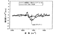
図6は、tBLMに組み入れられたOR5のIR分光法データを示す。 FIG. 6 shows OR5 IR spectroscopy data incorporated into tBLM.
図7は、人工の平面膜表面中に挿入した標識された膜タンパク質の画像を示す。 FIG. 7 shows an image of a labeled membrane protein inserted into an artificial planar membrane surface.
図8は、P19に繋留させたBLM上のウシオプシンの結果を示す。 FIG. 8 shows the results for bovine opsin on BLM tethered to P19.
図9は、ウシオプシンを符号化するcDNA配列を含む発現プラスミドTNT−ρ3/4の略図及び符号化領域を示す。 FIG. 9 shows a schematic representation and coding region of the expression plasmid TNT-ρ3 / 4 containing the cDNA sequence encoding bovine opsin.
図10は、pTNT−ρ3/4でのtBLMのインキュベーションのSPFSスペクトルを示す。 FIG. 10 shows the SPFS spectrum of the tBLM incubation with pTNT-ρ3 / 4.
実施例
実施例1
ペプチド繋留された脂質膜の製造
平面の金表面上のペプチド分子の構築
ペプチドを、脂質膜を繋留するための親水性スペーサー分子のために使用する。α−ラミニンサブユニットから派生させた19個のペプチドCSRARKQAASIKVAVSADR(P19)(ドイツ、DeisenhofenのSigma−Aldrich社)を使用した。種々のペプチドの濃度を検査し、かつミリポア水中に25μg/mlの最終濃度が、時間/物質の消費率及び再現性に関する金表面に対するP19の会合を示す構築実験における最適なペプチド濃度であることを示した。
Example Example 1
Production of peptide tethered lipid membranes Construction of peptide molecules on planar gold surfaces Peptides are used for hydrophilic spacer molecules to tether lipid membranes. Nineteen peptides derived from α-laminin subunits CSRRAQQAASIKVAVSAD (P19) (Sigma-Aldrich, Deisenhofen, Germany) were used. Examining the concentration of the various peptides and confirming that the final concentration of 25 μg / ml in Millipore water is the optimal peptide concentration in construction experiments showing P19 association to the gold surface with respect to time / substance consumption and reproducibility. Indicated.
単分子のペプチド層の自己構築は、N−末端のシステイン残基の強い金−硫黄相互作用によって得られる。ペプチド層の構築は、ペプチド溶液中での金表面の30分のインキュベーション後に完了される。過剰な結合していないペプチドは洗い落とされ、かつ得られた光学的厚さを、SPSによって観測した。 Self-assembly of a monomolecular peptide layer is obtained by a strong gold-sulfur interaction of the N-terminal cysteine residue. The construction of the peptide layer is completed after a 30 minute incubation of the gold surface in the peptide solution. Excess unbound peptide was washed away and the resulting optical thickness was monitored by SPS.
ラミニンペプチドを、そのカルボキシ末端を介してリン脂質分子(ジミリストイルホスファチジルエタノールアミン、DMPE;0.1mg/ml(Sigma社製);0.001%(w/v)トリトンX−100(ドイツ、KarlsruheのRoth社製)中に可溶化させた)の一次アミノ残基に、活性エステル結合の手法で共有的に結合させた。N−エチル−N’−(ジメチルアミノプロピル)カルボジイミド(EDC;ドイツ、DeisenhofenのFluka社製)(11.5mg/ml)及びN−ヒドロキシスクシンイミド(NHS;Fluka社製)(75mg/ml)を10mlのミリポア水でそれぞれ溶解し、そして使用するまで−20℃で200μlのアリコートで保管した。EDC及びNHSを、400μl(NHS50mM、EDC200mM)の量で等量混合し、そしてペプチドを被膜した金表面上に適用した。得られたチオペプチド脂質層は、硬質な集成体(約1.5nmの厚さ)を形成する。
Laminin peptide is linked via its carboxy terminus to a phospholipid molecule (dimyristoylphosphatidylethanolamine, DMPE; 0.1 mg / ml (Sigma); 0.001% (w / v) Triton X-100 (Karlsruhe, Germany). And the primary amino residue solubilized in Roth Co., Ltd.) was covalently bound by the active ester bond technique. 10 ml of N-ethyl-N ′-(dimethylaminopropyl) carbodiimide (EDC; manufactured by Fluka, Deisenhofen, Germany) (11.5 mg / ml) and N-hydroxysuccinimide (NHS; manufactured by Fluka) (75 mg / ml) Of Millipore water and stored in 200 μl aliquots at −20 ° C. until use. EDC and NHS were mixed in equal amounts of 400 μl (
ペプチド脂質層に、ホスファチジルコリン小胞を添加し、そして2層の脂質膜を形成した。 To the peptide lipid layer, phosphatidylcholine vesicles were added and a bilayer lipid membrane was formed.
実施例2
Gタンパク質共役受容体の生体外発現
実験に使用された構成要素:
ウサギの網状赤血球を基礎とする、結合させた転写系及び翻訳系(Promega,Inc製のTNT(登録商標))を使用した。水疱性口内炎ウイルス糖タンパク質VSVを符号化するcDNAをアミノ末端又はカルボキシ末端に付加させたGPCR(ラットからの匂い受容体OR5)を符号化するcDNAを、それぞれに製造した。
Example 2
In vitro expression of G protein-coupled receptors The components used in the experiment:
A combined transcription and translation system (TNT® from Promega, Inc) based on rabbit reticulocytes was used. A cDNA encoding a GPCR (odor receptor OR5 from rat) in which a cDNA encoding vesicular stomatitis virus glycoprotein VSV was added to the amino terminus or carboxy terminus was prepared for each.
無細胞系抽出物を、実施例1で記載されているように製造された繋留された膜の上端で適用し、GPCRのcDNAコンストラクトを添加した。インキュベーション後、表面を洗浄し、VSVエピトープを認識する一次マウス抗体を添加した。該表面を再び洗浄し、続いてマウスに対する蛍光標識させた二次抗体と一緒にインキュベートした。得られた蛍光シグナルの増加が、表面の付近にVSVタグの存在を示す。 A cell-free extract was applied at the top of a tethered membrane prepared as described in Example 1 and a GPCR cDNA construct was added. Following incubation, the surface was washed and a primary mouse antibody recognizing the VSV epitope was added. The surface was washed again and subsequently incubated with a fluorescently labeled secondary antibody against the mouse. The resulting increase in fluorescence signal indicates the presence of the VSV tag near the surface.
繋留された膜中のGPCRタンパク質の挿入を証明するために、様々な実験から派生した蛍光シグナルを比較した。カルボキシ末端と比較して、アミノ末端のVSVタグについての再現性のある及び有意に異なるシグナル強度を得た。C末端でVSVタグを有するGPCR cDNAは、5倍低いシグナルをもたらし、そしてそれは、参照として挿入なしにTnTベクターからの蛍光シグナルに匹敵する。 To demonstrate the insertion of the GPCR protein in the tethered membrane, fluorescent signals derived from various experiments were compared. Reproducible and significantly different signal intensities for the amino terminal VSV tag were obtained compared to the carboxy terminus. A GPCR cDNA with a VSV tag at the C-terminus yields a signal that is 5 times lower, and it is comparable to the fluorescent signal from the TnT vector without insertion as a reference.
このことは、図2で図示されているような、平面膜系中にGPCR分子の組み入れを示す。 This indicates the incorporation of GPCR molecules in a planar membrane system, as illustrated in FIG.
無細胞系抽出物の活性並びにそれぞれのGPCRコンストラクトの転写及び翻訳の成功を証明した後で、無細胞系抽出物を、平面脂質膜に対するインキュベート後に収集し、そして変性させ、ウェスタンブロットのための準備をした。 After demonstrating the activity of the cell-free extract and the successful transcription and translation of the respective GPCR construct, the cell-free extract is collected after incubation on a planar lipid membrane and denatured, ready for Western blotting Did.
実施例3
合成膜及び天然膜の比較
ダイズのホスファチジルコリンリポソームと比較して、イヌのミクロソームを使用する場合に、蛍光シグナルにおける有意差を観察することができなかった。このことは、PCのみからなる平面脂質膜中への成功したタンパク質の組み込みが起こることを示す。
Example 3
Comparison of Synthetic and Natural Membranes No significant difference in fluorescence signal could be observed when using canine microsomes compared to soybean phosphatidylcholine liposomes. This indicates that successful protein incorporation into a planar lipid membrane consisting only of PC occurs.
実施例4
放射性検出を使用した挿入の証明
組み入れられたタンパク質分子のさらなる研究は、放射性標識によって行われていた。従って、35Sメチオニンの存在下でOR5を合成した。次の実験を提案した。膜の組み入れによって、OR5タンパク質を、7TMタンパク質からの露出されたループを除きプロテイナーゼKの消化に対して保護する。5.9〜6.8kDaの範囲のサイズ分布において得られた断片を算出した。その結果を、図5で示す。
Example 4
Demonstration of insertion using radioactive detection Further studies of the incorporated protein molecules were done by radiolabeling. Therefore, OR5 was synthesized in the presence of 35 S methionine. The following experiment was proposed. By incorporation of the membrane, the OR5 protein is protected against proteinase K digestion with the exception of the exposed loop from the 7TM protein. Fragments obtained in a size distribution ranging from 5.9 to 6.8 kDa were calculated. The result is shown in FIG.
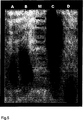
レーンA:生体外での実験を、膜表面上で直接展開した。プロテイナーゼKを、その後適用した。そしてプロテイナーゼKと消化されたペプチド断片を洗い流した。得られた膜表面(OR5ループ)を、アクリルアミドSDSゲル上に適用し、そして、タンパク質の放射性標識(Sは、OR5配列中に均一に存在する)をフィルムに露光し、6kDaの期待範囲内のシグナルを生じた。
レーンB:生体外での実験を、エッペンドルフチューブ中で行い、次に膜上に適用した − 膜中のOR5の発現は同一のシグナルをもたらし、生体外抽出物中に存在する膜断片中の発現を示唆し、非特異的に膜表面上に吸着された。
レーンM:188、98、62、49、38、28、17、14、6、3kDaのマーカー
レーンC:分析活性の陽性対照:改質なしにゲル上に適用された生体外抽出物
レーンD:プロテイナーゼK活性の陽性対照:プロテイナーゼKのインキュベーションを有する生体外抽出物
実施例5
組み入れられた匂い受容体の活性
組み入れられた匂い受容体の活性を、IR分光法を用いて測定した。1mMリリアール(lilial)の添加に対するOR5/tBLM系のIRアミドIのピークのシグナル変化は、受容体による匂いの認識を示す。リリアール/OR5の相互作用は、PBSを使用した洗浄によって逆転されることができる。従って、IRデータは、OR5タンパク質を組み込んだtBLMの生物学的活性を示す。
Lane A: In vitro experiments were developed directly on the membrane surface. Proteinase K was then applied. Proteinase K and digested peptide fragments were washed away. The resulting membrane surface (OR5 loop) is applied onto an acrylamide SDS gel and the protein radiolabel (S is uniformly present in the OR5 sequence) is exposed to the film and within the expected range of 6 kDa. A signal was generated.
Lane B: In vitro experiments were performed in Eppendorf tubes and then applied on the membrane—expression of OR5 in the membrane yielded the same signal, expression in membrane fragments present in the in vitro extract It was adsorbed on the membrane surface nonspecifically.
Lane M: 188, 98, 62, 49, 38, 28, 17, 14, 6, 3 kDa marker lane C: positive control for analytical activity: in vitro extract lane D applied on gel without modification: Proteinase K activity positive control: in vitro extract with proteinase K incubation Example 5
Incorporated Odor Receptor Activity Incorporated odor receptor activity was measured using IR spectroscopy. The signal change in the peak of IRamide I in the OR5 / tBLM system upon addition of 1 mM liaial indicates odor recognition by the receptor. Lilyal / OR5 interaction can be reversed by washing with PBS. Therefore, IR data indicates the biological activity of tBLM incorporating the OR5 protein.
実施例6
画像相関顕微鏡法による膜タンパク質の検出
本発明に従った新しい方法を、人工平面脂質膜構造体中への複雑な哺乳動物の膜タンパク質の生体外合成に使用した。ウサギの網状赤血球の細胞抽出物は、受容体からの単なるDNAから出発する匂い受容体種の新規合成のためのタンパク質合成機構を含む。画像相関顕微鏡法による挿入された受容体タンパク質の密度は、表面でのエバネッセント励起を使用して、検査された。例えば、図7は、個々の膜タンパク質のアフィニティー標識をタグ付けする蛍光標識させた抗体を示す。励起範囲は、約20μmの直径を有する。膜タンパク質の高分解能画像相関顕微鏡法、すなわち時空的自己相関は、空間的なタンパク質の分布(組み入れ密度)及びタンパク質の易動度(膜中での拡散)についての情報をもたらす。
Example 6
Detection of Membrane Proteins by Image Correlation Microscopy A new method according to the present invention was used for in vitro synthesis of complex mammalian membrane proteins into artificial planar lipid membrane structures. Rabbit reticulocyte cell extracts contain a protein synthesis mechanism for the novel synthesis of odorant receptor species starting from simple DNA from the receptor. The density of inserted receptor protein by image correlation microscopy was examined using evanescent excitation at the surface. For example, FIG. 7 shows fluorescently labeled antibodies that tag the affinity labels of individual membrane proteins. The excitation range has a diameter of about 20 μm. High resolution image correlation microscopy of membrane proteins, ie spatiotemporal autocorrelation, provides information about spatial protein distribution (incorporation density) and protein mobility (diffusion in the membrane).
実施例7
P19に繋留されたBLM上のウシオプシンの生体外発現
表面構築を、OR5の生体外発現/膜挿入に記載されている方法と同一の方法で組み立てた(図8)。
Example 7
In vitro expression of bovine opsin on BLM tethered to P19 Surface construction was assembled in the same way as described in OR5 in vitro expression / membrane insertion (FIG. 8).
ウシロドプシンの生体外発現を、生体外反応混合物のT7−TNT(登録商標)(Promega社)で行った。ウシオプシンを符号化するcDNA配列を、発現プラスミドpTNT(登録商標)−ρ3/4の形で反応に添加した。概略図及び符号化領域を、図9に示す。 In vitro expression of bovine rhodopsin was performed with the in vitro reaction mixture T7-TNT® (Promega). The cDNA sequence encoding bovine opsin was added to the reaction in the form of the expression plasmid pTNT®-ρ3 / 4. A schematic diagram and the coding region are shown in FIG.
生体外の反応混合物を、直接、SPRセンサー表面に取り付けたP19に繋留されたBLMの上側に対して90分30℃でインキュベートした。次に、この表面構築物を、PBS溶液含有物、及びモノクローナルのマウス抗−VSVとヤギ−抗マウスIgG−Cy5抗体との1:3の混合物からなる抗体サンドイッチ系にさらした。生体外発現の反応混合物を含有するpTNT−ρ3/4でのtBLMのインキュベーションのための対応するSPFSスペクトル、及びプラスミドを含まない反応混合物でのインキュベーションのための対応するSPFSスペクトルを、それぞれ図10で示す。 The in vitro reaction mixture was incubated for 90 minutes at 30 ° C. directly against the upper side of the BLM tethered to P19 attached to the SPR sensor surface. This surface construct was then exposed to an antibody sandwich system consisting of a PBS solution containing and a 1: 3 mixture of monoclonal mouse anti-VSV and goat-anti-mouse IgG-Cy5 antibodies. The corresponding SPFS spectrum for incubation of tBLM with pTNT-ρ3 / 4 containing the reaction mixture of in vitro expression and the corresponding SPFS spectrum for incubation with the reaction mixture without plasmid are shown in FIG. Show.
Claims (24)
(i)表面に結合された膜である膜の提供、
(ii)該膜に無細胞発現系及び膜タンパク質を符号化する核酸の適用、並びに
(iii)該膜中に組み込まれた、膜タンパク質の発現
を含む、膜中に組み入れられた膜タンパク質の製造方法。The next step (i) providing a membrane which is a membrane bonded to the surface;
(Ii) application of nucleic acid encoding cell-free expression system and membrane protein to the membrane, and (iii) production of membrane protein incorporated into the membrane, including expression of membrane protein incorporated into the membrane Method.
Applications Claiming Priority (3)
| Application Number | Priority Date | Filing Date | Title |
|---|---|---|---|
| EP05023705 | 2005-10-28 | ||
| EP05023705.6 | 2005-10-28 | ||
| PCT/EP2006/008318 WO2007048459A1 (en) | 2005-10-28 | 2006-08-24 | Cell-free in vitro transcription and translation of membrane proteins into tethered planar lipid layers |
Publications (2)
| Publication Number | Publication Date |
|---|---|
| JP2009513116A JP2009513116A (en) | 2009-04-02 |
| JP5044563B2 true JP5044563B2 (en) | 2012-10-10 |
Family
ID=37502637
Family Applications (1)
| Application Number | Title | Priority Date | Filing Date |
|---|---|---|---|
| JP2008536951A Expired - Fee Related JP5044563B2 (en) | 2005-10-28 | 2006-08-24 | Cell-free in vitro transcription and translation of membrane proteins in tethered planar lipid layers |
Country Status (10)
| Country | Link |
|---|---|
| US (1) | US20090202997A1 (en) |
| EP (1) | EP1948817B1 (en) |
| JP (1) | JP5044563B2 (en) |
| AT (1) | ATE458824T1 (en) |
| CA (1) | CA2627580A1 (en) |
| DE (1) | DE602006012532D1 (en) |
| DK (1) | DK1948817T3 (en) |
| ES (1) | ES2341886T3 (en) |
| PL (1) | PL1948817T3 (en) |
| WO (1) | WO2007048459A1 (en) |
Families Citing this family (13)
| Publication number | Priority date | Publication date | Assignee | Title |
|---|---|---|---|---|
| FR2915490B1 (en) * | 2007-04-26 | 2011-10-28 | Univ Joseph Fourier Grenoble I | FORMATION OF PROTEOLIPOSOMES CONTAINING MEMBRANE PROTEINS USING A CELLULAR PROTEIN SYNTHESIS SYSTEM |
| CN102460150B (en) * | 2009-04-20 | 2015-05-20 | 新加坡科技研究局 | Vesicular system and uses thereof |
| EP2561086A1 (en) | 2010-04-20 | 2013-02-27 | Institut Für Mikrotechnik Mainz GmbH | System for the in vitro transcription and translation of membrane proteins |
| WO2012164270A1 (en) | 2011-05-27 | 2012-12-06 | Oxford Nanopore Technologies Limited | Coupling method |
| US9541480B2 (en) | 2011-06-29 | 2017-01-10 | Academia Sinica | Capture, purification, and release of biological substances using a surface coating |
| US10458979B2 (en) * | 2012-08-29 | 2019-10-29 | Agency For Science, Technology And Research | Solid supported artificial cell membrane system |
| TWI512930B (en) * | 2012-09-25 | 2015-12-11 | Xintex Inc | Chip package and method for forming the same |
| US9494500B2 (en) | 2012-10-29 | 2016-11-15 | Academia Sinica | Collection and concentration system for biologic substance of interest and use thereof |
| GB201406155D0 (en) | 2014-04-04 | 2014-05-21 | Oxford Nanopore Tech Ltd | Method |
| CN106662514A (en) | 2014-04-01 | 2017-05-10 | 中央研究院 | Methods and systems for cancer diagnosis and prognosis |
| EP2998026B1 (en) | 2014-08-26 | 2024-01-17 | Academia Sinica | Collector architecture layout design |
| FR3027679B1 (en) * | 2014-10-28 | 2016-12-09 | Univ Claude Bernard Lyon | SUPPORT / PEPTIDE / LIPID BINOUCHE ASSEMBLY, METHODS OF PREPARATION AND DETECTION METHODS THEREOF |
| US10107726B2 (en) | 2016-03-16 | 2018-10-23 | Cellmax, Ltd. | Collection of suspended cells using a transferable membrane |
Family Cites Families (4)
| Publication number | Priority date | Publication date | Assignee | Title |
|---|---|---|---|---|
| JP4366497B2 (en) * | 2002-08-30 | 2009-11-18 | 独立行政法人農業・食品産業技術総合研究機構 | Novel peptides that act specifically on biological membranes |
| JP2005168316A (en) * | 2003-12-08 | 2005-06-30 | Toyobo Co Ltd | Method for synthesizing protein |
| WO2006069331A2 (en) * | 2004-12-22 | 2006-06-29 | The Salk Institute For Biological Studies | Compositions and methods for producing recombinant proteins |
| CN102460150B (en) * | 2009-04-20 | 2015-05-20 | 新加坡科技研究局 | Vesicular system and uses thereof |
-
2006
- 2006-08-24 DK DK06777059.4T patent/DK1948817T3/en active
- 2006-08-24 WO PCT/EP2006/008318 patent/WO2007048459A1/en active Application Filing
- 2006-08-24 US US12/091,607 patent/US20090202997A1/en not_active Abandoned
- 2006-08-24 PL PL06777059T patent/PL1948817T3/en unknown
- 2006-08-24 ES ES06777059T patent/ES2341886T3/en active Active
- 2006-08-24 EP EP06777059A patent/EP1948817B1/en not_active Not-in-force
- 2006-08-24 DE DE602006012532T patent/DE602006012532D1/en active Active
- 2006-08-24 CA CA002627580A patent/CA2627580A1/en not_active Abandoned
- 2006-08-24 AT AT06777059T patent/ATE458824T1/en active
- 2006-08-24 JP JP2008536951A patent/JP5044563B2/en not_active Expired - Fee Related
Also Published As
| Publication number | Publication date |
|---|---|
| ATE458824T1 (en) | 2010-03-15 |
| PL1948817T3 (en) | 2010-09-30 |
| DK1948817T3 (en) | 2010-06-14 |
| CA2627580A1 (en) | 2007-05-03 |
| EP1948817B1 (en) | 2010-02-24 |
| WO2007048459A1 (en) | 2007-05-03 |
| JP2009513116A (en) | 2009-04-02 |
| EP1948817A1 (en) | 2008-07-30 |
| DE602006012532D1 (en) | 2010-04-08 |
| ES2341886T3 (en) | 2010-06-29 |
| US20090202997A1 (en) | 2009-08-13 |
Similar Documents
| Publication | Publication Date | Title |
|---|---|---|
| JP5044563B2 (en) | Cell-free in vitro transcription and translation of membrane proteins in tethered planar lipid layers | |
| US20220283171A1 (en) | Methods and systems for producing nanolipoprotein particles | |
| Dorman et al. | The life of pi star: exploring the exciting and forbidden worlds of the benzophenone photophore | |
| Tian et al. | Labeling and single-molecule methods to monitor G protein-coupled receptor dynamics | |
| JP3942431B2 (en) | Protein-molecule interaction analysis method | |
| US20220326235A1 (en) | S-layer protein 2d lattice coupled detergent-free gpcr bioelectronic interfaces, devices, and methods for use thereof | |
| WO2018207906A1 (en) | Peptide fusion protein having affinity for substrate | |
| Chu et al. | Protein immobilization on liposomes and lipid‐coated nanoparticles by protein trans‐splicing | |
| JP4832291B2 (en) | Labeling substances and chimeric substances, methods for producing these substances, and methods for capturing, analyzing and / or identifying biological substances using the labeling substances | |
| Mouillac et al. | Fluorescent agonists and antagonists for vasopressin/oxytocin G protein-coupled receptors: usefulness in ligand screening assays and receptor studies | |
| El Kazzy et al. | Biomimetic olfactory biosensors and bioelectronic noses | |
| US8304520B2 (en) | Labeled fusion protein | |
| KR100718207B1 (en) | Bio-metal Chip Using Metal Binding Protein and Method for Fabricating the Same | |
| EP4123307A1 (en) | Methods for structure determination and drug design using salipro particles | |
| US20230221307A1 (en) | Conjugated composed of membrane-targeting peptides for extracellular vesicles isolation, analysis and their integration thereof | |
| WO2023001912A1 (en) | Methods for structure determination and drug design using salipro particles | |
| CN110117312A (en) | Lipid binding protein-antigen capture module compound, and its preparation method and application | |
| CN115254048B (en) | Molecular imprinting and coating polymer based on reverse microemulsion, preparation method and application | |
| Akram | Lipid nanoparticles for membrane protein target drug discovery | |
| Shumate et al. | A rapid, tag-free way to purify functional GPCRs | |
| US20150204890A1 (en) | Microarrays of g protein coupled receptors | |
| JP2005112800A (en) | Membrane protein reconstitution method used in analysis of interaction between membrane protein and ligand by nmr | |
| El Kazzy et al. | Biomimetic Olfactory Biosensors | |
| May | Functional characterisation of in vitro synthesised G-protein coupled receptors in polymersomes | |
| López | SYNTHESIS, MOLECULAR BIOLOGY, AND BIOANALYTICAL STUDIES OF A FLUORESCENT SEROTONIN LIGAND WITH 5-HT3A IN BULK AND SUPPORTED LIPID BILAYERS |
Legal Events
| Date | Code | Title | Description |
|---|---|---|---|
| A621 | Written request for application examination |
Free format text: JAPANESE INTERMEDIATE CODE: A621 Effective date: 20090507 |
|
| RD04 | Notification of resignation of power of attorney |
Free format text: JAPANESE INTERMEDIATE CODE: A7424 Effective date: 20101227 |
|
| RD04 | Notification of resignation of power of attorney |
Free format text: JAPANESE INTERMEDIATE CODE: A7424 Effective date: 20101228 |
|
| A131 | Notification of reasons for refusal |
Free format text: JAPANESE INTERMEDIATE CODE: A131 Effective date: 20111020 |
|
| A601 | Written request for extension of time |
Free format text: JAPANESE INTERMEDIATE CODE: A601 Effective date: 20120119 |
|
| A602 | Written permission of extension of time |
Free format text: JAPANESE INTERMEDIATE CODE: A602 Effective date: 20120126 |
|
| A601 | Written request for extension of time |
Free format text: JAPANESE INTERMEDIATE CODE: A601 Effective date: 20120220 |
|
| A602 | Written permission of extension of time |
Free format text: JAPANESE INTERMEDIATE CODE: A602 Effective date: 20120227 |
|
| A601 | Written request for extension of time |
Free format text: JAPANESE INTERMEDIATE CODE: A601 Effective date: 20120321 |
|
| A602 | Written permission of extension of time |
Free format text: JAPANESE INTERMEDIATE CODE: A602 Effective date: 20120328 |
|
| A521 | Request for written amendment filed |
Free format text: JAPANESE INTERMEDIATE CODE: A523 Effective date: 20120411 |
|
| TRDD | Decision of grant or rejection written | ||
| A01 | Written decision to grant a patent or to grant a registration (utility model) |
Free format text: JAPANESE INTERMEDIATE CODE: A01 Effective date: 20120614 |
|
| A01 | Written decision to grant a patent or to grant a registration (utility model) |
Free format text: JAPANESE INTERMEDIATE CODE: A01 |
|
| A61 | First payment of annual fees (during grant procedure) |
Free format text: JAPANESE INTERMEDIATE CODE: A61 Effective date: 20120713 |
|
| R150 | Certificate of patent or registration of utility model |
Free format text: JAPANESE INTERMEDIATE CODE: R150 |
|
| FPAY | Renewal fee payment (event date is renewal date of database) |
Free format text: PAYMENT UNTIL: 20150720 Year of fee payment: 3 |
|
| R250 | Receipt of annual fees |
Free format text: JAPANESE INTERMEDIATE CODE: R250 |
|
| LAPS | Cancellation because of no payment of annual fees |