JP5025269B2 - Noninvasive arthroscopy instrument sheath - Google Patents
Noninvasive arthroscopy instrument sheath Download PDFInfo
- Publication number
- JP5025269B2 JP5025269B2 JP2006551498A JP2006551498A JP5025269B2 JP 5025269 B2 JP5025269 B2 JP 5025269B2 JP 2006551498 A JP2006551498 A JP 2006551498A JP 2006551498 A JP2006551498 A JP 2006551498A JP 5025269 B2 JP5025269 B2 JP 5025269B2
- Authority
- JP
- Japan
- Prior art keywords
- sheath
- arthroscope
- invasive
- grip
- fluid
- Prior art date
- Legal status (The legal status is an assumption and is not a legal conclusion. Google has not performed a legal analysis and makes no representation as to the accuracy of the status listed.)
- Active
Links
- 239000012530 fluid Substances 0.000 claims description 65
- 238000001356 surgical procedure Methods 0.000 claims description 27
- 239000000463 material Substances 0.000 claims description 7
- 210000001519 tissue Anatomy 0.000 description 25
- 238000003860 storage Methods 0.000 description 14
- 238000000034 method Methods 0.000 description 12
- 238000004140 cleaning Methods 0.000 description 10
- 208000014674 injury Diseases 0.000 description 8
- 230000008733 trauma Effects 0.000 description 7
- 238000009966 trimming Methods 0.000 description 5
- 239000011248 coating agent Substances 0.000 description 4
- 238000000576 coating method Methods 0.000 description 4
- 239000007788 liquid Substances 0.000 description 4
- 230000003287 optical effect Effects 0.000 description 4
- FAPWRFPIFSIZLT-UHFFFAOYSA-M Sodium chloride Chemical compound [Na+].[Cl-] FAPWRFPIFSIZLT-UHFFFAOYSA-M 0.000 description 3
- 229920001971 elastomer Polymers 0.000 description 3
- 239000012858 resilient material Substances 0.000 description 3
- 239000011780 sodium chloride Substances 0.000 description 3
- 229920002725 thermoplastic elastomer Polymers 0.000 description 3
- 238000005452 bending Methods 0.000 description 2
- 239000008280 blood Substances 0.000 description 2
- 210000004369 blood Anatomy 0.000 description 2
- 238000013461 design Methods 0.000 description 2
- 239000013013 elastic material Substances 0.000 description 2
- 239000000806 elastomer Substances 0.000 description 2
- 230000005484 gravity Effects 0.000 description 2
- 238000003780 insertion Methods 0.000 description 2
- 230000037431 insertion Effects 0.000 description 2
- 230000002262 irrigation Effects 0.000 description 2
- 238000003973 irrigation Methods 0.000 description 2
- 210000003127 knee Anatomy 0.000 description 2
- 239000004033 plastic Substances 0.000 description 2
- 229920000642 polymer Polymers 0.000 description 2
- XLYOFNOQVPJJNP-UHFFFAOYSA-N water Substances O XLYOFNOQVPJJNP-UHFFFAOYSA-N 0.000 description 2
- 208000012260 Accidental injury Diseases 0.000 description 1
- 241001631457 Cannula Species 0.000 description 1
- 239000004809 Teflon Substances 0.000 description 1
- 229920006362 Teflon® Polymers 0.000 description 1
- 230000005856 abnormality Effects 0.000 description 1
- 239000000853 adhesive Substances 0.000 description 1
- 230000001070 adhesive effect Effects 0.000 description 1
- 210000003484 anatomy Anatomy 0.000 description 1
- 210000001264 anterior cruciate ligament Anatomy 0.000 description 1
- 238000013459 approach Methods 0.000 description 1
- 230000015572 biosynthetic process Effects 0.000 description 1
- 210000001124 body fluid Anatomy 0.000 description 1
- 239000010839 body fluid Substances 0.000 description 1
- 210000000988 bone and bone Anatomy 0.000 description 1
- 210000000845 cartilage Anatomy 0.000 description 1
- 239000002131 composite material Substances 0.000 description 1
- 238000012937 correction Methods 0.000 description 1
- 230000006378 damage Effects 0.000 description 1
- 230000010339 dilation Effects 0.000 description 1
- 239000003814 drug Substances 0.000 description 1
- 239000013536 elastomeric material Substances 0.000 description 1
- 229920000295 expanded polytetrafluoroethylene Polymers 0.000 description 1
- 239000000835 fiber Substances 0.000 description 1
- 238000002347 injection Methods 0.000 description 1
- 239000007924 injection Substances 0.000 description 1
- 239000000314 lubricant Substances 0.000 description 1
- 230000005499 meniscus Effects 0.000 description 1
- 239000002184 metal Substances 0.000 description 1
- 210000004417 patella Anatomy 0.000 description 1
- 229920001343 polytetrafluoroethylene Polymers 0.000 description 1
- 239000004810 polytetrafluoroethylene Substances 0.000 description 1
- 210000002967 posterior cruciate ligament Anatomy 0.000 description 1
- 230000001681 protective effect Effects 0.000 description 1
- 239000000523 sample Substances 0.000 description 1
- 230000002000 scavenging effect Effects 0.000 description 1
- 210000004872 soft tissue Anatomy 0.000 description 1
- 229940124597 therapeutic agent Drugs 0.000 description 1
- 210000002303 tibia Anatomy 0.000 description 1
- 210000002105 tongue Anatomy 0.000 description 1
- 210000000689 upper leg Anatomy 0.000 description 1
- 230000000007 visual effect Effects 0.000 description 1
- 238000012800 visualization Methods 0.000 description 1
Images
Classifications
-
- A—HUMAN NECESSITIES
- A61—MEDICAL OR VETERINARY SCIENCE; HYGIENE
- A61B—DIAGNOSIS; SURGERY; IDENTIFICATION
- A61B17/00—Surgical instruments, devices or methods, e.g. tourniquets
- A61B17/34—Trocars; Puncturing needles
- A61B17/3417—Details of tips or shafts, e.g. grooves, expandable, bendable; Multiple coaxial sliding cannulas, e.g. for dilating
- A61B17/3421—Cannulas
-
- A—HUMAN NECESSITIES
- A61—MEDICAL OR VETERINARY SCIENCE; HYGIENE
- A61B—DIAGNOSIS; SURGERY; IDENTIFICATION
- A61B1/00—Instruments for performing medical examinations of the interior of cavities or tubes of the body by visual or photographical inspection, e.g. endoscopes; Illuminating arrangements therefor
- A61B1/00131—Accessories for endoscopes
- A61B1/00135—Oversleeves mounted on the endoscope prior to insertion
-
- A—HUMAN NECESSITIES
- A61—MEDICAL OR VETERINARY SCIENCE; HYGIENE
- A61B—DIAGNOSIS; SURGERY; IDENTIFICATION
- A61B1/00—Instruments for performing medical examinations of the interior of cavities or tubes of the body by visual or photographical inspection, e.g. endoscopes; Illuminating arrangements therefor
- A61B1/313—Instruments for performing medical examinations of the interior of cavities or tubes of the body by visual or photographical inspection, e.g. endoscopes; Illuminating arrangements therefor for introducing through surgical openings, e.g. laparoscopes
- A61B1/317—Instruments for performing medical examinations of the interior of cavities or tubes of the body by visual or photographical inspection, e.g. endoscopes; Illuminating arrangements therefor for introducing through surgical openings, e.g. laparoscopes for bones or joints, e.g. osteoscopes, arthroscopes
-
- A—HUMAN NECESSITIES
- A61—MEDICAL OR VETERINARY SCIENCE; HYGIENE
- A61B—DIAGNOSIS; SURGERY; IDENTIFICATION
- A61B17/00—Surgical instruments, devices or methods, e.g. tourniquets
- A61B17/32—Surgical cutting instruments
- A61B17/3205—Excision instruments
- A61B17/3207—Atherectomy devices working by cutting or abrading; Similar devices specially adapted for non-vascular obstructions
-
- A—HUMAN NECESSITIES
- A61—MEDICAL OR VETERINARY SCIENCE; HYGIENE
- A61M—DEVICES FOR INTRODUCING MEDIA INTO, OR ONTO, THE BODY; DEVICES FOR TRANSDUCING BODY MEDIA OR FOR TAKING MEDIA FROM THE BODY; DEVICES FOR PRODUCING OR ENDING SLEEP OR STUPOR
- A61M1/00—Suction or pumping devices for medical purposes; Devices for carrying-off, for treatment of, or for carrying-over, body-liquids; Drainage systems
- A61M1/84—Drainage tubes; Aspiration tips
- A61M1/85—Drainage tubes; Aspiration tips with gas or fluid supply means, e.g. for supplying rinsing fluids or anticoagulants
-
- A—HUMAN NECESSITIES
- A61—MEDICAL OR VETERINARY SCIENCE; HYGIENE
- A61B—DIAGNOSIS; SURGERY; IDENTIFICATION
- A61B17/00—Surgical instruments, devices or methods, e.g. tourniquets
- A61B17/32—Surgical cutting instruments
- A61B2017/320044—Blunt dissectors
- A61B2017/320048—Balloon dissectors
-
- A—HUMAN NECESSITIES
- A61—MEDICAL OR VETERINARY SCIENCE; HYGIENE
- A61B—DIAGNOSIS; SURGERY; IDENTIFICATION
- A61B17/00—Surgical instruments, devices or methods, e.g. tourniquets
- A61B17/34—Trocars; Puncturing needles
- A61B2017/347—Locking means, e.g. for locking instrument in cannula
-
- A—HUMAN NECESSITIES
- A61—MEDICAL OR VETERINARY SCIENCE; HYGIENE
- A61B—DIAGNOSIS; SURGERY; IDENTIFICATION
- A61B17/00—Surgical instruments, devices or methods, e.g. tourniquets
- A61B17/34—Trocars; Puncturing needles
- A61B2017/348—Means for supporting the trocar against the body or retaining the trocar inside the body
- A61B2017/3482—Means for supporting the trocar against the body or retaining the trocar inside the body inside
- A61B2017/349—Trocar with thread on outside
Landscapes
- Health & Medical Sciences (AREA)
- Life Sciences & Earth Sciences (AREA)
- Surgery (AREA)
- Heart & Thoracic Surgery (AREA)
- General Health & Medical Sciences (AREA)
- Public Health (AREA)
- Animal Behavior & Ethology (AREA)
- Engineering & Computer Science (AREA)
- Biomedical Technology (AREA)
- Veterinary Medicine (AREA)
- Molecular Biology (AREA)
- Nuclear Medicine, Radiotherapy & Molecular Imaging (AREA)
- Medical Informatics (AREA)
- Pathology (AREA)
- Vascular Medicine (AREA)
- Biophysics (AREA)
- Optics & Photonics (AREA)
- Radiology & Medical Imaging (AREA)
- Physics & Mathematics (AREA)
- Oral & Maxillofacial Surgery (AREA)
- Pulmonology (AREA)
- Anesthesiology (AREA)
- Hematology (AREA)
- Orthopedic Medicine & Surgery (AREA)
- Physical Education & Sports Medicine (AREA)
- Endoscopes (AREA)
- Surgical Instruments (AREA)
Description
以下に記載の発明は、関節鏡検査外科用器具の分野に関する。 The invention described below relates to the field of arthroscopic surgical instruments.
関節鏡視下手術には、患者の関節内部またはその近辺の手術領域を視覚化するため、関節鏡などの光学器具が用いられる。外科手術を行うにあたり、同一の器具を使っても異なる器具を使っても良い。一般に関節鏡のほかに使用される器具には、組織の切断に使用されるトリミング器具や手術領域の洗浄に使用される洗浄器具などが含まれる。手術領域に器具を差し込むには、それぞれの器具について切開が行われなければならない。従って、関節鏡視下手術では、トリミング器具と関節鏡だけを使うのを好む外科医が多い。 In the arthroscopic surgery, an optical instrument such as an arthroscope is used in order to visualize a surgical region in or near a patient's joint. In performing a surgical operation, the same instrument or different instruments may be used. In general, the instruments used in addition to the arthroscope include a trimming instrument used for cutting tissue and a cleaning instrument used for cleaning an operation area. In order to insert instruments into the surgical area, an incision must be made for each instrument. Therefore, many surgeons prefer to use only trimming instruments and arthroscopes in arthroscopic surgery.
関節鏡視下手術で使用される力に比べ関節鏡は脆弱なので、それを強化するため、関節鏡の上に堅いカニューレが配置される。堅いカニューレは先端が尖っており、通常鋭利であるので、手術領域内の柔組織を引っ掻いたり抉り取ったりする場合がある。堅いカニューレは、手術中に骨や軟骨の間に引っかかる場合もある。堅いカニューレは関節置換に使用される金属補綴を傷つける場合があり、補綴の耐用年数を短くするので、その矯正のため、患者は苦痛を伴う手術をもう一度受けなければならなくなる。 Since the arthroscope is more fragile than the force used in arthroscopic surgery, a stiff cannula is placed over the arthroscope to strengthen it. A stiff cannula has a pointed tip and is usually sharp and may scratch or scrape soft tissue in the surgical area. A stiff cannula can get caught between bones and cartilage during surgery. The stiff cannula can damage the metal prosthesis used for joint replacement and shortens the service life of the prosthesis, so that correction requires the patient to undergo another painful operation.
関節鏡視下手術に関連する別の問題点は、手術中に鮮明な手術領域を維持することである。血液や残骸物が視野を曇らせ、組織を明視化する外科医の能力を損なう場合がある。この問題を解決する一つの方法は、洗浄器具を使って生理食塩水で手術領域を鮮明にすることである。しかし、第3の器具を差し込むことにより別の外傷が生じるのを避けたいと願っている外科医が多い。そういう外科医は、手術領域の明視化に問題があったとしても、関節鏡視下手術を行う。従って、2つの器具だけを使いながら、鮮明な外科領域を維持しつつ患者への偶発的な事故を減少させる装置および方法が必要となる。 Another problem associated with arthroscopic surgery is maintaining a clear surgical field during surgery. Blood and debris can cloud the field of view and impair the surgeon's ability to visualize tissue. One way to solve this problem is to clear the surgical area with saline using a cleaning instrument. However, many surgeons wish to avoid having another trauma caused by inserting a third instrument. Such a surgeon performs arthroscopic surgery even if there is a problem with the visualization of the surgical field. Accordingly, there is a need for an apparatus and method that uses only two instruments while maintaining a sharp surgical field while reducing accidents to the patient.
以下に示す装置および方法は、関節鏡の堅いカニューレ上を滑動するプラスチック製の柔らかい非侵襲的使い捨てシースを提供する。 The devices and methods described below provide a soft non-invasive disposable sheath made of plastic that slides over a rigid cannula of an arthroscope.
非侵襲的シースの末端は、堅いカニューレの末端をわずかに越え、堅いカニューレの末端上に柔らかくて鋭利でないクッションを提供する。従って、非侵襲的シースは、手術領域内で関節鏡を扱っている間、偶発的な事故から周りの組織または物体を保護する。
非侵襲的シースは流入/流出シースとして機能させても良い。外科医は流体を手術領域から流出させたり、手術領域へ流入させたりできる。流入/流出シースは、関節鏡を差し込む多内腔チューブである。シースの基端側部分には、流体出入口、マニホルドおよびシース内の流体の流れを制御する他の手段が設けられる。流入/流出シースの末端部分には複数の孔が設けられる。孔はそれぞれチューブ内の1つまたはそれ以上の内腔に通じているので、流体は、手術領域と患者の外部にある供給源またはシンクとの間を流れることができる。従って、外科医は流入/流出シースを使うことにより、鮮明な手術領域を維持し、偶発的事故から患者を守ることができ、第3の洗浄器具も必要ではなくなる。
The end of the non-invasive sheath is slightly beyond the end of the rigid cannula and provides a soft and unsharp cushion on the end of the rigid cannula. Thus, the non-invasive sheath protects surrounding tissue or objects from accidental accidents while handling the arthroscope within the surgical area.
The non-invasive sheath may function as an inflow / outflow sheath. The surgeon can allow fluid to flow out of or into the surgical area. The inflow / outflow sheath is a multi-lumen tube into which the arthroscope is inserted. The proximal portion of the sheath is provided with a fluid inlet / outlet, a manifold, and other means for controlling the flow of fluid within the sheath. A plurality of holes are provided in the distal portion of the inflow / outflow sheath. Each of the holes communicates with one or more lumens in the tube so that fluid can flow between the surgical area and a source or sink external to the patient. Thus, the surgeon can use the inflow / outflow sheath to maintain a clear surgical area, protect the patient from accidents, and eliminate the need for a third cleaning instrument.
図1は、非侵襲的導入針用シースに納められた関節鏡検査器具を使って患者に関節鏡視下手術を行う方法を示している。関節鏡検査器具は、関節鏡、内視鏡、突き錐、ピック、シェーバなどでも良い。図1に示されている関節鏡検査器具2は関節鏡である。(関節鏡の種々の部分はシース内の位置を示すため模型的に表示してある。) 参考のため、患者の膝の解剖学的構造がいくつか示してあり、大腿骨5、膝蓋骨6、後十字靱帯7、前十字靱帯8、半月板9、頚骨10および腓骨11がそれに含まれる。外科医は手術中、手術領域を明視化するため、最初の切開12を通して膝に関節鏡2を差し込む。除去または切り取るべきであると医師が決定した組織を除去または切り取るため、第2切開を通してトリミング器具13が差し込まれる。手術領域を洗浄し鮮明な視野を維持するため、第3切開16を通して洗浄器具15を任意に差し込んでも良い。以下に示されるように、洗浄器具を関節鏡と流入/流出非侵襲的シースの複合体で置き換えても良い。そうすれば、外科手術に必要な切開の数を減らすことができる。
FIG. 1 illustrates a method for performing arthroscopic surgery on a patient using an arthroscopic instrument housed in a non-invasive introducer sheath. The arthroscopy instrument may be an arthroscope, endoscope, awl, pick, shaver, or the like. The
関節鏡2は、通常斜めに切り取られた末端の縁を有する堅いカニューレ18で囲まれた光学器具17である。患者を偶発的なけがまたは外傷から守るため、堅いカニューレから延びている弾力性のある外部導入針シースまたは非侵襲的シース3に関節鏡が挿入される。非侵襲的シースの末端側の先端19は、さらに患者を保護するため、関節鏡の末端と堅いカニューレを通過し末端方向に延びている。
The
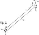
図2から図4までは非侵襲的シース3を示している。非侵襲的シースは、柔らかいプラスチックまたはゴムなどの弾力性のある素材でできたチューブである。非侵襲的シースの内径は関節鏡検査器具の外径上にあり、それと密接に適合した寸法になっている。非侵襲的シースの末端19は、関節鏡および/または堅いカニューレの末端側の先端の形状にきわめて近い形状になっている。シースの末端の周りに配置されているフランジ30は、堅いカニューレの末端側の先端が患者の組織を抉り取るのを防いでいる。フランジはシース壁と一体型であり、シースの軸に向かって内側に延びている。堅いカニューレの末端側の先端が外科手術中偶発的に末端方向に滑るのを防ぐ寸法になっている。非侵襲的シースの中には、開口部36が設けられているのもある。外科医が、開口部を通して、手術部位の空間に内視鏡や他の器具を挿入できるようにするためである。図3と4には、参考のため、光学器具の末端のレンズ31が示してある。
A
非侵襲的シースの基端32には、医療関係者が堅いカニューレ、関節鏡および/または関節鏡検査器具から非侵襲的シースを容易に引き離せるよう、タブ33が設けられている。非侵襲的シースの基端には、非侵襲的シースを固定するための取り付け部品も設けられている。関節鏡や他の器具上に配置された取り付け部品または開口部に接続されるロッキングハブやスナップラッチなどである。
The
タブ33は、非侵襲的シースに隣接している装置、例えばカメラ、光学機器、モータ、その他液体や湿気に敏感な器具から液体を迂回させて器具を守る寸法になっている。手術部位を出てシースの外面を流れる液体は、タブにより方向を変えられる。タブは、シースの内腔よりも大きく、放射状に延びている。
図2aは、シースの縦方向に沿って配置された2つのタブ33を有する非侵襲的シース3を示している。外科手術で多量の液体の流出が予想される場合でも、液体がシースの基端にある敏感な装置に近づくことがないように、追加のタブが保証する。追加のタブは、シースの縦方向に沿って配置されても良い。
FIG. 2a shows a
非侵襲的シースの外面には、関節鏡と堅いカニューレが手術部位内をさらに容易に移動できるように、滑らかなコーティングを施してもよい。例えば、シースにテフロン(登録商標)(PTFEまたは延伸ポリテトラフルオロエチレン)を施しても良いし、注水型潤滑剤で覆っても良い。これとは対照的に、非侵襲的シースの内面(チューブの内腔を形成する壁)には、滑り止めコーティングあるいは他の摩擦係数の高いコーティングを設けても良い。例えば、非侵襲的シースの内面を粘着性の同時押し出し熱可塑性エラストマ(TPE)でコーティングしても良い。滑り止めコーティングは、シースが堅いカニューレまたは関節鏡の上を滑りすぎるのを防ぐためのもので、非侵襲的シースが曲がったり関節鏡の上で滑ったりするのを予防できる。 The outer surface of the non-invasive sheath may be provided with a smooth coating so that the arthroscope and rigid cannula can more easily move within the surgical site. For example, Teflon (registered trademark) (PTFE or expanded polytetrafluoroethylene) may be applied to the sheath, or it may be covered with a water injection type lubricant. In contrast, the inner surface of the non-invasive sheath (the wall that forms the lumen of the tube) may be provided with a non-slip coating or other high coefficient of friction coating. For example, the inner surface of a non-invasive sheath may be coated with an adhesive coextruded thermoplastic elastomer (TPE). The non-slip coating prevents the sheath from slipping over the rigid cannula or arthroscope and can prevent the non-invasive sheath from bending or sliding over the arthroscope.
図3と4は、非侵襲的シース内に置かれる関節鏡検査器具や内視鏡あるいは関節鏡2に使用される非侵襲的シースを示している。図3に示されている非侵襲的シースには、シースの末端部分にバルーン34が設けられている。(バルーンはシースと一体型に成形しても良い。) バルーンは、外科医が組織内に空間を開け、手術領域を切開するのを可能にする。次に関節鏡を開口部36から末端方向に延ばし、手術部位の空間を明視化する。さらにシースの末端には、関節鏡の前に空間を開けるため、末端方向に突出したスプーンあるいは末端方向に突出した他の物体を設けても良い。このように、バルーンと末端方向に突出したスプーンは、組織を切開し、あるいは取り出し、外科手術用の小さい空間を形成する手段を提供する。
3 and 4 show a non-invasive sheath used for an arthroscopic instrument or endoscope or
図4は、第2作動チューブ35を有する非侵襲的シース3を示している。作動チューブは、洗浄液、ファイバーオプティックス、縫糸、針、探針あるいは外科用具が内腔を通過するのを可能にする。図4に示される非侵襲的シースは、図3に示された非侵襲的シースと組み合わせて、バルーンと作業チューブの両方を備えた非侵襲的シースを提供しても良い。
FIG. 4 shows a
図5は、図2に示される非侵襲的シース3とシース内に配置された関節鏡検査器具2の横断面を示す。シースを関節鏡から引き離すのを容易にするため、非侵襲的シースにはシースの基端にタブ33が設けられている。シースの末端には、光が関節鏡と手術部位の空間の間を通過できるよう、そして場合によっては、追加器具が関節鏡を通ってあるいはそれに沿って手術領域に入れるよう、開口部36が設けられる。シースの末端19の壁37はシースの他の部分より厚く、シースの末端にはフランジ30が形成される。(フランジはシースの内側に接触した素材による別のリングであっても良い。) フランジは関節鏡検査器具の鋭利な末端を覆い、器具が開口部36を通って末端方向に滑るのを防ぐ。非侵襲的シースの残りの壁は、シースと関節鏡検査器具全体の厚さを最小にするため、薄くなっている。
FIG. 5 shows a cross section of the
非侵襲的シースは使用時に準備されて、関節鏡検査器具の上に被せられる。(器具をシースに挿入すると考えても良い。) 次にシースで覆われた関節鏡検査器具は手術部位へ挿入され、そこで外科医が医療処置を施す。バルーンが設けられる場合は、バルーンは組織を切開するのに使用され、関節鏡が開口部36から末端方向に延長され、手術部位の空間を明視化するのを助ける。
A non-invasive sheath is prepared at the time of use and is placed over the arthroscopic instrument. (You may consider inserting the instrument into the sheath.) The arthroscopic instrument covered with the sheath is then inserted into the surgical site, where the surgeon performs the medical procedure. If a balloon is provided, the balloon is used to incise tissue and an arthroscope extends distally from the
図6および7は、流入/流出非侵襲的シース50およびシース内に配置された関節鏡2を示す。流入/流出非侵襲的シース50は、図2に示されたシース同様、関節鏡が組織を突いたり引っ掻いたりしても偶発的外傷から患者が守られるよう、弾力性のある素材でできている。シースの素材はラジオパークであっても良い。シース素材の好ましいデュロメーター硬度は、ショアD約40からショアD約90の範囲にある。この硬度範囲は、患者を偶発的外傷から守るのに十分な弾力性を有しており、しかもシース内の内腔がつぶれるのを防ぐのに十分な硬さでもある。
6 and 7 show the inflow / outflow
流入/流出シース50は、関節鏡か挿入される多内腔チューブである。内腔はそれぞれシースの末端部分51からシースの基端側部分52へ延びる。シースの基端側部分には、第1ポート53や第2ポート54のような1つまたはそれ以上の流体ポート、1つまたはそれ以上の止水栓55または流体スイッチ、逆止弁などの1つまたはそれ以上の弁、マニホルド56、あるいはシース内の流体の流れを制御する他の手段が設けられる。流入/流出シースの末端部分51には複数の孔57が設けられる。孔はそれぞれチューブ内の1つまたはそれ以上の内腔に通じているので、流体は手術部位とシース内の内腔との間を流れることができる。
Inflow /
手術前に、医療関係者または装置製造業者が関節鏡を流入/流出非侵襲的シースに挿入し、止めねじ、スナップオン付属装置、他の取り外し可能な付属装置またはシースを関節鏡に固定するための他の手段58で、シースを関節鏡に固定する。使用時、流入矢印60で示されるように、外科医は流体、好ましくは生理食塩水を流体源59から関節鏡を通して手術領域へ流すことができる。(関節鏡には、流体が関節鏡を通って手術領域へ流れることができるように、1つまたはそれ以上の内腔、ポートまたは作動チューブが設けられる。) 血液その他の流体および残骸物は、流出矢印61で示されるように、手術領域から孔57を通って流され、シースの1つまたはそれ以上の内腔を通って流れる。澄んだ生理食塩水の流入および濁った流体と残骸物の流出により、外科医は1つの器具を使って鮮明な手術領域を維持できる。その結果、洗浄器具の使用も必要ではなくなる。従って外科医は2度の切開だけで、鮮明な視野を維持しながら関節鏡視下手術を行うことができる。
Prior to surgery, medical personnel or device manufacturers insert the arthroscope into the inflow / outflow non-invasive sheath and secure the set screw, snap-on attachment, or other removable attachment or sheath to the arthroscope Other means 58 secure the sheath to the arthroscope. In use, as shown by the
図7は、真空源70を使って、あるいはシースマニホルド56に接続されているポート53などの流体ポートに操作可能な状態で取り付けられる重力ドレーンを使って、流体が流入/流出非侵襲的シースを通って流される様子も示している。流体は、関節鏡に接続されている第3ポート72または第4ポート73などの流体ポートに操作可能な状態で取り付けられている流体源59から(ポンプまたは重力フィードを使い)、関節鏡を通って提供される。関節鏡の性能または外科医のニーズによっても異なるが、真空源と流体源は、流入/流出非侵襲的シースまたは関節鏡に設けられる種々のポートに、異なる組み合わせで接続しても良い。例えば、真空源を流入/流出シースのポート73に接続し、流体源をポート72に接続しても良い。この場合、外科医は流入/流出非侵襲的シースだけを使って、手術部位へ流体を導入したり、そこから流体を流出させたりできる。従って、関節鏡には手術部位に流体を導入したり、そこから流体を流出させたりする機能がなかったとしても、外科医は流入/流出シースを使うことにより、洗浄器具の必要性を排除できる。いずれにせよ、手術部位へ流れ込んだり、そこから流出したりする流体の流速を自動的に監視および制御するため、圧力センサ、流速制御システムおよびフィードバック制御システムが設けられても良い。
FIG. 7 illustrates a non-invasive sheath for fluid inflow / outflow using a
図8は、図6に示される流入/流出シース3の末端部分の横断面である。流入/流出シース50には内壁81に境を接している中央内腔80があり、そこに関節鏡が挿入される。シースには4つの外側内腔、つまり第1外側内腔82、第2外側内腔83、第3外側内腔84および第4外側内腔85があり、それらは内壁81、外壁86および内壁と外壁の間に延びシースの縦方向に沿って延伸する比較的硬いリブ87に境を接している。外側内腔は環状である。シースの末端は、外側内腔82、83、84および85の部分で密閉されており、患者への外傷を防ぐため丸みを帯びた形状になっている。(関節鏡検査器具を収納するため、中央内腔は空洞である。) 外壁に配置された孔57または開口部により、流体は外側内腔に流入したり、そこから流出したりできる。例えば、内腔82と84を流体が手術部位へ導入される通路として用い、内腔83と85を流体が手術部位から流出する通路として使っても良い。別の外科手術では、流体を流し出すか導入するのに、4つの内腔すべてを使っても良い。従って、流入/流出非侵襲的シースを様々な方法で使用する選択肢が外科医に与えられる。(シースと関節鏡が手術部位に挿入された後、少なくとも1つの外側内腔が流体を流すために空洞になっている限り、シースには4つのリブより多いリブあるいはそれより少ないリブを形成してもかまわない。)
FIG. 8 is a cross-sectional view of the distal portion of the inflow /
図9から16までは、種々の流入/流出非侵襲的シースの末端部分の横断面を示している。図9は第2セットの内側内腔を有する流入/流出非侵襲的シースを示しており、第1内側内腔100、第2内側内腔101、第3内側内腔102および第3内側内腔103がそれに含まれる。この設計では、内側内腔すべてを使って流体を手術部位へ導入し、外側内腔82、83、84および85のすべてを使って流体を手術部位から流出させる(その逆でも良い)ことにより、外科医は流体交換率を高めることができる。
Figures 9 through 16 show cross sections of the distal portions of various inflow / outflow non-invasive sheaths. FIG. 9 shows an inflow / outflow non-invasive sheath having a second set of inner lumens, a first
図10は、内壁のない流入/流出シースを示している。内壁がない代わり、関節鏡がシースに挿入されると、関節鏡2の外面がシースの内壁として機能する。4つの比較的硬いリブ87が関節鏡の外面88と密着し、その結果、4つの外側内腔82、83、84および85が形成される。リブの端104には、リブと関節鏡の間の密着状態をさらに高めるため、弾力性のあるフランジまたは拡張部分を設けても良い。この構成であれば、流入/流出シースおよび関節鏡複合体の寸法を全体的に小さくできる。(外壁86をエラストマ系の素材で構成すれば、チューブは関節鏡の様々な寸法に合わせて放射状に延びる。)
FIG. 10 shows an inflow / outflow sheath without an inner wall. When the arthroscope is inserted into the sheath instead of having an inner wall, the outer surface of the
図10に示すように、関節鏡2は中央内腔80を通してシース50に挿入される。関節鏡2は、挿入の前に、第2の保護シースで覆っても良いし、覆わなくても良い。一旦挿入されたら、関節鏡2の外面88はリブ87のフランジまたは拡張部分と接触する。リブ87がフランジまたは拡張部分を有しない場合は、リブのランドを関節鏡2の外面と接触させるのに使用しても良い。関節鏡2の外面88がリブ87とリブフランジまたはリブ拡張部分に押し付ける力により、リブ87と関節鏡2の外面88が密着する。外側内腔82、83、84および85は、リブ、内視鏡88の外面および流入/流出シースの外壁86の内面89により形成される。リブはシースがつぶれるのを防ぐ縦方向の支柱として機能し、圧力下にあるシースを支える。リブは支えられていない薄い外壁の横方向における支間を減少させる。従って、シースのつぶれをさらに防ぐことができる。リブ87と関節鏡の外面88の接触により密着状態が形成されることにより、外側内腔82、83、84および85の相互間に流体が流れるのを防ぐことができる。外側内腔82、83、84および85は、患者1の外部から流体を手術部位に流入したり、手術部位から流体を流出させたりする連続操作を容易にする。流出する流体が手術部位へ逆戻りしたり、流入する流体がシースの基端から流出したりするのを防ぐため、外側内腔82、83、84および85内で、逆止弁またはゲートを流入/流出シース50の内壁に連結させても良い。
As shown in FIG. 10, the
図10に示された流入/流出シースの外径は、大き目の関節用として関節鏡検査器具用のシースが製造されるときは通常約5ないし7mmであるが、この寸法は関節鏡検査器具の直径次第で異なっていても良い。小さめの関節用として関節鏡検査器具用のシースが製造されるときは、シース50の外径は約2ないし3mmである。流入/流出シース50の外壁86の厚さは、チューブを構成する拡張部分および素材次第であるが、通常1mm以下である。流入/流出シース50は、シースの呼び径の+/−10%の範囲の関節鏡に適合できる。リブ87は流入/流出シースの内面から内側方向に延びており、関節鏡が挿入されると密着状態となる。
The outer diameter of the inflow / outflow sheath shown in FIG. 10 is typically about 5 to 7 mm when a sheath for an arthroscopy instrument is manufactured for a large joint, but this dimension is the size of the arthroscopy instrument. It may be different depending on the diameter. When a sheath for an arthroscopy instrument is manufactured for a smaller joint, the outer diameter of the
外径の小さい流入/流出シース50は関節鏡視下手術では特に有用である。流入/流出シース50は、そのユニークさの故に、カニューレの内壁が関節鏡の外壁に接触する必要のある多内腔カニューレと比べ、直径の30%減少を達成している。現在、関節鏡視下手術の技術は標準の三切開技術を使用している。最初の切開では、流入カニューレを挿入して関節を拡張させる。流入カニューレは、関節を滅菌流体で満たし、関節を広げ、外科医が観察したり作業したりするための空間を作るのに使用される。第2切開では、患者の手術部位を見るため関節鏡が挿入される。第3切開では、外科医は外傷または異常を矯正するため特殊な外科手術器具を挿入する。手術後、関節を流体で洗浄し、器具を外し、切開部を縫糸、ステープルまたはテーピングで閉じる。外科医は最近、関節鏡を使用した二切開技術の方へ移行し始めた。外科医は最初の切開で関節鏡を挿入し、第2の切開では特殊な外科手術器具を挿入する。流入/流出シース付きの関節鏡を使用するこの技術だと、第3切開部は必要ではなくなる。しかし、現在流入/流出に使用されているシースでは、手術部位への流体の流入および手術部位からの流体の流出を連続して同時に行うことはできないし、直径の小さなシースを使用しているわけでもない。現在のシースは、手術部位への流体の流入および手術部位からの流体の流出を交互に行っているにすぎず、直径も大きいので大きい切開が必要となる。本出願者の流入/流出シース50では、外側内腔82、83、84および85を使って、手術部位への流体の流入および手術部位からの流体の流出を実質的に同時に行うことができ、しかも小さな切開で済む。流入/流出を実質的に同時に行うことにより、外科医は手術部位を清浄に保ち、明瞭な視野を維持できる。
An inflow /
本出願者の流入/流出シース50のユニークな特徴は、シース50の流入を流出が超える許容差である。流出量が多ければ、手術部位からの残骸物や体液の排除が促進される。流入/流出シース50へ供給される流体圧は、通常、圧力水頭約6ないし8フィートである関節鏡標準拡張圧であるが、これは手術方法により異なっても良い。流入/流出シース50に使用される吸引力は、シースの大きさや手術方法により異なるが、約0ないし250mm/Hgである。流入/流出シースが5.7mm関節鏡と一緒に使用される場合は、手術部位への流体の流入は水柱6フィートで800ml/分の速度で行われ、手術部位からの流出は21mm/Hgの吸引で850ml/分で行われる。流出量が多ければ、洗浄液および手術中患者から排出される追加残骸物と体液の両方を排除できる。
A unique feature of Applicant's inflow /
図11は、図10に示されるのと類似の流入/流出非侵襲的シース50を示している。比較的硬いリブ87にはひだが付いているが、やはり関節鏡2の外壁と密着するので、関節鏡が一旦シースに挿入されると、外側内腔82、83、84および85が形成される。図11のシースは、図12に示されるようにひだ付きリブが大型の関節鏡を収納するのに必要な程度曲がるので、様々な大きさの関節鏡が収納できる。
FIG. 11 shows an inflow / outflow
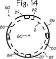
図13は、図11に示されているのと類似の流入/流出非侵襲的シース50を示している。このシースのリブは弾力性のあるチューブで、関節鏡2の外壁と密着し、一旦関節鏡がシースに挿入されると、外側内腔82、83、84および85が形成される。図13のシースは、図14に示されるようにチューブが大型の関節鏡を収納するのに必要な程度圧縮されるので、様々な大きさの関節鏡が収納できる。
FIG. 13 shows an inflow / outflow
図15は、C型または切れ目付き流入/流出シース50を示している。図8のシース同様、4つの外側内腔82、83、84および85が設けられ、3つのリブ87、内壁81および外壁86に境を接する。関節鏡2がシースに挿入されると、弓状の第1セグメントと弓状の第2セグメントのそれぞれの先端の間に、小さいギャップ105が形成される。(関節鏡が手術部位の空間に挿入されると、組織108がギャップを塞ぎ、流体が手術部位の空間から体外へ漏れるのを防ぐ。) 図15のシースは、図16に示されるように、大型の関節鏡がシースに挿入されると弓状のセグメントが外側へ放射状に移動するので、様々な大きさの関節鏡が収納できる。
FIG. 15 shows a C-shaped or scoring inflow /
関節鏡かシースから、突出物またはガイドレール109を任意に延長させても良い。ガイドレールは、ユーザが関節鏡をシースに挿入するとき、シースが関節鏡と整合するのを助ける。ガイドレールは、手術中にシースが関節鏡の上で思わぬ回転をしたり曲がったりするのも防ぐ。
The protrusion or
図17および18は、流入/流出非侵襲的シース50とシースに挿入された関節鏡2を示す。図6から図16までに示された流入/流出シースとは対照的に、シースの末端部分51の外壁86は連続したチューブ(すなわちシースの末端部分には孔は設けられない)でできている。しかし、図17のシースは、図8のシース同様、関節鏡を納めるための内側内腔、および第1外側内腔82、第2外側内腔83、第3外側内腔84および第4外側内腔85を含む流体の流入と流出のための4つの外側内腔を有している。外側内腔は内壁81、外壁86および支持リブ87に境を接している。図17に示される器具は、シースの末端110への流体の流入とそこからの流体の流出を助ける。
17 and 18 show the inflow / outflow
図19は、ぴったりと適合した末端部分111を有する流入/流出非侵襲的シース50を示している。末端部分は、関節鏡2の末端部分の外径にぴったり合う内径を有している。流体を導くシース部分112は、ぴったり適合したシースの末端部分111から基端方向に置かれている。流体導入部分112の外径およびぴったり適合した末端部分111の外径は、互いに同じシースの一部となるように一体型に成形しても良い。末端部分111の基端方向近傍にある流体導入部分112の孔57はシース内の1つまたはそれ以上の内腔と通じているので、外科医はそれを使って手術部位へ流体を導入したりそこから流体を流出させたりできる。図19に示されるシースは、シースが関節鏡の末端部分で関節鏡にぴったり適合するのであるから、比較的小さな半径の末端部分111を有している。従って、外科医は関節鏡を狭い手術部位へ挿入できる。さらに流体導入部分により、外科医はシース/関節鏡複合体を使って手術領域の洗浄が行える。
FIG. 19 shows an inflow / outflow
図20および21は、関節鏡2の上に配置された非侵襲性シース3とシースの基端側部分121上に配置された弾力性のあるグリップ120を示している。弾力性のある素材(例えば熱可塑性エラストマ)でできた空洞の人間工学的シリンダで、外科医の手が濡れていたとしても関節鏡とシースを容易に操作できる寸法になっているグリップ120が好ましい。グリップは、グリップの基端側部分122がシース3内に配置された関節鏡2上に延びるように、シースの基端32の基端方向に延びている。(図20のシースの基端側部分121は、グリップ内の位置を示すため、模型的に表示してある。) グリップの設計は、関節鏡検査器具の外径より小さい内径を有する形状になるよう、そして好ましくはシースの内側内腔の内径よりも小さい内径を有する形状になるよう、付勢されるようになっている。従って、図21の矢印123に示されるように、グリップはシース内に配置された器具に対して内側方向に放射状の力を発する。
20 and 21 show a
使用時には、グリップ120の基端側部分122によって、シース3内に配置された関節鏡2がつかまれる。グリップの基端側部分の手を離すと、グリップは最初の形状に戻るように付勢される。従って、手術中に関節鏡またはシースが操作されたとしても、関節鏡はシース内に安定した状態で存在できる。
In use, the
図22は、関節鏡2の上に配置された非侵襲的シース3、弾力性のあるグリップ120、そしてグリップの基端の開口部を広げる目的でグリップ内に配置されたレバー124および125の横断面を示している。図22に示されるグリップには、第1レバー124と第2レバー125を挿入するのに使用される第1チャネル126と第2チャネル127がそれぞれ設けられる。レバーには、バーブ、タングあるいは各チャネル内にレバーを固定するための他の手段が設けられる。レバーの末端部分は、シースから弓状に湾曲しながら離れていく形状になっている。
FIG. 22 shows the crossing of the
使用時、ユーザはレバーの末端部分を圧縮する。レバーの末端部分が放射状に内部方向に移動すると、レバーの基端側部分では、グリップの基端側部分の対応セグメントを放射状に外部方向に向ける力が加わり、グリップの基端側部分は放射状に外側方向に曲げられる。その結果、グリップの基端の開口部は拡張する。グリップの基端の開口部が拡張すれば、ユーザは容易に関節鏡をシースに挿入したり取り外したりできる。レバーの末端部分に配置された支点128は、レバーが予め決められた距離以上放射状に内側へ移動するのを防ぐ。支点があることにより、ユーザはグリップの基端側部分の対応セグメントにさらに強い力を加えことができるので、器具の挿入がさらに容易になる。
In use, the user compresses the end portion of the lever. When the distal end portion of the lever moves radially inward, the proximal end portion of the lever applies a force that radially directs the corresponding segment of the proximal end portion of the grip outward, and the proximal end portion of the grip radially Bent outward. As a result, the opening at the proximal end of the grip expands. If the opening at the proximal end of the grip expands, the user can easily insert and remove the arthroscope from the sheath. A
図23および24は、グリップ120の末端とグリップの末端から延びているレバー124と125を示している。図23では、参考のために、グリップから末端方向に延びているシース3が示してある。グリップ内に配置されたチャネル126と127は、レバーを収納するように、グリップを縦方向に通過して(あるいは部分的に通過して)延びている。使用時には、ユーザはレバーを圧縮し、グリップの基端側部分を広げる。それからユーザは関節鏡をシースに入れたり取り外したりする。
FIGS. 23 and 24 show the end of the
図25は、非侵襲的シース3の末端部分、およびシース3の末端140から末端方向に延びている関節鏡2を示している。シースの末端部分には孔57が設けられている。孔はシースの1つまたはそれ以上の内腔と通じている。内腔は、真空源、流体源、治療剤源またはその複合体に通じている。従って、孔は手術中、流体の流入/流出を助ける。
FIG. 25 shows the distal portion of the
シースの末端側の先端141は、シースの基端側部分の素材の弾力係数よりも大きい弾力係数を有する弾力性のある素材でできている。別の実施例では、柔軟性があり滅菌可能な単一のポリマを使って、シースおよび末端側の先端141を製造しても良い。さらにシースの末端側の先端は、ほとんどの関節鏡の外径よりもわずかに小さい内径を有している。別の実施形態では、柔軟性があり滅菌可能な単一のポリマを使って、シースおよび末端側の先端141を製造しても良い。
The
使用時、ユーザは関節鏡をシースに挿入する。シースの末端側の先端は、関節鏡の末端の端が末端側の先端を滑りながら通過するにつれて拡張する。先端の内径は関節鏡の外径より小さいので、先端は流体を通さないように関節鏡と密着する。 In use, the user inserts the arthroscope into the sheath. The distal tip of the sheath expands as the distal end of the arthroscope slides past the distal tip. Since the inner diameter of the tip is smaller than the outer diameter of the arthroscope, the tip is in close contact with the arthroscope so as not to pass fluid.
図26は、非侵襲的シース3およびシースの末端140を末端方向に延びている関節鏡2を示している。シースには孔57が設けられているので、手術中、流体の流入および流出が可能となる。シースの末端側の先端141は、シースの基端側部分の硬度よりも小さい硬度を有する弾力性のある素材でできている。先端には切れ目142が設けられているので、シースの末端部分に向かって拡張できる。使用時、ユーザーが関節鏡を先端に挿入するにしたがい、先端は拡張する。従って、シースは大型の関節鏡や他の医療器具も収納可能である。
FIG. 26 shows the
図27および28は、組織貯留機能113を有する連続流入/流出非侵襲的シース50を示している。シースの基端側部分52の外面は、シースまたは器具が手術領域から偶発的に抜け落ちるのを防ぐため、波形になっている。あるいはねじ山114が設けられている。組織貯留機能113のねじ山114はシースの周囲に配置されている。図27に示されるように、放射状に外部方向に延びる直列形のねじ山であっても良い。組織貯留機能113のねじ山114は、図28に示されるように、ねじ山付きスクリュー型であっても良い。
FIGS. 27 and 28 show a continuous inflow / outflow
図29および30は、組織貯留機能113なしに非侵襲的シース50上に使用される分離型の組織貯留スリーブ115に、組織貯留機能が組み込まれている状態を示している。この実施形態では、組織貯留スリーブは非侵襲的シース上に適合する寸法を有している。組織貯留スリーブは、部品が非侵襲的シースの外部表面上を滑って所定の位置に配置されたら、それ以上容易に移動できないような摩擦係数を有するエラストマを使って製造される。スリーブの摩擦は外科手術器具や非侵襲的シースに適合している。エラストマは患者用として滅菌可能である。組織貯留スリーブの外面には、組織貯留スリーブをシースか器具に使用した場合、シースまたは器具が手術領域から偶発的に抜け落ちるのを防ぐため、波形になっている。あるいはねじ山が設けられている。スリーブ上のねじ山114はスリーブ115の外面の周囲に配置されているが、放射状に外側方向に延びる直列形のねじ山であっても良い。ねじ山114はねじ山付きスクリュー型であっても良い。
FIGS. 29 and 30 show a state in which the tissue storage function is incorporated into a separate
非侵襲的シースの構成は、異なる形状の器具に適合する設計または寸法にしても良い。本発明のシースは、周囲の組織を偶発的な外傷から保護する必要のある他の医療器具や外科手術にも有用である。例えば、非侵襲的シースは、関節鏡を用いる外科手術またはレーザやRFエネルギー器具などのエネルギー供給医療器具使用中に、トリミング器具上に配置しても良い。非侵襲的シースが役に立つ他の手法には、腹腔鏡を用いた手術や他の内視鏡を用いた手術なども含まれる。さらに、本出願に示されている種々の構成のシースを組み合わせて、別のタイプの器具用シースを成形しても良い。本出願には開発された環境との関連で好ましい方法の実施形態だけが示されているが、それは本発明の原理を単に例示したものにすぎない。本発明の精神および添付の特許請求の範囲から逸脱することなく、別の実施形態や構成も考案可能である。 The non-invasive sheath configuration may be designed or dimensioned to accommodate different shaped instruments. The sheath of the present invention is also useful for other medical instruments and surgical procedures where the surrounding tissue needs to be protected from accidental trauma. For example, a non-invasive sheath may be placed on a trimming instrument during surgery using an arthroscope or using an energy delivery medical instrument such as a laser or RF energy instrument. Other techniques that benefit from a non-invasive sheath include surgery using a laparoscope and surgery using other endoscopes. Furthermore, other types of sheaths for instruments may be formed by combining the sheaths of various configurations shown in the present application. Although only preferred method embodiments are shown in this application in the context of the developed environment, they are merely illustrative of the principles of the present invention. Other embodiments and configurations may be devised without departing from the spirit of the invention and the scope of the appended claims.
2…関節鏡検査器具
3…非侵襲的シース
13…トリミング器具
15…洗浄器具
17…光学器具
18…カニューレ
19…先端または末端
30…フランジ
31…レンズ
32…基端
33…タブ
34…バルーン
35…第2作動チューブ
2 ...
Claims (10)
関節鏡を使った外科手術を行うのに適した関節鏡検査器具と、
基端側部分と基端により特徴付けられるチューブを含む非侵襲的シースで、前記シースは関節鏡検査器具の外径にきわめて近い寸法の内径を有し、前記関節鏡検査器具上に取り外し可能な状態で配置されている非侵襲的シースと、
前記シースの前記基端側部分に配置された円筒状のグリップで、前記基端側部分により特徴付けられているグリップと、
を備え、
前記グリップの基端側部分は前記シースの基端を越えて基端方向に延びており、
前記グリップの基端側部分の素材および前記グリップの基端側部分の寸法は、前記関節鏡検査器具の外径よりも小さい内径を有する形に付勢されるように選択され、これにより前記グリップの基端側部分は前記関節鏡検査器具をつかむことができるようになっており、
操作可能な状態で前記グリップに取り付けられており、前記グリップの基端側部分の周方向の第1部分と第2部分を放射状に外側に移動させることのできる第1レバーと第2レバーをさらに含む、システム。A system for performing a surgical operation using an arthroscope,
An arthroscopic instrument suitable for performing arthroscopic surgery,
A non-invasive sheath comprising a proximal portion and a tube characterized by a proximal end, the sheath having an inner diameter that is very close to the outer diameter of the arthroscopic instrument and removable on the arthroscopic instrument A non-invasive sheath placed in a state;
A cylindrical grip disposed on the proximal portion of the sheath, characterized by the proximal portion;
With
A proximal end portion of the grip extends in a proximal direction beyond the proximal end of the sheath;
The material of the proximal end portion of the grip and the dimensions of the proximal end portion of the grip are selected to be biased into a shape having an inner diameter that is smaller than the outer diameter of the arthroscopic instrument, whereby the grip The proximal end portion of can be used to grab the arthroscopy instrument,
A first lever and a second lever that are attached to the grip in an operable state, and that can radially move the first portion and the second portion in the circumferential direction of the proximal end portion of the grip further outward. Including the system.
前記第1レバーと前記第2レバーの末端部分は、前記シースから放射状に外側へ延びた弓状のセグメントを含むことを特徴とする請求項1記載のシステム。The first lever and the second lever extend in a distal direction from the grip,
Before SL-terminal portion of the first lever and the second lever system of claim 1, wherein it contains an arcuate segment extending radially outwardly from the sheath.
前記末端側の先端は前記シースの内径よりも小さい内径を有することを特徴とする請求項1記載のシステム。The tip of the front Symbol distal consists of a material having a softer hardness than the hardness of the sheath,
The system of claim 1, wherein the distal tip has an inner diameter that is smaller than an inner diameter of the sheath.
Applications Claiming Priority (7)
| Application Number | Priority Date | Filing Date | Title |
|---|---|---|---|
| US10/769,629 US7413542B2 (en) | 2004-01-29 | 2004-01-29 | Atraumatic arthroscopic instrument sheath |
| US10/769,629 | 2004-01-29 | ||
| US11/016,274 | 2004-12-17 | ||
| US11/016,274 US7435214B2 (en) | 2004-01-29 | 2004-12-17 | Atraumatic arthroscopic instrument sheath |
| US11/031,149 | 2005-01-07 | ||
| US11/031,149 US7445596B2 (en) | 2004-01-29 | 2005-01-07 | Atraumatic arthroscopic instrument sheath |
| PCT/US2005/002720 WO2005072402A2 (en) | 2004-01-29 | 2005-01-28 | Atraumatic arthroscopic instrument sheath |
Publications (3)
| Publication Number | Publication Date |
|---|---|
| JP2007522837A JP2007522837A (en) | 2007-08-16 |
| JP2007522837A5 JP2007522837A5 (en) | 2008-03-21 |
| JP5025269B2 true JP5025269B2 (en) | 2012-09-12 |
Family
ID=34831025
Family Applications (1)
| Application Number | Title | Priority Date | Filing Date |
|---|---|---|---|
| JP2006551498A Active JP5025269B2 (en) | 2004-01-29 | 2005-01-28 | Noninvasive arthroscopy instrument sheath |
Country Status (3)
| Country | Link |
|---|---|
| EP (1) | EP1748722A4 (en) |
| JP (1) | JP5025269B2 (en) |
| WO (1) | WO2005072402A2 (en) |
Families Citing this family (30)
| Publication number | Priority date | Publication date | Assignee | Title |
|---|---|---|---|---|
| US7413542B2 (en) | 2004-01-29 | 2008-08-19 | Cannuflow, Inc. | Atraumatic arthroscopic instrument sheath |
| US7500947B2 (en) * | 2004-01-29 | 2009-03-10 | Cannonflow, Inc. | Atraumatic arthroscopic instrument sheath |
| US7445596B2 (en) | 2004-01-29 | 2008-11-04 | Cannuflow, Inc. | Atraumatic arthroscopic instrument sheath |
| US7435214B2 (en) | 2004-01-29 | 2008-10-14 | Cannuflow, Inc. | Atraumatic arthroscopic instrument sheath |
| US7575565B2 (en) | 2005-08-19 | 2009-08-18 | Cannuflow, Inc. | Extravasation minimization device |
| US7503893B2 (en) * | 2006-02-03 | 2009-03-17 | Cannuflow, Inc. | Anti-extravasation sheath and method |
| US8652090B2 (en) * | 2006-05-18 | 2014-02-18 | Cannuflow, Inc. | Anti-extravasation surgical portal plug |
| US8226548B2 (en) * | 2007-07-07 | 2012-07-24 | Cannuflow, Inc. | Rigid arthroscope system |
| FR2945723B1 (en) * | 2009-05-19 | 2012-06-15 | Axess Vision Technology | MEDICAL INSTRUMENT WITH MULTIPLE FUNCTIONS FOR ENDOSCOPE. |
| WO2010150666A1 (en) | 2009-06-22 | 2010-12-29 | オリンパスメディカルシステムズ株式会社 | Endoscope flushing sheath |
| EP2347777A1 (en) * | 2010-01-26 | 2011-07-27 | Vesalius Medical Technologies Bvba | Bursitis treatment device and method |
| US9375139B2 (en) | 2010-07-29 | 2016-06-28 | Cannuflow, Inc. | Arthroscopic system |
| JP2013252155A (en) * | 2010-09-28 | 2013-12-19 | Terumo Corp | Puncture needle for bone cement injection and method of manufacturing the same |
| JP5554203B2 (en) * | 2010-10-14 | 2014-07-23 | 日機装株式会社 | Trocar |
| KR101132841B1 (en) | 2011-03-07 | 2012-04-02 | 김영재 | A suture |
| KR101185583B1 (en) | 2011-12-27 | 2012-09-24 | 김영재 | A suture which need not be knotted and a kit comprising the suture |
| EP2811919B1 (en) * | 2012-02-10 | 2020-09-09 | Merit Medical Systems, Inc. | Snare introducer |
| US8852091B2 (en) | 2012-04-04 | 2014-10-07 | Alcon Research, Ltd. | Devices, systems, and methods for pupil expansion |
| DE102012213205A1 (en) * | 2012-07-26 | 2014-05-15 | Aesculap Ag | Medical introducer with drawn insertion channel |
| US10010317B2 (en) * | 2012-12-05 | 2018-07-03 | Young Jae Kim | Method of improving elasticity of tissue of living body |
| DE102013102024A1 (en) * | 2013-02-01 | 2014-08-21 | Firma Trokamed Gmbh | arthroscopy shaft |
| JP6145580B2 (en) | 2013-12-06 | 2017-06-14 | ワイ.ジェイコブス メディカル インコーポレーテッド | Medical tube insertion device and medical tube insertion treatment kit including the same |
| WO2016018643A1 (en) * | 2014-07-30 | 2016-02-04 | Medovex Corp. | Surgical tools for spinal facet therapy to alleviate pain and related methods |
| CN105963809B (en) * | 2016-05-26 | 2018-11-13 | 广州医科大学附属第一医院 | A kind of percutaneous puncture visually rinses attraction system and its application method |
| WO2018207895A1 (en) * | 2017-05-12 | 2018-11-15 | 国立大学法人香川大学 | Instrument holding tool and medical instrument supply tool |
| US10888364B2 (en) | 2018-01-02 | 2021-01-12 | Medtronic Holding Company Sarl | Scoop cannula with deflectable wings |
| WO2020092080A1 (en) * | 2018-10-29 | 2020-05-07 | Stryker Corporation | Systems and methods of performing spine surgery and maintaining a volume of fluid at a surgical site |
| JP7078522B2 (en) * | 2018-11-19 | 2022-05-31 | 日機装株式会社 | Trocar |
| CN114727829A (en) * | 2020-01-29 | 2022-07-08 | 博戈外科股份有限公司 | Endoscopic surgery robot |
| US11583315B2 (en) * | 2020-11-09 | 2023-02-21 | Covidien Lp | Surgical access device including variable length cannula |
Family Cites Families (17)
| Publication number | Priority date | Publication date | Assignee | Title |
|---|---|---|---|---|
| SE442377B (en) * | 1984-06-29 | 1985-12-23 | Mediplast Ab | CATS, HEALTH OR SIMILAR DEVICE |
| GB8424436D0 (en) * | 1984-09-27 | 1984-10-31 | Pratt Int Ltd Burnerd | Surgical appliance |
| US4886049A (en) * | 1988-05-17 | 1989-12-12 | Darras Robert L | Medical instrument cover |
| US4959058A (en) * | 1989-03-17 | 1990-09-25 | Michelson Gary K | Cannula having side opening |
| US4973321A (en) * | 1989-03-17 | 1990-11-27 | Michelson Gary K | Cannula for an arthroscope |
| US5237984A (en) * | 1991-06-24 | 1993-08-24 | Xomed-Treace Inc. | Sheath for endoscope |
| US5273545A (en) * | 1991-10-15 | 1993-12-28 | Apple Medical Corporation | Endoscopic cannula with tricuspid leaf valve |
| JP3126064B2 (en) * | 1992-04-14 | 2001-01-22 | オリンパス光学工業株式会社 | Trocar |
| JPH06217988A (en) * | 1993-01-26 | 1994-08-09 | Terumo Corp | Blood vessel sticking instrument |
| DE59400518D1 (en) * | 1993-06-16 | 1996-09-26 | White Spot Ag | DEVICE FOR PUTTING FIBRINE ADHESIVE INTO A STITCH CHANNEL |
| US5545150A (en) * | 1994-05-06 | 1996-08-13 | Endoscopic Concepts, Inc. | Trocar |
| US5569183A (en) * | 1994-06-01 | 1996-10-29 | Archimedes Surgical, Inc. | Method for performing surgery around a viewing space in the interior of the body |
| JP3737540B2 (en) * | 1995-03-28 | 2006-01-18 | テルモ株式会社 | Trocar mantle and trocar |
| JPH1176247A (en) * | 1997-07-11 | 1999-03-23 | Olympus Optical Co Ltd | Surgical operation system |
| US5916145A (en) * | 1998-08-07 | 1999-06-29 | Scimed Life Systems, Inc. | Device and method of using a surgical assembly with mesh sheath |
| GB9927338D0 (en) * | 1999-11-18 | 2000-01-12 | Gyrus Medical Ltd | Electrosurgical system |
| US20030018340A1 (en) * | 2001-06-29 | 2003-01-23 | Branch Thomas P. | Method and apparatus for installing cannula |
-
2005
- 2005-01-28 WO PCT/US2005/002720 patent/WO2005072402A2/en active Application Filing
- 2005-01-28 JP JP2006551498A patent/JP5025269B2/en active Active
- 2005-01-28 EP EP05712239A patent/EP1748722A4/en not_active Withdrawn
Also Published As
| Publication number | Publication date |
|---|---|
| EP1748722A2 (en) | 2007-02-07 |
| JP2007522837A (en) | 2007-08-16 |
| EP1748722A4 (en) | 2009-11-11 |
| WO2005072402A3 (en) | 2006-12-07 |
| WO2005072402A2 (en) | 2005-08-11 |
Similar Documents
| Publication | Publication Date | Title |
|---|---|---|
| JP5025269B2 (en) | Noninvasive arthroscopy instrument sheath | |
| US9375207B2 (en) | Atraumatic arthroscopic instrument sheath | |
| US20200337531A1 (en) | Rigid Endoscope System | |
| US9872604B2 (en) | Atraumatic arthroscopic instrument sheath and method | |
| US9827009B2 (en) | Atraumatic arthroscopic instrument sheath | |
| US9364204B2 (en) | Atraumatic arthroscopic instrument sheath |
Legal Events
| Date | Code | Title | Description |
|---|---|---|---|
| A521 | Request for written amendment filed |
Free format text: JAPANESE INTERMEDIATE CODE: A523 Effective date: 20080128 |
|
| A621 | Written request for application examination |
Free format text: JAPANESE INTERMEDIATE CODE: A621 Effective date: 20080128 |
|
| RD04 | Notification of resignation of power of attorney |
Free format text: JAPANESE INTERMEDIATE CODE: A7424 Effective date: 20090212 |
|
| A131 | Notification of reasons for refusal |
Free format text: JAPANESE INTERMEDIATE CODE: A131 Effective date: 20100824 |
|
| A601 | Written request for extension of time |
Free format text: JAPANESE INTERMEDIATE CODE: A601 Effective date: 20101122 |
|
| A602 | Written permission of extension of time |
Free format text: JAPANESE INTERMEDIATE CODE: A602 Effective date: 20101130 |
|
| A601 | Written request for extension of time |
Free format text: JAPANESE INTERMEDIATE CODE: A601 Effective date: 20101222 |
|
| A602 | Written permission of extension of time |
Free format text: JAPANESE INTERMEDIATE CODE: A602 Effective date: 20110105 |
|
| A601 | Written request for extension of time |
Free format text: JAPANESE INTERMEDIATE CODE: A601 Effective date: 20110124 |
|
| A602 | Written permission of extension of time |
Free format text: JAPANESE INTERMEDIATE CODE: A602 Effective date: 20110131 |
|
| A521 | Request for written amendment filed |
Free format text: JAPANESE INTERMEDIATE CODE: A523 Effective date: 20110218 |
|
| A131 | Notification of reasons for refusal |
Free format text: JAPANESE INTERMEDIATE CODE: A131 Effective date: 20110517 |
|
| A601 | Written request for extension of time |
Free format text: JAPANESE INTERMEDIATE CODE: A601 Effective date: 20110817 |
|
| A602 | Written permission of extension of time |
Free format text: JAPANESE INTERMEDIATE CODE: A602 Effective date: 20110824 |
|
| A601 | Written request for extension of time |
Free format text: JAPANESE INTERMEDIATE CODE: A601 Effective date: 20110920 |
|
| A602 | Written permission of extension of time |
Free format text: JAPANESE INTERMEDIATE CODE: A602 Effective date: 20110928 |
|
| A521 | Request for written amendment filed |
Free format text: JAPANESE INTERMEDIATE CODE: A523 Effective date: 20111014 |
|
| A131 | Notification of reasons for refusal |
Free format text: JAPANESE INTERMEDIATE CODE: A131 Effective date: 20120124 |
|
| A521 | Request for written amendment filed |
Free format text: JAPANESE INTERMEDIATE CODE: A523 Effective date: 20120424 |
|
| TRDD | Decision of grant or rejection written | ||
| A01 | Written decision to grant a patent or to grant a registration (utility model) |
Free format text: JAPANESE INTERMEDIATE CODE: A01 Effective date: 20120612 |
|
| A01 | Written decision to grant a patent or to grant a registration (utility model) |
Free format text: JAPANESE INTERMEDIATE CODE: A01 |
|
| A61 | First payment of annual fees (during grant procedure) |
Free format text: JAPANESE INTERMEDIATE CODE: A61 Effective date: 20120619 |
|
| FPAY | Renewal fee payment (event date is renewal date of database) |
Free format text: PAYMENT UNTIL: 20150629 Year of fee payment: 3 |
|
| R150 | Certificate of patent or registration of utility model |
Free format text: JAPANESE INTERMEDIATE CODE: R150 Ref document number: 5025269 Country of ref document: JP Free format text: JAPANESE INTERMEDIATE CODE: R150 |
|
| R250 | Receipt of annual fees |
Free format text: JAPANESE INTERMEDIATE CODE: R250 |
|
| R250 | Receipt of annual fees |
Free format text: JAPANESE INTERMEDIATE CODE: R250 |
|
| R250 | Receipt of annual fees |
Free format text: JAPANESE INTERMEDIATE CODE: R250 |
|
| R250 | Receipt of annual fees |
Free format text: JAPANESE INTERMEDIATE CODE: R250 |
|
| R250 | Receipt of annual fees |
Free format text: JAPANESE INTERMEDIATE CODE: R250 |
|
| R250 | Receipt of annual fees |
Free format text: JAPANESE INTERMEDIATE CODE: R250 |
|
| R250 | Receipt of annual fees |
Free format text: JAPANESE INTERMEDIATE CODE: R250 |
|
| R250 | Receipt of annual fees |
Free format text: JAPANESE INTERMEDIATE CODE: R250 |
|
| S531 | Written request for registration of change of domicile |
Free format text: JAPANESE INTERMEDIATE CODE: R313531 |
|
| S111 | Request for change of ownership or part of ownership |
Free format text: JAPANESE INTERMEDIATE CODE: R313113 |
|
| R350 | Written notification of registration of transfer |
Free format text: JAPANESE INTERMEDIATE CODE: R350 |
|
| R350 | Written notification of registration of transfer |
Free format text: JAPANESE INTERMEDIATE CODE: R350 |
|
| R250 | Receipt of annual fees |
Free format text: JAPANESE INTERMEDIATE CODE: R250 |
|
| R250 | Receipt of annual fees |
Free format text: JAPANESE INTERMEDIATE CODE: R250 |


