JP4895802B2 - Composition for chemoembolization of solid tumors - Google Patents
Composition for chemoembolization of solid tumors Download PDFInfo
- Publication number
- JP4895802B2 JP4895802B2 JP2006500252A JP2006500252A JP4895802B2 JP 4895802 B2 JP4895802 B2 JP 4895802B2 JP 2006500252 A JP2006500252 A JP 2006500252A JP 2006500252 A JP2006500252 A JP 2006500252A JP 4895802 B2 JP4895802 B2 JP 4895802B2
- Authority
- JP
- Japan
- Prior art keywords
- composition according
- microspheres
- particles
- water
- doxorubicin
- Prior art date
- Legal status (The legal status is an assumption and is not a legal conclusion. Google has not performed a legal analysis and makes no representation as to the accuracy of the status listed.)
- Expired - Lifetime
Links
- 239000000203 mixture Substances 0.000 title claims abstract description 55
- 206010028980 Neoplasm Diseases 0.000 title claims abstract description 45
- 230000010109 chemoembolization Effects 0.000 title description 2
- 239000003814 drug Substances 0.000 claims abstract description 77
- 229940079593 drug Drugs 0.000 claims abstract description 66
- 239000002245 particle Substances 0.000 claims abstract description 42
- 229920002451 polyvinyl alcohol Polymers 0.000 claims abstract description 39
- 229920000642 polymer Polymers 0.000 claims abstract description 30
- 229940045799 anthracyclines and related substance Drugs 0.000 claims abstract description 24
- 239000004005 microsphere Substances 0.000 claims description 113
- 239000000243 solution Substances 0.000 claims description 51
- XLYOFNOQVPJJNP-UHFFFAOYSA-N water Substances O XLYOFNOQVPJJNP-UHFFFAOYSA-N 0.000 claims description 39
- 239000000178 monomer Substances 0.000 claims description 38
- 238000000034 method Methods 0.000 claims description 36
- 238000011282 treatment Methods 0.000 claims description 31
- 230000010102 embolization Effects 0.000 claims description 28
- 239000007788 liquid Substances 0.000 claims description 18
- 230000008569 process Effects 0.000 claims description 18
- 125000000129 anionic group Chemical group 0.000 claims description 14
- 239000003795 chemical substances by application Substances 0.000 claims description 12
- 238000004108 freeze drying Methods 0.000 claims description 11
- 239000002872 contrast media Substances 0.000 claims description 10
- 206010073071 hepatocellular carcinoma Diseases 0.000 claims description 9
- 231100000844 hepatocellular carcinoma Toxicity 0.000 claims description 9
- 239000011159 matrix material Substances 0.000 claims description 9
- 125000004178 (C1-C4) alkyl group Chemical group 0.000 claims description 8
- 210000004204 blood vessel Anatomy 0.000 claims description 8
- 239000001257 hydrogen Substances 0.000 claims description 8
- 229910052739 hydrogen Inorganic materials 0.000 claims description 8
- 239000000725 suspension Substances 0.000 claims description 8
- 229940124597 therapeutic agent Drugs 0.000 claims description 8
- 150000001875 compounds Chemical class 0.000 claims description 7
- 239000002002 slurry Substances 0.000 claims description 7
- UFHFLCQGNIYNRP-UHFFFAOYSA-N Hydrogen Chemical compound [H][H] UFHFLCQGNIYNRP-UHFFFAOYSA-N 0.000 claims description 6
- 238000003860 storage Methods 0.000 claims description 6
- 239000000758 substrate Substances 0.000 claims description 5
- QAOWNCQODCNURD-UHFFFAOYSA-N Sulfuric acid Chemical group OS(O)(=O)=O QAOWNCQODCNURD-UHFFFAOYSA-N 0.000 claims description 4
- 125000003277 amino group Chemical group 0.000 claims description 4
- 239000007864 aqueous solution Substances 0.000 claims description 4
- 125000004432 carbon atom Chemical group C* 0.000 claims description 4
- 125000002887 hydroxy group Chemical group [H]O* 0.000 claims description 4
- 238000004519 manufacturing process Methods 0.000 claims description 4
- 125000002496 methyl group Chemical group [H]C([H])([H])* 0.000 claims description 3
- 238000002156 mixing Methods 0.000 claims description 3
- LSNNMFCWUKXFEE-UHFFFAOYSA-M Bisulfite Chemical group OS([O-])=O LSNNMFCWUKXFEE-UHFFFAOYSA-M 0.000 claims description 2
- GRYLNZFGIOXLOG-UHFFFAOYSA-N Nitric acid Chemical group O[N+]([O-])=O GRYLNZFGIOXLOG-UHFFFAOYSA-N 0.000 claims description 2
- NBIIXXVUZAFLBC-UHFFFAOYSA-N Phosphoric acid Chemical group OP(O)(O)=O NBIIXXVUZAFLBC-UHFFFAOYSA-N 0.000 claims description 2
- DHKHKXVYLBGOIT-UHFFFAOYSA-N acetaldehyde Diethyl Acetal Natural products CCOC(C)OCC DHKHKXVYLBGOIT-UHFFFAOYSA-N 0.000 claims description 2
- 229910052799 carbon Inorganic materials 0.000 claims description 2
- BVKZGUZCCUSVTD-UHFFFAOYSA-N carbonic acid Chemical group OC(O)=O BVKZGUZCCUSVTD-UHFFFAOYSA-N 0.000 claims description 2
- 150000001732 carboxylic acid derivatives Chemical group 0.000 claims description 2
- 238000007334 copolymerization reaction Methods 0.000 claims description 2
- 125000000816 ethylene group Chemical group [H]C([H])([*:1])C([H])([H])[*:2] 0.000 claims description 2
- 125000001153 fluoro group Chemical group F* 0.000 claims description 2
- 125000004435 hydrogen atom Chemical group [H]* 0.000 claims description 2
- 229910017604 nitric acid Inorganic materials 0.000 claims description 2
- UEZVMMHDMIWARA-UHFFFAOYSA-M phosphonate Chemical group [O-]P(=O)=O UEZVMMHDMIWARA-UHFFFAOYSA-M 0.000 claims description 2
- 239000006228 supernatant Substances 0.000 claims description 2
- 229920003176 water-insoluble polymer Polymers 0.000 claims description 2
- 230000002792 vascular Effects 0.000 claims 2
- 125000004430 oxygen atom Chemical group O* 0.000 claims 1
- AOJJSUZBOXZQNB-TZSSRYMLSA-N Doxorubicin Chemical compound O([C@H]1C[C@@](O)(CC=2C(O)=C3C(=O)C=4C=CC=C(C=4C(=O)C3=C(O)C=21)OC)C(=O)CO)[C@H]1C[C@H](N)[C@H](O)[C@H](C)O1 AOJJSUZBOXZQNB-TZSSRYMLSA-N 0.000 abstract description 156
- 229960004679 doxorubicin Drugs 0.000 abstract description 76
- -1 poly(vinyl alcohol) Polymers 0.000 abstract description 4
- 229920006318 anionic polymer Polymers 0.000 abstract 1
- CSCPPACGZOOCGX-UHFFFAOYSA-N Acetone Chemical compound CC(C)=O CSCPPACGZOOCGX-UHFFFAOYSA-N 0.000 description 27
- 238000011068 loading method Methods 0.000 description 22
- XHZPRMZZQOIPDS-UHFFFAOYSA-N 2-Methyl-2-[(1-oxo-2-propenyl)amino]-1-propanesulfonic acid Chemical compound OS(=O)(=O)CC(C)(C)NC(=O)C=C XHZPRMZZQOIPDS-UHFFFAOYSA-N 0.000 description 20
- XEKOWRVHYACXOJ-UHFFFAOYSA-N Ethyl acetate Chemical compound CCOC(C)=O XEKOWRVHYACXOJ-UHFFFAOYSA-N 0.000 description 18
- 241001465754 Metazoa Species 0.000 description 18
- 230000003073 embolic effect Effects 0.000 description 17
- 239000011324 bead Substances 0.000 description 16
- 239000000126 substance Substances 0.000 description 16
- FAPWRFPIFSIZLT-UHFFFAOYSA-M Sodium chloride Chemical compound [Na+].[Cl-] FAPWRFPIFSIZLT-UHFFFAOYSA-M 0.000 description 15
- 239000000047 product Substances 0.000 description 15
- 238000002474 experimental method Methods 0.000 description 13
- 241000283973 Oryctolagus cuniculus Species 0.000 description 12
- 239000013543 active substance Substances 0.000 description 12
- 230000000694 effects Effects 0.000 description 12
- 210000004185 liver Anatomy 0.000 description 12
- 238000006243 chemical reaction Methods 0.000 description 11
- 239000011521 glass Substances 0.000 description 11
- 239000011780 sodium chloride Substances 0.000 description 11
- 238000010828 elution Methods 0.000 description 10
- 238000006116 polymerization reaction Methods 0.000 description 9
- 239000000523 sample Substances 0.000 description 9
- BPYKTIZUTYGOLE-IFADSCNNSA-N Bilirubin Chemical compound N1C(=O)C(C)=C(C=C)\C1=C\C1=C(C)C(CCC(O)=O)=C(CC2=C(C(C)=C(\C=C/3C(=C(C=C)C(=O)N\3)C)N2)CCC(O)=O)N1 BPYKTIZUTYGOLE-IFADSCNNSA-N 0.000 description 8
- DKPFZGUDAPQIHT-UHFFFAOYSA-N Butyl acetate Natural products CCCCOC(C)=O DKPFZGUDAPQIHT-UHFFFAOYSA-N 0.000 description 8
- 239000002904 solvent Substances 0.000 description 8
- NIXOWILDQLNWCW-UHFFFAOYSA-N acrylic acid group Chemical group C(C=C)(=O)O NIXOWILDQLNWCW-UHFFFAOYSA-N 0.000 description 7
- 239000000463 material Substances 0.000 description 7
- VEXZGXHMUGYJMC-UHFFFAOYSA-N Hydrochloric acid Chemical compound Cl VEXZGXHMUGYJMC-UHFFFAOYSA-N 0.000 description 6
- HEMHJVSKTPXQMS-UHFFFAOYSA-M Sodium hydroxide Chemical compound [OH-].[Na+] HEMHJVSKTPXQMS-UHFFFAOYSA-M 0.000 description 6
- 238000002835 absorbance Methods 0.000 description 6
- 230000002378 acidificating effect Effects 0.000 description 6
- 229920006217 cellulose acetate butyrate Polymers 0.000 description 6
- 238000005259 measurement Methods 0.000 description 6
- 239000012071 phase Substances 0.000 description 6
- 229920000307 polymer substrate Polymers 0.000 description 6
- 238000002360 preparation method Methods 0.000 description 6
- 206010002091 Anaesthesia Diseases 0.000 description 5
- KWYHDKDOAIKMQN-UHFFFAOYSA-N N,N,N',N'-tetramethylethylenediamine Chemical compound CN(C)CCN(C)C KWYHDKDOAIKMQN-UHFFFAOYSA-N 0.000 description 5
- 239000004372 Polyvinyl alcohol Substances 0.000 description 5
- 230000037005 anaesthesia Effects 0.000 description 5
- 239000008346 aqueous phase Substances 0.000 description 5
- 210000001367 artery Anatomy 0.000 description 5
- 201000011510 cancer Diseases 0.000 description 5
- 210000004027 cell Anatomy 0.000 description 5
- 238000004132 cross linking Methods 0.000 description 5
- 238000011026 diafiltration Methods 0.000 description 5
- 238000001035 drying Methods 0.000 description 5
- 238000002347 injection Methods 0.000 description 5
- 239000007924 injection Substances 0.000 description 5
- 230000004044 response Effects 0.000 description 5
- 238000003756 stirring Methods 0.000 description 5
- IJGRMHOSHXDMSA-UHFFFAOYSA-N Atomic nitrogen Chemical compound N#N IJGRMHOSHXDMSA-UHFFFAOYSA-N 0.000 description 4
- WSFSSNUMVMOOMR-UHFFFAOYSA-N Formaldehyde Chemical compound O=C WSFSSNUMVMOOMR-UHFFFAOYSA-N 0.000 description 4
- 206010028851 Necrosis Diseases 0.000 description 4
- 238000004458 analytical method Methods 0.000 description 4
- 230000015572 biosynthetic process Effects 0.000 description 4
- 210000004369 blood Anatomy 0.000 description 4
- 239000008280 blood Substances 0.000 description 4
- 229940127089 cytotoxic agent Drugs 0.000 description 4
- 238000009472 formulation Methods 0.000 description 4
- FUZZWVXGSFPDMH-UHFFFAOYSA-N hexanoic acid Chemical compound CCCCCC(O)=O FUZZWVXGSFPDMH-UHFFFAOYSA-N 0.000 description 4
- 238000001361 intraarterial administration Methods 0.000 description 4
- 238000001990 intravenous administration Methods 0.000 description 4
- 210000005228 liver tissue Anatomy 0.000 description 4
- 239000012528 membrane Substances 0.000 description 4
- 230000017074 necrotic cell death Effects 0.000 description 4
- 239000012074 organic phase Substances 0.000 description 4
- 239000000843 powder Substances 0.000 description 4
- 229910001220 stainless steel Inorganic materials 0.000 description 4
- 239000010935 stainless steel Substances 0.000 description 4
- 230000001954 sterilising effect Effects 0.000 description 4
- 238000004659 sterilization and disinfection Methods 0.000 description 4
- 238000003786 synthesis reaction Methods 0.000 description 4
- 230000004580 weight loss Effects 0.000 description 4
- 229920002818 (Hydroxyethyl)methacrylate Polymers 0.000 description 3
- SMZOUWXMTYCWNB-UHFFFAOYSA-N 2-(2-methoxy-5-methylphenyl)ethanamine Chemical compound COC1=CC=C(C)C=C1CCN SMZOUWXMTYCWNB-UHFFFAOYSA-N 0.000 description 3
- 102000008186 Collagen Human genes 0.000 description 3
- 108010035532 Collagen Proteins 0.000 description 3
- 206010019695 Hepatic neoplasm Diseases 0.000 description 3
- WOBHKFSMXKNTIM-UHFFFAOYSA-N Hydroxyethyl methacrylate Chemical compound CC(=C)C(=O)OCCO WOBHKFSMXKNTIM-UHFFFAOYSA-N 0.000 description 3
- OKKJLVBELUTLKV-UHFFFAOYSA-N Methanol Chemical compound OC OKKJLVBELUTLKV-UHFFFAOYSA-N 0.000 description 3
- 239000002253 acid Substances 0.000 description 3
- 239000002246 antineoplastic agent Substances 0.000 description 3
- 230000017531 blood circulation Effects 0.000 description 3
- 229920001436 collagen Polymers 0.000 description 3
- 239000012153 distilled water Substances 0.000 description 3
- 230000009429 distress Effects 0.000 description 3
- 238000009826 distribution Methods 0.000 description 3
- 239000012530 fluid Substances 0.000 description 3
- 238000010438 heat treatment Methods 0.000 description 3
- 238000004128 high performance liquid chromatography Methods 0.000 description 3
- 239000000017 hydrogel Substances 0.000 description 3
- 238000001727 in vivo Methods 0.000 description 3
- 239000011148 porous material Substances 0.000 description 3
- USHAGKDGDHPEEY-UHFFFAOYSA-L potassium persulfate Chemical compound [K+].[K+].[O-]S(=O)(=O)OOS([O-])(=O)=O USHAGKDGDHPEEY-UHFFFAOYSA-L 0.000 description 3
- 238000007873 sieving Methods 0.000 description 3
- 239000003381 stabilizer Substances 0.000 description 3
- 238000010186 staining Methods 0.000 description 3
- 238000002560 therapeutic procedure Methods 0.000 description 3
- 210000001519 tissue Anatomy 0.000 description 3
- 239000003981 vehicle Substances 0.000 description 3
- 125000000391 vinyl group Chemical group [H]C([*])=C([H])[H] 0.000 description 3
- 229920002554 vinyl polymer Polymers 0.000 description 3
- DBCAQXHNJOFNGC-UHFFFAOYSA-N 4-bromo-1,1,1-trifluorobutane Chemical compound FC(F)(F)CCCBr DBCAQXHNJOFNGC-UHFFFAOYSA-N 0.000 description 2
- HRPVXLWXLXDGHG-UHFFFAOYSA-N Acrylamide Chemical compound NC(=O)C=C HRPVXLWXLXDGHG-UHFFFAOYSA-N 0.000 description 2
- MWWSFMDVAYGXBV-RUELKSSGSA-N Doxorubicin hydrochloride Chemical compound Cl.O([C@H]1C[C@@](O)(CC=2C(O)=C3C(=O)C=4C=CC=C(C=4C(=O)C3=C(O)C=21)OC)C(=O)CO)[C@H]1C[C@H](N)[C@H](O)[C@H](C)O1 MWWSFMDVAYGXBV-RUELKSSGSA-N 0.000 description 2
- 208000012671 Gastrointestinal haemorrhages Diseases 0.000 description 2
- SXRSQZLOMIGNAQ-UHFFFAOYSA-N Glutaraldehyde Chemical compound O=CCCCC=O SXRSQZLOMIGNAQ-UHFFFAOYSA-N 0.000 description 2
- DGAQECJNVWCQMB-PUAWFVPOSA-M Ilexoside XXIX Chemical compound C[C@@H]1CC[C@@]2(CC[C@@]3(C(=CC[C@H]4[C@]3(CC[C@@H]5[C@@]4(CC[C@@H](C5(C)C)OS(=O)(=O)[O-])C)C)[C@@H]2[C@]1(C)O)C)C(=O)O[C@H]6[C@@H]([C@H]([C@@H]([C@H](O6)CO)O)O)O.[Na+] DGAQECJNVWCQMB-PUAWFVPOSA-M 0.000 description 2
- 208000001647 Renal Insufficiency Diseases 0.000 description 2
- PPBRXRYQALVLMV-UHFFFAOYSA-N Styrene Chemical compound C=CC1=CC=CC=C1 PPBRXRYQALVLMV-UHFFFAOYSA-N 0.000 description 2
- 210000001015 abdomen Anatomy 0.000 description 2
- 150000001252 acrylic acid derivatives Chemical class 0.000 description 2
- 229940009456 adriamycin Drugs 0.000 description 2
- 238000010171 animal model Methods 0.000 description 2
- 150000001450 anions Chemical class 0.000 description 2
- 239000007900 aqueous suspension Substances 0.000 description 2
- 239000008135 aqueous vehicle Substances 0.000 description 2
- 230000010108 arterial embolization Effects 0.000 description 2
- 230000008901 benefit Effects 0.000 description 2
- 229960000074 biopharmaceutical Drugs 0.000 description 2
- RMRJXGBAOAMLHD-IHFGGWKQSA-N buprenorphine Chemical compound C([C@]12[C@H]3OC=4C(O)=CC=C(C2=4)C[C@@H]2[C@]11CC[C@]3([C@H](C1)[C@](C)(O)C(C)(C)C)OC)CN2CC1CC1 RMRJXGBAOAMLHD-IHFGGWKQSA-N 0.000 description 2
- 229960001736 buprenorphine Drugs 0.000 description 2
- 230000008859 change Effects 0.000 description 2
- 230000004087 circulation Effects 0.000 description 2
- DQLATGHUWYMOKM-UHFFFAOYSA-L cisplatin Chemical compound N[Pt](N)(Cl)Cl DQLATGHUWYMOKM-UHFFFAOYSA-L 0.000 description 2
- 229960004316 cisplatin Drugs 0.000 description 2
- 238000007796 conventional method Methods 0.000 description 2
- 238000001816 cooling Methods 0.000 description 2
- 229920001577 copolymer Polymers 0.000 description 2
- 229920006037 cross link polymer Polymers 0.000 description 2
- 239000003431 cross linking reagent Substances 0.000 description 2
- 239000003085 diluting agent Substances 0.000 description 2
- 201000010099 disease Diseases 0.000 description 2
- 208000037265 diseases, disorders, signs and symptoms Diseases 0.000 description 2
- 238000004090 dissolution Methods 0.000 description 2
- 238000013161 embolization procedure Methods 0.000 description 2
- STVZJERGLQHEKB-UHFFFAOYSA-N ethylene glycol dimethacrylate Substances CC(=C)C(=O)OCCOC(=O)C(C)=C STVZJERGLQHEKB-UHFFFAOYSA-N 0.000 description 2
- 238000011156 evaluation Methods 0.000 description 2
- 238000000605 extraction Methods 0.000 description 2
- 238000001914 filtration Methods 0.000 description 2
- 208000030304 gastrointestinal bleeding Diseases 0.000 description 2
- 239000000499 gel Substances 0.000 description 2
- 239000008187 granular material Substances 0.000 description 2
- 238000010348 incorporation Methods 0.000 description 2
- 238000001802 infusion Methods 0.000 description 2
- 201000006370 kidney failure Diseases 0.000 description 2
- 201000007270 liver cancer Diseases 0.000 description 2
- 208000014018 liver neoplasm Diseases 0.000 description 2
- 230000001613 neoplastic effect Effects 0.000 description 2
- 229910052757 nitrogen Inorganic materials 0.000 description 2
- 239000003960 organic solvent Substances 0.000 description 2
- 230000036961 partial effect Effects 0.000 description 2
- 230000002572 peristaltic effect Effects 0.000 description 2
- 239000008363 phosphate buffer Substances 0.000 description 2
- 229920003023 plastic Polymers 0.000 description 2
- 239000004033 plastic Substances 0.000 description 2
- 229920000747 poly(lactic acid) Polymers 0.000 description 2
- 230000000379 polymerizing effect Effects 0.000 description 2
- 230000005855 radiation Effects 0.000 description 2
- 230000002829 reductive effect Effects 0.000 description 2
- 150000003839 salts Chemical class 0.000 description 2
- 229910052708 sodium Inorganic materials 0.000 description 2
- 239000011734 sodium Substances 0.000 description 2
- FWFUWXVFYKCSQA-UHFFFAOYSA-M sodium;2-methyl-2-(prop-2-enoylamino)propane-1-sulfonate Chemical compound [Na+].[O-]S(=O)(=O)CC(C)(C)NC(=O)C=C FWFUWXVFYKCSQA-UHFFFAOYSA-M 0.000 description 2
- 239000004094 surface-active agent Substances 0.000 description 2
- 238000012360 testing method Methods 0.000 description 2
- 238000002054 transplantation Methods 0.000 description 2
- 238000003828 vacuum filtration Methods 0.000 description 2
- AOJJSUZBOXZQNB-VTZDEGQISA-N 4'-epidoxorubicin Chemical compound O([C@H]1C[C@@](O)(CC=2C(O)=C3C(=O)C=4C=CC=C(C=4C(=O)C3=C(O)C=21)OC)C(=O)CO)[C@H]1C[C@H](N)[C@@H](O)[C@H](C)O1 AOJJSUZBOXZQNB-VTZDEGQISA-N 0.000 description 1
- STQGQHZAVUOBTE-UHFFFAOYSA-N 7-Cyan-hept-2t-en-4,6-diinsaeure Natural products C1=2C(O)=C3C(=O)C=4C(OC)=CC=CC=4C(=O)C3=C(O)C=2CC(O)(C(C)=O)CC1OC1CC(N)C(O)C(C)O1 STQGQHZAVUOBTE-UHFFFAOYSA-N 0.000 description 1
- RZVHIXYEVGDQDX-UHFFFAOYSA-N 9,10-anthraquinone Chemical group C1=CC=C2C(=O)C3=CC=CC=C3C(=O)C2=C1 RZVHIXYEVGDQDX-UHFFFAOYSA-N 0.000 description 1
- QTBSBXVTEAMEQO-UHFFFAOYSA-M Acetate Chemical compound CC([O-])=O QTBSBXVTEAMEQO-UHFFFAOYSA-M 0.000 description 1
- 102000009027 Albumins Human genes 0.000 description 1
- 108010088751 Albumins Proteins 0.000 description 1
- 206010003445 Ascites Diseases 0.000 description 1
- 201000001320 Atherosclerosis Diseases 0.000 description 1
- 208000031729 Bacteremia Diseases 0.000 description 1
- CURLTUGMZLYLDI-UHFFFAOYSA-N Carbon dioxide Chemical compound O=C=O CURLTUGMZLYLDI-UHFFFAOYSA-N 0.000 description 1
- 206010061818 Disease progression Diseases 0.000 description 1
- NKZRZOVSJNSBFR-UHFFFAOYSA-N Doxorubicinol Natural products C1=2C(O)=C3C(=O)C=4C(OC)=CC=CC=4C(=O)C3=C(O)C=2CC(O)(C(O)CO)CC1OC1CC(N)C(O)C(C)O1 NKZRZOVSJNSBFR-UHFFFAOYSA-N 0.000 description 1
- HTIJFSOGRVMCQR-UHFFFAOYSA-N Epirubicin Natural products COc1cccc2C(=O)c3c(O)c4CC(O)(CC(OC5CC(N)C(=O)C(C)O5)c4c(O)c3C(=O)c12)C(=O)CO HTIJFSOGRVMCQR-UHFFFAOYSA-N 0.000 description 1
- LFQSCWFLJHTTHZ-UHFFFAOYSA-N Ethanol Chemical compound CCO LFQSCWFLJHTTHZ-UHFFFAOYSA-N 0.000 description 1
- 108010049003 Fibrinogen Proteins 0.000 description 1
- 102000008946 Fibrinogen Human genes 0.000 description 1
- 206010016717 Fistula Diseases 0.000 description 1
- AEMRFAOFKBGASW-UHFFFAOYSA-N Glycolic acid Polymers OCC(O)=O AEMRFAOFKBGASW-UHFFFAOYSA-N 0.000 description 1
- 208000032843 Hemorrhage Diseases 0.000 description 1
- 206010019663 Hepatic failure Diseases 0.000 description 1
- 206010023025 Ischaemic hepatitis Diseases 0.000 description 1
- GUBGYTABKSRVRQ-QKKXKWKRSA-N Lactose Natural products OC[C@H]1O[C@@H](O[C@H]2[C@H](O)[C@@H](O)C(O)O[C@@H]2CO)[C@H](O)[C@@H](O)[C@H]1O GUBGYTABKSRVRQ-QKKXKWKRSA-N 0.000 description 1
- 206010024652 Liver abscess Diseases 0.000 description 1
- OFOBLEOULBTSOW-UHFFFAOYSA-N Malonic acid Chemical compound OC(=O)CC(O)=O OFOBLEOULBTSOW-UHFFFAOYSA-N 0.000 description 1
- CERQOIWHTDAKMF-UHFFFAOYSA-N Methacrylic acid Chemical compound CC(=C)C(O)=O CERQOIWHTDAKMF-UHFFFAOYSA-N 0.000 description 1
- 229930192392 Mitomycin Natural products 0.000 description 1
- NWIBSHFKIJFRCO-WUDYKRTCSA-N Mytomycin Chemical compound C1N2C(C(C(C)=C(N)C3=O)=O)=C3[C@@H](COC(N)=O)[C@@]2(OC)[C@@H]2[C@H]1N2 NWIBSHFKIJFRCO-WUDYKRTCSA-N 0.000 description 1
- 206010067482 No adverse event Diseases 0.000 description 1
- 208000031481 Pathologic Constriction Diseases 0.000 description 1
- 206010062070 Peritonitis bacterial Diseases 0.000 description 1
- 229920000954 Polyglycolide Polymers 0.000 description 1
- 239000004743 Polypropylene Substances 0.000 description 1
- 206010058989 Portal vein occlusion Diseases 0.000 description 1
- 108010094028 Prothrombin Proteins 0.000 description 1
- 102100027378 Prothrombin Human genes 0.000 description 1
- KEAYESYHFKHZAL-UHFFFAOYSA-N Sodium Chemical compound [Na] KEAYESYHFKHZAL-UHFFFAOYSA-N 0.000 description 1
- 208000002847 Surgical Wound Diseases 0.000 description 1
- IUJDSEJGGMCXSG-UHFFFAOYSA-N Thiopental Chemical compound CCCC(C)C1(CC)C(=O)NC(=S)NC1=O IUJDSEJGGMCXSG-UHFFFAOYSA-N 0.000 description 1
- 208000007536 Thrombosis Diseases 0.000 description 1
- 206010054094 Tumour necrosis Diseases 0.000 description 1
- 206010046798 Uterine leiomyoma Diseases 0.000 description 1
- 230000003187 abdominal effect Effects 0.000 description 1
- 239000002250 absorbent Substances 0.000 description 1
- 239000006096 absorbing agent Substances 0.000 description 1
- NOSIYYJFMPDDSA-UHFFFAOYSA-N acepromazine Chemical compound C1=C(C(C)=O)C=C2N(CCCN(C)C)C3=CC=CC=C3SC2=C1 NOSIYYJFMPDDSA-UHFFFAOYSA-N 0.000 description 1
- 229960005054 acepromazine Drugs 0.000 description 1
- 150000003926 acrylamides Chemical class 0.000 description 1
- 230000009471 action Effects 0.000 description 1
- 239000004480 active ingredient Substances 0.000 description 1
- 230000002411 adverse Effects 0.000 description 1
- 238000007605 air drying Methods 0.000 description 1
- 125000003158 alcohol group Chemical group 0.000 description 1
- 150000001299 aldehydes Chemical class 0.000 description 1
- 125000000217 alkyl group Chemical group 0.000 description 1
- 150000001412 amines Chemical class 0.000 description 1
- 230000000202 analgesic effect Effects 0.000 description 1
- 239000003242 anti bacterial agent Substances 0.000 description 1
- 229940088710 antibiotic agent Drugs 0.000 description 1
- 238000011394 anticancer treatment Methods 0.000 description 1
- 230000001640 apoptogenic effect Effects 0.000 description 1
- 238000013459 approach Methods 0.000 description 1
- 125000003118 aryl group Chemical group 0.000 description 1
- 239000012298 atmosphere Substances 0.000 description 1
- VSRXQHXAPYXROS-UHFFFAOYSA-N azanide;cyclobutane-1,1-dicarboxylic acid;platinum(2+) Chemical compound [NH2-].[NH2-].[Pt+2].OC(=O)C1(C(O)=O)CCC1 VSRXQHXAPYXROS-UHFFFAOYSA-N 0.000 description 1
- HFACYLZERDEVSX-UHFFFAOYSA-N benzidine Chemical compound C1=CC(N)=CC=C1C1=CC=C(N)C=C1 HFACYLZERDEVSX-UHFFFAOYSA-N 0.000 description 1
- 210000000013 bile duct Anatomy 0.000 description 1
- 229940088623 biologically active substance Drugs 0.000 description 1
- 208000034158 bleeding Diseases 0.000 description 1
- 230000000740 bleeding effect Effects 0.000 description 1
- 230000000903 blocking effect Effects 0.000 description 1
- 238000004820 blood count Methods 0.000 description 1
- 230000036770 blood supply Effects 0.000 description 1
- 238000009835 boiling Methods 0.000 description 1
- 210000001185 bone marrow Anatomy 0.000 description 1
- 230000006931 brain damage Effects 0.000 description 1
- 231100000874 brain damage Toxicity 0.000 description 1
- 208000029028 brain injury Diseases 0.000 description 1
- 239000013590 bulk material Substances 0.000 description 1
- 244000309464 bull Species 0.000 description 1
- RYYVLZVUVIJVGH-UHFFFAOYSA-N caffeine Chemical compound CN1C(=O)N(C)C(=O)C2=C1N=CN2C RYYVLZVUVIJVGH-UHFFFAOYSA-N 0.000 description 1
- 235000011089 carbon dioxide Nutrition 0.000 description 1
- 229960004562 carboplatin Drugs 0.000 description 1
- 230000000747 cardiac effect Effects 0.000 description 1
- 238000005341 cation exchange Methods 0.000 description 1
- 125000002091 cationic group Chemical group 0.000 description 1
- 238000004113 cell culture Methods 0.000 description 1
- 230000022534 cell killing Effects 0.000 description 1
- 229920002678 cellulose Polymers 0.000 description 1
- 239000001913 cellulose Substances 0.000 description 1
- 239000003153 chemical reaction reagent Substances 0.000 description 1
- 201000001352 cholecystitis Diseases 0.000 description 1
- 230000015271 coagulation Effects 0.000 description 1
- 238000005345 coagulation Methods 0.000 description 1
- 239000013065 commercial product Substances 0.000 description 1
- 238000013170 computed tomography imaging Methods 0.000 description 1
- 239000000356 contaminant Substances 0.000 description 1
- 229940039231 contrast media Drugs 0.000 description 1
- 239000013068 control sample Substances 0.000 description 1
- 238000009295 crossflow filtration Methods 0.000 description 1
- MGNCLNQXLYJVJD-UHFFFAOYSA-N cyanuric chloride Chemical compound ClC1=NC(Cl)=NC(Cl)=N1 MGNCLNQXLYJVJD-UHFFFAOYSA-N 0.000 description 1
- 239000002254 cytotoxic agent Substances 0.000 description 1
- 231100000599 cytotoxic agent Toxicity 0.000 description 1
- 230000003013 cytotoxicity Effects 0.000 description 1
- 231100000135 cytotoxicity Toxicity 0.000 description 1
- 229960000975 daunorubicin Drugs 0.000 description 1
- STQGQHZAVUOBTE-VGBVRHCVSA-N daunorubicin Chemical compound O([C@H]1C[C@@](O)(CC=2C(O)=C3C(=O)C=4C=CC=C(C=4C(=O)C3=C(O)C=21)OC)C(C)=O)[C@H]1C[C@H](N)[C@H](O)[C@H](C)O1 STQGQHZAVUOBTE-VGBVRHCVSA-N 0.000 description 1
- 239000007857 degradation product Substances 0.000 description 1
- 230000018044 dehydration Effects 0.000 description 1
- 238000006297 dehydration reaction Methods 0.000 description 1
- 238000004925 denaturation Methods 0.000 description 1
- 230000036425 denaturation Effects 0.000 description 1
- 230000001419 dependent effect Effects 0.000 description 1
- 238000001212 derivatisation Methods 0.000 description 1
- 238000001514 detection method Methods 0.000 description 1
- 238000010790 dilution Methods 0.000 description 1
- 239000012895 dilution Substances 0.000 description 1
- LOKCTEFSRHRXRJ-UHFFFAOYSA-I dipotassium trisodium dihydrogen phosphate hydrogen phosphate dichloride Chemical compound P(=O)(O)(O)[O-].[K+].P(=O)(O)([O-])[O-].[Na+].[Na+].[Cl-].[K+].[Cl-].[Na+] LOKCTEFSRHRXRJ-UHFFFAOYSA-I 0.000 description 1
- 230000008034 disappearance Effects 0.000 description 1
- 230000005750 disease progression Effects 0.000 description 1
- BNIILDVGGAEEIG-UHFFFAOYSA-L disodium hydrogen phosphate Chemical compound [Na+].[Na+].OP([O-])([O-])=O BNIILDVGGAEEIG-UHFFFAOYSA-L 0.000 description 1
- 239000006185 dispersion Substances 0.000 description 1
- 231100000371 dose-limiting toxicity Toxicity 0.000 description 1
- NKZRZOVSJNSBFR-FEMMEMONSA-N doxorubicinol Chemical compound O([C@H]1C[C@@](O)(CC=2C(O)=C3C(=O)C=4C=CC=C(C=4C(=O)C3=C(O)C=21)OC)[C@@H](O)CO)[C@H]1C[C@H](N)[C@H](O)[C@H](C)O1 NKZRZOVSJNSBFR-FEMMEMONSA-N 0.000 description 1
- 238000009513 drug distribution Methods 0.000 description 1
- 238000005538 encapsulation Methods 0.000 description 1
- 229960001904 epirubicin Drugs 0.000 description 1
- RTZKZFJDLAIYFH-UHFFFAOYSA-N ether Substances CCOCC RTZKZFJDLAIYFH-UHFFFAOYSA-N 0.000 description 1
- ZJXZSIYSNXKHEA-UHFFFAOYSA-N ethyl dihydrogen phosphate Chemical compound CCOP(O)(O)=O ZJXZSIYSNXKHEA-UHFFFAOYSA-N 0.000 description 1
- 238000001704 evaporation Methods 0.000 description 1
- 230000008020 evaporation Effects 0.000 description 1
- 230000007717 exclusion Effects 0.000 description 1
- 210000001105 femoral artery Anatomy 0.000 description 1
- 229940012952 fibrinogen Drugs 0.000 description 1
- 239000000706 filtrate Substances 0.000 description 1
- 230000003890 fistula Effects 0.000 description 1
- 230000009969 flowable effect Effects 0.000 description 1
- 239000006260 foam Substances 0.000 description 1
- 239000011888 foil Substances 0.000 description 1
- 235000013305 food Nutrition 0.000 description 1
- 238000005194 fractionation Methods 0.000 description 1
- 238000007710 freezing Methods 0.000 description 1
- 230000008014 freezing Effects 0.000 description 1
- 239000012520 frozen sample Substances 0.000 description 1
- 125000000524 functional group Chemical group 0.000 description 1
- 239000007789 gas Substances 0.000 description 1
- 238000004442 gravimetric analysis Methods 0.000 description 1
- 238000000227 grinding Methods 0.000 description 1
- 244000144993 groups of animals Species 0.000 description 1
- 230000023597 hemostasis Effects 0.000 description 1
- 210000002767 hepatic artery Anatomy 0.000 description 1
- 230000002440 hepatic effect Effects 0.000 description 1
- 230000036571 hydration Effects 0.000 description 1
- 238000006703 hydration reaction Methods 0.000 description 1
- 230000002209 hydrophobic effect Effects 0.000 description 1
- 125000004356 hydroxy functional group Chemical group O* 0.000 description 1
- 238000002513 implantation Methods 0.000 description 1
- 238000000338 in vitro Methods 0.000 description 1
- 239000003701 inert diluent Substances 0.000 description 1
- 208000015181 infectious disease Diseases 0.000 description 1
- 230000036512 infertility Effects 0.000 description 1
- 239000004615 ingredient Substances 0.000 description 1
- 230000003993 interaction Effects 0.000 description 1
- 238000005342 ion exchange Methods 0.000 description 1
- 150000002500 ions Chemical class 0.000 description 1
- 239000008101 lactose Substances 0.000 description 1
- 238000002386 leaching Methods 0.000 description 1
- 201000010260 leiomyoma Diseases 0.000 description 1
- 230000003902 lesion Effects 0.000 description 1
- 210000000265 leukocyte Anatomy 0.000 description 1
- 230000000670 limiting effect Effects 0.000 description 1
- 208000019423 liver disease Diseases 0.000 description 1
- 231100000835 liver failure Toxicity 0.000 description 1
- 208000007903 liver failure Diseases 0.000 description 1
- 230000003908 liver function Effects 0.000 description 1
- 230000033001 locomotion Effects 0.000 description 1
- 210000003141 lower extremity Anatomy 0.000 description 1
- 238000012792 lyophilization process Methods 0.000 description 1
- 239000006166 lysate Substances 0.000 description 1
- 230000014759 maintenance of location Effects 0.000 description 1
- 229960004857 mitomycin Drugs 0.000 description 1
- 229960001156 mitoxantrone Drugs 0.000 description 1
- KKZJGLLVHKMTCM-UHFFFAOYSA-N mitoxantrone Chemical compound O=C1C2=C(O)C=CC(O)=C2C(=O)C2=C1C(NCCNCCO)=CC=C2NCCNCCO KKZJGLLVHKMTCM-UHFFFAOYSA-N 0.000 description 1
- ZIUHHBKFKCYYJD-UHFFFAOYSA-N n,n'-methylenebisacrylamide Chemical compound C=CC(=O)NCNC(=O)C=C ZIUHHBKFKCYYJD-UHFFFAOYSA-N 0.000 description 1
- 230000001338 necrotic effect Effects 0.000 description 1
- 230000003472 neutralizing effect Effects 0.000 description 1
- 239000012188 paraffin wax Substances 0.000 description 1
- 230000001575 pathological effect Effects 0.000 description 1
- 210000005259 peripheral blood Anatomy 0.000 description 1
- 239000011886 peripheral blood Substances 0.000 description 1
- 230000002093 peripheral effect Effects 0.000 description 1
- 230000008855 peristalsis Effects 0.000 description 1
- 239000012466 permeate Substances 0.000 description 1
- 125000002081 peroxide group Chemical group 0.000 description 1
- 239000000825 pharmaceutical preparation Substances 0.000 description 1
- 229940127557 pharmaceutical product Drugs 0.000 description 1
- 239000002953 phosphate buffered saline Substances 0.000 description 1
- 230000004962 physiological condition Effects 0.000 description 1
- 239000002504 physiological saline solution Substances 0.000 description 1
- 230000036470 plasma concentration Effects 0.000 description 1
- 229920003229 poly(methyl methacrylate) Polymers 0.000 description 1
- 229920002338 polyhydroxyethylmethacrylate Polymers 0.000 description 1
- 239000002861 polymer material Substances 0.000 description 1
- 239000004926 polymethyl methacrylate Substances 0.000 description 1
- 229920001155 polypropylene Polymers 0.000 description 1
- 239000002243 precursor Substances 0.000 description 1
- 238000004321 preservation Methods 0.000 description 1
- 239000003755 preservative agent Substances 0.000 description 1
- 230000002335 preservative effect Effects 0.000 description 1
- 230000003449 preventive effect Effects 0.000 description 1
- 238000012545 processing Methods 0.000 description 1
- 229940039716 prothrombin Drugs 0.000 description 1
- 238000000746 purification Methods 0.000 description 1
- 238000011002 quantification Methods 0.000 description 1
- 238000011552 rat model Methods 0.000 description 1
- 238000011084 recovery Methods 0.000 description 1
- 238000011160 research Methods 0.000 description 1
- 238000002271 resection Methods 0.000 description 1
- 230000000717 retained effect Effects 0.000 description 1
- 238000007789 sealing Methods 0.000 description 1
- 238000000926 separation method Methods 0.000 description 1
- 210000002966 serum Anatomy 0.000 description 1
- NLAIHECABDOZBR-UHFFFAOYSA-M sodium 2,2-bis(2-methylprop-2-enoyloxymethyl)butyl 2-methylprop-2-enoate 2-hydroxyethyl 2-methylprop-2-enoate 2-methylprop-2-enoate Chemical compound [Na+].CC(=C)C([O-])=O.CC(=C)C(=O)OCCO.CCC(COC(=O)C(C)=C)(COC(=O)C(C)=C)COC(=O)C(C)=C NLAIHECABDOZBR-UHFFFAOYSA-M 0.000 description 1
- 239000012312 sodium hydride Substances 0.000 description 1
- 229910000104 sodium hydride Inorganic materials 0.000 description 1
- 239000007787 solid Substances 0.000 description 1
- 238000002466 solution-enhanced dispersion by supercritical fluid Methods 0.000 description 1
- 238000000935 solvent evaporation Methods 0.000 description 1
- 241000894007 species Species 0.000 description 1
- 208000037959 spinal tumor Diseases 0.000 description 1
- 239000012192 staining solution Substances 0.000 description 1
- 230000036262 stenosis Effects 0.000 description 1
- 208000037804 stenosis Diseases 0.000 description 1
- 239000008223 sterile water Substances 0.000 description 1
- 238000010254 subcutaneous injection Methods 0.000 description 1
- 239000007929 subcutaneous injection Substances 0.000 description 1
- 238000000859 sublimation Methods 0.000 description 1
- 230000008022 sublimation Effects 0.000 description 1
- 125000000547 substituted alkyl group Chemical group 0.000 description 1
- 125000000542 sulfonic acid group Chemical group 0.000 description 1
- 238000001356 surgical procedure Methods 0.000 description 1
- 239000000375 suspending agent Substances 0.000 description 1
- 238000010558 suspension polymerization method Methods 0.000 description 1
- 230000002459 sustained effect Effects 0.000 description 1
- 238000013268 sustained release Methods 0.000 description 1
- 239000012730 sustained-release form Substances 0.000 description 1
- 239000003356 suture material Substances 0.000 description 1
- 230000008961 swelling Effects 0.000 description 1
- 208000024891 symptom Diseases 0.000 description 1
- 229960003279 thiopental Drugs 0.000 description 1
- 231100000419 toxicity Toxicity 0.000 description 1
- 230000001988 toxicity Effects 0.000 description 1
- 238000005809 transesterification reaction Methods 0.000 description 1
- 238000012546 transfer Methods 0.000 description 1
- 230000001131 transforming effect Effects 0.000 description 1
- 108010078742 trisacryl gelatin microspheres Proteins 0.000 description 1
- 210000004881 tumor cell Anatomy 0.000 description 1
- 238000000825 ultraviolet detection Methods 0.000 description 1
- 230000009790 vascular invasion Effects 0.000 description 1
- 210000003462 vein Anatomy 0.000 description 1
- 230000035899 viability Effects 0.000 description 1
- 230000000007 visual effect Effects 0.000 description 1
- 238000012800 visualization Methods 0.000 description 1
- 239000002699 waste material Substances 0.000 description 1
- 229920003169 water-soluble polymer Polymers 0.000 description 1
Images


Classifications
-
- A—HUMAN NECESSITIES
- A61—MEDICAL OR VETERINARY SCIENCE; HYGIENE
- A61K—PREPARATIONS FOR MEDICAL, DENTAL OR TOILETRY PURPOSES
- A61K9/00—Medicinal preparations characterised by special physical form
- A61K9/14—Particulate form, e.g. powders, Processes for size reducing of pure drugs or the resulting products, Pure drug nanoparticles
- A61K9/16—Agglomerates; Granulates; Microbeadlets ; Microspheres; Pellets; Solid products obtained by spray drying, spray freeze drying, spray congealing,(multiple) emulsion solvent evaporation or extraction
-
- A—HUMAN NECESSITIES
- A61—MEDICAL OR VETERINARY SCIENCE; HYGIENE
- A61L—METHODS OR APPARATUS FOR STERILISING MATERIALS OR OBJECTS IN GENERAL; DISINFECTION, STERILISATION OR DEODORISATION OF AIR; CHEMICAL ASPECTS OF BANDAGES, DRESSINGS, ABSORBENT PADS OR SURGICAL ARTICLES; MATERIALS FOR BANDAGES, DRESSINGS, ABSORBENT PADS OR SURGICAL ARTICLES
- A61L24/00—Surgical adhesives or cements; Adhesives for colostomy devices
- A61L24/04—Surgical adhesives or cements; Adhesives for colostomy devices containing macromolecular materials
- A61L24/06—Surgical adhesives or cements; Adhesives for colostomy devices containing macromolecular materials obtained by reactions only involving carbon-to-carbon unsaturated bonds
-
- A—HUMAN NECESSITIES
- A61—MEDICAL OR VETERINARY SCIENCE; HYGIENE
- A61K—PREPARATIONS FOR MEDICAL, DENTAL OR TOILETRY PURPOSES
- A61K31/00—Medicinal preparations containing organic active ingredients
- A61K31/70—Carbohydrates; Sugars; Derivatives thereof
- A61K31/7028—Compounds having saccharide radicals attached to non-saccharide compounds by glycosidic linkages
- A61K31/7034—Compounds having saccharide radicals attached to non-saccharide compounds by glycosidic linkages attached to a carbocyclic compound, e.g. phloridzin
- A61K31/704—Compounds having saccharide radicals attached to non-saccharide compounds by glycosidic linkages attached to a carbocyclic compound, e.g. phloridzin attached to a condensed carbocyclic ring system, e.g. sennosides, thiocolchicosides, escin, daunorubicin
-
- A—HUMAN NECESSITIES
- A61—MEDICAL OR VETERINARY SCIENCE; HYGIENE
- A61K—PREPARATIONS FOR MEDICAL, DENTAL OR TOILETRY PURPOSES
- A61K47/00—Medicinal preparations characterised by the non-active ingredients used, e.g. carriers or inert additives; Targeting or modifying agents chemically bound to the active ingredient
- A61K47/30—Macromolecular organic or inorganic compounds, e.g. inorganic polyphosphates
-
- A—HUMAN NECESSITIES
- A61—MEDICAL OR VETERINARY SCIENCE; HYGIENE
- A61K—PREPARATIONS FOR MEDICAL, DENTAL OR TOILETRY PURPOSES
- A61K9/00—Medicinal preparations characterised by special physical form
- A61K9/14—Particulate form, e.g. powders, Processes for size reducing of pure drugs or the resulting products, Pure drug nanoparticles
- A61K9/16—Agglomerates; Granulates; Microbeadlets ; Microspheres; Pellets; Solid products obtained by spray drying, spray freeze drying, spray congealing,(multiple) emulsion solvent evaporation or extraction
- A61K9/1605—Excipients; Inactive ingredients
- A61K9/1629—Organic macromolecular compounds
- A61K9/1635—Organic macromolecular compounds obtained by reactions only involving carbon-to-carbon unsaturated bonds, e.g. polyvinyl pyrrolidone, poly(meth)acrylates
-
- A—HUMAN NECESSITIES
- A61—MEDICAL OR VETERINARY SCIENCE; HYGIENE
- A61L—METHODS OR APPARATUS FOR STERILISING MATERIALS OR OBJECTS IN GENERAL; DISINFECTION, STERILISATION OR DEODORISATION OF AIR; CHEMICAL ASPECTS OF BANDAGES, DRESSINGS, ABSORBENT PADS OR SURGICAL ARTICLES; MATERIALS FOR BANDAGES, DRESSINGS, ABSORBENT PADS OR SURGICAL ARTICLES
- A61L24/00—Surgical adhesives or cements; Adhesives for colostomy devices
- A61L24/001—Use of materials characterised by their function or physical properties
- A61L24/0015—Medicaments; Biocides
-
- A—HUMAN NECESSITIES
- A61—MEDICAL OR VETERINARY SCIENCE; HYGIENE
- A61P—SPECIFIC THERAPEUTIC ACTIVITY OF CHEMICAL COMPOUNDS OR MEDICINAL PREPARATIONS
- A61P35/00—Antineoplastic agents
-
- C—CHEMISTRY; METALLURGY
- C08—ORGANIC MACROMOLECULAR COMPOUNDS; THEIR PREPARATION OR CHEMICAL WORKING-UP; COMPOSITIONS BASED THEREON
- C08F—MACROMOLECULAR COMPOUNDS OBTAINED BY REACTIONS ONLY INVOLVING CARBON-TO-CARBON UNSATURATED BONDS
- C08F2/00—Processes of polymerisation
- C08F2/12—Polymerisation in non-solvents
- C08F2/14—Organic medium
-
- C—CHEMISTRY; METALLURGY
- C08—ORGANIC MACROMOLECULAR COMPOUNDS; THEIR PREPARATION OR CHEMICAL WORKING-UP; COMPOSITIONS BASED THEREON
- C08F—MACROMOLECULAR COMPOUNDS OBTAINED BY REACTIONS ONLY INVOLVING CARBON-TO-CARBON UNSATURATED BONDS
- C08F290/00—Macromolecular compounds obtained by polymerising monomers on to polymers modified by introduction of aliphatic unsaturated end or side groups
- C08F290/08—Macromolecular compounds obtained by polymerising monomers on to polymers modified by introduction of aliphatic unsaturated end or side groups on to polymers modified by introduction of unsaturated side groups
- C08F290/12—Polymers provided for in subclasses C08C or C08F
-
- C—CHEMISTRY; METALLURGY
- C08—ORGANIC MACROMOLECULAR COMPOUNDS; THEIR PREPARATION OR CHEMICAL WORKING-UP; COMPOSITIONS BASED THEREON
- C08F—MACROMOLECULAR COMPOUNDS OBTAINED BY REACTIONS ONLY INVOLVING CARBON-TO-CARBON UNSATURATED BONDS
- C08F8/00—Chemical modification by after-treatment
- C08F8/30—Introducing nitrogen atoms or nitrogen-containing groups
-
- C—CHEMISTRY; METALLURGY
- C08—ORGANIC MACROMOLECULAR COMPOUNDS; THEIR PREPARATION OR CHEMICAL WORKING-UP; COMPOSITIONS BASED THEREON
- C08F—MACROMOLECULAR COMPOUNDS OBTAINED BY REACTIONS ONLY INVOLVING CARBON-TO-CARBON UNSATURATED BONDS
- C08F8/00—Chemical modification by after-treatment
- C08F8/48—Isomerisation; Cyclisation
-
- A—HUMAN NECESSITIES
- A61—MEDICAL OR VETERINARY SCIENCE; HYGIENE
- A61L—METHODS OR APPARATUS FOR STERILISING MATERIALS OR OBJECTS IN GENERAL; DISINFECTION, STERILISATION OR DEODORISATION OF AIR; CHEMICAL ASPECTS OF BANDAGES, DRESSINGS, ABSORBENT PADS OR SURGICAL ARTICLES; MATERIALS FOR BANDAGES, DRESSINGS, ABSORBENT PADS OR SURGICAL ARTICLES
- A61L2300/00—Biologically active materials used in bandages, wound dressings, absorbent pads or medical devices
- A61L2300/20—Biologically active materials used in bandages, wound dressings, absorbent pads or medical devices containing or releasing organic materials
- A61L2300/23—Carbohydrates
- A61L2300/232—Monosaccharides, disaccharides, polysaccharides, lipopolysaccharides
-
- A—HUMAN NECESSITIES
- A61—MEDICAL OR VETERINARY SCIENCE; HYGIENE
- A61L—METHODS OR APPARATUS FOR STERILISING MATERIALS OR OBJECTS IN GENERAL; DISINFECTION, STERILISATION OR DEODORISATION OF AIR; CHEMICAL ASPECTS OF BANDAGES, DRESSINGS, ABSORBENT PADS OR SURGICAL ARTICLES; MATERIALS FOR BANDAGES, DRESSINGS, ABSORBENT PADS OR SURGICAL ARTICLES
- A61L2300/00—Biologically active materials used in bandages, wound dressings, absorbent pads or medical devices
- A61L2300/40—Biologically active materials used in bandages, wound dressings, absorbent pads or medical devices characterised by a specific therapeutic activity or mode of action
- A61L2300/416—Anti-neoplastic or anti-proliferative or anti-restenosis or anti-angiogenic agents, e.g. paclitaxel, sirolimus
-
- A—HUMAN NECESSITIES
- A61—MEDICAL OR VETERINARY SCIENCE; HYGIENE
- A61L—METHODS OR APPARATUS FOR STERILISING MATERIALS OR OBJECTS IN GENERAL; DISINFECTION, STERILISATION OR DEODORISATION OF AIR; CHEMICAL ASPECTS OF BANDAGES, DRESSINGS, ABSORBENT PADS OR SURGICAL ARTICLES; MATERIALS FOR BANDAGES, DRESSINGS, ABSORBENT PADS OR SURGICAL ARTICLES
- A61L2400/00—Materials characterised by their function or physical properties
- A61L2400/06—Flowable or injectable implant compositions
-
- C—CHEMISTRY; METALLURGY
- C08—ORGANIC MACROMOLECULAR COMPOUNDS; THEIR PREPARATION OR CHEMICAL WORKING-UP; COMPOSITIONS BASED THEREON
- C08F—MACROMOLECULAR COMPOUNDS OBTAINED BY REACTIONS ONLY INVOLVING CARBON-TO-CARBON UNSATURATED BONDS
- C08F2810/00—Chemical modification of a polymer
- C08F2810/30—Chemical modification of a polymer leading to the formation or introduction of aliphatic or alicyclic unsaturated groups
Landscapes
- Health & Medical Sciences (AREA)
- Chemical & Material Sciences (AREA)
- Life Sciences & Earth Sciences (AREA)
- Medicinal Chemistry (AREA)
- Animal Behavior & Ethology (AREA)
- General Health & Medical Sciences (AREA)
- Public Health (AREA)
- Veterinary Medicine (AREA)
- Epidemiology (AREA)
- Engineering & Computer Science (AREA)
- Pharmacology & Pharmacy (AREA)
- Bioinformatics & Cheminformatics (AREA)
- Chemical Kinetics & Catalysis (AREA)
- Organic Chemistry (AREA)
- Polymers & Plastics (AREA)
- Molecular Biology (AREA)
- General Chemical & Material Sciences (AREA)
- Surgery (AREA)
- Materials Engineering (AREA)
- Nuclear Medicine, Radiotherapy & Molecular Imaging (AREA)
- Inorganic Chemistry (AREA)
- Medicinal Preparation (AREA)
- Pharmaceuticals Containing Other Organic And Inorganic Compounds (AREA)
- Medicines Containing Antibodies Or Antigens For Use As Internal Diagnostic Agents (AREA)
- Nitrogen Condensed Heterocyclic Rings (AREA)
- Saccharide Compounds (AREA)
Abstract
Description
本発明は、塞栓性ポリマー物質、および前記ポリマー基質に組み込まれる治療剤を含む組成物に関する。組成物は腫瘍に対して塞栓形成して細胞障害剤を送達するのに有用である。 The present invention relates to a composition comprising an embolic polymer material and a therapeutic agent incorporated into the polymer matrix. The composition is useful for embolizing tumors and delivering cytotoxic agents.
塞栓療法は、インターベンション医療において成長しつつある領域であるが、通常、所望の部位に対するカテーテルの経動脈的接近に依存し、接近の時点で、特定の血管を閉塞するために介在物が放出される。この治療は、従来、いくつかの過剰に血管の発達した腫瘍、例えば、肝細胞ガンに対する血液補給を阻止するために用いられてきたが、最近では、子宮筋腫に対する使用度の高い選択治療となっている。 Embolization is a growing area in interventional medicine but usually relies on the transarterial approach of the catheter to the desired site, at which point the inclusions are released to occlude specific blood vessels Is done. This treatment has traditionally been used to prevent blood supply to some overly vascularized tumors, such as hepatocellular carcinoma, but has recently become a highly used selective treatment for uterine fibroids. ing.
塞栓材料としては一定の範囲のものが臨床用途に用いられているが、それらは、塞栓部位に経カテーテル的に送達され、その部位への到着時点で、その部位をブロックするために血流中に放出されることを要求する。塞栓による血流阻止は、小型粒子または小球による血管の物理的ブロックによって実現されるか、または、液体塞栓剤の場合には、流動可能な材料を固化して血管内にキャスト(型)を形成するために、ある種の相変化または反応を必要とする。 A range of embolic materials are used in clinical applications, but they are delivered via catheter to the embolic site and, when arriving at the site, in the bloodstream to block the site. To be released into. Blocking blood flow by embolization is achieved by physical blockage of the blood vessel by small particles or globules, or in the case of liquid embolizers, the flowable material is solidified and cast into the blood vessel. Some form of phase change or reaction is required to form.
もっとも広く用いられる粒状塞栓剤は、ポリ(ビニルアルコール)(PVA)フォーム粒子(例えばアイヴァロン(Ivalon)(登録商標))であり、これは数十年に渡って使用されている。最近、この材料は、シート状ではなく、粒状で市販されており、送達前に外科医が粒状化する必要が無くなっている。 The most widely used particulate embolic agent is poly (vinyl alcohol) (PVA) foam particles (eg, Ivalon®), which have been used for decades. Recently, this material has been marketed in granular form rather than in sheet form, eliminating the need for the surgeon to granulate prior to delivery.
特許文献1には、塞栓療法用PVA系組成物が記載されている。このPVAは最初誘導体形成作用を受けて、ペンダントアクリル基を有するマクロモノマーを形成する。次いで、このアクリル基が、要すれば随意にコモノマーの存在下に重合されて、水不溶性、水膨張性ポリマー基質を形成する。この重合反応は生体内で実行することが可能であり、その場合、PVAは、血管中に送達後、塞栓部位において水不溶性となる。別法として、重合化は、送達前に行われ、一般的には微小球を形成する。この微小球は、水性ベヒクルにおいて懸濁状態で送達される。
特許文献1では、この塞栓組成物に生物学的活性物質を含め、活性剤を、形成されたヒドロゲルから送達させるようにすることが可能であることが示されている。活性剤の一つのクラスは化学療法剤である。化学療法剤の例は、シスプラチン、ドキソルビシン、およびマイトマイシンである。活性剤を塞栓組成物に組み込むための方法について、若干の一般的ガイダンスを行う。組成物が、生体内で固化する液体の場合、活性剤は、単純に、その液体と混合してよい。品物があらかじめ形成される場合、活性剤は、「カプセル封入」によって、または、表面塗布によって組み込むことが推奨される。一つの治療剤が、どのようなタイプの組成物にも組み込まれる実際の例は無い。
ポリ(ヒドロキシエチルメタクリレート)、加水分解されたポリ(メチルメタクリレート)、および、グルタールアルデヒドのようなアルデヒド架橋剤によって架橋されたPVAから形成されるヒドロゲル材料から成る微小球も塞栓剤として用いられてきた。ヒドロキシエチルメタクリレートは、例えば酸性基を有するコモノマーと共に共重合させてもよい。例えば、ヒドロキシエチルメタクリレートと、0.3-1.0モル%のエチレングリコールジメタクリレートで架橋した、約1-2モル%のアクリル酸との架橋性コポリマーは、55-60重量%の範囲に水の平衡含量を持ち、長年コンタクトレンズ処方として用いられている。 Microspheres made of hydrogel materials formed from poly (hydroxyethyl methacrylate), hydrolyzed poly (methyl methacrylate), and PVA crosslinked by aldehyde crosslinking agents such as glutaraldehyde have also been used as embolic agents. It was. Hydroxyethyl methacrylate may be copolymerized with a comonomer having an acidic group, for example. For example, a crosslinkable copolymer of hydroxyethyl methacrylate and about 1-2 mol% acrylic acid crosslinked with 0.3-1.0 mol% ethylene glycol dimethacrylate has an equilibrium water content in the range of 55-60 wt%. Has been used as a contact lens prescription for many years.
市場に見られる一つの塞栓製品が、Biosphere(登録商標)によって市販されている。この製品は、コラーゲン被覆の、トリスアクリルゼラチンの微小球を含む。コラーゲンは、生理的pHにおいて、全体的に陽イオン電荷を持つ。非特許文献1において、Biosphere(登録商標)は、塞栓性組成物と一般的に混合される一定範囲の薬物と混合されても、微小球の機械的特性は悪影響を受けないことを示している。具体的には、ドキソルビシン、シスプラチン、およびミトザントロンが試験されている。
One embolic product found on the market is marketed by Biosphere®. This product contains collagen-coated, trisacryl gelatin microspheres. Collagen has an overall cationic charge at physiological pH. In Non-Patent
ドキソルビシン、および他のアントラサイクリン類は、各種の、ポリマー基質系送達システム、例えば、ポリラクチドまたはポリグリコリドの微小球、架橋フィブリノーゲン、および、アルブミン微小球に組み込まれている。非特許文献2は、ドキソルビシンをポリ(乳酸)微小球へ組み込み、その組成物を、動脈内からイヌ肝臓に送達したことを記載している。この組成物は、肝臓の末梢動脈を血栓閉塞した。このタイプの微小球は固く、保存し、送達するのは容易ではない。ドキソルビシンは、架橋結合ポリ(ビニルアルコール)の表面に共有結合された状態で、その細胞傷害性について試験された(非特許文献3)。薬剤は、ポリマーに共有結合しているので、表面から放出されるためには、その前に分断されなければならず、従って、生理的条件下では放出されない可能性がある。
Doxorubicin, and other anthracyclines, are incorporated into a variety of polymer-based delivery systems, such as polylactide or polyglycolide microspheres, cross-linked fibrinogen, and albumin microspheres. Non-Patent
非特許文献4は、ドキソルビシンの、イオン交換微小球への組み込みと、ラットモデルの腫瘍の化学塞栓療法における前記組成物の使用について記載する。
塞栓形成に好適な本発明による新規組成物は、水膨張性の、水に不溶のポリマー基質と、その基質に吸収される、水溶性治療剤とを有する粒子を含み、前記ポリマーは、6から8の範囲のpHにおいて全体に陰イオン性電荷を持ち、前記粒子は、水中で平衡に達するまで膨張すると、40-1500 μm範囲の粒子サイズを持ち、前記治療剤は、少なくとも一つのアミン基を持つアントラサイクリン化合物であることを特徴とする。 A novel composition according to the present invention suitable for embolization comprises particles having a water-swellable, water-insoluble polymer matrix and a water-soluble therapeutic agent absorbed by the matrix, the polymer comprising from 6 Have a total anionic charge at a pH in the range of 8, and when the particles swell to equilibrium in water, have a particle size in the range of 40-1500 μm, and the therapeutic agent has at least one amine group. It is an anthracycline compound possessed.
本発明のポリマーは、水膨張性ではあるが、水不溶性でなければならない。従って、水性液体の存在下では、ポリマーはヒドロゲルを形成する。一般に、ポリマーは共有結合により架橋される。ただし、少なくとも部分的にイオン性に架橋されることは適当である。ポリマーは、エチレン的に不飽和なモノマーを、二価、またはさらに多価の機能的架橋性モノマーの存在下に重合することによって形成されてもよく、エチレン的に不飽和なモノマーとしては、陰イオン性モノマーが挙げられる。エタフィルコンA系コンタクトレンズに使用されるような、ヒドロキシエチルメタクリレート、アクリル酸、および架橋性モノマー、例えば、エチレングリコールジメタクリレートまたはメチレンビスアクリルアミドのコポリマーを使用してもよい。 The polymers of the present invention must be water swellable but water insoluble. Thus, in the presence of an aqueous liquid, the polymer forms a hydrogel. In general, the polymer is crosslinked by covalent bonds. However, it is appropriate to at least partially crosslink ionicly. The polymer may be formed by polymerizing an ethylenically unsaturated monomer in the presence of a divalent or even multivalent functional crosslinkable monomer, and as an ethylenically unsaturated monomer, a negative An ionic monomer is mentioned. Copolymers of hydroxyethyl methacrylate, acrylic acid, and crosslinkable monomers, such as ethylene glycol dimethacrylate or methylene bisacrylamide, as used for etafilcon A based contact lenses may be used.
水膨張性、水不溶性基質を形成するために使用が可能なさらに別のタイプのポリマーは、アルデヒドタイプ架橋剤、例えば、グルタールアルデヒドによって架橋されるポリビニルアルコールである。上述のような製品とするためには、ポリビニルアルコールは、例えば、モノマーを含む酸性官能基をヒドロキシル基と反応させて陰イオン性ペンダント基を設けることによって陰イオン性としなければならない。好適な反応試薬の例としては、二価酸、例えば、ジカルボン酸がある。 Yet another type of polymer that can be used to form a water-swellable, water-insoluble substrate is polyvinyl alcohol that is crosslinked by an aldehyde-type crosslinking agent, such as glutaraldehyde. In order to make a product as described above, polyvinyl alcohol must be made anionic by, for example, reacting an acidic functional group containing a monomer with a hydroxyl group to provide an anionic pendant group. An example of a suitable reaction reagent is a diacid, for example a dicarboxylic acid.
本発明は、ポリマー基質が、1分子当たり1個以上のエチレン的に不飽和なペンダント基を有するポリビニルアルコール・マクロマーと、酸性モノマーを含むエチレン的に不飽和なモノマーとの共重合によって形成される場合に特に価値がある。このPVAマクロマーは、例えば、ペンダント性のビニル基またはアクリル基を、適当な分子量の、例えば、1000から500,000 D、好ましくは10,000から100,000 D範囲のPVAポリマーに供給することによって形成してもよい。ペンダントアクリル基は、例えば、アクリル酸またはメタクリル酸をPVAと反応させて、ヒドロキシル基のいくつかを介してエステル結合を形成することによって供給してもよい。ポリビニルアルコールに対して重合が可能なビニル基の付着法は、例えば、米国特許第4,978,713号、好ましくは、米国特許第5,508,317、および5,583,163号に記載される。従って、好ましいマクロマーは、環状アセタール結合を介して、(アルク)アクリルアミノアルキル成分に結合される、ポリビニルアルコールのバックボーンを含む。実施例1は、このようなマクロマーの合成を記載する。好ましくは、PVAマクロマーは、1分子当たり約2から20個の、例えば、5から10個のペンダントエチレン基を持つ。 In the present invention, the polymer substrate is formed by copolymerization of a polyvinyl alcohol macromer having one or more ethylenically unsaturated pendant groups per molecule and an ethylenically unsaturated monomer including an acidic monomer. It is especially valuable when. This PVA macromer may be formed, for example, by feeding pendant vinyl or acrylic groups to a PVA polymer of appropriate molecular weight, for example in the range of 1000 to 500,000 D, preferably 10,000 to 100,000 D. Pendant acrylic groups may be supplied, for example, by reacting acrylic acid or methacrylic acid with PVA to form ester bonds via some of the hydroxyl groups. Methods for attaching vinyl groups that can be polymerized to polyvinyl alcohol are described, for example, in US Pat. No. 4,978,713, preferably US Pat. Nos. 5,508,317 and 5,583,163. Accordingly, preferred macromers include a polyvinyl alcohol backbone attached to the (alk) acrylaminoalkyl moiety via a cyclic acetal linkage. Example 1 describes the synthesis of such a macromer. Preferably, the PVA macromer has about 2 to 20, for example, 5 to 10 pendant ethylene groups per molecule.
PVAマクロマーが、酸性モノマーを含むエチレン的に不飽和なモノマーと共重合される場合、酸性モノマーは好ましくは一般式I:

R1は、水素またはC1-C4アルキル基であり、
R2は、水素またはC1-C4アルキル基、またはBQであり、ここにBおよびQは下記に定義され、
Aは、-O-または-NR1-であり、
K1は、基-(CH2)rOC(O)-、 -(CH2)rC(O)O-、 -(CH2)rOC(O)O-、 -(CH2)rNR3-、 -(CH2)rNR3C(O)-、 -(CH2)rC(O)NR3-、 -(CH2)rNR3C(O)O-、 -(CH2)rOC(O)NR3- -(CH2)rNR3C(O)NR3-(式中、複数の基R3は同じであるか、異なる)、-(CH2)rO-、 -(CH2)rSO3-、または、要すれば随意にB1と結合した原子価結合でもよく、rは1から12であり、R3は水素またはC1-C4アルキル基であり、
Bは、直鎖または分枝鎖の、アルカンジイル、オキサアルキレン、アルカンジイルオキサアルカンジイル、または、アルカンジイルオリゴ(オキサアルカンジイル)鎖であって、要すれば随意に、1個以上のフッ素原子を、過フッ化鎖以下の数まで含んでもよく、あるいは、QまたはY1が、Bと結合する末端炭素原子を含む場合、原子価結合を含んでもよく、
Qは陰イオン基である。
When the PVA macromer is copolymerized with an ethylenically unsaturated monomer including an acidic monomer, the acidic monomer is preferably of the general formula I:
R 1 is hydrogen or a C 1 -C 4 alkyl group,
R 2 is hydrogen or a C 1 -C 4 alkyl group, or BQ, where B and Q are defined below,
A is -O- or -NR 1-
K 1 is a group-(CH 2 ) r OC (O)-,-(CH 2 ) r C (O) O-,-(CH 2 ) r OC (O) O-,-(CH 2 ) r NR 3 -,-(CH 2 ) r NR 3 C (O)-,-(CH 2 ) r C (O) NR 3 -,-(CH 2 ) r NR 3 C (O) O-,-(CH 2 ) r OC (O) NR 3 --(CH 2 ) r NR 3 C (O) NR 3- (wherein the plurality of groups R 3 are the same or different),-(CH 2 ) r O- ,-(CH 2 ) r SO 3- or, optionally, a valence bond bonded to B 1 , where r is 1 to 12 and R 3 is hydrogen or a C 1 -C 4 alkyl group Yes,
B is a linear or branched alkanediyl, oxaalkylene, alkanediyloxaalkanediyl, or alkanediyl oligo (oxaalkanediyl) chain, optionally with one or more fluorine atoms Up to a number of perfluorinated chains or when Q or Y 1 contains a terminal carbon atom bonded to B, it may contain a valence bond,
Q is an anionic group.
この陰イオン基は、例えば、カルボン酸、炭酸、スルフォン酸、硫酸、硝酸、フォスフォネート、またはリン酸基であってよく、好ましくはスルフォン酸基である。モノマーは、遊離酸として、または、塩の形で重合されてもよい。好ましくは、共役酸のpKaは5未満である。 This anionic group may be, for example, a carboxylic acid, carbonic acid, sulfonic acid, sulfuric acid, nitric acid, phosphonate, or phosphoric acid group, preferably a sulfonic acid group. The monomer may be polymerized as a free acid or in the form of a salt. Preferably, the pKa of the conjugate acid is less than 5.
一般式Iのモノマーでは、Y1は、好ましくはCH2=CRCOA-基であって、式中RはHまたはメチル、好ましくはメチルであり、Aは、好ましくはNHである。Bは、1から12個の、好ましくは2から6個の炭素原子から成る、好ましくはアルカンジイル基である。 In the monomer of general formula I, Y 1 is preferably a CH 2 ═CRCOA— group, wherein R is H or methyl, preferably methyl, and A is preferably NH. B is preferably an alkanediyl group consisting of 1 to 12, preferably 2 to 6 carbon atoms.
一つの特に好ましいタイプのモノマーは、(アルク)アクリルアミドアルカン-スルフォン酸、例えば、2-アクリルアミド-2-メチル-1-プロパン-スルフォン酸(AMPS)である。 One particularly preferred type of monomer is (alk) acrylamide alkane-sulfonic acid, such as 2-acrylamido-2-methyl-1-propane-sulfonic acid (AMPS).
エチレン的に不飽和なモノマーには、希釈剤モノマー、例えば、非イオン性モノマーを含めてもよい。このようなモノマーは、酸性基のpKaを調節する、産物の親水性または疎水性を調節する、ポリマーへ疎水性領域を供給する、または、単に不活性の希釈剤として作用するに当たって有用である。非イオン性希釈剤モノマーの例としては、例えば、アルキル(アルク)アクリレート、および(アルク)アクリルアミド、特に、1から12個の炭素原子、ヒドロキシを持つ上記化合物、および、ジヒドロキシ置換アルキル(アルク)アクリレートおよび、-(アルク)アクリルアミド、ビニールラクタム、スチレン、およびその他の芳香族モノマーがある。 Ethylenically unsaturated monomers may include diluent monomers, such as nonionic monomers. Such monomers are useful in adjusting the pKa of acidic groups, adjusting the hydrophilicity or hydrophobicity of the product, providing a hydrophobic region to the polymer, or simply acting as an inert diluent. Examples of non-ionic diluent monomers include, for example, alkyl (alk) acrylates and (alk) acrylamides, especially those compounds having 1 to 12 carbon atoms, hydroxy, and dihydroxy substituted alkyl (alk) acrylates And-(alk) acrylamide, vinyl lactam, styrene, and other aromatic monomers.
エチレン的に不飽和なモノマーはまた、たとえば、粒子の疎水性、潤滑性、生物適合性、および/または、血液適合性を増すために、両性イオンモノマーを含んでもよい。好適な両性イオンモノマーは、我々の以前の公刊物、国際公開第9207885、9416748、9416749、および9520407号に記載される。両性イオンモノマーは、好ましくは、2-メタクリロイルオキシ-2’-トリメチルアンモニウムエチルフォスフェートの分子内塩(MPC)である。 The ethylenically unsaturated monomer may also include zwitterionic monomers, for example, to increase the hydrophobicity, lubricity, biocompatibility, and / or blood compatibility of the particles. Suitable zwitterionic monomers are described in our previous publications, WO9207885, 9416748, 9416749, and 9520407. The zwitterionic monomer is preferably 2-methacryloyloxy-2'-trimethylammonium ethyl phosphate inner salt (MPC).
ポリマー基質において、陰イオンの濃度は、好ましくは0.1から10 meq g-1であり、好ましくは少なくとも1.0 meq g-1である。 In the polymer substrate, the anion concentration is preferably 0.1 to 10 meq g −1 , preferably at least 1.0 meq g −1 .
PVAマクロマーが、他のエチレン的に不飽和なモノマーと共重合される場合、PVAマクロマーの他のモノマーに対する重量比は、好ましくは50:1から1:5の範囲に、より好ましくは20:1から1:2の範囲にある。エチレン的に不飽和なモノマーにおいて、陰イオン性モノマーは、好ましくは10から100モル%の範囲の量、好ましくは少なくとも25モル%の量として存在する。 When the PVA macromer is copolymerized with other ethylenically unsaturated monomers, the weight ratio of the PVA macromer to other monomers is preferably in the range of 50: 1 to 1: 5, more preferably 20: 1. Is in the range of 1: 2. In the ethylenically unsaturated monomer, the anionic monomer is preferably present in an amount ranging from 10 to 100 mol%, preferably in an amount of at least 25 mol%.
水に不溶で、水膨張性のポリマーは、好ましくは、重量分析によって測定した場合、40から99重量%、好ましくは75から95%の、水の平衡含量を有する。 The water-insoluble, water-swellable polymer preferably has an equilibrium content of water of 40 to 99% by weight, preferably 75 to 95%, as measured by gravimetric analysis.
本ポリマーは、いくつかのやり方で粒子に成形される。例えば、架橋ポリマーは、例えば、シートまたはブロックの形をしたバルク材料として製造し、次に、所望のサイズに粉砕してもよい。別法として、架橋ポリマーは、例えば、連続的な、非混和性担体における分散相中の、モノマーの滴において重合することによって、粒状に形成されてもよい。膨潤すると所望のサイズを持つ粒子を生産するのに好適な油中水重合の例は既知である。例えば、米国特許第4,224,427号は、水溶性モノマーを、懸濁剤の存在下に、連続溶媒相に分散させることによって、直径が最大5mmの、均一な、球形ビーズを形成するための工程を記載する。分散相粒子のサイズに対するコントロールを実現するために、安定化剤や界面活性剤が存在してもよい。重合の後、架橋微小球は、既知の手段によって回収され、洗浄され、要すれば随意に滅菌される。好ましくは、この粒子、例えば、微小球は、水性液体において膨張し、そのサイズに従って分類される。 The polymer is formed into particles in several ways. For example, the crosslinked polymer may be manufactured as a bulk material, for example in the form of a sheet or block, and then ground to the desired size. Alternatively, the cross-linked polymer may be formed into granules, for example by polymerizing in drops of monomer in a dispersed phase in a continuous, immiscible carrier. Examples of water-in-oil polymerization suitable for producing particles of the desired size when swollen are known. For example, U.S. Pat.No. 4,224,427 describes a process for forming uniform, spherical beads up to 5 mm in diameter by dispersing a water-soluble monomer in a continuous solvent phase in the presence of a suspending agent. To do. In order to achieve control over the size of the dispersed phase particles, stabilizers and surfactants may be present. After polymerization, the crosslinked microspheres are recovered by known means, washed and optionally sterilized if necessary. Preferably, the particles, such as microspheres, swell in aqueous liquid and are classified according to their size.
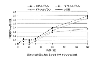
本発明で用いられる治療活性剤は、アミン糖が付着するアントラキノン基を含むアントラサイクリン化合物である。糖につくアミノ基は、ポリマー基質の陰イオン基と会合すると考えられており、これによって、高濃度の負荷と、投与後の調節された送達が可能となる。 The therapeutically active agent used in the present invention is an anthracycline compound containing an anthraquinone group to which an amine sugar is attached. The amino group attached to the sugar is believed to associate with the anionic group of the polymer substrate, which allows for a high concentration of loading and controlled delivery after administration.
好適なアントラサイクリンの例は、下記の一般式IIを持つ。
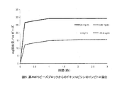
各種腫瘍に対する効力がこれまでに徹底的に調べられているドキソルビシンが、特に興味深い負荷特性、および、放出特性を持つことを我々は見出した。この薬は、ポリ(ビニルアルコール-グラフト-アクリルアミドプロパンスルフォン酸)に対して特別の親和性を持つようであり、そのために、高濃度のドキソルビシンがこのポリマーに組み込まれ、長い日数をかけての放出が可能である。 We have found that doxorubicin, which has been extensively investigated for efficacy against various tumors, has particularly interesting loading and release properties. The drug appears to have a special affinity for poly (vinyl alcohol-graft-acrylamide propane sulfonic acid), so a high concentration of doxorubicin is incorporated into the polymer and released over a long period of time. Is possible.
本発明では、薬剤が、ポリマー基質に共有結合によって付着していないことが重要である。 In the present invention, it is important that the drug is not covalently attached to the polymer substrate.
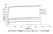
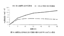
治療活性剤は、いろいろの技術によってポリマー基質に組み込むことが可能である。一つの方法では、治療活性剤は、重合または架橋反応前に、ポリマーの前駆物質、例えば、モノマーまたはマクロマー混合物、または、架橋性ポリマーおよび架橋性混合物、と混合してもよい。別法として、活性剤は、架橋反応後に、ポリマーに負荷してもよい。例えば、粒状の乾燥ポリマーを、治療活性剤の溶液、好ましくは水溶液中で膨潤させ、要すれば随意にその後、吸収されなかった活性剤の除去および/または溶媒留去を行ってもよい。アルコールのような有機溶媒、またはより好ましくは水に溶かした、活性剤の溶液を、粒子の移動ベッドに噴霧し、薬剤を粒子の本体に吸収させ、同時に溶媒を除去するようにしてもよい。もっとも好都合にも、液体の連続ベヒクルに懸濁させた膨潤粒子を、薬剤の溶液に単に長期間接触させることだけでも、薬剤を粒子の本体に吸収させることが可能であることを我々は見出した。これは、陽イオン交換タイプの過程と類似のものと考えられる。次に、この膨潤させるベヒクルを除去し、あるいは、好都合なことであるが、続いて塞栓剤として使用される製品の一部として粒子と共に保存されてもよい。 The therapeutically active agent can be incorporated into the polymer matrix by a variety of techniques. In one method, the therapeutically active agent may be mixed with a polymer precursor, such as a monomer or macromer mixture, or a crosslinkable polymer and a crosslinkable mixture, prior to the polymerization or crosslinking reaction. Alternatively, the active agent may be loaded into the polymer after the crosslinking reaction. For example, the granular dry polymer may be swollen in a solution of the therapeutically active agent, preferably in an aqueous solution, optionally followed by removal of the unabsorbed active agent and / or solvent evaporation. A solution of the active agent, such as an organic solvent such as alcohol, or more preferably dissolved in water, may be sprayed onto the moving bed of particles to allow the drug to be absorbed into the body of the particles while simultaneously removing the solvent. Most conveniently, we have found that swollen particles suspended in a continuous liquid vehicle can be absorbed into the body of the particles by simply contacting the drug solution for a long period of time. . This is considered similar to the cation exchange type process. This swelling vehicle can then be removed or conveniently stored with the particles as part of a product that is subsequently used as an embolic agent.
一つの特に好ましい実施態様では、膨潤粒子は、単純なゲル/液体分離技術、例えば、適当な孔口を持つフィルター、好適にはガラスフィルターによって、基質に吸収されない膨潤ベヒクルから分離される。ほとんどまたは全く粒子以外に液体を持たない膨潤粒子のスラリーを、滅菌のために、また、そのまま保存するために、適当な保存容器に注入してもよい。このスラリーは、その形で保存中に、液体の滲出がほとんど無く、また、薬剤の消失が起こらないという点で十分に安定であることが見いだされている。 In one particularly preferred embodiment, the swollen particles are separated from the swollen vehicle that is not absorbed by the substrate by simple gel / liquid separation techniques, such as a filter with a suitable pore opening, preferably a glass filter. A slurry of swollen particles with little or no liquid other than particles may be injected into a suitable storage container for sterilization and for storage as is. This slurry has been found to be sufficiently stable in that form during storage that there is little liquid leaching and no loss of drug occurs.
別態様として、粒子の懸濁液を濾過して残余の薬剤負荷液を完全に除去し、かつ、製薬製品を乾燥させるのに用いられる従来技術の内から選ばれる任意の技術によって、粒子を乾燥させることも可能である。そのような技術としては、室温または高温での、あるいは、減圧下または真空下での空気乾燥、従来技術による凍結乾燥、大気圧凍結乾燥、臨界超過液の溶液強化分散(SEDS)が挙げられるが、ただしそれらに限定されない。別態様として、薬剤負荷微小球は、有機溶媒を用いた一連の工程で水を置換して脱水し、その後に、より揮発性の高いその溶媒を蒸発させる。薬剤に対して溶媒とならない溶媒を選択しなければならない。 Alternatively, the particle suspension may be dried by filtering the particle suspension to completely remove the remaining drug load and any technique selected from the prior art used to dry pharmaceutical products. It is also possible to make it. Such techniques include air drying at room temperature or elevated temperature, or reduced pressure or vacuum, freeze drying according to the prior art, atmospheric pressure freeze drying, solution-strengthened dispersion (SEDS) of supercritical fluids. However, it is not limited to them. Alternatively, the drug-loaded microspheres are dehydrated by replacing water in a series of steps with an organic solvent, followed by evaporation of the more volatile solvent. A solvent must be selected that is not a solvent for the drug.
簡潔に言うと、従来法による典型的凍結乾燥工程は下記のように進行してもよい。すなわち、サンプルの分液を、部分的に閉鎖したガラス瓶に取り、この瓶を、凍結乾燥器内部の、冷却した、温度調節棚の上に置く。棚の温度を下げ、サンプルを、定められた均一な温度に凍結させる。完全な凍結後、乾燥器内の圧を、定められた圧に下げて、一次乾燥を起動する。この一次乾燥の間に、水蒸気が、昇華によって凍結体から次第に奪われ、一方、棚の温度は、一定の、低温に調整される。棚の温度を高め、室内の圧をさらに下げることによって二次乾燥を開始し、半乾燥体に吸収された水分を、残りの水含量が所望のレベルに下がるまで除去する。この瓶は、必要ならば保存的雰囲気の下で、そのまま封印することも可能である。 Briefly, a typical lyophilization process according to conventional methods may proceed as follows. That is, the sample aliquot is taken into a partially closed glass bottle and the bottle is placed on a cooled, temperature-controlled shelf inside the freeze dryer. The shelf temperature is lowered and the sample is frozen to a defined uniform temperature. After complete freezing, the pressure in the dryer is lowered to the prescribed pressure and primary drying is started. During this primary drying, water vapor is gradually taken away from the frozen body by sublimation, while the shelf temperature is adjusted to a constant, low temperature. Secondary drying is initiated by raising the shelf temperature and further reducing the pressure in the room, removing moisture absorbed by the semi-dried body until the remaining water content has dropped to the desired level. The bottle can be sealed as it is, if necessary, under a preservative atmosphere.
大気圧凍結乾燥は、凍結した製品の上に極めて乾燥した空気を急速に循環させることによって実現される。従来の凍結乾燥法と比べて、真空無しの凍結乾燥は多くの利点を持つ。風の強い日には洗濯物が早く乾くというのと同じように、循環乾燥ガスでは、凍結サンプルからの熱および質量転移が向上する。この領域での多くの作業は食品生産に関わっており、揮発性の芳香化合物の保存が向上することが観察されているが、それが、生物製剤の乾燥に対してどのような利点をもたらすかは、まだ明確にされていない。特に興味があるのは、大気圧粉末乾燥工程を用いることによって、ケーキではなく、微細な、さらさらと流れる粉末が得られるという事実である。ミクロン以下の直径を有する粒子を得ることが可能で、これは、一般に粉砕によって得られるものよりも10倍小さい。高度の表面積を持つという粒子の性質により、簡単な再水和が可能な製品が得られるが、吸引性の、また、経皮性の用途用に必要な粒子サイズに対する微細なコントロールは、現在では可能ではないが、この領域での可能性がある。 Atmospheric pressure lyophilization is achieved by rapidly circulating very dry air over the frozen product. Compared to conventional lyophilization methods, lyophilization without vacuum has many advantages. Just as the laundry dries quickly on windy days, circulating dry gas improves heat and mass transfer from the frozen sample. Much work in this area is involved in food production and it has been observed that the preservation of volatile aroma compounds has been improved, but what benefits do it have for drying biologics? Has not yet been clarified. Of particular interest is the fact that by using an atmospheric pressure powder drying process, a fine, free flowing powder is obtained rather than a cake. It is possible to obtain particles with submicron diameters, which are generally 10 times smaller than those obtained by grinding. The high surface area of the particles results in a product that can be easily rehydrated, but fine control over the particle size required for aspiration and transdermal applications is now available Although not possible, there is a possibility in this area.
固形腫瘍、例えば、肝細胞ガンを持ち、塞栓療法を必要とする患者に対して投与される組成物は、吸収された薬剤を含む膨潤粒子の水性懸濁液である。この懸濁液は、ゲルタイプの塞栓組成に用いられるように、送達前に、造影剤、例えば、通例の放射線不透過剤と混合することが好ましい場合がよくある。例えば、吸収薬剤を含む膨潤粒子の水性懸濁液を、投与直前に、塞栓剤と共に通常使用される、液状放射線不透過剤、例えば、リピオドールと、容量で2:1から1:2の範囲、好ましくは約1:1で混合してもよい。吸収薬剤を持つが、粒子外液体をほとんど、または全く持たない膨潤粒子のスラリーを含む本発明の実施態様では、同様にして、このスラリーと造影(放射線不透過)剤を、例えば、容量で1:5から2:1の範囲で、好ましくは1:2から1:1の範囲で、送達の直前に混合してもよい。薬剤を含む組成物が、用時に乾燥形として支給される場合、粒子を、乾燥したまま造影剤に加えるか、あるいは、好ましくは、最初生理的食塩水のような水性ベヒクルにおいて膨潤させてスラリーまたは懸濁液を形成し、次に、送達前に造影剤と混合してもよい。別態様として、あるいは、上記に加えて、粒子に、あらかじめ、アントラサイクリンの他に放射線不透過物質を負荷してもよい。投与される組成物はまた、他の治療薬と混合してもよいし、あるいは、他の治療薬と別々に、ただしそれと組み合わせて投与してもよい。通常、組成物は、通例の送達デバイス、例えば、動脈内カテーテルを用い、注射器の貯留槽から投与される。
Compositions administered to patients with solid tumors, such as hepatocellular carcinoma and in need of embolization, are aqueous suspensions of swollen particles containing absorbed drugs. This suspension is often preferred to be mixed with a contrast agent, eg, a conventional radiopaque agent, prior to delivery, as used in gel-type embolic compositions. For example, an aqueous suspension of swollen particles containing an absorbing agent can be used immediately prior to administration with a liquid radiopaque agent, such as lipiodol, commonly used with an embolic agent, in a volume range of 2: 1 to 1: 2. Preferably it may be mixed at about 1: 1. In an embodiment of the invention comprising a slurry of swollen particles with an absorbent agent but little or no extra-particle liquid, the slurry and contrast (radiopaque) agent are similarly used, for example, by
塞栓療法を必要とする患者に対して投与される塞栓組成物は、単一の、一回きり用量として投与されてもよい。塞栓形成は、通例の技術を用いて造影剤を追跡することによって監視される。塞栓組成物の第2回目投与を、好ましくは本発明で有用な化学的塞栓組成物を、第1回投与からある時間後に、例えば、腫瘍を養う新規形成の血管に塞栓形成するために、例えば、ドキソルビシン含有組成物による第1回目治療の4から10週後に送達することが好ましいことが見出されるかも知れない。組成物は、1回の治療当たり25-100 mg/m2の範囲の薬剤用量で投与されるが、ただし十分な安全評価をした後であれば、より高い用量を用いることも可能である。ドキソルビシンについては治療当たりの好ましい用量は、50 mg/m2以上、例えば、最大100 mg/m2以上である。治療当たり、患者一人当たり、150 mg/m2を越える量を投与するのは望ましくないと一般に考えられている。 The embolic composition administered to a patient in need of embolization therapy may be administered as a single, single dose. Embolization is monitored by tracking the contrast agent using conventional techniques. To embolize a second administration of the embolic composition, preferably a chemical embolization composition useful in the present invention, for example, into a newly formed blood vessel that feeds the tumor, some time after the first administration, e.g. It may be found preferable to deliver 4 to 10 weeks after the first treatment with the doxorubicin-containing composition. The composition is administered at a drug dose ranging from 25-100 mg / m 2 per treatment, although higher doses can be used after a thorough safety assessment. For doxorubicin, the preferred dose per treatment is 50 mg / m 2 or more, for example up to 100 mg / m 2 or more. It is generally considered undesirable to administer doses exceeding 150 mg / m 2 per patient per treatment.
本発明の第2局面として、塞栓療法による固形腫瘍の治療に使用される組成物製造におけるアントラサイクリン化合物の用法が提供される。この治療では、アントラサイクリンは、分子当たり少なくとも2個のエチレン的に不飽和なペンダント基を持つポリ(ビニルアルコール)マクロマーと、エチレン的に不飽和な陰イオン性モノマーとを共重合させることによって形成されるポリマー基質から送達される。 As a second aspect of the present invention, there is provided a method for using an anthracycline compound in the production of a composition used for treatment of a solid tumor by embolization therapy. In this treatment, anthracyclines are formed by copolymerizing a poly (vinyl alcohol) macromer with at least two ethylenically unsaturated pendant groups per molecule and an ethylenically unsaturated anionic monomer. Delivered from a polymeric substrate.
本発明のこの局面では、ポリマー基質は生体内で形成されてもよい。従って、マクロマーと陰イオン性モノマーとアントラサイクリンを含む液体組成物は、患者の循環中に送達され、標的部位において重合化を起動する条件に暴露されて、塞栓性ゲルを形成するようにしてもよい。別態様として、ポリマー基質は、本発明の第1局面で記載されたように、投与前にあらかじめ形成されてもよい。 In this aspect of the invention, the polymer matrix may be formed in vivo. Thus, a liquid composition comprising a macromer, an anionic monomer and an anthracycline may be delivered into the patient's circulation and exposed to conditions that initiate polymerization at the target site to form an embolic gel. Good. Alternatively, the polymer matrix may be preformed prior to administration as described in the first aspect of the invention.
PVAマクロマーと陰イオン性モノマーは、好ましくは、第1局面に関連して上述した通りのものである。本発明の第1局面に関連して上述したように、他のモノマー同士を共重合させてもよい。 The PVA macromer and the anionic monomer are preferably as described above in connection with the first aspect. As described above in connection with the first aspect of the present invention, other monomers may be copolymerized.
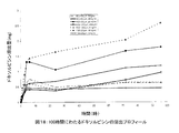
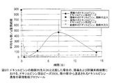
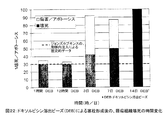
本発明は、その結果が図に示される下記の実施例において具体的に説明される。 The present invention is specifically illustrated in the following examples, the results of which are shown in the figures.
[実施例1: 微小球調製の概略法]
===ネルフィルコンBマクロマー合成===
微小球合成の第1段階は、広く使用されている水溶性ポリマーPVAからネルフィルコンB、すなわち重合可能なマクロマーを調製することを含む。Mowiol(登録商標) 8-88ポリ(ビニルアルコール)(PVA)粉末(88%加水分解、12%酢酸塩含有、平均分子量約67,000D)(150 g)(Clariant、シャーロット、ノースカロライナ州、米国)を、2リットルのガラス反応容器に加える。穏やかに攪拌しながら、1000 mlの水を加え、攪拌を400 rpmに増す。PVAの完全溶解を確保するために、温度を、2-3時間99±9℃に上げる。室温に冷却した時点でN-アクリロアミノアセトアルデヒド(NAAADA)(Ciba Vision、ドイツ)(PVAのg当たり2.49 gまたは0.104 mmol)をPVA溶液と混合し、次いで濃塩酸(100 ml)を加える。濃塩酸は、経エステル化によってPVAに対するNAAADAの添加を触媒する。この反応を室温で6-7時間進行させ、2.5 M水素化ナトリウムを用いてpH7.2に中和することによって反応停止させた。得られた塩化ナトリウム+未反応NAAADAは全て透析濾過によって除去する(工程2)。
[Example 1: Outline of microsphere preparation]
=== Nelfilcon B Macromer Synthesis ===
The first stage of microsphere synthesis involves preparing Nelfilcon B, a polymerizable macromer, from the widely used water-soluble polymer PVA. Mowiol® 8-88 poly (vinyl alcohol) (PVA) powder (88% hydrolyzed, containing 12% acetate, average molecular weight about 67,000D) (150 g) (Clariant, Charlotte, NC, USA) Add to a 2 liter glass reaction vessel. While stirring gently, add 1000 ml of water and increase the stirring to 400 rpm. To ensure complete dissolution of the PVA, the temperature is raised to 99 ± 9 ° C. for 2-3 hours. Upon cooling to room temperature, N-acryloaminoacetaldehyde (NAAADA) (Ciba Vision, Germany) (2.49 g or 0.104 mmol per g PVA) is mixed with the PVA solution and then concentrated hydrochloric acid (100 ml) is added. Concentrated hydrochloric acid catalyzes the addition of NAAADA to PVA by transesterification. The reaction was allowed to proceed for 6-7 hours at room temperature and quenched by neutralizing to pH 7.2 with 2.5 M sodium hydride. All of the obtained sodium chloride + unreacted NAAADA is removed by diafiltration (step 2).
===マクロマーの透析濾過===
透析濾過(diafiltration)(接線流濾過)は、精製される入力液(この場合はネルフィルコンB液)を、膜表面を横切って連続的に循環させ、望ましくない物質(NaCl、NAAADA)を透過させて廃棄処理へ向かわせ、一方、ポアの大きさを十分小さくして保持体の通過を阻止して、循環中に留めさせる。
=== Diafiltration of Macromer ===
Diafiltration (tangential flow filtration) allows the input fluid to be purified (in this case Nerfilcon B solution) to circulate continuously across the membrane surface and to permeate undesired substances (NaCl, NAAADA). The waste is then disposed of, while the size of the pores is made sufficiently small to prevent the holder from passing through and remain in circulation.
ネルフィルコンBの透析濾過は、3000の分子量カットオフを持つポアサイズの0.1 m2のセルロース膜(Millipore社、ベッドフォード、マサチューセッツ州、米国)を積層したステンレススチールのペリコン2ミニホールダーを用いて行った。Mowiol8-88は、67000の平均分子量を持ち、従って、膜を通過する能力には限界がある。
Nelfilcon B diafiltration was performed using a
マクロマーを含むフラスコは、マグネットの攪拌棒を含み、攪拌プレートの上に設置される。溶液は、Easy Load IIポンプヘッドを装備したMasterflex(登録商標) LSペリスタルティックポンプを介し、LS24クラスVIチューブを用いて、透析濾過アッセンブリーに供給される。ネルフィルコンは、透過を加速するため、約50 psiの下で、膜の上を循環される。溶液が約1000 mlに濃縮された時点で、濾液が廃棄処理のために回収されるのと同じ速度で、6000 mlの追加分の添加が完了するまで水を添加することによって、容量を一定に保つ。一旦完了すると、溶液は、20-23%の固形分に濃縮され、25℃における粘度は1700-3400 cPとなる。ネルフィルコンは、GFC、NMR、および粘度によって特徴付けられる。 The flask containing the macromer contains a magnetic stir bar and is placed on a stir plate. The solution is fed to the diafiltration assembly using LS24 class VI tubing via a Masterflex® LS peristaltic pump equipped with an Easy Load II pump head. Nelfilcon is circulated over the membrane under about 50 psi to accelerate permeation. When the solution is concentrated to approximately 1000 ml, keep the volume constant by adding water at the same rate that the filtrate is collected for disposal until the addition of an additional 6000 ml is complete. keep. Once complete, the solution is concentrated to 20-23% solids and the viscosity at 25 ° C. is 1700-3400 cP. Nelfilcon is characterized by GFC, NMR, and viscosity.
===微小球合成===
球は、懸濁重合法によって合成する。すなわち、水相(ネルフィルコンB)を、有機相(酢酸ブチル)に両相が混和しない状態において加える。急速混合法を用いて、水相を分散させ、小滴を形成させることが可能であり、しかも、そのサイズおよび安定性を、攪拌速度、粘度、水相の有機相に対する比、および、両相の間の界面エネルギーに影響を及ぼす安定化剤と界面活性剤の使用、等の因子によって調節することが可能である。低AMPSシリーズおよび高AMPSシリーズの、二つのシリーズの微小球を製造する。これらのシリーズの処方は下記の通りである。
A. 高AMPS
水相:およそ21%w/w(重量/重量)のネフィリコンB液(約400±50 g)
およそ50%w/wの2-アクリルアミド-2-メチルプロパンスルフォネートNa塩
(140±10 g)
純水
過硫酸カリ(5.22±0.1 g)
テトラメチルエチレンジアミンTMEDA(6.4±0.1 ml)
有機相:n-ブチルアセテート(2.7±0.3 L)
10%w/wセルロースアセテートブチレートの酢酸エチル溶液(4.6±0.5 g)
(安定化剤)
純水(19.0±0.5 ml)
B. 低AMPS
水相:およそ21%w/wのネフィリコンB液(約900±100 g)
およそ50%w/wの2-アクリルアミド-2-メチルプロパンスルフォネートNa塩
(30.6±6 g)
純水(426±80 g)
過硫酸カリ(20.88±0.2 g)
TMEDA(25.6±0.5 ml)
有機相:n-ブチルアセテート(2.2±0.3 L)
10%w/wセルロースアセテートブチレート(CAB)の酢酸エチル溶液
(92±1.0 g)
純水(16.7±0.5 ml)
=== Microsphere synthesis ===
The spheres are synthesized by a suspension polymerization method. That is, the aqueous phase (Nelfilcon B) is added to the organic phase (butyl acetate) in a state where both phases are not miscible. Using a rapid mixing method, it is possible to disperse the aqueous phase and form droplets, and to determine its size and stability in terms of stirring speed, viscosity, ratio of aqueous phase to organic phase, and both phases It can be controlled by factors such as the use of stabilizers and surfactants that affect the interfacial energy. Produces two series of microspheres, low AMPS series and high AMPS series. The prescriptions for these series are as follows:
A. High AMPS
Aqueous phase: approx. 21% w / w (weight / weight) nephilon B solution (approximately 400 ± 50 g)
Approximately 50% w / w 2-acrylamido-2-methylpropanesulfonate sodium salt
(140 ± 10 g)
Pure water Potassium persulfate (5.22 ± 0.1 g)
Tetramethylethylenediamine TMEDA (6.4 ± 0.1 ml)
Organic phase: n-butyl acetate (2.7 ± 0.3 L)
10% w / w cellulose acetate butyrate in ethyl acetate (4.6 ± 0.5 g)
(Stabilizer)
Pure water (19.0 ± 0.5 ml)
B. Low AMPS
Aqueous phase: approx. 21% w / w Nephilon B solution (approx. 900 ± 100 g)
Approximately 50% w / w 2-acrylamido-2-methylpropanesulfonate sodium salt
(30.6 ± 6 g)
Pure water (426 ± 80 g)
Potassium persulfate (20.88 ± 0.2 g)
TMEDA (25.6 ± 0.5 ml)
Organic phase: n-butyl acetate (2.2 ± 0.3 L)
10% w / w cellulose acetate butyrate (CAB) in ethyl acetate
(92 ± 1.0 g)
Pure water (16.7 ± 0.5 ml)
ジャケット付き4000ml反応容器を、コンピュータ制御浴(Julabo PN 9-300-650)を用いて加熱する。該容器には、反応温度を連続的に監視するフィードバックセンサーが備えられる。 A jacketed 4000 ml reaction vessel is heated using a computer controlled bath (Julabo PN 9-300-650). The vessel is equipped with a feedback sensor that continuously monitors the reaction temperature.
酢酸ブチルを25℃で反応容器に加え、次いでCAB液と水を加える。システムに15分窒素を通じて清掃して後、PVAマクロマーを添加する。分散PVA液の架橋は、TMEDAを添加し、窒素下において温度を55℃に上げて3時間加熱することによって起動する。架橋反応は、レドックス起動重合によって行う。この重合では、TMEDAのアミノ基が過硫酸カリの過酸化基と反応してラジカル分子種が形成される。次に、このラジカルが、PVAとAMPS上の二重結合について重合および架橋を起動し、分散したPVA-AMPS小滴を不溶のポリマー微小球に変形させる。25℃に冷却した後、産物を、精製のためにフィルター反応器に転送し、そこで、酢酸ブチルを濾過によって除去した後、下記の処理を行う。すなわち、
*2 x 300 mlの酢酸エチルで洗浄し、酢酸ブチルとCABを除去する。
*酢酸エチルで30分平衡させ、次に濾過。
*減圧濾過の下で2 x 300 mlの酢酸エチルで洗浄。
*アセトンで30分平衡させ、濾過して、酢酸エチル、CAB、および水を除去する。
*減圧濾過の下で2 x 300 mlのアセトンで洗浄。
*アセトンで一晩平衡させる。
*減圧下で2 x 300 mlのアセトンで洗浄。
*真空乾燥、2時間、55℃で残余の溶媒を除去する。
Butyl acetate is added to the reaction vessel at 25 ° C., then CAB solution and water are added. After the system is purged with nitrogen for 15 minutes, the PVA macromer is added. Cross-linking of the dispersed PVA liquid is started by adding TMEDA and raising the temperature to 55 ° C. and heating for 3 hours under nitrogen. The cross-linking reaction is performed by redox-initiated polymerization. In this polymerization, the amino group of TMEDA reacts with the peroxide group of potassium persulfate to form radical molecular species. This radical then initiates polymerization and crosslinking for double bonds on PVA and AMPS, transforming the dispersed PVA-AMPS droplets into insoluble polymer microspheres. After cooling to 25 ° C., the product is transferred to a filter reactor for purification, where the butyl acetate is removed by filtration before the following treatment. That is,
* Wash with 2 x 300 ml of ethyl acetate to remove butyl acetate and CAB.
* Equilibrate with ethyl acetate for 30 minutes, then filter.
* Wash with 2 x 300 ml of ethyl acetate under vacuum filtration.
* Equilibrate with acetone for 30 minutes and filter to remove ethyl acetate, CAB, and water.
* Wash with 2 x 300 ml acetone under vacuum filtration.
* Equilibrate overnight with acetone.
* Wash with 2 x 300 ml acetone under reduced pressure.
* Vacuum dry, remove remaining solvent at 55 ° C for 2 hours.
===染色===
この工程は必要であれば随意に行ってもよいが、薬剤に有色の活性成分が負荷されている場合は(これが色を与えるため)、一般に不要である。水和されると、微小球は、約90%(w/w)の水を含むので、それと見て取るのが難しい。臨床環境における可視性を助けるために、小球は、反応性の青色#4染料(RB4)を用いて青く染める。RB4は、水溶性クロロトリアジン染料であり、アルカリ性条件下でPVAバックボーンのペンダントヒドロキシル基と反応して、エーテル共有結合を形成する。この反応はpH12(NaOH)で実行され、生成されたHClは中和されてNaClになる。
=== Staining ===
This step may be optionally performed if necessary, but is generally unnecessary if the drug is loaded with colored active ingredients (to give color). When hydrated, the microspheres contain about 90% (w / w) water and are difficult to see. To aid visibility in the clinical environment, the globules are dyed blue with a reactive
染色前に、小球は改めて十分に水和し、35 g分液に分割する(それぞれに処理する)。染色液は、0.8 gのRB4を2.5M NaOH液(25 ml)と水(15 ml)に溶解し、次に、小球を80 g/l-1生食液に加えて調製する。20分間混合した後、産物を32 μmの篩で回収し、濯ぐことによって、未反応の染料バルクを除去する。 Prior to staining, the globules are again fully hydrated and divided into 35 g aliquots (treat each). The staining solution is prepared by dissolving 0.8 g RB4 in 2.5 M NaOH solution (25 ml) and water (15 ml), then adding the globules to 80 g / l −1 saline. After mixing for 20 minutes, the product is collected on a 32 μm sieve and the unreacted dye bulk is removed by rinsing.
===抽出===
広範な抽出過程を用いて、未結合の、または、非特異的に吸収されたRB4を除去する。順守したプロトコールは下記の通りである。
*2 lの水で5分間平衡させる。篩で回収し、濯ぐ。5度繰り返す。
*80 mMのリン酸水素二ナトリウムの、0.29%(w/w)生食液の2 l溶液中で平衡させる。30分間沸騰加熱する。冷却し、篩で回収し、1 l生食液で洗浄する。さらに2回繰り返す。
*平衡産物を篩上で回収し、2 l水にて10分間洗浄する。
*回集し、1 lアセトンにて30分脱水する。
*全ての分液を合わせて、2 lアセトンにて一晩平衡させる。
=== Extraction ===
An extensive extraction process is used to remove unbound or non-specifically absorbed RB4. The protocol followed is as follows.
* Equilibrate with 2 l of water for 5 minutes. Collect with a sieve and rinse.
* Equilibrate in 2 l solution of 0.29% (w / w) saline in 80 mM disodium hydrogen phosphate. Heat to boiling for 30 minutes. Cool, collect with a sieve, and wash with 1 l saline. Repeat two more times.
* Collect the equilibrium product on a sieve and wash with 2 l water for 10 minutes.
* Collect and dehydrate in 1 l acetone for 30 minutes.
* Combine all aliquots and equilibrate overnight with 2 l acetone.
===篩分け===
製造された微小球は、そのサイズが100から1200ミクロンに渡るので、下記に掲げる指定の分布を得るために、ある範囲のメッシュサイズを用いる篩工程によって分画しなければならない。
1. 100-300 μm
2. 300-500 μm
3. 500-700 μm
4. 700-900 μm
5. 900-1200 μm
=== Sieving ===
Since the produced microspheres range in size from 100 to 1200 microns, they must be fractionated by a sieving process using a range of mesh sizes to obtain the specified distribution listed below.
1. 100-300 μm
2. 300-500 μm
3.500-700 μm
4. 700-900 μm
5. 900-1200 μm
篩分けの前に、小球は真空乾燥して完全に溶媒を除去し、次に60℃で水に平衡させて十分に再水和させる。この小球を、メッシュサイズが32から1000 μm範囲の15’’ステンレススチール篩トレーを備えた316Lステンレススチールvortisieveユニット(MM Industries、サーレム、オハイオ州)にて濾す。濾過された生食液はユニットを再度循環して分画を支援する。32ミクロン篩で回収された小球は廃棄する。 Prior to sieving, the globules are vacuum dried to completely remove the solvent and then equilibrated to water at 60 ° C. to fully rehydrate. The globules are filtered through a 316L stainless steel vortisieve unit (MM Industries, Salem, OH) equipped with a 15 ″ stainless steel sieve tray with a mesh size ranging from 32 to 1000 μm. The filtered saline solution is circulated through the unit again to support fractionation. Discard the small balls collected with a 32 micron sieve.
[実施例2: ドキソルビシンの負荷]
本実験のために、実施例1のように調製した低AMPS微小球を用いた。使用する各サイズのビーズについて、0.5 mlを、2本の、1 ml注射筒に移した。1本は薬剤取り込みのため、2本目は対照として用いるためである。実験のために選んだサイズは、106-300 μm, 300-500 μm, 500-710 μmおよび850-1000 μmである。工程の有効性を確認するために500-710 μmのさらに3本の注射筒を準備した。実験の持続時間において光によってドキソルビシンが変性されるのを防ぐために、11本の、10 mlガラス瓶をフォイルで被った。標準曲線を作成した。20 mg/mlの薬剤液80 mlを用いて、下記の濃度を調製し、その吸光度(483 nmにおける)を測定した。すなわち、100 μg/ml, 50 μg/ml, 25 μg/ml, 12.5 μg/ml, 6.25 μg/mlおよび3.125 μg/mlである。得られた吸光度をグラフにプロットし、線の等式を用いて、実験においてビーズに取り込まれた薬剤の濃度を計算した。瓶の内の4本には5 mlの蒸留水(ROMIL)を満たし、ビーズを加えたときの対照として用いた。残りの7本の瓶には、所望の濃度で5 mlの薬液を加えた。開始の吸光度、従って溶液の濃度は、標準曲線の作成から既知である。(20 mg/ml溶液の吸光度を測定するためには、その溶液を200倍に希釈し、100 μg/ml濃度を用いる必要があった。この1:200希釈は、ビーズによる溶液の取り込みを測定する間中実行した。)第1セットの微小球を第1薬剤液を含む瓶に添加した直後にストップウォッチをスタートさせ、微小球を、最小から最大のものへと、残りの6本の瓶のそれぞれに加えた。キャップを用いて封印した後、それらの瓶を回転式ミキサー上に設置した。この過程を、対照サンプルについても繰り返した。瓶を設定したのと同じ順序で、0.167時間(10分)、0.5時間、1時間、2時間、24時間、および96時間間隔で吸光度を測定した。データから、1 mlの微小球当たりの薬剤量(mgで表した)、および、1 mlの微小球による薬剤の取り込み%が計算可能であった。結果を図1に示す。
[Example 2: Doxorubicin load]
For this experiment, low AMPS microspheres prepared as in Example 1 were used. For each size bead used, 0.5 ml was transferred to two 1 ml syringes. One for drug uptake and the second for use as a control. The sizes chosen for the experiments are 106-300 μm, 300-500 μm, 500-710 μm and 850-1000 μm. Three additional syringes of 500-710 μm were prepared to confirm the effectiveness of the process. To prevent the doxorubicin from being denatured by light over the duration of the experiment, 11 10 ml glass bottles were covered with foil. A standard curve was created. The following concentrations were prepared using 80 ml of 20 mg / ml drug solution, and the absorbance (at 483 nm) was measured. That is, 100 μg / ml, 50 μg / ml, 25 μg / ml, 12.5 μg / ml, 6.25 μg / ml and 3.125 μg / ml. The resulting absorbance was plotted on a graph and the concentration of drug incorporated into the beads in the experiment was calculated using the line equation. Four of the bottles were filled with 5 ml of distilled water (ROMIL) and used as controls when beads were added. To the remaining 7 bottles, 5 ml of chemical solution was added at the desired concentration. The starting absorbance, and hence the concentration of the solution, is known from the creation of a standard curve. (To measure the absorbance of a 20 mg / ml solution, it was necessary to dilute the solution 200-fold and use a concentration of 100 μg / ml. This 1: 200 dilution measures the uptake of the solution by the beads. The stopwatch was started immediately after the first set of microspheres was added to the bottle containing the first drug solution, and the remaining six bottles were moved from the smallest to the largest. Added to each of the. After sealing with a cap, the bottles were placed on a rotary mixer. This process was repeated for the control sample. Absorbance was measured at 0.167 hours (10 minutes), 0.5 hours, 1 hour, 2 hours, 24 hours, and 96 hours intervals in the same order that the bottles were set. From the data, the amount of drug per ml of microspheres (expressed in mg) and the% drug uptake by 1 ml of microspheres could be calculated. The results are shown in Figure 1.
[実施例3: 薬剤濃度の負荷に対する効果]
実施例2に略述した処理過程に従って、ある範囲の異なる濃度のドキソルビシンを、高AMPS微小球処方に負荷することが可能であった。薬剤の大部分は、数時間内に微小球(500-710 μmのサイズ範囲)中に負荷するのが見られた(図2参照)。負荷は、重量に基づくと、高AMPS処方の方が、低AMPS処方よりもはるかに高いことが見て取れる。
[Example 3: Effect on loading of drug concentration]
Following a process outlined in Example 2, it was possible to load a range of different concentrations of doxorubicin into the high AMPS microsphere formulation. Most of the drug was seen loading in the microspheres (size range of 500-710 μm) within a few hours (see Figure 2). It can be seen that the load is much higher for high AMPS formulations than for low AMPS formulations based on weight.
[実施例4: 微小球サイズの負荷に対する効果]
ドキソルビシンの負荷を、取り込み量の比較を実行するために、いくつかの異なるサイズの範囲の微小球について行った。小さい微小球の方が、比較的速く薬剤を負荷させるのが見られる一方、24時間に渡る連続負荷では、等重量の微小球は、ほぼ同じ薬剤負荷に平衡することを示した。この比較的速い取り込みは、小さい微小球では表面積が増すためである(図3参照)。
[Example 4: Effect on load of microsphere size]
Doxorubicin loading was performed on several different size ranges of microspheres to perform uptake comparisons. Smaller microspheres were seen to load the drug relatively quickly, while equal loads of microspheres were shown to equilibrate to approximately the same drug load at 24 hours of continuous loading. This relatively fast uptake is due to the increased surface area of small microspheres (see Figure 3).
[実施例5: 負荷の再現性]
ドキソルビシン負荷の再現性を測定するために、実施例2で略述した負荷実験を数回繰り返した。500-710 μmサイズ範囲の高AMPS微小球を、20 mg/mlの薬剤水溶液で負荷し、薬剤取り込みの時間変化を監視した(図4)。
[Example 5: Load reproducibility]
In order to measure the reproducibility of doxorubicin loading, the loading experiment outlined in Example 2 was repeated several times. High AMPS microspheres in the 500-710 μm size range were loaded with 20 mg / ml aqueous drug solution and the time course of drug uptake was monitored (Figure 4).
[実施例6: 微小球からのドキソルビシンの溶出]
高AMPS微小球を、各種濃度のドキソルビシンで負荷し、その微小球を250 mlの蒸留水に溶出させた(図5)。
[Example 6: Elution of doxorubicin from microspheres]
High AMPS microspheres were loaded with various concentrations of doxorubicin and the microspheres were eluted in 250 ml distilled water (Figure 5).
133.2 μg/mlおよび2 mg/mlで負荷した微小球から溶出した薬剤は、3時間でも依然として検出限界以下であった。それより高濃度の薬剤負荷では、バースト作用が、最初の数分間に明らかであり、その後、長時間のよりゆっくりした放出が続く。バーストは、微小球の中に保持されていた水からの遊離薬剤の放出を表し、一方、持続的溶出は、微小球の中で、基本的に荷電基の間のイオン相互作用によって「結合」されている薬剤によるものと推測される。薬剤最大負荷の場合(20 mg/mlの負荷液による)、バースト作用は、小球の全体薬剤負荷のほぼ45%を表し、残余のものは、担体から完全に溶出するためには数日を要する。実験は、100%の薬剤が、最終的には微小球から溶出することを示した。 Drugs eluted from microspheres loaded at 133.2 μg / ml and 2 mg / ml were still below the detection limit at 3 hours. At higher drug loads, burst effects are evident in the first few minutes, followed by a slower and longer release. Burst represents the release of free drug from the water that was retained in the microsphere, while sustained elution is “bound” in the microsphere, essentially by ionic interactions between charged groups. Presumably due to the drug being used. In the case of maximum drug loading (with a loading solution of 20 mg / ml), the burst effect represents approximately 45% of the total drug load of the globules, with the rest remaining several days for complete dissolution from the carrier. Cost. Experiments have shown that 100% of the drug eventually elutes from the microspheres.
[実施例7: 高AMPS微小球によるドキソルビシン捕獲の可視化]
サイズ範囲850-1000 μm(手で篩にかけたもの)の高AMPS微小球約0.5 gを含む瓶に、リン酸バッファー生食液PBS(66.6 μg/ml)に溶解させたドキソルビシン1 mlおよび3 mlのPBSを加えた。この微小球をCCDカメラの下に設置し、2分に1回2.5時間に渡って映像を取った。この期間にサンプルの煽動は見られなかったが、光源からの局所的な加熱のために小さな動きが観察された。従って、最初と最後の微小球は同一であり、この期間を通して比較が可能である。薬剤の取り込みは、微小球における赤色の増加、および、周囲の溶液における赤色の喪失によって観察された(図6)。
[Example 7: Visualization of doxorubicin capture by high AMPS microspheres]
1 ml and 3 ml of doxorubicin dissolved in phosphate buffered saline PBS (66.6 μg / ml) in a bottle containing about 0.5 g of high AMPS microspheres with a size range of 850-1000 μm (sieved by hand) PBS was added. These microspheres were placed under the CCD camera, and images were taken once every 2 minutes for 2.5 hours. During this period, there was no peristalsis of the sample, but small movements were observed due to local heating from the light source. Thus, the first and last microspheres are identical and can be compared throughout this period. Drug uptake was observed by a red increase in the microspheres and a red loss in the surrounding solution (Figure 6).
[実施例8: 乾燥薬剤負荷微小球の調製]
微小球は、実施例2に概説した方法によってドキソルビシンを負荷することも可能である。微小球は、下記の処理過程を用いて脱水した。脱水される微小球を、プラスチック容器に入れ、PBSで製造した10%アセトン(ROMIL)液(Inverclyde Biologicals)で被った。微小球を、溶液中に10分間放置し、この間、30秒間、数回に渡って振とうした。次に、溶液を傾寫除去し、前記過程をさらに2回繰り返した。この過程を、アセトン濃度を25%、50%、75%、および最後に100%と増加させて繰り返した。最後の100%脱水工程の後、アセトンを傾寫除去し、ビーズを、50℃に設定したオーブンに入れて、定常質量となるまで乾燥した。
[Example 8: Preparation of dry drug-loaded microspheres]
Microspheres can also be loaded with doxorubicin by the method outlined in Example 2. The microspheres were dehydrated using the following process. The microspheres to be dehydrated were placed in a plastic container and covered with 10% acetone (ROMIL) solution (Inverclyde Biologicals) made with PBS. The microspheres were left in the solution for 10 minutes, during which time they were shaken several times for 30 seconds. The solution was then decanted and the process was repeated two more times. This process was repeated with increasing acetone concentrations of 25%, 50%, 75% and finally 100%. After the final 100% dehydration step, the acetone was decanted and the beads were placed in an oven set at 50 ° C. and dried to a steady mass.
得られた乾燥産物は、塞栓化処置の前に、生食液/造影剤中に再懸濁/再水和することが可能である。水和は急速に起こり、数分だけで完全水和サイズの>80%まで膨潤する。 The resulting dried product can be resuspended / rehydrated in saline / contrast media prior to the embolization procedure. Hydration occurs rapidly and swells to> 80% of fully hydrated size in just a few minutes.
[実施例9: 微小球スラリーの調製]
上記実施例1によって生産された高AMPS微小球を、20 mg/mlのドキソルビシン水溶液中で30分膨潤させた。この期間の後、余分の粒状液体は、実質的に脱色されて見え、色(赤)は、実質的に微小球内部に局在していた。この懸濁液を、焼結ガラス漏斗を通じて濾過して上清を除去した。微小球を、2倍容量の蒸留水で洗浄する一方、僅かな陰圧の下に濾過した。次に、微小球をジャーに移し、ジャーから、ペリスタルティックポンプによってガラス注射筒に汲み上げた。ポンプを取り外して後、注射筒を注射筒ロックにて閉鎖し、ガンマ線放射によって滅菌した。
[Example 9: Preparation of microsphere slurry]
The high AMPS microspheres produced according to Example 1 were swollen in a 20 mg / ml doxorubicin aqueous solution for 30 minutes. After this period, the extra granular liquid appeared to be substantially decolorized and the color (red) was substantially localized inside the microspheres. The suspension was filtered through a sintered glass funnel to remove the supernatant. The microspheres were washed with 2 volumes of distilled water while being filtered under slight negative pressure. Next, the microspheres were transferred to a jar and pumped from the jar into a glass syringe by a peristaltic pump. After removing the pump, the syringe was closed with a syringe lock and sterilized by gamma radiation.
[実施例10: 負荷−標的負荷用量対実際負荷用量]
22-80 mg/ml水溶液から一連のドキソルビシン溶液を調製した。これらの溶液の1 mlを、1 mlの高AMPS微小球に加え、取り込みをUVで監視した。サンプルは、ローラーミキサーにて攪拌した。終了時点を、10、20、30、60分、次に、2時間、最長24時間に取った。取り込みは、溶液に残っているドキソルビシンから計算した。微小球には、水和化微小球1 ml当たり最大80 mgまで様々の用量を負荷することが可能であった。そして、30分未満で、薬剤溶液の90%が微小球に局在した。
[Example 10: Load-target load dose versus actual load dose]
A series of doxorubicin solutions were prepared from 22-80 mg / ml aqueous solution. 1 ml of these solutions was added to 1 ml of high AMPS microspheres and uptake was monitored by UV. The sample was stirred with a roller mixer. The end point was taken at 10, 20, 30, 60 minutes, then 2 hours, up to 24 hours. Uptake was calculated from doxorubicin remaining in solution. Microspheres could be loaded with various doses up to 80 mg per ml of hydrated microspheres. In less than 30 minutes, 90% of the drug solution was localized in the microspheres.
[実施例11: 高用量ドキソルビシン]
実施例10で、ドキソルビシンの80 mg/ml溶液を調製した。これは、粘ちょうな、ゼラチン状混合物で、日常的使用には適当ではないと思われた。この高用量を、今度は20 mg/mlのドキソルビシン液4 mlを用いて再現した。取り込みは、UVで監視し、最終的に、水和化高AMPS微小球に対し80 mgの薬剤負荷が再び達成された。
[Example 11: High-dose doxorubicin]
In Example 10, an 80 mg / ml solution of doxorubicin was prepared. This was a thick, gelatinous mixture that seemed unsuitable for daily use. This high dose was then reproduced using 4 ml of 20 mg / ml doxorubicin solution. Incorporation was monitored by UV and finally a drug load of 80 mg was again achieved for hydrated high AMPS microspheres.
[実施例12: 負荷−薬剤供給源]
ドキソルビシン25 mg/ml負荷の本発明の微小球を調製するために、3種類のドキソルビシン供給源が用いられた。
*アドリアマイシン(商標)PFSは、2 mg/ml溶液として市販される(Pharmacia and Upjohn)。
*アドリアマイシン(商標)RDFは、可溶性を促進するためにラクトースを添加した粉末処方として市販される(Pharmacia and Upjohn)。
*ドキソルビシンEP。
[Example 12: Load-drug source]
Three doxorubicin sources were used to prepare the microspheres of the present invention loaded with
* Adriamycin ™ PFS is commercially available as a 2 mg / ml solution (Pharmacia and Upjohn).
* Adriamycin ™ RDF is marketed as a powder formulation with lactose added to enhance solubility (Pharmacia and Upjohn).
* Doxorubicin EP.
アドリアマイシンRDFおよびドキソルビシンEP液について、2 mlを微小球2 mlに加え、取り込みをUVで監視した。アドリアマイシンPFS液については、2 mg/ml溶液50 mlを微小球2 mlに添加し、取り込みを監視した。30分後、これら25 mg/ml溶液は両方とも完全に負荷され、2 mg/ml溶液は24時間追跡したところ完全な取り込みを示した(図8)。 For adriamycin RDF and doxorubicin EP solution, 2 ml was added to 2 ml of microspheres and uptake was monitored by UV. For adriamycin PFS solution, 50 ml of a 2 mg / ml solution was added to 2 ml of microspheres and the uptake was monitored. After 30 minutes, both of these 25 mg / ml solutions were fully loaded, and the 2 mg / ml solution showed complete uptake when followed for 24 hours (Figure 8).
[実施例13: 他のアントラサイクリン類の負荷]
8 mlガラス容器において、水和化高AMPS微小球(900-1200 μm)1 mlから成る4個のサンプルを調製した。リン酸バッファー生食液(PBS)に溶解した微小球を、10 mlガラスシリンダーで1 mlを測りとってガラス容器に移した。この後、全てのPBSを、各サンプルにおいて、ガラスパスツールピペットで除去した。負荷液を下記のように調製した。すなわち、ダウノルビシン(Beacon Pharmaceuticals) 20 mg入り1瓶は、1 mlの水(ROMIL)で再構成し、最終的に20 mg/mlの濃度を得た。エピルビシン急速溶解物(Pharmacia)50 mg入り1瓶は、2.5 mlの水で再構成し、最終的に20 mg/mlの濃度を得た。20 mg/mlドキソルビシン液(Dabur Oncology)を、比較のために、前の実施例のように調製した。調製後、これらの溶液の吸光度を483 nmのUVで読み取り、希釈を行って、各薬剤液について標準曲線を作成した。
[Example 13: Load of other anthracyclines]
Four samples consisting of 1 ml of hydrated high AMPS microspheres (900-1200 μm) were prepared in an 8 ml glass container. 1 ml of microspheres dissolved in phosphate buffer saline (PBS) were measured with a 10 ml glass cylinder and transferred to a glass container. After this, all PBS was removed with a glass Pasteur pipette in each sample. The loading solution was prepared as follows. That is, one bottle containing 20 mg of daunorubicin (Beacon Pharmaceuticals) was reconstituted with 1 ml of water (ROMIL) to finally obtain a concentration of 20 mg / ml. One bottle with 50 mg epirubicin rapid lysate (Pharmacia) was reconstituted with 2.5 ml water to give a final concentration of 20 mg / ml. A 20 mg / ml doxorubicin solution (Dabur Oncology) was prepared as in the previous example for comparison. After preparation, the absorbance of these solutions was read with UV at 483 nm and diluted to create a standard curve for each drug solution.
各負荷液1 mlおよび水1 ml(対照として)を、上記のように調製した1 mlの微小球を含む各瓶に加え、時間測定を開始した。瓶は、全ての実験においてローラーミキサーに設置した。指定の時点(0、10、20、30、45、および60分)で、50 μlを取り出し、必要に応じて希釈し、483 nmで読み取った。これらの読み取り値から、各時点における溶液の濃度を、各場合における対応標準曲線から計算した。微小球に負荷された薬剤の量は、装置によって抽出された時の、溶液の薬剤の消失によって測定した。データから、水和微小球1 ml当たりの薬剤のmg負荷量が計算され、グラフにプロットされた(図9)。グラフは、調べたアントラサイクリン類は同様に負荷することを示す。 1 ml of each loading solution and 1 ml of water (as a control) was added to each bottle containing 1 ml of microspheres prepared as described above, and the time measurement started. The bottles were placed on a roller mixer in all experiments. At specified time points (0, 10, 20, 30, 45, and 60 minutes), 50 μl was removed, diluted as necessary, and read at 483 nm. From these readings, the concentration of the solution at each time point was calculated from the corresponding standard curve in each case. The amount of drug loaded on the microspheres was measured by the disappearance of the drug in solution when extracted by the device. From the data, the mg loading of drug per ml of hydrated microspheres was calculated and plotted on a graph (Figure 9). The graph shows that the anthracyclines examined are similarly loaded.
[実施例14: 他のアントラサイクリン類の溶出]
薬剤放出プロフィールを求めるために、実施例13で記述するように負荷した微小球を用いた。各薬剤負荷微小球タイプ1 mlを、100 mlのPBSで満たした褐色ガラス容器に移し、時間測定を開始した。全実験を通じて容器は、37℃の水浴に設置した。指定の時点(0、0.16、0.5、1、2、および72時間)に、溶液1 mlを取り出し、読み取り、容器に戻し容量を定常に保った。サンプルを483 nmで読み取り、濃度を、実施例12で求めたそれぞれの標準曲線の等式から計算した。データから、水和微小球1 mlから溶出される薬剤のmg量が計算され、グラフにプロットされた(図10と11)。グラフは、調べたアントラサイクリン類は同様の溶出プロフィールを描いて微小球から溶出することを示す。
[Example 14: Elution of other anthracyclines]
To determine the drug release profile, loaded microspheres were used as described in Example 13. 1 ml of each drug-loaded microsphere type was transferred to a brown glass container filled with 100 ml of PBS and time measurement was started. The container was placed in a 37 ° C water bath throughout the entire experiment. At specified time points (0, 0.16, 0.5, 1, 2, and 72 hours), 1 ml of solution was removed, read and returned to the container to keep the volume steady. The sample was read at 483 nm and the concentration was calculated from the equation of each standard curve determined in Example 12. From the data, the mg amount of drug eluted from 1 ml of hydrated microspheres was calculated and plotted on the graph (Figures 10 and 11). The graph shows that the anthracyclines examined elute from the microspheres with a similar elution profile.
[実施例15: アントラサイクリンの微小球サイズに及ぼす効果]
実施例13で記述した微小球を用い、CCDカメラと顕微鏡によって撮影した微小球の画像を用い、Image Pro Plus 4.05で直径を割り出してサイズ分布を求めた。様々のアントラサイクリンで負荷した微小球を、小さな細胞培養フラスコに移し、画像毎に50から1500個の微小球を撮影した。Image Pro Plus 4.05は、100から1500個の微小球の直径を、サイズ範囲に応じて分解した。直径を表にまとめ、サイズ範囲対頻度のヒストグラムに変換し、正規化し、Excelを用いてグラフに表した(図12)。グラフは、これらのアントラサイクリンは、微小球サイズに対して同じ効果を及ぼすことを示す。
[Example 15: Effect of anthracycline on microsphere size]
Using the microspheres described in Example 13 and using the images of microspheres taken with a CCD camera and a microscope, the diameter was determined with Image Pro Plus 4.05 to obtain the size distribution. Microspheres loaded with various anthracyclines were transferred to small cell culture flasks and 50 to 1500 microspheres were photographed per image. Image Pro Plus 4.05 resolved the diameter of 100 to 1500 microspheres according to the size range. Diameters were tabulated, converted to size range vs. frequency histograms, normalized, and graphed using Excel (Figure 12). The graph shows that these anthracyclines have the same effect on microsphere size.
[実施例16: 他の市販の微小球に対する薬剤負荷]
別の市販の塞栓性微小球(Embosphere(登録商標)、コラーゲンでコートされたトリスアクリル微小球)に対し、本発明の微小球におけるドキソルビシン負荷について、前述の実施例で概説した手順を用いて比較を行った。図15から明らかなように、本発明の微小球は、薬剤を捕獲する能力を示したのに対し、標準的市販製品はそのような能力を示さない。
[Example 16: Drug loading on other commercially available microspheres]
Comparison of doxorubicin loading on microspheres of the present invention against another commercially available embolic microsphere (Embosphere®, collagen-coated trisacrylic microsphere) using the procedure outlined in the previous examples Went. As is apparent from FIG. 15, the microspheres of the present invention showed the ability to capture drugs, whereas the standard commercial product does not show such ability.
[実施例17: 負荷−物理効果]
ドキソルビシンの高AMPS微小球に対する負荷および溶出の効果を、薬剤の負荷および溶出後のサイズおよび圧縮性、および、ドキソルビシン負荷微小球の送達性を測定することによって評価した。薬剤の負荷は、薬剤が、水和状態の小球から水分を効果的に移動させるので、サイズの全体範囲において僅かな減少をもたらした(図14)。これは、圧縮性における僅かな減少を伴っていた。溶出では、サイズは恒久的に影響されず、圧縮性も同様であった(Instron張力テスターによるヤング率測定によって求めた)。標準カテーテルを通じての小球の送達性は、薬剤負荷過程によって変えられなかった。
[Example 17: Load-physical effect]
The effect of loading and elution of doxorubicin on high AMPS microspheres was evaluated by measuring drug loading and size and compressibility after elution, and the delivery of doxorubicin-loaded microspheres. Drug loading resulted in a slight decrease in the overall size range as the drug effectively removed water from the hydrated globules (FIG. 14). This was accompanied by a slight decrease in compressibility. In elution, the size was not permanently affected and the compressibility was similar (determined by Young's modulus measurement with an Instron tension tester). The delivery of globules through a standard catheter was not altered by the drug loading process.
[実施例18: 溶出−ビーズサイズの効果]
各サイズの、本発明の微小球に、ドキソルビシン液70 mg/mlを負荷した。次に、微小球を、500 mlのリン酸バッファー生食液に移し、放出をUVによって測定した。インビトロ溶出プロフィールは、負荷されたドキソルビシンの最大40%が、バーストとして溶出の最初の2時間で放出され、次に残余の薬剤が、少なくとも12日間に渡って溶出した。全てのサイズ範囲が、同様の特性を示し、±5%内にて放出された(図15)。
[Example 18: Elution-Effect of bead size]
Each size of the microspheres of the present invention was loaded with
[実施例19: 溶出−溶媒の効果]
25 mg/mlのドキソルビシンを負荷した本発明の微小球を、様々な媒体中に入れ、溶出を60分間にわたって監視した。血漿とPBSは最初の60分放出を示した。水への放出は、UVの検出限界以下であった。これは、薬剤放出は、陰イオン荷電ポリマー基質からイオン性に結合した薬剤を移動するイオンの存在に決定的に依存することを示唆している(図16)。
[Example 19: Elution-Effect of solvent]
Microspheres of the invention loaded with 25 mg / ml doxorubicin were placed in various media and elution was monitored for 60 minutes. Plasma and PBS showed the first 60 minute release. Release into water was below the UV detection limit. This suggests that drug release is critically dependent on the presence of ions that migrate ionically bound drug from the anion charged polymer substrate (FIG. 16).
[実施例20: ドキソルビシンの安定性]
負荷および放出過程が薬剤の安定に及ぼす効果を求めた。様々な条件で保存した場合のドキソルビシン液の安定性を求めた。本発明の微小球に対してドキソルビシンを負荷、および放出させ、その後USP法によるHPLCを行い、ドキソルビシンがこの過程によって影響されるかどうかを調べた。得られたクロマトグラムは全て、同じ保持時間を持つ単一ピークを示し、微小球に対する負荷、および微小球からの放出の間、ドキソルビシンに対する有害作用が無いことが示された。
[Example 20: Stability of doxorubicin]
The effect of loading and release process on drug stability was determined. The stability of doxorubicin solution when stored under various conditions was determined. Doxorubicin was loaded on and released from the microspheres of the present invention, followed by HPLC using the USP method to examine whether doxorubicin is affected by this process. All obtained chromatograms showed a single peak with the same retention time, indicating no adverse effects on doxorubicin during loading on the microspheres and release from the microspheres.
[実施例21: ドキソルビシン負荷微小球−材料との適合性]
本発明の微小球に25 mg/mlのドキソルビシンを負荷した。次に、その微小球を造影剤および生食液に懸濁し、次に、送達カテーテル(Progreat(登録商標), Terumo)および注射筒(Merit)中に24時間放置した。様々な時点で、ドキソルビシン、および成分の安定性を測定した(ドキソルビシンについてはUV/HPLC、成分については目視/SEM)。室温で24時間に渡って、成分にも薬剤にも変性は観察されなかった。
Example 21: Doxorubicin loaded microspheres-compatibility with materials
The microspheres of the present invention were loaded with 25 mg / ml doxorubicin. The microspheres were then suspended in contrast medium and saline and then left in a delivery catheter (Progreat®, Terumo) and syringe (Merit) for 24 hours. At various time points, doxorubicin and component stability were measured (UV / HPLC for doxorubicin, visual / SEM for components). No denaturation was observed in the ingredients or drugs over 24 hours at room temperature.
[実施例22: 事前負荷産品−負荷用量]
本発明の微小球のサイズ範囲全体に渡って、設定用量5、10、20、45 mg/mlでサンプルを調製した。各サンプルに対する負荷をUV測定で求めた。25回の独立試行のデータを下記の表に示す。
[Example 22: Preloaded product-loaded dose]
Samples were prepared at set doses of 5, 10, 20, 45 mg / ml over the entire size range of the microspheres of the present invention. The load on each sample was determined by UV measurement. The data for 25 independent trials are shown in the table below.
表1: 負荷液のUV測定に基づく実際用量
測定された用量範囲は、
45 mg/ml: 44.37-45.77 mg/ml (3.11%範囲)
20 mg/ml: 21.11-21.32 mg/ml (1.05%範囲)
10 mg/ml: 9.93-9.98 mg/ml(0.5%範囲)
5 mg/ml: 4.98-5.14 mg/ml (3.2%範囲)
これらのデータは、複数の試行間にほとんど変動が無いこと、および、厳密で正確な薬剤負荷の実現が可能であることを示す。
The measured dose range is
45 mg / ml: 44.37-45.77 mg / ml (3.11% range)
20 mg / ml: 21.11-21.32 mg / ml (1.05% range)
10 mg / ml: 9.93-9.98 mg / ml (0.5% range)
5 mg / ml: 4.98-5.14 mg / ml (3.2% range)
These data indicate that there is little variation between multiple trials and that a precise and accurate drug load can be achieved.
[実施例23: 事前負荷産品−凍結乾燥による重量損失]
本発明のドキソルビシン負荷微小球を特許サイクルを用いて凍結乾燥した。全ての微小球範囲について、25回の独立試行で、5、10、20および45 mg/ml用量について重量損失パーセントを求めた(負荷微小球の%で表した、表2)。一貫した重量損失が得られた。これは、凍結乾燥前の負荷微小球の重量変動は、凍結乾燥後の産品に効果を及ぼさないことを示す。図17のデータは、凍結乾燥の際、一貫して82%を越える水分消失による重量減少があることを示す。
[Example 23: Preloaded product-weight loss due to freeze drying]
The doxorubicin-loaded microspheres of the present invention were lyophilized using a patent cycle. For all microsphere ranges, percent weight loss was determined for 5, 10, 20 and 45 mg / ml doses in 25 independent trials (expressed as% of loaded microspheres, Table 2). Consistent weight loss was obtained. This indicates that the weight variation of the loaded microspheres before lyophilization has no effect on the product after lyophilization. The data in FIG. 17 shows that there is a weight loss due to water loss consistently over 82% during lyophilization.
表2: ドキソルビシン負荷微小球における凍結乾燥による重量損失
[実施例24: 事前負荷産品−残留水分含量]
全サイズ範囲に渡って5、10、20、および45 mg/mlのドキソルビシンで負荷した本発明の微小球を調製し、凍結乾燥し、次に、滅菌のためにガンマ線放射した。次に、これらのサンプルの残留水分含量を、70℃で微小球を定常重量が実現されるまで加熱することを含む、重量定量法によって定めた。全てのサンプルについて、5%未満の残留水分含量が求められた。
[Example 24: Preloaded product-residual moisture content]
Microspheres of the invention loaded with 5, 10, 20, and 45 mg / ml doxorubicin over the entire size range were prepared, lyophilized and then gamma irradiated for sterilization. The residual moisture content of these samples was then determined by a gravimetric method involving heating the microspheres at 70 ° C. until a steady weight was achieved. For all samples, a residual moisture content of less than 5% was determined.
表3
[実施例25: 事前負荷産品−再水和後の放出]
全サイズ範囲に渡って5、20、および45 mg/mlのドキソルビシンで負荷した本発明の微小球を調製し、凍結乾燥し、次に、滅菌のためにガンマ線放射した。次に、サンプルを再び水和し、薬剤のPBSへの放出を、UVにて100時間まで監視した(図18)。
Example 25: Preloaded product-release after rehydration
Microspheres of the invention loaded with 5, 20, and 45 mg / ml doxorubicin over the entire size range were prepared, lyophilized and then gamma irradiated for sterilization. Samples were then hydrated again and drug release into PBS was monitored for up to 100 hours in UV (Figure 18).
[実施例26: 再水和したドキソルビシン負荷微小球のサイズ]
20 mg/mlのドキソルビシンで負荷した本発明の微小球(300-500 μm)を調製し、凍結乾燥し、次に、いくつかのサンプルを滅菌のためにガンマ線放射した。サンプルを再度水和し、較正された画像分析装置(Image Pro-Plus)を用いてサイズ測定を行った(図19)。
Example 26: Size of rehydrated doxorubicin-loaded microspheres
Microspheres of the invention (300-500 μm) loaded with 20 mg / ml doxorubicin were prepared and lyophilized, then several samples were gamma irradiated for sterilization. Samples were hydrated again and sized using a calibrated image analyzer (Image Pro-Plus) (Figure 19).
非薬剤負荷サンプルを同様に処理し、サイズ測定した。図19のデータは、各種処理後のサイズに若干のシフトが見られるが、産品を250-500 μmの受容可能な特定範囲の外側に押しやるほどではないことを示す。 Non-drug loaded samples were processed similarly and sized. The data in FIG. 19 shows that there is a slight shift in size after various treatments, but not enough to push the product out of the specific acceptable range of 250-500 μm.
[実施例27: ウサギ肝臓ガンモデル(Vx-2)における経カテーテル化学的動脈塞栓形成後の、ドキソルビシン負荷微小球からのドキソルビシンの持続放出]
本実験の目的は、ウサギ肝臓ガンモデルにおいて、経カテーテル化学的動脈塞栓形成(TACE)を介して送達される本発明のドキソルビシン負荷微小球のパフォーマンスを評価することである。実験は、ジョンズホプキンス大学、バルチモアで実施された。
[Example 27: Sustained release of doxorubicin from doxorubicin-loaded microspheres after transcatheter chemical arterial embolization in a rabbit liver cancer model (Vx-2)]
The purpose of this experiment is to evaluate the performance of doxorubicin-loaded microspheres of the present invention delivered via transcatheter chemical arterial embolization (TACE) in a rabbit liver cancer model. The experiment was conducted at Johns Hopkins University, Baltimore.
===27.1 材料および方法===
動物は、それぞれ5匹の動物(実験動物4匹、対照1匹)から成る6グループ(グループ1、2、3、4、5、6)に分けた。全てのグループにおいて、対照動物にはドキソルビシン(治療動物と同じ濃度)を動脈内注入した。一方、治療動物は、ドキソルビシンを含む薬剤溶出球を用いる、化学的塞栓形成の修正プロトコールによって治療した。下記の三つの時点で各種グループの動物を屠殺した。すなわち、
グループ1: 化学的塞栓形成処置後1時間
グループ2: 化学的塞栓形成処置後12時間
グループ3: 化学的塞栓形成処置後24時間
グループ4: 化学的塞栓形成処置後3日
グループ5: 化学的塞栓形成処置後7日
グループ6: 化学的塞栓形成処置後14日
=== 27.1 Materials and Methods ===
The animals were divided into 6 groups (
Group 1: 1 hour after chemical embolization treatment Group 2: 12 hours after chemical embolization treatment Group 3: 24 hours after chemical embolization treatment Group 4: 3 days after chemical embolization treatment Group 5: Chemical embolization treatment 7 days after plastic treatment Group 6: 14 days after chemical embolization
===27.2 動物の準備===
VX2腫瘍細胞系統を、キャリヤーウサギ(ニュージーランドホワイト種)の後肢に注入し、14日間育成した。得られた腫瘍を、各キャリヤーウサギから回収し、生きた腫瘍組織を切り出し、無菌的に細切し、ステンレススチール篩を通過させて、それぞれから粥状腫瘍サンプルを調製した。ウサギを、アセプロマジン(1 mg/kg)と塩酸ケタマイシン(20 mg/kg)の混合物筋注によって麻酔前処置した。約15分後、静脈内導通を耳の辺縁静脈を通じて確立し、動物にナトリウムペントタール(40 mg/kg)を静注し、外科的麻酔深度を維持した。下腹を剃毛し、ベンジジンを塗布し、正中切開を行った。各ウサギの肝臓を、正中腹部切開によって露出し、周囲を十分な肝臓実質に囲まれた孤立病巣を発達させるために、粥状腫瘍サンプルの分液(0.2 ml)を、21G血管造影カテーテルを用いて露出肝臓の左葉に直接注入した。各2匹の試験ウサギについて一つの腫瘍粥を用いた。腫瘍は、ウサギ肝臓において14日間育成させた。これは、以前の実験に基づいて、直径2.5から3.5 cmの範囲のサイズに成長すると期待される期間である。出血は全て電気焼灼によってコントロールした。次に、下腹を走行縫合にて閉じ、皮膚は縫合にて閉じ、包帯した。処置の間、適正な無菌技法が順守された。手術後、動物をケージに入れ、毛布で保温し、麻酔から回復するまで呼気終末CO2濃度で監視した。動物が痛がっていたり、身体的に不調なことが明らかである場合は、鎮痛剤ブプレノルフィン0.02-0.05 mg/kgを皮下に12時間おきに3日間投与した。
=== 27.2 Animal preparation ===
VX2 tumor cell lines were injected into the hind limbs of carrier rabbits (New Zealand White) and grown for 14 days. The resulting tumors were collected from each carrier rabbit, live tumor tissue was excised, aseptically minced and passed through a stainless steel sieve to prepare a saddle tumor sample from each. Rabbits were pre-anesthetized with a mixture of acepromazine (1 mg / kg) and ketamicin hydrochloride (20 mg / kg) intramuscularly. About 15 minutes later, intravenous conduction was established through the marginal vein of the ear, and the animals were intravenously injected with sodium pentotal (40 mg / kg) to maintain the surgical depth of anesthesia. The lower abdomen was shaved, benzidine was applied, and a midline incision was made. Each rabbit's liver is exposed by a midline abdominal incision and a spinal tumor sample aliquot (0.2 ml) is used to develop an isolated lesion surrounded by sufficient liver parenchyma using a 21G angiographic catheter. And injected directly into the left lobe of the exposed liver. One tumor fistula was used for each of two test rabbits. Tumors were grown for 14 days in rabbit liver. This is the period expected to grow to sizes ranging from 2.5 to 3.5 cm in diameter based on previous experiments. All bleeding was controlled by electrocautery. Next, the lower abdomen was closed with running suture and the skin was closed with suture and bandaged. Appropriate sterility techniques were observed during the procedure. After surgery, animals were placed in cages, kept warm with blankets, and monitored with end-tidal CO 2 concentration until recovery from anesthesia. If the animal was painful or apparently physically ill, the analgesic buprenorphine 0.02-0.05 mg / kg was administered subcutaneously every 12 hours for 3 days.
===27.3 ドキソルビシン溶出微小球の調製===
サイズ範囲が100-300ミクロンで、45 mg/mlのドキソルビシンを負荷した本発明の微小球を、実施例22のように調製し、実施例23のように凍結乾燥し、ガンマ線放射して滅菌した。使用直前に、微小球を1 mlの無菌水で水和し、それに2 mlのオムニパークと1 mlの生食液を加えた。溶液は、注入予定の少なくとも10分前に調製し、全体溶液の1 mlを、各ウサギについて動脈内注入した(後述の通り)。
=== 27.3 Preparation of doxorubicin-eluting microspheres ===
Microspheres of the present invention having a size range of 100-300 microns and loaded with 45 mg / ml doxorubicin were prepared as in Example 22, lyophilized as in Example 23, and sterilized by gamma radiation. . Immediately before use, the microspheres were hydrated with 1 ml of sterile water, to which 2 ml of Omnipark and 1 ml of saline were added. The solution was prepared at least 10 minutes prior to the infusion and 1 ml of the total solution was injected intraarterially for each rabbit (as described below).
===27.4 化学的塞栓形成過程===
ウサギ肝臓に腫瘍の移植2週間後に、動物を「化学的塞栓形成」のために戻した。麻酔前の投与、静脈内導通、およびナトリウムペントタール麻酔は前述のように行った。導通を、総大腿動脈に確保し、その後、カテーテルを操作して総肝動脈に挿入した。造影剤の注入によって腫瘍の位置が示され、その後、2F JB1 カテーテルをできるだけ腫瘍近くに進めた。要すれば、標的動脈にカテーテルを導くためにTranssendガイドワイヤーを用いた。カテーテルが適切に配置されたならば、ドキソルビシン溶出小球を、前述のようにして腫瘍ベッドに注入した。対照には、塞栓形成無しに等価濃度のドキソルビシンのみを注入した。「化学的塞栓形成」の完了後に、カテーテルを除去し、止血を実現するために、動脈を再吸収可能な縫合材料によって縫い閉じた。処置および処置後を通じて、適正な無菌技法が順守された。不快と苦痛を最小にするためにあらゆる努力、例えば、腫瘍移植のための外科切開を限局すること、苦痛を最小とするためのブプレノルフィンの皮下注射、動物の状態、苦痛・不快レベルの評価が必要な場合にいつでも即応できる臨床コールの設置を含む努力が取られた。動物はその後飼育ケージに戻した。動物は全て前述の時点で屠殺した。
動物は、獣医学統制規則に従って屠殺した。各動物グループの動物は全て、治療後のそれぞれ指定の時点毎に、100 mg/mlのチオペンタール静注から成る静注深麻酔の下で殺した。
=== 27.4 Chemical embolization process ===
The animals were returned for “chemical embolization” two weeks after tumor implantation in the rabbit liver. Administration prior to anesthesia, intravenous conduction, and sodium pentotal anesthesia were performed as described above. Conduction was ensured in the common femoral artery, after which the catheter was manipulated and inserted into the common hepatic artery. The injection of contrast agent indicated the location of the tumor, and then the 2F JB1 catheter was advanced as close to the tumor as possible. If needed, a Transsend guidewire was used to guide the catheter to the target artery. Once the catheter was properly positioned, doxorubicin-eluting globules were injected into the tumor bed as described above. Controls were injected with an equivalent concentration of doxorubicin without embolization. After completion of “chemical embolization”, the catheter was removed and the artery was sewn closed with resorbable suture material to achieve hemostasis. Appropriate aseptic techniques were observed throughout the procedure and after the procedure. Every effort is needed to minimize discomfort and distress, for example, limiting surgical incision for tumor transplantation, subcutaneous injection of buprenorphine to minimize distress, evaluation of animal condition, distress / discomfort level Efforts have been made, including the establishment of clinical calls that can be readily accommodated in any case. The animals were then returned to their cages. All animals were sacrificed at the aforementioned time points.
The animals were sacrificed according to veterinary control rules. All animals in each animal group were sacrificed under deep intravenous anesthesia consisting of 100 mg / ml thiopental intravenous at each designated time point after treatment.
===27.5 病理学的および組織学的評価===
ウサギの肝臓を剖出し、注意深く取り出し、5%フォルムアルデヒドを含む容器に入れた。肝臓は、肉眼検査のために5 mm間隔でスライスした。各薄切部を完全にパラフィンに包埋し、その後4 μ厚に薄切し、H&E染色で処理した。腫瘍の生存率は目視で評価し、各スライスについて生存腫瘍面積のパーセントで表した。腫瘍内および非腫瘍肝臓組織におけるドキソルビシンの濃度はHPLCによって定量した。
=== 27.5 Pathological and histological evaluation ===
Rabbit livers were necropsied, carefully removed and placed in containers containing 5% formaldehyde. The liver was sliced at 5 mm intervals for gross examination. Each slice was completely embedded in paraffin, then sliced 4 μm thick and processed with H & E staining. Tumor viability was assessed visually and expressed as a percentage of viable tumor area for each slice. The concentration of doxorubicin in tumor and non-tumor liver tissue was quantified by HPLC.
===27.6 ドキソルビシン分析用血液採取===
3 mlの全血を、薬剤溶出ビーズ注入時点から、それぞれ、20分、40分、60分、120分、および180分後に耳から挿入された動脈カテーテルを通じて採取した。次に、血液を、ヘパリン添加真空容器に移し、室温で2000gで10分遠心した。血漿を分離し、ラベル添付ポリプロピレンのキャップ付き管に移した。これを、メタノール/氷で急速冷凍し、分析の時点まで-20℃で保存した。
=== 27.6 Blood collection for doxorubicin analysis ===
Three ml of whole blood was collected through an arterial catheter inserted through the
===27.7 腫瘍、および非腫瘍肝臓組織におけるドキソルビシン濃度定量のための組織処理===
腫瘍および非腫瘍肝臓組織(約100 mg)を摘出し、上を被っている皮膚と雑物を除去した。組織の重量を、あらかじめ重量を測定した管を用いて正確に定量・記録し、直ちに、ドライアイスの上に置き、その後分析の時まで-80℃に保存した。
=== 27.7 Tissue processing for quantification of doxorubicin concentration in tumor and non-tumor liver tissue ===
Tumor and non-tumor liver tissue (about 100 mg) was removed and the overlying skin and other contaminants were removed. The tissue weight was accurately quantified and recorded using a pre-weighed tube, immediately placed on dry ice, and then stored at -80 ° C. until analysis.
===27.8 結果===
<薬剤分布要約>
1. 腫瘍内部におけるドキソルビシンの最大濃度は、治療後7日目に得られ、それに続くのは3日目グループであった(図20)。
2. 腫瘍内部におけるドキソルビシンの濃度は14日グループでも高いままであった。これは、ビーズからドキソルビシンが連続的に溶出することを示している(図20)。
3. 全ての時点で血漿中のドキソルビシンおよび分解産物(ドキソルビチノール)の濃度は極少であった。これは、特に投与後20分において目覚しく、血漿におけるドキソルビシン濃度は、20分から180分にかけてほぼ直線的に減少し続ける。ドキソルビシン濃度は、ビーズ無しで動脈内注入した場合血漿で10から17倍高い(図21)。
=== 27.8 Result ===
<Drug distribution summary>
1. The maximum concentration of doxorubicin within the tumor was obtained on the 7th day after treatment, followed by the 3rd day group (Figure 20).
2. The concentration of doxorubicin inside the tumor remained high in the 14-day group. This indicates that doxorubicin elutes continuously from the beads (FIG. 20).
3. Plasma concentrations of doxorubicin and degradation products (doxorubicinol) were minimal at all time points. This is particularly striking 20 minutes after administration, and the doxorubicin concentration in plasma continues to decrease almost linearly from 20 minutes to 180 minutes. Doxorubicin concentrations are 10 to 17 times higher in plasma when injected intra-arterially without beads (Figure 21).
<組織学的観察>
1. 腫瘍壊死は7-14日目に最大であった(図22)。
2. 1および12時間グループは対照と考えられることに注意。なぜなら、この時間は、化学療法剤が細胞殺傷を始めるにはあまりにも早すぎるからである。
3. 最終的に、3日グループと7日グループでは、それぞれ、ガン細胞の50%および37%が完全に死滅もしなければ、完全に生存もしていない状態であった。これらの細胞は「損傷している」、または、恐らくアポトーシス性でさえあると思われる。
4. ビーズの分布に関しては、期待した通り、対照動物では全く見られなかった。実験動物では、ビーズは全て、小動脈内の、予想した血管サイズ、すなわち、100から300ミクロンにおいて残存していたことが判明した。血管内スペースから逸出したビーズは無かった。
5. 14日グループでは、腫瘍を移植した左側において肝臓組織の大部分が壊死しているようであった。死滅したガンと正常細胞との区別を確立するのが極めて困難であった。従って、我々は、薬剤溶出ビーズの効力は、14日以降にその最大値に達するものと仮定することが可能である。これらの結果は一貫していた。
6. 組織学的分析も、薬剤溶出ビーズがガン細胞を殺傷する有効性を明らかにした。我々は、以前の複数の実験から、ドキソルビシンまたはカルボプラチンの動脈内注入(同じ濃度で)は、腫瘍の僅かに30%の壊死しかもたらさないことを知っていた。これは、未処置(すなわち、対照)動物の壊死率とほぼ等しい。それとは対照的に、薬剤溶出ビーズでウサギを治療すると、50から100%の壊死をもたらし、14日でもっとも完全であり、その壊死率は、動脈内注入のみの場合よりも有意に大きかった(図22)。
<Histological observation>
1. Tumor necrosis was greatest on day 7-14 (Figure 22).
2. Note that the 1 and 12 hour groups are considered controls. This is because the chemotherapeutic agent is too early for cell killing to begin.
3. Ultimately, in the 3-day group and the 7-day group, 50% and 37% of the cancer cells were not completely killed or survived, respectively. These cells appear to be “damaged” or perhaps even apoptotic.
4. As expected, no bead distribution was seen in the control animals. In experimental animals, all of the beads were found to remain at the expected vessel size within the small artery, ie, 100 to 300 microns. No beads escaped from the intravascular space.
5. In the 14-day group, most of the liver tissue appeared necrotic on the left side of the tumor transplant. It was extremely difficult to establish a distinction between dead cancer and normal cells. We can therefore assume that the potency of drug eluting beads reaches its maximum after 14 days. These results were consistent.
6. Histological analysis also revealed the effectiveness of drug-eluting beads to kill cancer cells. We knew from previous experiments that intra-arterial infusion of doxorubicin or carboplatin (at the same concentration) resulted in only 30% necrosis of the tumor. This is approximately equal to the necrosis rate of untreated (ie, control) animals. In contrast, treatment of rabbits with drug-eluting beads resulted in 50 to 100% necrosis, most complete at 14 days, and the necrosis rate was significantly greater than with intraarterial injection alone ( Figure 22).
[実施例28: 臨床治験]
実施例9に従って製造した微小球スラリーを用いて、切除不能肝細胞ガン(HCC)を患う患者の治療における安全性、薬理動態プロフィール、および効力を評価した。実験に参加資格のある患者は、18から75歳で、切除、肝移植、経皮治療のような重い治療に適しないHCCを持つが、肝機能はよく保存されており(Child-PughのAクラス)、肝代償不全を持たない患者である。除外されたのは、
a)HCCについて以前に何らかの抗ガン治療を受けた患者、
b)別の一次腫瘍を抱える患者、
c)進行した肝臓病を持つ患者:Child-PughのB-Cクラス、または活発な消化管出血、脳障害または腹水、ビリルビン濃度>3 mg/dL、
d)腫瘍性疾患を持つ患者:BCLCクラスC(血管侵襲であって、分節性門脈閉塞、肝臓外拡散、または、ガン関連性症状=PST1-4を含むもの)、またはクラスD(WHOパフォーマンス状態3または4、オクダのIII段階)、
e)肝臓塞栓形成処置にたいして何らかの不適応を持つ患者:門脈-体循環シャント、遠肝性血流、不良な凝固試験結果(血小板数< 50,000/mm3、または、プロトロンビン活性 < 50%)、腎不全、重度のアテローム症、および、
f)ドキソルビシン投与に対して何らかの不適応を持つ患者(血清ビリルビン ≧ 5 mg/dL、白血球数 ≦ 3.000 細胞/mm3、心臓駆出率 < 50パーセント)、
である。
[Example 28: Clinical trial]
The microsphere slurry prepared according to Example 9 was used to evaluate safety, pharmacokinetic profile, and efficacy in the treatment of patients with unresectable hepatocellular carcinoma (HCC). Patients eligible to participate in the experiment are 18 to 75 years old and have HCC not suitable for heavy treatments such as resection, liver transplantation, and transcutaneous treatment, but liver function is well preserved (Child-Pugh A Class), patients without liver decompensation. Excluded
a) Patients who have previously received some anti-cancer treatment for HCC,
b) a patient with another primary tumor,
c) Patients with advanced liver disease: Child-Pugh BC class, or active gastrointestinal bleeding, brain damage or ascites, bilirubin concentration> 3 mg / dL,
d) Patients with neoplastic disease: BCLC class C (vascular invasion, including segmental portal vein occlusion, extrahepatic spread, or cancer-related symptoms = PST1-4), or class D (
e) Patients with any maladaptation to liver embolization procedures: portal vein-systemic shunt, far hepatic blood flow, poor coagulation test results (platelet count <50,000 / mm 3 or prothrombin activity <50%), Kidney failure, severe atherosclerosis, and
f) Patients with any maladjustment to doxorubicin administration (serum bilirubin ≥ 5 mg / dL, white blood cell count ≤ 3.000 cells / mm 3 , cardiac ejection fraction <50 percent),
It is.
化学的塞栓形成は、時点0および2月目に実行する。治療は、いずれかの除外基準の発生と共に、または、患者の決定により停止する。塞栓形成は、投与直前に造影剤と混合したドキソルビシン負荷微小球を注入し、血流停滞を実現するまで注入を持続することによって行う。PVA微小球の直径は医師によって選ばれるが、大部分は500 μm周辺のサイズを持つ。抗生物質による予防治療は行わない。
Chemical embolization is performed at
ドキソルビシン微小球の薬理動態プロフィールを分析するための増進用量は、ビリルビン濃度< 1.5 mg/dLでは、治療当たり25 mg/m2(2名の患者)、50 mg/m2(2名の患者)、75 mg/m2(2名の患者)、100 mg/m2(2名の患者)から開始する。ビリルビン濃度が1.5-3 mg/dLの患者では、用量は、治療当たり25 mg/m2(2名の患者)、50 mg/m2(2名の患者)、75 mg/m2(2名の患者)、100 mg/m2(2名の患者)から開始する。ある用量レベルで用量限定性の毒性が見られない場合、用量は、次のグループでは増大され、単一治療における全体最大用量を150 mgのドキソルビシンとする。 Enhanced doses for analyzing the pharmacokinetic profile of doxorubicin microspheres are 25 mg / m 2 (2 patients), 50 mg / m 2 (2 patients) per treatment at bilirubin concentrations <1.5 mg / dL , 75 mg / m 2 (2 patients), 100 mg / m 2 (2 patients). For patients with a bilirubin concentration of 1.5-3 mg / dL, the doses were 25 mg / m 2 (2 patients), 50 mg / m 2 (2 patients), 75 mg / m 2 (2 patients) per treatment. Patients), 100 mg / m 2 (2 patients). If no dose-limiting toxicity is seen at a dose level, the dose is increased in the next group, bringing the overall maximum dose in a single treatment to 150 mg doxorubicin.
薬理動態評価:ドキソルビシン濃度測定用サンプルを、処置後、1時間、6時間、24時間、48時間、および7日に、入院時に、または外来クリニックにおいて末梢血から採取する。 Pharmacokinetic assessment: Samples for doxorubicin concentration measurement are collected from peripheral blood at the time of hospitalization or at an outpatient clinic at 1, 6, 24, 48 and 7 days after treatment.
安全性:6ヶ月の試験期間を通じて、治療関連の合併症を記録し、分析する。0月目および2月目では、副作用は入院時(4日間)に記録する。7日目、14日目、1月目、3月目、および6月目に、患者は、診察と検査パラメータのために外来クリニックを訪問する。治療後の全ての外来訪問時に不快事象を調べる。そのようなものとしては、骨髄機能低下を初めとするドキソルビシン関連毒性の記録、肝不全、腎不全、感染症-胆のう炎、肝膿瘍、自発性細菌性腹膜炎、菌血症、虚血性肝炎または胆管狭窄、消化管出血が挙げられる。
Safety: Record and analyze treatment-related complications throughout the 6-month study period. In
効力:3月目および6月目に、コントラスト強調螺旋型CT画像法によって客観的反応を評価する。その大きさは、EASL共通基準に従って定義される。完全反応:新生物病の兆候無し、部分的反応:> 50%の全体腫瘍負荷の減少、変化無し:< 50%の減少、または< 25%の増加、病気進行:> 25%の増加。結果は、客観的反応は、完全および部分的反応で占められた。 Efficacy: Objective response is assessed by contrast-enhanced spiral CT imaging in the third and sixth months. Its size is defined according to the EASL common standard. Complete response: no signs of neoplastic disease, partial response:> 50% decrease in overall tumor burden, no change: <50% decrease, or <25% increase, disease progression:> 25% increase. The result was that the objective response was dominated by full and partial responses.
Claims (28)
前記ポリマーは、6から8の範囲のpHにおいて全体に陰イオン性電荷を持ち、架橋されたポリ(ビニルアルコール)を含み、
前記粒子は水中で平衡に達するまで膨張すると40−1500μm範囲の粒子サイズを持ち、
前記治療剤は少なくとも一つのアミン基を持つアントラサイクリン化合物であって、
前記ポリマーは共有結合により架橋されており、
1分子当たり少なくとも2個のエチレン的に不飽和なペンダント基を有するポリ(ビニルアルコール)マクロマーと、陰イオン性モノマーを含むエチレン的に不飽和なモノマーとの共重合によって形成されることを特徴とする組成物。A composition comprising particles having a water-swellable, water-insoluble polymeric matrix and a water-soluble therapeutic agent absorbed by the matrix,
The polymer comprises a poly (vinyl alcohol) that has an anionic charge overall at a pH in the range of 6 to 8 and is crosslinked;
The particles have a particle size in the range of 40-1500 μm when expanded to equilibrium in water,
I wherein the therapeutic agent is an anthracycline compound der having at least one amine group,
The polymer is covalently crosslinked,
Characterized in that it is formed by copolymerization of a poly (vinyl alcohol) macromer having at least two ethylenically unsaturated pendant groups per molecule and an ethylenically unsaturated monomer including an anionic monomer. Composition.
R1は、水素またはC1-C4アルキル基であり、
R2は、水素またはC1-C4アルキル基、またはBQであり、ここにBおよびQは下記に定義され、
Aは、-O-または-NR1-であり、
K1は、基-(CH2)rOC(O)-、 -(CH2)rC(O)O-、 -(CH2)rOC(O)O-、 -(CH2)rNR3-、 -(CH2)rNR3C(O)-、 -(CH2)rC(O)NR3-、 -(CH2)rNR3C(O)O-、 -(CH2)rOC(O)NR3- -(CH2)rNR3C(O)NR3-(式中、複数の基R3は同じであるか、異なる)、-(CH2)rO-、 -(CH2)rSO3-、または、任意にB1と結合した原子価結合でもよく、rは1から12であり、R3は水素またはC1-C4アルキル基であり、
Bは、直鎖または分枝鎖の、アルカンジイル、オキサアルキレン、アルカンジイルオキサアルカンジイル、または、アルカンジイルオリゴ(オキサアルカンジイル)鎖であって、任意に、1個以上のフッ素原子を、過フッ化鎖以下の数まで含んでもよく、あるいは、QまたはY1が、Bと結合する末端炭素原子を含む場合、原子価結合を含んでもよく、
Qは陰イオン基である、
ことを特徴とする、請求項1による組成物。The anionic monomer has the general formula I:
R 1 is hydrogen or a C 1 -C 4 alkyl group,
R 2 is hydrogen or a C 1 -C 4 alkyl group, or BQ, where B and Q are defined below,
A is -O- or -NR 1-
K 1 is a group-(CH 2 ) r OC (O)-,-(CH 2 ) r C (O) O-,-(CH 2 ) r OC (O) O-,-(CH 2 ) r NR 3 -,-(CH 2 ) r NR 3 C (O)-,-(CH 2 ) r C (O) NR 3 -,-(CH 2 ) r NR 3 C (O) O-,-(CH 2 ) r OC (O) NR 3 --(CH 2 ) r NR 3 C (O) NR 3- (wherein the plurality of groups R 3 are the same or different),-(CH 2 ) r O- ,-(CH 2 ) r SO 3- , or a valence bond optionally attached to B 1 , r is 1 to 12, R 3 is hydrogen or a C 1 -C 4 alkyl group,
B is a linear or branched alkanediyl, oxaalkylene, alkanediyloxaalkanediyl, or alkanediyl oligo (oxaalkanediyl) chain, optionally containing one or more fluorine atoms. May contain up to a number below the fluorinated chain, or if Q or Y 1 contains a terminal carbon atom bonded to B, it may contain a valence bond,
Q is an anionic group,
Wherein the composition according to claim 1.
Applications Claiming Priority (5)
| Application Number | Priority Date | Filing Date | Title |
|---|---|---|---|
| EP03250868.1 | 2003-02-12 | ||
| EP03250868 | 2003-02-12 | ||
| GB0305322.0 | 2003-03-07 | ||
| GB0305322A GB0305322D0 (en) | 2003-03-07 | 2003-03-07 | Chemoembolisation |
| PCT/GB2004/000548 WO2004071495A1 (en) | 2003-02-12 | 2004-02-12 | Composition for chemoembolotherapy of solid tumors |
Related Child Applications (1)
| Application Number | Title | Priority Date | Filing Date |
|---|---|---|---|
| JP2011034961A Division JP5616251B2 (en) | 2003-02-12 | 2011-02-21 | Composition for chemoembolization of solid tumors |
Publications (3)
| Publication Number | Publication Date |
|---|---|
| JP2006515358A JP2006515358A (en) | 2006-05-25 |
| JP2006515358A5 JP2006515358A5 (en) | 2011-04-14 |
| JP4895802B2 true JP4895802B2 (en) | 2012-03-14 |
Family
ID=32870779
Family Applications (2)
| Application Number | Title | Priority Date | Filing Date |
|---|---|---|---|
| JP2006500252A Expired - Lifetime JP4895802B2 (en) | 2003-02-12 | 2004-02-12 | Composition for chemoembolization of solid tumors |
| JP2011034961A Expired - Lifetime JP5616251B2 (en) | 2003-02-12 | 2011-02-21 | Composition for chemoembolization of solid tumors |
Family Applications After (1)
| Application Number | Title | Priority Date | Filing Date |
|---|---|---|---|
| JP2011034961A Expired - Lifetime JP5616251B2 (en) | 2003-02-12 | 2011-02-21 | Composition for chemoembolization of solid tumors |
Country Status (9)
| Country | Link |
|---|---|
| EP (1) | EP1592405B1 (en) |
| JP (2) | JP4895802B2 (en) |
| KR (1) | KR101157260B1 (en) |
| CN (4) | CN103393605B (en) |
| AT (1) | ATE396708T1 (en) |
| DE (1) | DE602004014101D1 (en) |
| ES (1) | ES2308149T3 (en) |
| HK (1) | HK1079980B (en) |
| WO (1) | WO2004071495A1 (en) |
Families Citing this family (29)
| Publication number | Priority date | Publication date | Assignee | Title |
|---|---|---|---|---|
| ES2551164T3 (en) | 2000-03-24 | 2015-11-16 | Biosphere Medical, Inc. | Microspheres for active embolization |
| WO2005087193A2 (en) * | 2004-03-11 | 2005-09-22 | Biocompatibles Uk Limited | Chemoembolisation involving an anthracycline compound |
| EP3085361A1 (en) * | 2004-09-07 | 2016-10-26 | Biocompatibles UK Limited | Compositions comprising camptothecins in microspheres |
| EP1879554B2 (en) * | 2005-05-09 | 2018-03-07 | Biosphere Medical, S.A. | Compositions and methods using microspheres and non-ionic contrast agents |
| EP1978938B1 (en) * | 2006-01-24 | 2015-09-09 | Biocompatibles UK Limited | Process for loading polymer particles with drug |
| US20080033366A1 (en) | 2006-01-30 | 2008-02-07 | Surgica Corporation | Compressible intravascular embolization particles and related methods and delivery systems |
| PT2034951E (en) | 2006-06-22 | 2013-04-18 | Biocompatibles Uk Ltd | Rehydratable pharmaceutical product |
| EP1985286A1 (en) | 2007-04-24 | 2008-10-29 | Biocompatibles UK Limited | Microspheres for treatment of brain tumours |
| ES2552718T3 (en) | 2007-05-11 | 2015-12-01 | Nerviano Medical Sciences S.R.L. | Pharmaceutical composition of an anthracycline |
| WO2009022190A1 (en) * | 2007-08-16 | 2009-02-19 | Biocompatibles Uk Limited | Delivery of drug combinations |
| CN104382862A (en) * | 2008-12-02 | 2015-03-04 | 生物相容英国有限公司 | Pancreatic tumour treatment |
| GB201101429D0 (en) * | 2011-01-27 | 2011-03-16 | Biocompatibles Uk Ltd | Drug delivery system |
| EP2891485B1 (en) | 2012-08-31 | 2018-12-26 | Chung-Ang University Industry Academic Cooperation Foundation | Method for preparing microspheres for emboli, and method for preparing microspheres to which drug-containing carrier is bound |
| US10307493B2 (en) * | 2013-03-15 | 2019-06-04 | Biocompatible UK Limited | Imageable embolic microsphere |
| GB2519738A (en) | 2013-09-06 | 2015-05-06 | Biocompatibles Uk Ltd | Radiopaque polymers |
| GB2521997A (en) * | 2013-09-06 | 2015-07-15 | Biocompatibles Uk Ltd | Radiopaque polymers |
| PL3264997T3 (en) * | 2015-03-02 | 2019-07-31 | Accurate Medical Therapeutics Ltd. | Catheters with side openings for modifying and delivering suspensions to a subject |
| GB201515602D0 (en) | 2015-09-03 | 2015-10-21 | Biocompatibles Uk Ltd | Polymers and microspheres |
| EP3429627B1 (en) | 2016-03-14 | 2024-08-21 | Boston Scientific Medical Device Limited | Emulsion comprising particles |
| PL3451942T3 (en) | 2016-05-04 | 2021-07-26 | Accurate Medical Therapeutics Ltd. | Embolization microcatheter head having slitted pattern |
| EP3541368B1 (en) | 2016-11-16 | 2023-07-12 | The United States of America, as represented by the Secretary, Department of Health and Human Services | Imageable particles, methods of making and methods of use thereof |
| EP3651737B1 (en) | 2017-07-13 | 2023-05-10 | Varian Medical Systems Inc | Embolic microspheres |
| KR20200085285A (en) | 2017-11-02 | 2020-07-14 | 애커러트 메디컬 테라퓨틱스 엘티디. | Embolization catheter with built-in filter |
| TW202000189A (en) | 2018-06-29 | 2020-01-01 | 英商白康帝英國有限公司 | Radiopaque polymers |
| US20200297635A1 (en) | 2019-03-22 | 2020-09-24 | Biocompatibles Uk Limited | Embolic microspheres and methods |
| GB201910286D0 (en) | 2019-07-18 | 2019-09-04 | Biocompatibles Uk Ltd | Radiopaque polymers |
| EP4110346A1 (en) * | 2020-02-25 | 2023-01-04 | Hepion Pharmaceuticals, Inc. | Use of cyclosporine analogues for treating cancer |
| CN113797383A (en) * | 2021-09-28 | 2021-12-17 | 迪格瑞医疗科技(苏州)有限公司 | High-elasticity high-drug-loading-rate embolism microsphere and preparation method thereof |
| AU2022422129A1 (en) | 2021-12-23 | 2024-06-27 | Boston Scientific Medical Device Limited | Chemoembolic compositions and methods of treatment using them |
Citations (5)
| Publication number | Priority date | Publication date | Assignee | Title |
|---|---|---|---|---|
| JPH0347194A (en) * | 1989-04-13 | 1991-02-28 | Takeda Chem Ind Ltd | Stable composition of anthracycline-based antibiotic substance |
| JPH06329542A (en) * | 1993-05-21 | 1994-11-29 | Kibun Food Chemifa Co Ltd | Composition for local vascular hemostatis and composition for arterial chemical embolus containing insoluble aliginic acid salt particle |
| WO2001068720A1 (en) * | 2000-03-13 | 2001-09-20 | Biocure, Inc. | Embolic compositions |
| JP2004510851A (en) * | 2000-10-06 | 2004-04-08 | バイオコンパティブルズ ユーケー リミテッド | Zwitterionic polymer |
| JP2006508710A (en) * | 2002-09-25 | 2006-03-16 | バイオコンパティブルズ ユーケー リミテッド | Polymer composition for animal administration |
Family Cites Families (3)
| Publication number | Priority date | Publication date | Assignee | Title |
|---|---|---|---|---|
| US4978713A (en) * | 1987-12-16 | 1990-12-18 | Ciba-Geigy Corporation | Polyvinyl alcohol derivatives containing pendant vinylic monomer reaction product units bound through ether groups and hydrogel contact lenses made therefrom |
| DE69432867T2 (en) * | 1993-11-18 | 2004-04-22 | Sirtex Medical Ltd., Burswood | PREPARATION WITH CONTROLLED RELEASE |
| WO1999012577A1 (en) * | 1997-09-05 | 1999-03-18 | Nycomed Imaging As | Polymer particles made of polyvinyl alcohol and comprising a contrast agent for chemoembolization |
-
2004
- 2004-02-12 WO PCT/GB2004/000548 patent/WO2004071495A1/en active IP Right Grant
- 2004-02-12 CN CN201310320841.6A patent/CN103393605B/en not_active Expired - Lifetime
- 2004-02-12 ES ES04710431T patent/ES2308149T3/en not_active Expired - Lifetime
- 2004-02-12 KR KR1020057014847A patent/KR101157260B1/en active IP Right Grant
- 2004-02-12 AT AT04710431T patent/ATE396708T1/en not_active IP Right Cessation
- 2004-02-12 JP JP2006500252A patent/JP4895802B2/en not_active Expired - Lifetime
- 2004-02-12 EP EP04710431A patent/EP1592405B1/en not_active Expired - Lifetime
- 2004-02-12 CN CN201210299592.2A patent/CN102895662B/en not_active Expired - Lifetime
- 2004-02-12 CN CN201410044028.5A patent/CN103908704B/en not_active Expired - Lifetime
- 2004-02-12 CN CN201610844279.0A patent/CN106267325B/en not_active Expired - Lifetime
- 2004-02-12 DE DE602004014101T patent/DE602004014101D1/en not_active Expired - Lifetime
-
2005
- 2005-12-30 HK HK05112166.8A patent/HK1079980B/en not_active IP Right Cessation
-
2011
- 2011-02-21 JP JP2011034961A patent/JP5616251B2/en not_active Expired - Lifetime
Patent Citations (5)
| Publication number | Priority date | Publication date | Assignee | Title |
|---|---|---|---|---|
| JPH0347194A (en) * | 1989-04-13 | 1991-02-28 | Takeda Chem Ind Ltd | Stable composition of anthracycline-based antibiotic substance |
| JPH06329542A (en) * | 1993-05-21 | 1994-11-29 | Kibun Food Chemifa Co Ltd | Composition for local vascular hemostatis and composition for arterial chemical embolus containing insoluble aliginic acid salt particle |
| WO2001068720A1 (en) * | 2000-03-13 | 2001-09-20 | Biocure, Inc. | Embolic compositions |
| JP2004510851A (en) * | 2000-10-06 | 2004-04-08 | バイオコンパティブルズ ユーケー リミテッド | Zwitterionic polymer |
| JP2006508710A (en) * | 2002-09-25 | 2006-03-16 | バイオコンパティブルズ ユーケー リミテッド | Polymer composition for animal administration |
Also Published As
| Publication number | Publication date |
|---|---|
| KR101157260B1 (en) | 2012-06-15 |
| KR20050103224A (en) | 2005-10-27 |
| DE602004014101D1 (en) | 2008-07-10 |
| CN103908704A (en) | 2014-07-09 |
| JP5616251B2 (en) | 2014-10-29 |
| EP1592405A1 (en) | 2005-11-09 |
| CN103908704B (en) | 2017-09-01 |
| ATE396708T1 (en) | 2008-06-15 |
| JP2011178784A (en) | 2011-09-15 |
| CN106267325A (en) | 2017-01-04 |
| CN102895662A (en) | 2013-01-30 |
| CN103393605A (en) | 2013-11-20 |
| CN102895662B (en) | 2017-09-12 |
| CN103393605B (en) | 2018-05-29 |
| EP1592405B1 (en) | 2008-05-28 |
| JP2006515358A (en) | 2006-05-25 |
| HK1079980B (en) | 2008-11-14 |
| HK1079980A1 (en) | 2006-04-21 |
| WO2004071495A1 (en) | 2004-08-26 |
| ES2308149T3 (en) | 2008-12-01 |
| CN106267325B (en) | 2019-09-24 |
Similar Documents
| Publication | Publication Date | Title |
|---|---|---|
| JP5616251B2 (en) | Composition for chemoembolization of solid tumors | |
| JP5395226B2 (en) | Rehydratable pharmaceutical products | |
| US9901590B2 (en) | Chemoembolisation | |
| JP5792691B2 (en) | Drug delivery from embolic agents | |
| US20020009415A1 (en) | Microspheres for use in the treatment of cancer | |
| WO2005087193A2 (en) | Chemoembolisation involving an anthracycline compound | |
| CN106344968B (en) | Composition is in preparation for carrying out the purposes in the drug for forming embolism in the circulatory system in embolotherapy in the patient of solid tumor |
Legal Events
| Date | Code | Title | Description |
|---|---|---|---|
| A621 | Written request for application examination |
Free format text: JAPANESE INTERMEDIATE CODE: A621 Effective date: 20051013 |
|
| A131 | Notification of reasons for refusal |
Free format text: JAPANESE INTERMEDIATE CODE: A131 Effective date: 20090623 |
|
| A601 | Written request for extension of time |
Free format text: JAPANESE INTERMEDIATE CODE: A601 Effective date: 20090918 |
|
| A602 | Written permission of extension of time |
Free format text: JAPANESE INTERMEDIATE CODE: A602 Effective date: 20090930 |
|
| A521 | Request for written amendment filed |
Free format text: JAPANESE INTERMEDIATE CODE: A523 Effective date: 20091023 |
|
| A131 | Notification of reasons for refusal |
Free format text: JAPANESE INTERMEDIATE CODE: A131 Effective date: 20101019 |
|
| A601 | Written request for extension of time |
Free format text: JAPANESE INTERMEDIATE CODE: A601 Effective date: 20110119 |
|
| A602 | Written permission of extension of time |
Free format text: JAPANESE INTERMEDIATE CODE: A602 Effective date: 20110126 |
|
| A524 | Written submission of copy of amendment under article 19 pct |
Free format text: JAPANESE INTERMEDIATE CODE: A524 Effective date: 20110221 |
|
| A521 | Request for written amendment filed |
Free format text: JAPANESE INTERMEDIATE CODE: A523 Effective date: 20110221 |
|
| TRDD | Decision of grant or rejection written | ||
| A01 | Written decision to grant a patent or to grant a registration (utility model) |
Free format text: JAPANESE INTERMEDIATE CODE: A01 Effective date: 20111122 |
|
| A01 | Written decision to grant a patent or to grant a registration (utility model) |
Free format text: JAPANESE INTERMEDIATE CODE: A01 |
|
| A61 | First payment of annual fees (during grant procedure) |
Free format text: JAPANESE INTERMEDIATE CODE: A61 Effective date: 20111220 |
|
| R150 | Certificate of patent or registration of utility model |
Ref document number: 4895802 Country of ref document: JP Free format text: JAPANESE INTERMEDIATE CODE: R150 Free format text: JAPANESE INTERMEDIATE CODE: R150 |
|
| FPAY | Renewal fee payment (event date is renewal date of database) |
Free format text: PAYMENT UNTIL: 20150106 Year of fee payment: 3 |
|
| R250 | Receipt of annual fees |
Free format text: JAPANESE INTERMEDIATE CODE: R250 |
|
| R250 | Receipt of annual fees |
Free format text: JAPANESE INTERMEDIATE CODE: R250 |
|
| R250 | Receipt of annual fees |
Free format text: JAPANESE INTERMEDIATE CODE: R250 |
|
| R250 | Receipt of annual fees |
Free format text: JAPANESE INTERMEDIATE CODE: R250 |
|
| R250 | Receipt of annual fees |
Free format text: JAPANESE INTERMEDIATE CODE: R250 |
|
| R250 | Receipt of annual fees |
Free format text: JAPANESE INTERMEDIATE CODE: R250 |
|
| R250 | Receipt of annual fees |
Free format text: JAPANESE INTERMEDIATE CODE: R250 |
|
| R250 | Receipt of annual fees |
Free format text: JAPANESE INTERMEDIATE CODE: R250 |
|
| R250 | Receipt of annual fees |
Free format text: JAPANESE INTERMEDIATE CODE: R250 |
|
| R250 | Receipt of annual fees |
Free format text: JAPANESE INTERMEDIATE CODE: R250 |
|
| S111 | Request for change of ownership or part of ownership |
Free format text: JAPANESE INTERMEDIATE CODE: R313113 |
|
| R350 | Written notification of registration of transfer |
Free format text: JAPANESE INTERMEDIATE CODE: R350 |
|
| RD03 | Notification of appointment of power of attorney |
Free format text: JAPANESE INTERMEDIATE CODE: R3D03 |
|
| EXPY | Cancellation because of completion of term |