JP4869258B2 - Reagent for stabilizing cells by permeabilization and method of using the same - Google Patents
Reagent for stabilizing cells by permeabilization and method of using the same Download PDFInfo
- Publication number
- JP4869258B2 JP4869258B2 JP2007554139A JP2007554139A JP4869258B2 JP 4869258 B2 JP4869258 B2 JP 4869258B2 JP 2007554139 A JP2007554139 A JP 2007554139A JP 2007554139 A JP2007554139 A JP 2007554139A JP 4869258 B2 JP4869258 B2 JP 4869258B2
- Authority
- JP
- Japan
- Prior art keywords
- reagent
- cell
- cells
- permeabilization
- blood
- Prior art date
- Legal status (The legal status is an assumption and is not a legal conclusion. Google has not performed a legal analysis and makes no representation as to the accuracy of the status listed.)
- Active
Links
- 239000003153 chemical reaction reagent Substances 0.000 title claims description 107
- 230000000087 stabilizing effect Effects 0.000 title claims description 16
- 230000008823 permeabilization Effects 0.000 title description 63
- 238000000034 method Methods 0.000 title description 17
- 210000004027 cell Anatomy 0.000 claims description 86
- 239000000203 mixture Substances 0.000 claims description 60
- 210000004369 blood Anatomy 0.000 claims description 28
- 239000008280 blood Substances 0.000 claims description 28
- PEDCQBHIVMGVHV-UHFFFAOYSA-N Glycerine Chemical compound OCC(O)CO PEDCQBHIVMGVHV-UHFFFAOYSA-N 0.000 claims description 21
- 210000000170 cell membrane Anatomy 0.000 claims description 21
- 230000003834 intracellular effect Effects 0.000 claims description 21
- FSYKKLYZXJSNPZ-UHFFFAOYSA-N N-methylaminoacetic acid Natural products C[NH2+]CC([O-])=O FSYKKLYZXJSNPZ-UHFFFAOYSA-N 0.000 claims description 16
- 108010077895 Sarcosine Proteins 0.000 claims description 15
- 229940043230 sarcosine Drugs 0.000 claims description 15
- 108091003079 Bovine Serum Albumin Proteins 0.000 claims description 14
- BACYUWVYYTXETD-UHFFFAOYSA-N N-Lauroylsarcosine Chemical group CCCCCCCCCCCC(=O)N(C)CC(O)=O BACYUWVYYTXETD-UHFFFAOYSA-N 0.000 claims description 14
- 229940098773 bovine serum albumin Drugs 0.000 claims description 13
- 238000004458 analytical method Methods 0.000 claims description 12
- 150000003839 salts Chemical class 0.000 claims description 12
- 239000012528 membrane Substances 0.000 claims description 11
- 102000004169 proteins and genes Human genes 0.000 claims description 11
- 108090000623 proteins and genes Proteins 0.000 claims description 11
- 108700004121 sarkosyl Proteins 0.000 claims description 11
- 125000000217 alkyl group Chemical group 0.000 claims description 9
- 125000002947 alkylene group Chemical group 0.000 claims description 9
- 125000004432 carbon atom Chemical group C* 0.000 claims description 9
- 210000000601 blood cell Anatomy 0.000 claims description 7
- 230000002776 aggregation Effects 0.000 claims description 6
- 238000004220 aggregation Methods 0.000 claims description 6
- 229910052708 sodium Inorganic materials 0.000 claims description 6
- 239000007864 aqueous solution Substances 0.000 claims description 4
- 150000001720 carbohydrates Chemical class 0.000 claims description 4
- 150000002772 monosaccharides Chemical class 0.000 claims description 4
- 150000002016 disaccharides Chemical class 0.000 claims description 3
- 239000000834 fixative Substances 0.000 claims description 3
- 235000000346 sugar Nutrition 0.000 claims description 3
- 150000008163 sugars Chemical class 0.000 claims description 2
- 239000004094 surface-active agent Substances 0.000 description 45
- 210000003743 erythrocyte Anatomy 0.000 description 41
- 230000000694 effects Effects 0.000 description 28
- 239000000523 sample Substances 0.000 description 26
- 238000003860 storage Methods 0.000 description 20
- 102000004243 Tubulin Human genes 0.000 description 19
- 108090000704 Tubulin Proteins 0.000 description 19
- 239000000427 antigen Substances 0.000 description 19
- 102000036639 antigens Human genes 0.000 description 19
- 108091007433 antigens Proteins 0.000 description 19
- 239000000306 component Substances 0.000 description 18
- 238000001514 detection method Methods 0.000 description 17
- RWRDLPDLKQPQOW-UHFFFAOYSA-N Pyrrolidine Chemical compound C1CCNC1 RWRDLPDLKQPQOW-UHFFFAOYSA-N 0.000 description 15
- 230000001605 fetal effect Effects 0.000 description 14
- 210000003850 cellular structure Anatomy 0.000 description 13
- MHMNJMPURVTYEJ-UHFFFAOYSA-N fluorescein-5-isothiocyanate Chemical compound O1C(=O)C2=CC(N=C=S)=CC=C2C21C1=CC=C(O)C=C1OC1=CC(O)=CC=C21 MHMNJMPURVTYEJ-UHFFFAOYSA-N 0.000 description 13
- 230000001413 cellular effect Effects 0.000 description 12
- 210000000265 leukocyte Anatomy 0.000 description 11
- 230000006641 stabilisation Effects 0.000 description 11
- 238000011105 stabilization Methods 0.000 description 11
- 239000002771 cell marker Substances 0.000 description 10
- 238000006243 chemical reaction Methods 0.000 description 10
- 238000011534 incubation Methods 0.000 description 10
- 238000011533 pre-incubation Methods 0.000 description 10
- 108010054147 Hemoglobins Proteins 0.000 description 9
- 102000001554 Hemoglobins Human genes 0.000 description 9
- XLYOFNOQVPJJNP-UHFFFAOYSA-N water Substances O XLYOFNOQVPJJNP-UHFFFAOYSA-N 0.000 description 9
- LFQSCWFLJHTTHZ-UHFFFAOYSA-N Ethanol Chemical compound CCO LFQSCWFLJHTTHZ-UHFFFAOYSA-N 0.000 description 8
- 108010044495 Fetal Hemoglobin Proteins 0.000 description 8
- 238000000684 flow cytometry Methods 0.000 description 8
- 102000017011 Glycated Hemoglobin A Human genes 0.000 description 7
- 239000002253 acid Substances 0.000 description 7
- 108091005995 glycated hemoglobin Proteins 0.000 description 7
- 238000001556 precipitation Methods 0.000 description 7
- WSFSSNUMVMOOMR-UHFFFAOYSA-N Formaldehyde Chemical compound O=C WSFSSNUMVMOOMR-UHFFFAOYSA-N 0.000 description 6
- 230000027455 binding Effects 0.000 description 6
- 239000008367 deionised water Substances 0.000 description 6
- 229910021641 deionized water Inorganic materials 0.000 description 6
- 238000002156 mixing Methods 0.000 description 6
- 230000035699 permeability Effects 0.000 description 6
- 210000002966 serum Anatomy 0.000 description 6
- 239000000243 solution Substances 0.000 description 6
- LYCAIKOWRPUZTN-UHFFFAOYSA-N Ethylene glycol Chemical compound OCCO LYCAIKOWRPUZTN-UHFFFAOYSA-N 0.000 description 5
- 230000000890 antigenic effect Effects 0.000 description 5
- 239000011575 calcium Substances 0.000 description 5
- 239000003599 detergent Substances 0.000 description 5
- 238000010586 diagram Methods 0.000 description 5
- 239000011734 sodium Substances 0.000 description 5
- 239000011550 stock solution Substances 0.000 description 5
- OYPRJOBELJOOCE-UHFFFAOYSA-N Calcium Chemical compound [Ca] OYPRJOBELJOOCE-UHFFFAOYSA-N 0.000 description 4
- FAPWRFPIFSIZLT-UHFFFAOYSA-M Sodium chloride Chemical compound [Na+].[Cl-] FAPWRFPIFSIZLT-UHFFFAOYSA-M 0.000 description 4
- 230000002378 acidificating effect Effects 0.000 description 4
- -1 aliphatic aldehydes Chemical class 0.000 description 4
- 229910052791 calcium Inorganic materials 0.000 description 4
- 230000003436 cytoskeletal effect Effects 0.000 description 4
- 210000004698 lymphocyte Anatomy 0.000 description 4
- 230000035515 penetration Effects 0.000 description 4
- 238000002360 preparation method Methods 0.000 description 4
- 239000011780 sodium chloride Substances 0.000 description 4
- KCXVZYZYPLLWCC-UHFFFAOYSA-N EDTA Chemical compound OC(=O)CN(CC(O)=O)CCN(CC(O)=O)CC(O)=O KCXVZYZYPLLWCC-UHFFFAOYSA-N 0.000 description 3
- 108010004729 Phycoerythrin Proteins 0.000 description 3
- HEMHJVSKTPXQMS-UHFFFAOYSA-M Sodium hydroxide Chemical compound [OH-].[Na+] HEMHJVSKTPXQMS-UHFFFAOYSA-M 0.000 description 3
- QAOWNCQODCNURD-UHFFFAOYSA-L Sulfate Chemical compound [O-]S([O-])(=O)=O QAOWNCQODCNURD-UHFFFAOYSA-L 0.000 description 3
- 239000003945 anionic surfactant Substances 0.000 description 3
- 239000003002 pH adjusting agent Substances 0.000 description 3
- 230000004845 protein aggregation Effects 0.000 description 3
- 230000006920 protein precipitation Effects 0.000 description 3
- TYTUELFFKLZOTE-UHFFFAOYSA-N 2-amino-2-(hydroxymethyl)propane-1,3-diol;dodecyl hydrogen sulfate Chemical compound OCC(N)(CO)CO.CCCCCCCCCCCCOS(O)(=O)=O TYTUELFFKLZOTE-UHFFFAOYSA-N 0.000 description 2
- BHPQYMZQTOCNFJ-UHFFFAOYSA-N Calcium cation Chemical compound [Ca+2] BHPQYMZQTOCNFJ-UHFFFAOYSA-N 0.000 description 2
- IAZDPXIOMUYVGZ-UHFFFAOYSA-N Dimethylsulphoxide Chemical compound CS(C)=O IAZDPXIOMUYVGZ-UHFFFAOYSA-N 0.000 description 2
- 239000012736 aqueous medium Substances 0.000 description 2
- 238000000149 argon plasma sintering Methods 0.000 description 2
- 229910001424 calcium ion Inorganic materials 0.000 description 2
- 239000013592 cell lysate Substances 0.000 description 2
- 150000001875 compounds Chemical class 0.000 description 2
- 210000000172 cytosol Anatomy 0.000 description 2
- 150000002632 lipids Chemical class 0.000 description 2
- 238000005259 measurement Methods 0.000 description 2
- 230000004048 modification Effects 0.000 description 2
- 238000012986 modification Methods 0.000 description 2
- 239000003607 modifier Substances 0.000 description 2
- 238000004321 preservation Methods 0.000 description 2
- 239000003755 preservative agent Substances 0.000 description 2
- 230000009870 specific binding Effects 0.000 description 2
- 239000000725 suspension Substances 0.000 description 2
- 210000001519 tissue Anatomy 0.000 description 2
- RJYOKYDKKOFLBT-UHFFFAOYSA-N 2-[methyl(octadecanoyl)amino]acetic acid Chemical compound CCCCCCCCCCCCCCCCCC(=O)N(C)CC(O)=O RJYOKYDKKOFLBT-UHFFFAOYSA-N 0.000 description 1
- NGOZDSMNMIRDFP-UHFFFAOYSA-N 2-[methyl(tetradecanoyl)amino]acetic acid Chemical compound CCCCCCCCCCCCCC(=O)N(C)CC(O)=O NGOZDSMNMIRDFP-UHFFFAOYSA-N 0.000 description 1
- DIOYAVUHUXAUPX-ZHACJKMWSA-N 2-[methyl-[(e)-octadec-9-enoyl]amino]acetic acid Chemical compound CCCCCCCC\C=C\CCCCCCCC(=O)N(C)CC(O)=O DIOYAVUHUXAUPX-ZHACJKMWSA-N 0.000 description 1
- QGZKDVFQNNGYKY-UHFFFAOYSA-O Ammonium Chemical compound [NH4+] QGZKDVFQNNGYKY-UHFFFAOYSA-O 0.000 description 1
- 229920001917 Ficoll Polymers 0.000 description 1
- WQZGKKKJIJFFOK-GASJEMHNSA-N Glucose Natural products OC[C@H]1OC(O)[C@H](O)[C@@H](O)[C@@H]1O WQZGKKKJIJFFOK-GASJEMHNSA-N 0.000 description 1
- 206010061191 Haemorrhage foetal Diseases 0.000 description 1
- 101000738771 Homo sapiens Receptor-type tyrosine-protein phosphatase C Proteins 0.000 description 1
- DGAQECJNVWCQMB-PUAWFVPOSA-M Ilexoside XXIX Chemical compound C[C@@H]1CC[C@@]2(CC[C@@]3(C(=CC[C@H]4[C@]3(CC[C@@H]5[C@@]4(CC[C@@H](C5(C)C)OS(=O)(=O)[O-])C)C)[C@@H]2[C@]1(C)O)C)C(=O)O[C@H]6[C@@H]([C@H]([C@@H]([C@H](O6)CO)O)O)O.[Na+] DGAQECJNVWCQMB-PUAWFVPOSA-M 0.000 description 1
- 108020004711 Nucleic Acid Probes Proteins 0.000 description 1
- 108020005187 Oligonucleotide Probes Proteins 0.000 description 1
- 101710160107 Outer membrane protein A Proteins 0.000 description 1
- ZLMJMSJWJFRBEC-UHFFFAOYSA-N Potassium Chemical compound [K] ZLMJMSJWJFRBEC-UHFFFAOYSA-N 0.000 description 1
- 102100037422 Receptor-type tyrosine-protein phosphatase C Human genes 0.000 description 1
- 102000005890 Spectrin Human genes 0.000 description 1
- 108010019965 Spectrin Proteins 0.000 description 1
- 239000007983 Tris buffer Substances 0.000 description 1
- 239000000980 acid dye Substances 0.000 description 1
- 150000007513 acids Chemical class 0.000 description 1
- 150000001298 alcohols Chemical class 0.000 description 1
- 150000008051 alkyl sulfates Chemical class 0.000 description 1
- 125000000129 anionic group Chemical group 0.000 description 1
- 230000000844 anti-bacterial effect Effects 0.000 description 1
- 229940088710 antibiotic agent Drugs 0.000 description 1
- 239000003146 anticoagulant agent Substances 0.000 description 1
- 229940127219 anticoagulant drug Drugs 0.000 description 1
- 239000003963 antioxidant agent Substances 0.000 description 1
- 230000009286 beneficial effect Effects 0.000 description 1
- 230000008901 benefit Effects 0.000 description 1
- 239000012503 blood component Substances 0.000 description 1
- 210000001124 body fluid Anatomy 0.000 description 1
- 239000010839 body fluid Substances 0.000 description 1
- 239000000872 buffer Substances 0.000 description 1
- 238000007444 cell Immobilization Methods 0.000 description 1
- 230000006037 cell lysis Effects 0.000 description 1
- 238000007385 chemical modification Methods 0.000 description 1
- 239000012531 culture fluid Substances 0.000 description 1
- 230000001086 cytosolic effect Effects 0.000 description 1
- 230000006378 damage Effects 0.000 description 1
- 230000001419 dependent effect Effects 0.000 description 1
- 206010012601 diabetes mellitus Diseases 0.000 description 1
- 238000003745 diagnosis Methods 0.000 description 1
- 238000007865 diluting Methods 0.000 description 1
- 201000010099 disease Diseases 0.000 description 1
- 208000037265 diseases, disorders, signs and symptoms Diseases 0.000 description 1
- 238000004090 dissolution Methods 0.000 description 1
- MOTZDAYCYVMXPC-UHFFFAOYSA-N dodecyl hydrogen sulfate Chemical compound CCCCCCCCCCCCOS(O)(=O)=O MOTZDAYCYVMXPC-UHFFFAOYSA-N 0.000 description 1
- 229940043264 dodecyl sulfate Drugs 0.000 description 1
- 230000001206 effect on leukocytes Effects 0.000 description 1
- 230000007717 exclusion Effects 0.000 description 1
- 210000004700 fetal blood Anatomy 0.000 description 1
- 238000012921 fluorescence analysis Methods 0.000 description 1
- 239000007850 fluorescent dye Substances 0.000 description 1
- 239000008103 glucose Substances 0.000 description 1
- 150000004676 glycans Chemical class 0.000 description 1
- 210000003714 granulocyte Anatomy 0.000 description 1
- WGCNASOHLSPBMP-UHFFFAOYSA-N hydroxyacetaldehyde Natural products OCC=O WGCNASOHLSPBMP-UHFFFAOYSA-N 0.000 description 1
- 239000012678 infectious agent Substances 0.000 description 1
- 230000003993 interaction Effects 0.000 description 1
- 150000008040 ionic compounds Chemical class 0.000 description 1
- 150000002500 ions Chemical class 0.000 description 1
- 230000007774 longterm Effects 0.000 description 1
- 229920002521 macromolecule Polymers 0.000 description 1
- 201000004792 malaria Diseases 0.000 description 1
- 239000002609 medium Substances 0.000 description 1
- 229910021645 metal ion Inorganic materials 0.000 description 1
- 210000005087 mononuclear cell Anatomy 0.000 description 1
- 210000002433 mononuclear leukocyte Anatomy 0.000 description 1
- 239000013642 negative control Substances 0.000 description 1
- 239000002853 nucleic acid probe Substances 0.000 description 1
- 108020004707 nucleic acids Proteins 0.000 description 1
- 102000039446 nucleic acids Human genes 0.000 description 1
- 150000007523 nucleic acids Chemical class 0.000 description 1
- QYSGYZVSCZSLHT-UHFFFAOYSA-N octafluoropropane Chemical compound FC(F)(F)C(F)(F)C(F)(F)F QYSGYZVSCZSLHT-UHFFFAOYSA-N 0.000 description 1
- 239000002751 oligonucleotide probe Substances 0.000 description 1
- 230000003287 optical effect Effects 0.000 description 1
- 239000003960 organic solvent Substances 0.000 description 1
- 239000008188 pellet Substances 0.000 description 1
- 230000002093 peripheral effect Effects 0.000 description 1
- 239000012466 permeate Substances 0.000 description 1
- 239000008363 phosphate buffer Substances 0.000 description 1
- 229920001184 polypeptide Polymers 0.000 description 1
- 229920001282 polysaccharide Polymers 0.000 description 1
- 239000005017 polysaccharide Substances 0.000 description 1
- 229910052700 potassium Inorganic materials 0.000 description 1
- 239000011591 potassium Substances 0.000 description 1
- 230000002335 preservative effect Effects 0.000 description 1
- 102000004196 processed proteins & peptides Human genes 0.000 description 1
- 108090000765 processed proteins & peptides Proteins 0.000 description 1
- 238000012545 processing Methods 0.000 description 1
- 230000009145 protein modification Effects 0.000 description 1
- 238000011160 research Methods 0.000 description 1
- 238000000926 separation method Methods 0.000 description 1
- 239000000126 substance Substances 0.000 description 1
- 230000002195 synergetic effect Effects 0.000 description 1
- LENZDBCJOHFCAS-UHFFFAOYSA-N tris Chemical compound OCC(N)(CO)CO LENZDBCJOHFCAS-UHFFFAOYSA-N 0.000 description 1
Images


Classifications
-
- A—HUMAN NECESSITIES
- A61—MEDICAL OR VETERINARY SCIENCE; HYGIENE
- A61K—PREPARATIONS FOR MEDICAL, DENTAL OR TOILETRY PURPOSES
- A61K31/00—Medicinal preparations containing organic active ingredients
- A61K31/185—Acids; Anhydrides, halides or salts thereof, e.g. sulfur acids, imidic, hydrazonic or hydroximic acids
- A61K31/19—Carboxylic acids, e.g. valproic acid
- A61K31/195—Carboxylic acids, e.g. valproic acid having an amino group
- A61K31/197—Carboxylic acids, e.g. valproic acid having an amino group the amino and the carboxyl groups being attached to the same acyclic carbon chain, e.g. gamma-aminobutyric acid [GABA], beta-alanine, epsilon-aminocaproic acid, pantothenic acid
- A61K31/198—Alpha-aminoacids, e.g. alanine, edetic acids [EDTA]
-
- Y—GENERAL TAGGING OF NEW TECHNOLOGICAL DEVELOPMENTS; GENERAL TAGGING OF CROSS-SECTIONAL TECHNOLOGIES SPANNING OVER SEVERAL SECTIONS OF THE IPC; TECHNICAL SUBJECTS COVERED BY FORMER USPC CROSS-REFERENCE ART COLLECTIONS [XRACs] AND DIGESTS
- Y10—TECHNICAL SUBJECTS COVERED BY FORMER USPC
- Y10T—TECHNICAL SUBJECTS COVERED BY FORMER US CLASSIFICATION
- Y10T436/00—Chemistry: analytical and immunological testing
- Y10T436/10—Composition for standardization, calibration, simulation, stabilization, preparation or preservation; processes of use in preparation for chemical testing
- Y10T436/107497—Preparation composition [e.g., lysing or precipitation, etc.]
Landscapes
- Health & Medical Sciences (AREA)
- Chemical & Material Sciences (AREA)
- Medicinal Chemistry (AREA)
- Pharmacology & Pharmacy (AREA)
- Epidemiology (AREA)
- Life Sciences & Earth Sciences (AREA)
- Animal Behavior & Ethology (AREA)
- General Health & Medical Sciences (AREA)
- Public Health (AREA)
- Veterinary Medicine (AREA)
- Investigating Or Analysing Biological Materials (AREA)
- Measuring Or Testing Involving Enzymes Or Micro-Organisms (AREA)
Description
本発明は、細胞構成要素の解析のために細胞含有試料を調製するために、細胞を透過処理して安定化する試薬及びその使用方法、に関する。 The present invention relates to a reagent for permeabilizing cells to stabilize them and a method for using the same in order to prepare a cell-containing sample for analysis of cell components.
細胞内部の分子レベルでの解析は関心の高まりを見せている問題である。日常的にだけでなく研究で使用され、そして細胞内構造を対象とする複数のプローブと抗体が近年登場している。これらのプローブと抗体は、それらの高分子の特徴により、それら自身では細胞膜を通過して細胞内に透過することができない。従って、細胞膜を透過性にするのには細胞の処理が必要である(透過段階)。この処理は、外側の脂質膜に重大な変更をもたらし、そして、使用する方法によっては、細胞の形態の損失、また、場合によっては細胞全体の損失を招くことがある。 Analysis at the molecular level inside the cell is a growing concern. A number of probes and antibodies have recently emerged that are used in research as well as routinely and that target intracellular structures. Due to the characteristics of these macromolecules, these probes and antibodies cannot themselves permeate through the cell membrane and into the cell. Therefore, cell processing is required to make the cell membrane permeable (permeation stage). This treatment can cause significant changes in the outer lipid membrane and, depending on the method used, can lead to loss of cell morphology and, in some cases, loss of the entire cell.
標準的な透過処理方法は、顕微鏡のスライド上で、又は懸濁液中で、希釈したアルコールを用い、低温(−20℃)で細胞を処理することから成る。この方法は、分子構造及び細胞内の標的の抗原をよく保存するという利点を有する。しかしながら、複雑な手順、及び使用する温度が低いことに加え、細胞の形態は処理の終わりには実質的に変更する。 A standard permeabilization method consists of treating cells at low temperature (−20 ° C.) with diluted alcohol on a microscope slide or in suspension. This method has the advantage of well conserving the molecular structure and target antigen in the cell. However, in addition to the complicated procedure and the low temperature used, the cell morphology changes substantially at the end of the treatment.
複数の透過処理方法が、脂肪族アルデヒドを用いたタンパク質の化学修飾による細胞の固定を使用しており、その結果当該タンパク質は架橋及び凝集される。透過処理は、アルコール又は界面活性剤を用いた処理により達成される。脂肪族アルデヒドによる固定は、透過処理後の細胞の形態が良好に保持されることが特に知られている。しかしながら、タンパク質分子のレベルでは、多くの抗原部位が固定化法によって破壊される。 Several permeabilization methods use cell immobilization by chemical modification of proteins with aliphatic aldehydes so that the proteins are crosslinked and aggregated. The permeation treatment is achieved by treatment with an alcohol or a surfactant. It is particularly known that fixation with an aliphatic aldehyde maintains a good morphology of cells after permeabilization. However, at the protein molecule level, many antigenic sites are destroyed by the immobilization method.
細胞を透過処理する試薬は、有機溶媒、アルコール、弱塩基及び弱酸の群においてより一般的に見られる。これらの試薬は、細胞膜を透過処理するが、それらは概して細胞の形態を安定化しない。 Reagents that permeabilize cells are more commonly found in the group of organic solvents, alcohols, weak bases and weak acids. Although these reagents permeabilize the cell membrane, they generally do not stabilize cell morphology.
従って、透過処理後も細胞の形態を保護し、そして細胞の内側及び外側にある抗原部位を修飾しない細胞透過処理試薬が望ましい。 Therefore, a cell permeabilization reagent that protects cell morphology after permeabilization and does not modify antigen sites inside and outside the cell is desirable.
本発明の要約
1つの態様において、本発明は、細胞の透過処理と安定化のための試薬であって、以下の分子構造:R1−CO−N(CH3)CH2COOX1(ここで、R1は、8〜18個の炭素原子を有するアルキル又はアルキレンであり、そしてX1はH、Na+、又はK+である)によって表されるN−アシルサルコシン又はその塩;前記試薬のpHを7未満に調節するpH調節剤;及び水性媒体を含んで成る試薬に関する。当該試薬は、9.0mS/cm未満の導電率によって定義される低イオン強度を有している。好ましくは、N−アシルサルコシンはN−ラウリルサルコシン又はその塩であり、前記試薬のpHは約4〜6の範囲内であり、そして導電率は1.2mS/cm未満である。
SUMMARY OF THE INVENTION In one embodiment, the present invention is a reagent for cell permeabilization and stabilization, comprising the following molecular structure: R 1 —CO—N (CH 3 ) CH 2 COOX 1 (where , R 1 is alkyl or alkylene having 8 to 18 carbon atoms, and X 1 is H, Na + , or K + ); N-acyl sarcosine or a salt thereof; a pH adjusting agent for adjusting the pH to less than 7; and a reagent comprising an aqueous medium. The reagent has a low ionic strength defined by a conductivity of less than 9.0 mS / cm. Preferably, the N-acyl sarcosine is N-lauryl sarcosine or a salt thereof, the pH of the reagent is in the range of about 4-6, and the conductivity is less than 1.2 mS / cm.
好ましくは、前記透過処理安定化試薬は、細胞膜の透過性を増大させ、そして界面活性剤を安定化するために、ウシ血清アルブミン及びグリセロールを更に含んで成る。 Preferably, the permeabilization stabilizing reagent further comprises bovine serum albumin and glycerol to increase the permeability of the cell membrane and stabilize the surfactant.
任意に、前記透過処理安定化試薬は、以下の分子構造:R2−O−SO3X2(ここで、R2は8〜18個の炭素原子を有するアルキル又はアルキレン基であり;そしてX2はNa+、K+、NH4 +又はNH2C(CH2OH)3である)によって表される陰イオン性界面活性剤を更に含んで成ってもよい。 Optionally, the permeabilization stabilizing reagent has the following molecular structure: R 2 —O—SO 3 X 2 wherein R 2 is an alkyl or alkylene group having 8 to 18 carbon atoms; 2 may further comprise an anionic surfactant represented by Na + , K + , NH 4 + or NH 2 C (CH 2 OH) 3 .
追加の態様において、本発明は、フローサイトメトリー解析のために細胞膜を透過処理し、そして細胞構成要素を保存する方法に関する。当該方法は、細胞含有試料と、前記細胞透過処理安定化試薬とを混合して、試料混合物を形成させ;そして、細胞膜を透過処理して、細胞膜内で細胞内タンパク質の凝集が生じさせ、同時に、細胞マーカーと結合するための細胞構成要素を保存するのに十分な時間前記試料混合物をインキュベートし;;細胞マーカーを前記試料混合物に添加し、そして、当該細胞マーカーが保存された細胞構成要素と結合するのに可能な追加の時間前記試料混合物をインキュベートする、段階を含んで成る。当該方法は更に、細胞マーカーが試料混合物中の細胞構成要素と結合した後、細胞を固定する段階を更に含んで成ることがある。試料混合物は、フローサイトメトリー機器上で光散乱及び蛍光解析により解析することができる。 In additional embodiments, the present invention relates to methods of permeabilizing cell membranes and preserving cellular components for flow cytometric analysis. The method comprises mixing a cell-containing sample and the cell permeabilization stabilizing reagent to form a sample mixture; and permeabilizing the cell membrane to cause aggregation of intracellular proteins within the cell membrane, simultaneously Incubating the sample mixture for a time sufficient to preserve the cell component for binding to the cell marker; adding a cell marker to the sample mixture, and the cell component having the cell marker stored therein; Incubating the sample mixture for an additional time possible for binding. The method may further comprise the step of fixing the cell after the cell marker has bound to a cell component in the sample mixture. The sample mixture can be analyzed by light scattering and fluorescence analysis on a flow cytometry instrument.
本発明の詳細な説明
1つの態様において、本発明はフローサイトメトリー解析のために細胞を準備するための細胞透過処理安定化試薬を提供する。当該細胞透過処理安定化試薬は:
(a)以下の分子構造:
R1−CO−N(CH3)CH2COOX1
(ここで、R1は8〜18個の炭素原子を有するアルキル又はアルキレンであり、そしてX1はH、Na+、又はK+である)によって表されるN−アシルサルコシン又はその塩;
(b)前記試薬のpHを7未満に調節するpH調節剤;及び
(c)水性媒体、
を含んで成る。
Detailed Description of the Invention In one aspect, the present invention provides a cell permeabilization stabilizing reagent for preparing cells for flow cytometric analysis. The cell permeabilization stabilization reagent is:
(A) The following molecular structure:
R 1 —CO—N (CH 3 ) CH 2 COOX 1
Wherein R 1 is alkyl or alkylene having 8 to 18 carbon atoms and X 1 is H, Na + , or K + , or an N-acyl sarcosine or salt thereof;
(B) a pH adjusting agent that adjusts the pH of the reagent to less than 7; and (c) an aqueous medium;
Comprising.
前記試薬は、9.0mS/cm未満の導電率によって定義される低イオン強度を有している。 The reagent has a low ionic strength defined by a conductivity of less than 9.0 mS / cm.
前記透過処理安定化試薬は、好ましくは弱酸性であり、約4〜6の範囲内のpHを有する。更に好ましくは、前記試薬のpHは、約4.6〜約5.6である。好ましくは、pH調節剤は強塩基又は酸であり、そのため、少量の化学物質を使用することで、所望の範囲内にpHを調節することができる。1つの好ましい態様において、N−アシルサルコシン遊離酸が使用され、そして有機強塩基であるピロリジン、又は無機強塩基であるNaOHを使用することでpHが4〜6の間に調節される。N−アシルサルコシン塩を使用する場合、強酸、例えばHClを使用することでpHを調節することができる。 The permeabilization stabilizing reagent is preferably weakly acidic and has a pH in the range of about 4-6. More preferably, the reagent has a pH of about 4.6 to about 5.6. Preferably, the pH adjuster is a strong base or acid, so that the pH can be adjusted within the desired range by using a small amount of chemical. In one preferred embodiment, N-acyl sarcosine free acid is used and the pH is adjusted between 4-6 by using pyrrolidine, an organic strong base, or NaOH, an inorganic strong base. When N-acyl sarcosine salts are used, the pH can be adjusted by using a strong acid such as HCl.
細胞を透過処理安定化試薬に曝露する際、透過処理後の細胞の完全性を保存するのに必要な、細胞膜内での細胞内タンパク質の凝集は、低イオン強度のもとでより有効であることがわかっている。本発明のために、水性試薬組成物のイオン強度は、試薬の導電率によって定義される。イオン強度が高すぎる場合、例えば、試薬の導電率が9mS/cm超の場合、当該試薬はもはや細胞内タンパク質を凝集させることができず、そして細胞はその完全性を失う。好ましくは、透過処理安定化試薬は、1.2mS/cm未満の導電率を有する。イオン化合物、例えば塩は、前記試薬のイオン強度の主要原因であるため、前記試薬中では低塩濃度であることが好ましい。 When exposing cells to permeabilization stabilization reagents, intracellular protein aggregation within the cell membrane, which is necessary to preserve the integrity of the cells after permeabilization, is more effective at low ionic strength. I know that. For the purposes of the present invention, the ionic strength of an aqueous reagent composition is defined by the conductivity of the reagent. If the ionic strength is too high, for example, if the conductivity of the reagent is greater than 9 mS / cm, the reagent can no longer aggregate intracellular proteins and the cell loses its integrity. Preferably, the permeabilization stabilizing reagent has a conductivity of less than 1.2 mS / cm. Since ionic compounds, such as salts, are a major cause of the ionic strength of the reagent, it is preferred that the reagent has a low salt concentration.
好ましくは、細胞透過処理安定化試薬は、更に、ウシ血清アルブミン(BSA)、及びグリセロールを更に含んで成ることがある。ウシ血清アルブミンは、水溶液中の界面活性剤の溶解度を増強し、それ故に、試薬を長期間使用し、そして保存するのに有益である。ウシ血清アルブミンとグリセロールの組み合わせは、試薬による細胞膜の透過性を更に増強することが分かっている。 Preferably, the cell permeabilization stabilizing reagent may further comprise bovine serum albumin (BSA) and glycerol. Bovine serum albumin enhances the solubility of the surfactant in aqueous solution and is therefore beneficial for long-term use and storage of the reagent. The combination of bovine serum albumin and glycerol has been found to further enhance the permeability of the cell membrane by the reagent.
N−アシルサルコシンは、遊離酸形態で、そしてその塩で、市販されている。金属イオンを試薬中に導入しない遊離酸形態で使用するのが好ましい。遊離酸形態のN−アシルサルコシンは、水溶性ではない。これはエタノール溶液中で予め溶解することができ、そしてその後水溶液中に加えられる。試薬のpHはpH調節試薬によって4〜6の間に調節されるので、N−アシルサルコシンは溶液中でアニオンの形態にある。 N-acyl sarcosine is commercially available in free acid form and in its salts. It is preferred to use in the free acid form that does not introduce metal ions into the reagent. The free acid form of N-acyl sarcosine is not water soluble. This can be pre-dissolved in an ethanol solution and then added to the aqueous solution. Since the pH of the reagent is adjusted between 4 and 6 by the pH adjusting reagent, N-acyl sarcosine is in anionic form in solution.
N−アシルサルコシンの好適な例として、N−オレオイルサルコシン、N−ステアロイルサルコシン、N−ラウロイルサルコシン、N−ミリストイルサルコシン、N−ココイルサルコシン、及びそれらの塩が含まれる。好ましくは、R1のアルキル又はアルキレン基は、12個の炭素原子を有する。好ましい態様において、N−ラウロイルサルコシンが用いられる。 Suitable examples of N-acyl sarcosine include N-oleoyl sarcosine, N-stearoyl sarcosine, N-lauroyl sarcosine, N-myristoyl sarcosine, N-cocoyl sarcosine, and salts thereof. Preferably, the alkyl or alkylene group of R 1 has 12 carbon atoms. In a preferred embodiment, N-lauroyl sarcosine is used.
更なる態様において、細胞透過処理安定化試薬は、以下の分子構造:
R2−O−SO3X2
(ここで、R2は8〜18個の炭素原子を有するアルキル又はアルキレン基であり;そしてX2はNa+、K+、NH4 +又はNH2C(CH2OH)3である)によって表される陰イオン性界面活性剤(すなわち、トリス(ヒドロキシメチル)−アミノメタン)を更に含んで成ることがある。
In a further embodiment, the cell permeabilization stabilization reagent has the following molecular structure:
R 2 —O—SO 3 X 2
Where R 2 is an alkyl or alkylene group having 8 to 18 carbon atoms; and X 2 is Na + , K + , NH 4 + or NH 2 C (CH 2 OH) 3 . The anionic surfactant represented (ie, tris (hydroxymethyl) -aminomethane) may further be included.
好ましくは、陰イオン性界面活性剤のR2のアルキル又はアルキレンは、12個の炭素原子を有する。好適な例には、ナトリウム、カリウム、アンモニウム及びラウリル硫酸トリス(ヒドロキシメチル)アミノメタンが含まれる。好ましい態様において、ラウリル硫酸トリス(ヒドロキシメチル)アミノメタンが用いられ、これは以後トリスラウリル硫酸塩と称する。 Preferably, the R 2 alkyl or alkylene of the anionic surfactant has 12 carbon atoms. Suitable examples include sodium, potassium, ammonium and tris (hydroxymethyl) aminomethane lauryl sulfate. In a preferred embodiment, tris (hydroxymethyl) aminomethane lauryl sulfate is used, hereinafter referred to as tris lauryl sulfate.
N−アシルサルコシン又はその塩、あるいはアルキル硫酸塩又はアルキレン硫酸塩の界面活性剤とのその組み合わせは、前記試薬が、細胞内マーカーの浸透のために細胞の細胞膜を透過処理するのを可能にし、同時に、フローサイトメトリーによるそれらの細胞マーカーとの特異的な結合のために、細胞膜及び細胞構成要素を実質的に保存するのを可能にする十分な量で存在する。両方の界面活性剤の濃度は、約0.01mM〜100mM、好ましくは0.1mM〜10mM、更に好ましくは1mM〜5mMの範囲内にあってもよいことが明らかとなっている。1つの例示的な態様において、2.3mMのN−ラウロイルサルコシンを使用した。別の態様において、0.5mMのトリスラウリル硫酸塩と2.2mMのN−ラウロイルサルコシンを使用した。 N-acyl sarcosine or a salt thereof, or a combination thereof with an alkyl sulfate or alkylene sulfate surfactant, enables the reagent to permeabilize the cell membrane of cells for penetration of intracellular markers; At the same time, it is present in an amount sufficient to allow the cell membrane and cellular components to be substantially preserved for specific binding to their cellular markers by flow cytometry. It has been found that the concentration of both surfactants may be in the range of about 0.01 mM to 100 mM, preferably 0.1 mM to 10 mM, more preferably 1 mM to 5 mM. In one exemplary embodiment, 2.3 mM N-lauroyl sarcosine was used. In another embodiment, 0.5 mM trislauryl sulfate and 2.2 mM N-lauroyl sarcosine were used.
任意に、透過処理安定化試薬は更に、有機性のモル浸透圧濃度(somolarity)調節剤を更に含んで成ることがある。モル浸透圧濃度調節剤の好適な例には、限定しないが、エチレングリコール、ジメチルスルホキシド、糖類、又はグリセロールが含まれる。好ましくは、糖類又はグリセロールが用いられる。糖類は多糖、例えば二糖、又は単糖でもよい。好ましくは、単糖、例えば(D+)グルコースが用いられる。 Optionally, the permeabilization stabilizing reagent may further comprise an organic osmolarity modifier. Suitable examples of osmolarity modifiers include, but are not limited to, ethylene glycol, dimethyl sulfoxide, sugars, or glycerol. Preferably, saccharide or glycerol is used. The saccharide may be a polysaccharide, such as a disaccharide or a monosaccharide. Preferably, a monosaccharide such as (D +) glucose is used.
更に、透過処理安定化試薬は、更に1又は複数の保存剤を含んで成ることがある。好適な例には、試薬の保存寿命を延長するための、抗菌剤及び抗酸化剤が含まれる。保存剤は、試薬の機能を妨害しない量で存在しうる。 Further, the permeabilization stabilizing reagent may further comprise one or more preservatives. Suitable examples include antibacterials and antioxidants to extend the shelf life of the reagents. The preservative may be present in an amount that does not interfere with the function of the reagent.
実施例1は、本発明の3つの例示的な試薬組成物を示す。 Example 1 shows three exemplary reagent compositions of the present invention.
細胞と混合した場合、本発明の細胞透過処理安定化試薬は細胞膜を効果的に透過処理し、これにより細胞内マーカーは細胞解析のための細胞内に浸透することが可能となり;そして当該試薬はまた、細胞膜内で細胞内タンパク質の沈殿又は凝集をもたらす。しかしながら、同時に、試薬は細胞内構成要素、例えば細胞内の及び細胞表面の抗原部位、DNA及びRNA分子、並びに細胞骨格因子を保存する。 When mixed with cells, the cell permeabilization stabilization reagent of the present invention effectively permeabilizes the cell membrane, thereby allowing intracellular markers to penetrate into the cells for cell analysis; It also causes precipitation or aggregation of intracellular proteins within the cell membrane. At the same time, however, the reagent preserves intracellular components such as intracellular and cell surface antigenic sites, DNA and RNA molecules, and cytoskeletal factors.
本発明において、用語「細胞構成要素」には、細胞膜の内側の細胞成分、そして細胞膜の外側の細胞成分、例えば細胞表面抗原部位が含まれる。用語「細胞内構成要素」は細胞膜の内側の細胞成分を指し、これには、限定しないが、細胞内タンパク質、例えば赤血球の内側にあるヘモグロビン及びヘモグロビン変異体、細胞骨格因子、並びにDNA及びRNAが含まれる。細胞骨格因子には、限定しないが、チューブリン及びスペクトリンが含まれる。 In the present invention, the term “cell component” includes cell components inside the cell membrane and cell components outside the cell membrane, eg, cell surface antigen sites. The term “intracellular component” refers to cellular components inside the cell membrane, including but not limited to intracellular proteins such as hemoglobin and hemoglobin variants inside red blood cells, cytoskeletal factors, and DNA and RNA. included. Cytoskeletal factors include but are not limited to tubulin and spectrin.
上記濃度の界面活性剤は、弱酸性のpHでポリペプチド及びタンパク質の凝集を引き起こす特性を有する一方で、細胞内抗原部位を変性させず、そして細胞膜を破壊しない。浸透及び抗体と抗原の反応を生じさせるためには、pHを上げつつ抗体を導入するのが好ましい。細胞を細胞透過処理安定化試薬で処理した後、細胞構成要素は、細胞を固定剤で固定する前に、抗体を細胞内抗原と反応させるために塩含有緩衝媒体を添加するのを可能にする十分な期間安定であることが分かっている。これらの特性については、後述する実施例において詳細に例示する。 These concentrations of surfactants have the property of causing aggregation of polypeptides and proteins at mildly acidic pH, while not denaturing intracellular antigenic sites and not destroying cell membranes. In order to cause permeation and reaction between the antibody and the antigen, it is preferable to introduce the antibody while raising the pH. After the cells are treated with a cell permeabilization stabilization reagent, the cell component allows the addition of a salt-containing buffer medium to react the antibody with intracellular antigens before fixing the cells with a fixative. It has been found that it is stable for a sufficient period of time. These characteristics are illustrated in detail in the examples described later.
本発明の細胞透過処理安定化試薬中の界面活性剤によって生じる効果は、血液試料を調製するのに典型的に使用される界面活性剤によって生じる、細胞溶解を引き起こす効果とは実質的に異なると解されるべきである。これらの条件下では、赤血球の細胞膜は、ヘモグロビンの測定のために、又は白血球の測定のために、ヘモグロビンを放出するために破壊される。 The effect produced by the surfactant in the cell permeabilization stabilization reagent of the present invention is substantially different from the effect causing cell lysis caused by the surfactant typically used to prepare blood samples. Should be understood. Under these conditions, the cell membrane of erythrocytes is disrupted to release hemoglobin for hemoglobin measurement or for leukocyte measurement.
追加の態様において、本発明は、フローサイトメトリー機器上での細胞構成要素の解析のために、本発明の試薬を用いて、細胞膜を透過処理して細胞の細胞構成要素を安定化する方法を提供する。 In an additional aspect, the present invention provides a method for permeabilizing a cell membrane to stabilize a cell component of a cell using the reagent of the present invention for analysis of the cell component on a flow cytometry instrument. provide.
更に具体的には、当該方法は、細胞含有試料と本発明の細胞透過処理安定化試薬とを混合して試料混合物を形成し;そして、細胞膜を透過処理し、前記細胞膜内の細胞内タンパク質の凝集を引き起こし、そして細胞マーカーと結合するための細胞構成要素を保存するのに十分な期間前記試料混合物をインキュベートする、段階を含んで成る。当該方法は更に、細胞マーカーを前記試料混合物に添加し、そして、細胞マーカーを保存した細胞構成要素と結合させるための第二の期間当該試料混合物をインキュベートする段階、を含んで成る。任意に、当該方法は、固定剤を試料混合物に添加して細胞を固定させる段階を、細胞マーカーが細胞構成要素と結合した後に更に含んで成ることもある。試料混合物は、注目の細胞構成要素の解析のために、フローサイトメトリー機器に導入することができる。 More specifically, the method comprises mixing a cell-containing sample with the cell permeabilization stabilizing reagent of the present invention to form a sample mixture; Incubating the sample mixture for a period of time sufficient to cause aggregation and preserve cellular components for binding to cellular markers. The method further comprises adding a cell marker to the sample mixture and incubating the sample mixture for a second period of time to allow the cell marker to bind to stored cell components. Optionally, the method may further comprise the step of adding a fixative to the sample mixture to fix the cells after the cell marker has bound to the cell component. The sample mixture can be introduced into a flow cytometry instrument for analysis of the cellular component of interest.
本明細書で使用する「細胞マーカー」という用語は、限定しないが、細胞内タンパク質の抗原部位、細胞表面抗原部位、又は細胞骨格因子に特異的な抗体;DNA又はRNA分子に特異的な核酸色素及び核酸プローブ、例えばオリゴヌクレオチドプローブ、が含まれる。好ましくは、細胞マーカーは、蛍光色素で標識される。更に、細胞内構成要素に特異的な細胞マーカーは、細胞内マーカーとも称される。 The term “cell marker” as used herein includes, but is not limited to, an antibody specific for an intracellular protein antigenic site, a cell surface antigenic site, or a cytoskeletal factor; a nucleic acid dye specific for a DNA or RNA molecule And nucleic acid probes, such as oligonucleotide probes. Preferably, the cell marker is labeled with a fluorescent dye. Furthermore, cell markers specific for intracellular components are also referred to as intracellular markers.
試料と細胞透過処理安定化試薬とのインキュベーションは、室温で5秒から、好ましくは約5分間でなされうることが明らかとなっている。細胞マーカー添加後の第二のインキュベーションは、約2分から、好ましくは約15分間でありうる。 It has been found that incubation of the sample with the cell permeabilization stabilizing reagent can be done at room temperature for 5 seconds, preferably about 5 minutes. The second incubation after addition of the cell marker can be from about 2 minutes, preferably about 15 minutes.
本発明の細胞透過処理安定化試薬を用いて解析される細胞は、顕微鏡用スライド上の組織細胞、又は培養中の細胞系に由来するか、又は体液、特に血球、具体的には赤血球及び白血球中に存在する細胞、であってもよい。解析される細胞は、ヒト組織中、顕微鏡用スライド上、又は懸濁液中に存在することがある。 The cells analyzed using the cell permeabilization stabilizing reagent of the present invention are derived from tissue cells on a microscope slide, or a cell line in culture, or body fluids, particularly blood cells, specifically red blood cells and white blood cells. It may be a cell present therein. The cells to be analyzed may be present in human tissue, on a microscope slide, or in suspension.
更に具体的には、本発明の細胞透過処理安定化試薬は、赤血球の細胞構成要素の解析のために、血液学の分野で使用することができる。更に具体的には、当該試薬は異なる以上な形態のヘモグロビンに関連する疾患、例えば、胎児ヘモグロビンの存在による妊娠女性における胎児の出血、又はグリコールヘモグロビンの存在による糖尿病、の研究及び診断に使用することができる。これは、赤血球の感染物質、例えばマラリアの検出にも場合によって使用することができる。 More specifically, the cell permeabilization stabilization reagent of the present invention can be used in the field of hematology for analysis of cell components of erythrocytes. More specifically, the reagent should be used for the study and diagnosis of diseases related to different forms of hemoglobin such as fetal hemorrhage in pregnant women due to the presence of fetal hemoglobin or diabetes due to the presence of glycol hemoglobin. Can do. This can also be used in some cases to detect red blood cell infectious agents, such as malaria.
実施例2〜4は、実施例1の組成物Cによる、透過処理安定化試薬におけるpH及び界面活性剤の効果を例示する。 Examples 2-4 illustrate the effect of pH and surfactant in the permeabilization stabilizing reagent with composition C of Example 1.
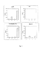
図1は、実施例2に記載の、血清成分、可溶性細胞画分(サイトゾル)及び膜調製物の沈殿に対する実施例1の組成物CにおけるpH及び界面活性剤の効果を示している。ウシ血清アルブミンを除き、各調製物において、タンパク質凝集に関して酸性pHと界面活性剤との間の相乗作用が証明された。サイトゾル試料の当該相乗作用は、ヘモグロビンの存在のために非常に強力であり、そして透過処理試薬との反応で強力な凝集及び沈殿を伴う。膜画分のかかる相乗作用はやや強力なようであるが、この結果は、界面活性剤による膜の脂質部分の溶解によってマスキングされており、これによりカラム4のOD650値が過小評価される。
FIG. 1 shows the effect of pH and surfactant in Composition C of Example 1 on the precipitation of serum components, soluble cell fraction (cytosol) and membrane preparation as described in Example 2. With the exception of bovine serum albumin, a synergistic effect between acidic pH and surfactant was demonstrated for protein aggregation in each preparation. This synergy of cytosolic samples is very strong due to the presence of hemoglobin and is accompanied by strong aggregation and precipitation in reaction with permeabilization reagents. Although such synergism of the membrane fraction appears to be somewhat strong, this result is masked by the dissolution of the lipid portion of the membrane by the surfactant, which underestimates the OD650 value of
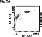
図2Aから図2Jは、細胞の形態保存及び抗体による赤血球の透過に対する、実施例1の組成物CにおけるpH及び界面活性剤の存在の効果を示す。 FIGS. 2A-2J show the effect of pH and the presence of surfactant in composition C of Example 1 on cell morphology preservation and red blood cell permeation by antibodies.
pHが7.0で、界面活性剤が不在のもと、赤血球は図2Aの上方の集団として現れ、これは下方の集団における血小板及び残骸からよく分離されている。図2Bは、赤血球が抗チューブリン−フルオレセインN−イソチオシアネート(FITC)抗体に対し透過性がないことを示している。 At pH 7.0 and in the absence of detergent, red blood cells appear as the upper population in FIG. 2A, which is well separated from platelets and debris in the lower population. FIG. 2B shows that red blood cells are not permeable to anti-tubulin-fluorescein N-isothiocyanate (FITC) antibody.
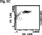
pHが5.0で、界面活性剤が不在のもと(図2C及び2D)、形態及び透過性は前述の条件とほぼ同じである。他方、図2E及び2Fは、pH7.0で界面活性剤の存在下赤血球が破壊されることを示している。 With a pH of 5.0 and no surfactant (FIGS. 2C and 2D), morphology and permeability are almost the same as described above. On the other hand, FIGS. 2E and 2F show that red blood cells are destroyed in the presence of detergent at pH 7.0.
図2G及び2Hは、実施例1の組成物と一緒に血液をインキュベーションした後、赤血球がよく保存されたことを示す。図2Hで示されている細胞のクラスターは、細胞と抗チューブリン−FITC抗体との結合をはっきりと示しており、そして赤血球が抗チューブリン−FITC抗体に対し透過処理されたことを例示している。図2I及び2Jは、実施例1の組成物Cで処理したが、FITCと接合したイソ型のコントロール抗体であって、既知の細胞構成要素のいずれについても非特異的なものとインキュベートした赤血球を示す。図2Jは、細胞と当該コントロール抗体との結合が非常にわずかであるか、又は全くないことを示しており、これにより、抗チューブリン−FITC抗体反応の特異性が確認される。 2G and 2H show that red blood cells were well preserved after incubation of blood with the composition of Example 1. The cluster of cells shown in FIG. 2H clearly shows the binding of the cells to the anti-tubulin-FITC antibody and illustrates that erythrocytes were permeabilized against the anti-tubulin-FITC antibody. Yes. FIGS. 2I and 2J show erythrocytes incubated with composition C of Example 1 but isotype control antibody conjugated to FITC and non-specific for any of the known cellular components. Show. FIG. 2J shows very little or no binding between the cells and the control antibody, confirming the specificity of the anti-tubulin-FITC antibody reaction.
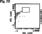
図3Aから図3Jは、白血球の保存及び透過性に対するpHと界面活性剤の効果を示す。赤血球及び顆粒球の分離後、抹消単核球を、pH7.0の界面活性剤を含まない透過処理試薬と混合した。図3Aは、四角内にリンパ球群を示す。下方の集団は、血小板と残骸である。上記条件下で、リンパ球は、図3Bに示す通り、抗チューブリン抗体に対し透過性ではない。 3A-3J show the effect of pH and surfactant on leukocyte storage and permeability. After separation of erythrocytes and granulocytes, the peripheral mononuclear cells were mixed with a permeabilizing reagent without pH 7.0 surfactant. FIG. 3A shows a group of lymphocytes within the square. The lower population is platelets and debris. Under the above conditions, lymphocytes are not permeable to anti-tubulin antibodies as shown in FIG. 3B.
図3C及び3Dは、pH5.0の界面活性剤を含まない透過処理試薬を用いて得られた結果を示し、これは図3A及び3Bにおいて観察されたものと類似している。図3E及び3Fについては、透過処理試薬はpH7.0で界面活性剤の存在下用いられる。リンパ球の破壊が観察された。 FIGS. 3C and 3D show the results obtained using a permeabilization reagent without pH 5.0 surfactant, which is similar to that observed in FIGS. 3A and 3B. For FIGS. 3E and 3F, the permeabilization reagent is used in the presence of a surfactant at pH 7.0. Lymphocyte destruction was observed.
図3G及び3Hについては、界面活性剤を含みpH5.0の実施例1の組成物Cを使用した。図3Hで示す細胞は、抗チューブリン−FITC抗体によるリンパ球の透過をはっきりと例示している。図3I及び3Jは、非特異的なイソ型コントロール抗体を用いて得られた結果を示す。図3Jは、細胞とコントロール抗体との結合が極めて少ないか、又は全くないことを示しており、これにより抗チューブリン−FITC抗体反応の特異性が確認される。 For FIGS. 3G and 3H, composition C of Example 1 containing a surfactant and having a pH of 5.0 was used. The cells shown in FIG. 3H clearly illustrate the penetration of lymphocytes by anti-tubulin-FITC antibody. Figures 3I and 3J show the results obtained with non-specific isotype control antibodies. FIG. 3J shows that there is very little or no binding between the cells and the control antibody, confirming the specificity of the anti-tubulin-FITC antibody reaction.
実施例5は、胎児ヘモグロビンの解析のための細胞透過処理安定化試薬を用いた方法を例示する。 Example 5 illustrates a method using a cell permeabilization stabilization reagent for analysis of fetal hemoglobin.
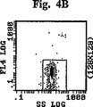
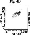
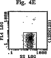
図4から図4Fは、抗HbF−FITCによる細胞内マーキング及び抗iフィコエリトリン(PE)による細胞外マーキングによる胎児赤血球の同定を示す。更に具体的には、個々の散乱図は以下の通りである:
図4A、4B及び4C:カルシウム不在下でのマーキング。
図4D、4E及び4F:1mMのカルシウムの存在下でのマーキング。
図4A及び4D:血液構成要素の散乱図。血小板及び残骸は閾値を設けることで散乱図から除外した。
Figures 4 to 4F show the identification of fetal erythrocytes by intracellular marking with anti-HbF-FITC and extracellular marking with anti-i phycoerythrin (PE). More specifically, the individual scatter diagrams are as follows:
Figures 4A, 4B and 4C: marking in the absence of calcium.
Figures 4D, 4E and 4F: marking in the presence of 1 mM calcium.
4A and 4D: Scatter diagrams of blood components. Platelets and debris were excluded from the scattergram by setting a threshold.
図4B及び4E:FL4におけるCD45−PC5による白血球の排除後に枠内に入れられた赤血球の集団。 4B and 4E: Population of red blood cells encased after exclusion of white blood cells by CD45-PC5 in FL4.
図4C及び4Fは、Quadrant3(左下)に成体の赤血球(HbF−、i+)を、Quadrant2(右上)に胎児赤血球(HbF+、i+)を、そしてQuadrant4(右下)にF細胞(HbF+、i−)を示す。 Figures 4C and 4F show adult red blood cells (HbF-, i +) in Quadrant3 (lower left), fetal red blood cells (HbF +, i +) in Quadrant2 (upper right), and F cells (HbF +, i-) in Quadrant4 (lower right). ).
実施例6は、本発明の細胞透過処理安定化試薬を用いた、フローサイトメトリー上での蛍光によるアルファチューブリン及び糖化ヘモグロビン(HbA1c)の検出を例示する。 Example 6 illustrates the detection of alpha tubulin and glycated hemoglobin (HbA1c) by fluorescence on flow cytometry using the cell permeabilization stabilization reagent of the present invention.
本発明の透過処理試薬は、細胞膜を透過処理し、細胞内タンパク質の沈殿を引き起こし、そして更に、光散乱及び蛍光を用いたフローサイトメトリー解析のための細胞マーカーに対する特異的結合のために細胞構成要素を保存すること、が可能である。 The permeabilization reagent of the present invention permeabilizes the cell membrane, causes precipitation of intracellular proteins, and, further, cell configuration for specific binding to cell markers for flow cytometric analysis using light scattering and fluorescence. It is possible to save the element.
以下の実施例は本発明の例示であり、特許請求の範囲で定義するような本発明の範囲を限定するものと決して解されるべきではない。前述の開示に従い、種々の他の成分及び比率が採用されうることが理解されよう。 The following examples are illustrative of the invention and should in no way be construed as limiting the scope of the invention as defined in the claims. It will be understood that various other components and ratios may be employed in accordance with the foregoing disclosure.
実施例1:透過処理試薬組成物
組成物A
以下の透過処理安定化試薬組成物を調製した。

The following permeation treatment stabilizing reagent composition was prepared.
更に具体的には、N−ラウロイルサルコシンのストック溶液を最初に作った。1.0gのN−ラウロイルサルコシン(Fluka)を1.5mlのエタノール(96%)に予め溶解した。180μlのピロリジン(Aldrich)を95mlの脱イオン水に添加した。続いて、N−ラウロイルサルコシン/エタノール溶液をピロリジン溶液に添加し;pHをピロリジン又はHClで5.6に調節し、そして容積を脱イオン水で100mlに調節してストック溶液を形成した。試薬の全容積を脱イオン水で100mlに調節した。透過処理安定化試薬は、6.25mlのストック溶液を脱イオン水で100mlに希釈し、そしてpHをピロリジン又はHClで5.3に調節することによって調製した。組成物Aは0.1mS/cmの導電率を有していた。 More specifically, a stock solution of N-lauroyl sarcosine was first made. 1.0 g N-lauroyl sarcosine (Fluka) was pre-dissolved in 1.5 ml ethanol (96%). 180 μl pyrrolidine (Aldrich) was added to 95 ml deionized water. Subsequently, the N-lauroyl sarcosine / ethanol solution was added to the pyrrolidine solution; the pH was adjusted to 5.6 with pyrrolidine or HCl, and the volume was adjusted to 100 ml with deionized water to form a stock solution. The total volume of the reagent was adjusted to 100 ml with deionized water. The permeabilization stabilization reagent was prepared by diluting 6.25 ml of stock solution to 100 ml with deionized water and adjusting the pH to 5.3 with pyrrolidine or HCl. Composition A had a conductivity of 0.1 mS / cm.
組成物B
以下の透過試薬組成物は、各化合物を脱イオン水に溶解することで調製した。
The following permeation reagent compositions were prepared by dissolving each compound in deionized water.
当該試薬組成物は、0.25mS/cmの導電率を有していた。N−ラウロイルサルコシンを上述のようにストック溶液として添加した。 The reagent composition had a conductivity of 0.25 mS / cm. N-lauroyl sarcosine was added as a stock solution as described above.
組成物C
以下の透過試薬組成物は、各化合物を脱イオン水に溶解することで調製した。
The following permeation reagent compositions were prepared by dissolving each compound in deionized water.
当該試薬組成物は、0.5mS/cmの導電率を有していた。N−ラウロイルサルコシンを上述のようにストック溶液として添加した。 The reagent composition had a conductivity of 0.5 mS / cm. N-lauroyl sarcosine was added as a stock solution as described above.
実施例2:赤血球の異なる成分の沈殿に対する、pH、界面活性剤及びpHと界面活性剤の組み合わせの効果
0.7mMのエチレンジアミン四酢酸(EDTA)を抗凝固剤として用いて処理した大量の血液を使用することで血清を調製した。別の大量のEDAT処理済みの血液をリン酸緩衝液(PBS)で洗浄し、9倍量の水で希釈して細胞可溶化物を取得した。細胞可溶化物を200gの遠心に15分間かけて、可溶性の細胞画分と膜画分とを分離した。ペレット含有膜画分を、可溶性細胞画分を体積当たり5%含むように大量の水と混合した。
Example 2: Effect of pH, surfactant and combination of pH and surfactant on precipitation of different components of red blood cells A large amount of blood treated with 0.7 mM ethylenediaminetetraacetic acid (EDTA) as an anticoagulant. Serum was prepared by use. Another large amount of EDAT-treated blood was washed with a phosphate buffer (PBS) and diluted with 9 times the amount of water to obtain a cell lysate. The cell lysate was centrifuged at 200 g for 15 minutes to separate the soluble cell fraction and the membrane fraction. The pellet-containing membrane fraction was mixed with a large amount of water to contain a soluble cell fraction at 5% by volume.
血清、可溶性細胞画分、膜画分、並びにコントロールとしてのウシ血清アルブミンを、実施例1の組成物Cと、そして以下の修飾試薬との混合後にモニタリングした:
1.修飾試薬1:界面活性剤(トリスラウリル硫酸塩及びN−ラウリルサルコシン)無添加で、pH7.0の実施例1の組成物C。
2.修飾試薬2:界面活性剤(トリスラウリル硫酸塩及びN−ラウリルサルコシン)無添加で、pH5.0の実施例1の組成物C。
3.修飾試薬3:pH7.0の実施例1の組成物C。
4.透過処理試薬4:上述の(pH5.0の)実施例1の組成物C。
Serum, soluble cell fraction, membrane fraction, and bovine serum albumin as a control were monitored after mixing composition C of Example 1 and the following modifying reagents:
1. Modifying reagent 1: Composition C of Example 1 at pH 7.0 without the addition of surfactants (trislauryl sulfate and N-lauryl sarcosine).
2. Modifying reagent 2: Composition C of Example 1 at pH 5.0 without addition of surfactants (trislauryl sulfate and N-lauryl sarcosine).
3. Modifying reagent 3: Composition C of Example 1 at pH 7.0.
4). Permeation reagent 4: Composition C of Example 1 above (pH 5.0).
試料混合物は、2mlの上述の特定の試薬を以下の4つの成分のうちの1つと混合することで生成した:
−0.01mlの血清(A)、
−0.01mlの15%重量/体積のウシアルブミン調製物(B)、
−0.1mlの膜画分(D)。
The sample mixture was generated by mixing 2 ml of the specific reagent described above with one of the following four components:
-0.01 ml of serum (A),
-0.01 ml of 15% weight / volume bovine albumin preparation (B),
-0.1 ml membrane fraction (D).
タンパク質の沈殿を測定するために、試料混合物の1時間後に、試料混合物の光学密度を、前記画分の主要構成要素であるヘモグロビンが吸収しない650nmの波長で測定した。 To measure protein precipitation, after 1 hour of the sample mixture, the optical density of the sample mixture was measured at a wavelength of 650 nm where the main component, hemoglobin, does not absorb.
図1は、血清;15%重量/体積のウシ血清アルブミン(BSA)の調製物;可溶性細胞画分(サイトゾル);及び膜画分、における細胞成分の沈殿に対する前記試薬におけるpH及び界面活性剤の効果を示す。上文で言及した細胞成分のそれぞれについての棒グラフは、左から右へ、修飾試薬1〜3及び透過処理試薬4を用いて得られた結果を示す。
FIG. 1 shows the pH and surfactant in the reagents for precipitation of cellular components in serum; 15% weight / volume bovine serum albumin (BSA) preparation; soluble cell fraction (cytosol); and membrane fraction. The effect of The bar graph for each of the cellular components referred to above shows the results obtained using modification reagents 1-3 and
図1は、上述の通り、低pHで界面活性剤を含む透過処理試薬4のみがタンパク質の凝集及び沈殿をもたらしたことを示している。
FIG. 1 shows that only the
実施例3:赤血球内への抗体の浸透に対する、pH、界面活性剤、及び酸性pHと界面活性剤の組み合わせの効果
0.01mlの全血を100μlの生理食塩水と混合し、続いて2mlの実施例2に記載の各透過処理試薬の変形版と混合した。5分間インキュベーションした後、50μlの各混合物を、0.2%(w/v)のウシ血清アルブミン及び、FITCと接合した、アルファチューブリンに対するモノクローナル抗体(Beckman Coulter Inc.米国マイアミ)を含む50μlのPBS溶液に添加した。アルファチューブリンは、専ら細胞の内側で発現する分子である。15分間のインキュベーション後、混合物は、0.5%ホルムアルデヒドを含む1mlのPBSと混合することで反応を停止させ、そして細胞を固定し、これにより解析用の試料混合物を形成させた。
Example 3: Effect of pH, surfactant, and acidic pH and surfactant combination on antibody penetration into red blood cells 0.01 ml whole blood was mixed with 100 μl saline followed by 2 ml Mixed with a modified version of each permeabilization reagent described in Example 2. After 5 minutes of incubation, 50 μl of each mixture was added with 50 μl of 0.2% (w / v) bovine serum albumin and a monoclonal antibody to alpha tubulin conjugated with FITC (Beckman Coulter Inc. Miami, USA). Added to PBS solution. Alpha tubulin is a molecule that is expressed exclusively inside cells. After 15 minutes incubation, the mixture was stopped by mixing with 1 ml PBS containing 0.5% formaldehyde and cells were fixed, thereby forming a sample mixture for analysis.
試料混合物をXLフローサイトメーター(Beckman Coulter Inc.米国マイアミ)上で解析した。細胞の完全性は、前方散乱及び側方散乱によって解析した。細胞の透過性は、FITC抗体の蛍光によって解析した。結果を図2A〜2Jに示す。更に具体的には、個々の散乱図は以下の通りである:
図2A及び2B:血液と修飾試薬1とのプレインキュベーション。
図2C及び2D:血液と修飾試薬2とのプレインキュベーション。
図2E及び2F:血液と修飾試薬3とのプレインキュベーション。
図2G及び2H:血液と透過処理試薬4とのプレインキュベーション。
The sample mixture was analyzed on an XL flow cytometer (Beckman Coulter Inc. Miami, USA). Cell integrity was analyzed by forward and side scatter. Cell permeability was analyzed by FITC antibody fluorescence. The results are shown in FIGS. More specifically, the individual scatter diagrams are as follows:
2A and 2B: Preincubation of blood with modifying
2C and 2D: Preincubation of blood with modifying
2E and 2F: Preincubation of blood with modifying
2G and 2H: Preincubation of blood with
図2I及び2J:透過処理試薬4とのプレインキュベーションであって、FITCと接合したイソ型コントロール抗体と反応させるもの。
FIGS. 2I and 2J: Preincubation with
ここで、FSは前方光散乱であり;SSは側方光散乱であり;FL1、FL2及びFL4は、それぞれ525nm、575nm、及び675nmで測定した蛍光シグナルである。 Where FS is forward light scatter; SS is side light scatter; FL1, FL2 and FL4 are fluorescence signals measured at 525 nm, 575 nm and 675 nm, respectively.
コントロール抗体は、FITCと接合したイソ型抗体であり、これは既知の細胞構成要素のいずれにも特異的ではない。 The control antibody is an isotype antibody conjugated to FITC, which is not specific for any known cellular component.
界面活性剤を含み、且つpHが低い透過処理試薬4のみが、赤血球の透過及び保存に効果を有していたことが観察された。
It was observed that only the
実施例4:白血球内への抗体の浸透に対する、pH、界面活性剤、及びpHと界面活性剤の組み合わせの効果
単核白血球は、EDTAの存在下、フィコールを用い、A. Boyem (1968, Scand. J. Clin. Lab. Invest., 21 Suppl. 97)の方法に従い抹消血から調製した。1000万の細胞を100μlの生理食塩水と混合し、続いて2mlの実施例2に記載の各透過処理試薬の変形版と混合した。細胞のインキュベーションと抗チューブリンFITC抗体による細胞内マーキングを実施例3に記載のように実施した。結果を図3Aから3Jに示す。更に具体的には、個々の散乱図は以下の通りである:
図3A、3B:細胞と修飾試薬1とのプレインキュベーション。
図3C、3D:細胞と修飾試薬2とのプレインキュベーション。
図3E、3F:細胞と修飾試薬3とのプレインキュベーション。
図3G、3H:細胞と透過処理試薬4とのプレインキュベーション。
図3I、3J:細胞と透過処理試薬4とのプレインキュベーション、FITCと接合したイソ型コントロール抗体との反応。
Example 4: Effect of pH, surfactant, and combination of pH and surfactant on antibody penetration into leukocytes Mononuclear leukocytes were treated with A. Boyem (1968, Scand) using Ficoll in the presence of EDTA. J. Clin. Lab. Invest., 21 Suppl. 97). Ten million cells were mixed with 100 μl of saline followed by 2 ml of a modified version of each permeabilization reagent described in Example 2. Cell incubation and intracellular marking with anti-tubulin FITC antibody were performed as described in Example 3. The results are shown in FIGS. 3A to 3J. More specifically, the individual scatter diagrams are as follows:
3A, 3B: Preincubation of cells with modifying
3C, 3D: Preincubation of cells with modifying
3E, 3F: Preincubation of cells with modifying
Figures 3G, 3H: Preincubation of cells with
3I, 3J: Preincubation of cells with
界面活性剤を含み、且つpHが低い透過処理試薬4のみが、白血球の透過及び保存に効果を有していたことが観察された。
It was observed that only the
実施例5:細胞の内側及び表面にある胎児抗原に基づく胎児赤血球の検出のための透過処理試薬の使用
99%(v/v)の正常な血液及び1%(v/v)の臍帯血の混合物を胎児赤血球の検出に使用した。100μlの生理食塩水、続いて1mlの組成物Cを5μlの血液混合物に添加した。10分間のインキュベーション後、50μlの試料混合物を、以下のものを含む2つのチューブに添加した:
−チューブAには、0.2%(w/v)のウシ血清アルブミンと以下の抗体混合物:
−FITCと接合した抗胎児ヘモグロビン(HbF)モノクローナル抗体(フランス国特許第98 09006号);
−フィコエリトリンと接合した抗胎児血液群iモノクローナル抗体(フランス国特許第98 09006号);及び
−抗CD45−PC5モノクローナル抗体(Beckman Coulter Inc.フランス国マルセーユ)、
を含む50μlのPBS溶液を含めた。
Example 5: Use of permeabilization reagents for detection of fetal erythrocytes based on fetal antigens inside and on the surface of 99% (v / v) normal blood and 1% (v / v) umbilical cord blood The mixture was used for detection of fetal erythrocytes. 100 μl saline followed by 1 ml composition C was added to 5 μl blood mixture. After a 10 minute incubation, 50 μl of the sample mixture was added to two tubes containing:
-In tube A, 0.2% (w / v) bovine serum albumin and the following antibody mixture:
An anti-fetal hemoglobin (HbF) monoclonal antibody conjugated to FITC (French Patent No. 98 09006);
An anti-fetal blood group i monoclonal antibody conjugated with phycoerythrin (French Patent No. 98 09006); and an anti-CD45-PC5 monoclonal antibody (Beckman Coulter Inc. Marseille, France),
50 μl of PBS solution containing was included.
−チューブBには、チューブAと同内容のものを含め、更に10μlの10mMCaCl2を添加した。 -To tube B, 10 μl of 10 mM CaCl 2 was added, including the same content as tube A.
チューブAは、HbFとの反応のネガティブコントロールとしての役割を果たし、これは、当該反応がカルシウム(Ca2+)イオンの存在に依存するためである。 Tube A serves as a negative control for the reaction with HbF because it depends on the presence of calcium (Ca 2+ ) ions.
前記混合物をXLフローサイトメーター(Beckman Coulter Inc.米国マイアミ)上で解析した。細胞の完全性は、前方散乱及び側方散乱によって解析した。CD45を使用して、この解析から白血球を排除した。胎児赤血球は、抗体の蛍光によって検出した。結果を図4A〜4Fに示す。図4A〜4Cは、チューブAから得られた結果を示し、図4D〜4EはチューブBから得られた結果を示す。 The mixture was analyzed on an XL flow cytometer (Beckman Coulter Inc. Miami, USA). Cell integrity was analyzed by forward and side scatter. CD45 was used to exclude leukocytes from this analysis. Fetal erythrocytes were detected by antibody fluorescence. The results are shown in FIGS. 4A-4C show the results obtained from tube A and FIGS. 4D-4E show the results obtained from tube B. FIGS.
図4C及び4Fにおいて、Quadrant3(左下)に成体の赤血球(HbF−、i+)が配置され、Quadrant2(右上)に胎児赤血球(HbF+、i+)が配置され、そしてQuadrant4(右下)にF細胞(HbF+、i−)が配置されていたことが観察された。 In FIGS. 4C and 4F, adult red blood cells (HbF−, i +) are placed in Quadrant3 (lower left), fetal red blood cells (HbF +, i +) are placed in Quadrant2 (upper right), and F cells in Quadrant4 (lower right) ( It was observed that HbF +, i−) was located.
注目すべきは、カルシウムイオンの不在下では、図4CのQuadrant2には胎児赤血球が見られなかったことである。従って、赤血球は接合した抗体に対し透過性ではなく、そしてHbF抗原と抗HbF抗体との反応がカルシウムイオンに依存する。一見、HbF抗原はその天然の状態で保存され、これは、赤血球と本発明の透過処理試薬とのインキュベーション後の、HbFと抗体との間の相互作用のカルシウム依存性の保存によって確認された。
It should be noted that in the absence of calcium ions, fetal erythrocytes were not seen in
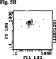
実施例6:赤血球におけるアルファチューブリン及びHbA1Cの検出
0.01mlの全血を100μlの生理食塩水、続いて2mlの実施例1の組成物Bと混合した。5分間のインキュベーション後、50μlの混合物を、FITCと接合した、アルファチューブリンに対するモノクローナル抗体(Beckman Coulter Inc.米国マイアミ)を含むか、あるいはHbA1Cに対する、FITCと接合したモノクローナル抗体(抗HbA1c−FITC抗体)であって、米国特許第4,727,036号に記載のKnowles et alの方法に従い調製したものを含む50μlのPBS溶液に添加した。15分間のインキュベーション後、混合物は、0.5%ホルムアルデヒドを含む1mlのPBSと混合することで反応を停止させ、そして細胞を固定し、これにより解析用の試料混合物を形成させた。
Example 6: Detection of alpha tubulin and HbA1C in erythrocytes 0.01 ml of whole blood was mixed with 100 μl of saline followed by 2 ml of composition B of Example 1. After 5 minutes of incubation, 50 μl of the mixture contains a monoclonal antibody against alpha tubulin (Beckman Coulter Inc. Miami, USA) conjugated to FITC or a monoclonal antibody conjugated to FITC (anti-HbA1c-FITC antibody against HbA1C). ) And prepared according to the method of Knowles et al as described in US Pat. No. 4,727,036. After 15 minutes incubation, the mixture was stopped by mixing with 1 ml PBS containing 0.5% formaldehyde and cells were fixed, thereby forming a sample mixture for analysis.
試料混合物をXLフローサイトメーター(Beckman Coulter Inc.米国マイアミ)上で解析した。細胞の完全性は、前方散乱及び側方散乱によって解析した。細胞の透過性は、FITC抗体の蛍光によって解析した。結果を図5A〜5Fに示す。更に具体的には、個々の散乱図は以下の通りである:
図5A及び5B:抗チューブリン−FITC抗体との反応。
図5B及び5D:抗HbA1c−FITC抗体との反応。
図5E及び5F:FITCと接合したイソ型コントロール抗体との反応。
The sample mixture was analyzed on an XL flow cytometer (Beckman Coulter Inc. Miami, USA). Cell integrity was analyzed by forward and side scatter. Cell permeability was analyzed by FITC antibody fluorescence. The results are shown in FIGS. More specifically, the individual scatter diagrams are as follows:
Figures 5A and 5B: Reaction with anti-tubulin-FITC antibody.
Figures 5B and 5D: Reaction with anti-HbA1c-FITC antibody.
Figures 5E and 5F: Reaction with isotype control antibody conjugated to FITC.
前記コントロール抗体は、FITCTと接合したイソ型抗体であり、これは既知の細胞構成要素のいずれについても非特異的である。 The control antibody is an isotype antibody conjugated to FITCT, which is non-specific for any known cellular component.
図示したように、赤血球は抗チューブリン−FITC抗体、及び抗HbA1c−FITC抗体の両方に透過性があり、これにより、フローサイトメーターによる赤血球中のアルファチューブリン及びHbA1cの検出が可能となった。 As shown, erythrocytes are permeable to both anti-tubulin-FITC antibody and anti-HbA1c-FITC antibody, which allowed detection of alpha tubulin and HbA1c in erythrocytes by flow cytometer. .
本発明を詳細に説明し、そして添付図面において絵を用いて本発明を示したが、これらは本発明の範囲を限定するものとしてみなされるべきではなく、むしろそれらの好ましい態様の例示としてみなされるべきである。しかしながら、種々の修飾及び変更が本明細書に記載され、そして特許請求の範囲で定義された発明及びその均等物の精神及び範囲内で行われうることは自明であろう。 The present invention has been described in detail and illustrated with the aid of drawings in the accompanying drawings, which should not be construed as limiting the scope of the invention, but rather as examples of preferred embodiments thereof. Should. However, it will be apparent that various modifications and changes may be made within the spirit and scope of the invention as described herein and defined in the claims and equivalents thereof.
Claims (12)
(a)以下の分子構造:
R1−CO−N(CH3)CH2COOX1
(ここで、R1は8〜18個の炭素原子を有するアルキル基又はアルキレン基であり、そしてX1はH、Na+、又はK+である)によって表されるN−アシルサルコシン又はその塩;
(b)ウシ血清アルブミン;及び
(c)糖類又はグリセロール、
の水溶液を含んで成り、
該試薬は、7未満のpHを有し、かつ、1.2mS/cm未満の導電率によって定義される低イオン強度を有している、試薬。A reagent that permeabilizes cells and stabilizes:
(A) The following molecular structure:
R 1 —CO—N (CH 3 ) CH 2 COOX 1
(Wherein, R 1 is an alkyl or alkylene group having 8 to 18 carbon atoms, and X 1 is H, Na +, or K + is a) N- acyl sarcosine or a salt thereof represented by ;
(B) bovine serum albumin ; and (c) sugars or glycerol ,
Comprising an aqueous solution of
The reagent has a pH of less than 7 and a low ionic strength defined by a conductivity of less than 1.2 mS / cm.
(a)全血;及び(A) whole blood; and
(b)0.1mM〜10mMの、以下の分子構造:(B) 0.1 mM to 10 mM of the following molecular structure:
RR 11 −CO−N(CH-CO-N (CH 3Three )CH) CH 22 COOXCOOX 11
(ここで、R(Where R 11 は8〜18個の炭素原子を有するアルキル基又はアルキレン基であり、そしてXIs an alkyl or alkylene group having 8 to 18 carbon atoms and X 11 はH、NaIs H, Na ++ 、又はKOr K ++ である)によって表されるN−アシルサルコシン又はその塩の水溶液を含んで成る、細胞を透過処理して安定化する試薬A reagent for stabilizing cells by permeabilizing them, comprising an aqueous solution of N-acyl sarcosine or a salt thereof represented by
を含んで成り、該試薬は、4〜6の間のpHと、1.2mS/cm未満の導電率を有しており、そして、該試薬によって該全血中の血球の細胞膜が透過処理され、該血球の細胞内タンパク質が凝集させられる一方で、該血球は細胞解析のために保存される、組成物。The reagent has a pH between 4 and 6 and a conductivity of less than 1.2 mS / cm, and the reagent permeabilizes the cell membrane of blood cells in the whole blood. A composition wherein the intracellular proteins of the blood cells are aggregated while the blood cells are stored for cell analysis.
(a)0.1mM〜10mMの、以下の分子構造:(A) 0.1 mM to 10 mM of the following molecular structure:
RR 11 −CO−N(CH-CO-N (CH 3Three )CH) CH 22 COOXCOOX 11
(ここで、R(Where R 11 は8〜18個の炭素原子を有するアルキル基又はアルキレン基であり、そしてXIs an alkyl or alkylene group having 8 to 18 carbon atoms and X 11 はH、NaIs H, Na ++ 、又はKOr K ++ である)によって表されるN−アシルサルコシン又はその塩;及びN-acyl sarcosine or a salt thereof represented by
(b)糖類又はグリセロール(B) Sugar or glycerol
の水溶液を含んで成り、該試薬は、7未満のpHと、1.2mS/cm未満の導電率によって定義される低イオン強度を有しており;該試薬は、血球の細胞膜の透過を可能にして、該血球の細胞内タンパク質の凝集を引き起こす一方で、該血球を細胞解析のために保存する、試薬。Wherein the reagent has a pH of less than 7 and a low ionic strength defined by a conductivity of less than 1.2 mS / cm; the reagent allows permeation of blood cell membranes A reagent that preserves the blood cells for cell analysis while causing aggregation of intracellular proteins of the blood cells.
Applications Claiming Priority (3)
| Application Number | Priority Date | Filing Date | Title |
|---|---|---|---|
| US11/052,269 US7678578B2 (en) | 2005-02-07 | 2005-02-07 | Cell permeabilization and stabilization reagent and method of use |
| US11/052,269 | 2005-02-07 | ||
| PCT/US2006/002612 WO2006086157A1 (en) | 2005-02-07 | 2006-01-24 | Cell permeabilization and stabilization reagent and method of use |
Publications (2)
| Publication Number | Publication Date |
|---|---|
| JP2008530526A JP2008530526A (en) | 2008-08-07 |
| JP4869258B2 true JP4869258B2 (en) | 2012-02-08 |
Family
ID=36780675
Family Applications (1)
| Application Number | Title | Priority Date | Filing Date |
|---|---|---|---|
| JP2007554139A Active JP4869258B2 (en) | 2005-02-07 | 2006-01-24 | Reagent for stabilizing cells by permeabilization and method of using the same |
Country Status (5)
| Country | Link |
|---|---|
| US (1) | US7678578B2 (en) |
| EP (1) | EP1863348B1 (en) |
| JP (1) | JP4869258B2 (en) |
| CN (1) | CN101115388B (en) |
| WO (1) | WO2006086157A1 (en) |
Families Citing this family (10)
| Publication number | Priority date | Publication date | Assignee | Title |
|---|---|---|---|---|
| US7541190B2 (en) * | 2005-02-07 | 2009-06-02 | Beckman Coulter, Inc. | Method of measurement of cellular hemoglobin |
| US7625757B2 (en) * | 2006-01-27 | 2009-12-01 | Sysmex Corporation | Reagent for immature leukocyte analysis and reagent kit |
| US20090035758A1 (en) * | 2007-08-02 | 2009-02-05 | Beckman Coulter, Inc. | Method of Measurement of Micronucleated Erythrocyte Populations |
| US7968279B2 (en) * | 2008-08-13 | 2011-06-28 | Beckman Coulter, Inc. | Reference control for cell by cell analysis |
| US8293537B2 (en) * | 2010-02-10 | 2012-10-23 | Selinfreund Richard H | Systems and methods for hemoglobin analysis |
| US9295251B1 (en) | 2011-04-08 | 2016-03-29 | Safehands Solutions, LLC | Synergistic antimicrobial compositions of PCMX and carboxylic acid and related methods |
| EP2607899B1 (en) * | 2011-12-21 | 2016-08-10 | Beckman Coulter, Inc. | Method for labeling intracellular and extracellular targets of leukocytes |
| TW201439515A (en) | 2012-12-28 | 2014-10-16 | Beckman Coulter Inc | Composition for high stringency cell treatment and antigen retrieval |
| CN111504887B (en) * | 2020-05-09 | 2023-05-09 | 苏州四正柏生物科技有限公司 | Hemolysin and preparation method thereof |
| CN112504793B (en) * | 2020-10-14 | 2021-10-01 | 核工业总医院 | Reagent for permeating and fixing blood cells and analysis method |
Citations (3)
| Publication number | Priority date | Publication date | Assignee | Title |
|---|---|---|---|---|
| JPH10206423A (en) * | 1996-11-20 | 1998-08-07 | Toa Medical Electronics Co Ltd | Classification counting method of juvenile leukocyte |
| US20030092187A1 (en) * | 2001-11-13 | 2003-05-15 | Karsten Stahnke | Method of detecting the release of substances from cell organelles by means of flow cytometry |
| WO2004085989A2 (en) * | 2003-03-20 | 2004-10-07 | Beckman Coulter, Inc. | Dye compositions which provide enhanced differential fluorescence and light scatter characteristics |
Family Cites Families (22)
| Publication number | Priority date | Publication date | Assignee | Title |
|---|---|---|---|---|
| US4545979A (en) * | 1982-02-22 | 1985-10-08 | Warner-Lambert Company | Dental hygiene compositions |
| US4477438A (en) * | 1982-11-12 | 1984-10-16 | Surgikos, Inc. | Hydrogen peroxide composition |
| US4727036A (en) * | 1985-08-08 | 1988-02-23 | Molecular Diagnostics, Inc. | Antibodies for use in determining hemoglobin A1c |
| US5714380A (en) * | 1986-10-23 | 1998-02-03 | Amoco Corporation | Closed vessel for isolating target molecules and for performing amplification |
| US4810630A (en) * | 1987-03-30 | 1989-03-07 | E. I. Du Pont De Nemours And Company | Use of polyoxyethylene ethers to improve the performance of immunoassays employing peroxidase conjugates |
| JP2935529B2 (en) | 1990-03-01 | 1999-08-16 | シスメックス株式会社 | Leukocyte classification method and reagent |
| US5268292A (en) * | 1988-06-27 | 1993-12-07 | Robertson Betty H | Reproducible generation of high yields of hepatitis A virus by cell culture |
| AU666957B2 (en) * | 1992-08-28 | 1996-02-29 | Alcon Laboratories, Inc. | Use of certain anionic surfactants to enhance antimicrobial effectiveness of ophthalmic compositions |
| JP3301646B2 (en) | 1993-03-19 | 2002-07-15 | シスメックス株式会社 | Reagent for immature cell measurement |
| US5386043A (en) * | 1994-06-07 | 1995-01-31 | Hampshire Chemical Corp. | Non-aqueous neutralization of N-acyl sarcosines |
| ES2278566T3 (en) * | 1994-10-20 | 2007-08-16 | Sysmex Corporation | REAGENT AND METHOD FOR ANALYZING SOLID COMPONENTS IN THE URINE. |
| JP3324050B2 (en) * | 1994-10-31 | 2002-09-17 | 日本光電工業株式会社 | Leukocyte classification reagent and leukocyte classification method |
| WO1999050460A1 (en) * | 1998-03-31 | 1999-10-07 | Tularik, Inc. | HIGH THROUGHPUT ASSAY FOR DETECTION OF mRNA IN CELLS |
| US6271035B1 (en) * | 1998-10-20 | 2001-08-07 | Coulter International Corp. | Methods and compositions for rapid staining of nucleic acids in whole cells |
| US6060322A (en) * | 1998-10-20 | 2000-05-09 | Coulter International Corp. | Method for identification of reticulated cells |
| EP1124582B1 (en) * | 1998-10-27 | 2003-12-10 | Alcon Laboratories, Inc. | Preservative system for topically administrable pharmaceutical compositions |
| US6228652B1 (en) * | 1999-02-16 | 2001-05-08 | Coulter International Corp. | Method and apparatus for analyzing cells in a whole blood sample |
| DE60019458T2 (en) * | 1999-12-14 | 2006-02-23 | Thermo BioStar, Inc., Boulder | STABILIZING DILUENT FOR POLYPEPTIDES AND ANTIGENES |
| NZ522847A (en) * | 2000-05-16 | 2004-11-26 | Bolder Biotechnology Inc | Methods for refolding proteins containing free cysteine residues |
| US6413921B1 (en) * | 2000-08-01 | 2002-07-02 | Allegiance Corporation | Antimicrobial composition containing parachlorometaxylenol (PCMX) |
| US6958244B2 (en) * | 2002-04-12 | 2005-10-25 | Bruker Biospin Corp. | Low-conductivity buffers for magnetic resonance measurements |
| US20040214243A1 (en) * | 2003-04-25 | 2004-10-28 | Beckman Coulter, Inc. | Differential determination of hemoglobins |
-
2005
- 2005-02-07 US US11/052,269 patent/US7678578B2/en active Active
-
2006
- 2006-01-24 EP EP06719464.7A patent/EP1863348B1/en active Active
- 2006-01-24 JP JP2007554139A patent/JP4869258B2/en active Active
- 2006-01-24 CN CN2006800040905A patent/CN101115388B/en active Active
- 2006-01-24 WO PCT/US2006/002612 patent/WO2006086157A1/en active Application Filing
Patent Citations (3)
| Publication number | Priority date | Publication date | Assignee | Title |
|---|---|---|---|---|
| JPH10206423A (en) * | 1996-11-20 | 1998-08-07 | Toa Medical Electronics Co Ltd | Classification counting method of juvenile leukocyte |
| US20030092187A1 (en) * | 2001-11-13 | 2003-05-15 | Karsten Stahnke | Method of detecting the release of substances from cell organelles by means of flow cytometry |
| WO2004085989A2 (en) * | 2003-03-20 | 2004-10-07 | Beckman Coulter, Inc. | Dye compositions which provide enhanced differential fluorescence and light scatter characteristics |
Also Published As
| Publication number | Publication date |
|---|---|
| CN101115388B (en) | 2011-12-14 |
| US7678578B2 (en) | 2010-03-16 |
| EP1863348B1 (en) | 2016-10-12 |
| CN101115388A (en) | 2008-01-30 |
| WO2006086157A1 (en) | 2006-08-17 |
| EP1863348A4 (en) | 2010-05-26 |
| JP2008530526A (en) | 2008-08-07 |
| US20060178294A1 (en) | 2006-08-10 |
| EP1863348A1 (en) | 2007-12-12 |
Similar Documents
| Publication | Publication Date | Title |
|---|---|---|
| JP4869258B2 (en) | Reagent for stabilizing cells by permeabilization and method of using the same | |
| US7541190B2 (en) | Method of measurement of cellular hemoglobin | |
| US5811099A (en) | Method and composition for preserving antigens and process for utilizing cytological material produced by same | |
| US5849517A (en) | Method and composition for preserving antigens and nucleic acids and process for utilizing cytological material produced by same | |
| US5459073A (en) | Method and composition for preserving antigens and process for utilizing cytological material produced by same | |
| EP2324350B1 (en) | Reference control for cell by cell analysis | |
| US20080293144A1 (en) | Five-part differential white blood cell control and method for preparation of the same | |
| EP0639985B1 (en) | Method and composition for preserving antigens and process for utilizing cytological material produced by same | |
| TW201439515A (en) | Composition for high stringency cell treatment and antigen retrieval | |
| CA3063144C (en) | Compositions and methods for lysis of red blood cells | |
| JP2023524649A (en) | Nucleic acid and cell preservative compositions and methods of use | |
| Brown et al. | Specific precipitin reactions with the viruses of foot-and-mouth disease and vesicular stomatitis | |
| FI903510A0 (en) | LITHIUM SALT SOM REAGENTS SOM BRYTER NED ROEDA BLODKROPPAR OCH SOM DENATURERAR HEMOGLOBIN. | |
| Wright et al. | Acute Babesia bovis infection: A study of the vascular lesions in kidney and lung | |
| CN112504793B (en) | Reagent for permeating and fixing blood cells and analysis method |
Legal Events
| Date | Code | Title | Description |
|---|---|---|---|
| A621 | Written request for application examination |
Free format text: JAPANESE INTERMEDIATE CODE: A621 Effective date: 20090108 |
|
| A521 | Request for written amendment filed |
Free format text: JAPANESE INTERMEDIATE CODE: A821 Effective date: 20100129 |
|
| RD02 | Notification of acceptance of power of attorney |
Free format text: JAPANESE INTERMEDIATE CODE: A7422 Effective date: 20100129 |
|
| RD04 | Notification of resignation of power of attorney |
Free format text: JAPANESE INTERMEDIATE CODE: A7424 Effective date: 20100129 |
|
| A977 | Report on retrieval |
Free format text: JAPANESE INTERMEDIATE CODE: A971007 Effective date: 20101217 |
|
| A131 | Notification of reasons for refusal |
Free format text: JAPANESE INTERMEDIATE CODE: A131 Effective date: 20101221 |
|
| A601 | Written request for extension of time |
Free format text: JAPANESE INTERMEDIATE CODE: A601 Effective date: 20110316 |
|
| A602 | Written permission of extension of time |
Free format text: JAPANESE INTERMEDIATE CODE: A602 Effective date: 20110324 |
|
| A521 | Request for written amendment filed |
Free format text: JAPANESE INTERMEDIATE CODE: A523 Effective date: 20110601 |
|
| TRDD | Decision of grant or rejection written | ||
| A01 | Written decision to grant a patent or to grant a registration (utility model) |
Free format text: JAPANESE INTERMEDIATE CODE: A01 Effective date: 20111107 |
|
| A01 | Written decision to grant a patent or to grant a registration (utility model) |
Free format text: JAPANESE INTERMEDIATE CODE: A01 |
|
| A61 | First payment of annual fees (during grant procedure) |
Free format text: JAPANESE INTERMEDIATE CODE: A61 Effective date: 20111115 |
|
| R150 | Certificate of patent or registration of utility model |
Ref document number: 4869258 Country of ref document: JP Free format text: JAPANESE INTERMEDIATE CODE: R150 Free format text: JAPANESE INTERMEDIATE CODE: R150 |
|
| FPAY | Renewal fee payment (event date is renewal date of database) |
Free format text: PAYMENT UNTIL: 20141125 Year of fee payment: 3 |
|
| R250 | Receipt of annual fees |
Free format text: JAPANESE INTERMEDIATE CODE: R250 |
|
| R250 | Receipt of annual fees |
Free format text: JAPANESE INTERMEDIATE CODE: R250 |
|
| R250 | Receipt of annual fees |
Free format text: JAPANESE INTERMEDIATE CODE: R250 |
|
| R250 | Receipt of annual fees |
Free format text: JAPANESE INTERMEDIATE CODE: R250 |
|
| R250 | Receipt of annual fees |
Free format text: JAPANESE INTERMEDIATE CODE: R250 |
|
| R250 | Receipt of annual fees |
Free format text: JAPANESE INTERMEDIATE CODE: R250 |
|
| R250 | Receipt of annual fees |
Free format text: JAPANESE INTERMEDIATE CODE: R250 |
|
| R250 | Receipt of annual fees |
Free format text: JAPANESE INTERMEDIATE CODE: R250 |
|
| R250 | Receipt of annual fees |
Free format text: JAPANESE INTERMEDIATE CODE: R250 |
|
| R250 | Receipt of annual fees |
Free format text: JAPANESE INTERMEDIATE CODE: R250 |