JP4807902B2 - Surfactant protein D for prevention and diagnosis of lung disease - Google Patents
Surfactant protein D for prevention and diagnosis of lung disease Download PDFInfo
- Publication number
- JP4807902B2 JP4807902B2 JP2000577281A JP2000577281A JP4807902B2 JP 4807902 B2 JP4807902 B2 JP 4807902B2 JP 2000577281 A JP2000577281 A JP 2000577281A JP 2000577281 A JP2000577281 A JP 2000577281A JP 4807902 B2 JP4807902 B2 JP 4807902B2
- Authority
- JP
- Japan
- Prior art keywords
- mice
- lung
- emphysema
- protein
- alveolar
- Prior art date
- Legal status (The legal status is an assumption and is not a legal conclusion. Google has not performed a legal analysis and makes no representation as to the accuracy of the status listed.)
- Expired - Lifetime
Links
Images


Classifications
-
- C—CHEMISTRY; METALLURGY
- C07—ORGANIC CHEMISTRY
- C07K—PEPTIDES
- C07K14/00—Peptides having more than 20 amino acids; Gastrins; Somatostatins; Melanotropins; Derivatives thereof
- C07K14/435—Peptides having more than 20 amino acids; Gastrins; Somatostatins; Melanotropins; Derivatives thereof from animals; from humans
- C07K14/785—Alveolar surfactant peptides; Pulmonary surfactant peptides
-
- A—HUMAN NECESSITIES
- A61—MEDICAL OR VETERINARY SCIENCE; HYGIENE
- A61P—SPECIFIC THERAPEUTIC ACTIVITY OF CHEMICAL COMPOUNDS OR MEDICINAL PREPARATIONS
- A61P11/00—Drugs for disorders of the respiratory system
-
- C—CHEMISTRY; METALLURGY
- C07—ORGANIC CHEMISTRY
- C07K—PEPTIDES
- C07K14/00—Peptides having more than 20 amino acids; Gastrins; Somatostatins; Melanotropins; Derivatives thereof
- C07K14/435—Peptides having more than 20 amino acids; Gastrins; Somatostatins; Melanotropins; Derivatives thereof from animals; from humans
- C07K14/46—Peptides having more than 20 amino acids; Gastrins; Somatostatins; Melanotropins; Derivatives thereof from animals; from humans from vertebrates
- C07K14/47—Peptides having more than 20 amino acids; Gastrins; Somatostatins; Melanotropins; Derivatives thereof from animals; from humans from vertebrates from mammals
- C07K14/4701—Peptides having more than 20 amino acids; Gastrins; Somatostatins; Melanotropins; Derivatives thereof from animals; from humans from vertebrates from mammals not used
- C07K14/4726—Lectins
-
- A—HUMAN NECESSITIES
- A61—MEDICAL OR VETERINARY SCIENCE; HYGIENE
- A61K—PREPARATIONS FOR MEDICAL, DENTAL OR TOILETRY PURPOSES
- A61K48/00—Medicinal preparations containing genetic material which is inserted into cells of the living body to treat genetic diseases; Gene therapy
Landscapes
- Health & Medical Sciences (AREA)
- Chemical & Material Sciences (AREA)
- Organic Chemistry (AREA)
- Life Sciences & Earth Sciences (AREA)
- General Health & Medical Sciences (AREA)
- Medicinal Chemistry (AREA)
- Gastroenterology & Hepatology (AREA)
- Zoology (AREA)
- Biochemistry (AREA)
- Biophysics (AREA)
- Toxicology (AREA)
- Genetics & Genomics (AREA)
- Molecular Biology (AREA)
- Proteomics, Peptides & Aminoacids (AREA)
- Pulmonology (AREA)
- Bioinformatics & Cheminformatics (AREA)
- Engineering & Computer Science (AREA)
- Chemical Kinetics & Catalysis (AREA)
- General Chemical & Material Sciences (AREA)
- Nuclear Medicine, Radiotherapy & Molecular Imaging (AREA)
- Pharmacology & Pharmacy (AREA)
- Animal Behavior & Ethology (AREA)
- Public Health (AREA)
- Veterinary Medicine (AREA)
- Medicines That Contain Protein Lipid Enzymes And Other Medicines (AREA)
- Medicines Containing Material From Animals Or Micro-Organisms (AREA)
- Pharmaceuticals Containing Other Organic And Inorganic Compounds (AREA)
- Peptides Or Proteins (AREA)
- Medicines Containing Antibodies Or Antigens For Use As Internal Diagnostic Agents (AREA)
- Medicinal Preparation (AREA)
- Investigating Or Analysing Biological Materials (AREA)
- Micro-Organisms Or Cultivation Processes Thereof (AREA)
- Measuring Or Testing Involving Enzymes Or Micro-Organisms (AREA)
Abstract
Description
【0001】
本発明における政府の関連
本明細書に記載した本発明のある様態は、National Institutes of Health、交付番号HL41320、SCOR HL 56387、HL 28623、HL 58795およびHL 03905の下に、米国政府の支持によりなされた。米国政府は、本発明のこれらの態様に対して一定の権利を有する。
【0002】
発明の分野
本発明は、一般的に、生物学的に活性なタンパク質の分野に関する。より具体的には、本発明は、肺表面活性剤ホメオスタシスおよび構造、並びに肺およびSP−D(−/−)ヌルマウスにおける肺胞の構造に含まれるSP−Dタンパク質に関する。
【0003】
発明の背景
肺表面活性化は、正常な肺機構および肺におけるガス交換に必須である。肺表面活性剤は、II型上皮細胞により生成され、表面活性化の能力を与えて肺における表面張力を低下させるリン脂質成分から構成されている。さらに、コラーゲン性であり、レクチンドメイン含有ポリペプチドであるコレクチンと呼ばれる表面活性化と関連するタンパク質がある。表面活性剤タンパク質A(SP−A)および表面活性剤タンパク質D(SP−D)と呼ばれるこれらのうちの2つは、同様に表面活性化構造および機能および宿主防御に関連する。肺表面活性剤の定量的欠失および定性的欠失は、共に、新生児の呼吸窮迫、成人呼吸窮迫症候群、表面活性剤タンパク質Bの先天性欠失およびアレルギー性喘息に関連する。さらに、肺表面活性剤の欠失は、特に、不適切なまたは悪化した特定の免疫性の設定における微生物攻撃に対するいくつかの個体の増大した罹病性に関与しうる。これらの疾患、並びに肺炎の増大した危険に関連するいくつかの疾患(嚢胞性線維症、早熟、慢性気管支炎、びまん性肺胞損傷)はまた、コレクチン機能における後天性の欠損または欠失と関連し得る。肺胞表面活性剤蓄積は、肺胞マクロファージによるこれらの成分の細胞内合成、分泌、再取り込みおよび分解を含む複数のレベルにおいて調節される。表面活性剤リン脂質およびタンパク質の合成およびクリアランスは、さらに、生後の定常状態表面活性剤濃度を維持する作用を有する発生、機能的、および体液性刺激により影響される。
【0004】
表面活性剤および正常な肺機能におけるコレクチンの役割は、広範囲に試験されている。C型レクチンのコレクチン族は、既知の宿主防御機能を有する多数の分子を含む。SP−AおよびSP−D、またC型レクチンは、インフルエンザウィルスおよび単純ヘルペスウィルス並びにグラム陽性およびグラム陰性細菌および種々の菌類に結合する。結合により、これらは、肺胞マクロファージおよび好中球による取り込みを増大する。SP−AおよびSP−Dにおける種々の細胞結合部位は、肺胞マクロファージまたは、SP−Aの場合には、II型上皮細胞において同定された。宿主防御におけるSP−Aの決定的に重要な役割は、SP−A欠失マウスが、in vivoでB群連鎖球菌、シュードモナス アルギノーサ(Pseudomonas aeruginosa)、呼吸性シンシチウムウィルス、アデノウィルスおよびマイコプラスマによる感染症に感受性であるという観察により、支持される。したがって、呼吸性防御機構におけるSP−Aの明確な役割およびSP−Dのそれらしい役割がある。コレクチンはまた、他の複合有機物質、例えば花粉および塵ダニアレルゲンの認識またはクリアランスに関与し得る。しかし、現在、いずれのヒト疾患も、SP−AまたはSP−Dにおける特定の欠失には関連していない。
【0005】
SP−Dは、肺における宿主防御において役割を果たすと提案された43キロダルトンタンパク質である。このcDNAおよび遺伝子は、ヒトを含む種々の哺乳動物において配列決定されている。SP−Dは、表面活性剤タンパク質A(SP−A)、コングルチニン、ウシコレクチン−43およびマンノース結合タンパク質を含む他のC型レクチンと顕著な構造相同性を共有する。in vitro研究および哺乳動物Ca2+依存性レクチン族(特に共有された構造モチーフ)に対するその密接な構造的関連性は、この宿主防御における役割を支持する。SP−Dは先ず、肺中のII型上皮細胞および繊毛を有さない細気管支の上皮細胞により比較的高濃度で合成されるが、また胃腸管、心臓、腎臓、膵臓、尿管、生殖器管および腸間膜細胞においても発現され得る。in vitro研究により、SP−Dは、結合、凝集、オプソニン処理および、若干の例においてはin vitroでの食細胞による殺菌の活性化を引き起こすレクチンドメイン(または糖結合ドメイン)により生物の表面に結合する。SP−Dは、リポ多糖、種々の細菌、菌類およびインフルエンザウィルスを含むウィルスに結合する。これはまた、肺胞マクロファージおよび多形核細胞の両方に結合する。これは、場合によっては、in vitroでのII型細胞によるリン脂質に対するSP−Aの影響を含む表面活性リン脂質ホメオスタシスにおいて役割を奏し得るが、これには論争の余地があり、in vivoでのSP−Dの正確な役割は、依然として不明確である。
【0006】
in vitro研究は、表面活性剤タンパク質が表面活性剤ホメオスタシスの調節において重要であり得るという概念を支持する。疎水性表面活性剤タンパク質SP−BおよびSP−Cは、表面活性剤単一層の生成において役割を果たすが、in vitro研究により、表面活性剤タンパク質Aはまた、II型上皮細胞による表面活性剤取り込みおよび/または分泌を促進し得ることが示された。事実、SP−Aが表面活性剤ホメオスタシスにおいて重要な役割を有することが広く信じられてきた。しかし、SP−Aヌルマウスの最近の研究は、表面活性剤分泌または再取り込みにおいて表面活性剤タンパク質Aの主要な役割を支持していなかった。SP−Aの欠失は、肺の明らかな生理学的または形態的構造的異常に至らない。SP−Aヌル突然変異マウスは、管状ミエリン図式を欠くが、迅速に吸収され、単一層を生成する高度に機能的な表面活性剤を生成する。表面活性剤脂質合成、分泌および再取り込みは、本質的にSP−Aヌルマウスにおいて本質的に正常であった。
【0007】
したがって、表面活性剤調節において作用するさらなる表面活性剤タンパク質は、依然として同定されていない。さらに、正常な肺機能におけるSP−Dの正確な役割は、この時点では明確には規定されておらず、その疾患および疾患罹病性における役割は、不明確である。
【0008】
発明の概要
本発明は、気腫のモデルとして用いることができるSP−D(−/−)マウスを提供する。以前は、SP−Dタンパク質が肺の脂質ホメオスタシスに関与していることは、知られていなかった。またSP−Dヌルマウスが、気腫の兆候を有することは、知られていなかった。
【0009】
本発明の1つの実施形態は、ヒト以外のSP−D(−/−)哺乳動物を含む気腫のヒト以外の哺乳動物モデルである。
【0010】
さらなる実施形態は、哺乳動物SP−Dタンパク質または哺乳動物SP−Dタンパク質を発現するベクターを、ヒトまたは哺乳動物中に、疾患の兆候を減少させるかまたは疾患を防止するのに有効な量で導入することによる、肺疾患の浄化または治療方法である。
【0011】
さらなる実施形態は、SP−Dタンパク質と薬として認められる担体との混合物である、肺疾患を治療するのに有効な量の薬理学的組成物である。
【0012】
さらなる実施形態は、SP−Dを上方調節する薬剤である、哺乳動物における肺疾患を治療するための生物学的に活性な薬剤である。
【0013】
さらなる実施形態は、SP−Dタンパク質と相互作用する薬剤である、哺乳動物における肺疾患を治療するための生物学的に活性な薬剤である。
【0014】
さらなる実施形態は、不完全なSP−Dを生じるSP−D遺伝子における突然変異を同定し、試験哺乳動物におけるこの突然変異を、PCR、ハイブリダイゼーションまたはELISAにより同定することによる、哺乳動物における肺疾患への罹病性の診断方法である。
【0015】
さらなる実施形態は、SP−Dヌルマウスに肺疾患を発生させ、哺乳動物に薬剤を投与し、その薬剤を、肺疾患が改善されたか否かの効果として同定することによる、肺疾患を治療するのに有用な薬剤の同定方法である。
【0016】
さらなる実施形態は、SP−D抗体を、SP−Dタンパク質を生成しない任意のマウスからの肺ホモジネートの固体相で精製する方法である。
【0017】
発明の詳細な説明
本発明者等は、SP−D(−/−)ノックアウトマウスを作製して、正常な肺機能および発達におけるSP−Dの役割を同定し、これらのマウスの肺における生後の気室拡大および自発的な炎症の変化のその時点の進行を例示した。SP−D(−/−)マウスには、肺胞マクロファージによる慢性炎症および増大したオキシダント生成に関連する進行性肺気腫が発生する。肺の異常は、このマウスを気腫についての優れたモデルにする。気腫の治療における在来の療法、最も一般的なのは肺容積減少手術である、は極めて少ないため、モデルが緊急に必要とされている。気腫におけるモデルマウスに基づいて、本発明者等は、SP−Dタンパク質および発現ベクターを試験する多くの方法、並びにモデルマウスにおいて気腫または慢性肺損傷の他の形態の治療に有効な薬物を提案した。本発明者等はまた、異所の表面活性生成、肺線維症、サルコイドーシス、肺損傷、毒薬/酸素露出、感染症、オキシダント露出の増大といった種々の他の疾患を治療するためのSP−Dタンパク質および発現ベクターの使用を提案した。最後に、本発明者等は、SP−DのcDNA、SP−D抗体、PCRおよびディファレンシャルハイブリダイゼーション手法を用いて、気腫、肺窮迫症候群、および他の種類の呼吸疾患の危険がある患者を同定することを提案した。本明細書に記載したものに類似するかまたはこれと同等である他の材料および方法を、本発明の実施または試験に用いることができるが、好ましい材料および方法をここで記載する。実施例1は、SP−D(−/−)マウスを作製するのに必要なステップについて記載する。
【0018】
【表1】

【0019】
実施例1
SP−D(−/−)ノックアウトマウス作製
SP−D(−/−)マウスを、標的遺伝子の不活性化により発生させた。SP−D遺伝子のエクソン2の配列を含むpGKneo標的ベクターの組み込みにより、SP−D遺伝子の第2のエクソンの欠失が発生し、これは、開始メチオニンおよび翻訳開始配列の欠失を含んでいた。エクソン1および2のマウスSP−D遺伝子配列は、Genbank登録番号AF047741に見出すことができる。標的ベクターは、pGKneoを用いて、先ずイントロン2〜エクソン6をコードする5.1kbの平滑末端化されたKpnlを末端に接続したHindIIIゲノムフラグメントを、ネオマイシン耐性カセットとチミジンキナーゼカセットとの間のKpnl部位中にサブクローニングして作製した。その後、イントロンIの部分を含む1.5−kbゲノムPstlフラグメントを、XhoIリンカーを末端に接続し、ネオマイシン耐性カセットの5’のXhoI部位中にクローニングした。二重選択プロセス下で生存した104個のESクローンうちの8個は、5’および3’の両PCR分析において、正確に標的されていることが確認された。高度に未分化および増殖性クローンであるクローン93は、キメラ雄を生じるためC57/B16胚盤胞中に発達させされ、注入された。キメラ雄を、NIHスイスブラック雌に交配した。標的遺伝子を有する雌を得て、NIHスイスブラック雄に交配し、正常のSP−D(−/−)およびSP−D(±)マウスを発生した。異種接合の組合せからの遺伝子型の分布は、メンデルパターンに従い、115の子の30(+/+)、45(+/−)および25%(−/−)であり、SP−D対立遺伝子に関連する生存における明らかな異常はなかった。
【0020】
SP−D(−/−)マウスは、Children's Hospital Medical Center, Cincinnati, Ohioにおけるバリア内施設の飼育器において正常に生存し、交配した。マウスは、血清学的に評価されてウィルスフリーであった。SP−D(−/−)マウスにおけるウィルス感染の血清学的証拠は、検死において決定された。
【0021】
遺伝子型を決定するために、尾切片からのDNAを、BamHIで消化し、エクソン2およびイントロン2の一部を含むゲノムマウスDNAから由来するPCR生成物およびG418耐性cDNAクローンと検査した。SP−D(±)およびSP−D(−/−)マウスにおけるG418耐性をコードする配列の存在とエクソン2欠失が同時に示された。
【0022】
SP−Dがヌル動物において発現されなかったことを示すために、RNAブロット分析をヌル、正常および異種接合動物からの肺の全てのRNAを用いて実施した。結果は、正常な大きさのSP−D mRNAがヌルマウスにおいては完全に消失し、異種接合動物においてはSP−Dハイブリダイゼーションバンドの強度が約50%低下した。長期の露出後、正常のSP−D mRNAよりも約150ヌクレオチド小さい拡散mRNAバンドが検出された。デンシトメトリーを走査することにより、このバンドは、異種接合動物の正常なSP−D転写物の強度の5%未満を示す。
【0023】
ウサギ抗ラットSP−D抗血清を用いた肺ホモジネートのウェスタンブロット分析により、SP−Dが、異種接合SP−D(+/−)マウスにおいて約50%低下し、SP−D(−/−)マウスにおいて消失することが明らかになった。
【0024】
SP−D(−/−)マウスおよびSP−D(+/−)マウスが、共に出生前および出生後の期間において正常に生存した。選択された齢において、体、肺および心臓重量は、直接測定により得られ;肺および心臓容積は流体置換により得られた。肺タンパク質およびDNA含量は、それぞれ基準としてウシ血清アルブミンおよびサケ精子DNAを用いて評価した。SP−D(−/−)マウスの体重は、離乳の前にわずかに小さかったが、3週齢後ではSP−D(+/+)マウスと有意には異ならなかった(表1)。肺の容積は顕著には異ならなかった一方、肺容積対体重比は、SP−D(−/−)マウスにおいて3および6週齢において増加した(表1)。心臓容積または心臓容積対体重比において有意な差異は観察されなかった。成熟期(5ヶ月)において、湿潤肺重量、合計肺DNAまたはタンパク質における変化は、記録されなかった。
【0025】
しかし、体重においては異常が観察されなかったが、実施例2〜5では、SP−D(−/−)マウスにおいて見出された他の異常または変化を記載する。
【0026】
実施例2は、リン脂質レベルに対する効果を例示する。肺胞および組織リン脂質レベル、特にホスファチジルコリン蓄積レベルは顕著に上昇し、一方、合計の気管支肺胞洗浄液(BAL)タンパク質レベルは、不変のままであった。
【0027】
実施例2
SP−D(−/−)マウスにおけるリン脂質レベル
肺胞、組織および合計飽和ホスファチジルコリン(Sat−PC)(p<0.001)は、SP−D(−/−)マウスにおいて約3倍増加した。Sat−PCのレベルは、SP−D(+/−)マウスにおいて変化しなかった。肺胞洗浄リン脂質組成分析については、2〜3匹のマウスからの蓄積した洗浄液から成る2〜4の試料を、ホスファチジルコリン、ホスファチジルエタノールアミン、ホスファチジルグリセロール、ホスファチジルイノシトール、スフィンゴミエリンおよびリソビスホスファチジン酸の相対量について評価した。リン脂質組成は、遺伝子型間で異ならなかった。(3H)コリンの肺の総Sat−PCへの導入は、注射後8時間にわずかに増加し、導入はSP−D(−/−)マウスにおいて約20%大きかった(p<0.05)。
【0028】
この結果は、以前の研究が、肺のリン脂質ホメオスタシスにおいてSP−Aについての明らかな役割およびSP−Dについての限定された役割を提唱していたので、完全に予測されなかったものである。前記の疾患は、表面活性剤タンパク質および脂質の両方の蓄積を伴う表面活性剤ホメオスタシスと関連し、したがってSP−D(−/−)ヌルマウスは、SP−Dは表面活性剤脂質ホメオスタシスにおいて重要な役割を有すること、および表面活性剤において合計のタンパク質濃度が変化しないため、表面活性剤脂質およびタンパク質ホメオスタシスは、in vivoで別のものとして考えることができることを初めて例証した。しかし、実施例3に例示するように、SP−Aの合計濃度における若干の減少はあった。
【0029】
実施例3
SP−D(−/−)マウスにおけるSP−Aレベルの低下
SP−BおよびSP−C mRNAまたはタンパク質における差異は、SP−D(−/−)マウスにおいて観察されなかった。対照的に、SP−D(+/+)マウス、SP−D(+/−)マウスおよびSP−D(−/−)マウスからの全肺RNAのSP−Aプローブとのノーザンブロットハイブリダイゼーションは、SP−A mRNAがSP−D(−/−)マウスにおいて減少したことを示す。SP−A mRNAの減少と一致して、、BAL SP−Aタンパク質における3匹のマウスからの肺胞洗浄のウェスタンブロット分析により評価されたように、SP−D(−/−)マウスにおいて明らかに約25%減少した。
【0030】
したがって、SP−Dは、SP−A生成の調節の役割を有する。SP−Aが肺における宿主防御に関係しており、SP−Dは、2つの方法で宿主防御に影響することができる。SP−A生成の上方調節により、および免疫および微生物細胞との直接相互反応によるものである。
【0031】
SP−D(−/−)マウスのBALから分離されたリン脂質に富む物質の超構造は、実施例4に記載したように評価された。
【0032】
実施例4
SP−D(−/−)マウスにおける表面活性化構造の変化
大きい表面活性剤凝集体が、SP−D(−/−)マウスおよびSP−D(+/+)マウスの蓄積した肺胞洗浄液から単離され、以下に概説する手法を用いてEMにより試験された。SP−D(−/−)マウスにおける脂質凝集体は、拡大され、電子密リン脂質アレイを構築された。SP−D(+/+)マウスと比較して少ない管状ミエリンを含んでいた。超構造は、顕著に異常であり、減少した量の管状ミエリンを含み、独特の密に詰め込まれた脂質構造を形成することが明らかになった。したがって、SP−Dは、肺胞脂質の構造的機構における役割を有する。
【0033】
肺胞洗浄液からの凝集形態。肺胞における表面活性体は、大きい凝集体(重い、高密度)および小さい凝集体(軽い、小胞状)分画に遠心分離により分離することができる。肺胞洗浄液を、40,000×gで0.8Mスクロース緩衝剤で15分にわたり遠心分離した。大きい凝集表面活性体を、次に界面から採集し、通常の生理的食塩水で希釈し、再び40,000×gで15分遠心分離した。小さい凝集表面活性体を含む第1の40,000×gの遠心分離からの上清液を、4℃で限外濾過により300,000分子量の保持フィルター(Minitan, Miliore Corp., Bedford, MA)または遠心分離濃縮器(Amicon Corp., Danvers, MA)を用いて濃縮した。小さい凝集体表面活性剤を、50mlの通常の生理的食塩水で希釈し、3回限外濾過して可溶性タンパク質を除去した。
【0034】
最後に、肺の構造を分析した。SP−D(+/−)マウスにおいては正常であったが、大きい泡状肺胞マクロファージの増大した数および拡大された肺胞が、SP−D(−/−)マウスにおいて観察された。実施例5においては、肺異常の同定についての方法および結果を概説する。
【0035】
実施例5
SP−D(−/−)マウスにおける肺異常
SP−D発現の消失が構造異常に至ったか否かを決定するために、ヌル、正常および異種接合マウスからの肺を、膨張させて固定し、形態および組織化学的分析を、光学顕微鏡により切片に対して実施した。感染の証拠はなく、光学顕微鏡のレベルにおける気道上皮細胞における明らかな変化はなかった。しかし、拡大された肺胞とは異質の肺柔組織における異常は、SP−D(−/−)においては一貫して観察されたが、SP−D(+/−)またはSP−D(+/+)対照においては観察されなかった。
【0036】
形態的および組織化学的方法
肺組織は2週間、3週間、および6週間にSP−D(+/+)およびSP−D(+/−)マウスを屠殺したものである。動物を秤量し、ケタミン、アセプロマジンおよびキシラジンの4:1:1混合物で麻酔し、動脈空洞下部および大動脈下流を切断することにより放血した。気管にカニューレ挿入し、横隔膜を穿通することにより肺をつぶした。リン酸緩衝生理食塩水(PBS)に溶解した4%パラホルムアルデヒドで25cmの水圧において1分間肺を膨張固定した。カニューレをはずす際に気管を縛って、膨張した肺の固定を維持した。切除した肺および心臓を、これらが容器の底に沈むまで低温の固定液中で平衡化した。次に、肺および心臓の容積を、流体置換により決定した。各葉をその最長の軸に沿って測定し、長軸に垂直に二分し、パラフィンブロック中に加工した。5ミクロンの切片を、各葉の長さにわたり連続して切り、ポリシン(polysine)で被覆したスライド上に置き、コラーゲンについてヘマトキシリンおよびエオシン、マッソンの三色染色、またはエラスチンについてオセイン(ocein)で染色した。
【0037】
肺形態
さらに詳細には、生命の最初の2週間以内の試験は、肺形態における検出可能な異常を例証しなかったが、正常な肺胞マクロファージの出現の数の増大が、14日齢のSP−D(−/−)マウスの肺胞において記録された。対照的に、肺組織学における異常が、拡大された気室およびまれな泡状の肺胞マクロファージの湿潤が、3および6週齢のSP−D(−/−)マウスにおいて観察された。肥大した泡状の肺胞マクロファージおよび血管周囲/細気管支周囲単細胞浸潤物の蓄積に関連した拡大された気室は、6〜7ヶ月齢により観察されたが、個別のSP−D(−/−)マウスにおける気室拡大の程度は、この齢の群において中程度から高程度に変化した。
【0038】
7ヶ月齢のSP−D(−/−)マウスにおいて、胸膜下の線維症部位を観察し、コラーゲンについて強度に染色した。エラスチン体積における異常はまた、この時点においてSP−D(−/−)マウスからの肺の柔組織において観察された。これらは、わずかな厚さの肺柔組織の領域、および一層高度にらせんまきした弾性繊維、並びにエラスチン染色が隣接する肺胞中隔(マクロファージ蓄積および線維症に隣接する)から成った。
【0039】
増大した気管支付属リンパ組織(BALT)は、SP−D(−/−)マウスにおいて記録された。II型細胞におけるSP−B免疫染色の強度は、3つの遺伝子型間で類似した。II型細胞は、以下に概説するように精製された。しかし、大きい泡状の肺胞間細胞の増大した数の病巣領域があり、これは豊富な細胞質小胞を含む肺胞マクロファージであると見られる。細胞質小胞の数および容積が増大した結果、これらの細胞の大きさが増大した。小胞をナイルブルーで染色した後、ナイルレッドで染色し、520〜550nmの緑色光で励起された際蛍光化したので、つまり脂質またはリン脂質を含んでいた。これらのマクロファージはまた、SP−B抗血清により染色された。肺胞洗浄において、約4倍多いマクロファージ(マウスあたり1.2×106)が、正常なマウス(マウスあたり0.36×106)と比較して観察されたが、相対的な好中球またはリンパ球数に変化はなかった。マクロファージの大きさを、1500×gで2分間ガラススライド上に沈降したサイトスピン(cytospin)調整物から固定し、染色したマクロファージの直径から評価した。(+/+)からのマクロファージの平均直径は11.75±1.75μmであり、これと比較して(−/−)マウスは18.75±7.25μmであった。正常の2倍の直径を有するものとして定義された異常に大きいマクロファージは、(−/−)マウスからのマクロファージの22.4±0.6%であり、これと比較して(+/+)マウスからは18±1.0%であった。肺胞マクロファージの数および形態は、SP−D(+/−)マウスにおいて異ならなかった。II型細胞の超構造特性は、SP−D(+/+)マウスと比較してSP−D(−/−)において同様であった。肺胞マクロファージにおける形態は、炎症と関連すると知られている活性化された「泡」細胞のものと一致する。
【0040】
マウスII型細胞の単離
II型細胞は、以下の方法を用いてこの実験室において日常的に単離される。マウスを、腹腔内注射およびペントバルビタール(50mg/ml、3.25ml/体重1kg)により麻酔した。腹部凹部を開いた後、マウスを、動脈空洞下部を切断することにより放血した。気管を露出させ、20ゲージのルエルスタブ(luer stub)アダプターでカニューレ挿入し、縫合により固定した。胸プレートを除去し、肺を、見かけ上血液がなくなるまで肺動脈を通して10〜20mlの無菌生理食塩水で潅流した。ディスペース(Dispase)(Collaborative Research, Inc., Bedford, MA)を、気管カテーテルを通して肺中に点滴し、次に1%低融点アガロースを、45℃に加温した。肺を直ちに氷で覆い、2分間インキュベートしてアガロースを固定した。肺を切り出し、さらに1mlのディスペースを含む培養管中に入れ、室温で45分間インキュベートした。次に、肺を、7mlのDMEM(Gibco BRL, Gaithersburgh, MD)に溶解した100U/mlのDNAアーゼ1(Sigma, St Louis, MO)を含む60mmの培養皿に移した。組織を丁寧に気道から引き裂き、5分間かきまぜた。次に、細胞を、しみ出されるまで氷上に置いた。細胞懸濁液を連続的に100μmおよび40μmの細胞ストレーナー、次に25μmのナイロンガーゼ(Tetko, Briarcliff Manor, NY)を通して濾過した。細胞を、4℃で130×gで7分間ペレット化し、10% FBS(Intergen Co., Purchase, NY)を含む10ml DMEM中に再懸濁した。粗細胞懸濁液を、予めCD−45およびCD−32抗体(Pharmingen. San Diego, CA)で被覆した100mmの培養皿に加え、5% CO2の存在下で、37℃で102時間インキュベートした。プレートをインキュベーターから除去し、静かに「動か」し、II型細胞を自由に沈降させた。細胞懸濁液を、130×gで4℃で遠心分離し、10% FBS(Intergen Co., Purchase, NY)を含む10ml DMEM中に再懸濁した。粗細胞懸濁液を、予めCD−45およびCD−32抗体(Pharmingen. San Diego, CA)で被覆した100mmの培養皿に加え、5% CO2の存在下で、37℃で102時間インキュベートした。プレートをインキュベーターから除去し、静かに「動か」して、II型細胞を自由に沈降させた。細胞懸濁液を、130×gで7分間遠心分離し、細胞を、10% FBSを含むDMEM中に再懸濁した。
【0041】
気室および呼吸柔組織
形態計測測定を、5日(0.5週)、14日(2週)および17日(2.5週)、3および6週、および6〜7ヶ月齢においてマウスに対して実施した。呼吸柔組織および気室の全体の割合(%部分領域)を、点計数法を用いて測定した。測定は、左、右上および右下葉を通して間隔をおいて採取した切片において実施した。20×対物レンズを用いてスライドを観察し、画像(場)をビデオカメラによりコンピューター画面にMetaMorph画像形成ソフトウエア(Universal Imaging Corp., West Chester, PA)を用いてうつした。コンピューターにより発生させた121点の格子を、各場上に重ね合わせ、呼吸柔組織(肺胞および肺胞ダクト)または気室上の交差点(点)を計数した。細気管支、大血管および小動脈および細静脈上の点を、研究から除外した。部分領域(% Fx Area)を、各区画についての点の数(n)を場内に含まれる点の合計数(N)で除算して、次に100倍することにより計算した:
【数1】
% 部分領域=n/N×100
【0042】
切片あたりの場を分析して、データを採集した。測定した各場についてのxおよびy座標を、乱数発生器を用いて選択した。
【0043】
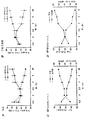
図1に示すように、気室(a)および呼吸柔組織(b)の相対的割合(%部分領域)における差異が、5日(0.5週)、14日(2週)および17日(2.5週)齢において観察されなかった一方、気室の%部分領域は、3週齢のSP−D(−/−)マウスにおいて有意に増大した(p=0.013)。より具体的には、気室(a)および柔組織(b)の両方についての部分領域は、3週(*p=0.013)、6週(*p=0.0007)および28週(*p=0.004)齢において、2つの異なる遺伝子型間で顕著に異なった。同様に、呼吸柔組織の%部分領域は、年齢に対応させたSP−D(+/+)対照(それぞれ34%柔組織/66%気室、これに対して42.5%柔組織/57.5%気室)と比較して、SP−D(−/−)マウスにおいて減少した(図1)。気室および呼吸柔組織の相対的割合は、後の時点において対照とは有意に異なり続け、%部分領域は、7ヶ月齢SP−D(−/−)マウスにおいて27%柔組織/73%気室〜37%柔組織/63%気室の範囲内であった(n=5)。年齢に対応させたSP−D(+/+)対象は、比較的小さい可変性を示し、この時点において45%柔組織/55%気室〜47%柔組織/53%気室の範囲内であった(n=4)。SP−D(−/−)マウスにおける7ヶ月齢においての柔組織の全体的な%減少は、対照値の32%であり、一方SP−D(−/−)マウスにおける気室の%増加は、対照値の27%であった。
【0044】
細胞増殖
屠殺の4時間前に、動物にBrdUを予め注入して、細胞増殖の変化を評価した。含入されたBrdUの免疫組織化学的検出を、市場で入手できるキット(Zymed Laboratories, Inc., San Francisco, CA)を用いて実施した。各動物からの小腸の切片を、BrdU含入についての正の対照として肺切片と平行して免疫染色した。
【0045】
BrdU標識係数は比較的低く、呼吸柔組織細胞または肺胞マクロファージのBrdU標識における変化は、対照と比較してSP−D(−/−)マウスの肺においては観察されなかった。
【0046】
肺容積
圧力−容積曲線を用いた肺容積の測定は、以下のように行われた:12週齢マウスに、ペントバルビタールナトリウムを注射し、100%酸素を含む室中に配置して、酸素吸収により肺胞が完全につぶれるのを確実にした。マウスを放血により屠殺し、気管にカニューレを挿入し、3方向コネクター(Mouse Pulmonary Testing System, TSS Incorporated, Cincinnati, OH)により圧力センサーに結合させたシリンジに接続した。横隔膜を開いた後、肺を、10秒おきに75μlの増分で膨張させて水の最大圧力28cmとし、次に空気を抜いた。圧力−容積曲線を、各動物について作成し、収縮曲線間の10、5、および0cmの水圧において肺容積を測定した(体重で除算した)。図2において、圧力−容積曲線を、12週齢において5〜6匹のマウスにおいて作成した。圧力−容積曲線の収縮葉に関連する肺容積は、10cmH2Oにおいて、および28cmH2Oの最大圧力において年齢に対応させたSP−D(+/+)マウスと比較して12週齢のSP−D(−/−)マウスについて有意に大きかった(*p<0.05)。
【0047】
統計的に有意な差異が、部分領域および圧力−容積曲線についての変化の分析、続いてステューデント−ニューマン−ケウルスの手順、または体重、肺および心臓容積、容積:体重比、合計タンパク質およびDNA含量の比較のためのステューデントT検定の分析のいずれかを用いることにより決定された。p<0.5の差異を、有意であると考慮した。値は、平均±SEとして示した。
【0048】
増加した肺容積は、12週齢のSP−D(−/−)マウスにおいて容易に明らかであり、これは気腫を例証する組織学的および形態計測研究と一致した(図2参照)。
【0049】
肺胞
拡大された肺胞は、SP−D(−/−)マウスにおいて一貫して観察された。したがって、SP−Dは、肺において肺胞のリモデリングの調節に関与する傾向が極めて強い。異常および気室のリモデリングが気腫の決定的な特性であるため、SP−D(−/−)マウスは、気腫についての理想的なモデルである。
【0050】
実施例6
サイトカイン、過酸化水素生成、およびメタロプロテイナーゼ活性
方法
サイトカイン測定
6〜9週齢マウスからの肺ホモジネートを、2000RPMにおいて遠心分離し、−20℃で貯蔵した。腫瘍壊死因子アルファ(TNF−α)、インターロイキン(IL)−1β、IL−6およびマクロファージ炎症タンパク質(MIP)−2を、ムリンサンドウィッチELISAキット(R&D Systems, Minneapoles, MN)を用いて、製造者の指示に従って定量した。すべてのプレートを、マイクロプレートリーダー(Molecular Devices, Menlo Park, CA)上で読み取り、コンピューターでアシストされた分析プログラム(Softmax, Molecular Devices)を用いて分析した。>0.95の計算された回帰線値を有する標準曲線を有するアッセイのみを、分析に受け入れた。
【0051】
過酸化水素生成
肺胞マクロファージを、3倍の1mlの色素を含まないRPMI培地(Gibco, Grand Island, NY)を有する気管支肺胞洗浄により採集した。8〜10匹のマウスからの気管支肺胞洗浄液(BALF)を蓄積して、分析用に十分な数のマクロファージを提供させた。洗浄を、1200RPMで10分間遠心分離して、100万のマクロファージをPBS中に再懸濁させた。マクロファージによる過酸化水素生成を、酸性条件下での過酸化水素による第一鉄イオン(Fe2+)の第二鉄イオン(Fe3+)への酸化に基づいて、市場で入手できるアッセイ(Bioxytech H2O2-560 assay, OXIS International, Portland, OR)を用いて測定した。方法は、製造者の推薦に従った。過酸化水素生成を、100ng/mlのホルボールミリステートアセテート(PMA)での活性化の後または刺激を加えずに測定した。
【0052】
メタロプロテイナーゼ活性
マウス洗浄試料を、SW−28ローター(Beckman, Palo Alto, CA)において遠心分離した(100,000×g、1時間)。上清液を、Centricon-30濾過ユニット(Amicon, Inc., Beverly, MA)を用いて濃縮した。試料(200μgタンパク質)を、非還元条件下で(レムリー)、10%ザイモグラム(Zymogram)、ゼラチンおよびカゼインゲル(Novex, San Diego, CA)中に電気泳動した。電気泳動に続いて、ゲルを、2.5%のトライトン(Triton)X−100で2回洗浄し(37℃、15分)、40mMのトリス(Tris)−HCl、pH7.5、10mMのCaCl2、1uMのZnCl2で16時間インキュベートした。ゲルを、50%メタノール、10%酢酸に溶解した0.5%(w/v)クマシーブルーで1時間染色し、次に脱色した。メタロプロテイナーゼを、青いバックグラウンドに対して透明なバンドとして検出した。メタロプロテイナーゼ2および9のmRNAを、[32P]で標識したcDNAプローブ(Chemicon International, Inc., Temecula, CA)を用いて、野生型およびSP−D(−/−)マウスからの全肺のmRNAのノーザンブロット分析により定量した。
【0053】
結果
6および9週齢において、SP−D(−/−)マウスからの肺ホモジネートは、プロ炎症サイトカインTNF−α、IL−1β、IL−6またはMIP−2の炎症レベルを含まなかったが、IL−1βの基底レベルは顕著に上昇した。図3。対照的に、SP−D(−/−)マウスから単離された肺胞マクロファージによる過酸化水素生成を測定することにより評価されたオキシダント生成は、10倍増大した。図4。過酸化水素およびスーパーオキシド生成は、マクロファージ活性化、特に殺微生物活性化の基準である。オキシダント生成が、多くのメタロプロテイナーゼの活性化並びにヒトおよび動物研究における気腫と関連していたので、メタロプロテイナーゼ活性は、SP−D(−/−)マウスおよびSP−D(+/+)マウスから単離されたBALF上清液のSDS−PAGEの後のゼラチン基質の分解により評価された。メタロプロテイナーゼ−2および−9と一致する活性のバンドは、両方の遺伝子型において容易に検出されたが、SP−D(−/−)マウスからのBALFにおいて変化しなかった。同様に、メタロプロテイナーゼ−2および−9のmRNAに富んでいることは、ノーザンブロット分析により評価されたように、SP−D(−/−)マウスおよびSP−D(+/+)マウスからの全肺RNA試料において同様であった。
【0054】
実施例1〜6の結果は、完全に予測されないものであった。SP−Dヌルマウスが気腫のモデルであることを示唆するものは、文献中には何も存在しない。
【0055】
要するに、SP−D(−/−)マウスは、表面活性剤ホメオスタシスの調節、肺中の肺胞表面活性剤、SP−A発現の調節におけるSP−Dの顕著で驚異的な役割を決定的に例証し、または肺中のオキシダント、過酸化水素生成における決定的な阻害の役割を果たす。したがって、このレベルは、継続するオキシダント生成および損傷および肺胞リモデリングの調節の抑制に重要である。これにより、SP−D(−/−)マウスは、気腫の優れたモデルとなる。実施例7は、気腫のモデルマウスについての結果を要約する。
【0056】
実施例7
気腫のモデルとしてのSP−D(−/−)マウス
SP−D欠失は、炎症、単離された肺胞マクロファージによる増大されたオキシダント生成、気腫および遺伝子不活性化SP−D(−/−)マウスにおける局所的な線維症を引き起こした。これらの肺異常のタイミングおよび進行性性質は、SP−D(−/−)マウスにおける肺胞拡大が、肺胞発生(alveologenesis)の間に発生する発生異常よりむしろ、慢性炎症に関連する肺胞リモデリングにより引き起こされたという結論を支持する。ここでの知見は、肺炎症およびオキシダント生成の調節におけるSP−Dの重要であり予測されない役割と一致し、SP−Dの調節または機能における変化が、慢性肺損傷に続く気腫に至る病理学的プロセスにおいて役割を奏し得ることを示唆する。
【0057】
SP−D(−/−)マウスからの肺の組織学的および形態計測分析で、生後3週齢まで肺構造における異常は表れず、肺胞発生の1週間後にマウスにおいて見られる。これは、呼吸柔組織および気室の相対的割合が、生後5日と17日との間のSP−D(−/−)マウスとSP−D(+/+)マウスとの両方において同様であったという観察結果と一致した。2週齢の後に、増大した柔組織−気室比率が、SP−D(−/−)マウスにおいて観察されたが、これは柔組織および肺胞室の進行するリモデリングと一致する。拡大された気室は、一般的に大きい泡状の肺胞マクロファージの集中した蓄積と関連するが、炎症浸潤物および比較的加齢したマウスの局所化および範囲において若干の異質性があった。SP−D(−/−)マウスの肺における肺胞マクロファージの集中した蓄積が、2週齢と早く観察された際には、この時点においてマクロファージ形態は通常のままであった。拡大された泡状細胞から成る異常な肺胞マクロファージ形態は、3週齢において記録され、その後の肺胞構造の拡大と一致した。以前の研究は、8週齢までのSP−D(−/−)マウスにおける拡大された肺胞マクロファージの増加した数を例証した。したがって、SP−D(−/−)マウスにおける気腫の発生は、活性化されたマクロファージの一時的および空間的な蓄積と一致し、これは、リモデリングにおけるこれらの役割を示唆する。ここでの知見は、通常の長い形態発生および肺胞発生におけるSP−Dの役割を支持せず、プロセスは、一般的にマウスにおける生後約2週齢により完成される。
【0058】
ここでの知見は、気腫および線維症に至る肺胞マクロファージの活性化およびオキシダント生成の調節におけるSP−Dの重要な役割を支持する。SP−D(−/−)マウスにおけるマクロファージ浸潤および肺リモデリングは、IL−1b、MIP−2を含むがTNF−αおよびIL−6を含まず、むしろ単離された肺胞マクロファージによる顕著に増大した過酸化水素を含む種々のプロ炎症メディエイターの炎症レベルにおける中程度であるが有意な差異と関連した。IL−β1の基底レベルが、SP−D(−/−)マウスにおいて顕著に増大したが、IL−β1は、重度の炎症において代表的に検出されたレベルには増大しなかった。増大されたIL−1βおよび過酸化水素生成が、SP−D(−/−)マウスにおいて観察された一方、これらのマウスに見られる肺異常がサイトカインまたはオキシダント誘発損傷により直接媒介されたか否かは、不明確なままである。SP−Dは、宿主防御において重要な役割を果たすことが提案されているが、SP−D(−/−)コロニーにおいて感染の組織学的または血清学的証拠はない。
【0059】
SP−D(−/−)マウスの肺において見出された増強された過酸化水素生成および増加した数の肺胞マクロファージは、SP−Dが、肺において決定的に重要な抗炎症役割を奏し、in vivoにおける肺胞マクロファージによる過酸化水素生成を調節するという概念を支持する。オキシダント損傷と気腫および肺線維症の発生との間の関係は、多くの動物および遺伝子モデルにおいて確立されている。例えば、高酸素症への新生仔の暴露は、新生マウスにおいて肺胞のリモデリングおよび線維症を引き起こす。メタロプロテイナーゼの活性化が、オキシダント損傷および気腫と関連するため、メタロプロテイナーゼ活性は、SP−D(−/−)マウスからのBALFにおいて評価された。メタロプロテイナーゼ−2および−9と一致するプロテアーゼ活性が、ザイモグラフィーにより容易に検出された一方、これらのプロテナーゼまたはこれらのmRNAの活性の一致する変化は、SP−D(−/−)マウスにおいて検出されなかった。しかし、他のプロテアーゼまたは抗プロテアーゼにおけるメタロプロテイナーゼの増大し局所化された組織濃度がSP−D欠失と関連し得ることは、依然として可能性がある。抗プロテアーゼにおける欠失並びに酸化毒薬(例えばブレオマイシンまたはパラカート)からの喫煙およびオキシダント損傷はすべて、ヒト肺における気腫または肺線維症と関連する。
【0060】
表面活性剤リン脂質含量が、SP−D(−/−)マウスにおいて増加し、大きい泡状の肺胞マクロファージの増加した数と関連した一方、増大したリン脂質含量のみでは、SP−D(−/−)マウスにおいて観察された肺胞リモデリングを生じるのに十分ではない傾向がある。実際に、表面活性剤リン脂質の全体的な効果は、抗炎症性であり、食作用、オキシダント生成およびサイトカイン放出を変化させ、リンパ球増殖、免疫グロブリン生成および接着分子の発現を阻害すると見られる。他方、GM−CSFが呼吸上皮において過剰発現されたトランスジェニックマウスは、通常存在する肺胞マクロファージの顕著に増大した数を有したが、肺胞タンパク症/リポイド沈着症、気腫または線維症を発生しなかった。対照的に、表面活性剤リン脂質およびタンパク質は、肺胞マクロファージ蓄積および血管周囲/気管周囲単球浸潤と関連するGM−CSF(−/−)およびGM受容体共通ベータサブユニット(βc)の両方からの肺において顕著に増大した;しかし、肺胞タンパク症/リポイド沈着症のいずれのモデルも、気腫または線維症に関連していなかった。同様に、肺においてIL−4を過剰に発現するトランスジェニックマウスはまた、増大した量の表面活性剤タンパク質および脂質並びに増加した数の炎症細胞を示したが、気腫を生じなかった。
【0061】
肺におけるSP−Dの濃度が、発生中に変化して、年齢の進行に伴い増加するが、SP−Dレベルはまた、種々の臨床的条件により影響される。最近の研究は、嚢胞性線維症(CF)を患っている患者から得られたBALFのSP−D濃度の顕著な減少を例証し、CF肺疾患に関連する慢性炎症の病理学におけるSP−Dの強力な役割を支持する。SP−Dレベルはまた、喫煙者のBALFにおいても減少し、これは、これらの患者において、減少したレベルのSP−Dが後に慢性閉塞性肺病(COPD)を発生させるのに寄与し得ることを示唆している。BALFにおけるSP−Dの濃度が、肺胞タンパク症(PAP)を患っている患者において増大するが、特発性肺線維症(IPF)およびコラーゲン血管病(IPCD)と関連する間質性肺炎を患っている患者は、SP−Dの減少したBALFレベルを有する。他方、SP−Dの血清濃度は、PAP、IPFおよびIPCDを患っている患者において増大した;しかし、SP−AおよびSP−Dの両方の血清レベルは、IPFの重篤度と共に、および抗炎症療法の経過の間に変化する。これらの臨床的知見および本研究は、SP−Dが正常の肺構築およびオキシダント生成の抑制の維持に必要であることを例証するが、SP−D濃度における変化が、オキシダント損傷、肺膿瘍(abcess)、二次的疾患、嚢胞性線維症、間質性肺線維症(IPF)、および慢性閉塞性肺病(COPD)、種々の肺感染症、呼吸窮迫症候群(RDS)、気管支肺異形成症(BPD)、化学療法誘発肺損傷、一次膿瘍に対して二次的な肺線維症(即ちサルコイド)、および喘息を含む種々の臨床的条件と関連する肺損傷の病理学に関連するという概念を支持する。
【0062】
本発明者等の以前の研究において、肺胞マクロファージまたは肺形態における異常は、異種接合SP−D(+/−)マウスにおいて観察されず、BALFにおけるSP−D濃度の50%の減少は、肺異常を引き起こすのに十分ではないことを例証する。オキシダント誘発損傷および肺リモデリングの阻害に必要なSP−Dの正確な濃度は、現在不明確である。SP−D(+/−)マウスまたはSP−D(−/−)マウスの肺のさらなる損傷またはオキシダントストレスが、動物モデルにおいて気腫および線維症を悪化させるか否かは、決定されるべき問題として残っている。
【0063】
SP−D(−/−)マウスにおいて見出された肺SP−A濃度の中程度の減少は、これらのマウスにおいて観察された肺形態の変化に寄与する傾向はない。その理由は、SP−A(+/−)マウスもSP−A(−/−)マウスも、気腫を発生しないからである。さらに、SP−A欠失マウスの肺形態は正常であり、SP−D(−/−)マウスとは対照的に、SP−A欠失は、単離された肺胞マクロファージにより減少した過酸化水素生成と関連した。
【0064】
SP−D(−/−)マウスは、重篤な進行性気腫を発生した。肺胞リモデリングおよびマクロファージ異常は、3週齢という早期に明らかであり、一方穏やかな病巣の肺線維症は、6〜7週齢において観察され、炎症および肺胞リモデリングの調節におけるSP−Dの役割を例証する。本研究はまた、in vivoにおける肺胞マクロファージによる過酸化水素生成の調節においてSP−Dの予期されない役割を例証し、これは、SP−D(−/−)マウスにおける肺の気腫の発生に寄与し得る。SP−D欠失が、進行する炎症または、喫煙または他のオキシダントにより発生したものである種々のヒト慢性肺疾患において見出される気腫および線維症の発生に寄与するか否かは、決定されるべき問題として残っている。
【0065】
モデルマウスにおける試験療法
気腫の治療のための薬理学的療法がないために、可能な療法を試験するためのモデルが必要不可欠である。SP−D(−/−)マウスは、このモデルを提供する。したがって、実施例8は、薬物の試験、タンパク質調製、または気腫の治療のための遺伝子操作のための試料枠組みを提供する。
【0066】
実施例8
適切な緩衝液で希釈した多くの用量または濃度のタンパク質または薬剤を、SP−D(−/−)マウスに気管内に投与した。タンパク質および薬剤を、in vivoでの使用に適するように精製した。関連する遺伝子を含む組換えアデノウィルスまたは他の遺伝子ベクターを、以下のようにして投与した。SP−D(−/−)マウスを、免疫抑制して、T細胞媒介免疫応答を特異的に遮断し、遺伝子導入された細胞において関連する遺伝子を発現するように設計されたアデノウィルス構成体で処理した。マウスに、アデノウィルス構成体を与える3日前に、H57抗体を腹腔内に注射した。H57は、T細胞受容体における免疫認識を変化させ、脾臓のおよび肺のTおよびBリンパ球を減少させた。1用量を、気管内に点滴し、他の群をH57で腹腔内処理し、次に賦形剤のみの気管内投与を施した。関連するタンパク質のレベルを投与の1週間後に測定して、ベクターの取り込みおよび発現を検出した。4匹のマウスを試験し、未処理SP−D(−/−)マウスを対照として用いた。気管内接種は、イソフルオランでの麻酔を含み、前方の中心線切開を実施して気管を露出させた。ツベルクリンシリンジに付着させた30ゲージ針を気管内に挿入し、タンパク質または薬物の100μl接種物を肺中に分散させた。切開を1滴のネクサバンド(Nexaband)を添加し閉じた。非化膿性PBSを、対照として気管内に注入した。
【0067】
タンパク質、薬物、または気腫の消失における遺伝子複製の効率を試験するために、多くの試験を実施した。
【0068】
肺構造に対するタンパク質または薬剤の効果を測定するために、肺を膨張固定し、切片を電子顕微鏡により評価した。処理および未処理マウスからの肺を、4%のパラホルムアルデヒドを含む圧力20cmの気管カニューレにより膨張させ、胸郭から一括して除去した。肺を脱水し、パラフィン中に包埋した。組織切片(5μm)を、ヘマトキシリンおよびエオシンで染色した。
【0069】
マクロファージの数および形態を試験するために:ナイルレッドでの染色は、小胞を検出し、ナイルブルーでの染色および520〜550mmの緑色光での励起は、脂質またはリン脂質を検出するための追加の方法である。マクロファージ数は、SP−B抗血清での染色により決定される。マクロファージの大きさは、1500×gでの2分間のガラススライド上に沈降したサイトスピン(cytospin)調製物からの固定し染色したマクロファージの直径から推定する。
【0070】
表面活性剤組成および超構造を、以下のようにして分析する:表面活性化の構造を、SP−D(−/−)処理および未処理マウスの蓄積した肺胞洗浄液からの大きい凝集物を単離することにより分析し、EMにより試験する(以下の原理を参照)。肺胞洗浄リン脂質組成分析のために、2〜3匹のマウスからの蓄積した洗浄液から成る2〜4個の試料を、ホスファチジルコリン、ホスファチジルエタノールアミン、ホスファチジルグリセロール、ホスファチジルイノシトール、スフィンゴミエリンおよびリソビスホスファチジン酸の相対的量について評価する。(3H)コリンの合計肺Sat−PC中への導入を評価して、合計リン脂質濃度を測定する。
【0071】
肺胞洗浄液からの凝集形態。肺胞からの表面活性体を、大きい凝集体(重い、高密度)および小さい凝集体(軽い、小胞状)部分に遠心分離により分離することができる。肺胞洗浄は、40,000×gで0.8Mスクロース緩衝剤で15分にわたり遠心分離した。大きい凝集表面活性体を、次に界面から採集し、通常の生理的食塩水で希釈し、再び40,000×gで15分遠心分離した。小さい凝集表面活性体を含む第1の40,000×gの遠心分離からの上清液を、4℃で限外濾過により300,000分子量の保持フィルター(Minitan, Miliore Corp., Bedford, MA)または遠心分離濃縮器(Amicon Corp., Danvers, MA)を用いて濃縮した。小さい凝集表面活性体を、50mlの通常の生理的食塩水で希釈し、3回限外濾過して可溶性タンパク質を除去した。
【0072】
肺疾患のための治療としてのSP−D
SP−Dの欠失が気腫のためのモデルマウスを生成するため、SP−Dは、気腫の治療または防止として明らかな選択である。これはまた、これらの疾患が異常な表面活性剤生成により特徴づけられるため、他の種類の肺疾患の明らかな治療である。さらに、SP−Aに対する効果および宿主防御における可能な役割は、これを、肺における免疫機能を増大するための有用なツールとする。呼吸上皮に対する遺伝子の導入は、種々の肺疾患のための治療として極めて有望である。種々のウィルスに基づく、およびウィルスに基づかないベクターが、遺伝子を気道の細胞に移動させるために開発されてきたが、これには組換えアデノウィルスベクターが含まれる。これらのベクターは、これらがエアロゾル化できるため、呼吸治療において用いるのに特に有望である。したがって、実施例9は、SP−D(−/−)マウスにおける気腫の治療のための精製したマウスSP−Dタンパク質を用いた実験である。実施例10は、アデノウィルスを用いて、SP−D(−/−)マウスにおける気腫の治療のためのラットSP−Dを発現する実験である。実施例11は、SP−Dペプチドの使用のための試料基盤またはこれらの疾患の防止および治療のためのSP−Dを発現するベクターを提供する。気腫は、例示的な肺疾患として用いられる。アデノウィルスは、例示的なベクターとして用いられる。
【0073】
精製したSP−Dでの処理
実施例9
SP−D(−/−)マウスを、精製したマウスSP−Dで処理し、以下に概説するように精製した。飽和PCレベルを、肺胞洗浄液および合計肺洗浄液において分析した。24時間間隔での気管内への繰り返した投与の結果、3〜7投与の後に脂質蓄積の部分的な補正が生じた(図12参照)。
【0074】
SP−Dの気道における半減期を、マウスにおいて13時間であると判定した(図13参照)(この手法を以下に概説する);したがって、SP−D欠失を、エアロゾルまたは粒状吸入器または表面活性体混合物により合理的な間隔でSP−Dタンパク質の置換により処理することができる。
【0075】
マウスSP−Dの精製
GMCSFおよびSP−A二重ヌル突然変異マウスからのマウス気管支肺胞洗浄(BAL)液を採集し、凍結し、後のSP−Dの精製のために蓄積した。マルトシル−アガロース(Sigma)を、重力フローカラム(10×80mm)中に詰め、20mMのトリス−HCl、pH7.4、10mMの塩化カルシウム、0.02%(W/V)のアジ化ナトリウムを含む緩衝液(TCB)で平衡にした。BALを、トリス−HClに対して20mM、EDTAに対して10mMとし、pH7.4とし、室温で1時間かきまぜた。混濁溶液を、10,000×gで40分間、4℃で遠心分離した。上清液を、塩化カルシウムに対して20mMとし、マルトシル−アガロースカラムに装填する前にpH7.4に再調整した。カラムをバックグラウンド吸収のためTCBで洗浄し、続いて1.0M塩化ナトリウムを含むTCBで洗浄した。マルトースへの結合において特別の要求を有するSP−Dを、50mMの塩化マグネシウム、20mMのトリス−HCl、0.02%(W/V)のアジ化ナトリウム、pH7.4で溶出した。SP−Dを含むフラクションを、SDSポリアクリルアミドゲル電気泳動または直接ELISAにより測定し、蓄積し、20mMのトリス−HCl、100mMの塩化ナトリウム、5mMのEDTA、pH7.4の3回交換で透析した。このプロトコルは、Strong, Peter; Kishore, Uday; Morgan, Cloff; Bernal, Anders Lopez; Singh, Mamta; およびReid, Kenneth B. M.; Journal of Immunological Methods 220 (1998) 139-149から適合された。
【0076】
マウスの表面活性体成分での処理。本発明者等は、問題なく、7日目まで毎日SP−Dでマウスを反復して処理するため、イソフルランでの麻酔の下に、マウスにおいて26gの供給管を用いた経口盲目挿管法のための手法を、好首尾に用いた。この方法は、外科手術を回避し、SP−D置換のために提案された実験および突然変異SP−Dタンパク質での処理の形を可能にする。
【0077】
最初に、SP−D(−/−)マウスを、気管点滴により精製したマウスSP−Dで処理した。24時間間隔で与えられた2.9gのSP−Dの3回以上の投与により、肺胞および飽和PC蓄積の両方が減少した(図14参照)。与えられたSP−Dのこの用量は、およそSP−D(+/+)マウスにおける内因性蓄積に存在する量である。肺結合およびクリアランスカイネティックスが考慮されると、これは、低い用量である。SP−Dの外因性投与は、表面活性剤脂質代謝に直接影響し、本発明者等がin vivoで改変SP−D分子の機能を試験することができる実験モデルを提供する。
【0078】
生物学的半減期プロトコル。本発明者等は、マウスにおけるSP−Dの生物学的半減期を測定して、SP−Dでの処理のための実験を設計した。本発明者等は、以前に本発明者等がSP−Aおよび他の表面活性剤タンパク質について実施したように、ボルトン−ハンター(Bolton-Hunter)試薬を用いて、125Iで精製したマウスSP−Dをヨウ素標識した。SP−D(+/+)マウスおよびSP−D(−/−)マウスの肺胞洗浄液からのSP−Dのクリアランスは、約13時間の半減期と同様であった(図13参照)。SP−D(−/−)マウスの肺におけるSP−Dについての17時間のt1/2は、SP−D(+/+)マウスについての13時間のt1/2のものより若干長かった。
【0079】
GM−CSF欠失は、SP−Dにおいて48倍の増加を生じ、GM−CSF(−/−)×SP−A(−/−)の交差は、同様にSP−Dを上昇させたがSP−Aを上昇させなかった。本発明者等は、高純度で、および大量に、Persson等により記載された方法により、Ca2+の存在下においてマンノース−セファロースのアフィニティーカラムを用いて、GM−CSF(−/−)×SP−A(−/−)マウスからの肺胞洗浄液からSP−Dを単離した。
【0080】
実施例10
アデノウィルスで発現させたSP−Dでの処理
本発明者等は、ラットSP−Dを発現する新規なアデノウィルスを作成した。このウィルスは、正常またはSP−D欠失マウスの細胞および肺においてSP−Dを生成する。本発明者等は、293細胞およびマウスにおいて生成したラットSP−Dのウエスタンブロットを有する。
【0081】
Ad−rSPDアデノウィルスの構成(図14参照)。野生型ラットSPD cDNAを、EcoR I消化でプラスミドWT−rSPD/pG3Zから切り出し、3’末端をクレノウ(Klenow)で埋めた。1.3kB rSPD cDNAを、プラスミドpAvS6aのEcoR V部位に挿入して、プラスミドpAvS6a−rSPDを作成した。プラスミドpAvS6a−rSPDは、RSVプロモーター、rSPC cDNA、SV40ポリAシグナルおよびAd5配列(9.24〜17.34mu)を有する。Not IでpAvS6a−rSPDを直鎖化したものは、アデノウィルスDNA Ad.dl327のCla I消化された大きなフラグメントと293細胞中に同時トランスフェクトさるが、これは、E3領域(78.5〜84.7mu)が削除されている。相同組換えの後、個別のプラークを、ウエスタンブロットアッセイにより分析して、rSPDタンパク質発現を決定した。1つのrSPD陽性クローンに、プラーク精製を一回施した。Ad−rSPDアデノウィルスは、E1およびE3領域において欠失を有し、複製欠損である。293細胞の増幅の後、精製されたAd−rSPDアデノウィルスを、2回のCsCl勾配超遠心分離により生成した。SP−Dを発現するアデノウィルスは、気管内投与により若干の脂質異常を補正することができた。したがって、これは、気腫および多くの他のSP−D欠失疾患並びに種々の他の形態の肺損傷および欠失の治療のために極めてよい可能性を残す。
【0082】
実施例11
他のベクター、タンパク質または薬剤から発現したSP−Dでの処理
リン脂質ホメオスタシスの回収におけるSP−Dに対する時間的、空間的および化学量論的要求を、実施例9において決定した。SP−Dのクリアランスカイネティックスを決定する最初の研究を、125Iで標識したSP−Dを気管内に投与して実施し;半減期を計算し、情報をSP−D置換実験の設計において用いた。投与後のSP−Dの通常の生理学的濃度を達成するのに必要なSP−Dの用量を、明らかにした。
【0083】
実施例9において、精製したSP−Dタンパク質の投与を用いて、種々の肺疾患を治療した。しかし、肺疾患における生理学的異常は、肺中のSP−Dの長期間補正を必要とし得る。したがって、哺乳動物SP−D遺伝子を含む組換えアデノウィルスまたは他の遺伝子ベクターを用いる(実施例10および11参照)。組換えアデノウィルスベクターは、クララ(Clara)細胞分泌タンパク質(CCSP)およびSP−Cプロモーターを用いて、細気管支(クララ細胞)および肺胞(II型細胞)区画においてSP−Dを選択的に発現する(実施例10参照)。アデノウィルスベクターでの処理の3日前に、マウスを、200ugのモノクローナル抗T細胞受容体抗体、H57を腹腔内注入することにより免疫抑制した。アデノウィルスを、ウィルスの5×10⁸PFUの気管内注入により投与した。SP−Dタンパク質のレベルを、投与の1週間後に測定して、ベクターの取り込みおよび発現を検出した。4匹のマウスを試験し、処理を受けていないSP−D(−/−)マウスを、対照として用いた。気腫の影響を消失させるSP−Dの効率を試験するために、多くの試験を以下のように実施した。
【0084】
肺構造に対するタンパク質または薬剤の効果を決定するために(実施例11)、肺を膨張固定し、切片を電子顕微鏡により評価する。肺を、4%のパラホルムアルデヒドを含む圧力20cmの気管カニューレにより膨張させ、胸郭から一括して除去する。肺を脱水し、パラフィン中に包埋する。組織切片(5μm)を、ヘマトキシリンおよびエオシンで染色する。
【0085】
マクロファージの数および形態を分析する。ナイルレッドでの染色は、小胞を検出し、ナイルブルーでの染色および520〜550mmの緑色光での励起は、脂質またはリン脂質を検出するための追加の方法である。マクロファージ数は、SP−B抗血清での染色により決定される。マクロファージの大きさは、1500×gでの2分間のガラススライド上に沈降したサイトスピン調製物から固定し、染色したマクロファージの直径から推定する。
【0086】
表面活性体組成および超構造を、以下のようにして分析する:表面活性体の構造を、SP−D(−/−)処理および未処理マウスの蓄積した肺胞洗浄液からの大きい凝集物を単離することにより分析し、EMにより試験する。肺胞洗浄液リン脂質組成分析のために、2〜3匹のマウスからの蓄積した洗浄液から成る2〜4個の試料を、ホスファチジルコリン、ホスファチジルエタノールアミン、ホスファチジルグリセロール、ホスファチジルイノシトール、スフィンゴミエリンおよびリソビスホスファチジン酸の相対量について評価した。(3H)コリンの合計肺Sat−PC中への導入を評価して、合計リン脂質濃度を測定した。
【0087】
処理の効率が決定されれば、処理を他の適切な哺乳動物に対して試験することができる。
【0088】
SP−Dの肺感染の改善
肺での宿主防御におけるSP−DおよびSP−Aの役割を、繰り返して例証した。SP−AおよびSP−Dは、サイトカインおよびフリーラジカル生成の調節によるin vitroでの肺炎症を調節をする種々の微生物とin vitroで特異的な相互作用を有する。細菌クリアランスおよび肺の炎症応答におけるSP−Dの役割を、SP−D欠失のモデルマウスを用いてin vivoで評価した。SP−A欠失マウスは、感染症に一層罹病性であることが知られている。多くのin vitro研究は、SP−Aの上方調節におけるこの役割に加えて、宿主防御についてのSP−Dの可能な役割を示した。実施例8〜11は、細菌または菌類感染したSP−D(−/−)マウス並びにSP−A(−/−)マウスにおける治療としてSP−Dを試験するための試料プロトコルを概説する。実施例12〜14は、細菌、菌類およびウィルス感染に対する応答におけるSP−Dの役割を示す実験である。実施例13は、呼吸シンシチウムウィルスに感染したSP−D(−/−)マウスの効果を示す実験である。
【0089】
実施例12
SP−D(−/−)マウスからの細菌病原体のクリアランス
SP−D欠失マウス(SP−D −/−)に、B群ストレプトコッカス(GBS)またはヘモフィルス・インフルエンザ(Hemophilus influenzae)(Hflu)を気管内に感染させて、野生型マウスと比較してのクリアランスを評価した。A群ストレプトコッカスを、104CFUにおいて投与した。また、肺炎症を、合計細胞数(図5、6および7)、肺ホモジネートにおけるサイトカインレベル(図8)、肺胞マクロファージによる酸素ラジカル生成(図11)およびBALにおける亜硝酸塩レベル(図9)について、BAL液の分析により評価した。
【0090】
SP−D −/−マウスは、野生型マウスと同様に、細菌を排出した(図5および6参照)。GBSおよびHfluでの感染の結果、野生型マウスと比較して、SP−D −/−マウスのBAL液における有意に大きい合計細胞数を生じた(図7)。サイトカインレベルの選択的変化は、SP−D −/−マウスにおいて検出された。腫瘍壊死因子(TNF−)およびインターロイキン(IL)−6レベルは、GBSまたはHfluでの感染の後の早期のSP−D −/−マウスからの肺ホモジネートにおいて大きかった(図8)。マクロファージ炎症タンパク質−2(MIP−2)、好中球化学誘引物質は、Hflu感染の後にSP−A −/−マウスからの肺ホモジネートにおいて顕著に多かったが、GBS感染ではなかった(図8)。SP−D −/−マウスからのマクロファージは、野生型マウスと比較して、顕著にスーパーオキシドおよび過酸化水素を多く発生した(図11)。
【0091】
BAL亜硝酸塩レベルは、野生型マウスと比較して、SP−D(−/−)マウスにおいて増加した。一酸化窒素は、BALF中の亜硝酸塩として測定された。一酸化窒素は、殺菌に寄与し、宿主防御の役割を有する。一酸化窒素は、スーパーオキシドと反応して、有力な殺微生物剤であるペルオキシ硝酸塩を生成する。
【0092】
図10において、光学顕微鏡を用いて、食作用を評価した。SP−D(−/−)マウスは、野生型と比較して、顕著に減少した細菌の食作用を示した。
【0093】
したがって、SP−Dの非存在時に、増大した炎症応答が、GBSまたはHfluでの細菌感染に続いて観察された。肺胞マクロファージによる活性酸素種の生成は、SP−D −/−マウスにおいて増大した。これらの結果は、in vivoでの細菌感染に対する肺免疫および炎症応答におけるSP−Dの決定的に重要であり明らかな役割を支持する。
【0094】
実施例13においては、SP−D(−/−)マウスに、呼吸シンシチウムウィルスを感染させた。
【0095】
宿主防御機構は、細菌の生来のメディエイタおよびウィルスクリアランスおよび後天性免疫応答を含む微生物病理学の肺クリアーを維持した。
【0096】
実施例13
SP−D(−/−)マウスからのウィルスのクリアランス
SP−D(−/−)マウスに、呼吸シンシチウムウィルス(RSV)、小児における一般的な呼吸病原菌を気管内に感染させた。ウィルス力価および肺炎症を、SP−D(−/−)マウスおよび野生型マウスにおいて評価した。肺ホモジネートにおけるRSV力価は、投与の3日後および5日後で、SP−D(−/−)マウスと野生型マウスとの間で同様であった。しかし、炎症細胞の顕著に増加した数は、RSV感染の3日後および5日後に、野生型マウスと比較して、PMNの大きい割合を有するSP−D(−/−)マウスのBAL液において見出された。さらに、RSV感染の5日後に、組織学的に評価された肺炎症は、野生型マウスと比較して、SP−D(−/−)において多かった。TNF−a、IL−1、IL−6およびMIP−2を含むプロ炎症サイトカインは、RSV感染の3日後および5日後に、SP−D(−/−)マウスからの肺ホモジネートにおいて多かった。SP−D(−/−)マウスは、肺からの効率的なウィルスクリアランスを有していたが、RSV感染に続いて野生型マウスよりも大きい炎症応答を例示した。これらの発見は、SP−Dが、in vivoでのRSV感染の後に肺における生体の防御および炎症の調節において重要な役割を果たすことを例証する。
【0097】
実施例14
SP−D(−/−)マウスからの菌のクリアランス
マウスを以下のように感染させる:菌類病原菌の適切なプロトタイプを用いる。感染性病原菌を、適切に精製し、適切な緩衝液中に懸濁し、SP−D(−/−)マウス中にSP−Dと共に、またはこれを含めずに気管内に投与する(実施例12および13におけるように)。菌プロトタイプを、適切な用量において投与する。SP−D(−/−)マウスおよびSP−D(+/+)マウスを用いて、感染に対するマウスの罹病性におけるSP−Dの効果を試験する。SP−Dタンパク質を含む、または含まないSP−D(−/−)マウスを用いて、感染についての治療としてSP−Dを試験する。感染のクリアランスを、実施例12および13におけるように、および以下のようにして評価する:
【0098】
菌クリアランスを、動物に感染性病原菌またはSP−Dを含む感染性病原菌を接種した6、24および48時間後において肺および脾臓ホモジネートを精製することにより、決定した。肺からの細菌クリアランスを、SP−D濃度を適切に変化させた後に決定した。また、定量的培養を、SP−D(+/−)マウスについて決定して、SP−Dの50%の減少が、細菌またはウィルスクリアランスについて十分な内因性SP−Dを提供するか否かを決定する。
【0099】
哺乳動物SP−Dの適切な濃度を、肺感染症の治療のために他の哺乳動物において用いる。
【0100】
SP−Dレベルを調節する薬剤
通常の機能および肺の発達におけるSP−Dの重要性は、SP−D(−/−)ヌルマウスにより明確に例証される。したがって、SP−Dの生成、発現または作用を調節する薬剤は、重要な将来的な薬剤であり、さらにこのような薬剤を同定するための実験的補剤である。このような薬剤を同定するための多くの手法は、当業者には自明であろう。実施例15および16は、これらの手法の2つについての試料プロトコルを概説する。実施例17は、IL−4が、in vivoにおいてSP−Dレベルを顕著に増大させ、したがってSP−Dを付加したかまたは付加しない種々の肺疾患を治療するのに用いることができることを示す。
【0101】
実施例15
SP−Dプロモーターと相互作用するタンパク質
SP−Dプロモーターを用いて1つのハイブリッドを行い、SP−Dの発現を上方調節するタンパク質を同定する。次に、これらのタンパク質を、SP−D(−/−)マウスにおいて、気腫並びに実施例8における他の肺疾患および感染症の治療の効率について試験する。
【0102】
実施例16
SP−Dタンパク質と直接相互作用するタンパク質
2つのハイブリッド手法を行い、SP−Dタンパク質と直接相互作用するタンパク質を同定する。次に、これらのタンパク質を、SP−D(−/−)マウスにおいて、気腫並びに実施例8における他の肺疾患および感染症の治療の効率について試験する。
【0103】
実施例17
IL−4は、in vivoでのSP−Dレベルを上昇させる
クララ細胞においてIL−4を発現するマウス(CCSP−IL−4)は、慢性気道炎症および肺胞タンパク症状症候群を発生する。表面活性剤ホメオスタシスにおけるIL−4の役割を同定するために、本発明者等は、in vivoでのCCSP−IL−4マウスの肺における脂質およびタンパク質代謝を測定した。肺胞の飽和ホスファチジルコリン(Sat PC)蓄積を、6.5倍増加させ、肺組織Sat PC蓄積を、IL−4トランスジェニックマウスにおいて4.8倍増大させた(図15参照)。SP−Dは、野生型マウスと比較して、IL−4マウスにおいて約90倍増加し、SP−D mRNAの2.8倍の増加に関連した(図15参照)。パルミチン酸塩およびコリンのSat PCへの導入は、CCSP−IL−4マウスにおいて約2倍増加した。CCSP−IL−4マウスの肺からのSat PCの正味のクリアランスは、野生型のマウス(10.3モル/kg)よりも、IL−4マウスにおいて6倍高かった(60モル/kg)。クララ細胞におけるIL−4の発現は、表面活性剤脂質合成およびクリアランスを増大させたが、これは、SP−Dタンパク質における選択的増大に関連する増大した表面活性剤蓄積および肺胞タンパク症との新たな平衡を確立し、肺表面活性剤ホメオスタシスの従来予測されなかった効果を例証する。
【0104】
SP−Dタンパク質または配列を用いた診断
SP−Dは、正常な肺の機能および発達に重要である。SP−D(−/−)マウスは、気腫のモデルである。次に、これは、遺伝子またはSP−Dについての遺伝子の対立遺伝子における突然変異が、肺疾患罹病性に対して顕著な効果を有することを示唆する。したがって、突然変異または対立遺伝子、および突然変異体タンパク質を同定する方法は、気腫、肺感染症および多くの他の呼吸疾患の恐れのある個体を同定する。実施例18および19は、これらの診断手法の試料プロトコルである。
【0105】
SP−D遺伝子における突然変異を有する患者の診断
実施例18
SP−D遺伝子における突然変異は、気腫の兆候および病因論に関与する傾向がある。したがって、突然変異は、統計的に有意な数の患者の配列分析により同定される。これらの突然変異を用いて、診断試験を得る。SP−D遺伝子における突然変異は、以下の方法により検出される:適切なプライマーを用いたSP−D遺伝子のPCR分析を実施する。得られたPCRフラグメントを、SSCPにより分析し、配列決定して、突然変異または対立遺伝子を決定する。あるいはまた、ゲノムDNAまたはcDNAのディファレンシャルハイブリダイゼーションを用いて、突然変異を検出する。
【0106】
突然変異体SP−Dタンパク質を有する患者の診断
実施例19
気腫および他の肺疾患に関連する突然変異体SP−Dタンパク質またはSP−Dの対立遺伝子を特異的に認識するモノクローナルまたはポリクローナル抗体を得る。次に、これらの抗体を用いて、これらの肺疾患に対する酵素結合イムノアッセイを行う。実施例20の抗体を、このアッセイに用いることができる。
【0107】
実施例20は、トランスジェニック技術を用いたポリクローナル抗体の精製またはモノクローナル抗体のさらなる精製についてのプロトコルを示す。
【0108】
実施例20
SP−D特異的モノクローナルおよびポリクローナル抗体の精製
高い反応性での特異的なポリクローナル抗体の生成は、関連する抗原の広範囲な精製を必要とする。本発明者等は、投与のための関連する抗原に関して高い力価を有し、不純物と反応性である抗体を得た部分的に精製された抗原を用いて、いくつかのポリクローナル抗体を開発した。トランスジェニックマウスからの組織固体相を用いて、これらの抗血清から非特異的抗体を除去した。表面活性剤タンパク質D(SP−D)を、マンガン溶出液を有するマルトースカラムを用いて、精製した。精製したSP−Dを、不完全フロイントアジュバント中のニュージーランドラビット中に注入した。得られた抗血清を、ウエスタンブロットにおいて全肺洗浄液に対して試験し、SP−Dおよび他のタンパク質に対する結合を明らかにした。この抗血清を、SP−Dタンパク質を生成しないヌル突然変異体マウスからの肺ホモジネート固体相と共に一夜反応させた。抗血清を、SP−Dに対してのみ反応性を示す吸着の後に、全肺洗浄液に対して反応させた。また、この抗血清を、免疫組織化学的実験において評価し、これは、SP−Dヌル突然変異体マウスからの肺切片に対する極めて低い反応性および正常な対照マウスにおける極めて特異的なII型細胞反応性を例証した。この手法は、高い力価を有する高度に特異的な抗体を調製する能力を大きく増強し、吸着抗体を用いる際のブロッキング剤を用いる必要性を解消する。
【0109】
この抗体は、診断、精製およびSP−Dタンパク質の他の研究に用いることができる。
【図面の簡単な説明】
【図1】 図1A、図1B、図1Cおよび図1D。
SP−D(−/−)マウスの齢および齢一致したSP−D(+/+)対照との気室(a)および呼吸柔組織(b)の部分領域(% Fx Area)における変化の比較。各個別の遺伝子型についての齢に伴うこれらのパラメーターの変化(cおよびd)。データは%部分領域として表現し、平均±SEを示す。
【図2】 SP−D(+/+)マウスおよびSP−D(−/−)マウスの収縮葉の圧力―容積曲線。データはml/kgとして表し、平均±SEを示す。
【図3】 SP−D(−/−)マウスからの肺ホモジネートにおけるプロ炎症サイトカイン。TNF−、IL−1、IL−6およびMIP−2の濃度は、SP−D(−/−)マウス(黒塗り棒)およびSP−D(+/+)マウス(斜線棒)からの肺ホモジネートにおいて評価した。データは、pg/mlで表し、群あたりn=5で平均±SEを示す;SP−D(+/+)マウスと比較して、*p<0.05。
【図4】 SP−D(−/−)マウス(黒塗り棒)からの肺胞マクロファージにおける過酸化水素生成を、PMA刺激を有するおよび有さないSP−D(+/+)マウス(斜線棒)と比較して気管支肺胞洗浄液(BALF)から単離した1×108マクロファージから評価した。データを、H2O2のMとして表し、群あたりn=4で平均±SEを示す;SP−D(+/+)マウスと比較して、*p<0.05。
【図5】 B群ストレプトコッカス(GBS)で感染させた後のSP−D(−/−)マウスおよびSP−D(+/+)マウスにおける肺のコロニー数。
【図6】 ヘモフィルス・インフルエンザ(H.flu)で感染させた後のSP−D(−/−)マウスおよびSP−D(+/+)マウスにおける肺のコロニー数。
【図7】 図7Aおよび図7B。
GBSおよびH.fluで感染させた後の気管支肺胞洗浄(BAL)液における合計細胞数。
【図8】 図8Aおよび図8B。
GBSおよびH.fluで感染させた後の肺ホモジネートにおけるサイトカインレベル。
【図9】 図9Aおよび図9B。
GBSおよびH.fluで感染させた後のBAL亜硝酸塩レベル。
【図10】 図10AおよびB。
GBSおよびH.fluで感染させた後の光学顕微鏡により分析した食作用およびFACS分析。
【図11】 図11Aおよび図11B。
GBSおよびH.fluで感染させた後のBALから単離されたマクロファージにおける過酸化水素およびスーパーオキシドレベル。
【図12】 SP−D(−/−)マウスに対するSP−Dタンパク質処理の効果。
【図13】 図13Aおよび図13B。
マウスにおけるSP−Dタンパク質の全肺および肺胞クリアランスカイネティックス。
【図14】 ラットSP−D cDNAを含むアデノウィルスベクターAd−rSPD。
【図15】 野生型マウスおよびCCSP−IL−4マウス(IL−4マウス)からの肺胞洗浄におけるSP−AおよびSP−Dのイムノブロットの定量。p<0.01。[0001]
Government relations in the present invention
Certain aspects of the invention described herein were made with the support of the United States government under the National Institutes of Health, grant numbers HL41320, SCOR HL 56387, HL 28623, HL 58795 and HL 03905. The US government has certain rights to these aspects of the invention.
[0002]
Field of Invention
The present invention relates generally to the field of biologically active proteins. More specifically, the present invention relates to the SP-D protein involved in lung surfactant homeostasis and structure, and the structure of alveoli in the lung and SP-D (− / −) null mice.
[0003]
Background of the Invention
Lung surface activation is essential for normal lung mechanisms and gas exchange in the lungs. Lung surfactants are made up of type II epithelial cells and are composed of phospholipid components that provide the ability for surface activation and reduce surface tension in the lung. In addition, there is a protein associated with surface activation called collagen, which is collagenous and is a lectin domain-containing polypeptide. Two of these, called surfactant protein A (SP-A) and surfactant protein D (SP-D), are similarly associated with surface activated structure and function and host defense. Both quantitative and qualitative deletions of lung surfactant are associated with neonatal respiratory distress, adult respiratory distress syndrome, congenital deletion of surfactant protein B and allergic asthma. In addition, pulmonary surfactant deficiency can be associated with the increased susceptibility of some individuals to microbial attack, particularly in inappropriate or exacerbated specific immunity settings. These diseases, as well as some diseases associated with an increased risk of pneumonia (cystic fibrosis, prematurity, chronic bronchitis, diffuse alveolar injury) are also associated with acquired deficiencies or deletions in collectin function Can do. Alveolar surfactant accumulation is regulated at multiple levels including intracellular synthesis, secretion, reuptake and degradation of these components by alveolar macrophages. The synthesis and clearance of surfactant phospholipids and proteins is further affected by developmental, functional, and humoral stimuli that have the effect of maintaining postnatal steady state surfactant concentrations.
[0004]
The role of collectins in surfactants and normal lung function has been extensively tested. The collectin family of C-type lectins contains a number of molecules with known host defense functions. SP-A and SP-D, as well as type C lectins, bind to influenza and herpes simplex viruses as well as gram positive and gram negative bacteria and various fungi. Upon binding, they increase uptake by alveolar macrophages and neutrophils. Various cell binding sites in SP-A and SP-D have been identified in alveolar macrophages or, in the case of SP-A, type II epithelial cells. The critical role of SP-A in host defense is that SP-A-deficient mice are infectious for infection by group B streptococci, Pseudomonas aeruginosa, respiratory syncytial virus, adenovirus and mycoplasma in vivo. Supported by the observation that it is sensitive. Thus, there is a clear role for SP-A and that for SP-D in the respiratory defense mechanism. Collectins may also be involved in the recognition or clearance of other complex organic substances such as pollen and dust mite allergens. However, currently no human disease is associated with a specific deletion in SP-A or SP-D.
[0005]
SP-D is a 43 kilodalton protein that has been proposed to play a role in host defense in the lung. The cDNA and gene have been sequenced in various mammals including humans. SP-D shares significant structural homology with other C-type lectins including surfactant protein A (SP-A), conglutinin, bovine collectin-43 and mannose binding protein. In vitro studies and mammalian Ca 2+ Its close structural relevance to dependent lectin families (especially shared structural motifs) supports this role in host defense. SP-D is first synthesized at relatively high concentrations by type II epithelial cells in the lung and epithelial cells of bronchioles without cilia, but also in the gastrointestinal tract, heart, kidney, pancreas, ureter, genital tract. And can also be expressed in mesenteric cells. In vitro studies have shown that SP-D binds to the surface of organisms through lectin domains (or sugar binding domains) that cause binding, aggregation, opsonization, and in some cases activation of phagocytic killing in vitro. To do. SP-D binds to viruses including lipopolysaccharide, various bacteria, fungi and influenza viruses. It also binds to both alveolar macrophages and polymorphonuclear cells. This may in some cases play a role in surface-active phospholipid homeostasis, including the effect of SP-A on phospholipids by type II cells in vitro, but this is controversial and in vivo The exact role of SP-D is still unclear.
[0006]
In vitro studies support the notion that surfactant proteins can be important in the regulation of surfactant homeostasis. Hydrophobic surfactant proteins SP-B and SP-C play a role in the generation of surfactant monolayers, but in vitro studies, surfactant protein A has also been shown to be surfactant uptake by type II epithelial cells. And / or have been shown to promote secretion. In fact, it has been widely believed that SP-A has an important role in surfactant homeostasis. However, recent studies of SP-A null mice did not support the major role of surfactant protein A in surfactant secretion or reuptake. Deletion of SP-A does not lead to overt physiological or morphological structural abnormalities of the lung. SP-A null mutant mice lack a tubular myelin scheme but produce a highly functional surfactant that is rapidly absorbed and produces a single layer. Surfactant lipid synthesis, secretion and reuptake were essentially normal in SP-A null mice.
[0007]
Thus, additional surfactant proteins that act in surfactant modulation have not yet been identified. Furthermore, the exact role of SP-D in normal lung function is not clearly defined at this time, and its role in disease and disease susceptibility is unclear.
[0008]
Summary of the Invention
The present invention provides an SP-D (− / −) mouse that can be used as a model of emphysema. Previously, it was not known that SP-D protein is involved in lung lipid homeostasis. In addition, it was not known that SP-D null mice have signs of emphysema.
[0009]
One embodiment of the present invention is a non-human mammal model of emphysema, including non-human SP-D (− / −) mammals.
[0010]
A further embodiment introduces a mammalian SP-D protein or a vector expressing a mammalian SP-D protein in an amount effective to reduce symptoms of disease or prevent disease in a human or mammal. A method for purifying or treating lung diseases.
[0011]
A further embodiment is an amount of a pharmacological composition effective to treat lung disease that is a mixture of SP-D protein and a pharmaceutically acceptable carrier.
[0012]
A further embodiment is a biologically active agent for treating lung disease in a mammal that is an agent that upregulates SP-D.
[0013]
A further embodiment is a biologically active agent for treating lung disease in a mammal that is an agent that interacts with SP-D protein.
[0014]
Further embodiments identify lung mutations in mammals by identifying mutations in the SP-D gene that result in incomplete SP-D and identifying this mutation in the test mammal by PCR, hybridization or ELISA. This is a method for diagnosing susceptibility to sarcoidosis.
[0015]
Further embodiments treat lung disease by generating lung disease in SP-D null mice, administering a drug to a mammal, and identifying the drug as an effect of whether or not the lung disease has improved. It is a method for identifying a useful drug.
[0016]
A further embodiment is a method of purifying SP-D antibodies in a solid phase of lung homogenate from any mouse that does not produce SP-D protein.
[0017]
Detailed Description of the Invention
We have created SP-D (− / −) knockout mice to identify the role of SP-D in normal lung function and development, and postnatal airway enlargement and spontaneous in the lungs of these mice Exemplifies the time course of typical inflammatory changes. SP-D (-/-) mice develop progressive emphysema associated with chronic inflammation and increased oxidant production by alveolar macrophages. Lung abnormalities make this mouse an excellent model for emphysema. Because there are very few conventional therapies in the treatment of emphysema, the most common is lung volume reduction surgery, models are urgently needed. Based on model mice in emphysema, we have developed a number of methods for testing SP-D proteins and expression vectors, and drugs that are effective in treating other forms of emphysema or chronic lung injury in model mice. Proposed. We also have SP-D proteins to treat a variety of other diseases such as ectopic surface activity generation, pulmonary fibrosis, sarcoidosis, lung injury, poison / oxygen exposure, infections, increased oxidant exposure. And the use of expression vectors was proposed. Finally, we use SP-D cDNA, SP-D antibodies, PCR and differential hybridization techniques to identify patients at risk for emphysema, pulmonary distress syndrome, and other types of respiratory diseases. Proposed to identify. Although other materials and methods similar or equivalent to those described herein can be used in the practice or testing of the present invention, the preferred materials and methods are now described. Example 1 describes the steps necessary to create SP-D (− / −) mice.
[0018]
[Table 1]
[0019]
Example 1
SP-D (-/-) knockout mouse production
SP-D (− / −) mice were generated by target gene inactivation. Integration of the pGKneo targeting vector containing the
[0020]
SP-D (-/-) mice survived normally and mated in in-barrier incubators at Children's Hospital Medical Center, Cincinnati, Ohio. The mice were serologically evaluated and virus free. Serological evidence of viral infection in SP-D (− / −) mice was determined at necropsy.
[0021]
To determine genotype, DNA from tail sections was digested with BamHI and examined with PCR products derived from genomic mouse
[0022]
To demonstrate that SP-D was not expressed in null animals, RNA blot analysis was performed with all lung RNA from null, normal and heterozygous animals. As a result, normal-sized SP-D mRNA was completely lost in null mice, and the intensity of SP-D hybridization band was reduced by about 50% in heterozygous animals. After prolonged exposure, a diffuse mRNA band about 150 nucleotides smaller than normal SP-D mRNA was detected. By scanning densitometry, this band shows less than 5% of the intensity of the normal SP-D transcript of the heterozygous animal.
[0023]
Western blot analysis of lung homogenates using rabbit anti-rat SP-D antiserum reduced SP-D by approximately 50% in heterozygous SP-D (+/-) mice, and SP-D (-/-) It was found to disappear in mice.
[0024]
Both SP-D (− / −) and SP-D (+/−) mice survived normally in the prenatal and postnatal periods. At selected ages, body, lung and heart weights were obtained by direct measurement; lung and heart volume were obtained by fluid displacement. Lung protein and DNA content were assessed using bovine serum albumin and salmon sperm DNA as standards, respectively. The body weight of SP-D (− / −) mice was slightly smaller before weaning, but was not significantly different from SP-D (+ / +) mice after 3 weeks of age (Table 1). While lung volume did not differ significantly, lung volume to body weight ratio increased at 3 and 6 weeks of age in SP-D (− / −) mice (Table 1). No significant difference was observed in heart volume or heart volume to body weight ratio. At maturity (5 months), no changes in wet lung weight, total lung DNA or protein were recorded.
[0025]
However, no abnormalities in body weight were observed, but Examples 2-5 describe other abnormalities or changes found in SP-D (− / −) mice.
[0026]
Example 2 illustrates the effect on phospholipid levels. Alveolar and tissue phospholipid levels, particularly phosphatidylcholine accumulation levels, were significantly elevated, while total bronchoalveolar lavage (BAL) protein levels remained unchanged.
[0027]
Example 2
Phospholipid levels in SP-D (-/-) mice
Alveoli, tissue and total saturated phosphatidylcholine (Sat-PC) (p <0.001) increased approximately 3-fold in SP-D (− / −) mice. The level of Sat-PC did not change in SP-D (+/−) mice. For alveolar lavage phospholipid composition analysis, 2-4 samples of accumulated lavage fluid from 2-3 mice were used for phosphatidylcholine, phosphatidylethanolamine, phosphatidylglycerol, phosphatidylinositol, sphingomyelin and lysobisphosphatidic acid. The relative amount was evaluated. Phospholipid composition did not differ between genotypes. ( Three H) Introduction of choline into total lung Sat-PC was slightly increased 8 hours after injection and the introduction was about 20% greater in SP-D (− / −) mice (p <0.05).
[0028]
This result is completely unpredictable as previous studies have proposed an apparent role for SP-A and a limited role for SP-D in pulmonary phospholipid homeostasis. Said disease is associated with surfactant homeostasis with the accumulation of both surfactant protein and lipid, so SP-D (− / −) null mice have an important role in SP-D, surfactant lipid homeostasis. It has been demonstrated for the first time that surfactant lipids and protein homeostasis can be considered differently in vivo, because the total protein concentration in the surfactant does not change. However, as illustrated in Example 3, there was a slight decrease in the total concentration of SP-A.
[0029]
Example 3
Reduction of SP-A levels in SP-D (− / −) mice
No differences in SP-B and SP-C mRNA or protein were observed in SP-D (− / −) mice. In contrast, Northern blot hybridization of total lung RNA from SP-D (+ / +), SP-D (+/−) and SP-D (− / −) mice with SP-A probe is , SP-A mRNA is decreased in SP-D (− / −) mice. Consistent with a decrease in SP-A mRNA, apparent in SP-D (− / −) mice, as assessed by Western blot analysis of alveolar lavage from 3 mice on BAL SP-A protein It decreased by about 25%.
[0030]
Thus, SP-D has a role in regulating SP-A production. SP-A is involved in host defense in the lung, and SP-D can affect host defense in two ways. By upregulation of SP-A production and by direct interaction with immune and microbial cells.
[0031]
The superstructure of the phospholipid-rich material isolated from the BAL of SP-D (− / −) mice was evaluated as described in Example 4.
[0032]
Example 4
Changes in the surface activation structure in SP-D (-/-) mice
Large surfactant aggregates were isolated from accumulated alveolar lavage fluid of SP-D (− / −) and SP-D (+ / +) mice and tested by EM using the procedure outlined below. It was. Lipid aggregates in SP-D (− / −) mice were expanded and constructed with electron-dense phospholipid arrays. It contained less tubular myelin compared to SP-D (+ / +) mice. The superstructure was markedly abnormal and was found to contain a reduced amount of tubular myelin, forming a unique tightly packed lipid structure. Thus, SP-D has a role in the structural mechanism of alveolar lipids.
[0033]
Aggregated form from alveolar lavage fluid. Surfactants in the alveoli can be separated by centrifugation into large aggregate (heavy, dense) and small aggregate (light, vesicular) fractions. The alveolar lavage fluid was centrifuged at 40,000 xg with 0.8 M sucrose buffer for 15 minutes. Large aggregated surfactant was then collected from the interface, diluted with normal saline, and centrifuged again at 40,000 × g for 15 minutes. The supernatant from the first 40,000 × g centrifugation containing small aggregated surfactant was ultrafiltered at 4 ° C. with a 300,000 molecular weight retention filter (Minitan, Miliore Corp., Bedford, Mass.) Alternatively, it was concentrated using a centrifugal concentrator (Amicon Corp., Danvers, MA). Small aggregate surfactant was diluted with 50 ml normal saline and ultrafiltered 3 times to remove soluble proteins.
[0034]
Finally, lung structure was analyzed. Although normal in SP-D (+/−) mice, an increased number of large foamy alveolar macrophages and enlarged alveoli were observed in SP-D (− / −) mice. Example 5 outlines the methods and results for identifying lung abnormalities.
[0035]
Example 5
Lung abnormalities in SP-D (-/-) mice
To determine if the loss of SP-D expression resulted in structural abnormalities, lungs from null, normal and heterozygous mice were expanded and fixed, and morphological and histochemical analyzes were sectioned by light microscopy. Carried out against. There was no evidence of infection and there was no obvious change in airway epithelial cells at the level of light microscopy. However, abnormalities in the pulmonary parenchyma that were different from the enlarged alveoli were consistently observed in SP-D (− / −), but were SP-D (+/−) or SP-D (+ / +) Not observed in controls.
[0036]
Morphological and histochemical methods
Lung tissue is a sacrifice of SP-D (+ / +) and SP-D (+/-) mice at 2, 3, and 6 weeks. The animals were weighed and anesthetized with a 4: 1: 1 mixture of ketamine, acepromazine and xylazine and exsanguinated by cutting the lower arterial cavity and downstream of the aorta. The lungs were collapsed by cannulating the trachea and penetrating the diaphragm. The lungs were inflated and fixed with 4% paraformaldehyde dissolved in phosphate buffered saline (PBS) at 25 cm water pressure for 1 minute. The trachea was tied as the cannula was removed to maintain fixation of the expanded lung. Excised lungs and hearts were equilibrated in cold fixative until they settled to the bottom of the container. Next, lung and heart volumes were determined by fluid displacement. Each leaf was measured along its longest axis, bisected perpendicular to the long axis, and processed into paraffin blocks. 5 micron sections are cut sequentially over the length of each leaf and placed on a polysine-coated slide and stained with hematoxylin and eosin for collagen, three-color staining for Masson, or ocein for elastin. did.
[0037]
Lung form
More specifically, although studies within the first two weeks of life did not demonstrate detectable abnormalities in lung morphology, an increase in the number of normal alveolar macrophage appearances was observed in 14-day-old SP-D. (− / −) Recorded in mouse alveoli. In contrast, abnormalities in lung histology were observed in 3 and 6 week old SP-D (− / −) mice, with expanded air chambers and rare foamy alveolar macrophage wetting. Enlarged air chambers associated with the accumulation of enlarged foamy alveolar macrophages and perivascular / peribronchial single cell infiltrates were observed between 6 and 7 months of age, although individual SP-D (-/- ) The degree of airway expansion in mice varied from moderate to high in this age group.
[0038]
In 7-month-old SP-D (− / −) mice, the subpleural fibrosis site was observed, and collagen was strongly stained. Abnormalities in elastin volume were also observed in lung parenchyma from SP-D (− / −) mice at this point. These consisted of areas of pulmonary parenchyma with a slight thickness, and more highly spiraled elastic fibers, and an alveolar septum (adjacent to macrophage accumulation and fibrosis) flanked by elastin staining.
[0039]
Increased bronchial accessory lymphoid tissue (BALT) was recorded in SP-D (− / −) mice. The intensity of SP-B immunostaining in type II cells was similar between the three genotypes. Type II cells were purified as outlined below. However, there is an increased number of focal areas of large foamy interalveolar cells, which appear to be alveolar macrophages containing abundant cytoplasmic vesicles. As the number and volume of cytoplasmic vesicles increased, the size of these cells increased. The vesicles were stained with Nile Blue, then stained with Nile Red and fluorescent when excited with green light at 520-550 nm, ie contained lipids or phospholipids. These macrophages were also stained with SP-B antiserum. In alveolar lavage, approximately 4 times more macrophages (1.2 x 10 per mouse) 6 ) But normal mice (0.36 × 10 6 per mouse) 6 ), But there was no change in relative neutrophil or lymphocyte counts. Macrophage size was estimated from the diameter of stained macrophages, fixed from cytospin preparations sedimented on glass slides at 1500 × g for 2 minutes. The mean diameter of macrophages from (+ / +) was 11.75 ± 1.75 μm, compared to 18.75 ± 7.25 μm for (− / −) mice. Abnormally large macrophages defined as having twice the diameter of normal are 22.4 ± 0.6% of macrophages from (− / −) mice compared to (+ / +) From mice, 18 ± 1.0%. The number and morphology of alveolar macrophages was not different in SP-D (+/−) mice. The ultrastructural characteristics of type II cells were similar in SP-D (− / −) compared to SP-D (+ / +) mice. The morphology in alveolar macrophages is consistent with that of activated “bubble” cells known to be associated with inflammation.
[0040]
Isolation of mouse type II cells
Type II cells are routinely isolated in this laboratory using the following method. Mice were anesthetized by intraperitoneal injection and pentobarbital (50 mg / ml, 3.25 ml / kg body weight). After opening the abdominal recess, the mice were exsanguinated by cutting the lower arterial cavity. The trachea was exposed and cannulated with a 20 gauge luer stub adapter and secured by suture. The chest plate was removed and the lungs were perfused with 10-20 ml of sterile saline through the pulmonary artery until apparently free of blood. Dispase (Collaborative Research, Inc., Bedford, Mass.) Was instilled into the lungs through a tracheal catheter and then 1% low melting point agarose was warmed to 45 ° C. The lungs were immediately covered with ice and incubated for 2 minutes to fix the agarose. The lungs were excised and placed in a culture tube containing 1 ml of despace and incubated for 45 minutes at room temperature. The lungs were then transferred to a 60 mm culture dish containing 100 U / ml DNAase 1 (Sigma, St Louis, MO) dissolved in 7 ml DMEM (Gibco BRL, Gaithersburgh, MD). The tissue was carefully torn from the airways and stirred for 5 minutes. The cells were then placed on ice until exuded. 100 cell suspensions continuously μ m and 40 μ m cell strainers, then 25 μ Filter through a nylon gauze (Tetko, Briarcliff Manor, NY). Cells were pelleted at 130 × g for 7 minutes at 4 ° C. and resuspended in 10 ml DMEM containing 10% FBS (Intergen Co., Purchase, NY). The crude cell suspension is added to a 100 mm culture dish previously coated with CD-45 and CD-32 antibodies (Pharmingen. San Diego, Calif.) With 5% CO2. 2 Incubated for 102 hours at 37 ° C. in the presence of The plate was removed from the incubator and gently “moved” to allow the type II cells to settle freely. The cell suspension was centrifuged at 130 × g at 4 ° C. and resuspended in 10 ml DMEM containing 10% FBS (Intergen Co., Purchase, NY). The crude cell suspension is added to a 100 mm culture dish previously coated with CD-45 and CD-32 antibodies (Pharmingen. San Diego, Calif.) With 5% CO2. 2 Incubated for 102 hours at 37 ° C. in the presence of The plate was removed from the incubator and gently “moved” to allow the type II cells to settle freely. The cell suspension was centrifuged at 130 × g for 7 minutes and the cells were resuspended in DMEM containing 10% FBS.
[0041]
Air chamber and respiratory parenchyma
Morphometric measurements were performed on mice at 5 days (0.5 weeks), 14 days (2 weeks) and 17 days (2.5 weeks), 3 and 6 weeks, and 6-7 months of age. The total proportion (% partial area) of respiratory parenchyma and air chambers was measured using a point counting method. Measurements were performed on sections taken at intervals through the left, upper right and lower right lobes. The slide was observed using a 20 × objective lens, and the image (field) was transferred to the computer screen with a video camera using MetaMorph image forming software (Universal Imaging Corp., West Chester, Pa.). A 121-point grid generated by a computer was superimposed on each field and the respiratory parenchyma (alveoli and alveolar duct) or intersections (points) on the air chamber were counted. Points on bronchioles, large vessels and small arteries and venules were excluded from the study. The subregion (% Fx Area) was calculated by dividing the number of points (n) for each partition by the total number of points contained in the field (N) and then multiplying by 100:
[Expression 1]
% Partial region = n / N × 100
[0042]
Data was collected by analyzing the field per section. The x and y coordinates for each measured field were selected using a random number generator.
[0043]
As shown in FIG. 1, the difference in relative proportions (% subregion) of air chamber (a) and respiratory parenchyma (b) is 5 days (0.5 weeks), 14 days (2 weeks) and 17 days. While not observed at age (2.5 weeks), the% subregion of the air chamber was significantly increased in 3-week-old SP-D (− / −) mice (p = 0.013). More specifically, the subregions for both air chamber (a) and soft tissue (b) are 3 weeks ( * p = 0.013), 6 weeks ( * p = 0.007) and 28 weeks ( * p = 0.004) At age, there was a marked difference between the two different genotypes. Similarly, the% subregion of respiratory parenchyma is the age-matched SP-D (+ / +) control (34% parenchyma / 66% air chamber, respectively, versus 42.5% parenchyma / 57 Decreased in SP-D (− / −) mice compared to .5% air chambers (FIG. 1). The relative proportions of air chambers and respiratory parenchyma continued to differ significantly from controls at later time points, and the% subregion was 27% parenchyma / 73% ki It was in the range of chamber to 37% soft tissue / 63% air chamber (n = 5). SP-D (+ / +) subjects matched to age show relatively little variability, at this time within the range of 45% parenchyma / 55% air chamber to 47% parenchyma / 53% air chamber (N = 4). The overall% decrease in parenchyma at 7 months of age in SP-D (− / −) mice is 32% of the control value, whereas the% increase in air chambers in SP-D (− / −) mice is 27% of the control value.
[0044]
Cell growth
Four hours prior to sacrifice, animals were pre-injected with BrdU to assess changes in cell proliferation. Immunohistochemical detection of included BrdU was performed using a commercially available kit (Zymed Laboratories, Inc., San Francisco, Calif.). Small intestine sections from each animal were immunostained in parallel with lung sections as a positive control for BrdU inclusion.
[0045]
The BrdU labeling coefficient was relatively low and no change in BrdU labeling of respiratory parenchyma cells or alveolar macrophages was observed in the lungs of SP-D (− / −) mice compared to controls.
[0046]
Lung volume
Measurement of lung volume using a pressure-volume curve was performed as follows: 12-week-old mice were injected with sodium pentobarbital and placed in a room containing 100% oxygen and lungs were absorbed by oxygen absorption. Ensured that the cell collapsed completely. Mice were sacrificed by exsanguination, the trachea was cannulated and connected to a syringe coupled to a pressure sensor by a three-way connector (Mouse Pulmonary Testing System, TSS Incorporated, Cincinnati, OH). After opening the diaphragm, the lungs were inflated in 75 μl increments every 10 seconds to a maximum water pressure of 28 cm and then deflated. A pressure-volume curve was generated for each animal and lung volume was measured (divided by body weight) at 10, 5, and 0 cm water pressure between the contraction curves. In FIG. 2, pressure-volume curves were generated in 5-6 mice at 12 weeks of age. The lung volume associated with the contraction lobe of the pressure-volume curve is 10 cmH 2 At O and 28 cmH 2 Significantly greater for 12-week-old SP-D (− / −) mice compared to age-matched SP-D (+ / +) mice at the maximum pressure of O ( * p <0.05).
[0047]
Statistically significant differences were found in the analysis of changes for subregions and pressure-volume curves, followed by the Student-Newman-Keuls procedure, or the weight, lung and heart volume, volume: weight ratio, total protein and DNA content Determined by using any of the Student T-test analyzes for comparison. Differences with p <0.5 were considered significant. Values are shown as mean ± SE.
[0048]
Increased lung volume was readily apparent in 12-week-old SP-D (− / −) mice, consistent with histological and morphometric studies demonstrating emphysema (see FIG. 2).
[0049]
Alveoli
Expanded alveoli were consistently observed in SP-D (− / −) mice. Thus, SP-D has a very strong tendency to participate in the regulation of alveolar remodeling in the lung. SP-D (− / −) mice are an ideal model for emphysema because abnormalities and air chamber remodeling are critical characteristics of emphysema.
[0050]
Example 6
Cytokines, hydrogen peroxide production, and metalloproteinase activity
Method
Cytokine measurement
Lung homogenates from 6-9 week old mice were centrifuged at 2000 RPM and stored at -20 ° C. Tumor necrosis factor alpha (TNF-α), interleukin (IL) -1β, IL-6 and macrophage inflammatory protein (MIP) -2 using the Murin Sandwich ELISA kit (R & D Systems, Minneapoles, Minn.) Quantified according to the instructions. All plates were read on a microplate reader (Molecular Devices, Menlo Park, CA) and analyzed using a computer assisted analysis program (Softmax, Molecular Devices). Only assays with a standard curve with a calculated regression line value> 0.95 were accepted for analysis.
[0051]
Hydrogen peroxide generation
Alveolar macrophages were collected by bronchoalveolar lavage with 3
[0052]
Metalloproteinase activity
Mouse wash samples were centrifuged (100,000 × g, 1 hour) in a SW-28 rotor (Beckman, Palo Alto, Calif.). The supernatant was concentrated using a Centricon-30 filtration unit (Amicon, Inc., Beverly, MA). Samples (200 μg protein) were electrophoresed under non-reducing conditions (Remley), 10% Zymogram, gelatin and casein gel (Novex, San Diego, Calif.). Following electrophoresis, the gel was washed twice with 2.5% Triton X-100 (37 ° C., 15 minutes), 40 mM Tris-HCl, pH 7.5, 10 mM CaCl. 2 1 uM ZnCl 2 And incubated for 16 hours. The gel was stained with 0.5% (w / v) Coomassie blue dissolved in 50% methanol, 10% acetic acid for 1 hour and then decolorized. Metalloproteinase was detected as a clear band against a blue background. The
[0053]
result
At 6 and 9 weeks of age, lung homogenates from SP-D (− / −) mice did not contain inflammatory levels of the proinflammatory cytokines TNF-α, IL-1β, IL-6 or MIP-2, but IL The basal level of -1β was significantly increased. FIG. In contrast, oxidant production, as assessed by measuring hydrogen peroxide production by alveolar macrophages isolated from SP-D (− / −) mice, was increased 10-fold. FIG. Hydrogen peroxide and superoxide production are criteria for macrophage activation, particularly microbicidal activation. Since oxidant production was associated with the activation of many metalloproteinases and emphysema in human and animal studies, metalloproteinase activity was observed in SP-D (− / −) and SP-D (+ / +) mice. The BALF supernatant isolated from was evaluated by degradation of the gelatin substrate after SDS-PAGE. Bands of activity consistent with metalloproteinase-2 and -9 were readily detected in both genotypes, but were unchanged in BALF from SP-D (− / −) mice. Similarly, the enrichment of metalloproteinase-2 and -9 mRNAs, as assessed by Northern blot analysis, from SP-D (− / −) and SP-D (+ / +) mice. Similar in total lung RNA samples.
[0054]
The results of Examples 1-6 were not completely predicted. There is nothing in the literature that suggests that SP-D null mice are a model of emphysema.
[0055]
In short, SP-D (-/-) mice are critically responsible for the remarkable and surprising role of SP-D in the regulation of surfactant homeostasis, the alveolar surfactant in the lung, the regulation of SP-A expression. It serves as an illustrative or critical inhibitor in oxidant, hydrogen peroxide production in the lung. This level is therefore important for the suppression of continued oxidant production and damage and regulation of alveolar remodeling. This makes SP-D (− / −) mice an excellent model of emphysema. Example 7 summarizes the results for emphysema model mice.
[0056]
Example 7
SP-D (-/-) mice as a model of emphysema
SP-D deficiency caused inflammation, increased oxidant production by isolated alveolar macrophages, emphysema and local fibrosis in gene-inactivated SP-D (− / −) mice. The timing and progressive nature of these lung abnormalities indicates that alveolar enlargement in SP-D (− / −) mice is associated with chronic inflammation rather than developmental abnormalities that occur during alveologenesis. I support the conclusion that it was caused by remodeling. The findings here are consistent with the important and unpredictable role of SP-D in the regulation of lung inflammation and oxidant production, and the pathology that changes in SP-D regulation or function lead to emphysema following chronic lung injury. Suggest a role in the process.
[0057]
Lung histological and morphometric analysis from SP-D (-/-) mice shows no abnormalities in lung structure until 3 weeks of age and is seen in mice one week after alveolar development. This is similar for both SP-D (− / −) and SP-D (+ / +) mice between 5 and 17 days of age, with relative proportions of respiratory parenchyma and air chambers. It was consistent with the observation that there was. After 2 weeks of age, an increased parenchyma-air chamber ratio was observed in SP-D (− / −) mice, consistent with progressive remodeling of parenchyma and alveolar chambers. Expanded air chambers are generally associated with a concentrated accumulation of large foamy alveolar macrophages, but there was some heterogeneity in the localization and extent of inflammatory infiltrates and relatively aged mice. When concentrated accumulation of alveolar macrophages in the lungs of SP-D (− / −) mice was observed as early as 2 weeks of age, macrophage morphology remained normal at this point. Abnormal alveolar macrophage morphology consisting of expanded foam cells was recorded at 3 weeks of age, consistent with subsequent expansion of alveolar structure. Previous studies have demonstrated an increased number of expanded alveolar macrophages in SP-D (− / −) mice up to 8 weeks of age. Therefore, the development of emphysema in SP-D (− / −) mice is consistent with the temporal and spatial accumulation of activated macrophages, suggesting their role in remodeling. The findings here do not support the role of SP-D in normal long morphogenesis and alveolar development, and the process is generally completed by about 2 weeks of age in mice.
[0058]
The findings here support an important role for SP-D in regulating alveolar macrophage activation and oxidant production leading to emphysema and fibrosis. Macrophage infiltration and pulmonary remodeling in SP-D (− / −) mice are markedly due to isolated alveolar macrophages containing IL-1b, MIP-2 but not TNF-α and IL-6 Associated with moderate but significant differences in inflammatory levels of various pro-inflammatory mediators including increased hydrogen peroxide. Although basal levels of IL-β1 were significantly increased in SP-D (− / −) mice, IL-β1 did not increase to the levels typically detected in severe inflammation. While increased IL-1β and hydrogen peroxide production was observed in SP-D (− / −) mice, whether the lung abnormalities seen in these mice were directly mediated by cytokines or oxidant-induced damage Remain unclear. Although SP-D has been proposed to play an important role in host defense, there is no histological or serological evidence of infection in SP-D (− / −) colonies.
[0059]
The enhanced hydrogen peroxide production and increased number of alveolar macrophages found in the lungs of SP-D (− / −) mice indicate that SP-D plays a critical anti-inflammatory role in the lung. Support the concept of regulating hydrogen peroxide production by alveolar macrophages in vivo. The relationship between oxidant damage and the development of emphysema and pulmonary fibrosis has been established in many animals and genetic models. For example, neonatal exposure to hyperoxia causes alveolar remodeling and fibrosis in newborn mice. Since metalloproteinase activation is associated with oxidant damage and emphysema, metalloproteinase activity was evaluated in BALF from SP-D (− / −) mice. Protease activity consistent with metalloproteinases-2 and -9 was readily detected by zymography, while consistent changes in the activity of these proteinases or their mRNAs were detected in SP-D (− / −) mice. Was not. However, it is still possible that increased and localized tissue concentrations of metalloproteinases in other proteases or anti-proteases can be associated with SP-D deletion. Deletions in antiproteases and smoking and oxidant damage from oxidative poisons (eg bleomycin or paracart) are all associated with emphysema or pulmonary fibrosis in the human lung.
[0060]
Surfactant phospholipid content was increased in SP-D (− / −) mice and was associated with an increased number of large foamy alveolar macrophages, whereas only increased phospholipid content was SP-D (− /-) There is a tendency not to be sufficient to produce the alveolar remodeling observed in mice. Indeed, the overall effect of surfactant phospholipids is anti-inflammatory and appears to alter phagocytosis, oxidant production and cytokine release and inhibit lymphocyte proliferation, immunoglobulin production and adhesion molecule expression . On the other hand, transgenic mice in which GM-CSF was overexpressed in the respiratory epithelium had a significantly increased number of alveolar macrophages normally present, but had alveolar proteinosis / lipoidosis, emphysema or fibrosis Did not occur. In contrast, surfactant phospholipids and proteins are both GM-CSF (− / −) and GM receptor common beta subunit (βc) associated with alveolar macrophage accumulation and perivascular / peritracheal monocyte infiltration However, no model of alveolar proteinosis / lipoidosis was associated with emphysema or fibrosis. Similarly, transgenic mice overexpressing IL-4 in the lung also showed increased amounts of surfactant proteins and lipids and an increased number of inflammatory cells, but did not produce emphysema.
[0061]
Although the concentration of SP-D in the lung changes during development and increases with age, SP-D levels are also affected by various clinical conditions. Recent studies illustrate a marked decrease in the SP-D concentration of BALF obtained from patients suffering from cystic fibrosis (CF), and SP-D in the pathology of chronic inflammation associated with CF lung disease Support the powerful role of SP-D levels are also reduced in smokers' BALFs, indicating that in these patients, reduced levels of SP-D can contribute to later developing chronic obstructive pulmonary disease (COPD). Suggests. The concentration of SP-D in BALF is increased in patients suffering from alveolar proteinosis (PAP) but suffering from interstitial pneumonia associated with idiopathic pulmonary fibrosis (IPF) and collagen vascular disease (IPCD) Patients have a reduced BALF level of SP-D. On the other hand, the serum concentration of SP-D was increased in patients suffering from PAP, IPF and IPCD; however, both SP-A and SP-D serum levels increased with the severity of IPF and anti-inflammatory It changes during the course of therapy. These clinical findings and this study illustrate that SP-D is required for normal lung architecture and maintenance of inhibition of oxidant production, but changes in SP-D concentration indicate that oxidant damage, lung abscess ), Secondary diseases, cystic fibrosis, interstitial pulmonary fibrosis (IPF), and chronic obstructive pulmonary disease (COPD), various pulmonary infections, respiratory distress syndrome (RDS), bronchopulmonary dysplasia ( Supports the notion of BPD), chemotherapy-induced lung injury, secondary pulmonary fibrosis (ie sarcoid) to primary abscess, and the pathology of lung injury associated with various clinical conditions including asthma To do.
[0062]
In our previous study, no abnormalities in alveolar macrophages or lung morphology were observed in heterozygous SP-D (+/−) mice, and a 50% decrease in SP-D concentration in BALF Illustrate that it is not enough to cause anomalies. The exact concentration of SP-D required for inhibition of oxidant-induced damage and pulmonary remodeling is currently unclear. Whether further damage or oxidant stress in the lungs of SP-D (+/−) or SP-D (− / −) mice exacerbates emphysema and fibrosis in animal models is a question to be determined Remains as.
[0063]
The moderate decrease in lung SP-A concentration found in SP-D (− / −) mice does not tend to contribute to the lung morphology changes observed in these mice. The reason is that neither SP-A (+/−) mice nor SP-A (− / −) mice develop emphysema. Furthermore, the lung morphology of SP-A deficient mice is normal, and in contrast to SP-D (− / −) mice, SP-A deficiency is decreased peroxidation by isolated alveolar macrophages. Associated with hydrogen production.
[0064]
SP-D (− / −) mice developed severe progressive emphysema. Alveolar remodeling and macrophage abnormalities are evident as early as 3 weeks of age, while mild focal pulmonary fibrosis is observed at 6-7 weeks of age, with SP-D in regulating inflammation and alveolar remodeling. Illustrate the role of. This study also illustrates the unexpected role of SP-D in the regulation of hydrogen peroxide production by alveolar macrophages in vivo, which is related to the development of pulmonary emphysema in SP-D (− / −) mice. Can contribute. It is determined whether SP-D deletion contributes to the development of progressive inflammation or emphysema and fibrosis found in various human chronic lung diseases that are caused by smoking or other oxidants. It remains as a problem.
[0065]
Test therapy in model mice
Because there is no pharmacological therapy for the treatment of emphysema, a model for testing possible therapies is essential. SP-D (− / −) mice provide this model. Thus, Example 8 provides a sample framework for genetic testing for drug testing, protein preparation, or treatment of emphysema.
[0066]
Example 8
Many doses or concentrations of protein or drug diluted with appropriate buffer were administered intratracheally to SP-D (− / −) mice. Proteins and drugs were purified to be suitable for in vivo use. A recombinant adenovirus or other gene vector containing the relevant gene was administered as follows. SP-D (− / −) mice are immunosuppressed and specifically blocked from T cell mediated immune responses and treated with adenoviral constructs designed to express relevant genes in the transfected cells. did. Mice were injected intraperitoneally with
[0067]
A number of tests were performed to test the efficiency of gene replication in the disappearance of proteins, drugs, or emphysema.
[0068]
To measure the effect of proteins or drugs on lung structure, the lungs were inflated and fixed, and sections were evaluated by electron microscopy. Lungs from treated and untreated mice were inflated with a 20 cm pressure tracheal cannula containing 4% paraformaldehyde and collectively removed from the rib cage. Lungs were dehydrated and embedded in paraffin. Tissue section (5 μ m) was stained with hematoxylin and eosin.
[0069]
To test the number and morphology of macrophages: staining with Nile Red detects vesicles, staining with Nile Blue and excitation with 520-550 mm green light to detect lipids or phospholipids It is an additional method. Macrophage number is determined by staining with SP-B antiserum. Macrophage size is estimated from the diameter of fixed and stained macrophages from a cytospin preparation that settled on a glass slide for 2 minutes at 1500 × g.
[0070]
Surfactant composition and superstructure are analyzed as follows: the structure of the surface activation is determined by simulating large aggregates from accumulated alveolar lavage fluid from SP-D (− / −) treated and untreated mice. Analyze by separation and test by EM (see principle below). For alveolar lavage phospholipid composition analysis, 2-4 samples consisting of accumulated lavage fluid from 2-3 mice were analyzed as phosphatidylcholine, phosphatidylethanolamine, phosphatidylglycerol, phosphatidylinositol, sphingomyelin and lysobiphosphatidine. Assess the relative amount of acid. ( Three H) Evaluate the introduction of choline into the total lung Sat-PC and measure the total phospholipid concentration.
[0071]
Aggregated form from alveolar lavage fluid. Surfactants from the alveoli can be separated by centrifugation into large aggregate (heavy, high density) and small aggregate (light, vesicular) parts. Alveolar lavage was centrifuged at 40,000 xg with 0.8 M sucrose buffer for 15 minutes. Large aggregated surfactant was then collected from the interface, diluted with normal saline, and centrifuged again at 40,000 × g for 15 minutes. The supernatant from the first 40,000 × g centrifugation containing small aggregated surfactant was ultrafiltered at 4 ° C. with a 300,000 molecular weight retention filter (Minitan, Miliore Corp., Bedford, Mass.) Alternatively, it was concentrated using a centrifugal concentrator (Amicon Corp., Danvers, MA). Small aggregated surfactants were diluted with 50 ml normal saline and ultrafiltered 3 times to remove soluble proteins.
[0072]
SP-D as a treatment for lung disease
SP-D is an obvious choice for the treatment or prevention of emphysema because the deletion of SP-D generates model mice for emphysema. This is also an obvious treatment for other types of pulmonary diseases, as these diseases are characterized by abnormal surfactant production. Furthermore, the effects on SP-A and possible roles in host defense make it a useful tool to increase immune function in the lung. Introduction of genes into the respiratory epithelium is very promising as a treatment for various lung diseases. Various virus-based and non-virus-based vectors have been developed to transfer genes to airway cells, including recombinant adenoviral vectors. These vectors are particularly promising for use in respiratory therapy because they can be aerosolized. Therefore, Example 9 is an experiment using purified mouse SP-D protein for the treatment of emphysema in SP-D (-/-) mice. Example 10 is an experiment using adenovirus to express rat SP-D for the treatment of emphysema in SP-D (-/-) mice. Example 11 provides a sample-based for use of SP-D peptide or a vector expressing SP-D for prevention and treatment of these diseases. Emphysema is used as an exemplary lung disease. Adenovirus is used as an exemplary vector.
[0073]
Treatment with purified SP-D
Example 9
SP-D (− / −) mice were treated with purified mouse SP-D and purified as outlined below. Saturated PC levels were analyzed in alveolar lavage fluid and total lung lavage fluid. Repeated administration into the trachea at 24 hour intervals resulted in partial correction of lipid accumulation after 3-7 administration (see FIG. 12).
[0074]
The half-life in the respiratory tract of SP-D was determined to be 13 hours in mice (see FIG. 13) (this procedure is outlined below); therefore, SP-D deficiency was detected in aerosol or particulate inhalers or surfaces It can be processed by substitution of SP-D protein at reasonable intervals by the active substance mixture.
[0075]
Purification of mouse SP-D
Mouse bronchoalveolar lavage (BAL) fluid from GMCSF and SP-A double null mutant mice was collected, frozen, and accumulated for later purification of SP-D. Maltosyl-agarose (Sigma) is packed in a gravity flow column (10 × 80 mm) and contains 20 mM Tris-HCl, pH 7.4, 10 mM calcium chloride, 0.02% (W / V) sodium azide. Equilibrated with buffer (TCB). BAL was adjusted to 20 mM for Tris-HCl, 10 mM for EDTA, pH 7.4, and stirred for 1 hour at room temperature. The turbid solution was centrifuged at 10,000 xg for 40 minutes at 4 ° C. The supernatant was brought to 20 mM with respect to calcium chloride and readjusted to pH 7.4 before loading onto a maltosyl-agarose column. The column was washed with TCB for background absorption followed by TCB containing 1.0 M sodium chloride. SP-D with special requirements in binding to maltose was eluted with 50 mM magnesium chloride, 20 mM Tris-HCl, 0.02% (W / V) sodium azide, pH 7.4. Fractions containing SP-D were measured by SDS polyacrylamide gel electrophoresis or direct ELISA, accumulated and dialyzed with 3 changes of 20 mM Tris-HCl, 100 mM sodium chloride, 5 mM EDTA, pH 7.4. This protocol was adapted from Strong, Peter; Kishore, Uday; Morgan, Cloff; Bernal, Anders Lopez; Singh, Mamta; and Reid, Kenneth BM; Journal of Immunological Methods 220 (1998) 139-149.
[0076]
Treatment with mouse surfactant components. For the oral blind intubation method using 26 g feeding tubes in mice under anesthesia with isoflurane to treat mice repeatedly with SP-D daily until
[0077]
First, SP-D (− / −) mice were treated with mouse SP-D purified by tracheal instillation. Three or more doses of 2.9 g SP-D given at 24 hour intervals reduced both alveolar and saturated PC accumulation (see FIG. 14). This dose of SP-D given is approximately that amount present in endogenous accumulation in SP-D (+ / +) mice. This is a low dose when lung binding and clearance kinetics are considered. Exogenous administration of SP-D directly affects surfactant lipid metabolism and provides an experimental model that allows us to test the function of modified SP-D molecules in vivo.
[0078]
Biological half-life protocol. We designed an experiment for treatment with SP-D by measuring the biological half-life of SP-D in mice. We have used the Bolton-Hunter reagent, as we did previously for SP-A and other surfactant proteins, 125 Mouse SP-D purified with I was iodine-labeled. The clearance of SP-D from the alveolar lavage fluid of SP-D (+ / +) and SP-D (− / −) mice was similar to a half-life of about 13 hours (see FIG. 13). 17-hour t for SP-D in the lungs of SP-D (-/-) mice 1/2 Is the 13 hour t for SP-D (+ / +) mice. 1/2 It was slightly longer than the one.
[0079]
GM-CSF deletion produced a 48-fold increase in SP-D, and crossing GM-CSF (− / −) × SP-A (− / −) similarly increased SP-D but SP -A was not raised. The inventors have obtained a high purity and large amount of Ca by the method described by Persson et al. 2+ SP-D was isolated from alveolar lavage fluid from GM-CSF (− / −) × SP-A (− / −) mice using a mannose-Sepharose affinity column in the presence of
[0080]
Example 10
Treatment with SP-D expressed in adenovirus
The present inventors created a novel adenovirus that expresses rat SP-D. This virus produces SP-D in cells and lungs of normal or SP-D deficient mice. We have Western blots of rat SP-D generated in 293 cells and mice.
[0081]
Structure of Ad-rSPD adenovirus (see FIG. 14). Wild type rat SPD cDNA was excised from plasmid WT-rSPD / pG3Z by EcoR I digestion and the 3 ′ end was filled in with Klenow. The 1.3 kB rSPD cDNA was inserted into the EcoR V site of plasmid pAvS6a to create plasmid pAvS6a-rSPD. The plasmid pAvS6a-rSPD has an RSV promoter, rSPC cDNA, SV40 poly A signal and Ad5 sequence (9.24-17.34 mu). The linearized pAvS6a-rSPD with Not I is adenovirus DNA Ad. Co-transfected into 293 cells with a large Cla I digested fragment of dl327, which deletes the E3 region (78.5-84.7 mu). After homologous recombination, individual plaques were analyzed by Western blot assay to determine rSPD protein expression. One rSPD positive clone was subjected to plaque purification once. Ad-rSPD adenovirus has deletions in the E1 and E3 regions and is replication defective. After amplification of 293 cells, purified Ad-rSPD adenovirus was generated by two rounds of CsCl gradient ultracentrifugation. Adenovirus expressing SP-D was able to correct some lipid abnormalities by intratracheal administration. This therefore leaves a tremendous potential for the treatment of emphysema and many other SP-D deficiency diseases and various other forms of lung injury and deletion.
[0082]
Example 11
Treatment with SP-D expressed from other vectors, proteins or drugs
The temporal, spatial and stoichiometric requirements for SP-D in the recovery of phospholipid homeostasis were determined in Example 9. The first study to determine the clearance kinetics of SP-D 125 I-labeled SP-D was administered intratracheally; half-life was calculated and information was used in the design of SP-D displacement experiments. The dose of SP-D required to achieve normal physiological concentrations of SP-D after administration was determined.
[0083]
In Example 9, administration of purified SP-D protein was used to treat various lung diseases. However, physiological abnormalities in lung disease may require long-term correction of SP-D in the lung. Therefore, a recombinant adenovirus or other gene vector containing a mammalian SP-D gene is used (see Examples 10 and 11). Recombinant adenoviral vectors selectively express SP-D in bronchiole (Clara cells) and alveolar (Type II cells) compartments using Clara cell secreted protein (CCSP) and SP-C promoter. (See Example 10). Three days before treatment with the adenovirus vector, mice were immunosuppressed by intraperitoneal injection of 200 ug of monoclonal anti-T cell receptor antibody, H57. Adenovirus, 5x10 virus 8 Administered by intratracheal infusion of PFU. SP-D protein levels were measured one week after administration to detect vector uptake and expression. Four mice were tested and untreated SP-D (− / −) mice were used as controls. In order to test the efficiency of SP-D to eliminate the effects of emphysema, a number of tests were performed as follows.
[0084]
To determine the effect of a protein or drug on lung structure (Example 11), the lungs are inflated and fixed and the sections are evaluated by electron microscopy. The lungs are inflated with a 20 cm pressure tracheal cannula containing 4% paraformaldehyde and removed in bulk from the rib cage. The lungs are dehydrated and embedded in paraffin. Tissue section (5 μ m) is stained with hematoxylin and eosin.
[0085]
Macrophage numbers and morphology are analyzed. Nile red staining detects vesicles, Nile blue staining and excitation with 520-550 mm green light are additional methods for detecting lipids or phospholipids. Macrophage number is determined by staining with SP-B antiserum. Macrophage size is estimated from the diameter of stained macrophages fixed from cytospin preparations settled on a 2 minute glass slide at 1500 × g.
[0086]
Surfactant composition and superstructure are analyzed as follows: The structure of the surfactant is determined by analyzing large aggregates from accumulated alveolar lavage fluid from SP-D (− / −) treated and untreated mice. Analyze by separating and test by EM. For alveolar lavage fluid phospholipid composition analysis, 2-4 samples consisting of accumulated lavage fluid from 2-3 mice were analyzed as phosphatidylcholine, phosphatidylethanolamine, phosphatidylglycerol, phosphatidylinositol, sphingomyelin and lysobiphosphatidine. The relative amount of acid was evaluated. ( Three H) The introduction of choline into the total lung Sat-PC was evaluated and the total phospholipid concentration was measured.
[0087]
Once the efficiency of the treatment is determined, the treatment can be tested against other suitable mammals.
[0088]
Improvement of pulmonary infection of SP-D
The role of SP-D and SP-A in host defense in the lung was repeatedly demonstrated. SP-A and SP-D have specific in vitro interactions with various microorganisms that regulate in vitro pulmonary inflammation by regulating cytokine and free radical production. The role of SP-D in bacterial clearance and pulmonary inflammatory response was assessed in vivo using SP-D-deficient model mice. SP-A deficient mice are known to be more susceptible to infections. Many in vitro studies have shown a possible role for SP-D in host defense in addition to this role in SP-A upregulation. Examples 8-11 outline a sample protocol for testing SP-D as a treatment in SP-D (-/-) mice as well as SP-A (-/-) mice infected with bacteria or fungi. Examples 12-14 are experiments showing the role of SP-D in response to bacterial, fungal and viral infections. Example 13 is an experiment showing the effect of SP-D (− / −) mice infected with respiratory syncytial virus.
[0089]
Example 12
Clearance of bacterial pathogens from SP-D (-/-) mice
SP-D-deficient mice (SP-D − / −) were infected intratracheally with group B Streptococcus (GBS) or Hemophilus influenzae (Hflu) and clearance compared to wild type mice Evaluated.
[0090]
SP-D − / − mice, like wild type mice, excreted bacteria (see FIGS. 5 and 6). Infection with GBS and Hflu resulted in significantly larger total cell numbers in the BAL fluid of SP-D − / − mice compared to wild type mice (FIG. 7). Selective changes in cytokine levels were detected in SP-D − / − mice. Tumor necrosis factor (TNF-) and interleukin (IL) -6 levels were greater in lung homogenates from early SP-D − / − mice after infection with GBS or Hflu (FIG. 8). Macrophage inflammatory protein-2 (MIP-2), a neutrophil chemoattractant, was significantly more in lung homogenates from SP-A − / − mice after Hflu infection, but not GBS infection (FIG. 8). . Macrophages from SP-D − / − mice generated significantly more superoxide and hydrogen peroxide than wild type mice (FIG. 11).
[0091]
BAL nitrite levels were increased in SP-D (− / −) mice compared to wild type mice. Nitric oxide was measured as nitrite in BALF. Nitric oxide contributes to sterilization and has a host defense role. Nitric oxide reacts with superoxide to produce peroxynitrate, a potent microbicide.
[0092]
In FIG. 10, the phagocytosis was evaluated using an optical microscope. SP-D (-/-) mice showed a markedly reduced bacterial phagocytosis compared to the wild type.
[0093]
Thus, in the absence of SP-D, an increased inflammatory response was observed following bacterial infection with GBS or Hflu. Production of reactive oxygen species by alveolar macrophages was increased in SP-D − / − mice. These results support a critically important and clear role of SP-D in pulmonary immunity and inflammatory responses to bacterial infection in vivo.
[0094]
In Example 13, SP-D (-/-) mice were infected with respiratory syncytial virus.
[0095]
Host defense mechanisms maintained pulmonary clearing of microbial pathology including bacterial innate mediators and viral clearance and acquired immune responses.
[0096]
Example 13
Clearance of viruses from SP-D (-/-) mice
SP-D (− / −) mice were infected intratracheally with respiratory syncytial virus (RSV), a common respiratory pathogen in children. Viral titer and pulmonary inflammation were evaluated in SP-D (− / −) and wild type mice. RSV titers in lung homogenates were similar between SP-D (− / −) mice and
[0097]
Example 14
Bacterial clearance from SP-D (-/-) mice
Mice are infected as follows: Use an appropriate prototype of the fungal pathogen. Infectious pathogens are appropriately purified, suspended in an appropriate buffer, and administered intratracheally with or without SP-D in SP-D (− / −) mice (Example 12). And as in 13). The fungal prototype is administered at an appropriate dose. SP-D (-/-) and SP-D (+ / +) mice are used to test the effect of SP-D on the susceptibility of mice to infection. SP-D (-/-) mice with or without SP-D protein are used to test SP-D as a treatment for infection. Infection clearance is assessed as in Examples 12 and 13 and as follows:
[0098]
Bacterial clearance was determined by purifying lung and spleen homogenates at 6, 24 and 48 hours after inoculation of animals with infectious pathogens or infectious pathogens including SP-D. Bacterial clearance from the lung was determined after appropriately changing the SP-D concentration. Quantitative cultures were also determined for SP-D (+/−) mice to see if a 50% reduction in SP-D provides sufficient endogenous SP-D for bacterial or viral clearance. decide.
[0099]
Appropriate concentrations of mammalian SP-D are used in other mammals for the treatment of pulmonary infections.
[0100]
Drugs that regulate SP-D levels
The importance of SP-D in normal function and lung development is clearly illustrated by SP-D (− / −) null mice. Thus, agents that modulate the production, expression or action of SP-D are important future agents, and are experimental supplements to identify such agents. Many techniques for identifying such agents will be apparent to those skilled in the art. Examples 15 and 16 outline sample protocols for two of these approaches. Example 17 shows that IL-4 significantly increases SP-D levels in vivo and can therefore be used to treat various lung diseases with or without the addition of SP-D.
[0101]
Example 15
Protein that interacts with SP-D promoter
One hybrid is performed using the SP-D promoter to identify proteins that upregulate SP-D expression. These proteins are then tested in SP-D (− / −) mice for the efficacy of treatment of emphysema and other lung diseases and infections in Example 8.
[0102]
Example 16
Proteins that interact directly with SP-D protein
Two hybrid approaches are performed to identify proteins that interact directly with the SP-D protein. These proteins are then tested in SP-D (− / −) mice for the efficacy of treatment of emphysema and other lung diseases and infections in Example 8.
[0103]
Example 17
IL-4 increases SP-D levels in vivo
Mice expressing IL-4 in Clara cells (CCSP-IL-4) develop chronic airway inflammation and alveolar protein syndrome. To identify the role of IL-4 in surfactant homeostasis, we measured lipid and protein metabolism in the lungs of CCSP-IL-4 mice in vivo. Alveolar saturated phosphatidylcholine (Sat PC) accumulation was increased 6.5-fold and lung tissue Sat PC accumulation was increased 4.8-fold in IL-4 transgenic mice (see FIG. 15). SP-D increased approximately 90-fold in IL-4 mice compared to wild-type mice and was associated with a 2.8-fold increase in SP-D mRNA (see FIG. 15). The introduction of palmitate and choline into Sat PC was increased approximately 2-fold in CCSP-IL-4 mice. The net clearance of Sat PC from the lungs of CCSP-IL-4 mice was 6 times higher in IL-4 mice (60 mol / kg) than in wild type mice (10.3 mol / kg). IL-4 expression in Clara cells increased surfactant lipid synthesis and clearance, which was associated with increased surfactant accumulation and alveolar proteinosis associated with a selective increase in SP-D protein. A new equilibrium is established to illustrate the previously unforeseen effects of pulmonary surfactant homeostasis.
[0104]
Diagnosis using SP-D protein or sequence
SP-D is important for normal lung function and development. SP-D (− / −) mice are a model of emphysema. This in turn suggests that mutations in the gene or alleles of the gene for SP-D have a significant effect on susceptibility to lung disease. Thus, methods for identifying mutations or alleles and mutant proteins identify individuals at risk for emphysema, pulmonary infections and many other respiratory diseases. Examples 18 and 19 are sample protocols for these diagnostic procedures.
[0105]
Diagnosis of patients with mutations in the SP-D gene
Example 18
Mutations in the SP-D gene tend to be involved in the signs and pathogenesis of emphysema. Thus, mutations are identified by sequence analysis of a statistically significant number of patients. These mutations are used to obtain diagnostic tests. Mutations in the SP-D gene are detected by the following method: PCR analysis of the SP-D gene with appropriate primers is performed. The resulting PCR fragment is analyzed by SSCP and sequenced to determine mutations or alleles. Alternatively, mutations are detected using differential hybridization of genomic DNA or cDNA.
[0106]
Diagnosis of patients with mutant SP-D protein
Example 19
Monoclonal or polyclonal antibodies that specifically recognize mutant SP-D protein or SP-D alleles associated with emphysema and other lung diseases are obtained. These antibodies are then used to perform enzyme-linked immunoassays for these lung diseases. The antibody of Example 20 can be used in this assay.
[0107]
Example 20 shows a protocol for purification of polyclonal antibodies or further purification of monoclonal antibodies using transgenic technology.
[0108]
Example 20
Purification of SP-D specific monoclonal and polyclonal antibodies
Generation of specific polyclonal antibodies with high reactivity requires extensive purification of the relevant antigen. We have developed several polyclonal antibodies using partially purified antigens that have antibodies that have high titers with respect to the relevant antigen for administration and are reactive with impurities. . Non-specific antibodies were removed from these antisera using tissue solid phase from transgenic mice. Surfactant protein D (SP-D) was purified using a maltose column with manganese eluate. Purified SP-D was injected into New Zealand rabbits in incomplete Freund's adjuvant. The resulting antiserum was tested against whole lung lavage fluid in a Western blot and revealed binding to SP-D and other proteins. This antiserum was reacted overnight with a lung homogenate solid phase from a null mutant mouse that does not produce SP-D protein. The antiserum was reacted to whole lung lavage fluid after adsorption showing reactivity only to SP-D. This antiserum was also evaluated in immunohistochemical experiments, which showed very low reactivity to lung sections from SP-D null mutant mice and very specific type II cell responses in normal control mice. Illustrated sex. This approach greatly enhances the ability to prepare highly specific antibodies with high titers and eliminates the need to use blocking agents when using adsorbed antibodies.
[0109]
This antibody can be used for diagnosis, purification and other studies of SP-D protein.
[Brief description of the drawings]
FIG. 1A, FIG. 1B, FIG. 1C and FIG. 1D.
Comparison of SP-D (-/-) mice age and age-matched SP-D (+ / +) controls with changes in airspace (a) and respiratory parenchyma (b) subregions (% Fx Area) . Changes in these parameters with age (c and d) for each individual genotype. Data are expressed as% partial area and show mean ± SE.
FIG. 2 Pressure-volume curves of contractile leaves of SP-D (+ / +) and SP-D (− / −) mice. Data are expressed as ml / kg and represent mean ± SE.
FIG. 3. Pro-inflammatory cytokines in lung homogenates from SP-D (− / −) mice. The concentrations of TNF-, IL-1, IL-6 and MIP-2 were determined from lung homogenates from SP-D (-/-) mice (black bars) and SP-D (+ / +) mice (hatched bars). Evaluated. Data are expressed in pg / ml and show mean ± SE with n = 5 per group; compared to SP-D (+ / +) mice, * p <0.05.
FIG. 4. SP-D (+ / +) mice (hatched bars) with and without PMA stimulation of hydrogen peroxide production in alveolar macrophages from SP-D (− / −) mice (black bars). 1 × 10 isolated from bronchoalveolar lavage fluid (BALF) 8 Evaluated from macrophages. The data is H 2 O 2 As mean M, with mean = SE per group = n = 4; compared to SP-D (+ / +) mice, * p <0.05.
FIG. 5: Lung colony numbers in SP-D (− / −) and SP-D (+ / +) mice after infection with group B Streptococcus (GBS).
FIG. 6 Lung colony numbers in SP-D (− / −) and SP-D (+ / +) mice after infection with Haemophilus influenza (H. flu).
7A and 7B.
GBS and H.C. Total cell number in bronchoalveolar lavage (BAL) fluid after infection with flu.
FIG. 8A and FIG. 8B.
GBS and H.C. Cytokine levels in lung homogenates after infection with flu.
9A and 9B.
GBS and H.C. BAL nitrite levels after infection with flu.
10A and B. FIG.
GBS and H.C. Phagocytosis and FACS analysis analyzed by light microscopy after infection with flu.
FIG. 11A and FIG. 11B.
GBS and H.C. Hydrogen peroxide and superoxide levels in macrophages isolated from BAL after infection with flu.
FIG. 12 shows the effect of SP-D protein treatment on SP-D (− / −) mice.
FIG. 13A and FIG. 13B.
Whole lung and alveolar clearance kinetics of SP-D protein in mice.
FIG. 14: Adenoviral vector Ad-rSPD containing rat SP-D cDNA.
FIG. 15. Quantification of SP-A and SP-D immunoblots in alveolar lavage from wild type and CCSP-IL-4 mice (IL-4 mice). p <0.01.
Claims (8)
Applications Claiming Priority (3)
| Application Number | Priority Date | Filing Date | Title |
|---|---|---|---|
| US10494198P | 1998-10-20 | 1998-10-20 | |
| US60/104,941 | 1998-10-20 | ||
| PCT/US1999/024675 WO2000023569A1 (en) | 1998-10-20 | 1999-10-20 | Surfactant protein d for the prevention and diagnosis of pulmonary emphysema |
Publications (3)
| Publication Number | Publication Date |
|---|---|
| JP2002527102A JP2002527102A (en) | 2002-08-27 |
| JP2002527102A5 JP2002527102A5 (en) | 2007-06-21 |
| JP4807902B2 true JP4807902B2 (en) | 2011-11-02 |
Family
ID=22303258
Family Applications (1)
| Application Number | Title | Priority Date | Filing Date |
|---|---|---|---|
| JP2000577281A Expired - Lifetime JP4807902B2 (en) | 1998-10-20 | 1999-10-20 | Surfactant protein D for prevention and diagnosis of lung disease |
Country Status (11)
| Country | Link |
|---|---|
| EP (2) | EP1123383B1 (en) |
| JP (1) | JP4807902B2 (en) |
| AT (1) | ATE402255T1 (en) |
| AU (1) | AU767774B2 (en) |
| BR (1) | BR9914645A (en) |
| CA (2) | CA2749553C (en) |
| DE (1) | DE69939179D1 (en) |
| DK (1) | DK1995315T3 (en) |
| ES (2) | ES2488692T3 (en) |
| IL (2) | IL142639A0 (en) |
| WO (1) | WO2000023569A1 (en) |
Families Citing this family (10)
| Publication number | Priority date | Publication date | Assignee | Title |
|---|---|---|---|---|
| US20030221199A1 (en) * | 1998-10-20 | 2003-11-27 | Whitsett Jeffrey A. | Surfactant protein D for the prevention and diagnosis of pulmonary emphysema |
| US8933032B2 (en) | 1998-10-20 | 2015-01-13 | Children's Hospital Medical Center | Surfactant protein D for the treatment of disorders associated with lung injury |
| US6838428B2 (en) | 1998-10-20 | 2005-01-04 | Children's Hospital Medical Center | Surfactant protein D for the prevention and diagnosis of pulmonary emphysema |
| EP1440083B1 (en) | 2001-10-25 | 2013-01-02 | Medical Research Council | Molecules |
| KR20040105838A (en) * | 2002-04-01 | 2004-12-16 | 지티씨바이오쎄라퓨틱스,인크. | Treatment of lung disorder |
| SE0203591D0 (en) * | 2002-12-04 | 2002-12-04 | Siemens Elema Ab | Method of preparing a media clamp and a medical device |
| CA2628472C (en) * | 2005-11-03 | 2015-01-27 | Children's Hospital Medical Center | Surfactant protein-d for prevention and treatment of lung infections and sepsis |
| US8883730B2 (en) * | 2006-09-29 | 2014-11-11 | Council Of Scientific And Industrial Research | Human lung surfactant protein, SP-D, modulates eosinophil activation and survival and enhances phagocytosis of apoptotic bosinophils |
| GB201402909D0 (en) | 2014-02-19 | 2014-04-02 | Univ Southampton | Treating infection |
| WO2016199146A1 (en) | 2015-06-09 | 2016-12-15 | B. G. Negev Technologies And Applications Ltd | A controlled release system for pulmonary delivery of surfactant protein d |
Citations (1)
| Publication number | Priority date | Publication date | Assignee | Title |
|---|---|---|---|---|
| JPH09500782A (en) * | 1993-04-08 | 1997-01-28 | ジェネティック セラピー,インコーポレイテッド | Adenovirus vector containing DNA encoding lung surfactant protein |
Family Cites Families (1)
| Publication number | Priority date | Publication date | Assignee | Title |
|---|---|---|---|---|
| AU638474B2 (en) * | 1989-07-11 | 1993-07-01 | Genentech Inc. | Surfactant compositions and methods |
-
1999
- 1999-10-20 BR BR9914645-2A patent/BR9914645A/en not_active Application Discontinuation
- 1999-10-20 DE DE69939179T patent/DE69939179D1/en not_active Expired - Lifetime
- 1999-10-20 DK DK08012802.8T patent/DK1995315T3/en active
- 1999-10-20 AU AU15980/00A patent/AU767774B2/en not_active Expired
- 1999-10-20 WO PCT/US1999/024675 patent/WO2000023569A1/en active IP Right Grant
- 1999-10-20 ES ES08012802.8T patent/ES2488692T3/en not_active Expired - Lifetime
- 1999-10-20 IL IL14263999A patent/IL142639A0/en unknown
- 1999-10-20 CA CA2749553A patent/CA2749553C/en not_active Expired - Lifetime
- 1999-10-20 EP EP99958659A patent/EP1123383B1/en not_active Expired - Lifetime
- 1999-10-20 CA CA2347248A patent/CA2347248C/en not_active Expired - Lifetime
- 1999-10-20 ES ES99958659T patent/ES2311307T3/en not_active Expired - Lifetime
- 1999-10-20 JP JP2000577281A patent/JP4807902B2/en not_active Expired - Lifetime
- 1999-10-20 AT AT99958659T patent/ATE402255T1/en not_active IP Right Cessation
- 1999-10-20 EP EP08012802.8A patent/EP1995315B1/en not_active Expired - Lifetime
-
2001
- 2001-04-17 IL IL142639A patent/IL142639A/en not_active IP Right Cessation
Patent Citations (1)
| Publication number | Priority date | Publication date | Assignee | Title |
|---|---|---|---|---|
| JPH09500782A (en) * | 1993-04-08 | 1997-01-28 | ジェネティック セラピー,インコーポレイテッド | Adenovirus vector containing DNA encoding lung surfactant protein |
Also Published As
| Publication number | Publication date |
|---|---|
| CA2749553A1 (en) | 2000-04-27 |
| WO2000023569A1 (en) | 2000-04-27 |
| BR9914645A (en) | 2002-02-05 |
| CA2347248A1 (en) | 2000-04-27 |
| AU1598000A (en) | 2000-05-08 |
| CA2347248C (en) | 2011-09-20 |
| ATE402255T1 (en) | 2008-08-15 |
| DE69939179D1 (en) | 2008-09-04 |
| ES2488692T3 (en) | 2014-08-28 |
| CA2749553C (en) | 2015-09-15 |
| IL142639A (en) | 2009-09-01 |
| ES2311307T3 (en) | 2009-02-01 |
| AU767774B2 (en) | 2003-11-27 |
| EP1123383A1 (en) | 2001-08-16 |
| EP1123383B1 (en) | 2008-07-23 |
| EP1995315A3 (en) | 2010-06-23 |
| IL142639A0 (en) | 2002-03-10 |
| EP1995315A2 (en) | 2008-11-26 |
| WO2000023569A9 (en) | 2002-08-29 |
| EP1995315B1 (en) | 2014-05-14 |
| DK1995315T3 (en) | 2014-07-28 |
| JP2002527102A (en) | 2002-08-27 |
| EP1123383A4 (en) | 2002-07-17 |
| WO2000023569A8 (en) | 2003-11-06 |
Similar Documents
| Publication | Publication Date | Title |
|---|---|---|
| US9370555B2 (en) | Surfactant protein D for the treatment of disorders associated with lung injury | |
| JP2948910B2 (en) | Adenovirus vectors for gene therapy | |
| US6838428B2 (en) | Surfactant protein D for the prevention and diagnosis of pulmonary emphysema | |
| Amin et al. | Surfactant protein deficiency in familial interstitial lung disease | |
| WO2007109118A9 (en) | RECOMBINANT HUMAN CC 10 (rhCC10) FOR TREATMENT OF RESPIRATORY DISORDERS | |
| JP4807902B2 (en) | Surfactant protein D for prevention and diagnosis of lung disease | |
| JPH08503855A (en) | Gene therapy for cystic fibrosis | |
| US6399585B1 (en) | In Utero treatment of CFTR-related deficiencies | |
| US20030221199A1 (en) | Surfactant protein D for the prevention and diagnosis of pulmonary emphysema | |
| CA2628472C (en) | Surfactant protein-d for prevention and treatment of lung infections and sepsis | |
| Wallot et al. | Congenital alveolar proteinosis caused by a novel mutation of the surfactant protein B gene and misalignment of lung vessels in consanguineous kindred infants | |
| Wilmott et al. | Recent advances in the treatment of cystic fibrosis | |
| AU2001257386C1 (en) | Surfactant protein D for the prevention and diagnosis of pulmonary emphysema | |
| AU2001257386B2 (en) | Surfactant protein D for the prevention and diagnosis of pulmonary emphysema | |
| Deutsch et al. | Extensive macrophage accumulation in young and old Niemann–Pick C1 model mice involves the alternative, M2, activation pathway and inhibition of macrophage apoptosis | |
| AU2001257386A1 (en) | Surfactant protein D for the prevention and diagnosis of pulmonary emphysema | |
| AU2002360332A1 (en) | Surfactant protein D for the prevention and diagnosis of pulmonary emphysema | |
| Sakai et al. | Pulmonary alveolar proteinosis in infants |
Legal Events
| Date | Code | Title | Description |
|---|---|---|---|
| A621 | Written request for application examination |
Free format text: JAPANESE INTERMEDIATE CODE: A621 Effective date: 20061020 |
|
| A521 | Request for written amendment filed |
Free format text: JAPANESE INTERMEDIATE CODE: A523 Effective date: 20070425 |
|
| A131 | Notification of reasons for refusal |
Free format text: JAPANESE INTERMEDIATE CODE: A131 Effective date: 20091104 |
|
| A601 | Written request for extension of time |
Free format text: JAPANESE INTERMEDIATE CODE: A601 Effective date: 20100204 |
|
| A602 | Written permission of extension of time |
Free format text: JAPANESE INTERMEDIATE CODE: A602 Effective date: 20100212 |
|
| A601 | Written request for extension of time |
Free format text: JAPANESE INTERMEDIATE CODE: A601 Effective date: 20100304 |
|
| A602 | Written permission of extension of time |
Free format text: JAPANESE INTERMEDIATE CODE: A602 Effective date: 20100311 |
|
| A521 | Request for written amendment filed |
Free format text: JAPANESE INTERMEDIATE CODE: A523 Effective date: 20100401 |
|
| A02 | Decision of refusal |
Free format text: JAPANESE INTERMEDIATE CODE: A02 Effective date: 20110208 |
|
| A521 | Request for written amendment filed |
Free format text: JAPANESE INTERMEDIATE CODE: A523 Effective date: 20110530 |
|
| A521 | Request for written amendment filed |
Free format text: JAPANESE INTERMEDIATE CODE: A821 Effective date: 20110531 |
|
| A911 | Transfer to examiner for re-examination before appeal (zenchi) |
Free format text: JAPANESE INTERMEDIATE CODE: A911 Effective date: 20110621 |
|
| TRDD | Decision of grant or rejection written | ||
| A01 | Written decision to grant a patent or to grant a registration (utility model) |
Free format text: JAPANESE INTERMEDIATE CODE: A01 Effective date: 20110719 |
|
| A01 | Written decision to grant a patent or to grant a registration (utility model) |
Free format text: JAPANESE INTERMEDIATE CODE: A01 |
|
| A61 | First payment of annual fees (during grant procedure) |
Free format text: JAPANESE INTERMEDIATE CODE: A61 Effective date: 20110816 |
|
| FPAY | Renewal fee payment (event date is renewal date of database) |
Free format text: PAYMENT UNTIL: 20140826 Year of fee payment: 3 |
|
| R150 | Certificate of patent or registration of utility model |
Free format text: JAPANESE INTERMEDIATE CODE: R150 Ref document number: 4807902 Country of ref document: JP Free format text: JAPANESE INTERMEDIATE CODE: R150 |
|
| R250 | Receipt of annual fees |
Free format text: JAPANESE INTERMEDIATE CODE: R250 |
|
| R250 | Receipt of annual fees |
Free format text: JAPANESE INTERMEDIATE CODE: R250 |
|
| R250 | Receipt of annual fees |
Free format text: JAPANESE INTERMEDIATE CODE: R250 |
|
| R250 | Receipt of annual fees |
Free format text: JAPANESE INTERMEDIATE CODE: R250 |
|
| R250 | Receipt of annual fees |
Free format text: JAPANESE INTERMEDIATE CODE: R250 |
|
| R250 | Receipt of annual fees |
Free format text: JAPANESE INTERMEDIATE CODE: R250 |
|
| EXPY | Cancellation because of completion of term |