JP4795539B2 - Methods and compositions for treating cell proliferative disorders - Google Patents
Methods and compositions for treating cell proliferative disorders Download PDFInfo
- Publication number
- JP4795539B2 JP4795539B2 JP2000589041A JP2000589041A JP4795539B2 JP 4795539 B2 JP4795539 B2 JP 4795539B2 JP 2000589041 A JP2000589041 A JP 2000589041A JP 2000589041 A JP2000589041 A JP 2000589041A JP 4795539 B2 JP4795539 B2 JP 4795539B2
- Authority
- JP
- Japan
- Prior art keywords
- glcnac
- cells
- endothelin
- poly
- deacetylated
- Prior art date
- Legal status (The legal status is an assumption and is not a legal conclusion. Google has not performed a legal analysis and makes no representation as to the accuracy of the status listed.)
- Expired - Lifetime
Links
Images


Classifications
-
- C—CHEMISTRY; METALLURGY
- C08—ORGANIC MACROMOLECULAR COMPOUNDS; THEIR PREPARATION OR CHEMICAL WORKING-UP; COMPOSITIONS BASED THEREON
- C08B—POLYSACCHARIDES; DERIVATIVES THEREOF
- C08B37/00—Preparation of polysaccharides not provided for in groups C08B1/00 - C08B35/00; Derivatives thereof
- C08B37/0006—Homoglycans, i.e. polysaccharides having a main chain consisting of one single sugar, e.g. colominic acid
- C08B37/0024—Homoglycans, i.e. polysaccharides having a main chain consisting of one single sugar, e.g. colominic acid beta-D-Glucans; (beta-1,3)-D-Glucans, e.g. paramylon, coriolan, sclerotan, pachyman, callose, scleroglucan, schizophyllan, laminaran, lentinan or curdlan; (beta-1,6)-D-Glucans, e.g. pustulan; (beta-1,4)-D-Glucans; (beta-1,3)(beta-1,4)-D-Glucans, e.g. lichenan; Derivatives thereof
- C08B37/0027—2-Acetamido-2-deoxy-beta-glucans; Derivatives thereof
- C08B37/003—Chitin, i.e. 2-acetamido-2-deoxy-(beta-1,4)-D-glucan or N-acetyl-beta-1,4-D-glucosamine; Chitosan, i.e. deacetylated product of chitin or (beta-1,4)-D-glucosamine; Derivatives thereof
-
- A—HUMAN NECESSITIES
- A61—MEDICAL OR VETERINARY SCIENCE; HYGIENE
- A61K—PREPARATIONS FOR MEDICAL, DENTAL OR TOILETRY PURPOSES
- A61K31/00—Medicinal preparations containing organic active ingredients
- A61K31/74—Synthetic polymeric materials
- A61K31/785—Polymers containing nitrogen
-
- A—HUMAN NECESSITIES
- A61—MEDICAL OR VETERINARY SCIENCE; HYGIENE
- A61L—METHODS OR APPARATUS FOR STERILISING MATERIALS OR OBJECTS IN GENERAL; DISINFECTION, STERILISATION OR DEODORISATION OF AIR; CHEMICAL ASPECTS OF BANDAGES, DRESSINGS, ABSORBENT PADS OR SURGICAL ARTICLES; MATERIALS FOR BANDAGES, DRESSINGS, ABSORBENT PADS OR SURGICAL ARTICLES
- A61L31/00—Materials for other surgical articles, e.g. stents, stent-grafts, shunts, surgical drapes, guide wires, materials for adhesion prevention, occluding devices, surgical gloves, tissue fixation devices
- A61L31/04—Macromolecular materials
- A61L31/042—Polysaccharides
-
- A—HUMAN NECESSITIES
- A61—MEDICAL OR VETERINARY SCIENCE; HYGIENE
- A61P—SPECIFIC THERAPEUTIC ACTIVITY OF CHEMICAL COMPOUNDS OR MEDICINAL PREPARATIONS
- A61P35/00—Antineoplastic agents
-
- A—HUMAN NECESSITIES
- A61—MEDICAL OR VETERINARY SCIENCE; HYGIENE
- A61P—SPECIFIC THERAPEUTIC ACTIVITY OF CHEMICAL COMPOUNDS OR MEDICINAL PREPARATIONS
- A61P43/00—Drugs for specific purposes, not provided for in groups A61P1/00-A61P41/00
-
- C—CHEMISTRY; METALLURGY
- C12—BIOCHEMISTRY; BEER; SPIRITS; WINE; VINEGAR; MICROBIOLOGY; ENZYMOLOGY; MUTATION OR GENETIC ENGINEERING
- C12P—FERMENTATION OR ENZYME-USING PROCESSES TO SYNTHESISE A DESIRED CHEMICAL COMPOUND OR COMPOSITION OR TO SEPARATE OPTICAL ISOMERS FROM A RACEMIC MIXTURE
- C12P19/00—Preparation of compounds containing saccharide radicals
- C12P19/26—Preparation of nitrogen-containing carbohydrates
-
- C—CHEMISTRY; METALLURGY
- C12—BIOCHEMISTRY; BEER; SPIRITS; WINE; VINEGAR; MICROBIOLOGY; ENZYMOLOGY; MUTATION OR GENETIC ENGINEERING
- C12N—MICROORGANISMS OR ENZYMES; COMPOSITIONS THEREOF; PROPAGATING, PRESERVING, OR MAINTAINING MICROORGANISMS; MUTATION OR GENETIC ENGINEERING; CULTURE MEDIA
- C12N2533/00—Supports or coatings for cell culture, characterised by material
- C12N2533/70—Polysaccharides
Landscapes
- Health & Medical Sciences (AREA)
- Chemical & Material Sciences (AREA)
- Life Sciences & Earth Sciences (AREA)
- Organic Chemistry (AREA)
- General Health & Medical Sciences (AREA)
- Engineering & Computer Science (AREA)
- Public Health (AREA)
- Chemical Kinetics & Catalysis (AREA)
- Medicinal Chemistry (AREA)
- Veterinary Medicine (AREA)
- Animal Behavior & Ethology (AREA)
- Biochemistry (AREA)
- Molecular Biology (AREA)
- General Chemical & Material Sciences (AREA)
- Zoology (AREA)
- Wood Science & Technology (AREA)
- Pharmacology & Pharmacy (AREA)
- Epidemiology (AREA)
- Bioinformatics & Cheminformatics (AREA)
- Vascular Medicine (AREA)
- Genetics & Genomics (AREA)
- Microbiology (AREA)
- Surgery (AREA)
- Heart & Thoracic Surgery (AREA)
- Nuclear Medicine, Radiotherapy & Molecular Imaging (AREA)
- General Engineering & Computer Science (AREA)
- Biotechnology (AREA)
- Materials Engineering (AREA)
- Polymers & Plastics (AREA)
- Medicines That Contain Protein Lipid Enzymes And Other Medicines (AREA)
- Pharmaceuticals Containing Other Organic And Inorganic Compounds (AREA)
- Medicinal Preparation (AREA)
- Medicines Containing Material From Animals Or Micro-Organisms (AREA)
- Plural Heterocyclic Compounds (AREA)
- Separation Using Semi-Permeable Membranes (AREA)
Abstract
Description
【0001】
本発明は、1998年12月22日出願の米国特許出願第09/218,288号の一部継続出願である。前記出願は1995年6月6日出願の米国特許出願第471,290号(米国特許第5,858,350号)の一部継続出願であり、さらに該出願は1994年12月1日出願の米国特許出願第347,911号(米国特許第5,623,064号)の一部継続出願であり、さらに該出願は1993年12月1日出願の米国特許出願第160,569号(米国特許第5,622,834号)の一部継続出願である。これらの出願は参考としてその全体を本明細書に含めるものとする。
【0002】
1.序
本発明は、少なくとも1種のエンドセリンアンタゴニストを、好ましくはポリ-β1→4-N-アセチルグルコサミン(p-GlcNAc)多糖マトリックスと組み合わせて、含有する癌や他の増殖性疾患の治療に用いるための組成物ならびに方法に関するものである。さらに特定すると、本発明のエンドセリンアンタゴニストはペプチドまたは非ペプチド化合物であってよく、本発明のp-GlcNAcマトリックスは、構成成分の単糖がβ1→4立体配座で結合されている高分子量のポリマーであって、タンパク質を含まず、単一のアミノ酸やその他の有機および無機汚染物質を実質的に含まないものである。本発明の組成物および方法は腫瘍やその他の新生物細胞の増殖を抑制し、かつ/また、新生物細胞のin vivo転移を防止するのに有用である。
【0003】
2.発明の背景
エンドセリン類は21アミノ酸のペプチド(例えば、ET-1、ET-2およびET-3)のファミリーであり、もともとはそれらの強力な血管収縮作用および脈管形成作用により特徴づけられたものである(例えば、Luscherら, 1995, Agents Actions Suppl. (Switzerland) 45: 237-253; Yanagisawaら, 1988, Nature 332: 411-415を参照のこと)。これらのペプチドはさらにbFGFのような増殖因子と関係があるようであり、しばしばそれらと協同的に作用する(例えば、Halaban, 1996, Seminars in Oncology 23: 673-681; Reidら, 1996, Development 122: 3911-3919; Markewitzら, 1995, Am. J. Physiol. 268: L192-L200; およびNelsonら, 1996, Cancer Res. 56: 663-668を参照のこと)。さらに、これらのペプチドはサイトカイン様調節活性を示し、インスリンやアンギオテンシンIIなどのホルモンおよびTGF-βやTNF-αなどの増殖因子によって影響を受けることがある(Nelsonら, 前掲; Suzukiら, 1989, J. Biochem. 106: 736-741; およびLundbladら, 1996, Crit. Care Med. 24: 820-826)。エンドセリン活性は、自己分泌/パラ分泌様式で2つの異なるG共役型受容体ETAおよびETBに高親和性で結合することにより媒介される(例えば、Hocherら, 1997, Eur. J. Clin. Chem. Clin. Biochem. 35(3): 175-189; Shichiriら, 1991, J. Cardiovascular Pharmacol. 17: S76-S78を参照のこと)。
【0004】
種々のエンドセリン受容体のアゴニストおよびアンタゴニストが存在しており(Webbら, 1997, Medicinal Research Reviews 17(1): 17-67)、これらはエンドセリンの作用機構の研究に使用されてきた。エンドセリンは強力な血管収縮活性をもつことが知られているので、特にエンドセリンのアンタゴニスト(当技術分野では「エンドセリン受容体アンタゴニスト」ともいう)はヒトの疾患、主に高血圧、うっ血性心不全、アテローム硬化症、再狭窄、心筋梗塞といった心血管疾患の治療におけるそれらの可能な役割に関して研究されている(Mateoら, 1997, Pharmacological Res. 36(5): 339-351)。例えば、Ro 46-2005やボセンタン(bosentan)のようなピリミジニルスルホンアミド化合物群に属する非ペプチド系のエンドセリンアンタゴニストは、その芳香環を介してエンドセリン受容体と相互作用するのであるが、現在、高血圧、血管病およびうっ血性心不全の治療についての臨床評価が進行中である。これらのアンタゴニストはETAとETBの双方に様々な親和性で結合することができ、ペプチド系のアンタゴニストに比して有利である。なぜならば、非ペプチド系アンタゴニストは代謝安定性が高いからである(Webbら, 前掲; Parrisら, 前掲)。その上、エンドセリンアンタゴニストは肝硬変における腎機能障害および急性腎不全のような腎臓病を治療する上でのそれらの可能な役割に関しても研究されている(Gomez-Garreら, 1996, Kidney Int. 50: 962-972; Hocherら, 前掲)。
【0005】
最近になって、エンドセリンとエンドセリン受容体はいくつかの正常および病理学的細胞増殖過程、例えば、細胞周期の進行、細胞増殖および細胞の発達に関与していることが示された(例えば、Parrisら, 1997, Vascular Medicine 2: 31-43; Markewitzら, 前掲; Morbidelliら, 1995, Am. J. Physiol. 269; H686-H695; およびBattistiniら, 1993, Peptides 14: 385-399を参照のこと)。ET1およびET3は、内皮および上皮細胞からマクロファージまでの範囲にわたる正常組織の有糸分裂因子およびケモキネシス因子であることが示されている(例えば、Webbら, 1997, Medicinal Research Reviews 17(1): 17-67; およびGomez-Garreら, 前掲を参照のこと)。加えて、エンドセリンのその受容体への結合は、正常および腫瘍性細胞においてDNA合成、増殖および細胞可動化を引き起こすことがわかっている(Webbら, 前掲; Zicheら, 1995, Cardiovasc. Pharmacol. 26: S284-S286; およびYamashitaら, 1991, Res. Comm. in Chem. Pathol. and Pharmacol. 74(3): 363-369)。
【0006】
細胞増殖および細胞周期の進行を媒介するエンドセリンのこの潜在能力は、いくつかの初期の研究を癌細胞におけるエンドセリン発現および/またはエンドセリン受容体の存在についての研究へと導いた。例えば、ET-1は乳癌および膵臓の細胞系において過剰発現されることが示され、乳癌組織、卵巣細胞系および前立腺腫瘍において増殖を誘導する(例えば、Moriatisら, 1997, Eur. J. Canc. 33(4): 661-668; Nelsonら, 1996, Cancer Res. 56: 663-668; Patelら, 1995, Br. J. Cancer 71: 442-447; Oikawaら, 1994, Br. J. Cancer 69: 1059-1064; Shichiriら, 前掲; およびYamashitaら, 前掲を参照のこと)。さらに、ET1とET2に対する親和性がより高いETA型受容体の存在は、卵巣細胞系(Moriatisら, 前掲)および乳癌組織(Yamashitaら, 前掲)において実証されている。エンドセリンの3種すべてのアイソフォームに対して同様の親和性を有するETB受容体を発現する腫瘍の1つはメラノーマである(Yohnら, 1994, Biochem. Biophys. Res. Comm. 201(1): 449-457)。興味深いことに、ETB受容体は原発または再発メラノーマでは高度に発現されているが、転移メラノーマではそれほど多く発現されていない(Kikuchiら, 1996, Biochem. Biophys. Res. Comm. 219: 734-739)。
【0007】
これらの研究は、エンドセリンアンタゴニストが癌治療に利用可能であることを示唆しているが、そのような治療用途を実証している研究はこれまで皆無である。事実、エンドセリンが、各種の血管増殖性疾患および良性の前立腺肥大症(BPH)のような増殖性疾患を促進させるうえで、どのような役割を果たしているのか不明である(Webbら, 前掲; およびKennyら, 1997, J. Med. Chem. 40(9): 1293-1315)。さらに、米国特許第5,550,110号および同第5,641,752号は、特定のヘキサペプチドエンドセリンアンタゴニストを用いた癌治療について開示しているが、それらの開示には癌治療に関するデータが実際に存在しておらず、かかる治療を如何に行なうのか、また、実際にそのような治療がうまくいくのかについて何も示していない(さらに、PCT出願 WO 97/37987、WO 96/11927およびWO 94/03483、カナダ国特許出願第2072395号、および米国特許第5,658,943号を参照のこと)。
【0008】
3.発明の概要
本発明は、癌などの細胞増殖性疾患を治療するための方法および組成物に関する。より詳細には、本発明は、少なくとも1種のエンドセリンアンタゴニストを、好ましくはポリ-β1→4-N-アセチルグルコサミン(p-GlcNAc)と組み合わせて、含有する癌やその他の増殖性疾患の治療に用いるための組成物に関する。本発明は、一部には、エンドセリンアンタゴニストを、単独で高用量をin vivo投与するか、または多糖マトリックスと組み合わせて投与すると、腫瘍細胞の増殖および/または新生物細胞の増殖もしくは転移が著しく抑制されるという本発明者らの発見に基づくものである。
【0009】
本発明の好ましい実施形態によれば、エンドセリンアンタゴニストは下記の式Iに示すような非ペプチド系ピリミジルスルホンアミド化合物である:

【0010】
本発明の好ましい実施形態によれば、多糖マトリックスは、米国特許第5,635,493号に記載されているような、ポリ-β1→4-N-アセチルグルコサミン(p-GlcNAc)多糖マトリックスまたはその誘導体であり、前記特許を参考としてここに含めるものとする。p-GlcNAcまたはその誘導体は膜、フィラメント、不織布、スポンジ、ゲル、三次元マトリックスを含めて、種々の形態で利用することができる。好適な実施形態によると、p-GlcNAcはゲルの形態をしており、好ましくは脱アセチル化されており、場合によりp-GlcNAc乳酸塩へと誘導体化されており、そしてin vivo投与のためにRo61と組み合わされる。
【0011】
本発明の組成物はドラッグデリバリーシステム、例えば徐放性薬物送達に有用である。本発明の組成物は、それが例えば有効性の向上、毒性の低下、生物学的利用能(バイオアベイラビリティー)の増加をもたらすという点で従来の薬剤より改良されている。
【0012】
本発明の方法は、ヒトを含めた動物の細胞増殖性疾患(例えば、癌)を治療するために、治療に有効な量の本発明組成物をin vivo投与することを含んでなる。本発明の一実施形態によれば、癌やその他の増殖性疾患もしくは障害の治療のために、Ro61のようなエンドセリンアンタゴニスト少なくとも1種を、脱アセチル化したp-GlcNAc乳酸塩ゲル中に溶解し、治療に有効な量で患者にin vivo投与する。本発明の別の実施形態は、癌やその他の増殖性疾患もしくは障害の治療のために、エンドセリンアンタゴニスト、より好ましくは非ペプチド系のエンドセリンアンタゴニスト、例えばピリミジルスルホンアミドエンドセリンアンタゴニストをin vivo投与することを含む。本発明のさらに別の実施形態は、癌やその他の増殖性疾患もしくは障害の治療のために、p-GlcNAcマトリックス単独をin vivo投与することを含む。本発明の組成物および方法は、in vivoにおいて腫瘍および/または他の新生物細胞の増殖を抑制したり、かつ/また、新生物細胞の転移を防止するのに有用である。
【0013】
4.図面の簡単な説明
(本明細書の最後に記載する。)
5.詳細な説明
本発明は、少なくとも1種のエンドセリンアンタゴニストを、好ましくはポリ-β1→4-N-アセチルグルコサミン(p-GlcNAc)多糖マトリックスと組み合わせて、含有する組成物、ならびに癌やその他の増殖性疾患の治療においてこれらの組成物を使用する方法に関する。本発明によるエンドセリンアンタゴニストは、ETAまたはETB受容体に対して特異的もしくは非特異的であってよく、また、ペプチド系もしくは非ペプチド系の化合物でありうる。本発明の好ましい実施形態によれば、エンドセリンアンタゴニストは非ペプチド系の非特異的エンドセリンアンタゴニストである。他の好ましい実施形態によれば、エンドセリンアンタゴニストは非ペプチド系のピリミジルスルホンアミド化合物、例えば、下記の式Iに示されるRo61化合物である:
本発明によれば、少なくとも1種のエンドセリンアンタゴニストは、単独でまたは1種以上の他の抗腫瘍薬と組み合わせて、p-GlcNAc(これについては以下の第5.1節で詳述する)に共有結合でまたは非共有結合で結合されるか、または組み合わされる。本発明の好ましい一実施形態では、Ro61のような少なくとも1種のエンドセリンアンタゴニストを脱アセチル化p-GlcNAcゲルに溶解して、本発明のエンドセリンアンタゴニスト(「EA」)/p-GlcNAc組成物を形成させる。さらに好ましい実施形態によると、脱アセチル化p-GlcNAcはp-GlcNAc乳酸塩を形成するように乳酸で誘導体化される。
【0015】
本明細書中で定義する「エンドセリンアンタゴニスト」とは、エンドセリン受容体アンタゴニストを含み、また、「EA/p-GlcNAc組成物」とは、少なくとも1種のエンドセリンアンタゴニストがp-GlcNAcに共有結合で結合されているか、非共有結合で結合されているか、p-GlcNAcと混合されているか、またはp-GlcNAcの内部に封入されているか、のいずれかである組成物を含む。本発明の組成物はさらに、エンドセリンアンタゴニストと組み合わせて、腫瘍や他の新生物細胞の増殖および/または転移を抑制するように作用する他の抗腫瘍薬を含むことができる。本明細書中で定義する「抗腫瘍薬」とは、腫瘍細胞、癌細胞、または他のいずれかのタイプの新生物細胞の増殖または転移を抑制するあらゆる化合物を含む。
【0016】
本発明は、一部には、エンドセリンアンタゴニストが、単独でまたはここに記載のp-GlcNAcとの組合せで、in vitroにおいて新生物細胞の増殖を抑制し、かつin vivoで腫瘍細胞の転移を低下させかつ/また腫瘍細胞保有動物の生存を高めるという本発明者らの発見に基づくものである(以下の第12〜16節を参照のこと)。さらに、本発明のp-GlcNAcは単独でもin vivoにおいて新生物細胞の増殖および転移に対して抑制効果を及ぼす。
【0017】
したがって、本発明の方法によれば、癌やその他の増殖性疾患の治療のために、本発明のEA/p-GlcNAc組成物を含む医薬組成物が治療に有効な量で患者にin vivo投与される。本発明の別の好ましい実施形態は、増殖性疾患の治療のために、エンドセリンアンタゴニスト、例えばピリミジルスルホンアミドエンドセリンアンタゴニストをin vivo投与することを含む。そして、さらに別の実施形態は、増殖性疾患の治療のために、以下で説明されるp-GlcNAcをin vivo投与することを含む。
【0018】
単に説明を容易にするために、発明の詳細な説明を次の小節に分けて記載する:(1) 本発明の組成物および方法のp-GlcNAc、(2) 本発明の組成物および方法のエンドセリンアンタゴニスト、(3) 本発明の組成物の好ましい製剤、および(4) 本発明の組成物および方法の用途。
【0019】
5.1 本発明の組成物の p-GlcNAc
本発明の組成物および方法において利用されるp-GlcNAc多糖マトリックスは、ゲル濾過クロマトグラフィーでの測定に基いて、重量平均で約800,000ダルトン〜約3,000万ダルトンの範囲にある高分子量のポリマーを含む。このような分子量の範囲は、β1→4立体配座で結合した約4,000〜約150,000個のN-アセチルグルコサミン単糖を有するp-GlcNAc種を示すものである。約4,000〜約15,000個のN-アセチルグルコサミン単糖が好ましい(図1)。
【0020】
このp-GlcNAcの多様性は非常に低く、純度は非常に高い。これらはともに、化学的および物理的基準により証明されている。中でも、これらは化学組成および非多糖汚染物質である。第一に、2種の異なる精製方法を用いて得られたp-GlcNAcの化学組成データを以下の表1に示す。表に見られるように、両方の方法で得られたp-GlcNAcの化学組成は、実験誤差の範囲内でp-GlcNAcの公式組成と同一である。第二に、同様に表1に示されたとおり、得られたp-GlcNAcは検出し得るタンパク質汚染物質を含まず、遊離のアミノ酸などの他の有機汚染物質を実質的に含まず、また灰分や金属イオンなどの無機汚染物質を実質的に含んでいない(本発明のp-GlcNAcは、純粋なp-GlcNAcについての炭素、水素、窒素および酸素の理論値から約2%まで外れていてもよい)。従って、本明細書で使用されているとおり、用語「実質的に有機汚染物質を含まない」および「実質的に無機汚染物質を含まない」とは、理論値から約2%より大きくは外れない炭素、水素、窒素および酸素のプロフィールを有するp-GlcNAcの組成を意味し、好ましくは本発明のp-GlcNAcは、表Iのp-GlcNAcマットについての実験データにおいて例示したようなプロフィール(偏差%を考慮)を有する。さらに、p-GlcNAcは結合水の割合が非常に低い。
【0021】
本発明の組成物のp-GlcNAcは、図2に示されたものと実質的に同様の炭水化物分析プロフィールを示す。p-GlcNAcの主要な単糖はN-アセチルグルコサミンである。さらに、p-GlcNAcは単糖グルコサミンを含有しない。p-GlcNAcの他の物理的特性は、参考として本明細書に組み入れる米国特許第5,635,493号に詳細に記載されている。
【0023】
本発明のp-GlcNAcは高度な生体適合性を有する。このことは、限定するものではないが、溶出試験、筋内埋込み、または動物被験体への皮内もしくは全身的注射などの種々の技術により測定し得る。例えば、参考として本明細書に組み入れる米国特許第5,635,493号を参照されたい。
【0024】
p-GlcNAcは、微細藻類、好ましくは珪藻により産生され、それらから精製することができる。p-GlcNAc調製の出発源として使用し得る珪藻には、限定するものではないが、Coscinodiscus属、Cyclotella属およびThalassiosira属のメンバーが含まれるが、Thalassiosira属が好ましい。
【0025】
Coscinodiscus属の中では、使用し得る珪藻の種としては、限定するものではないが、concinnus種およびradiatus種が含まれる。Cyclotella属の中では、使用し得る珪藻の種としては、限定するものではないが、capsia種、cryptica種およびmeneghiniana種が含まれる。本発明のp-GlcNAcの出発原料を製造するために利用し得るThalassiosira属の珪藻には、限定するものではないが、nitzschoides、aestivalis、antarctica、deciphens、eccentrica、floridana、fluviatilis、gravida、guillardii、hyalina、minima、nordenskioldii、oceanica、polychorda、pseudonana、rotula、tubifera、tumidaおよびweissflogiiの各種が含まれ、fluviatilis種およびweissflogii種が好ましい。上記のような珪藻は、例えば、Bigelow Laboratory for Ocean Science, Center for Collection of Marine Phytoplancton(McKown Point, West Boothbay Harbor, Maine,04575)のタイプカルチャーコレクションから入手し得る。これらの珪藻はいずれも、参考として本明細書に組み入れる米国特許第5,635,493号に記載された方法および栄養培地を用いて増殖させ得る。
【0026】
p-GlcNAc繊維は、上記のような珪藻培養物から、多数の異なる方法により得られる。機械力による方法では、培養物の内容物を好適な機械力に付すことにより、p-GlcNAc繊維を珪藻細胞体から分離する。かかる機械力には、限定されるものではないが、コロイドミル、超音波装置もしくは泡発生器などにより発生される剪断力、または例えばワーリングブレンダーにより発生される切断力などが含まれる。
【0027】
続いて、得られた珪藻細胞体懸濁液とp-GlcNAc繊維を分離する。例えば懸濁液を、細胞体からp-GlcNAc繊維を分離する一連の遠心分離ステップに付し、目視で観察し得る綿状物質を、含むにしてもごく微量しか含まない清澄な上清を得る。遠心分離ステップには、固定アングルのローターおよび約10℃の温度が好ましい。必要となる速度、持続時間、および遠心分離ステップの総数は、例えば使用される特定の遠心分離ローターなどにより変動し得るが、このようなパラメーターの数値決定は当業者には自明であろう。
【0028】
次いで、上清中のp-GlcNAc繊維を、当業者によく知られた技術を用いて濃縮し得る。このような技術としては、限定するものではないが、吸引および濾過装置が挙げられる。最後に、濃縮p-GlcNAc繊維を、例えば蒸留脱イオン水、HClおよびエタノール、または他の好適な溶媒、好ましくは有機物と無機物を両方とも溶解するアルコールなどの溶媒を用いて洗浄する。このp-GlcNAcの精製方法の使用を実証する例を、以下の第6節に示す。
【0029】
化学的/生物学的方法では、珪藻細胞体を化学的および/または生物学的薬剤に曝露することにより、珪藻細胞体からp-GlcNAc繊維を分離する。例えば、珪藻培養物を、珪藻細胞壁を弱体化させ得る化学薬品で処理し、それによりp-GlcNAc繊維をその構造を変えることなく放出させる。かかる化学薬品には、限定するものではないが、フッ化水素酸(HF)が含まれる。あるいは、成熟珪藻培養物を、p-GlcNAc繊維の合成を抑制するように生物学的プロセスを変化させ得る生物学的薬剤で処理して、それにより既に存在する繊維を放出させてもよい。かかる薬剤としては、限定するものではないが、酵素N-アセチルグルコサミニル-P-トランスフェラーゼの阻害剤であるポリオキシン-Dが挙げられる。
【0030】
次に、上記の化学的薬剤または生物学的薬剤のメンバーで処理した珪藻培養物の、細胞体およびpGlcNAc含有繊維を分離する。例えば、処理した珪藻培養物の内容物を静置して、培養物の内容物が2つの別々の層を形成するようにする。上方の層は主にp-GlcNAc繊維を含み、下方の層は細胞体を含むだろう。静置した細胞物質の下層を残しつつ、上方のp-GlcNAc繊維を含む層をサイフォンで吸い上げて取り出す。次いで、サイフォンで取り出したp-GlcNAc繊維を含む層をさらに精製し、p-GlcNAc繊維に害を与えない界面活性剤で処理することにより、タンパク質およびその他の所望でないものを除去する。かかる界面活性剤には、限定するものではないが、ドデシル硫酸ナトリウム(SDS)が含まれる。
【0031】
p-GlcNAc繊維を珪藻細胞体から分離するためにHF処理のような酸処理を用いる場合には、繊維を分散させるためのステップが含まれ得る。かかるステップとしては、限定するものではないが、繊維分散のための機械力を使用するステップ、例えば軌道シェーカーの運動に繊維を付すステップが挙げられる。
【0032】
あるいは、任意のステップにおいて、酸処理した懸濁液を界面活性剤処理によりさらに精製する前に中和してもよい。かかる中和では、一般に、懸濁液のpHを約1.8〜約7.0へと変化させるが、これを行うには例えば適切な量の1M Tris(pH 8.0)を添加するか、または適切な量の水酸化ナトリウム(NaOH)を添加する。中和により一般に、本明細書で検討した他の精製方法よりも実質的に長い純粋なp-GlcNAc繊維が得られる。
【0033】
次いで、精製したp-GlcNAc繊維を、吸引および濾過装置の利用などの当業者に公知の技術を用いて濃縮し得る。最後に、p-GlcNAc繊維を、蒸留脱イオン水、HClおよびエタノール、または他の好適な溶媒、好ましくは有機物と無機物の両方を溶解するアルコールなどの溶媒を用いる一連のステップで洗浄する。このような精製方法を用いて成功した実証例を、以下の第7節に示す。
【0034】
藻類(好ましくは珪藻)出発源からのp-GlcNAcの精製方法は各々が、非常に高純度の、品質低下していない結晶p-GlcNAcをもたらす一方で、各方法は、特定の性質および有利な特徴を有するp-GlcNAcを生成した。例えば、機械力による方法で精製されたp-GlcNAcからは、細胞をp-GlcNAcに付着させるための優れた基体を提供するp-GlcNAc膜が作製される。化学的/生物学的方法によっては、機械力による方法により得られるp-GlcNAcの平均収量よりもはるかに高い平均収量が得られる。さらに、化学的/生物学的方法の酸処理/中和変法によっては、非常に長いp-GlcNAc繊維(繊維によっては100μmを超え、また非常に高い分子量(2000〜3000万ダルトンもの高分子量)を有するp-GlcNAcポリマーの分子を含む)が得られる。
【0035】
本発明の方法および組成物において用いられるp-GlcNAc(化学的/生物学的方法の酸処理/中和変法により得られたもの)の電子顕微鏡写真を図3に示す。p-GlcNAc繊維の精製においては、図3に示すような繊維性の膜が形成されることが多い。
【0036】
5.1.1. p-GlcNAc の誘導体化
完全にアセチル化された本発明のp-GlcNAcを、多様な制御条件および手順を用いて、広範囲にわたる様々な化合物へと誘導体化することができる。これらの化合物のいくつかを示す図は、図4を参照されたい。かかる誘導体化された化合物には、限定するものではないが、以下にさらに詳述する化学的および/または酵素的手段により改変された、部分的または完全脱アセチル化p-GlcNAcが含まれる。本発明の好ましい実施形態によれば、p-GlcNAcは100%脱アセチル化されたp-GlcNAcである。
【0037】
加えて、p-GlcNAcまたはその脱アセチル化誘導体を、硫酸化、リン酸化、および/またはニトロ化により誘導体化し得る。さらに、以下に詳述するとおり、O-スルホニル、N-アシル、O-アルキル、N-アルキル、デオキシハロゲンおよびN-アルキリデンおよびN-アリーリデンおよびその他の誘導体を、本発明のp-GlcNAcまたは脱アセチル化p-GlcNAcから調製できる。また本発明の脱アセチル化p-GlcNAcを使用して、種々の有機塩および/または金属キレートを調製できる。
【0038】
本発明の好ましい実施形態によると、p-GlcNAcの1個以上の単糖ユニットを脱アセチル化し、脱アシル化ポリ-β1→4-N-グルコサミン種を作製できる。ポリ-β1→4-N-アセチルグルコサミン種の各単糖ユニットが脱アセチル化されているポリ-β1→4-N-グルコサミン種(すなわち、100%脱アセチル化された誘導体)は、約640,000ダルトン〜約2400万ダルトンの分子量を有するであろうが、約640,000ダルトン〜約240万ダルトンが好ましい。このような分子量範囲を有する種は、β1→4立体配座で共有結合した約4,000〜約150,000個のグルコサミン単糖を有する種に相当する。
【0039】
p-GlcNAcを塩基処理して脱アセチル化すると、遊離のアミノ基を有するグルコサミンが得られる。この加水分解プロセスは、濃水酸化ナトリウム溶液または濃水酸化カリウム溶液を用いて昇温で実施し得る。例えば、以下の第8節を参照されたい。あるいは、キチンデアセチラーゼ酵素を利用する酵素的方法をp-GlcNAcの脱アセチル化のために用いてもよい。このようなデアセチラーゼによる酵素的方法は当業者によく知られており、参考として本明細書にその全体を組み入れる米国特許第5,219,749号に記載のとおりに実施し得る。
【0040】
さらに、以下に示すように、本発明のp-GlcNAcの1個以上の単糖ユニットを、少なくとも1個の硫酸基を含むように誘導体化することができ、または他には、リン酸化もしくはニトロ化することができる。
【0041】
以下に、かかるp-GlcNAc誘導体の調製方法を記載する。これらの方法を実施する前に、そのp-GlcNAc出発原料を最初に凍結乾燥し、液体窒素中で凍結し、そして粉砕しておくのが有利である。
【0042】
硫酸化p-GlcNAc誘導体は、例えば、2工程法により調製し得る。第一工程においては、本発明のp-GlcNAcおよび/またはp-GlcNAc誘導体から、例えばTokuraら(Tokura, S.ら、1983, Polym. J. 15:485)に記載されるような方法を用いて、O-カルボキシメチルp-GlcNAcを調製する。第二に硫酸化工程を、例えば、N,N-ジメチルホルムアミド-三酸化硫黄を用いて、Schweiger(Schweiger, R. G., 1972, Carbohydrate Res. 21:219)により記述されるような当業者によく知られた方法に従って実施し得る。得られた生成物はナトリウム塩として単離し得る。
【0043】
リン酸化p-GlcNAc誘導体は、例えば、Nishiら(Nishi, N. ら、1986, in “Chitin in Nature and Technology”, Muzzarelliら編、Plenum Press, New York, pp.297-299)により記述された方法など、当業者によく知られた方法を利用して調製し得る。簡潔に説明すると、p-GlcNAc/メタンスルホン酸混合物を、撹拌しながら、約0℃〜約5℃の温度で五酸化リン(約0.5〜4.0モル等量)により処理する。処理はおよそ2時間にわたる。次いで、得られた生成物を、当業者によく知られた標準的な技術を用いて沈殿させ、洗浄する。例えば、サンプルをエーテルなどの溶媒により沈殿させ、遠心分離し、エーテル、アセトン、またはメタノールなどの溶媒を用いて洗浄し、乾燥させる。
【0044】
ニトロ化p-GlcNAc誘導体は、例えば、SchoriginおよびHalt(Schorigin, R.およびHalt, E., 1934, Chem. Ber. 67:1712)に記載の技術など、当業者によく知られた技術を用いて調製し得る。簡潔に説明すると、p-GlcNAcおよび/またはp-GlcNAc誘導体を濃硝酸で処理し、安定なニトロ化生成物を形成する。
【0045】
本発明のp-GlcNAcの1個以上の単糖ユニットは、以下のように、スルホニル基を含んでいてもよい。
【0046】
本発明のp-GlcNAcまたはその脱アセチル化誘導体の1個以上の単糖は、以下のように1個以上のO-アシル基を含んでもよい。
【0047】
本発明の脱アセチル化p-GlcNAcの1個以上の単糖は、以下のように、N-アシル基を含んでもよい。
【0048】
本発明のp-GlcNAcまたはその脱アセチル化誘導体の1個以上の単糖は、以下のように、O-アルキル基を含んでもよい。
【0049】
本発明のp-GlcNAcの単糖ユニットの1個以上は、以下に示すようなアルカリ誘導体であってもよい。
【0050】
【0051】
本発明のp-GlcNAcの脱アセチル化誘導体の単糖ユニットの1個以上は、以下に示すようにN-アルキル基を含有していてもよい。
【0052】
【0053】
本発明のp-GlcNAcの脱アセチル化誘導体の単糖ユニットの1個以上は、以下に示すようなデオキシハロゲン誘導体を1つ以上含有していてもよい。
【0054】
【0055】
本発明のp-GlcNAcの脱アセチル化誘導体の単糖ユニットの1つ以上は、以下に示すような塩を形成していてもよい。
【0056】
【0057】
本発明のp-GlcNAcの脱アセチル化誘導体の単糖ユニットの1個以上は、以下に示すような金属キレートを形成していてもよい。
【0058】
【0059】
本発明のp-GlcNAcの脱アセチル化誘導体の単糖ユニットの1個以上は、以下に示すようにN-アルキリデン基またはN-アリーリデン基を含有していてもよい。
【0060】
【0061】
更に、p-GlcNAcまたはその脱アセチル化誘導体を制御された加水分解条件に付すことにより、一様な個別の分子量および別の物理的特性を有する分子のグループを得ることが可能である。そのような加水分解条件としては、例えば、酵素リゾチームによる処理が挙げられる。加水分解度を制御するために、p-GlcNAcを様々な時間にわたってリゾチームに暴露することが可能である。その上、加水分解の速度は、リゾチーム処理されるp-GlcNAcの脱アセチル化度の関数として制御することが可能である。脱アセチル化条件は、先に記載した通りであってよい。約20〜約90%の間で脱アセチル化される場合、p-GlcNAc分子がより完全に脱アセチル化される程、分子は所定の時間内でより完全に加水分解されるであろう。加水分解処理および/または脱アセチル化処理によって、分子量の低下に加えて物理的特性の変化を導き出すことが可能である。大規模に加水分解を行うと、p-GlcNAcの液化が起こる。
【0062】
更に、熱変性は、p-GlcNAcの結晶構造を改変する働きをしうる。p-GlcNAc生成物の結晶質構造をそのように改変することにより、例えば、p-GlcNAcの反応性に有利な影響を及ぼすことが可能である。
【0063】
そのほか、p-GlcNAcおよび/またはp-GlcNAc誘導体を含むハイブリッドを形成することも可能である。そのようなハイブリッドには、p-GlcNAcおよび/またはp-GlcNAc誘導体に加えて、何種類かの天然および/または合成の物質が含まれていてもよい。例えば、ハイブリッドは、p-GlcNAcおよび/またはp-GlcNAc誘導体と1種類以上の細胞外マトリックス(ECM)成分から形成されていてもよい。そのようなECM成分としては、コラーゲン、フィブロネクチン、グリコサミノグリカン、および/またはペプチドグリカンが挙げられるが、これらに限定されるものではない。また、ハイブリッドは、p-GlcNAcおよび/またはp-GlcNAc誘導体と1種類以上の合成物質、例えばポリエチレン、から形成されていてもよい。そのようなp-GlcNAc/ポリエチレンハイブリッドまたはp-GlcNAc誘導体/ポリエチレンハイブリッドは、例えば、オートクレーブによる処理を介して、ハイブリッド成分を熱的に連結させることによって作製することが可能である。
【0064】
特許請求の範囲に係る本発明で使用するための好ましいp-GlcNAc誘導体は、p-GlcNAc乳酸塩誘導体のような脱アセチル化p-GlcNAc塩誘導体、特に、p-GlcNAc乳酸塩ゲル誘導体である。本明細書で使用される場合、用語「p-GlcNAc乳酸塩」は、部分的にまたは完全に脱アセチル化されたp-GlcNAcに、乳酸部分が機能的に結合していることを意味する。そのようなp-GlcNAc乳酸塩誘導体は、先に記載したようにして(例えば、乳酸で誘導体化することによって)得られ、そして以下の第10節の実施例に記載されているようにプロピレングリコールおよび水を用いてゲルとして製剤化することが可能である。高粘度および低粘度を有するp-GlcNAc乳酸塩誘導体を製造することが可能であり、これにより、対象となる特定の適応症に合わせてp-GlcNAcを調製することが可能になる。例えば、注射器や噴霧器を介して送達するために、より低粘度を有するp-GlcNAcを使用することが有用な場合もある。
【0065】
以下の第5.3節に更に詳細に記載されているように、先に記載のp-GlcNAcおよび/またはその誘導体は、エンドセリンアンタゴニストのような対象となる分子または薬物に共有結合もしくは非共有結合で結合させるかまたはそれらと組み合わせることによって、更に誘導体化することができる。
【0066】
5.1.2 p-GlcNAc の再製剤化
本発明の組成物中で使用されるp-GlcNAc、先に記載したような、その脱アセチル化誘導体、および/またはそれらの誘導体は、溶解してから様々な形状および形態に再製剤化することが可能である。
【0067】
p-GlcNAcの溶解は、ジメチルアセトアミド(DMA)/塩化リチウムで処理することによって達成することができる。LiClを5% (DMAの重量基準で)含有するDMA溶液中で攪拌することによってp-GlcNAcを容易に溶解させることが可能である。p-GlcNAc塩、例えば、乳酸塩誘導体またはカルボキシメチル誘導体のような水溶性p-GlcNAc誘導体は、水に溶解させることが可能である。少なくとも約75%脱アセチル化されたp-GlcNAcは、例えば、1%酢酸のような弱酸性の溶液に溶解させることが可能である。水に不溶性のp-GlcNAc誘導体は、有機溶剤に溶解させることが可能である。
【0068】
DMA:LiCl中のp-GlcNAcをフェニルイソシアネートで誘導体化することによって、カルバニレートを生成することが可能である。更に、DMA:LiCl中のp-GlcNAcをトルエン-p-スルホニルクロリドで誘導体化することによって、トルエン-p-スルホネートを生成することが可能である。
【0069】
溶液状態にあるp-GlcNAc、その脱アセチル化誘導体、および/またはそれらの誘導体は、次に、沈殿させていろいろな形状に再製剤化することが可能である。こうした形状としては、マット、ひも、ミクロスフェア、ミクロビーズ、膜、繊維、粉末、スポンジ、およびゲルが挙げられるが、これらに限定されるものではない。更に、超薄(すなわち、厚さ約1ミクロン未満)の均一な膜に再製剤化することも可能である。そのほか、ピル剤、錠剤、およびカプセル剤のような医薬製剤を調製することもできる。
【0070】
そのような再製剤化は、純粋なp-GlcNAcが水およびアルコール、好ましくはエタノールのような溶液に不溶であるという事実を利用することによって達成することが可能である。例えば、p-GlcNAc含有DMA/LiCl混合物を注入のような従来の手段によって水またはアルコール、好ましくはエタノールに導入すると、溶液は再沈殿を起こし、その結果、溶解していたp-GlcNAcをもたらす。p-GlcNAc膜から繊維状物質への再製剤化については、以下の第9節の実施例で具体的に説明する。水溶性のp-GlcNAc誘導体の場合、例えば、酢酸エチルまたはイソプロパノールのような有機溶剤中で再沈殿させることによって再製剤化を行うことが可能である。少なくとも約75%脱アセチル化されたp-GlcNAcの再製剤化は、アルカリ性溶液中で再沈殿させることによって達成することが可能である。水に不溶性のp-GlcNAc誘導体は、例えば、水のような水性溶液中で再沈殿させることによって再製剤化を行うことが可能である。
【0071】
p-GlcNAc膜および三次元p-GlcNAcマトリックスは、膜内またはマトリックス内に制御された平均細孔サイズの形成をもたらす方法を利用して作製することが可能である。膜および/またはマトリックスを形成する前に、使用するp-GlcNAc材料の量を変化させることによって、およびメタノールまたはエタノール、好ましくはエタノールのような特定の溶剤を約5%〜約40%の範囲の特定の量で添加することによって、膜中およびマトリックス中における細孔サイズを制御することができる。一般的には、溶剤のパーセントが大きくなる程、形成される平均細孔サイズは小さくなるであろう。
【0072】
本発明の好ましい再製剤化によれば、p-GlcNAc乳酸塩誘導体は、以下の第10節の実施例で詳細に説明するようにゲルに再製剤化される。
【0073】
5.2 本発明の組成物中のエンドセリンアンタゴニスト
本発明の組成物および方法で利用されるエンドセリンアンタゴニストとしては、ペプチド系エンドセリンアンタゴニスト、非ペプチド系エンドセリンアンタゴニスト、ETA特異的エンドセリンアンタゴニスト、ETB特異的エンドセリンアンタゴニスト、または非特異的エンドセリンアンタゴニストが挙げられるが、これらに限定されるものではない。本発明の組成物および方法に有用なペプチド系エンドセリン受容体アンタゴニストとしては、例えば、BQ-123 (シクロ(-D-Trp-D-Asp-L-Pro-D-Val-L-Leu-))、BQ-153、BQ-238、BQ-485、BQ-610、BQ-788、BQ-928、TAK-044、FR139317 (ペルヒドロアゼピン-1-イルカルボニル-L-ロイシル-(l-メチル)-D-トリプトフィル-[3-(2-ピリジル)]-D-アラニン)、RES-701-1 (Novabiochem)、PD 142893 (アセチル-(3,3-ジフェニル-D-アラニン)-L-Leu-L-Asp-L-Ile-L-Ile-L-Trp)、PD 145065、CP 170687、AcDBhgl6-Leu-Asp-Ile、IRL-1038 ([Cys11-Cys15]-エンドセリン-1 (11-21))、GRGDSペンタペプチド、およびET-1 [Dprl-Asp 15]が挙げられる。これらのペプチドの多くは、例えば、American Peptide Company, Sunnyvale CAまたはCalbiochem Novabiochem Company, San Diego CAから市販品として入手可能である。
【0074】
本発明の組成物および方法に使用するための非ペプチド系エンドセリン受容体アンタゴニストとしては、例えば、Ro 61-0612、Ro 61-1790、Ro 42-2005、Ro 46-2005、Ro 46-8443、Ro 47-0203 (当技術分野においてボセンタン(bosentan)としても知られる)、PD 155080、PD 156707、SB 209670、SB 217242、L-744,453、L-749,329、L-754,142、CGS 27830、BMS 182874、LU 135252、S-1039、mA386、A-127722、TBC11251、Nz-arg-3-(isoxazdylsulfameyl)-2-チオフェンカルボキサミド、およびEQ 123が挙げられる。これらの公知のエンドセリンアンタゴニストの多くの構造については、例えば、先に記載のWebbらの文献およびOhlsteinらの文献を参照されたい。
【0075】
本発明によれば、非ペプチド系エンドセリンアンタゴニストが好ましいこともある。なぜなら、ペプチド系アンタゴニストよりも好ましい薬物動力学的性質、例えば、向上した代謝安定性ならびにより良好なバイオアベイラビリティ−および経口活性を呈するからである。本発明の好ましい実施形態によれば、利用されるエンドセリンアンタゴニストは、先の図1に示されているRo61である。
【0076】
5.3 本発明の組成物の好ましい製剤
本発明の好ましい実施形態によれば、先に記載のエンドセリンアンタゴニスト(「EA」)は、p-GlcNAc、または先に記載したような1種以上のその誘導体もしくは再製剤化物に共有結合もしくは非共有結合により機能的に結合されるかまたはそれらと組み合わされる。一実施形態によれば、少なくとも一つのタイプのエンドセリンアンタゴニストが、脱アセチル化p-GlcNAcと、共有結合、非共有結合、または他の方法で組み合わさせるかまたは混合される。本発明のEA/p-GlcNAc組成物と一緒に使用しうる他の抗腫瘍剤については、以下で説明する。
【0077】
エンドセリンアンタゴニストまたは他の抗腫瘍剤は、例えば、特定の長さの化学的スペーサーとして作用する二官能性架橋試薬を利用した化学的結合によって、脱アセチル化p-GlcNAcの露出した第一級アミンに共有結合させることが可能である。そのような技法は、当業者に公知であり、例えば、DavisおよびPreston (Davis, M. and Preston, J. F. 1981, Anal. Biochem. 116: 404-407)ならびにStarosら (Staros, J. V. et al., 1986, Anal. Biochem. 156: 220-222)の方法に類似したものであると言ってよいであろう。例えば、ペプチド系化合物の場合、脱アセチル化または部分的に脱アセチル化されたp-GlcNAcに結合されるペプチド上のカルボキシル基を活性化し、次に、p-GlcNAcに架橋結合させることが可能である。活性化は、例えば、カルボジイミドEDC (1-エチル-3-(3-ジメチルアミノプロピル)カルボジイミド)などの溶液を、リン酸塩緩衝液中のペプチド溶液に添加することによって達成することが可能である。好ましくは、この溶液には、このほかに、カップリングを促進するためのスルホ-NHS (N-ヒドロキシスルホスクシンイミド)のような試薬が含まれるであろう。活性化されたペプチドは、炭酸塩緩衝液のような高pH緩衝液中(pH 9.0〜9.2)で混合することによって、脱アセチル化p-GlcNAcに架橋させることが可能である。
【0078】
結合された分子の生物学的活性は、該分子をp-GlcNAcに結合させるために利用するリンカー分子(例えば、二官能性架橋性化合物)の長さを変化させることによって保持することができる。結合された分子の生物学的活性を変化させないような、結合される所定の分子に対するリンカーの適切な長さは、慣例に従って確定することができる。例えば、所定の長さのリンカーを介して結合された分子の生物学的活性(例えば、治療に有効なレベルの生物学的活性)は、結合された所定の分子に特異的な公知のアッセイを利用して試験することができる。このほか、結合された分子の生物学的活性を保持するために、適切な天然の酵素により開裂されて結合分子を放出することのできるリンカーを利用することが必要な場合もある。当業者により一般に用いられるアッセイを使用して、結合された特定の分子の生物学的活性が保持されているかを試験することにより、許容しうるレベルの活性(例えば、治療に有効なレベルの活性)が得られるかを確認することが可能である。
【0079】
このほか、ペプチド系または非ペプチド系のエンドセリンアンタゴニストは、単独でまたは他の抗腫瘍剤と組み合わせて、当業者に公知の技法を用いてp-GlcNAcおよび/またはその誘導体と混合するかあるいは非共有結合でそれらに結合させることにより本発明の組成物を形成することが可能である。例えば、エンドセリンアンタゴニストなどの選択された1種または複数種の分子を、p-GlcNAcの懸濁液、脱アセチル化もしくは部分的に脱アセチル化されたp-GlcNAcの溶液、脱アセチル化もしくは部分的に脱アセチル化p-GlcNAc塩の溶液、例えば、(部分的または完全に脱アセチル化された)p-GlcNAc-乳酸塩の溶液、または任意のp-GlcNAc誘導体の溶液と、混合することが可能である。混合物は、場合により、凍結乾燥させてもよい。凍結乾燥の後、恐らく、疎水的、静電的、および他の非共有結合的な相互作用により、分子はp-GlcNAcマトリックスに非共有結合で結合されるようになる。そのようなp-GlcNAc製剤は、製造が非常に容易である。更に、そのような製剤は、広範にわたる物理的特性および極めて疎水的なものから極めて親水的なものにまで及ぶ水溶性を有する多種多様な物質を用いて効果的に得ることができる。1種または複数種の分子を結合させる際には、非共有結合で結合させる特定の1種または複数種の分子の活性を試験するために当業者により一般に用いられているアッセイを使用することにより、結合させた分子を用いて許容しうるレベルの活性(例えば、治療に有効な活性)が得られるかを確認することができる。
【0080】
更に、エンドセリンアンタゴニストは、単独でまたは他の抗腫瘍剤と組み合わせて、当技術分野で公知の方法によりp-GlcNAc中に封入することができる。例えば、封入を行う一方法として、Hwangらにより概説された手順が挙げられる (Hwang, C. et al. in Muzzarelli, R. et al., eds., 1985, “Chitin in Nature and Technology”, Plenum Press, pp. 389-396)。この文献の全体を、参考として本明細書に組み入れるものとする。また、封入は、例えば、先に提示した酸処理/中和型の化学的/生物学的精製方法の別法に従って行うこともできる。p-GlcNAc溶液のpHをほぼ中性のpH領域(すなわち、約7.4)にまで上昇させるのではなく、p-GlcNAcの精製が終了した後、pHを約9.0にまで上昇させることによって塩基性のpH環境を形成することが可能である。より高い塩基性pHでは、p-GlcNAcまたはその誘導体の構造は、より三次元的または「開放的」な形態をとる。pHを低下させるにつれて、分子の形態は、よりコンパクトで「閉鎖的」な形態に戻る。従って、エンドセリンアンタゴニストのような対象の化合物または薬物を高pHのp-GlcNAc溶液に添加し、次に、p-GlcNAc/薬物懸濁液のpHを低下させることが可能であり、これによって、対象の薬物をp-GlcNAcマトリックス内に「捕獲」または封入することができる。分子を封入した後、当業者により一般に用いられているアッセイを利用して、封入させた1種または複数種の特定の分子の活性を試験することにより、封入させた分子が許容しうるレベルの生物学的活性(例えば、治療に有効な活性)を保有しているかを確認することができる。
【0081】
本発明のEA/p-GlcNAc組成物の製剤の一例が、エンドセリンアンタゴニストをp-GlcNAc乳酸塩ゲルと混合させる以下の第10節の実施例に示されている。このほか、EA組成物(付随するp-GlcNAcが含まれていない)は、例えば、エンドセリンアンタゴニストをPBSもしくはHBSS中に溶解させるかまたは製造業者の取扱い説明書の記載に従って溶解させ、そして所望の濃度に溶液を調節することによって、調製することができる。
【0082】
EA/p-GlcNAc組成物を含めて、本発明の組成物は、医薬組成物として投与すべく、例えば、吸入もしくは吹送による投与(口または鼻のいずれかを経由して)、または経口、口腔内、非経口、もしくは直腸内の投与を行うべく、製剤化することができる。好ましい実施形態によれば、本発明のEA/p-GlcNAc組成物は、以下の第10節の実施例に記載されているようなゲルの形態で注射することにより投与される。癌などの増殖性疾患を治療するために、医薬組成物が、エンドセリンアンタゴニスト(例えば、ピリミジルスルホンアミドのような非ペプチジルエンドセリンアンタゴニスト)の投与を含んでなる本発明の実施形態では、組成物には、治療に有効な量のエンドセリンアンタゴニストが製薬上許容される担体と組み合わせて含まれていてもよい。
【0083】
経口投与の場合には、医薬組成物は、例えば、結合剤(例えば、前もってゼラチン化した(pregelatinised)トウモロコシデンプン、ポリビニルピロリドン、またはヒドロキシプロピルメチルセルロース)、充填剤(例えば、ラクトース、微結晶性セルロース、またはリン酸水素カルシウム)、滑沢剤(例えば、ステアリン酸マグネシウム、タルク、またはシリカ)、崩壊剤(例えば、ジャガイモデンプンまたはナトリウムデンプングリコレート(sodium starch glycolate))、あるいは湿潤剤(例えば、ラウリル硫酸ナトリウム)のような製薬上許容される賦形剤または担体を用いた従来の手段により調製される錠剤またはカプセル剤の形態をとることができる。賦形剤、担体、および充填剤の代わりに、またはそれらに加えて、p-GlcNAcを使用することができる。当技術分野で公知の方法を用いて、p-GlcNAcで錠剤をコーティングすることができる。
【0084】
経口投与用の液状製剤は、例えば、溶液剤、シロップ剤、または懸濁剤の形態をとることができるか、あるいは使用前に水または他の好適なビヒクルとの構成体を形成するための乾燥品として提供することができる。そのような液状製剤は、懸濁化剤(例えば、ソルビトールシロップ、セルロース誘導体、または水素化食用脂)、乳化剤(例えば、レシチンまたはアラビアゴム)、非水性ビヒクル(例えば、扁桃油、油性エステル、エチルアルコール、または分別植物油)、および保存剤(例えば、メチルもしくはプロピル-p-ヒドロキシベンゾエートまたはソルビン酸)のような製薬上許容される添加剤を用いた従来の手段により調製することができる。この製剤にはまた、適切な場合には、バッファー塩、矯味矯臭剤、着色剤、および甘味剤が含まれていてもよい。
【0085】
5.4 本発明の組成物および方法の用途
本発明の組成物の生医学的使用としては、エンドセリンアンタゴニストならびに他の抗腫瘍剤のような他の治療剤のための薬物送達システムとしての使用が含まれる。本発明のp-GlcNAc含有製剤は、公知の薬物製剤と比較して、例えば、増大した効力、低減した毒性および改良された生物学的利用能を含めた更なる利点を提供する。実際に、本発明のp-GlcNAc系薬物送達システムの使用には多数の利点が存在する。例えば、注射による伝統的な薬物投与は、一般に、タンパク質および多くの他の薬物と併用される。しかしながら、繰り返し投与を行うと、血中薬物濃度が変動し、患者の快適さおよび服薬遵守に影響を及ぼすことになる。経口投与が有利である可能性がある。なぜなら、経口投与では、放出されるべき薬物の量をより大きく変化させることが可能であり、しかも患者の受ける不快感が少なくなるからである。しかしながら、タンパク質および他の化合物は、胃の中で変性および劣化する。
【0086】
しかしながら、一度投与された薬物を保護する環境を提供することによって、改良された経口投与が本発明のp-GlcNAc含有組成物により達成される。例えば、p-GlcNAcは、ペプチド系エンドセリンアンタゴニストを胃の酸性および酵素的環境から保護する。P-GlcNAc系は、ひとたび腸領域に到達すると、拡散および/またはカプセル分解により化合物を放出し、そこで効果的に血流中に吸収される。本発明のこれらのp-GlcNAc系を使用することにより、例えば、タンパク質ならびに多くの他の化合物を送達することができる。p-GlcNAc誘導体でコーティングされたリポソームまたはp-GlcNAc誘導体-アルギネートカプセル化物は、そのような経口送達方法のために好ましい。
【0087】
更に、本発明の組成物を患者に導入すると、p-GlcNAcが次第に生分解されて、結果として、結合または内包された化合物が徐々に患者の血流中に放出されるため、制御された徐放性薬物送達を行う方法が提供される。
【0088】
予測可能な生分解速度を有する脱アセチル化または部分脱アセチル化p-GlcNAc種を生成することができる。例えば、脱アセチル化のパーセントは、p-GlcNAc種が劣化する速度に影響を及ぼす。一般的には、脱アセチル化のパーセントが高くなる程、生分解および再吸収の速度は速くなるであろう。従って、p-GlcNAcの生成の間、p-GlcNAcの生分解度およびin vivo再吸収速度を制御することができる。
【0089】
そのような制御可能な生分解速度を有するp-GlcNAc物質は、膜、ゲル、スポンジ、ミクロスフェア、繊維などに製剤化することが可能である。本発明の好ましい実施形態によれば、予測可能な生分解速度を有する100%脱アセチル化または部分脱アセチル化p-GlcNAcを利用することができる。
【0090】
本発明のp-GlcNAc/薬物組成物は、当業者に公知の標準的な手順を用いて様々な経路を介して患者に送達することができる。例えば、そのような送達は、部位特異的、経口、経鼻、静脈内、皮下、皮内、経皮、筋肉内、または腹腔内の投与であってよい。部位特異的送達に関して、投与方法としては、注射法、埋植法、関節鏡法、腹腔鏡法、または類似の手段があるが、これらに限定されるものではない。p-GlcNAcの膜および/またはゲルならびにミクロスフェアおよびスポンジは、そのような部位特異的送達方法のために好ましい。
【0091】
先に述べたように、本発明の組成物のp-GlcNAcは、膜、ゲル、スポンジ、ミクロスフェア、繊維などに製剤化することができる。これらのp-GlcNAc生成物は、縫合を必要とすることなく、軟組織および硬組織の両方を含めて人体内の組織に固着したり組織を密着させる。p-GlcNAc物質は、例えば、腹腔鏡手術のような一般手術または低侵襲手術の際に適用することができる。
【0092】
本発明の好ましい実施形態によれば、p-GlcNAcは、エンドセリンアンタゴニストおよび/または他の抗腫瘍剤が溶解されているか、ないしは他の方法で組み入れられるゲルの形態で存在する。p-GlcNAc系のゲルおよび膜には、例えば、腫瘍への、または手術後の腫瘍が除去された領域への直接的な部位特異的徐放性送達を提供するための治療薬送達システムとしての種々の適用がある。そのような固定化徐放性組成物は、手術後の重要な初期防御方法として作用できる。また、そのような抗腫瘍薬送達システムは、手術をまったくまたは部分的に行うことのできない腫瘍、例えば、特定の脳腫瘍を処置するのに特に有用でありうる。
【0093】
従って、本発明のEA/p-GlcNAc組成物は、癌および他の増殖性疾患の治療を行うための治療薬送達システムとして有用である。これらの組成物には更に、相乗効果を得るために、本発明のp-GlcNAcに結合させるかまたはその中にカプセル化することのできる他の抗腫瘍剤を含有させることができる。そのような抗腫瘍剤は、当業者に公知であり、例えば、次のようなカテゴリーおよび特定化合物:アルキル化剤、代謝拮抗剤、抗腫瘍抗生物質、ビンカアルカロイド剤およびエピポドフィロトキシン剤、ニトロソウレア、酵素、合成物、ホルモン治療薬、生物製剤、ならびに治験薬があるが、これらに限定されるものではない。
【0094】
そのようなアルキル化剤には、ナイトロジェンマスタード、クロラムブシル、シクロホスファミド、イホスファミド、メルファラン、チオテパ、およびブスルファンが含まれうるが、これらに限定されるものではない。
【0095】
代謝拮抗剤には、メトトレキセート、5-フルオロウラシル、シトシンアラビノシド(ara-C)、5-アザシチジン(azacytidine)、6-メルカプトプリン、6-チオグアニン、およびフルダラビンホスフェートが含まれうるが、これらに限定されるものではない。抗腫瘍抗生物質には、ドキソルビシン、ダウノルビシン、ダクチノマイシン、ブレオマイシン、マイトマイシンC、プリカマイシン(plicamycin)、イダルビシンおよびミトキサントロンが含まれうるが、これらに限定されるものではない。ビンカアルカロイドおよびエピポドフィロトキシンとしては、ビンクリスチン、ビンブラスチン、ビンデシン、エトポシド、およびテニポシドが含まれうるが、これらに限定されるものではない。
【0096】
ニトロソウレアには、カルムスチン、ロムスチン、セムスチン、およびストレプトゾシンが含まれる。酵素には、L-アスパラギナーゼが含まれうるが、これに限定されるものではない。合成物には、ダカルバジン、ヘキサメチルメラミン、ヒドロキシウレア、ミトタン、プロカルバジン、シスプラチン、およびカルボプラチンが含まれうるが、これらに限定されるものではない。
【0097】
ホルモン治療薬には、コルチコステロイド(酢酸コルチゾン、ヒドロコルチゾン、プレドニゾン、プレドニゾロン、メチルプレドニゾロン、およびデキサメタゾン)、エストロゲン(ジエチルスチルベステロール、エストラジオール、エステル化エストロゲン、抱合卵胞ホルモン、クロロトリアニセン)、プロゲスチン(酢酸メドロキシプロゲステロン、カプロン酸ヒドロキシプロゲステロン、酢酸メゲストロール)、抗エストロゲン(タモキシフェン)、アロマターゼ阻害剤(アミノグルテチミド)、アンドロゲン(プロピオン酸テストステロン、メチルテストステロン、フロオキシメステロン、テストラクトン)、抗アンドロゲン(フルタミド)、LHRH類似体(酢酸ロイプロリド)、ならびに前立腺癌用の内分泌物(ケトコナゾール)が含まれうるが、これらに限定されるものではない。
【0098】
生物製剤には、インターフェロン、インターロイキン、腫瘍壊死因子、および生物学的応答調節剤が含まれうるが、これらに限定されるものではない。
【0099】
治験薬には、アルキル化剤、例えば、ニムスチンAZQ、 BZQ、シクロジソン(cyclodisone)、DADAG、CB10-227、CY233、DABISマレエート、EDMN、ホテムスチン(Fotemustine)、ヘプスルファム(Hepsulfam)、ヘキサメチルメラミン、マホサミド(Mafosamide)、MDMS、PCNU、スピロムスチン(Spiromustine)、TA-077、TCNU、およびテモゾロミド(Temozolomide); 代謝拮抗剤、例えば、アシビシン、アザシチジン(Azacytidine)、5-アザ(aza)-デオキシシチジン、A-TDA、ベンジリデングルコース、カルベチメル(Carbetimer)、CB3717、デアザグアニンメシレート、DODOX、ドキシフルリジン、DUP-785、10-EDAM、ファザラビン(Fazarabine)、フルダラビン(Fludarabine)、MZPES、MMPR、PALA、PLAC、TCAR、TMQ、TNC-P、およびピリトレキシム(Piritrexim); 抗腫瘍抗生物質、例えば、AMPAS、 BWA770U、BWA773U、 BWA502U、アモナフィド(Amonafide)、m-AMSA、CI-921、ダテルリプチウム(Datelliptium)、ミトナフィド(Mitonafide)、ピロキサントロン(Piroxantrone)、アクラルビシン、シトロジン(Cytorhodin)、エピルビシン、エソルビシン(esorubicin)、イダルビシン、ヨード-ドキソルビシン、マルセロマイシン(Marcellomycin)、メナリル(Menaril)、モルホリノアントラサイクリン、ピラルビシン、およびSM-5887; 微小管紡錘体阻害剤、例えば、アムフェチニル(Amphethinile)、ナベルビン、およびタキソール; アルキルリソホスホ脂質(lysophospholipid)、例えば、BM41-440、ET-18-OCH3、およびヘキサシクロホスホコリン; 金属化合物、例えば、硝酸ガリウム、CL286558、CL287110、シクロプラタム(Cycloplatam)、DWA2114R、NK121、イプロプラチン(Iproplatin)、オキサリプラチン(Oxaliplatin)、スピロプラチン(Spiroplatin)、スピロゲルマニウム、およびチタン化合物; ならびに新規な化合物、例えば、アフィドイコリン(Aphidoicolin)グリシネート、アムバゾン(Ambazone)、BSO、カラセミド(Caracemide)、DSG、ジデムニン(Didemnin)、B、DMFO、エルサミシン(Elsamicin)、エスペルタトルシン(Espertatrucin)、フラボン酢酸、HMBA、HHT、ICRF-187、ヨードデオキシウリジン、イポメアノール、リブロマイシン(Liblomycin)、ロニダミン、LY186641、MAP、MTQ、メラバロン(Merabarone) SK & F104864、スラミン、タルリソマイシン(Tallysomycin)、テニポシド、THU、およびWR2721; ならびにトレミフェン(Toremifene)、トリロサン(Trilosane)、およびジンドキシフェン(zindoxifene)が含まれうるが、これらに限定されるものではない。
【0100】
放射線増強剤である抗腫瘍薬は、手術の代わりにまたは手術の後で放射線治療を行うように指示された場合に好適である。そのような薬物としては、例えば、化学療法薬の5’-フルオロウラシル、マイトマイシン、シスプラチンおよびその誘導体、タキソール、ドキソルビシン、アクチノマイシン、ブレオマイシン、ダウノマイシン、メタマイシンなどがある。
【0101】
急性非リンパ球性白血病の改良された治療を行うためにシトシンアラビノシド(ara-C)と組み合わされたチオグアニン、乳癌のためにシスプラチンと組み合わされたタモキシフェン、および乳癌や前立腺癌のためにシスプラチンと組み合わされたプロスタグランジンのような2種以上の他の抗腫瘍剤と組み合わせて本発明のEA/GlcNAc組成物を使用すると、更なる相乗効果を得ることができる。当業者の公知の抗癌剤の多くの他の相乗的組み合わせを、本発明のEA/p-GlcNAcおよびEA/p-GlcNAc誘導体の製剤と一緒に使用することができる。
【0102】
加えて、今まで製剤化および送達を行うことが困難であったいくつかの薬物の製剤化および送達を可能にする化学的性質および特徴をp-GlcNAcポリマーが有しているならば、本発明のp-GlcNAc含有組成物を使用することが望ましい。例えば、乳癌を治療するために使用される微小管紡錘体阻害剤であるタキソールは、疎水性であり、静脈内送達用の液状注入剤として溶解させるためには、ポリオキシエチル化ヒマシ油の添加が必要である。タキソールの疎水性の性質によって、タキソールがp-GlcNAcポリマー物質との局所的制御放出性送達用の製剤化に、理想的な化合物となる。参考として本明細書に組み込まれる米国特許第5,635,493号の第23節には、そのようなp-GlcNAc/タキソール製剤が提示されている。p-GlcNAc抗腫瘍系の更なる標的としては、皮膚、GI管、膵臓、肺、胸部、尿路、および子宮の腫瘍、ならびにHIV関連カポージ肉腫があるが、これらに限定されるものではない。
【0103】
本発明のp-GlcNAc物質は、ヒトにおいて免疫応答を誘導しないという点で、それ自体、免疫学的に中性であるので、固定化された薬物を収容するp-GlcNAcの膜、3D多孔性マトリックス、および/またはゲルを含有してなる上記のようなp-GlcNAcデバイスは、免疫応答を起こさない方法で該薬物を送達することができる。p-GlcNAc物質と組み合わせてそのようなデバイスを構築するいくつか場合、天然のアルギネートおよび合成ポリマーのような特定の追加物質を使用することができる。例えば、A. Polk (Polk, A. et al., 1994, J. of Pharmaceutical Sciences, 83 (2): 178-185)によって提案されたものと類似の方法により、高分子系遅延放出薬物送達システムを作製することができる。そのような手順では、脱アセチル化p-GlcNAcを、塩化カルシウムの存在下でアルギン酸ナトリウムと反応させることにより、適切な条件下で一定の時間経過にわたって送達および放出すべき薬物を含有してなるマイクロカプセルを形成する。
【0104】
本明細書に記載のp-GlcNAc系のシステムで併用される上記の任意の薬物または薬剤の治療に有効な用量は、当業者に公知の方法を用いて慣例に従って決定することができる。「治療に有効な」用量とは、本明細書中に記載されている過程および/または疾患の症状を改善するのに十分な化合物量を意味する。
【0105】
薬物の毒性および治療効能は、細胞培養物または実験動物を用いて行われる標準的な薬剤学的手順により、例えば、LD50 (集団の50%が死亡する用量)およびED50 (集団の50%で治療に有効な用量)を調べることにより、決定することができる。毒性作用と治療効果の用量比は治療指数であり、LD50/ED50の比で表すことができる。大きな治療指数を呈する化合物が好ましい。毒性副作用を呈する化合物を使用することもできるが、感染していない細胞で生じる可能性のある損傷を最小限に抑えるために、またそれによって副作用を低減させるために、罹患した組織の部位に該化合物をターゲッティングする送達システムを設計するように注意を払うべきである。
【0106】
ヒトに使用する用量の範囲を定式化する際、細胞培養アッセイおよび動物実験から得られたデータを使用することができる。そのような化合物の用量は、毒性をほとんどまたはまったく示さないED50が含まれる循環濃度の範囲内にあることが好ましい。用量は、使用する剤形および利用する投与経路に応じて、この範囲内で変化させることができる。本発明の方法で使用される任意の化合物に対して、最初に、治療に有効な用量を細胞培養アッセイから推定することができる。細胞培養での決定と同様にして、IC50(すなわち、症状の最大抑制の半分が達成される試験化合物濃度)が含まれる循環血漿中濃度の範囲が得られるように、動物モデルで用量を定式化することができる。そのような情報を使用することにより、ヒトにおける有用な用量をより正確に決定することができる。血漿中のレベルは、例えば高性能液体クロマトグラフィーにより測定できる。好ましい実施形態によれば、本発明の組成物中で使用されるエンドセリンアンタゴニストの用量範囲は、約1mg/kg〜約100mg/kgである。
【0107】
更に、先に列挙した抗腫瘍薬の多くは、その用量が当業者に公知であり、PHYSICIANS DESK REFERENCE, Medical Economics Data Publishers; REMINGTON'S PHARMACEUTICAL SCIENCES, Mack Publishing Co.; GOODMAN & GILMAN, THE PHARMACOLOGICAL BASIS OF THERAPEUTICS, McGraw Hill Publishers, THE CHEMOTHERAPY SOURCE BOOK, Williams and Wilkens Publishersのような概説中に、Cancer Lit(登録商標), U. S. National Cancer Institute databaseのようなオンラインサービス中に、ならびに 「A MultiCenter Randomized Trial of Trial of Two Doses of Taxol」Nabholtz, J. M., Gelmon, K., Bontenbal, M. et al. Medical Education Services Monograph - 1994 Bristol-Myers Squibb Company Publication; 「Randomized Trial of Two Doses of Taxol in Metastatic Breast Cancer: An Interim Analysis」 Nabholtz, J. M., Gelmon, K., Bontenbal, M., et al. 1993, Proc. Am. Clin. Oncol., 12: 60. Abstract 42のような薬理学的研究の報告中に、容易に見いだすことができる。
【0108】
本発明の組成物中の抗腫瘍薬の用量範囲は、患者の全身的治療に対して処方される典型的な日常の用量よりも少なくてもよいし、同じでもよいし、または多くてもよい。例えば、ヒトにおいて5’-FUで結腸直腸癌を治療するのに使用される標準的な用量の50%に等しい5’-FUの用量(5日間、毎日静脈内に300〜450mg/m2)では、scidマウスにおいて異所性HT29大腸癌腫瘍埋植片の体積が80〜90%減少した。5’-FUを投与するための薬物送達マトリックスとしてp-GlcNAc膜を使用したところ、必要な用量は減少し、静脈内投与の対照動物と比較して腫瘍体積が劇的に50%低下した。このデータに関する詳細は、参考として本明細書に組み込まれる米国特許第5,635,493号の第21節の実施例中に見いだすことができる。より高い用量が必要な場合には、こうしたより高い用量は、薬物が腫瘍の部位に局所的に送達されるということから考えて、従って、血液細胞を含めて他の組織が薬物にそれほど容易に暴露されることはないということから考えて、許容しうる。
【0109】
ある種の抗腫瘍剤は、ダクチノマイシン、ダウノマイシン、ドキソルビシン、エストラムスチン、メクロレタミン、マイトマイシンC、ビンブラスチン、ビンクリスチンおよびビンデシンを含めた発疱薬であり、一方、ある種の抗腫瘍薬は、カルムスチン、デカルバジン(decarbazine)、エトポシド、ミトラマイシン、ストレプトゾシンおよびテニポシドを含めた刺激薬である。発疱薬および刺激薬は、疼痛を伴う組織の溢出および刺激、発赤、腫脹および他の症状を含む有害な副作用の原因となる。更に、副作用のいくつかから組織壊死を生じる恐れがある。抗腫瘍薬の局所的制御放出に使用される本発明の組成物のp-GlcNAc膜およびゲル物質は、創傷治癒特性を有する。従って、本発明のp-GlcNAc膜およびゲル製剤により送達される発疱性または刺激性の抗腫瘍薬に接触する正常組織は、それほど容易に損傷を受けることはなく、本発明のp-GlcNAc含有組成物のp-GlcNAc成分の有効な治癒効果のおかげでより迅速に治癒するであろう。
【0110】
6. 実施例 : 機械力精製法を用いる p-GlcNAc の精製
この節では、先の第5.1節に記載の機械力精製法を用いてp-GlcNAcを精製した。
【0111】
6.1 材料および方法/結果
珪藻培養条件:参考として本明細書に組み込まれる米国特許第5,635,493号に記載の手順に従って珪藻種タラシオシラ・フルビアチリス(Thalassiosira fluviatilis)を増殖させた。
【0112】
SEM手順:ここで使用されるSEM法は、次の通りである。加速電圧10kVおよび作動距離15mmでZeiss 962装置を利用した。記載の種々の倍率でPolaroidタイプ55 p/n (u4)を利用した。サンプルコート: 炭素コート(100Å) & 100Å AuPd。
【0113】
(a) 試料の作製:一次固定のために、培養増殖培地を、グルタルアルデヒドを2%含有し血清の含まれないイーグルDMEMと交換した。増殖培地から固定液に完全に移行させるべく、交換を数回行った。固定は、室温で0.5時間続けた。0.1Mスクロースを含むpH7.2の0.1Mカコジル酸Na中にグルタルアルデヒドを2%含有する溶液の入った新しいバイアルにカバースリップを移し、さらに室温で1.5時間固定した。
【0114】
p-GlcNAc精製手順:上記の第5.1節に記載の機械力法を利用して珪藻培養物からp-GlcNAcを精製した。具体的には、ワーリングブレンダー中において最高速度の混合運動の3回の短時間バーストに培養物の中身を付すことによって、珪藻細胞体からp-GlcNAc繊維を分離した。3回のバーストの合計時間は、約1秒間であった。得られた懸濁液を、約10℃においてSorvall GS-4固定角ローターにより3500rpmで20分間遠心した。上清をデカントし、そして今度は約10℃においてSorvall GS-4固定角ローターにより4000rpmで再度20分間遠心した。もう一度、上清をデカントし、10℃において4000rpmで遠心した。3回目の遠心で得られた最終上清は清澄であり、液体中に浮遊する目に見える凝集塊は、仮に存在したとしてもごく僅かであった。清澄な上清を、細孔サイズ0.8μmのSupor-800ポリエーテルスルホン濾過膜を備えたBuchner濾過装置(Gelma, Inc.)にデカントし、次に、吸引を行って繊維懸濁液から液体を濾過し、膜上に繊維を集めることを可能にした。回収した繊維を、蒸留および脱イオン処理の施された70℃のH2O 1リットルで洗浄した。ほとんどすべての水を排出させた後、吸引しながら70℃の1N HCl 1リットルで繊維を洗浄した。ほとんどの酸溶液を排出させた後、吸引を用いて、蒸留および脱イオン処理の施された70℃のH2O 1リットルで洗浄した。ほとんどの洗浄水を排出させた後、室温の95%エタノール1リットルで繊維を洗浄し、そして減圧処理した。次に、白色繊維膜が集められた濾過膜を濾過装置から移し、膜および膜支持体を58℃の乾燥オーブン中で20分間乾燥させ、その後、膜および膜支持体をデシケーター中に16時間入れた。
【0115】
この精製手順の後、1000mlの培養物から得られたp-GlcNAcの収量は、珪藻培養物1リットルあたり6.85ミリグラムであった。
【0116】
7.実施例:生物学的/化学的精製方法を使用する p-GlcNAc の精製
この節においては、上記の第5.1節に記載した化学的/生物学的技法の2つを使用して、p-GlcNAcを精製した。簡潔に述べると、第1の場合はHF処理により、第2の場合は酸処理/中和によって、p-GlcNAcを精製した。
【0117】
7.1 材料および方法/結果
珪藻培養条件:参考として本明細書に含める米国特許第5,635,493号に記載された方法にしたがう培地で、Thalassiosira fluviatilisを増殖させた。
【0118】
SEM 操作法:この研究において利用した技法は上記のとおりである。
【0119】
精製操作法:最初に、HF処理によって、p-GlcNAcを精製した。詳記すると、換気フード下で、珪藻含有培養物に、当初の細胞培養物の容積1000 mlあたり、室温で49%(29N)HF溶液2.42 mlを添加して、0.07 M HF溶液とした。次に混合物を約30秒間激しく振盪し、持続性の泡を発生させて液体を覆うようにした。容器を5〜6時間動かさないようにして、重い粒子を沈降させた。この時間の最後に、泡の層が形成され、一方液体自体は2層に分かれた。その1層は第2層の下の容器の底部に残留する、非常に暗い緑色の薄層であり、第2層はおそらく液体の総容積の85〜90%に相当する、ずっと明るい灰緑色の曇った相であった。ガラス毛細管および真空吸引を使用して、泡の層を注意深く吸い上げた。次に主として沈降した細胞体からなる暗色の底部層をかき乱さないように注意しながら、灰色がかった不透明な上層を吸い上げ、別のプラスチック容器に移した。この灰色がかった不透明な上層をさらに16時間静置した。この液体は当初ほとんど無色で、明灰色だったが、透明ではなかった。16時間静置した後、液体主部の上面に少量の泡が残留し、少量の緑色物質が容器の底部に沈降していた。液体の色は明るくなったが、まだ透明ではなかった。液体の上部の泡を前記のように吸い上げた。次に、容器の底部の沈降した少量の緑色物質を残して、液体主部を注意深く吸い上げた。こうして単離した液体は大部分のp-GlcNAc繊維といくらかの不純物を含んでいた。
【0120】
繊維を含有する液体から、この操作の前記ステップ中に珪藻から遊離したタンパク質およびその他の不要物質を除去するため、繊維および細胞残留物の懸濁液をドデシル硫酸ナトリウム(SDS)で洗浄した。詳記すると、液体中の最終濃度が容積で0.5% SDSになるように、必要な容積の20% SDS溶液を添加した。液体を保持した容器を密閉し、振盪装置上で水平に固定して、1分間に約100振盪数で24時間振盪した。振盪開始後間もなく、懸濁液中に白いp-GlcNAc繊維の大きな固まりが出現し、容器の上部空間に相当量の泡が蓄積した。SDS洗浄の最後に、0.8ミクロン孔径のSupor-800ポリエーテルスルホンフィルター膜(Gelman,Inc.)を装備したBuchner 濾過器に、容器の内容物を移した。液体を吸引濾過し、液体中のp-GlcNAc繊維をフィルター膜上に集めた。
【0121】
次に、フィルター膜上に集めたp-GlcNAc繊維をさらに洗浄した。最初に、高温(70℃)の蒸留脱イオンH2Oで、当初の懸濁液の容積の3倍を使用して、繊維を 洗浄した。蒸留脱イオンH2Oを使用するウォータージェットによって、ブーフナーろ過器のフィルター膜上に集めた白色繊維凝塊をWaringブレンダーに移し、約10回の短いミキシングジェットによって、繊維凝塊を崩壊させた。崩壊させた繊維の懸濁液を、上記のようにポリエーテルスルホンフィルター膜を装備したブーフナーロートに移し、液体を吸引除去した。集めた繊維を熱(70℃)1N HCl溶液1000 mlで洗浄し、その後熱(70℃)蒸留脱イオンH2O 1000 mlでさらに洗浄した。最後に、室温で95%エタノール1000 mlで繊維を洗浄し、濾過して乾燥させた。次に、繊維膜および繊維膜を支持するフィルター膜を58℃の乾燥オーブンで20分乾燥させた。次に膜および膜支持体をデシケーター中に16時間置いた。次に膜をフィルター膜から注意深く分離した。
【0122】
第2方法として、上記第5.1節に記載した酸処理/中和法を使用することによって、p-GlcNAcを精製した。詳記すると、SDS洗浄ステップの前までは本節ですでに記載したようにして、p-GlcNAcを処理し、この時点で2.9 M Tris溶液の添加によって、溶液を約pH 7.0に中和した。この特定の精製操作からのp-GlcNAc収量は珪藻培養物1リットルについて約20.20ミリグラムだった。しかし、平均では珪藻培養物1リットルについて約60ミリグラムが得られる。酸処理/中和精製操作の結果として形成された膜のSEM顕微鏡写真を図3に示す。
【0123】
8.実施例: p-GlcNAc の脱アセチル化
p-GlcNAc膜を水性50% NaOH溶液中に懸濁させた。懸濁液を80℃に2時間加熱した。生成した脱アセチル化膜を乾燥し、図5に示すように、走査電子顕微鏡によって調べた。
【0124】
9.実施例: p-GlcNAc の再製剤化
5% LiClを含有するジメチルアセトアミド溶液 1 ml中にp-GlcNAc膜(16.2 mg)を溶解させた。このp-GlcNAc含有溶液を注射器に入れ、純水 50 ml中に押し出し、繊維を沈殿させた。図6に示すように、走査電子顕微鏡を使用して、生成した繊維物質を調べた。
【0125】
10.実施例:本発明の EA/p-GlcNAc 組成物の調製
Ro61-0612/001(本明細書中で「Ro61」とも称する)は、上記の式Iに示す構造を持つ非特異的で非ペプチド性のエンドセリンアンタゴニストである。その塩の化学名は、5-イソプロピル-ピリジン-2-スルホン酸[6-(2-ヒドロキシ-エトキシ)-5-(2-メトキシ-フェノキシ)、-2-[2-(1H-テトラゾール-5-イル)-ピリジン-4-イル]-ピリミジン-4-イル]アミドナトリウム塩(1:2)であり、その分子量は649.59である。その水への溶解度は3%より大きい。Ro61の結合阻害力(IC50)は、ETA受容体に対して1〜20 nMであり、ETB受容体に対しては20〜30 nMである。その機能的阻害力(pA2)は、ETA受容体に対して9.5であり、ETB受容体に対しては7.7である。そのin vivoでの推奨用量は静脈内投与若しくは腹腔内投与で1〜30 mg/kgある。そのin vitroでの推奨用量は10-9〜10-5 Mである。
【0126】
本発明の組成物に使用するRo61は、最初にAcetelion Ltd,Allschwil,Switzerlandからの凍結乾燥粉末として供給され、これを滅菌水中に懸濁し、pHを滅菌塩酸で4.0に調整した。あるいは、当分野で知られた技術を使用して、Ro61を合成することもできる。
【0127】
p-GlcNAc繊維スラリーを以下のようにして調製した。上記の第7節の実施例で記載した生物学的/化学的方法によって調製したp-GlcNAcを蒸留脱イオン水に再懸濁し、撹拌して約1 mg/mlの繊維性懸濁液またはスラリーを形成させた。次にこの繊維スラリーを60℃で2時間オーブン乾燥し、p-GlcNAcポリマー膜を形成させた。この膜を40% NaOH溶液中、80℃で2時間脱アセチル化した。膜が100%脱アセチル化に到達したとき、pH7.0に達するまで、それらを蒸留脱イオン水で洗浄した。
【0128】
次に、参考として本明細書に含める米国特許第5,623,064号の記載に基本的にしたがって、乳酸の存在下で、洗浄した脱アセチル化膜をp-GlcNAc乳酸塩に変換させた。簡潔に述べると、(10%の水を含有する)2-プロパノールなどの有機溶媒中に脱アセチル化p-GlcNAcを懸濁して、脱アセチル化p-GlcNAc物質全体に浸潤するようにし、撹拌しながら、適切な量の50%乳酸水性溶液を添加した。この乳酸は試薬グレードのものとすべきで、分析して、存在する有効な(すなわちエステル化していない)乳酸の正確な濃度を決定しなければならない。これは一般的に、0.1 N NaOHでフェノールフタレイン終点(pH 7.0)まで滴定することによって達成させた。混合物を室温で少なくとも2時間撹拌した。反応速度を増大させるために、低温加熱をしてもよい。反応の完結を確実にするため、反応時間を延長するか、または50%乳酸水性溶液の量を増加してもよい。次に定量用無灰濾紙を使用して、ブーフナーロートで懸濁液を精密に濾過し、物質は、膜のまま、無水2-プロパノールで洗浄した。次に膜を換気フード内で2時間空気乾燥し、その後40℃のオーブン内に一晩置いた。
【0129】
次に、p-GlcNAc乳酸塩膜を所望の濃度、例えば2%p-GlcNAc乳酸塩になるように、蒸留脱イオン水中に溶解し、この溶液にRo61を添加することによって、注射用のEA/p-GlcNAcゲルを調製した。ゲルサンプル 200μlについて各動物が3 mg/kgを受容するように、ゲル中のRo61の最終濃度を調整した。任意に、p-GlcNAc溶液に、試薬グレードのプロピレングリコール(2-プロパンジオール)を、最終プロピレングリコール濃度が1〜10%になるように、添加することができる。場合によっては、細菌および/または菌類の混入を防止するために、防腐剤を添加してもよい。その他の実施形態において、上記のようにして、0.1%から4.0%の範囲でp-GlcNAc乳酸塩の濃度を調製することができる。これらの調製物の粘度はp-GlcNAc乳酸塩の割合が増加するにつれて増大し、p-GlcNAc乳酸塩が0.5%またはそれ以上の製剤はゲルとして挙動する。
【0130】
11A.実施例:エンドセリン応答性腫瘍モデルとしての B16
エンドセリン応答性腫瘍モデルシステムとしての使用について、B16細胞を評価した。B16細胞、すなわち(繊維芽細胞起源の)B16マウスメラノーマ細胞系由来の細胞は、American Type Culture Collection(Rockville MD)から凍結保存品として取得した。細胞を以下の完全培地(CM)で培養した: 10%熱不活性化ウシ胎仔血清(Summit Biotechnologies,Ft.Collins CO)、ペニシリン(50単位/ml)、ストレプトマイシン(50μg/ml)、2 mM L-グルタミン、0.1 mM MEM非必須アミノ酸(Gibco BRL,Gaithesburg MD)、1 mMピルビン酸ナトリウムおよび 0.05 mM 2-メルカプトエタノール(Sigma Immunochemicals,St.Louis MO)を補充したRPMI 1640(Irvine Scientific,Santa Ana CA)。加湿した5%CO2インキュベーター中、37℃で細胞を増殖させ、2日毎に1x10⁵細胞/mlに調整した。
【0131】
このB16細胞をエンドセリンレベルおよびエンドセリン受容体(ETR)発現について、以下のようにして分析した。放射性リガンドおよびET1特異的抗体を使用する競合ラジオイムノアッセイ(RPA 545,Amersham,Milford Ma)によって、B16培養上清中のET1を測定した。結合および遊離ETを第2抗体相システムと反応させ、その後磁力分離した。結合リガンド/ゼロ標準の比率(B/Bo)を算出することによって、標準曲線を決定し、この標準曲線からET濃度を読み取ることを可能にした。抽出操作からの回収は血漿添加標準(4〜20 fmol/ml)を基準として75±5%だった。このETラジオイムノアッセイ操作について、アッセイ間の変動は10%、アッセイ内の変動は9%だった。
【0132】
1実験においてこのアッセイを使用したとき、24時間のB16培養物上清中の基準ET1レベルは1.269 fmol/mlであることがわかった。ETA若しくはETBのいずれかまたは両方の10μMの培養物への添加は、それぞれ無処理対照に対して137%、117%および164%まで、B16細胞の増殖の増加を誘導したが、これはそれぞれ34.01、1.158および34.01 fmol/mlのET1レベルに相当する。その後の実験において、24時間のB16培養物におけるET1の基準レベルは597±58 fmol/lであり、非選択的アゴニスト(ET1)、選択的ETB受容体アゴニスト(BQ3020)または両方の10μMの添加は、それぞれ無処理対照に比較して、154%、116%および141%まで、B16細胞の増殖を増加させた(下記表1を参照のこと)。これらのデータはマウスB16メラノーマ細胞がETRを発現し、これが確立されたエンドセリンアゴニストに応答性であることを意味している。
【0133】
【表1】
さらに、Pharmingen社製細胞内フローサイトメトリーキットを抗細胞質エンドセリン受容体抗体、すなわち受容体の細胞質領域に対して特異的なもの(Research Diagnostics Inc.,Flanders NJ)およびヒツジ抗マウスIgGイソタイプ抗体対照(Sigma Immunchemicals,St.Louis MO)とともに使用して、免疫蛍光染色により、B16細胞はエンドセリン受容体を発現することが確定された。この操作において、B16細胞をCytofix/Cytopermバッファー(Pharmingenキットより)で洗浄し、透過性化溶液(0.1%クエン酸ナトリウム中0.1%Triton X100)とともに20分インキュベートした。次に細胞を一次抗体である抗エンドセリン受容体抗体若しくはイソタイプ対照抗体とともに暗所、4℃で30分インキュベートし、その後洗浄し、FITC標識した二次抗体(マウス抗ヒツジIgG;Sigma)とともにさらに30分インキュベートした。細胞を可視化し、100ワット水銀光源および40x plan-neufluar nal.3対物レンズを装備したAxioplan 研究用顕微鏡(Carl Zeiss Inc.Jena Germany)を使用して、撮影した。
【0135】
これらの免疫蛍光染色試験から、B16メラノーマ細胞が検出し得るレベルのETAおよびETB受容体を発現することが示された。
【0136】
ET-1125による結合アッセイもまた、B16細胞がET受容体を発現することを示唆した。これらのアッセイは以下のように実施した。A10細胞と命名した大動脈血管平滑筋細胞、B16およびCHO細胞を、別々のチューブ内で結合バッファー(50 mM Tris/HCl - pH7.4、5 mM EDTA、および0.5% BSA)中に2x10⁶細胞/mlの濃度で懸濁させた。A10をETA陽性対照として使用し、一方CHO細胞を陰性対照として使用し、標識ET1の結合がないことが証明され、この結果はこの細胞系にはETRが不在であることと整合する。
【0137】
各チューブに、(3-[125I]ヨードチロシル)エンドセリン-1(ET1-I125、Amersham Life Science Inc,Arlington Heights,ILより)21μlを分取し、最終濃 度10-12 Mで添加した。次に標識した細胞をマイクロ遠心チューブに分注し、非標識ET1(最終濃度 10-8 M)若しくはHBSS 18μlを添加した。チューブ内容を混合し、各サンプルを300 pl分取し、シリコーン処理したチューブに入れ、37℃で2.5時間振盪した。インキュベート後、チューブを10,000 rpmで6分遠心分離し、細胞ペレットを結合バッファー 300μl中にボルテックスして再懸濁し、結合バッファーでさらに2回洗浄した。最後の洗浄後、細胞を1N NaOH 500μlに再懸濁し、37℃で10分振盪し、Packard Cobra Autogamma 5000 Series(Model 5002)ガンマカウントシステム(Packard Instrument Co.,Meridea,CT)でカウントするため、シンチレーションチューブに入れた。
【0138】
これらのET1結合アッセイの結果はB16細胞がETRを発現することを証明しており、これを下記の表2に示す。
【0139】
【表2】
11B.実施例: in vitro でのメラノーマ細胞増殖の Ro61 による抑制
上記のように免疫蛍光によって検出した場合、正常な脾細胞は可視レベルのETRを示さない。脾細胞の単離および培養は容易であることから、細胞増殖に及ぼすRo61の影響を評価するための対照細胞として、これらを使用した。以下のようにして、6〜8週齢の雌のC57BL/6(H-2b)マウス(Jackson Laboratories,Bar HarborMA)から脾細胞を収穫した。脾臓を取り出し、CM中に入れ、その細胞を3 ccシリンジプランジャーで分散させた。次に細胞懸濁液を70μm細胞濾過器で濾過し、塩化アンモニウム溶解溶液(8.3 g/Lの塩化アンモニウム9部と、20.59 g/LのTris(pH 7.65)1部とを、使用直前に混合することによって調製)で赤血球を溶解した。次に脾細胞を洗浄してCM中に再懸濁した。
【0141】
上記のように、Acetelion Ltd,Allschwil,Switzerlandから凍結乾燥粉末としてRo61を入手し、B16細胞増殖に及ぼすRo61の影響をin vitroで評価するために、増殖アッセイを実施した。さらに詳記すると、96ウェル培養プレートに、HBSS中のRo61を濃度を増加させながら添加した。次に、上記のB16細胞または対照脾細胞を1ウェルについて105細胞の濃度でウェルに添加し、72時間増殖させた。以下 のようにして、非放射性細胞増殖測定用のCellTiter96キット(Promega,Madison WI)を使用して、細胞増殖をアッセイした。簡潔に述べると、細胞を15mlのMTT染料(Promegaキットより)とともに4 時間インキュベートした。その後、ホルマザン結晶の形成が見られた。結晶を可溶化/停止溶液(Promegaキットより)50 ml中に室温で30分溶解し、OD 570 nmで色の変化を測定した。630でのバックグラウンド ODを自動的に差し引いた。3組のウェルの平均値を算定した。また、細胞の増殖または死を40xの倍率での光学顕微鏡写真によって記録した。
【0142】
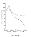
B16メラノーマ細胞に及ぼすRo61の抑制効果を図7に示す。Ro61処理細胞の増殖を未処理対照細胞に対する比率として表わす。3組のウェルの平均値を算定した。図7に見られるように、Ro61はB16細胞(黒丸)の増殖を抑制したが、正常脾細胞(白丸)は抑制しなかった。さらに、培養物中の細胞に濃度を増加させながらRo61を添加したとき、用量依存性の抑制が観察された。この効果は濃度 0.1μMで見られ(未処理対照と比較して22%抑制)、10μMで最大であった(83%抑制)。顕微鏡下では、最高濃度のRo61では、B16細胞は最早正常な繊維芽様の紡錘形ではなく、球形で疎らであって、生存率は低かった(図17A〜17Bを参照のこと)。これとは対照的に、最高濃度のRo61(10μM)を添加しても、脾細胞はほんの少し影響を受けただけだった。
【0143】
B16培養上清中のエンドセリンレベルが用量依存様相で(p<0.001)(50 nMのRo61で513±27 fmol/lから5μMのRo61で954±31 fmol/lに)増加することもわかった。この観察結果はETRのRo61遮断と一致し、受容体が介在するフィードバック回路を妨害する。
【0144】
ET受容体のインヒビターであるRo61はB16細胞の増殖を抑制するので、ETA若しくはETBに特異的なペプチドアゴニストの添加がこの効果を逆行させる、すなわちRo61と受容体結合部位について競合することによって該効果を逆行させるかどうかを決定することが、興味の対象となった。アゴニストBQ-3020-[Ac-[Ala11,15]-エンドセリン(6,21)、すなわちBQ3020(Novabiochem,カタログ番号Al 4534 )およびアゴニスト[Ala1,3,11,15]-エンドセリン1(Sigma Immunochemicals,St. Louis MO,カタログ番号E6877)をHBSS中に懸濁して使用した。5x10⁴ 個のB16細胞を、各ウェルについて10 nMの濃度のいずれかのアゴニストのみか、両方のアゴニストとともにか、またはいずれも含ませないで、培養し、次にウェルに各種濃度のRo61を添加した。
【0145】
図10に示すように、B16増殖のRo61による抑制は、ET受容体アゴニストの添加によって妨害された。Ro61処理細胞の増殖を未処理対照細胞との比率として表わす。3組のウェルの平均値を算定した。最初に、図10のY軸(Ro61濃度 0)によって示すように、BQ3020アゴニスト(黒三角)若しくはET-1アゴニスト(白菱形)のみ、または2つのアゴニストを組合せたもの(白四角)の添加は、B16細胞の増殖を誘導した。BQ3020アゴニストについては、増殖は未処理対照の137%、ET1アゴニストについては、増殖は未処理対照の117%であり、アゴニストの組合せについては、増殖は未処理対照の164%だった。
【0146】
図10にさらに示すように、これらのアゴニストの添加はまた、Ro61の用量を増加させることによって誘導される抑制をも妨害した。例えば、10 nM濃度のET1の添加は、1 nM濃度のRo61の効果を完全に逆転させ、5μMでのRo61の抑制を50%低下させた。BQ3020アゴニストは5μMのRo61の効果(5μMで増殖のわずか12%の阻害)を有意に逆転させた。2つのアゴニストの組合せによって、最も有意な効果が観察され、0.1μM Ro61の添加があっても増殖が誘導された(未処理対照の123%)。これらの用量依存性の知見は、ETRアンタゴニストによって抑制され得るマウスB16細胞に対するエンドセリン受容体介在性増殖応答を定義付けた。
【0147】
別の実験において、上記の細胞増殖アッセイを使用し、ETAおよびETBの両方のアンタゴニストであって、ETAに対して約10倍高い親和性を持つRo61を、ETAアンタゴニストであるBQ123およびETBアンタゴニストであるBQ788(いずれもAmerican Peptide Co.,Sunnyvale,CAより入手)とともに試験し、B16細胞の増殖に及ぼすこれらのアンタゴニストの影響を判定した。培養物中にBQ123、BQ788、BQ123+BQ788、またはRo61-0612/001(それぞれの合計濃度が10μM)を存在させた結果、増殖の有意な抑制があった(未処理対照と比較してそれぞれ16、18、19および50%;p=0.001、下記表3参照)。
【0148】
【表3】
さらに別の上記の増殖アッセイにおいて、各種のエンドセリンアンタゴニストを試験して、B16細胞の増殖に及ぼすこれらの影響を判定した。動物において10回継代した野生型全長ETRを発現するB16細胞を、FOと命名した対照B16細胞と比較した。この対照B16細胞は末端切断型ETA mRNAを発現するので、不完全なETA受容体を産生するものと見られる。
【0150】
図8および9に示すように、エンドセリンアンタゴニストであるIRL、BQ123、BQ610、BQ485、BQ788、RESは10-6Mの濃度においてすべて、FO対照ならびにエンドセリンアゴニスト対照であるET1およびBQ3020と比較して、B16細胞の増殖を抑制した。
【0151】
12.実施例: B16 メラノーマ腫瘍アポトーシス性細胞死の Ro61 による誘導
in vitro研究で、Ro61処理は細胞増殖の抑制に寄与するばかりでなく、細胞死または壊死の存在の重要な構成成分であることも示された。実際に、Ro61で処理されたB16細胞はプログラムされた細胞死と一致する有意な程度の形態学的変化を示した(図11を参照のこと)。
【0152】
そこで、B16アポトーシス性細胞死に及ぼすRo61の影響を調査した。25 ml培養フラスコ内のCM中、1μM Ro61の存在下または不在下で、B16細胞を5% CO2インキュベーター中、37℃で72時間まで増殖させた。以下のようにして、Fluorescein In Situ Cell Death Detection Kit(Boehringer Mannheim,Mannheim,Germany)を使用し、アポトーシス若しくは細胞死について、細胞をアッセイした。簡潔に述べると、細胞をトリプシン処理し、1%ウシ血清アルブミン(BSA)を含有するPBSで洗浄した。PBS中4%パラホルムアルデヒド溶液(pH 7.4)で30分固定した後、0.1%クエン酸ナトリウム中0.1%Triton X100の溶液で氷上で2分、細胞を透過性化した。次に細胞を洗浄し、TUNEL反応混合物(Boehringerキット)を用いて37℃で1時間標識し、洗浄した。Coulter EPICS XLフローサイトメーター(Coulter,Miami FL)で蛍光を分析した。DNase I(Boehringer Mannheim,Mannheim,Germany)100μg/mlを室温で10分使用して、2本鎖DNAの開裂を誘導し、測定値を陽性対照と比較した。
【0153】
図12に示すように、エンドセリンアンタゴニストのRo61は培養中のB16メラノーマ細胞のアポトーシスを誘導した。例えば、B16細胞に1μMのRo61を添加することにより、未処理対照と比較して、アポトーシスに到る細胞の割合が有意に増加した(p=0.0007)。上記のTUNELアッセイで陽性を示す細胞の割合の増加は、24時間目という早さで検出することができ、72時間目でもまだ検出することができた。Ro61との培養後の持続期間による影響は顕著ではなかったが、未処理対照と比較したRo61処理細胞のアポトーシスの増加は非常に顕著だった(12.7%、95%信頼区間:11.7%-13.8%)。これらの結果から、ETRによって仲介されるアポトーシス性細胞のシグナル伝達の存在と、これをETRアンタゴニストによって誘導し得ることが立証された。このように、アポトーシスは少なくとも部分的に、細胞の増殖の抑制と観察される細胞死とに寄与している。
【0154】
13.実施例:エンドセリンアンタゴニストまたは本発明の EA/p-GlcNAc 組成物による in vivo での B16 メラノーマ腹腔内癌腫症の抑制
Ro61がB16細胞に及ぼすin vitroでの有意な効果に基づいて、攻撃性腹腔内(IP)B16メラノーマ転移/癌腫症モデルを利用する、in vivoでの腫瘍増殖に及ぼすETRアンタゴニズムの影響力を評価するための別の研究が導かれた。このモデルについては、HBSS 100 ml中の5x10⁴ 個のB16細胞を雌 C57BL/6マウスの腹腔内に注入した。1日後、このマウスにHBSSのみ(未処理)、3 mg/kgのRo61を含有するHBSS(毎日投与)、30 mg/kgのRo61を含有するHBSS(毎日投与)、p-GlcNAcゲル(2%)のみ、または18 mg/kgのRo61を含有するp-GlcNAcゲル(2%)のいずれかを100 μl注入した。
【0155】
3 mg/kg若しくは 30 mg/kgのRo61を含有するHBSSのみで処理するグループの動物には毎日i.p.注入を行い、一方その他のグループはすべて1回のみの処理をした。この実験で使用したp-GlcNAcゲルのみのものは、上記第10節の実施例に示すものからRo61の添加を除いて調製した。この実験で使用したRo61/p-GlcNAcゲルも、上記第10節で詳述したようにして調製した。7日後にマウスを犠牲にし、腹腔内の疾患の存在を評価した。さらに詳記すると、解剖用顕微鏡下で、各動物について腹腔表面、腸間膜、肝臓、脾臓および膵臓上の個々の腫瘍コロニーを計数した。研究は二重盲検条件下で実施した。腹膜および消化器官の写真をとり、またHおよびEならびにメラニン染色による組織学的分析のために、腸間膜、肝臓、脾臓および膵臓脂肪ならびに腹膜の切片の写真をとった。
【0156】
図13Aは、低用量、例えば 3 mg/kgで6日間毎日(合計で18 mg/kg)、Ro61のIP注入を受けたマウスが、1動物についての転移若しくは腫瘍コロニーの数の有意な低下がなかったことを示している(本明細書で使用する用語「転移」は、上記の癌腫症モデルにしたがってB16メラノーマ細胞を注入したマウスの腹膜および消化器官内に検出される腫瘍コロニーを称する)。
【0157】
対照的に、30 mg/kgで6日間毎日(合計で180 mg/kg)というより高い用量のRo61のみの注入は、腫瘍注入後7日目の腫瘍コロニーの平均数を有意に減少させた。
【0158】
その上、図13Bに示すように、p-GlcNAcゲルのみでは、IP注入したときに未処理対照と比較して腫瘍コロニーの平均数を減少させたが、皮下に注入したとき(SC)はコロニーの増殖に何ら有意な影響を与えなかった。図13Bはまた、p-GlcNAcゲル内に配合した単一用量(18 mg/kg)のRo61の投与により腫瘍コロニーの最大の減少が見られたことを示している(コロニーの平均数=2.7±1.4)。このゲル/Ro61組成物は、高用量のRo61(p=0.001)およびp-GlcNAcゲルのみのIP(p=0.02)の両方と比較したとき、コロニーが顕著に少なかった。低用量のRo61処理およびp-GlcNAcゲルのみのSCは、未処理対照と有意な差異が無かった。
【0159】
Ro61がp-GlcNAcによる局所作用だけでなく全身作用も有するかどうかを判定するため、B16メラノーマの皮下、SCモデルにおけるIP Ro61処理の効果を究明した。雌CS7BL/6マウスの側腹部にHBSS 100μl中の5×10⁴ 個のB16メラノーマ細胞をSC注入した。24時間後、この動物に以下の処理の1つ(100μl、IP)を実施した:HBSS(毎日×6)、HBSS中 30 mg/kgのRo61(毎日×6)、p-GlcNAcゲル(1回投与)、またはp-GlcNAcゲル中 18mg/kgのRo61(1回投与)。腫瘍注入後3週間、 腫瘍の出現および増殖について、動物をモニターした(全グループについてn=10)。
【0160】
図14に示すように、B16メラノーマのSCモデルから、HBSS中のRo61 IPを受けた動物は全部17日目までに腫瘍を発生させたが、腫瘍の出現には有意な遅延があることが示された(未処理対照における15日目での腫瘍担持マウスは100%であることと比較して、15日目での腫瘍担持マウスは50%)。Ro61とp-GlcNAcとの組み合わせは、腫瘍の出現の遅延(13日目での腫瘍担持マウスは50%)をもたらし、また腫瘍注入後21日目で腫瘍がない動物が10%となる。このことは、腫瘍とは別の部位におけるRo61の持続放出が、B16メラノーマの増殖に有意に影響を与えることができることを示唆している。未処理対照グループとIP p-GlcNAcグループ間では腫瘍の出現および増殖に差異がなかった。
【0161】
上記の癌腫症モデルを使用して、B16腫瘍コロニー形成に及ぼす各種のその他のエンドセリンアンタゴニストの影響を評価した。これらの試験では、各薬剤若しくは薬剤混合物 14.3 mg/mlを含有するサンプル 100μlを1回投与で動物に注入した。図15に示されるように、最終合計濃度がRo61(ETAおよびETBアンタゴニスト)のものと等しいBQ123(ETAアンタゴニスト)およびBQ788(ETBアンタゴニスト)の混合物は、未処理対照B16細胞と比較して、Ro61によって示されたものと同程度まで腫瘍コロニーの数を有意に減少させた。さらに、上記のようにBQ混合物をp-GlcNAcと配合したとき、腫瘍コロニーの数はさらに減少し、Ro61プラスp-GlcNAcで見られたものとほとんど同程度となった。最後に、市販のペンタペプチドである混合ETA/ETBアンタゴニストのGRGDS(American Peptide Co.)をp-GlcNAcと配合したとき、Ro61/p-GlcNAcよりもさらに腫瘍コロニーの数が減少することが示された(図15、最終欄を参照のこと)。
【0162】
14.実施例:エンドセリンアンタゴニストのみまたは本発明の EA/p-GlcNAc 組成物による腹腔内 B16 メラノーマチャレンジ後の C57BL/6 マウスの長期生存
in vivoでの長期生存実験におけるエンドセリンアンタゴニズム治療法をも評価した。結果を図16に示す。雌C57BL/6マウスの腹腔内にHBSS 100 ml中5×10⁴ 個のB16細胞を注入した。以下のいずれかの処理のために、マウスをランダムに4グループに分けた:(a)未処理(黒四角);(b)p-GlcNAcゲル 100μlのみ(十字);(c) 3 mg/kg のRo61を含有するHBSSを毎日100μl(黒三角)、または(d) 3 mg/kg のRo61を含有する p-GlcNAcゲル 100μl(白四角)。動物を毎日モニターし、死が近いと判定されたとき、苦痛を与えないという理由から犠牲にした。
【0163】
B16メラノーマが非常に悪性の腫瘍であり、腫瘍注入後19〜20日以内に一様に生存率 0%となることに着目することは興味深い。図10に示すように、p-GlcNAcのみの注入は死を5日遅延させたが、動物の生存率を増加しなかった。しかし、低用量(3 mg/kg)のp-GlcNAcとRo61の配合では死を遅延させ、腫瘍注入後33日目には動物の33%が腫瘍の跡を示さなかった。Ro61のみを同一の低用量で毎日注入しても、マウスの生存に影響しなかった。
【0164】
15.結果の考察
本明細書中で実施した研究は、エンドセリンのその受容体への結合の阻害が、in vitroおよびin vivoの両方で、マウスメラノーマ細胞系の標準的な増殖に影響を与え得ることの直接の証拠を示している。例えば、エンドセリンアンタゴニストのRo61は、ETAおよびETB受容体の両方のインヒビターであり、ETAの方に約10倍高い親和性がある。これは、本発明者らの実験におけるRo61によるメラノーマ細胞増殖の用量依存性抑制、ならびにETBアゴニストとは対照的にETAアゴニストの添加によって獲得されるこの抑制に対するより強い対抗効果とよく相関する。その上、多数のその他のエンドセリンアンタゴニストもメラノーマ細胞の増殖を抑制することが本明細書中で証明された。これらのアンタゴニストには、ETAおよびETB特異的アンタゴニストの両方、例えばBQ123、BQ485、BQ610、BQ788、RESおよびIRLが含まれる。
【0165】
メラノーマ細胞が高レベルのET受容体を発現することが示され、ずっと少ない膜ET受容体を持つことが知られている正常脾細胞よりも、エンドセリンアンタゴニスト抑制を受けやすいことに着目することは興味深い。このように、本明細書中に提供した証拠は、Ro61、BQ123、BQ485、BQ610、BQ788、RESおよびIRLなどのエンドセリンアンタゴニストが培養中の腫瘍細胞に対して抗増殖作用を持つものであり、この腫瘍細胞増殖の抑制はETAおよび/またはETBに特異的なペプチドアゴニストに応答するエンドセリン受容体に結合することによって仲介されることを指摘するものである。
【0166】
その上、本発明者らの研究は、エンドセリンアンタゴニストのみまたは本発明に記載するp-GlcNAcとの組み合わせがin vivoで癌腫を有意に減退させることを示している。さらに、本発明のEA/p-GlcNAc組成物、例えばRo61/p-GlcNAcは、in vivoで腫瘍細胞担持動物の生存率を劇的に増大させる。このRo61の効果はアポトーシス性作用機構に関与するものと見られ、これはエンドセリンのいくつかの既知の作用機構およびそのシグナル伝達機構に矛盾しない。
【0167】
Ro61についての薬理学的研究では、Ro61がin vivoで比較的迅速に、例えば2〜4時間以内に代謝されることを示している。しかし、Ro61を本発明のp-GlcNAcポリマーと組み合わせたとき、Ro61はゲル内に保持され、より緩やかに放出され、そして例えば少なくとも48時間検出することができる。さらに、ゲル内のエンドセリンアンタゴニストの濃度を増加させれば、このアンタゴニストを少なくとも72時間ゲル内に保持することができる(データは示していない)。このように、本発明のp-GlcNAc中のエンドセリンアンタゴニストの濃度を改変することによって、増殖性疾患を治療する際に、アンタゴニストの所望の徐放性、したがって強化された効果を獲得することができる。
【0168】
最後に、下記の表4に示すように、ETA受容体を発現するその他の細胞型も、本明細書に記載するように、エンドセリンアンタゴニスト Ro61に反応する、すなわちエンドセリンアンタゴニストに曝露したとき、細胞増殖を抑制する。このデータから、各種細胞型において、例えばETA受容体の存在と、細胞増殖に及ぼすエンドセリンアンタゴニストの影響とが関連づけられる。例えば、これらの結果は、膵臓、乳房または前立腺腫瘍などの腫瘍がETA受容体を発現するならば、本明細書で開示したようにして、ETA感受性エンドセリンアンタゴニストで治療することができることを意味している。
【0169】
【表4】
本発明は本明細書中に記載した特定の実施形態によって範囲を限定するものではなく、それらは本発明の個々の態様の単なる説明として意図するものであり、機能的に等価な方法および構成成分は本発明の範囲内である。実際、前記の記載および添付する図面から、本明細書中に示しかつ記述したものの他に、本発明の各種の改変が当業者にとって明らかになるであろう。こうした改変は添付する特許請求の範囲の範囲内として意図するものである。
【図面の簡単な説明】
【図1】 100% p-GlcNAcの化学構造。nは約4,000〜約150,000の範囲の整数をさし、約4,000〜約15,000が好適である。
【図2】 p-GlcNAcの炭水化物解析、ガスクロマトグラフィー/質量スペクトル解析データ。黒四角は以下の第5.1節に記載する酸処理/中和法を用いて精製したp-GlcNAcを表す。
【図3】 化学的/生物学的精製法の酸処理/中和変法で調製したp-GlcNAc膜を示す走査型電子顕微鏡写真。倍率:10,000x
【図4】 本発明の可能なp-GlcNAcおよび脱アセチル化p-GlcNAc誘導体のいくつかを示す図(S. Hirano, "Chitin and Chitosan", 1989, Skjak-Braek, AnthonsenおよびSanford編, Elsevier Science Publishing Co., pp. 37-43から改作したもの)。
【図5】 脱アセチル化p-GlcNAcマットの走査型電子顕微鏡写真。倍率:図5A: 1000x ;図5B: 10,000x
【図6】 以下の第8節の実施例に記載するように、ジメチルアセトアミド/塩化リチウム中に溶解し、水中で繊維材料へと再沈殿させたp-GlcNAc膜の走査型電子顕微鏡写真。
【図7】 in vitroにおけるB16メラノーマ細胞増殖のエンドセリン受容体アンタゴニストRo61による阻害。96ウェル培養プレートにRo61を次第に増加する濃度で加え、次にC57BL/6(H-2b)マウス由来のB16細胞(黒丸)および脾細胞(白丸)を加えた。Ro61で処理した細胞の増殖は、未処理対照細胞に対するパーセントとして表してある。3回反復実験ウェルの平均値を求めた。
【図8】 細胞を種々のエンドセリンアンタゴニストに暴露したときの未処理対照に対するB16メラノーマ細胞の増殖パーセントを示す棒グラフ。結果は、細胞をエンドセリンアンタゴニストに暴露したときの増殖の抑制を示している。FO細胞は全長ETA受容体を欠失している対照B16細胞である。ET1と表示した棒グラフは対照を表し、対照では細胞を既知のエンドセリンアゴニストに暴露した。
【図9】 細胞を種々のエンドセリンアンタゴニストに暴露したときの未処理対照に対するB16メラノーマ細胞の増殖パーセントを示す棒グラフ。結果は、細胞をエンドセリンアンタゴニストに暴露したときの増殖の抑制を示している。FO細胞は全長ETA受容体を欠失している対照B16細胞である。ET1およびBQ3020と表示した棒グラフは対照を表し、対照では細胞を2種類の既知のエンドセリンアゴニストに暴露した。
【図10】 in vitroにおけるB16メラノーマ細胞増殖のRo61阻害のETAおよびETBアゴニストによる逆転。B16細胞をアゴニストBQ-3020-[Ac-[Ala11,Ala15]-エンドセリン(6,21)(黒三角)、アゴニスト[Ala1,3,11,15]-エンドセリン1(白菱形)、両アゴニスト(白四角)と共に、または両アゴニストなし(黒丸)で培養し、次にRo61を各ウェルに添加した。Ro61で処理した細胞の増殖を、未処理対照細胞に対するパーセントとして表してある。3回反復実験ウェルの平均値を求めた。
【図11】 培養下のB16細胞に及ぼすRo61の影響。倍率40xでのB16細胞の光学顕微鏡写真。図A:96ウェルプレートにおいて完全培地中37℃で5x10⁴個/ウェルにて72時間培養したB16細胞。図B:5μMのRo61を含む完全培地中で培養したB16細胞。
【図12】 Ro61はアポトーシスを誘導する。B16細胞を、標準培地(対照)またはRo61 (1μM)(■)と共に37℃で0、24、48および72時間培養した後にフルオレセインin situ細胞死検出キットを使ってアポトーシスについてアッセイした。
【図13】 B16腹腔内癌腫症のRo61抑制。図13Aは、B16腹腔内癌腫症に及ぼすIP Ro61の影響を示す。C57BL/6雌マウスに5x104個のB16メラノーマ細胞を腹腔内注射し、翌日、動物に100μlの次のいずれかを注射した:HBSSを6日間(対照)、3mg/kgのRo61を含むHBSSを6日間(低用量)、30mg/kgのRo61を含むHBSSを6日間(高用量)。図13Bは、Ro61のp-GlcNAc送達の効果を示す。図13Aと同様にして動物を腫瘍でチャレンジし、翌日100μlのp-GlcNAcゲルをIP注射、SC注射または18mg/kgのRo61と共にIP注射(IP+Ro61)して処理した。7日後動物を犠牲にし、B16コロニーの存在を評価した。数値は肉眼で見えるコロニーの平均数および各群の標準誤差を表す(全ての群はn=11、ただし未処理群はn=13)。
【図14】 B16メラノーマ腫瘍を皮下にチャレンジした後のRo61処理C57BL/6マウスにおける遅れた腫瘍出現。C57BL/6雌マウスに5x104個のB16メラノーマ細胞をSC注射した。翌日、動物を無作為に4群に分けた:未処理(▲)、p-GlcNAcゲルのみのIP注射1回(○)、3mg/kgのRo61を含むHBSSのIP注射毎日(6日間)(□)、18mg/kgのRo61を含むp-GlcNAcゲルのIP注射1回(■)。腫瘍の存在について動物を3週間モニターした(全ての群においてn=10)。
【図15】 後述する癌腫症モデルを用いた、B16腫瘍コロニーの出現に及ぼす種々のエンドセリンアンタゴニストの阻害効果。F10は未処理のB16対照細胞を表し、BQmixはETAアンタゴニストのBQ123とETBアンタゴニストのBQ788との混合物を表し、BQmix/ゲルは後述するp-GlcNAcゲルと組み合わせたBQ123とBQ788の混合物を表し、Ro61は後述する非特異的ETA/ETBエンドセリンアンタゴニストであり、Ro61/ゲルはp-GlcNAcゲルと組み合わせたRo61であり、GRGDS/ゲルはp-GlcNAcとETA/ETBペプチドエンドセリンアンタゴニストGRGDSとの組合せである。
【図16】 B16メラノーマの腹腔内チャレンジ後のRo61処理C57BL/6マウスの長期生存。C57BL/6マウスにB16細胞を腹腔内注射した。動物を次の処理のいずれかのために無作為に4群に分けた:(a) 未処理(黒四角)、(b) p-GlcNAcゲルのみを100μl(×印)、(c) 3mg/kgのRo61を含むHBSSを毎日100μl(黒三角)、または(d) 18mg/kgのRo61を含むp-GlcNAcゲルを100μl(白四角)。動物を毎日モニターし、瀕死の状態にあると判定したときには安楽死させた。
【図17】 Ro61で処理した(図17A)および処理してない(図17B)B16細胞の細胞形態の顕微鏡写真(倍率40X);10-7M Ro61、105個の細胞。[0001]
The present invention is a continuation-in-part of US patent application Ser. No. 09 / 218,288, filed Dec. 22, 1998. Said application is a continuation-in-part of U.S. Patent Application No. 471,290 (U.S. Pat. No. 5,858,350) filed on June 6, 1995, which is further U.S. Patent Application No. 347,911 filed on Dec. 1, 1994. (US Pat. No. 5,623,064), which is a continuation-in-part of US Patent Application No. 160,569 (US Pat. No. 5,622,834) filed on Dec. 1, 1993. These applications are hereby incorporated by reference in their entirety.
[0002]
1.Introduction
The present invention provides for the use of at least one endothelin antagonist, preferably in combination with a poly-β1 → 4-N-acetylglucosamine (p-GlcNAc) polysaccharide matrix, for the treatment of cancer and other proliferative diseases. It relates to compositions and methods. More specifically, the endothelin antagonist of the present invention may be a peptide or non-peptide compound, and the p-GlcNAc matrix of the present invention comprises a high molecular weight polymer in which the constituent monosaccharides are bound in a β1 → 4 conformation. It is free of protein and substantially free of single amino acids and other organic and inorganic contaminants. The compositions and methods of the present invention are useful for inhibiting tumor and other neoplastic cell growth and / or preventing neoplastic cell metastasis in vivo.
[0003]
2.Background of the Invention
Endothelins are a family of 21 amino acid peptides (eg ET-1, ET-2 and ET-3), originally characterized by their potent vasoconstrictive and angiogenic effects ( (See, for example, Luscher et al., 1995, Agents Actions Suppl. (Switzerland) 45: 237-253; Yanagisawa et al., 1988, Nature 332: 411-415). These peptides also appear to be associated with growth factors such as bFGF and often act cooperatively with them (eg, Halaban, 1996, Seminars in Oncology 23: 673-681; Reid et al., 1996, Development 122 : 3911-3919; Markewitz et al., 1995, Am. J. Physiol. 268: L192-L200; and Nelson et al., 1996, Cancer Res. 56: 663-668). Furthermore, these peptides exhibit cytokine-like regulatory activity and may be affected by hormones such as insulin and angiotensin II and growth factors such as TGF-β and TNF-α (Nelson et al., Supra; Suzuki et al., 1989, J. Biochem. 106: 736-741; and Lundblad et al., 1996, Crit. Care Med. 24: 820-826). Endothelin activity is mediated by binding with high affinity to two different G-coupled receptors ETA and ETB in an autocrine / paracrine manner (see, for example, Hocher et al., 1997, Eur. J. Clin. Chem. Clin. Biochem. 35 (3): 175-189; Shichiri et al., 1991, J. Cardiovascular Pharmacol. 17: S76-S78).
[0004]
There are various endothelin receptor agonists and antagonists (Webb et al., 1997, Medicinal Research Reviews 17 (1): 17-67), which have been used to study the mechanism of action of endothelin. Since endothelin is known to have strong vasoconstrictive activity, endothelin antagonists (also referred to in the art as “endothelin receptor antagonists”) are human diseases, mainly hypertension, congestive heart failure, atherosclerosis Has been studied for their possible role in the treatment of cardiovascular diseases such as sarcoidosis, restenosis and myocardial infarction (Mateo et al., 1997, Pharmacological Res. 36 (5): 339-351). For example, non-peptide endothelin antagonists belonging to the group of pyrimidinylsulfonamide compounds such as Ro 46-2005 and bosentan interact with endothelin receptors through their aromatic rings, Clinical evaluation is ongoing for the treatment of vascular disease and congestive heart failure. These antagonists can bind to both ETA and ETB with varying affinities and are advantageous over peptide-based antagonists. This is because non-peptide antagonists have high metabolic stability (Webb et al., Supra; Parris et al., Supra). In addition, endothelin antagonists have also been studied for their possible role in treating kidney disease such as renal dysfunction and acute renal failure in cirrhosis (Gomez-Garre et al., 1996, Kidney Int. 50: 962-972; Hocher et al., Supra).
[0005]
Recently, endothelin and endothelin receptors have been shown to be involved in several normal and pathological cell growth processes, such as cell cycle progression, cell proliferation and cell development (eg, Parris 1997, Vascular Medicine 2: 31-43; Markewitz et al., Supra; Morbidelli et al., 1995, Am. J. Physiol. 269; H686-H695; and Battistini et al., 1993, Peptides 14: 385-399. ). ET1 and ET3 have been shown to be normal tissue mitogenic and chemokinetic factors ranging from endothelial and epithelial cells to macrophages (eg, Webb et al., 1997, Medicinal Research Reviews 17 (1): 17 -67; and Gomez-Garre et al., Supra). In addition, binding of endothelin to its receptor has been shown to cause DNA synthesis, proliferation and cell mobilization in normal and neoplastic cells (Webb et al., Supra; Ziche et al., 1995, Cardiovasc. Pharmacol. 26 : S284-S286; and Yamashita et al., 1991, Res. Comm. In Chem. Pathol. And Pharmacol. 74 (3): 363-369).
[0006]
This potential of endothelin to mediate cell proliferation and cell cycle progression has led some early studies to studies on endothelin expression and / or the presence of endothelin receptors in cancer cells. For example, ET-1 has been shown to be overexpressed in breast and pancreatic cell lines and induces proliferation in breast cancer tissues, ovarian cell lines and prostate tumors (see, eg, Moriatis et al., 1997, Eur. J. Canc. 33 (4): 661-668; Nelson et al., 1996, Cancer Res. 56: 663-668; Patel et al., 1995, Br. J. Cancer 71: 442-447; Oikawa et al., 1994, Br. J. Cancer 69 : 1059-1064; Shichiri et al., Supra; and Yamashita et al., Supra). Furthermore, the presence of ETA type receptors with higher affinity for ET1 and ET2 has been demonstrated in ovarian cell lines (Moriatis et al., Supra) and breast cancer tissues (Yamashita et al., Supra). One tumor that expresses an ETB receptor with similar affinity for all three isoforms of endothelin is melanoma (Yohn et al., 1994, Biochem. Biophys. Res. Comm. 201 (1): 449-457). Interestingly, the ETB receptor is highly expressed in primary or recurrent melanoma but not so much in metastatic melanoma (Kikuchi et al., 1996, Biochem. Biophys. Res. Comm. 219: 734-739) .
[0007]
Although these studies suggest that endothelin antagonists can be used to treat cancer, there are no studies to date that demonstrate such therapeutic use. In fact, it is unclear what role endothelin plays in promoting various vascular proliferative diseases and proliferative diseases such as benign prostatic hyperplasia (BPH) (Webb et al., Supra; and Kenny et al., 1997, J. Med. Chem. 40 (9): 1293-1315). In addition, U.S. Pat.Nos. 5,550,110 and 5,641,752 disclose cancer treatment using certain hexapeptide endothelin antagonists, but there is no actual data on cancer treatment in those disclosures, No indication is given as to how such treatment is performed and whether such treatment will actually work (further, PCT applications WO 97/37987, WO 96/11927 and WO 94/03483, Canadian patent applications) No. 2072395, and US Pat. No. 5,658,943).
[0008]
3.Summary of the Invention
The present invention relates to methods and compositions for treating cell proliferative diseases such as cancer. More particularly, the present invention relates to the treatment of cancer and other proliferative diseases containing at least one endothelin antagonist, preferably in combination with poly-β1 → 4-N-acetylglucosamine (p-GlcNAc). It relates to a composition for use. In part, the present invention significantly suppresses tumor cell growth and / or neoplastic cell growth or metastasis when an endothelin antagonist is administered alone at a high dose in vivo or in combination with a polysaccharide matrix. This is based on the discovery of the present inventors.
[0009]
According to a preferred embodiment of the present invention, the endothelin antagonist is a non-peptide pyrimidyl sulfonamide compound as shown in Formula I below:
[0010]
According to a preferred embodiment of the present invention, the polysaccharide matrix is a poly-β1 → 4-N-acetylglucosamine (p-GlcNAc) polysaccharide matrix or a derivative thereof as described in US Pat. No. 5,635,493; That patent is hereby incorporated by reference. p-GlcNAc or a derivative thereof can be used in various forms including a membrane, a filament, a nonwoven fabric, a sponge, a gel, and a three-dimensional matrix. According to a preferred embodiment, p-GlcNAc is in the form of a gel, preferably deacetylated, optionally derivatized to p-GlcNAc lactate, and for in vivo administration Combined with Ro61.
[0011]
The compositions of the present invention are useful in drug delivery systems such as sustained release drug delivery. The composition of the present invention is an improvement over conventional drugs in that it provides, for example, increased efficacy, reduced toxicity, and increased bioavailability.
[0012]
The method of the invention comprises in vivo administration of a therapeutically effective amount of the composition of the invention to treat a cell proliferative disorder (eg, cancer) in animals, including humans. According to one embodiment of the present invention, at least one endothelin antagonist such as Ro61 is dissolved in a deacetylated p-GlcNAc lactate gel for the treatment of cancer and other proliferative diseases or disorders. Administered to a patient in a therapeutically effective amount in vivo. Another embodiment of the invention administers in vivo an endothelin antagonist, more preferably a non-peptide endothelin antagonist, such as a pyrimidylsulfonamide endothelin antagonist, for the treatment of cancer and other proliferative diseases or disorders. Including that. Yet another embodiment of the invention involves administering a p-GlcNAc matrix alone in vivo for the treatment of cancer and other proliferative diseases or disorders. The compositions and methods of the present invention are useful for inhibiting tumor and / or other neoplastic cell growth in vivo and / or preventing neoplastic cell metastasis in vivo.
[0013]
4).Brief Description of Drawings
(Described at the end of this specification.)
5.Detailed description
The present invention relates to a composition comprising at least one endothelin antagonist, preferably in combination with a poly-β1 → 4-N-acetylglucosamine (p-GlcNAc) polysaccharide matrix, and the treatment of cancer and other proliferative diseases Relates to a method of using these compositions. Endothelin antagonists according to the present invention may be specific or non-specific for ETA or ETB receptors and may be peptide-based or non-peptide-based compounds. According to a preferred embodiment of the present invention, the endothelin antagonist is a non-peptide non-specific endothelin antagonist. According to another preferred embodiment, the endothelin antagonist is a non-peptidic pyrimidylsulfonamide compound, such as the Ro61 compound shown in Formula I below:
According to the present invention, at least one endothelin antagonist is covalently linked to p-GlcNAc (which is described in detail in Section 5.1 below), alone or in combination with one or more other anti-tumor agents. Or are combined or combined in a non-covalent bond. In one preferred embodiment of the invention, at least one endothelin antagonist such as Ro61 is dissolved in a deacetylated p-GlcNAc gel to form an endothelin antagonist (“EA”) / p-GlcNAc composition of the invention. Let According to a further preferred embodiment, deacetylated p-GlcNAc is derivatized with lactic acid to form p-GlcNAc lactate.
[0015]
“Endothelin antagonist” as defined herein includes an endothelin receptor antagonist, and “EA / p-GlcNAc composition” refers to at least one endothelin antagonist covalently bound to p-GlcNAc. Or a composition that is either non-covalently bound, mixed with p-GlcNAc, or encapsulated within p-GlcNAc. The compositions of the present invention can further comprise other anti-tumor agents that act in combination with endothelin antagonists to inhibit the growth and / or metastasis of tumors and other neoplastic cells. As defined herein, an “anti-tumor agent” includes any compound that inhibits the growth or metastasis of tumor cells, cancer cells, or any other type of neoplastic cells.
[0016]
In part, the present invention shows that endothelin antagonists alone or in combination with the p-GlcNAc described herein inhibit neoplastic cell proliferation in vitro and reduce tumor cell metastasis in vivo. And / or based on the inventors' discovery to enhance survival of tumor cell-bearing animals (see Sections 12-16 below). Furthermore, p-GlcNAc of the present invention alone has an inhibitory effect on neoplastic cell proliferation and metastasis in vivo.
[0017]
Therefore, according to the method of the present invention, a pharmaceutical composition comprising the EA / p-GlcNAc composition of the present invention is administered to a patient in vivo in a therapeutically effective amount for the treatment of cancer or other proliferative diseases. Is done. Another preferred embodiment of the invention involves administering in vivo a endothelin antagonist, such as a pyrimidylsulfonamide endothelin antagonist, for the treatment of proliferative diseases. And yet another embodiment includes administering in vivo p-GlcNAc described below for the treatment of proliferative diseases.
[0018]
For ease of explanation, the detailed description of the invention is divided into the following subsections: (1) p-GlcNAc of the composition and method of the present invention, (2) of the composition and method of the present invention. Endothelin antagonists, (3) preferred formulations of the compositions of the invention, and (4) uses of the compositions and methods of the invention.
[0019]
5.1Of the composition of the invention p-GlcNAc
The p-GlcNAc polysaccharide matrix utilized in the compositions and methods of the present invention comprises a high molecular weight polymer ranging from about 800,000 daltons to about 30 million daltons on a weight average basis, as measured by gel filtration chromatography. . Such molecular weight ranges are indicative of p-GlcNAc species having from about 4,000 to about 150,000 N-acetylglucosamine monosaccharides bound in a β1 → 4 conformation. About 4,000 to about 15,000 N-acetylglucosamine monosaccharides are preferred (FIG. 1).
[0020]
The diversity of this p-GlcNAc is very low and the purity is very high. Both of these have been proven by chemical and physical standards. Among these are chemical composition and non-polysaccharide contaminants. First, the chemical composition data of p-GlcNAc obtained using two different purification methods are shown in Table 1 below. As can be seen in the table, the chemical composition of p-GlcNAc obtained by both methods is identical to the official composition of p-GlcNAc within experimental error. Second, as also shown in Table 1, the resulting p-GlcNAc is free of detectable protein contaminants, substantially free of other organic contaminants such as free amino acids, and ash content. (P-GlcNAc of the present invention may be up to about 2% from the theoretical values of carbon, hydrogen, nitrogen and oxygen for pure p-GlcNAc. Good). Accordingly, as used herein, the terms “substantially free of organic pollutants” and “substantially free of inorganic pollutants” do not deviate from the theoretical value by more than about 2%. Means the composition of p-GlcNAc with carbon, hydrogen, nitrogen and oxygen profiles, preferably the p-GlcNAc of the invention is a profile (deviation%) as illustrated in the experimental data for the p-GlcNAc mats in Table I Have). Furthermore, p-GlcNAc has a very low proportion of bound water.
[0021]
The p-GlcNAc of the composition of the present invention exhibits a carbohydrate analysis profile substantially similar to that shown in FIG. The main monosaccharide of p-GlcNAc is N-acetylglucosamine. Furthermore, p-GlcNAc does not contain the monosaccharide glucosamine. Other physical properties of p-GlcNAc are described in detail in US Pat. No. 5,635,493, incorporated herein by reference.
[0023]
The p-GlcNAc of the present invention has a high degree of biocompatibility. This can be measured by various techniques such as, but not limited to, dissolution testing, intramuscular implantation, or intradermal or systemic injection into animal subjects. See, eg, US Pat. No. 5,635,493, incorporated herein by reference.
[0024]
p-GlcNAc is produced by and can be purified from microalgae, preferably diatoms. Diatoms that can be used as a starting source for the preparation of p-GlcNAc include, but are not limited to, members of the genera Coscinodiscus, Cyclotella and Thalassiosira, with the Thalassiosira genus being preferred.
[0025]
Within the genus Coscinodiscus, diatom species that can be used include, but are not limited to, concinnus species and radiatus species. Within the genus Cyclotella, diatom species that can be used include, but are not limited to, capsia species, cryptica species, and meneghiniana species. Diatoms of the genus Thalassiosira that can be used to produce the starting material of p-GlcNAc of the present invention include, but are not limited to, nitzschoides, aestivalis, antarctica, deciphens, eccentrica, floridana, fluviatilis, gravida, guillardii, hyalina , Minima, nordenskioldii, oceanica, polychorda, pseudonana, rotula, tubifera, tumida and weissflogii, with fluviatilis and weissflogii being preferred. Diatoms as described above can be obtained, for example, from the type culture collection of Bigelow Laboratory for Ocean Science, Center for Collection of Marine Phytoplancton (McKown Point, West Boothbay Harbor, Maine, 04575). Any of these diatoms can be grown using the methods and nutrient media described in US Pat. No. 5,635,493, incorporated herein by reference.
[0026]
p-GlcNAc fibers can be obtained from diatom cultures as described above in a number of different ways. In the mechanical force method, p-GlcNAc fibers are separated from diatom cell bodies by subjecting the contents of the culture to a suitable mechanical force. Such mechanical forces include, but are not limited to, shear forces generated by colloid mills, ultrasonic devices or foam generators, or cutting forces generated by, for example, a Waring blender.
[0027]
Subsequently, the obtained diatom cell body suspension and p-GlcNAc fiber are separated. For example, the suspension is subjected to a series of centrifugation steps that separate the p-GlcNAc fibers from the cell body, resulting in a clear supernatant that contains only a trace amount of cotton-like material that can be visually observed. . For the centrifugation step, a fixed angle rotor and a temperature of about 10 ° C. are preferred. The required speed, duration, and total number of centrifugation steps may vary depending on, for example, the particular centrifuge rotor used, but the determination of such parameters will be obvious to those skilled in the art.
[0028]
The p-GlcNAc fibers in the supernatant can then be concentrated using techniques well known to those skilled in the art. Such techniques include, but are not limited to, suction and filtration devices. Finally, the concentrated p-GlcNAc fibers are washed using, for example, distilled deionized water, HCl and ethanol, or other suitable solvent, preferably a solvent such as an alcohol that dissolves both organic and inorganic materials. An example demonstrating the use of this p-GlcNAc purification method is shown in
[0029]
In chemical / biological methods, p-GlcNAc fibers are separated from diatom cell bodies by exposing the diatom cell bodies to chemical and / or biological agents. For example, diatom cultures are treated with chemicals that can weaken diatom cell walls, thereby releasing p-GlcNAc fibers without changing their structure. Such chemicals include, but are not limited to, hydrofluoric acid (HF). Alternatively, the mature diatom culture may be treated with a biological agent that can alter the biological process to inhibit the synthesis of p-GlcNAc fibers, thereby releasing the fibers that are already present. Such agents include, but are not limited to, polyoxin-D, which is an inhibitor of the enzyme N-acetylglucosaminyl-P-transferase.
[0030]
The cell bodies and pGlcNAc-containing fibers are then separated from diatom cultures treated with the chemical or biological agent members described above. For example, the treated diatom culture contents are allowed to settle so that the culture contents form two separate layers. The upper layer will mainly contain p-GlcNAc fibers and the lower layer will contain cell bodies. The upper layer containing p-GlcNAc fibers is siphoned up and removed while leaving the lower layer of the cell material left still. The siphoned p-GlcNAc fiber-containing layer is then further purified and treated with a surfactant that does not harm the p-GlcNAc fiber to remove proteins and other undesired things. Such surfactants include, but are not limited to, sodium dodecyl sulfate (SDS).
[0031]
If an acid treatment such as HF treatment is used to separate the p-GlcNAc fibers from the diatom cell bodies, a step for dispersing the fibers can be included. Such steps include, but are not limited to, using mechanical forces for fiber dispersion, such as attaching fibers to the motion of an orbital shaker.
[0032]
Alternatively, in any step, the acid-treated suspension may be neutralized before further purification by surfactant treatment. Such neutralization generally changes the pH of the suspension from about 1.8 to about 7.0, but this can be done, for example, by adding an appropriate amount of 1M Tris (pH 8.0) or an appropriate amount of Add sodium hydroxide (NaOH). Neutralization generally results in pure p-GlcNAc fibers that are substantially longer than other purification methods discussed herein.
[0033]
The purified p-GlcNAc fiber can then be concentrated using techniques known to those skilled in the art such as use of suction and filtration equipment. Finally, the p-GlcNAc fibers are washed in a series of steps using distilled deionized water, HCl and ethanol, or other suitable solvent, preferably a solvent such as an alcohol that dissolves both organic and inorganic materials. A successful demonstration using such a purification method is shown in
[0034]
While each method of purifying p-GlcNAc from an algae (preferably diatom) source yields very high purity, undegraded crystalline p-GlcNAc, each method has certain properties and advantages. Characteristic p-GlcNAc was generated. For example, p-GlcNAc purified by a mechanical method produces a p-GlcNAc membrane that provides an excellent substrate for attaching cells to p-GlcNAc. Depending on the chemical / biological method, an average yield is obtained which is much higher than the average yield of p-GlcNAc obtained by the mechanical force method. In addition, depending on the acid treatment / neutralization variant of the chemical / biological method, very long p-GlcNAc fibers (over 100 μm for some fibers and very high molecular weights (20-30 million Daltons high molecular weight) P-GlcNAc polymer molecules having
[0035]
An electron micrograph of p-GlcNAc (obtained by a modified acid / neutralized chemical / biological method) used in the methods and compositions of the present invention is shown in FIG. In the purification of p-GlcNAc fiber, a fibrous membrane as shown in FIG. 3 is often formed.
[0036]
5.1.1.p-GlcNAc Derivatization of
The fully acetylated p-GlcNAc of the present invention can be derivatized into a wide variety of different compounds using a variety of control conditions and procedures. See FIG. 4 for a diagram illustrating some of these compounds. Such derivatized compounds include, but are not limited to, partially or fully deacetylated p-GlcNAc modified by chemical and / or enzymatic means as described in further detail below. According to a preferred embodiment of the invention, p-GlcNAc is 100% deacetylated p-GlcNAc.
[0037]
In addition, p-GlcNAc or a deacetylated derivative thereof can be derivatized by sulfation, phosphorylation, and / or nitration. Further, as detailed below, O-sulfonyl, N-acyl, O-alkyl, N-alkyl, deoxyhalogen and N-alkylidene and N-arylidene and other derivatives may be converted to p-GlcNAc or deacetylated of the present invention. P-GlcNAc. Various organic salts and / or metal chelates can also be prepared using the deacetylated p-GlcNAc of the present invention.
[0038]
According to a preferred embodiment of the present invention, one or more monosaccharide units of p-GlcNAc can be deacetylated to produce a deacylated poly-β1 → 4-N-glucosamine species. A poly-β1 → 4-N-glucosamine species (ie a 100% deacetylated derivative) in which each monosaccharide unit of the poly-β1 → 4-N-acetylglucosamine species is deacetylated is approximately 640,000 daltons It will have a molecular weight of about 24 million daltons, with about 640,000 daltons to about 2.4 million daltons being preferred. A species having such a molecular weight range corresponds to a species having about 4,000 to about 150,000 glucosamine monosaccharides covalently bonded in a β1 → 4 conformation.
[0039]
When p-GlcNAc is treated with a base and deacetylated, glucosamine having a free amino group is obtained. This hydrolysis process may be carried out at elevated temperature using concentrated sodium hydroxide solution or concentrated potassium hydroxide solution. For example, see
[0040]
Furthermore, as shown below, one or more monosaccharide units of the p-GlcNAc of the present invention can be derivatized to contain at least one sulfate group, or else phosphorylated or nitro Can be
[0041]
A method for preparing such p-GlcNAc derivative will be described below. Prior to carrying out these methods, it is advantageous that the p-GlcNAc starting material is first lyophilized, frozen in liquid nitrogen and ground.
[0042]
Sulfated p-GlcNAc derivatives can be prepared, for example, by a two-step method. In the first step, a method as described in, for example, Tokura et al. (Tokura, S. et al., 1983, Polym. J. 15: 485) is used from the p-GlcNAc and / or p-GlcNAc derivative of the present invention. To prepare O-carboxymethyl p-GlcNAc. Secondly, the sulfation step is well known to those skilled in the art as described by Schweiger (Schweiger, RG, 1972, Carbohydrate Res. 21: 219) using, for example, N, N-dimethylformamide-sulfur trioxide. Can be carried out according to the methods described. The resulting product can be isolated as a sodium salt.
[0043]
Phosphorylated p-GlcNAc derivatives were described, for example, by Nishi et al. (Nishi, N. et al., 1986, in “Chitin in Nature and Technology”, edited by Muzzarelli et al., Plenum Press, New York, pp. 297-299). It can be prepared using methods well known to those skilled in the art, such as methods. Briefly, the p-GlcNAc / methanesulfonic acid mixture is treated with phosphorus pentoxide (about 0.5-4.0 molar equivalents) at a temperature of about 0 ° C. to about 5 ° C. with stirring. Processing takes approximately 2 hours. The resulting product is then precipitated and washed using standard techniques well known to those skilled in the art. For example, the sample is precipitated with a solvent such as ether, centrifuged, washed with a solvent such as ether, acetone, or methanol, and dried.
[0044]
The nitrated p-GlcNAc derivative can be obtained using techniques well known to those skilled in the art, such as those described in Schorigin and Halt (Schorigin, R. and Halt, E., 1934, Chem. Ber. 67: 1712). Can be prepared. Briefly, p-GlcNAc and / or p-GlcNAc derivatives are treated with concentrated nitric acid to form a stable nitration product.
[0045]
One or more monosaccharide units of p-GlcNAc of the present invention may contain a sulfonyl group as follows.
[0046]
One or more monosaccharides of p-GlcNAc or a deacetylated derivative thereof of the present invention may contain one or more O-acyl groups as follows.
[0047]
One or more monosaccharides of the deacetylated p-GlcNAc of the present invention may comprise an N-acyl group as follows.
[0048]
One or more monosaccharides of p-GlcNAc or a deacetylated derivative thereof of the present invention may contain an O-alkyl group as follows.
[0049]
One or more of the monosaccharide units of p-GlcNAc of the present invention may be an alkali derivative as shown below.
[0050]
[0051]
One or more monosaccharide units of the deacetylated derivative of p-GlcNAc of the present invention may contain an N-alkyl group as shown below.
[0052]
[0053]
One or more monosaccharide units of the deacetylated derivative of p-GlcNAc of the present invention may contain one or more deoxyhalogen derivatives as shown below.
[0054]
[0055]
One or more monosaccharide units of the deacetylated derivative of p-GlcNAc of the present invention may form a salt as shown below.
[0056]
[0057]
One or more monosaccharide units of the deacetylated derivative of p-GlcNAc of the present invention may form a metal chelate as shown below.
[0058]
[0059]
One or more of the monosaccharide units of the deacetylated derivative of p-GlcNAc of the present invention may contain an N-alkylidene group or an N-arylidene group as shown below.
[0060]
[0061]
Furthermore, by subjecting p-GlcNAc or its deacetylated derivative to controlled hydrolysis conditions, it is possible to obtain groups of molecules with uniform individual molecular weights and other physical properties. Examples of such hydrolysis conditions include treatment with the enzyme lysozyme. In order to control the degree of hydrolysis, p-GlcNAc can be exposed to lysozyme for various times. Moreover, the rate of hydrolysis can be controlled as a function of the degree of deacetylation of lysozyme treated p-GlcNAc. Deacetylation conditions may be as described above. When deacetylated between about 20 and about 90%, the more fully the p-GlcNAc molecule is deacetylated, the more fully the molecule will be hydrolyzed within a given time. Hydrolysis and / or deacetylation can lead to changes in physical properties in addition to molecular weight reduction. Large scale hydrolysis results in liquefaction of p-GlcNAc.
[0062]
Furthermore, heat denaturation can serve to modify the crystal structure of p-GlcNAc. By so modifying the crystalline structure of the p-GlcNAc product, it is possible, for example, to beneficially affect the reactivity of p-GlcNAc.
[0063]
In addition, hybrids containing p-GlcNAc and / or p-GlcNAc derivatives can be formed. Such hybrids may contain several types of natural and / or synthetic substances in addition to p-GlcNAc and / or p-GlcNAc derivatives. For example, a hybrid may be formed from p-GlcNAc and / or p-GlcNAc derivatives and one or more extracellular matrix (ECM) components. Such ECM components include, but are not limited to, collagen, fibronectin, glycosaminoglycan, and / or peptidoglycan. Hybrids may also be formed from p-GlcNAc and / or p-GlcNAc derivatives and one or more synthetic substances such as polyethylene. Such p-GlcNAc / polyethylene hybrids or p-GlcNAc derivative / polyethylene hybrids can be made, for example, by thermally linking the hybrid components through treatment with an autoclave.
[0064]
Preferred p-GlcNAc derivatives for use in the claimed invention are deacetylated p-GlcNAc salt derivatives such as p-GlcNAc lactate derivatives, in particular p-GlcNAc lactate gel derivatives. As used herein, the term “p-GlcNAc lactate” means that the lactic acid moiety is functionally linked to partially or fully deacetylated p-GlcNAc. Such p-GlcNAc lactate derivatives are obtained as described above (for example by derivatization with lactic acid) and are propylene glycol as described in the examples in
[0065]
As described in further detail in Section 5.3 below, the previously described p-GlcNAc and / or derivatives thereof are covalently or non-covalently bound to a molecule or drug of interest such as an endothelin antagonist. Or can be further derivatized by combining them.
[0066]
5.1.2p-GlcNAc Re-formulation
P-GlcNAc used in the composition of the present invention, its deacetylated derivatives, as described above, and / or their derivatives must be dissolved and then re-formulated into various shapes and forms Is possible.
[0067]
Dissolution of p-GlcNAc can be achieved by treatment with dimethylacetamide (DMA) / lithium chloride. It is possible to dissolve p-GlcNAc easily by stirring in a DMA solution containing 5% LiCl (based on the weight of DMA). A p-GlcNAc salt such as a water-soluble p-GlcNAc derivative such as a lactate derivative or a carboxymethyl derivative can be dissolved in water. At least about 75% deacetylated p-GlcNAc can be dissolved in a weakly acidic solution such as, for example, 1% acetic acid. The water-insoluble p-GlcNAc derivative can be dissolved in an organic solvent.
[0068]
Carvanilate can be generated by derivatizing p-GlcNAc in DMA: LiCl with phenyl isocyanate. Furthermore, it is possible to generate toluene-p-sulfonate by derivatizing p-GlcNAc in DMA: LiCl with toluene-p-sulfonyl chloride.
[0069]
The p-GlcNAc in solution, its deacetylated derivatives, and / or their derivatives can then be precipitated and re-formulated into various shapes. Such shapes include, but are not limited to, mats, strings, microspheres, microbeads, membranes, fibers, powders, sponges, and gels. In addition, it can be re-formulated into a uniform membrane that is ultra-thin (ie, less than about 1 micron thick). In addition, pharmaceutical preparations such as pills, tablets, and capsules can be prepared.
[0070]
Such re-formulation can be accomplished by taking advantage of the fact that pure p-GlcNAc is insoluble in solutions such as water and alcohol, preferably ethanol. For example, when a p-GlcNAc-containing DMA / LiCl mixture is introduced into water or an alcohol, preferably ethanol, by conventional means such as injection, the solution undergoes reprecipitation, resulting in dissolved p-GlcNAc. Reformulation from a p-GlcNAc membrane to a fibrous material will be specifically described in the examples in
[0071]
p-GlcNAc membranes and three-dimensional p-GlcNAc matrices can be made using methods that result in the formation of controlled average pore sizes within the membrane or matrix. Prior to forming the membrane and / or matrix, by varying the amount of p-GlcNAc material used and a specific solvent such as methanol or ethanol, preferably ethanol, ranging from about 5% to about 40%. By adding in specific amounts, the pore size in the membrane and matrix can be controlled. In general, the higher the percent of solvent, the smaller the average pore size formed.
[0072]
According to a preferred re-formulation of the present invention, the p-GlcNAc lactate derivative is re-formulated into a gel as described in detail in the Examples in
[0073]
5.2Endothelin antagonists in the composition of the present invention
Endothelin antagonists utilized in the compositions and methods of the present invention include peptide-based endothelin antagonists, non-peptide-based endothelin antagonists, ETA-specific endothelin antagonists, ETB-specific endothelin antagonists, or non-specific endothelin antagonists, It is not limited to these. Peptide-based endothelin receptor antagonists useful in the compositions and methods of the present invention include, for example, BQ-123 (cyclo (-D-Trp-D-Asp-L-Pro-D-Val-L-Leu-)) , BQ-153, BQ-238, BQ-485, BQ-610, BQ-788, BQ-928, TAK-044, FR139317 (perhydroazepin-1-ylcarbonyl-L-leucyl- (l-methyl)- D-tryptophyll- [3- (2-pyridyl)]-D-alanine), RES-701-1 (Novabiochem), PD 142893 (acetyl- (3,3-diphenyl-D-alanine) -L-Leu-L -Asp-L-Ile-L-Ile-L-Trp), PD 145065, CP 170687, AcDBhgl6-Leu-Asp-Ile, IRL-1038 ([Cys11-Cys15] -endothelin-1 (11-21)), GRGDS pentapeptide, and ET-1 [Dprl-Asp 15]. Many of these peptides are commercially available from, for example, American Peptide Company, Sunnyvale CA, or Calbiochem Novabiochem Company, San Diego CA.
[0074]
Non-peptide endothelin receptor antagonists for use in the compositions and methods of the present invention include, for example, Ro 61-0612, Ro 61-1790, Ro 42-2005, Ro 46-2005, Ro 46-8443, Ro 47-0203 (also known in the art as bosentan), PD 155080, PD 156707, SB 209670, SB 217242, L-744,453, L-749,329, L-754,142, CGS 27830, BMS 182874, LU 135252 S-1039, mA386, A-127722, TBC11251, Nz-arg-3- (isoxazdylsulfameyl) -2-thiophenecarboxamide, and EQ123. For many structures of these known endothelin antagonists, see, for example, Webb et al. And Ohlstein et al., Cited above.
[0075]
According to the present invention, non-peptide endothelin antagonists may be preferred. This is because they exhibit favorable pharmacokinetic properties over peptide-based antagonists, such as improved metabolic stability and better bioavailability and oral activity. According to a preferred embodiment of the present invention, the endothelin antagonist utilized is Ro61 as shown above in FIG.
[0076]
5.3Preferred formulations of the composition of the present invention
According to a preferred embodiment of the invention, the previously described endothelin antagonist (“EA”) is covalently or non-covalently bound to p-GlcNAc, or one or more derivatives or re-formulations thereof as described above. They are functionally linked by a bond or combined with them. According to one embodiment, at least one type of endothelin antagonist is covalently, non-covalently or otherwise combined or mixed with deacetylated p-GlcNAc. Other anti-tumor agents that can be used with the EA / p-GlcNAc composition of the present invention are described below.
[0077]
Endothelin antagonists or other anti-tumor agents can be attached to exposed primary amines of deacetylated p-GlcNAc, for example by chemical conjugation using a bifunctional cross-linking reagent that acts as a chemical spacer of a specific length. It can be covalently bonded. Such techniques are known to those skilled in the art and include, for example, Davis and Preston (Davis, M. and Preston, JF 1981, Anal. Biochem. 116: 404-407) and Staros et al. (Staros, JV et al., 1986, Anal. Biochem. 156: 220-222). For example, in the case of peptide-based compounds, it is possible to activate the carboxyl group on the peptide that is bound to deacetylated or partially deacetylated p-GlcNAc and then crosslink to p-GlcNAc. is there. Activation can be achieved, for example, by adding a solution such as carbodiimide EDC (1-ethyl-3- (3-dimethylaminopropyl) carbodiimide) to the peptide solution in phosphate buffer. . Preferably, the solution will contain other reagents such as sulfo-NHS (N-hydroxysulfosuccinimide) to facilitate coupling. Activated peptides can be cross-linked to deacetylated p-GlcNAc by mixing in a high pH buffer such as carbonate buffer (pH 9.0-9.2).
[0078]
The biological activity of the bound molecule can be preserved by changing the length of the linker molecule (eg, bifunctional crosslinkable compound) utilized to attach the molecule to p-GlcNAc. Appropriate lengths of linkers for a given molecule to be attached that do not change the biological activity of the attached molecule can be determined according to convention. For example, the biological activity of a molecule attached through a linker of a given length (e.g., a therapeutically effective level of biological activity) can be determined using a known assay specific for the given molecule attached. It can be used and tested. In addition, in order to retain the biological activity of the bound molecule, it may be necessary to utilize a linker that can be cleaved by an appropriate natural enzyme to release the bound molecule. By using an assay commonly used by one skilled in the art to test whether the biological activity of a particular molecule bound is retained, an acceptable level of activity (e.g., a therapeutically effective level of activity). ) Can be obtained.
[0079]
In addition, peptide-based or non-peptide-based endothelin antagonists can be mixed with p-GlcNAc and / or its derivatives using techniques known to those skilled in the art, alone or in combination with other antitumor agents, or non-covalently. It is possible to form the compositions of the present invention by bonding to them with a bond. For example, a selected molecule or molecules, such as an endothelin antagonist, can be transferred to a suspension of p-GlcNAc, a solution of deacetylated or partially deacetylated p-GlcNAc, deacetylated or partially Can be mixed with a solution of deacetylated p-GlcNAc salt, for example, a solution of p-GlcNAc-lactate (partially or fully deacetylated), or any p-GlcNAc derivative It is. The mixture may optionally be lyophilized. After lyophilization, molecules are likely to be non-covalently bound to the p-GlcNAc matrix, presumably due to hydrophobic, electrostatic, and other non-covalent interactions. Such p-GlcNAc formulations are very easy to manufacture. Further, such formulations can be effectively obtained using a wide variety of materials having a wide range of physical properties and water solubility ranging from very hydrophobic to very hydrophilic. In binding one or more molecules, by using assays commonly used by those skilled in the art to test the activity of a particular molecule or molecules to be bound non-covalently The conjugated molecule can be used to confirm that an acceptable level of activity (eg, a therapeutically effective activity) can be obtained.
[0080]
Furthermore, endothelin antagonists can be encapsulated in p-GlcNAc by methods known in the art, either alone or in combination with other anti-tumor agents. For example, one method of encapsulation is the procedure outlined by Hwang et al. (Hwang, C. et al. In Muzzarelli, R. et al., Eds., 1985, “Chitin in Nature and Technology”, Plenum Press, pp. 389-396). The entirety of this document is hereby incorporated by reference. In addition, the encapsulation can be performed, for example, according to another method of the acid treatment / neutralization type chemical / biological purification method presented above. Rather than raising the pH of the p-GlcNAc solution to approximately the neutral pH range (i.e., about 7.4), after the purification of p-GlcNAc is complete, the pH is raised to about 9.0 to increase the basic pH. It is possible to create a pH environment. At higher basic pH, the structure of p-GlcNAc or its derivatives takes a more three-dimensional or “open” form. As the pH is lowered, the molecular morphology returns to a more compact and “closed” morphology. Thus, it is possible to add a compound or drug of interest, such as an endothelin antagonist, to a high pH p-GlcNAc solution and then reduce the pH of the p-GlcNAc / drug suspension, thereby Can be “captured” or encapsulated in a p-GlcNAc matrix. After encapsulating the molecule, the level of the encapsulated molecule is acceptable by testing the activity of the particular molecule or molecules encapsulated using assays commonly used by those skilled in the art. It can be confirmed whether it possesses biological activity (for example, therapeutically effective activity).
[0081]
An example of a formulation of an EA / p-GlcNAc composition of the present invention is shown in the Example in
[0082]
Compositions of the invention, including EA / p-GlcNAc compositions, can be administered as pharmaceutical compositions, for example, by inhalation or insufflation (either via the mouth or nose), orally, buccal It can be formulated for internal, parenteral or rectal administration. According to a preferred embodiment, the EA / p-GlcNAc composition of the invention is administered by injection in the form of a gel as described in the Examples in
[0083]
For oral administration, the pharmaceutical composition comprises, for example, a binder (e.g., pregelatinised corn starch, polyvinyl pyrrolidone, or hydroxypropyl methylcellulose), a filler (e.g., lactose, microcrystalline cellulose, Or calcium hydrogen phosphate), lubricants (e.g., magnesium stearate, talc, or silica), disintegrants (e.g., potato starch or sodium starch glycolate), or wetting agents (e.g., lauryl sulfate). It may take the form of tablets or capsules prepared by conventional means using pharmaceutically acceptable excipients or carriers such as sodium). P-GlcNAc can be used in place of or in addition to excipients, carriers, and fillers. Tablets can be coated with p-GlcNAc using methods known in the art.
[0084]
Liquid dosage forms for oral administration can take the form of, for example, solutions, syrups or suspensions, or dry to form a construct with water or other suitable vehicle before use. Can be offered as goods. Such liquid formulations include suspending agents (e.g. sorbitol syrup, cellulose derivatives, or hydrogenated edible fats), emulsifiers (e.g. lecithin or gum arabic), non-aqueous vehicles (e.g. tonsil oil, oily esters, ethyl Alcohol, or fractionated vegetable oils), and conventional means using pharmaceutically acceptable additives such as preservatives (eg, methyl or propyl-p-hydroxybenzoate or sorbic acid). The formulations may also contain buffer salts, flavoring, coloring and sweetening agents as appropriate.
[0085]
5.4Applications of the compositions and methods of the present invention
Biomedical uses of the compositions of the present invention include use as drug delivery systems for other therapeutic agents such as endothelin antagonists as well as other anti-tumor agents. The p-GlcNAc-containing formulations of the present invention provide further advantages compared to known drug formulations, including, for example, increased potency, reduced toxicity and improved bioavailability. Indeed, there are numerous advantages to using the p-GlcNAc drug delivery system of the present invention. For example, traditional drug administration by injection is generally combined with proteins and many other drugs. However, repeated dosing results in fluctuations in blood drug levels, affecting patient comfort and compliance. Oral administration may be advantageous. This is because oral administration can change the amount of drug to be released to a greater extent and lessen the patient's discomfort. However, proteins and other compounds denature and degrade in the stomach.
[0086]
However, improved oral administration is achieved with the p-GlcNAc-containing compositions of the present invention by providing an environment that protects drugs once administered. For example, p-GlcNAc protects peptide-based endothelin antagonists from the acidic and enzymatic environment of the stomach. Once the P-GlcNAc system reaches the intestinal region, it releases the compound by diffusion and / or capsule degradation, where it is effectively absorbed into the bloodstream. By using these p-GlcNAc systems of the present invention, for example, proteins as well as many other compounds can be delivered. Liposomes coated with p-GlcNAc derivatives or p-GlcNAc derivative-alginate encapsulates are preferred for such oral delivery methods.
[0087]
Furthermore, when the composition of the present invention is introduced into a patient, p-GlcNAc gradually biodegrades, resulting in the release of bound or encapsulated compounds gradually into the patient's bloodstream, resulting in controlled gradual progression. Methods are provided for effecting releasable drug delivery.
[0088]
Deacetylated or partially deacetylated p-GlcNAc species with predictable biodegradation rates can be generated. For example, the percentage of deacetylation affects the rate at which p-GlcNAc species degrade. In general, the higher the percent deacetylation, the faster the rate of biodegradation and resorption. Thus, during the production of p-GlcNAc, the degree of biodegradation and in vivo resorption rate of p-GlcNAc can be controlled.
[0089]
P-GlcNAc materials with such controllable biodegradation rates can be formulated into membranes, gels, sponges, microspheres, fibers, and the like. According to a preferred embodiment of the invention, 100% deacetylated or partially deacetylated p-GlcNAc with a predictable biodegradation rate can be utilized.
[0090]
The p-GlcNAc / drug composition of the present invention can be delivered to a patient via various routes using standard procedures known to those skilled in the art. For example, such delivery may be site-specific, oral, nasal, intravenous, subcutaneous, intradermal, transdermal, intramuscular, or intraperitoneal administration. With regard to site-specific delivery, methods of administration include, but are not limited to, injection, implantation, arthroscopy, laparoscopic, or similar means. P-GlcNAc membranes and / or gels and microspheres and sponges are preferred for such site-specific delivery methods.
[0091]
As mentioned above, the p-GlcNAc of the composition of the present invention can be formulated into a membrane, gel, sponge, microsphere, fiber or the like. These p-GlcNAc products adhere to and adhere to tissues in the human body, including both soft and hard tissues, without the need for suturing. The p-GlcNAc substance can be applied during general surgery such as laparoscopic surgery or minimally invasive surgery.
[0092]
According to a preferred embodiment of the present invention, p-GlcNAc is present in the form of a gel in which the endothelin antagonist and / or other anti-tumor agent is dissolved or otherwise incorporated. p-GlcNAc-based gels and membranes can be used, for example, as therapeutic drug delivery systems to provide direct site-specific sustained release delivery to tumors or to areas where tumors have been removed after surgery. There are various applications. Such immobilized sustained release compositions can act as an important initial defense method after surgery. Such anti-tumor drug delivery systems may also be particularly useful for treating tumors that cannot be performed at all or in part, such as certain brain tumors.
[0093]
Accordingly, the EA / p-GlcNAc composition of the present invention is useful as a therapeutic agent delivery system for the treatment of cancer and other proliferative diseases. These compositions can further include other anti-tumor agents that can be bound to or encapsulated in the p-GlcNAc of the present invention to achieve a synergistic effect. Such antitumor agents are known to those skilled in the art and include, for example, the following categories and specific compounds: alkylating agents, antimetabolites, antitumor antibiotics, vinca alkaloid agents and epipodophyllotoxin agents, Examples include, but are not limited to, nitrosourea, enzymes, synthetics, hormone therapeutics, biologics, and investigational drugs.
[0094]
Such alkylating agents can include, but are not limited to, nitrogen mustard, chlorambucil, cyclophosphamide, ifosfamide, melphalan, thiotepa, and busulfan.
[0095]
Antimetabolites can include, but are not limited to, methotrexate, 5-fluorouracil, cytosine arabinoside (ara-C), 5-azacytidine, 6-mercaptopurine, 6-thioguanine, and fludarabine phosphate. Is not to be done. Anti-tumor antibiotics can include, but are not limited to, doxorubicin, daunorubicin, dactinomycin, bleomycin, mitomycin C, plicamycin, idarubicin and mitoxantrone. Vinca alkaloids and epipodophyllotoxins can include, but are not limited to, vincristine, vinblastine, vindesine, etoposide, and teniposide.
[0096]
Nitrosourea includes carmustine, lomustine, semustine, and streptozocin. Enzymes can include, but are not limited to, L-asparaginase. Synthetic products can include, but are not limited to, dacarbazine, hexamethylmelamine, hydroxyurea, mitotane, procarbazine, cisplatin, and carboplatin.
[0097]
Hormone treatments include corticosteroids (cortisone acetate, hydrocortisone, prednisone, prednisolone, methylprednisolone, and dexamethasone), estrogens (diethylstilbestrol, estradiol, esterified estrogens, conjugated follicular hormone, chlorotrianicene), progestins ( Medroxyprogesterone acetate, hydroxyprogesterone caproate, megestrol acetate), antiestrogen (tamoxifen), aromatase inhibitor (aminoglutethimide), androgen (testosterone propionate, methyltestosterone, furoxymesterone, test lactone), Anti-androgens (flutamide), LHRH analogs (leuprolide acetate), and endocrine products for prostate cancer (ketoconazole) can include but are not limited to. Not intended to be.
[0098]
Biologics can include, but are not limited to, interferons, interleukins, tumor necrosis factors, and biological response modifiers.
[0099]
Study drugs include alkylating agents such as nimustine AZQ, BZQ, cyclodisone, DADAG, CB10-227, CY233, DABIS maleate, EDMN, hotemustine, hepsulfam, hexamethylmelamine, mafosamide ( Mafosamide), MDMS, PCNU, Spiromustine, TA-077, TCNU, and Temozolomide; antimetabolites such as acivicin, Azacytidine, 5-aza-deoxycytidine, A- TDA, benzylidene glucose, carbetimer, CB3717, deazaguanine mesylate, DODOX, doxyfluridine, DUP-785, 10-EDAM, fazarabine, fludarabine, MZPES, MMPR, PALA, PLAC, TCAR, TMQ, TNC-P, and Piritrexim; antitumor antibiotics such as AMPAS, BWA770U, BWA773U, BWA502U, Amonafide, m-AMSA, CI-921 , Datelliptium, Mitonafide, Piroxantrone, Aclarubicin, Cytorhodin, Epirubicin, Esorubicin, Idarubicin, Iodo-doxorubicin, Marcellomycin, ril Anthracyclines, pirarubicin, and SM-5887; microtubule spindle inhibitors such as Amphethinile, navelbine, and taxol; alkyl lysophospholipids such as BM41-440, ET-18-OCHThreeAnd hexacyclophosphocholine; metal compounds such as gallium nitrate, CL286558, CL287110, Cycloplatam, DWA2114R, NK121, Iproplatin, Oxaliplatin, Spiroplatin, Spirogermanium, and Titanium As well as new compounds such as Aphidoicolin glycinate, Ambazone, BSO, Caracemide, DSG, Didemnin, B, DMFO, Elsamicin, Espertatrucin ), Flavone acetic acid, HMBA, HHT, ICRF-187, iododeoxyuridine, ipomeanol, liblomycin, lonidamine, LY186641, MAP, MTQ, Merabarone SK & F104864, suramin, tallysomycin (Tallysomycin) , Teniposide, THU, and WR2721; and toremifene (Toremi fene), trilosane, and zindoxifene, but are not limited to these.
[0100]
Antitumor agents that are radiation enhancers are preferred when instructed to perform radiation therapy instead of or after surgery. Such drugs include, for example, the chemotherapeutic drugs 5'-fluorouracil, mitomycin, cisplatin and its derivatives, taxol, doxorubicin, actinomycin, bleomycin, daunomycin, metamycin and the like.
[0101]
Thioguanine combined with cytosine arabinoside (ara-C) for improved treatment of acute nonlymphocytic leukemia, tamoxifen combined with cisplatin for breast cancer, and cisplatin for breast cancer and prostate cancer Further synergistic effects can be obtained when using the EA / GlcNAc compositions of the present invention in combination with two or more other anti-tumor agents such as prostaglandins in combination. Many other synergistic combinations of known anticancer agents of ordinary skill in the art can be used with the formulations of EA / p-GlcNAc and EA / p-GlcNAc derivatives of the present invention.
[0102]
In addition, if the p-GlcNAc polymer has chemical properties and characteristics that enable the formulation and delivery of some drugs that have been difficult to formulate and deliver up to now, the present invention It is desirable to use a p-GlcNAc-containing composition. For example, taxol, a microtubule spindle inhibitor used to treat breast cancer, is hydrophobic and added with polyoxyethylated castor oil to dissolve as a liquid infusion for intravenous delivery is required. The hydrophobic nature of taxol makes it an ideal compound for formulation for local controlled release delivery with p-GlcNAc polymeric material.
[0103]
Since the p-GlcNAc substance of the present invention is immunologically neutral in itself in that it does not induce an immune response in humans, the p-GlcNAc membrane that contains the immobilized drug, 3D porosity A p-GlcNAc device as described above comprising a matrix and / or gel can deliver the drug in a manner that does not cause an immune response. In some cases of building such devices in combination with p-GlcNAc materials, certain additional materials such as natural alginate and synthetic polymers can be used. For example, polymeric delayed release drug delivery systems by methods similar to those proposed by A. Polk (Polk, A. et al., 1994, J. of Pharmaceutical Sciences, 83 (2): 178-185) Can be produced. In such procedures, deacetylated p-GlcNAc is reacted with sodium alginate in the presence of calcium chloride to produce a microbe comprising a drug to be delivered and released over a period of time under appropriate conditions. Form a capsule.
[0104]
Effective doses for the treatment of any of the above drugs or agents used in combination with the p-GlcNAc-based systems described herein can be routinely determined using methods known to those skilled in the art. A “therapeutically effective” dose means an amount of the compound sufficient to ameliorate the processes and / or disease symptoms described herein.
[0105]
Drug toxicity and therapeutic efficacy are determined by standard pharmacological procedures performed using cell cultures or laboratory animals, e.g., LD.50 (Dose that kills 50% of the population) and ED50 This can be determined by examining (a dose effective for treatment in 50% of the population). The dose ratio between toxic and therapeutic effects is the therapeutic index, LD50/ ED50It can be expressed by the ratio of Compounds that exhibit large therapeutic indices are preferred. Compounds that exhibit toxic side effects can also be used, but to minimize damage that may occur in uninfected cells and thereby reduce side effects, Care should be taken to design a delivery system that targets the compound.
[0106]
Data from cell culture assays and animal studies can be used in formulating a range of doses for use in humans. The dose of such compounds is ED with little or no toxicity50It is preferable that it exists in the range of the circulating concentration in which. The dosage may vary within this range depending on the dosage form used and the route of administration utilized. For any compound used in the method of the invention, the therapeutically effective dose can be estimated initially from cell culture assays. IC as determined in cell culture.50The dose can be formulated in animal models to provide a range of circulating plasma concentrations that includes (ie, the test compound concentration at which half-maximal suppression of symptoms is achieved). By using such information, useful doses in humans can be determined more accurately. Levels in plasma can be measured, for example, by high performance liquid chromatography. According to a preferred embodiment, the dosage range of the endothelin antagonist used in the composition of the invention is from about 1 mg / kg to about 100 mg / kg.
[0107]
In addition, many of the anti-tumor drugs listed above have doses known to those skilled in the art, and PHYSICIANS DESK REFERENCE, Medical Economics Data Publishers; REMINGTON'S PHARMACEUTICAL SCIENCES, Mack Publishing Co .; GOODMAN & GILMAN, THE PHARMACOLOGICAL BASIS OF THERAPEUTICS , McGraw Hill Publishers, THE CHEMOTHERAPY SOURCE BOOK, during reviews such as Williams and Wilkens Publishers, during online services such as Cancer Lit®, US National Cancer Institute database, and “A MultiCenter Randomized Trial of Trial of Trial of Two Doses of Taxol, Nabholtz, JM, Gelmon, K., Bontenbal, M. et al. Medical Education Services Monograph-1994 Bristol-Myers Squibb Company Publication; `` Randomized Trial of Two Doses of Taxol in Metastatic Breast Cancer: An Interim Analysis ”Nabholtz, JM, Gelmon, K., Bontenbal, M., et al. 1993, Proc. Am. Clin. Oncol., 12: 60. Easily found in reports of pharmacological studies such as Abstract 42 Can Kill.
[0108]
The dose range of the antineoplastic agent in the composition of the present invention may be less than, the same as, or more than the typical daily dose prescribed for systemic treatment of the patient. . For example, a dose of 5'-FU equal to 50% of the standard dose used to treat colorectal cancer with 5'-FU in humans (300-450 mg / m intravenously daily for 5 days2) Reduced the volume of ectopic HT29 colon cancer tumor implants in scid mice by 80-90%. Using p-GlcNAc membrane as a drug delivery matrix to administer 5'-FU, the required dose was reduced and the tumor volume was dramatically reduced by 50% compared to intravenously administered control animals. Details regarding this data can be found in the example in
[0109]
Certain antitumor agents are blistering drugs including dactinomycin, daunomycin, doxorubicin, estramustine, mechloretamine, mitomycin C, vinblastine, vincristine and vindesine, while certain antitumor agents are carmustine , Stimulants including decarbazine, etoposide, mitramycin, streptozocin and teniposide. Blisters and stimulants cause adverse side effects, including painful tissue overflow and irritation, redness, swelling and other symptoms. In addition, tissue necrosis can result from some of the side effects. The p-GlcNAc membrane and gel material of the composition of the present invention used for local controlled release of antineoplastic agents have wound healing properties. Thus, normal tissues that come into contact with the blistering or stimulating antitumor drug delivered by the p-GlcNAc membrane and gel formulation of the present invention are not so easily damaged and contain the p-GlcNAc of the present invention. It will heal more quickly thanks to the effective healing effect of the p-GlcNAc component of the composition.
[0110]
6.Example : Use mechanical force purification method p-GlcNAc Purification
In this section, p-GlcNAc was purified using the mechanical force purification method described in Section 5.1 above.
[0111]
6.1Materials and methods / results
Diatom culture conditions: The diatom species Thalassiosira fluviatilis was grown according to the procedure described in US Pat. No. 5,635,493, incorporated herein by reference.
[0112]
SEM procedure: The SEM method used here is as follows. A Zeiss 962 device was used with an acceleration voltage of 10 kV and a working distance of 15 mm. Polaroid type 55 p / n (u4) was utilized at the various magnifications described. Sample coat: Carbon coat (100mm) & 100mm AuPd.
[0113]
(a) Sample preparation: For primary fixation, the culture growth medium was replaced with Eagle DMEM containing 2% glutaraldehyde and no serum. Several changes were made to completely transfer from the growth medium to the fixative. Fixing was continued for 0.5 hours at room temperature. The coverslips were transferred to a new vial containing a solution containing 2% glutaraldehyde in 0.1M Na cacodylate pH 7.2 containing 0.1M sucrose and further fixed at room temperature for 1.5 hours.
[0114]
p-GlcNAc purification procedure: p-GlcNAc was purified from diatom cultures using the mechanical force method described in Section 5.1 above. Specifically, p-GlcNAc fibers were separated from diatom cell bodies by attaching the contents of the culture to three short bursts of maximum speed mixing motion in a Waring blender. The total time for the three bursts was about 1 second. The resulting suspension was centrifuged at 3500 rpm for 20 minutes with a Sorvall GS-4 fixed angle rotor at about 10 ° C. The supernatant was decanted and then centrifuged again at 4000 rpm for 20 minutes at about 10 ° C. with a Sorvall GS-4 fixed angle rotor. Once again, the supernatant was decanted and centrifuged at 4000 rpm at 10 ° C. The final supernatant obtained by the third centrifugation was clear and there were very few visible clumps, if any, floating in the liquid. The clarified supernatant is decanted into a Buchner filtration device (Gelma, Inc.) equipped with a Supor-800 polyethersulfone filtration membrane with a pore size of 0.8 μm and then aspirated to remove liquid from the fiber suspension. Filtration allowed the fibers to be collected on the membrane. The recovered fibers are distilled and deionized at 70 ° C H2Washed with 1 liter of O. After draining almost all the water, the fibers were washed with 1 liter of 1N HCl at 70 ° C. with suction. After draining most of the acid solution, use suction to remove distilled and deionized 70 ° C H2Washed with 1 liter of O. After draining most of the wash water, the fibers were washed with 1 liter of 95% ethanol at room temperature and vacuumed. Next, the filtration membrane on which the white fiber membrane is collected is removed from the filtration device, and the membrane and membrane support are dried in a drying oven at 58 ° C. for 20 minutes, after which the membrane and membrane support are placed in a desiccator for 16 hours. It was.
[0115]
After this purification procedure, the yield of p-GlcNAc obtained from 1000 ml culture was 6.85 milligrams per liter of diatom culture.
[0116]
7).Example: Using biological / chemical purification methods p-GlcNAc Purification
In this section, p-GlcNAc was purified using two of the chemical / biological techniques described in Section 5.1 above. Briefly, p-GlcNAc was purified by HF treatment in the first case and acid treatment / neutralization in the second case.
[0117]
7.1Materials and methods / results
Diatom culture conditionsA medium according to the method described in US Pat. No. 5,635,493, incorporated herein by reference,Thalassiosira fluviatilisWas grown.
[0118]
SEM Operation method: The techniques used in this study are as described above.
[0119]
Purification procedure: First, p-GlcNAc was purified by HF treatment. Specifically, under a fume hood, 2.42 ml of a 49% (29N) HF solution was added to a diatom-containing culture at room temperature per 1000 ml of the initial cell culture volume to obtain a 0.07 M HF solution. The mixture was then shaken vigorously for about 30 seconds to generate a persistent foam to cover the liquid. The heavy particles were allowed to settle without allowing the vessel to move for 5-6 hours. At the end of this time, a foam layer was formed, while the liquid itself was split into two layers. One layer is a very dark green thin layer that remains on the bottom of the container below the second layer, and the second layer is a much lighter grey-green color, probably representing 85-90% of the total volume of liquid. It was a cloudy phase. The foam layer was carefully siphoned using a glass capillary and vacuum suction. The grayish opaque top layer was then siphoned off and transferred to another plastic container, taking care not to disturb the dark bottom layer consisting primarily of sedimented cell bodies. This grayish opaque upper layer was allowed to stand for an additional 16 hours. This liquid was initially almost colorless and light gray, but not transparent. After standing for 16 hours, a small amount of foam remained on the upper surface of the liquid main part, and a small amount of green substance settled on the bottom of the container. The color of the liquid became bright but not yet transparent. The top bubble of liquid was siphoned as before. The liquid main was then carefully aspirated, leaving a small amount of settled green material at the bottom of the container. The liquid thus isolated contained most of the p-GlcNAc fibers and some impurities.
[0120]
The fiber and cell residue suspension was washed with sodium dodecyl sulfate (SDS) to remove protein and other unwanted material released from the diatom during the previous step of the procedure from the fiber-containing liquid. Specifically, the required volume of 20% SDS solution was added so that the final concentration in the liquid was 0.5% SDS by volume. The container holding the liquid was sealed, fixed horizontally on a shaker, and shaken for 24 hours at about 100 shakes per minute. Soon after the start of shaking, a large mass of white p-GlcNAc fibers appeared in the suspension, and a considerable amount of foam accumulated in the upper space of the container. At the end of the SDS wash, the contents of the container were transferred to a Buchner filter equipped with a 0.8 micron pore size Supor-800 polyethersulfone filter membrane (Gelman, Inc.). The liquid was suction filtered, and p-GlcNAc fibers in the liquid were collected on the filter membrane.
[0121]
Next, the p-GlcNAc fibers collected on the filter membrane were further washed. First, high temperature (70 ° C) distilled deionized H2The fibers were washed with O using 3 times the original suspension volume. Distilled deionized H2The white fiber agglomerates collected on the filter membrane of the Buchner filter were transferred to a Waring blender by a water jet using O, and the fiber agglomerates were broken by about 10 short mixing jets. The disrupted fiber suspension was transferred to a Buchner funnel equipped with a polyethersulfone filter membrane as described above, and the liquid was removed by suction. The collected fibers are washed with 1000 ml of hot (70 ° C) 1N HCl solution, then hot (70 ° C) distilled deionized H2Further washing with 1000 ml of O. Finally, the fibers were washed with 1000 ml of 95% ethanol at room temperature, filtered and dried. Next, the fiber membrane and the filter membrane supporting the fiber membrane were dried in a drying oven at 58 ° C. for 20 minutes. The membrane and membrane support were then placed in a desiccator for 16 hours. The membrane was then carefully separated from the filter membrane.
[0122]
As a second method, p-GlcNAc was purified by using the acid treatment / neutralization method described in Section 5.1 above. Specifically, prior to the SDS wash step, p-GlcNAc was treated as previously described in this section, at which point the solution was neutralized to about pH 7.0 by the addition of 2.9 M Tris solution. The p-GlcNAc yield from this particular purification operation was approximately 20.20 milligrams per liter of diatom culture. However, on average about 60 milligrams are obtained per liter of diatom culture. A SEM micrograph of the film formed as a result of the acid treatment / neutralization purification operation is shown in FIG.
[0123]
8).Example: p-GlcNAc Deacetylation
The p-GlcNAc membrane was suspended in an aqueous 50% NaOH solution. The suspension was heated to 80 ° C. for 2 hours. The resulting deacetylated membrane was dried and examined with a scanning electron microscope as shown in FIG.
[0124]
9.Example: p-GlcNAc Re-formulation
A p-GlcNAc membrane (16.2 mg) was dissolved in 1 ml of a dimethylacetamide solution containing 5% LiCl. This p-GlcNAc-containing solution was put into a syringe and extruded into 50 ml of pure water to precipitate the fibers. As shown in FIG. 6, the produced fiber material was examined using a scanning electron microscope.
[0125]
Ten.Example: of the present invention EA / p-GlcNAc Preparation of the composition
Ro61-0612 / 001 (also referred to herein as “Ro61”) is a non-specific, non-peptide endothelin antagonist having the structure shown in Formula I above. The chemical name of the salt is 5-isopropyl-pyridine-2-sulfonic acid [6- (2-hydroxy-ethoxy) -5- (2-methoxy-phenoxy), -2- [2- (1H-tetrazole-5 -Yl) -pyridin-4-yl] -pyrimidin-4-yl] amide sodium salt (1: 2), with a molecular weight of 649.59. Its solubility in water is greater than 3%. Ro61 binding inhibition (IC50) Is 1-20 nM for the ETA receptor and 20-30 nM for the ETB receptor. Its functional inhibitory power (pA2) is 9.5 for the ETA receptor and 7.7 for the ETB receptor. The recommended dose in vivo is 1-30 mg / kg administered intravenously or intraperitoneally. Its recommended dose in vitro is 10-9~Ten-Five M.
[0126]
Ro61 used in the composition of the present invention was first supplied as a lyophilized powder from Acetelion Ltd, Allschwil, Switzerland, which was suspended in sterile water and the pH was adjusted to 4.0 with sterile hydrochloric acid. Alternatively, Ro61 can be synthesized using techniques known in the art.
[0127]
A p-GlcNAc fiber slurry was prepared as follows. P-GlcNAc prepared by the biological / chemical method described in the example in
[0128]
The washed deacetylated membrane was then converted to p-GlcNAc lactate in the presence of lactic acid essentially in accordance with the description in US Pat. No. 5,623,064, which is incorporated herein by reference. Briefly, the deacetylated p-GlcNAc is suspended in an organic solvent such as 2-propanol (containing 10% water) to infiltrate the entire deacetylated p-GlcNAc material and stirred. While, an appropriate amount of 50% aqueous lactic acid solution was added. The lactic acid should be reagent grade and must be analyzed to determine the exact concentration of effective (ie non-esterified) lactic acid present. This was generally accomplished by titrating with 0.1 N NaOH to the phenolphthalein endpoint (pH 7.0). The mixture was stirred at room temperature for at least 2 hours. In order to increase the reaction rate, low temperature heating may be performed. To ensure completion of the reaction, the reaction time may be extended or the amount of 50% aqueous lactic acid solution may be increased. The suspension was then finely filtered with a Buchner funnel using quantitative ashless filter paper, and the material was washed with anhydrous 2-propanol while still in the form of a membrane. The membrane was then air dried in a fume hood for 2 hours and then placed in an oven at 40 ° C. overnight.
[0129]
The p-GlcNAc lactate membrane is then dissolved in distilled deionized water to the desired concentration, eg, 2% p-GlcNAc lactate, and Ro61 is added to this solution to add EA / A p-GlcNAc gel was prepared. The final concentration of Ro61 in the gel was adjusted so that each animal received 3 mg / kg for a 200 μl gel sample. Optionally, reagent grade propylene glycol (2-propanediol) can be added to the p-GlcNAc solution such that the final propylene glycol concentration is 1-10%. In some cases, preservatives may be added to prevent contamination by bacteria and / or fungi. In other embodiments, the concentration of p-GlcNAc lactate can be adjusted in the range of 0.1% to 4.0% as described above. The viscosity of these preparations increases as the proportion of p-GlcNAc lactate increases, and formulations with 0.5% or more p-GlcNAc lactate behave as gels.
[0130]
11A.Example: as an endothelin-responsive tumor model B16
B16 cells were evaluated for use as an endothelin responsive tumor model system. B16 cells, ie cells derived from the B16 mouse melanoma cell line (of fibroblast origin) were obtained from the American Type Culture Collection (Rockville MD) as a cryopreserved product. Cells were cultured in the following complete medium (CM): 10% heat-inactivated fetal calf serum (Summit Biotechnologies, Ft. Collins CO), penicillin (50 units / ml), streptomycin (50 μg / ml), 2 mM L -RPMI 1640 (Irvine Scientific, Santa Ana CA) supplemented with glutamine, 0.1 mM MEM non-essential amino acids (Gibco BRL, Gaithesburg MD), 1 mM sodium pyruvate and 0.05 mM 2-mercaptoethanol (Sigma Immunochemicals, St. Louis MO) ).
[0131]
The B16 cells were analyzed for endothelin levels and endothelin receptor (ETR) expression as follows. ET1 in B16 culture supernatants was measured by a competitive radioimmunoassay (RPA 545, Amersham, Milford Ma) using radioligand and ET1-specific antibodies. The bound and free ET was reacted with the second antibody phase system and then magnetically separated. By calculating the ratio of bound ligand / zero standard (B / Bo), a standard curve was determined and it was possible to read the ET concentration from this standard curve. Recovery from the extraction procedure was 75 ± 5% based on plasma addition standard (4-20 fmol / ml). For this ET radioimmunoassay procedure, inter-assay variation was 10% and intra-assay variation was 9%.
[0132]
When this assay was used in one experiment, the baseline ET1 level in the 24-hour B16 culture supernatant was found to be 1.269 fmol / ml. Addition of either ETA or ETB or both to 10 μM culture induced an increase in B16 cell proliferation by 137%, 117% and 164%, respectively, relative to the untreated control, which was 34.01 respectively. Corresponding to ET1 levels of 1.158 and 34.01 fmol / ml. In subsequent experiments, the baseline level of ET1 in 24-hour B16 cultures was 597 ± 58 fmol / l and the addition of 10 μM of non-selective agonist (ET1), selective ETB receptor agonist (BQ3020) or both , Increased B16 cell proliferation by 154%, 116% and 141%, respectively, compared to untreated controls (see Table 1 below). These data mean that mouse B16 melanoma cells express ETR, which is responsive to established endothelin agonists.
[0133]
[Table 1]
In addition, Pharmingen's intracellular flow cytometry kits are anti-cytoplasmic endothelin receptor antibodies, ie specific for the cytoplasmic region of the receptor (Research Diagnostics Inc., Flanders NJ) and sheep anti-mouse IgG isotype antibody controls ( Using with Sigma Immunchemicals, St. Louis MO), immunofluorescence staining confirmed that B16 cells express the endothelin receptor. In this procedure, B16 cells were washed with Cytofix / Cytoperm buffer (from Pharmingen kit) and incubated with permeabilization solution (0.1% Triton X100 in 0.1% sodium citrate) for 20 minutes. The cells are then incubated with primary antibody anti-endothelin receptor antibody or isotype control antibody in the dark for 30 minutes at 4 ° C., then washed and further 30 with FITC-labeled secondary antibody (mouse anti-sheep IgG; Sigma). Incubated for minutes. Cells were visualized and photographed using an Axioplan research microscope (Carl Zeiss Inc. Jena Germany) equipped with a 100 watt mercury light source and a 40 × plan-neufluar nal.3 objective.
[0135]
These immunofluorescence staining studies showed that B16 melanoma cells express detectable levels of ETA and ETB receptors.
[0136]
ET-1125The binding assay by also suggested that B16 cells express the ET receptor. These assays were performed as follows. Aortic vascular smooth muscle cells, designated A10 cells, B16 and CHO cells, 2x10 in binding buffer (50 mM Tris / HCl-pH 7.4, 5 mM EDTA, and 0.5% BSA) in separate tubes.6Suspended at a concentration of cells / ml. A10 was used as an ETA positive control, while CHO cells were used as a negative control, demonstrating no binding of labeled ET1, and this result is consistent with the absence of ETR in this cell line.
[0137]
In each tube, (3- [125I] iodotyrosyl) endothelin-1 (ET1-I125(From Amersham Life Science Inc, Arlington Heights, IL)-12 M was added. The labeled cells are then dispensed into microcentrifuge tubes and unlabeled ET1 (final concentration 10-8 M) or 18 μl of HBSS was added. The tube contents were mixed, and 300 pl of each sample was taken, placed in a silicone-treated tube, and shaken at 37 ° C. for 2.5 hours. After incubation, the tube was centrifuged at 10,000 rpm for 6 minutes and the cell pellet was resuspended by vortexing in 300 μl binding buffer and washed twice more with binding buffer. After the last wash, cells are resuspended in 500 μl of 1N NaOH, shaken at 37 ° C. for 10 minutes, and counted on a Packard Cobra Autogamma 5000 Series (Model 5002) gamma counting system (Packard Instrument Co., Meridea, CT) Placed in scintillation tube.
[0138]
The results of these ET1 binding assays demonstrate that B16 cells express ETR, which is shown in Table 2 below.
[0139]
[Table 2]
11B.Example: in in vitro Of melanoma cells in Ro61 Suppression by
Normal splenocytes do not show visible levels of ETR when detected by immunofluorescence as described above. Since splenocytes were easily isolated and cultured, they were used as control cells to assess the effect of Ro61 on cell proliferation. Splenocytes were harvested from 6-8 week old female C57BL / 6 (H-2b) mice (Jackson Laboratories, Bar Harbor MA) as follows. The spleen was removed and placed in CM and the cells were dispersed with a 3 cc syringe plunger. The cell suspension is then filtered through a 70 μm cell strainer and mixed with ammonium chloride lysis solution (9 parts 8.3 g / L ammonium chloride and 1 part 20.59 g / L Tris (pH 7.65) immediately before use. Red blood cells were lysed by preparation). The splenocytes were then washed and resuspended in CM.
[0141]
As described above, Ro61 was obtained as a lyophilized powder from Acetelion Ltd, Allschwil, Switzerland and a proliferation assay was performed to assess the effect of Ro61 on B16 cell proliferation in vitro. More specifically, Ro61 in HBSS was added to a 96-well culture plate at increasing concentrations. Next, the above B16 cells or control spleen cells were added to 10 wells per well.FiveCells were added to the wells at a concentration and allowed to grow for 72 hours. Cell proliferation was assayed using the CellTiter96 kit for measuring non-radioactive cell proliferation (Promega, Madison WI) as follows. Briefly, cells were incubated with 15 ml of MTT dye (from Promega kit) for 4 hours. Thereafter, formation of formazan crystals was observed. Crystals were dissolved in 50 ml of solubilization / stop solution (from Promega kit) at room temperature for 30 minutes and the color change was measured at OD 570 nm. The background OD at 630 was automatically subtracted. The average value of triplicate wells was calculated. Cell proliferation or death was also recorded by light micrograph at 40 × magnification.
[0142]
The inhibitory effect of Ro61 on B16 melanoma cells is shown in FIG. Proliferation of Ro61 treated cells is expressed as a ratio relative to untreated control cells. The average value of triplicate wells was calculated. As can be seen in FIG. 7, Ro61 suppressed the growth of B16 cells (black circles) but not normal splenocytes (white circles). Furthermore, a dose-dependent suppression was observed when Ro61 was added to the cells in the culture at increasing concentrations. This effect was seen at a concentration of 0.1 μM (22% suppression compared to untreated controls) and was maximal at 10 μM (83% suppression). Under the microscope, at the highest concentration of Ro61, B16 cells were no longer normal fibroblast-like spindles, but spheroids and sparseness with low viability (see FIGS. 17A-17B). In contrast, splenocytes were only slightly affected by the addition of the highest concentration of Ro61 (10 μM).
[0143]
It was also found that endothelin levels in B16 culture supernatant increased in a dose-dependent manner (p <0.001) (513 ± 27 fmol / l with 50 nM Ro61 to 954 ± 31 fmol / l with 5 μM Ro61). This observation is consistent with ETR Ro61 blockade and interferes with the receptor-mediated feedback circuit.
[0144]
Ro61, an inhibitor of the ET receptor, inhibits B16 cell proliferation, so the addition of a peptide agonist specific for ETA or ETB reverses this effect, i.e. by competing for receptor binding sites with Ro61. It was interesting to decide whether or not to reverse. Agonist BQ-3020- [Ac- [Ala11,15] -endothelin (6,21), ie BQ3020 (Novabiochem, catalog number Al 4534) and agonist [Ala1,3,11,15] -Endothelin 1 (Sigma Immunochemicals, St. Louis MO, catalog number E6877) was used suspended in HBSS. 5x10Four Individual B16 cells were cultured for each well with either 10 nM concentration of either agonist, with or without both agonists, and then various concentrations of Ro61 were added to the wells.
[0145]
As shown in FIG. 10, inhibition of B16 proliferation by Ro61 was prevented by the addition of ET receptor agonist. Proliferation of Ro61 treated cells is expressed as a percentage of untreated control cells. The average value of triplicate wells was calculated. First, as shown by the Y-axis (Ro61 concentration 0) in FIG. 10, the addition of BQ3020 agonist (black triangle) or ET-1 agonist (white rhombus) alone, or a combination of two agonists (white square) Induced proliferation of B16 cells. For the BQ3020 agonist, proliferation was 137% of the untreated control, for the ET1 agonist, proliferation was 117% of the untreated control, and for the agonist combination, proliferation was 164% of the untreated control.
[0146]
As further shown in FIG. 10, the addition of these agonists also prevented the suppression induced by increasing the dose of Ro61. For example, the addition of 10 nM concentration of ET1 completely reversed the effect of 1 nM concentration of Ro61 and reduced the inhibition of Ro61 at 5 μM by 50%. The BQ3020 agonist significantly reversed the effect of 5 μM Ro61 (only 12% inhibition of proliferation at 5 μM). The most significant effect was observed with the combination of the two agonists, with proliferation induced even with the addition of 0.1 μM Ro61 (123% of the untreated control). These dose-dependent findings defined an endothelin receptor-mediated proliferative response to mouse B16 cells that can be suppressed by ETR antagonists.
[0147]
In another experiment, using the cell proliferation assay described above, both ETA and ETB antagonists, Ro61 with about 10-fold higher affinity for ETA, are ETA antagonists BQ123 and ETB antagonists Tested with BQ788 (both from American Peptide Co., Sunnyvale, CA) to determine the effect of these antagonists on the proliferation of B16 cells. The presence of BQ123, BQ788, BQ123 + BQ788, or Ro61-0612 / 001 (10 μM total concentration) in the culture resulted in significant inhibition of proliferation (16, 18 respectively compared to untreated controls) 19 and 50%; p = 0.001, see Table 3 below).
[0148]
[Table 3]
In yet another proliferation assay described above, various endothelin antagonists were tested to determine their effect on B16 cell proliferation. B16 cells expressing wild type full-length ETR passaged 10 times in animals were compared to control B16 cells designated FO. This control B16 cell expresses truncated ETA mRNA and appears to produce incomplete ETA receptors.
[0150]
As shown in FIGS. 8 and 9, the endothelin antagonists IRL, BQ123, BQ610, BQ485, BQ788, RES are 10-6All concentrations of M inhibited the growth of B16 cells compared to FO control and endothelin agonist controls ET1 and BQ3020.
[0151]
12.Example: B16 Melanoma tumor of apoptotic cell death Ro61 Induced by
In vitro studies have shown that Ro61 treatment not only contributes to the suppression of cell proliferation, but is also an important component of the presence of cell death or necrosis. Indeed, B16 cells treated with Ro61 showed a significant degree of morphological changes consistent with programmed cell death (see FIG. 11).
[0152]
Therefore, the effect of Ro61 on B16 apoptotic cell death was investigated. B16 cells in 5% CO in the presence or absence of 1 μM Ro61 in CM in a 25 ml culture flask2Grows at 37 ° C. for up to 72 hours in an incubator. Cells were assayed for apoptosis or cell death using the Fluorescein In Situ Cell Death Detection Kit (Boehringer Mannheim, Mannheim, Germany) as follows. Briefly, cells were trypsinized and washed with PBS containing 1% bovine serum albumin (BSA). After fixing with 4% paraformaldehyde solution (pH 7.4) in PBS for 30 minutes, the cells were permeabilized with a solution of 0.1% Triton X100 in 0.1% sodium citrate for 2 minutes on ice. The cells were then washed and labeled with the TUNEL reaction mixture (Boehringer kit) for 1 hour at 37 ° C. and washed. Fluorescence was analyzed with a Coulter EPICS XL flow cytometer (Coulter, Miami FL). DNase I (Boehringer Mannheim, Mannheim, Germany) 100 μg / ml was used at room temperature for 10 minutes to induce double-strand DNA cleavage and the measurements compared to positive controls.
[0153]
As shown in FIG. 12, the endothelin antagonist Ro61 induced apoptosis of B16 melanoma cells in culture. For example, the addition of 1 μM Ro61 to B16 cells significantly increased the proportion of cells leading to apoptosis compared to untreated controls (p = 0.007). An increase in the percentage of cells that were positive in the above TUNEL assay could be detected as early as 24 hours and still detected at 72 hours. Although the effect of duration after incubation with Ro61 was not significant, the increase in apoptosis of Ro61-treated cells compared to untreated controls was very significant (12.7%, 95% confidence interval: 11.7% -13.8% ). These results demonstrated the existence of ETR-mediated apoptotic cell signaling and that it can be induced by ETR antagonists. Thus, apoptosis contributes, at least in part, to suppression of cell proliferation and observed cell death.
[0154]
13.Example: Endothelin antagonist or of the present invention EA / p-GlcNAc Depending on the composition in vivo In B16 Suppression of melanoma intraperitoneal carcinomatosis
Based on the significant in vitro effect of Ro61 on B16 cells, the impact of ETR antagonism on tumor growth in vivo using an aggressive intraperitoneal (IP) B16 melanoma metastasis / carcinoma model Another study was led to evaluate. For this model, 5 x 10 in 100 ml HBSSFour B16 cells were injected intraperitoneally into female C57BL / 6 mice. One day later, the mice received only HBSS (untreated), HBSS containing 3 mg / kg Ro61 (daily administration), HBSS containing 30 mg / kg Ro61 (daily administration), p-GlcNAc gel (2% ) Alone or 100 μl of either p-GlcNAc gel (2%) containing 18 mg / kg Ro61.
[0155]
Animals in groups treated only with HBSS containing 3 mg / kg or 30 mg / kg Ro61 received daily i.p. injections, while all other groups received only one treatment. The p-GlcNAc gel alone used in this experiment was prepared by removing the addition of Ro61 from those shown in the Example in
[0156]
FIG. 13A shows that mice receiving IP injections of Ro61 at a low dose, eg 3 mg / kg daily for 6 days (total 18 mg / kg) showed a significant reduction in the number of metastases or tumor colonies per animal. (The term “metastasis” as used herein refers to tumor colonies detected in the peritoneum and digestive tract of mice injected with B16 melanoma cells according to the above-mentioned model of carcinomatosis).
[0157]
In contrast, a higher dose of Ro61 only, 30 mg / kg daily for 6 days (total 180 mg / kg), significantly reduced the average number of
[0158]
In addition, as shown in FIG. 13B, the p-GlcNAc gel alone reduced the average number of tumor colonies when injected IP compared to the untreated control, but when injected subcutaneously (SC) Had no significant effect on the growth of. FIG. 13B also shows that administration of a single dose (18 mg / kg) Ro61 formulated in p-GlcNAc gel showed the greatest reduction in tumor colonies (mean number of colonies = 2.7 ± 1.4). This gel / Ro61 composition had significantly fewer colonies when compared to both high dose Ro61 (p = 0.001) and p-GlcNAc gel only IP (p = 0.02). Low dose Ro61-treated and p-GlcNAc gel-only SCs were not significantly different from untreated controls.
[0159]
To determine whether Ro61 has systemic effects as well as local effects due to p-GlcNAc, we investigated the effects of IP Ro61 treatment in a subcutaneous, SC model of B16 melanoma. 5 × 10 in 100 μl HBSS on the flank of female CS7BL / 6 miceFour B16 melanoma cells were SC injected. After 24 hours, the animals were subjected to one of the following treatments (100 μl, IP): HBSS (daily x 6), 30 mg / kg Ro61 in HBSS (daily x 6), p-GlcNAc gel (once) Administration), or 18 mg / kg Ro61 in p-GlcNAc gel (single administration). Animals were monitored for tumor appearance and
[0160]
As shown in FIG. 14, the SC model of B16 melanoma shows that all animals receiving Ro61 IP in HBSS developed tumors by
[0161]
Using the above-mentioned model of carcinomatosis, the effect of various other endothelin antagonists on B16 tumor colony formation was evaluated. In these studies, animals were injected in a single dose with 100 μl of sample containing 14.3 mg / ml of each drug or drug mixture. As shown in FIG. 15, a mixture of BQ123 (ETA antagonist) and BQ788 (ETB antagonist) with a final total concentration equal to that of Ro61 (ETA and ETB antagonist) is compared to untreated control B16 cells by Ro61. The number of tumor colonies was significantly reduced to the same extent as shown. In addition, when the BQ mixture was formulated with p-GlcNAc as described above, the number of tumor colonies was further reduced, almost the same as that seen with Ro61 plus p-GlcNAc. Finally, when the commercially available pentapeptide mixed ETA / ETB antagonist GRGDS (American Peptide Co.) was combined with p-GlcNAc, the number of tumor colonies was further reduced compared to Ro61 / p-GlcNAc. (See FIG. 15, last column).
[0162]
14.Example: Endothelin antagonist only or of the invention EA / p-GlcNAc Intraperitoneal by composition B16 After the melanoma challenge C57BL / 6 Long-term survival of mice
Endothelin antagonism therapy in long-term survival experiments in vivo was also evaluated. The results are shown in FIG. 5 x 10 in 100 ml HBSS intraperitoneally in female C57BL / 6 miceFour B16 cells were injected. Mice were randomly divided into 4 groups for either of the following treatments: (a) untreated (black squares); (b) p-
[0163]
It is interesting to note that B16 melanoma is a very malignant tumor with a uniform survival rate of 0% within 19-20 days after tumor injection. As shown in FIG. 10, infusion of p-GlcNAc alone delayed death by 5 days but did not increase animal survival. However, the low dose (3 mg / kg) combination of p-GlcNAc and Ro61 delayed death, and 33% of animals showed no trace of tumor on
[0164]
15.Discussion of results
The studies carried out here show direct evidence that inhibition of endothelin binding to its receptor can affect standard growth of mouse melanoma cell lines both in vitro and in vivo. Is shown. For example, the endothelin antagonist Ro61 is an inhibitor of both ETA and ETB receptors and has an affinity about 10 times higher towards ETA. This correlates well with the dose-dependent inhibition of melanoma cell proliferation by Ro61 in our experiments, as well as the stronger counteracting effect obtained by the addition of an ETA agonist as opposed to an ETB agonist. Moreover, a number of other endothelin antagonists have also been demonstrated herein to inhibit melanoma cell proliferation. These antagonists include both ETA and ETB specific antagonists such as BQ123, BQ485, BQ610, BQ788, RES and IRL.
[0165]
It is interesting to note that melanoma cells are more susceptible to endothelin antagonist suppression than normal splenocytes, which have been shown to express high levels of ET receptors and are known to have much less membrane ET receptors . Thus, the evidence provided herein is that endothelin antagonists such as Ro61, BQ123, BQ485, BQ610, BQ788, RES and IRL have anti-proliferative effects on tumor cells in culture. It is pointed out that inhibition of tumor cell growth is mediated by binding to endothelin receptors in response to peptide agonists specific for ETA and / or ETB.
[0166]
Moreover, our studies have shown that endothelin antagonists alone or in combination with the p-GlcNAc described in this invention significantly reduce carcinomas in vivo. Furthermore, EA / p-GlcNAc compositions of the invention, such as Ro61 / p-GlcNAc, dramatically increase the survival rate of tumor cell bearing animals in vivo. This effect of Ro61 appears to be involved in the apoptotic mechanism of action, which is consistent with some known mechanism of action of endothelin and its signaling mechanism.
[0167]
Pharmacological studies on Ro61 show that Ro61 is metabolized relatively rapidly in vivo, for example within 2-4 hours. However, when Ro61 is combined with the p-GlcNAc polymer of the present invention, Ro61 is retained in the gel, released more slowly, and can be detected for at least 48 hours, for example. Furthermore, increasing the concentration of endothelin antagonist in the gel allows the antagonist to remain in the gel for at least 72 hours (data not shown). Thus, by modifying the concentration of the endothelin antagonist in the p-GlcNAc of the present invention, the desired sustained release of the antagonist, and thus an enhanced effect, can be obtained in treating proliferative diseases. .
[0168]
Finally, as shown in Table 4 below, other cell types that express the ETA receptor also respond to the endothelin antagonist Ro61, as described herein, ie, when exposed to an endothelin antagonist, cell proliferation. Suppress. This data correlates, for example, the presence of ETA receptors with the effects of endothelin antagonists on cell proliferation in various cell types. For example, these results mean that if a tumor, such as a pancreatic, breast or prostate tumor, expresses an ETA receptor, it can be treated with an ETA-sensitive endothelin antagonist as disclosed herein. Yes.
[0169]
[Table 4]
The present invention is not to be limited in scope by the specific embodiments described herein, which are intended as merely illustrations of individual aspects of the invention and are functionally equivalent methods and components. Is within the scope of the present invention. Indeed, various modifications of the invention in addition to those shown and described herein will become apparent to those skilled in the art from the foregoing description and accompanying drawings. Such modifications are intended to fall within the scope of the appended claims.
[Brief description of the drawings]
FIG. 1 Chemical structure of 100% p-GlcNAc. n represents an integer in the range of about 4,000 to about 150,000, with about 4,000 to about 15,000 being preferred.
[Fig. 2] Carbohydrate analysis and gas chromatography / mass spectrum analysis data of p-GlcNAc. The black square represents p-GlcNAc purified using the acid treatment / neutralization method described in Section 5.1 below.
FIG. 3 is a scanning electron micrograph showing a p-GlcNAc membrane prepared by a modified acid treatment / neutralization method of a chemical / biological purification method. Magnification: 10,000x
FIG. 4 shows some possible p-GlcNAc and deacetylated p-GlcNAc derivatives of the present invention (S. Hirano, “Chitin and Chitosan”, 1989, edited by Skjak-Braek, Anthonsen and Sanford, Elsevier Science Adapted from Publishing Co., pp. 37-43).
FIG. 5 is a scanning electron micrograph of a deacetylated p-GlcNAc mat. Magnification: Figure 5A: 1000x; Figure 5B: 10,000x
FIG. 6: Scanning electron micrograph of a p-GlcNAc membrane dissolved in dimethylacetamide / lithium chloride and reprecipitated into fiber material in water as described in the Example in
FIG. 7. Inhibition of B16 melanoma cell proliferation in vitro by the endothelin receptor antagonist Ro61. Ro61 was added to the 96-well culture plate at increasing concentrations, followed by B16 cells (black circles) and spleen cells (white circles) derived from C57BL / 6 (H-2b) mice. The proliferation of cells treated with Ro61 is expressed as a percentage of untreated control cells. The average of triplicate wells was determined.
FIG. 8 is a bar graph showing percent proliferation of B16 melanoma cells relative to untreated controls when cells are exposed to various endothelin antagonists. The results show inhibition of proliferation when the cells are exposed to the endothelin antagonist. FO cells are control B16 cells that lack the full-length ETA receptor. The bar graph labeled ET1 represents a control in which cells were exposed to a known endothelin agonist.
FIG. 9 is a bar graph showing percent proliferation of B16 melanoma cells relative to untreated controls when cells are exposed to various endothelin antagonists. The results show inhibition of proliferation when the cells are exposed to the endothelin antagonist. FO cells are control B16 cells that lack the full-length ETA receptor. Bar graphs labeled ET1 and BQ3020 represent controls, in which cells were exposed to two known endothelin agonists.
FIG. 10. Reversal of Ro61 inhibition of B16 melanoma cell proliferation in vitro by ETA and ETB agonists. B16 cells were transformed into agonist BQ-3020- [Ac- [Ala11, Ala15] -endothelin (6,21) (black triangle), agonist [Ala1,3,11,15] -Endothelin 1 (white rhombus), cultured with both agonists (white squares) or without both agonists (filled circles), then Ro61 was added to each well. Proliferation of cells treated with Ro61 is expressed as a percentage of untreated control cells. The average of triplicate wells was determined.
FIG. 11: Effect of Ro61 on B16 cells in culture. Photomicrograph of B16 cells at 40x magnification. Figure A: 5x10 at 37 ° C in complete medium in 96 well platesFourB16 cells cultured in individual / well for 72 hours. FIG. B: B16 cells cultured in complete medium containing 5 μM Ro61.
FIG. 12. Ro61 induces apoptosis. B16 cells were assayed for apoptosis using fluorescein in situ cell death detection kit after culturing for 0, 24, 48 and 72 hours at 37 ° C. with standard medium (control) or Ro61 (1 μM) (■).
FIG. 13: Ro61 suppression of B16 intraperitoneal carcinomatosis. FIG. 13A shows the effect of IP Ro61 on B16 intraperitoneal carcinomatosis. 5x10 for C57BL / 6 female miceFourAnimals were injected intraperitoneally and the next day the animals were injected with 100 μl of either: HBSS for 6 days (control), HBSS containing 3 mg / kg Ro61 for 6 days (low dose), 30 mg HBSS containing / kg Ro61 for 6 days (high dose). FIG. 13B shows the effect of Ro61 p-GlcNAc delivery. Animals were challenged with tumors as in FIG. 13A and the
FIG. 14 Delayed tumor appearance in Ro61 treated C57BL / 6 mice after subcutaneous challenge with B16 melanoma tumors. 5x10 for C57BL / 6 female miceFourB16 melanoma cells were injected SC. The next day, the animals were randomly divided into 4 groups: untreated (▲), one IP injection of p-GlcNAc gel only (○), daily IP injection of HBSS containing 3 mg / kg Ro61 (6 days) ( □), one IP injection of p-GlcNAc gel containing 18 mg / kg Ro61 (■). Animals were monitored for the presence of tumors for 3 weeks (n = 10 in all groups).
FIG. 15 shows the inhibitory effect of various endothelin antagonists on the appearance of B16 tumor colonies using an oncology model described below. F10 represents untreated B16 control cells, BQmix represents a mixture of ETA antagonist BQ123 and ETB antagonist BQ788, BQmix / gel represents a mixture of BQ123 and BQ788 combined with p-GlcNAc gel described below, Ro61 Is a non-specific ETA / ETB endothelin antagonist described later, Ro61 / gel is Ro61 combined with p-GlcNAc gel, and GRGDS / gel is a combination of p-GlcNAc and ETA / ETB peptide endothelin antagonist GRGDS.
FIG. 16 Long-term survival of Ro61-treated C57BL / 6 mice after intraperitoneal challenge of B16 melanoma. C57BL / 6 mice were injected intraperitoneally with B16 cells. Animals were randomly divided into 4 groups for either of the following treatments: (a) untreated (black squares), (b) 100 μl of p-GlcNAc gel only (x), (c) 3 mg / 100 μl of HBSS containing kg Ro61 daily (black triangle), or (d) 100 μl of p-GlcNAc gel containing 18 mg / kg Ro61 (white square). Animals were monitored daily and euthanized when determined to be moribund.
FIG. 17: Photomicrograph (40 × magnification) of cell morphology of B16 cells treated with Ro61 (FIG. 17A) and untreated (FIG. 17B); 10-7M Ro61, 10FiveCells.
Claims (16)
Applications Claiming Priority (3)
| Application Number | Priority Date | Filing Date | Title |
|---|---|---|---|
| US09/218,288 | 1998-12-22 | ||
| US09/218,288 US6063911A (en) | 1993-12-01 | 1998-12-22 | Methods and compositions for treatment of cell proliferative disorders |
| PCT/US1999/030575 WO2000036918A1 (en) | 1998-12-22 | 1999-12-21 | Methods and compositions for treatment of cell proliferative disorders |
Publications (3)
| Publication Number | Publication Date |
|---|---|
| JP2003522732A JP2003522732A (en) | 2003-07-29 |
| JP2003522732A5 JP2003522732A5 (en) | 2007-02-08 |
| JP4795539B2 true JP4795539B2 (en) | 2011-10-19 |
Family
ID=22814511
Family Applications (1)
| Application Number | Title | Priority Date | Filing Date |
|---|---|---|---|
| JP2000589041A Expired - Lifetime JP4795539B2 (en) | 1998-12-22 | 1999-12-21 | Methods and compositions for treating cell proliferative disorders |
Country Status (14)
| Country | Link |
|---|---|
| US (1) | US6063911A (en) |
| EP (1) | EP1139752B1 (en) |
| JP (1) | JP4795539B2 (en) |
| CN (1) | CN100488512C (en) |
| AT (1) | ATE294504T1 (en) |
| AU (1) | AU780248B2 (en) |
| CA (1) | CA2356087C (en) |
| DE (1) | DE69925156T2 (en) |
| ES (1) | ES2242448T3 (en) |
| HK (1) | HK1043507B (en) |
| IL (2) | IL143700A0 (en) |
| NO (1) | NO330033B1 (en) |
| NZ (1) | NZ512586A (en) |
| WO (1) | WO2000036918A1 (en) |
Families Citing this family (38)
| Publication number | Priority date | Publication date | Assignee | Title |
|---|---|---|---|---|
| US5962490A (en) | 1987-09-25 | 1999-10-05 | Texas Biotechnology Corporation | Thienyl-, furyl- and pyrrolyl-sulfonamides and derivatives thereof that modulate the activity of endothelin |
| US6541498B2 (en) | 1993-05-20 | 2003-04-01 | Texas Biotechnology | Benzenesulfonamides and the use thereof to modulate the activity of endothelin |
| US6376523B1 (en) | 1994-05-20 | 2002-04-23 | Texas Biotechnology Corporation | Benzenesulfonamides and the use thereof to modulate the activity of endothelin |
| US6342610B2 (en) | 1993-05-20 | 2002-01-29 | Texas Biotechnology Corp. | N-aryl thienyl-, furyl-, and pyrrolyl-sulfonamides and derivatives thereof that modulate the activity of endothelin |
| US6613804B2 (en) | 1993-05-20 | 2003-09-02 | Encysive Pharmaceuticals, Inc. | Biphenylsulfonamides and derivatives thereof that modulate the activity of endothelin |
| US5858350A (en) | 1993-12-01 | 1999-01-12 | Marine Polymer Technologies | Methods and compositions for poly-β-1→4-N-acetylglucosamine cell therapy system |
| US6743783B1 (en) * | 1993-12-01 | 2004-06-01 | Marine Polymer Technologies, Inc. | Pharmaceutical compositions comprising poly-β-1→4-N-acetylglucosamine |
| US5977117A (en) | 1996-01-05 | 1999-11-02 | Texas Biotechnology Corporation | Substituted phenyl compounds and derivatives thereof that modulate the activity of endothelin |
| US5804585A (en) | 1996-04-15 | 1998-09-08 | Texas Biotechnology Corporation | Thieno-pyridine sulfonamides derivatives thereof and related compounds that modulate the activity of endothelin |
| TR200101905T2 (en) | 1997-04-28 | 2002-06-21 | Texas Biotechnology Corporation | Sulfanoamides used in the treatment of endothelin related diseases. |
| US5783705A (en) | 1997-04-28 | 1998-07-21 | Texas Biotechnology Corporation | Process of preparing alkali metal salys of hydrophobic sulfonamides |
| US7566452B1 (en) | 1999-05-04 | 2009-07-28 | New York University | Cancer treatment with endothelin receptor antagonists |
| US6545048B1 (en) * | 1999-06-29 | 2003-04-08 | California Institute Of Technology | Compositions and methods of treating cancer using compositions comprising an inhibitor or endothelin receptor activity |
| WO2001024763A2 (en) | 1999-10-01 | 2001-04-12 | Immunogen, Inc. | Compositions and methods for treating cancer using immunoconjugates and chemotherapeutic agents |
| ATE286051T1 (en) | 1999-12-31 | 2005-01-15 | Encysive Pharmaceuticals Inc | SULFONAMIDES AND THEIR DERIVATIVES THAT MODULATE THE ACTIVITY OF ENDOTHELIN |
| US20050245451A1 (en) * | 2000-04-05 | 2005-11-03 | Pincus Matthew R | Peptides selectively lethal to malignant and transformed mammalian cells |
| TWI306760B (en) * | 2000-08-07 | 2009-03-01 | Abbott Lab | Use of an endothelin et-a receptor antagonist and orally delivered pharmaceutical composition comprising the same |
| MXPA03001509A (en) * | 2000-08-22 | 2003-06-09 | Boehringer Ingelheim Pharma | Pharmaceutical combination of angiotensin ii antagonists and angiotensin i converting enzyme inhibitors. |
| US7041657B2 (en) * | 2001-02-12 | 2006-05-09 | Marine Polymer Technologies Inc. | Compositions and methods for modulation of vascular structure and/or function |
| DE10155076A1 (en) * | 2001-11-09 | 2003-05-22 | Merck Patent Gmbh | Use of endothelin receptor antagonists for the treatment of tumor diseases |
| GB0219660D0 (en) | 2002-08-23 | 2002-10-02 | Astrazeneca Ab | Therapeutic use |
| GB0223854D0 (en) * | 2002-10-12 | 2002-11-20 | Astrazeneca Ab | Therapeutic treatment |
| NZ541489A (en) | 2002-12-31 | 2011-06-30 | Marinepolymer Tech Inc | Topically applied vasoconstrictor for treatment in cardic catheterization complications |
| US6989400B2 (en) * | 2003-01-17 | 2006-01-24 | Threshold Pharmaceuticals, Inc. | Treatment of benign prostatic hyperplasia |
| US7285266B2 (en) * | 2003-02-24 | 2007-10-23 | Marine Polymer Technologies, Inc. | Cell-polymer fiber compositions and uses thereof |
| WO2004100991A1 (en) * | 2003-05-15 | 2004-11-25 | Universite Catholique De Louvain | Use of endothelin-1 antagonists for improving cancer therapy |
| NZ546204A (en) * | 2003-09-12 | 2009-12-24 | Marinepolymer Tech Inc | Vascular access preservation in haemodialysis patients |
| GB0403744D0 (en) | 2004-02-20 | 2004-03-24 | Astrazeneca Ab | Chemical process |
| US20080166323A1 (en) * | 2004-04-14 | 2008-07-10 | Uab Research Foundation | Activators of Hexosamine Biosynthesis as Inhibitors of Injury Induced by Ischemia or Hermorrhagic Shock |
| CA2608474C (en) | 2005-05-17 | 2019-11-12 | University Of Connecticut | Compositions and methods for immunomodulation in an organism |
| EP1984505B1 (en) | 2006-01-13 | 2019-12-25 | The Government of the U.S.A., as repr. by the Secretary, Dept. of Health & Human Services, the Nat. Inst. of Health | Codon optimized il-15 and il-15r-alpha genes for expression in mammalian cells |
| EP3000487B8 (en) * | 2007-02-19 | 2022-06-15 | Marine Polymer Technologies, Inc. | Hemostatic compositions and therapeutic regimens |
| CA2691785C (en) * | 2007-06-27 | 2017-01-10 | Marine Polymer Technologies Inc. | Complexes of il-15 and il-15ralpha and uses thereof |
| WO2009070650A1 (en) * | 2007-11-26 | 2009-06-04 | The Research Foundation Of State University Of New York | Small molecule cancer treatments that cause necrosis in cancer cells but do not affect normal cells |
| WO2011020047A1 (en) | 2009-08-14 | 2011-02-17 | The Government Of The United States Of America As Represented By The Secretary Of The Department Of Health And Human Services | Use of il-15 to increase thymic output and to treat lymphopenia |
| NZ703163A (en) | 2010-04-15 | 2016-06-24 | Marinepolymer Tech Inc | Anti-bacterial applications of poly-n-acetylglucosamine nanofibers |
| CN107362171A (en) | 2011-04-15 | 2017-11-21 | 海洋聚合物技术公司 | Disease is treated with poly- N acetyl glucosamines nanofiber |
| GB201616087D0 (en) * | 2016-09-21 | 2016-11-02 | Tomalgae Cvba | Algae comprising therapeutic and/or nutritional agents |
Citations (5)
| Publication number | Priority date | Publication date | Assignee | Title |
|---|---|---|---|---|
| JPS5927826A (en) * | 1982-08-10 | 1984-02-14 | Masuko Suzuki | Antitumor agent |
| JPH04334322A (en) * | 1991-05-09 | 1992-11-20 | Unitika Ltd | Antineoplastic agent |
| WO1996011927A1 (en) * | 1994-10-12 | 1996-04-25 | Abbott Laboratories | Endothelin antagonists |
| JPH09506126A (en) * | 1993-12-01 | 1997-06-17 | マリン ポリマー テクノロジーズ,インコーポレーテッド | Poly-β-1 → 4-N-acetylglucosamine |
| JPH10509182A (en) * | 1994-12-20 | 1998-09-08 | エフ・ホフマン−ラ ロシュ アーゲー | New sulfonamides |
Family Cites Families (33)
| Publication number | Priority date | Publication date | Assignee | Title |
|---|---|---|---|---|
| GB1038367A (en) * | 1963-05-28 | 1966-08-10 | Beecham Group Ltd | Penicillin derivatives |
| US3989535A (en) * | 1974-02-11 | 1976-11-02 | American Cyanamid Company | Solution of poly(N-acetyl-D-glucosamine) |
| US3988411A (en) * | 1974-02-11 | 1976-10-26 | American Cyanamid Company | Spinning and shaping poly-(N-acetyl-D-glucosamine) |
| JPS55152705A (en) * | 1979-05-18 | 1980-11-28 | Mitsubishi Rayon Co Ltd | Preparation of water-soluble alkali chitin |
| US4378017A (en) * | 1980-03-21 | 1983-03-29 | Kureha Kagaku Kogyo Kabushiki Kaisha | Composite material of de-N-acetylated chitin and fibrous collagen |
| US4803168A (en) * | 1983-09-01 | 1989-02-07 | Damon Biotech, Inc. | Microencapsulation with polymers |
| US4749620A (en) * | 1984-02-15 | 1988-06-07 | Massachusetts Institute Of Technology | Encapsulated active material system |
| GR862353B (en) * | 1985-09-23 | 1987-01-19 | Lilly Co Eli | Anti-tumor method and compounds |
| JPS62288602A (en) * | 1986-06-06 | 1987-12-15 | Agency Of Ind Science & Technol | Production of modified chitosan particle |
| US4942129A (en) * | 1987-07-28 | 1990-07-17 | Queen's University At Kingston | Multiple membrane microencapsulation |
| US5571821A (en) * | 1993-05-20 | 1996-11-05 | Texas Biotechnology Corporation | Sulfonamides and derivatives thereof that modulate the activity of endothelin |
| US5008116A (en) * | 1988-11-14 | 1991-04-16 | Frederick Cahn | Immunostimulatory microsphere |
| US5071977A (en) * | 1989-05-10 | 1991-12-10 | The United States Of America As Represented By The Secretary Of The Department Of Health And Human Services | Plaque inhibiting oligosaccharide |
| US5116747A (en) * | 1989-08-11 | 1992-05-26 | University Of Waterloo | Immobilization of biologically active material in capsules prepared from a water-soluble polymer and chitosan acetate |
| US5529914A (en) * | 1990-10-15 | 1996-06-25 | The Board Of Regents The Univeristy Of Texas System | Gels for encapsulation of biological materials |
| US5382569A (en) * | 1991-05-16 | 1995-01-17 | Warner-Lambert Company | Endotherlin antagonists |
| CA2072395A1 (en) * | 1991-07-01 | 1993-01-02 | John T. Hunt | Bicyclic endothelin analogues |
| US5219749A (en) * | 1991-10-09 | 1993-06-15 | Institute For Molecular Biology & Biotechnology/Forth | Process for isolating and preparing purified chitin deacetylase |
| US5229123A (en) * | 1991-10-09 | 1993-07-20 | Hoffmann-La Roche Inc. | Antifungal agents |
| CA2121129A1 (en) * | 1991-10-29 | 1993-05-13 | Patrick Soon-Shiong | Crosslinkable polysaccharides, polycations and lipids useful for encapsulation and drug release |
| AU669866B2 (en) * | 1991-11-05 | 1996-06-27 | Smithkline Beecham Corporation | Endothelin receptor antagonists |
| IL100096A (en) * | 1991-11-20 | 1996-03-31 | Univ Ramot | Method for entrapment of active materials in chitosan |
| US5260002A (en) * | 1991-12-23 | 1993-11-09 | Vanderbilt University | Method and apparatus for producing uniform polymeric spheres |
| US5573934A (en) * | 1992-04-20 | 1996-11-12 | Board Of Regents, The University Of Texas System | Gels for encapsulation of biological materials |
| US5550110A (en) * | 1992-04-22 | 1996-08-27 | Warner-Lambert Company | Endothelin Antagonists II |
| WO1994003483A1 (en) * | 1992-07-30 | 1994-02-17 | Chiron Corporation | Endothelin receptor-binding compounds |
| TW394761B (en) * | 1993-06-28 | 2000-06-21 | Hoffmann La Roche | Novel Sulfonylamino Pyrimidines |
| US5624679A (en) * | 1993-12-01 | 1997-04-29 | Marine Polymer Technologies, Inc. | Methods and compositions for poly-β-1-4-N-acetylglucosamine biological barriers |
| US5858350A (en) * | 1993-12-01 | 1999-01-12 | Marine Polymer Technologies | Methods and compositions for poly-β-1→4-N-acetylglucosamine cell therapy system |
| US5658943A (en) * | 1995-01-05 | 1997-08-19 | Warner-Lambert Company | Phenylalanine derivatives as endothelin antagonists |
| AU5917896A (en) * | 1995-06-06 | 1996-12-24 | Marine Polymer Technologies, Inc. | Poly-beta-1-4-N-acetylglucosamine |
| US5998468A (en) * | 1995-08-24 | 1999-12-07 | Warner-Lambert Company | Furanone endothelin antagonists |
| AU2529297A (en) * | 1996-04-10 | 1997-10-29 | Warner-Lambert Company | Ketoacid endothelin antagonists |
-
1998
- 1998-12-22 US US09/218,288 patent/US6063911A/en not_active Expired - Lifetime
-
1999
- 1999-12-21 CN CNB998163155A patent/CN100488512C/en not_active Expired - Lifetime
- 1999-12-21 DE DE69925156T patent/DE69925156T2/en not_active Expired - Lifetime
- 1999-12-21 JP JP2000589041A patent/JP4795539B2/en not_active Expired - Lifetime
- 1999-12-21 ES ES99968523T patent/ES2242448T3/en not_active Expired - Lifetime
- 1999-12-21 AU AU25919/00A patent/AU780248B2/en not_active Expired
- 1999-12-21 NZ NZ512586A patent/NZ512586A/en not_active IP Right Cessation
- 1999-12-21 EP EP99968523A patent/EP1139752B1/en not_active Expired - Lifetime
- 1999-12-21 CA CA002356087A patent/CA2356087C/en not_active Expired - Lifetime
- 1999-12-21 AT AT99968523T patent/ATE294504T1/en not_active IP Right Cessation
- 1999-12-21 IL IL14370099A patent/IL143700A0/en active IP Right Grant
- 1999-12-21 WO PCT/US1999/030575 patent/WO2000036918A1/en active IP Right Grant
-
2001
- 2001-06-12 IL IL143700A patent/IL143700A/en not_active IP Right Cessation
- 2001-06-20 NO NO20013071A patent/NO330033B1/en not_active IP Right Cessation
-
2002
- 2002-06-25 HK HK02104737.8A patent/HK1043507B/en not_active IP Right Cessation
Patent Citations (5)
| Publication number | Priority date | Publication date | Assignee | Title |
|---|---|---|---|---|
| JPS5927826A (en) * | 1982-08-10 | 1984-02-14 | Masuko Suzuki | Antitumor agent |
| JPH04334322A (en) * | 1991-05-09 | 1992-11-20 | Unitika Ltd | Antineoplastic agent |
| JPH09506126A (en) * | 1993-12-01 | 1997-06-17 | マリン ポリマー テクノロジーズ,インコーポレーテッド | Poly-β-1 → 4-N-acetylglucosamine |
| WO1996011927A1 (en) * | 1994-10-12 | 1996-04-25 | Abbott Laboratories | Endothelin antagonists |
| JPH10509182A (en) * | 1994-12-20 | 1998-09-08 | エフ・ホフマン−ラ ロシュ アーゲー | New sulfonamides |
Also Published As
| Publication number | Publication date |
|---|---|
| HK1043507A1 (en) | 2002-09-20 |
| EP1139752A1 (en) | 2001-10-10 |
| HK1043507B (en) | 2009-12-24 |
| DE69925156D1 (en) | 2005-06-09 |
| CN1335749A (en) | 2002-02-13 |
| IL143700A0 (en) | 2002-04-21 |
| AU780248B2 (en) | 2005-03-10 |
| CN100488512C (en) | 2009-05-20 |
| NZ512586A (en) | 2004-01-30 |
| ES2242448T3 (en) | 2005-11-01 |
| JP2003522732A (en) | 2003-07-29 |
| DE69925156T2 (en) | 2006-02-16 |
| AU2591900A (en) | 2000-07-12 |
| WO2000036918A1 (en) | 2000-06-29 |
| CA2356087A1 (en) | 2000-06-29 |
| NO330033B1 (en) | 2011-02-07 |
| EP1139752B1 (en) | 2005-05-04 |
| NO20013071L (en) | 2001-08-20 |
| CA2356087C (en) | 2009-02-17 |
| EP1139752A4 (en) | 2003-01-02 |
| NO20013071D0 (en) | 2001-06-20 |
| IL143700A (en) | 2006-08-01 |
| ATE294504T1 (en) | 2005-05-15 |
| WO2000036918A9 (en) | 2000-11-23 |
| US6063911A (en) | 2000-05-16 |
Similar Documents
| Publication | Publication Date | Title |
|---|---|---|
| JP4795539B2 (en) | Methods and compositions for treating cell proliferative disorders | |
| US6610668B2 (en) | Methods and compositions for poly-β-1→4-N-acetylglucosamine cell therapy system | |
| JP3993633B2 (en) | Poly-β-1 → 4-N-acetylglucosamine | |
| Bashir et al. | N-succinyl chitosan preparation, characterization, properties and biomedical applications: a state of the art review | |
| US20070105815A1 (en) | Biocompatible poly-beta-1-4-N-acetylglucosamine | |
| JP5945293B2 (en) | Compositions and methods for modulation of vasculature and / or function | |
| WO2008096894A1 (en) | Cellulose derivative and method for production thereof | |
| WO1996039122A1 (en) | POLY-β-1→4-N-ACETYLGLUCOSAMINE | |
| MXPA01006483A (en) | Methods and compositions for treatment of cell proliferative disorders | |
| Giri et al. | Polysaccharide Hydrogels for Drug Delivery and Regenerative Medicine | |
| KR20080026259A (en) | Pullulan acetate for drug delivery, its microsphere and method for preparing the same |
Legal Events
| Date | Code | Title | Description |
|---|---|---|---|
| A521 | Request for written amendment filed |
Free format text: JAPANESE INTERMEDIATE CODE: A523 Effective date: 20061213 |
|
| A621 | Written request for application examination |
Free format text: JAPANESE INTERMEDIATE CODE: A621 Effective date: 20061213 |
|
| A131 | Notification of reasons for refusal |
Free format text: JAPANESE INTERMEDIATE CODE: A131 Effective date: 20100216 |
|
| A601 | Written request for extension of time |
Free format text: JAPANESE INTERMEDIATE CODE: A601 Effective date: 20100510 |
|
| A602 | Written permission of extension of time |
Free format text: JAPANESE INTERMEDIATE CODE: A602 Effective date: 20100517 |
|
| A521 | Request for written amendment filed |
Free format text: JAPANESE INTERMEDIATE CODE: A523 Effective date: 20100816 |
|
| A02 | Decision of refusal |
Free format text: JAPANESE INTERMEDIATE CODE: A02 Effective date: 20101019 |
|
| A521 | Request for written amendment filed |
Free format text: JAPANESE INTERMEDIATE CODE: A523 Effective date: 20110218 |
|
| A521 | Request for written amendment filed |
Free format text: JAPANESE INTERMEDIATE CODE: A821 Effective date: 20110405 |
|
| A911 | Transfer to examiner for re-examination before appeal (zenchi) |
Free format text: JAPANESE INTERMEDIATE CODE: A911 Effective date: 20110426 |
|
| TRDD | Decision of grant or rejection written | ||
| A01 | Written decision to grant a patent or to grant a registration (utility model) |
Free format text: JAPANESE INTERMEDIATE CODE: A01 Effective date: 20110705 |
|
| A01 | Written decision to grant a patent or to grant a registration (utility model) |
Free format text: JAPANESE INTERMEDIATE CODE: A01 |
|
| A61 | First payment of annual fees (during grant procedure) |
Free format text: JAPANESE INTERMEDIATE CODE: A61 Effective date: 20110728 |
|
| R150 | Certificate of patent or registration of utility model |
Ref document number: 4795539 Country of ref document: JP Free format text: JAPANESE INTERMEDIATE CODE: R150 Free format text: JAPANESE INTERMEDIATE CODE: R150 |
|
| FPAY | Renewal fee payment (event date is renewal date of database) |
Free format text: PAYMENT UNTIL: 20140805 Year of fee payment: 3 |
|
| R250 | Receipt of annual fees |
Free format text: JAPANESE INTERMEDIATE CODE: R250 |
|
| R250 | Receipt of annual fees |
Free format text: JAPANESE INTERMEDIATE CODE: R250 |
|
| R250 | Receipt of annual fees |
Free format text: JAPANESE INTERMEDIATE CODE: R250 |
|
| R250 | Receipt of annual fees |
Free format text: JAPANESE INTERMEDIATE CODE: R250 |
|
| R250 | Receipt of annual fees |
Free format text: JAPANESE INTERMEDIATE CODE: R250 |
|
| R250 | Receipt of annual fees |
Free format text: JAPANESE INTERMEDIATE CODE: R250 |
|
| EXPY | Cancellation because of completion of term |