JP4781494B2 - Process for producing imperfect crystalline calcium phosphate and its use - Google Patents
Process for producing imperfect crystalline calcium phosphate and its use Download PDFInfo
- Publication number
- JP4781494B2 JP4781494B2 JP51857598A JP51857598A JP4781494B2 JP 4781494 B2 JP4781494 B2 JP 4781494B2 JP 51857598 A JP51857598 A JP 51857598A JP 51857598 A JP51857598 A JP 51857598A JP 4781494 B2 JP4781494 B2 JP 4781494B2
- Authority
- JP
- Japan
- Prior art keywords
- calcium phosphate
- pca
- bone
- composition
- phosphate
- Prior art date
- Legal status (The legal status is an assumption and is not a legal conclusion. Google has not performed a legal analysis and makes no representation as to the accuracy of the status listed.)
- Expired - Lifetime
Links
- QORWJWZARLRLPR-UHFFFAOYSA-H tricalcium bis(phosphate) Chemical compound [Ca+2].[Ca+2].[Ca+2].[O-]P([O-])([O-])=O.[O-]P([O-])([O-])=O QORWJWZARLRLPR-UHFFFAOYSA-H 0.000 title claims abstract description 558
- 239000001506 calcium phosphate Substances 0.000 title claims abstract description 550
- 235000011010 calcium phosphates Nutrition 0.000 title claims abstract description 537
- 229910000389 calcium phosphate Inorganic materials 0.000 title claims abstract description 524
- 238000000034 method Methods 0.000 title claims abstract description 141
- 230000008569 process Effects 0.000 title abstract description 27
- 210000000988 bone and bone Anatomy 0.000 claims abstract description 345
- 239000000463 material Substances 0.000 claims abstract description 341
- 239000007943 implant Substances 0.000 claims abstract description 159
- 238000006243 chemical reaction Methods 0.000 claims abstract description 158
- XLYOFNOQVPJJNP-UHFFFAOYSA-N water Substances O XLYOFNOQVPJJNP-UHFFFAOYSA-N 0.000 claims abstract description 87
- XYJRXVWERLGGKC-UHFFFAOYSA-D pentacalcium;hydroxide;triphosphate Chemical compound [OH-].[Ca+2].[Ca+2].[Ca+2].[Ca+2].[Ca+2].[O-]P([O-])([O-])=O.[O-]P([O-])([O-])=O.[O-]P([O-])([O-])=O XYJRXVWERLGGKC-UHFFFAOYSA-D 0.000 claims abstract description 81
- 229910052588 hydroxylapatite Inorganic materials 0.000 claims abstract description 80
- 230000007547 defect Effects 0.000 claims abstract description 58
- 230000000399 orthopedic effect Effects 0.000 claims abstract description 22
- 239000004568 cement Substances 0.000 claims abstract description 13
- 230000001009 osteoporotic effect Effects 0.000 claims abstract description 13
- 210000003781 tooth socket Anatomy 0.000 claims abstract description 4
- RBLGLDWTCZMLRW-UHFFFAOYSA-K dicalcium;phosphate;dihydrate Chemical compound O.O.[Ca+2].[Ca+2].[O-]P([O-])([O-])=O RBLGLDWTCZMLRW-UHFFFAOYSA-K 0.000 claims description 182
- 239000000203 mixture Substances 0.000 claims description 160
- 239000002245 particle Substances 0.000 claims description 89
- 239000000843 powder Substances 0.000 claims description 65
- 239000011575 calcium Substances 0.000 claims description 63
- 229910052586 apatite Inorganic materials 0.000 claims description 48
- VSIIXMUUUJUKCM-UHFFFAOYSA-D pentacalcium;fluoride;triphosphate Chemical compound [F-].[Ca+2].[Ca+2].[Ca+2].[Ca+2].[Ca+2].[O-]P([O-])([O-])=O.[O-]P([O-])([O-])=O.[O-]P([O-])([O-])=O VSIIXMUUUJUKCM-UHFFFAOYSA-D 0.000 claims description 48
- 238000004519 manufacturing process Methods 0.000 claims description 36
- 239000007788 liquid Substances 0.000 claims description 33
- 239000003462 bioceramic Substances 0.000 claims description 32
- 210000003205 muscle Anatomy 0.000 claims description 28
- 238000002441 X-ray diffraction Methods 0.000 claims description 26
- 229910052791 calcium Inorganic materials 0.000 claims description 26
- 235000001465 calcium Nutrition 0.000 claims description 26
- OYPRJOBELJOOCE-UHFFFAOYSA-N Calcium Chemical compound [Ca] OYPRJOBELJOOCE-UHFFFAOYSA-N 0.000 claims description 25
- NBIIXXVUZAFLBC-UHFFFAOYSA-K phosphate Chemical compound [O-]P([O-])([O-])=O NBIIXXVUZAFLBC-UHFFFAOYSA-K 0.000 claims description 25
- 229920000642 polymer Polymers 0.000 claims description 25
- 239000000835 fiber Substances 0.000 claims description 22
- 229910019142 PO4 Inorganic materials 0.000 claims description 21
- 238000002156 mixing Methods 0.000 claims description 21
- 239000010452 phosphate Substances 0.000 claims description 21
- 235000021317 phosphate Nutrition 0.000 claims description 21
- 239000000047 product Substances 0.000 claims description 20
- 229920001432 poly(L-lactide) Polymers 0.000 claims description 19
- 238000011282 treatment Methods 0.000 claims description 19
- BVKZGUZCCUSVTD-UHFFFAOYSA-L Carbonate Chemical compound [O-]C([O-])=O BVKZGUZCCUSVTD-UHFFFAOYSA-L 0.000 claims description 18
- -1 monetite Chemical compound 0.000 claims description 17
- 238000001228 spectrum Methods 0.000 claims description 17
- 239000000919 ceramic Substances 0.000 claims description 15
- 239000013078 crystal Substances 0.000 claims description 15
- FAPWRFPIFSIZLT-UHFFFAOYSA-M Sodium chloride Chemical compound [Na+].[Cl-] FAPWRFPIFSIZLT-UHFFFAOYSA-M 0.000 claims description 14
- 239000011230 binding agent Substances 0.000 claims description 13
- 239000002639 bone cement Substances 0.000 claims description 12
- 239000000314 lubricant Substances 0.000 claims description 12
- 239000011780 sodium chloride Substances 0.000 claims description 12
- 235000019731 tricalcium phosphate Nutrition 0.000 claims description 12
- 210000002805 bone matrix Anatomy 0.000 claims description 11
- 239000000499 gel Substances 0.000 claims description 11
- 102000004169 proteins and genes Human genes 0.000 claims description 11
- 108090000623 proteins and genes Proteins 0.000 claims description 11
- 229910000391 tricalcium phosphate Inorganic materials 0.000 claims description 11
- 229940078499 tricalcium phosphate Drugs 0.000 claims description 11
- VTYYLEPIZMXCLO-UHFFFAOYSA-L Calcium carbonate Chemical compound [Ca+2].[O-]C([O-])=O VTYYLEPIZMXCLO-UHFFFAOYSA-L 0.000 claims description 10
- NBIIXXVUZAFLBC-UHFFFAOYSA-N Phosphoric acid Chemical compound OP(O)(O)=O NBIIXXVUZAFLBC-UHFFFAOYSA-N 0.000 claims description 10
- JUNWLZAGQLJVLR-UHFFFAOYSA-J calcium diphosphate Chemical compound [Ca+2].[Ca+2].[O-]P([O-])(=O)OP([O-])([O-])=O JUNWLZAGQLJVLR-UHFFFAOYSA-J 0.000 claims description 10
- 230000036571 hydration Effects 0.000 claims description 10
- 238000006703 hydration reaction Methods 0.000 claims description 10
- 235000019821 dicalcium diphosphate Nutrition 0.000 claims description 9
- 229920002643 polyglutamic acid Polymers 0.000 claims description 9
- 102000008186 Collagen Human genes 0.000 claims description 8
- 108010035532 Collagen Proteins 0.000 claims description 8
- 229920001244 Poly(D,L-lactide) Polymers 0.000 claims description 8
- 229920000331 Polyhydroxybutyrate Polymers 0.000 claims description 8
- 239000004372 Polyvinyl alcohol Substances 0.000 claims description 8
- 238000010521 absorption reaction Methods 0.000 claims description 8
- 239000007795 chemical reaction product Substances 0.000 claims description 8
- 229920001436 collagen Polymers 0.000 claims description 8
- 229910052751 metal Inorganic materials 0.000 claims description 8
- 239000002184 metal Substances 0.000 claims description 8
- 239000005015 poly(hydroxybutyrate) Substances 0.000 claims description 8
- 229920003229 poly(methyl methacrylate) Polymers 0.000 claims description 8
- 239000004926 polymethyl methacrylate Substances 0.000 claims description 8
- 229920002451 polyvinyl alcohol Polymers 0.000 claims description 8
- 235000000346 sugar Nutrition 0.000 claims description 8
- ODINCKMPIJJUCX-UHFFFAOYSA-N Calcium oxide Chemical compound [Ca]=O ODINCKMPIJJUCX-UHFFFAOYSA-N 0.000 claims description 7
- 239000003242 anti bacterial agent Substances 0.000 claims description 7
- 229940088710 antibiotic agent Drugs 0.000 claims description 7
- 229920000747 poly(lactic acid) Polymers 0.000 claims description 7
- 229920000728 polyester Polymers 0.000 claims description 7
- 150000003839 salts Chemical class 0.000 claims description 7
- 102000007350 Bone Morphogenetic Proteins Human genes 0.000 claims description 6
- 108010007726 Bone Morphogenetic Proteins Proteins 0.000 claims description 6
- 229920000049 Carbon (fiber) Polymers 0.000 claims description 6
- 229920002307 Dextran Polymers 0.000 claims description 6
- 239000004698 Polyethylene Substances 0.000 claims description 6
- 239000005313 bioactive glass Substances 0.000 claims description 6
- ROPDWRCJTIRLTR-UHFFFAOYSA-L calcium metaphosphate Chemical compound [Ca+2].[O-]P(=O)=O.[O-]P(=O)=O ROPDWRCJTIRLTR-UHFFFAOYSA-L 0.000 claims description 6
- 229940043256 calcium pyrophosphate Drugs 0.000 claims description 6
- OSGAYBCDTDRGGQ-UHFFFAOYSA-L calcium sulfate Chemical compound [Ca+2].[O-]S([O-])(=O)=O OSGAYBCDTDRGGQ-UHFFFAOYSA-L 0.000 claims description 6
- 239000004917 carbon fiber Substances 0.000 claims description 6
- 229920001577 copolymer Polymers 0.000 claims description 6
- KBQIPTXDQGPPIO-UHFFFAOYSA-K heptacalcium;phosphate Chemical compound [Ca+2].[Ca+2].[Ca+2].[Ca+2].[Ca+2].[Ca+2].[Ca+2].[O-]P([O-])([O-])=O KBQIPTXDQGPPIO-UHFFFAOYSA-K 0.000 claims description 6
- JJTUDXZGHPGLLC-UHFFFAOYSA-N lactide Chemical compound CC1OC(=O)C(C)OC1=O JJTUDXZGHPGLLC-UHFFFAOYSA-N 0.000 claims description 6
- 229910044991 metal oxide Inorganic materials 0.000 claims description 6
- 150000004706 metal oxides Chemical class 0.000 claims description 6
- 229920000573 polyethylene Polymers 0.000 claims description 6
- 239000005020 polyethylene terephthalate Substances 0.000 claims description 6
- 210000002966 serum Anatomy 0.000 claims description 6
- 239000003104 tissue culture media Substances 0.000 claims description 6
- 229920002732 Polyanhydride Polymers 0.000 claims description 5
- 229910000147 aluminium phosphate Inorganic materials 0.000 claims description 5
- 239000012867 bioactive agent Substances 0.000 claims description 5
- 229940112869 bone morphogenetic protein Drugs 0.000 claims description 5
- VSGNNIFQASZAOI-UHFFFAOYSA-L calcium acetate Chemical compound [Ca+2].CC([O-])=O.CC([O-])=O VSGNNIFQASZAOI-UHFFFAOYSA-L 0.000 claims description 5
- 239000001639 calcium acetate Substances 0.000 claims description 5
- 235000011092 calcium acetate Nutrition 0.000 claims description 5
- 229960005147 calcium acetate Drugs 0.000 claims description 5
- 229910000019 calcium carbonate Inorganic materials 0.000 claims description 5
- OLSDWRNWUGHKSY-UHFFFAOYSA-J dicalcium;phosphonato phosphate;dihydrate Chemical compound O.O.[Ca+2].[Ca+2].[O-]P([O-])(=O)OP([O-])([O-])=O OLSDWRNWUGHKSY-UHFFFAOYSA-J 0.000 claims description 5
- 235000014113 dietary fatty acids Nutrition 0.000 claims description 5
- 239000000194 fatty acid Substances 0.000 claims description 5
- 229930195729 fatty acid Natural products 0.000 claims description 5
- 150000004665 fatty acids Chemical class 0.000 claims description 5
- 229910000392 octacalcium phosphate Inorganic materials 0.000 claims description 5
- 150000004760 silicates Chemical class 0.000 claims description 5
- 229920002545 silicone oil Polymers 0.000 claims description 5
- 150000008163 sugars Chemical class 0.000 claims description 5
- YIGWVOWKHUSYER-UHFFFAOYSA-F tetracalcium;hydrogen phosphate;diphosphate Chemical compound [Ca+2].[Ca+2].[Ca+2].[Ca+2].OP([O-])([O-])=O.[O-]P([O-])([O-])=O.[O-]P([O-])([O-])=O YIGWVOWKHUSYER-UHFFFAOYSA-F 0.000 claims description 5
- 229960005486 vaccine Drugs 0.000 claims description 5
- KIUKXJAPPMFGSW-DNGZLQJQSA-N (2S,3S,4S,5R,6R)-6-[(2S,3R,4R,5S,6R)-3-Acetamido-2-[(2S,3S,4R,5R,6R)-6-[(2R,3R,4R,5S,6R)-3-acetamido-2,5-dihydroxy-6-(hydroxymethyl)oxan-4-yl]oxy-2-carboxy-4,5-dihydroxyoxan-3-yl]oxy-5-hydroxy-6-(hydroxymethyl)oxan-4-yl]oxy-3,4,5-trihydroxyoxane-2-carboxylic acid Chemical compound CC(=O)N[C@H]1[C@H](O)O[C@H](CO)[C@@H](O)[C@@H]1O[C@H]1[C@H](O)[C@@H](O)[C@H](O[C@H]2[C@@H]([C@@H](O[C@H]3[C@@H]([C@@H](O)[C@H](O)[C@H](O3)C(O)=O)O)[C@H](O)[C@@H](CO)O2)NC(C)=O)[C@@H](C(O)=O)O1 KIUKXJAPPMFGSW-DNGZLQJQSA-N 0.000 claims description 4
- 239000004952 Polyamide Substances 0.000 claims description 4
- 229920000954 Polyglycolide Polymers 0.000 claims description 4
- 229920001710 Polyorthoester Polymers 0.000 claims description 4
- 239000010408 film Substances 0.000 claims description 4
- 229920002674 hyaluronan Polymers 0.000 claims description 4
- 229960003160 hyaluronic acid Drugs 0.000 claims description 4
- 210000001847 jaw Anatomy 0.000 claims description 4
- 150000002632 lipids Chemical class 0.000 claims description 4
- 150000002739 metals Chemical class 0.000 claims description 4
- VNWKTOKETHGBQD-UHFFFAOYSA-N methane Chemical compound C VNWKTOKETHGBQD-UHFFFAOYSA-N 0.000 claims description 4
- 229920002647 polyamide Polymers 0.000 claims description 4
- 229920000139 polyethylene terephthalate Polymers 0.000 claims description 4
- 239000004633 polyglycolic acid Substances 0.000 claims description 4
- 230000005855 radiation Effects 0.000 claims description 4
- 230000001172 regenerating effect Effects 0.000 claims description 4
- 239000001993 wax Substances 0.000 claims description 4
- 238000011068 loading method Methods 0.000 claims description 3
- 239000004094 surface-active agent Substances 0.000 claims description 3
- 239000003795 chemical substances by application Substances 0.000 claims description 2
- 230000000887 hydrating effect Effects 0.000 claims description 2
- 230000009466 transformation Effects 0.000 claims description 2
- 229920001606 poly(lactic acid-co-glycolic acid) Polymers 0.000 claims 7
- 239000006174 pH buffer Substances 0.000 claims 4
- JJTUDXZGHPGLLC-IMJSIDKUSA-N 4511-42-6 Chemical compound C[C@@H]1OC(=O)[C@H](C)OC1=O JJTUDXZGHPGLLC-IMJSIDKUSA-N 0.000 claims 3
- 239000006260 foam Substances 0.000 claims 3
- 239000004626 polylactic acid Substances 0.000 claims 3
- FHVDTGUDJYJELY-UHFFFAOYSA-N 6-{[2-carboxy-4,5-dihydroxy-6-(phosphanyloxy)oxan-3-yl]oxy}-4,5-dihydroxy-3-phosphanyloxane-2-carboxylic acid Chemical compound O1C(C(O)=O)C(P)C(O)C(O)C1OC1C(C(O)=O)OC(OP)C(O)C1O FHVDTGUDJYJELY-UHFFFAOYSA-N 0.000 claims 2
- 229910018072 Al 2 O 3 Inorganic materials 0.000 claims 2
- 229910004298 SiO 2 Inorganic materials 0.000 claims 2
- 229940072056 alginate Drugs 0.000 claims 2
- 229920000615 alginic acid Polymers 0.000 claims 2
- 235000010443 alginic acid Nutrition 0.000 claims 2
- 239000011521 glass Substances 0.000 claims 2
- 239000010445 mica Substances 0.000 claims 2
- 229910052618 mica group Inorganic materials 0.000 claims 2
- BPQQTUXANYXVAA-UHFFFAOYSA-N Orthosilicate Chemical compound [O-][Si]([O-])([O-])[O-] BPQQTUXANYXVAA-UHFFFAOYSA-N 0.000 claims 1
- 239000002745 poly(ortho ester) Substances 0.000 claims 1
- 239000002243 precursor Substances 0.000 abstract description 150
- 239000002131 composite material Substances 0.000 abstract description 100
- 239000000945 filler Substances 0.000 abstract description 26
- 239000000376 reactant Substances 0.000 abstract description 25
- 230000012010 growth Effects 0.000 abstract description 24
- 230000035876 healing Effects 0.000 abstract description 14
- 230000036760 body temperature Effects 0.000 abstract description 10
- 208000010392 Bone Fractures Diseases 0.000 abstract description 7
- 230000008467 tissue growth Effects 0.000 abstract description 3
- 210000001909 alveolar process Anatomy 0.000 abstract 1
- 230000001419 dependent effect Effects 0.000 abstract 1
- 230000000153 supplemental effect Effects 0.000 abstract 1
- 239000006072 paste Substances 0.000 description 122
- 241001465754 Metazoa Species 0.000 description 59
- 239000008188 pellet Substances 0.000 description 58
- 239000000523 sample Substances 0.000 description 50
- 210000001519 tissue Anatomy 0.000 description 48
- 210000004027 cell Anatomy 0.000 description 41
- 239000007787 solid Substances 0.000 description 40
- 230000011164 ossification Effects 0.000 description 37
- 239000000243 solution Substances 0.000 description 32
- 238000012360 testing method Methods 0.000 description 30
- 239000000126 substance Substances 0.000 description 27
- 238000002513 implantation Methods 0.000 description 26
- 238000001727 in vivo Methods 0.000 description 26
- 230000015572 biosynthetic process Effects 0.000 description 25
- 238000009472 formulation Methods 0.000 description 23
- 230000009257 reactivity Effects 0.000 description 22
- 238000001356 surgical procedure Methods 0.000 description 22
- 239000011159 matrix material Substances 0.000 description 21
- 241000282472 Canis lupus familiaris Species 0.000 description 19
- 239000012153 distilled water Substances 0.000 description 18
- 238000009826 distribution Methods 0.000 description 18
- 239000003112 inhibitor Substances 0.000 description 18
- 210000004872 soft tissue Anatomy 0.000 description 17
- 230000008468 bone growth Effects 0.000 description 16
- 230000001054 cortical effect Effects 0.000 description 16
- 239000012530 fluid Substances 0.000 description 16
- 238000000227 grinding Methods 0.000 description 16
- 238000010438 heat treatment Methods 0.000 description 16
- 229910052500 inorganic mineral Inorganic materials 0.000 description 15
- 235000010755 mineral Nutrition 0.000 description 15
- 239000011707 mineral Substances 0.000 description 15
- 210000005009 osteogenic cell Anatomy 0.000 description 15
- 230000009103 reabsorption Effects 0.000 description 15
- 206010017076 Fracture Diseases 0.000 description 14
- 239000000316 bone substitute Substances 0.000 description 14
- 230000008859 change Effects 0.000 description 14
- 206010002091 Anaesthesia Diseases 0.000 description 13
- 230000037005 anaesthesia Effects 0.000 description 13
- 230000006835 compression Effects 0.000 description 13
- 238000007906 compression Methods 0.000 description 13
- 238000000338 in vitro Methods 0.000 description 13
- 210000004373 mandible Anatomy 0.000 description 13
- HEMHJVSKTPXQMS-UHFFFAOYSA-M Sodium hydroxide Chemical compound [OH-].[Na+] HEMHJVSKTPXQMS-UHFFFAOYSA-M 0.000 description 12
- 210000000845 cartilage Anatomy 0.000 description 12
- 238000002425 crystallisation Methods 0.000 description 12
- 150000002500 ions Chemical class 0.000 description 12
- 239000002244 precipitate Substances 0.000 description 12
- 150000001875 compounds Chemical class 0.000 description 11
- 230000008025 crystallization Effects 0.000 description 11
- 239000012634 fragment Substances 0.000 description 11
- 230000001976 improved effect Effects 0.000 description 11
- 238000002347 injection Methods 0.000 description 11
- 239000007924 injection Substances 0.000 description 11
- 230000001737 promoting effect Effects 0.000 description 11
- 210000000515 tooth Anatomy 0.000 description 11
- 238000002054 transplantation Methods 0.000 description 11
- 206010061218 Inflammation Diseases 0.000 description 10
- 208000006735 Periostitis Diseases 0.000 description 10
- 230000006378 damage Effects 0.000 description 10
- 230000004927 fusion Effects 0.000 description 10
- 239000008187 granular material Substances 0.000 description 10
- 230000004054 inflammatory process Effects 0.000 description 10
- 239000004615 ingredient Substances 0.000 description 10
- 210000003460 periosteum Anatomy 0.000 description 10
- 238000002360 preparation method Methods 0.000 description 10
- 206010016654 Fibrosis Diseases 0.000 description 9
- 238000005033 Fourier transform infrared spectroscopy Methods 0.000 description 9
- 239000004053 dental implant Substances 0.000 description 9
- 230000004761 fibrosis Effects 0.000 description 9
- 235000018102 proteins Nutrition 0.000 description 9
- GBNXLQPMFAUCOI-UHFFFAOYSA-H tetracalcium;oxygen(2-);diphosphate Chemical compound [O-2].[Ca+2].[Ca+2].[Ca+2].[Ca+2].[O-]P([O-])([O-])=O.[O-]P([O-])([O-])=O GBNXLQPMFAUCOI-UHFFFAOYSA-H 0.000 description 9
- 210000002303 tibia Anatomy 0.000 description 9
- 210000004369 blood Anatomy 0.000 description 8
- 239000008280 blood Substances 0.000 description 8
- 238000000576 coating method Methods 0.000 description 8
- 230000000694 effects Effects 0.000 description 8
- 238000010899 nucleation Methods 0.000 description 8
- 230000008439 repair process Effects 0.000 description 8
- 230000009102 absorption Effects 0.000 description 7
- PNEYBMLMFCGWSK-UHFFFAOYSA-N aluminium oxide Inorganic materials [O-2].[O-2].[O-2].[Al+3].[Al+3] PNEYBMLMFCGWSK-UHFFFAOYSA-N 0.000 description 7
- 230000010478 bone regeneration Effects 0.000 description 7
- 239000011248 coating agent Substances 0.000 description 7
- 239000003814 drug Substances 0.000 description 7
- 238000004108 freeze drying Methods 0.000 description 7
- 230000001965 increasing effect Effects 0.000 description 7
- 238000007711 solidification Methods 0.000 description 7
- 230000008023 solidification Effects 0.000 description 7
- 230000001225 therapeutic effect Effects 0.000 description 7
- WSFSSNUMVMOOMR-UHFFFAOYSA-N Formaldehyde Chemical compound O=C WSFSSNUMVMOOMR-UHFFFAOYSA-N 0.000 description 6
- WZUVPPKBWHMQCE-UHFFFAOYSA-N Haematoxylin Chemical compound C12=CC(O)=C(O)C=C2CC2(O)C1C1=CC=C(O)C(O)=C1OC2 WZUVPPKBWHMQCE-UHFFFAOYSA-N 0.000 description 6
- 239000002250 absorbent Substances 0.000 description 6
- 230000002745 absorbent Effects 0.000 description 6
- 230000002378 acidificating effect Effects 0.000 description 6
- 239000000654 additive Substances 0.000 description 6
- 239000000853 adhesive Substances 0.000 description 6
- 230000001070 adhesive effect Effects 0.000 description 6
- 238000013459 approach Methods 0.000 description 6
- 239000012736 aqueous medium Substances 0.000 description 6
- 230000000975 bioactive effect Effects 0.000 description 6
- 210000001124 body fluid Anatomy 0.000 description 6
- 239000010839 body fluid Substances 0.000 description 6
- 229910010293 ceramic material Inorganic materials 0.000 description 6
- 239000003153 chemical reaction reagent Substances 0.000 description 6
- 229940079593 drug Drugs 0.000 description 6
- 238000001704 evaporation Methods 0.000 description 6
- 230000008020 evaporation Effects 0.000 description 6
- 230000006870 function Effects 0.000 description 6
- 230000036541 health Effects 0.000 description 6
- 238000002329 infrared spectrum Methods 0.000 description 6
- 239000004570 mortar (masonry) Substances 0.000 description 6
- 238000001556 precipitation Methods 0.000 description 6
- 125000006850 spacer group Chemical group 0.000 description 6
- 239000007858 starting material Substances 0.000 description 6
- 230000017423 tissue regeneration Effects 0.000 description 6
- 241000282465 Canis Species 0.000 description 5
- RTAQQCXQSZGOHL-UHFFFAOYSA-N Titanium Chemical compound [Ti] RTAQQCXQSZGOHL-UHFFFAOYSA-N 0.000 description 5
- 208000027418 Wounds and injury Diseases 0.000 description 5
- 230000000740 bleeding effect Effects 0.000 description 5
- 210000002449 bone cell Anatomy 0.000 description 5
- 210000001612 chondrocyte Anatomy 0.000 description 5
- 238000012377 drug delivery Methods 0.000 description 5
- 238000000605 extraction Methods 0.000 description 5
- 239000012467 final product Substances 0.000 description 5
- 208000015181 infectious disease Diseases 0.000 description 5
- 208000014674 injury Diseases 0.000 description 5
- 238000003860 storage Methods 0.000 description 5
- 239000000758 substrate Substances 0.000 description 5
- 239000010936 titanium Substances 0.000 description 5
- LCSKNASZPVZHEG-UHFFFAOYSA-N 3,6-dimethyl-1,4-dioxane-2,5-dione;1,4-dioxane-2,5-dione Chemical group O=C1COC(=O)CO1.CC1OC(=O)C(C)OC1=O LCSKNASZPVZHEG-UHFFFAOYSA-N 0.000 description 4
- IJGRMHOSHXDMSA-UHFFFAOYSA-N Atomic nitrogen Chemical compound N#N IJGRMHOSHXDMSA-UHFFFAOYSA-N 0.000 description 4
- CURLTUGMZLYLDI-UHFFFAOYSA-N Carbon dioxide Chemical compound O=C=O CURLTUGMZLYLDI-UHFFFAOYSA-N 0.000 description 4
- 206010018691 Granuloma Diseases 0.000 description 4
- YQEZLKZALYSWHR-UHFFFAOYSA-N Ketamine Chemical compound C=1C=CC=C(Cl)C=1C1(NC)CCCCC1=O YQEZLKZALYSWHR-UHFFFAOYSA-N 0.000 description 4
- 208000001132 Osteoporosis Diseases 0.000 description 4
- VYPSYNLAJGMNEJ-UHFFFAOYSA-N Silicium dioxide Chemical compound O=[Si]=O VYPSYNLAJGMNEJ-UHFFFAOYSA-N 0.000 description 4
- 239000002253 acid Substances 0.000 description 4
- 230000004913 activation Effects 0.000 description 4
- 238000010171 animal model Methods 0.000 description 4
- 239000007864 aqueous solution Substances 0.000 description 4
- 230000006399 behavior Effects 0.000 description 4
- 239000000872 buffer Substances 0.000 description 4
- 239000007853 buffer solution Substances 0.000 description 4
- 229910002092 carbon dioxide Inorganic materials 0.000 description 4
- 238000007596 consolidation process Methods 0.000 description 4
- 230000001276 controlling effect Effects 0.000 description 4
- 230000007423 decrease Effects 0.000 description 4
- 229910000393 dicalcium diphosphate Inorganic materials 0.000 description 4
- 235000011180 diphosphates Nutrition 0.000 description 4
- 238000002474 experimental method Methods 0.000 description 4
- 238000001914 filtration Methods 0.000 description 4
- 239000003102 growth factor Substances 0.000 description 4
- 210000001624 hip Anatomy 0.000 description 4
- 230000001939 inductive effect Effects 0.000 description 4
- 229960003299 ketamine Drugs 0.000 description 4
- 239000011777 magnesium Substances 0.000 description 4
- 238000005259 measurement Methods 0.000 description 4
- 230000007246 mechanism Effects 0.000 description 4
- 239000007758 minimum essential medium Substances 0.000 description 4
- 238000000465 moulding Methods 0.000 description 4
- NJPPVKZQTLUDBO-UHFFFAOYSA-N novaluron Chemical compound C1=C(Cl)C(OC(F)(F)C(OC(F)(F)F)F)=CC=C1NC(=O)NC(=O)C1=C(F)C=CC=C1F NJPPVKZQTLUDBO-UHFFFAOYSA-N 0.000 description 4
- 230000004962 physiological condition Effects 0.000 description 4
- 229940065514 poly(lactide) Drugs 0.000 description 4
- 229920001282 polysaccharide Polymers 0.000 description 4
- 239000005017 polysaccharide Substances 0.000 description 4
- 238000003825 pressing Methods 0.000 description 4
- 108090000765 processed proteins & peptides Proteins 0.000 description 4
- 210000003625 skull Anatomy 0.000 description 4
- 210000000278 spinal cord Anatomy 0.000 description 4
- 238000003756 stirring Methods 0.000 description 4
- 238000007920 subcutaneous administration Methods 0.000 description 4
- 210000002435 tendon Anatomy 0.000 description 4
- 229910052719 titanium Inorganic materials 0.000 description 4
- 238000012546 transfer Methods 0.000 description 4
- 239000011800 void material Substances 0.000 description 4
- BHPQYMZQTOCNFJ-UHFFFAOYSA-N Calcium cation Chemical compound [Ca+2] BHPQYMZQTOCNFJ-UHFFFAOYSA-N 0.000 description 3
- 239000004134 Dicalcium diphosphate Substances 0.000 description 3
- JLVVSXFLKOJNIY-UHFFFAOYSA-N Magnesium ion Chemical compound [Mg+2] JLVVSXFLKOJNIY-UHFFFAOYSA-N 0.000 description 3
- 241000283973 Oryctolagus cuniculus Species 0.000 description 3
- 241000700159 Rattus Species 0.000 description 3
- 239000004809 Teflon Substances 0.000 description 3
- 229920006362 Teflon® Polymers 0.000 description 3
- 239000004480 active ingredient Substances 0.000 description 3
- 230000000996 additive effect Effects 0.000 description 3
- 238000004458 analytical method Methods 0.000 description 3
- 229940125717 barbiturate Drugs 0.000 description 3
- 230000008901 benefit Effects 0.000 description 3
- 230000003115 biocidal effect Effects 0.000 description 3
- 229920013641 bioerodible polymer Polymers 0.000 description 3
- 230000005540 biological transmission Effects 0.000 description 3
- 229910001424 calcium ion Inorganic materials 0.000 description 3
- 150000004649 carbonic acid derivatives Chemical class 0.000 description 3
- 230000022159 cartilage development Effects 0.000 description 3
- 230000010261 cell growth Effects 0.000 description 3
- 238000002659 cell therapy Methods 0.000 description 3
- 230000002648 chondrogenic effect Effects 0.000 description 3
- 238000007796 conventional method Methods 0.000 description 3
- 230000002939 deleterious effect Effects 0.000 description 3
- 238000001035 drying Methods 0.000 description 3
- YQGOJNYOYNNSMM-UHFFFAOYSA-N eosin Chemical compound [Na+].OC(=O)C1=CC=CC=C1C1=C2C=C(Br)C(=O)C(Br)=C2OC2=C(Br)C(O)=C(Br)C=C21 YQGOJNYOYNNSMM-UHFFFAOYSA-N 0.000 description 3
- NPUKDXXFDDZOKR-LLVKDONJSA-N etomidate Chemical compound CCOC(=O)C1=CN=CN1[C@H](C)C1=CC=CC=C1 NPUKDXXFDDZOKR-LLVKDONJSA-N 0.000 description 3
- 229960001690 etomidate Drugs 0.000 description 3
- 238000001125 extrusion Methods 0.000 description 3
- 239000012091 fetal bovine serum Substances 0.000 description 3
- 239000007789 gas Substances 0.000 description 3
- 230000006698 induction Effects 0.000 description 3
- 210000002540 macrophage Anatomy 0.000 description 3
- 229910001425 magnesium ion Inorganic materials 0.000 description 3
- 230000004048 modification Effects 0.000 description 3
- 238000012986 modification Methods 0.000 description 3
- 230000007935 neutral effect Effects 0.000 description 3
- 230000006911 nucleation Effects 0.000 description 3
- 102000039446 nucleic acids Human genes 0.000 description 3
- 108020004707 nucleic acids Proteins 0.000 description 3
- 150000007523 nucleic acids Chemical class 0.000 description 3
- 210000000056 organ Anatomy 0.000 description 3
- 230000000704 physical effect Effects 0.000 description 3
- 229920001296 polysiloxane Polymers 0.000 description 3
- 102000004196 processed proteins & peptides Human genes 0.000 description 3
- 239000012713 reactive precursor Substances 0.000 description 3
- 238000011084 recovery Methods 0.000 description 3
- 230000002829 reductive effect Effects 0.000 description 3
- 230000036387 respiratory rate Effects 0.000 description 3
- 239000011734 sodium Substances 0.000 description 3
- 238000009331 sowing Methods 0.000 description 3
- 241000894007 species Species 0.000 description 3
- 238000002560 therapeutic procedure Methods 0.000 description 3
- 239000003204 tranquilizing agent Substances 0.000 description 3
- 230000002936 tranquilizing effect Effects 0.000 description 3
- 230000001228 trophic effect Effects 0.000 description 3
- 229960001600 xylazine Drugs 0.000 description 3
- BPICBUSOMSTKRF-UHFFFAOYSA-N xylazine Chemical compound CC1=CC=CC(C)=C1NC1=NCCCS1 BPICBUSOMSTKRF-UHFFFAOYSA-N 0.000 description 3
- ZHJGWYRLJUCMRT-UHFFFAOYSA-N 5-[6-[(4-methylpiperazin-1-yl)methyl]benzimidazol-1-yl]-3-[1-[2-(trifluoromethyl)phenyl]ethoxy]thiophene-2-carboxamide Chemical compound C=1C=CC=C(C(F)(F)F)C=1C(C)OC(=C(S1)C(N)=O)C=C1N(C1=C2)C=NC1=CC=C2CN1CCN(C)CC1 ZHJGWYRLJUCMRT-UHFFFAOYSA-N 0.000 description 2
- 206010067484 Adverse reaction Diseases 0.000 description 2
- 229930003347 Atropine Natural products 0.000 description 2
- 208000020084 Bone disease Diseases 0.000 description 2
- JUQPZRLQQYSMEQ-UHFFFAOYSA-N CI Basic red 9 Chemical compound [Cl-].C1=CC(N)=CC=C1C(C=1C=CC(N)=CC=1)=C1C=CC(=[NH2+])C=C1 JUQPZRLQQYSMEQ-UHFFFAOYSA-N 0.000 description 2
- 102100027992 Casein kinase II subunit beta Human genes 0.000 description 2
- 101710158100 Casein kinase II subunit beta Proteins 0.000 description 2
- 229920004934 Dacron® Polymers 0.000 description 2
- 208000018035 Dental disease Diseases 0.000 description 2
- 208000001695 Dry Socket Diseases 0.000 description 2
- 102000004190 Enzymes Human genes 0.000 description 2
- 108090000790 Enzymes Proteins 0.000 description 2
- 208000005422 Foreign-Body reaction Diseases 0.000 description 2
- RKUNBYITZUJHSG-UHFFFAOYSA-N Hyosciamin-hydrochlorid Natural products CN1C(C2)CCC1CC2OC(=O)C(CO)C1=CC=CC=C1 RKUNBYITZUJHSG-UHFFFAOYSA-N 0.000 description 2
- FYYHWMGAXLPEAU-UHFFFAOYSA-N Magnesium Chemical compound [Mg] FYYHWMGAXLPEAU-UHFFFAOYSA-N 0.000 description 2
- TWRXJAOTZQYOKJ-UHFFFAOYSA-L Magnesium chloride Chemical compound [Mg+2].[Cl-].[Cl-] TWRXJAOTZQYOKJ-UHFFFAOYSA-L 0.000 description 2
- 206010028980 Neoplasm Diseases 0.000 description 2
- 239000004677 Nylon Substances 0.000 description 2
- 241000237988 Patellidae Species 0.000 description 2
- 241001494479 Pecora Species 0.000 description 2
- WCUXLLCKKVVCTQ-UHFFFAOYSA-M Potassium chloride Chemical compound [Cl-].[K+] WCUXLLCKKVVCTQ-UHFFFAOYSA-M 0.000 description 2
- 241000283984 Rodentia Species 0.000 description 2
- CDBYLPFSWZWCQE-UHFFFAOYSA-L Sodium Carbonate Chemical compound [Na+].[Na+].[O-]C([O-])=O CDBYLPFSWZWCQE-UHFFFAOYSA-L 0.000 description 2
- UIIMBOGNXHQVGW-UHFFFAOYSA-M Sodium bicarbonate Chemical compound [Na+].OC([O-])=O UIIMBOGNXHQVGW-UHFFFAOYSA-M 0.000 description 2
- 239000007983 Tris buffer Substances 0.000 description 2
- 239000012042 active reagent Substances 0.000 description 2
- 230000006838 adverse reaction Effects 0.000 description 2
- 230000003444 anaesthetic effect Effects 0.000 description 2
- 229940030225 antihemorrhagics Drugs 0.000 description 2
- QVGXLLKOCUKJST-UHFFFAOYSA-N atomic oxygen Chemical compound [O] QVGXLLKOCUKJST-UHFFFAOYSA-N 0.000 description 2
- RKUNBYITZUJHSG-SPUOUPEWSA-N atropine Chemical compound O([C@H]1C[C@H]2CC[C@@H](C1)N2C)C(=O)C(CO)C1=CC=CC=C1 RKUNBYITZUJHSG-SPUOUPEWSA-N 0.000 description 2
- 229960000396 atropine Drugs 0.000 description 2
- 230000003416 augmentation Effects 0.000 description 2
- QVQLCTNNEUAWMS-UHFFFAOYSA-N barium oxide Chemical compound [Ba]=O QVQLCTNNEUAWMS-UHFFFAOYSA-N 0.000 description 2
- 229940052223 basic fuchsin Drugs 0.000 description 2
- 239000012620 biological material Substances 0.000 description 2
- 238000001574 biopsy Methods 0.000 description 2
- 238000007470 bone biopsy Methods 0.000 description 2
- 230000037182 bone density Effects 0.000 description 2
- 210000001185 bone marrow Anatomy 0.000 description 2
- 239000006227 byproduct Substances 0.000 description 2
- ZCCIPPOKBCJFDN-UHFFFAOYSA-N calcium nitrate Chemical compound [Ca+2].[O-][N+]([O-])=O.[O-][N+]([O-])=O ZCCIPPOKBCJFDN-UHFFFAOYSA-N 0.000 description 2
- 229910052799 carbon Inorganic materials 0.000 description 2
- 125000004432 carbon atom Chemical group C* 0.000 description 2
- 239000005018 casein Substances 0.000 description 2
- BECPQYXYKAMYBN-UHFFFAOYSA-N casein, tech. Chemical compound NCCCCC(C(O)=O)N=C(O)C(CC(O)=O)N=C(O)C(CCC(O)=N)N=C(O)C(CC(C)C)N=C(O)C(CCC(O)=O)N=C(O)C(CC(O)=O)N=C(O)C(CCC(O)=O)N=C(O)C(C(C)O)N=C(O)C(CCC(O)=N)N=C(O)C(CCC(O)=N)N=C(O)C(CCC(O)=N)N=C(O)C(CCC(O)=O)N=C(O)C(CCC(O)=O)N=C(O)C(COP(O)(O)=O)N=C(O)C(CCC(O)=N)N=C(O)C(N)CC1=CC=CC=C1 BECPQYXYKAMYBN-UHFFFAOYSA-N 0.000 description 2
- 235000021240 caseins Nutrition 0.000 description 2
- 239000003054 catalyst Substances 0.000 description 2
- 230000001413 cellular effect Effects 0.000 description 2
- 229910052681 coesite Inorganic materials 0.000 description 2
- 238000012669 compression test Methods 0.000 description 2
- 229910052906 cristobalite Inorganic materials 0.000 description 2
- 239000002178 crystalline material Substances 0.000 description 2
- 230000003111 delayed effect Effects 0.000 description 2
- 210000003298 dental enamel Anatomy 0.000 description 2
- 238000013461 design Methods 0.000 description 2
- VLCINIKIVYNLPT-UHFFFAOYSA-J dicalcium;hydrogen phosphate Chemical compound [Ca+2].[Ca+2].OP(O)([O-])=O.[O-]P([O-])([O-])=O VLCINIKIVYNLPT-UHFFFAOYSA-J 0.000 description 2
- XPPKVPWEQAFLFU-UHFFFAOYSA-N diphosphoric acid Chemical compound OP(O)(=O)OP(O)(O)=O XPPKVPWEQAFLFU-UHFFFAOYSA-N 0.000 description 2
- 201000010099 disease Diseases 0.000 description 2
- 208000037265 diseases, disorders, signs and symptoms Diseases 0.000 description 2
- 239000006185 dispersion Substances 0.000 description 2
- 238000006073 displacement reaction Methods 0.000 description 2
- 238000004090 dissolution Methods 0.000 description 2
- 230000009977 dual effect Effects 0.000 description 2
- 238000005530 etching Methods 0.000 description 2
- 210000003414 extremity Anatomy 0.000 description 2
- IVLVTNPOHDFFCJ-UHFFFAOYSA-N fentanyl citrate Chemical compound OC(=O)CC(O)(C(O)=O)CC(O)=O.C=1C=CC=CC=1N(C(=O)CC)C(CC1)CCN1CCC1=CC=CC=C1 IVLVTNPOHDFFCJ-UHFFFAOYSA-N 0.000 description 2
- 238000011049 filling Methods 0.000 description 2
- 150000004676 glycans Chemical class 0.000 description 2
- 208000010758 granulomatous inflammation Diseases 0.000 description 2
- 108060003552 hemocyanin Proteins 0.000 description 2
- 239000002874 hemostatic agent Substances 0.000 description 2
- 238000010562 histological examination Methods 0.000 description 2
- 125000002887 hydroxy group Chemical group [H]O* 0.000 description 2
- 238000003384 imaging method Methods 0.000 description 2
- 238000007654 immersion Methods 0.000 description 2
- 238000011534 incubation Methods 0.000 description 2
- 238000011081 inoculation Methods 0.000 description 2
- 238000007918 intramuscular administration Methods 0.000 description 2
- 238000002386 leaching Methods 0.000 description 2
- 229910052749 magnesium Inorganic materials 0.000 description 2
- 230000014759 maintenance of location Effects 0.000 description 2
- 239000002609 medium Substances 0.000 description 2
- 230000000877 morphologic effect Effects 0.000 description 2
- 229910052757 nitrogen Inorganic materials 0.000 description 2
- 235000015097 nutrients Nutrition 0.000 description 2
- 229920001778 nylon Polymers 0.000 description 2
- 229920000620 organic polymer Polymers 0.000 description 2
- 210000000963 osteoblast Anatomy 0.000 description 2
- 239000001301 oxygen Substances 0.000 description 2
- 229910052760 oxygen Inorganic materials 0.000 description 2
- NPIJXCQZLFKBMV-YTGGZNJNSA-L pancuronium bromide Chemical compound [Br-].[Br-].C[N+]1([C@@H]2[C@@H](OC(C)=O)C[C@@H]3CC[C@H]4[C@@H]5C[C@@H]([C@@H]([C@]5(CC[C@@H]4[C@@]3(C)C2)C)OC(=O)C)[N+]2(C)CCCCC2)CCCCC1 NPIJXCQZLFKBMV-YTGGZNJNSA-L 0.000 description 2
- 230000036961 partial effect Effects 0.000 description 2
- 235000011837 pasties Nutrition 0.000 description 2
- 230000035515 penetration Effects 0.000 description 2
- 238000001020 plasma etching Methods 0.000 description 2
- 229920003023 plastic Polymers 0.000 description 2
- 239000004033 plastic Substances 0.000 description 2
- 229920000058 polyacrylate Polymers 0.000 description 2
- 229920001343 polytetrafluoroethylene Polymers 0.000 description 2
- 239000004810 polytetrafluoroethylene Substances 0.000 description 2
- 238000009101 premedication Methods 0.000 description 2
- 230000002062 proliferating effect Effects 0.000 description 2
- 230000035484 reaction time Effects 0.000 description 2
- 230000009467 reduction Effects 0.000 description 2
- 230000011514 reflex Effects 0.000 description 2
- 230000002787 reinforcement Effects 0.000 description 2
- 238000007634 remodeling Methods 0.000 description 2
- 238000011160 research Methods 0.000 description 2
- 230000000717 retained effect Effects 0.000 description 2
- 238000012216 screening Methods 0.000 description 2
- 238000007493 shaping process Methods 0.000 description 2
- 238000007873 sieving Methods 0.000 description 2
- 239000000377 silicon dioxide Substances 0.000 description 2
- 239000002002 slurry Substances 0.000 description 2
- 210000000130 stem cell Anatomy 0.000 description 2
- 230000000638 stimulation Effects 0.000 description 2
- 229910052682 stishovite Inorganic materials 0.000 description 2
- 238000004381 surface treatment Methods 0.000 description 2
- 238000003786 synthesis reaction Methods 0.000 description 2
- 239000003826 tablet Substances 0.000 description 2
- 238000010998 test method Methods 0.000 description 2
- 239000000606 toothpaste Substances 0.000 description 2
- 229940034610 toothpaste Drugs 0.000 description 2
- 229910052905 tridymite Inorganic materials 0.000 description 2
- LENZDBCJOHFCAS-UHFFFAOYSA-N tris Chemical compound OCC(N)(CO)CO LENZDBCJOHFCAS-UHFFFAOYSA-N 0.000 description 2
- 230000003442 weekly effect Effects 0.000 description 2
- IMLJLCJZQLGHJS-JEKSYDDFSA-N (4s,4ar,5s,5ar,6s,12ar)-4-(dimethylamino)-1,5,6,10,11,12a-hexahydroxy-6-methyl-3,12-dioxo-4,4a,5,5a-tetrahydrotetracene-2-carboxamide;dihydrate Chemical compound O.O.C1=CC=C2[C@](O)(C)[C@H]3[C@H](O)[C@H]4[C@H](N(C)C)C(=O)C(C(N)=O)=C(O)[C@@]4(O)C(=O)C3=C(O)C2=C1O IMLJLCJZQLGHJS-JEKSYDDFSA-N 0.000 description 1
- CPKVUHPKYQGHMW-UHFFFAOYSA-N 1-ethenylpyrrolidin-2-one;molecular iodine Chemical compound II.C=CN1CCCC1=O CPKVUHPKYQGHMW-UHFFFAOYSA-N 0.000 description 1
- OGQYJDHTHFAPRN-UHFFFAOYSA-N 2-fluoro-6-(trifluoromethyl)benzonitrile Chemical compound FC1=CC=CC(C(F)(F)F)=C1C#N OGQYJDHTHFAPRN-UHFFFAOYSA-N 0.000 description 1
- MGGVALXERJRIRO-UHFFFAOYSA-N 4-[2-(2,3-dihydro-1H-inden-2-ylamino)pyrimidin-5-yl]-2-[2-oxo-2-(2,4,6,7-tetrahydrotriazolo[4,5-c]pyridin-5-yl)ethyl]-1H-pyrazol-5-one Chemical compound C1C(CC2=CC=CC=C12)NC1=NC=C(C=N1)C=1C(=NN(C=1)CC(=O)N1CC2=C(CC1)NN=N2)O MGGVALXERJRIRO-UHFFFAOYSA-N 0.000 description 1
- BVKZGUZCCUSVTD-UHFFFAOYSA-M Bicarbonate Chemical compound OC([O-])=O BVKZGUZCCUSVTD-UHFFFAOYSA-M 0.000 description 1
- 208000006386 Bone Resorption Diseases 0.000 description 1
- 206010005949 Bone cancer Diseases 0.000 description 1
- 206010065687 Bone loss Diseases 0.000 description 1
- 208000018084 Bone neoplasm Diseases 0.000 description 1
- 108091003079 Bovine Serum Albumin Proteins 0.000 description 1
- 206010006956 Calcium deficiency Diseases 0.000 description 1
- OKTJSMMVPCPJKN-UHFFFAOYSA-N Carbon Chemical compound [C] OKTJSMMVPCPJKN-UHFFFAOYSA-N 0.000 description 1
- 206010008164 Cerebrospinal fluid leakage Diseases 0.000 description 1
- 229920002101 Chitin Polymers 0.000 description 1
- URYAFVKLYSEINW-UHFFFAOYSA-N Chlorfenethol Chemical compound C=1C=C(Cl)C=CC=1C(O)(C)C1=CC=C(Cl)C=C1 URYAFVKLYSEINW-UHFFFAOYSA-N 0.000 description 1
- KRKNYBCHXYNGOX-UHFFFAOYSA-K Citrate Chemical compound [O-]C(=O)CC(O)(CC([O-])=O)C([O-])=O KRKNYBCHXYNGOX-UHFFFAOYSA-K 0.000 description 1
- 206010010214 Compression fracture Diseases 0.000 description 1
- 206010010356 Congenital anomaly Diseases 0.000 description 1
- RYGMFSIKBFXOCR-UHFFFAOYSA-N Copper Chemical compound [Cu] RYGMFSIKBFXOCR-UHFFFAOYSA-N 0.000 description 1
- 208000011231 Crohn disease Diseases 0.000 description 1
- 235000019739 Dicalciumphosphate Nutrition 0.000 description 1
- 238000002965 ELISA Methods 0.000 description 1
- 241000258955 Echinodermata Species 0.000 description 1
- 108010041308 Endothelial Growth Factors Proteins 0.000 description 1
- 239000004593 Epoxy Substances 0.000 description 1
- LFQSCWFLJHTTHZ-UHFFFAOYSA-N Ethanol Chemical compound CCO LFQSCWFLJHTTHZ-UHFFFAOYSA-N 0.000 description 1
- IAYPIBMASNFSPL-UHFFFAOYSA-N Ethylene oxide Chemical compound C1CO1 IAYPIBMASNFSPL-UHFFFAOYSA-N 0.000 description 1
- 206010015548 Euthanasia Diseases 0.000 description 1
- KRHYYFGTRYWZRS-UHFFFAOYSA-M Fluoride anion Chemical compound [F-] KRHYYFGTRYWZRS-UHFFFAOYSA-M 0.000 description 1
- 238000001157 Fourier transform infrared spectrum Methods 0.000 description 1
- 229920002527 Glycogen Polymers 0.000 description 1
- 102000003886 Glycoproteins Human genes 0.000 description 1
- 108090000288 Glycoproteins Proteins 0.000 description 1
- 206010020100 Hip fracture Diseases 0.000 description 1
- 241000282412 Homo Species 0.000 description 1
- PIWKPBJCKXDKJR-UHFFFAOYSA-N Isoflurane Chemical compound FC(F)OC(Cl)C(F)(F)F PIWKPBJCKXDKJR-UHFFFAOYSA-N 0.000 description 1
- 102000011782 Keratins Human genes 0.000 description 1
- 108010076876 Keratins Proteins 0.000 description 1
- OJMMVQQUTAEWLP-UHFFFAOYSA-N Lincomycin Natural products CN1CC(CCC)CC1C(=O)NC(C(C)O)C1C(O)C(O)C(O)C(SC)O1 OJMMVQQUTAEWLP-UHFFFAOYSA-N 0.000 description 1
- 102000004895 Lipoproteins Human genes 0.000 description 1
- 108090001030 Lipoproteins Proteins 0.000 description 1
- 241001529936 Murinae Species 0.000 description 1
- 241000699670 Mus sp. Species 0.000 description 1
- 208000023178 Musculoskeletal disease Diseases 0.000 description 1
- MKYBYDHXWVHEJW-UHFFFAOYSA-N N-[1-oxo-1-(2,4,6,7-tetrahydrotriazolo[4,5-c]pyridin-5-yl)propan-2-yl]-2-[[3-(trifluoromethoxy)phenyl]methylamino]pyrimidine-5-carboxamide Chemical compound O=C(C(C)NC(=O)C=1C=NC(=NC=1)NCC1=CC(=CC=C1)OC(F)(F)F)N1CC2=C(CC1)NN=N2 MKYBYDHXWVHEJW-UHFFFAOYSA-N 0.000 description 1
- AFCARXCZXQIEQB-UHFFFAOYSA-N N-[3-oxo-3-(2,4,6,7-tetrahydrotriazolo[4,5-c]pyridin-5-yl)propyl]-2-[[3-(trifluoromethoxy)phenyl]methylamino]pyrimidine-5-carboxamide Chemical compound O=C(CCNC(=O)C=1C=NC(=NC=1)NCC1=CC(=CC=C1)OC(F)(F)F)N1CC2=C(CC1)NN=N2 AFCARXCZXQIEQB-UHFFFAOYSA-N 0.000 description 1
- KKCBUQHMOMHUOY-UHFFFAOYSA-N Na2O Inorganic materials [O-2].[Na+].[Na+] KKCBUQHMOMHUOY-UHFFFAOYSA-N 0.000 description 1
- 238000011887 Necropsy Methods 0.000 description 1
- 102000011931 Nucleoproteins Human genes 0.000 description 1
- 108010061100 Nucleoproteins Proteins 0.000 description 1
- 239000004100 Oxytetracycline Substances 0.000 description 1
- 206010033372 Pain and discomfort Diseases 0.000 description 1
- 240000002834 Paulownia tomentosa Species 0.000 description 1
- 235000010678 Paulownia tomentosa Nutrition 0.000 description 1
- 229930182555 Penicillin Natural products 0.000 description 1
- JGSARLDLIJGVTE-MBNYWOFBSA-N Penicillin G Chemical compound N([C@H]1[C@H]2SC([C@@H](N2C1=O)C(O)=O)(C)C)C(=O)CC1=CC=CC=C1 JGSARLDLIJGVTE-MBNYWOFBSA-N 0.000 description 1
- OAICVXFJPJFONN-UHFFFAOYSA-N Phosphorus Chemical compound [P] OAICVXFJPJFONN-UHFFFAOYSA-N 0.000 description 1
- XUIMIQQOPSSXEZ-UHFFFAOYSA-N Silicon Chemical compound [Si] XUIMIQQOPSSXEZ-UHFFFAOYSA-N 0.000 description 1
- 206010061363 Skeletal injury Diseases 0.000 description 1
- 206010041541 Spinal compression fracture Diseases 0.000 description 1
- 229920002472 Starch Polymers 0.000 description 1
- CZMRCDWAGMRECN-UGDNZRGBSA-N Sucrose Chemical compound O[C@H]1[C@H](O)[C@@H](CO)O[C@@]1(CO)O[C@@H]1[C@H](O)[C@@H](O)[C@H](O)[C@@H](CO)O1 CZMRCDWAGMRECN-UGDNZRGBSA-N 0.000 description 1
- 208000033809 Suppuration Diseases 0.000 description 1
- 102000004887 Transforming Growth Factor beta Human genes 0.000 description 1
- 108090001012 Transforming Growth Factor beta Proteins 0.000 description 1
- 241000700605 Viruses Species 0.000 description 1
- 238000002083 X-ray spectrum Methods 0.000 description 1
- 230000003187 abdominal effect Effects 0.000 description 1
- 206010000269 abscess Diseases 0.000 description 1
- 150000001242 acetic acid derivatives Chemical class 0.000 description 1
- 230000009471 action Effects 0.000 description 1
- 230000001464 adherent effect Effects 0.000 description 1
- 238000005275 alloying Methods 0.000 description 1
- 229910052782 aluminium Inorganic materials 0.000 description 1
- XAGFODPZIPBFFR-UHFFFAOYSA-N aluminium Chemical compound [Al] XAGFODPZIPBFFR-UHFFFAOYSA-N 0.000 description 1
- 229940035676 analgesics Drugs 0.000 description 1
- 238000004873 anchoring Methods 0.000 description 1
- 239000000730 antalgic agent Substances 0.000 description 1
- HNYOPLTXPVRDBG-UHFFFAOYSA-N barbituric acid Chemical compound O=C1CC(=O)NC(=O)N1 HNYOPLTXPVRDBG-UHFFFAOYSA-N 0.000 description 1
- 230000004888 barrier function Effects 0.000 description 1
- 230000009286 beneficial effect Effects 0.000 description 1
- 229940064804 betadine Drugs 0.000 description 1
- 239000000560 biocompatible material Substances 0.000 description 1
- 238000006065 biodegradation reaction Methods 0.000 description 1
- 239000005312 bioglass Substances 0.000 description 1
- 239000003124 biologic agent Substances 0.000 description 1
- 230000033558 biomineral tissue development Effects 0.000 description 1
- 238000009835 boiling Methods 0.000 description 1
- 230000024279 bone resorption Effects 0.000 description 1
- 230000037118 bone strength Effects 0.000 description 1
- 239000012888 bovine serum Substances 0.000 description 1
- 150000001669 calcium Chemical class 0.000 description 1
- 229910001634 calcium fluoride Inorganic materials 0.000 description 1
- FUFJGUQYACFECW-UHFFFAOYSA-L calcium hydrogenphosphate Chemical compound [Ca+2].OP([O-])([O-])=O FUFJGUQYACFECW-UHFFFAOYSA-L 0.000 description 1
- 235000011132 calcium sulphate Nutrition 0.000 description 1
- ZBZJARSYCHAEND-UHFFFAOYSA-L calcium;dihydrogen phosphate;hydrate Chemical compound O.[Ca+2].OP(O)([O-])=O.OP(O)([O-])=O ZBZJARSYCHAEND-UHFFFAOYSA-L 0.000 description 1
- KMQAPZBMEMMKSS-UHFFFAOYSA-K calcium;magnesium;phosphate Chemical compound [Mg+2].[Ca+2].[O-]P([O-])([O-])=O KMQAPZBMEMMKSS-UHFFFAOYSA-K 0.000 description 1
- ORSVWFJAARXBHC-UHFFFAOYSA-N carbamic acid;phosphoric acid Chemical compound NC(O)=O.OP(O)(O)=O ORSVWFJAARXBHC-UHFFFAOYSA-N 0.000 description 1
- 150000001721 carbon Chemical group 0.000 description 1
- 239000001569 carbon dioxide Substances 0.000 description 1
- BVKZGUZCCUSVTD-UHFFFAOYSA-N carbonic acid Chemical compound OC(O)=O BVKZGUZCCUSVTD-UHFFFAOYSA-N 0.000 description 1
- 238000012754 cardiac puncture Methods 0.000 description 1
- 230000012292 cell migration Effects 0.000 description 1
- 230000004663 cell proliferation Effects 0.000 description 1
- 230000033077 cellular process Effects 0.000 description 1
- 239000001913 cellulose Substances 0.000 description 1
- 229920002678 cellulose Polymers 0.000 description 1
- 238000005119 centrifugation Methods 0.000 description 1
- 238000012512 characterization method Methods 0.000 description 1
- 125000003636 chemical group Chemical group 0.000 description 1
- 229940112822 chewing gum Drugs 0.000 description 1
- 235000015218 chewing gum Nutrition 0.000 description 1
- 229960005091 chloramphenicol Drugs 0.000 description 1
- WIIZWVCIJKGZOK-RKDXNWHRSA-N chloramphenicol Chemical compound ClC(Cl)C(=O)N[C@H](CO)[C@H](O)C1=CC=C([N+]([O-])=O)C=C1 WIIZWVCIJKGZOK-RKDXNWHRSA-N 0.000 description 1
- CYDMQBQPVICBEU-UHFFFAOYSA-N chlorotetracycline Natural products C1=CC(Cl)=C2C(O)(C)C3CC4C(N(C)C)C(O)=C(C(N)=O)C(=O)C4(O)C(O)=C3C(=O)C2=C1O CYDMQBQPVICBEU-UHFFFAOYSA-N 0.000 description 1
- 229960004475 chlortetracycline Drugs 0.000 description 1
- 235000019365 chlortetracycline Nutrition 0.000 description 1
- CYDMQBQPVICBEU-XRNKAMNCSA-N chlortetracycline Chemical compound C1=CC(Cl)=C2[C@](O)(C)[C@H]3C[C@H]4[C@H](N(C)C)C(O)=C(C(N)=O)C(=O)[C@@]4(O)C(O)=C3C(=O)C2=C1O CYDMQBQPVICBEU-XRNKAMNCSA-N 0.000 description 1
- 229960003185 chlortetracycline hydrochloride Drugs 0.000 description 1
- 239000004927 clay Substances 0.000 description 1
- 239000003086 colorant Substances 0.000 description 1
- 239000013065 commercial product Substances 0.000 description 1
- 238000012790 confirmation Methods 0.000 description 1
- 239000002872 contrast media Substances 0.000 description 1
- 229940039231 contrast media Drugs 0.000 description 1
- 239000013068 control sample Substances 0.000 description 1
- 229910052802 copper Inorganic materials 0.000 description 1
- 239000010949 copper Substances 0.000 description 1
- 239000002537 cosmetic Substances 0.000 description 1
- 238000002316 cosmetic surgery Methods 0.000 description 1
- 238000005336 cracking Methods 0.000 description 1
- 238000013036 cure process Methods 0.000 description 1
- 239000003479 dental cement Substances 0.000 description 1
- 210000004268 dentin Anatomy 0.000 description 1
- MNNHAPBLZZVQHP-UHFFFAOYSA-N diammonium hydrogen phosphate Chemical compound [NH4+].[NH4+].OP([O-])([O-])=O MNNHAPBLZZVQHP-UHFFFAOYSA-N 0.000 description 1
- 229910000388 diammonium phosphate Inorganic materials 0.000 description 1
- 235000019838 diammonium phosphate Nutrition 0.000 description 1
- NEFBYIFKOOEVPA-UHFFFAOYSA-K dicalcium phosphate Chemical compound [Ca+2].[Ca+2].[O-]P([O-])([O-])=O NEFBYIFKOOEVPA-UHFFFAOYSA-K 0.000 description 1
- 229910000390 dicalcium phosphate Inorganic materials 0.000 description 1
- 229940038472 dicalcium phosphate Drugs 0.000 description 1
- 239000001177 diphosphate Substances 0.000 description 1
- XPPKVPWEQAFLFU-UHFFFAOYSA-J diphosphate(4-) Chemical class [O-]P([O-])(=O)OP([O-])([O-])=O XPPKVPWEQAFLFU-UHFFFAOYSA-J 0.000 description 1
- LOKCTEFSRHRXRJ-UHFFFAOYSA-I dipotassium trisodium dihydrogen phosphate hydrogen phosphate dichloride Chemical compound P(=O)(O)(O)[O-].[K+].P(=O)(O)([O-])[O-].[Na+].[Na+].[Cl-].[K+].[Cl-].[Na+] LOKCTEFSRHRXRJ-UHFFFAOYSA-I 0.000 description 1
- BNIILDVGGAEEIG-UHFFFAOYSA-L disodium hydrogen phosphate Chemical compound [Na+].[Na+].OP([O-])([O-])=O BNIILDVGGAEEIG-UHFFFAOYSA-L 0.000 description 1
- 229910000397 disodium phosphate Inorganic materials 0.000 description 1
- 235000019800 disodium phosphate Nutrition 0.000 description 1
- 238000010494 dissociation reaction Methods 0.000 description 1
- 230000005593 dissociations Effects 0.000 description 1
- 238000007580 dry-mixing Methods 0.000 description 1
- 230000005489 elastic deformation Effects 0.000 description 1
- 238000004453 electron probe microanalysis Methods 0.000 description 1
- 238000005516 engineering process Methods 0.000 description 1
- 230000002708 enhancing effect Effects 0.000 description 1
- 125000003700 epoxy group Chemical group 0.000 description 1
- 238000011156 evaluation Methods 0.000 description 1
- 230000001747 exhibiting effect Effects 0.000 description 1
- 210000003195 fascia Anatomy 0.000 description 1
- 230000002349 favourable effect Effects 0.000 description 1
- 210000002436 femur neck Anatomy 0.000 description 1
- 229960002428 fentanyl Drugs 0.000 description 1
- 229960004207 fentanyl citrate Drugs 0.000 description 1
- 210000000630 fibrocyte Anatomy 0.000 description 1
- 239000000834 fixative Substances 0.000 description 1
- 230000008014 freezing Effects 0.000 description 1
- 238000007710 freezing Methods 0.000 description 1
- 210000001214 frontal sinus Anatomy 0.000 description 1
- 230000005021 gait Effects 0.000 description 1
- 230000002496 gastric effect Effects 0.000 description 1
- 230000002068 genetic effect Effects 0.000 description 1
- 229940096919 glycogen Drugs 0.000 description 1
- 230000036449 good health Effects 0.000 description 1
- 230000005484 gravity Effects 0.000 description 1
- 208000019622 heart disease Diseases 0.000 description 1
- XLYOFNOQVPJJNP-ZSJDYOACSA-N heavy water Substances [2H]O[2H] XLYOFNOQVPJJNP-ZSJDYOACSA-N 0.000 description 1
- 238000009775 high-speed stirring Methods 0.000 description 1
- 210000001981 hip bone Anatomy 0.000 description 1
- 238000011540 hip replacement Methods 0.000 description 1
- 229920001519 homopolymer Polymers 0.000 description 1
- 150000004677 hydrates Chemical class 0.000 description 1
- 238000005984 hydrogenation reaction Methods 0.000 description 1
- 238000006460 hydrolysis reaction Methods 0.000 description 1
- 230000003100 immobilizing effect Effects 0.000 description 1
- 230000028993 immune response Effects 0.000 description 1
- 238000010348 incorporation Methods 0.000 description 1
- 239000003999 initiator Substances 0.000 description 1
- 238000003780 insertion Methods 0.000 description 1
- 230000037431 insertion Effects 0.000 description 1
- 230000003993 interaction Effects 0.000 description 1
- 238000010255 intramuscular injection Methods 0.000 description 1
- 239000007927 intramuscular injection Substances 0.000 description 1
- 238000011835 investigation Methods 0.000 description 1
- 230000003447 ipsilateral effect Effects 0.000 description 1
- 230000002262 irrigation Effects 0.000 description 1
- 238000003973 irrigation Methods 0.000 description 1
- 229960002725 isoflurane Drugs 0.000 description 1
- 238000010030 laminating Methods 0.000 description 1
- 210000003041 ligament Anatomy 0.000 description 1
- 230000000670 limiting effect Effects 0.000 description 1
- OJMMVQQUTAEWLP-KIDUDLJLSA-N lincomycin Chemical compound CN1C[C@H](CCC)C[C@H]1C(=O)N[C@H]([C@@H](C)O)[C@@H]1[C@H](O)[C@H](O)[C@@H](O)[C@@H](SC)O1 OJMMVQQUTAEWLP-KIDUDLJLSA-N 0.000 description 1
- 229960005287 lincomycin Drugs 0.000 description 1
- 230000007774 longterm Effects 0.000 description 1
- 230000001050 lubricating effect Effects 0.000 description 1
- 238000009593 lumbar puncture Methods 0.000 description 1
- 210000005230 lumbar spinal cord Anatomy 0.000 description 1
- 210000004705 lumbosacral region Anatomy 0.000 description 1
- 210000004698 lymphocyte Anatomy 0.000 description 1
- 238000012792 lyophilization process Methods 0.000 description 1
- 229910001629 magnesium chloride Inorganic materials 0.000 description 1
- 210000002050 maxilla Anatomy 0.000 description 1
- 230000001404 mediated effect Effects 0.000 description 1
- 230000002503 metabolic effect Effects 0.000 description 1
- 238000001000 micrograph Methods 0.000 description 1
- 230000005012 migration Effects 0.000 description 1
- 238000013508 migration Methods 0.000 description 1
- 238000003801 milling Methods 0.000 description 1
- 230000037230 mobility Effects 0.000 description 1
- 238000012544 monitoring process Methods 0.000 description 1
- 239000000178 monomer Substances 0.000 description 1
- 239000002105 nanoparticle Substances 0.000 description 1
- 210000003928 nasal cavity Anatomy 0.000 description 1
- 230000017074 necrotic cell death Effects 0.000 description 1
- 208000015122 neurodegenerative disease Diseases 0.000 description 1
- 238000011587 new zealand white rabbit Methods 0.000 description 1
- 150000002823 nitrates Chemical class 0.000 description 1
- 150000002826 nitrites Chemical class 0.000 description 1
- 239000012457 nonaqueous media Substances 0.000 description 1
- 230000009972 noncorrosive effect Effects 0.000 description 1
- 239000002667 nucleating agent Substances 0.000 description 1
- 235000003170 nutritional factors Nutrition 0.000 description 1
- 239000002674 ointment Substances 0.000 description 1
- 210000002997 osteoclast Anatomy 0.000 description 1
- 230000002188 osteogenic effect Effects 0.000 description 1
- 229960000625 oxytetracycline Drugs 0.000 description 1
- 235000019366 oxytetracycline Nutrition 0.000 description 1
- 229960003379 pancuronium bromide Drugs 0.000 description 1
- 239000012188 paraffin wax Substances 0.000 description 1
- 244000052769 pathogen Species 0.000 description 1
- 210000004197 pelvis Anatomy 0.000 description 1
- 229940049954 penicillin Drugs 0.000 description 1
- 229960001412 pentobarbital Drugs 0.000 description 1
- WEXRUCMBJFQVBZ-UHFFFAOYSA-N pentobarbital Chemical compound CCCC(C)C1(CC)C(=O)NC(=O)NC1=O WEXRUCMBJFQVBZ-UHFFFAOYSA-N 0.000 description 1
- 230000003239 periodontal effect Effects 0.000 description 1
- 239000002953 phosphate buffered saline Substances 0.000 description 1
- 229940085991 phosphate ion Drugs 0.000 description 1
- DHRLEVQXOMLTIM-UHFFFAOYSA-N phosphoric acid;trioxomolybdenum Chemical compound O=[Mo](=O)=O.O=[Mo](=O)=O.O=[Mo](=O)=O.O=[Mo](=O)=O.O=[Mo](=O)=O.O=[Mo](=O)=O.O=[Mo](=O)=O.O=[Mo](=O)=O.O=[Mo](=O)=O.O=[Mo](=O)=O.O=[Mo](=O)=O.O=[Mo](=O)=O.OP(O)(O)=O DHRLEVQXOMLTIM-UHFFFAOYSA-N 0.000 description 1
- 229910052698 phosphorus Inorganic materials 0.000 description 1
- 239000011574 phosphorus Substances 0.000 description 1
- 239000002504 physiological saline solution Substances 0.000 description 1
- 229920000647 polyepoxide Polymers 0.000 description 1
- 229920001184 polypeptide Polymers 0.000 description 1
- 230000002980 postoperative effect Effects 0.000 description 1
- 239000001103 potassium chloride Substances 0.000 description 1
- 235000011164 potassium chloride Nutrition 0.000 description 1
- TYJJADVDDVDEDZ-UHFFFAOYSA-M potassium hydrogencarbonate Chemical compound [K+].OC([O-])=O TYJJADVDDVDEDZ-UHFFFAOYSA-M 0.000 description 1
- 230000002028 premature Effects 0.000 description 1
- 238000012545 processing Methods 0.000 description 1
- 230000035755 proliferation Effects 0.000 description 1
- 230000001681 protective effect Effects 0.000 description 1
- 230000004952 protein activity Effects 0.000 description 1
- 239000012217 radiopharmaceutical Substances 0.000 description 1
- 229940121896 radiopharmaceutical Drugs 0.000 description 1
- 230000002799 radiopharmaceutical effect Effects 0.000 description 1
- 239000011541 reaction mixture Substances 0.000 description 1
- 238000005057 refrigeration Methods 0.000 description 1
- 230000001105 regulatory effect Effects 0.000 description 1
- 230000003014 reinforcing effect Effects 0.000 description 1
- 239000012783 reinforcing fiber Substances 0.000 description 1
- 230000000395 remineralizing effect Effects 0.000 description 1
- 238000002271 resection Methods 0.000 description 1
- 239000011347 resin Substances 0.000 description 1
- 229920005989 resin Polymers 0.000 description 1
- 230000004044 response Effects 0.000 description 1
- 238000004062 sedimentation Methods 0.000 description 1
- 230000035945 sensitivity Effects 0.000 description 1
- 229910052710 silicon Inorganic materials 0.000 description 1
- 239000010703 silicon Substances 0.000 description 1
- 238000005245 sintering Methods 0.000 description 1
- 235000017557 sodium bicarbonate Nutrition 0.000 description 1
- 229910000030 sodium bicarbonate Inorganic materials 0.000 description 1
- 229910000029 sodium carbonate Inorganic materials 0.000 description 1
- 239000001488 sodium phosphate Substances 0.000 description 1
- 229910000162 sodium phosphate Inorganic materials 0.000 description 1
- 235000011008 sodium phosphates Nutrition 0.000 description 1
- 239000011343 solid material Substances 0.000 description 1
- 239000006104 solid solution Substances 0.000 description 1
- 239000002904 solvent Substances 0.000 description 1
- 229960000268 spectinomycin Drugs 0.000 description 1
- UNFWWIHTNXNPBV-WXKVUWSESA-N spectinomycin Chemical compound O([C@@H]1[C@@H](NC)[C@@H](O)[C@H]([C@@H]([C@H]1O1)O)NC)[C@]2(O)[C@H]1O[C@H](C)CC2=O UNFWWIHTNXNPBV-WXKVUWSESA-N 0.000 description 1
- 238000013223 sprague-dawley female rat Methods 0.000 description 1
- 230000007480 spreading Effects 0.000 description 1
- 238000003892 spreading Methods 0.000 description 1
- 238000010186 staining Methods 0.000 description 1
- 229910001220 stainless steel Inorganic materials 0.000 description 1
- 239000010935 stainless steel Substances 0.000 description 1
- 239000008107 starch Substances 0.000 description 1
- 235000019698 starch Nutrition 0.000 description 1
- 239000008223 sterile water Substances 0.000 description 1
- 230000001954 sterilising effect Effects 0.000 description 1
- 239000004575 stone Substances 0.000 description 1
- 238000005728 strengthening Methods 0.000 description 1
- 210000005065 subchondral bone plate Anatomy 0.000 description 1
- 238000006467 substitution reaction Methods 0.000 description 1
- 238000011477 surgical intervention Methods 0.000 description 1
- 239000003356 suture material Substances 0.000 description 1
- 229920002994 synthetic fiber Polymers 0.000 description 1
- IWVCMVBTMGNXQD-UHFFFAOYSA-N terramycin dehydrate Natural products C1=CC=C2C(O)(C)C3C(O)C4C(N(C)C)C(O)=C(C(N)=O)C(=O)C4(O)C(O)=C3C(=O)C2=C1O IWVCMVBTMGNXQD-UHFFFAOYSA-N 0.000 description 1
- ZRKFYGHZFMAOKI-QMGMOQQFSA-N tgfbeta Chemical compound C([C@H](NC(=O)[C@H](C(C)C)NC(=O)CNC(=O)[C@H](CCC(O)=O)NC(=O)[C@H](CCCNC(N)=N)NC(=O)[C@H](CC(N)=O)NC(=O)[C@H](CC(C)C)NC(=O)[C@H]([C@@H](C)O)NC(=O)[C@H](CCC(O)=O)NC(=O)[C@H]([C@@H](C)O)NC(=O)[C@H](CC(C)C)NC(=O)CNC(=O)[C@H](C)NC(=O)[C@H](CO)NC(=O)[C@H](CCC(N)=O)NC(=O)[C@@H](NC(=O)[C@H](C)NC(=O)[C@H](C)NC(=O)[C@@H](NC(=O)[C@H](CC(C)C)NC(=O)[C@@H](N)CCSC)C(C)C)[C@@H](C)CC)C(=O)N[C@@H]([C@@H](C)O)C(=O)N[C@@H](C(C)C)C(=O)N[C@@H](CC=1C=CC=CC=1)C(=O)N[C@@H](C)C(=O)N1[C@@H](CCC1)C(=O)N[C@@H]([C@@H](C)O)C(=O)N[C@@H](CC(N)=O)C(=O)N[C@@H](CCC(O)=O)C(=O)N[C@@H](C)C(=O)N[C@@H](CC=1C=CC=CC=1)C(=O)N[C@@H](CCCNC(N)=N)C(=O)N[C@@H](C)C(=O)N[C@@H](CC(C)C)C(=O)N1[C@@H](CCC1)C(=O)N1[C@@H](CCC1)C(=O)N[C@@H](CCCNC(N)=N)C(=O)N[C@@H](CCC(O)=O)C(=O)N[C@@H](CCCNC(N)=N)C(=O)N[C@@H](CO)C(=O)N[C@@H](CCCNC(N)=N)C(=O)N[C@@H](CC(C)C)C(=O)N[C@@H](CC(C)C)C(O)=O)C1=CC=C(O)C=C1 ZRKFYGHZFMAOKI-QMGMOQQFSA-N 0.000 description 1
- 230000009772 tissue formation Effects 0.000 description 1
- OGIDPMRJRNCKJF-UHFFFAOYSA-N titanium oxide Inorganic materials [Ti]=O OGIDPMRJRNCKJF-UHFFFAOYSA-N 0.000 description 1
- 238000002627 tracheal intubation Methods 0.000 description 1
- 229940125725 tranquilizer Drugs 0.000 description 1
- 238000004627 transmission electron microscopy Methods 0.000 description 1
- 230000008733 trauma Effects 0.000 description 1
- VQNBUJAEBQLLKU-UHFFFAOYSA-H tricalcium;diphosphate;hydrate Chemical compound O.[Ca+2].[Ca+2].[Ca+2].[O-]P([O-])([O-])=O.[O-]P([O-])([O-])=O VQNBUJAEBQLLKU-UHFFFAOYSA-H 0.000 description 1
- RYFMWSXOAZQYPI-UHFFFAOYSA-K trisodium phosphate Chemical compound [Na+].[Na+].[Na+].[O-]P([O-])([O-])=O RYFMWSXOAZQYPI-UHFFFAOYSA-K 0.000 description 1
- 238000002604 ultrasonography Methods 0.000 description 1
- 210000000689 upper leg Anatomy 0.000 description 1
- 238000003828 vacuum filtration Methods 0.000 description 1
- 230000002792 vascular Effects 0.000 description 1
- 230000000007 visual effect Effects 0.000 description 1
- 238000005406 washing Methods 0.000 description 1
- 238000005303 weighing Methods 0.000 description 1
- 239000004563 wettable powder Substances 0.000 description 1
- 238000004804 winding Methods 0.000 description 1
- 239000002023 wood Substances 0.000 description 1
Images


Classifications
-
- A—HUMAN NECESSITIES
- A61—MEDICAL OR VETERINARY SCIENCE; HYGIENE
- A61L—METHODS OR APPARATUS FOR STERILISING MATERIALS OR OBJECTS IN GENERAL; DISINFECTION, STERILISATION OR DEODORISATION OF AIR; CHEMICAL ASPECTS OF BANDAGES, DRESSINGS, ABSORBENT PADS OR SURGICAL ARTICLES; MATERIALS FOR BANDAGES, DRESSINGS, ABSORBENT PADS OR SURGICAL ARTICLES
- A61L24/00—Surgical adhesives or cements; Adhesives for colostomy devices
- A61L24/02—Surgical adhesives or cements; Adhesives for colostomy devices containing inorganic materials
-
- A—HUMAN NECESSITIES
- A61—MEDICAL OR VETERINARY SCIENCE; HYGIENE
- A61L—METHODS OR APPARATUS FOR STERILISING MATERIALS OR OBJECTS IN GENERAL; DISINFECTION, STERILISATION OR DEODORISATION OF AIR; CHEMICAL ASPECTS OF BANDAGES, DRESSINGS, ABSORBENT PADS OR SURGICAL ARTICLES; MATERIALS FOR BANDAGES, DRESSINGS, ABSORBENT PADS OR SURGICAL ARTICLES
- A61L27/00—Materials for grafts or prostheses or for coating grafts or prostheses
- A61L27/02—Inorganic materials
- A61L27/12—Phosphorus-containing materials, e.g. apatite
-
- A—HUMAN NECESSITIES
- A61—MEDICAL OR VETERINARY SCIENCE; HYGIENE
- A61L—METHODS OR APPARATUS FOR STERILISING MATERIALS OR OBJECTS IN GENERAL; DISINFECTION, STERILISATION OR DEODORISATION OF AIR; CHEMICAL ASPECTS OF BANDAGES, DRESSINGS, ABSORBENT PADS OR SURGICAL ARTICLES; MATERIALS FOR BANDAGES, DRESSINGS, ABSORBENT PADS OR SURGICAL ARTICLES
- A61L27/00—Materials for grafts or prostheses or for coating grafts or prostheses
- A61L27/40—Composite materials, i.e. containing one material dispersed in a matrix of the same or different material
- A61L27/42—Composite materials, i.e. containing one material dispersed in a matrix of the same or different material having an inorganic matrix
- A61L27/425—Composite materials, i.e. containing one material dispersed in a matrix of the same or different material having an inorganic matrix of phosphorus containing material, e.g. apatite
-
- A—HUMAN NECESSITIES
- A61—MEDICAL OR VETERINARY SCIENCE; HYGIENE
- A61P—SPECIFIC THERAPEUTIC ACTIVITY OF CHEMICAL COMPOUNDS OR MEDICINAL PREPARATIONS
- A61P19/00—Drugs for skeletal disorders
-
- A—HUMAN NECESSITIES
- A61—MEDICAL OR VETERINARY SCIENCE; HYGIENE
- A61F—FILTERS IMPLANTABLE INTO BLOOD VESSELS; PROSTHESES; DEVICES PROVIDING PATENCY TO, OR PREVENTING COLLAPSING OF, TUBULAR STRUCTURES OF THE BODY, e.g. STENTS; ORTHOPAEDIC, NURSING OR CONTRACEPTIVE DEVICES; FOMENTATION; TREATMENT OR PROTECTION OF EYES OR EARS; BANDAGES, DRESSINGS OR ABSORBENT PADS; FIRST-AID KITS
- A61F2310/00—Prostheses classified in A61F2/28 or A61F2/30 - A61F2/44 being constructed from or coated with a particular material
- A61F2310/00005—The prosthesis being constructed from a particular material
- A61F2310/00179—Ceramics or ceramic-like structures
- A61F2310/00293—Ceramics or ceramic-like structures containing a phosphorus-containing compound, e.g. apatite
-
- A—HUMAN NECESSITIES
- A61—MEDICAL OR VETERINARY SCIENCE; HYGIENE
- A61L—METHODS OR APPARATUS FOR STERILISING MATERIALS OR OBJECTS IN GENERAL; DISINFECTION, STERILISATION OR DEODORISATION OF AIR; CHEMICAL ASPECTS OF BANDAGES, DRESSINGS, ABSORBENT PADS OR SURGICAL ARTICLES; MATERIALS FOR BANDAGES, DRESSINGS, ABSORBENT PADS OR SURGICAL ARTICLES
- A61L2430/00—Materials or treatment for tissue regeneration
- A61L2430/02—Materials or treatment for tissue regeneration for reconstruction of bones; weight-bearing implants
Landscapes
- Health & Medical Sciences (AREA)
- Chemical & Material Sciences (AREA)
- Life Sciences & Earth Sciences (AREA)
- Animal Behavior & Ethology (AREA)
- General Health & Medical Sciences (AREA)
- Public Health (AREA)
- Veterinary Medicine (AREA)
- Medicinal Chemistry (AREA)
- Inorganic Chemistry (AREA)
- Epidemiology (AREA)
- Engineering & Computer Science (AREA)
- Dermatology (AREA)
- Oral & Maxillofacial Surgery (AREA)
- Transplantation (AREA)
- Chemical Kinetics & Catalysis (AREA)
- Nuclear Medicine, Radiotherapy & Molecular Imaging (AREA)
- Materials Engineering (AREA)
- Bioinformatics & Cheminformatics (AREA)
- Physical Education & Sports Medicine (AREA)
- Surgery (AREA)
- General Chemical & Material Sciences (AREA)
- Composite Materials (AREA)
- Organic Chemistry (AREA)
- Pharmacology & Pharmacy (AREA)
- Materials For Medical Uses (AREA)
- Pharmaceuticals Containing Other Organic And Inorganic Compounds (AREA)
- Organic Low-Molecular-Weight Compounds And Preparation Thereof (AREA)
- Catalysts (AREA)
Abstract
Description
技術分野
この発明は、整形外科及び歯科用途用並びにその他の目的のための、人間又は動物に移植可能な生体セラミックとして有用な不完全結晶性リン灰石系(アパタイト系)カルシウムホスフェートを含む硬組織移植材料に関する。更に本発明は、生体再吸収可能な複合物、細胞療法、並びに人間及び動物の治療において有用な治療用物質供給器具に関する。
従来技術
カルシウムホスフェートは、硬組織(骨、軟骨、歯エナメル及び象牙質)の主要な成分である。生物学的組織においてカルシウムホスフェートは一般にリン灰石系形態である。たとえば、骨無機質組成は下式で表される:
Ca8.3(PO4)4.3(HPO4、CO3)1.7(OH、CO3)0.3
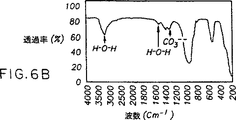
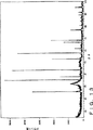
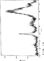
理想的な化学量論的結晶性ヒドロキシアパタイト(Ca10(PO4)6(OH)2)又はホスフェートに対するカルシウムの比率(Ca/P)が1.67である一般式(Ca5(PO4)3X)の化学量論的アパタイトとは異なり、骨無機質は非化学量論的アパタイト(リン灰石)である。その非化学量論は主に、三価のPO4 3-イオンを置換する二価のイオン(例えば、CO3 2-及びHPO4 2-)の存在のためである。HPO4 2-及びCO3 2-イオンによる置換は、Ca/P比率の変化をもたらし、Ca/P比率は年齢及び骨の位置により1.50〜1.70の間で変化することになる。一般に、Ca/P比率は骨の年齢とともに増加し、炭酸塩種の量が古い骨ほど増加することを示唆している。自然発生の骨無機質は、nmサイズのリン灰石系構造を有する不完全結晶性カルシウムホスフェートから成る。骨の不完全結晶性リン灰石系カルシウムホスフェートは、より結晶性のヒドロキシアパタイト(HA)や非化学量論的ヒドロキシアパタイトから図7に示すような特徴的XRDパターンによって区別できる。骨無機質の特定の可溶性性質をもたらすのは、ナノ結晶性粒径と不完全結晶性に連動するCa/P比率である。骨組織が、無機質を再吸収する細胞(破骨細胞)及び無機質を製造する細胞(造骨細胞)によって調節される一定の組織修復を経るため、これらの細胞活動の間の繊細な代謝のバランスを維持する際に、無機質の可溶性挙動は重要である。
合成骨移植材料は、自然の骨無機質によく似ているため、自然の骨の有用な置換材である。許容可能な合成骨は、自家組織の骨(患者自身の骨)の有効性及び取り入れの問題、並びに同種移植片骨(死体からの骨)に付随する危険性(例えば、ウィルスの伝送の危険)及び複雑性の問題を避けることができる。理想的な合成骨移植は、少なくとも以下の4つの性質を有していなければならない。:(1)それは、化学的に生物学的適合性でなければならない。;(2)それは、その周りの患者自身の骨が直るまで、移植物が定置にあり無傷であるように、構造上の完全性を提供するものでなけれなばらない。;(3)患者自身の骨が最終的に移植物に置き換わるように、それは再吸収可能でなければならない;及び(4)細胞及び/又は生体分子を合成骨材料に組み込むことが必要であるため、材料を形成するために用いられるプロセスの条件は、低温及び化学的に穏やかな条件であることが望ましい。また、同様の基準は、他の硬組織移植(例えば、軟骨)にとって重要である。
Ca/P比率、結晶の粒径、結晶度、多孔性、密度、熱安定性及び材料純度のようなパラメータが制御された材料によって、これらの基準を満たすことができる。インプラントとして用いるためにセラミック材料を合成する多くの努力が成されてきたが、化学式が骨内に自然に形成される無機質と非常に似ているため、合成ヒドロキシアパタイトが最も頻繁に使われてきた。LeGeros R.Z.,“Calcium Phosphates in Oral Biology and Medicine”, Karger Pub. Co., New York, 1991は、高温(800〜1200℃)で焼結することに続いて溶液沈降によって生じる高度に結晶性形態のヒドロキシアパタイトを教示している。高温処理することにより、結晶サイズが数ミクロン程度である、Ca/Pが1.67の高度に化学量論的なヒドロキシアパタイトが生じる。このような高度に結晶性のヒドロキシアパタイトは、本質的に生体内で再吸収性ではない。それは、生きている骨組織と置換されず、望ましくない長期間の間患者の中に無傷のまま残る。
骨置換材料を製造するこの他の多くのアプローチにおいて、結晶性カルシウムホスフェート反応物の固体酸性反応によって製造されるヒドロキシアパタイトが主に用いられてきた。これらのヒドロキシアパタイト骨置換材料は、不完全に反応し、均質でなく、顕著な結晶性ヒドロキシアパタイト含量を有することがしばしばである。
米国特許第4,880,610号においてConstantzは、高濃度のリン酸とカルシウム源を塩基の存在下で反応させてカルシウムホスフェート無機質を製造することについて報告している。その結果生じる生成物は、ヒドロキシアパタイト無機質の結晶性形態を含む多結晶材料である。同様に、Constantzらの米国特許第5,053,212号は、酸/塩基混合物の実行可能性と混合可能性を改善するために、粉末酸源の使用を開示しているが、米国特許第4,880,610号と同様の混合相カルシウムホスフェート材料を報告している。最近ConstantzらはScience(Vol. 267, pp. 1796-9(24 March, 1995))において、リン酸ナトリウム溶液中でモノカルシウムホスフェート一水和物、β-トリカルシウムホスフェート、α-トリカルシウムホスフェート及び炭酸カルシウムを反応させて炭酸塩化されたリン灰石を形成し、自然に生成する骨無機質よりも実質的に結晶性であるカルシウムホスフェート材料を提供することを報告している。
同様に、Brownらの米国再発行特許第33,221号は、結晶性テトラカルシウムホスフェート(Ca/Pが2.0)と酸性カルシウムホスフェートの反応について報告している。Liuらの米国特許第5,149,368号は、酸性のクエン酸塩と結晶性カルシウムホスフェート塩との反応を開示する。
多くのカルシウムホスフェート骨充填材及びセメントは「再吸収可能物」として記載されている。一般に、これらは、トリカルシウムホスフェート、テトラカルシウムホスフェート若しくはヒドロキシアパタイトを含む化合物又はこれらから誘導された化合物である。これらの材料はせいぜい弱再吸収性にすぎないと考えられる。これらの中で、トリカルシウムホスフェート化合物は最も再吸収可能であることが証明されてきたが、長年の研究の後にもそれらはまだ臨床場面で広く使われてはいない。トリカルシウムホスフェートは、たいへん長く予測不能の再吸収性(一般に再吸収のために1年以上の期間を必要とする)を有することは公知である。更に、非常に多孔性か溝をつけたサンプルを製造する段階をとらない限り、トリカルシウムホスフェートは骨と交換されない。最近、“TCPの生分解(HAP(ヒドロキシアパタイト)より高い)は充分ではない”と結論された(Berger et al., Biomaterials, 16:1241(1995))。テトラカルシウムホスフェート及びヒドロキシアパタイトから誘導された化合物もまた弱再吸収性である。テトラカルシウムホスフェート充填材は、30ヵ月後に再吸収が80%程度の長期間にわたる部分的再吸収性を示す(Horioglu et al., Soc. for Biomaterials, March 18-22, pg 198(1995))。18ヵ月後に、正面の副鼻洞に移植された微晶質ヒドロキシアパタイトの約30%が猫中に残っていた。
これらの引例の全ては、カルシウムホスフェートの結晶性固体を反応させることによって結晶性形態のヒドロキシアパタイト固体を生成する化学反応を開示する。非晶質カルシウムホスフェートはカルシウムホスフェート類の中で最も理解されていない固体であり、非晶質カルシウムホスフェートは伝統的に比較的不活性で非反応性固体であると考えられてきたので、反応物のうちの1つとして非晶質カルシウムホスフェート(Ca/Pが約1.5)を使用することはほとんど報告されていない。
非晶質カルシウムホスフェート(ACP)材料は、一般に高温処理により高度に結晶性のヒドロキシアパタイト化合物を形成するための直接の前駆体として用いられてきた。生理的な条件ではよく溶解しないので、このような高度に結晶性材料は合成骨のためには不適当である。Chowらの米国特許第5,525,148号は、多くの反応機構におけるACP前駆体の試験結果を報告しているが、ACP単独若しくはACP混合物を含む種々の混合物のスラリーは、設置セメントを形成しないし、効果的な再鉱化剤としても機能しないと述べている。
Brownらの米国再発行特許第33,221号は、特にテトラカルシウムホスフェート(Ca/Pが2.0)に限定された歯科用セメントのための非晶質相と、少なくとも一つのより酸性のカルシウムホスフェートを反応させて、結晶性ヒドロキシアパタイトを形成することを報告している。更に、Brownらはこのような非晶質テトラカルシウムホスフェートの製造法やその性質を開示していない。Tungの米国特許第5,037,639号は、歯の再無機質化のための標準の非晶質カルシウムホスフェートペーストの使用法及び用途を開示している。Tungは、チューインガム、ロリンス又は練り歯磨きで混合又はこれらを通して供給される標準不活性非晶質カルシウムホスフェートを使用し、経口流体を入れると、歯エナメルを再無機質化するために有用であるヒドロキシアパタイトを含む結晶性フッ化物に変換することを提案している。SimkissのPCT出願GB93/01519は、非晶質カルシウムホスフェートと混合して生組織に移植されるマグネシウムイオン又は二リン酸塩のような抑制剤の使用法を記載している。例えばマグネシウムイオンが周囲の体液へ浸出すると、非晶質のカルシウム-マグネシウムホスフェートはより結晶性形態に変わる。
硬組織内の自然発生無機質の性質により近似する性質を有する新しい合成材料を開発する必要がある。特に、低温で形成することが可能で、nmサイズの結晶を有する不完全結晶性である、完全に生体再吸収可能である合成骨材料を提供する必要がある。
発明の概要
この発明は、生物学的適合性であり、生体再吸収可能であって、長期間室温で使用可能な、バイオ活性セラミック材料を提供する。このバイオ活性セラミック材料は、低温で形成されてもよく、直ちに形成可能及び/又は注入可能であり、更なる反応により高強度に硬化することができる。このバイオ活性セラミック材料は、自然に生じる骨無機質に匹敵するCa/P比率、並びに自然の骨と同様の硬度及び破砕強さを有する不完全結晶性リン灰石系カルシウムホスフェート固体を含む。このバイオ活性セラミック複合物は強度に生体再吸収可能であり、その生体吸収性及び反応性を特定の療法及び/又はインプラント位置の要求に応ずるように調整することができる。
この材料は、骨プレート、骨スクリュー及びその他の治具並びに獣医用途を含む医療用器具として製造することができる。それは強生体再吸収性であり及び/又は骨化するものである。これら又はこの他の本発明は、カルシウムホスフェートの水和前駆体及びペースト又はパテを形成するためにカルシウムホスフェートを水和するのに十分な量の水性液体から成り、この水和前駆体の硬化が吸熱反応に関連することを特徴とする、自硬化の生体セラミック組成物である。また本発明は、非晶質カルシウムホスフェート及びペースト又はパテを形成するためにカルシウムホスフェートを水和するのに十分な量の水性液体から成る水和前駆体を含み、この水和前駆体の硬化が10分より長時間で起こることを特徴とする、自硬化の生体セラミック組成物である。
別の観点から見ると本発明は、非晶質カルシウムホスフェート及びペースト又はパテを形成するためにカルシウムホスフェートを水和するのに十分な量の水性液体から成る水和前駆体の硬化を促進することによって製造された不完全結晶性カルシウムホスフェートを含み、その硬化が吸熱反応及び非晶質カルシウムホスフェートから不完全結晶性カルシウムホスフェートへの変換に関連する、生体セラミック組成物である。
本発明の生体セラミック組成物を、(a)非晶質カルシウムホスフェート、(b)促進剤、及び(c)ペースト又はパテを形成するのに十分な量内の水性液体、をいかなる順序であっても混合して製造してもよく、このペースト又はパテは不完全結晶性リン灰石系カルシウムホスフェートに変換され、この変換は吸熱反応においてこのペーストの硬化と関連する。
定義
ここで用いられる「非晶質」とは、顕著に非晶質の特徴を有することを意味する。顕著な非晶質は、非晶質含量が75%以上、好ましくは90%以上であることを特徴とし、広く特色のないX線回折パターンによって特徴づけられる。小程度の結晶度がその材料の中に存在してもよいことが認識される。しかし、この発明の非晶質前駆体材料の結晶度は生成物材料において要求される結晶度未満であることが好ましい。
「バイオ活性」とは、インプラントの中又はそれについて硬組織形成を促進することに関する。軟組織に移植する場合、このバイオ活性は成長又は栄養因子の存在を必要とするか、又はインプラントに硬組織形成細胞型の種まきをを必要とする。
ここで用いられる「生物学的適合性」とは、そのホストに実質的な有害な反応を誘導しないことを意味する。異物が生体に導入された場合に、その異物が例えばホストに負の影響を及ぼす炎症性反応のような免疫反応を促進することには常に関心がある。例えば、ヒドロキシアパタイトは一般に「生物学的適合性」であると考えられているにもかかわらず、結晶性ヒドロキシアパタイトミクロキャリアーが動物の筋肉内に挿入された場合に、顕著な炎症及び組織壊死が観察された(例えば、Ijntema et al., Int. J. Pharm 112:215(1994)を参照のこと)。
「生体再吸収」とは、生体内で再吸収されることに関する。「充分な」再吸収とは、細胞外断片が顕著には残らないことを意味する。再吸収プロセスは、体液、酵素又は細胞の活動を通してオリジナルのインプラント材料が除去されることを含む。再吸収されたカルシウムホスフェートは、例えば、骨無機質として再沈澱するか、その体内の他で再利用されるか、又は排出されてもよい。「強生体再吸収性」とは、筋肉内又は皮下に移植されたインプラント材料の全量の少なくとも80%、好ましくは95〜99%、最も好ましくは99%以上が1年以内で再吸収されることをいう。本発明の好ましい実施態様において、強く再吸収している不完全結晶性リン灰石系(PCA)カルシウムホスフェートは、少なくとも1g(好ましくは1〜5g)のPCA材料が筋肉内又は皮下に移植された場合に、その材料の少なくとも80%が1年以内に再吸収されることを特徴とする。より好ましい実施態様において、この材料は、9ヵ月、6ヵ月、3ヵ月又は理想的には1ヵ月以内に再吸収される。更に、特に好ましい材料は、それらが定められた期間内に充分に再吸収され得ることを特徴とする。また「弱い」再吸収とは、出発材料の80%未満が1年以後に再吸収されることをいう。
補助材料の「効果的な量」とは、所望の機械的又は化学的性質をその複合物に付与するのに十分な量である。
「硬化」とは、水和された前駆体が硬化PCA材料に変わるプロセスをいう。このPCA材料は、それが実質的に形成可能でない固体である場合に「硬化された」とみなされる。このような硬化されたPCA材料は、最小の圧縮性を有し、弾性変形に対して可塑性である傾向がある。
ここで用いられる「水和された前駆体」とは、わずかな量の水性溶液(即ち、1gの前駆体粉末に対して水性溶液が1mL以下)の存在下で乾燥前駆体を水和することにより形成されるペースト又はパテのことをいう。この水和された前駆体は、この変換の進行程度によって、多様な組合せの反応物及び製造物の両方を含んでもよい。「注入可能な」及び「形成可能な」前駆体ペーストは水和された前駆体である。好ましい「注入可能な」水和された前駆体は、18ゲージ針を通して供給するのに適した粘稠度を有する。
ここで用いられる「不完全結晶性リン灰石系カルシウムホスフェート」、「PCAカルシウムホスフェート」及び「PCA材料」とは、合成不完全結晶性リン灰石系カルシウムホスフェートのことをいう。このPCA材料は、それが特徴的なXRD及びFTIRパターンを有する限り、必ずしも単一のカルシウムホスフェート相に限定されるわけではない。PCAカルシウムホスフェートは、実質的に骨と同じX線回折スペクトルを有する。このスペクトルは一般に20〜35度の範囲における2つのみのブロードなピークにより特徴づけられ、その一方のピークは26度、もう一方のピークは32度にある。更にそれは、563cm-1、1034cm-1、1638cm-1及び3432cm-1(±2cm-1)のFTIRピークによって特徴づけられる。更に1422cm-1及び1457cm-1に最大値がある二重ピークを有する603cm-1及び875cm-1の鋭い肩が観察される。
ここで用いられる「促進剤」とは、水和された前駆体の硬化を促進し、ACPをPCAカルシウムホスフェートに変換することを促進する材料又は処理をいう。いくつかの促進剤はこの変換に関与し、生成物であるPCA材料に組み込まれる。「消極的」促進剤として知られているこの他の促進剤は関与しない。
「反応性」とは、水和された前駆体を形成するために液体と混合する場合に、前駆体材料の硬化に関連する促進剤の存在下で、非晶質カルシウムホスフェートがこの発明のPCA材料への変換を行う能力をいう。好ましいACPは、完全に変換する能力、急速に硬化を伴い変換する能力、それ以外には不活性な化合物と共に変換を行う能力、及び/又は実質的に均一なPCA材料に変換する能力によって特徴づけられる。このACPが第2のカルシウムホスフェートと反応する場合、この「変換」は、ACP及び第2のカルシウムホスフェートの両方の変換をも含む。硬化の程度及び硬化プロセスの速度もまた反応性の重要な要素である。いくつかのACPは他より反応性である。例えば100μm以上の顕著な粒子部分を含む粒径分布を有するジカルシウムホスフェート二水和物(DCPD)のような、弱い促進剤の存在下でPCA材料への変換及び硬化が行われる場合には、ACPは「高度に反応性」とみなされる。好ましい高度に反応性のACPは、弱促進性DCPD及び水の存在下で37℃において12時間以内に硬化されたPCA材料を製造し、この硬化は約1〜5時間及び理想的には10〜30分で実質的に完了する。
【図面の簡単な説明】
図1は、比較的はっきりしない境界を有し部分的に無形状に浸漬された(矢印)クラスター内のnmサイズの粒子を示す反応性非晶質カルシウムホスフェートの高解像度の透過電子放射線写真である。
図2は、Ca/Pが1.58になるような真空加熱手順の後のこの発明の反応性非晶質カルシウムホスフェートのエネルギー分散電子マイクロプローブスペクトルである。
図3は、この発明の非晶質カルシウムホスフェートから誘導された不完全結晶性リン灰石系カルシウムホスフェート生成物の溶解曲線を、結晶性ヒドロキシアパタイトと比べたものである。37℃の溶液に解離するカルシウムイオンの量を測定すると、より結晶性形態のヒドロキシアパタイトに比べて、この発明の材料の溶解度が高いことに注目されたい。
図4は、(a)反応性非晶質カルシウムホスフェート及び(b)本発明の骨置換材料を形成する反応に用いられるジカルシウム二リン酸塩のX線回折パターンである。
図5a〜dは、反応性非晶質カルシウムホスフェート及びジカルシウム二リン酸塩の混合物が反応してこの発明のPCA材料を形成する進行を追跡したX線回折パターンである。
図6は、(a)ジカルシウムホスフェート二水和物、(b)本発明の活性化されたACP、及び(c)この発明のPCA材料の赤外線のスペクトルである。
図7は、自然に生じる骨のX線回折パターンである。
図8は、実施例10に記載されている多様な調合物の粒子径分布を示す棒グラフである。
図9は、(9a)無治療又は(9b)この発明の供給ビヒクルで治療された、脛骨の欠陥の顕微鏡写真を示す。図9aにおいて、小さい矢印は欠陥の1つのエッジを示し、大きい矢の先端は架橋されていない欠陥を示す。図9bにおいて、大きい矢の先端は欠陥の1つのエッジを示す。両方の図において、拡大率は4倍であり、骨は脱石灰されており、スライドはヘマトキシリン及びエオシンで治療されている。
図10は、この発明のPCA材料で治療された欠陥に成長した犬の小柱の骨の顕微鏡写真である(拡大率10倍;脱石灰;ヘマトキシリン及びエオシン)。
図11は、この発明のPCA材料で治療された犬の円錐の骨欠陥の顕微鏡写真である(拡大率4倍;脱石灰されていない;薄緑色の塩基性フクシン)。
図12は、(図12a)無治療及び(図12b)手術後4週間目に治療されたウサギ脛骨欠陥の顕微鏡写真を示す(拡大率4倍;脱石灰;マッソン三色染色法)。
図13は、A1203消極的促進剤から製造されるPCAカルシウムホスフェートのX線回折パターンである(A1203のピークは線で示されている)。
図14は、実施例1-2の記載のように製造されたPCAカルシウムホスフェートのX線回折パターンである。
図15は、実施例1-4の記載のように製造されたPCAカルシウムホスフェートのX線回折パターンである。
図16は、反応性ACPとDCPDの反応が吸熱性であることを示す示差走査熱量計(DSC)プロット線である。
図17は、(図17a)熱処理前、及び(図17b)熱処理後の非晶質カルシウムホスフェート材料の赤外線のスペクトルである。
図18は、本発明のPCAカルシウムホスフェートの広幅XRDである。
図19は、癒着不能の骨部位に導入される本発明のインプラントを示す。
図20は、砕けた骨部位に導入される本発明のインプラントを示す。
図21は、骨粗しょう症の骨部位に導入される本発明のインプラントを示す。
図22は、骨粗しょう症の脊椎骨に注入器により導入されるインプラントを示す。
図23は、腰プロステーゼを固定するために骨セメントとして用いられる本発明のインプラントを示す。
図24は、歯ソケットに導入される本発明のインプラントを示す。
図25は顎堤に導入される本発明のインプラントを示す。
図26は、この発明のPCAカルシウムホスフェートで治療された犬の歯ソケット欠陥の顕微鏡写真である(拡大率4倍;脱石灰されていない;薄緑色の塩基性フクシン)。
図27は、人の死体の骨粗しょう症の脊骨の示している配置のX線写真であり、PCAカルシウムホスフェートの注入前の針の配置を示す。
図28は、PCAカルシウムホスフェートの(図28a)注入前、及び(図28b)注入後の人の死体の脊椎骨の平面図の写真である。
発明の詳細な説明
この発明は、再吸収性整形外科及び歯科用の治具の製作を含む硬組織の修復及び成長促進に用いるために適した生物学的適合性セラミック組成物に関する。この組成物は、しばしば適切な生物学的適合性マトリックス又は添加物と結合した、生物学的適合性及び高度に生体再吸収性の不完全結晶性リン灰石系カルシウムホスフェート(PCAカルシウムホスフェート)を含む。このPCAカルシウムホスフェートは、歯科、整形外科、薬供給、細胞療法、及びその他の治療用用途に用いることができる。
この発明の組成物を、補てつ器具の骨に接触している表面に骨セメントとして適用してもよい。それを充填材として直接骨欠陥に適用してもよく、それは新しい骨組織の成長を促進することができる。この組成物を、同様に、軟骨組織の修復、成長又は製造のために適用してもよい。またこの組成物を、適当な状況下で再吸収されて骨又は軟骨と交換されるスクリューやプレートのような治具又は器具を組み立てるために用いてもよい。またこの組成物を軟組織中に遊離放置して用いてもよい。薬学的に活性な試薬をこの組成物に加えると、それは薬供給器具として機能し、移植後長期間にわたってこの組成物がゆっくり生分解するに従ってこの試薬を解放する。
本発明はまた、予想どうり硬化するペースト又はパテの形態で、制御された方法で、ACPからPCAカルシウムホスフェートへの変換を促進するための方法を提供する。
本発明のPCAカルシウムホスフェート生体セラミックは一般的に、ヒドロキシアパタイトのための理想的な化学量論的カルシウム/ホスフェート比率である約1.67と比較すると、その比率が1.5未満でありカルシウムが不十分である。それらは、その生物学的生体再吸収性及び最小の結晶度によって更に特徴づけられる。それらは、速生体再吸収性、高多孔性及び/若しくは低密度であってもよく、又は遅生体再吸収性、低多孔性及び/若しくは高密度であってもよい。それらの結晶性の特徴は、この技術分野で公知の骨置換材料で見られるようなより高度の結晶度を有せず、実質的に自然の骨と同じである。この発明のPCAカルシウムホスフェートはまた生物学的適合性であり、即ち、移植された材料によってホストに顕著な有害な反応(例えば、炎症又は線維症)が引き起こされることはない。炎症又は線維症が医学的に許容範囲内であることをもたらす材料は、生物学的適合性であると考えられる。このPCAカルシウムホスフェートを湿潤前駆体形態(即ち、水和された前駆体)で用いてもよく、破砕のような手術部位にセメントとして直接適用してもよく、またそれを生体外で硬化させその後移植してもよい。
この発明のPCAカルシウムホスフェートの再吸収性は、密度、多孔性、化学組成及び結晶構造の組合せに起因している。リン灰石の低結晶度は、この他のより結晶性の種と比較すると、水性系に対する幾分高い溶解度と関連している。そのためこの発明のPCAカルシウムホスフェートの低い結晶度及び/又は安定した非晶質のリン灰石系ドメインの存在が、生物学的系におけるその再吸収性と関連していると思われる。多孔性は、細胞の生体セラミックマトリックスへの浸透、及び細胞プロセスをマトリックス内部への/からの物質の分散を促進する。従って、多孔性の低いPCAカルシウムホスフェート組成物は、多孔性の高いPCAカルシウムホスフェート組成物よりゆっくりと生体内に再吸収される。ある実施態様において、制御された粒子径を有する反応物を用いると、制御された多孔性を有するPCAカルシウムホスフェート材料が得られる。化学的又は物理的なエッチング及び浸出のような、多孔性を促進するその他の方法を用いてもよい。
この発明のPCAカルシウムホスフェートを、1年(弱再吸収性ヒドロキシアパタイト骨充填材及びこの技術分野で公知の骨代替材では典型的な値である。)以上の遅い再吸収時間から、1〜2ヵ月に数グラム(例えば、1〜5g)の速い再吸収率まで、いろいろな再吸収率を有するように製作してもよい。したがって、密度、多孔性、使われる反応物、反応生成物の最終の結晶度、及び用いられる結晶化抑制剤の量によって、1グラムの器具が2週間から1月、3月又は6月更に1年、2年又は3年までの所望の期間内に充分に再吸収するように調合物を製造することができる。本発明の強再吸収性PCAカルシウムホスフェートは、出発材料の80%以上(好ましくは95〜99%、より好ましくは99%以上)又はその少なくとも1g(好ましくは、1〜5g)が1年以内、好ましくは6ヵ月以内、より好ましくは3ヵ月以内、最も好ましくは1〜2ヵ月以内に再吸収されるような生体内再吸収率を有する。
負荷状況で新しい骨を作る場合、約6〜8週間で充分に再吸収されて骨と交換されるようにすると、12週間までに組織構造的に通常の骨をもたらすことがわかった。いくつかの負荷状況においては、再吸収をよりゆっくり行うことが望ましい場合もある。さらに、硬組織が異所で製造されるか、又は既存の硬組織の形を拡大する場合には、よりゆっくり再吸収するPCAカルシウムホスフェートを用いることが望ましいかもしれない。
生成するPCAカルシウムホスフェートの密度又は多孔性を調整すること、又は固化の速度及び硬さに影響を及ぼす反応パラメータを用いることはすべて、この発明のPCAカルシウムホスフェートの生体内再吸収時間を変化させるための有用なアプローチである。特定の用途に必要なように、これらのパラメータを単独又は組合せて調節してもよい。
高密度、低多孔性PCAカルシウムホスフェート並びに/又は急速反応及び急速硬化時間を用いる場合には、遅い再吸収(3ヵ月以上)が好ましい。低密度、高多孔性PCAカルシウムホスフェート並びに/又は遅い反応及び遅い硬化時間を用いる場合には、速い再吸収(3月以下)が好ましい。PCAカルシウムホスフェートを形成する反応速度及び反応性を調整する指針はこの明細書中にどこかに記載されている。生体内で異なる再吸収性速度を有する硬化PCAカルシウムホスフェートセメントをもたらす好ましいPCAカルシウムホスフェート前駆体の製造法を以下に記載する。
速再吸収のPCAカルシウムホスフェートは、100μmより大きい粒径の粒子をかなりの含量で含む粒径分布(例えば、表3の分布B1)を有するDCPDを促進剤として使用して、実施例5の高度に反応性ACPを変換することにより得られる。この粉末は、実施例8に記載のように水和された前駆体として製造される。
遅再吸収のPCAカルシウムホスフェートは、100μmより大きい粒径の粒子を最少含量で含む粒径分布(例えば、表3の分布B3)を有するDCPDを促進剤として使用して、実施例5の高度に反応性ACPを変換することにより得られる。この粉末は、実施例9に記載のように水和された前駆体として製造される。
この発明のPCAカルシウムホスフェートは骨形成を行う。この骨形成とは、移植された合成カルシウムホスフェートを、組織構造的に自然の骨と全く同じ又はそれに類似した骨で置換することをいう。この発明のPCAカルシウムホスフェートの骨形成は、より自然らしい組織が確立する前に現れるより未組織の骨の段階で起きる傾向がある。この発明のPCAカルシウムホスフェートは、骨形成がインプラントの外端のみで起こるわけではないので、従前の骨充填材及びセメントとは異なり、おそらく再吸収プロセスと同時にインプラントの全体にわたってその骨形成を開始する。脛骨又は橈骨のような負荷領域へのPCA材料の移植後2〜3週以内に、無機質化された類骨形成(針状体)の小さい発生点の形成により、予備的骨形成が観察される。4週間までに、この針状体はレース状外観の薄い海綿状の小柱骨及び薄い皮質性骨に変わる。6週間目に、小孔を含む骨細胞を有する規則正しい正常又は厚いコンパクトな皮質性骨が観察される。6週間以後に最終的な改造が生じ、12週間までには新しく骨化された骨が自分の骨と見分けがつかなくなる。
したがって、PCAカルシウムホスフェートの存在下での骨形成は一般に完成に達し、通常の骨成長より速く生じるように見える。骨形成のこの急速な速度は、この発明のPCAカルシウムにホスフェートが骨の治癒を促進していることを示唆する。新しい骨は早くも2週間で観察され、6週間以内(いかなる場合においても3〜6ヵ月まで)に組織構造的に充分に構成される状態に達することができる。最高3gの水和された前駆体のインプラントを用いた羊分節性骨折破砕モデルにおいて、骨折していない骨の強さの100%を有する骨が3ヵ月以内に観察された。骨形態形成タンパク質のような栄養又は成長因子の存在下においては、このプロセスはより促進される。
好ましい実施態様において、骨形成を最適化するために、本発明の器具、ペースト及びパテに骨形成細胞の種まきをしてもよい。これは、その器具(PCAカルシウムホスフェート又は水和されたその前駆体を含む)を患者自身の骨形成細胞の源と接触させることにより最も容易に行うことができる。このような細胞は骨に付随する血又は液体中に見出される。これには、骨又は骨材料若しくは領域に接触した外因性流体が含まれ、また骨膜、皮質性骨、海綿状骨又は髄が含まれる。それらはまた皮質性若しくは海綿状骨、骨髄又は骨膜を含む組織中に存在する。スクリュー及びピンのような器具の場合、それを骨へ導入すると出血するので、更なる種まきは必要ではない。皮質性の骨のみに対するプレートの場合には、その器具に接触する骨膜の障害の誘導が推奨される。また他の実施態様において、インプラント部位の円錐形の骨の一部を除去することによって外科的に骨内に台座を製造することは有用である。また骨形成を促すために、患者から採取した骨形成細胞を移植物に導入したり、その器具の中又は上へ栄養因子又は骨成長誘導タンパク質を編入することなど、この他の手段を用いてもよい。骨形成細胞に対するホストの拒絶が起きる前に所望量の骨の再生が生じるならば、自家組織でない骨細胞もまた本発明の範囲内である。したがって、一次源、細胞線又は細胞バンクから得られる細胞又は組織を一定の実施態様において用いてもよい。同様の考え方を、軟骨形成及び治癒並びに軟骨細胞及び/又はその他の軟骨形成細胞をこの発明のPCAカルシウムホスフェートに種まきするために応用できる。
この発明のPCAカルシウムホスフェートの好ましい調合物を製造するために用いる反応の性質のために、外科手術固化におけるインプラント材料としての使い易さは、この技術分野で公知の他の骨置換材料よりも顕著に改善されている。特に、反応が体外で開始して、ゆっくり室温で進行し、それによってその材料が固化して外科手術部位に用いる前に使えなくなる可能性を最小にする。この反応は体温度で顕著に速まり、その材料はその場所で硬化する。更に、治療上の必要性に従って2、3の簡単なパラメータを変えることによって、発明のPCAカルシウムホスフェートの粘稠度及び形成性並びに反応速度を変えることができる。
PCAカルシウムホスフェートの製造 多くの非晶質カルシウムホスフェートは、時間とともにより結晶性形態に自然発生的に変わる傾向がある。ヒドロキシアパタイトは、カルシウムホスフェートの熱力学的に有利な形態であって、たびたびこのような変換の生成物である。本発明は、制御の下にACPをより結晶性形態(例えば、PCAカルシウムホスフェート)に、更に顕著な結晶化を生ぜずに、変換するという価値を認識した(特に、この変換がわずかな量の水の存在下で行われ硬化反応を伴う場合)。本発明は、PCAカルシウムホスフェートの形成をもたらす反応を提供する。これらの反応は、有利には、ペースト又はパテの粘稠度を有する前駆体を使用して、体外で開始され、37℃で顕著に促進し硬化されたカルシウムホスフェート生成物を生じてもよい。いくつかの実施態様において、硬化されたPCAカルシウムホスフェートは従来の黒板チョークと同様の硬度及びモジュラスを有する。いくつかの例では、硬化されたPCA材料は、未反応前駆体、促進剤、及び/又は補助材料、副生成物等の存在を伴う。
本発明の方法によれば、ペースト又はパテ状の水和された前駆体は、水の添加によってカルシウムホスフェート前駆体を形成する。次に水和された前駆体は約37℃まで加熱され、図16の示差走査熱量計(DSC)データにより示されるように、ペースト又はパテの硬化によって特徴づけられる実質的に正味の吸熱反応を開始する。好ましい実施態様において、PCAカルシウムホスフェート材料は、促進剤の存在下で反応性非晶質カルシウムホスフェートをPCAカルシウムホスフェートへ変換することにより、水和された前駆体から製造される。吸熱反応によるペーストの硬化を伴う、ペースト形態のACPからよく結晶化したヒドロキシアパタイトへの変換を促進することは、本発明の範囲内である。
吸熱的に固化する骨セメントは、公知技術であるカルシウムホスフェート骨セメント及び充填材にに比べていくつかの重要な利点を有する。反応が熱を放出しないので、インプラント領域の細胞及び組織に対する熱に関連した損傷の危険がない。さらに、反応の吸熱性質は、反応の進行をその反応を支えることができる熱量を調節することによって制御することができることを意味する。水和された前駆体は、室温以下ではわずかしか反応しない。このことは、この技術分野で公知の外科手術用セメント及び充填材に付随する取扱い上の問題の多くが避けられることを意味する。
好ましい実施態様において、反応物は体外で混合され、外科手術部位に適用するのに適した水和されたPCAカルシウムホスフェート前駆体材料を生じる。いくつかの実施態様においてはこの反応は生体外で完了するにもかかわらず、外科手術部位に適用した後一般にこの反応は完了する。本発明のPCAカルシウムホスフェート反応は、一般に5時間以内に水和された前駆体の硬化に至り、生理的な条件下では約1〜5時間、好ましくは約10〜30分で実質的に硬化する。好ましい実施態様内において、この反応は厚いペーストを形成するために生理食塩水を2つの乾燥成分の混合物に加えることによって開始し、37℃における約30分の吸熱反応に伴って硬化する。水、緩衝液、漿液又は組織培養媒体のようなこの他の水性試薬をこの食塩水の代わりに使ってもよいが、これらに限定されるわけではない。
いかなる反応機構においても、変換前にこのACPが顕著な非晶質の特徴を保持することは重要である。特に、出発ACP内の全体の結晶度は最終製品において要求される結晶度それを上回ることができない。したがって、一定の反応機構において、反応全期間にわたってこのACPの非晶質の性質が安定することを必要としてもよい。この技術分野で公知の結晶形成に対する適切な抑制剤の例には、炭酸塩、ピロリン酸塩及びマグネシウムがある。結晶化の抑制剤の使用のための追加的ガイダンスには、Elliot, Structure and Chemistry of the Apatites and Other Calcium Orthophosphates, Elsevier, The Netherlands, 1994があるので、参照されたい。
促進剤のタイプ 促進剤の目的は、水和された前駆体の硬化を促進することであり、好ましくはACPからPCAカルシウムホスフェートへの変換を促進することである。この目的にかなういかなる材料又は方法も、この反応の範囲内であると考えられる。これは、変換を起こさずに硬化が起きる場合、即ちPCAカルシウムホスフェート前駆体を出発材料として用いる場合のような限られたケースを含む。
ACPの変換に関して、促進剤は全体の反応又は変換又は硬化プロセスに含まれるいかなる中間反応をも促進することができる。これについて、好ましい促進剤は、変換又は硬化プロセスの一以上の特定段階の活性化エネルギーを下げる。
反応性ACPをこの発明のPCAカルシウムホスフェートに変換するために用いる促進剤自体は、PCAカルシウムホスフェートカルシウムホスフェートに変換されてもよく、そうでなければ変換過程において化学的若しくは物理的反応に関与してもよい。このような促進剤を、ここでは「関与性」促進剤と呼ぶ。
一方、ある促進剤は、この反応性PCAが核形成及び硬化を触媒作用するか又は開始若しくは促進する役割をする際に、実質的に不変のままであってもよい。これらの促進剤を「消極的」促進剤と呼ぶ。
熱、圧力、反応性ガス、溶媒、イオン性溶液又は放射性薬品の使用のようなこの他の手段を用いることにより硬化及び反応性ACPからPCAカルシウムホスフェートへの変換を促進することもまた本発明の範囲内であるとみなされる。このような促進手段は、反応強化又は「強化」促進剤と呼ばれる。
促進剤は、ACPから硬化されたPCAカルシウムホスフェートを製造することを促進する場合に異なる能力又は強度を有してもよい。同様に、全てのACPが等しく反応性であるとは限らない。したがって、弱い促進剤は低反応性でACPと反応する際に常に効果的であるとは限らない。このような状況においては、より強い促進剤が好ましい。促進剤の強度は、実施例8に記載の方法を用いて、熱で活性化された高度に反応性形態又はその熱で活性化されない形態の両方において、本発明の好ましい炭酸塩化ACPに対する所与の促進剤の反応性を比較することによって簡便に試験することができる。反応物を手で攪拌することは、高度に反応性の促進剤を識別するために特に適している。低反応性の促進剤は、実施例9に記載のような自動化されたミルで攪拌すると有利である。これらの方法を用いることにより、実施例10のB1の粒径分布を有するDCPDは弱い促進剤であることが示され、粒径が100μm未満であるものは強反応促進剤であることがわかった。
特定の促進剤を所与のACPに整合させるための上記のガイダンスに加えて、このような整合は、経験的に、約1.0mL水/1g粉末の存在下で所与のACPを選択された促進剤と混合し、この混合物を湿潤環境で37℃に加熱することによって行ってもよい。適切な促進剤は、これらの条件下でPCAカルシウムホスフェートの形成及びペースト硬化を示す。
促進剤及び/又はACPの製造方法は、水和された前駆体がPCA材料に変換される容易さに影響を及ぼす。上記のように、粉末の反応物を液体の添加の前に混合する方法は、系の反応性に影響を及ぼす。したがって、乳鉢及び乳棒を使用する手混合は、反応物粉末の機械による長時間の粉砕と同じ反応性の系をもたらさない。したがって、促進剤を比較する場合には、標準化された製造条件を用いることが重要である。
ACPから反応性PCAカルシウムホスフェートへの変換に付随する硬化は表面で触媒作用された現象であると仮定される。もしそうならば、再生可能な表面を有する促進剤を製造することが望ましい。ACP及び促進剤粉末の比表面積を制御して、反応条件及び最終的なPCA材料性能を制御することができる。したがって、反応再現性を制御するために、公知の粒径分布を有する促進剤を提供することは有利である。標準ふるい技術が、特定の粒径を選択するために適している。比表面積は、PCA材料の耐圧強度並びにおそらく多孔性及び再吸収性と相関していることが示された。
多くのカルシウム又はホスフェートを含む化合物を硬化反応の関与性促進剤として用いることができる。カルシウムホスフェート促進剤はいかなる結晶性構造ものであってもよく、直接又は促進剤を用いるかのいずれにおいてもACPと反応するように選択されなければならない。好ましい関与性促進剤は、中間体のPCAカルシウムホスフェート相を通じて、それ自体をヒドロキシアパタイトへ変換する。
適当な関与性カルシウムホスフェート促進剤は中性、塩基性及び酸性のカルシウムホスフェート、好ましくはリン灰石系ホスフェートを含み、それはリン灰石系カルシウムホスフェートを得る反応のために適当な化学量論を提供する。適切なカルシウムホスフェート促進剤には、カルシウムメタリン酸塩、ジカルシウムホスフェート二水和物、モネタイト、ヘプタカルシウムホスフェート、トリカルシウムホスフェート、カルシウムピロリン酸塩二水和物、ヒドロキシアパタイト、不完全結晶性リン灰石系カルシウムホスフェート、テトラカルシウムホスフェート、カルシウムピロリン酸塩、オクタカルシウムホスフェート、及び二級ACPが含まれるが、これらに制限されるわけではない。CaO、CaCO3、カルシウムアセテート及びH3PO4のような(これらは単なる例示にすぎない)ホスフェート又はカルシウムの他の源を混合して自然の骨が所望のCa/P比率となるような最終生成物を形成してもよい。また、非晶質又は不完全結晶性の第2の成分を加えてもよい。
好ましい実施態様において、粒径が200μm未満、より好ましくは95μm未満、最も好ましくは平均粒径が約35〜45μmであって、最大粒径が約110μm未満である関与性促進剤としてDCPDが用いられる。
非晶質カルシウムホスフェートをこの発明のPCAカルシウムホスフェートを製造するための単一の前駆体として用いる場合、ACPを高度に結晶性ヒドロキシアパタイトに変換する自然の傾向を制御することが重要である。他方、転換及び硬化の速度は、外科手術用途用に十分な程速くなければならない。1つの方法は、結晶形成の抑制剤を含むACP前駆体(例えば、実施例5のACP)を結晶形成の抑制剤を含まないACP(例えば、促進剤)と結合することである。その反応物を、適当な粒径であって、抑制剤を含むACPの過剰量と混合してもよい。次にこの反応物を水を添加するような結晶を形成する条件にさらし、加温(例えば、体内に入れた場合に起こるような)を行い、この反応物を本発明のPCAカルシウムホスフェートに変換する。更にこの反応を促進するような段階をとらない限り、単独の促進剤としてACPを使用すると、例外的に良く硬化する傾向にないPCAカルシウムホスフェートをもたらす。
関与性促進剤がACPとともに同様にPCAカルシウムホスフェートに変換されることは、この発明の反応の興味深くかつ予想外の特徴である。これは、DCPD及び化学量論的ヒドロキシアパタイトにおいて実験的に証明された。したがって、結晶性カルシウムホスフェートが実質的に吸熱反応でより結晶性でない状態へ変換することがまず示された。
ACPからPCAカルシウムホスフェートへの変換が上記のように証明された一方で、その代替材料がまたPCAカルシウムホスフェートに変換されうることも認識される。したがって、37℃での正味の吸熱反応によりわずかな量の水の存在下で結晶性カルシウムホスフェート(PCAカルシウムホスフェートを含むこと)からの水和された前駆体ペーストを製造し、それに伴いペーストが硬化することは本発明の範囲内であると考えられる。このアプローチの好ましい実施態様において、PCAカルシウムホスフェート及びDCPDは、PCAカルシウムホスフェート生体セラミックを製造するため反応物としての特徴を有する。ヒドロキシアパタイトは、カルシウムホスフェートの熱力学的に有利な形態である。
それゆえ、それ自体はPCAカルシウムホスフェート(又はヒドロキシアパタイト)に変わらない促進剤を用いて、水和された前駆体の硬化を伴って、反応性ACPからPCAカルシウムホスフェートへの変換を促進することもまた本発明の範囲内である。適当なこのような促進剤は「消極的」と呼ばれ、核形成を起こす物質及び触媒を含むが、これに限定されるわけではない。不完全結晶性リン灰石系カルシウムホスフェートを製造するためにリン灰石系結晶化を弱くしか促進しない反応性表面を提供する物質は、この観点から特に適している。
ある観点では、本発明は、ACPを水和させるために用いられる水性液体に不溶又はあまり溶解しない消極的促進剤を用いることを特徴とする。適切な促進剤には、金属、金属酸化物、セラミックス、シリケート、糖、塩又は高分子粒子があるが、これらに限定されるわけではない。多くの用途において、好ましい促進剤はそれ自体で生物分解性である。一般に、これらの物質は粒径が1〜500μm、好ましくは1〜300μm、最も好ましくは1〜200μmの顆粒状である。用いられる実際の粒径は、特定の物質の反応促進性を改良するために変更されてもよい。
実施例3の表2は、わずかな容積の水の存在下でのACPからPCAカルシウムホスフェートへの変換における種々の消極的促進剤の効果を示す。一般に、この促進剤はがACPの量以下で存在し、特にACP:促進剤は約1:1〜約5:1である。2つの乾燥成分の全重量にほぼ等しい量の水が(ここで、水の密度は1であるのでこの重量は容積に等しい。)ペーストを製造するために用いられる。ACP、促進剤及び水の実際の割合は、成分の量を変化させ、処方を選択して成分を混合することにより、簡便に決定することが可能であり、37℃において所望の時間内に硬化されたPCAカルシウムホスフェートをもたらす。好ましい消極的促進剤には、顆粒状のSiO2、マイカ、Al2O3、ポリ(L-ラクチド)(PLLA)、ポリグリコリド(PGA)及びポリ(ラクチドコグリコシド)(PLGA)共重合体があるが、これらに限定されるわけではない。
最後に適当な強化促進剤には、水、熱、塩及び追加のカルシウムホスフェート源があるが、これらに限定されるわけではない。一般にこれらの物質は、ACPと第2のPCAカルシウムホスフェートとの反応を促進し、ACPからPCAカルシウムホスフェートへの変換を促進する。変換反応には、酸/塩基、変位、置換、及び加水分解反応が含まれる。
この発明の反応は、生成物の化学組成物を設計及び変成し、それにより最終生成物のバイオ活性を制御モードを提供することを可能にする。非晶質カルシウムホスフェートは完全に他の固体と反応する傾向があるので、生成する固体のCa/Pは出発材料として存在する全てのカルシウム及びホスフェートによって決定される。これは、単に出発非晶質及び二次のカルシウムホスフェートの相対割合を選択することによって、PCAカルシウムホスフェートを安定して製造することを可能にする。ホスフェートに対するカルシウムの比を、一般に約1.1〜1.9、好ましくは1.5未満、最も好ましくは約1.4未満に保つことが望ましい。
特に有用な方法は、前駆体ペーストをおよその形又は粒径にして、次に37℃の湿潤環境の試験管内でその材料を硬化させることである。望むならば、外科手術固化に用いる前に、硬化された材料を所望の形に正確にミル又は機械加工してもよい。硬化された材料を貯蔵することを望む場合には、この発明のPCAカルシウムホスフェートの安定性を強化することが有用であろう。そのような場合に、プリフォームされた対象物をヒドロキシアパタイト結晶化の抑制剤に曝露することは有効である。この発明のPCAカルシウムホスフェートカルシウムホスフェートを製造するために用いられる水性媒体に、抑制剤を加えてもよい。その代わりに、それから作られる最終材料又は目的物を抑制物質に曝露してもよい。適当なこのような抑制剤には、マグネシウム、炭酸塩、ピロリン酸塩、ポリL-グルタミン酸塩、ポリアクリレート、ホスビチン、カゼイン及びたんぱく質-多糖類があるが、これらに限定されるわけではない。このような化合物を使用するためのガイダンスにはTermineらのArch. Biochem. Biophys. 140:318-325(1970)があるので、参照されたい。4℃又は好ましくは-20℃若しくは-75℃のような低温での貯蔵はまた結晶化を遅くする。
上記の実施態様において、ペースト又はパテは37℃で硬化する。37℃での硬化は水和された前駆体の生体内用途のための重要であるが、反応はより高温又は低温でも進行する。ペースト又はパテを体外で硬化する場合には、この反応性範囲が有利である。そのような場合に、更に硬化プロセスを速めるためにより高温を用いてもよい。これについては温度は約48℃未満が好ましい。
試験管内で硬化するためには、この反応は水を消費する傾向があるので、湿潤環境でおこなうことが有効である(但し決定的ではない。)。それに加えて、それが硬化する間、サンプルの蒸気による水の損失を避けることが望ましい。したがって、高湿度(湿度が80%以上、好ましくは100%)の反応チャンバーを使用することが好ましい。その代わりに、しばしば反応及び硬化プロセスを水中で行ってもよい。
本発明のPCAカルシウムホスフェート材料及び複合物は多孔性である。空気乾燥されたサンプルは一般的に全容積の少なくとも20%以上の水を吸収することができる。多くの実施態様において、サンプルの全容積の30%以上好ましくは40%以上、より好ましくは50%以上の水が吸収されてもよい。
体外で作成するための制御された圧縮を行う方法や、体外又は体内のいずれの硬化のために特定の粒径の促進剤を使用する方法も好ましいアプローチであるが、硬化されたサンプルの多孔性に影響を及ぼすいかなる方法を用いてもよい。この反応を、少なくとも5トンまでのいかなる圧において、チャンバー又は型の中で行ってもよい。この発明の材料の新しい処方を確立する際に、この反応の性質及び限界を知ることは有用である。反応製造物及び反応完了を識別するために多くの試験を行われるであろう。
反応生成物の簡単な調査又は手で調べることにより硬さを決定してもよいが、ロードセル及び力変換器を用いて定量的測定を行うことが好ましい。この発明の反応を変換に硬化が伴って起こるように設計することができるが、必ずしも硬さのみで変換が完了したことを確認できない。
この発明のPCAカルシウムホスフェートのX線スペクトルを図18に示す。この図から分かるように、このスペクトルは2θが約26及び32の広いピークによって特徴づけられる。追加的に広い肩が約2θ=29にあり、別の肩が約2θ=33.6にある。このスペクトルには、より結晶性のリン灰石の特徴ある2θが27〜34における追加的な鋭いピークと鋭い肩がない。特に、ヒドロキシアパタイトのミラーインデックスの210、112又は300に相当する鋭いピーク又は肩がない。
FTIRスペクトルは、563cm-1、1034cm-1、1638cm-1及び3432cm-1(±2cm-1)におけるピークで特徴づけられる。603cm-1及び875cm-1に最大値のあるダブレットを有する1422cm-1及び1457cm-1の鋭い肩が観察される(図6c)。
いくつかの実施態様において、変換に続いていくつかの未反応結晶性カルシウムホスフェートが実際に存在することが望ましいかもしれない(例えば、DCPD又はヒドロキシアパタイト)。このような状況において、存在するACPの量に対する第2のカルシウムホスフェートの量を調節してもよい。その代わりに、より弱い促進剤又はより低い反応性のACPを用いると、いくつかの未反応出発材料が結果として生じるかもしれない。PCAカルシウムホスフェート及びDCPDの混合物、PCAカルシウムホスフェート及びヒドロキシアパタイトの混合物又はPCAカルシウムホスフェート及び他の反応物の混合物は本発明の範囲内である。いくらかの限られたケースにおいて、ACPの代わりにPCAカルシウムホスフェート自体を用いてもよい(それが顕著な非晶質の性質を有する限り)。
移植可能な生体セラミック材料を、前駆体材料へ流体を添加して混合することにより、ペースト又はパテのような前駆体形態で製造してもよい。必要ならば、前駆体材料はACP、促進剤及び追加の補助材料を含んでもよい(いくつかの場合においては、これらの成分の中のいくつか又は全てを部分的に予め水和してもよい。)。成分の混合はいかなる都合のよい順序で行ってもよい。体を添加する前に、これらの成分を混合したり及び/又は物理的に粉末化してもよい。その代わりに、流体を単一の乾燥成分に加えて、次に追加の乾燥成分を加えてペーストを完成させてもよい。
反応物を広範囲の割合で用いてもよい。ほとんどの場合、成分の絶対比は意図された使用状況に依存する。ACP及び関与性促進剤だけを用いる系においては、一般的に反応物は等重量で用いられる。また水は他の乾燥反応物の合計重量に等しい重量で添加される。
好ましい実施態様においては、実施例10の分布B3と同じ粒径分布のDCPD及びACP:DCPD比率が0.5g:0.5gである実施例5の高度に反応性の炭酸塩化ACPを、0.7〜1.3mLの範囲の水と結合してもよい。
消極的促進剤及びACPのみの関与する反応の場合、ACP:促進剤比が約5:1〜1:1の場合に良く機能することが分かった。反応物の合計重量1gにつき0.5〜1.5mLの水を用いてもよい。
反応物及び水の適切な量は、(a)使用可能なペースト又はパテを形成するための乾燥成分及び水の比率を確立し、(b)37℃で適当な時間(20〜60分)で硬化する処方を選択し、及び/又は(c)適当なモデル系において(例えば、生体内皮下再吸収又は試験管内組織培養再吸収モデル)選択した処方物の性能を試験することにより、経験的に決定してもよい。
いくらかの好ましい実施態様(例えば、実施例8〜10)において、反応は室温でゆっくり進行するが、18又は19℃以下ではほとんど進行しない(DSCデータを参照のこと)。この反応はより高温(特に体温)で促進される。この性質は、反応物を少量の水と混合して形成させた水和された前駆体ペーストは、蒸発して水分が失われないように注意している限り、室温に保つ間かなり時間(数時間まで)注入可能及び/又は形成可能のままであるため、特に外科手術において有用である。したがって、空中の室温(約22℃)において、このペーストは1時間以上後に硬化し、10分以上、好ましくは1時間以上、最も好ましくは3時間以上形成可能及び/又は注入可能のままである。インプラント部位(約37℃)に注入した後、このペーストは約1時間以内、好ましくは約10〜30分で硬化する。このペーストは、4℃に保たれた場合、水分が蒸発しないように注意すれば数日後にも硬化しない。一方、一旦この材料が移植されたならば、硬化は熱がインプラントに伝わって硬化は促進される。身体を局部的に加温又は冷却するために、レーザー、超音波等、又は医薬を使用することを含むその他の方法により、熱を加えてもよい。
加える流体の量を変えることで、ACP及び促進剤の混合物は、粘稠度を変化させた水和された前駆体混合物をもたらす。反応物に加える液体の量を適切に選ぶことにより、前駆体ペーストの粘性を必要に応じて調節することができる。このペーストを、注入可能又は形成可能な粘稠度のどちらでも作成してもよく、また注入可能でかつ形成可能な粘稠度で作成してもよい。一般的に、この反応物を、注入するための所望の粘度となるのに充分な量の水又は緩衝液と混合して、注入可能なペーストを製造する。このペーストをできるだけ粘稠にしてかつ16〜18ゲージの注入器を通過可能であるようにするのが一般である。直接固体組織(例えば、骨粗鬆症患者の円錐骨)に、注入する必要のある調合物の場合、より低い粘稠度(例えば、1.5mL H2O/1g乾燥前駆体)が望まれる。ペースト中の固体成分の結晶度が低いため、この材料は従来技術の組成物よりも流動性が顕著に改善されている。従来技術の材料は顆粒又はオートミールのような粘稠度を有するのに対し、生成物であるペーストの流動特性は練り歯磨きのようである。室温で保ちかつ蒸発による水の損失を最小にするならば、このペーストを使用前(数時間前)に作成してもよい。蒸発を最小にする段階を行う場合であっても、室温に保持することは、水和された材料の乾燥を伴う。このような場合、少量の水を加えて混合し、所望の粘稠度に戻してもよい。水分の蒸発を最小に抑える段階をとる限り、冷蔵庫内の1〜10℃の低温にペーストを保つことにより貯蔵時間を延ばすことができる。
別の好ましい実施態様において、インプラント部位に導入することのできる形成可能なペースト又はパテを製造してもよい。一般に乾燥反応物を、形成するための所望の粘稠度をもたらすのに十分な量の水又は緩衝液と混合して、形成可能な前駆体を製造する。多くの用途においてより流動性の低粘稠度が望ましいが、通常できるだけ粘稠にしてかつ手で形成可能であるようにする。多くの実施態様において、好ましい粘稠度は、クレイ又はうわぐすりの粘稠度と同様である。室温以下で保ちかつ蒸発による水の損失を最小にするならば、水和された材料を使用の数時間前に製造してもよい。水分の蒸発を最小に抑える段階をとる限り、冷蔵庫内の1〜10℃の低温に水和された材料を保つことにより貯蔵時間を延ばすことができる。
インプラント部位への応用は、外科医特定の指示及びその好みに従って行われる。骨の場合と同様の考慮が軟骨のインプラントの場合にも適用される。この発明のPCAカルシウムホスフェート前駆体を直接硬組織(例えば、骨粗鬆症患者のための)又は小破砕部に供給するために注入技術が採用される。より大型の破砕部の場合には、パテのような粘稠度が好ましく、手又はスパチュラ等を用いて移植される。復元にはパテのような形態をよく用いるが、いくつかの例においては、プリフォームし、硬化させ、生体外でその材料を形成し、硬化された形態を移植することがより有利である。この材料を血又は体液に曝露するか又はこれらと混合することは許容範囲内であって、多くの場合において骨又は軟骨形成を促進するために好ましい。軟組織への移植は上記の方法のいずれをも用いて行うことができる。
反応性非晶質カルシウムホスフェートの形成 好ましい実施態様において、ACPは促進剤及び水の存在下でPCAカルシウムホスフェートに変わる。非晶質カルシウムホスフェートを使用すると、急速に反応して更に顕著に結晶化せずに完全に生成物PCAカルシウムホスフェートになるが、それは高度に再吸収性のカルシウムホスフェートの種々の医学用使用法を含む新規な用途を提供する。本発明の促進剤は、ACPと共に反応物として直接関与するか、又は触媒、核形成剤若しくは反応促進剤又はこれらの組み合わせとして消極的に、変換及び硬化を促進する。
全てのACPは所与の促進剤に対して同じ反応性を有するというわけではなく、それらの反応性は、表3のB1に似た粒径分布のDCPDとの反応性に比例する。実施例10及び11は、反応性のためのこのようなDCPDとの反応性を試験されたいろいろなACPを記載している。より小さい粒径を有するより強いDCPD促進剤を用いると、弱反応性又は非反応性のACPとの反応を促進する。一般に弱反応性ACPにはより強い促進剤が必要であり、いくつかのケースにおいては複数の促進剤の組み合わせを要する。
好ましい実施態様においては、高度に反応性のACPが用いられる。このACPを含む水和された前駆体は、粒径の小さい(例えば、63μm未満)DCPDのような強い促進剤の存在下、又は100μm以上の大きい顆粒を相当量を含むDCPDサンプル(例えば、分布B1)のような比較的弱い促進剤の存在下で、硬化及び変換を行うことができる。高度に反応性のACPの一例は、460℃で約1時間熱処理されて活性化された炭酸塩化ACPである。
本発明はまたこの発明のPCAカルシウムホスフェートのために適当な反応性前駆体を識別するための試験法を提供する。この試験は、非晶質カルシウムホスフェート、DCPD及び水を結合して、水和されたPCAカルシウムホスフェート前駆体物質を製造する段階、及び体温近辺において約10〜60分で硬化させる能力を証明する段階からなる。次にこの試験において硬化されたPCAカルシウムホスフェートを製造することがわかった反応物を試験動物の筋肉内に置き、生物学的再吸収性をチェックする。このように製造された100mg、好ましくは300mgのPCAカルシウムホスフェートは、ネズミ筋肉において12ヵ月未満、好ましくは6ヵ月未満、最も好ましくは2ヵ月未満で再吸収される。更に、筋肉内に1gを置くとその80%が同じ時間で再吸収される。その代わりに、ネズミの皮下に少なくとも2gを置くと、12ヵ月未満、好ましくは6ヵ月未満、最も好ましくは2ヵ月未満内で完全に再吸収される。より低反応性形態のACPを識別するために、弱いDCPD促進剤を用いることが好ましい。この他の関与性又は消極的促進剤を用いて同様の試験を行ってもよい。
この発明の方法は、1000Å未満、好ましくは200〜500Å、最も好ましくは300Åの非晶質カルシウムホスフェート粒子を最初に形成し、この生成物を溶液から急速に沈降させてその更なる成長を抑える。図1は、本発明の好ましい反応性非晶質カルシウムホスフェートの形態上の特徴及びオングストロームスケールの性質を例証する高解像度透過電子放射線写真を示す。結晶性材料とは対照的に、不鮮明な境界が小球体のようなクラスターを分離しており、明確なエッジ及び表面がないことに注意されたい。
カルシウム及びホスフェートイオン源が反応して非晶質カルシウムホスフェートが形成される間に、第3のイオンを溶液に加えて三価のPO4 3-基の代わりに非晶質の沈殿構造に組み込んでもよい。いくつかのPO4 3-がこの第3イオンに置換されるので、全体のPO4 3-は減少し、(標準の非晶質カルシウムホスフェートに比べて)非晶質の沈殿物のCa/P比率を増加させ、カルシウムホスフェートの原子価又は電荷状態負を変更する。次にその材料の化学的及び物理的的性質を保存するため、この非晶質固体を凍結乾燥してもよい。次にその第3イオンの少なくとも一部の除去を促進するために選択された特定条件下で、この非晶質固体を処理してもよい。炭酸塩の場合、特定の温度及び圧力条件において非晶質性を維持しながら、全炭素原子は減少する。恐らく非晶質固体から二酸化炭素ガスとして除去される。
強化された反応性の原因は完全には理解されていないが、それは非晶質性(結晶度の不足)と関連しており、いくつかの実施態様においてはこの発明のプロセスによってつくられた材料内の空格子点に関連しているものと考えられる。ここで空格子点とは、多くのイオンペアを含む材料内において、CaCO3から失ったイオンペア(例えば、CO3 2-)の一組の不足のことをいう。空格子点の存在は、引き続く反応のための反応部位を提供するものといえる。この化学量論的アンバランスは、非晶質カルシウムホスフェートの反応性の増加に寄与するものと考えられる。この反応性ACPは、典型的な非晶質カルシウムホスフェートより高いCa/P比率を有する非晶質固体であり、そのCa/P比率は一般に約1.50であると報告されている。
非晶質状態は、沈降プロセスの速度及び期間を制御することにより促進される。この発明の非晶質ヒドロキシアパタイトは、最初の沈降が急速であるような条件下で溶液から沈殿させられる。急速な結晶又は粒子の成長は各粒子内の欠陥数を増加させ、それにより可溶性が良くなる。スペクトルの末端において、結晶又は粒子の成長が急速であり、欠陥密度が顕著であるので、非晶質カルシウムホスフェートが結果として生じる。非晶質カルシウムホスフェートはゲル状であり、様々な組成を有する固体溶液を含む。オングストロームスケールで測定すると、これらのゲルは長範囲構造を有せず均一である。生理的条件において、これらの非晶質化合物は、高い可溶性、高い形成速度、及びPCAカルシウムホスフェートへの速い変換速度を有する。
この方法によって製造される非晶質カルシウムホスフェート固体は、最終反応に実質的な非晶質固体として加えられるのに十分長い非晶質性を保持している。
本発明の実施例において、カルシウム及びホスフェートイオン並びに第3のイオンを含む溶液は、急速な核形成及びカルシウムホスフェートの沈降を促進するようなpH及び温度で、製造される。沈降が十分に速い場合、非晶質ゲル状カルシウムホスフェートが形成される。熱力学的に有利なヒドロキシアパタイトの結晶性形態は遅い反応速度で促進されるので、非晶質化合物が得られることを確実にするために、反応速度を増加させるような一定のプロセス段階を導入してもよい。この発明の非晶質カルシウムホスフェートの急速な沈降のための溶液を設計する場合、以下に記載する因子を他の因子よりも考慮すべきである。
好ましい条件: カルシウムとホスフェート源を急速に混合すると反応速度が増加する。生成物として非安定相をもたらすためにこの反応速度を増加する。各イオンを正しく並べて固体を形成するためにより長い反応時間をかけると、より熱力学的に有利な結晶及び安定な構造を結果としてもたらす。
好ましいカルシウム及びホスフェート源: 高濃度又は過飽和に近い溶液を使用すると、より急速な反応が確実に起こる。
好ましい温度: この反応を室温で行うことは可能であるが、1つの反応物の濃度を増加させるために沸点に近い温度を用いることは反応速度を増加させるための可能な手段である。
ある実施態様において、非晶質カルシウムホスフェート固体を含む炭酸塩を得るために、カルシウムイオン、ホスフェートイオン及び炭酸塩イオンから成る水性溶液を急速に混合する。所望のCa/P比率を有する沈殿物を得るために、各イオンの相対濃度を選択する。炭酸塩イオンは、非晶質カルシウムホスフェートのホスフェートイオンを置換する。炭酸塩化非晶質カルシウムホスフェートは、沈降によって水性炭酸塩溶液から得られる。適当な水性炭酸塩溶液には、重炭酸塩溶液、炭酸ナトリウム溶液、カリウム炭酸塩溶液があるが、これらは単に例示にすぎない。非水性溶液を用いることも本発明の範囲内であると考えられる。
炭酸塩化材料を使用することは、PO4 3-をCO3 2-で置換してCa/P比率を操作できるようにするため、望ましい。それに加えて、CO3 2-の存在が非晶質カルシウムホスフェートの結晶度の進行を遅らせることが知られている。しかし、他のイオン又はイオンの混合物は、Ca/P比率を変更し、非晶質カルシウムホスフェートへ反応性空格子点を導入する場合に、炭酸塩を置換するか又は炭酸塩に付加するのに適当であり、この例には、硝酸塩、亜硝酸塩、アセテート、Mg+2及びP2O7 4-イオンが挙げられるが、これらは単なる例示にすぎない。
非晶質カルシウムホスフェート沈殿物を活性化する前に集めて濾過してもよい。集められた沈殿物の非晶質状態を維持するために、冷蔵室の中又は周囲温度以下でこの段階を行うことが好ましい。この集めることを典型的にいかなる従来手段で行ってもよい。この手段には重力ろ過、真空ろ過又は遠心分離があるが、これらに限定されるわけではない。集められた沈殿物はゼラチン状で、一回以上蒸留水で洗浄される。
次にこの材料の非晶質性質を維持するようないかなる条件下で、この洗浄された沈殿物を乾燥する。このために凍結乾燥は適した技術であるが、これに限定されるわけではない。凍結の際、冷凍状態に保持されている間その沈殿物は乾燥されて、含まれた液体の多くが除去される。この凍結沈殿物を真空小室に一定時間置くことによってこの手順は完了する。液体窒素温度において、12〜78時間、好ましくは約24時間で、10-1〜10-4トル(torr)、好ましくは10-2トルの真空下で、冷凍乾燥が典型的に起こる。凍結乾燥に典型的に用いられる極低温は材料が更に結晶化することを抑制するので、凍結乾燥は好ましい方法である。この結果、得られた非晶質カルシウムホスフェートは極めて微細な自由流動性粉末である。
次にこの乾燥されたACPを活性化して高度に反応性ACPにしてもよい。炭酸塩がACP中に存在する好ましい実施態様において、ACP粉末を加熱して、残留する自由水(水和水)を除去し、(恐らくCO3 2-をCO2及び酸素に分解して)炭素原子を除去する。非晶質カルシウムホスフェートが結晶性ヒドロキシアパタイトに変換することを防ぐために、425〜500℃における加熱段階を行う。加熱は、450〜460℃で1〜6時間、より好ましくは50〜90分行うことが好ましい。
ACPを製造するここに記載されたほとんどの実施態様において、大気圧を用いるのが簡便である。しかし、適当な温度で真空を使用することも本発明の範囲内であると考えられる。
高度に反応性のACPを製造するために、ACP合成の全体にわたって材料の非晶質性質を維持することが望ましい。このプロセス中又は最終生成物中に、その全体(一つの結晶性領域)中又は部分的領域(微晶質領域)中に顕著な結晶度が導入されたならば、その固体はより低反応性になることが分かった。結果として生じた高度に反応性のカルシウムホスフェートは非晶質で、ホスフェートに対するカルシウムの比率は1.55〜1.65である。好ましい実施態様において、非晶質カルシウムホスフェートは約1.58のCa/P比率を有する。
低結晶度及び空格子点(多孔性及び/又は化学量論的変化)は、この発明の非晶質カルシウムホスフェートが高反応性であることの説明となりうる。これは、以下の観察で裏づけられる:a)525℃まで加熱された炭酸塩を含む非晶質カルシウムホスフェートの結晶性含量が大きく、それに対応して反応性が低いことが観察されている。b)400℃まで加熱された非晶質カルシウムホスフェートは非晶質特徴を保持するが、その反応性は低い。c)460℃まで加熱された非炭酸塩化ACPをDCPD反応を用いて調べると(実施例8)、強いDCPD促進剤とは反応性であるが、弱いDCPD促進剤とは反応性でない。
これらの観察は、非晶質及び炭素原子の低含量(空の反応性部位)が反応性に影響する因子であることを示唆する。これは、反応性の根拠の排他的な説明であることを目的としない。観察された反応性のためのこの他の根拠も本発明の範囲内であると考えられる。
この結果生じた非晶質カルシウムホスフェート粉末は、Ca/P比率が1.1〜1.9、好ましくは約1.55〜1.65、最も好ましくは約1.58の高度に反応性の非晶質カルシウムホスフェート材料である。図17a及び17bは、凍結乾燥プロセス後(図17a)、及びそれに続く450℃で1時間熱処理の後(図17b)の非晶質カルシウムホスフェートの赤外線のスペクトルを示す。材料中に局部的な化学基が存在することを示す赤外線ピークにおいて、H-O-H(約3,400cm-1及び1640cm-1)及びCO3 2-基(1420〜1450cm-1)が熱処理後顕著に減少する。しかし、熱で活性化されたACPのX線回折パターン(図4a)は、加熱及び凍結乾燥後、非晶質状態が保持されていることを示す。このXRDパターンは、広いピーク及び未確定のバックグラウンドを有しており、公知の結晶性カルシウムホスフェートに対応する2θ=20〜35又はいかなる回折角度における鋭いピークがないことを特徴とする。
熱処理後のその材料を電子マイクロプローブの波長分散性X線分析器具を用いてCa/Pの測定を行い、Ca/P比として1.59を得た(図2)。これらのキャラクタリゼーションは、非晶質カルシウムホスフェート固体中のある基を有する局部的部分に変化があるにもかかわらず、全体の非晶質性がプロセス全体にわたって維持されていることを示している。
本発明のPCAカルシウムホスフェートをいろいろな調合物及び用途に用いてもよく、それらのうちのいくつかを以下に記す。
複合物
強生体再吸収性のセラミック組成物を骨組織(骨置換複合物)の修復及び成長促進に用いてもよい。この組成物は、適当な生物学的適合性マトリックス又は添加物と結合した生物学的適合性であり強生体再吸収性の不完全結晶性リン灰石系(PCA)カルシウムホスフェートを含む。
ある観点では、本発明は、生体再吸収性のPCAカルシウムホスフェート及び追加の生体再吸収性補助材料から成る強生体再吸収性複合物であって、室温又は体温(例えば、20〜40℃)のような穏やかな条件で製造されうる複合物を提供する。骨セメントとしての用途のため、この複合物を補てつ器具の骨に接触する表面に適用することができる。それを直接充填材として骨欠陥部に適用してもよく、それは新しい骨組織の成長を促進することができる。その代わりに、この複合物を治具又は器具(例えば、スクリュー及びプレート)を組み立てるために用いてもよく、それは適当な状況下で再吸収され骨と置換される。またこの複合物を非骨組織内に固定されない状態で用いてもよい。薬学的に活性な成分をこの複合物に加えてた場合には、それは薬供給器具として機能する。移植後この複合物がゆっくり生分解するにつれて、その試薬は長期間にわたって解離する。その詳細については、同時係属出願U.S.S.N. 08/729,342「供給ビヒクル」を参照されたい。
本発明は、生体吸収性カルシウムホスフェートが必要な骨の障害・怪我並びにこの他の生物学的治療のための移植可能な生体セラミックとして使用できる、強生体吸収性及び骨化するPCAカルシウムホスフェートを提供する。「充分な」再吸収とは細胞外断片が顕著に残らないことを意味する。再吸収プロセスは、体液、酵素又は細胞の作用を通して、オリジナルのインプラント材料の除去を含む。再吸収されたカルシウムホスフェートは、例えば、骨無機質として再沈澱するか又は体内で再利用されるか若しくは排出される。ここで開示される複合物は、移植されたPCA材料の全量(少なくとも1g、好ましくは1〜5g)の再吸収(即ち、少なくとも80%、好ましくは95〜99%、最も好ましくは99%以上)を、好ましくは1年以内、より好ましくは9ヵ月若しくは6ヵ月以内、更に好ましくは3ヵ月以内、最も好ましくは1ヵ月以内に行うことができる。
本発明のPCAカルシウムホスフェートは、すでに議論したようにその生物学的再吸収性及び結晶性が実質的に欠如していることに特徴がある。この技術分野で公知の骨置換材料に見られるようなより高い結晶度と比べて、その結晶性特徴は実質的に自然の骨と同じである。この発明のPCAカルシウムホスフェートはまた生物学的適合性であり、即ち、移植された複合物によってホストに顕著な有害反応(例えば、炎症又は線維症)が誘発されない。医学的に許容範囲の炎症又は線維症しか誘発しない材料は生物学的適合性であると考えられる。それに加えて、ホスト/複合物の層間での新しい骨の付加が起こるという点で、材料はまたバイオ活性である。
本発明の重要な観点から見ると、外科手術用又は製造用固化にこの発明の移植可能な生体セラミック材料を使う場合の容易さは、公知技術である他の骨置換複合物よりも大幅に改善されている。特にPCAカルシウムホスフェートの形成に関連した固化反応は体外で開始し、室温でゆっくり進行し、従って材料が加熱前(例えば、外科手術部位へ応用する前又は製造用室温放置の前)に「セットアップされる」可能性を最小にすることができる。固化は約37℃で顕著に速くなり、材料の硬化を引き起こす。硬化されたPCAカルシウムホスフェートのみの硬度及びモジュラスは従来の黒板チョークと同様である。いくつかの事例では、硬化されたPCA材料は、未反応前駆体、促進剤、補助材料、及び副生成物等を含む。
PCA材料を複合物として処方することによって、材料の機械的性質を改良することができる。いくつかの調合物において、硬化されたPCAカルシウムホスフェート単独ではもろくて、従来の黒板チョークと同様の硬度及びバルクモジュラスを有する。医学用用途のための機械的性質を変えるために、複合物としてPCAカルシウムホスフェートを製造することが好ましい。更に、PCAカルシウムホスフェートの粘稠度、形成可能性、硬さ及び反応速度を、本発明の移植可能な生体セラミック複合物をそれから製造する適当な補助材料を選択することによって、治療に必要なように変更してもよい。
移植可能な生体セラミック複合物の製造 この発明の複合物は、選ばれた補助材料と本発明のPCAカルシウムホスフェートを結合することによって製造される。このPCAカルシウムホスフェートは、補強材、マトリックス又はその両方として機能する。最初のペースト形態の(即ち、水和された前駆体としての)本発明のPCAカルシウムホスフェートは典型的に約6〜7のpHを維持し、有害な効果なしに広範囲の添加物と適合性である。補助材料は、カルシウムホスフェートとの適合性及びその複合物に特定の治療目的のための望ましい性質(生物学的、化学的、又は機械的性質)を付与する能力を基に選択される。例えば、引張強さ及び硬さを改良するため、破砕強さを増加させるため、弾性を変えるため、イメージ能力を付与するため、並びに/又はPCAカルシウムホスフェートの流動性及び固化時間を変えるために、この補助材料を選択してもよい。
想定される治療用用途によって、この補助材料を、種々の量及び種々の物理的形態で、このPCAカルシウムホスフェートに加えてもよい。例えば、この補助材料は、スポンジ(多孔性構造)、メッシュ、フィルム、繊維、ゲル、フィラメント、又はμm及びnmサイズの粒子を含む粒子形状であってもよい。この補助材料自体が複合物であってもよい。再吸収性PCAカルシウムホスフェートと完全に混合されるような粒状若しくは液体添加物又はドーピング試薬として、補助材料を用いてもよい。この補助材料はPCAカルシウムホスフェートのためのマトリックスとして機能してもよく、それはマトリックス内に包埋されるか又は分散する。その代わりに、PCAカルシウムホスフェートをその中で分散する補助材料のためのマトリックスとして機能させてもよい。例えば、補助材料をPCAカルシウムホスフェートボディへのコーティングとして適用してもよく、再吸収時間を遅らせるため又は生体セラミック材料性質に影響を及ぼすために制作後コーティングとして適用してもよい。最後に、この補助材料をPCAカルシウムホスフェートで被覆してもよい。
この補助材料は望ましくは生物学的適合性であり、即ちホストに導入する場合にその材料により誘発される有害な反応がない。多くの例において、補助材料も生体再吸収性であることが望ましい。多くの好ましい実施態様において、この補助材料は、PCAカルシウムホスフェート/補助材料の界面の強度を改良するカルシウム、ホスフェート又はカルシウムホスフェートに対する親和性を有する。この親和力は特異性又は非特異性イオン相互作用を通じて媒介性である。例えば、複合物のマトリックスとして用いるのに適当なバイオ腐食性ポリマーには、コラーゲン、グリコーゲン、キチン質、セルロース、澱粉、ケラチン、シルク、核酸、脱無機質化骨マトリックス、誘導化ヒアルロン酸、ポリ無水物、ポリオルトエステル、ポリグリコール酸、及びそれらの共重合体が挙げられるがこれらに限定されるわけではない。特に、例えば、ポリ(L-ラクチド)(PLLA)、ポリ(D,L-ラクチド)(PDLLA)、ポリグリコリド(PGA)、ポリ(ラクチドコグリコリド)(PLGA)、ポリ(D,Lラクチドコトリメチレン炭酸塩)及びポリヒドロキシブチレート(PHB)のようなα-ヒドロキシカルボン酸のポリエステル、並びに、例えば、ポリ(アンヒドライドコイミド)及びその同重合体のようなポリ無水物はバイオ腐食性であって本発明に用いるのに適している。それに加えて、SiO2、Na2O、CaO、P2O5、Al2O3及び/又はCaF2を含む組成物のようなバイオ活性ガラス組成物を本発明のPCAカルシウムホスフェートと組み合わせて用いることができる。この他の有用なバイオ腐食性ポリマーには、多糖類、ペプチド及び脂肪酸が含まれる。
バイオ腐食性ポリマーは、骨部位における移植用の仲介用釘、ピン、スクリュー、プレート及び錨のような生体再吸収性ハードウェアの製造において有利に用いられるが、これらに制限されるわけではない。好ましい生体再吸収性なハードウェアの実施態様において、補助材料自体が生体再吸収性であって、粒子又は繊維形状で1〜50容積%、好ましくは1〜20重量%の割合でPCAカルシウムホスフェートに加えられる。いくつかの好ましい実施態様において、この生体再吸収性繊維は、この分野で公知の複合物設計の原則に従ってカルシウムホスフェートと相互作用するウィスカー形状である。PCAカルシウムホスフェート及びポリマーの粉末粒子混合物を圧縮することによって、このようなハードウェアを形成することができる。ある実施態様において、Tormalaらの生物分解可能な自己補強複合物(Clin. Mater. 10:29-34(1992))に記載されたものと同様のPLLA繊維を用いて、PCAカルシウムホスフェートマトリックスをPLLA繊維で補強する。詳細についてはその記載を参照されたい。
移植可能な生体セラミック複合物を、PCAカルシウムホスフェート及び補助材料の混合物へ流体(例えば、水又は生理的液体)を添加することによってペースト状に製造してもよい。その代わりに、PCAカルシウムホスフェートと水和された前駆体粉末を有する補助材料との混合物をペースト又はパテとして製造してもよい。補助材料がPCAカルシウムホスフェートマトリックス中に分散されるか又はPCAカルシウムホスフェートマトリックスと反応する場合、水和された前駆体ペーストを形成するために前駆体カルシウムホスフェートのうちの1つに水を加えてもよく、その結果生じるペーストを補助材料と混合し、次に第2のカルシウムホスフェート源を加える。その代わりに、PCAカルシウムホスフェート前駆体粉末を形成するカルシウムホスフェート源をプレミックスし、次に水を加えて、更に補助材料を加えてもよい。補助材料をマトリックスとして機能させることが望ましい場合、充分硬化されたPCAカルシウムホスフェートを所望の形態(制御された粒子径)で製造し、マトリックスを形成する反応に(例えば、ゲル化しているコラーゲンに)直接加える。次にこれらの材料を型に入れるか又は所望の形に成形し、約35〜100℃で硬化してもよい。特に有用なアプローチは、この複合物前駆体ペーストをおよその形又は粒径に成形し、次にこの材料を37℃における湿潤状態で硬化させることである。次に硬化された複合物を、外科手術固化に用いるために所望の形状に正確にミル又は機械加工してもよい。補助材料マトリックスに組み込まれる特定PCAカルシウムホスフェートの量は、この分野で公知の規格に従って硬化された複合物の物理的性質を試験することによって経験的に決定される。
好ましい実施態様において、反応物は体外で混合され、外科手術部位へ応用するために適当な物理的完全性を有する水和された前駆体材料から成る形成可能な複合物を生じる。外科手術部位へ応用した後、PCA材料への変換は一般に完了する。ペーストに加えられるモノマー及び前駆体を用いることも本発明の範囲内であると考えられるが、PCAカルシウムホスフェート又は水和された前駆体ペーストに加えられる場合、この補助材料は一般に最終形態である。好ましい実施態様において、この変換反応は蒸留水を乾燥前駆体成分の混合物に加えることによって開始し、ペースト又はパテの形状の濃い水和された前駆体を形成する。蒸留水の代わりに、緩衝液、食塩水、漿液又は組織培養媒体のようなこの他の水性試薬を用いてもよい。この他の好ましい実施態様において、容易に18ゲージ針で注入可能なペーストを形成するために、充分な水を前駆体粉末に加えてもよい。本発明の生体セラミック複合物は一般に、生理的な条件下で約1〜5時間、好ましくは約10〜30分で実質的に硬化する。この結果生じる生体再吸収性PCAカルシウムホスフェートは、ヒドロキシアパタイトのための理想的な化学量論的カルシウム/ホスフェート比率値である約1.67と比較すると、その比率はしばしば1.5未満でありカルシウムが不足である。
本発明はまた本発明の複合物に用いるのに適当な反応性PCAカルシウムホスフェート及び反応性前駆体を識別するための試験法を提供する。特に、前駆体を結合し、ペースト又はパテを形成するように少量の水で水和し、そして硬化してPCA材料にする。前駆体は湿潤環境において、体温近辺の温度で、5時間以内、好ましくは10-30分に硬化することが望ましい。次にこのように硬化した成分を試験動物の筋肉内又は皮下に置き、生物学的再吸収性をチェックする。望ましい材料は、1〜5個のペレットとして移植する場合、その少なくとも80%、好ましくは95〜99%、最も好ましくは99%以上が1年以内に再吸収されるものである。一般に、皮下部位における材料の再吸収のグラム量を試験することは容易である。
全ての生体再吸収性な補助材料を用いてこの発明の複合物から製造される医療用器具は、それ自体が再吸収性であり、好ましい実施態様においては強生体再吸収性である。これらの器具に用いられる複合物は、所望の機械的性質をその器具に付与するように設計されており、それを外科手術固化(例えば、整形外科用ピン及びスクリュー)に用いる場合に有効にする。ホストに設置すると、それに続いてこの器具は徐々に骨に置き換えられ、即ち骨部位の骨形成が起こる。これは、器具がその表面における骨の付着を促進するが、再吸収されずインプラント部位を骨化することのない単なる生物学的適合性材料とは対照的である。20容積%未満の補助材料を含むこれら複合物において、生体内の再吸収時間は一般に補助材料の実質的同一性、粒径及び位置に依存するが、PCAカルシウムホスフェートの再吸収及びインプラント部位での骨形成は、一般に6ヵ月以内、最もたびたび約1ヵ月以内に完了する。いくつかのケースにおいて、再吸収性補助材料は、新しく形成された骨に埋め込まれて存在し、従ってPCAカルシウムホスフェートより長時間にわたって再吸収される。再吸収性ハードウェアを使用すると、この器具を除去する次の外科手術手順の必要がない。
本発明の移植可能な生体セラミック複合物の再吸収性は、多孔性、結晶度及びその成分材料の化学組成に部分的に起因している。本発明の生体セラミック複合物は、自然の骨に見られるものと実質的に同様の不完全結晶性リン灰石系カルシウムホスフェートを含む。リン灰石内の結晶度の不足は、他のより結晶性種に比べて水性系への可溶性がいくぶん大きいことと関連しており、この低結晶度及び/又は安定非晶質リン灰石系ドメインの存在が生物学的系におけるその再吸収性を促進すると考えられる。多孔性は、骨置換材料マトリックスへの細胞の浸透、及びそのマトリックス内部への物質の分散の両方を促進する。従って、低多孔性の複合物は、高多孔性のものよりゆっくりと生体内に再吸収される。
好ましい実施態様において、骨形成を最適化するために、この器具及び目的物に骨形成細胞を種まきしてもよい。これは、器具を患者自身の骨形成細胞源と接触するように置くことによって最も簡単に行われる。このような細胞は、骨に関連する組織、血、又は骨、骨材料又は骨膜、海綿状骨若しくは髄を含む領域に接触する外因性流体を含む液体中に見出され得る。スクリュー及びピンのような器具において、これらを骨へ導入することが、骨膜の割れ及び/又は出血を伴う場合には、更なる種まきは必要でない。皮質性の骨に対してのみ用いられるプレートの場合、この器具に接触する骨膜障害の誘導が推奨される。また他の実施態様において、インプラント部位で円錐形の骨部分を除去することによって、外科的に骨内に台座を製造することは有用である。また、患者から取った骨形成細胞をこの移植物へ導入する段階、又は栄養因子若しくは骨成長誘導タンパク質をこの器具の上若しくは中に組み入れる段階のような、骨形成を増加させるこの他の段階を行ってもよい。所望量の骨再生がホストにおける骨形成細胞の拒絶の前に起こるならば、自家組織でない骨細胞を用いることもまた本発明の範囲内である。したがって、一次源、細胞線又は細胞バンクから得られる細胞又は組織は一定の実施態様において全く有用である。米国特許出願U.S.S.N. 08/729,354「セラミック組成物における細胞種まき」を参照されたい。
また生体再吸収性ポリマーを、負荷のかかった状況で用いるための骨接着剤又はパテを製造する場合に用いてもよい。骨接着剤の圧縮性及び負荷特性を強化するために補助材料を複合物に加えてもよい。特に、炭素繊維又は他の補強繊維を複合物に加えてもよい。繊維で補強された骨置換接着剤を製造する場合、その品質及びカルシウムホスフェート/繊維界面の強度を改善するために、この繊維にプラズマエッチングを施すと有利である。PCAカルシウムホスフェートを37℃で硬化し、微粉化又は分解し、そして骨接着剤セメント、充填材、プラスタ、エポキシ、その他のカルシウムホスフェートような公知のバインダー、又はカルシウム硫酸塩、トリカルシウムホスフェート、テトラカルシウムホスフェート、アルギン酸塩、コラーゲンのようなゲル、又はEndobone(Merck), Hapset(Lifecore BiomedicaD, SRS(Norian), Bonesource(Leibinger), Collograft(Zimmer), Osteograf(CereMed), 及びSimplex(Howmedica)のような市販の製品と混合してもよいが、これらに限定されるわけではない。硬化されたPCAカルシウムホスフェートがバインダー物質内に分散されるような用途において、実際の割合は所望の粘稠度、取扱性及び接着性を有する組成物を製造するこの分野で公知の方法によって変わるが、通常このバインダーは技術分野にとって公知の方法によって製造され、粒子状PCAカルシウムホスフェートとほぼ等容積で混合される。
また別の実施態様において、生物学的適合性を改良するために、(典型的にはポリエステルから製造される)編み縫合物を本発明のPCAカルシウムホスフェートで被覆してもよい。被覆縫合物は、この縫合物を微細に分離された粒子状PCAカルシウムホスフェートを含むスラリーに浸すことによって製造することができる。縫合物、PCAカルシウムホスフェート粒子又はこれら双方を表面処理することにより、このPCAカルシウムホスフェートコーティングに対する縫合物の親和力は改良される。この表面処理には、プラズマエッチング及び/又は化学的グラフト化がある。
この他の実施態様において、複合物はPCAカルシウムホスフェート及び非再吸収性又は不完全再吸収性材料を含む。適当な非腐食性又は不完全腐食性材料には、デキストラン、ポリエチレン、ポリメチルメタクリレート(PMMA)、炭素繊維、ポリビニールアルコール(PVA)、ポリ(エチレンテレフタレート)ポリアミド、生体ガラス、及び骨接着剤又はパテに用いるために既に記載した化合物が挙げられる。ある実施態様において、炭素繊維をこのPCAカルシウムホスフェートを補強するために用いてもよい。このような応用において、予定する用途に従って繊維長が0.05μm〜20cm及び繊維含量が0.01〜50容積%の繊維が典型的に用いられる。
また別の使用法は、骨自体へ有効な目的物(例えば、ピン又は補強メッシュ)を埋め込むことである。この目的物は自然の骨への安定した付着物のための錨として機能する。これは、特に骨に靭帯及び腱を付着させるのに有効である。生体再吸収性で骨化又は歯科用補てつ台座PCAカルシウムホスフェート及び適当な非再吸収性ハードウェアを骨の中に置いてもよく、それは更に骨接着剤調合物中の追加のPCAカルシウムホスフェート材料又は複合物で固定され得る。次にPCAカルシウムホスフェートの再骨形成に続いてこのハードウェアは骨に包埋される。
特に複合物の生物学的適合性を維持するために、ヒドロキシアパタイト、トリカルシウムホスフェート及びテトラカルシウムホスフェートなどのカルシウムホスフェートを、この発明の複合物の非再吸収性補助材料として用いてもよい。これらの実施態様において、このカルシウムホスフェートは非再吸収性であって粒子状、繊維状又はプリフォーム形状で硬化される。これらの固体カルシウムホスフェート添加物をPCAカルシウムホスフェートと混合する前に更に圧縮し、焼結し、又は変成してもよい。
本発明のまた別の実施態様において、この組成物は、PCAカルシウムホスフェートとこの複合物の再吸収性質、固化時間及び/又は流動特性を変えるような添加物を完全に混合することによって製造される。例えば、シリコーン油又はその他の潤滑ポリマー若しくは液体をこの複合物に添加して、注入器によってホストへ供給するために複合物の流動特性を改善してもよい。この潤滑剤は生物学的適合性であって、生体内でのPCAカルシウムホスフェートの固化に続いて骨置換材料複合物から急速に浸出するのが好ましい。適当な潤滑剤には、例えば、ポリマーワックス、脂質、界面活性剤及び脂肪酸があるが、これらは例示にすぎない。この潤滑剤を約0.1〜約30重量%の濃度で用いてもよい。
本発明のもう一つの実施態様において、この複合物はPCAカルシウムホスフェート及び生体内インプラントを撮像するための放射線用補助材料含む。適当な高電子密度材料には、臨床に関連した濃度のこの分野で公知の材料(例えば、チタン及びバリウムオキシド)がある。
好ましい実施態様において、本発明の再吸収性PCAカルシウムホスフェートを含むポリエチレン複合物を製造することによって、骨と同様のヤング率を持つ生体セラミック材料を製造してもよい。その他の好ましい実施態様において、通常の骨に類似した性質を有する複合物を製造するために、ポリ(L-ラクチド)又はコラーゲンのような再吸収性ポリマーを用いてもよい。もう一つの好ましい実施態様内の、粒子状PCAカルシウムホスフェートを所望の形に圧縮し、圧縮されたボディに補助材料を含浸させる。またもう一つの好ましい実施態様において、PCAカルシウムホスフェートの水和された前駆体材料と補助材料を混合し、生体セラミック材料への変換を補助材料の存在下で開始する。一般に、この発明のPCAカルシウムホスフェートは、複合物中の容積部分が0.7未満、好ましくは0.5未満で存在する。
本発明の組成物を、複合物の製造において有用な、混合、アロイ、ラミネート、巻上げ及び出し成形を含む、いかなる従来の方法においても製造することができる。ポリマー/無機複合物、繊維及びマトリックス樹脂の設計及び製作並びにその他の補強技術は有用であり、この分野で知られてゆくであろう。HA/ポリエチレン複合物の製造に関するガイダンスには、BonfieldのIntroduction of Bioceramics pp. 299-303があり、詳細についてはその内容を参照されたい。更に追加的ガイダンスには以下があり、詳細についてはその内容を参照されたい:Jang, Advanced. Polymer Composites: Principles and Applications, ASTM Int’l, Materials Park, OH(1994);Opila et al. Eds., Polymer/Inorganic Interfaces, Materials Research Soc., Pittsburgh, PA(1993); Saifullin, Physical Chemistry on Inorganic. Polymeric and Composite Materials, Ellis Horwood, N. Y.(1992); Ducheyne et al. in Introduction to Bioceramics, Hench and Wilson, Eds. World Scientific Publishing, N. J. pp 281-298(1993);及び
![]()
また生体セラミック複合物を多孔性の程度を変化させて製造してもよい。ある実施態様において、制御された粒子径を有する反応物の乾燥混合物を使用すると多孔性複合物が得られる。化学的又は物理的エッチング及び浸出のような多孔性を促進する他の方法を用いてもよい。
また別の実施態様において、チューブ、繊維、その他の形を形成するために、PCAカルシウムホスフェート及びポリマー状補助材料の混合物を従来のポリマー押出成形技術によって押出成形してもよい。押出成形の場合補助材料は有機ポリマーであることが好ましい。高い引張強さ及びモジュラス並びに剛性が望まれるいくつかの場合において、ポリマー鎖を一列に並べて複合物の強度を上げるために、この複合物を押出成形してもよく、又は機械的に変形させてもよい。またこの複合物を加圧及び/又は加熱して硬化させて、より高密度で、より強靭で、生体内でより遅い速度で再吸収される複合物を得てもよい。一般に、PCAカルシウムホスフェートからより結晶性のHAへの急速な変換を引き起こすような条件は避けなければならない。
いくつかの実施態様において、2つの材料間の界面状態を改良するため及び/又は複合物への薬学的活性試薬(例えば、タンパク質)の親和力を改良するために、PCAカルシウムホスフェート及び/又は補助材料の表面を変成することが望ましい。例えば、この発明のカルシウムホスフェートを、タンパク質及び他の有機分子に対する親和力を示す部分でグラフト化してもよい。その代わりに、この複合物に表面処理(例えば、2つの)相間の界面状態を改善するためにこの技術分野では公知のプラズマエッチング)を施してもよい。
外科手術に使用する前に複合物を製造し、硬化し、その貯蔵を望むような場合には、その複合物の不完全結晶性状態の安定性を改良することが望ましい。そのような場合に、プリフォームされた複合物を結晶化抑制剤に曝露することが有効である。この発明のPCAカルシウムホスフェートを製造するために用いられる水性媒体に抑制剤を加えてもよいし、又は製作に続いてそれから作られた完成複合物又は目的物を抑制物質に曝露してもよい。適当なこのような抑制剤には、マグネシウムイオン、炭酸塩イオン、ポリ(L-グルタメート)、ポリアクリレート、ホスビチン、カゼイン及びタンパク質-多糖類があるが,これらに限定されるわけではない。このような化合物を使用するためのガイダンスには、LeGerosのMonographs in Oral Science Vol. 15 pp 84-107;LeGeros Prog. Crystal Growth Charact. 4:1-45(1981)及びTermineらのArch. Biochem. Biophys. 140:318-325(1970)があるので、詳細についてはその内容を参照されたい。
またこの発明の複合物を、生物活性材料を複合物に組み合わせて、薬供給系として用いてもよい。更に詳細については同時係属出願U.S.S.N. 09/72,342“Drug Delivery Device”を参照されたい。
整形外科用及び歯科用器具
強生体再吸収性セラミック材料を骨の組織の修復及び成長促進に、即ち骨置換材料として、用いてもよい。ある観点では、整形外科又は歯科用インプラントをインプラント部位に導入すると、強生体再吸収性、優秀な再骨形成性、及びインプラント部位において円錐及び小柱骨の骨内部への成長を示すことが証明される。この発明の整形外科又は歯科用インプラントは合成強生体再吸収性不完全結晶性リン灰石系カルシウムホスフェート材料から成る。好ましい実施態様におけるこの材料は、同時係属出願U.S.S.N. 08/650,764及び/又はU.S.S.N. 08/446,182(米国特許第5,650,176号)及び/又は“Method and Products Related to the Physical Conversion of Reactive Amorphous Calcium Phosphate”, U.S.S.N. 09/729,344等に記載されているので、詳細についてはその内容を参照されたい。
本発明は、骨及び歯の障害・怪我並びにこの他の生物学的治療のための移植可能な生体セラミックとして、強生体再吸収性及び再骨化するPCAカルシウムホスフェートを用いる。このインプラントはいろいろな治療に有効である。例えば、セラミック材料を補てつ器具の骨に接触する表面に骨セメントとして適用してもよいが、これに限定されるわけではない。それを直接骨欠陥部に充填材として適用してもよく、それは新しい骨組織の成長を促進することができる。ドライソケットのような抜歯に関連する問題を避けるために、及び/又はその上に置換歯をアンカーするための固定基板を提供するために、それを歯槽に応用してもよい。その代わりに、PCA材料をスクリュー及びプレートのような治具又は器具を組み立てるために用いてもよく、それは再吸収されて骨で置換される。成長因子又は抗生物質のような薬学的に活性な成分をこの複合物に加えた場合、それは薬供給器具として機能する。PCA材料がゆっくり生分解するにつれて、移植後長期間にわたって試薬の解離が起こる。これに関連する同時係属出願U.S.S.N. 08/729,342“Delivery Vehicle”を参照されたい。
この発明のPCA材料を用いて製造されたインプラントは、強生体再吸収性であり、移植されたPCA材料の少なくとも80%、好ましくは95〜99%、最も好ましくは99%以上が1年以内に再吸収される。PCA材料の特徴、即ち、多孔性、組成、結晶度等を変えることによって、再吸収性を変化させることができて、少なくとも1g、好ましくは1〜5gのPCA材料の少なくとも80%は移植から12ヵ月、9ヵ月、6ヵ月、3ヵ月又は理想的には1ヵ月内に再吸収される。
それに加えて、この発明のPCAカルシウムホスフェートから製造されたインプラントは、インプラント部位の内部への新しい骨の成長を強く促進する。多くの従来の骨インプラント材料、例えば、生体再吸収性有機ポリマー、は単にインプラント表面での骨の付着を促進するだけである。これと対照的に、この発明のインプラントは、インプラント自体の内部において新しい骨の成長を促進する。小柱骨及び皮質性骨(外骨層)の成長の起こることが証明されている。骨内部への顕著な成長は移植後数日以内に起きる。移植後6ヵ月以内、理想的には1ヵ月以内に、実質的に全インプラント部位は新しい骨によって包含された。加重負荷の骨は非負荷の骨より速く骨を再生する傾向がある。したがって、後者の骨内部への成長はいくぶんよりゆっくり起こる。
この発明のPCAカルシウムホスフェートは骨形成を行う。ここで骨形成とは、移植された合成カルシウムホスフェートを、組織構造的に自然の骨と同じ又は類似する骨で置換することをいう。この発明のPCAカルシウムホスフェートの骨形成は、より自然な外観の組織を形成する前に現れるより未組織の骨で段階で起こる傾向がある。この発明のPCAカルシウムホスフェートは、骨形成がインプラントの外端のみでは起こらないので、従来の骨充填材やセメントとは異なるが、その骨形成は恐らく再吸収プロセスと同時にインプラントの全体にわたって始まる。負荷地域(例えば、脛骨又は半径)へのPCA材料の移植に続く2〜3週以内に、無機化骨形成(針状体)の小さい点の形成により、予備的骨形成が観察される。4週間までに、この針状体はレース状外観の薄い海綿状の小柱骨及び薄い皮質性骨に変わる。6週間で、小孔を含む骨細胞を有する、規則正しい正常な(又は正常より厚い)コンパクトな皮質性骨が観察される。6週間以後には最終の改造が起き、12週間までには新しく骨化された骨が自分の骨と区別ができなくなる。
したがって、PCAカルシウムホスフェートの存在下での骨形成は一般に完了に達し、通常の骨成長より速く起きるように見える。この急速な骨形成の速度は、この発明のPCAカルシウムにホスフェートが骨の治癒を促進することを示す。新しい骨は2週間程度で観察され、6週間以内に、いかなる場合でも3〜6ヵ月までに、充分に組織構造的に構成される状態になることができる。水和された前駆体を最高3g含むインプラントを用いた羊断片の欠陥破砕モデルにおいて、この骨は3ヵ月以内に骨折していない骨の強度の100%の強度を有することがわかった。このプロセスは骨形態形成のタンパク質のような栄養又は成長因子の存在下で加速される。
好ましい実施態様において、骨形成を最適化するために、本発明の器具、ペースト及びパテに骨形成細胞の種まきをしてもよい。これは、その器具(PCAカルシウムホスフェート又は水和されたその前駆体を含む)を患者自身の骨形成細胞の源と接触させることにより最も容易に行うことができる。このような細胞は骨に付随する血又は液体中に見出される。これには、骨又は骨材料若しくは領域に接触した外因性流体が含まれ、また骨膜、皮質性骨、海綿状骨又は髄が含まれる。それらはまた皮質性若しくは海綿状骨、骨髄又は骨膜を含む組織中に存在する。スクリュー及びピンのような器具の場合、それを骨へ導入すると出血するので、更なる種まきは必要ではない。皮質性の骨のみに対するプレートの場合には、その器具に接触する骨膜の障害の誘導が推奨される。また他の実施態様において、インプラント部位の円錐形の骨の一部を除去することによって外科的に骨内に台座を製造することは有用である。また骨形成を促すために、患者から採取した骨形成細胞を移植物に導入したり、その器具の中又は上へ、栄養因子又は骨成長誘導タンパク質を編入することなど、この他の手段を用いてもよい。骨形成細胞に対するホストの拒絶が起きる前に、所望量の骨の再生が生じるならば、自家組織でない骨細胞もまた本発明の範囲内である。したがって、一次源、細胞線又は細胞バンクから得られる細胞又は組織を一定の実施態様において用いてもよい。同様の考え方を、軟骨形成及び治癒並びに軟骨細胞及び/又はその他の軟骨形成細胞をこの発明のPCAカルシウムホスフェートに種まきするために応用できる。
またインプラントは有害な反応が骨ギャップの中で起こるのを妨ぐ。例えば、繊維状組織はしばしば骨欠陥部位で形成し、骨内部への成長を阻害する。本発明のインプラントは生物学的適合性であり、骨欠陥部において繊維形成の成長を減らすことが証明されている。この発明の整形外科又は歯科用インプラントは、ペースト又はパテ形態(水和された前駆体として)で患者に移植することができる。PCA材料を製造するこの発明の反応は体外で開始し、室温でゆっくり進行するので、その材料が外科手術部位へ適用する前に「セットアップ」して使えなくなることを最小限にする。この反応は体温で顕著に加速され、材料は正しい場所で硬化する。特にこの特徴は、器具をインプラント位置に客ごとにぴったり合わせることが典型的に必要である外科手術固化において有用である。その代わりに、この発明の整形外科又は歯科用インプラントを体外で予め硬化して、あとで移植してもよい。この方法は、客ごとの形状が必須でない場合や、大量のインプラントを製造することを望む場合に有用である。
骨部位へのインプラントの適用方法 本発明のインプラントを身体の外部で種々の形状で調製し、インプラントの形状及び疾患の性状に対して適切な方法を使用して移植部位で患者に導入することができる。
一つの具体例では、インプラントは、注入可能なペーストとして調製することができる。前記のように、生体再吸収性PCAリン酸カルシウムに生体内で変換できる注入可能な水和前駆体を形成させるために、前駆体粉末に液体が添加される。液体の正確な量は、ペーストの所望の稠度並びにPCA材料を調製するのに使用する前駆体粉末の性状に依存して変わる。典型的には、粉末1g当たり約0.75〜1.1mLの液体が使用される。望ましくは、ペーストは、注射器により、好ましくは16番又は18番ゲージの注射器を使用して移植部位に注入される。ある具体例では、それは、早めにペーストを調製し、このペーストを注射器内に必要となるまで周囲温度よりも低い温度で貯蔵するのが望ましいであろう。また、ある具体例では、注射器による身体空隙又は髄質間空間への注入は、真空を使用して流体又はガスの置換を促進させることによって助成することができる。最もしばしばであるが、真空は、意図した注入部位の近傍に第二の針を挿入することによって適用することができる。次いで、穏やかな真空を第二の針より適用することできる。注入によるインプラントの適用は、材料が骨片を結合し適所に保持し又は例えば股関節補てつ材の密着を改善させるための骨セメントとして使用される状況にとって特に望ましい。また、切開のない外科的整復での移植も望ましい。
別の具体例では、インプラントは、成形可能なパテとして調製することができる。液体が前駆体粉末に添加されて、生体再吸収性PCAリン酸カルシウムに生体内で変換できるパテ様水和前駆体が形成される。液体の正確な量は、パテの所望の調度並びにPCA材料を調整するのに使用する前駆体粉末の性状に依存して変わる。典型的には、粉末1g当たり約1.0mL以下の液体が使用される。水和前駆体のパテは、インプラントの形状に近似させるように調製し成形することができる。次いで、パテは、骨、歯槽又はその他の部位の間隙を充填するように適所にプレスすることができる。骨用パテの使用は、治癒していない骨の骨欠損部の修復に或いは充填すべき間隙が大きくて間隙の充填とその形状の保持の双方を行うのにインプラント材料のある程度の機械的一体性を要求するようなその他の状況に特に望ましい。
さらに別の具体例では、乾燥前駆体粉末を骨欠損部に直接適用することができる。前駆体のPCA材料への水和と変換が、血液又はその他の生理学的流体に直接曝されることによって骨欠損部位で起きる。このような適用は、骨欠損部が過剰な出血を伴う場合に特に望ましいであろう。前駆体粉末の吸湿性が体液を再吸収するように働き、負傷部位を保護し且つ欠損部位での骨の内方成長を促進させる骨代替材料を与えるための物理的バリアーを提供する。
本発明のさらに他の具体例では、インプラントは、所望の形状に成形してある予備硬化したPCAリン酸カルシウムから調製することができる。これは、水和前駆体を前記のようにパテ又はペーストとして調製し、水和前駆体を型に注入し又はプレスし、前駆体材料をPCAリン酸カルシウムに変換し硬化させることによって達成することができる。別法として、PCAリン酸カルシウムは、固形ブロック又はこのようなその他の幾何学的形状物として調製し、ドリル又は当業者に知られたこのようなその他の賦形用器具を使用して所望の物品に賦形することができる。この方法は、歯科インプラントの固定材、頸部融合用のスペーサー、再吸収可能なネジ及びプレート、増殖用の遅再吸収性形状物のような再吸収性物品を製造するのに特に望ましい。
整形外科及び歯科用のインプラント 上で説明したインプラントは、種々の整形外科の障害及び歯科の障害の治療に有用である。インプラントの調製に使用される材料は望ましくは無菌であり、ガンマ線照射、ろ過及びエチレンオキシド(これらに限定されない)を含めて慣用の技術を使用して殺菌することができる。
骨折及び骨欠損部の治癒。PCAリン酸カルシウムは、骨折によって残された間隙又は骨折の結果としての圧迫損傷により生じた空間を充填させることによって2個以上の骨片を結合させ及び(又は)骨折の治癒を向上させるように使用することができる。
癒合しない骨折を伴う状況においては、インプラントは、これが生体内で適所に硬化するために骨欠損部を安定化するのに使用することができる。本発明のインプラントは、骨の間隙が切開手術なしで充填できる場合に特に有益である。このためには、骨欠損部位は、注射針の適切な位置決定を確保するようにX線により観察することができる。次いで、インプラントを欠損部位に直接注入することができる。所望ならば、配置を確認するためにX線又はMRIにより影像化を使用することができる。図19は、インプラント材料が骨欠損部に注入される頸骨欠損部へのインプラントの適用を絵によって例示するものである。間隙が特に大きいときは、まず欠損部を不動化又は“固定”し、次いでインプラント材料により間隙を充填させることが望ましいであろう。欠損部は、チタン製のネジ、ピン及びプレートのような慣用の固定器具を使用して固定することができる。好ましい具体例では、欠損部は、それ自体が生体再吸収性であり、従って欠損部位での完全な骨の内方成長を可能にし、器具を取り出すためのその後の外科的処置を要求しない硬化したPCA材料又はその複合物から調製されたネジ又はプレートにより固定される。
骨が挫傷し又は破断した状況では、骨片を再組立し、インプラント材料を使用して骨片を適所に保持すると共に骨基質を骨折部位で再成長させることができる。図20は、再組立した破断した骨を絵により例示したものである。水和前駆体ペーストが骨片の周囲に注入され、骨片はペーストがPCAリン酸カルシウムに変換するやいなや固く保持される。骨の再成長が起って骨組織を再生させ、元の骨片を新しい骨基質内に埋め込ませる。
また、インプラントは、頸骨の圧迫のような圧迫断片を治癒させるのに使用することができる。皮質の骨表面を再整列させ、機械的固定を使用して適所に固定し、インプラントを使用して骨の圧迫破壊により作られた空隙を充填することができる。
本発明のさらに他の具体例では、PCAリン酸カルシウムインプラントは、骨を適所に保持するのに使用するピン、ネジ及びその他のさらに複雑な補綴器具を固定するノンに使用することができる。器具を使用して骨折を不動化し、器具をPCAペースト内に埋め込むことによって、潜在的な空隙が充填され、これによってネジの周囲の新しい骨形成が促進される。さらに、インプラントは大きい表面積にわたってネジの力を分配させるように作用し、これにより移動又は早期の骨再吸収の恐れを減少させる。この方法は、股関節部補てつ材が体重のかかる大腿骨頸を補強するのに使用されるような破断した股関節部の骨を修復するのに最もしばしば使用される。
外科的介入を最小限することを望む場合には、PCA材料をペーストとして使用し、インプラントを注射器により骨欠損部に導入することが好ましい。もちろん、最小の介入が関心事でない場合に、即ち、切開による外科手術の間にインプラントはパテとして使用することができる。事実、このことは、PCAパテの成形適性が加わってインプラント器具の最終形状について医師による制御が増え、近傍の骨表面とのインプラントの適合性が向上するので、ある場合には好ましいであろう。
骨粗鬆症の治療 骨は年齢を重ねるにつれて、質量を失い、これによって多孔質となり、脆くなる。PCAインプラント材料は骨の成長を促進し骨を緻密にするのに使用することができる。図21は、柱状骨の規則的で緻密な網状構造を有する正常な骨60の絵による例示を含む。また、図21は、有意の骨量が失われた骨粗鬆症の骨62を例示する。骨粗鬆症の骨は、骨を緻密にし且つ骨折及び欠損に対して保護するように本発明の再骨形成性PCA材料により処理することができる。骨の強度・密度は水和前駆体ペーストを骨の内部に注入することによって向上させることができる。前駆体は、骨をいくつかの方法で向上させるように働く。まず、水和前駆体は、強く且つすでに脆い骨を補強するように働くPCAリン酸カルシウムに硬化する。第二に、PCAリン酸カルシウムは新たな骨成長を順応させ且つ刺激する生体適合性の基質であるから、それが生体浸食されるにつれて新しい骨がそれを置換するように形成される。第三に、浸食性のPCAリン酸カルシウムは、新しい骨の形成に有用な骨芽細胞のための生体利用可能なカルシウム源である。
インプラントは、椎骨の崩壊を予防するのに特に有効であろう。図22は、椎骨70、71及び72と円板73及び74を含む脊柱の一部の絵による例示である。椎骨70は健康であり、緻密な柱状骨基質を示す。椎骨72は、多孔性の増大のために押しつぶされ、骨密度が減少した骨粗鬆症の椎骨である。椎骨71は、骨を強化させ且つ骨基質を再生させるためにPCAリン酸カルシウムの移植を受ける骨粗鬆症の椎骨である。
脊椎及び頸部の融合 原則として、円板及び椎体が変性的な疾病、傷害又は腫瘍の治療のために切除されるときは、それらは患者の頸部の整列を保持するために構造用移植片によって置き換える必要がある。通常は、骨移植片が椎体の融合を容易にさせ且つ身長を元通りにするために椎骨間にスペーサーとして配置される。慣用のスペーサー(そのいくつかは“ケージ”として知られる)はチタン又は自家組織の若しくは同種移植片の骨から作られる。しかし、これらの従来技術の器具のそれぞれは、欠点を有する。自家組織の骨は常に入手できるわけではなく、同種移植片の骨は感染及び病原体への露出の危険を有し、チタンは身体により再吸収されず、いずれも残存するか又は外科的に除去されなければならない。
従来技術のインプラントのこれらの欠点を克服するために、PCAリン酸カルシウムを頸部の融合操作のスペーサーとして使用することができる。PCAリン酸カルシウムの円板を隣接椎骨の融合のために硬化した遅再吸収性のスペーサーとして使用することができる。好ましい具体例では、スペーサーは中空リングの形状にある。リングの中心部は、迅速な生体再吸収性及び骨の内方成長のために処方されたPCAリン酸カルシウムで充填することができる。
脊椎の融合は側方からの方法により行われる。例えば、サンダブ他、Spine,vol.20:2669−2682,1995を参照されたい。
補てつ材 関節部の代替物、特に股関節部代替物用の補てつ材が広く使用されており、それらを受ける患者にとってクオリテイオブライフを相当に向上させることができる。しかし、現在の結合技術は、補てつ材と移植を受ける天然骨との間のすべての“微細運動”及び間隙を予防できず、時間が経つにつれて関節部代替物の緩み及び破損の発生の増加があると共に付随的な痛みや患者への不快感を伴う。
図23は、PCAリン酸カルシウムを骨接合剤として使用して天然骨中にしっかりと固定される股関節部補てつ材の絵による例示である。しかして、股関節部の球及び腔は、これらを受けるように準備された空間に天然骨内で位置させることができる。位置させたならば、水和前駆体ペーストを補てつ材の周囲に注入して骨壁と補てつ材との間の間隙を充填させ且つ補てつ材を患者自身の骨に強固に結合させることができる。別法として、骨表面を水和前駆体で被覆し、補てつ材をPCA材料を被覆した骨内に所定の位置に挿入することができる。水和前駆体は硬化し固化して補てつ材を適所に強固に固定させる。両シナリオにおいて、PCA材料はゆっくりと生体再吸収され、天然骨により弛緩され、しかして補綴器具と関連した間隙及び微細運動は最小限になる。
別の具体例では、補てつ材は、PCA材料により被覆することができる。しかして、水和前駆体は身体の外で補てつ材の表面に適用することができ、PCAリン酸カルシウムに固化変換せしめられる。この被覆は補てつ材のホストによる受け入れを容易にさせ、補てつ材の表面での骨の成長を促進させる。
また、本発明のインプラント材料は、補てつ材−骨界面で膿胞が生成した予め移植された補てつ材器具の生体内での処置として使用することができる。この膿胞は慣用の技術により除去できるが、しかしこの方法は補てつ材に接して大きい間隙を残す。これらの間隙は、本発明のインプラント材料をこの間隙に注入することによって充填することができる。
自家組織の骨インプラントのための代替材料 種々の理由のために、PCA材料はインプラントとして使用するには好ましくはないかもしれず、患者自身の骨が好ましい(例えば、患者自身の腸骨稜から取得した自家組織の骨)。これはしばしば骨癌の治療における場合である。しかし、PCA材料は、骨切除部位で使用して骨取得部位で骨の再成長を迅速に促進させて美容上の欠陥を防ぎ又は将来使用するための新しい骨を作ることができる。
再建的な整形外科 所望の形状の予備硬化したPCAリン酸カルシウムを水和前駆体ペーストを使用して結合させることができる。別法として、水和前駆体ペーストを形成し、生体内で成形し、水和前駆体ペーストを使用して適所に固定することができる。合成骨移植片が医学的に不適切であるときは、患者自身の骨を取得し、水和前駆体ペースト又はパテを使用して移植部位に固定することができる。上で説明したように、前駆体は、徐々に再吸収され且つ移植部位内で新しい骨の成長を促進させるPCAリン酸カルシウムに変換される。好ましい具体例では、存在する骨膜が閉じる前にインプラント表面上に引き出される。
歯周の欠陥 PCAリン酸カルシウムは、抜歯と関連する問題点、例えばドライソケット、感染及び線維の成長を回避するために歯槽にインプラントとして使用することができる。図24は、インプラントを注入により受ける歯槽の絵による例示である。インプラントがPCAリン酸カルシウムに変換し、6ヶ月内に、好ましくは6週間内に、理想的には3週間ほどに早く新しい骨で置き換えられる。新しい骨は、歯科補てつ材(代替用の歯)を移植すべき高められた表面を提供する。
顎堤の欠損 損傷、先天性の異常又は病気により歯槽を含む顎部分に骨損失が起こり、歯科補てつ材の移植が行われる前に稜の再構築が必要になろう。顎堤の欠損の処理は、骨の欠損部の大きさが抜歯から生じるものよりもしばしば大きく且つ十分な量の骨組織の弛緩(又は再成長)を要求しうるために難問を提起する。
慣用の方法は、鼻床の上昇、骨の移植及び骨の再生を要求するであろう。歯科補てつ材の移植前の骨の再生は、移植のために大きい骨量、従って向上したインプラントの整列及び強度を提供するという利点を有する。しかし、この方法は、典型的には長時間のために段階的な間隔で行われ、しかして骨のインプラントを取り扱うのに十分な強度の骨を発生させるためには慣用の骨の再生が要求される。従って、二段階方法は、インプラントの弛緩の前に長い取り扱い時間(約9ヶ月)並びに再生した組織の劣った骨量という欠点を有する。CM.ミッシュ及びC.E.ミッシュ、Implant Dentistry,4(4):261(1995)を参照されたい。
本発明のインプラントは、顎堤の増成及び歯科移植をもっと短時間で、しばしば単一工程で行うのを可能にさせる。インプラントは稜部位にペースト又はパテとして導入され、そこでそれはその現場でセットされ、硬化する。数時間以内又は数分以内でもこのインプラントは歯科インプラントを受け入れるのに十分に固いものである。6ヶ月以内に、インプラントは天然骨を徐々に発生し、これにより歯科インプラントを固い骨部位中に結合させる。しかして、稜の増成及び歯科補てつ材の移植が互いに同時に又は数日以内に起こりうる。また、歯科インプラントは硬化の前に水和前駆体中に導入することができる。また、本発明のPCAリン酸カルシウムは、骨インプラントへの及びその周囲での骨の内方成長を増大させるための伝統的な方法と併用してインプラント中に注入することができる。
同様に、本発明のインプラントは、天然骨が薄すぎて補てつ材を受け入れることができない鼻腔のそばの稜を増強させるのに使用することができる。しかして、図25に絵により例示するように、空洞に接した顎堤にドリルで穴をあけることができ、空洞の嚢を上昇させ、インプラント材料を部位に注射により導入し、硬化させる。硬化及び骨形成により、その稜は歯科補てつ材の移植を行える状態にある。
止血剤としての使用 また、乾燥前駆体粉末は止血剤又は再吸収剤として適用することができる。体液と接したならば、この材料は、生体外で調製した水和前駆体と同様に、水和し、次いで適所で硬化する。この性質は硬質組織及び軟質組織の双方での出血を制御するのに特に有用である。一つの適用例では、材料は脊椎穿刺又は脊椎手術の後の開口に斑を形成しCSFの漏れを防止するように適用される。
頭蓋の修復 頭蓋の修復は、頭蓋の再建手術に関連する骨の治癒が遅いために特別の問題を提起している。本発明のPCA材料は、頭蓋骨を修復し及びその成長を促進させるのに使用することができる。さらに、インプラントに成長因子又は骨原性細胞を含めて治癒を刺激させることができる。
軟骨の成長 PCAリン酸カルシウムインプラントは新しい軟骨の成長を促進させるのに使用することができる。軟骨形成性細胞(例えば、一次軟骨細胞、又は軟骨細胞の細胞系統)をインプラントと併用することができる。インプラントは細胞の成長及び増殖のための基質並びにその他の組織表面(例えば、骨又は軟骨)を結合させる手段を提供する。
この目的のためには、軟骨嚢が破られ、PCAリン酸カルシウムが軟骨部位に注入される。PCA材料は、望ましくは、軟骨の成長を促進させる軟骨細胞を含有する。組織マトリックスの細胞接種についてのさらなる情報については、“セラミックス組成物の細胞接種”、米国特許出願第08/729,354号を参照されたい(これをここで参照することによって中に含めるものとする)。
骨の伸延 本発明のPCAリン酸カルシウムは骨の伸延として知られた方法に有用である。骨の伸延は骨を究極的に強化させる整形外科手術である。この方法では、骨が切断され、整形外科用クランプを使用して徐々に離間される。PCA材料は経皮的に注入し又は外科的に移植して骨の膨張により生じた骨の間隙を充填させることができる。この適用例で使用するときは、PCAリン酸カルシウムは骨の成長及び修復を促進させる。PCA材料は共に骨組みとして、またカルシウム源としても作用するために、伸延速度は従前の方法よりも有意に促進されうる。現在のプラクテスを使用しての最大の伸延速度は、ほぼ1mm/日に限られる。PCAリン酸カルシウムを使用すると、伸延速度は、これよりも大きく、ある場合には2〜5mm/日まで増大させることができる。
一時的な骨構造体 本発明のインプラントは、身体の骨部位以外で使用することができる。例えば、インプラントは、身体の種々の器官のための保護構造体として使用されるようにPCAリン酸カルシウムから調製することができる。本発明によれば、PCA材料は感受性の器官を保持し、遮蔽し又は枠にはめるように使用することができる。例示すれば、PCA材料は、心臓病の治療では硬化した血管ステントとして又はクローン病の治療では胃腸ステントとして身体以外で調製することができよう。別法として、インプラントは、縫合した又はステープルで止めた修復体、バイパス、或いは器官又は組織の移植片又はインプラントのための一時的な支持体を提供するように使用することができる。水和前駆体を支持体の必要時に構造体の周囲に配置することができ、そこでそれは適所に硬化して再吸収が起こるまで支持又は機械的な保護を与える。
本発明のPCAリン酸カルシウムの再吸収性並びにこれが蛋白、核酸及びその他の物質と穏和に相互作用してこれらを再吸収する性質は、これを治療用物質を身体に移送するのに使用するための移植可能な補給所として使用するのに理想的な材料にさせる。一般に、主な要件は、移送されるべき薬剤が製造中に及び(又は)負荷中にビヒクルの存在下に活性を留め、或いはその後に活性化又は再活性化できることである。特定の薬剤の本発明の材料との安定性及び(又は)相容性並びに製造の戦略は、インビトロで実験的に試験することができる。有用な生物学的薬剤の代表的ないくつかの部類は、有機分子、蛋白、ペプチド、核酸、核蛋白、多糖類、糖蛋白、リポ蛋白、並びにこれらの合成類似体及び生物学的に設計された類似体を含む。
本発明の一つの観点から、骨再生性蛋白(BRP)を本発明のPCAリン酸カルシウムに包含させることができる。BRPは、骨の成長速度を増大させ及び骨の治癒を促進させることが証明された。本発明のPCAリン酸カルシウム及びBRPを含む骨移植片は、本発明のPCAリン酸カルシウムを単独で使用する移植片よりもさらに迅速に骨の治癒を促進させることが期待される。BRPの効能は、PCAリン酸カルシウムの再吸収を、それがBRP、カルシウム及びリンを骨の成長のために最適な量で移送させる割合で溶解するように制御することによってさらに高められる。BRPを包含させるこのような方法は、蒸留水の代わりに、蛋白活性を保持するであろうその最適なpHでBRPを含有する緩衝溶液を混合することを包含するが、これに限定されない。BRPの例としては、形質転換成長因子−β、細胞連結因子、内皮成長因子及び骨形態発生因子が含まれるが、これらに限定されない。このようなBRPは、現在ジェネチック・インスチチューツ社(ケンブリッジ、MA)、ジェネンテック社(パロアルト、CA)及びクリエーチブ・バイオモレキュールズ社(ホプキントン、MA)により開発されつつある。
本発明の別の具体例では、非晶質リン酸カルシウム及びその混合物に抗生物質又はその他の薬剤を包含させることが意図される。臨床的な意味からは、骨移植手術より生じる主な関連事項の一つは、術後の炎症又は感染を制御することの必要性である。本発明のPCAリン酸カルシウム及び抗生物質を含有する骨移植片は、手術部位での局部的な感染の機会を減少させ、感染のない状態に寄与し、しかして骨治療の過程を早めることが期待される。抗生物質の効能は、PCAリン酸カルシウム移送用ビヒクルからのそれらの放出を、それが組織修復部位に抗生物質ペプチド又はその活性成分を最も有効な量で移送させる割合で溶解するように再吸収速度を調節することによって、制御することによりさらに高められる。抗生物質の例は、ペニシリン、クロルテトラサイクリン塩酸塩(オーレオマイシン)、クロラムフェニコール及びオキシテトラサイクリン(テラマイシン)を包含するが、これらに限定されない。抗生物質、大抵はポリペプチド及び骨再生性蛋白は共に本発明のPCAリン酸カルシウム材料と混合して骨組織の修復のための最適な条件を容易にするのに必要な成分の全部又は大部分を局部的に移送することができる。
また、非再吸収性アパタイト系骨充填剤及びセメントは、本発明の方法によってACPをPCAリン酸カルシウムよりもさらに結晶質状態への変換を促進させることによって調製することができる。一般に、大きいヒドロキシアパタイト化学量論的Ca/P比の使用は結晶化抑制剤の使用を削減させ、高められた温度のような結晶化促進条件はより結晶質の生成物に変換させる傾向がある。
固体PCAリン酸カルシウム器具
本発明の別の適用では、固体PCAリン酸カルシウム組成物は生体内か又は生体外のいずれかで調製される。固体を製造する第一の方法は、PCA材料の未反応前駆体を圧縮することである。ペレットが水性の環境(例えば、生体内での移植)に曝されたならば、ACPはPCAリン酸カルシウムに変換する。第二の製造法は、既に変換されたPCA顆粒を所望の形状に圧縮することを含む。また、この材料は、製薬工業で知られた任意のその他のペレット製造法により成形することができる。成型物が作られたならば、それは次の方法で変性することができる。被覆を賦形した材料に被覆を付加することができる。治療用物質が固体材料に再吸収される。また、ペレットの形状及び表面組織をさらに変性することができる。無菌のペレットは、予備無菌性成分の使用により又はペレットを最後に殺菌することにより調製することができる。固体PCAリン酸カルシウムへの全ての変更は本発明の範囲内にあるとみなされる。
ペレットの製造法 一つの具体例では、PCA材料の未反応前駆体を圧縮すると予備硬化したペレットが生じる。第一の成分は非晶質リン酸カルシウムである。第二の成分は促進剤である。好ましい促進剤はリン酸ジカルシウム二水和物(DCPD)である。その他の場合には、促進剤は、結晶性HAのようなその他のリン酸カルシウムであってよい。二つの成分が圧縮され、任意の好適な方法により所望の形状に成形される。圧縮及び成形の好ましい具体例は、例32及び33に記載のような手動のプレス及び液圧プレスを包含する。圧縮の圧力はどんな特性がペレットに望ましいかに依存する。例えば、迅速に再吸収されるペレットには低い圧力が好ましい。製薬工業で知られた他のペレット製造法も満足できる。所望の形状の圧縮物品は、最も好ましくは生体内で37℃で吸熱的に反応してPCAリン酸カルシウムを形成する。促進剤の存在下でのACPの変換は、この反応中にこれらの条件下で起こってPCAリン酸カルシウムを形成する。
別の具体例では、PCAリン酸カルシウムはインビトロで形成される。促進剤及び限られた量の水性媒体の存在下での非晶質リン酸カルシウムは、不完全結晶性アパタイト系リン酸カルシウムに変換される。最も好ましい具体例では、PCA材料は37℃で硬化される。PCA材料が固体になったならば、それは凍結乾燥される。次いで、乾燥材料は粉砕室で特定の時間にわたり粉砕される。乳鉢及び乳棒のようなその他の粉砕方法も満足できる。粉末は次いで上記した方法によってペレット又はその他の所望の形状に成形される。
また、PCA材料は、非晶質リン酸カルシウムを促進剤及び生物学的に好適な水性媒体と混合することによって調製することができる。このときは、PCA材料は、ペースト又はパテの稠度を有するものとして、任意の好適な方法により所望の形状に成形される。この材料が成形されたならば、次いでそれは最も好ましくは37℃で硬化される。37℃前後の温度範囲も満足できる。成型品が固体になったならば、それは次いで凍結乾燥される。ペレット内の水の存在は材料をさらに不安定にし且つより結晶性になる傾向があるために、成型品は凍結乾燥される。
PCA材料が製造されたならば、それは上記のように成形され硬化され、次いで凍結乾燥される。ある場合には、それは不安定であり、さらに結晶性になる傾向があり、場合によってはヒドロキシアパタイトに変換するかもしれない。調製した固体PCAリン酸カルシウムは次いで湿ったまま又は乾燥したままのいずれかで貯蔵することができる。PCA材料の貯蔵にまつわる安定性の問題点として、温度、凍結乾燥、抑制剤の使用並びに材料が湿っているか又は乾燥しているかどうかという点がある。凍結乾燥はPCA材料の安定性を向上させる。何故ならば、水の存在は変換反応の原因となるからである。低温は、室温又は生体内での安定性と比べて、PCA材料をさらに安定にさせよう。理想的な条件は、湿分に曝すことなくペレットを室温で乾燥貯蔵することである。また、PCA材料は水性媒体中で室温及びpH=7で30日間まで貯蔵することができる。PCA材料について貯蔵期間中のPCA材料の安定性を監視するためにFTIR及びXRD分析を実施することができる。563cm-1、1034cm-1、1638cm-1及び3432cm-1(FTIR)のピークの存在は変化しないでそのままであるべきである。
ペレットの医学的な使用 固体PCAリン酸カルシウム材料は、状況の詳細に応じて多くの異なった応用例に使用することができる。第一に応用例は、整形外科への使用である。ペレット、プレート、ネジ、顆粒、骨空隙充填材及びその他の形態が整形外科の応用例に適切である。
骨空隙充填材は、外科的に作られた欠損部であるか或いは外傷、腫瘍又はその他の疾病から作られた欠損部である骨内の空隙に穏やかに充填される。また、PCAリン酸カルシウムの砂粒大の顆粒(1〜2mm)も追加的な硬質組織部位に使用することができる。この顆粒は、顎堤の修復及び毛髪様骨折に特に有用である。しかし、その他の応用例として、頸骨の骨折、上顎骨及び頭蓋の適応症、抜歯の歯槽空隙及び後期脊柱融合があるが、これらの限定されない。顆粒の有意義な利点は、骨の再生が必要である小さい領域に適合するようにそれらを配列できるということである。また、砂粒大の顆粒は、それらが移植される領域に移動してそこに落ち着き且つ種々の医療器具を適所に保持するように働くことができるので、補てつ材を結合させるのに使用される。さらに、ペレットは移植目的のためにPCAペーストと混合することができる。また、これらの固体再吸収性インプラントの使用は身体への金属インプラントの必要性を排除する。
固体PCAリン酸カルシウムの第二の応用例は、生きている組織のための支持体基質を提供することである。これらの基質は細胞の成長、細胞の移植及び細胞療法を促進するために使用することができる。予備硬化したPCA材料の支持体基質に適当な細胞を供給することによって、その細胞は所望の移植部位に有効に移送される。細胞は、所定の適応症のために何が適切であるかによってPCA中にインビトロで又は生体内で播種することができる。体内に生きている細胞を使用することは組織の再生を通して自己治癒を促進させる。
固体PCA材料インプラントの多孔度は骨組織を再生させるために細胞の内方成長にとって重要な因子である。支持体基質はPCA材料からなるので、それらはまた体内で十分に再吸収可能であり、従って移植された基質は再吸収されながら細胞の成長を開始させる。
さらに、固体PCA材料の第三の応用例は、移送用ビヒクルとしての使用である。固体PCAリン酸カルシウムは、抗生物質、ワクチン、骨形態発生性蛋白又はその他の医療的に有用な物質と併用することができる。それぞれの生物活性剤を製造の前駆体段階で又は変換反応が起こった後に添加することができる。また、ペレットを移送すべき因子に浸漬し又はそれによって被覆することができる。
使用の変形例 予備硬化したPCAリン酸カルシウムは、骨代替材料のためのその他の変形例に適応するように変化させることができる。第一の変形例は、PCA材料を複合物として使用することである。バインダー、重合体、充填剤及び被覆材のような物質並びにその他の物質がPCA材料の物理的及び(又は)機械的性質を変化させるように該材料に添加される。バインダー及び重合体は、PCAの機械的特性及び再吸収特性を変化させるためにそれに添加される。充填剤は、PCAリン酸カルシウムを低い圧縮力でペレット形態に成形するのを可能にさせる。また、バインダー及び充填剤は、PCA材料に強度、嵩さ及び密着性を付加させる。充填剤又はバインダーの添加後では、充填剤又はバインダーが固体基質を形成させるのに十分な骨組みを提供しうるので、PCA材料をペレット形態に圧縮する必要性はないかもしれない。PCA材料上の被覆材は、この材料に緩衝体を提供し、場合によりACPからPCAへの変換を生じるであろう湿分への暴露から内部表面を保護する。
好ましい具体例では、PCA材料は移植後に骨により置換される。骨以外の組織による固体PCAの置換は、PCAに幹細胞若しくは拘束された幹細胞又は軟骨のようなその他の組織の前駆体を播種することにより誘発させることができる。さらに、移植部位の特性は置換組織も指令する(例えば、減少した酸素は軟膏形成をもたらす)。
さらに、PCAリン酸カルシウムの製造法の第三の変形例は、促進剤を変えることである。促進剤は、所望の結果に応じて異なった役割を引き受ける。
本発明を以下の実施例を参照してさらに例示するが、これらは例示のためにのみ示すものであって、本発明を制限するものとみなすべきではない。
例1
ACP及び関与性促進剤を使用するPCAリン酸カルシウムの製造。この例は、多数の種々の関与性促進剤を使用するACPからPCAリン酸カルシウムの形成及び硬化性を証明する。高活性ACPは例5に従って調製した。
試料1−1、1−2及び1−3のナノ結晶性ヒドロキシアパタイトを、以下のように結晶化抑制剤なしで調製した。218gのオルトリン酸水素ジナトリウム(Na2HPO4・12H2O)を1200mLの蒸留水溶液に溶解した。試料1−1及び1−2の炭酸塩化PCAリン酸カルシウムについては、80gのNaHCO3をこの溶液に添加した。70gの硝酸カルシウム[Ca(NO3)2・4H2O]を500mLの蒸留水に溶解した。このカルシウム溶液をリン酸塩溶液に絶えず撹拌しながら室温で素早く注入した。沈澱が直ちに起こり、実質的に完了した。沈澱のpHは酸性リン酸カルシウムの形成を回避するために水酸化ナトリウム溶液の添加によって7.4に調節した。沈澱をブフナーフィルター(全表面積約0.1m2)によりろ過することにより溶液から直ちに分離し、約3Lの蒸留水で洗浄した。フィルター上に低結晶性リン酸カルシウムのゲル状ケーキを得た。ゲル状ケーキの一部を試料1−2及び1−3のために直ちに凍結乾燥した。
試料1−1のためにゲル状ケーキを次のように処理した。ろ過し洗浄した後、ゲル状沈澱に適当量(5〜80重量%)の蒸留水を添加した。ゲルを数分間高速撹拌することにより均質化した。次いで、これをポリテトラフルオルエチレン(PTFE)製の型(直径60mm、高さ2mm)に注型し、ゲル中に捕捉された気泡を放出させるために数分間音波処理した。
成型物を室で制御した温度(5〜37℃)及び湿度(10〜95%RH)で乾燥した。試料は乾燥するとゆっくりと収縮し、その水の大部分を放出した。試料の乾燥速度及び収縮は初期の水分に依存した。該材料は乾燥すると硬化し、ガラス状になった。それは約10%の残留水を含有した。
残りのヒドロキシアパタイト及びカルシウム減は商業的供給源からのものをそのまま使用した。

例2
この例は、天然アパタイト系リン酸カルシウムをACPから本発明のPCAリン酸カルシウムに変換させるための促進剤として使用して生体内での骨の成長を促進させることを立証する。化学量論的な量のヒドロキシアパタイトを例1−4に記載のように反応性ACPと反応させる。水和前駆体ペーストを例15、16又は19に記載のように動物被検体に適用する。骨の治癒及び生体適合性を指示した時点で記載のように監視する。
例3
この例は、多くの異なった消極的促進剤を使用してACPからPCAリン酸カルシウムの調製を証明する。
高活性ACPを例5に従って調製した。ACPをSPEX実験室用ミルで特定の促進剤と約5:1又は1:1(表2を参照)の比(wt/wt)で5分間混合した。水(0.75〜0.85mL)を添加し、混合してパテにした。次いでこの混合物を球体に成形し、湿ったテイッシュペーパーにより包み、37℃に少なくとも30分間加熱した。30分間後及びその後は種々の時点でペーストを硬度について監視した。図13は、アルミナ促進剤を使用する試料2−4からの代表的なXRDである。この図では、標準PCAリン酸カルシウムプロフィル上に重なったアルミナピークを見ることができる。
この例は、走査示差熱量計(DSC)を使用して活性ACP及びDCPD前駆体を使用する好ましい具体例反応の温度感受性及び正味吸熱性を監視することを立証する。
等量のACP及びDCPDを含有する乾燥前駆体混合物を例9に記載のように調製した。ほぼ4℃に予備冷却した水(0.05mL)を47.27mgの乾燥前駆体混合物に添加し、直ちに熱量計に移した。DSC(パーキンエルマー7シリーズ熱分析装置)を5℃/分の走査速度で0℃の撹拌温度に設定した。結果を図16に示す。プロットは、最初の7分間の反応性の監視の結果を表し、0.0℃とほぼ20℃(この点で吸熱性の熱移動の開始が起こる)の間には本質的に熱移動がないことを示す。熱移動性は、37℃では反応は本質的に吸熱性であり、使用した条件下では反応は20℃以上の温度では少しあるとしても非常にゆっくりとしか起こらないことを示している。従って、系の正味の反応性、即ち系の吸熱性及び発熱性の熱移動の和は吸熱性である。
例5
この例は、本発明の高反応性非晶質リン酸カルシウムの段階的調製及び合成法を説明する。
激しく攪拌している溶液A(1.3Lの蒸留水に55gのNa2HPO4・7H2O(リン酸ナトリウム)、50gのNaOH(水酸化ナトリウム)、30gのNaHCO3(重炭酸ナトリウム)及び2gのNa4P2O7・10H2Oを溶解したもの)に溶液B(0.5Lの蒸留水に43gのCa(NO3)2・4H2O(硝酸カルシウム四水和物及び1gのMgCl2・6H2Oを溶解したもの)を迅速に添加することによって不活性な炭酸塩化非晶質リン酸カルシウムを調製した。このようにして形成されたゲル様非晶質リン酸カルシウムの沈澱を中間ろ紙速度及び約10-2トールの真空圧でろ紙(0.05m2)を使用して直ちにろ過した。生成した物質は薄いケーキであり、ほぼ4Lの蒸留水を使用してろ過ロートにこれを添加することにより洗浄した。次いで、洗浄した物質をスパチュラを使用して集め、2.5Lの容器内の液体窒素に浸漬した。硬い冷凍片の形成に続いて、容器を真空室に24時間(10-1〜10-2トール)微細な乾燥粉末が得られるまで移した。
上で説明した操作は室温で達成できるが、好ましくは全体の方法は、非晶質状態がより安定な結晶性形態に変換するのをさらに防止するように周囲温度以下で行われる。
この方法の時点での不活性な非晶質材料の赤外線スペクトルを図17aに示す。このスペクトルは、P−O基(570及び1040cm-1)、CO3 2-基(1,420cm-1及び450cm-1)に特徴的なピークと比較的大きいO−H基のピーク(〜3,530cm-1)を含む。同じ材料のX線回折図形は、2θ=20〜35の範囲にどんな鋭いピークもないことにより示されるように材料の非晶質性を立証する。
上記の非晶質材料を次いで450℃(±3℃)で60分間加熱することによって高活性形態に活性化した。加熱された材料のIRを図17bに示す。このスペクトルは、特にO−H及びCO3 2-基の減少を示し、H2O及びCO3 2-がCO2及びH2Oとして相当に減少することを示している。同様に調製した試料では、炭素含有量はほぼ60%に低下することが認められ、全炭酸塩比は1.56%から0.5%に減少した。しかし、材料の非晶質性は、図4aに示されるように、この方法中に失われなかったことに注目されたい。熱処理後のこの材料のCa/P比の測定値は、定量電子マイクロプローブ分析法を使用して1.575であると決定された。非晶質材料の全体の形態学的及び超構造的性質を、図1に示すように、透過電子顕微鏡によって確認した。それぞれの顆粒を分離する鋭い縁がなく、ある部分が形のない形態(矢印)を示すこの材料の“非晶質性”の外観に注目されたい。
例6
上記の例5に記載のようにするが、ただし溶液A及びBを次の方法で調製したことを除いて、ACPを合成した。溶液Aは90.68gのCa(NO3)2・4H2Oを1.2Lの炭酸塩化蒸留H2Oに迅速に溶解することによって室温で調製した。溶液Bは40.57gのK2HPO4を1.53Lの蒸留H2Oに溶解することによって調製したもので、24mLの45容量%のKOH溶液を含有する。この操作から生じた生成物の非晶質リン酸カルシウムの化学的及び物理的性質は例5に従って調製した材料のものと類似していた。
例7
上記の例5に記載のようにするが、ただし溶液A及びBを次の方法で調製したことを除いて、ACPを合成した。溶液Aは10.58gのCa(NO3)2・6H2Oを0.15Lの炭酸塩化蒸留H2Oに、NaOHにより調節して9.0よりも高いpHで迅速に溶解することによって室温で調製した。溶液Bは7.8gの(NH4)2HPO4を0.35Lの蒸留H2Oに溶解することによって調製した。
例8
この例は、乾燥反応体の手動による混合を使用する本発明のPCAリン酸カルシウムの調製を説明する。
溶液A(10gのH9N2O4P(リン酸水素ジアンモニウム)を500mLの蒸留水に4.6〜4.8のpHで溶解したもの)に溶液B(17.1gのCa(NO3)2・4H2O(硝酸カルシウム四水和物)を250mLの蒸留水に溶解したもの)を絶えず撹拌しながら迅速に添加することによって室温でリン酸ジカルシウム二水和物(DCPD)を調製した。その後直ちに、試料を中間ろ紙速度及び約10-2トールの真空圧でろ紙(0.05m2)を使用してろ過した。生成した物質は薄いケーキであり、約2Lの蒸留水により洗浄し、次いで室温で24〜72時間乾燥した。
例5から調製した反応性非晶質リン酸カルシウム材料をリン酸ジカルシウム二水和物(CaHPO4・2H2O)と50:50重量%で乳鉢と乳棒を使用して3〜5分間にわたり物理的に乾式混合した。次いで、粉末混合物に水(1mL/g混合材料)を添加してペースト状稠度を生じさせた。H2Oの添加量は、濃いペーストか又は薄いペーストのどちらを望むかによって変えた。次いで、水和前駆体材料を湿ったテイッシュペーパーにより緩く包み、37℃に加熱した。この温度では、ペーストは実質的に吸熱反応によって固体物体に硬化した。硬化の過程は、試料を4℃で冷蔵することによって数時間遅延させることができた。硬化した物質は、合成ヒドロキシアパタイトについて報告された溶解度を超えた固有の溶解度特性を持ったPCAリン酸カルシウムからなっていた。これは図3で立証される。ここに、制御したpHの緩衝溶液中で37℃で24時間にわたり上昇したカルシウムイオンの濃度が本発明のPCAリン酸カルシウム材料(曲線50)について標準結晶性ヒドロキシアパタイト材料(曲線52)よりも相当に高かった。
例9
この例は、乾燥前駆体の自動化された混合を使用する本発明のPCAリン酸カルシウムの調製を説明する。
例8に記載のように、乾燥ACP及びDCPD前駆体を調製した。乳鉢及び乳棒による混合に代えて、SPEX8505アルミナセラミックス粉砕室を有するSPEX8510実験室用ミルを使用して2分間混合した。混合乾燥前駆体1g当たり0.7〜1.5mLの水を添加することにより水素化前駆体の調製を達成した。
例10
この例は、特定の粒度分布のDCPDを使用するPCAリン酸カルシウムの調製を立証する。
DCPDを例8に記載のように調製した。乾燥材料をSPEX8505アルミナセラミックス粉砕室を有するSPEX8510実験室用ミルを使用して2分間粉砕した。粉砕に続いて、この材料をタイラー試験篩振動機によって連続的に篩い分けして表3及び図8に示すような8種の異なった粒度分布を有するDCPDを生じさせた。
例5から調製された活性な非晶質リン酸カルシウム材料を表3からのDCPD試料のそれぞれと1:1(wt/wt)でSPEX8505アルミナセラミックス粉砕室を有するSPEX8510実験室用ミルを使用して10分間にわたり物理的に乾式混合した。次いで、各粉末混合物に水(0.8〜1.0mL/g乾燥混合物)を添加してペースト様稠度を持った水和PCAリン酸カルシウム前駆体を生じさせた。表3に示した8個の試料のうちの6個は37℃で30分間で良く硬化した。試料10−6、10−7及び10−8はその他の試料ほどには迅速に又はしっかりと硬化しなかった。これらの試料のそれぞれは、他の試料よりも有意に高い割合の>100μmの粒子を有した。これらの観察から、小さい粒度のDCPDの使用は大きい粒度のDCPDよりも迅速で且つ完全な硬化をもたらすと結論される。
例11
この例は、本発明の二つの好ましい具体例を記載する。
(a)例5に従って調製された反応性の非晶質リン酸カルシウム材料を表8のB3の粒度分布を持つDCPD試料と50:50重量%でSPEX8505アルミナセラミックス粉砕室を有するSPEX8510実験室用ミルを使用して2分間にわたり物理的に乾式混合し、次いで150μm未満の粒度に篩い分けした(タイプ2粉末)。この粉末混合物に水(0.8mL/g混合材料)を添加して水和前駆体を形成させた。
(b)この好ましい具体例は、(a)のようにするが、ただし試料を乾式混合し、次いで10分間粉砕したことを除いて、調製した(タイプ10粉末)。
(タイプ2粉末)及び(タイプ10粉末)について軟質組織において異なった再吸収挙動が観察された(例16を参照されたい)。しかし、硬質組織(骨)における両粉末タイプの再吸収は類似している。これは軟質組織と硬質組織で再吸収機構が異なるためであろう。軟質組織の試験はより感度があり、従って新規なPCA材料の再吸収性を評価するときに有益であることを立証できよう。
例12
この例は、水和PCAリン酸カルシウム前駆体を調製するための別の方法を記載する。
(a)反応性のACP及びDCPDを、例9に記載のようにするが、ただし乾燥前駆体を混合しなっかったことを除いて、調製した。ACP(0.5g)に水(0.8mL)を添加し、スパチュラにより均質になるまで十分に混合してペーストを形成させた。次いで、このペーストにDCPD(0.5g)を添加し、ペーストをほぼ2分間混合した。生じたペーストを湿った環境に37℃で30分間置いた。
(b)反応性のACP及びDCPDを例8に記載のように調製した。DCPD(0.5g)に水(0.8mL)を添加し、スパチュラにより均質になるまで十分に混合してペーストを形成させた。次いで、このペーストにACP(0.5g)を添加し、ペーストをさらに2分間混合した。生じたペーストを湿った環境に37℃で30分間置いた。
双方の場合とも、ペーストは30分後に硬化し、上首尾の反応を示した。
例13
この例は、PCAリン酸カルシウムの硬度試験を記載する。
例9に従ってPCAリン酸カルシウムを調製してペーストを形成させた。このペーストを、37℃の水に浸漬した6mm(直径)×10mm(深さ)の中空テフロンチューブに30分間入れた。次いで、硬化したPCAリン酸カルシウムをチューブから取り出し、37℃の水に1時間入れ、次いで複式の10kg/15トン荷重セルを有するインストロン試験機4206に垂直に入れた。圧縮試験を使用して圧縮性を決定した。試料を破損に至らせるにはほぼ200〜250Nが要求された。この力は7〜9Mpaの圧縮強度に相当する。
ポリ(ラクチド)ホイスカーを約5〜100μm直径×10〜250μmの平均寸法で調製する。ホイスカーを上記のように調製した不完全結晶性ヒドロキシアパタイトペーストと10%wt/wtの濃度で混合する。この複合物ペーストを湿った環境で37℃で終夜硬化させる。圧縮性について試験すると、この材料は複合してないPCAリン酸カルシウムよりも向上した圧縮性を有することがわかった。
例14
この例は、骨代替材料の形成に使用すべき注入可能なペーストの稠度及び反応性に対する流体容積の効果を証明する。ペーストのそれぞれを例8に記載のように調製し、室温及び37℃での稠度及び反応速度を決定した。得られた結果を表4に示す。
皮下部位でのPCAリン酸カルシウムの移植及び再吸収。この例は、ラットに皮下移植したときの本発明のPCAリン酸カルシウムの再吸収を立証する。また、それは、バイオセラミックインプラント材料及び複合物の新規な処方物の再吸収特性を試験するための有用なスクリーニング操作を証明する。
80匹の雄及び80匹の雌のスプラグ−ダウレイラットのそれぞれの背側の皮膚下に4mL(2〜4g)の本発明のPCA(例8に従って調製)を移植した(kg当たりに基準でヒトの場合に最大であるとみなされる量の10倍以上)。対照例動物を等容積の食塩水により処理した。手術操作は例16に記載する。ラットを以下の表5に示した予定に従って犠牲にした。移植部位を例16に記載のように検査した。
1週目にPCA材料は移植部位に存在し、再吸収過程と関連すると推測できる中程度ないし著しい肉芽腫と関連していることがわかった。2週間では、少量のPCA材料
が移植部位にまだ存在し、関連した肉芽腫は軽度ないし中程度であった。4週間までは、大部分の組織は正常に見え、移植部位に少しの軽度の肉芽腫が残存した。12週目に、インプラントの証拠は残らなかった。
例16
筋肉内部位でのPCAリン酸カルシウムの移植及び再吸収。この例は、粉砕時間の変更の結果として生体内での再吸収時間が変化したPCAリン酸カルシウムの調製を記載する。
個々の乾燥前駆体、ACP及びDCPDを例8に記載のように調製した。次いで、DCPD及びACPの数個の異なった処方物をi)DCPDをSPEX粉砕機で15秒、30秒、1分間、2.5分間又は5分間粉砕し、ii)粉砕したDCPDをACPと1:1で混合し、iii)この混合物をさらにそれぞれ15秒、30秒、1分間、2.5分間又は5分間粉砕することによって調製した。異なった調製物についての総粉砕時間は、従って30秒、1分間、2分間(“タイプ2”粉末)、5分間、及び10分間(“タイプ10”粉末)であった。
ほぼ2.5Mradのガンマ線により粉末状で滅菌処理したPCAリン酸カルシウムを、材料を粉末状にし、無菌水又は食塩水と混合し、それを2mmの厚みのほぼ1cm円板に成型することによって調製し、37℃で最低30分間インキュベーションした。円板を、製作した後直ちに、成熟した雄のニュージランドホワイトラビットに移植した。
動物は合計で15頭のについて雄3頭を含む投薬グループに割り当てた。インプラントは、ウサギにランダムに割り当てた。外科手術の10〜15分前に、動物にキシラジン(10mg/mg、i.m.)を前もって投薬した。次いで、動物にケタミン(50mg/mg、i.m.)を与えた。動物の背側表面から毛を摘み、ベタジン手術用溶液及びアルコールにより洗浄した。手術前に、動物を適切に麻酔されていることを確認すべく監視した。これを行うには、圧力を足パッドに加えた。応答がないときには、動物は適切に麻酔された。操作中ずっと、動物はひげの引張及び足指のつねりに対する反射(これらは動物が目覚めていないことを示すものである)について監視した。
無菌技術及び小刀の刃を使用して、m.longissimus lumborum(これは脊椎の両側に沿って存在する)の上の皮膚に長さ1〜2cmの切開を入れた。切開を入れたときに、下層の筋膜及び筋肉も切断して試料を筋肉内に通した。試料の円板を筋肉内に直接入れ、インプラントの全体が筋肉内に埋設されるのを確実にした。筋肉を1本の再吸収性縫合糸により閉じ、皮膚を皮下で縫い合わせて閉じた。外部皮膚表面の切開を閉じるのに金属製皮膚ステープルを使用した。この方法で5個の試料をそれぞれの側に置いた。各資料を切開の端部に入れ、それらを互いに1cm離間させた(図を参照されたい)。試料は、重量が約150mgである7mm×2mmの円板の形態であった。動物を監視し、目覚めたならばバプレノルフィン(0.02〜0.05mg/kg、s.q.)を与えた。外科手術の3日後に毎日2回鎮痛剤を投与した。
動物は、手術の直後及びその後は2週毎にX線撮影をした。材料の再吸収を追跡するためにX線写真を比較した。X線写真については時点の間のどんな変化も最小限にするために標準化方法を使用した。
安楽死させた後、移植部位をまず全体検査により評価した。目視できるインプラントのある部位では、インプラントは灰色ないし黄色の固体円板として見えた。インプラントが再吸収された部位では、筋肉の赤色ないし黄褐色の変色領域が観察された。
インプラントを乱さないように注意して、筋肉をインプラントと共に、切除した。組織及び識別マークを10%の中性緩衝ホルマリンを満たした標識ジャーに入れた。全てのインプラント部位を処理し、顕微鏡で評価した。観察事項は、病巣線維症、病巣肉芽腫性炎症及びインプラントの外観(ある場合には)を含んだ。線維症はまず線維細胞及びコラーゲンとして見えた。大きな再吸収のある動物は線維症及び最小ないし中程度の肉芽腫性の病巣炎症を有した。肉芽腫性病巣炎症は、マクロファージ及び巨細胞(しばしば細胞質内結晶を持つ)と、ヘテロフィル及びリンパ球の病巣集合体として見えた。再吸収されなかったインプラントの周囲の炎症は主として最小ないし軽度の線維症及び(又は)肉芽腫性炎症であり、その両者とも筋肉内インプラントにとって満足できる範囲内にある。
4週目に、30秒間、1分間又は2分間粉砕することによって調製したPCAリン酸カルシウムインプラントから作ったペレットは、完全に再吸収された。5分間又は10分間粉砕することによって調製したものは、完全に再吸収されなかった。
例17
反応性非晶質リン酸カルシウム材料を例5のように調製し、例8に記載の方法に従うが、ただし下記の修正を行って、その他のリン酸カルシウム化合物と乾式混合する。DCPDに代えて、Ca(PO3)2(メタリン酸カルシウム)、Ca7(P5O16)2(リン酸ヘプタカルシウム)、Ca2P2O7(ピロリン酸カルシウム)、Ca3(PO4)2(リン酸トリカルシウム)を含めて(これらの限定されない)、他のリン酸カルシウム化合物を使用する。乾式混合比は、反応性非晶質リン酸カルシウムと混合する化合物のCa/Pモル比に依存して、1.5〜1.70のCa/Pモル比となるように適当に計算する。次いで、生じた材料のPCAリン酸カルシウムの同一性をXRD及びFTIRを使用することにより確認する。
例18
この例は、X線回折及びフーリエ変換赤外線分光法を使用して水和前駆体の硬化と関連して生じる変換反応を追跡する。
水和前駆体を例9に記載のように調製した。反応混合物を湿った環境に37℃で入れ、異なった時点でX線回折分光計により検査した。図5a〜5dはDCPDと例5に記載のような反応性非晶質リン酸カルシウムとの反応生成物のX線回折スペクトルである。X線走査条件は、(a)銅アノード、(b)λ=1.4540598及び(c)0.02°のステップ及び2秒のステップ間隔で走査範囲20−35°である。図6は、リン酸ジカルシウム二水和物(図6a)、本発明の活性化されたACP(図6b)及び本発明の不完全結晶性ヒドロキシアパタイト(図6c)の赤外スペクトルを示す。
図5a〜5dに示す試料をそれぞれ0分、20分間、75分間及び5時間インキュウベーションした。試料を指定した時間に取り出し、化学的特性を保存するように凍結乾燥した。反応開始時にとった図5aは、出発物質PCA及びジリン酸ジカルシウム(成分XRDパターンについて図4を参照)に起因するピークの結合を表す。結晶性ジリン酸ジカルシウムについて約20.25°、23.5°、29.5°、30.75°及び34.2°での鋭いピークが容易に観察される。反応時間を増加させると、鋭い結晶性ピークは小さくなり、広い(非晶質)ピークは2θ=26°、28.5°、32.0°及び33.0°で中心として現れる。興味のあることは、75分の反応後にはスペクトルに変化がなく、反応はほとんど1時間くらいで本質的に完了することを示していることである。本発明の骨代替材料のX線回折図形(図5d)は図7に示した天然産の骨のそれと比較することができる。二つのスペクトルはほぼ同一である。
例19
骨部位へのPCAリン酸カルシウムの移植及び再吸収
この研究の目的は、骨移植部位でのPCAリン酸カルシウムの再吸収及び骨形成を検査することであった。また、この方法は、本発明のPCAリン酸カルシウム処方物及び複合物の再吸収及び骨形成性を試験するのに有用である。
使用した試験片は、例8に記載のように調製したPCAリン酸カルシウム処方物であった。ACPとDCPDを特定の割合で混合し、SPEX粉砕装置で1分30秒間粉砕した。
成熟した(>生後5ヶ月)のNZWの雄のウサギを、試験の開始前に最低で10日間、隔離状態にし、気候順化させた。動物を個々に吊したステンレス鋼製のかごに収容した。かごの下の落下物受け皿内に木材かんな屑を使用した。研究の開始前に、動物をランダムにグループ又は処理に振り分け、番号を付けた耳の入れ墨及び相当するかごカードにより身元確認した。全ての動物が一つの頸骨に入れた1個の欠損部を有した。評価のための時点は、2週、4週及び8週間である(各時点で2頭の動物)。外科手術は完全な麻酔及び無菌の手術条件下で達成した。
適切な麻酔(効果があるためには例えばケタミン/キシラジン)を得た後に、無菌技術を使用して、側方近位の頸骨の上を切開した。軟質組織を横にそらし、骨を露出させた。必要時に灌注(0.9%の生理学的食塩水)しながら低速歯科用ハンドピースにほぼ5mm直径のトレフィンを使用して、〜5.5mmの孔を骨の皮質部分を抜くように作った。骨小板を皮質がないように解剖し、部位を移植のために準備した。ペースト状の水和前駆体材料を欠損部に置いた。対照例動物における欠損部は、未処理のままにした。次いで、軟質組織を層状に閉じた。この方法を使用して動物1頭当たり一つの試料を調製した。
動物の一般的な健康及び健全性、特に歩行応力に関しての臨床学的観察を少なくとも毎週行った。全ての動物が良好な健康状態にあるように見られた。研究の終了時に、麻酔剤を過剰投与して動物を安楽死させ、移植部位を集めた。手術後及び解剖検査時を含めて予定された間隔で頸骨の放射線透過写真を撮った。
移植部位をホルマリン中で固定し、ヘマトキシリン及びエオシン、メーソンの三色により染色するか、或いは脱灰した試料からのスライドをホンコーサで染色した。脱灰してない組織学的試料も調製し、淡い緑色の塩基性フシンにより染色した。スライドを実験動物組織学で経験のある協会認定獣医組織学者(ACVP)により顕微鏡で評価した。骨の形態を主観的に観察し、組織化した骨及び検出可能なPCAリン酸カルシウム材料の有無を記録した。
組織学的結果は2週間で若干の鉱物化を示した。4〜6週間までは、移植を受けた動物は移植部位に正常な小柱骨を有し、残留PCAリン酸カルシウムの証拠はなかった。未処理の対照例は、それらがそれほど完全でない内方成長を有し及び(又は)非皮質性型の骨を有するという点で完全に治癒しなかった。図9a及び図9bは、手術して2週間後のそれぞれ未処理及び処理頸骨欠損部の顕微鏡写真である。わかるように、未処理試料の欠損部の縁の右側にある骨(図9a)は薄い小柱骨であり、処理試料の欠損部の縁の右側にある新しい骨(図9b)は厚い小柱骨である。
例20
この例は、異なったDCPD粒度分布を持つ2個の前駆体処方物の間の再吸収速度の相違を証明する。PCAリン酸カルシウム前駆体材料を例10に従って調製する。2種の前駆体混合物を調製する。試料Aは試料10−6に相当し、試料Bは試料10−1、10−2、10−3及び10−4の2:4:3:1の混合物に相当する。2種の試料の水和前駆体ペーストを例15の皮下試験で齧歯類で試験する。再吸収を種々の時点で監視する。
例21
この例は、高反応性のACPと反応性のACPの双方の変換における二つの異なった粒度分布を持つDCPDの促進活性の相違を証明する。
ACPを、例5におけるようにするが、ただし試料のいくつかについては最終加熱活性化工程を省くことを除いて、調製する。例10のB1及びB3に相当する粒度分布を持つDCPDの2種の試料を調製した。次いで、ACP及びDCPDを手動で又はSPEX粉砕機により5分間混合する。次いで、硬化特性を決定する。機械粉砕した試料は手動で粉砕した試料よりも優れた硬化性を示したことが明らかである。また、小さい粒度を持つ試料(B3)は大きい粒度を持つ試料(B1)よりも優れた硬化性を示すことが明らかである。
この例は、PCAリン酸カルシウム材料の比表面積及び多孔度を決定する。
ACPを例5に従って調製した。最終加熱活性化工程の前後の試料を、篩い分けしてないDCPD(例8に記載のような)を使用してインビトロでの硬化試験においてそれらの活性について比較した。また、比表面積及び平均多孔度も測定した。結果を下記の表7に要約する。
この例は、促進剤の不存在下でのACPからPCAリン酸カルシウムへの変換を記載し、新たに形成されたPCAリン酸カルシウムが硬化し損なうことを証明する。同様に、促進剤DCPDは独力では硬化又は変換し損なうことも立証する。
DCPD並びに種々のACP及びその他のリン酸カルシウムを水と混合し、それらが37℃で硬化する能力を試験した。表8は、これらの結果並びに試験期間後のあるとすれば反応生成物の同定を要約する。どんなことがあっても3日までは硬化は観察されなかった。ACPからPCAリン酸カルシウムへの変換が起こりうるが、促進剤の存在は固化及び硬化を達成させるのに望ましいことが結論された。
硬化及び最終生成物に対する異なった水和剤の効果。
水和前駆体(ACP及びDCPD)を、例8、9又は10に記載のようにして、ただし種々の水和剤を使用することを除いて、調製した。次いで、試料を種々の時点での硬度及び反応の完全性について試験した。全ての場合に、1gの混合前駆体を0.75〜1.0mLの水和媒体により水和させてペーストを生じさせた。表9は結果を要約し、種々の水を主体とした液体、特に生理学的に許容できる媒体がPCAリン酸カルシウムの調製に使用できることを立証する。
ACPを、例5に記載のようにするが、ただし450℃でのACPの加熱を1時間か又は6時間行うことを除いて、調製した。加熱に続いて、ACPを例8に記載のようにDCPDとの反応のために準備した。6時間加熱したACPにより調製された水和PCAリン酸カルシウム前駆体は37℃で2時間後に硬化しないことがわかった。
例26
例10−5に従って調製したPCAリン酸カルシウムの硬化した試料の多孔度を決定した。
湿ったインキュベーターから取り出した直後にPCAリン酸カルシウムの硬化した試料(1g)を秤量し、次いで室温で12時間風乾した。乾燥試料を注意深く秤量し、次いで容積を計算した。試料を20mLの試料水中に入れた。1分後に、おおよその置換容積が認められた。乾燥試料は、その乾燥重量の50〜60%までH2Oを再吸収することがわかった。これらの結果は、試料が50〜60%まで多孔質であることを意味するものと説明される。密度はほぼ1.65g/cm3であった。
例27
この例は、ACPからPCAリン酸カルシウムへの変換を促進させるために再吸収性の重合体の使用を証明する。
顆粒状PLLAを調製し、100μmの粒度に篩い分けする。このようにして得られた粉末を例9のACP(5:1のACP:PLLA)と混合し、SPEX実験室用ミルで5分間粉砕する。1gの混合物に水を添加して加工可能なペーストにする。このペーストを球体に成形し、湿った環境で37℃に1時間加熱する。硬化した試料をFTIR及びXRDを使用して分析する。
例28
この例は、周囲温度以下の温度での水和前駆体の硬化特性を研究する。
水和前駆体を例9に記載のように水を使用して調製し、次いで蒸発損失を避けるためにパラフィンか又はアルミニウムチューブ内にしっかりと密封した。次いで、試料を1時間、24時間及び6日間保持した。指定した時点で、37℃の湿った環境に置いた冷蔵庫から水和試料を取り出した。全ての場合に、試料は30分間以内に硬化した。
例29
この例は、大きい動物モデルにおける体重保持四肢の完全分節欠損部の治癒を促進させる際の本発明のPCAリン酸カルシウムの効能を立証する。
水和前駆体タイプ2及びタイプ10を例16に記載のように手術直前に調製し、処理した。
動物は麻酔前に24時間断食させたが、この間に水は任意に得ることができた。動物を完全に麻酔させる15分前にケタミン(Aescoket(登録商標)、10mg/kg、i.m.)及びアトロピン(1.5mg、i.m.)を前投薬として投与した。麻酔剤としてエトミデート(Hypnomidaat(登録商標)、0.3mg/kg、i.v.)を使用した。挿管した後、麻酔を2%のイソフルランを追加してO2/N2O混合物(1:1、vol/vol)により維持した。
外科手術を完全麻酔の下で無菌状態で達成した。毛を剃り、皮膚を沃素化した後、頸骨の前方内側上に切開を作った。筋肉をぶっきらぼうに解剖し、可能な限り組織のない頸骨軸を調製した。髄質腔を拡げた後、骨髄内用釘(直径8mm)を孔を介して前方頸骨平坦部に挿入した。挿入した釘を2本の近位のボルト及び2本の末端ボルトにより締めた。次いで、頸骨の中央軸に糸鋸及び振動鋸によって20mmの骨膜分節欠損部を作った。
処理群に従って欠損部を充填した。一方の群では、自己骨を同側腸骨稜から採取し、欠損部に入れた。他方の群では、ほぼ2〜4gの水和PCAリン酸カルシウム前駆体(タイプ2又はタイプ10)を手により適用して欠損部に充填した。軟質組織及び皮膚を再吸収性縫合材料により層状に閉じた。
動物は、術後3日間にリンコマイシン/スペクチノマイシン(Vualin Plus(登録商標)、5mg/10mg/kg/日)を筋肉内注射により受けた。動物は、手術した肢の全体重の保持が可能になるやいなや放牧場の外で飼った。頸骨の外移植の前に動物を以下のように犠牲にした。前投薬として、ケタミン(Aescoket(登録商標)、500mg、i.m.)及びキシラジン(Pompun(登録商標)、40mg、i.m.)を与えた。次いで、0.5mgのくえん酸フェンタニル(Fentanyl(登録商標))、10mgのエトミデート(Hypnomidate(登録商標))、4mgの臭化パンクロニウム(Pavulon(登録商標))及び1.4gの塩化カリウムを静脈内投与した。
本発明のPCAリン酸カルシウムを受ける動物は、3ヶ月で完全な治癒を証明した。次いで、試験骨を動物から解剖し、強度について試験した。予備的な結果は、本発明のPCAリン酸カルシウムが3ヶ月以内に再吸され収骨化されて自家骨と同等か又はこれよりも良好な骨を生じることを示した。
例30
この研究の目的は、イヌの下顎骨部位における本発明のPCAリン酸カルシウムの二つの処方物の再吸収、骨形成及び生体適合性を評価することであった。予備硬化したPCAリン酸カルシウムを、増殖モデルとしても使用することができるイヌの下顎骨アンレーモデルに移植した。
試験片は例11に記載したタイプ2及びタイプ10に相当する二つの処方物におけるPCAリン酸カルシウムであった。PCAリン酸カルシウムを、移植の直前に湿った環境でほぼ40℃で予備硬化した。対照例インプラントはシリコーン及び多孔質ヒドロキシアパタイトのそれぞれ3mm×4mmの円筒体であった。
研究には2頭の雌の猟犬(20〜25kg)を使用した。2頭のイヌの双方は、下顎骨の右側に2個の対照例インプラント(それぞれ1個)を、左側(反対側)にタイプ2及びタイプ10のPCAリン酸カルシウムの処方物のそれぞれ1個を受けた。
移植は完全な麻酔及び無菌手術条件したに達成した。動物にはトランキライザー及びアトロピン型薬剤を前投薬し、バルビツレートにより導入させた。動物の生命サイン(体温、心拍数、呼吸数)を操作の前及び操作中ずっと監視した。動物を足指の採り及び角膜の刺激により適当な麻酔の深さについて試験した。適切な麻酔を得た後に、無菌技術を使用して、下顎骨及び近位の首(下顎骨の下方の縁上)の中間側方表面上の皮膚に切開を作った。軟質組織を横にそらし、骨を露出させた。外側下顎骨表面上の骨膜を上げ、骨表面をバール又はドリルによってそれが円筒状インプラントを受けるための形状で粗く且つ血だらけになるまで粗くされた。対照例物品及び予備硬化したPCAリン酸カルシウムを欠損部に入れた。この方法を使用して、側面毎に動物1頭当たり2個の試料をそれぞれの外側下顎骨表面上に載せた(2個の実験用PCAリン酸カルシウム試料及び2つの対照例)。試料は、互いに並ばないようにするため約1cm離して置いた。まず、3.0ビクリルを使用して骨膜を閉じた。次いで、軟質組織を3−0ビクリル再吸収性縫合糸により層状に閉じた。皮膚を5−0ナイロンの単純なとぎれた縫合糸により閉じた。動物は予定された期間にわたって治癒させた。一方のイヌを3週間目に、他方のイヌを3ヶ月目に犠牲にし、試験部位を組織学的研究のために切除した。全ての動物を安楽死させ、同定用のマークを集めた。
移植部位を脱灰してない切片として調製した。切片を生体同化性、生物分解性及び生体適合性について評価した。
結果は次の通りである。全ての時点で、優れた生体適合性が認められた。巨細胞及び最小マクロファージは観察されなかった。PCAリン酸カルシウムインプラントの基部には数個の細胞厚にすぎない最小の反応層だけが存在した。これは、対照例のいずれについても観察されたよりも相当に良好である。
3週間目に、タイプ2の材料の大部分は再吸収された。12週目に、タイプ2は元の骨の表面に完全に再吸収された。さらに、ソケットにおける骨は十分に差異が認められなかった。
タイプ10試料は、骨同化と、新しい骨の内方成長及びインプラントへの細胞の移動を立証した。インプラント自体は、12週間後にほぼ10%再吸収された。
再吸収性ではないシリコーン対照例インプラントは、小ないし中程度の異物反応を示した。空隙は3週間で充填されなかったが、12週までに繊維組織により充填された。ヒドロキシアパタイト対照例インプラントは、最初の12週間以内に再吸収又は骨同化性を示さなかった。
この実験は、本発明のPCAリン酸カルシウムの優れた生体適合性を確認するものである。さらに、2種のPCAリン酸カルシウム処方物の間の再吸収時間の相違が、前駆体を長時間にわたって混合/粉砕した試料(タイプB)について延長された再吸収時間の経過と共に観察された。
また、これらの結果は、例29の迅速骨形成過重保持の適用例と比較して、非荷重保持の下顎骨インプラント部位で観察された遅い再吸収及び骨形成性を指摘している。最後に、結果は、増殖整形外科において適切な骨同化のためにはPCAをゆっくりと再吸収する必要性を立証している。
例31
この例は、室温でカバーしないで水和前駆体を保持する効果を証明する。
乾燥前駆体を例11(b)に記載のように調製した。乾燥前駆体を指定量の水と混合し、室温で種々の時間にわたりカバーしないで放置した後に16番ゲージの針により硬化及び注入適性について試験した。結果を表10に記載する。
例32
液圧プレスを使用する前駆体の圧縮。この例は、液圧プレスによりペレットを調製する方法を例示する。
カルバー実験室用プレスを使用する。特定の量の粉末を重量で測定する。次いで、この粉末をダイセット付きの型に入れる。高さ又は厚みを型に使用した材料の重量部で決定する。材料がダイセットにあるときに、型を液圧プレス上に置く。所望の荷重をプレスにセットする。次いで、材料を特定の時間にわたって圧縮する。時間が経過した後、生じたペレットをダイセットから保持用容器に排出させる。
ロットAB971002からの0.5gの試料、ID=ABcom1をカルバー実験室用プレスで500psi(ポンド/平方インチ)で5分間圧縮した。生じたペレットの物理的外形は、直径=13mm、高さ=3mmであり、密度は1.27g/cm3であった。機械的強度は硬く且つ手により破壊できるものと記載された。FTIR分析の後、ペレットは、湿った組織内に70%のPCA、20mLの蒸留水中に90%のPCA、炭酸塩緩衝溶液(CO3−20.2モル)中に100%のPCAであった。ロットAB971002からの0.5gの第二の試料、ID=ABcom2をカルバー実験室用プレスで4700psiで5分間圧縮した。ペレットは、次の結果:直径=13mm、高さ=2mmを有し、密度は1.99g/cm3であった。機械的強度は硬く且つ手により破壊できるものと記載された。ペレットを37℃で60時間インキュベーションし、FTIR分析により分析すると、次の結果が分かった。湿った組織内に60%のPCA、20mLの蒸留水中に60%のPCA、炭酸塩緩衝溶液(CO3−20.2モル)中に60%のPCAであった。
例33
手動プレスを使用する前駆体の圧縮。この例は、手動プレスによりペレットを調製する方法を立証する。
パーキンエルマークイックプレスを使用する。クイックプレスと組み合わせた選定したダイセットを使用して直径が7mmのペレットを作る。また、所望の測定によっては、種々の直径の他のダイセットも使用することができる。ペレットの表面は、型の形状によっては、平らに又は丸くすることができる。試料を選定したダイセットに装入する。試料の量が増大するにつれて、ペレットの厚みも増大する。次いで、参照位置をクイックプレスの頂部にセットした種々の手動位置から選定する。ダイセットをクイックプレスの所定の位置に入れる。定常圧力をクイックプレスのハンドルに選定した時間にわたり適用する。時間が切れたならば、底キャップをダイセットから外し、ペレットをダイセットから引っぱり出すために頂部ダイに圧力を適用するすことによってペレットを型から取り出す。
ロットAB971002からの0.08gの試料、ID=ABcom3を7mm直径のダイセット中で測定する。クイックプレスの手動位置を20に設定し、1分間圧縮した。生じたペレットは7mmの直径及び1.5mmの高さを有し、密度は1.39g/cm3であった。ロットAB971002からの0.1gの第二の試料、ID=ABcom4を7mm直径のダイセット中で測定する。クイックプレスの手動位置を20に設定し、30秒間圧縮した。7.0mmの直径及び2.0mmの高さを有するペレットが形成され、密度は1.23g/cm3であった。
例34
異なった媒体によるPCAペレットの挙動。この例は、異なった媒体中でのPCAリン酸カルシウムペレットの挙動を説明する。
選定された4種の媒体は、α−MEM(最低必須媒体)、TBS(Trisウシ血清:50mMのTris+150mMのNaCl)、α−MEM+FBS(胎生ウシ血清10%)及び完全媒体(TBSに37℃で2時間浸漬、次いで(α−MEM+FBS)に浸漬)であった。
混合前駆体ACP及びDCPDの0.3g試料をカルバー実験室用プレスを使用して7トンで1分間圧縮した。生じたペレット(a)は12mmの直径及び1mmの高さを有した。このペレットを10mLの蒸留水に37℃で30分間入れた。インキュベーションした後、ペレットを6mLの異なった媒体に37℃で24時間及び48時間入れた。
混合前駆体ACP及びDCPDの第二の1g試料を0.8mmの蒸留水と混合した。混合物を球体にロール加工し、10mmの蒸留水に37℃で30分間落下させた。次いで、球体を乳鉢と乳棒を使用して粉砕して微細粉末を得た。この粉末をカルバー実験室用プレスを使用して7トンで1分間圧縮した。生じたペレット(b)は12mmの直径及び1mmの高さを有した。次いで、このペレットを異なった媒体に37℃で24時間及び48時間入れた。
37℃でインキュベーションした後に、媒体溶液のpHを0時間、24時間及び48時間の異なった時間で測定した(25℃で)。この研究の結果を表11に示す。
反応性前駆体、凍結乾燥、粉砕、圧縮。
本例は、PCAリン酸カルシウムペーストからペレットを形成する方法を例示するものである。
促進剤としてACP及びDCPDを使用してPCAを作る。生物学的に適した水性媒体として食塩水を使用する。調製したPCAペーストを次いでインビトロで37℃でリオホール(lyophol)硬化させ、その後に凍結乾燥させる。硬化したPCA物質を次いで手によって粉砕する。一旦粉砕してから、PCA物質を例32及び33に記載の方法によってペレットに成形する。
例36
賦形、硬化、粉砕せずに凍結乾燥。
本例は、PCAリン酸カルシウムペーストからペレットを形成する方法を示す。
前駆体として、ACP及びDCPDを選択する。適正量の食塩水を使用してPCAペーストを作る。PCAペーストを所望の形態に賦形する。次いで、それをインビトロにおいて37℃で30分間インキュベーションする。硬化した物を次いで凍結乾燥する。
例37
各方法を比較する生体内での実験。
本例は、生体内での実験によってペレットを製造する各方法を比較するものである。
例32に従ってペレットを作る。2個のペレットをイヌの大腿骨に移植する。動物を犠牲にし、そして3、4及び6週間目の時点において移植部位を残留物について分析する。各時点において、移植部位の脱灰した及び未脱灰のスライドを調製しそして着色する。これらのスライドを組織形態学的に分析して、調製したペレットとPCAリン酸カルシウムペーストのものとの類似点を調べる。
例38
充填剤又はバインダーの組み込み。
本例は、充填剤を使用しての塑性流れの研究、特にペレットの引張強度に及ぼす影響を例示するものである。
ペレットの製造に関連して、圧縮性砂糖を充填剤として使用する。砂糖を圧縮前に前駆体ACP及びDCPCと1:1:1の比率で混合する。全圧縮サイクルの期間及び最大圧縮力の期間を変更して例33に従ってペレットを製造する。ペレットの引張強度を比較することによって砂糖充填剤の有効性を測定する。引張強度を計算するのに使用した等式は、
σ0=2F/Πdt
[式中、σ0は強度であり、Fは錠剤を割るのに要する力であり、dはペレットの直径であり、そしてtは錠剤の厚さ又は高さである]
である。
例39
ペレット中のワクチンの送出。
本例は、ワクチン用の送出ビヒクルとしてペレットを使用する方法を説明するものである。
棘皮動物であるカサガイのヘモシアニンをpH7のリン酸塩緩衝食塩水中において0.5mg/mLの濃度で調製する。この溶液の0.8mLを活性ACDとDCPDとの1:1混合物1gに添加し、そして混合してパテを作る。調製したPCAパテを次いで凍結乾燥させる。SPEX8505アルミナセラミック粉砕室を持つSPEX8510実験室用ミルを使用して、乾燥物質を10分間粉砕して粉末にする。次いで、粉末にしたPCAを例32に記載の如くしてペレットに調製する。例32によって形成したペレットをラットに皮下移植する。このプロセスを月1回の基準で4ヶ月間反復する。定期的な基準で血液試料を採取し、そして抗棘皮動物カサガイヘモシアニン抗体滴定量をELISAによって測定する。
例40
本例は、別の第二のリン酸カルシウム源を使用するPCAリン酸カルシウムの製造を記載するものである。予備硬化PCAリン酸カルシウム及び結晶質ヒドロキシアパタイトの両方を反応性非結晶質ACPと反応させてPCAリン酸カルシウムを製造する。
(a)1995年11月7日付け出願の米国特許出願08/554817に記載される如くして開始剤(Mg++又はピロリン酸塩を有しない)として炭酸塩のみを使用して不完全結晶性HAを調製する。ここにかかる米国特許出願を参照の対象として挙げる。得られた粉末を次いで凍結乾燥した。
(b)オールドリッチ・ケミカルズから粉末形態のヒドロキシアパタイトを入手した(#28、936−6;ロット00325AQ)。
2種の粉末の各々に例5に記載の如くして製造した反応性非晶質リン酸カルシウムを1:1で混合し、そして水を混合した。両方の混合物は37℃において30分以内に硬化し、そして反応生成物のIRスペクトルは例8に従って製造したPCAリン酸カルシウムのものと実質上同じであった。
例41
本例は、本発明の複合物中に使用することができる粒状PCAリン酸カルシウムの製造を記載するものである。
例5及び8に記載の如くして反応性非晶質リン酸カルシウム及びDCPDを調製し、そしてこれらを使用して例8に記載の如くして不完全結晶性ヒドロキシアパタイトを調製する。硬化したPCAリン酸カルシウムを夜通し凍結乾燥し、粉砕機で微粉砕し、次いで1個以上の篩に通して所望の粒度を得る。次いで、粒子をPLGA中に導入する。次の如き種々の複合物マトリックス、
(a)PLGA中における25μm平均粒度PCAリン酸カルシウム(10%wt/wt)、
(b)PLGA中における25μm平均粒度PCAリン酸カルシウム(5%wt/wt)、
(c)PLGA中における100μm平均粒度PCAリン酸カルシウム(5%wt/wt)、及び
(d)PLGA中における200μm平均粒度PCAリン酸カルシウム(5%wt/wt)、
を調製する。
上記の如くして調製した複合物を齧歯類の筋肉内に置き、そして例16に従って吸収速度を測定して吸収性バイオセラミック複合物で使用するのに適合する複合物を同定する。
例42
本例は、吸収性PCAリン酸カルシウム複合物の製造及び試験を記載するものである。
例1及び2に従ってPCAリン酸カルシウムを調製してペーストを形成させた。このペーストを、37℃の水に浸漬した6mm(直径)×10mm(深さ)の中空テフロンチューブに30分間入れた。次いで、硬化したPCAリン酸カルシウムをチューブから取り出し、37℃の水に1時間入れ次いで、湿っている間に、複式の10kg/15トン荷重セルを有するインストロン試験機4206に垂直に入れた。圧縮試験を使用して圧縮性を決定した。試料を破損に至らせるにはほぼ200〜250Nが要求された。この力は7〜9MPaの圧縮強度に相当する。
ポリ(ラクチド)ホイスカーを約5〜100μm直径×10〜250μmの平均寸法で調製する。ホイスカーを上記のように調製した不完全結晶性ヒドロキシアパタイトペーストと10%wt/wtの濃度で混合する。この組成物ペーストを湿った環境で37℃で終夜硬化させる。圧縮性について試験すると、この組成物は組成物としてないPCAリン酸カルシウムよりも向上した圧縮性を有することがわかった。
例43
本例は、吸収性PCAリン酸カルシウム組成物の製造及び試験を記載するものである。
例13又は例41に記載の如くしてPCAリン酸カルンウム/ポリ(ラクチド)複合物ペーストを調製する。ペーストを骨髄間用くぎ、支持プレート及びネジの形状にある型に充填する。型を湿った環境中において37℃で3時間加熱し、そして硬化した物を型から取り出す。例19に記載した操作に従って複合物からなる物を動物のモデルに移植するが、すべての場合にその物を骨形成細胞と確実に接触させる。6ヶ月以内に完全に吸収されて骨化することが判明した複合物は、生物吸収性バイオセラミック複合物インプラント材料として使用するのに適合する。
例44
本例は、骨充填剤又はセメントとして使用するための吸収性複合物を記載するものである。
例8に記載の如くして先ずペーストを調製することによってPCAリン酸カルシウム/デキストラン複合物を調製することができる。ペーストに10% vol/volの多分散デキストランを十分に混合し、湿った環境中で硬化させると、向上した強度及び圧縮性を有することを示すことができる。次いで、硬化した複合物を例19に従って動物のモデルの骨折部位に導入することができる。吸収及び骨化の時間を測定する。例29に従ったスクリーニングを使用して、吸収性バイオセラミックインプラント材料としての複合物の適合性を決定する。
例45
本例は、生物分解性外部被膜によるPCAリン酸カルシウム粒子の被覆を記載するものである。この態様で製造された粒子は、PCAリン酸カルシウム単独と比較して初期の遅れ期間の経過後に吸収され且つ(又は)骨化する。
PCAリン酸カルシウム粒子は、例8に記載の如くして調製されることができる。かかる粒子は、例11で使用される方法に従って60〜100ミクロンの範囲内の平均粒度を有する一連の均一なロットで製造することができる。次いで、これらの粒子は、ポリ(ラクチド)で均一に浸漬被覆することができる。被覆粒子は吸収速度を評価するために筋肉内に置かれるが、この速度は未被覆粒子と比較して遅らされる可能性がある。
例46
本例は、PCAリン酸カルシウム/ヒドロキシアパタイト複合物を使用して新しい骨を形成することを記載するものである。この形態の骨は、増殖療法において有用である。
結晶質ヒドロキシアパタイトは、50〜200ミクロン粒子として調製する又は得ることができる。これらの粒子をPCAリン酸カルシウムペースト中に約1〜50重量%で導入し、そして十分に混合することができる。得られた複合物ペーストを所望の形状に成形し、骨形成性細胞を播種し、皮質骨に隣接して移植し、そして縫合及び軟質組織近置法によって固定することができる。また、複合物は、例19の方法に従って外科的に形成された受容体の骨の上に座着させることもできる。3ヶ月後、移植部位を例19における如くして調べて、粒状ヒドロキシアパタイトを含浸させた新しい骨が形成インプラント材料の形で形成されることを立証することができる。
例47
本例は、滑剤を含むPCAリン酸カルシウム複合物の形成を記載するものである。
例8に従ってPCAリン酸カルシウムペーストを調製することができる。そのペーストにシリコーン油を0.1〜30重量%の範囲内の濃度で混合することができる。硬化反応が起こる前に、16〜22ゲージ針を通してペーストを注入することができ、そして未処理ペーストと比較して有意に向上した注入適性を有することが分かる。
例48
本例は、PCAリン酸カルシウム複合物を使用して目的物を受容体の骨に埋め込むことを例示するものである。固定装置の配置の他に同様の方法を使用してほとんど任意の所望の物質を受容体の骨に埋め込むことができ、そしてその例として支持棒及び繊維、造影剤、及びテフロンプレートの如き摩擦減少物質が挙げられるが、これらに限定されるものではない。
2cmダクロン縫合糸の上に直径約1mmのダクロンループを形成することができる。ループから約2mm離れた縫合糸内に結節を置くことができる。次いで、縫合糸を結節のところでトリミングして2mm結節尾部を持つループを残すことができる。受容体の骨に1mm直径の穴を約3mmあけることができる。縫合糸の結節端を穴の中に入れ、次いで穴にPCAリン酸カルシウムペーストを充填することができる。6ヶ月後に、縫合部位をPCA物質の吸収について調べて吸収性バイオセラミック複合物のとしての複合物の適合性を評価する。
上記の操作は、次の修正と共に第二の被験物において反復することができる。穴の中に結節縫合糸を置いた後に、予備硬化されたPCAリン酸カルシウムプラグを穴の中に確実に押し込み、これによって縫合糸をその場所に機械的に固定させることができる。次いで、穴を不完全結晶性ヒドロキシアパタイトペーストで封止することができる。6ヶ月後、縫合部位をPCA物質の吸収について調べてその複合物の吸収性バイオセラミック複合物としての適合性を評価する。
例49
この研究の目的は、イヌの下顎骨部位における本発明のPCAリン酸カルシウムの二つの処方物の吸収、骨形成及び生体適合性を評価することであった。予備硬化したPCAリン酸カルシウムを、増殖モデルとしても使用することができるイヌの下顎骨アンレーモデルに移植した。
試験物は例18に記載したタイプ2及びタイプ10に相当する二つの処方物であるPCAリン酸カルシウムであった。PCAリン酸カルシウムを、移植の直前に湿った環境でほぼ40℃で予備硬化した。対照例インプラントはシリコーン及び多孔質ヒドロキシアパタイトのそれぞれ3mm×4mmの円筒体であった。
研究には2頭の雌のハウンド型成犬(20〜25kg)を使用した。双方のイヌは、下顎骨の右側に2個の対照例インプラント(それぞれ1個)を、左側(反対側)にタイプ2及びタイプ10のPCAリン酸カルシウムの処方物のそれぞれ1個を受けた。
移植は完全な麻酔及び無菌手術条件下に達成した。動物にはトランキライザー及びアトロピン型薬剤を前投薬し、バルビツレートにより導入させた。動物の生命サイン(体温、心拍数、呼吸数)を操作の前及び操作中ずっと監視した。動物を足指の抓り及び角膜反射により適当な麻酔の深さについて試験した。適切な麻酔を得た後に、無菌技術を使用して、下顎骨及び近位の首(下顎骨の下方の縁上)の中間側方表面上の皮膚に切開を作った。軟質組織を横にそらし、骨を露出させた。外側下顎骨表面上の骨膜を上げ、骨表面をバール又はドリルによってそれが円筒状インプラントを受けるための形状で粗く且つ血だらけになるまで粗くされた。対照例物品及び予備硬化したPCAリン酸カルシウムを欠損部に入れた。この方法を使用して、側面毎に動物1頭当たり2個の試料をそれぞれの外側下顎骨表面上に載せた
(2つのPCAリン酸カルシウム実験試料及び2つの対照試料)。各試料を約1cm離してそれらが互いに並ばないようにした。最初に3.0のビクリルを使用して骨膜を閉じた。次いで、軟質組織を3−0のビクリル吸収性縫合糸を使用して層状で閉じた。皮膚を5−0ナイロンの簡単な断続縫合糸で閉じた。動物を健康回復のために予定の期間の間放置した。一方のイヌを3週間目に、他方のイヌを3ヶ月目に犠牲にし、そして試験部位を組織学的検査のために取り出した。すべての動物を安楽死させ、そして識別マークを集めた。
移植部位を未脱灰切片として準備した。各切片を生体同化性、生物分解性及び生体適合性について評価した。
結果は次の通りであった。すべての時点において、優秀な生体適合性が観察された。巨大細胞及び最小マクロファージは全く観察されなかった。PCAリン酸カルシウムインプラント材料の底部に僅か数個の細胞の厚さの最小反応層だけが存在していた。これは、対照試料のどちらかで観察されるよりも有意に良好である。
3週間目において、タイプ2材料の大半が吸収された。12週間目において、タイプ2は、元の骨の表面へ完全に吸収された。加えて、腔(ソケット)にある骨は完全には分化されなかった。
タイプ10試料は、新しい骨の内部成長及びインプラント材料への細胞移行によって骨同化を示した。インプラント材料それ自体は、12週間後にほぼ10%吸収された。
吸収性でないシリコン対照例インプラント材料は、軽ないし中程度の異物反応を示した。空隙は3週間目で満たされなかったが、しかし12週間目までに繊維組織で満たされた。ヒドロキシアパタイト対照インプラント材料は、最初の12週間内に吸収又は骨同化の徴候を全く示さなかった。
この実験は、本発明のPCAリン酸カルシウムの優秀な生体適合性を確認するものである。加えて、2つのPCA処方物間の吸収時間の差違が観察され、そして前駆体がより長い期間の間混合/粉砕された試料(タイプB)では長い吸収時間が観察された。
また、結果は、例21の急速骨形成負荷応用例と比較して無負荷顎移植部位ではより遅い吸収及び骨化特性が観察されることを指摘している。最後に、結果は、増殖形成外科において適切な骨同化性を得るためにはPCAを徐々に吸収することの必要性を示している。
例50
イヌの歯槽増殖/歯槽モデルにおけるPCAの効能研究。
本例は、抜き取ったイヌの犬歯槽において本発明のPCAを使用して骨組織を回復させることを示すものである。
各動物にトランキライザー及びアトロピン系薬剤を前投薬し、そしてバルビツレートで誘発維持する。操作の前に且つその間に動物の生命サイン(体温、心拍数、呼吸数)を監視する。次いで、動物を足指のつねり及び隔膜の刺激によって適切な麻酔の深度について試験する。
適切な麻酔を得た後に、各前臼歯の周辺から歯肉の軟質組織を静かに横にそらす。前臼歯の半分に低速歯科ドリルで穴をあけ、そして歯の口表面から歯根間の下方表面まで食塩水を移行させる。次いで、各歯の半分を抜歯鉗子でしっかり掴み、そして歯のアタッチメントが壊れるまで静かにしかししっかりと回転させる。次いで、各歯の半分を取り除く。出血を圧力及び時間によって止める。すべての前臼歯を上記のようにして抜き取る。歯の除去後でPCAリン酸カルシウムの配置後に、少なくとも3つの位置において舌から口腔歯槽の厚さを測定して記録する。これらの測定をPCAリン酸カルシウム配置後に且つ検死時に反復し、そして骨の内部成長の尺度として使用する。
例11に記載の如くしてPCAリン酸カルシウムをタイプ10として調製する。前臼歯が以前に占めていた腔にある下顎骨の片側に沿って空の歯槽を位置づける。すべてのイヌにその下顎骨の片側にPCAリン酸カルシウムを移植し、そしてその反対側は未充填対照試料として未処理のままに残す。次いで、歯肉軟質組織を3−0縫合糸で層状に閉じる。外科手術操作の後に、動物をそれらが安定になるまで監視する。
各動物を健康回復のために予定の期間の間放置する。3週間目に2匹のイヌを犠牲にし、そして2ヶ月目に2匹のイヌを犠牲にする。
すべての動物を安楽死のために使用される市販品(ナトリウムペントバルビタールの如き)で安楽死させ、次いで下顎骨及び識別マークを集め、そして10%中性緩衝ホルマリン中に又は脱灰した及び未脱灰の骨切片用の他の固定剤中に保存する。下顎骨を上記の如くして測定し、そしてX線写真を撮る。その後、試験部位を組織学的検査のために取り出す。
移植部位を脱灰した及び未脱灰の切片として準備する。切片を生体同化性、生物分解性及び生体適合性について評価する。
一匹のイヌに対して同様の操作を実施した。インプラント材料は、生体吸収性でありそして4週間内に骨同化を示すことが分かった。図26は、外科手術から4週間後の歯槽移植部位の組織学的スライドの写真であって、歯槽への骨の内部成長の程度を示している。大きい矢印は、生来の骨1と移植部位2との間の境界を示す。部位2における骨組織の広範囲の内部成長に注目されたい。歯肉組織は参照数字3で示される。
例51
骨粗鬆症脊髄腱。
本例は、骨粗鬆症椎骨の治療に使用される操作を例示するものである。
骨粗鬆症患者の死体から脊髄腱を得た。水和剤としてPCA1g当たり1.5mLの水を使用して例11に記載の如くして注入可能なPCAをタイプ10として調製した。小柱椎骨中に16番ゲージの骨生検針(この目的に対してQuantico骨針も有用である)を挿入した(例27)。空の50CC注射器に取り付けた第二の16ゲージ針を同じ椎骨の反対側に挿入した。針の位置をX線によって確認した(図27)。針の位置の確認後、新たに水和したPCAを収容する注射器を骨生検針に取り付けた。生検針を徐々に抜き取りそして50CC注射器によって静かな吸引を適用すると同時に注射器から生検針でPCAリン酸カルシウムを徐々に注入した。注入したPCAは、移植前の骨粗鬆症椎骨(図28a)と比較して、図28bにおいてX線で椎骨内に電子稠密領域として見ることができる。これらの結果は、骨粗鬆症患者の脊髄に本発明のPCAリン酸カルシウムペーストを注入できることを確認している。
例52
イヌの腹側腰椎の椎体内融合。
本例は、イヌの脊髄椎骨の融合におけるPCAリン酸カルシウムの使用を記載するものである。
動物に麻酔をかけ、右側面位の位置に位置づけし、腹側から背部の中間線まで毛を剃り、かくして胸部中央から骨盤まで広げた。無菌プレプ及びドレイプの後に、腹側腰椎脊髄に対する標準左側腹膜接近をL3−L6椎骨の露出と共に行った。L4及びL5の上にある分節性血管を結紮して分け、しかしてL3−4、L4−5及びL5−6円板の前外側露出を可能にした。平衡の対刃を持つ振動型のこぎり(Aesculap)を使用して、出血する肋軟骨下の骨に対して平行して調製した終板で椎間板切除を各レベルにおいて行った。椎間板切除後に、PCAリン酸カルシウム若しくは自己骨のどちらかを収容する円筒状チタンケージ又は未充填のケージを各円板腔に挿入した。ケージへの充填及び円板腔への挿入の直前に別の切開によって左腹側腸骨稜から自原性腸骨稜骨移植片を得た。3個のすべてのケージを挿入した後に、L3〜L6から4.5mm椎骨身体ネジ及び6mm直径縦ロッドを使用して内部固定を適用した。次いで、吸収性縫合糸及び皮膚ステープルを使用して、腹の創傷及び腸骨稜移植部位の閉鎖を層状で行った。
イヌを2週間目及び12週間目で犠牲にし、そして未脱灰切片の組織を新しい骨の成長及び椎骨融合の徴候について調べる。体外移植組織に対して視覚的検査を行うと、本発明のPCAリン酸カルシウムを使用した脊髄腱は融合していることが明らかであった。
他の具体例
上記の説明は本発明のある種の好ましい具体例を単に記載したものであって、いかなる点においても本発明を限定するものではないことを理解されたい。次の請求項は、ここに本明細書及び添付図面に記載した本発明の一般的及び特定の特徴のすべてを網羅するものである。 Technical field
This invention relates to a hard tissue graft material comprising imperfectly crystalline apatite (apatite) calcium phosphate useful as a bioceramic that can be implanted in humans or animals for orthopedic and dental applications and other purposes. About. The present invention further relates to bioresorbable composites, cell therapies, and therapeutic substance delivery devices useful in human and animal therapy.
Conventional technology
Calcium phosphate is a major component of hard tissue (bone, cartilage, tooth enamel and dentin). In biological tissues, calcium phosphate is generally in the apatite-based form. For example, bone mineral composition is represented by the following formula:
Ca8.3(POFour)4.3(HPOFour, COThree)1.7(OH, COThree)0.3
Ideal stoichiometric crystalline hydroxyapatite (CaTen(POFour)6(OH)2) Or a general formula (Ca / P) with a calcium to phosphate ratio (Ca / P) of 1.67Five(POFour)ThreeUnlike the stoichiometric apatite of X), the bone mineral is non-stoichiometric apatite (apatite). Its non-stoichiometry is mainly trivalent POFour 3-A divalent ion that replaces the ion (eg, COThree 2-And HPOFour 2-) For the existence of. HPOFour 2-And COThree 2-Replacement with ions results in a change in the Ca / P ratio, which will vary between 1.50 and 1.70 depending on age and bone location. In general, the Ca / P ratio increases with bone age, suggesting that the amount of carbonate species increases with older bone. Naturally occurring bone mineral consists of incompletely crystalline calcium phosphate with nm-sized apatite-based structure. Bone incomplete crystalline apatite calcium phosphate can be distinguished from more crystalline hydroxyapatite (HA) and nonstoichiometric hydroxyapatite by the characteristic XRD pattern as shown in FIG. It is the Ca / P ratio that is linked to the nanocrystalline particle size and incomplete crystallinity that gives the bone bone specific solubility properties. A delicate metabolic balance between these cellular activities, as bone tissue undergoes a constant tissue repair regulated by cells that resorb minerals (osteoclasts) and cells that produce minerals (osteoblasts) The solubility behavior of minerals is important in maintaining
Synthetic bone graft material is a useful replacement for natural bone because it resembles natural bone mineral. Acceptable synthetic bone is a problem with the effectiveness and uptake of autologous bone (the patient's own bone) and the risks associated with allograft bone (bone from the cadaver) (eg, risk of virus transmission). And avoid complexity issues. An ideal synthetic bone graft must have at least the following four properties: : (1) It must be chemically biocompatible. ; (2) It must provide structural integrity so that the implant is stationary and intact until the patient's own bone around it has healed. ; (3) it must be resorbable so that the patient's own bone will eventually replace the implant; and (4) because it is necessary to incorporate cells and / or biomolecules into the synthetic bone material; Desirably, the process conditions used to form the material are low temperature and chemically mild conditions. Similar criteria are also important for other hard tissue transplants (eg, cartilage).
Materials with controlled parameters such as Ca / P ratio, crystal grain size, crystallinity, porosity, density, thermal stability and material purity can meet these criteria. Many efforts have been made to synthesize ceramic materials for use as implants, but synthetic hydroxyapatite has been used most frequently because the chemical formula is very similar to the mineral that forms naturally in bone. . LeGeros RZ, “Calcium Phosphates in Oral Biology and Medicine”, Karger Pub. Co., New York, 1991, is a highly crystalline form produced by solution precipitation following sintering at high temperatures (800-1200 ° C.). Teaches hydroxyapatite. High temperature treatment produces highly stoichiometric hydroxyapatite with a crystal size of a few microns and a Ca / P of 1.67. Such highly crystalline hydroxyapatite is essentially not resorbable in vivo. It is not replaced by live bone tissue and remains intact in the patient for an undesirably long period.
In many other approaches to producing bone replacement materials, hydroxyapatite produced by a solid acidic reaction of crystalline calcium phosphate reactants has been mainly used. These hydroxyapatite bone replacement materials often react incompletely, are not homogeneous, and have a significant crystalline hydroxyapatite content.
In U.S. Pat. No. 4,880,610, Constantz reports the production of calcium phosphate minerals by reacting high concentrations of phosphoric acid with a calcium source in the presence of a base. The resulting product is a polycrystalline material containing a crystalline form of hydroxyapatite mineral. Similarly, Constantz et al. U.S. Pat.No. 5,053,212 discloses the use of a powdered acid source to improve the feasibility and mixability of acid / base mixtures, but similar to U.S. Pat.No. 4,880,610. Of mixed phase calcium phosphate materials. Recently in Constantz et al. In Science (Vol. 267, pp. 1796-9 (24 March, 1995)) monocalcium phosphate monohydrate, β-tricalcium phosphate, α-tricalcium phosphate and It has been reported to react with calcium carbonate to form carbonated apatite, providing a calcium phosphate material that is substantially more crystalline than the naturally occurring bone mineral.
Similarly, Brown et al., US Reissue Pat. No. 33,221, reports the reaction of crystalline tetracalcium phosphate (Ca / P 2.0) with acidic calcium phosphate. US Pat. No. 5,149,368 to Liu et al. Discloses the reaction of acidic citrate with crystalline calcium phosphate salt.
Many calcium phosphate bone fillers and cements are described as “resorbables”. In general, these are compounds comprising or derived from tricalcium phosphate, tetracalcium phosphate or hydroxyapatite. These materials are considered to be only weakly resorbable at best. Of these, tricalcium phosphate compounds have proven to be the most resorbable, but after years of research they have not yet been widely used in clinical settings. Tricalcium phosphate is known to have a very long and unpredictable resorbability (generally requiring a period of one year or longer for resorption). Furthermore, tricalcium phosphate is not exchanged for bone unless steps are taken to produce very porous or grooved samples. It was recently concluded that "TCP biodegradation (higher than HAP (hydroxyapatite)) is not enough" (Berger et al., Biomaterials, 16: 1241 (1995)). Compounds derived from tetracalcium phosphate and hydroxyapatite are also weakly resorbable. Tetracalcium phosphate filler shows partial resorbability over a long period of about 80% after 30 months (Horioglu et al., Soc. For Biomaterials, March 18-22, pg 198 (1995)). After 18 months, approximately 30% of the microcrystalline hydroxyapatite implanted in the frontal sinus remained in the cat.
All of these references disclose chemical reactions that produce a crystalline form of a hydroxyapatite solid by reacting a crystalline solid of calcium phosphate. Amorphous calcium phosphate is the least understood solid of calcium phosphates, and amorphous calcium phosphate has traditionally been considered to be a relatively inert and non-reactive solid. There is little reported use of amorphous calcium phosphate (Ca / P about 1.5) as one of them.
Amorphous calcium phosphate (ACP) materials have generally been used as direct precursors to form highly crystalline hydroxyapatite compounds by high temperature processing. Such highly crystalline materials are unsuitable for synthetic bone because they do not dissolve well at physiological conditions. Chow et al., U.S. Pat.No. 5,525,148, reports test results of ACP precursors in many reaction mechanisms, but slurry of various mixtures including ACP alone or ACP mixture does not form installed cement and is effective. It does not function as an effective remineralizer.
Brown et al., US Pat. No. 33,221, reacts at least one more acidic calcium phosphate with an amorphous phase, especially for dental cements limited to tetracalcium phosphate (Ca / P 2.0). Have been reported to form crystalline hydroxyapatite. Furthermore, Brown et al. Do not disclose a method for producing such amorphous tetracalcium phosphate or its properties. Tung U.S. Pat. No. 5,037,639 discloses the use and use of standard amorphous calcium phosphate paste for tooth remineralization. Tung uses standard inert amorphous calcium phosphate mixed or supplied through chewing gum, rollin or toothpaste, and with hydroxy fluid, hydroxyapatite, which is useful for remineralizing tooth enamel when placed in an oral fluid. It is proposed to convert to crystalline fluoride containing. Simkiss's PCT application GB93 / 01519 describes the use of inhibitors such as magnesium ions or diphosphates mixed with amorphous calcium phosphate and transplanted into living tissue. For example, when magnesium ions are leached into the surrounding body fluid, the amorphous calcium-magnesium phosphate changes to a more crystalline form.
There is a need to develop new synthetic materials with properties that more closely approximate the properties of naturally occurring minerals in hard tissues. In particular, there is a need to provide a synthetic bone material that can be formed at low temperatures, is incompletely crystalline with nm-sized crystals, and is fully bioresorbable.
Summary of the Invention
The present invention provides a bioactive ceramic material that is biocompatible, bioresorbable, and usable at room temperature for extended periods of time. The bioactive ceramic material may be formed at low temperatures, can be readily formed and / or poured, and can be cured to high strength by further reaction. This bioactive ceramic material comprises an imperfectly crystalline apatite-based calcium phosphate solid that has a Ca / P ratio comparable to naturally occurring bone mineral, as well as hardness and crush strength similar to natural bone. The bioactive ceramic composite is strongly bioresorbable and its bioabsorbability and reactivity can be tailored to meet specific therapy and / or implant location requirements.
This material can be manufactured as a medical instrument including bone plates, bone screws and other jigs and veterinary applications. It is strongly bioresorbable and / or ossified. These or other inventions comprise a hydrated precursor of calcium phosphate and an amount of aqueous liquid sufficient to hydrate the calcium phosphate to form a paste or putty, the curing of the hydrated precursor being A self-curing bioceramic composition characterized by being related to an endothermic reaction. The present invention also includes a hydration precursor comprising an amorphous calcium phosphate and a sufficient amount of an aqueous liquid to hydrate the calcium phosphate to form a paste or putty, the curing of the hydration precursor being A self-curing bioceramic composition characterized in that it takes longer than 10 minutes.
Viewed from another perspective, the present invention promotes the curing of a hydrated precursor comprising an amount of aqueous liquid sufficient to hydrate calcium phosphate to form amorphous calcium phosphate and paste or putty. Is a bioceramic composition comprising an incompletely crystalline calcium phosphate produced by the process of which the curing is associated with an endothermic reaction and conversion of amorphous calcium phosphate to incompletely crystalline calcium phosphate.
The bioceramic composition of the present invention can be prepared in any order: (a) amorphous calcium phosphate, (b) an accelerator, and (c) an aqueous liquid in an amount sufficient to form a paste or putty. The paste or putty is converted to incompletely crystalline apatite-based calcium phosphate, which is associated with the hardening of the paste in an endothermic reaction.
Definition
As used herein, “amorphous” means having significantly amorphous characteristics. Prominent amorphous is characterized by an amorphous content of 75% or more, preferably 90% or more, and is characterized by a wide and unique X-ray diffraction pattern. It will be appreciated that a small degree of crystallinity may be present in the material. However, the crystallinity of the amorphous precursor material of this invention is preferably less than that required for the product material.
“Bioactivity” refers to promoting hard tissue formation in or about an implant. When transplanted into soft tissue, this bioactivity requires the presence of growth or trophic factors, or the implant requires seeding of a hard tissue-forming cell type.
As used herein, “biocompatibility” means not inducing substantial deleterious reactions to the host. There is always an interest in promoting an immune response, such as an inflammatory reaction that has a negative impact on the host when the foreign body is introduced into the body. For example, despite the fact that hydroxyapatite is generally considered to be “biologically compatible”, significant inflammation and tissue necrosis occur when crystalline hydroxyapatite microcarriers are inserted into animal muscles. Observed (see, for example, Ijntema et al., Int. J. Pharm 112: 215 (1994)).
“Bioresorption” refers to reabsorption in vivo. “Sufficient” reabsorption means that no significant extracellular fragments remain. The resorption process involves the removal of the original implant material through body fluid, enzyme or cellular activity. The resorbed calcium phosphate may, for example, reprecipitate as bone mineral, be reused elsewhere in the body, or be excreted. “Strong bioresorbability” means that at least 80%, preferably 95-99%, most preferably 99% or more of the total amount of implant material implanted intramuscularly or subcutaneously is resorbed within one year. Say. In a preferred embodiment of the invention, the strongly resorbing imperfect crystalline apatite-based (PCA) calcium phosphate has at least 1 g (preferably 1-5 g) of PCA material implanted intramuscularly or subcutaneously. In some cases, at least 80% of the material is resorbed within one year. In more preferred embodiments, the material is resorbed within 9, 6, 3, or ideally within 1 month. Furthermore, particularly preferred materials are characterized in that they can be sufficiently resorbed within a defined period of time. “Weak” reabsorption means that less than 80% of the starting material is reabsorbed after one year.
An “effective amount” of auxiliary material is an amount sufficient to impart the desired mechanical or chemical properties to the composite.
“Curing” refers to the process by which a hydrated precursor is converted into a cured PCA material. This PCA material is considered “cured” if it is a solid that is not substantially formable. Such cured PCA materials have minimal compressibility and tend to be plastic to elastic deformation.
As used herein, “hydrated precursor” means hydrating a dried precursor in the presence of a small amount of an aqueous solution (ie, no more than 1 mL of aqueous solution for 1 g of precursor powder). Refers to a paste or putty formed by The hydrated precursor may include both various combinations of reactants and products, depending on the extent of this conversion. “Injectable” and “formable” precursor pastes are hydrated precursors. Preferred “injectable” hydrated precursors have a consistency suitable for delivery through an 18 gauge needle.
As used herein, “incompletely crystalline apatite-based calcium phosphate”, “PCA calcium phosphate” and “PCA material” refer to synthetic incompletely crystalline apatite-based calcium phosphate. This PCA material is not necessarily limited to a single calcium phosphate phase as long as it has characteristic XRD and FTIR patterns. PCA calcium phosphate has substantially the same X-ray diffraction spectrum as bone. This spectrum is generally characterized by only two broad peaks in the range of 20-35 degrees, one peak at 26 degrees and the other at 32 degrees. Furthermore, it is 563cm-1, 1034cm-1, 1638cm-1And 3432cm-1(± 2cm-1) Characterized by FTIR peaks. 1422cm-1And 1457 cm-1603cm with a double peak with a maximum at-1And 875cm-1A sharp shoulder is observed.
As used herein, “accelerator” refers to a material or treatment that promotes the hardening of the hydrated precursor and promotes the conversion of ACP to PCA calcium phosphate. Some accelerators are involved in this transformation and are incorporated into the product PCA material. Other promoters known as “reactive” promoters are not involved.
“Reactive” means that when mixed with a liquid to form a hydrated precursor, the amorphous calcium phosphate is in the presence of an accelerator associated with the curing of the precursor material. The ability to convert to material. Preferred ACPs are characterized by the ability to fully convert, convert with rapid cure, otherwise convert with inert compounds, and / or convert to a substantially uniform PCA material. It is done. When this ACP reacts with a second calcium phosphate, this “conversion” includes the conversion of both the ACP and the second calcium phosphate. The degree of cure and the speed of the cure process are also important factors of reactivity. Some ACPs are more reactive than others. When conversion and curing to a PCA material is performed in the presence of a weak accelerator, such as dicalcium phosphate dihydrate (DCPD) having a particle size distribution with a significant particle portion of 100 μm or more, ACP is considered “highly responsive”. The preferred highly reactive ACP produces a PCA material that is cured within 12 hours at 37 ° C. in the presence of weakly accelerating DCPD and water, this curing being about 1-5 hours and ideally 10-10 Virtually complete in 30 minutes.
[Brief description of the drawings]
FIG. 1 is a high-resolution transmission electron radiograph of a reactive amorphous calcium phosphate showing nm-sized particles in clusters that are relatively unclear and partly shaped soaked (arrows). .
FIG. 2 is an energy dispersive electron microprobe spectrum of the reactive amorphous calcium phosphate of this invention after a vacuum heating procedure such that Ca / P is 1.58.
FIG. 3 compares the dissolution curve of an incompletely crystalline apatite-based calcium phosphate product derived from the amorphous calcium phosphate of the present invention with crystalline hydroxyapatite. Note that when the amount of calcium ions dissociated into a 37 ° C. solution is measured, the material of this invention is more soluble than the more crystalline form of hydroxyapatite.
FIG. 4 is an X-ray diffraction pattern of (a) reactive amorphous calcium phosphate and (b) dicalcium diphosphate used in the reaction to form the bone replacement material of the present invention.
FIGS. 5a-d are X-ray diffraction patterns that track the progress of a mixture of reactive amorphous calcium phosphate and dicalcium diphosphate reacting to form the PCA material of this invention.
FIG. 6 is an infrared spectrum of (a) dicalcium phosphate dihydrate, (b) the activated ACP of the present invention, and (c) the PCA material of the present invention.
FIG. 7 is a naturally occurring bone X-ray diffraction pattern.
FIG. 8 is a bar graph showing the particle size distribution of various formulations described in Example 10.
FIG. 9 shows photomicrographs of tibial defects treated with (9a) untreated or (9b) the delivery vehicle of this invention. In FIG. 9a, a small arrow indicates one edge of the defect and a large arrow tip indicates an unbridged defect. In FIG. 9b, the tip of the large arrow indicates one edge of the defect. In both figures, the magnification is 4 times, the bone is decalcified, and the slide is treated with hematoxylin and eosin.
FIG. 10 is a photomicrograph of canine trabecular bone grown into a defect treated with the PCA material of the present invention (
FIG. 11 is a photomicrograph of a conical bone defect in a dog treated with the PCA material of this invention (magnification 4 ×; not decalcified; light green basic fuchsin).
FIG. 12 shows micrographs of rabbit tibial defects (FIG. 12a) untreated and (FIG. 12b) treated 4 weeks after surgery (magnification 4 ×; decalcification; Masson trichrome staining).
FIG. 13 is an X-ray diffraction pattern of PCA calcium phosphate produced from the A1203 passive accelerator (A1203 peak is shown as a line).
FIG. 14 is an X-ray diffraction pattern of PCA calcium phosphate prepared as described in Example 1-2.
FIG. 15 is an X-ray diffraction pattern of PCA calcium phosphate prepared as described in Example 1-4.
FIG. 16 is a differential scanning calorimeter (DSC) plot line showing that the reaction of reactive ACP and DCPD is endothermic.
FIG. 17 is the infrared spectrum of the amorphous calcium phosphate material (FIG. 17a) before heat treatment and (FIG. 17b) after heat treatment.
FIG. 18 is a wide XRD of the PCA calcium phosphate of the present invention.
FIG. 19 shows an implant of the present invention introduced into a non-adherent bone site.
FIG. 20 shows an implant of the present invention introduced into a fractured bone site.
FIG. 21 shows an implant of the present invention introduced into a bone site of osteoporosis.
FIG. 22 shows an implant introduced by an injector into an osteoporotic vertebra.
FIG. 23 shows an implant of the present invention used as a bone cement to secure a lumbar prosthesis.
FIG. 24 shows the implant of the present invention introduced into a tooth socket.
FIG. 25 shows the implant of the present invention introduced into the ridge.
FIG. 26 is a photomicrograph of a tooth socket defect in a dog treated with PCA calcium phosphate of this invention (magnification 4 ×; not decalcified; light green basic fuchsin).
FIG. 27 is an X-ray of the placement of an osteoporotic spine of a human cadaver, showing the placement of the needle prior to PCA calcium phosphate injection.
FIG. 28 is a photograph of a plan view of a human cadaver vertebra before (FIG. 28a) and after (FIG. 28b) injection of PCA calcium phosphate.
Detailed Description of the Invention
The present invention relates to a biocompatible ceramic composition suitable for use in hard tissue repair and growth promotion, including the fabrication of resorbable orthopedic and dental jigs. This composition contains biocompatible and highly bioresorbable imperfect crystalline apatite-based calcium phosphate (PCA calcium phosphate), often combined with a suitable biocompatible matrix or additive. Including. This PCA calcium phosphate can be used in dental, orthopedic, drug delivery, cell therapy, and other therapeutic applications.
The composition of this invention may be applied as a bone cement to the surface of the prosthetic device in contact with the bone. It may be applied directly to bone defects as a filler, which can promote the growth of new bone tissue. This composition may likewise be applied for the repair, growth or manufacture of cartilage tissue. The composition may also be used to assemble jigs or instruments such as screws and plates that are resorbed and replaced with bone or cartilage under appropriate circumstances. Further, this composition may be used by leaving it in soft tissue. When a pharmaceutically active reagent is added to the composition, it functions as a drug delivery device and releases the reagent as the composition slowly biodegrades over an extended period of time after implantation.
The present invention also provides a method for promoting the conversion of ACP to PCA calcium phosphate in a controlled manner in the form of a paste or putty that cures as expected.
The PCA calcium phosphate bioceramics of the present invention are generally less than 1.5 and have insufficient calcium when compared to the ideal stoichiometric calcium / phosphate ratio of about 1.67 for hydroxyapatite. . They are further characterized by their biological bioresorbability and minimal crystallinity. They may be fast bioresorbability, high porosity and / or low density, or slow bioresorption, low porosity and / or high density. Their crystalline character does not have the higher degree of crystallinity as found in bone replacement materials known in the art and is substantially the same as natural bone. The PCA calcium phosphate of this invention is also biocompatible, i.e., the implanted material does not cause a significant adverse reaction (e.g., inflammation or fibrosis) to the host. Materials that cause inflammation or fibrosis to be medically acceptable are considered biocompatible. This PCA calcium phosphate may be used in a wet precursor form (ie, a hydrated precursor), applied directly as a cement to a surgical site such as crushing, and then cured in vitro and then It may be transplanted.
The resorbability of the PCA calcium phosphate of this invention is due to a combination of density, porosity, chemical composition and crystal structure. The low crystallinity of apatite is associated with a somewhat higher solubility in aqueous systems compared to other more crystalline species. Thus, the low crystallinity and / or the presence of a stable amorphous apatite domain of the PCA calcium phosphate of this invention appears to be associated with its resorbability in biological systems. The porosity facilitates the penetration of cells into the bioceramic matrix and the dispersion of substances into / out of the cellular process. Therefore, a PCA calcium phosphate composition with low porosity is resorbed into the body more slowly than a PCA calcium phosphate composition with high porosity. In certain embodiments, the use of a reactant having a controlled particle size results in a PCA calcium phosphate material having controlled porosity. Other methods of promoting porosity may be used such as chemical or physical etching and leaching.
The PCA calcium phosphate of the present invention is 1-2 times from a slow resorption time of more than 1 year (typical for weakly resorbable hydroxyapatite bone filler and bone substitutes known in the art). It can be made to have various reabsorption rates, up to a fast reabsorption rate of a few grams (eg 1-5 g) per month. Thus, depending on the density, porosity, reactants used, final crystallinity of the reaction product, and the amount of crystallization inhibitor used, 1 gram of equipment can vary from 2 weeks to January, March or June and 1 more. Formulations can be made to fully resorb within a desired period of up to 2 years or 3 years. The strongly resorbable PCA calcium phosphate of the present invention has 80% or more (preferably 95 to 99%, more preferably 99% or more) of the starting material or at least 1 g (preferably 1 to 5 g) thereof within one year, Preferably, it has an in vivo resorption rate such that it is resorbed within 6 months, more preferably within 3 months, and most preferably within 1-2 months.
It has been found that when creating new bone under stress conditions, if it is sufficiently resorbed and replaced with bone in about 6-8 weeks, it will result in normal structural bone by 12 weeks. In some load situations, it may be desirable to perform reabsorption more slowly. Furthermore, it may be desirable to use PCA calcium phosphate, which resorbs more slowly if the hard tissue is manufactured ectopically or expands the shape of the existing hard tissue.
Adjusting the density or porosity of the PCA calcium phosphate produced or using reaction parameters that affect the rate and hardness of solidification are all to change the in vivo resorption time of the PCA calcium phosphate of this invention. Is a useful approach. These parameters may be adjusted alone or in combination as required for a particular application.
When using high density, low porosity PCA calcium phosphate and / or rapid reaction and fast cure times, slow resorption (3 months or longer) is preferred. Fast resorption (less than 3 months) is preferred when using low density, highly porous PCA calcium phosphate and / or slow reaction and slow cure time. Guidance for adjusting the reaction rate and reactivity to form PCA calcium phosphate is provided elsewhere in this specification. A method for preparing a preferred PCA calcium phosphate precursor that results in a cured PCA calcium phosphate cement having different resorbability rates in vivo is described below.
The fast resorbing PCA calcium phosphate is based on the high level of Example 5 using DCPD as a promoter with a particle size distribution (eg, distribution B1 in Table 3) containing a significant amount of particles with a particle size greater than 100 μm. Can be obtained by converting reactive ACP into This powder is produced as a hydrated precursor as described in Example 8.
Slow reabsorption PCA calcium phosphate is highly enriched in Example 5 using DCPD as a promoter with a particle size distribution (eg, distribution B3 in Table 3) with a minimum content of particles with a particle size greater than 100 μm. Obtained by converting reactive ACP. This powder is produced as a hydrated precursor as described in Example 9.
The PCA calcium phosphate of this invention performs bone formation. This osteogenesis refers to the replacement of transplanted synthetic calcium phosphate with bone that is histologically identical or similar to natural bone. The osteogenesis of the PCA calcium phosphate of this invention tends to occur at the more unorganized bone stage that appears before the more natural tissue is established. The PCA calcium phosphate of this invention, unlike conventional bone fillers and cements, initiates bone formation throughout the implant at the same time as the resorption process, since bone formation does not occur only at the outer edge of the implant. . Preliminary bone formation is observed within 2 to 3 weeks after implantation of PCA material in a load area such as the tibia or ribs due to the formation of small points of mineralized osteoid formation (needle) . By 4 weeks, the needles turn into thin cancellous trabecular bone and thin cortical bone with a lace-like appearance. At 6 weeks, regular normal or thick compact cortical bone with bone cells containing stoma is observed. The final remodeling occurs after 6 weeks, and by 12 weeks the newly ossified bone becomes indistinguishable from your own bone.
Thus, bone formation in the presence of PCA calcium phosphate generally reaches completion and appears to occur faster than normal bone growth. This rapid rate of bone formation suggests that phosphate in PCA calcium of this invention promotes bone healing. New bone is observed as early as 2 weeks and can reach a well-structured histological structure within 6 weeks (in any case up to 3-6 months). Sheep with up to 3g hydrated precursor implantSegmental fractureIn the fracture model, bone with 100% of the strength of unbroken bone was observed within 3 months. In the presence of nutrients or growth factors such as bone morphogenetic proteins, this process is more accelerated.
In preferred embodiments, the devices, pastes and putty of the present invention may be seeded with osteogenic cells to optimize bone formation. This is most easily done by contacting the device (including PCA calcium phosphate or its hydrated precursor) with the patient's own source of osteogenic cells. Such cells are found in blood or fluid associated with bone. This includes exogenous fluids in contact with bone or bone material or areas, and also includes periosteum, cortical bone, cancellous bone or pith. They are also present in tissues including cortical or cancellous bone, bone marrow or periosteum. In the case of instruments such as screws and pins, when introduced into the bone it bleeds and no further sowing is necessary. In the case of plates for cortical bone only, induction of periosteal damage contacting the device is recommended. In yet another embodiment, it is useful to surgically produce a pedestal in the bone by removing a portion of the conical bone at the implant site. In order to promote bone formation, other means such as introducing osteogenic cells collected from the patient into the implant or incorporating a trophic factor or bone growth-inducing protein into or on the device are used. Also good. Bone cells that are not autologous tissue are also within the scope of the present invention provided that the desired amount of bone regeneration occurs before host rejection of the osteogenic cells occurs. Thus, cells or tissues obtained from primary sources, cell lines or cell banks may be used in certain embodiments. Similar considerations can be applied to chondrogenesis and healing and seeding chondrocytes and / or other chondrogenic cells to the PCA calcium phosphate of the present invention.
Due to the nature of the reaction used to produce the preferred formulation of the PCA calcium phosphate of this invention, the ease of use as an implant material in surgical consolidation is significant over other bone replacement materials known in the art. Has been improved. In particular, the reaction begins outside the body and proceeds slowly at room temperature, thereby minimizing the possibility that the material will solidify and become unusable before use at the surgical site. This reaction is significantly faster at body temperature and the material cures in place. Furthermore, by varying a few simple parameters according to therapeutic needs, the consistency and formability of the PCA calcium phosphate and the reaction rate of the invention can be varied.
Production of PCA calcium phosphate Many amorphous calcium phosphates tend to spontaneously change to a more crystalline form over time. Hydroxyapatite is a thermodynamically advantageous form of calcium phosphate and is often the product of such conversion. The present invention has recognized the value of converting ACP under control to a more crystalline form (eg, PCA calcium phosphate) without further significant crystallization (particularly, this conversion is a small amount). In the presence of water and with a curing reaction). The present invention provides a reaction that results in the formation of PCA calcium phosphate. These reactions may advantageously produce a calcium phosphate product that is initiated outside the body and significantly accelerated and cured at 37 ° C. using a precursor having a paste or putty consistency. In some embodiments, the cured PCA calcium phosphate has a hardness and modulus similar to conventional blackboard chalk. In some examples, the cured PCA material is accompanied by the presence of unreacted precursors, accelerators, and / or auxiliary materials, byproducts, and the like.
According to the method of the present invention, a paste or putty-like hydrated precursor forms a calcium phosphate precursor upon addition of water. The hydrated precursor is then heated to approximately 37 ° C. and undergoes a substantially net endothermic reaction characterized by paste or putty curing, as shown by the differential scanning calorimeter (DSC) data in FIG. Start. In a preferred embodiment, the PCA calcium phosphate material is made from a hydrated precursor by converting reactive amorphous calcium phosphate to PCA calcium phosphate in the presence of an accelerator. It is within the scope of the present invention to promote the conversion of paste form ACP to well crystallized hydroxyapatite, accompanied by hardening of the paste by endothermic reaction.
Endothermic solidifying bone cement has several important advantages over the prior art calcium phosphate bone cements and fillers. Since the reaction does not release heat, there is no risk of heat related damage to cells and tissues in the implant area. Furthermore, the endothermic nature of the reaction means that the progress of the reaction can be controlled by adjusting the amount of heat that can support the reaction. Hydrated precursors react only slightly below room temperature. This means that many of the handling problems associated with surgical cements and fillers known in the art are avoided.
In a preferred embodiment, the reactants are mixed in vitro to yield a hydrated PCA calcium phosphate precursor material suitable for application to the surgical site. Although in some embodiments the reaction is completed in vitro, the reaction is generally complete after application to the surgical site. The PCA calcium phosphate reaction of the present invention generally leads to cure of the hydrated precursor within 5 hours and substantially cures in about 1-5 hours, preferably about 10-30 minutes under physiological conditions. . In a preferred embodiment, this reaction is initiated by adding saline to the mixture of the two dry ingredients to form a thick paste and cures with an endothermic reaction at 37 ° C. for about 30 minutes. Other aqueous reagents such as water, buffer, serum or tissue culture medium may be used in place of the saline solution, but are not limited thereto.
In any reaction mechanism, it is important that the ACP retains significant amorphous characteristics prior to conversion. In particular, the overall crystallinity in the starting ACP cannot exceed that required in the final product. Therefore, it may be necessary for the amorphous nature of this ACP to be stable over the entire reaction period in a certain reaction mechanism. Examples of suitable inhibitors for crystal formation known in the art are carbonates, pyrophosphates and magnesium. For additional guidance for the use of crystallization inhibitors, see Elliot, Structure and Chemistry of the Apatites and Other Calcium Orthophosphates, Elsevier, The Netherlands, 1994.
Accelerator type The purpose of the accelerator is to promote curing of the hydrated precursor, preferably to promote the conversion of ACP to PCA calcium phosphate. Any material or method that serves this purpose is considered to be within the scope of this reaction. This includes limited cases, such as when curing occurs without conversion, ie, when a PCA calcium phosphate precursor is used as the starting material.
With respect to the conversion of ACP, the accelerator can promote the entire reaction or any intermediate reaction involved in the conversion or curing process. In this regard, preferred accelerators lower the activation energy of one or more specific stages of the conversion or curing process.
The accelerator itself used to convert reactive ACP to the PCA calcium phosphate of this invention may be converted to PCA calcium phosphate calcium phosphate, otherwise it is involved in chemical or physical reactions in the conversion process. Also good. Such promoters are referred to herein as “participating” promoters.
On the other hand, certain promoters may remain substantially unchanged as the reactive PCA serves to catalyze or initiate or promote nucleation and curing. These accelerators are referred to as “passive” accelerators.
It is also of the present invention to promote curing and conversion of reactive ACP to PCA calcium phosphate by using other means such as use of heat, pressure, reactive gases, solvents, ionic solutions or radiopharmaceuticals. Considered to be within range. Such facilitating means are called reaction enhancing or “strengthening” promoters.
Accelerators may have different abilities or strengths in facilitating the production of cured PCA calcium phosphate from ACP. Similarly, not all ACPs are equally reactive. Therefore, weak promoters are not always reactive and are not always effective in reacting with ACP. In such situations, stronger accelerators are preferred. The strength of the accelerator is given for the preferred carbonated ACP of the present invention, both in the heat activated highly reactive form or in its heat activated form, using the method described in Example 8. Can be conveniently tested by comparing the reactivity of the accelerators. Stirring the reactants by hand is particularly suitable for identifying highly reactive promoters. The low reactivity promoter is advantageously stirred in an automated mill as described in Example 9. By using these methods, DCPD having the particle size distribution of B1 of Example 10 was shown to be a weak accelerator, and those having a particle size of less than 100 μm were found to be strong reaction accelerators. .
In addition to the above guidance for matching specific accelerators to a given ACP, such matching has been empirically selected for a given ACP in the presence of about 1.0 mL water / 1 g powder. This may be done by mixing with an accelerator and heating the mixture to 37 ° C. in a humid environment. Suitable accelerators exhibit PCA calcium phosphate formation and paste hardening under these conditions.
The method of making the accelerator and / or ACP affects the ease with which the hydrated precursor is converted to a PCA material. As noted above, the method of mixing the powdered reactants prior to the addition of the liquid affects the reactivity of the system. Thus, manual mixing using a mortar and pestle does not result in the same reactive system as long-term mechanical grinding of the reactant powder. Therefore, it is important to use standardized manufacturing conditions when comparing accelerators.
It is postulated that the cure associated with the conversion of ACP to reactive PCA calcium phosphate is a surface catalyzed phenomenon. If so, it is desirable to produce an accelerator with a renewable surface. The specific surface area of the ACP and promoter powder can be controlled to control reaction conditions and final PCA material performance. Therefore, it is advantageous to provide an accelerator with a known particle size distribution to control reaction reproducibility. Standard sieving techniques are suitable for selecting specific particle sizes. The specific surface area was shown to correlate with the pressure strength of the PCA material and possibly the porosity and resorbability.
Many calcium- or phosphate-containing compounds can be used as accelerators for the curing reaction. The calcium phosphate promoter may be of any crystalline structure and must be selected to react with ACP, either directly or using a promoter. A preferred engagement promoter converts itself to hydroxyapatite through the intermediate PCA calcium phosphate phase.
Suitable participating calcium phosphate promoters include neutral, basic and acidic calcium phosphates, preferably apatite phosphate, which provides the appropriate stoichiometry for the reaction to obtain apatite calcium phosphate To do. Suitable calcium phosphate promoters include calcium metaphosphate, dicalcium phosphate dihydrate, monetite, heptacalcium phosphate, tricalcium phosphate, calcium pyrophosphate dihydrate, hydroxyapatite, incomplete crystalline phosphate ash Stone based calcium phosphate, tetracalcium phosphate, calcium pyrophosphate, octacalcium phosphate, and secondary ACP are included, but are not limited to these. CaO, CaCOThree, Calcium acetate and HThreePOFour(These are merely exemplary) may be mixed with other sources of phosphate or calcium to form the final product such that the natural bone has the desired Ca / P ratio. Further, an amorphous or incompletely crystalline second component may be added.
In a preferred embodiment, DCPD is used as a participation promoter with a particle size of less than 200 μm, more preferably less than 95 μm, most preferably an average particle size of about 35-45 μm and a maximum particle size of less than about 110 μm. .
When amorphous calcium phosphate is used as the single precursor to produce the PCA calcium phosphate of this invention, it is important to control the natural tendency to convert ACP to highly crystalline hydroxyapatite. On the other hand, the rate of conversion and curing must be fast enough for surgical applications. One method is to combine an ACP precursor containing an inhibitor of crystal formation (eg, the ACP of Example 5) with an ACP that does not contain an inhibitor of crystal formation (eg, an accelerator). The reactant may be mixed with an excess of ACP of appropriate particle size and containing an inhibitor. The reaction is then subjected to conditions that form crystals such as the addition of water and heated (eg, as it occurs in the body) to convert the reaction to the PCA calcium phosphate of the present invention. To do. Further, unless steps are taken to promote this reaction, the use of ACP as the sole accelerator results in PCA calcium phosphate that does not tend to cure exceptionally well.
It is an interesting and unexpected feature of the reaction of this invention that the engagement promoter is converted to PCA calcium phosphate as well as ACP. This has been experimentally proven in DCPD and stoichiometric hydroxyapatite. Thus, it was first shown that crystalline calcium phosphate was converted to a less crystalline state by a substantially endothermic reaction.
While conversion of ACP to PCA calcium phosphate has been demonstrated as described above, it is also recognized that the alternative material can also be converted to PCA calcium phosphate. Therefore, a net endothermic reaction at 37 ° C produces a hydrated precursor paste from crystalline calcium phosphate (including PCA calcium phosphate) in the presence of a small amount of water, and the paste hardens accordingly This is considered to be within the scope of the present invention. In a preferred embodiment of this approach, PCA calcium phosphate and DCPD have reactant characteristics for producing PCA calcium phosphate bioceramics. Hydroxyapatite is a thermodynamically advantageous form of calcium phosphate.
Therefore, it is also possible to promote the conversion of reactive ACP to PCA calcium phosphate with the hardening of the hydrated precursor using an accelerator that itself does not change to PCA calcium phosphate (or hydroxyapatite). It is also within the scope of the present invention. Suitable such promoters are referred to as “reactive” and include, but are not limited to, substances and catalysts that cause nucleation. Materials that provide a reactive surface that only weakly promotes apatite-based crystallization to produce incompletely crystalline apatite-based calcium phosphate are particularly suitable in this regard.
In one aspect, the invention features the use of passive accelerators that are insoluble or poorly soluble in the aqueous liquid used to hydrate the ACP. Suitable accelerators include, but are not limited to, metals, metal oxides, ceramics, silicates, sugars, salts or polymer particles. For many applications, the preferred accelerator is biodegradable by itself. In general, these substances are in the form of granules having a particle size of 1 to 500 μm, preferably 1 to 300 μm, most preferably 1 to 200 μm. The actual particle size used may be varied to improve the reaction promoting properties of certain materials.
Table 2 of Example 3 shows the effect of various passive promoters on the conversion of ACP to PCA calcium phosphate in the presence of a small volume of water. In general, the accelerator is present in an amount less than or equal to the amount of ACP, in particular the ACP: accelerator is about 1: 1 to about 5: 1. An amount of water approximately equal to the total weight of the two dry ingredients (where the weight is equal to the volume since the density of water is 1) is used to produce the paste. The actual proportion of ACP, accelerator and water can be easily determined by changing the amount of ingredients, selecting the recipe and mixing the ingredients, and curing within 37 hours at the desired time The resulting PCA calcium phosphate. Preferred passive accelerators include granular SiO2, Mica, Al2OThree, Poly (L-lactide) (PLLA), polyglycolide (PGA) and poly (lactide coglycoside) (PLGA) copolymers, but are not limited thereto.
Finally, suitable toughening accelerators include, but are not limited to, water, heat, salt, and additional calcium phosphate sources. In general, these substances promote the reaction of ACP with a second PCA calcium phosphate and promote the conversion of ACP to PCA calcium phosphate. Conversion reactions include acid / base, displacement, substitution, and hydrolysis reactions.
The reactions of this invention allow the chemical composition of the product to be designed and modified, thereby providing a mode of control of the bioactivity of the final product. Since amorphous calcium phosphate tends to react completely with other solids, the resulting solid Ca / P is determined by all the calcium and phosphate present as starting materials. This makes it possible to stably produce PCA calcium phosphate by simply selecting the relative proportions of starting amorphous and secondary calcium phosphate. It is desirable to keep the ratio of calcium to phosphate generally from about 1.1 to 1.9, preferably less than 1.5, and most preferably less than about 1.4.
A particularly useful method is to shape the precursor paste to an approximate shape or particle size and then cure the material in a test tube in a 37 ° C. humid environment. If desired, the cured material may be precisely milled or machined into the desired shape prior to use in surgical consolidation. If it is desired to store the cured material, it would be useful to enhance the stability of the PCA calcium phosphate of this invention. In such cases, it is effective to expose the preformed object to a hydroxyapatite crystallization inhibitor. An inhibitor may be added to the aqueous medium used to produce the PCA calcium phosphate calcium phosphate of this invention. Alternatively, the final material or object made therefrom may be exposed to the inhibitor. Suitable such inhibitors include, but are not limited to, magnesium, carbonate, pyrophosphate, poly L-glutamate, polyacrylate, phosvitin, casein, and protein-polysaccharide. See Termine et al., Arch. Biochem. Biophys. 140: 318-325 (1970) for guidance on using such compounds. Storage at a low temperature such as 4 ° C or preferably -20 ° C or -75 ° C also slows crystallization.
In the above embodiment, the paste or putty is cured at 37 ° C. While curing at 37 ° C. is important for in vivo use of hydrated precursors, the reaction proceeds at higher or lower temperatures. This reactive range is advantageous when the paste or putty is cured outside the body. In such cases, higher temperatures may be used to further speed the curing process. In this regard, the temperature is preferably less than about 48 ° C.
In order to cure in a test tube, this reaction tends to consume water, so it is effective (but not critical) to perform in a humid environment. In addition, it is desirable to avoid loss of water due to sample vapor while it cures. Therefore, it is preferable to use a reaction chamber with high humidity (humidity is 80% or more, preferably 100%). Instead, the reaction and curing process can often be carried out in water.
The PCA calcium phosphate materials and composites of the present invention are porous. An air-dried sample can generally absorb at least 20% or more of the total volume of water. In many embodiments, 30% or more, preferably 40% or more, more preferably 50% or more of the total volume of the sample may be absorbed.
The preferred approach is to perform controlled compression to create outside the body, or to use specific particle size accelerators for either extracorporeal or in vivo curing, but the porosity of the cured sample Any method that affects the process may be used. This reaction may be performed in a chamber or mold at any pressure up to at least 5 tons. It is useful to know the nature and limitations of this reaction in establishing new formulations for the materials of this invention. Many tests will be conducted to identify the reaction product and reaction completion.
Hardness may be determined by simple investigation of the reaction product or by manual examination, but preferably quantitative measurements are made using a load cell and force transducer. Although the reaction of the present invention can be designed so that the conversion is accompanied by curing, it is not always possible to confirm that the conversion is completed only by the hardness.
The X-ray spectrum of the PCA calcium phosphate of this invention is shown in FIG. As can be seen from this figure, this spectrum is characterized by broad peaks with 2θ of about 26 and 32.AdditionalHave wide shouldersAbout 2θ = 29 and another shoulder is about 2θ =At 33.6. This spectrum lacks the additional sharp peaks and sharp shoulders in the more crystalline apatite characteristic 2θ of 27-34. In particular, there is no sharp peak or shoulder corresponding to the
FTIR spectrum is 563cm-1, 1034cm-1, 1638cm-1And 3432cm-1(± 2cm-1). 603cm-1And 875cm-11422cm with doublet with maximum-1And 1457 cm-1A sharp shoulder is observed (Fig. 6c).
In some embodiments, it may be desirable that some unreacted crystalline calcium phosphate is actually present following conversion (eg, DCPD or hydroxyapatite). In such a situation, the amount of the second calcium phosphate relative to the amount of ACP present may be adjusted. Instead, using weaker accelerators or less reactive ACP may result in some unreacted starting material. Mixtures of PCA calcium phosphate and DCPD, mixtures of PCA calcium phosphate and hydroxyapatite or PCA calcium phosphate and other reactants are within the scope of the present invention. In some limited cases, PCA calcium phosphate itself may be used instead of ACP (as long as it has significant amorphous properties).
The implantable bioceramic material may be manufactured in a precursor form such as a paste or putty by adding fluid to the precursor material and mixing. If necessary, the precursor material may include ACP, accelerators and additional auxiliary materials (in some cases, some or all of these components may be partially prehydrated). .) Mixing of the components may be done in any convenient order. These ingredients may be mixed and / or physically powdered before the body is added. Alternatively, the fluid may be added to a single dry ingredient and then additional dry ingredients are added to complete the paste.
The reactants may be used in a wide range of proportions. In most cases, the absolute ratio of the components depends on the intended use situation. In systems using only ACP and a participation promoter, the reactants are generally used in equal weight. Water is added in a weight equal to the total weight of the other dry reactants.
In a preferred embodiment, 0.7 to 1.3 mL of the highly reactive carbonated ACP of Example 5 having a DCPD and ACP: DCPD ratio of 0.5 g: 0.5 g with the same particle size distribution as distribution B3 of Example 10. You may combine with the water of the range.
For reactions involving only passive accelerators and ACP, it has been found that it works well when the ACP: accelerator ratio is about 5: 1 to 1: 1. 0.5-1.5 mL of water may be used per gram of total reaction mass.
Appropriate amounts of reactants and water (a) establish the ratio of dry ingredients and water to form a usable paste or putty, and (b) at 37 ° C for an appropriate time (20-60 minutes) Empirically by selecting the formulation to cure and / or (c) testing the performance of the selected formulation in an appropriate model system (eg, in vivo subcutaneous resorption or in vitro tissue culture resorption model) You may decide.
In some preferred embodiments (eg, Examples 8-10), the reaction proceeds slowly at room temperature, but hardly proceeds below 18 or 19 ° C. (see DSC data). This reaction is accelerated at higher temperatures (particularly body temperature). This property means that a hydrated precursor paste formed by mixing the reactants with a small amount of water will take a considerable amount of time (several times) to keep at room temperature, as long as care is taken not to evaporate and lose moisture. It is particularly useful in surgery because it remains injectable and / or formable (up to time). Thus, at room temperature in air (about 22 ° C.), the paste hardens after more than 1 hour and remains formable and / or injectable for more than 10 minutes, preferably more than 1 hour, most preferably more than 3 hours. After injection into the implant site (about 37 ° C.), the paste hardens within about 1 hour, preferably about 10-30 minutes. When kept at 4 ° C., this paste does not harden even after a few days if care is taken not to evaporate moisture. On the other hand, once this material has been implanted, the cure is accelerated as heat is transferred to the implant. Heat may be applied by a laser, ultrasound, or other method, including using medicine, to locally warm or cool the body.
By varying the amount of fluid added, the mixture of ACP and accelerator results in a hydrated precursor mixture with varying consistency. By appropriately selecting the amount of liquid added to the reactants, the viscosity of the precursor paste can be adjusted as needed. The paste may be made with either an injectable or formable consistency, and may be made with an injectable and formable consistency. In general, the reactants are mixed with a sufficient amount of water or buffer to produce the desired viscosity for injection to produce an injectable paste. It is common to make this paste as viscous as possible and to be able to pass through a 16-18 gauge syringe. For formulations that need to be injected directly into solid tissue (eg, the conical bone of an osteoporotic patient), a lower consistency (eg, 1.5 mL H 22O / 1g dry precursor) is desired. Due to the low crystallinity of the solid component in the paste, this material has a significantly improved flowability over prior art compositions. Prior art materials have a consistency like granules or oatmeal, whereas the flow properties of the product paste appear to be toothpaste. This paste may be made before use (several hours ago) if it is kept at room temperature and minimizes water loss due to evaporation. Even when performing steps to minimize evaporation, keeping at room temperature involves drying the hydrated material. In such a case, a small amount of water may be added and mixed to return to the desired consistency. So long as steps are taken to minimize the evaporation of moisture, the storage time can be extended by keeping the paste at a low temperature of 1-10 ° C. in the refrigerator.
In another preferred embodiment, a formable paste or putty that can be introduced into the implant site may be produced. In general, the dry reactants are mixed with an amount of water or buffer sufficient to provide the desired consistency to form to produce a formable precursor. A more fluid low consistency is desirable in many applications, but it is usually as viscous as possible and can be formed by hand. In many embodiments, the preferred consistency is similar to that of clay or glaze. Hydrated materials may be made hours before use, provided they are kept below room temperature and minimize water loss due to evaporation. As long as steps are taken to minimize moisture evaporation, storage time can be extended by keeping the hydrated material at a low temperature of 1-10 ° C. in the refrigerator.
Application to the implant site is done according to surgeon specific instructions and preferences. The same considerations as for bone apply to the case of cartilage implants. Injection techniques are employed to deliver the PCA calcium phosphate precursor of this invention directly to hard tissue (eg, for osteoporosis patients) or small fractures. In the case of a larger crushing portion, a consistency like a putty is preferable, and transplanted using a hand or a spatula. While a putty-like form is often used for reconstitution, in some instances it is more advantageous to preform and harden to form the material in vitro and to implant the hardened form. It is acceptable to expose this material to or mix with blood or body fluids and is often preferred to promote bone or cartilage formation. Transplantation into soft tissue can be performed using any of the methods described above.
Formation of reactive amorphous calcium phosphate. In a preferred embodiment, ACP is converted to PCA calcium phosphate in the presence of an accelerator and water. The use of amorphous calcium phosphate reacts rapidly and completely into the product PCA calcium phosphate without further crystallisation, but it uses various medical uses of highly resorbable calcium phosphate. Provide new uses including. The accelerators of the present invention either directly participate as reactants with ACP or passively promote conversion and curing as catalysts, nucleating agents or reaction accelerators or combinations thereof.
Not all ACPs have the same reactivity for a given accelerator, and their reactivity is proportional to the reactivity with DCPD with a particle size distribution similar to B1 in Table 3. Examples 10 and 11 describe various ACPs that were tested for reactivity with such DCPD for reactivity. Using stronger DCPD promoters with smaller particle size promotes reaction with weakly or non-reactive ACPs. In general, weaker reactive ACPs require stronger accelerators, and in some cases require a combination of accelerators.
In a preferred embodiment, highly reactive ACP is used. This hydrated precursor containing ACP can be obtained in the presence of a strong promoter such as DCPD with a small particle size (eg, less than 63 μm) or with a large amount of granules larger than 100 μm (eg, distribution) Curing and conversion can be carried out in the presence of relatively weak accelerators such as B1). An example of a highly reactive ACP is carbonated ACP activated by heat treatment at 460 ° C. for about 1 hour.
The present invention also provides a test method for identifying suitable reactive precursors for the PCA calcium phosphate of this invention. This test combines amorphous calcium phosphate, DCPD and water to produce a hydrated PCA calcium phosphate precursor material and demonstrates its ability to cure in about 10-60 minutes near body temperature. Consists of. The reaction found to produce cured PCA calcium phosphate in this test is then placed in the muscle of the test animal and checked for biological resorbability. 100 mg, preferably 300 mg of PCA calcium phosphate so produced is reabsorbed in murine muscle in less than 12 months, preferably less than 6 months, most preferably less than 2 months. Furthermore, when 1 g is placed in the muscle, 80% of it is reabsorbed in the same time. Instead, placing at least 2 g subcutaneously in mice will completely resorb within less than 12 months, preferably less than 6 months, and most preferably less than 2 months. In order to identify the less reactive form of ACP, it is preferred to use a weak DCPD promoter. Similar tests may be performed using other engaging or passive accelerators.
The method of the present invention initially forms amorphous calcium phosphate particles of less than 1000 、, preferably 200-500 Å, most preferably 300 、, and rapidly precipitates the product from solution to prevent its further growth. FIG. 1 shows a high-resolution transmission electron radiograph illustrating the morphological features and angstrom scale properties of the preferred reactive amorphous calcium phosphate of the present invention. Note that, in contrast to crystalline materials, the blurry boundaries separate the spheroid-like clusters and there are no distinct edges and surfaces.
While the calcium and phosphate ion sources react to form amorphous calcium phosphate, a third ion is added to the solution to add trivalent PO.Four 3-Instead of the group, it may be incorporated into an amorphous precipitate structure. Some POFour 3-Is replaced by this third ion, so the entire POFour 3-Decreases and increases the Ca / P ratio of the amorphous precipitate (compared to standard amorphous calcium phosphate), altering the valence or negative charge state of the calcium phosphate. This amorphous solid may then be lyophilized to preserve the chemical and physical properties of the material. The amorphous solid may then be treated under specific conditions selected to facilitate the removal of at least a portion of the third ions. In the case of carbonates, the total number of carbon atoms decreases while maintaining amorphousness at specific temperature and pressure conditions. It is probably removed from the amorphous solid as carbon dioxide gas.
The cause of enhanced reactivity is not fully understood, but it is related to amorphousness (lack of crystallinity) and in some embodiments materials made by the process of this invention It is thought to be related to the vacancies inside. Here, the vacancy is a CaCO in a material containing many ion pairs.ThreeIon pairs lost from (eg, COThree 2-) A set of shortages. The presence of vacancies can provide a reactive site for subsequent reactions. This stoichiometric imbalance is believed to contribute to increased reactivity of the amorphous calcium phosphate. This reactive ACP is an amorphous solid with a higher Ca / P ratio than typical amorphous calcium phosphate, and its Ca / P ratio is generally reported to be about 1.50.
The amorphous state is promoted by controlling the rate and duration of the precipitation process. The amorphous hydroxyapatite of this invention is precipitated from solution under conditions such that the initial settling is rapid. Rapid crystal or grain growth increases the number of defects in each grain, thereby improving solubility. At the end of the spectrum, the growth of crystals or particles is rapid and the defect density is significant, resulting in amorphous calcium phosphate. Amorphous calcium phosphate is gel-like and includes solid solutions with various compositions. When measured on the angstrom scale, these gels are uniform with no long-range structure. At physiological conditions, these amorphous compounds have high solubility, high formation rate, and fast conversion rate to PCA calcium phosphate.
The amorphous calcium phosphate solid produced by this process retains an amorphous nature that is long enough to be added as a substantially amorphous solid to the final reaction.
In an embodiment of the present invention, a solution containing calcium and phosphate ions and a third ion is produced at a pH and temperature that promotes rapid nucleation and calcium phosphate precipitation. If the settling is fast enough, amorphous gel-like calcium phosphate is formed. The crystalline form of the hydrodynamically favorable hydroxyapatite is promoted at a slow reaction rate, so certain process steps are introduced to increase the reaction rate to ensure that an amorphous compound is obtained. May be. When designing a solution for rapid sedimentation of the amorphous calcium phosphate of this invention, the factors described below should be considered more than other factors.
Preferred conditions: Rapid mixing of calcium and phosphate sources increases the reaction rate. This reaction rate is increased to provide an unstable phase as product. A longer reaction time to properly align each ion to form a solid results in a more thermodynamically advantageous crystal and stable structure.
Preferred calcium and phosphate sources: Using a solution with high concentration or near supersaturation ensures a more rapid reaction.
Preferred temperature: While it is possible to perform this reaction at room temperature, using a temperature near the boiling point to increase the concentration of one reactant is a possible means to increase the reaction rate.
In one embodiment, an aqueous solution consisting of calcium ions, phosphate ions and carbonate ions is rapidly mixed to obtain a carbonate containing amorphous calcium phosphate solids. To obtain a precipitate with the desired Ca / P ratio, the relative concentration of each ion is selected. Carbonate ions replace the phosphate ions of amorphous calcium phosphate. Carbonated amorphous calcium phosphate is obtained from an aqueous carbonate solution by precipitation. Suitable aqueous carbonate solutions include bicarbonate solution, sodium carbonate solution, potassium carbonate solution, but these are merely exemplary. The use of non-aqueous solutions is also considered within the scope of the present invention.
Using carbonated material is a POFour 3-COThree 2-It is desirable to be able to manipulate the Ca / P ratio by replacing with. In addition to that, COThree 2-Is known to delay the progression of crystallinity of amorphous calcium phosphate. However, other ions or mixtures of ions can be used to replace or add to the carbonate when changing the Ca / P ratio and introducing reactive vacancies into the amorphous calcium phosphate. Examples include nitrates, nitrites, acetates, Mg+2And P2O7 Four-Ions may be mentioned, but these are merely examples.
The amorphous calcium phosphate precipitate may be collected and filtered prior to activation. In order to maintain the collected precipitate in an amorphous state, it is preferred to carry out this step in a refrigerator or below ambient temperature. This gathering may typically be done by any conventional means. This means includes, but is not limited to, gravity filtration, vacuum filtration or centrifugation. The collected precipitate is gelatinous and is washed once or more with distilled water.
The washed precipitate is then dried under any conditions that maintain the amorphous nature of the material. For this reason, lyophilization is a suitable technique, but is not limited thereto. During freezing, the precipitate is dried while being kept frozen to remove much of the contained liquid. The procedure is completed by placing the frozen precipitate in a vacuum chamber for a period of time. At liquid nitrogen temperature, 12 to 78 hours, preferably about 24 hours, 10-1~Ten-FourTorr, preferably 10-2Freeze-drying typically occurs under Torr vacuum. Freeze drying is a preferred method because the cryogenic temperature typically used for lyophilization suppresses further crystallization of the material. As a result, the obtained amorphous calcium phosphate is a very fine free-flowing powder.
This dried ACP may then be activated to make it highly reactive ACP. In a preferred embodiment where carbonate is present in the ACP, the ACP powder is heated to remove residual free water (hydration water) and (possibly CO2.Three 2-CO2And decompose to oxygen) to remove carbon atoms. In order to prevent the conversion of amorphous calcium phosphate into crystalline hydroxyapatite, a heating step at 425-500 ° C. is performed. Heating is preferably performed at 450 to 460 ° C. for 1 to 6 hours, more preferably 50 to 90 minutes.
In most of the embodiments described herein for producing ACP, it is convenient to use atmospheric pressure. However, the use of a vacuum at an appropriate temperature is also considered within the scope of the present invention.
In order to produce highly reactive ACP, it is desirable to maintain the amorphous nature of the material throughout the ACP synthesis. If significant crystallinity is introduced in this process or in the final product, in its entirety (single crystalline region) or in a partial region (microcrystalline region), the solid is less reactive I found out that The resulting highly reactive calcium phosphate is amorphous with a calcium to phosphate ratio of 1.55-1.65. In a preferred embodiment, the amorphous calcium phosphate has a Ca / P ratio of about 1.58.
The low crystallinity and vacancies (porosity and / or stoichiometric change) can explain the high reactivity of the amorphous calcium phosphate of this invention. This is supported by the following observations: a) It has been observed that the amorphous calcium phosphate containing carbonate heated to 525 ° C. has a high crystalline content and correspondingly low reactivity. b) Amorphous calcium phosphate heated to 400 ° C retains its amorphous character, but its reactivity is low. c) When non-carbonated ACP heated to 460 ° C. is examined using the DCPD reaction (Example 8), it is reactive with strong DCPD promoters but not with weak DCPD promoters.
These observations suggest that amorphous and low carbon atom content (empty reactive sites) are factors that affect reactivity. This is not intended to be an exclusive explanation of the basis of reactivity. Other grounds for observed reactivity are also considered to be within the scope of the present invention.
The resulting amorphous calcium phosphate powder is a highly reactive amorphous calcium phosphate material with a Ca / P ratio of 1.1 to 1.9, preferably about 1.55 to 1.65, most preferably about 1.58. FIGS. 17a and 17b show the infrared spectra of the amorphous calcium phosphate after the lyophilization process (FIG. 17a) and subsequent heat treatment at 450 ° C. for 1 hour (FIG. 17b). In the infrared peak indicating the presence of local chemical groups in the material, H-O-H (about 3,400 cm-1And 1640cm-1) And COThree 2-Base (1420-1450cm-1) Significantly decreases after heat treatment. However, the X-ray diffraction pattern of heat activated ACP (FIG. 4a) shows that the amorphous state is retained after heating and lyophilization. This XRD pattern has a broad peak and an undefined background and is characterized by no sharp peaks at 2θ = 20-35 or any diffraction angle corresponding to known crystalline calcium phosphates.
The heat-treated material was subjected to Ca / P measurement using an electron microprobe wavelength dispersive X-ray analyzer, and a Ca / P ratio of 1.59 was obtained (FIG. 2). These characterizations indicate that the overall amorphous nature is maintained throughout the process despite changes in the local portion with certain groups in the amorphous calcium phosphate solid.
The PCA calcium phosphates of the present invention may be used in a variety of formulations and applications, some of which are described below.
Compound
Strong bioresorbable ceramic compositions may be used to repair and promote bone tissue (bone replacement composites). The composition comprises a biocompatible, strong bioresorbable, imperfect crystalline apatite-based (PCA) calcium phosphate combined with a suitable biocompatible matrix or additive.
In one aspect, the present invention is a strong bioresorbable composite comprising a bioresorbable PCA calcium phosphate and an additional bioresorbable auxiliary material at room temperature or body temperature (eg, 20-40 ° C.). A composite that can be produced under such mild conditions is provided. For use as a bone cement, the composite can be applied to the surface of the prosthetic device that contacts the bone. It may be applied directly to the bone defect as a filler, which can promote the growth of new bone tissue. Alternatively, the composite may be used to assemble jigs or instruments (eg, screws and plates) that are resorbed and replaced with bone under appropriate circumstances. The composite may be used in a state where it is not fixed in the non-bone tissue. When a pharmaceutically active ingredient is added to the composite, it functions as a drug delivery device. As this complex slowly biodegrades after implantation, the reagent dissociates over time. See copending application U.S.S.N. 08 / 729,342 “Supply Vehicle” for details.
The present invention provides strong bioabsorbability and ossification that can be used as an implantable bioceramic for bone disorders / injuries and other biological treatments that require bioabsorbable calcium phosphate.PCA calcium phosphateI will provide a.“Sufficient” reabsorption means that no significant extracellular fragments remain. The resorption process involves the removal of the original implant material through the action of body fluids, enzymes or cells. The resorbed calcium phosphate is, for example, reprecipitated as bone mineral or reused or excreted in the body. The composite disclosed herein is resorbed (ie, at least 80%, preferably 95-99%, most preferably 99% or more) of the total amount of implanted PCA material (at least 1 g, preferably 1-5 g) Can be performed preferably within 1 year, more preferably within 9 months or 6 months, even more preferably within 3 months, and most preferably within 1 month.
The PCA calcium phosphate of the present invention is characterized by its substantial lack of biological resorbability and crystallinity as previously discussed. Compared to the higher crystallinity found in bone replacement materials known in the art, its crystallinity characteristics are substantially the same as natural bone. The PCA calcium phosphate of this invention is also biocompatible, i.e. no significant adverse reactions (e.g. inflammation or fibrosis) are induced in the host by the implanted complex. A material that induces only medically acceptable inflammation or fibrosis is considered biocompatible. In addition, the material is also bioactive in that new bone addition occurs between the host / composite layers.
Viewed from an important aspect of the present invention, the ease of using the implantable bioceramic material of this invention for surgical or manufacturing consolidation is significantly improved over other bone replacement composites of the prior art. Has been. In particular, the solidification reaction associated with the formation of PCA calcium phosphate begins outside the body and proceeds slowly at room temperature, so that the material is “set up” before heating (eg, before application to a surgical site or standing at manufacturing room temperature). Can be minimized. Solidification is noticeably faster at about 37 ° C, causing the material to harden. The hardness and modulus of the cured PCA calcium phosphate alone is similar to that of conventional blackboard chalk. In some cases, the cured PCA material includes unreacted precursors, accelerators, auxiliary materials, byproducts, and the like.
By formulating the PCA material as a composite, the mechanical properties of the material can be improved. In some formulations, cured PCA calcium phosphate alone is brittle and has the same hardness and bulk modulus as conventional blackboard chalk. In order to change the mechanical properties for medical applications, it is preferred to produce PCA calcium phosphate as a composite. Furthermore, the consistency, formability, hardness and reaction rate of the PCA calcium phosphate may be required for treatment by selecting appropriate auxiliary materials from which the implantable bioceramic composite of the present invention is manufactured. You may change to
Manufacture of implantable bioceramic composites The composite of this invention is made by combining the selected auxiliary material with the PCA calcium phosphate of this invention. This PCA calcium phosphate functions as a reinforcement, matrix or both. The PCA calcium phosphate of the present invention in its original paste form (ie, as a hydrated precursor) typically maintains a pH of about 6-7 and is compatible with a wide range of additives without deleterious effects. is there. Auxiliary materials are selected based on their compatibility with calcium phosphate and their ability to confer desirable properties (biological, chemical, or mechanical properties) for specific therapeutic purposes to the composite. For example, to improve tensile strength and hardness, to increase crushing strength, to change elasticity, to impart image capability, and / or to change the fluidity and setting time of PCA calcium phosphate, This auxiliary material may be selected.
Depending on the envisaged therapeutic application, the auxiliary material may be added to the PCA calcium phosphate in various amounts and in various physical forms. For example, the auxiliary material may be a sponge (porous structure), a mesh, a film, a fiber, a gel, a filament, or a particle shape including μm and nm size particles. The auxiliary material itself may be a composite. Auxiliary materials may be used as particulate or liquid additives or doping reagents such that they are thoroughly mixed with the resorbable PCA calcium phosphate. This auxiliary material may function as a matrix for the PCA calcium phosphate, which is embedded or dispersed within the matrix. Instead, it may serve as a matrix for the auxiliary material in which the PCA calcium phosphate is dispersed. For example, the auxiliary material may be applied as a coating on the PCA calcium phosphate body, and may be applied as a post-production coating to delay resorption time or affect bioceramic material properties. Finally, this auxiliary material may be coated with PCA calcium phosphate.
This auxiliary material is desirably biocompatible, i.e., there is no deleterious reaction induced by the material when introduced into the host. In many instances, it is desirable that the auxiliary material also be bioresorbable. In many preferred embodiments, the auxiliary material has an affinity for calcium, phosphate or calcium phosphate that improves the strength of the PCA calcium phosphate / auxiliary interface. This affinity is mediated through specific or non-specific ionic interactions. For example, bioerodible polymers suitable for use as a composite matrix include collagen, glycogen, chitin, cellulose, starch, keratin, silk, nucleic acid, demineralized bone matrix, derivatized hyaluronic acid, polyanhydrides , Polyorthoesters, polyglycolic acid, and copolymers thereof, but are not limited thereto. In particular, for example, poly (L-lactide) (PLLA), poly (D, L-lactide) (PDLLA), polyglycolide (PGA), poly (lactide coglycolide) (PLGA), poly (D, L lactide cotrimethylene Carbonates) and α-hydroxycarboxylic acid polyesters such as polyhydroxybutyrate (PHB), and polyanhydrides such as poly (anhydridocoimide) and its homopolymers are biocorrosive. And suitable for use in the present invention. In addition to that, SiO2, Na2O, CaO, P2OFive, Al2OThreeAnd / or CaF2Bioactive glass compositions, such as compositions containing can be used in combination with the PCA calcium phosphate of the present invention. Other useful bioerodible polymers include polysaccharides, peptides and fatty acids.
Bioerodible polymers are advantageously used in the manufacture of bioresorbable hardware such as intermediary nails, pins, screws, plates, and scissors for implantation at bone sites, but are not limited thereto. In a preferred bioresorbable hardware embodiment, the auxiliary material itself is bioresorbable and is in the form of 1-50% by volume, preferably 1-20% by weight in PCA calcium phosphate in particle or fiber form. Added. In some preferred embodiments, the bioresorbable fibers are in the form of whiskers that interact with calcium phosphate according to composite design principles known in the art. Such hardware can be formed by compressing a powder particle mixture of PCA calcium phosphate and polymer. In one embodiment, the PCA calcium phosphate matrix is treated with PLLA using PLLA fibers similar to those described in Tormala et al.'S biodegradable self-reinforcing composite (Clin. Mater. 10: 29-34 (1992)). Reinforce with fiber. Refer to the description for details.
The implantable bioceramic composite may be made into a paste by adding a fluid (eg, water or a physiological liquid) to the PCA calcium phosphate and auxiliary material mixture. Alternatively, a mixture of PCA calcium phosphate and an auxiliary material having a hydrated precursor powder may be produced as a paste or putty. If the auxiliary material is dispersed in or reacts with the PCA calcium phosphate matrix, water can be added to one of the precursor calcium phosphates to form a hydrated precursor paste. Often, the resulting paste is mixed with auxiliary materials, and then a second calcium phosphate source is added. Alternatively, the calcium phosphate source that forms the PCA calcium phosphate precursor powder may be premixed, then water is added, and further auxiliary materials are added. If it is desired to make the auxiliary material function as a matrix, fully cured PCA calcium phosphate is produced in the desired form (controlled particle size) and reacted in the formation of the matrix (eg to gelled collagen) Add directly. These materials may then be placed in a mold or formed into the desired shape and cured at about 35-100 ° C. A particularly useful approach is to mold the composite precursor paste to an approximate shape or particle size and then cure the material wet at 37 ° C. The cured composite may then be precisely milled or machined into the desired shape for use in surgical solidification. The amount of the specific PCA calcium phosphate incorporated into the auxiliary material matrix is determined empirically by testing the physical properties of the composite cured according to standards known in the art.
In a preferred embodiment, the reactants are mixed outside the body to yield a formable composite consisting of a hydrated precursor material with appropriate physical integrity for application to a surgical site. After application to the surgical site, conversion to PCA material is generally complete. Although it is considered within the scope of the present invention to use monomers and precursors added to the paste, this auxiliary material is generally in the final form when added to PCA calcium phosphate or hydrated precursor paste. In a preferred embodiment, the conversion reaction is initiated by adding distilled water to the mixture of dry precursor components to form a thick hydrated precursor in the form of a paste or putty. Instead of distilled water, other aqueous reagents such as buffer, saline, serum or tissue culture media may be used. In another preferred embodiment, sufficient water may be added to the precursor powder to form a paste that can be easily injected with an 18 gauge needle. The bioceramic composite of the present invention generally cures substantially under physiological conditions in about 1 to 5 hours, preferably about 10 to 30 minutes. The resulting bioresorbable PCA calcium phosphate is often less than 1.5 and lacks calcium when compared to the ideal stoichiometric calcium / phosphate ratio value of about 1.67 for hydroxyapatite .
The present invention also provides a test method for identifying reactive PCA calcium phosphates and reactive precursors suitable for use in the composites of the present invention. In particular, the precursors are combined, hydrated with a small amount of water to form a paste or putty, and cured to a PCA material. It is desirable that the precursor is cured in a humid environment at a temperature close to body temperature within 5 hours, preferably 10-30 minutes. The ingredients thus cured are then placed in the muscle of the test animal or subcutaneously and checked for biological resorbability. Desirable materials are those that, when transplanted as 1-5 pellets, at least 80%, preferably 95-99%, most preferably 99% or more are resorbed within one year. In general, it is easy to test the gram quantity of material resorption at the subcutaneous site.
Medical devices made from the composites of this invention using all bioresorbable auxiliary materials are themselves resorbable and in a preferred embodiment are strongly bioresorbable. The composites used in these instruments are designed to impart the desired mechanical properties to the instrument, making it useful when used for surgical solidification (eg, orthopedic pins and screws). . When placed on the host, the instrument is then gradually replaced by bone, i.e. bone formation occurs at the bone site. This is in contrast to simple biocompatible materials where the instrument promotes bone attachment on its surface but does not resorb and ossify the implant site. In these composites containing less than 20% by volume of auxiliary material, the resorption time in vivo generally depends on the substantial identity, particle size and location of the auxiliary material, but PCA calcium phosphate resorption and at the implant site. Bone formation is generally completed within 6 months, most often within about 1 month. In some cases, the resorbable adjunct material resides in the newly formed bone and is therefore resorbed over a longer period of time than PCA calcium phosphate. With resorbable hardware, there is no need for a subsequent surgical procedure to remove the instrument.
The resorbability of the implantable bioceramic composite of the present invention is due in part to the porosity, crystallinity and chemical composition of its component materials. The bioceramic composite of the present invention comprises imperfectly crystalline apatite-based calcium phosphate substantially similar to that found in natural bone. Lack of crystallinity in apatite is associated with a somewhat greater solubility in aqueous systems than other more crystalline species, and this low crystallinity and / or stable amorphous apatite system The presence of the domain is thought to promote its resorbability in biological systems. The porosity promotes both the penetration of cells into the bone replacement material matrix and the dispersion of the substance within the matrix. Thus, the low porosity composite is resorbed into the body more slowly than the high porosity.
In a preferred embodiment, the device and object may be seeded with osteogenic cells to optimize osteogenesis. This is most easily done by placing the instrument in contact with the patient's own source of osteogenic cells. Such cells can be found in fluids that contain bone related tissue, blood, or exogenous fluid that contacts bone, bone material or regions including the periosteum, cancellous bone, or medulla. In devices such as screws and pins, introducing them into the bone is not necessary if sowing involves periosteal cracking and / or bleeding. In the case of plates that are used only for cortical bone, induction of periosteal damage that contacts this device is recommended. In yet another embodiment, it is useful to surgically manufacture a pedestal in the bone by removing the conical bone portion at the implant site. Also, other steps to increase bone formation, such as introducing osteogenic cells taken from the patient into the implant, or incorporating trophic factors or bone growth inducing proteins onto or into the device. You may go. It is also within the scope of the present invention to use bone cells that are not autologous if the desired amount of bone regeneration occurs prior to rejection of osteogenic cells in the host. Thus, cells or tissues obtained from a primary source, cell line or cell bank are quite useful in certain embodiments. See U.S. Patent Application U.S.S.N. 08 / 729,354 "Cell seeding in ceramic compositions".
The bioresorbable polymer may also be used when producing a bone adhesive or putty for use in a loaded condition. Auxiliary materials may be added to the composite to enhance the compressibility and loading characteristics of the bone adhesive. In particular, carbon fibers or other reinforcing fibers may be added to the composite. When producing a fiber reinforced bone replacement adhesive, it is advantageous to plasma etch the fiber in order to improve its quality and the strength of the calcium phosphate / fiber interface. PCA calcium phosphate is cured at 37 ° C, pulverized or decomposed, and known binders such as bone adhesive cements, fillers, plasters, epoxies and other calcium phosphates, or calcium sulfate, tricalcium phosphate, tetracalcium Phosphate, alginAcid saltGels such as collagen, or commercially available products such as Endobone (Merck), Hapset (Lifecore Biomedica D, SRS (Norian), Bonesource (Leibinger), Collograft (Zimmer), Osteograf (CereMed), and Simplex (Howmedica) In applications where the cured PCA calcium phosphate is dispersed within the binder material, the actual proportions are the desired consistency, handleability and adhesion, although they may be mixed. Usually, the binder is prepared by methods known in the art and is mixed in approximately equal volumes with the particulate PCA calcium phosphate.
In yet another embodiment, knitted sutures (typically made from polyester) may be coated with the PCA calcium phosphate of the present invention to improve biocompatibility. The coated suture can be produced by immersing the suture in a slurry containing finely divided particulate PCA calcium phosphate. By surface treating the suture, PCA calcium phosphate particles, or both, the affinity of the suture for this PCA calcium phosphate coating is improved. This surface treatment includes plasma etching and / or chemical grafting.
In this other embodiment, the composite comprises PCA calcium phosphate and a non-resorbable or incomplete resorbable material. Suitable non-corrosive or partially corrosive materials include dextran, polyethylene, polymethyl methacrylate (PMMA), carbon fiber, polyvinyl alcohol (PVA), poly (ethylene terephthalate) polyamide, bioglass, and bone adhesive or The compounds already described for use in putty are mentioned. In some embodiments, carbon fibers may be used to reinforce the PCA calcium phosphate. In such applications, fibers with a fiber length of 0.05 μm to 20 cm and a fiber content of 0.01 to 50% by volume are typically used according to the intended use.
Another use is to embed a useful object (eg, a pin or reinforcing mesh) into the bone itself. This object acts as a heel for a stable adherence to natural bone. This is particularly effective for attaching ligaments and tendons to bone. Bioresorbable, ossified or dental prosthetic pedestal PCA calcium phosphate and suitable non-resorbable hardware may be placed in the bone, which is also an additional PCA calcium phosphate in the bone adhesive formulation It can be fixed with a material or a composite. This hardware is then embedded in bone following re-bone formation of PCA calcium phosphate.
In particular, calcium phosphates such as hydroxyapatite, tricalcium phosphate and tetracalcium phosphate may be used as non-resorbable adjuncts in the composite of the present invention to maintain the biocompatibility of the composite. In these embodiments, the calcium phosphate is non-resorbable and is cured in particulate, fibrous or preform form. These solid calcium phosphate additives may be further compressed, sintered, or modified before mixing with PCA calcium phosphate.
In yet another embodiment of the invention, the composition is made by thoroughly mixing PCA calcium phosphate and an additive that alters the resorption properties, setting time and / or flow properties of the composite. . For example, silicone oil or other lubricating polymer or liquid may be added to the composite to improve the flow characteristics of the composite for delivery to the host via an injector. The lubricant is biocompatible and preferably leaches rapidly from the bone replacement material composite following solidification of the PCA calcium phosphate in vivo. Suitable lubricants include, for example, polymer waxes, lipids, surfactants and fatty acids, but these are only exemplary. The lubricant may be used at a concentration of about 0.1 to about 30% by weight.
In another embodiment of the present invention, the composite includes PCA calcium phosphate and a radiation auxiliary material for imaging an in vivo implant. Suitable high electron density materials include clinically relevant concentrations of materials known in the art (eg, titanium and barium oxide).
In a preferred embodiment, a bioceramic material having a Young's modulus similar to bone may be produced by producing a polyethylene composite comprising the resorbable PCA calcium phosphate of the present invention. In other preferred embodiments, resorbable polymers such as poly (L-lactide) or collagen may be used to produce composites having properties similar to normal bone. Within another preferred embodiment, the particulate PCA calcium phosphate is compressed to the desired shape and the compressed body is impregnated with the auxiliary material. In yet another preferred embodiment, the hydrated precursor material of PCA calcium phosphate and the auxiliary material are mixed and the conversion to a bioceramic material is initiated in the presence of the auxiliary material. In general, the PCA calcium phosphate of this invention is present in a volume fraction in the composite of less than 0.7, preferably less than 0.5.
The compositions of the present invention can be made in any conventional manner useful in the manufacture of composites, including mixing, alloying, laminating, winding and extrusion. The design and fabrication of polymer / inorganic composites, fibers and matrix resins and other reinforcement techniques are useful and will be known in the art. Guidance for the production of HA / polyethylene composites is Bonfield's Introduction of Bioceramics pp. 299-303, which is referred to for details. Additional guidance includes the following, for details see: Jang, Advanced. Polymer Composites: Principles and Applications, ASTM Int'l, Materials Park, OH (1994); Opila et al. Eds. , Polymer / Inorganic Interfaces, Materials Research Soc., Pittsburgh, PA (1993); Saifullin, Physical Chemistry on Inorganic. Polymeric and Composite Materials, Ellis Horwood, NY (1992); Ducheyne et al. In Introduction to Bioceramics, Hench and Wilson , Eds. World Scientific Publishing, NJ pp 281-298 (1993); and
![]()
In addition, the bioceramic composite may be manufactured with varying degrees of porosity. In certain embodiments, a porous composite is obtained when a dry mixture of reactants having a controlled particle size is used. Other methods of promoting porosity such as chemical or physical etching and leaching may be used.
In yet another embodiment, a mixture of PCA calcium phosphate and polymeric auxiliary material may be extruded by conventional polymer extrusion techniques to form tubes, fibers, and other shapes. In the case of extrusion, the auxiliary material is preferably an organic polymer. In some cases where high tensile strength and modulus and stiffness are desired, the composite may be extruded or mechanically deformed to align the polymer chains and increase the strength of the composite. Also good. Alternatively, the composite may be cured by pressing and / or heating to obtain a composite that is denser, tougher, and resorbed at a slower rate in vivo. In general, conditions that cause rapid conversion of PCA calcium phosphate to more crystalline HA should be avoided.
In some embodiments, PCA calcium phosphate and / or auxiliary material to improve the interfacial state between the two materials and / or to improve the affinity of the pharmaceutically active reagent (eg, protein) to the composite. It is desirable to modify the surface. For example, the calcium phosphates of this invention may be grafted with moieties that show affinity for proteins and other organic molecules. Alternatively, the composite may be subjected to a surface treatment (eg, plasma etching known in the art to improve the interfacial state between the two) phases.
It is desirable to improve the stability of the incomplete crystalline state of the composite if it is desired to be manufactured, cured, and stored prior to use in surgery. In such cases, it is useful to expose the preformed composite to a crystallization inhibitor. Inhibitors may be added to the aqueous medium used to produce the PCA calcium phosphate of this invention, or the finished composite or object made therefrom may be exposed to the inhibitor following fabrication. Suitable such inhibitors include, but are not limited to, magnesium ions, carbonate ions, poly (L-glutamate), polyacrylates, phosvitin, casein, and protein-polysaccharides. Guidance for using such compounds includes LeGeros Monographs in Oral Science Vol. 15 pp 84-107; LeGeros Prog. Crystal Growth Charact. 4: 1-45 (1981) and Termine et al. Arch. Biochem. Biophys. 140: 318-325 (1970), the contents of which are referred to for details.
The composite of the present invention may be used as a drug supply system by combining a bioactive material with the composite. See copending application U.S.S.N. 09 / 72,342 “Drug Delivery Device” for further details.
Orthopedic and dental instruments
Strong bioresorbable ceramic materials may be used for bone tissue repair and growth promotion, ie as a bone replacement material. In one aspect, the introduction of orthopedic or dental implants to an implant site has demonstrated strong bioresorbability, excellent re-osteogeneity, and growth of conical and trabecular bone inside the bone at the implant site. Is done. The orthopedic or dental implant of this invention comprises a synthetic strong bioresorbable imperfect crystalline apatite-based calcium phosphate material. This material in a preferred embodiment is described in copending applications USSN 08 / 650,764 and / or USSN 08 / 446,182 (US Pat. No. 5,650,176) and / or “Method and Products Related to the Physical Conversion of Reactive Amorphous Calcium Phosphate”, USSN 09 / 729,344 etc., please refer to the contents for details.
The present invention uses strongly bioresorbable and re-ossifying PCA calcium phosphate as an implantable bioceramic for bone and dental disorders / injuries and other biological treatments. This implant is effective for various treatments. For example, the ceramic material may be applied as a bone cement to a surface that contacts the bone of the prosthetic device, but is not limited thereto. It may be applied directly to the bone defect as a filler, which can promote the growth of new bone tissue. It may be applied to the alveoli to avoid problems associated with tooth extraction such as dry sockets and / or to provide a fixed substrate for anchoring replacement teeth thereon. Alternatively, PCA material may be used to assemble jigs or instruments such as screws and plates, which are resorbed and replaced with bone. When a pharmaceutically active ingredient such as a growth factor or antibiotic is added to the complex, it functions as a drug delivery device. As the PCA material slowly biodegrades, reagent dissociation occurs over an extended period after implantation. See related copending application U.S.S.N. 08 / 729,342 “Delivery Vehicle”.
Implants made using the PCA material of this invention are strongly bioresorbable, with at least 80%, preferably 95-99%, most preferably 99% or more of the implanted PCA material within one year. Reabsorbed. By changing the characteristics of the PCA material, i.e., porosity, composition, crystallinity, etc., the resorbability can be changed so that at least 80% of at least 1 g, preferably 1-5 g of PCA material is 12 Reabsorbed within months, 9 months, 6 months, 3 months or ideally within 1 month.
In addition, implants made from the PCA calcium phosphate of this invention strongly promote new bone growth inside the implant site. Many conventional bone implant materials, such as bioresorbable organic polymers, only promote bone attachment at the implant surface. In contrast, the implant of the present invention promotes new bone growth within the implant itself. It has been demonstrated that trabecular bone and cortical bone (outer bone layer) grow. Significant growth inside the bone occurs within days after transplantation. Within 6 months after implantation, ideally within 1 month, virtually all implant sites were covered by new bone. Weighted bone tends to regenerate bone faster than unloaded bone. Therefore, the latter growth into the bone occurs somewhat more slowly.
The PCA calcium phosphate of this invention performs bone formation. As used herein, bone formation refers to the replacement of transplanted synthetic calcium phosphate with bone that is histologically the same or similar to natural bone. The osteogenesis of the PCA calcium phosphate of this invention tends to occur in stages in the more unorganized bone that appears before forming a more natural appearance of tissue. The PCA calcium phosphate of this invention differs from conventional bone fillers and cements because bone formation does not occur only at the outer end of the implant, but its bone formation probably begins throughout the implant simultaneously with the resorption process. Within 2-3 weeks following implantation of PCA material in the load area (eg tibia or radius), preliminary bone formation is observed by the formation of small spots of mineralized bone formation (needle). By 4 weeks, the needles turn into thin cancellous trabecular bone and thin cortical bone with a lace-like appearance. At 6 weeks, regular normal (or thicker than normal) compact cortical bone with bone cells containing stoma is observed. After 6 weeks, a final remodeling will occur, and by 12 weeks the newly ossified bone will be indistinguishable from your own bone.
Thus, bone formation in the presence of PCA calcium phosphate generally reaches completion and appears to occur faster than normal bone growth. This rapid rate of bone formation indicates that phosphate promotes bone healing in the PCA calcium of this invention. New bone is observed in as little as 2 weeks and can become fully histologically structured within 6 weeks and in any case by 3-6 months. In a defect fracture model of sheep fragments using implants containing up to 3 g of hydrated precursors, this bone was found to have a strength of 100% of the strength of unbroken bone within 3 months. This process is accelerated in the presence of nutrients or growth factors such as bone morphogenetic proteins.
In preferred embodiments, the devices, pastes and putty of the present invention may be seeded with osteogenic cells to optimize bone formation. This is most easily done by contacting the device (including PCA calcium phosphate or its hydrated precursor) with the patient's own source of osteogenic cells. Such cells are found in blood or fluid associated with bone. This includes exogenous fluids in contact with bone or bone material or areas, and also includes periosteum, cortical bone, cancellous bone or pith. They are also present in tissues including cortical or cancellous bone, bone marrow or periosteum. In the case of instruments such as screws and pins, when introduced into the bone it bleeds and no further sowing is necessary. In the case of plates for cortical bone only, induction of periosteal damage contacting the device is recommended. In yet another embodiment, it is useful to surgically produce a pedestal in the bone by removing a portion of the conical bone at the implant site. In order to promote bone formation, other means such as introducing osteogenic cells collected from the patient into the implant or incorporating a nutritional factor or bone growth-inducing protein into or on the device are used. May be. Bone cells that are not autologous tissue are also within the scope of the present invention, provided that a desired amount of bone regeneration occurs before host rejection of osteogenic cells occurs. Thus, cells or tissues obtained from primary sources, cell lines or cell banks may be used in certain embodiments. Similar considerations can be applied to chondrogenesis and healing and seeding chondrocytes and / or other chondrogenic cells to the PCA calcium phosphate of the present invention.
Implants also prevent harmful reactions from occurring in the bone gap. For example, fibrous tissue often forms at the site of a bone defect and inhibits growth into the bone. The implants of the present invention are biocompatible and have been shown to reduce fibrosis growth in bone defects. The orthopedic or dental implant of this invention can be implanted in a patient in paste or putty form (as a hydrated precursor). The reaction of the present invention for producing PCA material begins outside the body and proceeds slowly at room temperature, thus minimizing the material from being “set up” and unusable prior to application to the surgical site. This reaction is significantly accelerated at body temperature and the material cures in the right place. In particular, this feature is useful in surgical consolidation where it is typically necessary to fit the instrument to the implant location for each customer. Alternatively, the orthopedic or dental implant of this invention may be precured outside the body and implanted later. This method is useful when a customer-specific shape is not essential or when it is desired to produce a large number of implants.
How to apply an implant to a bone site The implants of the invention can be prepared in various shapes outside the body and introduced into the patient at the site of implantation using methods appropriate to the shape of the implant and the nature of the disease.
In one embodiment, the implant can be prepared as an injectable paste. As described above, a liquid is added to the precursor powder to form an injectable hydrated precursor that can be converted in vivo to bioresorbable PCA calcium phosphate. The exact amount of liquid will vary depending on the desired consistency of the paste as well as the nature of the precursor powder used to prepare the PCA material. Typically, about 0.75 to 1.1 mL of liquid per gram of powder is used. Desirably, the paste is injected into the implantation site with a syringe, preferably using a 16 or 18 gauge syringe. In certain embodiments, it may be desirable to prepare a paste early and store the paste at a temperature below ambient temperature until needed in a syringe. Also, in certain embodiments, injection into a body void or intermedullary space by a syringe can be aided by using a vacuum to facilitate fluid or gas replacement. Most often, a vacuum can be applied by inserting a second needle in the vicinity of the intended injection site. A gentle vacuum can then be applied from the second needle. Application of implants by injection is particularly desirable for situations where the material is used as a bone cement to bind and hold bone fragments in place or to improve the adhesion of, for example, a hip prosthesis. Also, transplantation with surgical reduction without incision is desirable.
In another embodiment, the implant can be prepared as a moldable putty. A liquid is added to the precursor powder to form a putty-like hydrated precursor that can be converted in vivo to bioresorbable PCA calcium phosphate. The exact amount of liquid will vary depending on the desired degree of putty as well as the nature of the precursor powder used to adjust the PCA material. Typically, no more than about 1.0 mL of liquid per gram of powder is used. Hydrate precursor putty can be prepared and shaped to approximate the shape of the implant. The putty can then be pressed in place to fill gaps in bone, alveoli or other sites. The use of a bone putty means a certain degree of mechanical integrity of the implant material to repair a bone defect in an unhealed bone or to fill the gap and retain its shape with a large gap to be filled. It is particularly desirable in other situations that require
In yet another embodiment, the dry precursor powder can be applied directly to the bone defect. Hydration and conversion of the precursor to PCA material occurs at the site of the bone defect by direct exposure to blood or other physiological fluids. Such an application may be particularly desirable when the bone defect is accompanied by excessive bleeding. The hygroscopic nature of the precursor powder serves to resorb body fluids and provides a physical barrier to provide a bone replacement material that protects the injury site and promotes bone ingrowth at the defect site.
In yet another embodiment of the invention, the implant can be prepared from pre-cured calcium phosphate PCA that has been shaped into the desired shape. This can be accomplished by preparing the hydrated precursor as a putty or paste as described above, injecting or pressing the hydrated precursor into a mold, and converting the precursor material to PCA calcium phosphate and curing. . Alternatively, PCA calcium phosphate can be prepared as a solid block or other such geometric shape and formed into the desired article using a drill or other such shaping tool known to those skilled in the art. Can be shaped. This method is particularly desirable for producing resorbable articles such as dental implant anchors, neck fusion spacers, resorbable screws and plates, slow resorbable features for growth.
Orthopedic and dental implants The implants described above are useful for the treatment of various orthopedic and dental disorders. The materials used to prepare the implant are desirably sterile and can be sterilized using conventional techniques including, but not limited to, gamma irradiation, filtration and ethylene oxide.
Healing fractures and bone defects. PCA calcium phosphate is used to join two or more bone fragments and / or improve fracture healing by filling gaps left by fractures or spaces created by compression damage as a result of fractures be able to.
In situations involving fractures that do not heal, the implant can be used to stabilize the bone defect because it hardens in place in vivo. The implant of the present invention is particularly beneficial when the bone gap can be filled without open surgery. For this purpose, the bone defect site can be observed by X-ray so as to ensure proper positioning of the injection needle. The implant can then be injected directly into the defect site. If desired, imaging can be used by X-ray or MRI to confirm placement. FIG. 19 illustrates the application of the implant to the tibial defect where the implant material is injected into the bone defect. When the gap is particularly large, it may be desirable to first immobilize or “fix” the defect and then fill the gap with the implant material. The defect can be fixed using conventional fixing devices such as titanium screws, pins and plates. In a preferred embodiment, the defect is itself bioresorbable, thus allowing complete bone ingrowth at the defect site and hardening without requiring subsequent surgical procedures to remove the instrument. Secured by screws or plates prepared from PCA material or composites thereof.
In situations where the bone has been crushed or broken, the bone fragments can be reassembled and implant material can be used to hold the bone fragments in place and the bone matrix to be regrown at the fracture site. FIG. 20 illustrates the reassembled fractured bone as a picture. The hydrated precursor paste is injected around the bone fragment and the bone fragment is held tight as soon as the paste is converted to PCA calcium phosphate. Bone regrowth occurs to regenerate bone tissue, and the original bone fragment is embedded in the new bone matrix.
The implant can also be used to heal compression fragments, such as compression of the tibia. Cortical bone surfaces can be realigned and fixed in place using mechanical fixation, and the implant can be used to fill voids created by compression fracture of bone.
In yet another embodiment of the invention, the PCA calcium phosphate implant can be used for non-fixing pins, screws and other more complex prosthetic devices used to hold the bone in place. By immobilizing the fracture using the instrument and embedding the instrument in the PCA paste, the potential void is filled, thereby promoting new bone formation around the screw. Furthermore, the implant acts to distribute the screw force over a large surface area, thereby reducing the risk of migration or premature bone resorption. This method is most often used to repair a fractured hip bone such that a hip prosthesis is used to reinforce a heavy femoral neck.
If it is desired to minimize surgical intervention, it is preferable to use PCA material as a paste and introduce the implant into the bone defect with a syringe. Of course, the implant can be used as a putty when minimal intervention is not a concern, i.e. during incisional surgery. In fact, this may be preferable in some cases because of the added formability of the PCA putty, which gives the physician more control over the final shape of the implant device and improves the compatibility of the implant with the nearby bone surface.
Treatment of osteoporosis As bones age, they lose mass, which makes them porous and brittle. PCA implant materials can be used to promote bone growth and densify bone. FIG. 21 includes a pictorial illustration of
The implant will be particularly effective in preventing vertebral collapse. FIG. 22 is a pictorial illustration of a portion of the spine that includes
Spinal and cervical fusion As a general rule, when discs and vertebral bodies are excised for the treatment of degenerative diseases, injuries or tumors, they must be replaced by structural implants to maintain patient neck alignment . Usually, bone grafts are placed as spacers between vertebrae to facilitate fusion of vertebral bodies and restore height. Conventional spacers (some of which are known as “cages”) are made from titanium or autologous tissue or allograft bone. However, each of these prior art devices has drawbacks. Autologous bone is not always available, allograft bone is at risk of infection and exposure to pathogens, titanium is not resorbed by the body, either remains or is surgically removed. There must be.
To overcome these shortcomings of prior art implants, PCA calcium phosphate can be used as a spacer for cervical fusion operations. A PCA calcium phosphate disc can be used as a hard, slow resorbable spacer for fusion of adjacent vertebrae. In a preferred embodiment, the spacer is in the form of a hollow ring. The center of the ring can be filled with PCA calcium phosphate formulated for rapid bioresorption and bone ingrowth.
The fusion of the spine is performed by a lateral method. For example, Sandab et al., Spine, vol. 20: 2669-2682, 1995.
Prosthetic material Prosthetics for joint replacements, especially hip replacements, are widely used and can significantly improve the quality of life for patients receiving them. However, current bonding techniques do not prevent all “micromotion” and gaps between the prosthesis and the natural bone to be transplanted, and over time, the occurrence of loosening and breakage of the joint replacement There is an increase and accompanying pain and discomfort to the patient.
FIG. 23 is a pictorial illustration of a hip prosthesis that is firmly fixed in natural bone using PCA calcium phosphate as the osteosynthesis. Thus, the hip spheres and cavities can be located within the natural bone in a space prepared to receive them. Once positioned, the hydrated precursor paste is injected around the prosthesis to fill the gap between the bone wall and the prosthesis and to secure the prosthesis to the patient's own bone Can be combined. Alternatively, the bone surface can be coated with a hydrated precursor and the prosthesis can be inserted into place within the bone coated with PCA material. The hydrated precursor is cured and solidified to firmly fix the prosthetic material in place. In both scenarios, the PCA material is slowly bioresorbed and relaxed by natural bone, thus minimizing gaps and micromotion associated with the prosthetic device.
In another embodiment, the prosthesis can be coated with a PCA material. Thus, the hydrated precursor can be applied to the surface of the prosthesis outside the body and solidified to PCA calcium phosphate. This coating facilitates acceptance of the prosthetic material by the host and promotes bone growth on the surface of the prosthetic material.
The implant material of the present invention can also be used as an in vivo treatment for a pre-implanted prosthetic device in which a pus has formed at the prosthetic material-bone interface. The abscess can be removed by conventional techniques, but this method leaves a large gap in contact with the prosthesis. These gaps can be filled by injecting the implant material of the present invention into this gap.
Alternative materials for autologous bone implants For various reasons, the PCA material may not be preferred for use as an implant, and the patient's own bone is preferred (eg, autologous bone obtained from the patient's own iliac crest). This is often the case in the treatment of bone cancer. However, PCA material can be used at a bone resection site to quickly promote bone regrowth at the bone acquisition site to prevent cosmetic defects or create new bone for future use.
Reconstructive orthopedics The desired shape of precured PCA calcium phosphate can be bound using a hydrated precursor paste. Alternatively, a hydrated precursor paste can be formed, molded in vivo, and fixed in place using the hydrated precursor paste. When a synthetic bone graft is medically inadequate, the patient's own bone can be obtained and secured to the graft site using a hydrated precursor paste or putty. As explained above, the precursor is gradually resorbed and converted to PCA calcium phosphate which promotes new bone growth within the implantation site. In a preferred embodiment, the existing periosteum is pulled over the implant surface before closing.
Periodontal defects PCA calcium phosphate can be used as an implant in the alveoli to avoid problems associated with tooth extraction, such as dry sockets, infection and fiber growth. FIG. 24 is a pictorial illustration of an alveolar receiving an implant by injection. The implant converts to PCA calcium phosphate and is replaced with new bone within 6 months, preferably within 6 weeks, ideally as early as 3 weeks. The new bone provides an enhanced surface on which dental prostheses (replacement teeth) should be implanted.
Jaw crest defect Injuries, congenital anomalies or illnesses can cause bone loss in the jaw, including the alveoli, which may require ridge reconstruction before the dental prosthesis is implanted. The treatment of a ridge defect presents a challenge because the size of the bone defect is often larger than that resulting from extraction and may require a sufficient amount of bone tissue relaxation (or regrowth).
Conventional methods will require elevation of the nasal bed, bone grafting and bone regeneration. Bone regeneration prior to implantation of a dental prosthesis has the advantage of providing large bone mass for implantation, and thus improved implant alignment and strength. However, this method is typically performed at gradual intervals for a long period of time, thus requiring conventional bone regeneration to generate bones of sufficient strength to handle bone implants. Is done. Thus, the two-stage method has the disadvantages of long handling time (approximately 9 months) prior to implant relaxation and poor bone mass of the regenerated tissue. CM. Mish and C.I. E. See Mish, Implant Dentry, 4 (4): 261 (1995).
The implant of the present invention makes it possible to perform ridge augmentation and dental transplantation in a shorter time, often in a single step. The implant is introduced into the ridge site as a paste or putty where it is set in place and hardened. Even within hours or minutes, the implant is stiff enough to accept a dental implant. Within 6 months, the implant gradually develops natural bone, thereby binding the dental implant into a hard bone site. Thus, ridge augmentation and dental prosthesis implantation can occur simultaneously with each other or within a few days. Dental implants can also be introduced into the hydrated precursor prior to curing. The PCA calcium phosphate of the present invention can also be injected into implants in conjunction with traditional methods for increasing bone ingrowth into and around bone implants.
Similarly, the implants of the present invention can be used to augment ridges near the nasal cavity where natural bone is too thin to accept a prosthesis. Thus, as illustrated by the pictures in FIG. 25, the chin ridge in contact with the cavity can be drilled to raise the cavity sac and introduce the implant material into the site by injection and cure. Due to hardening and bone formation, the ridge is ready for implantation of a dental prosthesis.
Use as a hemostatic agent The dried precursor powder can be applied as a hemostatic agent or a resorbent. Once in contact with body fluids, the material hydrates and then cures in place, similar to hydration precursors prepared in vitro. This property is particularly useful for controlling bleeding in both hard and soft tissues. In one application, the material is applied to form a plaque in the opening after spinal tap or spinal surgery to prevent CSF leakage.
Skull repair Cranial repair poses a particular problem because of the slow healing of bone associated with skull reconstruction surgery. The PCA material of the present invention can be used to repair the skull and promote its growth. In addition, the implant can include growth factors or osteogenic cells to stimulate healing.
Cartilage growth PCA calcium phosphate implants can be used to promote the growth of new cartilage. Chondrogenic cells (eg, primary chondrocytes or chondrocyte cell lines) can be used in combination with the implant. Implants provide a means for bonding substrates for cell growth and proliferation as well as other tissue surfaces (eg, bone or cartilage).
For this purpose, the cartilage sac is broken and PCA calcium phosphate is injected into the cartilage site. The PCA material desirably contains chondrocytes that promote cartilage growth. For further information on tissue matrix cell inoculation, see “Ceramic Inoculation of Ceramic Composition”, US patent application Ser. No. 08 / 729,354, which is hereby incorporated by reference. ).
Bone distraction The PCA calcium phosphate of the present invention is useful in a method known as bone distraction. Bone distraction is an orthopedic surgery that ultimately strengthens the bone. In this method, bone is cut and gradually spaced using an orthopedic clamp. The PCA material can be injected percutaneously or surgically implanted to fill the bone gap created by bone expansion. When used in this application, PCA calcium phosphate promotes bone growth and repair. Since both PCA materials act as a framework and also as a calcium source, the distraction rate can be significantly accelerated over previous methods. The maximum distraction rate using current practice is limited to approximately 1 mm / day. With PCA calcium phosphate, the distraction rate is higher, and in some cases it can be increased to 2-5 mm / day.
Temporary bone structure The implant of the present invention can be used outside the body bone region. For example, the implant can be prepared from PCA calcium phosphate to be used as a protective structure for various organs of the body. In accordance with the present invention, PCA material can be used to retain, shield or frame sensitive organs. By way of example, PCA material could be prepared outside the body as a hardened vascular stent for the treatment of heart disease or as a gastrointestinal stent for the treatment of Crohn's disease. Alternatively, the implant can be used to provide a temporary support for sutured or stapled restorations, bypasses, or organ or tissue grafts or implants. The hydration precursor can be placed around the structure when needed for the support, where it provides support or mechanical protection until it cures in place and reabsorption occurs.
The reabsorbability of the PCA calcium phosphate of the present invention and the ability of it to gently interact with proteins, nucleic acids and other substances to reabsorb them is a transplant for use in transporting therapeutic substances to the body. Make it the ideal material to use as a possible supply station. In general, the main requirement is that the drug to be transferred can remain active in the presence of the vehicle during manufacture and / or during loading, or be activated or reactivated thereafter. The stability and / or compatibility of certain agents with the materials of the present invention and manufacturing strategies can be experimentally tested in vitro. Some representative classes of useful biological agents are organic molecules, proteins, peptides, nucleic acids, nucleoproteins, polysaccharides, glycoproteins, lipoproteins, and their synthetic analogs and biologically designed. Analogs.
From one aspect of the present invention, bone regenerative protein (BRP) can be included in the PCA calcium phosphate of the present invention. BRP has been shown to increase bone growth rate and promote bone healing. The bone graft containing the PCA calcium phosphate and BRP of the present invention is expected to promote bone healing more rapidly than the graft using the PCA calcium phosphate of the present invention alone. The efficacy of BRP is further enhanced by controlling the resorption of PCA calcium phosphate to dissolve at a rate that allows it to transport BRP, calcium and phosphorus in optimal amounts for bone growth. Such methods of including BRP include, but are not limited to, mixing a buffer solution containing BRP at its optimal pH that will retain protein activity instead of distilled water. Examples of BRP include, but are not limited to, transforming growth factor-β, cell linking factor, endothelial growth factor and bone morphogenetic factor. Such BRPs are currently being developed by Genetic Instruments (Cambridge, MA), Genentech (Palo Alto, CA) and Creative Biomolecules (Hopkinton, MA).
In another embodiment of the invention, it is contemplated that amorphous calcium phosphate and mixtures thereof include antibiotics or other drugs. From a clinical point of view, one of the main relevant issues arising from bone graft surgery is the need to control post-operative inflammation or infection. The bone graft containing PCA calcium phosphate and antibiotics of the present invention is expected to reduce the chance of local infection at the surgical site, contribute to the absence of infection, and thus speed up the bone treatment process. The Antibiotic efficacy regulates the rate of reabsorption so that their release from the PCA calcium phosphate transport vehicle dissolves at a rate that allows it to transport the antibiotic peptide or its active ingredient in the most effective amount to the tissue repair site. This can be further enhanced by controlling. Examples of antibiotics include but are not limited to penicillin, chlortetracycline hydrochloride (Aureomycin), chloramphenicol and oxytetracycline (Teramycin). Antibiotics, mostly polypeptides and bone regenerative proteins, together with the PCA calcium phosphate material of the present invention are combined locally with all or most of the ingredients necessary to facilitate optimal conditions for bone tissue repair. Can be transported automatically.
Non-resorbable apatite bone filler and cement can be prepared by further promoting the conversion of ACP to a crystalline state than PCA calcium phosphate by the method of the present invention. In general, the use of large hydroxyapatite stoichiometric Ca / P ratios reduces the use of crystallization inhibitors, and crystallization promoting conditions such as elevated temperatures tend to convert to more crystalline products. .
Solid PCA calcium phosphate appliance
In another application of the invention, the solid PCA calcium phosphate composition is prepared either in vivo or in vitro. The first method of producing the solid is to compress the unreacted precursor of the PCA material. If the pellet is exposed to an aqueous environment (eg, in vivo transplantation), ACP converts to PCA calcium phosphate. The second manufacturing method involves compressing the already converted PCA granules into the desired shape. The material can also be formed by any other pellet manufacturing method known in the pharmaceutical industry. Once the molding is made, it can be modified in the following way. A coating can be added to a material that has been shaped. The therapeutic substance is reabsorbed into the solid material. Moreover, the shape and surface structure of the pellet can be further modified. Sterile pellets can be prepared by using pre-sterile components or by sterilizing the pellets at the end. All modifications to solid PCA calcium phosphate are considered to be within the scope of the present invention.
Pellet manufacturing method In one embodiment, compression of unreacted precursors of PCA material results in precured pellets. The first component is amorphous calcium phosphate. The second component is an accelerator. A preferred accelerator is dicalcium phosphate dihydrate (DCPD). In other cases, the promoter may be other calcium phosphates such as crystalline HA. The two components are compressed and formed into the desired shape by any suitable method. Preferred embodiments of compression and molding include manual and hydraulic presses as described in Examples 32 and 33. The pressure of compression depends on what properties are desired for the pellet. For example, low pressure is preferred for pellets that are rapidly resorbed. Other pellet manufacturing methods known in the pharmaceutical industry are satisfactory. The compressed article of the desired shape most preferably reacts endothermically at 37 ° C. in vivo to form PCA calcium phosphate. Conversion of ACP in the presence of a promoter occurs under these conditions during this reaction to form PCA calcium phosphate.
In another embodiment, PCA calcium phosphate is formed in vitro. Amorphous calcium phosphate in the presence of a promoter and a limited amount of aqueous medium is converted to incompletely crystalline apatite calcium phosphate. In the most preferred embodiment, the PCA material is cured at 37 ° C. Once the PCA material has become solid, it is lyophilized. The dried material is then ground for a specified time in the grinding chamber. Other grinding methods such as mortar and pestle are satisfactory. The powder is then formed into pellets or other desired shapes by the methods described above.
PCA materials can also be prepared by mixing amorphous calcium phosphate with a promoter and a biologically suitable aqueous medium. At this time, the PCA material is formed into a desired shape by any suitable method, assuming that it has a paste or putty consistency. Once this material has been formed, it is then most preferably cured at 37 ° C. A temperature range around 37 ° C. is also satisfactory. Once the molding has become solid, it is then lyophilized. Molded articles are lyophilized because the presence of water in the pellet tends to make the material more unstable and more crystalline.
Once the PCA material has been manufactured, it is shaped and cured as described above and then lyophilized. In some cases it is unstable and tends to become more crystalline and in some cases may be converted to hydroxyapatite. The prepared solid PCA calcium phosphate can then be stored either wet or dry. Stability issues associated with storage of PCA materials include temperature, lyophilization, use of inhibitors, and whether the material is wet or dry. Freeze drying improves the stability of the PCA material. This is because the presence of water causes a conversion reaction. Low temperature will make the PCA material more stable compared to room temperature or in vivo stability. The ideal condition is to store the pellets dry at room temperature without exposure to moisture. PCA materials can also be stored in aqueous media at room temperature and pH = 7 for up to 30 days. FTIR and XRD analysis can be performed on the PCA material to monitor the stability of the PCA material during storage. 563cm-11034 cm-11638cm-1And 3432 cm-1The presence of (FTIR) peaks should remain unchanged.
Medical use of pellets The solid PCA calcium phosphate material can be used in many different applications depending on the details of the situation. The first application is for orthopedic use. Pellets, plates, screws, granules, bone void fillers and other forms are suitable for orthopedic applications.
Bone void fillers gently fill voids in bone that are surgically created defects or defects created from trauma, tumors or other diseases. PCA calcium phosphate sand-sized granules (1-2 mm) can also be used for additional hard tissue sites. This granule is particularly useful for ridge restoration and hair-like fractures. However, other applications include, but are not limited to, fractures of the tibia, indications of the maxilla and skull, extraction alveolar spaces and late spinal fusion. A significant advantage of the granules is that they can be arranged to fit into small areas where bone regeneration is required. Also, sand-sized granules are used to bind prosthetics because they can move to the area where they are implanted, settle there, and hold various medical devices in place. The In addition, the pellet can be mixed with PCA paste for implantation purposes. Also, the use of these solid resorbable implants eliminates the need for metal implants on the body.
A second application of solid PCA calcium phosphate is to provide a support matrix for living tissue. These substrates can be used to promote cell growth, cell transplantation and cell therapy. By supplying the appropriate cells to a pre-cured PCA material support matrix, the cells are effectively transferred to the desired implantation site. Cells can be seeded in PCA in vitro or in vivo depending on what is appropriate for a given indication. Using living cells in the body promotes self-healing through tissue regeneration.
The porosity of solid PCA material implants is an important factor for cell ingrowth to regenerate bone tissue. Since the support matrix is composed of PCA material, they are also sufficiently resorbable in the body, so that the implanted matrix initiates cell growth while being resorbed.
Furthermore, a third application of solid PCA material is its use as a transport vehicle. Solid PCA calcium phosphate can be used in combination with antibiotics, vaccines, bone morphogenetic proteins or other medically useful substances. Each bioactive agent can be added at the precursor stage of manufacture or after the conversion reaction has occurred. It is also possible to dip or coat the pellets in the factor to be transferred.
Variations of use The pre-cured PCA calcium phosphate can be varied to accommodate other variations for bone replacement materials. The first variation is to use PCA material as a composite. Substances such as binders, polymers, fillers and coatings and other substances are added to the materials to change the physical and / or mechanical properties of the PCA material. Binders and polymers are added to it to change the mechanical and reabsorption properties of the PCA. The filler allows PCA calcium phosphate to be formed into pellet form with low compression force. The binder and filler also add strength, bulk and adhesion to the PCA material. After the addition of the filler or binder, it may not be necessary to compress the PCA material into a pellet form because the filler or binder may provide sufficient skeleton to form a solid substrate. The dressing on the PCA material provides a cushion to the material and protects the internal surface from exposure to moisture that would possibly result in ACP to PCA conversion.
In a preferred embodiment, the PCA material is replaced by bone after implantation. Replacement of solid PCA by tissues other than bone can be induced by seeding PCA with stem cells or restrained stem cells or other tissue precursors such as cartilage. In addition, the characteristics of the implant site also dictate replacement tissue (eg, reduced oxygen results in ointment formation).
Furthermore, a third variation of the method for producing PCA calcium phosphate is to change the accelerator. Accelerators take on different roles depending on the desired result.
The invention will be further illustrated with reference to the following examples, which are given for illustration only and should not be construed as limiting the invention.
Example 1
Production of PCA calcium phosphate using ACP and a participation promoter. This example demonstrates the formation and curability of PCA calcium phosphate from ACP using a number of different engagement promoters. Highly active ACP was prepared according to Example 5.
Samples 1-1, 1-2 and 1-3 nanocrystalline hydroxyapatite were prepared without a crystallization inhibitor as follows. 218 g of disodium hydrogen orthophosphate (Na2HPOFour・ 12H2O) was dissolved in 1200 mL of distilled aqueous solution. For samples 1-1 and 1-2, carbonated PCA calcium phosphate, 80 g NaHCO 3ThreeWas added to this solution. 70 g of calcium nitrate [Ca (NOThree)2・ 4H2O] was dissolved in 500 mL of distilled water. This calcium solution was quickly poured into the phosphate solution at room temperature with constant stirring. Precipitation occurred immediately and was substantially complete. The pH of the precipitate was adjusted to 7.4 by adding sodium hydroxide solution to avoid the formation of acidic calcium phosphate. Buchner filter (total surface area about 0.1m)2) Was immediately separated from the solution by filtration and washed with about 3 L of distilled water. A gel cake of low crystalline calcium phosphate was obtained on the filter. A portion of the gel cake was immediately lyophilized for Samples 1-2 and 1-3.
The gel cake for Sample 1-1 was processed as follows. After filtration and washing, an appropriate amount (5 to 80% by weight) of distilled water was added to the gel-like precipitate. The gel was homogenized by high speed stirring for several minutes. This was then cast into a polytetrafluoroethylene (PTFE) mold (
The molded product was dried at a room temperature (5-37 ° C.) and humidity (10-95% RH). The sample shrank slowly when dried, releasing most of its water. The drying rate and shrinkage of the sample depended on the initial moisture. The material hardened upon drying and became glassy. It contained about 10% residual water.
The remaining hydroxyapatite and calcium depletions were used as received from commercial sources.
Example 2
This example demonstrates the use of natural apatite calcium phosphate as a promoter for converting ACP to the PCA calcium phosphate of the present invention to promote bone growth in vivo. A stoichiometric amount of hydroxyapatite is reacted with reactive ACP as described in Examples 1-4. The hydrated precursor paste is applied to the animal subject as described in Examples 15, 16 or 19. Monitor as described when bone healing and biocompatibility are indicated.
Example 3
This example demonstrates the preparation of PCA calcium phosphate from ACP using many different passive accelerators.
Highly active ACP was prepared according to Example 5. ACP was mixed in a SPEX laboratory mill with specific accelerators at a ratio (wt / wt) of about 5: 1 or 1: 1 (see Table 2) for 5 minutes. Water (0.75-0.85 mL) was added and mixed to putty. The mixture was then formed into spheres, wrapped with wet tissue paper and heated to 37 ° C. for at least 30 minutes. The paste was monitored for hardness after 30 minutes and at various times thereafter. FIG. 13 is a representative XRD from Sample 2-4 using an alumina promoter. In this figure, an alumina peak can be seen superimposed on a standard PCA calcium phosphate profile.
This example demonstrates using a scanning differential calorimeter (DSC) to monitor the temperature sensitivity and net endotherm of the preferred embodiment reaction using active ACP and DCPD precursors.
A dry precursor mixture containing equal amounts of ACP and DCPD was prepared as described in Example 9. Water (0.05 mL) precooled to approximately 4 ° C. was added to 47.27 mg of the dry precursor mixture and immediately transferred to a calorimeter. A DSC (Perkin Elmer 7 series thermal analyzer) was set to a stirring temperature of 0 ° C. at a scanning rate of 5 ° C./min. The results are shown in FIG. The plot represents the result of the first 7 minutes of reactivity monitoring, with essentially no heat transfer between 0.0 ° C. and approximately 20 ° C. (onset of endothermic heat transfer occurs at this point). It shows that. Thermal mobility indicates that at 37 ° C the reaction is essentially endothermic and under the conditions used, the reaction occurs very slowly, if any, at temperatures above 20 ° C. Thus, the net reactivity of the system, ie the sum of the endothermic and exothermic heat transfer of the system, is endothermic.
Example 5
This example illustrates the stepwise preparation and synthesis of the highly reactive amorphous calcium phosphate of the present invention.
Vigorously stirred solution A (55 g Na in 1.3 L distilled water)2HPOFour・ 7H2O (sodium phosphate), 50 g NaOH (sodium hydroxide), 30 g NaHCO 3Three(Sodium bicarbonate) and 2 g NaFourP2O7・ 10H2Solution B (43 g of Ca (NO) in 0.5 L of distilled water)Three)2・ 4H2O (calcium nitrate tetrahydrate and 1 g MgCl2・ 6H2Inactive carbonated amorphous calcium phosphate was prepared by rapidly adding O). The precipitation of the gel-like amorphous calcium phosphate formed in this way is carried out at an intermediate filter speed and about 10-2Filter paper (0.05 m2) And filtered immediately. The resulting material was a thin cake that was washed by adding it to the filter funnel using approximately 4 L of distilled water. The washed material was then collected using a spatula and immersed in liquid nitrogen in a 2.5 L container. Following the formation of hard frozen pieces, the container is placed in a vacuum chamber for 24 hours (10-1-10-2Toll) transferred until a fine dry powder was obtained.
Although the operations described above can be accomplished at room temperature, preferably the entire process is performed at or below ambient temperature to further prevent the amorphous state from converting to a more stable crystalline form.
The infrared spectrum of the inert amorphous material at the time of this method is shown in FIG. 17a. This spectrum shows PO groups (570 and 1040 cm-1), COThree 2-Base (1,420cm-1And 450cm-1) And a peak of a relatively large OH group (˜3,530 cm)-1)including. The X-ray diffraction pattern of the same material demonstrates the amorphous nature of the material as shown by the absence of any sharp peaks in the range 2θ = 20-35.
The amorphous material was then activated to a highly active form by heating at 450 ° C. (± 3 ° C.) for 60 minutes. The IR of the heated material is shown in FIG. This spectrum is in particular OH and CO.Three 2-Indicating a decrease in the group H2O and COThree 2-Is CO2And H2It shows that it decreases considerably as O. In a similarly prepared sample, the carbon content was observed to drop to approximately 60% and the total carbonate ratio was reduced from 1.56% to 0.5%. However, note that the amorphous nature of the material was not lost during this method, as shown in FIG. 4a. The measured Ca / P ratio for this material after heat treatment was determined to be 1.575 using quantitative electron microprobe analysis. The overall morphological and superstructural properties of the amorphous material were confirmed by transmission electron microscopy as shown in FIG. Note the “amorphous” appearance of this material without sharp edges separating each granule and some parts exhibiting an unshaped morphology (arrows).
Example 6
ACP was synthesized as described in Example 5 except that solutions A and B were prepared in the following manner. Solution A is 90.68 g Ca (NOThree)2・ 4H2O in 1.2 L of carbonic acid distillate H2Prepared at room temperature by rapid dissolution in O. Solution B is 40.57 g K2HPOFour1.53 L of distilled H2Prepared by dissolving in O, containing 24 mL of 45% by volume KOH solution. The chemical and physical properties of the product amorphous calcium phosphate resulting from this operation were similar to those of the material prepared according to Example 5.
Example 7
ACP was synthesized as described in Example 5 except that solutions A and B were prepared in the following manner. Solution A contains 10.58 g of Ca (NOThree)2・ 6H20.15 L of carbonated distilled H with O2Prepared at room temperature by dissolving rapidly in O at a pH higher than 9.0, adjusted with NaOH. Solution B contains 7.8 g (NHFour)2HPOFour0.35 L of distilled H2Prepared by dissolving in O.
Example 8
This example illustrates the preparation of the inventive PCA calcium phosphate using manual mixing of dry reactants.
Solution A (10 g H9N2OFourP (diammonium hydrogen phosphate) dissolved in 500 mL of distilled water at a pH of 4.6 to 4.8) and solution B (17.1 g of Ca (NO)Three)2・ 4H2Dicalcium phosphate dihydrate (DCPD) was prepared at room temperature by rapidly adding O (calcium nitrate tetrahydrate) dissolved in 250 mL of distilled water) with constant stirring. Immediately thereafter, the sample was removed at an intermediate filter paper speed of about 10-2Filter paper (0.05 m2). The resulting material was a thin cake, washed with about 2 L of distilled water and then dried at room temperature for 24-72 hours.
Reactive amorphous calcium phosphate material prepared from Example 5 was converted to dicalcium phosphate dihydrate (CaHPOFour・ 2H2O) and 50:50 wt% were physically dry mixed using a mortar and pestle for 3-5 minutes. Then, water (1 mL / g mixed material) was added to the powder mixture to produce a pasty consistency. H2The amount of O added varied depending on whether a thick or thin paste was desired. The hydrated precursor material was then loosely wrapped with wet tissue paper and heated to 37 ° C. At this temperature, the paste hardened into a solid object substantially by an endothermic reaction. The curing process could be delayed for several hours by refrigeration of the sample at 4 ° C. The cured material consisted of PCA calcium phosphate with inherent solubility characteristics beyond those reported for synthetic hydroxyapatite. This is demonstrated in FIG. Here, the concentration of calcium ions increased over 24 hours at 37 ° C. in a controlled pH buffer solution is significantly higher for the PCA calcium phosphate material of the present invention (curve 50) than for the standard crystalline hydroxyapatite material (curve 52). It was.
Example 9
This example illustrates the preparation of the PCA calcium phosphate of the present invention using an automated blend of dry precursors.
Dry ACP and DCPD precursors were prepared as described in Example 8. Instead of mixing with a mortar and pestle, mixing was performed for 2 minutes using a SPEX 8510 laboratory mill having a SPEX 8505 alumina ceramic grinding chamber. Preparation of the hydrogenation precursor was accomplished by adding 0.7-1.5 mL of water per gram of mixed dry precursor.
Example 10
This example demonstrates the preparation of PCA calcium phosphate using DCPD with a specific particle size distribution.
DCPD was prepared as described in Example 8. The dried material was ground for 2 minutes using a SPEX 8510 laboratory mill with a SPEX 8505 alumina ceramic grinding chamber. Following milling, the material was continuously screened with a Tyler test sieve vibrator to produce DCPD having eight different particle size distributions as shown in Table 3 and FIG.
The active amorphous calcium phosphate material prepared from Example 5 was mixed with each of the DCPD samples from Table 3 at 1: 1 (wt / wt) for 10 minutes using a SPEX 8510 laboratory mill with a SPEX 8505 alumina ceramic grinding chamber. Was dry mixed physically. Then, water (0.8-1.0 mL / g dry mixture) was added to each powder mixture to give a hydrated PCA calcium phosphate precursor with a paste-like consistency. Six of the eight samples shown in Table 3 cured well at 37 ° C. for 30 minutes. Samples 10-6, 10-7 and 10-8 did not cure as quickly or firmly as the other samples. Each of these samples had a significantly higher proportion of> 100 μm particles than the other samples. From these observations, it is concluded that the use of small particle size DCPD results in faster and more complete cure than large particle size DCPD.
Example 11
This example describes two preferred embodiments of the present invention.
(A) Reactive amorphous calcium phosphate material prepared according to Example 5 using a SPEX 8510 laboratory mill with a DCPD sample having a B3 particle size distribution in Table 8 and a 50:50 wt% SPEX 8505 alumina ceramic grinding chamber And then physically dry mixed for 2 minutes and then sieved to a particle size of less than 150 μm (
(B) This preferred embodiment was prepared as in (a) except that the sample was dry mixed and then ground for 10 minutes (
Different resorption behaviors were observed in soft tissue for (
Example 12
This example describes another method for preparing a hydrated PCA calcium phosphate precursor.
(A) Reactive ACP and DCPD were prepared as described in Example 9, except that the dry precursors were not mixed. Water (0.8 mL) was added to ACP (0.5 g) and mixed well with a spatula until homogeneous to form a paste. DCPD (0.5 g) was then added to the paste and the paste was mixed for approximately 2 minutes. The resulting paste was placed in a humid environment at 37 ° C. for 30 minutes.
(B) Reactive ACP and DCPD were prepared as described in Example 8. Water (0.8 mL) was added to DCPD (0.5 g) and mixed well with a spatula until homogeneous to form a paste. ACP (0.5 g) was then added to the paste and the paste was mixed for an additional 2 minutes. The resulting paste was placed in a humid environment at 37 ° C. for 30 minutes.
In both cases, the paste hardened after 30 minutes and showed a successful reaction.
Example 13
This example describes the hardness test of PCA calcium phosphate.
PCA calcium phosphate was prepared according to Example 9 to form a paste. This paste was placed in a 6 mm (diameter) × 10 mm (depth) hollow Teflon tube immersed in water at 37 ° C. for 30 minutes. The cured PCA calcium phosphate was then removed from the tube, placed in 37 ° C. water for 1 hour, and then placed vertically into an Instron tester 4206 with a dual 10 kg / 15 ton load cell. Compressibility was determined using a compression test. Approximately 200-250 N was required to bring the sample to breakage. This force corresponds to a compressive strength of 7-9 Mpa.
Poly (lactide) whiskers are prepared with an average dimension of about 5-100 μm diameter × 10-250 μm. The whisker is mixed with the incompletely crystalline hydroxyapatite paste prepared as described above at a concentration of 10% wt / wt. The composite paste is cured overnight at 37 ° C. in a moist environment. When tested for compressibility, this material was found to have improved compressibility over uncomposited PCA calcium phosphate.
Example 14
This example demonstrates the effect of fluid volume on the consistency and reactivity of an injectable paste to be used to form a bone substitute material. Each of the pastes was prepared as described in Example 8, and the consistency and reaction rate at room temperature and 37 ° C. were determined. Table 4 shows the obtained results.
Transplantation and resorption of PCA calcium phosphate at the subcutaneous site. This example demonstrates the reabsorption of the PCA calcium phosphate of the present invention when implanted subcutaneously in rats. It also demonstrates a useful screening procedure for testing the resorption properties of novel formulations of bioceramic implant materials and composites.
Under the dorsal skin of each of 80 male and 80 female Sprague-Dawley rats, 4 mL (2-4 g) of the PCA of the invention (prepared according to Example 8) was implanted (human on a basis per kg). More than 10 times the amount considered to be the maximum). Control animals were treated with an equal volume of saline. The surgical operation is described in Example 16. Rats were sacrificed according to the schedule shown in Table 5 below. The transplant site was examined as described in Example 16.
At
Was still present at the transplant site and the associated granuloma was mild to moderate. Up to 4 weeks, most tissues appeared normal and some mild granulomas remained at the transplant site. At 12 weeks, there was no evidence of implants.
Example 16
Transplantation and resorption of PCA calcium phosphate at the intramuscular site. This example describes the preparation of PCA calcium phosphate with altered resorption time in vivo as a result of changing the grinding time.
Individual dry precursors, ACP and DCPD were prepared as described in Example 8. Then, several different formulations of DCPD and ACP were i) DCPD was ground in a SPEX mill for 15 seconds, 30 seconds, 1 minute, 2.5 minutes or 5 minutes, and ii) the ground DCPD was 1 Iii) This mixture was further prepared by grinding for 15 seconds, 30 seconds, 1 minute, 2.5 minutes or 5 minutes, respectively. The total grinding time for the different preparations was therefore 30 seconds, 1 minute, 2 minutes ("
PCA calcium phosphate, sterilized in powder form with approximately 2.5 Mrad gamma rays, is prepared by powdering the material, mixing with sterile water or saline, and molding it into a 2 mm thick approximately 1 cm disc, Incubated at 37 ° C for a minimum of 30 minutes. The disks were transplanted into mature male New Zealand white rabbits immediately after production.
Animals were assigned to a dosing group containing 3 males for a total of 15 animals. Implants were randomly assigned to rabbits. Animals were pre-medicated with xylazine (10 mg / mg, im) 10-15 minutes prior to surgery. The animals were then given ketamine (50 mg / mg, im). Hair was picked from the dorsal surface of the animal and washed with betadine surgical solution and alcohol. Prior to surgery, the animals were monitored to ensure that they were properly anesthetized. To do this, pressure was applied to the foot pad. When there was no response, the animals were properly anesthetized. Throughout the operation, the animals were monitored for reflexes to beard tension and toe twists (which indicate that the animal was not awake).
Using aseptic technique and a knife blade, m. A 1-2 cm long incision was made in the skin over the longissimus lumborum, which is along both sides of the spine. When the incision was made, the underlying fascia and muscle were also cut and the sample passed through the muscle. The sample disc was placed directly into the muscle to ensure that the entire implant was embedded in the muscle. The muscle was closed with a single resorbable suture and the skin was closed by stitching subcutaneously. Metal skin staples were used to close the external skin surface incision. In this way, 5 samples were placed on each side. Each specimen was placed at the end of the incision and they were 1 cm apart from each other (see figure). The sample was in the form of a 7 mm × 2 mm disc weighing about 150 mg. The animals were monitored and given waprenorphine (0.02-0.05 mg / kg, sq.) When awakened. Analgesics were administered twice daily 3 days after surgery.
The animals were radiographed immediately after surgery and every 2 weeks thereafter. Radiographs were compared to follow material resorption. For radiographs, a standardized method was used to minimize any change between time points.
After euthanization, the transplant site was first evaluated by a general examination. At some sites of visible implant, the implant appeared as a gray to yellow solid disk. At the site where the implant was resorbed, a red to tan discolored area of muscle was observed.
The muscle was excised with the implant, taking care not to disturb the implant. Tissue and identification marks were placed in labeled jars filled with 10% neutral buffered formalin. All implant sites were processed and evaluated with a microscope. Observations included focal fibrosis, focal granulomatous inflammation, and implant appearance (if any). Fibrosis initially appeared as fibrocytes and collagen. Animals with large reabsorption had fibrosis and minimal to moderate granulomatous focal inflammation. Granulomatous lesion inflammation appeared as macrophage and giant cells (often with intracytoplasmic crystals), as well as heterophyll and lymphocyte focus aggregates. The inflammation around the implant that has not been resorbed is primarily minimal to mild fibrosis and / or granulomatous inflammation, both of which are within the acceptable range for intramuscular implants.
At 4 weeks, pellets made from PCA calcium phosphate implants prepared by grinding for 30 seconds, 1 minute or 2 minutes were completely resorbed. Those prepared by grinding for 5 or 10 minutes were not completely resorbed.
Example 17
A reactive amorphous calcium phosphate material is prepared as in Example 5 and follows the method described in Example 8, except that the following modifications are made and dry mixed with other calcium phosphate compounds. Instead of DCPD, Ca (POThree)2(Calcium metaphosphate), Ca7(PFiveO16)2(Heptacalcium phosphate), Ca2P2O7(Calcium pyrophosphate), CaThree(POFour)2Other calcium phosphate compounds are used, including but not limited to (tricalcium phosphate). The dry mixing ratio is appropriately calculated so as to have a Ca / P molar ratio of 1.5 to 1.70 depending on the Ca / P molar ratio of the compound mixed with the reactive amorphous calcium phosphate. The identity of the resulting material in PCA calcium phosphate is then confirmed by using XRD and FTIR.
Example 18
This example uses X-ray diffraction and Fourier transform infrared spectroscopy to track the conversion reaction that occurs in connection with the curing of the hydrated precursor.
The hydrated precursor was prepared as described in Example 9. The reaction mixture was placed in a humid environment at 37 ° C. and examined at different time points by an X-ray diffraction spectrometer. FIGS. 5a-5d are X-ray diffraction spectra of the reaction product of DCPD and reactive amorphous calcium phosphate as described in Example 5. FIG. The X-ray scanning conditions are (a) copper anode, (b) λ = 1.450598 and (c) a scan range of 20-35 ° with a step of 0.02 ° and a step interval of 2 seconds. FIG. 6 shows infrared spectra of dicalcium phosphate dihydrate (FIG. 6a), activated ACP of the present invention (FIG. 6b) and incomplete crystalline hydroxyapatite of the present invention (FIG. 6c).
The samples shown in FIGS. 5a-5d were incubated for 0 minutes, 20 minutes, 75 minutes and 5 hours, respectively. Samples were removed at specified times and lyophilized to preserve chemical properties. FIG. 5a taken at the start of the reaction represents the peak binding due to the starting material PCA and dicalcium diphosphate (see FIG. 4 for component XRD pattern). Sharp peaks at about 20.25 °, 23.5 °, 29.5 °, 30.75 °, and 34.2 ° are readily observed for crystalline dicalcium phosphate. As the reaction time is increased, the sharp crystalline peak becomes smaller and broad (amorphous) peaks appear centered at 2θ = 26 °, 28.5 °, 32.0 ° and 33.0 °. What is interesting is that there is no change in the spectrum after 75 minutes of reaction, indicating that the reaction is essentially complete in about an hour. The X-ray diffraction pattern (FIG. 5d) of the bone substitute material of the present invention can be compared with that of the naturally occurring bone shown in FIG. The two spectra are almost identical.
Example 19
Transplantation and resorption of PCA calcium phosphate to bone sites
The purpose of this study was to examine PCA calcium phosphate resorption and bone formation at the bone graft site. This method is also useful for testing the resorption and osteogenic properties of the PCA calcium phosphate formulations and composites of the present invention.
The test specimen used was a PCA calcium phosphate formulation prepared as described in Example 8. ACP and DCPD were mixed at a specific ratio, and pulverized with a SPEX pulverizer for 1 minute and 30 seconds.
Mature (> 5 months old) NZW male rabbits were quarantined and acclimated for a minimum of 10 days prior to the start of the study. Animals were housed in individually hung stainless steel cages. Wood planer was used in the falling object tray under the basket. Prior to the start of the study, the animals were randomly assigned to groups or treatments and identified by numbered ear tattoos and corresponding basket cards. All animals had one defect placed in one tibia. The time points for evaluation are 2, 4 and 8 weeks (2 animals at each time point). Surgery was accomplished under full anesthesia and aseptic surgical conditions.
After obtaining proper anesthesia (eg ketamine / xylazine to be effective), an incision was made over the lateral proximal tibia using aseptic technique. The soft tissue was deflected to expose the bone. Using an approximately 5 mm diameter trephine in a low speed dental handpiece with irrigation (0.9% physiological saline) as needed, a ~ 5.5 mm hole was made to extract the cortical portion of the bone. The bone plate was dissected free of cortex and the site was prepared for implantation. A pasty hydrated precursor material was placed in the defect. Defects in control animals were left untreated. The soft tissue was then closed in layers. One sample was prepared per animal using this method.
Clinical observations were made at least weekly regarding the general health and health of the animals, particularly gait stress. All animals appeared to be in good health. At the end of the study, animals were euthanized with an overdose of anesthetic and the transplant site was collected. Radiographic photographs of the tibia were taken at scheduled intervals including after surgery and during anatomical examination.
The transplant site was fixed in formalin and stained with hematoxylin and eosin, mason three colors, or slides from decalcified samples were stained with Honkosa. Undecalcified histological samples were also prepared and stained with pale green basic fusins. Slides were evaluated microscopically by an association-certified veterinary histologist (ACVP) with experience in laboratory animal histology. Bone morphology was subjectively observed and the presence of organized bone and detectable PCA calcium phosphate material was recorded.
Histological results showed some mineralization in 2 weeks. Up to 4-6 weeks, the animals that received the transplant had normal trabecular bone at the site of transplant and there was no evidence of residual PCA calcium phosphate. Untreated controls did not heal completely in that they had less complete ingrowth and / or had non-cortical types of bone. 9a and 9b are photomicrographs of the untreated and treated tibial defect, respectively, 2 weeks after surgery. As can be seen, the bone on the right side of the edge of the defect in the untreated sample (FIG. 9a) is a thin trabecular bone, and the new bone on the right side of the edge of the defect in the treated sample (FIG. 9b) is a thick trabecular bone. It is a bone.
Example 20
This example demonstrates the difference in resorption rate between two precursor formulations with different DCPD particle size distributions. A PCA calcium phosphate precursor material is prepared according to Example 10. A two precursor mixture is prepared. Sample A corresponds to Sample 10-6 and Sample B corresponds to a 2: 4: 3: 1 mixture of Samples 10-1, 10-2, 10-3 and 10-4. Two samples of the hydrated precursor paste are tested in rodents in the subcutaneous test of Example 15. Reabsorption is monitored at various times.
Example 21
This example demonstrates the difference in promoting activity of DCPD with two different particle size distributions in the conversion of both highly reactive and reactive ACP.
ACP is prepared as in Example 5, except that some of the samples are omitted from the final heat activation step. Two samples of DCPD having a particle size distribution corresponding to B1 and B3 of Example 10 were prepared. The ACP and DCPD are then mixed manually or with a SPEX mill for 5 minutes. The curing characteristics are then determined. It is clear that the mechanically ground sample showed better curability than the manually ground sample. Also, it is clear that the sample (B3) having a small particle size shows better curability than the sample (B1) having a large particle size.
This example determines the specific surface area and porosity of the PCA calcium phosphate material.
ACP was prepared according to Example 5. Samples before and after the final heat activation step were compared for their activity in an in vitro cure test using unsieved DCPD (as described in Example 8). The specific surface area and average porosity were also measured. The results are summarized in Table 7 below.
This example describes the conversion of ACP to PCA calcium phosphate in the absence of an accelerator and demonstrates that the newly formed PCA calcium phosphate fails to harden. Similarly, the accelerator DCPD also proves to fail to cure or convert on its own.
DCPD and various ACPs and other calcium phosphates were mixed with water and tested for their ability to cure at 37 ° C. Table 8 summarizes these results as well as the identification of reaction products, if any after the test period. No matter what happened, no hardening was observed until 3 days. It was concluded that conversion from ACP to PCA calcium phosphate can occur, but the presence of an accelerator is desirable to achieve solidification and hardening.
Effect of different wettable powders on curing and final product.
Hydration precursors (ACP and DCPD) were prepared as described in Example 8, 9 or 10 except that various wettable powders were used. Samples were then tested for hardness and reaction integrity at various times. In all cases, 1 g of mixed precursor was hydrated with 0.75-1.0 mL of hydration medium to produce a paste. Table 9 summarizes the results and demonstrates that various water-based liquids, particularly physiologically acceptable media, can be used to prepare PCA calcium phosphate.
ACP was prepared as described in Example 5, except that heating of ACP at 450 ° C. was performed for 1 hour or 6 hours. Following heating, the ACP was prepared for reaction with DCPD as described in Example 8. It was found that the hydrated PCA calcium phosphate precursor prepared by ACP heated for 6 hours did not cure after 2 hours at 37 ° C.
Example 26
The porosity of the cured sample of PCA calcium phosphate prepared according to Example 10-5 was determined.
Immediately after removal from the wet incubator, a cured sample of PCA calcium phosphate (1 g) was weighed and then air dried at room temperature for 12 hours. The dry sample was carefully weighed and then the volume was calculated. The sample was placed in 20 mL of sample water. After 1 minute, an approximate displacement volume was observed. The dry sample is H to 50-60% of its dry weight.2It was found to reabsorb O. These results are explained to mean that the sample is porous up to 50-60%. Density is approximately 1.65 g / cmThreeMet.
Example 27
This example demonstrates the use of a resorbable polymer to facilitate the conversion of ACP to PCA calcium phosphate.
Granular PLLA is prepared and sieved to a particle size of 100 μm. The powder thus obtained is mixed with the ACP of Example 9 (5: 1 ACP: PLLA) and ground in a SPEX laboratory mill for 5 minutes. Water is added to 1 g of the mixture to make a processable paste. This paste is formed into a sphere and heated to 37 ° C. for 1 hour in a moist environment. The cured sample is analyzed using FTIR and XRD.
Example 28
This example studies the curing properties of hydrated precursors at temperatures below ambient temperature.
Hydrated precursors were prepared using water as described in Example 9 and then sealed tightly in paraffin or aluminum tubes to avoid evaporation loss. The sample was then held for 1 hour, 24 hours and 6 days. At designated time points, hydrated samples were removed from refrigerators placed in a 37 ° C. humid environment. In all cases, the samples cured within 30 minutes.
Example 29
This example demonstrates the efficacy of the PCA calcium phosphate of the present invention in promoting healing of a complete segmental defect in a weight bearing limb in a large animal model.
Hydrated
The animals were fasted for 24 hours before anesthesia, during which time water could optionally be obtained. Ketamine (Aescoket®, 10 mg / kg, im) and atropine (1.5 mg, im) were administered as
Surgery was accomplished under aseptic conditions under complete anesthesia. After shaving the hair and iodizing the skin, an incision was made on the anterior medial side of the tibia. The muscles were dissected out and a tibial axis with as little tissue as possible was prepared. After expanding the medullary cavity, an intramedullary nail (8 mm diameter) was inserted through the hole into the anterior tibial flat. The inserted nail was tightened with two proximal bolts and two end bolts. Next, a 20 mm periosteal segment defect was made on the central axis of the tibia with a thread saw and a vibrating saw.
The defect was filled according to the treatment group. In one group, autologous bone was collected from the ipsilateral iliac crest and placed in the defect. In the other group, approximately 2-4 g of hydrated PCA calcium phosphate precursor (
The animals received lincomycin / spectinomycin (Vualin Plus®, 5 mg / 10 mg / kg / day) by intramuscular injection 3 days after surgery. The animals were kept outside the pasture as soon as the entire weight of the operated limbs could be retained. Prior to the tibial explant, the animals were sacrificed as follows. As premedication, ketamine (Aescoket®, 500 mg, im) and xylazine (Pompun®, 40 mg, im) were given. Then 0.5 mg fentanyl citrate (Fentanyl®), 10 mg etomidate (Hypnomidate®), 4 mg pancuronium bromide (Pavulon®) and 1.4 g potassium chloride intravenously Administered.
Animals receiving PCA calcium phosphate of the present invention demonstrated complete healing in 3 months. The test bone was then dissected from the animal and tested for strength. Preliminary results have shown that the PCA calcium phosphate of the present invention is resorbed and resorbed within 3 months to produce bone that is equal to or better than autologous bone.
Example 30
The purpose of this study was to evaluate the resorption, bone formation and biocompatibility of two formulations of the present invention PCA calcium phosphate in the mandibular region of the dog. Precured PCA calcium phosphate was implanted in a canine mandibular onlay model that can also be used as a growth model.
The specimen was PCA calcium phosphate in two formulations corresponding to
Two female hounds (20-25 kg) were used in the study. Both dogs received two control implants (one each) on the right side of the mandible and one each of a
Transplantation was achieved with full anesthesia and aseptic surgical conditions. Animals were premedicated with tranquilizers and atropine-type drugs and introduced with barbiturates. Animal life signs (body temperature, heart rate, respiratory rate) were monitored before and throughout the operation. Animals were tested for appropriate anesthesia depth by toe picking and corneal stimulation. After obtaining proper anesthesia, an incision was made in the skin on the medial lateral surface of the mandible and proximal neck (on the lower edge of the mandible) using aseptic technique. The soft tissue was deflected to expose the bone. The periosteum on the outer mandible surface was raised and the bone surface was roughened by a bar or drill until it was rough and bloody in shape to receive a cylindrical implant. A control article and precured PCA calcium phosphate were placed in the defect. Using this method, two samples per animal per side were placed on each lateral mandibular surface (two experimental PCA calcium phosphate samples and two controls). Samples were placed approximately 1 cm apart to avoid lining them up. First, the periosteum was closed using 3.0 vicryl. The soft tissue was then closed in layers with 3-0 vicryl resorbable suture. The skin was closed with a simple 5-0 nylon suture. The animals were allowed to heal over the scheduled period. One dog was sacrificed at 3 weeks and the other dog at 3 months, and the test site was excised for histological study. All animals were euthanized and a mark for identification was collected.
The transplant site was prepared as an undecalcified section. Sections were evaluated for bioanabolic, biodegradable and biocompatible.
The results are as follows. Excellent biocompatibility was observed at all time points. Giant cells and minimal macrophages were not observed. There was only a minimal reaction layer at the base of the PCA calcium phosphate implant, only a few cell thicknesses. This is considerably better than that observed for any of the controls.
At 3 weeks, most of the
Silicone control implants that were not resorbable showed a small to moderate foreign body reaction. The voids were not filled in 3 weeks, but were filled with fibrous tissue by 12 weeks. Hydroxyapatite control implants showed no resorption or bone anabolism within the first 12 weeks.
This experiment confirms the excellent biocompatibility of the PCA calcium phosphate of the present invention. In addition, a difference in resorption time between the two PCA calcium phosphate formulations was observed with an extended resorption time course for the sample (type B) where the precursor was mixed / milled over time.
These results also point to the slow resorption and osteogenesis observed at the mandibular implant site with unloaded retention compared to the rapid osteogenesis retention application of Example 29. Finally, the results demonstrate the need to slowly resorb PCA for proper bone assimilation in proliferative orthopedics.
Example 31
This example demonstrates the effect of retaining the hydrated precursor without covering at room temperature.
The dry precursor was prepared as described in Example 11 (b). The dried precursor was mixed with the specified amount of water and left uncovered at room temperature for various times before testing for cure and injectability with a 16 gauge needle. The results are listed in Table 10.
Example 32
Precursor compression using a hydraulic press. This example illustrates a method of preparing pellets by hydraulic press.
Use a Culver laboratory press. A specific amount of powder is measured by weight. The powder is then placed in a die set. The height or thickness is determined by the weight part of the material used for the mold. When the material is in the die set, the mold is placed on a hydraulic press. Set the desired load on the press. The material is then compressed for a specific time. After time has elapsed, the resulting pellets are discharged from the die set into a holding container.
A 0.5 g sample from lot AB971002, ID = ABcom1, was compressed in a Culver laboratory press at 500 psi (pounds per square inch) for 5 minutes. The physical shape of the resulting pellet has a diameter = 13 mm, a height = 3 mm, and a density of 1.27 g / cm.ThreeMet. It was described that the mechanical strength was hard and could be broken by hand. After FTIR analysis, the pellet was mixed with 70% PCA in wet tissue, 90% PCA in 20 mL distilled water, carbonate buffer solution (COThree-20.2 mol) was 100% PCA. A 0.5 g second sample from lot AB971002, ID = ABcom2, was compressed with a Culver laboratory press at 4700 psi for 5 minutes. The pellet has the following results: diameter = 13 mm, height = 2 mm, density 1.99 g / cmThreeMet. It was described that the mechanical strength was hard and could be broken by hand. The pellet was incubated at 37 ° C. for 60 hours and analyzed by FTIR analysis and the following results were found: 60% PCA in wet tissue, 60% PCA in 20 mL distilled water, carbonate buffer solution (COThree-20.2 mol) was 60% PCA.
Example 33
Precursor compression using a manual press. This example demonstrates how to prepare pellets by manual pressing.
Use a Perkin Elmer Quick Press. Make a 7mm diameter pellet using a selected die set combined with a quick press. Other die sets of various diameters can also be used depending on the desired measurement. The surface of the pellet can be flat or round depending on the shape of the mold. Insert the sample into the selected die set. As the amount of sample increases, the thickness of the pellet also increases. The reference position is then selected from various manual positions set on the top of the quick press. Place the die set in place on the quick press. Apply steady pressure to the handle of the quick press for a selected time. When time has expired, remove the bottom cap from the die set and remove the pellet from the mold by applying pressure to the top die to pull the pellet out of the die set.
A 0.08 g sample from lot AB971002, ID = ABcom3, is measured in a 7 mm diameter die set. The manual position of the quick press was set to 20 and compressed for 1 minute. The resulting pellet has a diameter of 7 mm and a height of 1.5 mm and a density of 1.39 g / cm.ThreeMet. A 0.1 g second sample from lot AB971002, ID = ABcom4, is measured in a 7 mm diameter die set. The manual position of the quick press was set to 20 and compressed for 30 seconds. A pellet having a diameter of 7.0 mm and a height of 2.0 mm is formed, the density being 1.23 g / cm.ThreeMet.
Example 34
PCA pellet behavior with different media. This example illustrates the behavior of PCA calcium phosphate pellets in different media.
The four selected media are: α-MEM (minimum essential medium), TBS (Tris bovine serum: 50 mM Tris + 150 mM NaCl), α-MEM + FBS (fetal
A 0.3 g sample of the mixed precursor ACP and DCPD was compressed using a Culver laboratory press at 7 tons for 1 minute. The resulting pellet (a) had a diameter of 12 mm and a height of 1 mm. This pellet was placed in 10 mL of distilled water at 37 ° C. for 30 minutes. After incubation, the pellet was placed in 6 mL of different media for 24 and 48 hours at 37 ° C.
A second 1 g sample of mixed precursor ACP and DCPD was mixed with 0.8 mm distilled water. The mixture was rolled into a sphere and dropped into 10 mm distilled water at 37 ° C. for 30 minutes. The sphere was then pulverized using a mortar and pestle to obtain a fine powder. This powder was compressed at 7 tons for 1 minute using a Culver laboratory press. The resulting pellet (b) had a diameter of 12 mm and a height of 1 mm. The pellets were then placed in different media at 37 ° C. for 24 and 48 hours.
After incubation at 37 ° C., the pH of the media solution was measured at different times of 0, 24 and 48 hours (at 25 ° C.). The results of this study are shown in Table 11.
Reactive precursor, lyophilized, ground, compressed.
This example illustrates a method of forming pellets from PCA calcium phosphate paste.
PCA is made using ACP and DCPD as accelerators. Saline is used as the biologically suitable aqueous medium. The prepared PCA paste is then lyophol cured at 37 ° C. in vitro and then lyophilized. The cured PCA material is then ground by hand. Once ground, the PCA material is formed into pellets by the method described in Examples 32 and 33.
Example 36
Freeze-dried without shaping, curing or crushing.
This example shows a method of forming pellets from PCA calcium phosphate paste.
ACP and DCPD are selected as precursors. Make a PCA paste using the right amount of saline. Shape the PCA paste into the desired form. It is then incubated in vitro at 37 ° C. for 30 minutes. The cured product is then lyophilized.
Example 37
In vivo experiments comparing each method.
In this example, the methods for producing pellets by experiments in vivo are compared.
Pellets are made according to Example 32. Two pellets are implanted into a dog's femur. The animals are sacrificed and the transplant sites are analyzed for residues at the 3, 4 and 6 week time points. At each time point, decalcified and undecalcified slides at the implantation site are prepared and colored. These slides are analyzed histomorphologically to examine the similarities between the prepared pellets and those of PCA calcium phosphate paste.
Example 38
Incorporation of fillers or binders.
This example illustrates plastic flow studies using fillers, particularly the effect on the tensile strength of pellets.
In connection with the production of pellets, compressible sugar is used as a filler. Sugar is mixed with the precursors ACP and DCPC in a 1: 1: 1 ratio before compression. Pellets are produced according to Example 33, changing the duration of the entire compression cycle and the duration of the maximum compression force. The effectiveness of the sugar filler is measured by comparing the tensile strength of the pellets. The equation used to calculate the tensile strength is
σ0= 2F / Πdt
[Where σ0Is the strength, F is the force required to break the tablet, d is the diameter of the pellet, and t is the thickness or height of the tablet]
It is.
Example 39
Delivery of vaccine in pellets.
This example illustrates the use of pellets as a delivery vehicle for vaccines.
Echinoderm limpet hemocyanin is prepared in phosphate buffered saline at pH 7 at a concentration of 0.5 mg / mL. Add 0.8 mL of this solution to 1 g of a 1: 1 mixture of active ACD and DCPD and mix to make a putty. The prepared PCA putty is then lyophilized. Using a SPEX 8510 laboratory mill with a SPEX 8505 alumina ceramic grinding chamber, the dried material is ground to a powder for 10 minutes. The powdered PCA is then prepared into pellets as described in Example 32. The pellet formed according to Example 32 is implanted subcutaneously into rats. This process is repeated for 4 months on a monthly basis. Blood samples are taken on a regular basis and anti-echinoderm limpet hemocyanin antibody titer is measured by ELISA.
Example 40
This example describes the production of PCA calcium phosphate using another second source of calcium phosphate. Both pre-cured PCA calcium phosphate and crystalline hydroxyapatite are reacted with reactive amorphous ACP to produce PCA calcium phosphate.
(A) initiator (Mg) as described in US patent application Ser. No. 08/554817 filed Nov. 7, 1995;++Incompletely crystalline HA is prepared using only carbonate as (or without pyrophosphate). Such US patent applications are hereby incorporated by reference. The resulting powder was then lyophilized.
(B) Hydroxyapatite in powder form was obtained from Old Rich Chemicals (# 28, 936-6; lot 00345AQ).
Reactive amorphous calcium phosphate prepared as described in Example 5 was mixed 1: 1 with each of the two powders and water was mixed. Both mixtures cured within 30 minutes at 37 ° C. and the IR spectrum of the reaction product was substantially the same as that of PCA calcium phosphate prepared according to Example 8.
Example 41
This example describes the preparation of granular PCA calcium phosphate that can be used in the composite of the present invention.
Reactive amorphous calcium phosphate and DCPD are prepared as described in Examples 5 and 8, and they are used to prepare incompletely crystalline hydroxyapatite as described in Example 8. The cured PCA calcium phosphate is lyophilized overnight, pulverized in a pulverizer, and then passed through one or more sieves to obtain the desired particle size. The particles are then introduced into PLGA. Various composite matrices such as:
(A) 25 μm average particle size PCA calcium phosphate (10% wt / wt) in PLGA,
(B) 25 μm average particle size PCA calcium phosphate (5% wt / wt) in PLGA,
(C) 100 μm average particle size PCA calcium phosphate (5% wt / wt) in PLGA, and
(D) 200 μm average particle size PCA calcium phosphate (5% wt / wt) in PLGA,
To prepare.
The composite prepared as described above is placed in the rodent muscle and the absorption rate is measured according to Example 16 to identify a composite that is suitable for use in an absorbent bioceramic composite.
Example 42
This example describes the preparation and testing of an absorbent PCA calcium phosphate composite.
PCA calcium phosphate was prepared according to Examples 1 and 2 to form a paste. This paste was placed in a 6 mm (diameter) × 10 mm (depth) hollow Teflon tube immersed in water at 37 ° C. for 30 minutes. The cured PCA calcium phosphate was then removed from the tube, placed in 37 ° C. water for 1 hour, and then placed vertically into an Instron tester 4206 with a dual 10 kg / 15 ton load cell while wet. Compressibility was determined using a compression test. Approximately 200-250 N was required to bring the sample to breakage. This force corresponds to a compressive strength of 7-9 MPa.
Poly (lactide) whiskers are prepared with an average dimension of about 5-100 μm diameter × 10-250 μm. The whisker is mixed with the incompletely crystalline hydroxyapatite paste prepared as described above at a concentration of 10% wt / wt. The composition paste is cured overnight at 37 ° C. in a moist environment. When tested for compressibility, this composition was found to have improved compressibility over non-composition PCA calcium phosphate.
Example 43
This example describes the manufacture and testing of an absorbent PCA calcium phosphate composition.
A PCA carbamate phosphate / poly (lactide) composite paste is prepared as described in Example 13 or Example 41. The paste is filled into a marrow nail, a mold in the form of a support plate and screws. The mold is heated in a humid environment at 37 ° C. for 3 hours and the cured product is removed from the mold. The composite article is implanted into an animal model according to the procedure described in Example 19, but in all cases the article is securely contacted with osteogenic cells. Composites that are found to be fully resorbed and ossified within 6 months are suitable for use as bioabsorbable bioceramic composite implant materials.
Example 44
This example describes an absorbent composite for use as a bone filler or cement.
A PCA calcium phosphate / dextran composite can be prepared by first preparing a paste as described in Example 8. When the paste is thoroughly mixed with 10% vol / vol polydisperse dextran and cured in a moist environment, it can be shown to have improved strength and compressibility. The cured composite can then be introduced into the fracture site of the animal model according to Example 19. Measure the time of resorption and ossification. Screening according to Example 29 is used to determine the suitability of the composite as an absorbable bioceramic implant material.
Example 45
This example describes the coating of PCA calcium phosphate particles with a biodegradable outer coating. Particles produced in this manner are absorbed and / or ossified after an initial lag period compared to PCA calcium phosphate alone.
PCA calcium phosphate particles can be prepared as described in Example 8. Such particles can be produced in a series of uniform lots having an average particle size in the range of 60-100 microns according to the method used in Example 11. These particles can then be uniformly dip coated with poly (lactide). Coated particles are placed in the muscle to assess the rate of absorption, but this rate can be delayed compared to uncoated particles.
Example 46
This example describes the use of a PCA calcium phosphate / hydroxyapatite composite to form new bone. This form of bone is useful in proliferation therapy.
Crystalline hydroxyapatite can be prepared or obtained as 50-200 micron particles. These particles can be introduced into the PCA calcium phosphate paste at about 1-50% by weight and mixed well. The resulting composite paste can be formed into the desired shape, seeded with osteogenic cells, transplanted adjacent to cortical bone, and fixed by suturing and soft tissue apposition. The composite can also be seated on a recipient bone that has been surgically formed according to the method of Example 19. After 3 months, the implantation site can be examined as in Example 19 to demonstrate that new bone impregnated with granular hydroxyapatite is formed in the form of a formed implant material.
Example 47
This example describes the formation of a PCA calcium phosphate composite containing a lubricant.
A PCA calcium phosphate paste can be prepared according to Example 8. Silicone oil can be mixed in the paste at a concentration in the range of 0.1 to 30% by weight. It can be seen that the paste can be injected through a 16-22 gauge needle before the curing reaction occurs and has significantly improved injectability compared to the untreated paste.
Example 48
This example illustrates the use of a PCA calcium phosphate composite to embed an object in a recipient bone. In addition to the placement of the fixation device, similar methods can be used to embed almost any desired material into the recipient bone, and examples include friction reduction such as support rods and fibers, contrast media, and Teflon plates. Examples include, but are not limited to, substances.
A Dacron loop about 1 mm in diameter can be formed on a 2 cm Dacron suture. A knot can be placed in the suture about 2 mm away from the loop. The suture can then be trimmed at the nodule, leaving a loop with a 2 mm nodule tail. A hole of about 1 mm diameter can be drilled in the receptor bone. The suture nodal end can be placed in the hole, and then the hole can be filled with PCA calcium phosphate paste. After 6 months, the suture site is examined for absorption of the PCA material to assess the suitability of the composite as an absorbent bioceramic composite.
The above operation can be repeated on the second test subject with the next modification. After placing the knot suture in the hole, the pre-cured PCA calcium phosphate plug can be pushed firmly into the hole, thereby mechanically securing the suture in place. The hole can then be sealed with an incomplete crystalline hydroxyapatite paste. After 6 months, the suture site is examined for PCA material absorption to assess the suitability of the composite as an absorbent bioceramic composite.
Example 49
The purpose of this study was to evaluate the resorption, bone formation and biocompatibility of the two formulations of PCA calcium phosphate of the present invention in the mandibular region of the dog. Precured PCA calcium phosphate was implanted in a canine mandibular onlay model that can also be used as a growth model.
The test article was PCA calcium phosphate, two formulations corresponding to
Two female Hound adult dogs (20-25 kg) were used in the study. Both dogs received two control implants (one each) on the right side of the mandible and one each of
Transplantation was achieved under complete anesthesia and aseptic surgical conditions. Animals were premedicated with tranquilizers and atropine-type drugs and introduced with barbiturates. Animal life signs (body temperature, heart rate, respiratory rate) were monitored before and throughout the operation. The animals were tested for appropriate anesthesia depth by toe rump and corneal reflex. After obtaining proper anesthesia, an incision was made in the skin on the medial lateral surface of the mandible and proximal neck (on the lower edge of the mandible) using aseptic technique. The soft tissue was deflected to expose the bone. The periosteum on the outer mandible surface was raised and the bone surface was roughened by a bar or drill until it was rough and bloody in shape to receive a cylindrical implant. A control article and precured PCA calcium phosphate were placed in the defect. Using this method, two samples per animal per side were placed on the surface of each lateral mandible.
(Two PCA calcium phosphate experimental samples and two control samples). Each sample was about 1 cm apart so that they did not line up with each other. The periosteum was first closed using 3.0 vicryl. The soft tissue was then closed in layers using 3-0 vicryl absorbable suture. The skin was closed with a simple interrupted suture of 5-0 nylon. The animals were left for a scheduled period for health recovery. One dog was sacrificed at 3 weeks, the other dog at 3 months, and the test site was removed for histological examination. All animals were euthanized and an identification mark was collected.
The transplant site was prepared as an undecalcified section. Each section was evaluated for bioanabolic, biodegradable and biocompatible.
The results were as follows. Excellent biocompatibility was observed at all time points. No giant cells or minimal macrophages were observed. There was only a few cell thickness minimal reaction layer at the bottom of the PCA calcium phosphate implant material. This is significantly better than observed with either of the control samples.
At week 3, most of the
The non-absorbable silicon control implant material showed a mild to moderate foreign body reaction. The voids were not filled at 3 weeks, but were filled with fibrous tissue by 12 weeks. The hydroxyapatite control implant material showed no signs of resorption or bone anabolism within the first 12 weeks.
This experiment confirms the excellent biocompatibility of the PCA calcium phosphate of the present invention. In addition, a difference in absorption time between the two PCA formulations was observed, and a long absorption time was observed in the sample where the precursor was mixed / milled for a longer period (type B).
The results also indicate that slower resorption and ossification characteristics are observed at the unloaded jaw implant site compared to the rapid bone formation load application example of Example 21. Finally, the results indicate the need for gradual absorption of PCA to obtain proper bone anabolism in proliferative plastic surgery.
Example 50
PCA efficacy study in canine alveolar growth / alveolar model.
This example demonstrates the recovery of bone tissue using the PCA of the present invention in an extracted dog canine alveoli.
Each animal is premedicated with a tranquilizer and atropine and is induced and maintained with barbiturate. The animal's vital signs (body temperature, heart rate, respiratory rate) are monitored before and during the operation. The animals are then tested for appropriate depth of anesthesia by toe pinching and diaphragm stimulation.
After obtaining proper anesthesia, gently soften the gingival soft tissue from the periphery of each anterior molar. A hole is drilled in the half of the anterior molar with a low speed dental drill and saline is transferred from the mouth surface to the lower surface between the roots. The half of each tooth is then grasped firmly with extraction forceps and rotated gently but firmly until the tooth attachment breaks. The half of each tooth is then removed. Stop bleeding by pressure and time. All anterior molars are extracted as above. After placement of the PCA calcium phosphate after tooth removal, the oral alveolar thickness is measured and recorded from the tongue at at least three locations. These measurements are repeated after PCA calcium phosphate placement and at necropsy and used as a measure of bone ingrowth.
PCA calcium phosphate is prepared as
Each animal is left for a scheduled period for health recovery. Two dogs are sacrificed at 3 weeks and 2 dogs are sacrificed at 2 months.
All animals were euthanized with a commercial product used for euthanasia (such as sodium pentobarbital), then the mandible and identification marks were collected and either in 10% neutral buffered formalin or decalcified and not Store in other fixatives for demineralized bone sections. The mandible is measured as described above and an X-ray is taken. The test site is then removed for histological examination.
The transplant site is prepared as decalcified and undecalcified sections. Sections are evaluated for bioanabolic, biodegradable and biocompatible.
The same operation was performed on one dog. The implant material was found to be bioresorbable and show bone anabolism within 4 weeks. FIG. 26 is a photograph of a histological slide at the alveolar implantation site 4 weeks after surgery showing the degree of bone ingrowth into the alveoli. The large arrow indicates the boundary between the
Example 51
Osteoporosis spinal cord tendon.
This example illustrates the procedure used to treat osteoporotic vertebrae.
Spinal cord tendons were obtained from the corpses of osteoporotic patients. Injectable PCA was prepared as
Example 52
Intravertebral fusion of the ventral lumbar spine of the dog.
This example describes the use of PCA calcium phosphate in fusion of canine spinal vertebrae.
The animals were anesthetized and positioned in the right lateral position, and the hair was shaved from the ventral side to the midline of the back, thus spreading from the center of the chest to the pelvis. Following sterile prep and drape, a standard left peritoneal approach to the ventral lumbar spinal cord was performed with exposure of the L3-L6 vertebrae. The segmented vessels above L4 and L5 were ligated and separated, thus allowing for anterolateral exposure of the L3-4, L4-5 and L5-6 discs. Discectomy was performed at each level with an endplate prepared in parallel to the bleeding subchondral bone using a vibrating Aesculap with a balanced pair of blades. After discectomy, a cylindrical titanium cage containing either PCA calcium phosphate or autologous bone or an unfilled cage was inserted into each disc cavity. Autogenous iliac crest grafts were obtained from the left ventral iliac crest by another incision just prior to filling into the cage and insertion into the disc cavity. After all three cages were inserted, internal fixation was applied using L3-L6 to 4.5 mm vertebral body screws and 6 mm diameter longitudinal rods. The abdominal wound and iliac crest graft site were then closed in layers using resorbable sutures and skin staples.
Dogs are sacrificed at 2 and 12 weeks and the tissue of undecalcified sections is examined for signs of new bone growth and vertebral fusion. Visual examination of the explants revealed that the spinal cord tendons using the PCA calcium phosphate of the present invention were fused.
Other examples
It should be understood that the foregoing description is merely illustrative of certain preferred embodiments of the invention and is not intended to limit the invention in any way. The following claims are intended to cover all of the general and specific features of the present invention as described herein and in the accompanying drawings.
Claims (105)
(a)反応性非晶質カルシウムホスフェート;
(b)反応性非晶質カルシウムホスフェートから不完全結晶性リン灰石系(PCA)カルシウムホスフェートへの変換を促進する消極的促進剤又は関与性促進剤であり、前記消極的促進剤は、金属、金属酸化物、セラミックス、シリケート、糖、塩及び高分子粒子の群から選択され、及び前記関与性促進剤は、カルシウム及びホスフェート源の群から選択される促進剤;並びに
(c)生体吸収性重合体、生体活性ガラス、非生体吸収性材料、潤滑剤、バインダー、及び放射線用材料の群から選択される補助材料;
水性液体の存在下で吸熱反応で自硬化してPCAカルシウムホスフェートを形成する組成物。A self-curing biocompatible ceramic composition containing:
(A) reactive amorphous calcium phosphate;
(B) a passive accelerator or a participation promoter that promotes conversion of reactive amorphous calcium phosphate to incompletely crystalline apatite-based (PCA) calcium phosphate , wherein the passive accelerator is a metal Selected from the group of metal oxides, ceramics, silicates, sugars, salts and polymer particles, and the participating promoter is selected from the group of calcium and phosphate sources ; and (c) bioabsorbable An auxiliary material selected from the group of polymers, bioactive glasses, non-bioabsorbable materials, lubricants, binders, and radiation materials;
A composition that self-cure by endothermic reaction in the presence of an aqueous liquid to form PCA calcium phosphate.
コラーゲン、シルク、脱無機質化骨マトリックス、ヒアルロン酸及びその誘導体、ポリ無水物、ポリオルトエステル、ポリグリコール酸、アルギン酸塩、ポリ乳酸、α−ヒドロキシカルボン酸のポリエステル、ポリ(アンヒドライドコイミド)、生体活性ガラス組成物の群から選択される生体吸収性材料である請求項1に記載の組成物。The auxiliary material is:
Collagen, silk, demineralized bone matrix, hyaluronic acid and its derivatives, polyanhydrides, polyorthoesters, polyglycolic acid, alginate, polylactic acid, polyester of α-hydroxycarboxylic acid, poly (anhydridocoimide) The composition of claim 1, wherein the composition is a bioabsorbable material selected from the group of bioactive glass compositions.
デキストラン、ポリエチレン、ポリメチルメタクリレート(PMMA)、炭素繊維、ポリビニルアルコール(PVA)、ポリ(エチレンテレフタレート)ポリアミド、生体ガラス、カルシウムスルフェート及びカルシウムホスフェートの群から選択される非生体吸収性材料である請求項1に記載の組成物。The auxiliary material is:
A non-bioabsorbable material selected from the group of dextran, polyethylene, polymethyl methacrylate (PMMA), carbon fiber, polyvinyl alcohol (PVA), poly (ethylene terephthalate) polyamide, biological glass, calcium sulfate and calcium phosphate Item 2. The composition according to Item 1.
シリコーン油、ポリマーワックス、脂質、界面活性剤及び脂肪酸の群から選択される潤滑剤である請求項1に記載の組成物。The auxiliary material is:
The composition of claim 1 which is a lubricant selected from the group of silicone oils, polymer waxes, lipids, surfactants and fatty acids.
前記補助材料は、生体吸収性重合体、生体活性ガラス、非生体吸収性材料、潤滑剤、バインダー、放射線用材料及び生体吸収性材料の群から選択され;
前記生体吸収性材料は;
コラーゲン、シルク、脱無機質化骨マトリックス、ヒアルロン酸及びその誘導体、ポリオルトエステル、ポリグリコール酸、ポリ乳酸、及びこれらの共重合体、α−ヒドロキシカルボン酸のポリエステル、ポリ(L−ラクチド)(PLLA)、ポリ(D,L−ラクチド)(PDLLA)、ポリグリコリド(PGA)、ポリ(ラクチドコグリコリド)(PLGA)、ポリ(D,L−ラクチドコトリメチレン炭酸塩)及びポリヒドロキシブチレート(PHB)、ポリ無水物、ポリ(アンヒドライドコイミド)及びそれらの共重合体の群から選択される、組成物。Imperfectly crystalline apatite-based (PCA) calcium phosphate characterized by having an X-ray diffraction spectrum showing a broad peak at 2θ of 26 ° and 32 ° and a wide shoulder of 2θ at 29 ° and 33.6 °; A biocompatible ceramic composition comprising auxiliary materials:
The auxiliary material is selected from the group of bioabsorbable polymers, bioactive glasses, non-bioabsorbable materials, lubricants, binders, radiation materials and bioabsorbable materials;
The bioabsorbable material is:
Collagen, silk, demineralized bone matrix, hyaluronic acid and its derivatives, polyorthoester, polyglycolic acid, polylactic acid, and copolymers thereof, polyester of α-hydroxycarboxylic acid, poly (L-lactide) (PLLA) ), Poly (D, L-lactide) (PDLLA), polyglycolide (PGA), poly (lactide coglycolide) (PLGA), poly (D, L-lactide cotrimethylene carbonate) and polyhydroxybutyrate (PHB) A composition selected from the group of polyanhydrides, poly (anhydridocoimides) and copolymers thereof.
デキストラン、ポリエチレン、ポリメチルメタクリレート(PMMA)、炭素繊維、ポリビニルアルコール(PVA)及びポリ(エチレンテレフタレート)ポリアミドの群から選択され、及び/又は
前記潤滑剤は、シリコーン油、ポリマーワックス、脂質及び脂肪酸の群から選択され;
前記補助材料は強度、(生体)吸収時間、接着性、摩擦性、放出速度、引張強度、硬度、破砕強さ、弾性、及びイメージ能力の群から選択される性質を前記組成物へ付与するに充分な量で存在する請求項31に記載の組成物。The non-bioabsorbable material is:
Selected from the group of dextran, polyethylene, polymethyl methacrylate (PMMA), carbon fiber, polyvinyl alcohol (PVA) and poly (ethylene terephthalate) polyamide, and / or the lubricant comprises silicone oil, polymer wax, lipid and fatty acid Selected from the group;
The auxiliary material imparts to the composition properties selected from the group of strength, (bio) absorption time, adhesion, friction, release rate, tensile strength, hardness, crush strength, elasticity, and image ability. 32. The composition of claim 31 present in a sufficient amount.
上記(c)補助材料は、生体吸収性重合体、生体活性ガラス、非生体吸収性材料、潤滑剤、バインダー、及び放射線用材料の群から選択され;
上記ペースト又はパテは不完全結晶性リン灰石系(PCA)カルシウムホスフェートへ変換され、その変換は吸熱反応によるペースト又はパテの硬化である製造方法。(A) a reactive amorphous calcium phosphate, (b) a passive accelerator or a promoter that promotes the conversion of amorphous calcium phosphate to incomplete crystalline apatite (PCA) calcium phosphate The passive promoter is selected from the group of metals, metal oxides, ceramics, silicates, sugars, salts and polymer particles, and the participating promoter is selected from the group of calcium and phosphate sources A living organism comprising the steps of mixing, in any order, an agent, (c) an auxiliary material, and (d) a sufficient amount of aqueous liquid to form a paste or putty having an injectable or formable consistency. A method for producing a biocompatible ceramic composition;
The (c) auxiliary material is selected from the group of bioabsorbable polymers, bioactive glasses, non-bioabsorbable materials, lubricants, binders, and radiation materials;
The above paste or putty is converted into incomplete crystalline apatite-based (PCA) calcium phosphate, the conversion being a hardening of the paste or putty by an endothermic reaction.
0.5〜2.0mL液体/gカルシウムホスフェートである請求項35に記載の方法。The amount of the liquid is
36. The method of claim 35 , which is 0.5-2.0 mL liquid / g calcium phosphate.
コラーゲン、シルク、脱無機質化骨マトリックス、ヒアルロン酸及びその誘導体、ポリ無水物、ポリオルトエステル、ポリグリコール酸、アルギン酸塩、ポリ乳酸、α−ヒドロキシカルボン酸のポリエステル、ポリ(アンヒドライドコイミド)及び生体活性ガラス組成物の群から選択される生体吸収性材料である請求項35に記載の方法。The auxiliary material is:
Collagen, silk, demineralized bone matrix, hyaluronic acid and its derivatives, polyanhydrides, polyorthoesters, polyglycolic acid, alginate, polylactic acid, polyester of α-hydroxycarboxylic acid, poly (anhydridocoimide) 36. The method of claim 35 , wherein the bioabsorbable material is selected from the group of bioactive glass compositions.
デキストラン、ポリエチレン、ポリメチルメタクリレート(PMMA)、炭素繊維、ポリビニルアルコール(PVA)、ポリ(エチレンテレフタレート)ポリアミド、生体ガラス、カルシウムスルフェート及びカルシウムホスフェートの群から選択される非生体吸収性材料である請求項35に記載の方法。The auxiliary material is:
A non-bioabsorbable material selected from the group of dextran, polyethylene, polymethyl methacrylate (PMMA), carbon fiber, polyvinyl alcohol (PVA), poly (ethylene terephthalate) polyamide, biological glass, calcium sulfate and calcium phosphate Item 36. The method according to Item 35 .
シリコーン油、ポリマーワックス、脂質、界面活性剤及び脂肪酸の群から選択される潤滑剤である請求項35に記載の方法。The auxiliary material is:
36. The method of claim 35 , wherein the lubricant is selected from the group of silicone oils, polymer waxes, lipids, surfactants and fatty acids.
前記粉末圧縮物は:
i)不完全結晶性リン灰石系(PCA)カルシウムホスフェートを含有する乾燥粉末;又は
ii)反応性非晶質カルシウムホスフェート及び消極的促進剤若しくは関与性促進剤を含有する乾燥粉末
を含有し、前記消極的促進剤は、金属、金属酸化物、セラミックス、シリケート、糖、塩及び高分子粒子の群から選択され、及び前記関与性促進剤は、カルシウム及びホスフェート源の群から選択される促進剤であり、前記促進剤は前記カルシウムホスフェートから強生体吸収性、不完全結晶性リン灰石系(PCA)カルシウムホスフェートへの変換を促進するために選択される組成物。A bioceramic composition containing a powder compact of a predetermined shape;
The powder compact is :
i) a dry powder containing imperfectly crystalline apatite-based (PCA) calcium phosphate; or
ii) a dry powder containing a reactive amorphous calcium phosphate and a passive promoter or a participating promoter , wherein the passive promoter is a metal, metal oxide, ceramics, silicate, sugar Selected from the group of salts and polymer particles, and the participation promoter is a promoter selected from the group of calcium and phosphate sources, the promoter being strongly bioabsorbable, incomplete from the calcium phosphate Composition selected to facilitate conversion to crystalline apatite-based (PCA) calcium phosphate.
(a)不完全結晶性リン灰石系(PCA)カルシウムホスフェートを含有する乾燥粉末又は、反応性非晶質カルシウムホスフェート及び消極的促進剤若しくは関与性促進剤を含有する乾燥粉末であり、前記消極的促進剤は、金属、金属酸化物、セラミックス、シリケート、糖、塩及び高分子粒子の群から選択され、及び前記関与性促進剤は、カルシウム及びホスフェート源の群から選択される促進剤である乾燥粉末を混合する段階;
(b)前記乾燥粉末を圧縮して予め決められた形の粉末圧縮物を形成する段階を含有する方法。A method for producing a bioceramic composition is:
(A) a dry powder containing incompletely crystalline apatite-based (PCA) calcium phosphate or a dry powder containing reactive amorphous calcium phosphate and a negative accelerator or a participation accelerator, The promoter is selected from the group of metals, metal oxides, ceramics, silicates, sugars, salts and polymer particles, and the participating promoter is a promoter selected from the group of calcium and phosphate sources Mixing the dry powder;
(B) a method of containing a stage for forming a pre-Symbol dried powder was pressed powder compacts of a predetermined shape.
(a)反応性非晶質カルシウムホスフェート、
(b)第二カルシウムホスフェートと反応性非晶質カルシウムホスフェートの割合が、生体吸収性、不完全結晶性リン灰石系(PCA)カルシウムホスフェートを形成する割合である第二カルシウムホスフェート、及び
(c)注入可能又は形成可能な粘稠度を有するペースト又はパテを形成する量の水性液体、
上記ペースト又はパテは吸熱反応で自硬化する使用。Use of a composition containing the following for the manufacture of a device for the treatment of bone defects:
(A) reactive amorphous calcium phosphate;
(B) a second calcium phosphate wherein the proportion of the second calcium phosphate and the reactive amorphous calcium phosphate is a proportion that forms a bioabsorbable, incompletely crystalline apatite-based (PCA) calcium phosphate, and (c A) an amount of an aqueous liquid that forms a paste or putty having an injectable or formable consistency;
The paste or putty is used for self-curing by endothermic reaction
前記消極的促進剤は、金属、金属酸化物、セラミックス、シリケート、糖、塩及び高分子粒子の群から選択され、及び前記関与性促進剤は、カルシウム及びホスフェート源の群から選択される促進剤であり、
(a)反応性非晶質カルシウムホスフェート及び促進剤の粉末を混合する段階;及び
(b)前記粉末を圧縮して予め決められた形の粉末圧縮物を形成する段階、
ただし、圧縮物はインプラント部位で水和されて、生体吸収性の不完全結晶性リン灰石系(PCA)カルシウムホスフェートへ変換される使用。The use of reactive amorphous calcium phosphate and a passive or involved promoter for the manufacture of a device for the treatment of bone defects according to the following steps:
The passive promoter is selected from the group of metals, metal oxides, ceramics, silicates, sugars, salts and polymer particles, and the participation promoter is selected from the group of calcium and phosphate sources And
(A) mixing reactive amorphous calcium phosphate and promoter powder; and (b) compressing said powder to form a powder compact of a predetermined shape;
However, the compressed product is used to be hydrated at the implant site and converted to a bioabsorbable incomplete crystalline apatite-based (PCA) calcium phosphate.
Applications Claiming Priority (7)
| Application Number | Priority Date | Filing Date | Title |
|---|---|---|---|
| US08/732,016 | 1996-10-16 | ||
| US08/729,343 US20020136696A1 (en) | 1995-05-19 | 1996-10-16 | Orthopedic and dental ceramic implants |
| US08/732,016 US6027742A (en) | 1995-05-19 | 1996-10-16 | Bioresorbable ceramic composites |
| US08/729,344 | 1996-10-16 | ||
| US08/729,343 | 1996-10-16 | ||
| US08/729,344 US6117456A (en) | 1995-05-19 | 1996-10-16 | Methods and products related to the physical conversion of reactive amorphous calcium phosphate |
| PCT/US1997/018631 WO1998016268A2 (en) | 1996-10-16 | 1997-10-16 | Method of preparing a poorly crystalline calcium phosphate and methods of its use |
Publications (3)
| Publication Number | Publication Date |
|---|---|
| JP2001502215A JP2001502215A (en) | 2001-02-20 |
| JP2001502215A5 JP2001502215A5 (en) | 2005-08-11 |
| JP4781494B2 true JP4781494B2 (en) | 2011-09-28 |
Family
ID=27419129
Family Applications (1)
| Application Number | Title | Priority Date | Filing Date |
|---|---|---|---|
| JP51857598A Expired - Lifetime JP4781494B2 (en) | 1996-10-16 | 1997-10-16 | Process for producing imperfect crystalline calcium phosphate and its use |
Country Status (9)
| Country | Link |
|---|---|
| EP (1) | EP0936929B1 (en) |
| JP (1) | JP4781494B2 (en) |
| AT (1) | ATE269720T1 (en) |
| AU (1) | AU742748B2 (en) |
| CA (1) | CA2270185C (en) |
| DE (1) | DE69729647T2 (en) |
| DK (1) | DK0936929T3 (en) |
| ES (1) | ES2227719T3 (en) |
| WO (1) | WO1998016268A2 (en) |
Families Citing this family (41)
| Publication number | Priority date | Publication date | Assignee | Title |
|---|---|---|---|---|
| US6554803B1 (en) | 1997-04-02 | 2003-04-29 | Arthur Ashman | Combination syringe and aspirator for bone regeneration material and method for using the syringe |
| IT1299563B1 (en) * | 1998-07-17 | 2000-03-16 | Giovanni Dolci | ODONTOSTOMATOLOGICAL USE OF NANOSTRUCTURED APATITIC BASED MATERIALS |
| CA2350638C (en) | 1998-09-11 | 2009-11-24 | Gerhard Schmidmaier | Biologically active implants coated with a biodegradable polymer |
| DE69924376T2 (en) * | 1999-10-21 | 2005-09-15 | Ashman, Arthur, Westport | Combination of syringe and teat for bone regeneration material |
| JP4669932B2 (en) * | 2000-05-02 | 2011-04-13 | 学校法人日本大学 | Biomaterial composition and cured product thereof |
| JP4726281B2 (en) * | 2000-07-27 | 2011-07-20 | テルモ株式会社 | Injectable composition |
| CA2426406A1 (en) * | 2000-10-24 | 2002-05-02 | Sdgi Holdings, Inc. | Spinal fusion methods and devices |
| WO2002034113A2 (en) * | 2000-10-24 | 2002-05-02 | Sdgi Holdings, Inc. | Methods and instruments for treating pseudoarthrosis |
| JP2002226241A (en) * | 2001-01-30 | 2002-08-14 | Mitsui Constr Co Ltd | Method for recycling concrete material by vacuum freeze drying |
| JP2002296205A (en) * | 2001-03-29 | 2002-10-09 | Japan Tissue Engineering:Kk | Method of analyzing element composition of cultured tissue, method of determing implantation suitability and method of controlling quality, method of manufacturing cultured tissue having implantation suitability and cultured tissue having implantation suitability. |
| US6730324B2 (en) | 2001-04-20 | 2004-05-04 | The University Of British Columbia | Biofunctional hydroxyapatite coatings and microspheres for in-situ drug encapsulation |
| CA2466947C (en) | 2001-11-19 | 2012-05-22 | Scil Technology Gmbh | A homogeneously coated device having osteoinductive and osteoconductive properties |
| US6840961B2 (en) * | 2001-12-21 | 2005-01-11 | Etex Corporation | Machinable preformed calcium phosphate bone substitute material implants |
| ATE322916T1 (en) | 2002-09-10 | 2006-04-15 | Scil Technology Gmbh | METAL IMPLANT COATED WITH AN OSTEOINDUCTIVE PROTEIN UNDER REDUCED OXYGEN CONCENTRATION |
| US7776594B2 (en) | 2002-10-10 | 2010-08-17 | Wright Medical Technology, Inc. | Bone marrow infusion chamber and method |
| FR2850282B1 (en) | 2003-01-27 | 2007-04-06 | Jerome Asius | INJECTABLE IMPLANT BASED ON CERAMIC FOR THE FILLING OF WRINKLES, CUTANEOUS DEPRESSIONS AND SCARS, AND ITS PREPARATION |
| GB0307011D0 (en) | 2003-03-27 | 2003-04-30 | Regentec Ltd | Porous matrix |
| EP1686934B1 (en) * | 2003-11-07 | 2020-03-18 | Vivex Biologics Group, Inc. | Injectable bone substitute |
| DE10357460A1 (en) * | 2003-12-04 | 2005-07-07 | Friedrich-Baur-Gmbh | Bioresorbable material |
| US20050136764A1 (en) | 2003-12-18 | 2005-06-23 | Sherman Michael C. | Designed composite degradation for spinal implants |
| US7416602B2 (en) | 2005-04-08 | 2008-08-26 | Howmedica Leibinger, Inc. | Calcium phosphate cement |
| US7459018B2 (en) | 2005-04-08 | 2008-12-02 | Howmedica Leibinger Inc. | Injectable calcium phosphate cement |
| DE102005022176B4 (en) * | 2005-05-09 | 2009-06-25 | Martin-Luther-Universität Halle-Wittenberg | Process for the preparation of bioresorbable composite materials and their use as implant material and bioresorbable composite materials |
| DE202007015205U1 (en) * | 2007-10-05 | 2008-02-28 | Epple, Matthias, Prof. Dr. | implant |
| ES2428263T3 (en) * | 2008-01-09 | 2013-11-06 | Oswestry Tissue Bank Ltd. | Method for manufacturing a bone repair composition |
| CN107737367A (en) | 2008-04-15 | 2018-02-27 | 生命科学企业股份有限公司 | The vertebrae minimally-invasive of the bone cement combined using calcium phosphate is treated |
| AU2015215892B2 (en) * | 2008-04-15 | 2017-11-30 | Life Science Enterprises, Inc. | Minimally invasive treatment of vertebra (mitv) using a calcium phosphate combination bone cement |
| EP2358651A2 (en) | 2008-11-12 | 2011-08-24 | Engqvist, Håkan | Hydraulic cements, methods and products |
| WO2011058443A1 (en) * | 2009-11-13 | 2011-05-19 | Barralet Jake E | Method and device to induce collagen biomineralisation |
| JP5871822B2 (en) | 2010-03-10 | 2016-03-01 | オスディーサイン アーベー | Implants and methods for correcting tissue defects |
| US10182973B2 (en) | 2010-11-10 | 2019-01-22 | Stryker European Holdings I, Llc | Polymeric bone foam composition and method |
| US9463046B2 (en) | 2011-08-22 | 2016-10-11 | Ossdsign Ab | Implants and methods for using such implants to fill holes in bone tissue |
| US8591645B2 (en) | 2011-09-09 | 2013-11-26 | Ossdsign Ab | Hydraulic cements with optimized grain size distribution, methods, articles and kits |
| US20130066327A1 (en) | 2011-09-09 | 2013-03-14 | Håkan Engqvist | Hydraulic cement compositions with low ph methods, articles and kits |
| DE102012108320A1 (en) * | 2012-09-07 | 2014-03-13 | Aap Biomaterials Gmbh | Bone cement used for manufacturing shaped material e.g. implant, comprises calcium phosphate and bioactive glass with average particle size of below specified value |
| CN104853783B (en) | 2012-12-14 | 2018-03-02 | 奥斯设计公司 | Form composition, monetite cement, implant and the method for correcting Cranial defect of cement |
| WO2014125381A2 (en) | 2013-02-12 | 2014-08-21 | Ossdsign Ab | Mosaic implants, kits and methods for correcting bone defects |
| KR20170043110A (en) | 2014-08-14 | 2017-04-20 | 오에스에스디자인 아베 | Bone implants for correcting bone defects |
| KR101631387B1 (en) * | 2014-11-27 | 2016-06-16 | 에스케이이노베이션 주식회사 | Amorphous calcium phosphate catalyst used for 1,3-butadiene and methyl ethyl ketone from 2,3-butanediol and preparation method thereof |
| JP2016209599A (en) * | 2016-05-20 | 2016-12-15 | ライフ サイエンス エンタープライジズ インコーポレイテッド | Minimally invasive treatment of vertebra (mitv) using calcium phosphate combination bone cement |
| CN109663147B (en) * | 2019-02-19 | 2022-07-05 | 邢叔星 | PEEK bone grafting body attached with tricalcium phosphate slow-release antibiotics and preparation method thereof |
Family Cites Families (9)
| Publication number | Priority date | Publication date | Assignee | Title |
|---|---|---|---|---|
| US4880610A (en) * | 1988-04-20 | 1989-11-14 | Norian Corporation | In situ calcium phosphate minerals--method and composition |
| US5037639A (en) * | 1989-05-24 | 1991-08-06 | American Dental Association Health Foundation | Methods and compositions for mineralizing calcified tissues |
| JP3748567B2 (en) * | 1990-07-27 | 2006-02-22 | ノリアン コーポレイション | Homogeneous mixture of calcium and phosphate sources as precursors to hydroxyapatite |
| EP0520690B1 (en) * | 1991-06-26 | 1995-11-02 | Nitta Gelatin Inc. | Calcium phosphate type hardening material for repairing living hard tissue |
| GB9215356D0 (en) * | 1992-07-20 | 1992-09-02 | Univ Reading The | Bone materials |
| CA2142282A1 (en) * | 1992-08-13 | 1994-03-03 | Paul Ducheyne | Bioactive material template for in vitro synthesis of bone tissue |
| US5525148A (en) * | 1993-09-24 | 1996-06-11 | American Dental Association Health Foundation | Self-setting calcium phosphate cements and methods for preparing and using them |
| US5676976A (en) * | 1995-05-19 | 1997-10-14 | Etex Corporation | Synthesis of reactive amorphous calcium phosphates |
| US5783217A (en) * | 1995-11-07 | 1998-07-21 | Etex Corporation | Low temperature calcium phosphate apatite and a method of its manufacture |
-
1997
- 1997-10-16 DK DK97945578T patent/DK0936929T3/en active
- 1997-10-16 DE DE69729647T patent/DE69729647T2/en not_active Expired - Lifetime
- 1997-10-16 AT AT97945578T patent/ATE269720T1/en not_active IP Right Cessation
- 1997-10-16 EP EP97945578A patent/EP0936929B1/en not_active Expired - Lifetime
- 1997-10-16 JP JP51857598A patent/JP4781494B2/en not_active Expired - Lifetime
- 1997-10-16 AU AU46746/97A patent/AU742748B2/en not_active Ceased
- 1997-10-16 CA CA002270185A patent/CA2270185C/en not_active Expired - Fee Related
- 1997-10-16 ES ES97945578T patent/ES2227719T3/en not_active Expired - Lifetime
- 1997-10-16 WO PCT/US1997/018631 patent/WO1998016268A2/en active IP Right Grant
Also Published As
| Publication number | Publication date |
|---|---|
| WO1998016268A2 (en) | 1998-04-23 |
| DE69729647D1 (en) | 2004-07-29 |
| JP2001502215A (en) | 2001-02-20 |
| DE69729647T2 (en) | 2005-07-07 |
| ATE269720T1 (en) | 2004-07-15 |
| AU4674697A (en) | 1998-05-11 |
| AU742748B2 (en) | 2002-01-10 |
| ES2227719T3 (en) | 2005-04-01 |
| WO1998016268A3 (en) | 1998-07-30 |
| EP0936929B1 (en) | 2004-06-23 |
| EP0936929A2 (en) | 1999-08-25 |
| DK0936929T3 (en) | 2004-11-08 |
| CA2270185A1 (en) | 1998-04-23 |
| CA2270185C (en) | 2009-01-06 |
Similar Documents
| Publication | Publication Date | Title |
|---|---|---|
| JP4781494B2 (en) | Process for producing imperfect crystalline calcium phosphate and its use | |
| US6953594B2 (en) | Method of preparing a poorly crystalline calcium phosphate and methods of its use | |
| US7517539B1 (en) | Method of preparing a poorly crystalline calcium phosphate and methods of its use | |
| US6287341B1 (en) | Orthopedic and dental ceramic implants | |
| US6117456A (en) | Methods and products related to the physical conversion of reactive amorphous calcium phosphate | |
| US6331312B1 (en) | Bioresorbable ceramic composites | |
| Schmitz et al. | Reconstruction of bone using calcium phosphate bone cements: a critical review | |
| EP0941079B1 (en) | Bioceramic compositions | |
| Pietrzak et al. | Calcium sulfate bone void filler: a review and a look ahead | |
| Shindo et al. | Facial skeletal augmentation using hydroxyapatite cement | |
| JP5792633B2 (en) | Bone regeneration material based on monetite and other bioactive calcium composites and silicon compounds | |
| Kao et al. | A review of bone substitutes | |
| US4917702A (en) | Bone replacement material on the basis of carbonate and alkali containing calciumphosphate apatites | |
| JP4260880B2 (en) | Bone substitute material and method for producing the same | |
| US5605713A (en) | Process for the preparation of calcium phosphate cements and its application as bio-materials | |
| JP5091188B2 (en) | Calcium phosphate delivery vehicle for osteoinductive proteins | |
| US20030055512A1 (en) | Calcium based neutral and bioresorbable bone graft | |
| Verheggen et al. | Correction of skull defects using hydroxyapatite cement (HAC)–evidence derived from animal experiments and clinical experience | |
| US7150879B1 (en) | Neutral self-setting calcium phosphate paste | |
| EP1804814A2 (en) | Multi-purpose bio-material composition | |
| US20020136696A1 (en) | Orthopedic and dental ceramic implants | |
| EP2080528B1 (en) | Preparation for regeneration of postoperative and post-traumatic bone defects | |
| WO2006076426A2 (en) | Multi-purpose bio-material composition | |
| Weiss et al. | Calcium phosphate bone cements: a comprehensive review | |
| WO2007046109A2 (en) | System and method for bone repair and/or bone augmentation |
Legal Events
| Date | Code | Title | Description |
|---|---|---|---|
| A621 | Written request for application examination |
Free format text: JAPANESE INTERMEDIATE CODE: A621 Effective date: 20041018 |
|
| A521 | Request for written amendment filed |
Free format text: JAPANESE INTERMEDIATE CODE: A523 Effective date: 20041119 |
|
| A131 | Notification of reasons for refusal |
Free format text: JAPANESE INTERMEDIATE CODE: A131 Effective date: 20080930 |
|
| A601 | Written request for extension of time |
Free format text: JAPANESE INTERMEDIATE CODE: A601 Effective date: 20081215 |
|
| RD04 | Notification of resignation of power of attorney |
Free format text: JAPANESE INTERMEDIATE CODE: A7424 Effective date: 20081215 |
|
| RD13 | Notification of appointment of power of sub attorney |
Free format text: JAPANESE INTERMEDIATE CODE: A7433 Effective date: 20081215 |
|
| A602 | Written permission of extension of time |
Free format text: JAPANESE INTERMEDIATE CODE: A602 Effective date: 20090202 |
|
| A521 | Request for written amendment filed |
Free format text: JAPANESE INTERMEDIATE CODE: A523 Effective date: 20090331 |
|
| A524 | Written submission of copy of amendment under article 19 pct |
Free format text: JAPANESE INTERMEDIATE CODE: A524 Effective date: 20090331 |
|
| A131 | Notification of reasons for refusal |
Free format text: JAPANESE INTERMEDIATE CODE: A131 Effective date: 20100309 |
|
| A601 | Written request for extension of time |
Free format text: JAPANESE INTERMEDIATE CODE: A601 Effective date: 20100608 |
|
| A602 | Written permission of extension of time |
Free format text: JAPANESE INTERMEDIATE CODE: A602 Effective date: 20100716 |
|
| A521 | Request for written amendment filed |
Free format text: JAPANESE INTERMEDIATE CODE: A523 Effective date: 20100909 |
|
| TRDD | Decision of grant or rejection written | ||
| A01 | Written decision to grant a patent or to grant a registration (utility model) |
Free format text: JAPANESE INTERMEDIATE CODE: A01 Effective date: 20110607 |
|
| A01 | Written decision to grant a patent or to grant a registration (utility model) |
Free format text: JAPANESE INTERMEDIATE CODE: A01 |
|
| A61 | First payment of annual fees (during grant procedure) |
Free format text: JAPANESE INTERMEDIATE CODE: A61 Effective date: 20110706 |
|
| FPAY | Renewal fee payment (event date is renewal date of database) |
Free format text: PAYMENT UNTIL: 20140715 Year of fee payment: 3 |
|
| R150 | Certificate of patent or registration of utility model |
Free format text: JAPANESE INTERMEDIATE CODE: R150 |
|
| R250 | Receipt of annual fees |
Free format text: JAPANESE INTERMEDIATE CODE: R250 |
|
| RD02 | Notification of acceptance of power of attorney |
Free format text: JAPANESE INTERMEDIATE CODE: R3D02 |
|
| R250 | Receipt of annual fees |
Free format text: JAPANESE INTERMEDIATE CODE: R250 |
|
| RD14 | Notification of resignation of power of sub attorney |
Free format text: JAPANESE INTERMEDIATE CODE: R3D14 |
|
| RD04 | Notification of resignation of power of attorney |
Free format text: JAPANESE INTERMEDIATE CODE: R3D04 |
|
| R250 | Receipt of annual fees |
Free format text: JAPANESE INTERMEDIATE CODE: R250 |
|
| R250 | Receipt of annual fees |
Free format text: JAPANESE INTERMEDIATE CODE: R250 |
|
| EXPY | Cancellation because of completion of term |