JP4702107B2 - Biological light measurement device - Google Patents
Biological light measurement device Download PDFInfo
- Publication number
- JP4702107B2 JP4702107B2 JP2006057090A JP2006057090A JP4702107B2 JP 4702107 B2 JP4702107 B2 JP 4702107B2 JP 2006057090 A JP2006057090 A JP 2006057090A JP 2006057090 A JP2006057090 A JP 2006057090A JP 4702107 B2 JP4702107 B2 JP 4702107B2
- Authority
- JP
- Japan
- Prior art keywords
- signal
- measurement
- hemoglobin
- activity
- light
- Prior art date
- Legal status (The legal status is an assumption and is not a legal conclusion. Google has not performed a legal analysis and makes no representation as to the accuracy of the status listed.)
- Expired - Fee Related
Links
Images


Classifications
-
- A—HUMAN NECESSITIES
- A61—MEDICAL OR VETERINARY SCIENCE; HYGIENE
- A61B—DIAGNOSIS; SURGERY; IDENTIFICATION
- A61B5/00—Measuring for diagnostic purposes; Identification of persons
- A61B5/0059—Measuring for diagnostic purposes; Identification of persons using light, e.g. diagnosis by transillumination, diascopy, fluorescence
-
- A—HUMAN NECESSITIES
- A61—MEDICAL OR VETERINARY SCIENCE; HYGIENE
- A61B—DIAGNOSIS; SURGERY; IDENTIFICATION
- A61B5/00—Measuring for diagnostic purposes; Identification of persons
- A61B5/145—Measuring characteristics of blood in vivo, e.g. gas concentration, pH value; Measuring characteristics of body fluids or tissues, e.g. interstitial fluid, cerebral tissue
- A61B5/14535—Measuring characteristics of blood in vivo, e.g. gas concentration, pH value; Measuring characteristics of body fluids or tissues, e.g. interstitial fluid, cerebral tissue for measuring haematocrit
-
- A—HUMAN NECESSITIES
- A61—MEDICAL OR VETERINARY SCIENCE; HYGIENE
- A61B—DIAGNOSIS; SURGERY; IDENTIFICATION
- A61B5/00—Measuring for diagnostic purposes; Identification of persons
- A61B5/145—Measuring characteristics of blood in vivo, e.g. gas concentration, pH value; Measuring characteristics of body fluids or tissues, e.g. interstitial fluid, cerebral tissue
- A61B5/1455—Measuring characteristics of blood in vivo, e.g. gas concentration, pH value; Measuring characteristics of body fluids or tissues, e.g. interstitial fluid, cerebral tissue using optical sensors, e.g. spectral photometrical oximeters
- A61B5/14551—Measuring characteristics of blood in vivo, e.g. gas concentration, pH value; Measuring characteristics of body fluids or tissues, e.g. interstitial fluid, cerebral tissue using optical sensors, e.g. spectral photometrical oximeters for measuring blood gases
- A61B5/14553—Measuring characteristics of blood in vivo, e.g. gas concentration, pH value; Measuring characteristics of body fluids or tissues, e.g. interstitial fluid, cerebral tissue using optical sensors, e.g. spectral photometrical oximeters for measuring blood gases specially adapted for cerebral tissue
-
- A—HUMAN NECESSITIES
- A61—MEDICAL OR VETERINARY SCIENCE; HYGIENE
- A61B—DIAGNOSIS; SURGERY; IDENTIFICATION
- A61B5/00—Measuring for diagnostic purposes; Identification of persons
- A61B5/68—Arrangements of detecting, measuring or recording means, e.g. sensors, in relation to patient
- A61B5/6801—Arrangements of detecting, measuring or recording means, e.g. sensors, in relation to patient specially adapted to be attached to or worn on the body surface
- A61B5/6813—Specially adapted to be attached to a specific body part
- A61B5/6814—Head
-
- A—HUMAN NECESSITIES
- A61—MEDICAL OR VETERINARY SCIENCE; HYGIENE
- A61B—DIAGNOSIS; SURGERY; IDENTIFICATION
- A61B5/00—Measuring for diagnostic purposes; Identification of persons
- A61B5/72—Signal processing specially adapted for physiological signals or for diagnostic purposes
- A61B5/7235—Details of waveform analysis
- A61B5/7264—Classification of physiological signals or data, e.g. using neural networks, statistical classifiers, expert systems or fuzzy systems
-
- A—HUMAN NECESSITIES
- A61—MEDICAL OR VETERINARY SCIENCE; HYGIENE
- A61B—DIAGNOSIS; SURGERY; IDENTIFICATION
- A61B5/00—Measuring for diagnostic purposes; Identification of persons
- A61B5/74—Details of notification to user or communication with user or patient ; user input means
- A61B5/742—Details of notification to user or communication with user or patient ; user input means using visual displays
-
- A—HUMAN NECESSITIES
- A61—MEDICAL OR VETERINARY SCIENCE; HYGIENE
- A61B—DIAGNOSIS; SURGERY; IDENTIFICATION
- A61B2562/00—Details of sensors; Constructional details of sensor housings or probes; Accessories for sensors
- A61B2562/02—Details of sensors specially adapted for in-vivo measurements
- A61B2562/0233—Special features of optical sensors or probes classified in A61B5/00
-
- A—HUMAN NECESSITIES
- A61—MEDICAL OR VETERINARY SCIENCE; HYGIENE
- A61B—DIAGNOSIS; SURGERY; IDENTIFICATION
- A61B2562/00—Details of sensors; Constructional details of sensor housings or probes; Accessories for sensors
- A61B2562/04—Arrangements of multiple sensors of the same type
- A61B2562/046—Arrangements of multiple sensors of the same type in a matrix array
-
- G—PHYSICS
- G16—INFORMATION AND COMMUNICATION TECHNOLOGY [ICT] SPECIALLY ADAPTED FOR SPECIFIC APPLICATION FIELDS
- G16H—HEALTHCARE INFORMATICS, i.e. INFORMATION AND COMMUNICATION TECHNOLOGY [ICT] SPECIALLY ADAPTED FOR THE HANDLING OR PROCESSING OF MEDICAL OR HEALTHCARE DATA
- G16H50/00—ICT specially adapted for medical diagnosis, medical simulation or medical data mining; ICT specially adapted for detecting, monitoring or modelling epidemics or pandemics
- G16H50/20—ICT specially adapted for medical diagnosis, medical simulation or medical data mining; ICT specially adapted for detecting, monitoring or modelling epidemics or pandemics for computer-aided diagnosis, e.g. based on medical expert systems
Landscapes
- Health & Medical Sciences (AREA)
- Life Sciences & Earth Sciences (AREA)
- Physics & Mathematics (AREA)
- Engineering & Computer Science (AREA)
- General Health & Medical Sciences (AREA)
- Biomedical Technology (AREA)
- Heart & Thoracic Surgery (AREA)
- Medical Informatics (AREA)
- Molecular Biology (AREA)
- Surgery (AREA)
- Animal Behavior & Ethology (AREA)
- Biophysics (AREA)
- Public Health (AREA)
- Veterinary Medicine (AREA)
- Pathology (AREA)
- Optics & Photonics (AREA)
- Artificial Intelligence (AREA)
- Neurology (AREA)
- Spectroscopy & Molecular Physics (AREA)
- Evolutionary Computation (AREA)
- Fuzzy Systems (AREA)
- Mathematical Physics (AREA)
- Computer Vision & Pattern Recognition (AREA)
- Physiology (AREA)
- Psychiatry (AREA)
- Signal Processing (AREA)
- Measurement Of The Respiration, Hearing Ability, Form, And Blood Characteristics Of Living Organisms (AREA)
Description
本発明は生体内部の情報,特に光吸収物質の濃度変化による信号を,光によって計測する生体光計測装置に関し,特に生体光計測で計測したデータを用いて脳活動を可視化する生体光計測装置に関する。 The present invention relates to a biological light measuring device that measures information inside a living body, in particular, a signal due to a concentration change of a light-absorbing substance, by means of light, and particularly relates to a biological light measuring device that visualizes brain activity using data measured by biological light measurement. .
生体に対する透過性が高い,可視から近赤外領域に光強度ピーク波長を持つ光を用いることにより,生体内部の情報を無侵襲に計測することが可能である。これは,計測される光信号の対数値が光路長と濃度の積に比例することを示したLambert-Beer則に基づく。この法則を発展させ,生体中のヘモグロビン(Hb)の相対的濃度変化を表す信号(以下Hb信号と呼ぶ)を計測する技術が開発されてきた。本技術で計測できるHb信号には,基本的に「酸素化Hb(oxy-Hb)」,「脱酸素化Hb(deoxy-Hb)」,および「総Hb(oxy-Hbとdeoxy-Hbの総和: total-Hb)」の3種類があり,それぞれ「oxy-Hb信号」,「deoxy-Hb信号」,「total-Hb信号」と呼ぶ。 By using light having a light intensity peak wavelength in the visible to near-infrared region with high permeability to the living body, it is possible to noninvasively measure information inside the living body. This is based on the Lambert-Beer law, which shows that the logarithmic value of the measured optical signal is proportional to the product of optical path length and concentration. By developing this law, a technique for measuring a signal (hereinafter referred to as Hb signal) representing a relative concentration change of hemoglobin (Hb) in a living body has been developed. Hb signals that can be measured with this technology are basically “oxygenated Hb”, “deoxy-Hb”, and “total Hb (sum of oxy-Hb and deoxy-Hb) : total-Hb) ”, which are called“ oxy-Hb signal ”,“ deoxy-Hb signal ”, and“ total-Hb signal ”, respectively.
特に,この技術を用いて人間の大脳皮質におけるHb信号を無侵襲に多点同時計測する技術が提案され(非特許文献1),研究および臨床面において広まりつつある。上記文献では,大脳皮質のHb信号を計測することにより,人の脳機能を計測する方法が開示されている。具体的には,人の知覚機能や運動機能の賦活に伴い,その機能を司る大脳皮質領野の血液量が局所的に増加し,該当部位のoxy-Hb信号やdeoxy-Hb信号が変化するため,脳の活動状況が評価できる。脳活動に伴う典型的な変化は,oxy-Hb信号の増加とdeoxy-Hb信号の減少である。これは,神経活動に伴う代謝活動で消費された酸素とグルコースを補うために血液流が増加することによって生じる。増加する血液は酸素を含んだ動脈血であるが,その増加は酸素消費に比べて過剰であるため,結果的にoxy-Hb信号は増加しdeoxy-Hb信号は減少すると考えられる。また,一般的にdeoxy-Hb信号の減少と比べてoxy-Hb信号の増加は大きいので,oxy-Hb信号とdeoxy-Hb信号の総和であるtotal-Hb信号は増加する。また,これらの血液量変化は,一般に神経活動から5〜7秒程度遅れることが知られている。この技術は,被験者に対し無侵襲且つ低拘束で,簡便に脳機能を計測できる特長を有し,これまでは実現できなかった健常の新生児,乳幼児の脳機能計測まで実現した(非特許文献2、3)。 In particular, a technique for non-invasive simultaneous measurement of multiple Hb signals in the human cerebral cortex using this technique has been proposed (Non-Patent Document 1), and is spreading in research and clinical aspects. The above document discloses a method for measuring a human brain function by measuring an Hb signal of the cerebral cortex. Specifically, with the activation of human perceptual and motor functions, the blood volume in the cerebral cortex area that controls these functions increases locally, and the oxy-Hb and deoxy-Hb signals at the relevant site change. , Evaluate the activity status of the brain. Typical changes associated with brain activity are an increase in oxy-Hb signal and a decrease in deoxy-Hb signal. This is caused by an increase in blood flow to make up for oxygen and glucose consumed by metabolic activity associated with neural activity. The increasing blood is arterial blood containing oxygen, but the increase is excessive compared to oxygen consumption, and as a result, the oxy-Hb signal increases and the deoxy-Hb signal decreases. In general, since the increase in the oxy-Hb signal is larger than the decrease in the deoxy-Hb signal, the total-Hb signal, which is the sum of the oxy-Hb signal and the deoxy-Hb signal, increases. These blood volume changes are generally known to be delayed by about 5-7 seconds from neural activity. This technique has the feature that it can measure brain function easily and non-invasively and with low restraint on the subject, and has realized the brain function measurement of healthy newborn infants and infants that could not be realized until now (Non-patent Document 2). 3).
本技術は,oxy-Hb信号やdeoxy-Hb信号,total-Hb信号の3種類のHb信号を計測できるが,従来の脳機能研究では,それら複数の信号が効果的に使われている例は殆どない。例えば,成人の運動野活動を計測した非特許文献1においては,3種類のHb信号の時間変化を示しているものの,その違いについては殆ど言及せず総Hb信号の増加に注目し活動画像を表示している。同様に非特許文献2においても,総Hb信号の増加だけに注目し脳活動の評価を行っている。これに対して,非特許文献3ではoxy-Hb信号とdeoxy-Hb信号の両方を示しているが,それらの違いから有益な情報を引き出しているわけではない。例えば,活動波形の違いから,oxy-Hb信号の増加に比べてdeoxy-Hb信号の減少が遅く小さいことを述べているが,その違いに注目した解析は行われていない。非特許文献3をはじめとした過去の研究では,活動が明確に示されたoxy-Hb信号と比べてdeoxy-Hb信号の結果には一貫性が乏しいことから,deoxy-Hb信号では信号雑音比が低いことを考察するだけのものが多い。このことは,oxy-Hb信号とdeoxy-Hb信号が,本来なら同様の脳活動を表すという前提が含まれている。このように,3種類のHb信号が計測できる技術にも関わらずそれらの違いや共通性を積極的に利用して脳活動の検出精度を向上させる方法は従来なかった。
This technology can measure three types of Hb signals, oxy-Hb signal, deoxy-Hb signal, and total-Hb signal. However, in the conventional brain function research, there are examples where these multiple signals are used effectively. Almost no. For example, Non-Patent
複数の位置でHb信号を計測し脳活動を可視化する生体光計測装置において,脳活動部位を精度よく表示する方法は必須である。しかし,従来は,どのHb信号を用いてどのように表示するのが有効か一致した見解はなく,複数の計測位置のデータから脳活動の中心位置を明示することが困難であった。主な従来法では,複数の計測位置のデータから空間的な脳活動の位置を可視化するために,単一のHb信号を用いて,mM・mm(ミリモル・ミリメーター)という単位の信号強度を色などで表し,計測位置毎に地図のように表示(マッピング)していた。しかし,ある計測者はoxy-Hb信号だけで脳活動を評価するが,別の計測者はtotal-Hb信号だけを用いるなど,計測者によって検討するHb信号が異なるという現状があった。Hb信号によって活動マップに違いがあるのか,また,どのHb信号を用いて活動を評価すべきか,については,未だ一致した見解が得られていない。 In the biological optical measurement device that measures the Hb signal at a plurality of positions and visualizes the brain activity, a method of accurately displaying the brain activity site is essential. However, conventionally, there is no consensus on which Hb signal is used and how it is displayed effectively, and it has been difficult to clearly indicate the central position of brain activity from data of a plurality of measurement positions. In the main conventional method, in order to visualize the position of the spatial brain activity from the data of a plurality of measurement positions, a single Hb signal is used and the signal intensity in units of mM · mm (millimeter · millimeter) is set. It was represented by color, etc., and displayed (mapped) like a map for each measurement position. However, while one measurer evaluates brain activity using only the oxy-Hb signal, another measurer uses only the total-Hb signal, and the Hb signal studied varies depending on the measurer. There is still no consensus on whether there is a difference in the activity map depending on the Hb signal and which Hb signal should be used to evaluate the activity.
このように,複数の位置でHb信号を計測し脳活動を可視化する生体光計測装置において,脳活動の生じた部位を標準的に,より精度よく同定する解析法および表示法の開発は重要な課題であった。 As described above, it is important to develop an analysis method and a display method for accurately identifying a site where brain activity occurs in a biological optical measurement device that visualizes brain activity by measuring Hb signals at a plurality of positions. It was a challenge.
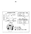
発明者らは,前記生体光計測装置を用いて,頭部全体に複数の計測点を配置し,脳活動を計測した。得られた各Hb信号(oxy-Hb信号, deoxy-Hb信号)を統計解析し,有意に変化した計測点(所定の解析方法により検定した結果,ある一定の確率以下でしか生じない変化が生じた場合)を活動位置として表示することにより,oxy-Hb信号とdeoxy-Hb信号では活動位置の分布が若干異なっているものの,一部の計測点においては両方のHb信号の活動が重なっていることを見出した(図1の表示部101参照)。ここでは,oxy-Hb信号とdeoxy-Hb信号を用いて活動を評価し,oxy-Hb信号だけが活動(増加)した計測点を格子模様で,deoxy-Hb信号だけが活動(減少)した計測点を点模様で,oxy-Hb信号とdeoxy-Hb信号の両方が活動した場合を黒で表示した。上記複数のHb信号の活動が重なった位置は,与えた刺激から予測された活動位置によく一致し,単一のHb信号だけの活動マップだけでは同定できない,より限局した活動部位を示す。この知見から,本発明では,各計測位置において,複数の生体情報(oxy-Hb信号とdeoxy-Hb信号など)の一定期間における変化を独立に統計的に検定し,前記複数の生体情報の結果を組み合わせて各計測位置を一つの範疇に分類し,同一の図中に表示する方法を提案する。この解決手段により,より限局した脳活動部位を精度よく検出することが可能となる。
The inventors measured a brain activity by arranging a plurality of measurement points on the entire head using the biological light measurement device. Each Hb signal obtained (oxy-Hb signal, deoxy-Hb signal) is statistically analyzed, and the measurement points that have changed significantly (as a result of testing by a predetermined analysis method, changes that occur only below a certain probability occur. ) Is displayed as the active position, the distribution of the active position is slightly different between the oxy-Hb signal and the deoxy-Hb signal, but the activities of both Hb signals overlap at some measurement points. (See the
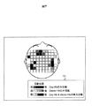
また,上記Hb信号の活動は,Hb信号の種類(oxy-Hb信号や deoxy-Hb信号)によって活動の空間的広がりおよびピークを迎える時間が異なる(図2)。図2では,10秒間の音声刺激を与えた場合の脳活動を,刺激開始から2.5秒毎に解析し,表示した例である。図1と同様にHb信号毎に統計解析を行い,その活動を3種類(oxy-Hb信号だけが活動した場合,deoxy-Hb信号だけが活動した場合,oxy-Hb信号とdeoxy-Hb信号の両方が活動した場合)に分類して表示した。oxy-Hb信号が増加する計測点は,刺激開始後2.6〜5.0秒後からが現れ始め,7.6〜12.5秒くらいの期間で最も多くの計測点が示される。一方,deoxy-Hb信号が減少する計測点は,刺激開始後7.6〜10.0秒後からが現れ始め,10.1〜15.0秒くらいの期間で最も多くの計測点が示される。このように,Hb信号の種類によって最も活動を検出できる期間が異なるため,Hb信号の種類によって異なる活動期間を設定して活動の有無もしくは強度を評価し,同一の図中に前記複数のHb信号の活動を組み合わせた分類を表示することにより,更に精度よく脳活動部位を同定することが可能となる(図3)。 In addition, the activity of the Hb signal differs depending on the type of Hb signal (oxy-Hb signal or deoxy-Hb signal), and the time when the activity reaches a spatial spread and peak (FIG. 2). FIG. 2 shows an example in which the brain activity when a voice stimulus is applied for 10 seconds is analyzed and displayed every 2.5 seconds from the start of the stimulus. As in Fig. 1, statistical analysis is performed for each Hb signal, and the activity is classified into three types (when only the oxy-Hb signal is activated, when only the deoxy-Hb signal is activated, the oxy-Hb signal and the deoxy-Hb signal When both were active, they were classified and displayed. Measurement points where the oxy-Hb signal increases begin to appear 2.6 to 5.0 seconds after the start of stimulation, and the most measurement points are shown in the period of 7.6 to 12.5 seconds. On the other hand, the measurement points where the deoxy-Hb signal decreases begin to appear 7.6 to 10.0 seconds after the start of stimulation, and the most measurement points are shown in the period of 10.1 to 15.0 seconds. As described above, since the period in which the activity can be detected most differs depending on the type of the Hb signal, the presence or intensity of the activity is evaluated by setting different activity periods depending on the type of the Hb signal, and the plurality of Hb signals in the same figure. It is possible to identify the brain activity site with higher accuracy by displaying the classification combining the activities (Fig. 3).
つまり、被検体頭部に光を照射する複数の光照射手段と、前記光照射手段から照射され前記被検体頭部を伝播した通過光を検出する複数の受光手段と、前記受光手段により検出された信号に基づき、前記光照射手段と前記受光手段の対により計測される計測点における、前記被検体頭部内の酸素化ヘモグロビンおよび脱酸素化ヘモグロビンの濃度変化を演算する演算部と、前記演算部による演算結果を表示する表示部とを有し、前記演算部は前記計測点における、前記酸素化ヘモグロビンおよび前記脱酸素化ヘモグロビンの濃度変化が有意なものであるかを前記計測点毎に判定し、前記表示部は前記判定結果を複数の前記計測点毎に表示することを特徴とする脳機能計測のための生体光計測装置により、脳活動部位・時間を精度よく解析・表示することができる。 That is, a plurality of light irradiating means for irradiating the subject head with light, a plurality of light receiving means for detecting passing light irradiated from the light irradiating means and propagated through the subject head, and detected by the light receiving means. A calculation unit for calculating a change in the concentration of oxygenated hemoglobin and deoxygenated hemoglobin in the subject's head at a measurement point measured by the pair of the light irradiating unit and the light receiving unit based on the obtained signal; A display unit that displays a calculation result by the unit, and the calculation unit determines whether the concentration change of the oxygenated hemoglobin and the deoxygenated hemoglobin at the measurement point is significant for each measurement point. Then, the display unit displays the determination result for each of the plurality of measurement points, and accurately analyzes and displays the brain activity site / time by the biological optical measurement device for brain function measurement. It can be.
本発明の脳活動解析方法および表示方法を用いた生体光計測装置により,従来法より脳活動が生じた部位を精度よく検出することが可能となる。また,図2のようにoxy-Hb信号の活動とdeoxy-Hb信号の活動の,時間的相違および空間的広がりの相違を可視化することにより,脳活動に伴う血行動態をより詳細に評価することが可能となる。 The living body optical measurement device using the brain activity analysis method and the display method of the present invention makes it possible to accurately detect a site where brain activity has occurred compared to the conventional method. In addition, by visualizing the temporal and spatial differences in oxy-Hb signal activity and deoxy-Hb signal activity as shown in Fig. 2, the hemodynamics associated with brain activity should be evaluated in more detail. Is possible.
本発明を実施するための基本形態を,図1を参照して説明する。図1は,本発明による生体光計測装置の概要を示すブロック図である。生体光計測部は,パーソナルコンピュータやワークステーションに代表される電子計算機から構成される制御装置103と,異なる波長にピーク波長を持つ2つのレーザダイオード109と110と,前記2つのレーザダイオードを異なった周波数で変調するための信号を生成する発振器107と108と,前記ピーク波長の異なる2つの光を混合する光混合器111と,前記光混合器111からの光を光ファイバ経由で被検体上の光照射位置に照射する光照射手段と,前記光照射手段から適度に離れた光検出位置(本実施例では約3cm離れた点)から混合光を検出する光検出器112と,前記発振器からの変調周波数が参照信号として入力されたロックインアンプ105および106と,ロックインアンプの出力である各波長帯の光の透過光信号をアナログ信号からデジタル信号へ変換するアナログ−デジタル変換器104を備える。前記光照射位置と光検出位置の略中点を,計測位置の中心とする。図1では,計測位置3のみ正確な装置への接続構成を記したが,実際には図1に記した全ての光照射位置と光検出位置は,光ファイバ経由で装置へ接続されている。本装置では,発振器を用いて複数の光信号を分離するため,一つの検出器でも複数の位置からの光信号を計測することが出来る。本実施例では発振器を用いて複数の光信号を分離しているが,発振器は使わずにパルス光を用いて点灯タイミングで光信号を分離することも可能である。各波長帯の光の透過光信号はアナログ−デジタル変換器104でアナログ−デジタル変換された後,制御装置103に入力・記憶される。制御装置103では透過光信号を元に各計測部位における各Hb信号が算出され,元信号(透過光信号)と共にデータ解析装置102に転送され記憶される。透過光信号からHb信号を算出する方法については,非特許文献1に詳しく記されている。また,本実施例では,制御装置103と解析装置102を分けて記載したが,両方の機能を1台のPCで行うことも可能である。
A basic mode for carrying out the present invention will be described with reference to FIG. FIG. 1 is a block diagram showing an outline of a biological light measurement device according to the present invention. The biological light measurement unit is different from the
解析装置102では,計測された各Hb信号を統計解析し,有意な活動の有無を評価する。ここでの統計解析法は,例えば,t検定や分散分析などの手法に代表される。一般に脳活動計測は,特定の刺激や課題を被験者に与えたときの反応を,ある安静期間における状態と比べて脳活動として捉えるため,安静期間における信号と,刺激や課題による反応が生じていると考えられる期間(活動期間)における信号を比較することが統計解析の基本となる。この比較を行う代表的な統計手法には,t検定やF検定,あるいは分散分析や多重比較がある。また,パラメトリック検定だけでなく,ノンパラメトリック検定も使用可能である。他にも,複数の異なる刺激や課題を被験者に与えた場合,それぞれの活動期間における信号同士を直接比較する方法や,ある刺激や課題に対する活動を示す信号波形を仮定し,その信号波形との類似性を相関係数などで評価する方法も可能である。一般的に脳活動に伴うHb信号の変化は,oxy-Hb信号の増加とdeoxy-Hb信号の減少,total-Hb信号の増加であることが知られているため,本実施例では,これら3つの変化を活動信号として定義する。各計測位置において,Hb信号毎にt検定を用いて活動の有無を検定した結果を表示部101に記す。本実施例の計測では,20秒から30秒の期間内で設定した任意の長さの安静期間後に,10秒間の音声刺激を与えるシーケンスを1ブロックと定義し,5回のブロックを繰り返した。はじめに,各ブロックにおいて,刺激開始後5秒の時点から刺激終了後10秒までの期間を活動期間として,その活動期間における平均信号強度を活動値として求める(あるいは信号強度の最大値を活動値としてもよい)。更に,各ブロックにおいて,刺激開始直前の5秒間の安静期間における平均信号強度を安静値として求める。5ブロック分の活動値から得られる平均値と偏差,および5ブロック分の安静値から得られる平均値と偏差を使用して,活動値と安静値の平均に差があるかどうか,t検定を行った。t検定とは,帰無仮説が正しいと仮定した場合に統計量がt分布に従うことを利用する統計学的検定法の総称である。母集団が正規分布に従うと仮定するパラメトリック検定法であり,t分布が直接もとの平均や標準偏差にはよらない(ただし自由度による)ことを利用している。2つの集団の平均値に有意差があるかどうかの検定などに用いられる。ここでは,「安静値の平均値と活動値の平均値は同じである」という帰無仮説を立て,活動値と安静値の平均に差があるかどうか検定する。つまり,「安静値の平均値と活動値の平均値は同じである」という帰無仮説を,例えば95%以上の確率で棄却できる場合,有意な活動とみなす。このような手法で,ある閾値より有意な確率でoxy-Hb信号の増加や,deoxy-Hb信号の減少が認められた場合,脳活動として評価した。
The
また、有意な活動の有無の評価は、測定者が任意に設定部より設定した閾値を脳活動値が越えたか否かや、脳活動が最大値となるまでにかかった時間など、測定者が任意に設定した基準により評価してもよい。 In addition, the evaluation of the presence or absence of significant activity is based on whether the measurer determines whether the brain activity value has exceeded the threshold value arbitrarily set by the measurer, such as whether the brain activity value has reached the maximum value. You may evaluate by the criteria set arbitrarily.
また、有意な活動の有無の評価基準は計測位置ごとに異なるように設定してもよい。 Moreover, you may set so that the evaluation criteria of the presence or absence of significant activity may differ for every measurement position.
oxy-Hb信号だけが活動(増加)した計測点を格子模様で,deoxy-Hb信号だけが活動(減少)した計測点を点模様で,oxy-Hb信号とdeoxy-Hb信号の両方が活動した場合を黒で表示した。上記2つのHb信号の活動が重なった位置は,より限局した活動部位を示した。これらの活動部位は,音声刺激に反応すると予測された聴覚野付近に対応する左右の側頭部において見られた。また,言語音声を用いたため,記憶や注意に関係すると考えられている前頭部においても活動が見られた。このように,上記2つのHb信号の活動が重なった位置は与えた刺激から予測された活動位置によく一致し,単一のHb信号だけの活動マップより高精度で脳活動部位を検出できることを示した。 The measurement points where only the oxy-Hb signal was activated (increased) were shown in a grid pattern, and the measurement points where only the deoxy-Hb signal was activated (decreased) were shown as a dot pattern, and both the oxy-Hb signal and deoxy-Hb signal were active The case is shown in black. The position where the activities of the two Hb signals overlapped indicated a more limited activity site. These active sites were found in the left and right temporal regions corresponding to the vicinity of the auditory cortex that was predicted to respond to voice stimuli. In addition, because speech was used, activity was also seen in the frontal region, which is thought to be related to memory and attention. Thus, the position where the activity of the two Hb signals overlaps well matches the activity position predicted from the applied stimulus, and the brain activity site can be detected with higher accuracy than the activity map of only a single Hb signal. Indicated.
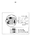
更に,上記のデータに対し,活動期間を表す時間窓を刺激開始から2.5秒毎に設定し,解析・表示した例を図2に示す。図1と同様にHb信号毎に統計解析を行い,その活動を3種類(oxy-Hb信号だけが活動した場合,deoxy-Hb信号だけが活動した場合,oxy-Hb信号とdeoxy-Hb信号の両方が活動した場合)に分類して表示した。oxy-Hb信号が増加する計測点は,刺激開始後2.6〜5.0秒後からが現れ始め,7.6〜12.5秒くらいの時間窓で最も多くの計測点が示される。一方,deoxy-Hb信号が減少する計測点は,刺激開始後7.6〜10.0秒後からが現れ始め,10.1〜15.0秒くらいの時間窓で最も多くの計測点が示される。このように,Hb信号の種類によって活動の空間的広がりおよびピークを迎える時間が異なることが分かった。つまり,Hb信号の種類によって最もよく活動を検出できる時間窓が異なる。従って,Hb信号の種類によって異なる活動期間(時間窓)を設定して活動の有無もしくは強度を評価し,同一の図中に前記複数のHb信号の活動を組み合わせた分類を表示することにより,更に精度よく脳活動部位を同定することが可能となる。図3には,oxy-Hb信号の活動期間を表す時間窓に刺激開始後7.6〜12.5秒の期間を,deoxy-Hb信号の活動期間を表す時間窓に刺激開始後10.1〜15.0秒の期間を設定して解析した結果を,同一の図中に表示した例を示した。このように,Hb信号の種類によって異なる活動期間を表す時間窓を設定できる設定部301を設けることにより,活動期間として一律の時間窓を設定した場合には検出できない,より精度の高い活動領域を表示することが出来る。
Furthermore, for the above data, Fig. 2 shows an example in which a time window representing the activity period is set and analyzed every 2.5 seconds from the start of stimulation. As in Fig. 1, statistical analysis is performed for each Hb signal, and the activity is classified into three types (when only the oxy-Hb signal is activated, when only the deoxy-Hb signal is activated, the oxy-Hb signal and the deoxy-Hb signal When both were active, they were classified and displayed. Measurement points where the oxy-Hb signal increases begin to appear 2.6 to 5.0 seconds after the start of stimulation, and the most measurement points are shown in a time window of about 7.6 to 12.5 seconds. On the other hand, the measurement points where the deoxy-Hb signal decreases begin to appear 7.6 to 10.0 seconds after the start of stimulation, and the most measurement points are shown in the time window of 10.1 to 15.0 seconds. Thus, it was found that the spatial spread of activity and the time to reach a peak differ depending on the type of Hb signal. That is, the time window in which the activity can be detected best differs depending on the type of Hb signal. Therefore, by setting different activity periods (time windows) depending on the type of Hb signal and evaluating the presence or intensity of the activity, and displaying the classification combining the activities of the plurality of Hb signals in the same figure, It becomes possible to identify the brain activity site with high accuracy. Figure 3 shows the time window representing the oxy-Hb signal activity period from 7.6 to 12.5 seconds after the start of stimulation, and the time window representing the deoxy-Hb signal activity period from 10.1 to 15.0 seconds after the start of stimulation. An example of setting and analysis results displayed in the same figure is shown. In this way, by providing a
また,より高精度で脳活動を検出するために,各Hb信号の活動波形から各Hb信号の活動期間を表す時間窓を設定する方法を図4〜5に示す。図4では,全計測点における各Hb信号の平均波形から算出した各Hb信号の活動ピークを表示する表示部401と,その活動ピーク時を中心として活動期間の時間窓を設定する設定部402を有する表示方法を示した。活動期間の時間窓は,平均波形などを参考にユーザーが任意に決定できる。特に,oxy-Hb信号,deoxy-Hb信号など異なるHb信号毎に独立の活動期間の時間窓を設定できることを特徴とする。更に,図5では,全計測点における各Hb信号の平均波形から算出した各Hb信号の活動ピークに対する半値幅を表示する表示部501,および各Hb信号の活動ピークを中心にある基準値以上を示す時間窓を設定する設定部502を有する表示方法を示した。ここでは,時間窓の設定方法として,矢印ボタン503,504を操作することにより,oxy-Hb信号,deoxy-Hb信号それぞれの基準値を変化させ,時間窓を設定する方法を示した。ここでは,基準値を分かり易く表示するため,矢印ボタン503,504の操作により,時間窓を示す矢印505,506が上下に動く表示方法も有効である。もちろん基準値の設定は,直接の値を入力する方法や,活動信号のピークに対する割合(%)を入力する方法を用いても構わない。
4 to 5 show a method for setting a time window representing the activity period of each Hb signal from the activity waveform of each Hb signal in order to detect brain activity with higher accuracy. In FIG. 4, a
上記の例は全て,活動の評価にoxy-Hb信号とdeoxy-Hb信号の2種類を用いた場合だが,図6に示したように,oxy-Hb信号,deoxy-Hb信号,およびtotal-Hb信号の3種類を用いた場合も,全て同様の方法が可能である。3種類のHb信号を用いるため,例えば活動の分類は3種類から7種類に増えるが,3種類のHb信号が全て活動した部位に注目すると,更に活動部位が限局して示されることが分かる。このoxy-Hb信号の増加,deoxy-Hb信号の減少,およびtotal-Hb信号の増加の全てが見られる部位は,理論的に示される典型的な脳活動を示していると考えられる。つまり,動脈血の流入によりoxy-Hbとtotal-Hbが増加し,更にその血流速が酸素消費に比べて早くなるため,deoxy-Hbは減少する。その他の変化パターンでは,例えばoxy-Hb信号やtotal-Hb信号が増加するもののdeoxy-Hb信号が減少しない場合,動脈血の供給は行われているが血流速の上昇は少なく,活動部位の中心からは外れている可能性が示唆される。また逆に,deoxy-Hb信号だけが減少しoxy-Hb信号やtotal-Hb信号の増加が見られない場合,動脈血の供給は主に血流速の上昇という形で行われ血液量全体としては殆ど増えていないことが予想される。この部位も活動の中心からは外れている可能性が示唆される。また,これらのHb信号の変化パターンから,血管の弾力性など脳の血行動態が推測できる可能性もある。 In all the above examples, oxy-Hb signal and deoxy-Hb signal are used for activity evaluation. As shown in Fig. 6, oxy-Hb signal, deoxy-Hb signal, and total-Hb signal are used. The same method can be used for all three types of signals. Since three types of Hb signals are used, for example, the activity classification is increased from three types to seven types. However, when all three types of Hb signals are activated, it can be seen that the active sites are further localized. The region where all of this increase in oxy-Hb signal, decrease in deoxy-Hb signal, and increase in total-Hb signal are considered to indicate typical brain activity shown theoretically. In other words, oxy-Hb and total-Hb increase due to the inflow of arterial blood, and deoxy-Hb decreases because the blood flow rate becomes faster than oxygen consumption. In other change patterns, for example, when the oxy-Hb signal and total-Hb signal increase but the deoxy-Hb signal does not decrease, arterial blood is supplied, but the blood flow rate is not increased, and the center of the active site is low. This suggests that it may be off. Conversely, when only the deoxy-Hb signal decreases and no increase in the oxy-Hb signal or total-Hb signal is observed, arterial blood supply is mainly performed in the form of an increase in blood flow rate, and the total blood volume is It is expected that there has been little increase. This suggests that this part may also be off the center of activity. In addition, it is possible that the hemodynamics of the brain, such as the elasticity of blood vessels, can be estimated from these Hb signal change patterns.
図4〜6では,全計測点における各Hb信号の平均波形を例として示したが,活動期間の時間窓を設定するために用いるHb信号の波形は,特に全計測点の平均値とは限らない。例えば,最も強く活動した計測位置のHb信号波形や,理論的に求められた標準的な脳活動波形(テンプレート波形)を用いることも可能である。あるいは,注目している部位の平均波形や,各被験者の過去の脳活動信号から作成した標準的な活動波形を用いることも可能である。被験者毎にHb信号波形を求めて解析に使用することは,Hb信号波形の個人差を吸収し,より精度よく脳活動を検出することに役立つ。 In FIGS. 4 to 6, the average waveform of each Hb signal at all measurement points is shown as an example. However, the waveform of the Hb signal used for setting the time window of the activity period is not limited to the average value at all measurement points. Absent. For example, it is possible to use the Hb signal waveform at the measurement position where the strongest activity is performed, or a standard brain activity waveform (template waveform) theoretically obtained. Alternatively, it is possible to use an average waveform of a region of interest or a standard activity waveform created from past brain activity signals of each subject. Obtaining the Hb signal waveform for each subject and using it for analysis is useful for absorbing individual differences in the Hb signal waveform and detecting brain activity more accurately.
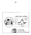
図4〜6においては,ある閾値により活動の有無の有意性を統計的に検定し1―0で分類していたが,図7には,活動を示した計測点における活動強度を段階的に表示した例を記した。活動強度とは,例えば,活動期間における平均信号強度(mM・mm)活動強度の最大値(最小値),あるいは,統計値であるt値やF値,p値などを用いる。ある活動波形テンプレートを用いた場合は,そのテンプレートとの相関係数など類似性を示す指標を用いてもよい。図7の例では,まず図4〜6で示したように各Hb信号における活動の有無を統計的に検定した後,その活動期間における平均信号強度をカラーバー701に従い表示した。oxy-Hb信号かdeoxy-Hb信号が単独で活動を示した場合は,それぞれの平均信号強度を異なる色の濃淡などで表示すればよいが,oxy-Hb信号とdeoxy-Hb信号の両方が活動した場合は,また単独で活動した場合とは異なる色の濃淡などを用いて,oxy-Hb信号強度とdeoxy-Hb信号強度の和,あるいは平均値を表示する。あるいは,oxy-Hb信号強度とdeoxy-Hb信号強度が極端に異なる場合などは,oxy-Hb信号強度とdeoxy-Hb信号強度を同レベルに標準化し,合わせた信号強度を算出することも有用である。また,図8では,全計測点の平均波形や,最も強く活動した計測位置の活動波形,あるいは理論的に求められた標準的な脳活動波形をテンプレート波形とした時の,相関係数を表示した例を示した。ここでは,図7と同様にある閾値で各Hb信号における活動の有無を検定した後,その相関係数を異なる色の濃淡で表示した。oxy-Hb信号とdeoxy-Hb信号など複数のHb信号両方で有意な活動が見られた場合は,その平均相関係数などを表示する。このように,複数のHb信号を組み合わせた活動の有無の分類に加えて,それぞれの活動強度を段階的に表示することによって,より限局した活動部位を検出することが容易になる。
In FIGS. 4-6, the significance of the presence or absence of activity was statistically tested according to a certain threshold and classified as 1-0. However, in FIG. 7, the activity intensity at the measurement point indicating the activity is stepwise. The displayed example is described. As the activity intensity, for example, the maximum value (minimum value) of the average signal intensity (mM · mm) activity intensity in the activity period, or t value, F value, p value, etc., which are statistical values, are used. When a certain activity waveform template is used, an index indicating similarity such as a correlation coefficient with the template may be used. In the example of FIG. 7, first, as shown in FIGS. 4 to 6, the presence or absence of activity in each Hb signal is statistically tested, and then the average signal intensity during the activity period is displayed according to the
上記いずれかの方法で脳活動部位を検出した後,その活動位置における活動の詳細を表示することは,脳活動を詳細に検討する際に有効である。図9には,oxy-Hb信号とdeoxy-Hb信号の両方において活動が示され,更にその中で最も活動が強かった計測点を選択し,その計測点におけるHb信号の時間波形を表示した例である。このように実際のHb信号の時間変化を示すことは,脳活動が何らかのアーチファクトではないことを確認したり,その活動パターンを評価したりする上で,有用である。また,図10には,脳活動を詳細に検討するために有用な,Hb信号のピーク値あるいは活動期間の平均値を縦軸に取り,その活動ピークに達するまでに要した時間遅れを横軸に示したグラフを示した。エラーバーは標準偏差,あるいは標準誤差,あるいは信頼区間などを示す。このグラフ表示により,どのHb信号強度が最も強く,あるいは,最も早く信号強度のピークに達したかが分かる。このグラフのパターンは,脳活動に伴う血行動態を評価する上で有効である。これまでの知見では,脳活動に伴うHb信号の変化は,はじめにtotal-Hb信号の増加がピークを迎え,次にoxy-Hb信号の増加がピークを迎え,最後にdeoxy-Hb信号の減少がピークを迎えることが知られている。このグラフを用いると,その典型パターンに合致するかどうか容易に判定することが出来るので,典型パターンに対する合致か否かをユーザーに表示する機能を追加することも出来る。図9,10では,全体から一つの計測点だけを選択し表示した例を示したが,複数の計測点を表示することも可能である。例えば,前頭部,側頭部,といった領域を複数設定し,それぞれの領域において活動強度の最も強い計測位置を選択する方法も有効である。 After detecting a brain activity site by any of the above methods, displaying the details of the activity at the activity position is effective when examining brain activity in detail. FIG. 9 shows an example in which the activity is shown in both the oxy-Hb signal and the deoxy-Hb signal, the measurement point having the strongest activity is selected, and the time waveform of the Hb signal at the measurement point is displayed. It is. Thus, showing the actual time change of the Hb signal is useful for confirming that the brain activity is not an artifact or evaluating the activity pattern. FIG. 10 shows the peak value of the Hb signal or the average value of the activity period, which is useful for examining the brain activity in detail, on the vertical axis, and the time delay required to reach the activity peak on the horizontal axis. The graph shown in is shown. Error bars indicate standard deviation, standard error, or confidence interval. This graph display shows which Hb signal intensity is the strongest or the peak of the signal intensity has reached the earliest. The pattern of this graph is effective in evaluating hemodynamics associated with brain activity. According to the findings so far, the change in Hb signal associated with brain activity begins with an increase in total-Hb signal, followed by an increase in oxy-Hb signal, and finally a decrease in deoxy-Hb signal. It is known to reach a peak. If this graph is used, it can be easily determined whether or not it matches the typical pattern. Therefore, it is possible to add a function for displaying to the user whether or not it matches the typical pattern. 9 and 10 show an example in which only one measurement point is selected and displayed from the whole, it is also possible to display a plurality of measurement points. For example, a method of setting a plurality of areas such as the frontal region and the temporal region and selecting a measurement position with the highest activity intensity in each region is also effective.
以上,脳活動の評価を支援する実施例を挙げたが,同様の機能により,脳機能診断あるいは脳循環診断の支援が可能となる。例えば,図2のように,刺激開始から順次2.5秒間の時間窓における脳活動を解析し,表示した場合,各Hb信号の活動の空間的広がりおよびピークを迎える時間窓が分かり易い(図2)。oxy-Hb信号が最初に増加し,刺激開始後2.6〜5.0秒後から活動部位が現れ始め,7.6〜12.5秒くらいの期間で最も多くの計測点が示される。一方,deoxy-Hb信号の減少は遅く,刺激開始後7.6〜10.0秒後から活動部位が現れ始め,10.1〜15.0秒くらいの期間で最も多くの計測点が示される。個人によって若干異なるが,このパターンは一つの典型的な活動パターンと考えられる。例えば,最初に来るはずのoxy-Hb信号の増加領域が狭い,あるいは活動強度が小さい場合,脳活動に伴う動脈血の供給が少ないことを意味するので,血管の弾力性が低い可能性が示唆される。あるいは,最初にdeoxy-Hb信号の増加が見られて,遅れてoxy-Hb信号の増加が見られる場合も同様に,動脈血の供給が遅い傾向を示すので,血管の弾力性が低い可能性が示唆される。また,deoxy-Hb信号の減少が見られない,あるいは小さい場合は,血流速の増加が顕著ではないと考えられ,血管の弾力性が高いか,血管の流れが悪い可能性が示唆される。もしくは,動脈血の供給と均衡する酸素の消費が行われている可能性を示す。それ以外の変化パターンで,予測される生理変化から著しく外れた変化を示した場合は,脳活動信号を計測できていない恐れがあるので,エラーメッセージを表示する機能も有用である。 As mentioned above, although the example which supports evaluation of brain activity was given, the support of a brain function diagnosis or a cerebral circulation diagnosis is attained by the same function. For example, as shown in FIG. 2, when the brain activity in the time window of 2.5 seconds is analyzed and displayed sequentially from the start of stimulation, the spatial spread and peak time window of each Hb signal activity is easy to understand (FIG. 2). . The oxy-Hb signal increases first, the active site begins to appear 2.6 to 5.0 seconds after the start of stimulation, and the most measurement points are shown in the period of 7.6 to 12.5 seconds. On the other hand, the deoxy-Hb signal decreases slowly, and the active site begins to appear 7.6 to 10.0 seconds after the start of stimulation, and the most measurement points are shown in the period of 10.1 to 15.0 seconds. This pattern is considered to be one typical activity pattern, although it varies slightly depending on the individual. For example, if the increase area of the oxy-Hb signal that should come first is narrow or the activity intensity is small, it means that the supply of arterial blood accompanying brain activity is small, which suggests that the elasticity of the blood vessel may be low The Alternatively, when an increase in deoxy-Hb signal is first observed and an increase in oxy-Hb signal is observed later, the arterial blood supply tends to be slow, and the elasticity of blood vessels may be low. It is suggested. In addition, if the deoxy-Hb signal does not decrease or is small, the increase in blood flow rate may not be significant, suggesting the possibility of high blood vessel elasticity or poor blood flow . Or it may indicate that oxygen consumption is balanced with the supply of arterial blood. The function to display an error message is also useful if there is a change that is significantly different from the predicted physiological change with other change patterns, because there is a possibility that the brain activity signal cannot be measured.
101…脳活動表示部,102…データ解析装置,103…制御装置,104…アナログデジタル変換器,105…ロックインアンプ,106…ロックインアンプ,107…発振器,108…発振器,109…光源,110…光源,111…光混合器,112…光検出器,301…活動期間を表す時間窓の設定部,401…Hb信号の活動ピークを表示する表示部,402…活動ピーク時を中心とした活動期間の時間窓を設定する設定部,501…各Hb信号の活動ピークに対する半値幅を表示する表示部,502…時間窓を設定する基準値を示す設定部, 503…oxy-Hb信号の時間窓を設定する矢印ボタン,504…deoxy-Hb信号の時間窓を設定する矢印ボタン,505…oxy-Hb信号の設定された時間窓を示す矢印, 506…deoxy-Hb信号の設定された時間窓を示す矢印, 701…活動期間における平均信号強度を表すカラーバー。
DESCRIPTION OF
Claims (19)
前記光照射手段から照射され前記被検体頭部を伝播した通過光を検出する複数の受光手段と、
前記受光手段により検出された信号に基づき、前記光照射手段と前記受光手段の対により計測される計測点における、前記被検体頭部内の酸素化ヘモグロビンに対応する信号および脱酸素化ヘモグロビンに対応する信号を算出する演算部と、
前記演算部による演算結果を表示する表示部とを有し、
前記演算部は前記計測点における、前記酸素化ヘモグロビンに対応する信号および前記脱酸素化ヘモグロビンに対応する信号の変化が有意なものであるかを判定し、
さらに、前記計測点における前記酸素化ヘモグロビンに対応する信号の変化が有意性を示す領域を含む期間を第一の計測期間とし、前記脱酸素化ヘモグロビンに対応する信号の変化が有意性を示す領域を含む期間を第二の計測期間として、当該第一の計測期間における前記酸素化ヘモグロビンに対応する信号及び第二の計測期間における前記脱酸素化ヘモグロビンに対応する信号に基づいて、前記酸素化ヘモグロビンおよび脱酸素化ヘモグロビンの濃度変化の相関を解析し、当該解析結果を前記表示部に出力させることを特徴とする生体光計測装置。
A plurality of light irradiation means for irradiating the subject's head with light;
A plurality of light receiving means for detecting passing light irradiated from the light irradiation means and propagated through the subject head;
Corresponding to a signal corresponding to oxygenated hemoglobin in the subject's head and deoxygenated hemoglobin at a measurement point measured by the pair of the light irradiation means and the light receiving means based on the signal detected by the light receiving means An arithmetic unit for calculating a signal to be
The calculation result by the calculating section and a front Shimesuru display unit,
The arithmetic unit in the measurement point, or change of the signal corresponding to the signal and the deoxygenated hemoglobin corresponding to the oxygenated hemoglobin is of significant and determine a constant,
Further, a region including a region in which a change in the signal corresponding to the oxygenated hemoglobin at the measurement point is significant is a first measurement period, and a region in which the change in the signal corresponding to the deoxygenated hemoglobin is significant And the oxygenated hemoglobin based on a signal corresponding to the oxygenated hemoglobin in the first measurement period and a signal corresponding to the deoxygenated hemoglobin in the second measurement period. And a biological light measurement device characterized in that a correlation between concentration changes of deoxygenated hemoglobin is analyzed and the analysis result is output to the display unit .
前記ピーク時間に基づいて、前記第一の計測期間及び前記第二の計測期間を設定する設定部を有することを特徴とする請求項1記載の生体光計測装置。 The biological light measurement apparatus according to claim 1, further comprising a setting unit configured to set the first measurement period and the second measurement period based on the peak time.
前記演算部による前記判定の方法を設定する設定部を有することを特徴とする請求項1記載の生体光計測装置。The biological light measurement apparatus according to claim 1, further comprising a setting unit configured to set the determination method by the calculation unit.
被検体頭部を伝播した通過光に基づく信号を取得し、
前記信号に基づいて、前記被検体頭部内の酸素化ヘモグロビンに対応する信号および脱酸素化ヘモグロビンに対応する信号を算出し、
前記酸素化ヘモグロビンに対応する信号および前記脱酸素化ヘモグロビンに対応する信号の変化が有意なものであるかを判定し、
さらに、前記酸素化ヘモグロビンに対応する信号の変化が有意性を示す領域を含む期間を第一の計測期間とし、前記脱酸素化ヘモグロビンに対応する信号の変化が有意性を示す領域を含む期間を第二の計測期間として、当該第一の計測期間における前記酸素化ヘモグロビンに対応する信号及び第二の計測期間における前記脱酸素化ヘモグロビンに対応する信号に基づいて、前記酸素化ヘモグロビンおよび脱酸素化ヘモグロビンの濃度変化の相関を解析することを特徴とする脳機能計測装置に用いる演算装置。 An arithmetic device used for a brain function device that performs an operation based on a measurement signal obtained from the brain function measurement device,
Obtain a signal based on the light passing through the subject's head,
Based on the signal, a signal corresponding to oxygenated hemoglobin in the subject's head and a signal corresponding to deoxygenated hemoglobin are calculated,
Change in the signal corresponding to the signal and the deoxygenated hemoglobin corresponding to the oxygenated hemoglobin to determine those significant,
Further, a period including a region where the change in the signal corresponding to oxygenated hemoglobin is significant is a first measurement period, and a period including a region where the change in the signal corresponding to the deoxygenated hemoglobin is significant As the second measurement period, based on the signal corresponding to the oxygenated hemoglobin in the first measurement period and the signal corresponding to the deoxygenated hemoglobin in the second measurement period, the oxygenated hemoglobin and deoxygenation An arithmetic unit used for a brain function measuring device, characterized by analyzing a correlation of hemoglobin concentration change.
前記信号に基づいて、前記被検体頭部内の酸素化ヘモグロビンに対応する信号および脱酸素化ヘモグロビンに対応する信号を算出し、
前記酸素化ヘモグロビンに対応する信号および前記脱酸素化ヘモグロビンに対応する信号の変化が有意なものであるかを判定し、
さらに、前記酸素化ヘモグロビンに対応する信号の変化が有意性を示す領域を含む期間を第一の計測期間とし、前記脱酸素化ヘモグロビンに対応する信号の変化が有意性を示す領域を含む期間を第二の計測期間として、当該第一の計測期間における前記酸素化ヘモグロビンに対応する信号及び第二の計測期間における前記脱酸素化ヘモグロビンに対応する信号に基づいて、前記酸素化ヘモグロビンおよび脱酸素化ヘモグロビンの濃度変化の相関を解析することを特徴とする光を用いた脳機能計測のための演算方法。 Obtain a signal based on the light passing through the subject's head,
Based on the signal, a signal corresponding to oxygenated hemoglobin in the subject's head and a signal corresponding to deoxygenated hemoglobin are calculated,
Change in the signal corresponding to the signal and the deoxygenated hemoglobin corresponding to the oxygenated hemoglobin to determine those significant,
Further, a period including a region where the change in the signal corresponding to oxygenated hemoglobin is significant is a first measurement period, and a period including a region where the change in the signal corresponding to the deoxygenated hemoglobin is significant As the second measurement period, based on the signal corresponding to the oxygenated hemoglobin in the first measurement period and the signal corresponding to the deoxygenated hemoglobin in the second measurement period, the oxygenated hemoglobin and deoxygenation An arithmetic method for brain function measurement using light, characterized by analyzing a correlation of hemoglobin concentration changes.
Priority Applications (5)
| Application Number | Priority Date | Filing Date | Title |
|---|---|---|---|
| JP2006057090A JP4702107B2 (en) | 2006-03-03 | 2006-03-03 | Biological light measurement device |
| US11/646,468 US7933723B2 (en) | 2006-03-03 | 2006-12-28 | Living body light measuring device |
| EP06027010A EP1829475B1 (en) | 2006-03-03 | 2006-12-28 | Living body light measuring device |
| DE602006015598T DE602006015598D1 (en) | 2006-03-03 | 2006-12-28 | Device for measuring on the living body with the help of light |
| CN200710001380A CN100586373C (en) | 2006-03-03 | 2007-01-11 | Living body light measuring device |
Applications Claiming Priority (1)
| Application Number | Priority Date | Filing Date | Title |
|---|---|---|---|
| JP2006057090A JP4702107B2 (en) | 2006-03-03 | 2006-03-03 | Biological light measurement device |
Publications (3)
| Publication Number | Publication Date |
|---|---|
| JP2007229322A JP2007229322A (en) | 2007-09-13 |
| JP2007229322A5 JP2007229322A5 (en) | 2008-10-16 |
| JP4702107B2 true JP4702107B2 (en) | 2011-06-15 |
Family
ID=38109500
Family Applications (1)
| Application Number | Title | Priority Date | Filing Date |
|---|---|---|---|
| JP2006057090A Expired - Fee Related JP4702107B2 (en) | 2006-03-03 | 2006-03-03 | Biological light measurement device |
Country Status (5)
| Country | Link |
|---|---|
| US (1) | US7933723B2 (en) |
| EP (1) | EP1829475B1 (en) |
| JP (1) | JP4702107B2 (en) |
| CN (1) | CN100586373C (en) |
| DE (1) | DE602006015598D1 (en) |
Families Citing this family (15)
| Publication number | Priority date | Publication date | Assignee | Title |
|---|---|---|---|---|
| JP2009082595A (en) * | 2007-10-02 | 2009-04-23 | Shimadzu Corp | Optical biometric device |
| NL1036613A1 (en) | 2008-03-03 | 2009-09-07 | Asml Netherlands Bv | Lithographic apparatus, plasma source, and reflecting method. |
| JP5319960B2 (en) * | 2008-05-28 | 2013-10-16 | 株式会社日立製作所 | Biological light measurement device |
| CN102076267B (en) * | 2008-07-08 | 2013-02-06 | 株式会社日立制作所 | Light meter |
| WO2010038774A1 (en) * | 2008-10-01 | 2010-04-08 | 株式会社 日立メディコ | Biological light measurement device and position display method for displaying light irradiation position and light detection position, or measurement channel |
| JP2010252906A (en) * | 2009-04-22 | 2010-11-11 | Hitachi Medical Corp | Biological light measuring instrument |
| EP2698111A4 (en) * | 2011-04-15 | 2014-10-15 | Hitachi Medical Corp | Biophotonic measurement device, biophotonic measurement device operating method, and biophotonic measurement data analysis and display method |
| JP5382666B2 (en) * | 2011-04-21 | 2014-01-08 | 学校法人 聖マリアンナ医科大学 | Concentration measuring device and concentration measuring method |
| JP5668138B2 (en) * | 2011-06-17 | 2015-02-12 | 株式会社日立製作所 | Biological light measurement device |
| JP5809864B2 (en) * | 2011-07-07 | 2015-11-11 | 株式会社日立メディコ | Biological light measurement device, waveform analysis method |
| US9897666B2 (en) * | 2013-12-16 | 2018-02-20 | The Johns Hopkins University | Chip-scale optomechanical magnetometer |
| JP6285774B2 (en) * | 2014-03-31 | 2018-02-28 | リオン株式会社 | Language listening inspection device and method |
| WO2018071854A1 (en) * | 2016-10-13 | 2018-04-19 | Photon Migration Technologies Corp. | Method for representations of network-dependent features of the hemoglobin signal in living tissues for detection of breast cancer and other applications |
| JP6281628B2 (en) * | 2016-12-28 | 2018-02-21 | 株式会社島津製作所 | Optical measurement system |
| CN107595295B (en) * | 2017-06-09 | 2019-10-29 | 苏州大学 | A kind of lower extremity movement resistive state recognition methods based on brain hemoglobin information |
Citations (5)
| Publication number | Priority date | Publication date | Assignee | Title |
|---|---|---|---|---|
| JPH069178A (en) * | 1992-04-09 | 1994-01-18 | Werner Hagel | Elevator |
| JPH08103434A (en) * | 1994-10-06 | 1996-04-23 | Hitachi Ltd | Device and method for measuring living body light |
| JP2000237194A (en) * | 1999-02-19 | 2000-09-05 | Hitachi Ltd | Light measuring method and device |
| WO2002032317A1 (en) * | 2000-10-16 | 2002-04-25 | Hitachi Medical Corporation | Organism optical measurement instrument |
| WO2005120349A1 (en) * | 2004-06-14 | 2005-12-22 | Hitachi Medical Corporation | Biophotometer, biophotometry, and program |
Family Cites Families (6)
| Publication number | Priority date | Publication date | Assignee | Title |
|---|---|---|---|---|
| US6240309B1 (en) | 1995-10-06 | 2001-05-29 | Hitachi, Ltd. | Optical measurement instrument for living body |
| US7092748B2 (en) * | 2000-02-18 | 2006-08-15 | Centro Nacional De Investigaciones Cientificas (Cnic) | System and method for the tomography of the primary electric current of the brain and of the heart |
| US6922580B2 (en) * | 2002-06-04 | 2005-07-26 | Koninklijke Philips Electronics N.V. | Blood flow gated MRI |
| JP4167860B2 (en) * | 2002-07-08 | 2008-10-22 | 株式会社日立製作所 | Biological light measurement device |
| CN1718154B (en) * | 2004-07-08 | 2011-06-15 | 株式会社日立制作所 | Optical measurement system for living bodies and method thereof |
| EP2319406A1 (en) * | 2004-12-28 | 2011-05-11 | Hyperspectral Imaging, Inc | Hyperspectral/multispectral imaging in determination, assessment and monitoring of systemic physiology and shock |
-
2006
- 2006-03-03 JP JP2006057090A patent/JP4702107B2/en not_active Expired - Fee Related
- 2006-12-28 DE DE602006015598T patent/DE602006015598D1/en active Active
- 2006-12-28 EP EP06027010A patent/EP1829475B1/en not_active Ceased
- 2006-12-28 US US11/646,468 patent/US7933723B2/en not_active Expired - Fee Related
-
2007
- 2007-01-11 CN CN200710001380A patent/CN100586373C/en not_active Expired - Fee Related
Patent Citations (5)
| Publication number | Priority date | Publication date | Assignee | Title |
|---|---|---|---|---|
| JPH069178A (en) * | 1992-04-09 | 1994-01-18 | Werner Hagel | Elevator |
| JPH08103434A (en) * | 1994-10-06 | 1996-04-23 | Hitachi Ltd | Device and method for measuring living body light |
| JP2000237194A (en) * | 1999-02-19 | 2000-09-05 | Hitachi Ltd | Light measuring method and device |
| WO2002032317A1 (en) * | 2000-10-16 | 2002-04-25 | Hitachi Medical Corporation | Organism optical measurement instrument |
| WO2005120349A1 (en) * | 2004-06-14 | 2005-12-22 | Hitachi Medical Corporation | Biophotometer, biophotometry, and program |
Also Published As
| Publication number | Publication date |
|---|---|
| DE602006015598D1 (en) | 2010-09-02 |
| CN100586373C (en) | 2010-02-03 |
| EP1829475A2 (en) | 2007-09-05 |
| US20070208239A1 (en) | 2007-09-06 |
| US7933723B2 (en) | 2011-04-26 |
| EP1829475B1 (en) | 2010-07-21 |
| JP2007229322A (en) | 2007-09-13 |
| CN101028190A (en) | 2007-09-05 |
| EP1829475A3 (en) | 2007-10-17 |
Similar Documents
| Publication | Publication Date | Title |
|---|---|---|
| JP4702107B2 (en) | Biological light measurement device | |
| US9149216B2 (en) | Photoplethysmography device and method | |
| US8521243B2 (en) | Biological optical measurement instrument | |
| JP5433772B2 (en) | Biological light measurement device | |
| US20110082355A1 (en) | Photoplethysmography device and method | |
| Obrig et al. | Near-infrared spectroscopy in functional activation studies: Can NIRS demonstrate cortical activation? | |
| JPH11244267A (en) | Blood component concentration measuring device | |
| JP3797454B2 (en) | Brain oxygen saturation measuring device | |
| US7890270B2 (en) | Optical system for measuring metabolism in a body, method and program | |
| EP1402820A1 (en) | Biological optical measuring instrument | |
| JP4856477B2 (en) | Biological light measurement device | |
| US20230172565A1 (en) | Systems, devices, and methods for developing a model for use when performing oximetry and/or pulse oximetry and systems, devices, and methods for using a fetal oximetry model to determine a fetal oximetry value | |
| Yamakoshi et al. | Integrating sphere finger-photoplethysmography: preliminary investigation towards practical non-invasive measurement of blood constituents | |
| JP2002177281A (en) | Organism light measuring apparatus | |
| US10835169B2 (en) | Brain function index computing device and brain function index computing method | |
| JP3876322B2 (en) | Non-invasive brain activity measurement method | |
| Zhang et al. | Adaptation of stimulation duration to enhance auditory response in fNIRS block design | |
| WO2020166091A1 (en) | Biological function measurement device, and biological function measurement method, and program | |
| JP5809864B2 (en) | Biological light measurement device, waveform analysis method | |
| JP5565769B2 (en) | Stress state measuring device | |
| Li et al. | Assessing working memory in real-life situations with functional near-infrared spectroscopy | |
| JP4230729B2 (en) | Biological light measurement device | |
| Selb et al. | Sensitivity of Continuous-Wave NIRS and Diffuse Correlation Spectroscopy to Cerebral Hemodynamics during Hypercapnia | |
| Gao et al. | Reliability Evaluation for Continuous-Wave Functional Near-Infrared Spectroscopy Systems: Comprehensive Testing from Bench Characterization to Human Test | |
| Lychagov et al. | Hemoglobin Concentration Measurements with the Reflectance Mode Photoplethysmography |
Legal Events
| Date | Code | Title | Description |
|---|---|---|---|
| A521 | Request for written amendment filed |
Free format text: JAPANESE INTERMEDIATE CODE: A523 Effective date: 20080828 |
|
| A621 | Written request for application examination |
Free format text: JAPANESE INTERMEDIATE CODE: A621 Effective date: 20080828 |
|
| A131 | Notification of reasons for refusal |
Free format text: JAPANESE INTERMEDIATE CODE: A131 Effective date: 20101102 |
|
| A521 | Request for written amendment filed |
Free format text: JAPANESE INTERMEDIATE CODE: A523 Effective date: 20101227 |
|
| TRDD | Decision of grant or rejection written | ||
| A01 | Written decision to grant a patent or to grant a registration (utility model) |
Free format text: JAPANESE INTERMEDIATE CODE: A01 Effective date: 20110208 |
|
| A61 | First payment of annual fees (during grant procedure) |
Free format text: JAPANESE INTERMEDIATE CODE: A61 Effective date: 20110221 |
|
| R151 | Written notification of patent or utility model registration |
Ref document number: 4702107 Country of ref document: JP Free format text: JAPANESE INTERMEDIATE CODE: R151 |
|
| LAPS | Cancellation because of no payment of annual fees |