JP4638042B2 - Computer processing system for chest X-ray images - Google Patents
Computer processing system for chest X-ray images Download PDFInfo
- Publication number
- JP4638042B2 JP4638042B2 JP2000581581A JP2000581581A JP4638042B2 JP 4638042 B2 JP4638042 B2 JP 4638042B2 JP 2000581581 A JP2000581581 A JP 2000581581A JP 2000581581 A JP2000581581 A JP 2000581581A JP 4638042 B2 JP4638042 B2 JP 4638042B2
- Authority
- JP
- Japan
- Prior art keywords
- image
- images
- difference
- chest
- difference image
- Prior art date
- Legal status (The legal status is an assumption and is not a legal conclusion. Google has not performed a legal analysis and makes no representation as to the accuracy of the status listed.)
- Expired - Lifetime
Links
- 238000012545 processing Methods 0.000 title claims description 12
- 238000011976 chest X-ray Methods 0.000 title description 10
- 238000000034 method Methods 0.000 claims description 142
- 210000004072 lung Anatomy 0.000 claims description 52
- 238000006073 displacement reaction Methods 0.000 claims description 39
- 239000011159 matrix material Substances 0.000 claims description 21
- 238000012937 correction Methods 0.000 claims description 11
- 230000009466 transformation Effects 0.000 claims description 8
- 238000009499 grossing Methods 0.000 claims description 2
- 230000003044 adaptive effect Effects 0.000 claims 5
- 230000002708 enhancing effect Effects 0.000 claims 2
- 210000000038 chest Anatomy 0.000 description 87
- 238000001514 detection method Methods 0.000 description 36
- 238000011156 evaluation Methods 0.000 description 17
- 238000009826 distribution Methods 0.000 description 13
- 230000002685 pulmonary effect Effects 0.000 description 11
- 238000004458 analytical method Methods 0.000 description 9
- 230000006872 improvement Effects 0.000 description 9
- 230000005856 abnormality Effects 0.000 description 8
- 238000013461 design Methods 0.000 description 8
- 210000001370 mediastinum Anatomy 0.000 description 8
- 206010056342 Pulmonary mass Diseases 0.000 description 7
- 238000010586 diagram Methods 0.000 description 7
- 230000002093 peripheral effect Effects 0.000 description 6
- 230000006978 adaptation Effects 0.000 description 5
- 230000008859 change Effects 0.000 description 5
- 238000003384 imaging method Methods 0.000 description 5
- 238000007430 reference method Methods 0.000 description 4
- 230000002159 abnormal effect Effects 0.000 description 3
- 210000003484 anatomy Anatomy 0.000 description 3
- 238000013528 artificial neural network Methods 0.000 description 3
- 238000011161 development Methods 0.000 description 3
- 230000000694 effects Effects 0.000 description 3
- 238000005516 engineering process Methods 0.000 description 3
- 230000003902 lesion Effects 0.000 description 3
- 206010058467 Lung neoplasm malignant Diseases 0.000 description 2
- 206010054107 Nodule Diseases 0.000 description 2
- 206010035664 Pneumonia Diseases 0.000 description 2
- 230000008901 benefit Effects 0.000 description 2
- 210000004204 blood vessel Anatomy 0.000 description 2
- 238000004364 calculation method Methods 0.000 description 2
- 210000003109 clavicle Anatomy 0.000 description 2
- 238000004195 computer-aided diagnosis Methods 0.000 description 2
- 238000007796 conventional method Methods 0.000 description 2
- 238000003708 edge detection Methods 0.000 description 2
- 238000007689 inspection Methods 0.000 description 2
- 201000005202 lung cancer Diseases 0.000 description 2
- 208000020816 lung neoplasm Diseases 0.000 description 2
- 238000005259 measurement Methods 0.000 description 2
- 201000003144 pneumothorax Diseases 0.000 description 2
- 230000008569 process Effects 0.000 description 2
- 238000003672 processing method Methods 0.000 description 2
- 238000005070 sampling Methods 0.000 description 2
- 239000007787 solid Substances 0.000 description 2
- 230000001629 suppression Effects 0.000 description 2
- RMFAWIUWXUCNQL-UHFFFAOYSA-N 1-[2-[[2-hydroxy-3-(3-methoxyphenoxy)propyl]amino]ethylamino]-3-(3-methoxyphenoxy)propan-2-ol;dihydrochloride Chemical compound Cl.Cl.COC1=CC=CC(OCC(O)CNCCNCC(O)COC=2C=C(OC)C=CC=2)=C1 RMFAWIUWXUCNQL-UHFFFAOYSA-N 0.000 description 1
- 208000004434 Calcinosis Diseases 0.000 description 1
- 230000000712 assembly Effects 0.000 description 1
- 238000000429 assembly Methods 0.000 description 1
- 230000002146 bilateral effect Effects 0.000 description 1
- 239000008280 blood Substances 0.000 description 1
- 210000004369 blood Anatomy 0.000 description 1
- 210000000621 bronchi Anatomy 0.000 description 1
- 230000000747 cardiac effect Effects 0.000 description 1
- 238000006243 chemical reaction Methods 0.000 description 1
- 238000004891 communication Methods 0.000 description 1
- 238000004590 computer program Methods 0.000 description 1
- 230000003247 decreasing effect Effects 0.000 description 1
- 230000007547 defect Effects 0.000 description 1
- 238000011549 displacement method Methods 0.000 description 1
- 230000002068 genetic effect Effects 0.000 description 1
- 230000036541 health Effects 0.000 description 1
- 230000003601 intercostal effect Effects 0.000 description 1
- 238000000691 measurement method Methods 0.000 description 1
- 230000007246 mechanism Effects 0.000 description 1
- 239000000203 mixture Substances 0.000 description 1
- 238000012986 modification Methods 0.000 description 1
- 230000004048 modification Effects 0.000 description 1
- 230000005855 radiation Effects 0.000 description 1
- 238000002601 radiography Methods 0.000 description 1
- 238000011160 research Methods 0.000 description 1
- 238000013077 scoring method Methods 0.000 description 1
- 238000012216 screening Methods 0.000 description 1
- 238000013515 script Methods 0.000 description 1
- 238000011410 subtraction method Methods 0.000 description 1
- 230000001131 transforming effect Effects 0.000 description 1
Images


Classifications
-
- G06T3/14—
-
- G—PHYSICS
- G06—COMPUTING; CALCULATING OR COUNTING
- G06T—IMAGE DATA PROCESSING OR GENERATION, IN GENERAL
- G06T5/00—Image enhancement or restoration
- G06T5/50—Image enhancement or restoration by the use of more than one image, e.g. averaging, subtraction
-
- G—PHYSICS
- G06—COMPUTING; CALCULATING OR COUNTING
- G06T—IMAGE DATA PROCESSING OR GENERATION, IN GENERAL
- G06T7/00—Image analysis
- G06T7/10—Segmentation; Edge detection
- G06T7/174—Segmentation; Edge detection involving the use of two or more images
Description
【0001】
連邦政府援助に関する公告
本発明の一部は、(米国国立保健研究所の)USPHS研究補助費CA62625およびCA64370の下において、米国政府の援助によって為された。合衆国政府は、本研究においてある種の権利を有する。
【0002】
(発明の分野)
本発明は、一般に、放射線専門家が、胸部X線写真において、肺結節、気胸、肺炎、および、水疱等の異常を検出する際、それを補佐するために供給される、コンピュータ処理法およびシステムに関わる。
【0003】
本発明はさらに一般的に、例えば、米国特許4,839,807;4,841,555;4,851,984;4,875,165;4,907,156;4,918,534;5,072,384;5,133,020;5,150,292;5,224,177;5,289,374;5,319,549;5,343,390;5,359,513;5,452,367;5,463,548;5,491,627;5,537,485;5,598,481;5,622,171;5,638,458;5,657,362;5,666,434;5,673,332;5,668,888;5,740,268;5,790,690;および5,832,103、および、米国特許出願08/158,388(PCT公報WO95/14431);08/173,935;08/220,917(PCT公報WO95/26682);08/398,307(PCT公報WO96/27846);08/523,210(PCT公報WO95/15537);08/536,149;08/562,087;08/757,611;08/758,438;08/900,191;08/900,361;08/900,362;08/900,188;08/900,189;08/900,192;08/979,623;08/979,639;08/982,282;09/027,468;09/027,685;09/028,518;09/053,798;09/092,004;09/098,504;09/121,719;09/131,162;09/141,535;および09/156,413の内の1個以上において開示される、ディジタル画像自動分析用のコンピュータ処理技術に関わる。上記特許および特許出願を引用することにより本申請書に含めることとする。上記特許および特許出願の内、特に、4,907,156;5,072,384;5,224,177;5,289,374;5,319,549;5,359,513;5,463,548;5,622,171;5,790,690;08/562,087;08/562,188;08/757,611;08/758,438;08/900,191;08/900,361;08/900,362;09/027,685;09/053,789;および09/121,719は興味深い。
【0004】
本発明は、著者名(単数または複数)および発行年による付属の「付録」において特定された引用文献と同様に、上記米国特許および特許出願において記述される各種技法を含み、かつ、付録に挙げたそれぞれの引用文献に対応する括弧付数字によって本申請書全体に渡ってクロスリファレンスされ、その全体的内容は上記リストされた関連する特許および特許出願、および付録にリストされた引用文献を含み、ここに引用文献として組込まれる。
【0005】
(発明の背景)
胸部X線写真において初期の肺癌を検出することは、放射線専門家にとって困難な作業である。なぜならば、微妙な病巣は、コントラストが低く、かつ、肋骨や鎖骨と重複することがあり得るからである。胸部X線写真において新たに発生した異常を検出するに当って放射線専門家を補佐するために、時間差分法が報告されている。[1]この方法においては、現行胸部画像から、先行胸部画像を差し引き、それによって差し引き画像を生成する。胸部X線写真における微妙な変化は、この差し引き画像において強調されるから、この時間間隔変化の検出正確度は、この時間差分法の使用によって目立って改善される可能性がある。[2]しかしながら、この時間差分法は、先行胸部写真のない場合は適用できない。
【0006】
(発明の概要)
従って、単一胸部X線写真において、非対称的異常を、コンピュータ処理によって検出するための方法およびシステムを供給するのが本発明の目的である。
【0007】
単一胸部X線写真において、非対称的異常を、対側差分法を用いて、コンピュータ処理によって検出するための方法およびシステムを供給するのが本発明のもう一つの目的である。
【0008】
さらに、本発明のもう一つの目的は、単一胸部X線写真における非対称異常に対するコンピュータ処理検出用の方法ならびにシステムを供給することであって、側方傾斜補正、反転「鏡」像、鏡像の歪み、および、元の画像から歪鏡像を差し引くことによって対側差分画像を獲得する対側差分法を使用することを特徴とする。
【0009】
本発明のさらにもう一つの目的は、対側差分画像を用いた、単一胸部X線写真における非対称異常に対するコンピュータ処理検出用の方法ならびにシステムを供給することであって、最初の対側差分画像に対して3種の技法を順次適用することによって、改良型差分画像を獲得することを特徴とする。
【0010】
本発明のさらにもう一つの目的は、単一胸部X線写真における肺結節のコンピュータ処理検出において、異なる画像から得られた偽似陽性を、対側差分法によって除去するための方法とシステムを供給することである。
【0011】
本発明のさらにもう一つの目的は、対側差分法の内の中線検出法による、改良型時間差分実行のための方法とシステムを供給することである。
【0012】
上記ならびにその他の目的は、本発明に従って、胸部画像コンピュータ処理用の新規方法、システム、および、コンピュータ読み取り可能な媒体を提供することによって達成される。上記処理は、胸部の、第1のディジタル画像を獲得すること、第1画像の鏡像である第2画像を生成すること、第1と第2画像の内の一方に画像変形を実施して、変形画像を生成すること、ただしここに、この変形画像は第1と第2画像の内の他方に登録される、さらに、他方画像から変形画像を差し引いて、差分画像を生成することを含む。
【0013】
時間的に隔たった複数の画像が入手可能な場合に有用なもう一つの実施態様は、ある被験者の胸部の第1ディジタル画像を獲得すること、第1胸部画像において、肺の両側の肋骨枠辺縁を検出すること、複数の垂直部位において、左右の肋骨枠辺縁の平均水平部位を求めること、得られた平均水平部位を直線に合わせて、中線を誘導すること、胸部画像を中線が垂直になるように回転させること、この回転画像を変位させて、同側方傾斜補正の施された画像において中線がその中央を通る側方傾斜補正を施した第2画像を生成することを含む。
【0014】
本発明はさらに、コンピュータ読み取り可能な、プログラム指令と、本発明の方法実行用システムを保存するための媒体を含む。ここにプログラム指令とは、保存されたプログラム指令が適当にコンピュータにロードされた場合、本発明の方法が実行可能となる、プログラム指令である。
【0015】
従って、本発明によれば、一つの前後(PA)方向胸部画像に基づく、新規の対側差分法が開発されたことになる。肋骨構造はほとんど対称的であるから、右側肺末梢部の胸部画像は、一般に、左側肺のものと近似する。このことを利用して、本技法は、胸郭の中線が、元の画像の垂直中央線と揃うように、元の画像を回転、変位させることによる側方傾斜補正;反転「鏡」像を生成するための、回転画像の側方反転;鏡像の変形;および、対側差分画像を得るための、元の画像からの変形鏡像の差引計算とを含む。その後、この初回対側差分画像に対して、さらに、連続的に処理技法を適用し、それによって改良差分画像を獲得することが可能である。本技法において、一つの対側差分画像は、ある単一胸部画像から、その右/左反転「鏡」像を差し引くことによって、獲得が可能である。時間差分法と同様、本対側差分法においても、対称的な骨格の多くを相殺し、非対称的な不透過部を強調し、それによって、微妙な異常をさらに明瞭に提示することが可能である。一方、時間差分法とは異なり、単一のPA画像が入手可能であれば何時でも差分画像の獲得が可能である。従って、本対側差分法は、いくつかの症例において重要な臨床的意義を持つ可能性がある。
【0016】
本発明ならびにそれに付随する効果の多くは、下記の詳細な説明を添付の図面と関連させて考慮する時さらに良く理解することが可能である。
【0017】
(詳細な説明)
本発明を開発するのに使用される胸部画像は、50枚の正常画像、および、50枚の、肺孤立結節を含む異常画像から成る。これらの画像は、日本放射線技術学会によって開発された、日本ディジタル画像標準データベースの中の247枚の胸部画像から選択された。[3]これらの画像は、0.175mmピクセルサイズ、2048x2048マトリックスサイズ、および、12ビットグレースケールの下でディジタル化された。一方、元の画像データをさらにサンプル(サブサンプル)することによって、マトリックスサイズを512x512に下げ、グレーレベルの数を10ビットに下げた。
【0018】
同図面においては、いくつかの図面を通じて、類似の参照数字は、同一部分、または、対応部分を示す図面を参照すると、さらに特にその図1(a)を参照すると、本発明による対側差分法の、上段ブロックダイアグラムが示される。
【0019】
先ず第一に、ステップS100において獲得されたPA画像について、患者の不正な位置付けによって生じた側方傾斜が、ステップS200において、画像回転法によって補正される。回転された画像は、ステップS300において、側方(右/左)に反転され、反転「鏡」像を生成する。鏡像は、交差相関法によって元の画像に登録され、次に、ステップS400において、非直線的画像変形法に基づいて、鏡像中の末梢肋骨が元の画像の末梢肋骨と合致するように、変形される。[1]最後に、ステップS500において、この変形画像が、元の画像から差し引かれ、ステップS600において、対側差分画像が得られる。次に、ステップS100−S600の詳細を述べる。
【0020】
側方傾斜補正(ステップS200)は、特に、胸部画像における胸郭の側方傾斜を補正するために設計される。もしも胸郭の中線が、垂直方向に対してある角度をもって僅かに傾斜しているならば、元の胸部画像と、反転胸部画像の、中線間の角度は2倍の大きさになる。これは、重大な登録誤差を生じ、従って、不良な差分画像を生じることになる。従って、差分法を適用する前に、この側方傾斜を補正することが必要になる。これは、ステップS200において、その中線が垂直方向に来るよう画像を回転させ、次に、胸郭の中線を元の胸部画像の垂直中央線まで変位させることによって実行される。画像の回転と変位は、例えば、画像技術で既知の、通常の画像回転・変位法によって実行が可能である。
【0021】
中線検出
過去において、輪郭準拠中線検出法が、コンピュータ補佐診断法(CAD)に応用され、比較的満足すべき結果を残している。[5,6]先ず、9個の関心領域(ROI)が、縦隔領域において、肺の頂上から底部に渡って選択される。ノイズレベルの、中線確定に及ぼす影響を下げるために、各ROIにおけるピクセル値を垂直方向に平均する。これは、一次元(1−D)水平プロフィールを与える。各プロフィールについて、最大ピクセル値と、その対応位置が求められる。最後に、9個の点を直線に適合させることによって、中線が求められる。
【0022】
中線に沿うピクセル値が、ある直線に沿って局所的最大値を有する場合、この方法は満足すべき結果を与える可能性がある。しかしながら、多くの場合、その中線上のピクセル値は、必ずしも局所的最大値ではない。例えば、心臓領域のピクセル値は、中線近くのものよりも大きいことがしばしばあり、従って、検出中線は一般的に心臓領域側に変位され、後に例示するように、傾斜角を持った不正な中線となる。さらに、たとえ中線上のピクセル値が局所的最大値を与えたとしても、これらの値と、隣接区域のピクセル値との差は極めて小さい。このため従来法は、内在的に画像ノイズの影響に敏感にさせられている。従って、本発明によれば、次に述べるように、肋骨枠辺縁に基づく新規の中線検出法が工夫される。
【0023】
第一に、両肺の両側における肋骨枠辺縁が検出される。第二に、左右の肋骨枠辺縁の平均水平位置が求められ、次に、直線に適合され中線を得る。この肋骨枠辺縁は、胸像全体のプロフィールの一次導関数、二次導関数を分析することによって検出される。[4]次に、別報に詳細に記載したように、この検出された肋骨枠辺縁を、3元多項式に適合させて滑らかな曲線を形成し、かつ、ノイズを減少させる[4]この肋骨枠辺縁検出法と、前述の中線検出従来法とは、両方ともプロフィールを分析し、それらを何かの関数に適合させることによって辺縁またはピークを検出する。しかしながら、肋骨枠辺縁検出法による結果の方が、従来の中線検出法の結果よりもはるかに信頼性が高い。なぜなら、肋骨枠辺縁におけるコントラストの方が、通常はるかに大きいからである。これが、本発明においては、中線を確定するのに肋骨枠辺縁を採用するのが好ましい理由である。
【0024】
一旦右と左の肋骨枠辺縁が得られたならば、胸部画像の同一垂直位置における、その右と左の肋骨枠辺縁の水平平均位置(すなわち、中点)が定められる。両側肺における、肋骨枠辺縁のほぼ対称的である性質に基づいて、中点(すなわち、平均位置)の多くは直線に乗ることになるから、これらの点は直線に適合され、期待の中線を生成する。別言すれば、両肺を二つのほぼ対称的で等しい部分に分割する直線が求められる。中点は、右と左の肋骨枠辺縁の水平平均位置から得られたものであるから、これら中点の誤差変動は、検出された肋骨枠辺縁のものの半分であることが予想される。このことも、検出される中線の正確性をさらに高めるのに貢献する。
【0025】
胸部画像中線用黄金基準の確定
検出された中線の正確度を評価するためには、PA胸部画像の中線に関する「真の」データが必要である。本発明によって、主観的判断に基づく「黄金基準」が得られた。100枚の胸部画像の各々について、3人の観察者が、独立に、コンピュータ画面において、マウスを使って中線の両端を示した。3本の、このように位置付けられた中線について、対応する端点が平均され、各症例における黄金基準が得られた。
【0026】
右/左反転「鏡」像の非直線的変形(ステップS300、S400)
ステップS200において側方傾斜が補正された後、ステップS300において、元の画像を側方に反転することによって、右/左反転鏡像が得られる。この反転鏡像が変形され、次に、元の画像から差し引かれ、対側差分画像を生成する。既報(例えば、特許出願シリアル番号09/053,789参照)に詳細に記述したように、胸部画像に適用される時間差分法において、従来から非直線的変形法が使用され成功を収めている。本発明によれば、画像登録を実行するのに同様の画像変形法が用いられる。しかしながら、下記の対側差分に適用されるように、画像変形に適正な修正が実行される。この非直線的変形法は、最初の粗大適合、肋骨辺縁の詳細な局所的適合、変位値確定、および座標変換を含む。さらに、非直線的変形は、繰り返し実行して改良結果を生成することが可能である。[5]
胸部全体画像における初回粗大適合
初回粗大適合法は、既に時間差分法にも適用されているものであるが、これは、反転胸像の肺の近似区域を、元の画像の対応域に揃えるのに用いられる。[6]第一に、二つの画像、すなわち元の胸部画像と反転画像のマトリックス・サイズを係数4だけ下げ、さらに、ガウスフィルターによって平滑化して、細い血管、気管支、装置およびカテーテル等の微細な解剖構造が、二つの画像の粗大適合に及ぼす影響を低下させる。次に、下記の肋骨枠辺縁情報を用いて、二つの画像の各々から両肺を抽出し肋骨辺縁の外側の領域は無視する。最後に、両肺の上部を交差相関法を用いて揃える。
【0027】
縦隔と心臓領域を除いた、末梢肋骨の局所的画像適合
右と左の肺の末梢肋骨の局所的画像適合を実行するには、多数のテンプレートROIと、探索域ROIを選択することが必要である。テンプレートROIと、対応する探索域ROIとは、それぞれ、自動的に、元の画像と反転画像の肺領域内に位置付けられる。これは、両ROIが、時間差分法によって位置付けられるのと同じやり方で行われる。[1]テンプレートROIと、探索域ROIのマトリックスサイズは、それぞれ、32x32と64x64である。ここで、対側差分法においては、ROIの選択に当って、縦隔と心臓領域とが除外されていることに注意することが重要である。なぜなら、縦隔と心臓領域におけるROIは、対側差分法における信頼性の高い画像適合を実行するのに有用な情報を含まないからである。一旦、テンプレートROIと探索域ROIが確定されたならば、交差相関法を用いて、二つの互いに直角な方向における変位値ΔxとΔyを求める。この変位値は、テンプレートROIが探索域ROIにおける対応する「適合」域にたいし最大適合を与えるために、反転鏡像の探索域ROIの中心座標が取るべき変位を示す。[1]
右肺と左肺における変位値の個別的適合
肺領域の全体に渡って変位値には若干の変動があるから、10元多項式による2次平面適合を用いて、各一組の変位値(ΔxとΔy)を平滑化した。[1]最初、時間差分法で用いられたように、右肺と左肺における各一組の変位値を同時に適合させた。しかしながら、対側差分法においては、右肺と左肺における変位値は、いくつかの症例でまったく異なっていることが認められた。従って、対側差分においては、右肺における変位値と左肺の変位値とは、別々に独立して適合を行った。この個別的適合法が可能なのは、対側差分法では、胸部画像全体に渡って一つの平滑関数を得る必要がないからである。ΔxとΔyの各々について4組の10元多項式を用いて、反転画像において右肺と左肺の各々について座標変換を行う。これによって、対側差分法における変形反転像が得られる。
【0028】
対側差分画像の表示(ステップS500、S600)
対側差分画像は、ステップS500において、元の画像からこの変形反転画像を差し引くことによって得られる。画像コントラストを適正なレベルに強調するために、差分画像のピクセル値にある係数、例えば、1.5や2.0を掛ける。また一方、対側差分画像における異常な不透明部を、元の画像のコントラストと同じコントラストに維持するために、係数1を使用することも可能である。次に、512というピクセル定常値をこのコントラスト強調差分画像に加え、かつ、肺の外側の背景領域に512のピクセル定常値を与え、これによって、周辺肺において対側差分画像のみを表示するようにする。縦隔や心臓領域の表示は有用ではなく、かつ、大きな「登録間違い」アーチファクトをもたらすことで、放射線専門家にとって邪魔な対象となることがあることを銘記しなければならない。図2(a)は元の胸部画像で、図2(b)と2(c)は、比較のために、それぞれ背景を均一にさせていない場合の、均一にさせた場合の、対側差分画像を示す。均一背景を持つ差分画像は、肺領域における非対称的不透明部を、同側では暗黒パターンとして、反対側では明色パターンとして提示することによって効果的に表わすことが可能である。さらに、肺領域外側の元のPA胸部画像は、図2(d)に示すように定常な背景ではなく、一般的な「胸部」背景の外観を維持するように表示されていることに注意しなければならない。実際、多くの放射線専門家は、図2(d)の対側差分画像の表示を好んだ。
【0029】
差分画像の質に対する主観的評価
対側差分画像の質を主観的に評価するために、従来から時間差分画像の主観的評価に用いられている5点評価得点法を、本発明に従って用いた。すなわち、
5(優):全ての肋骨が完全に登録された(従ってまた除去された);
4(良):大部分の肋骨がほぼ完全に登録されたが、一部に、ごく僅かな誤登録エラーがあった;
3(可):大部分の肋骨は十分登録されたが、いくつかの僅かな誤登録エラーがあった;
2(不良):大部分の肋骨は十分登録されたが、肋間の半分に出現している;
1(不可):大部分の肋骨は登録されておらず、肋間全体に出現している。
【0030】
もう一つの評価法はさらに、後述する改良法の使用による差分画像の質の変化を調べるのにも用いられた。評価得点は、下記のように、−2から+2の範囲にあった。すなわち、差分画像が、
+2:明瞭に改善された場合、
+1:中等度に改善された場合、
0 :変わらなかった場合、
−1:中等度に質が低下した場合、
−2:明瞭に質が低下した場合、
である。
【0031】
二つの中線検出法の正確度の比較
検出された中線の正確度は、黄金基準と比較した場合の中線の角度・位置の誤差を求めることによって評価される。図3は、検出された中線の角度・位置の誤差に関する定義を示す。検出中線と黄金基準間の角度誤差は、検出中線と黄金基準の方向線間の角度と定義される。位置の誤差は、黄金基準の両端間の中点の垂直位置において、検出中点が黄金基準から隔てられる水平距離によって定義される。
【0032】
図4は、黄金基準(実線)、輪郭準拠法によって検出された中線(鎖線)、および肋骨枠辺縁準拠法による中線(点線)を含む胸部X線写真を示す。さらに、検出された肋骨枠辺縁を実線曲線にて示す。肋骨枠辺縁準拠法による中線は、黄金基準に極めて良く近似するのに、輪郭準拠法による中線は傾斜角を持ち、黄金基準とはまったく異なる。これは恐らく前述したように、心臓領域の影響によるものと思われる。
【0033】
図5(a)は、3人の観察者の示した個々の中線について、黄金基準からの角度誤差と位置誤差の分布を示す。多くの点が原点の周囲に分布していることが見て取れるが、これは誤差が比較的小さく、かつ3人の観察者によって示された中線データがまったく一致していることを示す。図5(b)と5(c)は、それぞれ輪郭準拠法と肋骨枠準拠法に関する誤差分布を示す。肋骨枠準拠法の方が、輪郭準拠法よりも正確な結果を与えることは明らかである。表1は、中線の角度・位置の誤差の標準偏差を、測定にあずかった観察者による、また二つの中線測定法による比較を示す。肋骨枠辺縁準拠法で得られた標準偏差の方が、輪郭準拠法によるものよりも遥かに小さいこと、かつ観察者データのものとほぼ近似することに注意しなければならない。
【表1】
各種方法によって測定された中線の角度と位置において、その黄金基準からのRMS(二乗平均平方根値)誤差

3種の技法による対側差分画像の改善
表2は、差分画像の質に対する様々な主観的評価得点における症例数分布を示す。これらの評価得点は初回設計法と、さらに順次3種の技法を導入することによって得られたものである。
【表2】
対側差分画像の質に関する、様々な主観的評価得点における、胸部画像の例数分布。得点は初回設計法と、改良のためにさらに技法を導入することによって得た。
表3は、上記3通りの技法を順次使用した場合の差分画像の質に関する主観的評価得点に見られる変化に対する症例数の分布を示す。本研究の初期相においては、上記3種の技法を実施しない、初回対側差分法を用いた100例の胸部X線写真を調べた。
【表3】
対側差分画像の質に関する主観的評価得点に関して並べた、3種の技法の影響を受けた胸部X線写真数の分布。
表2に示すように、100枚の差分X線写真の内73例(73%)は、可、良、優評価の品質を示したが、残りの27例の登録はきわめて実際とかけ離れていた。これら実際とかけ離れた症例の多くは、後述するように、3種の技法を導入することによって改善が可能である。
【0037】
第1技法は輪郭準拠法ではなく、肋骨枠辺縁に基づく改良型中線検出法を用い、それによって、胸部X線写真における胸郭の側方傾斜を補正するものである。表3の第1コラム(a)に示すように、この技法は対側差分画像を、特に初回の主観的評価得点が比較的低かった症例については、かなりの改善をもたらすことが可能である。このことはさらに、表2の第2コラム(b)においても観察される。すなわち、同コラムにおいて、1と2の評価得点を持つ症例数が、この新しい中線検出法の使用後は相当に減少している。図6は、(a)元の胸部X線写真、(b)中線検出に輪郭準拠法を用いて得た対側差分画像、および、(c)中線検出に肋骨枠辺縁準拠法を用いて得た改良型差分画像の比較を示す。図6において、差分画像は、この新しい中線検出法を用いることによってはっきりと改善されることが外見から見て取れる。
【0038】
第2技法は、変位値分析用の縦隔と心臓領域の除去である。この技法は、肺領域のみに対側差分法を適用することに相当する。対側差分画像における改善は、第2コラム(b)に示され、また、差分画像の改善された画質に対する評価得点は、表2の第3コラム(c)にリストされる。図7は、(a)元の胸部X線写真、(b)変位値分析のために、縦隔と心臓領域を含めて得た対側差分画像、および、(c)変位値分析から、縦隔と心臓領域を除去して得た改良型差分画像の比較を示す。図7において、差分画像は第2の技法を加えることによってさらに改善されることが外見から見て取れる。
【0039】
第3技法は、右肺と左肺において独立に、変位値の適合を個別に実施することである。表2および表3の最後のコラムが、それぞれ、本技法による改善と、差分画像の画質に対する最終評価得点を示す。これら3種の技法を用いることによって、100例の内の91例(91%)が、得点3以上と評価された。図8は、(a)元の胸部X線写真、(b)右肺・左肺変位値の同時的適合によって得た対側差分画像、および、(c)右肺と左肺変位値の個別的適合によって得た改良型差分画像の比較を示す。これらの実験結果から、本対側差分法は多くの症例において、良好な差分画像を提供することが可能であることが示された。
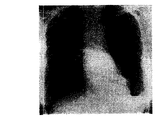
【0040】
この対側差分法は、単一胸部X線写真において、非対称異常、例えば肺結節、気胸、肺炎、および水疱を検出するのに使用が可能である。図9(a)は、肺結節を有する胸部X線写真を示し、図9(b)には、その対側差分画像を示す。肺結節は、差分画像において強調されることが見て取れる。これは、対称的な骨格構造が相殺されて消されるからである。
【0041】
本発明では、胸郭の中線はほぼ直線によって表わすことが可能であると仮定された。この仮定は、PA胸部画像の大部分において有効であると思われる。しかしながら、個々のX線写真には大きな変動があるものであるから、この仮定は、全ての臨床的胸部X線写真の中線を記述するのに必ずしも有効であるとは限らない。ある症例においては、中線を表わすのに非直線的モデルを必要とされることがあるかもしれない。例えば、この曲線中線を表わすには、2または3元多項式が好適であるかもしれない。にも拘わらず、本発明では直線モデルを用いた。これは、中線は胸部X線写真における胸郭の側方傾斜の補正のためにのみ使用されるからである。直線モデルは単純で、かつ本発明には十分であることを銘記しなければならない。複雑な非直線性モデルは問題を複雑にし、適正な中線補正を求めるに当って困難に導く可能性がある。
【0042】
過去においては、差分画像は、512x512という小さなマトリックスサイズで表示されていた。これは、交差相関法とそれに続く画像変形法が、この小さなマトリックスサイズに適用されていたからである。本発明では、高画質で大きなマトリックス差分画像を表示する技法、そしてそれと等価的なものとして、小ピクセルサイズを持つ、高画質差分画像を表示する技法が新規導入された。これは、後に詳述するように、対側差分法においては反転鏡像の画像変形のために、1024x1024または2048x2048のような大きなマトリックス画像を用い、時間差分法の場合には従来通りの画像を用いることによって達成された。座標変換の変位値は初回では512x512のマトリックスサイズで定めたが、大マトリックスサイズに対する対応変位値は、1024x1024または2048x2048のマトリックスサイズにおける反転鏡像の大きさを、それぞれ係数2または4を用いることによって得た(または、時間差分法の場合は従来画像)。大マトリックス差分画像は、元の大マトリックス胸部画像から、大マトリックス変形画像を差し引くことによって得た。大マトリックスサイズ表示のための、従って小ピクセルサイズ使用のためのこの方法は、時間差分法や対側差分法用に小マトリックスサイズ(または大ピクセルサイズ)を用いることによって生ずるピクセル端や、ピクセルアーチファクトを除去することができるので、高画質差分画像を得るのに極めて有効であった。また、画像ノイズや、比較的小さな登録不備アーチファクトを除去することによって差分画像を平滑化するために、中央値フィルターや、ガウスフィルターのような画像処理法を用いることも効果的である。
【0043】
新規中線検出法による時間差分改良法
本発明による対側差分画像改良のための前記3種の技法は、時間差分法に、その時間差分画像の画質改善のために適用が可能である。[1,2]時間差分法の基本設計をここに簡単に述べる。いくつかのテンプレートROI(32x32のマトリックス)と、対応する探索域ROI(64x64マトリックス)が、それぞれ先行画像と現行画像から選択され、それによって、この2個の画像において画像細部の局所適合が実施される。次に、選択されたROIの全てのペアについて、変位値ΔxとΔyを交差相関法を用いて求め、それによって、探索域ROIにおいて最高適合域を見出す。多項式関数による2次平面適合を、各組の、マップされた変位値ΔxとΔyに適用して、先行画像のx,y座標値を変換して画像を変形する。次に、この変形された先行画像を現行画像から差し引き、時間差分画像を生成する。この時間差分画像は、胸部画像において、微妙な時間差変化を検出するのに極めて有用であることが従来から判明している。時間差分画像において、登録ミスアーチファクトを減らすために、現行画像と最初の変形先行画像を用いることで、さらに別の画像変形を採用することが可能である。最終的な時間差分画像は、現行画像から第二の変形先行画像を差し引くことによって得られる。この画像変形法は繰り返し適用することが可能である。従って、この方法は繰り返し画像変形法と呼ばれる。[5]
図1(b)は、対側差分法における前述の中線検出法使用(例えば、図1(a)のステップS200)による時間差分改良法の全体設計図である。図1(b)では、ステップS100’において、現行胸部画像と先行胸部画像が獲得される。ステップS200’において、対側差分法の中線検出法を用いて、この獲得された画像に対して側方傾斜補正が実施される。次に、ステップS500’において、時間差分法が実行され、ステップS600’において、時間的に差分された画像が表示される。
【0044】
本発明によれば、中線検出改善のために導入された肋骨辺縁準拠法が特に有用であることが判明した。それは、表4と表5に示した主観的評価の結果が示す通りである。
【表4】
時間差分画像に画質に関する様々な評価得点における、胸部画像例数の分布。ただし、これらの画像は、側方傾斜を補正するのに輪郭準拠中線検出法と、肋骨枠辺縁準拠中線検出法とを用いて得た。
【表5】
輪郭準拠中線検出法を、肋骨枠辺縁準拠中線検出法に替えることによって得られた、時間差分画像画質の改善における、様々な主観的評価得点に対する胸部画像例数分布
本発明では、日本の岩手県で行われた肺癌集団検診で得られた、181対の現行・先行胸部画像を用いた。これらの胸部画像は、移動検査設備に設置された富士コンピュータ処理放射線検査システム(CR)を使用して得た。このCRシステムからのディジタル画像データを直接使用して、画像変形法による時間差分画像を計算によって求めた。表4と表5において、時間差分画像の画質は、新規の中線検出法を用いることによってかなり改善されることが明らかに見て取れる。
【0047】
前述の高画質、大マトリックス差分画像表示法を、時間差分画像に適用することも可能である。
【0048】
対側差分―胸部X線写真における肺結節のコンピュータ検出における偽似陽性の除去
対側差分法をコンピュータ支援診断計画に組み込み、それによって、全体性能を改善することが可能である。例えば、差分画像法による胸部X線写真の肺結節のコンピュータ検出においては、肋骨・肋骨交差、または、血管・肋骨交差によってもたらされる偽似陽性の内のいくつかは、除去がきわめて困難である。これらの偽似陽性は、対側差分画像から導かれる画像特質を分析することによって除去が可能である。
【0049】
コンピュータ化差分画像法を簡単にここで述べる。[8]先ず、ディジタル胸部画像、または通常の胸部X線写真のディジタル化版を、平行して操作される二つのフィルター操作にかける。一方は、適合フィルターのようなフィルターを用いることにより結節の強調画像を生成し、他方は、リング平均フィルターのような、また別のフィルターを用いることによって結節の抑制画像を供給する。次に、差分画像は、強調画像から抑制画像を差し引くことによって得られる。この差分画像は、極度に強調された結節と、大多数の背景となる正常解剖構造の除去による極度に抑制された背景を含むから、結節候補を特定するのに有用である。この差分画像法は、辺縁強調法の一般化と考えられ、事実、この差分画像は辺縁強調画像と酷似する。もしも2枚のフィルターが直線フィルターであれば、この2枚を結合させて、1個のフィルター操作とすることも可能である。一方、非直線性フィルターを用いるのであれば、二つのフィルター操作は別々に平行して適用する必要がある。この差分画像法は、孤立した異常パターン、例えば微小石灰化病巣や、マンモグラムにおける固まり等のコンピュータ検出に適用が可能である。
【0050】
肺結節の初期候補は、差分画像において、大きなピクセル値を持つ比較的円形のパターンから特定される。しかし、そのようなパターンは、結節と同時に肋骨や、肺の血管といった正常構造を含むかもしれない。従って、これら候補の画像特質が抽出され、大きさ、コントラスト、候補の形に関するその他のパラメータに関して定量化される。具体的には、抽出される特質は、グレイレベル、形態、または実効直径のような辺縁勾配、円形度と不整性、実効直径の勾配と、円形性と不整性の度合い、平均勾配、勾配方向の標準偏差、コントラストと残高コントラスト(例えば、特許出願08/562,087に教示されるような)に関わる。次に、基準依拠法(例えば、特許5,463,548と5,622,171、および、特許出願08/562,087;08/562,188;08/758,438;08/900,361と09/027,685に教示される)を適用して、もしもいくつかの候補の特質が、正常の解剖構造、例えば正面向きの血管、肋骨・肋骨交差、肋骨・血管交差、血管集合、および、肋骨・鎖骨交差のものと適合する場合には、それら候補を偽似陽性として除去する。
【0051】
最後に、いくつかの画像特質使用によって訓練された、訓練済みの人工神経ネットワーク(ANN)を用いて、残りの候補の中の偽似陽性をさらに除去する(例えば、特許5,463,548と5,622,171、および、特許出願08/562,087;08/562,188;08/758,438;08/900,361と09/027,685に教示されるような)。肺結節を有する100枚の胸部画像と、100枚の正常例から成るデータベースを用いた場合、差分画像法の性能は70%の感度であり、偽似陽性率は画像1枚当り1.7であった。
【0052】
図1(c)は、単一胸部X線写真における肺結節のコンピュータ検出において、対側差分法による差分画像から偽似陽性を除去するための全体設計である。
【0053】
本発明では、それぞれが肺の孤立結節を含む10枚の胸部X線写真を得た(ステップS100)。この研究の結果から、10個の肺結節の全てが、コンピュータ化差分画像法によって正しく検出されたことが明らかにされたが、また、10枚の胸部画像の肺の分野において、合計14個の偽似陽性が出現したことが報告された(ステップS700−S800)。これら偽似陽性の全てが、肋骨・肋骨交差、肋骨・血管交差、および肋骨・鎖骨交差等肋骨に関連していた。
【0054】
次に、対側差分画像を10枚の胸部画像全てについて得た(ステップS200−S600)。元の胸部画像上の24部位全てを−この24部位は、10個の結節と、14個の偽似陽性を含み、結節検出法のコンピュータ出力を表わすものであるが−対側差分画像上の対応位置に変換した(ステップS900)。この変換は必要である。なぜなら、前述のように、元の胸部画像を側方傾斜の補正のために回転し、変位させて後(ステップS200−S600)、対側差分画像は得られるものだからである。
【0055】
対側差分画像において、一方の肺の肋骨が他方の肺の肋骨に適合する場合、一般に肋骨は除去され、従って、隣接背景と同様の均一な密度を有するようになるから、結節は暗い、円形パターンとして出現する。従って、コンピュータ検出による結節候補に関連する画像特質が肋骨のものと合致するならば、肋骨によるコンピュータの偽似陽性出力は特定が可能である。結節と、肋骨による偽似陽性とを区別するための画像特質として、結節候補のコントラストを用いた(ステップS1000)。ここでは、コントラストを候補の中央部と直近の背景の間の平均ピクセル値における差と定義する。候補の中央部の平均ピクセル値は、直径dの円において経験的に定め、一方、背景部の平均ピクセル値は、内径dで、外径2dのドーナツ型区域について定めた。結節と非結節(偽似陽性)を含む全ての候補のコントラストを、dを2mmから40mmの広範囲に変化させて求めた。対側差分画像において、結節のコントラストは、一般に非結節のものよりも大きい傾向のあることが判明した。例えば、図10は、10mmと20mm直径における結節のコントラストは、20ピクセル値より大きいが(点線)、一方、6個の非結節が20ピクセル値未満の低コントラストを持っていた。従って、この結果から、もしも、対側差分画像において、ある指定の閾値レベル、例えば、20ピクセル値未満のコントラストを含む結節候補を除去するとすれば、肺結節の可能部位を示すコンピュータ出力リストの内から、6個の偽似陽性を除去することが可能であることが明らかにされた。
【0056】
しかしながら、10個の結節のコントラストとほぼ等しい、大きなコントラストを有する8個の非結節がある。いくつかの非結節の大きなコントラストは、対側差分画像における登録ミス・アーチファクトによるものであることが判明した。登録ミス・アーチファクトによるパターンは、一般に、局所的な明暗密度の混合体を含むから、中央部および・または背景部におけるピクセル値の標準偏差は、登録ミス・アーチファクトのない部分に比べて大きくなることが予想される。図11は、中央部と背景部における相対的標準偏差を示す。この相対的標準偏差は、中央部、および・または背景部における、ピクセル値変動の標準偏差の平均ピクセル値に対する比として求めた。図11から見て取れることは、結節における相対的標準偏差は比較的小さいが、15%(点線)を越える大きな相対標準偏差−これは登録ミス・アーチファクトによるものである−を有する非結節は5個あるということである。従って、この結果から、もしも、対側差分画像中のある結節候補の相対標準偏差が、指定の閾値レベルを越える大きな値を有する場合、その結節候補を除去するとすれば、5個の偽似陽性を除去することが可能であることが示された。図10と11の結果を合わせることによって、14個の偽似陽性の内、10個の除去が可能である。なぜならば、図10と図11のいずれでも除去可能な「重複」偽似陽性が1個あるからである。
【0057】
偽似陽性除去のためのもう一つの方法を図12に示す。同図は、結節のコントラストと、中央部の相対標準偏差との間の相関を示す。図12から明らかに見て取れるのは、結節は大きなコントラストと、小さな標準偏差を有する傾向を持つのに対して、非結節は、小さなコントラストを有するか、または大きなコントラストと大きな相対標準偏差を有する傾向を持つ。従って、点線で示すような、コントラスト対標準偏差閾値を設けた場合、14個の偽似陽性の内13個の除去が可能である(ステップS1200−S1300)。この結果から、対側差分画像を用いることによって、多数の偽似陽性の除去が可能であること、従って、胸部画像における肺結節検出のためのコンピュータ検査法の性能を実質的に改善することが可能であることが示される。
【0058】
図13は、単一胸部X線写真における非対称異常検出のためのコンピュータシステムの模式図である。コンピュータ100は本発明の方法を導入する。同方法においては、コンピュータは、例えば、タッチスクリーン・インターフェイスを有するタッチスクリーン・モニターのような表示装置102;キーボード104;ポイント装置106;マウスパッドまたはディジタル入力パッド108;適当なデバイスバス(例えば、SCSIバス、強化IDEバス、ウルトラDMAバス、PCIバス等)で接続された、ハードディスク110、または、その他の高密度メディアドライブ;フロッピードライブ112;テープまたはCDメディア付、テープまたはCD−ROMドライブ114;または、光磁気メディア等のような、その他の取り外し可能なメディア装置;および、マザーボードを含む。マザーボード118は、例えば、プロセッサー120;RAM122とROM124(例えば、DRAM、ROM、EPROM、EEPROM、SRAM、SDRAM、および、フラッシュRAM等);画像獲得装置(図示せず)と接続して使用されてもよいIOポート126;および、音声加工、画像加工、信号加工、ニューラルネットワーク加工等のような特殊なハードウェア/ソフトウェア機能を実行するための、要すればあってもよい特別用途論理装置(例えば、ASIC)、または、構成可能論理装置(例えば、GALおよびプログラム変更可能FPGA);マイクロフォン130;および、単一または複数のスピーカー132、を含む。
【0059】
前述のように、本システムは少なくとも1個のコンピュータ読み取り可能な媒体を含む。コンピュータ読み取り可能な媒体の実例としては、コンパクトディスク、ハードディスク、フロッピーディスク、テープ、光磁気ディスク、PROM(EPROM、EEPROM、フラッシュEPROM)、DRAM、SRAM、SDRAM等である。本発明は、コンピュータ読み取り可能な、いずれのものであってもよい一つの媒体、または、それらの結合体の中に保存されるが、コンピュータ100のハードウェアを制御し、かつコンピュータ100が、人間ユーザーと対話することを可能にするソフトウェアを含む。このようなソフトウェアは、デバイスドライバー、オペレーティング・システム、および開発ツールのようなユーザー用アプリケーションを含んでいてもよいが、ただし、それらに限定されない。このようなコンピュータ読み取り可能な媒体はさらに、前述の本発明のプロセス(例えば、図1(a)−(c)参照)のいずれのものかを実行するための、本発明のコンピュータ・プログラム製品を含む。本発明のコンピュータコード・デバイスは、任意の解釈される、または実行可能なコードメカニズムであってもよく、スクリプト、翻訳機、動的結合ライブラリー、ジャバ・クラス、および完全に実行可能なプログラム等を含むが、ただしそれらに限定されない。
【0060】
汎用コンピュータ100のプログラムは、画像獲得装置(図示せず)から得た画像をディジタル化し、保存するためのソフトウェア・モジュールを含む。また、別態様として、本発明は、絵画文書交信装置(PACS)のような、その他の手段で得た画像からのディジタルデータを加工するように設置することも可能である。別言すれば、加工の対象となるディジタル画像は、ディジタル形式で存在することが多いものであるから、その場合は、本発明を実施するに当って、ディジタル形式に変換する必要はない。
【0061】
本発明はさらに、当業者には直ちに明らかなように、アプリケーション特異的集積回路の作製によって、または、従来の成分回路から成る適当なネットワークを相互接続することによっても導入が可能である。
【0062】
さらに本発明は、コントラスト、相対標準偏差、および・または、結節中心部および・または背景部におけるコントラストと相対標準偏差との間の相関に基づいて、結節候補の中の偽似陽性を除去する点において定義されたけれども、当業者には直ちに明らかなように、結節の他の画像特質、および・または、他の部分も使用が可能である。
【0063】
本発明では、単一胸部X線写真における非対称不透過部検出のために、新規な対側差分法の再構成が行われ、対側差分画像改善のための3種の技法(すなわち、側方傾斜補正、心臓領域の除去、および左と右肺における個別的変位値適合)が用いられた。本対側差分法は、胸部X線写真において周辺肋骨の大部分を除去し、かつ低コントラストの末梢病巣を強調することを可能とする。本法は微妙な肺の不透過部を検出するに当り、放射線専門家を補佐するものとなり得ると考えられる。
【0064】
明らかに、本発明について多くの修正や変更が、前述の教示に照らして可能である。従って、請求項の範囲内においても、本発明をここに詳述したのとは別のやり方で実行することが可能であることを理解しなければならない。
【0065】
付録
[1]A. Kano, K. Doi, H. MacMahon, D.D. Hassel, M. Giger
「時間間隔変化を検出するための、時間的に連続する胸部画像のディジタル画像差分法」
Med. Phys. 21, 453−461(1994)、および、米国特許5,359,513
[2]M.C. Difazio, H. MacMahon, X.W. Xu, P. Tsai, J. Shiraishi, S.G. Armato III, K. Doi
「ディジタル胸部X線撮影法:時間差分画像の検出正確度に及ぼす作用」
Radiology 202, 447−452 (1997)
[3〕J. Shiraishi, S. Katsuragawa, J. Ikezoe, T. Matsumoto, T. Kobayashi, K. Doi等
「肺結節を含む胸部X線写真のディジタル画像データベースの開発−ROC分析による評価」
Radiology 205(p), 394, 1997
[4]X.W. Xu, K. Doi
「コンピュータ支援診断のための画像特質分析−胸部X線写真における肋骨枠境界の正確な確定」
Med. Phys. 22, 617−626(1995)および米国特許5,790,690
[5]T. Ishida, S. Katsuragawa, K. Nakamura, H. MacMahon, K. Doi
「時間差変化検出のための、連続胸部X線写真における時間差分用頻回画像変形法」
Med. Physに投稿、および、米国特許出願09/053,789
[6]T. Ishida, K. Ashizawa, R. Engelman, S. Katsuragawa, H. MacMahon, K. Doi
「胸部X線写真の時間差変化検出のための時間差分法の適用−自動的初回画像適合による差分画像の改善」
Journal of Digital Imagingに投稿、および、米国特許出願08/900,362
[7]N. Nakamori, K. Doi, V. Sabeti, H. MacMahon
「ディジタル放射線学における画像特質分析とコンピュータ支援診断−ディジタル胸部画像における心臓・肺サイズの自動分析」
Med Phys. 17, 342−350(1990)および米国特許5,072,384
[8]X.W. Xu, K. Doi, T. Kobayashi, H. MacMahon, M.L. Giger
「ディジタル胸部画像における肺結節の自動化検出のためのCAD改良法の開発」
Med. Phys. 24, 1395−1403(1997)および米国特許出願08/562,087、および、その他の特許と特許出願(特許4,907,156; 5,224,177; 5,289,374および特許出願08/757,611; 08/900,191; 09/027,685
【図面の簡単な説明】
【図1(a)】 PA胸部画像に対する対側差分法の全体設計図である。
【図1(b)】 対側差分法の中線検出法を用いた時間差分改良法の全体設計図である。
【図1(c)】 単一胸部X線画像の肺結節コンピュータ検出において、異なる画像から得られる偽似陽性を除去するための全体設計図である。
【図2(a)】 比較のために示した画像であって、元の画像である。
【図2(b)】 比較のために示した画像であって、全ての領域を含む対側差分画像である。
【図2(c)】 比較のために示した画像であって、2つの肺の外側の区域全体を均一な背景とした対側差分画像である。
【図2(d)】 比較のために示した画像であって、「胸部」背景付の対側差分画像であって、2つの肺の外側の区域全体において元の画像を重ねたものである。
【図3】 黄金基準に対して、検出された中線の角度と位置の誤差測定値を例示したものである。
【図4】 黄金基準(黒実線)を中線とする胸部X線写真、輪郭線準拠法(鎖線)によって得られた検出中線、および、肋骨枠辺縁依拠法(点線)によって得られた検出中線である。
【図5(a)】 3人の観察者によって示された、個々の中線における黄金基準からの角度と位置の分布を示すグラフである。
【図5(b)】 輪郭準拠法によって検出された中線の誤差分布を示すグラフである。
【図5(c)】 肋骨枠準拠法によって検出された中線の誤差分布を示すグラフである。
【図6(a)】 比較のために示した画像であって、元の胸部X線写真である。
【図6(b)】 比較のために示した画像であって、中線検出のために輪郭準拠法を用いて得られた対側差分画像である。
【図6(c)】 比較のために示した画像であって、中線検出のために肋骨辺縁準拠法を用いて得られた改良型差分画像である。
【図7(a)】 比較のために示した画像であって、元の胸部X線写真である。
【図7(b)】 比較のために示した画像であって、変位値分析のために縦隔・心臓領域を含んで得られた対側差分画像である。
【図7(c)】 比較のために示した画像であって、同変位値から縦隔・心臓領域を除いて得られた改良型対側差分画像である。
【図8(a)】 比較のために示した画像であって、元の胸部X線写真である。
【図8(b)】 比較のために示した画像であって、変位値を右と左の肺に同時適合させることによって得られる対側差分画像である。
【図8(c)】 比較のために示した画像であって、右と左の肺において、変位値を分離適合させることによって得られる改良型対側差分画像である。
【図9(a)】 比較のために示した画像であって、提示された結節画像である。
【図9(b)】 比較のために示した画像であって、結節を強調した対側差分画像である。
【図10】 結節と結節候補について、10mmと20mm直径において結節候補のコントラスト分布を示す。
【図11】 結節と結節候補について、10mm直径において、結節候補の対象領域と背景領域における相対的標準偏差の分布を示す。
【図12】 結節と結節候補について、10mm直径におけるコントラストと、相対的標準偏差の間の関係を示すグラフである。
【図13】 本発明の教示に従ってプログラムされた、汎用コンピュータ100の模式図である。
【符号の説明】
100…コンピュータ 102…表示装置 104…キーボード 106…ポイント装置 108…ディジタル入力パッド 110…ハードディスク 112…フロッピードライブ 114…ROMドライブ 118…マザーボード 120…プロセッサー 122…RAM 124…ROM 126…IOポート 130…マイクロフォン 132…スピーカー[0001]
Notice on federal aid
Part of this invention was made with US government support under USPHS research grants CA62625 and CA64370 (National Health Institute). The United States government has certain rights in this study.
[0002]
(Field of Invention)
The present invention generally provides a computer processing method and system provided to assist radiologists in detecting abnormalities such as pulmonary nodules, pneumothorax, pneumonia, and blisters in chest radiographs. Involved.
[0003]
The present invention is more generally described, for example, in U.S. Pat. Nos. 4,839,807; 4,841,555; 4,851,984; 4,875,165; 4,907,156; 4,918,534; 5,133,020; 5,150,292; 5,224,177; 5,289,374; 5,319,549; 5,343,390; 5,359,513; 5,452, 367; 5,463,548; 5,491,627; 5,537,485; 5,598,481; 5,622,171; 5,638,458; 5,657,362; 5,666,434; 5,673,332; 5,668,888; 5,740,268; 5,790,690; and 5,832,103; and US patent application 08 / 158,388 (PCT publication WO 95). 08431, 935; 08/220, 917 (PCT publication WO 95/26682); 08/398, 307 (PCT publication WO 96/27846); 08/523, 210 (PCT publication WO 95/15537); 08/562, 087; 08/757, 611; 08/758, 438; 08/900, 191; 08/900, 361; 08/900, 362; 08/900, 188; 08/900, 08/900, 192; 08/979, 623; 08/979, 639; 08 / 982,282; 09 / 027,468; 09 / 027,685; 09 / 028,518; 09 / 053,798; 09 / 092,004; 09 / 098,504; 09 / 121,719; 09 / 131,162 09 / 141,535; and 09 / 156,413 are disclosed in one or more of the, related to computer processing technology for digital image automatic analysis. The above patents and patent applications are cited and included in this application. Among the above patents and patent applications, in particular 4,907,156; 5,072,384; 5,224,177; 5,289,374; 5,319,549; 5,359,513; 5,463, 548; 5,622,171; 5,790,690; 08 / 562,087; 08 / 562,188; 08 / 757,611; 08 / 758,438; 08 / 900,191; 08 / 900,361; 08 / 900,362; 09 / 027,685; 09 / 053,789; and 09 / 121,719 are interesting.
[0004]
The present invention includes various techniques described in the above US patents and patent applications, as well as cited references identified in the attached “Appendix” by author (s) and year of publication, and is listed in the Appendix. Cross-referenced throughout the application by parenthesized numbers corresponding to each cited reference, the entire contents of which include the related patents and patent applications listed above, and the cited references listed in the Appendix, It is incorporated herein as a cited reference.
[0005]
(Background of the Invention)
Detecting early lung cancer in chest radiographs is a difficult task for radiologists. This is because subtle lesions have low contrast and can overlap with ribs and clavicles. A time difference method has been reported to assist radiologists in detecting newly occurring abnormalities in chest radiographs. [1] In this method, the previous chest image is subtracted from the current chest image, thereby generating a subtracted image. Since subtle changes in chest radiographs are emphasized in this subtracted image, the detection accuracy of this time interval change may be noticeably improved by using this time difference method. [2] However, this time difference method cannot be applied when there is no preceding chest photo.
[0006]
(Summary of Invention)
Accordingly, it is an object of the present invention to provide a method and system for detecting asymmetric abnormalities in a single chest radiograph by computer processing.
[0007]
It is another object of the present invention to provide a method and system for computer-aided detection of asymmetric abnormalities in a single chest radiograph using the contralateral difference method.
[0008]
In addition, another object of the present invention is to provide a method and system for computerized detection of asymmetry abnormalities in a single chest radiograph, comprising lateral tilt correction, inverted “mirror” image, mirror image It is characterized by using a contralateral difference method that obtains the contralateral difference image by subtracting the distortion and the distorted mirror image from the original image.
[0009]
Yet another object of the present invention is to provide a method and system for computerized detection of asymmetric abnormalities in a single chest radiograph using contralateral difference images, wherein the first contralateral difference image An improved difference image is obtained by sequentially applying three types of techniques.
[0010]
Yet another object of the present invention is to provide a method and system for removing false positives obtained from different images by contralateral difference method in computerized detection of lung nodules in a single chest radiograph. It is to be.
[0011]
Yet another object of the present invention is to provide a method and system for improved time difference execution by the midline detection method of the contralateral difference method.
[0012]
These and other objects are achieved in accordance with the present invention by providing a novel method, system, and computer readable medium for chest image computer processing. The processing includes acquiring a first digital image of the chest, generating a second image that is a mirror image of the first image, performing image transformation on one of the first and second images, Generating a deformed image, where the deformed image is registered in the other of the first and second images, and further includes subtracting the deformed image from the other image to generate a difference image.
[0013]
Another embodiment useful when multiple time-separated images are available is to acquire a first digital image of a subject's chest, in the first chest image, the rib borders on both sides of the lung Detecting the edge, obtaining the average horizontal part of the left and right rib frame edges at multiple vertical parts, aligning the obtained average horizontal part with a straight line, guiding the middle line, and chest image And rotating the rotated image so as to generate a second image in which the center line of the image subjected to the same side inclination correction is subjected to the side inclination correction passing through the center thereof. including.
[0014]
The invention further includes computer readable program instructions and a medium for storing the method execution system of the invention. Here, the program command is a program command that enables the method of the present invention to be executed when the stored program command is properly loaded into a computer.
[0015]
Therefore, according to the present invention, a novel contralateral difference method has been developed that is based on a single anteroposterior (PA) chest image. Since the rib structure is almost symmetrical, the chest image of the right lung periphery generally approximates that of the left lung. Taking advantage of this, the technique can correct the side tilt by rotating and displacing the original image so that the midline of the rib cage is aligned with the vertical center line of the original image; Side rotation of the rotated image to generate; deformation of the mirror image; and subtraction calculation of the deformed mirror image from the original image to obtain a contralateral difference image. Thereafter, it is possible to continuously apply a processing technique to the first contralateral difference image, thereby obtaining an improved difference image. In this technique, one contralateral difference image can be obtained by subtracting its right / left inverted “mirror” image from a single chest image. Similar to the time difference method, this contralateral difference method can cancel out many of the symmetric skeletons and emphasize the asymmetrical impermeability, thereby making it possible to present subtle anomalies more clearly. is there. On the other hand, unlike the time difference method, a difference image can be acquired at any time as long as a single PA image is available. Therefore, the contralateral difference method may have important clinical significance in some cases.
[0016]
The present invention and many of the attendant advantages can be better understood when the following detailed description is considered in conjunction with the accompanying drawings.
[0017]
(Detailed explanation)
The chest image used to develop the present invention consists of 50 normal images and 50 abnormal images including isolated lung nodules. These images were selected from 247 chest images in the Japanese digital image standard database developed by the Japanese Society of Radiological Technology. [3] These images were digitized under a 0.175 mm pixel size, a 2048 × 2048 matrix size, and a 12-bit gray scale. On the other hand, by further sampling (sub-sampling) the original image data, the matrix size was reduced to 512 × 512 and the number of gray levels was reduced to 10 bits.
[0018]
In the drawings, like reference numerals refer to the same or corresponding parts throughout the several views, and more particularly to FIG. 1 (a), the contralateral difference method according to the present invention. The upper block diagram is shown.
[0019]
First of all, for the PA image acquired in step S100, the lateral tilt caused by the incorrect positioning of the patient is corrected by the image rotation method in step S200. The rotated image is inverted laterally (right / left) in step S300 to generate an inverted “mirror” image. The mirror image is registered with the original image by the cross-correlation method, and then deformed so that the peripheral ribs in the mirror image match the peripheral ribs of the original image based on the non-linear image deformation method in step S400. Is done. [1] Finally, in step S500, the deformed image is subtracted from the original image, and in step S600, a contralateral difference image is obtained. Next, details of steps S100-S600 will be described.
[0020]
Side tilt correction (step S200) is specifically designed to correct the lateral tilt of the thorax in the chest image. If the midline of the rib cage is slightly tilted at an angle with respect to the vertical direction, the angle between the midline of the original chest image and the inverted chest image is twice as large. This results in a significant registration error and therefore a bad difference image. Therefore, it is necessary to correct this side slope before applying the difference method. This is performed in step S200 by rotating the image so that its midline is in the vertical direction, and then displacing the midline of the rib cage to the vertical centerline of the original chest image. The rotation and displacement of the image can be executed by, for example, a normal image rotation / displacement method known in image technology.
[0021]
Midline detection
In the past, contour-based midline detection has been applied to computer aided diagnostics (CAD), leaving relatively satisfactory results. [5, 6] First, nine regions of interest (ROI) are selected from the top to the bottom of the lung in the mediastinum region. In order to reduce the effect of noise level on midline determination, the pixel values at each ROI are averaged in the vertical direction. This gives a one-dimensional (1-D) horizontal profile. For each profile, the maximum pixel value and its corresponding position are determined. Finally, the midline is determined by fitting nine points to a straight line.
[0022]
If the pixel values along the midline have a local maximum along a straight line, this method may give satisfactory results. However, in many cases, the pixel value on that midline is not necessarily a local maximum. For example, the pixel values in the heart region are often larger than those near the midline, and therefore the detection midline is generally displaced toward the heart region side and, as will be illustrated later, It becomes a middle line. Furthermore, even if the pixel values on the midline give local maximum values, the difference between these values and the pixel values in the adjacent area is very small. For this reason, the conventional method is inherently sensitive to the influence of image noise. Therefore, according to the present invention, a novel midline detection method based on the rib frame edge is devised as described below.
[0023]
First, the rib borders on both sides of both lungs are detected. Second, the average horizontal position of the left and right rib frame edges is determined, and then fitted to a straight line to obtain a midline. This rib frame edge is detected by analyzing the first and second derivatives of the overall bust profile. [4] Next, as described in detail elsewhere, this detected rib frame edge is fitted to a ternary polynomial to form a smooth curve and reduce noise [4] Both the rib frame edge detection method and the aforementioned midline detection conventional method detect edges or peaks by analyzing the profiles and fitting them to some function. However, the result of the rib frame edge detection method is much more reliable than the result of the conventional midline detection method. This is because the contrast at the edge of the rib frame is usually much larger. This is the reason why it is preferable to use the rib frame edge to determine the midline in the present invention.
[0024]
Once the right and left rib frame edges are obtained, the horizontal average position (ie, midpoint) of the right and left rib frame edges at the same vertical position of the chest image is determined. Based on the nearly symmetric nature of the rib borders in the bilateral lungs, many of the midpoints (ie, the average position) will lie on a straight line, so these points are fitted to the straight line and Generate a line. In other words, a straight line is required that divides both lungs into two approximately symmetrical equal parts. Since the midpoint is obtained from the horizontal average position of the right and left rib frame edges, the error variation at these midpoints is expected to be half that of the detected rib frame edges. . This also contributes to further improving the accuracy of the detected midline.
[0025]
Determination of golden reference for chest image midline
In order to evaluate the accuracy of the detected midline, “true” data about the midline of the PA chest image is needed. By this invention, the “golden standard” based on subjective judgment was obtained. For each of the 100 chest images, three observers independently showed the ends of the midline using a mouse on the computer screen. For the three midlines thus positioned, the corresponding endpoints were averaged to obtain a golden reference in each case.
[0026]
Non-linear deformation of right / left inverted “mirror” image (steps S300, S400)
After the side tilt is corrected in step S200, a right / left inverted mirror image is obtained by inverting the original image to the side in step S300. This inverted mirror image is deformed and then subtracted from the original image to produce a contralateral difference image. As described in detail in the previous report (for example, see Patent Application Serial No. 09 / 053,789), in the time difference method applied to the chest image, a nonlinear deformation method has been used successfully. According to the present invention, a similar image deformation method is used to perform image registration. However, appropriate correction for image deformation is performed as applied to the following contralateral difference. This non-linear deformation method includes an initial coarse fit, a detailed local fit of the rib edges, displacement value determination, and coordinate transformation. Further, non-linear deformation can be performed repeatedly to produce improved results. [5]
First coarse fit in whole chest image
The first coarse fitting method, which has already been applied to the time difference method, is used to align the approximated lung area of the inverted chest image with the corresponding area of the original image. [6] First, the matrix size of the two images, ie, the original chest image and the inverted image, is reduced by a factor of 4, and is further smoothed by a Gaussian filter so that fine blood vessels, bronchi, devices, catheters, etc. Reduces the effect of the anatomy on the coarse fit of the two images. Next, using the following rib frame edge information, both lungs are extracted from each of the two images, and the region outside the rib edge is ignored. Finally, the upper parts of both lungs are aligned using the cross-correlation method.
[0027]
Local image adaptation of peripheral ribs, excluding mediastinum and heart region
To perform local image fitting of the peripheral ribs of the right and left lungs, it is necessary to select a number of template ROIs and search area ROIs. The template ROI and the corresponding search area ROI are automatically positioned in the lung regions of the original image and the inverted image, respectively. This is done in the same way that both ROIs are located by the time difference method. [1] The matrix sizes of the template ROI and the search area ROI are 32 × 32 and 64 × 64, respectively. Here, in the contralateral difference method, it is important to note that the mediastinum and the heart region are excluded in selecting the ROI. This is because ROIs in the mediastinum and heart region do not contain information useful for performing reliable image fitting in the contralateral difference method. Once the template ROI and the search area ROI are determined, the displacement values Δx and Δy in two mutually perpendicular directions are obtained using the cross correlation method. This displacement value indicates the displacement that the center coordinates of the reverse mirror image search area ROI should take in order for the template ROI to give the maximum fit to the corresponding “fit” area in the search area ROI. [1]
Individual fit of displacement values in the right and left lungs
Since there is some variation in the displacement value over the entire lung region, each set of displacement values (Δx and Δy) was smoothed using quadratic plane fitting with a 10-way polynomial. [1] Initially, as used in the time difference method, each set of displacement values in the right and left lungs was fitted simultaneously. However, in the contralateral difference method, the displacement values in the right and left lungs were found to be completely different in some cases. Therefore, in the contralateral difference, the displacement value in the right lung and the displacement value in the left lung were separately adapted independently. This individual fitting method is possible because the contralateral difference method does not require a single smoothing function over the entire chest image. Coordinate transformation is performed for each of the right and left lungs in the inverted image using four sets of 10-element polynomials for each of Δx and Δy. Thereby, a modified inverted image in the contralateral difference method is obtained.
[0028]
Display of contralateral difference image (steps S500 and S600)
The contralateral difference image is obtained by subtracting the deformed and inverted image from the original image in step S500. In order to enhance the image contrast to an appropriate level, the pixel value of the difference image is multiplied by a coefficient, for example, 1.5 or 2.0. On the other hand, a coefficient of 1 can be used to maintain an abnormal opaque portion in the contralateral difference image at the same contrast as that of the original image. Next, a pixel steady value of 512 is added to the contrast-enhanced difference image, and a pixel steady value of 512 is given to the background area outside the lungs, thereby displaying only the contralateral difference image in the surrounding lungs. To do. It should be noted that mediastinal and cardiac region displays are not useful and can be a disturbing target for radiologists by introducing large “registration error” artifacts. 2 (a) is an original chest image, and FIGS. 2 (b) and 2 (c) are contralateral differences when the background is not uniform for comparison. Images are shown. A difference image having a uniform background can be effectively represented by presenting asymmetric opaque portions in the lung region as a dark pattern on the same side and as a light color pattern on the opposite side. In addition, note that the original PA chest image outside the lung region is displayed to maintain the general “chest” background appearance, rather than a steady background as shown in FIG. 2 (d). There must be. In fact, many radiographers preferred the display of the contralateral difference image of FIG.
[0029]
Subjective evaluation of difference image quality.
In order to subjectively evaluate the quality of the contralateral difference image, a five-point evaluation scoring method conventionally used for subjective evaluation of time difference images was used according to the present invention. That is,
5 (excellent): all ribs are fully registered (and thus removed);
4 (Good): Most ribs were almost completely registered, but some had very few misregistration errors;
3 (Yes): Most ribs were well registered, but there were some slight misregistration errors;
2 (Poor): Most ribs are well registered, but appear in half of the ribs;
1 (not possible): Most ribs are not registered and appear throughout the intercostal space.
[0030]
Another evaluation method was also used to examine changes in the quality of the difference image due to the use of the improved method described below. The evaluation score was in the range of -2 to +2 as described below. That is, the difference image is
+2: When clearly improved,
+1: when moderately improved
0: If not changed,
-1: When the quality is moderately degraded,
-2: When the quality is clearly reduced,
It is.
[0031]
Comparison of accuracy of two midline detection methods
The accuracy of the detected center line is evaluated by obtaining the error of the angle and position of the center line when compared with the golden standard. FIG. 3 shows a definition relating to the error of the detected midline angle and position. The angle error between the detection center line and the golden reference is defined as the angle between the detection center line and the golden reference direction line. The position error is defined by the horizontal distance that the detected midpoint is separated from the golden reference at the vertical position of the midpoint between the ends of the golden reference.
[0032]
FIG. 4 shows a chest X-ray photograph including a golden reference (solid line), a center line (dashed line) detected by the contour-based method, and a center line (dotted line) by the rib frame edge-based method. Furthermore, the detected rib frame edge is indicated by a solid curve. The midline according to the rib frame edge method is very close to the golden standard, whereas the midline according to the contour standard has a tilt angle and is completely different from the golden standard. This is probably due to the influence of the heart region, as described above.
[0033]
FIG. 5A shows the distribution of the angle error and the position error from the golden reference for each midline indicated by the three observers. It can be seen that many points are distributed around the origin, which indicates that the error is relatively small and that the midline data presented by the three observers is exactly the same. 5 (b) and 5 (c) show error distributions for the contour-based method and the rib frame-based method, respectively. It is clear that the rib frame compliance method gives more accurate results than the contour compliance method. Table 1 shows a comparison of the standard deviation of the angle / position error of the midline by the observer who took part in the measurement and by the two midline measurement methods. It should be noted that the standard deviation obtained by the rib frame edge reference method is much smaller than that by the contour reference method, and approximates that of the observer data.
[Table 1]
RMS (root mean square) error from the golden reference at midline angles and positions measured by various methods
Improvement of contralateral difference image by three kinds of techniques
Table 2 shows the number distribution of cases at various subjective evaluation scores for the quality of the difference image. These evaluation scores were obtained by introducing the initial design method and three techniques in turn.
[Table 2]
Example number distribution of chest images at various subjective evaluation scores regarding the quality of contralateral difference images. The score was obtained by introducing the initial design method and further techniques for improvement.
Table 3 shows the distribution of the number of cases with respect to the change seen in the subjective evaluation score regarding the quality of the difference image when the above three techniques are sequentially used. In the initial phase of this study, 100 chest radiographs were examined using the first contralateral difference method without the above three techniques.
[Table 3]
Distribution of the number of chest radiographs affected by the three techniques, arranged in terms of subjective assessment scores for the quality of the contralateral difference image.
As shown in Table 2, 73 out of 100 differential radiographs (73%) showed acceptable, good, and excellent evaluation quality, but the remaining 27 cases were very different from the actual registration. . Many of these far-off cases can be improved by introducing three techniques as described below.
[0037]
The first technique is not a contour-based method, but uses an improved midline detection method based on the edge of the rib frame, thereby correcting the lateral inclination of the rib cage in the chest radiograph. As shown in the first column (a) of Table 3, this technique can provide significant improvement for contralateral difference images, especially for cases where the initial subjective assessment score was relatively low. This is also observed in the second column (b) of Table 2. That is, in the same column, the number of cases with evaluation scores of 1 and 2 has decreased considerably after using this new midline detection method. FIG. 6 shows (a) the original chest X-ray, (b) the contralateral difference image obtained using the contour-based method for detecting the midline, and (c) the rib frame edge-based method for detecting the midline. The comparison of the improved type difference image obtained by using is shown. In FIG. 6, it can be seen from the appearance that the difference image is clearly improved by using this new midline detection method.
[0038]
The second technique is removal of the mediastinum and heart region for displacement value analysis. This technique corresponds to applying the contralateral difference method only to the lung region. The improvement in the contralateral difference image is shown in the second column (b), and the evaluation score for the improved image quality of the difference image is listed in the third column (c) of Table 2. FIG. 7 shows (a) the original chest radiograph, (b) the contralateral difference image obtained including the mediastinum and heart region for the displacement value analysis, and (c) the longitudinal value from the displacement value analysis. A comparison of improved differential images obtained by removing the septum and heart region is shown. In FIG. 7, it can be seen from the appearance that the difference image is further improved by adding the second technique.
[0039]
The third technique is to perform the displacement value adaptation independently in the right and left lungs. The last columns of Table 2 and Table 3 show the improvement by this technique and the final evaluation score for the image quality of the difference image, respectively. Using these three techniques, 91 out of 100 cases (91%) were rated 3 or higher. FIG. 8 shows (a) original chest X-ray, (b) contralateral difference image obtained by simultaneous fitting of right and left lung displacement values, and (c) individual right lung and left lung displacement values. A comparison of improved difference images obtained by genetic matching is shown. From these experimental results, it was shown that this contralateral difference method can provide a good difference image in many cases.
[0040]
This contralateral difference method can be used to detect asymmetric abnormalities such as pulmonary nodules, pneumothorax, pneumonia, and blisters in a single chest radiograph. FIG. 9A shows a chest radiograph having a pulmonary nodule, and FIG. 9B shows a contralateral difference image thereof. It can be seen that the pulmonary nodules are enhanced in the difference image. This is because the symmetrical skeletal structure is canceled out.
[0041]
In the present invention, it has been assumed that the midline of the rib cage can be represented by a substantially straight line. This assumption appears to be valid for the majority of PA chest images. However, this assumption is not always valid for describing the midline of all clinical chest radiographs, since individual radiographs are highly variable. In some cases, a non-linear model may be required to represent the midline. For example, a binary or ternary polynomial may be suitable to represent this curve midline. Nevertheless, the present invention uses a linear model. This is because the midline is only used for correcting the lateral inclination of the rib cage in chest radiographs. It should be noted that the linear model is simple and sufficient for the present invention. Complex nonlinearity models complicate the problem and can lead to difficulties in finding the proper midline correction.
[0042]
In the past, difference images have been displayed with a small matrix size of 512 × 512. This is because the cross correlation method and the subsequent image deformation method have been applied to this small matrix size. In the present invention, a technique for displaying a large matrix difference image with high image quality and a technique for displaying a high-quality difference image having a small pixel size are introduced as equivalent techniques. As will be described in detail later, in the contralateral difference method, a large matrix image such as 1024 × 1024 or 2048 × 2048 is used for image transformation of an inverted mirror image, and in the case of the time difference method, a conventional image is used. Was achieved. The displacement value of the coordinate transformation was initially determined with a matrix size of 512 × 512, but the corresponding displacement value for the large matrix size was obtained by using the factor of 2 or 4 for the size of the inverted mirror image at the matrix size of 1024 × 1024 or 2048 × 2048, respectively. (Or conventional image in the case of the time difference method). The large matrix difference image was obtained by subtracting the large matrix deformation image from the original large matrix chest image. This method for large matrix size display, and therefore for small pixel size use, is a pixel edge or pixel artifact caused by using a small matrix size (or large pixel size) for time difference or contralateral difference methods. Therefore, it was extremely effective for obtaining a high-quality difference image. It is also effective to use an image processing method such as a median filter or a Gaussian filter in order to smooth the difference image by removing image noise and relatively small registration defect artifacts.
[0043]
Time difference improvement method by new midline detection method
The above three kinds of techniques for improving the contralateral difference image according to the present invention can be applied to the time difference method for improving the image quality of the time difference image. [1, 2] The basic design of the time difference method is briefly described here. A number of template ROIs (32x32 matrix) and corresponding search area ROIs (64x64 matrix) are selected from the previous and current images, respectively, so that local adaptation of image details is performed in the two images. The Next, displacement values Δx and Δy are determined for all selected pairs of ROIs using the cross-correlation method, thereby finding the best fit region in the search region ROI. A quadratic plane fit with a polynomial function is applied to each set of mapped displacement values Δx and Δy to transform the image by transforming the x and y coordinate values of the preceding image. Next, the deformed preceding image is subtracted from the current image to generate a time difference image. It has been conventionally found that this time difference image is extremely useful for detecting a subtle time difference change in a chest image. In order to reduce the registration error artifact in the time difference image, it is possible to adopt another image deformation by using the current image and the first deformation preceding image. The final time difference image is obtained by subtracting the second modified preceding image from the current image. This image deformation method can be applied repeatedly. Therefore, this method is called an iterative image deformation method. [5]
FIG. 1B is an overall design diagram of the time difference improvement method using the above-described midline detection method in the contralateral difference method (for example, step S200 in FIG. 1A). In FIG. 1B, in step S100 ′, the current chest image and the preceding chest image are acquired. In step S200 ′, side tilt correction is performed on the acquired image using the midline detection method of the contralateral difference method. Next, in step S500 ′, a time difference method is executed, and in step S600 ′, an image that is temporally different is displayed.
[0044]
In accordance with the present invention, it has been found that the rib margin reference method introduced for improved midline detection is particularly useful. This is as shown by the results of subjective evaluation shown in Tables 4 and 5.
[Table 4]
Distribution of the number of chest image examples at various evaluation scores regarding image quality in the time difference image. However, these images were obtained using the contour-based midline detection method and the rib frame edge-based midline detection method to correct the lateral inclination.
[Table 5]
Chest image number distribution for various subjective evaluation scores in improving time difference image quality obtained by replacing the contour-based midline detection method with the rib frame border-based midline detection method
In the present invention, 181 pairs of current / preceding chest images obtained by mass screening for lung cancer in Iwate Prefecture, Japan were used. These chest images were obtained using a Fuji Computer Processing Radiation Inspection System (CR) installed in a mobile inspection facility. Using the digital image data from this CR system directly, a time difference image by the image deformation method was obtained by calculation. In Tables 4 and 5, it can be clearly seen that the image quality of the time difference image is significantly improved by using the novel midline detection method.
[0047]
It is also possible to apply the above-described high-quality, large matrix difference image display method to the time difference image.
[0048]
Contralateral difference-removal of false positives in computer detection of lung nodules on chest radiographs
It is possible to incorporate the contralateral difference method into a computer-aided diagnosis plan, thereby improving the overall performance. For example, in computer detection of pulmonary nodules on chest radiographs by differential imaging, some of the false positives caused by rib-rib crosses or blood vessel-rib crosses are very difficult to remove. These false positives can be removed by analyzing the image characteristics derived from the contralateral difference image.
[0049]
The computerized difference image method is briefly described here. [8] First, a digital chest image or a digitized version of a normal chest radiograph is subjected to two filter operations operated in parallel. One generates a nodule enhanced image by using a filter, such as a matched filter, and the other supplies a nodule suppression image by using another filter, such as a ring average filter. Next, the difference image is obtained by subtracting the suppression image from the enhanced image. This difference image includes a nodule that is extremely emphasized and a background that is extremely suppressed by the removal of the majority of normal anatomical structures, and thus is useful for identifying nodule candidates. This difference image method is considered to be a generalization of the edge enhancement method. In fact, this difference image is very similar to the edge enhancement image. If the two filters are linear filters, it is possible to combine the two filters into one filter operation. On the other hand, if a non-linear filter is used, the two filter operations need to be applied separately and in parallel. This differential imaging method can be applied to computer detection of isolated abnormal patterns, such as microcalcification lesions and masses in mammograms.
[0050]
An initial candidate for a pulmonary nodule is identified from a relatively circular pattern having a large pixel value in the difference image. However, such patterns may include normal structures such as ribs and pulmonary blood vessels as well as nodules. Accordingly, these candidate image features are extracted and quantified with respect to size, contrast, and other parameters related to the candidate shape. Specifically, the extracted attributes are edge level gradients such as gray level, morphology, or effective diameter, circularity and irregularity, effective diameter gradient and degree of circularity and irregularity, average gradient, gradient Involves standard deviation in direction, contrast and balance contrast (eg as taught in patent application 08 / 562,087). Next, standards-based methods (e.g., patents 5,463,548 and 5,622,171 and patent applications 08 / 562,087; 08 / 562,188; 08 / 758,438; 08 / 900,361) 09 / 027,685), if some of the candidate characteristics are normal anatomical structures such as frontal vessels, rib-rib crosses, rib-vessel crosses, vessel assemblies, and If they match the rib / clavicle crossing, they are removed as false positives.
[0051]
Finally, a trained artificial neural network (ANN) trained through the use of several image attributes is used to further remove spurious positives in the remaining candidates (see, for example, Patents 5,463,548 and 5,622,171 and patent applications 08 / 562,087; 08 / 562,188; 08 / 758,438; 08 / 900,361 and 09 / 027,685). Using a database of 100 chest images with pulmonary nodules and 100 normal cases, the performance of the differential imaging method is 70% sensitive and the false positive rate is 1.7 per image. there were.
[0052]
FIG. 1C is an overall design for removing false positives from a difference image by the contralateral difference method in computer detection of lung nodules in a single chest radiograph.
[0053]
In the present invention, ten chest radiographs each containing isolated nodules of the lung were obtained (step S100). The results of this study revealed that all 10 lung nodules were correctly detected by computerized difference imaging, but there were also a total of 14 in the lung field of 10 chest images. It was reported that false positives appeared (steps S700-S800). All of these false positives were associated with ribs such as rib-rib crosses, rib-vessel crosses, and rib-clavicle crosses.
[0054]
Next, contralateral difference images were obtained for all 10 chest images (steps S200 to S600). All 24 sites on the original chest image-these 24 sites contain 10 nodules and 14 false positives and represent the computer output of the nodule detection method-on the contralateral difference image The corresponding position is converted (step S900). This conversion is necessary. This is because, as described above, after the original chest image is rotated and corrected for correction of the lateral inclination (steps S200 to S600), the contralateral difference image is obtained.
[0055]
In the contralateral difference image, if the ribs of one lung fit the ribs of the other lung, the ribs are generally removed and thus have a uniform density similar to the adjacent background, so the nodules are dark, circular Appears as a pattern. Therefore, if the image quality associated with the nodule candidate by computer detection matches that of the rib, the false positive output of the computer by the rib can be identified. The contrast of the nodule candidate was used as an image characteristic for distinguishing the nodule from the false positive due to the rib (step S1000). Here, contrast is defined as the difference in average pixel values between the center of the candidate and the immediate background. The average pixel value in the center of the candidate was determined empirically in a circle with a diameter d, while the average pixel value in the background was determined for a donut-shaped area with an inner diameter d and an outer diameter 2d. The contrasts of all candidates including nodules and non-nodules (false positives) were determined by varying d from a wide range of 2 mm to 40 mm. In contralateral difference images, it has been found that the nodule contrast generally tends to be greater than that of non-nodules. For example, FIG. 10 shows that the nodule contrast at 10 mm and 20 mm diameters is greater than the 20 pixel value (dotted line), while the 6 non-nodules had a low contrast of less than 20 pixel value. Therefore, from this result, if nodule candidates including a contrast of less than a specified threshold level, for example, 20 pixel value, are removed from the contralateral difference image, the computer output list indicating the possible sites of pulmonary nodules is included. It was revealed that it was possible to remove 6 false positives.
[0056]
However, there are 8 non-nodules with a large contrast, approximately equal to the contrast of 10 nodules. The large contrast of some non-nodules was found to be due to registration error artifacts in the contralateral difference image. Patterns due to registration error artifacts typically include a mixture of local light and dark densities, so that the standard deviation of pixel values in the center and / or background area is greater than in parts without registration error artifacts Is expected. FIG. 11 shows the relative standard deviation at the center and the background. This relative standard deviation was calculated as the ratio of the standard deviation of the pixel value variation to the average pixel value in the central part and / or the background part. It can be seen from FIG. 11 that the relative standard deviation in the nodule is relatively small, but there are 5 non-nodules with a large relative standard deviation of over 15% (dotted line)-this is due to registration error artifacts. That's what it means. Therefore, from this result, if the relative standard deviation of a nodule candidate in the contralateral difference image has a large value exceeding the specified threshold level, if the nodule candidate is removed, 5 false positives Has been shown to be possible to remove. By combining the results of FIGS. 10 and 11, it is possible to remove 10 of the 14 false positives. This is because there is one “duplicate” pseudopositive that can be removed in either FIG. 10 or FIG.
[0057]
Another method for removing false positives is shown in FIG. The figure shows the correlation between the nodule contrast and the central relative standard deviation. It can be clearly seen from FIG. 12 that nodules tend to have a large contrast and a small standard deviation, whereas non-nodules tend to have a small contrast or a large contrast and a large relative standard deviation. Have. Therefore, when the contrast versus standard deviation threshold value as shown by the dotted line is provided, 13 out of 14 false positives can be removed (steps S1200 to S1300). From this result, it is possible to eliminate many false positives by using contralateral difference images, and thus substantially improve the performance of computer examination methods for detecting pulmonary nodules in chest images. It is shown to be possible.
[0058]
FIG. 13 is a schematic diagram of a computer system for detecting an asymmetric abnormality in a single chest radiograph.
[0059]
As described above, the system includes at least one computer readable medium. Examples of the computer-readable medium include a compact disk, hard disk, floppy disk, tape, magneto-optical disk, PROM (EPROM, EEPROM, flash EPROM), DRAM, SRAM, SDRAM, and the like. The present invention is stored in one computer readable medium, or any combination thereof, that controls the hardware of
[0060]
The program of the general-
[0061]
The present invention can also be introduced by the creation of application specific integrated circuits or by interconnecting appropriate networks of conventional component circuits, as will be readily apparent to those skilled in the art.
[0062]
Furthermore, the present invention eliminates false positives in nodule candidates based on contrast, relative standard deviation, and / or correlation between contrast and relative standard deviation in the nodule center and / or background. As will be readily apparent to those skilled in the art, other image characteristics and / or other parts of the nodule can also be used.
[0063]
In the present invention, a novel contralateral reconstruction method is reconstructed to detect asymmetric opaque areas in a single chest radiograph, and three techniques for improving contralateral difference images (i.e., lateral). Tilt correction, heart region removal, and individual displacement value adaptation in the left and right lungs) were used. This contralateral difference method makes it possible to remove most of the peripheral ribs and emphasize low-contrast peripheral lesions in chest radiographs. This method may assist radiological experts in detecting subtle opacity of the lung.
[0064]
Obviously, many modifications and variations of the present invention are possible in light of the above teachings. It is therefore to be understood that within the scope of the appended claims, the invention may be practiced otherwise than as specifically described herein.
[0065]
Appendix
[1] A. Kano, K .; Doi, H.C. MacMahon, D.M. D. Hassel, M.M. Giger
"Digital image subtraction method for temporally continuous chest images to detect time interval changes"
Med. Phys. 21, 453-461 (1994) and US Pat. No. 5,359,513.
[2] M.M. C. Diffazio, H.M. MacMahon, X. W. Xu, P.M. Tsai, J.A. Shirai, S .; G. Armato III, K.M. Doi
"Digital chest radiography: effect on time difference image detection accuracy"
Radiology 202, 447-452 (1997)
[3] J. Shirai, S .; Katsuragawa, J. et al. Ikezoe, T .; Matsumoto, T .; Kobayashi, K. et al. Doi etc.
"Development of a digital image database of chest radiographs including lung nodules-Evaluation by ROC analysis"
Radiology 205 (p), 394, 1997
[4] X. W. Xu, K. Doi
"Image quality analysis for computer-aided diagnosis-accurate determination of rib border boundaries on chest radiographs"
Med. Phys. 22, 617-626 (1995) and US Pat. No. 5,790,690
[5] T.M. Ishida, S .; Katsuragawa, K .; Nakamura, H .; MacMahon, K.M. Doi
"Frequency image deformation method for time difference in continuous chest X-ray for detecting time difference change"
Med. Posted in Phys, and US patent application 09 / 053,789
[6] T.M. Ishida, K .; Ashizawa, R.A. Engelman, S.M. Katsuragawa, H .; MacMahon, K.M. Doi
"Application of time difference method for detecting time difference change of chest radiographs-Improvement of difference image by automatic initial image adaptation"
Posted in Journal of Digital Imaging and US patent application 08 / 900,362
[7] N. Nakamori, K .; Doi, V.D. Sabeti, H .; MacMahon
"Image quality analysis and computer-aided diagnosis in digital radiology-automatic analysis of heart and lung size in digital chest images"
Med Phys. 17, 342-350 (1990) and US Pat. No. 5,072,384.
[8] X. W. Xu, K. Doi, T .; Kobayashi, H .; MacMahon, M.M. L. Giger
“Development of CAD improvement method for automated detection of lung nodules in digital chest images”
Med. Phys. 24, 1395-1403 (1997) and US patent application 08 / 562,087 and other patents and patent applications (patent 4,907,156; 5,224,177; 5,289,374 and patent application 08 / 757,611; 08 / 900,191; 09 / 027,685
[Brief description of the drawings]
FIG. 1A is an overall design diagram of a contralateral difference method for a PA chest image.
FIG. 1B is an overall design diagram of a time difference improvement method using a midline detection method of the contralateral difference method.
FIG. 1C is an overall design diagram for removing false positives obtained from different images in pulmonary nodule computer detection of a single chest X-ray image.
FIG. 2A is an image shown for comparison and is an original image.
FIG. 2B is an image shown for comparison, and is a contralateral difference image including all regions.
FIG. 2 (c) is an image shown for comparison, and is a contralateral difference image with the entire area outside the two lungs as a uniform background.
FIG. 2 (d) is an image shown for comparison, a contralateral difference image with a “chest” background, with the original image superimposed over the entire area outside the two lungs. .
FIG. 3 illustrates an error measurement of the detected midline angle and position relative to the golden reference.
FIG. 4 shows a chest X-ray photograph with the golden reference (solid black line) as the middle line, a detection center line obtained by the contour-based method (dashed line), and a rib frame edge-based method (dotted line). It is a detection middle line.
FIG. 5 (a) is a graph showing the angle and position distribution from the golden reference at each midline shown by three observers.
FIG. 5B is a graph showing an error distribution of the middle line detected by the contour conforming method.
FIG. 5 (c) is a graph showing the error distribution of the middle line detected by the rib frame conforming method.
FIG. 6 (a) is an image shown for comparison, and is an original chest X-ray photograph.
FIG. 6 (b) is an image shown for comparison, and is a contralateral difference image obtained by using a contour-based method for detecting a middle line.
FIG. 6 (c) is an image shown for comparison, and is an improved difference image obtained by using the rib edge reference method for detecting the midline.
FIG. 7 (a) is an image shown for comparison and is an original chest X-ray.
FIG. 7 (b) is an image shown for comparison, and is a contralateral difference image obtained by including the mediastinum / heart region for displacement value analysis.
FIG. 7 (c) is an improved contralateral difference image obtained by excluding the mediastinum / heart region from the same displacement value, for comparison.
FIG. 8 (a) is an image shown for comparison and is an original chest X-ray.
FIG. 8 (b) is an image shown for comparison, and is a contralateral difference image obtained by simultaneously fitting displacement values to the right and left lungs.
FIG. 8C is an image shown for comparison, which is an improved contralateral difference image obtained by separating and fitting displacement values in the right and left lungs.
FIG. 9A is an image shown for comparison, and is a presented nodule image.
FIG. 9B is an image shown for comparison, and is a contralateral difference image in which nodules are emphasized.
FIG. 10 shows the contrast distribution of nodule candidates at 10 mm and 20 mm diameters for nodules and nodule candidates.
FIG. 11 shows a distribution of relative standard deviations in a nodule candidate target region and a background region for a nodule and a nodule candidate at a diameter of 10 mm.
FIG. 12 is a graph showing the relationship between contrast at 10 mm diameter and relative standard deviation for nodules and nodule candidates.
FIG. 13 is a schematic diagram of a
[Explanation of symbols]
DESCRIPTION OF
Claims (7)
第1画像の鏡像である第2画像を生成するステップと、
第1と第2の画像の内の一方に画像変形を実行して、変形画像を得て、前記変形画像を前記第1と第2画像の内の他方に記録するステップと、および
前記第1と第2画像の内の他方の画像から変形画像を差し引いて、差分画像を生成するステップと、を含み、
第1ディジタル画像を獲得する前記ステップが、
オリジナルのディジタル画像を獲得するステップと、および
オリジナルのディジタル画像に対して側方傾斜補正を実施して、前記第1画像を生成するステップとを含み、
側方傾斜補正により第1画像を生成するステップが、
オリジナルの画像の肺の両側において肋骨枠辺縁を検出するステップと、
複数の垂直位置において、左右の肋骨枠辺縁の平均水平位置を確定するステップと、
確定された平均水平位置を直線に適合させて中線を導出するステップと、
オリジナルの画像を中線が垂直になるように回転させるステップと、および
回転画像を変位させて、中線が前記第1画像において中央に位置付けられた前記第1画像を生成するステップと、
を含む胸部画像のコンピュータ処理方法。Acquiring a first digital image of the chest;
Generating a second image that is a mirror image of the first image;
Performing image deformation on one of the first and second images to obtain a deformed image and recording the deformed image on the other of the first and second images; and the first And subtracting the deformed image from the other image of the second images to generate a difference image,
Said step of acquiring a first digital image comprises:
Obtaining an original digital image; and performing side tilt correction on the original digital image to generate the first image;
The step of generating the first image by lateral inclination correction includes:
Detecting rib border edges on both sides of the lungs of the original image;
Determining an average horizontal position of the left and right rib frame edges at a plurality of vertical positions;
Fitting a determined average horizontal position to a straight line to derive a midline;
Rotating the original image so that the midline is vertical; and displacing the rotated image to generate the first image in which the midline is centered in the first image;
A method for computer processing of chest images including:
一方の画像の肺区域を、他方の画像に揃える、グローバルマッチングを実施すること、
前記一方の画像の右と左の肺における選択された関心領域について、他方の画像の右と左の肺に対してローカルマッチングを実施し、前記一方の画像のピクセルについて変位値を生成すること、
一方の画像の右肺の変位値に対して表面適合を実施し、かつそれと独立に、一方の左肺の変位値に対して表面適合を実施し、それによって変位値を平滑化し、かつ一方の画像の右と左の肺に対する適合変位値を生成すること、
この適合された変位値を用いて、一方のピクセルを変位させること、
を含む請求項1の方法。Performing the image transformation comprises:
Performing global matching that aligns the lung area of one image with the other,
Performing local matching on the right and left lungs of the other image for the selected region of interest in the right and left lungs of the one image and generating displacement values for the pixels of the one image;
A surface fit is performed on the displacement value of the right lung of one image and, independently, a surface fit is performed on the displacement value of one left lung, thereby smoothing the displacement value and Generating adaptive displacement values for the right and left lungs of the image;
Displacing one pixel using this adapted displacement value;
The method of claim 1 comprising:
差分画像の肺領域のピクセル値に、ある指定のピクセル値を加えること、
差分画像の肺領域以外の領域のピクセル値を、下記、すなわち、
(1)均一な背景を生成するように定常なピクセル値に、
(2)第1と第2画像の内の一方における対応ピクセルの値に、
の内の一方に維持すること、および
強調差分画像を表示すること、
を含む、強調された差分画像を生成することをさらに含む請求項1の方法。Enhancing the contrast of the lung region of the difference image,
Adding a specified pixel value to the pixel value of the lung region of the difference image,
The pixel values of the region other than the lung region of the difference image are as follows:
(1) To a steady pixel value to produce a uniform background,
(2) To the value of the corresponding pixel in one of the first and second images,
Maintaining one of the images and displaying an enhanced difference image,
The method of claim 1, further comprising generating an enhanced difference image comprising:
差分画像の肺領域のピクセル値に、ある指定のピクセル値を加えること、
差分画像の肺領域以外の領域のピクセル値を、下記、すなわち、
(1)均一な背景を生成するように定常なピクセル値に、
(2)第1と第2画像の内の一方における対応ピクセルの値に、
の内の一方に維持すること、および
強調差分画像を表示すること、
を含む、強調された差分画像を生成することをさらに含む請求項3の方法。Enhancing the contrast of the lung region of the difference image,
Adding a specified pixel value to the pixel value of the lung region of the difference image,
The pixel values of the region other than the lung region of the difference image are as follows:
(1) To a steady pixel value to produce a uniform background,
(2) To the value of the corresponding pixel in one of the first and second images,
Maintaining one of the images and displaying an enhanced difference image,
4. The method of claim 3, further comprising: generating an enhanced difference image that includes:
粗大適合を実施する前に、第1と第2画像のマトリックスサイズをある指定の係数だけ減少させること、
適合変位値を指定の係数によってスケール合わせをして、このスケール調整適合変位値を前記一方の画像のピクセルに適用すること、
このスケール調整適合変位値を用いて、前記一方の画像のピクセルを変位させること、
この差分画像を表示すること、
を備える請求項3の方法。The step of performing image transformation is:
Reducing the matrix size of the first and second images by some specified factor before performing coarse fitting;
Scaling the adaptive displacement value by a specified factor and applying the scaled adaptive displacement value to the pixels of the one image;
Displacing the pixels of said one image using this scale adjustment fit displacement value;
Displaying this difference image,
The method of claim 3 comprising:
粗大適合を実施する前に、第1と第2画像のマトリックスサイズをある指定の係数だけ減少させること、
適合変位値を指定の係数によってスケール合わせをして、このスケール調整適合変位値を前記一方の画像のピクセルに適用すること、
このスケール調整適合変位値を用いて、前記一方の画像のピクセルを変位させること、
この差分画像を表示すること、
を含む請求項5の方法。The step of performing image transformation is:
Reducing the matrix size of the first and second images by some specified factor before performing coarse fitting;
Scaling the adaptive displacement value by a specified factor and applying the scaled adaptive displacement value to the pixels of the one image;
Displacing the pixels of said one image using this scale adjustment fit displacement value;
Displaying this difference image,
The method of claim 5 comprising:
Applications Claiming Priority (3)
| Application Number | Priority Date | Filing Date | Title |
|---|---|---|---|
| US10709598P | 1998-11-05 | 1998-11-05 | |
| US60/107,095 | 1998-11-05 | ||
| PCT/US1999/024007 WO2000028466A1 (en) | 1998-11-05 | 1999-11-05 | System for computerized processing of chest radiographic images |
Publications (2)
| Publication Number | Publication Date |
|---|---|
| JP2002529996A JP2002529996A (en) | 2002-09-10 |
| JP4638042B2 true JP4638042B2 (en) | 2011-02-23 |
Family
ID=22314805
Family Applications (1)
| Application Number | Title | Priority Date | Filing Date |
|---|---|---|---|
| JP2000581581A Expired - Lifetime JP4638042B2 (en) | 1998-11-05 | 1999-11-05 | Computer processing system for chest X-ray images |
Country Status (4)
| Country | Link |
|---|---|
| EP (1) | EP1127330A4 (en) |
| JP (1) | JP4638042B2 (en) |
| AU (1) | AU1706000A (en) |
| WO (1) | WO2000028466A1 (en) |
Cited By (1)
| Publication number | Priority date | Publication date | Assignee | Title |
|---|---|---|---|---|
| CN112790778A (en) * | 2019-11-14 | 2021-05-14 | 西门子医疗有限公司 | Collecting mis-alignments |
Families Citing this family (2)
| Publication number | Priority date | Publication date | Assignee | Title |
|---|---|---|---|---|
| US8233692B2 (en) * | 2008-02-27 | 2012-07-31 | Siemens Computer Aided Diagnosis Ltd. | Method of suppressing obscuring features in an image |
| US10163040B2 (en) * | 2016-07-21 | 2018-12-25 | Toshiba Medical Systems Corporation | Classification method and apparatus |
Family Cites Families (11)
| Publication number | Priority date | Publication date | Assignee | Title |
|---|---|---|---|---|
| US4907156A (en) * | 1987-06-30 | 1990-03-06 | University Of Chicago | Method and system for enhancement and detection of abnormal anatomic regions in a digital image |
| JPH02297099A (en) * | 1989-05-12 | 1990-12-07 | Fuji Photo Film Co Ltd | Radiation image information recording medium |
| JP3284122B2 (en) * | 1990-11-22 | 2002-05-20 | 株式会社東芝 | Medical diagnosis support system |
| EP0487110B1 (en) * | 1990-11-22 | 1999-10-06 | Kabushiki Kaisha Toshiba | Computer-aided diagnosis system for medical use |
| US5359513A (en) * | 1992-11-25 | 1994-10-25 | Arch Development Corporation | Method and system for detection of interval change in temporally sequential chest images |
| US5319549A (en) * | 1992-11-25 | 1994-06-07 | Arch Development Corporation | Method and system for determining geometric pattern features of interstitial infiltrates in chest images |
| US5440647A (en) * | 1993-04-22 | 1995-08-08 | Duke University | X-ray procedure for removing scattered radiation and enhancing signal-to-noise ratio (SNR) |
| GB2279744A (en) * | 1993-07-02 | 1995-01-11 | Ibm | Error detection in seismic data |
| JP3128036B2 (en) * | 1993-12-28 | 2001-01-29 | 株式会社日立メディコ | X-ray equipment |
| US5526442A (en) * | 1993-10-04 | 1996-06-11 | Hitachi Medical Corporation | X-ray radiography method and system |
| US5638458A (en) * | 1993-11-30 | 1997-06-10 | Arch Development Corporation | Automated method and system for the detection of gross abnormalities and asymmetries in chest images |
-
1999
- 1999-11-05 JP JP2000581581A patent/JP4638042B2/en not_active Expired - Lifetime
- 1999-11-05 AU AU17060/00A patent/AU1706000A/en not_active Abandoned
- 1999-11-05 EP EP99960126A patent/EP1127330A4/en not_active Withdrawn
- 1999-11-05 WO PCT/US1999/024007 patent/WO2000028466A1/en active Application Filing
Cited By (2)
| Publication number | Priority date | Publication date | Assignee | Title |
|---|---|---|---|---|
| CN112790778A (en) * | 2019-11-14 | 2021-05-14 | 西门子医疗有限公司 | Collecting mis-alignments |
| US11645767B2 (en) * | 2019-11-14 | 2023-05-09 | Siemens Healthcare Gmbh | Capturing a misalignment |
Also Published As
| Publication number | Publication date |
|---|---|
| EP1127330A1 (en) | 2001-08-29 |
| WO2000028466A1 (en) | 2000-05-18 |
| AU1706000A (en) | 2000-05-29 |
| WO2000028466A9 (en) | 2000-09-28 |
| EP1127330A4 (en) | 2003-03-26 |
| JP2002529996A (en) | 2002-09-10 |
Similar Documents
| Publication | Publication Date | Title |
|---|---|---|
| JP4718003B2 (en) | Method for detecting interval changes in radiographs | |
| US6067373A (en) | Method, system and computer readable medium for iterative image warping prior to temporal subtraction of chest radiographs in the detection of interval changes | |
| US6724925B2 (en) | Method and system for the automated delineation of lung regions and costophrenic angles in chest radiographs | |
| US7043066B1 (en) | System for computerized processing of chest radiographic images | |
| JP4130661B2 (en) | Device for detecting temporal changes between temporally continuous chest images | |
| Ko et al. | Computer-aided diagnosis and the evaluation of lung disease | |
| JP2018511443A (en) | Dual energy X-ray coronary calcium grading | |
| US20040184647A1 (en) | System, method and apparatus for small pulmonary nodule computer aided diagnosis from computed tomography scans | |
| JPH01125675A (en) | Method and device for automatic analysis of digital x-ray picture | |
| Chen et al. | Separation of bones from chest radiographs by means of anatomically specific multiple massive-training ANNs combined with total variation minimization smoothing | |
| US20090074276A1 (en) | Voxel Matching Technique for Removal of Artifacts in Medical Subtraction Images | |
| JP2002369079A (en) | Method for improving illness diagnosis by using contrast emphasis presentation | |
| Li et al. | Contralateral subtraction: A novel technique for detection of asymmetric abnormalities on digital chest radiographs | |
| Gao et al. | A novel software tool for semi-automatic quantification of thoracic aorta dilatation on baseline and follow-up computed tomography angiography | |
| Hong et al. | Automatic lung nodule matching on sequential CT images | |
| Shahzad et al. | A patient-specific coronary density estimate | |
| Yoshida | Local contralateral subtraction based on bilateral symmetry of lung for reduction of false positives in computerized detection of pulmonary nodules | |
| JP4408863B2 (en) | Medical image processing apparatus and method | |
| JP4638042B2 (en) | Computer processing system for chest X-ray images | |
| Zhou et al. | Three-dimensional CBCT images registration method for TMJ based on reconstructed condyle and skull base | |
| JP4613317B2 (en) | Subtraction method for detecting temporal changes from two chest X-ray images that are temporally continuous | |
| Hiroshima et al. | Digital subtraction angiogram registration method with local distortion vectors to decrease motion artifact | |
| Inaba et al. | A genetic-algorithm-based temporal subtraction for chest radiographs | |
| Išgum | Computer-aided detection and quantification of arterial calcifications with CT |
Legal Events
| Date | Code | Title | Description |
|---|---|---|---|
| A621 | Written request for application examination |
Free format text: JAPANESE INTERMEDIATE CODE: A621 Effective date: 20061026 |
|
| A131 | Notification of reasons for refusal |
Free format text: JAPANESE INTERMEDIATE CODE: A131 Effective date: 20091006 |
|
| A601 | Written request for extension of time |
Free format text: JAPANESE INTERMEDIATE CODE: A601 Effective date: 20100106 |
|
| A602 | Written permission of extension of time |
Free format text: JAPANESE INTERMEDIATE CODE: A602 Effective date: 20100114 |
|
| A521 | Request for written amendment filed |
Free format text: JAPANESE INTERMEDIATE CODE: A523 Effective date: 20100406 |
|
| A131 | Notification of reasons for refusal |
Free format text: JAPANESE INTERMEDIATE CODE: A131 Effective date: 20100629 |
|
| A521 | Request for written amendment filed |
Free format text: JAPANESE INTERMEDIATE CODE: A523 Effective date: 20100929 |
|
| TRDD | Decision of grant or rejection written | ||
| A01 | Written decision to grant a patent or to grant a registration (utility model) |
Free format text: JAPANESE INTERMEDIATE CODE: A01 Effective date: 20101026 |
|
| A01 | Written decision to grant a patent or to grant a registration (utility model) |
Free format text: JAPANESE INTERMEDIATE CODE: A01 |
|
| A61 | First payment of annual fees (during grant procedure) |
Free format text: JAPANESE INTERMEDIATE CODE: A61 Effective date: 20101125 |
|
| FPAY | Renewal fee payment (event date is renewal date of database) |
Free format text: PAYMENT UNTIL: 20131203 Year of fee payment: 3 |
|
| R150 | Certificate of patent or registration of utility model |
Free format text: JAPANESE INTERMEDIATE CODE: R150 Ref document number: 4638042 Country of ref document: JP Free format text: JAPANESE INTERMEDIATE CODE: R150 |
|
| R250 | Receipt of annual fees |
Free format text: JAPANESE INTERMEDIATE CODE: R250 |
|
| R250 | Receipt of annual fees |
Free format text: JAPANESE INTERMEDIATE CODE: R250 |
|
| R250 | Receipt of annual fees |
Free format text: JAPANESE INTERMEDIATE CODE: R250 |
|
| R250 | Receipt of annual fees |
Free format text: JAPANESE INTERMEDIATE CODE: R250 |
|
| R250 | Receipt of annual fees |
Free format text: JAPANESE INTERMEDIATE CODE: R250 |
|
| R250 | Receipt of annual fees |
Free format text: JAPANESE INTERMEDIATE CODE: R250 |
|
| EXPY | Cancellation because of completion of term |