JP4579414B2 - Kit with ligand binding assay and inhibitory analyte separation region - Google Patents
Kit with ligand binding assay and inhibitory analyte separation region Download PDFInfo
- Publication number
- JP4579414B2 JP4579414B2 JP2000549963A JP2000549963A JP4579414B2 JP 4579414 B2 JP4579414 B2 JP 4579414B2 JP 2000549963 A JP2000549963 A JP 2000549963A JP 2000549963 A JP2000549963 A JP 2000549963A JP 4579414 B2 JP4579414 B2 JP 4579414B2
- Authority
- JP
- Japan
- Prior art keywords
- analyte
- binding
- matrix
- ligand
- reactant
- Prior art date
- Legal status (The legal status is an assumption and is not a legal conclusion. Google has not performed a legal analysis and makes no representation as to the accuracy of the status listed.)
- Expired - Lifetime
Links
Images


Classifications
-
- G—PHYSICS
- G01—MEASURING; TESTING
- G01N—INVESTIGATING OR ANALYSING MATERIALS BY DETERMINING THEIR CHEMICAL OR PHYSICAL PROPERTIES
- G01N33/00—Investigating or analysing materials by specific methods not covered by groups G01N1/00 - G01N31/00
- G01N33/48—Biological material, e.g. blood, urine; Haemocytometers
- G01N33/50—Chemical analysis of biological material, e.g. blood, urine; Testing involving biospecific ligand binding methods; Immunological testing
- G01N33/53—Immunoassay; Biospecific binding assay; Materials therefor
- G01N33/543—Immunoassay; Biospecific binding assay; Materials therefor with an insoluble carrier for immobilising immunochemicals
- G01N33/54366—Apparatus specially adapted for solid-phase testing
- G01N33/54386—Analytical elements
- G01N33/54387—Immunochromatographic test strips
- G01N33/54388—Immunochromatographic test strips based on lateral flow
-
- G—PHYSICS
- G01—MEASURING; TESTING
- G01N—INVESTIGATING OR ANALYSING MATERIALS BY DETERMINING THEIR CHEMICAL OR PHYSICAL PROPERTIES
- G01N33/00—Investigating or analysing materials by specific methods not covered by groups G01N1/00 - G01N31/00
- G01N33/48—Biological material, e.g. blood, urine; Haemocytometers
- G01N33/50—Chemical analysis of biological material, e.g. blood, urine; Testing involving biospecific ligand binding methods; Immunological testing
- G01N33/53—Immunoassay; Biospecific binding assay; Materials therefor
- G01N33/543—Immunoassay; Biospecific binding assay; Materials therefor with an insoluble carrier for immobilising immunochemicals
- G01N33/54366—Apparatus specially adapted for solid-phase testing
- G01N33/54386—Analytical elements
-
- G—PHYSICS
- G01—MEASURING; TESTING
- G01N—INVESTIGATING OR ANALYSING MATERIALS BY DETERMINING THEIR CHEMICAL OR PHYSICAL PROPERTIES
- G01N33/00—Investigating or analysing materials by specific methods not covered by groups G01N1/00 - G01N31/00
- G01N33/48—Biological material, e.g. blood, urine; Haemocytometers
- G01N33/50—Chemical analysis of biological material, e.g. blood, urine; Testing involving biospecific ligand binding methods; Immunological testing
- G01N33/53—Immunoassay; Biospecific binding assay; Materials therefor
- G01N33/558—Immunoassay; Biospecific binding assay; Materials therefor using diffusion or migration of antigen or antibody
-
- Y—GENERAL TAGGING OF NEW TECHNOLOGICAL DEVELOPMENTS; GENERAL TAGGING OF CROSS-SECTIONAL TECHNOLOGIES SPANNING OVER SEVERAL SECTIONS OF THE IPC; TECHNICAL SUBJECTS COVERED BY FORMER USPC CROSS-REFERENCE ART COLLECTIONS [XRACs] AND DIGESTS
- Y10—TECHNICAL SUBJECTS COVERED BY FORMER USPC
- Y10S—TECHNICAL SUBJECTS COVERED BY FORMER USPC CROSS-REFERENCE ART COLLECTIONS [XRACs] AND DIGESTS
- Y10S435/00—Chemistry: molecular biology and microbiology
- Y10S435/97—Test strip or test slide
-
- Y—GENERAL TAGGING OF NEW TECHNOLOGICAL DEVELOPMENTS; GENERAL TAGGING OF CROSS-SECTIONAL TECHNOLOGIES SPANNING OVER SEVERAL SECTIONS OF THE IPC; TECHNICAL SUBJECTS COVERED BY FORMER USPC CROSS-REFERENCE ART COLLECTIONS [XRACs] AND DIGESTS
- Y10—TECHNICAL SUBJECTS COVERED BY FORMER USPC
- Y10S—TECHNICAL SUBJECTS COVERED BY FORMER USPC CROSS-REFERENCE ART COLLECTIONS [XRACs] AND DIGESTS
- Y10S436/00—Chemistry: analytical and immunological testing
- Y10S436/807—Apparatus included in process claim, e.g. physical support structures
- Y10S436/81—Tube, bottle, or dipstick
-
- Y—GENERAL TAGGING OF NEW TECHNOLOGICAL DEVELOPMENTS; GENERAL TAGGING OF CROSS-SECTIONAL TECHNOLOGIES SPANNING OVER SEVERAL SECTIONS OF THE IPC; TECHNICAL SUBJECTS COVERED BY FORMER USPC CROSS-REFERENCE ART COLLECTIONS [XRACs] AND DIGESTS
- Y10—TECHNICAL SUBJECTS COVERED BY FORMER USPC
- Y10S—TECHNICAL SUBJECTS COVERED BY FORMER USPC CROSS-REFERENCE ART COLLECTIONS [XRACs] AND DIGESTS
- Y10S436/00—Chemistry: analytical and immunological testing
- Y10S436/825—Pretreatment for removal of interfering factors from sample
Abstract
Description
【0001】
本発明の技術分野
本発明はサンプル中の分析物を測定する方法および当該法に用いるキットに関する。
【0002】
先行技術から始まり、本発明の方法は以下の工程を含んでなる。
i.サンプル中に存在する成分の輸送が起こり得る(輸送流)流動マトリックス上のサンプル適用領域(ASZ)にサンプルを適用する。
この流動マトリックスはさらに、
a)所望により、分析的に検出可能な結合反応物(反応物*=R*)適用領域(AR*Z)、
b)ASZの下流に位置し、かつ、マトリックスにしっかり固定されたもう1つの結合反応物(キャプチャー)を呈示し、そこで当該法においてキャプチャーおよび分析物ならびに/または反応物*を含む複合体(シグナル複合体)が形成される検出領域(DZ)
を含んでなる。
ii.流れによりサンプル成分の輸送が達成される。
iii.検出領域においてシグナル複合体を検出し、測定されたシグナルを分析物を測定に使用する。
【0003】
本発明は主として、例えば、免疫クロマトグラフィーにおいてこれまでに使用されていたものと同種のものであってもよい流動マトリックスに向けられる。以下を参照。
【0004】
好適な結合反応物としては、いわゆる親和性反応、特に生物特異的親和性反応、および共有結合反応、特に遊離チオールと反応性ジスルフィド間の交換反応ならびに穏やかな求電子物質と穏やかな求核原子間のその他の反応に関与するものがある。一般的な生物特異的親和性反応には免疫化学、すなわち抗体と抗原またはハプテン間のものがある。他のタイプの生物親和性反応としては、相補的な核酸間(オリゴヌクレオチドを含む)のハイブリダイゼーション、レクチンと炭水化物構造間の反応、Ig(Fc)構造とAタンパク質またはGタンパク質などのIg(Fc)結合性タンパク質間の反応がある。生物親和性反応としては、生体分子と合成により製造されたリガンド/キャプチャー間の反応が含まれる。
【0005】
問題の方法の種類について、非競合法、例えばサンドイッチ技術および競合法について記載する。サンドイッチ技術とは、通常、分析物が2種の生物親和性対応物に結合する、分析的に検出可能な複合体が形成され、その一方が分析的に検出可能であり、他方がキャプチャーであるということを意味する。一般的な競合的変法では、分析物および分析的に検出可能な分析物類似体が限定量の生物親和性対応物をめぐって競合する。2種の競合的変法の例としては、a)しっかり固定されたキャプチャーの形の限定量のリガンドをめぐる、分析物と標識された分析物類似体の間の競合、およびb)可溶性で、かつ、分析的に検出可能な限定量の生物親和性対応物をめぐる、分析物としっかり固定されたキャプチャーの形の分析物類似体の間の競合を用いるものが挙げられる。
【0006】
本発明の技術分野内においてこれまでに用いられていた方法論に関するさらなる情報については、US−A−4,861,711(Behringwerke)、WO 88/08534(Unilever)、US−A−5,120,643および4,740,468(Abbott)、EP−A−284,232およびUS−A−4,855,240(Becton Dickinson)ならびにWO 96/22532(Pharmacia AB)を参照。
【0007】
ヘテロ型
上記の結合反応の1つによって対応物への結合に関して競合し得る化合物。ヘテロ型はタンパク質のイソ型、例えばイソ酵素などであり得る。ヘテロ型という用語には特に種々の形態の生物親和性複合体が含まれ、これらは上記の定義を満たすことで互いに「相似している」。例としては、抗原は同一であるが抗体が種々のクラス/サブクラスのものである免疫複合体が挙げられる。さらには下記のタイトル「分析物」を参照。
【0008】
2種の化合物が互いにヘテロ型であるかどうかは、いわゆる阻害試験において決定され得る。
【0009】
本発明により解決すべき問題
DZにおいて検出されるシグナルに作用するか、または影響を及ぼし得るサンプルの成分は、2つの主要な群:a)分析物およびb)直接的または間接的に検出を阻害する成分に分類することができる。直接的阻害成分には、例えば複合体が蛍光によって検出される場合の血清中の蛍光成分など、シグナルを干渉するものがある。間接的阻害成分の例としては、キャプチャーおよび/または付加された生物親和性反応物R(例えばR*)に関するヘテロ型が挙げられる。その他の間接的阻害成分、例えば異種親和性抗体が最初のサンプル中に存在していてDZにおけるシグナル複合体の形成を阻害する場合もある。本発明のある具体例では、本発明の分離領域から放出されるリガンドが阻害的に作用し得る(実施例1を参照)。
【0010】
低濃度で存在する分析物に関しては、サンプル中の阻害成分に伴う問題から阻害成分の分離と検出は異なる系で実施されていることが多い。
【0011】
イオン交換分離後に免疫学的系によるかまたは吸収基のオンライン測定(460nm)のいずれかにより分析が実施されている例としては、炭水化物欠損トランスフェリン(CDT=CDトランスフェリン=アシアロ−、モノシアロ−およびジシアロ−トランスフェリン)の測定がある。CDTが相対的に高い濃度(10-9M)で存在する場合には、両検出法とも可能であったが、低濃度の分析物には免疫測定が必要である。イオン交換クロマトグラフィー分離は進んだ、コストの高い装置で制御されており、これには特別に教育された人員が必要である。また、従来の免疫学的試験もコストが高く、かつ、十分に教育された人員を必要とする。
【0012】
クロマトグラフィー分離工程後の免疫学的オンライン測定に関する技術はAfeyanら(Nature 358(1992)603−604)およびIrthら(Anal. Chem. 14(1995)355−361)により記載されており、その問題点もKrullら(LC−GC 15(7)(1997)620−629)により要約されている。
【0013】
DZへの全細胞の輸送は検出複合体からのシグナルを阻害し得る。より密度の高い前領域において細胞が機械的に(濾過により)捕捉される流動マトリックスの使用がこれまでに知られている(Oudheusdenら,Ann. Clin. Biochem. 28(1991)55−59)。
【0014】
EP−A−696,735では、分析物の測定範囲を拡張するために、サンプル適用領域に所定量の分析物結合性抗体が固定されており、一定量の分析物がそこに保持されるといった、クロマトグラフィー免疫分析系が開示されている。
【0015】
EP−A702,233では、EP−A−696,735に記載されたものと同様の方法で、標識された反応物(次に、これが検出領域において検出される)と反応する前に一定量の分析物を捕捉することによってサンプルの希釈効果を達成するといった、クロマトグラフィー免疫分析系が開示されている。
【0016】
WO 97/35205には、(i)分析物に結合していない標識された分析物結合性反応物の検出領域、および(ii)分析物結合性反応物と分析物との間の複合体の検出領域を有する、免疫分析用のクロマトグラフィー膜が開示されている。非結合分析物結合性反応物と分析物の反応複合体の相対的な量によりサンプル中の分析物の量の測定値が得られる。
【0017】
WO 94/06012には、分析物検出領域の前に配置された負の制御領域を有する分析試験装置が開示されている。この負の制御領域はサンプルにおいて、分析物検出に作用し、それが信頼できないものとなるほどの成分の存在を示す機能を有する。
【0018】
本発明の目的
本発明の第1の主要な目的は、阻害成分の存在下で分析物の測定を助ける簡単かつ迅速な方法を作り出すことである。特に本発明の目的は、注目される液体媒質に可溶性であるかまたは懸濁し得る阻害成分に伴う問題を回避することである。
【0019】
本発明の第2の主要な目的は、個々のヘテロ型またはその組合せ、特に多種の生理学的に活性な化合物を含む、ペプチド、炭水化物または脂質構造を示すヘテロ型のより迅速かつ簡単な測定にある。脂質の中にはステロイドおよびその他の脂溶性物質が含まれる。
【0020】
本発明の第3の主要な目的は、特に分析物の阻害性ヘテロ型含有サンプルに対して、<10-7M、特に<10-9Mという濃度範囲の分析物の測定を助けることにある。
【0021】
本発明の第4の主要な目的は、生物学的材料に由来するサンプル中の個々のヘテロ型またはその組合せの測定を簡単にすることにある。
【0022】
本発明の第5の主要な目的は、化合物ライブラリー、例えば組合せライブラリーなどの化学物質ライブラリーのより迅速かつ簡単な評価を提供することにある。
【0023】
上記の4つの主要な目的の副次的な目的として、屋外環境において(通常、半定量的に)、ならびに、進んだ実験室において(正確な定量の可能性を有する)測定に実施の可能性を高めることにある。
【0024】
本発明
上記の目的は、流動マトリックスがASZとDZの間に1以上の分離領域(SZ)を含み、それによりDZにおいてシグナル複合体からのシグナルに影響を及ぼし得る少なくとも1つの成分の遅延/分離が可能となる場合に、本明細書の導入部分に記載された方法を用いて達成され得る。これは以下に記載のリガンド相互作用によりSZにおいて起こるはずであり、それは可逆的でも、不可逆的でもあり得る。その成分は阻害成分であても、分析物であってもよい。成分が分析物でない場合には、遅延とは、成分がSZを通って分析物よりもよりゆっくりと移動するか、またはSZに不可逆的に結合し、それによってDZに到達しなくなり、その結果、DZにおける分析物の検出が問題の成分によって実質的に阻害されなくなるということを意味する。通常、これは影響を受けるサンプル中の阻害成分の実質的にすべてに対して十分量のリガンドが存在していなければならないことを意味する。「実質的にすべて」とは、成分の相対的な濃度にもよるが、通常、阻害成分の少なくとも90%、好ましくは少なくとも約95%、より好ましくは少なくとも99%が分離領域において遅延または捕捉されることを意味する。1以上のリガンドの分析物に結合する能力を調べたい場合には、成分は分析物であり得る。この場合にはかかるリガンドは分離領域に固定化される。
【0025】
分離領域で遅延する構造/リガンドの選択は遅延される成分によって決定される。遅延は多少ともリガンド構造と遅延される成分間の種々の特異的な相互作用に基づき得る。以下のタイトル「分離領域」を参照。SZを通過後、分析物は輸送流とともに検出領域(DZ)に移動し、そこでキャプチャーおよび分析物ならびに/またはR*を含む複合体が形成される。
【0026】
1種以上の阻害成分の遅延を意図する場合には、シグナル複合体の形成はその不在下で起こる。DZにおけるシグナル複合体の検出は分析物の定性および定量値として取ってもよい。
【0027】
分析物の遅延を意図する場合には、シグナル複合体の形成時点が変更されるか、またはSZにおける分析物−リガンド結合が不可逆的である場合には、シグナル複合体の形成は完全に阻害され可能性がある。DZにおけるシグナル複合体の形成はSZにおける分析物のリガンドに対する結合能の測定値となる。
【0028】
図1〜3は本発明の流動マトリックスの種々の変形を示すものである。
図1は、ASZ、ARZ、SZおよびDZを有する簡単な変形である。ARZとASZは分離されている。
図2Aは、主として、同一のリガンドを有する5個の分離領域を有することで図1の変形とは異なる。ARZとASZは分離されている。
図2Bは、ARZとASZが一致しているという点を除いては、図2Aの変形と同様である。
図3は、実施例1において使用される、3つの分離領域を有する本発明の流動マトリックスの変形を示す。このうち2つの領域(SZ1)はあるリガンドを呈示し、もう1つの領域(SZ2)は別のリガンドを呈示する。ASZとARZ(=AR*Z)は分離されている。
【0029】
図1のより詳細な記載はタイトル「マトリックスおよび輸送流」に、図2〜3に関しては実施例1の導入部に示されている。図1〜3で示される流動マトリックスは原則として以下のいずれかの幾何学的具体例を有し得る。
【0030】
マトリックスおよび輸送流
マトリックスは、いわゆる免疫クロマトフラフィー検出法においてこれまでに使用されたものと同種のもの(流動マトリックス)であり、反応物とサンプル成分が輸送される区画を規定する。従って、マトリックスは単一の流動チャンネル(例えば毛管)の内部表面、流動チャンネルの浸透系を有する有孔マトリックス(有孔マトリックス)の内部表面などであり得る。マトリックスはモノリス、シート、カラム、膜、分離流動チャンネル、例えば毛管状のもの、またはかかる流動チャンネルの集合系などの形態であり得る。また、それらはカラムカートリッジまたはカット溝に充填された粒子、圧縮ファイバーなどの形態でもあってもよい。あるいはいわゆる液体クロマトグラフィーナノカラム、すなわちマイクロ平板印刷により作製された約2μm以下のチャンネルを有するシリコーンまたは石英プレートがある(例えば、He. B.ら,Anal. Chem. 1998,70,3790−3797を参照)。マトリックスの内表面、すなわち流動チャンネルの表面は、毛管力によるかまたは加圧もしくは吸引のいずれかによって水性媒質(主として水)がマトリックスを通って輸送されるよう十分親水性でなければならない。流動チャンネルの最小内寸(球形チャンネルに関しては直径として測定される)は、分析物、付加反応物、および検出領域において相互作用し、かつSZにおいて遅延される成分がマトリックスを通って輸送されるよう十分大きくなければならない。経験則としては、好適なマトリックスは0.1〜1000μmの範囲の最小内寸を有する流動チャンネルを有するものの中から選択すればよく、マトリックスが連絡流動チャンネル系を有する場合には0.4〜100μmが好ましい。外的な加圧/吸引によって駆動される流れについては、主として広い範囲(1000μmまでの)の高い方に最小寸法を有する流動チャンネルが注目される。
【0031】
好適なマトリックスはポリマー、例えばニトロセルロース、ポリエステル、ポリエチルスルホン、ナイロン、硝酸/酢酸セルロース、セルロース、再生セルロースから構築されることが多い。これらの膜は例えばポリエステルの堅い裏貼りをともなって提供するのが有利である。
【0032】
マトリックスの材料ならびに流動チャンネルの物理的および幾何学的設計はマトリックスのある部分の意図した使用に応じた流れに沿って可変である[WO 96/22532(Pharmacia AB)、WO 94/15215(Medix)]。1つの同一のマトリックスは平行であるかまたは共通中心から放射状に向けられたいくつかの輸送流を、例えば個別のチャンネルの形態で含んでなってもよい。いくつかの最も重要な具体例では、少なくとも検出領域およびマトリックスの最も近い部分は、DZへの、DZにおける、またDZからの輸送流がマトリックスにおいて横方向に起こり得るような形態であるべきであり、すなわち、マトリックスの少なくともこの部分は膜ストリップまたはカット溝を有するプレートなどの形態である。
【0033】
問題のタイプの試験に使用してよい種々の流動マトリックスは、先行する特許公報に記載されている。例えば、US−A−4,861,711(Behringwerke)、WO 88/08534(Unilever)、US−A−5,120,643およびUS−A−4,740,468(Abbott)、EP−A−284,232およびUS−A−4,855,240(Becton Dickinson)、WO 96/22532(Pharmacia AB)を参照。
【0034】
優先日の本発明の最も重要な具体例は流動マトリックスにおける液体輸送に基づいており、それは例えば膜ストリップの形態である(図1を参照)。このストリップは輸送流(1)を規定するマトリックスから作製し、また液体−堅い裏貼り(2)に適用し、プラスチック製が好適である。マトリックス上にはサンプル適用領域(3、ASZ)およびその下流に位置する検出領域(4、DZ)がある。輸送流はASZからDZへ向かう方向にある。サンプル適用領域(ASZ)および検出領域の間には分離領域(5、SZ)がある。輸送流には、特定の具体例に必要とされる場合には付加反応物適用領域(6)(R、例えばR*、適用領域ARZに関しては、例えばAR*Z)が存在してもよい。該領域間にはその唯一の機能が反応物を輸送することである領域(7)が存在してもよい。適用領域ARZ(AR*Z)の位置は使用される試験プロトコールによって決定され、ASZの上流もしくは下流にあってもよいし、またはASZと一致していてもよい。ARZ(例えばAR*Z)がASZの上流にある場合に関しては、ASZにおける液体の付加がその上流に位置する領域ARZ(AR*Z)における液体の付加と実質的に同時に起これば有利である。発明者らの初期に出願した国際特許出願PCT/SE98/02463を参照(出展明示により本明細書の一部とする)。ある種の試験プロトコールに関しては、ARZ(AR*Z)はDZは一致していてもよい。
【0035】
いくつかの具体例では、反応物R、例えばR*が予め付着されていれば有利である。これは特にARZがASZの下流に位置する場合であり、同時に使用される試験プロトコールの変法が生じ、すなわち、反応物Rおよび分析物がDZに実質的に同時に移動する。
【0036】
分析物が反応物(R)の前にDZに輸送されるというような連続した変法を使用するのが望ましい場合には、反応物適用領域(ARZ)がASZの下流にあれば、RはサンプルがARZを通過した後に加えられるべきである。連続法はまた、ARZがASZの上流にある場合にも達成され、その場合Rは所望によりARZに予め付着されていてもよい。
【0037】
別の具体例では、反応物(R)、例えばR*は分析物をDZに輸送する流れのものとは別の方向からの分離した輸送流においてDZに移動されてもよい。例えば、US−A−4,855,240(Becton & Dickinson)を参照。
【0038】
1つの同一の輸送流において種々の分析物または種々の濃度範囲の同一の分析物を意図したいくつかの検出領域が存在してもよい。分析物が異なる場合に関しては、個々のDZにおけるキャプチャーは、もちろん、いずれの分析物に対してもいずれの実質的な交差反応性も示してはならない。
【0039】
ASZから分離領域(SZ)を通り、さらに検出領域(DZ)へ向かう輸送流は毛管力により駆動される液流であってよい。要すれば、流動マトリックスは輸送液に浸漬され、ASZの上流に適用される有孔マトリックスの形態で液体貯蔵槽(8)および/またはDZの下流に位置する吸引有孔マトリックス(9)を示してもよい。液体貯蔵槽および吸引マトリックスは流れの維持を助けるものである。液流はまた、マトリックスを通じての圧力または吸引によっても達成され得る。従って、圧力は、例えばマトリックスの一部が、垂直に位置し、かつ、その出口が水平に位置した流動マトリックスと直接液体連絡するミニカラムとして設計することにより、静水的にてかけてもよい。後者の形態では、水平に位置したマトリックスの一部はストリップ/膜形態であり得る。分析物、反応物および阻害成分の輸送の別法として、マトリックスへ電場を適用してもよい。
【0040】
また、図1と同様の領域配列は他種の流動マトリックス、例えば、毛細管および輸送流が深層にあるマトリックスに対しても構築され得る。
【0041】
上記の1以上のマトリックス/輸送流はともに、例えば共通の裏貼り上に、所望によりそれらの間に液体遮断層をともなって配置してもよい。所望により、流れは共通のASZ、共通のARZ(AR*Z)などを有してもよい。基本的には、DZは各々の輸送流に対して分離されている。
【0042】
上記の変法では、分離領域を有するマトリックスを使用して1種のヘテロ型(分析物)を測定する。分離領域のないマトリックスを使用すれば、分析物に対するものと類似の方法でサンプル中に存在する分析物のすべてのヘテロ型を測定できる。これら2種の領域配列を組み合わせれば、サンプル中の分析物の相対量ならびに絶対量を容易に測定することができる。
【0043】
分離領域(SZ)
分離領域は、DZにおいて検出を阻害したと考えられる1以上のサンプル成分に対して結合能を有するリガンド/構造を呈示する。マトリックスがSZにおいて阻害成分に機械的障害(濾過)を与えるからではなく、分離が数種の特異的/選択的結合反応によって達成されることが特徴である。特に特異性、結合力(親和力)および速度論に関する分離/遅延するリガンド/構造の選択の指標となる原理はイオン交換クロマトグラフィー、共有結合クロマトグラフィーをはじめとするアフィニティークロマトフラフィー、およびキャプチャーに対して固相技術が使用される生物特異的分析法におけるものと同様である。結合力(親和力(affinity,avidity))および速度論に関しては、本発明の目下のところ好ましい変法の主たる目的は、分析物に対して阻害成分を遅延させることであり、その結果、DZにおける検出がこれらの成分の不在下で起こり得る。一般に、このことは阻害成分が分離領域においてできる限り効率的に遅延される、またはできる限り強く、かつ、迅速に結合されなければならないことを意味する。
【0044】
従って、SZにおいて分離を行うリガンドは、a)荷電したもの(陰イオンのもの、陽イオンのもの、両性のもの=イオン交換リガンド)、両性のもの/両親媒性のもの、生物親和性のもの、キレート化している、硫黄含有である(いわゆる硫黄親和性に関しては主としてチオエーテル)、共有結合クロマトグラフィーを可能にするもの(ピリジルジスルフィドなどの反応性ジスルフィド)、またはπ−π相互作用するもの、疎水性のものなどであってよい。
【0045】
阻害成分が遅延される場合には、経験則としては1種以上の阻害成分に対するリガンドの結合能が分析物に対するものよりも強くなければならない。これをSZにおける分離に使用される条件に適用する。どれほど分離が成功するかを決定する因子は分離領域の長さ、リガンド密度、リガンドの有効性、温度、流速、バッファー、イオン強度、pHなどである。
【0046】
生物特異的親和性リガンドの中でも、主としていわゆる免疫リガンド、すなわち、抗体およびその抗原結合性断片、ならびに抗原およびハプテンが挙げられる。その他の親和性リガンドの例としては、レクチン(例えば、シアル酸結合レクチン)、Ig(Fc)結合性タンパク質(Aタンパク質およびGタンパク質など)、一本鎖もしくは二本鎖形態のオリゴまたはポリヌクレオチドなどの核酸、酵素の基質類似体、酵素阻害剤などが挙げられる。生物特異的親和性リガンドに関しては、特異性は遅延される成分上の1以上の結合部位に向けられるものであってよい。相当する結合部位は分析物には同程度には利用できないものであるべきである(これには非露出形態では存在しさえしない場合も意図される)。
【0047】
問題のリガンド/構造は、物理的または生物特異的吸着のいずれかによるマトリックスへの共有結合によって分離領域に固定され得る。後者の例としてはビオチンとストレプトアビジン間、高親和性の抗体とハプテン間の相互作用などが挙げられる。マトリックスへの固定はポリマーまたはその他の置換基を介して起こってもよく、次に、それは分離に使用される、共有結合的に、物理的吸着により、または生物特異的に結合したリガンドを保持する。もう1つの可能性としては所望の種類のリガンドを呈示する高分子粒子の付着がある。この粒子は親水性または疎水性のものであってよく、またこれにリガンド構造を示す化合物が吸着または共有結合されている。分離するリガンドをマトリックスSZに結合する技術は基本的には、DZにおけるキャプチャーに関してこれまでに知られているものと同様にして選択すればよい。例えば、発明者らの初期に出願した国際特許出願PCT/SE98/02462、PCT/SE98/02463およびPCT/SE98/02464を参照(なお、それらはキャプチャーの検出領域への導入に関しては、出典明示により本明細書の一部とする)。この点について、共有結合されたリガンドを有する市販の膜、例えばDEAEセルロース紙(ジエチルアミノエチル)(DE81、Whatman International Ltd,England)があるということが記載できる。
【0048】
検出領域
検出領域のキャプチャーは分離領域のリガンドに適用されるものと同様の規定に従って選択され得る(ただし、キャプチャーの結合能は分析物に、および/または分析物が関係する反応物に向けられるべきである)。リガンドの捕捉に関して迅速な速度論を有する高親和性のキャプチャーを選択することが有利である。抗体または抗原/ハプテンの使用が主として注目され、それに関しては高親和性抗体を見出すことが容易であることが多い。
【0049】
分析物が関係する反応物は、付加され、かつ、DZを通って移動する際にサンプル中の分析物の存在に比例する量でキャプチャーに結合し得る反応物(R)を意味する。分析物が関係する反応物の例としては、a)固相に結合された限定量の抗分析物抗体をめぐる競合を使用する競合法における標識された分析物類似体、およびb)固相に結合された分析物類似体と分析物間の、溶解した形態の限定量の抗分析物抗体をめぐる競合/阻害を使用する方法における標識されたまたは非標識の可溶性抗分析物抗体の形態のR*が挙げられる。
【0050】
キャプチャーは分離領域にリガンドを結合するために使用されたものと類似の技術により検出領域に固定すればよい。
【0051】
分離領域における分離原理と検出領域における種々の捕捉原理とを組み合わせること、例えば、分離用イオン交換クロマトグラフィーとDZにおける捕捉用免疫化学的吸着が好適であり得る。2つの領域において遅延と捕捉に同一の原理を使用することが実用的であり得る場合もある(例えば、異なる特異性を有する2種のモノクローナル抗体、実施例を参照)。
【0052】
分析物
分析物とは定量または定性される化合物を意味する。定量は絶対量ならびに相対関係の測定に関するものである。分析物の定性はいずれかの有無を検出すること(イエス/ノー試験)、またはあるリガンドへの親和性結合能など、化合物の質的特性に関するものである。
【0053】
相対的測定とは得られる測定値が1以上の選択されたヘテロ型の合計とヘテロ型のもう1つの組合せの合計の割合であることを意味する。例としてはある対応物に関する分析物量とすべてのヘテロ型の総量の割合が挙げられる(総量は分析物量を含む)。
【0054】
本発明は結合性反応物として機能し得る分析物に適用できる。これは上記のようなキャプチャーを提供できれば分析物は基本的にはいずれの物質であってもよいということを意味する。特異な例としては、抗原/ハプテン、酵素または抗体または完全にか、または部分的に一本鎖形態である核酸が挙げられる。分析物はアミノ酸/ペプチド、炭水化物または脂質構造を示し得る。
【0055】
ヘテロ型とともに存在する分析物に対して、キャプチャーおよび/または付加反応物R、例えばR*への結合能に関して特に大きな利点が得られる。これは特に分析物が<10-7M、特に10-9Mのサンプル濃度中に存在する場合に適用される。この種のヘテロ型の例としては、a)例えば、分析物としてCDTを用いるイソトランスフェリン、例えば分析物としてHbAlcを用いるイソヘモグロビンなど、荷電において互いに異なる化合物、b)付加的に挿入されたまたは切断された(例えば分解による)アミノ酸、またはペプチド鎖の部分的相違など、基本構造のある部分において互いに異なる化合物、c)異なる物質/構造が基本構造に付加されているという点によって互いに異なる化合物、例えば共有結合した炭水化物構造、d)高分子中で、受容体リガンド複合体における受容体とリガンド間および免疫複合体における抗原と抗体間の生物親和性結合などの非共有結合によって、または例えば抗体の鎖間のシステイン架橋によって互いに結合する2個以上のサブユニットからなる高分子が挙げられる。
【0056】
可能性ある使用/分析物の例としては以下のものが挙げられる。
【0057】
a)分析物は炭水化物含量(グリコシル化)に関してその他のヘテロ型と異なるヘテロ型、例えば同一か、または同様のタンパク質部分を有する糖タンパク質である。この種のヘテロ型の変種は癌、炎症および肝疾患などのいくつかの疾病症状において知られている(Turner G A,”N−glycosylation of serum proteins in disease and its investigation using lectins”,Clin. Chim. Acta 208(1992)149−171;およびVarki A,”Biological roles of oligosaccharides:all of the theories correct”,Glycobiology 3(2)(1993)97−130)。特に、i)アシアロ−、モノシアロ−およびジシアロ−トランスフェリンの組合せの測定(これに関する分離はSZにおけるイオン交換リガンドおよびレクチンリガンドによって行えばよい)ならびに、ii)HbAlcの測定(これはイオン交換またはボロネートリガンドによって分離すればよい)が挙げられる。タンパク質の炭水化物含量における変種はまた、正常な生物学的変化、例えば月経周期および年齢および性別における相違に関しても知られている。
【0058】
b)組換えタンパク質のグリコシル化度はイオン交換、SZにおけるレクチンまたはボロネートリガンドによって測定することができる。この場合には、分析物はSZにおいてリガンドに結合する炭水化物構造を含まない組換えタンパク質の画分であり、それゆえSZを最も迅速に移動する。
【0059】
c)分離ハンドルが挿入されている組換えタンパク質、例えばヒスチジン配列またはIgG結合性配列は、ハンドルの完全な切断が重要であるが、それぞれ金属キレートリガンドまたは、およびIgG(Fc)リガンドによるSZにおける分離後に確認できる。この場合分析物は、それからそれぞれヒスチジン配列またはIg(Fc)結合性配列が切断除去されている組換えタンパク質の画分である。
【0060】
d)酵素は、SZにおいて問題の酵素の基質類似体または阻害剤であるリガンドによって活性および不活性型に分離できる。分析物は不活性酵素である。
【0061】
e)特異的受容体への結合によりそれらの生物学的機能を発揮するタンパク質、ペプチドまたはその他の生体分子は、SZにおいて生体分子の受容体であるリガンドによって分離できる。分析物は受容体への結合能を欠くか、または低下した分子の画分である。
【0062】
f)タンパク質(例えば、IgE)はin vivoでそれに結合した自己抗体(IgG、IgA、IgM)を有し得る。これらの自己抗体は一方でタンパク質の免疫化学的測定において異なる応答を生じ、他方、変化したターンオーバー速度/機能を生じる。分離領域において問題の自己抗体に対する抗体をリガンドとして用いることによって、遊離型および免疫複合体結合型の自己抗体を分離することができ、また遊離型のタンパク質(=分析物、例えばIgEの)量を算出することができる。
【0063】
g)タンパク質のある結合部位に向けられ、かつ、SZに固定化されたモノクローナル抗体によって、結合部位を示さないタンパク質のヘテロ型(=分析物)の存在はDZにおける定量により検出できる。
【0064】
h)輸送タンパク質に結合する種々の物質、例えばアルブミンに結合した薬剤の存在は、SZにおいて好適なリガンドを使用することによって測定できる。SZにおける好適なリガンドの選択によって、結合した薬剤を含む、または含まない輸送タンパク質をDZにおいて測定できる。
【0065】
i)特定のリウマチ性疾患または自己免疫疾患においては、血清中のIgGおよびIgAが種々の表面への吸着が増加している。荷電特性を持つIgGおよびIgAに結合し得るリガンドを分離領域に固定することによって、DZにおいて非荷電吸着特性を持つIgGとIgAの割合(=分析物)を測定することができる。対応する自己抗原/ハプテンをDZにキャプチャーとして持つことによって、IgGまたはIgAクラスの特異的自己抗体が良好な感度で測定できる。
【0066】
j)多数の生物学的に活性な化合物(例えば、ペプチドまたはステロイド)は血清中でバインダータンパク質との複合体の形態で輸送される。SZにおいてリガンドとしてバインダータンパク質に対する抗体を使用することにより、これらの化合物の非複合体結合型(遊離型)(=分析物)が後に続く検出領域で免疫化学的に測定できる。例としてはトリヨードチロニンおよびチロキシンがあり、これらはチロキシン結合性グロブリン(TBG)またはチロキシン結合性プレアルブミン(TBPA)と結合して輸送される。同様に、性ホルモン結合性グロブリンと結合した形態で輸送される遊離型のエストラジオールおよびテストステロンも測定できる。
【0067】
k)本発明を用いて、第1の化合物(=分析物)の第2の化合物への結合能が測定できる。この具体例では、あるものは、SZにリガンドとしての第2の化合物を、DZには分析物に対して公知の結合能を有するキャプチャーを有する。SZにおける捕捉/遅延は分析物の結合能の測定値となり、これはDZにおいて測定できる。
【0068】
本発明のこの具体例はSZにおいてリガンドとしてライブラリーメンバーを有する化合物の種々のライブラリー(例えば、化学物質ライブラリー)のスクリーニングにおいて特に有利であり得る。
【0069】
l)本発明によってアミノ酸が切断除去されているタンパク質の分解イソ型が測定できる。例えば、クレアチンキナーゼ(CK)の分解イソ型は興味深い心臓病マーカーである。
【0070】
DZにおける検出および標識反応物(R*)
シグナル複合体の検出および定量は分析的に検出可能な反応物(反応物*=R*)によって行えばよい。分析物自体が検出可能で、かつ、シグナル複合体の一部である場合には、検出および定量はR*を用いずに行える。
【0071】
R*は通常、酵素活性団、放射性団、蛍光団、発色団、ハプテン、ビオチン、粒子など、分析的に検出可能な原子団で標識された生物特異的親和反応物である。また、分析的に検出可能な反応物(R*)としては、それ自体結合部位または結合性を有し、該反応物がシグナル複合体の一部である場合には分析的に検出され得る反応物が挙げられる。反応物が抗体である場合、かかる結合部位の例としては、Igクラス特異的およびIgサブクラス特異的抗原決定基であり、その抗原結合部分を用いて検出領域で複合体を形成する。
【0072】
分析上標識される反応物の通常形態としては、標識抗体および標識抗原/ハプテンがある。標識抗体は主に下記のような用途を持つ。
A)サンドイッチ法などの非競合法、ここではキャプチャーは
a)標識抗体と同じ抗原(=分析物)に向けられる抗体、または
b)抗原/ハプテンである、または
B)競合法、ここでは限定された量の抗分析物抗体をめぐって、分析物と固相結合分析物類似体との間で競合が起こり、固相上のフリーな部位または占有部位の検出はそれぞれ標識抗分析物抗体と抗抗分析物抗体によりなされ得る。
【0073】
標識抗原/ハプテンは主として下記のような用途を持つ。
A)競合法、ここでは標識抗原/ハプテンが限定された量の抗体(キャプチャー)をめぐって非標識抗原/ハプテンと競合する、または
B)サンドイッチ法、ここでは抗原/ハプテン特異的抗体がキャプチャーとしての抗抗体を用いて求められる。
【0074】
分析的に検出可能な反応物(R*)を用いない本発明の変法の例としては、分析物自体が複合体の一部である場合にDZにおいて検出可能な場合である。これは分析物としての酵素を、分析的に検出可能な生成物を与える基質、例えば不溶性であるべき発色または蛍光生成物を与える基質と併用して用いて例示される。
【0075】
R*は限定されるものではないが、SZにおいて分離される阻害成分に対して結合能を示す。R*が結合能を有する限り、SZに対するR*の結合量による阻害性ヘテロ型のレベルの測定が望まれるとしても、その適用領域は分離領域(SZ)の下流に置くべきである。
【0076】
特に有用な標識団としては、蛍光団または発色団(それぞれ、蛍光粒子および発色粒子)など、所望により上記の検出可能な原子団を1つ含む粒子である。有用な粒子としては0.001ないし5μmの範囲、好ましくは0.05ないし5μmの範囲のサイズである場合が多い。該粒子はコロイド状、いわゆるゾル(すなわち、通常0.001ないし1μmの範囲のサイズを有する球形の単分散)であってもよい。特に、金属粒子(例えば金ゾル)、非金属粒子(例えばSiO2、炭素、ラテックスならびに死滅した赤血球および細菌)が挙げられる。また非コロイド状粒子も使用されている。これらの粒子は多少不規則で、多少多分散である(例えば炭素粒子<1μm;Pharmacia AB,WO 96/22532)。
【0077】
本発明において粒子が標識団である場合、DZにおける複合体は視覚的に、または光学測定装置(例えば、画像解析用の特殊なソフトウエアを備えたコンピューターに接続したCCDカメラまたはレーザースキャナ)により検出されることが多い。
【0078】
標識団としての粒子については、WO 88/08534(Unilever);US−A−5,120,643(Abbott);EP−A−284,232(Becton Dickinson)他に記載されている。
【0079】
サンプル
本発明は主として生物学的サンプル、例えば血液(血清、血漿、全血)、唾液、涙、尿、脳骨髄液、汗などを意図したものである。本発明はまた、例えば発酵溶液、反応混合物、SZにおけるリガンドに対する結合能が調べられるあるタンパク質を含有する溶液など、その他のサンプルにも適用できる。上記タイトル「分析物」を参照。環境上のサンプルの分析に対する本発明を使用が特に注目されよう。
【0080】
本方法の他、本発明はまた、上記で定義された流動マトリックスを含有する装置およびキットそれぞれに関する。
【0081】
上記の国際出願PCT/SE98/02462、PCT/SE98/02463およびPCT/SE98/02464に開示される発明は、関連部分において本発明の好ましい具体例をなす。3つの出願はすべて出典明示により本発明の一部とみなす。
【0082】
特許実施例
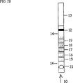
実施例1.遊離IgE、IgGに結合したIgE、およびIgEに対する抗体の割合の測定のための試験ストリップ
図2A、2Bおよび3では、輸送流の方向が矢印(10)で示されている。各変法では輸送流の最初にサンプル用の領域ASZ(11)、その下流に領域DZ(12)、輸送流の末端に吸引部(13)、および各種領域の間に輸送領域としてのみ働く部分(14)があってよい。
図2A:この図面の変法は5つの分離領域(SZ)(ここでリガンドは同一であっても異なっていてもよいし、あるいは異なる量で存在していてもよい(15−19))および試薬用のAR*Z(20)を有する。
図2B:これはASZ(11)とAR*Z(20)が一致している(21)こと以外は図2Aと同様の領域配列である。この領域配列はまた、分析物自体がDZにおけるシグナル複合体の一部である場合に検出可能な場合にも使用してよい。この時、AR*Zは必要でない。
図3:この図面の領域配列は2つのタイプの分離領域、それぞれSZ1(22、23)およびSZ2(24)と、SZ1(23)とSZ2(24)の下流に独立にAR*Zを示す。
【0083】
自己抗体(IgA、IgGおよびIgM)に対する遊離のIgEおよびIgE複合体結合の測定が注目される。しかしながら上記すべてのうち、遊離IgEは厳密に定量しなければならない。IgEの測定に関する現行の試験では、自己抗体は試薬抗体(抗IgE抗体)と同じIgEエピトープに結合でき、次いでこれはこの試験の設計によっても異なるが、誤った低すぎる総IgEレベルを与える。IgEの測定前にIgG、IgMおよびIgAを分離することで、遊離IgEが検出され得る。自己抗体の量はまた、複合体としても、IgEに向けられた遊離IgG抗体としても定量されるはずである。
【0084】
一般試験の大部分は希釈率の高い血清サンプルと相互作用させる固相(DZに相当)に結合したIgEを用いる方法により遊離抗体を測定する。この血清サンプルが抗IgE抗体を含むならば、後者は免疫複合体を形成する固相に結合する。非結合血清成分を洗い去った後、例えば酵素で標識した抗IgG抗体(R*)を加える。過剰量の標識抗体(R*)を除去し、固定化された免疫複合体に結合した酵素標識抗IgG抗体(R*)の量を好適な基質の添加により求める。これらの試験の感受性は固相に対するIgGの非特異的結合により制限される。このIgG集団のIgE特異的部分は一般に非常に小さく、非特異的に結合したIgGの量から区別するのは困難であると考えられる。IgGを固相に捕捉し、IgEの結合を測定することで、この制限は回避できる。
【0085】
IgG複合体と結合したIgEを測定する場合、IgGは、共有結合した抗IgG抗体(キャプチャー)を支持する固相(DZに相当)に捕捉される。標識された抗IgE抗体(R*)を加えることで、複合体結合IgEの量が測定できる。
【0086】
流れがまず1以上の分離領域(SZ)を、次に、検出領域(DZ)を通る上記のような横方向の液体輸送に基づく免疫アッセイ技術の使用により、IgE−IgG関連パラメーターの単純な測定に多くの可能性が開かれる。例えばまず、遊離IgEおよびIgGクラスのヒト抗IgE抗体と結合したIgEの混合物を含むサンプルを作製して、固相に結合した抗ヒトIgG抗体(SZにおけるリガンド)を含有する領域、次に、固相に結合した抗IgE抗体(DZにおけるキャプチャー)を含有する領域を通過させ、IgEとIgGクラスの抗IgE抗体との複合体のサンプル含量が分離領域に結合し、一方、遊離のIgEは検出領域へと進み、そこでそれは検出領域(12)の上流であり、単に検出領域を通すためだけの分離領域(15−19)の下流に標識された抗IgE抗体(R*)を加えることにより測定される(図2Aの領域20での添加)。抗IgE抗体(R*)も分離領域を通過させることで、抗ヒトIgG(リガンド)と結合することによって分離領域に捕捉されるIgE−IgG複合体の量も測定され得る(ASZとAR*Zが一致)(図2Bの領域21における添加、ASZはAR*Zと共通)。分離領域では、IgEとIgGクラスの抗IgE抗体との複合体の他、IgEに対する遊離の抗体も存在する。後者の量は、標識IgE(R* 1)を分離領域に通すことにより測定され得る。次いで標識IgE(R* 1)は別の試験でSZの上流のメンブランストリップに添加される。
【0087】
血清中のIgGの量が極めて高い場合には、高濃度の抗IgGを伴うバンドがいくつかSZとして使用しなければならない。複合体と結合した、また遊離の抗IgE抗体は次に双方ともIgGの総量によるいくつかのバンドにわたって阻害され、これらのバンドのシグナル強度の合計がIgEに対する抗体の量を示す。
【0088】
以下の実施例では、人工的に製造したIgEとIgGの複合体の試験成分を示している。当該複合体はIgEに対するモノクローナル抗体を用いて製造したが、それゆえマウスIgGに対する抗体は分離膜に結合している。この検出系では、複合体を形成している抗体以外のエピトープに対して向けられたIgE抗体が用いられている。これにより検出系において遊離のIgEと同様に複合体の測定が可能となる。
【0089】
分離膜1(SZ1):4℃にて20時間、25mM リン酸バッファー、pH 6.6中で、1.0mg/mlの抗体と20mg/mlポリスチレンアルデヒド粒子を混合することにより、ヒツジ抗マウスIgG(Fc)(リガンド1)とポリスチレンアルデヒド粒子(直径0.29μm,IDC,Portland,Oregon,U.S.A.)とを結合させた。該粒子を20mM ホウ酸バッファー、pH 8.6中で洗浄し、粒子50mgにつき15mgのNaCNBH3(Sigma−Aldrich Chemie,Steinheim,Germany)と20時間反応させた。次いでこの粒子を20mM ホウ酸バッファー、pH 8.6中で繰り返し懸濁させ、遠心分離し、デカントすることで洗浄した。この粒子懸濁液を3% トレハロース、20mM ホウ酸バッファーで希釈して25mg粒子/mlとした。希釈した懸濁液を、ニトロセルロース膜(ポリエステル上のニトロセルロース、孔径5μm、Whatman International Ltd,England)のストリップ(20cm×3cm)にストリップの長辺に平行な幅0.3cmの2本のラインとなるように噴霧した。噴霧装置(IVEKライナーストリッパー,IVEK Corporation,Vermont,U.S.A.)では、各ラインにつき約50μgのポリスチレン粒子/cmが送達された。当該膜を室温で乾燥させた後、小片に切断した(0.5cm×3cm)。
【0090】
分離膜2(SZ2):マウスIgG(リガンド2)を20mM ホウ酸バッファーで希釈して3.4mgタンパク質/mlとした。希釈した抗体をニトロセルロース膜(上記と同様のタイプ)のストリップ(20cm×4cm)上に、約6.8μg抗体/cmを含む幅0.3cmのライン状になるように噴霧した(上記の噴霧装置)。この膜を室温で乾燥させた後、小片に切断した(0.5cm×1cm)。
【0091】
検出膜(DZ):マウス抗IgEモノクローナル抗体(IgEのドメイン4に対して向けられたもの、キャプチャー)を20mM ホウ酸バッファーで希釈して1.0mgタンパク質/mlとした。希釈した抗体をニトロセルロース膜(上記と同様のタイプ)のストリップ(20cm×4cm)上に、約1μg抗体/cmを含む幅0.15cmのライン状になるように噴霧した(上記の噴霧装置)。この膜を室温で乾燥させた後、抗体を含んだラインが短辺と平行となるように小片に切断した(0.5cm×4cm)。
【0092】
複合膜:図3を参照。一枚の分離膜1(0.5cm×3cm、それぞれ図3のSZ1、22および23)を一枚の分離膜2(0.5cm×1cm、図3のSZ2、24)に貼り付け、このようにして得られた複合分離膜を次に検出膜のストリップ(0.5cm×4cm、図3のライン=DZ=12)に組み込んだ(短辺に対してそれらの間にギャップを持つ短辺)。これらの小片を接着テープで底辺にともに固定した。上辺には、隣り合う2辺の短辺といく分か重ねてニトロセルロース片(0.5cm×0.3cm)(A100、12μm、Schleicher and Schull,Dassel,Germany)を置いた。後者の小片はさらに接着テープで正しい位置に固定した。セルロースフィルター(図3の13)(0.5cm×2cm;GB004、Schleicher and Schull,Dassel,Germany)を検出膜の固定されていない短辺と重ねて吸引膜として固定した。領域の配列はASZ、SZ1、SZ2、DZであった。
【0093】
炭素粒子複合体(R*)の製造:
炭素懸濁液(原液):2gの炭素粒子(sp 100、Degussa,Germany)を5mM ホウ酸バッファー、pH 8.4 200mlに懸濁し、氷浴中で、100% 振幅、9.9+2秒パルスにて5分間3回音波処理を施した(VibraCell 600W,1.5cm探針,Soniced Materials,Danebury,Connecticut,U.S.A.)。
【0094】
炭素粒子複合体(R*):抗IgEモノクローナル抗体(IgEのドメイン3に向けられたもの)のFab' 35μg/mlと炭素粒子(250μg/ml)懸濁液とを3時間混合した。ウシ血清アルブミン(BSA)を1%まで加え、粒子をさらに30分間混合した後、1% BSA(0.1M ホウ酸バッファー、pH 8.5、0.05% NaN3)中で遠心分離することで洗浄し、洗浄バッファーで希釈して0.8mg炭素/mlとした。調製済みの炭素複合体を洗浄バッファー中4℃で保存した。
【0095】
サンプル材料:
IgEとIgGとの複合体の製造:1mgのIgE(ND)/mlと5mg/mlのマウス抗IgEモノクローナル抗体(IgGクラスのもので、ドメイン2に向けられたもの)を50mM リン酸バッファー、pH 7.5中、室温で2.75時間反応させた。サンプル混合物(0.35ml)をSuperdexTM 200prep級、16/60(Amersham Pharmacia Biotech AB,Sweden)で分離した。この分離により識別できる2つの複合体ピークが得られ、一方のピークはIgE−IgGに相当し、もう一方のピークはIgG−IgE−IgGに相当した。
【0096】
125I−標識タンパク質による制御(標識抗IgE抗体および標識IgE):
分離膜1(リガンド=抗マウスIgG):マウス抗IgE抗体(IgEのドメイン2に向けられたもの)とIgEを125I(クロラミンT)で、抗IgE抗体については0.03、IgEについては1.5の標識度で標識した。この標識タンパク質を6% BSA(50mM ホウ酸バッファー、pH 7.5)で希釈し、抗IgE抗体は約2.4μg/ml、IgEは0.06μg/mlとした。より高レベルの抗IgE抗体を測定するために125I−抗IgE抗体(ドメイン2)と非標識の抗IgE抗体(ドメイン2に向けられたもの)とを混合した。吸引膜(0.5cm×2cm、GB004、Schleicher and Schull,Dassel,Germany)を、ヒツジ抗マウスIgG(Fc)を吸着させた一枚の分離膜1(0.5cm×4cm)の一方の末端にテープで貼り付けた。10μlの0.1M ホウ酸バッファー、pH 8.5(6% BSA、0.05% NaN3)、次いで10μlの125I−タンパク質溶液を分離膜の固定されていない末端につけた。次いで4×10μlの0.1M ホウ酸バッファー、pH 8.5(1% BSA、0.05% NaN3)を固定されていない末端へ添加することにより横方向の流れを開始させた。液体がすべて膜へ移動した後、シートの種々の領域(分離および輸送領域)の放射能を測定するために細かく切断した。ガンマカウンターで測定を行い、遊離の放射性ヨウ素量の補正をした後に、種々の領域で捕捉された125I−タンパク質(標識抗IgE抗体および標識IgE、各々)の割合を算出した。IgEは、IgG抗体がリガンドである分離領域には、中間の輸送領域以上には結合しなかった。85%を超えるIgEが膜を通過した。他方、120ngまでの抗IgE抗体を添加した場合には、標識抗IgE抗体はすべて2つの分離領域へ結合した。1000ngの抗IgE抗体を添加した場合には、200ngがそれぞれの抗マウスIgG領域(分離領域)に結合し、500ngが通過した。ヒト血清中のIgGに関しては、血清を1/100(約1000ngのIgG)希釈し、より多くの抗IgG抗体(ヒトIgGに対するもの)がしっかりと固定されたリガンドとして使用されば、この能力で十分であると考えられる。
【0097】
分離膜2(リガンド=マウスIgG):この膜を導入して、分離膜1から遊離した可能性がある、あるいはバックグラウンドシグナルを強める結果となる、検出領域に結合するであろう抗マウスIgG抗体のいずれをも結合させた(抗マウスIgG抗体は2つのFab部分を有しており、そのため双方がマウスIgGであるR*およびキャプチャーと同時に結合する)。遊離したヒツジ抗IgGの量は検出領域の前のマウスIgGを含む分離領域で結合させるのが有利である。この捕捉領域(SZ2)によって、検出領域における非特異的な結合を6倍を超えて低下させることができる。
【0098】
分離と免疫化学測定を併用する標準プロトコール:
20μlの洗浄バッファー(1% BSA、0.9% NaCl、1% Tween20、0.1M ホウ酸バッファー、pH 8.4、0.05% NaN3)を上記のもの(配列SZ1、SZ2、DZ)の複合ストリップ上の分離膜1の固定されていない末端の縁(図3のASZ=11)につけた。次いで10μlのIgE標準(IgE、4〜500kU/l、0.01〜1.2μg/ml)およびサンプル(約1μg複合体/mlのIgE−IgG複合体、約1.3μg複合体/mlのIgG−IgE−IgG複合体)をそれぞれ添加した。両サンプルおよび標準は6% BSAおよび0.05% NaN3を含む50mM リン酸バッファー、pH 7.5で希釈した。洗浄バッファー、0.1M ホウ酸バッファー、pH 8.4(1% BSA、0.9% NaCl、1% Tween20、0.05% NaN3)を含む0.6cm×0.6cm×0.3cmのセルローススポンジをストリップの分離部分の固定されていない末端に置くことで横方向の流れを開始させた。試験溶液は分離領域(図3の22、23、24)および検出領域(図3の12)を通って、吸引セルローススポンジ(図3の13)へと移動した。7分間流した後、10μlの炭素粒子と抗IgE抗体の複合体(R*)(0.1M ホウ酸バッファー、pH 8.4(1% BSA、0.05% NaN3)中、0.8mg炭素/ml)を検出領域とストリップの分離部分(25)との間の位置に添加した。さらに5分間流した後、検出領域は灰色ないし黒色となった。黒色化部分をレーザースキャナ(Ultroscan,Amersham Pharmacia Biotech AB,Uppsala,Sweden)で読み取り、ピーク強度を算出し、IgE標準曲線で読み取ることで濃度を求めた。IgE濃度が高いほど、シグナルは黒みを増す。
【0099】
対照として、リガンドを含まないニトロセルロースに置き換えた分離領域1を有するストリップについても同様評価した(標準およびサンプル)。
【0100】
結果:
標準(IgE)では両測定系において黒色化曲線での強度は同等であった。SZ1をリガンドを含まないニトロセルロースに置き換えた場合には、複合体(IgE−IgGおよびIgG−IgE−IgG)はDZにおける強い黒色シグナルとして検出された。SZ1がリガンドとして抗マウスIgGを含んでいる場合には、DZにおいて複合体のシグナルは検出できなかった。
【0101】
表1
【表1】

このように抗マウスIgGを含む分離領域では97%を超えるまでの複合体が捕捉された。
【0103】
実施例2.患者サンプルにおけるCD−トランスフェリンの測定法
陰イオン交換特性を有する分離膜:ニトロセルロース膜(5μm、ポリエステル上のニトロセルロース、Whatman International Ltd,England)を超高純度の水(Milli Q,Millipore Corp.,Bedford,MA,U.S.A.)中の0.1% ポリエチレンイミン(PEI,Sigma,St Louis,MO,U.S.A.)溶液に入れた。この溶液を3時間振盪し、次いで30分間Tween20中に入れ、風乾させ、その後、プラスチックバッグに入れて4℃で保存した。膜の変性度はブロモフェノールブルー(pK=4.1)で確認した。
【0104】
荷電タンパク質と相互作用する変性膜の機能を、シートのストリップにおいて横方向の液流で125I標識タンパク質(クロラミンT法により標識したウシ血清アルブミン、テトラアシアロトランスフェリンおよびアシアロトランスフェリン)を輸送することにより確認した。最も高いpIを有するタンパク質が液流とともに移動する傾向が最も強かった。別の試験において液体が漸増濃度のNaCl(0〜1000mM)を含んでいる場合、最も低いpIを有するタンパク質の移動率がか最も影響を受けた。これらの2つの機能の制御は、ポリエチレンイミンによる処理において正電荷を持つ官能基が導入され、これらの官能基がタンパク質およびNaClのイオン交換基として機能し得るということを支持するものである。
【0105】
検出膜:4℃にて18時間、25mM リン酸バッファー、pH 6.6中で、1.3mg/mlの抗体と22mg/mlポリスチレンアルデヒド粒子とを混合することにより、抗トランスフェリンモノクロナール抗体とポリスチレンアルデヒド粒子(直径0.29μm,IDC,Portland,Oregon,U.S.A.)とを結合させた。該粒子を20mM ホウ酸バッファー、pH 8.6中で洗浄し、粒子40mg/mlにつき5mgのNaCNBH3(Sigma−Aldrich Chemie,GmbH Steinheim,Germany)と18時間反応させた。この粒子を20mM ホウ酸バッファー、pH 8.6中で洗浄し、6% トレハロースを含む20mM ホウ酸バッファーで希釈して14mg粒子/mlとした。希釈した懸濁液を、ニトロセルロース膜(5μm、ポリエステル支持体上のニトロセルロース、Whatman International Ltd,England)のストリップ(20cm×4cm)にストリップの中央に、かつストリップの長辺に平行な幅1.4mmのラインとなるように噴霧した。噴霧装置は実施例1と同一のものであり、これにより約14μgのポリスチレン粒子/cmが送達された。当該膜を室温で乾燥させた後、プラスチック袋に入れて4℃で保存した。
【0106】
複合膜:図1を参照。分離膜のストリップ(0.5cm×3cm)(図1の=SZ=5)の末端を0.5cmに短くした検出膜のストリップ(0.5cm×3.5cm、図1の抗体を含むライン=DZ=4)の末端の下辺にテープで貼り付けた。この末端間のギャップはニトロセルロース片(0.3cm×0.5cm、A100、12μm、Schleicher and Schull,Dassel,Germany)を重ねることでブリッジし、これをテープで固定した。吸引膜(図1の9)としてセルロースフィルター(0.5cm×2cm、GB004、Schleicher and Schull,Dassel,Germany)を検出膜から送達されるストリップの固定されていない末端と重ねてテープで固定した。
【0107】
炭素粒子複合体(R*)
炭素懸濁液(原液):2gの炭素粒子(sp4、Degussa,Germany)を5mM ホウ酸バッファー、pH 8.4 100mlに懸濁し、実施例1と同じ装置を用いて、氷浴中で、100% 振幅、5+5秒パルスにて5分間音波処理を施した。
【0108】
炭素粒子複合体:抗トランスフェリンモノクロナール抗体100μg/mlと炭素懸濁液(250μg/ml)とを2時間混合した。BSAを1%まで加え、粒子をさらに30分間混合した後、0.1M ホウ酸バッファー、pH 8.5、(1% BSAおよび0.05% NaN3を含む)中で遠心分離することで洗浄し、洗浄バッファーで希釈して1.9mg炭素/mlとした。調製済みの炭素複合体を洗浄バッファー中4℃で保存した。
【0109】
サンプル材料:
テトラアシアロトランスフェリン:テトラアシアロトランスフェリンをヒトトランスフェリン(主としてテトラアシアロトランスフェリン)の鉄で飽和した調製物からMono Q(Amersham Pharmacia Biotech AB,Uppsala Sweden)イオン交換クロマトグラフィーにより単離した。
【0110】
アシアロトランスフェリン:トランスフェリンの鉄で飽和した調製物(Sigma,St Louis,MO,U.S.A.)をノイラミダーゼ(Behringwerke,Marburg,Germany)で処理した後、アシアロトランスフェリンをMono Q(Amersham Pharmacia Biotech AB,Uppsala Sweden)イオン交換クロマトグラフィーにより単離した。
【0111】
等電点(pI):これらの値を個々のイソ型調製物とBSAについて、Phast系(Amersham Pharmacia Biotech AB,Uppsala Sweden)における等電点電気泳動法により求めた。アシアロ型 pI=5.7、テトラアシアロ型 pI=5.3、およびウシ血清アルブミン pI=4.7。
【0112】
トランスフェリン標準:上記のように調製したアシアロトランスフェリンを0.2% BSA、0.1% Tween20、0.1mM クエン酸鉄(III)、1mM NaHCO3および0.05% NaN3を含む20mM BIS−TRIS、pH 6.3で希釈して濃度0.07〜16.6μgトランスフェリン/mlとし、標準として使用した。
【0113】
血清サンプル:11種の血清サンプルと6種の血清対照を0.1% ウシγグロブリン(Sigma,St Louis,U.S.A.)、0.1% Tween20、0.1mM クエン酸鉄(III)、1mM NaHCO3および0.05% NaN3を含む20mM BIS−TRIS、pH 6.3で1/50希釈した。血清サンプルを予めCDTect(Pharmacia & Upjohn Diagnostics AB,Uppsala Sweden)によってCDTについて分析した。CDTectはCD−トランスフェリンを測定するものである。
【0114】
分離と免疫化学測定を併用する標準プロトコール:2μlのサンプル(トランスフェリンの希釈系および希釈した血清サンプルそれぞれ)を上記のものの複合ストリップ上の分離領域を有する膜部分の固定されていない末端の縁(図1のASZ=3)から1cmの位置につけた。15mM NaClおよび0.1% Tween20を含む20mM BIS−TRISバッファー、pH 6.5を吸収させた0.6cm×0.6cm×0.3cmのセルローススポンジ(図1の8)を分離領域の固定されていない末端に置くことで横方向の液流を開始させた。分離領域(図1の5)では分析物(CD−トランスフェリン)とそのヘテロ型(他のトランスフェリン)は領域内にしっかりと固定された正電荷(PEI処理により導入されたリガンド)によって引き寄せられ、大きな負電荷を有するヘテロ型(他のトランスフェリン)が小さな負電荷を有するヘテロ型(CD−トランスフェリン)より強く引き寄せられ、すなわちCD−トランスフェリンはトリシアロ−、テトラアシアロ−、ペンタアシアロ−などのトランスフェリンよりも容易に液流とともに移動する。そのため複合ストリップ/マトリックスを通るその移動中、全トランスフェリン量の一定の割合が検出領域(図1のDZ=4)において抗トランスフェリン抗体(キャプチャー)と結合できる。4分間流した後、5μlの炭素粒子と抗トランスフェリン抗体の複合体(R*)(30% トレハロース、1% Tween20、1% BSA、0.05% NaN3を含む0.1M ホウ酸バッファー、pH 8.4中、1.8mg炭素/ml)を分離領域と検出領域との間(図1の領域(6)(=AR*Z))に添加した。さらに5分間後、流れを止め、検出領域の黒色部をレーザースキャナ(Ultroscan,Amersham Pharmacia Biotech AB,Uppsala,Sweden)で読み取り、アシアロトランスフェリンの希釈系の測定値に対して読み取って濃度を算出した。サンプルにおけるCD−トランスフェリンのレベルが高いほど、黒色シグナルが強くなる。
【0115】
表2:結果
【表2】
本発明の方法によって得られた測定値はCDTectによって得られたものとの非常に優れた一致(相関係数0.97)を示した。本発明はCDTectの実施よりも極めて迅速、かつ簡単である。
【0117】
実施例3.分離領域においてSambucus Nigraレクチンを含む試験ストリップ
分離膜:セルロースシート(4cm×12cm)(セルロースフィルター54、Whatman International Ltd,England)をシアノ−ジエチル−アミノピリジン(CDAP)(KohnおよびWilchek,Appl. Biochem. Biotechnol. 9(1984)285−304)によって活性化した。活性化したシートを0.1M NaHCO3、pH 8.4中の0.1mg/mlのSambucus Nigraレクチン(炭素鎖の末端位にあるシアル酸と結合する;Vector Laboratories Inc.,Burlingame. CA. U.S.A)溶液に入れた。この溶液を2時間振盪し、次いでそのシートをa)0.1M NaHCO3、b)0.5M NaCl、c)蒸留水、d)0.1M 酢酸バッファー、pH 4.5、e)0.1M NaHCO3、pH 8.4、f)0.5M NaCl、g)蒸留水、h)0.1M 酢酸バッファー、pH 4.5、i)0.1% Tween20を含む5mM BIS−TRIS、pH 6.4中に入れた。種々の水浴間では、過剰な液体をキッチンロールペーパーで吸い取った。洗浄工程の後、シートを風乾させ、プラスチックバッグに入れて4℃で保存した。
【0118】
シートを使用する前に、シートを粘着プラスチック(75μm粘着ポリエステルフィルム;Gelman Science Inc,Ann Arbor,MI,U.S.A.)に貼り付けた。
【0119】
検出領域と複合ストリップを含む膜:これらの膜を実施例2と同様の方法で作製すればよい。図1も参照。SZにおけるリガンドはここではレクチンである。
【0120】
炭素粒子複合体(R*)と125I標識タンパク質:実施例2を参照。
【0121】
125I標識タンパク質による分離膜の制御:テトラアシアロおよびアシアロトランスフェリンとウシアルブミンを125I(クロラミンT、標識度0.08〜0.13)で標識した。標識タンパク質を0.1% Tween20、0.04mM クエン酸鉄(III)および0.05% NaN3を含む10mM BIS−TRIS、pH 6.4で希釈して約0.3μg/mlとした。さらに0.4mgBSA/mlを加えた。
【0122】
分離膜のストリップ(0.5cm×4cm)と一枚のセルロース吸引膜(0.5cm×2cm、GB004、Schleicher and Schull,Dassel,Germany)を、それらの末端がある程度重なるように下辺でテープで貼り合わせた。1μlの125Iタンパク質溶液をそれぞれの分離膜の固定されていない末端から1cmの位置につけた。セルローススポンジ(0.6cm×0.6cm×0.3cm)を分離膜の固定されていない末端に置くことで横方向の流れを開始させた。このスポンジには0.1% Tween20とともに0.5M NaCl、1mM CaCl2を含む20mM TRIS−HCLバッファー、pH 7.5を吸収させた。2、4、6および10分後それぞれにセルローススポンジを取り除き、流れを中断させて、分離膜の固定されていない末端から膜を2および3cm切断した。放射能を持つ膜片をガンマカウンターで測定し、2および3cm移動した添加の割合を算出した。1cm以上の移動についての値を表4に示している。
【0123】
表4:分離膜において1cmを超えて移動した全添加125Iタンパク質に対する%
【表3】
結論:この結果より、テトラアシアロトランスフェリンはSambucus Nigraレクチンによって分離膜で著しく遅延されているが、アシアロトランスフェリンおよびBSAではそれと同程度まで遅延されることがないことが明らかである。この結果により、Sambucus Nigraレクチンを含む分離膜を実施例2と同様の検出膜と組み合わせ、CD−トランスフェリンよりシアル酸の含量の高いトランスフェリンを含むサンプル中のCD−トランスフェリンを定量するのに使用してもよいことがわかる。
【図面の簡単な説明】
【図1】 ASZ、ARZ、SZおよびDZを有する簡単な変形である。ARZとASZは分離されている。
【図2A】 主として、同一のリガンドを有する5個の分離領域を有することで図1の変形とは異なる。ARZとASZは分離されている。
【図2B】 ARZとASZが一致しているという点を除いては、図2Aの変形と同様である。
【図3】 実施例1において使用される、3つの分離領域を有する本発明の流動マトリックスの変形を示す。このうち2つの領域(SZ1)はあるリガンドを呈示し、もう1つの領域(SZ2)は別のリガンドを呈示する。ASZとARZ(=AR*Z)は分離されている。[0001]
TECHNICAL FIELD OF THE INVENTION
The present invention relates to a method for measuring an analyte in a sample and a kit used in the method.
[0002]
Starting from the prior art, the method of the present invention comprises the following steps.
i. The sample is applied to the sample application zone (ASZ) on the flow matrix where transport of components present in the sample can occur (transport stream).
This flow matrix further
a) if desired, analytically detectable binding reactant (reactant)*= R*) Application area (AR*Z),
b) Another binding reactant (capture) located downstream of the ASZ and firmly attached to the matrix.PresentationWhere the capture and analyte and / or reactant in the method*Detection region (DZ) in which a complex (signal complex) containing
Comprising.
ii. Flow of sample components is achieved by flow.
iii. The signal complex is detected in the detection region, and the measured signal is used for the measurement.
[0003]
The present invention is primarily directed to a fluid matrix that may be of the same type as previously used, for example, in immunochromatography. See below.
[0004]
Suitable binding reactants include so-called affinity reactions, in particular biospecific affinity reactions, and covalent reactions, in particular exchange reactions between free thiols and reactive disulfides and between mild electrophiles and mild nucleophilic atoms. Some are involved in other reactions. Common biospecific affinity reactions include immunochemistry, ie between antibodies and antigens or haptens. Other types of bioaffinity reactions include hybridization between complementary nucleic acids (including oligonucleotides), reactions between lectins and carbohydrate structures, Ig (Fc) structures and Ig (Fc) such as A protein or G protein. ) There is a reaction between binding proteins. Bioaffinity reactions include reactions between biomolecules and synthetically produced ligands / captures.
[0005]
The type of method in question is described for non-competitive methods such as sandwich technology and competitive methods. Sandwich technology typically forms an analytically detectable complex in which an analyte binds to two bioaffinity counterparts, one of which is analytically detectable and the other is capture. It means that. In a typical competitive variation, the analyte and the analytically detectable analyte analog compete for a limited amount of bioaffinity counterpart. Examples of two competitive variants include: a) competition between an analyte and a labeled analyte analog for a limited amount of ligand in the form of a tightly immobilized capture; and b) soluble. And those that use competition between the analyte and an analyte analog in the form of a tightly-fixed capture for a limited amount of analytically detectable bioaffinity counterpart.
[0006]
For further information on the methodologies previously used within the technical field of the present invention, see US-A-4,861,711 (Behringwerke), WO 88/08534 (Unilever), US-A-5,120, 643 and 4,740,468 (Abbott), EP-A-284,232 and US-A-4,855,240 (Becton Dickinson) and WO 96/22532 (Pharmacia AB).
[0007]
Hetero
A compound that can compete for binding to its counterpart by one of the binding reactions described above. Heterotypes can be protein isoforms, such as isoenzymes. The term heterotype specifically includes various forms of bioaffinity complexes that are “similar” to each other by satisfying the above definition. Examples include immune complexes where the antigens are the same but the antibodies are of various classes / subclasses. See also “Analyte” below.
[0008]
Whether two compounds are heterozygous to each other can be determined in so-called inhibition tests.
[0009]
Problems to be solved by the present invention
Sample components that can affect or affect signals detected in DZ can be classified into two main groups: a) analytes and b) components that directly or indirectly inhibit detection. it can. Direct inhibitory components include those that interfere with the signal, such as fluorescent components in serum when the complex is detected by fluorescence. Examples of indirect inhibitory components include captured and / or added bioaffinity reactants R (eg, R*). Other indirect inhibitory components, such as heterophilic antibodies, may be present in the initial sample to inhibit signal complex formation in the DZ. In certain embodiments of the invention, ligands released from the separation region of the invention can act inhibitory (see Example 1).
[0010]
For analytes present at low concentrations, separation and detection of inhibitory components are often performed in different systems due to problems with inhibitory components in the sample.
[0011]
Examples where analysis has been performed either by immunological systems after ion exchange separation or by on-line measurement of absorbing groups (460 nm) include carbohydrate deficient transferrins (CDT = CD transferrin = asialo-, monosialo- and disialo- There is a measurement of transferrin). A relatively high concentration of CDT (10-9If present in M), both detection methods were possible, but immunoassays are required for low concentrations of analyte. Ion exchange chromatography separation is controlled by advanced, costly equipment, which requires specially trained personnel. Conventional immunological tests are also expensive and require well-trained personnel.
[0012]
Techniques for immunological on-line measurement after chromatographic separation steps are described by Afeyan et al. (Nature 358 (1992) 603-604) and Irth et al. (Anal. Chem. 14 (1995) 355-361), and the problem The points are also summarized by Krull et al. (LC-GC 15 (7) (1997) 620-629).
[0013]
Transport of whole cells to DZ can inhibit the signal from the detection complex. The use of a fluid matrix in which cells are mechanically (by filtration) trapped in the denser front region has been previously known (Oudheusden et al., Ann. Clin. Biochem. 28 (1991) 55-59).
[0014]
In EP-A-696,735, in order to extend the measurement range of an analyte, a predetermined amount of an analyte-binding antibody is fixed to the sample application region, and a certain amount of the analyte is retained therein. A chromatographic immunoassay system is disclosed.
[0015]
In EP-A 702,233, in a manner similar to that described in EP-A-696,735, a certain amount of water is reacted before reacting with the labeled reactant (which is then detected in the detection region). Chromatographic immunoassay systems have been disclosed that achieve sample dilution effects by capturing analytes.
[0016]
WO 97/35205 includes (i) a detection region of a labeled analyte-binding reactant that is not bound to an analyte, and (ii) a complex between the analyte-binding reactant and the analyte. A chromatographic membrane for immunoassay having a detection region is disclosed. The relative amount of unbound analyte binding reactant and analyte reaction complex provides a measure of the amount of analyte in the sample.
[0017]
WO 94/06012 discloses an analytical test apparatus having a negative control region disposed in front of an analyte detection region. This negative control region has the function of indicating the presence of components in the sample that affect analyte detection and make it unreliable.
[0018]
Object of the present invention
The first major objective of the present invention is to create a simple and quick method that helps to measure analytes in the presence of inhibitory components. In particular, an object of the present invention is to avoid problems with inhibitory components that are soluble or suspendable in the liquid medium of interest.
[0019]
The second main object of the present invention is the quicker and simpler measurement of individual heterotypes or combinations thereof, in particular heterotypes exhibiting peptide, carbohydrate or lipid structures, including a variety of physiologically active compounds. . Lipids include steroids and other fat-soluble substances.
[0020]
The third main objective of the present invention is <10, particularly for samples containing inhibitory heterotypes of analytes.-7M, especially <10-9To help measure analytes in the M concentration range.
[0021]
The fourth main object of the present invention is to simplify the measurement of individual heterotypes or combinations thereof in a sample derived from biological material.
[0022]
The fifth main object of the present invention is to provide a quicker and easier evaluation of chemical libraries such as compound libraries, eg combinatorial libraries.
[0023]
Sub-objectives of the above four main objectives include the possibility of performing measurements in outdoor environments (usually semi-quantitatively) and in advanced laboratories (with the possibility of accurate quantification) Is to increase.
[0024]
The present invention
The above objective is to allow the flow matrix to contain one or more separation zones (SZ) between ASZ and DZ, thereby delaying / separating at least one component that can affect the signal from the signal complex in DZ Can be achieved using the methods described in the introductory part of this specification. This should occur in SZ by the ligand interactions described below, which can be reversible or irreversible. The component may be an inhibitory component or an analyte. If the component is not an analyte, the delay is that the component moves through the SZ more slowly than the analyte or binds irreversibly to the SZ, thereby preventing it from reaching the DZ, so that It means that the detection of the analyte in the DZ is not substantially inhibited by the component in question. Usually this means that a sufficient amount of ligand must be present for substantially all of the inhibitory components in the affected sample. “Substantially all” depends on the relative concentrations of the components, but usually at least 90%, preferably at least about 95%, more preferably at least 99% of the inhibitory components are delayed or trapped in the separation region. Means that. A component can be an analyte if it is desired to determine the ability of one or more ligands to bind to the analyte. In this case, such a ligand is immobilized on the separation region.
[0025]
The choice of structure / ligand that is delayed in the separation region is determined by the delayed component. The delay may be based more or less on various specific interactions between the ligand structure and the delayed component. See title "Separation area" below. After passing through the SZ, the analyte moves with the transport stream to the detection zone (DZ) where the capture and analyte and / or R*Is formed.
[0026]
If the delay of one or more inhibitory components is intended, the formation of the signal complex occurs in the absence. Detection of signal complexes in DZ may be taken as qualitative and quantitative values for the analyte.
[0027]
If the analyte is intended to be delayed, the signal complex formation is altered, or if the analyte-ligand binding in the SZ is irreversible, signal complex formation is completely inhibited. there is a possibility. Formation of the signal complex in DZ is a measure of the ability of the analyte to bind to the ligand in SZ.
[0028]
1-3 show various variations of the flow matrix of the present invention.
FIG. 1 is a simple variant with ASZ, ARZ, SZ and DZ. ARZ and ASZ are separated.
FIG. 2A differs from the variant of FIG. 1 primarily by having five separate regions with the same ligand. ARZ and ASZ are separated.
FIG. 2B is similar to the variation of FIG. 2A except that ARZ and ASZ match.
FIG. 3 shows a variation of the flow matrix of the present invention having three separation regions used in Example 1. Two of these regions (SZ1)PresentationThe other region (SZ2) has another ligandPresent. ASZ and ARZ (= AR*Z) is separated.
[0029]
A more detailed description of FIG. 1 is given in the title “Matrix and Transport Stream” and with reference to FIGS. The flow matrix shown in FIGS. 1-3 can in principle have any of the following geometric embodiments.
[0030]
Matrix and transport flow
The matrix is of the same type (flow matrix) as previously used in so-called immunochromatography detection methods and defines the compartment in which reactants and sample components are transported. Thus, the matrix can be the internal surface of a single flow channel (eg, capillary), the internal surface of a porous matrix having a flow channel osmotic system (porous matrix), and the like. The matrix can be in the form of a monolith, a sheet, a column, a membrane, a separate flow channel, such as a capillary, or a collection of such flow channels. They may also be in the form of particles packed in column cartridges or cut grooves, compressed fibers, and the like. Alternatively, there are so-called liquid chromatography nanocolumns, i.e. silicone or quartz plates with channels of about 2 [mu] m or less made by microlithography (e.g. He. B. et al., Anal. Chem. 1998, 70, 3790-3797). reference). The inner surface of the matrix, ie the surface of the flow channel, must be sufficiently hydrophilic so that the aqueous medium (primarily water) is transported through the matrix either by capillary forces or by pressure or suction. The minimum inner dimension of the flow channel (measured as the diameter for spherical channels) is such that components interacting in the analyte, addition reactant, and detection region and delayed in SZ are transported through the matrix. Must be large enough. As a rule of thumb, a suitable matrix may be selected from those having flow channels having a minimum internal dimension in the range of 0.1 to 1000 μm, and 0.4 to 100 μm if the matrix has a communicating flow channel system. Is preferred. For flows driven by external pressurization / suction, the flow channel with the smallest dimension is noted, mainly in the wide range (up to 1000 μm).
[0031]
Suitable matrices are often constructed from polymers such as nitrocellulose, polyester, polyethylsulfone, nylon, nitric acid / cellulose acetate, cellulose, regenerated cellulose. These membranes are advantageously provided with a rigid backing of, for example, polyester.
[0032]
The physical and geometrical design of the matrix material and flow channels are variable along the flow depending on the intended use of a portion of the matrix [WO 96/22532 (Pharmacia AB), WO 94/15215 (Medix) ]. One identical matrix may comprise several transport streams parallel or radially directed from a common center, for example in the form of individual channels. In some of the most important embodiments, at least the detection region and the closest portion of the matrix should be in a form such that transport flow to, in, and out of the DZ can occur laterally in the matrix. That is, at least this part of the matrix is in the form of a membrane strip or a plate with cut grooves.
[0033]
Various flow matrices that may be used for the type of test in question are described in the prior patent publications. For example, US-A-4,861,711 (Behringwerke), WO 88/08534 (Unilever), US-A-5,120,643 and US-A-4,740,468 (Abbott), EP-A- 284,232 and US-A-4,855,240 (Becton Dickinson), WO 96/22532 (Pharmacia AB).
[0034]
The most important embodiment of the invention on the priority date is based on liquid transport in the flow matrix, for example in the form of a membrane strip (see FIG. 1). This strip is made from a matrix that defines the transport stream (1) and is applied to a liquid-rigid backing (2), preferably made of plastic. On the matrix are a sample application area (3, ASZ) and a detection area (4, DZ) located downstream thereof. The transport stream is in the direction from ASZ to DZ. There is a separation region (5, SZ) between the sample application region (ASZ) and the detection region. The transport stream may contain additional reactant application areas (6) (R, eg R, if required for a particular embodiment.*For the application area ARZ, for example, AR*Z) may be present. Between the regions there may be regions (7) whose sole function is to transport reactants. Application area ARZ (AR*The position of Z) is determined by the test protocol used and may be upstream or downstream of ASZ or may be consistent with ASZ. ARZ (eg AR*For the case where Z) is upstream of ASZ, the area ARZ (AR*It is advantageous if it occurs substantially simultaneously with the addition of the liquid in Z). See international patent application PCT / SE98 / 02463 filed earlier by the inventors (part of which is hereby expressly indicated). For certain test protocols, ARZ (AR*Z) may coincide with DZ.
[0035]
In some embodiments, reactant R, such as R*It is advantageous if is previously deposited. This is particularly the case when ARZ is located downstream of ASZ, resulting in a variation of the test protocol used at the same time, i.e., reactant R and analyte move to DZ substantially simultaneously.
[0036]
If it is desirable to use a continuous variant where the analyte is transported to the DZ before the reactant (R), then if the reactant application zone (ARZ) is downstream of the ASZ, then R is The sample should be added after passing through the ARZ. A continuous process is also achieved when ARZ is upstream of ASZ, where R may be pre-attached to ARZ if desired.
[0037]
In another embodiment, the reactant (R), such as R*May be moved to DZ in a separate transport stream from a different direction than that of the stream transporting the analyte to DZ. See, for example, US-A-4,855,240 (Becton & Dickinson).
[0038]
There may be several detection regions intended for different analytes or different concentrations of the same analyte in one and the same transport stream. For the case of different analytes, the capture in the individual DZ must, of course, not show any substantial cross-reactivity to any analyte.
[0039]
The transport flow from the ASZ through the separation region (SZ) and further toward the detection region (DZ) may be a liquid flow driven by capillary force. If necessary, the flow matrix is immersed in the transport liquid and shows a liquid reservoir (8) and / or a suction perforated matrix (9) located downstream of the DZ in the form of a perforated matrix applied upstream of the ASZ. May be. The liquid reservoir and suction matrix help maintain the flow. Liquid flow can also be achieved by pressure or suction through the matrix. Thus, the pressure may be applied hydrostatically, for example by designing as a mini-column in which a part of the matrix is in direct liquid communication with a flow matrix with its outlet positioned vertically and its outlet positioned horizontally. In the latter form, the part of the horizontally positioned matrix can be in strip / membrane form. As an alternative to transporting analytes, reactants and inhibitory components, an electric field may be applied to the matrix.
[0040]
A region arrangement similar to that of FIG. 1 can also be constructed for other types of flow matrices, for example, matrices in which capillaries and transport streams are deep.
[0041]
Both of the one or more matrix / transport streams described above may be disposed, for example, on a common backing, optionally with a liquid barrier layer therebetween. If desired, the flow can be common ASZ, common ARZ (AR*Z) or the like. Basically, the DZ is separated for each transport stream.
[0042]
In the above variant, one heterotype (analyte) is measured using a matrix with a separation region. If a matrix without a separation region is used, all heterotypes of the analyte present in the sample can be measured in a manner similar to that for the analyte. By combining these two types of region sequences, the relative amount and absolute amount of the analyte in the sample can be easily measured.
[0043]
Separation zone (SZ)
The separation region contains ligands / structures that have the ability to bind to one or more sample components that are believed to inhibit detection in DZ.Present. It is characterized by the fact that the separation is achieved by several specific / selective binding reactions, rather than because the matrix gives mechanical inhibition (filtration) to the inhibitory component in SZ. The principle that is an indicator of separation / retarding ligand / structure selection, especially with respect to specificity, binding power (affinity) and kinetics, is for affinity chromatography, including affinity chromatography and covalent chromatography, and capture Similar to that in biospecific analysis where solid phase technology is used. With regard to binding force (affinity, avidity) and kinetics, the main objective of the presently preferred variant of the invention is to delay the inhibitory component relative to the analyte, so that detection in DZ Can occur in the absence of these components. In general, this means that the inhibitory component must be delayed as efficiently as possible in the separation region or be as strong and as fast as possible.
[0044]
Therefore, ligands for separation in SZ are: a) charged (anion, cation, amphoteric = ion exchange ligand), amphoteric / amphiphilic, biocompatible Chelating, sulfur-containing (primarily thioethers for so-called sulfur affinity), those that allow covalent chromatography (reactive disulfides such as pyridyl disulfides), or those that interact with π-π, hydrophobic It may be of sex.
[0045]
If the inhibitory component is delayed, as a rule of thumb, the ability of the ligand to bind to one or more inhibitory components must be stronger than that to the analyte. This applies to the conditions used for separation in SZ. Factors that determine how successful a separation is the length of the separation region, ligand density, ligand effectiveness, temperature, flow rate, buffer, ionic strength, pH, and the like.
[0046]
Among the biospecific affinity ligands, there are mainly so-called immune ligands, ie antibodies and antigen-binding fragments thereof, and antigens and haptens. Examples of other affinity ligands include lectins (eg, sialic acid binding lectins), Ig (Fc) binding proteins (such as A and G proteins), single or double stranded oligos or polynucleotides, etc. Nucleic acid, enzyme substrate analogues, enzyme inhibitors and the like. For biospecific affinity ligands, the specificity may be directed to one or more binding sites on the delayed component. The corresponding binding site should not be as accessible to the analyte (this is intended even if it is not even present in the unexposed form).
[0047]
The ligand / structure in question can be immobilized on the separation region by covalent attachment to the matrix, either by physical or biospecific adsorption. Examples of the latter include the interaction between biotin and streptavidin, between the high affinity antibody and the hapten. Immobilization to the matrix may occur via a polymer or other substituent, which then retains the covalently, physically adsorbed, or biospecifically bound ligand used for separation. . Another possibility is to have the desired type of ligandPresentThere is adhesion of polymer particles. The particles may be hydrophilic or hydrophobic, and a compound exhibiting a ligand structure is adsorbed or covalently bound thereto. The technique for binding the ligand to be separated to the matrix SZ may be basically selected in the same manner as that known so far for capture in DZ. See, for example, the international patent applications PCT / SE98 / 02462, PCT / SE98 / 02463, and PCT / SE98 / 02464 filed earlier by the inventors (note that they are not relevant for introduction into the detection region of capture).ClassicExpressly incorporated herein). In this regard, it can be stated that there are commercially available membranes with covalently bound ligands, such as DEAE cellulose paper (diethylaminoethyl) (DE81, Whatman International Ltd, England).
[0048]
Detection area
The capture of the detection region can be selected according to similar conventions applied to the ligand of the separation region (however, the binding capacity of the capture should be directed to the analyte and / or to the reactant with which the analyte is related) ). It is advantageous to select a high affinity capture that has rapid kinetics with respect to ligand capture. The use of antibodies or antigens / haptens is of primary interest, in which it is often easy to find high affinity antibodies.
[0049]
The reactant to which the analyte relates refers to a reactant (R) that is added and can bind to the capture in an amount proportional to the presence of the analyte in the sample as it travels through the DZ. Examples of reactants involving analytes include: a) a labeled analyte analog in a competition method using competition for a limited amount of anti-analyte antibody bound to the solid phase, and b) on the solid phase. R in the form of labeled or unlabeled soluble anti-analyte antibody in a method using competition / inhibition between bound analyte analog and analyte for a limited amount of anti-analyte antibody in dissolved form*Is mentioned.
[0050]
The capture may be immobilized on the detection region by a technique similar to that used to bind the ligand to the separation region.
[0051]
Combining the separation principle in the separation region with various capture principles in the detection region, for example, separation ion exchange chromatography and capture immunochemical adsorption in DZ may be suitable. It may be practical to use the same principle for delay and capture in the two regions (eg, two monoclonal antibodies with different specificities, see examples).
[0052]
Analyte
Analyte means a compound that is quantified or qualified. Quantification relates to the measurement of absolute quantities as well as relative relationships. The qualitative nature of an analyte relates to the qualitative properties of the compound, such as detecting the presence or absence of any (yes / no test) or the ability to bind affinity to a ligand.
[0053]
Relative measurement means that the measurement obtained is the proportion of the sum of one or more selected heterotypes and the sum of another combination of heterotypes. Examples include the ratio of the analyte amount for a counterpart to the total amount of all heterotypes (the total amount includes the analyte amount).
[0054]
The present invention is applicable to analytes that can function as binding reactants. This means that the analyte can be essentially any substance that can provide such a capture. Specific examples include antigens / haptens, enzymes or antibodies or nucleic acids that are fully or partially in single stranded form. The analyte may exhibit amino acid / peptide, carbohydrate or lipid structure.
[0055]
For analytes present with heterozygous form, capture and / or addition reactant R, eg R*A particularly great advantage is obtained with respect to the ability to bind to. This is especially true for analytes <10-7M, especially 10-9Applicable if present in M sample concentration. Examples of this type of heterogeneous form are: a) compounds that differ from one another in charge, such as, for example, isotransferrin using CDT as the analyte, eg isohemoglobin using HbAlc as the analyte, b) additionally inserted or cleaved Compounds that differ from each other in certain parts of the basic structure, such as partial differences in amino acids (eg due to degradation) or peptide chains, c) compounds that differ from each other in that different substances / structures are added to the basic structure, eg A covalently bonded carbohydrate structure, d) in a macromolecule, by non-covalent bonds such as a bioaffinity bond between a receptor and a ligand in a receptor-ligand complex and between an antigen and an antibody in an immune complex, or for example an antibody chain Consists of two or more subunits joined together by a cysteine bridge between Molecule, and the like.
[0056]
Examples of possible uses / analytes include the following:
[0057]
a) The analyte is a heterotype that differs from other heterotypes in terms of carbohydrate content (glycosylation), eg, a glycoprotein having the same or similar protein portion. This type of heterozygous variant is known in several disease conditions such as cancer, inflammation and liver disease (Turner GA, “N-glycosylation of serum proteins in disease and its investigation using lectins”, Clin. Chim. Acta 208 (1992) 149-171; and Varki A, “Biological roles of oligosaccharides: all of the theories correct”, Glycobiology 3 (2) (1993) 97-130). In particular, i) measurement of the combination of asialo-, monosialo- and disialo-transferrin (separation in this regard may be done by ion exchange and lectin ligands in SZ) and ii) measurement of HbAlc (which is ion exchange or boronate) May be separated by a ligand). Variants in protein carbohydrate content are also known for normal biological changes such as menstrual cycle and differences in age and gender.
[0058]
b) The degree of glycosylation of the recombinant protein can be measured by ion exchange, lectin in SZ or boronate ligand. In this case, the analyte is a fraction of the recombinant protein that does not contain the carbohydrate structure that binds the ligand in SZ and therefore moves most rapidly through SZ.
[0059]
c) Recombinant proteins into which a separation handle has been inserted, such as histidine sequences or IgG binding sequences, where complete cleavage of the handle is important, but separation in SZ by metal chelate ligands and IgG (Fc) ligands, respectively. You can check later. In this case, the analyte is a fraction of the recombinant protein from which the histidine sequence or Ig (Fc) binding sequence, respectively, has been cleaved off.
[0060]
d) Enzymes can be separated into active and inactive forms by ligands that are substrate analogs or inhibitors of the enzyme in question in SZ. The analyte is an inactive enzyme.
[0061]
e) Proteins, peptides or other biomolecules that exert their biological function by binding to specific receptors can be separated in SZ by ligands that are receptors for biomolecules. The analyte is a fraction of the molecule that lacks or has reduced ability to bind to the receptor.
[0062]
f) The protein (eg, IgE) may have autoantibodies (IgG, IgA, IgM) bound to it in vivo. These autoantibodies produce on the one hand a different response in the immunochemical measurement of the protein, on the other hand an altered turnover rate / function. By using an antibody against the autoantibody in question in the separation region as a ligand, free and immune complex-bound autoantibodies can be separated and the amount of free protein (= analyte, eg IgE) can be determined. Can be calculated.
[0063]
g) By the monoclonal antibody directed to a certain binding site of the protein and immobilized on SZ, the presence of a heterozygous form (= analyte) of the protein that does not show the binding site can be detected by quantification in DZ.
[0064]
h) The presence of various substances that bind to transport proteins, such as drugs bound to albumin, can be measured by using a suitable ligand in SZ. By selection of suitable ligands in SZ, transport proteins with or without bound drug can be measured in DZ.
[0065]
i) In certain rheumatic diseases or autoimmune diseases, serum IgG and IgA have increased adsorption on various surfaces. By immobilizing IgG having charged characteristics and a ligand capable of binding to IgA in the separation region, the ratio of IgG and IgA having uncharged adsorption characteristics in DZ (= analyte) can be measured. By having the corresponding autoantigen / hapten as a capture in DZ, IgG or IgA class specific autoantibodies can be measured with good sensitivity.
[0066]
j) A large number of biologically active compounds (eg peptides or steroids) are transported in serum in the form of complexes with binder proteins. By using antibodies against binder proteins as ligands in SZ, the non-complex bound form (free form) (= analyte) of these compounds can be immunochemically measured in the detection region that follows. Examples are triiodothyronine and thyroxine, which are transported in combination with thyroxine binding globulin (TBG) or thyroxine binding prealbumin (TBPA). Similarly, free estradiol and testosterone transported in a form bound to sex hormone binding globulin can also be measured.
[0067]
k) Using the present invention, the ability of the first compound (= analyte) to bind to the second compound is measured.Surelywear. In this example, some are in SZReThe second compound as Gand has a capture in DZ that has a known binding ability for the analyte. In SZcapture/ Delay is a measure of analyte binding capacity, which can be measured in DZ.
[0068]
This embodiment of the invention may be particularly advantageous in screening various libraries (eg, chemical libraries) of compounds having library members as ligands in SZ.
[0069]
l) Degradation isoforms of proteins from which amino acids have been cleaved and removed according to the present invention can be measured. For example, the degradation isoform of creatine kinase (CK) is an interesting heart disease marker.
[0070]
Detection and labeling reactant in DZ (R*)
Detection and quantification of the signal complex can be done by analytically detectable reactants (reactants*= R*). If the analyte itself is detectable and is part of the signal complex, detection and quantification is R*Can be done without using
[0071]
R*Is usually a biospecific affinity reactant labeled with an analytically detectable atomic group such as an enzyme active group, a radioactive group, a fluorophore, a chromophore, a hapten, biotin, a particle. Also, analytically detectable reactants (R*) Includes reactants that have their own binding sites or binding properties and can be detected analytically when the reactants are part of a signal complex. When the reactant is an antibody, examples of such binding sites are Ig class specific and Ig subclass specific antigenic determinants, which use the antigen binding portion to form a complex in the detection region.
[0072]
Common forms of analytically labeled reactants include labeled antibodies and labeled antigens / haptens. The labeled antibody mainly has the following uses.
A) Non-competitive methods such as the sandwich method, where capture is
a) an antibody directed against the same antigen (= analyte) as the labeled antibody, or
b) is an antigen / hapten, or
B) Competition method, in this case a limited amount of anti-analyte antibody, competes between the analyte and the solid-phase bound analyte analog, and detection of free or occupied sites on the solid phase respectively This can be done with labeled and anti-analyte antibodies.
[0073]
The labeled antigen / hapten mainly has the following uses.
A) Competition method, where labeled antigen / hapten competes with unlabeled antigen / hapten for a limited amount of antibody (capture), or
B) A sandwich method, where an antigen / hapten specific antibody is determined using an anti-antibody as a capture.
[0074]
Analytically detectable reactant (R*An example of a variant of the invention that does not use () is the case where the analyte itself is detectable in DZ when it is part of a complex. This is illustrated using an enzyme as an analyte in combination with a substrate that provides an analytically detectable product, such as a substrate that provides a chromogenic or fluorescent product that should be insoluble.
[0075]
R*Although it is not limited, it shows binding ability with respect to the inhibitory component isolate | separated in SZ. R*As long as has binding ability, R against SZ*Even if it is desired to measure the level of inhibitory heterotype by the amount of binding, the application region should be placed downstream of the separation region (SZ).
[0076]
Particularly useful labeling groups are particles containing one of the above detectable atomic groups, if desired, such as fluorophores or chromophores (fluorescent particles and chromogenic particles, respectively). Useful particles often have a size in the range of 0.001 to 5 μm, preferably in the range of 0.05 to 5 μm. The particles may be colloidal, so-called sols (ie spherical monodisperse usually having a size in the range of 0.001 to 1 μm). In particular, metal particles (for example, gold sol), non-metal particles (for example, SiO)2Carbon, latex and dead red blood cells and bacteria). Non-colloidal particles are also used. These particles are somewhat irregular and somewhat polydisperse (eg carbon particles <1 μm; Pharmacia AB, WO 96/22532).
[0077]
In the present invention, when the particle is a label, the complex in DZ is detected visually or by an optical measuring device (eg, a CCD camera or laser scanner connected to a computer with special software for image analysis). Often done.
[0078]
Particles as labeling groups are described in WO 88/08534 (Unilever); US-A-5,120,643 (Abbott); EP-A-284,232 (Becton Dickinson) et al.
[0079]
sample
The present invention is primarily intended for biological samples such as blood (serum, plasma, whole blood), saliva, tears, urine, cerebral bone marrow, sweat and the like. The invention can also be applied to other samples, such as fermentation solutions, reaction mixtures, solutions containing certain proteins that are tested for their ability to bind ligands in SZ. See title "Analyte" above. Of particular interest will be the use of the present invention for the analysis of environmental samples.
[0080]
In addition to this method, the present invention also relates to each of the devices and kits containing a flow matrix as defined above.
[0081]
The inventions disclosed in the above international applications PCT / SE98 / 02462, PCT / SE98 / 02463 and PCT / SE98 / 02464 constitute preferred embodiments of the invention in the relevant part. All three applications are considered part of this invention by reference.
[0082]
Patent Examples
Example 1. Test strip for determination of free IgE, IgE bound to IgG, and the ratio of antibodies to IgE
In FIGS. 2A, 2B and 3, the direction of the transport flow is indicated by an arrow (10). In each variant, the sample area ASZ (11) at the beginning of the transport stream, the area DZ (12) downstream, the suction part (13) at the end of the transport stream, and the part that only serves as a transport area between the various areas (14) There may be.
FIG. 2A: A variant of this figure shows five separation regions (SZ) (wherein the ligands may be the same or different or present in different amounts (15-19)) and AR for reagents*Z (20).
Figure 2B: This is ASZ (11) and AR*The region arrangement is the same as that shown in FIG. 2A except that Z (20) matches (21). This region sequence may also be used when the analyte itself is detectable when it is part of a signal complex in the DZ. At this time, AR*Z is not necessary.
FIG. 3: The region arrangement of this figure shows two types of separation regions, SZ1 (22, 23) and SZ2 (24), respectively, and AR independently downstream of SZ1 (23) and SZ2 (24).*Z is shown.
[0083]
Of note is the measurement of free IgE and IgE complex binding to autoantibodies (IgA, IgG and IgM). Of all the above, however, free IgE must be strictly quantified. In current tests for measurement of IgE, autoantibodies can bind to the same IgE epitope as the reagent antibody (anti-IgE antibody), which then gives a total IgE level that is falsely too low, depending on the design of the test. By separating IgG, IgM and IgA prior to measurement of IgE, free IgE can be detected. The amount of autoantibodies should also be quantified as a complex and as a free IgG antibody directed against IgE.
[0084]
Most of the general tests measure free antibody by a method using IgE bound to a solid phase (corresponding to DZ) that interacts with a highly diluted serum sample. If this serum sample contains anti-IgE antibodies, the latter will bind to the solid phase forming the immune complex. After washing away unbound serum components, for example, an anti-IgG antibody labeled with an enzyme (R*) Is added. Excess labeled antibody (R*) And the enzyme-labeled anti-IgG antibody (R) bound to the immobilized immune complex.*) Is determined by the addition of a suitable substrate. The sensitivity of these tests is limited by nonspecific binding of IgG to the solid phase. The IgE-specific portion of this IgG population is generally very small and may be difficult to distinguish from the amount of non-specifically bound IgG. This limitation can be avoided by capturing IgG on the solid phase and measuring the binding of IgE.
[0085]
When measuring IgE bound to an IgG complex, the IgG is captured on a solid phase (corresponding to DZ) supporting a covalently bound anti-IgG antibody (capture). Labeled anti-IgE antibody (R*) Can be added to measure the amount of complex-bound IgE.
[0086]
Simple measurement of IgE-IgG related parameters by using immunoassay techniques based on lateral fluid transport as described above, where the flow first passes through one or more separation zones (SZ) and then through the detection zone (DZ). Many possibilities are opened up. For example, first a sample containing a mixture of free IgE and IgG class human anti-IgE antibody combined with IgG is bound to a region containing anti-human IgG antibody (ligand in SZ) bound to a solid phase, The sample content of the complex of IgE and IgG class anti-IgE antibody binds to the separation region while passing through the region containing anti-IgE antibody bound to the phase (capture in DZ), while free IgE is detected region Where it is labeled with an anti-IgE antibody (R) upstream of the detection region (12) and downstream of the separation region (15-19) just for passing through the detection region.*) (Addition in region 20 of FIG. 2A). Anti-IgE antibody (R*) Can also be passed through the separation region to measure the amount of IgE-IgG complex trapped in the separation region by binding to anti-human IgG (ligand) (ASZ and AR*Z coincides) (addition in
[0087]
If the amount of IgG in the serum is very high, several bands with high concentrations of anti-IGG must be used as SZ. Both the bound and free anti-IgE antibody bound to the complex is then inhibited over several bands due to the total amount of IgG, and the sum of the signal intensities of these bands indicates the amount of antibody to IgE.
[0088]
In the following examples, test components of an artificially produced complex of IgE and IgG are shown. The complex was produced using a monoclonal antibody against IgE, so the antibody against mouse IgG is bound to the separation membrane. In this detection system, an IgE antibody directed against an epitope other than the antibody forming the complex is used. This allows the complex to be measured in the detection system as well as free IgE.
[0089]
Separation membrane 1 (SZ1): sheep anti-mouse IgG by mixing 1.0 mg / ml antibody and 20 mg / ml polystyrene aldehyde particles in 25 mM phosphate buffer, pH 6.6 for 20 hours at 4 ° C. (Fc) (Ligand 1) and polystyrene aldehyde particles (diameter 0.29 μm, IDC, Portland, Oregon, USA) were bound. The particles were washed in 20 mM borate buffer, pH 8.6 and 15 mg NaCNBH per 50 mg particles.Three(Sigma-Aldrich Chemie, Steinheim, Germany) for 20 hours. The particles were then washed by repeatedly suspending in 20 mM borate buffer, pH 8.6, centrifuging and decanting. The particle suspension was diluted with 3% trehalose and 20 mM borate buffer to 25 mg particles / ml. The diluted suspension is applied to a strip (20 cm × 3 cm) of a nitrocellulose membrane (nitrocellulose on polyester, pore size 5 μm, Whatman International Ltd, England) in two lines with a width of 0.3 cm parallel to the long side of the strip. It sprayed so that it might become. The nebulizer (IVEK liner stripper, IVEK Corporation, Vermont, USA) delivered approximately 50 μg of polystyrene particles / cm for each line. The membrane was dried at room temperature and then cut into small pieces (0.5 cm × 3 cm).
[0090]
Separation membrane 2 (SZ2): Mouse IgG (ligand 2) was diluted with 20 mM borate buffer to give 3.4 mg protein / ml. The diluted antibody was sprayed onto a strip (20 cm × 4 cm) of a nitrocellulose membrane (same type as above) in a 0.3 cm wide line containing about 6.8 μg antibody / cm (the above spray). apparatus). The membrane was dried at room temperature and then cut into small pieces (0.5 cm × 1 cm).
[0091]
Detection membrane (DZ): Mouse anti-IgE monoclonal antibody (directed against IgE domain 4; capture) was diluted with 20 mM borate buffer to 1.0 mg protein / ml. The diluted antibody was sprayed onto a strip (20 cm × 4 cm) of a nitrocellulose membrane (same type as above) in a 0.15 cm width line containing about 1 μg antibody / cm (the above spraying device). . The membrane was dried at room temperature and then cut into small pieces (0.5 cm × 4 cm) so that the lines containing the antibody were parallel to the short side.
[0092]
Composite membrane: see FIG. A single separation membrane 1 (0.5 cm × 3 cm, SZ1, 22 and 23 in FIG. 3 respectively) is attached to a single separation membrane 2 (0.5 cm × 1 cm, SZ2, 24 in FIG. 3), and The composite separation membrane thus obtained was then incorporated into a strip of detection membrane (0.5 cm × 4 cm, line = DZ = 12 in FIG. 3) (short side with a gap between them on the short side) . These pieces were fixed together on the bottom with adhesive tape. On the upper side, a piece of nitrocellulose (0.5 cm × 0.3 cm) (A100, 12 μm, Schleicher and Schull, Dassel, Germany) was placed on the short side of the two adjacent sides. The latter piece was further fixed in place with adhesive tape. A cellulose filter (13 in FIG. 3) (0.5 cm × 2 cm; GB004, Schleicher and Schull, Dassel, Germany) was fixed as a suction film so as to overlap the short side where the detection film was not fixed. The arrangement of the regions was ASZ, SZ1, SZ2, DZ.
[0093]
Carbon particle composite (R*)Manufacturing of:
Carbon suspension (stock solution): 2 g of carbon particles (sp 100, Degusssa, Germany) suspended in 200 ml of 5 mM borate buffer, pH 8.4, in an ice bath to 100% amplitude, 9.9 + 2 sec pulse. And sonicated three times for 5 minutes (VibraCell 600W, 1.5 cm probe, Soniced Materials, Danebury, Connecticut, USA).
[0094]
Carbon particle composite (R*): Fab ′ of 35 μg / ml of an anti-IgE monoclonal antibody (directed to domain 3 of IgE) and a suspension of carbon particles (250 μg / ml) were mixed for 3 hours. Bovine serum albumin (BSA) was added to 1% and the particles were mixed for an additional 30 minutes before 1% BSA (0.1M borate buffer, pH 8.5, 0.05% NaN).Three) By centrifuging in) and diluted with wash buffer to 0.8 mg carbon / ml. The prepared carbon complex was stored in washing buffer at 4 ° C.
[0095]
Sample material:
Preparation of IgE and IgG complex: 1 mg IgE (ND) / ml and 5 mg / ml mouse anti-IgE monoclonal antibody (of the IgG class, directed to domain 2) in 50 mM phosphate buffer, pH The reaction was allowed to proceed for 2.75 hours at room temperature in 7.5. Sample mixture (0.35 ml) with SuperdexTM Separated on 200 prep grade, 16/60 (Amersham Pharmacia Biotech AB, Sweden). Two complex peaks distinguishable by this separation were obtained, one peak corresponding to IgE-IgG and the other peak corresponding to Ig-IgE-IgG.
[0096]
125Control by I-labeled protein (labeled anti-IgE antibody and labeled IgE):
Separation membrane 1 (ligand = anti-mouse IgG): mouse anti-IgE antibody (directed to domain 2 of IgE) and IgE125I (chloramine T) was labeled with a degree of labeling of 0.03 for anti-IgE antibody and 1.5 for IgE. This labeled protein was diluted with 6% BSA (50 mM borate buffer, pH 7.5) to give an anti-IgE antibody of about 2.4 μg / ml and IgE of 0.06 μg / ml. To measure higher levels of anti-IgE antibodies125I-anti-IgE antibody (domain 2) and unlabeled anti-IgE antibody (directed to domain 2) were mixed. A suction membrane (0.5 cm × 2 cm, GB004, Schleicher and Schull, Dassel, Germany) is attached to one end of one separation membrane 1 (0.5 cm × 4 cm) adsorbing sheep anti-mouse IgG (Fc). Pasted with tape. 10 μl 0.1 M borate buffer, pH 8.5 (6% BSA, 0.05% NaNThree), Then 10 μl125The I-protein solution was applied to the unfixed end of the separation membrane. Then 4 × 10 μl 0.1 M borate buffer, pH 8.5 (1% BSA, 0.05% NaNThree) Was added to the non-fixed ends to initiate lateral flow. After all of the liquid had moved to the membrane, it was cut into small pieces to measure the radioactivity in various areas of the sheet (separation and transport areas). Measured with a gamma counter and corrected for free radioactive iodine, then captured in various areas125The proportion of I-protein (labeled anti-IgE antibody and labeled IgE, respectively) was calculated. IgE did not bind to the separation region where the IgG antibody is a ligand more than the intermediate transport region. More than 85% of IgE passed through the membrane. On the other hand, when up to 120 ng of anti-IgE antibody was added, all of the labeled anti-IgE antibody bound to the two separation regions. When 1000 ng of anti-IgE antibody was added, 200 ng bound to each anti-mouse IgG region (separation region) and 500 ng passed. For IgG in human serum, this ability is sufficient if the serum is diluted 1/100 (about 1000 ng IgG) and more anti-IgG antibodies (to human IgG) are used as tightly immobilized ligands. It is thought that.
[0097]
Separation membrane 2 (ligand = mouse IgG): An anti-mouse IgG antibody that may be released from the separation membrane 1 by introduction of this membrane, or that will bind to the detection region, resulting in an enhanced background signal (An anti-mouse IgG antibody has two Fab portions, so both are mouse IgG R*And combine with capture). Advantageously, the amount of sheep anti-IgG released is bound in a separation region containing mouse IgG in front of the detection region. By this capture region (SZ2), non-specific binding in the detection region can be reduced more than 6 times.
[0098]
Standard protocol for combined isolation and immunochemical measurements:
20 μl wash buffer (1% BSA, 0.9% NaCl, 1% Tween 20, 0.1 M borate buffer, pH 8.4, 0.05% NaNThree) Was attached to the unfixed end edge (ASZ = 11 in FIG. 3) of the separation membrane 1 on the composite strip of the above (sequence SZ1, SZ2, DZ). Then 10 μl of IgE standard (IgE, 4-500 kU / l, 0.01-1.2 μg / ml) and sample (about 1 μg complex / ml IgE-IgG complex, about 1.3 μg complex / ml IgG) -IgE-IgG complex) was added respectively. Both samples and standards are 6% BSA and 0.05% NaNThreeDilute with 50 mM phosphate buffer, pH 7.5. Washing buffer, 0.1M borate buffer, pH 8.4 (1% BSA, 0.9% NaCl, 1% Tween 20, 0.05% NaNThreeA 0.6 cm × 0.6 cm × 0.3 cm cellulose sponge containing) was placed on the non-fixed end of the separation section of the strip to initiate lateral flow. The test solution moved through the separation region (22, 23, 24 in FIG. 3) and the detection region (12 in FIG. 3) to the suction cellulose sponge (13 in FIG. 3). After flowing for 7 minutes, 10 μl of carbon particle and anti-IgE antibody complex (R*) (0.1M borate buffer, pH 8.4 (1% BSA, 0.05% NaN)Three), 0.8 mg carbon / ml) was added at a position between the detection area and the separation part (25) of the strip. After a further 5 minutes, the detection area became gray or black. The darkened portion was read with a laser scanner (Ultratro, Amersham Pharmatech AB, Uppsala, Sweden), the peak intensity was calculated, and the concentration was determined by reading with an IgE standard curve. The higher the IgE concentration, the darker the signal.
[0099]
As a control, strips with separation region 1 replaced with nitrocellulose containing no ligand were also evaluated (standard and sample).
[0100]
result:
In the standard (IgE), the intensity in the blackening curve was the same in both measurement systems. When SZ1 was replaced by nitrocellulose without ligand, the complexes (IgE-IgG and IgG-IgE-IgG) were detected as a strong black signal in DZ. When SZ1 contained anti-mouse IgG as a ligand, no complex signal could be detected in DZ.
[0101]
Table 1
[Table 1]
Thus, in the separation region containing anti-mouse IgG, up to 97% of the complex was captured.
[0103]
Example 2 Method for measuring CD-transferrin in patient samples
Separation membrane with anion exchange properties: nitrocellulose membrane (5 μm, nitrocellulose on polyester, Whatman International Ltd, England) with ultrapure water (Milli Q, Millipore Corp., Bedford, MA, USA) In a 0.1% polyethylenimine (PEI, Sigma, St Louis, MO, USA) solution. This solution was shaken for 3 hours, then placed in Tween 20 for 30 minutes, allowed to air dry and then stored in a plastic bag at 4 ° C. The degree of denaturation of the membrane was confirmed with bromophenol blue (pK = 4.1).
[0104]
The function of a denatured membrane that interacts with charged proteins is controlled by the lateral flow of the sheet strip.125It was confirmed by transporting I-labeled proteins (bovine serum albumin, tetraasialotransferrin and asialotransferrin labeled by the chloramine T method). The protein with the highest pI was most likely to move with the liquid flow. In other tests, when the liquid contained increasing concentrations of NaCl (0-1000 mM), the transfer rate of the protein with the lowest pI was most affected. Control of these two functions supports that positively charged functional groups are introduced in the treatment with polyethyleneimine, and that these functional groups can function as protein and NaCl ion exchange groups.
[0105]
Detection membrane: Anti-transferrin monoclonal antibody and polystyrene by mixing 1.3 mg / ml antibody and 22 mg / ml polystyrene aldehyde particles in 25 mM phosphate buffer, pH 6.6 for 18 hours at 4 ° C. Aldehyde particles (diameter 0.29 μm, IDC, Portland, Oregon, USA) were bound. The particles were washed in 20 mM borate buffer, pH 8.6 and 5 mg NaCNBH per 40 mg / ml of particles.Three(Sigma-Aldrich Chemie, GmbH Steinheim, Germany) for 18 hours. The particles were washed in 20 mM borate buffer, pH 8.6 and diluted with 20 mM borate buffer containing 6% trehalose to 14 mg particles / ml. The diluted suspension is applied to a strip (20 cm × 4 cm) of a nitrocellulose membrane (5 μm, nitrocellulose on a polyester support, Whatman International Ltd, England) in the center of the strip and a width 1 parallel to the long side of the strip Sprayed to a 4 mm line. The spray device was the same as in Example 1, which delivered about 14 μg of polystyrene particles / cm. The membrane was dried at room temperature and then stored in a plastic bag at 4 ° C.
[0106]
Composite membrane: see FIG. Separation membrane strip (0.5 cm × 3 cm) (= SZ = 5 in FIG. 1) The end of the detection membrane strip (0.5 cm × 3.5 cm, line containing the antibody of FIG. 1) = SZ = 5 = Affixed to the lower edge of the end of DZ = 4) with tape. The gap between the ends was bridged by overlapping nitrocellulose pieces (0.3 cm × 0.5 cm, A100, 12 μm, Schleicher and Schull, Dassel, Germany), and fixed with tape. A cellulose filter (0.5 cm × 2 cm, GB004, Schleicher and Schull, Dassel, Germany) as a suction membrane (9 in FIG. 1) was fixed on the tape by overlapping with an unfixed end of the strip delivered from the detection membrane.
[0107]
Carbon particle composite (R*)
Carbon suspension (stock solution): 2 g of carbon particles (sp4, Degussa, Germany) were suspended in 100 ml of 5 mM borate buffer, pH 8.4, and 100 ml in an ice bath using the same apparatus as in Example 1. Sonication was performed for 5 minutes with a% amplitude, 5 + 5 second pulse.
[0108]
Carbon particle complex: 100 μg / ml of anti-transferrin monoclonal antibody and carbon suspension (250 μg / ml) were mixed for 2 hours. After adding BSA to 1% and mixing the particles for another 30 minutes, 0.1M borate buffer, pH 8.5, (1% BSA and 0.05% NaNThreeWas washed by centrifuging and diluted with washing buffer to 1.9 mg carbon / ml. The prepared carbon complex was stored in washing buffer at 4 ° C.
[0109]
Sample material:
Tetraasialotransferrin: Tetraasialotransferrin was isolated from a preparation of human transferrin (mainly tetraasialotransferrin) saturated with iron by Mono-Q (Amersham Pharmacia Biotech AB, Uppsala Sweden) ion exchange chromatography.
[0110]
Asialotransferrin: An iron-saturated preparation of transferrin (Sigma, St Louis, MO, USA) was treated with neuramidase (Behringwerke, Marburg, Germany), and then asialotransferrin was converted to Mono Q (Amersham Pharmacia Biotech AB). , Uppsala Sweden) ion exchange chromatography.
[0111]
Isoelectric point (pI): These values were determined for each isoform preparation and BSA by isoelectric focusing in the Phast system (Amersham Pharmacia Biotech AB, Uppsala Sweden). Asialo pI = 5.7, Tetraasialo pI = 5.3, and Bovine Serum Albumin pI = 4.7.
[0112]
Transferrin standard: Asialotransferrin prepared as described above was 0.2% BSA, 0.1% Tween 20, 0.1 mM iron (III) citrate, 1 mM NaHCO 3.ThreeAnd 0.05% NaNThreeWas diluted with 20 mM BIS-TRIS, pH 6.3 containing 0.07 to 16.6 μg transferrin / ml, and used as a standard.
[0113]
Serum samples: 11 serum samples and 6 serum controls were prepared using 0.1% bovine gamma globulin (Sigma, St Louis, USA), 0.1% Tween 20, 0.1 mM iron citrate (III ) 1 mM NaHCOThreeAnd 0.05% NaNThree1/50 diluted with 20 mM BIS-TRIS, pH 6.3. Serum samples were previously analyzed for CDT by CDTect (Pharmacia & Upjohn Diagnostics AB, Uppsala Sweden). CDTect measures CD-transferrin.
[0114]
Standard protocol for combined separation and immunochemical measurement: 2 μl of sample (transferrin dilution system and diluted serum sample, respectively), the edge of the unfixed end of the membrane part with the separation region on the composite strip of the above (Figure 1 ASZ = 3) from 1 cm. A 20 cm BIS-TRIS buffer containing 15 mM NaCl and 0.1% Tween 20, 0.6 cm × 0.6 cm × 0.3 cm cellulose sponge (8 in FIG. 1) having a pH of 6.5 was immobilized on the separation region. The lateral liquid flow was initiated by placing it at the end that was not. In the separation region (5 in FIG. 1), the analyte (CD-transferrin) and its heterozygous form (other transferrins) are attracted by positive charges (ligands introduced by PEI treatment) firmly fixed in the region. Heterotypes with negative charges (other transferrins) are attracted more strongly than heterotypes with small negative charges (CD-transferrins), ie CD-transferrins are easier than transferrins such as trisialo-, tetraasialo-, pentaasialo-, etc. Move along with the liquid flow. Thus, during that movement through the composite strip / matrix, a certain percentage of the total transferrin amount can bind to the anti-transferrin antibody (capture) in the detection region (DZ = 4 in FIG. 1). After flowing for 4 minutes, 5 μl of carbon particle / anti-transferrin antibody complex (R*) (30% trehalose, 1% Tween 20, 1% BSA, 0.05% NaNThree0.1M borate buffer, pH 8.4 in 1.8M carbon / ml) between the separation region and the detection region (region (6) in FIG. 1 (= AR*Z)). After an additional 5 minutes, the flow was stopped, the black portion of the detection area was read with a laser scanner (Ultroscan, Amersham Pharmacia AB, Uppsala, Sweden), and the concentration was calculated by reading the measured value of the asialotransferrin dilution system. The higher the level of CD-transferrin in the sample, the stronger the black signal.
[0115]
Table 2: Results
[Table 2]
The measurements obtained by the method of the present invention showed a very good agreement (correlation coefficient 0.97) with that obtained by CDTect. The present invention is much faster and simpler than CDTect implementation.
[0117]
Example 3 Test strip containing Sambucus Nigra lectin in the separation zone
Separation membrane: Cellulose sheet (4 cm × 12 cm) (cellulose filter 54, Whatman International Ltd, England) with cyano-diethyl-aminopyridine (CDAP) (Kohn and Wilchek, Appl. Biochem. Biotechnol. 9 (1984) 285-304) Activated by. Activated sheet with 0.1M NaHCO 3ThreeIn a solution of 0.1 mg / ml Sambucus Nigra lectin (binding to sialic acid at the end of the carbon chain; Vector Laboratories Inc., Burlingame. CA. USA) in pH 8.4. The solution was shaken for 2 hours and then the sheet was a) 0.1 M NaHCO 3ThreeB) 0.5 M NaCl, c) distilled water, d) 0.1 M acetate buffer, pH 4.5, e) 0.1 M NaHCO 3ThreePH 8.4, f) 0.5 M NaCl, g) distilled water, h) 0.1 M acetate buffer, pH 4.5, i) 5 mM BIS-TRIS with 0.1% Tween 20, pH 6.4 Put it in. Excess liquid was blotted with kitchen roll paper between the various water baths. After the washing step, the sheet was air-dried and stored in a plastic bag at 4 ° C.
[0118]
Before using the sheet, the sheet was affixed to an adhesive plastic (75 μm adhesive polyester film; Gelman Science Inc, Ann Arbor, MI, USA).
[0119]
Membrane including detection region and composite strip: These membranes may be produced in the same manner as in Example 2. See also FIG. The ligand in SZ is here a lectin.
[0120]
Carbon particle composite (R*)When125I-labeled protein: see Example 2.
[0121]
125Control of separation membrane by I-labeled protein: tetraasialo and asialotransferrin and bovine albumin125Labeled with I (chloramine T, labeling degree 0.08 to 0.13). Labeled protein was 0.1% Tween 20, 0.04 mM iron (III) citrate and 0.05% NaN.ThreeThe solution was diluted with 10 mM BIS-TRIS, pH 6.4 containing about 0.3 μg / ml. An additional 0.4 mg BSA / ml was added.
[0122]
A strip of separation membrane (0.5 cm x 4 cm) and a piece of cellulose suction membrane (0.5 cm x 2 cm, GB004, Schleicher and Schull, Dassel, Germany) are affixed with tape at the bottom so that their ends overlap to some extent Combined. 1 μl125The I protein solution was placed 1 cm from the non-fixed end of each separation membrane. A lateral flow was initiated by placing a cellulose sponge (0.6 cm × 0.6 cm × 0.3 cm) on the unfixed end of the separation membrane. This sponge contains 0.1M Tween20 with 0.5M NaCl, 1 mM CaCl.220 mM TRIS-HCL buffer, pH 7.5, was absorbed. After 2, 4, 6 and 10 minutes, the cellulose sponge was removed, the flow was interrupted, and the membrane was cut 2 and 3 cm from the unfixed end of the separation membrane. Radioactive membrane pieces were measured with a gamma counter and the rate of addition moved 2 and 3 cm was calculated. Table 4 shows values for movement of 1 cm or more.
[0123]
Table 4: Total addition moved over 1 cm in the separation membrane125% Of protein I
[Table 3]
Conclusion: From this result, it is clear that tetraasialotransferrin is significantly delayed in the separation membrane by Sambucus Nigra lectin, but not as late in asialotransferrin and BSA. Based on this result, a separation membrane containing Sambucus Nigra lectin was combined with a detection membrane similar to Example 2 and used to quantify CD-transferrin in a sample containing transferrin having a higher sialic acid content than CD-transferrin. You can see that
[Brief description of the drawings]
FIG. 1 is a simple variation with ASZ, ARZ, SZ and DZ. ARZ and ASZ are separated.
FIG. 2A differs from the variant of FIG. 1 mainly by having five separate regions with the same ligand. ARZ and ASZ are separated.
FIG. 2B is similar to the variation of FIG. 2A, except that ARZ and ASZ match.
FIG. 3 shows a variation of the flow matrix of the present invention having three separation regions used in Example 1. Two of these regions (SZ1)PresentationThe other region (SZ2) has another ligandPresent. ASZ and ARZ (= AR*Z) is separated.
Claims (21)
i.サンプル中に存在する成分の輸送流が起こり得る流動マトリックス上のサンプル適用領域(ASZ)にサンプルを適用し、該流動マトリックスがさらに、
a)分析的に検出可能な結合反応物R*適用領域(AR*Z)、
b)ASZの下流に位置し、かつ、マトリックスにしっかり固定されたもう1つの結合反応物からなるキャプチャーを含んでなり、そこで本方法の際にキャプチャーおよび分析物ならびに/またはR*を含むシグナル複合体が形成される、検出領域(DZ)
を含んでなり、さらに、
ii.検出領域においてシグナル複合体を検出し、測定された当該シグナルは分析物を測定するのに使用されること
を含んでなり、
流動マトリックスがASZとDZの間に少なくとも1つの分離領域(SZ)を含んでなり、またこの領域SZがリガンド構造を呈示し、該リガンド構造が、
(i)マトリックス内に輸送される分析物以外の少なくとも1つの成分であって、DZに輸送されたならば測定可能なシグナルに影響を及ぼすであろう成分がSZ中で遅延されることによってDZにおける分析物の検出を実質的に阻害しない、少なくとも1つの成分に対する選択的結合能、または
(ii)リガンドに対する分析物の結合能が測定される場合には、分析物に対する結合能を有することを特徴とする方法。A method for measuring an analyte in a sample by a binding reaction, comprising:
i. Applying the sample to a sample application zone (ASZ) on a flow matrix where a transport flow of components present in the sample can occur, the flow matrix further comprising:
a) analytically detectable binding reactant R * application area (AR * Z),
b) A signal complex comprising a capture consisting of another binding reactant located downstream of the ASZ and firmly anchored to the matrix, where the capture and analyte and / or R * are included during the method Detection area (DZ) where the body is formed
Further comprising
ii. Detecting a signal complex in the detection region, wherein the measured signal is used to measure the analyte,
The flow matrix comprises at least one separation region (SZ) between ASZ and DZ, and this region SZ exhibits a ligand structure,
(I) DZ by delaying in the SZ at least one component other than the analyte transported into the matrix that would affect the measurable signal if transported to the DZ The ability to selectively bind to at least one component that does not substantially inhibit the detection of the analyte in (ii), or (ii) the ability to bind to the analyte if the ability of the analyte to bind to the ligand is measured. Feature method.
A.1つの同一の輸送流中に
a)分析物含有サンプル適用領域(ASZ)、
b)分析物または分析物が関係する反応物に向けられ、かつ、DZのマトリックスにしっかり固定されている、生物特異的親和性反応物であるキャプチャーが存在する検出領域(DZ)
を含んでなる流動マトリックス、
B.分析物またはキャプチャーのいずれかに対して生物特異的親和性を有する分析的に検出可能な反応物R*
を含んでなり、
流動マトリックスがASZとDZの間に、少なくとも1つの阻害成分に対して選択的結合能を有するリガンド構造を呈示し、実質的に該阻害成分のすべてを遅延し得る分離領域(SZ)を含んでなることを特徴とするキット。A test kit for measuring an analyte in a sample that does not contain other components that interfere with the measurement,
A. In one and the same transport stream a) Analyte-containing sample application area (ASZ),
b) Detection region (DZ) where there is a capture that is a biospecific affinity reactant that is directed to the analyte or reactant to which the analyte is concerned and is firmly anchored to the matrix of DZ
A fluid matrix comprising,
B. An analytically detectable reactant R * with biospecific affinity for either analyte or capture
Comprising
The flow matrix exhibits a ligand structure between ASZ and DZ that has a selective binding capacity for at least one inhibitory component, and includes a separation region (SZ) that can substantially delay all of the inhibitory component. The kit characterized by becoming.
A.1つの同一の輸送流に
a)分析物適用領域ASZ、
b)分析物または分析物に関係する反応物に向けられ、かつDZのマトリックスにしっかり固定されている、生物特異的親和性反応物であるキャプチャーが存在する検出領域DZ、
を含んでなる流動マトリックス
B.分析物またはキャプチャーのいずれかに対して生物特異的親和性を有する分析的に検出可能な反応物R*
を含んでなり、
流動マトリックスがASZとDZの間に、該リガンドを呈示する分離領域SZを含んでなることを特徴とするキット。A test kit for measuring the binding ability of a ligand to an analyte,
A. A) Analyte application area ASZ,
b) a detection region DZ in which there is a capture, which is a biospecific affinity reactant, directed to the analyte or the analyte-related reactant and firmly anchored to the matrix of DZ;
A flow matrix comprising B. An analytically detectable reactant R * with biospecific affinity for either analyte or capture
Comprising
A kit wherein the flow matrix comprises a separation region SZ presenting the ligand between ASZ and DZ.
Applications Claiming Priority (3)
| Application Number | Priority Date | Filing Date | Title |
|---|---|---|---|
| SE9801563A SE9801563D0 (en) | 1998-04-30 | 1998-04-30 | Method of separation and kit to be used in the process |
| SE9801563-9 | 1998-04-30 | ||
| PCT/SE1999/000722 WO1999060402A1 (en) | 1998-04-30 | 1999-04-30 | Ligand binding assay and kit with a separation zone for disturbing analytes |
Publications (3)
| Publication Number | Publication Date |
|---|---|
| JP2002516393A JP2002516393A (en) | 2002-06-04 |
| JP2002516393A5 JP2002516393A5 (en) | 2009-12-24 |
| JP4579414B2 true JP4579414B2 (en) | 2010-11-10 |
Family
ID=20411187
Family Applications (1)
| Application Number | Title | Priority Date | Filing Date |
|---|---|---|---|
| JP2000549963A Expired - Lifetime JP4579414B2 (en) | 1998-04-30 | 1999-04-30 | Kit with ligand binding assay and inhibitory analyte separation region |
Country Status (10)
| Country | Link |
|---|---|
| US (2) | US6737278B1 (en) |
| EP (2) | EP1075661B1 (en) |
| JP (1) | JP4579414B2 (en) |
| AT (1) | ATE371189T1 (en) |
| AU (1) | AU758583B2 (en) |
| CA (1) | CA2330100C (en) |
| DE (1) | DE69936916T2 (en) |
| ES (1) | ES2293736T3 (en) |
| SE (1) | SE9801563D0 (en) |
| WO (1) | WO1999060402A1 (en) |
Families Citing this family (51)
| Publication number | Priority date | Publication date | Assignee | Title |
|---|---|---|---|---|
| GB9827411D0 (en) * | 1998-12-11 | 1999-02-03 | Axis Biochemicals Asa | Dipstick assay |
| US6767710B2 (en) * | 2001-03-30 | 2004-07-27 | Praxsys Biosystems, Llc | Prewetting stop flow test strip |
| CA2452948C (en) * | 2001-07-18 | 2010-09-21 | Siliang Zhou | A test strip for a lateral flow assay for a sample containing whole cells |
| US7108993B2 (en) * | 2002-07-19 | 2006-09-19 | Bayer Healthcare Llc | Use of dual conjugated labels in the elimination of serum interference in immunochromatographic assays |
| AU2003296086A1 (en) * | 2002-12-26 | 2004-07-22 | Nitto Boseki Co., Ltd. | Immunoassay method and kit to be used therein |
| US8101429B2 (en) * | 2003-06-03 | 2012-01-24 | Siemens Healthcare Diagnostics Inc. | Native analyte as a reference in lateral flow assays |
| DE602005016527D1 (en) * | 2004-02-09 | 2009-10-22 | Rapid Pathogen Screening Inc | METHOD FOR FAST DIAGNOSIS OF TARGETS IN HUMAN BODY FLUIDS |
| JP2007530946A (en) * | 2004-03-23 | 2007-11-01 | クイデル コーポレイション | Hybrid phase lateral flow assay |
| US8003407B2 (en) * | 2004-07-29 | 2011-08-23 | Relia Diagnostic Systems, Llc | Lateral flow system and assay |
| US7465587B2 (en) * | 2004-12-03 | 2008-12-16 | Genzyme Corporation | Diagnostic assay device |
| US8669052B2 (en) | 2008-06-10 | 2014-03-11 | Rapid Pathogen Screening, Inc. | Lateral flow nucleic acid detector |
| US8445293B2 (en) | 2005-02-09 | 2013-05-21 | Rapid Pathogen Screening, Inc. | Method to increase specificity and/or accuracy of lateral flow immunoassays |
| MY147027A (en) | 2005-12-08 | 2012-10-15 | Coris Bioconcept | Test device for rapid diagnostics |
| US7794656B2 (en) * | 2006-01-23 | 2010-09-14 | Quidel Corporation | Device for handling and analysis of a biological sample |
| US7871568B2 (en) * | 2006-01-23 | 2011-01-18 | Quidel Corporation | Rapid test apparatus |
| US20080138842A1 (en) * | 2006-12-11 | 2008-06-12 | Hans Boehringer | Indirect lateral flow sandwich assay |
| US20100105071A1 (en) * | 2007-02-28 | 2010-04-29 | Children's Medical Center Corporation | Methods for predicting the onset of menarche |
| US8501495B2 (en) * | 2007-05-03 | 2013-08-06 | Equal Access To Scientific Excellence | Sequential solid phase immunoassay including contact pad to limit conjugate flow during manufacture |
| WO2008153462A1 (en) * | 2007-06-14 | 2008-12-18 | Maiia Ab | Determination of isoerythropoietins |
| ATE486135T1 (en) * | 2007-07-20 | 2010-11-15 | Univ Liege | COMBINATION OF METHOD FOR SEQUENTIAL MEASUREMENT (1.) THE ENZYMATICALLY ACTIVE FRACTION AND (2.) THE TOTAL AMOUNT OF AN ENZYME |
| WO2009034563A2 (en) * | 2007-09-14 | 2009-03-19 | Nanocomms Patents Limited | An analysis system |
| US8962260B2 (en) | 2008-05-20 | 2015-02-24 | Rapid Pathogen Screening, Inc. | Method and device for combined detection of viral and bacterial infections |
| US20130196310A1 (en) | 2008-05-20 | 2013-08-01 | Rapid Pathogen Screening, Inc. | Method and Device for Combined Detection of Viral and Bacterial Infections |
| US9068981B2 (en) | 2009-12-04 | 2015-06-30 | Rapid Pathogen Screening, Inc. | Lateral flow assays with time delayed components |
| US8609433B2 (en) | 2009-12-04 | 2013-12-17 | Rapid Pathogen Screening, Inc. | Multiplanar lateral flow assay with sample compressor |
| US8815609B2 (en) | 2008-05-20 | 2014-08-26 | Rapid Pathogen Screening, Inc. | Multiplanar lateral flow assay with diverting zone |
| US20110086359A1 (en) | 2008-06-10 | 2011-04-14 | Rapid Pathogen Screening, Inc. | Lateral flow assays |
| US8021873B2 (en) | 2008-07-16 | 2011-09-20 | Boston Microfluidics | Portable, point-of-care, user-initiated fluidic assay methods and systems |
| MX2011010826A (en) * | 2009-04-15 | 2012-01-20 | Relia Diagnostic Systems Inc | Diagnostic devices and related methods. |
| US8168396B2 (en) | 2009-05-11 | 2012-05-01 | Diabetomics, Llc | Methods for detecting pre-diabetes and diabetes using differential protein glycosylation |
| US20120288959A1 (en) * | 2009-06-08 | 2012-11-15 | Acrotech Systems Inc. | Rapid detection of cerebrospinal fluid, methods and systems therefore |
| EP2490800A4 (en) * | 2009-10-20 | 2013-05-01 | Boston Microfluidics | Methods and systems to collect and prepare samples, to implement, initiate and perform assays, and to control and manage fluid flow |
| AU2011236503B2 (en) | 2010-04-07 | 2014-10-30 | Biosensia Patents Limited | Flow control device for assays |
| KR101888213B1 (en) * | 2010-10-01 | 2018-08-13 | 홀로직, 인크. | Immunoassay test strip for use in a diagnostic system |
| US8486717B2 (en) | 2011-01-18 | 2013-07-16 | Symbolics, Llc | Lateral flow assays using two dimensional features |
| US10739337B2 (en) * | 2011-08-30 | 2020-08-11 | Board Of Trustees Of Michigan State University | Extraction and detection of pathogens using carbohydrate-functionalized biosensors |
| CA2802645C (en) | 2012-01-20 | 2020-08-11 | Ortho-Clinical Diagnostics, Inc. | Assay device having controllable sample size |
| WO2014025715A2 (en) * | 2012-08-08 | 2014-02-13 | Paul Saunders | Compact multiple media chromatography |
| WO2014145581A1 (en) | 2013-03-15 | 2014-09-18 | Hycor Biomedical, Inc. | Automated immunoanalyzer system for performing diagnostic assays for allergies and autoimmune diseases |
| CN103278637B (en) * | 2013-06-05 | 2015-09-30 | 姜竹泉 | A kind of carbon nano-tube Test paper detecting helicobacter pylori and preparation method thereof |
| CN107850548A (en) * | 2015-06-18 | 2018-03-27 | 神经外科公司 | The detection of cerebrospinal fluid |
| US20170089893A1 (en) * | 2015-09-24 | 2017-03-30 | Globallergy, Llc | Chromatographic immune assay for the detection of allergic sensitivities |
| US10808287B2 (en) | 2015-10-23 | 2020-10-20 | Rapid Pathogen Screening, Inc. | Methods and devices for accurate diagnosis of infections |
| US11802868B2 (en) | 2016-10-17 | 2023-10-31 | Reliant Immune Diagnostics, Inc. | System and method for variable function mobile application for providing medical test results |
| US11651866B2 (en) | 2016-10-17 | 2023-05-16 | Reliant Immune Diagnostics, Inc. | System and method for real-time insurance quote in response to a self-diagnostic test |
| US10902951B2 (en) | 2016-10-17 | 2021-01-26 | Reliant Immune Diagnostics, Inc. | System and method for machine learning application for providing medical test results using visual indicia |
| US11107585B2 (en) | 2016-10-17 | 2021-08-31 | Reliant Immune Diagnostics, Inc | System and method for a digital consumer medical wallet and storehouse |
| US11693002B2 (en) | 2016-10-17 | 2023-07-04 | Reliant Immune Diagnostics, Inc. | System and method for variable function mobile application for providing medical test results using visual indicia to determine medical test function type |
| US9857372B1 (en) | 2016-10-17 | 2018-01-02 | Reliant Immune Diagnostics, LLC | Arbovirus indicative birth defect risk test |
| US11579145B2 (en) | 2016-10-17 | 2023-02-14 | Reliant Immune Diagnostics, Inc. | System and method for image analysis of medical test results |
| CA3091087A1 (en) * | 2018-02-14 | 2019-08-22 | Salignostics Ltd. | Methods and apparatus for detecting analytes |
Family Cites Families (21)
| Publication number | Priority date | Publication date | Assignee | Title |
|---|---|---|---|---|
| DE3445816C1 (en) | 1984-12-15 | 1986-06-12 | Behringwerke Ag, 3550 Marburg | Flat diagnostic agent |
| US4740468A (en) | 1985-02-14 | 1988-04-26 | Syntex (U.S.A.) Inc. | Concentrating immunochemical test device and method |
| CA1303983C (en) | 1987-03-27 | 1992-06-23 | Robert W. Rosenstein | Solid phase assay |
| DE3887771C5 (en) | 1987-04-27 | 2009-06-04 | Inverness Medical Switzerland Gmbh | Immunoassays and devices therefor. |
| US4855240A (en) | 1987-05-13 | 1989-08-08 | Becton Dickinson And Company | Solid phase assay employing capillary flow |
| US5120643A (en) | 1987-07-13 | 1992-06-09 | Abbott Laboratories | Process for immunochromatography with colloidal particles |
| US5028535A (en) | 1989-01-10 | 1991-07-02 | Biosite Diagnostics, Inc. | Threshold ligand-receptor assay |
| US5939272A (en) | 1989-01-10 | 1999-08-17 | Biosite Diagnostics Incorporated | Non-competitive threshold ligand-receptor assays |
| US5451504A (en) * | 1991-07-29 | 1995-09-19 | Serex, Inc. | Method and device for detecting the presence of analyte in a sample |
| US5541069A (en) | 1992-02-28 | 1996-07-30 | Quidel Corporation | Assay having improved dose response curve |
| US5356782A (en) * | 1992-09-03 | 1994-10-18 | Boehringer Mannheim Corporation | Analytical test apparatus with on board negative and positive control |
| US5766552A (en) * | 1993-04-20 | 1998-06-16 | Actimed Laboratories, Inc. | Apparatus for red blood cell separation |
| JP3479100B2 (en) | 1993-06-02 | 2003-12-15 | 帝国臓器製薬株式会社 | Simple semi-quantitative immunochemical method and apparatus |
| US5521102A (en) | 1994-08-08 | 1996-05-28 | Quidel Corporation | Controlled sensitivity immunochromatographic assay |
| SE9500184D0 (en) | 1995-01-20 | 1995-01-20 | Pharmacia Ab | Assay method utilizing biospecific affinity reactions and labeled reactant as well as reagents to be used in preferred embodiment |
| GB2300914B (en) | 1995-04-28 | 1998-04-29 | Tepnel Medical Ltd | Analytical device |
| EP0888547B1 (en) * | 1996-03-14 | 2003-03-12 | Spectral Diagnostics Inc. | Immunoassay with whole blood |
| CA2249303C (en) | 1996-03-20 | 2012-05-08 | Serex, Inc. | Chromatographic immunoassay device and method utilizing particle valency for quantitation |
| WO1998036278A1 (en) | 1997-02-15 | 1998-08-20 | Beth Israel Deaconess Medical Center, Inc. | Multiple-site antibody capture immunoassays and kits |
| US20020081629A1 (en) * | 2000-06-30 | 2002-06-27 | Tibotec N.V. | Apparatus for the simultaneous transfer of liquid analytes |
| US20040137608A1 (en) * | 2002-11-27 | 2004-07-15 | Aaron Garzon | Chemical microarrays and method for constructing same |
-
1998
- 1998-04-30 SE SE9801563A patent/SE9801563D0/en unknown
-
1999
- 1999-04-30 EP EP99952122A patent/EP1075661B1/en not_active Expired - Lifetime
- 1999-04-30 AT AT99952122T patent/ATE371189T1/en not_active IP Right Cessation
- 1999-04-30 WO PCT/SE1999/000722 patent/WO1999060402A1/en active IP Right Grant
- 1999-04-30 CA CA2330100A patent/CA2330100C/en not_active Expired - Fee Related
- 1999-04-30 ES ES99952122T patent/ES2293736T3/en not_active Expired - Lifetime
- 1999-04-30 DE DE69936916T patent/DE69936916T2/en not_active Expired - Lifetime
- 1999-04-30 US US09/673,882 patent/US6737278B1/en not_active Expired - Lifetime
- 1999-04-30 JP JP2000549963A patent/JP4579414B2/en not_active Expired - Lifetime
- 1999-04-30 EP EP04013818A patent/EP1467210A1/en not_active Withdrawn
- 1999-04-30 AU AU43036/99A patent/AU758583B2/en not_active Ceased
-
2003
- 2003-08-05 US US10/633,653 patent/US20040023412A1/en not_active Abandoned
Also Published As
| Publication number | Publication date |
|---|---|
| CA2330100A1 (en) | 1999-11-25 |
| US6737278B1 (en) | 2004-05-18 |
| SE9801563D0 (en) | 1998-04-30 |
| CA2330100C (en) | 2011-08-09 |
| ES2293736T3 (en) | 2008-03-16 |
| AU4303699A (en) | 1999-12-06 |
| ATE371189T1 (en) | 2007-09-15 |
| EP1467210A1 (en) | 2004-10-13 |
| EP1075661A1 (en) | 2001-02-14 |
| EP1075661B1 (en) | 2007-08-22 |
| DE69936916T2 (en) | 2008-05-15 |
| US20040023412A1 (en) | 2004-02-05 |
| JP2002516393A (en) | 2002-06-04 |
| DE69936916D1 (en) | 2007-10-04 |
| AU758583B2 (en) | 2003-03-27 |
| WO1999060402A1 (en) | 1999-11-25 |
Similar Documents
| Publication | Publication Date | Title |
|---|---|---|
| JP4579414B2 (en) | Kit with ligand binding assay and inhibitory analyte separation region | |
| US5556756A (en) | Gold sol of less than 5 nm test method and reagent kit therefor | |
| CA1306675C (en) | Process for immunochromatography with colloidal particles | |
| EP0200381B1 (en) | A solid phase system for use in ligand-receptor assays | |
| JP3317699B2 (en) | Immunochromatographic assays and methods of use | |
| EP0465266B1 (en) | Complementary visual signal immunoassay | |
| EP0303110A2 (en) | Immunodiagnostic device and method | |
| JPS6343711B2 (en) | ||
| JP2005189246A (en) | Immunochromatographic assay of controlled sensitivity | |
| JPH0627738B2 (en) | Specific binding assay device and method | |
| JPH10111292A (en) | Assay reagent and apparatus | |
| JPH10253632A (en) | Method, kit and device for analysis | |
| EP0564494B1 (en) | Test method and reagent kit therefor | |
| FR2890173A1 (en) | DEVICE FOR DETERMINING AN ANALYTE IN A LIQUID SAMPLE BY A SANDWICH TEST AND A COMPETITION TEST | |
| EP0243370A1 (en) | Determination of clinical parameters by enzyme immunoprocess | |
| JPH1164336A (en) | Method for detecting analysis object by immunochromatography | |
| JP3833358B2 (en) | A homogeneous detection method for the measurement of subpopulations of analytes | |
| JP3644780B2 (en) | Immunological quantification equipment | |
| JP4437211B2 (en) | Measuring method using immunochromatography and sample analysis tool used therefor | |
| JPH09500962A (en) | Testing equipment | |
| EP0253581B1 (en) | Analytical element having water-soluble polymers and determinations using same | |
| CA2210794A1 (en) | Immunoassay method and reagent involving suspendible carbon labelled bioaffine particles | |
| WO2020105567A1 (en) | Immuno-chromatography test strip and immuno-chromatography detection kit | |
| JPH1068730A (en) | Test tool for immunochromatography | |
| AU2003200222B2 (en) | Ligand binding assay and kit with a separation zone for disturbing analytes |
Legal Events
| Date | Code | Title | Description |
|---|---|---|---|
| A521 | Request for written amendment filed |
Free format text: JAPANESE INTERMEDIATE CODE: A523 Effective date: 20060404 |
|
| A621 | Written request for application examination |
Free format text: JAPANESE INTERMEDIATE CODE: A621 Effective date: 20060404 |
|
| A711 | Notification of change in applicant |
Free format text: JAPANESE INTERMEDIATE CODE: A711 Effective date: 20070912 |
|
| A521 | Request for written amendment filed |
Free format text: JAPANESE INTERMEDIATE CODE: A821 Effective date: 20070912 |
|
| A977 | Report on retrieval |
Free format text: JAPANESE INTERMEDIATE CODE: A971007 Effective date: 20090327 |
|
| A131 | Notification of reasons for refusal |
Free format text: JAPANESE INTERMEDIATE CODE: A131 Effective date: 20090507 |
|
| A601 | Written request for extension of time |
Free format text: JAPANESE INTERMEDIATE CODE: A601 Effective date: 20090618 |
|
| A601 | Written request for extension of time |
Free format text: JAPANESE INTERMEDIATE CODE: A601 Effective date: 20090623 |
|
| A602 | Written permission of extension of time |
Free format text: JAPANESE INTERMEDIATE CODE: A602 Effective date: 20090625 |
|
| A602 | Written permission of extension of time |
Free format text: JAPANESE INTERMEDIATE CODE: A602 Effective date: 20090630 |
|
| A524 | Written submission of copy of amendment under article 19 pct |
Free format text: JAPANESE INTERMEDIATE CODE: A524 Effective date: 20091106 |
|
| A131 | Notification of reasons for refusal |
Free format text: JAPANESE INTERMEDIATE CODE: A131 Effective date: 20100406 |
|
| A601 | Written request for extension of time |
Free format text: JAPANESE INTERMEDIATE CODE: A601 Effective date: 20100625 |
|
| A602 | Written permission of extension of time |
Free format text: JAPANESE INTERMEDIATE CODE: A602 Effective date: 20100702 |
|
| A521 | Request for written amendment filed |
Free format text: JAPANESE INTERMEDIATE CODE: A523 Effective date: 20100708 |
|
| TRDD | Decision of grant or rejection written | ||
| A01 | Written decision to grant a patent or to grant a registration (utility model) |
Free format text: JAPANESE INTERMEDIATE CODE: A01 Effective date: 20100803 |
|
| A01 | Written decision to grant a patent or to grant a registration (utility model) |
Free format text: JAPANESE INTERMEDIATE CODE: A01 |
|
| A61 | First payment of annual fees (during grant procedure) |
Free format text: JAPANESE INTERMEDIATE CODE: A61 Effective date: 20100826 |
|
| FPAY | Renewal fee payment (event date is renewal date of database) |
Free format text: PAYMENT UNTIL: 20130903 Year of fee payment: 3 |
|
| R150 | Certificate of patent or registration of utility model |
Free format text: JAPANESE INTERMEDIATE CODE: R150 |
|
| R250 | Receipt of annual fees |
Free format text: JAPANESE INTERMEDIATE CODE: R250 |
|
| R250 | Receipt of annual fees |
Free format text: JAPANESE INTERMEDIATE CODE: R250 |
|
| R250 | Receipt of annual fees |
Free format text: JAPANESE INTERMEDIATE CODE: R250 |
|
| R250 | Receipt of annual fees |
Free format text: JAPANESE INTERMEDIATE CODE: R250 |
|
| R250 | Receipt of annual fees |
Free format text: JAPANESE INTERMEDIATE CODE: R250 |
|
| R250 | Receipt of annual fees |
Free format text: JAPANESE INTERMEDIATE CODE: R250 |
|
| EXPY | Cancellation because of completion of term |