JP4560403B2 - Flow method for screening chemical substances using micro X-ray fluorescence - Google Patents
Flow method for screening chemical substances using micro X-ray fluorescence Download PDFInfo
- Publication number
- JP4560403B2 JP4560403B2 JP2004524531A JP2004524531A JP4560403B2 JP 4560403 B2 JP4560403 B2 JP 4560403B2 JP 2004524531 A JP2004524531 A JP 2004524531A JP 2004524531 A JP2004524531 A JP 2004524531A JP 4560403 B2 JP4560403 B2 JP 4560403B2
- Authority
- JP
- Japan
- Prior art keywords
- chemical
- ray fluorescence
- present
- ray
- separated
- Prior art date
- Legal status (The legal status is an assumption and is not a legal conclusion. Google has not performed a legal analysis and makes no representation as to the accuracy of the status listed.)
- Expired - Fee Related
Links
Images

Classifications
-
- G—PHYSICS
- G01—MEASURING; TESTING
- G01N—INVESTIGATING OR ANALYSING MATERIALS BY DETERMINING THEIR CHEMICAL OR PHYSICAL PROPERTIES
- G01N23/00—Investigating or analysing materials by the use of wave or particle radiation, e.g. X-rays or neutrons, not covered by groups G01N3/00 – G01N17/00, G01N21/00 or G01N22/00
- G01N23/22—Investigating or analysing materials by the use of wave or particle radiation, e.g. X-rays or neutrons, not covered by groups G01N3/00 – G01N17/00, G01N21/00 or G01N22/00 by measuring secondary emission from the material
- G01N23/223—Investigating or analysing materials by the use of wave or particle radiation, e.g. X-rays or neutrons, not covered by groups G01N3/00 – G01N17/00, G01N21/00 or G01N22/00 by measuring secondary emission from the material by irradiating the sample with X-rays or gamma-rays and by measuring X-ray fluorescence
-
- G—PHYSICS
- G01—MEASURING; TESTING
- G01N—INVESTIGATING OR ANALYSING MATERIALS BY DETERMINING THEIR CHEMICAL OR PHYSICAL PROPERTIES
- G01N2223/00—Investigating materials by wave or particle radiation
- G01N2223/07—Investigating materials by wave or particle radiation secondary emission
- G01N2223/076—X-ray fluorescence
Description
(連邦の権利に関する宣言)本発明は、米国エネルギー庁により与えられた契約番号W-7405-ENG-36の下に政府の支援により行われた。政府は本発明について特定の権利を有する。 (Federal Rights Declaration) This invention was made with government support under contract number W-7405-ENG-36 awarded by the US Energy Agency. The government has certain rights in the invention.
本発明は、一般に結合現象を検出することに関し、より詳細には、潜在的な薬剤化学物質と対象バインダの間の結合現象を検出するためのマイクロX線蛍光分光法を使用したフロー方法に関する。 The present invention relates generally to detecting binding phenomena, and more particularly to a flow method using micro X-ray fluorescence spectroscopy to detect binding phenomena between a potential pharmaceutical chemical and a target binder.
薬剤化学物質は、現在人気のあるPrilosecTM、LipitorTM、ZocorTM、ProzacTM、ZoloftTM及びCelebrexTMのような薬剤における活性成分であり、これらの薬剤属性はこれらの一以上の蛋白質の「結合部位」に結合する能力に結びついていると信じられている。蛋白質の結合属性は、ポリペプチド鎖の露出面アミノ酸残基に大きく依存する(Bruce Alberts他「Molecular Biology of the Cell」第二版、Garland Publishing, Inc., New York、1989年及びH. Lodish他「Molecular Cell Biology」第四版、W. H. Freeman and Company、2000年参照)。これらのアミノ酸残基は、イオン及び他の分子と弱い非共有結合を形成し得る。有効な結合は、一般に、蛋白質の「結合部位」における多数の弱い結合の形成を必要とする。結合部位は、通常にはアミノ酸の特定の配列により形成された蛋白質内の空洞である。有効な結合が起こるためには、結合部位との正確なフィットがなければならない。結合部位の形状は異なる蛋白質の間において大きく異なる可能性があり、同一の蛋白質の異なる立体配座の間においてさえ異なる可能性がある。同一の蛋白質の僅かに異なる立体配座ですら、それらの結合能力において大きく異なり得る。これらの理由から、いずれの化学物質が蛋白質に有効に結合するかを予測することは極めて困難である。 Medicinal chemicals are the active ingredients in currently popular drugs such as Prilosec ™ , Lipitor ™ , Zocor ™ , Prozac ™ , Zoloft ™ and Celebrex ™ , whose drug attributes are the “binding” of one or more of these proteins It is believed to be linked to the ability to bind to the “site”. Protein binding attributes are highly dependent on the exposed amino acid residues of the polypeptide chain (Bruce Alberts et al. “Molecular Biology of the Cell”, 2nd edition, Garland Publishing, Inc., New York, 1989 and H. Lodish et al. "Molecular Cell Biology" 4th edition, WH Freeman and Company, 2000). These amino acid residues can form weak non-covalent bonds with ions and other molecules. Effective binding generally requires the formation of numerous weak bonds at the “binding site” of the protein. A binding site is usually a cavity in a protein formed by a specific sequence of amino acids. For effective binding to occur, there must be an exact fit with the binding site. The shape of the binding site can vary greatly between different proteins, and even between different conformations of the same protein. Even slightly different conformations of the same protein can vary greatly in their binding ability. For these reasons, it is very difficult to predict which chemical substances will bind effectively to proteins.
有効な薬剤化学物質を識別するためには、多くの年数が掛かり得る。重要な薬剤化学物質の識別を急ぐ要望は、構造的又は化学的に関連した多数の物質をスクリーニングするためのスクリーニング戦略の使用を促進した不断の挑戦となり、これは従来技術における蛋白質への結合属性についての「ライブラリ」として知られる。 It can take many years to identify an effective pharmaceutical chemical. The urgent need to identify important pharmacochemicals is a constant challenge that has facilitated the use of screening strategies to screen a large number of structurally or chemically related substances, which is attributed to protein binding in the prior art. Known as a "library" for
スクリーニング方法は、一般に、潜在的な薬剤化学物質を対象バインダに組み合わせることと、結合が起こる場合に潜在的な薬剤化学物質のいずれが対象バインダのいずれに結合するかを決定することを包含する。潜在的な薬剤化学物質は、好適には血流中に溶解し得る水溶性の有機化合物である。対象バインダは、一般に、酵素、非酵素蛋白質、DNA、RNA、微生物(例えばプリオン、ウィルス、バクテリア等)、ヒト細胞、植物細胞、動物細胞等のような生物学的物質である。少なくとも一つの対象バインダに結合する潜在的な薬剤化学物質は、さらなる薬剤的属性(例えば効能及び毒性)の調査のための候補となる可能性が高い。 Screening methods generally involve combining potential pharmaceutical chemicals with a target binder and determining which of the potential pharmaceutical chemicals bind to which of the target binders when binding occurs. Potential pharmaceutical chemicals are preferably water-soluble organic compounds that can dissolve in the bloodstream. The target binder is generally a biological substance such as an enzyme, non-enzyme protein, DNA, RNA, microorganism (eg, prion, virus, bacteria, etc.), human cell, plant cell, animal cell and the like. Potential pharmaceutical chemicals that bind to at least one target binder are likely to be candidates for investigation of additional pharmaceutical attributes (eg, efficacy and toxicity).
下記の三つの特許において、いくつかの公知のスクリーニング方法が説明されている。 In the following three patents, several known screening methods are described.
2000年11月14日に発行されたD. Allen Annis他への「Method for Identifying Compounds in a Chemical Mixture」と題する米国特許第6,147,344号は、化学物質化合物の混合物から質量分光法データを自動的に分析するための方法を説明する。 U.S. Pat.No. 6,147,344 entitled `` Method for Identifying Compounds in a Chemical Mixture '' issued on November 14, 2000 to D. Allen Annis et al. Automatically mass spectrometry data from a mixture of chemical compounds. A method for analysis will be described.
2002年2月5日に発行されたJonathan A. Ellman他への「Pharmacophore Recombination for the Identification of Small Molecule Drug Lead Compounds」と題する米国特許第6,344,334号は、対象生物学的分子をクロスリンクされた対象結合断片のライブラリに接触させることにより、前記対象生物学的分子の結合を抑制する薬剤先導化合物を識別するための方法を説明する。 US Patent No. 6,344,334 entitled "Pharmacophore Recombination for the Identification of Small Molecule Drug Lead Compounds" to Jonathan A. Ellman et al., Issued February 5, 2002, is a cross-linked subject biological molecule. A method for identifying drug-lead compounds that inhibit binding of the biological molecule of interest by contacting a library of binding fragments is described.
2002年5月28日に発行されたOle Hindsgaul他への「Apparatus for Screening Compound Libraries」と題する米国特許第6,395,169号は、対象受容体に結合するライブラリのメンバを識別しランク付けするための質量分光分析と組み合わされたフロンタルクロマトグラフィーを使用する装置を説明する。 US Pat. No. 6,395,169 entitled “Apparatus for Screening Compound Libraries” issued May 28, 2002 to Ole Hindsgaul et al., Mass spectrometry to identify and rank members of the library that bind to the receptor of interest. An apparatus using frontal chromatography combined with analysis is described.
スクリーニング方法は、さもなければ相似のタグ付けされていない物質はスクリーニング方法のために選択された分析技術を使用しても不可視であるので、時にタグ付けされた物質を使用する。タグ付けはラベル付けされた化学物質の部分を化学物質に付加することを含み得る。タグを必要とするスクリーニング方法の例は、蛍光活性細胞選別である。前記方法の例は、蛍光タグを有する細胞と抗体の溶液を準備することを含む。抗体のいくつかは、細胞のいくつかに結び付いている。一度に一つ、細胞はレーザービーム及び(紫外線/可視蛍光検出器のような)検出器を越えて流れる。蛍光を発する細胞は、タグ付けされた抗体に結び付いていると決定され、その後回収器に向かって転換させられる(例えばBruce Alberts他「Molecular Biology of the cell」第二版、Garland Publishing, Inc., New York、1989年、第159〜160頁参照)。 Screening methods sometimes use tagged materials because otherwise similar untagged materials are not visible using the analytical technique selected for the screening method. Tagging can include adding a portion of the labeled chemical to the chemical. An example of a screening method that requires a tag is fluorescent active cell sorting. An example of the method includes providing a cell and antibody solution having a fluorescent tag. Some of the antibodies are associated with some of the cells. One cell at a time flows past the laser beam and the detector (such as an ultraviolet / visible fluorescence detector). Fluorescent cells are determined to be associated with the tagged antibody and then converted to a collector (eg Bruce Alberts et al. “Molecular Biology of the cell” second edition, Garland Publishing, Inc., New York, 1989, pp. 159-160).
一般に、蛍光タグの付加は、化学物質及び/又は対象バインダを可視/さもなければ不可視にするためにのみ提供され、タグ付けされていない相似物の結合特性を変化させないと仮定される。化学物質又は対象バインダの構造への小さな変化であっても、その作用に影響を与え得ることは良く知られているため、前記仮定は有効なものではないことがあり得る。タグ付けされた代替物はそのタグ付けされていない対応物と構造的に異なり、これらの構造的な違いはこれらの結合特性に影響を与え得る。 In general, the addition of a fluorescent tag is provided only to make the chemical and / or target binder visible / otherwise invisible, and is assumed not to change the binding properties of the untagged analog. The assumption may not be valid because it is well known that even small changes to the structure of a chemical or target binder can affect its action. Tagged alternatives are structurally different from their untagged counterparts, and these structural differences can affect their binding properties.
対象バインダに結合するための潜在的な薬剤化学物質をスクリーニングするための効率的な方法は、高度に望まれたままである。 An efficient method for screening potential pharmaceutical chemicals for binding to the target binder remains highly desirable.
従って、本発明の目的は、潜在的な薬剤化学物質の結合特性を評価する効率的な方法を提供することである。 Accordingly, it is an object of the present invention to provide an efficient method for assessing the binding properties of potential pharmaceutical chemicals.
本発明の別の目的は、潜在的な薬剤化学物質と対象バインダの間の結合現象をスクリーニングするための効率的な方法である。 Another object of the present invention is an efficient method for screening for binding phenomena between potential pharmaceutical chemicals and target binders.
本発明のさらなる別の目的は、九以上の原子番号を有する少なくとも一つの原子を含む潜在的な薬剤化学物質と対象バインダの間の結合現象を検出するスクリーニング方法である。 Yet another object of the present invention is a screening method for detecting a binding phenomenon between a potential pharmaceutical chemical containing at least one atom having an atomic number of nine or more and a target binder.
本発明の追加の目的、利点及び新規な特徴は、部分的には以下の記載において述べられ、部分的には以下を検証することにより当業者にとって自明となり、あるいは本発明の実施により知得され得る。本発明の目的及び利点は、添付の特許請求の範囲において特に指摘される手段及び組み合わせにより実現及び達成され得る。 Additional objects, advantages and novel features of the invention will be set forth in part in the description which follows, and in part will be obvious to those skilled in the art upon examination of the following or may be learned by practice of the invention. obtain. The objects and advantages of the invention may be realized and attained by means of the instrumentalities and combinations particularly pointed out in the appended claims.
(発明の要旨)本発明の目的及び趣旨によれば、本明細書において具現され広く記載されるように、本発明は少なくとも一つの対象バインダに結合するための化学物質の混合物をスクリーニングするための方法を含む。前記方法は、化学物質の混合物を少なくとも一つの対象バインダに組み合わせることによって、化学物質の混合物の溶液を準備することを含む。前記溶液は、少なくとも二つの分離された成分にフロー分離される。少なくとも一つのフロー分離された成分は、X線励起ビームに曝される。前記方法は、少なくとも一つのフロー分離されて被曝された成分から放射されたX線蛍光信号を検出することと、検出可能なX線蛍光信号を有するフロー分離された成分を隔離することをも含む。その後、フロー分離されて隔離された成分の同一性が決定され得る。 SUMMARY OF THE INVENTION In accordance with the purpose and spirit of the present invention, as embodied and broadly described herein, the present invention is for screening a mixture of chemicals for binding to at least one target binder. Including methods. The method includes providing a solution of the chemical mixture by combining the chemical mixture with at least one target binder. The solution is flow separated into at least two separated components. At least one flow separated component is exposed to an x-ray excitation beam. The method also includes detecting an X-ray fluorescence signal emitted from at least one flow separated and exposed component and isolating the flow separated component having a detectable X-ray fluorescence signal. . Thereafter, the identity of the components separated by flow separation can be determined.
本発明は、少なくとも一つの対象バインダに結合するための潜在的な薬剤化学物質の混合物をスクリーニングするための装置をも含む。前記装置は、化学物質と少なくとも一つの対象バインダの混合物の溶液を収容するための容器を含む。前記装置は、前記溶液を少なくとも二つの分離された成分に分離するためのフロー分離器をも含む。前記装置は、分離されて流れる成分のうちの少なくとも一つをX線励起ビームに曝すためのX線励起源をも含む。前記装置は、フロー分離された成分から放射されたX線蛍光信号を検出するためのX線検出器と、選択されたフロー分離された成分を転換させるためのダイバータと、選択されたフロー分離された成分を隔離するための容器をも含む。 The present invention also includes an apparatus for screening a mixture of potential pharmaceutical chemicals for binding to at least one target binder. The apparatus includes a container for containing a solution of a mixture of a chemical and at least one target binder. The apparatus also includes a flow separator for separating the solution into at least two separated components. The apparatus also includes an x-ray excitation source for exposing at least one of the separately flowing components to the x-ray excitation beam. The apparatus includes an X-ray detector for detecting an X-ray fluorescence signal emitted from a flow-separated component, a diverter for diverting a selected flow-separated component, and a selected flow-separated device. A container for isolating the remaining components is also included.
(発明の詳細な説明)簡潔には、本発明は潜在的な薬剤化学物質と対象バインダの間の結合現象を識別するための方法を含む。前記方法は、少なくとも一つの対象バインダを前記混合物に加えることによって、潜在的な薬剤化学物質の混合物を変更することを含む。潜在的な薬剤化学物質のいずれかと対象バインダのいずれかの間に任意の結合した複合体を形成し得る場合に前記複合体を形成するのに十分な時間を与えた後、結果として生じる溶液は少なくとも二つの成分にフロー分離される。各成分は、X線励起ビームに曝される。被曝された成分が検出可能なX線蛍光信号を放射する場合、当該成分は隔離される。隔離された任意の成分の同一性は、ガスクロマトグラフィー、液体クロマトグラフィー、質量分光分析、核磁気共鳴分光法、赤外分光法、紫外分光法、可視分光法、元素分析、細胞培養、免疫学的検定等のような一以上の標準分析技術を使用して決定され得る。 DETAILED DESCRIPTION OF THE INVENTION Briefly, the present invention includes a method for identifying a binding phenomenon between a potential pharmaceutical chemical and a target binder. The method includes altering a mixture of potential pharmaceutical chemicals by adding at least one target binder to the mixture. After giving sufficient time to form the complex where any of the potential drug chemicals and any of the target binders can form any bound complex, the resulting solution is Flow separated into at least two components. Each component is exposed to an x-ray excitation beam. When an exposed component emits a detectable X-ray fluorescence signal, that component is isolated. The identity of any isolated component is gas chromatography, liquid chromatography, mass spectrometry, nuclear magnetic resonance spectroscopy, infrared spectroscopy, ultraviolet spectroscopy, visible spectroscopy, elemental analysis, cell culture, immunology It can be determined using one or more standard analytical techniques, such as a manual test.
本発明の方法は、結合現象を検出するためのプローブとしてX線蛍光発光を使用する。X線蛍光発光は、化学物質試料内に存在する化学元素を決定し、試料内のそれらの元素の重量を決定するために使用されてきた強力な技術である。前記方法の基礎をなす物理的原理は、特定の元素の原子がX線放射により照射された時、当該原子がK殻電子のような核電子を放出することである。結果として生じる原子は励起された状態にあり、放出された電子をより高いエネルギー軌道からの電子に置き換えることによって基底状態に戻り得る。これは、光子の放射、即ちX線蛍光発光を伴い、光子エネルギーは前記二つの電子のエネルギーの差分に等しい。各元素は軌道エネルギーの特徴的なセットを有し、それゆえに特徴的なX線蛍光スペクトルを有する。 The method of the present invention uses X-ray fluorescence as a probe for detecting the binding phenomenon. X-ray fluorescence is a powerful technique that has been used to determine the chemical elements present in a chemical sample and to determine the weight of those elements in the sample. The physical principle underlying the method is that when an atom of a particular element is irradiated by X-ray radiation, the atom emits a nuclear electron such as a K-shell electron. The resulting atoms are in an excited state and can be returned to the ground state by replacing the emitted electrons with electrons from higher energy orbitals. This is accompanied by photon emission, ie X-ray fluorescence, and the photon energy is equal to the difference between the energy of the two electrons. Each element has a characteristic set of orbital energies and therefore has a characteristic X-ray fluorescence spectrum.
PrilosecTM、LipitorTM、ZocorTM、ProzacTM、ZoloftTM及びCelebrexTMのような多くの人気のある薬剤化学物質は、フッ素、塩素及び/又は硫黄の元素を含む。X線蛍光発光は、前記の元素を検出し計量するのに使用され得て、また一般に、九以上の原子番号を有する任意の元素を検出し計量するのに使用され得るので、潜在的な薬剤化学物質を検出するのに特に適している。 Many popular pharmaceutical chemicals such as Prilosec ™ , Lipitor ™ , Zocor ™ , Prozac ™ , Zoloft ™ and Celebrex ™ contain elements of fluorine, chlorine and / or sulfur. X-ray fluorescence can be used to detect and quantitate the aforementioned elements and, in general, can be used to detect and quantitate any element having an atomic number of nine or more, a potential drug Particularly suitable for detecting chemical substances.
本発明は、少なくとも一つの対象バインダに結合するための潜在的な薬剤化学物質の混合物をスクリーニングするための装置をも含む。前記装置は、化学物質と少なくとも一つの対象バインダの混合物の溶液を収容するための容器を含む。前記装置は、前記溶液を少なくとも二つの分離された成分に分離するためのフロー分離器をも含む。前記装置は、少なくとも一つのフロー分離された成分をX線励起ビームに曝すためのX線励起源をも含む。前記装置は、フロー分離された成分から放射されたX線蛍光信号を検出するためのX線検出器と、選択されたフロー分離された成分を転換させるためのダイバータと、選択されたフロー分離された成分を隔離するための容器をも含む。 The present invention also includes an apparatus for screening a mixture of potential pharmaceutical chemicals for binding to at least one target binder. The apparatus includes a container for containing a solution of a mixture of a chemical and at least one target binder. The apparatus also includes a flow separator for separating the solution into at least two separated components. The apparatus also includes an x-ray excitation source for exposing the at least one flow separated component to the x-ray excitation beam. The apparatus includes an X-ray detector for detecting an X-ray fluorescence signal emitted from a flow-separated component, a diverter for diverting a selected flow-separated component, and a selected flow-separated device. A container for isolating the remaining components is also included.
X線蛍光分光計は、X線励起源とX線検出器を含む。前記X線蛍光分光計は、試料にX線ビームを照射し、試料からX線蛍光発光を検出し、いずれの元素が試料内に存在するかを決定するためにX線蛍光発光を使用し、これらの元素の量を提供することが可能である。本発明を実証するために使用されるX線蛍光分光計は、微小焦点X線管と、リチウム拡散型シリコン固体状態検出器と、処理電子機器と、製造供給元から供給されたオペレーティングソフトウェアを備えた、商業的に利用可能なEDAX Eagle XPLエネルギー分散X線蛍光分光計であった。 The X-ray fluorescence spectrometer includes an X-ray excitation source and an X-ray detector. The X-ray fluorescence spectrometer irradiates a sample with an X-ray beam, detects X-ray fluorescence emission from the sample, uses X-ray fluorescence emission to determine which elements are present in the sample, It is possible to provide amounts of these elements. The X-ray fluorescence spectrometer used to demonstrate the present invention comprises a microfocus X-ray tube, a lithium diffusion silicon solid state detector, processing electronics, and operating software supplied by the manufacturer. It was a commercially available EDAX Eagle XPL energy dispersive X-ray fluorescence spectrometer.
X線蛍光による毛管電気泳動の使用は、本明細書において参考として組み込まれる「Element-Specific Detection in Capillary Electrophoresis Using X-Ray Fluorescence Spectroscopy」、Analytical Chemistry、vol. 72、第1754〜1758頁(2000年)においてS. E. Mann他により記載されている。Mann他は、CDTA(シクロヘキサンジアミン四酢酸)のキレート化複合体の混合物の準備及びこれに続く毛管電気泳動を使用した分離を報告している。分離された複合体は、シンクロトロン生成された単色10KeVX線ビームを使用して検出された。 The use of capillary electrophoresis with X-ray fluorescence is described in “Element-Specific Detection in Capillary Electrophoresis Using X-Ray Fluorescence Spectroscopy”, Analytical Chemistry, vol. 72, pp. 1754-1758 (2000). ) By SE Mann et al. Mann et al. Report the preparation of a mixture of chelating complexes of CDTA (cyclohexanediaminetetraacetic acid) and subsequent separation using capillary electrophoresis. The separated complex was detected using a synchrotron generated monochromatic 10 KeV X-ray beam.
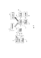
本発明の実施は添付の図面によりさらに理解され得る。類似又は同一の構造は、同一の参照符号を使用して識別される。図1は、本発明のための典型的な処理フロー図を示す。本発明によれば、貯蔵器12からの潜在的な薬剤化学物質は、対象バインダ貯蔵器14からの少なくとも一つの対象バインダと組み合わされて、貯蔵器16内の溶液を形成する。本発明に使用される潜在的な薬剤化学物質は、典型的には水溶性の有機化学物質であり、九以上の原子番号を有する少なくとも一つの元素を有する。好適には、前記薬剤化学物質は、フッ素、塩素、臭素、ヨウ素、硫黄、リン、セレニウム、ランタン、セリウム、プラセオジミウム、ネオジム、サマリウム、ユーロピウム、ガドリニウム、テルビウム、ジスプロジウム、ホルミウム、エルビウム、ツリウム、イッテルビウム、ルテチウム、アンチモン、ビスマス及びヒ素から選択された少なくとも一つの元素を含む。この発明に使用され得る対象バインダは、酵素、非酵素蛋白質、DNA、RNA、植物細胞、動物細胞、ヒト細胞及び微生物(例えばプリオン、ウィルス、バクテリアを含む)等を含む。
The practice of the present invention may be further understood with reference to the accompanying drawings. Similar or identical structures are identified using the same reference signs. FIG. 1 shows an exemplary process flow diagram for the present invention. In accordance with the present invention, potential pharmaceutical chemicals from
潜在的な薬剤化学物質と対象バインダの混合物の溶液は、フロー分離器18に入る。フロー分離器18は、前記溶液を少なくとも二つの成分にフロー分離する移動相を使用する。本発明に使用され得るフロー分離器は、遠心分離機、細胞選別器又はクロマトグラフ(例えば高性能液体クロマトグラフのような液体クロマトグラフ、毛管電気泳動分離器のような電気泳動分離器、ゲル濾過クロマトグラフ、ゲル浸透クロマトグラフ等)を含むが、これらに限られない。好適には、分離器は毛管電気泳動分離器、即ち、管内側の移動相(例えば水性緩衝溶液)と管の長さに亘る電位を有する長く細い管である。
A solution of the potential pharmaceutical chemical and target binder mixture enters the
上記混合物は複数の成分に分離されるので、前記成分はX線に曝される。好適にはロジウムターゲットX線管であるX線励起源20は、X線ビーム22を分離された成分に伝達する。前記成分は、X線蛍光信号24を放射し又は放射しない。X線蛍光信号24は、X線蛍光検出器26によって検出される。本発明に使用され得るX線検出器は、リチウム拡散型シリコン検出器、シリコン拡散検出器又はPINダイオードを含むが、これらに限られない。被曝された成分がX線蛍光信号を放射しない場合、当該成分は第一の回収器28に向けられる。被曝された成分がX線蛍光検出器26によって検出される蛍光信号を放射する場合、当該成分は第二の回収器30に向けられる。前記成分は、少なくとも一つの潜在的な薬剤化学物質と対象バインダの結合された複合体を含むことが期待される。
Since the mixture is separated into a plurality of components, the components are exposed to X-rays. An
第一の回収器と第二の回収器のみが図1に示されているが、上記混合物から隔離された分離された成分の数に依存して、より多くの回収器が使用され得ることは理解されるべきである。 Although only the first and second collectors are shown in FIG. 1, depending on the number of separated components isolated from the mixture, more collectors can be used. Should be understood.
検出可能なX線蛍光信号を放射する分離された成分、即ち第二の回収器30に向けられた成分は、その後分析器32に送られ得る。本発明に使用され得る分析器は、ガスクロマトグラフ、液体クロマトグラフ、質量分光計、核磁気共鳴分光計、赤外分光計、紫外−可視(UV-VIS)分光計、フッ素計、燃焼分析器(元素分析用)、細胞培養、免疫学的検定等を含むが、これらに限られない。分析器の選択は、分析される潜在的な薬剤化学物質及び/又はバインダの性質に依存するであろう。
Separated components that emit a detectable X-ray fluorescence signal, ie, components directed to the
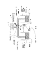
図2は、本発明のスクリーニング装置の実施形態の模式図を示す。図2が示すように、スクリーニング装置34はインレット移動相貯蔵器36を含み、インレット移動相貯蔵器36は毛管分離器40に移動相38を与える。分離器のインレット端42はインレット移動相貯蔵器36の中に所在し、アウトレット端44はアウトレット移動相貯蔵器46の中に所在する。移動相38が分離器40を満たした後、潜在的な薬剤化学物質と少なくとも一つの対象バインダの混合物の総量が、分離器40のインレット端42に導入される。その後、インレット端42は、移動相貯蔵器36に置き換えられる。分離器40のインレット端42とアウトレット端44の間の電位は、分離器40を通じて移動相38の流れと前記混合物の流れを促進する。図2は、成分48が前記混合物から分離されたことを示す。図2は、分離された成分48にX線励起ビーム22を方向付けるX線励起源20を示し、分離された成分48はその後X線蛍光検出器26によって検出されるX線蛍光信号24を放射する。放射されたX線蛍光信号の検出は転換弁50を始動させ、転換弁50は移動相36と分離された成分48の流れをダイバータ52に転換させる。ダイバータ52は、移動相36と分離された成分48を成分回収器54に向ける。
FIG. 2 shows a schematic diagram of an embodiment of the screening apparatus of the present invention. As shown in FIG. 2, the screening device 34 includes an inlet
前述した分離は電位を使用して達成され、電位は毛管分離器40の長さに亘る電気傾度を与えた。前記分離は、管の長さに沿って勾配圧力を与えることによっても達成され得る。本実施形態において、高性能液体クロマトグラフィーのために、管は固定相を含み、試料射出インレットは溶液を管に導入するために使用され、ポンプは勾配圧力を与えるであろう。
The separation described above was accomplished using an electric potential that provided an electrical gradient over the length of the
図2が示すように、成分48は、毛管分離器40の水平部分に沿って分離される。この特定の構成は、微生物又は細胞である対象バインダから派生した複合体を分離するためには最適でないと思われる。これらの対象バインダについては、垂直部分に沿って分離する分離器/選別器が好適である。図3は、この発明に使用され得る前記のような分離器/選別器の実施形態を示す。分離器/選別器56は、付加された蛋白質又は核酸等を有する細胞、微生物、小球体から派生した混合物を分離及び選別するために使用され得る。分離器/選別器56は垂直分離器58を含み、垂直分離器58を通じて分離が起こる。図3が示すように、混合物は成分48と成分60に分離された。X線励起源20からのX線ビーム22を受けた成分48はX線蛍光信号を放射し、前記X線蛍光信号はX線蛍光検出器26により検出された。これは印加電圧源62における反応を引き起こし、印加電圧源62は成分48を回収器64に逸らす電圧を印加する。成分60が検出可能なX線蛍光信号を放射しない場合、成分60を逸らすための電圧は印加されず成分60は回収器66に流れ込む。しかし、成分60が検出可能なX線蛍光信号を放射する場合、成分60を逸らすための電圧が印加されて成分60は回収器64に流れ込む。
As FIG. 2 shows, the
分離器/選別器56は、Bruce Alberts他「Molecular Biology of the Cell」第二版、Garland Publishing, Inc., New York、1989年、第159〜160頁により述べられたタイプの従来の蛍光発光活性化細胞選別を実行するためのレーザー源及び関連付けられた検出器を含み得る。 Separator / sorter 56 is a conventional fluorescent activity of the type described by Bruce Alberts et al. “Molecular Biology of the Cell”, 2nd edition, Garland Publishing, Inc., New York, 1989, pp. 159-160. A laser source and associated detector may be included for performing cellized cell sorting.
潜在的な薬剤化学物質が特定の対象バインダと結合する必要がある場合、例えば、本発明に従って当該蛋白質に結合するために多数の異なる潜在的な薬剤化学物質がスクリーニングされ得る。本発明は、どの潜在的な薬剤化学物質が強く当該蛋白質に結合するかを、弱く結合するもの又は全く結合しないものから見分けるために使用され得る。前記蛋白質は約十から一万の潜在的な薬剤化学物質と組み合わされ、それぞれの潜在的な薬剤化学物質は九又はそれより大きい原子番号を有する少なくとも一つの元素を含む。好適には、潜在的な薬剤化学物質は九又はそれより大きい原子番号を有する元素を含み、スクリーニング方法を単純化するため前記元素は対象バインダ内に発見されない。 If a potential drug chemical needs to bind to a particular target binder, for example, a number of different potential drug chemicals can be screened to bind to the protein according to the present invention. The present invention can be used to distinguish which potential pharmaceutical chemicals bind strongly to the protein from those that bind weakly or those that do not bind at all. The protein is combined with about ten to ten thousand potential drug chemicals, each potential drug chemical containing at least one element having an atomic number of nine or greater. Preferably, the potential pharmaceutical chemical contains an element having an atomic number of nine or greater, and the element is not found in the target binder to simplify the screening method.
本発明は、例えばコバルトイオン(Co2+)及び/又はシアノコバラミンのいずれが公知の生物学的に活性である蛋白質Ure2p(Finny G. Kuruvilla他「Dissecting Glucose Signaling With Diversity-Oriented Synthesis and Small-Molecule Microarrays」、Nature、Vol. 416、第653〜657頁参照)に結合するかを決定するために使用され得る。硝酸コバルト(II)及びシアノコバラミンの水溶液がUre2pに加えられる。結果として生じる水溶液は、例えば毛管電気泳動分離器を使用して本発明によりフロー分離される。Ure2pとCo2+及び/又はシアノコバラミンの間に形成されるいずれの複合体もCo2+及びシアノコバラミンのいずれかから異なる保持時間を有するべきであり、検出可能なX線蛍光信号を放射し、本発明を使用して隔離可能である。 The present invention relates to a protein Ure2p (Finny G. Kuruvilla et al., “Dissecting Glucose Signaling With Diversity-Oriented Synthesis and Small-Molecule Microarrays” in which either cobalt ion (Co 2+ ) and / or cyanocobalamin is known. , Nature, Vol. 416, pp. 653-657). An aqueous solution of cobalt (II) nitrate and cyanocobalamin is added to Ure2p. The resulting aqueous solution is flow separated according to the present invention using, for example, a capillary electrophoresis separator. Any complex formed between Ure2p and Co 2+ and / or cyanocobalamin should have a different retention time from either Co 2+ and cyanocobalamin, emitting a detectable X-ray fluorescence signal, It can be isolated using the invention.
上記分離は、例えば、電位を与えるためのBertanTMモデルARB-30高電圧電源と、長さ70cm、内径(id)100μm、外径(od)170μmの寸法を有する石英ガラス毛管(Polymicro TechnologiesTM)を使用して実行され得る。前記毛管は最初に15分間1.0モルのNaOH溶液により洗い流され、次に15分間蒸留され脱イオン化された水により洗浄され、次に追加の15分間75ミリモルのTrisma緩衝液(pH 8.0)により洗い流され満たされることによって状態が整えられる。 For example, a Bertan TM model ARB-30 high voltage power supply for applying a potential and a quartz glass capillary (Polymicro Technologies TM ) having a length of 70 cm, an inner diameter (id) of 100 μm, and an outer diameter (od) of 170 μm. Can be implemented using: The capillary is first rinsed with 1.0 molar NaOH solution for 15 minutes, then washed with distilled and deionized water for 15 minutes, and then rinsed with 75 mM Trisma buffer (pH 8.0) for an additional 15 minutes. The condition is adjusted by being satisfied.
ベースラインは、コバルト硝酸塩(Co(NO3)2、200ppm Co2+)とシアノコバラミン(10.2ミリモル)の水性混合物を毛管内に導入し、前記毛管の両端間に10kVの電位を印加し、前記混合物をその成分に分離することにより得られた。Rh対象励起源及びSiLi検出器を備えたEDAXTM Eagle IIマイクロX線蛍光システムが、分離された各成分を尋問し、放射された任意のX線蛍光信号を測定するために使用された。前記システムのX線管は、40kV及び1000μAにて動作させられた。CoKαX線放射は、結合していないCo2+とシアノコバラミンを検出するために監視された。スペクトル取得時間は約10秒であった。結合していないCo2+によるピークは、約1分の半値全幅(FWHM)と共に約4.5分に検出された。シアノコバラミンのピークは、約1.5分のFWHMと共に約8.5分に検出された。 Baseline introduces an aqueous mixture of cobalt nitrate (Co (NO 3 ) 2 , 200 ppm Co 2+ ) and cyanocobalamin (10.2 mmol) into the capillary, applies a 10 kV potential across the capillary, Was obtained by separating it into its components. An EDAX ™ Eagle II micro X-ray fluorescence system equipped with an Rh target excitation source and a SiLi detector was used to interrogate each separated component and measure any emitted X-ray fluorescence signal. The X-ray tube of the system was operated at 40 kV and 1000 μA. CoK alpha X-ray radiation was monitored to detect the Co 2+ and cyanocobalamin unbound. The spectrum acquisition time was about 10 seconds. A peak due to unbound Co 2+ was detected at about 4.5 minutes with a full width at half maximum (FWHM) of about 1 minute. The cyanocobalamin peak was detected at about 8.5 minutes with a FWHM of about 1.5 minutes.
同様に、本発明はフェリチン及び/又はシアノコバラミンがUre2pに結合するか否かを決定するために使用され得る。フェリチン及びシアノコバラミンの水溶液は、Ure2pに加えられる。結果として生じる水溶液は、毛管泳動分離器を使用してフロー分離され得る。X線ビームに被曝された時、フェリチン中の鉄及びシアノコバラミン中のコバルトが、それぞれ識別可能かつ検出可能なX線蛍光信号を放射する。前記X線蛍光信号は、フェリチン及び/又はシアノコバラミンとUre2pの間の複合体が形成されたか否かを決定するために使用され得る。 Similarly, the present invention can be used to determine whether ferritin and / or cyanocobalamin binds to Ure2p. An aqueous solution of ferritin and cyanocobalamin is added to Ure2p. The resulting aqueous solution can be flow separated using a capillary electrophoresis separator. When exposed to an X-ray beam, iron in ferritin and cobalt in cyanocobalamin each emit an identifiable and detectable X-ray fluorescence signal. The X-ray fluorescence signal can be used to determine whether a complex between ferritin and / or cyanocobalamin and Ure2p has formed.
ベースラインは以下のように得られた。毛管電気泳動分離器は、分離電位を与えるためのBertanTMモデルARB-30高電圧電源と、長さ70cm、内径(id)100μm、外径(od)170μmの寸法を有する石英ガラス毛管(Polymicro TechnologiesTM)を使用して準備される。前記毛管は最初に15分間1.0モルのNaOH溶液により洗い流され、次に15分間蒸留され脱イオン化された水により洗浄され、次に追加の15分間75ミリモルのTrisma緩衝液(pH 8.0)により洗い流されることによって状態が整えられる。 Baseline was obtained as follows. The capillary electrophoresis separator is a Bertan TM model ARB-30 high-voltage power supply for providing the separation potential and a quartz glass capillary (Polymicro Technologies) with dimensions of 70 cm length, inner diameter (id) 100 μm, outer diameter (od) 170 μm. TM ). The capillary is first rinsed with 1.0 molar NaOH solution for 15 minutes, then washed with distilled and deionized water for 15 minutes, and then rinsed with 75 mM Trisma buffer (pH 8.0) for an additional 15 minutes. The state is arranged by this.
フェリチン(1.16mg/ml)とコバラミン(10.2ミリモル)の水溶液が、毛管内に導入された。9.5kVの分離電位が毛管の両端の間に印加された後、前記溶液は前記毛管を通じて流れ、二つの成分に分離した。Rh対象励起源及びSiLi検出器を備えたEDAXTM Eagle IIマイクロX線蛍光システムが、分離された各成分を尋問し、放射された任意のX線蛍光信号を測定するために使用された。前記システムのX線管は、40kV及び1000μAにて動作させられた。CoKα及びFeKαX線放射線は、Fe3+結合フェリチンとコバラミンを検出するために監視された。スペクトル取得時間は約10秒であった。フェリチンのFe3+によるピークは、約1.7分の半値全幅(FWHM)と共に約9.3分に検出された。シアノコバラミンのピークは、約1分のFWHMと共に約6.3分に検出された。 An aqueous solution of ferritin (1.16 mg / ml) and cobalamin (10.2 mmol) was introduced into the capillary. After a 9.5 kV separation potential was applied across the capillary, the solution flowed through the capillary and separated into two components. An EDAX ™ Eagle II micro X-ray fluorescence system equipped with an Rh target excitation source and a SiLi detector was used to interrogate each separated component and measure any emitted X-ray fluorescence signal. The X-ray tube of the system was operated at 40 kV and 1000 μA. CoK alpha and FeK alpha X-ray radiation was monitored to detect the Fe 3+ binding ferritin and cobalamin. The spectrum acquisition time was about 10 seconds. A peak due to Fe 3+ of ferritin was detected at about 9.3 minutes with a full width at half maximum (FWHM) of about 1.7 minutes. The cyanocobalamin peak was detected at about 6.3 minutes with a FWHM of about 1 minute.
本発明は、潜在的な薬剤化学物質の危険な代謝副産物を検出するための薬学的代謝物質研究において使用され得る。九又はそれより大きい原子番号を有する少なくとも一つの原子を有する潜在的な薬剤化学物質は、ラット(又は他の実験動物)に与えられ得る。ベースラインを提供するために、潜在的な薬剤化学物質を管理する前にラットから血液サンプルが採取される。潜在的な薬剤化学物質を管理した後、この発明の方法を使用して代謝の存在についてラットからの血液が検査される。 The present invention can be used in pharmaceutical metabolite studies to detect dangerous metabolic byproducts of potential pharmaceutical chemicals. Potential pharmaceutical chemicals having at least one atom with an atomic number of nine or greater can be given to rats (or other laboratory animals). To provide a baseline, a blood sample is taken from the rat prior to managing potential drug chemicals. After managing potential drug chemicals, blood from rats is examined for the presence of metabolism using the method of the invention.
要約すれば、この発明は潜在的な薬剤化学物質と対象バインダの間の結合現象を検出するための装置及び方法を提供する。この発明は、フッ素、塩素、臭素、ヨウ素、リン及び硫黄のような元素の存在及び相対的な量を決定するために、マイクロX線蛍光発光を使用する。リン及び硫黄は、酵素、非酵素蛋白質、DNA及びRNAの重要な構成要素である。このように、本発明は、蛋白質又は核酸のような対象バインダと潜在的な薬剤化学物質の結合をスクリーニングする非破壊的方法を提供する。公知の方法は、バインダ及び/又は潜在的な薬剤化学物質が紫外線励起放射に曝された時に蛍光発光する共有結合性のタグを含むことをしばしば必要とするが、本発明はタグ付けされた物質を必要としない。 In summary, the present invention provides an apparatus and method for detecting a binding phenomenon between a potential pharmaceutical chemical and a target binder. The present invention uses micro X-ray fluorescence to determine the presence and relative amounts of elements such as fluorine, chlorine, bromine, iodine, phosphorus and sulfur. Phosphorus and sulfur are important components of enzymes, non-enzymatic proteins, DNA and RNA. Thus, the present invention provides a non-destructive method of screening for binding of a potential pharmaceutical chemical with a target binder such as a protein or nucleic acid. While known methods often require that the binder and / or potential pharmaceutical chemicals include a covalent tag that fluoresces when exposed to UV-excited radiation, the present invention provides for tagged materials Do not need.
本発明の上記説明は、図示及び記述の目的のために提示されたものであり、網羅的であること又は開示された正確な形式に限定することは意図されておらず、また上記教示に照らして明らかに多くの変更及び変形が可能である。 The foregoing description of the present invention has been presented for purposes of illustration and description, and is not intended to be exhaustive or limited to the precise form disclosed, and in light of the above teachings. Obviously many modifications and variations are possible.
上記実施形態は、本発明の原理及び実際の応用を最良に説明する目的で選択され記載されたものであり、これにより他の当業者が本発明を種々の実施形態において最良に利用することを可能にし、種々の変更により特定の熟考された使用に適する。本発明の範囲は、本明細書に添付の特許請求の範囲により定義されることが意図されている。 The above embodiments have been selected and described for the best purpose of illustrating the principles and practical applications of the present invention so that others skilled in the art can best utilize the present invention in various embodiments. Makes it suitable for a particular contemplated use with various modifications. It is intended that the scope of the invention be defined by the claims appended hereto.
添付の図面は、本明細書に包含されその一部を形成し、本発明の実施形態を図示し、その説明と共に本発明の原理を説明するために提供される。 The accompanying drawings, which are incorporated in and constitute a part of this specification, illustrate embodiments of the present invention and, together with the description, serve to explain the principles of the invention.
Claims (7)
少なくとも一つの化学物質と少なくとも一つの対象バインダを含む溶液を準備することと、
前記溶液を少なくとも二つの分離された成分にフロー分離することと、任意のフロー分離された成分中に存在する化学物質、及び、任意の結合された複合体の化学物質部分の原子からX線蛍光信号を生成するために、前記原子を励起することと、
分離された成分中に存在する化学物質、及び、任意の結合された複合体の化学物質部分に存在する励起された原子から生成されたX線蛍光信号を検出することと、
分離された成分中に存在する化学物質、及び、任意の結合された複合体の化学物質部分に存在する励起された原子から生成されたX線蛍光信号から、任意の分離された成分が結合された複合体を含むか否かを決定すること
を含む方法。A method for determining whether a chemical bond occurs between at least one target binder to form a combined complex and at least one chemical substance, comprising:
Providing a solution comprising at least one chemical and at least one target binder;
Flow separation of the solution into at least two separated components, and the X-ray fluorescence from the chemicals present in any flow separated component and the atoms of the chemical part of any combined complex Exciting the atoms to generate a signal;
Detecting x-ray fluorescence signals generated from excited atoms present in the chemical moiety present in the separated components and in the chemical moiety of any bound complex;
Any separated component is bound from the X-ray fluorescence signal generated from the chemicals present in the separated component and the excited atoms present in the chemical part of any bound complex. Determining whether to include a complex.
少なくとも一つの化学物質と少なくとも一つの対象バインダを含む溶液を準備することと、
勾配圧力を使用して前記溶液を少なくとも二つの分離された成分にフロー分離することと、
任意のフロー分離された成分中に存在する化学物質、及び、任意の結合された複合体の化学物質部分の原子からX線蛍光信号を生成するために、前記原子を励起することと、
分離された成分中に存在する化学物質、及び、任意の結合された複合体の化学物質部分に存在する励起された原子から生成されたX線蛍光信号を検出することと、
分離された成分中に存在する化学物質、及び、任意の結合された複合体の化学物質部分に存在する励起された原子から生成されたX線蛍光信号から、任意の分離された成分が結合された複合体を含むか否かを決定すること
を含む方法。A method for determining whether a chemical bond occurs between at least one target binder to form a combined complex and at least one chemical substance, comprising:
Providing a solution comprising at least one chemical and at least one target binder;
Flow separating the solution into at least two separated components using gradient pressure;
Exciting the atoms to generate an X-ray fluorescence signal from atoms present in any flow separated component, and atoms of the chemical portion of any bound complex;
Detecting x-ray fluorescence signals generated from excited atoms present in the chemical moiety present in the separated components and in the chemical moiety of any bound complex;
Any separated component is bound from the X-ray fluorescence signal generated from the chemicals present in the separated component and the excited atoms present in the chemical part of any bound complex. Determining whether to include a complex.
Applications Claiming Priority (2)
| Application Number | Priority Date | Filing Date | Title |
|---|---|---|---|
| US10/206,524 US20040017884A1 (en) | 2002-07-25 | 2002-07-25 | Flow method and apparatus for screening chemicals using micro x-ray fluorescence |
| PCT/US2003/020103 WO2004011898A2 (en) | 2002-07-25 | 2003-06-24 | Flow method and apparatus for screening chemicals using micro x-ray fluorescence |
Publications (2)
| Publication Number | Publication Date |
|---|---|
| JP2006503268A JP2006503268A (en) | 2006-01-26 |
| JP4560403B2 true JP4560403B2 (en) | 2010-10-13 |
Family
ID=30770310
Family Applications (1)
| Application Number | Title | Priority Date | Filing Date |
|---|---|---|---|
| JP2004524531A Expired - Fee Related JP4560403B2 (en) | 2002-07-25 | 2003-06-24 | Flow method for screening chemical substances using micro X-ray fluorescence |
Country Status (7)
| Country | Link |
|---|---|
| US (2) | US20040017884A1 (en) |
| EP (1) | EP1525458B1 (en) |
| JP (1) | JP4560403B2 (en) |
| AU (1) | AU2003267973A1 (en) |
| DK (1) | DK1525458T3 (en) |
| ES (1) | ES2623296T3 (en) |
| WO (1) | WO2004011898A2 (en) |
Families Citing this family (12)
| Publication number | Priority date | Publication date | Assignee | Title |
|---|---|---|---|---|
| US20080220441A1 (en) | 2001-05-16 | 2008-09-11 | Birnbaum Eva R | Advanced drug development and manufacturing |
| US9157875B2 (en) * | 2001-05-16 | 2015-10-13 | Benjamin P. Warner | Drug development and manufacturing |
| JP2006029921A (en) * | 2004-07-14 | 2006-02-02 | Institute Of Physical & Chemical Research | Flow site meter |
| US7581130B2 (en) | 2004-11-12 | 2009-08-25 | Hewlett-Packard Development Company, L.P. | Power management system and method |
| DK2084519T3 (en) * | 2006-10-10 | 2012-08-20 | Los Alamos Nat Security Llc | X-ray fluorescence analysis method |
| US7892492B2 (en) * | 2006-12-07 | 2011-02-22 | University Of Connecticut | Flow-through apparatus for microscopic investigation of dissolution of pharmaceutical solids |
| US8687189B2 (en) * | 2008-03-03 | 2014-04-01 | Ajjer, Llc | Analysis of arrays by laser induced breakdown spectroscopy |
| CA2882003C (en) * | 2012-08-17 | 2020-11-10 | Japan Science And Technology Agency | Method and apparatus for analyzing biomolecules using raman spectroscopy |
| JP5437525B1 (en) | 2012-12-28 | 2014-03-12 | 株式会社ナード研究所 | Tyrosine derivative and method for producing tyrosine derivative |
| WO2017024035A1 (en) * | 2015-08-03 | 2017-02-09 | UHV Technologies, Inc. | Metal analysis during pharmaceutical manufacturing |
| EP4004540A4 (en) | 2019-07-29 | 2023-03-29 | Shenzhen Xpectvision Technology Co., Ltd. | Biological imaging method using x-ray fluorescence |
| CN110608981A (en) * | 2019-09-09 | 2019-12-24 | 中国科学院高能物理研究所 | Method for measuring mercury sulfide nanoparticles in plants |
Family Cites Families (15)
| Publication number | Priority date | Publication date | Assignee | Title |
|---|---|---|---|---|
| US4745285A (en) * | 1986-08-21 | 1988-05-17 | Becton Dickinson And Company | Multi-color fluorescence analysis with single wavelength excitation |
| US5252743A (en) * | 1989-11-13 | 1993-10-12 | Affymax Technologies N.V. | Spatially-addressable immobilization of anti-ligands on surfaces |
| US5854084A (en) * | 1996-07-12 | 1998-12-29 | Biotraces, Inc. | Enhanced chromatography using multiphoton detection |
| US5493122A (en) * | 1994-02-04 | 1996-02-20 | Nucleonics Development Company | Energy resolving x-ray detector |
| US5660703A (en) * | 1995-05-31 | 1997-08-26 | The Dow Chemical Company | Apparatus for capillary electrophoresis having an auxiliary electroosmotic pump |
| AU6160896A (en) * | 1995-06-07 | 1996-12-30 | Life Technologies, Inc. | On line detection of a desired solute in an effluent stream using fluorescence spectroscopy |
| US6027890A (en) * | 1996-01-23 | 2000-02-22 | Rapigene, Inc. | Methods and compositions for enhancing sensitivity in the analysis of biological-based assays |
| US5668373A (en) * | 1996-04-26 | 1997-09-16 | Trustees Of Tufts College | Methods and apparatus for analysis of complex mixtures |
| US5982847A (en) * | 1996-10-28 | 1999-11-09 | Utah State University | Compact X-ray fluorescence spectrometer for real-time wear metal analysis of lubrucating oils |
| US6207861B1 (en) * | 1998-01-05 | 2001-03-27 | Neogenesis, Inc. | Method for producing and screening mass coded combinatorial libraries for drug discovery and target validation |
| US6344330B1 (en) * | 1998-03-27 | 2002-02-05 | The Regents Of The University Of California | Pharmacophore recombination for the identification of small molecule drug lead compounds |
| US6054047A (en) * | 1998-03-27 | 2000-04-25 | Synsorb Biotech, Inc. | Apparatus for screening compound libraries |
| US6147344A (en) * | 1998-10-15 | 2000-11-14 | Neogenesis, Inc | Method for identifying compounds in a chemical mixture |
| WO2000043761A2 (en) * | 1999-01-23 | 2000-07-27 | Merck Patent Gmbh | Energy dispersion x-ray fluorescence analysis of chemical substances |
| US6697454B1 (en) * | 2000-06-29 | 2004-02-24 | X-Ray Optical Systems, Inc. | X-ray analytical techniques applied to combinatorial library screening |
-
2002
- 2002-07-25 US US10/206,524 patent/US20040017884A1/en not_active Abandoned
-
2003
- 2003-06-24 EP EP03748920.0A patent/EP1525458B1/en not_active Expired - Lifetime
- 2003-06-24 ES ES03748920.0T patent/ES2623296T3/en not_active Expired - Lifetime
- 2003-06-24 JP JP2004524531A patent/JP4560403B2/en not_active Expired - Fee Related
- 2003-06-24 AU AU2003267973A patent/AU2003267973A1/en not_active Abandoned
- 2003-06-24 WO PCT/US2003/020103 patent/WO2004011898A2/en active Application Filing
- 2003-06-24 DK DK03748920.0T patent/DK1525458T3/en active
-
2005
- 2005-05-09 US US11/125,036 patent/US20050214847A1/en not_active Abandoned
Also Published As
| Publication number | Publication date |
|---|---|
| JP2006503268A (en) | 2006-01-26 |
| EP1525458A4 (en) | 2009-08-19 |
| EP1525458A2 (en) | 2005-04-27 |
| US20050214847A1 (en) | 2005-09-29 |
| WO2004011898A3 (en) | 2004-12-02 |
| AU2003267973A8 (en) | 2004-02-16 |
| AU2003267973A1 (en) | 2004-02-16 |
| DK1525458T3 (en) | 2017-04-24 |
| WO2004011898A2 (en) | 2004-02-05 |
| ES2623296T3 (en) | 2017-07-10 |
| US20040017884A1 (en) | 2004-01-29 |
| EP1525458B1 (en) | 2017-03-08 |
Similar Documents
| Publication | Publication Date | Title |
|---|---|---|
| US7929662B2 (en) | Flow method and apparatus for screening chemicals using micro x-ray fluorescence | |
| US20050214847A1 (en) | Flow method and apparatus for screening chemicals using micro x-ray fluorescence | |
| JP4782676B2 (en) | Method and apparatus for detecting chemical binding | |
| Liu et al. | Inductively coupled plasma mass spectrometry‐based immunoassay: A review | |
| CN100489534C (en) | Quantitation of biological molecules | |
| US7858385B2 (en) | Method for detecting binding events using micro-X-ray fluorescence spectrometry | |
| JP2007538262A (en) | Quantifying expression using mass spectrometry | |
| Gao et al. | Advanced nuclear analytical techniques for metalloproteomics | |
| Moser et al. | Clinical applications of capillary electrophoresis based immunoassays | |
| Chen et al. | Rapid and high-throughput detection and quantitation of radiation biomarkers in human and nonhuman primates by differential mobility spectrometry-mass spectrometry | |
| US9157875B2 (en) | Drug development and manufacturing | |
| Lidofsky et al. | Laser fluorescence immunoassay of insulin | |
| Maleknia et al. | Unfolding of apomyoglobin helices by synchrotron radiolysis and mass spectrometry | |
| JP3887670B2 (en) | Fluorescence intensity method for measuring binding between proteins or peptides | |
| US6328700B1 (en) | Locating marker/tracer elements detectable by neutron activated analysis within or on carrier microspheres, including microspheres used in biological experimentation | |
| Taraboletti et al. | Fabric Phase Sorptive Extraction—A metabolomic preprocessing approach for ionizing radiation exposure assessment | |
| Vogel et al. | Accelerator mass spectrometry best practices for accuracy and precision in bioanalytical 14C measurements | |
| Smith et al. | In vivo measurement and speciation of nephrotoxic metals | |
| Luo et al. | Chloride-Mediated Peroxide-Free Photochemical Oxidation of Proteins (PPOP) in Mass Spectrometry-Based Structural Analysis | |
| Alvarez et al. | Tissue proteomics of the low‐molecular weight proteome using an integrated c LC‐ESI‐QTOFMS approach | |
| Warner et al. | Method And Apparatus For Detecting Chemical Binding | |
| Aebersold et al. | Isotope-coded affinity tagging of proteins | |
| CN117597583A (en) | Method and system for detecting and quantifying a plurality of molecular biomarkers in a bodily fluid sample | |
| Minogue et al. | High-throughput screening of metal chelating compounds | |
| EP3176581A1 (en) | Construction kit for a multiplex drug discovery system with high-throughput properties |
Legal Events
| Date | Code | Title | Description |
|---|---|---|---|
| A621 | Written request for application examination |
Free format text: JAPANESE INTERMEDIATE CODE: A621 Effective date: 20060526 |
|
| A131 | Notification of reasons for refusal |
Free format text: JAPANESE INTERMEDIATE CODE: A131 Effective date: 20081211 |
|
| A601 | Written request for extension of time |
Free format text: JAPANESE INTERMEDIATE CODE: A601 Effective date: 20090309 |
|
| A601 | Written request for extension of time |
Free format text: JAPANESE INTERMEDIATE CODE: A601 Effective date: 20090311 |
|
| A602 | Written permission of extension of time |
Free format text: JAPANESE INTERMEDIATE CODE: A602 Effective date: 20090317 |
|
| A602 | Written permission of extension of time |
Free format text: JAPANESE INTERMEDIATE CODE: A602 Effective date: 20090318 |
|
| A521 | Request for written amendment filed |
Free format text: JAPANESE INTERMEDIATE CODE: A523 Effective date: 20090605 |
|
| A521 | Request for written amendment filed |
Free format text: JAPANESE INTERMEDIATE CODE: A523 Effective date: 20090708 |
|
| A131 | Notification of reasons for refusal |
Free format text: JAPANESE INTERMEDIATE CODE: A131 Effective date: 20091221 |
|
| A521 | Request for written amendment filed |
Free format text: JAPANESE INTERMEDIATE CODE: A523 Effective date: 20100224 |
|
| TRDD | Decision of grant or rejection written | ||
| A01 | Written decision to grant a patent or to grant a registration (utility model) |
Free format text: JAPANESE INTERMEDIATE CODE: A01 Effective date: 20100715 |
|
| A01 | Written decision to grant a patent or to grant a registration (utility model) |
Free format text: JAPANESE INTERMEDIATE CODE: A01 |
|
| A61 | First payment of annual fees (during grant procedure) |
Free format text: JAPANESE INTERMEDIATE CODE: A61 Effective date: 20100726 |
|
| R150 | Certificate of patent or registration of utility model |
Free format text: JAPANESE INTERMEDIATE CODE: R150 Ref document number: 4560403 Country of ref document: JP Free format text: JAPANESE INTERMEDIATE CODE: R150 |
|
| FPAY | Renewal fee payment (event date is renewal date of database) |
Free format text: PAYMENT UNTIL: 20130730 Year of fee payment: 3 |
|
| R250 | Receipt of annual fees |
Free format text: JAPANESE INTERMEDIATE CODE: R250 |
|
| R250 | Receipt of annual fees |
Free format text: JAPANESE INTERMEDIATE CODE: R250 |
|
| S111 | Request for change of ownership or part of ownership |
Free format text: JAPANESE INTERMEDIATE CODE: R313113 |
|
| R350 | Written notification of registration of transfer |
Free format text: JAPANESE INTERMEDIATE CODE: R350 |
|
| R250 | Receipt of annual fees |
Free format text: JAPANESE INTERMEDIATE CODE: R250 |
|
| R250 | Receipt of annual fees |
Free format text: JAPANESE INTERMEDIATE CODE: R250 |
|
| R250 | Receipt of annual fees |
Free format text: JAPANESE INTERMEDIATE CODE: R250 |
|
| R250 | Receipt of annual fees |
Free format text: JAPANESE INTERMEDIATE CODE: R250 |
|
| R250 | Receipt of annual fees |
Free format text: JAPANESE INTERMEDIATE CODE: R250 |
|
| LAPS | Cancellation because of no payment of annual fees |