JP4485049B2 - Tumor necrosis factor homologue called DNA19355 polypeptide - Google Patents
Tumor necrosis factor homologue called DNA19355 polypeptide Download PDFInfo
- Publication number
- JP4485049B2 JP4485049B2 JP2000521199A JP2000521199A JP4485049B2 JP 4485049 B2 JP4485049 B2 JP 4485049B2 JP 2000521199 A JP2000521199 A JP 2000521199A JP 2000521199 A JP2000521199 A JP 2000521199A JP 4485049 B2 JP4485049 B2 JP 4485049B2
- Authority
- JP
- Japan
- Prior art keywords
- dna19355
- cells
- polypeptide
- cell
- sequence
- Prior art date
- Legal status (The legal status is an assumption and is not a legal conclusion. Google has not performed a legal analysis and makes no representation as to the accuracy of the status listed.)
- Expired - Lifetime
Links
Images


Classifications
-
- C—CHEMISTRY; METALLURGY
- C07—ORGANIC CHEMISTRY
- C07K—PEPTIDES
- C07K14/00—Peptides having more than 20 amino acids; Gastrins; Somatostatins; Melanotropins; Derivatives thereof
- C07K14/435—Peptides having more than 20 amino acids; Gastrins; Somatostatins; Melanotropins; Derivatives thereof from animals; from humans
- C07K14/52—Cytokines; Lymphokines; Interferons
- C07K14/525—Tumour necrosis factor [TNF]
-
- A—HUMAN NECESSITIES
- A61—MEDICAL OR VETERINARY SCIENCE; HYGIENE
- A61P—SPECIFIC THERAPEUTIC ACTIVITY OF CHEMICAL COMPOUNDS OR MEDICINAL PREPARATIONS
- A61P35/00—Antineoplastic agents
-
- A—HUMAN NECESSITIES
- A61—MEDICAL OR VETERINARY SCIENCE; HYGIENE
- A61P—SPECIFIC THERAPEUTIC ACTIVITY OF CHEMICAL COMPOUNDS OR MEDICINAL PREPARATIONS
- A61P35/00—Antineoplastic agents
- A61P35/02—Antineoplastic agents specific for leukemia
-
- A—HUMAN NECESSITIES
- A61—MEDICAL OR VETERINARY SCIENCE; HYGIENE
- A61P—SPECIFIC THERAPEUTIC ACTIVITY OF CHEMICAL COMPOUNDS OR MEDICINAL PREPARATIONS
- A61P37/00—Drugs for immunological or allergic disorders
- A61P37/02—Immunomodulators
-
- A—HUMAN NECESSITIES
- A61—MEDICAL OR VETERINARY SCIENCE; HYGIENE
- A61P—SPECIFIC THERAPEUTIC ACTIVITY OF CHEMICAL COMPOUNDS OR MEDICINAL PREPARATIONS
- A61P43/00—Drugs for specific purposes, not provided for in groups A61P1/00-A61P41/00
-
- A—HUMAN NECESSITIES
- A61—MEDICAL OR VETERINARY SCIENCE; HYGIENE
- A61K—PREPARATIONS FOR MEDICAL, DENTAL OR TOILETRY PURPOSES
- A61K38/00—Medicinal preparations containing peptides
-
- C—CHEMISTRY; METALLURGY
- C07—ORGANIC CHEMISTRY
- C07K—PEPTIDES
- C07K2319/00—Fusion polypeptide
- C07K2319/01—Fusion polypeptide containing a localisation/targetting motif
- C07K2319/02—Fusion polypeptide containing a localisation/targetting motif containing a signal sequence
Abstract
Description
【0001】
(関連する出願)
これは、1997年12月18日に出願された仮出願番号60/065,635及び1997年12月12日に出願された仮出願番号60,069,661に基づく119条(e)の下での優先権を主張した非仮出願であり、それらの内容は出典明示して取り入れる。
【0002】
(発明の分野)
本発明は、一般に、新規なDNAの同定及び単離、並びに、ここに「DNA19355」と命名する新規ポリペプチドの組換え生産に関する。
【0003】
(発明の背景)
哺乳動物における細胞数のコントロールは、部分的には、細胞増殖と細胞死のバランスにより決定されると考えられている。壊死性細胞死と称されることのある細胞死の一形態は、典型的には、ある種の外傷又は細胞傷害の結果生じる細胞死の病理的形態として特性付けられる。これに対して、通常は規則的又はコントロールされた状態で進行する細胞死の他の「生理的」形態がある。細胞死のこの規則的又はコントロールされた形態は、しばしば「アポトーシス」と称される[例えば、Barrら, Bio/Technology, 12:487-493(1994);Stellerら, Science, 267:1445-1449(1995)を参照]。アポトーシス性細胞死は、免疫系におけるクローン選択及び胚の発達を含む多くの生理的プロセスにおいて自然に生じる[Itohら, Cell, 66:233-243(1991)]。アポトーシス性細胞死のレベルの減少は、癌、狼瘡、及びヘルペスウイスル感染を含む種々の病理学的状態に関連している[Thompson, Science, 267:1456-1462(1995)]。アポトーシス性細胞死のレベルの増加は、エイズ、アルツハイマー病、パーキンソン病、筋萎縮性側索硬化症、多発性硬化症、色素性網膜炎、小脳変性、無形成性貧血、心筋梗塞、発作、再灌流傷害、及び毒素誘発性肝疾患を含む様々な他の病理学的状態に関連している[上掲のThompsonを参照]。
【0004】
アポトーシス性細胞死は、典型的には、細胞における一又は複数の特徴的な形態学的及び生化学的変化、例えば細胞質の凝結、原形質膜の微絨毛の喪失、核の分節化、染色体DNAの分解又はミトコンドリア機能の喪失を伴う。様々な外因的及び内因的シグナルが、このような形態学的及び生化学的な細胞変化を惹起又は誘発すると考えられている[Raff, Nature, 356:397-400(1992);Steller, 上掲;Sachsら, Blood, 82:15(1993)]。例えば、それらは、ホルモン性刺激、例えば未成熟胸腺細胞に対する糖質コルチコイドホルモン、並びにある種の成長因子の退薬により惹起され得る[Watanabe-Fukunagaら, Nature, 356:314-317(1992)]。また、幾つかの同定された発癌遺伝子、例えばmyc、rel、及びE1A、及び腫瘍サプレッサー、例えばp53が、アポトーシスの誘発において役割を有していることも報告されている。ある種の化学療法薬及びある形態の放射線も同様にアポトーシス誘発活性を有していることも見出されている[Thompson, 上掲]。
【0005】
腫瘍壊死因子-α(「TNF−α」)、腫瘍壊死因子−β(「TNF−β」又は「リンホトキシン−α」)、リンホトキシン−β(「LT−β」)、CD30リガンド、CD27リガンド、CD40リガンド、OX-40リガンド、4-1BBリガンド、Apo−1リガンド(Fasリガンド又はCD95リガンドとも称される)、及びApo-2リガンド(トレイル(TRAIL)とも称される)等の様々な分子が、サイトカインの腫瘍壊死因子(「TNF」)ファミリーのメンバーとして同定された[例えば、Gruss及びDower, Blood, 85:3378-3404(1995);Pittiら, J. Biol. Chem., 271:12687-12690(1996);Wileyら, Immunity, 3:673-682(1995);Browning等, Cell. 72: 847-856 (1993);Armitage等, Nature, 357: 80-82 (1992)]。これらの分子のなかでも、TNF-α、TNF-β、CD30リガンド、4-1BBリガンド、Apo-1リガンド、及びApo-2リガンド(TRAIL)は、アポトーシス性細胞死に関与していることが報告されている。TNF-αとTNF-βの両方とも、感受性腫瘍細胞におけるアポトーシス性細胞死を誘発することが報告されている[Schmidら, Proc. Natl. Acad. Sci., 83:1881(1986); Dealtryら, Eur. J. Immunol., 17:689(1987)]。ゼング(Zheng)らは、TNF-αがCD8ポジティブT細胞の刺激後アポトーシスに関与していることを報告している[Zhengら, Nature, 377:348-351(1995)]。他の研究者等は、CD30リガンドが胸腺における自己反応性T細胞の欠失に関与していることを報告している[Amakawaら, プログラムされた細胞死に関するコールドスプリングハーバー研究所のシンポジウム、要旨集、第10巻、(1995)]。
【0006】
マウスFas/Apo-1又はリガンド遺伝子(各々1pr及びg1dと呼ばれる)における変異は幾つかの自己免疫疾患に関連しており、Apo-1リガンドが末梢における自己反応性リンパ球のクローン欠失の調節において役割を担っていることを示している[Krammer等, Curr. Op. Immunol., 6: 279-289 (1994);Nagata等, Science, 267: 1449-1456 (1995)]。また、Apo-1リガンドは、CD4陽性Tリンパ球及びBリンパ球における細胞刺激後アポトーシスを誘発すると報告されており、もはやそれらの機能が必要でない場合の活性化されたリンパ球の除去に含まれ得る[Krammer等, 上掲;Nagata等, 上掲]。Apo-1レセプターに特異的に結合するアゴニストのマウスモノクローナル抗体は、TNF-αに匹敵するか類似する細胞死滅活性を示すことが報告されている[Yoneharaら, J. Exp. Med., 169:1747-1756 (1989)]。
【0007】
このようなTNFファミリーのサイトカインが介在する種々の細胞反応の誘導は、それらが特異的な細胞レセプターに結合することにより開始されると考えられている。約55-kDa(TNFR1)と75-kDa(TNFR2)の2つの異なるTNFレセプターが同定されており[Hohmanら, J. Biol. Chem., 264:14927-14934(1989);Brockhausら, Proc. Natl. Acad. Sci., 87:3127-3131(1990);1991年3月20日に公開されたEp 417,563]、双方のレセプターの型に対応するヒト及びマウスcDNAが単離され、特徴付けされている[Loetscherら, Cell, 61:351(1990);Schallら, Cell, 61:361(1990);Smithら, Science, 248:1019-1023(1990);Lewisら, Proc. Natl. Acad. Sci., 88:2830-2834(1991);Goodwinら, Mol. Cell. Biol., 11:3020-3026(1991)]。広範な多型性が、双方のTNFレセプター遺伝子に付随している[例えば、Takaoら, Immunogenetics, 37:199-203(1993)を参照]。双方のTNFRは細胞外、膜貫通及び細胞内領域を含む細胞表面レセプターの典型的な構造を共有する。双方のレセプターの細胞外部分はまた可溶性TNF結合タンパク質として天然に見出される[Nophar, Yら, EMBO J., 9:3269(1990);及びKohno, Tら, Proc. Natl. Acad. Sci. U.S.A., 87:8331(1990)]。さらに最近になって、組換え可溶性TNFレセプターのクローン化がヘイル(Hale)らにより報告されている[J. Cell. Biochem. 増補15F, 1991, p.113(P424)]。
【0008】
1型又は2型のTNFR(TNFR1及びTNFR2)の細胞外部分は、NH2末端から出発して1〜4に設定される4つのシステインに富むドメイン(CRD)の反復アミノ酸配列パターンを含む。各CRDは約40のアミノ酸長のものであり、良好に保存された位置に4〜6のシステイン残基を含んでいる[Schallら, 上掲;Loetscherら, 上掲;Smithら, 上掲;Nopharら, 上掲;Kohnoら, 上掲]。TNFR1において、4つのCRDのおよその境界は次の通りである:CRD1−アミノ酸14から約53まで;CRD2−アミノ酸約54から約97まで;CRD3−アミノ酸約98から約138まで;CRD4−アミノ酸約139から約167まで。TNFR2において、CRD1は17〜約54までのアミノ酸を、CRD2は約55から約97までのアミノ酸を;CRD3は約98から約140までのアミノ酸を;CRD4は約141から約179までのアミノ酸を含む[Bannerら, Cell, 73:431-435(1993)]。また、リガンド結合におけるCRDの潜在的な役割は前掲のバナー(Banner)らにより記載されている。
【0009】
CRDの類似の反復パターンが、p75神経成長因子レセプター(NGFR)[Johnsonら, Cell, 47:545(1986);Radekeら, Nature, 325:593(1987)]、B細胞抗原CD40[Stamenkovicら, EMBO J., 8:1403(1989)]、T細胞抗原OX40[Malletら, EMBO J., 9:1063(1990)]及びFas抗原[Yoneharaら, 上掲、及びItohら, Cell, 66:233-243 (1991)]を含む幾つかの他の細胞表面タンパク質に存在している。また、CRDはショープ(Shope)及び粘液腫ポックスウィルスの可溶型TNFR(sTNFR)様T2タンパク質にも見出される[Uptonら, Virology, 160:20-29(1987);Smithら, Biochem. Biophys. Res. Commun., 176:335(1991);Uptonら, Virology, 184:370(1991)]。これらの配列の最適なアラインメントは、システイン残基の位置が良好に保存されていることを示している。これらレセプターは、集合的に、TNF/NGFレセプタースーパーファミリーのメンバーと称されることがある。p75NGFRに関する最近の研究では、CRD1の欠失[Welcher, A.A.ら, Proc. Natl. Acad. Sci. USA, 88:159-163(1991)]又はこのドメインにおける5-アミノ酸の挿入[Yan. H及びChao, M.V., J. Biol. Chem., 266:12099-12104(1991)]は、NGF結合にはほとんど又は全く影響を持たないことが示された[Yan. H及びChao, M.V., 上掲]。p75NGFRは、そのCRD4と膜貫通領域の間に、NGF結合に関与しない約60のアミノ酸のプロリンに富む伸展を含む[Peetre, Cら, Eur. J. Hematol., 41:414-419(1988);Seckinger, Pら, J. Biol. Chem., 264:11966-11973(1989);Yan. H. 及びChao, M.V., 上掲]。同様のプロリンに富む領域はTNFR2に見出されているがTNFR1にはない。
【0010】
イトー(Itoh)らは、Apo-1レセプターが55-kDaのTNFR1によりシグナル化されるものと類似のアポトーシス性細胞死をシグナル化し得ることを開示している[Itohら, 上掲]。また、Apo-1抗原の発現は、細胞をTNF-α又は抗-Apo-1マウスモノクローナル抗体で処理した場合に、TNFR1のものと共にダウンレギュレーションされることが報告されている[Krammerら, 上掲;Nagataら, 上掲]。従って、Apo-1及びTNFR1レセプターの双方を同時発現する株化細胞が、共通のシグナル伝達経路を通して細胞死滅を媒介しているとの仮説を唱える研究者もいた[同]。
【0011】
リンホトキシン-αを除き、今日までに同定されているTNFファミリーのリガンドは、II型の膜貫通タンパク質であり、そのC末端は細胞外にある。これに対して、今日までに同定されているTNFレセプター(TNFR)ファミリーのレセプター類はI型の膜貫通タンパク質である。しかしながら、TNFリガンド及びレセプターファミリーの双方において、ファミリーメンバー間で同定された相同性は、主として細胞外ドメイン(「ECD」)において見出されている。TNF-α、Apo-1リガンド及びCD40リガンドを含むTNFファミリーサイトカインのいくつかは、細胞表面においてタンパク質分解的に切断され;各場合に得られたタンパク質は、典型的には、可溶性サイトカインとして機能する同種三量体分子を形成する。また、TNFレセプターファミリーのタンパク質は、通常、タンパク質分解的に切断され、同族のサイトカインの阻害剤として機能し得る可溶性レセプターのECDを放出する。
【0012】
最近になって、TNFRファミリーの他のメンバーが同定された。これらの新たに同定されたTNFRファミリーのメンバーは、CAR1、HVEM及びオステオプロテゲリン(osteoprotegerin)(OPG)を含む[Brajatsh等, Cell, 87:845-855 (1996);Montgomery等, Cell, 87:427-436 (1996);Marsters等, J. Biol. Chem., 272:14029-14032 (1997);Simonet等, Cell, 89:309-319 (1997)]。他の知られたTNFR様分子とは異なり、上掲のシモネット(Simonet)等は、OPGが疎水的膜貫通スパンニング配列を含まないことを報告している。
【0013】
マースターズ(Marsters)ら、Curr. Biol., 6:750(1996)において、研究者等は、細胞外のシステインに富む反復においてTNFRファミリーに対して類似性を示し、細胞質死亡ドメイン配列を含む点でTNFR1及びCD95に似ている、Apo-3と称されるヒトのポリペプチド天然配列の全長を開示している[Marstersら, Curr. Biol., 6:1669(1996)もまた参照されたい]。他の研究者によれば、Apo-3は、DR3、wsl-1及びTRAMPとも称されている[Chinnaiyanら, Science, 274:990(1996);Kitsonら, Nature, 384:372(1996);Bodmerら, Immunity, 6:79(1997)]。
【0014】
パン(Pan)らは、「DR4」と称されるTNFレセプターファミリーの他のメンバーを開示している[Panら, Science, 276:111-113(1997)]。DR4は細胞自殺器を活動させる細胞質死亡ドメインを含むと報告されている。パンらは、DR4がApo-2リガンド又はTRAILとして知られているリガンドに対するレセプターであると考えられることを開示している。
【0015】
シェリダン(Sheridan)ら, Science, 277:818-821 (1997)及びパンら, Science, 277: 815-818 (1997)には、Apo-2リガンド(TRAIL)のレセプターと考えられる他の分子が記載されている。この分子は、DR5と称される(また、Apo-2とも称されている)。DR4と同様に、DR5は細胞質死亡ドメインを含み、アポトーシスのシグナル伝達ができる。
【0016】
上掲のシェリダンらには、DcR1(またはApo-2DcR)が、Apo-2リガンド(TRAIL)の潜在的デコイレセプターであると開示されている。シェリダンらは、DcR1がインビトロでApo-2リガンド機能を阻害することができると報告している。また、TRIDと称されるデコイレセプターに関する開示については、上掲のパン等を参照。
TNFファミリーのサイトカイン及びそれらのレセプターについての概説は、Gruss及びDower, 上掲参照。
【0017】
現在理解されているように、細胞死プログラムは、少なくとも3つの重要な成分−活性化剤、阻害剤及びエフェクターを含む;線虫(C. elegans)において、これらの成分は各々3つの遺伝子、Ced-4、Ced-9及びCed-3によりコードされている[Steller, Science, 267:1445(1995);Chinnaiyanら, Science, 275:1122-1126(1997); Wang等, Cell, 90: 1-20 (1997)]。TNFRファミリーのメンバーの2つ、TNFR1及びFas/Apol(CD95)は、アポトーシス性細胞死を活性化し得る[Chinnaiyan及びDixit, Current Biology, 6:555-562(1996);Fraser及びEvan, Cell, 85:781-784(1996)]。また、TNFR1は転写因子、NF-κBの活性化を媒介することも知られている[Tartagliaら, Cell, 74:845-853(1993);Hsuら, Cell, 84:299-308(1996)]。ある程度のECD相同性に加えて、これら2つのレセプターは、死亡ドメインとして知られているオリゴマー形成界面の細胞内ドメイン(ICD)での相同性を共有する[Tartagliaら, 上掲;Nagata, Cell, 88:355(1997)]。また、死亡ドメインはアポトーシスを調節する幾つかの後生動物タンパク質、すなわちFADD/MORT1、TRADD及びRIPと称されるショウジョウバエタンパク質、リーパー(Reaper)及び哺乳動物タンパク質中においても見出されている[Cleaveland及びIhle, Cell, 81:479-482(1995)]。リガンド結合及びレセプターのクラスター形成の際に、TNFR1とCD95は死亡誘発性シグナル伝達複合体にFADDを補充するものと考えられている。言われるところによれば、CD95はFADDに直接結合する一方、TNFR1はTRADDを介して間接的にFADDに結合する[Chinnaiyanら, Cell, 81:505-512(1995);Boldinら, J. Biol. Chem., 270:387-391(1995);Hsuら, 上掲;Chinnaiyanら, J. Biol. Chem., 271:4961-4965(1996)]。FADDは、Ced-3-関連プロテアーゼ、MACHα/FLICE(キャスパーゼ8)を、死亡シグナル伝達複合体に補充するアダプタータンパク質となることが報告されている[Boldinら, Cell, 85:803-815(1996);Muzioら, Cell, 85:817-827(1996)]。MACHα/FLICEは、細胞死プログラムの幾つかの重要な側面を実施し得る、インターロイキン-1β変換酵素(ICE)及びCPP32/Yamaを含むアポトーシス性プロテアーゼのカスケードを引き起こすトリガーであると思われる[Fraser及びEvan, 上掲]。
【0018】
プログラムされた細胞死が、線虫の細胞死遺伝子、ced-3、及び哺乳動物のIL-1-変換酵素、ICEに関連したシステインプロテアーゼファミリーのメンバーの活性に関与していることが最近開示された。ICE及びCPP32/Yamaプロテアーゼの活性は、牛痘ウイルス遺伝子、crmAの産物により阻害され得る[Rayら, Cell, 69:597-604(1992);Tewariら, Cell, 81:801-809(1995)]。最近の研究では、CrmAがTNFR1-及びCD95-誘発細胞死を阻害し得ることが示されている[Enariら, Nature, 375:78-81 (1995);Tewariら, J. Biol. Chem., 270:3255-3260 (1995)]。
【0019】
テワリ(Tewari)らにより最近概説されているように、TNFR1、TNFR2及びCD40は、転写因子、NF-κBの活性化を通して、炎症誘発性及び同時刺激性サイトカイン、サイトカインレセプター、及び細胞接着分子の発現を変調させる[Tewariら, Curr. Op. Genet. Develop., 6:39-44(1996)]。NF-κBは、そのサブユニットが保存Rel領域を含む二量体転写因子のファミリーの原型である[Vermaら, Genes Develop., 9:2723-2735(1996);Baldwin, Ann. Rev. Immunol., 14:649-681(1996)]。その潜伏形態において、NF-κBはIκB阻害剤ファミリーのメンバーと複合化しており;ある刺激に反応してのIκBの不活性化の際に、放出されたNF-κBが特異的DNA配列と結合する核に転座して遺伝子転写を活性化する。KF-κBは、IL-1及びToll様レセプターTLR2を介して作用するLPSを含む種々の炎症誘発性シグナル及びサイトカインによって誘発される[Baeuerle等, Ann. Rev. Immunol., 12: 141-79 (1994);Verma等, 上掲]。
【0020】
(発明の概要)
本出願人は、本出願において「DNA19355」と命名される新規ポリペプチドをコードするcDNAクローンを同定した。
【0021】
一実施態様において、本発明は、DNA19355ポリペプチドをコードするDNAを含む単離された核酸分子を提供する。場合によっては、単離された核酸分子は、Fig1(配列番号:1(SEQ ID NO:1))のアミノ酸残基1〜177又は52〜177を有するDNA19355ポリぺプチドをコードするDNAを含み、又はこのようなコード核酸配列に相補的であり、少なくとも中程度、場合によっては高い厳密性条件下でそれへの安定した結合を維持する。単離された核酸分子は、ATCC209466として寄託されたベクターのDNA19355cDNA挿入物、特にDNA19355ポリペプチドをコードするDNA配列を含む挿入物を含んでいてもよい。
【0022】
他の実施態様において、本発明はDNA19355ポリペプチドをコードするDNAを含んでなるベクターを提供する。また、このようなベクターを含有する宿主細胞も提供する。例として、宿主細胞はCHO細胞、大腸菌、又は酵母菌であってよい。さらに、DNA19355ポリペプチドの生産方法も提供され、その方法は宿主細胞をDNA19355の発現に適した条件下で培養し、細胞培地からDNA19355を回収することを含んでなる。
【0023】
他の実施態様では、本発明は単離されたDNA19355ポリペプチドを提供する。特に、本発明は、一実施態様においてはFig1(配列番号:1)の残基1〜177又は52〜177を含んでなるアミノ酸配列を含む、単離された天然配列DNA19355ポリペプチドを提供する。場合によっては、DNA19355ポリペプチドは、ATCC209466として寄託されたベクターのcDNA挿入物によってコードされたポリペプチドを発現させることにより得られ又は得られうる。
【0024】
他の実施態様においては、本発明は単離されたDNA19355ポリペプチド変異体を提供する。変異体は、Fig1(配列番号:1)の推定アミノ酸配列又はここで同定されるドメイン配列と少なくとも約80%のアミノ酸配列同一性を有し、好ましくは天然配列又は天然発生DNA19355ポリペプチドの活性を有する。
【0025】
他の実施態様において、本発明は、異種ポリペプチド又はアミノ酸配列と融合したDNA19355ポリペプチドを含んでなるキメラ分子を提供する。このようなキメラ分子の例には、エピトープタグ配列又は免疫グロブリン配列のFc領域と融合したDNA19355が含まれる。
【0026】
他の実施態様において、本発明はDNA19355ポリペプチドに特異的に結合する抗体を提供する。場合によっては、抗体はモノクローナル抗体である。
【0027】
さらなる実施態様において、本発明はDNA19355ポリペプチドを用いる診断及び治療方法も提供される。例えば、哺乳動物癌細胞においてアポトーシスを誘発する方法が提供される。
【0028】
(好ましい実施態様の詳細な説明)
I.定義
ここで使用されるときの「DNA19355ポリペプチド」及び「DNA19355」という用語には、天然配列DNA19355及びDNA19355変異体(ここでさらに定義される)が含まれる。DNA19355は種々の供給源、例えばヒト組織型又は他の供給源から単離されたもの、あるいは組換え又は合成法により調製されたものであってよい。ここで用いられるときの「DNA19355ポリペプチド」及び「DNA19355」という用語は、文献において「グリッター(GLITTER)」と呼ばれているのと同じポリペプチドを意味する。「天然配列DNA19355」は、天然由来のDNA19355と同じアミノ酸配列を有するポリペプチドを含んでいる。このような天然配列DNA19355は、自然から単離することもできるし、組換え又は合成手段により生産することもできる。「天然配列DNA19355」という用語には、特に、DNA19355の自然に生じる切断、可溶性又は分泌形態(例えば、細胞外ドメイン配列又は可溶性形態)、自然に生じる変異形態(例えば、選択的にスプライシングされた形態)及びDNA19355の自然に生じる対立遺伝子変異体が含まれる。本発明の一実施態様において、天然配列DNA19355は、Fig1(配列番号:1)のアミノ酸1〜177を含有する成熟又は全長天然配列DNA19355である。あるいは、DNA19355ポリペプチドは、Fig1(配列番号:1)のアミノ酸52〜177を含む。場合によっては、DNA19355ポリペプチドは、ATCC209466として寄託されたベクターのcDNA挿入物によってコードされるポリペプチドを発現させることにより得られ又は得られうる。
【0029】
「DNA19355細胞外ドメイン」又は「DNA19355ECD」は、DNA19355の膜貫通及び細胞質ドメインを実質的に有しないDNA19355の型を意味する。通常、DNA19355ECDは、そのような膜貫通及び/又は細胞質ドメインを1%未満、好ましくはそのようなドメインを0.5%未満しか持たない。場合によっては、DNA19355ECDは、Fig1(配列番号:1)のアミノ酸残基X〜177を含み、ここで、XはFig1(配列番号:1)のアミノ酸残基48〜57の任意のものである。当業者には、本発明のDNA19355ポリペプチドについて同定された膜貫通ドメインが、この分野におけるその種の疎水性ドメインの同定に日常的に用いられる基準に従って同定されることを理解するであろう。膜貫通ドメインの正確な境界は変動するが、最も可能性が高いのは、ここに特に述べたドメインのいずれかの末端における約5アミノ酸以下である。
【0030】
「DNA19355変異体」とは、全長天然配列ヒトDNA19355に対してFig1(配列番号:1)に示されている推定アミノ酸配列又はここで同定されるドメイン配列と少なくとも約80%のアミノ酸配列同一性を有する以下に定義するDNA19355を意味する。このようなDNA19355変異体には、例えば、Fig1(配列番号:1)の配列のN-又はC-末端において一又は複数のアミノ酸残基が付加され、もしくは欠失されたDNA19355ポリペプチドが含まれる。DNA19355変異体は、Fig1(配列番号:1)のアミノ酸残基52〜177未満を有する配列を含むECD断片を含む。通常、DNA19355変異体は、Fig1(配列番号:1)のアミノ酸配列と、少なくとも約80%又は85%のアミノ酸配列同一性、より好ましくは少なくとも約90%のアミノ酸配列同一性、さらにより好ましくは少なくとも約95%のアミノ酸配列同一性を有している。
【0031】
ここで同定されるDNA19355配列に対する「パーセント(%)アミノ酸配列同一性」は、配列を整列させ、最大のパーセント配列同一性を得るために必要ならば間隙を導入し、如何なる保存的置換も配列同一性の一部と考えないとした場合に、DNA19355配列のアミノ酸残基と同一である候補配列中のアミノ酸残基のパーセントとして定義される。パーセントアミノ酸配列同一性を決定する目的のためのアラインメントは、当業者の技量の範囲内にある種々の方法、例えばBLAST、ALIGN又はMegalign(DNASTAR)ソフトウエアのような公に入手可能なコンピュータソフトウエアを使用することにより達成可能である。当業者であれば、比較される配列の全長に対して最大のアラインメントを達成するために必要な任意のアルゴリズムを含む、アラインメントを測定するための適切なパラメータを決定することができる。
【0032】
ここで同定されるDNA19355配列に対する「パーセント(%)核酸配列同一性」は、配列を整列させ、最大のパーセント配列同一性を得るために必要ならば間隙を導入した場合に、DNA19355配列のヌクレオチドと同一である候補配列中のヌクレオチドのパーセントとして定義される。パーセント核酸配列同一性を決定する目的のためのアラインメントは、当業者の技量の範囲内にある種々の方法、例えばBLAST、ALIGN又はMegalign(DNASTAR)ソフトウエアのような公的に入手可能なコンピュータソフトウエアを使用することにより達成可能である。当業者であれば、比較される配列の全長に対して最大のアラインメントを達成するために必要な任意のアルゴリズムを含む、アラインメントを測定するための適切なパラメータを決定することができる。
【0033】
「エピトープタグ」なる用語は、ここで用いられるときは、「タグポリペプチド」に融合したDNA19355、又はそれらのドメイン配列を含んでなるキメラポリペプチドを指す。タグポリペプチドは、その抗体が産生され得る又は幾つかの他の試薬により同定され得るエピトープを提供するに十分な残基を有しているが、DNA19355の活性を阻害しないよう充分に短い。また、タグポリペプチドは、好ましくは、抗体が他のエピトープと実質的に交差反応をしないようにかなり独特である。適切なタグポリペプチドは、一般に、少なくとも6のアミノ酸残基、通常は約8〜約50のアミノ酸残基(好ましくは約10〜約20の残基)を有する。
【0034】
「単離された」とは、ここで開示される種々のポリペプチドを記述するために使用するときは、その自然環境の成分から同定され及び分離され及び/又は回収されたポリペプチドを意味する。その自然環境の汚染成分とは、典型的にはポリペプチドの診断又は治療への使用を妨害する物質であり、酵素、ホルモン、及び他のタンパク質様(proteinaceous)又は非タンパク質様(nonproteinaceous)の溶質が含まれる。好ましい実施態様において、ポリペプチドは、(1)スピニングカップシークエネーターを使用することにより、少なくとも15のN末端あるいは内部アミノ酸配列の残基を得るのに充分な程度まで、あるいは、(2)クーマシーブルー、または好ましくは銀染色を用いた非還元あるいは還元条件下でのSDS-PAGEによる均一性まで精製される。DNA19355の自然環境の少なくとも1つの成分が存在しないため、単離されたポリペプチドには、組換え細胞内のインサイツのポリペプチドが含まれる。しかしながら、通常は、単離されたポリペプチドは少なくとも1つの精製工程により調製される。
【0035】
「単離された」DNA19355核酸分子は、DNA19355核酸の天然供給源に通常付随している少なくとも1つの汚染核酸分子から同定され分離された核酸分子である。単離されたDNA19355核酸分子は、天然に見出される形態あるいは設定以外のものである。ゆえに、単離されたDNA19355核酸分子は、天然の細胞中に存在するDNA19355核酸分子とは区別される。しかし、単離されたDNA19355核酸分子は、例えば、核酸分子が天然細胞のものとは異なった染色体位置にある場合にDNA19355を通常発現する細胞に含まれるDNA19355核酸分子を含む。
【0036】
「コントロール配列」という用語は、特定の宿主生物において作用可能に結合したコード配列の発現に必要なDNA配列を指す。例えば原核生物に好適なコントロール配列は、プロモーター、場合によってはオペレータ配列、及びリボソーム結合部位を含む。真核生物の細胞は、プロモーター、ポリアデニル化シグナル及びエンハンサーを利用することが知られている。
【0037】
核酸は、他の核酸配列と機能的な関係に配置されているときに「作用可能に結合され」ている。例えば、プレ配列あるいは分泌リーダーのDNAは、ポリペプチドの分泌に参加するプレタンパク質として発現されるなら、そのポリペプチドのDNAに作用可能に結合しており;プロモーター又はエンハンサーは、配列の転写に影響を及ぼすならば、コード配列に作用可能に結合しており;又はリボソーム結合部位は、それが翻訳を容易にするような位置にあるなら、コード配列と作用可能に結合されている。一般的に、「作用可能に結合される」とは、結合されたDNA配列が近接しており、分泌リーダーの場合には近接していて読みフェーズにあることを意味する。しかし、エンハンサーは必ずしも近接している必要はない。結合は簡便な制限部位でのライゲーションにより達成される。そのような部位が存在しない場合は、通常の手法に従って、合成オリゴヌクレオチドアダプターあるいはリンカーが使用される。
【0038】
「抗体」という用語は最も広い意味において使用され、特に単一の抗-DNA19355モノクローナル抗体(アゴニスト、アンタゴニスト、及び中和抗体を含む)、及び多エピトープ特異性を持つ抗-DNA19355抗体組成物を包含している。ここで使用される「モノクローナル抗体」という用語は、実質的に均一な抗体の集団から得られる抗体を指し、即ち、その集団を構成する個々の抗体は、少量存在しうる自然に生じる可能な突然変異を除いて同一である。
【0039】
ここで意図している「生物学的に活性な」及び「所望の生物学的活性」とは、(1)インビボ又はエキソビボで少なくとも1つの型の哺乳動物細胞において(作用的又は刺激的方法あるいは拮抗的又は阻害的方法のいずれかで)アポトーシスを変調させる能力を持つこと、又は(2)インビボ又はエキソビボで少なくとも1つの型の哺乳動物細胞において炎症誘発性反応を誘発又は刺激する能力を持つことを意味する。
【0040】
「アポトーシス」及び「アポトーシス活性」という用語は広義に使用され、典型的には、細胞質の凝結、原形質膜の微絨毛の喪失、核の分節化、染色体DNAの分解又はミトコンドリア機能の喪失を含む一又は複数の特徴的な細胞変化を伴う、哺乳動物における細胞死の規則的又はコントロールされた形態を指す。この活性は、例えば全てこの分野で知られている細胞生死判別アッセイ、FACS分析又はDNA電気泳動法により決定及び測定することができる。
【0041】
ここで使用される「治療する」、「治療」及び「治療法」とは、治癒的療法、予防的療法及び防護的療法を称する。
【0042】
「癌」及び「癌性」という用語は、典型的には調節されない細胞成長を特徴とする、哺乳動物における生理学的状態を指すか記述する。癌の例には、これらに限定されるものではないが、癌腫、リンパ腫、芽細胞腫、肉腫、及び白血病が含まれる。このような癌のより特定の例には、扁平上皮細胞癌、小細胞肺癌、非小細胞肺癌、芽細胞腫、胃腸癌、腎臓癌、膵臓癌、神経膠芽細胞腫、神経芽腫、子宮頸管癌、卵巣癌、肝臓癌、胃癌、膀胱癌、肝細胞腫(hepatoma)、乳癌、大腸癌、結腸直腸癌、子宮体癌、唾液腺癌、腎臓癌、肝臓癌、前立腺癌、産卵口癌、甲状腺癌、肝癌(hepatic carcinoma)及び様々な種類の頭部及び頸部の癌が含まれる。
【0043】
ここで使用される「哺乳動物」という用語は、ヒト、ウシ、ウマ、イヌ及びネコを含む哺乳動物として分類される任意の動物を指す。本発明の好ましい実施態様においては、哺乳動物はヒトである。
【0044】
II. 本発明の組成物と方法
A.DNA19355ポリペプチド
本発明は、本出願においてDNA19355と称されるポリペプチドをコードする、新たに同定され単離された核酸配列を提供する。特に本出願人は、以下の実施例で更に詳細に開示するような、DNA19355ポリペプチドをコードするcDNAを同定し単離した。BLAST及びFastA配列アラインメントコンピュータプログラムを用いて、本出願人は、DNA19355(Fig1及び配列番号:1に示す)が、TNFファミリーの幾つかのメンバーとある程度のアミノ酸同一性を有することを見いだした(例えば、Fig2及び下記の実施例1参照)。下記の実施例に示されるように、DNA19355ポリペプチドは、アポトーシス活性及びGITRへの特異的結合性を有することが見出された。また、DNA19355がインビトロでの一次T細胞におけるTNF-アルファの分泌(実施例14)及びモルモット皮膚生検アッセイにおける好中球の浸潤及び流入(実施例15に記載するような)を刺激することも見出され、DNA19355の炎症誘発性反応における役割を示唆している。
【0045】
ここに記載する全長天然配列DNA19355及びDNA19355の可溶性形態に加えて、DNA19355変異体も調製できると考えられる。DNA19355変異体は、DNA19355核酸配列に適当なヌクレオチド変化を導入することにより、又は望まれるDNA19355ポリペプチドの合成により調製できる。当業者は、アミノ酸変化が、グリコシル化部位の数又は位置の変化又は膜固着特性の変更等のDNA19355の翻訳後プロセスを変え得ることを認めるであろう。
【0046】
天然全長配列DNA19355又はここに記載するDNA19355の種々のドメインにおける変異は、例えば、保存的又は非保存的突然変異についての、例えば米国特許第5,364,934号に記載された、任意の技術及び指針を用いてなすことができる。変異は、天然配列DNA19355と比較してDNA19355のアミノ酸配列の変化をもたらす、DNA19355をコードする1又は複数のコドンの置換、欠失又は挿入であってよい。場合によっては、変異は、少なくとも1つのアミノ酸の、DNA19355の1又は複数のドメインの他の任意のアミノ酸による置換による。望ましい活性に悪影響を及ぼすことなく、どのアミノ酸残基を挿入、置換又は欠失するかを決定する指針は、DNA19355の配列を相同の周知のタンパク質分子と比較し、高い相同性の領域内でなされたアミノ酸配列変化の数を最小にすることにより見出しうる。アミノ酸置換は、1つのアミノ酸を類似の構造的及び/又は化学的特性を持つ他のアミノ酸で置き換えた結果、例えばロイシンのセリンでの置換、即ち保存的アミノ酸置換とすることができる。挿入又は欠失は、場合によっては1から5のアミノ酸の範囲としてもよい。許容される変異は、配列におけるアミノ酸の体系的な挿入、欠失又は置換作成、及び得られた変異体の下記の実施例に記載する任意のインビトロアッセイにおける活性についての試験によって決定されうる。
【0047】
変異は、オリゴヌクレオチド媒介(部位指向性)突然変異誘発、アラニンスキャニング、及びPCR突然変異誘発などのこの分野で知られた方法を用いてなすことができる。部位指向性突然変異誘発[Carter等, Nucl. Acids Res., 13: 4331 (1986); Zoller等, Nucl. Acids Res., 10: 6487 (1987)]、カセット突然変異誘発[Wells等, Gene, 34: 315 (1985)]、制限選択突然変異誘発[Wells等, Philos. Trans. R. Soc. London SerA, 317: 415 (1986)]又は他の周知の技術が、DNA19355変異体DNAを製造するために、クローン化されたDNAに実施できる。
【0048】
また、近接配列に沿った1又は複数のアミノ酸を同定するためにスキャニングアミノ酸分析も用いることができる。中でも好ましいスキャニングアミノ酸は、比較的小さく中性のアミノ酸である。このようなアミノ酸は、アラニン、グリシン、セリン、及びシステインを含む。アラニンは、典型的にはこれらの群の中で好ましいスキャニングアミノ酸であるのは、それがベータ-炭素を越える側鎖を持たず、変異体の主鎖構造を変えにくいと思われるからである。また、アラニンも、最もありふれたアミノ酸であるため、典型的には好ましい。さらに、それは隠れた及び露出した位置の両方にしばしば見られる[Creighton, The Proteins, (W.H. Freemann & Co., N.Y.); Chothia, J. Mol. Biol., 150: 1 (1976)]。アラニン置換が適切な量の変異体を生じないならば、異性体(isoteric)アミノ酸が用いられる。
【0049】
B.DNA19355の修飾
DNA19355の共有結合的修飾は本発明の範囲内に含まれる。
共有結合的修飾の一型は、DNA19355の標的化されたアミノ酸残基を、DNA19355の選択された側鎖又はN又はC末端残基と反応できる有機誘導体化試薬と反応させることである。二官能性試薬での誘導体化が、例えばDNA19355を水不溶性支持体マトリクスあるいは抗DNA19355抗体の精製方法又はその逆で用いるための表面に架橋させるのに有用である。通常用いられる架橋剤は、例えば、1,1-ビス(ジアゾアセチル)-2-フェニルエタン、グルタルアルデヒド、N-ヒドロキシスクシンイミドエステル、例えば4-アジドサリチル酸、3,3’-ジチオビス(スクシンイミジルプロピオナート)等のジスクシンイミジルエステルを含むホモ二官能性イミドエステル、ビス-N-マレイミド-1,8-オクタン等の二官能性マレイミド、及びメチル-3-[(p-アジドフェニル)-ジチオ]プロピオイミダート等の試薬を含む。
【0050】
他の修飾は、グルタミニル及びアスパラギニル残基の各々対応するグルタミル及びアスパルチルへの脱アミノ化、プロリン及びリシンのヒドロキシル化、セリル又はトレオニル残基のヒドロキシル基のリン酸化、リシン、アルギニン、及びヒスチジン側鎖のα-アミノ基のメチル化[T.E. Creighton, Proteins: Structure and Molecular Properties, W.H. Freeman & Co., San Francisco, pp.79-86 (1983)]、N末端アミンのアセチル化、及び任意のC末端カルボキシル基のアミド化を含む。
【0051】
本発明の範囲内に含まれるDNA19355ポリペプチドの共有結合的修飾の他の型は、ポリペプチドの天然グリコシル化パターンの変更を含む。「天然グリコシル化パターンの変更」とは、ここで意図されるのは、天然配列DNA19355に見られる1又は複数の炭水化物部分の欠失、及び/又は天然配列DNA19355に存在しない1又は複数のグリコシル化部位の付加を意味する。
【0052】
DNA19355ポリペプチドへのグリコシル化部位の付加は、アミノ酸配列の変更を伴うこともある。この変更は、例えば、1又は複数のセリン又はトレオニン残基の天然配列DNA19355(O-結合グリコシル化部位)への付加、又は置換によってなされてもよい。DNA19355アミノ酸配列は、場合によっては、DNAレベルでの変化、特に、DNA19355ポリペプチドをコードするDNAの予め選択された塩基において変異させ、所望のアミノ酸に翻訳されるコドンを生成させることを通して変更されてもよい。
【0053】
DNA19355ポリペプチド上に炭水化物部分の数を増加させる他の手段は、グリコシドのポリペプチドへの化学的又は酵素的結合による。このような方法は、この技術分野において、例えば、1987年9月11日に発行されたWO87/05330、及びAplin及びWriston, CRC Crit. Rev. Biochem., pp. 259-306 (1981)に記載されている。
【0054】
DNA19355ポリペプチド上に存在する炭水化物部分の除去は、化学的又は酵素的に、あるいはグルコシル化の標的として提示されたアミノ酸残基をコードするコドンの変異的置換によってなすことができる。化学的脱グリコシル化技術は、この分野で知られており、例えば、Hakimuddin等, Arch. Biochem. Biophys., 259:52 (1987)により、及びEdge等, Anal. Biochem., 118: 131 (1981)により記載されている。ポリペプチド上の炭水化物部分の酵素的切断は、Thotakura等, Meth. Enzymol. 138:350 (1987)に記載されているように、種々のエンド及びエキソグリコシダーゼを用いることにより達成される。
【0055】
DNA19355の共有結合的修飾の他の型は、DNA19355ポリぺプチドの、種々の非タンパク質ポリマー、例えばポリエチレングリコール、ポリプロピレングリコール、又はポリオキシアルキレンの一つへの、米国特許第4,640,835号;第4,496,689号;第4,301,144号;第4,670,417号;第4,791,192号又は第4,179,337号に記載された方法での結合を含む。
【0056】
また、本発明のDNA19355は、他の異種ポリペプチド又はアミノ酸配列に融合したDNA19355を含むキメラ分子を形成する方法で修飾してもよい。一実施態様では、このようなキメラ分子は、抗タグ抗体が選択的に結合できるエピトープを提供するタグポリペプチドとDNA19355との融合を含む。エピトープタグは、一般的にはDNA19355のアミノ又はカルボキシル末端に位置する。このようなDNA19355のエピトープタグ形態の存在は、タグポリペプチドに対する抗体を用いて検出することができる。また、エピトープタグの提供は、抗タグ抗体又はエピトープタグに結合する他の型の親和性マトリクスを用いたアフィニティ精製によってDNA19355を容易に精製できるようにする。もう一つの実施態様において、キメラ分子はDNA19355の免疫グロブリン又は免疫グロブリンの特定領域との融合体を含む。キメラ分子の二価の形態には、このような融合はIgG分子のFc領域であり得る。特に、キメラ分子は、Hisタグ分子に融合したDNA19355のECDを含んでもよい。
【0057】
種々のタグポリペプチド及びそれら各々の抗体は、この分野で良く知られている。例としては、ポリ−ヒスチジン(ポリ-his)又はポリ−ヒスチジン−グリシン(ポリ-his-gly)タグ;flu HAタグポリペプチド及びその抗体12CA5[Field等, Mol. Cell. Biol., 8: 2159-2165 (1988)];c-mycタグ及びそれに対する8F9、3C7、6E10、G4、B7及び9E10抗体[Evan等, Molecular and Cellular Biology, 5: 3610-3616 (1985)];及び単純ヘルペスウイルス糖タンパク質D(gD)タグ及びその抗体[Paborsky等, Protein Engineering, 3(6): 547-553 (1990)]を含む。他のタグポリペプチドは、フラッグペプチド[Hopp等, BioTechnology, 6: 1204-1210 (1988)];KT3エピトープペプチド[Martin等, Science, 255: 192-194 (1992)];α-チューブリンエピトープペプチド[Skinner等, J. Biol. Chem., 266: 15163-15166 (1991)];及びT7遺伝子10タンパク質ペプチドタグ[Lutz-Freyermuth等, Proc. Natl. Acad. Sci. USA, 87: 6393-6397 (1990)]を含む。
【0058】
また、本発明のDNA19355は、ロイシンジッパーに融合したDNA19355を含むキメラ分子を形成する方法で修飾してもよい。種々のロイシンジッパーがこの分野で記載されている。例えば、Landschulz等, Science, 240: 1759 (1988); WO 94/10308; Hoppe等, FEBS Letters, 344: 1991 (1994)参照。DNA19355に融合したロイシンジッパーの使用は、溶液中での可溶性DNA19355に二量体化又は三量体化を助けるのに望ましい。当業者は、ロイシンジッパーがDNA19355分子の5’又は3’末端の何れかに融合しうることを認めるであろう。
【0059】
C.DNA19355の調製
以下の説明は、主として、DNA19355核酸を含むベクターで形質転換又は形質移入された細胞を培養してDNA19355を生産する方法に関する。もちろん、当該分野においてよく知られている他の方法を用いてDNA19355を調製することができると考えられる。例えば、DNA19355配列、又はその一部は、固相技術を用いた直接ペプチド合成によって製造してもよい[例えば、Stewart等, Solid-Phase Peptide Synthesis, W.H. Freeman Co., San Francisco, CA (1969);Merrifield, J. Am. Chem. Soc., 85:2149-2154 (1963)参照]。手動技術又は自動化によるインビトロタンパク質合成を行ってもよい。自動化合成は、例えば、アプライド・バイオシステムズ・ペプチド合成機(Foster City, CA)を用いて、製造者の指示により実施してもよい。DNA19355の種々の部分を、別々に化学的に合成し、化学的又は酵素的方法を用いて結合させて全長DNA19355を製造してもよい。
【0060】
1. DNA19355をコードするDNAの単離
DNA19355をコードするDNAは、DNA19355mRNAを有し、それを検出可能なレベルで発現すると考えられる組織から調製されたcDNAライブラリから得ることができる。従って、ヒトDNA19355DNAは、ヒトの組織から調製されたcDNAライブラリから簡便に得ることができる。またDNA19355コード遺伝子は、ゲノムライブラリから又はオリゴヌクレオチド合成により得ることもできる。
【0061】
ライブラリは、対象となる遺伝子あるいはそれによりコードされるタンパク質を同定するために設計されたプローブ(DNA19355に対する抗体又は少なくとも約20−80塩基のオリゴヌクレオチド等)によってスクリーニングできる。選択されたプローブによるcDNA又はゲノムライブラリのスクリーニングは、Sambrookら, Molecular Cloning: A Laboratory Manual (New York: Cold Spring Harbor Laboratory Press, 1989)に記載されているような標準的な手順を使用して実施することができる。DNA19355をコードする遺伝子を単離する他の方法はPCR法を使用するものである[Sambrookら,上掲;Dieffenbachら, PCR Primer:A Laboratory Manual (Cold Spring Harbor Laboratory Press, 1995)]。
【0062】
以下の実施例には、cDNAライブラリのスクリーニング技術を記載する。プローブとして選択されたオリゴヌクレオチド配列は、充分な長さで、疑陽性が最小化されるよう充分に明瞭でなければならない。オリゴヌクレオチドは、スクリーニングされるライブラリ内のDNAとのハイブリッド形成時に検出可能であるように標識されていることが好ましい。標識化の方法は、当該分野において良く知られており、32P標識されたATPのような放射標識、ビオチン化あるいは酵素標識の使用が含まれる。中程度の厳密性及び高い厳密性を含むハイブリッド形成条件は、Sambrookら, 上掲に提供されている。
【0063】
このようなライブラリースクリーニング法において同定された配列は、Genbank等の公的データベース又は他の個人の配列データベースに寄託され公衆に利用可能とされている周知の配列と比較及びアラインメントすることができる。分子の決定された領域内又は全長に渡っての(アミノ酸又は核酸レベルのいずれかでの)配列同一性は、同一性を測定するのに様々なアルゴリズムを用いるALIGN、DNAstar、及びINHERIT等のコンピュータソフトウェアプログラムを用いた配列アラインメントを通して決定することができる。
【0064】
タンパク質コード配列を有する核酸は、初めてここで開示された推定アミノ酸配列を使用し、また必要ならば、cDNAに逆転写されなかったmRNAの生成中間体及び先駆物質を検出する上掲のSambrookらにより記述されている従来のプライマー伸展法を使用し、選択されたcDNA又はゲノムライブラリをスクリーニングすることにより得られる。
【0065】
2.宿主細胞の選択及び形質転換
宿主細胞は、ここに記載したDNA19355生成のための発現又はクローニングベクターで形質移入又は形質転換し、プロモーターを誘導し、形質転換体を選択し、又は所望の配列をコードする遺伝子を増幅するために適当に変性された常套的栄養培地で培養する。培養条件、例えば培地、温度、pH等々は、過度の実験をすることなく当業者が選ぶことができる。一般に、細胞培養の生産性を最大にするための原理、プロトコール、及び実用技術は、Mammalian Cell Biotechnology: a Practical Approach, M. Butler編 (IRL Press, 1991)及びSambrook等, 上掲に見出すことができる。
【0066】
形質移入の方法、例えば、CaPO4及びエレクトロポレーションは当業者に知られている。用いられる宿主細胞に応じて、その細胞に対して適した標準的な方法を用いて形質転換はなされる。前掲のSambrook等に記載された塩化カルシウムを用いるカルシウム処理又はエレクトロポレーションは、原核生物又は実質的な細胞壁障壁を含む他の細胞に対して一般に用いられる。アグロバクテリウム・トゥメファシエンス(Agrobacterium tumefaciens)による感染が、Shaw等, Gene, 23: 315 (1983)及び1989年6月29日公開の国際特許出願第WO89/05859号に記載されたように、ある種の植物細胞の形質転換に用いられる。このような細胞壁のない哺乳動物の細胞に対しては、Graham及びvan der Eb, Virology, 52: 456-457 (1978)のリン酸カルシウム沈降法が用いられる。哺乳動物細胞の宿主系形質転換の一般的な態様は米国特許第4,399,216号に記載されている。酵母中への形質転換は、典型的には、Van solingen等, J. Bact., 130: 946 (1977)及びHsiao等, Proc. Natl. Acad. Sci. USA, 76:3829 (1979)の方法に従って実施される。しかしながら、DNAを細胞中に導入する他の方法、例えば、核マイクロインジェクション、エレクトロポレーション、無傷の細胞、又はポリカチオン、例えばポリブレン、ポリオルニチン等を用いる細菌プロトプラスト融合もまた用いることができる。哺乳動物細胞を形質転換するための種々の技術については、Keown等, Methods in Enzymology, 185: 527-537 (1990)及び Mansour等, Nature, 336: 348-352 (1988)を参照のこと。
【0067】
ここに記載のベクターにDNAをクローン化あるいは発現するために適切な宿主細胞は、原核生物、酵母、又は高等真核生物細胞である。適切な原核生物は、限定するものではないが、グラム陰性又はグラム陽性生物体などの真正細菌、例えば大腸菌のような腸内細菌科を含む。種々の大腸菌株、例えば、大腸菌K12株MM294(ATCC31,446);大腸菌X1776(ATCC31,537);大腸菌株W3110(ATCC27,325)及びK5 772(ATCC53,635)が公に利用可能である。
【0068】
原核生物に加えて、糸状菌又は酵母菌のような真核微生物は、DNA19355をコードするベクターのための適切なクローニング又は発現宿主である。サッカロミセス・セレヴィシア(Saccharomyces cerevisiae)は、通常用いられる下等真核生物宿主微生物である。
【0069】
グリコシル化DNA19355の発現に適切な宿主細胞は多細胞生物から誘導される。無脊椎動物細胞の例としては、ショウジョウバエS2及びスポドスペラSf9等の昆虫細胞並びに植物細胞が含まれる。有用な哺乳動物宿主株化細胞の例は、チャイニーズハムスター卵巣(CHO)及びCOS細胞を含む。より詳細な例は、SV40によって形質転換されたサル腎臓CV1株 (COS-7, ATCC CRL 1651);ヒト胚腎臓株(293又は懸濁培養での増殖のためにサブクローン化された293細胞、Grahamほか, J. Gen Virol., 36:59 (1977));チャイニーズハムスター卵巣細胞/-DHFR(CHO, Urlaub及びChasin, Proc. Natl. Acad. Sci. USA, 77: 4216 (1980));マウスのセルトリ細胞(TM4, Mather, Biol. Reprod., 23: 243-251 (1980))ヒト肺細胞 (W138, ATCC CCL 75); ヒト肝細胞 (Hep G2, HB 8065); 及びマウス乳房腫瘍細胞 (MMT 060562, ATTC CCL51)を含む。適切な宿主細胞の選択は、当業者の技量の範囲内にある。
【0070】
3.複製可能なベクターの選択及び使用
DNA19355をコードする核酸(例えば、cDNA又はゲノムDNA)は、クローン化(DNAの増幅)又は発現のために複製可能なベクター内に挿入される。様々なベクターが公的に入手可能である。ベクターは、例えば、プラスミド、コスミド、ウイルス粒子、又はファージの形態とすることができる。適切な核酸配列が、種々の手法によってベクターに挿入される。一般に、DNAはこの分野で周知の技術を用いて適当な制限エンドヌクレアーゼ部位に挿入される。ベクター成分は、一般に、これらに制限されるものではないが、一又は複数のシグナル配列、複製開始点、一又は複数のマーカー遺伝子、エンハンサーエレメント、プロモーター、及び転写終結配列を含む。これらの成分の一又は複数を含む適当なベクターの形成には、当業者に知られた標準的なライゲーション技術を用いる。
【0071】
DNA19355は直接的に組換え手法によって生産されるだけではなく、シグナル配列あるいは成熟タンパク質あるいはポリペプチドのN末端に特異的切断部位を有する他のポリペプチドである異種性ポリペプチドとの融合ペプチドとしても生産される。一般に、シグナル配列はベクターの成分であるか、ベクターに挿入されるDNA19355DNAの一部である。シグナル配列は、例えばアルカリホスファターゼ、ペニシリナーゼ、lppあるいは熱安定エンテロトキシンIIリーダーの群から選択される原核生物シグナル配列であってよい。酵母の分泌に関しては、シグナル配列は、例えば、酵母インベルターゼリーダー、アルファ因子リーダー(酵母菌属(Saccharomyces)及びクルイベロマイシス(Kluyveromyces)α因子リーダーを含み、後者は米国特許第5,010,182号に記載されている)、又は酸ホスフォターゼリーダー、白体(C.albicans)グルコアミラーゼリーダー(1990年4月4日発行のEP 362,179)、又は1990年11月15日に発行された国際特許出願第WO90/13646号に記載されているシグナルであってよい。哺乳動物細胞の発現においては、哺乳動物シグナル配列は、同一あるいは関連ある種の分泌ポリペプチド由来のシグナル配列並びにウイルス分泌リーダーのようなタンパク質の直接分泌に使用してもよい。
【0072】
発現及びクローニングベクターは共に一又は複数の選択された宿主細胞においてベクターの複製を可能にする核酸配列を含む。そのような配列は多くの細菌、酵母及びウイルスに対してよく知られている。プラスミドpBR322に由来する複製開始点は大部分のグラム陰性細菌に好適であり、2μプラスミド開始点は酵母に適しており、様々なウイルス開始点(SV40、ポリオーマ、アデノウイルス、VSV又はBPV)は哺乳動物細胞におけるクローニングベクターに有用である。
【0073】
発現及びクローニングベクターは、典型的には、選べるマーカーとも称される選択遺伝子を含む。典型的な選択遺伝子は、(a)抗生物質あるいは他の毒素、例えば、アンピシリン、ネオマイシン、メトトレキセートあるいはテトラサイクリンに耐性を与え、(b)栄養要求性欠陥を補い、又は(c)複合培地から得られない重要な栄養素、例えば、バシリに対する遺伝子コードD-アラニンラセマーゼを供給するタンパク質をコードする。
【0074】
哺乳動物細胞に適切な選べるマーカーの例は、DHFRあるいはチミジンキナーゼのように、DNA19355核酸を捕捉することのできる細胞成分を同定することのできるものである。野生型DHFRを用いた場合の好適な宿主細胞は、Urlaub 等により, Proc. Natl. Acad. Sci. USA, 77:4216 (1980)に記載されているようにして調製され増殖されたDHFR活性に欠陥のあるCHO株化細胞である。酵母中での使用に好適な選択遺伝子は酵母プラスミドYRp7に存在するtrp1遺伝子である[Stinchcombら, Nature, 282: 39 (1979);Kingmanら, Gene, 7: 141 (1979);Tschemperら, Gene, 10: 157 (1980)]。trp1遺伝子は、例えば、ATCC番号44076あるいはPEP4-1のようなトリプトファン内で成長する能力を欠く酵母の突然変異株に対する選択マーカーを提供する[Jones, Genetics, 85: 12 (1977)]。
【0075】
発現及びクローニングベクターは、通常、DNA19355核酸配列に作用可能に結合してmRNA合成を制御するプロモーターを含む。多種の可能な宿主細胞により認識されるプロモーターが知られている。原核生物宿主での使用に適したプロモーターは、β-ラクタマーゼ及びラクトースプロモーター系[Cahng等, Nature, 275: 615 (1978); Goeddel等, Nature, 281: 544 (1979)]、アルカリホスファターゼ、トリプトファン(trp)プロモーター系[Goeddel, Nucleic Acids Res., 8: 4057 (1980); EP 36,776]、及びtacプロモーター等のハイブリッドプロモーター[deBoer等, Proc. Natl. Acad. Sci. USA, 80: 21-25 (1983)]を含む。細菌系で使用するプロモータもまたDNA19355をコードするDNAと作用可能に結合したシャイン・ダルガーノ(S.D.)配列を有する。
【0076】
酵母宿主と共に用いるのに好適なプロモーター配列の例としては、3-ホスホグリセラートキナーゼ[Hitzeman 等, J. Biol. Chem., 255: 2073 (1980)]又は他の糖分解酵素[Hess 等, J. Adv. Enzyme Reg., 7: 149 (1968);Holland, Biochemistry, 17: 4900(1987)]、例えばエノラーゼ、グリセルアルデヒド-3-リン酸デヒドロゲナーゼ、ヘキソキナーゼ、ピルビン酸デカルボキシラーゼ、ホスホフルクトキナーゼ、グルコース-6-リン酸イソメラーゼ、3-ホスホグリセラートムターゼ、ピルビン酸キナーゼ、トリオセリン酸イソメラーゼ、ホスホグルコースイソメラーゼ、及びグルコキナーゼが含まれる。
【0077】
成長条件によって転写が制御される付加的効果を有する誘発的プロモーターである他の酵母プロモーターとしては、アルコールデヒドロゲナーゼ2、イソチトクロムC、酸ホスファターゼ、窒素代謝を伴う分解性酵素、メタロチオネイン、グリセルアルデヒド-3-リン酸デヒドロゲナーゼ、及びマルトース及びガラクトースの利用を支配する酵素がある。酵母での発現に好適に用いられるベクターとプロモータはEP 73,657に更に記載されている。
【0078】
哺乳動物の宿主細胞におけるベクターからのDNA19355転写は、例えば、ポリオーマウィルス、伝染性上皮腫ウィルス(1989年7月5日公開のUK 2,211,504)、アデノウィルス(例えばアデノウィルス2)、ウシ乳頭腫ウィルス、トリ肉腫ウィルス、サイトメガロウィルス、レトロウィルス、B型肝炎ウィルス及びサルウィルス40(SV40)のようなウィルスのゲノムから、異種哺乳動物プロモーター、例えばアクチンプロモーター又は免疫グロブリンプロモーターから、及び熱衝撃プロモーターから得られるプロモーターによって制御されるが、このようなプロモーターは宿主細胞系に適合し得る場合に限られる。
【0079】
より高等の真核生物による本発明のDNA19355をコードしているDNAの転写は、ベクター中にエンハンサー配列を挿入することによって増強され得る。エンハンサーは、通常は約10から300塩基対で、プロモーターに作用してその転写を増強するDNAのシス作動エレメントである。哺乳動物の遺伝子由来の多くのエンハンサー配列が現在知られている(グロビン、エラスターゼ、アルブミン、α-フェトプロテイン及びインシュリン)。しかしながら、典型的には、真核細胞ウィルス由来のエンハンサーが用いられるであろう。例としては、複製起点の後期側のSV40エンハンサー(100-270塩基対)、サイトメガロウィルス初期プロモーターエンハンサー、複製起点の後期側のポリオーマエンハンサー及びアデノウィルスエンハンサーが含まれる。エンハンサーは、DNA19355コード配列の5’又は3’位でベクター中にスプライシングされ得るが、好ましくはプロモーターから5’位に位置している。
【0080】
また、真核生物宿主細胞(酵母、真菌、昆虫、植物、動物、ヒト、又は他の多細胞生物由来の有核細胞)に用いられる発現ベクターは、転写の終結及びmRNAの安定化に必要な配列も含む。このような配列は、真核生物又はウィルスのDNA又はcDNAの5'、ときには3'の非翻訳領域から通常取得できる。これらの領域は、DNA19355をコードしているmRNAの非翻訳部分にポリアデニル化断片として転写されるヌクレオチドセグメントを含む。
【0081】
組換え脊椎動物細胞培養でのDNA19355の合成への適応化するのに適切な他の方法、ベクター、及び宿主細胞は、Gething等, Nature, 293: 620-625 (1981); Mantei等, Nature, 281: 40-46 (1979); EP 117,060; 及び欧EP 117,058に記載されている。
【0082】
4.遺伝子増幅/発現の検出
遺伝子の増幅及び/又は発現は、ここで提供された配列に基づき、適切に標識されたプローブを用い、例えば、従来よりのサザンブロット法、mRNAの転写を定量化するノーザンブロット法[Thomas, Proc. Natl. Acad. Sci. USA, 77: 5201-5205 (1980)]、ドットブロット法(DNA分析)、又はインサイツハイブリッド形成法によって、直接的に試料中で測定することができる。また、DNA二本鎖、RNA二本鎖及びDNA−RNAハイブリッド二本鎖又はDNA-タンパク二本鎖を含む、特異的二本鎖を認識することができる抗体を用いるてもよい。次いで、抗体を標識してアッセイを実施することができるが、ここで二本鎖は表面に結合しており、その結果二本鎖の表面での形成の時点でその二本鎖に結合した抗体の存在を検出することができる。
【0083】
あるいは、遺伝子の発現は、遺伝子産物の発現を直接的に定量する免疫学的な方法、例えば細胞又は組織切片の免疫組織化学的染色及び細胞培養又は体液のアッセイによって測定してもよい。試料液の免疫組織化学的染色及び/又はアッセイに有用な抗体は、モノクローナルでもポリクローナルでもよく、任意の哺乳動物で調製することができる。簡便には、抗体は、天然配列DNA19355ポリペプチドに対して、又はここで提供されるDNA配列をベースとした合成ペプチドに対して、又はDNA19355DNAに融合し特異的抗体エピトープをコードする外因性配列に対して調製され得る。
【0084】
5.ポリペプチドの精製
DNA19355の形態は、培養培地又は宿主細胞の溶解液から回収することができる。膜結合性であるならば、適切な洗浄液(例えばトリトン-X100)又は酵素的切断を用いて膜から引き離すことができる。DNA19355の発現に用いられる細胞は、凍結融解サイクル、超音波処理、機械的破壊、又は細胞溶解剤などの種々の化学的又は物理的手段によって破壊することができる。
【0085】
DNA19355を、組換え細胞タンパク質又はポリペプチドから精製することが望ましい。以下の手順は適切な精製手順の例である:すなわち、イオン交換カラムでの分画;エタノール沈殿;逆相HPLC;シリカ又はカチオン交換樹脂、例えばDEAEによるクロマトグラフィー;クロマトフォーカシング;SDS-PAGE;硫酸アンモニウム沈殿;例えばセファデックスG-75を用いるゲル濾過;IgGのような汚染物を除くプロテインAセファロースカラム;及びDNA19355のエピトープタグ形態を結合させる金属キレート化カラムである。例えば、Deutcher, Methodes in Enzymology, 182 (1990);Scopes, Protein Purification: Principles and Practice, Springer-Verlag, New York (1982)に記載された、この分野で知られた多くのタンパク質精製方法を用いることができる。選ばれる精製過程は、例えば、用いられる生産方法及び特に生産されるDNA19355の性質に依存する。
【0086】
D.DNA19355の用途
DNA19355をコードする核酸配列(又はそれらの補体)は、ハイブリッド形成プローブとして、染色体及び遺伝子マッピングにおいて、及びアンチセンスRNA及びDNAの生成においての使用を含む、分子生物学における種々の用途を有している。また、DNA19355核酸は、ここに記載される組換え技術によるDNA19355ポリペプチドの調製にも有用であろう。
【0087】
全長天然配列DNA19355(Fig1;配列番号:2)遺伝子、又はその一部は、Fig1(配列番号:2)に開示されたDNA19355配列に対して所望の配列同一性を持つ更に他の遺伝子(例えば、DNA19355の天然発生変異体又は他の種からのDNA19355をコードするもの)を単離するためのcDNAライブラリのためのハイブリッド形成プローブとして使用できる。場合によっては、プローブの長さは約20〜約50塩基である。ハイブリッド形成プローブは、配列番号:2の核酸配列又は天然配列DNA19355のプロモーター、エンハンサー成分及びイントロンを含むゲノム配列から誘導され得る。例えば、スクリーニング法は、DNA19355遺伝子のコード領域を周知のDNA配列を用いて単離して約40塩基の選択されたプローブを合成することを含む。ハイブリッド形成プローブは、32P又は35S等の放射性ヌクレオチド、又はアビディン/ビオチン結合系を介してプローブに結合したアルカリホスファターゼ等の酵素標識を含む種々の標識で標識されうる。本発明のDNA19355遺伝子に相補的な配列を有する標識されたプローブは、ヒトcDNA、ゲノムDNA又はmRNAのライブラリーをスクリーニングし、そのライブラリーの何れのメンバーがプローブにハイブリッド形成するかを決定するのに使用できる。ハイブリッド形成技術は、以下の実施例においてさらに詳細に記載する。
【0088】
また、DNA19355をコードする核酸配列は、そのDNA19355をコードする遺伝子のマッピングのため、及び遺伝子疾患を持つ個体の遺伝子分析のためのハイブリッド形成プローブの作成にも用いることができる。ここに提供される核酸配列は、インサイツハイブリッド形成、既知の染色体標識に対する結合分析、及びライブラリーでのハイブリッド形成スクリーニング等の周知の技術を用いて、染色体及び染色体の特定領域にマッピングすることができる。
【0089】
スクリーニングアッセイは、天然配列DNA19355又はDNA19355のリガンド又はレセプターの生物学的活性に似たリード化合物の発見のために設計される。このようなスクリーニングアッセイは、化学的ライブラリーの高スループットスクリーニングにも用いられ、小分子候補薬剤の同定に特に適したものとする。考慮される小分子は、合成有機又は無機化合物を含む。アッセイは、この分野で良く特徴付けられているタンパク質−タンパク質結合アッセイ、生物学的スクリーニングアッセイ、免疫検定及び細胞ベースのアッセイを含む種々の型式で実施される。
【0090】
また、DNA19355又はその修飾型をコードする核酸は、トランスジェニック動物又は「ノックアウト」動物を産生するのにも使用でき、これらは治療的に有用な試薬の開発やスクリーニングに有用である。トランスジェニック動物(例えばマウス又はラット)とは、出生前、例えば胚段階で、その動物又はその動物の祖先に導入された導入遺伝子を含む細胞を有する動物である。導入遺伝子とは、トランスジェニック動物が発生する細胞のゲノムに組み込まれたDNAである。一実施形態では、DNA19355をコードするcDNA又は、確立された技術によりDNA19355をコードするゲノムDNAをクローン化するために使用することができ、ゲノム配列は、DNA19355をコードするDNAを発現する細胞を有するトランスジェニック動物を産生するために使用することができる。トランスジェニック動物、特にマウス又はラット等の動物を産生する方法は当該分野において常套的になっており、例えば、米国特許第4,736,866号や第4,870,009号に記述されている。典型的には、特定の細胞を組織特異的エンハンサーでのDNA19355導入遺伝子の導入の標的にする。胚段階で動物の生殖系列に導入されたDNA19355をコードする導入遺伝子のコピーを含むトランスジェニック動物は、DNA19355をコードするDNAの増大した発現の影響を調べるために使用できる。このような動物は、例えばその過剰発現を伴う病理的状態に対して保護をもたらすと思われる試薬のテスター動物として使用できる。発明のこの態様に従えば、動物を試薬で治療し、その導入遺伝子を有する未治療の動物に比べて病理的状態の発症率が低くければ、その病理的状態に対する治療的処置の可能性が示されている。
【0091】
あるいは、DNA19355の非ヒト相同体は、動物の胚性細胞に導入されたDNA19355をコードする変更ゲノムDNAと、DNA19355をコードする内在性遺伝子との間の相同的組換えによって、DNA19355をコードする欠陥又は変更遺伝子を有するDNA19355「ノックアウト」動物を作成するために使用できる。例えば、DNA19355をコードするcDNAは、確立された技術に従い、DNA19355をコードするゲノムDNAのクローン化に使用できる。DNA19355をコードするゲノムDNAの一部を欠失したり、組み込みを監視するために使用する選択可能なマーカーをコードする遺伝子等の他の遺伝子で置換することができる。典型的には、ベクターは数キロベースの無変化のフランキングDNA(5’と3’末端の両方)を含む[例えば、相同的組換えベクターについてはThomas及びCapecchi, Cell, 51: 503 (1987)を参照のこと]。ベクターは胚性幹細胞系に(例えばエレクトロポレーション等によって)導入し、導入されたDNAが内在性DNAと相同的に組換えられた細胞を選択する[例えば、Li等, Cell, 69: 915(1992)参照]。選択された細胞は次に動物(例えばマウス又はラット)の胚盤胞内に注入され、集合キメラを形成する[例えば、Bradley, Teratocarcinomas and Embryonic Stem Cells: A Practical Approach, E. J. Robertson, ed. (IRL, Oxford, 1987), pp. 113-152参照]。その後、キメラ性胚を適切な偽妊娠の雌性育成動物に移植し、胚を期間をおいて「ノックアウト」動物につくりあげる。胚細胞に相同的に組換えられたDNAを有する子孫は、標準的な技術により同定され、動物の全細胞が相同的に組換えられたDNAを含む動物を繁殖させるのに利用することができる。ノックアウト動物は、例えば、ある種の病理的状態に対する防御能力及びDNA19355ポリペプチドが不在であることによるその病理的状態の発達によって特徴付けられる。
【0092】
また、DNA19355ポリペプチドは、例えば、哺乳動物組織におけるレセプター「GITR」の存在を検出するための診断アッセイに用いてもよい。このようなアッセイは、この分野で知られた技術を用いて、例えば、ここに述べる結合アッセイを用いて実施することができる。
【0093】
さらに、DNA19355はポリペプチドは、DNA19355に対する抗体を生ずる免疫原として用いてもよい。抗体を生成する技術及び方法は以下に記載する。
【0094】
また、DNA19355ポリペプチドは、治療的に用いることもできる。例えば、DNA19355ポリペプチドは、哺乳動物癌細胞においてアポトーシスを誘発するのに用いることができる。一般的に、哺乳動物癌細胞においてアポトーシスを誘発する方法は、細胞を有効(又はアポトーシスを誘発する)量のDNA19355ポリペプチドに暴露することを含む。DNA19355ポリペプチドの癌治療のための治療的応用は以下に詳細に記述する。
【0095】
癌の治療方法において、DNA19355ポリペプチドは、癌を持つと診断された哺乳動物に投与される。当然のことながら、DNA19355ポリペプチドは、他のアポトーシス誘発剤、化学治療薬、放射線治療及び手術を含むさらに他の治療的組成物及び技術と組み合わせて用いることができる。
【0096】
DNA19355ポリペプチドは、許容される担体、好ましくは製薬的に許容される担体中で投与される。好適な担体及びそれらの処方は、Osloらにより編集されたRemington's Pharmaceutical Sciences, 16th ed., 1980, Mack Publishing Co.,に記載されている。典型的には、適量の製薬的に許容可能な塩が、製剤を等浸透圧にするために製剤において使用される。担体の例には、生理食塩水、リンガー液及びデキストロース液が含まれる。溶液のpHは、好ましくは約5〜約8、さらに好ましくは約7.4〜約7.8である。例えば投与経路又は投与されるDNA19355ポリペプチドの濃度に応じて、ある種の担体がより好ましくなることは、当業者には明らかである。
【0097】
DNA19355ポリペプチドは、注射(例えば、静脈、腹腔内、皮下、筋内)、又は点滴のように、有効な形態で血流への送達を確実にする他の方法により哺乳動物に投与することができる。また、DNA19355ポリペプチドはインビボ又はエキソビボ遺伝子治療により投与できるとも考えられる。
【0098】
DNA19355ポリペプチドの投与の有効な用量とスケジュールは経験的に決定することができ、そのような決定は当業者の技量の範囲に含まれる。現在では、DNA19355ポリペプチドの有効投与量又は量は、1日当たりに、約1マイクログラム/kg〜薬100mg/kg体重の範囲であると考えられている。投与量の種内スケーリングは、例えば、Mordenti等, Pharmaceut. Res., 8: 1351 (1991)に記載されたような、この分野で知られた方法で実施できる。当業者は、DNA19355ポリペプチドの投与すべき用量が、例えば、DNA19355ポリペプチドを受容する哺乳類、投与経路、及び哺乳類に投与される他の薬剤又は治療薬に応じて変化することを理解するであろう。
【0099】
哺乳類に施される1又は複数の他の治療は、これらに限られないが、化学治療薬及び/又は放射線治療、イムノアジュバント、サイトカイン、及び抗体ベースの治療薬を含む。例として、インターロイキン(例えば、IL-1、IL-2、IL-3、IL-6)、白血病抑制因子、インターフェロン、エリスロポエチン、抗-VEGF抗体、及びHer-2抗体を含む。哺乳動物細胞でアポトーシスを誘発することが知られた他の薬剤も用いることができ、そのような薬剤は、TNF-アルファ、TNF-ベータ、CD30リガンド、4−1BBリガンド及びApo−1リガンドを含む。
【0100】
本発明で考慮される化学治療薬は、この分野で知られ市販されている化学物質又は薬剤、例えば、ドキソルビシン、5-フルオロウラシル、エトポシド、カンプトセシン、ロイコボリン、シトシンアラビノシド(Ara-C)、シクロホスファミド、チオテパ、ブスルファン、サイトキシン、タキソール、メトトレキセート、シスプラチン、メルファラン、ビンブラスチン及びカルボプラチンを含む。このような化学治療に対する調製法及び用量スケジュールは、製造者の指示に従って使用されるか、熟練した実務家により経験的に決定される。そのような化学治療に対する調製法及び用量スケジュールはまた化学治療サービス, M.C.Perry編, Williams & Wilkins, Baltimore, MD (1992)にも記載されている。
【0101】
化学治療薬は、上述したような、許容される担体の、好ましくは製薬的に許容される担体で投与される。化学治療薬の投与形態は、DNA19355ポリペプチドに採用したのと同じでもよく、又は異なる形態を通して哺乳動物に投与してもよい。例えば、DNA19355ポリペプチドは注射し、化学治療薬は哺乳動物に経口投与してもよい。
【0102】
放射線治療は、この分野で通常用いられ当業者に知られたプロトコールに従って施される。このような治療は、セシウム、イリジウム、ヨウ素、又はコバルト放射腺を含む。放射腺は全身照射でもよく、又は身体中又は上の特定の部位又は組織を局所的に指向してもよい。典型的には、放射線治療は約1から約2週間までの期間に渡ってパルス的に施される。場合によっては、放射線治療は単一用量として、又は多重、連続用量として施してもよい。
【0103】
DNA19355ポリペプチド及び1又は複数の他の治療は、哺乳動物に同時に又は連続して施してよい。DNA19355ポリペプチド及び1又は複数の他の治療を哺乳動物に施した後、哺乳動物の生理学的状態を、熟練した実務者に良く知られた種々の方法で監視することができる。例えば、腫瘍重量は、生検により、又はx-線画像化技術により物理的に観察できる。
【0104】
また、上記のDNA19355投与の様式及び方法は、熟練した実務者によって、それによって炎症誘発性反応の刺激又は誘発が望まれる状態の治療のために使用されうる。
【0105】
E.抗-DNA19355抗体
本発明は、さらに抗-DNA19355抗体を提供する。抗体の例としては、ポリクローナル、モノクローナル、ヒト化、二重特異性及びヘテロ抱合体抗体が含まれる。
【0106】
1.ポリクローナル抗体
DNA19355抗体はポリクローナル抗体を含んでよい。ポリクローナル抗体の調製方法は当業者に知られている。哺乳動物においてポリクローナル抗体は、例えば免疫化剤、及び望ましいならばアジュバントを、一又は複数回注射することで発生させることができる。典型的には、免疫化剤及び/又はアジュバントを複数回皮下又は腹腔内注射により哺乳動物に注射する。免疫化剤は、DNA19355ポリペプチド又はその融合タンパク質を含みうる。免疫化剤を、免疫化される哺乳動物において免疫原性が知られているタンパク質に抱合させるのが有用である。このような免疫原タンパク質の例は、これらに限られないが、キーホールリンペットヘモシアニン、血清アルブミン、ウシサイログロブリン、及び大豆トリプシンインヒビターが含まれる。使用され得るアジュバントの例には、フロイント完全アジュバント及びMPL-TDMアジュバント(モノホスホリル脂質A、合成トレハロースジコリノミコラート)が含まれる。免疫化プロトコールは、過度の実験なく当業者により選択され得る。
【0107】
2.モノクローナル抗体
あるいは、DNA19355抗体はモノクローナル抗体であってもよい。モノクローナル抗体は、Kohler及びMilstein, Nature, 256: 495 (1975)に記載されているようなハイブリドーマ法を用いて調製することができる。ハイブリドーマ法では、マウス、ハムスター又は他の適切な宿主動物を、典型的には免疫化剤により免疫化することで、免疫化剤に特異的に結合する抗体を生成するか又は生成可能なリンパ球を誘発する。また、リンパ球をインビトロで免疫化してもよい。
【0108】
免疫化剤は、典型的にはDNA19355ポリペプチド又はその融合タンパク質を含む。一般に、ヒト由来の細胞が望まれる場合には末梢血リンパ球(「PBL」)が使用され、あるいは非ヒト哺乳動物源が望まれる場合は、脾臓細胞又はリンパ節細胞が使用される。次いで、ポリエチレングリコール等の適当な融合剤を用いてリンパ球を不死化株化細胞と融合させ、ハイブリドーマ細胞を形成する[Goding, Monoclonal Antibodies: Principles and Practice, Academic Press, (1986) pp. 59-103]。不死化株化細胞は、通常は、形質転換した哺乳動物細胞、特に齧歯動物、ウシ及びヒト由来の骨髄腫細胞である。通常、ラット又はマウスの骨髄腫細胞が使用される。ハイブリドーマ細胞は、好ましくは、未融合の不死化細胞の生存又は成長を阻害する一又は複数の物質を含有する適切な培地で培養される。例えば、親細胞が酵素のヒポキサンチングアニンホスホリボシルトランスフェラーゼ(HGPRT又はHPRT)を欠いている場合、ハイブリドーマの培地は、典型的には、ヒポキサチン、アミノプチリン及びチミジンを含み(「HAT培地」)、この物質がHGPRT欠乏性細胞の成長を阻止する。
【0109】
好ましい不死化株化細胞は、効率的に融合し、選択された抗体生成細胞による安定した高レベルの抗体発現を支援し、HAT培地のような培地に対して感受性である。より好ましい不死化株化細胞はマウス骨髄腫系であり、これは例えばカリフォルニア州サンディエゴのサルク・インスティチュート・セル・ディストリビューション・センター(Salk Institute Cell Distribution Center)やバージニア州マナッサスのアメリカン・タイプ・カルチャー・コレクションより入手可能である。ヒトモノクローナル抗体を生成するためのヒト骨髄腫及びマウス-ヒト異種骨髄腫株化細胞も記載されている[Kozbor, J. Immunol., 133: 3001 (1984); Brodeur等, Monoclonal Antibody Production Techniques and Applications, Marcel Dekker, Inc., New York, (1987) pp. 51-63]。
【0110】
次いでハイブリドーマ細胞を培養する培地を、DNA19355に対するモノクローナル抗体の存在について検定することができる。好ましくは、ハイブリドーマ細胞によって生成されたモノクローナル抗体の結合特異性は、免疫沈降又はラジオイムノアッセイ(RIA)や酵素結合免疫測定法(ELISA)等のインビトロ結合アッセイによって測定する。このような技術及びアッセイは、当該分野において公知である。モノクローナル抗体の結合親和性は、例えばMunson及びPollard, Anal. Biochem., 107: 220 (1980)のスキャッチャード分析法によって測定することができる。
【0111】
所望のハイブリドーマ細胞が同定された後、クローンを制限希釈工程を経てサブクローン化し、標準的な方法で成長させることができる[Goding, 上掲]。この目的のための適当な培養培地には、例えば、ダルベッコの改変イーグル培地及びRPMI-1640培地が含まれる。あるいは、ハイブリドーマ細胞は哺乳動物においてインビボの腹水で成長させることもできる。
【0112】
サブクローンによって分泌されるモノクローナル抗体は、例えばプロテインA−セファロース、ヒドロキシルアパタイトクロマトグラフィー、ゲル電気泳動、透析又はアフィニティークロマトグラフィー等の従来の免疫グロブリン精製方法によって培地又は腹水液から単離又は精製されうる。
【0113】
また、モノクローナル抗体は、組換えDNA法、例えば米国特許第4,816,567号に記載された方法により作成することができる。本発明のモノクローナル抗体をコードするDNAは、常套的な方法により(例えば、マウス抗体の重鎖及び軽鎖をコードする遺伝子に特異的に結合可能なオリゴヌクレオチドプローブを使用することにより)、容易に単離し配列決定することができる。本発明のハイブリドーマ細胞は、そのようなDNAの好ましい供給源となる。ひとたび単離されれば、DNAは発現ベクター内に配することができ、これが宿主細胞、例えばサルCOS細胞、チャイニーズハムスター卵巣(CHO)細胞、あるいは免疫グロブリンタンパク質を生成等しない骨髄腫細胞内に形質移入され、組換え宿主細胞内でモノクローナル抗体の合成をすることができる。また、DNAは、例えば相同マウス配列に換えてヒト重鎖及び軽鎖定常ドメインのコード配列を置換することにより[US. Patent No.4816567;Morrison等, 上掲]、又は免疫グロブリンコード配列に非免疫グロブリンポリペプチドのコード配列の一部又は全部を共有結合することにより修飾することができる。このような非免疫グロブリンポリペプチドは、本発明の抗体の定常ドメインに換えて置換するか、本発明の抗体の一つの抗原結合部位の可変ドメインに換えて置換し、キメラ二価抗体を産生することができる。
【0114】
本発明の抗体は一価抗体であってもよい。一価抗体の調製方法は当該分野においてよく知られてる。例えば、一つの方法は免疫グロブリン軽鎖と修飾重鎖の組換え発現を含む。重鎖は一般的に、重鎖の架橋を防止するようにFc領域の任意の点で切断される。あるいは、関連したシステイン残基を他のアミノ酸残基で置換するか欠失させて架橋を防止する。
【0115】
一価抗体の調製にはインビトロ法がまた適している。抗体の消化による、その断片、特にFab断片の調製は、当該分野において知られている日常的技術を使用して達成できる。
【0116】
3.ヒト化抗体
本発明のDNA19355抗体は、さらにヒト化抗体又はヒト抗体を含む。非ヒト(例えばマウス)抗体のヒト化型とは、キメラ免疫グロブリン、免疫グロブリン鎖あるいはその断片(例えばFv、Fab、Fab’、F(ab’)2あるいは抗体の他の抗原結合性サブ配列)であって、非ヒト免疫グロブリンに由来する最小配列を含むものである。ヒト化抗体はヒト免疫グロブリン(レシピエント抗体)を含み、それは、レシピエントの相補性決定領域(CDR)の残基が、マウス、ラット又はウサギのような所望の特異性、親和性及び能力を有する非ヒト種(ドナー抗体)のCDRの残基によって置換されている。ある場合には、ヒト免疫グロブリンのFvフレームワーク残基は、対応する非ヒト残基によって置換されている。また、ヒト化抗体は、レシピエント抗体にも、移入されたCDRもしくはフレームワーク配列にも見出されない残基を含んでいてもよい。一般に、ヒト化抗体は、全てあるいは実質的に全てのCDR領域が非ヒト免疫グロブリンのものに対応し、全てあるいは実質的に全てのFR領域がヒト免疫グロブリンコンセンサス配列のものである、少なくとも1つ、典型的には2つの可変ドメインの実質的に全てを含む。ヒト化抗体は、最適には免疫グロブリン定常領域(Fc)、典型的にはヒトの免疫グロブリンの定常領域の少なくとも一部も含む[Jones等, Nature, 321:522-525 (1986); Riechmann等, Nature, 332:323-329 (1988); 及びPresta, Curr. Op. Struct. Biol., 2:593-596 (1992)]。
【0117】
非ヒト抗体をヒト化する方法はこの分野でよく知られている。一般的に、ヒト化抗体には非ヒトである供給源から導入された一又は複数のアミノ酸残基を有する。これら非ヒトアミノ酸残基は、しばしば、典型的には「移入」可変ドメインから得られる「移入」残基と称される。ヒト化は基本的に齧歯動物のCDR又はCDR配列でヒト抗体の該当する配列を置換することによる、ウィンター(Winter)及び共同研究者[Jones等, Nature, 321: 522-525 (1986); Riechmann等, Nature, 332: 323-327 (1988); Verhoeyen等, Science, 239:1534-1536 (1988)]の方法に従って本質的に行うことができる。従って、このような「ヒト化」抗体は、無傷のヒト可変ドメインより実質的に少ない分が非ヒト種由来の対応する配列で置換されたキメラ抗体(U.S. Patent No.4,816,567)である。実際には、ヒト化抗体は、典型的には幾つかのCDR残基及びことによっては幾つかのFR残基が齧歯類抗体の類似する部位からの残基によって置換されたヒト抗体である。
【0118】
また、ヒト抗体は、ファージ表示ライブラリ[Hoogenboom及びWinter, J. Mol. Biol., 227:381 (1992);Marks等, J. Mol. Biol., 222:581 (1991)]を含むこの分野で知られた種々の方法を用いて産生することもできる。また、Coleら及びBoernerらの方法も、ヒトモノクローナル抗体の調製に利用することができる[Coleら, Monoclonal Antibodies and Cancer Therapy, Alan R. Liss. p.77(1985)及びBoernerら, J. Immunol., 147(1):86-95(1991)]。
【0119】
4.二重特異性抗体
二重特異性抗体は、少なくとも2つの異なる抗原に対して結合特異性を有するモノクローナル抗体、好ましくはヒトもしくはヒト化抗体である。本発明の場合において、結合特異性の一方はDNA19355に対してであり、他方は任意の他の抗原、好ましくは細胞表面タンパク質又はレセプター又はレセプターサブユニットに対してである。
【0120】
二重特異性抗体を生成する方法は当該技術分野において公知である。伝統的には、二重特異性抗体の組換え生成方法は、二つの重鎖が異なる特異性を持つ二つの免疫グロブリン重鎖/軽鎖対の同時発現に基づく[Milstein及びCuello, Nature, 305: 537-539 (1983)]。免疫グロブリンの重鎖と軽鎖を無作為に組み合わせるため、これらハイブリドーマ(クアドローマ)は10種の異なる抗体分子の潜在的混合物を生成し、その内一種のみが正しい二重特異性構造を有する。正しい分子の精製は、アフィニティークロマトグラフィー工程によって通常達成される。同様の手順が1993年5月13日発行のWO93/08829号、及びTraunecker等, EMBO J., 10: 3655-3656 (1991)に開示されている。
【0121】
所望の結合特異性(抗体-抗原結合部位)を有する抗体可変ドメインを免疫グロブリン定常ドメイン配列に融合できる。融合は、好ましくは少なくともヒンジ、CH2及びCH3領域の一部を含む免疫グロブリン重鎖定常ドメインとのものである。少なくとも一つの融合に存在する軽鎖結合に必要な部位を含む第一の重鎖定常領域(CH1)を有するのが望ましい。免疫グロブリン重鎖融合をコードするDNA、及び望むのであれば免疫グロブリン軽鎖を、別々の発現ベクターに挿入し、適当な宿主生物に同時形質移入する。二重特異性抗体を生成するための更なる詳細については、例えばSuresh等, Methods in Enzymology, 121: 210 (1986)を参照されたい。
【0122】
5.ヘテロ抱合体抗体
ヘテロ抱合体抗体もまた本発明の範囲に入る。ヘテロ抱合体抗体は、2つの共有的に結合した抗体からなる。このような抗体は、例えば、免疫系細胞を不要な細胞に対して標的化させるため(米国特許第4,676,980号)及びHIV感染の治療のために(WO91/00360; WO92/200373; EP 03089)提案されている。これらの抗体は、架橋剤に関連したものを含む合成タンパク質化学における既知の方法を使用して、インビトロで調製することができると考えられる。例えば、ジスルフィド交換反応を使用するか又はチオエーテル結合を形成することにより免疫毒素を作成することができる。この目的のために好適な試薬の例には、イミノチオレート及びメチル-4-メルカプトブチリミデート、及び例えば米国特許第4,676,980号に開示されているものが含まれる。
【0123】
E.DNA19355抗体の用途
本発明のDNA19355抗体は様々な有用性を有している。例えば、DNA19355抗体は、DNA19355の診断アッセイ、例えば特定細胞、組織、又は血清でのその発現の検出に用いられる。競合的結合アッセイ、直接又は間接サンドウィッチアッセイ及び不均一又は均一相で行われる免疫沈降アッセイ[Zola, Monoclonal Antibodies: A Manual of Techniques, CRC Press, Inc. (1987) pp. 147-158]等のこの分野で知られた種々の診断アッセイ技術が使用される。診断アッセイで用いられる抗体は、検出可能な部位で標識される。検出可能な部位は、直接又は間接のいずれかで検出可能なシグナルを発生しなければならない。例えば、検出可能な部位は、3H、14C、32P、35S又は125I等の放射性同位体、フルオレセインイソチオシアネート、ローダミン又はルシフェリン等の蛍光又は化学発光化合物、あるいはアルカリホスファターゼ、ベータ-ガラクトシダーゼ又はセイヨウワサビペルオキシダーゼ等の酵素であってよい。Hunter等 Nature, 144:945 (1962);David等, Biochemistry, 13: 1014 (1974);Pain等, J. Immunol. Meth., 40:219 (1981) ;及びNygren, J. Histochem. and Cytochem., 30:407 (1982)に記載された方法を含む、抗体を検出可能な部位に抱合するためにこの分野で知られた任意の方法が用いられる。
【0124】
また、DNA19355抗体は組換え細胞培養又は天然供給源からのDNA19355のアフィニティー精製にも有用である。この方法においては、DNA19355に対する抗体を、当該分野でよく知られている方法を使用して、セファデックス樹脂や濾紙のような適当な支持体に固定する。次に、固定された抗体を、精製すべきDNA19355を含む試料と接触させた後、固定された抗体に結合したDNA19355以外の試料中の物質を実質的に全て除去する適当な溶媒で支持体を洗浄する。最後に、DNA19355を抗体から離脱させる他の適当な溶媒で支持体を洗浄する。
【0125】
以下の実施例は例示するためにのみ提供されるものであって、決して本発明の範囲を限定することを意図するものではない。
本明細書で引用した全ての特許及び参考文献の全体を、出典明示によりここに取り込む。
【0126】
(実施例)
実施例で言及されている全ての市販試薬は、特に示していない限りは、製造者の使用説明に従い使用した。次の実施例及び明細書全体を通してATCC受入番号により特定している細胞の供給源は、バージニア州、マナッサスのアメリカン・タイプ・カルチャー・コレクションである。
【0127】
実施例1
ヒトDNA19355の単離
Klein等, PNAS, 93: 7108-7113 (1996)に記載された方法に以下の修正をして用いた。酵母の形質転換は、多重形質転換された酵母細胞の数を減らすために制限された量の形質転換DNAで実施した。酵母からのプラスミドの単離は、Klein等, 上掲, に記載されたように大腸菌の形質転換に従ったが、PCR分析は単一の酵母コロニーに対して実施した。これは、元のスクロース陽性コロニーを新鮮なスクロース培地上に再画線培養して陽性クローンを精製することにより達成した。次いで、単一の精製コロニーを以下のプライマー:TGTAAAACGACGGCCAGTTTCTCTCAGAGAAACAAGCAAAAC(配列番号:7)及びCAGGAAACAGCTATGACCGAAGTGGACCAAAGGTCTATCGCTA(配列番号:8)を用いたPCRに使用した。PCRプライマーは、インベルターゼ遺伝子の挿入物及び小部分を増幅し(挿入物がインベルターゼのフレーム内であることを決定できるようにし)、そして全配列プライマー部位に付加するために二連となっている。
【0128】
インベルターゼに融合したヒトHUVEC細胞から誘導されたcDNA断片のライブラリを酵母に形質転換させ、形質転換体をSC−URA培地上で選択した。URA及び形質転換体は、インベルターゼを分泌するクローンを同定するためにスクロース培地にレプリカ法で蒔いた。陽性クローンを再試験し、PCR生成物を配列決定した。1つのクローンであるDNA1840の配列は、シグナルペプチドコード配列を含むと決定された。オリゴヌクレオチドプライマー及びプローブは、DNA1840の核酸配列を用いて設計された。ヒト臍静脈内皮細胞(HUVEC)からのcDNAの全長プラスミドライブラリを滴定し、約100,000cfuを、500cfu/プールの192プールに96-ウェルの丸底プレート中で蒔いた。プールを37℃で振盪(200rpm)しながら終夜成長させた。PCRは個々の培地でDNA1840に特異的なプライマーを用いて実施した。アガロースゲル電気泳動を実施し、陽性ウェルを予想されるサイズのバンド可視化によって同定した。個々の陽性クローンはコロニーリフトによって得て、次いで32P-標識オリゴヌクレオチドでハイブリッド形成した。これらのコロニーは、PCR、制限消化、及びサザンブロット分析によって特徴付けされた。
【0129】
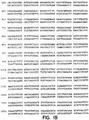
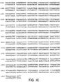
cDNAクローンは全部配列決定された。DNA19355の核酸配列はFig1(配列番号:2)に示す。クローンDNA19355−1150は、ヌクレオチド位置21−23に見かけの転写開始部位を持つ単一の読み枠を含む[Kozak等, 上掲](Fig1;配列番号:2)。予測されるポリペプチド前駆体は177アミノ酸長であり、約20,308ダルトンの計算上の分子量を持つ。ヒドロパシー分析は、推定細胞質領域(アミノ酸)1−25);膜貫通領域(アミノ酸26−51);及び細胞外領域(アミノ酸52−177)を持つII型の膜貫通タンパク質類型を示唆した。Fig1(配列番号:1)に示すように、2つの潜在的なN-結合グリコシル化部位が位置129(Asn)及び位置161(Asn)に同定された。クローンDNA19355−1150は、ATCCに寄託され、ATCC寄託番号209466が付与された。DNA19355ポリペプチドは、寄託されたATCC209466ベクターのcDNA挿入物によってコードされる分子の発現によって得られ又は得られうる。ベクターのXbaI及びNotI制限酵素での消化は、1411塩基対の断片及び668塩基対の断片を生じた。
【0130】
細胞外配列の(ALIGNコンピュータプログラムを用いた)BLAST及びFastA配列アラインメント分析に基づいて、DNA19355は、TNFサイトカインファミリの幾つかのメンバー、特にヒトAPo-2L(19.8%)、Fas/Apo1-リガンド(19.0%)、TNF-アルファ(20.6%)及びリンホトキシン-アルファ(17.5%)に対してアミノ酸配列同一性を示した(Fig2参照)。殆どのアミノ酸配列同一性は、TNF-アルファの結晶構造におけるベータ-鎖に対応する領域で見られた[Banner等, Cell, 73: 431-435 (1993); Eck等, J. Biol. Chem., 264: 17595-605 (1989); Lewit-Bentley等, J. Mol. Biol., 199: 389-92 (1988)]。鎖Cの配列は、特にこのファミリーの全てのメンバーに保存されている(Fig2参照)。推定膜貫通ドメインとDNA19355ポリペプチドの第1のベータ-鎖との間の配列は、TNF-アルファ、CD95L又はApo-2リガンドにおける約30から約80までの残基に比較して短く、5残基を含む。
【0131】
実施例2
ノーザンブロット分析
ヒト組織及び腫瘍株化細胞におけるDNA19355mRNAの発現を、ノーザンブロット分析によって試験した(Fig3参照)。ヒトRNAブロットを、全長DNA19355cDNAをコードするpRK5をXbaI-Iで消化することにより生成した32P標識DNAプローブにハイブリッド形成したが;このプローブは、全コード配列プラス幾つかのフランキング5’及び3’配列に相当する。
【0132】
ヒト胎児、成人、又は癌株化細胞mRNAブロット(クローンテック)を、DNAプローブと共にハイブリッド形成用バッファー(5X SSPE;2X デンハード溶液;100mg/mLの変性剪断されたサケ精子DNA;50%のホルムアミド;2%のSDS)中で、42℃で60時間インキュベートした。ブロットを2X SSC;0.05%のSDS中、室温で1時間、数回洗浄し、次いで0.1X SSC;0.1%のSDS中、50℃で30分間洗浄した。ブロットを終夜暴露した後、リン光体イメージャー分析(Fuji)により展開した。
【0133】
Fig3に示すように、約3.2kBの優勢なmRNA転写物を、胎児腎臓及び肺、及び成人小腸に検出した。また、発現は試験した8つのヒト腫瘍株化細胞中の6つでも検出され、それらは、ほぼ同じの3.2kB転写物を示し、並びに約1.5及び5kB転写物のより弱い発現も検出された。
【0134】
結果は、DNA19355ポリペプチドのmRNA発現が、正常組織では比較的抑制されるが、リンパ系並びに非リンパ系由来からの腫瘍株化細胞では顕著に増大することを示している。
【0135】
実施例3
大腸菌におけるDNA19355の発現
DNA19533ポリペプチドの細胞外領域(Fig1のアミノ酸52〜177;配列番号:1)をコードするDNA配列配列(Fig1;配列番号:2)を、各々:正:5’-GAC GAC AAG CAT ATG TTA GAG ACT GCT AAG GAG CCC TG-3’(配列番号:3);逆:5’-TAG CAG CCG GAT CCT AGG AGA TGA ATT GGG GATT-3’(配列番号:4)のフランキングNdeI及びXbaI制限部位を含むPCRプライマーで増幅した。PCRは消化し、Met Gly His10配列に続く12アミノ酸エンテロキナーゼ切断部位(プラスミドから誘導):Met Gly His His His His His His His His His His Ser Ser Gly His Ile Asp Asp Asp Asp Lys His Met(配列番号:5)の下流かつ枠内の、プラスミドpET19B(Novagen)のNdeI及びXbaI部位にクローン化した。
【0136】
得られたプラスミドは、大腸菌株JM109(ATCC 53323)のSambrook等, 上掲に記載された方法を用いた形質転換に使用した。形質転換体はPCRによって同定した。プラスミドDNAが単離され、制限分析及びDNA配列決定により確認された。
【0137】
選択されたクローンは、抗生物質を添加した液体培養培地LBで終夜成長させた。終夜培地は、続いて大規模培地の播種に用いられた。細胞は所望の光学密度まで成長させ、その間に発現プロモーターが作動する。
【0138】
細胞をさらに数時間の培養した後、細胞を採集して遠心分離した。遠心分離で得られた細胞ペレットは、マイクロフリューダイザを用いて0.1MのTris、0.2MのNaCl、50mMのEDTAを含むバッファー、pH8.0中に可溶化した。可溶化DNA19355タンパク質は、ニッケル−セファロース親和性クロマトグラフィを用いて精製した。
【0139】
DNA19355タンパク質は、DSD−PAGE、次いでニッケル抱合セイヨウワサビペルオキシダーゼでのウェスタンブロット、次いでECL検出(Boehringer Mannheim)により分析した。3つの優勢なタンパク質バンドが検出され、それらはタンパク質の単量体、同種二量体、及び同種三量体型のサイズに相当した(Fig4)。この結果に基づいて、可溶性DNA19355タンパク質は、その未変性型において、DSD変性無しに同種三量体を形成できると考えられる。
【0140】
実施例4
DNA19355のアポトーシス活性
全長DNA19355タンパク質をコードするpRK5プラスミド、又は空のpRK5プラスミド、又は全長ヒトApo-2リガンド(Apo-2L)をコードするpRK5を、リン酸カルシウム沈殿によりヒト293細胞(106細胞/10cm皿)に一過性形質移入した。幾つかの場合、ポックスウィルス由来のキャスパーゼ(caspase)阻害剤CrmA、又はFas/Apo1及びTNFR1による死亡シグナル伝達を媒介する死亡アダプタータンパク質FADD(FADD-DN)のドミナントネガティブな変異型とともに同時形質移入された。16時間後、細胞をヘキスト33342染料(10μg/ml)で染色し、ホフマン光学系を備えたライカ蛍光顕微鏡下でアポトーシス性又は正常な核を数えた。幾つかの場合、キャスパーゼ阻害剤z-VAD-fmk(Research Biochemicals)(200μM)を形質移入の直後に皿に添加した。
【0141】
Fig5に示すように、DNA19355による形質移入は、良好に確立されたアポトーシス誘発剤であるApo-2Lで観察される増加と同様に、pRK5に比較してアポトーシスレベルをかなり増加させている。DNA19355によるアポトーシス誘発における増加はCrmA又はz-VAD-fmkによって阻止され、この効果にキャスパーゼが含まれることを示している。さらに、Apo-2LではなくDNA19355に誘発されるアポトーシスの増加はFADD-DNによって阻止され、FADDアダプタータンパク質が、死亡シグナルをDNA19355からキャスパーゼ機構に伝達するのに必須の役割を果たすことを示している。
【0142】
実施例5
DNA19355によるNF-κBの活性化
全長DNA19355タンパク質をコードするpRK5プラスミド、又は空のpRK5プラスミド、又は全長ヒトApo-2LをコードするpRK5を、リン酸カルシウム沈殿によってヒト293細胞(106細胞/10cm皿)に形質移入した。細胞を、空のpRK5プラスミド、又はTNFによるNF-κB活性化を媒介する[Malinin等, Nature, 385: 540-544 (1997)]セリン/スレオニンキナーゼNIK(NIK-DN)のドミナントネガティブ、キナーゼ欠失変異型をコードするpRK5プラスミドとともに同時形質移入した。16時間後、細胞を収集し、核抽出物を調製し、1μgの核タンパク質を32P-標識NF-κB-特異的合成オリゴヌクレオチドプローブATCAGGGACTTTCCGCTGGGGACTTTCCG(配列番号:6)と反応させた[また、MacKay等, J. Immunol., 153: 5274-5284 (1994)も参照のこと]。
【0143】
Fig6に示すように、DNA19355による形質移入は、電気泳動移動度シフトアッセイ[Marsters等, PNAS, 92: 5401-5405 (1995)]で測定したところ、有意なNF-κB活性化を誘発し;活性化のレベルはApo-2Lで得られたレベルより大きかった。NIK-DNとの同時形質移入は、DNA19355によるNF-κB活性化を減少させたが、Apo-2Lによる活性化はさせなかった。この結果は、TNF-アルファと同様に、DNA19355はNIKタンパク質を含むシグナル化経路を通してNF-κBを活性化することを示唆している。
【0144】
実施例6
哺乳動物細胞におけるDNA19355の発現
この実施例は、哺乳動物細胞における組換え発現による、DNA19355の型の調製を例示する。
発現ベクターとして、ベクターpRK5(1989年3月15日発行のEP 307,247参照)を用いた。場合によっては、DNA19355DNAを選択した制限酵素でpRK5に結合させ、Sambrook等,上掲に記載されたような結合方法を用いてDNA19355DNAの挿入を行う。得られたベクターをpRK5−DNA19355と呼ぶ。
【0145】
一実施態様では、選択される宿主細胞を293細胞とすることができる。ヒト293細胞(ATCC CCL 1573)は、ウシ胎児血清及び場合によっては滋養成分及び/又は抗生物質を添加したDMEMなどの培地中で組織培養プレートにおいて成長させて集密化させた。約10μgのpRK5−DNA19355DNAを約1μgのVA RNA遺伝子をコードするDNA[Thimmappaya等, Cell, 31:543 (1982))]と混合し、500μlの1mMのTris-HCl、0.1mMのEDTA、0.227MのCaCl2に溶解させた。この混合物に、滴状の、500μlの50mMのHEPES(pH7.35)、280mMのNaCl、1.5mMのNaPO4を滴状で添加し、25℃で10分間析出物を形成させた。析出物を懸濁し、293細胞に加えて37℃で約4時間定着させた。培養培地を吸引し、PBS中20%グリセロールの2mlを30秒間添加した。293細胞は、次いで無血清培地で洗浄し、新鮮な培地を添加し、細胞を約5日間インキュベートした。
【0146】
形質移入の約24時間後、培養培地を除去し、培養培地(のみ)又は200μCi/mのl35S−システイン及び200μCi/mlの35S−メチオニンを含む培養培地で置換した。12時間のインキュベーションの後、条件培地を回収し、スピンフィルターで濃縮し、15%SDSゲルに負荷した。処理したゲルを乾燥させ、DNA19355ポリペプチドの存在を現すのに選択された時間に渡ってフィルムに暴露しうる。形質移入した細胞を含む培地は、さらなるインキュベーション(無血清培地)を施し、培地を選択したバイオアッセイで試験してもよい。
【0147】
それに換わる技術において、DNA19355は、Somparyac等, Proc. Natl. Acad. Sci., 12: 7575 (1981)に記載されたデキストラン硫酸法を用いて293細胞に一過的に導入されうる。293細胞をスピナーフラスコ内で最大密度まで成長させ、700μgのpRK5−DNA19355DNAを添加する。細胞は、まずスピナーフラスコから遠心分離によって濃縮してPBSで洗浄する。DNA−デキストラン沈殿物を細胞ペレット上で4時間インキュベートした。細胞を20%グリセロールで90秒間処理し、組織培養培地で洗浄し、組織培養培地、5μg/mlウシインシュリン及び0.1μg/mlウシトランスフェリンを含むスピナーフラスコに再度導入する。約4日後に、条件培地を遠心分離して濾過し、細胞及び細胞片を除去する。発現されたDNA19355を含む試料を濃縮し、透析及び/又はカラムクロマトグラフィー等の選択した方法によって精製できる。
【0148】
他の実施態様では、DNA19355はCHO細胞で発現させることができる。pRK5−DNA19355は、CaPO4又はDEAE-デキストランなどの周知の試薬を用いてCHO細胞に形質移入できる。上記のように、細胞培地をインキュベートし、その培地を培養培地(のみ)又は35S−メチオニンなどの放射性標識を含む培地で置換することができる。DNA19355の存在を測定した後、培養培地を無血清培地に置換してもよい。好ましくは、培地を約6日間インキュベートし、次いで条件培地を収集する。次いで、発現されたDNA19355を含む培地を濃縮し、任意の選択した方法で精製することができる。
【0149】
また、エピトープタグDNA19355を宿主CHO細胞で発現させることもできる。DNA19355はpRK5ベクターからサブクローン化される。サブクローン挿入物にPCRを施して、ポリ-hisタグ等の選択されたエピトープタグを持つ枠に融合させてバキュロウイルス発現ベクターとすることができる。ポリ-hisタグDNA19355挿入物は、次いで、安定なクローンの選択のためのDHFRなどの選択マーカーを含むSV40誘導ベクターにサブクローン化できる。最後に、SV40誘導ベクターでCHO細胞を(上記のように)形質移入できる。標識化を上記のように実施して発現を検証してもよい。次いで、発現されたポリ-hisタグDNA19355を含む培養培地を濃縮し、Ni2+-キレート親和性クロマトグラフィといった任意の選択した方法で精製することができる。
【0150】
実施例7
酵母菌でのDNA19355の発現
以下の方法は、酵母菌におけるDNA19355の組換え発現を記載する。
第1に、ADH2/GAPDHプロモーターからのDNA19355の細胞内生産又は分泌のための酵母菌発現ベクターを形成する。DNA19355、選択されたシグナルペプチド及びプロモーターをコードするDNAを、選択したプラスミドの適当な制限酵素部位に挿入してDNA19355の細胞内発現を制御する。分泌のために、DNA19355をコードするDNAを、ADH2/GAPDHプロモーター、酵母菌アルファ因子分泌シグナル/リーダー配列、及び(必要ならば)DNA19355の発現のためのリンカー配列とともに、選択したプラスミドにクローン化することができる。
【0151】
酵母菌株AB110などの酵母菌は、次いで上記の発現プラスミドで形質転換し、選択された発酵培地中で培養できる。形質転換した酵母菌上清は、10%トリクロロ酢酸での沈降及びSDS−PAGEによる分離、次いでクマシーブルー染色でのゲル染色により分析することができる。
【0152】
次いで、組換えDNA19355は、発酵培地から遠心分離により酵母菌細胞を除去し、選択されたカートリッジフィルターを用いて培地を濃縮することによって単離及び精製できる。DNA19355を含む濃縮物は、選択されたカラムクロマトグラフィー樹脂を用いてさらに精製してもよい。
【0153】
実施例8
バキュロウイルス感染昆虫細胞でのDNA19355の発現
以下の方法は、昆虫細胞におけるDNA19355の組換え発現を記載する。DNA19355は、バキュロウイルス発現ベクターに含まれるエピトープタグの上流に融合させた。このようなエピトープタグは、ポリ-hisタグ及び免疫グロブリンタグ(IgGのFc領域)を含む。pVL1393(Navagen)などの市販されているプラスミドから誘導されるプラスミドを含む種々のプラスミドを用いることができる。簡単には、DNA19355又はDNA19355の所定部分(細胞外ドメインをコードする配列等)が、5’及び3’領域に相補的なプライマーでのPCRにより増幅される。5’プライマーは、フランキングの(選択された)制限酵素部位を包含していてもよい。生成物は、次いで、これらの選択された制限酵素で消化され、発現ベクターにサブクローン化される。
【0154】
組換えバキュロウイルスは、上記のプラスミド及びBaculoGoldTMウイルスDNA(Pharmingen)を、Spodoptera frugiperda(「Sf9」)細胞(ATCC CRL 1711)中にリポフェクチン(GIBCO-BRLから市販)を用いて同時形質移入することにより生成される。28℃で4−5日インキュベートした後、放出されたウイルスを回収し、さらなる増幅に用いた。ウイルス感染及びタンパク質発現は、O'Reilley等, Baculovirus expression vectors: A laboratory Manual, Oxford: Oxford University Press (1994)に記載されているようにして実施した。
【0155】
次に、発現されたポリ-hisタグDNA19355は、例えばNi2+−キレートアフィニティクロマトグラフィーにより次のように精製される。抽出物は、Rupert等, Nature, 362: 175-179 (1993)に記載されているように、ウイルス感染した組換えSf9細胞から調製した。簡単には、Sf9細胞を洗浄し、超音波処理用バッファー(25mLのHepes、pH7.9;12.5mMのMgCl2;0.1mMのEDTA;10%のグリセロール;0.1%のNP−40;0.4MのKCl)中に再懸濁し、氷上で2回20秒間超音波処理する。超音波処理物を遠心分離で透明化し、上清を負荷バッファー(50mMのリン酸塩、300mMのNaCl、10%のグリセロール、pH7.8)で50倍希釈し、0.45μmフィルターを通して濾過する。Ni2+−NTAアガロースカラム(Qiagenから市販)を5mLの総容積で調製し、25mLの水で洗浄し、25mLの負荷バッファーで平衡させた。濾過した細胞抽出物は、毎分0.5mLでカラムに負荷した。カラムを、分画回収が始まる点であるA280のベースラインまで負荷バッファーで洗浄した。次に、カラムを、非特異的に結合したタンパク質を溶離する二次洗浄バッファー(50mMのリン酸塩;300mMのNaCl、10%のグリセロール、pH6.0)で洗浄した。A280のベースラインに再度到達した後、カラムを二次洗浄バッファー中で0から500mMのイミダゾール勾配で展開した。1mLの分画を回収し、SDS−PAGE及び銀染色又はアルカリホスファターゼ(Qiagen)に抱合したNi2+−NTAでのウェスタンブロットで分析した。溶離したHis10−タグDNA19355を含む分画をプールし、負荷バッファーで透析した。
【0156】
あるいは、IgGタグ(又はFcタグ)DNA19355の精製は、例えば、プロテインA又はプロテインGカラムクロマトグラフィを含む周知のクロマトグラフィー技術を用いて実施できる。
【0157】
実施例9
DNA19355に結合する抗体の調製
この実施例は、DNA19355に特異的に結合できるモノクローナル抗体の調製を例示する。
モノクローナル抗体の生産のための技術は当該分野で知られており、例えば、Goding,上掲に記載されている。用いられる免疫原は、精製DNA19355、DNA19355を含む融合タンパク質、細胞表面に組換えDNA19355を発現する細胞を含む。免疫原の選択は、当業者が過度の実験をすることなくなすことができる。
【0158】
Balb/cなどのマウスを、完全フロイントアジュバントに乳化し、皮下又は腹膜内に1−100マイクログラムで注入したDNA19355免疫原で免疫化する。あるいは、免疫原をMPL−TDMアジュバント(Ribi Immunochemical Researh, Hamilton, MT)に乳化し、動物の後足蹠に注入してもよい。免疫化したマウスは、次いで10から12日後に、選択したアジュバント中に乳化した付加的免疫原で追加免疫する。その後、数週間、マウスをさらなる免疫化注射でまた追加免疫してもよい。DNA19355抗体の検出のためのELISAアッセイで試験するために、レトロオービタル出血によって血清試料をマウスから周期的に採取してもよい。
【0159】
適当な抗体力価が検出された後、抗体に「陽性」な動物に、DNA19355の最後の静脈内注射の注入をすることができる。3から4日後、マウスを屠殺し、脾臓を取り出した。次いで脾臓細胞を(35%ポリエチレングリコールを用いて)、ACTTから番号CRL 1597で入手可能なP3X63AgU.1等の選択したマウス骨髄腫株化細胞に融合させた。融合によりハイブリドーマ細胞が生成され、次いで、それをHAT(ヒポキサンチン、アミノプテリン、及びチミジン)培地を含む96ウェル組織培養プレートに蒔き、非融合細胞、骨髄腫ハイブリッド、及び脾臓細胞ハイブリッドの増殖を阻害した。
【0160】
ハイブリドーマ細胞は、DNA19355に対する反応性についてのELISAでスクリーニングされる。所望のDNA19355に対するモノクローナル抗体を分泌する「陽性」ハイブリドーマ細胞の決定は当該分野の技量の範囲内である。
【0161】
陽性ハイブリドーマ細胞を同系のBalb/cマウスに腹膜内注入し、抗DNA19355モノクローナル抗体を含む腹水を生成できる。あるいは、ハイブリドーマ細胞を、組織培養フラスコ又はローラーボトルで成長させることもできる。腹水中に生成されたモノクローナル抗体の精製は、硫酸アンモニウム沈降、それに続くゲル排除クロマトグラフィを用いて行うことができる。あるいは、抗体のプロテインA又はプロテインGへの結合性に基づくアフィニティクロマトグラフィーを用いることもできる。
【0162】
実施例10
DNA19355のハイブリッド形成プローブとしての使用
以下の方法は、ハイブリッド形成プローブとしてのDNA19355をコードする核酸配列の使用を記載する。
DNA19355のコード配列を含むDNA(Fig1、配列番号:2に示したような)は、ヒト組織cDNAライブラリ又はヒト組織ゲノムライブラリにおいて(DNA19355の天然発生変異体をコードするもののような)相同DNAのスクリーニングのためのプローブとして用いられる。
【0163】
何れかのライブラリDNAを含むフィルターのハイブリッド化及び洗浄は、以下の高い厳密性条件下で実施する。放射性標識したDNA19355由来のプローブのフィルターへのハイブリッド形成は、50%のホルムアルデヒド、5x SSC、0.1%のSDS、0.1%のピロリン酸ナトリウム、50mMのリン酸ナトリウム、pH6.8、2x デンハード液、及び10%のデキストラン硫酸の溶液中、42℃で20時間実施される。フィルターの洗浄は、0.1x SSc及び0.1% SDSの水溶液中、42℃で実施される。
【0164】
次いで、全長天然配列DNA19355をコードするDNAと所望の配列同一性を持つDNAは、この分野で知られた標準的な技術を用いて同定できる。
【0165】
実施例11
染色体マッピング
ヒトDNA19355遺伝子の染色体局在化を、放射性ハイブリッド(RH)パネル分析によって試験した。RHマッピングを、マウス−ヒト細胞の放射線ハイブリッドパネル(Research Genetics)及びDNA19355 cDNAのコード領域に基づくプライマーを使用するPCRにより実施した[Gelb等, Hum. Genet., 98: 141 (1996)]。スタンフォードヒトゲノムセンターデータベースを使用したPCRデータの解析は、DNA19355がSTSマーカーD1S2790及びゲネトンマーカーAFMb352xe9に結合し、ヒト染色体1q23にマッピングされることを示した。特記すべきは、CD95Lも染色体1q23にマッピングされるが[Takahashi等, Int. Immunol., 6: 1567-1574 (1994)]、OX40リガンドは染色体1q25にマッピングされることである[Baum等, EMBO J., 13: 3992-4001 (1994)]。従って、これらのTNFファミリーは共通の祖先遺伝子の複製及び分岐によって生じ得る。
【0166】
実施例12
DNA19355ポリペプチドのヒトGITRレセプターに対する結合特異性
DNA19355ポリペプチドが「GITR」と呼ばれるレセプター分子のヒト相同体と相互作用及び特異的結合をするか否化を決定するためにアッセイを行った。マウスGITR(mGITR)ポリペプチドは、Nocentini等, Proc. Natl. Acad. Sci., 94: 6216-6221 (1997)に記載されている。何がmGITRのヒト相同体と考えられるかが記載されている。全長ヒトGITR(hGITR)のアミノ酸配列は、PCTの1998年2月19日に発行されたWO98/06842中の配列番号:4に示されている。hGITRとmGITRのアミノ酸配列の比較はFig7に示す。
【0167】
結合性を試験するために、hGITR細胞外ドメイン(Fig7のアミン酸1−167参照)を含む可溶性免疫グロブリン融合タンパク質(イムノグロブリン)を昆虫細胞で発現させた。hGITRのECDを、(上記実施例8に記載したように)バキュロウイルスを用いて昆虫細胞においてC-末端IgG-Fcタグ型として発現させた。
【0168】
また、可溶性DNA19355ポリペプチドを、ECDを(上記実施例3に記載したように)大腸菌細胞で発現させることにより調製した。可溶性DNA19355のECD分子は、次いで、125Iで標識した。比較のため、イムノアドヘシン形成物も以下のTNFレセプターファミリーのメンバー:CD95、DR4、DR5、TNFR1、TNFR2、及びApo-3で作成した。CD95、DR4、DR5、TNFR1、TNFR2、及びApo-3イムノアドヘシンは、TNFR1について既に記載されているように[Ashkenazi等, Proc. Natl. Acad. Sci., 88: 10535-10539 (1991)]、各レセプターのECDをヒトIgGのヒンジ及びFc部分に融合することにより調製した。各TNFレセプターファミリーメンバーは、発明の背景の部分に記載した(そして関連する参考文献を挙げた)。
【0169】
共沈殿アッセイのために、各イムノアドヘシン(5マイクログラム)を125I標識した可溶性DNA19355ポリペプチド(1マイクログラム)とともに24℃で1時間インキュベートし、次いで氷上で30分間プロテインA−セファロースを施した。反応混合物をスピン沈殿させ、PBSで数回洗浄し、20mMのジチオトレイトールを含むSDS−PAGEバッファー中で煮沸し、次いでSDS−PAGE及びオートラジオグラフで解析した。
【0170】
結果をFig8に示す。分子量マーカー(kDa)の位置を図に示した。hGITR−IgGは、放射性ヨウ素化した可溶性DNA19355ポリペプチドに結合した。しかしながら、hGITR-IgGはCD95、DR4、DR5、TNFR1、TNFR2、又はApo-3のイムノアドヘシン形成物には結合しなかった。
【0171】
他のアッセイにおいて、ヒト293細胞をDNA19355で一過性形質移入し、hGITR、TNFR1、HVEM、及びDcR1についてのレセプターイムノアドヘシン形成物のこれらの形質移入した細胞に結合する能力をFACS分析で測定した。293細胞を、10%のウシ血清アルブミン(FBS)、2mMのグルタミン、100マイクログラム/mlのペニシリン、及び100マイクログラム/mlのストレプトマイシンを添加した高グルコースDMEM培地中に維持した。形質移入した細胞(1x105)は、各レセプター又はリガンドイムノアドヘシン1マイクログラムを含む200マイクロリットルの2%FBS/PBS中、4℃で60分間インキュベートした。次いで、細胞を2%のFBS/PBSで洗浄し、R-フィコエリトリン-抱合ヤギ抗-ヒト抗体(Jackson Immunoresearch, West Grove, PA)で染色した。次に、細胞をFACSで分析した。各イムノアドヘシンの一過性形質移入された細胞への結合を試験するために、CD4に対する発現ベクター(pRK5−CD4;Smith等, Science, 328: 1704-1707 (1987))をDNA19355発現ベクターと同時形質移入した(実施例3参照)。次いで、FITC抱合抗-CD4(Pharmingen, San Diego, CA)を、FACS分析において形質移入された細胞集団を同定しゲートするのに用いた。
【0172】
Fig9Aに示すように、hGITR−IgGは、全長DNA19355をコードする発現プラスミドで形質移入した細胞表面に特異的に結合した。そのような結合は、TNFR1、HVEM又はDcR1については観察されなかった。hGITR−IgGは、対照プラスミドで形質移入した細胞には結合しなかった(データは示さず)。
【0173】
結果は、DNA19355ポリペプチドのhGITRとの特異的結合性相互作用、及びDNA19355ポリペプチドは試験した他のいかなるTNFレセプターファミリーメンバーとは相互作用しないことを示した。
【0174】
DNA19355ポリペプチドは、ヒト臍静脈内皮細胞(HUVEC)ライブラリにおいて同定され、DNA19355ポリペプチド転写物は、HUVECにおいてRT−PCRによって容易に検出可能である(データは示さず)。FACS分析アッセイは、hGITR−IgGの特異的結合がHUVECでFACS分析により示されるか否かを試験するために行った。HUVEC細胞は、Cell System(Kirkland, WA)から購入し、10%のウシ胎児血清、2mMのL-グルタミン、10mMのHepes、及び10ng/mlの塩基性FGFを含む、ハムのF12及び低グルコースDMEM培地の50:50混合物中で成長させた。細胞は、PBS、一次抗体としてのhGITR-IgG、TNFR1-IgG又はFas-IgG、及びフィコエリトリンに抱合したヤギ抗-ヒトF(ab’)2(CalTag, Burlingame, CA)とともにFACS貯蔵した。
【0175】
hGITR−IgGはHUVECに特異的に結合することが見出された。(Fig9B参照)。Fas−IgGもTNFR1−IgGもHUVEC細胞に特異的に結合しないことが示された。
【0176】
実施例13
DNA19355によるNF-κBの活性化
DNA19355/hGITRがNF-κB活性化を誘発するか否かを、E-セレクチン遺伝子からのNF-κB反応性エレメントを含むプロモーターによって誘導されたレポーター遺伝子の発現を分析することにより決定するためにアッセイを行った。
【0177】
ヒト293細胞(2x10⁵)を、0.5マイクログラムのホタルルシフェラーゼレポータープラスミドpGL3.ELAM.tk[Yang等, Nature, 395: 284-288 (1998)]及び0.05マイクログラムのレニラルシフェラーゼレポータープラスミド(内部形質移入対照用として)(Pharmacia)、並びに表示したDNA19355及びhGITRの付加的発現ベクター(上記)(0.1マイクログラムのhGITR;0.5マイクログラムの他の発現ベクター)、及び形質移入間の定常DNAを維持するための担体プラスミドpRK5Dとともに、リン酸カルシウム形質移入によって一過性形質移入をした。24時間後、細胞を収集し、ルシフェラーゼ活性を製造者(Pharmacia)に推奨されたように検定した。活性はホタルルシフェラーゼ活性をレニラルシフェラーゼ活性で除することにより形質移入効率における相違を規格化し、添加した発現ベクター無しで見られたものに対する活性として表現した。
【0178】
Fig10に見られるように、hGITRの過剰発現は有意な遺伝子活性化をもたらし、得られた結果は、DNA19355とhGITRの両方の同時発現によって向上した。
【0179】
実施例14
TNF−アルファ生成の刺激
DNA19355ポリペプチドによる刺激に応じた、単離した一次T細胞及びマクロファージからのTNF−アルファ及びIL−1ベータの生成を試験するためにアッセイを行った。
【0180】
一次T細胞又は単球/マクロファージはヒト供与者から単離した。一次ヒトT細胞は、T細胞富化カラム(R & D Systems)により全血から単離した。単球/マクロファージは、組織培養フラスコへの接着により全血から単離した。次いで、単離した各細胞は、10%のFBSを含むRPMI1640培地中の5マイクログラム/mlのDNA19355イムノアドヘシン(上記実施例3参照)で24時間処理した。次に培地上清中のTNF−アルファレベルをELISA(R & D Systems;製造者の指示に従って)で測定した。
【0181】
結果をFig11に例示した。DNA19355ポリペプチドは、T細胞から分泌されたTNF−アルファレベルにおいて約20倍の増加を誘発したが、マクロファージからのTNF−アルファ又はIL−1ベータの放出には影響しなかった(データは示さず)。ヒトT細胞からの誘発されたTNF−アルファ生成は、DNA19355ポリペプチド/hGITRが炎症誘発性反応に寄与することを示唆している。
【0182】
実施例15
モルモット皮膚生検アッセイ
炎症誘発性反応における候補分子の活性を決定するためにインビボアッセイを行った。詳細には、候補分子(DNA19355ポリペプチド等)をモルモットに注射し、処理した動物からの皮膚生検を、多形核/単核細胞浸潤又は好酸球浸潤について分析した。
【0183】
モルモットを筋肉内のケタミン(75−80mg/kgプラス5mg/kgキシラジン)で麻酔した。次いで候補分子を動物の背中16部位の皮膚に注射した(各部位100マイクロリットルの皮内)。約1mlのエバンスブルー染料/PBSを心臓内(intracordially)に注射した。
【0184】
1時間及び6時間に注射部位の斑点を測定(mm径)した。皮膚注射の6時間後にモルモットを屠殺した。注射部位の皮膚試料を切除し、パラホルムアルデヒドに固定した。次いで、組織を標準的な染色技術を用いて組織学的評価用に調製した。組織の分析は炎症性浸潤における細胞型の特徴付け及び血管周囲の浸潤の評価を含む。
【0185】
材料の寄託
次の材料をアメリカン・タイプ・カルチャー・コレクション(ATCC), 10801 ユニバーシティ通り、マナッサス、バージニア州 アメリカ合衆国に寄託した:
材料 ATCC寄託番号 寄託日
DNA19355-1150 209466 1997年11月18日
【0186】
この寄託は、特許手続き上の微生物の寄託の国際的承認に関するブダペスト条約及びその規則(ブダペスト条約)の規定に従って行われた。これは、寄託の日付から30年間、寄託の生存可能な培養が維持されることを保証するものである。寄託物はブダペスト条約の条項に従い、またジェネンテク社とATCCとの間の合意に従い、ATCCから入手することができ、これは、どれが最初であろうとも、関連した米国特許の発行時又は任意の米国又は外国特許出願の公開時に、寄託培養物の後代を永久かつ非制限的に入手可能とすることを保証し、米国特許法第122条及びそれに従う特許庁長官規則(特に参照番号886OG638の37CFR第1.14条を含む)に従って権利を有すると米国特許庁長官が決定した者に後代を入手可能とすることを保証するものである。
【0187】
本出願の譲受人は、寄託した培養物が、適切な条件下で培養されていた場合に死亡もしくは損失又は破壊されたならば、材料は通知時に同一の他のもの即座に取り替えることに同意する。寄託物質の入手可能性は、特許法に従いあらゆる政府の権限下で認められた権利に違反して、本発明を実施するライセンスであるとみなされるものではない。
【0188】
上記の文書による明細書は、当業者に本発明を実施できるようにするために十分であると考えられる。寄託した態様は、本発明のある側面の一つの説明として意図されており、機能的に等価なあらゆる作成物がこの発明の範囲内にあるため、寄託された作成物により、本発明の範囲が限定されるものではない。ここでの物質の寄託は、文書による説明が、そのベストモードを含む、本発明の任意の側面の実施を可能にするために不十分であることを認めるものではないし、それが表す特定の例証に対して請求の範囲を制限するものと解釈されるものでもない。実際、ここに示し記載したものに加えて、本発明を様々に改変することは、前記の記載から当業者にとっては明らかなものであり、添付の請求の範囲内に入るものである。
【0189】
【表1】

【図面の簡単な説明】
【Fig1A】 ヒトDNA19355に対するcDNAの核酸配列(配列番号:2)及びその誘導されたアミノ酸配列(配列番号:1)を示す図である。
【Fig1B】 ヒトDNA19355に対するcDNAの核酸配列(配列番号:2)及びその誘導されたアミノ酸配列(配列番号:1)を示す図である。
【Fig1C】 ヒトDNA19355に対するcDNAの核酸配列(配列番号:2)及びその誘導されたアミノ酸配列(配列番号:1)を示す図である。
【Fig2】 DNA19355ポリペプチドの細胞外アミノ酸配列の、ヒトApo-2L、Fas/Apo1リガンド(CD95L)、TNF-アルファ及びLT-アルファとのアラインメント及び比較を示す図である。各々のアミノ酸同一性(%)は、約19.8、19.0、20.6、及び17.5である。
【Fig3】 ヒト組織(同定された成人及び胎児組織)及び腫瘍株化細胞(HL60前骨髄球性白血病、HeLa S3頸部癌、K562慢性骨髄性白血病、MOLT4リンパ芽球性白血病、ラージバーッキットのリンパ腫、SW480結腸直腸腺癌、A549肺癌、及びG361黒色腫)でのDNA19355mRNA発現のノーザンブロット分析を示す図である。
【Fig4】 可溶性DNA19355ポリペプチドのSDS−PAGEによる分析を示す図である。
【Fig5A】 pRK5(a);DNA19355をコードするpRK5(b);Apo-2リガンドをコードするpRK5(c);DNA19355pRK5プラスCrmAをコードするpRK5(d);及びDNA19355をコードするpRK5プラスFADD-DNをコードするpRK5(e);Apo-2リガンドをコードするpRK5プラスFADD-DNをコードするpRK5(f)で形質移入した細胞からのヘキスト染色した核の蛍光画像を示す図である。
【Fig5B】 形質移入されたDNA19355又はApo-リガンドによるアポトーシスの誘発及びキャスパーゼ(caspase)阻害剤及びFADD-DNの効果を示すグラフである。
【Fig6】 NF-κB活性に対するDNA19355の影響を示す図である。pRK5、又はDNA19355をコードするpRK5、又はApo-2リガンドをコードするpRK5で形質移入した細胞における電気泳動移動度シフト分析。各場合において、細胞はpRK5(左の3レーン)又は顕性不活性NIKをコードするpRK5(NIK-DN、右の3レーン)で同時形質移入した。
【Fig7】 ヒトGITR(hGITR)及びマウスGITR(mGITR)についてのアミノ酸配列のアラインメント及び比較を示す図である。3つのシステインに富むドメイン(CRD1、CRD2及びCRD3)及び膜貫通領域(TM)を示す。
【Fig8】 上記の実施例12に記載する共沈殿アッセイの結果を示す図である。SDS−PAGEゲルのオートラジオグラフは、放射性ヨウ素化されたDNA19355ポリペプチドに結合したhGITR-IgG分子を明らかにした。同定された他のイムノアドヘシン作成物では結合が観察されなかった。
【Fig9A】 同定されたレセプター又はリガンドイムノアドヘシン作成物への結合について検定された、形質移入293細胞のFACS分析の結果を示すグラフである。
【Fig9B】 同定されたレセプターイムノアドヘシン作成物への結合について検定された、HUVEC細胞のFACS分析の結果を示すグラフである。
【Fig10】 DNA19355/hGITRによるNF-κB活性化を示すために行ったルシフェラーゼ活性アッセイの結果を示すグラフである。
【Fig11】 DNA19355ポリペプチドとともにインキュベートされた一次T細胞及び単球/マクロファージからの培養上清におけるTNF-アルファレベルを決定するために行ったELISAの結果を示すグラフである。
【配列表】
(Related application)
This claimed priority under section 119 (e) under
[0002]
(Field of Invention)
The present invention relates generally to the identification and isolation of novel DNA and the recombinant production of a novel polypeptide, herein designated “DNA19355”.
[0003]
(Background of the Invention)
Control of cell number in mammals is thought to be determined in part by the balance between cell proliferation and cell death. One form of cell death, sometimes referred to as necrotic cell death, is typically characterized as a pathological form of cell death resulting from some type of trauma or cell injury. In contrast, there are other “physiological” forms of cell death that normally proceed in a regular or controlled manner. This regular or controlled form of cell death is often referred to as “apoptosis” [eg, Barr et al., Bio / Technology, 12: 487-493 (1994); Steller et al., Science, 267: 1445-1449. (1995)]. Apoptotic cell death occurs naturally in many physiological processes, including clonal selection in the immune system and embryo development [Itoh et al., Cell, 66: 233-243 (1991)]. Reduced levels of apoptotic cell death are associated with a variety of pathological conditions, including cancer, lupus, and herpesvirus infection [Thompson, Science, 267: 1456-1462 (1995)]. Increased levels of apoptotic cell death can be seen in AIDS, Alzheimer's disease, Parkinson's disease, amyotrophic lateral sclerosis, multiple sclerosis, retinitis pigmentosa, cerebellar degeneration, aplastic anemia, myocardial infarction, stroke, relapse It is associated with a variety of other pathological conditions, including perfusion injury and toxin-induced liver disease [see Thompson, supra].
[0004]
Apoptotic cell death typically involves one or more characteristic morphological and biochemical changes in the cell, such as cytoplasmic coagulation, loss of plasma membrane microvilli, nuclear segmentation, chromosomal DNA With degradation of mitochondrion or loss of mitochondrial function. A variety of exogenous and intrinsic signals are believed to cause or induce such morphological and biochemical cellular changes [Raff, Nature, 356: 397-400 (1992); Steller, supra. Sachs et al., Blood, 82:15 (1993)]. For example, they can be triggered by hormonal stimuli such as glucocorticoid hormones on immature thymocytes, as well as withdrawal of certain growth factors [Watanabe-Fukunaga et al., Nature, 356: 314-317 (1992)]. . It has also been reported that several identified oncogenes, such as myc, rel, and E1A, and tumor suppressors, such as p53, have a role in inducing apoptosis. Certain chemotherapeutic drugs and certain forms of radiation have also been found to have pro-apoptotic activity [Thompson, supra].
[0005]
Tumor necrosis factor-α (“TNF-α”), tumor necrosis factor-β (“TNF-β” or “lymphotoxin-α”), lymphotoxin-β (“LT-β”), CD30 ligand, CD27 ligand, CD40 Various molecules such as ligands, OX-40 ligands, 4-1BB ligands, Apo-1 ligands (also referred to as Fas ligands or CD95 ligands), and Apo-2 ligands (also referred to as TRAIL) Identified as a member of the tumor necrosis factor (“TNF”) family of cytokines [eg, Gruss and Dower, Blood, 85: 3378-3404 (1995); Pitti et al., J. Biol. Chem., 271: 12687-12690 (1996); Wiley et al., Immunity, 3: 673-682 (1995); Browning et al., Cell. 72: 847-856 (1993); Armitage et al., Nature, 357: 80-82 (1992)]. Among these molecules, TNF-α, TNF-β, CD30 ligand, 4-1BB ligand, Apo-1 ligand, and Apo-2 ligand (TRAIL) have been reported to be involved in apoptotic cell death. ing. Both TNF-α and TNF-β have been reported to induce apoptotic cell death in susceptible tumor cells [Schmid et al., Proc. Natl. Acad. Sci., 83: 1881 (1986); Dealtry et al. , Eur. J. Immunol., 17: 689 (1987)]. Zheng et al. Report that TNF-α is involved in apoptosis following stimulation of CD8 positive T cells [Zheng et al., Nature, 377: 348-351 (1995)]. Other researchers have reported that CD30 ligand is involved in the deletion of autoreactive T cells in the thymus [Amakawa et al., Cold Spring Harbor Institute Symposium on Programmed Cell Death, Summary Collection, Vol. 10, (1995)].
[0006]
Mutations in the mouse Fas / Apo-1 or ligand genes (referred to as 1pr and g1d, respectively) are associated with several autoimmune diseases, and Apo-1 ligand regulates clonal deletion of autoreactive lymphocytes in the periphery [Krammer et al., Curr. Op. Immunol., 6: 279-289 (1994); Nagata et al., Science, 267: 1449-1456 (1995)]. Apo-1 ligand has also been reported to induce post-cell stimulation apoptosis in CD4 positive T lymphocytes and B lymphocytes and is included in the removal of activated lymphocytes when their function is no longer needed Obtain [Krammer et al., Supra; Nagata et al., Supra]. An agonistic mouse monoclonal antibody that specifically binds to the Apo-1 receptor has been reported to show cell killing activity comparable or similar to TNF-α [Yonehara et al., J. Exp. Med., 169: 1747-1756 (1989)].
[0007]
Induction of various cellular responses mediated by such TNF family cytokines is thought to be initiated by their binding to specific cell receptors. Two different TNF receptors of approximately 55-kDa (TNFR1) and 75-kDa (TNFR2) have been identified [Hohman et al., J. Biol. Chem., 264: 14927-14934 (1989); Brockhaus et al., Proc. Natl. Acad. Sci., 87: 3127-3131 (1990); Ep 417,563 published 20 March 1991], human and mouse cDNAs corresponding to both receptor types have been isolated and characterized. [Loetscher et al., Cell, 61: 351 (1990); Schall et al., Cell, 61: 361 (1990); Smith et al., Science, 248: 1019-1023 (1990); Lewis et al., Proc. Natl. Acad. Sci., 88: 2830-2834 (1991); Goodwin et al., Mol. Cell. Biol., 11: 3020-3026 (1991)]. Extensive polymorphisms are associated with both TNF receptor genes [see, eg, Takao et al., Immunogenetics, 37: 199-203 (1993)]. Both TNFRs share the typical structure of cell surface receptors including extracellular, transmembrane and intracellular regions. The extracellular portions of both receptors are also found naturally as soluble TNF binding proteins [Nophar, Y et al., EMBO J., 9: 3269 (1990); and Kohno, T et al., Proc. Natl. Acad. Sci. USA , 87: 8331 (1990)]. More recently, cloning of recombinant soluble TNF receptor has been reported by Hale et al. [J. Cell. Biochem. Augmented 15F, 1991, p.113 (P424)].
[0008]
The extracellular portion of
[0009]
A similar repetitive pattern of CRD is the p75 nerve growth factor receptor (NGFR) [Johnson et al., Cell, 47: 545 (1986); Radeke et al., Nature, 325: 593 (1987)], B cell antigen CD40 [Stamenkovic et al., EMBO J., 8: 1403 (1989)], T cell antigen OX40 [Mallet et al., EMBO J., 9: 1063 (1990)] and Fas antigen [Yonehara et al., Supra, and Itoh et al., Cell, 66: 233. -243 (1991)] is present in several other cell surface proteins. CRD is also found in the soluble TNFR (sTNFR) -like T2 protein of Shope and myxoma poxvirus [Upton et al., Virology, 160: 20-29 (1987); Smith et al., Biochem. Biophys. Res. Commun., 176: 335 (1991); Upton et al., Virology, 184: 370 (1991)]. The optimal alignment of these sequences shows that the positions of cysteine residues are well conserved. These receptors are sometimes collectively referred to as members of the TNF / NGF receptor superfamily. Recent studies on p75NGFR include deletions of CRD1 [Welcher, AA et al., Proc. Natl. Acad. Sci. USA, 88: 159-163 (1991)] or 5-amino acid insertions in this domain [Yan. Chao, MV, J. Biol. Chem., 266: 12099-12104 (1991)] has been shown to have little or no effect on NGF binding [Yan. H and Chao, MV, supra]. . p75NGFR contains a proline-rich extension of about 60 amino acids that is not involved in NGF binding between its CRD4 and transmembrane regions [Peetre, C et al., Eur. J. Hematol., 41: 414-419 (1988). Seckinger, P et al., J. Biol. Chem., 264: 11966-11973 (1989); Yan. H. and Chao, MV, supra]. A similar proline-rich region is found in TNFR2 but not in TNFR1.
[0010]
Itoh et al. Disclose that Apo-1 receptor can signal apoptotic cell death similar to that signaled by 55-kDa TNFR1 [Itoh et al., Supra]. It has also been reported that Apo-1 antigen expression is down-regulated with that of TNFR1 when cells are treated with TNF-α or anti-Apo-1 mouse monoclonal antibody [Krammer et al., Supra. ; Nagata et al., Supra]. Thus, some researchers hypothesized that cell lines that co-express both Apo-1 and TNFR1 receptors mediate cell death through a common signaling pathway [ibid].
[0011]
With the exception of lymphotoxin-α, the TNF family ligands identified to date are type II transmembrane proteins, whose C-terminus is extracellular. In contrast, the receptors of the TNF receptor (TNFR) family identified to date are type I transmembrane proteins. However, homology identified between family members in both the TNF ligand and receptor families has been found primarily in the extracellular domain ("ECD"). Some of the TNF family cytokines, including TNF-α, Apo-1 ligand and CD40 ligand, are proteolytically cleaved at the cell surface; the resulting protein in each case typically functions as a soluble cytokine Form homotrimeric molecules. TNF receptor family proteins are also usually proteolytically cleaved to release soluble receptor ECDs that can function as inhibitors of cognate cytokines.
[0012]
Recently, other members of the TNFR family have been identified. These newly identified members of the TNFR family include CAR1, HVEM and osteoprotegerin (OPG) [Brajatsh et al., Cell, 87: 845-855 (1996); Montgomery et al., Cell, 87: 427-436 (1996); Marsters et al., J. Biol. Chem., 272: 14029-14032 (1997); Simonet et al., Cell, 89: 309-319 (1997)]. Unlike other known TNFR-like molecules, Simonet et al., Supra, report that OPG does not contain a hydrophobic transmembrane spanning sequence.
[0013]
In Marsters et al., Curr. Biol., 6: 750 (1996), researchers have shown similarities to the TNFR family in extracellular cysteine-rich repeats and include cytoplasmic death domain sequences. Discloses the full-length human polypeptide native sequence termed Apo-3, which is similar to TNFR1 and CD95 [see also Marsters et al., Curr. Biol., 6: 1669 (1996)]. . According to other researchers, Apo-3 has also been referred to as DR3, wsl-1 and TRAMP [Chinnaiyan et al., Science, 274: 990 (1996); Kitson et al., Nature, 384: 372 (1996); Bodmer et al., Immunity, 6:79 (1997)].
[0014]
Pan et al. Disclose another member of the TNF receptor family termed “DR4” [Pan et al., Science, 276: 111-113 (1997)]. DR4 has been reported to contain a cytoplasmic death domain that activates cell suicides. Pan et al. Disclose that DR4 is thought to be a receptor for the ligand known as Apo-2 ligand or TRAIL.
[0015]
Sheridan et al., Science, 277: 818-821 (1997) and Pan et al., Science, 277: 815-818 (1997) describe other molecules that are considered receptors for Apo-2 ligand (TRAIL). Has been. This molecule is referred to as DR5 (also referred to as Apo-2). Like DR4, DR5 contains a cytoplasmic death domain and is capable of signaling apoptosis.
[0016]
Sheridan et al., Supra, disclose that DcR1 (or Apo-2DcR) is a potential decoy receptor for Apo-2 ligand (TRAIL). Sheridan et al. Report that DcR1 can inhibit Apo-2 ligand function in vitro. For the disclosure of the decoy receptor called TRID, see the above-mentioned bread and the like.
For a review of TNF family cytokines and their receptors, see Gruss and Dower, supra.
[0017]
As currently understood, the cell death program includes at least three important components—activators, inhibitors, and effectors; in C. elegans, these components each contain three genes, Ced -4, encoded by Ced-9 and Ced-3 [Steller, Science, 267: 1445 (1995); Chinnaiyan et al., Science, 275: 1122-1126 (1997); Wang et al., Cell, 90: 1- 20 (1997)]. Two members of the TNFR family, TNFR1 and Fas / Apol (CD95), can activate apoptotic cell death [Chinnaiyan and Dixit, Current Biology, 6: 555-562 (1996); Fraser and Evan, Cell, 85 : 781-784 (1996)]. TNFR1 is also known to mediate activation of a transcription factor, NF-κB [Tartaglia et al., Cell, 74: 845-853 (1993); Hsu et al., Cell, 84: 299-308 (1996). ]. In addition to some degree of ECD homology, these two receptors share homology in the intracellular domain (ICD) of the oligomerization interface known as the death domain [Tartaglia et al., Supra; Nagata, Cell, 88: 355 (1997)]. Death domains have also been found in several metazoan proteins that regulate apoptosis, namely Drosophila proteins called FADD / MORT1, TRADD and RIP, Reaper and mammalian proteins [Cleaveland and Ihle, Cell, 81: 479-482 (1995)]. Upon ligand binding and receptor clustering, TNFR1 and CD95 are thought to recruit FADD to the mortality-inducing signaling complex. Allegedly, CD95 binds directly to FADD, whereas TNFR1 binds indirectly to FADD via TRADD [Chinnaiyan et al., Cell, 81: 505-512 (1995); Boldin et al., J. Biol. Chem., 270: 387-391 (1995); Hsu et al., Supra; Chinnaiyan et al., J. Biol. Chem., 271: 4961-4965 (1996)]. FADD has been reported to be an adapter protein that recruits the Ced-3-related protease, MACHα / FLICE (Caspase 8), to the death signaling complex [Boldin et al., Cell, 85: 803-815 (1996). Muzio et al., Cell, 85: 817-827 (1996)]. MACHα / FLICE appears to be a trigger that triggers a cascade of apoptotic proteases, including interleukin-1β converting enzyme (ICE) and CPP32 / Yama, that may implement several important aspects of the cell death program [Fraser And Evan, supra].
[0018]
It has recently been disclosed that programmed cell death is involved in the activity of the nematode cell death gene, ced-3, and a member of the cysteine protease family related to mammalian IL-1-converting enzymes, ICE. It was. The activities of ICE and CPP32 / Yama protease can be inhibited by the product of the cowpox virus gene, crmA [Ray et al., Cell, 69: 597-604 (1992); Tewari et al., Cell, 81: 801-809 (1995)] . Recent studies have shown that CrmA can inhibit TNFR1- and CD95-induced cell death [Enari et al., Nature, 375: 78-81 (1995); Tewari et al., J. Biol. Chem., 270: 3255-3260 (1995)].
[0019]
As recently reviewed by Tewari et al., TNFR1, TNFR2 and CD40 express pro- and costimulatory cytokines, cytokine receptors, and cell adhesion molecules through activation of transcription factors, NF-κB. [Tewari et al., Curr. Op. Genet. Develop., 6: 39-44 (1996)]. NF-κB is the prototype of a family of dimeric transcription factors whose subunits contain a conserved Rel region [Verma et al., Genes Develop., 9: 2723-2735 (1996); Baldwin, Ann. Rev. Immunol. , 14: 649-681 (1996)]. In its latent form, NF-κB is complexed with members of the IκB inhibitor family; released NF-κB binds to specific DNA sequences upon inactivation of IκB in response to certain stimuli. Translocates into the nucleus to activate gene transcription. KF-κB is induced by various pro-inflammatory signals and cytokines including LPS acting through IL-1 and Toll-like receptor TLR2 [Baeuerle et al., Ann. Rev. Immunol., 12: 141-79 ( 1994); Verma et al., Supra].
[0020]
(Summary of Invention)
The Applicant has identified a cDNA clone encoding a novel polypeptide named “DNA19355” in this application.
[0021]
In one embodiment, the present invention provides an isolated nucleic acid molecule comprising DNA encoding a DNA19355 polypeptide. In some cases, the isolated nucleic acid molecule comprises DNA encoding a DNA19355 polypeptide having amino acid residues 1-177 or 52-177 of FIG1 (SEQ ID NO: 1); Alternatively, it is complementary to such encoding nucleic acid sequence and maintains stable binding to it under at least moderate and possibly high stringency conditions. The isolated nucleic acid molecule may comprise a DNA19355 cDNA insert of the vector deposited as ATCC 209466, in particular an insert comprising a DNA sequence encoding a DNA19355 polypeptide.
[0022]
In another embodiment, the present invention provides a vector comprising DNA encoding a DNA19355 polypeptide. Also provided are host cells containing such vectors. As an example, the host cell may be a CHO cell, E. coli, or yeast. Further provided is a method for producing a DNA19355 polypeptide, the method comprising culturing a host cell under conditions suitable for expression of DNA19355 and recovering DNA19355 from the cell culture medium.
[0023]
In another embodiment, the invention provides isolated DNA19355 polypeptide. In particular, the present invention provides an isolated native sequence DNA19355 polypeptide comprising, in one embodiment, an amino acid sequence comprising residues 1-177 or 52-177 of FIG. 1 (SEQ ID NO: 1). In some cases, a DNA19355 polypeptide can be obtained or obtained by expressing a polypeptide encoded by the cDNA insert of the vector deposited as ATCC 209466.
[0024]
In other embodiments, the invention provides isolated DNA19355 polypeptide variants. The variant has at least about 80% amino acid sequence identity with the deduced amino acid sequence of FIG. 1 (SEQ ID NO: 1) or the domain sequence identified herein, and preferably exhibits the activity of the native sequence or the naturally occurring DNA19355 polypeptide. Have.
[0025]
In another embodiment, the present invention provides a chimeric molecule comprising a DNA19355 polypeptide fused to a heterologous polypeptide or amino acid sequence. Examples of such chimeric molecules include DNA19355 fused to an epitope tag sequence or an Fc region of an immunoglobulin sequence.
[0026]
In another embodiment, the invention provides an antibody that specifically binds to DNA19355 polypeptide. In some cases, the antibody is a monoclonal antibody.
[0027]
In further embodiments, the present invention also provides diagnostic and therapeutic methods using DNA19355 polypeptides. For example, a method is provided for inducing apoptosis in mammalian cancer cells.
[0028]
Detailed Description of Preferred Embodiments
I. Definition
The terms “DNA19355 polypeptide” and “DNA19355” as used herein include native sequence DNA19355 and DNA19355 variants (as defined further herein). DNA19355 may be isolated from a variety of sources, such as human tissue types or other sources, or prepared by recombinant or synthetic methods. The terms “DNA19355 polypeptide” and “DNA19355” as used herein refer to the same polypeptide as referred to in the literature as “GLITTER”. “Native sequence DNA19355” includes a polypeptide having the same amino acid sequence as naturally occurring DNA19355. Such native sequence DNA19355 can be isolated from nature or can be produced by recombinant or synthetic means. The term “native sequence DNA19355” specifically includes naturally occurring truncated, soluble or secreted forms (eg, extracellular domain sequences or soluble forms) of DNA19355, naturally occurring mutated forms (eg, alternatively spliced forms). ) And naturally occurring allelic variants of DNA19355. In one embodiment of the invention, native sequence DNA19355 is mature or full-length native sequence DNA19355 containing amino acids 1-177 of FIG1 (SEQ ID NO: 1). Alternatively, the DNA19355 polypeptide comprises amino acids 52-177 of Fig1 (SEQ ID NO: 1). In some cases, a DNA19355 polypeptide can be obtained or obtained by expressing a polypeptide encoded by the cDNA insert of the vector deposited as ATCC 209466.
[0029]
“DNA19355 extracellular domain” or “DNA19355ECD” means a type of DNA19355 that is substantially free of the transmembrane and cytoplasmic domains of DNA19355. Usually, DNA19355ECD has less than 1% of such transmembrane and / or cytoplasmic domains, preferably less than 0.5% of such domains. In some cases, DNA19355ECD comprises FIG. 1 (SEQ ID NO: 1) amino acid residues X to 177, wherein X is any of FIG. 1 (SEQ ID NO: 1) amino acid residues 48 to 57. One skilled in the art will appreciate that the transmembrane domain identified for the DNA19355 polypeptide of the present invention is identified according to criteria routinely used for identifying such hydrophobic domains in the art. The exact boundaries of the transmembrane domain will vary, but most likely is no more than about 5 amino acids at either end of the domain specifically mentioned herein.
[0030]
“DNA19355 variant” refers to at least about 80% amino acid sequence identity with the deduced amino acid sequence shown in FIG. 1 (SEQ ID NO: 1) or the domain sequence identified herein relative to full-length native sequence human DNA19355 Means DNA19355 as defined below. Such DNA19355 variants include, for example, DNA19355 polypeptides with one or more amino acid residues added or deleted at the N- or C-terminus of the sequence of FIG. 1 (SEQ ID NO: 1). . The DNA19355 variant comprises an ECD fragment comprising a sequence having less than amino acid residues 52 to 177 of Fig1 (SEQ ID NO: 1). Usually, a DNA19355 variant will have at least about 80% or 85% amino acid sequence identity, more preferably at least about 90% amino acid sequence identity, and even more preferably at least about the amino acid sequence of FIG. It has about 95% amino acid sequence identity.
[0031]
“Percent (%) amino acid sequence identity” relative to the DNA19355 sequence identified herein introduces gaps as necessary to align the sequences and obtain maximum percent sequence identity, and any conservative substitutions are sequence-identical. Defined as the percentage of amino acid residues in the candidate sequence that are identical to the amino acid residues of the DNA19355 sequence if not considered part of the gender. Alignments for the purpose of determining percent amino acid sequence identity are various methods within the skill of the art, such as publicly available computer software such as BLAST, ALIGN, or Megalign (DNASTAR) software. Can be achieved by using. One skilled in the art can determine appropriate parameters for measuring alignment, including any algorithms necessary to achieve maximal alignment over the full length of the sequences being compared.
[0032]
“Percent (%) nucleic acid sequence identity” with respect to the DNA19355 sequence identified herein refers to the nucleotides of the DNA19355 sequence when the sequences are aligned and, if necessary, introduced with gaps to obtain maximum percent sequence identity. Defined as the percentage of nucleotides in the candidate sequence that are identical. Alignment for the purpose of determining percent nucleic acid sequence identity can be accomplished by various methods within the skill of the art, such as publicly available computer software such as BLAST, ALIGN, or Megalign (DNASTAR) software. This can be achieved by using wear. One skilled in the art can determine appropriate parameters for measuring alignment, including any algorithms necessary to achieve maximal alignment over the full length of the sequences being compared.
[0033]
The term “epitope tag” as used herein refers to a chimeric polypeptide comprising DNA19355, or a domain sequence thereof, fused to a “tag polypeptide”. The tag polypeptide has sufficient residues to provide an epitope from which the antibody can be produced or can be identified by several other reagents, but is short enough so as not to inhibit the activity of DNA19355. Also, the tag polypeptide is preferably fairly unique so that the antibody does not substantially cross-react with other epitopes. Suitable tag polypeptides generally have at least 6 amino acid residues, usually about 8 to about 50 amino acid residues (preferably about 10 to about 20 residues).
[0034]
“Isolated”, when used to describe the various polypeptides disclosed herein, means a polypeptide that has been identified and separated and / or recovered from a component of its natural environment. . Contaminants in the natural environment are substances that typically interfere with the diagnosis or treatment of the polypeptide, such as enzymes, hormones, and other proteinaceous or nonproteinaceous solutes. Is included. In a preferred embodiment, the polypeptide is (1) sufficient to obtain at least 15 N-terminal or internal amino acid sequence residues by using a spinning cup sequenator, or (2) Coomassie. Purified to homogeneity by SDS-PAGE under non-reducing or reducing conditions using blue or preferably silver staining. Isolated polypeptide includes polypeptide in situ within recombinant cells, since at least one component of the natural environment of DNA19355 will not be present. Ordinarily, however, isolated polypeptide will be prepared by at least one purification step.
[0035]
An “isolated” DNA19355 nucleic acid molecule is a nucleic acid molecule that has been identified and separated from at least one contaminating nucleic acid molecule that is normally associated with a natural source of DNA19355 nucleic acid. An isolated DNA19355 nucleic acid molecule is other than in the form or setting in which it is found in nature. Thus, an isolated DNA19355 nucleic acid molecule is distinguished from a DNA19355 nucleic acid molecule that is present in natural cells. However, an isolated DNA19355 nucleic acid molecule includes, for example, a DNA19355 nucleic acid molecule contained in a cell that normally expresses DNA19355 when the nucleic acid molecule is in a chromosomal location different from that of natural cells.
[0036]
The term “control sequence” refers to a DNA sequence necessary for the expression of a coding sequence operably linked in a particular host organism. For example, suitable control sequences for prokaryotes include a promoter, optionally an operator sequence, and a ribosome binding site. Eukaryotic cells are known to utilize promoters, polyadenylation signals and enhancers.
[0037]
A nucleic acid is “operably linked” when it is placed into a functional relationship with another nucleic acid sequence. For example, a presequence or secretory leader DNA is operably linked to the polypeptide DNA if expressed as a preprotein that participates in the secretion of the polypeptide; a promoter or enhancer affects the transcription of the sequence. The ribosome binding site is operably linked to the coding sequence if it is positioned so as to facilitate translation. In general, “operably linked” means that the linked DNA sequences are in close proximity and, in the case of a secretory leader, in close proximity and in the reading phase. However, enhancers do not necessarily have to be close together. Binding is achieved by ligation at convenient restriction sites. If such sites do not exist, synthetic oligonucleotide adapters or linkers are used according to conventional techniques.
[0038]
The term “antibody” is used in the broadest sense and specifically includes a single anti-DNA19355 monoclonal antibody (including agonists, antagonists, and neutralizing antibodies), and anti-DNA19355 antibody compositions with multiple epitope specificity. is doing. The term “monoclonal antibody” as used herein refers to an antibody obtained from a population of substantially homogeneous antibodies, ie, the individual antibodies that make up the population are those naturally occurring possible mutations that may be present in small amounts. Identical except for mutations.
[0039]
As used herein, “biologically active” and “desired biological activity” include (1) in vivo or ex vivo in at least one type of mammalian cell (an active or stimulating method or Have the ability to modulate apoptosis (either in an antagonistic or inhibitory manner), or (2) have the ability to induce or stimulate a pro-inflammatory response in at least one type of mammalian cell in vivo or ex vivo. Means.
[0040]
The terms “apoptosis” and “apoptotic activity” are used broadly and typically include cytoplasmic coagulation, loss of plasma membrane microvilli, nuclear segmentation, degradation of chromosomal DNA or loss of mitochondrial function Refers to a regular or controlled form of cell death in a mammal with one or more characteristic cell changes. This activity can be determined and measured, for example, by cell viability discriminating assays, FACS analysis or DNA electrophoresis, all known in the art.
[0041]
As used herein, “treat”, “treatment” and “treatment” refer to curative therapy, prophylactic therapy and protective therapy.
[0042]
The terms “cancer” and “cancerous” refer to or describe the physiological condition in mammals that is typically characterized by unregulated cell growth. Examples of cancer include, but are not limited to, carcinoma, lymphoma, blastoma, sarcoma, and leukemia. More specific examples of such cancers include squamous cell carcinoma, small cell lung cancer, non-small cell lung cancer, blastoma, gastrointestinal cancer, kidney cancer, pancreatic cancer, glioblastoma, neuroblastoma, uterus Cervical cancer, ovarian cancer, liver cancer, stomach cancer, bladder cancer, hepatoma, breast cancer, colorectal cancer, colorectal cancer, endometrial cancer, salivary gland cancer, kidney cancer, liver cancer, prostate cancer, spawning cancer, It includes thyroid cancer, hepatic carcinoma and various types of head and neck cancer.
[0043]
The term “mammal” as used herein refers to any animal classified as a mammal, including humans, cows, horses, dogs and cats. In a preferred embodiment of the invention, the mammal is a human.
[0044]
II. Compositions and Methods of the Invention
A. DNA19355 polypeptide
The present invention provides a newly identified and isolated nucleic acid sequence encoding a polypeptide referred to in this application as DNA19355. In particular, Applicants have identified and isolated cDNA encoding a DNA19355 polypeptide, as disclosed in further detail in the Examples below. Using the BLAST and FastA sequence alignment computer programs, Applicants have found that DNA19355 (shown in FIG. 1 and SEQ ID NO: 1) has some amino acid identity with several members of the TNF family (eg, , FIG. 2 and Example 1 below). As shown in the Examples below, DNA19355 polypeptide was found to have apoptotic activity and specific binding to GITR. DNA19355 also stimulates TNF-alpha secretion in primary T cells in vitro (Example 14) and neutrophil infiltration and influx (as described in Example 15) in guinea pig skin biopsy assays. Has been found and suggests a role for DNA19355 in the pro-inflammatory response.
[0045]
In addition to the soluble forms of full-length native sequence DNA19355 and DNA19355 described herein, it is contemplated that DNA19355 variants can also be prepared. DNA19355 variants can be prepared by introducing appropriate nucleotide changes into the DNA19355 nucleic acid sequence, or by synthesis of the desired DNA19355 polypeptide. One skilled in the art will appreciate that amino acid changes can alter the post-translational process of DNA19355, such as changing the number or position of glycosylation sites or changing membrane anchoring properties.
[0046]
Variations in the various domains of the native full-length sequence DNA19355 or the DNA19355 described herein can be achieved using any techniques and guidelines described, for example, in US Pat. No. 5,364,934 for conservative or non-conservative mutations. Can be made. The mutation may be a substitution, deletion or insertion of one or more codons encoding DNA19355 resulting in a change in the amino acid sequence of DNA19355 compared to native sequence DNA19355. In some cases, the mutation is due to substitution of at least one amino acid with any other amino acid of one or more domains of DNA19355. Guidance in determining which amino acid residues to insert, substitute or delete without adversely affecting the desired activity is made in the region of high homology, comparing the sequence of DNA19355 to homologous well-known protein molecules. Can be found by minimizing the number of amino acid sequence changes. An amino acid substitution can be, for example, a substitution of leucine with serine, ie a conservative amino acid substitution, as a result of replacing one amino acid with another amino acid having similar structural and / or chemical properties. Insertions or deletions may optionally range from 1 to 5 amino acids. Acceptable mutations can be determined by systematic insertion of amino acids in the sequence, deletion or substitution creation, and testing of the resulting mutants for activity in any of the in vitro assays described in the Examples below.
[0047]
Mutations can be made using methods known in the art such as oligonucleotide-mediated (site-directed) mutagenesis, alanine scanning, and PCR mutagenesis. Site-directed mutagenesis [Carter et al., Nucl. Acids Res., 13: 4331 (1986); Zoller et al., Nucl. Acids Res., 10: 6487 (1987)], cassette mutagenesis [Wells et al., Gene, 34: 315 (1985)], restriction selective mutagenesis [Wells et al., Philos. Trans. R. Soc. London SerA, 317: 415 (1986)] or other well-known techniques produce DNA19355 mutant DNA. Can be performed on cloned DNA.
[0048]
Scanning amino acid analysis can also be used to identify one or more amino acids along a contiguous sequence. Among them, preferred scanning amino acids are relatively small and neutral amino acids. Such amino acids include alanine, glycine, serine, and cysteine. Alanine is typically the preferred scanning amino acid in these groups because it does not have a side chain beyond the beta-carbon and is unlikely to alter the mutant backbone structure. Alanine is also typically preferred because it is the most common amino acid. Furthermore, it is often found in both hidden and exposed positions [Creighton, The Proteins, (WH Freemann & Co., NY); Chothia, J. Mol. Biol., 150: 1 (1976)]. If an alanine substitution does not produce the appropriate amount of mutant, an isoteric amino acid is used.
[0049]
B. Modification of DNA19355
Covalent modifications of DNA19355 are included within the scope of the present invention.
One type of covalent modification is to react a targeted amino acid residue of DNA19355 with an organic derivatization reagent capable of reacting with a selected side chain or N- or C-terminal residue of DNA19355. Derivatization with bifunctional reagents is useful, for example, to cross-link DNA19355 to a surface for use in a water-insoluble support matrix or anti-DNA19355 antibody purification method or vice versa. Commonly used crosslinking agents include, for example, 1,1-bis (diazoacetyl) -2-phenylethane, glutaraldehyde, N-hydroxysuccinimide ester, such as 4-azidosalicylic acid, 3,3′-dithiobis (succinimidyl) Homobifunctional imidoesters including disuccinimidyl esters such as propionate), bifunctional maleimides such as bis-N-maleimide-1,8-octane, and methyl-3-[(p-azidophenyl)- Reagents such as dithio] propioimidate.
[0050]
Other modifications include deamination of glutaminyl and asparaginyl residues to the corresponding glutamyl and aspartyl, hydroxylation of proline and lysine, phosphorylation of the hydroxyl group of seryl or threonyl residues, lysine, arginine, and histidine side chains, respectively. Methylation of the α-amino group of [TE Creighton, Proteins: Structure and Molecular Properties, WH Freeman & Co., San Francisco, pp. 79-86 (1983)], acetylation of N-terminal amine, and optional C-terminus Includes amidation of carboxyl groups.
[0051]
Another type of covalent modification of the DNA19355 polypeptide included within the scope of the present invention involves altering the native glycosylation pattern of the polypeptide. By “altered glycosylation pattern alteration” is intended herein the deletion of one or more carbohydrate moieties found in native sequence DNA19355 and / or one or more glycosylation not present in native sequence DNA19355. Means addition of a site.
[0052]
Addition of glycosylation sites to the DNA19355 polypeptide may be accompanied by changes in the amino acid sequence. This alteration may be made, for example, by the addition or substitution of one or more serine or threonine residues to the native sequence DNA19355 (O-linked glycosylation site). The DNA19355 amino acid sequence is optionally altered through changes at the DNA level, particularly by mutating in preselected bases of DNA encoding the DNA19355 polypeptide to generate codons that are translated into the desired amino acid. Also good.
[0053]
Another means of increasing the number of carbohydrate moieties on the DNA19355 polypeptide is by chemical or enzymatic coupling of glycosides to the polypeptide. Such methods are described in the art, for example, in WO87 / 05330 issued September 11, 1987, and in Aplin and Wriston, CRC Crit. Rev. Biochem., Pp. 259-306 (1981). Has been.
[0054]
Removal of the carbohydrate moiety present on the DNA19355 polypeptide can be done chemically or enzymatically, or by mutational substitution of codons encoding amino acid residues presented as targets for glucosylation. Chemical deglycosylation techniques are known in the art, for example, by Hakimuddin et al., Arch. Biochem. Biophys., 259: 52 (1987), and Edge et al., Anal. Biochem., 118: 131 (1981 ). Enzymatic cleavage of the carbohydrate moiety on the polypeptide is accomplished by using various endo and exoglycosidases as described in Thotakura et al., Meth. Enzymol. 138: 350 (1987).
[0055]
Other types of covalent modifications of DNA19355 are disclosed in US Pat. Nos. 4,640,835; 4,496,689 to one of various non-protein polymers, such as polyethylene glycol, polypropylene glycol, or polyoxyalkylene. 4,301,144; 4,670,417; binding in the manner described in 4,791,192 or 4,179,337.
[0056]
In addition, the DNA19355 of the present invention may be modified by a method of forming a chimeric molecule comprising DNA19355 fused to another heterologous polypeptide or amino acid sequence. In one embodiment, such a chimeric molecule comprises a fusion of a DNA polypeptide with a tag polypeptide that provides an epitope to which an anti-tag antibody can selectively bind. The epitope tag is generally located at the amino or carboxyl terminus of DNA19355. The presence of such an epitope tag form of DNA19355 can be detected using an antibody against the tag polypeptide. The provision of an epitope tag also allows DNA19355 to be easily purified by affinity purification using an anti-tag antibody or other type of affinity matrix that binds to the epitope tag. In another embodiment, the chimeric molecule comprises an immunoglobulin of DNA19355 or a fusion with a specific region of an immunoglobulin. For the bivalent form of the chimeric molecule, such a fusion can be the Fc region of an IgG molecule. In particular, the chimeric molecule may comprise an ECD of DNA19355 fused to a His tag molecule.
[0057]
Various tag polypeptides and their respective antibodies are well known in the art. Examples include poly-histidine (poly-his) or poly-histidine-glycine (poly-his-gly) tag; flu HA tag polypeptide and its antibody 12CA5 [Field et al., Mol. Cell. Biol., 8: 2159. -2165 (1988)]; c-myc tag and 8F9, 3C7, 6E10, G4, B7 and 9E10 antibodies thereto [Evan et al., Molecular and Cellular Biology, 5: 3610-3616 (1985)]; and herpes simplex virus sugars A protein D (gD) tag and its antibody [Paborsky et al., Protein Engineering, 3 (6): 547-553 (1990)]. Other tag polypeptides include flag peptides [Hopp et al., BioTechnology, 6: 1204-1210 (1988)]; KT3 epitope peptides [Martin et al., Science, 255: 192-194 (1992)]; α-tubulin epitope peptides [Skinner et al., J. Biol. Chem., 266: 15163-15166 (1991)]; and
[0058]
Further, the DNA19355 of the present invention may be modified by a method for forming a chimeric molecule containing DNA19355 fused to a leucine zipper. Various leucine zippers have been described in the art. See, for example, Landschulz et al., Science, 240: 1759 (1988); WO 94/10308; Hoppe et al., FEBS Letters, 344: 1991 (1994). The use of a leucine zipper fused to DNA19355 is desirable to help dimerize or trimerize soluble DNA19355 in solution. One skilled in the art will recognize that the leucine zipper can be fused to either the 5 ′ or 3 ′ end of the DNA19355 molecule.
[0059]
C. Preparation of DNA19355
The following description mainly relates to a method for producing DNA19355 by culturing cells transformed or transfected with a vector containing DNA19355 nucleic acid. Of course, it is believed that DNA19355 can be prepared using other methods well known in the art. For example, the DNA19355 sequence, or part thereof, may be produced by direct peptide synthesis using solid phase techniques [eg, Stewart et al., Solid-Phase Peptide Synthesis, WH Freeman Co., San Francisco, CA (1969). Merrifield, J. Am. Chem. Soc., 85: 2149-2154 (1963)]. In vitro protein synthesis may be performed by manual techniques or automation. Automated synthesis may be performed, for example, using an Applied Biosystems Peptide Synthesizer (Foster City, CA) according to the manufacturer's instructions. Various portions of DNA19355 may be chemically synthesized separately and combined using chemical or enzymatic methods to produce full-length DNA19355.
[0060]
1. Isolation of DNA encoding DNA19355
DNA encoding DNA19355 can be obtained from a cDNA library prepared from tissue that has DNA19355 mRNA and is believed to express it at a detectable level. Accordingly, human DNA19355 DNA can be easily obtained from a cDNA library prepared from human tissue. The DNA19355-encoding gene can also be obtained from a genomic library or by oligonucleotide synthesis.
[0061]
Libraries can be screened with probes (such as antibodies to DNA19355 or oligonucleotides of at least about 20-80 bases) designed to identify the gene of interest or the protein encoded thereby. Screening of cDNA or genomic libraries with selected probes is performed using standard procedures such as those described in Sambrook et al., Molecular Cloning: A Laboratory Manual (New York: Cold Spring Harbor Laboratory Press, 1989). can do. Another method for isolating the gene encoding DNA19355 is to use the PCR method [Sambrook et al., Supra; Dieffenbach et al., PCR Primer: A Laboratory Manual (Cold Spring Harbor Laboratory Press, 1995)].
[0062]
The following examples describe cDNA library screening techniques. The oligonucleotide sequence selected as the probe must be long enough and clear enough to minimize false positives. The oligonucleotide is preferably labeled such that it can be detected upon hybridization to DNA in the library being screened. Labeling methods are well known in the art, 32 This includes the use of radiolabels such as P-labeled ATP, biotinylation or enzyme labeling. Hybridization conditions including moderate stringency and high stringency are provided in Sambrook et al., Supra.
[0063]
Sequences identified in such library screening methods can be compared and aligned with known sequences that have been deposited in public databases such as Genbank or other individual sequence databases and made available to the public. Sequence identity within a determined region of the molecule or over the entire length (either at the amino acid or nucleic acid level) is a computer such as ALIGN, DNAstar, and INHERIT that uses various algorithms to measure identity. It can be determined through sequence alignment using a software program.
[0064]
Nucleic acids with protein coding sequences use the deduced amino acid sequences disclosed herein for the first time, and if necessary, by Sambrook et al., Supra, which detects intermediates and precursors for mRNA that was not reverse transcribed into cDNA. It is obtained by screening selected cDNA or genomic libraries using the conventional primer extension methods described.
[0065]
2. Host cell selection and transformation
To transfect or transform host cells with the expression or cloning vectors for production of DNA19355 described herein, to induce promoters, to select for transformants, or to amplify genes encoding the desired sequences Incubate in a suitably modified conventional nutrient medium. Culture conditions, such as medium, temperature, pH, etc. can be selected by one skilled in the art without undue experimentation. In general, principles, protocols, and practical techniques for maximizing cell culture productivity can be found in Mammalian Cell Biotechnology: a Practical Approach, edited by M. Butler (IRL Press, 1991) and Sambrook et al., Supra. it can.
[0066]
Methods of transfection, eg CaPO 4 Electroporation is known to those skilled in the art. Depending on the host cell used, transformation is done using standard methods appropriate to that cell. Calcium treatment or electroporation with calcium chloride described in Sambrook et al., Supra, is commonly used for prokaryotes or other cells that contain substantial cell wall barriers. Infection by Agrobacterium tumefaciens is described in Shaw et al., Gene, 23: 315 (1983) and International Patent Application No. WO 89/05859 published on June 29, 1989, Used for transformation of certain plant cells. For mammalian cells without such cell walls, the calcium phosphate precipitation method of Graham and van der Eb, Virology, 52: 456-457 (1978) is used. General aspects of mammalian cell host system transformations are described in US Pat. No. 4,399,216. Transformation into yeast is typically performed by the method of Van solingen et al., J. Bact., 130: 946 (1977) and Hsiao et al., Proc. Natl. Acad. Sci. USA, 76: 3829 (1979). Is carried out according to However, other methods of introducing DNA into cells, such as nuclear microinjection, electroporation, intact cells, or bacterial protoplast fusion using polycations such as polybrene, polyornithine, etc. can also be used. For various techniques for transforming mammalian cells, see Keown et al., Methods in Enzymology, 185: 527-537 (1990) and Mansour et al., Nature, 336: 348-352 (1988).
[0067]
Suitable host cells for cloning or expressing DNA in the vectors described herein are prokaryotic, yeast, or higher eukaryotic cells. Suitable prokaryotes include, but are not limited to, eubacteria such as Gram negative or Gram positive organisms, for example Enterobacteriaceae such as E. coli. Various E. coli strains are publicly available, for example E. coli K12 strain MM294 (ATCC 31,446); E. coli X1776 (ATCC 31,537); E. coli strain W3110 (ATCC 27,325) and K5 772 (ATCC 53,635).
[0068]
In addition to prokaryotes, eukaryotic microbes such as filamentous fungi or yeast are suitable cloning or expression hosts for DNA19355-encoding vectors. Saccharomyces cerevisiae is a commonly used lower eukaryotic host microorganism.
[0069]
Suitable host cells for the expression of glycosylated DNA19355 are derived from multicellular organisms. Examples of invertebrate cells include insect cells such as Drosophila S2 and Spodospera Sf9, and plant cells. Examples of useful mammalian host cell lines include Chinese hamster ovary (CHO) and COS cells. More detailed examples include monkey kidney CV1 strain transformed with SV40 (COS-7, ATCC CRL 1651); human embryonic kidney strain (293 or 293 cells subcloned for growth in suspension culture, Graham et al., J. Gen Virol., 36:59 (1977)); Chinese hamster ovary cells / -DHFR (CHO, Urlaub and Chasin, Proc. Natl. Acad. Sci. USA, 77: 4216 (1980)); mouse Sertoli cells (TM4, Mather, Biol. Reprod., 23: 243-251 (1980)) human lung cells (W138, ATCC CCL 75); human hepatocytes (Hep G2, HB 8065); and mouse breast tumor cells ( Including MMT 060562, ATTC CCL51). The selection of an appropriate host cell is within the skill of those in the art.
[0070]
3. Selection and use of replicable vectors
Nucleic acid encoding DNA19355 (eg, cDNA or genomic DNA) is inserted into a replicable vector for cloning (amplification of DNA) or expression. Various vectors are publicly available. The vector can be, for example, in the form of a plasmid, cosmid, virus particle, or phage. The appropriate nucleic acid sequence is inserted into the vector by a variety of techniques. In general, DNA is inserted into an appropriate restriction endonuclease site using techniques well known in the art. Vector components generally include, but are not limited to, one or more signal sequences, an origin of replication, one or more marker genes, an enhancer element, a promoter, and a transcription termination sequence. Standard ligation techniques known to those skilled in the art are used to form suitable vectors containing one or more of these components.
[0071]
DNA19355 is not only produced directly by recombinant techniques, but also as a fusion peptide with a heterologous polypeptide that is a signal sequence or a mature protein or other polypeptide having a specific cleavage site at the N-terminus of the polypeptide. Produced. In general, the signal sequence is a component of the vector or a part of DNA19355 DNA that is inserted into the vector. The signal sequence may be, for example, a prokaryotic signal sequence selected from the group of alkaline phosphatase, penicillinase, lpp or heat stable enterotoxin II leader. For yeast secretion, signal sequences include, for example, the yeast invertase leader, alpha factor leader (Saccharomyces and Kluyveromyces alpha factor leader, the latter described in US Pat. Or acid phosphatase leader, C. albicans glucoamylase leader (EP 362,179, issued April 4, 1990), or international patent application WO 90 / It may be the signal described in 13646. In mammalian cell expression, mammalian signal sequences may be used for direct secretion of proteins such as signal sequences from the same or related species of secreted polypeptides as well as viral secretory leaders.
[0072]
Both expression and cloning vectors contain a nucleic acid sequence that enables the vector to replicate in one or more selected host cells. Such sequences are well known for many bacteria, yeasts and viruses. The origin of replication derived from plasmid pBR322 is suitable for most gram-negative bacteria, the 2μ plasmid origin is suitable for yeast, and the various viral origins (SV40, polyoma, adenovirus, VSV or BPV) are mammalian. Useful for cloning vectors in animal cells.
[0073]
Expression and cloning vectors typically contain a selection gene, also termed a selectable marker. Typical selection genes are (a) resistant to antibiotics or other toxins such as ampicillin, neomycin, methotrexate or tetracycline, (b) supplemented with auxotrophic defects, or (c) obtained from complex media. It encodes a protein that supplies the genetic code D-alanine racemase for no important nutrients, eg, basil.
[0074]
Examples of markers that can be appropriately selected for mammalian cells are those that can identify cellular components capable of capturing DNA19355 nucleic acid, such as DHFR or thymidine kinase. Suitable host cells when using wild-type DHFR are DHFR activity prepared and grown as described by Urlaub et al., Proc. Natl. Acad. Sci. USA, 77: 4216 (1980). It is a defective CHO cell line. A suitable selection gene for use in yeast is the trp1 gene present in the yeast plasmid YRp7 [Stinchcomb et al., Nature, 282: 39 (1979); Kingman et al., Gene, 7: 141 (1979); Tschemper et al., Gene , 10: 157 (1980)]. The trp1 gene provides a selectable marker for a mutant strain of yeast lacking the ability to grow in tryptophan, such as, for example, ATCC No. 44076 or PEP4-1 [Jones, Genetics, 85: 12 (1977)].
[0075]
Expression and cloning vectors usually contain a promoter that is operably linked to the DNA19355 nucleic acid sequence to control mRNA synthesis. Promoters recognized by a wide variety of possible host cells are known. Suitable promoters for use in prokaryotic hosts include β-lactamase and lactose promoter systems [Cahng et al., Nature, 275: 615 (1978); Goeddel et al., Nature, 281: 544 (1979)], alkaline phosphatase, tryptophan ( trp) promoter system [Goeddel, Nucleic Acids Res., 8: 4057 (1980); EP 36,776], and hybrid promoters such as the tac promoter [deBoer et al., Proc. Natl. Acad. Sci. USA, 80: 21-25 ( 1983)]. The promoter used in bacterial systems also has a Shine-Dalgarno (SD) sequence operably linked to DNA encoding DNA19355.
[0076]
Examples of suitable promoter sequences for use with yeast hosts include 3-phosphoglycerate kinase [Hitzeman et al., J. Biol. Chem., 255: 2073 (1980)] or other glycolytic enzymes [Hess et al., J Adv. Enzyme Reg., 7: 149 (1968); Holland, Biochemistry, 17: 4900 (1987)], eg enolase, glyceraldehyde-3-phosphate dehydrogenase, hexokinase, pyruvate decarboxylase, phosphofructokinase , Glucose-6-phosphate isomerase, 3-phosphoglycerate tomase, pyruvate kinase, trioselate isomerase, phosphoglucose isomerase, and glucokinase.
[0077]
Other yeast promoters, which are inducible promoters that have the additional effect that transcription is controlled by growth conditions, include alcohol dehydrogenase 2, isocytochrome C, acid phosphatase, degrading enzymes with nitrogen metabolism, metallothionein, glyceraldehyde- There are 3-phosphate dehydrogenases and enzymes that govern the utilization of maltose and galactose. Vectors and promoters that are preferably used for expression in yeast are further described in EP 73,657.
[0078]
Transcription of DNA19355 from vectors in mammalian host cells can be performed, for example, by polyoma virus, infectious epithelioma virus (UK 2,211,504 published July 5, 1989), adenovirus (eg adenovirus 2), bovine papilloma virus, avian Obtained from the genomes of viruses such as sarcoma virus, cytomegalovirus, retrovirus, hepatitis B virus and simian virus 40 (SV40), from heterologous mammalian promoters, such as actin promoters or immunoglobulin promoters, and from heat shock promoters Controlled by a promoter, but only if such a promoter is compatible with the host cell system.
[0079]
Transcription of DNA encoding the DNA19355 of the present invention by higher eukaryotes can be enhanced by inserting an enhancer sequence into the vector. Enhancers are cis-acting elements of DNA, usually about 10 to 300 base pairs, that act on a promoter to enhance its transcription. Many enhancer sequences are now known from mammalian genes (globin, elastase, albumin, α-fetoprotein and insulin). Typically, however, one will use an enhancer from a eukaryotic cell virus. Examples include the late SV40 enhancer (100-270 base pairs) of the origin of replication, the cytomegalovirus early promoter enhancer, the polyoma enhancer and the adenovirus enhancer late of the origin of replication. Enhancers can be spliced into the vector at the 5 'or 3' position of the DNA19355 coding sequence, but are preferably located 5 'from the promoter.
[0080]
In addition, expression vectors used in eukaryotic host cells (nucleated cells from yeast, fungi, insects, plants, animals, humans, or other multicellular organisms) are required for transcription termination and mRNA stabilization. Also includes sequences. Such sequences can usually be obtained from the 5 'and sometimes 3' untranslated regions of eukaryotic or viral DNA or cDNA. These regions contain nucleotide segments transcribed as polyadenylated fragments in the untranslated portion of the mRNA encoding DNA19355.
[0081]
Other methods, vectors, and host cells suitable for adapting to the synthesis of DNA19355 in recombinant vertebrate cell culture are described in Gething et al., Nature, 293: 620-625 (1981); Mantei et al., Nature, 281: 40-46 (1979); EP 117,060; and European EP 117,058.
[0082]
4). Gene amplification / expression detection
Amplification and / or expression of the gene is based on the sequences provided herein, using appropriately labeled probes, eg, conventional Southern blotting, Northern blotting to quantify mRNA transcription [Thomas, Proc Natl. Acad. Sci. USA, 77: 5201-5205 (1980)], dot blotting (DNA analysis), or in situ hybridization, can be measured directly in the sample. Alternatively, an antibody capable of recognizing a specific double strand, including a DNA double strand, an RNA duplex and a DNA-RNA hybrid duplex or a DNA-protein duplex, may be used. The antibody can then be labeled and the assay can be performed, where the duplex is bound to the surface, so that the antibody bound to the duplex at the time of formation on the surface of the duplex The presence of can be detected.
[0083]
Alternatively, gene expression may be measured by immunological methods that directly quantify gene product expression, such as immunohistochemical staining of cells or tissue sections and cell culture or body fluid assays. Antibodies useful for immunohistochemical staining and / or assay of sample fluids can be monoclonal or polyclonal and can be prepared in any mammal. Conveniently, the antibody is directed against the native sequence DNA19355 polypeptide, or a synthetic peptide based on the DNA sequence provided herein, or to an exogenous sequence fused to DNA19355 DNA and encoding a specific antibody epitope. Can be prepared for.
[0084]
5). Purification of polypeptides
The form of DNA19355 can be recovered from culture media or host cell lysates. If membrane-bound, it can be detached from the membrane using an appropriate wash solution (eg Triton-X100) or enzymatic cleavage. Cells used for expression of DNA19355 can be disrupted by various chemical or physical means such as freeze-thaw cycles, sonication, mechanical disruption, or cytolytic agents.
[0085]
It may be desirable to purify DNA19355 from recombinant cell proteins or polypeptides. The following procedure is an example of a suitable purification procedure: fractionation on an ion exchange column; ethanol precipitation; reverse phase HPLC; chromatography on silica or a cation exchange resin such as DEAE; chromatofocusing; SDS-PAGE; Precipitation; gel filtration using, for example, Sephadex G-75; protein A sepharose column to remove contaminants such as IgG; and metal chelation column to bind the epitope tag form of DNA19355. For example, using many protein purification methods known in the art described in Deutcher, Methods in Enzymology, 182 (1990); Scopes, Protein Purification: Principles and Practice, Springer-Verlag, New York (1982). Can do. The purification process chosen depends, for example, on the production method used and in particular on the nature of the DNA19355 produced.
[0086]
D. Uses of DNA19355
Nucleic acid sequences encoding DNA19355 (or their complements) have a variety of uses in molecular biology, including use as hybridization probes, in chromosome and gene mapping, and in the production of antisense RNA and DNA. ing. The DNA19355 nucleic acid will also be useful in the preparation of DNA19355 polypeptides by the recombinant techniques described herein.
[0087]
The full-length native sequence DNA19355 (Fig1; SEQ ID NO: 2) gene, or part thereof, is a further gene having the desired sequence identity to the DNA19355 sequence disclosed in FIG1 (SEQ ID NO: 2) (eg, Naturally occurring variants of DNA19355 or those encoding DNA19355 from other species) can be used as hybridization probes for cDNA libraries. In some cases, the length of the probe is from about 20 to about 50 bases. The hybridization probe may be derived from the genomic sequence comprising the nucleic acid sequence of SEQ ID NO: 2 or the native sequence DNA19355 promoter, enhancer component and intron. For example, the screening method involves synthesizing a selected probe of about 40 bases by isolating the coding region of the DNA19355 gene using a known DNA sequence. The hybridization probe is 32 P or 35 It can be labeled with a variety of labels including radioactive nucleotides such as S or enzyme labels such as alkaline phosphatase bound to the probe via an avidin / biotin binding system. A labeled probe having a sequence complementary to the DNA19355 gene of the present invention screens a library of human cDNA, genomic DNA or mRNA and determines which members of that library hybridize to the probe. Can be used for Hybridization techniques are described in further detail in the examples below.
[0088]
The nucleic acid sequence encoding DNA19355 can also be used to create a hybridization probe for mapping of the gene encoding DNA19355 and for gene analysis of individuals with genetic diseases. The nucleic acid sequences provided herein can be mapped to specific regions of chromosomes and chromosomes using well-known techniques such as in situ hybridization, binding analysis to known chromosomal markers, and hybridization screening in libraries. .
[0089]
The screening assay is designed for the discovery of lead compounds that resemble the biological activity of the native sequence DNA19355 or DNA19355 ligands or receptors. Such screening assays are also used for high-throughput screening of chemical libraries, making them particularly suitable for the identification of small molecule candidate drugs. Small molecules considered include synthetic organic or inorganic compounds. Assays are performed in a variety of formats, including protein-protein binding assays, biological screening assays, immunoassays and cell-based assays that are well characterized in the art.
[0090]
Nucleic acids encoding DNA19355 or modified forms thereof can also be used to produce transgenic or “knockout” animals, which are useful in the development and screening of therapeutically useful reagents. A transgenic animal (eg, a mouse or rat) is an animal having cells containing a transgene introduced into the animal or its ancestors before birth, eg, at the embryonic stage. A transgene is DNA that is integrated into the genome of a cell in which a transgenic animal develops. In one embodiment, cDNA encoding DNA19355 or genomic DNA encoding DNA19355 can be used to clone by established techniques, and the genomic sequence has cells expressing DNA encoding DNA19355. It can be used to produce transgenic animals. Methods for producing transgenic animals, particularly animals such as mice or rats, are routine in the art and are described, for example, in US Pat. Nos. 4,736,866 and 4,870,009. Typically, specific cells are targeted for introduction of the DNA19355 transgene with a tissue-specific enhancer. Transgenic animals containing a copy of a transgene encoding DNA19355 that has been introduced into the germline of the animal at the embryonic stage can be used to examine the effects of increased expression of DNA encoding DNA19355. Such animals can be used, for example, as tester animals for reagents that would confer protection against pathological conditions associated with their overexpression. According to this aspect of the invention, if an animal is treated with a reagent and the incidence of the pathological condition is low compared to an untreated animal carrying the transgene, then there is a possibility of therapeutic treatment for the pathological condition. It is shown.
[0091]
Alternatively, the non-human homologue of DNA19355 is a defect that encodes DNA19355 by homologous recombination between a modified genomic DNA encoding DNA19355 introduced into an embryonic cell of an animal and an endogenous gene encoding DNA19355. Alternatively, it can be used to create DNA19355 “knockout” animals with altered genes. For example, cDNA encoding DNA19355 can be used to clone genomic DNA encoding DNA19355 according to established techniques. A portion of the genomic DNA encoding DNA19355 can be deleted or replaced with another gene, such as a gene encoding a selectable marker used to monitor integration. Typically, the vector contains several kilobases of unchanged flanking DNA (both 5 'and 3' ends) [eg, Thomas and Capecchi, Cell, 51: 503 (1987 for homologous recombination vectors). )checking]. The vector is introduced into an embryonic stem cell line (for example, by electroporation or the like), and a cell in which the introduced DNA is homologously recombined with endogenous DNA [eg, Li et al., Cell, 69: 915 ( 1992)]. The selected cells are then injected into the blastocyst of the animal (eg mouse or rat) to form an aggregate chimera [eg Bradley, Teratocarcinomas and Embryonic Stem Cells: A Practical Approach, EJ Robertson, ed. , Oxford, 1987), pp. 113-152]. The chimeric embryo is then transplanted into a suitable pseudopregnant female breeder and the embryo is made into a “knock-out” animal over time. Offspring with DNA homologously recombined into embryonic cells can be identified by standard techniques and used to breed animals that contain DNA in which all cells of the animal are homologously recombined . Knockout animals are characterized, for example, by their ability to defend against certain pathological conditions and the development of that pathological condition due to the absence of DNA19355 polypeptide.
[0092]
The DNA19355 polypeptide may also be used, for example, in diagnostic assays to detect the presence of the receptor “GITR” in mammalian tissue. Such assays can be performed using techniques known in the art, for example, using the binding assays described herein.
[0093]
Furthermore, the DNA19355 polypeptide may be used as an immunogen that raises antibodies against DNA19355. Techniques and methods for generating antibodies are described below.
[0094]
The DNA19355 polypeptide can also be used therapeutically. For example, the DNA19355 polypeptide can be used to induce apoptosis in mammalian cancer cells. In general, a method of inducing apoptosis in mammalian cancer cells comprises exposing the cell to an effective (or inducing apoptosis) amount of DNA19355 polypeptide. The therapeutic application of DNA19355 polypeptide for cancer treatment is described in detail below.
[0095]
In a method for treating cancer, DNA19355 polypeptide is administered to a mammal diagnosed as having cancer. Of course, the DNA19355 polypeptide can be used in combination with other therapeutic compositions and techniques including other apoptosis-inducing agents, chemotherapeutic agents, radiation therapy and surgery.
[0096]
The DNA19355 polypeptide is administered in an acceptable carrier, preferably a pharmaceutically acceptable carrier. Suitable carriers and their formulations are described in Remington's Pharmaceutical Sciences, 16th ed., 1980, Mack Publishing Co., edited by Oslo et al. Typically, an appropriate amount of a pharmaceutically acceptable salt is used in the formulation to make the formulation isotonic. Examples of the carrier include physiological saline, Ringer's solution, and dextrose solution. The pH of the solution is preferably from about 5 to about 8, more preferably from about 7.4 to about 7.8. It will be apparent to those persons skilled in the art that certain carriers may be more preferable depending upon, for instance, the route of administration or concentration of DNA19355 polypeptide administered.
[0097]
The DNA19355 polypeptide may be administered to a mammal by injection (e.g., intravenous, intraperitoneal, subcutaneous, intramuscular) or other method that ensures delivery to the bloodstream in an effective form, such as infusion. it can. It is also contemplated that the DNA19355 polypeptide can be administered in vivo or by ex vivo gene therapy.
[0098]
Effective doses and schedules for administration of the DNA19355 polypeptide can be determined empirically and such determinations are within the skill in the art. Currently, an effective dosage or amount of DNA19355 polypeptide is considered to be in the range of about 1 microgram / kg to 100 mg / kg body weight of drug per day. Intraspecific scaling of dose can be performed by methods known in the art, for example as described in Mordenti et al., Pharmaceut. Res., 8: 1351 (1991). One skilled in the art will appreciate that the dose to be administered of DNA19355 polypeptide will vary depending on, for example, the mammal receiving the DNA19355 polypeptide, the route of administration, and other drugs or therapeutics administered to the mammal. Let's go.
[0099]
One or more other treatments administered to the mammal include, but are not limited to, chemotherapeutic and / or radiotherapy, immunoadjuvants, cytokines, and antibody-based therapeutics. Examples include interleukins (eg, IL-1, IL-2, IL-3, IL-6), leukemia inhibitory factor, interferon, erythropoietin, anti-VEGF antibody, and Her-2 antibody. Other agents known to induce apoptosis in mammalian cells can also be used, and such agents include TNF-alpha, TNF-beta, CD30 ligand, 4-1BB ligand and Apo-1 ligand. .
[0100]
Chemotherapeutic agents contemplated by the present invention include chemicals or drugs known in the art and commercially available, such as doxorubicin, 5-fluorouracil, etoposide, camptothecin, leucovorin, cytosine arabinoside (Ara-C), cyclo Contains phosphamide, thiotepa, busulfan, cytoxin, taxol, methotrexate, cisplatin, melphalan, vinblastine and carboplatin. Preparation and dosage schedules for such chemotherapy are either used according to manufacturer's instructions or determined empirically by skilled practitioners. Preparation methods and dosage schedules for such chemotherapy are also described in Chemotherapy Services, edited by MCPerry, Williams & Wilkins, Baltimore, MD (1992).
[0101]
The chemotherapeutic agent is administered in an acceptable carrier, as described above, preferably a pharmaceutically acceptable carrier. The dosage form of the chemotherapeutic agent may be the same as that employed for the DNA19355 polypeptide or may be administered to the mammal through a different form. For example, the DNA19355 polypeptide may be injected and the chemotherapeutic agent may be administered orally to the mammal.
[0102]
Radiotherapy is administered according to protocols commonly used in this field and known to those skilled in the art. Such treatments include cesium, iridium, iodine, or cobalt radiation glands. The radiation glands may be whole body irradiation or may be locally directed to specific sites or tissues in or on the body. Typically, radiation therapy is pulsed over a period of about 1 to about 2 weeks. In some cases, radiation therapy may be administered as a single dose or as multiple, continuous doses.
[0103]
The DNA19355 polypeptide and one or more other treatments may be administered to the mammal simultaneously or sequentially. After the DNA19355 polypeptide and one or more other treatments are administered to the mammal, the mammal's physiological condition can be monitored in a variety of ways well known to skilled practitioners. For example, tumor weight can be physically observed by biopsy or by x-ray imaging techniques.
[0104]
Also, the modes and methods of administration of DNA19355 described above can be used by skilled practitioners for the treatment of conditions where it is desired to stimulate or induce a proinflammatory response.
[0105]
E. Anti-DNA19355 antibody
The present invention further provides anti-DNA19355 antibodies. Examples of antibodies include polyclonal, monoclonal, humanized, bispecific and heteroconjugate antibodies.
[0106]
1. Polyclonal antibody
The DNA19355 antibody may comprise a polyclonal antibody. Methods for preparing polyclonal antibodies are known to those skilled in the art. Polyclonal antibodies in mammals can be generated by one or more injections of, for example, an immunizing agent and, if desired, an adjuvant. Typically, the immunizing agent and / or adjuvant is injected into the mammal by multiple subcutaneous or intraperitoneal injections. The immunizing agent can comprise a DNA19355 polypeptide or a fusion protein thereof. It is useful to conjugate the immunizing agent to a protein that is known to be immunogenic in the mammal being immunized. Examples of such immunogenic proteins include, but are not limited to, keyhole limpet hemocyanin, serum albumin, bovine thyroglobulin, and soybean trypsin inhibitor. Examples of adjuvants that can be used include Freund's complete adjuvant and MPL-TDM adjuvant (monophosphoryl lipid A, synthetic trehalose dicorynomycolate). The immunization protocol can be selected by one skilled in the art without undue experimentation.
[0107]
2. Monoclonal antibody
Alternatively, the DNA19355 antibody may be a monoclonal antibody. Monoclonal antibodies can be prepared using the hybridoma method as described in Kohler and Milstein, Nature, 256: 495 (1975). In the hybridoma method, lymphocytes that produce or are capable of producing antibodies that specifically bind to the immunizing agent, typically by immunizing a mouse, hamster or other suitable host animal with the immunizing agent. To trigger. Lymphocytes may also be immunized in vitro.
[0108]
The immunizing agent typically comprises a DNA19355 polypeptide or a fusion protein thereof. In general, peripheral blood lymphocytes ("PBL") are used when human-derived cells are desired, or spleen cells or lymph node cells are used when non-human mammalian sources are desired. Subsequently, lymphocytes are fused with immortalized cell lines using an appropriate fusion agent such as polyethylene glycol to form hybridoma cells [Goding, Monoclonal Antibodies: Principles and Practice, Academic Press, (1986) pp. 59- 103]. Immortalized cell lines are usually transformed mammalian cells, in particular myeloma cells from rodents, cows and humans. Usually, rat or mouse myeloma cells are used. The hybridoma cells are preferably cultured in a suitable medium containing one or more substances that inhibit the survival or growth of unfused, immortalized cells. For example, if the parent cell lacks the enzyme hypoxanthine guanine phosphoribosyl transferase (HGPRT or HPRT), the hybridoma medium typically contains hypoxatin, aminopetitin and thymidine (“HAT medium”), This substance prevents the growth of HGPRT deficient cells.
[0109]
Preferred immortalized cell lines are those that fuse efficiently, support stable high levels of antibody expression by selected antibody-producing cells, and are sensitive to a medium such as HAT medium. A more preferred immortalized cell line is the mouse myeloma lineage, which includes, for example, the Salk Institute Cell Distribution Center in San Diego, California and the American Type of Manassas, Virginia. Available from the Culture Collection. Human myeloma and mouse-human heteromyeloma cell lines for generating human monoclonal antibodies have also been described [Kozbor, J. Immunol., 133: 3001 (1984); Brodeur et al., Monoclonal Antibody Production Techniques and Applications , Marcel Dekker, Inc., New York, (1987) pp. 51-63].
[0110]
The medium in which the hybridoma cells are cultured can then be assayed for the presence of monoclonal antibodies against DNA19355. Preferably, the binding specificity of monoclonal antibodies produced by hybridoma cells is measured by immunoprecipitation or in vitro binding assays such as radioimmunoassay (RIA) or enzyme linked immunoassay (ELISA). Such techniques and assays are known in the art. The binding affinity of a monoclonal antibody can be measured, for example, by the Scatchard analysis method of Munson and Pollard, Anal. Biochem., 107: 220 (1980).
[0111]
After the desired hybridoma cells have been identified, the clones can be subcloned via a limiting dilution step and grown by standard methods [Goding, supra]. Suitable culture media for this purpose include, for example, Dulbecco's Modified Eagle's Medium and RPMI-1640 medium. Alternatively, the hybridoma cells can be grown in vivo in ascites in a mammal.
[0112]
Monoclonal antibodies secreted by subclones can be isolated or purified from the medium or ascites fluid by conventional immunoglobulin purification methods such as protein A-sepharose, hydroxylapatite chromatography, gel electrophoresis, dialysis or affinity chromatography. .
[0113]
Monoclonal antibodies can also be made by recombinant DNA methods such as those described in US Pat. No. 4,816,567. The DNA encoding the monoclonal antibody of the present invention can be easily obtained by a conventional method (for example, by using an oligonucleotide probe capable of specifically binding to a gene encoding the heavy chain and light chain of a mouse antibody). Can be isolated and sequenced. The hybridoma cells of the present invention are a preferred source of such DNA. Once isolated, the DNA can be placed in an expression vector that transforms into host cells, such as monkey COS cells, Chinese hamster ovary (CHO) cells, or myeloma cells that do not produce immunoglobulin proteins. Once transferred, the monoclonal antibody can be synthesized in the recombinant host cell. DNA can also be obtained, for example, by substituting coding sequences for human heavy and light chain constant domains in place of homologous mouse sequences [US. Patent No. 4816567; Morrison et al., Supra], or non-coding immunoglobulin coding sequences. Modification can be made by covalently linking part or all of the coding sequence of an immunoglobulin polypeptide. Such a non-immunoglobulin polypeptide is substituted for the constant domain of the antibody of the present invention or substituted for the variable domain of one antigen-binding site of the antibody of the present invention to produce a chimeric bivalent antibody. be able to.
[0114]
The antibody of the present invention may be a monovalent antibody. Methods for preparing monovalent antibodies are well known in the art. For example, one method involves recombinant expression of immunoglobulin light chains and modified heavy chains. The heavy chain is generally cleaved at any point in the Fc region so as to prevent heavy chain crosslinking. Alternatively, the relevant cysteine residues are substituted or deleted with other amino acid residues to prevent crosslinking.
[0115]
In vitro methods are also suitable for preparing monovalent antibodies. Preparation of fragments thereof, particularly Fab fragments, by antibody digestion can be accomplished using routine techniques known in the art.
[0116]
3. Humanized antibody
The DNA19355 antibody of the present invention further includes a humanized antibody or a human antibody. Humanized forms of non-human (eg mouse) antibodies are chimeric immunoglobulins, immunoglobulin chains or fragments thereof (eg Fv, Fab, Fab ′, F (ab ′) 2 Or other antigen-binding subsequence of the antibody), which contains a minimal sequence derived from non-human immunoglobulin. Humanized antibodies include human immunoglobulins (recipient antibodies), where the recipient's complementarity determining region (CDR) residues have the desired specificity, affinity and ability, such as mouse, rat or rabbit. Substituted by the CDR residues of the non-human species (donor antibody). In some cases, Fv framework residues of the human immunoglobulin are replaced by corresponding non-human residues. Humanized antibodies may also contain residues which are found neither in the recipient antibody nor in the imported CDR or framework sequences. Generally, a humanized antibody has at least one of all or substantially all of the CDR regions corresponding to those of a non-human immunoglobulin, and all or substantially all of the FR regions of a human immunoglobulin consensus sequence. Typically comprising substantially all of the two variable domains. Humanized antibodies optimally also comprise an immunoglobulin constant region (Fc), typically at least part of the constant region of a human immunoglobulin [Jones et al., Nature, 321: 522-525 (1986); Riechmann et al. , Nature, 332: 323-329 (1988); and Presta, Curr. Op. Struct. Biol., 2: 593-596 (1992)].
[0117]
Methods for humanizing non-human antibodies are well known in the art. Generally, a humanized antibody has one or more amino acid residues introduced from a source that is non-human. These non-human amino acid residues are often referred to as “import” residues, which are typically taken from an “import” variable domain. Humanization is basically by replacing the corresponding sequence of a human antibody with a rodent CDR or CDR sequence [Winter] and co-workers [Jones et al., Nature, 321: 522-525 (1986); Riechmann et al., Nature, 332: 323-327 (1988); Verhoeyen et al., Science, 239: 1534-1536 (1988)]. Thus, such “humanized” antibodies are chimeric antibodies (US Pat. No. 4,816,567) wherein substantially less than an intact human variable domain has been substituted with the corresponding sequence from a non-human species. In practice, humanized antibodies are typically human antibodies in which some CDR residues and possibly some FR residues are substituted by residues from analogous sites in rodent antibodies. .
[0118]
Human antibodies also include phage display libraries [Hoogenboom and Winter, J. Mol. Biol., 227: 381 (1992); Marks et al., J. Mol. Biol., 222: 581 (1991)]. It can also be produced using various known methods. The methods of Cole et al. And Boerner et al. Can also be used to prepare human monoclonal antibodies [Cole et al., Monoclonal Antibodies and Cancer Therapy, Alan R. Liss. P. 77 (1985) and Boerner et al., J. Immunol. , 147 (1): 86-95 (1991)].
[0119]
4). Bispecific antibody
Bispecific antibodies are monoclonal antibodies, preferably human or humanized antibodies, that have binding specificities for at least two different antigens. In the present case, one of the binding specificities is for DNA19355 and the other is for any other antigen, preferably a cell surface protein or receptor or receptor subunit.
[0120]
Methods for generating bispecific antibodies are known in the art. Traditionally, recombinant production of bispecific antibodies is based on the co-expression of two immunoglobulin heavy / light chain pairs with two different heavy chains [Milstein and Cuello, Nature, 305 : 537-539 (1983)]. Because of the random combination of immunoglobulin heavy and light chains, these hybridomas (quadromas) produce a potential mixture of 10 different antibody molecules, only one of which has the correct bispecific structure. Purification of the correct molecule is usually accomplished by an affinity chromatography step. Similar procedures are disclosed in WO 93/08829 issued May 13, 1993 and Traunecker et al., EMBO J., 10: 3655-3656 (1991).
[0121]
Antibody variable domains with the desired binding specificities (antibody-antigen combining sites) can be fused to immunoglobulin constant domain sequences. The fusion preferably is with an immunoglobulin heavy chain constant domain, comprising at least part of the hinge, CH2, and CH3 regions. It is desirable to have a first heavy chain constant region (CH1) containing the site necessary for light chain binding present in at least one fusion. The DNA encoding the immunoglobulin heavy chain fusion and, if desired, the immunoglobulin light chain are inserted into separate expression vectors and cotransfected into a suitable host organism. For further details of generating bispecific antibodies see, for example, Suresh et al., Methods in Enzymology, 121: 210 (1986).
[0122]
5). Heteroconjugate antibody
Heteroconjugate antibodies are also within the scope of the present invention. A heteroconjugate antibody consists of two covalently bound antibodies. Such antibodies are proposed, for example, to target immune system cells to unwanted cells (US Pat. No. 4,676,980) and for the treatment of HIV infection (WO91 / 00360; WO92 / 200373; EP 03089). Has been. It is believed that these antibodies can be prepared in vitro using known methods in synthetic protein chemistry, including those related to cross-linking agents. For example, immunotoxins can be made using a disulfide exchange reaction or by forming a thioether bond. Examples of suitable reagents for this purpose include iminothiolate and methyl-4-mercaptobutyrimidate and those disclosed, for example, in US Pat. No. 4,676,980.
[0123]
E. Uses of DNA19355 antibody
The DNA19355 antibody of the present invention has various utilities. For example, the DNA19355 antibody is used in diagnostic assays for DNA19355, such as detecting its expression in specific cells, tissues, or serum. Competitive binding assays, direct or indirect sandwich assays, and immunoprecipitation assays performed in heterogeneous or homogeneous phases [Zola, Monoclonal Antibodies: A Manual of Techniques, CRC Press, Inc. (1987) pp. 147-158] Various diagnostic assay techniques known in the art are used. Antibodies used in diagnostic assays are labeled with a detectable moiety. The detectable site must generate a detectable signal either directly or indirectly. For example, the detectable site is 3 H, 14 C, 32 P, 35 S or 125 It may be a radioisotope such as I, a fluorescent or chemiluminescent compound such as fluorescein isothiocyanate, rhodamine or luciferin, or an enzyme such as alkaline phosphatase, beta-galactosidase or horseradish peroxidase. Hunter et al. Nature, 144: 945 (1962); David et al., Biochemistry, 13: 1014 (1974); Pain et al., J. Immunol. Meth., 40: 219 (1981); and Nygren, J. Histochem. And Cytochem. 30: 407 (1982), any method known in the art may be used to conjugate the antibody to the detectable site.
[0124]
The DNA19355 antibody is also useful for affinity purification of DNA19355 from recombinant cell culture or natural sources. In this method, an antibody against DNA19355 is immobilized on a suitable support such as Sephadex resin or filter paper using methods well known in the art. Next, after the immobilized antibody is brought into contact with the sample containing DNA19355 to be purified, the support is removed with an appropriate solvent that removes substantially all substances in the sample other than DNA19355 bound to the immobilized antibody. Wash. Finally, the support is washed with another suitable solvent that will release DNA19355 from the antibody.
[0125]
The following examples are provided for purposes of illustration only and are not intended to limit the scope of the invention in any way.
All patents and references cited herein are hereby incorporated by reference in their entirety.
[0126]
(Example)
All commercially available reagents referred to in the examples were used according to manufacturer's instructions unless otherwise indicated. The source of cells identified by ATCC accession numbers throughout the following examples and specification is the American Type Culture Collection, Manassas, Virginia.
[0127]
Example 1
Isolation of human DNA19355
The method described in Klein et al., PNAS, 93: 7108-7113 (1996) was used with the following modifications. Yeast transformation was performed with a limited amount of transforming DNA to reduce the number of multiply transformed yeast cells. Plasmid isolation from yeast followed transformation of E. coli as described in Klein et al., Supra, but PCR analysis was performed on single yeast colonies. This was accomplished by re-streaking the original sucrose positive colonies onto fresh sucrose medium and purifying positive clones. The single purified colony was then used for PCR with the following primers: TGTAAAACGACGGCCAGTTTCTCTCAGAGAAACAAGCAAAAC (SEQ ID NO: 7) and CAGGAAACAGCTATGACCGAAGTGGACCAAAGGTCTATCGCTA (SEQ ID NO: 8). PCR primers are duplicated to amplify the insert and small portion of the invertase gene (so that it can be determined that the insert is within the frame of the invertase) and add to the entire sequence primer site.
[0128]
A library of cDNA fragments derived from human HUVEC cells fused to invertase was transformed into yeast and transformants were selected on SC-URA medium. URA and transformants were replicated in sucrose medium in a replica manner to identify clones secreting invertase. Positive clones were retested and the PCR product was sequenced. The sequence of one clone, DNA1840, was determined to contain the signal peptide coding sequence. Oligonucleotide primers and probes were designed using the DNA 1840 nucleic acid sequence. A full length plasmid library of cDNA from human umbilical vein endothelial cells (HUVEC) was titrated and approximately 100,000 cfu was seeded in a 192 pool of 500 cfu / pool in a 96-well round bottom plate. The pool was grown overnight at 37 ° C. with shaking (200 rpm). PCR was performed on each medium using primers specific for DNA1840. Agarose gel electrophoresis was performed and positive wells were identified by band visualization of the expected size. Individual positive clones were obtained by colony lift and then 32 Hybridized with P-labeled oligonucleotide. These colonies were characterized by PCR, restriction digests, and Southern blot analysis.
[0129]
All cDNA clones were sequenced. The nucleic acid sequence of DNA19355 is shown in Fig1 (SEQ ID NO: 2). Clone DNA19355-1150 contains a single reading frame with an apparent transcription start site at nucleotide positions 21-23 [Kozak et al., Supra] (FIG. 1; SEQ ID NO: 2). The predicted polypeptide precursor is 177 amino acids long and has a calculated molecular weight of approximately 20,308 daltons. Hydropathy analysis suggested a type II transmembrane protein type with a putative cytoplasmic region (amino acids) 1-25); a transmembrane region (amino acids 26-51); and an extracellular region (amino acids 52-177). As shown in FIG. 1 (SEQ ID NO: 1), two potential N-linked glycosylation sites were identified at position 129 (Asn) and position 161 (Asn). Clone DNA19355-1150 has been deposited with ATCC and is assigned ATCC deposit no. The DNA19355 polypeptide can be obtained or obtained by expression of a molecule encoded by the cDNA insert of the deposited ATCC 209466 vector. Digestion of the vector with XbaI and NotI restriction enzymes resulted in a 1411 base pair fragment and a 668 base pair fragment.
[0130]
Based on BLAST and FastA sequence alignment analysis of extracellular sequences (using the ALIGN computer program), DNA19355 has been identified in several members of the TNF cytokine family, particularly human APo-2L (19.8%), Fas / Apo1- Amino acid sequence identity was shown for ligand (19.0%), TNF-alpha (20.6%) and lymphotoxin-alpha (17.5%) (see FIG. 2). Most amino acid sequence identity was found in the region corresponding to the beta-strand in the crystal structure of TNF-alpha [Banner et al., Cell, 73: 431-435 (1993); Eck et al., J. Biol. Chem. , 264: 17595-605 (1989); Lewit-Bentley et al., J. Mol. Biol., 199: 389-92 (1988)]. The sequence of strand C is particularly conserved among all members of this family (see FIG. 2). The sequence between the putative transmembrane domain and the first beta-strand of the DNA19355 polypeptide is short compared to about 30 to about 80 residues in TNF-alpha, CD95L or Apo-2 ligand, leaving 5 residues. Contains groups.
[0131]
Example 2
Northern blot analysis
Expression of DNA19355 mRNA in human tissues and tumor cell lines was examined by Northern blot analysis (see FIG. 3). A human RNA blot was generated by digesting pRK5 encoding full length DNA19355 cDNA with XbaI-I. 32 Hybridized to a P-labeled DNA probe; this probe corresponds to the entire coding sequence plus several flanking 5 'and 3' sequences.
[0132]
Human fetal, adult, or cancer cell line mRNA blot (Clontech) with DNA probe and hybridization buffer (5X SSPE; 2X Denhardt solution; 100 mg / mL denatured sheared salmon sperm DNA; 50% formamide; Incubate for 60 hours at 42 ° C. in 2% SDS). Blots were washed several times in 2X SSC; 0.05% SDS for 1 hour at room temperature and then washed in 0.1X SSC; 0.1% SDS at 50 ° C for 30 minutes. Blots were exposed overnight and developed by phosphor imager analysis (Fuji).
[0133]
As shown in FIG. 3, a preferential mRNA transcript of about 3.2 kB was detected in the fetal kidney and lung and the adult small intestine. Expression was also detected in 6 of the 8 human tumor cell lines tested, which showed approximately the same 3.2 kB transcript and also detected weaker expression of about 1.5 and 5 kB transcripts It was done.
[0134]
The results show that mRNA expression of DNA19355 polypeptide is relatively suppressed in normal tissues, but significantly increased in tumor cell lines derived from lymphoid and non-lymphoid systems.
[0135]
Example 3
Expression of DNA19355 in E. coli
DNA sequence sequences (FIG. 1; SEQ ID NO: 2) encoding the extracellular region of DNA 19533 polypeptide (FIG. 1 amino acids 52 to 177; SEQ ID NO: 1), respectively: positive: 5′-GAC GAC AAG CAT ATG TTA GAG ACT GCT AAG GAG CCC TG-3 ′ (SEQ ID NO: 3); reverse: 5′-TAG CAG CCG GAT CCT AGG AGA TGA ATT GGG GATT-3 ′ (SEQ ID NO: 4) flanking NdeI and XbaI restriction sites Amplified with the included PCR primers. PCR digests and Met Gly His 10 12 amino acid enterokinase cleavage site following sequence (derived from plasmid): Met Gly His His His His His His His His Ser Ser Gly His Ile Asp Asp Asp Asp Asp Lys His Met (SEQ ID NO: 5) Was cloned into the NdeI and XbaI sites of plasmid pET19B (Novagen).
[0136]
The resulting plasmid was used for transformation of E. coli strain JM109 (ATCC 53323) using the method described in Sambrook et al., Supra. Transformants were identified by PCR. Plasmid DNA was isolated and confirmed by restriction analysis and DNA sequencing.
[0137]
Selected clones were grown overnight in liquid culture medium LB supplemented with antibiotics. The overnight medium was subsequently used for seeding the large-scale medium. Cells are grown to the desired optical density, during which the expression promoter is activated.
[0138]
Cells were further cultured for several hours, after which the cells were collected and centrifuged. The cell pellet obtained by centrifugation was solubilized in a buffer containing 0.1 M Tris, 0.2 M NaCl, 50 mM EDTA, pH 8.0 using a microfluidizer. Solubilized DNA19355 protein was purified using nickel-sepharose affinity chromatography.
[0139]
DNA19355 protein was analyzed by DSD-PAGE followed by Western blot with nickel-conjugated horseradish peroxidase followed by ECL detection (Boehringer Mannheim). Three dominant protein bands were detected, which corresponded to the size of the protein monomer, homodimer and homotrimer forms (Fig 4). Based on this result, soluble DNA19355 protein is considered to be able to form homotrimers in its native form without DSD denaturation.
[0140]
Example 4
Apoptotic activity of DNA19355
The pRK5 plasmid encoding the full-length DNA19355 protein, or the empty pRK5 plasmid, or pRK5 encoding the full-length human Apo-2 ligand (Apo-2L) was isolated from human 293 cells (10 6 Cells / 10 cm dishes) were transiently transfected. In some cases, co-transfected with a dominant negative variant of the death adapter protein FADD (FADD-DN) that mediates death signaling by the poxvirus-derived caspase inhibitor CrmA, or Fas / Apo1 and TNFR1. It was. After 16 hours, cells were stained with Hoechst 33342 dye (10 μg / ml) and apoptotic or normal nuclei were counted under a Leica fluorescence microscope equipped with Hoffman optics. In some cases, the caspase inhibitor z-VAD-fmk (Research Biochemicals) (200 μM) was added to the dishes immediately after transfection.
[0141]
As shown in FIG. 5, transfection with DNA19355 significantly increases the level of apoptosis compared to pRK5, similar to the increase observed with the well-established apoptosis inducer Apo-2L. The increase in apoptosis induction by DNA19355 is blocked by CrmA or z-VAD-fmk, indicating that this effect includes caspases. Furthermore, the increase in apoptosis induced by DNA19355 but not Apo-2L is blocked by FADD-DN, indicating that the FADD adapter protein plays an essential role in transmitting death signals from DNA19355 to the caspase mechanism. .
[0142]
Example 5
Activation of NF-κB by DNA19355
A pRK5 plasmid encoding the full-length DNA19355 protein, or an empty pRK5 plasmid, or pRK5 encoding the full-length human Apo-2L was isolated from human 293 cells (10 6 Cells / 10 cm dishes). Cells mediate NF-κB activation by empty pRK5 plasmid, or TNF [Malinin et al., Nature, 385: 540-544 (1997)] Serine / threonine kinase NIK (NIK-DN) dominant negative, kinase deficient Co-transfected with the pRK5 plasmid encoding the mutant form. After 16 hours, cells were collected, nuclear extracts were prepared, and 1 μg of nuclear protein was reacted with 32P-labeled NF-κB-specific synthetic oligonucleotide probe ATCAGGGACTTTCCGCTGGGGACTTTCCG (SEQ ID NO: 6) [also MacKay et al. , J. Immunol., 153: 5274-5284 (1994)].
[0143]
As shown in FIG. 6, transfection with DNA19355 induced significant NF-κB activation as measured by electrophoretic mobility shift assay [Marsters et al., PNAS, 92: 5401-5405 (1995)]; The level of conversion was greater than that obtained with Apo-2L. Co-transfection with NIK-DN reduced NF-κB activation by DNA19355 but not Apo-2L. This result suggests that, like TNF-alpha, DNA19355 activates NF-κB through a signaling pathway involving NIK protein.
[0144]
Example 6
Expression of DNA19355 in mammalian cells
This example illustrates the preparation of a type of DNA19355 by recombinant expression in mammalian cells.
As an expression vector, vector pRK5 (see EP 307,247 issued on March 15, 1989) was used. In some cases, DNA19355 DNA is ligated to pRK5 with a selected restriction enzyme, and DNA19355 DNA is inserted using a ligation method such as described in Sambrook et al., Supra. The resulting vector is called pRK5-DNA19355.
[0145]
In one embodiment, the selected host cell can be a 293 cell. Human 293 cells (ATCC CCL 1573) were grown and confluent in tissue culture plates in media such as DMEM supplemented with fetal bovine serum and optionally nourishing ingredients and / or antibiotics. About 10 μg of pRK5-DNA19355 DNA is mixed with about 1 μg of DNA encoding VA RNA gene [Thimmappaya et al., Cell, 31: 543 (1982))], 500 μl of 1 mM Tris-HCl, 0.1 mM EDTA, 0 227M CaCl 2 Dissolved in. To this mixture, 500 μl of 50 mM HEPES (pH 7.35), 280 mM NaCl, 1.5 mM NaPO was added dropwise. 4 Was added dropwise and a precipitate formed at 25 ° C. for 10 minutes. The precipitate was suspended and added to 293 cells and allowed to settle for about 4 hours at 37 ° C. The culture medium was aspirated and 2 ml of 20% glycerol in PBS was added for 30 seconds. The 293 cells were then washed with serum free medium, fresh medium was added and the cells were incubated for about 5 days.
[0146]
Approximately 24 hours after transfection, the culture medium is removed and culture medium (only) or 200 μCi / m l35 S-cysteine and 200 μCi / ml 35 The culture medium was replaced with a culture medium containing S-methionine. After 12 hours of incubation, the conditioned medium was collected, concentrated with a spin filter, and loaded onto a 15% SDS gel. The treated gel can be dried and exposed to the film for a selected time to reveal the presence of the DNA19355 polypeptide. The medium containing the transfected cells may be further incubated (serum-free medium) and tested in a bioassay in which the medium is selected.
[0147]
In an alternative technique, DNA19355 can be transiently introduced into 293 cells using the dextran sulfate method described in Somparyac et al., Proc. Natl. Acad. Sci., 12: 7575 (1981). 293 cells are grown to maximum density in a spinner flask and 700 μg pRK5-DNA19355 DNA is added. Cells are first concentrated from the spinner flask by centrifugation and washed with PBS. The DNA-dextran precipitate was incubated on the cell pellet for 4 hours. Cells are treated with 20% glycerol for 90 seconds, washed with tissue culture medium, and reintroduced into a spinner flask containing tissue culture medium, 5 μg / ml bovine insulin and 0.1 μg / ml bovine transferrin. After about 4 days, the conditioned medium is centrifuged and filtered to remove cells and cell debris. Samples containing expressed DNA19355 can be concentrated and purified by selected methods such as dialysis and / or column chromatography.
[0148]
In other embodiments, DNA19355 can be expressed in CHO cells. pRK5-DNA19355 can be transfected into CHO cells using well-known reagents such as CaPO4 or DEAE-dextran. Incubate the cell culture medium as described above and use the culture medium (only) or 35 It can be replaced with a medium containing a radioactive label such as S-methionine. After measuring the presence of DNA19355, the culture medium may be replaced with a serum-free medium. Preferably, the medium is incubated for about 6 days and then the conditioned medium is collected. The medium containing the expressed DNA19355 can then be concentrated and purified by any selected method.
[0149]
Epitope tag DNA19355 can also be expressed in host CHO cells. DNA19355 is subcloned from the pRK5 vector. The subclone insert can be subjected to PCR and fused to a frame with a selected epitope tag such as a poly-his tag to provide a baculovirus expression vector. The poly-his tag DNA19355 insert can then be subcloned into an SV40 derived vector containing a selectable marker such as DHFR for selection of stable clones. Finally, CHO cells can be transfected (as described above) with the SV40 derived vector. Labeling may be performed as described above to verify expression. The culture medium containing the expressed poly-his tag DNA19355 is then concentrated and Ni 2+ Purification can be done by any selected method such as chelate affinity chromatography.
[0150]
Example 7
Expression of DNA19355 in yeast
The following method describes recombinant expression of DNA19355 in yeast.
First, a yeast expression vector is formed for intracellular production or secretion of DNA19355 from the ADH2 / GAPDH promoter. DNA encoding the DNA19355, the selected signal peptide and the promoter is inserted into an appropriate restriction enzyme site of the selected plasmid to control the intracellular expression of the DNA19355. For secretion, DNA encoding DNA19355 is cloned into a selected plasmid along with an ADH2 / GAPDH promoter, a yeast alpha factor secretion signal / leader sequence, and (if necessary) a linker sequence for expression of DNA19355. be able to.
[0151]
Yeasts such as yeast strain AB110 can then be transformed with the above expression plasmid and cultured in the selected fermentation medium. Transformed yeast supernatants can be analyzed by precipitation with 10% trichloroacetic acid and separation by SDS-PAGE followed by gel staining with Coomassie blue staining.
[0152]
The recombinant DNA19355 can then be isolated and purified by removing the yeast cells from the fermentation medium by centrifugation and concentrating the medium using a selected cartridge filter. The concentrate containing DNA19355 may be further purified using a selected column chromatography resin.
[0153]
Example 8
Expression of DNA19355 in baculovirus-infected insect cells
The following method describes recombinant expression of DNA19355 in insect cells. DNA19355 was fused upstream of the epitope tag contained in the baculovirus expression vector. Such epitope tags include poly-his tags and immunoglobulin tags (IgG Fc regions). A variety of plasmids can be used, including plasmids derived from commercially available plasmids such as pVL1393 (Navagen). Briefly, DNA19355 or a predetermined portion of DNA19355 (a sequence encoding an extracellular domain, etc.) is amplified by PCR with primers complementary to the 5 ′ and 3 ′ regions. The 5 'primer may include flanking (selected) restriction enzyme sites. The product is then digested with these selected restriction enzymes and subcloned into an expression vector.
[0154]
Recombinant baculovirus contains the above plasmid and BaculoGold TM Viral DNA (Pharmingen) is generated by co-transfecting into Spodoptera frugiperda (“Sf9”) cells (ATCC CRL 1711) using Lipofectin (commercially available from GIBCO-BRL). After incubation at 28 ° C. for 4-5 days, the released virus was collected and used for further amplification. Viral infection and protein expression were performed as described in O'Reilley et al., Baculovirus expression vectors: A laboratory Manual, Oxford: Oxford University Press (1994).
[0155]
Next, the expressed poly-his tag DNA19355 is, for example, Ni 2+ -Purified by chelate affinity chromatography as follows. Extracts were prepared from virus-infected recombinant Sf9 cells as described in Rupert et al., Nature, 362: 175-179 (1993). Briefly, Sf9 cells were washed and sonicated buffer (25 mL Hepes, pH 7.9; 12.5 mM MgCl 2 Resuspended in 0.1 mM EDTA; 10% glycerol; 0.1% NP-40; 0.4 M KCl) and sonicated twice on ice for 20 seconds. The sonicated product is clarified by centrifugation and the supernatant is diluted 50-fold with loading buffer (50 mM phosphate, 300 mM NaCl, 10% glycerol, pH 7.8) and filtered through a 0.45 μm filter. Ni 2+ -An NTA agarose column (commercially available from Qiagen) was prepared with a total volume of 5 mL, washed with 25 mL water and equilibrated with 25 mL loading buffer. The filtered cell extract was loaded onto the column at 0.5 mL per minute. Column A is the point where fraction collection begins. 280 Washed with loading buffer to baseline. The column was then washed with a secondary wash buffer (50 mM phosphate; 300 mM NaCl, 10% glycerol, pH 6.0) eluting nonspecifically bound protein. A 280 After reaching baseline again, the column was developed with a 0 to 500 mM imidazole gradient in the secondary wash buffer. 1 mL fractions were collected and Ni conjugated to SDS-PAGE and silver staining or alkaline phosphatase (Qiagen) 2+ -Analyzed by Western blot with NTA. Eluted His 10 -Fractions containing the tag DNA19355 were pooled and dialyzed against loading buffer.
[0156]
Alternatively, purification of IgG tag (or Fc tag) DNA19355 can be performed using well-known chromatographic techniques including, for example, protein A or protein G column chromatography.
[0157]
Example 9
Preparation of antibodies that bind to DNA19355
This example illustrates the preparation of a monoclonal antibody that can specifically bind to DNA19355.
Techniques for the production of monoclonal antibodies are known in the art and are described, for example, in Goding, supra. The immunogen used includes purified DNA19355, a fusion protein containing DNA19355, and cells expressing recombinant DNA19355 on the cell surface. The selection of the immunogen can be made without undue experimentation by one skilled in the art.
[0158]
Mice such as Balb / c are immunized with DNA19355 immunogen emulsified in complete Freund's adjuvant and injected subcutaneously or intraperitoneally at 1-100 micrograms. Alternatively, the immunogen may be emulsified in MPL-TDM adjuvant (Ribi Immunochemical Research, Hamilton, MT) and injected into the animal's hind footpad. Immunized mice are then boosted 10 to 12 days later with additional immunogen emulsified in the selected adjuvant. Thereafter, for several weeks, the mice may also be boosted with further immunization injections. Serum samples may be taken periodically from mice by retroorbital bleeding for testing in an ELISA assay for detection of DNA19355 antibody.
[0159]
After a suitable antibody titer has been detected, animals “positive” for antibodies can be injected with the last intravenous injection of DNA19355. Three to four days later, the mice were sacrificed and the spleen was removed. The spleen cells were then (using 35% polyethylene glycol) and P3X63AgU. Fused to 1st selected mouse myeloma cell line. Fusion produces hybridoma cells that are then seeded in 96-well tissue culture plates containing HAT (hypoxanthine, aminopterin, and thymidine) medium to inhibit the growth of non-fused cells, myeloma hybrids, and spleen cell hybrids did.
[0160]
Hybridoma cells are screened by ELISA for reactivity against DNA19355. The determination of “positive” hybridoma cells that secrete monoclonal antibodies to the desired DNA19355 is within the skill of the art.
[0161]
Positive hybridoma cells can be injected intraperitoneally into syngeneic Balb / c mice to produce ascites containing the anti-DNA19355 monoclonal antibody. Alternatively, hybridoma cells can be grown in tissue culture flasks or roller bottles. Purification of monoclonal antibodies produced in ascites can be performed using ammonium sulfate precipitation followed by gel exclusion chromatography. Alternatively, affinity chromatography based on the binding property of the antibody to protein A or protein G can also be used.
[0162]
Example 10
Use of DNA19355 as a hybridization probe
The following method describes the use of a nucleic acid sequence encoding DNA19355 as a hybridization probe.
DNA containing the coding sequence of DNA19355 (as shown in FIG. 1, SEQ ID NO: 2) is screened for homologous DNA (such as that encoding a naturally occurring variant of DNA19355) in a human tissue cDNA library or human tissue genomic library Used as a probe for
[0163]
Hybridization and washing of filters containing any library DNA is performed under the following high stringency conditions. Hybridization of the radiolabeled DNA19355-derived probe to the filter consists of 50% formaldehyde, 5 × SSC, 0.1% SDS, 0.1% sodium pyrophosphate, 50 mM sodium phosphate, pH 6.8, 2 ×. It is carried out in a solution of Denhardt's solution and 10% dextran sulfate for 20 hours at 42 ° C. Filter washing is performed at 42 ° C. in an aqueous solution of 0.1 × SSc and 0.1% SDS.
[0164]
The DNA having the desired sequence identity with the DNA encoding full-length native sequence DNA19355 can then be identified using standard techniques known in the art.
[0165]
Example 11
Chromosome mapping
Chromosomal localization of the human DNA19355 gene was examined by radiohybrid (RH) panel analysis. RH mapping was performed by PCR using a mouse-human cell radiation hybrid panel (Research Genetics) and primers based on the coding region of DNA19355 cDNA [Gelb et al., Hum. Genet., 98: 141 (1996)]. Analysis of PCR data using the Stanford Human Genome Center database showed that DNA19355 binds to STS marker D1S2790 and geneneton marker AFMb352xe9 and maps to human chromosome 1q23. It should be noted that CD95L is also mapped to chromosome 1q23 [Takahashi et al., Int. Immunol., 6: 1567-1574 (1994)], but OX40 ligand is mapped to chromosome 1q25 [Baum et al., EMBO J., 13: 3992-4001 (1994)]. Thus, these TNF families can arise from duplication and branching of a common ancestral gene.
[0166]
Example 12
Binding specificity of DNA19355 polypeptide for human GITR receptor
An assay was performed to determine whether the DNA19355 polypeptide interacts with and specifically binds to the human homologue of the receptor molecule called “GITR”. The mouse GITR (mGITR) polypeptide is described in Nocentini et al., Proc. Natl. Acad. Sci., 94: 6216-6221 (1997). It describes what is considered a human homologue of mGITR. The amino acid sequence of full-length human GITR (hGITR) is shown in SEQ ID NO: 4 in WO98 / 06842 issued February 19, 1998 of PCT. A comparison of the amino acid sequences of hGITR and mGITR is shown in FIG.
[0167]
To test for binding, a soluble immunoglobulin fusion protein (immunoglobulin) containing the hGITR extracellular domain (see Fig. 7 Amic acid 1-167) was expressed in insect cells. The hGITR ECD was expressed as a C-terminal IgG-Fc tag type in insect cells using baculovirus (as described in Example 8 above).
[0168]
Soluble DNA19355 polypeptide was also prepared by expressing ECD in E. coli cells (as described in Example 3 above). The ECD molecule of soluble DNA19355 is then 125 Labeled with I. For comparison, immunoadhesin formers were also made with the following members of the TNF receptor family: CD95, DR4, DR5, TNFR1, TNFR2, and Apo-3. CD95, DR4, DR5, TNFR1, TNFR2, and Apo-3 immunoadhesins have been previously described for TNFR1 [Ashkenazi et al., Proc. Natl. Acad. Sci., 88: 10535-10539 (1991)]. The ECD of each receptor was prepared by fusing to the hinge and Fc portion of human IgG. Each TNF receptor family member was described in the background section of the invention (and related references were given).
[0169]
For each co-precipitation assay, each immunoadhesin (5 micrograms) 125 Incubation with I-labeled soluble DNA19355 polypeptide (1 microgram) for 1 hour at 24 ° C. followed by Protein A-Sepharose for 30 minutes on ice. The reaction mixture was spin precipitated, washed several times with PBS, boiled in SDS-PAGE buffer containing 20 mM dithiothreitol, and then analyzed by SDS-PAGE and autoradiograph.
[0170]
The results are shown in FIG. The position of the molecular weight marker (kDa) is shown in the figure. hGITR-IgG bound to radioiodinated soluble DNA19355 polypeptide. However, hGITR-IgG did not bind to CD95, DR4, DR5, TNFR1, TNFR2, or Apo-3 immunoadhesin formers.
[0171]
In other assays, human 293 cells are transiently transfected with DNA19355 and the ability of receptor immunoadhesin formers for hGITR, TNFR1, HVEM, and DcR1 to bind to these transfected cells is determined by FACS analysis. did. 293 cells were maintained in high glucose DMEM medium supplemented with 10% bovine serum albumin (FBS), 2 mM glutamine, 100 microgram / ml penicillin, and 100 microgram / ml streptomycin. Transfected cells (1x10 5 ) Were incubated for 60 minutes at 4 ° C. in 200 microliters of 2% FBS / PBS containing 1 microgram of each receptor or ligand immunoadhesin. Cells were then washed with 2% FBS / PBS and stained with R-phycoerythrin-conjugated goat anti-human antibody (Jackson Immunoresearch, West Grove, PA). Cells were then analyzed by FACS. In order to test the binding of each immunoadhesin to transiently transfected cells, an expression vector for CD4 (pRK5-CD4; Smith et al., Science, 328: 1704-1707 (1987)) was used as a DNA19355 expression vector. Co-transfected (see Example 3). FITC-conjugated anti-CD4 (Pharmingen, San Diego, Calif.) Was then used to identify and gate the transfected cell population in FACS analysis.
[0172]
As shown in FIG. 9A, hGITR-IgG specifically bound to the cell surface transfected with the expression plasmid encoding full-length DNA19355. Such binding was not observed for TNFR1, HVEM or DcR1. hGITR-IgG did not bind to cells transfected with the control plasmid (data not shown).
[0173]
The results showed that the DNA19355 polypeptide specifically binds to hGITR and that the DNA19355 polypeptide does not interact with any other TNF receptor family member tested.
[0174]
The DNA19355 polypeptide is identified in a human umbilical vein endothelial cell (HUVEC) library, and the DNA19355 polypeptide transcript is readily detectable by RT-PCR in HUVEC (data not shown). A FACS analysis assay was performed to test whether specific binding of hGITR-IgG was shown by FACS analysis at HUVEC. HUVEC cells were purchased from Cell System (Kirkland, WA) and contained Ham's F12 and low glucose DMEM containing 10% fetal bovine serum, 2 mM L-glutamine, 10 mM Hepes, and 10 ng / ml basic FGF. Grown in a 50:50 mixture of medium. Cells were goat anti-human F (ab ′) conjugated to PBS, hGITR-IgG, TNFR1-IgG or Fas-IgG as primary antibody, and phycoerythrin. 2 FACS stored with (CalTag, Burlingame, CA).
[0175]
hGITR-IgG was found to bind specifically to HUVEC. (See FIG. 9B). Neither Fas-IgG nor TNFR1-IgG were shown to bind specifically to HUVEC cells.
[0176]
Example 13
Activation of NF-κB by DNA19355
Assay to determine whether DNA19355 / hGITR induces NF-κB activation by analyzing the expression of a reporter gene induced by a promoter containing an NF-κB responsive element from the E-selectin gene Went.
[0177]
Human 293 cells (2x10 5 ) 0.5 micrograms of firefly luciferase reporter plasmid pGL3. ELAM. tk [Yang et al., Nature, 395: 284-288 (1998)] and 0.05 micrograms of Renilla luciferase reporter plasmid (for internal transfection control) (Pharmacia), and additional expression of the indicated DNA19355 and hGITR Transient traits by calcium phosphate transfection along with vector (above) (0.1 microgram hGITR; 0.5 microgram other expression vector) and carrier plasmid pRK5D to maintain constant DNA during transfection I moved in. After 24 hours, cells were harvested and assayed for luciferase activity as recommended by the manufacturer (Pharmacia). Activity was normalized to the difference in transfection efficiency by dividing firefly luciferase activity by renilla luciferase activity and expressed as activity against that seen without the added expression vector.
[0178]
As seen in FIG. 10, overexpression of hGITR resulted in significant gene activation, and the results obtained were improved by co-expression of both DNA19355 and hGITR.
[0179]
Example 14
Stimulation of TNF-alpha production
Assays were performed to test the production of TNF-alpha and IL-1beta from isolated primary T cells and macrophages in response to stimulation with DNA19355 polypeptide.
[0180]
Primary T cells or monocytes / macrophages were isolated from human donors. Primary human T cells were isolated from whole blood by T cell enrichment columns (R & D Systems). Monocytes / macrophages were isolated from whole blood by adhesion to tissue culture flasks. Each isolated cell was then treated with 5 microgram / ml DNA19355 immunoadhesin (see Example 3 above) in RPMI 1640 medium containing 10% FBS for 24 hours. TNF-alpha levels in the culture supernatant were then measured by ELISA (R & D Systems; according to manufacturer's instructions).
[0181]
The results are illustrated in FIG. DNA19355 polypeptide induced an approximately 20-fold increase in TNF-alpha levels secreted from T cells, but did not affect the release of TNF-alpha or IL-1beta from macrophages (data not shown) ). Induced TNF-alpha production from human T cells suggests that DNA19355 polypeptide / hGITR contributes to pro-inflammatory responses.
[0182]
Example 15
Guinea pig skin biopsy assay
In vivo assays were performed to determine the activity of candidate molecules in pro-inflammatory responses. Specifically, guinea pigs were injected with candidate molecules (such as DNA19355 polypeptide) and skin biopsies from treated animals were analyzed for polymorphonuclear / mononuclear cell infiltration or eosinophil infiltration.
[0183]
Guinea pigs were anesthetized with intramuscular ketamine (75-80 mg / kg plus 5 mg / kg xylazine). The candidate molecules were then injected into the skin at the 16 sites of the animal's back (100 microliters of each site intradermally). Approximately 1 ml of Evans Blue dye / PBS was injected intracordially.
[0184]
Spots at the injection site were measured (mm diameter) at 1 hour and 6 hours. Guinea pigs were sacrificed 6 hours after skin injection. A skin sample at the injection site was excised and fixed in paraformaldehyde. The tissue was then prepared for histological evaluation using standard staining techniques. Tissue analysis includes characterization of cell types in inflammatory infiltrates and assessment of perivascular invasion.
[0185]
Deposit of materials
The following materials were deposited with the American Type Culture Collection (ATCC), 10801 University Street, Manassas, Virginia USA:
Material ATCC Deposit Number Deposit Date
DNA19355-1150 209466 November 18, 1997
[0186]
This deposit was made in accordance with the provisions of the Budapest Treaty and its regulations (Budapest Convention) on the international recognition of the deposit of microorganisms in the patent procedure. This ensures that the viable culture of the deposit is maintained for 30 years from the date of deposit. Deposits may be obtained from the ATCC in accordance with the terms of the Budapest Treaty and in accordance with the agreement between Genentech and ATCC, regardless of which is the first issue of the relevant U.S. patent or any Ensures that the progeny of the deposited culture are made available permanently and unrestricted at the time of publication of the US or foreign patent application, and is subject to 35 USC 122 and the Patent Office Commissioner Regulation (specifically 37 CFR of reference number 886OG638). Guarantees that the prosecutor will be available to those who have been determined to have rights in accordance with (including Section 1.14).
[0187]
The assignee of this application agrees that if the deposited culture dies or is lost or destroyed when cultured under appropriate conditions, the material will be immediately replaced with the same other upon notification. . The availability of deposited materials is not to be considered a license to practice the present invention in violation of rights granted under any governmental authority in accordance with patent law.
[0188]
The above written description is considered to be sufficient to enable one skilled in the art to practice the invention. The deposited embodiments are intended as an illustration of certain aspects of the invention, and since any functionally equivalent product is within the scope of the invention, the deposited product will limit the scope of the invention. It is not limited. The deposit of material herein does not admit that the written description is insufficient to allow implementation of any aspect of the invention, including its best mode, and the specific illustrations it represents. And is not to be construed as limiting the scope of the claims. Indeed, various modifications of the invention in addition to those shown and described herein will be apparent to those skilled in the art from the foregoing description and fall within the scope of the appended claims.
[0189]
[Table 1]
[Brief description of the drawings]
[FIG. 1A] A diagram showing the nucleic acid sequence (SEQ ID NO: 2) of cDNA for human DNA19355 and its derived amino acid sequence (SEQ ID NO: 1).
[FIG. 1B] A diagram showing the nucleic acid sequence (SEQ ID NO: 2) of cDNA for human DNA19355 and the derived amino acid sequence (SEQ ID NO: 1).
[FIG. 1C] A diagram showing the nucleic acid sequence (SEQ ID NO: 2) of cDNA for human DNA19355 and the derived amino acid sequence (SEQ ID NO: 1).
[FIG. 2] FIG. 2 shows alignment and comparison of the extracellular amino acid sequence of DNA19355 polypeptide with human Apo-2L, Fas / Apo1 ligand (CD95L), TNF-alpha and LT-alpha. Each amino acid identity (%) is about 19.8, 19.0, 20.6, and 17.5.
[Fig3] Human tissues (identified adult and fetal tissues) and tumor cell lines (HL60 promyelocytic leukemia, HeLa S3 cervical cancer, K562 chronic myelogenous leukemia, MOLT4 lymphoblastic leukemia, large-Bucktkit of (B) Northern blot analysis of DNA19355 mRNA expression in lymphoma, SW480 colorectal adenocarcinoma, A549 lung cancer, and G361 melanoma).
[FIG. 4] It is a figure which shows the analysis by SDS-PAGE of soluble DNA19355 polypeptide.
[Figure 5A] pRK5 (a); pRK5 (b) encoding DNA19355; pRK5 (c) encoding Apo-2 ligand; pRK5 (d) encoding DNA19355 pRK5 plus CrmA; and pRK5 plus FADD-DN encoding DNA19355 FIG. 5 shows fluorescence images of Hoechst-stained nuclei from cells transfected with pRK5 (e) encoding Apo-2 ligand; pRK5 encoding Apo-2 ligand plus pRK5 (f) encoding FADD-DN.
[FIG. 5B] A graph showing the induction of apoptosis by transfected DNA19355 or Apo-ligand and the effects of caspase inhibitor and FADD-DN.
[FIG. 6] It is a figure which shows the influence of DNA19355 with respect to NF-κB activity. Electrophoretic mobility shift analysis in cells transfected with pRK5, or pRK5 encoding DNA19355, or pRK5 encoding Apo-2 ligand. In each case, cells were co-transfected with pRK5 (left 3 lanes) or pRK5 encoding NIK (NIK-DN, right 3 lanes).
FIG. 7 shows alignment and comparison of amino acid sequences for human GITR (hGITR) and mouse GITR (mGITR). Three cysteine rich domains (CRD1, CRD2 and CRD3) and a transmembrane region (TM) are shown.
FIG. 8 shows the results of the coprecipitation assay described in Example 12 above. An autoradiograph of the SDS-PAGE gel revealed hGITR-IgG molecules bound to the radioiodinated DNA19355 polypeptide. No binding was observed with the other immunoadhesin constructs identified.
FIG. 9A is a graph showing the results of FACS analysis of transfected 293 cells assayed for binding to identified receptor or ligand immunoadhesin constructs.
FIG. 9B is a graph showing the results of FACS analysis of HUVEC cells assayed for binding to the identified receptor immunoadhesin construct.
FIG. 10 is a graph showing the results of a luciferase activity assay performed to show NF-κB activation by DNA19355 / hGITR.
FIG. 11 is a graph showing the results of an ELISA performed to determine TNF-alpha levels in culture supernatants from primary T cells and monocytes / macrophages incubated with DNA19355 polypeptide.
[Sequence Listing]
Claims (3)
Applications Claiming Priority (5)
| Application Number | Priority Date | Filing Date | Title |
|---|---|---|---|
| US6563597P | 1997-11-18 | 1997-11-18 | |
| US6966197P | 1997-12-12 | 1997-12-12 | |
| US60/065,635 | 1997-12-12 | ||
| US60/069,661 | 1997-12-12 | ||
| PCT/US1998/024621 WO1999025834A1 (en) | 1997-11-18 | 1998-11-18 | Dna19355 polypeptide, a tumor necrosis factor homolog |
Related Child Applications (1)
| Application Number | Title | Priority Date | Filing Date |
|---|---|---|---|
| JP2009281737A Division JP2010088448A (en) | 1997-11-18 | 2009-12-11 | Tumor necrosis factor homolog, dna19355 polypeptide |
Publications (2)
| Publication Number | Publication Date |
|---|---|
| JP2001523459A JP2001523459A (en) | 2001-11-27 |
| JP4485049B2 true JP4485049B2 (en) | 2010-06-16 |
Family
ID=26745805
Family Applications (2)
| Application Number | Title | Priority Date | Filing Date |
|---|---|---|---|
| JP2000521199A Expired - Lifetime JP4485049B2 (en) | 1997-11-18 | 1998-11-18 | Tumor necrosis factor homologue called DNA19355 polypeptide |
| JP2009281737A Withdrawn JP2010088448A (en) | 1997-11-18 | 2009-12-11 | Tumor necrosis factor homolog, dna19355 polypeptide |
Family Applications After (1)
| Application Number | Title | Priority Date | Filing Date |
|---|---|---|---|
| JP2009281737A Withdrawn JP2010088448A (en) | 1997-11-18 | 2009-12-11 | Tumor necrosis factor homolog, dna19355 polypeptide |
Country Status (12)
| Country | Link |
|---|---|
| US (4) | US20020146389A1 (en) |
| EP (1) | EP1032672B1 (en) |
| JP (2) | JP4485049B2 (en) |
| AT (1) | ATE419357T1 (en) |
| AU (1) | AU760010B2 (en) |
| CA (1) | CA2308365A1 (en) |
| DE (1) | DE69840413D1 (en) |
| DK (1) | DK1032672T3 (en) |
| ES (1) | ES2320285T3 (en) |
| IL (1) | IL135911A0 (en) |
| PT (1) | PT1032672E (en) |
| WO (1) | WO1999025834A1 (en) |
Families Citing this family (21)
| Publication number | Priority date | Publication date | Assignee | Title |
|---|---|---|---|---|
| US6406867B1 (en) | 1996-08-16 | 2002-06-18 | Human Genome Sciences, Inc. | Antibody to human endokine alpha and methods of use |
| WO2001040464A1 (en) * | 1999-11-30 | 2001-06-07 | Genentech, Inc. | Interleukin-1-receptor associated kinase-3 (irak3) and its use in promotion or inhibition of angiogenesis and cardiovascularization |
| DK1212417T3 (en) * | 1999-06-02 | 2008-09-01 | Genentech Inc | Promotion or inhibition of angiogenesis and vascularization |
| PT1196186E (en) * | 1999-07-12 | 2008-02-14 | Genentech Inc | Promotion or inhibition of angiogenesis and cardiovascularization by tumor necrosis factor ligand/receptor homologs |
| AU6391000A (en) * | 1999-07-28 | 2001-02-19 | Genentech Inc. | Method of preventing the injury or death of retinal cells and treating ocular diseases |
| DE10045591A1 (en) * | 2000-09-15 | 2002-04-04 | Klaus Pfizenmaier | Site-specific, antibody-mediated activation of proapoptotic cytokines - AMAIZe (Antibody-Mediated Apoptosis Inducing Cytokines) |
| US20030133936A1 (en) * | 2001-07-12 | 2003-07-17 | Byrne Michael Chapman | CD25markers and uses thereof |
| WO2003070763A1 (en) * | 2001-08-16 | 2003-08-28 | Human Genome Sciences, Inc. | Methods and compositions for treating metabolic bone diseases relating to human endokine alpha |
| CN1658863A (en) * | 2002-04-08 | 2005-08-24 | 武田药品工业株式会社 | Severe sepsis preventive therapeutic agent |
| KR20060052681A (en) * | 2003-05-23 | 2006-05-19 | 와이어쓰 | Gitr ligand and gitr ligand-related molecules and antibody and uses thereof |
| EP1660126A1 (en) * | 2003-07-11 | 2006-05-31 | Schering Corporation | Agonists or antagonists of the clucocorticoid-induced tumour necrosis factor receptor (gitr) or its ligand for the treatment of immune disorders, infections and cancer |
| WO2005055808A2 (en) * | 2003-12-02 | 2005-06-23 | Genzyme Corporation | Compositions and methods to diagnose and treat lung cancer |
| US20070065889A1 (en) * | 2004-12-02 | 2007-03-22 | Roberts Bruce L | Compositions and methods to diagnose and treat lung cancer |
| TW201605896A (en) | 2013-08-30 | 2016-02-16 | 安美基股份有限公司 | GITR antigen binding proteins |
| EP3151921B1 (en) | 2014-06-06 | 2019-08-28 | Bristol-Myers Squibb Company | Antibodies against glucocorticoid-induced tumor necrosis factor receptor (gitr) and uses thereof |
| MA41460A (en) | 2015-02-03 | 2017-12-12 | Oncomed Pharm Inc | TNFRSF LIAISON AGENTS AND THEIR USES |
| EP3304079B1 (en) | 2015-06-03 | 2020-06-03 | Bristol-Myers Squibb Company | Anti-gitr antibodies for cancer diagnostics |
| AU2016356780A1 (en) | 2015-11-19 | 2018-06-28 | Bristol-Myers Squibb Company | Antibodies against glucocorticoid-induced tumor necrosis factor receptor (GITR) and uses thereof |
| EP4344748A2 (en) | 2016-06-10 | 2024-04-03 | Regeneron Pharmaceuticals, Inc. | Anti-gitr antibodies and uses thereof |
| WO2018213297A1 (en) | 2017-05-16 | 2018-11-22 | Bristol-Myers Squibb Company | Treatment of cancer with anti-gitr agonist antibodies |
| WO2021178814A1 (en) | 2020-03-06 | 2021-09-10 | Regeneron Pharmaceuticals, Inc. | Anti-gitr antibodies and uses thereof |
Family Cites Families (5)
| Publication number | Priority date | Publication date | Assignee | Title |
|---|---|---|---|---|
| US5288852A (en) * | 1984-03-06 | 1994-02-22 | Dainippon Pharmaceutical Co., Ltd. | Human tumor necrosis factor polypeptides |
| DK1666591T3 (en) * | 1995-06-29 | 2011-05-23 | Immunex Corp | Cytokine that induces apoptosis |
| US5998171A (en) * | 1996-08-16 | 1999-12-07 | Human Genome Sciences, Inc. | Polynucleotides encoding human endokine alpha |
| ES2316153T3 (en) * | 1996-08-16 | 2009-04-01 | Human Genome Sciences, Inc. | HUMAN ALFA ENDOQUINA. |
| US6406867B1 (en) * | 1996-08-16 | 2002-06-18 | Human Genome Sciences, Inc. | Antibody to human endokine alpha and methods of use |
-
1998
- 1998-11-18 JP JP2000521199A patent/JP4485049B2/en not_active Expired - Lifetime
- 1998-11-18 IL IL13591198A patent/IL135911A0/en unknown
- 1998-11-18 PT PT98958067T patent/PT1032672E/en unknown
- 1998-11-18 DE DE69840413T patent/DE69840413D1/en not_active Expired - Lifetime
- 1998-11-18 DK DK98958067T patent/DK1032672T3/en active
- 1998-11-18 CA CA002308365A patent/CA2308365A1/en not_active Abandoned
- 1998-11-18 AU AU14180/99A patent/AU760010B2/en not_active Expired
- 1998-11-18 ES ES98958067T patent/ES2320285T3/en not_active Expired - Lifetime
- 1998-11-18 WO PCT/US1998/024621 patent/WO1999025834A1/en active IP Right Grant
- 1998-11-18 AT AT98958067T patent/ATE419357T1/en active
- 1998-11-18 EP EP98958067A patent/EP1032672B1/en not_active Expired - Lifetime
-
2002
- 2002-02-22 US US10/080,455 patent/US20020146389A1/en not_active Abandoned
-
2003
- 2003-11-14 US US10/713,391 patent/US20040234498A1/en not_active Abandoned
-
2005
- 2005-05-06 US US11/123,992 patent/US20070072263A1/en not_active Abandoned
-
2006
- 2006-02-14 US US11/353,441 patent/US20060216270A1/en not_active Abandoned
-
2009
- 2009-12-11 JP JP2009281737A patent/JP2010088448A/en not_active Withdrawn
Also Published As
| Publication number | Publication date |
|---|---|
| EP1032672B1 (en) | 2008-12-31 |
| IL135911A0 (en) | 2001-05-20 |
| JP2010088448A (en) | 2010-04-22 |
| EP1032672A1 (en) | 2000-09-06 |
| US20040234498A1 (en) | 2004-11-25 |
| AU760010B2 (en) | 2003-05-08 |
| PT1032672E (en) | 2009-04-02 |
| ES2320285T3 (en) | 2009-05-20 |
| US20070072263A1 (en) | 2007-03-29 |
| CA2308365A1 (en) | 1999-05-27 |
| US20020146389A1 (en) | 2002-10-10 |
| DE69840413D1 (en) | 2009-02-12 |
| US20060216270A1 (en) | 2006-09-28 |
| WO1999025834A1 (en) | 1999-05-27 |
| DK1032672T3 (en) | 2009-04-27 |
| ATE419357T1 (en) | 2009-01-15 |
| JP2001523459A (en) | 2001-11-27 |
| AU1418099A (en) | 1999-06-07 |
Similar Documents
| Publication | Publication Date | Title |
|---|---|---|
| US20060216270A1 (en) | DNA 19355 polypeptide, a tumor necrosis factor homolog | |
| JP5153679B2 (en) | TNFR homologue of DcR3 polypeptide | |
| US20020150993A1 (en) | Novel tumor necrosis factor receptor homolog and nucleic acids encoding the same | |
| US20080213837A1 (en) | Apo-3 ligand polypeptide | |
| JP2011097953A (en) | Nucleic acid encoding tumor necrosis factor homologue | |
| US20090191184A1 (en) | DcR3 polypeptide, A TNFR homolog | |
| AU2007200199A1 (en) | DNA19355 polypeptide, a tumor necrosis factor homolog | |
| AU2007202521A1 (en) | Novel tumor necrosis factor receptor homolog and nucleic acids encoding the same |
Legal Events
| Date | Code | Title | Description |
|---|---|---|---|
| A621 | Written request for application examination |
Free format text: JAPANESE INTERMEDIATE CODE: A621 Effective date: 20050920 |
|
| A131 | Notification of reasons for refusal |
Free format text: JAPANESE INTERMEDIATE CODE: A131 Effective date: 20080930 |
|
| A601 | Written request for extension of time |
Free format text: JAPANESE INTERMEDIATE CODE: A601 Effective date: 20090105 |
|
| A602 | Written permission of extension of time |
Free format text: JAPANESE INTERMEDIATE CODE: A602 Effective date: 20090113 |
|
| A601 | Written request for extension of time |
Free format text: JAPANESE INTERMEDIATE CODE: A601 Effective date: 20090130 |
|
| A602 | Written permission of extension of time |
Free format text: JAPANESE INTERMEDIATE CODE: A602 Effective date: 20090206 |
|
| A601 | Written request for extension of time |
Free format text: JAPANESE INTERMEDIATE CODE: A601 Effective date: 20090302 |
|
| A602 | Written permission of extension of time |
Free format text: JAPANESE INTERMEDIATE CODE: A602 Effective date: 20090309 |
|
| A521 | Request for written amendment filed |
Free format text: JAPANESE INTERMEDIATE CODE: A523 Effective date: 20090330 |
|
| A521 | Request for written amendment filed |
Free format text: JAPANESE INTERMEDIATE CODE: A523 Effective date: 20090519 |
|
| A02 | Decision of refusal |
Free format text: JAPANESE INTERMEDIATE CODE: A02 Effective date: 20090811 |
|
| A521 | Request for written amendment filed |
Free format text: JAPANESE INTERMEDIATE CODE: A523 Effective date: 20091211 |
|
| A911 | Transfer to examiner for re-examination before appeal (zenchi) |
Free format text: JAPANESE INTERMEDIATE CODE: A911 Effective date: 20091225 |
|
| TRDD | Decision of grant or rejection written | ||
| A01 | Written decision to grant a patent or to grant a registration (utility model) |
Free format text: JAPANESE INTERMEDIATE CODE: A01 Effective date: 20100223 |
|
| A01 | Written decision to grant a patent or to grant a registration (utility model) |
Free format text: JAPANESE INTERMEDIATE CODE: A01 |
|
| A61 | First payment of annual fees (during grant procedure) |
Free format text: JAPANESE INTERMEDIATE CODE: A61 Effective date: 20100324 |
|
| R150 | Certificate of patent or registration of utility model |
Free format text: JAPANESE INTERMEDIATE CODE: R150 |
|
| FPAY | Renewal fee payment (event date is renewal date of database) |
Free format text: PAYMENT UNTIL: 20130402 Year of fee payment: 3 |
|
| FPAY | Renewal fee payment (event date is renewal date of database) |
Free format text: PAYMENT UNTIL: 20140402 Year of fee payment: 4 |
|
| R250 | Receipt of annual fees |
Free format text: JAPANESE INTERMEDIATE CODE: R250 |
|
| R250 | Receipt of annual fees |
Free format text: JAPANESE INTERMEDIATE CODE: R250 |
|
| R250 | Receipt of annual fees |
Free format text: JAPANESE INTERMEDIATE CODE: R250 |
|
| R250 | Receipt of annual fees |
Free format text: JAPANESE INTERMEDIATE CODE: R250 |
|
| R250 | Receipt of annual fees |
Free format text: JAPANESE INTERMEDIATE CODE: R250 |
|
| EXPY | Cancellation because of completion of term |