JP4472176B2 - Biopsy position confirmation method and instrument - Google Patents
Biopsy position confirmation method and instrument Download PDFInfo
- Publication number
- JP4472176B2 JP4472176B2 JP2000555526A JP2000555526A JP4472176B2 JP 4472176 B2 JP4472176 B2 JP 4472176B2 JP 2000555526 A JP2000555526 A JP 2000555526A JP 2000555526 A JP2000555526 A JP 2000555526A JP 4472176 B2 JP4472176 B2 JP 4472176B2
- Authority
- JP
- Japan
- Prior art keywords
- biopsy
- bioabsorbable
- position confirmation
- biopsy position
- confirmation instrument
- Prior art date
- Legal status (The legal status is an assumption and is not a legal conclusion. Google has not performed a legal analysis and makes no representation as to the accuracy of the status listed.)
- Expired - Fee Related
Links
Images


Classifications
-
- A—HUMAN NECESSITIES
- A61—MEDICAL OR VETERINARY SCIENCE; HYGIENE
- A61B—DIAGNOSIS; SURGERY; IDENTIFICATION
- A61B90/00—Instruments, implements or accessories specially adapted for surgery or diagnosis and not covered by any of the groups A61B1/00 - A61B50/00, e.g. for luxation treatment or for protecting wound edges
- A61B90/39—Markers, e.g. radio-opaque or breast lesions markers
-
- A—HUMAN NECESSITIES
- A61—MEDICAL OR VETERINARY SCIENCE; HYGIENE
- A61B—DIAGNOSIS; SURGERY; IDENTIFICATION
- A61B10/00—Other methods or instruments for diagnosis, e.g. instruments for taking a cell sample, for biopsy, for vaccination diagnosis; Sex determination; Ovulation-period determination; Throat striking implements
- A61B10/02—Instruments for taking cell samples or for biopsy
-
- A—HUMAN NECESSITIES
- A61—MEDICAL OR VETERINARY SCIENCE; HYGIENE
- A61B—DIAGNOSIS; SURGERY; IDENTIFICATION
- A61B17/00—Surgical instruments, devices or methods, e.g. tourniquets
- A61B17/34—Trocars; Puncturing needles
- A61B17/3417—Details of tips or shafts, e.g. grooves, expandable, bendable; Multiple coaxial sliding cannulas, e.g. for dilating
- A61B17/3421—Cannulas
-
- A—HUMAN NECESSITIES
- A61—MEDICAL OR VETERINARY SCIENCE; HYGIENE
- A61B—DIAGNOSIS; SURGERY; IDENTIFICATION
- A61B17/00—Surgical instruments, devices or methods, e.g. tourniquets
- A61B17/34—Trocars; Puncturing needles
- A61B17/3468—Trocars; Puncturing needles for implanting or removing devices, e.g. prostheses, implants, seeds, wires
-
- A—HUMAN NECESSITIES
- A61—MEDICAL OR VETERINARY SCIENCE; HYGIENE
- A61B—DIAGNOSIS; SURGERY; IDENTIFICATION
- A61B17/00—Surgical instruments, devices or methods, e.g. tourniquets
- A61B2017/00004—(bio)absorbable, (bio)resorbable, resorptive
-
- A—HUMAN NECESSITIES
- A61—MEDICAL OR VETERINARY SCIENCE; HYGIENE
- A61B—DIAGNOSIS; SURGERY; IDENTIFICATION
- A61B90/00—Instruments, implements or accessories specially adapted for surgery or diagnosis and not covered by any of the groups A61B1/00 - A61B50/00, e.g. for luxation treatment or for protecting wound edges
- A61B90/39—Markers, e.g. radio-opaque or breast lesions markers
- A61B2090/3904—Markers, e.g. radio-opaque or breast lesions markers specially adapted for marking specified tissue
- A61B2090/3908—Soft tissue, e.g. breast tissue
-
- A—HUMAN NECESSITIES
- A61—MEDICAL OR VETERINARY SCIENCE; HYGIENE
- A61B—DIAGNOSIS; SURGERY; IDENTIFICATION
- A61B90/00—Instruments, implements or accessories specially adapted for surgery or diagnosis and not covered by any of the groups A61B1/00 - A61B50/00, e.g. for luxation treatment or for protecting wound edges
- A61B90/39—Markers, e.g. radio-opaque or breast lesions markers
- A61B2090/3962—Markers, e.g. radio-opaque or breast lesions markers palpable
-
- A—HUMAN NECESSITIES
- A61—MEDICAL OR VETERINARY SCIENCE; HYGIENE
- A61B—DIAGNOSIS; SURGERY; IDENTIFICATION
- A61B90/00—Instruments, implements or accessories specially adapted for surgery or diagnosis and not covered by any of the groups A61B1/00 - A61B50/00, e.g. for luxation treatment or for protecting wound edges
- A61B90/39—Markers, e.g. radio-opaque or breast lesions markers
- A61B2090/3987—Applicators for implanting markers
-
- A—HUMAN NECESSITIES
- A61—MEDICAL OR VETERINARY SCIENCE; HYGIENE
- A61B—DIAGNOSIS; SURGERY; IDENTIFICATION
- A61B6/00—Apparatus for radiation diagnosis, e.g. combined with radiation therapy equipment
- A61B6/50—Clinical applications
- A61B6/502—Clinical applications involving diagnosis of breast, i.e. mammography
Abstract
Description
【0001】
発明の背景
米国だけでも約100万人の女性が、不穏な乳房X線写真および触診異常によって乳房生検を受けている。触診不能である乳房病巣についての現在に於ける処置アルゴリズムを示す図1を参照のこと。触診不能病巣について、生検が、外科的切除生検や定位案内式針乳房生検及び超音波案内式針乳房生検等を含む様々な方法で実施され得る。画像案内式生検の場合においては、放射線科医師又はその他の医師は、異常組織の小さなサンプルを実験室で分析するために採取する。その生検によって悪性であることが判明した場合、更なる外科手術(代表的には乳腺腫瘤摘出又は乳房切除)が必要とされる。次いで、その患者は、その放射線科医師を1〜2日後に再度訪問し、そこで、外科手術の為の手術前位置確認として、生検部位(病巣部位)を針位置確認法と称される方法により位置を再確認する。
外科手術により変形が生ずることから、外科的切除生検後に被生検領域の位置を再確認することは通常は問題とはならない。しかしながら、一般的に実施されるような画像案内式針法によって生検が実施される場合、その生検部位を位置再確認のために、通常は介助が必要とされる。手術前の位置確認中において、生検部位が放射線科医師により再確認可能とする一つの方法は、石灰化の懸念がある部分の一部を残しておく方法であるが、これにはその欠点がある。
【0002】
放射線科医師が生検部位を位置再確認するための別の方法は、例えば、小さな金属製外科手術用クリップ(例えば、Biopsys社により製造されるクリップ)の使用を含む。この金属製クリップは生検針を介して配置され得、次いで、最初の生検時に生検部位に残される。この金属製クリップをガイドとして使用することにより、放射線科医師は、通常、Hawkins,Kopans,Homer,Sadowskiなどのような針付きワイヤまたはフック付きワイヤおよび他の針を、患者の乳房内に再度挿入し、そしてそのワイヤの先端部を乳房X線写真を使用して生検部位において位置決定してその配置を記録する。次いで、患者は、その乳房から針が突出したままの状態で手術室に搬送される。このクリップは、手術前における位置確認中に於いて生検部位を良好に示すものではあるが、このクリップは良性と診断された患者の80%の体内に恒久的に残されたままとなる。更に、このクリップは、生検部位の中心部ではなくむしろ生検部位周辺の1つの位置に取り付けられることが必要とされるので、その位置によって、その後の任意のさらなる医療介入処置中において疾患組織の位置を誤って示すものとなる可能性がある。更に、乳房組織は軟質であるため、この鉤付き針またはフック付き針の先端部が生検部位から比較的容易に移動しうる。このクリップはまた比較的高価である。
【0003】
もう一つの位置確認法は、レーザーに接続された光ファイバーの先端部からのレーザー光を使用するものである。光ファイバーの先端部に設けられた一対のフックによってこの先端部を生検部位に固定し、そこからの発光が数センチメートルの乳房組織を介して前記先端部の位置を示す。この手順は、鉤付きワイヤまたはフック付きワイヤを使用する方法と同じ問題の幾つかを伴う。もう一つの手術前位置再確認手順は、病巣部位から皮膚表面に医療用グレードの粉末カーボン懸濁液を注入する方法である。この方法にもカーボンに沿う痕跡の不連続性を含む特定の問題がある。
【0004】
発明の要旨
本発明は、生検部位に残される位置確認性生体吸収性部材を使用して、その生検サンプルのテストによって、そうする必要が認められた場合に、その生検部位をこの生体吸収性部材を見つけることによって位置再確認することが可能である生検位置確認方法および器具に関する。これによって、生検中に金属クリップを使用する必要性が無くなるとともに、多くの場合において、手術前の位置確認の為に放射線科医師を再訪する必要性もなくなる。更に、前記生体吸収性部材は、患部病巣の処置および止血についての治療器具としても使用され得る。
【0005】
本発明に依る生検位置確認器具は、部材送り出し器具によって患者の軟組織生検部位に送達前状態で送達される生体吸収性部材を含む。この生体吸収性部材は、その送達後の状態に於いて、この生検部位周辺の軟組織よりも触診的に硬いものとすることができる。
【0006】
この生体吸収性部材として使用される一つの好適な材料は、無水コラーゲン栓子である。このタイプの栓子は膨張し、そして後の位置確認時に外科医によって触診可能である。このコラーゲン栓子は全く膨張しなくてもよい。乳房の小さな女性、すなわち生検部位が表面に近い場合等のような幾つかの状況に於いて、PGAの円形ペレットのような、非膨張性の生体吸収性部材を膨張性生体吸収性部材の代りに使用することができる。この生体吸収性部材はまた、迅速に吸収されて局所的組織炎症を生じ得、この局所炎症は触診による位置確認の代りに生検部位を位置確認するために使用することができる。たとえば、コラーゲン栓子、PGAペレット又は生体吸収性縫合材を触診又は炎症による位置確認するために生検部位に残す代わりに、長手状生体吸収性縫合材、コラーゲンフィラメント、または皮膚を貫通して生検部位から延出するその他の生体吸収性部材を使用することができる。この場合、外科医は、Hawkins針を使用する場合と同様に、この生体吸収性部材に従って生検部位にたどり着くことができる。病巣が深部に位置する場合、すなわち大きな乳房の場合などなどのような他の場合においては、この生体吸収性部材は、放射線科医師により超音波や乳房X線写真等によって位置確認が必要とされ得る。いずれの場合に於いても、前記生体吸収性部材は、通常はその配置後約1ヶ月以内に吸収されてしまう。このように、従って、本発明は生検中に金属クリップを使用する必要性が無くなるとともに、多くの場合において、手術前の位置確認の為に放射線科医師を再訪する必要も無くなるのである。
【0007】
この器具の主たる使用法としては、将来行われる可能性のある外科的切除のために針生検部位を位置確認することが意図されているが、この器具はまた、外科的切除生検の部位を標識することにも有用であり得る。例えば、最近実施された外科的切除生検により診断された癌における大規模な外科的切除中において、外科医には、頻繁に、その手術による傷に対する前以って切除された組織の正確な関連性の判断において困難が伴う。従って、以前の病巣の正確な位置がより明確であったならば、除去されていたであろう組織よりも多くの組織が除去されてしまう。本発明に依れば、その生検によって万一癌と診断された場合に、大規模なものとなる可能性が高い切除のためにその部位を標識するために、外科的切除中において、その傷が縫合される前に生検部位に生体吸収性部材を挿入することができる。さらに、その傷を部分的又は完全に縫合することにより、送達器具によって生検部位に生体吸収性部材を挿入し、次いでこの送達器具を介して生体吸収性部材を設置し、縫合された切開部を経て送達器具を除去することも可能である。以前の切除生検部位内の触診可能マーカの存在により、外科医は、より容易かつ確信を伴ってこの部位から組織を除去し、正常な乳房組織をより多く保持することが出来るであろう。
【0008】
前記器具のもう一つの利用法は、外科的切除生検の前に、まず触診不能な病巣を位置確認することである。牽引による位置ずれを生じて外れる傾向のある針/ワイヤ器具を使用する代りに、外科的切除生検の直前に、乳房のX線写真撮影および超音波によってガイドされながら、乳房の懸念される領域に前記触診可能マーカを挿入することができる。これによって、上述したように、外科医に対して触診可能な位置確認手段が提供される。この場合において、いずれの場合に於いてもそれを除去することが意図されていることから、マーカは触診されるだけでよく、必ずしも生体吸収性である必要はない。
【0009】
その後の触診又は他の手段によって生検部位を位置確認することが可能になることに加えて、本発明はまた、治療及び止血の利点も提供することができる。生体吸収性は、1〜2日又は1年又はそれよりも長期間に変化する可能性があるので、その材料を、単に生検組織を位置確認するのみならず、そこを治療するために使用することが可能である。現在の治療法としては、放射線療法、化学療法、遺伝子治療及びその他の技術ならびに治療法が挙げられる。生体吸収性は容易に変化され得ることから、この生体吸収性部材に媒体を注入し、生検結果が悪性であった場合、それを外部から励起又は誘発させることができる。更に、生体吸収のコンセプトを将来の治療剤の移植のために使用することができる。たとえば、もしも生体吸収性部材が無水コラーゲンである場合、例えば、化学療法、近接照射療法などに有効である物質を送達するために、この物質を保有手段として使用することができる。実験結果が受取られ、その結果が生検結果が悪性であり外科的切除等による治療が必要であることを示すものであれば、医師は、その疾患部分の直接的処置のために、たとえば、放射線ペレット、化学療法剤、遺伝子治療剤を前記生体吸収性部材又はその近傍に注入することができる。
【0010】
前記生体吸収性部材の変更は、水化又は脱水、温度変化、電気刺激、磁気刺激、他の物質を伴う化学反応または物理反応、添加剤、酵素反応、イオン化、電荷、吸収及び他の手段などのような幾つかの方法の一つを介して為され得る。本発明は、生体吸収性部材の、粘度、硬度および/又はその配置及び非配置部位間のサイズを変えるために、これらの技術又は手段の1つ以上を使用することができる。前記生体吸収性部材の視認性は、メチレンブルー又はその他の色素などのような着色剤を使用することにより介助することができる。前記部材の放射線検出特性は、放射線不透過性マーカにより増強することができる。同様に、超音波検出特性もまた、生体吸収性部材の特殊処理によって増強することができる。
【0011】
前記生体吸収性部材は、組織内に於ける移動を阻止するために凹凸形成された縁部を備えることができる。この生体吸収性部材の縁部から延出するフィラメントは、腔部内においてこの器具の配置を安定化させるためにも使用され得る。このフィラメントは、生体吸収性部材と同様の物質からなり得るが、そうでなくてもよい。
【0012】
止血準備は、生検部位内部またはその周辺における出血または腫脹の低減を介助する。これは、物理的手段又は化学的手段によって達成することができる。即ち、この器具は、それによって生検腔部が充填されるように膨張したり、或いは、この器具は血液又は血液製剤もしくはその両方と化学反応して効果的な血液凝固を生じるように構成され得る。本発明に於いて、局所的止血を生じるための他の方法もまた可能である。
【0013】
本発明のその他の特徴及び利点は、好適実施例と方法とが、添付の図面を参照して詳細に説明されている以下の記載から明らかになる。
【0014】
具体的実施例の説明
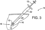
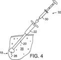
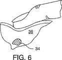
図2は、本発明に依る治療アルゴリズム2を示している。通常の乳房のX線写真撮影の結果により、6で腫瘍又は他の異常が検出されるかもしれない。これに対する代表的な対応は、多くの場合、更に拡大乳房X線写真を撮影するか、若しくは、例えば今後6ヶ月のような今後の特定日に継続診断の乳房X線写真撮影が予定されるなどが含まれる。これは8に示されている。その腫瘍が触診不能である場合には9を参照し、乳房放射線科医師による画像案内針生検が、通常では10において実施される。画像案内針生検は様々な方法で行うことができる。現在では、主にその正確性、迅速性及び患者に対する外傷が最小であることから、通常は定位(X線)針生検及び超音波案内式針生検が使用される。定位針生検は、代表的には、例えばFisher社やLorad社により製造される定位テーブルを使用し、これは生検針アセンブリに対する乳房X線写真ガイドを提供する。超音波案内式生検は、現在利用可能である多くの器具の任意のものを使用して実施され得る。図3に示される例示的な生検針アセンブリ14は、中空主ハウジング22から延出してシース20を通過する生検針13を備える。この生検針アセンブリ14の生検針13の先端部12は、標的部位18の異常部位16に自動的に挿入される。生検針13は、異常部16の組織サンプルを採るのに使用される前記先端部12近傍に形成された側方向きの横開口部24を備えている。組織サンプルが採取されると、標的部位18に生検開口領域26が形成される。図4を参照のこと。シース20および主ハウジング22から生検針13が取り外された後に、生体吸収性部材送達器具32のバレル30が、主ハウジング22を介してシース20に挿入される。前記バレル30は生体吸収性部材34を収納している。図5を参照のこと。この生体吸収性部材34は、この好適実施例においては、Davol社、Datascope社、Integra Life Sciences社、Collagen Matrix社、Vascular Solutions社などの複数の会社により市販されるもののような、脱水コラーゲンからなる栓子である。この生体吸収性部材34は、溶媒と接触して生検開口領域26内部で膨張し、図5に示されているように前記生検開口領域を実質的に充填する。この好適実施例に於いて、前記生体吸収性部材34は、前記バレル30内におけるその送達前状態から、領域26に於けるその送達後状態へと変換され、この過程において、膨張して、その送達後状態に於いてその送達前状態よりも幾分柔らかくなる。しかしながら、その送達後状態に於いて、生体吸収性部材34は、その周辺の軟組織、代表的には乳房組織36よりも触診的に硬く、好ましくは少なくとも約1.5倍硬い。これは、図6において示すように、医師による患者の触診で標的部位18において生体吸収性部材34が確認され、生体吸収性部材6を見出すことを可能にし、そしてこのことは以下において詳細に議論される。
【0015】
生体吸収性部材は、コラーゲン以外の材料からなり得、その送達前状態に於いて比較的硬い固体の栓子以外の形状であり得る。たとえば、バレル30内でのその送達前状態に於いて、この生体吸収性部材34は、液体その他の流動状のものとし、標的部位18の前記開口領域18への載置後に、この生体吸収性部材がその後の触診による標的部位18の位置再確認を可能とするべく、周辺組織36よりも触診的に固くなるように構成することができる。いくつかの状況に於いては、生体吸収性部材34のサイズ又は固さがその送達前状態と送達後状態との間で変化せず、これら両方の状態に於いて周辺組織36よりも触診的に硬いものであることが所望され得る。一好適実施例に於いて、この生体吸収性部材34の変容は、水性液体との接触によるものである。しかしながら、この生体吸収性部材の変容は、これがなされ得るという点において、たとえば、硬度、感触、形状、サイズ、又はこれらの組み合わせに於けるものとすることができる、前記生体吸収性部材の変容は、例えば、熱エネルギ、放射線、磁気エネルギ等のようなその他の要因によって生じるものであり得る。
【0016】
図2に戻ると、ここには、生体吸収性部材34の挿入後、36に於いてその生検サンプルが病理学的評価のために送られ得ると考えられる。もしも、一両日中に入手される生検のこの病理学的報告が良性である場合、その旨が患者に伝えられ、38に於いて、前記生体吸収性部材は、たとえば、1ヶ月以内にその患者によって簡単に吸収される。もしも病理学報告が陽性で、癌が見つかった場合には、図6に示されているように、外科医の触診によって標的部位18の生検開口領域26が位置確認される。放射線医師による手術前の位置確認の必要性を省く、触診による前記標的部位の位置確認後、切除手術等の適当な医療処置が実施され得る。
【0017】
もしもその腫瘍が触診可能である場合、外科医は、48に於いて直接的な切開生検を行うことを選択するかもしれない。本発明に依れば、前記生体吸収性部材送達器具32を、切開生検の部位に生体吸収性部材34に配置して使用することが可能である。送達器具32の取り外した後に、切開部は縫合され、生検サンプルは病理検査に送られ、患者は36から始まる上述の手順で帰宅する。
【0018】
前記生体吸収性部材34はまた、前記開口領域26を充填するか、又はほぼ充填することによる物理的手段、或いは、凝固等の、血液成分との化学的相互作用による化学的手段によって、部位18での出血を止める止血剤としても作用することが好ましくあり得る。更に、前記生体吸収性部材34を、非止血性の分解性外層によって被覆して、この外層が侵食されるまで止血その他の作用を遅延させるように構成することも可能である。いくつかの状況に於いて、前記生体吸収性部材が標的部位18に配置されるまでは、この部材を血液又はその他の体液から保護することが必要であり得るか、または少なくとも所望され得る。これは、たとえば、生体吸収性部材の送達中に除去可能な物理的バリアを使用することによって達成され得る。或いは、上述したように、生体吸収性コーティング又は層もまた使用され得る。前記生体吸収性部材は、その送達前状態から送達後状態へと、水化、温度変化、電気刺激、磁気刺激、刺激剤との化学反応、操作部材(たとえば、前記生体吸収性部材を収納したカプセルを切開開放するのに使用可能なナイフ刃など)との物理的相互作用、生体吸収性部材のイオン化、生体吸収性部材による流体の吸着又は吸収などを含む様々な方法によって変容され得る。
【0019】
本発明はまた、患者を医学的に処置するためにも更に使用することができる。即ち、病理学報告に医学的治療の必要性が示された場合にのみ、この生体吸収性部材に活性化治療部材を含ませることができる。生体吸収性部材中の薬剤の活性化は、標的部位近傍への放射線放出部材の注入、標的部位の外部からの照射、標的部位へのトリガ物質の供給、手動圧力、光感作療法、硬化化学作用、振動療法、超音波等の様々な方法で実施され得る。或いは、前記生体吸収性部材を、そのような活性化物質は含まず、むしろ、例えば化学療法剤、放射線放出部材、熱エネルギ、電気エネルギ、振動エネルギ、遺伝子治療、ベクタ治療、抗脈管形成療法を供給することにより医学的処置を行うように構成することも可能である。この供給を容易にするために、前記生体吸収性部材に、放射線不透過性マーカを含ませたり、或いは、触診可能であることに加えて、それを超音波によって検出されることに役立つ特性を備えさせることができる。
【0020】
本発明の一つの重要な使用は、乳癌の処置である。一実施例に於いて、前記生体吸収性部材34は、その送達後状態に於いてその周辺組織よりも触診的に固くなるように、乳房組織の少なくとも約1.5倍の硬さを伴うことが所望される。また、前記生体吸収性部材34は、一実施例に於いて、その送達前状態から送達後状態にかけて膨張して、前記開口領域26を充填するか、又は少なくとも実質的に充填することもまた所望される。これを達成するために、前記生体吸収性部材34は、送達前状態から、通常は水性液体との接触時である送達後状態にかけて、約50−1500%、より好ましくは約100−300%膨張することが所望される。この生体吸収性部材は、触診によるその位置確認を介助するべく、その送達後状態に於いて少なくとも約0.5cmの最大長を有することが好ましい。
【0021】
一つの実施例に於いて、好ましくは前記生体吸収性部材はコラーゲンからなる一方で、この生体吸収性部材に下記の1つ以上の物質を含ませることも可能である:polyactic酸及びポリグリコール酸、ポリオルトエステル、再吸収可能シリコーンおよびウレタン、脂質、ポリサッカライド、澱粉、セラミックス、ポリアミノ酸、タンパク質、水和ゲル及び他のゲル、ゼラチン、ポリマー、セルロース、エラスチンなど。
【0022】
いくつかの状況に於いては、図7に図示されるように、患者の皮膚46を経て生体吸収性部材34から延出する生体吸収性フィラメント44を使用することが所望され得る。これは、生体吸収性部材34が標的部位18の領域26に注入される時に、生体吸収性フィラメント44をシース20を通って送達することにより達成することができる。いくつかの状況に於いては、生体吸収性部材34を使用することが不可能であるかもしれないし、又は所望されないかもしれないが、そのような状況に於いては、標的部位18から患者の皮膚46の上方へと延出する生体吸収性フィラメント44のみを供給することが有用であり得る。
【0023】
現在に於いては、針アセンブリ14の一部、即ち、シース20および主ハウジング22を通して生体吸収性部材送り出し器具32を案内することが好適ではあるが、いくつかの状況に於いては、生検サンプルが取出され、生体針アセンブリ14全体が取出された後、その位置に残される外側シースでシース20を被覆することが有用であるかもしれない。その後、その位置に残されたシースを使用して、送達器具32のバレル30を標的部位18に案内する。当然のことだが、送り出し器具32の形状は、針又はハウジング22およびシース20を経るか、又は外側シースを経て注入される流体又はペーストを含む注射器などのような様々な方法であり得る。或いは、生体吸収性部材34を送達するために、他の送達器具を使用することも可能であろう。
【0024】
本発明は、生検のための乳房組織除去又は疾患組織除去によって生じる欠陥の矯正についての用途を有する。多くの場合、コラーゲンが体内に配置され、そこでそのコラーゲンは最終的にはヒトの自家組織に置換される。従って、本発明は、組織除去によって損傷した組織の修復に使用することが可能である。上述の送達器具は、そのような美容又は整形修復のためにその腔部に物質(合成又は哺乳動物性の物質)を送達するために使用することができる。この物質は、代表的にはシリコーンゲル充填カプセル又はバッグ等の事実上非生体吸収性の物質であろう。
【0025】
下記の請求項に定義された本発明の課題から逸脱することなく、説明した実施例に対して改変、変更を加えることが可能であろう。
【0026】
上述したすべての特許、特許出願、及び印刷刊行物をここに参考文献として合体させる。
【図面の簡単な説明】
【図1】触診不能な乳房病巣部についての従来の処置アルゴリズムの流れ図
【図2】本発明に依る処置アルゴリズムの流れ図
【図3】標的部位における異常性組織サンプルを得る針生検アセンブリを示す略図
【図4】前記組織サンプルの摘出後に標的部位に生検開口領域がそのままになっている状態で、その位置に残された前記針生検アセンブリの主ハウジングおよびシースを図示する
【図5】前記針生検アセンブリの前記主ハウジングに挿入された図4の送達器具のバレル、および前記生検開口領域に生体吸収性部材を注入し、これによって標的部位の前記生検開口領域を効果的に充填するために押し込まれたプランジャを図示する
【図6】外科医がその指を使用することにより図5の生体吸収性部材の位置確認を図示する
【図7】図4及び図5の送達器具を使用して前記生体吸収性部材に対して送達され、図5の生体吸収性部材から患者の皮膚を貫通して延出する生体吸収性糸を図示する[0001]
BACKGROUND OF THE INVENTION Approximately 1 million women in the United States alone have undergone breast biopsies with disturbed mammograms and palpation abnormalities. See FIG. 1 which shows the current treatment algorithm for breast lesions that are not palpable. For non-palpable lesions, biopsy can be performed in a variety of ways, including surgical resection biopsy, stereotactic needle breast biopsy, ultrasound guided needle breast biopsy, and the like. In the case of an image-guided biopsy, the radiologist or other doctor takes a small sample of abnormal tissue for analysis in the laboratory. If the biopsy proves malignant, further surgery (typically a mammary tumor removal or mastectomy) is required. The patient then revisits the radiologist 1-2 days later, where the biopsy site (focal site) is referred to as the needle location verification method as a pre-surgery location check for surgery. Confirm the position again with.
Re-identifying the location of the biopsy area after a surgical excision biopsy is usually not a problem because of the deformation caused by surgery. However, when a biopsy is performed by an image-guided needle method that is generally performed, assistance is usually required for reconfirming the position of the biopsy site. One method that allows the biopsy site to be reconfirmed by the radiologist during the pre-surgical location is to leave a portion of the calcification concern, but this has its disadvantages There is.
[0002]
Another method for the radiologist to reposition the biopsy site includes, for example, the use of small metal surgical clips (eg, clips manufactured by Biopsis). This metal clip can be placed through a biopsy needle and then left at the biopsy site during the first biopsy. By using this metal clip as a guide, the radiologist typically reinserts a wire with a needle, such as Hawkins, Kopans, Homer, Sadowski, and other needles into the patient's breast. The tip of the wire is then located at the biopsy site using a mammogram and the placement is recorded. The patient is then transported to the operating room with the needle protruding from its breast. While this clip provides a good indication of the biopsy site during localization prior to surgery, the clip remains permanently in the body of 80% of patients diagnosed as benign. In addition, the clip needs to be attached at one location around the biopsy site rather than at the center of the biopsy site, so that location may cause diseased tissue during any further medical intervention procedures. May be misplaced. Furthermore, since the breast tissue is soft, the tip of the hooked or hooked needle can move relatively easily from the biopsy site. This clip is also relatively expensive.
[0003]
Another location confirmation method uses laser light from the tip of an optical fiber connected to a laser. The tip is fixed to the biopsy site by a pair of hooks provided at the tip of the optical fiber, and the position of the tip is indicated through breast tissue whose light emission is several centimeters. This procedure entails some of the same problems as methods using barbed or hooked wires. Another pre-operative repositioning procedure is a method of injecting medical grade powdered carbon suspension from the lesion site onto the skin surface. This method also has certain problems including trace discontinuities along the carbon.
[0004]
SUMMARY OF THE INVENTION The present invention uses a position-recognizable bioabsorbable member that remains in a biopsy site, and when it is found necessary to do so by testing the biopsy sample, The present invention relates to a biopsy position confirmation method and instrument capable of repositioning by finding an absorbent member. This eliminates the need to use a metal clip during a biopsy, and in many cases eliminates the need to revisit a radiologist for location confirmation prior to surgery. Further, the bioabsorbable member can be used as a therapeutic instrument for treatment of the affected lesion and hemostasis.
[0005]
A biopsy location verification device according to the present invention includes a bioabsorbable member that is delivered in a pre-delivery state to a patient's soft tissue biopsy site by a member delivery device. The bioabsorbable member can be palpated harder than the soft tissue around the biopsy site in the post-delivery state.
[0006]
One suitable material used as this bioabsorbable member is anhydrous collagen plug. This type of obturator swells and can be palpated by a surgeon at a later location. This collagen plug does not have to swell at all. In some situations, such as a woman with a small breast, i.e., when the biopsy site is close to the surface, a non-inflatable bioabsorbable member, such as a PGA circular pellet, may be Can be used instead. The bioabsorbable member can also be rapidly absorbed to cause local tissue inflammation, which can be used to locate the biopsy site instead of palpation. For example, instead of leaving a collagen plug, PGA pellet or bioabsorbable suture at the biopsy site for palpation or inflammatory localization, it can penetrate through the longitudinal bioabsorbable suture, collagen filament, or skin. Other bioabsorbable members extending from the examination site can be used. In this case, the surgeon can reach the biopsy site according to the bioabsorbable member, similar to the case of using a Hawkins needle. In other cases, such as when the lesion is located deep, i.e. in the case of a large breast, the bioabsorbable member needs to be confirmed by the radiologist by ultrasound, mammogram, etc. obtain. In any case, the bioabsorbable member is normally absorbed within about one month after its placement. Thus, the present invention eliminates the need to use a metal clip during a biopsy and, in many cases, eliminates the need to revisit a radiologist for location confirmation prior to surgery.
[0007]
The primary use of this instrument is intended to locate a needle biopsy site for possible future surgical resections, but this instrument also provides a site for surgical resection biopsies. It can also be useful for labeling. For example, during a large surgical resection in cancer diagnosed by a recently performed surgical excision biopsy, the surgeon frequently has an accurate association of previously resected tissue to the surgical wound. Difficulty in judging sex. Thus, if the exact location of the previous lesion was clearer, more tissue would be removed than would have been removed. In accordance with the present invention, during a surgical resection, in order to label the site for resection that is likely to be large if the cancer is diagnosed by biopsy, A bioabsorbable member can be inserted into the biopsy site before the wound is sutured. Further, by partially or completely suturing the wound, the bioabsorbable member is inserted into the biopsy site by the delivery device, and then the bioabsorbable member is installed through the delivery device, and the sutured incision is made. It is also possible to remove the delivery device via. The presence of palpable markers within the previous ablation biopsy site will allow the surgeon to more easily and confidently remove tissue from this site and retain more normal breast tissue.
[0008]
Another use of the instrument is to first locate non-palpable lesions prior to surgical excision biopsy. Areas of concern of the breast, as guided by radiography and ultrasound of the breast, immediately prior to the surgical resection biopsy, instead of using a needle / wire instrument that tends to be displaced due to traction The palpable marker can be inserted into This provides a position verification means that can be palpated to the surgeon, as described above. In this case, since it is intended to remove it in any case, the marker need only be palpated and need not be bioabsorbable.
[0009]
In addition to being able to locate the biopsy site by subsequent palpation or other means, the present invention can also provide therapeutic and hemostatic benefits. Since bioabsorbability can change over a period of 1-2 days or 1 year or longer, the material can be used not only to locate the biopsy tissue but also to treat it Is possible. Current therapies include radiation therapy, chemotherapy, gene therapy and other techniques and therapies. Since the bioabsorbability can be easily changed, a medium can be injected into the bioabsorbable member, and if the biopsy result is malignant, it can be excited or induced from the outside. Furthermore, the bioabsorption concept can be used for future therapeutic agent implantation. For example, if the bioabsorbable member is anhydrous collagen, this material can be used as a holding means, for example, to deliver a material that is effective for chemotherapy, brachytherapy, and the like. If the experimental results are received and the results indicate that the biopsy results are malignant and require treatment, such as by surgical resection, the physician may, for example, for direct treatment of the diseased part: A radiation pellet, a chemotherapeutic agent, or a gene therapy agent can be injected into or near the bioabsorbable member.
[0010]
Changes in the bioabsorbable member include hydration or dehydration, temperature change, electrical stimulation, magnetic stimulation, chemical or physical reaction involving other substances, additives, enzyme reactions, ionization, charge, absorption, and other means. Can be done through one of several methods. The present invention can use one or more of these techniques or means to change the viscosity, hardness and / or size between the placed and unplaced portions of the bioabsorbable member. The visibility of the bioabsorbable member can be assisted by using a colorant such as methylene blue or other pigment. The radiation detection characteristics of the member can be enhanced by a radiopaque marker. Similarly, the ultrasonic detection characteristics can also be enhanced by special processing of the bioabsorbable member.
[0011]
The bioabsorbable member may have an uneven portion formed to prevent movement within the tissue. Filaments extending from the edges of the bioabsorbable member can also be used to stabilize the placement of the device within the cavity. The filament may be made of the same material as the bioabsorbable member, but this need not be the case.
[0012]
Hemostasis preparation helps reduce bleeding or swelling within or around the biopsy site. This can be achieved by physical or chemical means. That is, the device may be expanded to fill the biopsy cavity, or the device may be configured to chemically react with blood and / or blood products to produce effective blood clotting. obtain. In the present invention, other methods for producing local hemostasis are also possible.
[0013]
Other features and advantages of the present invention will become apparent from the following description in which the preferred embodiment and method are described in detail with reference to the accompanying drawings.
[0014]
DESCRIPTION OF SPECIFIC EMBODIMENTS FIG. 2 shows a
[0015]
The bioabsorbable member may be made of a material other than collagen and may have a shape other than a solid obturator that is relatively hard in its pre-delivery state. For example, in its pre-delivery state within the
[0016]
Returning to FIG. 2, it is now contemplated that after insertion of the
[0017]
If the tumor is palpable, the surgeon may choose to perform a direct incision biopsy at 48. According to the present invention, the bioabsorbable
[0018]
The
[0019]
The present invention can also be further used to medically treat a patient. That is, the bioabsorbable member can include an activated therapeutic member only when the pathology report indicates the necessity of medical treatment. Activation of the drug in the bioabsorbable member includes injection of a radiation emitting member near the target site, irradiation from the outside of the target site, supply of a trigger substance to the target site, manual pressure, photosensitizing therapy, curing chemistry It can be performed in various ways, such as action, vibration therapy, ultrasound, etc. Alternatively, the bioabsorbable member does not contain such an activating substance, but rather, for example, a chemotherapeutic agent, a radiation-emitting member, thermal energy, electrical energy, vibration energy, gene therapy, vector therapy, antiangiogenic therapy It can also be configured to perform a medical procedure by supplying In order to facilitate this supply, the bioabsorbable member includes a radiopaque marker, or in addition to being palpable, has characteristics that help it be detected by ultrasound. Can be provided.
[0020]
One important use of the present invention is the treatment of breast cancer. In one embodiment, the
[0021]
In one embodiment, preferably, the bioabsorbable member comprises collagen, but the bioabsorbable member may include one or more of the following substances: polyacid and polyglycolic acid , Polyorthoesters, resorbable silicones and urethanes, lipids, polysaccharides, starches, ceramics, polyamino acids, proteins, hydrated gels and other gels, gelatin, polymers, cellulose, elastin and the like.
[0022]
In some situations, it may be desirable to use a
[0023]
Currently, it is preferred to guide the bioabsorbable
[0024]
The present invention has application for correcting defects caused by breast tissue removal or diseased tissue removal for biopsy. In many cases, collagen is placed in the body where it is eventually replaced by human autologous tissue. Thus, the present invention can be used to repair tissue damaged by tissue removal. The delivery device described above can be used to deliver substances (synthetic or mammalian substances) into the cavity for such cosmetic or orthopedic repairs. This material will typically be a substantially non-bioabsorbable material such as a silicone gel filled capsule or bag.
[0025]
Modifications and changes may be made to the described embodiments without departing from the scope of the present invention as defined in the following claims.
[0026]
All patents, patent applications, and printed publications mentioned above are hereby incorporated by reference.
[Brief description of the drawings]
FIG. 1 is a flowchart of a conventional treatment algorithm for a non-palpable breast lesion. FIG. 2 is a flowchart of a treatment algorithm according to the present invention. FIG. 3 is a schematic diagram showing a needle biopsy assembly for obtaining an abnormal tissue sample at a target site. 4 illustrates the main housing and sheath of the needle biopsy assembly left in place with the biopsy opening area intact at the target site after removal of the tissue sample. FIG. 5 illustrates the needle biopsy. The barrel of the delivery device of FIG. 4 inserted into the main housing of the assembly, and injecting a bioabsorbable member into the biopsy opening area, thereby effectively filling the biopsy opening area of the target site FIG. 6 illustrates the plunger pushed in. FIG. 6 illustrates the location of the bioabsorbable member of FIG. 5 by the surgeon using his finger. Using a delivery instrument of FIG. 5 is delivered to the bioabsorbable member, illustrating the bioabsorbable yarns extending through the patient's skin from bioabsorbable member of Figure 5
Claims (27)
基端部、先端部、および、それらの間の管腔を有する細長い管状部材と、
前記細長い管状部材の内部に収容されている膨張性の生体吸収性物体と、
前記生体吸収性物体によって運ばれる放射性不透過性マーカと、
を含み、
前記生体吸収性物体は、送達前状態において前記細長い管状部材の中で概ね固体状を有し、前記細長い管状部材から送達されると周辺組織よりも概ね硬い材料を含む、生検位置確認器具。 In the biopsy position confirmation instrument,
An elongated tubular member having a proximal end, a distal end, and a lumen therebetween;
An inflatable bioabsorbable object housed within the elongate tubular member;
A radiopaque marker carried by the bioabsorbable object;
Only including,
The biopsy localization instrument, wherein the bioabsorbable object has a generally solid shape in the elongate tubular member in a pre-delivery state and includes a material that is generally harder than surrounding tissue when delivered from the elongate tubular member .
前記生体吸収性物体は、超音波および乳房X線写真撮影の少なくとも1つによって間接的に視覚化できる、生検位置確認器具。In the biopsy position confirmation instrument according to claim 1,
A biopsy localization instrument, wherein the bioabsorbable object can be visualized indirectly by at least one of ultrasound and mammography.
前記放射性不透過性マーカは、前記生体吸収性物体の内部に収容されている、生検位置確認器具。In the biopsy position confirmation instrument according to claim 1,
The radiopaque marker is a biopsy position confirmation instrument housed inside the bioabsorbable object.
前記生体吸収性物体は、少なくとも1つの生体吸収性物体を含む、生検位置確認器具。In the biopsy position confirmation instrument according to claim 1,
The biopsy position confirmation instrument, wherein the bioabsorbable object includes at least one bioabsorbable object.
前記生体吸収性物体は、体液と接触すると膨張する、生検位置確認器具。In the biopsy position confirmation instrument according to claim 1,
A biopsy position confirmation instrument that expands when the bioabsorbable object comes into contact with bodily fluids.
前記生体吸収性物体は、膨張して生検部位を実質的に満たす、生検位置確認器具。In the biopsy position confirmation instrument according to claim 5,
The biopsy position confirmation instrument, wherein the bioabsorbable object expands to substantially fill a biopsy site.
前記生体吸収性物体は、コラーゲンを含む、生検位置確認器具。In the biopsy position confirmation instrument according to claim 1,
The biopsy location object is a biopsy position confirmation instrument containing collagen.
前記生体吸収性物体は、ゼラチンを含む、生検位置確認器具。In the biopsy position confirmation instrument according to claim 1,
The biopsy position confirmation instrument, wherein the bioabsorbable object includes gelatin.
前記生体吸収性物体は、ポリ乳酸およびポリグリコール酸を含む、生検位置確認器具。In the biopsy position confirmation instrument according to claim 1,
The bioabsorbable object is a biopsy position confirmation instrument including polylactic acid and polyglycolic acid.
基端部、先端部、および、それらの間の管腔を有する細長い管状部材と、
前記細長い管状部材の内部に収容されている生体吸収性物体であり、ポリ乳酸およびポリグリコール酸を含む、前記生体吸収性物体と、
前記生体吸収性物体によって運ばれる放射性不透過性マーカと、
を含み、
前記生体吸収性物体は、送達前状態において前記細長い管状部材の中で概ね固体状を有し、前記細長い管状部材から送達されると周辺組織よりも概ね硬い材料を含む、生検位置確認器具。 In the biopsy position confirmation instrument,
An elongated tubular member having a proximal end, a distal end, and a lumen therebetween;
A bioabsorbable object housed within the elongate tubular member, the bioabsorbable object comprising polylactic acid and polyglycolic acid; and
A radiopaque marker carried by the bioabsorbable object;
Only including,
The biopsy localization instrument, wherein the bioabsorbable object has a generally solid shape in the elongate tubular member in a pre-delivery state and includes a material that is generally harder than surrounding tissue when delivered from the elongate tubular member .
前記生体吸収性物体は、超音波および乳房X線写真撮影の少なくとも1つによって間接的に視覚化できる、生検位置確認器具。The biopsy position confirmation instrument according to claim 10,
A biopsy localization instrument, wherein the bioabsorbable object can be visualized indirectly by at least one of ultrasound and mammography.
前記放射性不透過性マーカは、前記生体吸収性物体の内部に収容されている、生検位置確認器具。The biopsy position confirmation instrument according to claim 10,
The radiopaque marker is a biopsy position confirmation instrument housed inside the bioabsorbable object.
前記生体吸収性物体は、少なくとも1つの生体吸収性物体を含む、生検位置確認器具。The biopsy position confirmation instrument according to claim 10,
The biopsy position confirmation instrument, wherein the bioabsorbable object includes at least one bioabsorbable object.
前記生体吸収性物体は、体液と接触すると膨張する、生検位置確認器具。The biopsy position confirmation instrument according to claim 10,
A biopsy position confirmation instrument that expands when the bioabsorbable object comes into contact with bodily fluids.
前記生体吸収性物体は、膨張して生検部位を実質的に満たす、生検位置確認器具。The biopsy position confirmation instrument according to claim 14,
The biopsy position confirmation instrument, wherein the bioabsorbable object expands to substantially fill a biopsy site.
基端部、先端部、および、それらの間の管腔を有する細長い管状部材と、
前記細長い管状部材の内部に収容されている生体吸収性物体であり、コラーゲンを含む、前記生体吸収性物体と、
前記生体吸収性物体によって運ばれる放射性不透過性マーカと、
を含み、
前記生体吸収性物体は、送達前状態において前記細長い管状部材の中で概ね固体状を有し、前記細長い管状部材から送達されると周辺組織よりも概ね硬い材料を含む、生検位置確認器具。 In the biopsy position confirmation instrument,
An elongated tubular member having a proximal end, a distal end, and a lumen therebetween;
A bioabsorbable object housed within the elongate tubular member, comprising collagen, the bioabsorbable object;
A radiopaque marker carried by the bioabsorbable object;
Only including,
The biopsy localization instrument, wherein the bioabsorbable object has a generally solid shape in the elongate tubular member in a pre-delivery state and includes a material that is generally harder than surrounding tissue when delivered from the elongate tubular member .
前記生体吸収性物体は、超音波および乳房X線写真撮影の少なくとも1つによって間接的に視覚化できる、生検位置確認器具。The biopsy position confirmation instrument according to claim 16,
A biopsy localization instrument, wherein the bioabsorbable object can be visualized indirectly by at least one of ultrasound and mammography.
前記放射性不透過性マーカは、前記生体吸収性物体の内部に収容されている、生検位置確認器具。The biopsy position confirmation instrument according to claim 16,
The radiopaque marker is a biopsy position confirmation instrument housed inside the bioabsorbable object.
前記生体吸収性物体は、少なくとも1つの生体吸収性物体を含む、生検位置確認器具。The biopsy position confirmation instrument according to claim 16,
The biopsy position confirmation instrument, wherein the bioabsorbable object includes at least one bioabsorbable object.
前記生体吸収性物体は、体液と接触すると膨張する、生検位置確認器具。The biopsy position confirmation instrument according to claim 16,
A biopsy position confirmation instrument that expands when the bioabsorbable object comes into contact with bodily fluids.
前記生体吸収性物体は、膨張して生検部位を実質的に満たす、生検位置確認器具。The biopsy position confirmation instrument according to claim 20,
The biopsy position confirmation instrument, wherein the bioabsorbable object expands to substantially fill a biopsy site.
基端部、先端部、および、それらの間の管腔を有する細長い管状部材と、
前記細長い管状部材の内部に収容されている生体吸収性物体であり、ゼラチンを含む、前記生体吸収性物体と、
前記生体吸収性物体によって運ばれる放射性不透過性マーカと、
を含み、
前記生体吸収性物体は、送達前状態において前記細長い管状部材の中で概ね固体状を有し、前記細長い管状部材から送達されると周辺組織よりも概ね硬い材料を含む、生検位置確認器具。 In the biopsy position confirmation instrument,
An elongated tubular member having a proximal end, a distal end, and a lumen therebetween;
A bioabsorbable object housed within the elongate tubular member, the bioabsorbable object comprising gelatin;
A radiopaque marker carried by the bioabsorbable object;
Only including,
The biopsy localization instrument, wherein the bioabsorbable object has a generally solid shape in the elongate tubular member in a pre-delivery state and includes a material that is generally harder than surrounding tissue when delivered from the elongate tubular member .
前記生体吸収性物体は、超音波および乳房X線写真撮影の少なくとも1つによって間接的に視覚化できる、生検位置確認器具。The biopsy position confirmation instrument according to claim 22,
A biopsy localization instrument, wherein the bioabsorbable object can be visualized indirectly by at least one of ultrasound and mammography.
前記放射性不透過性マーカは、前記生体吸収性物体の内部に収容されている、生検位置確認器具。The biopsy position confirmation instrument according to claim 22,
The radiopaque marker is a biopsy position confirmation instrument housed inside the bioabsorbable object.
前記生体吸収性物体は、少なくとも1つの生体吸収性物体を含む、生検位置確認器具。The biopsy position confirmation instrument according to claim 22,
The biopsy position confirmation instrument, wherein the bioabsorbable object includes at least one bioabsorbable object.
前記生体吸収性物体は、体液と接触すると膨張する、生検位置確認器具。The biopsy position confirmation instrument according to claim 22,
A biopsy position confirmation instrument that expands when the bioabsorbable object comes into contact with bodily fluids.
前記生体吸収性物体は、膨張して生検部位を実質的に満たす、生検位置確認器具。The biopsy location verification instrument according to claim 26,
The biopsy position confirmation instrument, wherein the bioabsorbable object expands to substantially fill a biopsy site.
Applications Claiming Priority (11)
| Application Number | Priority Date | Filing Date | Title |
|---|---|---|---|
| US9024398P | 1998-06-22 | 1998-06-22 | |
| US9273498P | 1998-07-14 | 1998-07-14 | |
| US11486399P | 1999-01-06 | 1999-01-06 | |
| US11742199P | 1999-01-27 | 1999-01-27 | |
| US09/336,360 | 1999-06-18 | ||
| US60/092,734 | 1999-06-18 | ||
| US09/336,360 US6270464B1 (en) | 1998-06-22 | 1999-06-18 | Biopsy localization method and device |
| US60/117,421 | 1999-06-18 | ||
| US60/114,863 | 1999-06-18 | ||
| US60/090,243 | 1999-06-18 | ||
| PCT/US1999/013909 WO1999066834A1 (en) | 1998-06-22 | 1999-06-21 | Biopsy localization method and device |
Publications (3)
| Publication Number | Publication Date |
|---|---|
| JP2002518121A JP2002518121A (en) | 2002-06-25 |
| JP2002518121A5 JP2002518121A5 (en) | 2006-08-10 |
| JP4472176B2 true JP4472176B2 (en) | 2010-06-02 |
Family
ID=27536563
Family Applications (1)
| Application Number | Title | Priority Date | Filing Date |
|---|---|---|---|
| JP2000555526A Expired - Fee Related JP4472176B2 (en) | 1998-06-22 | 1999-06-21 | Biopsy position confirmation method and instrument |
Country Status (6)
| Country | Link |
|---|---|
| US (8) | US6270464B1 (en) |
| EP (2) | EP2258258B1 (en) |
| JP (1) | JP4472176B2 (en) |
| AT (1) | ATE483416T1 (en) |
| DE (1) | DE69942833D1 (en) |
| WO (1) | WO1999066834A1 (en) |
Families Citing this family (169)
| Publication number | Priority date | Publication date | Assignee | Title |
|---|---|---|---|---|
| CA2199864C (en) | 1994-09-16 | 2006-06-20 | Seth A. Foerster | Methods and devices for defining and marking tissue |
| US7637948B2 (en) | 1997-10-10 | 2009-12-29 | Senorx, Inc. | Tissue marking implant |
| US8288745B2 (en) * | 1997-10-10 | 2012-10-16 | Senorx, Inc. | Method of utilizing an implant for targeting external beam radiation |
| US8668737B2 (en) | 1997-10-10 | 2014-03-11 | Senorx, Inc. | Tissue marking implant |
| US6270464B1 (en) * | 1998-06-22 | 2001-08-07 | Artemis Medical, Inc. | Biopsy localization method and device |
| US6347241B2 (en) | 1999-02-02 | 2002-02-12 | Senorx, Inc. | Ultrasonic and x-ray detectable biopsy site marker and apparatus for applying it |
| US6161034A (en) * | 1999-02-02 | 2000-12-12 | Senorx, Inc. | Methods and chemical preparations for time-limited marking of biopsy sites |
| US20020058882A1 (en) * | 1998-06-22 | 2002-05-16 | Artemis Medical, Incorporated | Biopsy localization method and device |
| US6036698A (en) | 1998-10-30 | 2000-03-14 | Vivant Medical, Inc. | Expandable ring percutaneous tissue removal device |
| US6371904B1 (en) | 1998-12-24 | 2002-04-16 | Vivant Medical, Inc. | Subcutaneous cavity marking device and method |
| US6356782B1 (en) | 1998-12-24 | 2002-03-12 | Vivant Medical, Inc. | Subcutaneous cavity marking device and method |
| US9669113B1 (en) | 1998-12-24 | 2017-06-06 | Devicor Medical Products, Inc. | Device and method for safe location and marking of a biopsy cavity |
| US7983734B2 (en) | 2003-05-23 | 2011-07-19 | Senorx, Inc. | Fibrous marker and intracorporeal delivery thereof |
| US9820824B2 (en) | 1999-02-02 | 2017-11-21 | Senorx, Inc. | Deployment of polysaccharide markers for treating a site within a patent |
| US6862470B2 (en) | 1999-02-02 | 2005-03-01 | Senorx, Inc. | Cavity-filling biopsy site markers |
| US20080039819A1 (en) * | 2006-08-04 | 2008-02-14 | Senorx, Inc. | Marker formed of starch or other suitable polysaccharide |
| US7651505B2 (en) | 2002-06-17 | 2010-01-26 | Senorx, Inc. | Plugged tip delivery for marker placement |
| US20090216118A1 (en) * | 2007-07-26 | 2009-08-27 | Senorx, Inc. | Polysaccharide markers |
| US8498693B2 (en) | 1999-02-02 | 2013-07-30 | Senorx, Inc. | Intracorporeal marker and marker delivery device |
| US8361082B2 (en) | 1999-02-02 | 2013-01-29 | Senorx, Inc. | Marker delivery device with releasable plug |
| US6725083B1 (en) | 1999-02-02 | 2004-04-20 | Senorx, Inc. | Tissue site markers for in VIVO imaging |
| US6306132B1 (en) | 1999-06-17 | 2001-10-23 | Vivant Medical | Modular biopsy and microwave ablation needle delivery apparatus adapted to in situ assembly and method of use |
| US6575991B1 (en) | 1999-06-17 | 2003-06-10 | Inrad, Inc. | Apparatus for the percutaneous marking of a lesion |
| US6722371B1 (en) | 2000-02-18 | 2004-04-20 | Thomas J. Fogarty | Device for accurately marking tissue |
| US6564806B1 (en) | 2000-02-18 | 2003-05-20 | Thomas J. Fogarty | Device for accurately marking tissue |
| JP5090600B2 (en) | 2000-02-18 | 2012-12-05 | トーマス ジェイ. フォガーティー, | Improved device for accurately marking tissues |
| US6544185B2 (en) | 2000-10-23 | 2003-04-08 | Valentino Montegrande | Ultrasound imaging marker and method of use |
| CA2775170C (en) | 2000-11-20 | 2017-09-05 | Senorx, Inc. | An intracorporeal marker delivery system for marking a tissue site |
| US8187625B2 (en) | 2001-03-12 | 2012-05-29 | Boston Scientific Scimed, Inc. | Cross-linked gelatin composition comprising a wetting agent |
| AU2002320182B2 (en) * | 2001-06-29 | 2008-02-21 | Cook Biotech Incorporated | Porous sponge matrix medical devices and methods |
| WO2003020333A2 (en) * | 2001-08-29 | 2003-03-13 | Artemis Medical, Inc. | Undamaged tissue collection assembly and method |
| US6878147B2 (en) | 2001-11-02 | 2005-04-12 | Vivant Medical, Inc. | High-strength microwave antenna assemblies |
| US6654629B2 (en) | 2002-01-23 | 2003-11-25 | Valentino Montegrande | Implantable biomarker and method of use |
| WO2003077730A2 (en) | 2002-03-11 | 2003-09-25 | Wardle John L | Surgical coils and methods of deploying |
| US6752767B2 (en) | 2002-04-16 | 2004-06-22 | Vivant Medical, Inc. | Localization element with energized tip |
| US7197363B2 (en) | 2002-04-16 | 2007-03-27 | Vivant Medical, Inc. | Microwave antenna having a curved configuration |
| US7329414B2 (en) * | 2002-05-03 | 2008-02-12 | Biopsy Sciences, Llc | Biodegradable polymer for marking tissue and sealing tracts |
| US7158660B2 (en) * | 2002-05-08 | 2007-01-02 | Gee Jr James W | Method and apparatus for detecting structures of interest |
| US20060036158A1 (en) | 2003-11-17 | 2006-02-16 | Inrad, Inc. | Self-contained, self-piercing, side-expelling marking apparatus |
| US20040122349A1 (en) * | 2002-12-20 | 2004-06-24 | Lafontaine Daniel M. | Closure device with textured surface |
| US8709038B2 (en) * | 2002-12-20 | 2014-04-29 | Boston Scientific Scimed, Inc. | Puncture hole sealing device |
| US6912050B2 (en) * | 2003-02-03 | 2005-06-28 | Hach Company | Phase shift measurement for luminescent light |
| US7877133B2 (en) * | 2003-05-23 | 2011-01-25 | Senorx, Inc. | Marker or filler forming fluid |
| US20050119562A1 (en) * | 2003-05-23 | 2005-06-02 | Senorx, Inc. | Fibrous marker formed of synthetic polymer strands |
| US7169114B2 (en) * | 2003-06-04 | 2007-01-30 | Krause William R | Biopsy and delivery device |
| US7783336B2 (en) | 2003-06-06 | 2010-08-24 | Ethicon Endo-Surgery, Inc. | Subcutaneous biopsy cavity marker device |
| US7942897B2 (en) * | 2003-07-10 | 2011-05-17 | Boston Scientific Scimed, Inc. | System for closing an opening in a body cavity |
| US7311703B2 (en) | 2003-07-18 | 2007-12-25 | Vivant Medical, Inc. | Devices and methods for cooling microwave antennas |
| US7744852B2 (en) | 2003-07-25 | 2010-06-29 | Rubicor Medical, Llc | Methods and systems for marking post biopsy cavity sites |
| US7537788B2 (en) * | 2003-07-25 | 2009-05-26 | Rubicor Medical, Inc. | Post-biopsy cavity treatment implants and methods |
| US20050033157A1 (en) * | 2003-07-25 | 2005-02-10 | Klein Dean A. | Multi-modality marking material and method |
| US20050020899A1 (en) * | 2003-07-25 | 2005-01-27 | Rubicor Medical, Inc. | Post-biopsy cavity treatmetn implants and methods |
| WO2005018468A2 (en) * | 2003-08-11 | 2005-03-03 | Wilson-Cook Medical Inc. | Surgical implant |
| US7001341B2 (en) * | 2003-08-13 | 2006-02-21 | Scimed Life Systems, Inc. | Marking biopsy sites |
| US8172770B2 (en) * | 2005-09-28 | 2012-05-08 | Suros Surgical Systems, Inc. | System and method for minimally invasive disease therapy |
| US7815561B2 (en) * | 2003-09-25 | 2010-10-19 | Xoft, Inc. | Brachytherapy applicator |
| US20050273002A1 (en) | 2004-06-04 | 2005-12-08 | Goosen Ryan L | Multi-mode imaging marker |
| US20050234336A1 (en) * | 2004-03-26 | 2005-10-20 | Beckman Andrew T | Apparatus and method for marking tissue |
| US8442623B2 (en) * | 2004-10-13 | 2013-05-14 | Suros Surgical Systems, Inc. | Site marker visible under multiple modalities |
| US8280486B2 (en) * | 2004-10-13 | 2012-10-02 | Suros Surgical Systems, Inc. | Site marker visable under multiple modalities |
| US8419656B2 (en) * | 2004-11-22 | 2013-04-16 | Bard Peripheral Vascular, Inc. | Post decompression marker introducer system |
| US7731705B2 (en) * | 2005-01-10 | 2010-06-08 | Wardle John L | Eluting coils and methods of deploying and retrieving |
| AU2006230428A1 (en) * | 2005-03-31 | 2006-10-05 | Cytyc Corporation | Internal biopsy marking |
| US10357328B2 (en) | 2005-04-20 | 2019-07-23 | Bard Peripheral Vascular, Inc. and Bard Shannon Limited | Marking device with retractable cannula |
| US8052658B2 (en) | 2005-10-07 | 2011-11-08 | Bard Peripheral Vascular, Inc. | Drug-eluting tissue marker |
| US11241296B2 (en) | 2005-11-17 | 2022-02-08 | Breast-Med, Inc. | Imaging fiducial markers and methods |
| US7702378B2 (en) * | 2005-11-17 | 2010-04-20 | Breast-Med, Inc. | Tissue marker for multimodality radiographic imaging |
| US8673398B2 (en) * | 2006-02-23 | 2014-03-18 | Meadwestvaco Corporation | Method for treating a substrate |
| US20080294039A1 (en) * | 2006-08-04 | 2008-11-27 | Senorx, Inc. | Assembly with hemostatic and radiographically detectable pellets |
| CN100408170C (en) * | 2006-08-16 | 2008-08-06 | 哈尔滨工业大学 | Process for preparing CuO/gamma-Al2O3 used as catalyst for catalytic oxidation process by inducing ClO2 with microwave |
| US8068921B2 (en) | 2006-09-29 | 2011-11-29 | Vivant Medical, Inc. | Microwave antenna assembly and method of using the same |
| EP2079385B1 (en) | 2006-10-23 | 2013-11-20 | C.R.Bard, Inc. | Breast marker |
| WO2008073965A2 (en) | 2006-12-12 | 2008-06-19 | C.R. Bard Inc. | Multiple imaging mode tissue marker |
| ES2432572T3 (en) | 2006-12-18 | 2013-12-04 | C.R. Bard, Inc. | Biopsy marker with imaging properties generated in situ |
| EP2197548B1 (en) * | 2007-09-19 | 2012-11-14 | Walter A. Roberts | Direct visualization robotic intra-operative radiation therapy applicator device |
| US9622813B2 (en) | 2007-11-01 | 2017-04-18 | Covidien Lp | Method for volume determination and geometric reconstruction |
| US8292880B2 (en) | 2007-11-27 | 2012-10-23 | Vivant Medical, Inc. | Targeted cooling of deployable microwave antenna |
| US8311610B2 (en) | 2008-01-31 | 2012-11-13 | C. R. Bard, Inc. | Biopsy tissue marker |
| US8068895B2 (en) * | 2008-02-25 | 2011-11-29 | Devicor Medical Products, Inc. | Biopsy site marker deployment instrument |
| US8079964B2 (en) * | 2008-02-25 | 2011-12-20 | Devicor Medical Products, Inc. | Method and apparatus for inserting biopsy site marker in marker body |
| US9327061B2 (en) | 2008-09-23 | 2016-05-03 | Senorx, Inc. | Porous bioabsorbable implant |
| US8968210B2 (en) | 2008-10-01 | 2015-03-03 | Covidien LLP | Device for needle biopsy with integrated needle protection |
| US9332973B2 (en) | 2008-10-01 | 2016-05-10 | Covidien Lp | Needle biopsy device with exchangeable needle and integrated needle protection |
| US9186128B2 (en) | 2008-10-01 | 2015-11-17 | Covidien Lp | Needle biopsy device |
| US9782565B2 (en) | 2008-10-01 | 2017-10-10 | Covidien Lp | Endoscopic ultrasound-guided biliary access system |
| US11298113B2 (en) | 2008-10-01 | 2022-04-12 | Covidien Lp | Device for needle biopsy with integrated needle protection |
| US10820825B2 (en) | 2008-10-22 | 2020-11-03 | Cornell University | Method and device for evaluation of local tissue's biological or biomechanical character |
| ES2560515T3 (en) | 2008-12-30 | 2016-02-19 | C.R. Bard, Inc. | Marker administration device for tissue marker placement |
| US9014787B2 (en) | 2009-06-01 | 2015-04-21 | Focal Therapeutics, Inc. | Bioabsorbable target for diagnostic or therapeutic procedure |
| US8323249B2 (en) | 2009-08-14 | 2012-12-04 | The Regents Of The University Of Michigan | Integrated vascular delivery system |
| US9427186B2 (en) * | 2009-12-04 | 2016-08-30 | Endomagnetics Ltd. | Magnetic probe apparatus |
| US10634741B2 (en) | 2009-12-04 | 2020-04-28 | Endomagnetics Ltd. | Magnetic probe apparatus |
| US8814833B2 (en) | 2010-05-19 | 2014-08-26 | Tangent Medical Technologies Llc | Safety needle system operable with a medical device |
| WO2011146769A2 (en) | 2010-05-19 | 2011-11-24 | Tangent Medical Technologies Llc | Integrated vascular delivery system |
| US20110301456A1 (en) * | 2010-06-07 | 2011-12-08 | Malignext Targeting Technologies, Inc. | Tissue Marking for Lesion Removal |
| US9072523B2 (en) | 2010-11-05 | 2015-07-07 | Ethicon Endo-Surgery, Inc. | Medical device with feature for sterile acceptance of non-sterile reusable component |
| US9782214B2 (en) | 2010-11-05 | 2017-10-10 | Ethicon Llc | Surgical instrument with sensor and powered control |
| US9782215B2 (en) | 2010-11-05 | 2017-10-10 | Ethicon Endo-Surgery, Llc | Surgical instrument with ultrasonic transducer having integral switches |
| US10085792B2 (en) | 2010-11-05 | 2018-10-02 | Ethicon Llc | Surgical instrument with motorized attachment feature |
| US10660695B2 (en) | 2010-11-05 | 2020-05-26 | Ethicon Llc | Sterile medical instrument charging device |
| US9526921B2 (en) * | 2010-11-05 | 2016-12-27 | Ethicon Endo-Surgery, Llc | User feedback through end effector of surgical instrument |
| US9649150B2 (en) | 2010-11-05 | 2017-05-16 | Ethicon Endo-Surgery, Llc | Selective activation of electronic components in medical device |
| US20120116265A1 (en) | 2010-11-05 | 2012-05-10 | Houser Kevin L | Surgical instrument with charging devices |
| US9375255B2 (en) | 2010-11-05 | 2016-06-28 | Ethicon Endo-Surgery, Llc | Surgical instrument handpiece with resiliently biased coupling to modular shaft and end effector |
| US9011471B2 (en) | 2010-11-05 | 2015-04-21 | Ethicon Endo-Surgery, Inc. | Surgical instrument with pivoting coupling to modular shaft and end effector |
| US9039720B2 (en) | 2010-11-05 | 2015-05-26 | Ethicon Endo-Surgery, Inc. | Surgical instrument with ratcheting rotatable shaft |
| US9017851B2 (en) | 2010-11-05 | 2015-04-28 | Ethicon Endo-Surgery, Inc. | Sterile housing for non-sterile medical device component |
| US9089338B2 (en) | 2010-11-05 | 2015-07-28 | Ethicon Endo-Surgery, Inc. | Medical device packaging with window for insertion of reusable component |
| US9000720B2 (en) | 2010-11-05 | 2015-04-07 | Ethicon Endo-Surgery, Inc. | Medical device packaging with charging interface |
| US20120116381A1 (en) | 2010-11-05 | 2012-05-10 | Houser Kevin L | Surgical instrument with charging station and wireless communication |
| US9421062B2 (en) | 2010-11-05 | 2016-08-23 | Ethicon Endo-Surgery, Llc | Surgical instrument shaft with resiliently biased coupling to handpiece |
| US9247986B2 (en) | 2010-11-05 | 2016-02-02 | Ethicon Endo-Surgery, Llc | Surgical instrument with ultrasonic transducer having integral switches |
| US9161803B2 (en) | 2010-11-05 | 2015-10-20 | Ethicon Endo-Surgery, Inc. | Motor driven electrosurgical device with mechanical and electrical feedback |
| US10959769B2 (en) | 2010-11-05 | 2021-03-30 | Ethicon Llc | Surgical instrument with slip ring assembly to power ultrasonic transducer |
| US9597143B2 (en) | 2010-11-05 | 2017-03-21 | Ethicon Endo-Surgery, Llc | Sterile medical instrument charging device |
| US10881448B2 (en) | 2010-11-05 | 2021-01-05 | Ethicon Llc | Cam driven coupling between ultrasonic transducer and waveguide in surgical instrument |
| US9017849B2 (en) | 2010-11-05 | 2015-04-28 | Ethicon Endo-Surgery, Inc. | Power source management for medical device |
| US9381058B2 (en) | 2010-11-05 | 2016-07-05 | Ethicon Endo-Surgery, Llc | Recharge system for medical devices |
| US9510895B2 (en) | 2010-11-05 | 2016-12-06 | Ethicon Endo-Surgery, Llc | Surgical instrument with modular shaft and end effector |
| US20190060028A1 (en) * | 2010-12-16 | 2019-02-28 | Devicor Medical Products, Inc. | Method for identifying a site for surgical removal under magnetic guidance |
| US9414816B2 (en) | 2011-06-23 | 2016-08-16 | Devicor Medical Products, Inc. | Introducer for biopsy device |
| US20130046200A1 (en) * | 2011-08-18 | 2013-02-21 | Marshall Ephraim Stauber | Instrument For Concurrent Injection Of Anesthesia And Removal Of Specimens From A Body |
| US20130289389A1 (en) | 2012-04-26 | 2013-10-31 | Focal Therapeutics | Surgical implant for marking soft tissue |
| JP6487320B2 (en) | 2012-06-22 | 2019-03-20 | ライカ ビオズュステムス ヌスロッホ ゲーエムベーハー | Tissue sample container and method |
| CN104583384B (en) | 2012-06-22 | 2020-03-17 | 莱卡生物系统努斯洛赫有限责任公司 | Biopsy tissue sample transport device and method of use thereof |
| CN105283202B (en) | 2013-03-11 | 2019-04-23 | 安都磁学有限公司 | Hypotonic solution for lymph node detection |
| US9234877B2 (en) | 2013-03-13 | 2016-01-12 | Endomagnetics Ltd. | Magnetic detector |
| US9239314B2 (en) | 2013-03-13 | 2016-01-19 | Endomagnetics Ltd. | Magnetic detector |
| US9179999B2 (en) | 2013-06-06 | 2015-11-10 | Med-Genesis, Llc | Apparatus and method for installing a stent |
| KR102354675B1 (en) * | 2013-08-15 | 2022-01-24 | 인튜어티브 서지컬 오퍼레이션즈 인코포레이티드 | Systems and methods for medical procedure confirmation |
| USD715442S1 (en) | 2013-09-24 | 2014-10-14 | C. R. Bard, Inc. | Tissue marker for intracorporeal site identification |
| USD716450S1 (en) | 2013-09-24 | 2014-10-28 | C. R. Bard, Inc. | Tissue marker for intracorporeal site identification |
| USD715942S1 (en) | 2013-09-24 | 2014-10-21 | C. R. Bard, Inc. | Tissue marker for intracorporeal site identification |
| USD716451S1 (en) | 2013-09-24 | 2014-10-28 | C. R. Bard, Inc. | Tissue marker for intracorporeal site identification |
| EP3102258A4 (en) | 2014-02-04 | 2017-12-27 | ICU Medical, Inc. | Self-priming systems and methods |
| US10683119B2 (en) | 2014-05-23 | 2020-06-16 | Merit Medical Systems, Inc. | Marker element, device for making a marker element, and method for making a marker element |
| CA2955956C (en) | 2014-07-25 | 2022-10-18 | Focal Therapeutics, Inc. | Implantable devices and techniques for oncoplastic surgery |
| US10643371B2 (en) | 2014-08-11 | 2020-05-05 | Covidien Lp | Treatment procedure planning system and method |
| US9795455B2 (en) | 2014-08-22 | 2017-10-24 | Breast-Med, Inc. | Tissue marker for multimodality radiographic imaging |
| US10136938B2 (en) | 2014-10-29 | 2018-11-27 | Ethicon Llc | Electrosurgical instrument with sensor |
| CA2975309C (en) * | 2015-02-10 | 2019-03-05 | Vascular Solutions, Inc. | Closure device for sealing percutaneous opening in a vessel |
| WO2016179147A1 (en) * | 2015-05-06 | 2016-11-10 | Devicor Medical Products, Inc. | Marker delivery device for use with mri breast biopsy system |
| EP4085866A3 (en) | 2015-06-04 | 2023-01-18 | Endomagnetics Ltd. | Marker materials and forms for magnetic marker localization |
| KR20180039720A (en) | 2015-08-13 | 2018-04-18 | 코비디엔 아게 | Electrosurgical method and apparatus with variable stiffness capture elements |
| KR20180082444A (en) | 2015-11-11 | 2018-07-18 | 데비코어 메디컬 프로덕츠, 인코포레이티드 | How to Place Marker Delivery Devices and Markers |
| US10335124B1 (en) | 2016-02-29 | 2019-07-02 | Devicor Medical Products, Inc. | Marker delivery device with adaptor for biopsy site marking and method of use thereof |
| US20160206296A1 (en) * | 2016-03-25 | 2016-07-21 | Hamid Ehsani-Nia | Biopsy Syringe Device |
| JP7086863B2 (en) | 2016-05-20 | 2022-06-20 | テクニカル ユニバーシティ オブ デンマーク | Palpable marker composition |
| US10070938B2 (en) | 2016-05-20 | 2018-09-11 | David LeBeau | Stabilization device and method for surgical localization wire |
| US10610841B1 (en) | 2016-06-30 | 2020-04-07 | Devicor Medical Products, Inc. | Marker having enhanced ultrasound visibility and method of manufacturing the same |
| US11219502B2 (en) | 2017-09-11 | 2022-01-11 | Medtronic Advanced Energy, Llc | Transformative shape-memory polymer tissue cavity marker devices, systems and deployment methods |
| US11090132B2 (en) | 2017-09-15 | 2021-08-17 | Devicor Medical Products, Inc. | Method for manufacturing marker with aerated hydrogel |
| US11191610B2 (en) | 2017-09-26 | 2021-12-07 | Devicor Medical Products, Inc. | Biopsy site marker with microsphere coating |
| US11324567B2 (en) | 2018-02-01 | 2022-05-10 | Medtronic Advanced Energy, Llc | Expandable tissue cavity marker devices, systems and deployment methods |
| EP4193932A1 (en) | 2018-12-10 | 2023-06-14 | Devicor Medical Products, Inc. | Biopsy system with end deploy needle |
| WO2020131654A1 (en) | 2018-12-17 | 2020-06-25 | Devicor Medical Products, Inc. | Apparatus for delivering biopsy cavity marker |
| CN113301867A (en) | 2019-02-15 | 2021-08-24 | Devicor医疗产业收购公司 | Marker delivery device with sterile guide |
| CN113873964B (en) | 2019-05-30 | 2024-02-02 | Devicor医疗产业收购公司 | Method and apparatus for direct marking |
| WO2020243386A1 (en) | 2019-05-30 | 2020-12-03 | Devicor Medical Products, Inc. | Shape memory marker deployment device |
| EP3946135B1 (en) | 2019-05-30 | 2023-12-27 | Devicor Medical Products, Inc. | Biopsy site marker for limited migration |
| EP4076231A1 (en) | 2019-12-19 | 2022-10-26 | Bard Peripheral Vascular, Inc. | Introducer cannula having a pleural access liner for use in crossing pleural layers |
| CN115135276A (en) | 2020-01-15 | 2022-09-30 | Devicor医疗产业收购公司 | Marker delivery device with push rod having actuation feature |
| WO2021158685A1 (en) * | 2020-02-04 | 2021-08-12 | Oncosec Medical Incorporated | Hemostatic combination therapy with low voltage electroporation |
| JP2023518067A (en) | 2020-03-17 | 2023-04-27 | デビコー・メディカル・プロダクツ・インコーポレイテッド | Non-metastatic biopsy site identifier |
| WO2022119911A1 (en) | 2020-12-02 | 2022-06-09 | Devicor Medical Products, Inc. | A marker delivery device configured to decouple plunger and push rod |
| WO2023215090A1 (en) | 2022-05-03 | 2023-11-09 | Devicor Medical Products, Inc. | Biopsy site marker with increased visualization and non-migration features |
| WO2023249760A1 (en) | 2022-06-23 | 2023-12-28 | Devicor Medical Products, Inc. | Biopsy site marker with expandable mesh |
| WO2024039560A1 (en) | 2022-08-16 | 2024-02-22 | Devicor Medical Products, Inc. | Biopsy site marker having expandable portion |
| US20240058093A1 (en) | 2022-08-16 | 2024-02-22 | Devicor Medical Products, Inc. | Biopsy site marker having movable portions |
Family Cites Families (212)
| Publication number | Priority date | Publication date | Assignee | Title |
|---|---|---|---|---|
| US26201A (en) * | 1859-11-22 | Improvement in sewing-machines | ||
| US2899362A (en) * | 1959-08-11 | Hemostatic sponges and method of | ||
| DE935625C (en) | 1952-10-18 | 1955-11-24 | Guenther Bodendieck | Excision device |
| US3001522A (en) | 1957-12-26 | 1961-09-26 | Silverman Irving | Biopsy device |
| US3194239A (en) * | 1963-01-16 | 1965-07-13 | Cornelius J P Sullivan | Suture provided with radiopaque free metal |
| US3823212A (en) | 1968-11-27 | 1974-07-09 | Freudenberg C Fa | Process for the production of collagen fiber fabrics in the form of felt-like membranes or sponge-like layers |
| CS151338B1 (en) * | 1971-01-22 | 1973-10-19 | ||
| US3976071A (en) | 1974-01-07 | 1976-08-24 | Dynatech Corporation | Methods of improving control of release rates and products useful in same |
| US4197846A (en) * | 1974-10-09 | 1980-04-15 | Louis Bucalo | Method for structure for situating in a living body agents for treating the body |
| US3996938A (en) | 1975-07-10 | 1976-12-14 | Clark Iii William T | Expanding mesh catheter |
| US4034759A (en) * | 1975-08-27 | 1977-07-12 | Xomed, Inc. | Moisture-expandable prosthesis |
| US4007732A (en) * | 1975-09-02 | 1977-02-15 | Robert Carl Kvavle | Method for location and removal of soft tissue in human biopsy operations |
| DE2821048C2 (en) | 1978-05-13 | 1980-07-17 | Willy Ruesch Gmbh & Co Kg, 7053 Kernen | Medical instrument |
| US4230123A (en) | 1978-10-31 | 1980-10-28 | Hawkins Jr Irvin F | Needle sheath complex and process for decompression and biopsy |
| US4248214A (en) | 1979-05-22 | 1981-02-03 | Robert S. Kish | Illuminated urethral catheter |
| FR2460657A1 (en) | 1979-07-12 | 1981-01-30 | Anvar | BIODEGRADABLE IMPLANT FOR USE AS A BONE PROSTHESIS PIECE |
| US4331577A (en) | 1979-09-28 | 1982-05-25 | Union Carbide Corporation | Latex polymerization process |
| DE2943520C2 (en) * | 1979-10-27 | 1982-05-19 | Fa. Carl Freudenberg, 6940 Weinheim | Process for the production of collagen sponge for medical or cosmetic purposes |
| US4331654A (en) * | 1980-06-13 | 1982-05-25 | Eli Lilly And Company | Magnetically-localizable, biodegradable lipid microspheres |
| US4298998A (en) | 1980-12-08 | 1981-11-10 | Naficy Sadeque S | Breast prosthesis with biologically absorbable outer container |
| US4425908A (en) * | 1981-10-22 | 1984-01-17 | Beth Israel Hospital | Blood clot filter |
| US4545367A (en) | 1982-07-16 | 1985-10-08 | Cordis Corporation | Detachable balloon catheter and method of use |
| US4438253A (en) | 1982-11-12 | 1984-03-20 | American Cyanamid Company | Poly(glycolic acid)/poly(alkylene glycol) block copolymers and method of manufacturing the same |
| US4531933A (en) | 1982-12-07 | 1985-07-30 | C. R. Bard, Inc. | Helical ureteral stent |
| US4541438A (en) | 1983-06-02 | 1985-09-17 | The Johns Hopkins University | Localization of cancerous tissue by monitoring infrared fluorescence emitted by intravenously injected porphyrin tumor-specific markers excited by long wavelength light |
| US4647480A (en) * | 1983-07-25 | 1987-03-03 | Amchem Products, Inc. | Use of additive in aqueous cure of autodeposited coatings |
| CH661199A5 (en) | 1983-12-22 | 1987-07-15 | Sulzer Ag | MARKING IMPLANT. |
| CA1295796C (en) | 1984-03-27 | 1992-02-18 | Conrad Whyne | Biodegradable matrix and methods for producing same |
| US4611594A (en) | 1984-04-11 | 1986-09-16 | Northwestern University | Medical instrument for containment and removal of calculi |
| JPS6144825A (en) | 1984-08-09 | 1986-03-04 | Unitika Ltd | Hemostatic agent |
| JPS6176147A (en) * | 1984-09-21 | 1986-04-18 | オリンパス光学工業株式会社 | High frequency incision appliance |
| US4592356A (en) | 1984-09-28 | 1986-06-03 | Pedro Gutierrez | Localizing device |
| US5186922A (en) | 1985-03-15 | 1993-02-16 | See/Shell Biotechnology, Inc. | Use of biodegradable microspheres labeled with imaging energy constrast materials |
| US4608965A (en) | 1985-03-27 | 1986-09-02 | Anspach Jr William E | Endoscope retainer and tissue retracting device |
| US4787391A (en) | 1985-06-17 | 1988-11-29 | Elefteriades John A | Anastomotic marking device and related method |
| DE3522626A1 (en) | 1985-06-25 | 1987-01-08 | Merz & Co Gmbh & Co | SOLUBLE COLLAGEN SPONGE |
| US4650466A (en) * | 1985-11-01 | 1987-03-17 | Angiobrade Partners | Angioplasty device |
| US4847049A (en) | 1985-12-18 | 1989-07-11 | Vitaphore Corporation | Method of forming chelated collagen having bactericidal properties |
| US4693237A (en) | 1986-01-21 | 1987-09-15 | Hoffman Richard B | Radiopaque coded ring markers for use in identifying surgical grafts |
| US4682606A (en) | 1986-02-03 | 1987-07-28 | Decaprio Vincent H | Localizing biopsy apparatus |
| US4763642A (en) | 1986-04-07 | 1988-08-16 | Horowitz Bruce S | Intracavitational brachytherapy |
| US4832686A (en) * | 1986-06-24 | 1989-05-23 | Anderson Mark E | Method for administering interleukin-2 |
| US4817622A (en) | 1986-07-22 | 1989-04-04 | Carl Pennypacker | Infrared imager for viewing subcutaneous location of vascular structures and method of use |
| ES2053019T3 (en) | 1986-07-30 | 1994-07-16 | Sumitomo Pharma | SOLID PREPARATION ADMINISTRATION INSTRUMENT. |
| US4787384A (en) * | 1986-10-06 | 1988-11-29 | Bio Medic Data System, Inc. | Animal marker implanting system |
| US5002548A (en) * | 1986-10-06 | 1991-03-26 | Bio Medic Data Systems, Inc. | Animal marker implanting system |
| US4774948A (en) | 1986-11-24 | 1988-10-04 | Markham Charles W | Marking and retraction needle having retrievable stylet |
| US4744364A (en) * | 1987-02-17 | 1988-05-17 | Intravascular Surgical Instruments, Inc. | Device for sealing percutaneous puncture in a vessel |
| US4852568A (en) | 1987-02-17 | 1989-08-01 | Kensey Nash Corporation | Method and apparatus for sealing an opening in tissue of a living being |
| US4813422A (en) | 1987-03-06 | 1989-03-21 | Healthcare Technological Resources, Inc. | Bowel control probe and method for controlling bowel incontinence |
| US4799495A (en) | 1987-03-20 | 1989-01-24 | National Standard Company | Localization needle assembly |
| US4817600A (en) | 1987-05-22 | 1989-04-04 | Medi-Tech, Inc. | Implantable filter |
| US5120802A (en) * | 1987-12-17 | 1992-06-09 | Allied-Signal Inc. | Polycarbonate-based block copolymers and devices |
| US4907589A (en) * | 1988-04-29 | 1990-03-13 | Cosman Eric R | Automatic over-temperature control apparatus for a therapeutic heating device |
| US5195988A (en) | 1988-05-26 | 1993-03-23 | Haaga John R | Medical needle with removable sheath |
| US5080655A (en) | 1988-05-26 | 1992-01-14 | Haaga John R | Medical biopsy needle |
| US4838280A (en) | 1988-05-26 | 1989-06-13 | Haaga John R | Hemostatic sheath for a biopsy needle and method of use |
| US4832055A (en) | 1988-07-08 | 1989-05-23 | Palestrant Aubrey M | Mechanically locking blood clot filter |
| US5074840A (en) * | 1990-07-24 | 1991-12-24 | Inbae Yoon | Packing device and method of packing for endoscopic procedures |
| US5085629A (en) * | 1988-10-06 | 1992-02-04 | Medical Engineering Corporation | Biodegradable stent |
| US4909250A (en) * | 1988-11-14 | 1990-03-20 | Smith Joseph R | Implant system for animal identification |
| US5207705A (en) * | 1988-12-08 | 1993-05-04 | Brigham And Women's Hospital | Prosthesis of foam polyurethane and collagen and uses thereof |
| US5258028A (en) | 1988-12-12 | 1993-11-02 | Ersek Robert A | Textured micro implants |
| US4966583A (en) | 1989-02-03 | 1990-10-30 | Elie Debbas | Apparatus for locating a breast mass |
| US5183463A (en) * | 1989-02-03 | 1993-02-02 | Elie Debbas | Apparatus for locating a breast mass |
| US4986279A (en) | 1989-03-01 | 1991-01-22 | National-Standard Company | Localization needle assembly with reinforced needle assembly |
| DE3913935A1 (en) | 1989-04-27 | 1990-10-31 | Wiedeck Joerg Guenter Dr Med | Catheter for removing stones from the ureter - consists of plastics tube with retractable mandrel |
| US5197482A (en) | 1989-06-15 | 1993-03-30 | Research Corporation Technologies, Inc. | Helical-tipped lesion localization needle device and method of using the same |
| US5018530A (en) | 1989-06-15 | 1991-05-28 | Research Corporation Technologies, Inc. | Helical-tipped lesion localization needle device and method of using the same |
| USRE34056E (en) * | 1989-07-31 | 1992-09-08 | C.R. Bard, Inc. | Tissue sampling device |
| DE8910603U1 (en) * | 1989-09-06 | 1989-12-07 | Guenther, Rolf W., Prof. Dr. | |
| US5158084A (en) | 1989-11-22 | 1992-10-27 | Board Of Regents, The University Of Texas System | Modified localization wire for excisional biopsy |
| US5030201A (en) | 1989-11-24 | 1991-07-09 | Aubrey Palestrant | Expandable atherectomy catheter device |
| US5014713A (en) | 1989-12-05 | 1991-05-14 | Tarris Enterprises, Inc. | Method and apparatus for measuring thickness of fat using infrared light |
| US5469854A (en) | 1989-12-22 | 1995-11-28 | Imarx Pharmaceutical Corp. | Methods of preparing gas-filled liposomes |
| US5334381A (en) | 1989-12-22 | 1994-08-02 | Unger Evan C | Liposomes as contrast agents for ultrasonic imaging and methods for preparing the same |
| US5083570A (en) | 1990-06-18 | 1992-01-28 | Mosby Richard A | Volumetric localization/biopsy/surgical device |
| US5236410A (en) | 1990-08-02 | 1993-08-17 | Ferrotherm International, Inc. | Tumor treatment method |
| US5342283A (en) | 1990-08-13 | 1994-08-30 | Good Roger R | Endocurietherapy |
| US5100423A (en) * | 1990-08-21 | 1992-03-31 | Medical Engineering & Development Institute, Inc. | Ablation catheter |
| US5353804A (en) | 1990-09-18 | 1994-10-11 | Peb Biopsy Corporation | Method and device for percutaneous exisional breast biopsy |
| US5391183A (en) | 1990-09-21 | 1995-02-21 | Datascope Investment Corp | Device and method sealing puncture wounds |
| US5108421A (en) * | 1990-10-01 | 1992-04-28 | Quinton Instrument Company | Insertion assembly and method of inserting a vessel plug into the body of a patient |
| US5192300A (en) * | 1990-10-01 | 1993-03-09 | Quinton Instrument Company | Insertion assembly and method of inserting a vessel plug into the body of a patient |
| US5221269A (en) | 1990-10-15 | 1993-06-22 | Cook Incorporated | Guide for localizing a nonpalpable breast lesion |
| US5282781A (en) * | 1990-10-25 | 1994-02-01 | Omnitron International Inc. | Source wire for localized radiation treatment of tumors |
| US5148813A (en) | 1990-11-20 | 1992-09-22 | Bucalo Brian D | Biopsy instrument with tissue specimen retaining and retrieval device |
| US5127916A (en) | 1991-01-22 | 1992-07-07 | Medical Device Technologies, Inc. | Localization needle assembly |
| US5370901A (en) | 1991-02-15 | 1994-12-06 | Bracco International B.V. | Compositions for increasing the image contrast in diagnostic investigations of the digestive tract of patients |
| US5205290A (en) * | 1991-04-05 | 1993-04-27 | Unger Evan C | Low density microspheres and their use as contrast agents for computed tomography |
| US5183464A (en) * | 1991-05-17 | 1993-02-02 | Interventional Thermodynamics, Inc. | Radially expandable dilator |
| US5370134A (en) * | 1991-05-29 | 1994-12-06 | Orgin Medsystems, Inc. | Method and apparatus for body structure manipulation and dissection |
| US5803901A (en) | 1991-05-29 | 1998-09-08 | Origin Medsystems, Inc. | Inflatable devices for separating layers of tissue and methods of using |
| US5195540A (en) * | 1991-08-12 | 1993-03-23 | Samuel Shiber | Lesion marking process |
| CA2078530A1 (en) | 1991-09-23 | 1993-03-24 | Jay Erlebacher | Percutaneous arterial puncture seal device and insertion tool therefore |
| US5282827A (en) | 1991-11-08 | 1994-02-01 | Kensey Nash Corporation | Hemostatic puncture closure system and method of use |
| US5411520A (en) * | 1991-11-08 | 1995-05-02 | Kensey Nash Corporation | Hemostatic vessel puncture closure system utilizing a plug located within the puncture tract spaced from the vessel, and method of use |
| FR2686499A1 (en) | 1992-01-28 | 1993-07-30 | Technomed Int Sa | APPARATUS FOR TREATING A TARGET, SUCH AS A DAMAGE WITHIN THE BODY OF A MAMMAL, PARTICULARLY A HUMAN BEING, USING A MARKING ELEMENT IMPLANTED IN OR IN THE VICINITY OF THE TARGET TO CONTROL THERAPY OF THE SAME TARGET. |
| US5204382A (en) * | 1992-02-28 | 1993-04-20 | Collagen Corporation | Injectable ceramic compositions and methods for their preparation and use |
| US5674468A (en) | 1992-03-06 | 1997-10-07 | Nycomed Imaging As | Contrast agents comprising gas-containing or gas-generating polymer microparticles or microballoons |
| US5656297A (en) | 1992-03-12 | 1997-08-12 | Alkermes Controlled Therapeutics, Incorporated | Modulated release from biocompatible polymers |
| EP0633798B1 (en) | 1992-03-31 | 2003-05-07 | Boston Scientific Corporation | Vascular filter |
| FR2689400B1 (en) | 1992-04-03 | 1995-06-23 | Inoteb | BONE PROSTHESIS MATERIAL CONTAINING CALCIUM CARBONATE PARTICLES DISPERSED IN A BIORESORBABLE POLYMER MATRIX. |
| JPH0616985A (en) * | 1992-04-22 | 1994-01-25 | Lexmark Internatl Inc | Jet ink that does not solidify |
| US5326350A (en) | 1992-05-11 | 1994-07-05 | Li Shu Tung | Soft tissue closure systems |
| NL9200844A (en) | 1992-05-13 | 1993-12-01 | De Wijdeven Gijsbertus G P Van | DEVICE AND METHOD FOR INJECTING WITH A SOLID SUBSTANCE. |
| US5518730A (en) | 1992-06-03 | 1996-05-21 | Fuisz Technologies Ltd. | Biodegradable controlled release flash flow melt-spun delivery system |
| US5514379A (en) * | 1992-08-07 | 1996-05-07 | The General Hospital Corporation | Hydrogel compositions and methods of use |
| US5716407A (en) * | 1992-08-24 | 1998-02-10 | Lipomatrix, Incorporated | Method of rendering identifiable a living tissue implant using an electrical transponder marker |
| US5792157A (en) | 1992-11-13 | 1998-08-11 | Scimed Life Systems, Inc. | Expandable intravascular occlusion material removal devices and methods of use |
| US5334216A (en) | 1992-12-10 | 1994-08-02 | Howmedica Inc. | Hemostatic plug |
| US5330483A (en) | 1992-12-18 | 1994-07-19 | Advanced Surgical Inc. | Specimen reduction device |
| DK12293D0 (en) | 1993-02-02 | 1993-02-02 | Novo Nordisk As | HETEROCYCLIC COMPOUNDS AND THEIR PREPARATION AND USE |
| US5423321A (en) | 1993-02-11 | 1995-06-13 | Fontenot; Mark G. | Detection of anatomic passages using infrared emitting catheter |
| US5431676A (en) | 1993-03-05 | 1995-07-11 | Innerdyne Medical, Inc. | Trocar system having expandable port |
| US5388588A (en) | 1993-05-04 | 1995-02-14 | Nabai; Hossein | Biopsy wound closure device and method |
| US5576016A (en) | 1993-05-18 | 1996-11-19 | Pharmos Corporation | Solid fat nanoemulsions as drug delivery vehicles |
| US5409004A (en) | 1993-06-11 | 1995-04-25 | Cook Incorporated | Localization device with radiopaque markings |
| US5417697A (en) * | 1993-07-07 | 1995-05-23 | Wilk; Peter J. | Polyp retrieval assembly with cauterization loop and suction web |
| US5325857A (en) | 1993-07-09 | 1994-07-05 | Hossein Nabai | Skin biopsy device and method |
| CA2166797A1 (en) | 1993-07-12 | 1995-01-26 | Robert Schindler | Soft tissue augmentation apparatus |
| US5494030A (en) * | 1993-08-12 | 1996-02-27 | Trustees Of Dartmouth College | Apparatus and methodology for determining oxygen in biological systems |
| US5445128A (en) * | 1993-08-27 | 1995-08-29 | Detroit Diesel Corporation | Method for engine control |
| US5676698A (en) | 1993-09-07 | 1997-10-14 | Datascope Investment Corp. | Soft tissue implant |
| DE4330958A1 (en) | 1993-09-09 | 1995-03-16 | Schering Ag | Novel microparticles containing active compound, media containing these, their use for the ultrasonically controlled release of active compounds and process for the production thereof |
| US5394886A (en) | 1993-09-20 | 1995-03-07 | Nabai; Hossein | Skin biopsy plug and method |
| US5556410A (en) | 1993-09-27 | 1996-09-17 | M3 Systems, Inc. | Surgical needle with stress-relocation means |
| US5415656A (en) * | 1993-09-28 | 1995-05-16 | American Medical Systems, Inc. | Electrosurgical apparatus |
| US5487392A (en) | 1993-11-15 | 1996-01-30 | Haaga; John R. | Biopxy system with hemostatic insert |
| US5507813A (en) * | 1993-12-09 | 1996-04-16 | Osteotech, Inc. | Shaped materials derived from elongate bone particles |
| FR2714284B1 (en) | 1993-12-23 | 1996-03-08 | Hubert Petitier | Prosthesis for the closure of ruptures of the cardiac walls, in particular interventricular septal ruptures. |
| US5443515A (en) | 1994-01-26 | 1995-08-22 | Implex Corporation | Vertebral body prosthetic implant with slidably positionable stabilizing member |
| DE4403789A1 (en) | 1994-02-03 | 1995-08-10 | Schering Ag | Means for visually marking body tissues |
| US5626611A (en) * | 1994-02-10 | 1997-05-06 | United States Surgical Corporation | Composite bioabsorbable materials and surgical articles made therefrom |
| US5649547A (en) * | 1994-03-24 | 1997-07-22 | Biopsys Medical, Inc. | Methods and devices for automated biopsy and collection of soft tissue |
| US5526822A (en) | 1994-03-24 | 1996-06-18 | Biopsys Medical, Inc. | Method and apparatus for automated biopsy and collection of soft tissue |
| US5422730A (en) * | 1994-03-25 | 1995-06-06 | Barlow; Clyde H. | Automated optical detection of tissue perfusion by microspheres |
| US5454790A (en) | 1994-05-09 | 1995-10-03 | Innerdyne, Inc. | Method and apparatus for catheterization access |
| US5794626A (en) | 1994-08-18 | 1998-08-18 | Kieturakis; Maciej J. | Excisional stereotactic apparatus |
| US5643282A (en) | 1994-08-22 | 1997-07-01 | Kieturakis; Maciej J. | Surgical instrument and method for removing tissue from an endoscopic workspace |
| US5517997A (en) | 1994-09-15 | 1996-05-21 | Gabriel Medical, Inc. | Transillumination of body members for protection during body invasive procedures |
| CA2199864C (en) | 1994-09-16 | 2006-06-20 | Seth A. Foerster | Methods and devices for defining and marking tissue |
| US5716404A (en) * | 1994-12-16 | 1998-02-10 | Massachusetts Institute Of Technology | Breast tissue engineering |
| US5647374A (en) | 1994-12-30 | 1997-07-15 | North American Scientific | Needle for imaging and sampling |
| US5643246A (en) | 1995-02-24 | 1997-07-01 | Gel Sciences, Inc. | Electromagnetically triggered, responsive gel based drug delivery device |
| US5807276A (en) | 1995-03-09 | 1998-09-15 | Russin; Lincoln David | Biopsy device and method |
| US5795308A (en) | 1995-03-09 | 1998-08-18 | Russin; Lincoln D. | Apparatus for coaxial breast biopsy |
| US5660185A (en) | 1995-04-13 | 1997-08-26 | Neovision Corporation | Image-guided biopsy apparatus with enhanced imaging and methods |
| GB2301362B (en) * | 1995-05-30 | 1999-01-06 | Johnson & Johnson Medical | Absorbable implant materials having controlled porosity |
| US5609629A (en) | 1995-06-07 | 1997-03-11 | Med Institute, Inc. | Coated implantable medical device |
| AU716005B2 (en) * | 1995-06-07 | 2000-02-17 | Cook Medical Technologies Llc | Implantable medical device |
| US6231834B1 (en) * | 1995-06-07 | 2001-05-15 | Imarx Pharmaceutical Corp. | Methods for ultrasound imaging involving the use of a contrast agent and multiple images and processing of same |
| US6774278B1 (en) | 1995-06-07 | 2004-08-10 | Cook Incorporated | Coated implantable medical device |
| US5645566A (en) | 1995-09-15 | 1997-07-08 | Sub Q Inc. | Apparatus and method for percutaneous sealing of blood vessel punctures |
| US6183497B1 (en) | 1998-05-01 | 2001-02-06 | Sub-Q, Inc. | Absorbable sponge with contrasting agent |
| US6162192A (en) | 1998-05-01 | 2000-12-19 | Sub Q, Inc. | System and method for facilitating hemostasis of blood vessel punctures with absorbable sponge |
| US6071301A (en) * | 1998-05-01 | 2000-06-06 | Sub Q., Inc. | Device and method for facilitating hemostasis of a biopsy tract |
| CA2187975C (en) | 1995-10-20 | 2001-05-01 | Lisa W. Heaton | Surgical apparatus and method for marking tissue location |
| US6589502B1 (en) | 1995-11-27 | 2003-07-08 | International Brachytherapy S.A. | Radioisotope dispersed in a matrix for brachytherapy |
| US5752974A (en) * | 1995-12-18 | 1998-05-19 | Collagen Corporation | Injectable or implantable biomaterials for filling or blocking lumens and voids of the body |
| US5636255A (en) * | 1996-03-05 | 1997-06-03 | Queen's University At Kingston | Method and apparatus for CT image registration |
| CA2248076A1 (en) | 1996-03-11 | 1997-09-18 | Richard D. Leavitt | Polymeric delivery of radionuclides and radiopharmaceuticals |
| US5670161A (en) | 1996-05-28 | 1997-09-23 | Healy; Kevin E. | Biodegradable stent |
| US5735289A (en) * | 1996-08-08 | 1998-04-07 | Pfeffer; Herbert G. | Method and apparatus for organic specimen retrieval |
| US5902310A (en) * | 1996-08-12 | 1999-05-11 | Ethicon Endo-Surgery, Inc. | Apparatus and method for marking tissue |
| US7049346B1 (en) | 1996-08-20 | 2006-05-23 | Menlo Care Div Of Ethicon, Inc. | Swollen hydrogel for sphincter augmentation |
| US6066325A (en) * | 1996-08-27 | 2000-05-23 | Fusion Medical Technologies, Inc. | Fragmented polymeric compositions and methods for their use |
| US5676146B1 (en) | 1996-09-13 | 2000-04-18 | Osteotech Inc | Surgical implant containing a resorbable radiopaque marker and method of locating such within a body |
| WO1998030141A2 (en) | 1997-01-09 | 1998-07-16 | Cohesion Technologies, Inc. | Devices for tissue repair and methods for preparation and use thereof |
| US5827324A (en) | 1997-03-06 | 1998-10-27 | Scimed Life Systems, Inc. | Distal protection device |
| WO1998047430A1 (en) | 1997-04-23 | 1998-10-29 | Vascular Science Inc. | Medical plug |
| US5868708A (en) * | 1997-05-07 | 1999-02-09 | Applied Medical Resources Corporation | Balloon catheter apparatus and method |
| US5911734A (en) * | 1997-05-08 | 1999-06-15 | Embol-X, Inc. | Percutaneous catheter and guidewire having filter and medical device deployment capabilities |
| US6271278B1 (en) | 1997-05-13 | 2001-08-07 | Purdue Research Foundation | Hydrogel composites and superporous hydrogel composites having fast swelling, high mechanical strength, and superabsorbent properties |
| US5928260A (en) | 1997-07-10 | 1999-07-27 | Scimed Life Systems, Inc. | Removable occlusion system for aneurysm neck |
| DE69829974T8 (en) | 1997-07-24 | 2006-04-27 | Rex Medical, L.P. | DEVICE FOR BRUSH SURGERY |
| US6174330B1 (en) | 1997-08-01 | 2001-01-16 | Schneider (Usa) Inc | Bioabsorbable marker having radiopaque constituents |
| US6340367B1 (en) | 1997-08-01 | 2002-01-22 | Boston Scientific Scimed, Inc. | Radiopaque markers and methods of using the same |
| US5980564A (en) | 1997-08-01 | 1999-11-09 | Schneider (Usa) Inc. | Bioabsorbable implantable endoprosthesis with reservoir |
| US5899935A (en) | 1997-08-04 | 1999-05-04 | Schneider (Usa) Inc. | Balloon expandable braided stent with restraint |
| US5947836A (en) * | 1997-08-26 | 1999-09-07 | Callaway Golf Company | Integral molded grip and shaft |
| AU1070899A (en) | 1997-10-10 | 1999-05-03 | Corbitt, John D. Jr. | Breast implant |
| US6638308B2 (en) * | 1997-10-10 | 2003-10-28 | John D. Corbitt, Jr. | Bioabsorbable breast implant |
| US6309420B1 (en) | 1997-10-14 | 2001-10-30 | Parallax Medical, Inc. | Enhanced visibility materials for implantation in hard tissue |
| US6015541A (en) * | 1997-11-03 | 2000-01-18 | Micro Therapeutics, Inc. | Radioactive embolizing compositions |
| US6270464B1 (en) * | 1998-06-22 | 2001-08-07 | Artemis Medical, Inc. | Biopsy localization method and device |
| US6540693B2 (en) | 1998-03-03 | 2003-04-01 | Senorx, Inc. | Methods and apparatus for securing medical instruments to desired locations in a patients body |
| US6331166B1 (en) | 1998-03-03 | 2001-12-18 | Senorx, Inc. | Breast biopsy system and method |
| US6261241B1 (en) | 1998-03-03 | 2001-07-17 | Senorx, Inc. | Electrosurgical biopsy device and method |
| US6312429B1 (en) | 1998-09-01 | 2001-11-06 | Senorx, Inc. | Electrosurgical lesion location device |
| WO1999044643A1 (en) * | 1998-03-06 | 1999-09-10 | Biosepra Medical Inc. | Implantable particles for tissue bulking and the treatment of gastroesophageal reflux disease, urinary incontinence, and skin wrinkles |
| US6347241B2 (en) * | 1999-02-02 | 2002-02-12 | Senorx, Inc. | Ultrasonic and x-ray detectable biopsy site marker and apparatus for applying it |
| US6161034A (en) | 1999-02-02 | 2000-12-12 | Senorx, Inc. | Methods and chemical preparations for time-limited marking of biopsy sites |
| US6113629A (en) * | 1998-05-01 | 2000-09-05 | Micrus Corporation | Hydrogel for the therapeutic treatment of aneurysms |
| US20020058882A1 (en) | 1998-06-22 | 2002-05-16 | Artemis Medical, Incorporated | Biopsy localization method and device |
| US6086543A (en) | 1998-06-24 | 2000-07-11 | Rubicor Medical, Inc. | Fine needle and core biopsy devices and methods |
| JP2002524108A (en) * | 1998-07-28 | 2002-08-06 | インナーダイン, インコーポレイテッド | Absorbable brachytherapy and chemotherapy delivery devices and methods |
| US6136014A (en) | 1998-09-01 | 2000-10-24 | Vivant Medical, Inc. | Percutaneous tissue removal device |
| US6022362A (en) | 1998-09-03 | 2000-02-08 | Rubicor Medical, Inc. | Excisional biopsy devices and methods |
| US6056700A (en) * | 1998-10-13 | 2000-05-02 | Emx, Inc. | Biopsy marker assembly and method of use |
| JP2002531181A (en) * | 1998-12-01 | 2002-09-24 | クック・バイオテック・インコーポレーテッド | Polymorphic collagen biomaterial medical device |
| US6371904B1 (en) | 1998-12-24 | 2002-04-16 | Vivant Medical, Inc. | Subcutaneous cavity marking device and method |
| US6356782B1 (en) | 1998-12-24 | 2002-03-12 | Vivant Medical, Inc. | Subcutaneous cavity marking device and method |
| US6376742B1 (en) * | 1999-02-17 | 2002-04-23 | Richard J. Zdrahala | In vivo tissue engineering with biodegradable polymers |
| EP1155418B1 (en) | 1999-02-25 | 2007-04-11 | GE Healthcare Limited | Medical tools and devices with improved ultrasound visibility |
| US6053876A (en) | 1999-06-09 | 2000-04-25 | Fisher; John | Apparatus and method for marking non-palpable lesions |
| US6277083B1 (en) | 1999-12-27 | 2001-08-21 | Neothermia Corporation | Minimally invasive intact recovery of tissue |
| EP1142535B1 (en) | 2000-04-07 | 2012-10-03 | Collagen Matrix, Inc. | Embolization device |
| WO2002005717A1 (en) | 2000-07-18 | 2002-01-24 | Senorx, Inc. | Apparatus and method for tissue capture |
| WO2002020571A2 (en) | 2000-09-08 | 2002-03-14 | Biomerieux B.V.A. | Attenuated hiv strains and use thereof |
| US6605047B2 (en) * | 2001-09-10 | 2003-08-12 | Vivant Medical, Inc. | Biopsy marker delivery system |
| US7783336B2 (en) * | 2003-06-06 | 2010-08-24 | Ethicon Endo-Surgery, Inc. | Subcutaneous biopsy cavity marker device |
-
1999
- 1999-06-18 US US09/336,360 patent/US6270464B1/en not_active Expired - Lifetime
- 1999-06-21 WO PCT/US1999/013909 patent/WO1999066834A1/en active Application Filing
- 1999-06-21 EP EP10179734.8A patent/EP2258258B1/en not_active Expired - Lifetime
- 1999-06-21 AT AT99935335T patent/ATE483416T1/en not_active IP Right Cessation
- 1999-06-21 JP JP2000555526A patent/JP4472176B2/en not_active Expired - Fee Related
- 1999-06-21 DE DE69942833T patent/DE69942833D1/en not_active Expired - Lifetime
- 1999-06-21 EP EP99935335A patent/EP1096875B1/en not_active Expired - Lifetime
-
2001
- 2001-07-06 US US09/900,801 patent/US6699205B2/en not_active Expired - Lifetime
- 2001-12-20 US US10/027,157 patent/US6730042B2/en not_active Expired - Lifetime
-
2004
- 2004-05-04 US US10/839,112 patent/US8292822B2/en not_active Expired - Lifetime
- 2004-05-04 US US10/839,226 patent/US20040204660A1/en not_active Abandoned
- 2004-09-16 US US10/943,434 patent/US20050045192A1/en not_active Abandoned
- 2004-09-16 US US10/943,433 patent/US10010380B2/en not_active Expired - Fee Related
-
2005
- 2005-11-18 US US11/283,235 patent/US20060079829A1/en not_active Abandoned
Also Published As
| Publication number | Publication date |
|---|---|
| US10010380B2 (en) | 2018-07-03 |
| WO1999066834A1 (en) | 1999-12-29 |
| EP2258258B1 (en) | 2014-08-13 |
| US20020058883A1 (en) | 2002-05-16 |
| US6730042B2 (en) | 2004-05-04 |
| US6699205B2 (en) | 2004-03-02 |
| US20060079829A1 (en) | 2006-04-13 |
| JP2002518121A (en) | 2002-06-25 |
| EP1096875A1 (en) | 2001-05-09 |
| EP1096875B1 (en) | 2010-10-06 |
| US20040204660A1 (en) | 2004-10-14 |
| EP1096875A4 (en) | 2005-12-21 |
| EP2258258A3 (en) | 2013-04-17 |
| US20050033195A1 (en) | 2005-02-10 |
| US20010049481A1 (en) | 2001-12-06 |
| US20040210160A1 (en) | 2004-10-21 |
| DE69942833D1 (en) | 2010-11-18 |
| US8292822B2 (en) | 2012-10-23 |
| ATE483416T1 (en) | 2010-10-15 |
| US6270464B1 (en) | 2001-08-07 |
| US20050045192A1 (en) | 2005-03-03 |
| EP2258258A2 (en) | 2010-12-08 |
Similar Documents
| Publication | Publication Date | Title |
|---|---|---|
| JP4472176B2 (en) | Biopsy position confirmation method and instrument | |
| US20020058882A1 (en) | Biopsy localization method and device | |
| JP6105699B2 (en) | Ligation device | |
| US8306602B2 (en) | Biopsy cavity marking device | |
| US10299881B2 (en) | Marker or filler forming fluid | |
| WO2001008578A1 (en) | Device and method for safe location and marking of a cavity and sentinel lymph nodes | |
| JP2005312941A (en) | Device and method for marking tissue | |
| JPH10508504A (en) | Method and apparatus for identifying and marking tissue | |
| JP2006528907A (en) | Fibrous markers and their delivery in the body | |
| JP2011518639A (en) | Assembly with hemostatic and radiation detectable pellets | |
| JP2007516004A (en) | Cavity treatment implant and method after biopsy | |
| US20230000588A1 (en) | Biopsy site markers with non-migration features | |
| ES2358879T3 (en) | BIOPSY LOCATION DEVICE. | |
| US20230255720A1 (en) | Breast Biopsy Marker And System |
Legal Events
| Date | Code | Title | Description |
|---|---|---|---|
| A521 | Request for written amendment filed |
Free format text: JAPANESE INTERMEDIATE CODE: A821 Effective date: 20041028 |
|
| RD02 | Notification of acceptance of power of attorney |
Free format text: JAPANESE INTERMEDIATE CODE: A7422 Effective date: 20041028 |
|
| RD04 | Notification of resignation of power of attorney |
Free format text: JAPANESE INTERMEDIATE CODE: A7424 Effective date: 20041108 |
|
| A521 | Request for written amendment filed |
Free format text: JAPANESE INTERMEDIATE CODE: A821 Effective date: 20041028 |
|
| A521 | Request for written amendment filed |
Free format text: JAPANESE INTERMEDIATE CODE: A523 Effective date: 20060621 |
|
| A621 | Written request for application examination |
Free format text: JAPANESE INTERMEDIATE CODE: A621 Effective date: 20060621 |
|
| RD04 | Notification of resignation of power of attorney |
Free format text: JAPANESE INTERMEDIATE CODE: A7424 Effective date: 20080109 |
|
| RD04 | Notification of resignation of power of attorney |
Free format text: JAPANESE INTERMEDIATE CODE: A7424 Effective date: 20080911 |
|
| A131 | Notification of reasons for refusal |
Free format text: JAPANESE INTERMEDIATE CODE: A131 Effective date: 20090414 |
|
| A601 | Written request for extension of time |
Free format text: JAPANESE INTERMEDIATE CODE: A601 Effective date: 20090714 |
|
| A602 | Written permission of extension of time |
Free format text: JAPANESE INTERMEDIATE CODE: A602 Effective date: 20090722 |
|
| A521 | Request for written amendment filed |
Free format text: JAPANESE INTERMEDIATE CODE: A523 Effective date: 20090814 |
|
| TRDD | Decision of grant or rejection written | ||
| A01 | Written decision to grant a patent or to grant a registration (utility model) |
Free format text: JAPANESE INTERMEDIATE CODE: A01 Effective date: 20100202 |
|
| A01 | Written decision to grant a patent or to grant a registration (utility model) |
Free format text: JAPANESE INTERMEDIATE CODE: A01 |
|
| A61 | First payment of annual fees (during grant procedure) |
Free format text: JAPANESE INTERMEDIATE CODE: A61 Effective date: 20100303 |
|
| FPAY | Renewal fee payment (event date is renewal date of database) |
Free format text: PAYMENT UNTIL: 20130312 Year of fee payment: 3 |
|
| R150 | Certificate of patent or registration of utility model |
Free format text: JAPANESE INTERMEDIATE CODE: R150 |
|
| FPAY | Renewal fee payment (event date is renewal date of database) |
Free format text: PAYMENT UNTIL: 20130312 Year of fee payment: 3 |
|
| FPAY | Renewal fee payment (event date is renewal date of database) |
Free format text: PAYMENT UNTIL: 20140312 Year of fee payment: 4 |
|
| R250 | Receipt of annual fees |
Free format text: JAPANESE INTERMEDIATE CODE: R250 |
|
| R250 | Receipt of annual fees |
Free format text: JAPANESE INTERMEDIATE CODE: R250 |
|
| R250 | Receipt of annual fees |
Free format text: JAPANESE INTERMEDIATE CODE: R250 |
|
| R250 | Receipt of annual fees |
Free format text: JAPANESE INTERMEDIATE CODE: R250 |
|
| LAPS | Cancellation because of no payment of annual fees |