JP4414884B2 - Methods and devices for lumen and sphincter augmentation - Google Patents
Methods and devices for lumen and sphincter augmentation Download PDFInfo
- Publication number
- JP4414884B2 JP4414884B2 JP2004519943A JP2004519943A JP4414884B2 JP 4414884 B2 JP4414884 B2 JP 4414884B2 JP 2004519943 A JP2004519943 A JP 2004519943A JP 2004519943 A JP2004519943 A JP 2004519943A JP 4414884 B2 JP4414884 B2 JP 4414884B2
- Authority
- JP
- Japan
- Prior art keywords
- array
- magnet
- magnets
- magnetic
- tissue
- Prior art date
- Legal status (The legal status is an assumption and is not a legal conclusion. Google has not performed a legal analysis and makes no representation as to the accuracy of the status listed.)
- Expired - Lifetime
Links
Images


Classifications
-
- A—HUMAN NECESSITIES
- A61—MEDICAL OR VETERINARY SCIENCE; HYGIENE
- A61B—DIAGNOSIS; SURGERY; IDENTIFICATION
- A61B17/00—Surgical instruments, devices or methods, e.g. tourniquets
- A61B17/12—Surgical instruments, devices or methods, e.g. tourniquets for ligaturing or otherwise compressing tubular parts of the body, e.g. blood vessels, umbilical cord
- A61B17/12009—Implements for ligaturing other than by clamps or clips, e.g. using a loop with a slip knot
- A61B17/12013—Implements for ligaturing other than by clamps or clips, e.g. using a loop with a slip knot for use in minimally invasive surgery, e.g. endoscopic surgery
-
- A—HUMAN NECESSITIES
- A61—MEDICAL OR VETERINARY SCIENCE; HYGIENE
- A61B—DIAGNOSIS; SURGERY; IDENTIFICATION
- A61B17/00—Surgical instruments, devices or methods, e.g. tourniquets
- A61B17/12—Surgical instruments, devices or methods, e.g. tourniquets for ligaturing or otherwise compressing tubular parts of the body, e.g. blood vessels, umbilical cord
- A61B17/12022—Occluding by internal devices, e.g. balloons or releasable wires
- A61B17/12099—Occluding by internal devices, e.g. balloons or releasable wires characterised by the location of the occluder
-
- A—HUMAN NECESSITIES
- A61—MEDICAL OR VETERINARY SCIENCE; HYGIENE
- A61B—DIAGNOSIS; SURGERY; IDENTIFICATION
- A61B17/00—Surgical instruments, devices or methods, e.g. tourniquets
- A61B17/12—Surgical instruments, devices or methods, e.g. tourniquets for ligaturing or otherwise compressing tubular parts of the body, e.g. blood vessels, umbilical cord
- A61B17/12022—Occluding by internal devices, e.g. balloons or releasable wires
- A61B17/12131—Occluding by internal devices, e.g. balloons or releasable wires characterised by the type of occluding device
- A61B17/12181—Occluding by internal devices, e.g. balloons or releasable wires characterised by the type of occluding device formed by fluidized, gelatinous or cellular remodelable materials, e.g. embolic liquids, foams or extracellular matrices
- A61B17/12186—Occluding by internal devices, e.g. balloons or releasable wires characterised by the type of occluding device formed by fluidized, gelatinous or cellular remodelable materials, e.g. embolic liquids, foams or extracellular matrices liquid materials adapted to be injected
-
- A—HUMAN NECESSITIES
- A61—MEDICAL OR VETERINARY SCIENCE; HYGIENE
- A61N—ELECTROTHERAPY; MAGNETOTHERAPY; RADIATION THERAPY; ULTRASOUND THERAPY
- A61N2/00—Magnetotherapy
- A61N2/02—Magnetotherapy using magnetic fields produced by coils, including single turn loops or electromagnets
-
- A—HUMAN NECESSITIES
- A61—MEDICAL OR VETERINARY SCIENCE; HYGIENE
- A61N—ELECTROTHERAPY; MAGNETOTHERAPY; RADIATION THERAPY; ULTRASOUND THERAPY
- A61N2/00—Magnetotherapy
- A61N2/06—Magnetotherapy using magnetic fields produced by permanent magnets
-
- A—HUMAN NECESSITIES
- A61—MEDICAL OR VETERINARY SCIENCE; HYGIENE
- A61B—DIAGNOSIS; SURGERY; IDENTIFICATION
- A61B17/00—Surgical instruments, devices or methods, e.g. tourniquets
- A61B17/34—Trocars; Puncturing needles
- A61B17/3478—Endoscopic needles, e.g. for infusion
-
- A—HUMAN NECESSITIES
- A61—MEDICAL OR VETERINARY SCIENCE; HYGIENE
- A61B—DIAGNOSIS; SURGERY; IDENTIFICATION
- A61B17/00—Surgical instruments, devices or methods, e.g. tourniquets
- A61B2017/00743—Type of operation; Specification of treatment sites
- A61B2017/00805—Treatment of female stress urinary incontinence
-
- A—HUMAN NECESSITIES
- A61—MEDICAL OR VETERINARY SCIENCE; HYGIENE
- A61B—DIAGNOSIS; SURGERY; IDENTIFICATION
- A61B17/00—Surgical instruments, devices or methods, e.g. tourniquets
- A61B2017/00743—Type of operation; Specification of treatment sites
- A61B2017/00818—Treatment of the gastro-intestinal system
- A61B2017/00827—Treatment of gastro-esophageal reflux
-
- A—HUMAN NECESSITIES
- A61—MEDICAL OR VETERINARY SCIENCE; HYGIENE
- A61B—DIAGNOSIS; SURGERY; IDENTIFICATION
- A61B17/00—Surgical instruments, devices or methods, e.g. tourniquets
- A61B2017/00831—Material properties
- A61B2017/00876—Material properties magnetic
-
- A—HUMAN NECESSITIES
- A61—MEDICAL OR VETERINARY SCIENCE; HYGIENE
- A61B—DIAGNOSIS; SURGERY; IDENTIFICATION
- A61B17/00—Surgical instruments, devices or methods, e.g. tourniquets
- A61B17/12—Surgical instruments, devices or methods, e.g. tourniquets for ligaturing or otherwise compressing tubular parts of the body, e.g. blood vessels, umbilical cord
- A61B17/12022—Occluding by internal devices, e.g. balloons or releasable wires
- A61B2017/1205—Introduction devices
Description
(発明の背景)
本発明は、一般に、医療用または外科的治療のための方法およびデバイスに関する。より詳細には、本発明は、下部食道括約筋のような括約筋、または女性の尿道のような身体管腔を増強するための方法およびデバイスに関する。
(Background of the Invention)
The present invention relates generally to methods and devices for medical or surgical treatment. More particularly, the invention relates to methods and devices for augmenting sphincters such as the lower esophageal sphincter, or body lumens such as the female urethra.
種々のヒト病気は、疾患、傷害、加齢またはこれらの原因の組み合わせに起因して、身体管腔および腔を取り囲む組織の弱体化から生じる。本発明で特に目的とするのは、下部食道括約筋(「LES」)が弱くなり、そして胃の内容物が食道に移動して戻ることを可能にするとき生じる、胃食道逆流疾患または「GERD」として知られる症状である。同様に、糞便失禁は、肛門括約筋が、弱くなり、そして適正に機能することを止めるときに起こり得る。女性の尿失禁は、膀胱の内容物を含めるための尿道の弱体化とともに生じ得る。男性の尿失禁は、泌尿器括約筋への損傷とともに生じる。胃の出口は、幽門括約筋によって制御されている。幽門括約筋における減少した筋肉緊張は、迅速に胃を空に至らしめ得、それは、消化苦痛を生じ得る。 Various human illnesses result from weakening of the body lumen and surrounding tissue due to disease, injury, aging or a combination of these causes. Of particular interest in the present invention is gastroesophageal reflux disease or “GERD”, which occurs when the lower esophageal sphincter (“LES”) becomes weak and allows the contents of the stomach to move back into the esophagus. Symptoms known as. Similarly, fecal incontinence can occur when the anal sphincter becomes weak and stops functioning properly. Female urinary incontinence can occur with weakening of the urethra to contain the contents of the bladder. Male urinary incontinence occurs with damage to the urinary sphincter. The gastric outlet is controlled by the pyloric sphincter. Reduced muscle tone in the pyloric sphincter can quickly empty the stomach, which can cause digestive pain.
ヒトの胃腸管は口で始まり、そして咽頭、食道、胃、小腸および大腸、および直腸を含む。括約筋と呼ばれる、小さな、リング様の筋肉が、胃腸管の部分を取り囲んでいる。健常人では、これらの筋肉は、食事および消化の間に調和する様式で収縮し、消化管の1つの領域を他から一時的に分離する。胃腸管括約筋の1つの例は、糞便失禁を提供する肛門括約筋である。別の例は、食道と胃との間の開口部を取り囲む下部食道括約筋(「LES」)と呼ばれる筋肉のリングである。 The human gastrointestinal tract begins in the mouth and includes the pharynx, esophagus, stomach, small and large intestines, and rectum. A small, ring-like muscle, called the sphincter, surrounds the gastrointestinal tract. In healthy individuals, these muscles contract in a manner that harmonizes between meals and digestion, temporarily separating one area of the digestive tract from the other. One example of a gastrointestinal sphincter is the anal sphincter that provides fecal incontinence. Another example is a ring of muscle called the lower esophageal sphincter ("LES") that surrounds the opening between the esophagus and stomach.
通常、このLESは、弛緩して食物を食道から胃まで通過させ、そして収縮して胃中の食物が食道中に戻って還流することを防ぐ。胃の筋肉は、食物および消化液をかき回し、そしてび汁(chyme)と呼ばれる塊にする。これら筋肉は、次に、び汁を、胃の上部で開始し、そして下方に移動する蠕動の波によって胃の反対の腸端部に向かって圧迫する。別のリング様筋肉である幽門括約筋は、最終的に弛緩して胃内容物を小腸の最初の部分に侵入させる。LESが適正に収縮しない場合、しかし、び汁およびその他の胃内容物は、食道中に押し戻され得、胸やけの苦しい症状を、そして潜在的に食道壁に永久的な損傷を引き起こす。このLESの不十分さは、胃内容物の食道への逆流をともない、胃食道逆流疾患、または「GERD」と一般に称される。 Typically, this LES relaxes to pass food from the esophagus to the stomach and contracts to prevent food in the stomach from returning to the esophagus. The stomach muscle stirs food and digestive fluid and makes it a mass called chyme. These muscles then squeeze the juice into the upper intestinal end of the stomach by a wave of peristalsis that starts at the top of the stomach and moves downward. Another ring-like muscle, the pyloric sphincter, eventually relaxes and allows the stomach contents to enter the first part of the small intestine. If the LES does not contract properly, however, soup and other stomach contents can be pushed back into the esophagus, causing painful symptoms of heartburn and potentially permanent damage to the esophageal wall. This insufficiency of LES is commonly referred to as gastroesophageal reflux disease, or “GERD”, with reflux of gastric contents to the esophagus.
胃腸管還流疾患は、この症候群を患う成人集団の推定2%の一般的な障害である。GERDの発生率は、40の年齢後顕著に増加し、そして患者が、医療処置を求める前に数年待つ症候群を経験することは一般的でないことではない。処置を延期することは、さらなる健康利害関係に至り得る。なぜなら、食道における酸が継続して存在することは、食道の永久的損傷に至り得るからである。このような食道損傷はまた、食道癌の前駆体であり得るとの仮説がある。 Gastrointestinal reflux disease is a common disorder in an estimated 2% of the adult population suffering from this syndrome. The incidence of GERD increases markedly after the age of 40, and it is not uncommon for patients to experience a syndrome that waits several years before seeking medical treatment. Deferring treatment can lead to further health interests. This is because the continued presence of acid in the esophagus can lead to permanent damage to the esophagus. There is a hypothesis that such esophageal damage may also be a precursor of esophageal cancer.
一般に、多くの因子が、GERDの発生に潜在的に寄与していると考えられている。例えば、一過性のLES緩和、減少したLES静止緊張、損傷した食道クリアランス、遅延した胃空虚、減少した唾液分泌、および損傷した組織耐性はすべてGERDを引き起こすことに寄与し得る。ライフスタイルの因子もまた、還流を引き起こすことに寄与し得る。ライフスタイル因子もまた還流を引き起こすことに寄与し得る。喫煙、大食、脂肪食品、カフェイン、妊娠、肥満、体位、薬物、ホルモン、および対麻痺はすべて、GERDを悪化させる。また、裂孔ヘルニアは、しばしばGERDの重篤な事例をともなう。 In general, many factors are thought to potentially contribute to the occurrence of GERD. For example, transient LES relaxation, reduced LES resting tone, damaged esophageal clearance, delayed gastric emptying, reduced salivation, and damaged tissue resistance can all contribute to causing GERD. Lifestyle factors can also contribute to causing reflux. Lifestyle factors can also contribute to causing reflux. Smoking, large meals, fatty foods, caffeine, pregnancy, obesity, posture, drugs, hormones, and paraplegia all exacerbate GERD. In addition, hiatal hernias are often accompanied by severe cases of GERD.
胸やけに加え、GERDのそのその他の頻繁に報告された症状は、苦しい嚥下、困難な嚥下、咳、喘鳴、喘息、吸引性肺炎、および介在性線維症のような肺の症状、歯エナメル質崩壊、歯肉炎、および口臭のような口腔症状、苦痛、咽頭炎、しわがれ声、および球感覚のような、のどの症状;および耳痛を含む。上記で簡単にのべたように、GERDの合併症は、びらん、食道瘢痕、および食道狭窄症;正常食道上皮の異常上皮(Barrettの上皮)での置換;および吸引性肺炎のような食道損傷を含む。Barrett上皮は、次に、食道癌の前駆体であり得る。 In addition to heartburn, other frequently reported symptoms of GERD include painful swallowing, difficult swallowing, lung symptoms such as cough, wheezing, asthma, aspiration pneumonia, and intervening fibrosis, tooth enamel decay Oral symptoms such as gingivitis, and bad breath, throat symptoms such as pain, sore throat, hoarseness, and ball sensation; and ear pain. As briefly mentioned above, GERD complications include erosion, esophageal scar, and esophageal stenosis; replacement of the normal esophageal epithelium with an abnormal epithelium (Barrett's epithelium); and esophageal damage such as aspiration pneumonia Including. The Barrett epithelium can then be a precursor of esophageal cancer.
GERDの処置のために現在利用可能な治療は、一般に、薬学的治療および手術に焦点をあてている。薬物療法は、代表的には、胃酸分泌を減少するか、またはブロックするが、LESを強化しないか、またはその他にLESを処置しない。外科的介入は、代表的には、LESの部位で括約筋様の機構を生成することを試みる手順を含む。Nissenの胃底皺襞形成術は、例えば、胃の一部分を周辺連結組織から自由にすること、LESに近接する位置において食道の外側の周りでそれを覆うこと、およびそれを胃の別の部分に戻して付着することを含む腹部手術である。この手順は、しばしば、LESの近傍の領域で食道を首尾良く閉め、そしてGERDを防ぐことで成功し得るが、それは、如何に堅くまたは緩く胃を覆うかを判断することがしばしば困難であり−−堅過ぎて覆うと、食物は胃まで通過することが困難で、緩すぎて覆うと、GERDは防がれない。 The currently available therapies for the treatment of GERD generally focus on pharmaceutical therapy and surgery. Drug therapy typically reduces or blocks gastric acid secretion but does not enhance LES or otherwise treat LES. Surgical intervention typically involves a procedure that attempts to create a sphincter-like mechanism at the site of LES. Nissen's fundoplication, for example, frees a part of the stomach from the surrounding connective tissue, covers it around the outside of the esophagus in a position proximate to the LES, and places it in another part of the stomach. An abdominal operation that involves attaching it back. This procedure can often be successful in successfully closing the esophagus in the area near LES and preventing GERD, but it is often difficult to determine how hard or loosely covers the stomach − -If it is too hard to cover, food will be difficult to pass to the stomach, and if it is too loose, GERD will not be prevented.
それ故、GERDは、多くの苦しい症状、潜在的に重篤な合併症、および相対的に少ない実行可能な処置オプションをともなって非常に蔓延する状態である。従って、GERDを防ぐか、または処置するためにLESを増強するための方法およびデバイスを有することは有利であり得る。理想的には、このような方法およびデバイスは、非侵襲性または最小侵襲様式でLESの長期間またはさらに永久的な処置を提供し得る。さらに、このような方法およびデバイスが、糞便失禁を処置するために、肛門括約筋のようなその他の身体括約筋を処置することで使用され得る場合、有益であり得る。 Therefore, GERD is a highly prevalent condition with many painful symptoms, potentially serious complications, and relatively few viable treatment options. Accordingly, it may be advantageous to have methods and devices for enhancing LES to prevent or treat GERD. Ideally, such methods and devices may provide long-term or even permanent treatment of LES in a non-invasive or minimally invasive manner. Furthermore, such methods and devices may be beneficial if they can be used to treat other body sphincters, such as anal sphincters, to treat fecal incontinence.
尿失禁は、変化する程度の重篤度および異なる原因で、男性および女性の両方で生じる。男性では、この症状は、尿路括約筋を損傷する前立腺除去術の結果としてしばしば起こる。女性では、この症状は、代表的には、尿道を弱め得る尿管支持構造を伸張する妊娠後に起こる。 Urinary incontinence occurs in both men and women with varying degrees of severity and different causes. In men, this symptom often occurs as a result of prostatectomy, which damages the urinary sphincter. In women, this symptom typically occurs after pregnancy stretching the ureteral support structure that can weaken the urethra.
多くのアプローチが尿失禁を処置するために開発された。本発明に特に重要な、コラーゲンまたはその他のバルキング材を女性の尿道および男性の尿路括約筋を取り囲む組織中に注入するための技法が開発された。これら処置は、少なくとも部分的には成功しているが、代替の処置を提供することが有益であり得る。コラーゲンのようなバルキング材は吸収され、そしてそれ故、一時的な軽減のみを提供する。大部分の持続性かつ実質的なバルキング材は、括約筋を裏返し、そしてその正常な機能を防ぐ。 Many approaches have been developed to treat urinary incontinence. A technique has been developed for injecting collagen or other bulking material of particular importance to the present invention into the tissue surrounding the female urethra and the male urinary sphincter. Although these treatments are at least partially successful, it may be beneficial to provide alternative treatments. Bulking materials such as collagen are absorbed and therefore provide only temporary relief. Most persistent and substantial bulking material flips the sphincter and prevents its normal functioning.
従って、括約筋をかさばらせるか、または緊張する方法およびシステムを提供することが所望され得る。特に、括約筋を開放する(その時点で閉鎖力は顕著に低下する)ために十分な力が付与されるまで、括約筋を閉鎖して維持するに十分、括約筋を増強し得ることが所望され得る。同様に、尿失禁については、現在のバルキング材は、膀胱が完全に空になるまで尿管を閉鎖させる。尿管をより長く開放したままにすることを可能にするアプローチが有益であり得る。 Accordingly, it may be desirable to provide a method and system for bulking or tensioning the sphincter. In particular, it may be desirable to be able to strengthen the sphincter enough to close and maintain the sphincter until sufficient force is applied to open the sphincter (at which time the closure force is significantly reduced). Similarly, for urinary incontinence, current bulking materials close the ureter until the bladder is completely empty. An approach that allows the ureter to remain open longer may be beneficial.
本発明は、一旦管腔が開放されると、迅速に低下して、開放する力が非常に低い点に低下するまで管腔を開放したままにすることを可能にするバルキング力または閉鎖力を提供することにより、これらの目的を少なくとも部分的に満たし得る。 The present invention provides a bulking or closing force that allows the lumen to remain open until the lumen is opened quickly until the opening force drops to a very low point. By providing, these objectives can be at least partially met.
(発明の詳細な説明)
一般に、本発明は、ヒト身体中の括約筋を処置するためのデバイスおよび方法を提供する。より詳細には、本発明は、括約筋の壁内に2つ以上の磁性デバイスを移植、注入、またはそうでなければ位置決めすることにより括約筋を増強するデバイスおよび方法を含む。これら2つ以上のデバイス間の磁性誘引により、処置される括約筋の緊張は増大される。
(Detailed description of the invention)
In general, the present invention provides devices and methods for treating sphincters in the human body. More particularly, the present invention includes devices and methods that augment the sphincter by implanting, injecting, or otherwise positioning two or more magnetic devices within the sphincter wall. The magnetic attraction between these two or more devices increases the sphincter tone being treated.
多くの実施形態が、下部食道括約筋(LES)の処置のために構成され、そして以下の説明はその適用に焦点をあてているが、他の実施形態を用いて身体中の任意のその他の適切な括約筋を処置し得る。いくつかの実施形態では、例えば、本発明の方法およびデバイスは、例えば、糞便失禁を処置するために、肛門括約筋を増強するために用いられ得る。従って、以下の説明は、例示目的のみのために提供され、そして本発明の範囲を制限すると解釈されるべきではない。 Many embodiments are configured for treatment of the lower esophageal sphincter (LES), and the following description focuses on its application, but other embodiments are used to achieve any other suitable The sphincter can be treated. In some embodiments, for example, the methods and devices of the present invention can be used to augment the anal sphincter, for example, to treat fecal incontinence. Accordingly, the following description is provided for purposes of illustration only and should not be construed to limit the scope of the invention.
この適用の目的には、用語「磁性デバイス」は、磁気エネルギーを有し得るか、または磁気エネルギーで磁力を付与され得る任意の適切なデバイスまたは材料を意味することがまた強調されるべきである。例えば、いくつかの実施形態では、磁性デバイスは、互いに誘引する反対の極性をもつ2つの磁石を含み得る。その他の実施形態では、磁性デバイスは、磁力(charge)をもたない鉄またはその他の磁化可能な材料の2つの片を含み得る。これらの非磁性材料は、身体の領域中に挿入され得、そして次に、磁化デバイスがこれら材料を磁化するために用いられ得る。従って、「磁性デバイス」は、一般に、磁化されているか、または磁化可能である任意のデバイスまたは材料を意味する。 For the purpose of this application, it should also be emphasized that the term “magnetic device” means any suitable device or material that may have magnetic energy or be magnetized with magnetic energy. . For example, in some embodiments, a magnetic device may include two magnets with opposite polarities that attract each other. In other embodiments, the magnetic device may include two pieces of iron or other magnetizable material that does not have a magnetic charge. These non-magnetic materials can be inserted into areas of the body and then a magnetizing device can be used to magnetize these materials. Thus, “magnetic device” generally means any device or material that is magnetized or magnetizable.
ここで、図1を参照して、食道管腔10および食道壁12とともに、食道が、胃14に至って示される。本発明の1つの実施形態によれば、2つの磁性デバイス16、18は、LESの位置またはその近傍の壁12中に移植されている。矢印Bによって示されるそれらの誘引力により、磁性デバイス16、18は、壁12の緊張を増加することによるか、または換言すれば、食道の管腔10を部分的に収縮することによりLESを増強する。
Referring now to FIG. 1, the esophagus is shown leading to the
一般に、磁性デバイス16、18は、磁性性質を有するか、または磁化可能である任意の材料または材料の組み合わせを含み得る。磁性デバイス16、18はまた、それらが配置される組織と適合し、その結果、デバイスに対する副作用は避けられ得る。いくつかの実施形態では、例えば、適切な鉄材料または希土類金属が使用され得、しかも材料は、ステンレス鋼、プラチナ、ポリマー、または熱分解カーボンのような生体適合性金属中に被覆され得る。任意の適切なサイズ、形状および磁力の磁性デバイス16、18が使用され得、そしてこのようなサイズ、形状および磁力は、代表的には、患者の解剖学、LESが増強されることを所望される程度などに基づき得る。いくつかの実施形態では、磁性デバイス16、18は、この磁性デバイス16、18が壁12中に配置された後に磁化され得るか、および/またはそれらの磁力が調節され得る。この磁化および/または調節は、磁性デバイス16、18の近傍に位置決めされ得る、その遠位端に強力磁石をもつ内視鏡のような、任意の適切なデバイスの使用により達成され得る。
In general, the
ここで、図1aを参照して、磁性デバイス16、18は、任意の数の適切な形態を有し得る。1つの実施形態では、このデバイス16、18は、食道壁12内の食道管腔10を横切ってほぼ対向する位置に配置されるそれらの反対の極性を有して整列される。この反対の極性の、対抗して位置決めされたデバイス16、18間の誘引は、食道の緊張、および食道内の圧力を増加し、それ故、LESの自然の機能を増強することを支援する。
Referring now to FIG. 1a, the
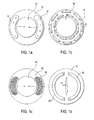
図1bのような別の実施形態では、複数の磁石22が、食道の壁12中に半径方向パターンで位置決めされ得る。磁石22は、それらが一緒に引っ張り、それ故、また食道中の緊張および圧力が増加するように、半径方向形態で互いに誘引し得る。図1b中のような複数の磁石22は、材料の分離した片として食道壁12内に配置され得、例えば、それらは、壁12の筋肉層の間に配置され得る。その他の実施形態では、磁石22は、保持リング内に含まれ得、次いで、ほぼ半径方向形態に整列されて維持される。このようなリングは、ポリマーメッシュのような任意の適切な材料から作製され得る。
In another embodiment, such as in FIG. 1b, a plurality of
ここで、図1cを参照して、なお別の実施形態は、LESを増強するために磁性また磁化可能な粒子26を用いる。複数の磁石22取り込む実施形態のように、磁性粒子26が用いられるとき、それらは、保持リング、サック、またはその他のデバイス中に含まれ得るか、または食道の壁12中に別個に注入されるか、または移植され得る。いくかの実施形態では、例えば、磁性粒子26は、食道壁12の2つの筋肉層の間に形成される領域中に注入され得る。このような領域は、これら磁性粒子26をその場に保持するためのポケットを形成し得る。磁性粒子26は、種々の実施形態で、食道壁12中の2つ以上の位置に配置され得る。例えば、図1cでは、磁性粒子26は、壁12中の2つのほぼ対向する位置に示され、そして食道管腔10を横切って互いに誘引するように反対の極性である。その他の実施形態では、磁性粒子26は、3、4、5またはそれ以上のポケット中に注入または配置され得、そして複数の磁石22とともに図1b中に記載されるように、半径方向誘引パターンを引き起こす磁力形態を有し得る。
Referring now to FIG. 1c, yet another embodiment uses magnetic or
図1cに示されるような粒子を採用するとき、食道の各側面上の磁場が、対向して反対の極性で配置されるように粒子配向を適正に整列することは困難であるかも知れない。それ故、通常、磁化の前に、まず粒子を注入またはそうでなければ導入することが好ましい。通常、これら粒子は、好ましくは熱分解カーボンである、上記のような生体適合性コーティングで被覆される鉄金属から構成されている。粒子が、食道の少なくとも1つの側面に注入された後、所望の磁化を提供するために、通常、内部から、または外部に位置決めされた適切な場強度をもつ電磁石である磁場に曝される。各々の個々の粒子が磁化されるようになると同時に、粒子は一緒に、対向する端部に極をもつ単一の大きな磁石として集合的に作用することが認識される。通常、第2のセットの磁性粒子が、次に、食道のもう一方の側面に導入される。第2の群の粒子における磁場を配向することの困難性のため、第2の群は必要に応じて磁化されないままにする。この第2の粒子が、これもまた、通常、熱分解カーボンで被覆された鉄金属から構成されて磁性(反対に磁化される)である限り、それらは、第1の群の粒子によって奏される磁場により誘引される。 When employing particles as shown in FIG. 1c, it may be difficult to properly align the particle orientation so that the magnetic fields on each side of the esophagus are placed opposite and of opposite polarity. Therefore, it is usually preferable to inject or otherwise introduce the particles first before magnetization. Usually these particles are composed of iron metal coated with a biocompatible coating as described above, preferably pyrolytic carbon. After the particles are injected into at least one side of the esophagus, they are exposed to a magnetic field, usually an electromagnet with an appropriate field strength positioned from the inside or outside to provide the desired magnetization. It will be appreciated that as each individual particle becomes magnetized, the particles together act collectively as a single large magnet with poles at opposite ends. Usually, a second set of magnetic particles is then introduced to the other side of the esophagus. Due to the difficulty of orienting the magnetic field in the second group of particles, the second group is left unmagnetized as needed. As long as this second particle is also composed of ferrous metal coated with pyrolytic carbon and is magnetic (oppositely magnetized), they are played by the first group of particles. Attracted by the magnetic field.
インサイチュ(In situ)磁化はまた、試験後、粒子(またはより大きなバーまたはなおさら言えばその他の磁石)中に誘導される磁場の調節を可能にする。例えば、増強された括約筋は、還流を引き起こすために圧力に曝される。還流が受容不能な低い圧力で起こる場合、磁性粒子は、第2の群の粒子が磁化されていない場合、それらを隔離することを考慮して、さらに磁化され得る。あるいは、患者は、最初の処置が十分に問題を解決したかどうかをみるために経過観察され得、そうでなければ、粒子は、さらなる増強を提供するためにさらに磁化され得る。 In situ magnetization also allows adjustment of the magnetic field induced in the particles (or larger bars or even other magnets) after testing. For example, the enhanced sphincter is exposed to pressure to cause reflux. If the reflux occurs at unacceptably low pressure, the magnetic particles can be further magnetized in view of isolating them if the second group of particles are not magnetized. Alternatively, the patient can be followed to see if the initial treatment has sufficiently solved the problem, otherwise the particles can be further magnetized to provide further enhancement.
ここで、図1dを参照して、C形状の磁石30および32は、食道中のLESまたはその他の対向する側面に移植され得る。この磁石30および32の特有のC形状は、それらが、還流を防ぐために食道管腔10に対して連続的な閉鎖力を奏するが、嚥下が必要なとき、管腔がなお開放し得るようそれらの間に開放領域を常に残すように選択されている。さらに、食道壁の組織を損傷するリスクは低減される。その他の実施形態では、管腔は、組織が破壊されないように有意に低減された力で閉鎖する。
Referring now to FIG. 1d, C-shaped
ここで、図2を参照して、磁性デバイス16、18、22、26は、任意の適切な手段によって食道壁内に配置され得る。例えば、いくつかの実施形態では、デバイスは、腹腔鏡検査またはロボット手術のような最小侵襲技法により配置され得る。その他の実施形態では、デバイスは、患者の口を通じて患者の胃中に挿入される内視鏡により配置され得る。磁性デバイスの実際の挿入はまた、任意の適切な手段により達成され得る。例えば、いくつかの実施形態では、磁性材料が、遠位ニードルおよび注入システムを含むデバイスでのうに、食道壁12中に注入され得る。その他の実施形態では、1つ以上の切開が、食道壁12中に作製されてもよく、そしてこの磁性デバイスは、この切開を通じて挿入され得る。
Referring now to FIG. 2, the
図2は、食道壁12に磁性粒子26を送達するためのデバイスの1つの実施形態を示す。一般に、このデバイスは、近位端(図示せず)および遠位端25をもつカテーテル20、ならびに近位端(図示せず)および遠位端27をもつ複数の送達管24を含む。この送達管24は、カテーテル20の遠位端25から突出し、磁性粒子26を送達する。いくつかの実施形態では、送達管24の遠位端27の各々は、食道の壁12を突き刺すニードルを含む。図2では2つの送達管24が示されているが、送達デバイスのその他の実施形態は、1つの管または2以上の管を含み得る。単一の管の使用は、粒子が2つ以上の位置に逐次的に送達され、粒子の部位の特定の1つのみが磁化されるときに好適であり得る。さらに、管24は、サイズ、長さ、直径および形態の任意の適切な組み合わせを有し得る。1つの実施形態では、例えば、内径または各管24は、粒子26がほぼ単一の綴じ込みラインで送達されるように、各磁性粒子26の外径よりわずかに大きい。このような形態は、食道の壁12内の処置位置への磁性粒子のより制御された送達を可能にし得る。
FIG. 2 shows one embodiment of a device for delivering
磁性デバイスを送達するためのその他のデバイスは、図2に示されるものとは1つ以上の異なる特徴を含み得る。例えば、いくつかの実施形態では、食道壁12を切開するため、および磁性デバイスを移植するための、切開デバイス、および握持かつ放出デバイスをもつ内視鏡を含み得る。その他の実施形態は、食道壁12中に配置された磁化可能材料のための磁化デバイスを含み得る。なお別の実施形態は、1つ以上の磁性デバイスの電力を調節するための調節機構を含み得る。一般に、括約筋中に磁性デバイスを送達するための送達デバイスの任意の適切な組み合わせが、内視鏡デバイス中に取り込まれるかまたはその他で提供されて、本発明の範囲内で企図される。
Other devices for delivering the magnetic device may include one or more different features than those shown in FIG. For example, some embodiments may include an incision device for incising the
1つの実施形態では、IESを適切に増強するための方法は、最初に磁石送達デバイスを患者の口中に導入する工程、そしてこのデバイスを患者の食道に進める工程を含む。このデバイスの遠位端は、次に、磁性デバイスを送達するために食道の管腔内の位置に位置決めされ得る。代表的には、この位置は、LESまたはその近傍であり得る。送達デバイスの送達機構は、次に、LESを増強するための任意の適切な位置にある食道の壁内に、磁性デバイスを注入、移植、またはそうでなければ位置決めするために用いられ得る。いくつかの事例では、圧力検知内視鏡またはカテーテルのような圧力検知デバイスが、次に、
食道中に配置され、LESの位置またはその近傍位置にある食道の管腔内の圧力を測定する。この圧力が所望の圧力に接近している場合、手順は終了し得る。その一方、この圧力に対して調節をすることが所望される場合、磁石またはその他の磁化デバイスが挿入および位置決めされ得、移植されたデバイスの磁力を増加または減少する。上記に記載のようなその他の実施形態では、移植されたデバイスは、食道壁中に配置されるとき磁力をもたなくても良く、そして磁化デバイスよって磁化され得る。いくつかの実施形態では、調節デバイスは、後日、移植されたデバイスの磁力を増加または減少するために使用され得、GERDまたは食道のその他の症状を処置するためにLESを、より適切に増強する。
In one embodiment, a method for appropriately enhancing IES includes first introducing a magnet delivery device into the patient's mouth and advancing the device into the patient's esophagus. The distal end of the device can then be positioned at a location within the lumen of the esophagus to deliver the magnetic device. Typically, this location may be at or near LES. The delivery mechanism of the delivery device can then be used to inject, implant, or otherwise position the magnetic device within the wall of the esophagus in any suitable location for enhancing LES. In some cases, a pressure sensing device, such as a pressure sensing endoscope or catheter, then
The pressure in the lumen of the esophagus is placed in the esophagus and is at or near the LES. If this pressure is close to the desired pressure, the procedure can end. On the other hand, if it is desired to adjust to this pressure, a magnet or other magnetizing device can be inserted and positioned to increase or decrease the magnetic force of the implanted device. In other embodiments as described above, the implanted device may have no magnetic force when placed in the esophageal wall and may be magnetized by a magnetizing device. In some embodiments, the modulating device may be used at a later date to increase or decrease the magnetic force of the implanted device, and more appropriately enhance LES to treat GERD or other symptoms of the esophagus .
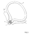
ここで、図3を参照して、女性の尿失禁は、ほぼLESを処置することに関して上記に記載したように、本発明の方法により、および本発明の装置で処置され得る。粒子Pは、前面Fと恥骨Pとの間に配置された尿道Uの対向する側面上に注入され得る。粒子は、食道に関して上記に記載のような様式と類似の様式で尿道中に導入されたカテーテルを用いて、1、2、またはより多くのステージで注入され得る。通常、粒子は、磁化の前に導入され、尿道の1つの側面にある粒子のみが最終的に磁化される。上記に記載の本発明のその他の局面が、女性の尿失禁を阻害するために尿道の処置に等しく良好に適用される。 Referring now to FIG. 3, female urinary incontinence can be treated by the method of the present invention and with the device of the present invention, substantially as described above with respect to treating LES. The particles P can be injected on the opposite side of the urethra U, which is located between the front surface F and the pubic bone P. The particles can be injected at one, two, or more stages using a catheter introduced into the urethra in a manner similar to that described above for the esophagus. Usually, the particles are introduced before magnetization, and only the particles on one side of the urethra are finally magnetized. The other aspects of the invention described above apply equally well to urethral treatment to inhibit female urinary incontinence.
先行する記載は、本発明の完全かつ正確な説明であるが、種々の改変または付加が、添付の請求項に提示されるような本発明の範囲から逸脱することなく種々の実施形態になされ得る。例えば、種々のさらなる材料または材料の組み合わせが磁性デバイスとして用いられ得る、磁石送達デバイスの異なる実施形態が用いられ得る、などである。従って、上記の詳細な説明は、本発明の範囲を制限すると解釈されるべきではない。なぜなら、それは添付の請求項によって規定されるからである。 While the preceding description is a complete and accurate description of the invention, various modifications or additions may be made to the various embodiments without departing from the scope of the invention as presented in the appended claims. . For example, various additional materials or combinations of materials can be used as magnetic devices, different embodiments of magnet delivery devices can be used, and so forth. Therefore, the above detailed description should not be taken as limiting the scope of the invention. Since it is defined by the appended claims.
Claims (6)
該組織の周囲に環状アレイで該患者に移植するように適合された2つより多い複数の磁石であって、該磁石のうちの少なくともいくつかが該移植の前に磁気エネルギーを有している、磁石;および
該磁石のそれぞれが該アレイの円周のそれぞれのセグメントを占め、そして(1)該アレイがある平面に存在し、(2)該アレイの円周に沿って延び、そして(3)各磁石が該アレイの次の隣接する磁石を連続的に磁気的に引きつけるように配向されたその磁軸を有するように、該磁石の全てが該アレイ内に整列され続けるように該組織の周りにおいて該患者に移植するように適合された、保持構造、
を備える、装置。A device for propelling tissue in a patient's body to a closed position, comprising:
More than two magnets adapted to be implanted in the patient in an annular array around the tissue, at least some of the magnets having magnetic energy prior to the implantation Magnets; and
Each of the magnets occupies a respective segment of the circumference of the array, and (1) the array is in a plane, (2) extends along the circumference of the array, and (3) each magnet is The patient around the tissue so that all of the magnets remain aligned within the array so that they have their magnetic axes oriented to continuously magnetically attract the next adjacent magnet of the array Retaining structure , adapted to be implanted in
An apparatus comprising:
該組織の周囲に環状アレイで移植するように適合された2つより多い複数の磁石であって、該磁石のうちの少なくともいくつかが該移植の前に磁気エネルギーを有している、磁石;および
該アレイにおける該磁石の位置を維持するための手段であって、各磁石が、該アレイの円周のそれぞれのセグメントを占め、そして(1)該アレイの円周に沿って延び、(2)該アレイがある平面に存在し、そして(3)各磁石が、該アレイの次の隣接する磁石を連続的に磁気的に引きつけるように配向されたその磁軸を有する、手段、
を備える、装置。A prosthetic device for implantation into a patient's body to drive tissue within the body to a closed position, comprising:
A plurality of more than two magnets adapted to be implanted in an annular array around the tissue, wherein at least some of the magnets have magnetic energy prior to the implantation ; and a means for maintaining the position of the magnet in the array, each magnet occupies a respective segment of a circle periphery of the array, and (1) extending along the circumference of said array, (2 Means ) wherein the array is in a plane and (3) each magnet has its magnetic axis oriented to continuously magnetically attract the next adjacent magnet of the array;
An apparatus comprising:
該組織の周囲に環状アレイで移植するように適合された2つより多い複数の磁石であって、該磁石のうちの少なくともいくつかが該移植の前に磁気エネルギーを有している、磁石;および
該アレイのそれぞれの位置において各磁石を維持する保持構造であって、その磁石の磁軸が、(1)該アレイがある平面に存在し、(2)その位置において該アレイの円周に沿って整列しており、そして(3)各磁石が該アレイにおいて次の隣接する磁石を連続的に磁気的に引きつけるように配向している、保持構造、
を備える、装置。A prosthetic device for implantation into a patient's body to drive tissue within the body to a closed position, comprising:
A plurality of more than two magnets adapted to be implanted in an annular array around the tissue, wherein at least some of the magnets have magnetic energy prior to the implantation; and a holding structure to maintain the magnets at each location of the array, the magnetic axis of the magnet, (1) present in the plane in which the said array, (2) to a circle periphery of the array at that position along and aligned, and (3) each magnet is oriented to attract the next adjacent magnets continuously magnetically in the array, the holding structure,
An apparatus comprising:
Applications Claiming Priority (2)
| Application Number | Priority Date | Filing Date | Title |
|---|---|---|---|
| US39362402P | 2002-07-02 | 2002-07-02 | |
| PCT/US2003/021167 WO2004004544A2 (en) | 2002-07-02 | 2003-07-02 | Methods and devices for luminal and sphincter augmentation |
Publications (3)
| Publication Number | Publication Date |
|---|---|
| JP2005532117A JP2005532117A (en) | 2005-10-27 |
| JP2005532117A5 JP2005532117A5 (en) | 2007-09-13 |
| JP4414884B2 true JP4414884B2 (en) | 2010-02-10 |
Family
ID=30115614
Family Applications (1)
| Application Number | Title | Priority Date | Filing Date |
|---|---|---|---|
| JP2004519943A Expired - Lifetime JP4414884B2 (en) | 2002-07-02 | 2003-07-02 | Methods and devices for lumen and sphincter augmentation |
Country Status (5)
| Country | Link |
|---|---|
| US (3) | US7175589B2 (en) |
| EP (2) | EP1517726A4 (en) |
| JP (1) | JP4414884B2 (en) |
| AU (1) | AU2003281342A1 (en) |
| WO (1) | WO2004004544A2 (en) |
Families Citing this family (106)
| Publication number | Priority date | Publication date | Assignee | Title |
|---|---|---|---|---|
| US6821285B2 (en) | 1999-06-22 | 2004-11-23 | Ndo Surgical, Inc. | Tissue reconfiguration |
| US6663639B1 (en) | 1999-06-22 | 2003-12-16 | Ndo Surgical, Inc. | Methods and devices for tissue reconfiguration |
| US7618426B2 (en) | 2002-12-11 | 2009-11-17 | Usgi Medical, Inc. | Apparatus and methods for forming gastrointestinal tissue approximations |
| US8574243B2 (en) | 1999-06-25 | 2013-11-05 | Usgi Medical, Inc. | Apparatus and methods for forming and securing gastrointestinal tissue folds |
| US7220266B2 (en) | 2000-05-19 | 2007-05-22 | C. R. Bard, Inc. | Tissue capturing and suturing device and method |
| WO2002013854A1 (en) * | 2000-08-11 | 2002-02-21 | Temple University Of The Commonwealth System Of Higher Education | Obesity controlling method |
| US7737109B2 (en) | 2000-08-11 | 2010-06-15 | Temple University Of The Commonwealth System Of Higher Education | Obesity controlling method |
| US20030161824A1 (en) * | 2002-02-27 | 2003-08-28 | Rackley Raymond R. | Bulking agent needle apparatus and method of using the needle apparatus |
| US7695427B2 (en) | 2002-04-26 | 2010-04-13 | Torax Medical, Inc. | Methods and apparatus for treating body tissue sphincters and the like |
| US7445010B2 (en) | 2003-01-29 | 2008-11-04 | Torax Medical, Inc. | Use of magnetic implants to treat issue structures |
| US7497822B1 (en) | 2003-04-10 | 2009-03-03 | Torax Medical, Inc. | Stomach reduction methods and apparatus |
| US7608114B2 (en) | 2002-12-02 | 2009-10-27 | Gi Dynamics, Inc. | Bariatric sleeve |
| US7766973B2 (en) | 2005-01-19 | 2010-08-03 | Gi Dynamics, Inc. | Eversion resistant sleeves |
| WO2004049982A2 (en) | 2002-12-02 | 2004-06-17 | Gi Dynamics, Inc. | Bariatric sleeve |
| US7025791B2 (en) | 2002-12-02 | 2006-04-11 | Gi Dynamics, Inc. | Bariatric sleeve |
| US7942898B2 (en) | 2002-12-11 | 2011-05-17 | Usgi Medical, Inc. | Delivery systems and methods for gastric reduction |
| US7942884B2 (en) | 2002-12-11 | 2011-05-17 | Usgi Medical, Inc. | Methods for reduction of a gastric lumen |
| DE602004019505D1 (en) * | 2003-03-28 | 2009-04-02 | Gi Dynamics Inc | DEVICES AGAINST GRAVITY |
| US8216252B2 (en) | 2004-05-07 | 2012-07-10 | Usgi Medical, Inc. | Tissue manipulation and securement system |
| EP2085049B1 (en) * | 2003-08-11 | 2019-06-19 | Cook Medical Technologies LLC | Surgical implant with penetrating tip |
| US8057420B2 (en) | 2003-12-09 | 2011-11-15 | Gi Dynamics, Inc. | Gastrointestinal implant with drawstring |
| US7815589B2 (en) | 2003-12-09 | 2010-10-19 | Gi Dynamics, Inc. | Methods and apparatus for anchoring within the gastrointestinal tract |
| US7361180B2 (en) | 2004-05-07 | 2008-04-22 | Usgi Medical, Inc. | Apparatus for manipulating and securing tissue |
| US7347863B2 (en) | 2004-05-07 | 2008-03-25 | Usgi Medical, Inc. | Apparatus and methods for manipulating and securing tissue |
| US7004965B2 (en) | 2003-12-17 | 2006-02-28 | Yosef Gross | Implant and delivery tool therefor |
| US7703459B2 (en) | 2004-03-09 | 2010-04-27 | Usgi Medical, Inc. | Apparatus and methods for mapping out endoluminal gastrointestinal surgery |
| US7736374B2 (en) | 2004-05-07 | 2010-06-15 | Usgi Medical, Inc. | Tissue manipulation and securement system |
| US7918869B2 (en) | 2004-05-07 | 2011-04-05 | Usgi Medical, Inc. | Methods and apparatus for performing endoluminal gastroplasty |
| US7931661B2 (en) | 2004-06-14 | 2011-04-26 | Usgi Medical, Inc. | Apparatus and methods for performing transluminal gastrointestinal procedures |
| US7955357B2 (en) | 2004-07-02 | 2011-06-07 | Ellipse Technologies, Inc. | Expandable rod system to treat scoliosis and method of using the same |
| EP1799145B1 (en) | 2004-09-17 | 2016-12-21 | GI Dynamics, Inc. | Gastrointestinal anchor |
| AU2005319399B2 (en) * | 2004-12-21 | 2011-05-12 | Davol Inc. | Anastomotic outlet revision |
| US7771382B2 (en) * | 2005-01-19 | 2010-08-10 | Gi Dynamics, Inc. | Resistive anti-obesity devices |
| ITRE20050009A1 (en) * | 2005-02-10 | 2006-08-11 | Mauro Bortolotti | ANTIREFLUX MEDICAL DEVICE BASED ON THE ACTION OF MAGNETS |
| US7736392B2 (en) * | 2005-04-28 | 2010-06-15 | Medtronic, Inc. | Bulking of upper esophageal sphincter for treatment of obesity |
| US7976488B2 (en) | 2005-06-08 | 2011-07-12 | Gi Dynamics, Inc. | Gastrointestinal anchor compliance |
| ITBO20060071A1 (en) * | 2006-02-08 | 2007-08-09 | Mauro Bortolotti | MAGNETIC DEVICE TO PREVENT GASTROESOPHAGEAL REFLUX AND INCONTINENCE BOTH FECAL AND URINARY |
| US7819836B2 (en) | 2006-06-23 | 2010-10-26 | Gi Dynamics, Inc. | Resistive anti-obesity devices |
| US8870916B2 (en) | 2006-07-07 | 2014-10-28 | USGI Medical, Inc | Low profile tissue anchors, tissue anchor systems, and methods for their delivery and use |
| JP4915024B2 (en) * | 2006-10-06 | 2012-04-11 | 国立大学法人徳島大学 | Swallowing function evaluation device, swallowing function evaluation device operation method, swallowing function evaluation device operation program, computer-readable recording medium, and stored device |
| US7862502B2 (en) | 2006-10-20 | 2011-01-04 | Ellipse Technologies, Inc. | Method and apparatus for adjusting a gastrointestinal restriction device |
| US8246533B2 (en) | 2006-10-20 | 2012-08-21 | Ellipse Technologies, Inc. | Implant system with resonant-driven actuator |
| US8801647B2 (en) | 2007-02-22 | 2014-08-12 | Gi Dynamics, Inc. | Use of a gastrointestinal sleeve to treat bariatric surgery fistulas and leaks |
| US8852216B2 (en) | 2007-03-23 | 2014-10-07 | Ethicon Endo-Surgery, Inc. | Tissue approximation methods |
| US20100130815A1 (en) * | 2007-05-18 | 2010-05-27 | Prostaplant Ltd. | Intraurethral and extraurethral apparatus |
| WO2008150878A1 (en) * | 2007-05-29 | 2008-12-11 | Cvdevices, Llc. | Devices, systems, and methods for deforming a body channel |
| US9855125B2 (en) | 2009-11-30 | 2018-01-02 | Cvdevices, Llc | Devices, systems, and methods for deforming a body channel |
| US9757585B2 (en) * | 2007-06-05 | 2017-09-12 | P Tech, Llc | Magnetic joint implant |
| EP2182885B1 (en) * | 2007-08-27 | 2015-03-04 | Torax Medical, Inc. | Magnetic gastric band or the like |
| US8057472B2 (en) | 2007-10-30 | 2011-11-15 | Ellipse Technologies, Inc. | Skeletal manipulation method |
| US20090248148A1 (en) | 2008-03-25 | 2009-10-01 | Ellipse Technologies, Inc. | Systems and methods for adjusting an annuloplasty ring with an integrated magnetic drive |
| US11202707B2 (en) | 2008-03-25 | 2021-12-21 | Nuvasive Specialized Orthopedics, Inc. | Adjustable implant system |
| US8382756B2 (en) | 2008-11-10 | 2013-02-26 | Ellipse Technologies, Inc. | External adjustment device for distraction device |
| US20100160995A1 (en) * | 2008-12-18 | 2010-06-24 | Jerome Dargent | Method for treating obesity |
| US20100211043A1 (en) * | 2009-02-17 | 2010-08-19 | Yehiel Ziv | Methods for fixing a gastrointestinal device in the gi tract |
| US8197490B2 (en) | 2009-02-23 | 2012-06-12 | Ellipse Technologies, Inc. | Non-invasive adjustable distraction system |
| US9622792B2 (en) | 2009-04-29 | 2017-04-18 | Nuvasive Specialized Orthopedics, Inc. | Interspinous process device and method |
| US8591396B2 (en) * | 2009-05-04 | 2013-11-26 | Covidien Lp | Magnetic gastric reduction device |
| US20110098731A1 (en) * | 2009-10-26 | 2011-04-28 | Eric Whitbrook | Magnetically assisted clasps for prosthetic implants, and related methods |
| US9248043B2 (en) | 2010-06-30 | 2016-02-02 | Ellipse Technologies, Inc. | External adjustment device for distraction device |
| US8734488B2 (en) | 2010-08-09 | 2014-05-27 | Ellipse Technologies, Inc. | Maintenance feature in magnetic implant |
| WO2012112396A2 (en) | 2011-02-14 | 2012-08-23 | Ellipse Technologies, Inc. | Device and method for treating fractured bones |
| US8734475B2 (en) | 2011-08-23 | 2014-05-27 | Torax Medical, Inc. | Medical implant with floating magnets |
| US10743794B2 (en) | 2011-10-04 | 2020-08-18 | Nuvasive Specialized Orthopedics, Inc. | Devices and methods for non-invasive implant length sensing |
| US10016220B2 (en) | 2011-11-01 | 2018-07-10 | Nuvasive Specialized Orthopedics, Inc. | Adjustable magnetic devices and methods of using same |
| CA2785105C (en) | 2012-08-09 | 2014-04-22 | Lotek Wireless Inc. | Self-adjusting magnetic link |
| RU2017126066A (en) | 2012-10-29 | 2019-01-31 | Нувэйсив Спешилайзд Ортопэдикс, Инк. | ADJUSTABLE DEVICES FOR TREATMENT OF KNEE ARTHRITIS |
| US10751094B2 (en) | 2013-10-10 | 2020-08-25 | Nuvasive Specialized Orthopedics, Inc. | Adjustable spinal implant |
| WO2015168175A1 (en) | 2014-04-28 | 2015-11-05 | Ellipse Technologies, Inc. | System for informational magnetic feedback in adjustable implants |
| WO2015195258A1 (en) * | 2014-06-20 | 2015-12-23 | Ethicon Endo-Surgery, Inc. | Artificial esophageal sphincter |
| AU2015371247B2 (en) | 2014-12-26 | 2020-06-04 | Nuvasive Specialized Orthopedics, Inc. | Systems and methods for distraction |
| WO2016134326A2 (en) | 2015-02-19 | 2016-08-25 | Nuvasive, Inc. | Systems and methods for vertebral adjustment |
| WO2016185479A1 (en) * | 2015-05-19 | 2016-11-24 | Innoventions Ltd. | Tissue regeneration system for sphincter dysfunction |
| BR112018002939A2 (en) * | 2015-08-12 | 2019-03-19 | Innoventions Ltd | system for treating a weak or leaking sphincter and system for inducing muscle tissue formation in an organ of the body |
| KR20180067632A (en) | 2015-10-16 | 2018-06-20 | 누베이시브 스페셜라이즈드 오소페딕스, 인크. | An adjustable device for treating arthritis of the knee |
| EP3386405B1 (en) | 2015-12-10 | 2023-11-01 | NuVasive Specialized Orthopedics, Inc. | External adjustment device for distraction device |
| US20170181807A1 (en) * | 2015-12-28 | 2017-06-29 | Boston Scientific Scimed, Inc. | Injectable magnetic microbeads for en bloc tissue resection |
| JP6888015B2 (en) | 2016-01-28 | 2021-06-16 | ニューベイシブ スペシャライズド オーソペディックス,インコーポレイテッド | System for bone movement |
| US10603199B2 (en) | 2017-05-15 | 2020-03-31 | Covidien Lp | Sphincter assist device and method of use |
| US10893369B2 (en) * | 2017-06-02 | 2021-01-12 | Cochlear Limited | Controlled fitting of an implantable medical device |
| US10716570B2 (en) | 2017-07-31 | 2020-07-21 | Ethicon Llc | Magnetic restraint mechanism for a sphincter assist device |
| US10716658B2 (en) | 2017-07-31 | 2020-07-21 | Ethicon Llc | Absorbable polymer for a magnetic sphincter assist device |
| US10729533B2 (en) | 2017-07-31 | 2020-08-04 | Ethicon Llc | Absorbable polymer with drug elution for a magnet sphincter assist device |
| US10722340B2 (en) | 2017-07-31 | 2020-07-28 | Ethicon Llc | Magnetic sphincter replacement device with internal seals |
| US10405865B2 (en) | 2017-07-31 | 2019-09-10 | Ethicon Llc | Method for assisting a sphincter |
| USD857195S1 (en) | 2018-03-01 | 2019-08-20 | Torax Medical, Inc. | Laparoscopic sizing instrument |
| US10828064B2 (en) | 2018-03-01 | 2020-11-10 | Torax Medical, Inc. | Laparoscopic sizing instrument |
| USD856513S1 (en) | 2018-03-01 | 2019-08-13 | Torax Medical, Inc. | Laparoscopic sizing instrument |
| US10813737B2 (en) | 2018-03-07 | 2020-10-27 | Torax Medical, Inc. | MRI compatible magnetic sphincter augmentation device |
| US10945738B2 (en) | 2018-03-07 | 2021-03-16 | Torax Medical, Inc. | Tunable magnetic sphincter augmentation device |
| US11160677B2 (en) | 2018-09-05 | 2021-11-02 | Ethicon, Inc. | Apparatus and method for gastric volume reduction utilizing an expandable member |
| US11324512B2 (en) | 2018-10-26 | 2022-05-10 | Torax Medical, Inc. | Magnetic sphincter augmentation device for urinary incontinence |
| US11051931B2 (en) | 2018-10-31 | 2021-07-06 | Cilag Gmbh International | Active sphincter implant to re-route flow through gastrointestinal tract |
| US11376146B2 (en) | 2018-12-17 | 2022-07-05 | Cilag Gmbh International | Tissue interface features for implantable sphincter assistance device |
| US10842496B2 (en) | 2018-12-17 | 2020-11-24 | Ethicon Llc | Implantable sphincter assistance device with tuned magnetic features |
| US11478347B2 (en) | 2018-12-17 | 2022-10-25 | Cilag Gmbh International | Sphincter sizing instrument |
| US11071619B2 (en) | 2018-12-17 | 2021-07-27 | Cilag Gmbh International | Coupling assembly for implantable sphincter assistance device |
| US11298136B2 (en) | 2018-12-19 | 2022-04-12 | Cilag Gmbh International | Implantable sphincter assistance device with deformable elements |
| US11399928B2 (en) | 2018-12-19 | 2022-08-02 | Cilag Gmbh International | Linking elements for implantable sphincter assistance device |
| US11219462B2 (en) | 2019-03-25 | 2022-01-11 | Laminar, Inc. | Devices, systems, and methods for treating the left atrial appendage |
| WO2021005471A1 (en) | 2019-07-09 | 2021-01-14 | Ethicon Llc | Esophagus sizing instrument |
| USD921895S1 (en) | 2019-07-09 | 2021-06-08 | Cilag Gmbh International | Esophagus sizing instrument |
| US11723775B2 (en) * | 2020-08-12 | 2023-08-15 | Asheesh BEDI | Magnetic medical implants |
| US11957352B2 (en) | 2021-12-16 | 2024-04-16 | Cilag Gmbh International | Implantable sphincter assistance device with controlled homogeneous dilation of the restricting elements |
| US11957349B2 (en) | 2021-12-16 | 2024-04-16 | Cilag Gmbh International | Implantable sphincter assistance device with interactive adjacent field intensity and angular orientation based on interconnection link length |
| US11957353B2 (en) | 2021-12-16 | 2024-04-16 | Cilag Gmbh International | Implantable sphincter assistance device with redirected or focused magnetic fields for interaction between adjacent beads |
Family Cites Families (24)
| Publication number | Priority date | Publication date | Assignee | Title |
|---|---|---|---|---|
| US3731670A (en) * | 1971-05-03 | 1973-05-08 | David Roy Pressman | Corporeal fluid control using bistable magnetic duct valve |
| US4005699A (en) * | 1974-10-09 | 1977-02-01 | Louis Bucalo | Methods and apparatus for use in magnetic treatment of the body |
| US4024855A (en) * | 1974-12-30 | 1977-05-24 | Louis Bucalo | Magnetic filamentary structure and method for using the same |
| DE3011742A1 (en) * | 1980-03-26 | 1981-10-01 | Siemens AG, 1000 Berlin und 8000 München | Magnetic closure system for natural or artificial anus - has magnetic cover held in place by ring of joined individual magnets embedded in tissue of patient |
| JPS588323U (en) | 1981-07-10 | 1983-01-19 | シチズン時計株式会社 | ball chain |
| DE3139811C2 (en) * | 1981-10-07 | 1985-09-19 | Andreas Dr. 7900 Ulm Grüneberger | Implantable device for occluding the female urethra |
| US4643169A (en) * | 1983-11-02 | 1987-02-17 | Walter Koss | Device for selectively opening and closing tubular organs of the body |
| US4994019A (en) | 1989-07-28 | 1991-02-19 | Micro-Magnetics, Inc. | Magnetic occluding device |
| US5176618A (en) * | 1989-08-10 | 1993-01-05 | George Freedman | System for preventing closure of passageways |
| US4978323A (en) | 1989-08-10 | 1990-12-18 | George Freedman | System and method for preventing closure of passageways |
| US5451406A (en) * | 1994-07-14 | 1995-09-19 | Advanced Uroscience, Inc. | Tissue injectable composition and method of use |
| US5509888A (en) * | 1994-07-26 | 1996-04-23 | Conceptek Corporation | Controller valve device and method |
| US5921244A (en) * | 1997-06-11 | 1999-07-13 | Light Sciences Limited Partnership | Internal magnetic device to enhance drug therapy |
| US7468060B2 (en) * | 1998-02-19 | 2008-12-23 | Respiratory Diagnostic, Inc. | Systems and methods for treating obesity and other gastrointestinal conditions |
| US6024704A (en) * | 1998-04-30 | 2000-02-15 | Medtronic, Inc | Implantable medical device for sensing absolute blood pressure and barometric pressure |
| US6251064B1 (en) * | 1998-12-11 | 2001-06-26 | Enteric Medical Technologies, Inc. | Method for creating valve-like mechanism in natural body passageway |
| WO2001026588A2 (en) * | 1999-10-13 | 2001-04-19 | Yeung Jeffrey E | Methods and devices for treating urinary incontinence or obstruction |
| US6348033B1 (en) * | 1999-11-22 | 2002-02-19 | James A. Catlett | Magnetic therapeutic penile band device |
| US6470892B1 (en) * | 2000-02-10 | 2002-10-29 | Obtech Medical Ag | Mechanical heartburn and reflux treatment |
| US6511508B1 (en) * | 2000-08-04 | 2003-01-28 | Environmental Robots, Inc. | Surgical correction of human eye refractive errors by active composite artificial muscle implants |
| WO2002011696A2 (en) * | 2000-08-08 | 2002-02-14 | Ev & M | Active tissue augmentation materials and method |
| US6730014B2 (en) | 2001-01-05 | 2004-05-04 | Peter J. Wilk | Medical treatment method and device utilizing magnetic particles |
| US20040173222A1 (en) * | 2001-04-24 | 2004-09-09 | Kim Young D. | Magnetic pellets and external electromagnetic device for assisting left ventricular contraction, method of pellet insertion, and method of ventricular electromagnetic assistance |
| US7445010B2 (en) * | 2003-01-29 | 2008-11-04 | Torax Medical, Inc. | Use of magnetic implants to treat issue structures |
-
2003
- 2003-07-01 US US10/612,325 patent/US7175589B2/en active Active
- 2003-07-02 EP EP03742434A patent/EP1517726A4/en not_active Withdrawn
- 2003-07-02 WO PCT/US2003/021167 patent/WO2004004544A2/en active Application Filing
- 2003-07-02 AU AU2003281342A patent/AU2003281342A1/en not_active Abandoned
- 2003-07-02 JP JP2004519943A patent/JP4414884B2/en not_active Expired - Lifetime
- 2003-07-02 EP EP07118531A patent/EP1892012B8/en not_active Expired - Lifetime
-
2006
- 2006-11-16 US US11/560,776 patent/US8070670B2/en not_active Expired - Lifetime
-
2007
- 2007-06-21 US US11/821,480 patent/US8070671B2/en not_active Expired - Lifetime
Also Published As
| Publication number | Publication date |
|---|---|
| WO2004004544A3 (en) | 2004-06-03 |
| US7175589B2 (en) | 2007-02-13 |
| JP2005532117A (en) | 2005-10-27 |
| US20070129597A1 (en) | 2007-06-07 |
| US20040122470A1 (en) | 2004-06-24 |
| EP1892012B8 (en) | 2012-10-10 |
| AU2003281342A1 (en) | 2004-01-23 |
| US8070671B2 (en) | 2011-12-06 |
| WO2004004544A2 (en) | 2004-01-15 |
| EP1517726A4 (en) | 2007-04-11 |
| US20070249892A1 (en) | 2007-10-25 |
| AU2003281342A8 (en) | 2004-01-23 |
| US8070670B2 (en) | 2011-12-06 |
| EP1517726A2 (en) | 2005-03-30 |
| EP1892012A1 (en) | 2008-02-27 |
| EP1892012B1 (en) | 2012-09-05 |
Similar Documents
| Publication | Publication Date | Title |
|---|---|---|
| JP4414884B2 (en) | Methods and devices for lumen and sphincter augmentation | |
| Cope | Creation of compression gastroenterostomy by means of the oral, percutaneous, or surgical introduction of magnets: feasibility study in swine | |
| US20200246009A1 (en) | Incisionless Gastric Bypass System | |
| JP5191489B2 (en) | Device and method for changing feeding behavior | |
| US8556919B2 (en) | Delivery system for magnetic anastomosis device | |
| Espinet-Coll et al. | Current endoscopic techniques in the treatment of obesity | |
| US20170119394A1 (en) | Methods and apparatus for magnet-induced compression anastomosis between adjacent organs | |
| US20070185371A1 (en) | Magnetic device and method to prevent gastroesophageal reflux, fecal incontinence and urinary incontinence | |
| US20050197715A1 (en) | Methods and apparatus for implanting devices into non-sterile body lumens or organs | |
| US20230157856A1 (en) | Magnetic Devices for Digestive Tract Partitioning | |
| Cope et al. | Stent placement of gastroenteric anastomoses formed by magnetic compression | |
| Bortolotti | Magnetic challenge against gastroesophageal reflux | |
| CN214908718U (en) | Closed function enhancement magnetic bead subassembly of annular tissue under scope | |
| CN112690938A (en) | Closed function enhancement magnetic bead subassembly of annular tissue under scope | |
| CN110090348A (en) | A kind of magnetic combination therapy device | |
| IL197198A (en) | Devices for altering eating behavior |
Legal Events
| Date | Code | Title | Description |
|---|---|---|---|
| A521 | Request for written amendment filed |
Free format text: JAPANESE INTERMEDIATE CODE: A523 Effective date: 20060628 |
|
| A621 | Written request for application examination |
Free format text: JAPANESE INTERMEDIATE CODE: A621 Effective date: 20060628 |
|
| A521 | Request for written amendment filed |
Free format text: JAPANESE INTERMEDIATE CODE: A523 Effective date: 20070718 |
|
| A711 | Notification of change in applicant |
Free format text: JAPANESE INTERMEDIATE CODE: A711 Effective date: 20071115 |
|
| A521 | Request for written amendment filed |
Free format text: JAPANESE INTERMEDIATE CODE: A821 Effective date: 20071115 |
|
| A131 | Notification of reasons for refusal |
Free format text: JAPANESE INTERMEDIATE CODE: A131 Effective date: 20090702 |
|
| A521 | Request for written amendment filed |
Free format text: JAPANESE INTERMEDIATE CODE: A523 Effective date: 20091001 |
|
| TRDD | Decision of grant or rejection written | ||
| A01 | Written decision to grant a patent or to grant a registration (utility model) |
Free format text: JAPANESE INTERMEDIATE CODE: A01 Effective date: 20091026 |
|
| A01 | Written decision to grant a patent or to grant a registration (utility model) |
Free format text: JAPANESE INTERMEDIATE CODE: A01 |
|
| A61 | First payment of annual fees (during grant procedure) |
Free format text: JAPANESE INTERMEDIATE CODE: A61 Effective date: 20091120 |
|
| FPAY | Renewal fee payment (event date is renewal date of database) |
Free format text: PAYMENT UNTIL: 20121127 Year of fee payment: 3 |
|
| R150 | Certificate of patent or registration of utility model |
Ref document number: 4414884 Country of ref document: JP Free format text: JAPANESE INTERMEDIATE CODE: R150 Free format text: JAPANESE INTERMEDIATE CODE: R150 |
|
| FPAY | Renewal fee payment (event date is renewal date of database) |
Free format text: PAYMENT UNTIL: 20131127 Year of fee payment: 4 |
|
| R250 | Receipt of annual fees |
Free format text: JAPANESE INTERMEDIATE CODE: R250 |
|
| R250 | Receipt of annual fees |
Free format text: JAPANESE INTERMEDIATE CODE: R250 |
|
| R250 | Receipt of annual fees |
Free format text: JAPANESE INTERMEDIATE CODE: R250 |
|
| R250 | Receipt of annual fees |
Free format text: JAPANESE INTERMEDIATE CODE: R250 |
|
| R250 | Receipt of annual fees |
Free format text: JAPANESE INTERMEDIATE CODE: R250 |
|
| RD02 | Notification of acceptance of power of attorney |
Free format text: JAPANESE INTERMEDIATE CODE: R3D02 |
|
| RD04 | Notification of resignation of power of attorney |
Free format text: JAPANESE INTERMEDIATE CODE: R3D04 |
|
| R250 | Receipt of annual fees |
Free format text: JAPANESE INTERMEDIATE CODE: R250 |
|
| R250 | Receipt of annual fees |
Free format text: JAPANESE INTERMEDIATE CODE: R250 |
|
| R250 | Receipt of annual fees |
Free format text: JAPANESE INTERMEDIATE CODE: R250 |
|
| R250 | Receipt of annual fees |
Free format text: JAPANESE INTERMEDIATE CODE: R250 |
|
| R250 | Receipt of annual fees |
Free format text: JAPANESE INTERMEDIATE CODE: R250 |
|
| R250 | Receipt of annual fees |
Free format text: JAPANESE INTERMEDIATE CODE: R250 |
|
| EXPY | Cancellation because of completion of term |