JP4335526B2 - Detection of binding species by colloidal and non-colloidal structures - Google Patents
Detection of binding species by colloidal and non-colloidal structures Download PDFInfo
- Publication number
- JP4335526B2 JP4335526B2 JP2002539812A JP2002539812A JP4335526B2 JP 4335526 B2 JP4335526 B2 JP 4335526B2 JP 2002539812 A JP2002539812 A JP 2002539812A JP 2002539812 A JP2002539812 A JP 2002539812A JP 4335526 B2 JP4335526 B2 JP 4335526B2
- Authority
- JP
- Japan
- Prior art keywords
- protein
- binding
- species
- interaction
- colloid
- Prior art date
- Legal status (The legal status is an assumption and is not a legal conclusion. Google has not performed a legal analysis and makes no representation as to the accuracy of the status listed.)
- Expired - Lifetime
Links
Images


Classifications
-
- B—PERFORMING OPERATIONS; TRANSPORTING
- B82—NANOTECHNOLOGY
- B82Y—SPECIFIC USES OR APPLICATIONS OF NANOSTRUCTURES; MEASUREMENT OR ANALYSIS OF NANOSTRUCTURES; MANUFACTURE OR TREATMENT OF NANOSTRUCTURES
- B82Y30/00—Nanotechnology for materials or surface science, e.g. nanocomposites
-
- G—PHYSICS
- G01—MEASURING; TESTING
- G01N—INVESTIGATING OR ANALYSING MATERIALS BY DETERMINING THEIR CHEMICAL OR PHYSICAL PROPERTIES
- G01N33/00—Investigating or analysing materials by specific methods not covered by groups G01N1/00 - G01N31/00
- G01N33/48—Biological material, e.g. blood, urine; Haemocytometers
- G01N33/50—Chemical analysis of biological material, e.g. blood, urine; Testing involving biospecific ligand binding methods; Immunological testing
- G01N33/53—Immunoassay; Biospecific binding assay; Materials therefor
- G01N33/543—Immunoassay; Biospecific binding assay; Materials therefor with an insoluble carrier for immobilising immunochemicals
- G01N33/54313—Immunoassay; Biospecific binding assay; Materials therefor with an insoluble carrier for immobilising immunochemicals the carrier being characterised by its particulate form
-
- G—PHYSICS
- G01—MEASURING; TESTING
- G01N—INVESTIGATING OR ANALYSING MATERIALS BY DETERMINING THEIR CHEMICAL OR PHYSICAL PROPERTIES
- G01N33/00—Investigating or analysing materials by specific methods not covered by groups G01N1/00 - G01N31/00
- G01N33/48—Biological material, e.g. blood, urine; Haemocytometers
- G01N33/50—Chemical analysis of biological material, e.g. blood, urine; Testing involving biospecific ligand binding methods; Immunological testing
- G01N33/58—Chemical analysis of biological material, e.g. blood, urine; Testing involving biospecific ligand binding methods; Immunological testing involving labelled substances
- G01N33/585—Chemical analysis of biological material, e.g. blood, urine; Testing involving biospecific ligand binding methods; Immunological testing involving labelled substances with a particulate label, e.g. coloured latex
- G01N33/586—Liposomes, microcapsules or cells
Abstract
Description
【0001】
発明の分野
本発明は、2つの化学的および/または生物学的スピーシーズの相互作用を迅速かつ鋭敏に検出するための方法、検定、およびキットの全般に関する。本発明により、薬剤スクリーニングおよびシグナル伝達経路マッピングを含む技術が容易になる。
【0002】
発明の背景
ヒトゲノムの最近の解明により、ヒトタンパク質のレパートリをコードする、別個のDNA配列の形で膨大な量の情報がもたらされた。分子生物学に精通する人達に認知されているように、生物学的機能に関与する圧倒的に多い分子は、DNAやRNAではなく、タンパク質である。機能ゲノミクスにおいては、さまざまなレベルの特定mRNAsが疾患状態に関連づけられる。プロテオミクスは、前駆体分子であるDNAまたはmRNAよりもタンパク質の機能を研究するため、機能ゲノミクスの後任者である。プロテオミクスの1つの側面は、どのようにタンパク機能が疾患に関連するかを決定することである。タンパク質の疾病関連ファミリー、または普通のシグナル伝達経路に関与するタンパク質は、タンパク質・タンパク質相互作用ネットワークを解明することにより、同定可能である。今日、生物学研究の主要な焦点は、生体シグナル伝達経路を作り上げている当該タンパク質相互作用ネットワークの解明することである。これらのタンパク質相互作用ネットワークを理解することによって、健康状態から疾患状態への転換を何が誘発するかについての決定的な手掛りが収集されている。
【0003】
プロテオミクスの研究に対する主要な障害は、片方または両方がまだ特徴解明されていないか、純粋でない場合、タンパク質・タンパク質相互作用を検出するために利用可能な方法がないことである。現行の検出方法の大部分は、当該推定結合パートナーの片方または両方を認識する特異的抗体を使用することが必要になる。これは、対象となる当該タンパク質を、それに対する抗体が作られるように、精製する必要があることを意味する。あるタンパク質の特徴解明がなされている場合、そのタンパク質は一般にアフィニティータグにより標識可能であり、それにより検出プロセスが助けられることが多い。これは、ある粗試料に対象となるタンパク質が含まれるかどうかを知る前に、現行の検定法で使用できるように、成分を分離そして精製するために沢山の作業が必要となることを意味する。タンパク質・タンパク質相互作用の研究における現行の方法のもう1つの主な欠点は、それらが順次的な労働集約型プロセスであることである。このことは、大きなタンパク質相互作用ネットワークを解明すること、またはいくつかの推定結合パートナーを並行して試験すること、のような複雑な問題を検討するために、それらを多重化できないことを意味する。約40Kと現在推定されているヒトゲノム中の遺伝子の数について、順次型ペアワイズ(2つ一組式)テストにより相互作用ネットワークを測定することは、約8X108回の実験を含むことになる。ヒトゲノムプロジェクトは完了間近であり、多くの遺伝子産物の機能的分析を可能にする適切な技術の開発が絶対に必要である。こういった理由から、一方または両方のタンパク質が粗混合物として存在するかもしれないタンパク質結合相互作用の並行分析を容易にする方法がもし利用できるならば、有益なはずである。例えば、cDNAライブラリーから生成される可能性がある、新たに発見された特徴未解明のタンパク質を迅速に特徴解明するための方法がもし利用できるならば、それも有益なはずである。例えば、非常に多くの既知および未知のスピーシーズが新薬剤の同定のためにスクリーニングされる、製薬産業を含むさまざまな産業において、これらの方法は特に重要であろう。同定に加えて、他の既知および未知のスピーシーズに対するそれらの相対的親和性について、スピーシーズを分類することも有用であろう。典型的には、スクリーニングするスピーシーズの総数を増やすためには、スクリーニング手順を行う速度を高めることが通常望ましい。さらに、検出方法を単純化でき、同時に多重検定を実施できる場合に、試料処理能力が改善される。
【0004】
発明の要約
本発明は、特徴未解明のタンパク質の特徴を解明するため、および一方または両方が混合物中に存在する結合パートナー間の相互作用を検出するために有用な技術を含み、生物学的および/または化学的スピーシーズ間の相互作用を検出するための各種の新規な方法、組成物、スピーシーズ、および物品を提供する。1つの観点によると、本発明は、タンパク質結合相互作用を検出するための処理能力の高い方法を提供する。ある実施態様において、1つの潜在的結合パートナーが特徴未解明および/または粗混合物中に存在する状況において、本発明が適用される。また、本発明は、各種の機能性タンパク質モジュールに結合する能力を検出することによって、特徴未解明のタンパク質の特徴を迅速に解明する方法を提供する。1つの観点からは、第1固定化成分をもつ第1表面および第2固定化成分をもつ第2表面の両方を、固定化スピーシーズをもつコロイド粒子に曝し、そして第1、第2または両表面のいずれかの上の当該コロイド粒子の固定化を決定する。
【0005】
本発明のもう1つの観点からは、第1スピーシーズは第1コロイド上に固定化され、そして第2スピーシーズは第2コロイド上に固定化される。第1および第2コロイドを、ある表面に曝し、そしてそれから当該表面上への第1または第2コロイドの固定化を決定する。当該表面は、前記第1および第2スピーシーズの一方または両方の推定結合パートナーを提示することも可能である。
【0006】
本発明のもう1つの観点から、固定相を含むクロマトグラフィー配置を用いて、ある混合物から少なくとも2つの成分をクロマトグラフィー的に分離する方法が提供される。次いで、当該クロマトグラフィー配置で使われたのと同じまたは違うタイプの固定相を、当該混合物の第1および第2分離成分が接着または付着する表面として使用する。次いで、当該付着成分を含むこの固定相を、当該第1および第2成分の少なくとも1つに結合することができると思われる固定化スピーシーズをもつコロイド粒子に曝す。すなわち、当該固定化スピーシーズは当該混合物中に存在するスピーシーズと相互作用するかも知れないと疑われるものである。当該コロイドに付随する固有のまたは補助的なシグナル伝達能力を検出することにより、コロイド上に固定化されたスピーシーズと、ビーズまたは他の固定相表面上に固定化されたスピーシーズとの結合を、決定することができる。
【0007】
本発明のもう1つの観点からは、ある混合物から成分をクロマトグラフィー的に分離し、次いで、当該画分中に存在する成分をビーズ以外の表面に接着または付着させ、さらに、当該混合物中に存在する成分の結合パートナーであると疑われるまたは同定された治療標的である固定化スピーシーズをもつコロイドに曝すための方法が提供される。次いで、当該コロイド上のスピーシーズと当該表面上のスピーシーズとの結合が決定される。分離された成分を付着させることができる他の表面としては、コロイドの第2集団、ナノ粒子、ポリマー、マルチウェルプレート、バイオチップ、空間的にアドレス可能なバイオチップ、電極、および電極アレイが含まれるが、これに限定されるものではない。
【0008】
分画された成分を、平坦な基質上へ空間的にアドレス可能な仕方で別々に沈着させ、それから、前記粗混合物中に存在する成分の1つと相互作用すると疑われる単一スピーシーズを示すコロイドとインキュベートしてもよい。
【0009】
本発明のもう1つの観点からは、別個の生物学的または化学的スピーシーズを空間的にアドレス可能な仕方で示すチップを含むキットが提供される。次いで、対象となる特徴未解明のタンパク質を一組のコロイドに付着させる。さらに、対象となるタンパク質を提示する当該コロイドを当該チップとともにインキュベートし、特定のチップ位置へのコロイドの結合を検出する。好ましい態様では、空間的にアドレス可能なチップ上に固定化されたスピーシーズは、タンパク質相互作用モジュールおよびモチーフである。このようにして、タンパク質相互作用モチーフへの結合を検出することによって、特徴未解明のタンパク質の特徴を解明することが可能である。
【0010】
本発明のもう1つの観点からは、結合スピーシーズが特異的または非特異的な仕方で付着しているコロイド粒子を含むパッケージを含むキットが提供される。次いで、これらのコロイド粒子を、ある表面に接着された標的生物学的または化学的スピーシーズとともにインキュベートすることも可能である。または、別個の組のコロイドを、マルチウェルプレート中へ分散されるように、別々のコンパートメントに提供してもよい。コロイドに個別に付着するスピーシーズとしては、抗体、既知薬剤、薬剤候補、標的化タンパク質、タンパク質の断片、タンパク質相互作用モジュール、またはcDNAライブラリーの産物が含まれていてもよい。
【0011】
本発明のもう1つの観点からは、SAMを含むコロイド粒子、ならびに当該コロイド粒子に結合パートナーを固定化するための、説明書を含むパッケージを含むキットが提供される。
【0012】
本発明のもう1つの観点からは、2つのパッケージを有するキットが提供されるが、第1のパッケージには当該粒子について固定化された第1スピーシーズをもつコロイド粒子が含まれ、第2のパッケージには当該粒子について固定化された第2スピーシーズをもつコロイド粒子が含まれる。
【0013】
本発明のもう1つの観点からは、別個の生物学的または化学的スピーシーズを空間的にアドレス可能な仕方で示すチップを含むキットが提供される。当該チップとのインキュベーションの前または後に、対象となる特徴未解明のタンパク質をコロイドに付着させる。次いで、当該チップ上の部位へのタンパク質の別個の結合またはタンパク質結合のパターンを検出する。
【0014】
本発明のもう1つの観点から、ある方法が提供されるが、当該方法には、各々が異なる化学的、生化学的、または生物学的官能基を提示する少なくとも2つの表面領域を試料に曝す工程、当該試料の少なくとも1つの成分と少なくとも2つの当該表面領域の各々との間の特徴的な相互作用を示す、少なくとも2つの当該表面領域との当該試料の相互作用パターンを決定する工程が含まれる。
【0015】
本発明のもう1つの観点から、ある方法が提供されるが、当該方法には、固定相上で混合物の少なくとも2つの成分を分離すること、流体で当該固定相から少なくとも第1成分を溶出すること、当該流体を換えること、ある表面へ第1成分の少なくとも一部を固定化すること、当該表面を推定結合パートナーに曝すこと、および第1成分の少なくとも一部と当該推定結合パートナーとの間の結合相互作用を決定することが含まれる。
【0016】
本発明のもう1つの観点から、ある方法が提供されるが、当該方法には、固定相上で混合物の少なくとも2つの成分を分離すること、流体で当該固定相から少なくとも第1成分を溶出すること、コロイドへ第1成分の少なくとも一部を固定化すること、当該コロイドをある表面上に固定化された推定結合パートナーに曝すこと、および当該第1成分の少なくとも一部と当該推定結合パートナーとの間の結合相互作用を決定することが含まれる。
【0017】
本発明の他の長所、新規な特徴、および目的は、概略的であり安分比例で描くようには意図されていない添付図面と併せて論考すれば、次の本発明の詳細な説明により明らかになるだろう。さまざまな図で図解されている同一のまたはほぼ同一の各成分は、図中では単一の数字で表示される。平明にするために、すべての図において必ずしもすべての成分が標号されているわけではなく、通常の当業者が本発明を理解するために図解が必要でない場合には、本発明の各実施態様におけるすべての成分が必ずしも示されているわけではない。
【0018】
発明の詳細な説明
定義:
「低分子」とは、本明細書で使用される場合、5キロダルトン以下、より典型的には1キロダルトン以下の分子を意味する。本明細書で使用される場合、「低分子」からはタンパク質が除外される。
【0019】
「候補薬剤」という用語は、本明細書で使用される場合、ヒト、動物、または植物に用いられる任意の医療用物質を指す。この定義には、化合物類似体、天然に存在する医薬品、合成医薬品、組換え体の医薬品、ホルモン、抗微生物剤、神経伝達物質などが含まれる。これには、異常な凝集により特徴づけられる神経変性疾患、もしくは他の疾患の治療またはそれらの予防のための薬剤としての使用が評価されるはずの任意の物質または前駆体(天然に存在するか、合成されたものであるか、組換え体であるかは問わない)が含まれる。典型的には、本発明のスクリーニング検定のような、検定における活性によって評価を行う。
【0020】
各種の粒子が本発明において使用可能である。例えば、「流体懸濁可能粒子」とは、本発明の目的のために使用される流体(典型的には水溶液)中において、単独で懸濁状態を保つようにできる粒子、あるいは磁場、電磁場を適用したり、撹拌、震盪、振動、超音波処理、遠心分離、渦動、または類似のもののような撹拌を適用することにより、溶液状態に維持することが可能な粒子、を意味する。「磁気的に懸濁可能」粒子は、磁場を適用することにより流体中に懸濁状態を維持できるものである。電磁気的に懸濁可能粒子とは、電磁場を適用することにより流体中に懸濁状態を維持できるものである(例えば、電荷を有する粒子、または電荷を有するように修飾された粒子)。「自己懸濁可能粒子」とは、例えば磁場の援助無しに、少なくとも1時間、それが使われる流体(典型的には水溶液)中で懸濁状態のままであるほどに、サイズおよび/または質量が十分小さい粒子である。他の自己懸濁可能粒子は、本発明によると、援助無しに、5時間、1日、1週間、または1ヵ月間、懸濁状態のままである。
【0021】
「タンパク質」および「ペプチド」とは、当該技術分野では周知の用語であり、各々に含まれるアミノ酸の数については、当該技術分野では厳密には定義されていない。本明細書で使用する場合、これらの用語には、当該技術分野における通常の意味が与えられる。一般的には、ペプチドとは、長さが約100アミノ酸以下のアミノ酸配列であるが、300アミノ酸までの配列を含んでいてもよい。タンパク質とは、一般には、少なくとも100アミノ酸の分子であると考えられている。
【0022】
本明細書で使用する場合、「金属結合タグ(標識)」は、キレートにより配位している金属に固着され得る一群の分子を指す。このような分子の適当な群としては、複数のヒスチジンおよび複数のシステイン(「ポリアミノ酸タグ」)を非限定的に含むアミノ酸配列が含まれる。金属結合タグには、以下に定義するヒスチジンタグが含まれる。
【0023】
本明細書で使用する場合、「金属を配位するキレート」またはキレートにより配位された金属とは、当該金属上の利用可能な全配位部位が充たされておらず、金属結合タグによる結合のために利用可能ないくつかの配位部位が残されている、キレート剤により配位された金属を指す。
【0024】
本明細書で使用する場合、「金属結合タグ/金属/キレート連結」とは、金属結合タグに対して固定化されている第1スピーシーズと、キレートに対して固定化されている第2スピーシーズとの連結であって、当該キレートが当該金属結合タグが配位している金属に配位する連結であると定義される。参考文献により本明細書に含まれるものとされるBamdadらの米国特許第5620850号には、模範的なな連結が記載されている。
【0025】
「シグナル伝達構成要素」とは、特定の試料中または特定の場所でその存在を示すことができる構成要素を意味する。本発明のシグナル伝達構成要素としては、人間の肉眼により同定できるもの、個々は目に見えないが十分な量あればヒトの肉眼により確認できると思われるもの(例えば、コロイド粒子)、可視的に(すなわち、肉眼あるいは電子顕微鏡または類似物を含む顕微鏡により)または分光法的に、容易に測定できるような量または波長範囲内で電磁放射線を吸収または放出する構成要素、電子的にまたは電気化学的に測定可能な構成要素(「電子シグナル伝達構成要素」、例えば、適切な活性化エネルギーに曝露すると特徴的な酸化/還元パターンを示すレドックス活性分子)、またはその類似物があり得る。例えば、染料、色素、レドックス活性分子のような電気活性分子、蛍光成分(定義によりリン光成分を含む)、上方制御(up−regulating)リン光体、化学発光構成要素、電気化学発光構成要素、または西洋ワサビペルオキシダーゼおよびアルカリホスファターゼを含む酵素結合シグナル伝達成分が含まれる。「シグナル伝達構成要素の前駆体」とは、単独ではシグナル伝達能力を有しないが、もう1つのスピーシーズと化学的、電気化学的、電気的、磁気的、または物理的相互作用することにより、シグナル伝達構成要素になる構成要素である。例えば、もう1つの分子と化学的相互作用するときだけ、特定の検出可能波長内の放射線を放出する能力を有する発色団が含まれる。シグナル伝達構成要素の前駆体は、本明細書で使用される場合、「シグナル伝達構成要素」と区別可能であるが、「シグナル伝達構成要素」の定義内に含まれる。
【0026】
本明細書で使用される場合、あるスピーシーズのもう1つのスピーシーズまたはある物品の表面に関する状況において、「固着される、または固着されるように適応される」とは、当該スピーシーズが、共有付着、特定の生物学的結合(例えば、ビオチン/ストレプトアビジン)を経る付着、キレート/金属結合のような配位結合、または類似のものを経て、化学的にまたは生化学的に連結されることを意味する。例えば、この状況における「固着される」とは、ポリスチレンビーズ上に合成されたペプチドのような結合スピーシーズ、ビーズに共有的に付着するプロテインAのようなタンパク質に結合する抗体に特異的に生物学的に共役する結合スピーシーズ、ある表面に共有的に固着された結合パートナー(例えば、GSTの場合はグルタチオン)に今度は特異的に生物学的に結合されるGSTまたはファージのような分子の一部を(遺伝子工学を経て)形成する結合スピーシーズなどを非限定的に含む、複数の化学的連結、複数の化学的/生物学的連結などが含まれる。もう1つの例として、チオールに共有的に連結された成分は、チオールが金に共有的に結合するので、金表面に固着されるように適応される。同様に、金属結合タグをもつスピーシーズは、その分子も金属を配位するキレートを提示する表面に共有的に付着された分子をもつ(チオール/金結合のような)表面に固着されるように適応される。ある表面が特定のヌクレオチド配列を有し、あるスピーシーズが相補的ヌクレオチド配列を有するならば、当該スピーシーズも当該表面に固着されるように適応される。
【0027】
「共有的に固着される」とは、1つまたは1つ以上の共有結合を経て固着される以外の何者でもない。例えば、金表面に今度は固着されているカルボン酸提示アルキルチオールに、EDC/NHS化学を経て、共有的に共役されているスピーシーズは、その表面に共有的に固着されている。
【0028】
「特異的に固着される(または結合される)」または「特異的に固着される(または結合される)ように適応される」とは、「固着される、または固着されるように適応される」の定義に関して上述したように、あるスピーシーズが、もう1つの試料または表面へ化学的にまたは生化学的に連結されることを意味するが、すべての非特異的結合は除かれる。
【0029】
「非特異的結合」とは、本明細書で使用される場合、生化学の分野における通常の意味が与えられる。
「コロイド」とは、本明細書で使用される場合、例えば、無機または有機質、ポリマー性、セラミック、半導体性、金属性(例えば、金)、非金属性、結晶性、非晶質、半導体性ナノクリスタル、またはそれらの組合せである材料からできているものを含むナノ粒子、すなわち、非常に小さな自己懸濁性または流体懸濁性粒子を意味する。本発明に従って使われるコロイド粒子は、典型的にはどの次元でも250nm以下の断面、より典型的にはどの次元でも100nm以下の断面、そして大部分の場合約2−30nmの断面である。本発明での使用に適しているコロイドの1クラスは、断面が10−30nmであり、そしてもう1つは、断面が約2−10nmである。本明細書で使われる場合、この用語には生化学の分野で普通に用いられる定義が含まれる。
【0030】
本明細書で使用する場合、もう1つの成分に「対して固定化される」成分は、他の成分に固着されるか、または他の成分に間接的に固着されるかのいずれかであり、例えば、他の成分も固着されている第3成分に固着されることによるか、さもなければ他の成分と移行的に会合していることによるものである。例えば、前記シグナル伝達構成要素が前記結合スピーシーズに固着されている場合、前記シグナル伝達構成要素が当該結合スピーシーズが固着されているコロイド粒子に固着されている場合、前記シグナル伝達構成要素が当該結合スピーシーズが固着されているデンドリマーまたはポリマーに固着されている場合などに、当該シグナル伝達構成要素は結合スピーシーズに対して固定化されている。もし第1コロイド粒子の表面に固着されているスピーシーズが、ある構成要素に付着し、そして第2コロイド粒子の表面上のスピーシーズが同じ構成要素に付着し、そこでの当該構成要素が、単一構成要素、複数スピーシーズの複合構成要素、細胞、もう1つの粒子などであってもよいならば、当該コロイド粒子はもう1つのコロイド粒子に対して固定化されている。
【0031】
「試料」という用語は、結合パートナーなどの被検体を含むことが疑われる任意の媒体を指し、望ましくはその存在または量が決定されるものである。試料には、生物学的試料(細胞、細胞ライゼート、組織、血清、血液または生物学的来源からの他の液体など)、生化学試料(cDNAライブラリーからの産物など)、環境試料(土壌抽出物など)、合成材料が含まれている生物学的または非生物学的な任意の他の媒体であってもよく、これらは本発明に従って有利に評価することが可能である。
【0032】
「構造的に予め決定された試料」とは、本明細書で使われる場合、その化学的または生物学的な配列または構造が予め決定されている構造である試料を意味し、神経変性疾患のような特定のプロセスと当該構造が関連するかどうかを試験するように設計された検定において用いられる。例えば、「構造的に予め決定された試料」には、ペプチド配列、ファージディスプレイライブラリー中のランダムペプチド配列、およびその類似物が含まれる。
【0033】
ある特定の成分を「含むと疑われる試料」は、それについては当該成分の含量が不明である試料を意味する。当該試料は、当該特定成分を含むことが不明でもよく、または当該特定成分を含むことがわかっているが、量が不明であってもよい。
【0034】
本明細書で使われる場合、「金属結合タグ(標識)」は、キレートにより配位されている金属に固着されるようになることが可能な一群の分子を指す。そのような分子の適当な群は、典型的には約2〜約10のアミノ酸残基のアミノ酸配列を含む。これらには、複数のヒスチジンおよび複数のシステイン(「ポリアミノ酸タグ」)が含まれるが、これに限定されるものではない。こうした結合タグがヒスチジンを含む場合、この結合タグは、「ポリヒスチジントラクト(管)」、「ヒスチジンタグ」または「HIS−タグ」と呼称されることがあり、そしてペプチド、タンパク質または核酸の、アミノ末端またはカルボキシ末端のいずれか、または任意の露出領域に存在可能である。6ないし10残基のポリヒスチジントラクトが、本発明における使用には好ましい。また、当該ポリヒスチジントラクトは、対象となるタンパク質に添加された多数の連続ヒスチジン残基であるとして機能的に定義され、これにより、金属キレートカラム上での当該生成タンパク質のアフィニティー精製、またはもう1つの分子(例えば、HIS−タグと反応性の抗体)との相互作用によるタンパク質末端の同定が可能になる。
【0035】
「金属を配位できる成分(moiety)」は、本明細書で使われる場合、金属結合タグまたはキレートのような、金属原子上に少なくとも2つの配位部位を占めることができる任意の分子を意味する。
【0036】
「アフィニティータグ」には、技術分野における通常の意味が与えられる。アフィニティータグには、例えば、金属結合タグ、GST(GST/グルタチオン結合クリップ中の)、およびストレプトアビジン(ビオチン/ストレプトアビジン結合中の)が含まれる。本明細書のさまざまな箇所で、特定のアフィニティータグが、結合相互作用との関連で記載されている。本発明には、アフィニティータグを用いるいずれの態様においても、本明細書に記載されている任意のアフィニティータグから選択することを含む、一連の個々の態様が含まれることは、言うまでもない。
【0037】
本明細書で使われる「分子ワイヤー」とは、SAMコート電極に遭遇する流体が当該電極と電気的に通信する能力を高めるワイヤーを意味する。これには、導電性分子が含まれ、上述したように、および以下でさらに十分に例示するように、当該電極との通信を可能にするSAM中にデフェクト(欠陥、傷)を起すこともあり得る分子も含まれる。追加の分子ワイヤーの非限定的リストには、2−メルカプトピリジン、2−メルカプトベンゾチアゾール、ジチオスレイトール、1,2−ベンゼンジチオール、1,2−ベンゼンジメタンチオール、ベンゼン−エタンチオール、および2−メルカプトエチルエーテルが含まれる。単分子層の導電性は、当該電極の平面で導電性を促進する分子の添加により高めることも可能である。導電性SAMsは、非限定的に、1)硫黄が末端のポリ(エチニルフェニル)鎖;2)ベンゼン環が末端のアルキルチオール;3)DNA塩基が末端のアルキルチオール;4)単分子層中へ充填されにくい任意の硫黄末端スピーシーズ;5)上記のすべてに、非特異的吸着を阻止するためのエチレングリコールユニットまたはメチル基のいずれかが末端のアルキルチオールスペーサー分子をプラスしたものまたはマイナスしたもの、から構成されてもよい。チオール類は、SAMの容易な形成における金に対するチオール類の親和性のため、挙げられている。米国特許第5620820号および他の参考文献から、当該技術分野では公知なように、他の分子でチオール類を代替することができる。分子ワイヤーは、それらのバルクまたは他の構造のために、典型的には、他の比較的密に充填されたSAM中に欠陥を生じさせ、曝される流体に対してSAMが表面をきつく塞いでしまわないようにしている。分子ワイヤーは、密に充填された自己集合構造体を崩壊させ、それにより欠陥が生じ、当該表面に曝される流体が当該表面と電気的に通信することが可能になる。この状況においては、表面に接触するか、トンネル現象または類似の現象が生じるほど表面に十分接近することにより、当該流体は当該表面と電気的に通信する。
【0038】
用語「生物学的結合」は、相互の親和性または結合能力を示す分子における対応するペア間の相互作用を指し、典型的には、特異的または非特異的な結合または相互作用を指し、生化学的、生理学的、および/または薬学的相互作用が含まれる。生物学的結合は、タンパク質、核酸、糖タンパク質、炭水化物、ホルモンおよびその類似物を含む分子のペア間に生じる相互作用の1類型を定義する。具体的な例には、抗体/抗原、抗体/ハプテン、酵素/基質、酵素/阻害物質、酵素/補因子、結合タンパク質/基質、担体タンパク質/基質、レクチン/炭水化物、受容体/ホルモン、受容体/エフェクター、核酸の相補鎖、タンパク質/核酸リプレッサー/インデューサー、リガンド/細胞表面受容体、ウイルス/リガンドなどが含まれる。
【0039】
用語「結合(結合する、結合すること)」または「結合される(結合した)」とは、相互の親和性または結合能力を示す分子における対応するペア間の相互作用、典型的には、特異的または非特異的な結合または相互作用を指し、生化学的、生理学的、および/または薬学的な相互作用が含まれる。生物学的結合は、タンパク質、核酸、糖タンパク質、炭水化物、ホルモンおよび類似物を含む分子のペア間に生じる相互作用の1タイプを定義する。具体的な例には、抗体/抗原、抗体/ハプテン、酵素/基質、酵素/阻害物質、酵素/補因子、結合タンパク質/基質、担体タンパク質/基質、レクチン/炭水化物、受容体/ホルモン、受容体/エフェクター、核酸の相補鎖、タンパク質/核酸リプレッサー/インデューサー、リガンド/細胞表面受容体、ウイルス/リガンドなどが含まれる。
【0040】
用語「結合パートナー」は、特定の分子との結合を受けることが可能な分子を指す。生物学的結合パートナーがその例である。例えば、プロテインAは生体分子IgGの結合パートナーであり、そして逆も同様である。
【0041】
用語「決定すること(測定すること)」は、例えば、分光分析法、偏光解析法、圧電測定、イムノアッセイ、電気化学測定、および類似のものによる、スピーシーズの定量的または定性的分析を指す。また、「決定すること」とは、スピーシーズ間の相互作用を検出または定量することを意味し、例えば、2つのスピーシーズ間の結合を検出することを意味する。
【0042】
用語「自己集合単分子層」(SAM)は、表面上に自然に化学吸着された分子の比較的規則正しい集合であって、その中で当該分子が相互にほぼ平行で、かつ当該表面に概ね垂直に配向されている集合を指す。各分子は、当該表面に接着する官能基、および当該単分子層中の隣接分子と相互作用する部分を含んでなり、比較的規則正しい配列を形成する。いずれも参考文献により本明細書に援用されている、Laibinis,P.E.;Hickman,J.;Wrighton,M.S.;Whitesides,G.M.Science 245,845(1989),Bain,C.;Evall,J.;Whitesides,G.M.J.Am.Chem.Soc.111,7155−7164(1989),Bain,C.;Whitesides,G.M.J.Am.Chem.Soc.111,7164−7175(1989)を参照されたい。
【0043】
用語「自己集合混合単分子層」は、不均質な自己集合単分子層、すなわち、少なくとも2つの異なる分子の比較的規則正しい集合からできているものを指す。
本発明のいくつかの実施態様は、表面(コロイド粒子の表面など)上の自己集合単分子層(SAMs)、およびSAMsでコートされた表面を有するコロイド粒子のような物品を構成する。一組の好ましい態様では、合成分子で完全に形成されたSAMsは、表面または表面の領域を完全に覆い、例えば、コロイド粒子の表面を完全に覆う。この文脈における「合成分子」とは、天然に存在しない分子であり、人間の指令の下、あるいは人間が創作したまたは人間が指令した制御の下に合成されたものを意味する。この文脈における「完全に覆う」とは、SAMによる完全で直接的な被覆を妨げるタンパク質、抗体、または他のスピーシーズが、直接接触する表面または領域の部分がないことを意味する。すなわち、好ましい態様では、当該表面または領域は、その全体にわたって、天然に存在しない分子(すなわち、合成分子)から完全になるSAMを含む。当該SAMを、表面で最密充填SAMsを形成するSAM形成スピーシーズ、または当該SAMを通じて電子通信を促進できる分子ワイヤーまたは他のスピーシーズ(SAMに参加できる傷促進スピーシーズを含め)と組み合わせられたこれらのスピーシーズ、またはSAMに参加できる他のスピーシーズ、およびこれらの任意の組み合わせ、から完全に作製することも可能である。当該SAMに参加するスピーシーズのすべては、当該表面に(任意で共有的に)結合する官能基(例えば、金表面に共有的に結合するはずのチオール)を含むことが好ましい。ある表面上の自己集合単分子層は、本発明に従って、本質的に任意の化学的または生物学的官能基を提示(露出)することができるスピーシーズ(例えば、金が表面の場合のチオールスピーシーズ)の混合物から構成されてもよい。例えば、それらには、非特異的吸着に抵抗するためトリエチレングリコール末端スピーシーズ(例えば、トリエチレングリコール末端チオール類)、およびアフィニティータグの結合パートナーが末端の(例えば、ニッケル原子と複合した場合、ヒスチジン標識結合スピーシーズのような金属結合標識スピーシーズを捕捉するニトリロ三酢酸のように金属を配位できるキレートが末端の)他のスピーシーズ(例えば、チオール類)が含まれてもよい。このような仕方で、本発明は、コロイド表面または任意の他の表面にある本質的に任意の化学的または生物学的なスピーシーズの濃度を厳密に制御するための方法を提供する。本発明の多くの実施態様において、自己集合単分子層は金コロイド粒子上に形成される。
【0044】
「キット」は、例えば単一検定で、一緒に使用されるように設計および構築される一連の構成要素を意味する。キットの構成要素は組み合わせてアレンジされ、例えば、一緒に包装(パッケージ)される。
【0045】
「パッケージ」は、他の容器または外界と離れた状態で内容物を維持する容器を意味する。例えば、第1成分を含む第1パッケージおよび第2成分を含む第2パッケージにより定義されるキットには、それぞれの成分を含む2つの別々のパッケージが含まれていてもよく、またはその中にそれぞれの成分を相互に分離して維持できる(例えばそれらの使用前に)2つの区画を含む単一ユニットが含まれていてもよい。
【0046】
「固定相」は、本明細書で使われる場合、クロマトグラフィー科学の技術における当業者により適用される意味と同様に使用される。固定相は、例えば、カラム、液体、ガスまたは超臨界流体クロマトグラフィーで使われる任意の固定相であってもよい。例として、アルミナ、シリカゲルおよびイオン交換樹脂が含まれる。「固定相」には、均質の固体物質、または例えば液相でコートされた下層の支持体が含まれる。
【0047】
発明の詳細な説明
「神経変性疾患における異常タンパク質凝集の迅速かつ高感度検出」の標題でBamdadらにより2000年1月25日に出願された国際特許出願番号PCT/US00/01997(2000年7月27日国際特許出願公開00/43791として公開)、「非コロイド構造体上のスピーシーズとコロイド固定化スピーシーズの相互作用」の標題でBamdadらにより2000年1月21日に出願された国際特許出願番号PCT/US00/01504(2000年7月27日国際特許出願公開00/43783として公開)、「非コロイド構造体上のスピーシーズとコロイド固定化スピーシーズの相互作用」の標題でBamdadらにより2000年6月23日に出願された共同所有の同時係属米国特許出願番号第09/602778号;および「タンパク質凝集の迅速かつ高感度検出」の標題でBamdadらにより2000年8月3日に出願された共同所有の同時係属米国特許出願番号第09/631818号は、いずれも参照により本明細書に含まれるものとする。
【0048】
本発明の1つの観点からは、1つの結合パートナーがナノ粒子に付着し、そして他方が表面に付着するような、2つの結合パートナー間における相互作用の検出が意図される。本発明で使用される表面は、マイクロタイタープレート、チップ、ガラスプレート、電極、ビーズおよび他のコロイドを非限定的に含む結合スピーシーズに固着することが可能である任意のタイプの固体支持体でもよく、当該スピーシーズに固着することに適応可能である任意のタイプの固体支持体でもよい。当該表面は、結合が生じるとシグナルが生じる検知表面でもよく、あるいは、当該表面に入手可能なコロイド粒子に対する付着スピーシーズが存在してもよく、補助的なシグナル伝達能力を使用することなく、当該結合相互作用を観察することができる。例えば、当該表面がもう1つのコロイドである場合、当該コロイドが補助的シグナル伝達エレメントを有している必要はない。それら2つのコロイド上における2つの固定化スピーシーズの結合により、当該コロイド同士は引き寄せられることができ、それにより、当該溶液の色が桃色から青色へ変化する。しかし、場合によっては、当該コロイドが、検出されることが可能な補助的シグナル伝達構成要素を有することが好ましいだろう。
【0049】
2つの結合パートナーの相互作用をさまざまな方法を用いて検出する方法が本明細書には記載されている。これらには、結合パートナーが付着しているナノ粒子の固有の光学性質を検出することを含んでもよく、または当該結合パートナーの片方または両方に付着されているシグナル伝達構成要素を検出することによってもよい。好ましい実施態様では、推定結合パートナーおよび補助シグナル伝達構成要素の両方が、共通のナノ粒子へ対するように、共通の基質へ付着する。もう1つの態様では、検出されるものは、ある表面上へのコロイド金粒子の塊状集積である。結合スピーシーズをもつコロイド金粒子は、それと同系統の結合パートナーを提示する表面上に凝集し、そして金粒子の当該塊状集積は、その表面を赤く着色し得る。あるいは、推定結合スピーシーズは、シリコンまたはガリウム砒化物の半導体性ナノ結晶のような、半導体性材料からできている粒子に付着させてもよい。この場合、各推定結合スピーシーズは、異なるサイズの一組のナノ粒子に付着させる。これらのナノ粒子は、当該粒子のサイズによって示差的に蛍光を発する。このようにして、当該粒子サイズを示す特定の蛍光シグナルを検出し、次いでそれを付着された結合スピーシーズに関連させることによって、当該表面固定化スピーシーズにどの粒子固定化スピーシーズが結合したかを決定することも可能である。
【0050】
なおもう1つの態様では、推定結合スピーシーズおよびシグナル伝達構成要素は、共通のナノ粒子に付着する。ナノ粒子に付着させることができるシグナル伝達構成要素の例は、電子的または電気化学的シグナルを配達可能な、蛍光成分、蛍光タンパク質、酵素、染料、および酸化還元活性成分が、非限定的に含まれる。粒子上のスピーシーズと表面上のスピーシーズとの間の結合相互作用は、次いで、当該粒子付着シグナル伝達構成要素を検出することにより検出される。
【0051】
結合スピーシーズは、単一表面に、または各種の表面に、または単一表面上の異なる部分に、固着されてもよい。処理能力の大きいスクリーニングを容易にするために、当該表面は、マルチウエルプレートの異なるウエル、複数の電極、ある表面上の領域群、またはチップ上における別個の領域群のような、個々に空間的にアドレス可能な領域を含んでもよい。例えば、複数の電極は、マルチウエルプレートの個々のウエル中に配置してもよく、あるいはチップは、複数の試料をチップのような単一表面上で同時に試験可能なように、マトリックス中へ分けてもよい。図7は、各試料の個別の分析用に、その中に異なる試料を置くことも可能な個別のウエル810を含む、マルチウエルプレートを図解したものである。
【0052】
結合パートナーは、それ自体が表面に付着、または固定化される必要はない。例えば、結合パートナーは、例えば、ポリマー、デンドリマー、またはハプテンに固着させることも可能であり、それはシグナル伝達構成要素を有しても有していなくてもよい。
【0053】
当業者により認められているように、生物学的結合検定を実施する前に、表面付着を容易にするために、推定結合スピーシーズを精製すること、またはアフィニティータグでそれらを誘導体化することは、必ずしも可能または実用的であるとは限らない。それ故、粗混合物中に存在する、標的スピーシーズと推定結合パートナーとの結合現象を検出するための方法が利用できれば、大きな改善となるはずである。本発明は、混合物の成分を分離すること、そして分離された成分を、空間的にアドレス可能なチップのような、固体基質、または固体基質の異なる領域に付着させること、を意図する。コロイド粒子に付着されている1つ以上のタイプの推定結合パートナーは、次いで、前記の分離された成分へ導入可能で、そして当該分離成分と当該推定結合パートナーとの間の相互作用を決定することも可能である。
【0054】
もう1つの観点からは、本発明は、混合物の成分を分離し、そして分離された成分をコロイド粒子に付着させること、を意図する。ある表面上に存在するか、それに付着されている1つまたは1つ以上のタイプの推定結合パートナーは、次いで、当該分離成分を提示するコロイド粒子へ導入することができ、そして当該分離成分と当該推定結合パートナーとの間の相互作用を決定することも可能である。
【0055】
もう1つの観点からは、本発明は、クロマトグラフィーを使用して混合物の成分を分離することを意図する。当該クロマトグラフィーカラムからの溶離液中に含まれる分離された成分は、別個の場所で収集され、そして表面に固着される。これらの表面付着成分は、次いで、推定結合パートナーを提示するように誘導体化されているコロイドへ導入される。次いで、当該コロイド固有の性質を検出することによって、または当該コロイド粒子にやはり付着されているシグナル伝達エレメントの存在を検出することによって、コロイド固定化スピーシーズと表面結合スピーシーズとの間の結合相互作用が決定される。あるいは、当該溶出成分はコロイド上に固定化することも可能で、そして当該コロイドと、既知または未知いずれかの結合パートナーで提示された表面との間の相互作用を決定可能である。
【0056】
本発明の1つの態様では、混合物は2つまたはそれ以上の成分に分離されてもよい。当該混合物は、結合パートナーまたは対象の他の化合物を含む任意の物質であってもよい。混合物から分離される成分は、cDNAライブラリーの産物、タンパク質、受容体、核酸、抗体、または薬剤のような、既知スピーシーズまたは特徴未解明のスピーシーズの結合パートナーであってもよく、そしてある表面上に付着させ、そして薬剤または他の化学的または生物学的スピーシーズを含むコロイド粒子との相互作用について試験をしてもよい。
【0057】
1つの態様では、粗混合物はクロマトグラフィーにより分離されることも可能である。溶出された画分は、非特異的吸着または特異的相互作用のいずれかによって、分離された成分を捕捉するように設計されたビーズを含むバイアル(小瓶)中に個別に集めてもよい。透析膜により隔てられた2つのコンパートメントを有するように、当該バイアルを設計してもよい。上のコンパートメントは、ビーズを含み、そして溶出された画分を受ける。下のコンパートメントは、緩衝液交換を容易にするために、貯水槽に露出する。当該溶出緩衝液が結合緩衝液に対して透析される場合、当該緩衝液は換られ、混合物の分離成分は当該ビーズに付着するようになる。
【0058】
ビーズは、クロマトグラフィーカラム中で使われるものと同じタイプでも、異なるタイプでもよい。吸着または共有的な付着を通じて、ビーズに生物学的および化学的なスピーシーズを付着させるためのいくつかの技術が、当業者に公知である。共役プロトコールを容易にするため化学官能基で誘導体化されたビーズの選択物、およびグルタチオン、NTA−Ni、ビオチンおよび類似物のような普通に使われるアフィニティータグの結合パートナーを提示するビーズは、商業的に入手可能である。
【0059】
ここで推定結合スピーシーズを提示し、そしてなお別々のバイアル中に保持されているビーズは、次に、ビーズ固定化スピーシーズの結合パートナーであってもよい、固定化スピーシーズをもつナノ粒子に露出される。
【0060】
ナノ粒子固定化スピーシーズのビーズ固定化スピーシーズへの結合は、当該ナノ粒子の固有の性質を検出することにより、または付着されたシグナル伝達構成要素を検出することにより、検出される。例えば、使用するナノ粒子が金コロイドである場合、当該ビーズ上への金粒子の塊状集積により当該ビーズが赤色に着色されるので、当該ビーズへの当該コロイドの結合は明瞭に確認できる。あるいは、当該ナノ粒子は、補助的に検出可能なシグナル伝達エレメントを提示するように誘導体化することも可能である。シグナル伝達構成要素は、蛍光性成分、天然に蛍光性のタンパク質、フェロセン誘導体のような電気活性または酸化還元活性成分、または西洋ワサビペルオキシダーゼのようなシグナル伝達酵素類または上方制御(up−regulating)リン光体を含んでもよい。
【0061】
好ましい態様では、シグナル伝達構成要素および推定結合スピーシーズはいずれも、共通のナノ粒子へ付着される。
特に好ましい態様では、シグナル伝達構成要素は、ナノ粒子の表面に形成される自己集合単分子層中へ取込まれる。あるいは、結合相互作用は、当該ナノ粒子の固有の性質を検出することにより検出される。半導体材料から形成されるナノ粒子は、当該粒子のサイズに依存して示差的に蛍光を発する。
【0062】
もう1つの態様では、当該スピーシーズは、クロマトグラフィービーズ(例えば、イオン交換樹脂)の表面上に非特異的に吸着され得る。吸着ビーズは、デキストラン、DEAE,q−セファロース、アガロースおよびポリスチレンを取込んでいるものを含んでもよい。吸着ビーズは、非常に多様な結合パートナーへの固着を容易にするために、荷電スピーシーズでコートされていてもよい。特定の付着を示すビーズは、例えば、NTA,ストレプトアビジン、ビオチン、グルタチオン、プロテインA、およびプロテインGを有することも可能である。ビーズは、結合パートナーが便宜にも固着されてもよい表面を提供するだけでなく、その上でコロイド粒子装飾が迅速かつ容易に決定される基質を提供することも可能である。ビーズはまた、付加的な補充力を必要とせずに重力により、電極のような第2表面へ、固着されたコロイドを運搬するための乗り物(vehicle)を提供することも可能である。
【0063】
本発明のもう1つの態様では、ビーズへの成分の結合を容易にするために、ビーズおよびクロマトグラフィー的に分離された成分を含むバイアルに、試薬を添加する。例えば、当該溶液のpHを変化させるため、当該画分に作用物質を加えることも可能である。EDC/NHSのような化学的共役物質を加えて、例えば、カルボン酸塩を提示するビーズへの当該成分の共有結合を容易にすることも可能である。
【0064】
本発明のもう1つの態様では、クロマトグラフィー的に分離されている成分を、空間的にアドレス可能な表面またはチップ上の異なる場所に付着させ、次いで本明細書に記載されているように相互作用を検出する。1つの態様では、当該表面は実質的に平坦であり、それにより、推定結合パートナーを有するコロイドの均質集団が当該表面とインキュベート可能であり、そして特定の場所に対するコロイドの結合を検出する。もう1つの態様では、別々のコロイド混合物と各場所とのインキュベーションを容易にするために、当該表面を輪郭づけすることも可能である。当該表面の各場所は、当該コロイド粒子へ付着されたスピーシーズへ結合することも可能な同一のまたは異なる成分を提示してもよい。
【0065】
本発明のさらにもう1つの態様では、第2コロイド粒子を使ってもよい。画分を別々の場所に収集し、当該コロイド表面へ分離された成分の結合を容易にするように適応されているコロイドへ導入することも可能である。例えば、露出カルボン酸塩を示す自己集合単分子層で誘導体化されたコロイドは、EDC/NHSのような共役化学親和力を用いて、露出アミンをもつ成分に共有的に連結も可能である。その表面に標的スピーシーズを結合させるために、抗体をもつコロイドを使用してもよい。ニトリロ三酢酸基およびニッケルをもつ自己集合単分子層で修飾されたコロイドは、ヒスチジン標識スピーシーズ(His標識ペプチドおよび核酸が非限定的に含まれる)を選択的に結合するのに使うことも可能である。
【0066】
検定されるスピーシーズの出所は、それに対する結合パートナーは不明であり、候補の大きなプールから同定されなければならない、精製された成分であってもよい。例えば、ある薬剤が、ある特定の疾患状態に対する治療効果をもつことはわかっているが、その作用の標的が不明なこともありえる。この場合、当該薬剤は、補助的シグナル伝達能力をもってももたなくてもよいコロイド粒子に、付着する。その結合パートナー(類)を同定するために、その特定の疾患状態に関連する出所に由来する細胞または組織を、細胞由来生体分子の混合物を生じるように処理することが可能である。当該混合物の成分は、次いで、カラムクロマトグラフィーを含む各種の既知技術により分離可能である。精製された成分またはずっと少数の成分を含む混合物のいずれかを含む溶出画分は、別々に収集される。当該溶出緩衝液は、例えば、適当な結合緩衝液を生じるように当該流体を交換するかまたは希釈することにより、交換することが可能であり、そして分離された画分からの成分は、好ましくは空間的にアドレス可能な、しかし必ずしもそうでなくてもよい、固体支持体へ別々に接着することも可能である。当該薬剤をもつコロイド粒子を含む溶液を、次いで、推定結合パートナーをもつ固体支持体とインキュベートすることが可能である。特定の固体支持体への当該コロイドのどのような結合も、検出可能であり、そしてその正体を同定するために、通常の当業者に公知の技術を用いて、当該固定化結合パートナーを分析することが可能である。
【0067】
あるいは、検定される当該スピーシーズの出所は、細胞溶解物、土壌試料、植物抽出物、天然物を含む任意の混合物、草本の混合物、または水試料のような混合物でよい。例えば、特定の生体分子が、ある解明された疾患過程に不可欠なことがわかっている場合があるため、この疾患関連生体分子に結合し、そしてその作用を遮断するはずの作用物質を見出すことは有益である。この場合、当該疾患関連生体分子は、補助的シグナル伝達構成要素をもってももたなくてもよい一組のコロイド粒子に、付着させることも可能である。医療物質を含有することも可能な粗混合物を、次いで、上記のように分離し、ビーズまたは他の固体基質へ接着させ、そしてコロイド結合スピーシーズと相互作用するそれらの能力について別々に試験することも可能である。本発明の方法を使用しなければ、これは、時間も、経費も、労力もかかる方法であるはずである。
【0068】
同様に、もしある混合物が医療性質を有すると考えられる場合、細胞または組織内の作用の標的は、同様の仕方で決定されてもよい。本発明の非特異的固着方法は、特徴未解明の結合パートナーの提示を可能にするかも知れない。このようにして、天然試料からの膨大な量の未知タンパク質および他の生物学的に活性な化合物を、特定の生物学的および化学的活性について、迅速にスクリーン可能である。
【0069】
一旦、ある混合物が得られたら、好ましくはクロマトグラフィーを通して、異なる成分に分けることも可能である。使われるクロマトグラフィー手順は、当該技術分野で知られているものでよい。例えば、サイズ排除ビーズおよび吸着ビーズを含め、各種の異なる基質または固定相を、当該混合物の成分を分離するために、用いることができる。例えば、細胞溶解物中の対象となる成分を、クロマトグラフィーカラム中の固定相上に吸着させ、そしてサイズ、質量比に対する電荷、および構造といった相違に基づいて当該カラムから選択的に溶離させることも可能である。これは、pH、塩含量、およびイオン強度のような因子類を変えることによって、移動相を換えることにより、行うことも可能である。溶離液の連続アリコートを収集可能で、その際の各アリコートは、独特なものであるスピーシーズまたはスピーシーズ群を含むこともあり得る。各アリコートに含まれるスピーシーズは、次いで、ある表面に固着させてもよい。もしあるアリコートが1つ以上の対象の結合スピーシーズを含んでいるならば、それら異なるスピーシーズのさらなる分離を達成するために、異なる条件下または異なるカラムで、さらに分離をしてもよい。
【0070】
当該混合物の異なる成分のアリコートが得られた後、それらのアリコート中に含まれる可能性もある被疑結合パートナーは、上記の表面のような固体基質に再結合させることも可能である。
【0071】
好ましい態様では、当該スピーシーズは、クロマトグラフィー分離を実施するのに用いた同じタイプのビーズ上に吸着させてもよい。当該カラムから得られた試料は非結合性溶離緩衝液中にあるだろうから、例えば、結合緩衝液へ交換することにより当該緩衝液を交換し、それにより対象のスピーシーズが当該ビーズ上に吸着できるようにすることが、好ましい。これは、溶離緩衝液の結合緩衝液への交換を可能にする透析膜を通して完成してもよい。一旦当該緩衝液が交換されたら、ビーズを各アリコートに添加して、そして対象のスピーシーズを当該表面に吸着または反応させてもよい。ビーズはまた、当該緩衝液の交換前に、添加してもよい。当該ビーズ上の当該スピーシーズの表面密度が増加するように、好ましくは少数のビーズが使われる。
【0072】
それらに固定化された対象のスピーシーズをもつビーズは、当該コロイド粒子と当該表面との間に相互作用があるかどうかを決定するために、コロイド粒子とインキュベートしてもよい。
【0073】
本発明はまた、推定結合パートナーが付着されているか、または別途付着可能である、使用準備済セットの粒子を作製することも意図する。粒子のこのような集合は、プローブライブラリーを構成し、これを、診断薬または薬剤スクリーニング用キットに組み込んでもよい。スピーシーズを提示するコロイドのこのライブラリーは、次いで、例えば、1)特定の疾患状態と関連する分子または構造体の存在に対する組織標本または細胞;2)単一のまたは多数の推定結合パートナーを提示するバイオチップ;3)コロイドまたはビーズに付着された既知または未知の生体分子を含む溶液;または4)薬剤候補の存在または不在での上記のいずれかまたはすべて:をプローブするために使用可能である。
【0074】
もう1つの観点からは、第1スピーシーズに固着されたコロイド粒子を含む第1パッケージ、および第2スピーシーズに固着されたコロイド粒子を含む第2パッケージを有するキットが提供される。当該キットは、第1スピーシーズ、第2スピーシーズ、または両方に対する結合パートナーを、連続してまたは並行して検出するために使用してもよい。あるいは、当該キットは、マルチウエルプレートフォーマット中に、特定の作用物質を予め結合させたコロイドの別個の集合を含ませることも可能である。この場合、当該キットは、ユーザーが提供する対象となる特定のタンパク質を、当該ユーザーが固定化してもよいコロイドのセットも提供するはずである。当該ユーザーは、次いで、マルチウエルプレートの各ウエルに対象となる当該タンパク質のアリコートを添加し、そして対象の当該タンパク質の結合パートナーを決定してもよい。コロイドに固定化可能なスピーシーズには、例えば、任意の核酸、タンパク質、ペプチド、抗体、cDNAライブラリーにより作製された成分、前述のタンパク質相互作用モチーフ、薬剤候補、酵素、およびこれらの類似物が含まれる。
【0075】
前記プローブライブラリーの成分は、本発明の方法を用いて作製することも可能である。例えば、混合物はクロマトグラフィー的に分離可能であり、そして誘導体化コロイドは当該画分を入れたバイアルに添加可能である。あるいは、当該画分を、分画された成分の付着を容易にするために、誘導体化されているコロイドのセットに添加することも可能である。それから、推定結合パートナーをここで提示することも可能な当該コロイドを包装し、キットの一部として利用可能にする。
【0076】
もう1つの実施態様では、推定結合パートナーの付着を容易にするために誘導体化されている粒子を含むキット、および推定結合パートナーを固定化するための説明書が、提供される。
【0077】
本発明のもう1つの観点からは、既知スピーシーズへ結合するための、対象のタンパク質の能力を決定することにより、未知機能のタンパク質の系統的な特徴解明を容易にすることも可能な方法が記載される。そうした既知スピーシーズには、化学化合物、薬剤または薬剤候補、細胞、タンパク質、タンパク質ドメイン、ペプチド、およびPNAsのようなそれらの非天然変異を含む核酸またはD−アミノ酸からできているペプチドを、非限定的に含んでもよい。
【0078】
コロイド粒子を用いる結合パートナーおよび薬剤候補の検出のための技術は、「タンパク質凝集の迅速かつ高感度検出」との標題でBamdadらにより2000年8月3日に出願された共同所有の同時係属米国特許出願番号第09/631818号、およびBamdadらによる国際特許出願番号PCT/US00/01997、に記載されており、いずれも参考文献によって本明細書に援用される。これらの技術を用いて、効率的な薬剤スクリーニングを容易にし、被疑化合物の種類を絞るために、各種のスピーシーズを同時にまたはバッチでスクリーンできる検定を実施することが有益であろう。加えて、一旦特定の物質が同定された場合、もし当該物質の特定の成分を特定の標的として分離そして同定することができれば、さらに有用な情報が得られる可能性がある。
【0079】
図1は、別々の領域100および110に分けられている、表面101の図を提供する。領域100に付着しているのが、第1結合スピーシーズ120である。領域110に付着しているのは、結合スピーシーズ120とは異なる第2結合スピーシーズ130である。結合パートナー160を含むコロイド粒子150は、全表面101にわたってインキュベートされる。適当にインキュベーションした後に、結合スピーシーズ160および120との間で結合が生じ、これにより、当該コロイド粒子150の一部が表面100へ固定される。結合スピーシーズ160および130との間には結合が生じておらず、それゆえ、どのコロイド粒子も表面110へは固定されない。結合されたコロイド粒子は表面100上に観察できる。
【0080】
図2は、非結合コロイド粒子が濯ぎ去られた後の図1と同じ表面を示す。コロイド粒子は表面100に付着して残っているが、表面110上には装飾は全く生じていない。これは、2つの当該表面間の観察可能な相違を提示している。使用する検出方法によっては、濯ぎが不要なこともある。
【0081】
本発明のいくつかの態様では、コロイド粒子の表面に形成される自己集合単分子層(SAMs)を利用する。好ましくは、SAMに参画するスピーシーズのすべては、当該表面に選択的に共有的に結合する官能基、非特異的吸着に抵抗する官能基、および生体分子または薬剤候補の付着を容易にする官能基が含まれる。好ましい態様では、自己集合単分子層は金コロイド上に形成される。自己集合単分子層(コロイド粒子上に形成されるか、別の表面上に形成されるかは問わない)は、チオールスピーシーズの混合物(金が表面の場合)を含んでなってもよく、この混合物には、非特異的吸着に抵抗するためトリエチレングリコール末端チオール類、およびアフィニティータグの結合パートナーが末端のチオール類(例えば、金属を配位できるキレートが末端のチオール類(例えば、ニッケル原子と複合化した場合、ヒスチジン標識結合スピーシーズを捕捉するニトリロ三酢酸))が含まれる。同様に、自己集合単分子層は、シリコンまたは半導体材料を含む粒子上に形成されることも可能である。
【0082】
本発明の方法で使用してもよい固体基質の例には、ビーズ、ならびにチップのような平坦な基質も含まれる。チップは、結合パートナーを固着可能な、任意の均一にコートされた基質であってもよい。チップは、スピーシーズの固着を容易にするように、容易に修飾可能である、金またはシリコンでコートされてもよい。チップは、導電性または非導電性のいずれでもよい、自己集合単分子層(SAM)を含んでもよく、また、チップは、1つの電極であっても、一連の別個の電極であってもよい。もしSAMがチップ上で使われるならば、それには、便利なアフィニティータグの結合パートナー(核酸を含む)、または当該表面へのスピーシーズの共有共役を容易にするような機能性基(例えばEDC/NHS化学親和力による)が取り込まれてもよい。
【0083】
もう1つの態様で、PCボードの一部を限定可能な電極を表面として使ってもよく、そしてSAMは、当該PCボードの金コートされたパッド上に形成されてもよい。PCボード上にSAMを形成させる1つの方法は、例えば、チタニウムまたはニッケルの接着層で当該ボードの領域をコートし、次いで金コーティングを行うことである。さらに、SAMは、上記のように当該金層に加えてもよい。
【0084】
もう1つの態様では、試料、混合物または化合物の「相互作用フィンガープリント」を作製してもよい。さらに、これらの相互作用フィンガープリントを他のフィンガープリントと比較して、当該2つの試料間の類似点または相違点を決定することも可能である。2以上の別々の表面またはある表面上の別々の領域を、生体分子アレイのような1つまたはそれ以上の成分で誘導体化し、そして提示することも可能である。単一の化合物、薬剤、薬剤候補、細胞溶解物、薬剤候補の混合物などを、前記2つまたはそれ以上の表面へ曝露し、当該試料中の成分間に特徴的な、および当該表面上の1つまたはそれ以上の領域との、相互作用を決定することが可能である。相互作用の特徴は、非結合から完全結合までの範囲にあることがあり得、またその間のいずれの値を含むこともあり得る。当該表面または複数の表面上の異なるアドレス可能な領域は、当該試料に対して異なる推定結合パートナーを提示することもあり得るので、結果は、アドレス可能な場所のそれぞれで異なる可能性もある。複数のアドレス可能な場所での結合相互作用の量を記録することによって、結合相互作用の「等高線図」または「相互作用パターン」を作製することが可能である。当該相互作用パターンは、アドレス可能な場所またはその近傍における特徴的な相互作用の尺度により「z」軸成分を表すことが可能なことから、三次元的と称してもよい。アドレス可能な領域の各々で異なる程度の相互作用を示すこの相互作用パターンを記録し、その後、同一のまたは異なる試料に曝露された場合に、同一の、同様な、または異なる表面から作られる同様な相互作用パターンと比較することも可能である。それゆえ、同じような三次元相互作用景色は、同じような組成物または、例えば、同じような薬剤活性を示している可能性もある。一旦、特定の相互作用パターンが作製されれば、それらは、任意の新たに作製される相互作用パターンを比較してもよい、ライブラリーを形成することも可能である。
【0085】
本発明のもう1つの観点からは、コロイドへの化学的または生物学的なスピーシーズの制御されかつ特異的な付着のための方法が提供される。1つの態様では、当該コロイド粒子へ、固体基質へ、または両方へ結合パートナーを固着させるために、SAMを使うことも可能である。SAMの使用は、コロイドへの各種の結合パートナーを固着させる効率的で特異的な方法を提供する。SAMを用いる共有付着の1つの方法は、EDC/NHS化学親和力によるものである。もう1つの方法は、キレート/金属/金属結合タグ連結を用いる。金属結合タグ/金属/キレート連結が使われる配置では、キレートは、コロイド上のSAMの一部を形成してもよく、そしてシグナル伝達構成要素に共有的に付着してもよい。キレートは、金属を配位するが、しかし当該金属上に少なくとも2つの開放配位部位を残す。ポリアミノ酸タグのような金属結合タグは、当該結合スピーシーズ中へ取込むことも可能で、当該金属への金属結合タグの配位により、当該結合スピーシーズに前記コロイドまたはシグナル伝達構成要素へ固着する能力を与える。適当なキレートの例は、ニトリロ三酢酸、2,2´−ビス(サリチリデンアミノ)−6,6´−デメチルジフェニル、または1,8−ビス(ア−ピリジル)−3,6−ジチアオクタンを含む。別の固定化技術では、結合スピーシーズは末端システインをもち、そしてそれにより、当該コロイドの金表面へ固着することも可能である。
【0086】
実質的には任意の生物学的スピーシーズを、コロイド粒子または固体支持体のいずれの上にも固定化可能である。例えば、SAMを当該コロイドの表面に形成させ、そして各種のスピーシーズを当該SAMに付着させてもよい。SAMを形成するのに使える方法のいくつかは、米国特許第5620850号中、および「非コロイド構造体上のスピーシーズとコロイド固定化スピーシーズの相互作用」の標題でBamdadらにより2000年6月23日に出願された米国特許出願番号第09/602778号;および「タンパク質凝集の迅速かつ高感度検出」の標題でBamdadらにより2000年8月3日に出願された共同所有、同時係属米国特許出願番号第09/631818号に記載されており、いずれも参考文献によって本明細書に援用される。SAMは、ヒスチジン標識タンパク質を効果的に捕捉するのに使用可能な、ニトリロ三酢酸Ni++(NTA/Ni++)残基(またはトリエチレングリコール末端チオール)を末端にもつチオールを、含んでもよい。SAMはまた、標準的なEDC/NHS化学を経て非修飾タンパク質の共役を可能にするカルボン酸基を、取込んでもよい。それらのタンパク質は、標識された成分に付着させることになるので、標識する必要はない。加えて、SAMは、結合現象が生じたシグナル伝達のために使われる成分を含んでもよい。シグナル伝達成分は、結合パートナーが関与する相互作用を検出させやすくするのに役立つ、例えば、荷電性、電気活性、または蛍光性化合物であってもよい。フェロセンのようなメタロセンは、本発明の感度を増加するのを助けるであろうシグナル伝達構成要素の例である。コロイド粒子は、多数の異なるシグナル伝達構成要素ならびに多数の異なる結合スピーシーズを含むことも可能である。
【0087】
上で論考したように、第1結合パートナーをチップまたはビーズのような表面へ固定化し、そして第2結合パートナーをコロイド粒子へ固定化してもよい。第1結合パートナーおよび第2結合パートナーは両方とも、既知または未知のいずれでもよい。例えば、既知スピーシーズをビーズの表面上に固定化し、一方、一連の未知スピーシーズ、例えば、混合物から分離された成分を、一連のコロイド上へ固定化し、続いて、当該コロイド粒子上の未知スピーシーズと当該ビーズ表面上の既知スピーシーズとの間の結合相互作用を決定する検定を行ってもよい。あるいは、当該コロイド粒子上に固定化された結合パートナーが既知で、一方、当該ビーズ表面上に固定化されている結合パートナーが未知であってもよい。当該コロイド粒子および当該ビーズ表面の両者の上の結合パートナーが未知である場合、または当該コロイド粒子および当該ビーズ表面の両者の上の結合パートナーが既知である場合のように、本手順をさまざまな入れ替えることも利用できる。
【0088】
もう1つの態様で、本発明は、競合的検定を取込んでもよい。例えば、コロイド上にまたは表面上に固定化されてない薬剤または推定薬剤を、その表面上の結合部位を競合する可能性がある結合スピーシーズを含むコロイド粒子とともに、インキュベートしてもよい。この仕方で、非付着薬剤の結合活性は、当該コロイド粒子と当該露出表面との間の結合の欠如により判断してもよい。それ故、正の結果は、シグナルの欠如または減少により示すことも可能である。
【0089】
もう1つの態様では、異なる結合パートナーへ固着された各種のコロイドは、1つまたはそれ以上の表面に添付された他の結合パートナーにより検定してもよい。試験されるコロイドの各々のシグナル伝達方法が異なる場合、当該表面上のコロイドタイプのいずれか1つの間の結合現象が選択的に決定されることも可能である。例えば、1つのコロイド粒子が第1の波長で発光する成分を含み、そして第2のコロイドタイプが第2波長で発光する成分を含んでもよい。適切なインキュベーションの後、当該表面をコロイド付着について調べることが可能である。もし第1波長の光がバックグラウンド閾値以上に当該表面に存在するなら、その場合、第1コロイドタイプが当該表面へうまく結合されており、一方、検出された光が第2波長のものである場合、第2コロイドタイプと当該表面との間でうまく結合が起っていたことになる。このタイプの分析は、どのコロイド固着結合スピーシーズが、当該表面に添付された結合スピーシーズに最も容易に結合されるかを決定するのに、競合的に使用してもよく、または、あるいは、2つまたはそれ以上の結合スピーシーズの、第3のまたは多くの結合スピーシーズを提示する表面との結合活性を、同時に試験するためのスクリーニング技術として使用してもよい。長時間にわたるシグナルの変化をモニターすることによって、結合相互作用の動態を研究し、そして記録することも可能である。この検定に使われるコロイド粒子は、シグナル伝達成分を含んでいても、いなくてもよく、そして異なるコロイド粒子の結合効率は、当該装飾表面の色、形、サイズ、または他の検出可能な特徴における差によって、観察も可能である。
【0090】
【実施例】
実施例1
本実施例は、コロイドならびに平坦な基質上にSAMsを形成するための方法を述べる。次のように、本質的に任意のスピーシーズの選択的付着のための自己集合単分子層で、表面をコートすることが可能である。
【0091】
1つの特定の技術では、始めに、1.5mlの商業的に入手可能な金コロイド(Auro Dye)をマイクロフュージ(小型遠心分離機)中、高速で10分間の遠心分離によりペレット化する。当該ペレットは100μLの貯蔵緩衝液(クエン酸ナトリウムおよびTween−20)中に再懸濁する。90μMニトリロ三酢酸(NTA)−チオール、90μMフェロセン−チオール、および500μMカルボキシ−末端チオールを含む100μLのジメチルホルムアミド(DMF)溶液を、次いで、添加する。当該チオール溶液中で3時間インキュベーションした後、当該コロイドをペレット化し、その上清を捨てる。当該コロイドは次に、DMF中400μMトリエチレングリコール末端チオールの100μL中、55℃で2分間、37℃で2分間、55℃で1分間、37℃で2分間、次いで室温で10分間インキュベートする。当該コロイドを次いでペレット化し、そして100μlのリン酸緩衝食塩水(PBS)を加える。当該コロイドは次に、前記コロイド貯蔵緩衝液中180μM NiSO4で1:1に希釈する。100μLのHis−標識ペプチドをPBS中100μMで、コロイドを提示する100μLのNTA−Ni(II)に加え、そして0.5時間インキュベートする。遊離の非付着ペプチドを除去するために、当該コロイドを、次いで、ペレット化し、そしてその上清を捨てる。当該コロイドペレットは、次に、100μL PBS中に再懸濁する。
【0092】
平坦基質上にSAMを形成する1つの方法は次のとおりである:顕微鏡用ガラススライドに、Ti(チタン)の層、続いてAu(金)の層を吹き付ける。各電極は、10%メチル末端チオール(HS−(CH2)15CH3)、40%トリエチレングリコール末端チオール,HS(CH2)11(CH2CH2)3OH,(式)および50%ポリ(エチニルフェニル)チオール(C16H10S)を含む300μLのDMF溶液と、60秒から数時間に及ぶ時間、室温でインキュベートする。2mlの400μMトリエチレングリコール末端チオールを、次いで、当該チップを含むシンチレーションバイアルに加え、そして当該バイアルを、次のように水浴中で熱サイクルする(55℃で2分間;37℃で2分間;55℃で1分間;37℃で2分間、次いで室温で10分間)。電極は、EtOH、次いで滅菌PBSに浸漬し、濯ぐ。
【0093】
実施例2
本実験では、ある低分子を示す金コロイドは、当該低分子が認識したタンパク質を提示したビーズに、特異的に結合した。当該ビーズ上への金コロイドの塊状集積はそれらを赤く着色したので、当該結合現象は直接観察により容易に検出された。
【0094】
ストレプトアビジンでコートされたビーズは商業的に購入した。コロイドは、上の実施例1におけるように、自己集合単分子層で誘導体化した。これらのコロイド上に形成されたSAMsは、i)非特異的結合に抵抗するためのトリエチレングリコール、ii)ストレプトアビジンに特異的に結合するビオチン、およびiii)ヒスチジン標識タンパク質を捕捉する、Ni++(NTA−Ni++)で複合体化されたニトリロ三酢酸、を末端にもつチオール類を取込んだ。
【0095】
負の対照として使うために、NTA−Niだけを提示し、タンパク質ビオチンを提示しないコロイドを調製した。
マイクロタイタープレートの各ウエルに、5μLの商業的に入手可能のストレプトアビジンでコートされたビーズを加えた。1つのウエル中に、単一ストレプトアビジンビーズおよび5μLのプロテインAでコートされたビーズを加えた。25μLの0.01mg/mLのヒスチジン標識グルタチオン−S−トランスフェラーゼ(GST)タンパク質を、100μLのNTA−Ni/ビオチン提示コロイドに加えた。負の対照として使うために、25μLのヒスチジン標識GSTを、NTA−Niだけ(ビオチンなし)を提示するコロイドに添加した。80μLの当該コロイド溶液を、前記ビーズを含むウエルに加えた。
【0096】
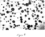
2時間後、ビオチンを示したコロイドは、ストレプトアビジンビーズ上に塊状集積し、そしてそれらを鮮赤色に着色し、そして当該コロイド溶液のバックグラウンドの桃色は消失していた(図8を参照)。ストレプトアビジンビーズおよび、NTA−Niを示すがビオチンを示さないコロイドを含む、ウエル中の溶液は、桃色を呈した。40倍の倍率で、当該ビーズは無色のままであることが観察され、当該コロイド上のスピーシーズと当該ビーズ上のスピーシーズとの間の結合は生じなかったことを示している(図9参照)。図10は、プロテインAビーズのバックグラウンド中に単一ストレプトアビジンビーズを含むウエル中では、ビオチン提示コロイドに結合された一方、付近の対照ビーズは結合されなかったことを示す。
【0097】
実施例3
上記の実験で使用したビーズは、次いで、ビーズ・コロイド相互作用が当該コロイドに付着させた蛍光成分を用いても検出可能なことを証明するために、使用された。上記のコロイドは、ヒスチジン標識GSTをもつように誘導体化された。蛍光標識をもつ抗GST抗体を、当該コロイドに別々に加え、PBSで濯ぎ、次いでストレプトアビジンビーズまたは対照ビーズに添加した。ビーズは、次いで、蛍光顕微鏡下40倍の倍率で視覚化した。図11は、ビーズならびに浮遊コロイドが蛍光を発しているのを示す。対照ビーズは全く蛍光を発しなかった。
【0098】
実施例4
本実験は、第1のものがビーズ上に固定化され、そして第2のものがSAMコート金コロイド上に固定化されている場合、それら2つのタンパク質間における相互作用の検出を証明する。次のように、商業的に入手可能なNTA−Ni++ビーズにヒスチジン標識GSTを付着させた。100μLのNTA−Ni++ビーズをPBS中で濯ぎ、次いで200μLのPBS中に再懸濁した。20μLの当該スラリーを清浄なエッペンドルフ管に移し、それに100μLのヒスチジン標識GSTを1mg/mlで添加した。GSTとインキュベートしなかったNTA−Ni++ビーズのアリコートを、負の対照として保存した。40分間のインキュベーション期間の後、過剰のタンパク質を除去するために、当該ビーズをPBS中で濯ぎ、遠心分離によりペレット化し、次いで100μL中に再懸濁した。コロイドは、ヒスチジン標識タンパク質を捕捉するためのNTA−Ni++と、非特異的結合への抵抗を援助するためのトリエチレングリコールとを、提示するSAMsで誘導体化した。500μLの当該コロイドは、次いで、それらのFc部分へ結合することによって抗体を捕捉する、0.01mg/mLのプロテインGのヒスチジン標識断片を含む溶液の100μLとインキュベートした。過剰のプロテインGを除去するために、コロイドは、濯ぎ、そしてペレット化した。当該プロテインG提示コロイドは、次いで、100μLの抗GST抗体と1mg/mlでインキュベートした。当該コロイドはPBSで濯いだ。当該抗GST提示コロイドは、次いで、当該GST提示ビーズと混合し、そして結合が生じるようにさせた。10分以内に、GSTを提示したビーズは、当該抗体所持コロイドが当該ビーズ上に塊状集積するにつれて、赤く着色するようになり始めた。ここで図12を参照すると、C6,C7およびC8とマークされている写真は陽性に相当する;抗GST提示コロイドとインキュベートされたGST提示ビーズ。D6,D7およびD8と標識されている写真は負の対照に相当し、それらは、GSTと予め結合されてないNTAビーズとインキュベートされた抗GST提示コロイドである。E6,E7およびE8と標識されている写真も再度陽性で、そして抗GSTを提示するコロイドと混合された、GSTを提示するビーズに相当する。シリーズCおよびE間の相違は、シリーズEで使われたコロイドが、シリーズCで撮影されているビーズで使われたような100μLでなく、10μLのプロテインGとインキュベートされたことである。
【0099】
実施例5
本実験は、SAMコートコロイド上に固定化された第1スピーシーズとビーズ上に固定化された第2スピーシーズであって、そこでの第2スピーシーズが全細胞溶解物から直接ビーズ固定化されている、それらのスピーシーズ間の相互作用の検出を証明する。
【0100】
コンピテントBL21細胞を、His−標識GSTをコードする発現プラスミドで形質転換した。標準的な分子生物学技術の当業者にはよく知られているように、当該細胞を増殖し、LBを接種するために用い、そして当該コードタンパク質を発現するように誘導した。負の対照として使用するため、BL21細胞のアリコートを発現プラスミドで形質転換せず、そして、選択抗体の存在下で増殖させなかったこと以外は、上記のように増殖しそして誘導した。細胞は、ペレット化し、そして標準的溶解緩衝液と混合することにより、続いて超音波処理により、溶解した。当該粗細胞溶解物に、商業的に入手可能なNTA−Ni++ビーズを添加し、そして4度で1時間インキュベートし、当該His−標識タンパク質を当該ビーズに結合させるようにした。ビーズは次いでペレット化し、PBS中に再懸濁し、そして(上記のように調製された)抗GSTを提示したコロイドと混合した。1時間のインキュベーション期間の後、ビーズを倍率40倍で調べた。結果は写真撮影し、そして図13に示す。本実験の結果は、シートが「SB133 10min」と標識されているところで始まる。各写真は、「+」または「−」で、およびインキュベーション時間で標識されている。陽性は、GST発現プラスミドで形質転換された細胞に対応し、そして陰性は、形質転換されなかった細胞に対応するが、それ以外は同様に処理した。
【0101】
実施例6
本実施例は、平坦チップに付着された第1タンパク質スピーシーズと、蛍光成分を提示するコロイドに付着した第2スピーシーズとの間の相互作用の検出を証明する。本明細書では、コロイドに付着されるかまたは溶液中で遊離ないずれかの、蛍光的に標識された抗体へのチップ固定化GSTの結合を、比較する。
【0102】
SAMsは、2.5%NTA−Ni++が97.5%トリエチレングリコールのバックグラウンド中で提示されるように、金コート平坦チップ上に形成された。一連のNTA−Ni++チップは、His−標識GSTと予め結合させた。NTA−Ni++−SAMコートされたコロイドを、調製し、そしてHis−標識プロテインGと予め結合させ、次いで緑色フルオレセイン標識化抗GST抗体とインキュベートした。当該GST提示チップの半数は、前記蛍光抗体所持コロイドとインキュベートした(図14の上部を参照)、一方他の半数は、前記蛍光抗GSTと直接インキュベートした(図15の上部を参照)。対照(図14および15の下半分)は、トリエチレングリコール末端SAMsで誘導体化されそしてNTA−Ni++を提示しないチップである。各図には、添加された2つの異なる濃度の抗GSTに相当する、2つの陽性が存在する。チップは、蛍光顕微鏡により倍率40倍で、分析しそして写真撮影した。
【0103】
予示的実施例
実施例7−12により、本発明の追加態様を例証する予示的実施例が以下に提供される。
【0104】
実施例7
薬剤候補を含むと言われる土壌試料からの抽出液を、図4に図解されるように、クロマトグラフィーカラム220上に吸着させる。クロマトグラフィーカラム220は、抽出液110中に溶解するかあるいは懸濁することも可能な、対象のスピーシーズを吸着するポリスチレン樹脂ビーズ230を含む。当該化合物が当該ビーズ上に懸濁された後、溶出溶剤を当該カラム中に通過させ、そして当該溶出溶剤のpHを長時間変化させる。溶出溶剤がカラム220を通過するにつれて、異なる時間で当該ビーズから化合物が選択的に除去される。溶出溶剤のpHが変わるにつれて、第1アリコートはAバイアルに,第2はBバイアルに,第3はCバイアルに、そして以下同様に収集される。このように、対象の化合物が当該カラムから溶出された後、各バイアルには、薬剤候補の可能性がある、異なるスピーシーズが含まれる。
【0105】
図5は、溶出緩衝液を結合緩衝液に交換し、そして樹脂ビーズを当該バイアルに添加した後、図4に示すのと同じバイアルを例示する。当該ビーズは、上記の分離過程で使われたものと同じタイプのポリスチレン樹脂ビーズである。当該ビーズを添加する前に、当該溶出溶剤を、透析膜上で結合溶剤と交換する。交換が完了した後、当該ビーズを添加してもよく、すると当該薬剤候補は当該ビーズの表面に固定化される。対象の固定化標的分子を含む懸濁性コロイド粒子を含む流体を、当該バイアルの各々に添加する。当該ビーズを30分間ないし2時間インキュベートする。結合パートナーの結合を促進するために、当該バイアルを撹拌してもよい。適当なインキュベーション期間の後、当該バイアルの各々に含まれるビーズを、コロイド粒子により装飾されていたかどうかを確かめるために、試験する。当該ビーズのいずれかが装飾されていた場合、当該標的分子と元の混合物のある成分との間で結合現象が生じていたことを示している可能性がある。当該成分が、当該標的分子の生物学的活性を遮断するための薬剤として使用できることを、本相互作用は示している可能性もある。あるいは、当該成分は、健康状態と当該標的タンパク質の存在と相関された疾患状態とを区別するための診断検定に使用することも可能である。
【0106】
実施例8
次のものは、疾患を診断しそして治療するための総合的方法であり、どのように本発明が使用可能であるかの実施例である。癌と関連する細胞は、その表面上に、ある種の受容体をしばしば独占的に発現するかまたは過剰発現することが、知られている。これらの癌関連細胞表面受容体は、腫瘍マーカーとして知られている。HER/2neuのような腫瘍マーカーの作用を、それらに強く結合する分子で遮断すると、腫瘍増殖が遅くされ、また排除されることが示されている。少数の腫瘍マーカーが同定されているものの、もっと多くが存在すると研究者は理論づけているが、しかしまだ不明のままである。本発明の方法は、腫瘍マーカーを検出するために、そしてまた、それらの活性を遮断するための薬剤をスクリーンするためにも、使用可能である。そうした検定を実施するための1つの方法は次のとおりである。
【0107】
腫瘍マーカーとして知られる細胞表面受容体は、体内の未知リガンドと相互作用する。本明細書に記載されているように、細胞または細胞膜は、溶解し、分画し、そしてその内容物を、SAM−修飾コロイドに付着させることも可能である。当該コロイドは、補助シグナル伝達成分も提示するように、誘導体化しても、しなくてもよい。健康細胞および疾患関連細胞は、候補リガンド所持コロイドのバッチからの同一アリコートと、別々にインキュベートされることになる。次いで、非疾患関連細胞への結合と比較した場合、疾患関連細胞に示差的に結合するコロイドの集団を探すことが可能である。この示差的結合は、当該コロイド固定化リガンドが当該細胞上の腫瘍マーカーの結合パートナーであることを、意味する。
【0108】
当該コロイドに付着した画分の保存部分を、次いで、当該腫瘍マーカーの結合パートナーを同定しそして特徴解明するために、分析することが可能である。当該結合パートナーで誘導体化されたコロイドは、疾患の存在をシグナルするための診断検定に使用してもよく、あるいは、当該結合パートナーは、当該腫瘍マーカーの生物学的活性を遮断するための診断薬として使用することも可能である。
【0109】
あるいは、前記溶解物から単離された結合パートナーは、次のものを含むさまざまな薬剤スクリーニング戦略で、使用可能である。健康な細胞および疾患関係細胞を、薬剤候補および当該結合パートナーをもつコロイドと、インキュベートしてもよい。当該細胞へのコロイド結合の消失は、当該候補薬剤が、当該腫瘍マーカーに直接結合したか、あるいは、当該細胞表面でのその発現に間接的に影響したか、のいずれかであることを示す可能性がある。In vitro(試験管内)薬剤スクリーニング検定では、当該結合パートナーをもつコロイドを、溶液中に遊離で、あるいは、例えば、平坦な基質または、ビーズまたはコロイドのような粒子であってもよい表面に付着された、候補薬剤を含む溶液中でインキュベートしてもよい。本明細書におよび、いずれも参考文献によって本明細書に援用される、Bamdadらにより「タンパク質凝集の迅速かつ高感度検出」の標題で2000年1月25日に出願された国際特許出願番号第PCT/US00/01997号中、および2000年1月21日に出願された国際特許出願番号第PCT/US00/01504号中、および共同所有、同時係属米国特許出願番号第09/631818号中、およびBamdadらによる「非コロイド構造体上のスピーシーズとコロイド固定化スピーシーズの相互作用」の標題の共同所有の同時係属米国特許出願番号第09/602778号、に記載されている検出方法を、次いで、当該コロイドに結合された結合パートナーに結合される薬剤候補を同定するために、使用することも可能である。例えば、土壌試料からの画分を、EDC/NHS共役を経てコロイドへ付着させ、そして腫瘍マーカーに対する結合パートナーをもつコロイドを含む溶液に別々に添加することも可能である。前記腫瘍マーカーの結合パートナーと相互作用するエレメントを含む溶液は、桃色から青色に変わるはずである。あるいは、薬剤候補は、ビーズ上へコンビナトリアルな方法により合成してもよい。各々が異なる薬剤候補をもつこれらのビーズは、次いで、当該腫瘍マーカーの結合パートナーをもつコロイドとインキュベートすることも可能である。この場合、当該コロイド上のスピーシーズと当該ビーズ上のスピーシーズとの間の結合は、当該コロイド上のシグナル伝達エレメントを経て、または当該同族体薬剤を提示するビーズ上へのコロイドの凝集を視覚的に観察することにより、検出することも可能である。
【0110】
実施例9
本発明のもう1つの側面で、化学的化合物、タンパク質、ペプチド、相互作用ドメインおよび核酸のようなある種の既知スピーシーズへ結合する、それらの能力に従ってタンパク質を特徴解明することも可能である。タンパク質の結合能力を決定することは、治療標的として、その対象のタンパク質の機能を割当てること、およびその可能性を決定することの、欠くことのできない部分である。対象である何らかの特定のタンパク質に対しタンパク質結合パートナーとなり得るものの数は多く、それゆえ、例えば、チップ表面上に便利にも展示することはできない。しかし、タンパク質相互作用は、いくつかの異なるタンパク質中に存在する、反復性の認識モチーフまたはドメインにより、仲介されることがしばしばある。タンパク質相互作用モチーフは、その親タンパク質の関係においてよりも、別個のユニットとして発現された場合でさえも、それらの機能性構造を典型的には保持しており、そしてそれ故、独立の機能性ユニットとして合成し、または発現し、そしてチップ上に固定化することも可能である。特徴未解明のタンパク質の機能に関する重要な手がかりを、他の生体分子(具体的にはそれらのタンパク質内の認識モチーフ)との、当該タンパク質の相互作用を決定することにより収集することも可能である。
【0111】
各種のタンパク質相互作用ドメインまたは認識モチーフが、文献に報告されており、そして当業者に知られている。例えば、「タンパク質機能チップ」の一部として、タンパク質を特徴解明するのに使うことができる、タンパク質相互作用モジュールの例を、以下に説明する。
【0112】
不連続のタンパク質相互作用モチーフは、細胞質シグナル伝達タンパク質類を補充する、Srcホモロジー2ドメイン(SH2)により例示される。例えば、Grb−2およびNckは、SH2およびSH3ドメインを含み、それらはプロリンリッチな配列を認識し、そして、追加のタンパク質が、活性化受容体チロシンキナーゼへ補充されるのを可能にするための、アダプタータンパク質として作用する。PDZドメインは、受容体の内面上のある種のC−末端配列に結合し、そして受容体クラスタリングに重要と考えられている。PTBドメインは、SH2ドメインに似ているが、pY−含有モチーフ周囲の異なる特定の残基に結合する。FHAドメインはホスホトレオニン含有ペプチドに結合し、一方14−3−3ドメインはホスホセリン含有モチーフに結合する。PHドメインは、ホスホイノシチドに結合し、シグナル伝達タンパク質の膜会合を可能にする。FYVEドメインも、PI3Pのような脂質ヘッドグループに結合する。デスドメイン、DED、BADおよびCARDドメイン類は、アポトーシスを誘発するタンパク質・タンパク質相互作用の肝要な成分であるモチーフである。機能性ドメインまたは結合モチーフには、デスドメイン、SH2およびSH3、クリングルドメイン、RGDモチーフ、酸性活性化ドメイン、およびポリグルタミンリピート(反復配列)、アルマジロモチーフ、Grb2ドメイン、酵素基質、タンパク質の細胞質尾部、MUC1リピート、WWモチーフおよびポリプロリン配列が、非限定的に含まれる。また、本発明に、現在公知の、または発見されるであろう、他のドメインまたはモチーフを取込むことももちろん可能である。
【0113】
本発明のデバイスは、単一の手順で複数の分子相互作用を特徴解明するように、配置・形成してもよい。例えば、1つまたは1つ以上の空間的にアドレス可能な領域をもつタンパク質アレイチップのような表面を、各空間的にアドレス可能な領域がタンパク質認識モジュールのような異なる固定化スピーシーズを示すように、作製することも可能である。特徴未解明のタンパク質のような固定化スピーシーズを、コロイド粒子に付着させ、そしてそれから前記チップとインキュベートすることも可能である。対象のタンパク質が結合する空間的アドレスは、各種の技術により見分けることが可能である。例えば、特定の領域におけるコロイドの塊状集積は、その場所を赤くすることも可能である。あるいは、当該コロイドは、電極上に形成された機能性ドメインチップとともに使用するための電気活性成分のようなシグナル伝達構成要素で、誘導体化してもよい。加えて、コロイドは、蛍光成分または蛍光タンパク質のような光学的に発光性の化合物を含むように、構築または修飾することも可能である。インキュベーション期間の後、未結合のコロイド粒子を除くため、当該チップ表面を濯ぐ、すると残存蛍光の空間的パターンは、対象のタンパク質が結合しているモチーフの正体を明らかにすることも可能である。結合されたコロイド粒子は、視覚的に、または機器、例えば、電極または光学検出器、の助けを借りて検出が可能である。これらのおよび他の技術は、当該特徴解明プロセスの一部または全部の自動化を提供することもあり得る。
【0114】
最小相互作用モチーフは、典型的にはそれらの機能性構造を保持しており、そしてそれ故、独立のユニットとして合成または発現され、そしてチップまたは粒子のような表面上に固定化することも可能である。便利にも、これらのモチーフは、これらのタグに対する結合パートナーを示す表面への結合を容易にするために、アフィニティータグで修飾することも可能である。例えば、NTA−Ni(II)自己集合単分子層は、ヒスチジンタグで修飾されているモチーフを固定化するために、金コート基質上に作製することも可能である。マスキング、マイクロ密着プリンティング、または物理的単離技術(すなわち、親水性対疎水性)を、別々のアフィニティー標識スピーシーズを表面上の異なる領域へ付着させるために用いることも可能である。
【0115】
本発明は、ペプチド、タンパク質の断片または混合物、核酸およびそれらの複合体、化学物質、薬剤候補、薬剤および細胞が、非限定的に含まれる、タンパク質以外の成分をもつ表面をインキュベートすることを予期している。これらの成分は、精製された形でも、または、細胞溶解物およびそれらの断片、ゲノムDNA、cDNAライブラリー、薬剤または化合物ライブラリー、天然物試料、および類似物から由来してもよい。
【0116】
実施例10
本発明のもう1つの観点から、本技術を変形したものが、特定の疾患状態で失われているきわめて重要な機能性を同定するために使われる。この場合、機能性モジュールのライブラリーを提示するチップを、光学的に発光性の成分のようなシグナル伝達能力ももつコロイド粒子の付着を容易にするように修飾されている、タンパク質の混合プールとインキュベートする。当該タンパク質プールの成分は、結合が生じるように、表面固定化モジュールと相互作用することが可能にされる。次いで、コロイドを加え、そして前記固定化スピーシーズと相互作用した当該タンパク質にコロイドが結合することが可能になる。例えば、前記タンパク質プールは、NTA−Ni提示コロイドがそれらに結合できように、ヒスチジンタグとともに発現されていてもよい。洗浄工程の後、三次元相互作用マップを、空間的にアドレス可能な表面の各場所における光放射を測定することにより、作製する。特定の疾患状態で失われるきわめて重要な機能性を決定するために、健康なセットの細胞または組織からの三次元相互作用マップは、特定の疾患状態に存在する細胞または組織から由来したタンパク質の結合により作製された相互作用マップに比較される。
【0117】
実施例11
本発明のもう1つの観点からは、成分を、一組の推定結合パートナーを提示する空間的にアドレス可能な表面と、溶液中で相互作用させることを可能にする。相互作用パターンまたはマップを作製するために、相互作用のレベルが、各空間場所で検出される。例えば、空間的にアドレス可能なチップが、生体分子アレイを提示するように、誘導体化される。溶液中の一組の成分と当該表面とが相互作用することが可能になり、そして相互作用パターンを作製するために、各場所での結合スピーシーズのレベルを決定する。当該アレイ中で固定化されたスピーシーズまたは溶液中に存在するスピーシーズは、タンパク質、タンパク質断片、タンパク質相互作用ドメインまたはモチーフ、核酸、薬剤または薬剤候補から構成されてもよい。
【0118】
化学化合物は、このようにして特徴解明が可能である。例えば、薬剤候補となり得る化合物を、タンパク質モチーフチップと相互作用することを可能にする。その結果の相互作用パターンは、当該化合物の活性の指紋である。相互作用パターンのライブラリーは、既知薬剤の活性の指紋を採取するために、作製することも可能である。相互作用パターンは、次いで、まだ特徴解明されていない化合物について作製される。これらの新しい相互作用パターンは、次いで、その特徴が未解明な化合物の活性に対する指標を得るため、既知薬剤の基準相互作用パターン(すなわち、指紋)と比較される。
【0119】
本発明の特徴は、細胞の活性プロファイルが薬剤候補での処置に応答してどのように変化するかを、決定するために使用してもよい。もう1つの態様で、細胞成分をある薬剤に曝露し、そして次に、推定結合パートナーを提示する空間的にアドレス可能なチップと相互作用することを可能にする。この場合、その結果得られる相互作用パターンは、薬剤の不在下での相互作用パターンに比較した場合、当該細胞成分の結合パターンにおける変化を反映する。薬剤処置による示差的結合効果を反映するこれらの細胞成分相互作用パターンも、既知薬剤の活性を未知薬剤候補に比較するのに使用可能である。
【0120】
1つの実施態様において、化合物薬剤ライブラリーは、例えば、相互作用モチーフへの細胞産物の結合を相互作用させるための、2つまたはそれ以上の他の成分の能力に対する、ある薬剤の影響を決定することにより、特徴解明される。例えば、腫瘍細胞系から由来のタンパク質を、シグナル伝達コロイドに付着させ、そして癌に関連づけられている固定化モチーフを示す、相互作用チップのような、表面とインキュベートする。その三次元相互作用景観を、次いで、当該細胞産物が薬剤候補で処理された後に作製された相互作用パターンと比較する。このことは、ある種の相互作用または相互作用の群を、阻害する、亢進する、または別のやり方で変更するための、それらの能力について、化合物ライブラリーの迅速な特徴解明を、容易にすることも可能である。特徴解明は、新規に(de novo)実施してもよく、または特徴未解明の薬剤で処理された細胞産物により作製された相互作用パターンを、既知の薬剤または他の生物学的に活性な化合物により作製された相互作用パターンと比較することにより、実施してもよい。このようにして、ある薬剤候補は、その結合相互作用を既知薬剤のものと比較することに基づき、例えば、「タキソール様」または「エンドスタチン様」であると、決定することも可能である。相互作用チップは、タンパク質、核酸、ペプチド、薬剤、低分子、および類似物を提示することも可能である。本発明は、どれか1つの疾患に関係する、特定の相互作用モチーフまたはタンパク質または薬剤の固定化に限定されることを、意図しない。
【0121】
本発明はまた、疾患細胞のものに比べて健康細胞により産生される成分の結合パターンの間の相違を描写するために、または多数の疾患の間の同じようなタンパク質標的を決定するために、タンパク質であってもよい、表面固定化スピーシーズと細胞成分との間の相互作用パターンを作製することを見込む。細胞タンパク質は、当該チップと別々に、または一緒に、インキュベートしてもよい。すなわち、特定の遺伝子産物に独特である相互作用パターンを作製するために、タンパク質は、別々にシグナル伝達コロイドに付着させ、そして続けて当該チップとインキュベートしてもよい。あるいは、一組の遺伝子産物をコードするcDNAライブラリーまたはそれらのサブセットを、発現されるタンパク質のシグナル伝達コロイドへの付着を容易にするように設計された発現ベクター中へ挿入する。タンパク質を、成長させ、そして宿主細胞中で発現させることも可能である。場合によっては、宿主細胞の本来のタンパク質と、当該cDNAライブラリーによりコードされるタンパク質とが相互作用することが望ましいこともある。これらの場合、当該cDNAは、商業的に入手可能なテトラサイクリン誘導性発現ベクターのような、調節可能発現ベクター中へ挿入されることになろう。このようにして、当該cDNAタンパク質のレベルは当該宿主細胞のタンパク質のレベルに匹敵するように、条件を決定することも可能である。
【0122】
コロイド付着の前に、当該発現タンパク質は、相互作用チップとインキュベートする。タンパク質の群は、それらが認識するモチーフまたは化合物を示す、空間アドレス上に蓄積するはずである。表面を濯ぎ、そしてシグナル伝達コロイドを次いで添加する。各部位でタンパク質に付着するコロイドの数は、その場所で示される構成要素に結合しているタンパク質の数に比例する。
【0123】
本発明はまた、コロイドを取込まない技術を用いる相互作用パターンの検出を見込む。これらには、表面プラズモン共鳴(SPR)、水晶結晶微量天秤(QCM)、ならびに当該アレイチップと相互作用することもあり得る成分を標識する方法が、非限定的に含まれる。例えば、各タンパク質成分が緑色蛍光タンパク質(GFP)融合タンパク質として発現されるように、cDNAライブラリーを作製することも可能である。このようにして、当該結合タンパク質と会合した蛍光を直接検出することが可能である。
【0124】
実施例12
もう1つの側面で、強化シグナル増幅を達成可能である。例えば、固定化成分を、タンパク質チップのような表面上に、当該表面へ第1相互作用スピーシーズを付着させることにより、提供することも可能である。推定結合パートナーのような固定化スピーシーズを、連結構成要素も含む、コロイド粒子に付着させる。コロイド粒子は、次いで、当該固定化スピーシーズが当該表面上の固定化成分と相互作用するように、当該表面上でインキュベートされる。濯ぎの前に、当該連結構成要素と相互作用するための2つまたはそれ以上の結合部位を含む、架橋化合物を加えてもよい。当該架橋化合物は、シグナル伝達構成要素も含んでよい。すると、当該架橋化合物は、2つまたはそれ以上のコロイド粒子上の連結構成要素と結合することによって、多数のコロイド粒子を統合し、ネットワークを形成することが可能である。架橋形成が起るための適切な時間をかけた後、すべての非結合コロイド粒子、非付着コロイドネットワークおよび非結合架橋化合物を、当該表面から濯ぎ去ることも可能である。当該表面に結合されたすべてのコロイド粒子は、次いで、コロイド粒子のネットワークを直接観察することにより、あるいは当該コロイド粒子または架橋化合物のいずれかの、存在、不在または相対強度を検出することにより、検出してもよい。
【0125】
あるいは、架橋化合物を添加する前に、非結合コロイドを当該表面から濯いでもよい。連結構成要素を含む第2タイプのコロイド粒子、および選択的にシグナル伝達構成要素(しかし固定化スピーシーズでない)を、次いで、加えてもよい。いずれのタイプのコロイド粒子も一緒に連結できる架橋化合物も、添加される。当該架橋化合物はシグナル伝達構成要素を含んでもよい。コロイドのネットワークが、次いで、当該表面上で発達するが、その一部は、固定化成分および固定化スピーシーズとの間の相互作用を経て、当該表面へ結合してもよい。非結合材料は、次いで、濯ぎ去り、そして上記のように結合現象を検出することも可能である。
【0126】
各種の連結構成要素/架橋化合物ペアを用いてもよい。当該架橋化合物は、多価であって、そして異なるコロイド粒子に付着されている2つまたはそれ以上の連結構成要素に結合できなければならない。好ましくは、当該架橋化合物は、可動性リンカーへ付着され、そして例えば、2つの隣接するコロイド粒子間に生じる可能性がある何らかの立体障害作用を避けるのを助けるために、ポリマー中へ取込まれてもよい。当該連結構成要素は、架橋化合物との結合のために1つの部位をもつだけが必要で、そして好ましくは、コロイド粒子に容易に付着される。連結構成要素/架橋化合物ペアのいくつかの例は、ビオチン/ストレプトアビジン、ヒスチジン標識ペプチド/NTA/Ni(II)およびDNA/DNAである。
【0127】
ある実施態様では、第1固定化成分をタンパク質チップ表面に付着する。推定結合パートナーを、固定化スピーシーズとして、金コロイド粒子へ付着し、そして当該コロイド粒子もビオチン成分をもつ。当該コロイド粒子は、次いで、タンパク質チップとともにインキュベートされる。当該チップ表面を濯がずに、ビオチンに対して4つの結合部位をもつ、蛍光的に標識されたストレプトアビジンを添加する。当該ストレプトアビジンは、検出可能な蛍光シグナルを提供し、そしてまた、コロイドのネットワークを架橋するのにも役立ち、単一のタンパク質/タンパク質という僅かな相互作用により当該表面に結合されているコロイドのネットワークをもたらす。インキュベーション時間は、当該ネットワークが検出可能なサイズへ成長するのを可能にするように、変えてもよい。当該ネットワークが形成された後、すべての非結合コロイドならびに非結合コロイドネットワークを、当該表面から濯ぎ去ることが可能である。すべての残ったコロイド粒子のネットワークおよび蛍光的に標識されたストレプトアビジンは、蛍光検出器により検出も可能な、当該シグナルを大きく増幅するのに役立つ。
【0128】
当該蛍光成分を遺伝子組み換えすることも可能だが、必ずしも必要ではない。例えば、任意の蛍光基を用いることが可能で、そして、SAMを形成するためのチオールを経ることを含む、多数の方法を用いて付着させてもよい。このようにして、対象のタンパク質へシグナル伝達構成要素を遺伝子組み換えという、厄介で、時に煩わしい工程を回避する。加えて、一般的支持体粒子へシグナル伝達構成要素を付着するための能力は、タンパク質性でも、または合成のものでも、任意の分子へシグナル伝達エレメントの「付着」を可能にする。さらに、当該蛍光基は、対象の分子へ直接分子的に結合する必要はなく、そしてそれ故、対象の当該分子の活性が、当該蛍光標識の存在により、変化させられる可能性はより少ないであろう。
【0129】
本明細書にリストされたすべてのパラメーターは模範的であることを意味し、そして実際のパラメーターは、本発明の方法および装置が使用される個々の適用例に依存することを、当業者は容易に理解するはずである。そのため、前掲の諸実施態様は、例示的に提示しただけであり、付帯の請求事項およびそれらの均等物の範囲内で、別途その際具体的に記載されるように、本発明を実行することも可能である。
【図面の簡単な説明】
【図1】 図1は、当該表面の各部分に付着された、異なる結合スピーシーズをもつ別々の部分に分けられた表面を図解する。懸濁中のコロイド粒子は、当該表面の一方の部分に付着されたスピーシーズと選択的に相互作用しており、そして他方とは相互作用していない。
【図2】 図2は、非結合コロイド粒子が当該表面から濯ぎ去られた後の、図1に図解した表面を示す。
【図3】 図3は、混合物が分離カラム中で分離され、当該混合物からの異なる成分の連続的なアリコートが順次収集されていることを概略図的に図示する。
【図4】 図4は、図3に示した処置により得られたアリコート中への、被疑結合パートナーを含む、コロイド粒子の添加を概略図的に図示する。
【図5】 図5は、コロイド装飾ビーズ、具体的には、タンパク質/タンパク質相互作用を経てビーズに連結されたコロイドの写真(倍率40倍)のコピーである。
【図6】 図6は、図7に示される実験における負の対照の写真(倍率40倍)のコピーである。
【図7】 図7は、別々にアドレス可能な検定領域を提供するマルチウエルプレートを示す。
【図8】 図8は、同系列タンパク質を示すビーズ上へ塊状集積する小分子を示し、当該ビーズを赤色に着色する金コロイドの写真のコピーである。
【図9】 図9は、図8に示される実験の負の対照の写真のコピーであり、ビーズはランダムタンパク質を示し、当該コロイド表面上に提示された小分子の当該結合パートナーは示されなかった。
【図10】 図10は、前記同系列タンパク質を示す単一ビーズを、ランダムタンパク質を示すビーズのバックグラウンド中へ混合した実験の写真のコピーであり、その単一ビーズ上へのコロイドの選択的塊状集積を示す。
【図11】 図11は、ビーズ上に固定化されたスピーシーズの結合パートナーおよび蛍光成分の両方をコロイドが示した実験の、蛍光顕微鏡写真のコピーである。
【図12】 図12は、第1タンパク質がコロイド固定化され、そして第2がビーズ固定化された、複数セットのタンパク質・タンパク質結合実験の写真のコピーである。
【図13】 図13は、全細胞可溶化物から直接、第1タンパク質がコロイド固定化され、第2のタンパク質がビーズ固定化された、複数セットのタンパク質・タンパク質結合実験における写真のコピーである。
【図14】 図14は、SAMコートされたコロイドの写真のコピーであり、蛍光的に標識された抗体、抗GST、その同族体リガンドへの結合、GST(NTA−SAMコートされたチップ上に固定化されている)を示す。
【図15】 図15は、図14に示されるものに対する比較実験の写真のコピーであり、蛍光的に標識された抗GST、溶液中の遊離のもの、その同系列リガンドに結合しているもの、GST(NTA−SAMコートされたチップ上に固定化されている)示される。[0001]
Field of Invention
The present invention relates generally to methods, assays, and kits for rapidly and sensitively detecting the interaction of two chemical and / or biological species. The present invention facilitates techniques including drug screening and signaling pathway mapping.
[0002]
Background of the Invention
Recent elucidation of the human genome has provided a tremendous amount of information in the form of discrete DNA sequences that encode the repertoire of human proteins. As recognized by those familiar with molecular biology, the overwhelming majority of molecules involved in biological functions are proteins, not DNA or RNA. In functional genomics, various levels of specific mRNAs are associated with disease states. Proteomics is the successor of functional genomics because it studies the function of proteins rather than the precursor molecule DNA or mRNA. One aspect of proteomics is determining how protein function is associated with disease. Disease-related families of proteins, or proteins involved in common signaling pathways, can be identified by elucidating protein-protein interaction networks. Today, the main focus of biological research is to elucidate the protein interaction networks that make up biological signaling pathways. By understanding these protein interaction networks, critical clues about what triggers the transition from a healthy state to a disease state have been collected.
[0003]
A major obstacle to proteomics research is that there is no method available to detect protein-protein interactions if one or both are not yet characterized or impure. Most current detection methods require the use of specific antibodies that recognize one or both of the putative binding partners. This means that the protein of interest needs to be purified so that antibodies against it can be made. If a protein has been characterized, the protein can generally be labeled with an affinity tag, often assisting the detection process. This means that a lot of work is required to separate and purify components so that they can be used in current assays before knowing whether a crude sample contains the protein of interest. . Another major drawback of current methods in the study of protein-protein interactions is that they are sequential labor intensive processes. This means that they cannot be multiplexed to investigate complex problems such as elucidating large protein interaction networks or testing several putative binding partners in parallel . Measuring the interaction network with a sequential pair-wise test for the number of genes in the human genome currently estimated at about 40K is about 8 × 10⁸Will be included. The human genome project is nearing completion and the development of appropriate technologies that allow functional analysis of many gene products is absolutely necessary. For these reasons, it would be beneficial if a method was available that facilitated parallel analysis of protein binding interactions where one or both proteins may exist as a crude mixture. For example, if a method for rapidly characterizing newly discovered uncharacterized proteins that could be generated from a cDNA library would be useful, it would also be beneficial. For example, these methods would be particularly important in a variety of industries, including the pharmaceutical industry, where a large number of known and unknown species are screened for the identification of new drugs. In addition to identification, it may also be useful to classify species for their relative affinity for other known and unknown species. Typically, it is usually desirable to increase the speed of the screening procedure in order to increase the total number of species to be screened. Furthermore, sample throughput is improved when the detection method can be simplified and multiple assays can be performed simultaneously.
[0004]
Summary of invention
The invention includes techniques useful for elucidating the characteristics of uncharacterized proteins, and for detecting interactions between binding partners, one or both of which are present in a mixture, including biological and / or chemical Various novel methods, compositions, species, and articles are provided for detecting interactions between mechanical species. According to one aspect, the present invention provides a high throughput method for detecting protein binding interactions. In certain embodiments, the present invention applies in situations where one potential binding partner is present in the uncharacterized and / or crude mixture. In addition, the present invention provides a method for rapidly elucidating the characteristics of uncharacterized proteins by detecting the ability to bind to various functional protein modules. From one aspect, both a first surface having a first immobilization component and a second surface having a second immobilization component are exposed to colloidal particles having immobilization species, and the first, second, or both surfaces Determine the immobilization of the colloidal particles on any of the above.
[0005]
From another aspect of the present invention, the first species is immobilized on the first colloid and the second species is immobilized on the second colloid. The first and second colloids are exposed to a surface and then the immobilization of the first or second colloid on the surface is determined. The surface can also present putative binding partners for one or both of the first and second species.
[0006]
Another aspect of the invention provides a method for chromatographic separation of at least two components from a mixture using a chromatographic arrangement comprising a stationary phase. The same or different type of stationary phase used in the chromatographic arrangement is then used as the surface to which the first and second separation components of the mixture adhere or adhere. The stationary phase containing the adherent component is then exposed to colloidal particles having immobilized species that are believed to be capable of binding to at least one of the first and second components. That is, it is suspected that the immobilized species may interact with the species present in the mixture. Determine the binding of species immobilized on the colloid and species immobilized on the surface of the bead or other stationary phase by detecting the intrinsic or auxiliary signaling capabilities associated with the colloid can do.
[0007]
From another aspect of the present invention, the components are separated chromatographically from a mixture, and then the components present in the fraction are adhered or attached to a surface other than the beads and further present in the mixture. Methods are provided for exposure to colloids with immobilized species that are suspected or identified therapeutic partners of the component to be immobilized. The binding between the species on the colloid and the species on the surface is then determined. Other surfaces to which the separated components can be attached include a second population of colloids, nanoparticles, polymers, multiwell plates, biochips, spatially addressable biochips, electrodes, and electrode arrays However, the present invention is not limited to this.
[0008]
The fractionated components are separately deposited in a spatially addressable manner on a flat substrate and then a colloid exhibiting a single species suspected of interacting with one of the components present in the crude mixture; You may incubate.
[0009]
Another aspect of the present invention provides a kit that includes a chip that displays separate biological or chemical species in a spatially addressable manner. The target uncharacterized protein is then attached to a set of colloids. Furthermore, the colloid presenting the protein of interest is incubated with the chip, and the binding of the colloid to a specific chip position is detected. In a preferred embodiment, the species immobilized on the spatially addressable chip are protein interaction modules and motifs. In this way, it is possible to elucidate the characteristics of uncharacterized proteins by detecting binding to protein interaction motifs.
[0010]
Another aspect of the invention provides a kit comprising a package comprising colloidal particles to which binding species are attached in a specific or non-specific manner. These colloidal particles can then be incubated with target biological or chemical species adhered to a surface. Alternatively, a separate set of colloids may be provided in separate compartments for dispersion into the multiwell plate. The species individually attached to the colloid may include antibodies, known drugs, drug candidates, targeted proteins, protein fragments, protein interaction modules, or products of cDNA libraries.
[0011]
Another aspect of the present invention provides a kit comprising a colloidal particle comprising SAM and a package comprising instructions for immobilizing a binding partner to the colloidal particle.
[0012]
Another aspect of the invention provides a kit having two packages, wherein the first package includes colloidal particles having first species immobilized on the particles, and the second package Includes colloidal particles having second species immobilized on the particles.
[0013]
Another aspect of the present invention provides a kit that includes a chip that displays separate biological or chemical species in a spatially addressable manner. Prior to or after incubation with the chip, the uncharacterized protein of interest is attached to the colloid. The protein's discrete binding or protein binding pattern to a site on the chip is then detected.
[0014]
From another aspect of the present invention, a method is provided that exposes a sample to at least two surface regions, each presenting a different chemical, biochemical, or biological functional group. Determining a pattern of interaction of the sample with at least two of the surface regions indicative of a characteristic interaction between at least one component of the sample and each of the at least two of the surface regions. It is.
[0015]
Another aspect of the invention provides a method, wherein the method separates at least two components of a mixture on a stationary phase and elutes at least a first component from the stationary phase with a fluid. Changing the fluid, immobilizing at least a portion of the first component to a surface, exposing the surface to a putative binding partner, and between at least a portion of the first component and the putative binding partner Determining the binding interaction.
[0016]
Another aspect of the invention provides a method, wherein the method separates at least two components of a mixture on a stationary phase and elutes at least a first component from the stationary phase with a fluid. Immobilizing at least a portion of the first component on the colloid, exposing the colloid to a putative binding partner immobilized on a surface, and at least a portion of the first component and the putative binding partner Determining the binding interaction between.
[0017]
Other advantages, novel features and objects of the invention will become apparent from the following detailed description of the invention when considered in conjunction with the accompanying drawings, which are schematic and are not intended to be drawn to scale. Will be. Each identical or nearly identical component that is illustrated in various figures is represented by a single number in the figures. For clarity, not all components are labeled in all figures, and each embodiment of the invention is not necessary for the ordinary person skilled in the art to understand the invention. Not all components in are necessarily shown.
[0018]
Detailed Description of the Invention
Definition:
“Small molecule” as used herein means a molecule of 5 kilodaltons or less, more typically 1 kilodalton or less. As used herein, “small molecule” excludes a protein.
[0019]
The term “candidate agent” as used herein refers to any medical substance used in humans, animals, or plants. This definition includes compound analogs, naturally occurring pharmaceuticals, synthetic pharmaceuticals, recombinant pharmaceuticals, hormones, antimicrobial agents, neurotransmitters, and the like. This includes any substance or precursor that should be evaluated for use as a drug for the treatment or prevention of neurodegenerative diseases characterized by abnormal aggregation, or other diseases , Synthesized or recombinant). Typically, assessment is performed by activity in an assay, such as the screening assay of the present invention.
[0020]
Various particles can be used in the present invention. For example, “fluid suspendable particles” refers to particles that can be kept suspended alone in a fluid (typically an aqueous solution) used for the purposes of the present invention, or a magnetic or electromagnetic field. Means particles that can be applied or maintained in solution by applying agitation such as agitation, shaking, vibration, sonication, centrifugation, vortexing, or the like. “Magnetically suspendable” particles are those that can be suspended in a fluid by applying a magnetic field. Electromagnetically suspendable particles are those that can remain suspended in a fluid by applying an electromagnetic field (eg, charged particles, or particles modified to have a charge). “Self-suspendable particles” are sizes and / or masses that remain suspended in the fluid (typically an aqueous solution) in which they are used, for example for at least 1 hour without the aid of a magnetic field. Are sufficiently small particles. Other self-suspendable particles remain in suspension for 5 hours, 1 day, 1 week, or 1 month without assistance according to the present invention.
[0021]
“Protein” and “peptide” are well-known terms in the art, and the number of amino acids contained in each is not strictly defined in the art. As used herein, these terms are given their ordinary meaning in the art. Generally, a peptide is an amino acid sequence having a length of about 100 amino acids or less, but may include a sequence of up to 300 amino acids. A protein is generally considered to be a molecule of at least 100 amino acids.
[0022]
As used herein, “metal binding tag (label)” refers to a group of molecules that can be attached to a metal coordinated by a chelate. A suitable group of such molecules includes amino acid sequences that include, but are not limited to, a plurality of histidines and a plurality of cysteines (“polyamino acid tags”). Metal binding tags include the histidine tags defined below.
[0023]
As used herein, a “metal-coordinating chelate” or a metal coordinated by a chelate is not filled with all available coordination sites on the metal and depends on the metal binding tag. Refers to a metal coordinated by a chelator, leaving some coordination sites available for binding.
[0024]
As used herein, “metal binding tag / metal / chelate linkage” refers to a first species that is immobilized to a metal binding tag and a second species that is immobilized to a chelate. Wherein the chelate is coordinated to the metal to which the metal binding tag is coordinated. Exemplary connections are described in Bamdad et al. US Pat. No. 5,620,850, which is hereby incorporated by reference.
[0025]
By “signaling component” is meant a component that can indicate its presence in a particular sample or at a particular location. Signal transduction components of the present invention include those that can be identified by the human naked eye, those that are not visible to the human eye but that can be confirmed by the human naked eye if they are of sufficient quantity (eg, colloidal particles), A component that absorbs or emits electromagnetic radiation within an amount or wavelength range that is readily measurable (ie, by the naked eye or by a microscope, including an electron microscope or the like) or spectroscopically, electronically or electrochemically Can be a measurable component (an “electronic signaling component”, eg, a redox-active molecule that exhibits a characteristic oxidation / reduction pattern upon exposure to a suitable activation energy), or the like. For example, dyes, pigments, electroactive molecules such as redox active molecules, fluorescent components (including phosphorescent components by definition), up-regulating phosphors, chemiluminescent components, electrochemiluminescent components, Or enzyme-linked signaling components including horseradish peroxidase and alkaline phosphatase are included. “Precursor of a signaling component” alone does not have the ability to transduce signals, but can interact with another species by chemically, electrochemically, electrically, magnetically, or physically interacting with the signal. A component that becomes a transmission component. For example, chromophores that have the ability to emit radiation within a specific detectable wavelength only when chemically interacting with another molecule are included. Signaling component precursors, as used herein, are distinguishable from “signaling components” but are included within the definition of “signaling components”.
[0026]
As used herein, in the context of another species of a species or the surface of an article, “adhered or adapted to adhere” means that the species is a covalent attachment, Means chemically or biochemically linked via attachment through a specific biological bond (eg biotin / streptavidin), a coordination bond such as a chelate / metal bond, or the like To do. For example, “sticking” in this context refers to a biological species that specifically binds to a binding species such as a peptide synthesized on polystyrene beads, an antibody that binds to a protein such as protein A that is covalently attached to the beads. Part of a molecule such as a GST or phage that is now biologically specifically bound to a binding partner that is covalently bound to a surface (eg, glutathione in the case of GST). Multiple chemical linkages, multiple chemical / biological linkages, etc., including but not limited to binding species that form (via genetic engineering). As another example, a component covalently linked to a thiol is adapted to be affixed to the gold surface because the thiol binds covalently to gold. Similarly, species with metal binding tags can be attached to surfaces (such as thiol / gold bonds) whose molecules also have molecules covalently attached to surfaces that present chelates that coordinate metals. Adapted. If a surface has a specific nucleotide sequence and a species has a complementary nucleotide sequence, the species is also adapted to be affixed to the surface.
[0027]
“Covalently attached” is nothing more than being attached via one or more covalent bonds. For example, species that are covalently conjugated via EDC / NHS chemistry to a carboxylic acid presenting alkyl thiol that is now anchored to the gold surface are covalently anchored to the surface.
[0028]
“Specifically anchored (or bound)” or “adapted to be specifically anchored (or bound)” means “adapted or anchored” As described above with respect to the definition of “is”, it means that a species is chemically or biochemically linked to another sample or surface, but excludes all non-specific binding.
[0029]
“Nonspecific binding”, as used herein, is given its ordinary meaning in the field of biochemistry.
“Colloid” as used herein is, for example, inorganic or organic, polymeric, ceramic, semiconducting, metallic (eg, gold), nonmetallic, crystalline, amorphous, semiconducting By nanocrystals, including those made of materials that are nanocrystals, or combinations thereof, that is, very small self-suspending or fluid-suspending particles. Colloidal particles used in accordance with the present invention typically have a cross section of 250 nm or less in any dimension, more typically a cross section of 100 nm or less in any dimension, and most often a cross section of about 2-30 nm. One class of colloids suitable for use in the present invention is 10-30 nm in cross section, and the other is about 2-10 nm in cross section. As used herein, this term includes definitions commonly used in the field of biochemistry.
[0030]
As used herein, a component that is “immobilized relative to” another component is either fixed to the other component or indirectly fixed to the other component. For example, by other components being fixed to the fixed third component, or otherwise by transitional association with other components. For example, when the signaling component is fixed to the binding species, when the signaling component is fixed to a colloidal particle to which the binding species is fixed, the signaling component is the binding species. The signaling component is immobilized to the binding species, such as when attached to a dendrimer or polymer to which is attached. If the species anchored to the surface of the first colloidal particles adhere to one component, and the species on the surface of the second colloidal particle adhere to the same component, where the component is a single component If it can be an element, a composite component of multiple species, a cell, another particle, etc., the colloidal particle is immobilized relative to another colloidal particle.
[0031]
The term “sample” refers to any medium suspected of containing an analyte, such as a binding partner, desirably the presence or amount of which is determined. Samples include biological samples (such as cells, cell lysates, tissues, serum, blood or other liquids from biological sources), biochemical samples (such as products from cDNA libraries), environmental samples (such as soil extraction) Any other biological or non-biological medium containing synthetic material, which can be advantageously evaluated according to the present invention.
[0032]
“Structurally predetermined sample” as used herein refers to a sample whose chemical or biological sequence or structure is a predetermined structure, and for neurodegenerative diseases. It is used in assays designed to test whether a particular process is associated with the structure. For example, “structurally predetermined sample” includes peptide sequences, random peptide sequences in phage display libraries, and the like.
[0033]
A “sample suspected of containing” a certain component means a sample for which the content of that component is unknown. The sample may be unknown to contain the specific component, or known to contain the specific component, but the amount may be unknown.
[0034]
As used herein, “metal binding tag” refers to a group of molecules that can become attached to a metal coordinated by a chelate. A suitable group of such molecules typically comprises an amino acid sequence of about 2 to about 10 amino acid residues. These include, but are not limited to, histidines and cysteines (“polyamino acid tags”). Where such binding tag comprises histidine, this binding tag may be referred to as a “polyhistidine tract”, “histidine tag” or “HIS-tag” and the amino acid of the peptide, protein or nucleic acid. It can be either at the terminus or the carboxy terminus, or in any exposed region. A 6 to 10 residue polyhistidine tract is preferred for use in the present invention. The polyhistidine tract is also functionally defined as being a number of consecutive histidine residues added to the protein of interest, thereby affinity purification of the product protein on a metal chelate column, or another It allows identification of protein termini by interaction with two molecules (eg, antibodies reactive with HIS-tag).
[0035]
“Metal-coordinated moiety” as used herein means any molecule that can occupy at least two coordination sites on a metal atom, such as a metal binding tag or chelate. To do.
[0036]
“Affinity tag” is given its ordinary meaning in the technical field. Affinity tags include, for example, metal binding tags, GST (in GST / glutathione binding clips), and streptavidin (in biotin / streptavidin binding). At various places in the present specification, specific affinity tags have been described in the context of binding interactions. It will be appreciated that the present invention includes a series of individual embodiments, including, in any embodiment using an affinity tag, selecting from any of the affinity tags described herein.
[0037]
As used herein, “molecular wire” refers to a wire that enhances the ability of a fluid encountering a SAM coated electrode to communicate electrically with the electrode. This includes conductive molecules and can cause defects in the SAM that allows communication with the electrode as described above and as more fully illustrated below. The resulting molecule is also included. Non-limiting lists of additional molecular wires include 2-mercaptopyridine, 2-mercaptobenzothiazole, dithiothreitol, 1,2-benzenedithiol, 1,2-benzenedimethanethiol, benzene-ethanethiol, and 2 -Mercaptoethyl ether is included. The conductivity of the monomolecular layer can also be increased by the addition of molecules that promote conductivity in the plane of the electrode. Conductive SAMs include, but are not limited to: 1) a sulfur-terminated poly (ethynylphenyl) chain; 2) a benzene ring-terminated alkylthiol; 3) a DNA base-terminated alkylthiol; 4) into a monolayer. Any sulfur-terminated species that are difficult to load; 5) all of the above plus or minus either an ethylene glycol unit or a methyl group to prevent non-specific adsorption plus a terminal alkyl thiol spacer molecule; May be configured. Thiols are listed because of the affinity of thiols for gold in the easy formation of SAMs. From US Pat. No. 5,620,820 and other references, thiols can be substituted with other molecules, as is known in the art. Molecular wires, due to their bulk or other structure, typically cause defects in other relatively densely packed SAMs, and the SAM tightly plugs the surface against the exposed fluid. I am trying not to get lost. The molecular wire collapses the tightly packed self-assembled structure, thereby creating defects and allowing fluid exposed to the surface to be in electrical communication with the surface. In this situation, the fluid is in electrical communication with the surface by contacting the surface or being close enough to the tunnel or similar phenomenon to occur.
[0038]
The term “biological binding” refers to an interaction between corresponding pairs in a molecule that exhibits mutual affinity or binding ability, typically refers to specific or non-specific binding or interaction, and Chemical, physiological, and / or pharmaceutical interactions are included. Biological binding defines a type of interaction that occurs between pairs of molecules including proteins, nucleic acids, glycoproteins, carbohydrates, hormones and the like. Specific examples include antibody / antigen, antibody / hapten, enzyme / substrate, enzyme / inhibitor, enzyme / cofactor, binding protein / substrate, carrier protein / substrate, lectin / carbohydrate, receptor / hormone, receptor / Effector, complementary strand of nucleic acid, protein / nucleic acid repressor / inducer, ligand / cell surface receptor, virus / ligand and the like.
[0039]
The terms “binding (binding)” or “bound (bound)” refer to an interaction between corresponding pairs in a molecule that exhibits mutual affinity or binding ability, typically specific. Refers to specific or non-specific binding or interaction, including biochemical, physiological, and / or pharmaceutical interactions. Biological binding defines one type of interaction that occurs between pairs of molecules including proteins, nucleic acids, glycoproteins, carbohydrates, hormones and the like. Specific examples include antibody / antigen, antibody / hapten, enzyme / substrate, enzyme / inhibitor, enzyme / cofactor, binding protein / substrate, carrier protein / substrate, lectin / carbohydrate, receptor / hormone, receptor / Effector, complementary strand of nucleic acid, protein / nucleic acid repressor / inducer, ligand / cell surface receptor, virus / ligand and the like.
[0040]
The term “binding partner” refers to a molecule capable of undergoing binding with a particular molecule. An example is a biological binding partner. For example, protein A is a binding partner for biomolecule IgG and vice versa.
[0041]
The term “determining (measuring)” refers to a quantitative or qualitative analysis of species by, for example, spectroscopic methods, ellipsometry, piezoelectric measurements, immunoassays, electrochemical measurements, and the like. In addition, “determining” means detecting or quantifying the interaction between species, for example, detecting the binding between two species.
[0042]
The term “self-assembled monolayer” (SAM) is a relatively ordered collection of molecules that are naturally chemisorbed on a surface, in which the molecules are substantially parallel to each other and generally perpendicular to the surface. Refers to a set oriented in Each molecule comprises a functional group that adheres to the surface and a portion that interacts with neighboring molecules in the monolayer, forming a relatively ordered array. Laibinis, P., which are both incorporated herein by reference. E. Hickman, J .; Wrightton, M .; S. Whitesides, G .; M.M. Science 245, 845 (1989), Bain, C .; Evall, J .; Whitesides, G .; M.M. J. et al. Am. Chem. Soc. 111, 7155-7164 (1989), Bain, C.I. Whitesides, G .; M.M. J. et al. Am. Chem. Soc. 111, 7164-7175 (1989).
[0043]
The term “self-assembled mixed monolayer” refers to a heterogeneous self-assembled monolayer, ie, made of a relatively ordered collection of at least two different molecules.
Some embodiments of the invention constitute articles such as colloidal particles having self-assembled monolayers (SAMs) on surfaces (such as the surface of colloidal particles) and surfaces coated with SAMs. In one set of preferred embodiments, SAMs completely formed of synthetic molecules completely cover a surface or surface area, eg, completely cover the surface of a colloidal particle. A “synthetic molecule” in this context refers to a molecule that does not exist in nature and that is synthesized under the direction of humans or under the control of human creation or human command. “Completely covered” in this context means that there is no portion of the surface or region that the protein, antibody, or other species that prevents complete direct coating by the SAM is in direct contact. That is, in a preferred embodiment, the surface or region includes a SAM that is entirely made up of non-naturally occurring molecules (ie, synthetic molecules). These species combined with SAM forming species that form close-packed SAMs on the surface, or molecular wires or other species that can facilitate electronic communication through the SAM, including wound promoting species that can participate in the SAM , Or other species that can participate in the SAM, and any combination thereof. All of the species participating in the SAM preferably include a functional group that binds (optionally covalently) to the surface (eg, a thiol that should bind covalently to the gold surface). A self-assembled monolayer on a surface can present (expose) essentially any chemical or biological functional group in accordance with the present invention (eg, thiol species when gold is the surface) It may be composed of a mixture of For example, they include triethylene glycol terminal species (eg, triethylene glycol terminal thiols) to resist nonspecific adsorption, and histidine when the affinity tag binding partner is conjugated to a terminal (eg, nickel atom). Other species (eg, thiols) terminated with a chelate capable of coordinating the metal, such as nitrilotriacetic acid, which captures the metal-bound label species, such as the label-bound species, may be included. In this way, the present invention provides a method for tightly controlling the concentration of essentially any chemical or biological species present on a colloidal surface or any other surface. In many embodiments of the invention, self-assembled monolayers are formed on gold colloidal particles.
[0044]
“Kit” means a set of components designed and constructed to be used together, eg, in a single assay. The components of the kit are arranged in combination, for example, packaged together.
[0045]
“Package” means a container that maintains its contents away from other containers or the outside world. For example, a kit defined by a first package that includes a first component and a second package that includes a second component may include two separate packages that include each component, or each within A single unit comprising two compartments may be included that can be maintained separately from each other (eg, prior to their use).
[0046]
“Stationary phase” as used herein has the same meaning as applied by one skilled in the art of chromatographic science. The stationary phase can be any stationary phase used in, for example, column, liquid, gas or supercritical fluid chromatography. Examples include alumina, silica gel and ion exchange resins. “Stationary phase” includes a homogeneous solid material or an underlying support, eg, coated with a liquid phase.
[0047]
Detailed Description of the Invention
International Patent Application No. PCT / US00 / 01997 filed Jan. 25, 2000 by Bamdad et al. Under the title of “Rapid and Sensitive Detection of Abnormal Protein Aggregation in Neurodegenerative Disease” Published as 00/43791), International Patent Application No. PCT / US00 / 01504 filed Jan. 21, 2000 by Bamdad et al. Under the title "Interaction of species on colloidal structures with colloid-immobilized species". (Published on Jul. 27, 2000 as International Patent Application Publication No. 00/43783), filed June 23, 2000 by Bamdad et al. Under the title "Interaction of species on colloidal structures and colloid-immobilized species". Co-owned copending US patent application Ser. No. 09/60277 And co-owned copending US patent application Ser. No. 09 / 631,818, filed Aug. 3, 2000, by Bamdad et al. Under the heading “Rapid and Sensitive Detection of Protein Aggregation”, both of which are incorporated herein by reference. It shall be included in the description.
[0048]
From one aspect of the present invention, it is contemplated to detect interactions between two binding partners, with one binding partner attached to the nanoparticle and the other attached to the surface. The surface used in the present invention may be any type of solid support capable of adhering to binding species including, but not limited to, microtiter plates, chips, glass plates, electrodes, beads and other colloids. It can be any type of solid support that can be adapted to adhere to the species. The surface may be a sensing surface that produces a signal when binding occurs, or there may be adhesion species for colloidal particles available on the surface, and the binding without using auxiliary signaling capabilities. The interaction can be observed. For example, if the surface is another colloid, the colloid need not have auxiliary signaling elements. Due to the binding of the two immobilized species on the two colloids, the colloids can be drawn together, thereby changing the color of the solution from pink to blue. However, in some cases it may be preferred that the colloid has an auxiliary signaling component that can be detected.
[0049]
Methods for detecting the interaction of two binding partners using various methods are described herein. These may include detecting the intrinsic optical properties of the nanoparticles to which the binding partner is attached, or by detecting the signaling component attached to one or both of the binding partners. Good. In preferred embodiments, both the putative binding partner and the auxiliary signaling component are attached to a common substrate, as opposed to a common nanoparticle. In another aspect, what is detected is a massive accumulation of colloidal gold particles on a surface. Colloidal gold particles with binding species agglomerate on a surface that presents the same family of binding partners, and the bulk accumulation of gold particles can color the surface red. Alternatively, the putative binding species may be attached to particles made of a semiconducting material, such as silicon or gallium arsenide semiconducting nanocrystals. In this case, each putative binding species is attached to a set of nanoparticles of different sizes. These nanoparticles fluoresce differentially depending on the size of the particles. In this way, it is determined which particle-immobilized species are bound to the surface-immobilized species by detecting a specific fluorescent signal indicative of the particle size and then associating it with the attached binding species. It is also possible.
[0050]
In yet another aspect, the putative binding species and signaling component are attached to a common nanoparticle. Examples of signal transduction components that can be attached to nanoparticles include, but are not limited to, fluorescent components, fluorescent proteins, enzymes, dyes, and redox active components capable of delivering electronic or electrochemical signals. It is. The binding interaction between the species on the particle and the species on the surface is then detected by detecting the particle attachment signaling component.
[0051]
The binding species may be affixed to a single surface, or to various surfaces, or to different parts on a single surface. To facilitate high throughput screening, the surface can be individually spatialized, such as different wells of a multi-well plate, multiple electrodes, regions on one surface, or separate regions on a chip. May include an addressable area. For example, multiple electrodes may be placed in individual wells of a multi-well plate, or the chip is divided into a matrix so that multiple samples can be tested simultaneously on a single surface such as a chip. May be. FIG. 7 illustrates a multi-well plate that includes
[0052]
The binding partner need not itself be attached to or immobilized on the surface. For example, a binding partner can be affixed to, for example, a polymer, dendrimer, or hapten, which may or may not have a signaling component.
[0053]
As will be appreciated by those skilled in the art, prior to conducting biological binding assays, purifying putative binding species or derivatizing them with an affinity tag to facilitate surface attachment It is not always possible or practical. Therefore, it would be a significant improvement if a method for detecting the binding phenomenon between the target species and the putative binding partner present in the crude mixture was available. The present invention contemplates separating the components of the mixture and attaching the separated components to a solid substrate, or a different region of the solid substrate, such as a spatially addressable chip. One or more types of putative binding partners attached to the colloidal particles can then be introduced into the separated component and determining the interaction between the separated component and the putative binding partner Is also possible.
[0054]
From another aspect, the present invention contemplates separating the components of the mixture and attaching the separated components to the colloidal particles. One or more types of putative binding partners present on or attached to a surface can then be introduced into the colloidal particles presenting the separation component, and the separation component and the It is also possible to determine the interaction between putative binding partners.
[0055]
From another aspect, the present invention contemplates the use of chromatography to separate the components of a mixture. The separated components contained in the eluent from the chromatography column are collected at a separate location and fixed to the surface. These surface attachment components are then introduced into a colloid that has been derivatized to present a putative binding partner. The binding interaction between the colloid-immobilized species and the surface-bound species is then detected by detecting the intrinsic properties of the colloid or by detecting the presence of signaling elements that are also attached to the colloidal particles. It is determined. Alternatively, the eluting component can be immobilized on a colloid and the interaction between the colloid and the surface presented with either a known or unknown binding partner can be determined.
[0056]
In one aspect of the invention, the mixture may be separated into two or more components. The mixture may be any substance that contains a binding partner or other compound of interest. The component separated from the mixture may be a binding partner of known or uncharacterized species, such as a product of a cDNA library, protein, receptor, nucleic acid, antibody, or drug, and on some surface And may be tested for interaction with colloidal particles including drugs or other chemical or biological species.
[0057]
In one embodiment, the crude mixture can be separated by chromatography. The eluted fractions may be collected individually in vials containing beads designed to capture separated components, either by non-specific adsorption or specific interactions. The vial may be designed to have two compartments separated by a dialysis membrane. The upper compartment contains beads and receives the eluted fraction. The lower compartment is exposed to the reservoir to facilitate buffer exchange. When the elution buffer is dialyzed against the binding buffer, the buffer is replaced and the separated components of the mixture become attached to the beads.
[0058]
The beads may be the same type as used in the chromatography column or a different type. Several techniques are known to those skilled in the art for attaching biological and chemical species to beads through adsorption or covalent attachment. A selection of beads derivatized with chemical functional groups to facilitate conjugation protocols, and beads presenting commonly used affinity tag binding partners such as glutathione, NTA-Ni, biotin and the like are commercially available. Are available.
[0059]
The beads that present here putative binding species and are still held in separate vials are then exposed to nanoparticles with immobilized species, which may be the binding partner of the bead-immobilized species. .
[0060]
Binding of the nanoparticle-immobilized species to the bead-immobilized species is detected by detecting the intrinsic properties of the nanoparticle or by detecting attached signaling components. For example, when the nanoparticle used is a gold colloid, the bead is colored red due to agglomeration of gold particles on the bead, so that the binding of the colloid to the bead can be clearly confirmed. Alternatively, the nanoparticles can be derivatized to present an auxiliary detectable signaling element. Signaling components may be fluorescent components, naturally fluorescent proteins, electroactive or redox active components such as ferrocene derivatives, or signaling enzymes such as horseradish peroxidase or up-regulating phosphorus A light body may be included.
[0061]
In preferred embodiments, both the signaling component and the putative binding species are attached to a common nanoparticle.
In particularly preferred embodiments, the signaling component is incorporated into a self-assembled monolayer formed on the surface of the nanoparticle. Alternatively, binding interactions are detected by detecting the intrinsic properties of the nanoparticles. Nanoparticles formed from semiconductor materials fluoresce differentially depending on the size of the particles.
[0062]
In another aspect, the species can be adsorbed non-specifically on the surface of a chromatography bead (eg, an ion exchange resin). Adsorbent beads may include those incorporating dextran, DEAE, q-sepharose, agarose and polystyrene. The adsorption beads may be coated with charged species to facilitate anchoring to a very wide variety of binding partners. Beads that exhibit specific attachment can also have, for example, NTA, streptavidin, biotin, glutathione, protein A, and protein G. The beads not only provide a surface to which the binding partner can be conveniently attached, but also provide a substrate on which the colloidal particle decoration can be determined quickly and easily. The beads can also provide a vehicle for carrying the anchored colloid to a second surface, such as an electrode, by gravity without the need for additional replenishment forces.
[0063]
In another aspect of the invention, reagents are added to vials containing beads and chromatographically separated components to facilitate binding of the components to the beads. For example, an agent can be added to the fraction in order to change the pH of the solution. Chemical conjugates such as EDC / NHS can also be added to facilitate covalent attachment of the component to, for example, beads presenting carboxylates.
[0064]
In another aspect of the invention, the components that are chromatographically separated are attached to different locations on the spatially addressable surface or chip and then interacted as described herein. Is detected. In one embodiment, the surface is substantially flat so that a homogenous population of colloids with putative binding partners can be incubated with the surface and detect the binding of the colloid to a particular location. In another aspect, the surface can be contoured to facilitate incubation of different colloidal mixtures with each location. Each location on the surface may present the same or different components that can also bind to species attached to the colloidal particles.
[0065]
In yet another aspect of the invention, second colloidal particles may be used. It is also possible to collect the fractions at separate locations and introduce them into a colloid adapted to facilitate the binding of the separated components to the colloid surface. For example, a colloid derivatized with a self-assembled monolayer that exhibits an exposed carboxylate can be covalently linked to a component with an exposed amine using a conjugate chemical affinity such as EDC / NHS. Colloids with antibodies may be used to bind the target species to the surface. Colloids modified with self-assembled monolayers with nitrilotriacetic acid groups and nickel can also be used to selectively bind histidine-labeled species, including but not limited to His-labeled peptides and nucleic acids. is there.
[0066]
The origin of the species to be assayed may be a purified component whose binding partner is unknown and must be identified from a large pool of candidates. For example, a drug may be known to have a therapeutic effect on a particular disease state, but the target of its action may be unknown. In this case, the drug adheres to colloidal particles that may or may not have auxiliary signaling capabilities. In order to identify the binding partner (s), cells or tissues derived from the source associated with that particular disease state can be treated to yield a mixture of cell-derived biomolecules. The components of the mixture can then be separated by various known techniques including column chromatography. Eluted fractions containing either purified components or mixtures containing much fewer components are collected separately. The elution buffer can be exchanged, for example, by exchanging or diluting the fluid to yield a suitable binding buffer, and the components from the separated fraction are preferably spatial. It is also possible to adhere separately to a solid support which is addressable but not necessarily so. The solution containing the colloidal particles with the drug can then be incubated with a solid support with a putative binding partner. Any binding of the colloid to a particular solid support is detectable and the immobilized binding partner is analyzed using techniques known to those of ordinary skill in the art to identify its identity. It is possible.
[0067]
Alternatively, the source of the species to be assayed may be a cell lysate, a soil sample, a plant extract, any mixture including natural products, a mixture of herbs, or a mixture such as a water sample. For example, it may be known that a specific biomolecule is essential for an elucidated disease process, so finding an agent that should bind to this disease-related biomolecule and block its action It is beneficial. In this case, the disease-related biomolecules can also be attached to a set of colloidal particles that may or may not have auxiliary signaling components. The crude mixture, which can also contain medical substances, can then be separated as described above, adhered to beads or other solid substrates and tested separately for their ability to interact with colloid binding species. Is possible. Without the method of the present invention, this would be a time consuming, expensive and labor intensive method.
[0068]
Similarly, if a mixture is considered to have medical properties, the target of action within a cell or tissue may be determined in a similar manner. The non-specific anchoring method of the present invention may allow the presentation of uncharacterized binding partners. In this way, large quantities of unknown proteins and other biologically active compounds from natural samples can be rapidly screened for specific biological and chemical activities.
[0069]
Once a mixture is obtained, it can be separated into different components, preferably through chromatography. The chromatographic procedures used may be those known in the art. For example, a variety of different substrates or stationary phases can be used to separate the components of the mixture, including size exclusion beads and adsorption beads. For example, a component of interest in a cell lysate can be adsorbed onto a stationary phase in a chromatography column and selectively eluted from the column based on differences in size, charge to mass ratio, and structure. Is possible. This can also be done by changing the mobile phase by changing factors such as pH, salt content, and ionic strength. Serial aliquots of the eluent can be collected, with each aliquot being able to contain unique species or groups of species. The species contained in each aliquot may then be secured to a surface. If an aliquot contains more than one binding species of interest, further separation may be performed under different conditions or on different columns to achieve further separation of the different species.
[0070]
After aliquots of different components of the mixture are obtained, suspected binding partners that may be included in the aliquots can be rebound to a solid substrate such as the surface described above.
[0071]
In a preferred embodiment, the species may be adsorbed on the same type of beads used to perform the chromatographic separation. The sample obtained from the column will be in non-binding elution buffer, for example, by exchanging the buffer by exchanging to binding buffer, so that the target species can be adsorbed onto the beads. It is preferable to do so. This may be completed through a dialysis membrane that allows the exchange of elution buffer to binding buffer. Once the buffer is changed, beads may be added to each aliquot and the target species may be adsorbed or reacted to the surface. The beads may also be added prior to changing the buffer. A small number of beads are preferably used to increase the surface density of the species on the beads.
[0072]
Beads with the species of interest immobilized on them may be incubated with the colloidal particles to determine if there is an interaction between the colloidal particles and the surface.
[0073]
The present invention also contemplates making a ready-to-use set of particles to which a putative binding partner is attached or can be attached separately. Such a collection of particles constitutes a probe library that may be incorporated into a diagnostic or drug screening kit. This library of colloids presenting species then presents, for example, 1) tissue specimens or cells for the presence of molecules or structures associated with specific disease states; 2) single or multiple putative binding partners Biochips; 3) solutions containing known or unknown biomolecules attached to colloids or beads; or 4) any or all of the above in the presence or absence of drug candidates.
[0074]
From another aspect, a kit is provided having a first package that includes colloidal particles anchored to the first species and a second package that includes colloidal particles anchored to the second species. The kit may be used to detect binding partners for the first species, the second species, or both, sequentially or in parallel. Alternatively, the kit can include a separate collection of colloids pre-bound with a particular agent in a multi-well plate format. In this case, the kit should also provide a set of colloids on which the user may immobilize the specific protein to be provided by the user. The user may then add an aliquot of the protein of interest to each well of the multi-well plate and determine the binding partner of the protein of interest. Examples of species that can be immobilized on colloids include any nucleic acid, protein, peptide, antibody, component made from a cDNA library, the aforementioned protein interaction motifs, drug candidates, enzymes, and the like. It is.
[0075]
The components of the probe library can also be prepared using the method of the present invention. For example, the mixture can be separated chromatographically and the derivatized colloid can be added to a vial containing the fraction. Alternatively, the fraction can be added to a set of derivatized colloids to facilitate attachment of the fractionated components. The colloid, which can also be presented here with a putative binding partner, is then packaged and made available as part of the kit.
[0076]
In another embodiment, a kit comprising particles that have been derivatized to facilitate attachment of the putative binding partner, and instructions for immobilizing the putative binding partner are provided.
[0077]
Another aspect of the invention describes a method that can also facilitate systematic characterization of proteins of unknown function by determining the ability of the protein of interest to bind to known species. Is done. Such known species include, but are not limited to, chemical compounds, drugs or drug candidates, cells, proteins, protein domains, peptides, and peptides made of nucleic acids or D-amino acids containing their non-natural mutations such as PNAs. May be included.
[0078]
A technique for the detection of binding partners and drug candidates using colloidal particles is the co-owned co-pending US filed Aug. 3, 2000 by Bamdad et al. Under the heading “Rapid and Sensitive Detection of Protein Aggregation”. No. 09/63818 and International Patent Application No. PCT / US00 / 01997 by Bamdad et al., Both of which are hereby incorporated by reference. Using these techniques, it would be beneficial to perform assays that can screen various species simultaneously or in batches to facilitate efficient drug screening and narrow down the types of suspect compounds. In addition, once a particular substance has been identified, more useful information may be obtained if a particular component of the substance can be separated and identified as a particular target.
[0079]
FIG. 1 provides an illustration of a
[0080]
FIG. 2 shows the same surface as FIG. 1 after unbound colloidal particles have been rinsed away. Although the colloidal particles remain attached to the
[0081]
Some embodiments of the present invention utilize self-assembled monolayers (SAMs) formed on the surface of colloidal particles. Preferably, all of the species participating in the SAM are functional groups that selectively and covalently bind to the surface, functional groups that resist non-specific adsorption, and functional groups that facilitate attachment of biomolecules or drug candidates. Is included. In a preferred embodiment, the self-assembled monolayer is formed on a gold colloid. The self-assembled monolayer (whether formed on colloidal particles or on another surface) may comprise a mixture of thiol species (if gold is the surface), this The mixture includes triethylene glycol-terminated thiols to resist non-specific adsorption, and thiols with terminal affinity tag binding partners (eg, thiols with terminal metal chelates capable of coordinating metals (eg, nickel atoms and When complexed, nitrilotriacetic acid)), which captures histidine-labeled binding species, is included. Similarly, self-assembled monolayers can be formed on particles comprising silicon or semiconductor materials.
[0082]
Examples of solid substrates that may be used in the methods of the invention include beads, as well as flat substrates such as chips. The chip may be any uniformly coated substrate capable of fixing a binding partner. The chip may be coated with gold or silicon, which can be easily modified to facilitate the fixation of the species. The chip may include a self-assembled monolayer (SAM), which may be either conductive or non-conductive, and the chip may be a single electrode or a series of separate electrodes. . If SAM is used on the chip, it may include convenient affinity tag binding partners (including nucleic acids) or functional groups that facilitate covalent coupling of species to the surface (eg, EDC / NHS). (Depending on chemical affinity) may be incorporated.
[0083]
In another aspect, an electrode that can define a portion of a PC board may be used as a surface, and the SAM may be formed on a gold-coated pad of the PC board. One way to form a SAM on a PC board is to coat the area of the board with, for example, an adhesive layer of titanium or nickel, followed by a gold coating. Further, SAM may be added to the gold layer as described above.
[0084]
In another aspect, an “interaction fingerprint” of a sample, mixture or compound may be created. In addition, these interaction fingerprints can be compared with other fingerprints to determine similarities or differences between the two samples. It is also possible to derivatize and present two or more separate surfaces or separate areas on one surface with one or more components, such as a biomolecule array. A single compound, drug, drug candidate, cell lysate, mixture of drug candidates, etc. is exposed to the two or more surfaces, characteristic between the components in the sample, and one on the surface It is possible to determine the interaction with one or more regions. The characteristics of the interaction can range from unbound to fully bound, and can include any value in between. Since different addressable regions on the surface or surfaces may present different putative binding partners for the sample, the results may be different at each addressable location. By recording the amount of binding interaction at multiple addressable locations, it is possible to create a “contour map” or “interaction pattern” of the binding interaction. The interaction pattern may be referred to as three-dimensional because it can represent the “z” axis component by a characteristic measure of interaction at or near the addressable location. Record this interaction pattern showing a different degree of interaction in each of the addressable regions, and then be made from the same, similar, or different surfaces when exposed to the same or different samples It is also possible to compare with the interaction pattern. Therefore, a similar three-dimensional interaction landscape may indicate a similar composition or, for example, a similar drug activity. Once specific interaction patterns are created, they can also form a library that can be compared to any newly created interaction pattern.
[0085]
Another aspect of the present invention provides a method for the controlled and specific attachment of chemical or biological species to a colloid. In one embodiment, a SAM can be used to attach a binding partner to the colloidal particles, to a solid substrate, or both. The use of SAM provides an efficient and specific method for anchoring various binding partners to the colloid. One method of covalent attachment using SAM is by EDC / NHS chemical affinity. Another method uses chelate / metal / metal binding tag linkage. In configurations where a metal binding tag / metal / chelate linkage is used, the chelate may form part of the SAM on the colloid and may be covalently attached to the signaling component. The chelate coordinates the metal, but leaves at least two open coordination sites on the metal. Metal binding tags, such as polyamino acid tags, can also be incorporated into the binding species, and the ability of the metal binding tag to bind to the colloid or signaling component by binding the metal binding tag to the metal. give. Examples of suitable chelates are nitrilotriacetic acid, 2,2'-bis (salicylideneamino) -6,6'-demethyldiphenyl, or 1,8-bis (a-pyridyl) -3,6-dithiaoctane including. In another immobilization technique, the binding species have a terminal cysteine and can thereby be anchored to the gold surface of the colloid.
[0086]
Virtually any biological species can be immobilized on either the colloidal particles or the solid support. For example, SAM may be formed on the surface of the colloid and various species may be attached to the SAM. Some of the methods that can be used to form SAMs are described in US Pat. No. 5,620,850 and by Bamdad et al., June 23, 2000, under the title "Interaction of species on colloidal and colloid-immobilized species" Co-owned, co-pending US patent application number filed Aug. 3, 2000 by Bamdad et al. Under the title of “Rapid and Sensitive Detection of Protein Aggregation”; 09/631818, both of which are incorporated herein by reference. SAM is a Nitrilotriacetic Acid Ni that can be used to effectively capture histidine-tagged proteins.++(NTA / Ni++) A thiol terminated with a residue (or triethylene glycol terminal thiol) may be included. The SAM may also incorporate carboxylic acid groups that allow conjugation of unmodified proteins via standard EDC / NHS chemistry. These proteins need to be labeled because they will be attached to the labeled component. In addition, the SAM may include components used for signal transduction in which the binding phenomenon has occurred. The signal transduction component may be, for example, a charged, electroactive, or fluorescent compound that serves to facilitate detection of interactions involving the binding partner. Metallocenes such as ferrocene are examples of signaling components that will help increase the sensitivity of the present invention. The colloidal particles can also include a number of different signaling components as well as a number of different binding species.
[0087]
As discussed above, the first binding partner may be immobilized to a surface such as a chip or bead and the second binding partner may be immobilized to the colloidal particle. Both the first binding partner and the second binding partner may be either known or unknown. For example, known species are immobilized on the surface of the beads, while a series of unknown species, e.g. components separated from a mixture, are immobilized onto a series of colloids, followed by the unknown species on the colloidal particles and the An assay may be performed to determine binding interactions with known species on the bead surface. Alternatively, the binding partner immobilized on the colloidal particles may be known, while the binding partner immobilized on the bead surface may be unknown. Various replacements of this procedure, such as when the binding partner on both the colloidal particle and the bead surface is unknown or the binding partner on both the colloidal particle and the bead surface is known Can also be used.
[0088]
In another aspect, the present invention may incorporate competitive assays. For example, an agent that is not immobilized on or on a colloid or a putative agent may be incubated with colloidal particles that contain binding species that may compete for binding sites on that surface. In this way, the binding activity of the non-adherent drug may be determined by the lack of binding between the colloidal particles and the exposed surface. Therefore, a positive result can be indicated by a lack or decrease in signal.
[0089]
In another aspect, various colloids anchored to different binding partners may be assayed by other binding partners attached to one or more surfaces. If each colloid to be tested has a different signaling method, the binding phenomenon between any one of the colloid types on the surface can also be determined selectively. For example, one colloidal particle may include a component that emits light at a first wavelength, and a second colloid type may include a component that emits light at a second wavelength. After appropriate incubation, the surface can be examined for colloidal adhesion. If light of the first wavelength is present on the surface above the background threshold, then the first colloid type is well coupled to the surface, while the detected light is of the second wavelength In this case, a good bond has occurred between the second colloid type and the surface. This type of analysis may be used competitively to determine which colloidal fixed binding species are most readily bound to the binding species attached to the surface, or alternatively two Alternatively, the binding activity of more binding species with the surface presenting the third or many binding species may be used as a screening technique to simultaneously test. It is also possible to study and record the dynamics of binding interactions by monitoring signal changes over time. The colloidal particles used in this assay may or may not contain a signal transducing component, and the binding efficiency of different colloidal particles is dependent on the color, shape, size, or other detectable characteristics of the decorative surface. Observation is also possible due to the difference in.
[0090]
【Example】
Example 1
This example describes a method for forming SAMs on colloids as well as flat substrates. It is possible to coat the surface with a self-assembled monolayer for selective deposition of essentially any species as follows.
[0091]
In one particular technique, 1.5 ml of commercially available gold colloid (Auro Dye) is first pelleted in a microfuge (small centrifuge) by centrifugation at high speed for 10 minutes. The pellet is resuspended in 100 μL of storage buffer (sodium citrate and Tween-20). 100 μL of dimethylformamide (DMF) solution containing 90 μM nitrilotriacetic acid (NTA) -thiol, 90 μM ferrocene-thiol, and 500 μM carboxy-terminal thiol is then added. After 3 hours incubation in the thiol solution, the colloid is pelleted and the supernatant is discarded. The colloid is then incubated in 100 μL of 400 μM triethylene glycol-terminated thiol in DMF at 55 ° C. for 2 minutes, 37 ° C. for 2 minutes, 55 ° C. for 1 minute, 37 ° C. for 2 minutes, and then room temperature for 10 minutes. The colloid is then pelleted and 100 μl phosphate buffered saline (PBS) is added. The colloid is then 180 μM NiSO in the colloid storage buffer.FourDilute 1: 1. 100 μL His-labeled peptide is added at 100 μM in PBS to 100 μL NTA-Ni (II) presenting colloids and incubated for 0.5 hours. In order to remove free, non-adherent peptides, the colloid is then pelleted and the supernatant discarded. The colloid pellet is then resuspended in 100 μL PBS.
[0092]
One method for forming a SAM on a flat substrate is as follows: a microscope glass slide is sprayed with a layer of Ti (titanium) followed by a layer of Au (gold). Each electrode is 10% methyl-terminated thiol (HS- (CH2)15CHThree), 40% triethylene glycol-terminated thiol, HS (CH2)11(CH2CH2)ThreeOH, (formula) and 50% poly (ethynylphenyl) thiol (C16HTenIncubate with 300 μL of DMF solution containing S) at room temperature for a period ranging from 60 seconds to several hours. 2 ml of 400 μM triethylene glycol terminated thiol is then added to the scintillation vial containing the chip and the vial is heat cycled in a water bath as follows (55 ° C. for 2 minutes; 37 ° C. for 2 minutes; 55 For 1 minute at 37 ° C .; 2 minutes at 37 ° C., then 10 minutes at room temperature). The electrode is immersed in EtOH and then in sterile PBS and rinsed.
[0093]
Example 2
In this experiment, a gold colloid showing a certain small molecule specifically bound to a bead on which a protein recognized by the small molecule was presented. The massive accumulation of colloidal gold on the beads colored them red, so the binding phenomenon was easily detected by direct observation.
[0094]
Beads coated with streptavidin were purchased commercially. The colloid was derivatized with a self-assembled monolayer as in Example 1 above. The SAMs formed on these colloids are i) triethylene glycol to resist non-specific binding, ii) biotin that specifically binds to streptavidin, and iii) captures histidine-labeled proteins, Ni++(NTA-Ni++The thiol having a terminal nitrilotriacetic acid complexed in (1) was incorporated.
[0095]
For use as a negative control, colloids were prepared that displayed only NTA-Ni and no protein biotin.
To each well of the microtiter plate, 5 μL of commercially available streptavidin-coated beads was added. In one well, single streptavidin beads and 5 μL protein A coated beads were added. 25 μL of 0.01 mg / mL histidine-labeled glutathione-S-transferase (GST) protein was added to 100 μL of NTA-Ni / biotin-presenting colloid. For use as a negative control, 25 μL of histidine-labeled GST was added to the colloid presenting NTA-Ni alone (no biotin). 80 μL of the colloidal solution was added to the well containing the beads.
[0096]
After 2 hours, the colloids that showed biotin accumulated in bulk on the streptavidin beads and colored them bright red, and the background pink color of the colloid solution had disappeared (see FIG. 8). The solution in the well containing streptavidin beads and a colloid showing NTA-Ni but no biotin was pink. At 40 × magnification, the beads were observed to remain colorless, indicating that no binding occurred between the species on the colloid and the species on the beads (see FIG. 9). FIG. 10 shows that in wells containing a single streptavidin bead in the background of protein A beads, bound to biotin-presenting colloid, while nearby control beads were not bound.
[0097]
Example 3
The beads used in the above experiments were then used to prove that bead-colloid interactions could be detected using a fluorescent component attached to the colloid. The colloid was derivatized to have a histidine-labeled GST. Anti-GST antibody with a fluorescent label was added separately to the colloid, rinsed with PBS, and then added to streptavidin beads or control beads. The beads were then visualized under a fluorescence microscope at 40x magnification. FIG. 11 shows that the beads as well as the suspended colloids are fluorescent. The control beads did not fluoresce at all.
[0098]
Example 4
This experiment demonstrates the detection of the interaction between the two proteins when the first is immobilized on the beads and the second is immobilized on the SAM-coated gold colloid. Commercially available NTA-Ni as follows:++Histidine-labeled GST was attached to the beads. 100 μL of NTA-Ni++The beads were rinsed in PBS and then resuspended in 200 μL PBS. 20 μL of the slurry was transferred to a clean Eppendorf tube to which 100 μL of histidine-labeled GST was added at 1 mg / ml. NTA-Ni not incubated with GST++An aliquot of the beads was stored as a negative control. After a 40 minute incubation period, the beads were rinsed in PBS, pelleted by centrifugation and then resuspended in 100 μL to remove excess protein. The colloid is NTA-Ni for capturing histidine-tagged proteins.++And triethylene glycol to aid in resistance to non-specific binding were derivatized with the presented SAMs. 500 μL of the colloids were then incubated with 100 μL of a solution containing 0.01 mg / mL protein G histidine-labeled fragment that captures antibodies by binding to their Fc portion. In order to remove excess protein G, the colloid was rinsed and pelletized. The protein G presenting colloid was then incubated with 100 μL of anti-GST antibody at 1 mg / ml. The colloid was rinsed with PBS. The anti-GST presenting colloid was then mixed with the GST presenting beads and allowed to bind. Within 10 minutes, the beads presenting GST began to turn red as the antibody-bearing colloid accumulated in blocks on the beads. Referring now to FIG. 12, photographs marked C6, C7 and C8 correspond to positives; GST-presenting beads incubated with anti-GST-presenting colloids. The photographs labeled D6, D7 and D8 correspond to negative controls, which are anti-GST presenting colloids incubated with NTA beads that have not been pre-bound with GST. The photos labeled E6, E7 and E8 are again positive and correspond to beads presenting GST mixed with colloids presenting anti-GST. The difference between series C and E is that the colloid used in series E was incubated with 10 μL protein G instead of 100 μL as used with the beads being photographed in series C.
[0099]
Example 5
This experiment consists of a first species immobilized on a SAM-coated colloid and a second species immobilized on a bead, wherein the second species are immobilized directly on the beads from the whole cell lysate. Prove the detection of the interaction between those species.
[0100]
Competent BL21 cells were transformed with an expression plasmid encoding His-tagged GST. The cells were grown, used to inoculate LB, and induced to express the encoded protein, as is well known to those skilled in standard molecular biology techniques. For use as a negative control, an aliquot of BL21 cells was not transformed with the expression plasmid and was grown and induced as described above, except that it was not grown in the presence of the selection antibody. Cells were lysed by pelleting and mixing with standard lysis buffer followed by sonication. Commercially available NTA-Ni is used in the crude cell lysate.++Beads were added and incubated for 1 hour at 4 degrees to allow the His-labeled protein to bind to the beads. The beads were then pelleted, resuspended in PBS and mixed with colloids presenting anti-GST (prepared as described above). After a 1 hour incubation period, the beads were examined at 40x magnification. The results are photographed and shown in FIG. The results of this experiment begin where the sheet is labeled “
[0101]
Example 6
This example demonstrates the detection of an interaction between a first protein species attached to a flat chip and a second species attached to a colloid presenting a fluorescent component. Herein, the binding of chip-immobilized GST to fluorescently labeled antibodies, either attached to colloids or free in solution, is compared.
[0102]
SAMs are 2.5% NTA-Ni++Was formed on a gold coated flat chip so that was presented in a 97.5% triethylene glycol background. A series of NTA-Ni++The chip was pre-coupled with His-labeled GST. NTA-Ni++-SAM-coated colloids were prepared and pre-coupled with His-labeled protein G and then incubated with green fluorescein labeled anti-GST antibody. Half of the GST-presenting chips were incubated with the fluorescent antibody-bearing colloid (see upper part of FIG. 14), while the other half were directly incubated with the fluorescent anti-GST (see upper part of FIG. 15). The control (lower half of FIGS. 14 and 15) was derivatized with triethylene glycol terminated SAMs and NTA-Ni++It is a chip that does not present. In each figure there are two positives corresponding to the two different concentrations of anti-GST added. The chip was analyzed and photographed at 40 × magnification with a fluorescence microscope.
[0103]
Prospective example
Examples 7-12 provide the following preliminary examples illustrating additional aspects of the present invention.
[0104]
Example 7
An extract from a soil sample that is said to contain drug candidates is adsorbed onto a
[0105]
FIG. 5 illustrates the same vial shown in FIG. 4 after exchanging the elution buffer for binding buffer and adding resin beads to the vial. The beads are the same type of polystyrene resin beads used in the above separation process. Prior to the addition of the beads, the elution solvent is exchanged with a binding solvent on the dialysis membrane. After the exchange is completed, the beads may be added, and the drug candidate is immobilized on the surface of the beads. A fluid containing suspendable colloidal particles containing an immobilized target molecule of interest is added to each of the vials. Incubate the beads for 30 minutes to 2 hours. The vial may be agitated to facilitate binding of the binding partner. After an appropriate incubation period, the beads contained in each of the vials are tested to see if they were decorated with colloidal particles. If any of the beads are decorated, this may indicate that a binding phenomenon has occurred between the target molecule and a component of the original mixture. This interaction may also indicate that the component can be used as an agent to block the biological activity of the target molecule. Alternatively, the component can be used in diagnostic assays to distinguish between health conditions and disease states correlated with the presence of the target protein.
[0106]
Example 8
The following are comprehensive methods for diagnosing and treating disease and examples of how the present invention can be used. It is known that cells associated with cancer often express or overexpress certain receptors on their surface. These cancer-related cell surface receptors are known as tumor markers. Blocking the action of tumor markers such as HER / 2neu with molecules that bind them strongly has been shown to slow and eliminate tumor growth. Although a small number of tumor markers have been identified, researchers theorize that there are more, but remain unclear. The methods of the present invention can be used to detect tumor markers and also to screen for drugs to block their activity. One method for performing such an assay is as follows.
[0107]
Cell surface receptors, known as tumor markers, interact with unknown ligands in the body. As described herein, cells or cell membranes can be lysed, fractionated, and their contents attached to SAM-modified colloids. The colloid may or may not be derivatized to also present auxiliary signaling components. Healthy cells and disease-related cells will be incubated separately with the same aliquot from a batch of colloids carrying candidate ligands. It is then possible to look for a population of colloids that differentially bind to disease-related cells when compared to binding to non-disease-related cells. This differential binding means that the colloid-immobilized ligand is a binding partner for a tumor marker on the cell.
[0108]
The conserved portion of the fraction attached to the colloid can then be analyzed to identify and characterize the binding partner of the tumor marker. The colloid derivatized with the binding partner may be used in a diagnostic assay to signal the presence of a disease, or the binding partner may be a diagnostic agent for blocking the biological activity of the tumor marker. It can also be used.
[0109]
Alternatively, binding partners isolated from the lysate can be used in a variety of drug screening strategies, including: Healthy cells and disease-related cells may be incubated with drug candidates and colloids with the binding partner. Loss of colloidal binding to the cell can indicate that the candidate drug either bound directly to the tumor marker or indirectly affected its expression on the cell surface. There is sex. In an in vitro drug screening assay, a colloid with the binding partner is attached to a surface that may be free in solution or may be, for example, a flat substrate or a particle such as a bead or colloid. Alternatively, incubation may be performed in a solution containing the candidate drug. International patent application number filed January 25, 2000, entitled “Rapid and Sensitive Detection of Protein Aggregation” by Bamdad et al., Hereby incorporated by reference. In PCT / US00 / 01997, and in International Patent Application No. PCT / US00 / 01504 filed January 21, 2000, and co-owned, co-pending US Patent Application No. 09/631818, and The detection method described in co-owned copending US patent application Ser. No. 09 / 602,778 entitled “Interaction of Spices on Colloidal Structures and Colloid-Immobilized Spices” by Bamdad et al. Can also be used to identify drug candidates that are bound to colloid-bound binding partners A. For example, fractions from soil samples can be attached to the colloid via EDC / NHS conjugation and added separately to a solution containing the colloid with a binding partner for the tumor marker. Solutions containing elements that interact with the tumor marker binding partner should turn from pink to blue. Alternatively, the drug candidate may be synthesized on the beads by a combinatorial method. These beads, each with a different drug candidate, can then be incubated with a colloid with the binding partner of the tumor marker. In this case, the binding between the species on the colloid and the species on the bead is visually linked to the aggregation of the colloid via the signaling element on the colloid or onto the bead presenting the homologous drug. It is also possible to detect by observation.
[0110]
Example 9
In another aspect of the invention, it is also possible to characterize proteins according to their ability to bind to certain known species such as chemical compounds, proteins, peptides, interaction domains and nucleic acids. Determining the binding ability of a protein is an essential part of assigning the function of the protein of interest as a therapeutic target and determining its potential. The number of potential protein binding partners for any particular protein of interest is large and therefore cannot be conveniently displayed on, for example, a chip surface. However, protein interactions are often mediated by repetitive recognition motifs or domains that are present in several different proteins. Protein interaction motifs typically retain their functional structure, even when expressed as separate units, rather than in the context of their parent protein, and are therefore independent functionalities It can also be synthesized or expressed as a unit and immobilized on a chip. It is also possible to collect important clues about the function of uncharacterized proteins by determining their interaction with other biomolecules, specifically the recognition motifs within those proteins. .
[0111]
Various protein interaction domains or recognition motifs have been reported in the literature and are known to those skilled in the art. For example, examples of protein interaction modules that can be used to characterize proteins as part of a “protein functional chip” are described below.
[0112]
The discontinuous protein interaction motif is exemplified by the Src homology 2 domain (SH2) that recruits cytoplasmic signaling proteins. For example, Grb-2 and Nck contain SH2 and SH3 domains, which recognize proline-rich sequences and allow additional proteins to be recruited to activated receptor tyrosine kinases. Acts as an adapter protein. The PDZ domain binds to certain C-terminal sequences on the inner surface of the receptor and is considered important for receptor clustering. The PTB domain is similar to the SH2 domain but binds to different specific residues around the pY-containing motif. The FHA domain binds to a phosphothreonine-containing peptide, while the 14-3-3 domain binds to a phosphoserine-containing motif. The PH domain binds to phosphoinositides and allows membrane association of signaling proteins. The FYVE domain is also PIThreeBind to a lipid head group such as P. Death domains, DED, BAD and CARD domains are motifs that are essential components of protein-protein interactions that induce apoptosis. Functional domains or binding motifs include death domain, SH2 and SH3, kringle domain, RGD motif, acidic activation domain, and polyglutamine repeat (repeat sequence), armadillo motif, Grb2 domain, enzyme substrate, cytoplasmic tail of protein, MUC1 repeats, WW motifs and polyproline sequences are included without limitation. It is of course also possible to incorporate into the present invention other domains or motifs now known or will be discovered.
[0113]
The devices of the present invention may be arranged and formed to characterize multiple molecular interactions in a single procedure. For example, a surface such as a protein array chip having one or more spatially addressable regions such that each spatially addressable region exhibits a different immobilization species such as a protein recognition module. It is also possible to produce. Immobilized species such as uncharacterized proteins can be attached to colloidal particles and then incubated with the chip. The spatial address to which the protein of interest binds can be identified by various techniques. For example, the mass accumulation of colloids in a particular area can make the location red. Alternatively, the colloid may be derivatized with a signaling component such as an electroactive component for use with a functional domain chip formed on an electrode. In addition, colloids can be constructed or modified to include optically luminescent compounds such as fluorescent components or fluorescent proteins. After the incubation period, the surface of the chip is rinsed to remove unbound colloid particles, and the spatial pattern of residual fluorescence can also reveal the identity of the motif to which the protein of interest is bound. . The bound colloidal particles can be detected visually or with the aid of instruments such as electrodes or optical detectors. These and other techniques may provide some or all of the automation of the characterization process.
[0114]
Minimal interaction motifs typically retain their functional structure and can therefore be synthesized or expressed as independent units and immobilized on surfaces such as chips or particles It is. Conveniently, these motifs can be modified with affinity tags to facilitate binding to surfaces that represent binding partners for these tags. For example, NTA-Ni (II) self-assembled monolayers can be made on a gold-coated substrate to immobilize a motif that has been modified with a histidine tag. Masking, microcontact printing, or physical isolation techniques (ie, hydrophilic versus hydrophobic) can also be used to attach separate affinity label species to different regions on the surface.
[0115]
The present invention contemplates incubating surfaces with components other than proteins, including but not limited to peptides, protein fragments or mixtures, nucleic acids and their complexes, chemicals, drug candidates, drugs and cells. is doing. These components may be in purified form or derived from cell lysates and fragments thereof, genomic DNA, cDNA libraries, drug or compound libraries, natural product samples, and the like.
[0116]
Example 10
From another aspect of the present invention, a variation of this technique is used to identify critical functionality that is lost in a particular disease state. In this case, a chip presenting a library of functional modules is combined with a mixed pool of proteins that are modified to facilitate the attachment of colloidal particles that also have signaling capabilities such as optically luminescent components. Incubate. The components of the protein pool are allowed to interact with the surface immobilization module so that binding occurs. A colloid is then added and the colloid can bind to the protein that has interacted with the immobilized species. For example, the protein pool may be expressed with a histidine tag so that NTA-Ni-presenting colloids can bind to them. After the cleaning step, a three-dimensional interaction map is created by measuring the light emission at each location on the spatially addressable surface. To determine the critical functionality lost in a particular disease state, a three-dimensional interaction map from a healthy set of cells or tissues binds proteins derived from cells or tissues present in a particular disease state To the interaction map created by
[0117]
Example 11
Another aspect of the present invention allows the component to interact in solution with a spatially addressable surface that presents a set of putative binding partners. In order to create an interaction pattern or map, the level of interaction is detected at each spatial location. For example, a spatially addressable chip is derivatized to present a biomolecule array. A set of components in the solution can interact with the surface, and the level of binding species at each location is determined to create an interaction pattern. The species immobilized in the array or the species present in the solution may be composed of proteins, protein fragments, protein interaction domains or motifs, nucleic acids, drugs or drug candidates.
[0118]
Chemical compounds can be characterized in this way. For example, it enables a compound that can be a drug candidate to interact with a protein motif chip. The resulting interaction pattern is an active fingerprint of the compound. A library of interaction patterns can also be generated to collect fingerprints of the activity of known drugs. Interaction patterns are then generated for compounds that have not yet been characterized. These new interaction patterns are then compared to a reference interaction pattern (ie, fingerprint) of known drugs to obtain an indication of the activity of the compound whose characteristics are not yet understood.
[0119]
Features of the invention may be used to determine how the activity profile of cells changes in response to treatment with a drug candidate. In another embodiment, cellular components are exposed to certain agents and then allowed to interact with a spatially addressable chip that presents a putative binding partner. In this case, the resulting interaction pattern reflects a change in the binding pattern of the cell component when compared to the interaction pattern in the absence of the drug. These cellular component interaction patterns that reflect the differential binding effects of drug treatment can also be used to compare the activity of known drugs to unknown drug candidates.
[0120]
In one embodiment, the compound drug library determines the effect of one drug on the ability of two or more other components to interact, for example, the binding of a cellular product to an interaction motif. This will elucidate the characteristics. For example, a protein derived from a tumor cell line is attached to a signaling colloid and incubated with a surface, such as an interaction chip, that shows an immobilization motif associated with cancer. The three-dimensional interaction landscape is then compared to an interaction pattern created after the cell product has been treated with drug candidates. This facilitates rapid characterization of compound libraries for their ability to inhibit, enhance or otherwise alter certain interactions or groups of interactions It is also possible. Characterization may be performed de novo, or an interaction pattern created by a cell product treated with an uncharacterized drug may be converted to a known drug or other biologically active compound. May be carried out by comparing with the interaction pattern produced by In this way, a drug candidate can be determined to be, for example, “taxol-like” or “endostatin-like” based on comparing its binding interactions with those of known drugs. Interaction chips can also present proteins, nucleic acids, peptides, drugs, small molecules, and the like. The present invention is not intended to be limited to the immobilization of a particular interaction motif or protein or drug that is associated with any one disease.
[0121]
The present invention is also used to depict differences between binding patterns of components produced by healthy cells relative to those of diseased cells, or to determine similar protein targets between multiple diseases. We anticipate creating an interaction pattern between surface-immobilized species and cellular components, which may be a protein. Cellular proteins may be incubated separately or together with the chip. That is, to create an interaction pattern that is unique to a particular gene product, the proteins may be separately attached to a signaling colloid and subsequently incubated with the chip. Alternatively, a cDNA library encoding a set of gene products, or a subset thereof, is inserted into an expression vector designed to facilitate attachment of the expressed protein to the signaling colloid. The protein can also be grown and expressed in the host cell. In some cases, it may be desirable for the native protein of the host cell to interact with the protein encoded by the cDNA library. In these cases, the cDNA will be inserted into a regulatable expression vector, such as a commercially available tetracycline inducible expression vector. In this way, conditions can be determined such that the level of the cDNA protein is comparable to the level of the protein in the host cell.
[0122]
Prior to colloid attachment, the expressed protein is incubated with the interaction chip. A group of proteins should accumulate on the spatial address, indicating the motif or compound they recognize. The surface is rinsed and the signaling colloid is then added. The number of colloids attached to the protein at each site is proportional to the number of proteins bound to the component indicated at that location.
[0123]
The present invention also allows for the detection of interaction patterns using techniques that do not incorporate colloids. These include, but are not limited to, surface plasmon resonance (SPR), quartz crystal microbalance (QCM), and methods for labeling components that may interact with the array chip. For example, a cDNA library can be prepared so that each protein component is expressed as a green fluorescent protein (GFP) fusion protein. In this way, it is possible to directly detect the fluorescence associated with the binding protein.
[0124]
Example 12
In another aspect, enhanced signal amplification can be achieved. For example, the immobilization component can be provided on a surface such as a protein chip by attaching a first interaction species to the surface. Immobilized species, such as putative binding partners, are attached to colloidal particles that also include a linking component. The colloidal particles are then incubated on the surface so that the immobilized species interact with the immobilized components on the surface. Prior to rinsing, a cross-linking compound may be added that includes two or more binding sites for interacting with the linking component. The cross-linking compound may also include a signaling component. The cross-linking compound can then be combined with connecting components on two or more colloidal particles to integrate multiple colloidal particles and form a network. It is also possible to rinse off all unbound colloidal particles, unattached colloidal networks and unbound crosslinked compounds from the surface after a suitable time for crosslinking to occur. All colloidal particles bound to the surface are then detected by directly observing the network of colloidal particles or by detecting the presence, absence or relative intensity of either the colloidal particles or cross-linking compounds. May be.
[0125]
Alternatively, unbound colloids may be rinsed from the surface before adding the crosslinking compound. A second type of colloidal particle comprising a linking component, and optionally a signaling component (but not an immobilized species) may then be added. A cross-linking compound that can link together any type of colloidal particles is also added. The cross-linking compound may include a signal transduction component. A colloidal network then develops on the surface, some of which may bind to the surface via an interaction between the immobilization component and the immobilization species. Unbound material can then be rinsed away and the binding phenomenon detected as described above.
[0126]
Various linking component / crosslinking compound pairs may be used. The crosslinking compound must be multivalent and capable of binding to two or more linking components attached to different colloidal particles. Preferably, the cross-linking compound is attached to a mobile linker and incorporated into the polymer to help avoid any steric hindrance that can occur, for example, between two adjacent colloidal particles. Also good. The linking component need only have one site for binding with the cross-linking compound and is preferably easily attached to the colloidal particles. Some examples of linking component / crosslinking compound pairs are biotin / streptavidin, histidine-labeled peptide / NTA / Ni (II) and DNA / DNA.
[0127]
In certain embodiments, the first immobilization component is attached to the protein chip surface. The putative binding partner is attached as an immobilized species to the gold colloid particles, which also have a biotin component. The colloidal particles are then incubated with the protein chip. Without rinsing the chip surface, fluorescently labeled streptavidin with four binding sites for biotin is added. The streptavidin provides a detectable fluorescent signal and also serves to crosslink the colloidal network, a colloidal network that is bound to the surface by a single protein / protein interaction. Bring. The incubation time may be varied to allow the network to grow to a detectable size. After the network is formed, all unbound colloids as well as unbound colloid networks can be rinsed away from the surface. The network of all remaining colloidal particles and fluorescently labeled streptavidin help to greatly amplify the signal, which can also be detected by a fluorescence detector.
[0128]
The fluorescent component can be genetically modified, but it is not necessary. For example, any fluorescent group can be used and may be attached using a number of methods, including through a thiol to form a SAM. In this way, the cumbersome and sometimes cumbersome process of genetically recombining signal transduction components to the protein of interest is avoided. In addition, the ability to attach a signaling component to a generic support particle allows the “attachment” of the signaling element to any molecule, whether proteinaceous or synthetic. Furthermore, the fluorescent group need not be directly molecularly bound to the molecule of interest, and therefore the activity of the molecule of interest is less likely to be altered by the presence of the fluorescent label. Let's go.
[0129]
All parameters listed herein are meant to be exemplary, and one skilled in the art will readily appreciate that the actual parameters will depend on the particular application in which the method and apparatus of the present invention is used. Should understand. As such, the embodiments described above have been presented by way of example only, and within the scope of the appended claims and their equivalents, the present invention is carried out as specifically described otherwise. Is also possible.
[Brief description of the drawings]
FIG. 1 illustrates a surface divided into separate parts with different binding species attached to each part of the surface. The colloidal particles in suspension are selectively interacting with the species attached to one part of the surface and not the other.
FIG. 2 shows the surface illustrated in FIG. 1 after unbound colloidal particles have been rinsed away from the surface.
FIG. 3 schematically illustrates that the mixture is separated in a separation column and successive aliquots of different components from the mixture are collected sequentially.
FIG. 4 schematically illustrates the addition of colloidal particles, including suspected binding partners, into an aliquot obtained by the procedure shown in FIG.
FIG. 5 is a copy (40 × magnification) of a photograph of a colloid-decorated bead, specifically a colloid linked to the bead via a protein / protein interaction.
FIG. 6 is a copy of a negative control photograph (40 × magnification) in the experiment shown in FIG.
FIG. 7 shows a multi-well plate that provides separately addressable assay areas.
FIG. 8 is a copy of a photograph of a colloidal gold that shows small molecules clumping up onto beads displaying a homologous protein and coloring the beads red.
FIG. 9 is a copy of a negative control photograph of the experiment shown in FIG. 8, where the beads show random proteins and the binding partners of the small molecules presented on the colloidal surface are not shown. It was.
FIG. 10 is a copy of a photograph of an experiment in which a single bead exhibiting the cognate protein was mixed into a background of beads representing a random protein, and selective selection of colloids on the single bead. Shows mass accumulation.
FIG. 11 is a copy of a fluorescence micrograph of an experiment in which the colloid showed both the species binding partners and fluorescent components immobilized on the beads.
FIG. 12 is a copy of a photograph of multiple sets of protein-protein binding experiments where the first protein is colloidally immobilized and the second is bead-immobilized.
FIG. 13 is a photocopy of multiple sets of protein-protein binding experiments in which the first protein was colloidally immobilized and the second protein was bead-immobilized directly from the whole cell lysate. .
FIG. 14 is a copy of a photograph of a SAM-coated colloid, fluorescently labeled antibody, anti-GST, binding to its homolog ligand, GST (on NTA-SAM-coated chip). Is fixed).
FIG. 15 is a copy of a photograph of a comparative experiment relative to that shown in FIG. 14, fluorescently labeled anti-GST, free in solution, bound to its cognate ligand. , GST (immobilized on NTA-SAM coated chip).
Claims (19)
個々に空間的にアドレス可能な領域を複数有する第1の表面を提供すること、ここで、異なるタンパク質またはタンパク質相互作用モチーフが前記各領域に固着される、 Providing a first surface having a plurality of individually spatially addressable regions, wherein different proteins or protein interaction motifs are affixed to each region;
細胞成分を含む第1のサンプルを、第1の薬剤または薬剤候補で処理すること、 Treating a first sample containing cellular components with a first agent or drug candidate;
処理した第1のサンプルを第1の表面に曝し、細胞成分と前記各領域との間に相互作用を生じさせること、ここで、細胞成分の少なくとも1つがタンパク質またはタンパク質相互作用モチーフの少なくとも1つに結合し、これにより、細胞成分と前記各領域との間の第1の相互作用パターンが決まる、 Exposing the treated first sample to a first surface and causing an interaction between a cellular component and each of the regions, wherein at least one of the cellular components is at least one of a protein or protein interaction motif Thereby determining a first interaction pattern between the cellular component and each region,
第1の相互作用パターンを決定すること、 Determining a first interaction pattern;
第1の表面と同じ個々に空間的にアドレス可能な領域を複数有する第2の表面を提供すること、 Providing a second surface having a plurality of the same individually spatially addressable regions as the first surface;
第1のサンプルと同じ細胞成分を含む第2のサンプルを、第2の薬剤または薬剤候補で処理すること、 Treating a second sample containing the same cellular components as the first sample with a second drug or drug candidate;
処理した第2のサンプルを第2の表面に曝し、細胞成分と各領域との間に相互作用を生じさせること、ここで、細胞成分の少なくとも1つがタンパク質またはタンパク質相互作用モチーフの少なくとも1つに結合し、これにより、細胞成分と各領域との間の第2の相互作用パターンが決まる、 Exposing the treated second sample to a second surface and causing an interaction between the cellular component and each region, wherein at least one of the cellular component is at least one of the protein or protein interaction motif Binding, which determines the second interaction pattern between the cellular components and each region,
第2の相互作用パターンを決定すること、 Determining a second interaction pattern;
第1および第2の相互作用パターンを比較すること、 Comparing the first and second interaction patterns;
を含む上記方法。Including the above method.
Applications Claiming Priority (2)
| Application Number | Priority Date | Filing Date | Title |
|---|---|---|---|
| US24503500P | 2000-11-01 | 2000-11-01 | |
| PCT/US2001/044964 WO2002037109A2 (en) | 2000-11-01 | 2001-11-01 | Detection of binding species with colloidal and non-colloidal structures |
Publications (3)
| Publication Number | Publication Date |
|---|---|
| JP2004532970A JP2004532970A (en) | 2004-10-28 |
| JP2004532970A5 JP2004532970A5 (en) | 2005-12-22 |
| JP4335526B2 true JP4335526B2 (en) | 2009-09-30 |
Family
ID=22925048
Family Applications (1)
| Application Number | Title | Priority Date | Filing Date |
|---|---|---|---|
| JP2002539812A Expired - Lifetime JP4335526B2 (en) | 2000-11-01 | 2001-11-01 | Detection of binding species by colloidal and non-colloidal structures |
Country Status (7)
| Country | Link |
|---|---|
| EP (1) | EP1336104B1 (en) |
| JP (1) | JP4335526B2 (en) |
| AT (1) | ATE426807T1 (en) |
| AU (1) | AU2002219971A1 (en) |
| CA (1) | CA2426446C (en) |
| DE (1) | DE60138130D1 (en) |
| WO (1) | WO2002037109A2 (en) |
Families Citing this family (6)
| Publication number | Priority date | Publication date | Assignee | Title |
|---|---|---|---|---|
| ATE349011T1 (en) | 2000-10-03 | 2007-01-15 | Minerva Biotechnologies Corp | MAGNETIC IN SITU DILUTION PROCESS |
| CN101163957B (en) | 2004-09-15 | 2011-07-13 | 新加坡科技研究局 | Surface plasma resonance and quartz crystal microscale sensor |
| JP5120588B2 (en) * | 2006-07-10 | 2013-01-16 | 独立行政法人科学技術振興機構 | Molecular element |
| JP5311406B2 (en) * | 2009-09-10 | 2013-10-09 | 独立行政法人産業技術総合研究所 | Immunosensor |
| CN103025886A (en) | 2010-05-25 | 2013-04-03 | 阿尔利克斯公司 | Holographic fluctuation microscopy apparatus and method for determining mobility of particle and/or cell dispersions |
| EP2788762B1 (en) * | 2011-12-05 | 2018-08-15 | The Trustees Of The University Of Pennsylvania | Graphene-biomolecule bioelectronic devices |
Family Cites Families (11)
| Publication number | Priority date | Publication date | Assignee | Title |
|---|---|---|---|---|
| US4791069A (en) * | 1984-09-21 | 1988-12-13 | Ortho Diagnostic Systems Inc. | Methods for attaching ligands or anti-ligands to a solid phase |
| US5620850A (en) | 1994-09-26 | 1997-04-15 | President And Fellows Of Harvard College | Molecular recognition at surfaces derivatized with self-assembled monolayers |
| DE19802576B4 (en) * | 1998-01-23 | 2004-10-28 | Xerion Pharmaceuticals Ag | Methods for the simultaneous identification of proteins and their binding partners |
| EP2320231A3 (en) | 1999-01-23 | 2012-07-18 | Minerva Biotechnologies Corporation | Assays involving sam-modified colloids and non-colloidal structures |
| EP1169646A2 (en) | 1999-01-25 | 2002-01-09 | Minerva Biotechnologies Corporation | Rapid and sensitive detection of aberrant protein aggregation in neurodegenerative diseases |
| CN1185492C (en) * | 1999-03-15 | 2005-01-19 | 清华大学 | Single-point strobed micro electromagnetic units array chip or electromagnetic biologic chip and application thereof |
| CA2366123A1 (en) * | 1999-04-15 | 2000-10-26 | The Board Of Trustees Of The Leland Stanford Junior University | Microarrays of polypeptides |
| AU774776B2 (en) * | 1999-04-15 | 2004-07-08 | University Of Virginia Patent Foundation | Proteome mining |
| EP1356296B1 (en) * | 2000-06-23 | 2016-04-20 | Minerva Biotechnologies Corporation | Rapid and sensitive detection of protein aggregation |
| AU2001270078A1 (en) * | 2000-06-23 | 2002-01-08 | Minerva Biotechnologies Corporation | Tandem signaling assay |
| ATE349011T1 (en) * | 2000-10-03 | 2007-01-15 | Minerva Biotechnologies Corp | MAGNETIC IN SITU DILUTION PROCESS |
-
2001
- 2001-11-01 JP JP2002539812A patent/JP4335526B2/en not_active Expired - Lifetime
- 2001-11-01 AU AU2002219971A patent/AU2002219971A1/en not_active Abandoned
- 2001-11-01 DE DE60138130T patent/DE60138130D1/en not_active Expired - Lifetime
- 2001-11-01 AT AT01992891T patent/ATE426807T1/en active
- 2001-11-01 WO PCT/US2001/044964 patent/WO2002037109A2/en active Application Filing
- 2001-11-01 CA CA2426446A patent/CA2426446C/en not_active Expired - Lifetime
- 2001-11-01 EP EP01992891A patent/EP1336104B1/en not_active Expired - Lifetime
Also Published As
| Publication number | Publication date |
|---|---|
| EP1336104B1 (en) | 2009-03-25 |
| DE60138130D1 (en) | 2009-05-07 |
| CA2426446A1 (en) | 2002-05-10 |
| WO2002037109A2 (en) | 2002-05-10 |
| AU2002219971A1 (en) | 2002-05-15 |
| EP1336104A2 (en) | 2003-08-20 |
| CA2426446C (en) | 2015-07-14 |
| JP2004532970A (en) | 2004-10-28 |
| ATE426807T1 (en) | 2009-04-15 |
| WO2002037109A3 (en) | 2003-04-17 |
Similar Documents
| Publication | Publication Date | Title |
|---|---|---|
| JP4516273B2 (en) | Oligonucleotide identifier | |
| CA2423630C (en) | Magnetic in situ dilution | |
| US8288162B2 (en) | Nano-particle biochip substrates | |
| AU2001296579A1 (en) | Magnetic in situ dilution | |
| US20030059955A1 (en) | Affinity tag modified particles | |
| US20050148101A1 (en) | Interaction of colloid-immobilized species with species on non-colloidal structures | |
| CA2360927A1 (en) | Interaction of colloid-immobilized species with species on non-colloidal structures | |
| JP4335526B2 (en) | Detection of binding species by colloidal and non-colloidal structures | |
| CA2413932A1 (en) | Interaction of colloid-immobilized species with species on non-colloidal structures | |
| US20050064446A1 (en) | Detection of binding species with colloidal and non-colloidal structures | |
| US20070087382A1 (en) | Molecule array and method for producing the same | |
| AU2012202818A1 (en) | Detection of binding species with colloidal and non-colloidal structures | |
| US20060024230A1 (en) | Customized therapeutics and in situ diagnostics | |
| US20050202402A1 (en) | Tandem signaling assay | |
| AU2002248159A1 (en) | Oligonucleotide identifiers | |
| AU2002335695A1 (en) | Affinity tag modified particles |
Legal Events
| Date | Code | Title | Description |
|---|---|---|---|
| A521 | Request for written amendment filed |
Free format text: JAPANESE INTERMEDIATE CODE: A523 Effective date: 20041101 |
|
| A621 | Written request for application examination |
Free format text: JAPANESE INTERMEDIATE CODE: A621 Effective date: 20041101 |
|
| A131 | Notification of reasons for refusal |
Free format text: JAPANESE INTERMEDIATE CODE: A131 Effective date: 20071204 |
|
| A601 | Written request for extension of time |
Free format text: JAPANESE INTERMEDIATE CODE: A601 Effective date: 20080303 |
|
| A602 | Written permission of extension of time |
Free format text: JAPANESE INTERMEDIATE CODE: A602 Effective date: 20080310 |
|
| A521 | Request for written amendment filed |
Free format text: JAPANESE INTERMEDIATE CODE: A523 Effective date: 20080604 |
|
| A131 | Notification of reasons for refusal |
Free format text: JAPANESE INTERMEDIATE CODE: A131 Effective date: 20080926 |
|
| A601 | Written request for extension of time |
Free format text: JAPANESE INTERMEDIATE CODE: A601 Effective date: 20081225 |
|
| A602 | Written permission of extension of time |
Free format text: JAPANESE INTERMEDIATE CODE: A602 Effective date: 20090108 |
|
| A521 | Request for written amendment filed |
Free format text: JAPANESE INTERMEDIATE CODE: A523 Effective date: 20090326 |
|
| TRDD | Decision of grant or rejection written | ||
| A01 | Written decision to grant a patent or to grant a registration (utility model) |
Free format text: JAPANESE INTERMEDIATE CODE: A01 Effective date: 20090527 |
|
| A01 | Written decision to grant a patent or to grant a registration (utility model) |
Free format text: JAPANESE INTERMEDIATE CODE: A01 |
|
| A61 | First payment of annual fees (during grant procedure) |
Free format text: JAPANESE INTERMEDIATE CODE: A61 Effective date: 20090625 |
|
| FPAY | Renewal fee payment (event date is renewal date of database) |
Free format text: PAYMENT UNTIL: 20120703 Year of fee payment: 3 |
|
| R150 | Certificate of patent or registration of utility model |
Free format text: JAPANESE INTERMEDIATE CODE: R150 Ref document number: 4335526 Country of ref document: JP Free format text: JAPANESE INTERMEDIATE CODE: R150 |
|
| FPAY | Renewal fee payment (event date is renewal date of database) |
Free format text: PAYMENT UNTIL: 20120703 Year of fee payment: 3 |
|
| S531 | Written request for registration of change of domicile |
Free format text: JAPANESE INTERMEDIATE CODE: R313531 |
|
| FPAY | Renewal fee payment (event date is renewal date of database) |
Free format text: PAYMENT UNTIL: 20120703 Year of fee payment: 3 |
|
| R350 | Written notification of registration of transfer |
Free format text: JAPANESE INTERMEDIATE CODE: R350 |
|
| FPAY | Renewal fee payment (event date is renewal date of database) |
Free format text: PAYMENT UNTIL: 20130703 Year of fee payment: 4 |
|
| R250 | Receipt of annual fees |
Free format text: JAPANESE INTERMEDIATE CODE: R250 |
|
| R250 | Receipt of annual fees |
Free format text: JAPANESE INTERMEDIATE CODE: R250 |
|
| R250 | Receipt of annual fees |
Free format text: JAPANESE INTERMEDIATE CODE: R250 |
|
| R250 | Receipt of annual fees |
Free format text: JAPANESE INTERMEDIATE CODE: R250 |
|
| R250 | Receipt of annual fees |
Free format text: JAPANESE INTERMEDIATE CODE: R250 |
|
| R250 | Receipt of annual fees |
Free format text: JAPANESE INTERMEDIATE CODE: R250 |
|
| R250 | Receipt of annual fees |
Free format text: JAPANESE INTERMEDIATE CODE: R250 |
|
| R250 | Receipt of annual fees |
Free format text: JAPANESE INTERMEDIATE CODE: R250 |
|
| R250 | Receipt of annual fees |
Free format text: JAPANESE INTERMEDIATE CODE: R250 |
|
| R250 | Receipt of annual fees |
Free format text: JAPANESE INTERMEDIATE CODE: R250 |
|
| EXPY | Cancellation because of completion of term |