JP4335211B2 - Inhibitor screening method using phenomenon of induction of interleukin 18 production by keratinocytes and use thereof - Google Patents
Inhibitor screening method using phenomenon of induction of interleukin 18 production by keratinocytes and use thereof Download PDFInfo
- Publication number
- JP4335211B2 JP4335211B2 JP2005506315A JP2005506315A JP4335211B2 JP 4335211 B2 JP4335211 B2 JP 4335211B2 JP 2005506315 A JP2005506315 A JP 2005506315A JP 2005506315 A JP2005506315 A JP 2005506315A JP 4335211 B2 JP4335211 B2 JP 4335211B2
- Authority
- JP
- Japan
- Prior art keywords
- skin
- protein
- mice
- cells
- spa
- Prior art date
- Legal status (The legal status is an assumption and is not a legal conclusion. Google has not performed a legal analysis and makes no representation as to the accuracy of the status listed.)
- Expired - Fee Related
Links
Images


Classifications
-
- G—PHYSICS
- G01—MEASURING; TESTING
- G01N—INVESTIGATING OR ANALYSING MATERIALS BY DETERMINING THEIR CHEMICAL OR PHYSICAL PROPERTIES
- G01N33/00—Investigating or analysing materials by specific methods not covered by groups G01N1/00 - G01N31/00
- G01N33/48—Biological material, e.g. blood, urine; Haemocytometers
- G01N33/50—Chemical analysis of biological material, e.g. blood, urine; Testing involving biospecific ligand binding methods; Immunological testing
- G01N33/5005—Chemical analysis of biological material, e.g. blood, urine; Testing involving biospecific ligand binding methods; Immunological testing involving human or animal cells
- G01N33/5008—Chemical analysis of biological material, e.g. blood, urine; Testing involving biospecific ligand binding methods; Immunological testing involving human or animal cells for testing or evaluating the effect of chemical or biological compounds, e.g. drugs, cosmetics
-
- A—HUMAN NECESSITIES
- A61—MEDICAL OR VETERINARY SCIENCE; HYGIENE
- A61P—SPECIFIC THERAPEUTIC ACTIVITY OF CHEMICAL COMPOUNDS OR MEDICINAL PREPARATIONS
- A61P37/00—Drugs for immunological or allergic disorders
- A61P37/02—Immunomodulators
- A61P37/06—Immunosuppressants, e.g. drugs for graft rejection
-
- A—HUMAN NECESSITIES
- A61—MEDICAL OR VETERINARY SCIENCE; HYGIENE
- A61P—SPECIFIC THERAPEUTIC ACTIVITY OF CHEMICAL COMPOUNDS OR MEDICINAL PREPARATIONS
- A61P37/00—Drugs for immunological or allergic disorders
- A61P37/08—Antiallergic agents
-
- C—CHEMISTRY; METALLURGY
- C07—ORGANIC CHEMISTRY
- C07K—PEPTIDES
- C07K14/00—Peptides having more than 20 amino acids; Gastrins; Somatostatins; Melanotropins; Derivatives thereof
- C07K14/435—Peptides having more than 20 amino acids; Gastrins; Somatostatins; Melanotropins; Derivatives thereof from animals; from humans
- C07K14/52—Cytokines; Lymphokines; Interferons
- C07K14/54—Interleukins [IL]
-
- G—PHYSICS
- G01—MEASURING; TESTING
- G01N—INVESTIGATING OR ANALYSING MATERIALS BY DETERMINING THEIR CHEMICAL OR PHYSICAL PROPERTIES
- G01N33/00—Investigating or analysing materials by specific methods not covered by groups G01N1/00 - G01N31/00
- G01N33/48—Biological material, e.g. blood, urine; Haemocytometers
- G01N33/50—Chemical analysis of biological material, e.g. blood, urine; Testing involving biospecific ligand binding methods; Immunological testing
- G01N33/5005—Chemical analysis of biological material, e.g. blood, urine; Testing involving biospecific ligand binding methods; Immunological testing involving human or animal cells
- G01N33/5008—Chemical analysis of biological material, e.g. blood, urine; Testing involving biospecific ligand binding methods; Immunological testing involving human or animal cells for testing or evaluating the effect of chemical or biological compounds, e.g. drugs, cosmetics
- G01N33/502—Chemical analysis of biological material, e.g. blood, urine; Testing involving biospecific ligand binding methods; Immunological testing involving human or animal cells for testing or evaluating the effect of chemical or biological compounds, e.g. drugs, cosmetics for testing non-proliferative effects
- G01N33/5023—Chemical analysis of biological material, e.g. blood, urine; Testing involving biospecific ligand binding methods; Immunological testing involving human or animal cells for testing or evaluating the effect of chemical or biological compounds, e.g. drugs, cosmetics for testing non-proliferative effects on expression patterns
-
- G—PHYSICS
- G01—MEASURING; TESTING
- G01N—INVESTIGATING OR ANALYSING MATERIALS BY DETERMINING THEIR CHEMICAL OR PHYSICAL PROPERTIES
- G01N33/00—Investigating or analysing materials by specific methods not covered by groups G01N1/00 - G01N31/00
- G01N33/48—Biological material, e.g. blood, urine; Haemocytometers
- G01N33/50—Chemical analysis of biological material, e.g. blood, urine; Testing involving biospecific ligand binding methods; Immunological testing
- G01N33/5005—Chemical analysis of biological material, e.g. blood, urine; Testing involving biospecific ligand binding methods; Immunological testing involving human or animal cells
- G01N33/5008—Chemical analysis of biological material, e.g. blood, urine; Testing involving biospecific ligand binding methods; Immunological testing involving human or animal cells for testing or evaluating the effect of chemical or biological compounds, e.g. drugs, cosmetics
- G01N33/5044—Chemical analysis of biological material, e.g. blood, urine; Testing involving biospecific ligand binding methods; Immunological testing involving human or animal cells for testing or evaluating the effect of chemical or biological compounds, e.g. drugs, cosmetics involving specific cell types
-
- G—PHYSICS
- G01—MEASURING; TESTING
- G01N—INVESTIGATING OR ANALYSING MATERIALS BY DETERMINING THEIR CHEMICAL OR PHYSICAL PROPERTIES
- G01N33/00—Investigating or analysing materials by specific methods not covered by groups G01N1/00 - G01N31/00
- G01N33/48—Biological material, e.g. blood, urine; Haemocytometers
- G01N33/50—Chemical analysis of biological material, e.g. blood, urine; Testing involving biospecific ligand binding methods; Immunological testing
- G01N33/5005—Chemical analysis of biological material, e.g. blood, urine; Testing involving biospecific ligand binding methods; Immunological testing involving human or animal cells
- G01N33/5008—Chemical analysis of biological material, e.g. blood, urine; Testing involving biospecific ligand binding methods; Immunological testing involving human or animal cells for testing or evaluating the effect of chemical or biological compounds, e.g. drugs, cosmetics
- G01N33/5082—Supracellular entities, e.g. tissue, organisms
- G01N33/5088—Supracellular entities, e.g. tissue, organisms of vertebrates
-
- G—PHYSICS
- G01—MEASURING; TESTING
- G01N—INVESTIGATING OR ANALYSING MATERIALS BY DETERMINING THEIR CHEMICAL OR PHYSICAL PROPERTIES
- G01N33/00—Investigating or analysing materials by specific methods not covered by groups G01N1/00 - G01N31/00
- G01N33/48—Biological material, e.g. blood, urine; Haemocytometers
- G01N33/50—Chemical analysis of biological material, e.g. blood, urine; Testing involving biospecific ligand binding methods; Immunological testing
- G01N33/68—Chemical analysis of biological material, e.g. blood, urine; Testing involving biospecific ligand binding methods; Immunological testing involving proteins, peptides or amino acids
- G01N33/6854—Immunoglobulins
-
- G—PHYSICS
- G01—MEASURING; TESTING
- G01N—INVESTIGATING OR ANALYSING MATERIALS BY DETERMINING THEIR CHEMICAL OR PHYSICAL PROPERTIES
- G01N2333/00—Assays involving biological materials from specific organisms or of a specific nature
- G01N2333/435—Assays involving biological materials from specific organisms or of a specific nature from animals; from humans
- G01N2333/52—Assays involving cytokines
- G01N2333/54—Interleukins [IL]
Landscapes
- Health & Medical Sciences (AREA)
- Life Sciences & Earth Sciences (AREA)
- Engineering & Computer Science (AREA)
- Immunology (AREA)
- Biomedical Technology (AREA)
- Chemical & Material Sciences (AREA)
- Molecular Biology (AREA)
- Hematology (AREA)
- Urology & Nephrology (AREA)
- Cell Biology (AREA)
- General Health & Medical Sciences (AREA)
- Medicinal Chemistry (AREA)
- Biochemistry (AREA)
- Physics & Mathematics (AREA)
- Toxicology (AREA)
- Food Science & Technology (AREA)
- Microbiology (AREA)
- Analytical Chemistry (AREA)
- Biotechnology (AREA)
- Bioinformatics & Cheminformatics (AREA)
- General Physics & Mathematics (AREA)
- Pathology (AREA)
- Tropical Medicine & Parasitology (AREA)
- Organic Chemistry (AREA)
- Proteomics, Peptides & Aminoacids (AREA)
- Biophysics (AREA)
- Genetics & Genomics (AREA)
- Gastroenterology & Hepatology (AREA)
- Zoology (AREA)
- Public Health (AREA)
- Chemical Kinetics & Catalysis (AREA)
- General Chemical & Material Sciences (AREA)
- Nuclear Medicine, Radiotherapy & Molecular Imaging (AREA)
- Pharmacology & Pharmacy (AREA)
- Animal Behavior & Ethology (AREA)
- Veterinary Medicine (AREA)
- Transplantation (AREA)
- Pulmonology (AREA)
- Medicines That Contain Protein Lipid Enzymes And Other Medicines (AREA)
- Investigating Or Analysing Biological Materials (AREA)
Description
本発明は、ケラチノサイトからのインターロイキン18の産生の誘導現象を利用した阻害剤のスクリーニング方法、およびアトピー性皮膚炎様症状の誘導方法、並びにその利用に関するものであり、特に、アトピー性皮膚炎やその類似症状の発症機構の解明や治療薬剤の開発に好適に用いることが可能なスクリーニング方法、およびアトピー性皮膚炎様症状の誘導方法、並びにその利用に関するものである。 The present invention relates to a method for screening an inhibitor using the induction phenomenon of production of
皮膚は体のなかで最も大きな器官であり、生体防御の最前線である。表皮はケラチノサイト(KC、角化細胞)、メラニン細胞、表皮ランゲルハンス細胞(LC)および上皮内T細胞等からなる。
LCは未成熟な樹状細胞(DC)であり、その機能は局部的に暴露されたタンパク質抗原を捕獲して、獲得的免疫反応が一般的に行われる領域リンパ節に運搬することである。リンパ節への移動の間に、LCは抗原提示能を有する成熟DCに変化する。その結果、LC/DCが運ぶ抗原に対して特異的な全身性免疫応答が引き起こされる。このように、皮膚の抗原特異的な免疫応答は、同じ抗原に対する全身性免疫応答と密接に関連する。したがって、皮膚と免疫臓器は、LC/DCの循環と抗原特異的な免疫細胞を通して相互に密接に関連していると考えられる。これに対して、KCとメラニン細胞は皮膚に定住し、基本的には獲得免疫応答にあずからない。
しかしながら、KCは皮膚局所における自然免疫応答と炎症の誘導に寄与する可能性が示唆される。微生物が皮膚に感染すると、ホストは炎症反応、ついで獲得免疫反応を皮膚限局性に展開する。その際、皮膚を構成するKCおよびLCが、それぞれの応答に深く関与すると考えられる。したがって、微生物や化学試薬による刺激に対してさまざまなサイトカインを産生するというユニークな性質に基づき、KCはLCに影響を与え、その結果獲得免疫応答を変化させる可能性が考えられる(非特許文献1・2参照)。これらの事実を踏まえると、KCが引き起こした皮膚の炎症が全身性免疫反応にも影響するかを決定することは重要である。
本発明者は、以前に、KC特異的にcaspase−1を発現し、インターロイキン18(IL−18)およびインターロイキン1β(IL−1β)に依存する方法でアトピー性皮膚炎(atopic dermatitis,AD)様の皮膚炎症(掻痒性慢性炎)を発症する、caspase−1−トランスジェニックマウス(KCASP1Tgマウス)を確立している(非特許文献3・4参照)。また、本発明者は、IL−1βが、IL−18のAD様の皮膚炎症を誘導する能力を増強させることも示している(非特許文献4参照)。これらの結果から、KCはIL−18およびIL−1βを含めた多くのサイトカインの産生により全身性免疫応答にも寄与する可能性があることが示唆される。
ところで、上記IL−18およびIL−1βは、生物学的に不活性な前駆体として産生され、caspase−1のような適切な細胞内酵素により切断された後活性型として放出される(非特許文献5〜9参照)。
上記IL−18は共存するサイトカインの種類に応じて、多様な生理活性を示す。特に、インターロイキン12(IL−12)の存在下で、IL−18は強い炎症誘導性のサイトカインであるIFN−γを誘導し、その結果炎症反応を促進する(非特許文献10)。これとは対照的に、IL−12が存在しない場合、IL−18は、Th2サイトカインの産生の誘導を通してアトピー応答を誘導する(非特許文献11〜14参照)。
上記ADは、外的刺激に対する炎症性皮膚病変で、慢性反復性の強い掻痒を伴う疾患である。発症には、遺伝的背景があり、患者はその血清中に高いレベルのIgEを有するが、その発症機序については不明な点が多い。AD発症のメカニズムについては、活性化T細胞、好塩基球、肥満細胞が深く関与する。
特に、アレルゲンによる肥満細胞または好塩基球の活性化の結果、Th2サイトカインとケミカルメディエーターの産生が起こり、その結果、ADが発症すると考えられている。上記アレルゲンによる肥満細胞または好塩基球の活性化は、これら細胞上のFcεR(好塩基球のIgE抗体に対するFc受容体(FcR))に結合したIgE分子の架橋による。上記Th2サイトカインとして重要なものは、インターロイキン4(IL−4)、インターロイキン5(IL−5)、インターロイキン9(IL−9)、インターロイキン13(IL−13)等が挙げられる。ケミカルメディエーターとして重要なものとしては、ヒスタミン、セロトニン、ロイコトルエン等が挙げられる。
ところで、黄色ブドウ球菌(Staphylococcus aureus)の感染が、状況によってはAD患者の皮膚の炎症変化を悪化させること(非特許文献15・16参照)、さらには、一部のAD患者では血清IL−18濃度が上昇する(非特許文献17参照)ことが知られている。すなわち、黄色ブドウ球菌の感染が、ADの誘引あるいは増悪因子であることはこれまで知られている。しかしながら、ADの発症に黄色ブドウ球菌がどのように関与するのかについては不明な点が多い。
LCs are immature dendritic cells (DCs) whose function is to capture locally exposed protein antigens and deliver them to regional lymph nodes where acquired immune responses are commonly performed. During migration to the lymph node, the LC changes to mature DC with antigen presenting ability. As a result, a systemic immune response specific to the antigen carried by LC / DC is triggered. Thus, the antigen-specific immune response of the skin is closely related to the systemic immune response against the same antigen. Thus, the skin and immune organs are thought to be closely related to each other through the circulation of LC / DC and antigen-specific immune cells. In contrast, KC and melanocytes settle in the skin and basically do not participate in the acquired immune response.
However, it is suggested that KC may contribute to induction of innate immune response and inflammation in the skin area. When microorganisms infect the skin, the host develops an inflammatory response and then an acquired immune response in the skin. At that time, it is considered that KC and LC constituting the skin are deeply involved in each response. Therefore, based on the unique property of producing various cytokines in response to stimulation by microorganisms or chemical reagents, KC may affect LC and consequently change the acquired immune response (Non-Patent Document 1). (See 2). In light of these facts, it is important to determine whether KC-induced skin inflammation also affects the systemic immune response.
The inventor has previously expressed caspase-1 in a KC-specific manner and is dependent on interleukin 18 (IL-18) and interleukin 1β (IL-1β) in atopic dermatitis, AD. ) -Like skin inflammation (pruritic chronic inflammation), a caspase-1-transgenic mouse (KCASP1Tg mouse) has been established (see Non-Patent
By the way, the above-mentioned IL-18 and IL-1β are produced as biologically inactive precursors and cleaved by an appropriate intracellular enzyme such as caspase-1 and then released as an active form (non-patent document). Reference 5-9).
The IL-18 exhibits various physiological activities depending on the type of cytokine that coexists. In particular, in the presence of interleukin 12 (IL-12), IL-18 induces IFN-γ, a strong inflammation-inducing cytokine, and consequently promotes the inflammatory response (Non-patent Document 10). In contrast, in the absence of IL-12, IL-18 induces an atopy response through induction of Th2 cytokine production (see Non-Patent Documents 11-14).
The AD is an inflammatory skin lesion to an external stimulus and is a disease with chronic repetitive strong pruritus. The onset has a genetic background and patients have high levels of IgE in their serum, but there are many unclear points about the pathogenesis. Regarding the mechanism of AD development, activated T cells, basophils, and mast cells are deeply involved.
In particular, the activation of mast cells or basophils by allergens results in the production of Th2 cytokines and chemical mediators, resulting in AD. Activation of mast cells or basophils by the allergen is due to cross-linking of IgE molecules bound to FcεR (Fc receptor (FcR) against basophil IgE antibody) on these cells. Examples of important Th2 cytokines include interleukin 4 (IL-4), interleukin 5 (IL-5), interleukin 9 (IL-9), and interleukin 13 (IL-13). Examples of important chemical mediators include histamine, serotonin, and leucotoluene.
By the way, infection with Staphylococcus aureus exacerbates inflammation changes in the skin of AD patients depending on the situation (see Non-Patent
現状では、ADの発症機構については不明な点が多く、それゆえ、ADの有効な治療薬剤の開発には、多くの困難が伴っている。
上記のように、表皮を構成する各種細胞が、様々な免疫応答に関与することが知られており、例えば、近年、樹状細胞が抗原提示細胞として獲得免疫応答を誘導することが報告されている。しかしながら、表皮を構成する最も主たる細胞であるKCについては、ホストの免疫応答にどのように関与するかについては明らかになっていない。
任意の疾患について有効な治療薬剤を開発する場合、その発症機構を解明し、それを利用して薬理作用のある物質をスクリーニングすることは重要な手法の一つである。しかしがなら、ADの発症においては、KCの関与や黄色ブドウ球菌の感染も含めて不明な点が多く、それゆえ、スクリーニングも含めたADの治療薬剤の開発にこれらを応用する技術については、これまでほとんど知られていなかった。
本発明は、上記の課題に鑑みなされたものであって、その目的は、KCからのIL−18の産生の誘導現象を利用して、アトピー性皮膚炎やその類似症状の発症機構の解明や治療薬剤の開発に好適に用いることが可能な各種方法とその利用方法とを提供することにある。At present, there are many unclear points about the onset mechanism of AD, and therefore, development of effective therapeutic agents for AD is accompanied by many difficulties.
As described above, various cells constituting the epidermis are known to be involved in various immune responses. For example, dendritic cells have recently been reported to induce acquired immune responses as antigen-presenting cells. Yes. However, it is not clear how KC, which is the main cell constituting the epidermis, is involved in the host immune response.
When developing an effective therapeutic drug for any disease, it is one of the important techniques to elucidate the onset mechanism and to screen for a substance having a pharmacological action using the mechanism. However, there are many unclear points in the onset of AD, including the involvement of KC and infection with Staphylococcus aureus. Therefore, regarding the technology to apply these to the development of therapeutic agents for AD including screening, Until now it was hardly known.
The present invention has been made in view of the above problems, and its purpose is to elucidate the onset mechanism of atopic dermatitis and similar symptoms by utilizing the induction phenomenon of IL-18 production from KC. It is an object of the present invention to provide various methods that can be suitably used for the development of therapeutic drugs and methods for using the methods.
本発明者は、上記課題に鑑み鋭意検討した結果、黄色ブドウ球菌由来のプロテインAがKCを刺激してIL−18の産生を誘導することを、インビトロ(in vitro)およびインビボ(in vivo)の系で実証し、本発明を完成させるに至った。
すなわち、本発明にかかるスクリーニング方法は、上記の課題を解決するために、アトピー性皮膚炎様の炎症性皮膚病変を有する生物において生ずるインターロイキン18の産生の誘導現象を利用した阻害剤のスクリーニング方法であって、in vivoまたはin vitroにて、刺激剤による刺激でケラチノサイトからのインターロイキン18の産生を誘導する環境を形成する環境形成工程と、当該環境の下にて候補物質を投与して、上記ケラチノサイトからのインターロイキン18の産生誘導を阻害する物質を、上記阻害剤として同定する阻害剤同定工程とを含むことを特徴としている。
上記スクリーニング方法においては、上記刺激剤として、黄色ブドウ球菌由来のプロテインA、ケラチノサイトを刺激することが可能な上記プロテインAの部分タンパク質、または、ケラチノサイトを刺激することが可能な上記プロテインAまたはその部分タンパク質の改変体の少なくとも何れかが用いられることが好ましい。さらに、上記刺激剤として、プロテインAに加えてドデシル硫酸ナトリウム(SDS)を併用することがより好ましい。
また、上記スクリーニング方法において、上記環境形成工程がインビトロにて実施される場合、ケラチノサイトの培養細胞の培養液に上記プロテインAを添加して培養することにより、上記環境の形成を実現してもよいし、上記環境形成工程がインビボにて実施される場合、上記プロテインAをホストとなる生物の皮膚に塗布することで、上記環境の形成を実現してもよい。
さらに、上記環境形成工程がインビボにて実施される場合、上記刺激剤として、アトピー性皮膚炎様の炎症性皮膚病変が生じた皮膚片を用い、当該皮膚片をホストとなる生物に移植することで、上記環境の形成を実現してもよい。
ここで、上記ホストとなる生物は、少なくとも、CD4+T細胞の発現、stat6の発現、およびNKT細胞におけるIL−18Rα鎖の発現という各形質を何れも具備していることが好ましい。上記ホストとなる生物は、例えばマウスが好ましく用いられる。
また、本発明には、上記クリーニング方法を用いて得られる阻害剤を含む免疫疾患治療薬剤が含まれる。さらに、本発明には、アトピー性皮膚炎様の炎症性皮膚病変を有する生物において生ずる血清中の高レベルのIgE発現を抑制する方法であって、上記スクリーニング方法を用いて得られる阻害剤、または、上記免疫疾患治療薬剤を用いて、ケラチノサイトから産生されたインターロイキン18の局所的な集積によっておこる、抗原への暴露なしに引き起こされる全身性のIgE応答を抑制する方法も含まれる。
本発明にかかるアトピー性皮膚炎様症状の誘導方法は、モデル生物に、アトピー性皮膚炎様の炎症性皮膚病変を発症させるアトピー性皮膚炎様症状の誘導方法であって、黄色ブドウ球菌由来のプロテインAをモデル生物の皮膚に塗布することを特徴としている。
上記誘導方法においては、上記プロテインAとして、当該プロテインAの完全タンパク質、ケラチノサイトを刺激することが可能な上記プロテインAの部分タンパク質、または、ケラチノサイトを刺激することが可能な上記プロテインAまたはその部分タンパク質の改変体の少なくとも何れかを用いることができる。さらに、上記プロテインAをモデル生物の皮膚に塗布するときには、SDSを併用することが好ましい。
また、本発明には、上記誘導方法により得られる、炎症性皮膚病変を発症したモデル生物が含まれ、このモデル生物の一例としてマウスを挙げることができる。
本発明の利用方法としては、例えば、上記スクリーニング方法を行うためのスクリーニングキットや、上記誘導方法を行うためのアトピー性皮膚炎様症状の誘導キットを挙げることができる。
上記各方法によれば、アトピー性皮膚炎(AD)様の炎症性皮膚病変において、ケラチノサイトから産生されたIL−18の局所的な集積が、抗原への暴露なしに全身性のIgE応答を引き起こすことを利用している。それゆえ、上記各方法は、アトピー性皮膚炎やその類似症状の発症機構の解明や治療薬剤の開発に好適に用いることが可能となる。
本発明のさらに他の目的、特徴、および優れた点は、以下に示す記載によって十分わかるであろう。また、本発明の利益は、添付図面を参照した次の説明で明白になるであろう。As a result of intensive studies in view of the above problems, the present inventor has shown that in vitro and in vivo protein A derived from Staphylococcus aureus stimulates KC to induce IL-18 production. The system was demonstrated and the present invention was completed.
That is, the screening method according to the present invention is a screening method for inhibitors using the induction phenomenon of
In the screening method, as the stimulant, protein A derived from Staphylococcus aureus, partial protein A that can stimulate keratinocytes, or protein A that can stimulate keratinocytes or a portion thereof It is preferable to use at least one of protein variants. Furthermore, it is more preferable to use sodium dodecyl sulfate (SDS) in combination with protein A as the stimulant.
Further, in the screening method, when the environment formation step is performed in vitro, the formation of the environment may be realized by adding the protein A to the culture solution of cultured cells of keratinocytes and culturing. When the environment formation step is performed in vivo, the formation of the environment may be realized by applying the protein A to the skin of a host organism.
Furthermore, when the environment formation step is performed in vivo, a skin piece having an atopic dermatitis-like inflammatory skin lesion is used as the stimulant, and the skin piece is transplanted to a host organism. Thus, the formation of the environment may be realized.
Here, the host organism preferably includes at least each of the traits of CD4 + T cell expression, stat6 expression, and IL-18Rα chain expression in NKT cells. For example, a mouse is preferably used as the host organism.
Further, the present invention includes an immunological disease therapeutic drug containing an inhibitor obtained by using the cleaning method. Furthermore, the present invention provides a method for suppressing high-level IgE expression in serum produced in an organism having an atopic dermatitis-like inflammatory skin lesion, the inhibitor obtained by using the above screening method, or Also included is a method of suppressing a systemic IgE response caused by local accumulation of
The method for inducing atopic dermatitis-like symptom according to the present invention is a method for inducing atopic dermatitis-like symptom that causes an atopic dermatitis-like inflammatory skin lesion in a model organism, and is derived from Staphylococcus aureus. It is characterized by applying Protein A to the skin of a model organism.
In the induction method, the protein A is a complete protein of the protein A, a protein A partial protein capable of stimulating keratinocytes, or the protein A capable of stimulating keratinocytes or a partial protein thereof. At least one of these modified forms can be used. Furthermore, when the protein A is applied to the skin of a model organism, it is preferable to use SDS together.
Further, the present invention includes a model organism that develops an inflammatory skin lesion obtained by the above induction method, and a mouse can be mentioned as an example of this model organism.
Examples of the utilization method of the present invention include a screening kit for performing the screening method and an induction kit for atopic dermatitis-like symptoms for performing the induction method.
According to each of the above methods, in atopic dermatitis (AD) -like inflammatory skin lesions, local accumulation of IL-18 produced from keratinocytes causes a systemic IgE response without exposure to antigen. I use that. Therefore, each of the above methods can be suitably used for elucidating the onset mechanism of atopic dermatitis and similar symptoms and developing therapeutic drugs.
Other objects, features, and advantages of the present invention will be fully understood from the following description. The benefits of the present invention will become apparent from the following description with reference to the accompanying drawings.
図1は、KCASP1Tgマウスの病変皮膚を移植することによる、ホストでのIgE誘導の結果をコントロールの結果とともに示す図である。
図2のA〜Fは、KCASP1Tgマウスの病変皮膚におけるIL−18の集積を、コントロールの結果とともに示す図である。
図3のA〜Fは、KCASP1Tgマウスの病変皮膚におけるCD4+T細胞の集積を、コントロールの結果とともに示す図である。
図4は、病変皮膚を移植されたホストにおいてTh2細胞の選択的な分化が欠失することを、コントロールの結果とともに示す図である。
図5のAおよびBは、IgE誘導のためのホストCD4+T細胞、ホストstat6およびホストIL−18応答性、並びにTh1/Th2細胞の発生の結果を、コントロールの結果とともに示す図である。
図6のA〜Cは、in vivoでのSpA処理によるIL−18およびIgEの誘導の結果を、コントロールの結果とともに示す図である。
図7のA〜Dは、SpAによる刺激を受けたKCから、IL−12ではなくIL−18のみが放出される結果を、コントロールの結果とともに示す図である。
図8のA〜Dは、4%SDSとSpA(200μg/日)の混合物を塗布したNC/Ngaマウスにおける皮膚病変の結果を、コントロールの結果とともに示す図である。
図9のA〜Dは、4%SDSとSpA(200μg/日)の混合物を塗布したNC/Ngaマウスにおける皮膚の経時変化を顕微鏡で観察した結果を、コントロールの結果とともに示す図である。FIG. 1 is a diagram showing the results of IgE induction in a host by transplanting lesion skin of KCASP1Tg mice together with the results of controls.
FIGS. 2A to 2F are diagrams showing IL-18 accumulation in lesion skin of KCASP1Tg mice together with the results of controls.
3A to 3F are diagrams showing accumulation of CD4 + T cells in lesion skin of KCASP1Tg mice together with the results of controls.
FIG. 4 is a diagram showing the lack of selective differentiation of Th2 cells in a host transplanted with lesion skin together with the results of controls.
FIGS. 5A and 5B are diagrams showing the results of the generation of host CD4 + T cells, host stat6 and host IL-18 for IgE induction, and Th1 / Th2 cells, together with the results of controls.
6A to 6C are diagrams showing the results of induction of IL-18 and IgE by SpA treatment in vivo together with the results of controls.
7A to 7D are diagrams showing the results of releasing only IL-18 instead of IL-12 from KC stimulated with SpA, together with the results of controls.
8A to 8D are diagrams showing the results of skin lesions in NC / Nga mice coated with a mixture of 4% SDS and SpA (200 μg / day) together with the results of controls.
9A to 9D are diagrams showing the results of observing changes in skin over time in NC / Nga mice coated with a mixture of 4% SDS and SpA (200 μg / day) together with the results of controls.
本発明者らは、ケラチノサイトから産生されたIL−18の局所的な集積が、抗原への暴露なしに全身性のIgE応答を引き起こし、これがアトピー性皮膚炎様の炎症性皮膚病変につながることを見出した。本発明では、上記知見を利用して、特に、アトピー性皮膚炎やその類似症状の発症機構の解明や治療薬剤の開発に好適に用いることが可能な阻害剤のスクリーニング方法、およびアトピー性皮膚炎様症状の誘導方法、並びにその利用方法を実現した。以下、本発明について具体的に説明する。
(1)本発明にかかるスクリーニング方法
本発明にかかるスクリーニング方法は、in vivoまたはin vitroにて、刺激剤による刺激でケラチノサイト(KC)からの局所的なIL−18の産生を誘導する環境を形成する環境形成工程と、当該環境の下にて候補物質を投与して、上記KCによるインターロイキン18の産生誘導を阻害する物質を、上記阻害剤として同定する阻害剤同定工程とを含んでいる。
アトピー性皮膚炎(AD)様の炎症性皮膚病変では、皮膚のKCからIL−18が局所的に過剰に産生され、これにより血清中において高レベルのIgEの発現が見られる場合もある。そこで、上記環境形成工程および阻害剤同定工程を含むスクリーニング方法を用いることにより、IL−18の分泌を阻害する物質を得ることが可能になる。得られた物質(阻害剤)は、ADの有効な治療薬剤となり得る。
なお、本発明でいうところのAD様の炎症性皮膚病変とは、IL−18依存的に発症する免疫疾患で、皮膚に炎症が現れる症状を広く指すものとし、厳密にADと認識される掻痒性慢性炎のみを指すものではない。
また、本発明における「in vitro(インビトロ)」とは、培養細胞により人工的に再現される反応系を指すものとし、「in vivo(インビボ)」とは、完全な個体における反応系を指すものとする。
さらに、本発明でいうところの「阻害剤」は、KCからのIL−18の分泌を阻害する物質であればよく、その阻害の具体的なメカニズムは特に限定されるものではない。したがって、本発明にかかるスクリーニング方法で得られる阻害剤は、上記KCからのIL−18の分泌の過程を可逆的に阻害するものであってもよいし、不可逆的に阻害するものであってもよい。また、可逆的に阻害する阻害剤の場合、競合型(拮抗型)の阻害剤(拮抗剤)であってもよいし、非競合型の阻害剤であってもよい。換言すれば、本発明にかかるスクリーニング方法では、拮抗剤を含む等の様々な種類の阻害剤をスクリーニングすることができる。
<環境形成工程>
上記環境形成工程は、刺激剤による刺激でKCからのIL−18の産生を誘導する環境を形成する工程であればよく、環境形成に関する詳細な条件については特に限定されるものではない。つまり、上記IL−18の産生を誘導できる環境を実現できれば、in vivoであってもin vitroであってもよい。
上記刺激剤としては、KCを刺激することが可能な各種タンパク質(KC刺激タンパク質)を挙げることができる。当該KC刺激タンパク質としては、KCを刺激することが可能なタンパク質であれば特に限定されるものではないが、代表的なものとして、黄色ブドウ球菌(S.aureus)由来のプロテインA(SpA)が挙げられる。
前述したように、黄色ブドウ球菌の感染が、ADの誘引あるいは増悪因子であることはこれまで知られており、さらに、本発明者らによって、SpAをマウスにSDSとともに塗布すると短期間のうちにAD様の掻痒性慢性皮膚炎を誘導できることが見出された(実施例参照)。したがって、KC刺激タンパク質として、上記SpAを特に好ましく用いることができる。
上記SpAは、黄色ブドウ球菌から得られる完全タンパク質(完全なアミノ酸配列を有するタンパク質)であってもよいが、ケラチノサイトを刺激することが可能であれば、SpAの部分タンパク質や、完全タンパク質または部分タンパク質の改変体であってもよい。
上記改変体とは、公知のSpAのアミノ酸配列において、1個または数個のアミノ酸が置換、欠失、挿入、および/または付加されたアミノ酸配列からなり、かつ、B7−2分子の細胞表面における発現を負に制御するタンパク質を指すものとする。また、上記「1個又は数個のアミノ酸が置換、欠失、挿入、および/または付加された」とは、部位特異的突然変異誘導法等の公知の変異タンパク質作製法により置換、欠失、挿入、および/または付加できる程度の数(好ましくは10個以下、より好ましくは7個以下、さらに好ましくは5個以下)のアミノ酸が置換、欠失、挿入、および/または付加されることを意味する。このように、上記改変体は、上記SpAの変異タンパク質であり、ここにいう「変異」は、主として公知の変異タンパク質作製法により人為的に導入された変異を意味するが、天然に存在する同様の変異タンパク質を単離精製したものであってもよい。また、上記SpAの改変体は、付加的なポリペプチドを含むものであってもよい。
また、上記刺激剤としては、SpAに加えてSDSを併用することがより好ましい。SDSとプロテインAとを併用することにより、より激しいAD様の掻痒性慢性皮膚炎を誘導することができる(実施例参照)。SpAとSDSとの併用方法については特に限定されるものではないが、SpAを溶解する溶媒にSDSを加えた、SpA−SDS溶液を調製し、これをモデル生物の皮膚に塗布すればよい。
<KCの刺激手法>
上記KC刺激タンパク質によるKCの刺激手法については特に限定されるものではなく、KCを刺激してIL−18の産生を誘導できる手法であればよい。具体的には、例えば、上記環境形成工程がin vitroにて実施される場合、KCの培養細胞の培養液に上記SpAを添加して培養すればよい。このとき用いられるKCの培養細胞については特に限定されるものではなく、公知の培養細胞を好適に用いることができる。例えば、後述する実施例6では、PAM212細胞を用いている。
上記KCの培養細胞の培養条件、すなわち培養液や培養温度、SpAの添加の仕方等についても特に限定されるものではなく、用いられる培養細胞の種類に応じて適当な条件を設定すればよい。
一方、上記環境形成工程がin vivoにて実施される場合、上記SpAをホストとなる生物の皮膚に塗布すればよい。SpAを塗布する条件は特に限定されるものではなく、SpAによるKCの刺激を妨げない方法であればよい。例えば、後述する実施例では、SpAを50%グリセロールのPBS溶液として用いているが、もちろん、本発明はこれに限定されるものではない。また、上述したように、SpAにSDSを加えたSpA−SDS溶液を調製し、これをホスト生物の皮膚に塗布してもよい。なお、塗布条件、例えば塗布方法やホスト生物に塗布する部位についても特に限定されるものではない。
このとき用いられるホスト生物としては、特に限定されるものではないが、通常は、一般的に各種実験に用いられている哺乳動物を用いればよい。具体的には、マウス、ラット、ウサギ、ブタ、サル等の実験動物を挙げることができる。例えば、後述する実施例では、ホスト生物としてマウスを用いている。特に、マウスは実験動物として広く用いられており、種々の突然変異体の系統が入手容易である等の実績があること、個体が小さいために飼育用のスペースを比較的小さくできること、等の利点があるため好ましい。
<皮膚片の移植>
上記環境形成工程がインビボにて実施される場合、上記刺激剤としてSpA等のKC刺激タンパク質以外のものを用いることができる。代表的なものとして、AD様の炎症性皮膚病変が生じた皮膚(病変皮膚)を用いることができる。この病変皮膚の皮膚片をホスト生物に移植することによっても、IL−18の産生を誘導できる環境を実現することができる。
上記病変皮膚は、AD様の炎症性皮膚病変が生じている皮膚であれば特に限定されるものではないが、移植の関係から、ホスト生物と同種の生物の皮膚であることが非常に好ましい。異種の生物の場合、拒絶反応のようなAD様の炎症性皮膚病変以外の原因で免疫応答が生じるため好ましくない。上記ホスト生物としては、KC刺激タンパク質に関する説明でも述べたように、各種実験動物を挙げることができるが、例えば、ホスト生物としてマウスを用いる場合、移植される病変皮膚の皮膚片もマウス由来であることが非常に好ましい(後述する実施例参照)。
ここで、上記ホスト生物は、少なくとも、(i)CD4+T細胞の発現、(ii)stat6の発現、および(iii)NKT細胞におけるIL−18Rα鎖の発現という各形質を何れも具備している必要がある。後述する実施例4の結果から明らかなように、CD4欠失マウス、stat6欠失マウス、IL−18Rα鎖欠失マウスは、何れもKCASP1Tgマウスから得られる病変皮膚の皮膚片を移植してもIgEの産生が誘導されなかった。すなわち、IL−18の産生を誘導できる環境を実現するためには、上記(i)〜(iii)の各形質が必要な条件となる。
なお、上記CD4+T細胞とは、抗原CD4(ヘルパー細胞の細胞膜糖タンパク質の一つ)を発現するT細胞を示す。CD4の発現の有無については、右上付きのプラスまたはマイナスで示す。それゆえ、CD4については、発現している場合には「CD4+」で示し、発現していない場合には「CD4−」で示す。また、上記stat6(STAT6)は、サイトカインの作用機構に関与する細胞内シグナル伝達分子で、IL−4で特異的に活性化されるものであり、上記IL−18Rα鎖は、T細胞に発現しIL−18の応答に関与する構造である。
<阻害剤同定工程>
上記阻害剤同定工程では、上記環境形成工程で形成されたIL−18の産生を誘導できる環境の下にて候補物質を投与して、上記ケラチノサイトによるインターロイキン18の産生誘導を阻害する物質を同定する工程であれば特に限定されるものではない。この工程で同定された物質が、IL−18の分泌の誘導を阻害する阻害剤となり得る。
上記候補物質を投与する手法は特に限定されるものではない。例えば、in vitroであれば、KCの培養細胞を培養している培養液に候補物質を投与すればよい。また、in vivoであれば、ホスト生物における炎症部位に候補物質を塗布してもよいし、ホスト生物に候補物質を内服させてもよい。また、上記阻害剤を同定する手法も特に限定されるものではなく、KCによるIL−18の産生誘導が阻害されていることが確実に確認できるような方法であればよい。IL−18の産生を誘導できる環境がin vitroであってもin vivoであっても、ELISAを用いたcell−free system等を用いることができる。
なお、本発明にかかるスクリーニング方法は、上述した環境形成工程および阻害剤同定工程を含んでいればその具体的な手法は特に限定されるものではなく、もちろんその他の工程を含んでいてもよい。
<免疫疾患治療薬剤・高レベルIgE発現の抑制方法>
さらに、本発明には、上記スクリーニング方法を用いて得られる阻害剤を含む免疫疾患治療薬剤が含まれる。この免疫疾患治療薬剤の具体的な組成等については特に限定されるものではなく、スクリーニングされた阻害剤の種類と、投与対象となる生物(ヒトも含む)の状態に応じて、適切なバッファーや添加剤を含んでいればよい。
さらに、本発明には、上記スクリーニング方法を用いて得られる阻害剤、または、上記免疫疾患治療薬剤を用いた高レベルIgE発現の抑制方法も含まれる。この方法では、上記阻害剤または免疫疾患治療薬剤を用いることで、KCから産生されたIL−18の局所的な集積により、抗原への暴露なしに引き起こされる全身性のIgE応答を抑制する。その結果、アトピー性皮膚炎様の炎症性皮膚病変において生ずる血清中の高レベルのIgE発現という現象を抑制または阻害することができる。
KCからのIL−18の産生誘導は、ダイレクトにADの発症につながる場合もあれば、高レベルのIgE発現を導いてADの発症につながる場合もある。本発明では、上記何れの場合であっても、ADの病状を有効に治療または緩和することができる。
(2)本発明にかかるアトピー性皮膚炎様症状の誘導方法
上記(1)では、本発明のうち、ADやその類似症状の治療薬剤を候補物質からスクリーニングする方法について詳細に説明したが、本発明はこれに限定されるものではなく、ADやその類似症状の発症機構を解明する目的で、モデル生物に、AD様の炎症性皮膚病変を発症させるAD様症状の誘導方法も含まれる。
本発明にかかるAD様症状の誘導方法は、上記(1)の環境形成工程にて、KC刺激タンパク質を刺激剤として用いた場合について説明したように、黄色ブドウ球菌由来のプロテインAをホスト生物の皮膚に塗布する方法であればよい。このときの塗布条件等についても上記(1)と同様特に限定されるものではない。
さらに、上記プロテインAとしては、上記(1)でも述べたように、当該プロテインAの完全タンパク質、ケラチノサイトを刺激することが可能な上記プロテインAの部分タンパク質、または、ケラチノサイトを刺激することが可能な上記プロテインAまたはその部分タンパク質の改変体の少なくとも何れかであればよい。
また、上記ホスト生物としても、上述したように、マウス、ラット、ウサギ、ブタ、サル等の実験動物、特にマウス等を好適に用いることができる。換言すれば、本発明には、上記誘導方法により得られた炎症性皮膚病変を発症したモデル生物が含まれる。
(3)本発明の利用
本発明にかかるスクリーニング方法、およびAD様症状の誘導方法は、上述したように、ADやその類似症状の発症機構の解明や治療薬剤の開発に好適に用いることができる。ここで、本発明にかかる上記各方法は、キットにより実現されてもよい。すなわち、本発明には、上記スクリーニング方法を実施するためのスクリーニングキットまたは上記AD様症状の誘導方法を実施するための誘導キットが含まれていてもよい。
上記スクリーニングキットの具体的な構成は特に限定されるものではないが、例えば、少なくとも、刺激剤(特にKC刺激タンパク質、促進物質)と、阻害剤同定工程で用いられる各種マテリアル類(例えばSpAの結合タンパク質とシグナル伝達分子等)を含む構成を挙げることができる。同じく、AD様症状の誘導キットの具体的な構成も特に限定されるものではないが、SpA溶液とSpAを効率的に塗布できるような塗布手段とを含む構成を挙げることができる。
本発明にかかるスクリーニング方法は、AD様の炎症性皮膚病変を治療する免疫疾患治療薬剤のスクリーニングに好適に用いられる。また、本発明にかかるAD様症状の誘導方法は、AD様症状をモデル動物に人工的に発症させることで、AD様症状の発症機構の解明に用いることが可能であるが、さらには、本発明にかかるスクリーニング方法により得られた阻害剤の効果を検証するために用いることもできる。
例えば、本発明にかかるスクリーニング方法として、環境形成工程をin vitroで実現する手法により、ある阻害剤(例えば、物質Xとする)が得られたとする。この場合では、KCの培養細胞における物質Xの効果を見ていることになる。そこで、本発明にかかるAD様症状の誘導方法を用いてAD様症状が発症したマウスを作製し、これを用いて物質Xの効果をin vivoで検証することができる。
なお、本発明の利用方法は、上記の例に限定されるものではなく、他の種々の用途にも用いることが可能であることはいうまでもない。
このように、本実施の形態では、具体的な例を挙げて本発明を詳細に説明したが、本発明は上記実施の形態のみに限定されるものではなく、本発明は、その趣旨を逸脱しない範囲内で、当業者が有する知識に基づいて、種々の改良、変更、修正を加えた態様で実施することができる。
以下、実施例および比較例により、本発明をさらに詳細に説明するが、本発明はこれに限定されるものではない。なお、実施例において用いられたマウスおよび試薬、並びに、具体的な実験方法の詳細を次に示す。
〔マウス〕
メスのC57BL/6(B6)野生型マウス(6−10週)はCLEA Japan社から購入した。B6背景CD4欠失マウス(メス、6−10週)は東京大学のDr.Taniguchiの好意により提供を受けた。B6背景stat 6欠失マウス(メス、6−10週)は大阪大学のDr.Takedaの好意により提供された(Takeda,K.,Tanaka,T.,Shi,W.,Matsumoto,M.,Minami,M.,Kashiwamura,S.,Nakanishi,K.,Yoshida,N.,Kishimoto,T.and Akira.S.,1996.Essential role of STAT6 in IL−4 signaling.Nature 380:627参照)。
B6マウスとF10マウス(メス、6−10週)とを交配したIL−18Rα欠失マウスは、大阪大学のDr.Hoshinoより提供を受けた(Hoshino,K.,Tsutsui,H.,Kawai,T.,Takeda,K.,Nakanishi,K.,Takeda,Y.and Akira,S.1999.Generation of IL−18 receptor−deficient mice:evidence for IL−1 receptor−related protein as an essential IL−18 binding receptor.J.Immunol.162:5041参照)。
皮膚移植片のドナーとしては、高い血清中IgE濃度(10−12μg/ml)及びIL−18濃度(5−7ng/ml)を有する慢性皮膚疾患を患うメスのマウスを選択して用いた。caspase−1欠失マウスをB6野生型マウスと交配して得られたF6マウス(メス、6−10週)を用いた(Seki,E.,Tsutsui,H.,T suji,N.M.,Hayashi,N.,Adachi,K.,Nakano,H.,Futatsugi−Yumikura,S.,Takeuchi,O.,Hoshino,K.,Akira,S.,Fujimoto,J.and Nakanishi,K.2002.Critical roles of myeloid differentiation factor 88−dependent proinflammatory cytokine release in early phase clearance of Listeria monocytogenes in mice.J.Immunol.169:3863参照)。B6/129背景の骨髄分化ファクター88(MyD88)欠失マウス、Toll様レセプター(TLR)2欠失マウスのF4マウス(メス)は、大阪大学のDr.Akiraから提供を受けた(Seki et al参照)。全てのマウスは無菌状態の下においた。
〔試薬〕
黄色ブドウ球菌Cowan1から精製したSpAは、Calbiochem社より購入した。大腸菌(055;B5)からのリポ多糖類(LPS)は、Difco社より購入した。Murine Fasリガンドトランスフェクタント(mFasL)については、非特許文献6を参照。広い範囲で活性を阻害するcaspase阻害剤であるz−VAD−FMK(ZVAD)と、caspase−1の特異的な阻害剤であるAc−YCAD−CMK(YVAD)は、Peptide Institute社から購入した。本実施例では、10%FCS、100U/mlペニシリン、100μg/mlストレプトマイシン、50μM 2−メルカプトエタノール及び2mM L−グルタミンを含むRPMI1640を通常の培養液として用いた。マウスのケラチノサイト細胞株PAM212は、東京大学のDr.Tamakiから提供された。
〔皮膚移植〕
皮膚標本(1cm2)は、野生型B6マウスの正常皮膚またはKCASP1Tgマウスの病変を発症した皮膚(病変皮膚)あるいは発症していない皮膚(非病変皮膚)から得た上で、野生型B6マウス、CD4欠失マウス、stat6欠失マウス、またはIL−18Rα欠失B6マウスの背中に移植した。皮膚標本としては、KCASP1Tgの病変皮膚あるいは非病変皮膚については、2片の移植片を得た。
皮膚移植の後、レシピエントは、可能なすべての感染を防ぐために、1mg/mlの硫酸ネオマイシン(Sigma社製)および1000U/mlの硫酸polymixinB(Sigma社製)を加えた飲用水を与えた。適宜、IgE濃度を決定するために血清をサンプリングした。
移植されたそれぞれのホストにおける移植皮膚の生着率は48日目まで観察した。組織学的な研究のために、ヘマトキシリンとエオジンで皮膚標本を染色した(非特許文献3・4参照)。
〔皮膚溶解物〕
皮膚標本(1cm2)は、野生型のB6マウスまたはKCASP1Tgマウスから得た上で、当該皮膚標本から調製した表皮のシートはPBS(リン酸緩衝整理食塩水)中、4℃でホモジナイズし、濾過した。それぞれの溶解物中に含まれる各種サイトカインおよびタンパク質の濃度を、サイトカイン用試薬およびタンパク質評価試薬(Pierce社製)を用い、ELISAキットにより測定した。
〔Th1/Th2分化〕
脾臓のCD4+T細胞は、AutoMACS(Miltenyi Biotec社製)を用いて様々な手法で処理したマウスから単離した。新たに単離した脾臓細胞は抗CD4ビーズ(Miltenyi Biotec社製)とともにインキュベートした。CD4+T細胞の純度は98%超であった。細胞(1×10⁶/ml)は、固相化された抗CD3ε(PharMingen社製)とともに48時間インキュベートし、それぞれの上清のIFN−γおよびIL−4の濃度をELISA法により決定した。
〔サイトカインおよびIgEの評価〕
IL−18濃度はMBL社製ELISAキットにより決定した。IL−4、IFN−γおよびIL−1βは、Genzyme社製ELISAキットにより決定した。IL−5のELISAキットは、Endogen社から購入した。血清IgE濃度は、PharMingen社製ELISAキットにより決定した。IL−18のIFN−γ誘導能は、LNKシリーズと名づけられたIL−18応答性のマウスNK細胞クローンを用いてバイオアッセイにより決定した(非特許文献6他参照)。
具体的には、LNK5E6細胞は、LNK5E3細胞よりも、IFN−γ産生に関してはIL−18刺激に対してより高い応答性を有しており(非特許文献6参照)、この細胞を各種サンプルおよび100pg/mlのIL−12とともに、抗IL−18抗体(50μg/ml)の存在下または不存在下で48時間インキュベートした。IL−18の活性度は、次式に示すように、IL−18に対する応答において、細胞によって産生されるIFN−γの濃度として定義される(非特許文献6参照)。
IL−18活性度=(抗IL−18抗体の不存在下における上清中のIFN−γ)−(抗IL−18抗体の存在下における上清中のIFN−γ)。
〔SpAの塗布〕
種々の量のSpAを、5μlの溶媒(50%グリセロールのPBS溶液)に溶かしたもの(SpA溶液)を調製し、このSpA溶液を野生型の耳の皮膚に、14日間毎日塗布した。コントロールとして、SpAを含まない5μlの溶媒を用いた。他のマウスの影響を避けるため、マウスを個別のケージに入れた。
〔KCの調製〕
KCは、Dr.Tamaki他により示された方法(Vestergaard,C.,Yoneyama,H.,Murai,M.,Nakamura,K.,Tamaki,K.,Terashima,Y.,Imai,T.,Yoshie,O.,Irimura,T.,Mizutani,H.,et al.1999.Overproduction of Th2−specific chemokine in NC/Nga mice exhibiting atopic dermatitis−like lesions.J.Clin.Invest.104:1097,または,Tamaki,K.,Stingl,G.,Gullino,M.,Sachs,D.H.and Katz,S.I.1979.la antigens in mouse skin are predominantly expressed on Langerhans cell.J.Immunol.123:784参照)にしたがって、各種遺伝子型のマウスから調製し、表面分子の発現を正常に回復させるために、一晩培養液中でインキュベートした。DCを除去するために、KCをCD11cマイクロビーズ(Miltenyi Biotec社製)とともにインキュベートした後、CD11c+細胞はAutoMACSを用いて除去した。
KC(5×10⁵/ml)またはPAM212細胞(2×105/ml)は、様々な量のSpAと、1μg/mlのLPSまたはmFasL(1×106/ml)とともに24時間インキュベートした。一部の実施例では、野生型のKCを、20μMのZVADまたはYVADの存在下で、100μg/mlのSpAとともに24時間インキュベートした。それぞれの上清におけるIL−18濃度およびその活性度は、ELISAとバイオアッセイによりそれぞれ決定した。一部の実施例では、KCは500μg/mlのSpAとともに4時間インキュベートし、総RNAを抽出して、RT−PCR(Tsutsui,H.,Matsui,K.,Kawada,N.,Hyodo,Y.,Hayashi,N.,Okamura,H.,Higashino,K.and Nakanishi,K.1997.IL−18 accounts for both TNF−α−and Fas ligand−mediated hepatotoxic pathways in endotoxin−induced liver injury in mice.J.Immunol.159:3961参照)を行った。IL−18、IL−12p35、IL−12p40またはβ−アクチンのためのプライマー及びそれぞれのサイトカインについてのPCRの条件については、上記Tsutsui et al.を参照。
〔Kupffer細胞の調製〕
Kupffer細胞(クッパー細胞)の調製は、非特許文献6にしたがって行った。Kupffer細胞(1×10⁶/ml)は、1μg/mlのLPS(リポ多糖類)とともに4時間インキュベートし、それらのIL−18、IL−12p35、IL−12p40およびβ−アクチンの発現をRT−PCRにより、mRNA濃度として決定した。
〔FACS(蛍光表示式細胞分取器)分析〕
DC、CD4+T細胞及びCD8+T細胞の比率は、非特許文献6にしたがって調製したKCを、フィコエリトリン(PE)を結合した抗CD11c抗体(PharMingen社製)とフルオレセインイソチオシアネート(FITC)を結合した抗I−Ab抗体(PharMingen社製)と、あるいはPEを結合した抗CD4抗体(PharMingen社製)とFITCを結合した抗CD8抗体(PharMingen社製)とインキュベートした後、二色フローサイトメトリー分析により決定した。
〔統計量〕
全てのデータは3個の組の平均と標準偏差として示した。対照群と処置群の間の有意性は、独立したスチューデントテストにより行った。P<0.05が有意であるとした。
〔実施例1:KCASP1Tgマウスからの皮膚移植片の移植によるIgEの誘導〕
最初に、KCASP1Tgマウスの病変皮膚が同系統の正常な野生型マウスに移植されると、血清IgE濃度の上昇を誘導する可能性を有するかを調べた。具体的には、それぞれの移植における皮膚移植片の条件を標準化するために、耳、顔および胴体に慢性皮膚炎を発症し、血清中に一定のIL−18およびIgE濃度を有するKCASP1Tgマウスをドナーとして選択した。KCASP1Tgマウスの病変皮膚片または非病変皮膚片を、正常なB6マウスに移植し、宿主の血清IgE濃度を測定した。その結果を図1に示す。
図1では、グラフ中の塗りつぶされた円および右側の上の写真が、正常なB6マウスにKCASP1Tgマウスの病変皮膚を移植した結果を示し、グラフ中の斜線を引いた円および右側の中央の写真が、KCASP1Tgマウスの非病変皮膚を移植した結果を示し、グラフ中の正方形および右側の下の写真が、野生型マウスの正常な皮膚を移植した結果を示す。グラフに示された時点でELISAによるIgE測定のために血清をサンプリングするとともに、移植した皮膚の生着率を観察した(図1の上方:移植皮膚の生着率)。データはそれぞれの実験群における3匹のマウスの平均と標準偏差とを示す。移植した皮膚の生着率は上方のパネルに示す。
図1に示すように、非病変皮膚を移植したB6マウスが、遅延した弱いIgE応答を示したのに対して、病変皮膚を移植したマウスの場合は、その血清中のIgE濃度は早期に上昇した。野生型マウスの皮膚移植片を移植したコントロールのB6マウスでは、IgEの上昇は見られなかった。病変皮膚を移植したB6マウスは、高い血清IgE濃度を示すが、移植片を脱離した後には濃度が低下する。一方、非病変皮膚を移植したB6マウスは、低いIgE濃度を維持する。さらに、病変皮膚を移植したB6マウスは、病変皮膚の拒否反応後であっても血清IgE濃度は再度上昇を始める。
なお、データは示さないが、血清IL−18濃度はどのタイプの皮膚移植片による刺激の後も上昇しなかった。同じくデータは示さないが、血清IL−4またはIL−6濃度は、市販のELISAキットでは検出されなかった。これらの結果から、KCASP1Tgマウスの病変皮膚は、正常なB6マウスに移植されたときに、全身性のIgE上昇を持続的に誘導することが示される。これは、IL−18の投与を停止した後に直ちに停止する、外因性のIL−18依存性のIgE産生(未公開データ)と対照的である。
〔実施例2:KCASP1Tgマウスの病変皮膚におけるIL−18−及びTh2−関連サイトカインの集積〕
病変皮膚の移植のみが、ホストにおけるIgE応答を効果的に誘導するメカニズムを解明するために、病変皮膚の移植片と非病変皮膚の移植片とにおけるIL−18濃度を比較した。各皮膚の移植片は、それぞれ前述したように溶解物とした上で、IL−18の濃度を測定した。その結果を図2A〜Fに示す。
図2A〜Fでは、塗りつぶした棒グラフが、2匹のKCASP1Tgマウスからサンプリングされた病変皮膚の標本の結果を示し、斜線を引いた棒グラフが、2匹のKCASP1Tgマウスからサンプリングされた非病変皮膚の標本の結果を示し、白抜きの棒グラフが、3匹の野生型マウスからサンプリングしたコントロールとしての皮膚標本の結果を示す。また、図2Aは、上記各皮膚標本の溶解物中におけるIL−18の濃度を示し、同じく図2Bは、IL−18の活性度(IL−18応答性のLNK5E6細胞によるIFN−γ産生)を示し、図2CはIL−1βの濃度を示し、図2DはIFN−γの濃度を示し、図2EはIL−4の濃度を示し、図2FはIL−5の濃度を示す。各結果は、それぞれのサンプルにおける3つの独立した実験結果の平均と標準偏差とを示す。なお、NDは検出されないことを示す。
図2Aに示すように、KCASP1Tgマウスの病変皮膚の溶解物はELISAで高いIL−18濃度を示し、IL−18の前駆体および成熟型の両方が検出された。一方、KCASP1Tgマウスの非病変皮膚の溶解物では、低いIL−18濃度しか示さず、野生型マウスの皮膚の溶解物においては、IL−18の濃度は最も低かった。
ここで、IL−18に対する免疫ブロット分析から、KCASP1Tgマウスの皮膚の病変部位は、生理活性を有する18kDaのIL−18と24kDaのIL−18前駆体との双方を発現し、野生型マウスの皮膚およびKCASP1Tgマウスの非病変皮膚は24kDaのIL−18前駆体のみを発現することが知られている。そこで、これを確かめるために、IL−18に対するバイオアッセイを行った。その結果、図2Bに示すように、KCASP1Tgマウスの病変皮膚はIL−18によるIFN−γ誘導の活性度の力価が高いことを示した。一方、非病変皮膚はほとんど活性を示さなかった。また、B6マウスの正常な皮膚には、そのような生理活性を有するIL−18は存在しない。
また、図2Cに示すように、IL−1βも病変皮膚においてのみKCに濃縮されていた。なお、IL−18、IL−1βに加えて、他のcaspase−1産生物も、病変皮膚においてのみKCに濃縮されることが知られている。このようにホスト中のIgE濃度は、移植された移植片におけるIL−18およびIL−1βの濃度と対応している。
次に、KCASP1Tgマウスの病変皮膚の溶解物中におけるIFN−γ、IL−4およびIL−5の濃度を測定した。その結果、図2D〜Fに示すように、KCASP1Tgマウスの病変皮膚の溶解物中では、IFN−γ、IL−4およびIL−5の濃度は、野生型マウスやKCASP1Tgマウスの非病変皮膚の溶解物と比較しても、すべて上昇していた。このように、病変皮膚の移植片は大量のIL−18、IL−1β、IFN−γ、IL−5およびIL−4を蓄積している。
KCASP1Tgマウスの病変皮膚にはどのタイプの細胞が集積しているのかを調べるために、FACSにより、KC標本中のCD4+T細胞、CD8+T細胞、樹状細胞の比率を測定した。その結果を図3A〜Fに示す。
図3A・Dは、野生型マウスの正常な皮膚、図3B・Eは、KCASP1Tgマウスの非病変皮膚、図3C・Fは、KCASP1Tgマウスの病変皮膚から得られたKC標本の結果を示す。
図3A〜Cは、各KC標本とPEを結合した抗CD4抗体およびFITCを結合した抗CD8抗体とをインキュベートした結果であり、図3D〜Fは、各KC標本とPEを結合した抗CD11c抗体とFITCを結合した抗I−Ab抗体とをインキュベートした結果を示す。また、図3A〜Fのそれぞれにおいては、CD4+T細胞およびCD8+T細胞の比率と、CD11c+DCおよびI−Ab+DCの比率を示す。各結果は、それぞれの実験群における3匹のマウスのうち、1匹の結果のみを示す。
図3A〜Fに示すように、病変皮膚では、野生型マウスと比較して、CD4+T細胞の数が実質的に上昇していた。また、これら3タイプのレシピエントの間では、CD8+T細胞またはDCの比率には違いがなかった。
データは示さないが、肥満細胞の数は、野生型マウスと比較して、KCASP1Tgマウスでは、病変皮膚において顕著に上昇し、非病変皮膚でにおいては上昇しなかった。この結果は、本発明者らによる以前の報告(非特許文献4)と一致する。このように、CD4+T細胞は、KCASP1Tgマウスの病変皮膚において優先的に集積される。また、病変皮膚には肥満細胞も多数集積している。
〔実施例3:病変皮膚を移植されたマウスにおけるTh1細胞およびTh2細胞の誘導〕
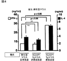
IgE応答は通常Th2細胞の活性化を必要とするため、次に、病変皮膚の移植後にホスト中のCD4+T細胞がTh2細胞に分化するかを調べた。具体的には、正常なB6マウスに対して、KCASP1Tgマウスの病変皮膚、非病変皮膚または野生型マウスの正常な皮膚を移植し、21日後に脾臓のCD4+T細胞を固相化された抗CD3とともに48時間インキュベートし、上清におけるIL−4濃度(塗りつぶした棒グラフ)とIFN−γ濃度(白抜きの棒グラフ)とをELISAで決定した。その結果を図4に示す。
図4では、IL−4濃度を塗りつぶした棒グラフに示し、IFN−γ濃度を白抜きの棒グラフに示す。それぞれのデータは、3個の組の平均と標準偏差とを示す。3の独立した実験から類似の結果が得られた。なお、NSは有意性なしを示す。
図4に示すように、KCASP1Tgマウスの病変皮膚を移植した後、21日目のホストにおける脾臓のCD4+T細胞は、正常なB6マウスの皮膚を移植したホストと比較して、固相化された抗CD3に応答してIL−4およびIFN−γの双方を多量に産生した。
これに対して、非病変皮膚を移植したレシピエントでは、脾臓のCD4+T細胞は、B6野生型マウスの皮膚を移植したレシピエントのものに匹敵する程度の量のIL−4とIFN−γを産生した(プレート結合抗CD3に対する応答)。しかしながら、データは示さないが、移植後7日目では、KCASP1Tgマウスの病変皮膚または非病変皮膚を移植したレシピエントのCD4+T細胞は、野生型マウスの正常な皮膚を移植したコントロールのレシピエントのCD4+T細胞と同程度の量のIL−4とIFN−γしか分泌しなかった。
これらの結果は、CD4+T細胞がTh1/Th2細胞に分化するためには、病変皮膚の移植片の移植による刺激から2−3週間必要であることを示す。
〔実施例4:IL−18、CD4およびstat6に依存するIgEの誘導〕
KCASP1Tgマウスの病変皮膚が、IgEを誘導するメカニズムを細胞レベルで分析した。ところで、CD4+T細胞を除いたマウスはIL−18を投与されても血清IgE濃度の上昇が見られないことが知られている。そのため、ホスト由来のCD4+T細胞が本来必要とされるのか、あるいは、病変皮膚に浸潤したIL−4を産生するドナーT細胞(図2E参照)が、IgE誘導に必要な条件を備えているのかを調べた。
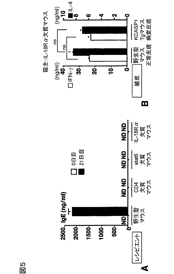
具体的には、野生型マウス、CD4欠失マウス、stat6欠失マウスまたはIL−18Rα欠失マウスに、KCASP1Tgマウスの病変皮膚を移植した。0日目と21日目に、IgE測定のために血清をサンプリングした。その結果を図5Aに示す。白抜きの棒グラフが0日目の結果であり、塗りつぶした棒グラフが21日目の結果を示す。各データはそれぞれの実験群における3匹のマウスの平均と標準偏差とを示す。
まず、CD4欠失マウスは、図5Aに示すように、KCASP1Tgマウスの病変皮膚を移植した後でさえもIgEを産生しなかった。この結果から、KCASP1Tgマウスの病変皮膚の移植片中のIL−4産生細胞あるいはIL−18は、ホストのCD4+T細胞なしにはIgEの全身性の上昇を誘導することができないことを示唆している。
また、stat6がIL−4のシグナル伝達に不可欠であることから、次に、ホストのstat6の活性化がIgE誘導に決定的かを分析したところ、図5Aに示すように、stat6欠失マウスは、病変皮膚を移植した後、いかなるIgEの上昇も示さない。この結果は、病変皮膚の移植片により誘導されたIgE応答は、ホスト由来のCD4+T細胞およびstat6に依存的であることを示唆している。データは示さないが、B6マウスの正常な皮膚の移植もCD4+T細胞またはstat6が欠失した宿主において、IgE産生を誘導しなかった。したがって、皮膚移植によるIgEの誘導は、ホストが完全なCD4+T細胞およびstat6を供えているときだけに起こる。
さらに、移植片中のIL−18が、ホスト中でIgEの誘導を引き起こすか否かを調べた。この目的のために、ホストとして、IL−18に応答することができないIL−18Rα欠失マウスを用いた。図5Aに示すように、IL−18Rα欠失マウスでは、IgEの上昇は見られなかった。また、データは示さないが、B6マウスの正常な皮膚を移植したIL−18Rα欠失マウスはIgEの上昇を示さなかった。
総合すると、これらの結果は、皮膚の病変部位における少量のIL−18の持続的な集積は、ホスト由来のCD4+T細胞およびstat6に依存的な、全身性のIgEの上昇を誘導する。
また、病変皮膚を移植された野生型マウスが、CD4+T細胞のTh1細胞およびTh2細胞への分化を伴うIgE応答を示したことから(図4参照)、このTh1/Th2細胞への分化も、病変皮膚の移植片に存在するIL−18に依存するかを調べた。
具体的には、IL−18Rα欠失マウスに、B6野生型マウスの正常な皮膚またはKCASP1Tgマウスの病変皮膚を移植した。21日目にレシピエントから脾臓のCD4+T細胞を単離し、固定化された抗CD3で刺激し、産生されたIL−4とIFN−γとをELISAで測定した。その結果を図5Bに示す。
白抜きの棒グラフがIFN−γの結果であり、塗りつぶした棒グラフがIL−4の結果である。各データはそれぞれの実験群における3匹のマウスの平均と標準偏差とを示す。なお、NSは有意性がないことを示す。
図5Bに示すように、病変皮膚の移植片を移植されたIL−18Rα欠失マウスのCD4+T細胞は、野生型マウスの正常な皮膚を移植されたIL−18Rα欠失マウスのCD4+T細胞と比較して、そのIL−4あるいはIFN−γの産生量に関して相違を認めなかった。この結果から、病変皮膚の移植片を介したTh1/Th2細胞の分化は、移植片から放出されたIL−18に依存している可能性が示される。
〔実施例5:in vivoにおけるSpAによるIL−18およびIgEの誘導〕
黄色ブドウ球菌の感染は、時にはADの患者において、皮膚の炎症変化を悪化させ、いくらかのAD患者では血清IL−18濃度が上昇することが知られている。そのため、黄色ブドウ球菌の産生物が、正常なB6マウスにおけるIL−18の全身的な上昇を引き起こすことができるかを調べた。
具体的には、正常なB6マウスの耳の皮膚に、1日1回様々な用量のSpAまたは溶媒のみを2週間塗布した。その後、14日目にIL−18またはIgEの濃度を測定するために血清および脾臓をサンプリングした。さらに、溶媒またはSpA(100μg/日)を塗布したマウスの脾臓のCD4+T細胞をプレート結合抗CD3とともに48時間インキュベートし、それぞれの上清におけるIFN−γおよびIL−4の濃度をELISAで測定した。その結果を図6A〜Cに示す。
図6AはIL−18の結果を示し、図6BはIgEの結果を示す。さらに、図6Cにおける白抜きの棒グラフはIFN−γの結果を示し、塗りつぶした棒グラフはIL−4の結果を示す。図6A・Bのデータは、それぞれの群において、3匹のマウスの平均と標準偏差とを示す。図6Cのデータは3つのサンプルの平均と標準偏差とを示し、それぞれの群における3匹のマウスのうち、1匹のデータを示す。なお、NDは検出されないことを示し、NSは有意性がないことを示す。
図6Aに示すように、SpAを塗布したマウスでは、血清中のIL−18のレベルは、投与量依存的に上昇したが、溶媒のみを塗布したマウスでは上昇しなかった。データは示さないが、SpAまたは溶媒を塗布したマウスにおける血清中のIL−12p40、IL−12p70は、ELISAでは検出されなかった。また、図6Bに示すように、IgEレベルも投与量依存的に上昇した。加えて、図6Cに示すように、SpAで処理されたマウスは、Th2細胞優位な分化を示さなかった。これらの結果は、IL−18処理と同様に、SpAがTh1/Th2細胞どちらかの優先的な分化なしに、全身性のIgEを誘導する可能性を示す。
〔実施例6:SpAに応答してKCはIL−18を分泌するが、IL−12は分泌しない〕
IL−18は、IL−12がなければIgEを誘導することができ、IL−12があれば逆にIgE誘導を阻害する。これまで、IL−18の分泌を誘導するLPS(リポ多糖類)を含めた刺激剤は常にIL−12の産生を引き起こすことが知られている。そこで、次に、SpAに応答して、どのタイプの細胞がIL−18を分泌し、これらの細胞がIL−12の産生を伴わずにIL−18を分泌するかを調べた。
具体的には、PAM212細胞(マウスのKCの培養株)をSpAとともに、またはSpAなしで24時間インキュベートし、それぞれの上清中のIL−18濃度とIL−18のIFN−γを誘導する生物活性をELISAとバイオアッセイでそれぞれ測定した。その結果を図7Aに示す。図7Aでは、左側がIL−18の濃度を示し、右側がIFN−γを誘導する活性度を示す。なお、図7Aのデータは、3つの値の平均と標準偏差とを示している。また、NDは検出されないことを示す。
図7Aから明らかなように、SpAによる刺激の後、PAM212細胞は、IFN−γ産生を誘導することができる活性型のIL−18を分泌している。
さらに、新たに単離したKCがSpAに応答してIL−18を分泌するか実証するために、野生型B6マウスのKCをいろいろな用量のSpAと24時間インキュベートした。
具体的には、野生型B6マウスの皮膚から調製したKCまたはCD11+細胞を除いたKCについて、CD11cとMHCクラスII(1−Ab)の発現をフローサイトメトリーで分析した。これらの細胞はSpA存在下、またはSpAのない状態で24時間インキュベートし、上清のIL−18濃度をELISAで測定した。その結果を図7Bに示す。図7Bでは、野生型B6マウスの皮膚から調製したKCを塗りつぶした棒グラフで示し、CD11c+細胞を除いたKCを白抜きの棒グラフで示す。
図7Bの左側に示すように、新たに単離されたKCはSpAに応答して用量依存的にIL−18を放出した。LC/DCはIL−18を放出することができるので、CD11c+細胞をKC標本から除いて、それらをSpAとインキュベートした。CD11c+細胞を除去した後であっても、図7Bの右側に示すように、KCはSpAの刺激を受けるとIL−18を分泌した。なお、図7Bのデータは、3つの値の平均と標準偏差とを示している。また、NDは検出されないことを示す。
次に、SpAに誘導されるKCからのIL−18分泌の分子的なメカニズムを解明するため、caspase−1がLPS(リポ多糖類)の誘導によるIL−18分泌に必要であることから、SpAによる刺激を受けたcaspase−1欠失KCからのIL−18分泌を調べた。
具体的には、KCは、野生型マウス、caspase−1欠失マウス、TLR2欠失マウス、またはMyD88欠失マウスから調製し、野生型マウスのKCは20μMZVAD、20μMYVADまたは同じ体積のDMSO(Veh)の存在下、500μg/mlのSpAとともに24時間インキュベートした。同時に、上記各突然変異体のKCを500μg/mlのSpAとともに24時間インキュベートした。それぞれの上清中のIL−18をELISAで測定した。その結果を図7Cに示す。
図7Cでは、WTが野生型マウスを、KOが上記各突然変異体(ノックアウトタイプ)のマウスを示し、白抜きの棒グラフがcaspase−1欠失マウスを、網掛けの棒グラフがTLR2欠失マウスを、斜線で示す棒グラフがMyD88欠失マウスを示す。
図7Cに示すように、DMSOを加えずに500μg/mlのSpAとインキュベートした野生型マウスのKCは、28.4±3.5pg/mlのIL−18を産生した。また、caspase−1欠失マウスのKCは野生型マウスのKCに匹敵するレベルのIL−18を分泌する。そのため、SpAの刺激に対するcaspase−1に依存しないIL−18の分泌を示唆している。なお、図7Cのデータは、3つの値の平均と標準偏差とを示している。
caspase−1阻害剤であるYVADや、広い範囲のcaspaseを阻害するZVADは、LPSまたは膜結合型のFasリガンド刺激による肝臓の組織マクロファージであるKupffer細胞からのIL−18分泌を強力に阻害する。そこで、SpAの刺激を受けたKCについても、IL−18の分泌がYVADまたはZVADで影響されるかを調べた。図7Cに示すように、これらの2種類のcaspase阻害剤はSpAの刺激を受けた野生型マウスのKCからのIL−18分泌を阻害しなかった。この結果は、SpAの刺激を受けたKCからのIL−18分泌において、caspaseは不必要であることを示す。データは示さないが、これはcaspase−1欠失マウスのKCの場合についても同様のことが言える。
さらに、多くの微生物がTLR/MyD88シグナル経路を刺激することから、KCが、グラム陽性菌に対する信号レセプターであるTLR2または全てのTLRメンバーによってシェアされる不可欠なアダプター分子であるMyD88に依存してIL−18を分泌するのかを調べた。図7Cに示すように、TLR2とMyD88の両者ともに、SpA刺激で誘導されるKCからのIL−18分泌に不必要であることから、SpAによるIL−18の分泌誘導は、TLRに依存しないIL−18分泌であることを示唆している。
また、Kupffer細胞がLPSまたは膜表現型のFasリガンドによる刺激に応答してIL−18を分泌することから、これらの刺激剤がKCからのIL−18分泌を誘導するかを調べた。具体的には、野生型B6マウスからのKC(5×10⁵/ml)を、500μg/mlのSpA、1μg/mlのLPS、または1×106/mlのmFasLとともに24時間インキュベートした。得られた上清に含まれるIL−18は、ELISAで測定した。その結果を表1に示す。
次に、SpAによる刺激を受けたKCが同時にIL−12を分泌するかを調べた。微量のIL−12を確認するために、SpAと4時間インキュベートしたKCから総RNAを得、これを用いてRT−PCRを行った。具体的には、野生型のB6マウスのKCとKupffer細胞とを500μg/mlのSpAの存在下または非存在下、並びに、1μg/mlのLPSの存在下または非存在下で、それぞれ4時間インキュベートした。抽出した総RNA中のIL−18、IL−12p40、IL−12p35およびβ−アクチンのmRNAの発現レベルをRT−PCRで測定した。その結果を図7Dに示す。
図7Dに示すように、LPSで刺激されたKupffer細胞がIL−12p35とIL−12p40との双方を発現したのに対し、SpAによる刺激を受けたKCでは、IL−12p35もIL−12p40も検出されなかった。8時間または16時間SpAとインキュベートしたKCから得た、総RNAを用いてIL−12に対するRT−PCRを行ったが、それらの中にはIL−12の何れも検出されなかった。データは示さないが、ELISAでもIL−12を測定したが、24時間インキュベートした後のSpAによる刺激を受けたKCの上清にはIL−12p40またはIL−12p70は検出されなかった。
これに対して、Kupffer細胞のKCは、IL−18を恒常的に発現し、SpAによる刺激を受けた後も発現のレベルは変わらなかった。また、データは示さないが、Kupffer細胞はSpAに対する応答としてIL−12またはIL−18を分泌しなかった。
これらの結果から、皮膚に塗布されたSpAの刺激により、局部的にKCがIL−18を分泌するが、IL−12は分泌せず、これがIgEの誘導につながることが示される。
〔実施例7:SDSとSpAの混合物を塗布したNC/Ngaマウスにおける皮膚病変〕
NC/Ngaマウスは、アトピー性皮膚炎好発マウスで、SPFでは発症しないが、コンベンショナルな環境下に移すと発症する。NC/Ngaマウスに4%のSDSあるいはSpA(200μg/日)を単独で塗布しても発症しないが、両者を混合してNC/Ngaマウスの剃毛した背部に塗布すると、AD様の皮膚病変が観察される。
図8のAないしDは、4%SDSとSpA(200μg/日)の混合物を塗布したNC/Ngaマウスにおける皮膚病変の結果を、コントロールの結果とともに示す図である。図8Cに示すように、4%SDSのみを塗布した場合は28日後でも発症は見られないが、4%SDSとSpA(200μg/日)の混合物を塗布した場合は、図8B、図8Dに示すように、14日後、28日後にAD様の皮膚病変が観察された。
図9のAないしDは、は4%SDSとSpA(200μg/日)の混合物を塗布したNC/Ngaマウスにおける皮膚の経時変化をコントロールの結果とともに示したものである。図9のAないしCは、ヘマトキシリン・エオジンで染色したAD様の病変皮膚を光学顕微鏡で観察した結果を示すものである。図9Aは倍率25倍、図9Bは倍率50倍、図9Cは倍率100倍の観察結果を表す。図9Dはアルシアン・ブルーで染色したAD様の病変皮膚を光学顕微鏡で観察した結果である。図9Dに示したように、組織学的には、病変部において表皮の肥厚が著明となり、アルシアン・ブルーで染色される肥満細胞の浸潤が認められるようになる。
尚、発明を実施するための最良の形態の項においてなした具体的な実施態様または実施例は、あくまでも、本発明の技術内容を明らかにするものであって、そのような具体例にのみ限定して狭義に解釈されるべきものではなく、本発明の精神と次に記載する特許請求の範囲内で、いろいろと変更して実施することができるものである。
産業上の利用の可能性
以上のように、本発明では、KCからのIL−18の局所的な集積が、抗原への暴露なしに全身性のIgE応答を引き起こし、これがAD様の炎症性皮膚病変において生ずる血清中の高レベルのIgE発現という現象を導くという知見に基づいて実現されたスクリーニング方法、およびアトピー性皮膚炎様症状の誘導方法、並びにその利用である。
それゆえ、本発明は、微生物刺激で誘導されるKCからのIL−18産生の重要性を示し、未知の抗原によるアレルギー疾患の病因に関連するであろうメカニズムに新しい見識を提供するとともに、ADの治療薬剤等の開発に有効に利用することができる。
したがって、本発明は、各種医薬品産業や研究用試薬産業に好適に利用できるだけでなく、医療分野にも応用することができる。それゆえ、本発明は、微生物刺激で起こるKCからのIL−18産生の重要性を示し、未知の抗原によるアレルギー疾患の病因に関連するであろうメカニズムに新しい見識を提供するとともに、ADの治療薬剤等の開発に有効に利用することができる。We have shown that the local accumulation of IL-18 produced from keratinocytes causes a systemic IgE response without exposure to antigen, which leads to inflammatory skin lesions like atopic dermatitis. I found it. In the present invention, using the above findings, in particular, a screening method for inhibitors that can be suitably used for elucidation of the onset mechanism of atopic dermatitis and its similar symptoms and development of therapeutic agents, and atopic dermatitis The induction method of the symptom and the usage method were realized. Hereinafter, the present invention will be specifically described.
(1) Screening method according to the present invention
The screening method according to the present invention includes an environment formation step for forming an environment inducing local IL-18 production from keratinocytes (KC) by stimulation with a stimulant in vivo or in vitro, An inhibitor identification step of administering a candidate substance below and identifying a substance that inhibits the production induction of
In atopic dermatitis (AD) -like inflammatory skin lesions, IL-18 is locally produced in excess from KC in the skin, which may result in high levels of IgE expression in the serum. Therefore, it is possible to obtain a substance that inhibits the secretion of IL-18 by using a screening method including the environment formation step and the inhibitor identification step. The obtained substance (inhibitor) can be an effective therapeutic agent for AD.
The AD-like inflammatory skin lesion referred to in the present invention is an immune disease that develops in an IL-18-dependent manner and broadly refers to a symptom in which inflammation occurs in the skin, and is an itching that is strictly recognized as AD. It does not refer to only chronic chronic inflammation.
In the present invention, “in vitro” refers to a reaction system artificially reproduced by cultured cells, and “in vivo (in vivo)” refers to a reaction system in a complete individual. And
Furthermore, the “inhibitor” referred to in the present invention may be any substance that inhibits the secretion of IL-18 from KC, and the specific mechanism of the inhibition is not particularly limited. Therefore, the inhibitor obtained by the screening method according to the present invention may reversibly inhibit the process of IL-18 secretion from KC, or may inhibit it irreversibly. Good. In addition, in the case of an inhibitor that reversibly inhibits, it may be a competitive (antagonistic) inhibitor (antagonist) or a non-competitive inhibitor. In other words, in the screening method according to the present invention, various types of inhibitors such as containing an antagonist can be screened.
<Environment formation process>
The environment forming step is not particularly limited as long as it is a step of forming an environment that induces IL-18 production from KC by stimulation with a stimulant. That is, it may be in vivo or in vitro as long as an environment capable of inducing the production of IL-18 can be realized.
Examples of the stimulant include various proteins that can stimulate KC (KC stimulating protein). The KC stimulating protein is not particularly limited as long as it is a protein capable of stimulating KC, but representatively, protein A (SpA) derived from S. aureus is used. Can be mentioned.
As described above, it has been known so far that infection with Staphylococcus aureus is an attractant or exacerbation factor of AD. Furthermore, when SpA is applied to mice with SDS within a short period of time. It was found that AD-like pruritic chronic dermatitis can be induced (see Examples). Therefore, the SpA can be particularly preferably used as the KC stimulating protein.
The SpA may be a complete protein (a protein having a complete amino acid sequence) obtained from Staphylococcus aureus, but if it can stimulate keratinocytes, it may be a partial protein of SpA, a complete protein, or a partial protein. It may be a modified form.
The above-mentioned variant consists of an amino acid sequence in which one or several amino acids are substituted, deleted, inserted and / or added in the known amino acid sequence of SpA, and on the cell surface of the B7-2 molecule. It shall mean a protein that negatively regulates expression. In addition, the above “one or several amino acids are substituted, deleted, inserted, and / or added” means substitution, deletion, or the like by a known mutant protein production method such as site-directed mutagenesis. Meaning that the number of amino acids that can be inserted and / or added (preferably 10 or less, more preferably 7 or less, and even more preferably 5 or less) are substituted, deleted, inserted, and / or added. To do. Thus, the above-mentioned variant is the above-mentioned SpA mutant protein, and the “mutation” referred to here mainly means a mutation artificially introduced by a known mutant protein production method. The mutant protein may be isolated and purified. In addition, the modified SpA may contain an additional polypeptide.
Moreover, as said stimulant, it is more preferable to use SDS together with SpA. By using SDS and protein A in combination, more severe AD-like pruritic chronic dermatitis can be induced (see Examples). The method of using SpA and SDS in combination is not particularly limited, but an SpA-SDS solution obtained by adding SDS to a solvent that dissolves SpA may be prepared and applied to the skin of a model organism.
<Stimulation method of KC>
The method for stimulating KC with the KC stimulating protein is not particularly limited as long as it is a method capable of inducing IL-18 production by stimulating KC. Specifically, for example, when the environment formation step is performed in vitro, the SpA may be added to the culture solution of KC cultured cells and cultured. The KC cultured cells used at this time are not particularly limited, and known cultured cells can be suitably used. For example, in Example 6 described later, PAM212 cells are used.
The culture conditions of the above KC cultured cells, that is, the culture solution, the culture temperature, the way of adding SpA, etc. are not particularly limited, and appropriate conditions may be set according to the type of cultured cells used.
On the other hand, when the environment forming step is performed in vivo, the SpA may be applied to the skin of a host organism. The conditions for applying SpA are not particularly limited, and any method that does not hinder the stimulation of KC by SpA may be used. For example, in the examples described later, SpA is used as a PBS solution of 50% glycerol, but the present invention is not limited to this. Further, as described above, a SpA-SDS solution in which SDS is added to SpA may be prepared and applied to the skin of the host organism. In addition, it is not specifically limited about application | coating conditions, for example, the application | coating method and the site | part applied to a host organism.
The host organism used at this time is not particularly limited, but usually a mammal generally used for various experiments may be used. Specific examples include experimental animals such as mice, rats, rabbits, pigs, monkeys. For example, in the examples described later, a mouse is used as the host organism. In particular, the mouse is widely used as an experimental animal, and has advantages such as the availability of various mutant strains, and the fact that the breeding space can be made relatively small due to the small number of individuals. This is preferable.
<Transplantation of skin piece>
When the environment formation step is performed in vivo, a substance other than a KC stimulating protein such as SpA can be used as the stimulant. As a representative example, skin (lesioned skin) in which AD-like inflammatory skin lesions have occurred can be used. An environment capable of inducing production of IL-18 can also be realized by transplanting the skin pieces of the diseased skin into a host organism.
The lesioned skin is not particularly limited as long as it has AD-like inflammatory skin lesions. However, from the viewpoint of transplantation, the skin of the same species as the host organism is very preferable. In the case of heterogeneous organisms, an immune response occurs due to causes other than AD-like inflammatory skin lesions such as rejection, which is not preferable. Examples of the host organism include various experimental animals as described in the explanation of the KC stimulating protein. For example, when a mouse is used as the host organism, the skin piece of the lesion skin to be transplanted is also derived from the mouse. It is very preferred (see the examples below).
Here, the host organism is at least (i) CD4 + Each of these traits must be equipped with T cell expression, (ii) stat6 expression, and (iii) IL-18Rα chain expression in NKT cells. As will be apparent from the results of Example 4 described later, CDE-deficient mice, stat6-deficient mice, and IL-18Rα chain-deficient mice are all IgE even when transplanted with skin pieces of lesioned skin obtained from KCASP1Tg mice. Production was not induced. That is, in order to realize an environment in which the production of IL-18 can be induced, the above characteristics (i) to (iii) are necessary conditions.
The above CD4 + T cells refer to T cells that express the antigen CD4 (one of the cell membrane glycoproteins of helper cells). The presence or absence of CD4 expression is indicated by plus or minus with an upper right. Therefore, for CD4, if expressed, “CD4 + ", And when not expressed" CD4 − ". Stat6 (STAT6) is an intracellular signal transduction molecule involved in cytokine action mechanism, and is specifically activated by IL-4. The IL-18Rα chain is expressed in T cells. It is a structure involved in the IL-18 response.
<Inhibitor identification step>
In the inhibitor identification step, a candidate substance is administered under an environment capable of inducing the production of IL-18 formed in the environment formation step, and a substance that inhibits the production induction of
The technique for administering the candidate substance is not particularly limited. For example, in the case of in vitro, the candidate substance may be administered to a culture solution in which KC cultured cells are cultured. Moreover, if it is in vivo, a candidate substance may be apply | coated to the inflammation site | part in a host organism, or a host substance may be taken internally. Moreover, the method for identifying the inhibitor is not particularly limited as long as it can reliably confirm that the induction of IL-18 production by KC is inhibited. Whether the environment in which IL-18 production can be induced is in vitro or in vivo, a cell-free system using ELISA or the like can be used.
In addition, the specific method is not specifically limited if the screening method concerning this invention contains the environment formation process and inhibitor identification process which were mentioned above, Of course, you may include another process.
<Immune disease therapeutic drug / High-level IgE expression suppression method>
Furthermore, the present invention includes a therapeutic agent for immune diseases including an inhibitor obtained by using the above screening method. There are no particular limitations on the specific composition or the like of this therapeutic agent for immunological diseases, depending on the type of inhibitor screened and the state of the organism (including humans) to be administered, It only has to contain an additive.
Furthermore, the present invention also includes a method for suppressing high-level IgE expression using the inhibitor obtained by using the screening method or the therapeutic agent for immune disease. In this method, the systemic IgE response caused by exposure to antigen is suppressed by local accumulation of IL-18 produced from KC by using the inhibitor or the therapeutic agent for immune diseases. As a result, it is possible to suppress or inhibit the phenomenon of high-level IgE expression in serum that occurs in inflammatory skin lesions like atopic dermatitis.
Induction of IL-18 production from KC may directly lead to the onset of AD, or may lead to the onset of AD by leading to a high level of IgE expression. In any of the above cases, the present invention can effectively treat or alleviate the pathology of AD.
(2) The method for inducing atopic dermatitis-like symptoms according to the present invention
In the above (1), among the present invention, the method for screening a therapeutic agent for AD or a similar symptom thereof from a candidate substance has been described in detail, but the present invention is not limited to this, and AD or a similar symptom thereof. In order to elucidate the onset mechanism of AD, a method of inducing AD-like symptoms in which AD-like inflammatory skin lesions are developed in a model organism is also included.
In the method for inducing AD-like symptoms according to the present invention, as described in the case where the KC stimulating protein is used as a stimulating agent in the environment forming step (1), protein A derived from S. aureus is used in the host organism. Any method may be used as long as it is applied to the skin. The coating conditions and the like at this time are not particularly limited as in the above (1).
Further, as described in the above (1), the protein A can stimulate a complete protein of the protein A, a partial protein of the protein A capable of stimulating keratinocytes, or a keratinocyte. What is necessary is just at least any one of the modification of the said protein A or its partial protein.
As the host organism, as described above, laboratory animals such as mice, rats, rabbits, pigs, monkeys, particularly mice can be preferably used. In other words, the present invention includes a model organism that has developed an inflammatory skin lesion obtained by the above induction method.
(3) Use of the present invention
As described above, the screening method and the method of inducing AD-like symptoms according to the present invention can be suitably used for elucidating the onset mechanism of AD and similar symptoms and developing therapeutic agents. Here, each said method concerning this invention may be implement | achieved by the kit. That is, the present invention may include a screening kit for carrying out the screening method or an induction kit for carrying out the method for inducing AD-like symptoms.
Although the specific configuration of the screening kit is not particularly limited, for example, at least a stimulant (particularly a KC stimulating protein, a promoter) and various materials used in the inhibitor identification step (for example, SpA binding) And a structure including a protein and a signal transduction molecule). Similarly, the specific configuration of the AD-like symptom induction kit is not particularly limited, but may include a configuration including an SpA solution and an application means capable of efficiently applying SpA.
The screening method according to the present invention is suitably used for screening for therapeutic agents for immune diseases for treating AD-like inflammatory skin lesions. The method for inducing AD-like symptoms according to the present invention can be used for elucidating the onset mechanism of AD-like symptoms by artificially developing AD-like symptoms in a model animal. It can also be used to verify the effect of the inhibitor obtained by the screening method according to the invention.
For example, suppose that as a screening method according to the present invention, an inhibitor (for example, substance X) is obtained by a technique for realizing the environment formation step in vitro. In this case, the effect of substance X on KC cultured cells is observed. Thus, a mouse in which AD-like symptoms have developed using the method for inducing AD-like symptoms according to the present invention can be prepared, and the effect of substance X can be verified in vivo using this.
Needless to say, the method of use of the present invention is not limited to the above example, and can be used for various other purposes.
As described above, in the present embodiment, the present invention has been described in detail with specific examples. However, the present invention is not limited to the above embodiment, and the present invention departs from the gist thereof. It is possible to implement the present invention in a mode in which various improvements, changes, and modifications are made based on the knowledge possessed by those skilled in the art without departing from the scope.
Hereinafter, although an example and a comparative example explain the present invention still in detail, the present invention is not limited to this. Details of the mice and reagents used in the examples and specific experimental methods are shown below.
〔mouse〕
Female C57BL / 6 (B6) wild type mice (6-10 weeks) were purchased from CLEA Japan. B6 background CD4 deficient mice (female, 6-10 weeks) Received courtesy of Taniguchi. B6 background stat 6-deficient mice (female, weeks 6-10) were obtained from Dr. Osaka University. Provided by courtesy of Takeda (Takeda, K., Tanaka, T., Shi, W., Matsumoto, M., Minami, M., Kashiwamura, S., Nakanishi, K., Yoshida, N., Kishimoto, T. and Akira.S., 1996. Essential role of STAT6 in IL-4 signaling. Nature 380: 627).
IL-18Rα-deficient mice in which B6 mice and F10 mice (female, 6-10 weeks) were crossed were obtained from Dr. Osaka University. Provided by Hoshino (Hoshino, K., Tsutsui, H., Kawai, T., Takeda, K., Nakanishi, K., Takeda, Y. and Akira, S. 1999. Generation of IL-18 receptor- (definition mice: evidence for IL-1 receptor-related protein as an essential IL-18 binding receptor. J. Immunol. 162: 5041).
As skin graft donors, female mice suffering from chronic skin diseases with high serum IgE concentrations (10-12 μg / ml) and IL-18 concentrations (5-7 ng / ml) were selected and used. F6 mice (female, 6-10 weeks) obtained by crossing caspase-1 deficient mice with B6 wild type mice were used (Seki, E., Tsutsui, H., Tsuji, NM, Hayashi, N., Adachi, K., Nakano, H., Futatsugi-Yumikura, S., Takeuchi, O., Hoshino, K., Akira, S., Fujimoto, J. and Nakanishi, K. 2002. Crit. of myeloid differentiation factor 88-dependent prolaminatory cytokinase release in early phase clearance of Listeria monocytogenes in mice.J. Immunol.169: 3863). B6 / 129 background myeloid differentiation factor 88 (MyD88) -deficient mice and Toll-like receptor (TLR) 2-deficient mice F4 mice (female) were obtained from Dr. Osaka University. Provided by Akira (see Seki et al). All mice were kept under aseptic conditions.
〔reagent〕
SpA purified from
[Skin transplant]
Skin specimen (1cm 2 ) Obtained from normal skin of wild-type B6 mice or skin (lesioned skin) or non-skinned skin (non-lesional skin) of KCASP1Tg mice, and then wild-type B6 mice, CD4-deficient mice, They were transplanted on the back of stat6-deficient mice or IL-18Rα-deficient B6 mice. As skin specimens, two grafts were obtained for KCASP1Tg lesioned or non-lesional skin.
After skin transplantation, the recipients were given drinking water with 1 mg / ml neomycin sulfate (Sigma) and 1000 U / ml polymixin B sulfate (Sigma) to prevent all possible infections. Where appropriate, serum was sampled to determine IgE concentrations.
The engraftment rate of transplanted skin in each transplanted host was observed up to
[Skin lysate]
Skin specimen (1cm 2 ) Was obtained from wild-type B6 mice or KCASP1Tg mice, and the epidermis sheets prepared from the skin specimens were homogenized at 4 ° C. in PBS (phosphate buffered saline) and filtered. The concentrations of various cytokines and proteins contained in each lysate were measured with an ELISA kit using a cytokine reagent and a protein evaluation reagent (Pierce).
[Th1 / Th2 differentiation]
CD4 of the spleen + T cells were isolated from mice treated with various techniques using AutoMACS (Miltenyi Biotec). Freshly isolated spleen cells were incubated with anti-CD4 beads (Miltenyi Biotec). CD4 + T cell purity was greater than 98%. Cells (1 × 10 6 / Ml) was incubated with immobilized anti-CD3ε (manufactured by PharMingen) for 48 hours, and the concentrations of IFN-γ and IL-4 in each supernatant were determined by ELISA.
[Evaluation of cytokines and IgE]
The IL-18 concentration was determined by an ELISA kit manufactured by MBL. IL-4, IFN-γ and IL-1β were determined using an ELISA kit manufactured by Genzyme. The IL-5 ELISA kit was purchased from Endogen. Serum IgE concentration was determined using an ELISA kit manufactured by PharMingen. The ability of IL-18 to induce IFN-γ was determined by a bioassay using an IL-18-responsive mouse NK cell clone named LNK series (see
Specifically, LNK5E6 cells have higher responsiveness to IL-18 stimulation with respect to IFN-γ production than LNK5E3 cells (see Non-Patent Document 6). Incubated with 100 pg / ml IL-12 in the presence or absence of anti-IL-18 antibody (50 μg / ml) for 48 hours. The activity of IL-18 is defined as the concentration of IFN-γ produced by cells in response to IL-18, as shown in the following formula (see Non-Patent Document 6).
IL-18 activity = (IFN-γ in the supernatant in the absence of anti-IL-18 antibody) − (IFN-γ in the supernatant in the presence of anti-IL-18 antibody).
[SpA application]
Various amounts of SpA dissolved in 5 μl of a solvent (50% glycerol in PBS) (SpA solution) were prepared, and this SpA solution was applied to wild type ear skin daily for 14 days. As a control, 5 μl of solvent not containing SpA was used. To avoid the effects of other mice, mice were placed in individual cages.
[Preparation of KC]
KC is a dr. The method shown by Tamaki et al. (Vestergaard, C., Yoneyama, H., Murai, M., Nakamura, K., Tamaki, K., Terashima, Y., Imai, T., Yoshie, O., Irimura, T., Mizutani, H., et al., 1999. Overproduction of Th2-specific chemokin in NC / Ngamice exhibiting atomic dermatitis-likeles, 109. J.t. G., Gullino, M., Sachs, DH and Katz, SI 1979. la antigens in mouse. skin are predominantly expressed on Langerhans cell.J.Immunol.123: according 784 reference), was prepared from various genotypes of mice, in order to restore normal expression of surface molecules, were incubated overnight in culture. To remove DC, KC was incubated with CD11c microbeads (Miltenyi Biotec), then CD11c + Cells were removed using AutoMACS.
KC (5 × 10 5 / Ml) or PAM212 cells (2 × 10 5 / Ml) with varying amounts of SpA and 1 μg / ml LPS or mFasL (1 × 10 6 / Ml) for 24 hours. In some examples, wild type KCs were incubated with 100 μg / ml SpA for 24 hours in the presence of 20 μM ZVAD or YVAD. IL-18 concentration and its activity in each supernatant were determined by ELISA and bioassay, respectively. In some examples, KCs are incubated with 500 μg / ml SpA for 4 hours, total RNA is extracted and RT-PCR (Tsutsui, H., Matsui, K., Kawada, N., Hyodo, Y.). , Hayashi, N., Okamura, H., Higasino, K. and Nakanishi, K. 1997. IL-18 accounts for bottling in TNF-α-and Fasligand-modified hepatitoxic inpatients. Immunol.159: 3961). For primers for IL-18, IL-12p35, IL-12p40 or β-actin and PCR conditions for each cytokine, see Tsutsui et al., Supra. See
[Preparation of Kupffer cells]
Preparation of Kupffer cells (Kupffer cells) was performed according to
[FACS (fluorescence display cell sorter) analysis]
DC, CD4 + T cells and CD8 + The ratio of T cells was determined by comparing KC prepared according to
[Statistics]
All data are presented as the mean and standard deviation of 3 sets. Significance between the control group and the treatment group was performed by an independent student test. P <0.05 was considered significant.
[Example 1: Induction of IgE by skin graft transplantation from KCASP1Tg mice]
First, it was examined whether the lesion skin of KCASP1Tg mice had the potential to induce an increase in serum IgE concentration when transplanted into normal wild-type mice of the same strain. Specifically, in order to standardize skin graft conditions in each transplant, KCASP1Tg mice that develop chronic dermatitis in the ear, face and torso and have constant IL-18 and IgE concentrations in the serum are donated. Selected as. KCASP1Tg mouse lesion skin pieces or non-lesion skin pieces were transplanted into normal B6 mice, and the serum IgE concentration of the host was measured. The result is shown in FIG.
In FIG. 1, the filled circle in the graph and the upper right photo show the result of transplanting the lesion skin of a KCASP1Tg mouse into a normal B6 mouse, the hatched circle in the graph and the right middle photo Shows the results of transplanting non-lesional skin of KCASP1Tg mice, and the squares in the graph and the lower right photo show the results of transplanting normal skin of wild type mice. At the time indicated in the graph, serum was sampled for IgE measurement by ELISA, and the survival rate of the transplanted skin was observed (upper part of FIG. 1: survival rate of the transplanted skin). The data shows the mean and standard deviation of 3 mice in each experimental group. The survival rate of the transplanted skin is shown in the upper panel.
As shown in FIG. 1, B6 mice transplanted with non-lesional skin showed a delayed weak IgE response, whereas in mice transplanted with lesional skin, the IgE concentration in the serum increased early. did. No increase in IgE was observed in control B6 mice transplanted with skin grafts from wild type mice. B6 mice transplanted with lesional skin show a high serum IgE concentration, but the concentration decreases after the graft is detached. On the other hand, B6 mice transplanted with non-lesional skin maintain low IgE concentrations. Further, in the B6 mice transplanted with the lesioned skin, the serum IgE concentration starts to rise again even after the rejection of the lesioned skin.
Although data are not shown, serum IL-18 concentrations did not increase after stimulation with any type of skin graft. Also not shown in the data, serum IL-4 or IL-6 concentrations were not detected with commercial ELISA kits. These results indicate that the lesioned skin of KCASP1Tg mice persistently induces systemic IgE elevation when transplanted into normal B6 mice. This is in contrast to exogenous IL-18-dependent IgE production (unpublished data) that stops immediately after the administration of IL-18 is stopped.
[Example 2: Accumulation of IL-18- and Th2-related cytokines in lesion skin of KCASP1Tg mice]
To elucidate the mechanism by which only skin graft transplantation effectively induces an IgE response in the host, IL-18 concentrations were compared between the lesion skin graft and the non-lesion skin graft. Each skin graft was dissolved as described above, and the IL-18 concentration was measured. The results are shown in FIGS.
In FIGS. 2A-F, solid bar graphs show the results of lesion skin samples sampled from two KCASP1Tg mice, and the shaded bar graphs are non-lesional skin samples sampled from two KCASP1Tg mice. The white bar graph shows the results of the skin sample as a control sampled from three wild type mice. FIG. 2A shows the concentration of IL-18 in the lysate of each of the above skin specimens. Similarly, FIG. 2B shows IL-18 activity (IFN-γ production by IL-18-responsive LNK5E6 cells). 2C shows the concentration of IL-1β, FIG. 2D shows the concentration of IFN-γ, FIG. 2E shows the concentration of IL-4, and FIG. 2F shows the concentration of IL-5. Each result represents the mean and standard deviation of three independent experimental results in each sample. Note that ND indicates that it is not detected.
As shown in FIG. 2A, lesional skin lysates from KCASP1Tg mice showed high IL-18 concentrations by ELISA, and both precursor and mature forms of IL-18 were detected. On the other hand, non-lesional skin lysates from KCASP1Tg mice showed only low IL-18 concentrations, whereas in wild type mouse skin lysates, the concentration of IL-18 was the lowest.
Here, from the immunoblot analysis for IL-18, the lesion site on the skin of KCASP1Tg mice express both 18 kDa IL-18 and 24 kDa IL-18 precursor having biological activity, and the skin of wild type mice. And non-lesional skin of KCASP1Tg mice are known to express only the 24 kDa IL-18 precursor. Therefore, in order to confirm this, a bioassay for IL-18 was performed. As a result, as shown in FIG. 2B, the lesion skin of KCASP1Tg mice showed a high titer of activity of IFN-γ induction by IL-18. On the other hand, non-lesional skin showed little activity. In addition, IL-18 having such physiological activity does not exist in the normal skin of B6 mice.
Further, as shown in FIG. 2C, IL-1β was also concentrated in KC only in the lesioned skin. In addition to IL-18 and IL-1β, other caspase-1 products are also known to be concentrated to KC only in the affected skin. Thus, the IgE concentration in the host corresponds to the concentration of IL-18 and IL-1β in the transplanted graft.
Next, the concentrations of IFN-γ, IL-4 and IL-5 in the lysate of lesion skin of KCASP1Tg mice were measured. As a result, as shown in FIGS. 2D to F, the concentration of IFN-γ, IL-4, and IL-5 in the lysate of lesion skin of KCASP1Tg mice was the same as that of non-lesional skin of wild-type mice and KCASP1Tg mice. Compared to things, everything was up. Thus, lesional skin grafts accumulate large amounts of IL-18, IL-1β, IFN-γ, IL-5 and IL-4.
In order to investigate what type of cells are accumulated in the lesion skin of KCASP1Tg mice, CD4 in KC specimens was analyzed by FACS. + T cells, CD8 + The ratio of T cells and dendritic cells was measured. The results are shown in FIGS.
3A and 3D show the results of KC specimens obtained from normal skin of wild-type mice, FIGS. 3B and 3E show non-lesional skin of KCASP1Tg mice, and FIGS. 3C and 3F show KC specimens obtained from the lesioned skin of KCASP1Tg mice.
3A to C show the results of incubating each KC specimen with anti-CD4 antibody conjugated with PE and anti-CD8 antibody conjugated with FITC, and FIGS. 3D to 3F show anti-CD11c antibodies conjugated with each KC specimen and PE. Anti-IA combined with FITC b The result of incubating with an antibody is shown. Further, in each of FIGS. + T cells and CD8 + T cell ratio and CD11c + DC and IA b + The ratio of DC is shown. Each result shows only one result out of 3 mice in each experimental group.
As shown in FIGS. 3A-F, the lesioned skin has CD4 as compared to wild-type mice. + There was a substantial increase in the number of T cells. Among these three types of recipients, CD8 + There was no difference in the ratio of T cells or DC.
Although data are not shown, the number of mast cells was markedly increased in lesioned skin and not in non-lesioned skin compared to wild type mice in KCASP1Tg mice. This result is consistent with the previous report by the present inventors (Non-patent Document 4). Thus, CD4 + T cells accumulate preferentially in the lesion skin of KCASP1 Tg mice. A large number of mast cells are also accumulated in the lesioned skin.
[Example 3: Induction of Th1 cells and Th2 cells in mice transplanted with lesion skin]
Since IgE responses usually require the activation of Th2 cells, CD4 in the host is then + It was examined whether T cells differentiate into Th2 cells. Specifically, normal B6 mice were transplanted with KCASP1Tg mouse lesion skin, non-lesion skin, or wild-type mouse normal skin, and spleen CD4 after 21 days. + T cells were incubated with immobilized anti-CD3 for 48 hours, and IL-4 concentration (filled bar graph) and IFN-γ concentration (open bar graph) in the supernatant were determined by ELISA. The result is shown in FIG.
In FIG. 4, the IL-4 concentration is shown in a solid bar graph, and the IFN-γ concentration is shown in a white bar graph. Each data shows the average and standard deviation of 3 sets. Similar results were obtained from 3 independent experiments. NS indicates no significance.
As shown in FIG. 4, CD4 of the spleen in the host on
In contrast, in recipients transplanted with non-lesional skin, CD4 of the spleen + T cells produced amounts of IL-4 and IFN-γ comparable to those of recipients transplanted with B6 wild type mouse skin (response to plate-bound anti-CD3). However, data are not shown, but on day 7 post-transplantation, CD4 of recipients transplanted with KCASP1Tg mouse diseased or non-lesional skin. + T cells were obtained from CD4 of control recipients transplanted with normal skin of wild type mice. + Only the same amount of IL-4 and IFN-γ as T cells were secreted.
These results indicate that CD4 + It shows that in order for T cells to differentiate into Th1 / Th2 cells, 2-3 weeks are required from stimulation by transplantation of a lesion skin graft.
[Example 4: Induction of IgE depending on IL-18, CD4 and stat6]
The mechanism by which lesioned skin of KCASP1Tg mice induces IgE was analyzed at the cellular level. By the way, CD4 + It has been known that mice without T cells do not show an increase in serum IgE concentration even when administered with IL-18. Therefore, host-derived CD4 + It was investigated whether T cells were originally required or whether donor T cells producing IL-4 infiltrating the diseased skin (see FIG. 2E) had the conditions necessary for IgE induction.
Specifically, lesion skin of KCASP1Tg mice was transplanted into wild type mice, CD4 deficient mice, stat6 deficient mice or IL-18Rα deficient mice. Serum was sampled for IgE measurements on
First, CD4 deficient mice did not produce IgE even after transplanting the lesioned skin of KCASP1Tg mice, as shown in FIG. 5A. From this result, it was found that IL-4 producing cells or IL-18 in the lesion skin grafts of KCASP1Tg mice were CD4 of the host. + It suggests that without T cells, a systemic increase in IgE cannot be induced.
In addition, since stat6 is essential for IL-4 signaling, it was next analyzed whether the activation of stat6 of the host was critical for IgE induction. As shown in FIG. It does not show any increase in IgE after transplanting the lesioned skin. This result shows that the IgE response induced by the lesion skin grafts is CD4 derived from the host. + This suggests that it is dependent on T cells and stat6. Data not shown, but normal skin transplantation of B6 mice was also CD4 + No IgE production was induced in hosts lacking T cells or stat6. Thus, the induction of IgE by skin grafting is the result that the host is fully CD4 + Occurs only when providing T cells and stat6.
In addition, it was examined whether IL-18 in the graft caused induction of IgE in the host. For this purpose, IL-18Rα-deficient mice that cannot respond to IL-18 were used as hosts. As shown in FIG. 5A, no increase in IgE was observed in IL-18Rα-deficient mice. In addition, although data are not shown, IL-18Rα-deficient mice transplanted with normal skin of B6 mice did not show an increase in IgE.
Taken together, these results indicate that sustained accumulation of small amounts of IL-18 at the site of skin lesions is associated with host-derived CD4 + Induces systemic IgE elevation dependent on T cells and stat6.
In addition, wild-type mice transplanted with lesional skin were found to be CD4. + Since the IgE response accompanied by differentiation of T cells into Th1 and Th2 cells was shown (see FIG. 4), this differentiation into Th1 / Th2 cells is also dependent on IL-18 present in the lesion skin graft. I investigated.
Specifically, normal skin of B6 wild type mice or lesion skin of KCASP1Tg mice was transplanted into IL-18Rα-deficient mice. CD4 of spleen from recipient on day 21 + T cells were isolated, stimulated with immobilized anti-CD3, and produced IL-4 and IFN-γ were measured by ELISA. The result is shown in FIG. 5B.
The open bar graph is the result of IFN-γ, and the filled bar graph is the result of IL-4. Each data represents the mean and standard deviation of 3 mice in each experimental group. NS indicates no significance.
As shown in FIG. 5B, CD4 of IL-18Rα-deficient mice transplanted with lesional skin grafts. + T cells are CD4 of IL-18Rα-deficient mice transplanted with normal skin of wild type mice. + Compared to T cells, there was no difference in the amount of IL-4 or IFN-γ produced. This result indicates that the differentiation of Th1 / Th2 cells through lesion skin grafts may depend on IL-18 released from the grafts.
[Example 5: Induction of IL-18 and IgE by SpA in vivo]
S. aureus infections are known to exacerbate inflammatory changes in the skin, sometimes in AD patients, and in some AD patients, serum IL-18 levels are elevated. Therefore, it was examined whether S. aureus products could cause a systemic elevation of IL-18 in normal B6 mice.
Specifically, various doses of SpA or solvent alone were applied to the ear skin of normal B6 mice once a day for 2 weeks. The serum and spleen were then sampled on day 14 to determine IL-18 or IgE concentrations. Furthermore, CD4 of the spleen of mice coated with solvent or SpA (100 μg / day) + T cells were incubated with plate-bound anti-CD3 for 48 hours and the concentration of IFN-γ and IL-4 in each supernatant was measured by ELISA. The results are shown in FIGS.
FIG. 6A shows the results for IL-18, and FIG. 6B shows the results for IgE. Further, the open bar graph in FIG. 6C shows the result of IFN-γ, and the filled bar graph shows the result of IL-4. The data in FIGS. 6A and B show the mean and standard deviation of 3 mice in each group. The data in FIG. 6C shows the mean and standard deviation of the three samples and shows data for one of the three mice in each group. In addition, ND shows that it is not detected and NS shows that there is no significance.
As shown in FIG. 6A, the serum IL-18 level increased in a dose-dependent manner in the SpA-coated mice, but not in the mice coated with the solvent alone. Although data are not shown, serum IL-12p40, IL-12p70 in mice coated with SpA or vehicle were not detected by ELISA. In addition, as shown in FIG. 6B, the IgE level also increased in a dose-dependent manner. In addition, as shown in FIG. 6C, mice treated with SpA did not show Th2 cell-dominated differentiation. These results indicate that, similar to IL-18 treatment, SpA may induce systemic IgE without preferential differentiation of either Th1 / Th2 cells.
[Example 6: In response to SpA, KC secretes IL-18 but not IL-12]
IL-18 can induce IgE in the absence of IL-12 and conversely inhibits IgE induction in the presence of IL-12. To date, it has been known that stimulants including LPS (lipopolysaccharide) that induces secretion of IL-18 always cause the production of IL-12. Therefore, it was next examined what types of cells secreted IL-18 in response to SpA and which secreted IL-18 without IL-12 production.
Specifically, PAM212 cells (a mouse KC culture) are incubated with or without SpA for 24 hours to induce IL-18 concentration and IL-18 IFN-γ in each supernatant. Activity was measured by ELISA and bioassay, respectively. The result is shown in FIG. 7A. In FIG. 7A, the left side shows the concentration of IL-18, and the right side shows the activity of inducing IFN-γ. In addition, the data of FIG. 7A has shown the average and standard deviation of three values. Further, ND indicates that it is not detected.
As apparent from FIG. 7A, after stimulation with SpA, PAM212 cells secrete an active form of IL-18 that can induce IFN-γ production.
In addition, wild type B6 mouse KCs were incubated with various doses of SpA for 24 hours to demonstrate whether freshly isolated KCs secreted IL-18 in response to SpA.
Specifically, KC or CD11 prepared from the skin of wild type B6 mice + For KC excluding cells, CD11c and MHC class II (1-A b ) Expression was analyzed by flow cytometry. These cells were incubated for 24 hours in the presence or absence of SpA, and the IL-18 concentration in the supernatant was measured by ELISA. The result is shown in FIG. 7B. In FIG. 7B, KC prepared from the skin of wild-type B6 mice is shown as a filled bar graph with CD11c + KC excluding cells is shown as a white bar graph.
As shown on the left side of FIG. 7B, freshly isolated KC released IL-18 in a dose-dependent manner in response to SpA. Since LC / DC can release IL-18, CD11c + Cells were removed from KC specimens and they were incubated with SpA. CD11c + Even after removal of the cells, as shown on the right side of FIG. 7B, KC secreted IL-18 upon stimulation with SpA. In addition, the data of FIG. 7B have shown the average and standard deviation of three values. Further, ND indicates that it is not detected.
Next, in order to elucidate the molecular mechanism of IL-18 secretion from KC induced by SpA, since caspase-1 is necessary for IL-18 secretion induced by LPS (lipopolysaccharide), SpA IL-18 secretion from caspase-1 deficient KC stimulated by was examined.
Specifically, KCs were prepared from wild type mice, caspase-1 deficient mice, TLR2 deficient mice, or MyD88 deficient mice, and KCs of wild type mice were 20 μMZVAD, 20 μMYVAD or the same volume of DMSO (Veh). Incubated with 500 μg / ml SpA for 24 hours. At the same time, KC of each mutant was incubated with 500 μg / ml SpA for 24 hours. IL-18 in each supernatant was measured by ELISA. The result is shown in FIG. 7C.
In FIG. 7C, WT indicates a wild type mouse, KO indicates a mouse of each of the above mutants (knockout type), an open bar graph indicates a caspase-1 deletion mouse, and a shaded bar graph indicates a TLR2 deletion mouse. The bar graph indicated by diagonal lines indicates MyD88-deficient mice.
As shown in FIG. 7C, wild-type mouse KCs incubated with 500 μg / ml SpA without the addition of DMSO produced 28.4 ± 3.5 pg / ml IL-18. Caspase-1 deficient mice also secrete IL-18 at levels comparable to those of wild-type mice. Therefore, it suggests the secretion of IL-18 independent of caspase-1 in response to SpA stimulation. In addition, the data of FIG. 7C have shown the average and standard deviation of three values.
YVAD, which is a caspase-1 inhibitor, and ZVAD, which inhibits a wide range of caspases, strongly inhibit IL-18 secretion from Kupffer cells, which are liver tissue macrophages stimulated by LPS or membrane-bound Fas ligand. Therefore, it was examined whether IL-18 secretion was affected by YVAD or ZVAD in KC stimulated with SpA. As shown in FIG. 7C, these two caspase inhibitors did not inhibit IL-18 secretion from KC of wild-type mice stimulated with SpA. This result shows that caspase is unnecessary in IL-18 secretion from KC stimulated with SpA. Data are not shown, but the same is true for KC in caspase-1 deficient mice.
Furthermore, since many microorganisms stimulate the TLR / MyD88 signaling pathway, KC is dependent on MyD88, an essential adapter molecule shared by TLR2 or all TLR members that are signal receptors for Gram-positive bacteria. It was investigated whether -18 was secreted. As shown in FIG. 7C, since both TLR2 and MyD88 are unnecessary for IL-18 secretion from KC induced by SpA stimulation, IL-18 secretion induction by SpA is not dependent on TLR. Suggests -18 secretion.
In addition, since Kupffer cells secrete IL-18 in response to stimulation with LPS or a membrane phenotype Fas ligand, it was investigated whether these stimulants induce IL-18 secretion from KC. Specifically, KC from wild type B6 mice (5 × 10 5 5 / Ml), 500 μg / ml SpA, 1 μg / ml LPS, or 1 × 10 6 Incubate with / ml mFasL for 24 hours. IL-18 contained in the obtained supernatant was measured by ELISA. The results are shown in Table 1.
Next, it was examined whether KC stimulated with SpA secretes IL-12 simultaneously. In order to confirm a trace amount of IL-12, total RNA was obtained from KC incubated with SpA for 4 hours, and RT-PCR was performed using this. Specifically, wild-type B6 mouse KC and Kupffer cells were incubated for 4 hours in the presence or absence of 500 μg / ml SpA and in the presence or absence of 1 μg / ml LPS, respectively. did. The expression levels of mRNA of IL-18, IL-12p40, IL-12p35 and β-actin in the extracted total RNA were measured by RT-PCR. The result is shown in FIG. 7D.
As shown in FIG. 7D, LPS-stimulated Kupffer cells expressed both IL-12p35 and IL-12p40, whereas SpA-stimulated KC detected both IL-12p35 and IL-12p40. Was not. RT-PCR for IL-12 was performed using total RNA obtained from KC incubated with SpA for 8 hours or 16 hours, but none of IL-12 was detected in them. Although data are not shown, IL-12 was also measured by ELISA, but no IL-12p40 or IL-12p70 was detected in the supernatant of KC stimulated with SpA after 24 hours of incubation.
In contrast, Kupffer cell KC constitutively expressed IL-18, and the level of expression remained unchanged after stimulation with SpA. Also, data are not shown, but Kupffer cells did not secrete IL-12 or IL-18 in response to SpA.
These results indicate that stimulation of SpA applied to the skin causes KC to secrete IL-18 locally but not IL-12, which leads to induction of IgE.
[Example 7: Skin lesions in NC / Nga mice coated with a mixture of SDS and SpA]
NC / Nga mice are atopic dermatitis-prone mice that do not develop with SPF, but develop when transferred to a conventional environment. NC / Nga mice do not develop 4% SDS or SpA (200 μg / day) alone, but when both are mixed and applied to the shaved back of NC / Nga mice, AD-like skin lesions Is observed.
8A to 8D are diagrams showing the results of skin lesions in NC / Nga mice coated with a mixture of 4% SDS and SpA (200 μg / day) together with the results of controls. As shown in FIG. 8C, when only 4% SDS was applied, no onset was observed even after 28 days, but when a mixture of 4% SDS and SpA (200 μg / day) was applied, FIG. 8B and FIG. As shown, AD-like skin lesions were observed after 14 and 28 days.
A to D in FIG. 9 show changes with time of skin in NC / Nga mice coated with a mixture of 4% SDS and SpA (200 μg / day) together with the results of the controls. 9A to 9C show the results of observing AD-like lesion skin stained with hematoxylin and eosin with an optical microscope. 9A shows an observation result at a magnification of 25 times, FIG. 9B shows an observation result at a magnification of 50 times, and FIG. 9C shows an observation result at a magnification of 100 times. FIG. 9D shows the result of observing an AD-like lesion skin stained with Alcian blue with an optical microscope. As shown in FIG. 9D, histologically, the thickening of the epidermis is marked in the lesion, and infiltration of mast cells stained with Alcian blue is observed.
It should be noted that the specific embodiments or examples made in the best mode for carrying out the invention are merely to clarify the technical contents of the present invention, and are limited to such specific examples. The present invention should not be construed as narrowly defined but can be implemented with various modifications within the spirit of the present invention and the scope of the following claims.
Industrial applicability
As described above, in the present invention, local accumulation of IL-18 from KC causes a systemic IgE response without exposure to antigen, which is associated with high serum levels occurring in AD-like inflammatory skin lesions. A screening method realized on the basis of the finding that a level of IgE expression is induced, a method for inducing atopic dermatitis-like symptoms, and use thereof.
The present invention therefore demonstrates the importance of IL-18 production from KC induced by microbial stimulation, provides new insights into the mechanisms that may be associated with the pathogenesis of allergic diseases with unknown antigens, and AD It can be effectively used for the development of therapeutic drugs.
Therefore, the present invention can be applied not only to various pharmaceutical industries and research reagent industries but also to the medical field. Therefore, the present invention demonstrates the importance of IL-18 production from KC caused by microbial stimulation, provides new insights into mechanisms that may be related to the pathogenesis of allergic diseases with unknown antigens, and treatment of AD It can be used effectively for the development of drugs and the like.
Claims (7)
インビボまたはインビトロにて、刺激剤による刺激でケラチノサイトからのインターロイキン18の産生を誘導する環境を形成する環境形成工程と、
当該環境の下にて候補物質を投与して、上記ケラチノサイトからのインターロイキン18の産生誘導を阻害する物質を、上記阻害剤として同定する阻害剤同定工程とを含み、
上記刺激剤として、アトピー性皮膚炎様の炎症性皮膚病変が生じた皮膚片、または、黄色ブドウ球菌由来のプロテインAもしくはケラチノサイトを刺激することが可能な上記プロテインAの改変体の少なくとも何れかが用いられ、
上記刺激剤として、アトピー性皮膚炎様の炎症性皮膚病変が生じた皮膚片を用いる場合は、当該皮膚片をホストとなる非ヒト生物に移植することで、上記環境の形成を実現することを特徴とするスクリーニング方法。
(ここで、上記改変体とは、1個または数個のアミノ酸が置換、欠失、挿入、および/または付加されたアミノ酸配列からなり、かつ、B7−2分子の細胞表面における発現を負に制御するタンパク質を指す。)A method for screening an inhibitor using an induction phenomenon of production of interleukin 18 occurring in a non-human organism having an atopic dermatitis-like inflammatory skin lesion,
An environment-forming step that creates an environment that induces production of interleukin 18 from keratinocytes upon stimulation with a stimulant in vivo or in vitro;
An inhibitor identification step of administering a candidate substance under the environment and identifying a substance that inhibits production induction of interleukin 18 from the keratinocytes as the inhibitor,
As the stimulant, at least one of a skin piece in which an atopic dermatitis-like inflammatory skin lesion has occurred, or protein A derived from Staphylococcus aureus or a variant of protein A capable of stimulating keratinocytes is provided. Used,
When using a skin piece having an atopic dermatitis-like inflammatory skin lesion as the stimulant, it is possible to realize the formation of the environment by transplanting the skin piece to a non-human organism serving as a host. A screening method characterized.
(Here, the variant is composed of an amino acid sequence in which one or several amino acids are substituted, deleted, inserted, and / or added, and negatively expresses the B7-2 molecule on the cell surface. Refers to the protein to be controlled.)
(ここで、上記改変体とは、1個または数個のアミノ酸が置換、欠失、挿入、および/または付加されたアミノ酸配列からなり、かつ、B7−2分子の細胞表面における発現を負に制御するタンパク質を指す。)(Here, the variant is composed of an amino acid sequence in which one or several amino acids are substituted, deleted, inserted, and / or added, and negatively expresses the B7-2 molecule on the cell surface. Refers to the protein to be controlled.)
Applications Claiming Priority (3)
| Application Number | Priority Date | Filing Date | Title |
|---|---|---|---|
| JP2003120630 | 2003-04-24 | ||
| JP2003120630 | 2003-04-24 | ||
| PCT/JP2004/005747 WO2004104578A1 (en) | 2003-04-24 | 2004-04-21 | Method of screening inhibitor by using induction of interleukin 18 production by keratinocyte, method of inducing atopic dermatitis-like symptom and utilization of the same |
Publications (2)
| Publication Number | Publication Date |
|---|---|
| JPWO2004104578A1 JPWO2004104578A1 (en) | 2006-07-20 |
| JP4335211B2 true JP4335211B2 (en) | 2009-09-30 |
Family
ID=33474677
Family Applications (1)
| Application Number | Title | Priority Date | Filing Date |
|---|---|---|---|
| JP2005506315A Expired - Fee Related JP4335211B2 (en) | 2003-04-24 | 2004-04-21 | Inhibitor screening method using phenomenon of induction of interleukin 18 production by keratinocytes and use thereof |
Country Status (8)
| Country | Link |
|---|---|
| US (2) | US20070092448A1 (en) |
| EP (2) | EP1635176B1 (en) |
| JP (1) | JP4335211B2 (en) |
| KR (2) | KR20080049851A (en) |
| CN (1) | CN1777807B (en) |
| AU (1) | AU2004241513B2 (en) |
| CA (2) | CA2523297C (en) |
| WO (1) | WO2004104578A1 (en) |
Families Citing this family (6)
| Publication number | Priority date | Publication date | Assignee | Title |
|---|---|---|---|---|
| US8541240B2 (en) | 2010-05-28 | 2013-09-24 | Florida State University Research Foundation, Inc. | Colorimetric and fluorimetric fluoride sensing |
| JP5520246B2 (en) * | 2011-02-25 | 2014-06-11 | 株式会社ファンケル | Test method for atopic dermatitis |
| CN102818878B (en) * | 2012-06-18 | 2014-11-26 | 辉源生物科技(上海)有限公司 | Method for screening GHSR1a agonist drug |
| FR3016170B1 (en) * | 2014-01-06 | 2020-05-08 | Pierre Fabre Dermo-Cosmetique | PROCESS FOR OBTAINING A CELL OR TISSUE MODEL REPRESENTATIVE OF FRAGILE SKIN |
| CN105111300A (en) * | 2015-08-26 | 2015-12-02 | 中国检验检疫科学研究院 | Method for promoting human keratinocyte to express inflammatory factor IL-alpha |
| RU2694223C1 (en) * | 2018-12-29 | 2019-07-10 | Федеральное государственное бюджетное образовательное учреждение высшего образования Читинская государственная медицинская академия Министерства здравоохранения российской федерации | Method for prediction of the risk of infectious complications in atopic dermatitis |
Family Cites Families (2)
| Publication number | Priority date | Publication date | Assignee | Title |
|---|---|---|---|---|
| WO2001095710A1 (en) * | 2000-06-16 | 2001-12-20 | Nakanishi, Kenji | Caspase 1 gene transfer animal |
| JP2003038063A (en) * | 2001-07-30 | 2003-02-12 | Kowa Co | Dermatitis-developed rat |
-
2004
- 2004-04-21 JP JP2005506315A patent/JP4335211B2/en not_active Expired - Fee Related
- 2004-04-21 EP EP04728686.9A patent/EP1635176B1/en not_active Expired - Lifetime
- 2004-04-21 EP EP09015379A patent/EP2175270A1/en not_active Withdrawn
- 2004-04-21 US US10/554,301 patent/US20070092448A1/en not_active Abandoned
- 2004-04-21 WO PCT/JP2004/005747 patent/WO2004104578A1/en active Application Filing
- 2004-04-21 AU AU2004241513A patent/AU2004241513B2/en not_active Ceased
- 2004-04-21 CA CA2523297A patent/CA2523297C/en not_active Expired - Fee Related
- 2004-04-21 CA CA002690035A patent/CA2690035A1/en not_active Abandoned
- 2004-04-21 KR KR1020087010777A patent/KR20080049851A/en not_active Application Discontinuation
- 2004-04-21 KR KR1020057020134A patent/KR20060011837A/en active Application Filing
- 2004-04-21 CN CN2004800109935A patent/CN1777807B/en not_active Expired - Fee Related
-
2009
- 2009-07-08 US US12/499,231 patent/US8293212B2/en not_active Expired - Fee Related
Also Published As
| Publication number | Publication date |
|---|---|
| CN1777807B (en) | 2011-05-25 |
| US8293212B2 (en) | 2012-10-23 |
| US20100003194A1 (en) | 2010-01-07 |
| US20070092448A1 (en) | 2007-04-26 |
| EP1635176A4 (en) | 2006-10-04 |
| KR20080049851A (en) | 2008-06-04 |
| EP1635176A1 (en) | 2006-03-15 |
| WO2004104578A1 (en) | 2004-12-02 |
| AU2004241513B2 (en) | 2007-09-20 |
| EP1635176B1 (en) | 2013-05-22 |
| CN1777807A (en) | 2006-05-24 |
| CA2690035A1 (en) | 2004-12-02 |
| CA2523297A1 (en) | 2004-12-02 |
| AU2004241513A1 (en) | 2004-12-02 |
| CA2523297C (en) | 2010-12-21 |
| JPWO2004104578A1 (en) | 2006-07-20 |
| EP2175270A1 (en) | 2010-04-14 |
| KR20060011837A (en) | 2006-02-03 |
Similar Documents
| Publication | Publication Date | Title |
|---|---|---|
| Shimba et al. | Glucocorticoids drive diurnal oscillations in T cell distribution and responses by inducing interleukin-7 receptor and CXCR4 | |
| Weitz et al. | Mouse pancreatic islet macrophages use locally released ATP to monitor beta cell activity | |
| Takimoto et al. | Smad2 and Smad3 are redundantly essential for the TGF-β–mediated regulation of regulatory T plasticity and Th1 development | |
| Van Den Bogaard et al. | Crosstalk between keratinocytes and T cells in a 3D microenvironment: a model to study inflammatory skin diseases | |
| Yoshida et al. | Different cytokines induce surface lymphotoxin-αβ on IL-7 receptor-α cells that differentially engender lymph nodes and Peyer's patches | |
| Croy et al. | Update on pathways regulating the activation of uterine Natural Killer cells, their interactions with decidual spiral arteries and homing of their precursors to the uterus | |
| Mosconi et al. | Intestinal bacteria induce TSLP to promote mutualistic T-cell responses | |
| US8293212B2 (en) | Inhibitor screening method and atopic dermatitis like symptom inducing method which utilizes induction of production of interleukin 18 by keratinocyte and utilization of same | |
| Ishii et al. | Osteopontin as a positive regulator in the osteoclastogenesis of arthritis | |
| Nakano et al. | Persistent secretion of IL‐18 in the skin contributes to IgE response in mice | |
| Teufel et al. | mTOR signaling mediates ILC3-driven immunopathology | |
| Chi et al. | Sexual dimorphism in skin immunity is mediated by an androgen-ILC2-dendritic cell axis | |
| Lynch et al. | Regulatory iNKT cells lack PLZF expression and control Treg cell and macrophage homeostasis in adipose tissue | |
| Deakin et al. | Characterisation of a K390R ITK kinase dead transgenic mouse–implications for ITK as a therapeutic target | |
| AU2007242943B2 (en) | Method of Screening Inhibitor by Using Induction of Interleukin 18 Production by Keratinocyte, Method of Inducing Atopic Dermatitis-Like Symptom and Utilization of the Same | |
| Bock | Identification and characterization of murine and human Th2/1 hybrid cells in Th2-driven diseases | |
| O'Brien | Intra and extra-adrenal immune mechanisms regulating adrenal hormonal output | |
| Pohlmeier | Characterization of inflammatory responses in mouse models of asthma and wound healing | |
| Biniwale | Regulation and functions of macrophages in the adult mouse testis | |
| Mantovani | Role of IL-1 Signaling in Controlling Synaptic Function | |
| Qin et al. | Sweet taste receptor cells may participate in mucosal immune surveillance | |
| Lei | Inflammasome regulation and activation in the intestinal epithelium | |
| Altorki | Dendritic cell migration in the intestine in response to infection | |
| Wanke | The role of EBI2 for encephalitogenic TH17 cells in EAE and in MS | |
| Díaz-San Segundo et al. | Altered lymphocyte homeostasis after oral prion infection in mouse |
Legal Events
| Date | Code | Title | Description |
|---|---|---|---|
| A131 | Notification of reasons for refusal |
Free format text: JAPANESE INTERMEDIATE CODE: A131 Effective date: 20090106 |
|
| A521 | Request for written amendment filed |
Free format text: JAPANESE INTERMEDIATE CODE: A523 Effective date: 20090302 |
|
| A131 | Notification of reasons for refusal |
Free format text: JAPANESE INTERMEDIATE CODE: A131 Effective date: 20090331 |
|
| A521 | Request for written amendment filed |
Free format text: JAPANESE INTERMEDIATE CODE: A523 Effective date: 20090529 |
|
| TRDD | Decision of grant or rejection written | ||
| A01 | Written decision to grant a patent or to grant a registration (utility model) |
Free format text: JAPANESE INTERMEDIATE CODE: A01 Effective date: 20090623 |
|
| A01 | Written decision to grant a patent or to grant a registration (utility model) |
Free format text: JAPANESE INTERMEDIATE CODE: A01 |
|
| A61 | First payment of annual fees (during grant procedure) |
Free format text: JAPANESE INTERMEDIATE CODE: A61 Effective date: 20090624 |
|
| FPAY | Renewal fee payment (event date is renewal date of database) |
Free format text: PAYMENT UNTIL: 20120703 Year of fee payment: 3 |
|
| R150 | Certificate of patent or registration of utility model |
Free format text: JAPANESE INTERMEDIATE CODE: R150 |
|
| FPAY | Renewal fee payment (event date is renewal date of database) |
Free format text: PAYMENT UNTIL: 20130703 Year of fee payment: 4 |
|
| R250 | Receipt of annual fees |
Free format text: JAPANESE INTERMEDIATE CODE: R250 |
|
| LAPS | Cancellation because of no payment of annual fees |