JP4283227B2 - Method for examining cancer by detecting GPC3 - Google Patents
Method for examining cancer by detecting GPC3 Download PDFInfo
- Publication number
- JP4283227B2 JP4283227B2 JP2004546394A JP2004546394A JP4283227B2 JP 4283227 B2 JP4283227 B2 JP 4283227B2 JP 2004546394 A JP2004546394 A JP 2004546394A JP 2004546394 A JP2004546394 A JP 2004546394A JP 4283227 B2 JP4283227 B2 JP 4283227B2
- Authority
- JP
- Japan
- Prior art keywords
- gpc3
- antibody
- protein
- cancer
- antibodies
- Prior art date
- Legal status (The legal status is an assumption and is not a legal conclusion. Google has not performed a legal analysis and makes no representation as to the accuracy of the status listed.)
- Expired - Lifetime
Links
- 238000000034 method Methods 0.000 title claims abstract description 84
- 101001014668 Homo sapiens Glypican-3 Proteins 0.000 title claims description 19
- 102100032530 Glypican-3 Human genes 0.000 title claims description 8
- 206010028980 Neoplasm Diseases 0.000 title abstract description 27
- 201000011510 cancer Diseases 0.000 title abstract description 23
- 238000012360 testing method Methods 0.000 claims abstract description 25
- 201000007270 liver cancer Diseases 0.000 claims description 30
- 208000014018 liver neoplasm Diseases 0.000 claims description 30
- 150000001413 amino acids Chemical class 0.000 claims description 22
- YBJHBAHKTGYVGT-ZKWXMUAHSA-N (+)-Biotin Chemical compound N1C(=O)N[C@@H]2[C@H](CCCCC(=O)O)SC[C@@H]21 YBJHBAHKTGYVGT-ZKWXMUAHSA-N 0.000 claims description 20
- 238000002372 labelling Methods 0.000 claims description 20
- 239000000126 substance Substances 0.000 claims description 20
- 108090000765 processed proteins & peptides Proteins 0.000 claims description 16
- 210000004369 blood Anatomy 0.000 claims description 14
- 239000008280 blood Substances 0.000 claims description 14
- 210000002966 serum Anatomy 0.000 claims description 13
- 239000000032 diagnostic agent Substances 0.000 claims description 11
- 229940039227 diagnostic agent Drugs 0.000 claims description 11
- 229960002685 biotin Drugs 0.000 claims description 10
- 235000020958 biotin Nutrition 0.000 claims description 10
- 239000011616 biotin Substances 0.000 claims description 10
- 238000009007 Diagnostic Kit Methods 0.000 claims description 3
- 108010033276 Peptide Fragments Proteins 0.000 claims description 2
- 102000007079 Peptide Fragments Human genes 0.000 claims description 2
- 125000003275 alpha amino acid group Chemical group 0.000 claims 2
- 102000010956 Glypican Human genes 0.000 abstract description 153
- 108050001154 Glypican Proteins 0.000 abstract description 153
- 108050007237 Glypican-3 Proteins 0.000 abstract description 146
- 239000000439 tumor marker Substances 0.000 abstract description 7
- 210000004027 cell Anatomy 0.000 description 53
- 108090000623 proteins and genes Proteins 0.000 description 38
- 239000000523 sample Substances 0.000 description 33
- 238000001514 detection method Methods 0.000 description 23
- 230000014509 gene expression Effects 0.000 description 22
- 108020004414 DNA Proteins 0.000 description 21
- 210000004408 hybridoma Anatomy 0.000 description 21
- 239000000427 antigen Substances 0.000 description 20
- 108091007433 antigens Proteins 0.000 description 20
- 102000036639 antigens Human genes 0.000 description 20
- 239000000243 solution Substances 0.000 description 19
- 239000012228 culture supernatant Substances 0.000 description 18
- 230000027455 binding Effects 0.000 description 17
- 108020004999 messenger RNA Proteins 0.000 description 15
- 239000002299 complementary DNA Substances 0.000 description 14
- 239000013604 expression vector Substances 0.000 description 14
- 238000003118 sandwich ELISA Methods 0.000 description 14
- 102000048373 human GPC3 Human genes 0.000 description 12
- 210000004898 n-terminal fragment Anatomy 0.000 description 12
- 230000001235 sensitizing effect Effects 0.000 description 12
- 101710132601 Capsid protein Proteins 0.000 description 11
- 241000699666 Mus <mouse, genus> Species 0.000 description 11
- 238000011534 incubation Methods 0.000 description 11
- 241001465754 Metazoa Species 0.000 description 10
- 206010035226 Plasma cell myeloma Diseases 0.000 description 10
- FAPWRFPIFSIZLT-UHFFFAOYSA-M Sodium chloride Chemical compound [Na+].[Cl-] FAPWRFPIFSIZLT-UHFFFAOYSA-M 0.000 description 10
- 235000001014 amino acid Nutrition 0.000 description 10
- 201000000050 myeloid neoplasm Diseases 0.000 description 10
- 235000018102 proteins Nutrition 0.000 description 10
- 102000004169 proteins and genes Human genes 0.000 description 10
- 238000005406 washing Methods 0.000 description 10
- 108010047041 Complementarity Determining Regions Proteins 0.000 description 9
- 229920002971 Heparan sulfate Polymers 0.000 description 9
- 241000124008 Mammalia Species 0.000 description 9
- 230000007910 cell fusion Effects 0.000 description 9
- 239000003623 enhancer Substances 0.000 description 9
- 230000003053 immunization Effects 0.000 description 9
- 239000002245 particle Substances 0.000 description 9
- 241000283707 Capra Species 0.000 description 8
- 238000002965 ELISA Methods 0.000 description 8
- 102000004190 Enzymes Human genes 0.000 description 8
- 108090000790 Enzymes Proteins 0.000 description 8
- 102000013529 alpha-Fetoproteins Human genes 0.000 description 8
- 108010026331 alpha-Fetoproteins Proteins 0.000 description 8
- 239000000872 buffer Substances 0.000 description 8
- 229940088598 enzyme Drugs 0.000 description 8
- 239000004816 latex Substances 0.000 description 8
- 229920000126 latex Polymers 0.000 description 8
- 238000012216 screening Methods 0.000 description 8
- QAPSNMNOIOSXSQ-YNEHKIRRSA-N 1-[(2r,4s,5r)-4-[tert-butyl(dimethyl)silyl]oxy-5-(hydroxymethyl)oxolan-2-yl]-5-methylpyrimidine-2,4-dione Chemical compound O=C1NC(=O)C(C)=CN1[C@@H]1O[C@H](CO)[C@@H](O[Si](C)(C)C(C)(C)C)C1 QAPSNMNOIOSXSQ-YNEHKIRRSA-N 0.000 description 7
- 241000699670 Mus sp. Species 0.000 description 7
- 238000006243 chemical reaction Methods 0.000 description 7
- 238000010586 diagram Methods 0.000 description 7
- 239000012634 fragment Substances 0.000 description 7
- 210000002865 immune cell Anatomy 0.000 description 7
- 210000002381 plasma Anatomy 0.000 description 7
- 239000013598 vector Substances 0.000 description 7
- 229920001213 Polysorbate 20 Polymers 0.000 description 6
- 210000004900 c-terminal fragment Anatomy 0.000 description 6
- LOKCTEFSRHRXRJ-UHFFFAOYSA-I dipotassium trisodium dihydrogen phosphate hydrogen phosphate dichloride Chemical compound P(=O)(O)(O)[O-].[K+].P(=O)(O)([O-])[O-].[Na+].[Na+].[Cl-].[K+].[Cl-].[Na+] LOKCTEFSRHRXRJ-UHFFFAOYSA-I 0.000 description 6
- BRZYSWJRSDMWLG-CAXSIQPQSA-N geneticin Chemical compound O1C[C@@](O)(C)[C@H](NC)[C@@H](O)[C@H]1O[C@@H]1[C@@H](O)[C@H](O[C@@H]2[C@@H]([C@@H](O)[C@H](O)[C@@H](C(C)O)O2)N)[C@@H](N)C[C@H]1N BRZYSWJRSDMWLG-CAXSIQPQSA-N 0.000 description 6
- 206010073071 hepatocellular carcinoma Diseases 0.000 description 6
- 238000002649 immunization Methods 0.000 description 6
- 238000004519 manufacturing process Methods 0.000 description 6
- 238000005259 measurement Methods 0.000 description 6
- 239000002953 phosphate buffered saline Substances 0.000 description 6
- 239000000256 polyoxyethylene sorbitan monolaurate Substances 0.000 description 6
- 235000010486 polyoxyethylene sorbitan monolaurate Nutrition 0.000 description 6
- 241000588724 Escherichia coli Species 0.000 description 5
- 239000004793 Polystyrene Substances 0.000 description 5
- 238000010367 cloning Methods 0.000 description 5
- 238000003745 diagnosis Methods 0.000 description 5
- 238000010195 expression analysis Methods 0.000 description 5
- 230000004927 fusion Effects 0.000 description 5
- 101150039713 gpc3 gene Proteins 0.000 description 5
- 238000002331 protein detection Methods 0.000 description 5
- 239000011780 sodium chloride Substances 0.000 description 5
- 239000000758 substrate Substances 0.000 description 5
- 230000009261 transgenic effect Effects 0.000 description 5
- 239000004475 Arginine Substances 0.000 description 4
- 108090001008 Avidin Proteins 0.000 description 4
- 108091003079 Bovine Serum Albumin Proteins 0.000 description 4
- 206010009944 Colon cancer Diseases 0.000 description 4
- 108010021625 Immunoglobulin Fragments Proteins 0.000 description 4
- 102000008394 Immunoglobulin Fragments Human genes 0.000 description 4
- 241000829100 Macaca mulatta polyomavirus 1 Species 0.000 description 4
- 108010076504 Protein Sorting Signals Proteins 0.000 description 4
- 238000011579 SCID mouse model Methods 0.000 description 4
- MTCFGRXMJLQNBG-UHFFFAOYSA-N Serine Natural products OCC(N)C(O)=O MTCFGRXMJLQNBG-UHFFFAOYSA-N 0.000 description 4
- 239000002671 adjuvant Substances 0.000 description 4
- 230000002776 aggregation Effects 0.000 description 4
- 238000004220 aggregation Methods 0.000 description 4
- 230000003321 amplification Effects 0.000 description 4
- ODKSFYDXXFIFQN-UHFFFAOYSA-N arginine Natural products OC(=O)C(N)CCCNC(N)=N ODKSFYDXXFIFQN-UHFFFAOYSA-N 0.000 description 4
- 210000004899 c-terminal region Anatomy 0.000 description 4
- 238000003776 cleavage reaction Methods 0.000 description 4
- 208000029742 colonic neoplasm Diseases 0.000 description 4
- 238000011161 development Methods 0.000 description 4
- 239000013024 dilution buffer Substances 0.000 description 4
- 230000000694 effects Effects 0.000 description 4
- 238000010828 elution Methods 0.000 description 4
- 230000002255 enzymatic effect Effects 0.000 description 4
- 239000012894 fetal calf serum Substances 0.000 description 4
- 238000003018 immunoassay Methods 0.000 description 4
- 210000004962 mammalian cell Anatomy 0.000 description 4
- 239000002609 medium Substances 0.000 description 4
- 238000003199 nucleic acid amplification method Methods 0.000 description 4
- 230000036961 partial effect Effects 0.000 description 4
- 238000000746 purification Methods 0.000 description 4
- 230000007017 scission Effects 0.000 description 4
- 230000003248 secreting effect Effects 0.000 description 4
- 238000001262 western blot Methods 0.000 description 4
- 108091032973 (ribonucleotides)n+m Proteins 0.000 description 3
- 102000002260 Alkaline Phosphatase Human genes 0.000 description 3
- 108020004774 Alkaline Phosphatase Proteins 0.000 description 3
- 206010006187 Breast cancer Diseases 0.000 description 3
- 208000026310 Breast neoplasm Diseases 0.000 description 3
- 239000006144 Dulbecco’s modified Eagle's medium Substances 0.000 description 3
- 206010058467 Lung neoplasm malignant Diseases 0.000 description 3
- 206010061902 Pancreatic neoplasm Diseases 0.000 description 3
- 241000276498 Pollachius virens Species 0.000 description 3
- 239000002202 Polyethylene glycol Substances 0.000 description 3
- 206010060862 Prostate cancer Diseases 0.000 description 3
- 208000000236 Prostatic Neoplasms Diseases 0.000 description 3
- 239000012980 RPMI-1640 medium Substances 0.000 description 3
- 238000001261 affinity purification Methods 0.000 description 3
- 238000004458 analytical method Methods 0.000 description 3
- 239000011324 bead Substances 0.000 description 3
- 239000007853 buffer solution Substances 0.000 description 3
- 230000008859 change Effects 0.000 description 3
- 239000013068 control sample Substances 0.000 description 3
- 238000002405 diagnostic procedure Methods 0.000 description 3
- 239000013613 expression plasmid Substances 0.000 description 3
- -1 for example Substances 0.000 description 3
- 239000001963 growth medium Substances 0.000 description 3
- RAXXELZNTBOGNW-UHFFFAOYSA-N imidazole Natural products C1=CNC=N1 RAXXELZNTBOGNW-UHFFFAOYSA-N 0.000 description 3
- 230000001900 immune effect Effects 0.000 description 3
- 230000000670 limiting effect Effects 0.000 description 3
- 201000005202 lung cancer Diseases 0.000 description 3
- 208000020816 lung neoplasm Diseases 0.000 description 3
- 208000015486 malignant pancreatic neoplasm Diseases 0.000 description 3
- 210000001161 mammalian embryo Anatomy 0.000 description 3
- 239000003550 marker Substances 0.000 description 3
- 239000012528 membrane Substances 0.000 description 3
- 235000013336 milk Nutrition 0.000 description 3
- 239000008267 milk Substances 0.000 description 3
- 210000004080 milk Anatomy 0.000 description 3
- 239000007758 minimum essential medium Substances 0.000 description 3
- 239000000203 mixture Substances 0.000 description 3
- 201000002528 pancreatic cancer Diseases 0.000 description 3
- 208000008443 pancreatic carcinoma Diseases 0.000 description 3
- 229920001223 polyethylene glycol Polymers 0.000 description 3
- 229920002223 polystyrene Polymers 0.000 description 3
- 238000002360 preparation method Methods 0.000 description 3
- 239000000047 product Substances 0.000 description 3
- 239000011347 resin Substances 0.000 description 3
- 229920005989 resin Polymers 0.000 description 3
- 235000020183 skimmed milk Nutrition 0.000 description 3
- 238000002415 sodium dodecyl sulfate polyacrylamide gel electrophoresis Methods 0.000 description 3
- 238000002198 surface plasmon resonance spectroscopy Methods 0.000 description 3
- 238000003786 synthesis reaction Methods 0.000 description 3
- QKNYBSVHEMOAJP-UHFFFAOYSA-N 2-amino-2-(hydroxymethyl)propane-1,3-diol;hydron;chloride Chemical compound Cl.OCC(N)(CO)CO QKNYBSVHEMOAJP-UHFFFAOYSA-N 0.000 description 2
- 229920000936 Agarose Polymers 0.000 description 2
- 102100027211 Albumin Human genes 0.000 description 2
- 108010088751 Albumins Proteins 0.000 description 2
- IJGRMHOSHXDMSA-UHFFFAOYSA-N Atomic nitrogen Chemical compound N#N IJGRMHOSHXDMSA-UHFFFAOYSA-N 0.000 description 2
- 241000701822 Bovine papillomavirus Species 0.000 description 2
- 229920002271 DEAE-Sepharose Polymers 0.000 description 2
- 101150074155 DHFR gene Proteins 0.000 description 2
- IAZDPXIOMUYVGZ-UHFFFAOYSA-N Dimethylsulphoxide Chemical compound CS(C)=O IAZDPXIOMUYVGZ-UHFFFAOYSA-N 0.000 description 2
- XZWYTXMRWQJBGX-VXBMVYAYSA-N FLAG peptide Chemical compound NCCCC[C@@H](C(O)=O)NC(=O)[C@H](CC(O)=O)NC(=O)[C@H](CC(O)=O)NC(=O)[C@H](CC(O)=O)NC(=O)[C@H](CC(O)=O)NC(=O)[C@H](CCCCN)NC(=O)[C@@H](NC(=O)[C@@H](N)CC(O)=O)CC1=CC=C(O)C=C1 XZWYTXMRWQJBGX-VXBMVYAYSA-N 0.000 description 2
- 108010020195 FLAG peptide Proteins 0.000 description 2
- 206010016654 Fibrosis Diseases 0.000 description 2
- ZRALSGWEFCBTJO-UHFFFAOYSA-N Guanidine Chemical compound NC(N)=N ZRALSGWEFCBTJO-UHFFFAOYSA-N 0.000 description 2
- 101100337462 Homo sapiens GPC3 gene Proteins 0.000 description 2
- 241000701024 Human betaherpesvirus 5 Species 0.000 description 2
- 241000699660 Mus musculus Species 0.000 description 2
- 102000003992 Peroxidases Human genes 0.000 description 2
- 241001505332 Polyomavirus sp. Species 0.000 description 2
- 239000012564 Q sepharose fast flow resin Substances 0.000 description 2
- 241000700159 Rattus Species 0.000 description 2
- 229920002684 Sepharose Polymers 0.000 description 2
- IQFYYKKMVGJFEH-XLPZGREQSA-N Thymidine Chemical compound O=C1NC(=O)C(C)=CN1[C@@H]1O[C@H](CO)[C@@H](O)C1 IQFYYKKMVGJFEH-XLPZGREQSA-N 0.000 description 2
- 102000006601 Thymidine Kinase Human genes 0.000 description 2
- 108020004440 Thymidine kinase Proteins 0.000 description 2
- 230000010056 antibody-dependent cellular cytotoxicity Effects 0.000 description 2
- 210000000628 antibody-producing cell Anatomy 0.000 description 2
- 230000015572 biosynthetic process Effects 0.000 description 2
- 230000000903 blocking effect Effects 0.000 description 2
- 230000007882 cirrhosis Effects 0.000 description 2
- 208000019425 cirrhosis of liver Diseases 0.000 description 2
- 238000007796 conventional method Methods 0.000 description 2
- 238000003113 dilution method Methods 0.000 description 2
- 238000001962 electrophoresis Methods 0.000 description 2
- 210000003527 eukaryotic cell Anatomy 0.000 description 2
- 239000012530 fluid Substances 0.000 description 2
- 238000010353 genetic engineering Methods 0.000 description 2
- 229930004094 glycosylphosphatidylinositol Natural products 0.000 description 2
- 208000006454 hepatitis Diseases 0.000 description 2
- 230000002209 hydrophobic effect Effects 0.000 description 2
- FDGQSTZJBFJUBT-UHFFFAOYSA-N hypoxanthine Chemical compound O=C1NC=NC2=C1NC=N2 FDGQSTZJBFJUBT-UHFFFAOYSA-N 0.000 description 2
- 238000000338 in vitro Methods 0.000 description 2
- 238000002955 isolation Methods 0.000 description 2
- 239000007788 liquid Substances 0.000 description 2
- 210000004185 liver Anatomy 0.000 description 2
- 210000004698 lymphocyte Anatomy 0.000 description 2
- 239000000463 material Substances 0.000 description 2
- 238000002156 mixing Methods 0.000 description 2
- 238000012986 modification Methods 0.000 description 2
- 230000004048 modification Effects 0.000 description 2
- 239000013642 negative control Substances 0.000 description 2
- 108020004707 nucleic acids Proteins 0.000 description 2
- 102000039446 nucleic acids Human genes 0.000 description 2
- 150000007523 nucleic acids Chemical class 0.000 description 2
- 238000011580 nude mouse model Methods 0.000 description 2
- 201000002523 pancreas lymphoma Diseases 0.000 description 2
- 210000001322 periplasm Anatomy 0.000 description 2
- 108040007629 peroxidase activity proteins Proteins 0.000 description 2
- 239000008363 phosphate buffer Substances 0.000 description 2
- 239000013641 positive control Substances 0.000 description 2
- 102000004196 processed proteins & peptides Human genes 0.000 description 2
- 210000001236 prokaryotic cell Anatomy 0.000 description 2
- 239000000941 radioactive substance Substances 0.000 description 2
- 238000003127 radioimmunoassay Methods 0.000 description 2
- 238000005215 recombination Methods 0.000 description 2
- 230000028327 secretion Effects 0.000 description 2
- 238000000926 separation method Methods 0.000 description 2
- 238000012163 sequencing technique Methods 0.000 description 2
- 238000002054 transplantation Methods 0.000 description 2
- 241000701161 unidentified adenovirus Species 0.000 description 2
- 239000011534 wash buffer Substances 0.000 description 2
- NHBKXEKEPDILRR-UHFFFAOYSA-N 2,3-bis(butanoylsulfanyl)propyl butanoate Chemical compound CCCC(=O)OCC(SC(=O)CCC)CSC(=O)CCC NHBKXEKEPDILRR-UHFFFAOYSA-N 0.000 description 1
- IVLXQGJVBGMLRR-UHFFFAOYSA-N 2-aminoacetic acid;hydron;chloride Chemical compound Cl.NCC(O)=O IVLXQGJVBGMLRR-UHFFFAOYSA-N 0.000 description 1
- UAIUNKRWKOVEES-UHFFFAOYSA-N 3,3',5,5'-tetramethylbenzidine Chemical compound CC1=C(N)C(C)=CC(C=2C=C(C)C(N)=C(C)C=2)=C1 UAIUNKRWKOVEES-UHFFFAOYSA-N 0.000 description 1
- FWMNVWWHGCHHJJ-SKKKGAJSSA-N 4-amino-1-[(2r)-6-amino-2-[[(2r)-2-[[(2r)-2-[[(2r)-2-amino-3-phenylpropanoyl]amino]-3-phenylpropanoyl]amino]-4-methylpentanoyl]amino]hexanoyl]piperidine-4-carboxylic acid Chemical compound C([C@H](C(=O)N[C@H](CC(C)C)C(=O)N[C@H](CCCCN)C(=O)N1CCC(N)(CC1)C(O)=O)NC(=O)[C@H](N)CC=1C=CC=CC=1)C1=CC=CC=C1 FWMNVWWHGCHHJJ-SKKKGAJSSA-N 0.000 description 1
- TVZGACDUOSZQKY-LBPRGKRZSA-N 4-aminofolic acid Chemical compound C1=NC2=NC(N)=NC(N)=C2N=C1CNC1=CC=C(C(=O)N[C@@H](CCC(O)=O)C(O)=O)C=C1 TVZGACDUOSZQKY-LBPRGKRZSA-N 0.000 description 1
- UZOVYGYOLBIAJR-UHFFFAOYSA-N 4-isocyanato-4'-methyldiphenylmethane Chemical compound C1=CC(C)=CC=C1CC1=CC=C(N=C=O)C=C1 UZOVYGYOLBIAJR-UHFFFAOYSA-N 0.000 description 1
- CJIJXIFQYOPWTF-UHFFFAOYSA-N 7-hydroxycoumarin Natural products O1C(=O)C=CC2=CC(O)=CC=C21 CJIJXIFQYOPWTF-UHFFFAOYSA-N 0.000 description 1
- 239000005995 Aluminium silicate Substances 0.000 description 1
- 108010039627 Aprotinin Proteins 0.000 description 1
- 206010003445 Ascites Diseases 0.000 description 1
- 101100136076 Aspergillus oryzae (strain ATCC 42149 / RIB 40) pel1 gene Proteins 0.000 description 1
- DWRXFEITVBNRMK-UHFFFAOYSA-N Beta-D-1-Arabinofuranosylthymine Natural products O=C1NC(=O)C(C)=CN1C1C(O)C(O)C(CO)O1 DWRXFEITVBNRMK-UHFFFAOYSA-N 0.000 description 1
- BTBUEUYNUDRHOZ-UHFFFAOYSA-N Borate Chemical compound [O-]B([O-])[O-] BTBUEUYNUDRHOZ-UHFFFAOYSA-N 0.000 description 1
- 101000693922 Bos taurus Albumin Proteins 0.000 description 1
- BVKZGUZCCUSVTD-UHFFFAOYSA-L Carbonate Chemical compound [O-]C([O-])=O BVKZGUZCCUSVTD-UHFFFAOYSA-L 0.000 description 1
- 108010078791 Carrier Proteins Proteins 0.000 description 1
- 102000014914 Carrier Proteins Human genes 0.000 description 1
- 102000011632 Caseins Human genes 0.000 description 1
- 108010076119 Caseins Proteins 0.000 description 1
- 241000282693 Cercopithecidae Species 0.000 description 1
- 241000725101 Clea Species 0.000 description 1
- 241000699800 Cricetinae Species 0.000 description 1
- XPDXVDYUQZHFPV-UHFFFAOYSA-N Dansyl Chloride Chemical compound C1=CC=C2C(N(C)C)=CC=CC2=C1S(Cl)(=O)=O XPDXVDYUQZHFPV-UHFFFAOYSA-N 0.000 description 1
- ZGTMUACCHSMWAC-UHFFFAOYSA-L EDTA disodium salt (anhydrous) Chemical compound [Na+].[Na+].OC(=O)CN(CC([O-])=O)CCN(CC(O)=O)CC([O-])=O ZGTMUACCHSMWAC-UHFFFAOYSA-L 0.000 description 1
- 101150000419 GPC gene Proteins 0.000 description 1
- 108010010803 Gelatin Proteins 0.000 description 1
- 108010073178 Glucan 1,4-alpha-Glucosidase Proteins 0.000 description 1
- 102100022624 Glucoamylase Human genes 0.000 description 1
- SXRSQZLOMIGNAQ-UHFFFAOYSA-N Glutaraldehyde Chemical compound O=CCCCC=O SXRSQZLOMIGNAQ-UHFFFAOYSA-N 0.000 description 1
- 108050007238 Glypican-1 Proteins 0.000 description 1
- 108050009388 Glypican-2 Proteins 0.000 description 1
- 108050009387 Glypican-4 Proteins 0.000 description 1
- 108050009377 Glypican-5 Proteins 0.000 description 1
- 102000008055 Heparan Sulfate Proteoglycans Human genes 0.000 description 1
- 241000238631 Hexapoda Species 0.000 description 1
- 241000282412 Homo Species 0.000 description 1
- 108010001336 Horseradish Peroxidase Proteins 0.000 description 1
- UGQMRVRMYYASKQ-UHFFFAOYSA-N Hypoxanthine nucleoside Natural products OC1C(O)C(CO)OC1N1C(NC=NC2=O)=C2N=C1 UGQMRVRMYYASKQ-UHFFFAOYSA-N 0.000 description 1
- 102100034343 Integrase Human genes 0.000 description 1
- 108060001084 Luciferase Proteins 0.000 description 1
- 239000005089 Luciferase Substances 0.000 description 1
- 206010025323 Lymphomas Diseases 0.000 description 1
- PEEHTFAAVSWFBL-UHFFFAOYSA-N Maleimide Chemical compound O=C1NC(=O)C=C1 PEEHTFAAVSWFBL-UHFFFAOYSA-N 0.000 description 1
- 102000016943 Muramidase Human genes 0.000 description 1
- 108010014251 Muramidase Proteins 0.000 description 1
- 241000711408 Murine respirovirus Species 0.000 description 1
- 101100337463 Mus musculus Gpc3 gene Proteins 0.000 description 1
- 108010062010 N-Acetylmuramoyl-L-alanine Amidase Proteins 0.000 description 1
- CHJJGSNFBQVOTG-UHFFFAOYSA-N N-methyl-guanidine Natural products CNC(N)=N CHJJGSNFBQVOTG-UHFFFAOYSA-N 0.000 description 1
- 229930193140 Neomycin Natural products 0.000 description 1
- 108091028043 Nucleic acid sequence Proteins 0.000 description 1
- 239000004677 Nylon Substances 0.000 description 1
- 108091034117 Oligonucleotide Proteins 0.000 description 1
- 241000283973 Oryctolagus cuniculus Species 0.000 description 1
- 102000004316 Oxidoreductases Human genes 0.000 description 1
- 108090000854 Oxidoreductases Proteins 0.000 description 1
- 108090000526 Papain Proteins 0.000 description 1
- 241001494479 Pecora Species 0.000 description 1
- 102000057297 Pepsin A Human genes 0.000 description 1
- 108090000284 Pepsin A Proteins 0.000 description 1
- 108091005804 Peptidases Proteins 0.000 description 1
- 102000035195 Peptidases Human genes 0.000 description 1
- 102000002508 Peptide Elongation Factors Human genes 0.000 description 1
- 108010068204 Peptide Elongation Factors Proteins 0.000 description 1
- 229920002535 Polyethylene Glycol 1500 Polymers 0.000 description 1
- 239000004365 Protease Substances 0.000 description 1
- 102000016611 Proteoglycans Human genes 0.000 description 1
- 108010067787 Proteoglycans Proteins 0.000 description 1
- 108010092799 RNA-directed DNA polymerase Proteins 0.000 description 1
- XUIMIQQOPSSXEZ-UHFFFAOYSA-N Silicon Chemical compound [Si] XUIMIQQOPSSXEZ-UHFFFAOYSA-N 0.000 description 1
- 239000012505 Superdex™ Substances 0.000 description 1
- 108090000054 Syndecan-2 Proteins 0.000 description 1
- 108010022394 Threonine synthase Proteins 0.000 description 1
- 102000004357 Transferases Human genes 0.000 description 1
- 108090000992 Transferases Proteins 0.000 description 1
- 239000007983 Tris buffer Substances 0.000 description 1
- 101100068489 Vicia faba AGPC gene Proteins 0.000 description 1
- 108010027570 Xanthine phosphoribosyltransferase Proteins 0.000 description 1
- JLCPHMBAVCMARE-UHFFFAOYSA-N [3-[[3-[[3-[[3-[[3-[[3-[[3-[[3-[[3-[[3-[[3-[[5-(2-amino-6-oxo-1H-purin-9-yl)-3-[[3-[[3-[[3-[[3-[[3-[[5-(2-amino-6-oxo-1H-purin-9-yl)-3-[[5-(2-amino-6-oxo-1H-purin-9-yl)-3-hydroxyoxolan-2-yl]methoxy-hydroxyphosphoryl]oxyoxolan-2-yl]methoxy-hydroxyphosphoryl]oxy-5-(5-methyl-2,4-dioxopyrimidin-1-yl)oxolan-2-yl]methoxy-hydroxyphosphoryl]oxy-5-(6-aminopurin-9-yl)oxolan-2-yl]methoxy-hydroxyphosphoryl]oxy-5-(6-aminopurin-9-yl)oxolan-2-yl]methoxy-hydroxyphosphoryl]oxy-5-(6-aminopurin-9-yl)oxolan-2-yl]methoxy-hydroxyphosphoryl]oxy-5-(6-aminopurin-9-yl)oxolan-2-yl]methoxy-hydroxyphosphoryl]oxyoxolan-2-yl]methoxy-hydroxyphosphoryl]oxy-5-(5-methyl-2,4-dioxopyrimidin-1-yl)oxolan-2-yl]methoxy-hydroxyphosphoryl]oxy-5-(4-amino-2-oxopyrimidin-1-yl)oxolan-2-yl]methoxy-hydroxyphosphoryl]oxy-5-(5-methyl-2,4-dioxopyrimidin-1-yl)oxolan-2-yl]methoxy-hydroxyphosphoryl]oxy-5-(5-methyl-2,4-dioxopyrimidin-1-yl)oxolan-2-yl]methoxy-hydroxyphosphoryl]oxy-5-(6-aminopurin-9-yl)oxolan-2-yl]methoxy-hydroxyphosphoryl]oxy-5-(6-aminopurin-9-yl)oxolan-2-yl]methoxy-hydroxyphosphoryl]oxy-5-(4-amino-2-oxopyrimidin-1-yl)oxolan-2-yl]methoxy-hydroxyphosphoryl]oxy-5-(4-amino-2-oxopyrimidin-1-yl)oxolan-2-yl]methoxy-hydroxyphosphoryl]oxy-5-(4-amino-2-oxopyrimidin-1-yl)oxolan-2-yl]methoxy-hydroxyphosphoryl]oxy-5-(6-aminopurin-9-yl)oxolan-2-yl]methoxy-hydroxyphosphoryl]oxy-5-(4-amino-2-oxopyrimidin-1-yl)oxolan-2-yl]methyl [5-(6-aminopurin-9-yl)-2-(hydroxymethyl)oxolan-3-yl] hydrogen phosphate Polymers Cc1cn(C2CC(OP(O)(=O)OCC3OC(CC3OP(O)(=O)OCC3OC(CC3O)n3cnc4c3nc(N)[nH]c4=O)n3cnc4c3nc(N)[nH]c4=O)C(COP(O)(=O)OC3CC(OC3COP(O)(=O)OC3CC(OC3COP(O)(=O)OC3CC(OC3COP(O)(=O)OC3CC(OC3COP(O)(=O)OC3CC(OC3COP(O)(=O)OC3CC(OC3COP(O)(=O)OC3CC(OC3COP(O)(=O)OC3CC(OC3COP(O)(=O)OC3CC(OC3COP(O)(=O)OC3CC(OC3COP(O)(=O)OC3CC(OC3COP(O)(=O)OC3CC(OC3COP(O)(=O)OC3CC(OC3COP(O)(=O)OC3CC(OC3COP(O)(=O)OC3CC(OC3COP(O)(=O)OC3CC(OC3COP(O)(=O)OC3CC(OC3CO)n3cnc4c(N)ncnc34)n3ccc(N)nc3=O)n3cnc4c(N)ncnc34)n3ccc(N)nc3=O)n3ccc(N)nc3=O)n3ccc(N)nc3=O)n3cnc4c(N)ncnc34)n3cnc4c(N)ncnc34)n3cc(C)c(=O)[nH]c3=O)n3cc(C)c(=O)[nH]c3=O)n3ccc(N)nc3=O)n3cc(C)c(=O)[nH]c3=O)n3cnc4c3nc(N)[nH]c4=O)n3cnc4c(N)ncnc34)n3cnc4c(N)ncnc34)n3cnc4c(N)ncnc34)n3cnc4c(N)ncnc34)O2)c(=O)[nH]c1=O JLCPHMBAVCMARE-UHFFFAOYSA-N 0.000 description 1
- 210000001015 abdomen Anatomy 0.000 description 1
- 238000002835 absorbance Methods 0.000 description 1
- 239000004480 active ingredient Substances 0.000 description 1
- 230000004520 agglutination Effects 0.000 description 1
- HAXFWIACAGNFHA-UHFFFAOYSA-N aldrithiol Chemical compound C=1C=CC=NC=1SSC1=CC=CC=N1 HAXFWIACAGNFHA-UHFFFAOYSA-N 0.000 description 1
- 235000012211 aluminium silicate Nutrition 0.000 description 1
- 125000000539 amino acid group Chemical group 0.000 description 1
- 229940126575 aminoglycoside Drugs 0.000 description 1
- 229960003896 aminopterin Drugs 0.000 description 1
- 210000004102 animal cell Anatomy 0.000 description 1
- 230000000692 anti-sense effect Effects 0.000 description 1
- 229960004405 aprotinin Drugs 0.000 description 1
- 238000007845 assembly PCR Methods 0.000 description 1
- 239000012752 auxiliary agent Substances 0.000 description 1
- OHDRQQURAXLVGJ-HLVWOLMTSA-N azane;(2e)-3-ethyl-2-[(e)-(3-ethyl-6-sulfo-1,3-benzothiazol-2-ylidene)hydrazinylidene]-1,3-benzothiazole-6-sulfonic acid Chemical compound [NH4+].[NH4+].S/1C2=CC(S([O-])(=O)=O)=CC=C2N(CC)C\1=N/N=C1/SC2=CC(S([O-])(=O)=O)=CC=C2N1CC OHDRQQURAXLVGJ-HLVWOLMTSA-N 0.000 description 1
- 230000001580 bacterial effect Effects 0.000 description 1
- 230000008901 benefit Effects 0.000 description 1
- 239000000440 bentonite Substances 0.000 description 1
- 229910000278 bentonite Inorganic materials 0.000 description 1
- SVPXDRXYRYOSEX-UHFFFAOYSA-N bentoquatam Chemical compound O.O=[Si]=O.O=[Al]O[Al]=O SVPXDRXYRYOSEX-UHFFFAOYSA-N 0.000 description 1
- XAHAOEIIQYSRHJ-UHFFFAOYSA-N benzene-1,2-diamine Chemical compound NC1=CC=CC=C1N.NC1=CC=CC=C1N XAHAOEIIQYSRHJ-UHFFFAOYSA-N 0.000 description 1
- 102000005936 beta-Galactosidase Human genes 0.000 description 1
- 108010005774 beta-Galactosidase Proteins 0.000 description 1
- 102000006995 beta-Glucosidase Human genes 0.000 description 1
- 108010047754 beta-Glucosidase Proteins 0.000 description 1
- IQFYYKKMVGJFEH-UHFFFAOYSA-N beta-L-thymidine Natural products O=C1NC(=O)C(C)=CN1C1OC(CO)C(O)C1 IQFYYKKMVGJFEH-UHFFFAOYSA-N 0.000 description 1
- 239000012148 binding buffer Substances 0.000 description 1
- 230000006287 biotinylation Effects 0.000 description 1
- 238000007413 biotinylation Methods 0.000 description 1
- 238000010805 cDNA synthesis kit Methods 0.000 description 1
- 150000001720 carbohydrates Chemical class 0.000 description 1
- 238000004113 cell culture Methods 0.000 description 1
- 230000032823 cell division Effects 0.000 description 1
- 238000003163 cell fusion method Methods 0.000 description 1
- 210000000170 cell membrane Anatomy 0.000 description 1
- 239000001913 cellulose Substances 0.000 description 1
- 229920002678 cellulose Polymers 0.000 description 1
- 210000001175 cerebrospinal fluid Anatomy 0.000 description 1
- 239000003153 chemical reaction reagent Substances 0.000 description 1
- 239000003795 chemical substances by application Substances 0.000 description 1
- 210000004978 chinese hamster ovary cell Anatomy 0.000 description 1
- 238000004587 chromatography analysis Methods 0.000 description 1
- 239000007979 citrate buffer Substances 0.000 description 1
- 239000011248 coating agent Substances 0.000 description 1
- 238000000576 coating method Methods 0.000 description 1
- 239000005515 coenzyme Substances 0.000 description 1
- 238000012258 culturing Methods 0.000 description 1
- 125000000151 cysteine group Chemical group N[C@@H](CS)C(=O)* 0.000 description 1
- 239000005549 deoxyribonucleoside Substances 0.000 description 1
- 238000000502 dialysis Methods 0.000 description 1
- 239000005546 dideoxynucleotide Substances 0.000 description 1
- 230000004069 differentiation Effects 0.000 description 1
- 102000004419 dihydrofolate reductase Human genes 0.000 description 1
- 238000010790 dilution Methods 0.000 description 1
- 239000012895 dilution Substances 0.000 description 1
- SWSQBOPZIKWTGO-UHFFFAOYSA-N dimethylaminoamidine Natural products CN(C)C(N)=N SWSQBOPZIKWTGO-UHFFFAOYSA-N 0.000 description 1
- 239000003814 drug Substances 0.000 description 1
- 238000013399 early diagnosis Methods 0.000 description 1
- 239000012149 elution buffer Substances 0.000 description 1
- 238000005516 engineering process Methods 0.000 description 1
- 210000003743 erythrocyte Anatomy 0.000 description 1
- 210000003722 extracellular fluid Anatomy 0.000 description 1
- 230000002349 favourable effect Effects 0.000 description 1
- GNBHRKFJIUUOQI-UHFFFAOYSA-N fluorescein Chemical compound O1C(=O)C2=CC=CC=C2C21C1=CC=C(O)C=C1OC1=CC(O)=CC=C21 GNBHRKFJIUUOQI-UHFFFAOYSA-N 0.000 description 1
- 239000007850 fluorescent dye Substances 0.000 description 1
- 230000002538 fungal effect Effects 0.000 description 1
- 239000000499 gel Substances 0.000 description 1
- 239000008273 gelatin Substances 0.000 description 1
- 229920000159 gelatin Polymers 0.000 description 1
- 235000019322 gelatine Nutrition 0.000 description 1
- 235000011852 gelatine desserts Nutrition 0.000 description 1
- 238000002523 gelfiltration Methods 0.000 description 1
- 238000007429 general method Methods 0.000 description 1
- 239000011521 glass Substances 0.000 description 1
- 150000004676 glycans Chemical class 0.000 description 1
- 231100000283 hepatitis Toxicity 0.000 description 1
- 231100000844 hepatocellular carcinoma Toxicity 0.000 description 1
- 239000005556 hormone Substances 0.000 description 1
- 229940088597 hormone Drugs 0.000 description 1
- 210000004754 hybrid cell Anatomy 0.000 description 1
- 230000003100 immobilizing effect Effects 0.000 description 1
- 230000000951 immunodiffusion Effects 0.000 description 1
- 230000016784 immunoglobulin production Effects 0.000 description 1
- 238000001114 immunoprecipitation Methods 0.000 description 1
- 238000012744 immunostaining Methods 0.000 description 1
- 238000001727 in vivo Methods 0.000 description 1
- ZPNFWUPYTFPOJU-LPYSRVMUSA-N iniprol Chemical compound C([C@H]1C(=O)NCC(=O)NCC(=O)N[C@H]2CSSC[C@H]3C(=O)N[C@@H](CCCCN)C(=O)N[C@@H](C)C(=O)N[C@@H](CCCNC(N)=N)C(=O)N[C@H](C(N[C@H](C(=O)N[C@@H](CCCNC(N)=N)C(=O)N[C@@H](CC=4C=CC(O)=CC=4)C(=O)N[C@@H](CC=4C=CC=CC=4)C(=O)N[C@@H](CC=4C=CC(O)=CC=4)C(=O)N[C@@H](CC(N)=O)C(=O)N[C@@H](C)C(=O)N[C@@H](CCCCN)C(=O)N[C@@H](C)C(=O)NCC(=O)N[C@@H](CC(C)C)C(=O)N[C@@H](CSSC[C@H](NC(=O)[C@H](CC(O)=O)NC(=O)[C@H](CCC(O)=O)NC(=O)[C@H](C)NC(=O)[C@H](CO)NC(=O)[C@H](CCCCN)NC(=O)[C@H](CC=4C=CC=CC=4)NC(=O)[C@H](CC(N)=O)NC(=O)[C@H](CC(N)=O)NC(=O)[C@H](CCCNC(N)=N)NC(=O)[C@H](CCCCN)NC(=O)[C@H](C)NC(=O)[C@H](CCCNC(N)=N)NC2=O)C(=O)N[C@@H](CCSC)C(=O)N[C@@H](CCCNC(N)=N)C(=O)N[C@@H]([C@@H](C)O)C(=O)N[C@@H](CSSC[C@H](NC(=O)[C@H](CC=2C=CC=CC=2)NC(=O)[C@H](CC(O)=O)NC(=O)[C@H]2N(CCC2)C(=O)[C@@H](N)CCCNC(N)=N)C(=O)N[C@@H](CC(C)C)C(=O)N[C@@H](CCC(O)=O)C(=O)N2[C@@H](CCC2)C(=O)N2[C@@H](CCC2)C(=O)N[C@@H](CC=2C=CC(O)=CC=2)C(=O)N[C@@H]([C@@H](C)O)C(=O)NCC(=O)N2[C@@H](CCC2)C(=O)N3)C(=O)NCC(=O)NCC(=O)N[C@@H](C)C(O)=O)C(=O)N[C@@H](CCC(N)=O)C(=O)N[C@H](C(=O)N[C@@H](CC=2C=CC=CC=2)C(=O)N[C@H](C(=O)N1)C(C)C)[C@@H](C)O)[C@@H](C)CC)=O)[C@@H](C)CC)C1=CC=C(O)C=C1 ZPNFWUPYTFPOJU-LPYSRVMUSA-N 0.000 description 1
- 238000007689 inspection Methods 0.000 description 1
- NLYAJNPCOHFWQQ-UHFFFAOYSA-N kaolin Chemical compound O.O.O=[Al]O[Si](=O)O[Si](=O)O[Al]=O NLYAJNPCOHFWQQ-UHFFFAOYSA-N 0.000 description 1
- 108010045069 keyhole-limpet hemocyanin Proteins 0.000 description 1
- 208000032839 leukemia Diseases 0.000 description 1
- 208000018191 liver inflammation Diseases 0.000 description 1
- 210000005228 liver tissue Anatomy 0.000 description 1
- 238000004020 luminiscence type Methods 0.000 description 1
- 210000004880 lymph fluid Anatomy 0.000 description 1
- 229960000274 lysozyme Drugs 0.000 description 1
- 239000004325 lysozyme Substances 0.000 description 1
- 235000010335 lysozyme Nutrition 0.000 description 1
- 239000012577 media supplement Substances 0.000 description 1
- 108010029942 microperoxidase Proteins 0.000 description 1
- 238000002715 modification method Methods 0.000 description 1
- 108700026460 mouse core Proteins 0.000 description 1
- 229960004927 neomycin Drugs 0.000 description 1
- 238000006386 neutralization reaction Methods 0.000 description 1
- 229910052757 nitrogen Inorganic materials 0.000 description 1
- 230000009871 nonspecific binding Effects 0.000 description 1
- 238000010899 nucleation Methods 0.000 description 1
- 235000015097 nutrients Nutrition 0.000 description 1
- 229920001778 nylon Polymers 0.000 description 1
- 229940055729 papain Drugs 0.000 description 1
- 235000019834 papain Nutrition 0.000 description 1
- 101150040383 pel2 gene Proteins 0.000 description 1
- 101150050446 pelB gene Proteins 0.000 description 1
- 229940111202 pepsin Drugs 0.000 description 1
- KHIWWQKSHDUIBK-UHFFFAOYSA-N periodic acid Chemical compound OI(=O)(=O)=O KHIWWQKSHDUIBK-UHFFFAOYSA-N 0.000 description 1
- 239000008055 phosphate buffer solution Substances 0.000 description 1
- 239000002504 physiological saline solution Substances 0.000 description 1
- 239000013612 plasmid Substances 0.000 description 1
- 210000004910 pleural fluid Anatomy 0.000 description 1
- 229920002401 polyacrylamide Polymers 0.000 description 1
- 229920005668 polycarbonate resin Polymers 0.000 description 1
- 239000004431 polycarbonate resin Substances 0.000 description 1
- 229920001282 polysaccharide Polymers 0.000 description 1
- 239000005017 polysaccharide Substances 0.000 description 1
- 229920005990 polystyrene resin Polymers 0.000 description 1
- 229920002102 polyvinyl toluene Polymers 0.000 description 1
- 230000004850 protein–protein interaction Effects 0.000 description 1
- 239000011541 reaction mixture Substances 0.000 description 1
- 230000006798 recombination Effects 0.000 description 1
- 230000002829 reductive effect Effects 0.000 description 1
- 230000010076 replication Effects 0.000 description 1
- 230000002441 reversible effect Effects 0.000 description 1
- PYWVYCXTNDRMGF-UHFFFAOYSA-N rhodamine B Chemical compound [Cl-].C=12C=CC(=[N+](CC)CC)C=C2OC2=CC(N(CC)CC)=CC=C2C=1C1=CC=CC=C1C(O)=O PYWVYCXTNDRMGF-UHFFFAOYSA-N 0.000 description 1
- 210000003296 saliva Anatomy 0.000 description 1
- 238000005185 salting out Methods 0.000 description 1
- 229920006395 saturated elastomer Polymers 0.000 description 1
- 230000035945 sensitivity Effects 0.000 description 1
- 239000012090 serum-supplement Substances 0.000 description 1
- 229910052710 silicon Inorganic materials 0.000 description 1
- 239000010703 silicon Substances 0.000 description 1
- 239000001488 sodium phosphate Substances 0.000 description 1
- 229910000162 sodium phosphate Inorganic materials 0.000 description 1
- 238000001179 sorption measurement Methods 0.000 description 1
- 210000004989 spleen cell Anatomy 0.000 description 1
- 238000003756 stirring Methods 0.000 description 1
- 239000012089 stop solution Substances 0.000 description 1
- 229960005322 streptomycin Drugs 0.000 description 1
- 229920003048 styrene butadiene rubber Polymers 0.000 description 1
- 238000006467 substitution reaction Methods 0.000 description 1
- 239000006228 supernatant Substances 0.000 description 1
- 239000013589 supplement Substances 0.000 description 1
- 239000004094 surface-active agent Substances 0.000 description 1
- 210000001179 synovial fluid Anatomy 0.000 description 1
- 239000000057 synthetic resin Substances 0.000 description 1
- 229920003002 synthetic resin Polymers 0.000 description 1
- 229940124597 therapeutic agent Drugs 0.000 description 1
- 229940104230 thymidine Drugs 0.000 description 1
- 210000001519 tissue Anatomy 0.000 description 1
- 238000012546 transfer Methods 0.000 description 1
- LENZDBCJOHFCAS-UHFFFAOYSA-N tris Chemical compound OCC(N)(CO)CO LENZDBCJOHFCAS-UHFFFAOYSA-N 0.000 description 1
- RYFMWSXOAZQYPI-UHFFFAOYSA-K trisodium phosphate Chemical compound [Na+].[Na+].[Na+].[O-]P([O-])([O-])=O RYFMWSXOAZQYPI-UHFFFAOYSA-K 0.000 description 1
- 238000000108 ultra-filtration Methods 0.000 description 1
- 238000005199 ultracentrifugation Methods 0.000 description 1
- ORHBXUUXSCNDEV-UHFFFAOYSA-N umbelliferone Chemical compound C1=CC(=O)OC2=CC(O)=CC=C21 ORHBXUUXSCNDEV-UHFFFAOYSA-N 0.000 description 1
- HFTAFOQKODTIJY-UHFFFAOYSA-N umbelliferone Natural products Cc1cc2C=CC(=O)Oc2cc1OCC=CC(C)(C)O HFTAFOQKODTIJY-UHFFFAOYSA-N 0.000 description 1
- 241001430294 unidentified retrovirus Species 0.000 description 1
- 238000011144 upstream manufacturing Methods 0.000 description 1
- 210000002700 urine Anatomy 0.000 description 1
- 210000003462 vein Anatomy 0.000 description 1
- 230000003612 virological effect Effects 0.000 description 1
- 238000012447 xenograft mouse model Methods 0.000 description 1
- 210000005253 yeast cell Anatomy 0.000 description 1
- 235000021247 β-casein Nutrition 0.000 description 1
Images


Classifications
-
- G—PHYSICS
- G01—MEASURING; TESTING
- G01N—INVESTIGATING OR ANALYSING MATERIALS BY DETERMINING THEIR CHEMICAL OR PHYSICAL PROPERTIES
- G01N33/00—Investigating or analysing materials by specific methods not covered by groups G01N1/00 - G01N31/00
- G01N33/48—Biological material, e.g. blood, urine; Haemocytometers
- G01N33/50—Chemical analysis of biological material, e.g. blood, urine; Testing involving biospecific ligand binding methods; Immunological testing
- G01N33/53—Immunoassay; Biospecific binding assay; Materials therefor
- G01N33/574—Immunoassay; Biospecific binding assay; Materials therefor for cancer
- G01N33/57407—Specifically defined cancers
- G01N33/57438—Specifically defined cancers of liver, pancreas or kidney
-
- G—PHYSICS
- G01—MEASURING; TESTING
- G01N—INVESTIGATING OR ANALYSING MATERIALS BY DETERMINING THEIR CHEMICAL OR PHYSICAL PROPERTIES
- G01N33/00—Investigating or analysing materials by specific methods not covered by groups G01N1/00 - G01N31/00
- G01N33/48—Biological material, e.g. blood, urine; Haemocytometers
- G01N33/50—Chemical analysis of biological material, e.g. blood, urine; Testing involving biospecific ligand binding methods; Immunological testing
- G01N33/53—Immunoassay; Biospecific binding assay; Materials therefor
- G01N33/574—Immunoassay; Biospecific binding assay; Materials therefor for cancer
- G01N33/57484—Immunoassay; Biospecific binding assay; Materials therefor for cancer involving compounds serving as markers for tumor, cancer, neoplasia, e.g. cellular determinants, receptors, heat shock/stress proteins, A-protein, oligosaccharides, metabolites
- G01N33/57488—Immunoassay; Biospecific binding assay; Materials therefor for cancer involving compounds serving as markers for tumor, cancer, neoplasia, e.g. cellular determinants, receptors, heat shock/stress proteins, A-protein, oligosaccharides, metabolites involving compounds identifable in body fluids
-
- G—PHYSICS
- G01—MEASURING; TESTING
- G01N—INVESTIGATING OR ANALYSING MATERIALS BY DETERMINING THEIR CHEMICAL OR PHYSICAL PROPERTIES
- G01N33/00—Investigating or analysing materials by specific methods not covered by groups G01N1/00 - G01N31/00
- G01N33/48—Biological material, e.g. blood, urine; Haemocytometers
- G01N33/50—Chemical analysis of biological material, e.g. blood, urine; Testing involving biospecific ligand binding methods; Immunological testing
- G01N33/53—Immunoassay; Biospecific binding assay; Materials therefor
- G01N33/574—Immunoassay; Biospecific binding assay; Materials therefor for cancer
- G01N33/57484—Immunoassay; Biospecific binding assay; Materials therefor for cancer involving compounds serving as markers for tumor, cancer, neoplasia, e.g. cellular determinants, receptors, heat shock/stress proteins, A-protein, oligosaccharides, metabolites
- G01N33/57492—Immunoassay; Biospecific binding assay; Materials therefor for cancer involving compounds serving as markers for tumor, cancer, neoplasia, e.g. cellular determinants, receptors, heat shock/stress proteins, A-protein, oligosaccharides, metabolites involving compounds localized on the membrane of tumor or cancer cells
-
- G—PHYSICS
- G01—MEASURING; TESTING
- G01N—INVESTIGATING OR ANALYSING MATERIALS BY DETERMINING THEIR CHEMICAL OR PHYSICAL PROPERTIES
- G01N2333/00—Assays involving biological materials from specific organisms or of a specific nature
- G01N2333/435—Assays involving biological materials from specific organisms or of a specific nature from animals; from humans
- G01N2333/46—Assays involving biological materials from specific organisms or of a specific nature from animals; from humans from vertebrates
- G01N2333/47—Assays involving proteins of known structure or function as defined in the subgroups
- G01N2333/4701—Details
- G01N2333/4722—Proteoglycans, e.g. aggreccan
-
- G—PHYSICS
- G01—MEASURING; TESTING
- G01N—INVESTIGATING OR ANALYSING MATERIALS BY DETERMINING THEIR CHEMICAL OR PHYSICAL PROPERTIES
- G01N2400/00—Assays, e.g. immunoassays or enzyme assays, involving carbohydrates
-
- Y—GENERAL TAGGING OF NEW TECHNOLOGICAL DEVELOPMENTS; GENERAL TAGGING OF CROSS-SECTIONAL TECHNOLOGIES SPANNING OVER SEVERAL SECTIONS OF THE IPC; TECHNICAL SUBJECTS COVERED BY FORMER USPC CROSS-REFERENCE ART COLLECTIONS [XRACs] AND DIGESTS
- Y02—TECHNOLOGIES OR APPLICATIONS FOR MITIGATION OR ADAPTATION AGAINST CLIMATE CHANGE
- Y02A—TECHNOLOGIES FOR ADAPTATION TO CLIMATE CHANGE
- Y02A90/00—Technologies having an indirect contribution to adaptation to climate change
- Y02A90/10—Information and communication technologies [ICT] supporting adaptation to climate change, e.g. for weather forecasting or climate simulation
Landscapes
- Health & Medical Sciences (AREA)
- Life Sciences & Earth Sciences (AREA)
- Immunology (AREA)
- Engineering & Computer Science (AREA)
- Urology & Nephrology (AREA)
- Molecular Biology (AREA)
- Biomedical Technology (AREA)
- Chemical & Material Sciences (AREA)
- Hematology (AREA)
- Cell Biology (AREA)
- Oncology (AREA)
- Physics & Mathematics (AREA)
- Microbiology (AREA)
- Biotechnology (AREA)
- Food Science & Technology (AREA)
- Medicinal Chemistry (AREA)
- Hospice & Palliative Care (AREA)
- Analytical Chemistry (AREA)
- Biochemistry (AREA)
- General Health & Medical Sciences (AREA)
- General Physics & Mathematics (AREA)
- Pathology (AREA)
- Gastroenterology & Hepatology (AREA)
- Peptides Or Proteins (AREA)
- Preparation Of Compounds By Using Micro-Organisms (AREA)
- Magnetic Resonance Imaging Apparatus (AREA)
- Apparatus For Radiation Diagnosis (AREA)
- Polysaccharides And Polysaccharide Derivatives (AREA)
- Measuring Or Testing Involving Enzymes Or Micro-Organisms (AREA)
- Nitrogen Condensed Heterocyclic Rings (AREA)
- Combined Controls Of Internal Combustion Engines (AREA)
- Investigating Or Analysing Biological Materials (AREA)
- Display Devices Of Pinball Game Machines (AREA)
Abstract
Description
本発明は血中可溶性癌マーカーに関する。具体的には、被検試料中の可溶性グリピカン3(GPC3)を検出し、癌を診断する方法に関する。 The present invention relates to a soluble cancer marker in blood. Specifically, the present invention relates to a method for diagnosing cancer by detecting soluble glypican 3 (GPC3) in a test sample.
細胞表面上に存在するヘパラン硫酸プロテオグリカンの新しいファミリーとしてグリピカンファミリーの存在が報告されている。現在までのところ、グリピカンファミリーのメンバーとして、5種類のグリピカン(グリピカン1、グリピカン2、グリピカン3、グリピカン4およびグリピカン5)が存在することが報告されている。このファミリーのメンバーは、均一なサイズ(約60kDa)のコアタンパク質を持ち、特異的でよく保持されたシステインの配列を共有しており、グリコシルフォスファチジルイノシトール(GPI)アンカーにより細胞膜に結合している。
グリピカン3(GPC3)は、発生における細胞分裂やそのパターンの制御に深く関わっていることが知られている。又、GPC3遺伝子が肝癌細胞において高発現しており、GPC3遺伝子が肝細胞癌マーカーとして利用できる可能性があること知られている。
以前、本発明者らは抗GPC3抗体がADCC活性及びCDC活性を有しており肝癌の治療に有用であることを見出し、特許出願を行った(特願2001−189443)。
しかしながら、GPC3は膜結合タンパク質であり分泌型のPGC3タンパク質が存在することは報告されておらず、GPC3タンパク質自体を血中の癌マーカーとして用いることは検討されていなかった。The existence of the glypican family has been reported as a new family of heparan sulfate proteoglycans existing on the cell surface. To date, it has been reported that there are five types of glypicans (glypican 1, glypican 2, glypican 3, glypican 4 and glypican 5) as members of the glypican family. Members of this family have a uniform size (about 60 kDa) core protein, share a specific and well-retained cysteine sequence, and are bound to the cell membrane by glycosylphosphatidylinositol (GPI) anchors. Yes.
Glypican 3 (GPC3) is known to be deeply involved in the control of cell division and pattern in development. Further, it is known that the GPC3 gene is highly expressed in hepatoma cells and the GPC3 gene may be used as a hepatocellular carcinoma marker.
Previously, the present inventors found that an anti-GPC3 antibody has ADCC activity and CDC activity and is useful for the treatment of liver cancer, and filed a patent application (Japanese Patent Application No. 2001-189443).
However, it has not been reported that GPC3 is a membrane-bound protein and secreted PGC3 protein exists, and the use of GPC3 protein itself as a cancer marker in blood has not been studied.
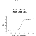
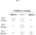
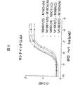
本発明者らは、グリピカン3(GPC3)が358番目のアルギニンと359番目のセリンの間で切断される事実を見出し、可溶型GPC3が肝癌患者の血中に分泌されるという仮説を立て、GPC3サンドイッチELISA系を確立し、GPC3高発現であるヒト肝癌細胞HepG2の培養上清中に分泌型GPC3の存在を明らかにした。さらに、HepG2を移植したマウス血漿中のみならずヒト肝癌患者血清中の可溶型GPC3測定にも成功した。GPC3は肝癌マーカーであるAFPよりも早期の肝癌で遺伝子発現が認められるので、GPC3の検出は癌の診断として有用であると考えられた。また可溶型GPC3はC末端断片側を認識する抗GPC3抗体では検出しにくい傾向にあることから、分泌型GPC3はN端断片優位と推定された。従って、N端を認識する抗GPC3抗体を用いるのが好ましいと考え、さらに鋭意検討を行い、本発明を完成させるに至った。
GPC3は肝癌細胞株以外に、肺癌、大腸癌、乳癌、前立腺癌、膵臓癌、リンパ腫などの癌細胞株においても発現が確認されているので、肝癌以外の診断にも適用できる可能性がある。
すなわち、本発明は以下の通りである。
(1)被検試料中の可溶化GPC3タンパク質を検出することを特徴とする癌の診断方法、
(2)可溶化GPC3タンパク質が、GPC3のN端ペプチドである(1)の癌の診断方法、
(3)GPC3のN端ペプチドがGPC3の第1番目のアミノ酸から第374番目のアミノ酸からなるアミノ酸配列中または第1番目のアミノ酸から第358番目のアミノ酸からなるアミノ酸配列中に含まれるペプチド断片である、(2)の癌の診断方法、
(4)被検試料が血液、血清、血漿のいずれかである(1)から(3)のいずれかの診断方法、
(5)肝臓癌である(1)から(4)のいずれかの診断方法、
(6)抗GPC3抗体を用いることを特徴とする(1)から(5)のいずれかの方法。
(7)支持体に固定した抗GPC3抗体と標識物質で標識された抗GPC3抗体を用いることを特徴とする(6)の方法、
(8)標識物質がビオチンである(7)の方法、
(9)抗GPC3抗体を含む癌の診断薬、
(10)支持体に固定した抗GPC3抗体と、標識物質で標識された抗体を含むことを特徴とする(9)の診断薬、
(11)肝臓癌である(9)又は(10)の診断薬、
(12)抗GPC3抗体がGPC3のN端ペプチドを認識することを特徴とする(9)から(11)のいずれかの診断薬、
(13)抗GPC3抗体を含む診断キット、および
(14)支持体に固定した抗GPC3抗体と、標識物質で標識された抗体を含むことを特徴とする(13)の診断キット。
以下、本発明について詳細に説明する。
本発明は、被験試料中の可溶化グリピカンを検出することにより癌を検出する方法である。
検出とは、定量的又は非定量的な検出を含み、例えば、非定量的な検出としては、単にGPC3タンパク質が存在するか否かの測定、GPC3タンパク質が一定の量以上存在するか否かの測定、GPC3タンパク質の量を他の試料(例えば、コントロール試料など)と比較する測定などを挙げることができ、定量的な検出としては、GPC3タンパク質の濃度の測定、GPC3タンパク質の量の測定などを挙げることができる。
被検試料とは、GPC3タンパク質が含まれる可能性のある試料であれば特に制限されないが、哺乳類などの生物の体から採取された試料が好ましく、さらに好ましくはヒトから採取された試料である。被検試料の具体的な例としては、例えば、血液、間質液、血漿、血管外液、脳脊髄液、滑液、胸膜液、血清、リンパ液、唾液、尿などを挙げることができるが、好ましいのは血液、血清、血漿である。又、生物の体から採取された細胞の培養液などの、被検試料から得られる試料も本発明の被検試料に含まれる。
診断される癌は、特に制限されず、具体的には、肝癌、膵臓癌、肺癌、大腸癌、乳癌、前立腺癌、白血病、リンパ腫などを挙げることができるが、好ましいのは肝癌である。
1.抗GPC3抗体の作製
本発明で用いられる抗GPC3抗体はGPC3タンパク質に特異的に結合すればよく、その由来、種類(モノクローナル、ポリクローナル)および形状を問わない。具体的には、マウス抗体、ラット抗体、ヒト抗体、キメラ抗体、ヒト型化抗体などの公知の抗体を用いることができる。
抗体はポリクローナル抗体でもよいが、モノクローナル抗体であることが好ましい。
又、支持体に固定される抗GPC3抗体と標識物質で標識される抗GPC抗体はGPC3分子の同じエピトープを認識してもよいが、異なるエピトープを認識することが好ましい。
エピトープとして認識する部位は特に制限されないが、GPC3タンパク質のN端側(アミノ酸1番目のMet〜358番目のArgまたは1番目のMet〜374番目のLys)に存在するエピトープを認識することが好ましい。
本発明で使用される抗GPC3抗体は、公知の手段を用いてポリクローナルまたはモノクローナル抗体として得ることができる。本発明で使用される抗GPC3抗体として、特に哺乳動物由来のモノクローナル抗体が好ましい。哺乳動物由来のモノクローナル抗体は、ハイブリドーマに産生されるもの、および遺伝子工学的手法により抗体遺伝子を含む発現ベクターで形質転換した宿主に産生されるものを含む。
モノクローナル抗体産生ハイブリドーマは、基本的には公知技術を使用し、以下のようにして作製できる。すなわち、GPC3を感作抗原として使用して、これを通常の免疫方法にしたがって免疫し、得られる免疫細胞を通常の細胞融合法によって公知の親細胞と融合させ、通常のスクリーニング法により、モノクローナルな抗体産生細胞をスクリーニングすることによって作製できる。
具体的には、モノクローナル抗体を作製するには次のようにすればよい。
まず、抗体取得の感作抗原として使用されるGPC3を、Lage,H.et al.,Gene 188(1997),151−156に開示されたGPC3(MXR7)遺伝子/アミノ酸配列を発現することによって得る。すなわち、GPC3をコードする遺伝子配列を公知の発現ベクター系に挿入して適当な宿主細胞を形質転換させた後、その宿主細胞中または培養上清中から目的のヒトGPC3タンパク質を公知の方法で精製する。
また、天然のGPC3を精製して用いることもできる。
次に、この精製GPC3タンパク質を感作抗原として用いる。あるいは、GPC3の部分ペプチドを感作抗原として使用することもできる。この際、部分ペプチドはヒトGPC3のアミノ酸配列より化学合成により得ることもできるし、GPC遺伝子の一部を発現ベクターに組込んで得ることもでき、さらに天然のGPC3をタンパク質分解酵素により分解することによっても得ることができる。部分ペプチドとして用いるGPC3の部分は限られないが、N端側に存在するエピトープを認識する抗体を得ようとする場合は、GPC3のアミノ酸1番目のMet〜358番目のArgまたは1番目のMet〜374番目のLysまでのペプチドを用いればよいし、この部分のエピトープを含むより小さいペプチドを用いることもできる。
感作抗原で免疫される哺乳動物としては、特に限定されるものではないが、細胞融合に使用する親細胞との適合性を考慮して選択するのが好ましく、一般的にはげっ歯類の動物、例えば、マウス、ラット、ハムスター、あるいはウサギ、サル等が使用される。
感作抗原を動物に免疫するには、公知の方法にしたがって行われる。例えば、一般的方法として、感作抗原を哺乳動物の腹腔内または皮下に注射することにより行われる。具体的には、感作抗原をPBS(Phosphate−Buffered Saline)や生理食塩水等で適当量に希釈、懸濁したものに所望により通常のアジュバント、例えばフロイント完全アジュバントを適量混合し、乳化後、哺乳動物に4〜21日毎に数回投与する。また、感作抗原免疫時に適当な担体を使用することもできる。特に分子量の小さい部分ペプチドを感作抗原として用いる場合には、アルブミン、キーホールリンペットヘモシアニン等の担体タンパク質と結合させて免疫することが望ましい。
このように哺乳動物を免疫し、血清中に所望の抗体レベルが上昇するのを確認した後に、哺乳動物から免疫細胞を採取し、細胞融合に付されるが、好ましい免疫細胞としては、特に脾細胞が挙げられる。
前記免疫細胞と融合される他方の親細胞として、哺乳動物のミエローマ細胞を用いる。このミエローマ細胞は、公知の種々の細胞株、例えば、P3(P3x63Ag8.653)(J.Immnol.(1979)123,1548−1550)、P3x63Ag8U.1(Current Topics in Microbiology and Immunology(1978)81,1−7)、NS−1(Kohler.G.and Milstein,C.Eur.J.Immunol.(1976)6,511−519)、MPC−11(Margulies.D.H.et al.,Cell(1976)8,405−415)、SP2/0(Shulman,M.et al.,Nature(1978)276,269−270)、FO(de St.Groth,S.F.et al.,J.Immunol.Methods(1980)35,1−21)、S194(Trowbridge,I.S.J.Exp.Med.(1978)148,313−323)、R210(Galfre,G.et al.,Nature(1979)277,131−133)等が好適に使用される。
前記免疫細胞とミエローマ細胞との細胞融合は、基本的には公知の方法、たとえば、ケーラーとミルステインらの方法(Kohler.G.and Milstein,C.、Methods Enzymol.(1981)73,3−46)等に準じて行うことができる。
より具体的には、前記細胞融合は、例えば細胞融合促進剤の存在下に通常の栄養培養液中で実施される。融合促進剤としては、例えばポリエチレングリコール(PEG)、センダイウイルス(HVJ)等が使用され、更に所望により融合効率を高めるためにジメチルスルホキシド等の補助剤を添加使用することもできる。
免疫細胞とミエローマ細胞との使用割合は任意に設定することができる。例えば、ミエローマ細胞に対して免疫細胞を1〜10倍とするのが好ましい。前記細胞融合に用いる培養液としては、例えば、前記ミエローマ細胞株の増殖に好適なRPMI1640培養液、MEM培養液、その他、この種の細胞培養に用いられる通常の培養液が使用可能であり、さらに、牛胎児血清(FCS)等の血清補液を併用することもできる。
細胞融合は、前記免疫細胞とミエローマ細胞との所定量を前記培養液中でよく混合し、予め37℃程度に加温したPEG溶液(例えば平均分子量1000〜6000程度)を通常30〜60%(w/v)の濃度で添加し、混合することによって目的とする融合細胞(ハイブリドーマ)を形成する。続いて、適当な培養液を逐次添加し、遠心して上清を除去する操作を繰り返すことによりハイブリドーマの生育に好ましくない細胞融合剤等を除去する。
このようにして得られたハイブリドーマは、通常の選択培養液、例えばHAT培養液(ヒポキサンチン、アミノプテリンおよびチミジンを含む培養液)で培養することにより選択される。上記HAT培養液での培養は、目的とするハイブリドーマ以外の細胞(非融合細胞)が死滅するのに十分な時間(通常、数日〜数週間)継続する。ついで、通常の限界希釈法を実施し、目的とする抗体を産生するハイブリドーマのスクリーニングおよび単一クローニングを行う。
目的とする抗体のスクリーニングおよび単一クローニングは、公知の抗原抗体反応に基づくスクリーニング方法で行えばよい。例えば、ポリスチレン等でできたビーズや市販の96ウェルのマイクロタイタープレート等の担体に抗原を結合させ、ハイブリドーマの培養上清と反応させ、担体を洗浄した後に酵素標識第2次抗体等を反応させることにより、培養上清中に感作抗原と反応する目的とする抗体が含まれるかどうか決定できる。目的とする抗体を産生するハイブリドーマを限界希釈法等によりクローニングすることができる。この際、抗原としては免疫に用いたものを用いればよく、また例えばGPC3のN端断片に対する抗体を得ようとする場合は、N端断片をスクリーニング用抗原として用いればよい。
また、ヒト以外の動物に抗原を免疫して上記ハイブリドーマを得る他に、ヒトリンパ球をin vitroでGPC3に感作し、感作リンパ球をヒト由来の永久分裂能を有するミエローマ細胞と融合させ、GPC3への結合活性を有する所望のヒト抗体を得ることもできる(特公平1−59878号公報参照)。さらに、ヒト抗体遺伝子の全てのレパートリーを有するトランスジェニック動物に抗原となるGPC3を投与して抗GPC3抗体産生細胞を取得し、これを不死化させた細胞からGPC3に対するヒト抗体を取得してもよい(国際特許出願公開番号WO 94/25585号公報、WO 93/12227号公報、WO 92/03918号公報、WO 94/02602号公報参照)。
このようにして作製されるモノクローナル抗体を産生するハイブリドーマは、通常の培養液中で継代培養することが可能であり、また、液体窒素中で長期保存することが可能である。
当該ハイブリドーマからモノクローナル抗体を取得するには、当該ハイブリドーマを通常の方法に従い培養し、その培養上清として得る方法、あるいはハイブリドーマをこれと適合性がある哺乳動物に投与して増殖させ、その腹水として得る方法などが採用される。前者の方法は、高純度の抗体を得るのに適しており、一方、後者の方法は、抗体の大量生産に適している。
本発明では、モノクローナル抗体として、抗体遺伝子をハイブリドーマからクローニングし、適当なベクターに組み込んで、これを宿主に導入し、遺伝子組換え技術を用いて産生させた組換え型のものを用いることができる(例えば、Vandamme,A.M.et al.,Eur.J.Biochem.(1990)192,767−775,1990参照)。具体的には、抗GPC3抗体を産生するハイブリドーマから、抗GPC3抗体の可変(V)領域をコードするmRNAを単離する。mRNAの単離は、公知の方法、例えば、グアニジン超遠心法(Chirgwin,J.M.et al.,Biochemistry(1979)18,5294−5299)、AGPC法(Chomczynski,P.et al.,Anal.Biochem.(1987)162,156−159)等により行って全RNAを調製し、mRNA Purification Kit(Pharmacia製)等を使用して目的のmRNAを調製する。また、QuickPrep mRNA Purification Kit(Pharmacia製)を用いることによりmRNAを直接調製することもできる。
得られたmRNAから逆転写酵素を用いて抗体V領域のcDNAを合成する。cDNAの合成は、AMV Reverse Transcriptase First−strand cDNA Synthesis Kit(生化学工業社製)等を用いて行う。また、cDNAの合成および増幅を行うには、5’−Ampli FINDER RACE Kit(Clontech製)およびPCRを用いた5’−RACE法(Frohman,M.A.et al.,Proc.Natl.Acad.Sci.USA(1988)85,8998−9002、Belyavsky,A.et al.,Nucleic Acids Res.(1989)17,2919−2932)等を使用することができる。
得られたPCR産物から目的とするDNA断片を精製し、ベクターDNAと連結する。さらに、これより組換えベクターを作製し、大腸菌等に導入してコロニーを選択して所望の組換えベクターを調製する。そして、目的とするDNAの塩基配列を公知の方法、例えば、ジデオキシヌクレオチドチェインターミネーション法等により確認する。
目的とする抗GPC3抗体のV領域をコードするDNAを得たのち、これを、所望の抗体定常領域(C領域)をコードするDNAを含有する発現ベクターへ組み込む。
本発明で使用される抗GPC3抗体を製造するには、抗体遺伝子を発現制御領域、例えば、エンハンサー、プロモーターの制御のもとで発現するよう発現ベクターに組み込む。次に、この発現ベクターにより、宿主細胞を形質転換し、抗体を発現させる。
抗体遺伝子の発現は、抗体重鎖(H鎖)または軽鎖(L鎖)をコードするDNAを別々に発現ベクターに組み込んで宿主細胞を同時形質転換させてもよいし、あるいはH鎖およびL鎖をコードするDNAを単一の発現ベクターに組み込んで宿主細胞を形質転換させてもよい(WO 94/11523号公報参照)。
また、組換え型抗体の産生には上記宿主細胞だけではなく、トランスジェニック動物を使用することができる。例えば、抗体遺伝子を、乳汁中に固有に産生されるタンパク質(ヤギβカゼインなど)をコードする遺伝子の途中に挿入して融合遺伝子として調製する。抗体遺伝子が挿入された融合遺伝子を含むDNA断片をヤギの胚へ注入し、この胚を雌のヤギへ導入する。胚を受容したヤギから生まれるトランスジェニックヤギまたはその子孫が産生する乳汁から所望の抗体を得る。また、トランスジェニックヤギから産生される所望の抗体を含む乳汁量を増加させるために、適宜ホルモンをトランスジェニックヤギに使用してもよい(Ebert,K.M.et al.,Bio/Technology(1994)12,699−702)。
本発明では、上記抗体のほかに、人為的に改変した遺伝子組換え型抗体、例えば、キメラ抗体、ヒト型化(Humanized)抗体を使用できる。これらの改変抗体は、既知の方法を用いて製造することができる。
キメラ抗体は、前記のようにして得た抗体V領域をコードするDNAをヒト抗体C領域をコードするDNAと連結し、これを発現ベクターに組み込んで宿主に導入し産生させることにより得られる。この既知の方法を用いて、本発明に有用なキメラ抗体を得ることができる。
ヒト型化抗体は、再構成(reshaped)ヒト抗体とも称され、これは、ヒト以外の哺乳動物、例えばマウス抗体の相補性決定領域(CDR;complementarity determining region)をヒト抗体の相補性決定領域へ移植したものであり、その一般的な遺伝子組換え手法も知られている(欧州特許出願公開番号EP 125023号公報、WO 96/02576号公報参照)。
具体的には、マウス抗体のCDRとヒト抗体のフレームワーク領域(framework region;FR)とを連結するように設計したDNA配列を、CDR及びFR両方の末端領域にオーバーラップする部分を有するように作製した数個のオリゴヌクレオチドをプライマーとして用いてPCR法により合成する(WO98/13388号公報に記載の方法を参照)。
CDRを介して連結されるヒト抗体のフレームワーク領域は、相補性決定領域が良好な抗原結合部位を形成するものが選択される。必要に応じ、再構成ヒト抗体の相補性決定領域が適切な抗原結合部位を形成するように、抗体の可変領域におけるフレームワーク領域のアミノ酸を置換してもよい(Sato,K.et al.,Cancer Res.(1993)53,851−856)。
キメラ抗体及びヒト型化抗体のC領域には、ヒト抗体のものが使用され、例えばH鎖では、Cγ1、Cγ2、Cγ3、Cγ4を、L鎖ではCκ、Cλを使用することができる。また、抗体またはその産生の安定性を改善するために、ヒト抗体C領域を修飾してもよい。
キメラ抗体は、ヒト以外の哺乳動物由来抗体の可変領域とヒト抗体由来の定常領域とからなる。一方、ヒト型化抗体は、ヒト以外の哺乳動物由来抗体の相補性決定領域と、ヒト抗体由来のフレームワーク領域およびC領域とからなる。ヒト型化抗体はヒト体内における抗原性が低下されているため、本発明の治療剤の有効成分として有用である。
本発明で使用される抗体は、抗体の全体分子に限られず、GPC3に結合する限り、抗体の断片又はその修飾物であってもよく、二価抗体も一価抗体も含まれる。例えば、抗体の断片としては、Fab、F(ab’)2、Fv、1個のFabと完全なFcを有するFab/c、またはH鎖若しくはL鎖のFvを適当なリンカーで連結させたシングルチェインFv(scFv)が挙げられる。具体的には、抗体を酵素、例えばパパイン、ペプシンで処理し抗体断片を生成させるか、または、これら抗体断片をコードする遺伝子を構築し、これを発現ベクターに導入した後、適当な宿主細胞で発現させる(例えば、Co,M.S.et al.,J.Immunol.(1994)152,2968−2976、Better,M.& Horwitz,A.H.Methods in Enzymology(1989)178,476−496,Academic Press,Inc.、Plueckthun,A.& Skerra,A.Methods in Enzymology(1989)178,476−496,Academic Press,Inc.、Lamoyi,E.,Methods in Enzymology(1989)121,652−663、Rousseaux,J.et al.,Methods in Enzymology(1989)121,663−669、Bird,R.E.et al.,TIBTECH(1991)9,132−137参照)。
scFvは、抗体のH鎖V領域とL鎖V領域とを連結することにより得られる。このscFvにおいて、H鎖V領域とL鎖V領域は、リンカー、好ましくはペプチドリンカーを介して連結される(Huston,J.S.et al.、Proc.Natl.Acad.Sci.U.S.A.(1988)85,5879−5883)。scFvにおけるH鎖V領域およびL鎖V領域は、本明細書に抗体として記載されたもののいずれの由来であってもよい。V領域を連結するベプチドリンカーとしては、例えばアミノ酸12〜19残基からなる任意の一本鎖ペプチドが用いられる。
scFvをコードするDNAは、前記抗体のH鎖またはH鎖V領域をコードするDNA、およびL鎖またはL鎖V領域をコードするDNAのうち、それらの配列のうちの全部又は所望のアミノ酸配列をコードするDNA部分を鋳型とし、その両端を規定するプライマー対を用いてPCR法により増幅し、次いで、さらにペプチドリンカー部分をコードするDNA、およびその両端が各々H鎖、L鎖と連結されるように規定するプライマー対を組み合せて増幅することにより得られる。
また、一旦scFvをコードするDNAが作製されると、それらを含有する発現ベクター、および該発現ベクターにより形質転換された宿主を常法に従って得ることができ、また、その宿主を用いることにより、常法に従ってscFvを得ることができる。
これら抗体の断片は、前記と同様にしてその遺伝子を取得し発現させ、宿主により産生させることができる。本発明における「抗体」にはこれらの抗体の断片も包含される。
抗体の修飾物として、標識物質等の各種分子と結合した抗グリピカン抗体を使用することもできる。本発明における「抗体」にはこれらの抗体修飾物も包含される。このような抗体修飾物は、得られた抗体に化学的な修飾を施すことによって得ることができる。なお、抗体の修飾方法はこの分野においてすでに確立されている。
さらに、本発明で使用される抗体は、二重特異性抗体(bispecific antibody)であってもよい。二重特異性抗体はGPC3分子上の異なるエピトープを認識する抗原結合部位を有する二重特異性抗体であってもよいし、一方の抗原結合部位がGPC3を認識し、他方の抗原結合部位が標識物質等を認識してもよい。二重特異性抗体は2種類の抗体のHL対を結合させて作製することもできるし、異なるモノクローナル抗体を産生するハイブリドーマを融合させて二重特異性抗体産生融合細胞を作製し、得ることもできる。さらに、遺伝子工学的手法により二重特異性抗体を作製することも可能である。
前記のように構築した抗体遺伝子は、公知の方法により発現させ、取得することができる。哺乳類細胞の場合、常用される有用なプロモーター、発現させる抗体遺伝子、その3’側下流にポリAシグナルを機能的に結合させて発現させることができる。例えばプロモーター/エンハンサーとしては、ヒトサイトメガロウイルス前期プロモーター/エンハンサー(human cytomegalovirus immediate early promoter/enhancer)を挙げることができる。
また、その他に本発明で使用される抗体発現に使用できるプロモーター/エンハンサーとして、レトロウイルス、ポリオーマウイルス、アデノウイルス、シミアンウイルス40(SV40)等のウイルスプロモーター/エンハンサー、あるいはヒトエロンゲーションファクター1a(HEF1a)などの哺乳類細胞由来のプロモーター/エンハンサー等が挙げられる。
SV40プロモーター/エンハンサーを使用する場合はMulliganらの方法(Nature(1979)277,108)により、また、HEF1aプロモーター/エンハンサーを使用する場合はMizushimaらの方法(Nucleic Acids Res.(1990)18,5322)により、容易に遺伝子発現を行うことができる。
大腸菌の場合、常用される有用なプロモーター、抗体分泌のためのシグナル配列及び発現させる抗体遺伝子を機能的に結合させて当該遺伝子を発現させることができる。プロモーターとしては、例えばlaczプロモーター、araBプロモーターを挙げることができる。laczプロモーターを使用する場合はWardらの方法(Nature(1098)341,544−546;FASEB J.(1992)6,2422−2427)により、あるいはaraBプロモーターを使用する場合はBetterらの方法(Science(1988)240,1041−1043)により発現することができる。
抗体分泌のためのシグナル配列としては、大腸菌のペリプラズムに産生させる場合、pelBシグナル配列(Lei,S.P.et al J.Bacteriol.(1987)169,4379)を使用すればよい。そして、ペリプラズムに産生された抗体を分離した後、抗体の構造を適切に組み直して(refold)使用する。
複製起源としては、SV40、ポリオーマウイルス、アデノウイルス、ウシパピローマウイルス(BPV)等の由来のものを用いることができ、さらに、宿主細胞系で遺伝子コピー数増幅のため、発現ベクターは、選択マーカーとしてアミノグリコシドトランスフェラーゼ(APH)遺伝子、チミジンキナーゼ(TK)遺伝子、大腸菌キサンチングアニンホスホリボシルトランスフェラーゼ(Ecogpt)遺伝子、ジヒドロ葉酸還元酵素(dhfr)遺伝子等を含むことができる。
本発明で使用される抗体の製造のために、任意の発現系、例えば真核細胞又は原核細胞系を使用することができる。真核細胞としては、例えば樹立された哺乳類細胞系、昆虫細胞系、真糸状菌細胞および酵母細胞などの動物細胞等が挙げられ、原核細胞としては、例えば大腸菌細胞等の細菌細胞が挙げられる。
好ましくは、本発明で使用される抗体は、哺乳類細胞、例えばCHO、COS、ミエローマ、BHK、Vero、HeLa細胞中で発現される。
次に、形質転換された宿主細胞をin vitroまたはin vivoで培養して目的とする抗体を産生させる。宿主細胞の培養は公知の方法に従い行う。例えば、培養液として、DMEM、MEM、RPMI1640、IMDMを使用することができ、牛胎児血清(FCS)等の血清補液を併用することもできる。
前記のように発現、産生された抗体は、細胞、宿主動物から分離し均一にまで精製することができる。本発明で使用される抗体の分離、精製はアフィニティーカラムを用いて行うことができる。例えば、プロテインAカラムを用いたカラムとして、Hyper D、POROS、Sepharose F.F.(Pharmacia製)等が挙げられる。その他、通常のタンパク質で使用されている分離、精製方法を使用すればよく、何ら限定されるものではない。例えば、上記アフィニティーカラム以外のクロマトグラフィーカラム、フィルター、限外濾過、塩析、透析等を適宜選択、組み合わせることにより、抗体を分離、精製することができる(Antibodies A Laboratory Manual.Ed Harlow,David Lane,Cold Spring Harbor Laboratory,1988)。
2.GPC3の検出
本発明において検出するGPC3は、特に限定されず、全長GPC3でも、その断片でもよい。GPC3断片を検出する場合には、N端断片でもC端断片でもよいが、好ましくはN端断片である。又、ヘパラン硫酸などが付加されたGPC3タンパク質でも、GPC3コアタンパク質でもよい。
被検試料に含まれるGPC3タンパク質の検出方法は特に限定されないが、抗GPC3抗体を用いた免疫学的方法により検出することが好ましい。免疫学的方法としては、例えば、ラジオイムノアッセイ、エンザイムイムノアッセイ、蛍光イムノアッセイ、発光イムノアッセイ、免疫沈降法、免疫比濁法、ウエスタンブロット、免疫染色、免疫拡散法などを挙げることができるが、好ましくはエンザイムイムノアッセイであり、特に好ましいのは酵素結合免疫吸着定量法(enzyme−linked immunosorbent assay:ELISA)(例えば、sandwich ELISA)である。ELISAなどの上述した免疫学的方法は当業者に公知の方法により行うことが可能である。
抗GPC3抗体を用いた一般的な検出方法としては、例えば、抗GPC3抗体を支持体に固定し、ここに被検試料を加え、インキュベートを行い抗GPC3抗体とGPC3タンパク質を結合させた後に洗浄して、抗GPC3抗体を介して支持体に結合したGPC3タンパク質を検出することにより、被検試料中のGPC3タンパク質の検出を行う方法を挙げることができる。
本発明において用いられる支持体としては、例えば、アガロース、セルロースなどの不溶性の多糖類、シリコン樹脂、ポリスチレン樹脂、ポリアクリルアミド樹脂、ナイロン樹脂、ポリカーボネイト樹脂などの合成樹脂や、ガラスなどの不溶性の支持体を挙げることができる。これらの支持体は、ビーズやプレートなどの形状で用いることが可能である。ビーズの場合、これらが充填されたカラムなどを用いることができる。プレートの場合、マルチウェルプレート(96穴マルチウェルプレート等)、やバイオセンサーチップなどを用いることができる。抗GPC3抗体と支持体との結合は、化学結合や物理的な吸着などの通常用いられる方法により結合することができる。これらの支持体はすべて市販のものを用いることができる。
抗GPC3抗体とGPC3タンパク質との結合は、通常、緩衝液中で行われる。緩衝液としては、例えば、リン酸緩衝液、Tris緩衝液、クエン酸緩衝液、ホウ酸塩緩衝液、炭酸塩緩衝液、などが使用される。また、インキュベーションの条件としては、すでによく用いられている条件、例えば、4℃〜室温にて1時間〜24時間のインキュベーションが行われる。インキュベート後の洗浄は、GPC3タンパク質と抗GPC3抗体の結合を妨げないものであれば何でもよく、例えば、Tween20等の界面活性剤を含む緩衝液などが使用される。
本発明のGPC3タンパク質検出方法においては、GPC3タンパク質を検出したい被検試料の他に、コントロール試料を設置してもよい。コントロール試料としては、GPC3タンパク質を含まない陰性コントロール試料やGPC3タンパク質を含む陽性コントロール試料などがある。この場合、GPC3タンパク質を含まない陰性コントロール試料で得られた結果、GPC3タンパク質を含む陽性コントロール試料で得られた結果と比較することにより、被検試料中のGPC3タンパク質を検出することが可能である。また、濃度を段階的に変化させた一連のコントロール試料を調製し、各コントロール試料に対する検出結果を数値として得て、標準曲線を作成し、被検試料の数値から標準曲線に基づいて、被検試料に含まれるGPC3タンパク質を定量的に検出することも可能である。
抗GPC3抗体を介して支持体に結合したGPC3タンパク質の検出の好ましい態様として、標識物質で標識された抗GPC3抗体を用いる方法を挙げることができる。
例えば、支持体に固定された抗GPC3抗体に被検試料を接触させ、洗浄後に、GPC3タンパク質を特異的に認識する標識抗体を用いて検出する。
抗GPC3抗体の標識は通常知られている方法により行うことが可能である。標識物質としては、蛍光色素、酵素、補酵素、化学発光物質、放射性物質などの当業者に公知の標識物質を用いることが可能であり、具体的な例としては、ラジオアイソトープ(32P、14C、125I、3H、131Iなど)、フルオレセイン、ローダミン、ダンシルクロリド、ウンベリフェロン、ルシフェラーゼ、ペルオキシダーゼ、アルカリホスファターゼ、β−ガラクトシダーゼ、β−グルコシダーゼ、ホースラディッシュパーオキシダーゼ、グルコアミラーゼ、リゾチーム、サッカリドオキシダーゼ、マイクロペルオキシダーゼ、ビオチンなどを挙げることができる。標識物質としてビオチンを用いる場合には、ビオチン標識抗体を添加後に、アルカリホスファターゼなどの酵素を結合させたアビジンをさらに添加することが好ましい。標識物質と抗GPC3抗体との結合には、グルタルアルデヒド法、マレイミド法、ピリジルジスルフィド法、過ヨウ素酸法、などの公知の方法を用いることができる。
具体的には、抗GPC3抗体を含む溶液をプレートなどの支持体に加え、抗GPC3抗体を支持体に固定する。プレートを洗浄後、タンパク質の非特異的な結合を防ぐため、例えばBSA、ゼラチン、アルブミンなどでブロッキングする。再び洗浄し、被検試料をプレートに加える。インキュベートの後、洗浄し、標識抗GPC3抗体を加える。適度なインキュベーションの後、プレートを洗浄し、プレートに残った標識抗GPC3抗体を検出する。検出は当業者に公知の方法により行うことができ、例えば、放射性物質による標識の場合には液体シンチレーションやRIA法により検出することができる。酵素による標識の場合には基質を加え、基質の酵素的変化、例えば発色を吸光度計により検出することができる。基質の具体的な例としては、2,2−アジノビス(3−エチルベンゾチアゾリン−6−スルホン酸)ジアンモニウム塩(ABTS)、1,2−フェニレンジアミン(オルソ−フェニレンジアミン)、3,3’,5,5’−テトラメチルベンジジン(TME)などを挙げることができる。蛍光物質の場合には蛍光光度計により検出することができる。
本発明のGPC3タンパク質検出方法の特に好ましい態様として、ビオチンで標識された抗GPC3抗体及びアビジンを用いる方法を挙げることができる。
具体的には、抗GPC3抗体を含む溶液をプレートなどの支持体に加え、抗GPC3抗体を固定する。プレートを洗浄後、タンパク質の非特異的な結合を防ぐため、例えばBSAなどでブロッキングする。再び洗浄し、被検試料をプレートに加える。インキュベートの後、洗浄し、ビオチン標識抗GPC3抗体を加える。適度なインキュベーションの後、プレートを洗浄し、アルカリホスファターゼ、ペルオキシダーゼなどの酵素と結合したアビジンを加える。インキュベーション後、プレートを洗浄し、アビジンに結合している酵素に対応した基質を加え、基質の酵素的変化などを指標にGPC3タンパク質を検出する。
本発明のGPC3タンパク質検出方法の他の態様として、GPC3タンパク質を特異的に認識する一次抗体、及び該一次抗体を特異的に認識する二次抗体を用いる方法を挙げることができる。
例えば、支持体に固定された抗GPC3抗体に被検試料を接触させ、インキュベーションした後、洗浄し、洗浄後に結合しているGPC3タンパク質を、一次抗GPC3抗体及び該一次抗体を特異的に認識する二次抗体により検出する。この場合、二次抗体は好ましくは標識物質により標識されている。
具体的には、抗GPC3抗体を含む溶液をプレートなどの支持体に加え、抗GPC3抗体を固定する。プレートを洗浄後、タンパク質の非特異的な結合を防ぐため、例えばBSAなどでブロッキングする。再び洗浄し、被検試料をプレートに加える。インキュベートの後、洗浄し、一次抗GPC3抗体を加える。適度なインキュベーションの後、プレートを洗浄し、次いで一次抗体を特異的に認識する二次抗体を加える。適度なインキュベーションの後、洗浄して、プレートに残った二次抗体を検出する。二次抗体の検出は前述の方法により行うことができる。
本発明のGPC3タンパク質の検出方法の他の態様としては、凝集反応を利用した検出方法を挙げることができる。該方法においては、抗GPC3抗体を感作した担体を用いてGPC3を検出することができる。抗体を感作する担体としては、不溶性で、非特異的な反応を起こさず、かつ安定である限り、いかなる担体を使用してもよい。例えば、ラテックス粒子、ベントナイト、コロジオン、カオリン、固定羊赤血球等を使用することができるが、ラテックス粒子を使用するのが好ましい。ラテックス粒子としては、例えば、ポリスチレンラテックス粒子、スチレン−ブタジエン共重合体ラテックス粒子、ポリビニルトルエンラテックス粒子等を使用することができるが、ポリスチレンラテックス粒子を使用するのが好ましい。感作した粒子を試料を混合し、一定時間攪拌した後に、試料中にGPC3抗体が高濃度で含まれるほど粒子の凝集度が大きくなるので、凝集を肉眼でみることによりGPC3を検出することができる。また、凝集による濁度を分光光度計等により測定することによっても検出することが可能である。
本発明のGPC3タンパク質の検出方法の他の態様としては、例えば、表面プラズモン共鳴現象を利用したバイオセンサーを用いた方法を挙げることができる。表面プラズモン共鳴現象を利用したバイオセンサーはタンパク質−タンパク質間の相互作用を微量のタンパク質を用いてかつ標識することなく、表面プラズモン共鳴シグナルとしてリアルタイムに観察することが可能である。例えば、BIAcore(Pharmacia製)等のバイオセンサーを用いることによりGPC3タンパク質と抗GPC3抗体の結合を検出することが可能である。具体的には、抗GPC3抗体を固定化したセンサーチップに、被検試料を接触させ、抗GPC3抗体に結合するGPC3タンパク質を共鳴シグナルの変化として検出することができる。
本発明の検出方法は、種々の自動検査装置を用いて自動化することもでき、一度に大量の試料について検査を行うことも可能である。
本発明は、癌の診断のための被検試料中のGPC3タンパク質を検出するための診断薬またはキットの提供をも目的とするが、該診断薬またはキットは少なくとも抗GPC3抗体を含む。該診断薬またはキットがELISA法に基づく場合は、抗体を固相化する担体を含んでいてもよく、抗体があらかじめ担体に結合していてもよい。該診断薬またはキットがラテックス等の担体を用いた凝集法に基づく場合は抗体が吸着した担体を含んでいてもよい。また、該キットは、適宜、ブロッキング溶液、反応溶液、反応停止液、試料を処理するための試薬等を含んでいてもよい。The present inventors have found that glypican 3 (GPC3) is cleaved between 358th arginine and 359th serine, and hypothesized that soluble GPC3 is secreted into the blood of patients with liver cancer. A GPC3 sandwich ELISA system was established, and the presence of secreted GPC3 was clarified in the culture supernatant of human hepatoma cell HepG2, which is highly expressing GPC3. Furthermore, soluble GPC3 was successfully measured not only in mouse plasma transplanted with HepG2, but also in serum of human liver cancer patients. Since GPC3 gene expression was observed in liver cancer earlier than AFP, which is a liver cancer marker, detection of GPC3 was considered useful as a cancer diagnosis. In addition, since soluble GPC3 tends to be difficult to detect with an anti-GPC3 antibody that recognizes the C-terminal fragment, secretory GPC3 was presumed to be predominant at the N-terminal fragment. Therefore, it was considered preferable to use an anti-GPC3 antibody that recognizes the N-terminal, and further intensive studies were carried out to complete the present invention.
Since expression of GPC3 has been confirmed in lung cancer, colon cancer, breast cancer, prostate cancer, pancreatic cancer, lymphoma and other cancer cell lines in addition to liver cancer cell lines, it may be applicable to diagnosis other than liver cancer.
That is, the present invention is as follows.
(1) A method for diagnosing cancer, comprising detecting solubilized GPC3 protein in a test sample,
(2) The method for diagnosing cancer according to (1), wherein the solubilized GPC3 protein is an N-terminal peptide of GPC3,
(3) A peptide fragment in which the N-terminal peptide of GPC3 is contained in the amino acid sequence consisting of the first to 374th amino acids of GPC3 or in the amino acid sequence consisting of the 358th amino acid from the first amino acid A diagnostic method for cancer according to (2),
(4) The diagnostic method according to any one of (1) to (3), wherein the test sample is blood, serum, or plasma,
(5) The diagnostic method of any one of (1) to (4), which is liver cancer,
(6) The method according to any one of (1) to (5), wherein an anti-GPC3 antibody is used.
(7) The method according to (6), wherein an anti-GPC3 antibody immobilized on a support and an anti-GPC3 antibody labeled with a labeling substance are used.
(8) The method according to (7), wherein the labeling substance is biotin,
(9) a cancer diagnostic agent comprising an anti-GPC3 antibody,
(10) The diagnostic agent according to (9), comprising an anti-GPC3 antibody immobilized on a support and an antibody labeled with a labeling substance,
(11) The diagnostic agent according to (9) or (10), which is liver cancer,
(12) The diagnostic agent according to any one of (9) to (11), wherein the anti-GPC3 antibody recognizes an N-terminal peptide of GPC3,
(13) A diagnostic kit containing an anti-GPC3 antibody, and (14) an anti-GPC3 antibody immobilized on a support and an antibody labeled with a labeling substance.
Hereinafter, the present invention will be described in detail.
The present invention is a method for detecting cancer by detecting solubilized glypican in a test sample.
Detection includes quantitative or non-quantitative detection. For example, non-quantitative detection includes simply measuring whether or not GPC3 protein is present, whether or not GPC3 protein is present in a certain amount or more. Measurement, measurement of comparing the amount of GPC3 protein with other samples (for example, control samples, etc.), etc., and quantitative detection includes measurement of the concentration of GPC3 protein, measurement of the amount of GPC3 protein, etc. Can be mentioned.
The test sample is not particularly limited as long as it may contain GPC3 protein, but a sample collected from the body of an organism such as a mammal is preferable, and a sample collected from a human is more preferable. Specific examples of the test sample include blood, interstitial fluid, plasma, extravascular fluid, cerebrospinal fluid, synovial fluid, pleural fluid, serum, lymph fluid, saliva, urine, etc. Preference is given to blood, serum and plasma. A sample obtained from a test sample such as a culture solution of cells collected from the body of an organism is also included in the test sample of the present invention.
The cancer to be diagnosed is not particularly limited, and specific examples include liver cancer, pancreatic cancer, lung cancer, colon cancer, breast cancer, prostate cancer, leukemia, lymphoma, and the like, but liver cancer is preferable.
1. Preparation of anti-GPC3 antibody The anti-GPC3 antibody used in the present invention may be specifically bound to the GPC3 protein, regardless of its origin, type (monoclonal, polyclonal) and shape. Specifically, known antibodies such as mouse antibodies, rat antibodies, human antibodies, chimeric antibodies, humanized antibodies can be used.
The antibody may be a polyclonal antibody, but is preferably a monoclonal antibody.
The anti-GPC3 antibody immobilized on the support and the anti-GPC antibody labeled with a labeling substance may recognize the same epitope of the GPC3 molecule, but preferably recognize different epitopes.
The site recognized as an epitope is not particularly limited, but it is preferable to recognize an epitope present on the N-terminal side of GPC3 protein (amino acid 1st Met to 358th Arg or first Met to 374th Lys).
The anti-GPC3 antibody used in the present invention can be obtained as a polyclonal or monoclonal antibody using known means. As the anti-GPC3 antibody used in the present invention, a monoclonal antibody derived from a mammal is particularly preferable. Mammal-derived monoclonal antibodies include those produced by hybridomas and those produced by hosts transformed with expression vectors containing antibody genes by genetic engineering techniques.
A monoclonal antibody-producing hybridoma can be basically produced using a known technique as follows. That is, GPC3 is used as a sensitizing antigen, and this is immunized according to a normal immunization method. The obtained immune cells are fused with a known parent cell by a normal cell fusion method, and monoclonal antibodies are obtained by a normal screening method. It can be produced by screening antibody-producing cells.
Specifically, the monoclonal antibody can be produced as follows.
First, GPC3, which is used as a sensitizing antigen for obtaining an antibody, was prepared using a method described in Lage, H. et al. et al. , Gene 188 (1997), 151-156, by expressing the GPC3 (MXR7) gene / amino acid sequence. That is, a gene sequence encoding GPC3 is inserted into a known expression vector system to transform an appropriate host cell, and then the desired human GPC3 protein is purified from the host cell or culture supernatant by a known method. To do.
Natural GPC3 can also be purified and used.
Next, this purified GPC3 protein is used as a sensitizing antigen. Alternatively, a partial peptide of GPC3 can also be used as a sensitizing antigen. In this case, the partial peptide can be obtained by chemical synthesis from the amino acid sequence of human GPC3, or part of the GPC gene can be incorporated into an expression vector, and further, natural GPC3 can be degraded by a proteolytic enzyme. Can also be obtained. The portion of GPC3 used as a partial peptide is not limited, but when an antibody that recognizes an epitope present on the N-terminal side is to be obtained, GPC3 amino acid 1st Met to 358th Arg or 1st Met Peptides up to the 374th Lys may be used, and smaller peptides containing the epitope of this part can also be used.
The mammal to be immunized with the sensitizing antigen is not particularly limited, but is preferably selected in consideration of compatibility with the parent cell used for cell fusion. Animals such as mice, rats, hamsters, rabbits, monkeys and the like are used.
In order to immunize an animal with a sensitizing antigen, a known method is performed. For example, as a general method, a sensitizing antigen is injected into a mammal intraperitoneally or subcutaneously. Specifically, the sensitizing antigen is diluted to an appropriate amount with PBS (Phosphate-Buffered Saline), physiological saline or the like, mixed with an appropriate amount of an ordinary adjuvant, for example, Freund's complete adjuvant, if necessary, and emulsified. The mammal is dosed several times every 4-21 days. In addition, an appropriate carrier can be used during immunization with the sensitizing antigen. In particular, when a partial peptide having a small molecular weight is used as a sensitizing antigen, it is desirable to immunize by binding to a carrier protein such as albumin or keyhole limpet hemocyanin.
Thus, after immunizing a mammal and confirming that the desired antibody level rises in serum, immune cells are collected from the mammal and subjected to cell fusion. Cell.
Mammalian myeloma cells are used as the other parent cell to be fused with the immune cells. This myeloma cell is known in various known cell lines such as P3 (P3x63Ag8.653) (J. Immunol. (1979) 123, 1548-1550), P3x63Ag8U. 1 (Current Topics in Microbiology and Immunology (1978) 81, 1-7), NS-1 (Kohler. G. and Milstein, C. Eur. J. Immunol. (1976) 6, 511-519), MPC-11 (Margulies. DH et al., Cell (1976) 8, 405-415), SP2 / 0 (Shulman, M. et al., Nature (1978) 276, 269-270), FO (de St. Groth, SF et al., J. Immunol. Methods (1980) 35, 1-21), S194 (Travebridge, I.S. J. Exp. Med. (1978) 148, 313-323), R210. (Galfre, G.e t al., Nature (1979) 277, 131-133) and the like are preferably used.
The cell fusion between the immune cells and myeloma cells is basically performed by a known method, for example, the method of Kohler and Milstein et al. (Kohler. G. and Milstein, C., Methods Enzymol. (1981) 73, 3- 46) and the like.
More specifically, the cell fusion is performed in a normal nutrient culture medium in the presence of a cell fusion promoter, for example. For example, polyethylene glycol (PEG), Sendai virus (HVJ), or the like is used as the fusion promoter, and an auxiliary agent such as dimethyl sulfoxide can be added and used to increase the fusion efficiency as desired.
The use ratio of immune cells and myeloma cells can be set arbitrarily. For example, the number of immune cells is preferably 1 to 10 times that of myeloma cells. As the culture solution used for the cell fusion, for example, RPMI1640 culture solution suitable for growth of the myeloma cell line, MEM culture solution, and other normal culture solutions used for this kind of cell culture can be used. Serum replacement fluid such as fetal calf serum (FCS) can be used in combination.
In cell fusion, a predetermined amount of the immune cells and myeloma cells are mixed well in the culture medium, and a PEG solution (for example, an average molecular weight of about 1000 to 6000) preliminarily heated to about 37 ° C. is usually 30 to 60% ( The target fusion cell (hybridoma) is formed by adding at a concentration of w / v) and mixing. Subsequently, cell fusion agents and the like that are undesirable for the growth of the hybridoma are removed by sequentially adding an appropriate culture medium and centrifuging to remove the supernatant.
The hybridoma thus obtained is selected by culturing in a normal selective culture solution, for example, a HAT culture solution (a culture solution containing hypoxanthine, aminopterin and thymidine). The culture in the HAT culture solution is continued for a sufficient time (usually several days to several weeks) for cells other than the target hybridoma (non-fused cells) to die. Subsequently, a normal limiting dilution method is performed, and screening and single cloning of a hybridoma that produces the target antibody are performed.
Screening and single cloning of the target antibody may be performed by a screening method based on a known antigen-antibody reaction. For example, an antigen is bound to a carrier such as beads made of polystyrene or a commercially available 96-well microtiter plate, reacted with the culture supernatant of the hybridoma, washed with the carrier, and then reacted with an enzyme-labeled secondary antibody or the like. Thus, it can be determined whether or not the target antibody reacting with the sensitizing antigen is contained in the culture supernatant. A hybridoma producing the target antibody can be cloned by limiting dilution or the like. In this case, an antigen used for immunization may be used. For example, when an antibody against the N-terminal fragment of GPC3 is to be obtained, the N-terminal fragment may be used as a screening antigen.
In addition to immunizing animals other than humans to obtain the above hybridomas, human lymphocytes are sensitized to GPC3 in vitro, and the sensitized lymphocytes are fused with human-derived myeloma cells having a permanent division ability. It is also possible to obtain a desired human antibody having binding activity to GPC3 (see Japanese Patent Publication No. 1-59878). Further, GPC3 as an antigen may be administered to a transgenic animal having all repertoires of human antibody genes to obtain anti-GPC3 antibody-producing cells, and human antibodies against GPC3 may be obtained from the immortalized cells. (See International Patent Application Publication Nos. WO 94/25585, WO 93/12227, WO 92/03918, WO 94/02602).
The hybridoma producing the monoclonal antibody thus produced can be subcultured in a normal culture solution, and can be stored for a long time in liquid nitrogen.
In order to obtain a monoclonal antibody from the hybridoma, the hybridoma is cultured according to a usual method and obtained as a culture supernatant thereof, or the hybridoma is administered to a mammal compatible therewith to proliferate, and as ascites is obtained. The method of obtaining is adopted. The former method is suitable for obtaining highly pure antibodies, while the latter method is suitable for mass production of antibodies.
In the present invention, as a monoclonal antibody, an antibody gene is cloned from a hybridoma, incorporated into an appropriate vector, introduced into a host, and a recombinant type produced using gene recombination technology can be used. (See, for example, Vandamme, AM et al., Eur. J. Biochem. (1990) 192, 767-775, 1990). Specifically, mRNA encoding the variable (V) region of the anti-GPC3 antibody is isolated from the hybridoma producing the anti-GPC3 antibody. Isolation of mRNA is performed by a known method such as guanidine ultracentrifugation (Chirgwin, JM et al., Biochemistry (1979) 18, 5294-5299), AGPC method (Chomczynski, P. et al., Anal. Biochem. (1987) 162, 156-159) etc. to prepare total RNA, and mRNA of interest is prepared using mRNA Purification Kit (manufactured by Pharmacia) or the like. Alternatively, mRNA can be directly prepared by using QuickPrep mRNA Purification Kit (Pharmacia).
Antibody V region cDNA is synthesized from the obtained mRNA using reverse transcriptase. The synthesis of cDNA is carried out using AMV Reverse Transscriptase First-strand cDNA Synthesis Kit (manufactured by Seikagaku Corporation). For synthesis and amplification of cDNA, 5′-Ampli FINDER RACE Kit (Clontech) and 5′-RACE method using PCR (Frohman, MA et al., Proc. Natl. Acad. Sci. USA (1988) 85, 8998-9002, Belyavsky, A. et al., Nucleic Acids Res. (1989) 17, 2919-2932) and the like can be used.
The target DNA fragment is purified from the obtained PCR product and ligated with vector DNA. Further, a recombinant vector is prepared from this, introduced into Escherichia coli, etc., and colonies are selected to prepare a desired recombinant vector. Then, the base sequence of the target DNA is confirmed by a known method such as the dideoxynucleotide chain termination method.
After obtaining DNA encoding the V region of the desired anti-GPC3 antibody, this is incorporated into an expression vector containing DNA encoding the desired antibody constant region (C region).
In order to produce the anti-GPC3 antibody used in the present invention, an antibody gene is incorporated into an expression vector so as to be expressed under the control of an expression control region such as an enhancer or promoter. Next, host cells are transformed with this expression vector to express the antibody.
The expression of the antibody gene may be carried out by co-transforming the host cell by separately incorporating DNAs encoding the antibody heavy chain (H chain) or light chain (L chain) into an expression vector, or H chain and L chain. Alternatively, a host cell may be transformed by incorporating a DNA encoding s into a single expression vector (see WO 94/11523).
In addition to the above host cells, transgenic animals can be used for the production of recombinant antibodies. For example, an antibody gene is inserted in the middle of a gene encoding a protein (such as goat β casein) inherently produced in milk to prepare a fusion gene. A DNA fragment containing the fusion gene into which the antibody gene has been inserted is injected into a goat embryo, and the embryo is introduced into a female goat. The desired antibody is obtained from the milk produced by the transgenic goat born from the goat that received the embryo or its offspring. In addition, hormones may be used in transgenic goats as appropriate to increase the amount of milk containing the desired antibody produced from the transgenic goat (Ebert, KM et al., Bio / Technology (1994). ) 12,699-702).
In the present invention, in addition to the above-described antibodies, artificially modified recombinant antibodies such as chimeric antibodies and humanized antibodies can be used. These modified antibodies can be produced using known methods.
A chimeric antibody can be obtained by ligating the DNA encoding the antibody V region obtained as described above with DNA encoding the human antibody C region, incorporating it into an expression vector, introducing it into a host, and producing it. Using this known method, a chimeric antibody useful in the present invention can be obtained.
A humanized antibody is also referred to as a reshaped human antibody, which is a complementarity determining region (CDR) of a non-human mammal such as a mouse antibody to a complementarity determining region of a human antibody. It is transplanted and its general gene recombination technique is also known (see European Patent Application Publication No. EP 125023, WO 96/02576).
Specifically, a DNA sequence designed to link a CDR of a mouse antibody and a framework region (FR) of a human antibody has a portion that overlaps both terminal regions of the CDR and FR. The prepared oligonucleotides are used as primers and synthesized by PCR (see the method described in WO 98/13388).
The framework region of a human antibody that is linked via CDR is selected such that the complementarity determining region forms a favorable antigen binding site. If necessary, the amino acid of the framework region in the variable region of the antibody may be substituted so that the complementarity determining region of the reshaped human antibody forms an appropriate antigen-binding site (Sato, K. et al.,). Cancer Res. (1993) 53, 851-856).
For the C region of the chimeric antibody and humanized antibody, human antibodies are used. For example, Cγ1, Cγ2, Cγ3, and Cγ4 can be used for the H chain, and Cκ and Cλ can be used for the L chain. In addition, the human antibody C region may be modified to improve the stability of the antibody or its production.
A chimeric antibody consists of a variable region of a non-human mammal-derived antibody and a constant region derived from a human antibody. On the other hand, a humanized antibody consists of a complementarity determining region of a non-human mammal-derived antibody, and a framework region and C region derived from a human antibody. Since humanized antibodies have reduced antigenicity in the human body, they are useful as active ingredients of the therapeutic agent of the present invention.
The antibody used in the present invention is not limited to the whole antibody molecule, and may be a fragment of an antibody or a modified product thereof as long as it binds to GPC3, and includes both a bivalent antibody and a monovalent antibody. For example, antibody fragments include Fab, F (ab ′) 2, Fv, Fab / c having one Fab and a complete Fc, or a single chain obtained by linking Hv or L chain Fv with an appropriate linker. Examples include chain Fv (scFv). Specifically, the antibody is treated with an enzyme such as papain or pepsin to generate antibody fragments, or a gene encoding these antibody fragments is constructed and introduced into an expression vector, followed by appropriate host cells. (Eg, Co, MS et al., J. Immunol. (1994) 152, 2968-2976, Better, M. & Horwitz, AH Methods in Enzymology (1989) 178, 476-496). , Academic Press, Inc., Pureckthun, A. & Skerra, A. Methods in Enzymology (1989) 178, 476-496, Academic Press, Inc., Lamoyi, E., Methods in Enoz 989) 121,652-663, Rousseaux, J.et al., Methods in Enzymology (1989) 121,663-669, Bird, R.E.et al., TIBTECH (1991) see 9,132-137).
scFv is obtained by linking the H chain V region and L chain V region of an antibody. In this scFv, the H chain V region and the L chain V region are linked via a linker, preferably a peptide linker (Huston, JS et al., Proc. Natl. Acad. Sci. US A. (1988) 85, 5879-5883). The H chain V region and the L chain V region in scFv may be derived from any of those described as antibodies herein. As the peptide linker that links the V regions, for example, any single chain peptide consisting of 12 to 19 amino acid residues is used.
The DNA encoding scFv is the DNA encoding the H chain or H chain V region of the antibody, and the DNA encoding the L chain or L chain V region, or all of these sequences or a desired amino acid sequence. Amplification by PCR using a DNA pair encoding as a template and a primer pair defining both ends thereof, and then further encoding DNA encoding a peptide linker part and both ends thereof linked to H chain and L chain, respectively. Obtained by combining and amplifying primer pairs defined in 1.
In addition, once a DNA encoding scFv is prepared, an expression vector containing them and a host transformed with the expression vector can be obtained according to conventional methods, and by using the host, ScFv can be obtained according to the method.
These antibody fragments can be produced by the host by obtaining and expressing the gene in the same manner as described above. The “antibody” in the present invention includes fragments of these antibodies.
Anti-glypican antibodies bound to various molecules such as labeling substances can also be used as modified antibodies. The “antibody” in the present invention includes these modified antibodies. Such a modified antibody can be obtained by chemically modifying the obtained antibody. Antibody modification methods have already been established in this field.
Furthermore, the antibody used in the present invention may be a bispecific antibody. The bispecific antibody may be a bispecific antibody having an antigen binding site that recognizes different epitopes on the GPC3 molecule, or one antigen binding site recognizes GPC3 and the other antigen binding site is labeled. Substances etc. may be recognized. Bispecific antibodies can be prepared by binding HL pairs of two types of antibodies, or by producing hybrid cells producing bispecific antibodies by fusing hybridomas producing different monoclonal antibodies. it can. Furthermore, bispecific antibodies can be produced by genetic engineering techniques.
The antibody gene constructed as described above can be expressed and obtained by a known method. In the case of mammalian cells, it can be expressed by functionally binding a useful promoter commonly used, an antibody gene to be expressed, and a poly A signal downstream of the 3 ′ side thereof. For example, as a promoter / enhancer, human cytomegalovirus immediate promoter / enhancer (human cytomegalovirus immediate early promoter / enhancer) can be mentioned.
In addition, other promoters / enhancers that can be used for the expression of antibodies used in the present invention include viral promoters / enhancers such as retrovirus, polyomavirus, adenovirus, simian virus 40 (SV40), or human elongation factor 1a ( And promoter / enhancer derived from mammalian cells such as HEF1a).
When using the SV40 promoter / enhancer, the method of Mulligan et al. (Nature (1979) 277, 108), and when using the HEF1a promoter / enhancer, the method of Mishimashima et al. (Nucleic Acids Res. (1990) 18, 5322). ) Allows easy gene expression.
In the case of Escherichia coli, the gene can be expressed by functionally combining a commonly used useful promoter, a signal sequence for antibody secretion, and an antibody gene to be expressed. Examples of the promoter include lacz promoter and araB promoter. When using the lacz promoter, the method of Ward et al. (Nature (1098) 341, 544-546; FASEB J. (1992) 6, 2422-2427) or when using the araB promoter (Science). (1988) 240, 1041-1043).
As a signal sequence for antibody secretion, the pelB signal sequence (Lei, SP et al J. Bacteriol. (1987) 169, 4379) may be used in the case of production in the periplasm of E. coli. Then, after the antibody produced in the periplasm is separated, the structure of the antibody is appropriately reassembled and used.
As the origin of replication, those derived from SV40, polyoma virus, adenovirus, bovine papilloma virus (BPV) and the like can be used. Furthermore, the expression vector is a selectable marker for amplification of gene copy number in the host cell system. The aminoglycoside transferase (APH) gene, thymidine kinase (TK) gene, E. coli xanthine guanine phosphoribosyltransferase (Ecogpt) gene, dihydrofolate reductase (dhfr) gene and the like can be included.
Any expression system, such as a eukaryotic or prokaryotic cell system, can be used for the production of the antibodies used in the present invention. Examples of eukaryotic cells include established mammalian cell systems, insect cell systems, filamentous fungal cells, and animal cells such as yeast cells. Examples of prokaryotic cells include bacterial cells such as E. coli cells.
Preferably, the antibodies used in the present invention are expressed in mammalian cells such as CHO, COS, myeloma, BHK, Vero, HeLa cells.
Next, the transformed host cell is cultured in vitro or in vivo to produce the desired antibody. Host cells are cultured according to a known method. For example, DMEM, MEM, RPMI 1640, and IMDM can be used as a culture solution, and a serum supplement such as fetal calf serum (FCS) can be used in combination.
The antibody expressed and produced as described above can be isolated from cells and host animals and purified to homogeneity. Separation and purification of the antibody used in the present invention can be performed using an affinity column. For example, as a column using a protein A column, Hyper D, POROS, Sepharose F. F. (Manufactured by Pharmacia) and the like. In addition, it is only necessary to use a separation and purification method used in ordinary proteins, and the method is not limited at all. For example, antibodies can be separated and purified by appropriately selecting and combining a chromatography column other than the affinity column, filter, ultrafiltration, salting out, dialysis, etc. (Antibodies A Laboratory Manual. Ed Harlow, David Lane) , Cold Spring Harbor Laboratory, 1988).
2. Detection of GPC3 GPC3 detected in the present invention is not particularly limited, and may be full-length GPC3 or a fragment thereof. When detecting a GPC3 fragment, it may be an N-terminal fragment or a C-terminal fragment, but is preferably an N-terminal fragment. Further, it may be a GPC3 protein to which heparan sulfate or the like is added or a GPC3 core protein.
Although the detection method of GPC3 protein contained in a test sample is not particularly limited, it is preferably detected by an immunological method using an anti-GPC3 antibody. Examples of the immunological method include radioimmunoassay, enzyme immunoassay, fluorescent immunoassay, luminescence immunoassay, immunoprecipitation method, immunoturbidimetric method, Western blot, immunostaining, immunodiffusion method, etc. An immunoassay, particularly preferred is an enzyme-linked immunosorbent assay (ELISA) (eg a sandwich ELISA). The above-described immunological methods such as ELISA can be performed by methods known to those skilled in the art.
As a general detection method using an anti-GPC3 antibody, for example, the anti-GPC3 antibody is immobilized on a support, a test sample is added thereto, the mixture is incubated, and the anti-GPC3 antibody and the GPC3 protein are combined and washed. A method of detecting GPC3 protein in a test sample by detecting GPC3 protein bound to a support through an anti-GPC3 antibody can be mentioned.
Examples of the support used in the present invention include insoluble polysaccharides such as agarose and cellulose, synthetic resins such as silicon resin, polystyrene resin, polyacrylamide resin, nylon resin and polycarbonate resin, and insoluble supports such as glass. Can be mentioned. These supports can be used in the form of beads or plates. In the case of beads, a column packed with these can be used. In the case of a plate, a multiwell plate (96-well multiwell plate or the like), a biosensor chip, or the like can be used. The anti-GPC3 antibody and the support can be bound by a commonly used method such as chemical bonding or physical adsorption. All of these supports can be commercially available.
The binding between the anti-GPC3 antibody and the GPC3 protein is usually performed in a buffer. As the buffer solution, for example, phosphate buffer solution, Tris buffer solution, citrate buffer solution, borate buffer solution, carbonate buffer solution and the like are used. Incubation is performed under conditions that are already often used, for example, incubation at 4 ° C. to room temperature for 1 to 24 hours. The washing after the incubation may be anything as long as it does not interfere with the binding between the GPC3 protein and the anti-GPC3 antibody. For example, a buffer containing a surfactant such as Tween 20 is used.
In the GPC3 protein detection method of the present invention, a control sample may be placed in addition to the test sample for which GPC3 protein is desired to be detected. Examples of the control sample include a negative control sample not containing GPC3 protein and a positive control sample containing GPC3 protein. In this case, it is possible to detect the GPC3 protein in the test sample by comparing the result obtained with the negative control sample not containing GPC3 protein and the result obtained with the positive control sample containing GPC3 protein. . In addition, a series of control samples with varying concentrations are prepared, the detection results for each control sample are obtained as numerical values, a standard curve is created, and a test curve is created based on the standard curve from the test sample values. It is also possible to quantitatively detect the GPC3 protein contained in the sample.
As a preferred embodiment of the detection of the GPC3 protein bound to the support through the anti-GPC3 antibody, a method using an anti-GPC3 antibody labeled with a labeling substance can be mentioned.
For example, the test sample is brought into contact with the anti-GPC3 antibody immobilized on the support, and after washing, detection is performed using a labeled antibody that specifically recognizes the GPC3 protein.
The anti-GPC3 antibody can be labeled by a generally known method. As the labeling substance, labeling substances known to those skilled in the art such as fluorescent dyes, enzymes, coenzymes, chemiluminescent substances, radioactive substances and the like can be used. As specific examples, radioisotopes ( 32 P, 14 C, 125 I, 3 H, 131 I, etc.), fluorescein, rhodamine, dansyl chloride, umbelliferone, luciferase, peroxidase, alkaline phosphatase, β-galactosidase, β-glucosidase, horseradish peroxidase, glucoamylase, lysozyme, saccharide Examples include oxidase, microperoxidase, and biotin. When biotin is used as the labeling substance, it is preferable to further add avidin to which an enzyme such as alkaline phosphatase is bound after adding the biotin-labeled antibody. For binding of the labeling substance and the anti-GPC3 antibody, a known method such as glutaraldehyde method, maleimide method, pyridyl disulfide method, or periodic acid method can be used.
Specifically, a solution containing an anti-GPC3 antibody is added to a support such as a plate, and the anti-GPC3 antibody is fixed to the support. After washing the plate, it is blocked with, for example, BSA, gelatin, albumin or the like in order to prevent nonspecific binding of proteins. Wash again and add the test sample to the plate. After incubation, wash and add labeled anti-GPC3 antibody. After moderate incubation, the plate is washed and the labeled anti-GPC3 antibody remaining on the plate is detected. Detection can be performed by methods known to those skilled in the art. For example, in the case of labeling with a radioactive substance, it can be detected by liquid scintillation or RIA. In the case of labeling with an enzyme, a substrate is added, and an enzymatic change of the substrate, such as color development, can be detected with an absorptiometer. Specific examples of the substrate include 2,2-azinobis (3-ethylbenzothiazoline-6-sulfonic acid) diammonium salt (ABTS), 1,2-phenylenediamine (ortho-phenylenediamine), 3,3 ′ , 5,5′-tetramethylbenzidine (TME) and the like. In the case of a fluorescent substance, it can be detected by a fluorometer.
A particularly preferred embodiment of the GPC3 protein detection method of the present invention includes a method using an anti-GPC3 antibody labeled with biotin and avidin.
Specifically, a solution containing an anti-GPC3 antibody is added to a support such as a plate, and the anti-GPC3 antibody is immobilized. After washing the plate, it is blocked with, for example, BSA in order to prevent non-specific protein binding. Wash again and add the test sample to the plate. After incubation, wash and add biotin-labeled anti-GPC3 antibody. After moderate incubation, the plate is washed and avidin conjugated with an enzyme such as alkaline phosphatase or peroxidase is added. After the incubation, the plate is washed, a substrate corresponding to the enzyme bound to avidin is added, and GPC3 protein is detected using an enzymatic change of the substrate as an index.
As another aspect of the GPC3 protein detection method of the present invention, a method using a primary antibody that specifically recognizes the GPC3 protein and a secondary antibody that specifically recognizes the primary antibody can be mentioned.
For example, the test sample is brought into contact with the anti-GPC3 antibody immobilized on the support, incubated, washed, and the GPC3 protein bound after the washing specifically recognizes the primary anti-GPC3 antibody and the primary antibody. Detect with secondary antibody. In this case, the secondary antibody is preferably labeled with a labeling substance.
Specifically, a solution containing an anti-GPC3 antibody is added to a support such as a plate, and the anti-GPC3 antibody is immobilized. After washing the plate, it is blocked with, for example, BSA in order to prevent non-specific protein binding. Wash again and add the test sample to the plate. After incubation, wash and add primary anti-GPC3 antibody. After moderate incubation, the plate is washed and then a secondary antibody that specifically recognizes the primary antibody is added. After moderate incubation, wash to detect any secondary antibody remaining on the plate. Detection of the secondary antibody can be performed by the method described above.
As another embodiment of the GPC3 protein detection method of the present invention, a detection method utilizing an agglutination reaction can be mentioned. In this method, GPC3 can be detected using a carrier sensitized with an anti-GPC3 antibody. As the carrier for sensitizing the antibody, any carrier may be used as long as it is insoluble, does not cause a non-specific reaction, and is stable. For example, latex particles, bentonite, collodion, kaolin, fixed sheep erythrocytes and the like can be used, but it is preferable to use latex particles. As the latex particles, for example, polystyrene latex particles, styrene-butadiene copolymer latex particles, polyvinyl toluene latex particles and the like can be used, but it is preferable to use polystyrene latex particles. After mixing the sample with sensitized particles and stirring for a certain period of time, the higher the concentration of GPC3 antibody in the sample, the greater the degree of aggregation of the particles. Therefore, GPC3 can be detected by viewing the aggregation with the naked eye. it can. It is also possible to detect turbidity due to aggregation by measuring with a spectrophotometer or the like.
As another aspect of the GPC3 protein detection method of the present invention, for example, a method using a biosensor utilizing the surface plasmon resonance phenomenon can be mentioned. A biosensor using the surface plasmon resonance phenomenon can observe a protein-protein interaction in real time as a surface plasmon resonance signal without labeling with a minute amount of protein. For example, it is possible to detect the binding of GPC3 protein and anti-GPC3 antibody by using a biosensor such as BIAcore (manufactured by Pharmacia). Specifically, a test sample is brought into contact with a sensor chip on which an anti-GPC3 antibody is immobilized, and a GPC3 protein that binds to the anti-GPC3 antibody can be detected as a change in resonance signal.
The detection method of the present invention can be automated using various automatic inspection apparatuses, and a large number of samples can be inspected at a time.
The object of the present invention is also to provide a diagnostic agent or kit for detecting GPC3 protein in a test sample for cancer diagnosis, and the diagnostic agent or kit contains at least an anti-GPC3 antibody. When the diagnostic agent or kit is based on the ELISA method, it may contain a carrier for immobilizing the antibody, and the antibody may be bound to the carrier in advance. When the diagnostic agent or kit is based on an aggregation method using a carrier such as latex, it may contain a carrier on which an antibody is adsorbed. Further, the kit may appropriately contain a blocking solution, a reaction solution, a reaction stop solution, a reagent for treating the sample, and the like.
図1は、Gene Chipを用いたGPC3 mRNAの発現解析の結果を示す図であり、図1AはGPC3の発現を、図1Bはアルファフェトプロテイン(AFP)の発現を示す。横軸のNL、CH、LC、WD、MDおよびPDはそれぞれ正常肝臓、肝炎症部位、肝硬変部位、高分化癌、中分化癌および低分化癌を示す。
図2は、精製ヘパラン硫酸付加型のGPC3及びGPC3コアタンパク質のCBB染色像を示す図である。
図3は、ヒト肝臓癌におけるGPC3遺伝子の発現を示す図である。
図4は、抗GPC3抗体を用いて行った可溶型コアタンパク質のウエスタンブロッティングの結果を示す図である。
図5は、抗GPC3抗体を用いたサンドイッチELISAの原理を示す図である。
図6は、M6B1およびM18D4を用いたGPC3サンドイッチELISAのスタンダードカーブを示す図である。
図7は、GPC3の構造を示す模式図である。
図8は、ELISAにおける抗GPC3抗体の組み合わせを示す図である。
図9は、様々な組み合わせの抗GPC3抗体を用いたGPC3サンドイッチELISA系のスタンダードカーブを示す図である。FIG. 1 shows the results of GPC3 mRNA expression analysis using Gene Chip. FIG. 1A shows GPC3 expression, and FIG. 1B shows alpha fetoprotein (AFP) expression. NL, CH, LC, WD, MD, and PD on the horizontal axis indicate normal liver, liver inflammation site, cirrhosis site, well differentiated cancer, moderately differentiated cancer, and poorly differentiated cancer, respectively.
FIG. 2 is a diagram showing CBB-stained images of purified heparan sulfate-added GPC3 and GPC3 core protein.
FIG. 3 is a diagram showing expression of GPC3 gene in human liver cancer.
FIG. 4 is a diagram showing the results of Western blotting of a soluble core protein performed using an anti-GPC3 antibody.
FIG. 5 is a diagram showing the principle of sandwich ELISA using anti-GPC3 antibody.
FIG. 6 is a diagram showing a standard curve of GPC3 sandwich ELISA using M6B1 and M18D4.
FIG. 7 is a schematic diagram showing the structure of GPC3.
FIG. 8 is a diagram showing combinations of anti-GPC3 antibodies in ELISA.
FIG. 9 shows standard curves of GPC3 sandwich ELISA system using various combinations of anti-GPC3 antibodies.
以下、実施例により、本発明を具体的に説明する。但し、本発明はこれらの実施例に限定されるものではない。
本願明細書記載の実施例において、以下の材料を用いた。
可溶型GPC3、可溶型GPC3コアタンパク質の発現ベクターとして、pCAGGSにDHFR遺伝子及びネオマイシン耐性遺伝子を組み込んだpCXND2、pCXND3を用いた。
DXB11はATCCより購入した細胞を用い、培養には5%FBS(GIBCO BRL CAT# 10099−141,LOT# A0275242)/Minimum Essential Medium Alpha medium(αMEM(+))(GIBCO BRL CAT# 12571−071)/1% Penicillin−Streptomycin(GIBCO BRL CAT# 15140−122)を用いた。DXB11を用いた発現株の選抜には、500μg/mL Geneticin(GIBCO BRL CAT# 10131−027)/5% FBS/αMEM without ribonucleosides and deoxyribonucleosides(GIBCO BRL CAT# 12561−056)(αMEM(−))/PSあるいは同培地に終濃度25nMとなるようにMTXを加えたものを用いた。
HepG2はATCCより購入した細胞を用い、10% FBS/ダルベッコの改変イーグル培地(Dulbecco’s Modified Eagle Medium,DMEM)(GIBCO BRL CAT# 11995−065)/PSで培養を行った。
ハイブリドーマは10%FBS/RPMI1640/1 x HAT media supplement(SIGMA CAT# H−0262)/0.5 x BM−Condimed H1 Hybridoma cloning supplement(Roche CAT# 1088947)で培養した。
実施例1 ヒトGPC3(GPC3)cDNAのクローニングおよび発現解析
ヒトグリピカン3(以下GPC3)をコードする全長cDNAのクローニング
ヒトGPC3をコードする全長cDNAは、大腸癌細胞株Caco2より常法により調製した1st strand cDNAを鋳型とし、Advantage2 kit(CLONTECH社 Cat.No.8430−1)を用いたPCR反応により増幅した。すなわち、2μlのCaco2由来cDNA、1μlのセンスプライマー(配列番号1)、1μlのアンチセンスプライマー(配列番号2)、5μlのAdvantage2 10xPCR buffer、8μlのdNTP mix(1.25mM)、1.0μlのAdvantage polymerase Mixを含む50μlの反応液を、94℃で1分、63℃で30秒、68℃で3分からなるサイクルを35回行った。PCR反応による増幅産物は(pGEM−TEasy Vector System I(Promega社Cat.No.A1360)を用いてTAベクターpGEM−Teasyに挿入した)ABI3100 DNAシーケンサーを用い配列の確認を行った結果、ヒトGPC3の全長をコードするcDNAを単離した。配列番号3で表される配列はヒトGPC3遺伝子の塩基配列を、配列番号4で表される配列はヒトGPC3タンパク質のアミノ酸配列を示す。

24例の肝臓癌腫瘍部(高分化癌:WD、中分化癌:MD、低分化癌:PD)、16例の肝臓癌非癌部(肝炎部位:CH、肝硬変部位:LC)、8例の正常肝臓:NL(インフォームドコンセント取得済み、東京大学医学部及び埼玉癌センターにおいて入手)におけるmRNA発現解析をGeneChipTM UG−95A Target(Affymetryx社)を用いて行った。すなわち、上記各組織よりISOGEN(日本ジーン社)を用いてトータルRNAを調製した後、それぞれ15μgのtotal RNAを使用し、Expression Analysis Technical Manual(Affymetryx社)に準じて遺伝子発現解析を行った。
その結果、図1に示すようにヒトGPC3遺伝子(Probe Set ID:39350_at)は肝癌の分化の程度に関わらず多くの症例においてmRNAの発現量が正常肝組織に比べ明らかに高いことが確認された。さらに、現在最もよく肝癌の診断マーカーとして使用されているアルファフェトプロテイン(Probe Set ID:40114_at)のmRNA発現と比較した結果、アルファフェトプロテインのmRNA発現がほとんどみられない高分化癌においてもGPC3は十分なmRNAの発現の亢進が認められ、かつmRNA発現亢進している割合がGPC3において高いことが明らかとなった。以上のことより、GPC3の検出は肝癌の早期診断法として有用と考えられる。
実施例2 抗GPC3抗体の作製
可溶型ヒトGPC3の作製
抗GPC3抗体作製のための材料として、C末端側の疎水性領域を欠損させた可溶型GPC3タンパク質を作製した。
東大先端研より供与された完全長ヒトGPC3 cDNAを含むプラスミドDNAを用い、可溶型GPC3 cDNA発現プラスミドDNAを構築した。C末端側の疎水領域(564−580アミノ酸)を除くように設計した下流プライマー(5’−ATA GAA TTC CAC CAT GGC CGG GAC CGT GCG C−3’(配列番号5))とEcoRI認識配列、Kozak配列を加えた上流プライマー(5’−ATA GGA TCC CTT CAG CGG GGA ATG AAC GTT C−3’(配列番号6)を用いてPCRを行った。得られたPCR断片(1711bp)をpCXND2−Flagにクローニングした。作製された発現プラスミドDNAをCHO細胞DXB11株へ導入し、500μg/mL Geneticinでの選抜により、可溶型GPC3高発現CHO株を得た。
1700cm2ローラーボトルを用い可溶型GPC3高発現CHO株の大量培養を行い、培養上清を回収し精製を行った。培養上清をDEAE sepharose Fast Flow(Amersham CAT# 17−0709−01)にチャージし、洗浄後、500mM NaClを含むバッファーにより溶出した。次に、Anti−Flag M2 agarose affinity gel(SIGMA CAT#A−2220)を用いてアフィニティー精製を行った。溶出は200μg/mLのFLAGペプチドにより行った。Centriprep−10(Millipore CAT#4304)による濃縮後、Superdex 200 HR 10/30(Amersham CAT# 17−1088−01)によるゲルろ過を行いFLAGペプチドを除去した。最後にDEAE sepharose Fast Flowカラムを用いて濃縮し、同時にTween20を含まないPBS(500mMのNaClを含む)で溶出を行うことによりバッファー置換を行った。
可溶型ヒトGPC3コアタンパク質の作製
上記野生型ヒトGPC3 cDNAをテンプレートとし、アッセンブリーPCR法によって495番目と509番目のSerをAlaに置換させたcDNAを作製した。この際、C末端にHisタグが付加されるようにプライマーを設計し、得られたcDNAをpCXND3ベクターにクローニングした。作製された発現プラスミドDNAをDXB11株へ導入し、500μg/mL Geneticinでの選抜により、可溶型GPC3コアタンパク質高発現CHO株を得た。
1700cm2ローラーボトルを用い大量培養を行い、培養上清を回収し精製を行った。培養上清をQ sepharose Fast Flow(Amersham CAT# 17−0510−01)にチャージし、洗浄後、500mM NaClを含むリン酸バッファーにより溶出した。次に、Chelating sepharose Fast Flow(Amersham CAT# 17−0575−01)を用いてアフィニティー精製を行った。10〜150mMのイミダゾールでグラジエント溶出を行った。最後にQ sepharose Fast Flowを用いて濃縮し、500mM NaClを含むリン酸バッファーにより溶出した。
SDSポリアクリルアミドゲル電気泳動の結果、50〜300kDaのスメアなバンドと、約40kDaのバンドが得られた。図2に電気泳動の結果を示す。GPC3は69kDaのC末端にヘパラン硫酸付加配列を有するプロテオグリカンである。スメアなバンドはヘパラン硫酸修飾を受けたGPC3であると考えられた。約40kDaのバンドはアミノ酸シークエンスの結果、GPC3のN末端側断片を起点としており、GPC3は何らかの切断を受けていることが予想された。
以下のハイブリドーマのスクリーニングにおいてヘパラン硫酸に対する抗体を排除するため、ヘパラン硫酸付加シグナル配列である495番目と509番目のSerをAlaに置換させた可溶型GPC3コアタンパク質を作製した。同様にCHO高発現株を構築し、培養上清よりHisタグを利用したアフィニティー精製を行った。SDSポリアクリルアミドゲル電気泳動の結果、70kDa、40kDa、30kDaの3つのバンドが得られた。アミノ酸シークエンスの結果、30kDaのバンドはGPC3のC末端側断片であることが判明し、GPC3は358番目のアルギニンと359番目のセリンの間で何らかの酵素的な切断を受けていることが示された。ヘパラン硫酸付加型GPC3でこの30kDaのバンドが見られなかったのは、ヘパラン硫酸が付加しているためスメアなバンドになっていたためと思われる。GPC3が特定のアミノ酸配列で酵素的な切断を受けることは新しい知見であり、生物学的意義に関しては明らかにされていない。
本発明者らは、この結果より肝癌患者においても膜上のGPC3が切断を受け、可溶型としてGPC3が血中に分泌されるという仮説を立てた。GPC3は肝癌腫瘍マーカーであるAFPと比較してより早期肝癌患者で遺伝子の発現が高値であることを見出した(図1)ので、AFPより臨床的有用性の高い新しい腫瘍マーカーとしての可能性について検討するため、実施例2以降に記載のように、抗GPC3抗体を作製し、サンドイッチELISA系を構築した。
抗GPC3抗体の作製
ヒトGPC3とマウスGPC3のホモロジーはアミノ酸レベルで94%の高い相同性を示すため、通常のマウスに免疫しても抗GPC3抗体を得難い可能性を考え、自己免疫疾患マウスであるMRL/lprマウスを免疫動物として用いた。MRL/lprマウス(CRL)5匹に可溶型GPC3を免疫した。初回免疫には免疫タンパク質を100μg/匹となるように調製し、FCA(フロイント完全アジュバント(H37 Ra)、Difco(3113−60)、ベクトンディッキンソン(cat#231131))を用いてエマルジョン化したものを皮下に投与した。2週間後に50μg/匹となるように調製したものをFIA(フロイント不完全アジュバント、Difco(0639−60)、ベクトンディッキンソン(cat#263910))でエマルジョン化したものを皮下に投与した。以降1週間間隔で追加免疫を合計5回行った。最終免疫については50μg/匹となるようにPBSに希釈し尾静脈内に投与した。GPC3コアタンパク質をコートしたイムノプレートを用いたELISAによりGPC3に対する血清中の抗体価が飽和しているのを確認後、マウスミエローマ細胞P3U1とマウス脾臓細胞を混合し、PEG1500(ロシュ・ダイアグノスティック、cat#783 641)により細胞融合を行った。96穴培養プレートに播種し、翌日よりHAT培地で選択後培養上清をELISAでスクリーニングした。陽性クローンについては限界希釈法によりモノクローン化した後、拡大培養を行い培養上清を回収した。ELISAによるスクリーニングは、GPC3コアタンパク質との結合活性を指標に行い、強い結合能を有する抗GPC3抗体を6クローン得た。
抗体の精製はHi Trap ProteinG HP(Amersham CAT#17−0404−01)を用いて行った。ハイブリドーマ培養上清を直接カラムにチャージし、結合バッファー(20mM リン酸ナトリウム(pH7.0))にて洗浄後、溶出バッファー(0.1M グリシン−HCl(pH2.7))で溶出した。溶出は中和バッファー(1M Tris−HCl(pH9.0))を加えたチューブに行い直ちに中和した。抗体画分をプールした後、0.05%Tween20/PBSで一昼夜透析を行いバッファー置換した。精製された抗体は0.02%となるようにNaN3を添加した後、4℃で保管した。
抗GPC3抗体の解析
抗体濃度はヤギ抗マウス IgG(gamma)(ZYMED CAT# 62−6600)とアルカリフォスファターゼ−ヤギ抗マウス IgG(gamma)(ZYMED CAT# 62−6622)を用いたマウスIgGサンドイッチELISAを行い、市販の精製マウスIgG1抗体(ZYMED CAT#02−6100)をスタンダードとして定量した。
抗GPC3抗体のアイソタイピングは、ImmunoPure Monoclonal Antibody Isotyping Kit II(PIERCE CAT# 37502)を用い、方法は添付のマニュアルに従った。アイソタイピングの結果全てIgG1タイプであった。
GPC3コアタンパク質を用いたウエスタンブロッティングにより抗GPC3抗体のエピトープ分類を行った。100ng/レーンとなるように可溶型GPC3コアタンパク質を10%SDS−PAGE mini(TEFCO CAT#01−075)にチャージし、電気泳動(60V 30min,120V 90min)後、Trans−Blot SD Semi−Dry Electrophoretic Transfer Cell(BIO−RAD)を用いてイモビロン−P(Millipore CAT#IPVH R85 10)ヘトランスファーした(15V 60min)。membraneをTBS−T(0.05% Tween20,TBS)で軽く洗った後、5%スキムミルク入りTBS−Tで1時間(室温)あるいは一晩(4℃)振とうした。TBS−Tで約10分間振とうした後、1%スキムミルク入りTBS−Tで0.1〜10μg/mLに希釈した各抗GPC3抗体を加え1時間振とうした。TBS−Tで洗い(10分間x3回)、1%スキムミルク入りTBS−Tで1.1000に希釈したHRP−抗マウスIgG抗体(Amersham CAT#NA931)で1時間振とう後、TBS−Tで洗った(10分x3回)。発色はECL−Plus(Amersham RPN2132)を用いて行い、Hyperfilm ECL(Amersham CAT# RPN2103K)を用いて現像した。図4にウエスタンブロット解析の結果を示す。40kDaのバンドに反応する抗体はN末端にエピトープを有し、30kDaのバンドに反応する抗体はC末端にエピトープを有すると判断し分類した。N末端側を認識する抗体としてM6B1、M18D4、M19B11、C末端側を認識する抗体としてM3C11、M13B3、M3B8を得た。BIACOREを用いた解析の結果、各抗体のKD値は0.2〜17.6nMであった。
実施例3 可溶性GPC3の検出
マウス異種移植(xenograft)モデル
6週令雌性のSCIDマウス(Fox CHASE C.B−17/Icr−scid Jcl、日本クレア株式会社)およびヌードマウス(BALB/cA Jcl−nu、日本クレア株式会社)の腹部皮下へヒト肝癌HepG2細胞を300万個移植した。腫瘤が充分に形成された53日後にHepG2移植SCIDマウス#1,3,4の後大静脈より全採血し、EDTA−2Naとアプロチニン存在下(ニプロネオチューブ真空採血管、NIPRO、NT−EA0205)で血漿を調製し、測定日まで−20℃で保管した。なお、HepG2移植SCIDマウス#2はHepG2移植62日後に、HepG2移植ヌードマウス#1,2は移植66日後に後大静脈より全採血した。対照として、同週令の正常SCIDマウスから同様の操作で血漿を調製した。
サンドイッチELISA
血中の可溶型GPC3を検出するため、GPC3のサンドイッチELISA系を構築した。96ウェルプレートにコートする抗体にはM6B1を、M6B1に結合したGPC3を検出する抗体としてビオチンで標識したM18D4を用いた。発色には高い検出感度を達成するためDAKO社のAMPAKを用いた。
96ウェルイムノプレートに10μg/mLとなるように抗GPC3抗体をコーティングバッファー(0.1M NaHCO3(pH9.6),0.02%(w/v)NaN3)で希釈したものをコートし、4℃で一晩インキュベートした。翌日300μL/wellの洗浄バッファー(0.05%(v/v)Tween20,PBS)で3回洗浄後、200μLの希釈バッファー(50mM Tris−HCl(pH8.1),1mM MgCl2,150mM NaCl,0.05%(v/v)Tween20,0.02%(w/v)NaN3,1%(w/v)BSA)を加えブロッキングを行った。室温で数時間後、あるいは4℃で一晩保管後、マウス血漿、あるいは培養上清を希釈バッファーで適当に希釈したものを加え1時間室温でインキュベートした。300μL/ウェルのRBで3回洗浄後、希釈バッファーで10μg/mLとなるように希釈したビオチン標識した抗GPC3抗体を加え1時間室温でインキュベートした。300μL/ウェルのRBで3回洗浄後、希釈バッファーで1/1000に希釈したAP−ストレプトアビジン(ZYMED)を加え、1時間室温でインキュベートした。300μL/wellの洗浄バッファーで5回洗浄した後、添付のプロトコールに従いAMPAK(DAKO CAT#K6200)を用いて発色させ、マイクロプレートリーダーで吸光度を測定した。
抗体のビオチン化にはRoche社のBiotin Labeling Kit(CAT# 1 418 165)を用いた。また、サンプル中の可溶型GPC3濃度の換算には、表計算ソフトGlaphPad PRISM(GlaphPad software Inc.ver.3.0)を用いて解析した。図5に本実施例のサンドイッチELISAの原理を示す。
精製可溶型GPC3を用いてスタンダードカーブを作製した結果、検出限界が数ng/mLの系を構築することができた。図6にM6B1およびM18D4を用いたGPC3サンドイッチELISAのスタンダードカーブを示した。この系を用い、前述のHepG2の培養上清、及びヒト肝癌HepG2細胞を移植したマウス血清中の分泌型GPC3の検出を試みた。コントロールの培地、及びコントロールマウス血清では可溶型GPC3は検出限界以下であったのに対し、HepG2の培養上清、及びヒト肝癌HepG2細胞を移植したマウス血清中に可溶型GPC3が検出された。精製可溶型GPC3の濃度に換算すると、HepG2培養上清では1.2μg/mL、マウス血清でも23〜90ng/mLであった(表1)。
先に立てた仮説通りGPC3が358番目のアルギニンと359番目のセリンとの間で切断を受けて分泌されているかについて検討を行った。分泌型GPC3がN末端断片であった場合、N末端認識抗体とC末端認識抗体の組み合わせのサンドイッチELISAでは検出できないと考えられる。N末端断片を認識する抗体及びC末端側断片を認識する抗体それぞれ3種ずつを用いて、様々な組み合わせのサンドイッチELISA系を構築した。図7に分泌可溶化型GPC3の構造を、図8に抗体の組み合わせを示す。図9にこのサンドイッチELISAのスタンダードカーブを示す。表1に測定結果を示すが、表1に示すようにHepG2の培養上清、及びヒト肝癌HepG2細胞を移植したマウス血清中の分泌型GPC3の検出はN末端側断片認識抗体同士の組み合わせでは高い値を示し、C末端断片認識抗体を含む系では多くのマウスで検出限界以下であった。このことから、今回明らかになった分泌型GPC3はN末端断片が優位であることが予想された。Hereinafter, the present invention will be described specifically by way of examples. However, the present invention is not limited to these examples.
In the examples described in the present specification, the following materials were used.
As expression vectors for soluble GPC3 and soluble GPC3 core protein, pCXND2 and pCXND3 in which DHFR gene and neomycin resistance gene were incorporated into pCAGGS were used.
DXB11 uses cells purchased from ATCC and is cultured for 5% FBS (GIBCO BRL CAT # 10099-141, LOT # A0275242) / Minimum Essential Medium Alpha medium (αMEM (+)) (GIBCO BRL CAT # 12571) / 1% Penicillin-Streptomycin (GIBCO BRL CAT # 15140-122) was used. For selection of an expression strain using DXB11, 500 μg / mL Geneticin (GIBCO BRL CAT # 10131-027) / 5% FBS / αMEM with ribribonucleosides and deoxyribonucleosides (GIBCO # BRC56BR12 C56BR12 C56BR12 B56 PS or the same medium with MTX added to a final concentration of 25 nM was used.
For HepG2, cells purchased from ATCC were used and cultured in 10% FBS / Dulbecco's Modified Eagle Medium (DMEM) (GIBCO BRL CAT # 1199-065) / PS.
The hybridomas were cultured in 10% FBS / RPMI1640 / 1 × HAT media supplement (SIGMA CAT # H-0262) /0.5×BM-Condensed H1 Hybridoma cloning supplement (Roche CAT # 1089947).
Example 1 Cloning and Expression Analysis of Human GPC3 (GPC3) cDNA Cloning of Full-length cDNA Encoding Human Glypican 3 (hereinafter GPC3) The full-length cDNA encoding human GPC3 is a 1st strand cDNA prepared from a colon cancer cell line Caco2 by a conventional method. Was used as a template and amplified by PCR reaction using Advantage2 kit (Clontech Cat. No. 8430-1). That is, 2 μl of Caco2-derived cDNA, 1 μl of sense primer (SEQ ID NO: 1), 1 μl of antisense primer (SEQ ID NO: 2), 5 μl of
As a result, as shown in FIG. 1, the human GPC3 gene (Probe Set ID: 39350_at) was confirmed to have a clearly higher mRNA expression level than normal liver tissues in many cases regardless of the degree of liver cancer differentiation. . Furthermore, as a result of comparison with the mRNA expression of alpha-fetoprotein (Probe Set ID: 40114_at), which is currently most frequently used as a diagnostic marker for liver cancer, GPC3 is sufficient even in well-differentiated cancers that hardly show alpha-fetoprotein mRNA expression. Increased mRNA expression was observed, and it was revealed that the rate of increased mRNA expression was high in GPC3. From the above, detection of GPC3 is considered useful as an early diagnosis method for liver cancer.
Example 2 Production of anti-GPC3 antibody Production of soluble human GPC3 As a material for producing anti-GPC3 antibody, a soluble GPC3 protein lacking the hydrophobic region on the C-terminal side was produced.
Soluble GPC3 cDNA expression plasmid DNA was constructed using plasmid DNA containing full-length human GPC3 cDNA provided by the University of Tokyo. Downstream primer (5′-ATA GAA TTC CAC CAT GGC CGG GAC CGT GCG C-3 ′ (SEQ ID NO: 5)) designed to remove the C-terminal hydrophobic region (564-580 amino acids), EcoRI recognition sequence, Kozak PCR was performed using an upstream primer (5′-ATA GGA TCC CTT CAG CGG GGA ATG AAC GTT C-3 ′ (SEQ ID NO: 6)) to which the sequence was added, and the resulting PCR fragment (1711 bp) was transferred to pCXND2-Flag. The cloned expression plasmid DNA was introduced into the CHO cell DXB11 strain, and selection with 500 μg / mL Geneticin gave a soluble GPC3 highly expressing CHO strain.
Mass culture of the soluble GPC3 highly expressing CHO strain was performed using a 1700 cm 2 roller bottle, and the culture supernatant was collected and purified. The culture supernatant was charged in DEAE sepharose Fast Flow (Amersham CAT # 17-0709-01), washed and then eluted with a buffer containing 500 mM NaCl. Next, affinity purification was performed using Anti-Flag M2 agarose affinity gel (SIGMA CAT # A-2220). Elution was performed with 200 μg / mL FLAG peptide. After concentration with Centriprep-10 (Millipore CAT # 4304), gel filtration with Superdex 200
Preparation of soluble human GPC3 core protein Using the wild type human GPC3 cDNA as a template, cDNA was prepared by substituting Ser at the 495th and 509th positions with Ala by the assembly PCR method. At this time, primers were designed so that a His tag was added to the C-terminus, and the resulting cDNA was cloned into the pCXND3 vector. The prepared expression plasmid DNA was introduced into the DXB11 strain, and selection with 500 μg / mL Geneticin gave a soluble GPC3 core protein highly expressing CHO strain.
Large-scale culture was performed using a 1700 cm 2 roller bottle, and the culture supernatant was collected and purified. The culture supernatant was charged into Q Sepharose Fast Flow (Amersham CAT # 17-0510-01), washed and then eluted with a phosphate buffer containing 500 mM NaCl. Next, affinity purification was performed using Chelating Sepharose Fast Flow (Amersham CAT # 17-0575-01). Gradient elution was performed with 10 to 150 mM imidazole. Finally, it concentrated using Q Sepharose Fast Flow, and eluted with the phosphate buffer containing 500 mM NaCl.
As a result of SDS polyacrylamide gel electrophoresis, a smear band of 50 to 300 kDa and a band of about 40 kDa were obtained. FIG. 2 shows the result of electrophoresis. GPC3 is a proteoglycan having a heparan sulfate addition sequence at the C-terminus of 69 kDa. The smear band was thought to be GPC3 that was modified with heparan sulfate. As a result of the amino acid sequence, the band of about 40 kDa originated from the N-terminal fragment of GPC3, and GPC3 was expected to undergo some kind of cleavage.
In order to eliminate antibodies against heparan sulfate in the following hybridoma screening, a soluble GPC3 core protein was prepared by substituting Ser of the 495th and 509th heparan sulfate addition signal sequences with Ala. Similarly, a CHO high expression strain was constructed, and affinity purification using a His tag was performed from the culture supernatant. As a result of SDS polyacrylamide gel electrophoresis, three bands of 70 kDa, 40 kDa, and 30 kDa were obtained. As a result of amino acid sequencing, the 30 kDa band was found to be a C-terminal fragment of GPC3, indicating that GPC3 had undergone some enzymatic cleavage between 358th arginine and 359th serine. . The reason why this 30 kDa band was not observed in heparan sulfate-added GPC3 is considered to be a smear band because heparan sulfate was added. It is a new finding that GPC3 undergoes enzymatic cleavage at a specific amino acid sequence, and its biological significance has not been clarified.
From these results, the present inventors have hypothesized that GPC3 on the membrane is also cleaved in liver cancer patients and GPC3 is secreted into the blood as a soluble form. GPC3 was found to have higher gene expression in early liver cancer patients compared to AFP, which is a liver cancer tumor marker (Fig. 1), so it could be a new tumor marker with higher clinical utility than AFP. To investigate, anti-GPC3 antibodies were prepared and a sandwich ELISA system was constructed as described in Example 2 onwards.
Preparation of anti-GPC3 antibody The homology between human GPC3 and mouse GPC3 shows a high homology of 94% at the amino acid level. Therefore, it is difficult to obtain anti-GPC3 antibody even when immunizing normal mice. MRL / lpr mice were used as immunized animals. Five MRL / lpr mice (CRL) were immunized with soluble GPC3. For the first immunization, an immune protein prepared to 100 μg / animal and emulsified using FCA (Freund's complete adjuvant (H37 Ra), Difco (3113-60), Becton Dickinson (cat # 2311131)) It was administered subcutaneously. What was prepared to become 50 μg / animal after 2 weeks was emulsified with FIA (Freund's incomplete adjuvant, Difco (0639-60), Becton Dickinson (cat # 263910)) subcutaneously. Thereafter, booster immunization was performed 5 times in total at 1-week intervals. The final immunization was diluted in PBS to 50 μg / animal and administered into the tail vein. After confirming that the antibody titer in serum against GPC3 was saturated by ELISA using an immunoplate coated with GPC3 core protein, mouse myeloma cells P3U1 and mouse spleen cells were mixed, and PEG1500 (Roche Diagnostics, Cell fusion was performed according to cat # 783 641). After seeding in a 96-well culture plate, the culture supernatant was screened by ELISA after selection on HAT medium from the next day. Positive clones were monocloned by the limiting dilution method, expanded, and the culture supernatant was collected. Screening by ELISA was performed using the binding activity with GPC3 core protein as an index, and 6 clones of anti-GPC3 antibody having strong binding ability were obtained.
The antibody was purified using Hi Trap ProteinG HP (Amersham CAT # 17-0404-01). The hybridoma culture supernatant was directly charged onto the column, washed with a binding buffer (20 mM sodium phosphate (pH 7.0)), and then eluted with an elution buffer (0.1 M glycine-HCl (pH 2.7)). Elution was performed in a tube to which neutralization buffer (1M Tris-HCl (pH 9.0)) was added, and neutralized immediately. The antibody fractions were pooled and dialyzed against 0.05% Tween20 / PBS for 24 hours to replace the buffer. After adding NaN 3 so that the purified antibody was 0.02%, it was stored at 4 ° C.
Analysis of anti-GPC3 antibody The antibody concentration was determined by mouse IgG sandwich ELISA using goat anti-mouse IgG (gamma) (ZYMED CAT # 62-6600) and alkaline phosphatase-goat anti-mouse IgG (gamma) (ZYMED CAT # 62-6622). Then, commercially available purified mouse IgG1 antibody (ZYMED CAT # 02-6100) was quantified as a standard.
Isotyping of the anti-GPC3 antibody was performed using ImmunoPure Monoclonal Antibody Isolation Kit II (PIERCE CAT # 37502), and the method was in accordance with the attached manual. The results of isotyping were all IgG1 type.
Epitope classification of anti-GPC3 antibody was performed by Western blotting using GPC3 core protein. The soluble GPC3 core protein was charged to 10% SDS-PAGE mini (TEFCO CAT # 01-075) so as to be 100 ng / lane, and after electrophoresis (60 V 30 min, 120 V 90 min), Trans-Blot SD Semi-Dry It was transferred to Immobilon-P (Millipore CAT # IPVH R85 10) using Electrophoretic Transfer Cell (BIO-RAD) (15V 60min). The membrane was lightly washed with TBS-T (0.05% Tween 20, TBS) and then shaken with TBS-T containing 5% skim milk for 1 hour (room temperature) or overnight (4 ° C.). After shaking with TBS-T for about 10 minutes, each anti-GPC3 antibody diluted to 0.1 to 10 μg / mL with TBS-T containing 1% skim milk was added and shaken for 1 hour. Wash with TBS-T (10 minutes x 3 times), shake for 1 hour with HRP-anti-mouse IgG antibody (Amersham CAT # NA931) diluted to 1.1000 with TBS-T containing 1% skim milk, then wash with TBS-T. (10 minutes x 3 times). Color development was performed using ECL-Plus (Amersham RPN2132) and developed using Hyperfilm ECL (Amersham CAT # RPN2103K). FIG. 4 shows the results of Western blot analysis. The antibody reacting with the 40 kDa band was classified as having an epitope at the N-terminus and the antibody reacting with the 30 kDa band having an epitope at the C-terminus. M6B1, M18D4 and M19B11 were obtained as antibodies recognizing the N-terminal side, and M3C11, M13B3 and M3B8 were obtained as antibodies recognizing the C-terminal side. As a result of analysis using BIACORE, the KD value of each antibody was 0.2 to 17.6 nM.
Example 3 Detection of soluble GPC3 Mouse xenograft model 6-week-old female SCID mice (Fox CHASE C.B-17 / Icr-scid Jcl, Claire Japan) and nude mice (BALB / cA Jcl-nu) 3 million human liver cancer HepG2 cells were transplanted subcutaneously into the abdomen of CLEA Japan, Inc.). 53 days after the tumor was fully formed, whole blood was collected from the posterior vena cava of HepG2-transplanted
Sandwich ELISA
In order to detect soluble GPC3 in the blood, a GPC3 sandwich ELISA system was constructed. M6B1 was used as the antibody coated on the 96-well plate, and M18D4 labeled with biotin was used as an antibody to detect GPC3 bound to M6B1. DAKO AMPAK was used for color development in order to achieve high detection sensitivity.
A 96-well immunoplate was coated with an anti-GPC3 antibody diluted with a coating buffer (0.1 M NaHCO 3 (pH 9.6), 0.02% (w / v) NaN 3 ) so as to be 10 μg / mL. Incubate overnight at 4 ° C. The next day, after washing 3 times with 300 μL / well washing buffer (0.05% (v / v) Tween 20, PBS), 200 μL dilution buffer (50 mM Tris-HCl (pH 8.1), 1 mM MgCl 2 , 150 mM NaCl, 0 .05% (v / v) Tween 20, 0.02% (w / v) NaN 3 , 1% (w / v) BSA) was added for blocking. After several hours at room temperature or overnight at 4 ° C., mouse plasma or culture supernatant appropriately diluted with a dilution buffer was added and incubated at room temperature for 1 hour. After washing 3 times with 300 μL / well of RB, biotin-labeled anti-GPC3 antibody diluted to 10 μg / mL with a dilution buffer was added and incubated at room temperature for 1 hour. After washing 3 times with 300 μL / well of RB, AP-streptavidin (ZYMED) diluted 1/1000 with a dilution buffer was added and incubated at room temperature for 1 hour. After washing 5 times with 300 μL / well washing buffer, color was developed using AMPAK (DAKO CAT # K6200) according to the attached protocol, and the absorbance was measured with a microplate reader.
Biotin Labeling Kit (
As a result of producing a standard curve using purified soluble GPC3, a system with a detection limit of several ng / mL could be constructed. FIG. 6 shows a standard curve of GPC3 sandwich ELISA using M6B1 and M18D4. Using this system, an attempt was made to detect secreted GPC3 in the serum of mice transplanted with the aforementioned HepG2 culture supernatant and human hepatoma HepG2 cells. While soluble GPC3 was below the detection limit in the control medium and control mouse serum, soluble GPC3 was detected in the culture supernatant of HepG2 and mouse serum transplanted with human hepatoma HepG2 cells. . When converted to the concentration of purified soluble GPC3, it was 1.2 μg / mL for HepG2 culture supernatant and 23 to 90 ng / mL for mouse serum (Table 1).
実施例に示したように、肝癌細胞で高発現しているGPC3は一部分泌型として血液中に存在する可能性が示された。GPC3は肝癌マーカーであるAFPよりも早期の癌で遺伝子発現が認められるので、GPC3の検出は癌の診断として有用であると考えられる。GPC3は肝癌細胞株以外に、肺癌、大腸癌、乳癌、前立腺癌、膵臓癌、リンパ腫などの癌細胞株においても発現が確認されているので、肝癌以外の診断にも適用できる可能性がある。
また、分泌型のGPC3はアミノ酸358番目のアルギニンと359番目のセリンの間で切断されたN末端断片が優位である可能性が示された。このことから診断用抗体としてはN末端断片認識抗体が有用と考えられる。また、ADCC活性及びCDC活性を有する肝癌治療用抗体としてはC末端断片認識抗体を用いれば、血中の分泌型GPC3にトラップされること無く効率的に肝癌細胞に到達することが可能であると考えられる。
本明細書に引用されたすべての刊行物は、その内容の全体を本明細書に取り込むものとする。また、添付の請求の範囲に記載される技術思想および発明の範囲を逸脱しない範囲内で本発明の種々の変形および変更が可能であることは当業者には容易に理解されるであろう。本発明はこのような変形および変更をも包含することを意図している。As shown in the Examples, it was shown that GPC3 highly expressed in liver cancer cells may partially exist in the blood as a secretory type. Since GPC3 gene expression is observed in cancers earlier than AFP, which is a liver cancer marker, detection of GPC3 is considered useful for cancer diagnosis. Since expression of GPC3 has been confirmed in lung cancer, colon cancer, breast cancer, prostate cancer, pancreatic cancer, lymphoma and other cancer cell lines in addition to liver cancer cell lines, it may be applicable to diagnosis other than liver cancer.
In addition, it was shown that the secreted GPC3 is predominantly an N-terminal fragment cleaved between the 358th amino acid arginine and the 359th serine. Therefore, an N-terminal fragment recognition antibody is considered useful as a diagnostic antibody. Further, if a C-terminal fragment recognition antibody is used as an antibody for treating liver cancer having ADCC activity and CDC activity, it can efficiently reach liver cancer cells without being trapped by secretory GPC3 in blood. Conceivable.
All publications cited herein are hereby incorporated by reference in their entirety. It will be readily apparent to those skilled in the art that various modifications and variations can be made in the present invention without departing from the spirit or scope of the invention as set forth in the appended claims. The present invention is intended to encompass such variations and modifications.
Claims (12)
Applications Claiming Priority (3)
| Application Number | Priority Date | Filing Date | Title |
|---|---|---|---|
| JPPCT/JP02/08997 | 2002-09-04 | ||
| PCT/JP2002/008997 WO2004023145A1 (en) | 2002-09-04 | 2002-09-04 | Method of diagnosing cancer by detecting gpc3 |
| PCT/JP2003/011320 WO2004038420A1 (en) | 2002-09-04 | 2003-09-04 | Method of diagnosing cancer by detecting gpc3 |
Publications (2)
| Publication Number | Publication Date |
|---|---|
| JPWO2004038420A1 JPWO2004038420A1 (en) | 2006-02-23 |
| JP4283227B2 true JP4283227B2 (en) | 2009-06-24 |
Family
ID=31972304
Family Applications (1)
| Application Number | Title | Priority Date | Filing Date |
|---|---|---|---|
| JP2004546394A Expired - Lifetime JP4283227B2 (en) | 2002-09-04 | 2003-09-04 | Method for examining cancer by detecting GPC3 |
Country Status (11)
| Country | Link |
|---|---|
| US (2) | US20060014223A1 (en) |
| EP (1) | EP1548442B1 (en) |
| JP (1) | JP4283227B2 (en) |
| KR (1) | KR101043921B1 (en) |
| CN (1) | CN100554964C (en) |
| AT (1) | ATE496301T1 (en) |
| AU (2) | AU2002330482A1 (en) |
| CA (1) | CA2497418C (en) |
| DE (1) | DE60335813D1 (en) |
| HK (1) | HK1079568A1 (en) |
| WO (2) | WO2004023145A1 (en) |
Cited By (1)
| Publication number | Priority date | Publication date | Assignee | Title |
|---|---|---|---|---|
| WO2015097928A1 (en) | 2013-12-24 | 2015-07-02 | 中外製薬株式会社 | Method for measuring soluble gpc3 protein |
Families Citing this family (31)
| Publication number | Priority date | Publication date | Assignee | Title |
|---|---|---|---|---|
| AU2002338020A1 (en) * | 2002-09-04 | 2004-03-29 | Chugai Seiyaku Kabushiki Kaisha | Antibody against blood-solubilized n-terminal peptide in gpc3 |
| UA94019C2 (en) * | 2004-07-09 | 2011-04-11 | Чугаи Сейяку Кабусики Кайся | Anti-glypican 3 antibody |
| WO2006022407A1 (en) | 2004-08-24 | 2006-03-02 | Chugai Seiyaku Kabushiki Kaisha | Adjuvant therapy with the use of anti-glypican 3 antibody |
| US20080003623A1 (en) * | 2004-10-05 | 2008-01-03 | Atsushi Nakajima | Monitoring Agent for Seriousness of Hepatitis |
| CN101068836B (en) | 2004-10-26 | 2013-08-14 | 中外制药株式会社 | Anti-glypican 3 antibody having modified sugar chain |
| CN103242428B (en) * | 2005-08-09 | 2015-04-15 | 肿瘤疗法.科学股份有限公司 | Glypican-3 (GPC3)-derived tumor rejection antigenic peptides useful for HLA-A2-positive patients and pharmaceutical comprising the same |
| US20070087005A1 (en) | 2005-10-14 | 2007-04-19 | Lazar Gregory A | Anti-glypican-3 antibody |
| US7723043B2 (en) | 2006-01-04 | 2010-05-25 | Picobella, Lp | Methods for screening for prostate cancer |
| EP2156184B1 (en) * | 2007-05-08 | 2013-11-27 | Picobella, LLC | Methods for diagnosing and treating prostate and lung cancer |
| ES2562790T3 (en) | 2007-07-17 | 2016-03-08 | E. R. Squibb & Sons, L.L.C. | Monoclonal antibodies against Glipicano-3 |
| RU2010116756A (en) * | 2007-09-28 | 2011-11-10 | Чугаи Сейяку Кабусики Кайся (Jp) | ANTIBODY AGAINST GLIPICAN-3 WITH IMPROVED KINETIC INDICATORS IN PLASMA |
| KR101105428B1 (en) * | 2009-02-12 | 2012-01-17 | 경북대학교 산학협력단 | A specific peptide targeting glypican-3 |
| CN102180969B (en) * | 2011-01-30 | 2013-04-10 | 中国人民解放军军事医学科学院微生物流行病研究所 | Monoclonal antibody with liver cancer resisting activity and application thereof |
| US20140322216A1 (en) | 2011-11-08 | 2014-10-30 | The Trustees Of The University Of Pennsylvania | Glypican-3-specific antibody and uses thereof |
| TWI693073B (en) * | 2012-12-21 | 2020-05-11 | 日商中外製藥股份有限公司 | Therapeutic agent for GPC3 target is the effective therapeutic agent for GPC3 target administered to patients |
| EP4119947A1 (en) | 2012-12-21 | 2023-01-18 | Chugai Seiyaku Kabushiki Kaisha | Gpc3-targeting drug which is administered to patient responsive to gpc3-targeting drug therapy |
| DK3094976T3 (en) * | 2014-01-17 | 2020-06-22 | Minomic Int Ltd | CELL SURFACE PROSTATE CRAFT ANTI-DIAGNOSIS |
| KR102409014B1 (en) | 2014-05-08 | 2022-06-14 | 추가이 세이야쿠 가부시키가이샤 | Gpc3-targeted therapeutic agent for administration to patients for whom gpc3-targeted therapeutic agent therapy is effective |
| MX2016015162A (en) * | 2014-05-22 | 2017-03-03 | Genentech Inc | Anti-gpc3 antibodies and immunoconjugates. |
| MA40764A (en) | 2014-09-26 | 2017-08-01 | Chugai Pharmaceutical Co Ltd | THERAPEUTIC AGENT INDUCING CYTOTOXICITY |
| CN104829704B (en) * | 2014-12-15 | 2016-08-17 | 河北省科学院生物研究所 | A kind of glypican GPC3 protein fragments and application thereof and the hybridoma cell strain of preparation |
| JP7096667B2 (en) * | 2015-07-01 | 2022-07-06 | 中外製薬株式会社 | GPC3 targeted therapeutic agent administered to patients for whom GPC3 targeted therapeutic agent is effective |
| CN105759051B (en) * | 2016-02-25 | 2018-12-18 | 广州科方生物技术股份有限公司 | A kind of quantification assay kit of Monophosphoinositideproteoglycans proteoglycans-3 and preparation method thereof |
| CN108738347B (en) * | 2016-03-10 | 2022-01-14 | 国立癌症研究中心 | Method, device, computer program product and kit for assisting recurrence risk prediction of hepatocellular carcinoma patients |
| AU2018208191B2 (en) * | 2017-01-10 | 2024-09-12 | National Cancer Center | Anti-GPC3 antibody |
| CN106636108A (en) * | 2017-01-25 | 2017-05-10 | 复旦大学附属中山医院 | Aptamer specifically bound to GPC3 and application of aptamer |
| CN107085109A (en) * | 2017-05-15 | 2017-08-22 | 山东大学深圳研究院 | Application of the high positive charge green fluorescent protein in hepatocarcinoma early diagnosis kit is prepared |
| KR102154683B1 (en) * | 2019-11-08 | 2020-09-11 | 주식회사 압타머사이언스 | Glypican-3 specific modified aptamer and uses thereof |
| JPWO2022154119A1 (en) | 2021-01-18 | 2022-07-21 | ||
| CN117222670A (en) * | 2021-04-23 | 2023-12-12 | 上海复宏汉霖生物技术股份有限公司 | anti-GPC 3 antibodies and methods of use |
| CN117607440B (en) * | 2023-11-28 | 2024-05-14 | 中拓生物有限公司 | Phosphatidylinositol proteoglycan 3 protein assay kit and preparation method and application thereof |
Family Cites Families (7)
| Publication number | Priority date | Publication date | Assignee | Title |
|---|---|---|---|---|
| US5644026A (en) | 1994-05-03 | 1997-07-01 | La Jolla Cancer Research Foundation | Epitaxin, a cell motility factor |
| US5760000A (en) | 1994-05-13 | 1998-06-02 | University Technologies International,Inc. | Inhibition of liver cancer by the use of GnRH and GnRH analogs |
| EP0773218A1 (en) | 1995-11-13 | 1997-05-14 | Mitsubishi Chemical Corporation | N-(Piperazinylalkoxyphenyl)-N'phenylalkylurea derivatives as ACAT inhibitors for the treatment of atheroschlerosis |
| US5843937A (en) | 1996-05-23 | 1998-12-01 | Panorama Research, Inc. | DNA-binding indole derivatives, their prodrugs and immunoconjugates as anticancer agents |
| TW586933B (en) | 1996-06-20 | 2004-05-11 | Chugai Pharmaceutical Co Ltd | Compositions for treating liver-complaints using EPO |
| AUPO727097A0 (en) | 1997-06-10 | 1997-07-03 | Unisearch Limited | Method of treatment of hepatoma and pharmaceutical compositions for use therein |
| PT1506406E (en) * | 2002-05-23 | 2009-06-03 | Sunnybrook Health Sciences Ct | Diagnosis of hepatocellular carcinoma |
-
2002
- 2002-09-04 AU AU2002330482A patent/AU2002330482A1/en not_active Abandoned
- 2002-09-04 WO PCT/JP2002/008997 patent/WO2004023145A1/en not_active Application Discontinuation
-
2003
- 2003-09-04 AT AT03809427T patent/ATE496301T1/en not_active IP Right Cessation
- 2003-09-04 WO PCT/JP2003/011320 patent/WO2004038420A1/en active Application Filing
- 2003-09-04 US US10/526,508 patent/US20060014223A1/en not_active Abandoned
- 2003-09-04 CN CNB038210770A patent/CN100554964C/en not_active Expired - Lifetime
- 2003-09-04 JP JP2004546394A patent/JP4283227B2/en not_active Expired - Lifetime
- 2003-09-04 DE DE60335813T patent/DE60335813D1/en not_active Expired - Lifetime
- 2003-09-04 KR KR1020057003814A patent/KR101043921B1/en active IP Right Grant
- 2003-09-04 CA CA2497418A patent/CA2497418C/en not_active Expired - Fee Related
- 2003-09-04 EP EP03809427A patent/EP1548442B1/en not_active Expired - Lifetime
- 2003-09-04 AU AU2003261943A patent/AU2003261943B2/en not_active Ceased
-
2005
- 2005-12-15 HK HK05111543.4A patent/HK1079568A1/en not_active IP Right Cessation
-
2014
- 2014-12-22 US US14/579,474 patent/US9513292B2/en not_active Expired - Lifetime
Cited By (1)
| Publication number | Priority date | Publication date | Assignee | Title |
|---|---|---|---|---|
| WO2015097928A1 (en) | 2013-12-24 | 2015-07-02 | 中外製薬株式会社 | Method for measuring soluble gpc3 protein |
Also Published As
| Publication number | Publication date |
|---|---|
| US9513292B2 (en) | 2016-12-06 |
| AU2003261943A1 (en) | 2004-05-13 |
| CN1678911A (en) | 2005-10-05 |
| CN100554964C (en) | 2009-10-28 |
| CA2497418C (en) | 2012-02-07 |
| KR101043921B1 (en) | 2011-06-29 |
| ATE496301T1 (en) | 2011-02-15 |
| EP1548442A1 (en) | 2005-06-29 |
| KR20050057205A (en) | 2005-06-16 |
| WO2004023145A1 (en) | 2004-03-18 |
| CA2497418A1 (en) | 2004-05-06 |
| DE60335813D1 (en) | 2011-03-03 |
| HK1079568A1 (en) | 2006-04-07 |
| JPWO2004038420A1 (en) | 2006-02-23 |
| AU2002330482A1 (en) | 2004-03-29 |
| WO2004038420A1 (en) | 2004-05-06 |
| US20060014223A1 (en) | 2006-01-19 |
| EP1548442B1 (en) | 2011-01-19 |
| AU2003261943B2 (en) | 2009-07-23 |
| US20150132782A1 (en) | 2015-05-14 |
| EP1548442A4 (en) | 2007-04-18 |
Similar Documents
| Publication | Publication Date | Title |
|---|---|---|
| JP4283227B2 (en) | Method for examining cancer by detecting GPC3 | |
| JP4221370B2 (en) | Antibody to GPC3 blood-solubilized N-terminal peptide or C-terminal peptide | |
| US10696743B2 (en) | Diagnosis and treatment of cancer using anti-desmoglein-3 antibodies | |
| JP4658926B2 (en) | Predictive drug for recurrence after liver cancer treatment | |
| JP4667372B2 (en) | Method for determining the degree of vascular injury | |
| JPWO2006064844A1 (en) | Diagnosis of colorectal cancer and / or colorectal polyps, follow-up follow-up and monitor for recurrence | |
| JPWO2006038588A1 (en) | Monitor drug for serious hepatitis | |
| JPWO2005023301A1 (en) | Bile duct cancer therapeutic agent and detection agent | |
| JP2007093274A (en) | Diagnostic method for cancer and therapeutic agent therefor |
Legal Events
| Date | Code | Title | Description |
|---|---|---|---|
| A131 | Notification of reasons for refusal |
Free format text: JAPANESE INTERMEDIATE CODE: A131 Effective date: 20080708 |
|
| A521 | Request for written amendment filed |
Free format text: JAPANESE INTERMEDIATE CODE: A523 Effective date: 20080725 |
|
| A521 | Request for written amendment filed |
Free format text: JAPANESE INTERMEDIATE CODE: A821 Effective date: 20080725 |
|
| TRDD | Decision of grant or rejection written | ||
| A01 | Written decision to grant a patent or to grant a registration (utility model) |
Free format text: JAPANESE INTERMEDIATE CODE: A01 Effective date: 20090310 |
|
| A01 | Written decision to grant a patent or to grant a registration (utility model) |
Free format text: JAPANESE INTERMEDIATE CODE: A01 |
|
| A61 | First payment of annual fees (during grant procedure) |
Free format text: JAPANESE INTERMEDIATE CODE: A61 Effective date: 20090318 |
|
| R150 | Certificate of patent or registration of utility model |
Ref document number: 4283227 Country of ref document: JP Free format text: JAPANESE INTERMEDIATE CODE: R150 Free format text: JAPANESE INTERMEDIATE CODE: R150 |
|
| FPAY | Renewal fee payment (event date is renewal date of database) |
Free format text: PAYMENT UNTIL: 20120327 Year of fee payment: 3 |
|
| FPAY | Renewal fee payment (event date is renewal date of database) |
Free format text: PAYMENT UNTIL: 20120327 Year of fee payment: 3 |
|
| FPAY | Renewal fee payment (event date is renewal date of database) |
Free format text: PAYMENT UNTIL: 20120327 Year of fee payment: 3 |
|
| FPAY | Renewal fee payment (event date is renewal date of database) |
Free format text: PAYMENT UNTIL: 20130327 Year of fee payment: 4 |
|
| R250 | Receipt of annual fees |
Free format text: JAPANESE INTERMEDIATE CODE: R250 |
|
| FPAY | Renewal fee payment (event date is renewal date of database) |
Free format text: PAYMENT UNTIL: 20130327 Year of fee payment: 4 |
|
| FPAY | Renewal fee payment (event date is renewal date of database) |
Free format text: PAYMENT UNTIL: 20130327 Year of fee payment: 4 |
|
| FPAY | Renewal fee payment (event date is renewal date of database) |
Free format text: PAYMENT UNTIL: 20140327 Year of fee payment: 5 |
|
| R250 | Receipt of annual fees |
Free format text: JAPANESE INTERMEDIATE CODE: R250 |
|
| R250 | Receipt of annual fees |
Free format text: JAPANESE INTERMEDIATE CODE: R250 |
|
| R250 | Receipt of annual fees |
Free format text: JAPANESE INTERMEDIATE CODE: R250 |
|
| R250 | Receipt of annual fees |
Free format text: JAPANESE INTERMEDIATE CODE: R250 |
|
| R250 | Receipt of annual fees |
Free format text: JAPANESE INTERMEDIATE CODE: R250 |
|
| R250 | Receipt of annual fees |
Free format text: JAPANESE INTERMEDIATE CODE: R250 |
|
| R250 | Receipt of annual fees |
Free format text: JAPANESE INTERMEDIATE CODE: R250 |
|
| R250 | Receipt of annual fees |
Free format text: JAPANESE INTERMEDIATE CODE: R250 |
|
| R250 | Receipt of annual fees |
Free format text: JAPANESE INTERMEDIATE CODE: R250 |
|
| R250 | Receipt of annual fees |
Free format text: JAPANESE INTERMEDIATE CODE: R250 |
|
| R250 | Receipt of annual fees |
Free format text: JAPANESE INTERMEDIATE CODE: R250 |
|
| EXPY | Cancellation because of completion of term |