JP4267693B2 - Method for enhancing virus vaccine production in cell culture by interferon suppression - Google Patents
Method for enhancing virus vaccine production in cell culture by interferon suppression Download PDFInfo
- Publication number
- JP4267693B2 JP4267693B2 JP51050897A JP51050897A JP4267693B2 JP 4267693 B2 JP4267693 B2 JP 4267693B2 JP 51050897 A JP51050897 A JP 51050897A JP 51050897 A JP51050897 A JP 51050897A JP 4267693 B2 JP4267693 B2 JP 4267693B2
- Authority
- JP
- Japan
- Prior art keywords
- virus
- pkr
- cells
- cell
- gene
- Prior art date
- Legal status (The legal status is an assumption and is not a legal conclusion. Google has not performed a legal analysis and makes no representation as to the accuracy of the status listed.)
- Expired - Lifetime
Links
Images


Classifications
-
- C—CHEMISTRY; METALLURGY
- C12—BIOCHEMISTRY; BEER; SPIRITS; WINE; VINEGAR; MICROBIOLOGY; ENZYMOLOGY; MUTATION OR GENETIC ENGINEERING
- C12N—MICROORGANISMS OR ENZYMES; COMPOSITIONS THEREOF; PROPAGATING, PRESERVING, OR MAINTAINING MICROORGANISMS; MUTATION OR GENETIC ENGINEERING; CULTURE MEDIA
- C12N9/00—Enzymes; Proenzymes; Compositions thereof; Processes for preparing, activating, inhibiting, separating or purifying enzymes
- C12N9/10—Transferases (2.)
- C12N9/12—Transferases (2.) transferring phosphorus containing groups, e.g. kinases (2.7)
- C12N9/1205—Phosphotransferases with an alcohol group as acceptor (2.7.1), e.g. protein kinases
-
- C—CHEMISTRY; METALLURGY
- C07—ORGANIC CHEMISTRY
- C07K—PEPTIDES
- C07K14/00—Peptides having more than 20 amino acids; Gastrins; Somatostatins; Melanotropins; Derivatives thereof
- C07K14/435—Peptides having more than 20 amino acids; Gastrins; Somatostatins; Melanotropins; Derivatives thereof from animals; from humans
- C07K14/52—Cytokines; Lymphokines; Interferons
- C07K14/555—Interferons [IFN]
-
- C—CHEMISTRY; METALLURGY
- C12—BIOCHEMISTRY; BEER; SPIRITS; WINE; VINEGAR; MICROBIOLOGY; ENZYMOLOGY; MUTATION OR GENETIC ENGINEERING
- C12N—MICROORGANISMS OR ENZYMES; COMPOSITIONS THEREOF; PROPAGATING, PRESERVING, OR MAINTAINING MICROORGANISMS; MUTATION OR GENETIC ENGINEERING; CULTURE MEDIA
- C12N15/00—Mutation or genetic engineering; DNA or RNA concerning genetic engineering, vectors, e.g. plasmids, or their isolation, preparation or purification; Use of hosts therefor
- C12N15/09—Recombinant DNA-technology
- C12N15/11—DNA or RNA fragments; Modified forms thereof; Non-coding nucleic acids having a biological activity
- C12N15/113—Non-coding nucleic acids modulating the expression of genes, e.g. antisense oligonucleotides; Antisense DNA or RNA; Triplex- forming oligonucleotides; Catalytic nucleic acids, e.g. ribozymes; Nucleic acids used in co-suppression or gene silencing
- C12N15/1137—Non-coding nucleic acids modulating the expression of genes, e.g. antisense oligonucleotides; Antisense DNA or RNA; Triplex- forming oligonucleotides; Catalytic nucleic acids, e.g. ribozymes; Nucleic acids used in co-suppression or gene silencing against enzymes
-
- C—CHEMISTRY; METALLURGY
- C12—BIOCHEMISTRY; BEER; SPIRITS; WINE; VINEGAR; MICROBIOLOGY; ENZYMOLOGY; MUTATION OR GENETIC ENGINEERING
- C12N—MICROORGANISMS OR ENZYMES; COMPOSITIONS THEREOF; PROPAGATING, PRESERVING, OR MAINTAINING MICROORGANISMS; MUTATION OR GENETIC ENGINEERING; CULTURE MEDIA
- C12N15/00—Mutation or genetic engineering; DNA or RNA concerning genetic engineering, vectors, e.g. plasmids, or their isolation, preparation or purification; Use of hosts therefor
- C12N15/09—Recombinant DNA-technology
- C12N15/63—Introduction of foreign genetic material using vectors; Vectors; Use of hosts therefor; Regulation of expression
- C12N15/67—General methods for enhancing the expression
-
- C—CHEMISTRY; METALLURGY
- C12—BIOCHEMISTRY; BEER; SPIRITS; WINE; VINEGAR; MICROBIOLOGY; ENZYMOLOGY; MUTATION OR GENETIC ENGINEERING
- C12N—MICROORGANISMS OR ENZYMES; COMPOSITIONS THEREOF; PROPAGATING, PRESERVING, OR MAINTAINING MICROORGANISMS; MUTATION OR GENETIC ENGINEERING; CULTURE MEDIA
- C12N7/00—Viruses; Bacteriophages; Compositions thereof; Preparation or purification thereof
-
- C—CHEMISTRY; METALLURGY
- C12—BIOCHEMISTRY; BEER; SPIRITS; WINE; VINEGAR; MICROBIOLOGY; ENZYMOLOGY; MUTATION OR GENETIC ENGINEERING
- C12P—FERMENTATION OR ENZYME-USING PROCESSES TO SYNTHESISE A DESIRED CHEMICAL COMPOUND OR COMPOSITION OR TO SEPARATE OPTICAL ISOMERS FROM A RACEMIC MIXTURE
- C12P21/00—Preparation of peptides or proteins
- C12P21/02—Preparation of peptides or proteins having a known sequence of two or more amino acids, e.g. glutathione
-
- C—CHEMISTRY; METALLURGY
- C12—BIOCHEMISTRY; BEER; SPIRITS; WINE; VINEGAR; MICROBIOLOGY; ENZYMOLOGY; MUTATION OR GENETIC ENGINEERING
- C12Q—MEASURING OR TESTING PROCESSES INVOLVING ENZYMES, NUCLEIC ACIDS OR MICROORGANISMS; COMPOSITIONS OR TEST PAPERS THEREFOR; PROCESSES OF PREPARING SUCH COMPOSITIONS; CONDITION-RESPONSIVE CONTROL IN MICROBIOLOGICAL OR ENZYMOLOGICAL PROCESSES
- C12Q1/00—Measuring or testing processes involving enzymes, nucleic acids or microorganisms; Compositions therefor; Processes of preparing such compositions
- C12Q1/02—Measuring or testing processes involving enzymes, nucleic acids or microorganisms; Compositions therefor; Processes of preparing such compositions involving viable microorganisms
- C12Q1/18—Testing for antimicrobial activity of a material
-
- A—HUMAN NECESSITIES
- A61—MEDICAL OR VETERINARY SCIENCE; HYGIENE
- A61K—PREPARATIONS FOR MEDICAL, DENTAL OR TOILETRY PURPOSES
- A61K38/00—Medicinal preparations containing peptides
-
- C—CHEMISTRY; METALLURGY
- C12—BIOCHEMISTRY; BEER; SPIRITS; WINE; VINEGAR; MICROBIOLOGY; ENZYMOLOGY; MUTATION OR GENETIC ENGINEERING
- C12N—MICROORGANISMS OR ENZYMES; COMPOSITIONS THEREOF; PROPAGATING, PRESERVING, OR MAINTAINING MICROORGANISMS; MUTATION OR GENETIC ENGINEERING; CULTURE MEDIA
- C12N2310/00—Structure or type of the nucleic acid
- C12N2310/10—Type of nucleic acid
- C12N2310/11—Antisense
- C12N2310/111—Antisense spanning the whole gene, or a large part of it
-
- C—CHEMISTRY; METALLURGY
- C12—BIOCHEMISTRY; BEER; SPIRITS; WINE; VINEGAR; MICROBIOLOGY; ENZYMOLOGY; MUTATION OR GENETIC ENGINEERING
- C12N—MICROORGANISMS OR ENZYMES; COMPOSITIONS THEREOF; PROPAGATING, PRESERVING, OR MAINTAINING MICROORGANISMS; MUTATION OR GENETIC ENGINEERING; CULTURE MEDIA
- C12N2710/00—MICROORGANISMS OR ENZYMES; COMPOSITIONS THEREOF; PROPAGATING, PRESERVING, OR MAINTAINING MICROORGANISMS; MUTATION OR GENETIC ENGINEERING; CULTURE MEDIA dsDNA viruses
- C12N2710/00011—Details
- C12N2710/16011—Herpesviridae
- C12N2710/16611—Simplexvirus, e.g. human herpesvirus 1, 2
- C12N2710/16641—Use of virus, viral particle or viral elements as a vector
- C12N2710/16643—Use of virus, viral particle or viral elements as a vector viral genome or elements thereof as genetic vector
-
- C—CHEMISTRY; METALLURGY
- C12—BIOCHEMISTRY; BEER; SPIRITS; WINE; VINEGAR; MICROBIOLOGY; ENZYMOLOGY; MUTATION OR GENETIC ENGINEERING
- C12N—MICROORGANISMS OR ENZYMES; COMPOSITIONS THEREOF; PROPAGATING, PRESERVING, OR MAINTAINING MICROORGANISMS; MUTATION OR GENETIC ENGINEERING; CULTURE MEDIA
- C12N2770/00—MICROORGANISMS OR ENZYMES; COMPOSITIONS THEREOF; PROPAGATING, PRESERVING, OR MAINTAINING MICROORGANISMS; MUTATION OR GENETIC ENGINEERING; CULTURE MEDIA ssRNA viruses positive-sense
- C12N2770/00011—Details
- C12N2770/32011—Picornaviridae
- C12N2770/32051—Methods of production or purification of viral material
Abstract
Description
関連出願
本願は米国仮出願USSN60/002,621号(1995年8月22日出願)の一部継続出願である。
序
技術分野
本発明は、ワクチン製造のために細胞培養でウィルスを産生させる方法に関する。
背景
汎発性流行病のインフルエンザを効率的に制御するには、新規に特定されたインフルエンザ株から製造した不活化ウィルスを用いる早期ワクチン接種にかかっている。しかしながら、より効率的に流行病を制御するにはワクチンの製造および検査の改良が必要である。インフルエンザウィルスは表面抗原が極めて頻繁に変異する。結果としてワクチン製造業者は、急激な使用増加に合わせて数百万回分を貯蔵することは不可能である。現在のインフルエンザ制御方法は、定常的な国際的調査および一切の新規出現株の特定とともに新規に特定された株に特異的なワクチンの製造を必要とする。ウィルスの接種および培養のために胚含有鶏卵の使用を必要とする現在のインフルエンザワクチンの製造は、煩雑で高価である。この方法はまた、適切な品質の鶏卵の供給に季節的な変動があることによって制限を受ける。したがって、短時間で大量のワクチンを製造するためには、鶏卵に頼らない別の製造技術を開発する方が有利であろう。この点に関して、安定な細胞系によるインフルエンザワクチンの製造は大量生産における問題の多くを解決するであろう。しかしながら、組織培養によるヒトインフルエンザウィルスの収量は胚含有鶏卵の場合より甚だしく低い(Tannockら、Vaccine 3:333-339(1985))。これらの限界を克服しワクチンの品質を改善するために、現在利用可能な細胞系より増加したウィルス収量をもたらす細胞培養系を開発することが有利であろう。完全ビリオン(virion)ワクチンを製造するためにホ乳類細胞系を用いる場合、ワクチン製造者における共通の問題は、ホ乳類細胞が固有の抗ウィルス特性を有する、具体的にはインターフェロン(IFN)系がウィルスの増殖に干渉するということである。IFNはその一次配列により主に2つの群に分類される。インターフェロンI型、IFN−αおよびIFM−βは、IFN−α遺伝子類およびただ1種のIFN−β遺伝子から成るイントロンの無い遺伝子上科によってコードされる。II型インターフェロン、即ちIFN−γは、ただ1種のタイプのみから成り、リンパ球(T−細胞およびナチュラルキラー細胞)に限られる。I型インターフェロンは、抗ウィルス活性の誘発、細胞の増殖及び分化の調節、並びに免疫機能の調整を含む多様な生物学的過程を仲介する(G.C.Sen & P.Lengyel,J.Biol.Chem.,267:5017-5020(1992);S.Pestka & J.A.Langer,Ann.Rev.Biochem.,56:727-777(1987))。I型IFN(IFN−αおよびIFN−β遺伝子類を含む)の発現誘発は典型的にはウィルス感染に続いて検出される。多くの研究によって、I型IFNの発現調節に必要なプロモータ成分および転写因子が特定された(W.Du,D.Thanos & T.Maniatis,Cell 74:887-898(1993);T.Matsuyama,T.Kimura,M.Kitagawa,K.Pfeffer,T.Kawakami,N.Watanabe,T.M.Kundig,R.Amakawa,K.Kishihara,A.Wakeham,J.Potter,C.L.Furlonger,A.Narendran,H.Suzuki,P.S.Ohashi,C.J.Paige,T.Taniguchi & T.W.Mak,Cell 75:83-97(1993);N.Tanaka & T.Taniguchi,Adv.Immunol.,52:263-281(1992))。しかしながら、ウィルス感染を細胞に知らせる具体的な生化学上のきっかけおよび必要な報知メカニズムが何であるかは不明のままである(インターフェロン系に関する最近の概説については、Jaramilloら、Cancer Investigation 13:327-337(1995)を参照されたい)。
IFNは、正常および癌化表現型を有する種々の細胞の増殖を抑制する能力を有する負の増殖因子類に属する。IFN治療は、ヒトの悪性疾患、例えばカポジ肉腫、慢性骨髄性白血病、非ホジキン型リンパ腫および毛様細胞白血病の治療とともに感染性疾患、例えばパピローマウィルス(性器いぼ)並びにBおよびC型肝炎の治療に有益であることが示されている(概説:Gutterman,Proc.Natl.Acad.Sci.,91:1198-1205(1994))。最近、遺伝子工学的に細菌で産生されたIFN−βは、米国だけで少なくとも250,000人が罹患している比較的一般的な神経疾患である多発性硬化症の治療用に承認された。
IFNは、その同系のレセプターに結合し、続いてシグナルを伝達し、IFN刺激遺伝子(ISG)の誘発をもたらすことによってその生物学的活性を誘導する。多数のISGが同定される一方、そのうちの幾つかしか性状が決定され、かつその生物学的活性が調べられていない。もっともよく解明されたISGの例として、二本鎖RNA(dsRNA)依存キナーゼ(PKR、以前にはp68キナーゼとして知られていた)、2’−5’連結オリゴアデニレート(2−5A)合成酵素およびMxタンパク質が挙げられる(J.L.Taylor & S.E.Grossberg,Virus Research,15:1-26(1990);B.R.G.Williams,Eur.J.Biochem.,200:1-11(1991))。ヒトMx Aタンパク質は、インフルエンザウィルスおよび水疱性口内炎ウィルスの増殖を抑制する76kDのタンパク質である(Pavlovicら、J.Virol.64:3370-3375(1990))。
2’−5’オリゴアデニレート合成酵素(2−5A合成酵素)は、ATPを利用して2’−5’ホスホジエステル結合によって連結された12個までのアデニレート残基の短いオリゴマーを合成する。得られたオリゴアデニレート分子は、ウィルスRNAおよび細胞RNAを分解する潜伏リボヌクレアーゼ、RNAアーゼLをアロステリック的に活性化する。この2−5A合成酵素経路は、IFN処理細胞から単離した無細胞タンパク質合成系でのウィルスタンパク質の合成を低下させ、かつおそらくは少なくともいくつかの種類のウィルスのインビボでの耐ウィルス感染性にに重要であるようである。
PKR(タンパク質キナーゼRNA依存“protein kinase RNA-dependent”の略)は、キナーゼ活性を有することが知られている唯一の同定されたdsRNA結合タンパク質である。PKRは、その酵素活性のためにdsRNAとの結合およびその結果の自己リン酸化を必要とするセリン/トレオニンキナーゼである(J.Galabru & A.Hovanessian,J.Biol.Chem.,262:15538-15544(1987);E.Meurs,K.Chong,J.Galabru,N.S.Thomas,I.M.Kerr,B.R.G.Williams & A.G.Hovanessian,Cell 62:379-390(1990))。PKRは文献ではまた、dsRNA活性化タンパクキナーゼ、P1/e1F2キナーゼ、dsRNA活性化抑制物質のためのDAIまたはdsI、およびp68(ヒト)またはp65(ネズミ)キナーゼと呼ばれている。類似の酵素がウサギの網状赤血球、ネズミの種々の組織、およびヒト末梢血の単核球で報告されている(Farrelら、Cell,11:187-200(1977);Levinら、Proc.Natl.Acad.Sci.USA,75:1121-1125(1987);Hovanessian,Biochimie,62:775-778(1980);Krustら、Virology 120:240-246(1982);Buffet-Janvresseら、J.Interferon Res.,6:85-96(1986))。最もよく性状が調べられたPKRのインビボ基質は真核細胞の開始因子−2のアルファサブユニット(eukaryotic initiation factor-2(eIF-2a))で、これは一旦リン酸化されると細胞およびウイルスタンパク質の合成を完全に抑制する(J.W.B.Hershey,Ann.Rev.Biochem.60:717-755(1991))。PKRは、二本鎖RNAによって活性化されたとき開始因子e1F−2αをインビトロでリン酸化できる(Chongら、EMBO J.,11:1553-1562(1992))。PKRのこの特定の機能は、IFN−αおよびIFN−βの抗ウィルスおよび抗増殖活性を仲介するために必要なメカニズムの1つとして示唆された。PKRのまた別の生物学的機能はシグナル誘導物質(transducer)としての推定される役割である。クマー(A.Kumar)らは、PKRがIκBαをリン酸化でき、その結果、核因子κB(NF−κB)の遊離および活性化を生じることを示した(A.Kumar,J.Haque,J.Lacoste,J.Hiscott & B.R.G.Williams,Proc.Natl.Acad.Sci.USA,91:6288-6292(1994))。IFN−βプロモータに十分に性状が明らかにされたNF−κB部位が与えられると、IFN−β転写のdsRNA活性化をPKRが仲介するメカニズムを明らかにするかもしれない(K.V.Visvanathan & S.Goodbourne,EMBO J.,8:1129-1138(1989))。
PKR(すなわちp68(ヒト)キナーゼおよびp65(ネズミ)キナーゼ)の触媒キナーゼサブドメインは、イーストGCN2キナーゼと高い配列同一性(38%)を有する(Chongら、EMBO J.,11:1553-1562(1992);Fengら、Proc.Natl.Acad.Sci.USA,89:5447-5451(1992))。サッカロミセス・セレビシアエ(Saccharomyces cerevisiae)で発現させた組換えp68キナーゼは増殖抑圧表現型を示す。これは、p68キナーゼの活性化およびそれに続くホ乳類e1F2αのイースト等価物のリン酸化によると考えられる(Chongら;Ciganら、Proc.Natl.Acad.Sci.USA,86:2784-2788(1982))。
驚くべきことに、本発明者らはある種のISGの発現操作は有益な用途をもつことを見出した。彼らは、PKRタンパク質または2−5A合成酵素タンパク質の発現または抑圧もしくはその双方が、ウィルス感染細胞からのウィルス収量を実質的に高め、動物細胞培養でのワクチン製造を強化するのに有用であることを見出した。
関連文献
PKRの生物学的役割を調べる一般的方法は、キナーゼ活性が不完全な変異体の作出を必要とする。PKRはdsRNA結合のための調節部位およびキナーゼ活性の触媒部位を有するので、研究者らは、調節部位または触媒部位についての変異体を作出するためにブロック欠失変異または位置特異的変異を利用した。触媒ドメインIIの296位の不変のリジンをアルギニンに置換した単一アミノ酸置換を含むPKR優性(−)変異体(PKR dominant negative mutant)、〔Arg296〕PKRが記載されている(K.V.Visvanathan & S.Goodbourne,EMBO J.8:1129-1138(1989);M.D’Addario,A.Roulston,M.A.Wainberg & J.Hiscott,J.Virol.,64:6080-6089(1990))。この変異タンパク質〔Arg296〕PKRは、インビボで内因性野生型PKRの活性を特異的に抑圧することができる。さらに他の変異体が、dsRNA結合モチーフを改変することによって作出された。例えば、フェン(Feng)ら(Proc.Natl.Acad.Sci.USA,89:5447-5451(1992))は、アミノ酸残基39〜50または58〜69の間に欠失を有する変異体を得て、欠失分析を用いてPKRのdsRNA結合能力を完全に破壊した。同様に、他の研究者らは、N末端領域のアミノ酸残基を変異させてdsRNA結合能力を抑圧し、PKRの酵素活性の消失をもたらした(S.R.Green,M.B.Mathews,Genes & Development,6:2478-2490(1992);S.J.McCormack,L.G.Ortega,J.P.Doohan,C.E.Samuels,Virology,198:92-99(1994))。最近の論文によって、dsRNA結合に絶対的に要求される2つのアミノ酸残基がさらに特定された。すなわちグリシン57およびリジン60である(N.A.J.McMillan,B.W.Carpick,B.Hollis,W.M.Toone,Zamanian-Daryoush & B.R.G.Williams,J.Biol.Chem.,270:2601-2606(1995))。これらの位置における変異体はインビトロでdsRNAに結合することができず、さらにネズミのマクロファージ細胞で発現させたときインビボで抗増殖活性を持たないことがわかった。
インビボでのPKR活性消失に関する生理学的な重要性を動物で調べた。触媒的に不活性なPKR変異体(〔Arg296〕PKRを含む)は、NIH3T3(マウス線維芽細胞)細胞に核酸感染させたとき、核酸感染細胞で内因性PKR活性の抑圧をもたらした。ヌードマウスに投与したとき、これらの核酸感染細胞は腫瘍を形成し、PKRの腫瘍抑圧活性を示唆した(A.E.Koromilas,S.Roy,G.N.Barber,M.G.Katze & N.Sonenberg,Science,257:1685-1689(1992);E.F.Meurs,J.Galabru,G.N.Barber,M.G.Katze & A.G.Hovanessian,Proc.Natl.Acad.Sci.USA,90:232-236(1993))。メウア(Meurs)ら(J.Virol.,66:5805(1992))は、CMVプロモータの制御下で、野生型(wt)PKR遺伝子または優性(−)変異体のいずれかを有する安定なNIH 3T3核酸感染体を作出し、wtクローンを与えられた核酸感染体のみが脳脊髄炎ウィルス(EMCV)の部分的耐感染性を有することを示した。リー(Lee)ら(Virol.,193:1037(1993))は、誘発可能プロモータの制御下で、PKR遺伝子を含む組換えワクシニアウィルスを構築し、この組換えウィルスを感染させ誘発したHeLa細胞ではワクシニアウィルスタンパク質の抑制とウィルス収量の全体的な減少がもたらされることを示した。ヘンリー(Henry)ら(J.Biol.Regulators & Homeostatic Agents,8:15(1994))は、PKR活性化因子の配列を含むレオウィルスmRNAが他のレオウィルスmRNAと比較して発現が少ないが、2−アミノプリン、PKR抑制物質の添加またはPKR優性(−)変異体の核酸感染により、活性化因子の配列を含むmRNAの発現を特異的に増加させることを示した。メラン(Maran)ら(Science,265:789(1994))は、2’−5’オリゴAに連結させたPKRアンチセンスオリゴで処理することによってPKR mRNAを選択的に欠損させたHeLa細胞がdsRNAポリ(I):ポリ(C)による核因子−κBの活性化に応答しないことを示した。
ワクチン製造用細胞培養から得られるウィルス収量を改善しようと幾つかの方法が用いられた。個々のウィルスに最適な増殖を行う最良の細胞系を得るために種々の細胞型が調べられた。ヒトの二倍体性胎児肺細胞系、MRC−5およびWI−38がワクチン製造のために特に開発された(Pearson Devel,Biol.Standard,76:13-17(1992);C.MacDonald,Critical Reviews Biotech.10:155-178(1990);Woodら、Biologicals,18:143-146(1990)を参照のこと)。細胞培養でのワクチン製造を改善するための他の試みには、低タンパク質血清置換因子の使用(Candalら、Biologicals,19:213-218(1991))およびタンパク分解酵素による細胞培養の処理(米国特許第RE33,164号)が含まれる。
発明の要旨
本発明の目的は、細胞培養でのウィルスの産生を強化する方法を提供することである。とりわけ本発明の方法は、種々の動物ウィルス用ウィルスワクチンの製造に、抗ウィルス化合物の評価に、さらにウィルス性病原体の同定と培養に有用である。
本発明の目的は一般に、インターフェロン遺伝子の発現が実質的に正常な発現レベルより低下した動物細胞培養を提供することによって達成される。これは、インターフェロンレベルを調節するためにインビボで機能する因子の発現レベルを操作することによって達成できるが、これら因子には、ある種のインターフェロン転写調節因子(例えばIRF1)、ある種のインターフェロンレセプターおよびある種のインターフェロン刺激遺伝子産物(例えばPKRおよび2−5A合成酵素)が含まれる。
このような目的および以降で明らかになる他の目的は、具体的にはインターフェロンによって仲介される抗ウィルスタンパク活性、特に二本鎖RNA依存キナーゼ(PRK)および2’−5’オリゴアデニレート合成酵素(2−5A合成酵素)に対する活性が正常レベルより顕著に低下した動物細胞培養を提供することによって達成される。
【図面の簡単な説明】
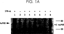
図1は、U937由来の安定な核酸感染細胞系のPKR活性とタンパク質レベルである。(A)機能的なPKR活性は、PKR自己リン酸化についてポリ(I):ポリ(C)−セルロースアッセイを用いて求めた。細胞抽出物は、表示のように組換えヒトIFN−α2(200U/ml)の存在下(+)または非存在下(−)でのインキュベーションに続いて種々のU937核酸感染細胞系から調製する一方、L929細胞は同様にマウスIFN−α/βで処理した。レーン1はHeLa、レーン2および3はU937−neo、レーン4はU937−AS1、レーン5はU937−AS3、レーン6はU937−M13、レーン7はU937−M22、レーン8はL929である。ヒト(68kDa)およびマウス(65kDa)PKRタンパク質、並びに分子サイズ標準物質(80および50kDa)の位置が表示されている。(B)細胞抽出物はIFN−αまたはIFN−γで誘発後、上記のように調製し、PKRタンパク質レベルはウェスタンブロット分析で決定した。
図2。EMCV複製の速度がPKR欠陥細胞で高められる。種々のU937細胞系を(A)0.1 TCID50/細胞または(B)0.001 TCID50/細胞のEMCVで攻撃した。表示した時間にサンプルを採集し、ウィルス収量をTCID50で測定した。
具体的な態様の説明
本発明は、細胞におけるインターフェロン産生レベルが、インビボでインターフェロン発現およびインターフェロン活性を通常調節するある種の因子の発現または活性を操作することによって調節できることを発明者らが見出したことに依る。これらの因子には、ある種のインターフェロン特異的転写調節因子、特にIRF1、ある種のインターフェロンレセプター、並びにある種のインターフェロン刺激遺伝子の遺伝子産物(インターフェロン仲介抗ウィルス反応とも呼ばれる)、特にPKRおよび2−5A合成酵素が含まれる。これら因子のいずれかの発現または活性を抑圧するかまたは排除することにより、正常レベルより低いインターフェロン遺伝子発現がもたらされる。この正常より低いインターフェロン発現レベルの結果の1つは、ウィルス複製に対する細胞の受容性の増加である。ウィルス複製に対して受容性が増した細胞は、ワクチン製造、低レベルのウィルスの感度の高い検出を含む多くの応用、及び抗ウィルス化合物の評価に有用である。
驚くべきことに本発明者らは、インターフェロン仲介抗ウィルス応答に欠陥がある細胞、特にdsRNA依存キナーゼ、2’−5’オリゴアデニレート合成酵素またはその双方に欠陥を有する細胞が、動物ウィルスに感染させたとき、正常レベルでこれらタンパク質を有する細胞より高いウィルス収量を生じることを見出した。本発明の方法を用いて、103から104またはそれ以上のウィルス収量増加が得られる。PKRまたは2−5A合成酵素に欠陥がある細胞培養で高いウィルス収量を得ることが可能になることによって、短時間で大量のウィルスを産生することが可能になった。これは、ウィルスワクチンの製造に特に重要であり、インフルエンザウィルスを含むRNAウィルスにとってきわめて重要である。この欠陥細胞のウィルス複製に対する受容性の増加によって、そのような細胞は、細胞培養で抗ウィルス薬を評価する方法、及びウィルス性病原体を検出する方法において有用性を有する。
本発明の特徴の1つは、細胞培養でウィルスワクチンを製造する方法であって、(a)インターフェロン刺激遺伝子の遺伝子生成物の活性に欠陥を有する細胞培養を供与株の動物ウィルスで感染する工程、(b)効率的にウィルス生長を提供するのに十分な条件下で感染した細胞培養を培養する工程、及び(c)産生したウィルスを採集する工程を有する方法を提供する。採集したウィルスは、ワクチンとして使用するために例えば濾過滅菌、超遠心およびまたはカラムクロマトグラフィーによる濃縮または他の方法によって精製してさらに調製することができる。場合によって、採集ウィルスはウィルス死滅ワクチンを製造するために処理してウィルスを不活化することができる。
好ましい態様として、細胞培養はPKR活性に欠陥がある。PKRに欠陥を有するとは、PKR活性が正常レベルの5%未満であることを意味する。PKR活性の正常レベルとは、安定なPKR欠陥細胞が得られた親細胞培養で認められるPKR活性、またはPKRの欠陥性が一時的に誘発される場合、PKRの欠陥性を誘発する前の細胞で認められたPKR活性レベルをいう。好ましくは、PKR欠陥細胞は正常レベルの1%未満のPKR活性を有する。より好ましくはPKR欠陥細胞は正常レベルの0.1%未満のPKR活性を有する。PKR活性とは、IFN−αおよびIFN−βの抗ウィルスおよび抗増殖活性を仲介する能力、開始因子e1f−2αをリン酸化する能力、または核因子κBを遊離させるためにIκBαをリン酸化する能力を意味する。PKRとはヒトp68キナーゼまたはヒトp68キナーゼの類似体もしくは相同体の全てを指す。ヒトp68キナーゼの類似体とは、インターフェロン転写のdsRNA活性化を仲介する全ての二本鎖RNA依存キナーゼを意味する。典型的には、そのようなdsRNA依存キナーゼは、例えばウサギまたはマウスのような他の種および多様な種の種々の組織に存在する等価なp68キナーゼである。例えばネズミのp65キナーゼはヒトp68キナーゼの類似体である。p68キナーゼの類似体の別の例はヒトの末梢血単核球で報告された(Farrelら)。相同体とは、ヒトp68キナーゼの少なくとも1つのドメイン、例えばdsRNA結合ドメインまたはキナーゼドメインと相同なタンパク質を指す。そのような機能的なキナーゼ相同体の1つはイーストGCN2キナーゼである。
PKR欠陥細胞は、当該技術分野で周知の種々の方法のいずれかによって得ることができる。PKR欠陥変異体は安定的にPKRに欠陥を有する場合もあり、またPKR欠陥性を一時的に誘発することもできる。安定なPKR欠陥変異体を製造する技術にはランダムまたは位置特異的変異誘発(例えばW.P.Deng & J.A.Nickoloff,Analytical Biochemistry 200:81-88(1992);S.Busy,M.Irani,B.J.Crombrugghe,J.Mol.Biol.,154:197-209(1982))、標的遺伝子削除(“遺伝子ノックアウト”)(例えばS.A.Camperら、Biology of Reproduction 52:246-257(1995);A.Aguzzi,S.Brandner,U.Sureら、Brain Pathology,4:3-20(1994))、PKRアンチセンスポリヌクレオチドによる核酸感染(例えばLeeら、Virology,192:380-385(1993))およびPKR優性(−)変異体遺伝子による核酸感染が含まれるが、これらに限定されるものではない。
PKR優性変異体は、正常なPKR活性を抑圧するためにただ1つの対立遺伝子のみが発現されねばならないPKR優性変異体である。PKR優性変異体遺伝子には、正常なPKR活性を抑圧するヒトp68キナーゼ変異体、ネズミp65キナーゼ変異体および、他のdsRNA依存キナーゼのいずれかの変異体またはヒトp68キナーゼの類似体もしくは相同体の変異体、例えば〔Arg296〕PKR(Meursら、J.Virol.66:5805-5814(1992))が含まれる。他のPKR優性変異体の例には、ウサギ網状赤血球、種々のマウス組織およびヒト末梢血単核球から得られたPKR変異体が含まれる(Farrelら、Levinら、Hovanessian,Krustら、Buffet-Janvresseら)。PKR優性変異体には、開始因子リン酸化、特にe1f−2αのリン酸化で干渉することによってタンパク質合成を抑圧する機能的相同体の変異体が含まれる。そのような機能的キナーゼ相同体の変異体の1つは、イーストGCN2キナーゼの変異体である。
PKRの欠陥が一時的な細胞を作出する技術には、2’−5’オリゴアデニレート連結PKRアンチセンスオリゴヌクレオチドの利用(A.Maran,R.K.Maitra,A.Kumar,B.Dong,W.Xiao,G.Williams,B.R.G.Torrence & R.H.Silverman,Science,265:789-792)または2-アミノプリンのようなPKRタンパク質の特異的抑制物質の利用(P.I.Marcus & M.J.Sekellick,J.Gen.Virol.,69:1637-45(1988);K.Zinn,A.Keller,L.A.Whittemore & T.Maniatis,Science,240:210-3(1988))、並びにPKR基質のリン酸化を妨害することができる他の競合的抑制物質の利用、または二本鎖RNA結合を妨害することができる抑制物質の利用が含まれるが、これらに限られるものではない。一時的PKR欠陥細胞培養は、そのようなアンチセンスオリゴヌクレオチドまたは抑制物質の存在下で細胞系を培養することによって得ることができる。
本発明の方法で使用するためには、細胞培養は、安定的にのPKR欠陥であるのが好ましい。典型的には、PKR欠陥細胞培養は、親細胞系、好ましくはワクチン製造に現在用いられている細胞系、好ましくはMRC−5、WI−38またはVero(アフリカミドリザル細胞)を機能的なPKRアンチセンス遺伝子構築物またはPKR優性(−)変異体構築物を含むベクターで核酸感染させ、続いてこのベクターを受容した細胞を選別することによって作製される。機能的PKRアンチセンス遺伝子構築物は、従来の方法、例えばMeursら(Cell,62:379-390(1990))に報告されたようなPKR cDNAを適切なプロモータ、例えばCMVプロモータの制御下で、アンチセンス方向にクローニングすることによって調製できる。PKR優性(−)変異体構築物は、PKR優性(−)変異体のcDNA、例えば〔Arg296〕PKRのcDNAを適切なプロモータの制御下でクローニングすることによって調製できる。
PKR欠陥細胞は、本発明の方法でワクチンを製造するために用いるためのものであるので、これらPKR欠陥細胞による腫瘍形成の危険性を減少させるために、PKR変異体遺伝子構築物は誘発可能プロモータの制御下でクローニングするのが好ましい。この方法はこれらの細胞によって産生されるワクチンの安全性を担保するであろう。PKR活性の消失は腫瘍形成に付随している(Koromilasら;Meursら)。採集ウィルスは細胞培養成分から精製することができるが、それにもかかわらずいくつかのPKR欠陥細胞が最終的なワクチン調製物に持ち込まれる危険性が残る。PKR活性が構造的に抑圧された状態を維持している場合、これらの細胞は腫瘍を誘発する潜在性を秘めているであろう。これはワクチン受容者に健康上の潜在的な危険性を与えるであろう。しかしながら、誘発可能プロモータが遺伝子構築物の発現制御に用いられる場合、内在性PKR活性は誘発物質の除去に際して回復するであろう。適切な誘発可能プロモータには、lacプロモータ、熱ショックプロモータ、メタロチオネインプロモータ、グルココルチコイドプロモータまたは当該技術分野で既知の他の誘発可能な全てのプロモータが含まれる。
例えば化学的もしくは酵素的に合成したDNA、PKR cDNAまたはPKR遺伝子のフラグメントを用いる同様なベクターを構築するその他の方法は、当業者にとって容易に知られよう。親細胞培養の核酸感染は標準的な方法、例えばDEAEデキストラン法(McCutchen & Pagano,J.Natl.Cancer Inst.,41:351-357(1968))、リン酸カルシウム法(Grahamら、J.Virol.,33:739-748(1973))または微量注入、リポフェクション、電気的穿孔を含むがこれらに限定されない当業界で既知の他のいずれかの方法によって行われる。そのような方法は一般的にサンブルックら(Molecular Cloning:A Laboratory Manual)(Sambrookら編、第2版、(1989)Cold Spring Harbor Laboratory Press刊)に記載されている。不完全なPKR活性を有する核酸感染体が選別される。選別を容易にするために、例えばネオマイシンホスホトランスフェラーゼII、アンピシリン耐性またはG418耐性のようなマーカー遺伝子をアンチセンスまたは変異体遺伝子を含むベクターに包含させることができる。マーカー遺伝子が含まれている場合、核酸感染体はマーカー遺伝子の発現(例えば抗生物質耐性)について選別し、培養し続いてPKR活性についてアッセイすることができる。
PKR欠陥細胞の残留PKR活性は、当業界で周知の多くの技術のいずれかによって決定できる。PKR活性は、例えばマラン(Maran)ら(Science,265:789-792(1994))またはシルバーマンら(R.H.Silverman & D.Krause,「インターフェロン:実際的アプローチ(Interferons:A practical approach)、A.G.Morris & M.J.Clemens編、pp71-74,IRL Press刊、Oxford-Washington,DC)が記載した自己リン酸化アッセイによって直接決定できる。典型的にはPKR活性のための自己リン酸化アッセイは以下のように実施される。タンパク質約100μgを含む、PKR活性を調べようとする細胞から得た抽出物をポリ(I):ポリ(C)−セルロースビーズ20μlと共に氷上で60分間インキュベートする。キナーゼはビーズ上で固定され活性化される。ポリヌクレオチドセルロース結合キナーゼ分画を洗浄した後、自己リン酸化反応をアッセイ溶液中で30℃で30分間実施する。アッセイ溶液は、〔γ32P〕ATPを1μCi、1.5mM酢酸マグネシウム、10μMのATP(pH7.5)、0.5%NP40および100μg/mlロイペプチンを含む。硫酸ドデシルナトリウム(SDS)を含むゲルサンプル緩衝液でサンプルを90℃で3分間加熱し、さらにタンパク質を10%SDS−ポリアクリルアミドゲル電気泳動で分析する。ゲルを乾燥させ、XAR−5X線フィルム(コダック)を用いてオートラジオグラフを調製する。
残留PKR活性はまた、例えばPKR特異的抗体を用いてウェスタンブロットによりPKRタンパク質の存在を、または例えばPKRに特異的なオリゴヌクレオチドもしくはcDNAプローブを用いてノザンブロットによりPKR RNAの存在をアッセイすることによって間接的に決定できる。明らかにわかるように、残留PKR活性の測定に適切なアッセイのタイプは、大抵の場合PKR欠陥表現型を得るために用いた方法に左右されるであろう。例えば、PKR欠陥細胞を作出するために用いた方法がPKR遺伝子発現の抑圧または排除をもたらす場合、(例えば、遺伝子ノックアウト)、mRNAもしくはcDNAの存在を検出する分析技術(例えばノザンブロットまたはサザンブロット)、またはタンパク質の存在を検出する分析技術(例えばウェスタンブロット)、または該タンパク質活性を検出する分析技術が、PKR欠陥欠陥細胞の残留PKR活性を測定するのに有用であろう。他方、PKR欠陥細胞を作出するために用いた方法が遺伝子発現の排除よりむしろ該タンパク質の抑制をもたらす場合(例えば優性(−)PKR変異体を担持するベクターでの核酸感染)、残留PKR活性の測定に自己リン酸化アッセイがウェスタンブロットより適切である。
他の態様として、本発明は、2’−5’オリゴアデニレート合成酵素活性に欠陥を有する細胞培養でウィルスワクチンを製造する方法を提供する。2−5A合成酵素に欠陥を有する細胞培養は、PKRに欠陥を有する細胞培養と同様な方法で単離できる。それらは、例えばランダムもしくは位置特異的変異誘発、2−5A合成酵素遺伝子の標的遺伝子削除またはアンチセンス2−5A合成酵素構築物の核酸感染である。2−5A合成酵素に欠陥を有するとは、2−5A合成酵素活性が正常な2−5A合成酵素活性の5%未満であることを意味する。正常レベルの2−5A合成酵素とは、安定な2−5A合成酵素欠陥細胞が得られた親細胞で認められる2−5A合成酵素活性レベル、または2−5A合成酵素欠陥性が一時的に誘発される場合、2−5A合成酵素欠陥性が誘発される前に認められる2−5A合成酵素活性レベルを意味する。好ましくは2−5A合成酵素活性欠陥細胞は、正常レベルの2−5A合成酵素活性の1%未満を有し、より好ましくは2−5A合成酵素欠陥細胞は、正常レベルの2−5A合成酵素活性の0.1%未満を有する。2−5A合成酵素欠陥細胞の残留2−5A合成酵素活性は、残留PKR活性を測定するために用いた方法と同様な方法で測定できる。すなわち、2−5A合成酵素特異的抗体を用いるウェスタンブロット、2−5A合成酵素に特異的なオリゴヌクレオチドもしくはcDNAを用いるノザンブロット、または酵素活性アッセイである(Readら、J.Infect.Dis.,152:466-472(1985);Hassel & Ts’o,J.Virol.Methods,50:323-334(1994)を参照のこと)。典型的には、2−5A合成酵素活性は以下のように求められる。アッセイするべき細胞をIFN−α2(RPMI+10%ウシ胎児血清に100U/1ml)で処理する。簡単に記せば、細胞培養を37℃で18時間インキュベートし、洗浄して細胞ペレットを細胞溶解緩衝液で10分間4℃で処理する。細胞抽出物の一部をポリ(I):ポリ(C)−アガロースビーズと共に30℃で30分間インキュベートして2−5A合成酵素を結合させるとともに活性化させる。このビーズを洗浄し、続いて3mMのATP、アッセイサンプル当たり4μCiの3H−ATP、および20mMのHepes緩衝液(pH7.5)を含むアッセイ溶液中で30℃で20時間インキュベートする。インキュベーション後、サンプルを90℃に加熱して酵素を不活化し、続いて細菌のアルカリホスファターゼ(BAP)で処理する。合成した2−5オリゴAはBAPに対して耐性を有する。サンプルを濾紙に滴下して洗浄し、シンチレーションカウンターを用いて3Hの放射能活性を計測することによって2−5オリゴAの量を求める。このオリゴA生成物の量は通常の方法による酵素活性と相関性を有する。また、2−5A合成酵素は、ラジオイムノ法および放射能結合法によってアッセイできる(M.Knightら、Radioimmune,radiobinding and HPLC analysis of 2-5A and related oligonucleotides from intact cells(完全細胞の2−5Aおよび関連オリゴヌクレオチドの放射能免疫分析、放射能結合分析およびHPLC分析)、nature,288:189-192(1980))。
PKR活性および2−5A合成酵素活性の両方に欠陥を有する細胞培養は上記の方法を組み合わせて作出できることは明白であろう。二重に欠陥を有する細胞培養は、連続的に(すなわち先ず1つの活性に欠陥を有する培養を選別し、続いて第二の欠陥細胞の調製にこの細胞培養を出発材料として用いる)または同時に(両欠陥を一度に選別する)調製できる。
また他の態様として、本発明は、ヒトのMxAタンパク質活性に欠陥を有する細胞培養でウィルスワクチンを製造する方法を提供する。ヒトMxAタンパク質活性に欠陥を有する細胞培養は、PKR欠陥細胞培養と同様な方法、例えばランダムもしくは位置特異的変異誘発、MxA遺伝子の標的遺伝子削除、またはアンチセンスMxA構築物の核酸感染で単離できる。MxAタンパク質に欠陥を有するとは、MxA活性が正常なMxA活性レベルの5%未満であることを意味する。正常レベルのMxA活性とは、安定なMxA欠陥細胞が得られた親細胞で認められるMxA活性、またはMxA欠陥性が一時的に誘発される場合、MxA欠陥性が誘発される前に認められるMxA活性レベルを意味する。好ましくはMxA欠陥細胞は、正常レベルのMxA活性の1%未満を有し、より好ましくはMxA欠陥細胞は、正常レベルのMxA活性の0.1%未満を有する。MxA欠陥細胞の残留MxA活性は、残留PKR活性を測定するのに用いた方法と同様な方法で測定できる。すなわち、MxA特異的抗体を用いるウェスタンブロット、MxAに特異的なオリゴヌクレオチドもしくはcDNAプローブを用いるノザンブロットまたは酵素活性アッセイである
![]()
![]()
さらに他の態様として、本発明は、インターフェロン応答性に欠陥を有する細胞培養でウィルスワクチンを製造する方法を提供する。インターフェロン応答性とは、インターフェロンによる刺激に応答する細胞の能力を指す。インターフェロン反応性に欠陥を有する細胞培養は、インターフェロンレセプターの抑制物質の存在下で細胞を培養することによって得られる。また、正常なインターフェロンレセプターの非存在下でインターフェロンに応答性をもたない変異インターフェロンレセプターを発現するように細胞を操作してもよい。
また他の態様として、本発明は、インターフェロン特異的転写調節因子に欠陥を有する細胞培養でウィルスワクチンを製造する方法を提供する。そのようなインターフェロン特異的転写調節因子の1つはIRF1である。インターフェロン特異的調節因子に安定的に欠陥を有する細胞は、当該技術分野で周知の多数の技術、例えばランダムもしくは位置特異的変異誘発、標的遺伝子削除またはアンチセンスベクターの核酸感染のいずれかによって得ることができる。一時的に欠陥を有する細胞は、アンチセンスオリゴヌクレオチドまたはインターフェロン転写の特異的抑制物質の存在下で細胞を培養することによって得られる。
本発明の方法は、初代細胞培養、二倍体細胞培養および継代細胞培養を含む種々の動物細胞を用いて実施できる。特に有用なものは、現在ワクチン製造に用いられている細胞培養、特にUSFDAおよび/またはWHOによりワクチン製造について承認されている細胞培養、例えばMRC−5(胎児肺組織由来ヒト二倍体細胞系(Nature Lond.,227:168-170(1970))、およびWI−38(胎児肺組織由来ヒト二倍体細胞系(Am.J.Hyg.,75:240(1962);ヒトのウィルス病およびリケッチア病に対するワクチンに関する第1回国際会議(First Conference on Vaccines Against Viral and Rickettsial Diseases of Man)、Pan American Health Organization,Pub.No.147:581,1981年5月)である。また有用なものは、Chang肝細胞(R.S.Chang,Proc.Exp.Biol.Med.,87:440(1954))、U937ヒト前単球(Sundstromら、Int.J.Cancer 17:565-577(1976))、Vero細胞、MRC−9細胞、1MR−90細胞、1MR−91細胞およびLederle130細胞(Biologicals,18:143-146(1991))である。U937細胞は、CD4を発現する免疫細胞に感染するウィルス、例えばHIVについて特に有用である。ワクチン製造で使用される細胞培養に関する概論として、グラシェフの論文(V.P.Grachev,「ウィルスワクチン(Viral Vaccines)」、A.Mizrahi編、pp.37-67(1990)、Wiley-Liss刊)を参照されたい。選択される個々の細胞培養は産生されるウィルスに左右され、一般には細胞培養は該ウィルスの天然の宿主である種に由来するであろう。ただしこのことは本発明を実施するうえで必ずしも必須ではない(例えばヒトのウィルスはネコの腎細胞系(MDCK細胞)またはミドリザル腎細胞系(Vero細胞(Swansonら、J.Biol.Stand.,16:311(1988))で増殖できる)。典型的には、選択される細胞は、産生されるウィルスに対して適切な宿主であることが分かっている細胞または細胞系のPKR欠陥誘導体または2−5A合成酵素欠陥誘導体である。例えば、インフルエンザウィルスおよびA型肝炎ウィルスワクチンでは好ましい宿主細胞はMRC−5の誘導体である。HIVワクチン製造に好ましい宿主細胞は、U937、H9、CEMまたはCD4発現HUT78細胞の誘導体である。ワクチン製造に用いられる細胞系は周知であり、かつ商業ルートの供給者、例えばアメリカン・タイプ・カルチャ・コレクション(American Type Culture Collection)から容易に入手できる。
本発明によるインターフェロン仲介抗ウィルス反応欠陥細胞の供与ウィルス感染は通常の技術によって実施される(例えば、J.Peetermans,J.Vaccine,1992/10増補1:S99-101;Shevitzら、「ウィルスワクチン」、A.Mizrahi編、pp.1-35(1990)、Wiley-Liss刊を参照のこと)。典型的には、ウィルスは0.001〜0.5TCID50/細胞、好ましくは0.01〜0.10TCID50/細胞で細胞培養に添加されるが、個々のウィルスおよび用いられる宿主細胞にとって適切なように変動するであろう。当業者には容易に理解されるところであるが、効率的なウィルス産生のために細胞培養の全ての細胞が最初から感染していなければならないということではない。感染をモニターした後、この細胞およびウィルス産生に適した条件下でこれら感染細胞を培養する。ウィルスの産生は、プラーク形成単位アッセイ、TCID50アッセイまたは血球凝集抑制アッセイを含む多数の標準的な技術のうち何れかによってモニターすることができる(Robertsonら、J.Gen.Virol.,72:2671-2677(1991))。感染細胞は効率のよいウィルス成長を提供するのに十分な条件下で培養される。細胞は、ウィルス収量がプラトーに達することによって表示されるように最大のウィルス産生が達成されるまで培養できる。ウィルスは標準的な方法によって採集され、続いて他の細胞性成分から精製される(Peetermans(1992)を参照のこと)。採集ウィルスは生ウィルスワクチン(完全な強毒また弱毒のいずれか)として用いてもよいし、または当該技術分野で周知の方法、例えばホルムアルデヒド処理によって使用前に不活化してもよい(J.Peetermans,J.Vaccine,1992/10増補1:S99-101;米国特許第RE33164号)。
ワクチンは乾燥形(希釈液と混合される)で用いてもよく、または濃縮もしくは直ぐに使用できる液体形(好ましくは水溶液)でもよい。ワクチンは単独で投与してもよく、または医薬上許容可能な担体、アジュバント、保存料、希釈液および、免疫誘発性を強化するか、もしくは投与を助け、もしくは保存を助けるのに有用であると当業界で周知の他の添加物と組み合わせて投与してもよい。適切なアジュバントには水酸化アルミニウム、ミョウバン、リン酸アルミニウム、フロイントまたは米国特許第3,790,665号および3,919,411号に記載されているものが含まれる。他の適切な添加物には、ショ糖、デキストロース、乳糖、および他の無毒な物質が含まれる。ワクチンは、筋肉内、静脈内、皮下、経気管的、経鼻的ルートを含む種々なルートで、またはエアゾルスプレーによって動物に投与され、さらにワクチンはヒト、ウマ、ニワトリ、ネコ、イヌ、ウシを含む種々の動物で有益に用いられる。
本発明の方法は、種々の供与動物ウィルスを用いて実施できる。供与ウィルスとは、ワクチンを製造するためにインビボで複製される個々のウィルス株を意味する。用いられる個々の供与動物ウィルスは所望するウィルスワクチンに左右される。ワクチン製造に現時点で用いられる供与ウィルスは当業界で周知であり、本発明の方法は新規に特定された何れの供与ウィルスにも容易に応用できる。好ましい供与ウィルスには、ヒトインフルエンザウィルス、特にインフルエンザA(H3N2)およびインフルエンザA(H1N1)(米国特許第4,552,758号:ATCC第VR-2072号、第VR-2073号、第VR-897号)、米国特許第3,953,592号に開示されたインフルエンザA、インフルエンザB(米国特許第3,962,423号:ATCC第VR-786号、第VR-791号)およびパラインフルエンザ1(センダイウィルス)(Cantellら、Meth.Enzymol.,78A:299-301(1980);ATCC第VR-907号)が含まれる。供与ウィルスはウィルス性病原体でもよく、または天然に得られる弱毒形、細胞培養の連続継代によって産生された弱毒形または組換え体もしくは再組合せ体でもよい。供与ウィルスが必須の抗原性を保持し、該ウィルス性病原体に対して防御することができるということを条件として、いずれのウィルス株も供与ウィルスとして用いることができる。本発明の方法は、弱毒供与ウィルスまたは複製の弱い供与ウィルスについて特に有用である。
本発明の方法によって提供することができるワクチンの幾つかには、ポリオウィルス、麻疹、流行性耳下腺炎、風疹、A型肝炎、インフルエンザ、パラインフルエンザ、日本脳炎、サイトメガロウィルス、HIV、デング熱ウィルス、狂犬病および水痘−帯状疱疹ウィルスに対するヒトワクチンとともに多くの非ヒト動物ワクチンが含まれるが、これらに限定されるものではない。非ヒト動物ワクチンには、例えばネコ白血病ウィルス、ウシ鼻気管炎ウィルス(赤鼻ウィルス)、牛痘ウィルス、イヌ肝炎ウィルス、イヌジステンパーウィルス、ウマライノウィルス、ウマインフルエンザウィルス、ウマ肺炎ウィルス、ウマ伝染性貧血ウィルス、ウマ脳炎ウィルス、ヒツジ脳炎ウィルス、ヒツジブルータングウィルス、狂犬病ウィルス、ブタインフルエンザウィルス、およびサル免疫不全ウィルスに対するワクチンが含まれる。前述の記載から明らかなように、本発明の方法はヒトのウィルスに対するワクチン製造に限定されず、非ヒト動物ウィルスワクチンの製造にも等しく適切である。
本発明の他の特徴により、抗ウィルス化合物の活性を評価する方法が提供される。PKR欠陥細胞ではウィルス複製に対する受容性が高まるために、そのような細胞は抗ウィルス化合物の効能を評価する感度の高いアッセイで役立つ。この特徴では、本発明は、(a)ウィルス、ウィルス感染宿主細胞またはウィルス感染前の宿主細胞を抗ウィルス化合物で処理する工程、及び(b)PKR欠陥または2−5A合成酵素欠陥インジケーター細胞培養を感染条件下に曝すことによって、残存する感染性ウィルスの存在をアッセイする工程を有する。
この特徴では、抗ウィルス化合物の検査対象であるウィルスはこの化合物で直接処理できる。この場合、その後感染条件下でPKR欠陥または2−5A合成酵素欠陥インジケーター細胞培養を処理ウィルスの一部に曝し、全ての残留感染性ウィルスの複製を可能にするために十分な時間培養し、さらに複製ウィルスの存在についてインジケーター培養を分析することによって残留感染性ウィルスの存在についてこの処理ウィルスを直接分析できる。または、抗ウィルス化合物の検査対象であるウィルスを用いて、宿主細胞培養を感染させることができる。その後感染宿主細胞培養を抗ウィルス化合物で処理する。処理した感染宿主細胞培養の細胞抽出物を通常の技術で調製し、抽出物の一部をPKR欠陥または2−5A合成酵素欠陥インジケーター細胞培養に上記のように曝して、残留する感染性ウィルスについて調べる。さらに別の選択肢では、宿主細胞培養は感染後よりむしろウィルス感染前に抗ウィルス化合物で処理してもよい。続いて処理細胞を抗ウィルス化合物の検査対象であるウィルスに感染させて培養し、さらに複製ウィルスの存在について調べる。選択される個々の処理方法は、抗ウィルス化合物の既に分かっている、または想定される作用態様に左右され、当業者は容易に決定できるであろう。感染条件下に曝すとは、何らかのウィルスが処理サンプルに存在する場合、欠陥細胞培養の感染を生じる条件下で欠陥を有するインジケーター細胞と処理サンプルの一部(ウィルスまたは感染細胞抽出物のいずれか)とを一緒にすることを意味する。処理サンプルに曝した後、欠陥を有するインジケーター細胞培養をさらに培養し、ウィルス複製について標準的な方法(例えばプラークアッセイまたはTCID50アッセイ、もしくはウィルスRNAまたはタンパク質についてノザンまたはウェスタン分析)によってアッセイする。
宿主細胞培養は、抗ウィルス化合物の検査対象であるウィルスによる感染に感受性を有する何れの細胞培養でもよい。インジケーター細胞培養は、抗ウィルス化合物による処理後に残存する感染性ウィルスについてのアッセイに用いられるPKR欠陥または2−5A合成酵素欠陥細胞培養である。PKR欠陥または2−5A合成酵素欠陥インジケーター細胞培養はワクチン製造について上記で述べたように調製される。欠陥を有するインジケーターを作出する親として適切な細胞は、ワクチン製造のためにPKR欠陥または2−5A合成酵素欠陥細胞培養を作出するために有用なものと同じである。また、以下の細胞系もまた適切である。一般にヘパトーマ細胞系、特にHep G2ヒト肝細胞性癌腫(Nature,282:615-616(1979):米国特許第4,393,133号)およびHep 3B(米国特許第4,393,133号)である。インジケーター細胞培養もまた、抗ウィルス化合物の検査対象であるウィルスの感染に感受性を有することは明白であろう。宿主細胞培養およびインジケーター細胞培養は同じでも異なっていてもよい。抗ウィルス化合物は、何らかの抗ウィルス活性を有すると思われる何れの化学的または生物学的調製物でもよい。ウィルス自体が抗ウィルス化合物で処理される場合、この化合物は、処理ウィルスに曝してインジケーター細胞培養を感染させる前に除去するのがよい。感染宿主細胞培養(または感染前宿主細胞)が抗ウィルス化合物で処理される場合、この化合物は細胞抽出物を調製する前に除去するのがよい。
また別の関連する特徴として、本発明は、ウィルス性病原体を同定する方法および培養する方法を提供する。PKR欠陥細胞のウィルス複製に対する受容性は、培養が困難な極めて低レベルのウィルス、例えば新生児の単球またはリンパ球でのHIVを検出する方法においてそれら細胞を特に有用なものにする。この特徴において、本発明は、(1)ウィルスを含むと思われるサンプルにPKR欠陥または2−5A合成酵素欠陥細胞培養を感染条件下で曝す工程、及び(2)曝露細胞における複製ウィルスの存在についてアッセイする工程を有する。本発明のこの特徴の実施は、抗ウィルス化合物との処理が省略されるという点を除いて前述の特徴の実施と同様である。この特徴では、ウィルスの存在についてアッセイされるべきサンプルは一般に、ウィルスに感染したと疑われる患者の臨床サンプルである。サンプルは、血液、唾液、尿、並びにリンパ節、肺臓、腸、肝臓、腎臓および脳組織の生検サンプルを含む適切な何れの臨床サンプルでもよい。サンプルはウィルス粒子を遊離させるために適切に処理されるか(例えば細胞抽出物を調製してもよい)、または患者から得た状態のまま用いてもよい。サンプルまたはサンプルの一部を欠陥を有するインジケーター細胞培養に感染条件下で曝し、全ての複製ウィルスの存在を上記のように測定する。
上述の工程の特定の例を以下の実施例で説明する。しかしながら、多くの変更が可能であり、さらにこれら実施例は説明のみを目的とし、特に限定されないかぎり本発明を制限するものではないことは当業者には明白であろう。
実施例
実施例1:プラスミドの調製
それぞれpBS−8.6Rおよびyex6Mから得られる(E.Meurs,K.Chong,J.Galabruら、Cell,62:379-90(1990);Chongら、EMBO J.,11:1553-1562(1992))野生型ヒトPKR遺伝子および優性で(−)の〔Arg296〕PKR変異体遺伝子に対応するcDNA挿入物を、HindIII消化によって遊離し、G418耐性マーカーを含む構造的真核細胞発現プラスミドであるpRC−CMV(Invitrogen)でサブクローニングした。選別したクローンにおける挿入物の方向性は制限消化分析によって決定し、配列分析(sequencing)(シークェナーゼ2.0、USB)によって確認した。この方法によって使用する発現プラスミド、pPKR−AS(ベクター中のCMVプロモーターの制御下でのアンチセンス方向のPKR cDNAを含む)およびp〔Arg296〕PKR(ベクター中のCMVプロモーターの制御下でのArg296PKRcDNAを含む)が得られた。
実施例2:PKR欠陥安定核酸感染体の単離
安定な核酸感染体は、DEAEデキストラン(50mg/ml)含有無血清RPMI−1640中で各プラスミド10mg、ジーンパルサー(Gene Pulser)装置(BioRad)(500μF、250Vに設定)を用いて対数増殖期の5×10⁶個のU937細胞に電気的穿孔を施して得られた。安定な核酸感染体の大集団を、ジェネチシン(geneticin)400μg/ml(GIBCO-BRL)による3週間の選別で得た。限定希釈クローニングにより続いてクローン系を得た。細胞系を10%ウシ胎児血清含有RPMI−1640(完全培養液)およびジェネチシン中で培養した。
5つの代表的な細胞株系を最初の性状分析のために選別した。“U937−neo”(U9K−Cとも呼ぶ)は親ベクター(pRC−CMV)を核酸感染させたコントロール細胞系であった。“U937−AS1”(U9K−A1とも呼ぶ)および“U937−AS3”(U9K−A3とも呼ぶ)はpPKR−ASを核酸感染させたそれぞれ別個のクローンであった。“U937−M13”(U9K−M13とも呼ぶ)および“U937−M22”(U9K−M22とも呼ぶ)はp〔Arg296〕PKRを核酸感染させたそれぞれ別個のクローンであった。
実施例3:PKR欠陥核酸感染体の性状分析
PKR酵素の結合と活性化のためにポリ(I):ポリ(C)−セルロースを用いる自己リン酸化アッセイでPKRキナーゼ活性を測定した。PKR自己リン酸化アッセイは、以下の変更を加えて本質的にはマラン(Maran)らの記載にしたがって実施した。細胞抽出物(1アッセイにつきタンパク質100μg)をポリ(I):ポリ(C)−セルロースとともに氷上で1時間インキュベートし、3回洗浄し、さらに〔γ−32P〕ATPを1μCi含む反応緩衝液50μl(20mMのHEPES(pH7.5)、50mMのKCl、50mMの2-メルカプトエタノール、1.5mM酢酸マグネシウム、1.5mMのMnCl2)に30℃で30分間インキュベートした。タンパク質を10%SDSポリアクリルアミドゲルで分離し、オートラジオグラフィーで分析した。
IFN処理HeLaおよびマウスL929細胞の細胞抽出物をポジティブ・コントロールとして用いたが、これらの細胞のPKR活性は以前に性状分析してあったからである(Meursら)(図1A、レーン1および8)。U937−neo細胞は低い基底レベルのPKR活性を含み、これはIFN−αによる処理後、増加した(図1A、レーン2および3)。親の非核酸感染U937細胞のPKR活性はU937−neo細胞と同様であった。しかしながら、pPKR−ASまたはp〔Arg296〕PKRプラスミドを核酸感染させた4つの細胞系のいずれにおいてもPKR活性は検出されなかった。さらにまた、IFN−αでこれらの細胞を処理してもPKR活性は回復せず(図1A、レーン4−7)、IFN−γで処理してもまた回復しなかった。
実施例4:PKR欠陥核酸感染体のウェスタン分析
pPKR−AS核酸感染細胞系のPKR発現抑制をさらに確認するために、ウェスタンブロット分析をヒトPKRに対して特異的なモノクローナル抗体を用いて行った。細胞抽出物(100μg)を10%SDS−ポリアクリルアミドゲル上で分離し、ニトロセルロース膜に電気的に移した。BLOTTO(トリス緩衝食塩水に5%無脂肪ドライミルク、0.05%トゥイーン20)で1:1000にした抗PKRモノクローナル抗体(Meursら、Cell(1990))とこの膜をインキュベートした。ホースラディッシュペルオキシダーゼ結合ヤギ抗マウス二次抗体(Santa Cruz Biotech)をプローブとして用いかつ化学ルミネセンス法(Amersham ECL)を用いて、PKRを最終的に検出した。
基底レベルのPKRタンパク質はU937−neo細胞(図1B、レーン1)で検出され、IFN−αおよびIFN−γによるその後の処理で増加した(図1B、レーン2および3)。対照的にPKR発現はU937−AS1およびU937−AS3細胞の両方で顕著に減少し(図1B、レーン4および6)、IFN−αによるその後の処理では増加しなかった(図1B、レーン5および7)。一方、PKRタンパク質はU937−M13およびU937−M22細胞で検出され、変異体〔Arg296〕PKRのタンパク質はウェスタンブロット分析を用いた場合、野生型PKRとは区別できなかった。
実施例5:PKR欠陥細胞でのEMCV複製の強化
IFN系は抗ウィルス反応で主要な役割を果たすので、我々は、PKR機能の消失が脳脊髄炎ウィルス(EMCV)の複製速度に影響を与えるのか否かを調べた。EMCV(ATCC No.VR-1314)のストックはL929細胞で継代して調製された。EMCV複製の判定のために、U937由来核酸感染細胞をIFN群(組換えヒトIFN−α2(Schering);組換えヒトIFN−γ(Amgen))とともに、またはIFN群を添加せずに完全培養液で18時間培養した。PBSで2回洗浄してから細胞をEMCVと共に無血清培養液で2時間インキュベートした。細胞を再度2回洗浄し、1%FCSを含む培養液を補充した。サンプルを必要とされる時点で採集し、凍結融解を3回繰り返して細胞を溶解させた。サンプルの4倍段階希釈をL929単層培養に加え、48時間培養し、続いて0.05%クリスタルバイオレットで染色して細胞障害効果および組織培養感染中央値(TCID50)を求めた。
0.1 TCID50/細胞のEMCVで攻撃した後コントロールのU937neo細胞系では、ウィルス力価は48時間後に約104TCID50/mlのピークに達し、72時間後にそれ以上増加しなかった(図2A)。しかしながら、U937−AS1およびU937−M22細胞では、EMCV複製は、わずか24時間後に104から105TCID50/mlの実質的により高い力価に達し、かつ48時間までに108TCID50/mlに達してコントロール細胞で得られたウィルス収量の103から104の増加を示した。より低い0.001 TCID50/mlの接種量を用いた別の実験では、コントロールとPKR欠陥細胞との間でEMCV感受性についてより劇的な相違が観察された(図2B)。このような条件下では、U937−neo細胞でのEMCVの複製は最小で、72時間後でさえ102TCID50/mlを越えなかった。一方、より高い108TCID50/mlの力価がU937−AS1およびU937−M22細胞の両方で48時間後に得られた。これらの結果は、インビボでPKR活性を抑圧することによって細胞はウィルス複製に強い受容性を有するようになることを示し、コントロール細胞より1000倍もの増加を示唆する。
本明細書で述べた全ての刊行物および特許出願は、個々の刊行物および特許出願が具体的にかつ個々に参照により本明細書に含まれるように、参照により本明細書に含まれる。
本発明をこれまでのところで十分に説明してきたが、多くの変更および改変が本発明の範囲を逸脱することなく為しえることは当業者には明白であろう。 Related applications
This application is a continuation-in-part of US provisional application USSN 60 / 002,621 (filed Aug. 22, 1995).
Introduction
Technical field
The present invention relates to a method for producing viruses in cell culture for vaccine production.
background
Efficient control of the pandemic influenza depends on early vaccination with inactivated viruses made from newly identified influenza strains. However, improved vaccine production and testing is needed to more effectively control epidemics. Influenza viruses have very frequent surface antigen mutations. As a result, vaccine manufacturers are unable to store millions of doses for rapid use. Current influenza control methods require the production of vaccines specific to newly identified strains along with routine international research and the identification of any newly emerging strains. The production of current influenza vaccines that require the use of embryonated chicken eggs for virus inoculation and culture is cumbersome and expensive. This method is also limited by seasonal variations in the supply of adequate quality eggs. Therefore, in order to produce a large amount of vaccine in a short time, it would be advantageous to develop another production technique that does not rely on chicken eggs. In this regard, the manufacture of influenza vaccines with stable cell lines will solve many of the problems in mass production. However, the yield of human influenza virus in tissue culture is significantly lower than that of embryonated chicken eggs (Tannock et al., Vaccine 3: 333-339 (1985)). In order to overcome these limitations and improve vaccine quality, it would be advantageous to develop a cell culture system that yields increased virus yield over currently available cell lines. When using mammalian cell lines to produce complete virion vaccines, a common problem for vaccine manufacturers is that mammalian cells have unique antiviral properties, specifically interferon (IFN). The system interferes with virus growth. IFNs are mainly classified into two groups according to their primary sequence. Interferon type I, IFN-α and IFM-β are encoded by an intronless gene superfamily consisting of the IFN-α genes and only one IFN-β gene. Type II interferon, or IFN-γ, consists of only one type and is limited to lymphocytes (T-cells and natural killer cells). Type I interferons mediate a variety of biological processes including induction of antiviral activity, regulation of cell proliferation and differentiation, and regulation of immune function (GCSen & P. Lengyel, J. Biol. Chem., 267: 5017-5020 (1992); S. Pestka & JALanger, Ann. Rev. Biochem., 56: 727-777 (1987)). Induction of expression of type I IFN (including IFN-α and IFN-β genes) is typically detected following viral infection. Numerous studies have identified promoter components and transcription factors required for regulation of type I IFN expression (W. Du, D. Thanos & T. Maniatis, Cell 74: 887-898 (1993); T. Matsuyama, T.Kimura, M.Kitagawa, K.Pfeffer, T.Kawakami, N.Watanabe, TMKundig, R.Amakawa, K.Kishihara, A.Wakeham, J.Potter, CLFurlonger, A.Narendran, H.Suzuki, PSOhashi, CJPaige, T. Taniguchi & TWMak, Cell 75: 83-97 (1993); N. Tanaka & T. Taniguchi, Adv. Immunol., 52: 263-281 (1992)). However, it remains unclear what specific biochemical triggers and necessary reporting mechanisms to inform cells of viral infection (for a recent review of the interferon system, see Jaramillo et al., Cancer Investigation 13: 327- 337 (1995)).
IFN belongs to negative growth factors that have the ability to inhibit the growth of various cells with normal and cancerous phenotypes. IFN treatment is used to treat human malignancies such as Kaposi's sarcoma, chronic myelogenous leukemia, non-Hodgkin's lymphoma and hairy cell leukemia as well as infectious diseases such as papillomavirus (genital warts) and hepatitis B and C. It has been shown to be beneficial (review: Gutterman, Proc. Natl. Acad. Sci., 91: 1198-1205 (1994)). Recently, genetically engineered bacterially produced IFN-β has been approved for the treatment of multiple sclerosis, a relatively common neurological disease affecting at least 250,000 people in the United States alone.
IFN induces its biological activity by binding to its cognate receptor and subsequently transmitting a signal resulting in the induction of an IFN-stimulated gene (ISG). While a large number of ISGs have been identified, only some of them have been characterized and their biological activity has been investigated. Examples of ISGs that have been best elucidated include double-stranded RNA (dsRNA) -dependent kinase (PKR, formerly known as p68 kinase), 2'-5 'linked oligoadenylate (2-5A) synthesis Enzymes and Mx proteins are mentioned (JLTaylor & SEGrossberg, Virus Research, 15: 1-26 (1990); BRGWilliams, Eur. J. Biochem., 200: 1-11 (1991)). The human MxA protein is a 76 kD protein that suppresses the growth of influenza virus and vesicular stomatitis virus (Pavlovic et al., J. Virol. 64: 3370-3375 (1990)).
2'-5 'oligoadenylate synthase (2-5A synthase) uses ATP to synthesize short oligomers of up to 12 adenylate residues linked by 2'-5' phosphodiester bonds . The resulting oligoadenylate molecule allosterically activates a latent ribonuclease, RNAase L, which degrades viral RNA and cellular RNA. This 2-5A synthase pathway reduces the synthesis of viral proteins in a cell-free protein synthesis system isolated from IFN-treated cells, and presumably reduces the resistance of at least some types of viruses to viral infection in vivo. Seems to be important.
PKR (abbreviation for protein kinase RNA-dependent) is the only identified dsRNA binding protein known to have kinase activity. PKR is a serine / threonine kinase that requires dsRNA binding and consequent autophosphorylation for its enzymatic activity (J. Galabru & A. Hovanessian, J. Biol. Chem., 262: 15538- 15544 (1987); E. Meurs, K. Chong, J. Galabru, NSThomas, IMKerr, BRGWilliams & AGHovanessian, Cell 62: 379-390 (1990)). PKR is also referred to in the literature as dsRNA-activated protein kinase, P1 / e1F2 kinase, DAI or dsI for dsRNA activation inhibitors, and p68 (human) or p65 (murine) kinase. Similar enzymes have been reported in rabbit reticulocytes, murine tissues, and human peripheral blood mononuclear cells (Farrel et al., Cell, 11: 187-200 (1977); Levin et al., Proc. Natl. Acad. Sci. USA, 75: 1121-1125 (1987); Hovanessian, Biochimie, 62: 775-778 (1980); Krust et al., Virology 120: 240-246 (1982); Buffet-Janvresse et al., J. Interferon Res ., 6: 85-96 (1986)). The best-characterized in vivo substrate for PKR is the eukaryotic initiation factor-2 (eIF-2a), which is a cellular and viral protein once phosphorylated. (JWBHershey, Ann. Rev. Biochem. 60: 717-755 (1991)). PKR can phosphorylate the initiation factor e1F-2α in vitro when activated by double-stranded RNA (Chong et al., EMBO J., 11: 1553-1562 (1992)). This particular function of PKR has been suggested as one of the mechanisms required to mediate the antiviral and antiproliferative activity of IFN-α and IFN-β. Another biological function of PKR is its putative role as a signal inducer. A. Kumar et al. Have shown that PKR can phosphorylate IκBα, resulting in the release and activation of nuclear factor κB (NF-κB) (A. Kumar, J. Haque, J. et al. Lacoste, J. Hiscott & BRGWilliams, Proc. Natl. Acad. Sci. USA, 91: 6288-6292 (1994)). Given the well-characterized NF-κB site of the IFN-β promoter, it may reveal the mechanism by which PKR mediates dsRNA activation of IFN-β transcription (KVVisvanathan & S. Goodbourne) , EMBO J., 8: 1129-1138 (1989)).
The catalytic kinase subdomain of PKR (ie, p68 (human) kinase and p65 (murine) kinase) has high sequence identity (38%) with yeast GCN2 kinase (Chong et al., EMBO J., 11: 1553-1562 ( 1992); Feng et al., Proc. Natl. Acad. Sci. USA, 89: 5447-5451 (1992)). Recombinant p68 kinase expressed in Saccharomyces cerevisiae exhibits a growth suppression phenotype. This is thought to be due to activation of p68 kinase followed by phosphorylation of the yeast equivalent of mammalian e1F2α (Chong et al .; Cigan et al., Proc. Natl. Acad. Sci. USA, 86: 2784-2788 (1982 )).
Surprisingly, the inventors have found that certain ISG expression manipulations have valuable applications. They found that expression or suppression of PKR protein or 2-5A synthase protein, or both, is useful to substantially increase virus yield from virus-infected cells and enhance vaccine production in animal cell culture. I found.
Related literature
General methods to investigate the biological role of PKR require the creation of mutants with incomplete kinase activity. Since PKR has a regulatory site for dsRNA binding and a catalytic site for kinase activity, researchers have used block deletion mutations or position-specific mutations to create variants for the regulatory or catalytic sites. . PKR dominant negative mutant containing a single amino acid substitution in which the invariant lysine at position 296 of catalytic domain II is replaced with arginine (PKR dominant negative mutant), [Arg296PKR is described (KVVisvanathan & S. Goodbourne, EMBO J.8: 1129-1138 (1989); M.D'Addario, A. Roulston, MAWainberg & J. Hiscott, J. Virol., 64 : 6080-6089 (1990)). This mutant protein [Arg296PKR can specifically suppress the activity of endogenous wild-type PKR in vivo. Still other mutants were created by modifying dsRNA binding motifs. For example, Feng et al. (Proc. Natl. Acad. Sci. USA, 89: 5447-5451 (1992)) have obtained mutants with deletions between amino acid residues 39-50 or 58-69. Thus, deletion analysis was used to completely destroy the ability of PKR to bind dsRNA. Similarly, other researchers mutated amino acid residues in the N-terminal region to suppress dsRNA binding ability, resulting in loss of PKR enzymatic activity (SRGreen, MBMathews, Genes & Development, 6: 2478-2490 (1992); SJMcCormack, LGOrtega, JPDoohan, CESamuels, Virology, 198: 92-99 (1994)). Recent papers have further identified two amino acid residues that are absolutely required for dsRNA binding. Glycine 57 and lysine 60 (N.A.J.McMillan, B.W.Carpick, B.Hollis, W.M.Toone, Zamanian-Daryoush & B.R.G.Williams, J. Biol. Chem., 270: 2601-2606 (1995)). Mutants at these positions were found to be unable to bind to dsRNA in vitro and to have no antiproliferative activity in vivo when expressed in murine macrophage cells.
Physiological importance for loss of PKR activity in vivo was investigated in animals. Catalytically inactive PKR mutant ([Arg296] (Including PKR) resulted in suppression of endogenous PKR activity in nucleic acid infected cells when NIH3T3 (mouse fibroblast) cells were nucleic acid infected. When administered to nude mice, these nucleic acid-infected cells formed tumors, suggesting tumor suppression activity of PKR (AEKoromilas, S. Roy, GNBarber, MGKatze & N. Sonenberg, Science, 257: 1685- 1689 (1992); EFMeurs, J. Galabru, GNBarber, MGKatze & AGHovanessian, Proc. Natl. Acad. Sci. USA, 90: 232-236 (1993)). Meurs et al. (J. Virol., 66: 5805 (1992)) have described stable NIH 3T3 with either the wild-type (wt) PKR gene or the dominant (-) mutant under the control of the CMV promoter. Nucleic acid infections were generated and only nucleic acid infections given wt clones were shown to have partial resistance to encephalomyelitis virus (EMCV). Lee et al. (Virol., 193: 1037 (1993)) constructed a recombinant vaccinia virus containing a PKR gene under the control of an inducible promoter and infected and induced this recombinant virus with HeLa cells. It was shown that vaccinia virus protein suppression and overall reduction in virus yield resulted. Henry et al. (J. Biol. Regulators & Homeostatic Agents, 8:15 (1994)) show that reovirus mRNA containing the sequence of the PKR activator is less expressed than other reovirus mRNAs. It was shown that the addition of 2-aminopurine, a PKR inhibitor, or the PKR dominant (-) mutant nucleic acid infection specifically increased the expression of mRNA containing the activator sequence. Maran et al. (Science, 265: 789 (1994)) reported that a HeLa cell in which PKR mRNA was selectively deleted by treatment with a PKR antisense oligo linked to 2′-5 ′ oligo A was detected in dsRNA. Poly (I): showed no response to activation of nuclear factor-κB by poly (C).
Several methods have been used to improve the virus yield obtained from cell cultures for vaccine production. Various cell types were examined to obtain the best cell line with optimal growth for individual viruses. Human diploid fetal lung cell lines, MRC-5 and WI-38, were specifically developed for vaccine production (Pearson Devel, Biol. Standard, 76: 13-17 (1992); C. MacDonald, Critical Reviews Biotech. 10: 155-178 (1990); see Wood et al., Biologicals, 18: 143-146 (1990)). Other attempts to improve vaccine production in cell culture include the use of low protein serum replacement factors (Candal et al., Biologicals, 19: 213-218 (1991)) and treatment of cell culture with proteolytic enzymes (US Patent No. RE33,164).
Summary of the Invention
It is an object of the present invention to provide a method for enhancing virus production in cell culture. In particular, the method of the present invention is useful for the production of various animal virus vaccines, for the evaluation of antiviral compounds, and for the identification and cultivation of viral pathogens.
The objects of the present invention are generally achieved by providing animal cell cultures in which the expression of the interferon gene is reduced from substantially normal expression levels. This can be accomplished by manipulating the expression levels of factors that function in vivo to regulate interferon levels, including certain interferon transcriptional regulators (eg, IRF1), certain interferon receptors and Certain interferon-stimulated gene products (eg, PKR and 2-5A synthase) are included.
Such objectives and other objectives that will become apparent hereinafter are specifically antiviral protein activity mediated by interferon, particularly double-stranded RNA-dependent kinase (PRK) and 2′-5 ′ oligoadenylate synthesis. This is accomplished by providing animal cell cultures that have significantly reduced activity against the enzyme (2-5A synthase) below normal levels.
[Brief description of the drawings]
FIG. 1 shows PKR activity and protein levels of stable nucleic acid-infected cell lines derived from U937. (A) Functional PKR activity was determined using a poly (I): poly (C) -cellulose assay for PKR autophosphorylation. Cell extracts are prepared from various U937 nucleic acid infected cell lines following incubation in the presence (+) or absence (-) of recombinant human IFN-α2 (200 U / ml) as indicated. L929 cells were similarly treated with mouse IFN-α / β.
FIG. The rate of EMCV replication is increased in PKR-deficient cells. Various U937 cell lines (A) 0.1 TCID50/ Cell or (B) 0.001 TCID50/ Attacked with EMCV of cells. Collect samples at the indicated times to determine virus yield50Measured with
Description of specific aspects
The present invention relies on the inventors' finding that the level of interferon production in cells can be modulated by manipulating the expression or activity of certain factors that normally modulate interferon expression and interferon activity in vivo. These factors include certain interferon-specific transcriptional regulators, particularly IRF1, certain interferon receptors, and gene products of certain interferon-stimulated genes (also called interferon-mediated antiviral responses), particularly PKR and 2- 5A synthase is included. Suppressing or eliminating the expression or activity of any of these factors results in interferon gene expression below normal levels. One of the consequences of this lower than normal interferon expression level is an increase in cellular susceptibility to viral replication. Cells with increased receptivity to viral replication are useful for vaccine production, many applications, including sensitive detection of low levels of virus, and evaluation of antiviral compounds.
Surprisingly, the inventors have found that cells that are defective in interferon-mediated antiviral responses, particularly cells that are defective in dsRNA-dependent kinases, 2'-5 'oligoadenylate synthase, or both, become animal viruses. It has been found that when infected, it produces higher virus yields than cells with these proteins at normal levels. Using the method of the present invention, 10ThreeTo 10FourOr a further increase in virus yield is obtained. The ability to obtain high virus yields in cell cultures that are defective in PKR or 2-5A synthase has made it possible to produce large quantities of virus in a short time. This is particularly important for the production of viral vaccines and is extremely important for RNA viruses including influenza viruses. Due to this increased susceptibility of defective cells to viral replication, such cells have utility in methods of evaluating antiviral drugs in cell culture and detecting viral pathogens.
One of the characteristics of the present invention is a method for producing a virus vaccine in cell culture, which comprises: (a) infecting a cell culture defective in the activity of a gene product of an interferon-stimulated gene with an animal virus of a donor strain. (B) culturing the infected cell culture under conditions sufficient to efficiently provide virus growth, and (c) collecting the produced virus. The harvested virus can be further prepared for use as a vaccine, for example by filtration sterilization, ultracentrifugation and / or column chromatography concentration or other methods of purification. In some cases, the harvested virus can be processed to inactivate the virus to produce a virus killing vaccine.
In a preferred embodiment, the cell culture is defective in PKR activity. Having a defect in PKR means that the PKR activity is less than 5% of the normal level. The normal level of PKR activity is the PKR activity observed in the parent cell culture from which stable PKR-deficient cells were obtained, or, if PKR deficiency is temporarily induced, the cells before inducing PKR deficiency Refers to the level of PKR activity observed in Preferably, the PKR deficient cells have a PKR activity of less than 1% of normal levels. More preferably, the PKR-deficient cells have a PKR activity of less than 0.1% of normal levels. PKR activity is the ability to mediate antiviral and antiproliferative activity of IFN-α and IFN-β, the ability to phosphorylate the initiation factor e1f-2α, or the ability to phosphorylate IκBα to liberate nuclear factor κB Means. PKR refers to human p68 kinase or any analog or homologue of human p68 kinase. By analog of human p68 kinase is meant any double-stranded RNA-dependent kinase that mediates dsRNA activation of interferon transcription. Typically, such dsRNA-dependent kinases are equivalent p68 kinases present in various tissues of other species such as rabbits or mice and various species. For example, murine p65 kinase is an analog of human p68 kinase. Another example of an analog of p68 kinase has been reported in human peripheral blood mononuclear cells (Farrel et al.). A homologue refers to a protein that is homologous to at least one domain of human p68 kinase, such as a dsRNA binding domain or a kinase domain. One such functional kinase homolog is yeast GCN2 kinase.
PKR-deficient cells can be obtained by any of a variety of methods well known in the art. PKR deficient mutants may be stably defective in PKR and may also induce PKR deficiency temporarily. Techniques for producing stable PKR deficient mutants include random or site-directed mutagenesis (eg, WPDeng & JANickoloff, Analytical Biochemistry 200: 81-88 (1992); S. Busy, M. Irani, BJ Crombrugghe, J Mol. Biol., 154: 197-209 (1982)), target gene deletion (“gene knockout”) (eg SACamper et al., Biology of Reproduction 52: 246-257 (1995); A. Aguzzi, S. Brandner U.Sure et al., Brain Pathology, 4: 3-20 (1994)), nucleic acid infection with PKR antisense polynucleotides (eg Lee et al., Virology, 192: 380-385 (1993)) and PKR dominant (-) mutations. It includes, but is not limited to, nucleic acid infection by somatic genes.
A PKR dominant variant is a PKR dominant variant in which only one allele must be expressed in order to suppress normal PKR activity. PKR dominant mutant genes include human p68 kinase mutants, murine p65 kinase mutants that suppress normal PKR activity, and mutants of any of the other dsRNA-dependent kinases or analogs or homologues of human p68 kinase. Mutants such as [Arg296] PKR (Meurs et al., J. Virol. 66: 5805-5814 (1992)). Examples of other PKR dominant mutants include those derived from rabbit reticulocytes, various mouse tissues and human peripheral blood mononuclear cells (Farrel et al., Levin et al., Hovanessian, Krust et al., Buffet- Janvresse et al.). PKR dominant variants include functional homologous variants that suppress protein synthesis by interfering with initiation factor phosphorylation, particularly phosphorylation of e1f-2α. One such functional kinase homologous variant is a variant of yeast GCN2 kinase.
Techniques for generating transient cells with PKR defects include the use of 2'-5 'oligoadenylate-linked PKR antisense oligonucleotides (A. Maran, RKMaitra, A. Kumar, B. Dong, W. Xiao, G.Williams, BRGTorrence & RHSilverman, Science, 265: 789-792) or use of specific inhibitors of PKR proteins such as 2-aminopurine (PIMarcus & MJSekellick, J.Gen.Virol. 69: 1637-45 (1988); K. Zinn, A. Keller, LAWhittemore & T. Maniatis, Science, 240: 210-3 (1988)), and others that can interfere with phosphorylation of PKR substrates The use of competitive inhibitors of, or the use of inhibitors that can interfere with double-stranded RNA binding. Transient PKR-deficient cell cultures can be obtained by culturing cell lines in the presence of such antisense oligonucleotides or inhibitors.
For use in the methods of the present invention, the cell culture is preferably a stable PKR defect. Typically, PKR-deficient cell cultures are produced from a parental cell line, preferably a cell line currently used for vaccine production, preferably MRC-5, WI-38 or Vero (African green monkey cells). It is made by nucleic acid infection with a vector containing a sense gene construct or a PKR dominant (-) mutant construct, followed by selection of cells that have received this vector. A functional PKR antisense gene construct can be obtained using conventional methods, eg, PKR cDNA as reported in Meurs et al. (Cell, 62: 379-390 (1990)) under the control of an appropriate promoter, eg, CMV promoter. It can be prepared by cloning in the sense direction. A PKR dominant (-) mutant construct is a cDNA of a PKR dominant (-) mutant, eg [Arg296It can be prepared by cloning the PKR cDNA under the control of an appropriate promoter.
Since PKR-deficient cells are for use in producing vaccines in the methods of the present invention, in order to reduce the risk of tumor formation by these PKR-deficient cells, the PKR mutant gene construct is an inducible promoter. Cloning under control is preferred. This method will ensure the safety of the vaccine produced by these cells. Loss of PKR activity is associated with tumor formation (Koromilas et al; Meurs et al.). Although the harvested virus can be purified from cell culture components, there remains a risk that some PKR-deficient cells may be brought into the final vaccine preparation. If PKR activity remains structurally suppressed, these cells will have the potential to induce tumors. This will pose a potential health risk to the vaccine recipient. However, if an inducible promoter is used to control the expression of the gene construct, endogenous PKR activity will be restored upon removal of the inducer. Suitable inducible promoters include the lac promoter, heat shock promoter, metallothionein promoter, glucocorticoid promoter or any other inducible promoter known in the art.
Other methods of constructing similar vectors using, for example, chemically or enzymatically synthesized DNA, PKR cDNA, or fragments of the PKR gene will be readily known to those skilled in the art. Nucleic acid infection of parent cell cultures can be performed by standard methods such as the DEAE dextran method (McCutchen & Pagano, J. Natl. Cancer Inst., 41: 351-357 (1968)), the calcium phosphate method (Graham et al., J. Virol., 33: 739-748 (1973)) or any other method known in the art including but not limited to microinjection, lipofection, electroporation. Such a method is generally described in Sunbrook et al. (Molecular Cloning: A Laboratory Manual) (edited by Sambrook et al., 2nd edition, (1989) Cold Spring Harbor Laboratory Press). Nucleic acid infections with incomplete PKR activity are selected. To facilitate selection, marker genes such as neomycin phosphotransferase II, ampicillin resistance or G418 resistance can be included in vectors containing antisense or mutant genes. If a marker gene is included, the nucleic acid infectious agent can be screened for marker gene expression (eg, antibiotic resistance), cultured and subsequently assayed for PKR activity.
The residual PKR activity of PKR-deficient cells can be determined by any of a number of techniques well known in the art. PKR activity is described, for example, by Maran et al. (Science, 265: 789-792 (1994)) or Silverman et al. (RHSilverman & D. Krause, “Interferons: A practical approach”, AGMorris. & MJ Clemens, pp71-74, published by IRL Press, Oxford-Washington, DC), typically determined as follows: Typically, an autophosphorylation assay for PKR activity is performed as follows: Incubate an extract from cells to be tested for PKR activity containing about 100 μg of protein with 20 μl of poly (I): poly (C) -cellulose beads on ice for 60 minutes. After washing the polynucleotide cellulose-bound kinase fraction, an autophosphorylation reaction is performed in the assay solution for 30 minutes at 30 ° C. The assay solution comprises [γ32P] ATP contains 1 μCi, 1.5 mM magnesium acetate, 10 μM ATP (pH 7.5), 0.5% NP40 and 100 μg / ml leupeptin. Samples are heated in a gel sample buffer containing sodium dodecyl sulfate (SDS) at 90 ° C. for 3 minutes, and proteins are further analyzed by 10% SDS-polyacrylamide gel electrophoresis. The gel is dried and an autoradiograph is prepared using XAR-5 X-ray film (Kodak).
Residual PKR activity can also be detected indirectly by assaying for the presence of PKR protein by Western blot, for example using a PKR-specific antibody, or by Northern blot using, for example, an oligonucleotide or cDNA probe specific for PKR. Can be determined. As can be clearly seen, the type of assay appropriate for measuring residual PKR activity will in most cases depend on the method used to obtain the PKR defect phenotype. For example, if the method used to generate PKR-deficient cells results in suppression or elimination of PKR gene expression (eg, gene knockout), an analytical technique that detects the presence of mRNA or cDNA (eg, Northern blot or Southern blot), Alternatively, analytical techniques that detect the presence of a protein (eg, Western blot), or analytical techniques that detect the protein activity would be useful for measuring residual PKR activity in PKR-deficient cells. On the other hand, if the method used to generate PKR-deficient cells results in repression of the protein rather than elimination of gene expression (eg, nucleic acid infection with a vector carrying a dominant (−) PKR variant), the residual PKR activity An autophosphorylation assay is more appropriate for measurement than Western blot.
In another aspect, the present invention provides a method of producing a viral vaccine in cell culture that is defective in 2'-5 'oligoadenylate synthase activity. Cell cultures defective in 2-5A synthase can be isolated in the same manner as cell cultures defective in PKR. They are, for example, random or position-directed mutagenesis, target gene deletion of the 2-5A synthase gene or nucleic acid infection of the antisense 2-5A synthase construct. Having a defect in 2-5A synthase means that the 2-5A synthase activity is less than 5% of the normal 2-5A synthase activity. The normal level of 2-5A synthetase is temporarily induced by the 2-5A synthetase activity level observed in the parent cell from which stable 2-5A synthetase-deficient cells were obtained, or 2-5A synthetase deficiency. If so, it means the level of 2-5A synthetase activity observed before the 2-5A synthetase defect is induced. Preferably, 2-5A synthetase activity deficient cells have less than 1% of normal levels of 2-5A synthetase activity, more preferably 2-5A synthetase deficient cells have normal levels of 2-5A synthetase activity. Of less than 0.1%. The residual 2-5A synthetase activity of 2-5A synthetase-deficient cells can be measured by the same method as used for measuring the residual PKR activity. That is, Western blot using 2-5A synthetase specific antibody, Northern blot using oligonucleotide or cDNA specific for 2-5A synthetase, or enzyme activity assay (Read et al., J. Infect. Dis., 152). : 466-472 (1985); Hassel & Ts'o, J. Virol. Methods, 50: 323-334 (1994)). Typically, 2-5A synthase activity is determined as follows. The cells to be assayed are IFN-α2Treat with (RPMI + 10% fetal bovine serum 100 U / 1 ml). Briefly, cell cultures are incubated at 37 ° C. for 18 hours, washed and the cell pellet treated with cell lysis buffer for 10 minutes at 4 ° C. A portion of the cell extract is incubated with poly (I): poly (C) -agarose beads for 30 minutes at 30 ° C. to bind and activate 2-5A synthase. The beads were washed followed by 3 mM ATP, 4 μCi per assay sample.ThreeIncubate for 20 hours at 30 ° C. in assay solution containing H-ATP and 20 mM Hepes buffer, pH 7.5. After incubation, the sample is heated to 90 ° C. to inactivate the enzyme and subsequently treated with bacterial alkaline phosphatase (BAP). The synthesized 2-5 oligo A is resistant to BAP. Use a scintillation counter to wash the sample by dripping it onto the filter paper.ThreeThe amount of 2-5 oligo A is determined by measuring the radioactivity of H. The amount of this oligo A product has a correlation with the enzyme activity by the usual method. Alternatively, 2-5A synthase can be assayed by radioimmuno and radioactivity binding methods (M. Knight et al., Radioimmune, radiobinding and HPLC analysis of 2-5A and related oligonucleotides from intact cells). Radioimmunoassay, radioactivity binding analysis and HPLC analysis of related oligonucleotides), nature, 288: 189-192 (1980)).
It will be apparent that cell cultures that are defective in both PKR activity and 2-5A synthase activity can be generated by combining the methods described above. Doubly defective cell cultures can be continuous (ie, first select cultures that are defective in one activity and then use this cell culture as the starting material for the preparation of a second defective cell) or simultaneously ( Both defects can be selected at once).
In yet another aspect, the present invention provides a method for producing a viral vaccine in a cell culture that is defective in human MxA protein activity. Cell cultures that are defective in human MxA protein activity can be isolated by methods similar to PKR-deficient cell cultures, such as random or position-directed mutagenesis, target gene deletion of the MxA gene, or nucleic acid infection of antisense MxA constructs. Defect in MxA protein means that MxA activity is less than 5% of normal MxA activity level. The normal level of MxA activity is the MxA activity observed in the parent cell from which stable MxA-deficient cells were obtained or, if MxA-deficiency is temporarily induced, MxA observed before MxA-deficiency is induced Means activity level. Preferably MxA deficient cells have less than 1% of normal levels of MxA activity, more preferably MxA deficient cells have less than 0.1% of normal levels of MxA activity. The residual MxA activity of MxA-deficient cells can be measured by a method similar to the method used to measure residual PKR activity. Western blots using MxA specific antibodies, Northern blots using oligonucleotides or cDNA probes specific for MxA, or enzyme activity assays.
![]()
![]()
In yet another aspect, the present invention provides a method for producing a viral vaccine in a cell culture that is defective in interferon responsiveness. Interferon responsiveness refers to the ability of a cell to respond to stimulation by interferon. A cell culture having a defect in interferon reactivity can be obtained by culturing cells in the presence of an interferon receptor inhibitor. Alternatively, the cells may be engineered to express a mutant interferon receptor that is not responsive to interferon in the absence of normal interferon receptor.
In yet another aspect, the present invention provides a method for producing a viral vaccine in a cell culture that is defective in an interferon-specific transcriptional regulator. One such interferon specific transcriptional regulator is IRF1. Cells that are stably defective in interferon-specific regulators can be obtained by a number of techniques well known in the art, such as either random or position-directed mutagenesis, target gene deletion or nucleic acid infection of antisense vectors. Can do. Temporarily defective cells are obtained by culturing the cells in the presence of antisense oligonucleotides or specific inhibitors of interferon transcription.
The methods of the present invention can be practiced using a variety of animal cells, including primary cell cultures, diploid cell cultures, and passage cell cultures. Particularly useful are cell cultures currently used for vaccine production, particularly cell cultures approved for vaccine production by USFDA and / or WHO, such as the MRC-5 (fetal lung tissue derived human diploid cell line ( Nature Lond., 227: 168-170 (1970)), and WI-38 (fetal lung tissue-derived human diploid cell line (Am. J. Hyg., 75: 240 (1962); human viral disease and rickettsia) 1st International Conference on Vaccines Against Viral and Rickettsial Diseases of Man, Pan American Health Organization, Pub. No. 147: 581, May 1981). Chang hepatocytes (RSChang, Proc. Exp. Biol. Med., 87: 440 (1954)), U937 human promonocytes (Sundstrom et al., Int. J. Cancer 17: 565-577 (1976)), Vero cells MRC-9 cells, 1MR-90 cells, 1MR-91 cells and Le erle130 cells (Biologicals, 18: 143-146 (1991)) U937 cells are particularly useful for viruses that infect immune cells that express CD4, such as HIV, an overview of cell culture used in vaccine production. For example, see Graschev's paper (VP Grachev, “Viral Vaccines”, edited by A. Mizrahi, pp. 37-67 (1990), published by Wiley-Liss). Depending on the virus produced, generally the cell culture will be derived from a species that is the natural host of the virus, although this is not necessarily essential to the practice of the invention (eg, human viruses are Cat kidney cell line (MDCK cell) or green monkey kidney cell line (which can be grown on Vero cells (Swanson et al., J. Biol. Stand., 16: 311 (1988)). Typically, the cells selected are , Production PKR defective or 2-5A synthase defective derivatives of cells or cell lines that have been found to be suitable hosts for live virus, eg preferred host cells for influenza virus and hepatitis A virus vaccines Is a derivative of MRC-5. Preferred host cells for HIV vaccine production are derivatives of U937, H9, CEM or CD4 expressing HUT78 cells. The cell lines used for vaccine production are well known and are readily available from commercial route suppliers such as the American Type Culture Collection.
Donor virus infection of interferon-mediated antiviral response-deficient cells according to the present invention is carried out by conventional techniques (eg J. Peeterans, J. Vaccine, 1992/10 augmentation 1: S99-101; Shevitz et al., “Virus vaccine” , A. Mizrahi, pp. 1-35 (1990), see Wiley-Liss). Typically, viruses are 0.001 to 0.5 TCID50/ Cell, preferably 0.01-0.10 TCID50Added to the cell culture / cell, but will vary as appropriate for the individual virus and the host cell used. As will be readily appreciated by those skilled in the art, not all cells in a cell culture must be infected from the beginning for efficient virus production. After monitoring infection, the infected cells are cultured under conditions suitable for production of the cells and virus. Virus production is determined by plaque forming unit assay, TCID50It can be monitored by any of a number of standard techniques including assays or hemagglutination inhibition assays (Robertson et al., J. Gen. Virol., 72: 2671-2677 (1991)). Infected cells are cultured under conditions sufficient to provide efficient virus growth. The cells can be cultured until maximal virus production is achieved as indicated by the virus yield reaching a plateau. Virus is harvested by standard methods and subsequently purified from other cellular components (see Peetermans (1992)). The harvested virus may be used as a live virus vaccine (either fully virulent or attenuated) or may be inactivated prior to use by methods well known in the art, such as formaldehyde treatment (J. Peetermans J. Vaccine, 1992/10 augmentation 1: S99-101; U.S. Pat. No. RE33164).
The vaccine may be used in dry form (mixed with diluent) or in liquid form (preferably an aqueous solution) that can be concentrated or used immediately. Vaccines may be administered alone or as pharmaceutically acceptable carriers, adjuvants, preservatives, diluents, and as useful to enhance immunity induction, aid administration, or aid preservation It may be administered in combination with other additives well known in the art. Suitable adjuvants include aluminum hydroxide, alum, aluminum phosphate, Freund or those described in US Pat. Nos. 3,790,665 and 3,919,411. Other suitable additives include sucrose, dextrose, lactose, and other non-toxic substances. Vaccines can be administered to animals by various routes including intramuscular, intravenous, subcutaneous, transtracheal, nasal routes, or by aerosol sprays, and vaccines can be applied to humans, horses, chickens, cats, dogs, cows. It is beneficially used in various animals including.
The methods of the invention can be practiced with a variety of donor animal viruses. By donor virus is meant an individual virus strain that is replicated in vivo to produce a vaccine. The particular donor animal virus used will depend on the desired viral vaccine. Donor viruses currently used in vaccine production are well known in the art, and the method of the invention can be readily applied to any newly identified donor virus. Preferred donor viruses include human influenza viruses, particularly influenza A (H3N2) and influenza A (H1N1) (US Pat. Nos. 4,552,758: ATCC VR-2072, VR-2073, VR-897), US Influenza A, Influenza B (US Pat. Nos. 3,962,423: ATCC VR-786, VR-791) and Parainfluenza 1 (Sendai virus) (Cantell et al., Meth. Enzymol., Disclosed in Japanese Patent No. 3,953,592) 78A: 299-301 (1980); ATCC VR-907). The donor virus may be a viral pathogen, or may be an attenuated form obtained in nature, an attenuated form produced by serial passage of cell culture, or a recombinant or recombinant. Any virus strain can be used as the donor virus, provided that the donor virus retains the essential antigenicity and can protect against the viral pathogen. The method of the invention is particularly useful for attenuated donor viruses or weakly replicating donor viruses.
Some of the vaccines that can be provided by the methods of the present invention include poliovirus, measles, mumps, rubella, hepatitis A, influenza, parainfluenza, Japanese encephalitis, cytomegalovirus, HIV, dengue fever Many non-human animal vaccines are included with, but not limited to, human vaccines against viruses, rabies and varicella-zoster virus. Non-human animal vaccines include, for example, feline leukemia virus, bovine rhinotracheitis virus (red nose virus), cowpox virus, canine hepatitis virus, canine distemper virus, equine rhinovirus, equine influenza virus, equine pneumonia virus, equine infectious anemia virus Vaccines against equine encephalitis virus, sheep encephalitis virus, sheep bluetongue virus, rabies virus, swine influenza virus, and simian immunodeficiency virus. As is apparent from the foregoing description, the method of the present invention is not limited to the production of vaccines against human viruses, but is equally suitable for the production of non-human animal virus vaccines.
According to another aspect of the invention, a method for assessing the activity of an antiviral compound is provided. Because PKR-deficient cells are more receptive to viral replication, such cells are useful in sensitive assays for assessing the efficacy of antiviral compounds. In this aspect, the invention comprises (a) treating a virus, a virus infected host cell or a host cell prior to virus infection with an antiviral compound, and (b) a PKR deficient or 2-5A synthase deficient indicator cell culture. Assaying for the presence of remaining infectious virus by exposure to infectious conditions.
In this feature, the virus being tested for antiviral compounds can be treated directly with this compound. In this case, the PKR-deficient or 2-5A synthetase-deficient indicator cell culture is then exposed to a portion of the treated virus under infectious conditions and incubated for a sufficient time to allow replication of all residual infectious virus, This treated virus can be directly analyzed for the presence of residual infectious virus by analyzing the indicator culture for the presence of replicating virus. Alternatively, the host cell culture can be infected with a virus to be tested for antiviral compounds. The infected host cell culture is then treated with an antiviral compound. A cell extract of the treated infected host cell culture is prepared by conventional techniques and a portion of the extract is exposed to PKR deficient or 2-5A synthase deficient indicator cell culture as described above for residual infectious virus. Investigate. In yet another option, the host cell culture may be treated with the antiviral compound prior to infection rather than after infection. Subsequently, the treated cells are infected with a virus to be tested for antiviral compounds, cultured, and further examined for the presence of replicating virus. The particular treatment method chosen will depend on the already known or envisaged mode of action of the antiviral compound and can be readily determined by one skilled in the art. Exposure to infectious conditions means that if any virus is present in the treated sample, the indicator cells that are defective under conditions that result in infection of the defective cell culture and a portion of the treated sample (either virus or infected cell extract) Means to be together. After exposure to the treated sample, the defective indicator cell culture is further cultured and standard methods for virus replication (eg plaque assay or TCID).50Assay or Northern or Western analysis for viral RNA or protein).
The host cell culture may be any cell culture that is susceptible to infection by the virus being tested for antiviral compounds. Indicator cell cultures are PKR-deficient or 2-5A synthetase-deficient cell cultures used in assays for infectious viruses that remain after treatment with antiviral compounds. PKR deficient or 2-5A synthase deficient indicator cell cultures are prepared as described above for vaccine production. Suitable cells for creating a defective indicator are the same as those useful for creating PKR deficient or 2-5A synthetase deficient cell cultures for vaccine production. The following cell lines are also suitable: Generally hepatoma cell lines, especially Hep G2 human hepatocellular carcinoma (Nature, 282: 615-616 (1979): US Pat. No. 4,393,133) and Hep 3B (US Pat. No. 4,393,133). It will be apparent that the indicator cell culture is also susceptible to infection with the virus being tested for antiviral compounds. The host cell culture and the indicator cell culture may be the same or different. The antiviral compound may be any chemical or biological preparation that appears to have some antiviral activity. If the virus itself is treated with an antiviral compound, the compound should be removed prior to exposure to the treated virus to infect the indicator cell culture. If infected host cell culture (or pre-infected host cells) is treated with an antiviral compound, this compound should be removed prior to preparing the cell extract.
As another related feature, the present invention provides methods for identifying and culturing viral pathogens. The acceptability of PKR-deficient cells for viral replication makes them particularly useful in methods of detecting HIV in very low levels of viruses that are difficult to culture, such as neonatal monocytes or lymphocytes. In this aspect, the invention relates to (1) the step of exposing a PKR-deficient or 2-5A synthase-deficient cell culture to a sample suspected of containing virus under infectious conditions, and (2) the presence of replicating virus in the exposed cells. Having the step of assaying. Implementation of this feature of the present invention is similar to the implementation of the previous feature, except that treatment with the antiviral compound is omitted. In this aspect, the sample to be assayed for the presence of the virus is generally a clinical sample of a patient suspected of being infected with the virus. The sample may be any suitable clinical sample including blood, saliva, urine, and biopsy samples of lymph nodes, lungs, intestines, liver, kidneys and brain tissue. The sample may be appropriately processed to release virus particles (eg, cell extracts may be prepared) or used as obtained from the patient. Samples or portions of samples are exposed to defective indicator cell cultures under infectious conditions and the presence of all replicating viruses is determined as described above.
Specific examples of the above steps are illustrated in the following examples. However, it will be apparent to those skilled in the art that many modifications are possible, and that the examples are for illustrative purposes only and are not intended to limit the invention unless otherwise limited.
Example
Example 1: Preparation of plasmid
Obtained from pBS-8.6R and yex6M, respectively (E. Meurs, K. Chong, J. Galabru et al., Cell, 62: 379-90 (1990); Chong et al., EMBO J., 11: 1553-1562 (1992). )) Wild-type human PKR gene and dominant (-) [Arg296The cDNA insert corresponding to the PKR mutant gene was released by HindIII digestion and subcloned with pRC-CMV (Invitrogen), a structural eukaryotic expression plasmid containing a G418 resistance marker. The orientation of the insert in the selected clones was determined by restriction digest analysis and confirmed by sequencing (Sequenase 2.0, USB). The expression plasmid used by this method, pPKR-AS (including PKR cDNA in the antisense orientation under the control of the CMV promoter in the vector) and p [Arg296] PKR (Arg under the control of the CMV promoter in the vector296Containing PKR cDNA).
Example 2: Isolation of PKR-deficient stable nucleic acid infection
Stable nucleic acid infections were obtained in logarithmic growth phase using 10 mg of each plasmid, Gene Pulser device (BioRad) (500 μF, set at 250 V) in serum-free RPMI-1640 containing DEAE dextran (50 mg / ml). 5 × 10⁶This was obtained by electroporation of U937 cells. A large population of stable nucleic acid infections was obtained by 3 weeks sorting with geneticin 400 μg / ml (GIBCO-BRL). A clonal line was subsequently obtained by limited dilution cloning. Cell lines were cultured in RPMI-1640 (complete medium) containing 10% fetal bovine serum and geneticin.
Five representative cell line lines were selected for initial characterization. “U937-neo” (also referred to as U9K-C) was a control cell line that was nucleic acid infected with the parent vector (pRC-CMV). “U937-AS1” (also referred to as U9K-A1) and “U937-AS3” (also referred to as U9K-A3) were separate clones each infected with pPKR-AS. “U937-M13” (also called U9K-M13) and “U937-M22” (also called U9K-M22) are p [Arg296Each clone was nucleic acid infected with PKR.
Example 3: Characterization of PKR-deficient nucleic acid infections
PKR kinase activity was measured in an autophosphorylation assay using poly (I): poly (C) -cellulose for PKR enzyme binding and activation. The PKR autophosphorylation assay was performed essentially as described by Maran et al. With the following modifications. Cell extracts (100 μg protein per assay) were incubated with poly (I): poly (C) -cellulose for 1 hour on ice, washed 3 times, and [γ-32P] 50 μl of reaction buffer containing 1 μCi of ATP (20 mM HEPES (pH 7.5), 50 mM KCl, 50 mM 2-mercaptoethanol, 1.5 mM magnesium acetate, 1.5 mM MnCl2) For 30 minutes at 30 ° C. Proteins were separated on a 10% SDS polyacrylamide gel and analyzed by autoradiography.
Cell extracts of IFN-treated HeLa and mouse L929 cells were used as positive controls because the PKR activity of these cells was previously characterized (Meurs et al.) (FIG. 1A,
Example 4: Western analysis of PKR-deficient nucleic acid infections
To further confirm the suppression of PKR expression in the pPKR-AS nucleic acid infected cell line, Western blot analysis was performed using a monoclonal antibody specific for human PKR. Cell extracts (100 μg) were separated on a 10% SDS-polyacrylamide gel and electrically transferred to a nitrocellulose membrane. The membrane was incubated with an anti-PKR monoclonal antibody (Meurs et al., Cell (1990)) made 1: 1000 in BLOTTO (5% non-fat dry milk in Tris-buffered saline, 0.05% Tween 20). PKR was finally detected using horseradish peroxidase-conjugated goat anti-mouse secondary antibody (Santa Cruz Biotech) as a probe and using chemiluminescence (Amersham ECL).
Basal levels of PKR protein were detected in U937-neo cells (FIG. 1B, lane 1) and increased with subsequent treatment with IFN-α and IFN-γ (FIG. 1B,
Example 5: Enhancement of EMCV replication in PKR-deficient cells
Since the IFN system plays a major role in the antiviral response, we investigated whether loss of PKR function affects the replication rate of encephalomyelitis virus (EMCV). A stock of EMCV (ATCC No. VR-1314) was prepared by passage in L929 cells. For determination of EMCV replication, U937-derived nucleic acid-infected cells were completely cultured with IFN group (recombinant human IFN-α2 (Schering); recombinant human IFN-γ (Amgen)) or without addition of IFN group. For 18 hours. After washing twice with PBS, the cells were incubated with EMCV for 2 hours in serum-free medium. The cells were washed again twice and supplemented with culture medium containing 1% FCS. Samples were collected when needed and the cells were lysed by repeated freeze-thaw three times. A 4-fold serial dilution of the sample was added to the L929 monolayer and incubated for 48 hours, followed by staining with 0.05% crystal violet for cytotoxic effects and median tissue culture infection (TCID).50)
0.1 TCID50In the control U937 neo cell line after challenge with EMCV / cell, the virus titer was about 10 after 48 hours.FourTCID50The peak reached / ml and did not increase further after 72 hours (FIG. 2A). However, in U937-AS1 and U937-M22 cells, EMCV replication is 10% after only 24 hours.FourTo 10FiveTCID50A substantially higher titer of / ml and reach 10 by 48 hours8TCID5010% of the virus yield obtained with control cells reaching / mlThreeTo 10FourShowed an increase. Lower 0.001 TCID50In another experiment with an inoculum of / ml, a more dramatic difference in EMCV sensitivity was observed between control and PKR-deficient cells (Figure 2B). Under these conditions, EMCV replication in U937-neo cells is minimal, 10 even after 72 hours.2TCID50/ Ml was not exceeded. On the other hand, higher 108TCID50A titer of / ml was obtained after 48 hours in both U937-AS1 and U937-M22 cells. These results indicate that by suppressing PKR activity in vivo cells become more receptive to viral replication, suggesting a 1000-fold increase over control cells.
All publications and patent applications mentioned in this specification are herein incorporated by reference, as if individual publications and patent applications were specifically and individually included herein by reference.
While the invention has been fully described so far, it will be apparent to those skilled in the art that many changes and modifications can be made without departing from the scope of the invention.
Claims (13)
(b)効率的なウィルス成長を提供するのに十分な条件下で感染細胞を培養する工程、及び
(c)産生されたウィルスを採集する工程
を有する動物ウィルスのウィルスワクチンの製造方法。 (A) a step of infecting the donor virus cells, in the cell, double stranded RNA dependent kinase (PKR) gene, a 2'-5 'oligoadenylate synthetase (2-5A synthetase) gene The cell is defective in the activity of PKR, 2-5A synthase, or Mx A protein because the Mx A protein gene is deleted by target gene deletion ,
(B) a method for efficiently producing viral vaccines, animal viruses with step, and the step of collecting (c) produced virus culturing infected cells under conditions sufficient to provide viral growth.
Applications Claiming Priority (3)
| Application Number | Priority Date | Filing Date | Title |
|---|---|---|---|
| US262195P | 1995-08-22 | 1995-08-22 | |
| US60/002,621 | 1995-08-22 | ||
| PCT/US1996/013745 WO1997008292A1 (en) | 1995-08-22 | 1996-08-22 | Methods for enhancing the production of viral vaccines in cell culture by interferon suppression |
Publications (2)
| Publication Number | Publication Date |
|---|---|
| JPH11514344A JPH11514344A (en) | 1999-12-07 |
| JP4267693B2 true JP4267693B2 (en) | 2009-05-27 |
Family
ID=21701659
Family Applications (2)
| Application Number | Title | Priority Date | Filing Date |
|---|---|---|---|
| JP51050897A Expired - Lifetime JP4267693B2 (en) | 1995-08-22 | 1996-08-22 | Method for enhancing virus vaccine production in cell culture by interferon suppression |
| JP51045097A Expired - Fee Related JP3432519B2 (en) | 1995-08-22 | 1996-08-22 | How to increase interferon production in cell culture |
Family Applications After (1)
| Application Number | Title | Priority Date | Filing Date |
|---|---|---|---|
| JP51045097A Expired - Fee Related JP3432519B2 (en) | 1995-08-22 | 1996-08-22 | How to increase interferon production in cell culture |
Country Status (10)
| Country | Link |
|---|---|
| US (6) | US5840565A (en) |
| EP (2) | EP0846174A4 (en) |
| JP (2) | JP4267693B2 (en) |
| CN (1) | CN1161469C (en) |
| AT (1) | ATE289348T1 (en) |
| AU (2) | AU707130B2 (en) |
| BR (1) | BR9610550A (en) |
| CA (2) | CA2229405C (en) |
| DE (1) | DE69634360T2 (en) |
| WO (2) | WO1997008324A1 (en) |
Families Citing this family (60)
| Publication number | Priority date | Publication date | Assignee | Title |
|---|---|---|---|---|
| US5840565A (en) * | 1995-08-22 | 1998-11-24 | The Regents Of The University Of California | Methods for enhancing the production of viral vaccines in PKR-deficient cell culture |
| US6855519B2 (en) * | 1995-08-22 | 2005-02-15 | The Regents Of The University Of California | Methods for enhancing the production of interferon in cell culture |
| US6740519B1 (en) | 1997-11-03 | 2004-05-25 | Mount Sinai School Of Medicine | Immortalized, homozygous stat1-deficient mammalian cell lines and their uses |
| DK2316923T3 (en) | 1998-06-12 | 2018-01-08 | Icahn School Med Mount Sinai | Attenuated influenza viruses with altered interferon antagonist activity for use as vaccines and drugs |
| US6762038B1 (en) | 1998-09-09 | 2004-07-13 | The Cleveland Clinic Foundation | Mutant cell lines and methods for producing enhanced levels of recombinant proteins |
| JP2003509012A (en) * | 1999-06-15 | 2003-03-11 | ジーントロール バイオセラピューティクス, インコーポレイテッド | Methods for enhancing cytokine production in cell culture |
| EP1067179A1 (en) * | 1999-07-09 | 2001-01-10 | Pierre Fabre Medicament | Method to select attenuated paramyxoviridae useful for vaccines and therapeutics |
| US20010031859A1 (en) * | 1999-09-08 | 2001-10-18 | Lau Allan S. | High level cytokine production with enhanced cell viability |
| CA2383563A1 (en) * | 1999-09-08 | 2001-03-15 | Genetrol Biotherapeutics, Inc | High level cytokine production with enhanced cell viability |
| US6635416B2 (en) | 2000-04-10 | 2003-10-21 | Mount Sinai School Of Medicine Of New York University | Screening methods for identifying viral proteins with interferon antagonizing functions and potential antiviral agents |
| IL136232A0 (en) * | 2000-05-18 | 2001-05-20 | Bar Ilan University Res Author | Measurements of enzymatic activity in a single, individual cell in population |
| ATE318893T1 (en) * | 2000-08-04 | 2006-03-15 | Chiron Corp | IMPROVED REPLICATION OF HCV RNA |
| US20020150541A1 (en) * | 2000-09-12 | 2002-10-17 | Gene Trol Biotherapeutics, Inc. | Compositions comprising mixtures of therapeutic proteins and methods of producing the same |
| US6864061B2 (en) | 2000-09-14 | 2005-03-08 | Genetrol Biotherapeutics, Inc. | Method for screening compounds for anti-inflammatory activity |
| AU2002251714A1 (en) * | 2000-12-19 | 2002-08-19 | Genetrol Biotherapeutics, Inc. | Cells with enhanced pkr expression for increasing cytokine production |
| DE10104094C1 (en) * | 2001-01-31 | 2002-07-25 | Angelika Vallbracht-Flehmig | Increasing viral replication and yield in cell cultures, useful e.g. for detecting residual hepatitis A in inactivated products, by blocking activation of RNaseL |
| US20050014201A1 (en) * | 2001-10-25 | 2005-01-20 | Mordechai Deuthsch | Interactive transparent individual cells biochip processor |
| IL154677A0 (en) | 2003-02-27 | 2003-09-17 | Univ Bar Ilan | A method and apparatus for manipulating an individual cell |
| WO2004113492A1 (en) * | 2003-06-26 | 2004-12-29 | Molecular Cytomics Ltd. | Improved materials for constructing cell-chips, cell-chip covers, cell-chip coats, processed cell-chips and uses thereof |
| US8597597B2 (en) | 2003-06-26 | 2013-12-03 | Seng Enterprises Ltd. | Picoliter well holding device and method of making the same |
| US9200245B2 (en) | 2003-06-26 | 2015-12-01 | Seng Enterprises Ltd. | Multiwell plate |
| US7888110B2 (en) | 2003-06-26 | 2011-02-15 | Seng Enterprises Ltd. | Pico liter well holding device and method of making the same |
| WO2005008626A1 (en) * | 2003-07-11 | 2005-01-27 | University Of North Carolina At Chapel Hill | Methods and systems for controlling a computer using a video image and for combining the video image with a computer desktop |
| US20050064524A1 (en) * | 2003-08-11 | 2005-03-24 | Mordechai Deutsch | Population of cells utilizable for substance detection and methods and devices using same |
| CN1867355A (en) * | 2003-08-11 | 2006-11-22 | 财团法人阪大微生物病研究会 | New vaccine containing mucosa immunity-inducing adjuvant |
| EP2267154A1 (en) | 2003-10-23 | 2010-12-29 | Illumigen Biosciences, Inc. | Oligoadenylate synthetase |
| NZ591879A (en) * | 2003-10-23 | 2012-11-30 | Kineta Two Llc | Detection of mutations in a gene associated with resistance to viral infection, OAS1 |
| AU2005211362B2 (en) | 2004-02-02 | 2008-03-13 | Ambrx, Inc. | Modified human interferon polypeptides and their uses |
| BRPI0506610A (en) * | 2004-02-03 | 2007-05-02 | Chemagis Ltd | amorphous montelukast sodium, amorphous sodium montelukast preparation process, pharmaceutical composition, treatment method using the administration of amorphous montelukast sodium, amorphous montelukast amorphous co-precipitate and amorphous montelukast amorphous co-precipitate preparation process |
| ES2329607T3 (en) * | 2004-02-23 | 2009-11-27 | Crucell Holland B.V. | VIRUS PURIFICATION METHODS |
| CA2610632C (en) | 2004-06-01 | 2022-10-04 | Mount Sinai School Of Medicine Of New York University | Genetically engineered swine influenza virus and uses thereof |
| US7403647B2 (en) * | 2004-09-13 | 2008-07-22 | Seng Enterprises Ltd. | Method for identifying an image of a well in an image of a well-bearing component |
| EP1763665A1 (en) * | 2004-07-07 | 2007-03-21 | Seng Enterprises Limited | Method and device for identifying an image of a well in an image of a well-bearing component |
| EP1781404A2 (en) * | 2004-08-25 | 2007-05-09 | Seng Enterprises Limited | Method and device for isolating cells |
| US20060051369A1 (en) * | 2004-08-27 | 2006-03-09 | Taylor Deborah R | ADAR1 antiviral pathway |
| US20080107683A1 (en) * | 2004-11-30 | 2008-05-08 | David Hone | Bacterial Packaging Strains Useful for Generation and Production of Recombinant Double-Stranded RNA Nucleocapsids and Uses Thereof |
| ES2352344T3 (en) | 2005-01-25 | 2011-02-17 | Seng Enterprises Limited | MICROFLUID DEVICE FOR CELL STUDY. |
| WO2006088481A2 (en) | 2005-02-15 | 2006-08-24 | Mount Sinai School Of Medicine Of New York University | Genetically engineered equine influenza virus and uses thereof |
| CA2602944C (en) * | 2005-04-11 | 2015-08-11 | Crucell Holland B.V. | Virus purification using ultrafiltration |
| UA95446C2 (en) * | 2005-05-04 | 2011-08-10 | Іллюміджен Байосайєнсіз, Інк. | Mutations in oas1 genes |
| DE102005034043B4 (en) * | 2005-07-18 | 2019-12-12 | Südzucker Aktiengesellschaft Mannheim/Ochsenfurt | Mixture containing L-carnitine and trehalulose and product containing the mixture |
| US20070111309A1 (en) * | 2005-10-04 | 2007-05-17 | Daelli Marcelo G | Vero cell line adapted to grow in suspension |
| US20070141555A1 (en) * | 2005-10-11 | 2007-06-21 | Mordechai Deutsch | Current damper for the study of cells |
| WO2007052245A1 (en) | 2005-11-03 | 2007-05-10 | Seng Enterprises Ltd. | Method and device for studying floating, living cells |
| US20070196333A1 (en) * | 2006-02-23 | 2007-08-23 | Fu-Yung Lin | Composition comprising mixtures of IFN-alpha subtypes |
| US20060223999A1 (en) * | 2006-05-10 | 2006-10-05 | Chemagis Ltd. | Process for preparing montelukast and precursors thereof |
| WO2008137471A2 (en) | 2007-05-02 | 2008-11-13 | Ambrx, Inc. | Modified interferon beta polypeptides and their uses |
| WO2009081409A2 (en) | 2007-12-26 | 2009-07-02 | Seng Enterprises Ltd. | Device for the study of living cells |
| US9145540B1 (en) | 2007-11-15 | 2015-09-29 | Seng Enterprises Ltd. | Device for the study of living cells |
| SG188143A1 (en) | 2008-02-08 | 2013-03-28 | Ambrx Inc | Modified leptin polypeptides and their uses |
| EP2133358A1 (en) * | 2008-06-10 | 2009-12-16 | Universitätsklinikum Freiburg | Rift valley fever virus-like particles and their use for immunization and as test system |
| CA2733356C (en) | 2008-08-01 | 2016-06-07 | Gamma Vaccines Pty Limited | Influenza vaccines |
| CN107365751B (en) | 2008-12-16 | 2021-07-09 | 纳米医疗公司 | Production of influenza vaccines |
| BR112012026095A2 (en) | 2010-04-14 | 2015-09-15 | Emd Millipore Corp | methods of producing high purity virus stocks and high titers and methods of using them. |
| EP2667891B1 (en) | 2011-01-27 | 2021-10-06 | Gamma Vaccines Pty Limited | Combination vaccines |
| CA2875752A1 (en) * | 2012-06-04 | 2013-12-12 | Novartis Ag | Improved safety testing |
| US10137188B2 (en) * | 2013-02-05 | 2018-11-27 | University Of Georgia Research Foundation, Inc. | Cell lines for virus production and methods of use |
| MX2017010908A (en) | 2015-02-26 | 2017-12-07 | Boehringer Ingelheim Vetmedica Gmbh | Bivalent swine influenza virus vaccine. |
| WO2017070167A1 (en) * | 2015-10-20 | 2017-04-27 | The University Of North Carolina At Chapel Hill | Methods and compositions for modified factor ix fusion proteins |
| KR102651055B1 (en) | 2016-03-24 | 2024-03-26 | 삼성디스플레이 주식회사 | Optical film and display apparatus |
Family Cites Families (16)
| Publication number | Priority date | Publication date | Assignee | Title |
|---|---|---|---|---|
| USRE33164E (en) * | 1979-05-15 | 1990-02-13 | Mobay Corporation | Influenza vaccine production in liquid cell culture |
| JPS5754586A (en) * | 1980-09-18 | 1982-04-01 | Ajinomoto Co Inc | Cell strain producing human interferon prolongably, its preparation, and preparation of human interferon using it |
| US4579821A (en) * | 1981-11-23 | 1986-04-01 | University Patents, Inc. | Control of DNA sequence transcription |
| JPS58209993A (en) * | 1982-05-31 | 1983-12-07 | Mochida Pharmaceut Co Ltd | Production of interferon |
| AU572783B2 (en) * | 1982-06-21 | 1988-05-19 | Wellcome Foundation Limited, The | Improvements in or relating to the production of antiviral agents |
| US4485038A (en) * | 1982-10-19 | 1984-11-27 | Health Research (Roswell Park Division) | Method for the production and purification of human leukocyte interferon |
| US4966843A (en) * | 1982-11-01 | 1990-10-30 | Cetus Corporation | Expression of interferon genes in Chinese hamster ovary cells |
| US4745053A (en) * | 1983-07-08 | 1988-05-17 | Kabushiki Kaisha Hayashibara Seibutsu Kagaku Kenkyojo | Process for producing human interferon and method for assaying the interferon productivity of blood |
| JPS6062997A (en) * | 1983-09-17 | 1985-04-11 | Mochida Pharmaceut Co Ltd | Preparation of interferon |
| US5196323A (en) * | 1985-04-27 | 1993-03-23 | Boehringer Ingelheim International Gmbh | Process for preparing and purifying alpha-interferon |
| US4732683A (en) * | 1986-12-02 | 1988-03-22 | Biospectrum, Inc. | Purification method for alpha interferon |
| ATE161041T1 (en) * | 1989-08-25 | 1997-12-15 | Chiron Corp | METHOD FOR CULTIVATION OF HCV IN CELL LINES FROM B OR T LYMPHOCYTES |
| US5149531A (en) * | 1990-06-27 | 1992-09-22 | University Of Pittsburgh Of The Commonwealth System Of Higher Education | Method of using cold-adapted live influenza virus vaccine as an antiviral agent against influenza |
| JPH05260981A (en) * | 1992-03-02 | 1993-10-12 | Massachusetts Inst Of Technol <Mit> | Dna encoding eukaryotic initiation factor 2 alpha kinase regulated with hem |
| FR2689520B1 (en) * | 1992-04-03 | 1996-07-19 | Bio Merieux | METHOD AND CULTURE MEDIUM FOR OBTAINING CELLS INFECTED WITH A VIRUS ASSOCIATED WITH MULTIPLE SCLEROSIS. |
| US5840565A (en) * | 1995-08-22 | 1998-11-24 | The Regents Of The University Of California | Methods for enhancing the production of viral vaccines in PKR-deficient cell culture |
-
1996
- 1996-08-21 US US08/700,198 patent/US5840565A/en not_active Expired - Lifetime
- 1996-08-22 CN CNB961977515A patent/CN1161469C/en not_active Expired - Fee Related
- 1996-08-22 EP EP96929017A patent/EP0846174A4/en not_active Withdrawn
- 1996-08-22 JP JP51050897A patent/JP4267693B2/en not_active Expired - Lifetime
- 1996-08-22 AU AU68608/96A patent/AU707130B2/en not_active Expired
- 1996-08-22 AT AT96929066T patent/ATE289348T1/en active
- 1996-08-22 WO PCT/US1996/013627 patent/WO1997008324A1/en not_active Application Discontinuation
- 1996-08-22 AU AU68576/96A patent/AU6857696A/en not_active Abandoned
- 1996-08-22 WO PCT/US1996/013745 patent/WO1997008292A1/en active IP Right Grant
- 1996-08-22 BR BR9610550A patent/BR9610550A/en not_active Application Discontinuation
- 1996-08-22 CA CA002229405A patent/CA2229405C/en not_active Expired - Fee Related
- 1996-08-22 EP EP96929066A patent/EP0846160B1/en not_active Expired - Lifetime
- 1996-08-22 JP JP51045097A patent/JP3432519B2/en not_active Expired - Fee Related
- 1996-08-22 CA CA2229163A patent/CA2229163C/en not_active Expired - Lifetime
- 1996-08-22 DE DE69634360T patent/DE69634360T2/en not_active Expired - Lifetime
-
1999
- 1999-11-19 US US09/444,224 patent/US6159712A/en not_active Expired - Fee Related
-
2000
- 2000-06-12 US US09/591,813 patent/US6489144B1/en not_active Expired - Fee Related
- 2000-12-13 US US09/736,748 patent/US6673591B2/en not_active Expired - Fee Related
- 2000-12-13 US US09/737,126 patent/US6686190B2/en not_active Expired - Lifetime
-
2003
- 2003-11-24 US US10/722,349 patent/US7132271B2/en not_active Expired - Fee Related
Also Published As
| Publication number | Publication date |
|---|---|
| US20040157310A1 (en) | 2004-08-12 |
| US7132271B2 (en) | 2006-11-07 |
| US20010001709A1 (en) | 2001-05-24 |
| DE69634360D1 (en) | 2005-03-24 |
| MX9801423A (en) | 1998-05-31 |
| CN1161469C (en) | 2004-08-11 |
| CA2229163A1 (en) | 1997-03-06 |
| CA2229163C (en) | 2011-04-05 |
| US5840565A (en) | 1998-11-24 |
| US20010001290A1 (en) | 2001-05-17 |
| DE69634360T2 (en) | 2005-12-29 |
| BR9610550A (en) | 1999-07-06 |
| JPH11511324A (en) | 1999-10-05 |
| US6159712A (en) | 2000-12-12 |
| JPH11514344A (en) | 1999-12-07 |
| JP3432519B2 (en) | 2003-08-04 |
| AU707130B2 (en) | 1999-07-01 |
| US6686190B2 (en) | 2004-02-03 |
| ATE289348T1 (en) | 2005-03-15 |
| EP0846160A4 (en) | 2002-01-02 |
| US6673591B2 (en) | 2004-01-06 |
| AU6860896A (en) | 1997-03-19 |
| EP0846160A1 (en) | 1998-06-10 |
| AU6857696A (en) | 1997-03-19 |
| US6489144B1 (en) | 2002-12-03 |
| CA2229405A1 (en) | 1997-03-06 |
| EP0846160B1 (en) | 2005-02-16 |
| CA2229405C (en) | 2004-03-02 |
| WO1997008324A1 (en) | 1997-03-06 |
| EP0846174A4 (en) | 2003-02-05 |
| EP0846174A1 (en) | 1998-06-10 |
| WO1997008292A1 (en) | 1997-03-06 |
| CN1200148A (en) | 1998-11-25 |
Similar Documents
| Publication | Publication Date | Title |
|---|---|---|
| JP4267693B2 (en) | Method for enhancing virus vaccine production in cell culture by interferon suppression | |
| JP2941804B2 (en) | Pseudorabies vaccine containing modified live pseudorabies virus | |
| Cheng et al. | Chicken STING mediates activation of the IFN gene independently of the RIG-I gene | |
| Young et al. | Paramyxoviridae use distinct virus-specific mechanisms to circumvent the interferon response | |
| Pavlovic et al. | Enhanced virus resistance of transgenic mice expressing the human MxA protein | |
| Marcus et al. | Interferon action: inhibition of vesicular stomatitis virus RNA synthesis induced by virion-bound polymerase | |
| Beattie et al. | Reversal of the interferon-sensitive phenotype of a vaccinia virus lacking E3L by expression of the reovirus S4 gene | |
| Komatsu et al. | Interferon-γ induced type I nitric oxide synthase activity inhibits viral replication in neurons | |
| Schmechel et al. | Preferential translation of reovirus mRNA by a σ3-dependent mechanism | |
| Vitour et al. | Induction and control of the type I interferon pathway by Bluetongue virus | |
| Anderson et al. | The endoplasmic reticular heat shock protein gp96 is transcriptionally upregulated in interferon-treated cells. | |
| Weck et al. | Use of UV irradiation to identify the genetic information of vesicular stomatitis virus responsible for shutting off cellular RNA synthesis | |
| Sandberg et al. | Expression of alpha/beta interferons (IFN-alpha/beta) and their relationship to IFN-alpha/beta-induced genes in lymphocytic choriomeningitis | |
| Zhang et al. | Expression of interferon-γ by a coronavirus defective-interfering RNA vector and its effect on viral replication, spread, and pathogenicity | |
| Bovolenta et al. | Vesicular stomatitis virus infection induces a nuclear DNA-binding factor specific for the interferon-stimulated response element | |
| JP2000503213A (en) | Unique sequence of Kaposi's sarcoma-associated virus and its use | |
| CN107858449A (en) | A kind of dog interferon alpha biological activity detection method | |
| Azzam-Smoak et al. | Interferon regulatory factor-1, interferon-β, and reovirus-induced myocarditis | |
| CN107096015A (en) | The antivirus action of novel antiviral molecule Kdm6a a kind of and its application | |
| Saito | Enhancement of the interferon‐induced double‐stranded RNA‐dependent protein kinase activity by Sindbis virus infection and heat‐shock stress | |
| MXPA98001423A (en) | Methods to improve the production of viral vaccines in cellular cultivation through interruption suppression | |
| Sherry | The role of interferon regulatory factors in the cardiac response to viral infection | |
| MIZUTANI et al. | Inhibition of mouse hepatitis virus multiplication by an oligonucleotide complementary to the leader RNA | |
| Marcus | Interferon induction by viruses | |
| Schmidt et al. | Progression of a vesicular stomatitis virus infection in primary lymphocytes is restricted at multiple levels during B cell activation. |
Legal Events
| Date | Code | Title | Description |
|---|---|---|---|
| A131 | Notification of reasons for refusal |
Free format text: JAPANESE INTERMEDIATE CODE: A131 Effective date: 20070320 |
|
| A521 | Request for written amendment filed |
Free format text: JAPANESE INTERMEDIATE CODE: A523 Effective date: 20070619 |
|
| TRDD | Decision of grant or rejection written | ||
| A01 | Written decision to grant a patent or to grant a registration (utility model) |
Free format text: JAPANESE INTERMEDIATE CODE: A01 Effective date: 20090210 |
|
| A01 | Written decision to grant a patent or to grant a registration (utility model) |
Free format text: JAPANESE INTERMEDIATE CODE: A01 |
|
| A61 | First payment of annual fees (during grant procedure) |
Free format text: JAPANESE INTERMEDIATE CODE: A61 Effective date: 20090219 |
|
| R150 | Certificate of patent or registration of utility model |
Free format text: JAPANESE INTERMEDIATE CODE: R150 |
|
| FPAY | Renewal fee payment (event date is renewal date of database) |
Free format text: PAYMENT UNTIL: 20120227 Year of fee payment: 3 |
|
| FPAY | Renewal fee payment (event date is renewal date of database) |
Free format text: PAYMENT UNTIL: 20130227 Year of fee payment: 4 |
|
| FPAY | Renewal fee payment (event date is renewal date of database) |
Free format text: PAYMENT UNTIL: 20130227 Year of fee payment: 4 |
|
| FPAY | Renewal fee payment (event date is renewal date of database) |
Free format text: PAYMENT UNTIL: 20140227 Year of fee payment: 5 |
|
| R250 | Receipt of annual fees |
Free format text: JAPANESE INTERMEDIATE CODE: R250 |
|
| R250 | Receipt of annual fees |
Free format text: JAPANESE INTERMEDIATE CODE: R250 |
|
| R250 | Receipt of annual fees |
Free format text: JAPANESE INTERMEDIATE CODE: R250 |
|
| EXPY | Cancellation because of completion of term |