JP4257441B2 - Medical information storage device and medical image diagnostic device - Google Patents
Medical information storage device and medical image diagnostic device Download PDFInfo
- Publication number
- JP4257441B2 JP4257441B2 JP2007037113A JP2007037113A JP4257441B2 JP 4257441 B2 JP4257441 B2 JP 4257441B2 JP 2007037113 A JP2007037113 A JP 2007037113A JP 2007037113 A JP2007037113 A JP 2007037113A JP 4257441 B2 JP4257441 B2 JP 4257441B2
- Authority
- JP
- Japan
- Prior art keywords
- information
- report
- image
- shared object
- medical
- Prior art date
- Legal status (The legal status is an assumption and is not a legal conclusion. Google has not performed a legal analysis and makes no representation as to the accuracy of the status listed.)
- Active
Links
- 238000003780 insertion Methods 0.000 claims description 18
- 230000037431 insertion Effects 0.000 claims description 18
- 238000003745 diagnosis Methods 0.000 description 22
- 238000007689 inspection Methods 0.000 description 20
- 238000003384 imaging method Methods 0.000 description 13
- 238000013500 data storage Methods 0.000 description 12
- 238000000034 method Methods 0.000 description 10
- 238000012545 processing Methods 0.000 description 9
- 238000002059 diagnostic imaging Methods 0.000 description 7
- 238000010586 diagram Methods 0.000 description 7
- 230000008569 process Effects 0.000 description 7
- 230000005540 biological transmission Effects 0.000 description 5
- 238000007405 data analysis Methods 0.000 description 5
- 238000013075 data extraction Methods 0.000 description 5
- 238000004458 analytical method Methods 0.000 description 4
- 238000001514 detection method Methods 0.000 description 3
- 238000002360 preparation method Methods 0.000 description 3
- 230000000052 comparative effect Effects 0.000 description 2
- 230000000694 effects Effects 0.000 description 2
- 238000004891 communication Methods 0.000 description 1
- 239000000470 constituent Substances 0.000 description 1
- 239000002872 contrast media Substances 0.000 description 1
- 238000009795 derivation Methods 0.000 description 1
- 239000003814 drug Substances 0.000 description 1
- 229940079593 drug Drugs 0.000 description 1
- 238000005516 engineering process Methods 0.000 description 1
- 239000000284 extract Substances 0.000 description 1
- 238000001914 filtration Methods 0.000 description 1
- 238000009434 installation Methods 0.000 description 1
- 238000012986 modification Methods 0.000 description 1
- 230000004048 modification Effects 0.000 description 1
- 238000001208 nuclear magnetic resonance pulse sequence Methods 0.000 description 1
- 238000009877 rendering Methods 0.000 description 1
- 230000000241 respiratory effect Effects 0.000 description 1
- 230000004044 response Effects 0.000 description 1
- 238000012360 testing method Methods 0.000 description 1
- 239000013598 vector Substances 0.000 description 1
- 238000009423 ventilation Methods 0.000 description 1
Images


Classifications
-
- G—PHYSICS
- G16—INFORMATION AND COMMUNICATION TECHNOLOGY [ICT] SPECIALLY ADAPTED FOR SPECIFIC APPLICATION FIELDS
- G16H—HEALTHCARE INFORMATICS, i.e. INFORMATION AND COMMUNICATION TECHNOLOGY [ICT] SPECIALLY ADAPTED FOR THE HANDLING OR PROCESSING OF MEDICAL OR HEALTHCARE DATA
- G16H15/00—ICT specially adapted for medical reports, e.g. generation or transmission thereof
-
- G—PHYSICS
- G16—INFORMATION AND COMMUNICATION TECHNOLOGY [ICT] SPECIALLY ADAPTED FOR SPECIFIC APPLICATION FIELDS
- G16H—HEALTHCARE INFORMATICS, i.e. INFORMATION AND COMMUNICATION TECHNOLOGY [ICT] SPECIALLY ADAPTED FOR THE HANDLING OR PROCESSING OF MEDICAL OR HEALTHCARE DATA
- G16H30/00—ICT specially adapted for the handling or processing of medical images
- G16H30/20—ICT specially adapted for the handling or processing of medical images for handling medical images, e.g. DICOM, HL7 or PACS
Landscapes
- Health & Medical Sciences (AREA)
- Medical Informatics (AREA)
- Engineering & Computer Science (AREA)
- General Health & Medical Sciences (AREA)
- Epidemiology (AREA)
- Primary Health Care (AREA)
- Public Health (AREA)
- Radiology & Medical Imaging (AREA)
- Nuclear Medicine, Radiotherapy & Molecular Imaging (AREA)
- Measuring And Recording Apparatus For Diagnosis (AREA)
- Medical Treatment And Welfare Office Work (AREA)
- Apparatus For Radiation Diagnosis (AREA)
- Magnetic Resonance Imaging Apparatus (AREA)
Description
本発明は、医用画像診断装置、医用画像診断ワークステーション、画像診断レポート作成支援システム等とネットワークを介して接続される医用情報保管装置及び医用画像診断装置に関する。 The present invention relates to a medical image diagnosis apparatus, a medical image diagnosis workstation, an image diagnosis report creation support system, and the like, which are connected via a network to a medical information storage apparatus and a medical image diagnosis apparatus .
近年、医療行為の専門分野は細分化されている。例えば、画像診断においては、患者の診断画像の取得、取得された診断画像の読影及びレポート作成、レポート結果に基づく診断結果や治療方針の説明、等の各作業に分割される。各作業は、各専門家(担当の医師又は担当技師)によって担当され、これら全ての作業によって、患者に対する診断等の医療行為が達成される。各専門家は、前段の作業において他の専門家が作成した情報に基づいて、且つ適宜過去の診断情報等を参照することにより、各作業を実行する。これらの作業は、例えば、診断画像を取得するX線CT装置、MRI装置等の医用画像診断装置、診断画像を記憶するPACSサーバ、診断画像を読影するための画像参照装置より行われる。 In recent years, specialized fields of medical practice have been subdivided. For example, in the image diagnosis, it is divided into operations such as acquisition of a diagnostic image of a patient, interpretation of the acquired diagnostic image and creation of a report, explanation of a diagnosis result and a treatment policy based on the report result. Each work is handled by each specialist (a doctor in charge or an engineer in charge), and medical actions such as diagnosis for a patient are achieved by all these works. Each expert performs each work by referring to past diagnosis information and the like as appropriate based on information created by other experts in the previous work. These operations are performed by, for example, an X-ray CT apparatus that acquires a diagnostic image, a medical image diagnostic apparatus such as an MRI apparatus, a PACS server that stores the diagnostic image, and an image reference apparatus that interprets the diagnostic image.
図6は、画像診断における医療行為の流れ(患者からの依頼〜画像検査まで)の一例を示した図である。同図に示すように、まず、被依頼医師(主治医)は、患者との問診等に基づいて検査(スタディ)オーダを作成し、検査技師に伝える(ステップSa)。ここで、オーダとは、オーダシステムを利用して各種医用画像診断装置にネットワーク等を介して送られる、次にしなければならない検査の要求である。 FIG. 6 is a diagram illustrating an example of a medical practice flow (from a request from a patient to an image examination) in an image diagnosis. As shown in the figure, first, the requested doctor (physician) creates an examination (study) order based on an inquiry with the patient, etc., and transmits it to the laboratory technician (step Sa). Here, the order is a request for examination to be performed next, which is sent to various medical image diagnostic apparatuses via a network or the like using the order system.
次に、検査技師は、所定の医用画像診断装置を用いて検査を実施し、患部に関する画像を取得する(ステップSb)。この検査は、例えば医用画像診断装置のモニタ上に表示される(検査オーダに基づく)検査要求のリストから、所望の検査を選択することで実行される。従って、検査は、原則検査オーダに従って実施されることになる。しかしながら、検査の方法や撮影すべき範囲/方向、撮影の条件の判断のためにそれらの情報だけでは不十分なことがある。その場合は、検査技師は、前回の検査画像、前回のレポート、前回のレポートに関連付けられたキー画像(診断の根拠となる画像)を参照して、その画像と同じ画像が得られるように撮影の範囲/方向、撮影条件を勘案し検査を実施している。取得された画像データは、デジタルとして医用画像診断装置から出力され、例えばPACSサーバにおいて保管される。なお、医用画像診断装置及びPACSサーバでは、一般に、検査(スタディ)、シリーズ(一回のスキャン処理を区別するための指標)、画像という階層に分けて画像を管理している。従って、ある一つの検査において複数回のスキャンが実行された場合には、その検査に対応する複数のシリーズ情報が対応付けて記憶され、また、各シリーズには、そのシリーズ(すなわち、そのスキャン処理)に対応する複数の画像が対応付けて記憶されることになる。 Next, the examination engineer performs an examination using a predetermined medical image diagnostic apparatus, and acquires an image related to the affected part (step Sb). This inspection is executed by selecting a desired inspection from a list of inspection requests (based on the inspection order) displayed on the monitor of the medical image diagnostic apparatus, for example. Therefore, the inspection is performed according to the inspection order in principle. However, such information alone may not be sufficient for determining the inspection method, the range / direction to be imaged, and the imaging conditions. In that case, the laboratory technician refers to the previous test image, the previous report, and the key image (the image that becomes the basis of diagnosis) associated with the previous report, and captures the same image as that image. The inspection is carried out in consideration of the range / direction and the shooting conditions. The acquired image data is output as a digital image from the medical image diagnostic apparatus and stored in, for example, a PACS server. Note that in the medical image diagnostic apparatus and the PACS server, images are generally managed in a hierarchy of examination (study), series (an index for distinguishing one scan process), and images. Therefore, when a plurality of scans are executed in one inspection, a plurality of series information corresponding to the inspection is stored in association with each other, and each series has its series (ie, its scan process). ) Are stored in association with each other.
次に、放射線科の読影医は、検査オーダに対応した読影レポートを作成する(ステップSc)。この際、前回の画像診断の根拠となった画像、すなわち前回レポートと関連付けられたキー画像と、今回の検査画像を比較して診断することが重要となる。従って、放射線科の読影医は、オーダを参照して依頼内容、及び前回検査のレポートや画像を参照して読影すべきポイントを確認し、今回検査の画像を読影(画像診断)する。 Next, the radiology interpreting doctor creates an interpretation report corresponding to the examination order (step Sc). At this time, it is important to make a diagnosis by comparing the image that is the basis of the previous image diagnosis, that is, the key image associated with the previous report with the current inspection image. Therefore, the radiology interpreting doctor refers to the order and confirms the point to be interpreted by referring to the request content and the report and image of the previous examination, and interprets the image of the current examination (image diagnosis).
次に、依頼医師は、作成されたレポートを参照して、画像診断の結果を判断する(ステップSd)。すなわち、依頼医師は、レポートに関連付けられたキー画像(診断根拠画像)を同時に参照しながらレポートの内容を解釈し、図示していない他の情報と統合して診断を行い、治療を行うことになる。 Next, the requesting doctor refers to the generated report and determines the result of the image diagnosis (step Sd). That is, the requesting doctor interprets the contents of the report while simultaneously referring to the key image (diagnosis basis image) associated with the report, integrates it with other information not shown, and performs the diagnosis. Become.
なお、本願に関連する公知文献としては、例えば次のようなものがある。
しかしながら、従来の医用画像診断システムにおいては、次のような問題がある。 However, the conventional medical image diagnostic system has the following problems.
過去の検査との比較診断を行うにあたって、今回の検査内容を過去の検査内容に近づけたいという要望があるが、従来のシステムでは、検査技師がファイルサーバーに記憶された過去検査の画像或いはフィルム画像を参考にして検査計画を立てたり、過去検査の画像に付帯された情報を使って検査計画を立てていた。しかしながら、過去の検査画像或いはフィルム画像は基本的に読影に用いられるためのものであり、検査(撮影)の参考として用いることを目的としたものではない。従って、今回の検査に必要な情報を十分に取得できず、過去画像等から想像して設定する項目があったり、前回と同じ設定ができない撮影条件が出てしまうという問題がある。特に、MRI画像やX線CT画像のような断面画像では、検査技師は撮影位置、再構成範囲等に関する情報を把握することは困難である。 In performing comparative diagnosis with past examinations, there is a request to make the contents of this examination closer to the contents of past examinations. In the conventional system, however, an examination engineer stores images of past examinations or film images stored in a file server. The inspection plan was made with reference to the above, and the inspection plan was made using the information attached to the image of the past inspection. However, past inspection images or film images are basically used for interpretation, and are not intended to be used as a reference for inspection (imaging). Therefore, there is a problem that information necessary for the current examination cannot be sufficiently obtained, there are items that are set by imagining from past images, and there are shooting conditions that cannot be set the same as the previous time. In particular, in a cross-sectional image such as an MRI image or an X-ray CT image, it is difficult for an examination engineer to grasp information regarding an imaging position, a reconstruction range, and the like.
また、一般に、寝台天板上での患者の位置、姿勢は、設定時の(患者、装置操作者両方の)状態により前回と異なることが多いため、前回検査の撮影計画(スキャン範囲、再構成位置等)をそのまま利用するだけでは、比較診断のための十分な精度を確保できない場合がある。 In general, the position and posture of the patient on the couch top are often different from the previous time depending on the settings (both patient and device operator), so the imaging plan for the previous examination (scan range, reconstruction) In some cases, sufficient accuracy for comparative diagnosis may not be ensured by simply using the position).
本発明は、過去の医療情報を広い適用性をもって高精度で再現可能な共有情報を生成することができ、また、当該共有情報を有効に利用することができる技術を提供することを目的とする。 It is an object of the present invention to provide a technique capable of generating shared information capable of reproducing past medical information with high applicability with high accuracy and effectively using the shared information. .
請求項1に記載の発明は、読影レポートを作成するためのレポート作成支援システムと接続され、医用画像診断装置により実行されるスキャンにより得られた医用画像を記憶する医用情報保管装置において、前記医用画像診断装置により実行されるスキャンに使用されるスキャン情報及び当該スキャンのために取得される位置決め画像を含む共有オブジェクトを、当該スキャンにより得られた医用画像に関連付けて記憶する記憶部と、スキャンにより得られた読影対象の医用画像に基づく読影レポート作成完了通知をレポート作成支援システムから受けて、前記読影対象の医用画像に関連する共有オブジェクトを前記記憶部から検索し、検索された共有オブジェクトへ当該読影レポートを特定する情報を挿入するレポート情報挿入部と、を備えたことを特徴とする医用情報保管装置である。
請求項3に記載の発明は、スキャンにより医用画像を取得するための医用画像診断装置であって、前記スキャンのスキャン情報、当該スキャンのために取得される位置決め画像、及び当該スキャンにより発生された医用画像に対するレポートの情報を含む共有オブジェクトを収集する収集部と、前記収集された共有オブジェクトを基に作成されたスキャン計画に基づいて医用画像を発生させる画像発生部と、前記収集部にて収集した共有オブジェクトに含まれるレポート情報を含ませて、前記画像発生部で発生された医用画像に対応付けて共有オブジェクトを生成する共有オブジェクト発生部と、を備えたことを特徴とする医用画像診断装置である。
Invention according to
The invention according to claim 3 is a medical image diagnostic apparatus for acquiring a medical image by a scan, and is generated by the scan information of the scan, a positioning image acquired for the scan, and the scan A collection unit that collects a shared object including report information on a medical image, an image generation unit that generates a medical image based on a scan plan created based on the collected shared object, and a collection unit A medical image diagnostic apparatus, comprising: a shared object generating unit that includes report information included in the shared object and generates a shared object in association with the medical image generated by the image generating unit It is.
本発明によれば、共有オブジェクトにレポート情報を挿入するようにしたので、共有オブジェクトに挿入された前回レポート情報を利用すれば、画像診断レポート作成支援システムに、自動で前回レポート情報を表示することができるようになり、レポート情報を探す準備の時間が削減でき、読影効率を向上することができる。 According to the present invention, since report information is inserted into the shared object, the previous report information is automatically displayed on the diagnostic imaging report creation support system by using the previous report information inserted into the shared object. This makes it possible to reduce preparation time for searching for report information and improve the interpretation efficiency.
図面を参照して本発明の実施の形態を説明する。 Embodiments of the present invention will be described with reference to the drawings.
本発明の発明者は、過去の医療情報を広い適用性をもって高精度で再現可能な共有情報を生成し、また、当該共有情報を有効に利用するために、次のような提案を行っている。この提案では、医用画像診断装置、医用情報保管装置、医用画像診断ワークステーション、及び画像診断レポート作成支援システムを備えたシステムにおいて、その検査の撮影情報やキー画像情報、撮影時に参照した検査情報を保持するオブジェクト(共有オブジェクト)を生成し、管理するようにしている。この共有オブジェクトは、過去の医療行為実施時において使用された情報(例えば、位置決め画像、撮影位置、撮影範囲、撮影条件、画像生成条件等)を有効に利用するために、画像情報と付帯(文字または数値)情報とからなる。そして、この共有オブジェクトは、通常の画像データとは分離した情報の実体(例えばファイル)として、それぞれの装置やシステムで生成され、保存・管理される。 The inventor of the present invention generates shared information that can reproduce past medical information with high applicability with high accuracy and makes the following proposals in order to effectively use the shared information. . In this proposal, in a system including a medical image diagnostic apparatus, a medical information storage apparatus, a medical image diagnostic workstation, and an image diagnostic report creation support system, imaging information, key image information, and examination information referred to at the time of imaging are obtained. Objects to be held (shared objects) are generated and managed. In order to effectively use information (for example, a positioning image, a shooting position, a shooting range, a shooting condition, an image generation condition, etc.) used in the past medical practice, this shared object includes image information and ancillary characters (characters). Or numerical) information. The shared object is generated as an information entity (for example, a file) separated from normal image data by each device or system, and is stored and managed.
具体的には、共有オブジェクトは、画像情報と、オブジェクト固有情報、人体座標情報、撮影条件、画像生成条件、キー画像情報の少なくとも1つの付帯情報とを含んでおり、それらの情報の概略は以下の通りである。 Specifically, the shared object includes image information, and object specific information, human body coordinate information, photographing conditions, image generation conditions, and at least one additional information of key image information. The outline of the information is as follows. It is as follows.
(1)画像情報:位置又は範囲を参照するための一つまたは複数の位置決め画像(例えば、X線CT装置で用いられるスカウト画像、MRI装置で用いられるパイロットスキャンによるコロナル像等)である。ここで、範囲とは、実際に医用画像診断装置がX線や高周波等によりエネルギーを供給し、検出器が供給されたエネルギーに基づく信号検出或いは画像生成の対象とする物理範囲である。 (1) Image information: one or a plurality of positioning images (for example, a scout image used in an X-ray CT apparatus, a coronal image by a pilot scan used in an MRI apparatus, etc.) for referring to a position or a range. Here, the range is a physical range which is a target of signal detection or image generation based on the energy supplied by the medical image diagnostic apparatus by X-rays or high frequency, and the detector.
(2)オブジェクト固有情報:1のオブジェクトと他のオブジェクトを区別するため、又は1のオブジェクトと他のオブジェクトとの関連性を示すための情報である。例えば、オブジェクト識別子(オブジェクトUID)、親オブジェクト識別子(親オブジェクトUID)、関係シリーズ識別子(関係シリーズUID)、当該シリーズ識別子(当該シリーズUID)を含む。 (2) Object-specific information: Information for distinguishing one object from another object or indicating the relationship between one object and another object. For example, an object identifier (object UID), a parent object identifier (parent object UID), a related series identifier (related series UID), and a series identifier (related series UID) are included.
なお、各UIDによって特定されるデータとは、リンクが張られている、従って、各UIDに基づいてリンク先のデータにアクセスすることで、その画像群の派生の検査経過を迅速に辿ることが可能になる。また、共有オブジェクトの作成日、作成時間をオブジェクト固有情報に含めても良い。 Note that the data specified by each UID is linked, and therefore, by accessing the link destination data based on each UID, it is possible to quickly follow the inspection process of the derivation of the image group. It becomes possible. Further, the creation date and creation time of the shared object may be included in the object specific information.
(3)人体座標情報:スキャンによって取得された画像群が持つ座標系(一般的には、絶対寝台位置または相対寝台位置を基準とする装置毎の座標)とは異なり、画像上の人体構造を基準とした座標(人体基準座標)に関する情報である。 (3) Human body coordinate information: Unlike the coordinate system (generally, the coordinates of each device based on the absolute bed position or relative bed position) of the image group acquired by scanning, the human body structure on the image This is information relating to the reference coordinates (human body reference coordinates).
(4)撮影条件:撮影動作によって患者から画像生成の元となる物理的なデータを収集するために必要な物理的条件である。この条件の内容は、モダリティの種類に依存する。 (4) Imaging conditions: These are physical conditions necessary to collect physical data from which an image is generated from a patient by an imaging operation. The content of this condition depends on the type of modality.
例えば、X線CT装置の撮影条件は、スキャンの開始位置と範囲(寝台移動量)、X線管球のKV/mA、得られる画像スライスの総幅に対する1回転での寝台移動量(ビームピッチ)といった物理量である。しかしながら、撮影条件の内容は、この例に拘泥されない。例えば、検査時の被検体挿入方向(装置に足から入るか頭から入るかの情報)、造影剤投与の有無、投与量、薬剤の種類、患者の体位(診断上で寝る方向、姿勢)等を含める構成としてもよい。さらに、最近は被曝低減のために一定の画質になるようにKV/mAを自動制御する機能があるが、そのような場合は、制御量である画像ノイズ(SD値)を撮影条件に含める構成としてもよい。 For example, the imaging conditions of the X-ray CT apparatus are: scan start position and range (couch movement), X-ray tube KV / mA, couch movement in one rotation with respect to the total width of the obtained image slice (beam pitch) ). However, the contents of the photographing conditions are not limited to this example. For example, the subject insertion direction at the time of examination (information on whether the device enters from the foot or from the head), the presence or absence of contrast medium administration, the dosage, the type of drug, the patient's position (direction to sleep on the diagnosis, posture), etc. May be included. Furthermore, recently, there is a function of automatically controlling KV / mA so as to obtain a constant image quality for reducing exposure. In such a case, a configuration in which image noise (SD value) as a control amount is included in the imaging condition. It is good.
また、例えばMRI装置の場合は、撮影範囲、患者の挿入方向や体位、磁場強度、パルスシーケンス、検出コイル種類、検出コイルの設置場所、心電同期、呼吸同期の有無、寝台送風の有無、撮影中心の身体部位、装着位置といったパラメータを撮影条件に含めることができる。 For example, in the case of an MRI apparatus, the imaging range, patient insertion direction and posture, magnetic field strength, pulse sequence, detection coil type, detection coil installation location, electrocardiogram synchronization, presence / absence of respiratory synchronization, presence of bed ventilation, imaging Parameters such as the central body part and the mounting position can be included in the imaging conditions.
(5)画像生成条件:撮影によって得られた物理データから画像を再構成するためのパラメータであり、例えば再構成範囲、時相、画像の位置、方向、厚さ、FOV(拡大率)、再構成関数等のフィルタ処理パラメータなどである。また、この画像生成条件には、各種医用画像診断装置や画像参照装置において実行されるボリュームレンダリングやMPR処理等の画像処理において使用される条件も含まれる。例えば、MPR処理の場合は、基準座標と法線ベクトル、スライス厚、範囲などが相当する。 (5) Image generation conditions: parameters for reconstructing an image from physical data obtained by photographing. For example, reconstruction range, time phase, image position, direction, thickness, FOV (magnification rate), reconfiguration Filtering parameters such as configuration functions. The image generation conditions also include conditions used in image processing such as volume rendering and MPR processing executed in various medical image diagnostic apparatuses and image reference apparatuses. For example, in the case of MPR processing, reference coordinates, normal vectors, slice thicknesses, ranges, and the like correspond.
(6)キー画像情報:PACS側のコンポーネントで読影や画像診断の段階で付されるキー画像の位置、方向、画像処理に関する情報等である。各装置の共有オブジェクト生成部は、PACSの画像参照装置に表示して特定の画像をキー画像として指定したタイミング、あるいはレポートへの貼り付け操作、レポート文章との関連付け操作(ハイパーリンク)等を行ったタイミングで、該当する画像をキー画像として認識する。画像参照装置は、認識した画像が含まれるシリーズの共有オブジェクトを検索して特定する。その画像の識別子たとえばDICOM規格のSOPInstanceUID、z軸座標位置あるいは、観察時の方向、拡大率、WW/WLといった情報をキー画像情報として保持する。また、MPRを作成した場合は、画像生成条件と同様にキー画像となるMPR画像についての位置や方向、生成条件を用いてもよい.
以上の付帯情報を共有オブジェクトとして保持することによって、検査読影開始時に前回画像と比較可能な画像を漏れなく適正に撮影することができるようになる。なお、共有オブジェクトは、上記に示した全ての情報を有する必要はなく、過去の医療行為実施時において使用された情報を有効に利用可能とするものであれば、利用される装置や目的に応じてその内容は種々変更可能である。例えば、医用画像診断装置(モダリティ)に用いる共有オブジェクトは、患者ID、スキャン範囲(再構成範囲)に関係する位置情報、ランドマークからなる付帯情報と、画像情報としてのリファレンス像とから構成することもできる。また、PACSに用いる共有オブジェクトでは、患者ID、キー画像の位置情報・ランドマークからなる付帯情報と、画像情報としてのリファレンス像から構成するとしてもよい。さらに、リファレンス像を必要とせず、単に過去の撮影条件等のみを利用する仕様を望む場合には、撮影条件等を含む付帯情報のみからなる構成にて共有オブジェクトを生成すればよい。
(6) Key image information: information on the position and direction of a key image, image processing, and the like attached at the stage of image interpretation and image diagnosis by a component on the PACS side. The shared object generation unit of each device performs a timing at which a specific image is displayed as a key image displayed on the PACS image reference device, an operation for pasting to a report, an operation for associating with report text (hyperlink), etc. At that timing, the corresponding image is recognized as a key image. The image reference device searches for and identifies a series of shared objects including the recognized image. Information such as an identifier of the image, for example, SOPInstanceUID of DICOM standard, z-axis coordinate position, observation direction, magnification, WW / WL is held as key image information. In addition, when the MPR is created, the position and direction of the MPR image serving as the key image and the generation conditions may be used in the same manner as the image generation conditions.
By holding the above supplementary information as a shared object, an image that can be compared with the previous image at the start of examination interpretation can be properly captured without omission. Note that the shared object does not need to have all the information shown above. If the information used in the past medical practice can be used effectively, the shared object depends on the device used and the purpose. The contents can be changed in various ways. For example, a shared object used for a medical image diagnostic apparatus (modality) is composed of a patient ID, position information related to a scan range (reconstruction range), incidental information including landmarks, and a reference image as image information. You can also. In addition, the shared object used for PACS may be composed of patient ID, incidental information including key image position information and landmarks, and a reference image as image information. Furthermore, when a specification that does not require a reference image and simply uses only past shooting conditions and the like is desired, a shared object may be generated with a configuration including only incidental information including shooting conditions and the like.
ところが、上記の技術では、
(1)画像診断レポート作成支援システム上で過去の複数のレポートを表示しなければならず、目的のレポート情報を探す準備にシステムとして時間がかかっている。
(2)画像診断レポート作成支援システム上で過去の複数のレポートから、前回のレポートを探す必要が発生し、読影効率が低下する。
However, with the above technology,
(1) A plurality of past reports must be displayed on the diagnostic imaging report creation support system, and it takes time for the system to prepare for searching for target report information.
(2) It is necessary to search for a previous report from a plurality of past reports on the diagnostic imaging report creation support system, and the interpretation efficiency is reduced.
という課題が生じている。 The problem that has arisen.
そこで、本発明では、共有オブジェクトに今回のレポート情報(以下、「今回レポート情報」と称する)を含ませるようにしている。なお、共有オブジェクトには、今回レポート情報に加えて、前回のレポート情報(以下、「前回レポート情報」と称する)も含まれる場合がある。これによって、今回の検査に対するレポートを作成するときに、前回の共有オブジェクトを調査するか、今回共有オブジェクトの前回レポート情報(例えばレポートUIDなどそのレポートを一意に特定できる情報や前回レポート情報そのもの)を利用すれば、画像診断レポート作成支援システムに、自動で前回レポート情報を表示することができるようになり、レポート情報を探す準備の時間が削減でき、共有オブジェクトによる読影効率を向上する手段が提供できる。 Therefore, in the present invention, the report information (hereinafter referred to as “current report information”) of this time is included in the shared object. The shared object may include previous report information (hereinafter referred to as “previous report information”) in addition to the current report information. As a result, when creating a report for the current examination, the previous shared object is investigated, or the previous report information of the current shared object (for example, information that can uniquely identify the report such as a report UID or the previous report information itself) If used, the previous report information can be automatically displayed on the diagnostic imaging report creation support system, the preparation time for finding the report information can be reduced, and a means for improving the reading efficiency by the shared object can be provided. .
以下、具体的に説明する。図1は、本発明の一実施形態に係る医用情報保管装置を含む医用画像診断システムの概略構成を示す図である。本実施形態に係る医用画像診断システムは、医用画像診断装置10と、医用情報保管装置20と、医用画像観察装置30と、医用レポート作成支援システム40を備え、各装置がLAN50(有線無線を問わず、また広域のネットワークでも、専用の通信回線でも良い)により相互に通信可能に接続されている。
This will be specifically described below. FIG. 1 is a diagram showing a schematic configuration of a medical image diagnostic system including a medical information storage apparatus according to an embodiment of the present invention. The medical image diagnostic system according to the present embodiment includes a medical image
医用画像診断装置10で、検査用の画像が撮影され、撮影された画像は、デジタル画像としてDICOMフォーマットで医用情報保管装置20へ送信される。その画像は、医用画像観察装置30で参照される。
The medical image
共有オブジェクトはシリーズ単位で構成され、上述したように、その検査を再現するための撮影範囲や撮影条件、撮影時に参照した過去検査の情報とリファレンス画像から成り、検査撮影時に医用画像診断装置10にて他の画像と併せて生成される。生成後、他の画像と同様に医用情報保管装置20にて保存される。
The shared object is configured in series units, and as described above, includes a photographing range and photographing conditions for reproducing the examination, information on a past examination referred to at the time of photographing, and a reference image. Are generated together with other images. After the generation, the image is stored in the medical
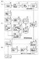
図2を参照して、共有オブジェクトへレポート情報を挿入するための構成を説明する。図2は、共有オブジェクトへレポート情報を挿入するための構成を示すブロック図である。 A configuration for inserting report information into a shared object will be described with reference to FIG. FIG. 2 is a block diagram showing a configuration for inserting report information into a shared object.
医用画像診断装置10は、スキャン計画部11と。画像発生部12と、共有オブジェクト発生部13と、データ送信部14とを備えている。
The medical image
スキャン計画部11は、同一患者の過去検査の共有オブジェクト情報を検索し、取得する。またその情報元に、ユーザにスキャン計画を立案させ、当該スキャン計画を実行させる。
The
画像発生部12は、スキャン計画に基づいて画像を発生させる。
The
共有オブジェクト発生部13は、今回の撮影条件などを含めて、上記したような共有オブジェクトを作成する。また、本実施形態では、更に前回の共有オブジェクトの中にあるレポート情報を前回レポート情報として新規に作成される共有オブジェクトに継承して挿入する。なお、レポート情報は、今回スキャン計画で利用されたものも含む。そして、画像とその情報を、データ送信部14に渡す。
The shared
データ送信部14は、渡されたデータを送信する。
The
医用情報保管装置20は、画像データ受信部21と、受信データ分析部22と、通常データ登録部23と、共有オブジェクトデータ情報分析部24と、共有オブジェクトデータ抽出情報登録部25と、データ保管部26と、データベース27と、共有オブジェクトへ今回(前回)レポート情報挿入部28と、保管データの配信部29とを備えている。
The medical
画像データ受信部21は、データ送信部14から送信された画像データの受信処理を行う。
The image
受信データ分析部22は、受信したデータが共有オブジェクトデータか或いは通常のDICOMデータかを判断して、受信したデータが共有オブジェクトデータであれば、共有オブジェクトデータ情報分析部24にデータを出力し、受信したデータがDICOMデータであれば、通常データ登録部23にデータを出力する。
The reception
通常データ登録部23は、受信データ分析部22から出力された共有オブジェクトデータでない画像データ(すなわち、DICOMデータ)をデータ保管部26とデータベース27に登録する。
The normal
共有オブジェクトデータ情報分析部24は、受信データ分析部22から出力された共有オブジェクトデータから必要な情報(例えば、スキャン情報)を抽出し、当該情報を共有オブジェクトデータ抽出情報登録部25に送る。
The shared object data
共有オブジェクトデータ抽出情報登録部25は、共有オブジェクトデータをデータ保管部26に登録し、共有オブジェクトデータから抽出した情報をデータベース27に登録する。
The shared object data extraction
データ保管部26は、共有オブジェクトデータ抽出情報登録部25から受け取ったデータを適切な場所に書き込み・保管する。この場合において、保管場所が決まったとき、削除されたとき、または変更になった場合などに、データベース27と通信し、データベース27の修正を行う。なお、データ保管部26は、複数のHDDやNAS等であれば良く、医用情報保管装置20でなく、別の場所にあってもよい。
The
データベース27は、管理している検査を特定するための情報、共有オブジェクトデータの情報および特定するための情報、及びデータの保管場所などを管理しているデータベースである。
The
共有オブジェクトへ今回(前回)レポート情報挿入部28は、検査の読影レポート作成完了時点に共有オブジェクトへレポート情報を追記(挿入)し、レポート情報が挿入された共有オブジェクトをデータ保管部26に渡す。ここで、データベース27の更新が必要な場合はデータベース27の更新も行う。
The current (previous) report
保管データの配信部29は、スキャン計画部11や共有オブジェクト情報収集部42の要求に応じて、データベース27やデータ保管部26から共有オブジェクトデータや画像データを要求元であるスキャン計画部11や共有オブジェクト情報収集部42に配信する。この場合において、画像データは、データ保管部26と直接要求元がやり取りするようにしても良い。
In response to a request from the
医用レポート作成支援システム40は、レポート作成指示部41と、共有オブジェクト情報収集部42と、前回レポート情報取得部43と、前回レポート表示部44と、今回レポート作成部45と、を備えている。
The medical report
レポート作成指示部41は、ユーザの要求に基づいて、指定検査のレポート作成開始指示を関連機能に指示する。 The report creation instructing unit 41 instructs a related function to issue a report creation start instruction for the designated inspection based on a user request.
共有オブジェクト情報収集部42は、指定検査に対して図示しない画像を表示する機能に指定画像を表示するように指示するとともに、それに関連している共有オブジェクトを収集する。そして、収集した情報を、前回レポート情報取得部43に渡す。なお、共有オブジェクト情報収集部42は、共有オブジェクトでなく、情報だけ収集するようにしてもよい。
The shared object
前回レポート情報取得部43は、共有オブジェクト情報収集部42で収集された情報を解析して、前回レポート情報を特定し、その情報を前回レポート表示部44に渡す。なお、共有オブジェクトに前回レポート情報がある場合はそれを利用するものとする。共有オブジェクトに前回レポート情報がない場合には、共有オブジェクトに前回共有オブジェクトUIDを挿入しても良いし、StudyInstanceUIDと主に使用された共有オブジェクトの中にあるレポート情報を前回レポート情報とみなしても良い。
The previous report
前回レポート表示部44は、前回レポート情報取得部43から渡された前回レポート情報を適切な位置に表示する。
The previous
今回レポート作成部45は、ユーザにレポートの作成を促し、ユーザが検査の読影レポート作成を完了した時点で、今回(前回)レポート情報挿入部28へ今回作成されたレポート情報を報告する。
The current
上記のように構成された本実施形態に係るシステムにおいて、各装置(システム)におけるレポート情報に関する処理を、フローチャートを参照して説明する。 In the system according to the present embodiment configured as described above, processing related to report information in each device (system) will be described with reference to flowcharts.
医用画像診断装置10で前回レポート情報として新規に作成される共有オブジェクトに継承挿入する処理の流れを、図3を参照して説明する。
A flow of processing for inheriting and inserting into a shared object newly created as previous report information in the medical image
スキャン計画部11は、スキャン準備を開始し、今回のスキャン計画立案のために、共有オブジェクトを検索し、利用する(ステップA1)。そして、スキャン計画部11は、使用する共有オブジェクトを決定し、スキャン計画を立案する(ステップA2)
画像発生部12は、被検体をスキャンし、画像を発生させる(ステップA3)。そして、共有オブジェクト発生部13は、使用した共有オブジェクトにレポート情報が発見できたかどうかを判定し(ステップA4)、レポート情報があれば(ステップA4のYes)、前回レポート情報として新規に作成する共有オブジェクトにその情報を挿入する(ステップA5)。また、共有オブジェクト発生部13は、共有オブジェクトを適切に保管する(ステップA6)。
The
The
また、共有オブジェクト発生部13は、ステップA4において、レポート情報がなければ(ステップA4のNo)、新規に共有オブジェクトを作成し、適切に保管する(ステップA7)。なお、作成された共有オブジェクトは、データ送信部14により、画像データ受信部に送信される。
If there is no report information in Step A4 (No in Step A4), the shared
医用情報保管装置20での共有オブジェクトへの今回(前回)レポート情報挿入処理の流れを、図4を参照して説明する。図4は、医用情報保管装置20での共有オブジェクトへの今回(前回)レポート情報挿入処理の流れを示す図であって、特に、今回(前回)レポート情報挿入部28の動作を示すフローチャートである。
The flow of the current (previous) report information insertion process to the shared object in the medical
今回(前回)レポート情報挿入部28は、今回のレポート作成時に主として使用された共有オブジェクトを検索する(ステップB1)。そして、今回レポート情報をその共有オブジェクトに挿入する(ステップB2)。
The current (previous) report
今回(前回)レポート情報挿入部28は、前回レポート情報が発見できたかどうかを判定し(ステップB3)、発見できた場合は、前回レポート情報も共有オブジェクトに挿入する(ステップB4)。
The current (previous) report
そして、今回(前回)レポート情報挿入部28は、共有オブジェクトをデータ保管部26に保管する(ステップB5)。また、必要に応じて、データベース27の更新も行う。
Then, the current (previous) report
なお、ステップB3において、前回レポート情報が発見できなかった場合には(ステップB3のNo)、今回レポート情報のみをもつ共有オブジェクトを、データ保管部26に保管する(ステップB6)。また、必要に応じて、データベース27の更新も行う。
In step B3, if the previous report information could not be found (No in step B3), the shared object having only the current report information is stored in the data storage unit 26 (step B6). In addition, the
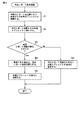
医用レポート作成支援システム40での前回レポート表示処理の流れを、図5を参照して説明する。
The flow of the previous report display process in the medical report
共有オブジェクト情報収集部42は、共有オブジェクト情報を収集し、前回検査の共有オブジェクトから前回レポート情報がある場合はそれを収集する。なければ、過去検査の共有オブジェクトの関連から前回レポート情報を探しだす(ステップC1)。
The shared object
前回レポート情報取得部43は、共有オブジェクト情報収集部42で収集された情報を解析して、前回レポート情報があるかを判定する(ステップC2)。ステップC2において、前回レポート情報がある場合には(ステップC2のYes)、前回レポート表示部44は、前回レポートを最適な位置に表示する(ステップC3)。そして、今回レポート作成部45は、ユーザにより今回レポート作成が行われると(ステップC4)、その完了時点で、今回レポート情報を医用情報保管装置20の今回(前回)レポート情報挿入部28に送信する(ステップC5)。
The previous report
ステップC2において、前回レポート情報がない場合には(ステップC2のNo)、今回レポート作成部45は、ユーザにより今回レポート作成が行われると(ステップC6)、その完了時点で、今回レポート情報を医用情報保管装置に送信する(ステップC7)。
In step C2, if there is no previous report information (No in step C2), the current
上記のように、本実施形態では、医用画像診断システムにおいて、検査の読影レポート作成完了時点での共有オブジェクトへ今回レポート情報あるいは前回レポート情報を挿入する機能と、過去レポート一覧上で自動的に今回あるいは前回共有オブジェクトをチェックし、前回レポートを表示する機能をもたせている。これにより、今回あるいは前回の共有オブジェクトの前回のレポート情報を保管・表示できるようになると共に、過去レポート一覧上で自動的に前回レポートが自動的に表示される。なお、前に利用した共有オブジェクトは、共有オブジェクトUIDなどを作成し、保存・管理する方法と、StudyInstanceUIDと主に使用された共有オブジェクトにより判明するようにしても良い。 As described above, in the present embodiment, in the medical image diagnosis system, the current report information or the previous report information is inserted into the shared object at the time when the interpretation report creation of the examination is completed, and the current report is automatically displayed on the past report list. Or, it has a function to check the previous shared object and display the previous report. As a result, the previous report information of the current or previous shared object can be stored and displayed, and the previous report is automatically displayed on the past report list. Note that the previously used shared object may be identified by a method of creating, storing, and managing a shared object UID and the like, and the StudyInstanceUID and the shared object used mainly.
また、医用画像診断装置10で、今回検査を撮影する際、前回検査の共有オブジェクト内の前回レポート情報を継承し、新規共有オブジェクトを生成する機能をもたせ、医用情報保管装置20はその情報を管理・配信する機能を持たせている。これにより、必要なときに、前回のレポート情報をいつでも取り出せるようになる。また、経過観察を要する患者の今回検査を読影する際、医用画像診断ワークステーション上ではビューア画面上に比較すべき前回検査の画像、画像診断レポート作成支援システム画面上に確認すべき前回検査の読影レポートが自動的に再現され、比較対象を探す手間を省略できる。
In addition, when the medical image
また、本実施形態では、共有オブジェクトに前回レポート情報を集約するため今回の共有オブジェクトだけで前回のレポート情報ももつことが可能となり、前回レポート情報を探すために、前回の共有オブジェクトが必要ではなくなる。しかし、医用画像診断装置10で、今回検査を撮影する際には、前回検査の共有オブジェクト内の前回レポート情報を継承し、新規共有オブジェクトを生成する機能が必要となる。また、共有オブジェクトには、今回レポート情報と前回レポート情報を同時に両方もつようにしても良い。
Further, in the present embodiment, since the previous report information is aggregated in the shared object, it is possible to have the previous report information only with the current shared object, and the previous shared object is not necessary for searching for the previous report information. . However, when the medical image
本発明は、上記各実施の形態に限ることなく、その他、実施段階ではその要旨を逸脱しない範囲で種々の変形を実施し得ることが可能である。さらに、上記各実施形態には、種々の段階の発明が含まれており、開示される複数の構成要件における適宜な組合せにより種々の発明が抽出され得る。 The present invention is not limited to the above-described embodiments, and various modifications can be made without departing from the scope of the invention at the stage of implementation. Further, the above embodiments include inventions at various stages, and various inventions can be extracted by appropriately combining a plurality of disclosed constituent elements.
また、例えば各実施形態に示される全構成要件から幾つかの構成要件が削除されても、発明が解決しようとする課題の欄で述べた課題が解決でき、発明の効果で述べられている効果が得られる場合には、この構成要件が削除された構成が発明として抽出され得る。 In addition, for example, even if some structural requirements are deleted from all the structural requirements shown in each embodiment, the problem described in the column of the problem to be solved by the invention can be solved, and the effect described in the effect of the invention Can be obtained as an invention.
10…医用画像診断装置
11…スキャン計画部
12…画像発生部
13…共有オブジェクト発生部
14…データ送信部
20…医用情報保管装置
21…画像データ受信部
22…受信データ分析部
23…通常データ登録部
24…共有オブジェクトデータ情報分析部
25…共有オブジェクトデータ抽出情報登録部
26…データ保管部
27…データベース
28…共有オブジェクトへ今回(前回)レポート情報挿入部
29…保管データの配信部
30…医用画像観察装置
40…医用レポート作成支援システム
41…レポート作成指示部
42…共有オブジェクト情報収集部
43…前回レポート情報取得部
44…前回レポート表示部
45…今回レポート作成部
50…LAN
DESCRIPTION OF
Claims (4)
前記医用画像診断装置により実行されるスキャンに使用されるスキャン情報及び当該スキャンのために取得される位置決め画像を含む共有オブジェクトを、当該スキャンにより得られた医用画像に関連付けて記憶する記憶部と、
スキャンにより得られた読影対象の医用画像に基づく読影レポート作成完了通知をレポート作成支援システムから受けて、前記読影対象の医用画像に関連する共有オブジェクトを前記記憶部から検索し、検索された共有オブジェクトへ当該読影レポートを特定する情報を挿入するレポート情報挿入部と、
を備えたことを特徴とする医用情報保管装置。 In a medical information storage apparatus that is connected to a report creation support system for creating an interpretation report and stores a medical image obtained by a scan executed by a medical image diagnostic apparatus,
A storage unit that stores scan information used for a scan executed by the medical image diagnostic apparatus and a shared object including a positioning image acquired for the scan in association with the medical image obtained by the scan;
An interpretation report creation completion notification based on a medical image to be interpreted obtained by scanning is received from the report creation support system, a shared object related to the medical image to be interpreted is retrieved from the storage unit, and the retrieved shared object A report information insertion section for inserting information for identifying the interpretation report,
A medical information storage device comprising:
前記スキャンのスキャン情報、当該スキャンのために取得される位置決め画像、及び当該スキャンにより発生された医用画像に対するレポートの情報を含む共有オブジェクトを収集する収集部と、
前記収集された共有オブジェクトを基に作成されたスキャン計画に基づいて医用画像を発生させる画像発生部と、
前記収集部にて収集した共有オブジェクトに含まれるレポート情報を含ませて、前記画像発生部で発生された医用画像に対応付けて共有オブジェクトを生成する共有オブジェクト発生部と、
を備えたことを特徴とする医用画像診断装置。 A medical image diagnostic apparatus for acquiring a medical image by scanning,
A collection unit that collects shared information including scan information of the scan, a positioning image acquired for the scan, and report information on a medical image generated by the scan;
An image generating unit for generating a medical image based on a scan plan created based on the collected shared object;
Including a report object included in the shared object collected by the collection unit, and generating a shared object in association with the medical image generated by the image generation unit;
A medical image diagnostic apparatus comprising:
Priority Applications (2)
| Application Number | Priority Date | Filing Date | Title |
|---|---|---|---|
| JP2007037113A JP4257441B2 (en) | 2007-02-16 | 2007-02-16 | Medical information storage device and medical image diagnostic device |
| US12/032,006 US8892577B2 (en) | 2007-02-16 | 2008-02-15 | Apparatus and method for storing medical information |
Applications Claiming Priority (1)
| Application Number | Priority Date | Filing Date | Title |
|---|---|---|---|
| JP2007037113A JP4257441B2 (en) | 2007-02-16 | 2007-02-16 | Medical information storage device and medical image diagnostic device |
Publications (3)
| Publication Number | Publication Date |
|---|---|
| JP2008200138A JP2008200138A (en) | 2008-09-04 |
| JP2008200138A5 JP2008200138A5 (en) | 2008-11-06 |
| JP4257441B2 true JP4257441B2 (en) | 2009-04-22 |
Family
ID=39707552
Family Applications (1)
| Application Number | Title | Priority Date | Filing Date |
|---|---|---|---|
| JP2007037113A Active JP4257441B2 (en) | 2007-02-16 | 2007-02-16 | Medical information storage device and medical image diagnostic device |
Country Status (2)
| Country | Link |
|---|---|
| US (1) | US8892577B2 (en) |
| JP (1) | JP4257441B2 (en) |
Families Citing this family (8)
| Publication number | Priority date | Publication date | Assignee | Title |
|---|---|---|---|---|
| JP4905967B2 (en) * | 2007-03-02 | 2012-03-28 | 富士フイルム株式会社 | Similar case retrieval apparatus, method, and program |
| JP5053690B2 (en) * | 2007-04-12 | 2012-10-17 | 株式会社東芝 | Image diagnosis support system and image diagnosis support program |
| JP5329911B2 (en) * | 2007-11-02 | 2013-10-30 | 株式会社東芝 | Medical image management apparatus and medical image system |
| JP6104516B2 (en) * | 2012-04-04 | 2017-03-29 | 東芝メディカルシステムズ株式会社 | Medical image diagnostic apparatus and control program |
| CN104274196B (en) * | 2013-07-09 | 2016-10-05 | 上海西门子医疗器械有限公司 | Ct apparatus |
| JP6951114B2 (en) * | 2017-04-27 | 2021-10-20 | キヤノンメディカルシステムズ株式会社 | Medical image diagnostic equipment and magnetic resonance imaging equipment |
| US11244746B2 (en) * | 2017-08-04 | 2022-02-08 | International Business Machines Corporation | Automatically associating user input with sections of an electronic report using machine learning |
| JP2021149226A (en) * | 2020-03-17 | 2021-09-27 | コニカミノルタ株式会社 | Medical information management apparatus, medical information management system, control program, and medical information management method |
Family Cites Families (19)
| Publication number | Priority date | Publication date | Assignee | Title |
|---|---|---|---|---|
| JP4659933B2 (en) | 1997-11-28 | 2011-03-30 | 株式会社東芝 | Medical imaging device |
| US6934698B2 (en) * | 2000-12-20 | 2005-08-23 | Heart Imaging Technologies Llc | Medical image management system |
| AU2003248930A1 (en) * | 2002-07-12 | 2004-02-02 | Allan Fabrick | Method, system, software and graphical user interface for presenting medical information |
| US7756725B2 (en) * | 2002-12-31 | 2010-07-13 | DeJarnette Research Systems, Inc | Breakaway interfacing of radiological images with work orders |
| JP2005031740A (en) | 2003-07-07 | 2005-02-03 | Hitachi Medical Corp | Medical information coordination management device and its program |
| US8200775B2 (en) * | 2005-02-01 | 2012-06-12 | Newsilike Media Group, Inc | Enhanced syndication |
| US20050075905A1 (en) * | 2003-08-22 | 2005-04-07 | Bennett Richard M. | Customizable automatic generation and ordering of a medical report summary |
| JP4389011B2 (en) * | 2004-04-07 | 2009-12-24 | 国立大学法人名古屋大学 | MEDICAL REPORT CREATION DEVICE, MEDICAL REPORT CREATION METHOD, AND PROGRAM THEREOF |
| US20060184524A1 (en) * | 2004-09-14 | 2006-08-17 | Gunter Pollanz | Method and system for automated data analysis, performance estimation and data model creation |
| US20060242143A1 (en) * | 2005-02-17 | 2006-10-26 | Esham Matthew P | System for processing medical image representative data from multiple clinical imaging devices |
| DE102005009056A1 (en) * | 2005-02-28 | 2006-09-07 | Siemens Ag | Method for operating a medical information system |
| JP5283839B2 (en) * | 2005-11-25 | 2013-09-04 | 東芝メディカルシステムズ株式会社 | Medical diagnostic imaging system |
| JP4891597B2 (en) * | 2005-11-25 | 2012-03-07 | 東芝メディカルシステムズ株式会社 | Medical diagnostic imaging system and radiology information system server |
| US20070192138A1 (en) * | 2006-02-16 | 2007-08-16 | Motoaki Saito | Medical record system in a wide-area network environment |
| JP5128154B2 (en) * | 2006-04-10 | 2013-01-23 | 富士フイルム株式会社 | Report creation support apparatus, report creation support method, and program thereof |
| US20070271316A1 (en) * | 2006-05-22 | 2007-11-22 | I3Archives, Inc. | System and method for backing up medical records |
| US20070282629A1 (en) * | 2006-06-01 | 2007-12-06 | Norbert Plambeck | Patient information and communication system |
| US7751604B2 (en) * | 2006-11-30 | 2010-07-06 | Fujifilm Corporation | Medical intelligent server architecture |
| US20080167902A1 (en) * | 2007-01-04 | 2008-07-10 | General Electric Company | Systems and methods for patient and study data export from a pacs database |
-
2007
- 2007-02-16 JP JP2007037113A patent/JP4257441B2/en active Active
-
2008
- 2008-02-15 US US12/032,006 patent/US8892577B2/en active Active
Also Published As
| Publication number | Publication date |
|---|---|
| US20080201372A1 (en) | 2008-08-21 |
| US8892577B2 (en) | 2014-11-18 |
| JP2008200138A (en) | 2008-09-04 |
Similar Documents
| Publication | Publication Date | Title |
|---|---|---|
| JP5525675B2 (en) | Medical image diagnosis support apparatus, medical image diagnosis apparatus, or medical image diagnosis support system | |
| JP5053690B2 (en) | Image diagnosis support system and image diagnosis support program | |
| JP5283839B2 (en) | Medical diagnostic imaging system | |
| JP6392387B2 (en) | Diagnostic imaging equipment | |
| US8401259B2 (en) | Image diagnosis support system | |
| US8386273B2 (en) | Medical image diagnostic apparatus, picture archiving communication system server, image reference apparatus, and medical image diagnostic system | |
| JP5670079B2 (en) | MEDICAL IMAGE DISPLAY DEVICE AND METHOD, AND PROGRAM | |
| JP4218772B2 (en) | Diagnostic imaging support device | |
| JP4257441B2 (en) | Medical information storage device and medical image diagnostic device | |
| JP5019199B2 (en) | Medical imaging device | |
| JP2012245090A (en) | Image processing device, method and program | |
| KR101352999B1 (en) | Appratus and method for image bookmarking using attribute information | |
| JP7102999B2 (en) | Information collection processing equipment, information collection processing method and program | |
| JP2008259661A (en) | Examination information processing system and examination information processor | |
| JP2019010411A (en) | Learning data generation support apparatus, method of operating learning data generation support apparatus, and learning data generation support program | |
| JP5389113B2 (en) | Medical imaging device | |
| JP2017199286A (en) | Information processing apparatus, information processing system, information processing method, and program | |
| JP2008245737A (en) | Medical information processor | |
| JP6536153B2 (en) | Medical image display device and program | |
| JP4810141B2 (en) | Image management apparatus and image management method | |
| JP2008289855A (en) | Image diagnostic support system and image diagnostic support method | |
| JP5972609B2 (en) | Managed object generation device and image display system | |
| JP2020067957A (en) | Information processing apparatus, information processing method, and program | |
| JP4495795B2 (en) | Medical diagnostic imaging system | |
| JP2017108851A (en) | Control device, control system, control method and program |
Legal Events
| Date | Code | Title | Description |
|---|---|---|---|
| A621 | Written request for application examination |
Free format text: JAPANESE INTERMEDIATE CODE: A621 Effective date: 20080811 |
|
| A871 | Explanation of circumstances concerning accelerated examination |
Free format text: JAPANESE INTERMEDIATE CODE: A871 Effective date: 20080811 |
|
| A521 | Request for written amendment filed |
Free format text: JAPANESE INTERMEDIATE CODE: A523 Effective date: 20080829 |
|
| A521 | Request for written amendment filed |
Free format text: JAPANESE INTERMEDIATE CODE: A523 Effective date: 20080919 |
|
| A975 | Report on accelerated examination |
Free format text: JAPANESE INTERMEDIATE CODE: A971005 Effective date: 20080919 |
|
| A131 | Notification of reasons for refusal |
Free format text: JAPANESE INTERMEDIATE CODE: A131 Effective date: 20081007 |
|
| A521 | Request for written amendment filed |
Free format text: JAPANESE INTERMEDIATE CODE: A523 Effective date: 20081202 |
|
| TRDD | Decision of grant or rejection written | ||
| A01 | Written decision to grant a patent or to grant a registration (utility model) |
Free format text: JAPANESE INTERMEDIATE CODE: A01 Effective date: 20090106 |
|
| A01 | Written decision to grant a patent or to grant a registration (utility model) |
Free format text: JAPANESE INTERMEDIATE CODE: A01 |
|
| A61 | First payment of annual fees (during grant procedure) |
Free format text: JAPANESE INTERMEDIATE CODE: A61 Effective date: 20090113 |
|
| R150 | Certificate of patent or registration of utility model |
Ref document number: 4257441 Country of ref document: JP Free format text: JAPANESE INTERMEDIATE CODE: R150 Free format text: JAPANESE INTERMEDIATE CODE: R150 |
|
| FPAY | Renewal fee payment (event date is renewal date of database) |
Free format text: PAYMENT UNTIL: 20120213 Year of fee payment: 3 |
|
| FPAY | Renewal fee payment (event date is renewal date of database) |
Free format text: PAYMENT UNTIL: 20120213 Year of fee payment: 3 |
|
| FPAY | Renewal fee payment (event date is renewal date of database) |
Free format text: PAYMENT UNTIL: 20130213 Year of fee payment: 4 |
|
| R250 | Receipt of annual fees |
Free format text: JAPANESE INTERMEDIATE CODE: R250 |
|
| FPAY | Renewal fee payment (event date is renewal date of database) |
Free format text: PAYMENT UNTIL: 20140213 Year of fee payment: 5 |
|
| R250 | Receipt of annual fees |
Free format text: JAPANESE INTERMEDIATE CODE: R250 |
|
| R250 | Receipt of annual fees |
Free format text: JAPANESE INTERMEDIATE CODE: R250 |
|
| R250 | Receipt of annual fees |
Free format text: JAPANESE INTERMEDIATE CODE: R250 |
|
| R250 | Receipt of annual fees |
Free format text: JAPANESE INTERMEDIATE CODE: R250 |
|
| R250 | Receipt of annual fees |
Free format text: JAPANESE INTERMEDIATE CODE: R250 |
|
| R250 | Receipt of annual fees |
Free format text: JAPANESE INTERMEDIATE CODE: R250 |
|
| S533 | Written request for registration of change of name |
Free format text: JAPANESE INTERMEDIATE CODE: R313533 |
|
| R350 | Written notification of registration of transfer |
Free format text: JAPANESE INTERMEDIATE CODE: R350 |
|
| R250 | Receipt of annual fees |
Free format text: JAPANESE INTERMEDIATE CODE: R250 |
|
| R250 | Receipt of annual fees |
Free format text: JAPANESE INTERMEDIATE CODE: R250 |
|
| R250 | Receipt of annual fees |
Free format text: JAPANESE INTERMEDIATE CODE: R250 |
|
| R250 | Receipt of annual fees |
Free format text: JAPANESE INTERMEDIATE CODE: R250 |