JP4219085B2 - Species-specific detection of nucleic acids with analytical elements - Google Patents
Species-specific detection of nucleic acids with analytical elements Download PDFInfo
- Publication number
- JP4219085B2 JP4219085B2 JP2000358273A JP2000358273A JP4219085B2 JP 4219085 B2 JP4219085 B2 JP 4219085B2 JP 2000358273 A JP2000358273 A JP 2000358273A JP 2000358273 A JP2000358273 A JP 2000358273A JP 4219085 B2 JP4219085 B2 JP 4219085B2
- Authority
- JP
- Japan
- Prior art keywords
- nucleic acid
- detection
- analytical element
- zone
- sample
- Prior art date
- Legal status (The legal status is an assumption and is not a legal conclusion. Google has not performed a legal analysis and makes no representation as to the accuracy of the status listed.)
- Expired - Lifetime
Links
Images


Classifications
-
- C—CHEMISTRY; METALLURGY
- C12—BIOCHEMISTRY; BEER; SPIRITS; WINE; VINEGAR; MICROBIOLOGY; ENZYMOLOGY; MUTATION OR GENETIC ENGINEERING
- C12Q—MEASURING OR TESTING PROCESSES INVOLVING ENZYMES, NUCLEIC ACIDS OR MICROORGANISMS; COMPOSITIONS OR TEST PAPERS THEREFOR; PROCESSES OF PREPARING SUCH COMPOSITIONS; CONDITION-RESPONSIVE CONTROL IN MICROBIOLOGICAL OR ENZYMOLOGICAL PROCESSES
- C12Q1/00—Measuring or testing processes involving enzymes, nucleic acids or microorganisms; Compositions therefor; Processes of preparing such compositions
- C12Q1/68—Measuring or testing processes involving enzymes, nucleic acids or microorganisms; Compositions therefor; Processes of preparing such compositions involving nucleic acids
- C12Q1/6813—Hybridisation assays
-
- Y—GENERAL TAGGING OF NEW TECHNOLOGICAL DEVELOPMENTS; GENERAL TAGGING OF CROSS-SECTIONAL TECHNOLOGIES SPANNING OVER SEVERAL SECTIONS OF THE IPC; TECHNICAL SUBJECTS COVERED BY FORMER USPC CROSS-REFERENCE ART COLLECTIONS [XRACs] AND DIGESTS
- Y10—TECHNICAL SUBJECTS COVERED BY FORMER USPC
- Y10T—TECHNICAL SUBJECTS COVERED BY FORMER US CLASSIFICATION
- Y10T436/00—Chemistry: analytical and immunological testing
- Y10T436/14—Heterocyclic carbon compound [i.e., O, S, N, Se, Te, as only ring hetero atom]
- Y10T436/142222—Hetero-O [e.g., ascorbic acid, etc.]
- Y10T436/143333—Saccharide [e.g., DNA, etc.]
Abstract
Description
【0001】
【発明の属する技術分野】
本発明は、試料適用ゾーンと検出ゾーンを含み、試料適用ゾーンから検出ゾーンへの液体移送が可能な分析エレメントにて核酸を検出する方法に関する。この方法は、試料を前記分析エレメントの試料適用ゾーンに適用し、試料に含まれる核酸を検出ゾーンにて検出プローブとのハイブリダイゼーションにより定性的に、また好ましくはマーカー基により定量的に検出し得るものである。さらに、核酸を検出するための新規分析エレメントおよび試薬キットも提供する。
【0002】
【従来の技術】
現在、診断用途において、PCRで得られた増幅産物などの核酸は、検出すべき核酸と検出プローブ(通常、オリゴヌクレオチドである)との特異的ハイブリダイゼーションならびに検出すべき核酸または/および検出プローブに存在し得るマーカー基を利用した非放射性検出反応により検出されている。この検出反応には様々な異なる方法を用いることができる。例えば、検出すべき核酸は、マイクロタイタープレート(Amplicor Detection Kit, Roche Diagnostics, 欧州特許出願公開EP-A-0 420 260号)などの固相に固定した検出プローブと直接ハイブリダイズさせることができる。さらに、検出すべき核酸を、標識した検出プローブと予めハイブリダイズさせることができ、かかるプロセスで形成された複合体を、マイクロタイタープレート(PCR-Detection Kit, Roche Diagnostics)などの固相上に捕獲することができる。
【0003】
診断手順での検査ストリップの使用は長きにわたり知られている。例えば、検査ストリップを用いたアナライトの検出は、主に免疫学的試験法に関する欧州特許EP-B-0 186 799号、欧州特許EP-B-0 262 328号および欧州特許出願公開EP-A-0 926 498号に記載されている。
【0004】
Jouら(Human Mutation 5(1995), 86-93)は、クロマトグラフ検査ストリップでの核酸検出方法を記載しており、この方法では、検出すべき核酸にハプテン基を導入して検出すべき核酸の抗ハプテン抗体による検査ストリップ上への免疫学的固定化を可能にしている。検査ストリップ上の固定化検出プローブに特異的ハイブリダイゼーションさせることによる検出すべき核酸の捕獲は、例えば、米国特許第5,310,650号、欧州特許EP-B-0 612 354号、Reinhartzら(Gene 136(1993), 221-226)およびRuleら(Clin. Chem. 42(1996), 1206-1209)に記載されている。
【0005】
【発明が解決しようとする課題】
しかしながら、検査ストリップ上での核酸検出を記載する上記の方法にはいくつかの欠点がある。例えば、核酸をサンドイッチ複合体により固定化する場合、感度が低下する。例えば検査容量を増大させることによる感度の増大は、検出すべき核酸へのマーカー基の組込みが限定されることにより制限される。
【0006】
また、前記方法を用いて核酸を直接検出するのは不可能である。標的核酸とハイブリダイゼーションプローブとの複合体を、まず、前工程で形成させる。この目的のために、標的核酸を変性させ(熱処理またはアルカリ溶液の添加により変性する)、必要に応じて中和し、プローブを加え、ハイブリダイゼーション条件に設定する。検査担体上での実際の検出は、この前処理後にのみ行うことができる。この処理は、試薬を適量に分けて添加することと装置(例えば加熱装置)を使用することを含むので、検査ストリップに基づく方法の本質的な利点、例えば装置に対する要求事項が少ないこと、取り扱いが簡単なこと、作業工程が少ないこと、訓練していない人がこの方法を行うことが可能であることなどを利用することができない。さらに、これらの追加の工程により汚染のリスクが生じるので、増幅核酸を検出する場合に偽陽性の結果がもたらされる可能性がある。
【0007】
また、検出すべき核酸と分析エレメント上のハイブリダイゼーションプローブとの直接ハイブリダイゼーションを可能にする方法の場合、従来技術では検出すべき核酸の前処理を必要としない感度のよい検査手順は知られていない。つまりこれらの従来技術の手順では核酸は直接検出できないか、増幅プロセス後に直接検出されない。これらの手順では、標的核酸(これは通常2本鎖の形態で存在する)は、ハイブリダイゼーションのために1本鎖コンホメーションに変換される。これについての方法がいくつか記載されている。例えば、よく知られたハイブリダイゼーション法によれば、核酸を分析エレメントに適用する前に、例えば熱処理またはアルカリ試薬の添加により完全に変性させている。また標的核酸の一方の鎖を特異的消化することが可能な酵素的方法も知られている(Ruleら, Clinical Chemistry 42, 1206-1209頁(1996))。
【0008】
前処理は、標的核酸がすでに完全に1本鎖として存在する場合のみ省略することができる。しかし、このことは通常すでに困難である。なぜならば、リボソーム標的核酸を検出する場合、これらの1本鎖核酸の天然二次構造の大部分の領域が2本鎖形態(例えばrRNA)にあるからである。机上の代案は、一方の核酸鎖のみの特異的合成を可能にする増幅方法である。例えば、非対称PCRである。しかしながら、この方法は、線状(linear)増幅のために通常のPCR法よりも極めて効率が悪く、感度がかなり制限され、低濃度でしか存在しないアナライトの検出には不十分である。したがって、従来技術において知られた、分析エレメント上で検出すべき核酸の直接ハイブリダイゼーションを可能にする方法も、間接検出法で見られるのと同じ欠点をもつ。
【0009】
【課題を解決するための手段】
本発明の主な利点は、上述した検出すべき核酸の前処理を省略でき、核酸を分析エレメントに直接または増幅プロセス後に直接適用できるということである。分析エレメントには試薬が含浸されているのが好ましく、この試薬が分析エレメント上での標的核酸の完全または部分的変性を可能にし、2本鎖コンホメーションで適用される核酸のハイブリダイゼーションも可能にする。本発明は、このことに対する可能性をいくつか提供する。例えば、変性試薬は、好ましくは核酸を完全に変性させるために予め検査担体上に適当な濃度で乾燥形態にて置くことができる。標的核酸は、例えばPCR反応溶液に含有させた液体形態にて分析エレメントに適用することができる。試薬の溶解後、標的核酸は完全に変性する。プローブオリゴヌクレオチドへのハイブリダイゼーションはこれらの条件下では生じ得ないので、この化学物質を適切に変更しなければならず、それと同時にアナライトは、好ましくはクロマトグラフィーにより検査担体を通って移送される。一方、これは、適当なクロマトグラフィーバッファーまたは下流に配置されている試薬により行うことができる。これには、塩基、特にNaOHによる変性および酸による中和が好ましい。あるいは、標的核酸の2本鎖コンホメーションを不安定化させ(部分的に変性させ)、プローブオリゴヌクレオチドへのハイブリダイゼーションを可能にするか促進するように化学物質を調節することができる。クロマトグラフィー中の化学物質の変更は、この変形法には必要ない。この実施形態に好適な試薬は、カオトロピック塩であり、特にチオシアン酸グアニジニウム(GuSCN)が好ましい。
【0010】
プローブオリゴヌクレオチドにハイブリダイズした標的核酸を検出するために、さらにその複合体を標識する必要がある。マーカー基は、事前に実施する任意の増幅プロセス中に酵素的組込みにより増幅標的核酸に組み込んでもよく、または別の適切に標識されたプローブオリゴヌクレオチドとハイブリダイズさせることにより標的核酸に結合させてもよい。
【0011】
驚くべきことに、上記の方法を用いると、分析エレメントに適用される二本鎖形態の標的核酸は、分析エレメント全体への該標的核酸のクロマトグラフ移送の間に利用できる時間が非常に短いにもかかわらず、非常に効率的にプローブオリゴヌクレオチドにハイブリダイズし、高感度で検出できることが分かった。
【0012】
よって、本発明は、試料適用ゾーンと検出ゾーンとを含み、該試料適用ゾーンから該検出ゾーンへの液体移送が可能な分析エレメントにて核酸を検出する方法であって、
検出すべき核酸を含む試料を該試料適用ゾーンに適用する工程、および
該検出ゾーン内で検出プローブとのハイブリダイゼーションにより該核酸を検出する工程
を含んでなり、該検出すべき核酸を該分析エレメント上で変性させることを特徴とする方法を提供する。
【0013】
「変性」は、本明細書で用いる場合、古典的な意味での完全な変性と解釈されるのみならず、核酸の二本鎖がプローブとハイブリダイズし得るのに十分な程度に不安定化されるプロセスにおける部分的変性も意図するものである。したがって、二本鎖を完全に分離させる必要はないが、もちろんそのような実施形態も本発明に含まれる。
【0014】
本発明によれば、検出すべき核酸は、分析エレメント上で変性され、すなわち、核酸は、分析エレメントに変性されていない好ましくは二本鎖の形態で適用され、分析エレメントに適用された後にはじめて変性試薬と接触することになる。変性試薬は、試料中の核酸を変性させるのに十分な量の塩基または/およびカオトロピック物質を含み得る。好ましい塩基には、例えば、アルカリ水酸化物、特にNaOHがある。好ましいカオトロピック物質には、例えば、ヨウ化物、酢酸塩、過塩素酸塩、チオシアン酸塩、トリフルオロ酢酸塩、トリクロロ酢酸塩または/およびグアニジニウム化合物がある。特に好ましいのはチオシアン酸グアニジニウムである。一方、カオトロピック物質の使用は、二本鎖を不安定化させ、検出すべき核酸と検出プローブとの間のハイブリダイゼーションを促進する。さらに重要な機能は、核酸の特異的な融解点を低下させて室温での特異的ハイブリダイゼーションを可能にすることである。
【0015】
このようなカオトロピック物質の作用は、溶液中でのハイブリダイゼーションに関して既に知られているが(国際公開WO-A-87 06621号, ThompsonおよびGillespie, Anal. Biochem. 163(1987), 281-291; Van NessおよびChen, Nucleic Acid Res. 19(1991), 5143-5151)、分析エレメントでのハイブリダイゼーションによる核酸の検出のための本発明の範囲内では初めて利用される。この技術の分析エレメントへの応用は、当業者には自明ではなかった。というのは、従来技術は、1時間程度のオーダーのハイブリダイゼーション時間を推奨しているからである。しかしながら、本発明の分析エレメントを用いた場合、試料適用から検出までの検査は、10分以内に既に完了している。検出プローブとのハイブリダイゼーションは、試料の検出ゾーンへのクロマトグラフ移送中に、プローブと試料が数秒間、場合によっては1分以下接触しただけで既に生じている。
【0016】
本発明の分析エレメントの本質的な特徴は、液体が分析エレメント内で試料適用ゾーンから検出ゾーンに移動し得ることである。液体のそのような流れは、例えば、適切に構築された中空体における引力により達成され得る。引力の1つのタイプである遠心力による液体移送を可能にする装置が、例えば欧州特許EP-B-0 052 769に記載されている。しかしながら、本発明の分析エレメントは、好ましくは、毛管力(capillary force)により液体を移動させ得る吸着材料を含む。本発明の分析エレメントの個々のゾーンの吸着材料は、この場合、同一でも異なってもよい。異なるゾーンが最適に機能するために異なる材料からなる場合が多い。
【0017】
吸着性の毛管力活性のある材料として可能なものは、例えば、米国特許US-A-4,861,711号、米国特許US-A-5,591,645号または欧州特許出願公開EP-A-0 291 194号に記載されているような、基本的に、いわゆる「乾式検査」において液体の取込みに用いられ得るもの全てである。膜、例えばニトロセルロース膜のような多孔質材料がこれに都合がよいことが判明した。しかしながら、フリース、織物またはメリヤス生地のような繊維状吸着マトリックス材料を用いることも可能である。特に好ましいのはフリースである。繊維状マトリックス材料は、ガラス、セルロース、セルロース誘導体、ポリエステル、ポリアミドを含んでもよく、さらにビスコース、セルウール(cell wool)または/およびポリビニルアルコールを含んでもよい。例えば、セルロースに基づく繊維、ポリエステルおよび/またはポリアミドに基づくポリマー繊維ならびに欧州特許EP-B-0 326 135に記載されているようなOH基および/またはエステル基を有する有機結合剤から製造されたフリースが、本発明に用いられ得る。欧州特許出願0 571 941号に記載されたような溶融コポリエステル繊維、セルロース繊維またはセルロース誘導体繊維を含むフリース材料も本発明の分析エレメントに用いることができる。ティーバッグ紙などの紙も簡単に用いることができる。
【0018】
本発明の分析エレメントの取り扱いを改善するために、吸着性の毛管力活性材料または種々の吸着性の毛管力活性材料を、液体不透過性であるがマトリックス材料内の液体の流れに悪影響を及ぼさず、分析エレメント内で生じる反応に関して不活性に挙動する剛性の支持担体材料上に配置することができる。例えば、液体移送を可能にするマトリックス材料が取り付けられたポリエステル箔が好ましい支持担体材料であり得る。
【0019】
本発明の分析エレメントにおける個々のゾーンは、上下に重なるように、隣り合わせになるように、一部が上下に重なるように、または一部が隣り合わせになるように支持担体材料上に配置することができる。本発明の分析エレメントは、試料適用ゾーンおよび検出ゾーンが支持担体材料上に隣り合わせで位置しているのが特に好ましい。この場合、隣り合わせとは、これらのゾーンが互いに直接接触しているか、または本質的に一つの平面内にその他のゾーンで分離された状態で配置されていることを意味する。
【0020】
試料適用ゾーンは、本発明の分析エレメントにおける試料が適用される領域である。検出ゾーンは、本発明の分析エレメントにおける領域であって、分析エレメントに適用される試料中に検査すべきアナライトまたは該アナライトから誘導される物質もしくは該アナライトを呈示する物質が存在するか否かを測定する領域である。この測定は、定性的、半定量的または定量的であり得る。これに関連して、半定量的とは、アナライトから誘導された物質またはアナライトを呈示する物質についての具体的な濃度の値が測定されるのではなく、アナライトの濃度が位置する濃度範囲を測定することを意味する。
【0021】
本発明の第1の実施形態では、試料を適用する前に分析エレメントに変性試薬を含浸させる。変性試薬は、分析エレメント上、好ましくは試料適用ゾーン上に液体または乾燥形態で存在し得る。変性試薬は好ましくは乾燥形態で存在する。さらなる実施形態では、試料を適用した後にはじめて分析エレメントに変性試薬を含浸させる。検出すべき試料に存在する核酸と変性試薬の接触により、核酸の二本鎖の変性が生じ、生じた核酸の一本鎖と検出プローブとのハイブリダイゼーションが可能になる。変性試薬が塩基を含む場合、分析エレメントは、試料適用ゾーンと検出ゾーンの間に中和試薬(例えば、リン酸または硫酸のような液体と接触して酸性反応する塩)を好ましくは乾燥形態で含んでいることが都合がよい。
【0022】
試料中に存在する検出すべき核酸の結合に用いられる検出プローブは、好ましくは、検査条件下でハイブリダイゼーションを可能にするのに十分なほどに検出すべき核酸と相補的である配列を含むオリゴヌクレオチドである。検出プローブは、好ましくはオリゴデオキシリボヌクレオチドであるが、検出プローブとして改変型ヌクレオチド構築ブロックまたはペプチド核酸を含む核酸類似体を用いることも可能である。検出プローブは、好ましくは検出ゾーンに固定された形態で存在する。すなわち、検出プローブは、検査条件下で本質的に検出ゾーンから溶出しない。固定化は、検出プローブを適用し、次いで、例えば昇温、放射線照射等の適当な方法で該プローブを固定することにより行い得る。あるいは、プローブは、検出ゾーンに高アフィニティー相互作用、例えばビオチン/ストレプトアビジンもしくはアビジンまたはハプテン/抗ハプテン抗体、糖/レクチンによって固定されてもよい。適当に改変された、例えばビオニチル化検出プローブの調製は、当業者にはよく知られており、ここで詳細に説明する必要はない。あるいは、検出すべき核酸と検出プローブとの間のハイブリダイゼーションは、検出ゾーンの上流側で生じてもよく、そのハイブリダイゼーション複合体は分析エレメントを移送中に例えば前記のような高アフィニティー相互作用により検出ゾーンに選択的に固定化することができる。
【0023】
核酸は、好ましくはマーカー基によって検出され、基本的に従来技術により知られたマーカー基は全て適している。酵素標識のような視覚的に検出可能なマーカー基が好ましいが、特に、金またはラテックス粒子のような粒状の標識が好ましい。この場合、標識は、検出すべき核酸に直接位置していてもよいが(直接標識)、核酸に、例えば上記のような高アフィニティー相互作用、例えばハプテン/抗ハプテン抗体により間接的に結合していてもよい(間接標識)。検出すべき核酸への間接マーカー基の結合は、ハイブリダイゼーション中、または洗浄工程後に生じ得る。
【0024】
本発明は、検査ストリップ上で核酸を検出する簡単な方法を提供する。検出すべき核酸は、好ましくは増幅産物、例えばPCRで産生された増幅産物である。この場合、増幅産物は、増幅反応を行ったバッファー溶液をクロマトグラフィー溶媒として用いて直接検査ストリップに適用することができる。
【0025】
本発明はさらに、試料適用ゾーンと検出ゾーンとを含み、該試料適用ゾーンから該検出ゾーンへの液体移送が可能な核酸検出用分析エレメントを提供するものであり、該エレメントは核酸の変性試薬で含浸され、変性試薬は好ましくは乾燥形態で存在する。分析エレメントは、クロマトグラフ移送を可能にする吸着材料を含み得るものであり、好ましくは検査ストリップとして構築される。好ましくは、検出すべき核酸に相補的な検出プローブが分析エレメントの検出ゾーンに固定された形態で存在する。
【0026】
最後に、本発明は、本発明の分析エレメント、ならびに核酸を検出するのに必要な追加の試薬、例えば標識試薬、バッファーなどを含む核酸検出用キットに関する。
【0027】
本発明により提供される方法、分析エレメントおよび試薬キットは、特に、核酸増幅産物の検出、例えば試料中の選択された生物または生物の一部(例えば細胞)の存在または量を検出するのに適している。この目的のために、例えば、試料中の生物または細胞由来のリボソームRNAまたはそれをコードするDNA配列の増幅などにより、試料中の核酸の属特異的および種特異的検出を行うことができる。この方法を用いて、臨床試料、食物、水試料等に含まれる微生物、例えば病原性微生物またはウイルスを検出することができる。さらに、この方法を法医学的分析に用いて特定の個体を探知することもできる。クラミジア・トラコーマ(Chlamydia trachomatis)由来の核酸の本発明の方法による検出を、下記の実施例に示す。
【0028】
【実施例】
実施例1 概念
1.分析エレメントの準備
図1に示した分析エレメントの吸着フリース(1)に、含浸バッファー(10mMのKPO4、pH7.5、100mMのNaCl、2または2.5Mのチオシアン酸グアニジニウム(GuSCN)、0.5%Triton-X100、25mMのNaOH、2%RPLAタイプ4(Roche Diagnostics、カタログ番号1726544-106)および3mg/mlニシン精子DNA)を含浸させた。この目的のために、吸着フリースをペトリ皿中で含浸バッファーで完全に湿らせて、循環空気中で45℃にて3時間乾燥した。
【0029】
15mmのニトロセルロース膜を液体移送ゾーン(2)として用いた。検出ゾーン(4)を、該膜に検出オリゴヌクレオチドプローブ(40pmol/検査ストリップ)を適用することにより調製した。このために、オリゴヌクレオチドの溶液(200μM)を検出ラインの形態で自動ディスペンサー(Hamilton Microlab N)を用いて適用し、オリゴヌクレオチドを膜上に80℃で30分間固定化した。一方、ビオチン標識オリゴヌクレオチドを、予めポリストレプトアビジンでコーティングした検出ゾーン(4)に適用した。このために、オリゴヌクレオチド溶液(8pmol/検査ストリップ)をニトロセルロース膜に適用し、10μlの標準クロマトグラフィーバッファーを用いてポリストレプトアビジンで前コーティングした検出ゾーン上でクロマトグラフィーによる分離を行った。
【0030】
クラミジア・トラコーマプラスミドpCTT1(C.トラコーマの塩基1〜7496)由来のpCTT1の354〜374位に相当する配列(5'-GTC TCT CAT CGA GAC AAA GTG-3')(配列番号1)を有するオリゴヌクレオチドを、検出プローブとして用いた(SriprakashおよびMacavoy, Plasmid 18(1987), 205-214)。
【0031】
2. PCR プロトコル
オリゴヌクレオチドCP24(5'-GGGATTCCTGTAACAACAAGTCAGG-3')(配列番号2)(pCTT1の195〜219位)およびCP27(5'-CCTCTTCCCCAGAACAATAAGAACAC-3')(配列番号3)(pCTT1の401〜376位)を任意に5'-ビオチニル化形態または5'-ジゴキシゲニル化形態で増幅用のプライマーとして用いた。
【0032】
反応容量は100μlであった(PCRバッファー(Roche Diagnosticsカタログ番号1600753)中に、4mMのMgCl2、それぞれ0.1mMのdNTP、それぞれ300nMのプライマーCP24およびCP27、2.5UのTaqポリメラーゼ、2UのUNG(ウラシルDNAグリコシラーゼ)ならびに鋳型を加えたもの)。
【0033】
反応シーケンスは下記のとおりであった:
・37℃で10分間、95℃で5分間、60℃で1分間、
・95℃、3秒および60℃、6秒を34サイクル、
・72℃で10分間、
・50℃に保持
PCR反応は、種々の量の鋳型を用いて行った(0〜100000プラスミドコピー/混合物)。
【0034】
3.検出
50μlの増幅液(実施例2)を検査ストリップの吸着フリースに適用し、4分間クロマトグラフィーによる分離を行った。続いて、30μlの標準クロマトグラフィーバッファー(0.15MのNaCl、9.6mMのKH2PO4、40.5mMのK2HPO4、0.25%のTween 20、2%のRPLAタイプ4、0.09%のアジ化ナトリウム)を適用し、4分間クロマトグラフィーによる分離を行った。その後、20μlの抗ジゴキシゲニン抗体金コンジュゲート溶液(2.1pmol)を適用し、4分間クロマトグラフィーによる分離を行った。次いで、50μlの標準クロマトグラフィーバッファーを2回適用し、4分間クロマトグラフィーによる分離を行った。
【0035】
この実験の結果を図2に示す。図2Aは、サンドイッチ様式を用いる免疫学的方法により核酸を検出する欧州特許出願公開EP-A-0 926 489号の検査ストリップを用いた結果を示す。図2Bには、実施例1の検査ストリップを用いた本発明の検出方法の結果を示す。図2Aの検査ストリップを用いた場合は感度が低いが(混合物当たり1000コピー以上のプラスミドの検出のみ)、本発明の方法を用いた場合はかなり低いプラスミドコピー数(50)の検出でも可能であった。一般に、検出シグナルは、ΔRem-%が3以上である場合にポジティブであることがわかる。高感度であることに加えて、本発明の方法は、かなり広いダイナミックレンジも有している。すなわち、検出すべき核酸の濃度範囲が広い場合にもシグナルの有意な変化をもたらす。
【0036】
実施例2
図1の分析エレメントを、吸着フリースの含浸バッファーがNaOHを含んでない以外は実施例1.1にしたがって調製した。0.5M、1.5Mおよび2.5M(飽和限界付近)のグアニジニウム濃度について試験した。
【0037】
50μlのPCR増幅液(100プラスミド/混合物)を、実施例1.2にしたがって調製した各分析エレメントに適用した。検出は、実施例1.3にしたがって行った。
【0038】
結果を図3に示す。チオシアン酸グアニジニウムの添加により有意に検出感度が改善されるということが分かる。
【図面の簡単な説明】
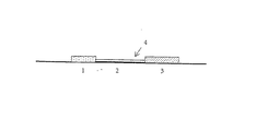
【図1】吸着フリースとして設計された試料適用ゾーン(1)、液体移送ゾーン(2)、該液体移送ゾーン内に位置する検出ゾーン(4)、および吸着フリースとして設計された吸着ゾーン(3)を含んでなる、本発明の検査ストリップの概略構造を示す図である。
【図2】本発明の方法(B)と欧州特許出願公開EP-A-0 926 498号に記載された方法(A)の感度の比較を示す図である。
【図3】分析エレメントの感度とチオシアン酸グアニジニウム(GuSCN)の濃度との関係を示す図である。[0001]
BACKGROUND OF THE INVENTION
The present invention relates to a method for detecting a nucleic acid with an analytical element that includes a sample application zone and a detection zone and is capable of liquid transfer from the sample application zone to the detection zone. In this method, the sample is applied to the sample application zone of the analytical element, and the nucleic acid contained in the sample can be detected qualitatively by hybridization with a detection probe in the detection zone, and preferably quantitatively by the marker group. Is. In addition, novel analytical elements and reagent kits for detecting nucleic acids are also provided.
[0002]
[Prior art]
At present, in diagnostic applications, nucleic acids such as amplification products obtained by PCR are used for specific hybridization between a nucleic acid to be detected and a detection probe (usually an oligonucleotide) and to a nucleic acid or / and a detection probe to be detected. It is detected by a non-radioactive detection reaction using a marker group that may be present. Various different methods can be used for this detection reaction. For example, the nucleic acid to be detected can be directly hybridized with a detection probe immobilized on a solid phase such as a microtiter plate (Amplicor Detection Kit, Roche Diagnostics, EP-A-0 420 260). Furthermore, the nucleic acid to be detected can be prehybridized with a labeled detection probe, and the complex formed by such a process can be captured on a solid phase such as a microtiter plate (PCR-Detection Kit, Roche Diagnostics). can do.
[0003]
The use of test strips in diagnostic procedures has been known for a long time. For example, the detection of analytes using test strips is mainly based on European patent EP-B-0 186 799, European patent EP-B-0 262 328 and European patent application publication EP-A relating to immunological tests. -0 926 It is described in 498.
[0004]
Jou et al. (Human Mutation 5 (1995), 86-93) describe a method for detecting nucleic acids on chromatographic test strips, in which a hapten group is introduced into the nucleic acid to be detected. The anti-hapten antibody allows immunological immobilization on a test strip. Capture of nucleic acids to be detected by specific hybridization to an immobilized detection probe on a test strip is described, for example, in US Pat. No. 5,310,650, European Patent EP-B-0 612 354, Reinhartz et al. (Gene 136 (1993 ), 221-226) and Rule et al. (Clin. Chem. 42 (1996), 1206-1209).
[0005]
[Problems to be solved by the invention]
However, the above described method for describing nucleic acid detection on a test strip has several drawbacks. For example, when nucleic acid is immobilized by a sandwich complex, the sensitivity decreases. The increase in sensitivity, for example by increasing the test volume, is limited by the limited incorporation of the marker group into the nucleic acid to be detected.
[0006]
In addition, it is impossible to directly detect nucleic acids using the above method. First, a complex of the target nucleic acid and the hybridization probe is formed in the previous step. For this purpose, the target nucleic acid is denatured (denaturation by heat treatment or addition of an alkaline solution), neutralized as necessary, a probe is added, and hybridization conditions are set. Actual detection on the test carrier can only take place after this pretreatment. This process involves adding appropriate amounts of reagents and using equipment (e.g. heating equipment), so that the essential advantages of the test strip based method, e.g. less equipment requirements and handling. It is not possible to take advantage of the simplicity, the small number of work steps, and the ability of untrained people to perform this method. In addition, these additional steps create a risk of contamination, which can lead to false positive results when detecting amplified nucleic acids.
[0007]
In addition, in the case of a method that enables direct hybridization between the nucleic acid to be detected and the hybridization probe on the analytical element, there is known a sensitive test procedure that does not require pretreatment of the nucleic acid to be detected in the prior art. Absent. That is, these prior art procedures cannot detect nucleic acids directly or directly after the amplification process. In these procedures, the target nucleic acid, which is usually present in double stranded form, is converted to a single stranded conformation for hybridization. Several methods are described for this. For example, according to the well-known hybridization method, the nucleic acid is completely denatured, for example, by heat treatment or addition of an alkaline reagent, before being applied to the analytical element. An enzymatic method capable of specifically digesting one strand of a target nucleic acid is also known (Rule et al., Clinical Chemistry 42, 1206-1209 (1996)).
[0008]
The pretreatment can be omitted only when the target nucleic acid is already completely present as a single strand. However, this is usually already difficult. This is because when detecting a ribosome target nucleic acid, most of the region of the natural secondary structure of these single-stranded nucleic acids is in a double-stranded form (eg, rRNA). A desktop alternative is an amplification method that allows specific synthesis of only one nucleic acid strand. For example, asymmetric PCR. However, this method is much less efficient than the normal PCR method due to linear amplification, is considerably limited in sensitivity, and is insufficient for detection of analytes that are present only at low concentrations. Thus, methods known in the prior art that allow direct hybridization of nucleic acids to be detected on analytical elements have the same disadvantages as seen with indirect detection methods.
[0009]
[Means for Solving the Problems]
The main advantage of the present invention is that the pretreatment of the nucleic acid to be detected as described above can be omitted and the nucleic acid can be applied directly to the analytical element or directly after the amplification process. The analytical element is preferably impregnated with a reagent, which allows complete or partial denaturation of the target nucleic acid on the analytical element and also allows hybridization of nucleic acids applied in double-stranded conformation To. The present invention offers several possibilities for this. For example, the denaturing reagent can be placed in a dry form at an appropriate concentration in advance on a test carrier, preferably in order to completely denature the nucleic acid. The target nucleic acid can be applied to the analysis element, for example, in a liquid form contained in a PCR reaction solution. After the reagent is dissolved, the target nucleic acid is completely denatured. Since hybridization to the probe oligonucleotide cannot occur under these conditions, this chemical must be modified appropriately, while the analyte is preferably transported through the test carrier, preferably by chromatography. . On the other hand, this can be done with a suitable chromatography buffer or a reagent located downstream. For this, modification with bases, in particular NaOH, and neutralization with acids are preferred. Alternatively, chemicals can be adjusted to destabilize (partially denature) the double-stranded conformation of the target nucleic acid to allow or facilitate hybridization to the probe oligonucleotide. No chemical changes during chromatography are necessary for this variant. A suitable reagent for this embodiment is a chaotropic salt, particularly guanidinium thiocyanate (GuSCN).
[0010]
In order to detect the target nucleic acid hybridized to the probe oligonucleotide, it is necessary to further label the complex. The marker group may be incorporated into the amplified target nucleic acid by enzymatic incorporation during any prior amplification process, or may be bound to the target nucleic acid by hybridizing with another appropriately labeled probe oligonucleotide. Good.
[0011]
Surprisingly, using the above method, the target nucleic acid in double-stranded form applied to the analytical element has a very short time available during chromatographic transfer of the target nucleic acid to the entire analytical element. Nevertheless, it was found that it hybridizes very efficiently to the probe oligonucleotide and can be detected with high sensitivity.
[0012]
Therefore, the present invention is a method for detecting a nucleic acid with an analytical element that includes a sample application zone and a detection zone and is capable of liquid transfer from the sample application zone to the detection zone,
Applying the sample containing the nucleic acid to be detected to the sample application zone, and detecting the nucleic acid by hybridization with a detection probe in the detection zone, the nucleic acid to be detected being the analytical element There is provided a method characterized by denaturing above.
[0013]
“Denature”, as used herein, is not only interpreted as complete denaturation in the classical sense, but also destabilized enough to allow the nucleic acid duplex to hybridize to the probe. Partial modification in the process is also contemplated. Thus, it is not necessary to completely separate the duplexes, but such embodiments are of course included in the present invention.
[0014]
According to the invention, the nucleic acid to be detected is denatured on the analytical element, ie the nucleic acid is applied to the analytical element, preferably in unmodified, preferably double-stranded form, and only after it has been applied to the analytical element. Contact with the denaturing reagent. The denaturing reagent may contain a sufficient amount of base or / and chaotropic substance to denature the nucleic acid in the sample. Preferred bases are, for example, alkali hydroxides, especially NaOH. Preferred chaotropic substances are, for example, iodide, acetate, perchlorate, thiocyanate, trifluoroacetate, trichloroacetate or / and guanidinium compounds. Particularly preferred is guanidinium thiocyanate. On the other hand, the use of a chaotropic substance destabilizes the double strand and promotes hybridization between the nucleic acid to be detected and the detection probe. A further important function is to reduce the specific melting point of the nucleic acid to allow specific hybridization at room temperature.
[0015]
The action of such chaotropic substances is already known for hybridization in solution (WO-A-87 06621, Thompson and Gillespie, Anal. Biochem. 163 (1987), 281-291; Van Ness and Chen, Nucleic Acid Res. 19 (1991), 5143-5151), first utilized within the scope of the present invention for the detection of nucleic acids by hybridization with analytical elements. The application of this technique to analytical elements was not obvious to those skilled in the art. This is because the prior art recommends a hybridization time on the order of about 1 hour. However, when the analysis element of the present invention is used, the inspection from sample application to detection is already completed within 10 minutes. Hybridization with the detection probe has already occurred during the chromatographic transfer of the sample to the detection zone when the probe and the sample have only contacted for a few seconds, possibly less than 1 minute.
[0016]
An essential feature of the analysis element of the present invention is that liquid can move from the sample application zone to the detection zone within the analysis element. Such a flow of liquid can be achieved, for example, by attractive forces in a properly constructed hollow body. A device that enables liquid transfer by centrifugal force, one type of attractive force, is described, for example, in European patent EP-B-0 052 769. However, the analytical element of the present invention preferably comprises an adsorbent material that can move the liquid by capillary force. The adsorbent material of the individual zones of the analytical element according to the invention can in this case be identical or different. Often, different zones are made of different materials in order to function optimally.
[0017]
Possible adsorptive capillary force active materials are described, for example, in US-A-4,861,711, US-A-5,591,645 or EP-A-0 291 194. Basically, everything that can be used for the uptake of liquid in the so-called “dry test”. A porous material such as a membrane, for example a nitrocellulose membrane, has proven convenient for this. However, it is also possible to use a fibrous adsorption matrix material such as a fleece, woven fabric or knitted fabric. Particularly preferred is a fleece. The fibrous matrix material may include glass, cellulose, cellulose derivatives, polyester, polyamide, and may further include viscose, cell wool, and / or polyvinyl alcohol. For example, fleeces made from cellulose based fibers, polyester and / or polyamide based polymer fibers and organic binders having OH and / or ester groups as described in European patent EP-B-0 326 135 Can be used in the present invention. Fleece materials comprising molten copolyester fibers, cellulose fibers or cellulose derivative fibers as described in European Patent Application 0 571 941 can also be used in the analytical element of the present invention. Paper such as tea bag paper can also be used easily.
[0018]
In order to improve the handling of the analytical element of the present invention, an adsorptive capillary force active material or various adsorptive capillary force active materials are liquid-impermeable but do not adversely affect the flow of liquid in the matrix material. Rather, it can be placed on a rigid support carrier material that behaves inertly with respect to reactions occurring within the analytical element. For example, a preferred support carrier material may be a polyester foil attached with a matrix material that allows liquid transfer.
[0019]
The individual zones in the analytical element of the present invention can be arranged on the support carrier material so that they overlap one above the other, so that they are next to each other, partly overlap each other, or partly next to each other. it can. In the analytical element according to the invention, it is particularly preferred that the sample application zone and the detection zone are located side by side on the support carrier material. In this case, side-by-side means that these zones are in direct contact with each other, or are essentially arranged in one plane separated by other zones.
[0020]
The sample application zone is an area where a sample in the analytical element of the present invention is applied. The detection zone is a region in the analysis element of the present invention, and whether an analyte to be examined, a substance derived from the analyte, or a substance presenting the analyte is present in a sample applied to the analysis element This is an area for measuring whether or not. This measurement can be qualitative, semi-quantitative or quantitative. In this context, semi-quantitative refers to the concentration at which the concentration of the analyte is located, rather than the specific concentration value being measured for a substance derived from or presenting an analyte. Means to measure the range.
[0021]
In the first embodiment of the invention, the analytical element is impregnated with a denaturing reagent before the sample is applied. The denaturing reagent may be present in liquid or dry form on the analytical element, preferably on the sample application zone. The denaturing reagent is preferably present in dry form. In a further embodiment, the analytical element is impregnated with the denaturing reagent only after the sample has been applied. By contacting the nucleic acid present in the sample to be detected with the denaturing reagent, double-stranded denaturation of the nucleic acid occurs, and hybridization between the single strand of the nucleic acid thus generated and the detection probe becomes possible. If the denaturing reagent contains a base, the analytical element is preferably used in a dry form with a neutralizing reagent (e.g. a salt that reacts acidicly with a liquid such as phosphoric acid or sulfuric acid) between the sample application zone and the detection zone. Convenient to include.
[0022]
The detection probe used to bind the nucleic acid to be detected present in the sample is preferably an oligo that contains a sequence that is sufficiently complementary to the nucleic acid to be detected to allow hybridization under test conditions. It is a nucleotide. The detection probe is preferably an oligodeoxyribonucleotide, but it is also possible to use a modified nucleotide building block or a nucleic acid analog comprising a peptide nucleic acid as the detection probe. The detection probe is preferably present in a fixed form in the detection zone. That is, the detection probe essentially does not elute from the detection zone under test conditions. The immobilization can be performed by applying a detection probe and then immobilizing the probe by an appropriate method such as temperature increase or radiation irradiation. Alternatively, the probe may be immobilized in the detection zone by a high affinity interaction, such as biotin / streptavidin or avidin or a hapten / anti-hapten antibody, sugar / lectin. The preparation of appropriately modified, eg biotinylated detection probes, is well known to those skilled in the art and need not be described in detail here. Alternatively, the hybridization between the nucleic acid to be detected and the detection probe may occur upstream of the detection zone, and the hybridization complex is transferred by high affinity interaction, for example as described above, during transport of the analytical element. It can be selectively immobilized on the detection zone.
[0023]
Nucleic acids are preferably detected by marker groups, basically all marker groups known from the prior art are suitable. Visually detectable marker groups such as enzyme labels are preferred, but particulate labels such as gold or latex particles are particularly preferred. In this case, the label may be located directly on the nucleic acid to be detected (direct labeling), but is indirectly bound to the nucleic acid, eg by a high affinity interaction as described above, eg a hapten / anti-hapten antibody. (Indirect labeling). Binding of the indirect marker group to the nucleic acid to be detected can occur during hybridization or after a washing step.
[0024]
The present invention provides a simple method for detecting nucleic acids on a test strip. The nucleic acid to be detected is preferably an amplification product, for example an amplification product produced by PCR. In this case, the amplification product can be directly applied to the test strip using the buffer solution subjected to the amplification reaction as a chromatography solvent.
[0025]
The present invention further provides an analytical element for detecting nucleic acid, which comprises a sample application zone and a detection zone, and is capable of transferring a liquid from the sample application zone to the detection zone. The element is a nucleic acid denaturing reagent. Impregnated and the denaturing reagent is preferably present in dry form. The analytical element can comprise an adsorbent material that allows chromatographic transfer and is preferably constructed as a test strip. Preferably, a detection probe complementary to the nucleic acid to be detected is present in a fixed form in the detection zone of the analytical element.
[0026]
Finally, the present invention relates to a nucleic acid detection kit comprising the analysis element of the present invention and additional reagents necessary for detecting a nucleic acid, such as a labeling reagent and a buffer.
[0027]
The methods, analytical elements and reagent kits provided by the present invention are particularly suitable for detecting nucleic acid amplification products, e.g. detecting the presence or amount of a selected organism or part of an organism (e.g. a cell) in a sample. ing. For this purpose, genus-specific and species-specific detection of nucleic acids in a sample can be performed, for example, by amplification of ribosomal RNA derived from organisms or cells in the sample or the DNA sequence encoding it. This method can be used to detect microorganisms, such as pathogenic microorganisms or viruses, contained in clinical samples, food, water samples, and the like. Furthermore, this method can be used for forensic analysis to detect specific individuals. The detection of nucleic acids from Chlamydia trachomatis by the method of the invention is shown in the examples below.
[0028]
【Example】
Example 1 Concept
1. Adsorption fleece analysis elements shown in Preparation <br/> Figure 1 analysis elements (1), impregnated buffer (10 mM of KPO 4, NaCl of pH 7.5, 100 mM, 2 or 2.5M guanidinium thiocyanate (GuSCN) , 0.5% Triton-X100, 25 mM NaOH, 2% RPLA type 4 (Roche Diagnostics, catalog number 176544-106) and 3 mg / ml herring sperm DNA). For this purpose, the adsorption fleece was completely wetted with impregnation buffer in a Petri dish and dried in circulating air at 45 ° C. for 3 hours.
[0029]
A 15 mm nitrocellulose membrane was used as the liquid transfer zone (2). A detection zone (4) was prepared by applying a detection oligonucleotide probe (40 pmol / test strip) to the membrane. For this purpose, an oligonucleotide solution (200 μM) was applied in the form of a detection line using an automatic dispenser (Hamilton Microlab N) and the oligonucleotide was immobilized on the membrane at 80 ° C. for 30 minutes. On the other hand, biotin-labeled oligonucleotide was applied to the detection zone (4) previously coated with polystorepavidin. For this, an oligonucleotide solution (8 pmol / test strip) was applied to a nitrocellulose membrane and chromatographic separation was performed on a detection zone precoated with polystreptavidin using 10 μl of standard chromatography buffer.
[0030]
Oligo having a sequence (5'-GTC TCT CAT CGA GAC AAA GTG-3 ') (SEQ ID NO: 1) corresponding to positions 354 to 374 of pCTT1 derived from the Chlamydia trachoma plasmid pCTT1 (
[0031]
2. PCR protocol Oligonucleotide CP24 (5'-GGGATTCCTGTAACAACAAGTCAGG-3 ') (SEQ ID NO: 2) (positions 195 to 219 of pCTT1) and CP27 (5'-CCTCTTCCCCAGAACAATAAGAACAC-3') (SEQ ID NO: 3) (pCTT1 (Positions 401 to 376) were optionally used as primers for amplification in 5'-biotinylated or 5'-digoxygenylated form.
[0032]
During the reaction volume was 100 [mu] l (PCR buffer (Roche Diagnostics Cat. No. 1600753), MgCl 2 of 4 mM, dNTPs each 0.1 mM, Taq polymerase primers CP24 and CP27,2.5U each 300 nM, 2U of UNG (uracil DNA glycosylase) plus template).
[0033]
The reaction sequence was as follows:
-37 ° C for 10 minutes, 95 ° C for 5 minutes, 60 ° C for 1 minute,
・ 34 cycles of 95 ℃, 3 seconds and 60 ℃, 6 seconds,
・ For 10 minutes at 72 ℃
・ Hold at 50 ℃
PCR reactions were performed using various amounts of template (0-100,000 plasmid copies / mixture).
[0034]
3. detection
50 μl of the amplification solution (Example 2) was applied to the adsorption fleece of the test strip and subjected to chromatographic separation for 4 minutes. Subsequently, 30 μl of standard chromatography buffer (0.15 M NaCl, 9.6 mM KH 2 PO 4 , 40.5 mM K 2 HPO 4 , 0.25
[0035]
The result of this experiment is shown in FIG. FIG. 2A shows the results using a test strip from EP-A-0 926 489, which detects nucleic acids by an immunological method using a sandwich format. FIG. 2B shows the result of the detection method of the present invention using the test strip of Example 1. The sensitivity is low when using the test strip of FIG. 2A (only detection of more than 1000 copies of plasmid per mixture), but detection of fairly low plasmid copy numbers (50) is possible when using the method of the present invention. It was. In general, it can be seen that the detection signal is positive when ΔRem-% is 3 or more. In addition to being highly sensitive, the method of the present invention also has a fairly wide dynamic range. That is, even when the concentration range of the nucleic acid to be detected is wide, a significant change in signal is caused.
[0036]
Example 2
The analytical element of FIG. 1 was prepared according to Example 1.1 except that the adsorption fleece impregnation buffer did not contain NaOH. Tested for guanidinium concentrations of 0.5M, 1.5M and 2.5M (near saturation limit).
[0037]
50 μl of PCR amplification solution (100 plasmid / mixture) was applied to each analytical element prepared according to Example 1.2. Detection was performed according to Example 1.3.
[0038]
The results are shown in FIG. It can be seen that the detection sensitivity is significantly improved by the addition of guanidinium thiocyanate.
[Brief description of the drawings]
FIG. 1 shows a sample application zone (1) designed as an adsorption fleece, a liquid transfer zone (2), a detection zone (4) located within the liquid transfer zone, and an adsorption zone (3) designed as an adsorption fleece. It is a figure which shows schematic structure of the test strip of this invention which comprises this.
FIG. 2 is a graph showing a comparison of sensitivity between the method (B) of the present invention and the method (A) described in EP-A-0 926 498.
FIG. 3 is a graph showing the relationship between the sensitivity of an analytical element and the concentration of guanidinium thiocyanate (GuSCN).
Claims (23)
検出すべき核酸を含む試料を該試料適用ゾーンに適用する工程、
該検出すべき核酸を該分析エレメント上で変性させる工程、および
該検出ゾーン内で検出プローブとのハイブリダイゼーションにより該核酸を検出する工程を含み、該検出すべき核酸が2本鎖 DNA であることを特徴とする、上記方法。A method for detecting nucleic acids with an analytical element that includes a sample application zone and a detection zone and is capable of liquid transfer from the sample application zone to the detection zone,
Applying a sample containing the nucleic acid to be detected to the sample application zone;
The nucleic acid to produce該検saw including the step of detecting a nucleic acid by hybridization with the detection probe in the step of analyzing modified on element, and detection zone, the nucleic acid to produce該検is a double-stranded DNA The method as described above.
(b) 標識試薬およびバッファーからなる群から選択される少なくとも1種の試薬
を含む、核酸検出用キット。(a) the analytical element according to any one of claims 16 to 20, and
(b) A nucleic acid detection kit comprising at least one reagent selected from the group consisting of a labeling reagent and a buffer.
Applications Claiming Priority (2)
| Application Number | Priority Date | Filing Date | Title |
|---|---|---|---|
| DE19956820A DE19956820A1 (en) | 1999-11-25 | 1999-11-25 | Species-specific detection of nucleic acids using an analysis element |
| DE19956820.0 | 1999-11-25 |
Publications (2)
| Publication Number | Publication Date |
|---|---|
| JP2001190291A JP2001190291A (en) | 2001-07-17 |
| JP4219085B2 true JP4219085B2 (en) | 2009-02-04 |
Family
ID=7930340
Family Applications (1)
| Application Number | Title | Priority Date | Filing Date |
|---|---|---|---|
| JP2000358273A Expired - Lifetime JP4219085B2 (en) | 1999-11-25 | 2000-11-24 | Species-specific detection of nucleic acids with analytical elements |
Country Status (6)
| Country | Link |
|---|---|
| US (1) | US6485915B1 (en) |
| EP (1) | EP1103622B1 (en) |
| JP (1) | JP4219085B2 (en) |
| AT (1) | ATE291098T1 (en) |
| DE (2) | DE19956820A1 (en) |
| ES (1) | ES2237377T3 (en) |
Families Citing this family (7)
| Publication number | Priority date | Publication date | Assignee | Title |
|---|---|---|---|---|
| US7807802B2 (en) | 2002-11-12 | 2010-10-05 | Abbott Lab | Polynucleotides for the amplification and detection of Chlamydia trachomatis and Neisseria gonorrhoeae |
| US7932093B2 (en) * | 2003-05-07 | 2011-04-26 | Coris Bioconcept Sprl | One step oligochromatographic device and method of use |
| EP1871527B1 (en) * | 2004-12-23 | 2017-09-27 | Abbott Point of Care Inc. | Molecular diagnostics system |
| ATE549413T1 (en) * | 2005-05-30 | 2012-03-15 | Qiagen Gmbh | DEVICE AND METHOD FOR NORMALIZING NUCLEIC ACID CONCENTRATIONS |
| EP1966366A4 (en) | 2005-12-29 | 2011-06-15 | I Stat Corp | Molecular diagnostics amplification system and methods |
| WO2009039239A2 (en) * | 2007-09-18 | 2009-03-26 | Idexx Laboratories, Inc. | Lateral flow assay using centrifugal force |
| DE102008047790A1 (en) * | 2008-09-17 | 2010-04-15 | Qiagen Gmbh | Method for normalizing the content of biomolecules in a sample |
Family Cites Families (9)
| Publication number | Priority date | Publication date | Assignee | Title |
|---|---|---|---|---|
| US4652517A (en) * | 1984-06-11 | 1987-03-24 | Diagnostic Research Limited Partnership | Methods for the in vitro detection and identification of unknown pathogens or genetic entities |
| US4740468A (en) * | 1985-02-14 | 1988-04-26 | Syntex (U.S.A.) Inc. | Concentrating immunochemical test device and method |
| CA1301606C (en) | 1986-05-02 | 1992-05-26 | David H. Gillespie | Chaotropic method for evaluating nucleic acids in a biological sample |
| US5310650A (en) * | 1986-09-29 | 1994-05-10 | Abbott Laboratoires | Method and device for improved reaction kinetics in nucleic acid hybridizations |
| US4960691A (en) * | 1986-09-29 | 1990-10-02 | Abbott Laboratories | Chromatographic test strip for determining ligands or receptors |
| IL102486A (en) * | 1991-10-04 | 1997-11-20 | Orgenics Ltd | Method and apparatus for detection of nucleic acid sequences with a nucleic acid probe |
| US6037127A (en) * | 1994-03-31 | 2000-03-14 | E. I. Du Pont De Nemours And Company | Method for detection of non-denatured nucleic acid fragments |
| WO1995027081A1 (en) * | 1994-03-31 | 1995-10-12 | E.I. Du Pont De Nemours And Company | A method for detection of nucleic acid fragments |
| DE19816550A1 (en) | 1997-12-24 | 1999-06-24 | Roche Diagnostics Gmbh | Universally applicable structure of an analysis element and its use for analyte determination |
-
1999
- 1999-11-25 DE DE19956820A patent/DE19956820A1/en not_active Withdrawn
-
2000
- 2000-11-17 US US09/716,013 patent/US6485915B1/en not_active Expired - Lifetime
- 2000-11-24 ES ES00125818T patent/ES2237377T3/en not_active Expired - Lifetime
- 2000-11-24 JP JP2000358273A patent/JP4219085B2/en not_active Expired - Lifetime
- 2000-11-24 DE DE50009774T patent/DE50009774D1/en not_active Expired - Lifetime
- 2000-11-24 AT AT00125818T patent/ATE291098T1/en active
- 2000-11-24 EP EP00125818A patent/EP1103622B1/en not_active Expired - Lifetime
Also Published As
| Publication number | Publication date |
|---|---|
| EP1103622A3 (en) | 2003-08-13 |
| DE19956820A1 (en) | 2001-05-31 |
| US6485915B1 (en) | 2002-11-26 |
| EP1103622B1 (en) | 2005-03-16 |
| DE50009774D1 (en) | 2005-04-21 |
| ES2237377T3 (en) | 2005-08-01 |
| EP1103622A2 (en) | 2001-05-30 |
| JP2001190291A (en) | 2001-07-17 |
| ATE291098T1 (en) | 2005-04-15 |
Similar Documents
| Publication | Publication Date | Title |
|---|---|---|
| US5972692A (en) | Gene detection method | |
| EP0478319B1 (en) | Gene detection method | |
| EP0235726B1 (en) | Rapid detection of nucleic acid sequences in a sample by labeling the sample | |
| AU2009231582B2 (en) | Amplicon rescue multiplex polymerase chain reaction for amplificaton of multiple targets | |
| JP2573443B2 (en) | Gene detection method | |
| EP3561071B1 (en) | Gene expression assays conducted by elemental analysis | |
| AU753191B2 (en) | Devices and methods for detecting target molecules in biological samples | |
| EP3348640B1 (en) | Amplified nucleic acid detection method and detection device | |
| US5580971A (en) | Fungal detection system based on rRNA probes | |
| WO2004092342A2 (en) | Lateral flow system for nucleic acid detection | |
| JPH08501689A (en) | Improved Strand Displacement Assay and Complexes Useful Therefor | |
| EP0892856A1 (en) | A method for the amplification and detection of a nucleic acid fragment of interest | |
| JP4219085B2 (en) | Species-specific detection of nucleic acids with analytical elements | |
| KR20150139582A (en) | Rna microchip detection using nanoparticle-assisted signal amplification | |
| JPH0998799A (en) | Method for quantitative detection of nucleic acid | |
| EP0425217A2 (en) | A hybridization assay for campylobacter rRNA | |
| JPH08501220A (en) | Isolated nucleotide sequence for identification of Neisseria gonorrhoeae | |
| WO1988006189A1 (en) | Test system, test device and method for detecting pathogenic organisms and antibiotic resistance genes in body fluids | |
| EP0622464A2 (en) | Nucleic acid assay procedure | |
| EP3967772A1 (en) | Tools & methods usefool for detection of lactose intolerance and uses thereof | |
| JP2000342282A (en) | Amplification and detection of bacteria of genus salmonella | |
| EP0311388B1 (en) | A rapid method for identifying a specific nucleic acid sequence | |
| JPH03123500A (en) | Fractionation of nucleic acid | |
| US20050239078A1 (en) | Sequence tag microarray and method for detection of multiple proteins through DNA methods | |
| WO2000011215A2 (en) | Method for diagnostic screening |
Legal Events
| Date | Code | Title | Description |
|---|---|---|---|
| A02 | Decision of refusal |
Free format text: JAPANESE INTERMEDIATE CODE: A02 Effective date: 20040330 |
|
| A521 | Request for written amendment filed |
Free format text: JAPANESE INTERMEDIATE CODE: A523 Effective date: 20040713 |
|
| A911 | Transfer to examiner for re-examination before appeal (zenchi) |
Free format text: JAPANESE INTERMEDIATE CODE: A911 Effective date: 20040901 |
|
| A912 | Re-examination (zenchi) completed and case transferred to appeal board |
Free format text: JAPANESE INTERMEDIATE CODE: A912 Effective date: 20040924 |
|
| A601 | Written request for extension of time |
Free format text: JAPANESE INTERMEDIATE CODE: A601 Effective date: 20080124 |
|
| A602 | Written permission of extension of time |
Free format text: JAPANESE INTERMEDIATE CODE: A602 Effective date: 20080129 |
|
| A521 | Request for written amendment filed |
Free format text: JAPANESE INTERMEDIATE CODE: A523 Effective date: 20080922 |
|
| A01 | Written decision to grant a patent or to grant a registration (utility model) |
Free format text: JAPANESE INTERMEDIATE CODE: A01 |
|
| A61 | First payment of annual fees (during grant procedure) |
Free format text: JAPANESE INTERMEDIATE CODE: A61 Effective date: 20081111 |
|
| FPAY | Renewal fee payment (event date is renewal date of database) |
Free format text: PAYMENT UNTIL: 20111121 Year of fee payment: 3 |
|
| R150 | Certificate of patent or registration of utility model |
Free format text: JAPANESE INTERMEDIATE CODE: R150 Ref document number: 4219085 Country of ref document: JP Free format text: JAPANESE INTERMEDIATE CODE: R150 |
|
| FPAY | Renewal fee payment (event date is renewal date of database) |
Free format text: PAYMENT UNTIL: 20121121 Year of fee payment: 4 |
|
| R250 | Receipt of annual fees |
Free format text: JAPANESE INTERMEDIATE CODE: R250 |
|
| FPAY | Renewal fee payment (event date is renewal date of database) |
Free format text: PAYMENT UNTIL: 20131121 Year of fee payment: 5 |
|
| R250 | Receipt of annual fees |
Free format text: JAPANESE INTERMEDIATE CODE: R250 |
|
| R250 | Receipt of annual fees |
Free format text: JAPANESE INTERMEDIATE CODE: R250 |
|
| R250 | Receipt of annual fees |
Free format text: JAPANESE INTERMEDIATE CODE: R250 |
|
| R250 | Receipt of annual fees |
Free format text: JAPANESE INTERMEDIATE CODE: R250 |
|
| R250 | Receipt of annual fees |
Free format text: JAPANESE INTERMEDIATE CODE: R250 |
|
| R250 | Receipt of annual fees |
Free format text: JAPANESE INTERMEDIATE CODE: R250 |
|
| R250 | Receipt of annual fees |
Free format text: JAPANESE INTERMEDIATE CODE: R250 |
|
| R250 | Receipt of annual fees |
Free format text: JAPANESE INTERMEDIATE CODE: R250 |
|
| R250 | Receipt of annual fees |
Free format text: JAPANESE INTERMEDIATE CODE: R250 |
|
| EXPY | Cancellation because of completion of term |