JP4098355B2 - Biocompatible immunoisolation capsule containing genetically modified cells - Google Patents
Biocompatible immunoisolation capsule containing genetically modified cells Download PDFInfo
- Publication number
- JP4098355B2 JP4098355B2 JP50422895A JP50422895A JP4098355B2 JP 4098355 B2 JP4098355 B2 JP 4098355B2 JP 50422895 A JP50422895 A JP 50422895A JP 50422895 A JP50422895 A JP 50422895A JP 4098355 B2 JP4098355 B2 JP 4098355B2
- Authority
- JP
- Japan
- Prior art keywords
- cells
- ngf
- capsule
- bhk
- cell
- Prior art date
- Legal status (The legal status is an assumption and is not a legal conclusion. Google has not performed a legal analysis and makes no representation as to the accuracy of the status listed.)
- Expired - Fee Related
Links
Images


Classifications
-
- A—HUMAN NECESSITIES
- A61—MEDICAL OR VETERINARY SCIENCE; HYGIENE
- A61K—PREPARATIONS FOR MEDICAL, DENTAL OR TOILETRY PURPOSES
- A61K9/00—Medicinal preparations characterised by special physical form
- A61K9/0087—Galenical forms not covered by A61K9/02 - A61K9/7023
- A61K9/0092—Hollow drug-filled fibres, tubes of the core-shell type, coated fibres, coated rods, microtubules or nanotubes
-
- A—HUMAN NECESSITIES
- A61—MEDICAL OR VETERINARY SCIENCE; HYGIENE
- A61K—PREPARATIONS FOR MEDICAL, DENTAL OR TOILETRY PURPOSES
- A61K48/00—Medicinal preparations containing genetic material which is inserted into cells of the living body to treat genetic diseases; Gene therapy
-
- A—HUMAN NECESSITIES
- A61—MEDICAL OR VETERINARY SCIENCE; HYGIENE
- A61K—PREPARATIONS FOR MEDICAL, DENTAL OR TOILETRY PURPOSES
- A61K9/00—Medicinal preparations characterised by special physical form
- A61K9/0012—Galenical forms characterised by the site of application
- A61K9/0019—Injectable compositions; Intramuscular, intravenous, arterial, subcutaneous administration; Compositions to be administered through the skin in an invasive manner
- A61K9/0024—Solid, semi-solid or solidifying implants, which are implanted or injected in body tissue
-
- A—HUMAN NECESSITIES
- A61—MEDICAL OR VETERINARY SCIENCE; HYGIENE
- A61K—PREPARATIONS FOR MEDICAL, DENTAL OR TOILETRY PURPOSES
- A61K9/00—Medicinal preparations characterised by special physical form
- A61K9/0012—Galenical forms characterised by the site of application
- A61K9/0085—Brain, e.g. brain implants; Spinal cord
-
- A—HUMAN NECESSITIES
- A61—MEDICAL OR VETERINARY SCIENCE; HYGIENE
- A61P—SPECIFIC THERAPEUTIC ACTIVITY OF CHEMICAL COMPOUNDS OR MEDICINAL PREPARATIONS
- A61P21/00—Drugs for disorders of the muscular or neuromuscular system
- A61P21/04—Drugs for disorders of the muscular or neuromuscular system for myasthenia gravis
-
- A—HUMAN NECESSITIES
- A61—MEDICAL OR VETERINARY SCIENCE; HYGIENE
- A61P—SPECIFIC THERAPEUTIC ACTIVITY OF CHEMICAL COMPOUNDS OR MEDICINAL PREPARATIONS
- A61P25/00—Drugs for disorders of the nervous system
-
- A—HUMAN NECESSITIES
- A61—MEDICAL OR VETERINARY SCIENCE; HYGIENE
- A61P—SPECIFIC THERAPEUTIC ACTIVITY OF CHEMICAL COMPOUNDS OR MEDICINAL PREPARATIONS
- A61P25/00—Drugs for disorders of the nervous system
- A61P25/04—Centrally acting analgesics, e.g. opioids
-
- A—HUMAN NECESSITIES
- A61—MEDICAL OR VETERINARY SCIENCE; HYGIENE
- A61P—SPECIFIC THERAPEUTIC ACTIVITY OF CHEMICAL COMPOUNDS OR MEDICINAL PREPARATIONS
- A61P25/00—Drugs for disorders of the nervous system
- A61P25/14—Drugs for disorders of the nervous system for treating abnormal movements, e.g. chorea, dyskinesia
- A61P25/16—Anti-Parkinson drugs
-
- A—HUMAN NECESSITIES
- A61—MEDICAL OR VETERINARY SCIENCE; HYGIENE
- A61P—SPECIFIC THERAPEUTIC ACTIVITY OF CHEMICAL COMPOUNDS OR MEDICINAL PREPARATIONS
- A61P25/00—Drugs for disorders of the nervous system
- A61P25/28—Drugs for disorders of the nervous system for treating neurodegenerative disorders of the central nervous system, e.g. nootropic agents, cognition enhancers, drugs for treating Alzheimer's disease or other forms of dementia
-
- A—HUMAN NECESSITIES
- A61—MEDICAL OR VETERINARY SCIENCE; HYGIENE
- A61P—SPECIFIC THERAPEUTIC ACTIVITY OF CHEMICAL COMPOUNDS OR MEDICINAL PREPARATIONS
- A61P3/00—Drugs for disorders of the metabolism
- A61P3/08—Drugs for disorders of the metabolism for glucose homeostasis
-
- A—HUMAN NECESSITIES
- A61—MEDICAL OR VETERINARY SCIENCE; HYGIENE
- A61P—SPECIFIC THERAPEUTIC ACTIVITY OF CHEMICAL COMPOUNDS OR MEDICINAL PREPARATIONS
- A61P5/00—Drugs for disorders of the endocrine system
-
- A—HUMAN NECESSITIES
- A61—MEDICAL OR VETERINARY SCIENCE; HYGIENE
- A61P—SPECIFIC THERAPEUTIC ACTIVITY OF CHEMICAL COMPOUNDS OR MEDICINAL PREPARATIONS
- A61P7/00—Drugs for disorders of the blood or the extracellular fluid
-
- C—CHEMISTRY; METALLURGY
- C07—ORGANIC CHEMISTRY
- C07K—PEPTIDES
- C07K14/00—Peptides having more than 20 amino acids; Gastrins; Somatostatins; Melanotropins; Derivatives thereof
- C07K14/435—Peptides having more than 20 amino acids; Gastrins; Somatostatins; Melanotropins; Derivatives thereof from animals; from humans
- C07K14/475—Growth factors; Growth regulators
- C07K14/48—Nerve growth factor [NGF]
-
- C—CHEMISTRY; METALLURGY
- C07—ORGANIC CHEMISTRY
- C07K—PEPTIDES
- C07K14/00—Peptides having more than 20 amino acids; Gastrins; Somatostatins; Melanotropins; Derivatives thereof
- C07K14/435—Peptides having more than 20 amino acids; Gastrins; Somatostatins; Melanotropins; Derivatives thereof from animals; from humans
- C07K14/665—Peptides having more than 20 amino acids; Gastrins; Somatostatins; Melanotropins; Derivatives thereof from animals; from humans derived from pro-opiomelanocortin, pro-enkephalin or pro-dynorphin
- C07K14/70—Enkephalins
-
- C—CHEMISTRY; METALLURGY
- C12—BIOCHEMISTRY; BEER; SPIRITS; WINE; VINEGAR; MICROBIOLOGY; ENZYMOLOGY; MUTATION OR GENETIC ENGINEERING
- C12N—MICROORGANISMS OR ENZYMES; COMPOSITIONS THEREOF; PROPAGATING, PRESERVING, OR MAINTAINING MICROORGANISMS; MUTATION OR GENETIC ENGINEERING; CULTURE MEDIA
- C12N15/00—Mutation or genetic engineering; DNA or RNA concerning genetic engineering, vectors, e.g. plasmids, or their isolation, preparation or purification; Use of hosts therefor
- C12N15/09—Recombinant DNA-technology
- C12N15/87—Introduction of foreign genetic material using processes not otherwise provided for, e.g. co-transformation
-
- A—HUMAN NECESSITIES
- A61—MEDICAL OR VETERINARY SCIENCE; HYGIENE
- A61K—PREPARATIONS FOR MEDICAL, DENTAL OR TOILETRY PURPOSES
- A61K38/00—Medicinal preparations containing peptides
Landscapes
- Health & Medical Sciences (AREA)
- Chemical & Material Sciences (AREA)
- Life Sciences & Earth Sciences (AREA)
- Engineering & Computer Science (AREA)
- General Health & Medical Sciences (AREA)
- Medicinal Chemistry (AREA)
- Organic Chemistry (AREA)
- Animal Behavior & Ethology (AREA)
- Bioinformatics & Cheminformatics (AREA)
- Public Health (AREA)
- Pharmacology & Pharmacy (AREA)
- Veterinary Medicine (AREA)
- Biomedical Technology (AREA)
- Genetics & Genomics (AREA)
- Neurosurgery (AREA)
- Neurology (AREA)
- Chemical Kinetics & Catalysis (AREA)
- Nuclear Medicine, Radiotherapy & Molecular Imaging (AREA)
- General Chemical & Material Sciences (AREA)
- Molecular Biology (AREA)
- Zoology (AREA)
- Epidemiology (AREA)
- Biotechnology (AREA)
- Biophysics (AREA)
- Biochemistry (AREA)
- General Engineering & Computer Science (AREA)
- Diabetes (AREA)
- Toxicology (AREA)
- Wood Science & Technology (AREA)
- Gastroenterology & Hepatology (AREA)
- Proteomics, Peptides & Aminoacids (AREA)
- Psychology (AREA)
- Orthopedic Medicine & Surgery (AREA)
- Plant Pathology (AREA)
- Endocrinology (AREA)
- Nanotechnology (AREA)
- Dermatology (AREA)
- Physics & Mathematics (AREA)
- Microbiology (AREA)
- Hematology (AREA)
Abstract
Description
本発明の技術的分野
本発明は、生物学的活性分子を宿主へ送達するための改良された生体適合性カプセルに関する。特に、この改良されたカプセルは、特定のマクロポア(macropore)の分布ならびにマクロポアのサイズ範囲によって特徴付けられた外表面の形態を有する。さらに本発明は、生物学的活性分子の長期間の安定な発現、およびそれら生物学的活性分子の送達、そして特にこのような発現および送達を達成する生体適合性免疫隔離(immunoisolatory)カプセルに含有された遺伝的に改変された細胞の使用のための改良されたデバイスおよび方法に関するものである。
本発明の背景
多くの臨床的状況、不全、および疾患の状態は、生きた細胞によって産生される1つまたは複数の因子を患者に与えることによって、あるいは生きた細胞によって代謝される有害な因子を患者から除去することにより処置されるか、または軽減される。多くの場合これらの因子は、器官または組織の機能の損傷または損失を回復させるか、あるいは補うことができる。疾患の原因が分泌性の器官または組織の機能の損失を包含する疾患または不全状態の例は、(a)膵臓のランゲルハンス島によるインスリンの産生が損傷または損失される糖尿病;(b)上皮小体ホルモンの産生の損失が血清カルシウムのレベルを低下させ、その結果重篤な筋肉のテタニーにつながる上皮小体機能不全症;(c)ドーパミンの産生が減少するパーキンソン病;および(d)エリスロポエチンの欠陥から派生する赤血球産生の損失により特徴付けられる貧血症を包含する。器官または組織の機能の損傷あるいは損失は、結果としてさらなる代謝機能の損失になり得る。例えば、劇症的な肝臓の機能不全において、肝臓組織は、毒物の除去、細胞の代謝産物の排泄、そしてアルブミンおよび第VIII因子のような必須産生物の分泌が不可能となる。Bontempo,F.A.ら(1987)Blood,69,pp.1721-1724を参照。
他の場合、これらの因子はリンホカインまたはサイトカインのような生物学的応答修飾因子であり、これらは患者の免疫システムを高めるか、あるいは抗炎症剤として作用する。これらは慢性寄生性または感染性疾患を持つ個人に特に有用であり、そしてある種の癌の処置にも有用であり得る。
患者に次のような栄養因子を与えることも望ましい。例えば、神経成長因子(NGF)、脳由来神経栄養因子(BDNF)、ニューロトロフィン-3(NT-3)、ニューロトロフィン-4/5(NT-4/5)、線毛性(ciliary)栄養因子(CNTF)、グリア細胞株由来栄養因子(GDNF)、コリン作動性分化因子/白血病阻害因子(CDF/LIF)、表皮成長因子(EGF)、インスリン様成長因子(IGF)、塩基性繊維芽細胞成長因子(bFGF)、血小板由来成長因子(PDGF)などである。
多くの疾患または不全状態において、冒された器官または組織は、常態では特定の代謝物のレベルの変動に応答するよう機能し、それによって恒常性を維持する。例えば、海馬のニューロンは高レベルのNGFを産生し、これが海馬を刺激する前脳部コリン作動性ニューロンの基礎量を直接維持する。このニューロンにより産生されるNGFのレベルが減少すると、この生体構造に対するコリン作動性のインプットが失われる結果になり、そしてアルツハイマー病に見られる老化に関連した記憶の損傷にいたる。
神経系においては、絶え間なく低レベルの栄養因子を送達することは、成長因子依存性細胞集団の健康を維持するのに十分である。アルツハイマー病およびハンチントン病のような慢性的疾患においては、NGF、BDNF、NT-3、NT-4/5、CNTF、GDNFおよびCDF/LIFのような神経栄養因子の1つまたはそれ以上を長期間送達することが、神経の生存能力を維持するために要求され得る。この成長因子は脳血液関門(blood-brain barrier)を越えることができないので、全身投与による送達は不可能である。従って、この神経栄養因子は中枢神経系(CNS)へ直接送達されなければならない。
多くの研究者が、分泌された産生物を提供するか、あるいは代謝機能に作用するような全器官、組織または細胞を移植することによって、器官または組織の機能を再構成、増加あるいは置換することを試みた。さらに、移植は劇的な恩恵を与えることはできるが、移植に適する入手可能な器官の数がかなり少ないので、その適用には限界がある。一般的には、患者は移植による免疫拒否を回避するために免疫抑制されなければならない。この拒否反応は一般に移植物の機能を損失させ、そして最終的には移植された組織または細胞の壊死を起こす結果になる。しかし、多くの場合において移植物は、たとえ患者の生涯の残された時間の間であっても、長期間機能し続けることが望ましい。患者をかなりの期間免疫抑制の状態に維持するのは、望ましくなくそして経費もかかる。
移植の手法に用いられる別のアプローチは、細胞または組織を半透過性の天然のバリア内に移植することであるが、このバリアは、栄養物、老廃物そして分泌産物を拡散させるが、免疫拒否反応を起こす細胞性および分子性エフェクターの有毒な影響を最小にする。選択された産生物を産生する組織または細胞を免疫系から保護する種々のデバイスまたはカプセルが探索された。これらは、血管外の拡散チャンバー、血管内の拡散チャンバー、血管内の限外濾過チャンバーおよびマイクロカプセル化された細胞の移植を包含する(Scharp, World J. Surg.,8,pp.221-9(1984)。このデバイスは、患者を免疫抑制の状態にしておく必要性を軽減し、そのために従来の移植術では用い得なかった提供者の細胞または組織の使用を許すことによって、より多くの患者が機能回復する移植または、別の有益な移植を受けられるので、移植の分野における著しい進歩を提供するものであると予見された。
カプセル化された細胞の使用は、免疫系の成分がカプセルへ入りこむのを防ぎ、それによってカプセル化された細胞を免疫破壊から保護する。この技術は、処置に使用され得る細胞タイプの多様性を増加する。カプセル膜の半透性はまた、目的の分子をカプセルから周囲の宿主組織へ容易に拡散させる。この技術は、腫瘍の形成という固有の危険性を防ぎ、そしてレシピエントが免疫抑制を起こさずに不適合なヒトまたは動物組織の使用を可能にする。さらに、移植物は必要あるいは所望ならば回収され得る。このような回収可能性は、多くの臨床状況において必須である。
外表面の形態は、カプセルの強度、カプセルの回収可能な性質を包含する種々のパラメーター、および長期間にわたって生存細胞を支持するためのカプセルの能力に影響を与え得る。
長期間にわたってカプセル化された細胞が生存でき、そして破損することなくより簡単に回収されるようなカプセルを提供することが望まれる。
種々の外表面の形態を有するカプセル化のデバイスが非常に多く知られている。カプセルは、その外表面の形態に応じて、タイプ1(T1)、タイプ2(T2)またはタイプ4(T4)に分類されてきた。このような膜は、例えばLasyら,「カプセル化された膵臓ランゲルハンス島の皮下異種移植片による糖尿病マウスの正常血糖の維持」,Science,254,pp.1782-84(1991)およびDionneら,PCT/US92/03327に記載されている。本発明の新規な膜は、T1/2と表されるものであり、そしてその全表面部がマクロポアで占められたハイブリットの外表面形態、およびマクロポアの直径が選択された範囲内であることによって特徴付けられている。
必要とされる生物学的な機能を供給するために分裂細胞および細胞系を使用することにより、十分に分化した組織および/または器官に対して数多くの有意な利点が提供される。細胞は、インビトロで多量に増殖させ得、そして貯蔵し得、病原体に関してスクリーニングし得る。さらに細胞および細胞系は、初めの器官または組織よりも遺伝子操作がより容易にできる。非相同的組換えDNAを導入する能力は、移植される細胞の機能あるいは表現型の改変について多くの新たな可能性を与える。これは、移植細胞の処置的使用により広い多様性を与える。
このような操作に使用される細胞を遺伝的に改変するには、レトロウイルスベクターが一般に利用されてきた(Gageら,米国特許第5,082,670号)。しかし、レトロウイルスの発現ベクターは、異種タンパク質の高レベルで長期間にわたるインビボの発現を提供しないことが知られている。種々の因子が、レトロウイルスのプロモーターの制御下で観察される導入遺伝子(transgene)発現のダウンレギュレーションに寄与している。この因子は、遺伝的に改変された細胞の休止状態、プロモーター内のCpG対のメチル化、および選択圧の除去を包含する。哺乳動物のプロモーターにより駆動される大抵の発現ベクターはまた、固有のプロモーターの活性が低レベルであるため、従来の移植の凡例(paradigm)に最適であるわけではない(M.SchinstineおよびF.Gage,神経性疾病の処置に対する分子的および細胞的研究アプローチ、S.G.Waxman編、Ravan Press pp.311-323(1993)を参照)。
CNSにおけるレトロウイルスのプロモーターのダウンレギュレーションに関する問題に加えて、遺伝子処置でレトロウイルスを使用するには、別の不利益がある。例えば、遺伝的要素を含有するウイルスに予め曝されるか、または現在感染されるかによって、移植された哺乳動物の宿主内で組換え事象が起こり、その結果、複製欠損ウイルスが生きたウイルスに改変するようになる可能性についての深刻な問題がある。さらに、感染性ウイルス粒子を用いての作業は、その試薬を生産しそして投与する研究室の作業員および専門医に対する安全性を損なうという問題がある。最終的には、これらの問題によって、研究者および専門医ならびに監視当局の間で危険性についての理解が深められた。
遺伝子操作された細胞をカプセル化するか、あるいはカプセル化しない両方の形態でインビトロ移植が行なわれたが、非相同DNAの長期間の安定な発現は、満足すべき結果が得られなかった。例えば、最近公表された研究(Hoffmanら,Experimental Neurology,122,pp.100-106(1993年8月))は、ラットの中枢神経系(CNS)へNGFを送達させるため、マウスNGFを分泌するように遺伝子操作された同種異系の細胞をカプセル化して使用する方法に言及している。記載された系におけるNGF遺伝子の発現は、レトロウイルスのプロモーターによって制御された。上記のように、レトロウイルスのベクターは、インビボで長期間の安定な導入遺伝子の発現を引き起こさない。従って、その研究に報告された方法は、長期間の処置的適用に適さない。
従って、適切な量の必要物質(例えば、成長因子、酵素およびホルモン)を遺伝的に改変された細胞から長期間にわたって送達する方法は未だ利用し得ず、そして長期間の処置が必要なときには極めて有益となる。さらに、移植された細胞中の導入遺伝子のインビボでの長期間の安定な発現を維持する方法も利用し得ず必要とされている(例えば、SchinstineおよびGage(1993)、前出、321頁を参照)。
従って、インビボでの導入遺伝子の長期間の安定な発現を容易にする遺伝的改変を受けた細胞を導入するデバイスおよび送達方法の必要性が残っている。
発明の要旨
本発明は、宿主哺乳動物に移植されたときカプセル化された細胞を長期間生存させることができ、そしてより簡単に回収し得る新規なT1/2ハイブリッドカプセルを提供する。
本発明は、生体適合性のカプセルを用いて、生物学的活性分子を長期間安定に発現させるためのデバイスおよび方法を提供する。このカプセルは、哺乳動物宿主内で生物学的機能をもたらすか、または高めるような生物学的活性分子の効果的な送達のために遺伝子操作された細胞を含有する。本発明は、組換えDNA分子を移入した細胞を利用する改良された方法および組成物を特に提供するし、哺乳動物宿主に移植されたときインビボでダウンレギュレーションを受けないプロモーターと作動可能に連結された生物学的活性分子をコードするDNA配列を含有する。さらに本発明の方法は、生物学的活性分子を生きた細胞から所定の哺乳動物内の特定部位へ長期間安定でそして有効な送達を提供する。本発明は、長期間にわたってインビボでの導入遺伝子の発現を維持する一般的な手段を提供する。さらに本発明は、老化に関連する(age-related)認識障害を処置するデバイスおよび方法を提供する。
定義
本明細書に用いられる「生体適合性カプセル」は、カプセルが宿主の哺乳動物に移植されたとき、結果としてカプセルを排除するか、またはカプセルが作用できないようにする(例えば分解によって)のに十分な有害な宿主応答を誘い出さないことを意味する。
本明細書に用いられる「免疫隔離カプセル」は、このカプセルが哺乳動物宿主に移植されたとき、カプセルのコア内の細胞に宿主免疫系が与える有毒な影響を最小限に抑えるカプセルを意味する。
生物学的活性とは、特定の細胞に対する分子の生物学的に有用な効果をいう。本明細書に用いられる「生物学的活性物質」は、細胞内で生物学的活性を発揮する物質をいう。この物質はその細胞の中で作られるか(例えば、アポトーシスを防止するbcl-2)あるいはその細胞表面上で発現され、そして他の細胞または生物学的活性分子(例えば、神経伝達物質レセプターまたは細胞接着分子)とその細胞との相互作用に影響を及ぼし得るか、もしくはこの物質を作る細胞から放出または分泌され、そして別の標的細胞にその影響を及ぼし得る(例えば、神経伝達物質、ホルモン、成長因子、あるいはサイトカイン)。
プロモーターのダウンレギュレーションは、導入遺伝子の産生物の発現が、インビボでの移植後に導入遺伝子産物の有意な生物学的活性の欠乏につながるレベルまで減少することを意味する。「ダウンレギュレーションを受けないプロモーター」は、哺乳動物宿主内にインビボ移植した後、生物学的に活性であるレベルで導入遺伝子の発現を駆動するかまたは駆動し続けるプロモーターを意味する。
本明細書に用いられる「生物学的活性分子の長期間の安定な発現」は、生物学的活性分子を、その有用な生物学的活性が維持されるのに十分なレベルで、1ケ月以上、好ましくは3ケ月以上そして最も好ましいくは6ケ月以上の期間連続して産生することを意味する。
本明細書に用いられる「老化した」個体は、自然な加齢プロセスの結果、あるいは自然な加齢プロセスに関連した神経変性障害の結果のいずれかで神経の変性が起こったか、または起こりつつある個体である。自然な加齢プロセスの結果としての神経の変性は、以前の状態と比較して神経機能が損失または衰弱することを意味するが、その原因はアルツハイマー病、パーキンソン病またはハンチントン病のような明らかな臨床的異常または神経学的/神経変性的障害ではない。
【図面の簡単な説明】
図1は、pNUT-β-NGFの発現ベクターの制限地図を表わす。
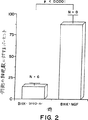
図2は、采−脳弓横断切開(fimbria-fornix transection)後、NGFのBHK細胞移植物と一緒にまたは無しで生存する中隔コリン作動性ニューロンの%を表わす。
図3は、一側性損傷QAラットのアポモルヒネ誘導回旋運動に対するカプセル化されたNGF分泌性BHK細胞の効果を表わす。動物は、3回の別々の場合の回旋運動に対してに試験された。データは、30分の試験時間の間の、または各処置群の完全な同側回旋運動の平均(±)SEM数として表わされる。黒丸/黒線は、QAのみを与えた動物のデータを表わし、白丸/黒線は、QAおよびNGF分泌性BHK細胞移植物を与えた動物のデータを表わし、そして黒丸/破線は、QAおよびNGF非分泌性BHK細胞移植物を与えた動物のデータを表わす。
図4は、両側性損傷QAラットの自発的な歩行運動(locomotor activity)に対する、カプセル化されたNGF分泌性BHK細胞の効果を示す。データは、それぞれの活動測定について、予備手術の活動レベルの平均(±)SEM%として表わされる。図中、HA=水平活動量、TD=合計距離、NM=移動回数、AD=平均距離、MT=移動時間、AS=平均速度、VA=垂直活動量、VM=垂直移動の回数、VT=垂直活動時間、SC=ステレオタイプ活動時間、そしてNS=ステレオタイプ移動の回数である。パネルA-QA;パネルB-2A/NGFなし;パネルC-QA/NGF。
図5は、両側性損傷QAラットにハロペリドール(パネルA)およびSCH23390(パネルB)で誘導したカタレプシーに対するカプセル化されたNGF分泌性BHK細胞の効果を示す。データは、処置群のそれぞれについて、カタレプシーに罹っている時間の平均(±)量として表わされる。黒丸/黒線は、QAおよびNGF非分泌性BHK細胞移植物を与えた動物のデータを表わし、白丸/黒線は、QAおよびNGF分泌性BHK細胞移植物を与えた動物のデータを表わし、そして黒丸/破線は、QAのみを与えた動物のデータを表わす。
図6は、3種類の強度の張力のそれぞれに応答する接触によって測定されたvon Freyの体性感覚閾値(Somatosensory Threshold)を図解する。パネルA-5.4mo.;パネルB−20.6mo.;パネルC−26.7mo.。カプセル化されたBHK-NGF細胞を移植された老齢ラット(26.7mos)は、コントロール群の老齢ラットよりも軽い刺激物質に対して良く応答したが、若齢ラットより応答は良くなかった。黒丸はコントロールを表わし;四角はNGF-BHK細胞のデータを表わす。
図7は、試行間隔2時間の条件で、Morrisの水迷路テストのワーキングメモリーの結果を示す。パネルA-移植前より良好な50%(better 50% pre-implant);パネルB-移植前より悪い50%(worse 50% pre-implant)。黒丸はコントロールを表わし;四角はNGF-BHK細胞のデータを表わす。
図8は、カプセル化されたBHK-NGF細胞を与えた老齢サルでは、損傷により誘導される中隔ニューロンの変性がコントロールに比べて顕著に弱められたことを示す。
発明の詳細な説明
本発明は、カプセル化された細胞が長期間生存でき、そして容易に回収され得る改良カプセルを指向する。本発明はまた、改良されたデバイスおよび方法を指向する。このデバイスおよび方法は、生物学的活性分子を発現させ、そしてこの分子を宿主の哺乳動物へ長期間安定に送達するために生体適合性カプセルに含まれた遺伝的に改変された細胞を使用することである。
本発明のデバイスおよび方法は、広範な生物学的活性分子(200kDまでの高分子量の産物を含む)を、それを必要とする個体に長期間安定に発現させ、および/または有害な物質を除去する代謝機能をそれを必要とする個体に提供することに有用である。本発明のデバイスおよび方法に用られる生物学的活性分子は、種々の器官または組織が通常分泌する多種類の因子を包含する。例えば、インスリンは糖尿病患者に、ドーパミンはパーキンソン病の患者に、第VIII因子はA型血友病患者に、あるいは鎮痛剤は疼痛の患者に送達され得る。
本発明の実施に使用され得る他の生物学的活性分子は、エリスロポエチン、成長ホルモン、サブスタンス−P、ニューロテンシン、NGF、BDNF、NT-3、NT-4/5、CNTF、GDNF、CDF/LIF、EGF、IGF、PDGF、bFGF、およびaFGFのような栄養因子を包含する。
本発明のデバイスおよび方法はまた、ヘモグロビン、チロシンヒドロキシラーゼ、プロホルモンコンバターゼ、bcl-2、ドーパデカルボキシラーゼ、そしてドーパミンβ-ヒドロキシラーゼを包含する生物学的活性分子を長期間安定に発現させるのに有用である。
本発明による送達に適した別のファミリーは、リンホカインおよびサイトカインを含む生物学的応答調節剤を包含する。
本明細書に記載のカプセル化された細胞は、肝細胞のような細胞による血流からのトキシンまたは有害代謝物(例えば、コレステロール)の除去のような必須の代謝機能を、回復または増強するのにも用い得る。本発明の方法は、レシピエントを処置する間に同時に免疫抑制する必要なしに細胞を移植することを可能にする。本発明の方法を用いることにより、特定の物質の恒常性が回復され、さらに長期間維持され得る。
本発明の範囲内に意図される生物学的活性分子は、カプセルまたは移植された細胞から分泌される分子であって、直接的または間接的に哺乳動物宿主において生物学的な効果をもたらす分子、および直接的または間接的にカプセル内に含まれる細胞に生物学的な効果をもたらす分子を包含する。
本発明の好ましい実施態様は、宿主哺乳動物の中枢神経系(CNS)に神経栄養因子を送達するための改良方法である。本発明の特定の実施態様では、神経成長因子(NGF)の、哺乳動物宿主のCNSの特定の領域に対する長期間の安定な発現および送達の改良方法が提供される。
多くの生物学的活性分子をコードする遺伝子がクローニングされ、それらのヌクレオチド配列が公表されている。これらの遺伝子の多くは、アメリカンタイプカルチャーコレクション(American Type Culture Collection(ATCC))のような寄託機関または種々の販売元から一般に入手できる。本発明に有用な生物学的活性分子をコードする遺伝子で一般に入手できないものは、PCR増幅、オリゴヌクレオチドのプローブを用いるゲノムおよびcDNAライブラリーのスクリーニングのような標準的組換えDNA法を用いて入手し得る。生物学的活性分子をコードする既知の遺伝子であればいずれも本発明の方法に用い得る。例えば、米国特許第5,049,493号; Gageら,米国特許第5,082,670号;およびGenentech米国特許第5,167,762号参照。
本発明に特に有用な遺伝子には、ヒトプロエンケファリンA、ヒトプロホルモンコンバターゼ2、ヒトプロホルモンコンバターゼ3、ヒトBDNF、POMC(プロ−オピオメラノコルチン)、β-エンドルフィン、プロジノルフィン、ヒトNGFシグナル配列を有する成熟ヒトBDNF、ヒトCNTF、ヒトNT3、ヒトNGF、ラットGDNF、ヒトNGFシグナル配列を有する成熟ヒトNT5、ウシドーパミンβヒドロキシラーゼ、ウシドーパミンデカルボキシラーゼ、およびチミジンキナーゼをコードする遺伝子がある。
目的の遺伝子(すなわち、適切な生物学的活性分子をコードする遺伝子)は、標準的な技術を用いて適切な発現ベクターのクローニング部位へ挿入し得る。1個より多い遺伝子を適当な発現ベクターに挿入し得るということが認識される。これらの技術は当業者に周知である。
次いで、目的の遺伝子を含む発現ベクターは、本発明の方法で用いる細胞系をトランスフェクトするのに用い得る。リン酸カルシウム共沈澱、DEAE-デキストラントランスフェクションまたはエレクトロポレーションのような標準的なトランスフェクション技術を用い得る。市販の哺乳動物トランスフェクションキットは、例えばStratageneより購入し得る。
目的の生物学的活性分子をコードする遺伝子を発現するために、広範な種類の宿主/発現ベクターの組合せが用いられ得る。長期間安定なインビボでの発現は、その中で生物学的活性分子をコードする遺伝子が、哺乳動物宿主中にインビボで移植されたときにダウンレギュレーションを受けないプロモーターに作動可能に連結しているような発現ベクター(すなわち、組換えDNA分子)を用いて達成される。従って、そのような発現ベクターは、典型的にはレトロウイルスのプロモーターを含まない。
適切なプロモーターは、例えば、SV40の初期および後期のプロモーターまたはアデノウイルスのプロモーターおよび遺伝子発現を制御できる他の既知の非レトロウイルスのプロモーターを含む。
有用な発現ベクターは、染色体、非染色体および合成のDNA配列(例えば、SV40およびE.coli由来のpUC、pBlue ScriptTMプラスミドのような既知の細菌プラスミドの誘導体(例えば、pBR322、pCR1、pMB9、pUC、pBlue ScriptTMおよびそれらの誘導体)のセグメントからなる。
ゲネチシン(G418)またはヒグロマイシン薬剤選択遺伝子を含む発現ベクター(Southern,P.J.(1981),In Vitro, 18,p. 315, Southern,P.J.およびBerg,P.(1982),J.Mol.Appl.Genet.,1,p.327)もまた本発明の実施に有用である。これらのベクターは、目的の生物学的遺伝子(例えば、NGF)、および/または、G418またはヒグロマイシンBのようなトキシンによる選択に対する耐性を付与する遺伝子の発現を起動する種々の異なるエンハンサー/プロモーター領域を用い得る。G418耐性遺伝子は、培養培地に添加されたG418(100-500μg/μl)を酵素的に不活性化するアミノグリコシドホスホトランスフェラーゼ(APH)をコードする。APH遺伝子を発現する細胞のみが薬剤選択から生き残り、第二の生物学的遺伝子をも発現するようになる。ヒグロマイシンBホスホトランスフェラーゼ(HPH)遺伝子は、ヒグロマイシントキシンを特異的に改変し不活性化する酵素をコードする。ヒグロマイシンBホスホトランスフェラーゼ遺伝子と同じプラスミドと同時にトランスフェクトされるか、またはプラスミドに含まれる遺伝子は、50-200μg/mlの濃度のヒグロマイシンB存在下優先的に発現される。
種々の異なる哺乳動物のプロモーターが、G418およびヒグロマイシンBの遺伝子および/または目的の生物学的遺伝子の発現を導くために用いられ得る。これらのプロモーターは、hDBH(ヒトドーパミンβヒドロキシラーゼ)のプロモーター(Mercerら,Neuron,7,pp.703-716,(1991))、hTH(ヒトチロシンヒドロキシラーゼ)(Kanedaら,Neuron,6,pp.583-594,(1991))、hPNMT(ヒトフェニルエタノールアミンN−メチルトランスフェラーゼ)(Baetgeら,PNAS,85,pp.3648-3652,(1988))、mGFAP(マウスグリア線維酸性タン白質)(Besnardら,J. Biol. Chem.,266,pp.18877-18883,(1991))、ミエリン塩基性タン白質(MBP)、mNF-L(マウスニューロフィラメント軽鎖サブユニット)(Nakahiraら,J.Biol.Chem.,265,pp.19786-19791,(1990))、hPo(ヒトPo、末梢神経系の主要ミエリン糖タン白質をコードする遺伝子のプロモーター)(Lemkeら,Neuron,1,pp.73-83,(1988))、mMT、rNSE(ラットニューロン特異的エノラーゼ)(Sakimuraら,Gene,60,pp.103-113,(1987))その他を包含するがそれに限定されない。
用いられ得る発現ベクターの例は、市販のpRC/CMV、pRC/RSV、およびpCDNA1NEO(InVitrogen)である。薬剤選択および目的の生物学的遺伝子の転写を支配するウイルスのプロモーター領域は、CNSにおいてウイルスプロモーターが受けるダウンレギュレーションの対象とはならない上記のプロモーター配列の1つと置換される。例えば、GFAPプロモーターは星状細胞および星状細胞系のトランスフェクションに用いられ、THプロモーターはPC12細胞に用いられ、またはMBPプロモーターは乏突起膠細胞に用いられる。
1つの実施態様において、pNUT発現ベクターが用いられる(図1参照)。さらに、DHFRのコーディング配列がG418またはヒグロマイシン薬剤耐性のコーディング配列により置き換えられるように、pNUT発現ベクターを改変し得る。pNUT発現ベクター中のSV40プロモーターも、上記のように適切に構造的に発現される哺乳動物のプロモーターと置き換えられ得る。
広範な種類の細胞が用いられ得る。それらは、既知の一般に入手可能な不死化細胞系および分裂中の初代細胞培養株を包含する。適切な一般に入手可能な細胞系の例は、幼若ハムスターの腎臓(BHK)、チャイニーズハムスターの卵巣(CHO)、マウス線維芽細胞(L-M)、NIHスイスマウス胎児(NIH/3T3)、アフリカミドリザル細胞系(COS-1、COS-7、BSC-1、BSC-40、BMT-10およびVeroを含む)、ラット副腎クローム親和性細胞腫(PC12およびPC12A)、AT3、ラットグリア腫(C6)、星状細胞および他の線維芽細胞系を包含する。用い得る初代細胞は、哺乳動物のCNSに由来するbFGF-応答性神経プロジェニター幹細胞(Richardsら,PNAS 89,pp.8591-8595(1992);Rayら,PNAS 90,pp.3602-3606(1993))、初代線維芽細胞、Schwann細胞、星状細胞、β-TC細胞、Hep-G2細胞、乏突起膠細胞、およびそれらの前駆体、筋原細胞などを包含する。
本発明の範囲内のカプセル化細胞による処置に利用され得る細胞のタイプは、同種異系および異種の供給源由来の細胞を包含する。本発明者らのカプセル化のアプローチの主要な利点の1つは、本発明の膜が有する免疫隔離の性質、および移植に適合しない細胞(すなわち、ヒト以外の供給源、不死化および/または腫瘍細胞系)を支持する能力に依存する。同種異系細胞よりも異種細胞を使用する特別の利点は、万一、膜に破損が生じた場合、異種細胞が同種細胞に比べ免疫系による破壊の標的とされ易いということである。さらに、異種細胞の供給源は入手が簡単であり、そしてその使用は、入手が困難でありそして社会的ならびに倫理的考慮を伴うヒトの組織の使用の必要性を排除する。さらにヒトの組織は、移植レシピエントへより容易に送達される外来性物質を含有することができる。最終的に、ヒトに移植するために異種組織および細胞系を用いることは、ヒトの組織の操作および処理に関連する危険性を除外する。
本発明の1つの実施態様において、pNUT増幅発現系を用いてBHK細胞をトランスフェクトする。しかしながら、目的の遺伝子を含むるpNUTベクターはまた、前述したようなCOS、L-細胞、CHOなどの多数の他の標準的な不死化または、形質転換された組織培養細胞系をトランスフェクトするために用いられ得る。さらにpNUT発現ベクターは、初代の星状細胞、乏突起膠細胞または神経細胞系(例えば前述したbFGF-応答性神経プロジェニター幹細胞)へトランスフェクトするために用いられ得る。
本発明によって形質転換された細胞株は、生物学的に活性な分子の長期間の安定な発現を提供し得る。このような長期間の安定な発現は、生物学的に活性な分子をコードする導入遺伝子のコピー数を、当該分野で周知の増幅方法を用いて増加または増幅させることにより達成され得る。このような増幅方法には、例えば、DHFR増幅(例えば、Kaufmanら、米国特許第4,470,461号参照)またはグルタミンシンセターゼ(「GS」)増幅(例えば、米国特許第5,122,464号およびヨーロッパ特許出願公開EP338,841号参照)が含まれる。
哺乳動物細胞で遺伝子の長期間の安定な発現を得るための別な方法は、2つの異なる薬剤選択マーカーを用いる二重トランスフェクションによる。ヒグロマイシンまたはG418耐性発現ベクターを用いて上記の多くの細胞株を連続的にまたは同時にトランスフェクトし、種々の遺伝子コピーの挿入を達成し得、従って、目的の遺伝子の発現レベルを達成し得る。
1つの実施態様においては、細胞は、β-NGFまたはGFAPプロモーター/エンハンサー因子(通常このベクターに見られるCMVプロモーター配列に取って代わっている)に作動可能に連結した他の目的の遺伝子のコーディング配列を含むプラスミドpRC/CMVを用いてトランスフェクトされる。トランスフェクトされた導入遺伝子を発現する安定なクローンの選択の際に、このクローンは、同じpRc/GFAP NGF発現ベクター(G418耐性遺伝子の代わりに細菌ヒグロマイシンBホスホトランスフェラーゼ遺伝子(GritzおよびDavies、Gene、pp.179-188(1983))を有する)を用いて再びトランスフェクトされる。安定なクローンをヒグロマイシンおよびG418の両方において再選択した後、この細胞は、RNAおよびタンパク質産物においてトランスフェクトされた導入遺伝子の最大数の組み込まれたコピーおよび/または発現を有するクローンに関してアッセイされ得る。この方式において、複数のベクターを用いてトランスフェクトされた細胞株(高いコピー数の遺伝物質を安定に組み込み、そして種々のレベルで目的の生物学的に活性な分子を発現する)を産生するために、別の組み合わせの薬剤選択発現ベクターを用い得る。
ポリマー−カプセルの移植が有効量の必要な物質または機能を個体に提供するのに十分であり得るように、多数の細胞が本発明の方法において用いられ得る。さらに、1より多い生物学的に活性な分子は、1つのカプセルから長期に亘って安定に発現および/または送達され得る。
この結果に達する1つの方法は、1より多い異種遺伝子を発現するように遺伝的に変更された単一の細胞株をカプセル化することである。
この結果に達する別の方法は、細胞の混合物を単一のカプセルに入れることである。ここで、ある細胞は1つの生物学的に活性な分子を発現するように遺伝的に改変され、別の細胞は第2の生物学的に活性な分子を発現するように遺伝的に改変されている。第2の生物学的に活性な分子を提供するために、遺伝子操作されていない細胞株も利用され得るということが認識されるであろう。
例えば、ある細胞株はまた、遺伝子操作されて異なる生物学的に活性な分子を発現し得る。親細胞株のサブクローンは、それぞれが異なる導入遺伝子を発現するが、このサブクローンはプールされ、カプセルに入れられて所望の効果を長期間達成し得る。
これらのアプローチは、1より多い生物学的に活性な分子を1つの部位に長期間、安定に発現および送達させるために複数の移植物を用いる必要性を排除する。
本発明はまた、異種遺伝子(この遺伝子は、細胞が宿主哺乳動物内に移植される際にカプセル内で生存可能のままであるようにし得る)によって遺伝的に改変されている細胞を含むカプセルを用いることを意図する。言い換えると、本発明の方法はまた、移植されたカプセル内の分子の送達方法に関する。
本発明の特定の実施態様において、本発明者らは、マウスメタロチオネインプロモーターに作動可能に連結したヒトβ-NGF遺伝子を含むpNUT発現ベクターを用いて、リン酸カルシウム共沈澱法によりBHK細胞をトランスフェクトした。
種々の生体適合性免疫隔離カプセルは、本発明による分子送達に適切である。このようなカプセルは、宿主免疫系の有害な影響を最少にしながら代謝物、栄養分および処置物質を通過させる。好ましくは本発明のカプセルは、Aebischerら、PST公開WO 92/19195(本明細書中に参考として援用する)に記載のカプセルと同じである。
最も好ましくは、本明細書に記載されたT1/2膜を用いて、本発明の方法に従って改変された細胞をカプセル化する。本明細書に記載されたT1/2膜はまた、他のいかなる適切な細胞または細胞株のカプセル化にも用いられ得ることが認識されるであろう。したがって、本発明のT1/2膜は初代細胞(primary cell)(非分裂細胞)および分裂細胞をカプセル化するために用いられ得る。
有用な生体適合性ポリマーカプセルは、(a)1つの細胞または複数の細胞(液体媒体に懸濁されているか、ヒドロゲルまたは細胞外マトリックス内に固定されている)を含むコア、および(b)単離された細胞を含まない選択透過性マトリックスまたは膜(ジャケット)(生体適合性であり、そしてコアに存在する場合には、単離された細胞を有害な免疫学的攻撃から保護するのに十分である)の周辺または末梢領域を含む。
ポリマーカプセルのコアは、コア内に隔離された細胞の連続した生存能力および機能のために適切な局所環境を提供するために構築される。
多くの形質転換された細胞および細胞株は、液体コアを有するカプセル内に非常に都合よく隔離されている。例えば、細胞は、コアが栄養培地(任意に細胞の生存能力および機能を維持するためのさらなる因子の液体供給源、例えば、胎児ウシ血清、またはウマ血清を含有する)を有するカプセル内に隔離され得る。
マイクロカプセルもまた、時に本発明の方法および組成物における使用に適切であり得る。マイクロカプセルの製造は、Espevikら、PCT公開WO 9107951号およびSefton、米国特許第4,353,888号(本明細書中で参考として援用する)に記載されている。
適切には、コアは、細胞凝集塊中の細胞の位置を安定させるヒドロゲルによって形成されるマトリックスから構成される。本明細書中の用語「ヒドロゲル」は、架橋した親水性ポリマーの3次元網状組織をいう。この網状組織は、ゲルの形態であり、実質的に水からなり、好ましくは90%以上が水であるゲルであるが、これに限定されない。
ヒドロゲルを形成する組成物は3つのクラスに分類される。第1のクラスは正味で負の電荷(net negative charge)を有する(例えば、アルギネート)。第2のクラスは正味で正の電荷(net positive charge)を有する(例えば、コラーゲンおよびラミニン)。市販の細胞外マトリックス成分の例は、MatrigelTMおよびVitrogenTMを含む。繊維芽細胞は、一般に正に帯電したマトリックスにおいてよく生存し、従って、細胞外マトリックスタイプのヒドロゲルに適切に封入される。第3のクラスは、電荷的に正味で中性(net neutral in charge)である(例えば、高度に架橋したポリエチレンオキシド、またはポリビニルアルコール)。任意の適切なマトリックスまたはスペーサーをコア内で用い得る。これらは沈澱させたキトサン、合成ポリマー、およびポリマーブレンド、マイクロキャリアなどを含み、カプセルに入れられる細胞の成長特性に依存する。
好ましくは、カプセルは免疫隔離である。免疫隔離であるためには、カプセルの周辺または末梢領域は、宿主体内の有害な物質がビヒクルのコアに入るのを防止することによって、および単離された細胞と宿主の免疫系との間の有害な免疫学的接触を防止するのに十分な物理的バリアを提供することによって、カプセルが移植される宿主の免疫系からの細胞の保護を与えるべきである。この物理的バリアの厚さは変化し得るが、それは、常にバリアの両側にある細胞および/または物質の直接の接触を防止するのに十分な厚さである。この領域の厚さは、一般に5ミクロンと20ミクロンとの間の範囲である;10ミクロン〜100ミクロンの厚さが好ましく、そして20ミクロン〜75ミクロンの厚さが特に好ましい。このビヒクルの使用によって防止されるかまたは最小化される免疫学的攻撃のタイプは、マクロファージ、好中球、細胞性免疫応答(例えば、ナチュラルキラー細胞および抗体依存性T細胞介在性細胞溶解(antibody-dependent T cell-mediated cytoloysis:(ADCC))および体液性応答(例えば、抗体依存性補体介在性細胞溶解)による攻撃を含む。
免疫隔離カプセルの使用は、レシピエントを免疫抑制するという必要を伴わずに、異種細胞または組織の移植を可能にする。免疫隔離カプセルの使用はまた、適合しない(unmatched)細胞(アログラフト)の使用を可能にする。異種細胞に対する免疫応答のタイプおよび活動力は、同系のまたは同種の組織がレシピエントに移植された場合に生じる応答とは異なることが予想される。この応答は、主に細胞介在性の攻撃によって、または補体介在性の攻撃によって生じ得る;特定の場合における決定(determining)パラメーターは、殆ど理解されていない。しかし、ビヒクルのコアからのIgGの排除は、免疫防御の規準ではない。なぜなら、殆どの場合、IgG単独では標的細胞または組織の溶解を生じるには不十分だからである。免疫学的攻撃の仲介に必要である重要な物質が免疫隔離カプセルから排除されているならば、免疫隔離マクロカプセルを用いることによって、必要とされる高分子量の産物を送達すること、または高分子量物質に適する代謝機能を提供することが可能である。このような物質は、補体攻撃複合体成分(complement attack complex component)Clqを含み得るか、またはそれらは、食細胞または細胞障害性細胞を含み得る;この免疫隔離カプセルは、これらの有害な物質と隔離された細胞との間に防御的バリアを提供する。従って、免疫隔離カプセルは、異種細胞からでさえ、広範な分子サイズを有する産生物(例えば、インシュリン、副甲状腺ホルモン、インターロイキン3、エリスロポイエチン、アルブミン、トランスフェリン、エンケファリン、エンドルフィン、カテコールアミン、ファクターVIII、NGF、BDNF、NT-3、NT-4/5、CNTF、GDNF、CDF/LIF、EGF、IGF、bFGF、aFGF、PDGF、TGFなど)の送達に用いられ得る。
種々のポリマーおよびポリマーブレンドを用いてカプセルジャケットを製造し得る。これらは、ポリアクリレート(アクリル酸コポリマーを含む)、ポリビニリデン、ポリ塩化ビニルコポリマー、ポリウレタン、ポリスチレン、ポリアミド、酢酸セルロース、硝酸セルロース、ポリスルホン、ポリホスファゼン、ポリアクリロニトリル、ポリ(アクリロニトリル/コビニルクロライド)、ならびにそれらの誘導体、コポリマー、および混合物を含む。
このカプセルは、生物学的活性を維持し、そして産生物または機能の送達のためのアクセスを提供するのに適切な外形であり得る。たとえば、円筒形、矩形、円盤形、パッチ型、卵形、星形、または球形を含む。さらに、このカプセルは、メッシュ様またはネスト様構造にコイルまたはラップされ得る。カプセルが移植された後に回収される場合には、外形は、カプセルの移植部位からの移動に導く傾向の外形、例えばレシピエントの血管を移動するのに十分に小さい球形カプセルは好ましくない。特定の形、例えば矩形、パッチ形、円盤形、円柱形、および平らなシート形は、より高い構造的に完全な状態を提供し、そして回収が望まれる場合には好ましい。
本カプセルは、隔離された細胞の生存能および機能を維持するため、コア中の任意の隔離された細胞がレシピエントの周辺組織(レシピエントの血流を含む)に少なくとも1次元的に充分近くとなるように提供されなければならない。しかし、カプセルを形成するために用いられる材料の拡散限界をもって、全ての場合におけるその外形の限界を一通りに規定はしない。基本ビヒクルの拡散特性、即ち栄養物または酸素輸送特性を変化し、あるいは増強する特定の添加物が使用され得る。例えば、内部媒体は、酸素飽和ペルフルオロカーボン(oxygen-saturated perfluorocarbon)で補充され得、このため血液によって運搬された酸素と直接に接触する必要性が減少する。このことにより、隔離された細胞は生存可能でいられるが、一方、例えばアンジオテンシンの勾配がカプセルから周辺組織に放出され、毛細血管の伸長を刺激する。ペルフルオロカーボンの使用についての参考文献および方法は、Faithful, N.S.(1987)Anaesthesia,42,234〜242頁およびNASA Tech Briefs MSC-21480, U.S. Govt. Printing Office, Washington, D.C. 20402により与えられ、この内容は本明細書中に参考として援用されている。
一つの実施態様において、移植可能なカプセルは、移植後の完全な回収のために充分なサイズおよび耐久性がある。このようなマクロカプセルは、約1〜10μlの好ましい最小容積のコアを有し、そして用途に依存して100μlを超える容積を有するように容易に構成される。
好ましいカプセルは、60〜98%のBSA排除係数および50〜90%のオボアルブミン排除係数の範囲の透過率である透過選択性(permselective)ポアサイズを有する、内側の単一限外濾過膜を有する。カプセルは、PCT出願WO 92/19195に記載の通り、外縁がシールされた平面シートの形態、または末端がシールされた中空のファイバーの形態であり得る。平面シートの形式では、2枚の壁が1000ミクロン未満、好ましくは300ミクロン未満の隙間の厚さで隔てられている。壁の厚さは約25〜200ミクロンの間、好ましくは約30〜75ミクロンの間であるべきである。
中空ファイバーの外形では、ファイバーは1500ミクロン未満、好ましくは300〜600ミクロン未満の内径を有する。いずれの外形においても、流体透過率(hydraulic permeability)は1〜100ml/分/M2/mmHgの範囲、好ましくは25〜70ml/分/M2/mmHgの範囲にある。Dionneら、ASAIO Abstracts, p.99(1993)およびColtonら、The Kidney, Brenner BMおよびRector FC編、2425〜89頁(1981)に記載の通り定義され、測定され、そして算出されたカプセルのグルコース物質移動係数は、10-6cm/秒より大きく、好ましくは10-4cm/秒より大きい。
カプセルの外壁表面の形態は、変化しやすい。先に記載されたT1、T2、T4膜および本発明の新規のT1/2膜は、外壁表面の形態が異なる。これらの全ての膜は、透過選択性の内皮により特徴付けられる。
T1膜は、「開いた」または窓のある非透過選択性の外壁表面および外壁表面と内壁表面との間の柱状の壁構造により特徴付けられる。例えば、Lacyら、Science,254, 1782〜84頁(1991)を参照のこと。T1膜の外壁表面の「マクロポア」の開口部は、代表的には総外壁膜表面積の約20%〜40%を占める。代表的に、マクロポアは、直径が10μm〜15μmであるかまたはより大きい。
T2膜は、内壁と外壁との間に類似の柱状構造(trabecular structure)を有するが、さらに「閉じた」またはより平滑な外表面壁によって特徴付けられる。T2膜は、代表的には、外表面壁上に10%未満のマクロポアを有し、そして5〜15μmの直径サイズの範囲では事実上マクロポアを有さないことを特徴とする。
さらにT4膜は、T1またはT2膜とは異なり、外表面も透過選択的である点でさらに異なる。T4膜は、脳室またはくも膜下腔ならびに液体に浸漬された他の移植部位のようなCSF移植部位に有用である。
霊長類の脳実質または他の「固形の」組織移植部位に関しては、本発明者らは、細胞に対して実質的に非透過性の平滑壁カプセルを用いることを好む。微小血管新生(microvascularization)を促進する開口壁について記載しているものもある(例えば、Braukerら、WO/92/07525を参照のこと)。T1およびT1/2カプセルは、長期間移植物に特に適している。本発明者らはT1/2カプセルを好む。
T1/2カプセルは、総外表面壁面積の約2〜20%、好ましくは2〜15%の間の総マクロポア分布によって特徴付けられる。マクロポアは、直径が約5μm〜約15μmのサイズ範囲内にあるべきである。この範囲内のポアサイズの相対的分布は変化し得る。
1つの特定の実施態様において、PAN/PVCから作成されたT1/2中空ファイバー膜カプセルが利用された。この実施態様によると、総マクロポア面積は、総外壁表面積の約12%であった。マクロポアの約20%は、直径5〜10μmの範囲であり、そしてマクロポアの約80%は直径が約10μmであった。
別の特定の実施態様では、総外壁表面積の約2.4%の総マクロポア面積を有するT1/2中空ファイバー膜カプセルが構成された。マクロポアの約17%は、直径が約5μmであり、約33%は直径が約10μmであり、そして約50%は直径が約15μmであった。
別の特定の実施態様では、総外壁表面積の約10%の総マクロポア面積を有するT1/2中空ファイバー膜カプセルが構成された。99%を超えるこれらのマクロポアは、直径が10〜15μmの範囲であった。
任意の標準的な方法を用いて、外表面マクロポアの形態の定量化を行い得る。本発明者らは、2つの方法を使用した。
一つの方法では、走査型電子顕微鏡に、中空ファイバー膜の切片を載せる。次にファイバーの外表面を金でスパッタリングコート(sputter coated)した。1000×倍率の画像が生じる。1000×での走査型顕微鏡写真は、約9.2kμm2(115μm×80μm)の面積を含む。これらの画像上では、10μm直径のポアは直径が1cmである。円形のテンプレートを用いて、全てのポアを15μm、10μm、5μm、または2.5μmの直径のいずれかに分類し得る。外壁表面開口(即ちマクロポアとして)の総パーセントを(全ポアが占める面積/顕微鏡写真の総面積)×100として算出する。同様の計算を用いて、個々のポアサイズを決定する。
自動システムを用いて、定量化を行い得る。上記の通り、中空ファイバー膜切片の1000×での走査型電子顕微鏡写真画像を調製する。イメージグラバー(image grabber)を用いて分析用に画像を保存する(SCION Image)。本発明者らは、NTH Image(バージョン1.55)と呼ばれるMacIntosh画像処理および分析プログラムを用いて保存された画像を処理した。このソフトウエアは、組織学的サンプルの形態的測定分析を実施するために開発され、そして本発明者らは、これを中空ファイバーの形態測定分析に適応している。各ポアをカウントし、そして直径を割り当てる。総マクロポア面積の算出および個々のポアサイズは上記の通りである。
本発明のT1/2ファイバーは、当該分野において既知である任意の適切な方法によって調製され得る。ある方法では、Cabasso, I., Encyclopedia of Chemical Technolology、12、492〜517頁(1980)に記載の周知の方法を用いるルミナール(luminal)およびキャスティング(casting)溶液の流速の適切な調整により、同軸紡糸金口(coaxial spinneret)からポリマー性キャスティング溶液と凝集剤との共押出成形を包含する。本明細書中に参考として援用されているDionne, WO 92/19195および米国特許第5,158,881号、同第5,283,187号、および同第5,284,761号に記載の通り、凝集剤は、生物学的組織断片、細胞小器官、または細胞および/または他の治療剤の懸濁液を含み得る。
これらの方法に従って、T1膜は、急冷浴(quench bath)に入る前に空気を通してのポリマー溶液および凝集剤溶液の共押出成形によって、形成され得る。T2膜は、加湿空気またはミスト中そして次にバス中へのポリマー溶液および凝集剤溶液の共押出成形によって、形成され得る。T4膜は、ポリマー溶液および凝集剤溶液を直接凝集剤バスへ共押出形成することによって、形成され得、このため透過選択性膜の形成が、外壁表面および内壁表面上で同時に起こる。
T1/2膜は、T2膜形成に用いられる同様の方法を用いて形成され得る。しかし、所望の外表面形態を作成するための既知の方法に従って、共押出口でミストまたは湿度が制御され得る。あるいは、ルーチン法に従うと、急冷浴からのノズルの距離は変化し得る。さらに、共押出形成を用いて膜をキャストする場合、ポリマーおよび凝集剤の絶対的および/または相対的流速を調整して所望の外壁表面形態を達成し得る。最後に、ポリマーおよび凝集剤溶液の組成および温度を変化させ、所望の外壁表面形態を達成し得る。例えば、キャスティング溶液は、DMSO(w/w)中の10〜15% PAN/PVCであり得、そして凝集剤は水、またはその他の水性媒体であり得る。あるいは、キャスティング溶液は、例えば、16% PAN/PVCであり得、そして凝集剤は、例えば23℃で40% NMP、60% H2Oであり得る。
カプセルをシールするいずれの適切な方法も用いられ得、この方法は、ポリマー接着剤および/またはクリンピング(crimping)、節止め(knotting)およびヒートシールの使用を含む。これらのシール技術は当該分野において公知である。さらに、いずれの適切な「乾燥」シール方法も使用し得る。このような方法においては、実質的に無孔性のフィッティング(fitting)が提供され、これにより細胞含有溶液が導入される。充填に続いて、カプセルがシールされる。このような方法は、本明細書中に参考として援用されている、同時係属中の米国特許出願第08/082,407号に記載されている。
本発明の方法およびデバイスは、哺乳動物の宿主、レシピエント、被検体、または個体、好ましくは霊長類、最も好ましくはヒトにおける使用が意図される。
本発明のデバイスおよび方法に関して、多くの異なる移植部位が意図される。これらの移植部位は、脳、脊髄を含む中枢神経系、ならびに眼の眼房水および硝子体液を含む。脳内の好ましい部位は、線条体体、大脳皮質、視床下核、およびMaynert基底核を含む。他の好ましい部位は、脳脊髄液であり、最も好ましくは、くも膜下腔および側脳室である。本発明はまた、腎被膜下部位、ならびに腹腔内部位および皮下部位、または他の任意の治療的に有益な部位への移植を意図する。
本発明の実施態様において、神経の変性によって引き起こされる疾患の処置のための方法が提供される。神経の変性に関連すると考えられるヒト疾患の例としては、アルツハイマー病、ハンチントン病、AIDS関連痴呆、筋萎縮性側索硬化症(ALS)、およびパーキンソン病が挙げられる。
神経変性状態のいくつかの動物モデルは、特定の傷害がニューロンを損傷または死滅し得るという前提に基づいている。いくつかの場合において、このことは、ニューロン死のカスケードを引き起こし得、特定の脳機能を担う経路に沿った栄養的に相互独立したニューロンに影響する。
神経変性状態の処置のためのストラテジーは、
(1)シナプス後ニューロンに対するさらなる損傷を阻害し、そして
(2)傷害を受けた細胞の生存能を改善するために、
成長因子または栄養因子の局所投与を包含する。ニューロンの生存能を改善することが知られている因子としては、NGF、BDNF、NT-3、NT-4/5、CNTF、GDNF、CDF/LIF、bFGF、aFGF、IGF、ニューロテンシン、およびサブスタンス-Pが挙げられる。
神経変性興奮性毒性(excitotoxicity)に対する動物モデルにおいて、グルタミン酸アナログであるキノリン酸を、線条体体および/または基底核と知られる脳領域内に定位的に注入してハンチントン病を患う患者の神経病理および徴候と類似の神経病理および徴候を生じさせる。モデルおよび実際のハンチントン病の両者は、運動制御の局面に必要なニューロンに対する傷害によって特徴付けられる。さらに、ハンチントン病の初期の徴候の一つは、体重の減少である(SanbergらMed J Aust.、1、407〜409頁(1981))。同様の効果がモデル系においても認められる(SanbergらExp Neurol、66、444〜466頁(1979))。キノリン酸はまた、AIDS関連痴呆を患うヒトにおいて異常に高いレベルで見出される。
ハンチントン病(HD)は、奇異な制御不能な運動および異常な姿勢を伴う進行性の痴呆により特徴付けられる常染色体優性疾患である。HDは、ほぼ全ての民族および人種のグループに見出され、米国での罹患率は約50/1,000,000である(Emerich, D.F.およびSanberg, Neuromethods、21、65〜134頁(1992))。本疾患の症状出現は、代表的には、中年(約35〜45歳)において生じ、続いて精神的低下および進行性の運動異常の難治性の経過をたどり、通常15年以内に死を招く。HDの神経病理学的研究により、関連し、そして相互に独立した神経化学的および組織病理学的変異の複雑なモザイクが示されている。
HDの動物モデルを開発するために、種々の手段が検討されている。最近の研究は、選択的細胞毒性化合物の使用から起こる線条体体損傷と歩行運動異常との間の関係に集中している。グルタミン酸は、CNSに見出される主要な興奮性神経伝達物質の一つである。しかし、それは強力な神経毒として作用し、そしてグルタミン酸およびその他の興奮性毒性化合物の比較的特異的な細胞毒性効果に基づくHDの動物モデルを開発するために、多くの試みが成されている。これらの化合物として、カイニン酸(KA)、イボテン酸(IA)、および内因性トリプトファン代謝物であるキノリン酸(QA)のようなグルタミン酸の構造的アナログが挙げられる。極少量をラットの脳内に注入すると、これらの化合物は、通過線維の軸索および求心性神経終末は残す一方で、著しくかつ局所的に限定された毒性効果を生産じる。興奮性毒性の行動的、神経化学的、および解剖学的結果は、HDにおいて観察された結果と類似し、そして内因性興奮性毒性化合物の異常な過剰生産または分解が、HDの病因論的要因であるという推測が導き出された。
キノリン酸(2,3-ピリジンジカルボン酸でトリプトファンの代謝物)は、その強力な興奮性毒性特性ならびにラットおよびヒト脳の両方における幅広い分布の故に大いに注目されている(SchwarczおよびKohler、Neurosci. Lett.、38、85〜90頁(1983);およびSchwarczら、Science、219、316〜318頁(1983))。高濃度の異化酵素(キノリン酸ホスホリボシルトランスフェラーゼ(QPRT))および直接の同化酵素(3-ヒドロキシアントラニル酸(3HAO)が、尾状核(caudate)内に検出されたことから、これは、通常、線条体体の機能において役割を果たすことが示唆される(Fosterら、Brain Res.、336、207〜214頁(1985))。線条体体は、QAの興奮性毒性効果に対して最も脆弱な構造にあり(SchwarczおよびKohler、(1983)、前出)、そして新生の(成熟ではない)動物は、QAの毒性効果に耐性であるようである。このことは中年における代表的なHDの発症におおよそ対応する。
キノリン酸は、線条体体においてKAよりも選択的な変性効果を発揮すると報告されている。KAは、HDの病理をより厳密にまねる(Beal、Synapse、3、38〜47頁(1989);およびRobertsおよびDiFiglia、Synapse、3、363〜371頁(1989))。KAと同様、QAの注入によりGABA感受性ニューロンの涸渇が生じ、一方、コリン作動性ニューロンおよび外部起始の軸索を相対的に残す。KAまたはIAとは異なり、QAの線条体体内注入は、ソマトスタチン含有およびニューロペプチドY含有ニューロンを残すようであることから、このモデルは、本疾患において観察される神経病理を最も厳密に再生することが示唆される(Bealら、J. Neuro Sci、8、3901〜3908頁(1988)。
本発明に従って、細胞(適切な因子を分泌する遺伝的に改変した細胞を含む)を含有するカプセルを移植することによって、適切な脳の領域に栄養因子が提供される。ある場合において、遺伝的に改変した細胞は宿主に対して自己由来(autologous)であり、そしてカプセル化を必要としない。
ヒトNGFを発現するように操作されているBHK細胞のような神経成長因子分泌細胞は、キノリン酸誘導性神経変性に対する療法の典型示す。
別の動物モデルでは、采-脳弓(齧歯類)または脳弓(霊長類)の傷害を包含する。詳細には、中隔海馬系のニューロンを軸索切断して、中隔コリン作動性ニューロンのNGF-依存変性および細胞死を起こす。これらの傷害は、ヒトのアルツハイマー病において影響されるのと同様の脳領域で変性変化を起こす。
本発明の方法に従って、NGFを分泌する遺伝的に改変した細胞を含むカプセルの移植によって、NGFが影響された領域に送達され得る。CNTF、BDNF、bFGF、CDF/LIFのような他の神経栄養因子もまた、中隔コリン作動性ニューロンの同様な集団または重複しない集団を萎縮および/または死から保護し得る。好ましくは、細胞は、組換えヒトNGFを産生するように遺伝子的に操作された線維芽細胞である。
脳弓の傷害もまた、動物被検体のモデルにおいて行動上の欠損(学習および記憶に関する作業課題(task)において最も容易に観察される)を引き起こす。采-脳弓の傷害を伴うラットへのNGFの慢性投与により動物の行動の回復が促進されることが報告されている(WillsらBehav. Brain Res.、17、17〜24頁(1985))。本発明において、NGF分泌細胞を含むポリマーカプセルの移植は、障害を受けた動物の適切な脳領域に継続的にNGFを送達する実際的な方法を提供する。本発明のカプセルは、病態が特定の脳領域へのNGFの継続的な送達によって改善され得るアルツハイマー患者に対する治療および/または予防処置の実際的な形態を提供する。
本発明の方法および組成物は、神経変性から生じる、年齢に関連した認識性欠損の処置に用いられ得る。このような処置は、認識パフォーマンスを増大し得、従って徴候的な利益を提供する。あるいは、処置は徴候的な利益はないものの、神経保護効果を提供し得る。ヒトにおける年齢に関連した認識機能不全および痴呆は、ニューロン変性、特にコリン作動性基底前脳ニューロンのニューロン変性、ならびに皮質および海馬のChATのレベルの低下に関連付けられている(Coyleら、Science、219、1184〜90頁(1983);Whitehouseら、Science、215、1237〜39頁(1982);Phelpsら、Neurobiol. Aging、10、205〜07頁(1989);Gageら、Neurobiol. Aging、9、645〜55頁(1988))。コリン作動性基底前脳ニューロンを選択的に破壊することによって痴呆の齧歯類モデルを作成する試みの前臨床研究では、広範で持続的な認識機能の欠損を生じさせることに失敗した。老齢ラットは、老齢かつ痴呆の患者において報告された神経病理と同様の神経病理を示す。Morris水迷路による空間学習は、これらの病理学的プロセスの有害な効果に極めて敏感である(Morris、J. Neurosci. Meth.、11、47〜60頁(1984);Morris、Learning and Motivation、12、239〜60頁(1981))。この作業課題は、年齢に関連した認識機能の尺度として妥当化されている。この作業課題における老齢ラットのパフォーマンスは、それらの運動、感覚運動または視覚の欠損、学習および記憶に関する他の試験を混同させる要因とは強くは関連していない(Gamzu、Ann. NY Acad. Sci.、444、370〜93頁(1985))。
空間地図の発達が要求される認識作業課題(Eichenbaumら、J. Neurosci.、10、3531〜42頁(1990))として、Morris水迷路は、臨床的設定における老化および痴呆傾向の障害に対して特に敏感な認識機能の非言語試験に類似すると思われる。従って、この作業課題は、痴呆の潜在的な新しい処置の評価に有効と思われる。例えば、いくつかの研究は、外因性NGFが老齢および学習障害ラットにおけるMorris水迷路のパフォーマンスを改善すると報告している。例えば、Fischerら、Nature、329、65〜68頁(1987);Fischerら、J. Neurosci.、11、1889〜1906頁(1991)を参照のこと。
NGFは、容易には脳血液関門を横切らないため、そのCNSへの投与には、血液脳関門の完全性を弱める侵入手順の使用を要する。例えば、外因性NGFの潜在的有効性を示した齧歯類の前臨床試験においては、NGFは浸透圧ミニポンプで、または慢性脳室内カニューレによって投与された。これらの技術は、カニューレによる注射またはポンプ(これは、リザーバーが涸渇するごとに交換しなければならない)からのいずれかによる脳内への反復注入を要する。ポンプリザーバーを取り替え、または注射シリンジをカニューレにより再挿入しなければならないが、そのたびごとに混入物が特に感染を受けやすい脳内に導入され得る、別の機会があることを示す。
滅菌手順を注意深く使用しても、感染の危険は存在する。集中治療室においてでさえ、頭蓋内圧をモニターするために使用する脳室内カテーテルが、約3日後に細菌に感染する(Saffran、Perspectives in Biology and Medicine、35、471〜86頁(1992)。感染の危険性に加えて、注入手順に関連するいくつかの危険があると思われる。脳室への注入は水頭症を生じることが報告されており(Saffranら、Brain Rresearch、492、245〜254頁(1989))、そして実質内への溶液の連続注入は壊死に関連している。
胎児組織の使用は倫理的関心から明瞭にされておらず、そして非カプセル化非胎児細胞は拒絶されるか、または腫瘍を生じ得る。さらに、胎児供給源(fatal source)から採取した組織は、高い生存能があり得る。NGF産生細胞をカプセル化することによって、比較的低い感染の危険性で、胎児組織を使用することなく、そして組織拒絶または腫瘍発生の危険性無く、外因性NGFが供給され得る。
最後に、既に使用されている用量での外因性NGFが、それ自体で有害または毒性であるかどうかについての懸念が表明されており、おそらくアルツハイマー病に関連する神経変性のプロセスを促進さえすると思われる(Saffran、Perspectives in Biology and Medicine、35、471〜86頁(1992)。外因性NGFは、もつれ形成(tangle formation)を促進し、脳血流の変化を潜在的に招く血管周囲の交感神経軸索の軸索発芽を開始させ、あるいはまだ影響を受けていない基底前脳ニューロンは機能不全となり、従って、痴呆のプロセスが促進されるような外因性NGFに応答する基底前脳ニューロンの投射をリモデリングする。(Saffran、Perspectives in Biology and Medicine、35、471〜86頁(1992))。
本発明の1つの局面によると、年齢に関連した認識性の欠損(ハンチントン病、パーキンソン病、アルツハイマー病、およびALSを含む)の処置のための外因性NGFの有益な効果は、外因性hNGF送達に効果的であると既に報告されている用量よりもかなり低い用量で得られる。
既に報告された用量のNGFの投与は、所望しない副作用を有し得る。それらには、著しい体重の減少、疼痛、意気消失、低食症(hypophagia)およびヘルペス感染症の再発が含まれる。
本発明によると、(インビボで合成された)NGFの脳室、脳実質、または他の適切なCNSの場所へのカプセル送達(1〜1500ng/日の範囲)が望ましい。NGFまたは他の適切な因子の実際の用量は、より少ないまたはより多い数のカプセルを移植することによって変化し得る。本発明者らは、脳室への送達については、1〜1500ng NGF/ヒト/日、好ましくは10〜600ng NGF/ヒト/日、最も好ましくは50〜500ng NGF/ヒト/日、そして実質への送達については1〜1500ng NGF/ヒト/日、好ましくは10〜150ng NGF/ヒト/日を意図する。これらの用量の範囲は、特に、用量を齧歯類、霊長類、およびヒト間の脳容積の差を考慮して規格化すると、齧歯類の研究および霊長類の研究においてCBFニューロンのスペアリング(sparing)/発芽(sprouting)に必要なNGFの既に報告された用量(17〜350μg/日)よりもかなり低い。Tuzynskiら、J. Neurosci.、10、3604〜14頁(1990);Koliatsosら、Ann. Neurol.、30、831〜840頁(1991)、Koliatsosら、Experimental Neurol.、112、161〜73頁(1991);Dekkerら、Neuroscience、60、299〜309頁(1994)。評価された1例の臨床患者においては、送達されたNGFの用量は75μg/日であった。(Olsonら、J. Neural Trans.、4、79〜95頁(1992))。1つの実施態様において、ヒトNGF(hNGF)を分泌する遺伝的に改変された細胞を半透過性膜内にカプセル化し、そして適切な哺乳動物宿主、好ましくは霊長類、最も好ましくはヒトの脳室内または実質内に移植する。
本発明をさらによく理解するために、以下の実施例を示す。これらの実施例は例示のみを目的とするものであって、そして如何なる様式によっても、本発明の範囲を限定するように解釈してはならない。
実施例
実施例1:成体ラットのCNSに移植するカプセル化NGF分泌BHK細胞
ヒトβ-NGFの発現およびBHK細胞株の生産
プレ−プロ形態のNGFの完全なアミノ酸配列をコードするβ-NGFのヒト遺伝子を、SV40プロモーターにより操作される変異形態のジヒドロ葉酸レダクターゼ(例えば、Kaufmanの米国特許第4,470,461号を参照のこと)を含む発現構築物中のマウスメタロチオネインプロモーターの後に、サブクローニングした(図1)。
β-NGF遺伝子の5’末端および3’末端をコードする2つのヒトゲノムクローンをATCCから購入した(phbeta N8D8、phbeta N8B9)。phbeta N8D8由来の440bpのSca1-EcoR1フラグメントの5’側を、phbeta N8B9から単離した2.0kbのEcoR1フラグメントの3’側に連結した。スプライシングしたNGFゲノム配列は、第1イントロンの3’末端の約37bp、プレ−プロNGFのタンパク質翻訳を開始すると考えられる2重のATG配列、ならびにヒト遺伝子の完全なコーディング配列および3’側の非翻訳領域全体を含んでいた(Hoyleら、Neuron 10, pp.1019-1034(1993))。2.51kbの結合したβ-NGF構築物を、pNUT発現ベクターを基にしたDHFR(Baetgeら、Proc. Natl. Acad. Sci. USA, 83, pp.5454-5458(1986))のマウスメタロチオネイン-Iプロモーター(-650〜+7)およびラットインスリンII遺伝子(Palmiter R.D.ら、Proc. Natl. Acad. Sci. USA, 88, pp.478-482(1991))の第1イントロンの直ぐ下流にサブクローニングした。pNUT-β-NGF構築物(図1を参照のこと)を、標準的なリン酸カルシウム介在トランスフェクション方法を用いて、BHK細胞に導入した。トランスフェクトしたBHK細胞を、200μMメトトレキセート(Sigma)を含有する培地で3〜4週間選択し、そして耐性の細胞を、200μMメトトキセレートとともにまたは含まないのいずれかでポリクローナル集団として維持した。
NGF生物活性の定量
神経成長因子(NGF)は、PC12細胞で神経突起の顕著な伸長を引き起こし、そしてそれ自身が、NGF生物活性について迅速かつ感度の良いアッセイを提供する。NGFトランスフェクトしたBHK細胞により生産されたNGFの生物活性を試験するために、親BHK細胞(BHK-コントロール)およびBHK-NGF細胞由来のならし培地(CM)を、6ウェルの標準組織培養プレートで生育したPC12A細胞(SchweitzerおよびKelly, J. Cell Biol., 101, pp.667-676(1985))に添加した。コントロールとして、25SのマウスNGFをいくつかのウエルに添加し、神経突起を誘導した(50ng/ml)。PC12A細胞を、1〜4日間にわたり細胞体の直径の長さを3回以上測定した神経突起について記録した。NGFバイオアッセイはまた、移植したコントロールカプセルおよびNGF分泌性BHK細胞充填カプセルの修復に基づき、カプセル由来のCMを新鮮な培地で24時間インキュベートしたカプセルから取り出した純粋なPC12A細胞に添加することにより行った。全ての実験では、BHK-NGF細胞由来のCMは、24時間以内にPC12A細胞で丈夫な神経突起伸長を生じ、このことはBHK細胞から生産されたNGFが生物活性であることを示す。BHKコントロールは、並行した実験でPC12A細胞の神経突起伸長を起こすこのような能力を示さなかった。
神経突起伸長がbFGFのようなNGF以外の因子によるかどうかを決定するために、本発明者らは、CMおよび組換えhNGFを組合せてNGFブロック抗体(マウス抗β-NGF;Boehringer-Mannheimカタログ番号1008-220)を添加した。このブロック抗体の添加は、用量依存様式でPC12A細胞由来の神経突起伸長を十分に阻害した。最後に、本発明者らは、塩基性線維芽細胞成長因子(bFGF)の存在について、ELISAによりBHKコントロール細胞のならし培地をプロービングする1組の実験を行った。bFGF ELISA(Research and Diagnostics Systems, QuantikineTM, ヒトFGF塩基性ELISAキット;Burgessら、Ann. Rev. Biochem, 58, p.575(1989))を用いると、本発明者らは、BHK細胞由来の多量のならし培地中の任意のbFGFを検出し得なかった。これら両方の実験は、BHK-NGF CMに応じてPC12A細胞で見られる神経突起伸長が分泌NGFの存在によることを明らかに示す。
NGF ELISA
カプセル化および非カプセル化BHK-NGF細胞由来のNGF発現の定量を以下のように行った:全ての試薬は、他の記載がない場合、Boehringer Mannheim Biochemicalsから入手した。Nunc-Immuno MaxiSorp ELISAプレートを、コート緩衝液(CACl2およびMgCl2/0.1%アジ化ナトリウムを含有しない1×PBS;pH9.6)中、1ng/mlの抗マウス-β(2.5S)神経成長因子を1ウエルにつき150μlでコートした。コートしたプレートを、37℃で少なくとも2時間、あるいは4℃で一晩インキュベートした。コート溶液をウエルから回収し、そしてウエルを300μlの洗浄緩衝液(50mM Tris-HCl/200mM NaCl/10mM CaCl2/1% Triton X-100/0.1%アジ化ナトリウム;pH7.0)で、3回洗浄した。次いで、ウエルを10mg/mlのウシ血清アルブミン(BSA)を含む300μlのコート溶液で、室温にて少なくとも30分間ブロックした。次いで、ウエルを300μl洗浄液で3回洗浄した。ならし培地のサンプルを、2×サンプル緩衝液(サンプル緩衝液は、2% BSA含有すること以外洗浄緩衝液と同じである)中に1:1希釈した。100μlの調製サンプルを、ウエルに充填した。プレートを覆い、次いで37℃で少なくとも2時間または4℃で一晩インキュベートした。溶液を、吸引によりウエルから除去し、そして300μlの洗浄緩衝液で3回洗浄した。各ウエルに、4U/mlの抗マウス-β(2.5S)神経成長因子-β-gal結合体を100μl添加した。プレートを少なくとも1時間37℃でインキュベートした。溶液を吸引によりウエルから除去し、300μlの洗浄緩衝液で3回洗浄した。最後に、200μlのクロロフェノールレッド-β-D-ガラクトピラノシド基質溶液(100mM Hepes/150mM NaCl/2mM MgCl2/0.1%アジ化ナトリウム/1% BSA中40mg CPRG;pH7.0)をウエルに添加し、そして37℃でインキュベートした。約30分〜1時間後または発色が570nmでの光度測定に十分になった後、サンプルをプレートリーダー上で分析し、そして組換えNGFタンパク質標準に対して測定した。
c-fos誘導アッセイ
PC12A細胞へのNGF投与により起こる定量的なc-fos誘導を、免疫蛍光アッセイにより測定した。PC12A細胞を、1mlにつき100,000細胞の密度で、ポリオルニチン処理したガラスカバーグラス(12mm)上に播き、そして24ウエルプレートで少なくとも24時間平衡化した。細胞を、神経突起伸長バイオアッセイのセクションにすでに記載したように、同じ培地中で増殖した。
PC12A細胞におけるc-fos誘導について試験するために、BHKおよびBHK-NGF細胞、または組換えヒトNGF(50ng/ml)を含有するカプセル由来のならし培地を、PC12A細胞に2時間添加し、そして37℃かつ5% CO2でインキュベートした。このインキュベーションの後、カバーグラスを4%パラホルムアルデヒド(0.1M PBS、pH7.4中)で固定し、PBS中の10mMグリシンで2回洗浄し、1% Triton X100(PBS中10分間)および1% nonidet P40(PBS中10分間)で透過した。カバーグラス上の細胞をPBSで5分間の洗浄を3回行い、PBS中の5%正常ヤギ血清(NGS)で1時間ブロックし、そしてPBS中1% NGSで1:10希釈したc-fosに対して生じたウサギポリクローナル抗血清(Oncogene Science)中で3時間インキュベートした。次いで、カバーグラスをPBSで5分間の洗浄を2回行い、そしてフルオレセイン結合ヤギ抗ウサギIgG抗体とともにインキュベートした。最後に、カバーグラスをPBSで2回洗浄し、そしてCitifluorRアンチフェーデント(antifadent)で固定し、そして蛍光顕微鏡で観察した。蛍光を顕微鏡によりモニターし、そしてc-fos誘導を、蛍光標識した核の存在により測定した。
カプセル化の手順
不均斉な単一皮膚中空ファイバーを、ジメチルスルホキシド中12.5%ポリ(アクリロニトリル塩化ビニル、すなわちPAN-PVC)コポリマー(w/w)の溶液からキャストした。製造プロセスは、Cabasso、Encyclopedia of Chemical Technology, 12, pp.492-517(1980)によりドライーウエット(ジェット)スピニング技法を用いる相転位として公知である。スピニングプロセス後、中空ファイバーを、25%グリセロールを含有する蒸留水浴に無菌的に移した。このことは、乾燥手順の間に完全なポアを維持する方法を提供する(Cabasso, 1980, 前出)。製造したファイバー(XP11)は、450±25μmの内径、53ml/(m2分mmHg)の液浸透性、88.7±2.1%のBSA排除係数、82.0±1.7%のオボアルブミン排除係数、および約8×10-4cm/秒のグルコース質量移動係数を有するT1/2膜タイプであった。乾燥後、デバイスを、6〜7mmの長さの乾燥中空ファイバーを遠位のシールで、近位末端に中隔定着物を有する光硬化アクリレートハブ上にマウントすることにより製造した。近位末端は、デバイスの内腔に注入される細胞のための充填アクセスを有する。グリセロールを、濾過滅菌した70%エタノールでデバイスから除去し、そしてカプセル化を行う前にHBSS中に入れた。
細胞を以下のように予め製造したカプセル化デバイスに充填した:BHKコントロール細胞またはBHK-NGF細胞のいずれかを、約107/mlの密度で予め製造したデバイスに充填した。2×107/mlの密度のBHK細胞懸濁液を、生理学的VitrogenR(Celtrix, Palo Alto, CA)と1:1で混合し、そして中隔アクセスポートから予め製造したデバイスに注入した。2〜2.5μlの細胞懸濁液を注入後、中隔を破り、そしてアクセスポートを光硬化アクリレート(Zeneca)を用いてシールした。BHK細胞充填デバイスを、移植前に、無血清培地であるPC1(Hycor)で4〜5日間維持した。インビトロで3または4日後、細胞を充填したカプセルをHBSSで2回洗浄し、そして1mlの新鮮な培地に入れ、ELISAによりNGFについて分析した。
成体ラットCNSにおけるβ-NGFの長期発現
本発明者らのインビトロでの実験は、BHK細胞でのヒトβ-NGFの長期で安定な高レベルの発現を示した。この長期で安定な発現がインビボで達成され得るかどうかを決定するために、本発明者らは、BHK細胞を含有するカプセルを成体LewisラットのCNSに移植した。
移植前に、BHK-NGFカプセルおよびBHKコントロールカプセルから採取したならし培地(CM)を、上記のようにNGFについて神経突起伸長アッセイに供した。BHK-NGF細胞由来の0.5mlのCMは、神経突起伸長の程度について、50ng/mlの外因的に添加したNGFと同等であったが、BHKコントロール細胞由来のCMは、検出可能なレベルのNGFを含んでいなかった。
カプセルを、成体Lewisラットの線状体に1、3、および6カ月間移植した。上記の説明に基づき、カプセルを、神経突起伸長アッセイによりNGFの生産について試験した。
インビボで1、3、および6カ月後、BHK-NGFを充填したカプセルは、50ng/mlのNGFと同等またはより大きいPC12A細胞で神経突起伸長を生じ得た。NGF活性は、BHKコントロールカプセル由来のCMで検出できなかった。表Iに示すように、カプセル化BHK-NGF細胞由来のサンプルのELISA定量は、3および6カ月後のインビボにおいて、約20ng/24時間/カプセルまで放出した。

カプセル化NGF分泌BHK細胞(実施例1に記載のとおり)が、インビボで有効量のNGFを放出する能力を評価するために、采−脳弓吸引性損傷(aspirative lesion)を14匹のLewisラットで定位手術で行った。損傷の直後、実施例1に記載のように、BHKコントロールまたはBHK-NGF充填XP-11デバイスを損傷部位に定位手術で移植した。
被験体
約3カ月齢であり、そして約300グラムの体重の成体の雄Lewisラット(Harlan Breeders, Indianapolis, IN)を以下の研究に用いた。動物を3つのグループで温度および湿度を制御したコロニー部屋で飼育し、その部屋は、0700時に点灯する12時間の明/暗サイクルで維持された。
定位手術
手術の直前に、ラットを、ケタミン、キシラジン、およびアセプロマジン混合物の筋肉内注射で麻酔をかけ、そしてKopf定位手術器具(Emerichら、1992参照)に置いた。矢状切開を頭皮に行い、そしてブレグマから2.0mm後方に、そして3.0mm側方にわたり開頭手術を行った。20ゲージのチップを有する吸引デバイスを、定位手術フレーム(Kopf Instruments)にマウントし、そして内壁皮質(medial parietal cortex)、帯状皮質(cingulate cortex)、脳梁、海馬の背面、視床の背面、および采−脳弓を、ブルグマの1.40mm後ろに吸引チップを置き、そして矢状洞の直ぐ側方に5.0mmの深さまでそれを入れ込むことにより吸引した。次いで、ブレグマの3.0mm側方の位置に達するまで、チップを0.5mmずつ側方に動かす。吸引直後に、定位フレームにマウントした18ゲージのテフロンカテーテル内にカプセルを置くことにより、ラットに、トランスフェクト(N=8)または非トランスフェクトBHK細胞含有カプセル(N=6)のいずれかを片側に移植した。各デバイスは、直径600ミクロンで長さ0.7cmを測定し、そして約15〜25×103細胞を含んでいた。
ステンレス鋼オブドュレーター(obdurator)をカニューレ内に置き、外側のカニューレが予め調製された窩内にカプセルを無抵抗に置けるようになるまで、オブドュレーターをその場所に保持した。移植のための定位手術座標は、ブレグマより0.5mm後方、矢状縫合の1.0mm側方、および皮質表面より7.5mm下方であった。
組織学
動物に手術後3週目に麻酔をかけ、そして組織学的分析用に調製した。動物を、以下のものでペリスタポンプを用いて、心臓を介して(transcardially)灌流した:20ml生理食塩水(0.9%、室温)、120mlのグルタルアルデヒド(0.1%)、500mlの0.1%グルタルアルデヒド/4%パラホルムアルデヒド、300mlのパラホルムアルデヒド(4%)、および最後に300mlの10%スクロース。全ての溶液を氷冷し(4℃)、そして他に記載がない場合、リン酸緩衝化生理食塩水中(pH=7.4)で調製した。
固定後、脳を取り出し、25%緩衝化スクロース(pH=7.4)に入れ、そして24〜48時間冷却した。組織を低温槽で20μmの間隔で切り、そしてポリリジンでコートしたスライドにマウントした。中隔全体を通しての3番日毎の切片を保存し、以下のプロトコールに従ってコリンアセチルトランスフェラーゼ(ChAT)免疫反応性について処理した:(1)0.8% Triton X-100+10%正常血清を含むPBS中で一晩インキュベーション、(2)1:1000の希釈した一次抗体(ChATに対するヤギ抗血清;Chemicon)とともに48時間インキュベーション、(3)PBS+0.2% Triton X-100で6×5分のすすぎ、続いて、ビオチン化二次抗体(IgG)中で1.5時間インキュベーション、(4)PBS+0.2% Triton X-100中で6×5分のすすぎ、(5)1.5時間、アビジン−ビオチン結合体(ABC、Vector elite)とともにインキュベーション、(6)PBS中で3×5分のすすぎ、(7)蒸留水で5分のすすぎ、(8)0.1% Tris緩衝液に溶解した3,3-ジアミノベンジジン(DBA)(0.05%)+2%ニッケルアンモニウム硫酸塩とともに5分間インキュベーションし、続いて5分間過酸化水素(0.01%)でインキュベーション、(9)反応をPBSで3×1分のすすぎにより停止した。
切片をマウントし、脱水し、そしてカバーグラスを載せた。隣接した切片をヘマトキシリンおよびエオシン(H+E)で染色した。采−脳弓の吸引後に生じた損傷の広がりを確認するために、10番目毎の切片を海馬全体を通して取り出し、そしてVan Ooteganら(Brain Res. Bull., 12, pp.543-553(1984))の方法に従ってアセチルコリンエステラーゼについて染色した。コリン作動性細胞の損失を定量するために、ChAT陽性ニューロンを10×の倍率で対角帯(diagonal band)の中隔の内側および鉛直肢(vertical limb)で計数した。各動物由来のブレグマから約0.7、0.5、および0.2mm前方に位置する代表的な切片(脳あたり3つ)をこの分析のために用いた。
NGF放出の結果
移植後3.5週後、動物を麻酔し、そしてデバイスをシリコーン鎖をゆっくりと引っ張ることにより回収した。各カプセルを、1mlの培地で24時間インキュベートし、そしてCMをELISAによりNGFについてアッセイした。移植の前および後のNGF分泌レベルを表IIに掲げる。
BHKコントロールおよびBHK-NGF移植動物の損傷後3.5週の中隔に残っている総ChAT-陽性ニューロンを計数し、そして合わせた結果を図2に示す。ChAT-陽性ニューロンの定量は、BHKコントロールカプセルを用いると、非損傷側と比較して、15±3%のみのニューロンが、脳の損傷側で陽性のままであり、一方、BHK-NGFカプセルを用いると、90±5%のコリン作動性ニューロンがChATについて免疫陽性であった。カプセル化細胞の生存は、BHKコントロールとBHK-hNGF細胞充填カプセルとの間で等しかった(データは示さず)。
実施例3:霊長類の脳弓損傷
実施例2は、本発明のBHK細胞から成体ラットの采−脳弓損傷を有する側脳室へのhNGFの慢性送達が、損傷の結果死んでしまうであろう中隔内側のコリン作動性ニューロンを保護することを示した。同様の実験をヒト以外の霊長類で行ったことを本明細書に記載する。サル(Cebus apella)の脳弓の損傷を起こさせることは、(KordowerおよびFiandaca, J. Comp. Neurol., 298, pp.443,(1990))に記載されている。
成体カニクイザルを、これらの損傷実験で用いた。一側性損傷を完了した後、実施例1に記載されるように5個のXP-11カプセルデバイス(番号により同定される)を、中隔内側の、損傷に同側に近接する側脳室内に手で置いた。コントロールとして、いくつかの動物に、BHKコントロール細胞を充填したデバイスを移植した。移植手順に続いて、手術部位を閉じ、そして動物を回復させ、そして3.5週間綿密にモニターした。3.5週の生存期間に続き、番号を付けたカプセルを注意深く取り出し、そして標準細胞培養培地に入れ、そして次いで、移植前であるとして、NGF放出についてELISAにより再アッセイした(表III)。次いで、動物をアルデヒド固定剤を灌流することにより屠殺し、そして脳を、組織化学および免疫組織化学手順のために切片にし処理した。
動物は、直ぐに手術から回復し、そして実験の間生存していた。BHK-hNGF含有カプセルを受けている動物は、嗜眠状態であり、手術後数日間減少した食欲を示した。これらの動物うち1匹はまた、複数の発作が手術後約2日で始まり、そして手術後5日以内におさまることを示した。このような合併症は、BHKコントロール細胞移植物を受けたいずれの動物にも示されず、そしてBHK-hNGF動物とBHKコントロール動物との間の違いは、手術後の最初の1週間後では観察されなかった。
BHKコントロール移植物を受けている1匹の動物のポリマーのカプセルを、インサイチュに残して置いた。全ての他のサルにおいては、BHK細胞充填デバイスを、移植後23〜28日の側脳室から、カプセルに宿主組織がほとんど付着しないように回収した。
カプセル取り出し時の視覚的観察に基づき、全てのカプセルを、側脳室内に位置させ、そして尾状核の頭および中隔外側の両方で隣接させた。全てのカプセルを完全に取り出し、いずれのカプセルもインサイチュまたは回収の手順のいずれかの間で壊れなかったことを証明した。細胞充填デバイスを1匹のBHKコントロール動物にインサイチュで残したままにし、デバイスの位置を示し、そして宿主組織の応答を評価した。この動物および全ての他の動物におけるカプセルへの宿主の応答は最小であった。かなり少量の反応性グリアが存在し、これは、全て観察された場合、移植片−宿主の界面で外接した位置でのみ見られた。さらに、マクロファージのような他の非ニューロン細胞は、移植片の周辺領域内で観察されなかった。
全てのサルは、損傷の同側の海馬内のアセチルコリンエステラーゼ含有線維の広い損失により示されるような、脳弓の完全な横断切開を示した。移植されていないか、またはBHKコントロール(NGF非分泌)細胞移植物を受けたかのいずれかであるコントロールのサルは、互いに異ならず、そしてコリンアセチルトランスフェラーゼおよびp75 NGFレセプター(NGFr)−免疫反応性ニューロンの広い損失を、損傷の同側の対角帯の中隔内側(MS;それぞれ53および54%)および鉛直肢(VLDB;それぞれ21および30%)内で示した。対照的に、BHK-hNGF細胞の移植物を受けたサルは、コリン作動性ニューロンのあまり大きくない損失のみを中隔(それぞれ19および20%)およびVLDB(7%)内で示した。さらに、hNGF分泌細胞の移植物のみが、中隔内でコリン作動性線維の稠密伸長を誘導し、これは、移植部位に隣接する脳室の上衣内層に対して分岐した。回収したカプセルの試験は、検出可能なレベルのhNGFを、培養でPC12A細胞を分化するために十分な濃度で生産する大量の細胞を示した。
実施例4:ハンチントン病動物モデルにおけるBHK−NGF移植片の効果
A.齧歯類
手術と移植
手術の直前に、Lewisラットをペントバルビタールナトリウム(45mg/kg, i.p.)で麻酔しKopf定位器に置いた。頭皮に矢状切開を施し、2ヶ所に穴をあけNGFを分泌するBHK細胞を含有するXP-11ポリマー製カプセルを(実施例1に記載した様に)側脳室に設置した。ラットの片側あるいは両側の移植は定位枠に取り付けた18ゲージのテフロン製カテーテル中にカプセルを入れ、それを適切な位置に低下させて行った。移植の定位座標は以下のとおりである:ブレグマ(Bregma)の0.5mm前方、矢状縫合の1.5mm側方、皮質表面の8.0mm下方。
約1週間後、動物を麻酔し、定位器に設置して225(片側)または150(両側)nmolのQAあるいはリン酸緩衝化生理食塩水を賦形剤として、線条体に以下の座標で片側に注射した: 脳表面から下方にAP=+1.2mm、ML=±2.6mm、DV=5.5mm。QAを2N水酸化ナトリウムに溶解してpH7.2のリン酸バッファーで希釈し、最終pHを7.4、最終濃度を225nmol/μlとした。QAの各線条体への注入は28ゲージのハミルトンシリンジを用い5分間にわたって1μl量の条件で実施した。注射カニューレをその場所にさらに2分間放置し潅流の拡散を可能にした。この手順の結果3つの実験グループが形成された: 1)キノリン酸のみ(QA)、2)キノリン酸+NGF-分泌BHK細胞(QA/NGF)、および3)キノリン酸+NGF非分泌BHK細胞(QA/NON-NGF)。手術後ただちに、動物に10mlの乳酸化リンガー溶液を腹腔内注射した。手術後、動物はマッシュ飼料と水を自由摂取で飼育した。
行動試験
回旋行動:
片側のキノリン酸注射後10日目から、動物を、自動化されたデータ収集用にIBMコンピューターを接続した自動化回旋計測器(Rotoscan, Omnitech Instruments)中でのアポモルヒネ誘導回旋行動試験に供した。まず動物を5分間の慣らし期間のために試験チャンバーに置き、次いでアポモルヒネ(1.0mg/kg、0.1%アスコルビン酸を含有する標準の生理食塩水中)を注射し、続く30分間の間に試験した。アポモルヒネにより誘導された回旋行動の増感作用は、線条体の刺激毒性損傷の後で起こる。それゆえ、動物の試験は各セッションを3ないし4日の間隔を開けて4回実施した。回旋は完全な360度の同側の回旋で定義され、2方向間の正味の差として報告された。
自発的運動能力:
手術前および両側QAの30日後に、それぞれの動物を自動化Digiscan-16 Animal Activity Monitor System(Omnitech Electronics, Columbus, Ohio)の5個の開放場ボックス中の1個に入れ、1時間の試験期間中の運動能力を試験した。以下に示す行動変数を収集した:(1)水平活動(HA):水平センサー中断の総数;(2)総距離(TD):インチで表現した動物の移動距離;(3)移動回数(NM):移動1回毎に増加するこのパラメーターは最後の中断から少なくとも1秒間分離された光線の遮断によって記録された;(4)移動時間(MT):秒で表現した動物の移動に要した時間;(5)平均速度(AS):cm/秒で表現した動物の移動の平均速度;(6)平均距離(AD):インチで表現した動物が1回の移動発作中に移動した平均距離;(7)垂直活動(VA):垂直センサーにおける光線中断の総数;(8)垂直時間(VT):このパラメーターは動物が垂直センサーの光線を遮断する間に増加した;(9)垂直移動回数(VM):これは少なくとも1秒間分離された垂直センサーの光線中断により増加する;(10)常同症的移動数(NS):このパラメーターは同じ光線が1秒間隔で繰り返し遮断されると増加した;(11)常同症時間(SC):常同症に費やされた時間の蓄積。
カタレプシー:
運動活動試験1日後、動物にD1およびD2ドーパミンレセプターのアンタゴニストであるハロペリドールとSCH2388を投与した後、運動機能減少(カタレプシー)の試験を実施した。カタレプシーは、動物の後ろ足を台の上に置き、前足を台の上9.0cmの所に吊るした水平棒(直径0.6cm)に置く棒試験により測定した。動物に生じたカタレプシーの度合いは、自分自身を棒から引き離すのにそれぞれの動物がどれだけの時間を要したかで測定した。許容範囲は最高300秒までとした。動物を3つの処置グループの1つにランダムに割り当て、ハロペリドール(1.0mg/kg)、SCH23388(1.0mg/kg)あるいはコントロール賦形剤(0.9%生理食塩水)のいずれかを注射した。棒試験は、薬物および/またはコントロール賦形剤の投与後1、2、3、および4時間後に再度実施した。すべての動物の試験は、各処理条件のもと3〜4日の間隔をおいて実施した。
結果
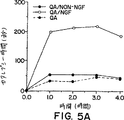
すべての動物は、試験セッションの繰り返しの中でアポモルヒネに対する感受性を増した。図3に示す様に、QAはアポモルヒネで誘導した回旋行動の顕著な増加を生じさせた。非NGFグループの動物は、QAのみを受けた動物と差がなかった。しかし、QAと同時にNGF分泌BHK細胞を受けた動物は(表IV、QA片側を参照のこと)すべての試験セッション期間の間、回旋行動の有意な減少を示した。
片側QA後に見られた行動保護と一致して、動物は、QAの両側注射により生じた活動亢進状態の減少を示した。QAのみ、またはQAおよび非NGF分泌BHK細胞移植物を受けた動物(即ちBHKコントロール)は、移植前のレベルの約100〜350%の範囲で活動の増加を示した(図4)。対照的に、QAと同時にNGF分泌BHK細胞を受けた動物(表IV、QA両側)は、非NGF処理動物に比較して非常に減少した、移植前の値の約25〜150%の範囲の行動レベルを示した。
同様の行動保護パターンが、両側QA動物をカタレプシーについて試験した時にも見られた。QAを単独で、あるいは非NGF分泌BHK細胞と組み合わせて受けた動物は、4時間の試験期間中、ハロペリドールとSCH23390の後、約30〜60秒の継続時間の範囲のカタレプシー時間を示した(図5)。対照的に、QAおよびNGF分泌BHK細胞を受けた動物は、強いカタレプシー応答を、ハロペリドール(180〜220秒で)およびSCH23390(150〜220秒で)の両方に対して示した。全体にまとめると、これらの結果より、ポリマーでカプセル化したBHK細胞の事前移植は、QAの片側、両側いずれの注射後にも観察された行動結果を顕著に減少し得た。
上記の研究を霊長類に拡張した。
4匹のカニクイザル(cynomologous monkey)が、実施例1に記載した様にXP-11ポリマーでカプセル化したBHK-hNGF細胞の移植物を受けた。4匹のコントロールのサルは、hNGF構築物を欠く同じカプセル化BHKコントロール移植物を受けた。それぞれの動物において3個の移植物を尾状核の頭部に、そして3個の移植物を後方被殻に設置した。座標はMRIガイダンスに基づいた。
手術は標準的な手法を用いて実施した。サルは麻酔した。無菌条件化で、正中線に基づいたU字型の切開を行い、右側線条体の上にある頭蓋骨を露出させた。高速ドリルを用いて線条体の上にある2cm×3cmの頭骨切開を行い、脳骨髄硬膜はU字型の態様を反映した。次いで3個のポリマーカプセル移植物を右方尾状核の頭部に定位的に設置し、そして3個のカプセルを右方被殻に定位的に設置した。つなぎ綱(tether)は、後の検証と回収を容易にするために皮質の表面で切断した。脳脊髄硬質と頭蓋帽を再び相接させてもとの場所に縫合し直した。皮下組織は4-0 Coated Vicryl逆縫合糸で縫合し、皮膚は4-0 Ethilon縫合糸を用いて通常の方法で閉じた。
カプセル移植の1週間後、サルは、尾状核と被殻のカプセル移植物の直ぐ近傍に、キノリン酸注射を受け、サルのビデオテープ撮影を、通常の条件下およびアポモルヒネ処置の後に、週に1回、移植の1週間前にはじまりそして損傷の後3週間に開始して2週間の間実施した。カプセルは屠殺の直前に取り出した。
それぞれのカプセルからのhNGF放出レベルはELISA法で定量した。回収の際のカプセルからのこのhNGF放出の生物的活性の適切さは、PC12細胞を用いたバイオアッセイにより評価し、移植したBHK細胞の生存率はニッスル染色で評価し得る。サルの脳は、ChAT、GAD、NADPH、NPY、ソマトスタチン、およびニッスルのために免疫組織学的に処理した。損傷領域はニッスル染色セクションで定量され、ChAT-ir、GAD-ir、NPY-ir、ソマトスタチン-ir、NADPH染色細胞は両側で定量される。
実施例5:ヒトCNSへのNGFの送達
近年Olsenら(J. Neural Transmission, 4, pp. 79-95(1993))は、ミニポンプシステム(Medtronic)を用いた脳室内注入によるマウスNGFの投与について報告した。この注入システムを腹壁(abdominal wall)の皮下に移植し、皮下カテーテルによって脳室内カテーテルに接続した。この脳室内カテーテルは頭骨の2mmバーの穴を通して右側の脳室に挿入した。総量32.4mlのマウスNGFを3ヶ月の期間にわたって注入した。
1-10 XP-11からヒトNGFを放出するカプセル化BHK細胞を用いる臨床試験において、カプセルは中隔と尾状核の間に位置する脳室空間または選択した新皮質/海馬領域に定位的に設置した。カプセルは24時間当たり50〜500ngのヒトNGFを放出した。カプセルは3〜12ヶ月移植したままにしておき、その間患者は行動と生化学的改善についてモニターされ得る。患者は以下を含まむがそれに限定はされない一連の認識機能試験によって評価され得る:(1)ミニ精神状態試験(mini-mental state examination):これは認識機能のレベルを評価するために用いられる(Folsteinら,J. Psychiatr. Res., 12, pp. 189-198(1975));(2)顔認識試験(Backmanら,Psychol. Aging, 6, pp. 489-492(1991));(3)即時および遅延(30分)空間記憶試験。患者はまた多くの非侵害的な方法でコリン作動性機能および血流の検査についてモニターされ得る。死後のインビトロ研究の結果、コリン作動性ニコチンレセプターはアルツハイマー病では消失している事が示されている(NordbergおよびWinblad, Neuroscience Lett., 72, pp. 115-119(1986))。陽電子射出断層撮影(PET)を用いて、ニコチンレセプターの利用性と機能の指標としての11C-ニコチンの取り込みを視覚化し得る。さらに11C-ブタノール(butamol)を用いて、脳の様々な領域における脳血流を評価し得る(Herseovitchら,J. Cereb. Blood Flow Metab., 7, pp. 527-542(1987))。
実施例6:鎮痛剤の送達
以下の実施例では、エンケファリンを分泌する自己細胞の使用により、末期ガン患者における慢性の痛みの緩和に対する本発明の使用を示す。神経幹細胞は、将来の被験者より摘出した脳側室の側背面の脳室下領域から得た1mgの脳組織バイオプシーから得た。胚海馬前駆細胞の増殖集団は、大脳皮質、海馬、間脳、線条体、および/または中隔の組み合わせで、bFGFの存在下、Richardsの方法(Richardsら,Proc. Natl. Acsd. Sci. USA, 89, pp. 8591-8595(1992))に従って得られる。増殖中の前駆細胞は、メタロチオネインのような構成的なプロモーターによって駆動されるエンケファリン遺伝子を含むpNUTベクターを用いてトランスフェクトされ、上述のとおりメトトレキセートによって選択される(例えば、Kaufmanら、米国特許第4,740,461号参照)。成功した形質転換体は、選択薬剤に対するそれらの耐性によって同定され、異種遺伝子の発現レベルはラジオイムノアッセイ(RIA)によって確認される。
約250,000の未分化前駆細胞をSagenの方法(Sagenら、米国特許第4,753,635号)で被験者のCSFに直接移植する。移植の後、幹細胞は複製の数サイクルの内に分裂を停止し、エンケファリン発現グリア細胞へと分化する。CSF内における増加したエンケファリンの持続的な存在は、数日から1ヶ月内にの被験者の鎮痛をもたらす。
上述した細胞、あるいはウシ副腎クロム親和性細胞は、例えば本発明のT1/2膜カプセルでカプセル化して、ヒトの脳室あるいはクモ膜下腔に移植され得る。
実施例7:T1/2膜の製作
T1、T2およびT1/2中空ファイバー膜の製作は、米国特許第5,158,881号、同第5,284,761号、および同第5,283,187号に記載された共押出成形紡糸技術と類似の技術により行った。表VにこれらT1、T2およびT1/2膜の特徴を示す。これらの膜の注型溶液は12.5% PAN/PVC(Dynel)のDMSO溶液(w/w)であった。溶液は使用前に0.22μmフィルターで濾過した。ファイバーは凝固溶液としての脱イオン水(MilliQ)の入った23℃の水浴中で紡糸した。ノズルの寸法は以下のとおりである:口環(面板)0.650mm、キャピラリー0.475mm。ポリマー流速(unitless)はGilmont #0 flowmeter(GFO)で計測した。凝固剤流速(unitless)はGilmont #4 flowmeter(GF4)(unitless)で測定した。T2およびT1/2膜については、ノズルと冷却浴の間の空気空隙の環境を、室内湿度調節装置を用いたミストチャンバー内で湿度調節した。表V中のT1/2ファイバーの破壊強度は19.3±0.5グラムであった。
これらのカプセルは移植に際して組織の内部成長の低下を示し、破損なしに100%の回収率を示した(10中10)。
やはり共押出成形で作製した第2のT1/2膜は、内径522.5±10μmで、外径670±11μmであった。
ポリマー性キャスティング溶液は、12.5% PAN/PVCのNMP溶液(w/w)であった。ボア凝固溶液と浴は、23℃の水(MilliQ脱イオン化)であった。ポリマー流速はGilmont flowmeter #0で測定したところ25psiで63であった。凝固剤流速はGilmont #4 flowmeterで測定したところ58psiで69であった。ノズル口環(面板)直径は650μmであった。キャピラリーO.D.は400μm、キャピラリーI.D.は310μmであった。ノズルと冷却浴の間の空気空隙は8.0インチであった。空気空隙の環境は雰囲気空気であった(ミスト無し)。
応用流体力学的浸透率は約35.0ml/(m2分mmHg)であった。破壊強度は31.0±0.3グラムであった。これらのファイバーは総外壁表面域の約10%のマクロポア域を有していた。99%以上のこれらのマクロポアは、直径が10〜15μmの範囲にあった。
第3のT1/2膜を共押出成形で作製した。重合注型溶液は16.0% PAV/PVC(Mw約40K)のNMP溶液(w/w)で、10%の水を含んでいた。ボア凝固液と浴溶液は23℃の40%NMP、60%水であった。ポリマー流速はGilmont #0 flowmeterで測定したところ28psiで40であった。凝固剤流速はGilmont #4 flowmeterで測定したところ15psiで97であった。ノズル寸法は以下のとおりであった:キャピラリーO.D. 400μmおよび口環I.D.650μm。ノズルと冷却浴との間の空隙は10.3インチであった。空気空隙の環境は室内湿度調節装置を用いてミストチャンバー内で湿度調節した。
T1/2ファイバーは内径約545μm、外径約639μmであった。これらのファイバーの応用流体力学的浸透率は、約29ml/(m2分mmHg)であった。破壊強度は29.3±0.5グラムであった。これらのファイバーは、総外壁表面域の約2.4%の総マクロポア域を有していた。約17%のマクロポアは直径約5μm、約33%は直径約10μm、そして約50%は直径約15μmであった。
これらのT1/2中空ファイバーは非対称である;これらは密な内側の選択透過性障壁と大きなマクロヴォイド小柱壁構造とを有する。外側表面は宿主組織の浸潤にはほんの僅か開放されているが、残りは多孔性(即ち選択的で無い)であるが宿主組織の内部成長に対して閉鎖されている。
実施例8:老齢ラットにおける認識機能の改善
本研究では、痴呆の齧歯類モデルにおける、カプセル化したNGF産生ベビーハムスター(BHX)細胞の潜在的な治療効能を評価した。さらに、ラットは、毒性のような、処置による非認識効果についても試験された。その理由から、ラットの苦痛閾値はホットプレート装置で評価した;そしてラットは有害な効果の証拠について、それらの死亡率、体重、昼/夜サイクルを通しての活動レベルに関連して調べられた。
細胞のトランスフェクションと培養
細胞は実施例1に記載のように調製した。
細胞のカプセル化
個々の長さ7.0±0.5mmのT1/2(XP1193-001)カプセルを実施例7に記載のように作製した。カプセルの内径は425〜500μmのサイズ範囲にあり、壁の厚みは50〜65μmの範囲であった。これらの膜の公称分子量カット・オフ値は、デキストラン運搬ふるい実験で測定したところ約100kDであった。
ファイバーデバイスは、細胞の添加接近について近位端で隔膜の固定物を持ち、そして遠位端でシールされていた。BHK細胞は単細胞懸濁液として調製し、そして隔膜口に104細胞/μlの密度で、生理的コラーゲンと1:1で混合した後注入した。202.5μlの細胞懸濁液を注入した後、隔膜を除去し、そして接近口をシールした。BHK細胞を添加したデバイスは、移植の前、PC-1培地に4〜5日間維持した。3または4日後、カプセルをハンクス調節塩溶液でリンスし、そしてhNGFをELISAで分析するために一晩新鮮な1mlのPC-1培地中に置いた。
カプセルのNGF産生評価
BHK-NGFを添加したカプセルから放出されたhNGFの定量は、2部位の酵素免疫アッセイで実施した。Boehringer Mannheimにより記載されたプロトコールを改変し、Nunc-Immuno Maxisorp ELISAプレートを使用した。発色(30分)の後、サンプルをプレートリーダーで分析し、そして組換えマウスNGFタンパク質を標準として測定した。カプセルは所定の範囲のhNGF産生を示した。すべてのラットが同等量の外因性hNGFを受けることを保証するため、それぞれのラットに明白に高いhNGF産生カプセル1個と明白に低いhNGF産生カプセル1個を移植した。
ラット
雄性Fischer 344ラットをNational Institutes of AgingのHarlan Sprague Dawley aging rat colonyから購入した。比較的高齢のこのラットは引退した繁殖動物であった。これらのラットは行動試験開始前の1.5ヶ月間コロニー室で慣らし、その間ずっとNIH-31飼料で生命を維持した。カゼインを含む飼料を用いるとF-344ラットにおける腎不全の発生が高く、問題となる老齢の動物モデルとしてのそれらラットの使用を可能とする。NIH-31飼料は大豆蛋白を含むので寿命を伸ばし、随意に飼料を与えたF-344ラットにおける重度の慢性腎臓病の発生を劇的に低減させる。このことから、腎臓疾患が老化研究におけるそれらの使用を混乱させることは無い。Winnら,Proc. Natl. Acad. Sci. USA, 92, pp. 2324-28(1994)参照。行動試験開始時、ラットは3.3ヶ月(n=30)、18.5ヶ月(n=45)、および24.6ヶ月(n=60)であった。結果におけるラットの年齢は、試験のその部分の間のそれらの実際の年齢で表した。
カプセル移植
ラットの麻酔はケタミン(33mg/ml)、キシラジン(1.7mg/ml)、およびアセプロマジン(10mg/ml)の混合物1.0ml/kgを注入して実施した。ラットはKopf定位器に置き、頭皮に矢状切開を施し、脳室にポリマーカプセルを設置するために適切な座標にバー穴をあけた。ラットへの移植はカプセルを定位枠に取付けた18-ゲージテフロン製カテーテルに入れて実施した。ステンレス鋼製のオブジュレーター(obdurator)をカニューレ内に入れ、デバイスを脳内に低下させ、側脳室内のカプセルを動かない様に設置するために外のカニューレを上げる間、オブジュレイターを適所に維持した。移植のための定位座標は以下のとおりであった: ブレグマの0.5mm前方、矢状縫合の1.5mm側方、および皮質表面の7.5mm下方。移植物を受けていないラットには疑似手術を施した(麻酔、頭皮切開、頭蓋ドリル、および脳脊髄硬膜せん刺)。
死亡率
日付と一般的外観をそれぞれのラットについて死亡した日に記した。研究を完了したラットの死亡した日付は潅流日付として記録した。
体重
ラットの体重は動物飼育施設到着時から、週に一度測定した。
モリス水迷路(Morris water maze)
装置: 直径1.5m、深さ76cmのガラス繊維製黒色タンクに、水深32cmまで水(21℃)を満たした。10cm×10cmの矩形の台を、参照およびワーキングメモリー試験の間、水面下0.5cmに沈めた。指示試行試験(cued trial)の間、台は木製のジベルで出来た白色の筒(高さ39cm、直径1.2cm)を台から垂直に立てて印を付けた。探試行試験(probe trial)には折り畳み式の台を使用した。Poly-Track Softwareを搭載したSA-3 Tracker(San Diego Instruments, San Diego, CA)を、白色のラットが黒色のバックグラウンドを移動するのを追跡して座標を記録するのに使用した。カメラを水面の178cm上に設置し、タンクの中心に向けた。タンクは、部屋の周囲でタンクの縁の下に位置する2個の18ワット蛍光灯と4個の50ワット白熱灯で照明された部屋で、壁に向けて置いた。大きな122cm×183cmの白黒視覚指示(チェッカー盤と標的中心円)をタンクそばの反対側の壁に設置した。
参照記憶試験と処置の割り当て: 静止台を、タンクの中央と外壁との中間にすべての試験について設置した。4個の4分円弧のランダムな位置から開始する方法を使用した。試験はラットが台を見つけたとき、または2分たった時点で終了した。各試験の後、ラットを台の上に10秒間置いた。手術の前、ラットは最初の7セッションについては1日あたり1回受け、次の8セッションについては1日あたり2回受けた。手術の後は、移植後15〜19日に1日あたり1セッションを5日間受けた。
移植後探試験: 折り畳み式の台を参照記憶試験で使用したのと同じ4分円弧の中央に設置した。台は試験開始時は低位置にあり、そして30秒後に高位置に上昇させた。ランダムな位置から開始する方法を使用した。試験はラットが台を見つけたとき、または2分たった時点で終了した。各試験の終了時、ラットを台の上に10秒間置いた。データは最初の30秒間のみ収集した。最初の30秒間(台がタンクの底に折り畳まれているとき)にラットが台の位置を泳いで渡る回数を記録した。ラットは、移植後参照記憶試験が行われたのと同じ日に、1日あたり1試験を5日間受けた。探試験と参照記憶試験の順序は、ある日とその翌日で交互させた。
移植後指示試験: ゴムバンドで上部に固定された黒色カバーを有する静止台を、参照記憶試験で使用したのと同じ4分円弧の中央に設置した。3個の木製ジベルを、1つを他の上部にテープでつなぎ、そして黒色カバーに取り付けた。ランダムな位置から開始する方法を使用した。探試験完了の後、ラットは3回の試験を試験間の間隔無しに受けた。
移植後のワーキングメモリー試験: 静止台を、セッション毎に4個の4分円弧すべてからランダムに選択した新たな場所に設置した。ランダムな位置から開始する方法を使用した。1セッションは試験間の間隔無しで3回の試験からなり、移植後26〜29日目の4日間実施した。他のシリーズのワーキングメモリーモリス水迷路試験は、試験間の間隔を2時間とって、手術後30〜35日目の6日間行った。試験はラットが台を見つけたとき、または2分たった時点で終了した。最初の2試験の終了時に、ラットを台の上に10秒間置いた。
ホットプレート侵害受容閾値
ホットプレートは50℃に熱してプレキシガラス製の箱で覆った。ラットをプレートに乗せ、不快の兆候(例えば、足をなめる、足を早く軽く打つ)が示されたら直ちに取り出した。たとえなんら不快な兆候が示されなくても、試行は60秒後に停止した。応答までの潜伏期間を依存する測定値として記録した。移植の45日前に、ラットは1日1試行を5日間受け、これら5試行の平均をベースラインパフォーマンスとして計算した。手術の15日後に、別の5セッションを1日あたり1回行った。最後に、手術の35日後に別のセッションをホットプレートで実施した。
手術後のフォン・フレイ(von Frey)毛体知覚機能
計量した7.37、12.5および20.9gのフォン・フレイ毛を使用した。ラットをテーブルから45cm上に上げた網目状の床(120×45cm)にのせ、透明なプラスチック製のケージ(24×14×13cm)で覆い、10分間順応させた。フォン・フレイ毛を、後ろ足の底に対して、足の裏の皮膚に、曲がり始めるまで押し付け、1秒当たり約2回の接触の頻度で最高20回まで押し付けた。ラットが20回の接触の後応答しなくなった場合、21の値が割り当てられた。閾値において、ラットは足を早く軽く打つ、足を引っ込める、足をなめるなどして応答した。毛は左右の後ろ足にランダムな順序で存在した。ラットは手術後1セッションを受け、そして左側と右側の平均値を使用した。
活動レベル
ラットを、視野計の回りに赤外光線のならび(Integrated Animal Monitoring System, Omnitech Electronics, Inc.)を有し、ベータチップの薄層を底に敷いたプレキシガラス製のケージ(42cm×42cm×30cm)に、5:00pmから9:00amまで(消灯は8:00pmから8:00amまで)、移植の前後に入れた。光線の遮断回数を変換して、16時間のセッションの1時間当たりの総移動距離とした。
データ解析
データの解析にはSAS-PCTMを使用した。分散の解析には、適所に繰り返し測定値のためのオプションを用いて一般線形モデルに対する手順を使用して実施し、クロンバックの係数αを内部信頼性の測定値として計算した(SAS Institute Inc., 1989)。ω2を、効果の大きさの測定値として計算した(DoddおよびSchultz, Jr., Psychol. Bull., 79, pp. 391-95(1973))。積限界法を用いたSAC-PC PROC LIFETEST法を、階級に従った生存の分布を評価するために使用した。log-rankテストを、階級間の生存の同等性を検定するために使用した。死亡率以外、すべての報告し解析したデータは、研究の終了まで生き残った実験動物のデータに限定した。
カプセルのhNGF産生
14個すべての(非トランスフェクト・コントロールからの)カプセル化したBHK-コントロール細胞は、外植の時点で0ng/20時間のNGFを放出していた。43匹の、両側にBHK-NGFカプセルを移植したラットが研究の終了まで生存した。移植前のBHK-NGFカプセルからのhNGF産生は0.80ng/20hrから17.4ng/20hrの範囲にあり、外植後は0.00ng/20時間から12.1ng/20h時間であった。各動物の2個のカプセルからのhNGF放出の総計は、移植前が11.5±0.56ng/20時間であり、外植後の測定では7.3±0.75ng/20時間に減少した。移植前の2個のカプセルからのhNGF放出の総計および外植後の総計のいずれもが、どの水迷路試験の反応性とも相関しなかった。
死亡率
動物飼育施設到着時のラットは1.8ヶ月(n=30)、17ヶ月(n=45)、および23.1ヶ月(n=60)であった。最も若齢のラットの0匹、中齢ラットの45匹中7匹(15%)、および最も高齢のラットの60匹中18匹(30%)が、動物飼育施設到着と約65日後の手術予定時との間に死亡した。
手術および麻酔からの回復の間に死亡したラットはどの月齢集団にも無かった。最も若齢のラットの0匹、カプセル化BHK-NGF細胞を移植した中齢ラットの18匹中1匹(5.5%)、最も高齢のコントロールラット20匹中4匹(20%)、およびカプセル化BHK-NGF細胞を移植した高齢ラットの22匹中6匹(27%)が、移植時と約40日後の安楽死予定時の間に死亡した。
安楽死時点のラットの生存数とそれらの月齢は以下のとおりである:合計N=99の研究の最後まで生存したラットについて、それぞれ5.4ヶ月(n=30)、20.6ヶ月(n=37)、および26.7ヶ月(n=32)。これら99匹の生存ラットについてのデータのみが、以下に報告するデータのセットの解析に含まれた。
体重
最も若齢のラットは体重が最も軽く、中齢ラットは最も重く、そして最も高齢のラットは中間の体重であった。年齢がデータの分散、ω2=0.89、F(2,81)=722.6、p=.0001、の大部分を説明した。体重は手術前の8週間で有意に変化した。最も若齢のラットが体重の増加を続けた一方で、中齢および最も高齢のラットは連続的に体重を減少させた。ラットのグループ割り当ては、移植前の体重には基づかず、モリス水迷路の参照試験における移植前の反応性に基づいて行った。その結果、中齢ラットでは、カプセル化BHK-NGF細胞を移植する予定のラットは、コントロール群に割り当てられたラットほど(移植前の時点で)非常に重くはなかった。
体重は手術後の4週間で変化した。移植/疑似手術の後、最も若齢のラットが体重の増加を続けた一方で、中齢および最も高齢のラットは体重の減少を続けた。事後(post-hoc)の対比の結果、カプセル化BHK細胞を移植したラットは、疑似手術を施したコントロールに比較して有意に軽量であった、F(1,81)=8.34、p=0.005。その一方、カプセル化BHK-NGF細胞を移植したラットは、カプセル化BHK細胞を移植したラットと比較して軽量ではなかった、F(1.81,)=0.07、p=0.79。移植後の平均体重を見ると、疑似移植ラットの体重は386.8±9.3gであり、その一方カプセル化BHK細胞を移植したラットの体重は疑似移植群より3%少なく(375.5±11.0g)、そしてカプセル化BHK-NGF細胞を移植したラットの体重は2%少なかった(379.8±6.5g)。分散の比率の点から説明しても、処置の体重に対する影響は小さい。さらに、体重変化の速度とパターンのいずれもが、移植後4週間の処置によって影響を受けたという証拠はなかった。
活動レベル
5:00pmから9:00amまで(消灯は8:00pm〜8:00amまで)の16時間の期間にわたる1時間毎の総移動距離を見ると、移植前の活動レベルは最初高くて減少したが、箱の中での第4時間日に照明が消えると劇的に増加した。若齢ラットは高齢ラットに比較してより活動的であった。活動レベルは1日単位の時間によっても有意に影響された。
処置による主要な効果は、移植後の活動レベルについて統計的に有意であった、F(2,81)=4.32、p=0.017。それぞれの月齢グループの移植後の活動レベルを事後解析した結果、処置(BHK-NGF移植物)による有意な効果が、3.3ヶ月ラットについて見られた、F(2,24)=4.18、p=0.027。処置による主要な効果は、中齢ラット、F(2,31)=1.92、p=0.16、または最も高齢のラット、F(2,26)=2.12、p=0.14のいずれでも有意では無かった。
ホットプレート
カプセル化した、脳室内のBHK-NGF細胞により送達されるNGFは、ホットプレートに応答する潜伏期間に影響しないように見受けられた。処置の主要な効果は統計的に有意で無かった、F(1,87)=0.03、p=0.865。
フォンフレイ毛体知覚閾値
ラットはより堅いフォン・フレイ毛により早く応答した。若齢ラットはまた高齢ラットに比較してより早く応答する傾向を見せた。外因性NGFは高齢ラットのより軽い刺激に対する反応性に影響したが、若齢グループには影響しなかった。しかしながら、外因性NGFを受けた最も高齢のラットは若齢ラットに比較してより応答性ではなかった(図6)。
空間学習モリス水迷路
移植前参照記憶モリス水迷路反応性: 移植前の台への遊泳経路の距離は、11日間の最初の試験で、すべてのグループについて減少した。高齢ラットは若齢ラットよりも大きく減少していた。すべての月齢に於いてパフォーマンスにはかなり大きな範囲が存在した。動物の処置グループへの割り当ては、移植前の参照記憶モリス水迷路でのそれらのパフォーマンスに従ってそれらをマッチさせて行い、移植前の処置の間には有意な差は存在しなかった。
移植後参照記憶モリス水迷路: 2つのコントロールグループ間には、移植後のモリス水迷路の参照記憶試験におけるそれらのパフォーマンスに差は無かった。それゆえこれら2つのグループ、カプセル化非トランスフェクトBHK細胞を移植したグループと疑似手術を受けたグループとを統合して単一のコントロールグループとした。遊泳距離は、移植前の最後に獲得した到達距離より減少することは無かった。
異なる月齢グループ間ではパフォーマンスに差が見られた。カプセル化BHK-NGF細胞はパフォーマンスについて統計的に有意な効果を及ぼさなかった。移植前の参照記憶モリス水迷路でパフォーマンスがより良い50%とより悪い50%のラット間では、これら移植後の試験で有意な差が見られた。
移植後探試験: 2つのコントロールグループ間では、移植後のモリス水迷路探試験におけるそれらの反応性について差は見られなかった。それゆえカプセル化した非トランスフェクトBHK細胞を移植したグループと疑似手術を受けたグループとを統合して単一のコントロールグループとした。若齢ラットは高齢ラットよりも、最初30秒の遊泳時間の間、台の位置をより頻繁に横断した。BHK-NGF細胞は統計的に有意な効果をもたらさなかった。
0 ITIのワーキングメモリーモリス水迷路: 手術の26〜29日後、ラットにモリス水迷路のワーキングメモリーバージョンを1日あたり3回試験した。台は試験日毎に、プールの壁からの距離の差異を含めて、プール中のランダムに選択した位置に移動させた。各試験間での遅延は無かった(0 ITI)。4日間のパフォーマンスを試行1、2、および3について平均した。BHKと疑似手術コントロールグループに差は見られなかったのでこれらのグループとを統合して1つのコントロールグループとした。パフォーマンスは3回の試験を通して一貫して改善された。試験における改善パターンは高齢ラットと若齢ラットで異なっていた。カプセル化BHK-NGF細胞によって供給されるNGFはこの課題に於いて統計的に有意な効果を示さなかった。
2時間のITIのワーキングメモリーモリス水迷路: 手術後の30〜35日後、ラットにモリス水迷路のワーキングメモリー試験を1日あたり3試験実施した。台は試験日毎に、プールの壁からの距離の差異を含めて、プール中のランダムに選択した位置に移動させた。試験の間に2時間の間隔を取り、その間ラットを15分間赤外熱ランプで乾燥し温めてからそれらをもとのケージに戻した。6日間のパフォーマンスを試験1、2、3について平均した(図7)。BHKと疑似手術コントロールグループとの間に差は見られなかったので、これらのグループを統合して1つのコントロールグループとした。
パフォーマンスは3回の試験を通して一貫して改善された。若齢動物は一般に高齢ラットより良好なパフォーマンスを示した。
手術前にパフォーマンスのより劣っていたカプセル化BHK-NGF細胞を移植したラットのパフォーマンスは、コントロールグループ内でパフォーマンスの劣るものよりは良好であった(図7)。
これらの結果から、遺伝的に改変された異種の細胞を含むCNS-移植半透性膜は、連続的にhNGFを産生して非免疫抑制ラットにおける老化に関連した認識障害(cognitive defect)を弱めること、および手術による移植の手順と低用量のhNGFに対する長期間の接触のいずれもが安全であるようであることを示唆する。
実施例9:カプセル化BHK-NGF細胞の悩弓損傷老齢サルへの移植
本研究では、ポリマーでカプセル化した、遺伝的に改変されたヒトNGF(hNGF)を分泌する細胞の移植が、老齢サルにおける軸索が切断されたCBFニューロンの衰退を防止し得、そしてこれらの移植が移植物に近接するコリン作動性線維の成長を誘発することを示す。
年齢24〜29歳の間(ヒトのほぼ75〜87歳に相当)の6匹の雌性アカゲザル(Macaca mullata)を本研究に使用した。すべてのサルは左の悩弓に片側の損傷を受けた。サルの麻酔はイソフラン(1.5〜2.0%)で行った。マンニトール(0.25g/kg, iv)を用いた前処理の後、左悩弓の片側切断を実施した(J.H. Kordower, M.S. Fiandaca, J. Comp. Neurol., 298, pp. 443(1990))。手術ドリルを用いて側矢状骨皮弁(サイズ=1.5cm×4.0cm)を作り、前頭骨上矢状静脈洞を露出させた。脳脊髄硬膜を引っ込め、自己保持性開創器を用いて半球間の列溝を露出させた。脳梁を縦軸にそって切開した。モンロ開口のレベルで、悩弓は分離した2〜3mm幅の白色線維束として容易に見ることが出来る。悩弓を先ず球状解剖器具を用いて切断した。次いで悩弓の切断末端を吸引して損傷の終了を確実にした。
その後直ちに、各サルに(実施例1に記載したように)、hNGFを分泌するように遺伝的に改変された(n=3)、またはされてない(n=3)ベビーハムスター腎臓(BHK)繊維芽細胞を含むXP-11ポリマーカプセルの、左側脳室への移植を施した。BHK細胞を含むXP-11カプセルを、個々に、手で側脳室内に精密鉗子を用いて尾状核の頭部と中隔核との間に設置した。合計5個のデバイスを、各動物に吻尾状核(rostrocaudal)の方向に向けて1列に移植した。尾状核と中隔に隣接したカプセルは、まっすぐなままであり、さらなる固定を必要としなかった。脳脊髄硬膜を再び近づけ、骨皮弁をもとどおりに縫合し、そして帽状器官と皮膚を通常の方法で縫合した。
すべてのサルを移植後3〜4週間後に屠殺し、そしてアセチルコリンエステラーゼ(AChE; Hedreenら,Cytochem., 33, pp. 134(1985))の組織学的可視化、およびコリンアセチルトランスフェラーゼ(ChAT)、低親和性NGFレセプター(P75 NGFr)、ドーパミンβ-ヒドロキシラーゼ(DβH)およびβアミロイドの免疫組織学的可視化のために処理した。動物を再度定位枠に設置し、先に調製した骨皮弁を取り除き、脳半球を引っ込めてBHK細胞添加カプセルを取り除いた。カプセル除去の直後、動物にリン酸緩衝化生理食塩水(pH=7.4)による、心臓を経由する潅流とそれに続く3.5リットルの4%ザンボニ固定剤による固定を施した。凍結したセクションをスライド式ナイフミクロトームで切断(40μm)した。中隔/斜走(diagonal)列複合体を通るセクションを3番目毎に、先に記載された手法(例えば、J.H. KordowerおよびFiandaca、上記; Kordowerら,J. Comp. Neurol., 277, pp. 465(1988))を用いて、ChAT(1:7,500; Chemicon)、p75 NGFレセプター(1:80,000; Dr. Mark Bothwell)またはA4(1:1,000; Dr. Dennis Selkoe)のために免疫細胞化学的に処理した。中間中隔内のコリン作動性ニューロンのカウントは手動で行った。中間中隔内のChAT-およびp75 NGFr-irニューロンの数は、両側について動物あたりレベルにマッチさせた最低5セクションから定量した。コリン作動性ニューロンの数は、グループを横断して両側スチューデントT検定を用いて比較した。
手術後に始まりそして実験の間中継続していたが、NGF分泌移植物を受けたサルはコントロール移植サルに比較して昏睡状態にある様子であった。それぞれの動物において多くのβアミロイド-免疫反応性斑様構造が、側頭と頭頂の新皮質、扁桃状部および海馬に見られた。NGFの投与は、コントロール動物と比較したところ、アミロイド斑の数または分布に影響を与えなかった(データ省略)。損傷部位のニッスルおよびAChE-染色セクションの結果から、左悩弓はすべての動物に於いてモンロの尾状開口のレベルで切断されており、その結果インタクトな対側に比較して同側の海馬でのAChE-含有線維の広範囲にわたる欠失が生じていることを示した。
BHK-コントロール移植片を受けたサルは、移植と同側の中間中隔内でChAT(57〜75%)とp75 NGFr-ir(52〜53%)ニューロンの有意な減少を示した(図8)。中隔に残存するニューロンの多くは、対側のChAT-およびp75 NGFr-ir中隔ニューロンに比較して萎縮しているように見えた。損傷により誘発された中隔ニューロンの衰退は、ポリマーでカプセル化されたBHK-hNGF細胞の移植片を受けたサルにおいて軽減された。というのは、これらのサルでは、対側と比較して損傷と同側の中間中隔内で、ChATニューロンの減少が0〜36%(p<.001)、そしてp75 NGFr-irニューロンの減少が7〜23%だけであったからである(図8)。
中隔ニューロンにおける生存率の維持とコリン作動性表現型の発現に加え、BHK-hNGF移植片によって同側中隔内でのコリン作動性線維の強い成長が誘発された。BHK-hNGF移植片を受けたすべてのサルは、移植片近傍の中隔の側面に於いてp75 NGFr-ir線維の網目構造を示した。これらの線維は側脳室の上衣裏装(ependymal lining)に対して枝状に広がっていた。対照的に、いずれのBHK-コントロール移植サルもコリン作動性成長応答を示さなかった。これらの線維はまた、ChAT-irかつAChE-陽性であり、このことはこの成長応答のコリン作動性表現型を裏付けた。1例において、BHK-hNGFカプセルは側脳室を貫いて、吻側基底(rostral basal)前脳の実質組織にとどまっていた。p75 NGFr-ir線維の病巣網目構造は、この移植部位の近傍に観察された。後中隔と斜状帯(diagonal band)の垂直突起の後外側部分から生じている僅かな線維がこの線維網目構造に貢献している様に見受けられるが、この線維網目構造を引き起こす発生源の正確な細胞は確認されていない。明らかなのは、これらの線維は交感神経系に由来していないということである。なぜならこれらは薄く、拡張しており、そしてDBHに免疫反応性でなかったからである。興味深いことに、線維網目構造内に時おりp75 NGFr-irニューロンが見られたが、これらの細胞はこの線維システムに有意に貢献するには数が少な過ぎた。
移植前、ELISAによる分析結果から、BHK-hNGF移植サルは、動物あたり24時間あたり合計44.65±.95ng NGFを産生するカプセルを受けたことを示した。屠殺直前の回収時、多くのニッスル染色BHK細胞が、hNGFを目下、動物あたり24時間あたり9.6ngの速度で産生するカプセル内に見られた。老齢サル内に1ヶ月間移植された後のこれらのカプセルから得た媒体は、インビトロでのPC12A細胞の強い分化を誘導した。このことはこのレベルのNGF産生が生物学的に適切である事を示す。
本研究は、NGFが老齢霊長類の脳におけるコリン作動性基底前脳(CBF)ニューロンの衰退に対して、栄養的および刺激的影響を提供し得ることを初めて示す。現在観察されている栄養的および刺激的効果は高度の確実性で移植由来のhNGFに起因し得る。なぜなら、それぞれBHK-hNGFおよびBHK-コントロール移植を受けたサルの2つグループにおける処置ストラテジーは、hNGF合成をコードする単一の遺伝子によって全く異なるからである。軸索切断時、予想どおりのCBFニューロンの衰退がコントロール移植サルに観察された。対照的に、遺伝的に改変されたhNGFを分泌するBHK細胞を含むポリマーカプセルの移植物は、損傷されたコリン作動性中間中隔ニューロンの衰退を防止した。
CBFニューロンは脳において唯一低親和性p75 NGFレセプターと高親和性trkAレセプターの両方を発現することが示されている細胞である(Steiningerら,Brain Res., 612, pp. 330(1993))。興味深いことに、本研究はまた、老齢動物における軸索切断に応答したこれら2種のコリン作動性マーカーの発現において、損傷の効果に対してp75 NGFRよりChATの発現がより感受性であるという部分的な不一致が示された。通常ChATとp75 NGFRの発現は高度に一致しているので、これはサルの中隔ニューロンが軸索切断後もなお生存しているが、検出可能なレベルのコリン作動性マーカーは発現していないことを示す。
さらに、hNGF分泌カプセルの移植物は、移植物近傍のコリン作動性線維の強い成長を誘発した。重要なことに、hNGF分泌移植片の栄養的および刺激的効果の両方が29歳の老サルに於いて観察された。これらのデータから、老齢霊長類におけるCBFニューロンはその生涯の間hNGFに応答することを示唆する。
このデータからはまた、比較的低レベル(ng/day)のhNGFが、基底前脳ニューロンの衰退を援助するために十分であり得ることを示唆する。
本研究は老齢霊長類の脳への、遺伝的に改変されてhNGFを分泌するカプセル化細胞移植の実現性を示す。このカプセル化手法は、免疫系を抑制する薬物を使用した、潜在的に毒性のある処置の必要性無しに異種移植を使用することを可能にする。本研究に於いてカプセル化細胞は良好に受容されており、僅かに最小限の星状細胞が移植物近傍で見られたのみであった。BHK細胞はカプセル内で生存を維持しており、実験の期間中検出可能な生物的なレベルのhNGFを産生した。 TECHNICAL FIELD OF THE INVENTION
The present invention relates to an improved biocompatible capsule for delivering biologically active molecules to a host. In particular, the improved capsule has an outer surface morphology characterized by a specific macropore distribution as well as a macropore size range. Furthermore, the present invention includes stable expression of biologically active molecules over a long period of time, and delivery of those biologically active molecules, and particularly in biocompatible immunoisolator capsules that achieve such expression and delivery. Improved devices and methods for the use of modified genetically modified cells.
Background of the invention
Many clinical situations, insufficiencies, and disease states remove patients from harmful factors that are metabolized by living cells or by providing the patient with one or more factors produced by living cells Treated or alleviated. Often these factors can restore or compensate for damage or loss of organ or tissue function. Examples of diseases or failure states where the cause of the disease involves loss of secretory organ or tissue function include: (a) diabetes in which the production of insulin by the islets of Langerhans in the pancreas is damaged or lost; (b) parathyroid Loss of hormone production reduces serum calcium levels, resulting in severe muscle tetany, parathyroid dysfunction; (c) Parkinson's disease with reduced production of dopamine; and (d) erythropoietin deficiency Includes anemia characterized by loss of erythropoiesis derived from. Damage or loss of organ or tissue function can result in further loss of metabolic function. For example, in fulminant liver dysfunction, liver tissue is unable to remove toxins, excrete cellular metabolites, and secrete essential products such as albumin and factor VIII. Bontempo, F.A. et al. (1987)Blood69, pp.1721-1724.
In other cases, these factors are biological response modifiers such as lymphokines or cytokines, which enhance the patient's immune system or act as anti-inflammatory agents. They are particularly useful for individuals with chronic parasitic or infectious diseases and may also be useful for the treatment of certain cancers.
It is also desirable to give patients the following nutritional factors: For example, nerve growth factor (NGF), brain-derived neurotrophic factor (BDNF), neurotrophin-3 (NT-3), neurotrophin-4 / 5 (NT-4 / 5), ciliary Trophic factor (CNTF), glial cell line-derived trophic factor (GDNF), cholinergic differentiation factor / leukemia inhibitory factor (CDF / LIF), epidermal growth factor (EGF), insulin-like growth factor (IGF), basic fibroblast These include cell growth factor (bFGF) and platelet-derived growth factor (PDGF).
In many diseases or dysfunctions, affected organs or tissues normally function to respond to fluctuations in the level of a particular metabolite, thereby maintaining homeostasis. For example, hippocampal neurons produce high levels of NGF, which directly maintain the basal amount of forebrain cholinergic neurons that stimulate the hippocampus. Decreased levels of NGF produced by these neurons result in loss of cholinergic input to this anatomy and lead to memory damage associated with aging seen in Alzheimer's disease.
In the nervous system, delivering constantly low levels of trophic factors is sufficient to maintain the health of growth factor-dependent cell populations. In chronic diseases such as Alzheimer's disease and Huntington's disease, one or more of the neurotrophic factors such as NGF, BDNF, NT-3, NT-4 / 5, CNTF, GDNF and CDF / LIF may be Delivery may be required to maintain nerve viability. Since this growth factor cannot cross the blood-brain barrier, it cannot be delivered systemically. Therefore, this neurotrophic factor must be delivered directly to the central nervous system (CNS).
Many researchers reorganize, increase or replace organ or tissue function by providing secreted products or transplanting whole organs, tissues or cells that affect metabolic function Tried. In addition, transplantation can provide dramatic benefits, but its application is limited because the number of available organs suitable for transplantation is rather small. In general, patients must be immunosuppressed to avoid immune rejection due to transplantation. This rejection generally results in loss of transplant function and ultimately necrosis of the transplanted tissue or cells. However, in many cases it is desirable for the implant to continue to function for an extended period of time, even during the remaining time of the patient's lifetime. Maintaining a patient in an immunosuppressed state for a significant period of time is undesirable and expensive.
Another approach used in transplantation techniques is to transplant cells or tissues into a semi-permeable natural barrier, which diffuses nutrients, waste products and secreted products, but rejects immune Minimize the toxic effects of cellular and molecular effectors that cause the reaction. Various devices or capsules have been sought that protect the tissues or cells producing the selected product from the immune system. These include extravascular diffusion chambers, intravascular diffusion chambers, intravascular ultrafiltration chambers and transplantation of microencapsulated cells (Scharp,World J. Surg., 8, pp. 221-9 (1984). This device alleviates the need to keep patients immunosuppressed, thus allowing more patients to recover function by allowing the use of donor cells or tissues that could not be used in traditional transplantation procedures Is expected to provide a significant advancement in the field of transplantation because it can be transplanted or otherwise beneficially transplanted.
The use of encapsulated cells prevents components of the immune system from entering the capsule, thereby protecting the encapsulated cells from immune destruction. This technique increases the variety of cell types that can be used for treatment. The semi-permeable nature of the capsule membrane also facilitates diffusion of the molecule of interest from the capsule into the surrounding host tissue. This technique prevents the inherent risk of tumor formation and allows the recipient to use incompatible human or animal tissue without causing immunosuppression. Further, the implant can be retrieved if necessary or desired. Such recoverability is essential in many clinical situations.
The shape of the outer surface can affect the strength of the capsule, various parameters including the recoverable nature of the capsule, and the ability of the capsule to support viable cells over an extended period of time.
It would be desirable to provide a capsule in which cells encapsulated over a long period of time can survive and be more easily recovered without breaking.
Very many encapsulated devices with various outer surface configurations are known. Capsules have been classified as type 1 (T1), type 2 (T2) or type 4 (T4) depending on the form of their outer surface. Such membranes are described, for example, by Lasy et al., “Maintaining normoglycemia in diabetic mice with encapsulated subcutaneous islets of islets of Langerhans”,Science254, pp. 1782-84 (1991) and Dionne et al., PCT / US92 / 03327. The novel membrane of the present invention is represented by T1 / 2, and the entire surface portion of the hybrid is occupied by macropores, and the diameter of the macropores is within a selected range. It is characterized.
The use of dividing cells and cell lines to provide the required biological functions provides a number of significant advantages over fully differentiated tissues and / or organs. Cells can be expanded in large quantities in vitro and stored and screened for pathogens. In addition, cells and cell lines can be more easily genetically manipulated than the original organ or tissue. The ability to introduce non-homologous recombinant DNA offers many new possibilities for alteration of the function or phenotype of the cells to be transplanted. This gives wider diversity to the therapeutic use of transplanted cells.
Retroviral vectors have been commonly used to genetically modify cells used for such manipulations (Gage et al., US Pat. No. 5,082,670). However, retroviral expression vectors are known not to provide long-term in vivo expression at high levels of heterologous proteins. Various factors contribute to the downregulation of transgene expression observed under the control of retroviral promoters. This factor includes genetically altered cell quiescence, methylation of CpG pairs within the promoter, and removal of selective pressure. Most expression vectors driven by mammalian promoters are also not optimal for the traditional paradigm due to the low level of intrinsic promoter activity (M. Schinstine and F. Gage). ,Molecular and cellular research approaches to the treatment of neurological diseasesS.G.Waxman, Ravan Press pp.311-323 (1993)).
In addition to the problems associated with retroviral promoter downregulation in the CNS, there are other disadvantages to using retroviruses in gene therapy. For example, a recombination event occurs in a transplanted mammalian host depending on whether it is pre-exposed to the virus containing the genetic element or is currently infected, so that the replication-deficient virus becomes a live virus. There is a serious problem about the possibility of becoming altered. In addition, working with infectious virus particles has the problem of compromising safety for laboratory workers and specialists who produce and administer the reagents. Ultimately, these issues deepened the understanding of risk among researchers and specialists and the monitoring authorities.
In vitro transplantation was performed in both encapsulated and unencapsulated forms of genetically engineered cells, but long-term stable expression of heterologous DNA did not give satisfactory results. For example, a recently published study (Hoffman et al.,Experimental Neurology, 122, pp. 100-106 (August 1993) encapsulates allogeneic cells that have been genetically engineered to secrete mouse NGF to deliver NGF to the rat central nervous system (CNS). It refers to the method of use. Expression of the NGF gene in the described system was controlled by a retroviral promoter. As noted above, retroviral vectors do not cause long-term stable transgene expression in vivo. Therefore, the methods reported in that study are not suitable for long-term therapeutic applications.
Therefore, methods for delivering appropriate amounts of necessary substances (eg, growth factors, enzymes and hormones) from genetically modified cells over a long period of time are not yet available, and are extremely necessary when long-term treatment is required. It will be beneficial. Further, methods that maintain long-term stable expression of transgenes in transplanted cells in vivo are not available and are needed (see, eg, Schinstine and Gage (1993), supra, page 321). reference).
Accordingly, there remains a need for devices and delivery methods that introduce cells that have undergone genetic modification that facilitate long-term, stable expression of the transgene in vivo.
Summary of the Invention
The present invention provides a novel T1 / 2 hybrid capsule that can survive encapsulated cells for a long period of time when transplanted into a host mammal and can be more easily recovered.
The present invention provides devices and methods for stably expressing biologically active molecules for long periods of time using biocompatible capsules. The capsule contains cells that have been genetically engineered for effective delivery of biologically active molecules to provide or enhance biological function within a mammalian host. The present invention specifically provides improved methods and compositions that utilize cells transfected with recombinant DNA molecules, and is operably linked to a promoter that is not down-regulated in vivo when transplanted into a mammalian host. DNA sequence encoding a biologically active molecule. In addition, the methods of the present invention provide long-term stable and effective delivery of biologically active molecules from living cells to specific sites within a given mammal. The present invention provides a general means of maintaining transgene expression in vivo over long periods of time. The present invention further provides devices and methods for treating age-related cognitive disorders.
Definition
As used herein, a “biocompatible capsule” is sufficient to eliminate the capsule or render it inoperable (eg, by degradation) when the capsule is implanted into a host mammal. Does not elicit any harmful host response.
“Immunoisolating capsule” as used herein refers to a capsule that minimizes the toxic effects of the host immune system on cells within the core of the capsule when the capsule is implanted in a mammalian host.
Biological activity refers to the biologically useful effect of a molecule on a specific cell. As used herein, “biologically active substance” refers to a substance that exhibits biological activity in a cell. This substance is made in the cell (eg, bcl-2 that prevents apoptosis) or expressed on the cell surface, and other cells or biologically active molecules (eg, neurotransmitter receptors or cells) Can affect the interaction of the cell with the cell, or it can be released or secreted from the cell that makes this substance and affect that other target cell (eg, neurotransmitter, hormone, growth) Factor, or cytokine).
Promoter down-regulation means that the expression of the transgene product is reduced to a level after transplantation in vivo that leads to a lack of significant biological activity of the transgene product. “Promoter not subject to down regulation” means a promoter that drives or continues to drive transgene expression at a level that is biologically active after in vivo implantation into a mammalian host.
As used herein, “long term stable expression of a biologically active molecule” refers to a biologically active molecule that is at a level sufficient to maintain its useful biological activity for a month or more. , Preferably 3 months or more and most preferably 6 months or more.
As used herein, an “aged” individual has or is undergoing neurodegeneration either as a result of a natural aging process or as a result of a neurodegenerative disorder associated with the natural aging process It is an individual. Nerve degeneration as a result of the natural aging process means that neurological function is lost or weakened compared to previous conditions, but the cause is obvious such as Alzheimer's disease, Parkinson's disease or Huntington's disease It is not a clinical abnormality or a neurological / neurodegenerative disorder.
[Brief description of the drawings]
FIG. 1 represents a restriction map of the expression vector of pNUT-β-NGF.
FIG. 2 represents the percentage of septal cholinergic neurons that survive with or without NGF BHK cell transplants after fimbria-fornix transection.
FIG. 3 represents the effect of encapsulated NGF-secreting BHK cells on apomorphine-induced rotational movement in unilaterally damaged QA rats. The animals were tested for 3 separate cases of rotational movement. Data are expressed as the mean (±) SEM number of complete ipsilateral rotations during the 30 minute test time or for each treatment group. Black circles / black lines represent data from animals that received QA only, white circles / black lines represent data from animals that received QA and NGF-secreting BHK cell transplants, and black circles / dashed lines represent QA and NGF. Data from animals given non-secretory BHK cell transplants.
FIG. 4 shows the effect of encapsulated NGF-secreting BHK cells on locomotor activity in bilaterally damaged QA rats. Data are expressed as the mean (±) SEM% of pre-surgery activity levels for each activity measure. In the figure, HA = horizontal activity, TD = total distance, NM = number of movements, AD = average distance, MT = movement time, AS = average speed, VA = vertical activity, VM = number of vertical movements, VT = vertical Activity time, SC = stereotype activity time, and NS = number of stereotype movements. Panel A-QA; without panel B-2A / NGF; panel C-QA / NGF.
FIG. 5 shows the effect of encapsulated NGF-secreting BHK cells on catalepsy induced by haloperidol (panel A) and SCH23390 (panel B) in bilaterally damaged QA rats. Data are expressed as the mean (±) amount of time of catalepsy for each treatment group. Black circles / black lines represent data for animals that received QA and NGF non-secreting BHK cell transplants, white circles / black lines represent data for animals that received QA and NGF secreting BHK cell transplants, and Filled circles / dashed lines represent data for animals that received QA only.
FIG. 6 illustrates von Frey's Somatosensory Threshold measured by contact in response to each of the three strengths of tension. Panel A-5.4mo .; Panel B-20.6mo .; Panel C-26.7mo. Older rats (26.7mos) transplanted with encapsulated BHK-NGF cells responded better to lighter stimulants than the older rats in the control group, but did not respond better than younger rats. Black circles represent controls; squares represent NGF-BHK cell data.
FIG. 7 shows the results of the Morris water maze test working memory with a trial interval of 2 hours. Panel A-50% better than before transplant (better 50% pre-implant); Panel B-50% worse than before transplant (worse 50% pre-implant). Black circles represent controls; squares represent NGF-BHK cell data.
FIG. 8 shows that in aged monkeys fed with encapsulated BHK-NGF cells, degeneration of septal neurons induced by injury was significantly attenuated compared to controls.
Detailed Description of the Invention
The present invention is directed to improved capsules in which encapsulated cells can survive for long periods and can be easily recovered. The present invention is also directed to improved devices and methods. The device and method use genetically modified cells contained in a biocompatible capsule to express a biologically active molecule and deliver the molecule stably to a host mammal for long periods of time. That is.
The devices and methods of the present invention allow a wide range of biologically active molecules (including high molecular weight products up to 200 kD) to be stably expressed in individuals who need it for long periods and / or remove harmful substances. It is useful for providing a metabolic function to an individual in need thereof. Biologically active molecules used in the devices and methods of the present invention include a wide variety of factors normally secreted by various organs or tissues. For example, insulin can be delivered to diabetic patients, dopamine to Parkinson's disease patients, factor VIII to type A hemophilia patients, or analgesics to pain patients.
Other biologically active molecules that can be used in the practice of the present invention are erythropoietin, growth hormone, substance-P, neurotensin, NGF, BDNF, NT-3, NT-4 / 5, CNTF, GDNF, CDF / LIF , Trophic factors such as EGF, IGF, PDGF, bFGF, and aFGF.
The devices and methods of the present invention are also used to stably express biologically active molecules including hemoglobin, tyrosine hydroxylase, prohormone convertase, bcl-2, dopa decarboxylase, and dopamine β-hydroxylase for long periods of time. Useful.
Another family suitable for delivery according to the present invention includes biological response modifiers including lymphokines and cytokines.
The encapsulated cells described herein restore or enhance essential metabolic functions such as removal of toxins or harmful metabolites (eg, cholesterol) from the bloodstream by cells such as hepatocytes. Can also be used. The method of the present invention allows cells to be transplanted without the need for simultaneous immunosuppression during treatment of the recipient. By using the method of the present invention, the homeostasis of a specific substance can be restored and maintained for a longer period.
Biologically active molecules contemplated within the scope of the present invention are molecules that are secreted from capsules or transplanted cells and that directly or indirectly cause a biological effect in a mammalian host, And molecules that have a biological effect on cells contained directly or indirectly in the capsule.
A preferred embodiment of the present invention is an improved method for delivering neurotrophic factors to the central nervous system (CNS) of a host mammal. In certain embodiments of the invention, improved methods of long-term stable expression and delivery of nerve growth factor (NGF) to specific regions of the mammalian host CNS are provided.
Genes encoding many biologically active molecules have been cloned and their nucleotide sequences have been published. Many of these genes are generally available from depository agencies such as the American Type Culture Collection (ATCC) or from various vendors. Genes that are not generally available that encode biologically active molecules useful in the present invention are obtained using standard recombinant DNA methods such as PCR amplification, genomic and cDNA library screening using oligonucleotide probes. Can do. Any known gene encoding a biologically active molecule can be used in the method of the present invention. See, for example, US Pat. No. 5,049,493; Gage et al., US Pat. No. 5,082,670; and Genentech US Pat. No. 5,167,762.
Genes particularly useful in the present invention include human proenkephalin A,
The gene of interest (ie, the gene encoding the appropriate biologically active molecule) can be inserted into the appropriate expression vector cloning site using standard techniques. It will be appreciated that more than one gene can be inserted into an appropriate expression vector. These techniques are well known to those skilled in the art.
The expression vector containing the gene of interest can then be used to transfect the cell line used in the method of the invention. Standard transfection techniques such as calcium phosphate coprecipitation, DEAE-dextran transfection or electroporation may be used. Commercially available mammalian transfection kits can be purchased from, for example, Stratagene.
A wide variety of host / expression vector combinations can be used to express a gene encoding a biologically active molecule of interest. Long-term stable in vivo expression is operably linked to a promoter in which the gene encoding the biologically active molecule is not down-regulated when implanted in vivo in a mammalian host This is accomplished using such an expression vector (ie, a recombinant DNA molecule). Thus, such expression vectors typically do not contain a retroviral promoter.
Suitable promoters include, for example, the early and late promoters of SV40 or adenoviral promoters and other known non-retroviral promoters capable of controlling gene expression.
Useful expression vectors include chromosomal, non-chromosomal and synthetic DNA sequences (eg, SV40 andE.coliPUC, pBlue ScriptTMDerivatives of known bacterial plasmids such as plasmids (eg pBR322, pCR1, pMB9, pUC, pBlue ScriptTMAnd their derivatives).
Expression vector containing geneticin (G418) or hygromycin drug selection gene (Southern, P.J. (1981),In vitro, 18, p. 315, Southern, P.J. and Berg, P. (1982),J.Mol.Appl.Genet.1, p. 327) is also useful in the practice of the present invention. These vectors contain a variety of different enhancer / promoter regions that drive the expression of the biological gene of interest (eg, NGF) and / or genes that confer resistance to selection by toxins such as G418 or hygromycin B. Can be used. The G418 resistance gene encodes an aminoglycoside phosphotransferase (APH) that enzymatically inactivates G418 (100-500 μg / μl) added to the culture medium. Only cells that express the APH gene will survive the drug selection and will also express the second biological gene. The hygromycin B phosphotransferase (HPH) gene encodes an enzyme that specifically alters and inactivates hygromycin toxin. Genes that are co-transfected or contained in the same plasmid as the hygromycin B phosphotransferase gene are preferentially expressed in the presence of hygromycin B at a concentration of 50-200 μg / ml.
A variety of different mammalian promoters can be used to direct expression of the G418 and hygromycin B genes and / or the biological gene of interest. These promoters are hDBH (human dopamine β hydroxylase) promoters (Mercer et al.,Neuron,7, pp. 703-716, (1991)), hTH (human tyrosine hydroxylase) (Kaneda et al.,Neuron, 6, pp.583-594, (1991)), hPNMT (human phenylethanolamine N-methyltransferase) (Baetge et al.,PNAS85, pp. 3648-3652, (1988)), mGFAP (mouse glial fibrillary acidic protein) (Besnard et al.,J. Biol. Chem., 266, pp. 18877-18883, (1991)), myelin basic protein (MBP), mNF-L (mouse neurofilament light chain subunit) (Nakahira et al.,J. Biol. Chem., 265, pp.19786-19791, (1990)), hPo (human Po, the promoter of the gene encoding the major myelin glycoprotein of the peripheral nervous system) (Lemke et al.,Neuron, 1, pp.73-83, (1988)), mmT, rNSE (rat neuron specific enolase) (Sakimura et al.,Gene60, pp. 103-113, (1987)) and the like.
Examples of expression vectors that can be used are commercially available pRC / CMV, pRC / RSV, and pCDNA1NEO (InVitrogen). The viral promoter region that governs drug selection and transcription of the biological gene of interest is replaced with one of the above promoter sequences that is not subject to down-regulation that the viral promoter undergoes in the CNS. For example, the GFAP promoter is used for transfection of astrocytes and astrocyte lines, the TH promoter is used for PC12 cells, or the MBP promoter is used for oligodendrocytes.
In one embodiment, a pNUT expression vector is used (see FIG. 1). In addition, the pNUT expression vector can be modified such that the coding sequence for DHFR is replaced by a coding sequence for G418 or hygromycin drug resistance. The SV40 promoter in the pNUT expression vector can also be replaced with a mammalian promoter that is appropriately structurally expressed as described above.
A wide variety of cells can be used. They include known publicly available immortal cell lines and dividing primary cell cultures. Examples of suitable publicly available cell lines are juvenile hamster kidney (BHK), Chinese hamster ovary (CHO), mouse fibroblast (LM), NIH Swiss mouse fetus (NIH / 3T3), African green monkey cell Lineage (including COS-1, COS-7, BSC-1, BSC-40, BMT-10 and Vero), rat adrenal pheochromocytoma (PC12 and PC12A), AT3, rat glioma (C6), astrocytes Includes cells and other fibroblast cell lines. Primary cells that can be used are bFGF-responsive neuroprogenitor stem cells derived from mammalian CNS (Richards et al.,PNAS 89, pp.8591-8595 (1992); Ray et al.,
Cell types that can be utilized for treatment with encapsulated cells within the scope of the present invention include cells from allogeneic and xenogeneic sources. One of the major advantages of our encapsulation approach is the nature of the immune sequestration that the membranes of the invention have and the cells that are not compatible for transplantation (ie non-human sources, immortalization and / or tumors) Depends on the ability to support the cell line). A special advantage of using xenogeneic cells over allogeneic cells is that if a membrane breaks, the xenogeneic cells are more likely to be targeted for destruction by the immune system than the allogeneic cells. In addition, sources of heterologous cells are readily available and their use is difficult to obtain and eliminates the need for the use of human tissue with social and ethical considerations. Furthermore, human tissue can contain foreign substances that are more easily delivered to transplant recipients. Ultimately, the use of heterogeneous tissues and cell lines for transplantation into humans eliminates the risks associated with the manipulation and processing of human tissues.
In one embodiment of the invention, BHK cells are transfected using a pNUT amplified expression system. However, pNUT vectors containing the gene of interest can also be used to transfect a number of other standard immortalized or transformed tissue culture cell lines such as COS, L-cells, CHO, etc. Can be used. In addition, pNUT expression vectors can be used to transfect primary astrocytes, oligodendrocytes or neural cell lines (eg, bFGF-responsive neuroprogenitor stem cells described above).
Cell lines transformed according to the present invention can provide long-term stable expression of biologically active molecules. Such long-term stable expression can be achieved by increasing or amplifying the copy number of the transgene encoding the biologically active molecule using amplification methods well known in the art. Such amplification methods include, for example, DHFR amplification (see, eg, Kaufman et al., US Pat. No. 4,470,461) or glutamine synthetase (“GS”) amplification (see, eg, US Pat. No. 5,122,464 and European Patent Application EP338, 841).
Another method for obtaining long-term stable expression of genes in mammalian cells is by double transfection using two different drug selection markers. Many cell lines described above can be transfected sequentially or simultaneously with hygromycin or G418 resistant expression vectors to achieve the insertion of various gene copies and thus achieve the expression level of the gene of interest.
In one embodiment, the cell has coding sequences for other genes of interest operably linked to the β-NGF or GFAP promoter / enhancer element (which normally replaces the CMV promoter sequence found in this vector). The plasmid pRC / CMV containing is transfected. Upon selection of stable clones expressing the transfected transgene, this clone was transformed into the same pRc / GFAP NGF expression vector (bacterial hygromycin B phosphotransferase gene (Gritz and Davies,GenePp.179-188 (1983)). After reselecting stable clones in both hygromycin and G418, the cells can be assayed for clones with the greatest number of integrated copies and / or expression of transgenes transfected in RNA and protein products. In this manner, to produce cell lines transfected with multiple vectors that stably incorporate high copy number genetic material and express the biologically active molecule of interest at various levels Alternatively, other combinations of drug selection expression vectors can be used.
A large number of cells can be used in the methods of the invention so that implantation of the polymer-capsule may be sufficient to provide an effective amount of the necessary substance or function to the individual. Furthermore, more than one biologically active molecule can be stably expressed and / or delivered from one capsule over a long period of time.
One way to reach this result is to encapsulate a single cell line that has been genetically modified to express more than one heterologous gene.
Another way to reach this result is to place the mixture of cells in a single capsule. Here, one cell is genetically modified to express one biologically active molecule and another cell is genetically modified to express a second biologically active molecule. ing. It will be appreciated that non-genetically engineered cell lines can also be utilized to provide a second biologically active molecule.
For example, certain cell lines can also be engineered to express different biologically active molecules. Each subclone of the parent cell line expresses a different transgene, which can be pooled and encapsulated to achieve the desired effect over time.
These approaches eliminate the need to use multiple implants to stably express and deliver more than one biologically active molecule to a single site for an extended period of time.
The present invention also includes a capsule comprising a cell that has been genetically modified by a heterologous gene, which can leave the cell viable when transplanted into a host mammal. Intended to be used. In other words, the method of the present invention also relates to a method for delivery of molecules within an implanted capsule.
In a particular embodiment of the invention, we transfected BHK cells by the calcium phosphate coprecipitation method with a pNUT expression vector containing the human β-NGF gene operably linked to the mouse metallothionein promoter.
A variety of biocompatible immunoisolating capsules are suitable for molecular delivery according to the present invention. Such capsules pass metabolites, nutrients and treatment substances while minimizing the detrimental effects of the host immune system. Preferably, the capsules of the present invention are the same as those described in Aebischer et al., PST Publication WO 92/19195 (incorporated herein by reference).
Most preferably, T1 / 2 membranes described herein are used to encapsulate cells that have been modified according to the methods of the invention. It will be appreciated that the T1 / 2 membrane described herein can also be used to encapsulate any other suitable cell or cell line. Thus, the T1 / 2 membrane of the present invention can be used to encapsulate primary cells (non-dividing cells) and dividing cells.
Useful biocompatible polymer capsules include (a) a core containing one cell or multiple cells (suspended in a liquid medium or immobilized within a hydrogel or extracellular matrix), and (b) a single cell. Permselective matrix or membrane (jacket) that does not contain detached cells (if biocompatible and present in the core, sufficient to protect isolated cells from harmful immunological attack In the peripheral or peripheral region.
The core of the polymer capsule is constructed to provide a suitable local environment for the continuous viability and function of cells sequestered within the core.
Many transformed cells and cell lines are very conveniently sequestered in capsules with a liquid core. For example, the cells are sequestered in a capsule where the core has a nutrient medium (optionally containing a liquid source of additional factors to maintain cell viability and function, such as fetal bovine serum, or horse serum). obtain.
Microcapsules may also sometimes be suitable for use in the methods and compositions of the present invention. The manufacture of microcapsules is described in Espevik et al., PCT Publication No. WO 9107951 and Sefton, US Pat. No. 4,353,888 (incorporated herein by reference).
Suitably, the core is composed of a matrix formed by a hydrogel that stabilizes the position of the cells in the cell aggregate. As used herein, the term “hydrogel” refers to a three-dimensional network of cross-linked hydrophilic polymers. This network is in the form of a gel and is substantially composed of water, preferably 90% or more of water, but is not limited thereto.
Compositions that form hydrogels fall into three classes. The first class has a net negative charge (eg, alginate). The second class has a net positive charge (eg, collagen and laminin). An example of a commercially available extracellular matrix component is MatrigelTMAnd VitrogenTMincluding. Fibroblasts generally survive well in a positively charged matrix and are therefore properly encapsulated in an extracellular matrix type hydrogel. The third class is net neutral in charge (eg, highly cross-linked polyethylene oxide or polyvinyl alcohol). Any suitable matrix or spacer can be used in the core. These include precipitated chitosan, synthetic polymers, and polymer blends, microcarriers, etc., depending on the growth characteristics of the encapsulated cells.
Preferably the capsule is immune sequestration. In order to be immune sequestration, the peripheral or peripheral area of the capsule is prevented by preventing harmful substances in the host body from entering the core of the vehicle and between the isolated cells and the host immune system. By providing a physical barrier sufficient to prevent harmful immunological contact, it should provide protection of the cells from the host immune system into which the capsule is implanted. The thickness of this physical barrier can vary, but it is always sufficient to prevent direct contact of cells and / or substances on both sides of the barrier. The thickness of this region is generally in the range between 5 and 20 microns; a thickness of 10 to 100 microns is preferred and a thickness of 20 to 75 microns is particularly preferred. The types of immunological attacks that are prevented or minimized by the use of this vehicle include macrophages, neutrophils, cellular immune responses (eg, natural killer cells and antibody-dependent T cell mediated cytolysis). -dependent T cell-mediated cytoloysis: (ADCC)) and humoral responses (eg, antibody-dependent complement-mediated cell lysis).
The use of immunoisolating capsules allows transplantation of xenogeneic cells or tissues without the need to immunosuppress the recipient. The use of immunoisolating capsules also allows the use of unmatched cells (allografts). It is expected that the type and activity of an immune response against a heterologous cell will differ from the response that occurs when syngeneic or allogeneic tissue is transplanted into the recipient. This response can occur mainly by cell-mediated attacks or by complement-mediated attacks; the determining parameters in certain cases are poorly understood. However, elimination of IgG from the vehicle core is not a criterion for immune protection. This is because, in most cases, IgG alone is insufficient to cause lysis of target cells or tissues. Deliver the required high molecular weight product by using immunoisolating macrocapsules, or high molecular weight, if the important substances necessary to mediate immunological attack are excluded from the immunoisolating capsule It is possible to provide a metabolic function suitable for the substance. Such substances can include complement attack complex components Clq, or they can include phagocytic cells or cytotoxic cells; the immunoisolating capsule can contain these harmful substances Provides a defensive barrier between cells and isolated cells. Thus, immunoisolating capsules are products that have a wide range of molecular sizes, even from heterologous cells (eg, insulin, parathyroid hormone,
Various polymers and polymer blends can be used to make capsule jackets. These include polyacrylate (including acrylic acid copolymer), polyvinylidene, polyvinyl chloride copolymer, polyurethane, polystyrene, polyamide, cellulose acetate, cellulose nitrate, polysulfone, polyphosphazene, polyacrylonitrile, poly (acrylonitrile / covinyl chloride), As well as derivatives, copolymers, and mixtures thereof.
The capsule may be of a suitable shape to maintain biological activity and provide access for delivery of product or function. For example, a cylindrical shape, a rectangular shape, a disk shape, a patch shape, an oval shape, a star shape, or a spherical shape is included. Further, the capsule can be coiled or wrapped in a mesh-like or nest-like structure. If the capsule is withdrawn after it has been implanted, the outline is not preferred, such as a spherical capsule that tends to lead to movement of the capsule from the implantation site, eg, a sufficiently small capsule to move through the recipient's blood vessels. Certain shapes, such as rectangular, patch-shaped, disc-shaped, cylindrical, and flat sheet shapes, provide a higher structural integrity and are preferred when recovery is desired.
The capsule maintains the viability and function of the isolated cells so that any isolated cells in the core are at least one-dimensionally close enough to the recipient's surrounding tissues (including the recipient's bloodstream). Must be provided to be. However, the diffusion limit of the material used to form the capsule does not prescribe the limits of its outer shape in all cases. Certain additives may be used that alter or enhance the diffusion characteristics of the base vehicle, i.e., nutrient or oxygen transport properties. For example, the internal medium can be supplemented with oxygen-saturated perfluorocarbon, thus reducing the need for direct contact with oxygen carried by the blood. This allows the sequestered cells to be viable while, for example, a gradient of angiotensin is released from the capsule to the surrounding tissue, stimulating capillary growth. References and methods for the use of perfluorocarbons are described in Faithful, N.S. (1987).Anaesthesia42, 234-242 and NASA Tech Briefs MSC-21480, U.S. Govt. Printing Office, Washington, D.C. 20402, the contents of which are incorporated herein by reference.
In one embodiment, the implantable capsule is of sufficient size and durability for complete recovery after implantation. Such macrocapsules have a preferred minimum volume core of about 1-10 μl and are easily configured to have a volume of over 100 μl depending on the application.
Preferred capsules have an inner single ultrafiltration membrane with a permselective pore size that is a permeability in the range of 60-98% BSA exclusion factor and 50-90% ovalbumin exclusion factor. The capsule may be in the form of a flat sheet with sealed outer edges or in the form of hollow fibers with sealed ends, as described in PCT application WO 92/19195. In the planar sheet format, the two walls are separated by a gap thickness of less than 1000 microns, preferably less than 300 microns. The wall thickness should be between about 25 and 200 microns, preferably between about 30 and 75 microns.
In the hollow fiber profile, the fiber has an inner diameter of less than 1500 microns, preferably less than 300-600 microns. In any external shape, the fluid permeability is 1 to 100 ml / min / M.2/ mmHg range, preferably 25-70ml / min / M2It is in the range of / mmHg. Dionne et al.ASAIO Abstracts, p.99 (1993) and Colton et al.The kidney, Brenner BM and Rector FC, Ed. 2425-89 (1981), defined, measured and calculated glucose mass transfer coefficient of the capsule is 10-6greater than cm / sec, preferably 10-FourGreater than cm / second.
The shape of the outer wall surface of the capsule is easily changed. The T1, T2, and T4 films described above and the novel T1 / 2 film of the present invention differ in the shape of the outer wall surface. All these membranes are characterized by permselective endothelium.
T1 membranes are characterized by an “open” or windowed non-permeable selective outer wall surface and a columnar wall structure between the outer wall surface and the inner wall surface. For example, Lacy et al.Science254, 1782-84 (1991). The “macropore” openings in the outer wall surface of the T1 membrane typically occupy about 20% to 40% of the total outer wall membrane surface area. Typically, macropores are 10 μm to 15 μm in diameter or larger.
T2 membranes have a similar trabecular structure between the inner and outer walls, but are further characterized by a “closed” or smoother outer surface wall. T2 membranes are typically characterized by having less than 10% macropores on the outer surface wall and virtually no macropores in the diameter size range of 5-15 μm.
Further, the T4 membrane is further different from the T1 or T2 membrane in that the outer surface is also permselective. T4 membranes are useful for CSF implantation sites such as the ventricle or subarachnoid space as well as other implantation sites immersed in fluid.
For primate brain parenchyma or other “solid” tissue transplantation sites, we prefer to use smooth wall capsules that are substantially impermeable to cells. Some have described open walls that promote microvascularization (see, eg, Brauker et al., WO / 92/07525). T1 and T1 / 2 capsules are particularly suitable for long term implants. We prefer T1 / 2 capsules.
T1 / 2 capsules are characterized by a total macropore distribution of between about 2-20%, preferably 2-15% of the total outer surface wall area. The macropore should be in the size range of about 5 μm to about 15 μm in diameter. The relative distribution of pore sizes within this range can vary.
In one particular embodiment, T1 / 2 hollow fiber membrane capsules made from PAN / PVC were utilized. According to this embodiment, the total macropore area was about 12% of the total outer wall surface area. About 20% of the macropores ranged from 5 to 10 μm in diameter, and about 80% of the macropores were about 10 μm in diameter.
In another specific embodiment, a T1 / 2 hollow fiber membrane capsule having a total macropore area of about 2.4% of the total outer wall surface area was constructed. About 17% of the macropores were about 5 μm in diameter, about 33% were about 10 μm in diameter, and about 50% were about 15 μm in diameter.
In another specific embodiment, a T1 / 2 hollow fiber membrane capsule having a total macropore area of about 10% of the total outer wall surface area was constructed. Over 99% of these macropores ranged from 10-15 μm in diameter.
Any standard method can be used to quantify the morphology of the outer surface macropores. We used two methods.
In one method, a section of a hollow fiber membrane is placed on a scanning electron microscope. Next, the outer surface of the fiber was sputter coated with gold. A 1000 × magnification image is produced. Scanning photomicrograph at 1000x is about 9.2kμm2Includes an area of (115 μm × 80 μm). On these images, a 10 μm diameter pore is 1 cm in diameter. Using circular templates, all pores can be classified as either 15 μm, 10 μm, 5 μm, or 2.5 μm in diameter. The total percentage of outer wall surface openings (ie as macropores) is calculated as (area occupied by all pores / total area of micrograph) × 100. Similar calculations are used to determine individual pore sizes.
Quantification can be performed using an automated system. Prepare a 1000x scanning electron micrograph image of the hollow fiber membrane section as described above. Save the image for analysis using an image grabber (SCION Image). We processed the stored image using a MacIntosh image processing and analysis program called NTH Image (version 1.55). This software has been developed to perform morphometric analysis of histological samples and we have adapted it to morphometric analysis of hollow fibers. Count each pore and assign a diameter. The calculation of the total macropore area and the individual pore sizes are as described above.
The T1 / 2 fibers of the present invention can be prepared by any suitable method known in the art. In one way, Cabasso, I.,Encyclopedia of Chemical Technolology12, 492-517 (1980), by appropriate adjustment of the flow rate of the luminal and casting solution from the coaxial spinneret to the polymeric casting solution. Includes coextrusion with flocculant. As described in Dionne, WO 92/19195 and U.S. Pat.Nos. 5,158,881, 5,283,187, and 5,284,761, which are incorporated herein by reference, an aggregating agent is a biological tissue fragment, cell. Organelles, or suspensions of cells and / or other therapeutic agents may be included.
According to these methods, T1 membranes can be formed by coextrusion of the polymer solution and the flocculant solution through air prior to entering the quench bath. T2 membranes can be formed by coextrusion of polymer solution and flocculant solution in humidified air or mist and then into the bath. T4 membranes can be formed by coextrusion of the polymer solution and the flocculant solution directly into the flocculant bath so that the formation of permselective membranes occurs simultaneously on the outer and inner wall surfaces.
The T1 / 2 film can be formed using the same method used for forming the T2 film. However, the mist or humidity can be controlled at the co-extrusion port according to known methods for creating the desired outer surface morphology. Alternatively, following routine methods, the distance of the nozzle from the quench bath may vary. Further, when casting membranes using coextrusion, the absolute and / or relative flow rates of the polymer and flocculant can be adjusted to achieve the desired outer wall surface morphology. Finally, the composition and temperature of the polymer and flocculant solution can be varied to achieve the desired outer wall surface morphology. For example, the casting solution can be 10-15% PAN / PVC in DMSO (w / w) and the flocculant can be water or other aqueous medium. Alternatively, the casting solution can be, for example, 16% PAN / PVC, and the flocculant can be, for example, 40% NMP, 60% H at 23 ° C.2Can be O.
Any suitable method of sealing the capsule can be used, including the use of polymer adhesives and / or crimping, knotting and heat sealing. These sealing techniques are well known in the art. In addition, any suitable “dry” sealing method may be used. In such methods, a substantially non-porous fitting is provided, thereby introducing a cell-containing solution. Following filling, the capsule is sealed. Such a method is described in copending US patent application Ser. No. 08 / 082,407, which is incorporated herein by reference.
The methods and devices of the present invention are intended for use in mammalian hosts, recipients, subjects, or individuals, preferably primates, most preferably humans.
Many different implantation sites are contemplated for the devices and methods of the present invention. These implantation sites include the central nervous system, including the brain, spinal cord, and aqueous humor and vitreous humor of the eye. Preferred sites in the brain include the striatum, cerebral cortex, subthalamic nucleus, and Maynert basal ganglia. Other preferred sites are cerebrospinal fluid, most preferably the subarachnoid space and the lateral ventricle. The present invention also contemplates implantation at the subcapsular site, as well as intraperitoneal and subcutaneous sites, or any other therapeutically beneficial site.
In an embodiment of the invention, a method for the treatment of a disease caused by nerve degeneration is provided. Examples of human diseases thought to be related to neurodegeneration include Alzheimer's disease, Huntington's disease, AIDS-related dementia, amyotrophic lateral sclerosis (ALS), and Parkinson's disease.
Some animal models of neurodegenerative conditions are based on the premise that certain injuries can damage or kill neurons. In some cases, this can cause a cascade of neuronal death, affecting nutritionally independent neurons along pathways responsible for specific brain functions.
Strategies for the treatment of neurodegenerative conditions are
(1) inhibits further damage to post-synaptic neurons, and
(2) To improve the viability of damaged cells,
Includes topical administration of growth or nutritional factors. Factors known to improve neuronal viability include NGF, BDNF, NT-3, NT-4 / 5, CNTF, GDNF, CDF / LIF, bFGF, aFGF, IGF, neurotensin, and substance -P.
In animal models for neurodegenerative excitotoxicity, the glutamate analog quinolinic acid is injected stereotactically into the striatum and / or brain region known as the basal ganglia, and the nerves of patients with Huntington's disease Produces neuropathology and signs similar to pathology and signs. Both the model and actual Huntington's disease are characterized by damage to neurons necessary for the motor control aspect. In addition, one of the early signs of Huntington's disease is weight loss (Sanberg et al.Med J Aust.1, pp. 407-409 (1981)). Similar effects are observed in model systems (Sanberg et al.Exp neurol66, 444-466 (1979)). Quinolic acid is also found at abnormally high levels in humans with AIDS-related dementia.
Huntington's disease (HD) is an autosomal dominant disorder characterized by progressive dementia with strange, uncontrollable movements and abnormal posture. HD is found in almost all ethnic and racial groups, with a prevalence in the United States of about 50 / 1,000,000 (Emerich, D.F. and Sanberg,Neuromethods21, 65-134 (1992)). Symptoms of the disease typically occur in middle age (approximately 35-45 years), followed by a refractory course of mental decline and progressive motor abnormalities, usually dying within 15 years. Invite. The neuropathological study of HD shows a complex mosaic of related and independent neurochemical and histopathological variations.
Various means have been studied to develop animal models of HD. Recent research has focused on the relationship between striatal damage resulting from the use of selective cytotoxic compounds and gait abnormalities. Glutamate is one of the major excitatory neurotransmitters found in the CNS. However, it acts as a potent neurotoxin and many attempts have been made to develop animal models of HD based on the relatively specific cytotoxic effects of glutamate and other excitatory toxic compounds. These compounds include structural analogs of glutamic acid such as kainic acid (KA), ibotenic acid (IA), and the endogenous tryptophan metabolite quinolinic acid (QA). When very small doses are injected into the rat brain, these compounds produce significant and locally limited toxic effects while leaving the axons of the passing fibers and afferent nerve endings. The behavioral, neurochemical, and anatomical results of excitotoxicity are similar to those observed in HD, and abnormal overproduction or degradation of endogenous excitotoxic compounds is an etiological factor of HD The guess that it is.
Quinolic acid (2,3-pyridinedicarboxylic acid and tryptophan metabolite) has received much attention because of its potent excitotoxic properties and wide distribution in both rat and human brain (Schwarcz and Kohler,Neurosci. Lett.38, 85-90 (1983); and Schwarcz et al.,Science219, 316-318 (1983)). This is usually because high concentrations of catabolic enzyme (quinoline phosphoribosyltransferase (QPRT)) and direct anabolic enzyme (3-hydroxyanthranilic acid (3HAO) were detected in the caudate. It is suggested to play a role in the function of the striatum (Foster et al.,Brain Res.336, 207-214 (1985)). The striatum is in the most vulnerable structure to the excitotoxic effects of QA (Schwarcz and Kohler, (1983)Above), And newborn (non-mature) animals appear to be resistant to the toxic effects of QA. This roughly corresponds to the onset of typical HD in middle age.
Quinolic acid has been reported to exert a selective denaturing effect over KA in the striatum. KA more closely mimics the pathology of HD (Beal,Synapse3, 38-47 (1989); and Roberts and DiFiglia,Synapse3, pp. 363-371 (1989)). Similar to KA, infusion of QA results in depletion of GABA-sensitive neurons, while leaving relatively cholinergic neurons and externally originated axons. Unlike KA or IA, the intrastriatal injection of QA appears to leave somatostatin-containing and neuropeptide Y-containing neurons, so this model most closely reproduces the neuropathology observed in this disease (Beal et al.,J. Neuro Sci8, 3901-3908 (1988).
In accordance with the present invention, trophic factors are provided to the appropriate brain area by transplanting capsules containing cells (including genetically modified cells that secrete the appropriate factors). In some cases, the genetically modified cells are autologous to the host and do not require encapsulation.
Nerve growth factor-secreting cells, such as BHK cells that have been engineered to express human NGF, are typical of therapies for quinolinate-induced neurodegeneration.
Another animal model involves injury of the heel-bow (rodent) or arch (primate). Specifically, axotomization of septal hippocampal neurons causes NGF-dependent degeneration and cell death of septal cholinergic neurons. These injuries cause degenerative changes in brain regions similar to those affected in human Alzheimer's disease.
According to the method of the present invention, NGF can be delivered to the affected area by implantation of capsules containing genetically modified cells that secrete NGF. Other neurotrophic factors such as CNTF, BDNF, bFGF, CDF / LIF may also protect similar or non-overlapping populations of septal cholinergic neurons from atrophy and / or death. Preferably, the cell is a fibroblast that has been genetically engineered to produce recombinant human NGF.
Forebrain injuries also cause behavioral deficits (most easily observed in learning and memory tasks) in animal subject models. Chronic administration of NGF to rats with sputum-bow injury has been reported to promote recovery of animal behavior (Wills et al.Behav. Brain Res.17, 17-24 (1985)). In the present invention, the implantation of polymer capsules containing NGF-secreting cells provides a practical way to continuously deliver NGF to the appropriate brain region of a damaged animal. The capsules of the present invention provide a practical form of therapeutic and / or prophylactic treatment for Alzheimer patients whose pathology can be improved by continuous delivery of NGF to specific brain regions.
The methods and compositions of the invention can be used to treat age-related cognitive deficits resulting from neurodegeneration. Such treatment can increase cognitive performance and thus provide symptomatic benefits. Alternatively, treatment may provide a neuroprotective effect with no symptomatic benefit. Age-related cognitive dysfunction and dementia in humans are associated with neuronal degeneration, particularly neuronal degeneration of cholinergic basal forebrain neurons, and reduced levels of cortical and hippocampal ChAT (Coyle et al.,Science219, 1184-190 (1983); Whitehouse et al.,Science215, 1237-39 (1982); Phelps et al.,Neurobiol. Aging10, 205-07 (1989); Gage et al.,Neurobiol. Aging9, pp. 645-55 (1988)). Preclinical studies in an attempt to create a rodent model of dementia by selectively destroying cholinergic basal forebrain neurons have failed to cause extensive and persistent cognitive deficits. Aged rats exhibit neuropathology similar to that reported in elderly and demented patients. Spatial learning with the Morris water maze is extremely sensitive to the deleterious effects of these pathological processes (Morris,J. Neurosci. Meth.11, 47-60 (1984); Morris,Learning and Motivation, 12, 239-60 (1981)). This task has been validated as a measure of age-related cognitive function. The performance of aged rats in this task is not strongly associated with factors that confuse other tests on their motor, sensorimotor or visual deficits, learning and memory (Gamzu,Ann. NY Acad. Sci.444, 370-93 (1985)).
Cognitive tasks that require the development of spatial maps (Eichenbaum et al.J. Neurosci.10, pp. 3531-42 (1990)), the Morris water maze appears to be similar to a non-verbal test of cognitive function particularly sensitive to aging and dementia-prone disorders in the clinical setting. Therefore, this task appears to be useful in evaluating potential new treatments for dementia. For example, several studies have reported that exogenous NGF improves Morris water maze performance in aged and learning-impaired rats. For example, Fischer et al.Nature329, 65-68 (1987); Fischer et al.,J. Neurosci.11, 1889-1906 (1991).
Because NGF does not readily cross the brain blood barrier, its administration to the CNS requires the use of an invasion procedure that weakens the integrity of the blood brain barrier. For example, in rodent preclinical studies that showed the potential efficacy of exogenous NGF, NGF was administered with an osmotic minipump or with a chronic intraventricular cannula. These techniques require repeated infusions into the brain, either from cannulated injections or from pumps, which must be replaced each time the reservoir is depleted. Although the pump reservoir must be replaced or the injection syringe must be re-inserted with a cannula, each time there is another opportunity where contaminants can be introduced into the brain that is particularly susceptible to infection.
Even with careful use of sterilization procedures, there is a risk of infection. Even in the intensive care unit, an intraventricular catheter used to monitor intracranial pressure is infected with bacteria after about 3 days (Saffran,Perspectives in Biology and Medicine35, 471-86 (1992). In addition to the risk of infection, there may be some risks associated with the injection procedure. Injection into the ventricle has been reported to cause hydrocephalus (Saffran et al.,Brain Rresearch492, pp. 245-254 (1989)), and continuous injection of solution into the parenchyma is associated with necrosis.
The use of fetal tissue has not been clarified from ethical concerns, and unencapsulated non-fetal cells can be rejected or produce tumors. Furthermore, tissue taken from a fetal source can be highly viable. By encapsulating NGF-producing cells, exogenous NGF can be supplied with a relatively low risk of infection, without the use of fetal tissue, and without the risk of tissue rejection or tumor development.
Finally, concerns have been expressed as to whether exogenous NGF at doses already in use is harmful or toxic in its own right and probably even promotes the process of neurodegeneration associated with Alzheimer's disease (Saffran,Perspectives in Biology and Medicine35, 471-86 (1992). Exogenous NGF promotes tangle formation, initiates axonal germination of perivascular sympathetic axons that potentially alter cerebral blood flow, or is not yet affected basal forebrain Neurons become dysfunctional and thus remodel the projection of basal forebrain neurons in response to exogenous NGF that promotes the dementia process. (Saffran,Perspectives in Biology and Medicine35, 471-86 (1992)).
According to one aspect of the invention, the beneficial effect of exogenous NGF for the treatment of age-related cognitive deficits (including Huntington's disease, Parkinson's disease, Alzheimer's disease, and ALS) is exogenous hNGF delivery. In doses much lower than those already reported to be effective.
Administration of previously reported doses of NGF can have undesirable side effects. They include significant weight loss, pain, weakness, hypohagia and recurrence of herpes infection.
According to the present invention, capsule delivery of NGF (synthesized in vivo) to the ventricle, brain parenchyma, or other suitable CNS location (range 1-1500 ng / day) is desirable. The actual dose of NGF or other suitable factor can be varied by implanting a smaller or larger number of capsules. For delivery to the ventricle, we have 1-1500 ng NGF / human / day, preferably 10-600 ng NGF / human / day, most preferably 50-500 ng NGF / human / day, and to parenchyma. For delivery, 1-1500 ng NGF / human / day, preferably 10-150 ng NGF / human / day are contemplated. These dose ranges, especially when doses are normalized to account for differences in brain volume among rodents, primates, and humans, sparing CBF neurons in rodent and primate studies Much lower than the already reported dose of NGF required for sparing / sprouting (17-350 μg / day). Tuzynski et al.J. Neurosci.,10, pages 3604-14 (1990); Koliatsos et al.,Ann. Neurol.30, 831-840 (1991), Koliatsos et al.,Experimental Neurol.112, 161-73 (1991); Dekker et al.,Neuroscience60, 299-309 (1994). In one clinical patient evaluated, the dose of NGF delivered was 75 μg / day. (Olson et al.J. Neural Trans.4, 79-95 (1992)). In one embodiment, genetically modified cells that secrete human NGF (hNGF) are encapsulated within a semipermeable membrane and are suitable mammalian hosts, preferably primates, most preferably human ventricles. Or transplant into the parenchyma.
In order to better understand the present invention, the following examples are given. These examples are for illustrative purposes only and should not be construed as limiting the scope of the invention in any way.
Example
Example 1:Encapsulated NGF-secreting BHK cells transplanted into adult rat CNS
Expression of human β-NGF and production of BHK cell lines
The human gene of β-NGF, which encodes the complete amino acid sequence of the pre-pro form of NGF, is transformed into a mutant form of dihydrofolate reductase that is manipulated by the SV40 promoter (see, eg, Kaufman US Pat. No. 4,470,461). Subcloning was performed after the mouse metallothionein promoter in the containing expression construct (FIG. 1).
Two human genomic clones encoding the 5 'and 3' ends of the β-NGF gene were purchased from ATCC (phbeta N8D8, phbeta N8B9). The 5 'side of the 440 bp Sca1-EcoR1 fragment derived from phbeta N8D8 was ligated to the 3' side of the 2.0 kb EcoR1 fragment isolated from phbeta N8B9. The spliced NGF genomic sequence is approximately 37 bp at the 3 'end of the first intron, a double ATG sequence that is thought to initiate protein translation of pre-pro NGF, and the complete coding sequence of the human gene and the 3' non- Included the entire translation domain (Hoyle et al.,
Quantification of NGF bioactivity
Nerve growth factor (NGF) causes significant neurite outgrowth in PC12 cells and itself provides a rapid and sensitive assay for NGF bioactivity. To test the biological activity of NGF produced by NGF-transfected BHK cells, conditioned medium (CM) from parent BHK cells (BHK-control) and BHK-NGF cells was added to a 6-well standard tissue culture plate. PC12A cells (Schweitzer and Kelly,J. Cell Biol., 101, pp. 667-676 (1985)). As a control, 25S mouse NGF was added to some wells to induce neurites (50 ng / ml). PC12A cells were recorded on neurites whose cell body diameter length was measured three or more times over 1 to 4 days. The NGF bioassay is also based on the repair of transplanted control capsules and NGF-secreting BHK cell-filled capsules by adding capsule-derived CM to pure PC12A cells removed from capsules incubated for 24 hours in fresh medium. It was. In all experiments, BHK-NGF cell-derived CM produced robust neurite outgrowth in PC12A cells within 24 hours, indicating that NGF produced from BHK cells is biologically active. BHK controls did not show such ability to cause neurite outgrowth of PC12A cells in parallel experiments.
To determine whether neurite outgrowth is due to factors other than NGF, such as bFGF, we combined CM and recombinant hNGF with NGF blocking antibody (mouse anti-β-NGF; Boehringer-Mannheim catalog number). 1008-220) was added. The addition of this blocking antibody sufficiently inhibited neurite outgrowth from PC12A cells in a dose-dependent manner. Finally, we performed a set of experiments to probe the conditioned medium of BHK control cells by ELISA for the presence of basic fibroblast growth factor (bFGF). bFGF ELISA (Research and Diagnostics Systems, QuantikineTMHuman FGF basic ELISA kit; Burgess et al.,Ann. Rev. Biochem, 58, p.575 (1989)), the present inventors could not detect any bFGF in a large amount of conditioned medium derived from BHK cells. Both these experiments clearly show that the neurite outgrowth seen in PC12A cells in response to BHK-NGF CM is due to the presence of secreted NGF.
NGF ELISA
Quantification of NGF expression from encapsulated and unencapsulated BHK-NGF cells was performed as follows: All reagents were obtained from Boehringer Mannheim Biochemicals unless otherwise noted. Nunc-Immuno MaxiSorp ELISA plate was washed with coat buffer (CACl2And
c-fos induction assay
Quantitative c-fos induction caused by NGF administration to PC12A cells was measured by immunofluorescence assay. PC12A cells were plated at a density of 100,000 cells per ml on polyornithine-treated glass coverslips (12 mm) and equilibrated in 24-well plates for at least 24 hours. Cells were grown in the same medium as previously described in the Neurite Outgrowth Bioassay section.
To test for c-fos induction in PC12A cells, conditioned media from capsules containing BHK and BHK-NGF cells or recombinant human NGF (50 ng / ml) is added to PC12A cells for 2 hours, and 37 ℃ and 5% CO2Incubated with. Following this incubation, coverslips were fixed with 4% paraformaldehyde (in 0.1 M PBS, pH 7.4), washed twice with 10 mM glycine in PBS, 1% Triton X100 (10 minutes in PBS) and 1% Permeabilized with nonidet P40 (10 minutes in PBS). Cells on the coverslips were washed 3 times with PBS for 5 minutes, blocked with 5% normal goat serum (NGS) in PBS for 1 hour, and diluted 1:10 with 1% NGS in PBS to c-fos. The resulting rabbit polyclonal antiserum (Oncogene Science) was incubated for 3 hours. The coverslips were then washed twice with PBS for 5 minutes and incubated with fluorescein-conjugated goat anti-rabbit IgG antibody. Finally, the cover glass is washed twice with PBS and CitifluorRFix with antifadent and observe with a fluorescence microscope. Fluorescence was monitored by microscopy and c-fos induction was measured by the presence of fluorescently labeled nuclei.
Encapsulation procedure
Asymmetric single skin hollow fibers were cast from a solution of 12.5% poly (acrylonitrile vinyl chloride or PAN-PVC) copolymer (w / w) in dimethyl sulfoxide. The manufacturing process is Cabasso,Encyclopedia of Chemical Technology, 12, pp. 492-517 (1980), known as a phase transition using dry wet (jet) spinning technique. After the spinning process, the hollow fibers were aseptically transferred to a distilled water bath containing 25% glycerol. This provides a way to maintain complete pores during the drying procedure (Cabasso, 1980, supra). The manufactured fiber (XP11) has an inner diameter of 450 ± 25μm, 53ml / (m2Min mmHg) liquid permeability, 88.7 ± 2.1% BSA exclusion factor, 82.0 ± 1.7% ovalbumin exclusion factor, and about 8 × 10-FourIt was a T1 / 2 membrane type with a glucose mass transfer coefficient of cm / sec. After drying, the device was fabricated by mounting a 6-7 mm long dry hollow fiber with a distal seal on a photocured acrylate hub with a septal anchor at the proximal end. The proximal end has a filling access for cells that are injected into the lumen of the device. Glycerol was removed from the device with filter sterilized 70% ethanol and placed in HBSS before encapsulation.
Cells were loaded into a pre-manufactured encapsulation device as follows: either BHK control cells or BHK-NGF cells, approximately 107Devices prefabricated at a density of / ml were filled. 2 × 10⁷BHK cell suspension with a density of / mlR(Celtrix, Palo Alto, CA) mixed 1: 1 and injected into the prefabricated device from the septal access port. After injection of 2-2.5 μl of cell suspension, the septum was broken and the access port was sealed with photocured acrylate (Zeneca). The BHK cell loading device was maintained in PC1 (Hycor), a serum-free medium, for 4-5 days prior to transplantation. After 3 or 4 days in vitro, the cell-filled capsules were washed twice with HBSS and placed in 1 ml of fresh medium and analyzed for NGF by ELISA.
Long-term expression of β-NGF in adult rat CNS
Our in vitro experiments showed long-term stable high level expression of human β-NGF in BHK cells. To determine if this long-term stable expression can be achieved in vivo, we transplanted capsules containing BHK cells into the adult Lewis rat CNS.
Prior to transplantation, conditioned medium (CM) collected from BHK-NGF capsules and BHK control capsules was subjected to neurite outgrowth assays for NGF as described above. 0.5 ml CM from BHK-NGF cells was equivalent to 50 ng / ml exogenously added NGF in the extent of neurite outgrowth, but CM from BHK control cells had detectable levels of NGF Was not included.
Capsules were transplanted into adult Lewis rat striatum for 1, 3, and 6 months. Based on the above description, the capsules were tested for NGF production by a neurite outgrowth assay.
After 1, 3, and 6 months in vivo, capsules filled with BHK-NGF could produce neurite outgrowth in PC12A cells equal to or greater than 50 ng / ml of NGF. NGF activity could not be detected with CM from BHK control capsule. As shown in Table I, ELISA quantification of samples from encapsulated BHK-NGF cells released to about 20 ng / 24 hours / capsule in vivo after 3 and 6 months.
To assess the ability of encapsulated NGF-secreting BHK cells (as described in Example 1) to release an effective amount of NGF in vivo, 14 Lewis rats were treated with an aspirative lesion. In stereotaxic surgery. Immediately following the injury, as described in Example 1, a BHK control or BHK-NGF filled XP-11 device was implanted in the stereotaxic site at the injury site.
Subject
Adult male Lewis rats (Harlan Breeders, Indianapolis, IN), approximately 3 months old and weighing approximately 300 grams, were used for the following studies. Animals were housed in three groups in controlled temperature and humidity colony rooms that were maintained on a 12 hour light / dark cycle that lit at 0700 hours.
Stereotaxic surgery
Immediately prior to surgery, rats were anesthetized with an intramuscular injection of a mixture of ketamine, xylazine, and acepromazine and placed in a Kopf stereotactic instrument (see Emerich et al., 1992). A sagittal incision was made in the scalp and craniotomy was performed 2.0 mm posteriorly and 3.0 mm laterally from the bregma. A suction device with a 20 gauge tip is mounted on a stereotaxic surgical frame (Kopf Instruments) and medial parietal cortex, cingulate cortex, corpus callosum, hippocampal back, thalamus back, and sputum The cerebral arch was aspirated by placing a suction tip 1.40 mm behind the bulgur and putting it to a depth of 5.0 mm just to the side of the sagittal sinus. The tip is then moved sideways by 0.5 mm until it reaches the 3.0 mm side position of the bregma. Immediately after aspiration, rats are placed on either side with either transfected (N = 8) or non-transfected BHK cell-containing capsules (N = 6) by placing the capsules in an 18-gauge Teflon catheter mounted in a stereotaxic frame. Transplanted. Each device measures 600cm in diameter and 0.7cm in length, and about 15-25x10ThreeContained cells.
A stainless steel obdurator was placed in the cannula and the obturator was held in place until the outer cannula was able to place the capsule in a pre-prepared cavity. Stereotaxic coordinates for transplantation were 0.5 mm posterior to Bregma, 1.0 mm lateral to the sagittal suture, and 7.5 mm below the cortical surface.
Histology
The animals were anesthetized 3 weeks after surgery and prepared for histological analysis. The animals were percardially perfused with a perista pump with: 20 ml saline (0.9%, room temperature), 120 ml glutaraldehyde (0.1%), 500 ml 0.1% glutaraldehyde / 4 % Paraformaldehyde, 300 ml paraformaldehyde (4%), and finally 300
After fixation, the brain was removed, placed in 25% buffered sucrose (pH = 7.4) and cooled for 24-48 hours. Tissues were cut at 20 μm intervals in a cryostat and mounted on slides coated with polylysine. Sections every third day throughout the septum were saved and processed for choline acetyltransferase (ChAT) immunoreactivity according to the following protocol: (1) overnight in PBS containing 0.8% Triton X-100 + 10% normal serum Incubation, (2) 48 hours incubation with 1: 1000 diluted primary antibody (goat antiserum to ChAT; Chemicon), (3) Rinse 6x5 min with PBS + 0.2% Triton X-100, followed by biotin Incubate in conjugated secondary antibody (IgG) for 1.5 hours, (4) Rinse 6 x 5 minutes in PBS + 0.2% Triton X-100, (5) Avidin-biotin conjugate (ABC, Vector elite) for 1.5 hours Incubate with, (6) Rinse in PBS for 3 × 5 minutes, (7) Rinse with distilled water for 5 minutes, (8) 3,3-Diaminobenzidine (DBA) dissolved in 0.1% Tris buffer (0.05% ) + 2% nickel ammonium sulfate Both incubated for 5 minutes, followed by incubation for 5 minutes hydrogen peroxide (0.01%), (9) the reaction was stopped by rinsing the 3 × 1 min with PBS.
Sections were mounted, dehydrated, and covered with coverslips. Adjacent sections were stained with hematoxylin and eosin (H + E). To confirm the extent of damage that occurred after acupuncture-brain aspiration, every tenth section was removed throughout the hippocampus and Van Ootegan et al.Brain Res. Bull., 12, pp. 543-553 (1984)). To quantify cholinergic cell loss, ChAT positive neurons were counted at 10 × magnification in the medial side of the septum of the diagonal band and in the vertical limb. Representative sections (3 per brain) located approximately 0.7, 0.5, and 0.2 mm forward from the bregma from each animal were used for this analysis.
Results of NGF release
3.5 weeks after implantation, the animals were anesthetized and the device was retrieved by slowly pulling on the silicone chain. Each capsule was incubated with 1 ml of medium for 24 hours and the CM was assayed for NGF by ELISA. NGF secretion levels before and after transplantation are listed in Table II.
Total ChAT-positive neurons remaining in the septum 3.5 weeks after injury in BHK control and BHK-NGF transplanted animals were counted and the combined results are shown in FIG. ChAT-positive neurons were quantified using BHK control capsules, with only 15 ± 3% of neurons remaining positive on the damaged side of the brain compared to the uninjured side, whereas BHK-NGF capsules When used, 90 ± 5% of cholinergic neurons were immunopositive for ChAT. Encapsulated cell survival was equal between BHK control and BHK-hNGF cell-filled capsules (data not shown).
Example 3:Primate arch injury
Example 2 demonstrates that chronic delivery of hNGF from BHK cells of the present invention to the lateral ventricle with adult rat caudal forebrain injury leads to cholinergic neurons inside the septum that would die as a result of the injury. Showed to protect. It is described herein that similar experiments were performed with non-human primates. Inducing monkey (Cebus apella) cerebral arch damage (Kordower and Fiandaca,J. Comp. Neurol., 298, pp.443, (1990)).
Adult cynomolgus monkeys were used in these injury experiments. After completing the unilateral injury, five XP-11 capsule devices (identified by number) as described in Example 1 were placed inside the septum, in the lateral ventricle, near the ipsilateral side of the injury. Placed by hand. As a control, several animals were implanted with devices filled with BHK control cells. Following the implantation procedure, the surgical site was closed and the animal was allowed to recover and closely monitored for 3.5 weeks. Following the 3.5 week survival, numbered capsules were carefully removed and placed in standard cell culture medium and then re-assayed by ELISA for NGF release as before transplantation (Table III). The animals were then sacrificed by perfusion with aldehyde fixative and the brains were sectioned and processed for histochemical and immunohistochemical procedures.
The animal immediately recovered from surgery and was alive during the experiment. Animals receiving BHK-hNGF containing capsules were lethargic and showed decreased appetite for several days after surgery. One of these animals also showed that multiple seizures began about 2 days after surgery and fell within 5 days after surgery. Such complications are not shown in any animal that received a BHK control cell transplant, and the difference between BHK-hNGF and BHK control animals is observed after the first week after surgery. There wasn't.
One animal polymer capsule receiving a BHK control implant was left in situ. In all other monkeys, the BHK cell loading device was harvested from the lateral ventricle 23-28 days after transplant, with very little host tissue attached to the capsule.
Based on visual observations at the time of capsule removal, all capsules were located in the lateral ventricle and were adjacent both on the caudate nucleus and outside the septum. All capsules were completely removed, proving that none of the capsules broke during either the in situ or collection procedure. The cell loading device was left in situ in one BHK control animal to indicate the location of the device and to assess host tissue response. Host response to capsules in this animal and all other animals was minimal. There was a fairly small amount of reactive glia, which was only seen at the circumscribing site at the graft-host interface when all were observed. Furthermore, other non-neuronal cells such as macrophages were not observed in the peripheral area of the graft.
All monkeys showed a complete transverse incision of the fornix as indicated by a wide loss of acetylcholinesterase-containing fibers in the hippocampus on the ipsilateral side of the injury. Control monkeys that were either not transplanted or received a BHK control (NGF non-secreting) cell transplant were not different from each other and of choline acetyltransferase and p75 NGF receptor (NGFr) -immunoreactive neurons Wide loss was shown within the medial septum (MS; 53 and 54%, respectively) and vertical limb (VLDB; 21 and 30%, respectively) on the ipsilateral side of the injury. In contrast, monkeys receiving BHK-hNGF cell transplants showed only modest loss of cholinergic neurons in the septum (19 and 20%, respectively) and VLDB (7%, respectively). Furthermore, only the transplant of hNGF secreting cells induced a dense extension of cholinergic fibers within the septum, which branched off to the upper lining of the ventricle adjacent to the transplant site. Examination of the recovered capsules showed large amounts of cells producing detectable levels of hNGF at concentrations sufficient to differentiate PC12A cells in culture.
Example 4:Effect of BHK-NGF graft in Huntington's disease animal model
A. rodent
Surgery and transplantation
Immediately prior to surgery, Lewis rats were anesthetized with sodium pentobarbital (45 mg / kg, i.p.) and placed in a Kopf stereotaxic device. A sagittal incision was made in the scalp and XP-11 polymer capsules containing BHK cells that were punctured in two places and secreted NGF were placed in the lateral ventricle (as described in Example 1). Rats were implanted on one or both sides by placing the capsule in an 18 gauge Teflon catheter attached to a stereotaxic frame and lowering it to the appropriate position. The stereotaxic coordinates of the transplant are as follows: 0.5 mm forward of the Bregma, 1.5 mm lateral to the sagittal suture, 8.0 mm below the cortical surface.
About 1 week later, the animal is anesthetized, placed in a stereotaxic instrument, and 225 (one side) or 150 (bilateral) nmol QA or phosphate buffered saline as an excipient, with the following coordinates on the striatum Injection on one side: AP = + 1.2 mm, ML = ± 2.6 mm, DV = 5.5 mm downward from the brain surface. QA was dissolved in 2N sodium hydroxide and diluted with phosphate buffer at pH 7.2 to a final pH of 7.4 and a final concentration of 225 nmol / μl. QA was injected into each striatum using a 28-gauge Hamilton syringe in a 1 μl amount for 5 minutes. The injection cannula was left in place for an additional 2 minutes to allow perfusion diffusion. This procedure resulted in the formation of three experimental groups: 1) quinolinic acid only (QA), 2) quinolinic acid + NGF-secreting BHK cells (QA / NGF), and 3) quinolinic acid + NGF non-secreting BHK cells ( QA / NON-NGF). Immediately after surgery, animals were injected intraperitoneally with 10 ml of lactated Ringer's solution. After the operation, the animals were kept free of mash feed and water.
Behavioral test
Rotating behavior:
Starting 10 days after unilateral quinolinic acid injection, animals were subjected to apomorphine-induced rotational behavior testing in an automated convolution meter (Rotoscan, Omnitech Instruments) connected to an IBM computer for automated data collection. The animals were first placed in the test chamber for a 5 minute habituation period, then injected with apomorphine (1.0 mg / kg, standard saline containing 0.1% ascorbic acid) and tested for the next 30 minutes. Apomorphine-induced sensitization of convolutive behavior occurs after striatal toxic toxicity damage. Therefore, animal testing was performed 4 times with 3-4 days between each session. The rotation was defined as a full 360 degree ipsilateral rotation and was reported as the net difference between the two directions.
Spontaneous motor ability:
Prior to surgery and 30 days after bilateral QA, each animal is placed in one of the five open field boxes of the automated Digiscan-16 Animal Activity Monitor System (Omnitech Electronics, Columbus, Ohio) for the 1 hour test period The athletic ability was tested. The following behavioral variables were collected: (1) Horizontal activity (HA): Total number of horizontal sensor interruptions; (2) Total distance (TD): Movement distance of animals expressed in inches; (3) Number of movements (NM) This parameter, which increases with each movement, was recorded by the blockage of light separated for at least 1 second from the last interruption; (4) Movement time (MT): Time taken to move the animal in seconds; (5) Average speed (AS): Average speed of animal movement expressed in cm / sec; (6) Average distance (AD): Average distance traveled by the animal in one moving attack expressed in inches; 7) Vertical activity (VA): total number of light interruptions in the vertical sensor; (8) Vertical time (VT): This parameter increased while the animal blocked the light from the vertical sensor; (9) Number of vertical movements (VM ): This is due to light interruption of the vertical sensor separated for at least 1 second (10) Number of stereotypic movements (NS): This parameter increased when the same light was repeatedly blocked at 1 second intervals; (11) Time to stereotypy (SC): Spending on stereotypy Accumulated time spent.
Catalepsy:
One day after the motor activity test, haloperidol and SCH2388, which are antagonists of the D1 and D2 dopamine receptors, were administered to the animals, and then a test for motor function reduction (catalepsy) was performed. Catalepsy was measured by a bar test in which the animal's hind paw was placed on a table and the front paw was placed on a horizontal bar (0.6 cm in diameter) suspended 9.0 cm above the table. The degree of catalepsy that occurred in animals was measured by how long each animal took to pull itself away from the stick. The allowable range was up to 300 seconds. Animals were randomly assigned to one of three treatment groups and injected with either haloperidol (1.0 mg / kg), SCH23388 (1.0 mg / kg) or control vehicle (0.9% saline). The bar test was performed again 1, 2, 3, and 4 hours after administration of drug and / or control vehicle. All animal studies were performed at 3-4 day intervals under each treatment condition.
result
All animals increased sensitivity to apomorphine over the course of the test session. As shown in FIG. 3, QA caused a significant increase in apomorphine-induced rotational behavior. Non-NGF group animals were not different from animals receiving QA alone. However, animals that received NGF-secreting BHK cells at the same time as QA (see Table IV, QA unilateral) showed a significant reduction in rotational behavior during the entire test session period.
Consistent with the behavioral protection seen after unilateral QA, the animals showed a decrease in hyperactivity caused by bilateral injection of QA. Animals receiving QA alone or QA and non-NGF secreting BHK cell transplants (ie BHK controls) showed an increase in activity in the range of about 100-350% of pre-transplant levels (FIG. 4). In contrast, animals that received NGF-secreting BHK cells simultaneously with QA (Table IV, both sides of QA) were greatly reduced compared to non-NGF-treated animals, ranging from about 25-150% of pre-transplant values. The action level is shown.
A similar behavioral protection pattern was seen when bilateral QA animals were tested for catalepsy. Animals receiving QA alone or in combination with non-NGF-secreting BHK cells showed catalepsy times ranging from about 30-60 seconds duration after haloperidol and SCH23390 during the 4-hour study period (Fig. 5). In contrast, animals that received QA and NGF secreting BHK cells showed strong catalepsy responses to both haloperidol (at 180-220 seconds) and SCH23390 (at 150-220 seconds). Taken together, these results indicate that pre-transplantation of polymer encapsulated BHK cells could significantly reduce the behavioral results observed after both unilateral and bilateral injections of QA.
The above study was extended to primates.
Four cynomologous monkeys received transplants of BHK-hNGF cells encapsulated with XP-11 polymer as described in Example 1. Four control monkeys received the same encapsulated BHK control implant lacking the hNGF construct. In each animal, 3 implants were placed in the caudate nucleus head and 3 implants in the posterior putamen. Coordinates were based on MRI guidance.
Surgery was performed using standard techniques. The monkey was anesthetized. Under aseptic conditions, a U-shaped incision based on the midline was made to expose the skull above the right striatum. A 2 cm × 3 cm skull incision was made on the striatum using a high-speed drill, and the cerebral bone marrow dura mater reflected a U-shaped aspect. Three polymer capsule implants were then stereotactically placed on the head of the right caudate nucleus, and three capsules were stereotactically placed on the right putamen. The tether was cut at the surface of the cortex to facilitate later verification and retrieval. The cerebral spinal cord and the cranium were again brought into contact with each other and sutured again. The subcutaneous tissue was sutured with 4-0 Coated Vicryl reverse suture and the skin was closed with 4-0 Ethilon suture in the usual manner.
One week after capsule implantation, monkeys received quinolinic acid injections in the immediate vicinity of the caudate and putamen capsule implants, and video recordings of monkeys were taken weekly under normal conditions and after apomorphine treatment. Once, one week before transplantation and started for 3 weeks after injury and performed for 2 weeks. Capsules were removed just before slaughter.
The level of hNGF release from each capsule was quantified by ELISA. The appropriate biological activity of this hNGF release from the capsule at the time of harvest can be assessed by a bioassay using PC12 cells, and the viability of the transplanted BHK cells can be assessed by Nissle staining. Monkey brains were processed immunohistologically for ChAT, GAD, NADPH, NPY, somatostatin, and nistle. Damaged areas are quantified in the Nistle staining section, and ChAT-ir, GAD-ir, NPY-ir, somatostatin-ir, NADPH-stained cells are quantified on both sides.
Example 5:Delivery of NGF to the human CNS
Recently Olsen et al. (J. Neural Transmission, 4, pp. 79-95 (1993) reported the administration of mouse NGF by intracerebroventricular injection using a minipump system (Medtronic). The infusion system was implanted subcutaneously in the abdominal wall and connected to the intraventricular catheter by a subcutaneous catheter. The intraventricular catheter was inserted into the right ventricle through a 2 mm bar hole in the skull. A total volume of 32.4 ml of mouse NGF was injected over a period of 3 months.
1-10 In clinical trials using encapsulated BHK cells that release human NGF from XP-11, the capsule is stereotactically located in the ventricular space or selected neocortex / hippocampal region located between the septum and caudate nucleus installed. The capsules released 50-500 ng human NGF per 24 hours. The capsules remain implanted for 3-12 months, during which time the patient can be monitored for behavioral and biochemical improvements. Patients can be evaluated by a series of cognitive tests, including but not limited to: (1) Mini-mental state examination: This is used to assess the level of cognitive function ( Folstein et al.J. Psychiatr. Res., 12, pp. 189-198 (1975)); (2) Face recognition test (Backman et al.,Psychol. Aging, 6, pp. 489-492 (1991)); (3) Immediate and delayed (30 minutes) spatial memory test. Patients can also be monitored for cholinergic function and blood flow testing in a number of non-invasive ways. Postmortem in vitro studies have shown that cholinergic nicotinic receptors are lost in Alzheimer's disease (Nordberg and Winblad,Neuroscience Lett., 72, pp. 115-119 (1986)). Using positron emission tomography (PET) as an indicator of nicotine receptor availability and function11C-nicotine uptake can be visualized. further11C-butanol can be used to assess cerebral blood flow in various regions of the brain (Herseovitch et al.,J. Cereb. Blood Flow Metab., 7, pp. 527-542 (1987)).
Example 6:Analgesic delivery
The following examples illustrate the use of the present invention for the relief of chronic pain in end-stage cancer patients by the use of autologous cells that secrete enkephalin. Neural stem cells were obtained from a 1 mg brain tissue biopsy obtained from the subventricular region of the back side of the lateral brain ventricle removed from a future subject. A proliferating population of embryonic hippocampal progenitor cells is a combination of cerebral cortex, hippocampus, diencephalon, striatum, and / or septum, in the presence of bFGF in Richards's method (Richards et al.,Proc. Natl. Acsd. Sci. USA89, pp. 8591-8595 (1992)). Proliferating progenitor cells are transfected with a pNUT vector containing an enkephalin gene driven by a constitutive promoter such as metallothionein and selected by methotrexate as described above (eg, Kaufman et al., US Pat. No. 4,740,461). Issue). Successful transformants are identified by their resistance to the selected drug, and the expression level of the heterologous gene is confirmed by radioimmunoassay (RIA).
Approximately 250,000 undifferentiated progenitor cells are transplanted directly into the subject's CSF using the method of Sagen (Sagen et al., US Pat. No. 4,753,635). After transplantation, stem cells stop dividing within several cycles of replication and differentiate into enkephalin-expressing glial cells. Increased enkephalin's persistent presence within the CSF results in subject analgesia within days to a month.
The above-mentioned cells or bovine adrenal chromaffin cells can be encapsulated with, for example, the T1 / 2 membrane capsule of the present invention and transplanted into the human ventricle or subarachnoid space.
Example 7:Production of T1 / 2 membrane
T1, T2 and T1 / 2 hollow fiber membranes were made by techniques similar to the coextrusion spinning techniques described in US Pat. Nos. 5,158,881, 5,284,761, and 5,283,187. Table V shows the characteristics of these T1, T2 and T1 / 2 films. The casting solution of these membranes was 12.5% PAN / PVC (Dynel) in DMSO (w / w). The solution was filtered through a 0.22 μm filter before use. The fiber was spun in a 23 ° C. water bath with deionized water (MilliQ) as the coagulation solution. The nozzle dimensions are as follows: mouth ring (face plate) 0.650 mm, capillary 0.475 mm. The polymer flow rate (unitless) was measured with a
These capsules showed reduced tissue ingrowth upon implantation and 100% recovery without failure (10 out of 10).
The second T1 / 2 film also produced by coextrusion molding had an inner diameter of 522.5 ± 10 μm and an outer diameter of 670 ± 11 μm.
The polymeric casting solution was 12.5% PAN / PVC NMP solution (w / w). The bore coagulation solution and bath were 23 ° C. water (MilliQ deionized). The polymer flow rate was 63 at 25 psi as measured by
Applied hydrodynamic permeability is about 35.0ml / (m2Min mmHg). The breaking strength was 31.0 ± 0.3 grams. These fibers had a macropore area of about 10% of the total outer wall surface area. Over 99% of these macropores were in the range of 10-15 μm in diameter.
A third T1 / 2 membrane was made by coextrusion. The polymerization casting solution was an NMP solution (w / w) of 16.0% PAV / PVC (Mw ca. 40K) and contained 10% water. The bore coagulation solution and bath solution were 40% NMP, 60% water at 23 ° C. The polymer flow rate was 40 at 28 psi as measured by
The T1 / 2 fiber had an inner diameter of about 545 μm and an outer diameter of about 639 μm. The applied hydrodynamic permeability of these fibers is approximately 29 ml / (m2Min mmHg). The breaking strength was 29.3 ± 0.5 grams. These fibers had a total macropore area of about 2.4% of the total outer wall surface area. About 17% of the macropores were about 5 μm in diameter, about 33% were about 10 μm in diameter, and about 50% were about 15 μm in diameter.
These T1 / 2 hollow fibers are asymmetric; they have a dense inner permselective barrier and a large macrovoid trabecular wall structure. The outer surface is only slightly open to host tissue infiltration, while the rest is porous (ie not selective) but closed to host tissue ingrowth.
Example 8:Improvement of cognitive function in aged rats
In this study, we evaluated the potential therapeutic efficacy of encapsulated NGF-producing baby hamster (BHX) cells in a rodent model of dementia. In addition, rats were also tested for non-cognitive effects by treatment, such as toxicity. For that reason, rat pain thresholds were assessed with a hot plate apparatus; and rats were examined for evidence of adverse effects in relation to their mortality, body weight, and activity level throughout the day / night cycle.
Cell transfection and culture
Cells were prepared as described in Example 1.
Cell encapsulation
Individual lengths of T ± 0.5 mm T1 / 2 (XP1193-001) capsules were made as described in Example 7. The inner diameter of the capsule was in the size range of 425-500 μm and the wall thickness was in the range of 50-65 μm. The nominal molecular weight cut-off value of these membranes was about 100 kD as measured by a dextran carrying sieve experiment.
The fiber device had a septum fixture at the proximal end for close access of cells and was sealed at the distal end. BHK cells are prepared as a single cell suspension and 10FourInfused 1: 1 with physiological collagen at a density of cells / μl and then injected. After injecting 202.5 μl of cell suspension, the diaphragm was removed and the access port was sealed. Devices with added BHK cells were maintained in PC-1 medium for 4-5 days prior to transplantation. After 3 or 4 days, the capsules were rinsed with Hanks-regulated salt solution and placed in fresh 1 ml PC-1 medium overnight for analysis of hNGF by ELISA.
Evaluation of NGF production in capsules
Quantification of hNGF released from capsules supplemented with BHK-NGF was performed by a two-site enzyme immunoassay. A protocol described by Boehringer Mannheim was modified and Nunc-Immuno Maxisorp ELISA plates were used. After color development (30 minutes), samples were analyzed with a plate reader and measured with recombinant mouse NGF protein as a standard. The capsules showed a range of hNGF production. To ensure that all rats receive an equivalent amount of exogenous hNGF, each rat was implanted with one clearly high hNGF-producing capsule and one clearly low hNGF-producing capsule.
Rat
Male Fischer 344 rats were purchased from Harlan Sprague Dawley aging rat colony of National Institutes of Aging. This relatively old rat was a retired breeder. These rats were habituated in the colony room for 1.5 months prior to the start of behavioral testing and remained alive on the NIH-31 diet throughout. Use of diet containing casein is problematic because of the high incidence of renal failure in F-344 ratsOld ageAllows the use of these rats as animal models. NIH-31 diets contain soy protein, thus extending lifespan and dramatically reducing the incidence of severe chronic kidney disease in F-344 rats fed diets as appropriate. For this reason, kidney disease does not disrupt their use in aging studies. Winn et al.Proc. Natl. Acad. Sci. USA, 92, pp. 2324-28 (1994). At the start of behavioral testing, rats were 3.3 months (n = 30), 18.5 months (n = 45), and 24.6 months (n = 60). The age of the rats in the results was expressed as their actual age during that part of the study.
Capsule transplantation
Rat anesthesia was performed by injecting a 1.0 ml / kg mixture of ketamine (33 mg / ml), xylazine (1.7 mg / ml), and acepromazine (10 mg / ml). Rats were placed in a Kopf stereotaxic instrument, a sagittal incision was made in the scalp, and a bar hole was drilled at the appropriate coordinates to place the polymer capsule in the ventricle. Transplantation into rats was performed by placing the capsule in an 18-gauge Teflon catheter attached to a stereotaxic frame. Place the stainless steel obdurator into the cannula, lower the device into the brain, and place the obturator in place while raising the outer cannula to keep the capsule in the lateral ventricle stationary Maintained. Stereotaxic coordinates for transplantation were as follows: 0.5 mm anterior to Bregma, 1.5 mm lateral to the sagittal suture, and 7.5 mm below the cortical surface. Rats that did not receive the implant received sham surgery (anesthesia, scalp incision, skull drill, and cerebrospinal dura puncture).
Death rate
Dates and general appearance were noted on the day of death for each rat. The date of death of the rats that completed the study was recorded as the perfusion date.
body weight
The weight of the rat was measured once a week from the arrival at the animal breeding facility.
Morris water maze
apparatus: A glass fiber black tank with a diameter of 1.5 m and a depth of 76 cm was filled with water (21 ° C.) to a depth of 32 cm. A 10 cm × 10 cm rectangular platform was submerged 0.5 cm below the surface during the reference and working memory tests. During the cued trial, the platform was marked with a white tube (height 39 cm, diameter 1.2 cm) made of wooden dowels, standing vertically from the platform. A foldable table was used for the probe trial. A SA-3 Tracker (San Diego Instruments, San Diego, Calif.) Equipped with Poly-Track Software was used to track the movement of white rats on a black background and record the coordinates. A camera was installed 178cm above the surface of the water and aimed at the center of the tank. The tank was placed against the wall in a room illuminated by two 18 watt fluorescent lamps and four 50 watt incandescent lamps located around the room and below the edge of the tank. A large 122cm x 183cm black and white visual indicator (checkerboard and target center circle) was placed on the opposite wall near the tank.
Reference memory test and treatment assignment: A stationary platform was installed for all tests between the center of the tank and the outer wall. A method of starting from random positions of four quadrants was used. The test was terminated when the rat found the platform or after 2 minutes. After each test, the rat was placed on the platform for 10 seconds. Prior to surgery, rats received once per day for the first 7 sessions and twice per day for the next 8 sessions. After surgery, one session per day was received for 5 days from 15 to 19 days after transplantation.
Post-transplant testing: A folding table was placed in the center of the same quadrant as used in the reference memory test. The platform was in the low position at the start of the test and was raised to the high position after 30 seconds. A method of starting from random positions was used. The test was terminated when the rat found the platform or after 2 minutes. At the end of each test, the rats were placed on a table for 10 seconds. Data was collected for the first 30 seconds only. The number of times the rat swam across the platform during the first 30 seconds (when the platform was folded to the bottom of the tank) was recorded. Rats received one test per day for 5 days on the same day that the post-transplant reference memory test was performed. The order of the exploration test and the reference memory test was alternated from one day to the next.
Post-transplant instruction test: A stationary platform with a black cover fixed on top with a rubber band was placed in the center of the same quadrant as used in the reference memory test. Three wooden dowels were taped one to the other and attached to the black cover. A method of starting from random positions was used. After completion of the probing test, the rats received three tests with no interval between tests.
Post-transplant working memory test: A stationary platform was installed at a new location randomly selected from all four quadrants per session. A method of starting from random positions was used. One session consisted of three trials with no interval between trials, and was conducted for 4 days from 26 to 29 days after transplantation. Another series of working memory Morris water maze tests were conducted for 6 days, 30-35 days after surgery, with an interval of 2 hours between tests. The test was terminated when the rat found the platform or after 2 minutes. At the end of the first two tests, the rats were placed on a table for 10 seconds.
Hot plate nociceptive threshold
The hot plate was heated to 50 ° C. and covered with a plexiglass box. Rats were placed on the plates and removed immediately when they showed signs of discomfort (eg, licking their paws, hitting their paws quickly). The trial stopped after 60 seconds, even if no unpleasant signs were shown. The latency to response was recorded as a dependent measurement. Forty-five days prior to transplantation, rats received 1 trial per day for 5 days and the average of these 5 trials was calculated as the baseline performance. 15 days after surgery, another 5 sessions were performed once a day. Finally, another session was performed on the hot plate 35 days after surgery.
Von Frey hair body perception after surgery
Weighed 7.37, 12.5 and 20.9 g of von Frey hair was used. Rats were placed on a mesh floor (120 x 45 cm) raised 45 cm above the table, covered with a clear plastic cage (24 x 14 x 13 cm) and allowed to acclimate for 10 minutes. Von Frey hair was pressed against the sole of the hind foot against the skin of the sole of the foot until it began to bend up to a maximum of 20 times with a frequency of about 2 touches per second. A value of 21 was assigned if the rat became unresponsive after 20 touches. At the threshold, the rats responded by tapping their paws quickly, retracting their paws, licking their paws, etc. The hair was present in random order on the left and right hind legs. Rats received one session after surgery and the mean values on the left and right sides were used.
Activity level
Rats have a plexiglass cage (42cm x 42cm x 30cm) with an array of infrared rays around the perimeter (Integrated Animal Monitoring System, Omnitech Electronics, Inc.) and a thin layer of beta chips on the bottom In addition, from 5:00 pm to 9:00 am (lights off from 8:00 pm to 8:00 am), it was put before and after transplantation. The total number of movements per hour for a 16-hour session was converted by converting the number of times the light was blocked.
Data analysis
SAS-PC for data analysisTMIt was used. Analysis of variance was performed using the procedure for a general linear model with the option for repeated measurements in place, and the Cronbach's coefficient α was calculated as a measure of internal reliability (SAS Institute Inc. , 1989). ω2Was calculated as a measure of the magnitude of the effect (Dodd and Schultz, Jr.,Psychol. Bull.79, pp. 391-95 (1973)). The SAC-PC PROC LIFETEST method using the product limit method was used to evaluate the distribution of survival according to class. The log-rank test was used to test for survival equivalence between classes. Except for mortality, all reported and analyzed data was limited to data from experimental animals that survived until the end of the study.
Capsule hNGF production
All 14 encapsulated BHK-control cells (from untransfected controls) released 0 ng / 20 hours of NGF at the time of explantation. Forty-three rats implanted with bilateral BHK-NGF capsules survived until the end of the study. HNGF production from BHK-NGF capsules before transplantation was in the range of 0.80 ng / 20 hr to 17.4 ng / 20 hr, and after explantation was 0.00 ng / 20 hours to 12.1 ng / 20 h hours. The total hNGF release from the two capsules of each animal was 11.5 ± 0.56 ng / 20 hours before transplantation and decreased to 7.3 ± 0.75 ng / 20 hours as measured after explantation. Neither the total hNGF release from the two capsules before transplantation nor the total after explantation correlated with the reactivity of any water maze test.
Death rate
Rats upon arrival at the animal care facility were 1.8 months (n = 30), 17 months (n = 45), and 23.1 months (n = 60). 0 of the youngest rats, 7 of 45 middle-aged rats (15%), and 18 of 60 of the oldest rats (30%) arrived at the animal facility and operated about 65 days later Died between the scheduled time.
None of the rats died during surgery and recovery from anesthesia in any age group. 0 of the youngest rats, 1 of 18 middle-aged rats implanted with encapsulated BHK-NGF cells (5.5%), 4 of 20 oldest control rats (20%), and encapsulation Six out of 22 elderly rats (27%) transplanted with BHK-NGF cells died between the time of transplantation and the scheduled euthanasia approximately 40 days later.
The number of surviving rats and their age at euthanasia are as follows: 5.4 months (n = 30) and 20.6 months (n = 37), respectively, for rats that survived to the end of the total N = 99 study. And 26.7 months (n = 32). Only data for these 99 surviving rats were included in the analysis of the data set reported below.
body weight
The youngest rats had the lightest weight, the middle-aged rats were the heaviest, and the oldest rats had an intermediate weight. Age is the variance of the data, ω2Most of = 0.89, F (2,81) = 722.6, p = .0001 have been described. Body weight changed significantly in the 8 weeks prior to surgery. The youngest rats continued to gain weight, while the middle and oldest rats lost weight continuously. Group assignment of rats was based on pre-transplant reactivity in the Morris water maze reference test, not on pre-transplant body weight. As a result, in the middle-aged rats, the rats scheduled to transplant the encapsulated BHK-NGF cells were not as heavy (as before transplantation) as the rats assigned to the control group.
Body weight changed 4 weeks after surgery. After transplantation / sham surgery, the youngest rats continued to gain weight while the middle and oldest rats continued to lose weight. As a result of post-hoc contrast, rats transplanted with encapsulated BHK cells were significantly lighter than sham-operated controls, F (1,81) = 8.34, p = 0.005 . On the other hand, rats transplanted with encapsulated BHK-NGF cells were not lighter than rats transplanted with encapsulated BHK cells, F (1.81,) = 0.07, p = 0.79. Looking at the average body weight after transplantation, the body weight of the sham-transplanted rats was 386.8 ± 9.3 g, whereas the weight of the rats transplanted with encapsulated BHK cells was 3% less than the sham-transplanted group (375.5 ± 11.0 g), and Rats implanted with encapsulated BHK-NGF cells weighed 2% less (379.8 ± 6.5 g). Even when described in terms of the proportion of variance, the effect of treatment on body weight is small. Furthermore, there was no evidence that either the rate or pattern of weight change was affected by
Activity level
Looking at the total distance traveled every hour over a 16 hour period from 5:00 pm to 9:00 am (lights off from 8:00 pm to 8:00 am), the pre-transplant activity level was initially high and decreased, It increased dramatically when the lights turned off on the fourth hour in the box. Young rats were more active than older rats. Activity levels were also significantly affected by the time of the day.
The main effect of treatment was statistically significant for post-transplant activity levels, F (2,81) = 4.32, p = 0.017. After the post-transplant activity level analysis for each age group, a significant effect of treatment (BHK-NGF transplant) was found in 3.3-month rats, F (2,24) = 4.18, p = 0.027 . The main effect of treatment was not significant in either middle-aged rats, F (2,31) = 1.92, p = 0.16, or the oldest rats, F (2,26) = 2.12, p = 0.14.
Hot plate
Encapsulated NGF delivered by intraventricular BHK-NGF cells did not appear to affect the incubation period in response to hotplate. The main effect of treatment was not statistically significant, F (1,87) = 0.03, p = 0.865.
Von Frey hair perception threshold
Rats responded faster to stiffer von Frey hairs. Young rats also tended to respond faster than older rats. Exogenous NGF affected responsiveness of older rats to lighter stimuli, but not younger groups. However, the oldest rats that received exogenous NGF were less responsive compared to young rats (FIG. 6).
Spatial learning Morris water maze
Pre-transplant reference memory Morris water maze reactivity: The distance of the swimming path to the platform before transplantation was reduced for all groups in the first trial of 11 days. Older rats were significantly less than younger rats. There was a fairly large range of performance at all ages. Assignment of animals to treatment groups was done by matching them according to their performance in the pre-transplant reference memory Morris water maze, and there was no significant difference between pre-transplant treatments.
Post-transplant reference memory Morris water maze: There was no difference between their performance in the reference memory test of the Morris water maze after transplantation between the two control groups. Therefore, these two groups, the group transplanted with encapsulated non-transfected BHK cells, and the group undergoing sham surgery were integrated into a single control group. The swimming distance did not decrease from the distance reached last before transplantation.
There were differences in performance among different age groups. Encapsulated BHK-NGF cells had no statistically significant effect on performance. There was a significant difference in these post-transplant studies between 50% better and worse 50% rats in the pre-transplant reference memory Morris water maze.
Post-transplant testing: There was no difference between the two control groups in their reactivity in the Morris water maze hunting test after transplantation. Therefore, the group transplanted with encapsulated non-transfected BHK cells and the group undergoing sham operation were integrated into a single control group. Young rats crossed the platform position more frequently during the first 30 seconds of swimming time than older rats. BHK-NGF cells had no statistically significant effect.
0 ITI Working Memory Morris Water Maze: Rats were tested 3 times a day for a working memory version of the Morris water maze 26-29 days after surgery. The platform was moved to a randomly selected position in the pool, including the difference in distance from the pool wall, on each test day. There was no delay between trials (0 ITI). The 4-day performance was averaged for
2-hour ITI Working Memory Morris Water Maze: 30-35 days after surgery, rats were subjected to 3 Morris water maze working memory tests per day. The platform was moved to a randomly selected position in the pool, including the difference in distance from the pool wall, on each test day. There was a 2 hour interval between the tests, during which time the rats were dried and warmed with an infrared heat lamp for 15 minutes before returning them to their original cage. The 6-day performance was averaged for
Performance was consistently improved throughout the three trials. Young animals generally performed better than older rats.
The performance of rats transplanted with encapsulated BHK-NGF cells that were inferior in performance prior to surgery was better than those in the control group (Figure 7).
From these results, CNS-implanted semipermeable membranes containing genetically modified heterologous cells continuously produce hNGF to attenuate cognitive defects associated with aging in non-immunosuppressed rats This suggests that both surgical implantation procedures and long-term contact with low doses of hNGF appear to be safe.
Example 9:Transplantation of encapsulated BHK-NGF cells into old monkeys with arch injury
In this study, transplantation of polymer-encapsulated cells that secrete genetically modified human NGF (hNGF) could prevent the decline of axotomized CBF neurons in aged monkeys, and these FIG. 3 shows that transplantation induces the growth of cholinergic fibers adjacent to the implant.
Six female rhesus monkeys (Macaca mullata) between 24 and 29 years of age (equivalent to approximately 75 to 87 years of humans) were used in this study. All monkeys were unilaterally damaged by the left arch. Monkeys were anesthetized with isoflurane (1.5-2.0%). After pretreatment with mannitol (0.25 g / kg, iv), one-sided cutting of the left arch was performed (J.H. Kordower, M.S. Fiandaca,J. Comp. Neurol., 298, pp. 443 (1990)). A lateral sagittal flap (size = 1.5 cm × 4.0 cm) was made using a surgical drill to expose the superior sagittal sinus of the frontal bone. The cerebrospinal dura was retracted and the sulcus between the hemispheres was exposed using a self-retaining retractor. The corpus callosum was incised along the vertical axis. At the level of the Monro opening, the arch can easily be seen as a separate bundle of white fibers 2-3 mm wide. The shaved arch was first cut using a spherical dissecting instrument. The cut end of the arch was then aspirated to ensure the end of the injury.
Immediately thereafter, each monkey (as described in Example 1) was genetically modified to secrete hNGF (n = 3) or not (n = 3) baby hamster kidney (BHK) XP-11 polymer capsules containing fibroblasts were implanted into the left ventricle. XP-11 capsules containing BHK cells were individually placed by hand between the caudate nucleus and the septal nucleus using precision forceps in the lateral ventricle. A total of five devices were implanted in a row in the direction of the rostrocaudal in each animal. Capsules adjacent to the caudate nucleus and septum remained straight and did not require further fixation. The cerebrospinal dura was approached again, the bone flap was sutured back, and the cap and skin were sutured in the usual manner.
All monkeys were sacrificed 3-4 weeks after transplantation and acetylcholinesterase (AChE; Hedreen et al.,Cytochem., 33, pp. 134 (1985)) and immunohistochemistry of choline acetyltransferase (ChAT), low affinity NGF receptor (P75 NGFr), dopamine β-hydroxylase (DβH) and β amyloid Processed for visualization. The animal was placed in the stereotaxic frame again, the previously prepared bone flap was removed, the brain hemisphere was retracted, and the BHK cell-added capsule was removed. Immediately after decapsulation, animals were perfused through the heart with phosphate buffered saline (pH = 7.4) followed by fixation with 3.5 liters of 4% Zamboni fixative. The frozen section was cut (40 μm) with a sliding knife microtome. Every third section through the septum / diagonal row complex, the procedure described previously (eg, J.H. Kordower and Fiandaca, supra; Kordower et al.,J. Comp. Neurol.277, pp. 465 (1988)) for ChAT (1: 7,500; Chemicon), p75 NGF receptor (1: 80,000; Dr. Mark Bothwell) or A4 (1: 1,000; Dr. Dennis Selkoe) Were treated with immunocytochemistry. Counting of cholinergic neurons within the intermediate septum was performed manually. The number of ChAT- and p75 NGFr-ir neurons within the septum was quantified from a minimum of 5 sections matched to the level per animal on both sides. The number of cholinergic neurons was compared across groups using a two-sided Student T test.
Beginning after surgery and continuing throughout the experiment, monkeys that received NGF-secreted transplants appeared to be comatose compared to control transplanted monkeys. In each animal, many β-amyloid-immunoreactive plaque-like structures were found in the temporal and parietal neocortex, tonsils and hippocampus. NGF administration did not affect the number or distribution of amyloid plaques when compared to control animals (data not shown). From the results of the injury site Nissl and AChE-stained sections, the left arch was cut at the level of Monro's caudal opening in all animals, resulting in the ipsilateral hippocampus compared to the intact contralateral It has been shown that extensive deletion of AChE-containing fibers has occurred.
Monkeys receiving BHK-control grafts showed a significant decrease in ChAT (57-75%) and p75 NGFr-ir (52-53%) neurons within the medial septum ipsilateral to the transplant (Figure 8). ). Many of the neurons remaining in the septum appeared to be atrophied compared to contralateral ChAT- and p75 NGFr-ir septal neurons. Injury-induced septal neuronal decline was alleviated in monkeys that received grafts of polymer encapsulated BHK-hNGF cells. This is because in these monkeys, 0-36% (p <.001) decrease in ChAT neurons and a decrease in p75 NGFr-ir neurons within the medial septum on the ipsilateral side of the injury compared to the contralateral side. This is because it was only 7 to 23% (FIG. 8).
In addition to maintaining survival and developing cholinergic phenotype in septal neurons, BHK-hNGF grafts induced strong growth of cholinergic fibers within the ipsilateral septum. All monkeys receiving the BHK-hNGF graft showed a network of p75 NGFr-ir fibers on the lateral aspect of the septum near the graft. These fibers spread in a branching manner against the lateral cerebral ependymal lining. In contrast, none of the BHK-control transplanted monkeys showed a cholinergic growth response. These fibers were also ChAT-ir and AChE-positive, confirming the cholinergic phenotype of this growth response. In one case, the BHK-hNGF capsule penetrated the lateral ventricle and remained in the parenchyma of the rostral basal forebrain. A foci network of p75 NGFr-ir fibers was observed near the transplant site. It appears that the few fibers originating from the posterior lateral part of the vertical septum and the diagonal band contribute to this fiber network, but the source of the source that causes this fiber network Accurate cells have not been confirmed. Obviously, these fibers are not derived from the sympathetic nervous system. Because they are thin, dilated and not immunoreactive with DBH. Interestingly, p75 NGFr-ir neurons were sometimes found in the fiber network, but these cells were too few to contribute significantly to this fiber system.
Prior to transplantation, ELISA analysis showed that BHK-hNGF transplanted monkeys received capsules producing a total of 44.65 ± .95 ng NGF per animal per 24 hours. At the time of harvest just prior to sacrifice, many Nissle-stained BHK cells were found in capsules that currently produce hNGF at a rate of 9.6 ng per 24 hours per animal. Media obtained from these capsules after transplanted into aged monkeys for 1 month induced strong differentiation of PC12A cells in vitro. This indicates that this level of NGF production is biologically relevant.
This study shows for the first time that NGF can provide a nutritional and stimulating effect on the decline of cholinergic basal forebrain (CBF) neurons in the aged primate brain. Currently observed trophic and stimulatory effects can be attributed to transplant-derived hNGF with a high degree of certainty. This is because the treatment strategies in the two groups of monkeys that received BHK-hNGF and BHK-control transplants, respectively, differed entirely by the single gene encoding hNGF synthesis. At the time of axotomy, the expected decline of CBF neurons was observed in control transplanted monkeys. In contrast, grafts of polymer capsules containing BHK cells that secrete genetically modified hNGF prevented the decline of damaged cholinergic septal neurons.
CBF neurons are the only cells in the brain that have been shown to express both low-affinity p75 NGF and high-affinity trkA receptors (Steininger et al.,Brain Res., 612, pp. 330 (1993)). Interestingly, this study also partially demonstrates that ChAT expression is more sensitive than p75 NGFR to the effect of injury in the expression of these two cholinergic markers in response to axotomy in aged animals. Disagreement was shown. Usually, the expression of ChAT and p75 NGFR is highly consistent, indicating that monkey septal neurons are still alive after axotomy, but do not express detectable levels of cholinergic markers It shows that.
Furthermore, transplants of hNGF secretion capsules induced strong growth of cholinergic fibers near the implant. Importantly, both the nutritional and stimulating effects of hNGF secretion grafts were observed in 29 year old monkeys. These data suggest that CBF neurons in aged primates respond to hNGF during their lifetime.
This data also suggests that relatively low levels (ng / day) of hNGF may be sufficient to assist in the decline of basal forebrain neurons.
This study demonstrates the feasibility of transplanting encapsulated cells that are genetically modified and secrete hNGF into the brain of aged primates. This encapsulation technique allows xenografts to be used without the need for potentially toxic treatments using drugs that suppress the immune system. In this study, encapsulated cells were well received and only a few minimal astrocytes were seen near the implant. BHK cells remained viable in the capsule and produced detectable biological levels of hNGF for the duration of the experiment.
Claims (9)
(a)該神経栄養因子を分泌する生存哺乳動物細胞を包含するコアであって、該細胞は、インビボでの移植の際にダウンレギュレーションを受けない非レトロウイルス由来のプロモーターに作動可能に連結された該神経栄養因子をコードするDNA配列を含む組換え体DNA分子でトランスフェクトされ、そしてここで該非レトロウイルス由来のプロモーターは、mMTであり、ここで該細胞は、該神経栄養因子を安定的に発現する、コア;および
(b)該コアを取り囲む生体適合性ジャケットであって、該ジャケットは、そこを横切って該神経栄養因子を該宿主内へ通過させ得る選択的透過性膜を包含する、ジャケット、
を包含する、カプセル。A biocompatible capsule for delivering neurotrophic factors to the striatum of a host for implantation into the striatum of a mammalian host, the capsule comprising:
(A) a core comprising living mammalian cells that secrete the neurotrophic factor, wherein the cells are operably linked to a promoter from a non-retrovirus that is not down-regulated upon in vivo transplantation. transfected with a recombinant DNA molecule comprising a DNA sequence encoding said neurotrophic factor, and wherein the promoter from non-retrovirus is mMT, wherein the cell is stably said neurotrophic factor And (b) a biocompatible jacket surrounding the core, the jacket including a selectively permeable membrane across which the neurotrophic factor can pass into the host ,Jacket,
Including capsules.
Applications Claiming Priority (3)
| Application Number | Priority Date | Filing Date | Title |
|---|---|---|---|
| US10527893A | 1993-08-12 | 1993-08-12 | |
| US08/105,278 | 1993-08-12 | ||
| PCT/US1994/009299 WO1995005452A2 (en) | 1993-08-12 | 1994-08-12 | Improved compositions and methods for the delivery of biologically active molecules using genetically altered cells contained in biocompatible immunoisolatory capsules |
Related Child Applications (2)
| Application Number | Title | Priority Date | Filing Date |
|---|---|---|---|
| JP2005261280A Division JP4524230B2 (en) | 1993-08-12 | 2005-09-08 | Biocompatible immunoisolation capsule containing genetically modified cells |
| JP2007326605A Division JP2008133285A (en) | 1993-08-12 | 2007-12-18 | Biocompatible immunity quarantined capsule containing genetically modified cells |
Publications (2)
| Publication Number | Publication Date |
|---|---|
| JPH09508002A JPH09508002A (en) | 1997-08-19 |
| JP4098355B2 true JP4098355B2 (en) | 2008-06-11 |
Family
ID=22304946
Family Applications (3)
| Application Number | Title | Priority Date | Filing Date |
|---|---|---|---|
| JP50422895A Expired - Fee Related JP4098355B2 (en) | 1993-08-12 | 1994-08-12 | Biocompatible immunoisolation capsule containing genetically modified cells |
| JP2005261280A Expired - Fee Related JP4524230B2 (en) | 1993-08-12 | 2005-09-08 | Biocompatible immunoisolation capsule containing genetically modified cells |
| JP2007326605A Pending JP2008133285A (en) | 1993-08-12 | 2007-12-18 | Biocompatible immunity quarantined capsule containing genetically modified cells |
Family Applications After (2)
| Application Number | Title | Priority Date | Filing Date |
|---|---|---|---|
| JP2005261280A Expired - Fee Related JP4524230B2 (en) | 1993-08-12 | 2005-09-08 | Biocompatible immunoisolation capsule containing genetically modified cells |
| JP2007326605A Pending JP2008133285A (en) | 1993-08-12 | 2007-12-18 | Biocompatible immunity quarantined capsule containing genetically modified cells |
Country Status (13)
| Country | Link |
|---|---|
| US (4) | US5653975A (en) |
| EP (2) | EP1179350A3 (en) |
| JP (3) | JP4098355B2 (en) |
| AT (1) | ATE218893T1 (en) |
| AU (2) | AU7568094A (en) |
| CA (1) | CA2169292C (en) |
| DE (1) | DE69430824T2 (en) |
| DK (1) | DK0802800T3 (en) |
| ES (1) | ES2178654T3 (en) |
| FI (1) | FI960611A (en) |
| NO (1) | NO960547L (en) |
| SG (1) | SG48813A1 (en) |
| WO (1) | WO1995005452A2 (en) |
Cited By (1)
| Publication number | Priority date | Publication date | Assignee | Title |
|---|---|---|---|---|
| JP2006063081A (en) * | 1993-08-12 | 2006-03-09 | Cytotherapeutics Inc | Biocompatible immunoisolatory capsule containing genetically altered cell |
Families Citing this family (288)
| Publication number | Priority date | Publication date | Assignee | Title |
|---|---|---|---|---|
| US5744327A (en) * | 1990-02-20 | 1998-04-28 | Board Of Regents, The University Of Texas System | Methods for producing insulin in response to non-glucose secretagogues |
| US5427940A (en) * | 1991-06-03 | 1995-06-27 | Board Of Regents, The University Of Texas System | Engineered cells producing insulin in response to glucose |
| US5792656A (en) * | 1991-06-03 | 1998-08-11 | Board Of Regents, The University Of Texas System | Methods of preparing genetically engineered cells that produce insulin in response to glucose |
| US5571787A (en) | 1993-07-30 | 1996-11-05 | Myelos Corporation | Prosaposin as a neurotrophic factor |
| US5908623A (en) * | 1993-08-12 | 1999-06-01 | Cytotherapeutics, Inc. | Compositions and methods for the delivery of biologically active molecules using genetically altered cells contained in biocompatible immunoisolatory capsules |
| US5550050A (en) | 1994-04-15 | 1996-08-27 | Cytotherapeutics, Inc. | Method for implanting encapsulated cells in a host |
| US5670347A (en) | 1994-05-11 | 1997-09-23 | Amba Biosciences Llc | Peptide-mediated gene transfer |
| US5776747A (en) * | 1994-07-20 | 1998-07-07 | Cytotherapeutics, Inc. | Method for controlling the distribution of cells within a bioartificial organ using polycthylene oxide-poly (dimethylsiloxane) copolymer |
| US6444201B1 (en) * | 1995-05-12 | 2002-09-03 | The Rockefeller University | Treatment of alzheimer disease by modulation of synapsins |
| IN181898B (en) * | 1995-06-07 | 1998-10-24 | Cytotherapeutics Inc | |
| US7364894B2 (en) * | 1995-09-15 | 2008-04-29 | Centelion | Circular DNA molecule having a conditional origin of replication, process for their preparation and their use in gene therapy |
| FR2738842B1 (en) * | 1995-09-15 | 1997-10-31 | Rhone Poulenc Rorer Sa | CIRCULAR DNA MOLECULE WITH ORIGIN OF CONDITIONAL REPLICATION, THEIR PREPARATION METHOD AND THEIR USE IN GENE THERAPY |
| US7279313B2 (en) * | 1995-09-15 | 2007-10-09 | Centelion | Circular DNA molecule having a conditional origin of replication, process for their preparation and their use in gene therapy |
| US5731284A (en) * | 1995-09-28 | 1998-03-24 | Amgen Inc. | Method for treating Alzheimer's disease using glial line-derived neurotrophic factor (GDNF) protein product |
| WO1997012635A1 (en) * | 1995-10-02 | 1997-04-10 | Cytotherapeutics, Inc. | Method for treating amyotrophic lateral sclerosis |
| US5855613A (en) * | 1995-10-13 | 1999-01-05 | Islet Sheet Medical, Inc. | Retrievable bioartificial implants having dimensions allowing rapid diffusion of oxygen and rapid biological response to physiological change |
| US5929041A (en) * | 1996-02-23 | 1999-07-27 | Amgen Inc. | Method for preventing and treating sensorineural hearing loss and vestibular disorders using glial cell line-derived neurotrophic factor(GDNF) protein product |
| US5837681A (en) * | 1996-02-23 | 1998-11-17 | Amgen Inc. | Method for treating sensorineural hearing loss using glial cell line-derived neurotrophic factor (GDNF) protein product |
| US6299895B1 (en) | 1997-03-24 | 2001-10-09 | Neurotech S.A. | Device and method for treating ophthalmic diseases |
| US7297331B2 (en) * | 1996-04-03 | 2007-11-20 | The Rogosin Institute | Beads containing restricted cancer cells producing material suppressing cancer cell proliferation |
| US6224912B1 (en) | 1996-04-03 | 2001-05-01 | The Rogo Institute | Cancer-cell proliferation-suppressing material produced by cancer cells restricted by entrapment |
| US6210664B1 (en) * | 1996-04-08 | 2001-04-03 | New York University Medical Center | Method for gene transfer to the central nervous system |
| AU2674097A (en) * | 1996-04-17 | 1997-11-07 | Cytotherapeutics, Inc. | Method and device for delivery of apoptosis-inducing molecules |
| US7138251B1 (en) | 1996-04-22 | 2006-11-21 | Amgen Inc. | Polynucleotides encoding a neurotrophic factor receptor |
| US6027721A (en) * | 1996-05-20 | 2000-02-22 | Cytotherapeutics, Inc. | Device and method for encapsulated gene therapy |
| US5945100A (en) | 1996-07-31 | 1999-08-31 | Fbp Corporation | Tumor delivery vehicles |
| US6054142A (en) * | 1996-08-01 | 2000-04-25 | Cyto Therapeutics, Inc. | Biocompatible devices with foam scaffolds |
| US6150164A (en) * | 1996-09-30 | 2000-11-21 | The Regents Of The University Of Michigan | Methods and compositions of a bioartificial kidney suitable for use in vivo or ex vivo |
| US6593290B1 (en) | 1996-11-01 | 2003-07-15 | Genentech, Inc. | Treatment of inner ear hair cells |
| US6156728A (en) | 1996-11-01 | 2000-12-05 | Genentech, Inc. | Treatment of inner ear hair cells |
| US5952226A (en) * | 1996-11-05 | 1999-09-14 | Modex Therapeutiques | Hypoxia responsive EPO producing cells |
| NZ336865A (en) | 1997-01-23 | 2001-01-26 | Sumitomo Pharma | Use of a therapeutic agent containing a neurotrophic factor for treatment of diabetes and hyperlipemia |
| US5844077A (en) * | 1997-04-28 | 1998-12-01 | Cytotherapeutics, Inc. | Use of conantokins for producing analgesia or for neuroprotection |
| US6042579A (en) * | 1997-04-30 | 2000-03-28 | Medtronic, Inc. | Techniques for treating neurodegenerative disorders by infusion of nerve growth factors into the brain |
| EP0983354A2 (en) * | 1997-05-30 | 2000-03-08 | Amgen Inc. | Neurotrophic factor receptors |
| US6254865B1 (en) | 1997-06-17 | 2001-07-03 | University Technology Corporation | Method of treating huntington's disease using HNT neurons |
| US6167888B1 (en) * | 1997-06-30 | 2001-01-02 | The Regents Of The University Of California | Method for inducing partial recovery of lost voluntary motor function after spinal cord injury in a mammal |
| US6121415A (en) | 1997-07-09 | 2000-09-19 | Genentech, Inc. | ErbB4 receptor-specific neuregolin related ligands and uses therefor |
| ZA986077B (en) | 1997-07-09 | 2000-01-10 | Genentech Inc | ErbB4 receptor-specific neuregulin related ligands and uses therefor. |
| US6469311B1 (en) | 1997-07-16 | 2002-10-22 | Molecular Devices Corporation | Detection device for light transmitted from a sensed volume |
| US6071748A (en) | 1997-07-16 | 2000-06-06 | Ljl Biosystems, Inc. | Light detection device |
| US6994856B1 (en) | 1997-07-24 | 2006-02-07 | Genentech, Inc. | ErbB4 receptor-specific neuregulin related ligands and uses therefor |
| US6043221A (en) * | 1997-07-30 | 2000-03-28 | Amgen Inc. | Method for preventing and treating hearing loss using a neuturin protein product |
| US6042909A (en) * | 1997-09-03 | 2000-03-28 | Circe Biomedical, Inc. | Encapsulation device |
| US7745142B2 (en) | 1997-09-15 | 2010-06-29 | Molecular Devices Corporation | Molecular modification assays |
| WO2000050877A1 (en) | 1999-02-23 | 2000-08-31 | Ljl Biosystems, Inc. | Frequency-domain light detection device |
| US6825921B1 (en) | 1999-11-10 | 2004-11-30 | Molecular Devices Corporation | Multi-mode light detection system |
| US6576476B1 (en) | 1998-09-02 | 2003-06-10 | Ljl Biosystems, Inc. | Chemiluminescence detection method and device |
| US6297018B1 (en) | 1998-04-17 | 2001-10-02 | Ljl Biosystems, Inc. | Methods and apparatus for detecting nucleic acid polymorphisms |
| US6326605B1 (en) | 1998-02-20 | 2001-12-04 | Ljl Biosystems, Inc. | Broad range light detection system |
| US6156729A (en) * | 1997-10-15 | 2000-12-05 | California Institute Of Technology | Leukemia inhibitory factor for use in modulating inflammation and pain |
| US6485901B1 (en) * | 1997-10-27 | 2002-11-26 | Boston Probes, Inc. | Methods, kits and compositions pertaining to linear beacons |
| CA2305787A1 (en) | 2000-05-09 | 2001-11-09 | The University Of British Columbia | Cxcr4 antagonist treatment of hematopoietic cells |
| WO1999047158A2 (en) | 1998-03-13 | 1999-09-23 | The University Of British Columbia | Therapeutic chemokine receptor antagonists |
| CA2245224A1 (en) | 1998-08-14 | 2000-02-14 | Jiang-Hong Giong | Chemokine receptor antagonists and chemotherapeutics |
| US6277820B1 (en) | 1998-04-09 | 2001-08-21 | Genentech, Inc. | Method of dopaminergic and serotonergic neuron formation from neuroprogenitor cells |
| US6303136B1 (en) | 1998-04-13 | 2001-10-16 | Neurotech S.A. | Cells or tissue attached to a non-degradable filamentous matrix encapsulated by a semi-permeable membrane |
| WO1999057248A1 (en) * | 1998-04-30 | 1999-11-11 | President And Fellows Of Harvard College | Induction of neuronal regeneration |
| US5973119A (en) * | 1998-06-05 | 1999-10-26 | Amgen Inc. | Cyclin E genes and proteins |
| US6593133B1 (en) | 1998-07-06 | 2003-07-15 | Nsgene A/S | Neurotrophic factors |
| WO2000004927A1 (en) * | 1998-07-24 | 2000-02-03 | Alleghney University Of The Health Sciences | Allogeneic cellular immunogens useful as cancer vaccines |
| AU5223899A (en) | 1998-07-27 | 2000-02-21 | Ljl Biosystems, Inc. | Apparatus and methods for spectroscopic measurements |
| AU5667599A (en) | 1998-07-27 | 2000-02-21 | Ljl Biosystems, Inc. | Apparatus and methods for time-resolved spectroscopic measurements |
| US20020115213A1 (en) * | 1998-08-14 | 2002-08-22 | Northeastern Ohio Universities Of Medicine | Engraftable neural progenitor & stem cells for brain tumor therapy |
| IL141865A0 (en) * | 1998-09-09 | 2002-03-10 | Myelos Corp | Method of stimulating prosaposin receptor activity |
| KR20010079770A (en) * | 1998-09-09 | 2001-08-22 | 추후제출 | Method of stimulating prosaposin receptor activity |
| US6344541B1 (en) | 1998-09-25 | 2002-02-05 | Amgen Inc. | DKR polypeptides |
| US6156514A (en) * | 1998-12-03 | 2000-12-05 | Sunol Molecular Corporation | Methods for making recombinant cells |
| US6303749B1 (en) | 1999-01-29 | 2001-10-16 | Amgen Inc. | Agouti and agouti-related peptide analogs |
| US8624010B1 (en) | 1999-02-03 | 2014-01-07 | Steven K. Yoshinaga | Nucleic acids encoding B7RP1 |
| US7435796B1 (en) | 1999-02-03 | 2008-10-14 | Amgen Inc. | Antibodies which bind B7RP1 |
| US7708993B2 (en) * | 1999-02-03 | 2010-05-04 | Amgen Inc. | Polypeptides involved in immune response |
| SI2332976T1 (en) * | 1999-02-03 | 2014-08-29 | Amgen Inc. | Novel polypeptides involved in immune response |
| US6713293B1 (en) | 1999-02-08 | 2004-03-30 | Friedrich Grummt | Encapsulated cells containing an amplified expression vector as a drug delivery device |
| JP4812167B2 (en) * | 1999-02-12 | 2011-11-09 | モレキュラー インサイト ファーマスーティカルズ インコーポレイテッド | Drug transport matrix and methods for making and using the same |
| US6903244B1 (en) | 1999-02-26 | 2005-06-07 | University Of Utah Research Foundation | Mice which are +/− or −/− for the elastin gene as models for vascular disease |
| US6303355B1 (en) | 1999-03-22 | 2001-10-16 | Duke University | Method of culturing, cryopreserving and encapsulating pancreatic islet cells |
| US6217895B1 (en) * | 1999-03-22 | 2001-04-17 | Control Delivery Systems | Method for treating and/or preventing retinal diseases with sustained release corticosteroids |
| US6365385B1 (en) | 1999-03-22 | 2002-04-02 | Duke University | Methods of culturing and encapsulating pancreatic islet cells |
| US20040121014A1 (en) * | 1999-03-22 | 2004-06-24 | Control Delivery Systems, Inc. | Method for treating and/or preventing retinal diseases with sustained release corticosteroids |
| AU3770800A (en) | 1999-03-26 | 2000-10-16 | Amgen, Inc. | Beta secretase genes and polypeptides |
| US20070071734A1 (en) * | 1999-04-06 | 2007-03-29 | Weng Tao | ARPE-19 as platform cell line for encapsulated cell-based delivery |
| US6361771B1 (en) | 1999-04-06 | 2002-03-26 | Neurotech S.A. | ARPE-19 as a platform cell line for encapsulated cell-based delivery |
| US6521431B1 (en) | 1999-06-22 | 2003-02-18 | Access Pharmaceuticals, Inc. | Biodegradable cross-linkers having a polyacid connected to reactive groups for cross-linking polymer filaments |
| US8147824B2 (en) | 1999-08-05 | 2012-04-03 | Athersys, Inc. | Immunomodulatory properties of multipotent adult progenitor cells and uses thereof |
| EP2348104A1 (en) | 1999-08-05 | 2011-07-27 | Mcl Llc | Multipotent adult stem cells and methods for isolation |
| US7408047B1 (en) | 1999-09-07 | 2008-08-05 | Amgen Inc. | Fibroblast growth factor-like polypeptides |
| US7459540B1 (en) | 1999-09-07 | 2008-12-02 | Amgen Inc. | Fibroblast growth factor-like polypeptides |
| US6878543B1 (en) | 1999-10-25 | 2005-04-12 | Nsgene Sa | Cultures of GFAP+ nestin+ cells that differentiate to neurons |
| US6267954B1 (en) | 1999-11-24 | 2001-07-31 | Universite De Paris V Rene-Descartes | Intraocular transplantation of encapsulated cells |
| US7060676B2 (en) * | 1999-12-20 | 2006-06-13 | Trustees Of Tufts College | T. cruzi-derived neurotrophic agents and methods of use therefor |
| US6458118B1 (en) | 2000-02-23 | 2002-10-01 | Medtronic, Inc. | Drug delivery through microencapsulation |
| US7514239B2 (en) | 2000-03-28 | 2009-04-07 | Amgen Inc. | Nucleic acid molecules encoding beta-like glycoprotein hormone polypeptides and heterodimers thereof |
| US7368425B2 (en) | 2006-03-24 | 2008-05-06 | Chemokine Therapeutics Corp. | Cyclic peptides for modulating growth of neo-vessels and their use in therapeutic angiogenesis |
| US20050059584A1 (en) | 2002-08-16 | 2005-03-17 | Ahmed Merzouk | Novel chemokine mimetics synthesis and their use |
| US7378098B2 (en) | 2000-04-12 | 2008-05-27 | The University Of British Columbia | CXC chemokine receptor 4 agonist peptides |
| US6375972B1 (en) | 2000-04-26 | 2002-04-23 | Control Delivery Systems, Inc. | Sustained release drug delivery devices, methods of use, and methods of manufacturing thereof |
| WO2002000724A2 (en) | 2000-06-28 | 2002-01-03 | Amgen, Inc. | Thymic stromal lymphopoietin receptor molecules and uses thereof |
| MXPA02012718A (en) * | 2000-06-28 | 2003-10-06 | Amgen Inc | B7-like molecules and uses thereof. |
| US6538010B1 (en) * | 2000-11-08 | 2003-03-25 | Research Triangle Institute | Compounds and methods for promoting smoking cessation |
| JP4236925B2 (en) | 2000-11-28 | 2009-03-11 | アムジエン・インコーポレーテツド | Novel polypeptides involved in immune responses |
| US7442370B2 (en) | 2001-02-01 | 2008-10-28 | Biogen Idec Ma Inc. | Polymer conjugates of mutated neublastin |
| US7276580B2 (en) | 2001-03-12 | 2007-10-02 | Biogen Idec Ma Inc. | Neurotrophic factors |
| EP1387670B1 (en) * | 2001-05-11 | 2008-10-15 | Ortho-McNeil-Janssen Pharmaceuticals, Inc. | Immune modulation device for use in animals |
| ES2339339T3 (en) * | 2001-06-22 | 2010-05-19 | Universite Catholique De Louvain | PEARLS OR HYDROGEL CAPSULES AS ARTIFICIAL MEANS FOR INSPECTION OVIPOSITION AND ENDOPARASITOID BREEDING. |
| EP1281767A3 (en) * | 2001-07-31 | 2003-05-28 | Aladar A. Szalay | Light emitting microorganisms and cells for diagnosis and therapy of tumors |
| US7332330B2 (en) | 2001-09-11 | 2008-02-19 | Renamed Biologics, Inc. | Device for maintaining vascularization near an implant |
| US7981863B2 (en) * | 2001-09-19 | 2011-07-19 | Neuronova Ab | Treatment of Parkinson's disease with PDGF |
| DE10164039A1 (en) * | 2001-12-28 | 2003-07-10 | Nmi Univ Tuebingen | Implantable device and its use |
| US7662924B2 (en) | 2002-02-22 | 2010-02-16 | The Board Of Trustees Of The University Of Illinois | Beta chain-associated regulator of apoptosis |
| US7442546B2 (en) * | 2002-03-15 | 2008-10-28 | The Regents Of The University Of Michigan | Method of modulating inflammatory response |
| US8871241B2 (en) * | 2002-05-07 | 2014-10-28 | Psivida Us, Inc. | Injectable sustained release delivery devices |
| NZ537220A (en) | 2002-05-16 | 2008-06-30 | Absorber Ab | Method of isolating an endothelial cell and method of donor specific crossmatching |
| US20030228261A1 (en) * | 2002-06-05 | 2003-12-11 | Aladar Szalay | Light emitting microorganisms and cells for diagnosis and therapy of diseases associated with wounded or inflamed tissue |
| EP1369491A1 (en) * | 2002-06-05 | 2003-12-10 | Aladar A. Szalay | Light emitting microorganisms and cells for diagnosis and therapy of diseases associated with wounded or inflamed tissue |
| ATE492630T1 (en) * | 2002-06-18 | 2011-01-15 | Eisai R&D Man Co Ltd | PRIMARY CULTIVATED ADIPOCYTES FOR GENE THERAPY |
| US7369894B2 (en) * | 2002-09-06 | 2008-05-06 | Medtronic, Inc. | Method, system and device for treating disorders of the pelvic floor by electrical stimulation of the sacral and/or pudendal nerves |
| US7328069B2 (en) * | 2002-09-06 | 2008-02-05 | Medtronic, Inc. | Method, system and device for treating disorders of the pelvic floor by electrical stimulation of and the delivery of drugs to the left and right pudendal nerves |
| US7427280B2 (en) | 2002-09-06 | 2008-09-23 | Medtronic, Inc. | Method, system and device for treating disorders of the pelvic floor by delivering drugs to various nerves or tissues |
| US7328068B2 (en) * | 2003-03-31 | 2008-02-05 | Medtronic, Inc. | Method, system and device for treating disorders of the pelvic floor by means of electrical stimulation of the pudendal and associated nerves, and the optional delivery of drugs in association therewith |
| US7276057B2 (en) * | 2002-09-06 | 2007-10-02 | Medtronic, Inc. | Method, system and device for treating disorders of the pelvic floor by drug delivery to the pudendal and sacral nerves |
| US7829694B2 (en) | 2002-11-26 | 2010-11-09 | Medtronic, Inc. | Treatment of neurodegenerative disease through intracranial delivery of siRNA |
| US7605249B2 (en) * | 2002-11-26 | 2009-10-20 | Medtronic, Inc. | Treatment of neurodegenerative disease through intracranial delivery of siRNA |
| US7618948B2 (en) * | 2002-11-26 | 2009-11-17 | Medtronic, Inc. | Devices, systems and methods for improving and/or cognitive function through brain delivery of siRNA |
| AU2002952993A0 (en) | 2002-11-29 | 2002-12-12 | The Corporation Of The Trustees Of The Order Of The Sisters Of Mercy In Queensland | Therapeutic and diagnostic agents |
| US20040230182A1 (en) * | 2002-12-27 | 2004-11-18 | Medtronic, Inc. | Drug delivery through encapsulation |
| US7994149B2 (en) | 2003-02-03 | 2011-08-09 | Medtronic, Inc. | Method for treatment of Huntington's disease through intracranial delivery of sirna |
| US7732591B2 (en) * | 2003-11-25 | 2010-06-08 | Medtronic, Inc. | Compositions, devices and methods for treatment of huntington's disease through intracranial delivery of sirna |
| ATE433759T1 (en) | 2003-04-18 | 2009-07-15 | Biogen Idec Inc | POLYMER CONJUGATED GLYCOSILATED NEUBLASTIN |
| US7459152B2 (en) * | 2003-04-23 | 2008-12-02 | Rush University Medical Center | Erythropoietin administration to improve graft survival |
| EP1624936B1 (en) | 2003-05-16 | 2009-10-28 | Universite Laval | Cns chloride modulation and uses thereof |
| ATE423134T1 (en) | 2003-06-10 | 2009-03-15 | Nsgene As | IMPROVED SECRETION OF NEW BLASTIN |
| EP1644492B1 (en) | 2003-06-18 | 2009-01-07 | Genelux Corporation | Modified recombinant vaccinia viruses, uses thereof |
| EP1667516A4 (en) | 2003-08-29 | 2009-11-11 | Inst Medical W & E Hall | Method of selecting animal models from animals which have been subject to mutagenesis, and the use of myb transcription factors for screening |
| DE602004025959D1 (en) | 2003-10-20 | 2010-04-22 | Nsgene As | Viral vector for use in in vivo gene therapy of Parkinson's disease |
| CA2551189A1 (en) | 2003-12-24 | 2005-07-07 | The Walter And Eliza Hall Institute Of Medical Research | Therapeutic agents and uses therefor |
| ATE409741T1 (en) * | 2004-01-19 | 2008-10-15 | Nsgene As | NERVE GROWTH FACTOR SECRETING HUMAN THERAPEUTIC CELLS |
| US8467875B2 (en) | 2004-02-12 | 2013-06-18 | Medtronic, Inc. | Stimulation of dorsal genital nerves to treat urologic dysfunctions |
| US20050208090A1 (en) * | 2004-03-18 | 2005-09-22 | Medtronic, Inc. | Methods and systems for treatment of neurological diseases of the central nervous system |
| EP2289911A3 (en) | 2004-03-30 | 2011-03-30 | NsGene A/S | Therapeutic use of a growth factor, NsG33 |
| CA2566549C (en) | 2004-05-12 | 2014-01-14 | The Walter And Eliza Hall Institute Of Medical Research | A method for isolating mammary stem cells |
| WO2005112959A2 (en) | 2004-05-17 | 2005-12-01 | Regents Of The University Of Minnesota | Use of umbilical cord blood stem cells to treat ischemic event |
| US20090136552A1 (en) * | 2004-07-30 | 2009-05-28 | Mette Gronborg | Growth factors nsg28, nsg30, and nsg32 |
| JP4959566B2 (en) | 2004-08-19 | 2012-06-27 | バイオジェン・アイデック・エムエイ・インコーポレイテッド | Folding of transforming growth factor β family proteins |
| CA2577755C (en) | 2004-08-19 | 2014-05-13 | Biogen Idec Ma Inc. | Neublastin variants |
| US20060099193A1 (en) * | 2004-10-22 | 2006-05-11 | Ikuko Sugaya | Methods and materials for suppressing pain |
| AU2006228873B2 (en) | 2005-04-01 | 2011-08-18 | Nsgene A/S | A human immortalised neural precursor cell line |
| US20060253068A1 (en) * | 2005-04-20 | 2006-11-09 | Van Bilsen Paul | Use of biocompatible in-situ matrices for delivery of therapeutic cells to the heart |
| WO2006118547A1 (en) | 2005-04-29 | 2006-11-09 | Agency For Science, Technology And Research | Hyperbranched polymers and their applications |
| US20060257368A1 (en) * | 2005-05-03 | 2006-11-16 | Clemson University | Devices and methods for improved viability of cells |
| WO2006116789A2 (en) | 2005-05-03 | 2006-11-09 | Veterinärmedizinische Universität Wien | Permeable capsules |
| EP1885854B1 (en) * | 2005-05-06 | 2012-10-17 | Medtronic, Inc. | Methods and sequences to suppress primate huntington gene expression |
| US7902352B2 (en) * | 2005-05-06 | 2011-03-08 | Medtronic, Inc. | Isolated nucleic acid duplex for reducing huntington gene expression |
| EP1883446B8 (en) | 2005-05-17 | 2017-11-15 | Gloria Therapeutics Sarl | An implantable therapy system for treating a living being with an active factor |
| US20090169523A1 (en) | 2005-06-13 | 2009-07-02 | Catherine Verfaillie | Hsc self-renewal |
| DK2407484T3 (en) | 2005-06-24 | 2016-09-05 | The Walter And Eliza Hall Inst Of Medical Res | Therapeutic pro-apoptotic BH3-like molecules, and methods of generating and / or selecting those |
| US9133517B2 (en) * | 2005-06-28 | 2015-09-15 | Medtronics, Inc. | Methods and sequences to preferentially suppress expression of mutated huntingtin |
| US20080280843A1 (en) * | 2006-05-24 | 2008-11-13 | Van Bilsen Paul | Methods and kits for linking polymorphic sequences to expanded repeat mutations |
| US8357666B2 (en) | 2005-08-01 | 2013-01-22 | Nupotential, Inc. | Reprogramming a cell by inducing a pluripotent gene through RNA interference |
| JP5164167B2 (en) | 2005-08-30 | 2013-03-13 | ユニバーシティー オブ マイアミ | Agonists, antagonists and immunotoxins of immunoregulatory tumor necrosis factor receptor 25 (TNFR25) |
| US9388382B2 (en) * | 2005-10-05 | 2016-07-12 | The Board Of Trustees Of The University Of Illinois | Isolation of CD14 negative, CD45 positive and CD117 positive embryonic-like stem cells free of monocytes from human umbilical cord blood mononuclear cells |
| WO2007048413A1 (en) * | 2005-10-28 | 2007-05-03 | Nsgene A/S | IMPLANTABLE BIOCOMPATIBLE IMMtTNOISOLATORY VEHICLE FOR DELIVERY OF GDNF |
| US11000546B2 (en) | 2005-11-09 | 2021-05-11 | Athersys, Inc. | Immunomodulatory properties of MAPCs and uses thereof |
| US10117900B2 (en) * | 2005-11-09 | 2018-11-06 | Athersys, Inc. | MAPC treatment of brain injuries and diseases |
| CN101360826B (en) | 2005-11-18 | 2014-04-30 | 格兰马克药品股份有限公司 | Anti-alpha2 integrin antibodies and their uses |
| JP5247468B2 (en) | 2005-12-30 | 2013-07-24 | ニューロテック ユーエスエー, インコーポレイテッド | Micronized device for delivery of bioactive molecules and method of use thereof |
| DK1981515T3 (en) | 2006-01-23 | 2013-07-22 | Athersys Inc | MAPC treatment of brain injuries and diseases |
| EP2392339A1 (en) | 2006-01-23 | 2011-12-07 | Athersys, Inc. | MAPC Therapeutics without adjunctive immunosuppressive treatment |
| TWI501774B (en) | 2006-02-27 | 2015-10-01 | Biogen Idec Inc | Treatments for neurological disorders |
| JP5630781B2 (en) | 2006-04-14 | 2014-11-26 | アドバンスド セル テクノロジー、インコーポレイテッド | Hemangio-colony forming cells |
| US20070255333A1 (en) * | 2006-04-28 | 2007-11-01 | Medtronic, Inc. | Neuromodulation therapy for perineal or dorsal branch of pudendal nerve |
| US9273356B2 (en) | 2006-05-24 | 2016-03-01 | Medtronic, Inc. | Methods and kits for linking polymorphic sequences to expanded repeat mutations |
| TW200819540A (en) | 2006-07-11 | 2008-05-01 | Genelux Corp | Methods and compositions for detection of microorganisms and cells and treatment of diseases and disorders |
| EP2051726B1 (en) | 2006-07-18 | 2018-04-18 | University of Utah Research Foundation | Methods for treating pain and screening analgesic compounds |
| US20080039415A1 (en) * | 2006-08-11 | 2008-02-14 | Gregory Robert Stewart | Retrograde transport of sirna and therapeutic uses to treat neurologic disorders |
| CA2666789C (en) * | 2006-10-18 | 2016-11-22 | Yong Zhao | Embryonic-like stem cells derived from adult human peripheral blood and methods of use |
| US9375440B2 (en) * | 2006-11-03 | 2016-06-28 | Medtronic, Inc. | Compositions and methods for making therapies delivered by viral vectors reversible for safety and allele-specificity |
| US8324367B2 (en) | 2006-11-03 | 2012-12-04 | Medtronic, Inc. | Compositions and methods for making therapies delivered by viral vectors reversible for safety and allele-specificity |
| US7819842B2 (en) * | 2006-11-21 | 2010-10-26 | Medtronic, Inc. | Chronically implantable guide tube for repeated intermittent delivery of materials or fluids to targeted tissue sites |
| US7988668B2 (en) * | 2006-11-21 | 2011-08-02 | Medtronic, Inc. | Microsyringe for pre-packaged delivery of pharmaceuticals |
| US20080171906A1 (en) * | 2007-01-16 | 2008-07-17 | Everaerts Frank J L | Tissue performance via hydrolysis and cross-linking |
| US8545834B2 (en) * | 2007-03-15 | 2013-10-01 | The Mclean Hospital Corporation | G-substrate for the treatment and prevention of parkinson's disease |
| WO2008120107A2 (en) | 2007-04-03 | 2008-10-09 | Oxyrane (Uk) Limited | Glycosylation of molecules |
| US8329655B2 (en) | 2007-05-01 | 2012-12-11 | Biogen Idec Ma Inc. | Methods for increasing vascularization |
| MX2010001993A (en) | 2007-08-23 | 2010-03-11 | Intrexon Corp | Methods and compositions for diagnosing disease. |
| WO2009029508A1 (en) * | 2007-08-28 | 2009-03-05 | Johnnie B. Byrd, Sr. Alzheimer's Center And Research Institute, Inc. | Intracranial catheter and methods of use |
| EP2205249B1 (en) | 2007-09-28 | 2018-11-07 | Intrexon Corporation | Therapeutic gene-switch constructs and bioreactors for the expression of biotherapeutic molecules, and uses thereof |
| CN101815786A (en) | 2007-10-12 | 2010-08-25 | 弗·哈夫曼-拉罗切有限公司 | Protein expression from multiple nucleic acids |
| AU2009205886B2 (en) | 2008-01-18 | 2015-08-27 | Katholieke Universiteit Leuven | Stem cell aggregates and methods for making and using |
| US20090196854A1 (en) * | 2008-02-04 | 2009-08-06 | Kytos Biosystems S.A. | Methods and compositions for use of crl 5803 cells for expression of biotherapeutics and encapsulated cell-based delivery |
| WO2009126250A2 (en) | 2008-04-07 | 2009-10-15 | Nupotential, Inc. | Reprogramming a cell by inducing a pluripotent gene through rna interference |
| WO2009125332A1 (en) * | 2008-04-07 | 2009-10-15 | Peter Bromley | New and safe procedure for immunization against infectious diseases agents |
| KR101923290B1 (en) | 2008-05-06 | 2018-11-28 | 아스텔라스 인스티튜트 포 리제너러티브 메디슨 | Hemangio colony forming cells and non-engrafting hemangio cells |
| JP5748654B2 (en) | 2008-05-06 | 2015-07-15 | オカタ セラピューティクス, インコーポレイテッド | Method for producing enucleated red blood cells derived from pluripotent stem cells |
| WO2009140649A2 (en) * | 2008-05-16 | 2009-11-19 | Mclean Hosptial | Rab3b for the treatment and prevention of parkinson's disease |
| JOP20190083A1 (en) | 2008-06-04 | 2017-06-16 | Amgen Inc | Fgf21 mutant fusion polypeptides and uses thereof |
| CA2730710C (en) | 2008-07-24 | 2021-06-08 | Nsgene A/S | Therapeutic use of a growth factor, metrnl |
| US9446168B2 (en) | 2010-06-07 | 2016-09-20 | Beta-O2 Technologies Ltd. | Multiple-layer immune barrier for donor cells |
| US9540630B2 (en) * | 2008-09-17 | 2017-01-10 | Beta O2 Technologies Ltd. | Optimization of alginate encapsulation of islets for transplantation |
| EA032727B1 (en) | 2008-10-10 | 2019-07-31 | Амген Инк. | Fgf21 mutant proteolysis-resistant polypeptide and use thereof |
| US20100158878A1 (en) | 2008-12-23 | 2010-06-24 | Alexandra Capela | Target populations of oligodendrocyte precursor cells and methods of making and using same |
| JP5897335B2 (en) * | 2009-01-23 | 2016-03-30 | エヌエスジーン・アクティーゼルスカブNsGene A/S | Improved cell lines and their use in encapsulated cell biodelivery |
| US20120021039A1 (en) | 2009-01-23 | 2012-01-26 | Nsgene A/S | Expression of neuropeptides in mammalian cells |
| WO2010111278A1 (en) | 2009-03-23 | 2010-09-30 | The Texas A&M University System | Compositions of mesenchymal stem cells to regenerate bone |
| US20150024024A1 (en) * | 2009-04-23 | 2015-01-22 | Neurotech Usa, Inc. | Cell Lines That Produce Prostaglandin F2 Alpha (PGF2A) And Uses Thereof |
| HRP20240135T1 (en) | 2009-05-05 | 2024-04-12 | Amgen Inc. | Fgf21 mutants and uses thereof |
| US20120052069A1 (en) | 2009-05-05 | 2012-03-01 | Amgen Inc | Fgf21 mutants and uses thereof |
| BRPI0902039B8 (en) | 2009-06-09 | 2021-05-25 | Anhanguera Educacional Ltda | pharmaceutical composition and use of pharmaceutical composition for the treatment, prophylaxis or prevention of neoplastic diseases in humans and animals |
| CA2806840C (en) | 2009-08-03 | 2019-08-27 | Eckhard R. Podack | Method for in vivo expansion of t regulatory cells |
| WO2011039634A2 (en) | 2009-09-29 | 2011-04-07 | Universiteit Gent | Hydrolysis of mannose-1-phospho-6-mannose linkage to phospho-6-mannose |
| CA2776748A1 (en) * | 2009-10-08 | 2011-04-14 | Neurotech Usa, Inc. | Use of pedf in an encapsulated cell-based delivery system |
| EP2501805B1 (en) | 2009-11-19 | 2019-02-20 | Oxyrane UK Limited | Yeast strains producing mammalian-like complex n-glycans |
| UA109888C2 (en) | 2009-12-07 | 2015-10-26 | ANTIBODY OR ANTIBODILITY ANTIBODY OR ITS BINDING TO THE β-CLOTE, FGF RECEPTORS AND THEIR COMPLEXES | |
| US20110238036A1 (en) * | 2009-12-23 | 2011-09-29 | Psivida Us, Inc. | Sustained release delivery devices |
| ES2703428T3 (en) | 2010-02-25 | 2019-03-08 | Abt Holding Co | Modulation of angiogenesis |
| SG10201907877YA (en) | 2010-02-25 | 2019-09-27 | Abt Holding Co | Modulation of macrophage activation |
| AU2011239689A1 (en) | 2010-04-15 | 2012-11-08 | Amgen Inc. | Human FGF receptor and beta-Klotho binding proteins |
| JP5988961B2 (en) | 2010-04-28 | 2016-09-07 | ザ ジェイ. デヴィッド グラッドストーン インスティテューツ | Methods for generating cardiomyocytes |
| SG10201503700WA (en) | 2010-05-12 | 2015-06-29 | Abt Holding Co | Modulation of splenocytes in cell therapy |
| ES2685171T3 (en) | 2010-06-14 | 2018-10-05 | The Scripps Research Institute | Reprogramming cells to a new destination |
| WO2012027494A1 (en) | 2010-08-24 | 2012-03-01 | Regents Of The University Of Minnesota | Bispecific targeting reagents |
| US9475709B2 (en) | 2010-08-25 | 2016-10-25 | Lockheed Martin Corporation | Perforated graphene deionization or desalination |
| CA2812870C (en) | 2010-09-29 | 2020-06-09 | Oxyrane Uk Limited | Mannosidases capable of uncapping mannose-1-phospho-6-mannose linkages and demannosylating phosphorylated n-glycans and methods of facilitating mammalian cellular uptake of glycoproteins |
| BR112013007263A2 (en) | 2010-09-29 | 2016-06-14 | Oxyrane Uk Ltd | demanosylation of phosphorylated n-glycans |
| CN103269708A (en) | 2010-10-01 | 2013-08-28 | Ns基因公司 | Use of meteorin for the treatment of allodynia, hyperalgesia, spontaneous pain and phantom pain |
| US8815942B2 (en) | 2010-10-20 | 2014-08-26 | The Royal Institution For The Advancement Of Learning/Mcgill University | Combination therapy and uses thereof for treatment and prevention of parasitic infection and disease |
| AU2011336470B8 (en) | 2010-12-01 | 2017-09-14 | Alderbio Holdings Llc | Anti-NGF compositions and use thereof |
| US9539324B2 (en) | 2010-12-01 | 2017-01-10 | Alderbio Holdings, Llc | Methods of preventing inflammation and treating pain using anti-NGF compositions |
| US11214610B2 (en) | 2010-12-01 | 2022-01-04 | H. Lundbeck A/S | High-purity production of multi-subunit proteins such as antibodies in transformed microbes such as Pichia pastoris |
| US9884909B2 (en) | 2010-12-01 | 2018-02-06 | Alderbio Holdings Llc | Anti-NGF compositions and use thereof |
| US9078878B2 (en) | 2010-12-01 | 2015-07-14 | Alderbio Holdings Llc | Anti-NGF antibodies that selectively inhibit the association of NGF with TrkA, without affecting the association of NGF with p75 |
| US9067988B2 (en) | 2010-12-01 | 2015-06-30 | Alderbio Holdings Llc | Methods of preventing or treating pain using anti-NGF antibodies |
| US9149427B2 (en) | 2010-12-02 | 2015-10-06 | Neurotech Usa, Inc. | Cell lines that secrete anti-angiogenic antibody-scaffolds and soluble receptors and uses thereof |
| CN114262690A (en) | 2011-04-15 | 2022-04-01 | 吉恩勒克斯公司 | Clonal strains of attenuated vaccinia virus and methods of use thereof |
| TWI535455B (en) | 2011-06-06 | 2016-06-01 | 奇尼塔一有限責任公司 | Shk-based pharmaceutical compositions and methods of manufacturing and using the same |
| US9474786B2 (en) | 2011-09-05 | 2016-10-25 | Nsgene A/S | Treatment of allodynia |
| EP3628326B1 (en) | 2012-03-15 | 2024-02-28 | Oxyrane UK Limited | Methods and materials for treatment of pompe's disease |
| US9610546B2 (en) | 2014-03-12 | 2017-04-04 | Lockheed Martin Corporation | Separation membranes formed from perforated graphene and methods for use thereof |
| US10376845B2 (en) | 2016-04-14 | 2019-08-13 | Lockheed Martin Corporation | Membranes with tunable selectivity |
| US10980919B2 (en) | 2016-04-14 | 2021-04-20 | Lockheed Martin Corporation | Methods for in vivo and in vitro use of graphene and other two-dimensional materials |
| US9744617B2 (en) | 2014-01-31 | 2017-08-29 | Lockheed Martin Corporation | Methods for perforating multi-layer graphene through ion bombardment |
| US10653824B2 (en) | 2012-05-25 | 2020-05-19 | Lockheed Martin Corporation | Two-dimensional materials and uses thereof |
| US9834809B2 (en) | 2014-02-28 | 2017-12-05 | Lockheed Martin Corporation | Syringe for obtaining nano-sized materials for selective assays and related methods of use |
| JP2015519370A (en) | 2012-05-30 | 2015-07-09 | ニューロテック ユーエスエー, インコーポレイテッド | Implantable cell culture device stored at low temperature and use thereof |
| CA2880808C (en) | 2012-08-01 | 2023-01-24 | United Therapeutics Corporation | Treatment of pulmonary arterial hypertension with mesenchymal stem cells |
| CA2880811C (en) | 2012-08-01 | 2021-12-07 | United Therapeutics Corporation | Treatment of pulmonary arterial hypertension with prostacyclin-treated endothelial progenitor cells |
| EP3415150B1 (en) | 2013-01-09 | 2021-08-18 | United Therapeutics Corporation | Treatment of vasculopathy with prostacyclin and mesenchymal stem cells |
| KR20150136047A (en) | 2013-01-09 | 2015-12-04 | 유니버시티 오브 마이애미 | Compositions and methods for the regulation of t regulatory cells using tl1a-ig fusion protein |
| WO2014136065A2 (en) | 2013-03-05 | 2014-09-12 | Oxyrane Uk Limited | Production of catalytically active type i sulfatase |
| USD720469S1 (en) | 2013-03-07 | 2014-12-30 | Viacyte, Inc. | Cell encapsulation device |
| JP6234485B2 (en) | 2013-03-07 | 2017-11-22 | ヴィアサイト インコーポレイテッド | 3D large capacity cell encapsulation device assembly |
| TW201504140A (en) | 2013-03-12 | 2015-02-01 | Lockheed Corp | Method for forming perforated graphene with uniform aperture size |
| US9458246B2 (en) | 2013-03-13 | 2016-10-04 | Amgen Inc. | Proteins specific for BAFF and B7RP1 |
| JOP20140087B1 (en) | 2013-03-13 | 2021-08-17 | Amgen Inc | Proteins specific for baff and b7rp1 and uses thereof |
| AU2014250761B2 (en) | 2013-04-12 | 2019-02-28 | Robert J. Deans | Improving organs for transplantation |
| DK2992088T3 (en) | 2013-04-30 | 2019-11-11 | Univ Leuven Kath | CELL THERAPY FOR MYELODYSPLASTIC SYNDROMES |
| CA2913993A1 (en) | 2013-05-31 | 2014-12-04 | University Of Utah Research Foundation | Conotoxin peptides, pharmaceutical compositions and uses thereof |
| US9572918B2 (en) | 2013-06-21 | 2017-02-21 | Lockheed Martin Corporation | Graphene-based filter for isolating a substance from blood |
| AU2014318846A1 (en) | 2013-09-11 | 2016-03-10 | Neurotech Usa, Inc. | Encapsulated cell therapy cartridge |
| WO2015042580A1 (en) | 2013-09-23 | 2015-03-26 | Biogen Idec Ma Inc. | Compositions and methods for treatment of neuropathic pain |
| CN105940479A (en) | 2014-01-31 | 2016-09-14 | 洛克希德马丁公司 | Methods for perforating two-dimensional materials using a broad ion field |
| CA2938305A1 (en) | 2014-01-31 | 2015-08-06 | Lockheed Martin Corporation | Processes for forming composite structures with a two-dimensional material using a porous, non-sacrificial supporting layer |
| JP2017512129A (en) | 2014-03-12 | 2017-05-18 | ロッキード・マーチン・コーポレーション | Separation membranes formed from perforated graphene |
| EP3188823A4 (en) | 2014-09-02 | 2018-04-25 | Lockheed Martin Corporation | Hemodialysis and hemofiltration membranes based upon a two-dimensional membrane material and methods employing same |
| WO2016036322A1 (en) * | 2014-09-04 | 2016-03-10 | Kemijski inštitut | Cell-based device for local treatment with therapeutic protein |
| ES2833457T3 (en) | 2015-05-27 | 2021-06-15 | Neurotech Usa Inc | Use of encapsulated cell therapy for the treatment of glaucoma |
| AU2016303048A1 (en) | 2015-08-05 | 2018-03-01 | Lockheed Martin Corporation | Perforatable sheets of graphene-based material |
| AU2016303049A1 (en) | 2015-08-06 | 2018-03-01 | Lockheed Martin Corporation | Nanoparticle modification and perforation of graphene |
| ES2959601T3 (en) | 2015-10-27 | 2024-02-27 | Univ Queensland | A modified EphA4 polypeptide |
| EP3443329A4 (en) | 2016-04-14 | 2020-04-08 | Lockheed Martin Corporation | Methods for in situ monitoring and control of defect formation or healing |
| EP3442786A4 (en) | 2016-04-14 | 2020-03-18 | Lockheed Martin Corporation | Two-dimensional membrane structures having flow passages |
| CA3020686A1 (en) | 2016-04-14 | 2017-10-19 | Lockheed Martin Corporation | Method for treating graphene sheets for large-scale transfer using free-float method |
| JP2019521055A (en) | 2016-04-14 | 2019-07-25 | ロッキード・マーチン・コーポレーション | Selective interface relaxation of graphene defects |
| US10183058B2 (en) | 2016-06-17 | 2019-01-22 | Nymox Corporation | Method of preventing or reducing the progression of prostate cancer |
| AU2017300590A1 (en) | 2016-07-18 | 2019-01-24 | Eth Zurich | B-cell-mimetic cells |
| EP3272877A1 (en) | 2016-07-18 | 2018-01-24 | ETH Zurich | B-cell-mimetic cells |
| US10172910B2 (en) | 2016-07-28 | 2019-01-08 | Nymox Corporation | Method of preventing or reducing the incidence of acute urinary retention |
| US10532081B2 (en) | 2016-09-07 | 2020-01-14 | Nymox Corporation | Method of ameliorating or preventing the worsening or the progression of symptoms of BPH |
| ES2927406T3 (en) | 2016-10-24 | 2022-11-04 | United Therapeutics Corp | Potentiation of the immunomodulatory properties of MSCs by treprostinil |
| JP7088564B2 (en) | 2016-11-10 | 2022-06-21 | ヴィアサイト,インコーポレイテッド | PDX1 pancreatic endoderm cells and methods thereof in a cell delivery device |
| SG11201908127WA (en) | 2017-03-10 | 2019-10-30 | Hoffmann La Roche | Method for producing multispecific antibodies |
| EP3658204A4 (en) * | 2017-07-24 | 2021-06-23 | California Institute of Technology | Low power, chemically amplified, electrically removable barrier |
| EP3656851A1 (en) | 2018-11-23 | 2020-05-27 | Technische Universität Dresden | Artificial hla-positive feeder cell lines for nk cells and uses thereof |
| WO2022011081A1 (en) | 2020-07-09 | 2022-01-13 | The United States Of America, As Represented By The Secretary, Department Of Health And Human Services | Cell lines that produce human retinoschisin proteins and uses thereof |
| US20230383261A1 (en) | 2020-10-14 | 2023-11-30 | Sana Biotechnology, Inc. | Hepatocyte-like cells |
| IT202100006569A1 (en) | 2021-03-18 | 2022-09-18 | Hemera S R L | METHOD FOR OBTAINING REGENERATIVE MACROPHAGES EDUCATED FROM TUMOR AND THEIR USE IN REGENERATIVE MEDICINE |
| WO2023081420A1 (en) | 2021-11-05 | 2023-05-11 | Abt Holding Company | Culture-expanded canine progenitor cells and related methods |
| WO2023164171A2 (en) | 2022-02-25 | 2023-08-31 | Viacyte, Inc. | Multilayer implantable cell encapsulation devices and methods thereof |
Family Cites Families (41)
| Publication number | Priority date | Publication date | Assignee | Title |
|---|---|---|---|---|
| US3093831A (en) * | 1959-10-22 | 1963-06-18 | Jordan Gerhard Paul Wilhelm | Artificial gland |
| US3615024A (en) * | 1968-08-26 | 1971-10-26 | Amicon Corp | High flow membrane |
| US4352883A (en) * | 1979-03-28 | 1982-10-05 | Damon Corporation | Encapsulation of biological material |
| US4409331A (en) * | 1979-03-28 | 1983-10-11 | Damon Corporation | Preparation of substances with encapsulated cells |
| US4298002A (en) * | 1979-09-10 | 1981-11-03 | National Patent Development Corporation | Porous hydrophilic materials, chambers therefrom, and devices comprising such chambers and biologically active tissue and methods of preparation |
| US4353888A (en) * | 1980-12-23 | 1982-10-12 | Sefton Michael V | Encapsulation of live animal cells |
| NO163060C (en) * | 1981-03-13 | 1990-03-28 | Damon Biotech Inc | PROCEDURE FOR THE PREPARATION OF SUBSTANCES MADE BY LIVING CELLS PRODUCING INTERFERONES OR ANTIBODIES. |
| JPS59501747A (en) * | 1982-09-29 | 1984-10-18 | スピ−ルバ−グ セオドア− イ− | Encapsulated genetically engineered organisms capable of producing therapeutic substances |
| US4470461A (en) * | 1982-09-30 | 1984-09-11 | Phillips Petroleum Company | Organic nitro compounds as cosurfactants in enhanced oil recovery processes |
| EP0127989A3 (en) * | 1983-06-01 | 1986-03-26 | Connaught Laboratories Limited | Microencapsulation of living tissue and cells |
| US4806355A (en) * | 1983-06-06 | 1989-02-21 | Connaught Laboratories Limited | Microencapsulation of living tissue and cells |
| US4689293A (en) * | 1983-06-06 | 1987-08-25 | Connaught Laboratories Limited | Microencapsulation of living tissue and cells |
| US4749620A (en) * | 1984-02-15 | 1988-06-07 | Massachusetts Institute Of Technology | Encapsulated active material system |
| US4686098A (en) * | 1984-05-14 | 1987-08-11 | Merck & Co., Inc. | Encapsulated mouse cells transformed with avian retrovirus-bovine growth hormone DNA, and a method of administering BGH in vivo |
| CA1215922A (en) * | 1984-05-25 | 1986-12-30 | Connaught Laboratories Limited | Microencapsulation of living tissue and cells |
| GB8500121D0 (en) * | 1985-01-03 | 1985-02-13 | Connaught Lab | Microencapsulation of living cells |
| US4868121A (en) * | 1985-02-07 | 1989-09-19 | Mcdonnell Douglas Corporation | Islet isolation process |
| US4902295A (en) * | 1985-08-26 | 1990-02-20 | Hana Biologics, Inc. | Transplantable artificial tissue |
| GB8601597D0 (en) * | 1986-01-23 | 1986-02-26 | Wilson R H | Nucleotide sequences |
| US4753635A (en) * | 1986-05-23 | 1988-06-28 | Jacqueline Sagen | Inducing analgesia by implantation of cells releasing neuroactive substances |
| US4883666A (en) * | 1987-04-29 | 1989-11-28 | Massachusetts Institute Of Technology | Controlled drug delivery system for treatment of neural disorders |
| US5026365A (en) * | 1987-04-29 | 1991-06-25 | The University Of Massachusetts | Method and apparatus for therapeutically treating immunological disorders and disease states |
| US4942129A (en) * | 1987-07-28 | 1990-07-17 | Queen's University At Kingston | Multiple membrane microencapsulation |
| EP0301777A1 (en) * | 1987-07-28 | 1989-02-01 | Queen's University At Kingston | Multiple membrane microencapsulation |
| US5049493A (en) * | 1987-10-23 | 1991-09-17 | California Institute Of Technology | Enhancement of cell growth by expression of a cloned hemoglobin gene |
| US5106627A (en) * | 1987-11-17 | 1992-04-21 | Brown University Research Foundation | Neurological therapy devices |
| US5182111A (en) * | 1987-11-17 | 1993-01-26 | Boston University Research Foundation | In vivo delivery of active factors by co-cultured cell implants |
| US4892538A (en) * | 1987-11-17 | 1990-01-09 | Brown University Research Foundation | In vivo delivery of neurotransmitters by implanted, encapsulated cells |
| US5158881A (en) * | 1987-11-17 | 1992-10-27 | Brown University Research Foundation | Method and system for encapsulating cells in a tubular extrudate in separate cell compartments |
| GB8809129D0 (en) * | 1988-04-18 | 1988-05-18 | Celltech Ltd | Recombinant dna methods vectors and host cells |
| US5082670A (en) * | 1988-12-15 | 1992-01-21 | The Regents Of The University Of California | Method of grafting genetically modified cells to treat defects, disease or damage or the central nervous system |
| US5002661A (en) * | 1989-08-25 | 1991-03-26 | W. R. Grace & Co.-Conn. | Artificial pancreatic perfusion device |
| WO1991007951A1 (en) * | 1989-12-05 | 1991-06-13 | Trancel Corporation | Homologous guluronic acid alginate coating composition for in-vivo application and implantation and method of using same |
| WO1991009119A1 (en) * | 1989-12-13 | 1991-06-27 | Trancel Corporation | Improved alginate microcapsules, methods of making and using same |
| WO1991010470A1 (en) * | 1990-01-08 | 1991-07-25 | Brown University Research Foundation | Devices and methods for enhanced delivery of active factors |
| US5084350A (en) * | 1990-02-16 | 1992-01-28 | The Royal Institution For The Advance Of Learning (Mcgill University) | Method for encapsulating biologically active material including cells |
| US5167762A (en) * | 1991-01-02 | 1992-12-01 | Micron Technology, Inc. | Anisotropic etch method |
| EP0585368B1 (en) * | 1991-04-25 | 1997-08-06 | Brown University Research Foundation | Implantable biocompatible immunoisolatory vehicle for delivery of selected therapeutic products |
| WO1993002635A1 (en) * | 1991-07-30 | 1993-02-18 | Baxter International Inc. | Foramenous implant |
| ATE193207T1 (en) * | 1992-02-14 | 2000-06-15 | Regeneron Pharma | PREVENTION OF INJURY AND DEGENERATION OF PHOTORECEPTORS CAUSED BY NEUROTROPHIC FACTORS |
| CA2169292C (en) * | 1993-08-12 | 2010-11-23 | E. Edward Baetge | Improved compositions and methods for the delivery of biologically active molecules using genetically altered cells contained in biocompatible immunoisolatory capsules |
-
1994
- 1994-08-12 CA CA2169292A patent/CA2169292C/en not_active Expired - Fee Related
- 1994-08-12 AT AT94925921T patent/ATE218893T1/en active
- 1994-08-12 EP EP01126545A patent/EP1179350A3/en not_active Withdrawn
- 1994-08-12 AU AU75680/94A patent/AU7568094A/en not_active Abandoned
- 1994-08-12 ES ES94925921T patent/ES2178654T3/en not_active Expired - Lifetime
- 1994-08-12 WO PCT/US1994/009299 patent/WO1995005452A2/en active IP Right Grant
- 1994-08-12 JP JP50422895A patent/JP4098355B2/en not_active Expired - Fee Related
- 1994-08-12 SG SG1996001956A patent/SG48813A1/en unknown
- 1994-08-12 DK DK94925921T patent/DK0802800T3/en active
- 1994-08-12 DE DE69430824T patent/DE69430824T2/en not_active Expired - Lifetime
- 1994-08-12 EP EP94925921A patent/EP0802800B1/en not_active Expired - Lifetime
-
1995
- 1995-05-25 US US08/451,044 patent/US5653975A/en not_active Expired - Lifetime
- 1995-05-25 US US08/450,316 patent/US5676943A/en not_active Expired - Lifetime
- 1995-05-25 US US08/449,946 patent/US5656481A/en not_active Expired - Lifetime
- 1995-05-25 US US08/449,756 patent/US5639275A/en not_active Expired - Lifetime
-
1996
- 1996-02-09 FI FI960611A patent/FI960611A/en unknown
- 1996-02-12 NO NO960547A patent/NO960547L/en unknown
-
1998
- 1998-12-17 AU AU97184/98A patent/AU717663B2/en not_active Ceased
-
2005
- 2005-09-08 JP JP2005261280A patent/JP4524230B2/en not_active Expired - Fee Related
-
2007
- 2007-12-18 JP JP2007326605A patent/JP2008133285A/en active Pending
Cited By (2)
| Publication number | Priority date | Publication date | Assignee | Title |
|---|---|---|---|---|
| JP2006063081A (en) * | 1993-08-12 | 2006-03-09 | Cytotherapeutics Inc | Biocompatible immunoisolatory capsule containing genetically altered cell |
| JP4524230B2 (en) * | 1993-08-12 | 2010-08-11 | ニューロテック ユーエスエー, インコーポレイテッド | Biocompatible immunoisolation capsule containing genetically modified cells |
Also Published As
| Publication number | Publication date |
|---|---|
| NO960547D0 (en) | 1996-02-12 |
| AU9718498A (en) | 1999-03-04 |
| US5676943A (en) | 1997-10-14 |
| DE69430824T2 (en) | 2003-01-23 |
| DK0802800T3 (en) | 2002-10-07 |
| AU7568094A (en) | 1995-03-14 |
| EP1179350A2 (en) | 2002-02-13 |
| EP1179350A3 (en) | 2003-01-02 |
| CA2169292A1 (en) | 1995-02-23 |
| US5639275A (en) | 1997-06-17 |
| WO1995005452A3 (en) | 1995-03-30 |
| ATE218893T1 (en) | 2002-06-15 |
| DE69430824D1 (en) | 2002-07-18 |
| WO1995005452A2 (en) | 1995-02-23 |
| CA2169292C (en) | 2010-11-23 |
| JP2006063081A (en) | 2006-03-09 |
| JP4524230B2 (en) | 2010-08-11 |
| FI960611A (en) | 1996-04-09 |
| US5653975A (en) | 1997-08-05 |
| FI960611A0 (en) | 1996-02-09 |
| EP0802800A2 (en) | 1997-10-29 |
| EP0802800B1 (en) | 2002-06-12 |
| NO960547L (en) | 1996-04-12 |
| JP2008133285A (en) | 2008-06-12 |
| AU717663B2 (en) | 2000-03-30 |
| US5656481A (en) | 1997-08-12 |
| ES2178654T3 (en) | 2003-01-01 |
| SG48813A1 (en) | 1998-05-18 |
| JPH09508002A (en) | 1997-08-19 |
Similar Documents
| Publication | Publication Date | Title |
|---|---|---|
| JP4098355B2 (en) | Biocompatible immunoisolation capsule containing genetically modified cells | |
| US5908623A (en) | Compositions and methods for the delivery of biologically active molecules using genetically altered cells contained in biocompatible immunoisolatory capsules | |
| Emerich et al. | Implants of polymer‐encapsulated human NGF‐secreting cells in the nonhuman primate: rescue and sprouting of degenerating cholinergic basal forebrain neurons | |
| AU697600B2 (en) | Method for implanting encapsulated cells in a host | |
| Emerich et al. | Implantation of polymer-encapsulated human nerve growth factor-secreting fibroblasts attenuates the behavioral and neuropathological consequences of quinolinic acid injections into rodent striatum | |
| CA1335715C (en) | In vivo delivery of neurotransmitters by implanted, encapsulated cells | |
| Sagot et al. | Polymer encapsulated cell lines genetically engineered to release ciliary neurotrophic factor can slow down progressive motor neuronopathy in the mouse | |
| Tobias et al. | Alginate encapsulated BDNF-producing fibroblast grafts permit recovery of function after spinal cord injury in the absence of immune suppression | |
| US5800828A (en) | Implantable biocompatible immunoisolatory vehicle for delivery of selected therapeutic products | |
| EP1574209B1 (en) | Method for treating ophthalmic diseases | |
| US5800829A (en) | Methods for coextruding immunoisolatory implantable vehicles with a biocompatible jacket and a biocompatible matrix core | |
| US20140255462A1 (en) | Xenotransplant Compositions and Methods of Stroke Therapy | |
| Date et al. | Long-term enhanced chromaffin cell survival and behavioral recovery in hemiparkinsonian rats with co-grafted polymer-encapsulated human NGF-secreting cells |
Legal Events
| Date | Code | Title | Description |
|---|---|---|---|
| A131 | Notification of reasons for refusal |
Free format text: JAPANESE INTERMEDIATE CODE: A131 Effective date: 20050308 |
|
| A601 | Written request for extension of time |
Free format text: JAPANESE INTERMEDIATE CODE: A601 Effective date: 20050603 |
|
| A602 | Written permission of extension of time |
Free format text: JAPANESE INTERMEDIATE CODE: A602 Effective date: 20050715 |
|
| A521 | Request for written amendment filed |
Free format text: JAPANESE INTERMEDIATE CODE: A523 Effective date: 20050908 |
|
| A131 | Notification of reasons for refusal |
Free format text: JAPANESE INTERMEDIATE CODE: A131 Effective date: 20060104 |
|
| A711 | Notification of change in applicant |
Free format text: JAPANESE INTERMEDIATE CODE: A711 Effective date: 20060206 |
|
| A601 | Written request for extension of time |
Free format text: JAPANESE INTERMEDIATE CODE: A601 Effective date: 20060323 |
|
| A602 | Written permission of extension of time |
Free format text: JAPANESE INTERMEDIATE CODE: A602 Effective date: 20060515 |
|
| A521 | Request for written amendment filed |
Free format text: JAPANESE INTERMEDIATE CODE: A523 Effective date: 20060704 |
|
| A131 | Notification of reasons for refusal |
Free format text: JAPANESE INTERMEDIATE CODE: A131 Effective date: 20060822 |
|
| A601 | Written request for extension of time |
Free format text: JAPANESE INTERMEDIATE CODE: A601 Effective date: 20061108 |
|
| A602 | Written permission of extension of time |
Free format text: JAPANESE INTERMEDIATE CODE: A602 Effective date: 20061225 |
|
| A521 | Request for written amendment filed |
Free format text: JAPANESE INTERMEDIATE CODE: A523 Effective date: 20070221 |
|
| A131 | Notification of reasons for refusal |
Free format text: JAPANESE INTERMEDIATE CODE: A131 Effective date: 20070724 |
|
| A601 | Written request for extension of time |
Free format text: JAPANESE INTERMEDIATE CODE: A601 Effective date: 20071023 |
|
| A602 | Written permission of extension of time |
Free format text: JAPANESE INTERMEDIATE CODE: A602 Effective date: 20071203 |
|
| A601 | Written request for extension of time |
Free format text: JAPANESE INTERMEDIATE CODE: A601 Effective date: 20071121 |
|
| A711 | Notification of change in applicant |
Free format text: JAPANESE INTERMEDIATE CODE: A711 Effective date: 20071120 |
|
| A602 | Written permission of extension of time |
Free format text: JAPANESE INTERMEDIATE CODE: A602 Effective date: 20080111 |
|
| A521 | Request for written amendment filed |
Free format text: JAPANESE INTERMEDIATE CODE: A523 Effective date: 20071218 |
|
| TRDD | Decision of grant or rejection written | ||
| A01 | Written decision to grant a patent or to grant a registration (utility model) |
Free format text: JAPANESE INTERMEDIATE CODE: A01 Effective date: 20080219 |
|
| A61 | First payment of annual fees (during grant procedure) |
Free format text: JAPANESE INTERMEDIATE CODE: A61 Effective date: 20080313 |
|
| R150 | Certificate of patent or registration of utility model |
Free format text: JAPANESE INTERMEDIATE CODE: R150 |
|
| FPAY | Renewal fee payment (event date is renewal date of database) |
Free format text: PAYMENT UNTIL: 20110321 Year of fee payment: 3 |
|
| LAPS | Cancellation because of no payment of annual fees |