JP4031154B2 - Rapid selection of gene-amplified cells - Google Patents
Rapid selection of gene-amplified cells Download PDFInfo
- Publication number
- JP4031154B2 JP4031154B2 JP21709599A JP21709599A JP4031154B2 JP 4031154 B2 JP4031154 B2 JP 4031154B2 JP 21709599 A JP21709599 A JP 21709599A JP 21709599 A JP21709599 A JP 21709599A JP 4031154 B2 JP4031154 B2 JP 4031154B2
- Authority
- JP
- Japan
- Prior art keywords
- gene
- cells
- mtx
- cell
- concentration
- Prior art date
- Legal status (The legal status is an assumption and is not a legal conclusion. Google has not performed a legal analysis and makes no representation as to the accuracy of the status listed.)
- Expired - Fee Related
Links
Images


Classifications
-
- Y—GENERAL TAGGING OF NEW TECHNOLOGICAL DEVELOPMENTS; GENERAL TAGGING OF CROSS-SECTIONAL TECHNOLOGIES SPANNING OVER SEVERAL SECTIONS OF THE IPC; TECHNICAL SUBJECTS COVERED BY FORMER USPC CROSS-REFERENCE ART COLLECTIONS [XRACs] AND DIGESTS
- Y02—TECHNOLOGIES OR APPLICATIONS FOR MITIGATION OR ADAPTATION AGAINST CLIMATE CHANGE
- Y02P—CLIMATE CHANGE MITIGATION TECHNOLOGIES IN THE PRODUCTION OR PROCESSING OF GOODS
- Y02P20/00—Technologies relating to chemical industry
- Y02P20/50—Improvements relating to the production of bulk chemicals
- Y02P20/52—Improvements relating to the production of bulk chemicals using catalysts, e.g. selective catalysts
Description
【0001】
【発明の属する技術分野】
本発明は、遺伝子組換えによって外来遺伝子が導入された宿主細胞によって、有用外来蛋白質を工業的に生産する際に、その生産効率が高い細胞株を効率良く製造し、かつ効率良くそれを選択する方法に関する。さらに、本発明は、その方法によって得られる宿主細胞、及びそれを用いた蛋白質の製造方法に関する。
【0002】
【従来の技術】
遺伝子組換えを用いて、糖鎖が付加される蛋白質を生産する場合、糖鎖を付加する能力を持つ真核生物細胞を宿主として用いる必要がある。糖鎖がその生理機能に重要である糖蛋白質の多くは、糖鎖の有無だけでなく、糖鎖の質が重要である。しかし、蛋白質に付加される糖鎖の質は宿主によって異なる。このため、例えばヒト由来の糖蛋白質を生産する場合、ヒトに近い質の糖鎖を付加できる高等動物細胞を宿主に用いる必要がある。高等動物細胞は、例えば酵母のような下等真核生物とは異なり、その細胞増殖が遅く培地も高価であるいという問題点を有している。これを補うため、高等動物細胞を宿主に用いる場合は、細胞当たり時間当たりの蛋白質の生産性、即ち比生産速度を向上させる必要がある。
【0003】
比生産速度を向上させるには、当該遺伝子を強力なプロモーターに接続するという方法も提案されているが、それだけでなく当該遺伝子のコピー数を増加させる遺伝子増幅と言う手法が用いられる。遺伝子増幅は、阻害剤などの薬剤の添加に対して細胞が耐性を獲得する機構の一つとして、その耐性を担う遺伝子のコピー数が増加する場合があり、この現象を利用した手法である。動物細胞において遺伝子増幅が起こる場合、薬剤で選択圧をかけた遺伝子そのものだけでなく、その遺伝子の近傍に存在する他の遺伝子も同時に増幅されるので、結果として外来遺伝子も同時に増幅される。例えば、当該遺伝子とジヒドロ葉酸還元酵素(以下、DHFRという。)遺伝子を有するべクターを宿主動物細胞に導入した後、DHFRの拮抗阻害剤であるメソトレキセート(以下、MTXという。)の存在下で培養すれば、DHFR遺伝子と共に当該遺伝子のコピー数が増加した細胞株が取得できることが知られている。
【0004】
しかし、この遺伝子増幅は全ての細胞に同時に起こるのではなく、一部の細胞で様々なタイミングで起こるため、ある濃度のMTXを含む培地で選択した細胞は、様々なコピー数の遺伝子を持つ細胞のへテロな集団となる。従って、目的蛋白質の生産性を最大とするためには、この細胞集団の中から、目的蛋白質の比生産速度が最も高い細胞(即ち、遺伝子のコピー数が最も高い細胞)をクローニングして株化することが望まれている。さらに、組換え蛋白質を工業生産する場合、とりわけ医薬品に供するために生産される場合には、厳しい品質管理が要求されるが、前記したヘテロな細胞集団を用いて生産すると、品質にバラツキが生じ品質管理が容易でないため、このクローニング作業が望まれていた。
しかしながら、比生産速度が最も高い細胞(即ち、遺伝子のコピー数が最も高い細胞)を効率よくクローニングするための指標が確立されておらず、形質転換体からのヘテロな細胞集団から目的の細胞をクローニングすることは困難であった。
【0005】
ところで、形質転換された宿主細胞をいきなり高濃度の薬剤にさらせば、遺伝子増幅が起こる前に全ての細胞が死滅してしまう。このため、遺伝子増幅細胞を選択する薬剤の濃度は低い濃度から段階的に上昇させなければならないが、低い濃度から緩やかに上昇させることにすると、細胞の選択には時間を要することになる。逆に細胞の選択を比較的短時間で行うために、薬剤の濃度を急激に上昇させれば、全ての細胞が死滅するほど極端でない場合であっても遺伝子増幅以外の機構で耐性を獲得する細胞の出現頻度が高まる。この場合には薬剤耐性は獲得することができるが、遺伝子増幅はされていないのでその細胞の当該蛋白質比生産速度は低いままとなるは、より低くなる。例えば、DHFRのMTXに対する親和性が低下する変異やMTXを取り込みにくくなる変異が生じた株の目的蛋白質の比生産速度は、遺伝子増幅によって同じMTX耐性レベルを獲得した株のそれに比べて低くなる。
【0006】
このような薬剤による選択で得られた細胞は上記のようにへテロな細胞集団であるため、仮に薬剤耐性の獲得が一様であったとしても、薬剤耐性の獲得が遺伝子増幅のみにとるものではないので、目的の蛋白質の生産性の高い株を選別するためにはクローンを取得してそれぞれについて比生産速度の評価をしなければならなかった。しかし、当該蛋白質の比生産速度の評価は手作業であり、クローンの取得には1〜2ヶ月もの時間を要していた。
以上のことから、当該遺伝子が増幅した細胞を効率良く迅速に選別する手法が望まれている。
【0007】
【発明が解決しようとする課題】
以上のように、遺伝子組換えによって外来遺伝子により形質転換された細胞の中から、目的の蛋白質の比生産速度が最も高い細胞(即ち、遺伝子のコピー数が最も高い細胞)を効率よくクローニングするための方法を確立すること望まれており、本発明は目的の蛋白質の比生産速度が最も高い細胞を選別するための指標を提供するものである。
また、本発明は、遺伝子増幅によって耐性を獲得する細胞を高頻度かつ短時間で得るMTX濃度上昇方法、及び、これによって得られた細胞群から最も高い目的蛋白質比生産速度を有する細胞を迅速に選別する方法、及び当該方法で選別された細胞を提供するものである。
【0008】
ところで、外来遺伝子により形質転換された細胞の中から、目的の蛋白質の比生産速度が最も高い細胞(即ち、遺伝子のコピー数が最も高い細胞)を効率よくクローニングするための方法を確立するためには、当該外来遺伝子のコピー数に対応する指標を確立することが重要であると考えられる。そして、当該外来遺伝子は薬剤耐性に伴って増幅する遺伝子と共に増幅するのであるから、薬剤耐性に伴って増幅する遺伝子との対応関係を高信頼性で担保することができれば、薬剤耐性に伴って増幅する遺伝子のコピー数を当該外来遺伝子のコピー数の指標とすることができる。さらに、薬剤耐性に伴って増幅する遺伝子のコピー数を標識化することが可能であれば、これを「標識化が可能な遺伝子」とすることもできることになる。
しかしながら、前述してきたように外来遺伝子と薬剤耐性に伴って増幅する遺伝子との対応関係を高信頼性で担保することが大きな問題であった。
【0009】
【課題を解決するための手段】
本発明者らは、外来遺伝子と薬剤耐性に伴って増幅する遺伝子との対応関係を高信頼性で担保することができる方法について鋭意研究してきた結果、形質転換細胞の薬剤選択において、最初の選択の薬剤濃度を低く設定することによって、遺伝子増幅以外の機構で耐性を獲得する細胞の出現頻度を抑えられること、及び、このようにして選択したへテロな細胞集団について、蛍光標識した薬剤を細胞に取り込ませ、発する蛍光強度が高い細胞をフローサイトメーターで選別することによって、当該遺伝子が増幅された細胞を迅速に選択できることを見いだした。
【0010】
本発明は、遺伝子組換えによって外来遺伝子が導入された宿主細胞において、導入した外来遺伝子が2コピー以上に増幅された細胞を、外来遺伝子のコピー数に対応した標識化をして、標識化された細胞の発する標識の強度を指標にして高いコピー数の外来遺伝子を持つ細胞を選別する方法に関する。
また、本発明は、外来遺伝子及びジヒドロ葉酸還元酵素(DHFR)遺伝子を含有するベクターを用いて形質転換された宿主細胞の外来遺伝子の増幅を誘導して外来遺伝子が2コピー以上に増幅された遺伝子の増幅方法において、当該形質転換後の初めてのメソトレキセート(MTX)を含む培地での培養の際に、当該培地におけるメソトレキセート(MTX)濃度が低濃度であることを特徴とする外来遺伝子の増幅方法に関する。
【0011】
さらに、本発明は、前記した外来遺伝子の増幅方法により外来遺伝子が増幅された宿主細胞を、外来遺伝子のコピー数に対応した標識化をして、標識化された細胞の発する標識の強度を指標にして高いコピー数の外来遺伝子を持つ細胞を選別する方法に関する。
また、本発明はこれらの方法により得られる高いコピー数の外来遺伝子を持つ宿主細胞に関する。
そして、これらの宿主細胞は、高いコピー数の外来遺伝子を持っているので、本発明はこれらの宿主細胞を培養して外来遺伝子がコードする蛋白質又は糖蛋白質を効率よく製造する方法に関する。
さらに、本発明は、遺伝子組換えによって外来遺伝子が導入された宿主細胞における導入した外来遺伝子を増幅する方法において、外来遺伝子が増幅され目的の蛋白質の比生産速度の高い宿主細胞であることを、染色体のテロメア近傍において増幅が行われていることを指標とする外来遺伝子を増幅する方法に関する。
【0012】
本発明者らは、外来遺伝子と薬剤耐性に伴って増幅する遺伝子との対応関係を高信頼性で担保できるかという問題を研究してきたところ、形質転換された細胞を比較的に低い薬剤濃度から徐々に高い濃度にして培養することにより、高頻度でかつ高信頼性でこの問題を解決できることを見出した。また、目的の蛋白質の比生産速度の高い宿主細胞は、染色体のテロメア近傍において増幅が行われていることを見出した。
【0013】
本発明の方法を、例えば、薬剤耐性に関するものとしてジヒドロ葉酸還元酵素(DHFR)を例にしてより具体的に説明する。
DHFRは、核酸の生合成に必須であり、DHFR遺伝子を欠損する細胞は核酸を含まない培地で生育できない。DHFR遺伝子を欠損する細胞に遺伝子組換え技術によりDHFR遺伝子を導入すると、形質転換により核酸要求性が回復した形質転換体を容易に選択することができるようになる。
しかし、メソトレキセート(MTX)という薬剤は、生体内においてDHFRの作用を阻害するので、DHFR遺伝子が導入された形質転換体においても、MTX存在下ではMTX耐性を獲得しないと生存することはできなくなる。
【0014】
さらに具体的には、DHFR遺伝子を有する適当な発現べクターに発現させたい蛋白質の外来遺伝子を挿入し、例えばチャイニーズハムスター卵巣細胞(以下CHO細胞)などの宿主細胞にこのべクターをエレクトロポレーション法、リン酸カルシウム法などの適当な方法を用いて導入して形質転換する。使用される宿主細胞は、例えばCHO DG44株のように、DHFR遺伝子が欠損しているものが好ましい。しかし、DHFR欠損株が利用できない場合であっても、用いるべクターに例えばG−418などの薬剤に対する耐性遺伝子を更に挿入しておくことによって、薬剤耐性を指標に形質転換体を選択することもできる。
【0015】
例えば、外来遺伝子としてヒト顆粒球マクロファージコロニー刺激因子(hGM−CSF)遺伝子を、DHFR遺伝子を持つ発現べクターに挿入し、DHFR遺伝子を欠損したチャイニーズハムスター卵巣(CHO)細胞DG44株に導入し、IMDM培地(核酸含有)に10%牛胎児血清を添加した培地で培養して形質転換体を得た。次いで、形質転換細胞をIMDM培地(核酸不含)、10%透析牛胎児血清を添加した培地で、形質転換体のみを選択した。
得られた形質転換体を図1に示すように、最初に50nM、100nMのMTXを含み、核酸を含まないIMDM培地を用いて、初発細胞数が1×10⁵cells/mlをその10から100倍程度に増加するまで培養した。その後、図1に示すように、MTX濃度を穏やかに上昇させる系列から急激に上昇させる系列からなる1から5の5系列で、最終濃度1000nMまででMTXの濃度上昇試験を行った。
【0016】
それぞれの系列(パターン)における比増殖速度μ(h−1)特許比生産速度ρ(10−16g−hGM−CSF・cell・h−1)を測定した結果を図2に示す。図2における黒丸印(●)は系列1(パターン1)の場合を示し、黒上三角印(▲)は系列2(パターン2)の場合を示し、黒四角印(■)は系列3(パターン3)の場合を示し、黒下三角印(▼)は系列4(パターン4)の場合を示し、黒菱形印(◆)は系列5(パターン5)の場合を示す。
系列1〜3はMTXの濃度を穏やかに上昇させたものであり、この場合のものはMTXの濃度を急激に上昇させた系列4及び5のものに比べて比生産速度の高い細胞が得られることがわかった。
【0017】
さらに、本発明者らは、これらの各系列の各ステップにおける遺伝子増幅領域分布の解析を行った。その結果を図3に示す。図3の黒部分は増幅された遺伝子がテロメア近傍に位置しているもの(以下、テロメアタイプという。)を示し、灰色部分は増幅された遺伝子がテロメア近傍以外の領域にに位置しているもの(以下、他のタイプという。)を示し、白色部分は増幅された遺伝子が遺伝子中に見出せないもの(以下、シグナル無しタイプという。)を示している。
この結果、最初のステップでMTXの濃度を50nMとしたグループ(系列1〜3)では、テロメアタイプが約70%となり、シグナル無しタイプがほとんど消失してゆくのに対して、最初のステップでMTXの濃度が100nMとしたグループ(系列4及び5)では、テロメアタイプが約40%程度で他のタイプもほぼ同程度となり、シグナル無しタイプも約20%残されることがわかった。
【0018】
また、最初のステップでMTXの濃度を50nMとしたグループ(系列1〜3)では、その後のMTXの濃度上昇に余り関係無く増幅遺伝子の領域分布が決定されるのに対して、最初のステップでMTXの濃度を100nMとしたグループ(系列4及び5)では、MTXの濃度上昇のパターンによりテロメアタイプと他のタイプの割合が変化することもわかった。
【0019】
シグナル無しタイプの細胞は、遺伝子増幅が行われていないか、又は検出不可能な程度しか遺伝子増幅が行われていない細胞であると考えられる。テロメアタイプの細胞と他のタイプの細胞について、両者の相違を検討した。
パターン1とパターン4におけるMTX濃度上昇試験によって得られたテロメアタイプのクローン細胞16個と他のタイプのクローン細胞11個を用いて、これらの細胞の1000nMMTX存在下における比増殖速度と比生産速度を測定した。結果を次の表1に示す。
【0020】
【表1】
また、テロメアタイプと他のタイプのクローン細胞を1株づつ取り出して、DHFR遺伝子とhGM−CSF遺伝子のコピー数を解析した結果を表2に示す。
【0021】
【表2】
そして、いずれの系統(パターン)による培養においても最終的にはシグナル無しタイプの細胞はほとんど無くなるものの、遺伝子増幅操作の初期段階でMTX濃度を低く抑えた場合には、テロメアタイプの細胞が多く得られるのに対して、MTX濃度を高くした場合には、他のタイプの細胞が多数混入することがわかった。
【0022】
これらの結果から、遺伝子増幅においては、培養段階の途中のMTX濃度の上昇のさせかたよりも、最初に遺伝子が組み込まれる染色体位置が重要であることは判明した。そして、最初に遺伝子が組み込まれる染色体の位置は、最初の培養における薬剤の濃度に依存していることが判明した。
【0023】
即ち、形質転換された細胞をMTXを含む培地で培養すれば、DHFR遺伝子及び目的蛋白質の遺伝子が増幅されるが、初めて添加するMTXの濃度が50nMより高ければ、DHFR遺伝子の増幅以外の機構でMTX耐性を獲得する細胞の割合が増加し、また、DHFR遺伝子の増幅によってMTX耐性を獲得した細胞であっても、その遺伝的な安定性が低い、即ち、非選択培地(核酸不含)で培養した場合に増幅遺伝子が容易に脱落する細胞株の割合が増加してしまう。これは、遺伝子増幅の際にはDHFR遺伝子のコピー数が一気に増加するのではなく、細胞周期を重ねるうち次第に増加するため、初めて添加するMTXの濃度が高い場合には、遺伝子増幅以外の耐性獲得機構、例えば、DHFR遺伝子が変異してDHFR蛋白質のMTXに対する親和性が低下する、あるいは、MTXを細胞内に取り込みにくくなるような変異が起こるなどの現象によってMTXに対する耐性を獲得した株が、耐性獲得のために長い時間を必要とする遺伝子増幅細胞に比べ、優先的に生育するためであると考えられる。しかしながら、染色体中のコピー数が少ないため、高い耐性の割に、組換え蛋白質の生産性は低い(表1及び2参照)。
【0024】
遺伝子増幅によってMTX耐性を獲得する細胞が生育できるようにするためには、初めて添加するMTXの濃度を低く設定することが重要であり、これによってDHFR遺伝子が、一旦ある程度のコピー数にまで増加すれば、その後はMTXの濃度を一気に10倍程度上げても遺伝子増幅による耐性獲得が可能であるため、遺伝子増幅以外の機構でMTX耐性を獲得する株が優先的に生育する頻度は極めて低くなる。
【0025】
上記の方法で侍られた遺伝子増幅細胞には、最終的に培地に添加したMTXに対してぎりぎりの耐性を与えるコピー数のDHFR遺伝子を持つ株、余裕ある耐性を与える更に高いコピー数のDHFR遺伝子を持つ株が含まれ、更に、低頻度ではあるが、遺伝子増幅以外の機構でMTX耐性を獲得した株が含まれている。従来は、このような細胞群からクローンを単離し、それぞれについて目的蛋白質の比生産速度を評価していた。しかし、目的蛋白質の比生産速度が高い株は目的蛋白質遺伝子のコピー数も高く、このような細胞はDHFR遺伝子のコピー数も高く、即ち、細胞内のDHFRの量も多いことも明らかとなった。従って、DHFRに特異的に結合する蛍光物質、例えばフルオレセイン標識したMTX(F−MTX)で細胞内DHFRを蛍光標識し、個々の細胞が発する蛍光強度を指標にして高い蛍光強度を持つ細胞を選別すれば、目的蛋白質の比生産速度が高い株を得ることができることになる。
【0026】
そこで、遺伝子増幅した細胞及びそれ以外のクローン細胞を用いて、DHFR活性や組換え蛋白質の生産性と、蛍光標識したMTXの蛍光強度の相関について検討を行った。
前記のMTX濃度上昇試験におけるパターン1とパターン4から得られたテロメアタイプのクローン細胞6個と他のタイプのクローン細胞7個について、DHFR活性とF−MTX平均蛍光強度との関係を解析した。結果を図4に示す。
図4から分かるように、テロメアタイプの遺伝子が増幅されたクローン細胞(●)のF−MTX平均蛍光強度は、他のタイプのクローン細胞(▲)よりも高い値を示し、蛍光強度とDHFR活性の間には、直線関係が得られることが確認された。
【0027】
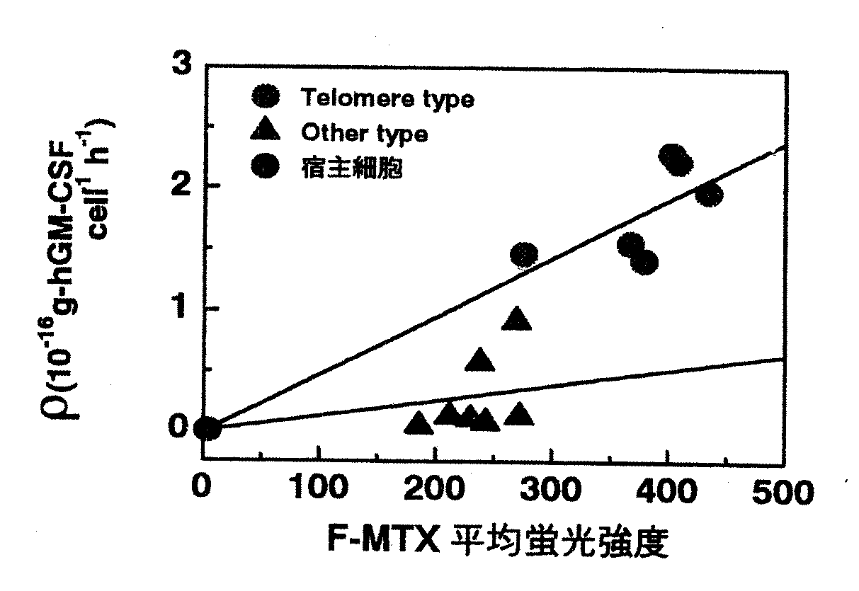
また、DHFR高生産細胞株が目的蛋白質高生産株であることを確認するために、F−MTX平均蛍光強度と比生産遠度の関係を解析した。その結果を図5に示し、両タイプクローン細胞における平均蛍光強度および非生産速度の平均値を表3に示す。
【0028】
【表3】
【0029】
以上の結果から、比生産速度とF−MTX蛍光強度の間にも相関関係があることが明らかとなった。そして、遺伝子が増幅されたテロメアタイプの細胞は、明らかにその他の耐性細胞と比較して高い蛍光強度を有しているため、これらの細胞をフローサイトメトリによって選別することが可能となる。
例えば、高い蛍光強度を持つ細胞を選別するには、短時間で膨大な数の細胞を選別可能な、図6に示すようなフローサイトメトリ(セルソーター)を用いて選別することができる。また、マイクロマニピュレーターを装備した蛍光顕微鏡を用いて高い蛍光強度を持つ細胞を採取しても良いし、光ピンセット機能を持つ共焦点レーザー顕微鏡を用いて選別することもできる。
【0030】
以上のように本発明の方法によれば、薬剤選択の最初に加えるMTX濃度を低く押さえ、遺伝子増幅した細胞のみを選択的に効率よく出現させ、様々な細胞が存在する細胞群から、遺伝子増幅によって高頻度に染色体中のコピー数を増加した細胞を、迅速かつ膨大な数の細胞について選択を行えるフローサイトメトリなどをを用いることによって、高い比生産速度を有する遺伝子増幅細胞を従来のクローニングによる選択法に比べ迅速かつ簡便に取得することが可能となった。
【0031】
即ち、本発明は、目的の外来遺伝子と標識化が可能な遺伝子との増幅において、標識化が可能な遺伝子を増幅する条件下において、両遺伝子の増幅を一定の相関を持って増幅することができるという新規な技術的思想を提供するものであり、係る本発明の技術的思想を具現化する手段は、使用する遺伝子や宿主細胞などの特性に応じて当業者が適宜実験的に設定することができ、本発明はこれらの具体的な手段にかかわらず本発明の前記した技術的思想に基づくものは全て包含している。そして、前記した本発明の技術的思想に基づいて遺伝子が増幅された細胞は、目的の外来遺伝子と標識化が可能な遺伝子とが高信頼性で相関を保っていることから、迅速かつ簡便な手段により遺伝子のコピー数に応じた細胞の選別をすることができるものであり、本発明は当該選別方法を包含するものでもある。次に本発明の実施の形態を例示するが、本発明はこれらの例示に限定されるものではない。
【0032】
本発明における薬剤選択の最初に加える薬剤の濃度としては、テロメアタイプの遺伝子導入細胞が選択的に出現する濃度であればよく、例えば薬剤がMTXの場合では50nM以下、好ましくは1〜50nM、より好ましくは10〜50nMである。
本発明における「標識化が可能な遺伝子」としては、薬剤耐性などにより増幅が可能な遺伝子であって、蛍光又は放射性同位元素などにより標識化でき得る遺伝子であればよい。DHFR遺伝子が本発明の好ましい例であるが、この遺伝子に限定されるものではない。
本発明の外来遺伝子としては、有用な蛋白質(糖蛋白質などの蛋白質誘導体を含む。)を製造することができる遺伝子であって前記した「標識化が可能な遺伝子」と共に増幅することができるものであればよい。好ましくは、hGM−CSF、EPO、t−PAなどの生理活性蛋白質をコードする遺伝子を例示することができる。
【0033】
本発明における「コピー数に対応した標識化」としては、蛍光標識や放射性同位元素などによる標識を例示することができるが、これらに限定されるものではなく、遺伝子の数を定量化できる手段であれば標識物質を使用しない機器分析手段のようなようなものであってもよい。
本発明の宿主細胞としては、遺伝子導入が可能であればよいが、真核細胞のみならず原核細胞であってもよいが、動物細胞のような真核細胞が好ましい。
宿主細胞を形質転換するためのベクターとしては、発現ベクターが好ましいがこれに限定されるものではない。
【0034】
また、本発明は前記した本発明の遺伝子増幅方法又は選別方法により、遺伝子が増幅された細胞及び選別された細胞を包含するものである。本発明の細胞は、目的の外来遺伝子が多数コピーされたものであり、当該コピー数としては好ましくは10コピー以上、より好ましくは50コピー以上であり、100コピー以上であってもよい。また、本発明の細胞の特徴は細胞群中の細胞の均質性が優れている点が挙げられ、コピー数の高い細胞、より好ましくはコピー数の均一な細胞が高確率で細胞群中に存在していることである。
さらに、本発明は前記した本発明の細胞を使用して効率よく目的の蛋白質(糖蛋白質などの蛋白質誘導体を含む。)を製造する方法を提供するものである。前記したように本発明の細胞はコピー数の均一な細胞を主としているために、品質のむらが少ない蛋白質を効率的に製造することができる。
【0035】
【実施例】
次に実施例により本発明をさらに具体的に説明するが、本発明はこれらの実施例に限定されるものではない。
【0036】
実施例1(形質転換体の製造)
SV40アリープロモーター及びマウスのdhfr遺伝子を有するプラスミドpSV2−dhfr(ATCC No.37146)と、SV40アリープロモーター及びhGM−CSF遺伝子を有するプラスミドpCD−hGM−CSF(ATCC No.57594)とから、図7に示すプラスミドを構築した。
構築されたプラスミドをCHO DG44株に導入し、形質転換された細胞をイスコフ培地(核酸+)で1〜2日間培養した後、イスコフ培地(核酸−)に1×10⁵細胞/mlの濃度で接種して、培地交換を4日ごとに行い、コロニーが形成されるまで培養を続けた。
【0037】
実施例2(形質転換体のMTX存在下での培養)
実施例1で選択した細胞の遺伝子の増幅を所定のMTXを含むイスコフ培地(核酸−)に、細胞を1×10⁵(cells/ml)の濃度で接種することによって行った。細胞がコンフルエントになった時、耐性を獲得したものとみなし、MTX濃度をさらに上昇させた。MTX濃度の上限を1000nMとし、緩やかに上昇させる系から急激に上昇させる系まで、図1に示すように5系列の上昇条件を設定した。
また、この実験においては、これらのMTX濃度上昇条件によって生じるいろいろな性質を有する細胞を集団として選択するために、濃度上昇各ステッフにおけるクローニング操作は一切行わず、細胞集団がどの様な挙動を示すかを解析した。
【0038】
実施例3(細胞のクローニング)
96穴マイクロプレートそれぞれに細胞を1個ずつとなるよう接種した(全量200μL)。1個の細胞からのコロニーであることを確認し、古い培地をベンチメイトで吸引して捨てた後、0.25%トリブシン溶液100μLを加え、37℃で10分間インキュベートした。
細胞がはがれて丸くなったら、新しい培地100μLを加えてよくビベッティングして、24穴マイクロプレートに継代した(全量2mL)。コンフルエント後、古い培地をベンチメイトで吸引して捨て、0.25%トリプシン溶液1mLを加え、37℃で10分間インキュベートした。さらに、細胞がはがれて丸くなったら、新しい培地1mLを加えてよくピベッティングして、継代(全量10mL)てクローンを得た。
【0039】
実施例4(hGM−CSF濃度の測定(ELISA法))
培養上清中のhGM−CsF濃度をELISA法により測定した。
ヒツジ抗hGM−CSF抗体(Endogen P−521)1mg/PBSからなる固層抗体50μLを0.05Mリン酸緩衝液10mLに希釈し、96穴プレート(greinerイミュロン600)に各ウェル100μLずつ分注し、4℃で一夜インキュベートした。ELISA洗浄液で3回洗浄した後、ブロッキング溶液(プレート1枚あたり約70mL)を各ウェルにいっぱいになる様に分注し、37℃で2時間インキュベートした。
ELISA洗浄液で3回洗浄した後、酵母由来hGM−CSF(Genzyme RH-CSF-C)1μg/PBS標準サンプルと適当な濃度に希釈した培養上澄みサンプルを、各ウェルに100μLずつ分注し、37℃で2時間インキュベートした。
ELISA洗浄液で3回洗浄した後、ウサギ抗hGM−CSF抗体(Genzume LP-174)1mg/PBS1mLからなる一次抗体20μLをPBS10mLに希釈したものを各ウェルに100μLずつ分注し、37℃で2時間インキュベートした。ELISA洗浄液で3回洗浄した後、アルカリホスファターゼ標識抗ウサギIgG(BIOSOURCE社 65OO)からなる二次抗体2μLを希釈液10mLで希釈し、各ウェルに100μLずつ分注し37℃で2時間インキュベートした。
ELISA洗浄液で5回洗浄した後、基質(p−ニトロフェニル ホスフェート(Sigma N-9389)4錠を酵素反応基質溶液20mLに希釈し、各ウェルに5秒おきに200μLずつ分注し、37℃で1時間インキュベートした。
反応停止液を各ウェルに5秒おきに100μLずつ分注し、マイクロプレートリーダー(東ソー MPR−A4)でA405の測定を行った。
【0040】
実施例5(遺伝子増幅領域の解析)
目的の遺伝子が染色体上のどの領域に存在するかを、蛍光 in situハイブリダイゼーシヨン(FISH)法によって解析した。
T−フラスコ中の細胞が80〜90%程度のコンフルエントの状態になった時、古い培地をピペットで吸引して捨て、新しい培地30mLを加えた。次いで、コルセミド溶液60μLを加え、4〜5時間インキュベートした。
シェイクオフ(Shake-off)法により、M期の細胞を浮遊させ、95×gで10分間遠心分離した後、上清を捨て、75mMKCl 1.5mLに懸濁して、室温で20分間細胞を膨張させた。
これに固定液を加えて、遠心分離し上清を捨てる操作を繰り返し、核膜を破壊した後、位相差顕微鏡で観察して、核のまわりに残さが残っていないことを確認した。
得られた標本にプローブ反応液8μL(スライド1枚当たり)とサケ精子DNA溶液8μLを混合し、エタノール沈殿させた。これに、精製したブローフにホルムアミド6.5μL(スライド1枚当たり)を加えて、さらに、20%デキストラン硫酸溶液6.5μL(スライド1枚当たり)を加えた後、80℃で変性し、エタノールを加えてインキュベートした。これにTNBブロッキング溶液、FTストック溶液及びDAPI溶液を加えてスライドガラスにのせて、蛍光を蛍光顕微鏡で観察した。
【0041】
実施例6(コピー数の定量)
細胞内のDNAは、RapidPrepTM Micro Genomic DNA Isolation Kit(Pharmacia Biotech 27-5225-01)を用いて抽出した。プローブに用いるためのDNAは、細胞に導入したブラスミドであるpSV2−dhfr/hGM−CSF中のdhfr遺伝子のみをPrep-A-Gene DNA purification Kit(Bio-rad 732-6009)を用いて回収したものを使用した。コピー数の定量は、DIG-DNA Labeling and Detection Kit(Boehringer Mannheim 1093657)を用いて、ドットブロッティング(Dot Blotting)法によって行った。
【0042】
実施例7(DHFR活性の測定)
1×10⁷個の細胞を500μLのPBSに懸濁し、超音波により細胞を破砕した後、遠心分離し、上清を粗酵素液とした。これに、ジヒドロ葉酸100μL、MTXストック溶液、NADPH溶液10μLなどを加えて、分光光度計により、340nmの吸光度を測定した。
【0043】
実施例8(DHFR量の測定と細胞の選別)
MTXを含まない培地(核酸−)で細胞を培養して、DHFRに結合しているMTXを完全に流出させる。5×10⁵個の細胞を取り、これにFITC標識したMTX(F−MTX)溶液200μL、イスコフ培地(核酸+)2.5mLを加えてインキュベートした。次いでイスコフ培地(核酸−)を用いて洗浄、インキュベートして余分のF−MTXを流出させた。トリプシン処理をした後、遠心分離して上清を捨て、PBS1mLに懸濁した。
これをフローサイトメーター(COULTER;EPICSELITE)を用いて励起波長488nmでDHFR量を測定した。同様な方法で、蛍光強度に応じて細胞を分離した。
【0044】
【発明の効果】
本発明は、目的の外来遺伝子のコピー数の多い宿主細胞を簡便でかつ高信頼性で選別氏得る方法を提供するものである。本発明の方法によれば、目的の外来遺伝子のコピー数が均一な宿主細胞を選択的に選別することができるので、遺伝子組換え手段による生理活性蛋白質などの蛋白質の製造において、その生産性を質的又は量的に改善することができる。
また、本発明の方法は薬剤耐性などによる外来遺伝子の増幅される染色体上の領域を明らかにするものであり、外来遺伝子の増幅のされかたの指標を提供するものであり、外来遺伝子の増幅方法に広く応用できるものである。
【図面の簡単な説明】
【図1】図1は、MTX濃度の上昇試験における濃度の上昇条件を示したものである。
【図2】図2は、MTX濃度の上昇条件と比生産速度の関係を示したものである。図中の黒丸(●)は上昇試験のパターン1の場合を示し、黒上三角(▲)は上昇試験のパターン2の場合を示し、黒四角(■)は上昇試験のパターン3の場合を示し、黒下三角(▼)は上昇試験のパターン4の場合を示し、黒菱形(◆)は上昇試験のパターン5の場合を示す。
【図3】図3は、MTX濃度の各上昇条件における遺伝子増幅領域の分布を示す。図中の黒色部分はテロメアタイプを示し、灰色部分は他のタイプを示し、白色部分はシグナル無しタイプを示す。
【図4】図4は、F−MTX平均蛍光強度とDHFR活性の関係を示す。図中の黒丸(●)はテロメアタイプを示し、黒三角(▲)は他のタイプを示し、左端の黒丸は元の宿主細胞を示す。
【図5】図5は、F−MTX平均蛍光強度と比生産速度(ρ)の関係を示す。図中の黒丸(●)はテロメアタイプを示し、黒三角(▲)は他のタイプを示し、左端の黒丸は元の宿主細胞を示す。
【図6】図6は、フローサイトメーターの模式図を示す。
【図7】図7は、形質転換に用いたプラスミドを示す。[0001]
BACKGROUND OF THE INVENTION
In the present invention, when a useful foreign protein is industrially produced by a host cell into which a foreign gene has been introduced by genetic recombination, a cell line with high production efficiency is efficiently produced and selected efficiently. Regarding the method. Furthermore, the present invention relates to a host cell obtained by the method and a method for producing a protein using the host cell.
[0002]
[Prior art]
When producing a protein to which a sugar chain is added using genetic recombination, it is necessary to use a eukaryotic cell having the ability to add a sugar chain as a host. In many glycoproteins in which sugar chains are important for their physiological functions, not only the presence or absence of sugar chains but also the quality of sugar chains is important. However, the quality of sugar chains added to proteins varies depending on the host. For this reason, for example, when producing a glycoprotein derived from a human, it is necessary to use a higher animal cell to which a sugar chain of a quality close to human can be added as a host. Unlike lower eukaryotes such as yeast, higher animal cells have the problem that their cell growth is slow and the medium is expensive. In order to compensate for this, when higher animal cells are used as the host, it is necessary to improve the protein productivity per cell, that is, the specific production rate.
[0003]
In order to improve the specific production rate, a method of connecting the gene to a strong promoter has been proposed, but not only that but a method called gene amplification that increases the copy number of the gene is used. Gene amplification is a technique that utilizes this phenomenon, as one of the mechanisms by which cells acquire resistance to the addition of drugs such as inhibitors, the number of copies of the gene responsible for that resistance may increase. When gene amplification occurs in animal cells, not only the gene itself subjected to selective pressure with the drug, but also other genes present in the vicinity of the gene are amplified simultaneously, so that the foreign gene is also amplified simultaneously. For example, a vector having the gene and a dihydrofolate reductase (hereinafter referred to as DHFR) gene is introduced into a host animal cell and then cultured in the presence of methotrexate (hereinafter referred to as MTX) which is a competitive inhibitor of DHFR. Then, it is known that a cell line with an increased copy number of the gene can be obtained together with the DHFR gene.
[0004]
However, since this gene amplification does not occur in all cells at the same time, but in some cells at various timings, cells selected with a medium containing a certain concentration of MTX are cells with various copy number genes. Become a heterogeneous group. Therefore, in order to maximize the productivity of the target protein, from this cell population, the cell with the highest specific production rate of the target protein (that is, the cell with the highest gene copy number) is cloned and established. It is hoped to do. Furthermore, strict quality control is required when industrially producing recombinant proteins, particularly when they are produced for use in pharmaceuticals. However, production using the above heterogeneous cell population results in variations in quality. This cloning work has been desired because quality control is not easy.
However, an index for efficiently cloning a cell having the highest specific production rate (ie, a cell having the highest gene copy number) has not been established, and the target cell can be obtained from a heterogeneous cell population from the transformant. It was difficult to clone.
[0005]
By the way, if the transformed host cell is suddenly exposed to a high concentration of drug, all cells will die before gene amplification occurs. For this reason, the concentration of the drug for selecting gene-amplified cells must be increased stepwise from a low concentration. However, if the concentration is gradually increased from a low concentration, it takes time to select cells. Conversely, in order to select cells in a relatively short time, if the concentration of the drug is increased rapidly, even if it is not so extreme that all cells will die, resistance will be acquired by a mechanism other than gene amplification. The frequency of appearance of cells increases. In this case, drug resistance can be acquired, but since the gene is not amplified, the specific protein production rate of the cell remains low but lower. For example, the specific production rate of a target protein of a strain in which a mutation that reduces the affinity of DHFR for MTX or a mutation that makes it difficult to incorporate MTX is lower than that of a strain that has acquired the same MTX resistance level by gene amplification.
[0006]
Since the cells obtained by such drug selection are heterogeneous cell populations as described above, even if drug resistance is acquired uniformly, drug resistance is acquired only by gene amplification. Therefore, in order to select a high-productivity strain of the target protein, it was necessary to obtain clones and evaluate the specific production rate for each. However, the evaluation of the specific production rate of the protein is a manual operation, and it takes 1 to 2 months to obtain clones.
From the above, a technique for efficiently and quickly selecting cells in which the gene is amplified is desired.
[0007]
[Problems to be solved by the invention]
As described above, to efficiently clone a cell having the highest specific production rate of the target protein (ie, a cell having the highest gene copy number) from cells transformed with a foreign gene by genetic recombination. Thus, the present invention provides an index for selecting cells having the highest specific production rate of the target protein.
In addition, the present invention provides a method for increasing MTX concentration in which cells that acquire resistance by gene amplification are obtained at high frequency and in a short time, and cells having the highest production rate of the target protein from the cell group obtained thereby can be rapidly obtained. The present invention provides a method for sorting and cells selected by the method.
[0008]
By the way, in order to establish a method for efficiently cloning a cell having the highest specific production rate of a target protein (ie, a cell having the highest gene copy number) from cells transformed with a foreign gene. Therefore, it is considered important to establish an index corresponding to the copy number of the foreign gene. Since the foreign gene is amplified together with the gene that is amplified along with drug resistance, if the correspondence with the gene that is amplified along with drug resistance can be secured with high reliability, the gene is amplified along with drug resistance. The copy number of the gene to be used can be used as an index of the copy number of the foreign gene. Furthermore, if it is possible to label the copy number of a gene that is amplified along with drug resistance, this can be designated as “a gene that can be labeled”.
However, as described above, it has been a big problem to ensure the correspondence between the foreign gene and the gene amplified with drug resistance with high reliability.
[0009]
[Means for Solving the Problems]
As a result of intensive research on a method that can reliably ensure the correspondence between a foreign gene and a gene that is amplified along with drug resistance, the present inventors have made the first selection in drug selection for transformed cells. By setting a low drug concentration, it is possible to suppress the frequency of appearance of cells that acquire resistance by a mechanism other than gene amplification, and for the heterogeneous cell population thus selected, fluorescently labeled drugs are It was found that cells with the gene amplified can be rapidly selected by sorting cells with high fluorescence intensity emitted by the flow cytometer using a flow cytometer.
[0010]
In the present invention, in a host cell into which a foreign gene has been introduced by genetic recombination, a cell in which the introduced foreign gene has been amplified to 2 or more copies is labeled according to the number of copies of the foreign gene. The present invention relates to a method for selecting a cell having a high copy number of a foreign gene using the intensity of the label emitted from the cell as an index.
The present invention also relates to a gene obtained by inducing amplification of a foreign gene in a host cell transformed with a vector containing a foreign gene and a dihydrofolate reductase (DHFR) gene to amplify the foreign gene into two or more copies. And a method for amplifying a foreign gene, wherein the concentration of methotrexate (MTX) in the medium is low when culturing in a medium containing methotrexate (MTX) for the first time after the transformation. .
[0011]
Furthermore, the present invention provides a method for labeling a host cell amplified with a foreign gene by the above-described method for amplifying a foreign gene in accordance with the copy number of the foreign gene and indicating the intensity of the label emitted by the labeled cell. The present invention relates to a method for selecting cells having a high copy number foreign gene.
The present invention also relates to a host cell having a high copy number foreign gene obtained by these methods.
Since these host cells have a high copy number foreign gene, the present invention relates to a method for efficiently producing a protein or glycoprotein encoded by the foreign gene by culturing these host cells.
Furthermore, the present invention relates to a method for amplifying a foreign gene introduced in a host cell into which a foreign gene has been introduced by genetic recombination, wherein the foreign gene is amplified and the host cell has a high specific production rate of the target protein. The present invention relates to a method for amplifying a foreign gene using as an index that amplification is performed in the vicinity of chromosome telomere.
[0012]
The present inventors have studied the problem of whether or not the correspondence relationship between a foreign gene and a gene amplified along with drug resistance can be ensured with high reliability. It has been found that culturing at gradually higher concentrations can solve this problem with high frequency and high reliability. It was also found that host cells with a high specific production rate of the target protein are amplified near the telomeres of the chromosome.
[0013]
The method of the present invention will be described more specifically by taking, for example, dihydrofolate reductase (DHFR) as an example relating to drug resistance.
DHFR is essential for nucleic acid biosynthesis, and cells lacking the DHFR gene cannot grow in a medium that does not contain nucleic acid. When a DHFR gene is introduced into a cell lacking the DHFR gene by a gene recombination technique, a transformant whose nucleic acid requirement has been recovered by transformation can be easily selected.
However, since a drug called methotrexate (MTX) inhibits the action of DHFR in vivo, even a transformant introduced with a DHFR gene cannot survive unless MTX resistance is acquired in the presence of MTX.
[0014]
More specifically, a foreign gene of a protein to be expressed is inserted into an appropriate expression vector having a DHFR gene, and this vector is electroporated into a host cell such as a Chinese hamster ovary cell (hereinafter referred to as CHO cell). Then, it is introduced and transformed using an appropriate method such as the calcium phosphate method. The host cell to be used is preferably one that is deficient in the DHFR gene, such as CHO DG44 strain. However, even when a DHFR-deficient strain cannot be used, a transformant can be selected using drug resistance as an index by further inserting a resistance gene for a drug such as G-418 into the vector to be used. it can.
[0015]
For example, a human granulocyte macrophage colony stimulating factor (hGM-CSF) gene as a foreign gene is inserted into an expression vector having a DHFR gene and introduced into a Chinese hamster ovary (CHO) cell strain DG44 lacking the DHFR gene. The transformant was obtained by culturing in a medium (containing nucleic acid) supplemented with 10% fetal bovine serum. Subsequently, the transformed cells were selected only in the IMDM medium (without nucleic acid) and the medium supplemented with 10% dialyzed fetal calf serum.
As shown in FIG. 1, the resulting transformant was first prepared with IMDM medium containing 50 nM and 100 nM MTX and no nucleic acid. 5 The cells / ml was cultured until the cell / ml increased from 10 to 100 times. Thereafter, as shown in FIG. 1, a test for increasing the concentration of MTX was carried out at a final concentration of 1000 nM in 5 series of 1 to 5 consisting of a series in which the MTX concentration was gently increased to a series in which it was rapidly increased.
[0016]
Specific growth rate μ (h) in each series (pattern) -1 ) Patent ratio production rate ρ (10 -16 g-hGM-CSF / cell / h -1 The results of measurement of) are shown in FIG. In FIG. 2, black circles (●) indicate the case of series 1 (pattern 1), black upper triangles (▲) indicate the case of series 2 (pattern 2), and black squares (■) indicate series 3 (pattern). 3), the black triangle (▼) indicates the case of the series 4 (pattern 4), and the black rhombus mark (♦) indicates the case of the series 5 (pattern 5).
[0017]
Furthermore, the present inventors analyzed the gene amplification region distribution in each step of each series. The result is shown in FIG. The black part in Fig. 3 shows that the amplified gene is located near the telomere (hereinafter referred to as the telomere type), and the gray part shows that the amplified gene is located in a region other than the vicinity of the telomere (Hereinafter referred to as other types), and the white portion indicates that the amplified gene cannot be found in the gene (hereinafter referred to as no signal type).
As a result, in the group (
[0018]
In the group (
[0019]
A cell without a signal type is considered to be a cell that has not undergone gene amplification or has undergone gene amplification only to an extent that it cannot be detected. The differences between the telomere type cells and other types of cells were examined.
Using 16 telomere type clonal cells and 11 other types of clonal cells obtained by the MTX concentration increase test in
[0020]
[Table 1]
Table 2 shows the results of analyzing the copy number of the DHFR gene and the hGM-CSF gene by taking out telomere type and other types of cloned cells one by one.
[0021]
[Table 2]
In any line (pattern) culture, no signal-type cells will eventually disappear. However, if the MTX concentration is kept low in the initial stage of gene amplification, many telomere-type cells can be obtained. On the other hand, it was found that when the MTX concentration was increased, many other types of cells were contaminated.
[0022]
From these results, it was found that in gene amplification, the position of the chromosome where the gene is first integrated is more important than the method of increasing the MTX concentration during the culture stage. Then, it was found that the position of the chromosome into which the gene is first integrated depends on the concentration of the drug in the initial culture.
[0023]
That is, if the transformed cells are cultured in a medium containing MTX, the DHFR gene and the gene of the target protein are amplified. If the concentration of MTX added for the first time is higher than 50 nM, a mechanism other than DHFR gene amplification is used. The percentage of cells that acquire MTX resistance increases, and even cells that have acquired MTX resistance by amplification of the DHFR gene have low genetic stability, that is, in a non-selective medium (without nucleic acid). When cultured, the proportion of cell lines in which the amplified gene is easily lost increases. This is because the copy number of the DHFR gene does not increase all at once during gene amplification, but gradually increases as the cell cycle is repeated. Therefore, when the concentration of MTX added for the first time is high, resistance other than gene amplification is acquired. A strain that has acquired resistance to MTX due to a mechanism such as a phenomenon in which the DHFR gene is mutated to reduce the affinity of the DHFR protein to MTX or a mutation that makes it difficult to incorporate MTX into the cell occurs. This is presumably because it grows preferentially compared to gene-amplified cells that require a long time for acquisition. However, since the number of copies in the chromosome is small, the productivity of the recombinant protein is low for high resistance (see Tables 1 and 2).
[0024]
In order to allow cells that acquire MTX resistance to grow by gene amplification, it is important to set the concentration of MTX added for the first time at a low level, which increases the DHFR gene once to a certain copy number. For example, since resistance can be acquired by gene amplification even if the MTX concentration is increased by about 10 times at a time, the frequency of preferential growth of strains that acquire MTX resistance by a mechanism other than gene amplification becomes extremely low.
[0025]
The gene amplified cells obtained by the above method include strains having a copy number of DHFR gene that gives marginal resistance to MTX finally added to the medium, and higher copy number of DHFR gene that gives marginal tolerance. In addition, a strain that has acquired MTX resistance by a mechanism other than gene amplification is included. Conventionally, clones were isolated from such a cell group, and the specific production rate of the target protein was evaluated for each. However, it has also been clarified that a strain with a high specific production rate of the target protein has a high copy number of the target protein gene, and such cells also have a high copy number of the DHFR gene, that is, a large amount of DHFR in the cell. . Therefore, intracellular DHFR is fluorescently labeled with a fluorescent substance that specifically binds to DHFR, such as fluorescein-labeled MTX (F-MTX), and cells with high fluorescence intensity are selected using the fluorescence intensity emitted by individual cells as an indicator. Then, a strain having a high specific production rate of the target protein can be obtained.
[0026]
Therefore, the correlation between the DHFR activity and the productivity of the recombinant protein and the fluorescence intensity of the fluorescently labeled MTX was examined using the gene-amplified cells and other cloned cells.
The relationship between DHFR activity and F-MTX average fluorescence intensity was analyzed for 6 telomere type clone cells and 7 other type clone cells obtained from
As can be seen from FIG. 4, the F-MTX average fluorescence intensity of the clonal cells (●) in which the telomere type gene was amplified was higher than that of other types of clonal cells (▲), and the fluorescence intensity and DHFR activity It was confirmed that a linear relationship was obtained between
[0027]
In addition, in order to confirm that the DHFR high-producing cell line is the target protein high-producing line, the relationship between the F-MTX average fluorescence intensity and the specific production distance was analyzed. The results are shown in FIG. 5, and the average values of the average fluorescence intensity and the non-production rate in both type clonal cells are shown in Table 3.
[0028]
[Table 3]
[0029]
From the above results, it became clear that there is also a correlation between the specific production rate and the F-MTX fluorescence intensity. And since the telomere type cell in which the gene was amplified clearly has higher fluorescence intensity than other resistant cells, it becomes possible to sort these cells by flow cytometry.
For example, in order to select cells having high fluorescence intensity, it is possible to select using a flow cytometry (cell sorter) as shown in FIG. 6, which can select an enormous number of cells in a short time. In addition, cells having high fluorescence intensity may be collected using a fluorescence microscope equipped with a micromanipulator, or may be selected using a confocal laser microscope having an optical tweezer function.
[0030]
As described above, according to the method of the present invention, the MTX concentration added at the beginning of drug selection is kept low, and only gene-amplified cells appear selectively and efficiently, and gene amplification is performed from a group of cells containing various cells. By means of flow cytometry, etc., which can quickly select a large number of cells that have increased the number of copies in the chromosome by high frequency, gene-amplified cells with a high specific production rate can be obtained by conventional cloning. Compared to the selection method, it was possible to obtain it quickly and easily.
[0031]
That is, the present invention can amplify the amplification of both genes with a certain correlation in the amplification of the target foreign gene and the gene capable of labeling under the condition of amplifying the gene capable of labeling. A means for realizing the technical idea of the present invention can be appropriately set experimentally by those skilled in the art according to the characteristics of the gene and host cell used. However, the present invention includes all those based on the above technical idea of the present invention regardless of these specific means. A cell in which a gene is amplified based on the technical idea of the present invention described above has a high-reliability correlation between a target foreign gene and a gene that can be labeled. The cells can be selected according to the copy number of the gene by means, and the present invention includes the selection method. Next, although embodiment of this invention is illustrated, this invention is not limited to these illustrations.
[0032]
The concentration of the drug to be added at the beginning of drug selection in the present invention may be a concentration at which telomere type transgenic cells selectively appear. For example, when the drug is MTX, it is 50 nM or less, preferably 1 to 50 nM. Preferably it is 10-50 nM.
The “gene that can be labeled” in the present invention is a gene that can be amplified by drug resistance or the like, and may be any gene that can be labeled by fluorescence or a radioisotope. The DHFR gene is a preferred example of the present invention, but is not limited to this gene.
The foreign gene of the present invention is a gene that can produce useful proteins (including protein derivatives such as glycoproteins) and can be amplified together with the above-mentioned “genes that can be labeled”. I just need it. Preferably, a gene encoding a physiologically active protein such as hGM-CSF, EPO, t-PA can be exemplified.
[0033]
Examples of “labeling corresponding to the copy number” in the present invention include fluorescent labeling and labeling with a radioisotope, but are not limited to these, and means that can quantify the number of genes. If it exists, it may be a device analysis means that does not use a labeling substance.
The host cell of the present invention is not limited as long as gene transfer is possible, but may be not only a eukaryotic cell but also a prokaryotic cell, but a eukaryotic cell such as an animal cell is preferred.
An expression vector is preferable as a vector for transforming a host cell, but is not limited thereto.
[0034]
In addition, the present invention encompasses cells and cells that have been amplified by the gene amplification method or selection method of the present invention described above. The cell of the present invention has a large number of copies of the target foreign gene, and the copy number is preferably 10 copies or more, more preferably 50 copies or more, and may be 100 copies or more. In addition, the characteristics of the cells of the present invention are that the homogeneity of the cells in the cell group is excellent, and cells with a high copy number, more preferably cells with a uniform copy number are present in the cell group with a high probability. Is.
Furthermore, the present invention provides a method for efficiently producing a target protein (including protein derivatives such as glycoproteins) using the above-described cells of the present invention. As described above, since the cells of the present invention are mainly cells having a uniform copy number, it is possible to efficiently produce a protein with less uneven quality.
[0035]
【Example】
EXAMPLES Next, although an Example demonstrates this invention further more concretely, this invention is not limited to these Examples.
[0036]
Example 1 (Production of transformant)
From the plasmid pSV2-dhfr (ATCC No. 37146) having the SV40 all promoter and the mouse dhfr gene and the plasmid pCD-hGM-CSF (ATCC No. 57594) having the SV40 all promoter and the hGM-CSF gene, FIG. The indicated plasmid was constructed.
The constructed plasmid is introduced into the CHO DG44 strain, and the transformed cells are cultured in Iskov medium (nucleic acid +) for 1 to 2 days. 5 The cells were inoculated at a concentration of cells / ml, the medium was changed every 4 days, and the culture was continued until colonies were formed.
[0037]
Example 2 (culture of transformant in the presence of MTX)
Amplification of the genes of the cells selected in Example 1 was carried out in Iscov's medium (nucleic acid) containing a predetermined MTX, and the cells were placed at 1 × 10. 5 Inoculated at a concentration of (cells / ml). When the cells became confluent, it was considered that resistance was acquired, and the MTX concentration was further increased. The upper limit of the MTX concentration was set to 1000 nM, and five series of rising conditions were set from a slowly increasing system to a rapidly increasing system as shown in FIG.
Moreover, in this experiment, in order to select cells having various properties generated by these MTX concentration increasing conditions as a population, no cloning operation is performed at each step of increasing concentration, and the behavior of the cell population is shown. It was analyzed.
[0038]
Example 3 (Cloning of cells)
Each 96-well microplate was inoculated with one cell (
When the cells were peeled off and rounded, 100 μL of fresh medium was added and well-beveled, and passaged to a 24-well microplate (
[0039]
Example 4 (Measurement of hGM-CSF concentration (ELISA method))
The hGM-CsF concentration in the culture supernatant was measured by ELISA.
50 μL of a solid layer antibody consisting of 1 mg / PBS of sheep anti-hGM-CSF antibody (Endogen P-521) is diluted in 10 mL of 0.05 M phosphate buffer, and 100 μL of each well is dispensed into a 96-well plate (greiner Imuron 600). Incubate overnight at 4 ° C. After washing 3 times with ELISA wash, blocking solution (about 70 mL per plate) was dispensed to fill each well and incubated at 37 ° C. for 2 hours.
After washing three times with an ELISA washing solution, 1 μg of yeast-derived hGM-CSF (Genzyme RH-CSF-C) / PBS standard sample and a culture supernatant sample diluted to an appropriate concentration were dispensed in 100 μL to each well at 37 ° C. And incubated for 2 hours.
After washing 3 times with an ELISA washing solution, 100 μL of a primary antibody consisting of 1 mg of rabbit anti-hGM-CSF antibody (Genzume LP-174) / 1 mL of PBS diluted in 10 mL of PBS was dispensed into each well at 100 μL for 2 hours at 37 ° C. Incubated. After washing 3 times with ELISA washing solution, 2 μL of secondary antibody consisting of alkaline phosphatase-labeled anti-rabbit IgG (BIOSOURCE 65OO) was diluted with 10 mL of diluent, dispensed 100 μL into each well, and incubated at 37 ° C. for 2 hours.
After washing 5 times with the ELISA washing solution, 4 tablets of substrate (p-nitrophenyl phosphate (Sigma N-9389)) were diluted in 20 mL of the enzyme reaction substrate solution, and 200 μL was dispensed into each well every 5 seconds, at 37 ° C. Incubated for 1 hour.
100 μL of the reaction stop solution was dispensed into each well every 5 seconds, and A405 was measured with a microplate reader (Tosoh MPR-A4).
[0040]
Example 5 (Analysis of gene amplification region)
In which region on the chromosome the target gene is present was analyzed by the fluorescence in situ hybridization (FISH) method.
When the cells in the T-flask became confluent about 80-90%, the old medium was aspirated and discarded, and 30 mL of new medium was added. Then 60 μL of colcemid solution was added and incubated for 4-5 hours.
M-phase cells are suspended by the shake-off method, centrifuged at 95 × g for 10 minutes, the supernatant is discarded, suspended in 1.5 mL of 75 mM KCl, and the cells are expanded for 20 minutes at room temperature. I let you.
An operation of adding a fixative, centrifuging and discarding the supernatant was repeated, and after rupturing the nuclear membrane, it was observed with a phase-contrast microscope to confirm that no residue remained around the nucleus.
The obtained specimen was mixed with 8 μL of the probe reaction solution (per slide) and 8 μL of salmon sperm DNA solution, and ethanol precipitated. To this, 6.5 μL of formamide (per slide) was added to the purified brouff, and 6.5 μL of 20% dextran sulfate solution (per slide) was added, followed by denaturation at 80 ° C. to remove ethanol. In addition, it was incubated. The TNB blocking solution, FT stock solution and DAPI solution were added to this and placed on a slide glass, and the fluorescence was observed with a fluorescence microscope.
[0041]
Example 6 (Quantification of copy number)
The intracellular DNA is RapidPrep TM Extraction was performed using Micro Genomic DNA Isolation Kit (Pharmacia Biotech 27-5225-01). The DNA used for the probe was obtained by using the Prep-A-Gene DNA purification Kit (Bio-rad 732-6009) only for the dhfr gene in pSV2-dhfr / hGM-CSF, which is a plasmid introduced into cells. It was used. Copy number quantification was performed by the dot blotting method using DIG-DNA Labeling and Detection Kit (Boehringer Mannheim 1093657).
[0042]
Example 7 (Measurement of DHFR activity)
1 × 10 7 The cells were suspended in 500 μL of PBS, and the cells were disrupted by ultrasonic waves, and then centrifuged, and the supernatant was used as a crude enzyme solution. To this was added 100 μL of dihydrofolic acid, MTX stock solution, 10 μL of NADPH solution, and the absorbance at 340 nm was measured with a spectrophotometer.
[0043]
Example 8 (Measurement of DHFR amount and selection of cells)
Cells are cultured in medium without MTX (nucleic acid) to completely drain MTX bound to DHFR. 5 × 10 5 Individual cells were taken and incubated with 200 μL of FITC-labeled MTX (F-MTX) solution and 2.5 mL of Iskov medium (nucleic acid +). Subsequently, it was washed and incubated with Iskov medium (nucleic acid-) to drain excess F-MTX. After trypsinization, the supernatant was discarded by centrifugation and suspended in 1 mL of PBS.
The amount of DHFR was measured at an excitation wavelength of 488 nm using a flow cytometer (COULTER; EPICSELITE). Cells were separated according to the fluorescence intensity in the same manner.
[0044]
【The invention's effect】
The present invention provides a method for easily and reliably selecting a host cell having a large number of copies of a target foreign gene. According to the method of the present invention, a host cell having a uniform copy number of a target foreign gene can be selectively selected. Therefore, in the production of a protein such as a physiologically active protein by genetic recombination means, its productivity is reduced. It can be improved qualitatively or quantitatively.
Further, the method of the present invention reveals a region on a chromosome where a foreign gene is amplified due to drug resistance, etc., and provides an indicator of how the foreign gene is amplified. It can be widely applied to methods.
[Brief description of the drawings]
FIG. 1 shows conditions for increasing concentration in a test for increasing MTX concentration.
FIG. 2 shows the relationship between the condition for increasing the MTX concentration and the specific production rate. In the figure, the black circle (●) shows the case of the ascending
FIG. 3 shows the distribution of gene amplification regions under each increase condition of MTX concentration. The black portion in the figure indicates the telomere type, the gray portion indicates the other type, and the white portion indicates the type without signal.
FIG. 4 shows the relationship between F-MTX average fluorescence intensity and DHFR activity. The black circle (●) in the figure indicates the telomere type, the black triangle (▲) indicates the other type, and the black circle at the left end indicates the original host cell.
FIG. 5 shows the relationship between F-MTX average fluorescence intensity and specific production rate (ρ). The black circle (●) in the figure indicates the telomere type, the black triangle (▲) indicates the other type, and the black circle at the left end indicates the original host cell.
FIG. 6 shows a schematic diagram of a flow cytometer.
FIG. 7 shows the plasmid used for transformation.
Claims (3)
(1)目的の蛋白質又は糖蛋白質をコードする外来遺伝子と共にジヒドロ葉酸還元酵素(DHFR)遺伝子を有するベクターを用いて動物宿主細胞を形質転換する工程、
(2)形質転換動物細胞をあらかじめメソトレキセート(MTX)を含まない培地で培養した後に、MTX濃度を50nMの低濃度から高濃度へ3段階又は4段階で上昇させ、それぞれの濃度条件下でMTX耐性を獲得させるまで培養することで、テロメアタイプの細胞の割合を高める工程、
(3)フルオレセイン標識したMTXを用いて蛍光標識化された細胞の蛍光強度が高い細胞を選別する工程。A method for obtaining a transformed animal cell having a high specific production rate of a target protein or glycoprotein, comprising the following steps (1) to (3).
(1) allowing the animal host cell is transformed with a vector having a protein or glycoprotein foreign gene co di folate reductase encoding (DHFR) gene of interest,
(2) After culturing transformed animal cells in a medium that does not contain methotrexate (MTX) in advance, the MTX concentration is increased from a low concentration of 50 nM to a high concentration in three or four steps , and MTX resistance under each concentration condition A step of increasing the proportion of telomere type cells by culturing until
(3) A step of selecting cells with high fluorescence intensity of cells fluorescently labeled using fluorescein-labeled MTX .
Priority Applications (1)
| Application Number | Priority Date | Filing Date | Title |
|---|---|---|---|
| JP21709599A JP4031154B2 (en) | 1999-07-30 | 1999-07-30 | Rapid selection of gene-amplified cells |
Applications Claiming Priority (1)
| Application Number | Priority Date | Filing Date | Title |
|---|---|---|---|
| JP21709599A JP4031154B2 (en) | 1999-07-30 | 1999-07-30 | Rapid selection of gene-amplified cells |
Publications (2)
| Publication Number | Publication Date |
|---|---|
| JP2001037478A JP2001037478A (en) | 2001-02-13 |
| JP4031154B2 true JP4031154B2 (en) | 2008-01-09 |
Family
ID=16698773
Family Applications (1)
| Application Number | Title | Priority Date | Filing Date |
|---|---|---|---|
| JP21709599A Expired - Fee Related JP4031154B2 (en) | 1999-07-30 | 1999-07-30 | Rapid selection of gene-amplified cells |
Country Status (1)
| Country | Link |
|---|---|
| JP (1) | JP4031154B2 (en) |
Cited By (1)
| Publication number | Priority date | Publication date | Assignee | Title |
|---|---|---|---|---|
| EP2151689B1 (en) * | 2008-08-08 | 2014-01-22 | Genetix Limited | Cell detection |
Families Citing this family (6)
| Publication number | Priority date | Publication date | Assignee | Title |
|---|---|---|---|---|
| JP2008199919A (en) * | 2007-02-16 | 2008-09-04 | Kyushu Univ | Method and apparatus for separating and collecting cell |
| WO2010023787A1 (en) * | 2008-08-29 | 2010-03-04 | 東洋紡績株式会社 | Element capable of stabilizing gene expression |
| KR101706399B1 (en) * | 2009-02-27 | 2017-02-13 | 노파르티스 아게 | Methods for selecting eukaryotic cells expressing a heterologous protein |
| JP4525863B1 (en) * | 2009-06-05 | 2010-08-18 | 東洋紡績株式会社 | Expression vector for establishment of high-productivity cells and high-productivity cells |
| RU2712507C2 (en) * | 2013-12-20 | 2020-01-29 | Новартис Аг | Novel eukaryotic cells and methods for preparing thereof for recombinant expression of product of interest |
| MX371025B (en) | 2013-12-20 | 2020-01-13 | Novartis Ag | Novel eukaryotic cells and methods for recombinantly expressing a product of interest. |
-
1999
- 1999-07-30 JP JP21709599A patent/JP4031154B2/en not_active Expired - Fee Related
Cited By (1)
| Publication number | Priority date | Publication date | Assignee | Title |
|---|---|---|---|---|
| EP2151689B1 (en) * | 2008-08-08 | 2014-01-22 | Genetix Limited | Cell detection |
Also Published As
| Publication number | Publication date |
|---|---|
| JP2001037478A (en) | 2001-02-13 |
Similar Documents
| Publication | Publication Date | Title |
|---|---|---|
| Lemmon et al. | Clathrin requirement for normal growth of yeast | |
| Sillar et al. | A new method for the preparation of metaphase chromosomes for flow analysis. | |
| JP3880778B2 (en) | Methods and materials for growing primate-derived primordial stem cells in cultures that do not contain feeder cells | |
| Nabeshima et al. | A novel meiosis‐specific protein of fission yeast, Meu13p, promotes homologous pairing independently of homologous recombination | |
| NZ228031A (en) | Primate gm-csf and method of extraction | |
| Sparling et al. | Ribosomes from Escherichia coli merodiploids heterozygous for resistance to streptomycin and to spectinomycin | |
| Yoshikawa et al. | Evaluation of stable and highly productive gene amplified CHO cell line based on the location of amplified genes | |
| JP4031154B2 (en) | Rapid selection of gene-amplified cells | |
| EA022993B1 (en) | Nucleic acid molecules coding for alpha- and beta-chains of human follicle stimulating hormone (fsh) | |
| Panton et al. | A test for genetic exchange in mixed infections of Leishmania major in the sand fly Phlebotomus papatasi | |
| EP2143791A1 (en) | Chicken embryonic stem cell and method for evaluation thereof | |
| WO1989005864A1 (en) | Transgenic testing systems for mutagens and carcinogens | |
| JP5282040B2 (en) | Method for assaying antibody-dependent cytotoxicity using Fc receptor gene-transferred NK cells | |
| CN109790539A (en) | The promoter of Hspa5 gene | |
| TWI626309B (en) | System for over-expressing target protein and method thereof | |
| Dee et al. | Growth, development and genetic characteristics of Physarum polycephalum amoebae able to grow in liquid, axenic medium | |
| Perucho et al. | Linkage and expression of foreign DNA in cultured animal cells | |
| US11796443B2 (en) | Method for isolating placental trophoblast cells from cervical exfoliated cells of pregnant woman | |
| EP0247145B1 (en) | Multiply-amplifiable vectors for high level expression of exogenous dna | |
| US20130266988A1 (en) | Method for gene amplification | |
| RU2502798C2 (en) | Cell line hufshik releasing recombinant human follicle-stimulating hormone | |
| CN101608227A (en) | Kit for genetic detection of thyroid cancer | |
| US5686263A (en) | Method for enhancing gene expression | |
| JPH074237B2 (en) | Immortalization of human or animal cells | |
| EA010059B1 (en) | Flp-mediated recombination |
Legal Events
| Date | Code | Title | Description |
|---|---|---|---|
| A131 | Notification of reasons for refusal |
Free format text: JAPANESE INTERMEDIATE CODE: A131 Effective date: 20061107 |
|
| A521 | Written amendment |
Free format text: JAPANESE INTERMEDIATE CODE: A523 Effective date: 20070105 |
|
| A131 | Notification of reasons for refusal |
Free format text: JAPANESE INTERMEDIATE CODE: A131 Effective date: 20070717 |
|
| A521 | Written amendment |
Free format text: JAPANESE INTERMEDIATE CODE: A523 Effective date: 20070910 |
|
| TRDD | Decision of grant or rejection written | ||
| A01 | Written decision to grant a patent or to grant a registration (utility model) |
Free format text: JAPANESE INTERMEDIATE CODE: A01 Effective date: 20071016 |
|
| A61 | First payment of annual fees (during grant procedure) |
Free format text: JAPANESE INTERMEDIATE CODE: A61 Effective date: 20071018 |
|
| R150 | Certificate of patent or registration of utility model |
Free format text: JAPANESE INTERMEDIATE CODE: R150 |
|
| FPAY | Renewal fee payment (event date is renewal date of database) |
Free format text: PAYMENT UNTIL: 20101026 Year of fee payment: 3 |
|
| FPAY | Renewal fee payment (event date is renewal date of database) |
Free format text: PAYMENT UNTIL: 20111026 Year of fee payment: 4 |
|
| LAPS | Cancellation because of no payment of annual fees |