JP4023828B2 - Artificial kidney and its use to treat kidney disease - Google Patents
Artificial kidney and its use to treat kidney disease Download PDFInfo
- Publication number
- JP4023828B2 JP4023828B2 JP51283398A JP51283398A JP4023828B2 JP 4023828 B2 JP4023828 B2 JP 4023828B2 JP 51283398 A JP51283398 A JP 51283398A JP 51283398 A JP51283398 A JP 51283398A JP 4023828 B2 JP4023828 B2 JP 4023828B2
- Authority
- JP
- Japan
- Prior art keywords
- kidney
- cells
- membrane structure
- porous membrane
- analog
- Prior art date
- Legal status (The legal status is an assumption and is not a legal conclusion. Google has not performed a legal analysis and makes no representation as to the accuracy of the status listed.)
- Expired - Fee Related
Links
Images


Classifications
-
- A—HUMAN NECESSITIES
- A61—MEDICAL OR VETERINARY SCIENCE; HYGIENE
- A61L—METHODS OR APPARATUS FOR STERILISING MATERIALS OR OBJECTS IN GENERAL; DISINFECTION, STERILISATION OR DEODORISATION OF AIR; CHEMICAL ASPECTS OF BANDAGES, DRESSINGS, ABSORBENT PADS OR SURGICAL ARTICLES; MATERIALS FOR BANDAGES, DRESSINGS, ABSORBENT PADS OR SURGICAL ARTICLES
- A61L27/00—Materials for grafts or prostheses or for coating grafts or prostheses
- A61L27/36—Materials for grafts or prostheses or for coating grafts or prostheses containing ingredients of undetermined constitution or reaction products thereof, e.g. transplant tissue, natural bone, extracellular matrix
- A61L27/38—Materials for grafts or prostheses or for coating grafts or prostheses containing ingredients of undetermined constitution or reaction products thereof, e.g. transplant tissue, natural bone, extracellular matrix containing added animal cells
- A61L27/3804—Materials for grafts or prostheses or for coating grafts or prostheses containing ingredients of undetermined constitution or reaction products thereof, e.g. transplant tissue, natural bone, extracellular matrix containing added animal cells characterised by specific cells or progenitors thereof, e.g. fibroblasts, connective tissue cells, kidney cells
-
- A—HUMAN NECESSITIES
- A61—MEDICAL OR VETERINARY SCIENCE; HYGIENE
- A61L—METHODS OR APPARATUS FOR STERILISING MATERIALS OR OBJECTS IN GENERAL; DISINFECTION, STERILISATION OR DEODORISATION OF AIR; CHEMICAL ASPECTS OF BANDAGES, DRESSINGS, ABSORBENT PADS OR SURGICAL ARTICLES; MATERIALS FOR BANDAGES, DRESSINGS, ABSORBENT PADS OR SURGICAL ARTICLES
- A61L27/00—Materials for grafts or prostheses or for coating grafts or prostheses
- A61L27/36—Materials for grafts or prostheses or for coating grafts or prostheses containing ingredients of undetermined constitution or reaction products thereof, e.g. transplant tissue, natural bone, extracellular matrix
- A61L27/38—Materials for grafts or prostheses or for coating grafts or prostheses containing ingredients of undetermined constitution or reaction products thereof, e.g. transplant tissue, natural bone, extracellular matrix containing added animal cells
- A61L27/3839—Materials for grafts or prostheses or for coating grafts or prostheses containing ingredients of undetermined constitution or reaction products thereof, e.g. transplant tissue, natural bone, extracellular matrix containing added animal cells characterised by the site of application in the body
-
- A—HUMAN NECESSITIES
- A61—MEDICAL OR VETERINARY SCIENCE; HYGIENE
- A61L—METHODS OR APPARATUS FOR STERILISING MATERIALS OR OBJECTS IN GENERAL; DISINFECTION, STERILISATION OR DEODORISATION OF AIR; CHEMICAL ASPECTS OF BANDAGES, DRESSINGS, ABSORBENT PADS OR SURGICAL ARTICLES; MATERIALS FOR BANDAGES, DRESSINGS, ABSORBENT PADS OR SURGICAL ARTICLES
- A61L27/00—Materials for grafts or prostheses or for coating grafts or prostheses
- A61L27/50—Materials characterised by their function or physical properties, e.g. injectable or lubricating compositions, shape-memory materials, surface modified materials
- A61L27/56—Porous materials, e.g. foams or sponges
-
- C—CHEMISTRY; METALLURGY
- C12—BIOCHEMISTRY; BEER; SPIRITS; WINE; VINEGAR; MICROBIOLOGY; ENZYMOLOGY; MUTATION OR GENETIC ENGINEERING
- C12N—MICROORGANISMS OR ENZYMES; COMPOSITIONS THEREOF; PROPAGATING, PRESERVING, OR MAINTAINING MICROORGANISMS; MUTATION OR GENETIC ENGINEERING; CULTURE MEDIA
- C12N5/00—Undifferentiated human, animal or plant cells, e.g. cell lines; Tissues; Cultivation or maintenance thereof; Culture media therefor
- C12N5/06—Animal cells or tissues; Human cells or tissues
- C12N5/0602—Vertebrate cells
- C12N5/0684—Cells of the urinary tract or kidneys
- C12N5/0686—Kidney cells
-
- A—HUMAN NECESSITIES
- A61—MEDICAL OR VETERINARY SCIENCE; HYGIENE
- A61K—PREPARATIONS FOR MEDICAL, DENTAL OR TOILETRY PURPOSES
- A61K35/00—Medicinal preparations containing materials or reaction products thereof with undetermined constitution
- A61K35/12—Materials from mammals; Compositions comprising non-specified tissues or cells; Compositions comprising non-embryonic stem cells; Genetically modified cells
-
- A—HUMAN NECESSITIES
- A61—MEDICAL OR VETERINARY SCIENCE; HYGIENE
- A61L—METHODS OR APPARATUS FOR STERILISING MATERIALS OR OBJECTS IN GENERAL; DISINFECTION, STERILISATION OR DEODORISATION OF AIR; CHEMICAL ASPECTS OF BANDAGES, DRESSINGS, ABSORBENT PADS OR SURGICAL ARTICLES; MATERIALS FOR BANDAGES, DRESSINGS, ABSORBENT PADS OR SURGICAL ARTICLES
- A61L2430/00—Materials or treatment for tissue regeneration
- A61L2430/26—Materials or treatment for tissue regeneration for kidney reconstruction
-
- A—HUMAN NECESSITIES
- A61—MEDICAL OR VETERINARY SCIENCE; HYGIENE
- A61M—DEVICES FOR INTRODUCING MEDIA INTO, OR ONTO, THE BODY; DEVICES FOR TRANSDUCING BODY MEDIA OR FOR TAKING MEDIA FROM THE BODY; DEVICES FOR PRODUCING OR ENDING SLEEP OR STUPOR
- A61M1/00—Suction or pumping devices for medical purposes; Devices for carrying-off, for treatment of, or for carrying-over, body-liquids; Drainage systems
- A61M1/14—Dialysis systems; Artificial kidneys; Blood oxygenators ; Reciprocating systems for treatment of body fluids, e.g. single needle systems for hemofiltration or pheresis
-
- C—CHEMISTRY; METALLURGY
- C12—BIOCHEMISTRY; BEER; SPIRITS; WINE; VINEGAR; MICROBIOLOGY; ENZYMOLOGY; MUTATION OR GENETIC ENGINEERING
- C12N—MICROORGANISMS OR ENZYMES; COMPOSITIONS THEREOF; PROPAGATING, PRESERVING, OR MAINTAINING MICROORGANISMS; MUTATION OR GENETIC ENGINEERING; CULTURE MEDIA
- C12N2500/00—Specific components of cell culture medium
- C12N2500/05—Inorganic components
- C12N2500/10—Metals; Metal chelators
- C12N2500/20—Transition metals
- C12N2500/24—Iron; Fe chelators; Transferrin
- C12N2500/25—Insulin-transferrin; Insulin-transferrin-selenium
-
- C—CHEMISTRY; METALLURGY
- C12—BIOCHEMISTRY; BEER; SPIRITS; WINE; VINEGAR; MICROBIOLOGY; ENZYMOLOGY; MUTATION OR GENETIC ENGINEERING
- C12N—MICROORGANISMS OR ENZYMES; COMPOSITIONS THEREOF; PROPAGATING, PRESERVING, OR MAINTAINING MICROORGANISMS; MUTATION OR GENETIC ENGINEERING; CULTURE MEDIA
- C12N2501/00—Active agents used in cell culture processes, e.g. differentation
- C12N2501/01—Modulators of cAMP or cGMP, e.g. non-hydrolysable analogs, phosphodiesterase inhibitors, cholera toxin
-
- C—CHEMISTRY; METALLURGY
- C12—BIOCHEMISTRY; BEER; SPIRITS; WINE; VINEGAR; MICROBIOLOGY; ENZYMOLOGY; MUTATION OR GENETIC ENGINEERING
- C12N—MICROORGANISMS OR ENZYMES; COMPOSITIONS THEREOF; PROPAGATING, PRESERVING, OR MAINTAINING MICROORGANISMS; MUTATION OR GENETIC ENGINEERING; CULTURE MEDIA
- C12N2501/00—Active agents used in cell culture processes, e.g. differentation
- C12N2501/30—Hormones
- C12N2501/38—Hormones with nuclear receptors
- C12N2501/39—Steroid hormones
Landscapes
- Health & Medical Sciences (AREA)
- Life Sciences & Earth Sciences (AREA)
- Engineering & Computer Science (AREA)
- Biomedical Technology (AREA)
- Chemical & Material Sciences (AREA)
- General Health & Medical Sciences (AREA)
- Zoology (AREA)
- Cell Biology (AREA)
- Urology & Nephrology (AREA)
- Public Health (AREA)
- Veterinary Medicine (AREA)
- Transplantation (AREA)
- Oral & Maxillofacial Surgery (AREA)
- Epidemiology (AREA)
- Medicinal Chemistry (AREA)
- Dermatology (AREA)
- Animal Behavior & Ethology (AREA)
- Botany (AREA)
- Chemical Kinetics & Catalysis (AREA)
- Genetics & Genomics (AREA)
- Organic Chemistry (AREA)
- Wood Science & Technology (AREA)
- Bioinformatics & Cheminformatics (AREA)
- Biotechnology (AREA)
- Vascular Medicine (AREA)
- General Engineering & Computer Science (AREA)
- Biochemistry (AREA)
- Microbiology (AREA)
- Dispersion Chemistry (AREA)
- External Artificial Organs (AREA)
- Micro-Organisms Or Cultivation Processes Thereof (AREA)
- Materials For Medical Uses (AREA)
Abstract
Description
背景
1.発明の分野
本発明は人工腎臓(prosthetic kidney)、人工腎臓を作成する方法および人工腎臓を用いて腎臓疾患を治療する方法に向けられる。
2.背景の説明
腎臓は血液から代謝廃棄物を除去し、ホメオスタシスを維持することにより体液バランスを調節し、そしてホルモンを分泌することにより重要な制御活性を提供する。通常、心臓により拍出された血液の約20%が腎臓により処理される。
ネフロンは、腎臓の機能単位であり、3つの過程により血液を処理する:すなわち、ろ過、再吸収および分泌である。それぞれの腎臓は、およそ100万個のネフロンを含み、それぞれのネフロンは腎小体および尿細管からなる。ネフロンの形状は、非常に長い回旋状の柄を持つ小型の漏斗に似ている。血液は糸球体を通じて腎小体に入る。血液からのろ過液は、ボーマン嚢とも呼ばれる糸球体嚢に入り、そして尿細管中を流れる。尿細管は4つの部分:すなわち、近位曲尿細管、ヘンレ係蹄、遠位曲尿細管および集合管を含む。
腎小体は、糸球体と呼ばれる、からまる血液毛細管の一群を含み、糸球体は直径およそ200ミクロンで、糸球体嚢と呼ばれる薄い壁の袋状構造に囲まれている。血液は輸入および輸出細動脈を通して糸球体に入り、そして糸球体から出る。糸球体内では血圧により、糸球体ろ過液として、水およびさまざまな溶解物質が糸球体毛細管から糸球体嚢へとこし出される。
血漿、糸球体ろ過液および尿中のいくつかの物質の相対濃度を、表Iおよび表IIに示す。これらの値は、患者の液体消費量、投薬、年齢、食餌、健康状態および腎機能などの多くの要素に依存して変化しうる。

分泌は、遠位尿細管および尿細管の周囲の毛細管中の血液から、物質および液体が遠位尿細管および集合管へ移動する過程である。分泌される物質は、水素イオン、カリウムイオン、アンモニア、およびある種の薬剤である。尿細管分泌は体の酸/塩基バランスを維持するのにきわめて重要な役割を果たしている。
ホメオスタシスは、体により腎臓の機能に影響する特殊なホルモンによって維持されている。下垂体ホルモンADH(抗利尿ホルモン)は、遠位尿細管および集合管の水に対する透過性を持たせることにより、尿量を減少させる。副腎から分泌されるアルドステロンは、尿細管の塩およびその他の電解質の再吸収を調節する。主として、アルドステロンは尿細管を刺激し、ナトリウムをより速く再吸収させる。
ホルモンは腎機能に作用するが、腎臓はまた、他の器官の機能を調節するホルモンを産生する。エリスロポエチンは腎細胞により分泌されるホルモンで、赤血球細胞形成速度を調節する。レニンは腎臓が分泌する第二のホルモンで、血圧を調節する。さらに腎臓は、骨格完全性(integrity)に関与するビタミンDを活性化する。
要約すると、血漿の糸球体ろ過、尿細管再吸収および尿細管分泌の結果、血液が処理され、そして尿が形成される。尿細管再吸収では、グルコース、アミノ酸、タンパク質、クレアチン、乳酸、クエン酸、尿酸、アスコルビン酸、リン酸イオン、硫酸イオン、カルシウム、カリウムイオン、ナトリウムイオン、水および尿素が再吸収される。尿細管分泌では、ペニシリン、クレアチニン、ヒスタミン、フェノバルビタール、水素イオン、アンモニア、およびカリウムが分泌される。
患者の両方の腎臓が不全になると、血圧が上がる可能性があり、体液が体内にたまる可能性があり、廃棄物レベルが血液中において有害なレベルにまで増加する可能性があり、そして赤血球産生が減少する可能性がある。前記のことが起こると、不全の腎臓の機能を置き換える治療が必要になる。腎機能不全の治療には、血液透析、腹膜透析および腎移植が含まれる。
血液透析は、腎臓機能不全の患者の血液を清浄化しそしてろ過する治療法である。当該治療法により、有害廃棄物、余分な塩および体液のレベルが減少する。血液透析はまた、血圧を調節するのを助け、そして体内のカリウム、ナトリウム、および塩化物などの化学物質の適切なバランスを維持する。
血液透析は、透析機または特別なフィルターを用いて、血液を処理する。処理中、患者血液は管を通って体外の透析機に移動する。透析機は、廃棄物および余分な体液をこし出し、そして新たに清浄化された血液を体内に戻す。典型的な治療計画は、週に3回、1回2〜4時間かかる血液透析治療を含んでもよい。治療中、移動は制限されるが、患者は読み書きなど、過剰な動きを必要としない活動に従事してもよい。
血液透析の不都合な点には、治療中に患者の体液および化学物質バランスが急速に変化することにより引き起こされる副作用および合併症が含まれる。筋肉の痙攣および低血圧は2つのよく見られる副作用である。血圧の急激な低下である低血圧は極端な脱力感およびめまい感を引き起こす可能性がある。
患者が血液透析の副作用に適応するには普通、数か月を要する。副作用は、指示された通り、適切な食餌および薬剤消費に厳密に従うことにより、抑えられる可能性がある。適切な食餌は患者血液中に蓄積する廃棄物量を減少させ、そして腎臓の負荷を減少させるのを助ける。医師の指示に従って食餌計画を立てるのには栄養士の助けが必要である。
血液透析のさらなる不都合な点には、高い経費、およびしばしばかつ長時間透析施設に通わなくてはならないことが含まれる。透析施設の代わりに家庭で透析を行う方法もある。家庭での透析にはヘルパーが必要であり、そして患者およびヘルパー双方が、特別な訓練を受ける必要がある。さらに、機械および必需品を家庭に保管する場所が必要である。
腹膜透析は患者の腹部内層、腹膜を用い、血液をろ過する。透析液(dialysate)と呼ばれる浄化溶液を特殊な管を通じて患者の腹部へ移動させる。液体、廃棄物、および化学物質が、腹膜の小さな血管から透析液へと通過する。数時間後、透析液を腹部から排出させ、それと共に廃棄物を血液から除く。その後、腹部を新しい透析液で満たし、そして清浄化過程を再開する。
当該透析法には、さまざまな程度の困難および著しい治療時間が伴われる。治療計画はさまざまだが、一般に著しい不便な体勢をとる。典型的な治療計画は、例えば、4から6時間おきに30から40分、毎夜10から12時間、1週間に36から42時間、または24時間の治療期間を含む。さらに、透析に加え、特別な、カロリー制限カリウム制限食餌が必要となる。
腹膜透析の合併症としてありうるものには、腹膜炎、または腹膜感染が含まれる。腹膜透析法には、細菌などの病原体が体内に導入される可能性がある多くの段階が含まれる。腹膜炎の症状には、炎症、血清フィブリン細胞および膿の滲出、吐き気、めまい感、発熱、腹痛、圧痛、便秘および嘔吐が含まれる。腹膜炎を防ぐため、透析法に正確に従った医療が必要である。患者は腹膜炎の初期徴候を認識する訓練を受ける必要がある。素早い干渉に失敗すると、深刻な問題につながる可能性もある。
血液透析および腹膜透析には、短期の不便および副作用に加え、深刻な長期の合併症がある。骨疾患、高血圧、神経損傷、および貧血などの合併症は、時間と共に破壊的な影響を有する可能性がある。これらの合併症の結果、腎透析患者の60%は雇用されず、そして30%は身体障害者となっている。腎透析患者は概して、一般的な人々に比べ、生存期間がより短く、そして入院期間が5倍も長い。
腎移植は、ドナーの人からから患者の体内に健康な腎臓を配置する方法である。移植された腎臓は、患者の不全になりつつある腎臓の血液ろ過容量を増大させるかまたは置換する。移植腎は大腿上部および腹部の間に配置される。新たな腎臓の動脈および静脈は、患者の動脈および静脈と連結され、そして新たな腎臓を通って血液が流れそして尿が作られる。依然として一部機能を持っている可能性がある患者自身の腎臓は、感染または高血圧を引き起こしているのでなければ、取り除かれない。
透析同様、組織非適合移植は治癒法ではない。組織拒絶反応は、組織適合性が優れている場合であっても重大な危険である。シクロスポリンなどの薬剤に基づいた、拒絶を防ぐ免疫抑制治療計画は、ほとんどの移植後医療の基礎であり続けている。しかし、同一患者および患者間で薬物動態および薬力学が著しく変わりやすいことにより決定されるように、適切な免疫抑制と毒性との間の、薬剤免疫抑制の治療量行きの範囲が狭いため、効果的であるが、しかし毒性が最小である免疫抑制効果のある薬剤レベルを見つけるのは困難なものになっている。したがって、移植後医療はいまだに著しく高い経費および危険性を招く。
長期の免疫抑制剤消費により、副作用が引き起こされる可能性がある。最も深刻なのは、免疫系が弱まり、より容易に感染が発生することである。いくつかの薬剤はまた、体重増加、ざ瘡、顔の発毛、白内障、胃酸過剰、股関節疾患、肝臓または腎臓損傷を引き起こす。移植患者の食餌は、透析患者のものより制限が少ないが、それでも患者は何種類かの食物を減らす必要がある。免疫抑制剤でも腎臓拒絶反応を防げないこともある。拒絶反応が起これば、患者はある種の透析を受け、そしておそらく別の移植を待つ必要があるであろう。
腎ドナーを見つけるのにかかる時間はさまざまである。移植を必要とするすべての人に十分なほどの死体ドナーはなく、そしてこの問題は腎移植の症例では特に深刻である。腎臓の別の供給源は、親戚および配偶者などの生存ドナーである。組織適合性がより優れているため、遺伝的に関係がある生存ドナーからの移植は、しばしば、死体ドナーからの移植よりもよく機能する。
Humes(米国特許第5,429,938号)は、in vitroの尿細管形成およびex vivoの尿細管構築のための腎細胞培養法を報告している。当該方法において、腫瘍増殖因子β1、上皮増殖因子および全トランス体レチノイン酸の存在下で腎細胞を培養し、三次元集合体を形成する。当該方法の不都合な点の中には、増殖因子を投与する必要があることおよび糸球体形成がないことがある。患者への増殖因子の投与は、望ましくない副作用および合併症を有する可能性がある。Humesは損傷を受けた腎組織の再増殖を助ける可能性がある方法を開示しているが、人工腎臓の構築法および使用法は開示されなかった。
半多孔性メンブレン(semiporous membrane)構造に固定された肝臓(Rozgaら,Hepatology 17, 258-65)および血液(Schwartzら,Blood 78, 3155-61)細胞増殖が報告されている。これらの器官構造は、体外で糸球体ろ過液の収集および体外への排出を考慮していないため、人工腎臓構築に適していない。
NaughtonおよびNaughton(米国特許第5,516,680号)は、三次元腎臓細胞および組織培養系を報告している。生存間質細胞の三次元構造は、間質支持マトリックスの上部に播かれる。腎細胞はこの三次元系の上部に重層され、そして培養される。
VacantiおよびLanger(WO 88/03785)は、生体分解性ポリマーの三次元ポリマー−細胞骨格中で細胞を培養する方法を開示している。ポリマー−細胞骨格中で培養された器官細胞は、患者体内に移植され、そして人工器官を形成する。
したがって、総合すると、腎臓培養の既知の方法は固有の欠点および欠陥を含み、そしてその設計の限界のため培養物を人工腎臓として使用する能力には特定の限界があることは、明白である。これら各方法は人工腎臓を構築する際に直面するいくつかの問題について述べようと試みてはいるものの、開示された方法で、血液をろ過し、糸球体ろ過、分泌、または再吸収を行うことが可能な現実の人工腎臓を構築する方法を示唆するものはない。
発明の概要
本発明は、腎機能障害および腎不全の治療に関する、現在の戦略および設計に関する問題および不都合な点を克服し、そして治療に関する方法および装置を提供する。
本発明の1つの態様は、少なくとも1つの人工腎単位(ARU)を含む人工腎臓に向けられる。人工腎単位は、少なくとも1つの流出チャンネルを有する封入内部空間の形を定める外部表面を有する多孔性メンブレン構造を含み、そして当該メンブレン構造はさらにその外部表面に付着し、そして複数のネフロン類似体(analog)である封入内部空間と液体伝達を行っている。各ネフロン類似体は、尿細管類似体の少なくとも1つの領域に糸球体様構造を形成する、血管新生を有する尿細管類似体を含む。尿細管類似体は、尿細管細胞の三次元細胞集合体を含み、集合体はメンブレン構造の内部空間と液体伝達を行う管腔(lumen)を含み、そして集合体中の尿細管細胞は刷子縁(brush border)を示す。
本発明の別の態様は、少なくとも1つの流出チャンネルを有する封入内部空間の形を定める外部表面を有する多孔性メンブレン構造を含み、そして当該メンブレン構造がさらにその外部表面に付着し、そして複数のネフロン類似体である封入内部空間と液体伝達を行うことを含む、人工腎単位に向けられる。各ネフロン類似体は、尿細管類似体の少なくとも1つの領域に糸球体様構造を形成する、血管新生を有する尿細管類似体を含む。尿細管類似体は、尿細管細胞の三次元細胞集合体を含み、集合体はメンブレン構造の内部空間と液体伝達を行う管腔を含み、そして尿細管細胞は刷子縁を示す。
本発明の別の態様は、付加的な腎機能が必要である患者に移植するのに適した人工腎単位前駆体であって、封入内部空間の形を定める外部表面を有し、そして少なくとも1つの流出チャンネルを有する多孔性メンブレン構造を含む前記前駆体に向けられる。当該メンブレン構造はさらにその外部表面に付着し、そして複数の尿細管類似体であるその封入内部空間と液体伝達を行っている。尿細管類似体は、尿細管細胞の三次元集合体を含み、集合体はメンブレン構造の内部空間と液体伝達を行う管腔を含み、そして集合体中の尿細管細胞は刷子縁を示す。
本発明の別の態様は、付加的な腎機能が必要である患者に移植するのに適した人工腎単位を作成する方法であって、少なくとも1つの流出チャンネルを有する封入内部空間の形を定める外部表面を有する多孔性メンブレン構造を提供し;腎組織細胞の懸濁物と外部表面を接触させ;そしてin vitroの外部表面上で腎細胞を培養し、メンブレン構造の封入空間と液体伝達を行う管腔を含む尿細管細胞の三次元集合体を含む複数の尿細管類似体を形成し、そして集合体中の尿細管細胞は刷子縁を示す、段階を含む、前記方法に向けられる。
本発明の別の態様は、患者の腎疾患を治療する、または腎機能を増大させる方法であって、患者の天然の血管供給がある領域に上述の人工腎単位前駆体を移植し;天然血管供給を誘導して1つの領域において糸球体様構造形成し;そしてメンブレン構造から患者の泌尿系に流出チャンネルを連結する、段階を含む、前記方法に向けられる。
本発明の別の態様は、人工腎臓の多孔性メンブレン構造であって、内部空間の形を定める外部表面を有する生体適合性ポリマーの半透性膜を含み、そしてメンブレン構造がヘッダーと液体伝達を行う複数の中空チューブおよび内部空間の排出を可能にするヘッダーへの流出チャンネルを含む、ことを含む、前記メンブレン構造に向けられる。
本発明の別の態様は、尿細管類似体を作成する方法であって、腎組織を単離し;腎組織を酵素処理により細胞懸濁物を形成するよう分離し;腎細胞懸濁物をin vitroで培養し;封入多孔性メンブレン構造を細胞外マトリックスタンパク質で処理し;封入多孔性メンブレン構造の処理した外部表面上で腎細胞を培養し、尿細管類似体を形成させ、該尿細管類似体は三次元細胞集合体内部に管腔を含む尿細管細胞の三次元細胞集合体を含み;そして尿細管細胞は刷子縁を示す、段階を含む、前記方法に向けられる。
本発明の他の態様および利点は、一部は以下の説明で明らかにされ、そして一部は本説明から明らかとなるであろうし、そして本発明を実施することにより学ばれる可能性がある。
図面の説明
本特許の情報は、彩色して作成した図を少なくとも1つ含む。彩色図を含む本特許のコピーは、特許商標庁に請求し、そして必要な料金を支払うことにより提供されるであろう。
図1A-Dは、人工腎単位(ARU)または多孔性メンブレン構造のさまざまな折りたたみ可能な形状を示している。
図2A-Cは、人工腎単位ARUまたは多孔性メンブレン構造の膨張性の折りたたみ可能な形状を示している。
図3A-Jは、人工腎単位または多孔性メンブレン構造のさまざまな形状を示している。
図4は、集合管および貯蔵容器に連結された、孔サイズが4ミクロンのポリカーボネート細管膜からなる人工腎装置を示している。
図5は、人工腎単位切片の抗オステオポンチン抗体による免疫細胞化学染色を示している。
図6は、新たに形成された細管の細胞外マトリックス中のフィブロネクチンが均一に染色されているのを示している。
図7A-Bは、抗アルカリ性ホスファターゼ抗体により染色されたARU切片を示している。
図8A-Bは、糸球体様構造および異なる大きさの非常に組織化された細管様構造の形成を示す、移植ARU切片を示している。
発明の説明
本明細書中に例示され、そして広く記載されているように、本発明は人工腎臓、人工腎臓を作成する方法、および人工腎臓により腎臓疾患を治療する方法に向けられる。
腎疾患を治療するのに用いられてきた数多くの方法および装置は、それぞれ、腎機能をうまく増大させるために重要と認められる1つまたはそれ以上の問題の解決を試みてきた。これらの方法の例には、血液透析およびさまざまな型の腹膜透析が含まれる。しかし、これらの方法および装置はすべて、系の複雑さ、サイズおよび重さが大きいこと、長期間の治療または感染調節の困難さにに関する問題に悩まされてきた。透析機および腹腔内透析などの、現在、腎疾患を治療する装置および方法に関連する問題には、移動性が高く、適度の運動のための適切な腎機能、厳密な食餌計画からの自由、および感染、吐き気、掻痒、栄養不足、偽痛風、B型肝炎感染、心臓血管疾患の加速、高血圧、腎性骨形成異常、貧血、胸水、血栓症、漿膜炎、心膜炎、透析腹水および透析痴呆などの合併症からの自由が得られる、質の高い生活を提供できないことが含まれる。
移植腎は、腎疾患を治療するための広く行き渡った基礎として、効果的に使用することが可能であり、そして長期間の生存が可能であることが立証されてきた。しかし、今日まで、1つの腎臓を必要とするすべての患者に対するドナー肝臓は不十分であり、そして広い適用に対し真に実用的な人工腎臓は開発されてこなかった。さらに、現在の腎移植患者には、患者免疫系の医学的管理を含む、非常に高度な追跡医療が必要である。
本発明の目的は、高いクオリティ・オブ・ライフを何年も維持することが可能な人工腎臓であって、腎臓疾患を治療する現時点の方法に関連する合併症、例えば嘔吐、感染、低血圧、痙攣、出血、白血球減少、低酸素症、電解質障害、および透析平衡異常および吐き気などの危険性が低い、前記人工腎臓を提供することである。
本発明の別の目的は、高いろ過速度で効果的に機能し、そしてしたがって占める容積がかなり小さい人工腎臓を提供することである。
本発明の別の目的は、患者に移植し、天然腎臓の機能を置換しまたは増大させる人工腎臓を提供することである。
本発明の別の目的は、患者自身の腎細胞またはドナーの細胞から人工腎臓を作成する方法を提供することである。
本発明の別の目的は、本質的に組織適合性であり、耐久性があり、そして信頼できる、そして長期間に渡り血液をろ過することが可能な、人工腎臓を提供することである。
本発明の別の目的は、1つまたはそれ以上の構成要素が不全となった場合も、適切な腎機能を果たすことができる代理機能性(redundancy)を有する人工腎臓を提供することである。
本発明の別の目的は、生体分解性構造であって、人工腎臓を形成するための初期の構造的役割を果たし、そしてその後、生体分解性構造が分解されるにつれ組織増殖を促進してもよい前記構造を含む、人工腎臓を提供することである。
本発明の別の目的は、流出ドレナージおよび収集系を備えた人工腎臓を提供することである。
本発明の1つの態様は、1つまたはそれ以上の人工腎単位(ARU)を含む人工腎臓に向けられる。こうした装置を作成する1つの好ましい過程を、ここに記載する。
腎細胞の採取
ARUは一部、ドナーからの腎細胞を用いて構築される。自家人工腎臓では、腎細胞が患者自身の腎臓に由来していてもよい。同種異系人工腎臓では、腎細胞が患者の種の他のメンバー由来であってもよい。異種人工腎臓では、腎細胞が患者とは異なる種由来であってもよい。ドナー細胞は、例えばヒト、ウシ、ブタ、ウマ、ヤギおよびヒツジなどの多くの哺乳動物供給源の腎皮質由来であってもよい。腎細胞は生検または死体解剖により単離してもよい。さらに、細胞は使用前に凍結するか、または増殖させてもよい。
ARU構築の準備として、腎臓または腎組織切片を分離して細胞懸濁物とする(実施例1)。1週間など、ある程度の期間in vitro培養を行った後、単一細胞懸濁に至る可能性があるため、初めの初代培養に細胞を単一細胞段階にまで分離するのは必要不可欠ではない。組織分離は、細胞外マトリックスおよび細胞を共につなぎ合わせている細胞間結合を、機械的および酵素的に破壊することにより行ってもよい。胎児、新生児、幼児から成人まで、あらゆる発生段階由来の腎細胞を使用してもよい。胎児または新生児組織からは、より高い収率で細胞が得られる可能性がある。
細胞へのストレスを減少し、そして扱いを容易にするため、破壊前に、腎組織切片を平衡化塩溶液に置いてもよい。平衡化塩溶液には、組織培養培地、ハンクス平衡化塩溶液およびリン酸緩衝生理食塩水およびそれに匹敵するものなどがあり、Gibco(メリーランド州ゲティスバーグ)およびSigma(ミズーリ州セントルイス)などの商業的供給源から入手可能である。腎組織は外科用メス、針、はさみまたは細胞分離ふるい(Sigma,ミズーリ州セントルイス)などの機械的組織破壊装置により破壊してもよい。機械的破壊の後、組織はさらに、トリプシン、コラゲナーゼ、ディスパーゼなどのタンパク質分解酵素により酵素的に、そしてエチレンジアミン四酢酸(EDTA)などのカルシウムイオンに結合するまたはカルシウムイオンをキレートする薬剤により、処理してもよい。カルシウムキレート剤は細胞細胞接着が依存するカルシウムイオンの生物学的利用能を除去する。
細胞生存率を維持するため、酵素的分離は、目または顕微鏡によりモニターしてもよい。望ましい分離が得られた後だが細胞生存率が実質的に損なわれる前に、酵素を失活させてもよい。酵素失活は、等張液洗浄または血清処理により行ってもよい。
初代培養は、細胞分画段階を含みまたは含まず分離した細胞から調製してもよい。細胞分画は、当業者に知られる蛍光表示式細胞分取法などの技術を用いて行ってもよい。細胞分画は、細胞の大きさ、DNA含有量、細胞表面抗原、および生存率に基づいて行ってもよい。例えば、細管細胞を濃縮してもよく、そして繊維芽細胞を減少させてもよい。細胞分画を用いてもよいが、本発明の実行に必要ではない。
細胞分取は、例えば、ドナーが腎臓癌またはその他の癌の腎臓への転移などの疾患を有するときなどに、望ましい可能性がある。腎細胞集団は悪性腎細胞または他の腫瘍細胞を、正常非癌腎細胞から分離するために分取してもよい。1つまたはそれ以上の分取技術により単離された正常非癌腎細胞を、人工腎臓を産生するために使用してもよい。
当該方法の別の任意の方法は、凍結保存である。凍結保存は、例えば、複数回の侵入的外科的方法の必要を減らすため、有用である可能性がある。腎臓から得られた細胞を増幅し、そして増幅された細胞の一部を使用してもよく、そして別の一部を凍結保存してもよい。細胞を増幅しそして保存することができれば、ドナー細胞の選択にかなりの柔軟性が生まれる。例えば、組織適合するドナーからの細胞を増幅し、1人以上のレシピエントに利用してもよい。凍結保存および増幅のさらなる利点は、1つの腎臓由来の細胞を増幅し、第二の腎臓を産生してもよいことである。
凍結保存の利用の別の例は、組織バンクである。ドナー細胞を組織適合性データと共に凍結保存してもよい。ドナー細胞は、例えば、ドナー組織バンクに貯蔵される。患者の腎臓疾患を治療するのに組織が必要とされると、患者に最も組織適合する細胞を選択することが可能である。腎臓に危険が及ぶ可能性がある疾患を有するかまたはそうした可能性がある治療を受ける患者は、自らの腎臓の生検を凍結保存してもよい。後に、患者自身の腎臓が衰えたならば、凍結保存された腎細胞を融解し、治療に用いてもよい。腎臓に損傷を与える疾患の例には、例えば、高血圧および糖尿病が含まれる。腎臓に損傷を与える治療法の例には、例えば、化学療法および放射線療法が含まれる。
腎細胞の培養
ARUを開始するに十分な材料を産生するため多くの細胞を増幅するのが望ましい可能性がある。増幅することにより、必要とされるドナー組織量を減らせるなどの利点がある。たとえば、生存ドナーから小さな腎生検切片を除いてもよい。除かれる組織は、ドナーの腎機能および能力を実質的に減少させないほど小さくてもよい。除かれた腎生検切片を増幅し、1人またはそれ以上の患者に人工腎臓を提供してもよい。自家移植であって、患者がすでにかなり腎損傷を受けている場合など、いくつかの状況では、腎細胞を増幅し、そしてARUを再生することができれば、かなりの利点を有する。
細胞増幅は、細胞を何度も引き続き、より大きなまたはより多くの培養容器に継代培養することにより、行ってもよい。細胞は何週間、何か月および何年も連続して継代培養してもよい。したがって、腎臓の小さな切片1つ、例えば生検による1グラム未満の組織を、連続培養後、ARU構築に使用する約10グラム、約50グラム、約200グラムまたはそれ以上の組織に増やすことも可能である。
1つの好ましい態様において、実施例2および3で論じられる条件下で、細胞を培養し、そして増幅する。簡潔には、全ての細胞をコラーゲン処理したプレート上で、約10%のウシ胎児血清、約5μg/mlのウシインシュリン、約10μg/mlのトランスフェリン、約10μg/mlの亜セレン酸ナトリウム、約0.5μMのヒドロコルチゾン、約10ng/mlのプロスタグランジンE2、約100ユニット/mlのペニシリンG、約100μg/mlのストレプトマイシンを補ったダルベッコの修飾イーグル培地中で、約37℃および約5%CO2のインキュベーターで培養する。すべての培地および試薬は、例えばミズーリ州セントルイスのSigmaなどの組織培養供給源から商業的に購入してもよい。
他の培地、例えば、MCDB、199、CMPL、RPMI、F10、F-12、MEMおよびそれに匹敵する培地は、Gibco(メリーランド州ゲティスバーグ)およびSigma(ミズーリ州セントルイス)などの商業的供給源から入手可能であり、これらを腎細胞の培養に使用してもよい。補助剤(supplement)もまた、血清濃度を例えば、約15%、約20%、約30%、約40%、または約50%などに増加して補ってもよい。さまざまな哺乳動物細胞に適した培地、血清、平衡化塩溶液、および補助剤の組成は、本明細書に援用されるGibco(メリーランド州ゲティズバーグ)およびSigma(ミズーリ州セントルイス)カタログに掲載されている。
培地および生存腎細胞を長期間維持するのに使用するマトリックスは、これらの細胞がホルモンに反応する状態に保持されるよう、選択しなくてはならない。少量のインシュリン、ヒドロコルチゾンおよびレチノイン酸は、細胞が尿細管類似体および人工腎単位を形成できるよう維持するのに、望ましい。インシュリン、ヒドロコルチゾンおよびレチノイン酸は、特に、細胞増幅中に遭遇するより長く培養される場合に望ましい。
多孔性メンブレン構造
本発明のARUは、封入多孔性メンブレン構造を含む。この多孔性メンブレン構造は、封入内部空間および少なくとも1つの流出チャンネルの形を定める外部表面と共に、多孔膜を含む。後述するように、尿細管類似体は多孔性メンブレン構造の外部表面上に形成される。本発明の最も単純な態様において、当該メンブレン構造は両端が堅く閉じられた中空チューブである。流出チャンネルは、封入管に結合し、内部空間の排出を行ってもよい。
本発明の多孔性メンブレン構造はまた、例えば、複数の中空チューブまたは中空繊維であって、それぞれが一端で閉じられ、そして別の端でヘッダーと液体伝達を行っていてもよい。中空繊維は商業的に購入してもよく、そして細胞培養のための中空繊維構築は当業者によく知られている。中空繊維製造の1つの方法は、膜ポリマーを細かいダイス型に突き出し、管が堅くなり、そして非常に孔が多い管を形成することを含む。数百から数千、数万の繊維をヘッダーに連結してもよい。さらに、複数のヘッダーを液体路によりつなぎ、人工腎臓のための超構造を形成してもよい。中空繊維構築は、広い表面を容積率にして比較的小さい容積に包み込むことが可能である。
別の多孔性メンブレン構造は、両端が開放された複数の中空多孔管または繊維を含み、そして一端は第一のヘッダーと液体伝達があり、他の端は第二のヘッダーと液体伝達があってもよい。この設計では、多孔性メンブレン構造を通して押し流すこと(flushing)または液体を一掃することができる可能性がある。押し流すことにより、多孔性メンブレン構造の使用前および使用中の、清浄化、消毒、治療、または薬剤または化学物質の供給ができる可能性がある。
多孔性メンブレン構造はまた、粒子を焼結融合(sintering-fusion)させ、三次構造を形成させることによって構築してもよい。多孔性メンブレン構造のその他の構築法には、ケーシング(casing)、伸張(stretching)、滲出(leaching)、核生成(nucleation)、およびレーザーによる製造が含まれる。ケーシングでは、ポリマーおよび溶剤を含む溶液の薄いフイルムを、時に布または紙の基盤の上に成形(cast)する。溶剤は流れ去り、そしてポリマー沈殿により多孔形成が起こる。伸張では、テフロンTM、ポリプロピレン、またはその他のポリマーシートを、均等にすべての方向に引き伸ばし、規則正しい多孔を生成する。滲出では、2つの構成要素を含む溶液をフィルム状に伸ばす。次に溶剤を用い、1つの構成要素を分解し去ることで、多孔が形成される(本明細書に援用されるMikos、米国特許第5,514,378号を参照されたい)。核生成では、薄いポリカーボネート膜を放射性核分裂産物に曝露し、放射損傷成分の軌道を生成する。次に当該ポリカーボネートシートを酸または塩基でエッチングし、放射損傷物質を孔にする。最後に、レーザーを使用し、多くの物質に個別に穴を焼ききり、多孔性メンブレン構造を形成してもよい。レーザーによる製造では、ポリマー材に加え、金属、ガラス、およびセラミックスを用いて多孔性メンブレン構造を形成してもよい。いずれの製造法の後でも、電子顕微鏡を用いて、品質管理およびメンブレン構造の決定を行ってもよい。
多孔性メンブレン構造は、天然または合成生体適合性ポリマー材、例えばセルロースエーテル、セルロース、セルロースエステル、フッ化ポリエチレン、フェノール類、ポリ-4-メチルペンテン、ポリアクリロニトリル、ポリアミド、ポリアミドイミド、ポリアクリル酸、ポリベンゾキサゾール、ポリカーボネート、ポリシアンアリルエーテル、ポリエステル、ポリエステルカーボネート、ポリエーテル、ポリエーテルエーテルケトン、ポリエーテルイミド、ポリエーテルケトン、ポリエーテルスルホン、ポリエチレン、ポリフルオロオレフィン、ポリイミド、ポリオレフィン、ポリオキサジアゾール、ポリフェニレンオキシド、ポリフェニレンスルフィド、ポリプロピレン、ポリスチレン、ポリスルフィド、ポリスルホン、ポリテトラフルオロエチレン、ポリチオエーテル、ポリトリアゾール、ポリウレタン、ポリビニル、ポリビニリデンフッ化物、再生セルロース、シリコン、尿素−ホルムアルデヒド、またはそれらのコポリマーまたは物理的混合物などを用いて製造してもよい。ポリマー材は体液および宿主細胞の攻撃に適合し、そして抵抗性があるものを選択しなくてはならない。さらに、当該ポリマー材は、腎臓疾患または患者がやはり有している可能性がある他のいずれの疾患の治療の一部として、患者がさらされる可能性がある薬剤または化学療法物質などの、いかなる薬剤または化学物質の攻撃にも抵抗性があることが好ましい。
多孔性メンブレン構造材は、生分解性であり、そして体内でゆっくり分解されるよう設計されてもよい。生分解性物質はまた、人工腎臓形成における初期構造の役割を果たし、そしてその後、それらが分解されると共に、組織増殖を促進してもよい。さらに、生分解性物質およびその分解産物は毒性がなく、そして容易に疾患患者または健常患者の系から除去することが可能であることが好ましい。生体分解性構造を形成する代表的な物質には、天然または合成ポリマーが含まれ、例えば、ポリ(乳酸)、ポリ(グリコール酸)、ポリオルトエステルおよびポリ無水物などのポリ(アルファエステル)およびそれらのコポリマーがあり、調節された速度で加水分解により分解され、そして再吸収される。これらの物質は、分解性、扱いやすさ、大きさおよび形状に関し最大限の調節を提供する。
多孔性メンブレン構造上の孔は、液体および気体が通過するに十分な大きさだが細胞を透過しない程度の小ささでなくてはならない。この目的を達成するに適した孔の大きさは、直径約0.04ミクロンから約10ミクロンであってもよく、好ましくは直径約0.4ミクロンから約4ミクロンの間である。ポリカーボネート膜は、特に適切である。例えば、約0.Olミクロン、約0,.05ミクロン、約0.1ミクロン、約0.2ミクロン、約0.45ミクロン、約0.6ミクロン、約1.0ミクロン、約2.0ミクロンおよび約4.0ミクロンなどの非常に調節された孔の大きさで製造することが可能であるからである。1ミクロン未満のレベルでは、多孔膜は、細菌、ウイルス、および他の微生物を通さない可能性がある。
多孔性メンブレン構造は、完全に多孔であっても、または部分的に多孔であってもよい。多孔性メンブレン構造の領域が非多孔であることが望ましい可能性もある。非多孔領域には、例えば、蝶つがい領域、構造領域、輸出および輸入ポート、接続部および結合部位が含まれる可能性がある。
流出チャンネルは、多孔性メンブレン構造中のいかなる排出開口部でもよく、例えば、ノズル、管、型、穴、または液体が多孔性メンブレン構造の内部空間から出てもよいような開口部またはそれに匹敵するものなどがある。所望により、一般にチェックバルブとして知られる、非逆流バルブを流出チャンネルに取り入れて、輸出液が多孔性メンブレン構造に逆流するのを防いでもよい。液体が通過するのに十分な大きさであるが、微生物が通過するのを防げるほど小さい孔を有するフィルターを流出チャンネルに加え、人工腎臓の感染可能性を防ぎまたは減少させてもよい。所望により、輸出液を収集しそして貯蔵する手段、例えば人工膀胱、中空チューブまたはそれに匹敵するものを、流出チャンネルまたは多孔性メンブレン構造に連結してもよい。輸出液は、排出期間の間またはその後の分析のために蓄えてもよい。
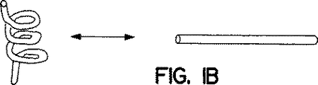
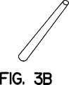
多孔性メンブレン構造は、折りたたみ可能な構造、例えば図1に示される構造であってもよい。折りたたみ可能な構造は、1つの形状に展開し(deploy)、そして第二のよりコンパクトな形状に収縮させることが可能な構造すべてである。図1には、形状を変換することが可能な構造の例が含まれる。例えば、平面から球根状に(図1A)、延長型かららせんに(図1B)、延長型から超らせんに(図1C)、および筒型からふいご型構造に(図1D)などである。展開された形状は、腎細胞を植え付ける、または移植するのにより適している可能性があり、一方収縮された形状は長期の移植により適している可能性がある。折りたたみ可能な多孔性メンブレン構造の展開された形状は、例えば、長いまたは平たい筒(図3A、3G)、延長型管(図3B、3E)、コイル(図3J)、らせん(図1Bおよび3C)、二重または多重らせん(図1C)、腎臓型(図3D)、立方体(図3H)、平たい袋(図3I)、またはそれらの組み合わせであってもよい。多孔性メンブレン構造はさらに、移植前または移植後の構造の変形および成形のため、ふいご10(図1D)、管、ひもまたはワイヤ20(図1D)を含んでもよい。さらに、多孔性メンブレン構造は、完全性を維持しそしてARU細胞の圧による壊死を防ぐための支持体を含んでもよい。こうした支持体は、多孔性メンブレン構造に結合していてもしていなくてもよく、そしてその内部または外部にあってもよい、柱、リブ(rib)、またはそれに匹敵するものを含んでもよい。折りたたみ可能な多孔性メンブレン構造の収縮された形状は、より複雑であることを除けば展開された形状に類似であってもよい。その他の収縮された形状は、平面、球体および腎臓型を含んでもよい。他の望ましい折りたたみ可能構造は、患者の皮下にまたは腹部または胸部に適するように設計された収縮された形状を含んでもよい。
人工腎臓に適した折りたたみ可能な構造の例には、アコーディオン様またはふいご型の折りたたみ可能構造(図2C)が含まれ、最も好ましくは多孔生物適合性ポリマーで形成される。折りたためみ可能な多孔性メンブレン構造は、腎細胞が付着する間、展開されてもよい。その後、展開された形状の折りたたみ可能構造を、患者に移植してもよい。患者の血管新生活動が人工腎臓を灌流する際、構造はゆっくりと収縮する可能性がある。また別に、腎細胞付着後、構造を収縮しそして患者に移植してもよい。折りたたみ可能な三次元多孔性メンブレン構造は、さまざまな形で開発してもよい。三次元折りたたみ可能メンブレン構造が、望ましい最終時および初期構造を有することが好ましい。望ましい構造は、人工腎臓の移植位置により決定される。皮下に移植されるよう設計された人工腎臓は、平面であってもよく、正規の腎臓と置換する人工腎臓は、腎臓型であってもよい。他の設計上考慮する点は、当該構造が折りたたまれたまたは展開された状態のいずれであっても、分解または構成要素の衰えを引き起こさずに展開可能でなくてはならないことである。例えば、こうした構造は構造完全性を維持し、そして広がった状態および折りたたまれた状態で、人工腎臓への血液供給を遮断するまたは減少させる可能性がある閉塞を起こさないことが望ましい。
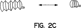
折りたたみ可能な多孔性メンブレン構造を形成する別の方法は、膨張性構造(図2A、2B、および2C)である。例えば、膨張性折りたたみ構造およびその対応する横断面構造の1つの態様を、それぞれ図2Aおよび2Bに示す。当該構造は、柔軟であるが実質的に非伸縮性でそして横断面が凸である壁20を有する膨張体10を含んでもよい。横断面が凸である壁は、伸張し、そして弓なりまたは凹であるよう適応させてもよく、そしてしたがってその幅に沿って柔軟性がある。膨張性構造は、腎細胞を植え付けるために膨張させてもよく、そして移植前または移植後のいずれかにしぼませてもよい。膨張性折りたたみ可能メンブレン構造の他の態様は、アコーディオン型であり、そして図2Cに示されている。
封入多孔性メンブレン構造は、所望により、ARUに機械的損傷を与えるのを防ぐにたる強度を持った、変形可能な外部外被(casing)を含んでもよい。変形可能な外被は、いくつでもよい望ましい形状に成形し、いくつでもよい全体系、構造または空間的制約を満足させてもよい。変形可能な外被は、フィルム、ガーゼ、帯紐、ワイヤメッシュ、布、フォームラバー、およびそれに匹敵するもので形成されてもよい。外被を構築するのに適した材料には、例えば、鋼鉄、チタンのような金属、ガラス、ポリマー、グラスファイバー、プラスチックまたはそれらに匹敵するものが含まれる。外部外被を有する多孔性メンブレン構造の最終的形状は、例えば、筒(図3A、3B、3G)、コイル(図3J)、らせん(図3C)、二重または多重らせん(図1C)、腎臓型(図3D)、立方体(図3H)、平たい袋(図3I)またはそれらの組み合わせ(図4)であってもよい。変形度の唯一の制限は、多孔性メンブレン構造およびそれに関連するARUの重要な領域が、人工腎臓の腎機能が損なわれ、減少し、またはそうでなければ有害な影響を受けるほど、著しく破損し、伸張し、ひだをつけられ、または過剰なストレス、圧力、または圧迫下に置かれることがないことである。
本発明の態様において、多孔性メンブレン構造を、放射線療法が人工腎臓の腎機能を好ましくなく改変しないように、放射線耐性である材質で作成してもよい。ある疾患、例えば腎臓癌などの治療においては、放射線を手術後に適用することが必要とされる可能性がある。放射線耐性多孔性メンブレン構造により、必要であれば、残った腫瘍の治療部位で、人工腎臓に重い放射線を当てることが可能になるであろう。
本発明の態様において、多孔性メンブレン構造は超音波耐性である材質で作成される。泌尿系中の結石(calculi)蓄積および被覆、例えば腎結石は、腎能力が低下した患者にはよく見られる問題である。結石蓄積、被覆および石(stone)の1つの治療法は、超音波エネルギーを用いて蓄積物を破壊することである。超音波耐性多孔性メンブレン構造により、人工腎臓を好ましくなく改変することなく、移植後に超音波治療を行うことが可能になるであろう。
本発明の別の態様は、薬剤、増殖因子を送り込む手段、および多孔性メンブレン構造への輸入路を少なくとも1つ提供することにより、人工腎単位(ARU)の多孔性メンブレン構造中の結石蓄積および被覆を抑制する手段に向けられる。輸入路は、固体、液体または気体を多孔性メンブレン構造に導入することが可能になる、いかなる手段、例えばポートまたは型であってもよい。輸入路は、流出チャンネルから遠位の点に置き、導入された液体が実質的に多孔性メンブレン構造の全内部表面を通過し、流出チャンネルから出て行くようにしてもよい。
当業者には、トランスフォーミング増殖因子β1、上皮増殖因子、および全トランス体レチノイン酸などの増殖因子の組み合わせが、尿細管形成を誘導する可能性があるζとが知られている。また、クエン酸カリウムおよびアセトヒドロキサミン酸などのさまざまな化学物質および薬剤が、ほとんど溶けないカルシウムおよびマグネシウム塩の結晶化を阻害することも知られている。抗生物質および金属化合物などのさまざまな化学物質および薬剤が、微生物の生長を阻害するか、または殺す可能性があることが知られている。増殖因子、抗被覆因子、および抗生物質を、輸入路から液体の形で多孔性メンブレン構造に加えてもよい。輸入路はさらに、例えばARUから皮下部位に伸長する管を含んでもよい。例えば、腹部ARUを持つ患者への増殖因子、抗被覆因子、および抗生物質の添加は、皮下に移植された注入口ポートに皮下針を連結することにより、容易に達成できる可能性がある。
本発明の1つの態様は、in vitroで尿細管類似体が生長および発達するため、多孔性メンブレン構造上を細胞外マトリックス成分で覆うことを提供する。ARU構築のための腎細胞の急速な三次元増殖は、細胞が落ち着きそして再生することが可能な生物成分マトリックスが入手可能であれば容易になる可能性がある。したがって、多孔性メンブレン構造の外部表面を、細胞外マトリックスタンパク質のような、細胞増殖制御の適切な情報を与える成分で処理することが好ましい可能性がある。
細胞外マトリックスタンパク質には、糖タンパク質、プロテオグリカンおよびコラーゲンが含まれる。多孔性メンブレン構造の外部表面は、これらタンパク質の1つまたは組み合わせにより処理してもよい。使用してもよい適切な細胞外タンパク質の例には、コラーゲン、フィブロネクチン、トロンボスポンジン、サイトアクチン、エキノネクチン、エンタクチン、ラミニン、テネイシン、ウボモルリン、ビトロネクチン、ビグリカン、コンドロイチン硫酸、デコリン、デルマタン硫酸、ヘパリン、ヘパリン硫酸およびヒアルロン酸が含まれる。
細胞外マトリックスタンパク質は、ラット尾などの哺乳動物供給源から新たに抽出してもよい。コラーゲンのその他の哺乳動物供給源には、ウシ、ブタおよびヒトの供給源が含まれる。コラーゲンなどのヒト細胞外タンパク質は、胎盤または死体などのヒト組織から集めてもよく、そしてSigma(ミズーリ州セントルイス)などの商業的供給者から購入してもよい。
細胞外マトリックスタンパク質および多孔性メンブレン構造は、使用前に殺菌してもよい。殺菌は、細胞外マトリックスタンパク質に微生物が混入する可能性を減少させる可能性がある。殺菌の好ましい方法には、限外ろ過および放射線曝露が含まれる。放射線曝露は、例えば、ガンマ線またはX線曝露であってもよい。また別に、抗生物質、抗細菌および細胞毒性剤、普通に効果的な用量の紫外線照射を使用してもよい。用いられる好ましい細胞毒性剤の1つは、エチレンオキシドである。
多孔性メンブレン構造の表面処理は、ECMタンパク質溶液と多孔性メンブレン構造を接触させることを含んでもよい。接触後、例えば水酸化アンモニウムによりpHを上げて、結合、ゲル化、ポリマー化を促進してもよい。また別に、細胞外マトリックスタンパク質を多孔性メンブレン構造にクロスリンクしてもよい。クロスリンクおよび誘導体化試薬は、Pierce(イリノイ州ロックフォード)などの商業的供給者から購入してもよい。
多孔性メンブレン構造上への腎細胞の植付け
腎細胞の多孔性メンブレン構造外部表面への付着は、分離した腎細胞を多孔性メンブレン構造と結合させることにより達成してもよい。好ましい方法の1つが、実施例3に詳細に記載される。簡潔には、多孔性メンブレン構造表面1平方センチメートル当たり約1x10⁷の細胞密度で、腎細胞懸濁と多孔性メンブレン構造を穏やかに接触させる。覆われた多孔性メンブレン構造を、約100%の湿度、約37℃、約5%CO2下の組織培養インキュベーター中で、約30分から約1時間インキュベーションする。この期間の後、培地を穏やかに加え、多孔性メンブレン構造を完全に沈める。
多孔性メンブレン構造上の細胞外膜タンパク質は、腎細胞の付着を促進する可能性があり、そしてこの付着は実質的に、約30分から約24時間以内、例えば1時間、2時間、4時間、8時間または16時間以内に完成する。完全付着に要する正確な時間は、多孔性メンブレン構造の表面特性および表面組成物、培地、および細胞外マトリックスおよび当該腎細胞に依存する。
尿細管類似体を形成するための多孔性メンブレン構造上での腎細胞の培養
付着後、腎細胞を含む構造を、尿細管類似体を形成するに足る時間および条件下でin vitroで培養する。例えば、約5%CO2および約37℃下で約3日から約20日、好ましくは約7から約10日の間培養すれば、一般には十分である。多孔性メンブレン構造を覆う培地は、約1日から約6日の時間間隔、好ましくは2日から5日の間、より好ましくは3日から4日の間で必要に応じて交換してもよい。培地交換の間隔は、培地の容積および培地中の栄養素の消費速度および廃棄産物の集積速度に依存する。培地の容積を調整することで、培地交換の間を、7日、8日、9日または10日などのより長い期間にすることも可能であると理解されている。ウシ血清、インシュリン、トランスフェリン、亜セレン酸ナトリウム、ヒドロコルチゾン、プロスタグランジンE2、ペニシリン、およびストレプトマイシンなどの栄養素、抗生物質およびホルモンを付加することで、培地交換の期間を延長することも可能であると理解されている。
細胞外マトリックスタンパク質処理により、多孔性メンブレン構造表面特性が変化し、付着細胞形態および移動に影響を与える。処理した多孔性メンブレン構造上で培養すると、腎細胞はまず単層で広がるが、時間と共に自発的に自己集合し、それぞれ内部管腔を含む三次元細胞集合体になる。細胞集合体の形状は尿細管の直線状部分に似ている。細胞集合体は支持体に付着し、そして細胞細胞境界が事実上区別できない滑らかな表面を有する。尿細管類似体は、一端が開放されていてもまた閉じていてもよい。1細胞当たりを基準とすると、尿細管類似体は、腎細胞単層と比較して、より高い腎臓特異的活性を示し、そしてより長い間生存可能である。細管細胞に特異的な細胞機能には、分泌、吸収、および、アルカリ性ホスファターゼ(図7AおよびB)およびオステオポンチン(図5)などの細胞特異的遺伝子産物発現が含まれ、そして単層の腎細胞より、より組織に似た超構造を示す。
尿細管類似体の多孔性メンブレン構造上の自己集合の過程で発展する腎特異的活性を、表現型上および機能上モニターを行ってもよい。表現型としては、尿細管類似体は、天然細管に似た刷子縁、管腔、および細胞結合などの特徴を有する。オステオポンチンおよびアルカリ性ホスファターゼなどの尿細管特異的遺伝子発現は、ノーザンブロット、ウェスタンブロット、ポリメラーゼ連鎖反応およびin situハイブリダイゼーションなど、当業者に知られるいかなる分子生物学的または免疫学的方法により、モニターしてもよい。グルコース代謝を調べる過ヨウ素酸シッフ(PAS)試験など、腎近位細管細胞に局在する活性に対する試験を用いて、腎近位表現型を検出してもよい。
腎近位尿細管は、多孔性メンブレン構造表面上に楕円から伸長した型の細胞の連続シートを形成することが観察された。植付け24時間後当初は、多孔性メンブレン構造上の腎細胞は、厚さが一細胞層であるが、集密(confluence)に近づくにつれ、複数細胞層である領域が見られる可能性がある。尿細管類似体のそれぞれの細胞は、明確な1つまたは2つの仁を持つ、目立つ丸い核を有する。細胞内空間は細胞内ブリッジに似た領域で付着する細胞では大きい。いくつかの領域では、細胞表面内への折りたたみまたはポケットが目立つ。いくつかの細胞は、多数の顆粒、おそらくリソソームを含む。これは近位尿細管細胞の特徴である。この細胞の顆粒化は、7日以上の培養体で有意に増加した。
顕微鏡下で調べると、尿細管類似体は、天然の細管同様、刷子縁に似た、細胞表面からの長細い微絨毛突起に満たされた広い細胞内空間を示す。刷子縁は線条縁とも呼ばれるが、近位腎尿細管細胞の頂端表面に特有な特徴である。顕微鏡では、刷子縁は細胞の接着していない表面の特殊化として現れ、表面領域を非常に増加させる細かい筒状変化(微絨毛)からなる。尿細管類似体上の刷子縁は、細胞表面に沿って形成される折りたたみまたはポケット内に続く、伸長した微絨毛表面を示す。細胞表面に平行だが、細胞表面下の折りたたみを通る組織切片は、細胞質に囲まれた微絨毛が並んだ路として現れる。刷子縁または微絨毛形成が並んだポケットは、微絨毛が多い立方体細胞が、そうでなければうろこ状の細胞培養環境中に、その広い頂端表面領域を維持する機構を代表する可能性がある。ポケットまたは路に連結した微絨毛がない細胞表面領域でも、細胞境界はまた、そこから突き出る長い微絨毛を有する。しかしその数は減少し、そしてその構造組織はポケットまたは路が隣接する表面と並んでいるのが観察される刷子縁とは異なる。微絨毛数が少ない領域は、正常の立方体腎細管細胞の側部および基底表面である可能性がある。
尿細管類似体は、損なわれていない(intact)近位尿細管細胞に典型的な細胞内組織を含む。細管細胞核は楕円であり、そして散在するヘテロクロマチンを主に核周囲に含む。核物質の多くは、外見上ユークロマチンであり、1つまたは2つの仁が顕微鏡下で視覚化される。尿細管類似体細胞の細胞質は、しばしば細胞表面に平行に配置された多くの繊維状ミトコンドリアを含む。棚状のクリステは概して、ミトコンドリアの長軸に直角に配置されている。より伸長した細胞では、特に細胞内結合と連続して、細かい細胞質微繊維束および細胞質微繊維の広いネットワークもまた存在する。これらの繊維束は、細胞の側表面に平行に走っている。ほとんどの細胞で、粗面小胞体の側面は短いが、非常に長くなりうる細胞もある。他の細胞小器官には、密なリソソーム顆粒および目立つゴルジネットワークがある。リソソーム内容物は培養を続けると有意に増加する可能性がある。
個々のデスモソームは、隣接する細胞との側縁に沿って接触部位に見られる可能性がある。より伸長した細胞群が単一の領域で接触する場所では、心筋の介在板に似た非常に複雑なデスモソーム様結合複合体が形成されることがある。これらの複合体から広い単一繊維のネットワークが放射状に作られる。これらの結合複合体は正常近位尿細管細胞の頂端境界の帯状デスモソームに似ている。したがって、これらの形態に基づき、尿細管類似体は近位尿細管の特徴をすべて有することが明らかである。
遺伝子発現解析もまた、尿細管類似体が天然細管に典型的な遺伝子を発現することを示す。天然腎細管は尿細管の全長でオステオポンチンを発現し、そして近位端に優先的にアルカリ性ホスファターゼを発現する。単層培養腎細胞は、抗オステオポンチンおよび抗アルカリ性ホスファターゼ抗体をプローブとして切片を染色すると低いレベルの染色が見られる、非常に低いレベルのオステオポンチンおよびアルカリ性ホスファターゼ活性を示す。腎細胞が尿細管類似体に集合し始めるにつれ、アルカリ性ホスファターゼ活性は上昇する。単一尿細管類似体内で、この活性の分布は相同ではない。尿細管類似体の近位端では、検出可能なレベルのアルカリ性ホスファターゼが示される。細胞を、例えばトリプシンなどで分離すると、細胞は増強された活性を失い、そして最初の単層で見られた低いレベルに戻る。しかし、尿細管類似体構造に残った細胞は、増強された活性を保持する。増強された細胞細胞接触およびより組織に似た構造が、尿細管類似体に見られる増強された活性に貢献していると仮定される。
人工腎単位前駆体の移植
尿細管類似体が付着した封入多孔性メンブレン構造を含む、人工腎単位前駆体を、宿主に移植して、細管端で糸球体を形成することにより、ネフロン類似体形成を誘導してもよい。血管新生は人工腎臓機能に重要である。人工腎臓の機能および増殖は血管供給を必要とする。
血管新生において、宿主組織は人工腎臓細胞により産生される情報に反応する。この反応は、少なくとも3つの構成要素を含むようである。まず、尿細管類似体の毛細管内皮細胞が、存在する血管を囲む基底層を破る;血管新生の間、宿主の内皮細胞はプラスミノーゲン活性化因子などのプロテアーゼを分泌し、親毛細管または細静脈の基底層を破って消化を進める。次に、宿主内皮細胞が腎細胞に向かい移動する。第三に、内皮細胞が増殖し、そして尿細管類似体の近位端に形成される糸球体様構造を灌流する、毛細管を形成する。その結果できる構造はネフロン類似体と称され、宿主に尿細管類似体を移植し、そして尿細管類似体の近位端に糸球体形成を誘導することにより、in vivoに形成される。糸球体形成は、移植2週間後までに、いくつかの尿細管類似体の端で見られる可能性がある。移植8週間後、糸球体形成は広がり、そしてほとんどの尿細管類似体上で見られるようになる。
人工腎単位前駆体は、新血管系形成を誘導する能力を有し、そして患者体内の血管化が少ないまたは多い領域のどちらに移植してもよい。皮下移植1週間後、血管形成は尿細管類似体の長さに沿って広がる。尿細管類似体は、細管および尿細管類似体の少なくとも1つの領域に形成される糸球体に沿った血管新生により、8週間後の終わりまでには、ネフロン類似体に発展する。複数の糸球体または糸球体様構造もまた、各尿細管類似体上に見られる。組織学的観察により、各尿細管類似体の長さに沿って広い血管形成が検出された。
調べたARUは、近位腎細管および糸球体の特徴を示す。ARUはアルカリ性ホスファターゼおよびガンマグルタミルトランスフェラーゼを発現する。ほぼ近位細管細胞でしか発現されない2つの遺伝子である。人工糸球体を機能的または免疫組織化学的に試験すると、凝固因子VIIIの存在が示された。
血管供給がない人工腎臓は、栄養素供給を拡散に頼っているため、血管新生が適切な灌流を供給することが可能になるまで、厚さ数ミリメートル未満の生存層に限られる可能性がある。本発明の1つの態様は、この限界を、宿主に複数のARU構造を移植し、そして血管新生によりARUを動脈または静脈に連結させることにより克服する方法に向けられる。その後ARUを、動脈および静脈と共に宿主から除き、そして外科的により大きいまたはより厚いARUと組み合わせ、そして患者に再移植する。ARUの動脈および静脈を、患者の動脈および静脈と連結し、組み立てた人工腎臓に血液供給を提供する。
流出チャンネルの連結
ARUの流出チャンネルは、患者のどの部位に連結して、人工腎臓からの輸出液を除去させてもよい。本発明の態様において、流出チャンネルを患者の器官に連結して、人工腎臓からのろ過液を除去させてもよい。流出チャンネルを、腎臓、尿管、腎盤、膀胱、尿道、精巣、前立腺または輸精管を含む尿路に連結してもよい。また別に、流出チャンネルを大腸または小腸などの腸に、腸導管手術で連結してもよい。さらに、流出チャンネルは管により延長し、人工腎臓を移植した患者の皮膚から突き出してもよい。人工腎臓の腹部移植において、例えば、流出チャンネルは腹壁を通して伸長されてもよい。流出チャンネルの端にキャップを取りつけ、輸出液の漏れを防いでもよい。望ましい時、患者がキャップをはずし、そして輸出液を外部に排出してもよい。
流出チャンネルの結合は縫合によってもよい。外科医によりARUを患者に取りつけることによりARUを適合させるためには、異なるARUの流出チャンネルが、異なる大きさを含んでもよい。それにより流出チャンネル結合部位に適合させることが可能になる。さらに、流出チャンネルは、移植中、手術時の状況および部位に合うように簡単な適合が行える材質で構築されてもよい。
In vitro操作
本発明の別の態様において、人工腎臓をその動脈および静脈と共に宿主から除き、そしてin vitroで培養してもよい。ネフロン類似体を含む人工腎臓は、実験台上の生物反応器として実験台上の人工腎臓を形成するのに用いてもよい。In vitro人工腎臓は、哺乳動物血清を用いてまたは直接哺乳動物に連結することにより栄養を与えてもよい。In vitro人工腎臓の初期パイロット規模を、それに続く産生規模in vitro人工腎臓の開始物質としてもよい。例えば、付加的多孔性メンブレン構造を人工腎臓に結合させて、培養により大きく増殖するよう誘導してもよい。In vitro人工腎臓はその後、個々に合わせた治療産物として患者に移植してもよい。人工腎臓は、in vitroで、産物の大規模工業的加工および採取が可能になる程度まで、保存し、そして増殖させてもよい。例えば、in vitro人工腎臓をレニン製造に用いてもよい。
本発明の態様は、腎臓への物質の影響を解析するための人工腎臓の使用に向けられる。物質は薬剤(drug)または製薬(pharmaceutical)、化学物質、微生物、生物学的産物または要素であってもよい。薬剤または製薬は、ヒトおよび動物を含む患者に、診断、治療、または疾患または他の異常状態の予防の一助として、使用されまたは投与されてもよい、すべて化学物質である。薬剤は痛みの緩和のため、またはいかなる生理学的または病理学的異常を制御しまたは改善するために用いられてもよい。薬剤の例には、ワクチン、組換え剤、化学物質、組換え核酸、組換えタンパク質、および生存、死亡または減弱微生物が含まれる。試験に有用な薬剤には、候補薬剤、化学物質、薬剤の特性を有すると疑われる化合物および剤が含まれる。試験してもよい化学物質には、患者または腎臓が曝露される可能性があるすべての化学物質または物質が含まれる。こうした化学物質には、環境化学物質、個人の衛生用品および化粧品が含まれる。微生物には、細菌、真菌、ウイルス、アメーバ、寄生虫、および酵母などの生存生命体すべて、または試験時に生存、死亡、仮死、不活発または減弱していてもよいそれに匹敵するものが含まれる。生物学的産物には、生存生命体が産生するタンパク質、脂質、核酸、糖、毒などの、生存生命体からの産物および廃棄産物が含まれる。
腎疾患の治療
本発明の1つの態様において、人工腎臓は外部で維持されそして操作され、そしてex vivoで機能してもよい。血液処理を要する患者は、一定期間、患者の血液供給を人工腎臓に連結してもよい。処理血液は、処理後、人工腎臓から分けられ、そして患者に戻されてもよい。
本発明の別の態様は、患者に人工腎臓を移植することにより、腎機能を増大させて、腎疾患を治療する方法に向けられる。腎疾患は一般的な用語であり、腎結石などそれほど生命に危険を与えない疾患から、腎多嚢胞病およびネフローゼ、一時的および慢性および永久腎不全などの、より生命に危険を与える障害が含まれる。本発明の方法により治療される可能性がある腎疾患には、腎機能の増大により利益を受ける可能性がある疾患すべてが含まれる。腎嚢胞形成異常、腎多嚢胞病、髄質の嚢胞疾患などの腎先天性異常、後天性(透析関連)嚢胞病および単純嚢胞;急性糸球体腎炎、三日月様糸球体腎炎、ネフローゼ症候群、膜性糸球体腎炎、最小変化病(minimal change disease)、リポイドネフローゼ、巣状分節性糸球体腎炎、膜性増殖性糸球体腎炎、IgA腎症、巣状増殖性糸球体腎炎、慢性糸球体腎炎、全身性エリテマトーデス、ヘーノホ−シェーンライン紫斑病、細菌性心内膜炎、糖尿病性糸球体硬化症、アミロイドーシスおよび遺伝性腎炎などの糸球体疾患、急性細管壊死、急性腎不全などの細管疾患、および、微小血管障害性溶血性貧血、アテローム塞栓性腎疾患、鎌状細胞疾患腎症、拡散皮質壊死、腎梗塞、線腫、癌、後腎芽、免疫的仲介腎疾患、薬剤誘導腎炎、尿酸腎症、高カルシウム症および腎石灰症などのその他の腎疾患などである。
治療してもよいその他の疾患には、患者の腎臓が損傷を受ける可能性があるすべての状態が含まれる。例えば、本発明の方法は、ネフロンに毒性を持つ薬剤による化学療法を受ける、健康な腎臓を持つ患者の治療に使用してもよい。治療を要する可能性があるその他の異常には、例えば、外傷、毒物摂取、自己免疫疾患、老齢、およびそれらに匹敵するものが含まれる。
本発明の方法は、患者の正規腎臓機能の増大が望ましい、いかなる腎疾患の治療にも有用である。人工腎臓は、腎機能増大の必要性が一時的かまたは永久であるかに依存して、限られた期間または永久に移植してもよい。
人工腎臓は、患者および移植部位の必要に合うように多くの異なる形状であってもよい。例えば、患者に存在する腎臓が除かれない場合、伸長または平面またはコンパクトな形状が、腹部または皮下移植に最適である可能性がある。また別に、天然腎臓の解剖学的形状をとる人工腎臓は、腎置換が必要な患者には最も適している可能性がある。人工腎臓の大きさもまた、最適な成果をあげるため、さまざまであってもよい。大きな患者は大きい人工腎臓を必要とする可能性があり、一方、子供などの小さな患者は小さい人工腎臓により適している可能性がある。
本発明の他の態様および利点は、一部は以下の説明に明示され、そして一部はこの説明から明らかであろうし、そして本発明を実行することにより学ばれる可能性がある。
実施例
実施例1 腎細胞の単離
小さい腎臓および大きい腎臓の切片、1週齢のC57ブラックマウス由来などを、被膜を剥離し、切断し、切り刻み、そして15mM Hepes、pH7.4および0.5μg/mlのインシュリン、1.0mg/mlのコラゲナーゼ、および0.5mg/mlのバシラス・ポリミキサル(Bacillus polymyxal)由来の中性プロテアーゼ、ディスパーゼ(Boehringer Mannheim、インディアナ州インディアナポリス)を含むダルベッコの修飾イーグル培地(DMEM;Sigma、ミズーリ州セントルイス)に懸濁した。
大きい腎臓、ブタ腎臓などは、抽出の3時間以内に、無カルシウムイーグル最少必須培地を用い37℃で10分、動脈灌流した。腎臓はその後、1.5mM MgCl2および1.5mM CaCl2を補った同じ緩衝液中の0.5mg/mlのコラゲナーゼ(Type IV, Sigma、ミズーリ州セントルイス)で灌流した。腎臓はその後、被膜を剥離し、切断し、切り刻み、そして15mM Hepes、pH7.4および0.5μg/mlのインシュリン、1.0mg/mlのコラゲナーゼ、および0.5mg/mlのバシラス・ポリミキサル由来の中性プロテアーゼ、ディスパーゼ(Boehringer Mannheim、インディアナ州インディアナポリス)を含むダルベッコの修飾イーグル培地(DMEM;Sigma、ミズーリ州セントルイス)に懸濁した。
大きいまたは小さい腎臓由来のものどちらかの腎細胞懸濁を、ウォーターバス中で、37℃で30分、穏やかに攪拌した。細胞および破片は50g、5分の遠心分離により回収された。沈殿はタンパク質分解を止めるため10%ウシ胎児血清(Biowhittaker、メリーランド州ウォーカーズビル)を含むDMEMに再懸濁し、そして濁った溶液を無菌80メッシュナイロンスクリーンに通し、大きな破片を除いた。細胞は遠心分離により回収し、そして無カルシウムダルベッコの修飾イーグル培地で2回洗浄した。
実施例2 腎細胞のin vitro培養
ラット尾コラーゲンの単離
ラット尾から腱を除き、そして50ml試験管の0.12M酢酸を含む脱イオン水に貯蔵した。4℃で16時間、一晩保存した。
透析バッグは孔サイズが均一であり、そして重金属が除かれたことを確実にするため前処理した。簡潔には、透析バッグを2%炭酸水素ナトリウムおよび0.05%EDTAの溶液に沈め、そして10分間煮沸した。蒸留水で複数回リンスし、炭酸水素ナトリウムおよび0.05%EDTAを除いた。
ラット腱を含む0.12M酢酸溶液を処理した透析バッグに入れ、そして酢酸を除くため2から3日透析した。透析溶液は3から4時間おきに交換した。
多孔性メンブレン構造のコラーゲンによる処理
多孔性メンブレン構造(図4)は、約30μg/mlのコラーゲン(Vitrogenまたはラット尾コラーゲン)、約10μg/mlのヒトフィブロネクチン(Sigma、ミズーリ州セントルイス)および約10μg/mlのウシ血清アルブミン(Sigma、ミズーリ州セントルイス)を含む溶液を、総容積約2mlの補足培地と、37℃で3時間インキュベーションして接触させ、処理した。その後、コラーゲンでコーティングされた多孔性メンブレン構造を、1mlの濃縮水酸化アンモニウム(約28%から約30%のNH4OH、Sigma、ミズーリ州セントルイス)と共に30分インキュベーターに置き、pHを上げ、そしてコラーゲンのゲル化を促進した。多孔性メンブレン構造の水酸化アンモニウム処理後、構造を等張培地でよく洗浄し、使用前に多孔性メンブレン構造のpHを中性化した。
組織培養プレートのコーティング
培養フラスコ75cm2は、約30μg/mlのコラーゲン(Vitrogenまたはラット尾コラーゲン)、約10μg/mlのヒトフィブロネクチン(Sigma、ミズーリ州セントルイス)および約10μg/mlのウシ血清アルブミン(Sigma、ミズーリ州セントルイス)を含む溶液を含む、総容積約2mlの補足培地中で、37℃で3時間インキュベーションすることにより、コーティングした。
細胞培養
消化された単一懸濁腎細胞は、修飾コラーゲンマトリックス上に、約1x10⁶細胞/mlの濃度でまき、そして約10%のウシ胎児血清、約5μg/mlのウシインシュリン、約10μg/mlのトランスフェリン、約10μg/mlの亜セレン酸ナトリウム、約0.5μMのヒドロコルチゾン、約10ng/mlのプロスタグランジンE2、約100ユニット/mlのペニシリンG、約100μg/mlのストレプトマイシン(Sigma、ミズーリ州セントルイス)を補ったDMEM中で、約37℃、5%CO2のインキュベーターで培養した。
集密した単層は、約0.05%のトリプシン、約0.53mMのEDTA(Gibco BRL、ニューヨーク州グランドアイランド)を含む無カルシウムイオンリン酸緩衝生理食塩水(PBS)(約1.51mMのKH2PO4、約155.17mMのNaCl、約2.8mMのNa2HPO・7H2O)で処理することにより、継代培養した。
細胞は、懸濁から最初の継代以降、どの時点でも、約10%DMSOを含む培地で培養し、液体培地中で凍結および貯蔵してもよい。
実施例3 人工腎臓の調製
腎細胞は10日間in vitroで培養し、そして増殖させた。In vitro培地、10%のウシ胎児血清、5μg/mlのウシインシュリン、10μg/mlのトランスフェリン、10μg/mlの亜セレン酸ナトリウム、0.5μMのヒドロコルチゾン、10ng/mlのプロスタグランジンE2、100ユニット/mlのペニシリンG、100μg/mlのストレプトマイシン(Sigma、ミズーリ州セントルイス)を補ったDMEMは、1日おきに交換した。細胞は、0.05%トリプシン、約0.53mM EDTA(Gibco BRL、ニューヨーク州グランドアイランド)を含む無カルシウムイオンリン酸緩衝生理食塩水(PBS)(約1.51mMのKH2PO4、約155.17mMのNaCl、約2.8mMのNa2HPO・7H2O)を用いたトリプシン処理により、採取した。37℃で10分消化した後、細胞をおよそ5x10⁶細胞/mlでDMEM培地に再懸濁した。
細胞懸濁を、ポリカーボネート膜で構築された、容器に続くシラスティック(silastic)カテーテルに一端が連結された、4ミクロン孔サイズの前もって形成された細管装置を含む、多孔性メンブレン構造上に、穏やかに重層した。多孔性メンブレン構造はラットコラーゲンによりコーティングされた(実施例2)。多孔性メンブレン構造表面の1平方センチメートル当たり、およそ107細胞が重層された多孔性メンブレン構造を、約30分から約40分、37℃、5%CO2下でインキュベーションした。インキュベーション時間の終わりに、さらに前もって温めた培地を穏やかに加え、多孔性メンブレン構造を沈めた。多孔性メンブレン構造は、37℃、5%CO2下で約7日から約10日インキュベーションした。培地を交換し、そして細胞には頻繁に、例えば約毎日、約2日ごとまたは約3日ごとなどに栄養を与えた。
植付けてから約7から約10日後、人工腎単位前駆体が多孔性メンブレン構造表面に発達した。In vitro培養の30日後、液体が観察され、多孔性メンブレン構造の容器に集められた。In vitro培地は、フェノールレッドのため赤いが、容器中の液体は透明で、そして無色であった。集められた液体が培地と異なることにより、人工腎単位前駆体がろ過または分泌機能を示すことを示している。
人工腎単位前駆体の移植
植付けてから約7から10日後、表面に人工腎単位前駆体を含む多孔性メンブレン構造のいくつかを、無胸腺マウスの皮下空間に移植した。無胸腺マウスは、メイン州バー・ハーバーのJackson Laboratoriesなどの供給者から商業的に購入してもよい。移植してから約2、約4および約8週間後に動物を屠殺し、そして人工腎単位前駆体を回収して、そして解析した。
回収した標本は、全体的およびヘマトキシリンおよびエオジンにより組織学的に調べた。オステオポンチン、フィブロネクチンおよびアルカリ性ホスファターゼの免疫組織化学染色を行い、細胞の型およびそのin vivoでの構造を決定した(図5、6、7)。ヒトフィブロネクチンモノクローナル抗体(Sigma、ミズーリ州セントルイス)をフィブロネクチンマトリックスに対して用いた。ローダミン結合ヤギ抗マウス(Boehringer Mannheim、インディアナ州インディアナポリス)を二次抗体として用いた。オステオポンチンの免疫細胞化学染色(図5)は我々の研究室で産生されたポリクローナル抗体を用いて行った。抗体は、ニュージーランドシロウサギ(New Zealand white rabbit)で標準的方法(HarlowおよびLane, Antibodies a laboratory manual, 1988, Cold Spring Harbor Press, コールドスプリングハーバー)を用いて産生され、そして1:5000の希釈率で使用した。FITCに結合したヤギ抗ウサギ抗体(Boehringer Mannheim、インディアナ州インディアナポリス)を二次抗体として用いた。アルカリ性ホスファターゼの免疫組織化学染色は、ニトロブルー、テトラゾリウムおよび5-ブロモ-4-クロロ-3-インドリルリン酸(Sigma、ミズーリ州セントルイス)を用いて行った。人工腎臓から集められたろ過液の色は、淡黄色であった。ろ過液の尿酸レベル解析は尿酸検出キット(Sigma Diagnostics、ミズーリ州セントルイス)を用いて行った。
すべての動物は、屠殺するまで生存していた。回収した標本は元の構造を維持した。人工腎単位前駆体は、全体に宿主組織で覆われていた。人工腎臓中の液体は、膜に連結したカテーテル中に集められた。移植人工腎臓を組織学的に調べると、広範な血管形成、糸球体形成(図8)および非常に組織化された細管様構造が見られた。主に近位および遠位細管細胞から分泌されるオステオポンチンに対する抗体により免疫細胞化学染色すると、細管部位が陽性に染色された。アルカリ性ホスファターゼに対する免疫組織化学染色により、近位細管様構造が陽性に染色された。さらに、新規に形成された細管の細胞外マトリックス中に均一なフィブロネクチンの染色が見られた(図6)。新規に形成された腎単位から回収された黄色の液体は、66mg/dlの尿酸を含んでおり、血漿中の2mg/dlと比べると、これらの細管が尿酸の一方向分泌および濃縮を行うことが可能であることを示唆している。糸球体形成の証拠、組織学的染色および尿酸分泌により、移植7日後までに、人工腎単位前駆体はARUに発達していることが示される。
尿細管類似体および天然細管の表現型比較
腎臓の近位尿細管細胞の形状は立方体である。これらは1つまたは2つの目立つ仁を含む、中心に配置された円から楕円の核を含む。細胞の頂端表面は、長細い指状の微絨毛を形成し、ラットなどの動物では高さ1.3mmに達しそして頂端表面領域をおよそ22倍に増加させる。これに比べ、遠位曲尿細管は、通常、頂端表面に短くずんぐりした微絨毛を含み、そして集合管には刷子縁がない。
尿細管類似体の細胞は、天然尿細管に似ている。人工尿細管細胞は、表面陥入部に続く長細い微絨毛の長い刷子縁を有する。培養細胞の微絨毛密度は、in vivoの近位細管細胞に見られるものに匹敵する。近位細胞の側面および基底部の境界は、非常に不規則で、そして隣接する細胞と広く絡み合っている。これは培養細胞で観察されるものと似ている。
近位細管細胞の1つの特徴は、多数の培養細胞でも見られる、多数のリソソーム、ファゴソームおよびペルオキシソームの存在である。近位細管細胞の刷子縁に対するマーカー酵素である、アルカリ性ホスファターゼおよびガンマグルタミルトランスフェラーゼ活性が、長期間の培養に渡り維持されるのは、人工尿細管の同一性および完全性のさらなる証拠である。
本発明の他の態様および使用は、本明細書に開示される本発明の明細および実行を考慮すれば、当業者には明らかであろう。本明細書中に述べられたすべての米国特許および他の参考文献は明確に本明細書に援用される。明細および実施例は、以下の請求項に示される、本明細書の真の範囲および精神のみを模範として考慮すべきである。
本発明の具体的態様には次のものが含まれる。
1.少なくとも一つの人工腎単位を含む人工腎臓であって、前記人工腎単位は少なくとも一つの流出チャンネルを有する封入内部空間を定める外側表面を有する多孔性メンブレン構造を含み、そして前記メンブレン構造がさらにその外側表面に付着し、そして複数のネフロン類似体であるその封入内部空間と液体伝達を行っており、前記各ネフロン類似体は血管新生を有し前記尿細管類似体の少なくとも一つの領域において糸球体様構造を形成する尿細管類似体を含み、前記尿細管類似体は尿細管細胞の三次元細胞集合体を含み、前記集合体が前記メンブレン構造の内部空間と液体伝達を行う管腔からなり、そして前記集合体中の尿細管細胞が刷子縁を示す、前記人工腎臓。
2.多孔性メンブレン構造が、ルロースエーテル、セルロース、セルロースエステル、フッ化ポリエチレン、フェノール類、ポリ-4-メチルペンテン、ポリアクリロニトリル、ポリアミド、ポリアミドイミド、ポリアクリル酸ポリベンゾキサゾール、ポリカーボネート、ポリシアノアリルエーテル、ポリエステル、ポリエステルカーボネート、ポリエーテル、ポリエーテルエーテルケトン、ポリエーテルイミド、ポリエーテルケトン、ポリエーテルスルホン、ポリエチレン、ポリフルオロオレフィン、ポリイミド、ポリオレフィン、ポリオキサジアゾール、ポリフェニレンオキシド、ポリフェニレンスルフィド、ポリプロピレン、ポリスチレン、ポリスルフィド、ポリスルホン、ポリテトラフルオロエチレン、ポリチオエーテル、ポリトリアゾール、ポリウレタン、ポリビニル、ポリビニリデンフッ化物、再生セルロース、シリコン、尿素−ホルムアルデヒド、またはそれらのコポリマーまたはそれらの物理的混合物から選択される生体適合性物質を含む、前項1の人工腎臓。
3.多孔性メンブレン構造が生物分解性物質を含む、前項1の人工腎臓。
4.多孔性メンブレン構造が細胞の通過を阻害し、そして体液およびガスの通過を可能にする孔サイズを有する、前項1の人工腎臓。
5.多孔性メンブレン構造が直径約0.04ミクロンから約10ミクロンの孔を含む、前項1の人工腎臓。
6.多孔性メンブレン構造が直径約0.4ミクロンから約4ミクロンの孔を含む、前項1の人工腎臓。
7.前記腎臓細胞集合体がほ乳類細胞を含む、前項1の人工腎臓。
8.前記ほ乳類細胞が人細胞である、前項7の人工腎臓。
9.前記人工腎臓がin vivoまたはex vivoにおいて機能する、前項1の人工腎臓。
10.刷子縁が前記腎臓細胞の自由表面上に多数の微絨毛を含む、前項1の人工腎臓。
11.細胞集合体がオステオポンチンを発現し、そして前記集合体の少なくとも一つの領域における尿細管細胞がアルカリ性ホスファターゼを発現する、前項1の人工腎臓。
12.細胞集合体がフィブロネクチンを発現する、前項1の人工腎臓。
13.細胞集合体が過ヨウ素酸−シッフ染色アッセイに陽性である、前項1の人工腎臓。
14.前記流出チャンネルが前記多孔性メンブレン構造中に体液が進入することを防ぐように位置取りされた非還流性バルブを含む、前項1の人工腎臓。
15.前記流出チャンネルが半多孔性のメンブレンバルブを含み、前記半多孔性メンブレンが微生物の通過を阻害し、そして体液およびガスの通過を可能にする孔サイズを有する、前項1の人工腎臓。
16.少なくとも一つの流出チャンネルを有する封入内部空間を定める外部表面を有する多孔性メンブレン構造を含む人工腎単位であって、前記メンブレン構造がさらにその外部表面に付着しそして複数のネフロン類似体であるその封入された内部空間と液体伝達を行い、前記各ネフロン類似体は前記尿細管類似体の少なくとも一つの領域における糸球体様構造を形成する血管新生を有する尿細管類似体を含み、前記尿細管類似体は尿細管細胞の三次元細胞集合体を含み、前記集合体は前記メンブレン構造の内部空間と液体伝達を行う管腔を含み、そして前記集合体中の尿細管細胞が刷子縁を示す、前記人工腎単位。
17.多孔性メンブレン構造が、ルロースエーテル、セルロース、セルロースエステル、フッ化ポリエチレン、フェノール類、ポリ-4-メチルペンテン、ポリアクリロニトリル、ポリアミド、ポリアミドイミド、ポリアクリル酸、ポリベンゾキサゾール、ポリカーボネート、ポリシアノアリルエーテル、ポリエステル、ポリエステルカーボネート、ポリエーテル、ポリエーテルエーテルケトン、ポリエーテルイミド、ポリエーテルケトン、ポリエーテルスルホン、ポリエチレン、ポリフルオロオレフィン、ポリイミド、ポリオレフィン、ポリオキサジアゾール、ポリフェニレンオキシド、ポリフェニレンスルフィド、ポリプロピレン、ポリスチレン、ポリスルフィド、ポリスルホン、ポリテトラフルオロエチレン、ポリチオエーテル、ポリトリアゾール、ポリウレタン、ポリビニル、ポリビニリデンフッ化物、再生セルロース、シリコン、尿素−ホルムアルデヒド、またはそれらのコポリマーまたはそれらの物理的混合物から選択される生体適合性物質を含む、前項16の人工腎単位。
18.多孔性メンブレン構造が生物分解性物質を含む、前項16の人工腎単位。
19.多孔性メンブレン構造が細胞の通過を阻害し、そして体液およびガスの通過を可能にする孔サイズを有する、前項16の人工腎単位。
20.多孔性メンブレン構造が直径約0.04ミクロンから約10ミクロンの孔を含む、前項16の人工腎単位。
21.多孔性メンブレン構造が直径約0.4ミクロンから約4ミクロンの孔を含む、前項16の人工腎単位。
22.前記腎臓細胞集合体がほ乳類細胞を含む、前項16の人工腎単位。
23.前記ほ乳類細胞が人細胞である、前項16の人工腎単位。
24.刷子縁が前記腎臓細胞の自由表面上に多数の微絨毛を含む、前項16の人工腎単位。
25.細胞集合体がオステオポンチンを発現し、そして前記集合体の少なくとも一つの領域における尿細管細胞がアルカリ性ホスファターゼを発現する、前項16の人工腎単位。
26.細胞集合体が過ヨウ素酸−シッフ染色アッセイに陽性である、前項16の人工腎単位。
27.前記人工腎単位がin vivoまたはex vivoにおいて機能する、前項16の人工腎単位。
28.前記人工腎単位が前記封入空間中に尿酸を排出する、前項16の人工腎単位。
29.封入内部空間を定める外部表面を有しそして少なくとも一つの流出チャンネルを有する多孔性メンブレン構造を含む、追加的な腎臓機能を必要とする患者に対して移植するために適した人工腎単位前駆体であって、前記メンブレン構造がさらにその外部表面に付着しそしてさらに複数の尿細管類似体であるその封入内部空間と液体伝達を行い、前記尿細管類似体が尿細管細胞の三次元集合体を含み、前記集合体が前記メンブレン構造の内部空間と液体伝達をするための管腔を含み、そして前記集合体中の尿細管細胞が刷子縁を示す、前記人工腎単位前駆体。
30.多孔性メンブレン構造が、ルロースエーテル、セルロース、セルロースエステル、フッ化ポリエチレン、フェノール類、ポリ-4-メチルペンテン、ポリアクリロニトリル、ポリアミド、ポリアミドイミド、ポリアクリル酸、ポリベンゾキサゾール、ポリカーボネート、ポリシアノアリルエーテル、ポリエステル、ポリエステルカーボネート、ポリエーテル、ポリエーテルエーテルケトン、ポリエーテルイミド、ポリエーテルケトン、ポリエーテルスルホン、ポリエチレン、ポリフルオロオレフィン、ポリイミド、ポリオレフィン、ポリオキサジアゾール、ポリフェニレンオキシド、ポリフェニレンスルフィド、ポリプロピレン、ポリスチレン、ポリスルフィド、ポリスルホン、ポリテトラフルオロエチレン、ポリチオエーテル、ポリトリアゾール、ポリウレタン、ポリビニル、ポリビニリデンフッ化物、再生セルロース、シリコン、尿素−ホルムアルデヒド、またはそれらのコポリマーまたはそれらの物理的混合物から選択される生体適合性物質を含む、前項29の人工腎単位前駆体。
31.多孔性メンブレン構造が生物分解性物質を含む、前記29の人工腎単位前駆体。
32.多孔性メンブレン構造が細胞の通過を阻害し、そして体液およびガスの通過を可能にする孔サイズを有する、前項29の人工腎単位前駆体。
33.多孔性メンブレン構造が直径約0.04ミクロンから約10ミクロンの孔を含む、前項29の人工腎単位前駆体。
34.多孔性メンブレン構造が直径約0.4ミクロンから約4ミクロンの孔を含む、前項29の人工腎単位前駆体。
35.前記腎臓細胞集合体がほ乳類細胞を含む、前項29の人工腎単位前駆体。
36.前記ほ乳類細胞が人細胞である、前項35の人工腎単位前駆体。
37.刷子縁が前記腎臓細胞の自由表面上に多数の微絨毛を含む、前項29の人工腎単位前駆体。
38.細胞集合体がオステオポンチンを発現し、そして前記集合体の少なくとも一つの領域における尿細管細胞がアルカリ性ホスファターゼを発現する、前項29の人工腎単位前駆体。
39.細胞集合体が過ヨウ素酸−シッフ染色アッセイに陽性である、前項29の人工腎単位前駆体。
40.前記人工腎単位が前記封入空間中に尿酸を排出する、前項29の人工腎単位前駆体。
41.以下の段階:
a) 少なくとも一つの流出チャンネルを有する封入内部空間を定める外部表面を有する多孔性メンブレン構造を提供する段階;
b) 前記外部表面を腎臓組織細胞の懸濁物と接触させる段階;そして
c)前記腎臓細胞をin vitroにおいて前記外部表面上で培養して複数の尿細管類似体を形成する段階であって、前記尿細管類似体は尿細管細胞の三次元集合体を含み、前記集合体は前記メンブレン構造の封入空間と液体伝達を行う管腔を含み、そして前記集合体中の尿細管細胞が刷子縁を示す段階;
を含む、追加の腎臓機能を必要とする患者体内に移植するために適した人工腎単位前駆体を作成する方法。
42.刷子縁が前記腎臓細胞の自由表面上に多数の微絨毛を含む、前項41の方法。
43.細胞集合体がオステオポンチンを発現し、そして前記集合体の少なくとも一つの領域における尿細管細胞がアルカリ性ホスファターゼを発現する、前項41の方法。
44.細胞集合体が過ヨウ素酸−シッフ染色アッセイに陽性である、前項41の方法。
45.尿細管類似体がフィブロネクチンを発現する、前項41の方法。
46.尿細管細胞が過ヨウ素酸−シッフ染色アッセイに陽性である、前項41の方法。
47.腎臓細胞が胎児腎臓細胞、または幼若体(juvenile)の腎臓細胞である、前項41の方法。
48.腎臓細胞が腎臓皮質由来である、前項41の方法。
49.腎臓細胞がヒトのものである、前項41の方法。
50.封入多孔性メンブレン構造が細胞の通過を阻害し、そして体液およびガスの通過を可能にする、前項41の方法。
51.封入多孔性メンブレン構造が直径約0.04ミクロンから約10ミクロンの孔を含む、前項41の方法。
52.封入多孔性メンブレン構造が直径約0.4ミクロンから約4ミクロンの孔を含む、前項41の方法。
53.以下の工程:
a) 患者体内の天然血管供給を有する領域に、前項29の方法により作成した人工腎単位前駆体を移植する工程;
b) 天然血管供給を誘導して前記位置の領域において一またはそれ以上の糸球体様構造を形成する工程;そして
c) 前記メンブレン構造からの前記流出チャンネルを患者の泌尿器系に連結する工程;
を含む、前記患者において腎臓疾患を治療する方法。
54.前記糸球醸造が第VIII因子を発現する、前項53の方法。
55.流出チャンネルが前記患者の尿管に接続される、前項53の方法。
56.尿管が本来の尿管であるかまたは人工尿管である、前項55の方法。
57.内部空間を定める外部表面を有する生体適合性ポリマーの半透性膜を含む人工腎臓のための多孔性メンブレン構造であって、前記メンブレン構造がヘッダーと液体伝達を行う複数の中空チューブを含み、そして前記ヘッダー上の流出チャンネルが前記内部空間のドレナージを可能にする、前記多孔性メンブレン構造。
58.半透膜が多孔性メンブレン構造が生物分解性物質を含む、前項57の多孔性メンブレン構造。
59.半透膜が細胞の通過を阻害し、そして体液およびガスの通過を可能にする孔サイズを有する、前項57の多孔性メンブレン構造。
60.半透膜が直径約0.04ミクロンから約10ミクロンの孔を含む、前項57の多孔性メンブレン構造。
61.半透膜が直径約0.4ミクロンから約4ミクロンの孔を含む、前項57の多孔性メンブレン構造。
62.前記中空チューブが円形の横断面を有する、前項57の多孔性メンブレン構造。
63.円形状、単一コイル状、多重コイル(multiple coil)状、渦巻き(spiral)状、球状、らせん(helix)状、多重ヘリックス状、楕円状、平面状、U字型、四角状、またはそれらの組合せの形状である、前項57の多孔性メンブレン構造。
64.前記流出チャンネルが前記多孔性メンブレン構造中に体液が進入することを防ぐように位置取りされた非還流性バルブを含む、前項57の多孔性メンブレン構造。
65.前記流出チャンネルが半多孔性のメンブレンバルブを含み、前記半多孔性メンブレンが微生物の通過を阻害し、そして体液およびガスの通過を可能にする孔サイズを有する、前項57の多孔性メンブレン構造。
66.前記多孔性メンブレン構造が機械的損傷から保護するための外側ケーシング(casing)をさらに含む、前項57の多孔性メンブレン構造。
67.流れ込み(affluent)チャンネルをさらに含む、前項57の多孔性メンブレン構造。
68.回収手段および前記流出チャンネルからの流出物貯蔵手段をさらに含む、前項57の多孔性メンブレン構造。
69.以下の段階:
a) 腎臓組織を単離する段階;
b) 前記腎臓組織を酵素的処理により分離して細胞懸濁物を形成する段階;
c) 前記腎臓細胞懸濁物をin vitroにおいて培養する段階;
d) 封入多孔性メンブレン構造を細胞外マトリックスタンパク質で処理する段階;
e) 前記腎臓細胞を封入多孔性メンブレンの処理した外部表面上で培養して尿細管類似体を形成する段階であって、前記尿細管類似体は尿細管細胞の三次元細胞集合体内に管腔を有する腎臓細胞の集合体内に管腔を含み、そして前記尿細管細胞は刷子縁を示す、前記段階;
を含む、尿細管類似体を作成する方法。
70.細胞外マトリックスがコラーゲンを含む、前項69の方法。
71.コラーゲンがラットの尾のコラーゲンである、前項69の方法。
72.刷子縁が前記腎臓細胞の自由表面上に多数の微絨毛を含む、前項69の方法。
73.細胞集合体がオステオポンチンを発現し、そして前記集合体の少なくとも一つの領域における尿細管細胞がアルカリ性ホスファターゼを発現する、前項69の方法。
74.細胞集合体が過ヨウ素酸−シッフ染色アッセイに陽性である、前項69の方法。
75.以下の段階:
a) 封入多孔性メンブレン構造を細胞外マトリックスタンパク質でコーティングする段階;
b) 腎臓細胞懸濁物を前記物質上に播く段階;
c) 前記腎臓細胞を前記封入多孔性メンブレン上で培養して尿細管を形成する段階であって、前記尿細管は内部に管腔を含む尿細管細胞の三次元細胞集合体を含み、そして前記尿細管細胞は刷子縁を示す、前記段階;
を含む、腎臓細胞上の物質の効果を測定する方法。
76.刷子縁が前記腎臓細胞の自由表面上に多数の微絨毛を含む、前項75の方法。
77.細胞集合体がオステオポンチンを発現し、そして前記集合体の少なくとも一つの領域における尿細管細胞がアルカリ性ホスファターゼを発現する、前項75の方法。
78.細胞集合体が過ヨウ素酸−シッフ染色アッセイに陽性である、前項75の方法。
79.以下の段階:
a) 物質を尿細管類似体細胞培養物に接触させる段階であって、前記尿細管類似体は多孔性メンブレンが付着し内部に管腔を有する尿細管細胞の三次元細胞集合体を含み、前記尿細管が刷子縁を示す、前記段階;そして
b) 前記腎臓細胞に対する物質の効果を決定する段階;
を含む、腎臓細胞に対する物質の効果を測定する方法。
80.物質が薬物、医薬品、化学物質、微生物、化学物質、または元素である、前項79の方法。
81.刷子縁が前記腎臓細胞の自由表面上に多数の微絨毛を含む、前項79の方法。
82.細胞集合体がオステオポンチンを発現し、そして前記集合体の少なくとも一つの領域における尿細管細胞がアルカリ性ホスファターゼを発現する、前項79の方法。
83.細胞集合体が過ヨウ素酸−シッフ染色アッセイに陽性である、前項79の方法。background
1. Field of Invention
The present invention is directed to a prosthetic kidney, a method of making an artificial kidney, and a method of treating kidney disease using an artificial kidney.
2. Background explanation
The kidney removes metabolic waste from the blood, regulates fluid balance by maintaining homeostasis, and provides important regulatory activity by secreting hormones. Usually, about 20% of the blood pumped by the heart is processed by the kidneys.
Nephrons are the functional unit of the kidney and process blood through three processes: filtration, reabsorption and secretion. Each kidney contains approximately 1 million nephrons, and each nephron consists of renal bodies and tubules. The nephron shape resembles a small funnel with a very long convoluted handle. Blood enters the renal body through the glomeruli. Filtrate from the blood enters the glomerular sac, also called Bowman's sac, and flows through the tubules. The tubule includes four parts: a proximal bent tubule, a Henle snare, a distal bent tubule and a collecting tube.
The renal bodies contain a group of tangled blood capillaries called glomeruli, which are approximately 200 microns in diameter and are surrounded by a thin-walled bag-like structure called the glomerular sac. Blood enters and exits the glomeruli through import and export arterioles. In the glomerulus, blood and various dissolved substances are squeezed out of the glomerular capillary tube into the glomerular sac as glomerular filtrate due to blood pressure.
The relative concentrations of several substances in plasma, glomerular filtrate and urine are shown in Table I and Table II. These values can vary depending on many factors such as the patient's liquid consumption, medication, age, diet, health status and renal function.
Secretion is the process by which substances and fluids move from the blood in the distal tubules and the capillaries around the tubules to the distal tubules and collecting ducts. The secreted substances are hydrogen ions, potassium ions, ammonia, and certain drugs. Tubular secretion plays a vital role in maintaining the body's acid / base balance.
Homeostasis is maintained by special hormones that affect kidney function by the body. The pituitary hormone ADH (antidiuretic hormone) reduces urine output by making the distal tubules and collecting ducts permeable to water. Aldosterone secreted from the adrenal glands regulates tubular salt and other electrolyte reabsorption. Mostly, aldosterone stimulates the tubules and resorbs sodium faster.
Although hormones affect kidney function, the kidneys also produce hormones that regulate the function of other organs. Erythropoietin is a hormone secreted by kidney cells that regulates the rate of red blood cell formation. Renin is the second hormone secreted by the kidneys that regulates blood pressure. In addition, the kidney activates vitamin D, which is involved in skeletal integrity.
In summary, blood is processed and urine is formed as a result of glomerular filtration of plasma, tubular reabsorption and tubular secretion. In tubular reabsorption, glucose, amino acids, proteins, creatine, lactic acid, citric acid, uric acid, ascorbic acid, phosphate ions, sulfate ions, calcium, potassium ions, sodium ions, water and urea are reabsorbed. In tubular secretion, penicillin, creatinine, histamine, phenobarbital, hydrogen ions, ammonia, and potassium are secreted.
If both kidneys of a patient fail, blood pressure can increase, fluid can accumulate in the body, waste levels can increase to harmful levels in the blood, and red blood cell production May decrease. When this happens, treatment is needed to replace the function of the failing kidney. Treatment of renal dysfunction includes hemodialysis, peritoneal dialysis and kidney transplantation.
Hemodialysis is a treatment that cleans and filters the blood of patients with renal insufficiency. The treatment reduces the level of hazardous waste, excess salt and body fluids. Hemodialysis also helps regulate blood pressure and maintains an appropriate balance of chemicals such as potassium, sodium, and chloride in the body.
Hemodialysis processes blood using a dialysis machine or special filters. During processing, patient blood travels through the tube to an external dialysis machine. The dialyzer spills waste and excess body fluid and returns freshly cleaned blood to the body. A typical treatment regime may include hemodialysis treatment which takes 2-4 hours, 3 times a week. During treatment, movement is restricted, but patients may engage in activities that do not require excessive movement, such as reading and writing.
Disadvantages of hemodialysis include side effects and complications caused by rapid changes in patient fluid and chemical balance during treatment. Muscle spasm and hypotension are two common side effects. Hypotension, a rapid drop in blood pressure, can cause extreme weakness and dizziness.
It usually takes months for patients to adapt to the side effects of hemodialysis. Side effects may be reduced by strictly following appropriate diet and drug consumption as indicated. Proper diet helps reduce the amount of waste that accumulates in the patient's blood and reduces kidney load. A dietitian's help is needed to plan a diet according to the doctor's instructions.
Further disadvantages of hemodialysis include high costs and often having to go to a dialysis facility for a long time. There is also a method of performing dialysis at home instead of a dialysis facility. Home dialysis requires helpers, and both patients and helpers need special training. In addition, there is a need for a place to store machinery and essentials at home.
Peritoneal dialysis uses the patient's abdominal lining and peritoneum to filter blood. A cleaning solution called dialysate is moved through a special tube to the patient's abdomen. Fluid, waste, and chemicals pass from the small peritoneal blood vessels to the dialysate. After a few hours, the dialysate is drained from the abdomen and at the same time the waste is removed from the blood. The abdomen is then filled with fresh dialysate and the cleaning process is resumed.
The dialysis method is associated with varying degrees of difficulty and significant treatment time. Treatment plans vary, but generally take a very inconvenient position. Typical treatment regimes include, for example, treatment periods of 30 to 40 minutes every 4 to 6 hours, 10 to 12 hours every night, 36 to 42 hours per week, or 24 hours. In addition to dialysis, a special, calorie-restricted potassium-restricted diet is required.
Possible complications of peritoneal dialysis include peritonitis or peritoneal infection. Peritoneal dialysis involves a number of stages where pathogens such as bacteria can be introduced into the body. Symptoms of peritonitis include inflammation, exudation of serum fibrin cells and pus, nausea, dizziness, fever, abdominal pain, tenderness, constipation and vomiting. To prevent peritonitis, medical care that exactly follows dialysis is required. Patients need to be trained to recognize early signs of peritonitis. Failure to do quick interference can lead to serious problems.
Hemodialysis and peritoneal dialysis have serious long-term complications in addition to short-term inconvenience and side effects. Complications such as bone disease, hypertension, nerve damage, and anemia can have devastating effects over time. As a result of these complications, 60% of renal dialysis patients are not employed and 30% are disabled. Renal dialysis patients generally have a shorter survival and five times longer hospital stays than the general population.
Kidney transplantation is a method of placing a healthy kidney from a donor person into a patient's body. The transplanted kidney increases or replaces the blood filtration capacity of the patient's failing kidney. The transplanted kidney is placed between the upper thigh and the abdomen. The new renal arteries and veins are connected to the patient's arteries and veins, and blood flows through the new kidneys and urine is made. The patient's own kidney, which may still have some function, is not removed unless it is causing infection or hypertension.
Similar to dialysis, non-tissue transplant is not a cure. Tissue rejection is a significant risk even if the tissue compatibility is excellent. Immunosuppressive treatment plans that prevent rejection, based on drugs such as cyclosporine, continue to be the basis of most post-transplant medicine. However, as the range of drug immunosuppression to therapeutic dose is narrow, between appropriate immunosuppression and toxicity, as determined by significant variability in pharmacokinetics and pharmacodynamics between patients and patients However, it has been difficult to find immunosuppressive drug levels with minimal toxicity. Therefore, post-transplant medical care still incurs significantly higher costs and risks.
Side effects may be caused by prolonged consumption of immunosuppressants. Most seriously, the immune system is weakened and infections occur more easily. Some drugs also cause weight gain, acne, facial hair growth, cataracts, stomach acid excess, hip disease, liver or kidney damage. The transplant patient's diet is less restrictive than that of dialysis patients, but patients still need to reduce some types of food. Even immunosuppressants may not prevent kidney rejection. If rejection occurs, the patient will have to undergo some form of dialysis and possibly wait for another transplant.
The time it takes to find a kidney donor varies. There are not enough cadaveric donors for everyone who needs a transplant, and this problem is particularly acute in cases of kidney transplantation. Another source of kidneys is living donors such as relatives and spouses. Because of the better histocompatibility, transplants from genetically related surviving donors often perform better than transplants from cadaveric donors.
Humes (US Pat. No. 5,429,938) reports a renal cell culture method for in vitro tubule formation and ex vivo tubule construction. In this method, kidney cells are cultured in the presence of tumor growth factor β1, epidermal growth factor and all-trans retinoic acid to form a three-dimensional aggregate. Among the disadvantages of the method are the need to administer growth factors and the absence of glomerular formation. Administration of growth factors to patients can have undesirable side effects and complications. Humes discloses methods that may help repopulate damaged kidney tissue, but did not disclose how to construct and use an artificial kidney.
Liver (Rozga et al., Hepatology 17, 258-65) and blood (Schwartz et al., Blood 78, 3155-61) cell growth fixed to a semiporous membrane structure has been reported. These organ structures are not suitable for the construction of artificial kidneys because they do not take into account glomerular filtrate collection and drainage outside the body.
Naughton and Naughton (US Pat. No. 5,516,680) report a three-dimensional kidney cell and tissue culture system. The three-dimensional structure of viable stromal cells is seeded on top of the stromal support matrix. Kidney cells are layered on top of this three-dimensional system and cultured.
Vacanti and Langer (WO 88/03785) disclose a method of culturing cells in a three-dimensional polymer-cytoskeleton of a biodegradable polymer. Organ cells cultured in a polymer-cytoskeleton are transplanted into the patient and form a prosthesis.
Thus, taken together, it is clear that the known methods of kidney culture contain inherent disadvantages and deficiencies, and because of its design limitations, there are certain limitations in the ability to use the culture as an artificial kidney. Although each of these methods attempts to address some of the problems faced in constructing an artificial kidney, the disclosed method filters blood and performs glomerular filtration, secretion, or reabsorption. There is no suggestion of how to build a real artificial kidney possible.
Summary of the Invention
The present invention overcomes the problems and disadvantages associated with current strategies and designs for the treatment of renal dysfunction and renal failure, and provides methods and apparatus for treatment.
One embodiment of the invention is directed to an artificial kidney that includes at least one artificial kidney unit (ARU). The artificial kidney unit includes a porous membrane structure having an outer surface defining an enclosed inner space having at least one outflow channel, and the membrane structure further adheres to the outer surface and a plurality of nephron analogs ( analog) and the fluid transmission with the enclosed internal space. Each nephron analog includes a tubule analog with angiogenesis that forms a glomerular-like structure in at least one region of the tubule analog. Tubular analogs contain three-dimensional cell aggregates of tubular cells, the aggregates contain the inner space of the membrane structure and lumens for fluid transmission, and the tubular cells in the aggregates are brush borders. Indicates (brush border).
Another aspect of the invention includes a porous membrane structure having an outer surface defining an enclosed interior space having at least one outflow channel, and the membrane structure further adheres to the outer surface, and a plurality of nephrons It is directed to an artificial kidney unit that includes conducting fluid communication with an encapsulated internal space that is an analog. Each nephron analog includes a tubule analog with angiogenesis that forms a glomerular-like structure in at least one region of the tubule analog. The tubule analog includes a three-dimensional cell assembly of tubule cells, the assembly includes an inner space of the membrane structure and a lumen for fluid transmission, and the tubule cells exhibit a brush border.
Another aspect of the present invention is an artificial kidney unit precursor suitable for implantation into a patient in need of additional renal function, having an outer surface defining an enclosed inner space, and at least one The precursor is directed to a porous membrane structure having two outflow channels. The membrane structure further adheres to its outer surface and is in fluid communication with its enclosed internal space, which is a plurality of tubule analogs. Tubular analogs include a three-dimensional collection of tubular cells, the collection includes an interior space of the membrane structure and a lumen for fluid transmission, and the tubular cells in the collection exhibit a brush border.
Another aspect of the invention is a method of making an artificial kidney unit suitable for implantation in a patient in need of additional renal function, defining an enclosed interior space having at least one outflow channel. Providing a porous membrane structure with an external surface; contacting the external surface with a suspension of renal tissue cells; and culturing the renal cell on the external surface in vitro to provide fluid communication with the enclosed space of the membrane structure The method is directed to a method comprising the steps of forming a plurality of tubular analogs comprising a three-dimensional collection of tubular cells including lumens, and wherein the tubular cells in the collection exhibit a brush border.
Another aspect of the present invention is a method of treating renal disease or increasing renal function in a patient, wherein the artificial kidney unit precursor described above is implanted in an area where the patient has a natural vascular supply; Directing the supply to form a glomerular-like structure in one area; and connecting the outflow channel from the membrane structure to the patient's urinary system is directed to the method.
Another aspect of the present invention is a porous membrane structure for an artificial kidney, comprising a biocompatible polymer semipermeable membrane having an outer surface defining an interior space, and the membrane structure provides fluid communication with the header. Including a plurality of hollow tubes to be performed and an outflow channel to a header that allows for drainage of the interior space.
Another aspect of the present invention is a method of making a tubule analog comprising isolating kidney tissue; isolating the kidney tissue to form a cell suspension by enzymatic treatment; culturing in vitro; treating the encapsulated porous membrane structure with extracellular matrix protein; culturing kidney cells on the treated outer surface of the encapsulated porous membrane structure to form a tubule analog; Is directed to the method, comprising the step of including a three-dimensional cell aggregate of tubular cells including a lumen within the three-dimensional cell aggregate; and the tubular cells exhibit a brush border.
Other aspects and advantages of the invention will be in part apparent from the description which follows, and in part will be apparent from the description, and may be learned by practice of the invention.
Description of drawings
The information in this patent contains at least one drawing created by coloring. Copies of this patent, including color diagrams, will be provided by requesting the Patent and Trademark Office and paying the necessary fee.
Figures 1A-D show various foldable shapes of an artificial kidney unit (ARU) or porous membrane structure.
Figures 2A-C show the expandable collapsible shape of an artificial kidney unit ARU or porous membrane structure.
Figures 3A-J show various shapes of artificial kidney units or porous membrane structures.
FIG. 4 shows an artificial kidney device consisting of a polycarbonate tubule membrane with a pore size of 4 microns connected to a collecting tube and a storage container.
FIG. 5 shows immunocytochemical staining of an artificial kidney unit section with an anti-osteopontin antibody.
FIG. 6 shows that fibronectin is uniformly stained in the extracellular matrix of newly formed tubules.
FIGS. 7A-B show ARU sections stained with anti-alkaline phosphatase antibody.
Figures 8A-B show transplanted ARU sections showing the formation of glomerular structures and highly organized tubule-like structures of different sizes.
Description of the invention
As exemplified and broadly described herein, the present invention is directed to an artificial kidney, a method of making an artificial kidney, and a method of treating kidney disease with an artificial kidney.
Numerous methods and devices that have been used to treat kidney disease have each attempted to solve one or more problems that are recognized as important for successfully increasing renal function. Examples of these methods include hemodialysis and various types of peritoneal dialysis. However, all of these methods and devices have been plagued by problems related to the complexity, size and weight of the system, the difficulty of long-term treatment or infection control. Problems currently associated with devices and methods for treating kidney disease, such as dialysis machines and intraperitoneal dialysis, include high mobility, proper renal function for moderate exercise, freedom from strict diet planning, And infection, nausea, pruritus, undernourishment, pseudogout, hepatitis B infection, accelerated cardiovascular disease, hypertension, renal osteogenesis abnormalities, anemia, pleural effusion, thrombosis, serositis, pericarditis, dialysis ascites and dialysis This includes not being able to provide a high quality of life with freedom from complications such as dementia.
Transplanted kidneys can be used effectively as a widespread basis for treating kidney disease and have proven to be capable of long-term survival. To date, however, donor livers for all patients who require one kidney are inadequate, and no truly practical artificial kidney has been developed for wide application. In addition, current kidney transplant patients require very advanced follow-up care, including medical management of the patient's immune system.
The object of the present invention is an artificial kidney capable of maintaining a high quality of life for many years, complications associated with current methods of treating kidney disease, such as vomiting, infection, hypotension, To provide said artificial kidney with low risks such as convulsions, bleeding, leukopenia, hypoxia, electrolyte disorders, and dialysis imbalance and nausea.
Another object of the present invention is to provide an artificial kidney that functions effectively at high filtration rates and therefore occupies a fairly small volume.
Another object of the present invention is to provide an artificial kidney that is transplanted into a patient to replace or increase the function of the natural kidney.
Another object of the present invention is to provide a method of making an artificial kidney from a patient's own kidney cells or donor cells.
Another object of the present invention is to provide an artificial kidney that is inherently tissue compatible, durable, reliable, and capable of filtering blood over an extended period of time.
Another object of the present invention is to provide an artificial kidney having a redundancy that can perform proper renal function even if one or more components fail.
Another object of the present invention is a biodegradable structure, which plays an initial structural role to form an artificial kidney and subsequently promotes tissue growth as the biodegradable structure is degraded. It is to provide an artificial kidney containing the above structure.
Another object of the present invention is to provide an artificial kidney with an effluent drainage and collection system.
One embodiment of the present invention is directed to an artificial kidney that includes one or more artificial kidney units (ARU). One preferred process for making such a device will now be described.
Kidney cell collection
ARU is constructed in part using kidney cells from a donor. In an autologous artificial kidney, the kidney cells may be derived from the patient's own kidney. In allogeneic artificial kidneys, kidney cells may be derived from other members of the patient's species. In a heterogeneous artificial kidney, the kidney cells may be from a different species than the patient. Donor cells may be derived from the renal cortex of many mammalian sources, such as humans, cows, pigs, horses, goats and sheep. Renal cells may be isolated by biopsy or autopsy. In addition, the cells may be frozen or grown prior to use.
In preparation for ARU construction, kidney or kidney tissue sections are separated into cell suspensions (Example 1). It is not essential to separate the cells to the single cell stage in the initial primary culture because after in vitro culture for a period of time, such as one week, may result in a single cell suspension. Tissue separation may be performed by mechanically and enzymatically breaking the extracellular matrix and the intercellular junctions that connect the cells together. Kidney cells from any developmental stage may be used, from fetuses, newborns, infants to adults. Cells may be obtained with higher yields from fetal or neonatal tissue.
To reduce stress on the cells and facilitate handling, kidney tissue sections may be placed in an equilibrated salt solution prior to disruption. Equilibrated salt solutions include tissue culture media, Hank's balanced salt solution and phosphate buffered saline and comparable, such as commercial products such as Gibco (Gettysburg, Maryland) and Sigma (St. Louis, MO). Available from source. Renal tissue may be disrupted with a mechanical tissue disruption device such as a scalpel, needle, scissors or cell separation sieve (Sigma, St. Louis, MO). After mechanical disruption, the tissue is further treated enzymatically with proteolytic enzymes such as trypsin, collagenase, dispase, and with agents that bind to or chelate calcium ions such as ethylenediaminetetraacetic acid (EDTA). May be. Calcium chelators eliminate the bioavailability of calcium ions on which cell cell adhesion depends.
Enzymatic separation may be monitored by eye or microscope to maintain cell viability. The enzyme may be inactivated after the desired separation is obtained but before cell viability is substantially impaired. Enzyme deactivation may be performed by isotonic solution washing or serum treatment.
Primary cultures may be prepared from isolated cells with or without a cell fractionation step. Cell fractionation may be performed using techniques such as fluorescence display cell sorting methods known to those skilled in the art. Cell fractionation may be performed based on cell size, DNA content, cell surface antigen, and viability. For example, tubule cells may be concentrated and fibroblasts may be reduced. Cell fractions may be used but are not necessary for the practice of the invention.
Cell sorting may be desirable, for example, when the donor has a disease, such as kidney cancer or other cancer metastasis to the kidney. The kidney cell population may be sorted to separate malignant kidney cells or other tumor cells from normal non-cancerous kidney cells. Normal non-cancerous kidney cells isolated by one or more sorting techniques may be used to produce an artificial kidney.
Another optional method is cryopreservation. Cryopreservation can be useful, for example, to reduce the need for multiple invasive surgical procedures. Cells obtained from the kidney may be amplified and a portion of the amplified cells may be used and another portion may be stored frozen. If cells can be amplified and stored, there will be considerable flexibility in the selection of donor cells. For example, cells from histocompatible donors may be amplified and utilized by one or more recipients. A further advantage of cryopreservation and amplification is that cells from one kidney may be amplified to produce a second kidney.
Another example of the use of cryopreservation is an organization bank. Donor cells may be stored frozen with histocompatibility data. Donor cells are stored, for example, in a donor tissue bank. Once tissue is needed to treat a patient's kidney disease, it is possible to select the cells that are most tissue compatible with the patient. Patients who have a disease that may be at risk for the kidney or who receive such a possible treatment may cryopreserve their kidney biopsy. Later, if the patient's own kidney has weakened, the cryopreserved kidney cells may be thawed and used for treatment. Examples of diseases that damage the kidneys include, for example, hypertension and diabetes. Examples of treatments that damage the kidneys include, for example, chemotherapy and radiation therapy.
Kidney cell culture
It may be desirable to amplify many cells to produce enough material to initiate ARU. Amplification has the advantage of reducing the amount of donor tissue required. For example, a small kidney biopsy section may be removed from a living donor. The removed tissue may be so small that it does not substantially reduce the kidney function and ability of the donor. The removed kidney biopsy section may be amplified and an artificial kidney provided to one or more patients. In some situations, such as autotransplantation, where the patient has already suffered significant kidney damage, there is a significant advantage if the kidney cells can be amplified and ARU regenerated.
Cell amplification may be performed by continuing the cells over and over again in larger or more culture vessels. Cells may be subcultured continuously for weeks, months and years. Thus, a small section of the kidney, such as less than 1 gram of tissue from a biopsy, can be increased to about 10 grams, about 50 grams, about 200 grams or more of tissue used for ARU construction after continuous culture. It is.
In one preferred embodiment, the cells are cultured and amplified under the conditions discussed in Examples 2 and 3. Briefly, about 10% fetal calf serum, about 5 μg / ml bovine insulin, about 10 μg / ml transferrin, about 10 μg / ml sodium selenite, about 0.5 on a collagen-treated plate of all cells. μM hydrocortisone, approximately 10 ng / ml prostaglandin E2In Dulbecco's modified Eagle's medium supplemented with about 100 units / ml penicillin G and about 100 μg / ml streptomycin at about 37 ° C. and about 5% CO2Incubate in an incubator. All media and reagents may be purchased commercially from tissue culture sources such as Sigma, St. Louis, MO.
Other media, such as MCDB, 199, CMPL, RPMI, F10, F-12, MEM and comparable media are obtained from commercial sources such as Gibco (Gettysburg, MD) and Sigma (St. Louis, MO). Yes, they may be used for culturing kidney cells. Supplements may also be supplemented by increasing the serum concentration to, for example, about 15%, about 20%, about 30%, about 40%, or about 50%. Suitable medium, serum, balanced salt solution, and adjuvant compositions for various mammalian cells are listed in the Gibco (Gettysburg, Maryland) and Sigma (St. Louis, MO) catalogs incorporated herein. Yes.
The matrix used to maintain the medium and viable kidney cells for a long period of time must be selected so that these cells remain responsive to the hormone. Small amounts of insulin, hydrocortisone and retinoic acid are desirable to maintain the cells to form tubule analogs and artificial kidney units. Insulin, hydrocortisone and retinoic acid are particularly desirable when cultured longer than encountered during cell amplification.
Porous membrane structure
The ARU of the present invention includes an encapsulated porous membrane structure. The porous membrane structure includes a porous membrane with an outer surface defining an enclosed interior space and at least one outflow channel. As described below, the tubule analog is formed on the outer surface of the porous membrane structure. In the simplest embodiment of the invention, the membrane structure is a hollow tube that is tightly closed at both ends. The outflow channel may be coupled to the containment tube to discharge the internal space.
The porous membrane structure of the present invention may also be, for example, a plurality of hollow tubes or hollow fibers, each closed at one end and in fluid communication with the header at the other end. Hollow fibers may be purchased commercially and the construction of hollow fibers for cell culture is well known to those skilled in the art. One method of hollow fiber production involves extruding the membrane polymer into a fine die, stiffening the tube and forming a very porous tube. Hundreds to thousands or tens of thousands of fibers may be connected to the header. Furthermore, a plurality of headers may be connected by a liquid channel to form a superstructure for an artificial kidney. Hollow fiber construction can be wrapped in a relatively small volume with a large surface as a volume fraction.
Another porous membrane structure includes a plurality of hollow perforated tubes or fibers open at both ends, and one end has liquid transmission with the first header and the other end has liquid transmission with the second header. Also good. With this design, it may be possible to flush or clear the liquid through the porous membrane structure. The flushing may allow for cleaning, disinfection, treatment, or drug or chemical delivery before and during use of the porous membrane structure.
A porous membrane structure may also be constructed by sintering-fusion of particles to form a tertiary structure. Other construction methods for porous membrane structures include casing, stretching, leaching, nucleation, and laser production. In the casing, a thin film of a solution containing polymer and solvent is sometimes cast on a cloth or paper substrate. The solvent flows away and pore formation occurs due to polymer precipitation. In stretching, TeflonTMPolypropylene, or other polymer sheet, is stretched evenly in all directions to produce regular porosity. In leaching, a solution containing two components is stretched into a film. A solvent is then used to break down one component and form a porosity (see Mikos, US Pat. No. 5,514,378, incorporated herein). In nucleation, a thin polycarbonate film is exposed to a radioactive fission product, creating a trajectory of radiation damage components. The polycarbonate sheet is then etched with acid or base to make the radiation damaging material pores. Finally, a laser may be used to individually perforate many materials to form a porous membrane structure. In the production by laser, in addition to the polymer material, a porous membrane structure may be formed using metal, glass, and ceramics. After any manufacturing method, quality control and membrane structure determination may be performed using an electron microscope.
The porous membrane structure can be a natural or synthetic biocompatible polymer material such as cellulose ether, cellulose, cellulose ester, fluorinated polyethylene, phenols, poly-4-methylpentene, polyacrylonitrile, polyamide, polyamideimide, polyacrylic acid, Polybenzoxazole, polycarbonate, polycyanyl ether, polyester, polyester carbonate, polyether, polyetheretherketone, polyetherimide, polyetherketone, polyethersulfone, polyethylene, polyfluoroolefin, polyimide, polyolefin, polyoxadi Azole, polyphenylene oxide, polyphenylene sulfide, polypropylene, polystyrene, polysulfide, polysulfone, polytetrafluoro Ethylene, polythioether, polytriazole, polyurethane, polyvinyl, polyvinylidene fluoride, regenerated cellulose, silicone, urea - formaldehyde or may be prepared by using a copolymer thereof or physical mixtures. The polymer material must be selected to be compatible and resistant to body fluid and host cell attack. In addition, the polymer material can be used for any drug or chemotherapeutic substance that the patient may be exposed to as part of the treatment of kidney disease or any other disease that the patient may also have. It is preferably resistant to drug or chemical attack.
The porous membrane structure is biodegradable and may be designed to slowly degrade in the body. Biodegradable materials may also play an initial structural role in artificial kidney formation and then promote tissue growth as they are degraded. Furthermore, the biodegradable substance and its degradation products are preferably non-toxic and can be easily removed from the diseased or healthy patient system. Exemplary materials that form biodegradable structures include natural or synthetic polymers, such as poly (alpha esters) such as poly (lactic acid), poly (glycolic acid), polyorthoesters, and polyanhydrides, and There are those copolymers that degrade by hydrolysis at a controlled rate and are resorbed. These materials offer maximum control over degradability, ease of handling, size and shape.
The pores on the porous membrane structure must be large enough to allow liquids and gases to pass through but small enough not to permeate cells. Suitable pore sizes to accomplish this goal may be from about 0.04 microns to about 10 microns in diameter, preferably between about 0.4 microns to about 4 microns in diameter. Polycarbonate membranes are particularly suitable. For example, highly regulated pores such as about 0.01 microns, about 0.05 microns, about 0.1 microns, about 0.2 microns, about 0.45 microns, about 0.6 microns, about 1.0 microns, about 2.0 microns, and about 4.0 microns It is because it is possible to manufacture with the magnitude | size of. At levels below 1 micron, porous membranes may be impermeable to bacteria, viruses, and other microorganisms.
The porous membrane structure may be completely porous or partially porous. It may be desirable for the region of the porous membrane structure to be non-porous. Non-porous regions can include, for example, hinge regions, structural regions, export and import ports, connections and binding sites.
The outflow channel may be any discharge opening in the porous membrane structure, for example, an opening or comparable such that a nozzle, tube, mold, hole, or liquid may exit the interior space of the porous membrane structure. There are things. If desired, a non-return valve, commonly known as a check valve, may be incorporated into the outflow channel to prevent the export liquid from flowing back into the porous membrane structure. A filter that is large enough to allow fluid to pass through but small enough to prevent microorganisms from passing through may be added to the outflow channel to prevent or reduce the potential for infection of the artificial kidney. If desired, means for collecting and storing the export fluid, such as an artificial bladder, hollow tube or the like, may be connected to the outflow channel or porous membrane structure. Export liquid may be stored for analysis during or after the discharge period.
The porous membrane structure may be a foldable structure, such as the structure shown in FIG. A collapsible structure is any structure that can be deployed into one shape and contracted into a second, more compact shape. FIG. 1 includes an example of a structure that can change the shape. For example, from flat to bulbous (Fig. 1A), from extended to spiral (Fig. 1B), from extended to super-helical (Fig. 1C), from cylindrical to bellows (Fig. 1D). The deployed shape may be more suitable for implanting or transplanting kidney cells, while the contracted shape may be more suitable for long-term implantation. The deployed shape of the foldable porous membrane structure is, for example, a long or flat tube (Figs. 3A, 3G), an extension tube (Figs. 3B, 3E), a coil (Fig. 3J), a helix (Figs. 1B and 3C) It may be a double or multiple helix (FIG. 1C), kidney type (FIG. 3D), cube (FIG. 3H), flat bag (FIG. 3I), or a combination thereof. The porous membrane structure may further include a bellows 10 (FIG. 1D), tube, string or wire 20 (FIG. 1D) for deformation and shaping of the structure before or after implantation. In addition, the porous membrane structure may include a support to maintain integrity and prevent necrosis due to ARU cell pressure. Such supports may include pillars, ribs, or the like, which may or may not be bonded to the porous membrane structure and may be internal or external thereto. The collapsed shape of the collapsible porous membrane structure may be similar to the deployed shape except that it is more complex. Other contracted shapes may include flat, sphere and kidney molds. Other desirable foldable structures may include a contracted shape designed to be suitable for the patient's subcutaneous or abdominal or thorax.
Examples of collapsible structures suitable for artificial kidneys include accordion-like or bellows-type collapsible structures (FIG. 2C), most preferably formed of porous biocompatible polymers. The foldable porous membrane structure may be deployed while the kidney cells are attached. The deployed shape of the collapsible structure may then be implanted in a patient. As the patient's angiogenic activity perfuses the artificial kidney, the structure may slowly contract. Alternatively, after renal cell attachment, the structure may be contracted and implanted into the patient. The collapsible three-dimensional porous membrane structure may be developed in various ways. It is preferred that the three-dimensional foldable membrane structure has a desired final and initial structure. The desired structure is determined by the implantation location of the artificial kidney. An artificial kidney designed to be implanted subcutaneously may be planar, and an artificial kidney that replaces a normal kidney may be of the kidney type. Another design consideration is that the structure must be deployable without causing disassembly or component decay, whether it is in a folded or unfolded state. For example, it is desirable for such structures to maintain structural integrity and not to cause occlusions that may block or reduce blood supply to the artificial kidney in the expanded and collapsed state.
Another method of forming a collapsible porous membrane structure is an expandable structure (FIGS. 2A, 2B, and 2C). For example, one embodiment of an inflatable folding structure and its corresponding cross-sectional structure is shown in FIGS. 2A and 2B, respectively. The structure may include an
The encapsulated porous membrane structure may optionally include a deformable outer casing that is strong enough to prevent mechanical damage to the ARU. The deformable jacket may be formed into any number of desirable shapes to satisfy any number of overall system, structure, or spatial constraints. The deformable jacket may be formed of film, gauze, straps, wire mesh, cloth, foam rubber, and the like. Suitable materials for constructing the jacket include, for example, steel, metals such as titanium, glass, polymers, glass fibers, plastics or comparable. The final shape of a porous membrane structure with an outer envelope is, for example, a tube (Figures 3A, 3B, 3G), a coil (Figure 3J), a helix (Figure 3C), a double or multiple helix (Figure 1C), a kidney It may be a mold (Fig. 3D), a cube (Fig. 3H), a flat bag (Fig. 3I) or a combination thereof (Fig. 4). The only limitation on the degree of deformation is that the porous membrane structure and associated critical areas of ARU are severely damaged so that the renal function of the artificial kidney is impaired, diminished, or otherwise adversely affected. Is not stretched, pleated, or placed under excessive stress, pressure, or pressure.
In embodiments of the invention, the porous membrane structure may be made of a material that is radiation resistant so that radiation therapy does not undesirably alter the renal function of the artificial kidney. In the treatment of certain diseases, such as kidney cancer, it may be necessary to apply radiation after surgery. A radiation resistant porous membrane structure will allow heavy radiation to be applied to the artificial kidney at the site of treatment of the remaining tumor, if necessary.
In an embodiment of the present invention, the porous membrane structure is made of a material that is ultrasonic resistant. Calculi accumulation and covering in the urinary system, such as kidney stones, is a common problem in patients with reduced renal capacity. One treatment for stone accumulation, coating and stone is to destroy the accumulation using ultrasonic energy. The ultrasonic resistant porous membrane structure will allow ultrasonic therapy to be performed after implantation without undesirably modifying the artificial kidney.
Another aspect of the present invention provides stone accumulation in the porous membrane structure of an artificial kidney unit (ARU) by providing at least one drug, a means for delivering growth factors, and an import path to the porous membrane structure. Directed to the means to suppress coating. The import path may be any means that allows solids, liquids or gases to be introduced into the porous membrane structure, such as ports or molds. The import path may be placed at a point distal from the outflow channel so that the introduced liquid passes through substantially the entire inner surface of the porous membrane structure and out of the outflow channel.
Those skilled in the art will know that transforming growth factor β1It is known that combinations of growth factors such as epidermal growth factor, and all-trans retinoic acid can induce tubule formation. It is also known that various chemicals and drugs such as potassium citrate and acetohydroxamic acid inhibit the crystallization of calcium and magnesium salts which are hardly soluble. It is known that various chemicals and drugs such as antibiotics and metal compounds can inhibit or kill microbial growth. Growth factors, anti-coating factors, and antibiotics may be added to the porous membrane structure in liquid form from the import path. The import tract may further include a tube extending from, for example, ARU to the subcutaneous site. For example, the addition of growth factors, anti-coating factors, and antibiotics to patients with abdominal ARUs may be easily achieved by connecting a hypodermic needle to a subcutaneously implanted inlet port.
One embodiment of the present invention provides for covering the porous membrane structure with extracellular matrix components as the tubular analog grows and develops in vitro. Rapid three-dimensional growth of kidney cells for ARU construction can be facilitated if a biocomponent matrix is available that allows the cells to settle and regenerate. Accordingly, it may be preferable to treat the outer surface of the porous membrane structure with a component that provides appropriate information for cell growth control, such as extracellular matrix proteins.
Extracellular matrix proteins include glycoproteins, proteoglycans and collagen. The outer surface of the porous membrane structure may be treated with one or a combination of these proteins. Examples of suitable extracellular proteins that may be used include collagen, fibronectin, thrombospondin, cytoactin, echinonectin, entactin, laminin, tenascin, uvomorulin, vitronectin, biglycan, chondroitin sulfate, decorin, dermatan sulfate, heparin, Heparin sulfate and hyaluronic acid are included.
Extracellular matrix proteins may be freshly extracted from a mammalian source such as a rat tail. Other mammalian sources of collagen include bovine, porcine and human sources. Human extracellular proteins such as collagen may be collected from human tissues such as placenta or cadaver and purchased from commercial suppliers such as Sigma (St. Louis, MO).
Extracellular matrix proteins and porous membrane structures may be sterilized prior to use. Sterilization can reduce the likelihood of microbial contamination of extracellular matrix proteins. Preferred methods of sterilization include ultrafiltration and radiation exposure. The radiation exposure may be, for example, gamma radiation or X-ray exposure. Alternatively, antibiotics, anti-bacterial and cytotoxic agents, commonly effective doses of ultraviolet radiation may be used. One preferred cytotoxic agent used is ethylene oxide.
The surface treatment of the porous membrane structure may include contacting the ECM protein solution with the porous membrane structure. After contact, the pH may be increased with, for example, ammonium hydroxide to promote bonding, gelation, and polymerization. Alternatively, extracellular matrix proteins may be cross-linked to the porous membrane structure. Cross-linking and derivatization reagents may be purchased from commercial suppliers such as Pierce (Rockford, IL).
Implantation of kidney cells on porous membrane structure
Adhesion of kidney cells to the outer surface of the porous membrane structure may be accomplished by combining the separated kidney cells with the porous membrane structure. One preferred method is described in detail in Example 3. Briefly, about 1x10 per square centimeter of porous membrane structure surface7Gently contact the renal cell suspension with the porous membrane structure at a cell density of. Covered porous membrane structure with about 100% humidity, about 37 ° C, about 5% CO2Incubate for about 30 minutes to about 1 hour in the lower tissue culture incubator. After this period, the medium is gently added to completely submerge the porous membrane structure.
Extracellular membrane proteins on the porous membrane structure can promote the attachment of kidney cells, and this attachment is substantially within about 30 minutes to about 24 hours, such as 1 hour, 2 hours, 4 hours, Complete within 8 or 16 hours. The exact time required for complete attachment depends on the surface properties and surface composition of the porous membrane structure, the medium, and the extracellular matrix and the kidney cells.
Culture of renal cells on porous membrane structures to form tubule analogs
After attachment, the structures containing kidney cells are cultured in vitro for a time and under conditions sufficient to form tubule analogs. For example, about 5% CO2Incubating at about 37 ° C. for about 3 to about 20 days, preferably about 7 to about 10 days is generally sufficient. The medium covering the porous membrane structure may be changed as needed between time intervals of about 1 day to about 6 days, preferably between 2 days and 5 days, more preferably between 3 days and 4 days. . The interval between medium changes depends on the volume of the medium and the consumption rate of nutrients in the medium and the accumulation rate of waste products. It is understood that by adjusting the volume of the medium, it is possible to have longer periods, such as 7 days, 8 days, 9 days or 10 days, between medium changes. Bovine serum, insulin, transferrin, sodium selenite, hydrocortisone, prostaglandin E2It is understood that it is possible to extend the duration of the medium change by adding nutrients such as penicillin, and streptomycin, antibiotics and hormones.
Extracellular matrix protein treatment changes the surface properties of the porous membrane structure, affecting adherent cell morphology and migration. When cultured on a treated porous membrane structure, kidney cells first spread in a monolayer, but spontaneously self-assemble over time, resulting in a three-dimensional cell aggregate, each containing an internal lumen. The shape of the cell aggregate is similar to the linear part of the tubule. The cell aggregates adhere to the support and have a smooth surface where cell cell boundaries are virtually indistinguishable. The tubule analog may be open or closed at one end. On a per cell basis, tubule analogs exhibit higher kidney-specific activity and are longer viable compared to kidney cell monolayers. Cell functions specific to tubule cells include secretion, absorption, and expression of cell-specific gene products such as alkaline phosphatase (Figures 7A and B) and osteopontin (Figure 5), and from monolayer renal cells , Show a superstructure similar to the organization.
Phenotypic and functional monitoring of renal-specific activity that develops during self-assembly on the tubular membrane structure of tubule analogs may be performed. As a phenotype, tubule analogs have features such as brush borders, lumens, and cell binding similar to natural tubules. Tubular specific gene expression, such as osteopontin and alkaline phosphatase, can be monitored by any molecular biological or immunological method known to those skilled in the art, such as Northern blot, Western blot, polymerase chain reaction and in situ hybridization. Also good. The renal proximal phenotype may be detected using a test for activity localized to proximal renal tubule cells, such as the Periodic Acid Schiff (PAS) test for glucose metabolism.
The proximal renal tubules were observed to form a continuous sheet of cells of the type that extended from an ellipse on the surface of the porous membrane structure. Initially, 24 hours after planting, the kidney cells on the porous membrane structure are a single cell layer, but as they approach confluence, areas with multiple cell layers may be seen. Each cell of the tubule analog has a prominent round nucleus with one or two distinct kidneys. The intracellular space is large for cells that attach in areas similar to intracellular bridges. In some areas, folding or pockets into the cell surface are noticeable. Some cells contain a large number of granules, possibly lysosomes. This is a characteristic of proximal tubule cells. This cell granulation was significantly increased in cultures over 7 days.
When examined under a microscope, tubule analogs, like natural tubules, exhibit a wide intracellular space filled with long microvillous processes from the cell surface, resembling the brush border. The brush border, also called the streak border, is a characteristic feature of the apical surface of the proximal renal tubular cells. In a microscope, the brush border appears as a specialization of the non-celled surface and consists of fine cylindrical changes (microvilli) that greatly increase the surface area. The brush border on the tubule analog shows an elongated microvillus surface that continues into a fold or pocket formed along the cell surface. A tissue section that is parallel to the cell surface but passes through a fold below the cell surface appears as a line of microvilli lined with cytoplasm. Pockets lined with brush borders or microvilli formation may represent a mechanism by which microvilli-rich cubic cells maintain their wide apical surface area in an otherwise scaly cell culture environment. Even in cell surface areas where there are no microvilli connected to pockets or tracts, the cell boundary also has long microvilli protruding therefrom. However, the number is reduced and the structure differs from the brush border where the pockets or channels are observed to be aligned with the adjacent surface. Regions with a low number of microvilli may be the side and basal surface of normal cubic renal tubule cells.
Tubular analogs contain intracellular tissue typical of intact proximal tubular cells. The tubular cell nucleus is elliptical and contains interspersed heterochromatin mainly around the nucleus. Most of the nuclear material is apparently euchromatin, and one or two buds are visualized under a microscope. The cytoplasm of tubular analog cells often contains many filamentous mitochondria arranged parallel to the cell surface. Shelf-like crystals are generally arranged perpendicular to the long axis of the mitochondria. In more elongated cells there is also a fine network of fine cytoplasmic fibril bundles and cytoplasmic fibrils, especially in continuation with intracellular connections. These fiber bundles run parallel to the side surface of the cell. In most cells, the side of the rough endoplasmic reticulum is short, but some cells can be very long. Other organelles are dense lysosomal granules and prominent Golgi networks. The lysosomal content can increase significantly as culture continues.
Individual desmosomes can be found at contact sites along the side edges with adjacent cells. Where more elongated cells meet in a single area, a highly complex desmosome-like binding complex that resembles the intervening plate of the myocardium may be formed. A wide single fiber network is made radially from these composites. These binding complexes resemble the zonal desmosomes at the apical boundary of normal proximal tubule cells. Thus, based on these forms, it is clear that tubule analogs have all the features of proximal tubules.
Gene expression analysis also shows that tubule analogs express genes typical of natural tubules. Natural renal tubules express osteopontin throughout the length of the tubule and preferentially express alkaline phosphatase at the proximal end. Monolayer cultured kidney cells show very low levels of osteopontin and alkaline phosphatase activity, with low levels of staining seen when sections are stained with anti-osteopontin and anti-alkaline phosphatase antibodies as probes. As renal cells begin to assemble into tubular analogs, alkaline phosphatase activity increases. Within a single tubule analog, the distribution of this activity is not homologous. At the proximal end of the tubular analog, a detectable level of alkaline phosphatase is shown. When cells are separated, such as with trypsin, the cells lose enhanced activity and return to the low levels seen in the original monolayer. However, the cells remaining in the tubule analog structure retain enhanced activity. It is hypothesized that enhanced cell-cell contact and more tissue-like structures contribute to the enhanced activity found in tubule analogs.
Transplantation of artificial kidney unit precursor
Nephron analog formation may be induced by implanting an artificial kidney unit precursor containing an encapsulated porous membrane structure with attached tubule analogs into a host to form glomeruli at the end of the tubule. Angiogenesis is important for artificial kidney function. Artificial kidney function and proliferation requires vascular supply.
In angiogenesis, host tissue responds to information produced by artificial kidney cells. This reaction appears to contain at least three components. First, capillary endothelial cells of the tubule analog break the basal layer that surrounds existing blood vessels; during angiogenesis, the host endothelial cells secrete proteases such as plasminogen activator, and parent capillaries or venules Break the basal layer of the digestion. Next, the host endothelial cells migrate toward the kidney cells. Third, endothelial cells proliferate and form capillaries that perfuse the glomerular-like structures formed at the proximal end of the tubule analog. The resulting structure is called a nephron analog and is formed in vivo by implanting a tubule analog into a host and inducing glomerular formation at the proximal end of the tubule analog. Glomerular formation may be seen at the end of several tubule analogs by 2 weeks after transplantation. After 8 weeks of transplantation, glomerular formation spreads and becomes visible on most tubule analogs.
Artificial kidney unit precursors have the ability to induce neovasculature formation and may be implanted in either less or more vascularized areas within the patient. One week after subcutaneous implantation, angiogenesis extends along the length of the tubule analog. Tubular analogs develop into nephron analogs by the end of 8 weeks by angiogenesis along the glomeruli formed in at least one region of the tubules and tubule analogs. Multiple glomeruli or glomerular structures are also found on each tubule analog. Histological observations detected wide angiogenesis along the length of each tubular analog.
The ARU examined shows the characteristics of proximal renal tubules and glomeruli. ARU expresses alkaline phosphatase and gamma glutamyl transferase. Two genes that are expressed almost exclusively in proximal tubule cells. Examination of the artificial glomeruli functionally or immunohistochemically showed the presence of coagulation factor VIII.
Artificial kidneys with no vascular supply rely on diffusion for nutrient supply, and thus may be limited to viable layers less than a few millimeters thick until angiogenesis can provide adequate perfusion. One embodiment of the present invention is directed to a method of overcoming this limitation by implanting multiple ARU structures in a host and connecting the ARU to an artery or vein by angiogenesis. The ARU is then removed from the host along with the artery and vein and surgically combined with a larger or thicker ARU and reimplanted into the patient. The ARU arteries and veins are connected to the patient's arteries and veins to provide blood supply to the assembled artificial kidney.
Outflow channel connection
The ARU outflow channel may be connected to any part of the patient to remove the export from the artificial kidney. In embodiments of the invention, the outflow channel may be connected to a patient's organ to remove filtrate from the artificial kidney. Outflow channels may be connected to the urinary tract including the kidney, ureter, kidney plate, bladder, urethra, testis, prostate or vas deferens. Alternatively, the outflow channel may be connected to an intestine such as the large or small intestine by intestinal conduit surgery. Furthermore, the outflow channel may be extended by a tube and protrude from the skin of a patient implanted with an artificial kidney. In an artificial kidney abdominal transplant, for example, the outflow channel may be extended through the abdominal wall. A cap may be attached to the end of the outflow channel to prevent leakage of the exported liquid. When desired, the patient may remove the cap and drain the export fluid to the outside.
The outflow channel may be joined by stitching. In order to adapt the ARU by attaching it to the patient by the surgeon, different ARU outflow channels may include different sizes. This allows adaptation to the outflow channel binding site. Furthermore, the outflow channel may be constructed of a material that can be easily adapted to suit the situation and site during surgery during implantation.
In vitro operation
In another embodiment of the invention, the artificial kidney may be removed from the host along with its arteries and veins and cultured in vitro. An artificial kidney containing a nephron analog may be used to form an artificial kidney on a laboratory bench as a bioreactor on the laboratory bench. In vitro artificial kidneys may be nourished with mammalian serum or directly linked to mammals. The initial pilot scale of the in vitro artificial kidney may be the starting material for the subsequent production scale in vitro artificial kidney. For example, an additional porous membrane structure may be attached to the artificial kidney and induced to grow greatly in culture. In vitro artificial kidneys may then be transplanted into patients as individually tailored therapeutic products. Artificial kidneys may be stored and propagated in vitro to the extent that large-scale industrial processing and harvesting of the product is possible. For example, an in vitro artificial kidney may be used for renin production.
Aspects of the present invention are directed to the use of an artificial kidney to analyze the effects of substances on the kidney. The substance may be a drug or pharmaceutical, chemical, microorganism, biological product or element. A drug or pharmaceutical is any chemical that may be used or administered to patients, including humans and animals, as a diagnostic, therapeutic, or aid in preventing a disease or other abnormal condition. The agent may be used for pain relief or to control or ameliorate any physiological or pathological abnormality. Examples of agents include vaccines, recombinant agents, chemicals, recombinant nucleic acids, recombinant proteins, and living, dead or attenuated microorganisms. Agents useful for testing include candidate agents, chemicals, compounds and agents suspected of having drug properties. Chemicals that may be tested include all chemicals or substances that a patient or kidney may be exposed to. These chemicals include environmental chemicals, personal hygiene products and cosmetics. Microorganisms include all living organisms such as bacteria, fungi, viruses, amoeba, parasites, and yeasts, or equivalents that may be alive, dead, asphyxia, inactive or attenuated at the time of testing. Biological products include products from living organisms and waste products such as proteins, lipids, nucleic acids, sugars, poisons, etc. produced by living organisms.
Treatment of kidney disease
In one embodiment of the invention, the artificial kidney is maintained and manipulated externally and may function ex vivo. A patient in need of blood treatment may connect the patient's blood supply to an artificial kidney for a period of time. The treated blood may be separated from the artificial kidney after treatment and returned to the patient.
Another aspect of the invention is directed to a method of treating renal disease by increasing renal function by implanting an artificial kidney in a patient. Renal disease is a general term and includes less life-threatening diseases such as kidney stones to more life-threatening disorders such as polycystic kidney disease and nephrosis, temporary and chronic and permanent renal failure It is. Renal diseases that can be treated by the methods of the present invention include all diseases that may benefit from increased renal function. Renal cyst formation abnormalities, renal polycystic diseases, renal congenital abnormalities such as medullary cyst diseases, acquired (dialysis-related) cystic diseases and simple cysts; Glomerulonephritis, minimal change disease, lipoid nephrosis, focal segmental glomerulonephritis, membranoproliferative glomerulonephritis, IgA nephropathy, focal proliferative glomerulonephritis, chronic glomerulonephritis, systemic Lupus erythematosus, Henoho-Schonlein purpura, bacterial endocarditis, diabetic glomerulosclerosis, glomerular diseases such as amyloidosis and hereditary nephritis, tubule diseases such as acute tubular necrosis, acute renal failure and microvessels Impaired hemolytic anemia, atheroembolic kidney disease, sickle cell disease nephropathy, diffuse cortical necrosis, renal infarction, nematoma, cancer, metanephrosis, immune-mediated renal disease, drug-induced nephritis, urate nephropathy, high calcium And other renal diseases such as nephrocalcinosis.
Other diseases that may be treated include all conditions that can damage the patient's kidneys. For example, the methods of the invention may be used to treat patients with healthy kidneys who receive chemotherapy with drugs that are toxic to nephrons. Other abnormalities that may require treatment include, for example, trauma, toxic intake, autoimmune disease, old age, and comparable.
The methods of the present invention are useful for the treatment of any renal disease where it is desirable to increase the patient's normal kidney function. The artificial kidney may be transplanted for a limited period or permanently depending on whether the need for increased kidney function is temporary or permanent.
The artificial kidney may be in many different shapes to suit the needs of the patient and the implantation site. For example, if the kidney present in the patient is not removed, an elongated or flat or compact shape may be optimal for abdominal or subcutaneous implantation. Alternatively, an artificial kidney that takes the anatomical shape of a natural kidney may be most suitable for patients who need kidney replacement. The size of the artificial kidney may also vary to achieve optimal results. Large patients may require large artificial kidneys, while small patients such as children may be better suited for small artificial kidneys.
Other aspects and advantages of the present invention will be set forth in part in the description which follows, and in part will be apparent from the description, and may be learned by practice of the invention.
Example
Example 1 Isolation of kidney cells
Small kidney and large kidney sections, such as from 1 week old C57 black mice, strip, cut, chop, and 15 mM Hepes, pH 7.4 and 0.5 μg / ml insulin, 1.0 mg / ml collagenase And suspended in Dulbecco's modified Eagle's medium (DMEM; Sigma, St. Louis, MO) containing 0.5 mg / ml of a neutral protease from Bacillus polymyxal, dispase (Boehringer Mannheim, Indianapolis, IN) .
Large kidneys, porcine kidneys, and the like were perfused with arteries at 37 ° C. for 10 minutes using calcium-free Eagle's minimum essential medium within 3 hours of extraction. The kidney is then 1.5 mM MgCl2And 1.5 mM CaCl2Perfusion with 0.5 mg / ml collagenase (Type IV, Sigma, St. Louis, MO) in the same buffer. The kidney then peels off, cuts, chops, and neutral protease from 15 mM Hepes, pH 7.4 and 0.5 μg / ml insulin, 1.0 mg / ml collagenase, and 0.5 mg / ml Bacillus polymixar Suspended in Dulbecco's Modified Eagle's Medium (DMEM; Sigma, St. Louis, MO) containing Dispase (Boehringer Mannheim, Indianapolis, Ind.).
Kidney cell suspensions of either large or small kidney origin were gently agitated in a water bath at 37 ° C. for 30 minutes. Cells and debris were recovered by centrifugation at 50 g for 5 minutes. The precipitate was resuspended in DMEM containing 10% fetal calf serum (Biowhittaker, Walkersville, Md.) To stop proteolysis and the turbid solution was passed through a sterile 80 mesh nylon screen to remove large debris. Cells were harvested by centrifugation and washed twice with calcium free Dulbecco's modified Eagle's medium.
Example 2 In vitro culture of kidney cells
Isolation of rat tail collagen
Tendons were removed from the rat tail and stored in deionized water containing 0.12M acetic acid in 50 ml tubes. Stored overnight at 4 ° C. for 16 hours.
The dialysis bag was pretreated to ensure that the pore size was uniform and that heavy metals were removed. Briefly, the dialysis bag was submerged in a solution of 2% sodium bicarbonate and 0.05% EDTA and boiled for 10 minutes. Rinse multiple times with distilled water to remove sodium bicarbonate and 0.05% EDTA.
A 0.12M acetic acid solution containing rat tendon was placed in a treated dialysis bag and dialyzed for 2-3 days to remove acetic acid. The dialysis solution was changed every 3 to 4 hours.
Treatment of porous membrane structure with collagen
The porous membrane structure (FIG. 4) consists of about 30 μg / ml collagen (Vitrogen or rat tail collagen), about 10 μg / ml human fibronectin (Sigma, St. Louis, MO) and about 10 μg / ml bovine serum albumin (Sigma, The solution containing St. Louis, MO was contacted and treated with supplemental medium having a total volume of about 2 ml for 3 hours at 37 ° C. The collagen-coated porous membrane structure is then applied to 1 ml of concentrated ammonium hydroxide (about 28% to about 30% NH).Four(OH, Sigma, St. Louis, Mo.) for 30 minutes in an incubator to raise the pH and promote collagen gelation. After the porous membrane structure was treated with ammonium hydroxide, the structure was thoroughly washed with an isotonic medium, and the pH of the porous membrane structure was neutralized before use.
Tissue culture plate coating
Culture flask 75cm2Contains a solution containing about 30 μg / ml collagen (Vitrogen or rat tail collagen), about 10 μg / ml human fibronectin (Sigma, St. Louis, MO) and about 10 μg / ml bovine serum albumin (Sigma, St. Louis, MO). Coated by incubation for 3 hours at 37 ° C. in supplemented medium with a total volume of about 2 ml.
Cell culture
Digested single suspension kidney cells are approximately 1x10x on the modified collagen matrix.6Cell / ml concentration and about 10% fetal bovine serum, about 5 μg / ml bovine insulin, about 10 μg / ml transferrin, about 10 μg / ml sodium selenite, about 0.5 μM hydrocortisone, about 10 ng / ml Prostaglandin E2In DMEM supplemented with about 100 units / ml penicillin G, about 100 μg / ml streptomycin (Sigma, St. Louis, MO) at about 37 ° C., 5% CO2In an incubator.
Confluent monolayer is calcium-free phosphate buffered saline (PBS) containing about 0.05% trypsin, about 0.53 mM EDTA (Gibco BRL, Grand Island, NY) (about 1.51 mM KH2POFourApprox. 155.17 mM NaCl, 2.8 mM Na2HPO ・ 7H2Subcultured by treating with O).
Cells may be cultured in medium containing about 10% DMSO at any time since the first passage from suspension, frozen and stored in liquid medium.
Example 3 Preparation of an artificial kidney
Kidney cells were cultured and expanded in vitro for 10 days. In vitro medium, 10% fetal bovine serum, 5 μg / ml bovine insulin, 10 μg / ml transferrin, 10 μg / ml sodium selenite, 0.5 μM hydrocortisone, 10 ng / ml prostaglandin E2DMEM supplemented with 100 units / ml penicillin G, 100 μg / ml streptomycin (Sigma, St. Louis, MO) was changed every other day. Cells are calcium free phosphate buffered saline (PBS) containing 0.05% trypsin, approximately 0.53 mM EDTA (Gibco BRL, Grand Island, NY) (approximately 1.51 mM KH2POFourApprox. 155.17 mM NaCl, 2.8 mM Na2HPO ・ 7H2Collected by trypsinization using O). After 10 minutes digestion at 37 ° C, the cells are approximately 5x10⁶Resuspended in DMEM medium at cells / ml.
The cell suspension is gently placed on a porous membrane structure comprising a pre-formed capillary device of 4 micron pore size, connected at one end to a silastic catheter following the container, constructed of a polycarbonate membrane. Layered. The porous membrane structure was coated with rat collagen (Example 2). Approximately 10 per square centimeter of porous membrane structure surface7Porous membrane structure layered with cells, about 30 to 40 minutes, 37 ° C, 5% CO2Incubated below. At the end of the incubation period, further pre-warmed medium was gently added to submerge the porous membrane structure. Porous membrane structure is 37 ° C, 5% CO2Incubate under about 7 to about 10 days. The medium was changed and the cells were frequently fed, for example, about every day, about every 2 days, or about every 3 days.
About 7 to about 10 days after planting, an artificial kidney unit precursor developed on the surface of the porous membrane structure. After 30 days of in vitro culture, liquid was observed and collected in a porous membrane structured container. The in vitro medium was red due to phenol red, but the liquid in the container was clear and colorless. The collected fluid is different from the medium, indicating that the artificial kidney unit precursor exhibits a filtration or secretion function.
Transplantation of artificial kidney unit precursor
Approximately 7 to 10 days after implantation, several porous membrane structures containing artificial kidney unit precursors on the surface were implanted into the subcutaneous space of athymic mice. Athymic mice may be purchased commercially from suppliers such as Jackson Laboratories in Bar Harbor, Maine. About 2, about 4 and about 8 weeks after transplantation, the animals were sacrificed and artificial kidney unit precursors were collected and analyzed.
Collected specimens were examined globally and histologically with hematoxylin and eosin. Immunohistochemical staining for osteopontin, fibronectin and alkaline phosphatase was performed to determine the cell type and its in vivo structure (FIGS. 5, 6, 7). Human fibronectin monoclonal antibody (Sigma, St. Louis, MO) was used against the fibronectin matrix. Rhodamine-conjugated goat anti-mouse (Boehringer Mannheim, Indianapolis, Ind.) Was used as the secondary antibody. Osteopontin immunocytochemical staining (Figure 5) was performed using a polyclonal antibody produced in our laboratory. Antibodies are produced in New Zealand white rabbits using standard methods (Harlow and Lane, Antibodies a laboratory manual, 1988, Cold Spring Harbor Press, Cold Spring Harbor) and at a dilution of 1: 5000. used. A goat anti-rabbit antibody conjugated to FITC (Boehringer Mannheim, Indianapolis, IN) was used as the secondary antibody. Immunohistochemical staining for alkaline phosphatase was performed using nitroblue, tetrazolium and 5-bromo-4-chloro-3-indolyl phosphate (Sigma, St. Louis, MO). The color of the filtrate collected from the artificial kidney was light yellow. The uric acid level analysis of the filtrate was performed using a uric acid detection kit (Sigma Diagnostics, St. Louis, MO).
All animals were alive until sacrifice. The collected specimens maintained their original structure. The artificial kidney unit precursor was entirely covered with host tissue. The fluid in the artificial kidney was collected in a catheter connected to the membrane. Histological examination of the transplanted artificial kidney showed extensive angiogenesis, glomerulus formation (Figure 8) and highly organized tubule-like structures. Immunocytochemical staining with antibodies against osteopontin secreted mainly from proximal and distal tubule cells stained the tubule site positively. Proximal tubule-like structures stained positive by immunohistochemical staining for alkaline phosphatase. Furthermore, uniform fibronectin staining was observed in the extracellular matrix of newly formed tubules (FIG. 6). The yellow fluid recovered from the newly formed kidney unit contains 66 mg / dl uric acid, and these tubules unidirectionally secrete and concentrate uric acid when compared to 2 mg / dl in plasma. Suggests that it is possible. Evidence of glomerular formation, histological staining and uric acid secretion indicate that the artificial kidney unit precursor has developed into ARU by 7 days after transplantation.
Phenotypic comparison of tubule analogs and natural tubules
The shape of the proximal tubular cells of the kidney is a cube. These contain a centrally located circle-to-elliptical nucleus containing one or two prominent lins. The apical surface of the cells forms elongated finger-like microvilli, reaching a height of 1.3 mm in animals such as rats and increasing the apical surface area approximately 22 times. In comparison, distal convoluted tubules usually contain short, bouncy microvilli on the apical surface, and the collecting duct has no brush border.
Tubular analog cells resemble natural tubules. Artificial tubule cells have long brush edges with long fine microvilli following the surface invagination. The microvilli density of cultured cells is comparable to that found in proximal tubule cells in vivo. The lateral and basal boundaries of the proximal cells are very irregular and are intertwined widely with neighboring cells. This is similar to that observed in cultured cells.
One characteristic of proximal tubule cells is the presence of numerous lysosomes, phagosomes and peroxisomes, which are also found in many cultured cells. It is further evidence of the identity and integrity of the artificial tubules that alkaline phosphatase and gamma glutamyltransferase activities, marker enzymes for the proximal tubule brush border, are maintained over long periods of culture.
Other aspects and uses of the invention will be apparent to those skilled in the art from consideration of the specification and practice of the invention disclosed herein. All US patents and other references mentioned herein are expressly incorporated herein. The specification and examples should be considered as exemplary only of the true scope and spirit of the specification, as set forth in the following claims.
Specific embodiments of the present invention include the following.
1. An artificial kidney comprising at least one artificial kidney unit, the artificial kidney unit including a porous membrane structure having an outer surface defining an enclosed internal space having at least one outflow channel, and the membrane structure further includes an outer surface thereof Adheres to the surface and is in fluid communication with its enclosed internal space, which is a plurality of nephron analogs, each nephron analog having angiogenesis and glomerular in at least one region of the tubule analog A tubule analog forming a structure, the tubule analog comprising a three-dimensional cell aggregate of tubule cells, the aggregate comprising a lumen for fluid transmission with the interior space of the membrane structure; and The artificial kidney, wherein the tubular cells in the aggregate exhibit a brush border.
2. Porous membrane structure is roulose ether, cellulose, cellulose ester, fluorinated polyethylene, phenols, poly-4-methylpentene, polyacrylonitrile, polyamide, polyamideimide, polybenzoxazole polyacrylate, polycarbonate, polycyanoallyl ether , Polyester, polyester carbonate, polyether, polyetheretherketone, polyetherimide, polyetherketone, polyethersulfone, polyethylene, polyfluoroolefin, polyimide, polyolefin, polyoxadiazole, polyphenylene oxide, polyphenylene sulfide, polypropylene, polystyrene , Polysulfide, polysulfone, polytetrafluoroethylene, polythioether, polytriazole Polyurethane, polyvinyl, polyvinylidene fluoride, regenerated cellulose, silicone, urea - formaldehyde or their containing copolymer or a biocompatible material selected from those of the physical mixture, the preceding one artificial kidney.
3. 2. The artificial kidney according to item 1 above, wherein the porous membrane structure contains a biodegradable substance.
4). 2. The artificial kidney of item 1 above, wherein the porous membrane structure has a pore size that inhibits the passage of cells and allows the passage of bodily fluids and gases.
5. The artificial kidney of paragraph 1, wherein the porous membrane structure comprises pores having a diameter of about 0.04 microns to about 10 microns.
6). The artificial kidney of paragraph 1, wherein the porous membrane structure comprises pores having a diameter of about 0.4 microns to about 4 microns.
7). 2. The artificial kidney according to item 1 above, wherein the kidney cell aggregate includes mammalian cells.
8). 8. The artificial kidney according to item 7 above, wherein the mammalian cells are human cells.
9. 2. The artificial kidney according to item 1 above, wherein the artificial kidney functions in vivo or ex vivo.
10. The artificial kidney of paragraph 1, wherein the brush border comprises a number of microvilli on the free surface of the kidney cells.
11. 2. The artificial kidney of paragraph 1 wherein the cell aggregate expresses osteopontin and the tubular cells in at least one region of the aggregate express alkaline phosphatase.
12 2. The artificial kidney according to item 1 above, wherein the cell aggregate expresses fibronectin.
13. 2. The artificial kidney according to item 1 above, wherein the cell aggregate is positive for a periodic acid-Schiff staining assay.
14 The artificial kidney of claim 1, wherein the outflow channel includes a non-refluxing valve positioned to prevent bodily fluids from entering the porous membrane structure.
15. The artificial kidney of claim 1, wherein the outflow channel includes a semiporous membrane valve, the semiporous membrane having a pore size that inhibits passage of microorganisms and allows passage of bodily fluids and gases.
16. An artificial kidney unit comprising a porous membrane structure having an outer surface defining an enclosed inner space having at least one outflow channel, wherein the membrane structure is further attached to the outer surface and is a plurality of nephron analogs Fluid communication with the inner space, wherein each nephron analog comprises a tubule analog having angiogenesis that forms a glomerular structure in at least one region of the tubule analog, and the tubule analog Including a three-dimensional cell aggregate of tubular cells, the aggregate including an inner space of the membrane structure and a lumen for fluid transmission, and the tubular cells in the aggregate exhibit a brush border Renal unit.
17. Porous membrane structure is roulose ether, cellulose, cellulose ester, fluorinated polyethylene, phenols, poly-4-methylpentene, polyacrylonitrile, polyamide, polyamideimide, polyacrylic acid, polybenzoxazole, polycarbonate, polycyanoallyl Ether, polyester, polyester carbonate, polyether, polyetheretherketone, polyetherimide, polyetherketone, polyethersulfone, polyethylene, polyfluoroolefin, polyimide, polyolefin, polyoxadiazole, polyphenylene oxide, polyphenylene sulfide, polypropylene, Polystyrene, polysulfide, polysulfone, polytetrafluoroethylene, polythioether, polytriazol , Polyurethane, polyvinyl, polyvinylidene fluoride, regenerated cellulose, silicone, urea - formaldehyde or their containing copolymer or a biocompatible material selected from those of physical mixtures, artificial kidney unit in the preceding paragraph 16,.
18. The artificial kidney unit according to 16 above, wherein the porous membrane structure contains a biodegradable substance.
19. The artificial kidney unit of paragraph 16 above, wherein the porous membrane structure has a pore size that inhibits the passage of cells and allows the passage of body fluids and gases.
20. The artificial kidney unit of paragraph 16 above, wherein the porous membrane structure comprises pores having a diameter of about 0.04 microns to about 10 microns.
21. The artificial kidney unit of paragraph 16 above, wherein the porous membrane structure comprises pores having a diameter of about 0.4 microns to about 4 microns.
22. 17. The artificial kidney unit according to 16 above, wherein the kidney cell aggregate includes mammalian cells.
23. The artificial kidney unit according to 16 above, wherein the mammalian cells are human cells.
24. The artificial kidney unit of paragraph 16 above, wherein the brush border comprises a number of microvilli on the free surface of the kidney cells.
25. The artificial kidney unit of paragraph 16 above, wherein the cell aggregate expresses osteopontin, and the tubular cells in at least one region of the aggregate express alkaline phosphatase.
26. The artificial kidney unit of paragraph 16 above, wherein the cell aggregate is positive for a periodic acid-Schiff staining assay.
27. 17. The artificial kidney unit according to 16 above, wherein the artificial kidney unit functions in vivo or ex vivo.
28. 17. The artificial kidney unit according to 16 above, wherein the artificial kidney unit discharges uric acid into the enclosed space.
29. An artificial kidney unit precursor suitable for implantation to a patient in need of additional kidney function, comprising a porous membrane structure having an exterior surface defining an enclosed interior space and having at least one outflow channel The membrane structure further adheres to its outer surface and performs fluid communication with its enclosed internal space, which is a plurality of tubule analogs, wherein the tubule analogs include a three-dimensional collection of tubule cells. The artificial kidney unit precursor, wherein the aggregate includes a lumen for fluid communication with the interior space of the membrane structure, and tubule cells in the aggregate exhibit a brush border.
30. Porous membrane structure is roulose ether, cellulose, cellulose ester, fluorinated polyethylene, phenols, poly-4-methylpentene, polyacrylonitrile, polyamide, polyamideimide, polyacrylic acid, polybenzoxazole, polycarbonate, polycyanoallyl Ether, polyester, polyester carbonate, polyether, polyetheretherketone, polyetherimide, polyetherketone, polyethersulfone, polyethylene, polyfluoroolefin, polyimide, polyolefin, polyoxadiazole, polyphenylene oxide, polyphenylene sulfide, polypropylene, Polystyrene, polysulfide, polysulfone, polytetrafluoroethylene, polythioether, polytriazol , Polyurethane, polyvinyl, polyvinylidene fluoride, regenerated cellulose, silicone, urea - formaldehyde, or their containing copolymer or a biocompatible material selected from those of physical mixtures, artificial kidney unit precursor in the preceding paragraph 29.
31. 29. The artificial kidney unit precursor as described in 29 above, wherein the porous membrane structure contains a biodegradable substance.
32. 30. The prosthetic kidney unit precursor of paragraph 29 above, wherein the porous membrane structure has a pore size that inhibits the passage of cells and allows the passage of body fluids and gases.
33. 30. The artificial kidney unit precursor of paragraph 29 above, wherein the porous membrane structure comprises pores having a diameter of about 0.04 microns to about 10 microns.
34. 30. The artificial kidney unit precursor of paragraph 29 above, wherein the porous membrane structure comprises pores having a diameter of about 0.4 microns to about 4 microns.
35. 30. The artificial kidney unit precursor according to 29 above, wherein the kidney cell aggregate includes mammalian cells.
36. 36. The artificial kidney unit precursor according to 35 above, wherein the mammalian cells are human cells.
37. 30. The artificial kidney unit precursor of paragraph 29, wherein the brush border comprises a number of microvilli on the free surface of the kidney cells.
38. 30. The artificial kidney unit precursor of paragraph 29, wherein the cell aggregate expresses osteopontin, and the tubular cells in at least one region of the aggregate express alkaline phosphatase.
39. 30. The artificial kidney unit precursor of paragraph 29 above, wherein the cell aggregate is positive for a periodic acid-Schiff staining assay.
40. 30. The artificial kidney unit precursor according to 29 above, wherein the artificial kidney unit excretes uric acid into the enclosed space.
41. The following stages:
a) providing a porous membrane structure having an outer surface defining an enclosed interior space having at least one outflow channel;
b) contacting said external surface with a suspension of kidney tissue cells; and
c) culturing the kidney cells in vitro on the external surface to form a plurality of tubular analogs, wherein the tubular analogs comprise a three-dimensional aggregate of tubular cells, The body includes an enclosed space of the membrane structure and a lumen for fluid transmission, and the tubular cells in the aggregate exhibit a brush border;
A method of creating an artificial kidney unit precursor suitable for implantation into a patient in need of additional kidney function.
42. 42. The method of paragraph 41, wherein the brush border comprises a number of microvilli on the free surface of the kidney cells.
43. 42. The method of paragraph 41, wherein the cell aggregate expresses osteopontin and the tubular cells in at least one region of the aggregate express alkaline phosphatase.
44. 42. The method of paragraph 41, wherein the cell aggregate is positive for a periodic acid-Schiff staining assay.
45. 42. The method of paragraph 41, wherein the tubular analog expresses fibronectin.
46. 42. The method of paragraph 41, wherein the tubular cells are positive for the periodic acid-Schiff staining assay.
47. 42. The method of paragraph 41, wherein the kidney cells are fetal kidney cells or juvenile kidney cells.
48. 42. The method of paragraph 41, wherein the kidney cells are derived from the kidney cortex.
49. 42. The method of paragraph 41, wherein the kidney cells are human.
50. 42. The method of paragraph 41, wherein the encapsulated porous membrane structure inhibits the passage of cells and allows passage of bodily fluids and gases.
51. 42. The method of paragraph 41, wherein the encapsulated porous membrane structure comprises pores having a diameter of about 0.04 microns to about 10 microns.
52. 42. The method of paragraph 41, wherein the encapsulated porous membrane structure comprises pores having a diameter of about 0.4 microns to about 4 microns.
53. The following steps:
a) a step of transplanting an artificial kidney unit precursor prepared by the method described in 29 above into a region having a natural blood vessel supply in a patient body;
b) inducing a natural vascular supply to form one or more glomerular structures in the region of said location;
c) connecting the outflow channel from the membrane structure to a patient's urinary system;
A method of treating kidney disease in said patient.
54. 54. The method of paragraph 53, wherein said glomerular brewing expresses factor VIII.
55. 54. The method of paragraph 53, wherein an outflow channel is connected to the patient's ureter.
56. 56. The method of the preceding item 55, wherein the ureter is an original ureter or an artificial ureter.
57. A porous membrane structure for an artificial kidney comprising a biocompatible polymer semipermeable membrane having an outer surface defining an interior space, the membrane structure comprising a plurality of hollow tubes in fluid communication with the header; and The porous membrane structure, wherein an outflow channel on the header allows drainage of the interior space.
58. 58. The porous membrane structure of paragraph 57, wherein the semipermeable membrane includes a porous membrane structure containing a biodegradable substance.
59. 58. The porous membrane structure of paragraph 57, wherein the semipermeable membrane inhibits the passage of cells and has a pore size that allows passage of bodily fluids and gases.
60. The porous membrane structure of paragraph 57, wherein the semipermeable membrane comprises pores having a diameter of about 0.04 microns to about 10 microns.
61. The porous membrane structure of paragraph 57, wherein the semipermeable membrane comprises pores having a diameter of about 0.4 microns to about 4 microns.
62. 58. The porous membrane structure of paragraph 57, wherein the hollow tube has a circular cross section.
63. Circular, single coil, multiple coil, spiral, spherical, helix, multiple helix, elliptical, planar, U-shaped, square, or any of them 58. The porous membrane structure of paragraph 57, which is a combination shape.
64. 58. The porous membrane structure of paragraph 57, wherein the outflow channel includes a non-reflux valve positioned to prevent body fluid from entering the porous membrane structure.
65. 58. The porous membrane structure of paragraph 57, wherein the outflow channel includes a semiporous membrane valve, the semiporous membrane having a pore size that inhibits passage of microorganisms and allows passage of bodily fluids and gases.
66. 58. The porous membrane structure of paragraph 57, further comprising an outer casing for protecting the porous membrane structure from mechanical damage.
67. 58. The porous membrane structure of paragraph 57, further comprising an affluent channel.
68. 58. The porous membrane structure of paragraph 57, further comprising recovery means and effluent storage means from the effluent channel.
69. The following stages:
a) isolating kidney tissue;
b) separating the kidney tissue by enzymatic treatment to form a cell suspension;
c) culturing the kidney cell suspension in vitro;
d) treating the encapsulated porous membrane structure with extracellular matrix proteins;
e) culturing the kidney cells on the treated outer surface of the encapsulated porous membrane to form a tubule analog, wherein the tubule analog is a lumen within a three-dimensional cell assembly of tubule cells. A lumen within a collection of kidney cells having, and wherein the tubular cells exhibit a brush border;
A method of creating a tubule analog.
70. 70. The method of paragraph 69, wherein the extracellular matrix comprises collagen.
71. 70. The method of paragraph 69, wherein the collagen is rat tail collagen.
72. 70. The method of paragraph 69, wherein the brush border comprises a number of microvilli on the free surface of the kidney cells.
73. 70. The method of paragraph 69, wherein the cell aggregate expresses osteopontin and the tubular cells in at least one region of the aggregate express alkaline phosphatase.
74. 70. The method of paragraph 69, wherein the cell aggregate is positive for a periodic acid-Schiff staining assay.
75. The following stages:
a) coating the encapsulated porous membrane structure with extracellular matrix proteins;
b) seeding a kidney cell suspension on said substance;
c) culturing the kidney cells on the encapsulated porous membrane to form tubules, the tubules comprising a three-dimensional cell aggregate of tubule cells including a lumen therein; and The tubular cells exhibit a brush border, said stage;
A method for measuring the effect of a substance on kidney cells.
76. 76. The method of paragraph 75, wherein the brush border comprises a number of microvilli on the free surface of the kidney cells.
77. 76. The method of paragraph 75, wherein the cell aggregate expresses osteopontin and the tubular cells in at least one region of the aggregate express alkaline phosphatase.
78. 76. The method of paragraph 75, wherein the cell aggregate is positive for a periodic acid-Schiff staining assay.
79. The following stages:
a) contacting the substance with a tubule analog cell culture, the tubule analog comprising a three-dimensional cell aggregate of tubule cells having a porous membrane attached and having a lumen therein; The stage wherein the tubules show a brush border; and
b) determining the effect of the substance on the kidney cells;
A method for measuring the effect of a substance on kidney cells.
80. 80. The method of paragraph 79, wherein the substance is a drug, a pharmaceutical product, a chemical substance, a microorganism, a chemical substance, or an element.
81. 80. The method of paragraph 79, wherein the brush border comprises a number of microvilli on the free surface of the kidney cells.
82. 80. The method of paragraph 79, wherein the cell aggregate expresses osteopontin and tubule cells in at least one region of the aggregate express alkaline phosphatase.
83. 80. The method of paragraph 79, wherein the cell aggregate is positive for a periodic acid-Schiff staining assay.
Claims (28)
a)少なくとも一つの流出チャンネルを有する封入内部空間を定める外部表面を有する多孔性メンブレン構造を提供する段階;
b)前記外部表面を腎臓組織細胞の懸濁物と接触させる段階;そして
c)前記腎臓細胞をin vitroにおいて前記外部表面上で培養して複数の尿細管類似体を形成する段階であって、前記尿細管類似体は尿細管細胞の三次元集合体を含み、前記集合体は前記メンブレン構造の封入空間と液体伝達を行う管腔を含み、そして前記集合体中の尿細管細胞が刷子縁を示す段階;
を含む、ネフロン類似体を作成する方法。The following stages:
a) providing a porous membrane structure having an outer surface defining an enclosed interior space having at least one outflow channel;
b) contacting the outer surface with a suspension of kidney tissue cells; and c) culturing the kidney cell on the outer surface in vitro to form a plurality of tubular analogs, The tubular analog includes a three-dimensional aggregate of tubular cells, the aggregate includes an enclosure space of the membrane structure and a lumen for fluid transmission, and the tubular cells in the aggregate have a brush border. Stage of showing;
A method of making a nephron analog comprising:
a)単離された腎臓組織を酵素的処理により分離して細胞懸濁物を形成する段階;
b)前記腎臓細胞懸濁物をin vitroにおいて培養する段階;
c)封入多孔性メンブレン構造を細胞外マトリックスタンパク質で処理する段階;
d)前記腎臓細胞を封入多孔性メンブレンの処理した外部表面上で培養して尿細管類似体を形成する段階であって、前記尿細管類似体は内部に管腔を含む尿細管細胞の三次元細胞集合体を含み、そして前記尿細管細胞は刷子縁を示す、前記段階;
を含む、尿細管類似体を作成する方法。The following stages:
a ) separating the isolated kidney tissue by enzymatic treatment to form a cell suspension;
b ) culturing said kidney cell suspension in vitro;
c ) treating the encapsulated porous membrane structure with extracellular matrix protein;
d ) culturing the kidney cells on the treated outer surface of the encapsulated porous membrane to form a tubule analog, wherein the tubule analog comprises a three-dimensional tubule cell including a lumen therein. The stage comprising a cell aggregate and the tubular cells exhibit a brush border;
A method of creating a tubule analog.
a)封入多孔性メンブレン構造を細胞外マトリックスタンパク質でコーティングする段階;
b)腎臓細胞懸濁物を前記物質上に播く段階;
c)前記腎臓細胞を前記封入多孔性メンブレン上で培養して尿細管を形成する段階であって、前記尿細管は内部に管腔を含む尿細管細胞の三次元細胞集合体を含み、そして前記尿細管細胞は刷子縁を示す、前記段階;
を含む、腎臓細胞上の物質の効果を測定する方法。The following stages:
a) coating the encapsulated porous membrane structure with extracellular matrix protein;
b) seeding a kidney cell suspension on said substance;
c) culturing the kidney cells on the encapsulated porous membrane to form tubules, the tubules comprising a three-dimensional cell aggregate of tubule cells including a lumen therein; and The tubular cells exhibit a brush border, said stage;
A method for measuring the effect of a substance on kidney cells.
a)物質を尿細管類似体細胞培養物に接触させる段階であって、前記尿細管類似体は多孔性メンブレンが付着し内部に管腔を有する尿細管細胞の三次元細胞集合体を含み、前記尿細管が刷子縁を示す、前記段階;そして
b)前記腎臓細胞に対する物質の効果を決定する段階;
を含む、腎臓細胞に対する物質の効果を測定する方法。The following stages:
a) contacting a substance with a tubule analog cell culture, the tubule analog comprising a three-dimensional cell aggregate of tubule cells having a porous membrane attached and having a lumen therein; Said step wherein the tubule exhibits a brush border; and b) determining the effect of the substance on said kidney cell;
A method for measuring the effect of a substance on kidney cells.
Applications Claiming Priority (3)
| Application Number | Priority Date | Filing Date | Title |
|---|---|---|---|
| US2551196P | 1996-09-05 | 1996-09-05 | |
| US60/025,511 | 1996-09-05 | ||
| PCT/US1997/015470 WO1998009582A1 (en) | 1996-09-05 | 1997-09-04 | Prosthetic kidney and its use for treating kidney disease |
Publications (3)
| Publication Number | Publication Date |
|---|---|
| JP2001506871A JP2001506871A (en) | 2001-05-29 |
| JP2001506871A5 JP2001506871A5 (en) | 2005-05-12 |
| JP4023828B2 true JP4023828B2 (en) | 2007-12-19 |
Family
ID=21826516
Family Applications (1)
| Application Number | Title | Priority Date | Filing Date |
|---|---|---|---|
| JP51283398A Expired - Fee Related JP4023828B2 (en) | 1996-09-05 | 1997-09-04 | Artificial kidney and its use to treat kidney disease |
Country Status (6)
| Country | Link |
|---|---|
| EP (1) | EP0929271B1 (en) |
| JP (1) | JP4023828B2 (en) |
| AT (1) | ATE260076T1 (en) |
| AU (1) | AU4331897A (en) |
| DE (1) | DE69727817T2 (en) |
| WO (1) | WO1998009582A1 (en) |
Cited By (1)
| Publication number | Priority date | Publication date | Assignee | Title |
|---|---|---|---|---|
| JP5166275B2 (en) * | 2006-10-17 | 2013-03-21 | 学校法人東海大学 | Bioartificial tubule |
Families Citing this family (32)
| Publication number | Priority date | Publication date | Assignee | Title |
|---|---|---|---|---|
| EP1032435B1 (en) | 1997-10-31 | 2003-09-03 | Children's Medical Center Corporation | Bladder reconstruction |
| US6479064B1 (en) | 1999-12-29 | 2002-11-12 | Children's Medical Center Corporation | Culturing different cell populations on a decellularized natural biostructure for organ reconstruction |
| US6428802B1 (en) | 1999-12-29 | 2002-08-06 | Children's Medical Center Corp. | Preparing artificial organs by forming polylayers of different cell populations on a substrate |
| US6368859B1 (en) | 1999-12-29 | 2002-04-09 | Children's Medical Center Corporation | Methods and compositions for producing a fascial sling |
| AT411328B (en) * | 2000-07-12 | 2003-12-29 | Universitaetsklinikum Freiburg | TUBE SYSTEM FOR THE RECONSTRUCTION OF A URINE TUBE |
| JP4181877B2 (en) * | 2001-05-17 | 2008-11-19 | 泰彦 田畑 | Artificial kidney having protein metabolism function and method for producing the same |
| JP4358621B2 (en) * | 2001-08-08 | 2009-11-04 | 泰彦 田畑 | A material for the regeneration of the kidney consisting of cells and cell growth factors |
| ATE526047T1 (en) * | 2001-11-16 | 2011-10-15 | Childrens Medical Center | IMPROVE THE FUNCTION OF ORGANS |
| AU2007201136B2 (en) * | 2001-11-16 | 2011-03-03 | Children's Medical Center Corporation | Augmentation of organ function |
| ATE439039T1 (en) | 2003-01-16 | 2009-08-15 | Gen Hospital Corp | USE OF THREE-DIMENSIONAL MICROMANUFACTURED TISSUE TECHNOLOGY SYSTEMS FOR PHARMACOLOGICAL APPLICATIONS |
| US9011329B2 (en) | 2004-04-19 | 2015-04-21 | Searete Llc | Lumenally-active device |
| US8512219B2 (en) | 2004-04-19 | 2013-08-20 | The Invention Science Fund I, Llc | Bioelectromagnetic interface system |
| US7998060B2 (en) | 2004-04-19 | 2011-08-16 | The Invention Science Fund I, Llc | Lumen-traveling delivery device |
| US8353896B2 (en) | 2004-04-19 | 2013-01-15 | The Invention Science Fund I, Llc | Controllable release nasal system |
| US8092549B2 (en) * | 2004-09-24 | 2012-01-10 | The Invention Science Fund I, Llc | Ciliated stent-like-system |
| US8024036B2 (en) | 2007-03-19 | 2011-09-20 | The Invention Science Fund I, Llc | Lumen-traveling biological interface device and method of use |
| US8361013B2 (en) | 2004-04-19 | 2013-01-29 | The Invention Science Fund I, Llc | Telescoping perfusion management system |
| US7857767B2 (en) | 2004-04-19 | 2010-12-28 | Invention Science Fund I, Llc | Lumen-traveling device |
| US8337482B2 (en) | 2004-04-19 | 2012-12-25 | The Invention Science Fund I, Llc | System for perfusion management |
| US20120035437A1 (en) | 2006-04-12 | 2012-02-09 | Searete Llc, A Limited Liability Corporation Of The State Of Delaware | Navigation of a lumen traveling device toward a target |
| US10590391B2 (en) | 2007-06-08 | 2020-03-17 | Wake Forest University Health Sciences | Selective cell therapy for the treatment of renal failure |
| JP2010528658A (en) * | 2007-06-08 | 2010-08-26 | ウェイク・フォレスト・ユニヴァーシティ・ヘルス・サイエンシズ | Selective cell therapy for the treatment of renal failure |
| JP5974233B2 (en) * | 2008-11-12 | 2016-08-24 | リージェンメド(ケイマン)エルティーディー. | Isolated kidney cells and uses thereof |
| WO2010057015A1 (en) | 2008-11-14 | 2010-05-20 | Wake Forest University Health Sciences | Kidney structures and methods of forming the same |
| KR101227453B1 (en) * | 2010-07-29 | 2013-01-29 | 서강대학교산학협력단 | Artificial nephron device |
| CA2962778C (en) * | 2014-10-06 | 2023-05-16 | Organovo, Inc. | Engineered renal tissues, arrays thereof, and methods of making the same |
| WO2016179242A1 (en) | 2015-05-05 | 2016-11-10 | President And Fellows Of Harvard College | Tubular tissue construct and a method of printing |
| AU2017296041A1 (en) | 2016-07-14 | 2019-01-24 | Qidni Labs, Inc. | Biocompatible and hemocompatible material and filter |
| DK3519558T3 (en) | 2016-09-28 | 2023-11-13 | Organovo Inc | USE OF MODIFIED KIDNEY TISSUE IN ASSAYS |
| EP3634516A1 (en) | 2017-05-14 | 2020-04-15 | Paragate Medical Ltd. | Implantable fluid extraction system |
| JP6926331B2 (en) | 2017-10-05 | 2021-08-25 | フレセニウス メディカル ケア ホールディングス インコーポレーテッド | Polysulfone-urethane copolymers, membranes and products containing them, and their manufacture and use |
| CA3130651A1 (en) | 2019-03-06 | 2020-09-10 | Gambro Lundia Ab | Blood treatment device comprising alkaline phosphatase |
Family Cites Families (5)
| Publication number | Priority date | Publication date | Assignee | Title |
|---|---|---|---|---|
| US5266480A (en) * | 1986-04-18 | 1993-11-30 | Advanced Tissue Sciences, Inc. | Three-dimensional skin culture system |
| US4769037A (en) * | 1986-10-28 | 1988-09-06 | Midcalf Robert J | Artificial replacement kidney implant and method of dialyzing blood |
| WO1993007913A1 (en) * | 1991-10-24 | 1993-04-29 | Children's Medical Center Corporation | Neomorphogenesis of urological structures in vivo from cell culture |
| US5429938A (en) * | 1992-03-02 | 1995-07-04 | University Of Michigan | Methods and compositions for isolation and growth of kidney tubule stem cells, in vitro kidney tubulogenesis and ex vivo construction of renal tubules |
| US5549674A (en) * | 1992-03-02 | 1996-08-27 | The Regents Of The University Of Michigan | Methods and compositions of a bioartificial kidney suitable for use in vivo or ex vivo |
-
1997
- 1997-09-04 AT AT97941403T patent/ATE260076T1/en not_active IP Right Cessation
- 1997-09-04 WO PCT/US1997/015470 patent/WO1998009582A1/en active IP Right Grant
- 1997-09-04 DE DE69727817T patent/DE69727817T2/en not_active Expired - Lifetime
- 1997-09-04 AU AU43318/97A patent/AU4331897A/en not_active Abandoned
- 1997-09-04 JP JP51283398A patent/JP4023828B2/en not_active Expired - Fee Related
- 1997-09-04 EP EP97941403A patent/EP0929271B1/en not_active Expired - Lifetime
Cited By (1)
| Publication number | Priority date | Publication date | Assignee | Title |
|---|---|---|---|---|
| JP5166275B2 (en) * | 2006-10-17 | 2013-03-21 | 学校法人東海大学 | Bioartificial tubule |
Also Published As
| Publication number | Publication date |
|---|---|
| AU4331897A (en) | 1998-03-26 |
| ATE260076T1 (en) | 2004-03-15 |
| DE69727817T2 (en) | 2005-01-05 |
| JP2001506871A (en) | 2001-05-29 |
| EP0929271A1 (en) | 1999-07-21 |
| DE69727817D1 (en) | 2004-04-01 |
| WO1998009582A1 (en) | 1998-03-12 |
| EP0929271B1 (en) | 2004-02-25 |
Similar Documents
| Publication | Publication Date | Title |
|---|---|---|
| JP4023828B2 (en) | Artificial kidney and its use to treat kidney disease | |
| US6673339B1 (en) | Prosthetic kidney and its use for treating kidney disease | |
| US20040167634A1 (en) | Prosthetic kidney and its use for treating kidney disease | |
| JP3692147B2 (en) | Filter device | |
| US5981211A (en) | Maintaining cells for an extended time by entrapment in a contracted matrix | |
| Cieslinski et al. | Tissue engineering of a bioartificial kidney | |
| JP2022107023A (en) | Decellularization and recellularization of organs and tissues | |
| Flanagan et al. | The in vitro development of autologous fibrin-based tissue-engineered heart valves through optimised dynamic conditioning | |
| AU770887B2 (en) | Reconstructing organs from decellularized biomaterial scaffold | |
| CN101272815B (en) | Decellularization and recellularization of organs and tissues | |
| Terada et al. | Tissue engineering in the twenty-first century | |
| Saxena et al. | Vascularized three‐dimensional skeletal muscle tissue‐engineering | |
| JP2003527170A (en) | Method and composition for reconstructing multilayer tissue structure | |
| JP2022068312A (en) | Tissue culture apparatus and method | |
| CA2121040A1 (en) | Prevascularized polymeric implants for organ transplantation | |
| CN102271722A (en) | Cell-scaffold constructs | |
| JPWO2005087286A1 (en) | Biological tissue sheet, method for producing the same, and transplantation method using the same | |
| EP3498313A1 (en) | Tubular protheses | |
| US7874998B2 (en) | Filtration devices and related methods thereof | |
| JP2015515277A (en) | Bioartificial filtration organ | |
| JP2005000608A (en) | Cultured cell sheet having high take capability and method for manufacturing and using the same | |
| CN116622618A (en) | Vascularized islet organoid and preparation method and application thereof | |
| JPWO2005089823A1 (en) | Regenerative luminal organ adherent, reproducible regenerative luminal organ, method for producing reproducible regenerative luminal organ, and regenerative engraftment method for luminal organ | |
| JPWO2005047496A1 (en) | Cell culture method, cell three-dimensional culture method, three-dimensional tissue, artificial organ, and tissue transplantation method | |
| Nagashima et al. | Basic studies on the application of an artificial esophagus using cultured epidermal cells |
Legal Events
| Date | Code | Title | Description |
|---|---|---|---|
| A521 | Request for written amendment filed |
Free format text: JAPANESE INTERMEDIATE CODE: A523 Effective date: 20040906 |
|
| A621 | Written request for application examination |
Free format text: JAPANESE INTERMEDIATE CODE: A621 Effective date: 20040906 |
|
| A131 | Notification of reasons for refusal |
Free format text: JAPANESE INTERMEDIATE CODE: A131 Effective date: 20060905 |
|
| A601 | Written request for extension of time |
Free format text: JAPANESE INTERMEDIATE CODE: A601 Effective date: 20061204 |
|
| A602 | Written permission of extension of time |
Free format text: JAPANESE INTERMEDIATE CODE: A602 Effective date: 20070129 |
|
| A521 | Request for written amendment filed |
Free format text: JAPANESE INTERMEDIATE CODE: A523 Effective date: 20070110 |
|
| A131 | Notification of reasons for refusal |
Free format text: JAPANESE INTERMEDIATE CODE: A131 Effective date: 20070327 |
|
| A601 | Written request for extension of time |
Free format text: JAPANESE INTERMEDIATE CODE: A601 Effective date: 20070329 |
|
| A602 | Written permission of extension of time |
Free format text: JAPANESE INTERMEDIATE CODE: A602 Effective date: 20070521 |
|
| A521 | Request for written amendment filed |
Free format text: JAPANESE INTERMEDIATE CODE: A523 Effective date: 20070502 |
|
| TRDD | Decision of grant or rejection written | ||
| A01 | Written decision to grant a patent or to grant a registration (utility model) |
Free format text: JAPANESE INTERMEDIATE CODE: A01 Effective date: 20070904 |
|
| A61 | First payment of annual fees (during grant procedure) |
Free format text: JAPANESE INTERMEDIATE CODE: A61 Effective date: 20071002 |
|
| R150 | Certificate of patent or registration of utility model |
Free format text: JAPANESE INTERMEDIATE CODE: R150 |
|
| FPAY | Renewal fee payment (event date is renewal date of database) |
Free format text: PAYMENT UNTIL: 20101012 Year of fee payment: 3 |
|
| FPAY | Renewal fee payment (event date is renewal date of database) |
Free format text: PAYMENT UNTIL: 20111012 Year of fee payment: 4 |
|
| FPAY | Renewal fee payment (event date is renewal date of database) |
Free format text: PAYMENT UNTIL: 20121012 Year of fee payment: 5 |
|
| FPAY | Renewal fee payment (event date is renewal date of database) |
Free format text: PAYMENT UNTIL: 20131012 Year of fee payment: 6 |
|
| LAPS | Cancellation because of no payment of annual fees |