JP3948495B2 - Asthma-related factors as targets for treating atopic allergies, including asthma and related diseases - Google Patents
Asthma-related factors as targets for treating atopic allergies, including asthma and related diseases Download PDFInfo
- Publication number
- JP3948495B2 JP3948495B2 JP51028397A JP51028397A JP3948495B2 JP 3948495 B2 JP3948495 B2 JP 3948495B2 JP 51028397 A JP51028397 A JP 51028397A JP 51028397 A JP51028397 A JP 51028397A JP 3948495 B2 JP3948495 B2 JP 3948495B2
- Authority
- JP
- Japan
- Prior art keywords
- sequence
- receptor
- seq
- dna
- asthma
- Prior art date
- Legal status (The legal status is an assumption and is not a legal conclusion. Google has not performed a legal analysis and makes no representation as to the accuracy of the status listed.)
- Expired - Fee Related
Links
Images


Classifications
-
- A—HUMAN NECESSITIES
- A61—MEDICAL OR VETERINARY SCIENCE; HYGIENE
- A61K—PREPARATIONS FOR MEDICAL, DENTAL OR TOILETRY PURPOSES
- A61K31/00—Medicinal preparations containing organic active ingredients
- A61K31/13—Amines
-
- A—HUMAN NECESSITIES
- A61—MEDICAL OR VETERINARY SCIENCE; HYGIENE
- A61K—PREPARATIONS FOR MEDICAL, DENTAL OR TOILETRY PURPOSES
- A61K31/00—Medicinal preparations containing organic active ingredients
-
- A—HUMAN NECESSITIES
- A61—MEDICAL OR VETERINARY SCIENCE; HYGIENE
- A61K—PREPARATIONS FOR MEDICAL, DENTAL OR TOILETRY PURPOSES
- A61K31/00—Medicinal preparations containing organic active ingredients
- A61K31/13—Amines
- A61K31/145—Amines having sulfur, e.g. thiurams (>N—C(S)—S—C(S)—N< and >N—C(S)—S—S—C(S)—N<), Sulfinylamines (—N=SO), Sulfonylamines (—N=SO2)
-
- A—HUMAN NECESSITIES
- A61—MEDICAL OR VETERINARY SCIENCE; HYGIENE
- A61P—SPECIFIC THERAPEUTIC ACTIVITY OF CHEMICAL COMPOUNDS OR MEDICINAL PREPARATIONS
- A61P11/00—Drugs for disorders of the respiratory system
-
- A—HUMAN NECESSITIES
- A61—MEDICAL OR VETERINARY SCIENCE; HYGIENE
- A61P—SPECIFIC THERAPEUTIC ACTIVITY OF CHEMICAL COMPOUNDS OR MEDICINAL PREPARATIONS
- A61P11/00—Drugs for disorders of the respiratory system
- A61P11/06—Antiasthmatics
-
- C—CHEMISTRY; METALLURGY
- C07—ORGANIC CHEMISTRY
- C07K—PEPTIDES
- C07K14/00—Peptides having more than 20 amino acids; Gastrins; Somatostatins; Melanotropins; Derivatives thereof
- C07K14/435—Peptides having more than 20 amino acids; Gastrins; Somatostatins; Melanotropins; Derivatives thereof from animals; from humans
- C07K14/52—Cytokines; Lymphokines; Interferons
- C07K14/54—Interleukins [IL]
- C07K14/5425—IL-9
-
- C—CHEMISTRY; METALLURGY
- C07—ORGANIC CHEMISTRY
- C07K—PEPTIDES
- C07K14/00—Peptides having more than 20 amino acids; Gastrins; Somatostatins; Melanotropins; Derivatives thereof
- C07K14/435—Peptides having more than 20 amino acids; Gastrins; Somatostatins; Melanotropins; Derivatives thereof from animals; from humans
- C07K14/705—Receptors; Cell surface antigens; Cell surface determinants
- C07K14/715—Receptors; Cell surface antigens; Cell surface determinants for cytokines; for lymphokines; for interferons
- C07K14/7155—Receptors; Cell surface antigens; Cell surface determinants for cytokines; for lymphokines; for interferons for interleukins [IL]
-
- C—CHEMISTRY; METALLURGY
- C07—ORGANIC CHEMISTRY
- C07K—PEPTIDES
- C07K16/00—Immunoglobulins [IGs], e.g. monoclonal or polyclonal antibodies
- C07K16/18—Immunoglobulins [IGs], e.g. monoclonal or polyclonal antibodies against material from animals or humans
- C07K16/24—Immunoglobulins [IGs], e.g. monoclonal or polyclonal antibodies against material from animals or humans against cytokines, lymphokines or interferons
- C07K16/244—Interleukins [IL]
-
- C—CHEMISTRY; METALLURGY
- C07—ORGANIC CHEMISTRY
- C07K—PEPTIDES
- C07K16/00—Immunoglobulins [IGs], e.g. monoclonal or polyclonal antibodies
- C07K16/18—Immunoglobulins [IGs], e.g. monoclonal or polyclonal antibodies against material from animals or humans
- C07K16/28—Immunoglobulins [IGs], e.g. monoclonal or polyclonal antibodies against material from animals or humans against receptors, cell surface antigens or cell surface determinants
- C07K16/2866—Immunoglobulins [IGs], e.g. monoclonal or polyclonal antibodies against material from animals or humans against receptors, cell surface antigens or cell surface determinants against receptors for cytokines, lymphokines, interferons
-
- A—HUMAN NECESSITIES
- A61—MEDICAL OR VETERINARY SCIENCE; HYGIENE
- A61K—PREPARATIONS FOR MEDICAL, DENTAL OR TOILETRY PURPOSES
- A61K39/00—Medicinal preparations containing antigens or antibodies
- A61K2039/505—Medicinal preparations containing antigens or antibodies comprising antibodies
-
- A—HUMAN NECESSITIES
- A61—MEDICAL OR VETERINARY SCIENCE; HYGIENE
- A61K—PREPARATIONS FOR MEDICAL, DENTAL OR TOILETRY PURPOSES
- A61K38/00—Medicinal preparations containing peptides
-
- C—CHEMISTRY; METALLURGY
- C07—ORGANIC CHEMISTRY
- C07K—PEPTIDES
- C07K2317/00—Immunoglobulins specific features
- C07K2317/30—Immunoglobulins specific features characterized by aspects of specificity or valency
- C07K2317/34—Identification of a linear epitope shorter than 20 amino acid residues or of a conformational epitope defined by amino acid residues
-
- C—CHEMISTRY; METALLURGY
- C07—ORGANIC CHEMISTRY
- C07K—PEPTIDES
- C07K2317/00—Immunoglobulins specific features
- C07K2317/70—Immunoglobulins specific features characterized by effect upon binding to a cell or to an antigen
- C07K2317/76—Antagonist effect on antigen, e.g. neutralization or inhibition of binding
Abstract
Description
関連出願の相互参照
本出願は、1995年8月24日に提出された米国仮出願(U.S. Provisional Application)第60/002,765号に関連している。
発明の分野
本発明は、IL-9とそのレセプターとの関係に基づいて、IL-9の活性を調節し、アトピー性アレルギーおよび喘息などの関連疾患を治療することに関する。
発明の背景
炎症は、身体の防御システムが異物を攻撃する複雑なプロセスである。身体が生き残るためには、異物に対する闘いが必要であるが、防御システムには、異物が無害な物であっても危険なものであるとして不適当に反応するものがあるため、引き続いて行われる闘いにおいて、周囲の組織を損傷することになる。
アトピー性アレルギーは、遺伝的な背景によって環境刺激に対する反応が左右されるという、環境−遺伝的な疾患である。この疾患は、一般的に、遍在する抗原に反応してIgE抗体を産生するリンパ球の能力を高めるという特徴を有する。これらの抗原による免疫システムの活性化により、アレルギー性炎症が引き起こされ、またこの活性化は、摂食後、皮膚からの浸透、または吸入の後に起こることがある。この免疫活性化が起こった後、引き続いて肺に炎症が起こると、この疾患は、広く喘息と特徴づけられる。この炎症反応には一定の細胞が重要であり、T細胞および抗原提示細胞、IgEを産生するB細胞、ならびに、IgEに結合する肥満細胞/好塩基球および好酸球が含まれる。これらの炎症細胞は、アレルギー性炎症を起こした部位に蓄積し、これらが放出する有毒な産物が、この疾患に関連する組織破壊の一因となる。
喘息は、気道の炎症性疾患であると一般的には定義されるが、臨床上の症状は、間欠的に空気の流れが閉ざされることに起因する。有病率および重篤度が上昇して行くと考えられている慢性の障害性疾患である1。人口の30〜40%がアトピー性アレルギーを患っており、人口うち小児の15%および成人の5%が喘息を患っているといると推定される1。このため、我々の保健財源にとって非常な重荷となっている。
アトピーおよび喘息に対する感受性のメカニズムは未解明のままである。興味深いことに、ほとんどの人が、同じような環境に曝されているのに、一部の人のみがアトピー性アレルギーや喘息を発症する。「アトピー」として知られている、環境アレルゲンに対するこの過敏性はしばしば、アトピー患者において、アトピーでない人よりも、血清のIgEレベルが上昇するかまたはアレルゲンに対する皮膚検査反応が異常に大きくなることによって示される10。アトピー性アレルギーと喘息との密接な関係に関する強力な証拠は、ほとんどの喘息患者には、臨床学上および血清学上のアトピーの証拠があるという事実に由来している4〜9。特に、若い喘息患者ほど、アトピーの発症率が高い10。さらに、血清の総IgEレベルの上昇に関連した免疫学的因子は、肺機能障害に非常に密接に関係している3。
これらの疾患の診断および治療の両方に問題がある1。炎症を起こした肺組織を評価することがしばしば困難であるため、炎症のもととなった部位を決定できないことがよくある。気道炎症の発症部位と、誘因となる外部環境因子からの防御に関する知識がないと、炎症プロセスを阻止することはできない。肺の炎症を制御することができないと、肺機能の重大な喪失がもたらされることが、今では一般に認められている。
現行の治療法には、本質的に一連の欠点がある。主要な治療薬であるβアゴニストは症状を抑える、すなわち、一時的に肺機能を改善させるが、そのもととなる炎症には影響しないため、肺組織は危険にさらされたままである。さらに、βアゴニストの定常的な使用は、その効能と安全性を低下させる脱感作をもたらす2。もととなる炎症を抑制できる薬剤である抗炎症性ステロイドには、免疫抑制から骨の損傷まで、一連の既知の短所がある2。
従来の治療法には付随する問題があるため、それに代わる治療方法が評価されてきた65〜66。グリコホリンA64、サイクロスポリン65、およびIL-2の9ペプチド断片63はすべて、インターロイキン-2依存型Tリンパ球の増殖と、それによるIL-9の産生51を阻害するが、その他にも多くの効果2を有することが知られている。例えば、サイクロスポリンは、器官移植後の免疫抑制剤として用いられる。これらの薬剤は、喘息患者の治療において、ステロイドに代わるものの代表であろう63〜66が、これらは、インターロイキン-2依存型Tリンパ球の増殖と、ホメオスタシスに関連する可能性がある重要な免疫機能を阻害する。当技術分野において必要なことは、喘息の一時的な性質と、これらの重要な免疫機能の下流にあるアレルギーとの密接な関連とを説明する、喘息発症に決定的な経路を同定することである。この経路には通常、生物学的な多様性が存在し、それがないと、このような人は一般的に、アトピー症状を除いては、免疫無防備状態でも病気でもないことから、自然によって、この経路が治療にとって適切な標的であることが示された。
喘息の診断および治療に関して問題があるために、この疾患の複雑な病態生理学が徹底的に調べられているところである。この疾患は均質なものではなく、多様な形態をとりうるため、定義するのは困難であるが、喘息患者に共通する一定の特徴が見られる。このような特徴の例には、血清IgEレベルの上昇、アレルゲン投与に対する異常な皮膚検査反応、気管支過反応[BHR]、気管支拡張可逆性、および気道閉塞が含まれる3〜10。これらの喘息に関連した表現型の発現を、定量的または定性的な規準として調べることができよう。
IgEレベルの上昇はまた、様々な刺激に対する強化された気管支収縮反応であるBHRと密接に相関している4、6、8、9。BHRは、気道に炎症が起きていることを反映していると考えられており6、8、喘息にとっての危険因子と見なされている11〜12。BHRは、喘息患者における気管支の炎症とアレルギー体質とを伴う13〜21。アトピーおよび喘息の症状がない子供においても、BHRは、IgEレベルの上昇と強く関連している19。
多くの研究が、アトピーおよび喘息に対する遺伝的な要素を論証している4、10、21。しかし、これらの疾患は、年齢および性別、ならびにアレルゲン、ウイルス感染、および公害物質などのさまざまな環境要因によって有意に影響を受けるため、家族研究は、解釈が難しかった22〜24。その上、これらの疾患に対する感受性に付随する生化学的な欠損が知られていないため、突然変異遺伝子、およびそれの異常な遺伝子産物は、それらが生み出す異常な表現型によってのみ認識されうる。したがって、遺伝的要素を単離し特徴を調べる上で重要な第一歩は、それらの遺伝子の染色体上の位置を同定することである。
クックソン(Cookson)らは、遺伝性アトピーに関する遺伝子の位置を初めて説明した25。この研究者たちは、アトピーと、11q13.1と名付けられた特定の染色体領域上の単一マーカーとの間にある遺伝的な連鎖の証拠について述べた。後になって、彼らは、この遺伝子座におけるアトピーに関する母性遺伝の証拠を示唆した26。アトピーについては、母性遺伝[遺伝的刷り込み]が観察されていたが、それ以前に説明されたことはなかった。しかし、この連鎖を確認するための努力は、一般的にうまくいかなかった27〜31。
最近になって、高親和性IgEレセプターのβサブユニットが、染色体の11qにマッピングされ、アトピーに関連した突然変異と推定されるものが、この遺伝子で説明されている32、33。しかし、この連鎖は、他人で反復することが難しいため、この遺伝子の重要性と多型性は不明なままである。この推定突然変異が一般的な人々にアトピーを引き起こす原因であるか否かを確認する必要があるが、これまでに集められたデータからは、この多型性がアトピーを頻繁に起こす原因の代表とは考えられない。
血清IgEレベルが、臨床疾患としてのアレルギーおよび喘息の開始と重篤度に非常に密接に関連していることから、血清の総IgEレベルの遺伝的な調節の研究に注意が集中した。過去の研究によって、単一の調節遺伝子の存在が示されて、血清総IgEレベルがメンデル型遺伝するという証拠が示された34〜39一方で、別の研究者が、IgEのポリジーンによる遺伝、すなわち、いくつかの関係遺伝子が存在する証拠を発見した39。
当業者は、IgEの調節と、喘息に付随する気管支炎の発症または進行にとって重要だと思われる遺伝子をいくつか、染色体5q上に発見している。これらの遺伝子には、IL-3、IL-4、IL-5、IL-9、IL-13、顆粒球マクロファージコロニー刺激因子[GM CSF]、マクロファージコロニー刺激因子レセプター[CSF-1R]、線維芽細胞酸性増殖因子[FGFA]など、いくつかのインターロイキンをコードする遺伝子が含まれる40。家族研究から得られた最近の証拠により、血清IgEレベルと、染色体5q上のこれらの候補遺伝子が乗っている領域中のDNAマーカーとの間に遺伝的連鎖があることが示唆される41、42。まとめると、これらの研究から、染色体5q上のインターロイキン複合領域の近傍にある1個以上の主要遺伝子が、アトピーと喘息の発症に重要だと考えられる血清IgEで見られる生物学的多様性の相当量を調節していることが示唆される。
また、連鎖[兄弟姉妹対解析(sib-pair analyses)]を用いて、以前にBHRに関する遺伝子の位置が同定された79。BHRは、アトピーの主要遺伝子と関連していることがわかっているため、血清IgEレベルを調節する上で重要だと報告されている染色体領域を調べた42。連鎖解析によって、アトピーに関する候補領域が同定されている。これらの研究から、ヒトの染色体5q31〜q33に、アトピーの主要遺伝子が存在することが確認された42。
従って、アトピーの主要遺伝子と共遺伝すると思われるBHRに対する感受性をもたらす遺伝子の染色体上の位置を決定するために、BHRと染色体5q上の遺伝子マーカーとの間の連鎖解析を用いて実験を行った42、79、82。ヒスタミンに対する反応性によって、BHRを有する人を同定した。喘息関連遺伝子をマッピングするのに有用なマーカーを図1に示す。
特に、喘息、気管支過反応、およびアトピーに関する遺伝子候補が、示されたマーカーに対するおおよその位置の[右]に示されている。マップには、インターロイキン遺伝子IL-4、IL-13、IL-5、およびIL-3;細胞分裂周期-25のCDC25;顆粒球マクロファージコロニー刺激因子[GM CSF]のCSF2;初期成長反応遺伝子-1のEGR1;細胞抗原14のCD14;β2アドレナリン作用性レセプターのADRB2;リンパ球特異的グルココルチコイドレセプターであるGRL1;血小板由来増殖因子レセプターであるPDGFRが含まれている。5q31〜q33のバンドは、ほぼIL-4からD5S410まで広がっている。報告されている距離は、性平均した組換え分数である。
用いられた兄弟姉妹対解析により、BHRと、D5S436、D5S658、および染色体5q31〜q33の近傍に位置するその他のいくつかのマーカーとの間に連鎖があることの、統計的に有意な証拠が明らかにされた79。これらのデータから、染色体5q31〜q33上に近接した間隔にある1個以上の遺伝子が、BHR、アトピーおよび喘息への罹病性を決定するという仮説が強く支持された79、80、81、82。
最近、喘息の表現型と染色体5q31〜q33上の遺伝子マーカーとの間の連鎖も示されている83。アトピーとBHRに対する遺伝的な罹病性をもたらす、合理的な位置にある候補遺伝子となるような遺伝子が数多くあるため、ヒトゲノムのこの領域を、喘息との連鎖について評価した。
上記の方法を用いて、連鎖が明らかになった42、83。特に、以前、BHRとアトピーに連鎖していることが分かっている染色体5qの、この同じ領域からとったマーカーに関して、兄弟姉妹対とLODとを用いて、オランダの84家族を解析した42、83。喘息の発端者と、その家族における閉塞性の気道病を分類するためのアルゴリズムを用いた。この分類スキームは、前述したように、ヒスタミンに対するBHRの有無、呼吸症状、有意な喫煙経歴[5箱より多く喫煙した年数]、皮膚検査反応によって定義されたアトピー、気道閉鎖[FEV1の予想%<95%CI]、および気管支拡張への可逆性[予想値で>9%]に基づいていた。
兄弟姉妹対解析(N=10、p<0.05)と、喘息の表現型について優性モデルを用いた最尤法解析とによって、喘息と染色体5q上のマーカーとの間の連鎖に関する証拠が発見された83。
遺伝子頻度0.015[有病率3%]で優性モデル[クラス1は影響あり、クラス2〜4は不明確、クラス5は影響なし]を用いると、喘息は、θ=0.03のとき最大LOD値3.64で、D5S658と連鎖していた。D5S436と較べて5 cMテロメア側にあるかまたはIL-9から離れているD5S470については、θ=0.0のとき最大LOD値2.71が観察された83。
本出願の最初の提出に続いて、IL-9またはその近傍の遺伝子が、アトピーと喘息で用いる上で重要である可能性が高いことが示された43。IL-9の示唆は、無作為に確かめられた集団において、血清総IgEレベルのlog値とIL-9遺伝子の遺伝子マーカーの対立配列との間に強い相関があることに基づいていた43。マーカーの1個以上の特異的な対立配列とのこのタイプの関連は、「連鎖不均衡」と名付けられており、近傍の遺伝子が、研究中の生物学的多様性を決定するということを一般的に示唆している44。
IL-9遺伝子は、染色体5のq31〜q33領域にマップされている40。ヒトのゲノムには、この遺伝子の唯一のコピーが存在する45、46。ヒトとマウスのIL-9遺伝子については、構造的な類似性が見られる45、46。それぞれの遺伝子は、5個のエキソンと4個のイントロンを有し、およそ4kbのDNAにわたって広がっている。この遺伝子の発現は、活性化されたT細胞に限定されていると考えられる45、46。
IL-9の機能は、今では、当初分かっていたよりも大きく広がっている。IL-9は、T細胞増殖因子として働くが、またこのサイトカインは、赤血球の前駆細胞、B細胞、肥満細胞、および胎児胸腺細胞の増殖を媒介することも知られている45、46。IL-9は、IL-3と協働的に作用して、肥満細胞の活性化と増殖を引き起こす47。このサイトカインはまた、IL-4によって誘導される、正常なヒトB型リンパ球によるIgE、IgG、およびIgMの産生を増加させる48。IL-9は、また、マウスB型リンパ球によるIgEおよびIgG1の放出を誘導するIL-4を増加させる49。寄生生物による感染に対する粘膜性炎症反応におけるIL-9の重要な役割も明らかになっている50、51。
IL-9に加えて、染色体5qには、IL-3、IRF1、EGR1、ITK、GRL1、ADRB2、CSF1R、FGFA、ITGA2、CD14、PDGFR、CDC25、CSF2、IL-4、IL-5、IL-12B、およびIL-13を含む、その他多数の遺伝子候補が乗っている。これらはすべて、アトピー性アレルギーにおいて重要で、治療薬開発の標的となる可能性があると考えられる。さらに、当技術分野では、IL-9の配列またはIL-9の機能がどのようにしてアトピー性アレルギー、喘息、または気管支過反応と特異的に相関しているのかに関する知識がない。このような知識がないと、当業者は、IL-9をこれらの疾患の診断に用いればよいのか、治療に用いればよいのか、また、どうやって用いるのかが分からないであろう。
当技術分野では、IL-9は、還元条件下のドデシル硫酸ナトリウム・ポリアクリルアミドゲル電気泳動によって決定されたところによると、約20〜30kDの見かけの分子量を有する新規のサイトカインであると規定されている。これは、144アミノ酸蛋白質として産生されるが、プロセシングされて126アミノ酸の糖蛋白質になる。ヤン(Yang)らは、IL-9をコードしているDNA配列が、この蛋白質に対する約450ヌクレオチドの適正な読み枠を有する約630ヌクレオチドからなっていることを開示している85。
また、当技術分野においては、選択的スプライシングによって、多数の蛋白質アイソフォームが、単一の遺伝子座から生成されることも知られている。選択的スプライシングは、単一の遺伝子座から多数の蛋白質アイソフォームを生成させるための効果的なメカニズムである。選択的スプライシングは、細胞の遺伝的構成を変更することなしに、蛋白質発現を可逆的に修飾するために、末端の分化細胞で用いられている。これらの蛋白質アイソフォームは、異なる組織、または異なる細胞分化もしくは活性化状態において選択的に発現される。蛋白質アイソフォームは、異なる機能を有することがあるので、アルムスとホワイト(Alms and White)は、IL-4のエキソン2を除いて形成されるためにIL-4δ2と呼ばれる、IL-4の天然のスプライシング変異体をクローン化して発現させた86。IL-4δ2は、IL-4によって誘導されるT細胞の増殖を阻害することが観察された。
ところで、当技術分野では、エキソン2および3を欠失させて生成させた、IL-9の蛋白質アイソフォーム、またはこれらの欠失蛋白質によって示される調節機能に関する知識がない。特に、生物学的活性の調節、すなわち、IL-9の発現または活性の抑制調節におけるそれらの役割ははっきりしていない。その上、選択的スプライシングによるこのような蛋白質アイソフォームの形成は、これまでに観察されておらず、また、もとのサイトカインのアゴニストまたはアンタゴニストとして機能するIL-9の変異体を提供するためにも用いられていなかった。
また、当技術分野には、喘息関連疾患に関するIL-9レセプターの役割についての知識もない。IL-9は、標的細胞の表面で発現される特異的なレセプターに結合することが知られている46、52、53。このレセプターは、実際には、2本の蛋白質鎖でできている。IL-9レセプターとして知られている1本目の蛋白質鎖は、IL-9に特異的に結合し、もう1本の蛋白質鎖は、IL-2レセプターと共通した鎖である46。さらに、ヒトのIL-9レセプターのcDNAがクローニングされている46、52、53。このcDNAは、マウスのIL-9レセプターに有意な相同性を示す522アミノ酸の蛋白質をコードしている。このレセプターの細胞外領域は、高度に保存されており、マウス蛋白質とヒト蛋白質とで67%の相同性が存在する。このレセプターの細胞質領域に、高い保存性はない。ヒトの細胞質ドメインは、マウスのレセプターの相当する領域よりもはるかに大きい46。
IL-9レセプター遺伝子も、その特徴が分かっている53。これは、単一コピー遺伝子として、マウスのゲノムの中に存在し、9個のエキソンと8個のイントロンからなると考えられている53。ヒトのゲノムには、少なくとも4個のIL-9レセプターの偽遺伝子が含まれている。ヒトIL-9レセプター遺伝子は、性染色体XおよびYの320kbの亜末端領域にマッピングされている46。このような研究があるにもかかわらず、当技術分野には、IL-9レセプターとアトピー性アレルギー、喘息、または気管支過反応との関係についての知識が全くない。
このように、当技術分野には、IL-9遺伝子、そのレセプター、およびそれらの機能が、どのように、アトピー性アレルギー、喘息、気管支過反応および関連疾患と関係しているのかに関する知識が欠けている。したがって、当技術分野においては、アトピー性アレルギー、喘息、または気管支過反応についての遺伝学的情報と、これらの疾患の病因におけるIL-9の役割を明らかにすることが特に必要である。また、これらの疾患における、IL-9レセプターとIL-9レセプター遺伝子の役割を明らかにする必要もある。さらに、最も重要なのは、それらの知識に基づいて、これらの疾患を治療するために、IL-9とそのレセプターとの相互作用を調節することができる因子を同定する必要があることである。
発明の概要
本出願者らは、喘息およびそれに関連した疾患を含むアトピー性アレルギーの病因におけるIL-9(喘息関連因子1、すなわちAAF1としても知られている)の役割を明らかにする情報で、IL-9の活性を調節できる化合物をもたらした情報を提供することによって、これらの疾患の治療に対する長い間感じられていた必要性を充たしている。本出願者らはまた、気道過反応性における生物学的な多様性を決定する遺伝子に関して、ヒトとマウスとの間で保存されている連関とシンテニー相同性を明らかにした。これらの関係は、遺伝子候補としてのIL-9を特異的に同定する。さらに、本出願者らは、マウスにおける気管支過反応、好酸球増加症と気管支洗浄液中の細胞数の増加、および血清総IgEの増加を含む、多数の抗原誘導応答にとってIL-9が重要であると判定した。これらの発見により、喘息に関連するアレルギー性炎症が類型化される。
さらに、本出願者らは、ヒトのIL-9遺伝子のエキソン5の3365番目の位置で、核酸をCからTに変化させると、IL-9の117番目のコドンでトレオニンがメチオニンになると推定されるアミノ酸置換が生じると判定した。この置換が一人の人の両方の対立遺伝子に起きると、これに付随して、喘息、異常な皮膚検査反応、および血清総IgEの低下を含む、アトピー性アレルギーの証拠が減少する。このように、本出願者らは、一人の人の中で両IL-9遺伝子産物の117番目のコドンがメチオニンになるという特徴を持つ非喘息性で非アトピー性の表現型が存在することを同定した。さらに有意義な推論となるよう、117番目のコドンのトレオニンによって特徴づけられる喘息性のアトピー表現型に対する罹病性の有無について同定した。このように、本発明には、このような配列を有する精製単離したDNA分子と、このDNAによってコードされるペプチドが含まれる。
IL-9の生物学的活性は、IL-9レセプターへの結合と、特異的な細胞の中の調節シグナルの伝達によって起こる。このため、IL-9のアゴニストまたはアンタゴニストと、IL-9またはそのレセプターとの相互作用によって、IL-9の機能を阻害または調節することができる。抑制制御、すなわち、IL-9によって制御されている機能の抑制は、さまざまな方法で行われる。IL-9のレセプターへの結合を妨害することができるアゴニストまたはアンタゴニストの投与は、重要なメカニズムの一つであり、そのようなアゴニストおよびアンタゴニストも、本発明の請求の範囲に含まれる。実施例には、さまざまな突然変異を含む、IL-9またはIL-9レセプターのDNA配列にコードされているポリペプチド産物を投与することが含まれる。これらの突然変異は、IL-9またはIL-9レセプターの点突然変異、挿入、欠失、またはスプライシング変異体でよい。
本発明のさらなる態様には、「アゴニストおよびアンタゴニスト」の投与によるIL-9の活性を調節することが含まれる。熟練した当業者には、必要な3次元構造的コンホメーションを有する分子と、レセプター結合に必須または重要な残基を含む分子のすべてが、本発明の範囲内にあることが容易に理解されると思われる。特に、成熟蛋白質の43〜60残基目および71〜90残基目は、レセプター結合に重要であると考えられる。本出願者らは、KP-16ペプチド(43〜60残基目)とKP-20ペプチド(71〜90残基目)が、レセプターのアンタゴニストとして作用することを明らかにした。さらに、本来のIL-9分子の中のこれらの残基は、逆平行ヘリックス構造を形成すると推定されている。この蛋白質の3次元構造は、52番目のセリンおよび/または53番目のグルタミン酸が、85番目のリジンと相互作用し、56番目のセリンが82番目のリジンと相互作用し、また、59番目のトレオニンが78番目のバリンと相互作用することを示している。これらの逆平行ヘリックスおよび関連する機能的な原子団の3次元的な配置が、レセプター結合にとって重要な必須の3次元的な立体的コンホメーションを表すため、これらの関係を刺激する化合物は、本発明の範囲内に含まれる。
IL-9レセプター(また、喘息関連因子2、AAF2とも呼ばれる)の生物学的活性は、可溶性IL-9レセプター分子を用いて調節することもできる。このような分子は、IL-9が細胞結合レセプターに結合するのを妨げ、IL-9に対するアンタゴニストとして作用するが、これも本発明の範囲内である。
IL-9がそのレセプターに結合するのを遮断するポリクローナル抗体およびモノクローナル抗体も、本発明の範囲内であり、また喘息および関連疾患を含むアトピー性アレルギーを治療するのに有用な治療薬である。
本発明の別の態様は、遺伝子治療において、IL-9またはIL-9レセプターの点突然変異、挿入、欠失、またはスプライシング変異など、さまざまな突然変異を含む単離DNA配列を使用することに関する。
また、IL-9およびIL-9レセプターの発現は、合成アンチセンス・オリゴヌクレオチド配列を有効量投与することによって抑制制御される。本発明のオリゴヌクレオチド化合物は、ヒトIL-9およびIL-9レセプターをコードするmRNAに結合し、それによってこれらの分子の発現を阻害する。
IL-9とIL-9レセプター両者の構造が、非常に詳細に調査解析され、レセプター結合には、IL-9のアミノ酸残基が重要であることが確認されている。構造研究と、この特異的な結合対の結合特性に基づき、本発明はさらに、IL-9がIL-9レセプターと相互作用するのを阻害するのに必要な残基が構造的なコンホメーションにより提供されるように作成された小分子を含む。このような阻害は、レセプター活性の調節をもたらすため、これらの分子は、アトピー性アレルギーを治療するのに有用である。
本発明の別の態様は、IL-9機能に必要なシグナル経路の下流を制御することを目的とする。IL-9は、IL-9シグナル経路に独特だと考えられる、Stat3のチロシンのリン酸化を誘導し58、阻害因子の標的として有用である。このため、チロフォスチン(tyrophostin)のようなチロシンキナーゼの特異的または非特異的阻害因子は、IL-9の生理学的活性の下流制御において有用であり、これらは本発明の一部をなす。さらなる態様において、アミノステロール化合物も、IL-9シグナル伝達経路のシグナル伝達の阻害に関与するため、アトピー性アレルギーおよび関連疾患を治療する上で有用である。
上で検討された産物は、アトピー性アレルギー、喘息、および他の関連疾患を治療するのに効果的なさまざまな治療薬を示したものである。
本発明はまた、上述のDNA分子にコードされる短縮したポリペプチドも含む。これらのポリペプチドは、IL-9のIL-9レセプターとの相互作用を調節することができる。
したがって、本出願者らは、気管支過反応、喘息、および関連疾患を含むアトピー性アレルギーの発病における、IL-9経路の重要な役割を同定した。より具体的には、本出願者らは、アンタゴニスト、およびIL-9とそのレセプターとの相互作用を制御することができるアンタゴニストを同定する方法を提供した。本出願者らはまた、1)117番目のコドンにメチオニンを有するIL-9に匹敵する活性を有し、IL-9に対するレセプターへIL-9の活性を抑制制御するのに十分なほど結合することができる化合物を投与することによって、また、2)エキソン1、2、3、4または5のいずれか一つ以上に欠失を含む、単離された塩基配列によってコードされている欠失蛋白質産物を投与することによって、IL-9の活性を制御するための方法を提供する。
アトピー性アレルギー、気管支過反応、および喘息におけるIL-9経路の重要な役割を同定し、本出願者らは、アトピー性アレルギー、喘息、および関連疾患への罹病性を診断するための方法も提供する。最後に、本出願者らは、IL-9およびそのレセプターの発現または機能のいずれかを抑制制御するのに十分な量投与することができる化合物または因子を同定するために、IL-9とそのレセプターの機能を測定するための方法を提供する。
添付した図面は、本明細書に組み入れられその一部を構成し、本発明のいくつかの態様を例示して、説明とともに、本発明の原理を説明するのに役立つ。
【図面の簡単な説明】
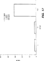
図1:喘息関連遺伝子のマッピングに有用な多形遺伝子マーカーの相対順序およびセンチモルガン[cM]での距離を示すマップ。
図2:ヒト染色体5q31-q33およびマウスのシンテニー領域の遺伝子マップの図解。
図3:気管支収縮刺激に対する感受性が増加したマウスにおけるアトラキュリウム誘発気道反応性に関するマウス第13染色体のLODスコア曲線。
図4:ヒトおよびマウスIL-9遺伝子のエキソン5に対応するアミノ酸配列の配置。最初の配列はヒト遺伝子のトレオニン対立遺伝子から翻訳される。中央の配列はヒト遺伝子のメチオニン対立遺伝子から翻訳される。最後の配列はマウス遺伝子から翻訳される。
図5:ヒトIL-9遺伝子対立遺伝子と国際単位で測定した血清中総IgE価の相関を示すヒストグラム。S/Sはトレオニン/トレオニンの人、S/Rはトレオニン/メチオニンの人、およびR/Rはメチオニン/メチオニンの人を指す。
図6:IL-9座での単純配列リピート多型性の図解。
図7:トレオニン117 IL-9のcDNA配列の翻訳。
図8:メチオニン117 IL-9のcDNA配列の翻訳。
図9:トレオニン117によるpFlag発現構築物のマップ。
図10:ライゲーション部位周辺領域からのトレオニン117 cDNAのpFlag発現構築物の配列。
図11:メチオニン117によるpFlag発現構築物のマップ。
図12:ライゲーション部位周辺領域からのメチオニン117 cDNAのpFlag発現構築物の配列。
図13:IL-9組換え蛋白のウェスタンブロット。
図14:阻害ペプチドのアミノ酸配列。
図15:IL-9媒介MO7e増殖のKP-16による阻害。
図16:IL-9媒介MO7e増殖のKP-20による阻害。
図17:IL-9媒介MO7e増殖のKP-23による阻害。
図18:IL-9媒介MO7e増殖の様々なチロフォスチンによる阻害。
図19:IL-9媒介MO7e増殖の様々なアミノステロールによる阻害。
図20:インビボでの抗原反応におけるIL-9の役割の特徴づけ。
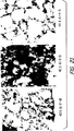
図21:対照、卵白アルブミンチャレンジ、および抗IL-9前処置動物からの肺の組織学検査。
図22:マウスIL-9受容体に対する遮断抗体によるインビボでの抗原反応の阻害。
図24:ヒトメチオニン117 IL-9およびトレオニン117 IL-9の発現。
図25:ヒト組換え型メチオニン117およびトレオニン117型IL-9の可溶性抗体に対する結合。
図26:非刺激および刺激マウス脾細胞におけるIL-9の定常状態濃度。
図27:化学部分の付録。
図28:イヌ、魚、サメから単離したアミノステロール。
発明の詳細な説明
本出願者らは、喘息および関連疾患を含むアトピー性アレルギーの診断、予防または治療において用いることができるIL-9経路およびこの経路に作用する組成物を明らかにすることによって、当技術分野における必要性を解決した。喘息には、可逆的な気道閉塞を伴う気道の炎症性疾患が含まれる。アトピー性アレルギーは、アトピー、ならびに喘息、気管支過反応(BHR)、鼻炎、蕁麻疹、腸のアレルギー性炎症疾患、およびさまざまな形の湿疹を含む関連疾患を意味する。アトピーは、対照と比較して、血清総IgEが上昇するかまたはアレルゲンに対する異常な皮膚検査反応となって発現する、環境アレルゲンに対する過敏感反応である。BHRは、さまざまな刺激に対する気管支収縮反応が増強した気管支過反応を意味する。
喘息関連疾患を示す家族のDNAを解析することによって、本出願者らは、アトピーの発現を測定できるものの一つとして、血清総IgEの生物学的な多様性と相関関係にある、IL-9遺伝子における多型性を確認した。IL-9遺伝子(喘息関連因子1、すなわちAAF1としても知られている)とは、ヒト骨髄系およびリンパ系の制御に関するさまざまな機能を示すサイトカイン、インターロイキン-9の遺伝子座を指す。本発明のIL-9遺伝子は、ヒトの第5染色体のq31〜q33領域中およびマウスでは第13染色体中に認められる。
本出願者らは、多型性という語で、「遺伝子座」と名付けられた特異的なDNA配列における、普通の配列からの変化を意味させている。一般的に、当業者がある遺伝子座を含む2個以上の対立遺伝子を同定し、最も少ない共通遺伝子座が1%以上の頻度で存在する場合、その遺伝子座は多型であると定義される。
本発明の多型性は、IL-9の117残基目のアミノ酸置換をもたらす。特に、親水性アミノ酸のトレオニンの代わりに、本発明のIL-9は、疎水性アミノ酸メチオニン(Met IL-9)を提示する。遺伝子レベルでは、本発明の多型性は、レナウルドら(Renauldand colleagues)(1990)[GenBank寄託番号M30135、およびM30136]54によって説明されているように、ヒトIL-9遺伝子の3365番目のヌクレオチド、またはケラーら(Kelleher)(1991)によって報告されている[GenBank寄託番号M86593]、ヒトIL-9遺伝子の配列の4244塩基目に相当するヌクレオチドでの、チミン残基によるシトシン残基の置換である55。
IL-9の対立遺伝子の片方または両方のいずれかにおいて、117番目のアミノ酸にトレオニン(Thr)を有する個体(Thr/ThrまたはThr/Met)は一般的に、喘息に対する罹病性またはアトピー性アレルギーの表現型を示すので、これらの遺伝子型は、調査した集団における血清総IgEレベルの平均を高くするという特徴を有する。これに対し、IL-9の対立遺伝子の両方で、117番目のコドンにメチオニン(Met)を有する個体(Met/Met)は、喘息を起こさず、皮膚検査反応における異常が少なく、血清総IgEが低い。したがって、IL-9のMet/Met遺伝子型により、喘息またはアトピー性アレルギーに対する防御が行われていると考えられる。
したがって、本発明は、117番目にメチオニンを有するヒト・インターロイキン9(Met IL-9)をコードする塩基配列を含む単離精製されたDNAまたはその断片を提供する。本発明はまた、実質的に相同な配列だけでなく、このDNAの縮退配列も含んでいる。本発明のIL-9の起源はヒトである。またはDNAもしくはその断片は、当技術分野において既知の方法を用いて合成してもよい。また、認められた技術によってDNAを構築し、そのDNAを発現ベクターにクローニングし、化合物を発現するはずの細胞を、このベクターでトランスフェクションするという、遺伝子工学的技術によって、この化合物を産生することもできる。例えば、サムブルックら(Sambrook)「分子クローニング:実験マニュアル」第2版(コールドスプリングハーバー研究所出版[1985])に記載の方法を参照のこと。
気道過反応が、事実上すべての喘息患者と、近交マウスのいくつかの系統(DBA/2)において見られる84。気道過反応は、ヒトにおいて、喘息の発症に関する危険因子であり、喘息の動物モデルにおいては、喘息に対する治療の効能を評価するための生理学的な尺度として用いられる。ヒト79およびマウスの遺伝子マッピングの結果(実施例1および2を参照)とともに、これらのデータは、マウスの気道反応において、マウスIL-9遺伝子産物についての重要な役割を示唆している。特に、過反応DBA/2(D2)マウスは、定常レベルのIL-9発現において、C57BL/6(B6)過反応マウス84とは異なっている(実施例14、図26を参照)。さらに、IL-9/IL-9レセプターに対する遮断抗体による前処理は、マウスにおいて、抗原に誘導される気道過反応および炎症を、場合によっては完全に防止することができるが、これは、これらの免疫応答におけるIL-9に関する重要な制御的な役割を示している。このように、分子の異なる変化により、ヒトおよびマウスでは気道過反応における生物学的な多様性が作出されるが、これらの変化は、この経路を制御する同一の遺伝子(IL-9/IL-9R)の中で起きていることをこれらのデータは示している。これらをまとめると、これらの観察から、気道過反応、喘息、およびアトピー性アレルギーにおける、IL-9およびIL-9レセプターの重要な役割が確認されている。さらに、これらのデータから、IL-9が、そのレセプターと相互作用することを遮断する従来からの薬剤が、上で詳述したような、抗原に誘導される反応から保護することが示される。
アトピ性アレルギー、気管支過反応、喘息、および関連疾患の発病におけるIL-9の決定的な役割を明らかにするさらなる証拠は、マウスにおいては、数多くの抗原誘導応答に対してIL-9が決定的であるという本出願者らの観察から直接に得られる。抗原によるエアロゾル投与の前に抗体で前処理して、IL-9の機能を抑制制御すると、抗原に誘導される応答から動物を完全に防御することができる。これらの応答には、気管支過反応、好酸球増加症および気管支洗浄液中の細胞数の増加、炎症に関連した肺の組織学的変化、および血清総IgEの増加が含まれる。したがって、アトピー性アレルギーの発病に重要な、喘息に関連したアレルギー性炎症を特徴づけるこのような反応を、IL-9の機能を抑制制御することによって治療することは、本発明の範囲内に含まれる。
本出願者らはまた、IL-9レセプターに対する「アゴニストおよびアンタゴニスト」を投与することによるIL-9の活性の制御について開示する。熟練した当業者には、必要な3次元構造的コンホメーションを有する分子と、レセプター結合に必須または重要な残基を含む分子のすべてが、本発明の範囲内にあることが容易に理解できると思われる。本出願者らは、KP-16ペプチド(IL-9の43〜60残基目)およびKP-20ペプチド(IL-9の71〜90残基目)(例えば、アプライドバイオシステムズ社モデル431Aペプチド合成機(Applied Biosystems Model 431A Peptid Synthesier)など、標準的なペプチド自動合成技術を用いて産生される)が、IL-9のアンタゴニストとして作用することを明らかにした。特に、本出願者らは、成熟蛋白質の43〜60残基目と71〜90残基目が、レセプター結合に重要らしいことを明らかにした。さらに、これらの残基には、エキソン4のほとんど(アミノ酸44〜88)が含まれ、逆平行ヘリックス構造を形成すると推定される。この蛋白質の3次元構造から、52番目のセリンおよび/または53番目のグルタミン酸が、85番目のリジンと相互作用し、56番目のセリンが82番目のリジンと相互作用し、また、59番目のトレオニンが78番目のバリンと相互作用することが示唆される。これらの逆平行ヘリックスおよび関連する機能的な原子団の3次元的な配置が、レセプター結合にとって重要な必須の3次元的な立体的コンホメーションを表すため、これらの関係を刺激する化合物は、本発明の範囲内である。
喘息様表現型に関連するIL-9配列および喘息様表現型の欠如に関連するIL-9配列を明らかにしたことから、抗原に対する肺の炎症反応がIL-9とは無関係であり、したがってIL-9の機能の抑制制御は抗原誘導反応に対する防御であることが示された。さらに、本出願者らは、アトピー性アレルギーおよび関連疾患に対する罹病性を診断する方法、ならびにIL-9とそのレセプターとの関係に基づいてこれらの疾患を治療するための方法も提供する。
レセプターは、分子を認識して結合する、可溶性または膜結合型の成分であり、本発明のIL-9レセプター(喘息関連因子2、すなわちAAF2としても知られている)は、IL-9を認識して結合する成分である。IL-9レセプターの機能には、IL-9様分子に結合することと、特異的な細胞の中でその制御シグナルを増幅させることが含まれる57〜60。この機能を阻害すると、IL-9の発現またはIL-9によって調節される機能の抑制制御、すなわち低下がもたらされる。したがって、IL-9とIL-9レセプターとのこの相互作用のおかげで、生物の一定の機能が、調節または制御されている。レセプターの一般的な議論については、グッドマンとギルマンの(Goodman and Gilman's)「治療学の薬学的基礎(The Phamacologic Basis of Therapeutics)」(第7版、マクミラン出版社、米国ニューヨーク、1985)を参照。
一つの診断的な態様には、IL-9のDNA配列における変異を認識することが含まれる。一つの方法は、当業者によって理解されているように、充分なハイブリッド形成を行う条件下で、本発明のIL-9に相補的な配列を有する核酸分子(プローブとしても知られている)を導入することを含む。一つの態様においては、この配列は、Met117 IL-9またはThr117 IL-9に特異的に結合し、別の態様において、Met117 IL-9とThr117 IL-9の両方に結合する。これらの疾患に関連したDNA配列の変異を識別する別の方法は、当技術分野において周知の複数の方法によって、直接DNA配列を解析することである77。別の態様には、これらの疾患に関連する、IL-9遺伝子におけるDNA配列の変異を検出することが含まれる73〜77。これらには、ポリメラーゼ連鎖反応、制限酵素断片長の多型性(RFLP)解析、および一本鎖のコンホメーション解析が含まれる。好ましい態様において、本出願者らは、本明細書において説明されているStyI RFLPを用いて、ThrおよびMet対立遺伝子に関係したIL-9の多型性を、遺伝子レベルで識別するための方法を具体的に提供する。別の態様においては、Nla、Pfim1、PflM1およびNco1 RFLPを用いて、IL-9遺伝子のこれら2つの対立遺伝子を区別する。
別の態様は、アトピー性アレルギーおよび関連疾患を治療することを含む。好ましい態様において、本出願者らは、Met IL-9に匹敵する活性と、IL-9の活性を抑制制御するのに充分な程度にIL-9レセプターに結合する能力を有する化合物を投与する方法を提供している。Met IL-9に匹敵する活性を有する化合物は、類似の機能を有するが、必ずしも同一でない化合物である。このように、これは、IL-9レセプターには結合するが、同一の生理学的効果をもたない。実施例には、さまざまな点突然変異および/または欠失を有するIL-9のアミノ酸配列、ならびに、実質的にそれと相同な配列が含まれる。例えば、このような化合物は、当技術分野において既知の技術にようて測定したところに従うと、Thr IL-9がIL-9レセプターに結合するのを阻害するかもしれない。本発明はまた、上述のアミノ酸配列の機能的に有効な断片を含む。このような技術の一つにおいて、Thr IL-9は、IL-9レセプターの「リガンド」と考えてもよく、この2つの間の結合は、グッドマンとギルマンの(Goodan and Gilman's)「治療学の薬学的基礎(The Pharmacologic Basis of Therapeutics)」(第7版、マクミラン出版社、米国ニューヨーク、1985)において示されているように、当技術分野において周知のリガンド結合測定法によって評価することもできる。
別の態様において、この化合物は、構造的にIL-9のMet対立遺伝子に似ているかもしれない。とすると、そのような化合物は、IL-9の117番目のコドンでメチオニンを取り込むか、または別の親水的なアミノ酸を取り込むかもしれない。したがって、本発明の範囲に含まれるのは、117番目の位置で、アラニン、バリン、ロイシン、イソロイシン、プロリン、フェニルアラニン、トリプトファン、およびメチオニンからなる群より選択されたアミノ酸による、Thrの置換を含むIL-9変異体である。または本発明の化合物は、Met IL-9に類似する構造的な組成物を有するIL-9の断片として存在しているかもしれない。本発明の別の態様において、この化合物はMet IL-9に匹敵する機能を保持しているかもしれないが、構造的には、Met IL-9に似ていないかもしれない。例えば、この化合物の組成には、アミノ酸以外の分子が含まれるかもしれない。この例は、単なる例示にすぎず、通常の技術を有する当業者は、効果的なアンタゴニストをもたらす、別の置換および/または欠失によるIL-9の類似体も、本発明の範囲に含まれることを容易に理解できよう。既に検討したように、必要な3次元構造的コンホメーションを有する分子と、レセプター結合に必須または重要な残基を含む分子のすべてが、本発明の範囲内に含まれる。
特異的な測定法は、IL-9の既知の制御、部分的には、Tリンパ球、IgE合成、および肥満細胞からの放出に基づくことができる54〜60。別の測定法には、ヒトIL-9が、M07e細胞において、複数の蛋白質の迅速で一過性的なチロシンリン酸化を特異的に誘導できることが含まれる57。この反応は、IL-9レセプターの発現および活性化に依存しているため、利用する価値があるかもしれない化合物の特徴を調べるための簡単な方法または測定法になる。転写因子Stat3のチロシンリン酸化は、IL-9の作用に特異的に関連していると考えられ58、この反応は、本発明の範囲内の化合物の特徴を調べるための簡単な方法または測定法になる。さらに、IL-9およびIL-9様分子の機能の特徴を調べるための別の方法には、細胞増殖測定法を用いてヒトIL-9の機能を測定するために用いられる、ATCCから入手可能な周知のマウスのTS1クローンおよびD10クローンが含まれる59。本発明の一部を構成するMet IL-9は、IL-9レセプターの「弱いアゴニスト」と見なすことができる。このような弱いアゴニストは、本発明のもう一つの好ましい態様である。本発明によれば、アゴニストという語は、レセプターと相互作用するかまたは結合して、内生する化合物の効果の少なくともいくつかを模倣する化合物を含む。ある細胞の表面上でIL-9レセプターと相互作用または結合するアゴニストは、このサイトカインの作用の特徴である、一連の生化学的および生理学的な変化を開始させる45〜51、54〜61。本発明に係る別の弱いアゴニストを同定するために、本明細書および引用された文献2、45〜51、54〜60で説明されているところにしたがって、IL-9レセプターへの結合またはIL-9様機能について調べることができる。
本発明は、また、IL-9とそのレセプターのアンタゴニストを含む。アンタゴニストは、それ自体は薬学的活性をもたないが、アゴニストの作用を阻害することによって効果をもたらす化合物である。本発明のアンタゴニストを同定するために、本明細書および引用された文献2、45〜51、54〜60で説明されているところにしたがって、既知のアゴニストとの競合的結合、またはIL-9様機能の抑制制御を調べることができる。
アゴニストまたはアンタゴニストのいずれかの結合には、イオン力、水素結合、疎水性相互作用、ファンデルワールス力、および共有結合を含む、既知のあらゆる相互作用のタイプが含まれる。多くの場合、アゴニストまたはアンタゴニストとレセプターとの相互作用において複数のタイプの結合が重要である。
さらなる態様において、これらの化合物は、IL-9の類似体であってもよい。当技術分野においてすべて十分に説明されている方法62によって作出することができる、この遺伝子の単離DNA配列における点突然変異、塩基置換、および/または欠失によって、IL-9の類似体を作出してもよい。本発明はまた、IL-9のスプライシング変異体を含み、その5個のエキソンに1個以上の欠失を有するインターロイキン-9の単離核酸配列を開示している。「スプライシング変異体」という語は、本明細書においては、少なくとも1個のエキソンを含むヒトIL-9をコードしている単離精製されたDNA分子を意味している。当技術分野においては、スプライシング変異体が、自然に発現しているという証拠はない。したがって、本発明は、ヒトIL-9のエキソン1、4および5を有する、単離核酸を提供する。本発明の範囲内にある別の変異体には、エキソン1、2、3、4および5;エキソン1、2、3および4;エキソン1、2、4および5;ならびに、エキソン1、3、4および5を含む配列が含まれる。これらのエキソンは、さまざまな点突然変異を含むものと理解されるべきである。
IL-9に由来する拮抗ペプチドの具体的な例としては、エキソン4に由来するKP-16(配列番号:13)およびKP-20(配列番号:14)が含まれる。エキソン4は、44個のアミノ酸をコードしているが、上記のペプチドは、それぞれ16個と20個のアミノ酸を含み、互いに重複していない。これらのペプチドは、個別に測定しても組み合わせて測定しても、かなりの阻害活性を示す。さらに、エキソン5に由来するKP-23(配列番号:15)およびKP-24(配列番号:16)も、同様の活性を示す。IL-9のエキソン1から5のいずれかを1個以上欠失させても、IL-9のスプライシング変異体を形成させることができる。上で示したように、これらのエキソンに由来するペプチドは制御能力を示し、このため、喘息を含むアトピー性アレルギーを治療するのに有用である。
多酵素システムにおいては、第一の酵素または制御的な酵素を、多酵素システムの最終産物によって活性化または阻害できることが、当技術分野において知られている。最終産物の濃度を定常状態の濃度よりも上昇させると、最終産物は、一連の流れの中の制御的な酵素の特異的な活性化因子または阻害因子として作用する。また、このようなフィードバック機構が、IL-9のシステムにも関連するため、本発明のさまざまなポリペプチドは、IL-9レセプターの活性に対し、またおそらく、喘息の発病に関与するその他のサイトカインおよびそれらのレセプターの発現または機能に対して、このような活性化または阻害調節を行うことができることが観察されている。
本発明にはまた、当業者には慣例的な知識を用いて行うことができるアゴニストまたはアンタゴニストの改変が含まれる。例えば、レセプターに対する化合物の親和性は、一般的に、この化合物の化学構造に密接に関連している。したがって、構造-活性関係を用いて、本発明のアゴニストとアンタゴニストの改変を行うことができる。例えば、結晶学/X線回折とNMRの技術を用いて、本発明の改変を行うことができる。
例えば、分子画像を用いて、欠失突然変異体の構造モデルを構築するための鋳型として用いることができるヒトIL-9の3次元構造を作出することができる。そして、これらのモデルを用いて、IL-9に匹敵するが生物学的活性はそれよりも低いIL-9レセプターに対する親和性を有する、IL-9分子の突然変異体を同定し構築することができる。より低い生物学的活性とは、IL-9よりも2倍から100,000倍低いことを意味し、好ましくは、IL-9よりも100倍から1,000倍低い。
さらに別の態様において、これらの化合物は、レセプターの構造に対する動態学的プローブとして用いることも、IL-9依存的な細胞系によりレセプターアンタゴニストを開発するために用いることもできる。
さらに、本発明は、IL-9またはIL-9レセプターの合成を阻害するか、またはそれらの生物学的安定性を低下させる化合物も提供する。生物学的安定性とは、分子の合成とその分解との間の時間の測定値のことである。例えば、蛋白質、ペプチド、またはペプチド模倣薬89の安定性は、D型アミノ酸を用いて引き延ばすことができ、または酵素による分解に対してより感受性になるように配列を変更することによって短縮することができる。
別の態様において、本発明のアゴニストとアンタゴニストは、IL-9とIL-9レセプターに対する抗体である。IL-9およびそのレセプターに対する抗体は、当技術分野において周知の標準的な技術を用いて作製されるモノクローナル抗体でも、ポリクローナル抗体でもよい(ハーローとレーンの(Harlow & Lane's)「抗体−実験マニュアル(Antibodies-A Laboratory Manual)」(コールドスプリングハーバー研究所、1988)を参照)。これらを用いて、IL-9がレセプターに結合するのを阻害することができる。一つの態様において、この抗体は、IL-9と相互作用する。別の態様においては、この抗体は、IL-9レセプターと相互作用する。これらの抗体を誘導するために用いられるIL-9は、上で検討したIL-9の変異体のいずれでもよい。
抗体は、当技術分野において標準的な技術を用いて、IL-9またはIL-9レセプターのペプチド配列からも作製される(免疫学のプロトコール、第9章、ワイリー社(Wiley)を参照のこと)。遮断抗血清を作製するために用いることができる、マウスIL-9レセプターからのペプチド配列は、以下のものが同定されている:GGQKAGAFTC(1〜10残基目)(配列番号:19)、LSNSIYRIDCHWSAPELGQESR(11〜32残基目)(配列番号:20)、およびCESYEDKTEGEYYKSHWSEWS(ペプチドを担体蛋白質に結合させるため、N末端にCys残基を付加した184〜203残基目)(配列番号:21)。さらに、ヒトIL-9レセプターに対する遮断抗体に結合するエピトープは、成熟ヒトIL-9レセプターの8〜14残基と同定されている。(TCLTNNI)(配列番号:22)およびヒトIL-9に対する遮断抗体に結合する2つのエピトープも、50〜67残基(CFSERLSQMTNTTMQTRY)(配列番号:23)および99〜116残基(TAGNALTFLKSLLEIFQK)(配列番号:16)であると同定されている。マウスの配列において遮断抗体を産生するペプチドに相当する、ヒトのペプチドおよびヒトのエピトープが、治療用抗体を作製するのに有用である可能性が最も高い。
さらに別の態様において、本発明の化合物は、アトピー性アレルギーおよび喘息の治療に有効となるよう、IL-9経路の機能または制御を変化させる蛋白質を含む、化学的な成分と結合させてもよい61。これらの蛋白質には、アトピー性アレルギーおよび喘息の治療にさらに有益となりうるIL-4、IL-5、IL-3、IL-2、IL-13、およびIL-10を含む別のサイトカインおよび増殖因子の組合せ62が含まれる。さらに、本発明のIL-9は、リン酸化によって共役させてもよく、ビオチニル酸、チオ酸、アセチル酸、ヨウ素酸、および図27に示したクロスリンキング試薬(ピアス社(Pierce))のいずれと共役してもよい。
さらなる態様において、本発明には、IL-9に結合する可溶性IL-9レセプター分子を投与することによって、IL-9の発現または機能を抑制制御することが含まれる。レナウルドら(Renauld)59は、IL-9レセプターに可溶性形態が存在することを明らかにした。この分子は、細胞に結合したレセプターへのIL-9の結合を阻害し、IL-9のアンタゴニストとして作用させるために用いることができる。可溶性レセプターは、サイトカインまたは別のリガンドに結合して、それらの機能を調節するために用いられてきた87。可溶性レセプターは、溶液中または膜の外側に生じる、膜結合レセプターの形態である。可溶性レセプターは、一般的には膜と結合しているこの分子のセグメントが存在しないために、生じるのかもしれない。このセグメントは、当技術分野においては、一般的に、この遺伝子の膜通過ドメインまたはこの蛋白質の膜結合セグメントと呼ばれている。したがって、本発明の一つの態様において、可溶性レセプターは、膜結合レセプターの断片または類似体を意味する。本発明の別の態様において、レセプターが膜の中に入り込めなくなるか、またはレセプターは膜に入り込めるがその中に留まることができなくなるよう、膜と結合するセグメントの構造を改変する(例えば、DNA配列の多型性または遺伝子の突然変異)。このように、可溶性レセプターは、対応する膜結合型とは対照的に、膜との結合に重要な、この遺伝子またはレセプター蛋白質の1個以上のセグメントに違いがある52、53。
これらの化合物は、IL-9に結合するよう作用する可溶性IL-9レセプターの既知の形態であってもよい。またはこれらの化合物は、IL-9レセプターの既知の形態に似ているが、断片として存在するものでもよい。本発明の別の態様において、この化合物は、可溶性IL-9レセプターに匹敵する機能を保持しているが、組成においては、可溶性IL-9レセプターに似ていなくてもよい。例えば、この化合物の組成には、アミノ酸以外の分子が含まれていてもよい。このように、これらの化合物は、IL-9に結合して、IL-9がその細胞表面レセプターに作用するのを阻害する。
本発明のさらなる態様は、アンチセンスまたは遺伝子治療に関する。アンチセンス、または遺伝子治療として提供される場合、特定の選択された効果をもたらすよう、改変したDNA分子を作製することができることが、当技術分野において、今では分かっている。IL-9レセプターをコードする本来のDNAセグメントは、他の哺乳動物すべてのDNA鎖と同じように、センス鎖とアンチセンス鎖が水素結合によって一緒になった2本鎖を有する。レセプターをコードするmRNAは、チミジンがウリジンに置き換わっていると予想されるが、センス鎖と同じ塩基配列を持っている。したがって、レセプターの配列に関する知識に基づいて、合成オリゴヌクレオチドを合成することができる。これらのオリゴヌクレオチドは、このレセプターをコードするDNAおよびRNAに結合することができる。本発明の活性断片は、mRNAおよびDNAのコード鎖に相補的であり、通常、少なくとも15ヌクレオチド、より一般的には20ヌクレオチド、好ましくは30ヌクレオチド、より好ましくは50ヌクレオチド以上である。センス鎖とアンチセンス鎖との間の結合強度は、総水素結合に依存する。したがって、熟練した当業者は、mRNAの総塩基数に基づいて、オリゴヌクレオチド配列の最適な長さを容易に計算することができると思われる。
この配列は、mRNAの配列いずれかの部分に対して相補的である。すなわち、5'末端もしくはキャッピング部位の近傍、またはキャッピング部位の下流、キャッピング部位と開始コドンとの間であるかもしれないし、非コード領域またはコード領域の全部または一部だけをカバーしているかもしれない。アンチセンス配列が結合する特定の部位は、所望の阻害の程度、配列の独自性、アンチセンス配列の安定性などによってさまざまである。
本発明の実施において、IL-9レセプターの発現は、上記の合成アンチセンスオリゴヌクレオチド配列を有効量投与することによって抑制制御される。本発明のオリゴヌクレオチド化合物は、ヒトIL-9またはIL-9レセプターをコードするmRNAに結合して、これらの蛋白質の発現(翻訳)を阻害する。グラスら(Gruss et al.)の「インターロイキン9は、初期および培養ホジキン細胞およびリード-スタンバーグ細胞によって発現されている(Interleukin 9 is expressed by primary and cultural Hodgkin and Reed-Stemberg cells.)」、Cancer Res.52:1026-31(1992年2月15日)を参照のこと。
IL-9の点突然変異、挿入、欠失、またはスプライシング突然変異など、さまざまな突然変異を有する単離DNA配列も同様に、遺伝子治療において有用である。
IL-9レセプターの直接的な制御に加えて、本発明は、シグナル伝達の阻害を含む下流制御の方法も含む。特に、本発明のさらなる態様は、チロシンのリン酸化阻害に向けられている。当技術分野において、高度な発エルゴンリン酸基転移反応は、キナーゼとして知られる多様な酵素によって触媒されることが分かっている。言い換えると、キナーゼは、ATPと代謝物との間でリン酸基を転移させる。IL-9は、多種の蛋白質のチロシンのリン酸化を誘導する。すなわち、JAK1チロシンキナーゼおよびJAK3チロシンキナーゼの活性化に加えて、IL-9は、Stat3のチロシンのリン酸化も誘導する58ことが、当技術分野において知られている。Stat3のリン酸化は、IL-9シグナル伝達経路に独特なものであるため、阻害因子にとって完全な標的となる58。本発明は、チロシンキナーゼ蛋白質の特異的な阻害因子であるチロフォスチンを、その範囲内に含む。このように、チロフォスチン(例えば、カルバイオケム社(Calbiochem)から入手可能)およびこれらのキナーゼのその他の同様の阻害因子は、シグナル伝達を調節する上で有用であり、アトピー性アレルギーおよび喘息を治療する上でも有用である。
本発明のさらに別の局面において、驚くべきことに、アミノステロール化合物も、IL-9の刺激によるシグナル伝達の阻害において有用であることが分かった。本発明において有用なアミノステロール化合物が、米国特許出願第08/290,826号と、その関連出願第08/416,883号および第08/478,763号で説明されており、また、第08/483,059号と、その関連出願第08/483,057号、第08/479,455号、第08/479,457号、第08/475,572号、第08/476,855号、第08/474,799号、および第08/487,443号でも説明されており、これらは、参照として本明細書に組み入れられる。
さらに、本発明には、薬学的に許容される担体と共に、本発明の化合物を含む薬学的組成物が含まれる。薬学的に許容される担体は、水および油などの滅菌した液体で、ピーナッツ油、大豆油、ミネラル油、ゴマ油などの、石油性、動物性、植物性、または合成した油が含まれる。薬学的組成物が静脈に投与される場合には、水が、好ましい担体である。食塩水および含水デキストロースとグリセロールの水溶液も、特に、注射可能な水溶液の液状担体として用いることができる。適当な薬学的担体は、マーチン、E.W.(Martin, E.w.)の「レミントンの製薬科学(Remington's Phamaceuticals Sciences)」で説明されており、これは特に、参照として本明細書に組み入れられる。
本発明に係る治療方法に用いられる化合物は、治療されるべき条件、部位特異的治療の必要性、投与すべき薬物の量などに応じて、全身的または局所的に投与することができる。
局所的な投与法を用いることができる。溶液、懸濁液、ゲル、軟膏、または塗剤などのあらゆる一般的な局所用の組成物を用いることができる。例えば、「レミントンの製薬科学」第17版、ペンシルバニア州イーストン、マック出版社(Mack Publishing Company)で例示されているように、局所的処方剤の調製が、薬学的処方剤の技術分野において充分に説明されている。局所的な施用のためには、粉末またはスプレー、特にエアロゾルの形状で投与することもできる。全身投与に合わせた薬学的組成物として、活性成分を投与してもよい。知られているように、薬物を全身的に投与すべき場合には、粉末、丸薬、錠剤などにして調製したり、経口投与用にシロップまたはエリキシルとして調製してもよい。静脈内、腹腔内、または病変部内に投与するためには、注射によって投与可能な溶液または懸濁液として化合物を調製する。ある場合には、これらの化合物を座薬の形状に処方するのが有用であろうし、皮下に蓄積させたり筋肉内注射をするためには、長時間かけて放出されるような処方剤とするのが有用なこともあろう。好ましい態様において、本発明の組成物は、吸入によって投与してもよい。吸入療法のためには、化合物を、用量計付きの吸入器によって投与するのに有用な溶液にするか、または乾燥粉末吸入器に適当な形状にすることができよう。
有効用量とは、IL-9の発現、またはIL-9によって調節される機能のいずれかを抑制制御する用量のことである。所定の有効用量は、条件によってさまざまであり、また一定の場合でも、治療すべき症状の重篤さおよび治療に対する患者の感受性によってさまざまである。したがって、絶えず実験によって、その時間と場所における所定の有効用量を決定するのが一番よい。しかし、本発明による喘息関連疾患の治療において、重量で0.001%〜5%の間、好ましくは、約0.01%〜1%を含む処方薬が、通常は、治療学的に有効な用量を構成すると予想される。全身的に投与する場合には、1日につき、体重1kg当たり0.01mg〜100mgの間の用量、しかし好ましくは、約0.1〜10mg/kgの用量が、ほとんどの場合、治療結果に効果をもたらすと考えられる。
本出願者らはまた、IL-9の発現、またはIL-9によって調節される機能を抑制制御する化合物をスクリーニングする方法を提供する。当技術分野において標準的な技術を用いて、IL-9によって発現される機能が抑制制御されるか否かを判定することができる57〜60。特定の態様において、本出願者らは、Met IL-9と同等の機能を有する化合物を同定する方法を提供する。したがって、一つの態様において、インビボでのIL-9機能の抑制制御における、化合物の効能を評価するための、当技術分野において周知の技術42を用いて、血清総IgEを測定することができる。別の態様においては、インビボでのIL-9機能の抑制制御における化合物の効能を評価するための、当技術分野において周知の技術42を用いて、気管支過反応、気管支肺胞洗浄液、および好酸球増加症を測定することができる。さらに別の態様において、IL-9の機能は、インビトロで評価することができる。当業者に知られているように、ヒトIL-9は、M07e細胞において、複数の蛋白質の迅速で一過性的なチロシンのリン酸化を特異的に誘導する。転写因子Stat3のチロシンのリン酸化は、IL-9の作用に特異的に関連していると考えられる。IL-9レセプターの「安定的な発現」に依存する、IL-9およびIL-9様分子の機能の特徴を調べるための別の方法では、周知のマウスのTS1クローンを用いて、細胞増殖測定法によってヒトIL-9の機能を評価する59。
また本発明には、IL-9レセプターを発現する細胞系に基づいて、飽和可能で特異的なリガンド結合を見つけるための簡単なスクリーニング法が含まれる46、59。IL-9レセプターは、K562、C8166-45、B細胞、T細胞、肥満細胞、好中球、巨核球(UT-7細胞)53、ヒト巨核芽性白血病細胞系MO7e57、TF159、マクロファージ、胎児胸腺細胞、ヒト腎細胞系29353、およびマウス胚海馬前駆細胞系46、52、53を含む、広く多様な細胞型において発現している。別の態様において、可溶性IL-9レセプターを用いて、リガンド結合およびレセプターのアンタゴニストの可能性があるものを評価することができる。
本発明の実施には、従来の用語および当業者の通常の技術の範囲内にある、分子生物学、薬学、免疫学、および生化学の技術が用いられる。例えば、サムブルックら(Sambrook)「分子クローニング:実験マニュアル(Molecular Cloning:A Laboratory Manual)」第2版(コールドスプリングハーバー研究所出版[1985])、またはアウスウーベルら(Ausubel)「分子生物学の最新プロトコール(Current Protocols in Molecular Biology)」ジョン・ワイリー・アンド・サンズ社(John Wiley & Sons, Inc.)[1994]を参照のこと。
ところで、本発明者らは、次の基本的な背景情報を提供する。身体の遺伝的物質であるDNAは、46本の染色体の上に配置されているが、染色体の各々は、セントロメアによって結合された2本の腕を有する。各染色体は、pまたはqと名付けられたセグメントに分けられる。pという記号は、セントロメアから最も近いテロメアまでを測定した染色体の短腕を同定するときに用いられる。染色体の長腕は、qという記号で表される。染色体上の位置は、染色体番号(すなわち、5番染色体)、ならびにpまたはq領域の位置(すなわち、q31-q33)によって示される。さらに、身体は、性染色体、XおよびYを持っている。減数分裂の過程で、XおよびY染色体は、偽常染色体領域として知られている領域でDNA配列情報を交換する。
DNA、すなわちデオキシリボ核酸は、4つの異なる塩基性化合物、アデニン[A]、チミン[T]、シトシン[C]、およびグアニン[G]を含む、2本の相補的なヌクレオチド鎖から成る。一方の鎖のAは、もう一方の鎖のTと結合し、一方の鎖のCは、もう一方の鎖のGと結合して、相補的な「塩基対」を形成し、各対は、それぞれの鎖に一つずつ塩基を有する。
3個のヌクレオチド(「コドン」)が連続したものをグループにすると、1個のアミノ酸をコードしている。したがって、例えば、3個のヌクレオチドCAGは、アミノ酸のグルタミンをコードしている。天然に存在する20個のアミノ酸と、それらの一文字コードは、以下の通りである。
アラニン Ala A
アルギニン Arg R
アスパラギン Asn N
アスパラギン酸 Asp D
アスパラギン、
またはアスパラギン酸 Asx B
システイン Cys C
グルタミン Gln Q
グルタミン酸 Glu E
グルタミン、
またはグルタミン酸 Glx Z
グリシン Gly G
ヒスチジン His H
イソロイシン Ile I
ロイシン Leu L
リジン Lys K
メチオニン Met M
フェニルアラニン Phe F
プロリン Pro P
セリン Ser S
トレオニン Thr T
トリプトファン Trp W
チロシン Tyr Y
バリン Val V
アミノ酸は、蛋白質を構成する。アミノ酸は、親水性、すなわち水に対して親和性を示すものもあり、または疎水性、すなわち水を忌避するものもある。例えば、G、A、V、L、I、P、F、Y、W、C、およびMと名付けられたアミノ酸は疎水性で、また、S、Q、K、R、H、D、E、NおよびTと名付けられたアミノ酸は親水性である。一般的に、アミノ酸の疎水的または親水的な性質は、ペプチド鎖の折りたたみに影響を与え、結果的に、蛋白質の3次元構造に影響を与える。
DNAは、以下のように蛋白質と関係している。

実際、真核生物の遺伝子は、イントロンによって中断されたエキソンからなるため、それらによってコードされる蛋白質について不連続である。RNAに転写された後、イントロンはスプライシングによって切り出され、成熟メッセンジャーRNA(mRNA)を生成させる。エキソンの間のスプライシング部位は、典型的には、スプライシング作用に対するシグナルとして作用するコンセンサス配列によって決められる。スプライシングは、一次RNA転写産物からのイントロンを除去することと、切り出されたイントロンの両側に残ったRNAの末端を結合または融合することから成り立つ。イントロンの有無、イントロンの組成物、および遺伝子当たりのイントロンの数は、同じ生物種の系統間および同一の基本的な機能遺伝子を有する生物種の間で異なる。ほとんどの場合、イントロンは本質的ではなく融通がきくと想定されるが、それらの分類も絶対的ではない。例えば、一つの遺伝子のあるイントロンは、別の遺伝子のエキソンであったりする。場合によっては、代わりのまたは別のスプライシングパターンによって、同じ一本のDNA鎖から別の蛋白質が生成される。実のところ、イントロンの構造的な特徴と、そのもとにあるスプライシング機構とが、異なる種類のイントロンの分類の基礎を作っている。
エキソンに関しては、これらは、例えば、機能的ドメイン、折りたたみ領域、または蛋白質の構造的要素など、不連続なドメインまたはモチーフに相当することがあり、または逆ターン、ループ、グリコシル化シグナル、およびその他のシグナル配列、または構造化されていないポリペプチドリンカー領域などの、短いポリペプチド配列に相当することがある。本組合わせ法のエキソンの基本単位は、天然に存在するエキソン配列に相当する核酸配列、または突然変異(例えば、点突然変異、欠失、融合)を起こした天然のエキソン配列を含むことができる。
ここでDNAの操作に戻ると、DNAは、一定の分かっている部位でDNAを切断する「制限酵素」と、DNAを連結させるDNAリガーゼとを用いて、切断し、スプライスし、またそれ以外の操作を行なうことができる。このような技術は、サムブルックら(Sambrook)「分子クローニング:実験マニュアル(Molecular Cloning:A Laboratory Manual)」第2版(コールドスプリングハーバー研究所出版[1985])、またはアウスウーベルら(Ausubel)「分子生物学の最新プロトコール(Current Protocols in Molecular Bioklogy)」ジョン・ワイリー・アンド・サンズ社(John Wiley & Sons, Inc.)[1994]などの教科書に開示されているため、当技術分野において通常の技術を有する者によく知られている。
そして、特異的な長さと配列を有するDNAを、プラスミド、コスミド、またはウイルスなどの遺伝的因子で、自らの調節に基づいて複製することのできる「レプリコン」の中に挿入することができる。「組換えベクター」または「発現ベクター」とは、DNAの発現、すなわち、DNAによってコードされている蛋白質の産生が可能なように、DNAセグメントが挿入されているレプリコンのことである。発現ベクターは、実験室で構築してもよいし、別の実験室から入手してもよいし、または商業的な供給業者から購入してもよい。
組換えベクター[当技術分野において、さまざまな名前で知られている]は、包括的には、「形質転換」として知られている処理によって、宿主の中に組み込まれうる。形質転換とは、感染、直接的な取り込み、形質導入、F-接合、マイクロインジェクション、またはエレクトロポレーションなど、多数の方法のいずれかによって、外来のDNAセグメントを宿主細胞へ転移させることである。組換え宿主細胞、細胞、および細胞培養物としてさまざまに知られている単細胞の宿主細胞には、バクテリア、酵母、昆虫細胞、植物細胞、哺乳動物細胞、およびヒト細胞が含まれる。特に好ましい態様において、宿主細胞には、大腸菌、シュードモナス菌、バチルス菌、放線菌、酵母、CHO、R1-1、B-W、LH、COS-J、COS-7、BSC1、BSC40、BMT10、およびS69細胞が含まれる。酵母細胞には、特に、サッカロマイセス属、ピヒア属、カンジダ属、ハンセヌラ(Hansenula)属、およびトルロピス(Torulopis)属が含まれる。
当業者は、宿主細胞によるDNAセグメントの発現には、適切な調節配列または因子が必要であることを認識している。調節配列は、用いる宿主細胞によって異なるが、例えば、原核生物においては、プロモーター、リボソーム結合部位、および/または転写終結部位が含まれる。真核生物においては、このような調節配列には、プロモーターおよび/または転写終結部位が含まれる。当業者にはよく理解されているように、注意深い選抜とこれらの調節配列の置き方によっては、ポリペプチドの発現が促進される、すなわち、標準的なレベルを超えて増加することがある。
別の態様において、用いられうるプロモーターには、ヒトのサイトメガロウイルス(CMV)プロモーター、テトラサイクリンで誘導できるプロモーター、シミアンウイルス(SV40)プロモーター、モロニーマウス白血病の長い末端反復配列(LTR)プロモーター、グルココルチコイドで誘導できるマウス乳腺癌ウイルス(MMTV)プロモーター、ヘルペスチミジンキナーゼ・プロモーター、マウスおよびヒトのβ-アクチン・プロモーター、HTLV1およびHIV IL-9の5'側隣接領域、ヒトおよびマウスのIL-9レセプターの5'側隣接領域、バクテリアのtacプロモーター、ならびにショウジョウバエのヒートショック骨格接着領域(SAR)エンハンサー因子が含まれる。
DNAは、ペプチド、オリゴペプチド、および蛋白質など、どのような長さのポリペプチドとしても発現させられる。ポリペプチドには、グリコシル化、アセチル化、リン酸化などの、翻訳後修飾も含まれる。
本発明にとって有益なもう一つの分子生物学的技術は、「連鎖解析」である。連鎖解析は、特性または疾患と相関関係にある染色体または染色体領域を同定するために用いられる解析的な方法である44。染色体は、その上に遺伝子が組織化されている、遺伝の基本的な単位である。遺伝子の他に、当業者は、染色体上に「DNAマーカー」を同定している。DNAマーカーとは、その同一性および配列を容易に決定できる、既知のDNA配列である。連鎖解析の方法論は、病気遺伝子、例えば、喘息に対する罹病性に関連した遺伝子を特定の染色体にマッピングするのに応用されてきた42、44。
本発明の別の態様は、開示された本発明の明細および実施を考慮することにより、当業者には明らかになると思われる。本発明の真の範囲は、請求の範囲によって示されており、本明細書および実施例は、例示にすぎないと考えられるよう意図されている。
方法
以下の実施例に記述した実験の実施に当たって、本出願者らは以下の方法を用いた:
患者集団
喘息の家族を2つの源から募集した27、42、79〜83、99。それぞれの場合において、ヒトIL-9遺伝子の3365位での多型性に関して患者の遺伝子型を分類した[GenBank寄託番号M30136]。
米国東海岸の74人から成る第3の集団を、喘息およびアトピーに関して無作為に確認した。コドン117におけるメチオニン置換の発生率を、一般集団におけるこの変種の発生率の不偏推計値として用いた。
ペンシルバニア州フィラデルフィアの49人から成る第4の集団を、喘息およびアトピーに関して無作為に確認した。血清中総IgEを、酵素結合イムノソルベント試験[ELISA、ゲンザイム、ケンブリッジ、マサチューセッツ州]によって解析した。各個人の末梢血の白血球からDNAを抽出した。抽出したゲノムDNAについて、遺伝子マーカー(遺伝子タイピング)および候補遺伝子の分析を実施した。再度、コドン117におけるメチオニン置換の発生率を、一般集団におけるこの変種の発生率の不偏推計値として用いた。
オリゴヌクレオチドプライマー。
プライマーは全て、OLIGO 4.0を用いて設計した。IL-9遺伝子の特徴付けは、報告された配列の5個の各エキソン周囲のプライマーを用いて実施した。各エキソン周囲のプライマー配列は:エキソン1[上流][5' GCT CCA GTC CGC TGT CAA 3']および[下流][5' CTC CCC CTG CAG CCT ACC 3'][産物のサイズ150bp];エキソン2[上流][5' CGG GGC TGA CTA AAG GTT CT 3']および[下流][5' GTT CTT AAA GAG CAT TCA CT 3'][産物のサイズ99bp];エキソン3[上流][5' ATT TTC ACA TCT GGA ATC TTC ACT 3']および[下流][5' AAT CCA AGG TCA ACA TTA TG 3'][産物のサイズ113bp];エキソン4[上流][5' TTT CTT TGA ATA AAT CCT TAC 3']および[下流][5' GAA ATC ACC AAC AGG AAC ATA 3'][産物のサイズ206bp];ならびにエキソン5[上流][5' ATC AAC TTT CAT CCC CAC AGT 3']および[下流][5' GGA TAA ATA ATA TTT CAT CTT CAT 3']であった。各エキソンはまず、一本鎖構造多型性解析[SSCP]を用いて調べた72、73。エキソン5のプライマーは、ポリメラーゼ連鎖反応[PCR]増幅後に160bpの産物を産生し、これはまた、直接固相配列分析によっても調べた72、73。上流のプライマーは5'ビオチン標識と共に合成し、増幅した後、ストレプトアビジン結合常磁性ビーズ[ダイナル]によってPCR産物を捕獲し、他所で記述されている77サンガーシークエンシングによって特徴付けを行った。配列の多型性は、繰り返し分析によって人為的誤差と識別した。
SSCP分析。
電気野に暴露された一本鎖DNAの移動の変化に基づく多型性の検出法であるSSCP72は、シュウェンゲルら(Schwengel、1994)が記述したように、室温で10%グリセロールの存在下および非存在下で、クロスリンクモノマー濃度2.67%で6%ポリアクリルアミドゲル電気泳動を用いて実施した77。PCR産物4μlを2倍濃度の停止緩衝液[95%ホルムアミド、20mM EDTA、0.05%BPB、0.05%キシレンシアノール]5μl、および0.5%SDS1μlならびに50μM EDTAと混合し、85〜90℃で8分間変性させ、その後直ちに氷上に置いた。電気泳動は、ゲルを含むグリセロールで約24時間、および非グリセロールゲルでは12時間、12ワットで実施した。次にゲルを乾燥させ、コダックのXAR(登録商標)フィルムに露出した。
DNAシークエンシング。
PCR産物の直接DNAシークエンシングは、シュウェンゲルら(Schwengel、1994)77が記述したように、エチジウムブロマイドで染色した1%アガロースゲル上に正しいサイズのPCR産物の存在を確認した後、固相法を用いて行った。PCR産物20μlをダイナビーズ(Dynabeads)(登録商標)m-280[ダイナル]40μlと共に15分インキュベートした。ビーズは、製造元の示唆通りに洗浄して希釈した。各試料をその後、10mMトリス塩酸pH7.5、1mM EDTA、2M NaClを含むB&W緩衝液で洗浄し、0.1N NaOHで変性させ、次に0.1N NaOH、B&W緩衝液、および10mMトリス塩酸pH8ならびに1mM EDTA[TE]で洗浄した。ビーズ沈殿物をH2O 10μlに再懸濁した。
サンガーシークエンシング反応は、シーケナーゼ[アメリカバイオケミカルカンパニー]を用いて行った。35S-dATPまたは33P-dATPはシークエンシング溶液に取り込まれ、その産物を、7M尿素を含む5%または6%のいずれかのポリアクリルアミドゲルで電気泳動した。ゲルは固定せずに乾燥させ、X線フィルムに露出した。両親および子供の遺伝子型の比較により、対立遺伝子を決定した。頻度の少ない人為的誤差は、繰り返すことで真の配列多型性と容易に識別された。
DNAを末梢白血球から得て抽出した。ゲノムDNAを、増幅のために200μg/mlの濃度に希釈した27、42。DXYS154を含む単純配列リピート[SSR]は、ゲノムデータベース[GDB;ウェルチ図書館、ジョン・ホプキンス大学、バルチモア、MD]から選択した。sKK-1マーカーの遺伝子タイピングは、90bpの産物を生じる以下のプライマーsKK-1U[5' CAA ATC TGA AGA GCA AAC TAT 3']およびsKK-1L[5' TTA AAA AAT TCA TTT CAG TAT TCT 3']を用いて実施した。各SSR産物は、PCR72によって増幅し、先に記述した方法に従ってサイズを測定した27、42。試料の取り扱いはウェーバーら(Weber)が記述した方法を少し改変して実施した71、27、42。各オートグラフの2回の独立した計測から遺伝子型を決定した。家族を遺伝子タイピングした個々の人には臨床データを知らせなかった。
RFLP分析。
3365位でのCからTへの多型性の結果として、IL-9エキソン5のPCR産物の52位でStyI制限酵素断片長多形[RFLP]を産生した。このDNA配列変種の存在に関して試験するために、PCR増幅の前にエキソン5の下流のプライマーを末端標識した。PCR産物を次にStyIで消化し、長さが108bp[標識]および52bp[非標識]の2つの断片を得た。このRFLPをSSCPと共に用いて、家族および個人におけるこの多型性の存在を確認した。
連鎖分析およびデータ処理。
連鎖分析は、BHR、アトピーおよび喘息のような複雑な形質の遺伝的基礎の調査のための確立されたアプローチである罹患兄弟ペア法(affected sib-pair methods)[SIBPAL, S.A.G.E.]78を用いて実施した。非罹患兄弟ペアの比率はなおも遺伝子担体である可能性があるが、形質を発現していないため、罹患兄弟ペアを通常、最初に試験する。遺伝[優勢、劣性等]のモデルが正確に特定されなければならないLODスコア法とは対照的に、兄弟ペア法による分析はこの点において明確な仮定をしない。このように、兄弟ペア分析では、両親の臨床情報は連鎖の試験に用いない。これらの方法では、罹患した2人の子供が、同じ両親のマーカー対立遺伝子のコピーをどれほどの頻度で共有するかという点を適切に観察する44。両親のマーカー対立遺伝子の同じコピーが異なる子孫に認められれば、それらは「遺伝により同じもの」を受け継いでいると言われる。偶然による予測値[50%]より起こる頻度が有意に高いマーカー対立形質に関して、罹患兄弟ペアが遺伝により同一である場合、連鎖が示唆される。同じマーカー対立遺伝子が疾患遺伝子と共に異なる子孫に伝搬されれば、このことは、マーカー座が連鎖している、または減数分裂の際に共分離されるほど、同じ染色体上で疾患遺伝子に極めて近い位置に存在するに違いないことを意味する。次に、マーカーの染色体での位置を知ることによって、形質をマッピングする。
ヒトにおける連鎖はまた、確率比の方法によって確立してもよい。この方法は、例えば1つの仮説の下で、観察された家族のデータが2つのDNAマーカー間に連鎖を生じる確率と、別の仮説の下で、典型的には非連鎖を生じる確率との比較を含む。これらの確率の比を、他の仮説に対する一つの仮説のオッズ比と呼ぶ。慣例により、哺乳類の遺伝学者はオッズ比の対数またはLODスコアを好む。一般に連鎖は、連鎖対非連鎖のオッズが圧倒的に大きい場合、または1,000対1[LOD=3]に達した場合に、証明されたと見なされる。オッズ比がこの仮説に対して100:1[LOD=-2]に低下すると連鎖は拒絶される。最大確率推計値は、確率比が最大の場合の再結合分画である。このように、多系統のLODsは、スコアが3[1,000対1のオッズを表す]になるまで、または-2[100:1のオッズを示す]に低下するまで加算される。
臨床および遺伝子型データは全て、マッキントッシュ(MacIntosh)(登録商標)またはサンマイクロシステムズ(Sun Microsystems)(登録商標)コンピューターでをエクセル(EXCELL)(登録商標)用いて処理した。統計分析はJMP[SASインスチチュート、インク、カリー、NC]を用いて行った。ウィルコキソン/クラスカル-ワリス(Wilcoxon/Kruskal-Wallis)検定[階数和]を用いて、コドン117でのホモ接合体[Met/Met]、ヘテロ接合体[Met/Thr]、またはホモ接合体[Thr/Thr]がその血清中総IgEが異なるか否かを検定した。P値は全て、対立遺伝子の増加的共有のみが予測されるため片側検定を用いた罹患兄弟ペア分析を除き、両側で行った。
この背景的情報を提供し、本出願者らは本発明の好ましい面について記述する。
実施例1
BHRとマウス第13染色体との連鎖分析
BHRの複雑な遺伝的決定因子を詳細に分析する一助として、本出願者らは、様々な気管支収縮刺激に対するその遺伝的感受性が異なるマウスモデルを開発した。組換え近親交配株[BXD]を用いた近交系動物モデルにより、BHRに対する感受性を制御する遺伝子の数、その影響の程度、およびその正確な染色体上の位置を明らかにするためのヒトで進行中の試験を促進することが可能である。特に、動物モデルにおいて、喘息に関して重要な危険因子に対する感受性を決定する遺伝子の位置を特定することは、ヒトにおけるこの遺伝子の位置クローニングの役に立つ可能性がある。
BHRおよびアトピーの素因を有する遺伝子は、本発明の前には確認されていないが、染色体5q31〜q33は、マウス第11、13、および18染色体の一部とシンテニーであることがわかっている。図2は、BHR、アトピー、および喘息に関連する気道炎症において、何らかの役割を果たしている可能性がある多数の位置候補領域を含むシンテニー領域を図示する。特に、BHRとの連鎖に関して有意な証拠を示しているヒト染色体5q31〜q33の領域は、多数の候補遺伝子を含むマウス第11、13、および18染色体の一部と相同である84。
特に、IL-9または近傍遺伝子は最近、無作為確認集団を用いた血清中総IgE濃度の対数値とこの遺伝子マーカーとの間の連鎖的不均衡に基づくと、可能性のある候補遺伝子として示唆されている43。
4つの候補区間の比較にもかかわらず、連鎖の証拠は唯一の領域で認められ、これをAib1[アトラキュリウム誘発気管支収縮1]と呼んだ。図3その結果を示す。特に、図3は、低反応性C57BL/6Jおよび高反応性DBA/2J原種系に由来する24 BXD RI系のアトラキュリウム誘発気道反応性に関するLODスコア曲線を示している[直線]。20 BXD系の選択的遺伝子タイピングから得られたLODスコア曲線も同様に示す[点線]。BXD系-2、-6、-18、および-32は、DBA/2の下1標準偏差以上でC57BL/6の平均反応以上の平均反応を示す表現型の中間であるため、2回目の分析では用いなかった。アトラキュリウム20mg/kgの静脈内注射に対する気管支収縮反応は、気道圧時間指数[APTI]と呼ばれる、時間[4分]に対して積分した最大吸気圧の変化によって評価した。アトラキュリウム誘発APTIはRT系当たり2〜8匹の動物において測定した。マーカーデータはマップマネージャーデータ分析プログラムのRWEデータベースから得た。マーカー間の遺伝子距離[cM]を横座標に示す。LODスコアはマップマーカー/QTL連鎖プログラムによって計算した。QTLはこの領域で検出され、アトラキュリウム誘発気道収縮-1[Aib1]と呼ばれた。
図3は、この定量的形質座[QTL]がマウス第13染色体の中央部に位置し、この区間でオッズ[LOD]2.42の最大見込み対数値が得られたことを示している。BXDマップの全てのマーカーを分析すると、アトラキュリウム誘発気管支収縮の総変動の44%はAib1で説明された。アトラキュリウムに対する反応が中間であった4つの系統[BXD-2、-6、-18、および-32]を除外した後にQTL分析を行うと、第13染色体のLODは2.85に増加した。第13染色体の連鎖領域における既知の位置候補には:D1ドーパミン受容体[Drd1]、繊維芽細胞増殖因子受容体4[Fgfr4]、リンパ球抗原-28[Ly28]、チオプリンメチルトランスフェラーゼ[Tpmt]およびIL-9が含まれる。
本出願者らは、ヒトにおけるこれまでのマッピングデータに基づき、マウスの4つの候補領域との連鎖に関して特に試験しているため、本明細書に示すデータは非常に重要である。ランダー&ポットスタイン(Lander and Botstein)が古い論文で述べているように67、連鎖に関する偽陽性率は、LOD閾値(T)をT=1/2(log10 e)(Z α/n)2(ここで、nは調べた区間の数に等しい)となるように選択すれば得られる。典型的には、連鎖の証拠として3.3という最大LODを必要とする67。しかし、この閾値は、ゲノム全体をを調査しているという仮説に基づいている。これらの同じ著者が、0.83というLODは、唯一の領域を調べる場合には十分であると指摘している。この場合、4つの候補領域では、多数の独立した比較について訂正すると、真のP≦0.05を得るためには、各領域について0.0125というP値(α)が必要である。ソラー&ブロディ(Soller and Brody)68が示唆しているように、ただ一つの偽陽性所見でも起こるような5%誤差率を採用し、等式1/2(log10 e)(Z α/n)2を解釈すると、LOD閾値は少なくとも1.36となる。1.48という最大LODは、IL-9遺伝子候補について得られた。許容可能な偽陽性誤差率を≦0.1%に制限すると、LOD閾値は2.36に増加する。このように、第13染色体(Aibl)上の候補区間について得られた2.42という最大LODは、非常に有意である。
これらのLOD閾値データは、ヒトおよびマウスにおけるBHRの連鎖の保存に関する証拠を提供する。ヒトにおけるBHRは、多くの増殖因子およびサイトカインが含まれ、IL-9遺伝子およびマウス第13染色体のIL-9領域に対するAibl座マップを含む染色体5q上の領域と連鎖する。
実施例2
IL-9遺伝子多型性の同定
本出願者らは、BHRに関してマウスとヒトの間に連鎖の保存があることを証明した。これらのデータは、この遺伝子またはDNA配列の機能の変化がマウスの気管支反応性の制御に重要である可能性があることを示唆している。上記の方法を用いて、PCR後のヒトIL-9の各エキソンに関して、正しいサイズの独自の産物をゲル電気泳動によって同定した。SSCPにより、一つの多型性をヒトIL-9遺伝子のエキソン5に同定した。直接DNA配列分析により、新規SSCPコンフォーマーの原因として、ヒトIL-9遺伝子の3365位[GenBank寄託番号M30136]でのCからTへのヌクレオチド置換が証明された。このDNA配列変化は、IL-9蛋白質のアミノ酸117でのトレオニン(親水性)からメチオニン(疎水性)への非保存的置換を予測する。
エキソン5は、マウスIL-9配列と比較してヒトIL-9の最高保存区間内にある蛋白質のこの部分をコードする(図4参照)。
様々な集団からの個々の人を遺伝子型分類を行い、ヒトIL-9のコード配列におけるヌクレオチド置換の直接分析によって、これらの対立遺伝子の発生率を調べた。喘息家系グループの394人中2人が、コドン117においてホモ接合[Met/Met]であった[0.5%]。ヘテロ接合は91人[23.1%]、そして[Thr/Thr]ホモ接合体は301人[76.4%]であった。イタリアの家系集団では喘息の症状患者により確認したため、このIL-9変種の真の発生率は有意により高い可能性がある。アトピーおよび喘息に関して無作為に確認した別の民族的に多様な集団では、コドン117において[Met/Met]ホモ接合体は49人中1人[2.0%]であった。ヘテロ接合は11人[22.4%]、そして[Thr/Thr]ホモ接合体は37人[75.5%]であった。Met/Thrヘテロ接合体の発生率は、アトピーおよび喘息に関して無作為に確認した第4の集団では18.9%であった。このように、報告された配列[GenBank寄託番号M30136]と比較すると、集団の約20%が3,365位でのT対立遺伝子の担体を表す可能性がある。集団におけるいかなる対立遺伝子の発生率もp2+2pq+q2であることは当技術分野で周知であるため、集団の約4%がIL-9のコドン117においてMet/Metホモ接合であると予想される。
全体的に、血清中総IgEは、[Met/Met]ホモ接合体では平均で44.5 I.U.で、これは野生型の[Thr/Thr]ホモ接合体の値[351.7 I.U.]または[Met/Thr]ヘテロ接合体の値[320.9 I.U.]とは有意に異なっていた。図5参照。ホモ接合の保護された人[Met/Met]は、1人の人で皮膚試験陽性が1回あったことを例外として、アトピー性アレルギーの証拠を証明できなかった。これらのデータは、この新規DNA多型性がホモ接合状態で遺伝されれば、血清中総IgEがより低くなることを含め、アトピー牲アレルギーからの保護に関連することを示している。
図6は、IL-9単純配列リピート多型性のPCR増幅を図示している。報告された配列[GenBank寄託番号M30136]と比較した3,365位でのヌクレオチド多型性によって生じた制限断片長に関して、このマーカーをこれらの個人の遺伝子型と比較する。2つの家族を示す。レーン1および2の人は、レーン3(Met/Thr)およびレーン4(Met/Met)の人の両親(Thr/Met)である;レーン5(Thr/Thr)および6(Met/Met)は、レーン7(Met/Thr)および8(Met/Thr)の子供の両親である。IL-9単純配列リピート多型性(各図における最低のバンドは、長さが248ヌクレオチドである)に関する最少の対立遺伝子は、これらの人および世界中で試験した集団からの全員において、Met117対立遺伝子による完全な連鎖不均衡の中にある。これは、イタリア人および調べた全ての無作為に確認した多様な民族的個人においても当てはまり、したがって、このマーカーはMet117対立遺伝子の存在の検出に診断的に用いてもよい。これらのデータは、この変種が世界中の集団に広く分布しており、これらの集団の多くが分離される前にこの変種が生じている、という仮説と最もよく一致する。
実施例3
1L-9受容体発現およびリガンド結合アッセイ
精製組換えThrIL-9、MetIL-9、および構造または機能がMetIL-9に類似している可能性がある化合物を、ボルトンAE&ハンターWM(Bolton AE,and Hunter WM, Biochem J.133:529〜539(1973))が記述したように、ボルトン・ハンター試薬を用いて放射標識する。この材料を2,300cpm/fmol以上の高比活性に標識する。ヒトK562およびMO7e細胞は、10%(v/v)ウシ胎児血清、50mM 2-メルカプトエタノール、0.55mM L-アルギニン、0.24mM L-アスパラギン、および1.25mM L-グルタミンを加えたダルベッコ改変イーグル培地0.8ml中で、30℃で増殖および再懸濁する。K562またはMO7e細胞はそのまま、または下記のようにIL-9受容体遺伝子にトランスフェクトさせた後に使用する。完全な長さのIL-9受容体を含むプラスミドDNAを、pRC/RSVプラスミド(In Vitrogen、サンジエゴ)にクローニングし、塩化セシウムを用いた遠心によって精製した。電気穿孔の直前に、プラスミドDNA(50μg)を0.4cmのキュベット中の細胞に加える。2重電気パルス(750V/74Ω/40マイクロファラデーおよび100V/74Ω/2100マイクロファラデー)の後、細胞をIL-9を加えた新鮮な培地で直ちに希釈する。24時間後、細胞を洗浄し、リガンドなし、または様々な濃度の125I-リガンドと共に20℃で3時間、G418(2.5mg/ml、GIBCO)中でインキュベートする。平行実験において大量の非標識リガンドを用いて、非特異的結合を推定する。次に、細胞を洗浄して濾過し、回収して計数した。特異的取り込みはスキャッチャード解析によって計算する。特異的結合を評価するために、125I-標識Thr117 IL-9および様々な量の推定のコールドリガンドを用いて同様の競合的アッセイを行う。
アミノ酸44〜270(R&D Systems)を含む可溶性のIL-9受容体を、異なる型のヒト組換えIL-9と共にインキュベートした。量を変化させたFlag Met117およびFlag Thr117(実施例7に記述)を、PBS中で可溶性受容体0.5μgと共に室温で30分インキュベートした。EBC緩衝液(50mMトリスpH7.5;0.1M NaCl;0.5%NP40)(300μl)を、抗FLAGモノクローナル抗体(IBI)1μgと共に加え、氷浴で1時間インキュベートした。プロテインAセファロース溶液40μlを各試料に加えて4℃で1時間混合した。試料を11,000×gで1分間遠心し、沈殿物をEBC 500μlで4回洗浄した。沈殿物を2倍濃度のSDS緩衝液26μlに溶解し、4分間沸騰させ、18%ポリアクリルアミドゲルで電気泳動した。ウェスタンブロットは、ブロットを抗IL-9受容体抗体でプローブしたことを除き、実施例15に記述のように実施した(R&D Systems)。
図25は、IL-9組換え蛋白質の可溶性IL-9受容体への結合を示す。レーン1は分子量マーカー、レーン2は受容体とインキュベートしたIL-9 Flag Met117、レーン3は受容体とインキュベートしたIL-9 Flag Thr117である。これらのデータは、いずれの型のIL-9組換え蛋白質も可溶性受容体に結合することを示している。のみならず、これらのデータは実施例2(ここでは、ヘテロ接合体の血清中IgEがThr117ホモ接合体と差がない)のデータと共に、IL-9 Met117型が弱いアゴニストを表すことと一致する。
実施例4
K562、C8166-45、およびMO7e細胞におけるIL-9受容体発現およびリガンド機能解析
組換えThr117 IL-9、Met117 IL-9、および構造または機能側MetIL-9に類似している可能性がある化合物を、ダルベッコの改変イーグル培地中で用いるために精製して調製した。K562、C8166-45、またはMO7e細胞はそのまま、または実施例3に記述のように、IL-9受容体遺伝子をトランスフェクトさせた後に用いた。増殖因子を除去して24時間後、細胞を精製Thr117 IL-9、Met117 IL-9、および構造または機能がMetIL-9に類似している可能性がある化合物の可変量の非存在下(対照)、または存在下でインキュベートした。細胞増殖は酸フォスファターゼの測定によって評価した。簡潔に述べると、MO7e細胞の各4本ずつの試料を平底マイクロタイタープレート(150または200μlのウェル)で、リガンドの存在下または非存在下で37℃で72〜96時間培養した。酸フォスファターゼは製造元(Clontech、パロアルト、CA)の示唆通りに測定する。実験は全て少なくとも2回繰り返す。
実施例5
細胞の単離と培養
ヒト末梢血単核細胞(PBMC)は、製造元(Phamacia Biotech、AB Uppsala、スウェーデン)に従って、エンドトキシン試験済フィコールパックPLUSを用いた密度勾配遠心によって健常ドナーから単離した。PBMC(5×10⁶)、マウス脾細胞(5×10⁶)、またはMO7e細胞5×10⁶個を、同系のヒト血清または熱不活化FBSのいずれかを終濃度10%となるように加えたRPMI-1640(Bethesda Research Labs(BRL)、ベセスダ、MD)7mlで培養した。細胞を非刺激、またはPMA 5μg/ml/PHA5μg/ml、またはPHA5μg/ml/rhIL-2 50 U(R&D Systems、ミネアポリス、MN)のいずれかで刺激して37℃で24時間培養した。
実施例6
RNA単離、RT-PCR、RT-PCR産物のクローニングおよびシークエンシング
RNA PCRコアキット(Perkin-Elmer Corp、フォスターシティ、CA)を用いて、発売元に従って、培養PBMC、マウス脾細胞、およびMO7e細胞から総細胞RNAを24時間後に抽出した。各細胞源からのRNA1μgを65℃で5分間変性させ、次にMuMLV逆転写酵素50 U、1U/μl RNアーゼ阻害剤、2.5mMオリゴd(T)16プライマー、各1mMのdATP、dCTP、dGTP、dTTP、50mM KCl、10mMトリス塩酸、pH7.0、25mM MgCl2を含む反応混合液(RNA PCRコアキット、Perkin-Elme Corp、フォスターシティ、CA)20μlを用いてcDNAに逆転写した。反応混合液をサーモサイクラーチューブにピペッテイングし、PCRサーマルサイクラーに置き、1サイクル(42℃で15分、99℃で5分、および4℃で5分)を行った。偽逆転写反応を陰性対照として用いた。
次に、2mM MgC12、50mM KCl、10mMトリス塩酸、pH7.0、DI水65.5μl、2.5 U AmplitaqDNAポリメラーゼ、およびヒトcDNA IL-9エキソン1(フォワード)およびエキソン5(リバース)を表すオリゴヌクレオチド各1μl(20μM)を含む第二のチューブに、この混合液を加えて全量を100μlとした。反応混合液を以下のPCRの状態にした:98℃で120秒、次に以下を30サイクル:94℃で30秒;55℃で40秒;72℃で40秒。最後に72℃で15分間1サイクル行って反応混合液を伸長させた。
hIL-9 cDNAを表すPCR産物を1.5%アガロースゲルでのゲル電気泳動に供し、エチジウムブロマイド染色を用いて可視化した。全ての実験において陰性対照増幅物として、RNAをH2Oに置換した偽逆転写酵素反応の産物を用いた。
cDNAを増幅するためにこれらの実験で用いたPCRオリゴヌクレオチドプライマー対は以下を含む:ヒトインターロイキン9(hIL-9)エキソン1フォワード5'-TCT CGA GCA GGG GTG TCC AAC CTT GGC G-3'(配列番号:1)およびエキソン5リバース5'-GCA GCT GGG ATA AAT AAT ATT TCA TCT TCA T-3'(配列番号:2);マウスインターロイキン9(mIL-9)エキソン1フォワード5'-TCT CGA GCA GAG ATG CAG CAC CAC ATG GGG C-3'(配列番号:3)およびマウスエキソン5リバース5'-GCA GCT GGT AAC AGT TAT GGA GGG GAG GTT T-3'(配列番号:4);XhoIおよびPvuII制限酵素認識配列は、ヒトおよびマウスIL-9プライマーの下線部である。PCR産物をTAクローニングベクター(In Vitrogen、サンジエゴ、CA)にサブクローニングした。マウスcDNAの増幅は438bp産物を生じ、ヒトcDNAの増幅は410bp産物を生じた。
ヒトIL-9およびマウスmIL-9の相補的DNAを生成し、XhoIおよびPvuII制限エンドヌクレアーゼの消化部位を含むIL-9エキソン1および5特異的プライマーを用いたRT-PCRによって増幅した。hIL-9およびmIL-9の増幅産物は、シリカを用いて2.5%アガロースゲルから単離した(サムブルックJら(Sambrook,J.)(1989)分子クローニング:実験室マニュアル(Molecular Cloning:A Laboratory Manual)Cold Spring Harbor Laboratory Press, New York)(参照として全文が本明細書に組み入れられる)。回収後、cDNA産物をTAクローニングベクターにライゲーションし(In Vitrogen Corp.、サンジエゴ、CA)、次にこれを用いて、製造元の指示に従って、INVαF'コンピテント細胞を形質転換させた。hIL-9およびmIL-9 cDNAインサートを含むプラスミドを、従来の方法によって単離した(サムブルックJら(Sambrook,J.)(1989)分子クローニング:実験室マニュアル(Molecular Cloning A Laboratory Manual)Cold Spring Harbor Laboratory Press, New York)。増幅後、各インサートを含む、およびこの周囲のDNA配列のPCRによる、またはクローニングによる誤差を分析した。
hIL-9およびmIL-9 cDNAインサートは、M13(-20)フォワードプライマー(5'-GTA AAA CGA CGG CCA GT-3')(配列番号:17)およびシーケナーゼ(Sequenase)(登録商標)(USB)を用いて、ジデオキシチェーンターミネーション法(サンガーら(Sanger, 1977)Proc.Natl.Acad.Sci.USA 74:5463)によってシークエンシングを行い、ゲル電気泳動によって分析した(サムブルックJら(Sambrook,J.)(1989)分子クローニング:実験マニュアル(Molecular Cloning A Laboratory Manual)Cold Spring Harbor Laboratory Press, New York)。クローニングおよび/またはTaqポリメラーゼによる配列誤差のないhIL-9およびmIL-9 cDNAインサート(図7および8の翻訳cDNA配列参照)を発現ベクターにサブクローニングした(図9〜12参照)、またはこれを用いてミスセンス変異および欠失変異を作製した。
実施例7
インビトロでのIL-9構築物のクローニングおよび発現
構築物に関する一般クローニング法
hIL-9を原核生物発現ベクターにサブクローニングした。TA2AAF1 MetおよびトレオニンベクターをEcoRIによって消化し、0.420kB断片(hIL-9 cDNAの5'末端でXhoI部位を含む)をpBluescript(PBS)(Stratagene)のポリリンカーを含むEcoRI部位にクローニングした。次に、T3プロモーターに対するセンス方向でのクローンをXhoI(TAベクターのIL-9 cDNAインサートからの5'XhoI部位およびPBSポリリンカーからの3'XhoI部位を含む断片)で消化し、インサートを原核細胞発現ベクターpGEXおよびpFLAGのXhoI部位にサブクローニングした。
pGEX-4T-1グルタチオンs-トランスフェラーゼ遺伝子融合ベクターにおけるIL-9構築物のクローニングおよび発現
IL-9蛋白質の発現、精製、および検出のために、定法によって4.9KbのpGEX-4T-1グルタチオンs-トランスフェラーゼ遺伝子融合ベクターの多クローニングカセット内のXhoI部位に、IL-9 cDNAインサートをサブクローニングした。簡潔に述べると、無傷のIL-9 cDNA配列を含むTAクローン、およびpGEX-4T-1ベクターを、1倍濃度のリアクト2緩衝液(New England Biolabs、ビバリー、MA)の存在下で、XhoIおよびPvuII制限エンドヌクレアーゼを用いて37℃で1時間消化した(総量50μl)。エチジウムブロマイド10μg/mlを含む1.5%調製アガロースゲルで産物を電気泳動した。適当なサイズのDNAバンドを切除し、アガロースをNaIストック溶液3容量の中で45℃で10分間融解させた。シリカマトリクスのDI H2O(Geneclean II、ラホヤ、CA)溶液を、DNA5μg当たり5μlの割合で溶液に加えた。スラリーを4℃で30分間時折攪拌しながらインキュベートした。次にスラリーを微量遠心によって沈殿させ、低塩緩衝液で3回洗浄し、DI H2O 10μlに再懸濁してシリカからDNAを溶出した。最後に微量遠心を行って、精製DNAを含む溶液10μl溶液を得た。
産物をDI H2O 50μlに再懸濁し、エタノール2容量および3 M酢酸ナトリウム1/10容量を加えて沈殿させた。試料をRTで14,000rpmで10分遠心し、陰圧下で風乾し、適当量のDI H2Oに再懸濁した。mIL-9およびhIL-9 cDNAインサートによるpGEX-4T-1ベクターへのDH5aバクテリア(GIBCO/BRL、ガイサーズバーグ、MD)のライゲーションおよび形質転換は、定法を用いて実施した。
pGEX-4T-1ベクターに含まれるhIL-9 cDNAインサートが正しいヌクレオチド配列であることを確認するために、前述のmIL-9およびhIL-9 cDNA特異的オリゴヌクレオチド(エキソン1フォワード、エキソン5リバースプライマー)を用いて、ジデオキシチェーンターミネーション法によって候補IL-9 cDNAを含むプラスミドをシークエンシングした。
組換え融合蛋白質を大規模培養から得た形質転換した大腸菌(50ml)を一晩培養して、新鮮なLB/ampブロスに接種した。培養液を激しく振って37℃で4時間インキュベートし、次にイソプロピルβ-D-チオガラクトピラノシドを加えて最終濃度を1mMとし、培養液をさらに1.5時間インキュベートした。細胞を4℃、500×gで遠心して回収し、グルタチオン-セファロース4Bカラム(Pharmacia)でのアフィニティークロマトグラフィーを利用して組換え変異体を精製し、GST-融合蛋白質を得た。変種のいくつかは封入体として発現され、マーストン(1987、クローバー(Clover)D.M.編、DNAクローニング:実践的アプローチ(DNA cloning:A practical approach)、IRL Press Pxfprd, 59〜88、大腸菌に発現させた真核細胞ポリペプチドの精製)が記述した方法によって、不溶性封入体から精製した。簡潔に述べると、細胞をライソザイムで溶解し、その後デオキシコール酸で処置した。混入した核酸をDNアーゼI処置によって除去した。不溶性材料を2 M尿素で1回洗浄し、8 M尿素を含む溶解緩衝液(50mMトリス塩素、pH8.0、1mM EDTA、100mM NaCl)中で最終的に可溶化した。溶解緩衝液中の尿素の濃度を減少させて(8 M尿素から開始して、6、4および2 M尿素)封入体の可溶化成分を段階的に透析し、変性蛋白質を再度折り畳ませた。最後に、試料を2 M尿素で透析し、2.5%β-メルカプトエタノール(β-ME)に対して透析し、10,000gで15分遠心した。融合蛋白質を最後に0.01Mトリス塩素、pH8.0に対して透析した。pGEX-4Tベクターに発現した融合蛋白質をトロンビン100 Uで37℃で6時間開裂させ、グルタチオン-セファロース4Bカラム上のクロマトグラフィー後のフロースルー分画から回収した。最終精製は、充填して0.05M重炭酸アンモニウム緩衝液で平衡にしたセファデックスG-100カラム(100×1.5cm)でのクロマトグラフィーによって行った。
pFLAG-1(登録商標)発現ベクターにおけるIL-9構築物のクローニングおよび発現
ヒトIL-9蛋白質の発現、精製および検出のために、IL-9 cDNAインサートを5.37Kb Flagベクターの多クローニング部位(XhoI)のXho2部位にサブクローニングした。低分子量(1kD)、疎水性、FLAGマーカーペプチドと、pFLAG-1(登録商標)発現ベクター(1)(IBIコダックから得た)によって発現された組換え蛋白質のN-末端との融合にFLAG技術を集中させた。各バクテリアコロニーは、50μg/mlアンピシリンを含むLBブロス中で、590nmでの光学密度が0.6に達するまで増殖させた。次に、IPTGを終濃度1mMとなるように加え、培養液をさらに1時間インキュベートして蛋白質合成を誘導した。遠心によって細胞を回収して、細胞沈殿物をラムリ緩衝液(ラムリ(Laemmli)、1970)50μl中で10分沸騰させ、10%ポリアクリルアミドゲルで電気泳動した。抗-FLAG(登録商標)M1モノクローナル抗体を用いて、その発現、アフィニティー精製、およびFLAGマーカー除去を通じて、ウェスタン(図13参照)スロットまたはドットブロット上でのFLAG融合蛋白質の特異的および有効な検出を行った。FLAG融合蛋白質は、アガロースに共有結合させたマウス抗-FLAG(登録商標)IgG M1モノクローナル抗体によるアフィニティークロマトグラフィーによってマイルドな非変性状態で1段階で速やかに精製した。アフィニティー精製の後、アフィニティーカラムからの除去後に融合蛋白質を用いてもよく、またはエンテロキナーゼによるFLAGペプチドの特異的および有効な蛋白質溶解除去によって、標準蛋白質を生物学的に活性な型で回収してもよい。最終精製は、充填し、0.05M重炭酸アンモニウム緩衝液で平衡にしたセファデックスG-100カラム(100×1.5cm)でのクロマトグラフィーによって得た。この実施例で先に述べたプロモーターはまた、FLAG技術で用いてもよい。
SDS-PAGEおよびイムノブロット分析
ミニゲルシステム(SE 280垂直ゲルユニット、ホーファー)において、ラムリ(Laemmli U.K.(1970)Nature 227,680〜685)(参照として本明細書に全文が組み入れられる)の方法によって、12.5%ポリアクリルアミドゲルを用いてSDS-PAGEを実施した。イムノブロット分析では、SDS-PAGEによって分離した蛋白質は、15%メタノールを含む25mMトリスグリシン緩衝液、pH8.3中で、TE 22Mマイティスモールトランスファーユニット(ホーファー)を用いてニトロセルロース膜にトランスファーした(トウビンら(Towbin H.)(1979)Proc.Natl.Acad.Sci.U.S.A.76, 4350〜4354)。膜上の未結合の結合部位は、2%ウシ血清アルブミンを含む20mMトリス塩酸緩衝液、pH8.0で1時間インキュベートすることによってブロックした。次に膜を1:200希釈の抗体で4℃で一晩インキュベートした。膜を洗浄して、ペルオキシダーゼまたはアルカリフォスファターゼのいずれかを結合させた1:2000希釈のヤギ抗ウサギIgGで1時間処置した。洗浄後、超基質化学ルミネッセント試薬(Pierce)または4-クロロ-1-ナフトール色展開試薬を加えることによって、結合抗体を可視化した。膜を蒸留水に浸して反応を停止させた。図13は、精製ヒトFLAG IL-9融合組換え蛋白質(Met117およびThr117)が、抗-FLAG(登録商標)M1モノクローナル抗体によって認識されることから、それらが正しい大きさで、正しいリーディングフレームにあることを証明している。
分析法
精製蛋白質の分子量は、パーセプティブ・バイオシステムズ・ボイジャー・バイオスペクトロメトリー・ワークステーション(Perceptive Biosystems Voyager Biospectrometry workstation)を用いたマトリクス補助レーザー脱着マススペクトロメトリーによって確認した。アミノ酸分析は、真空密封ガラス球内で6N HCL中で110℃で24時間の試料の加水分解後に実施した。
自動エドマン分解
精製蛋白質の部分的アミノ酸配列は、オンラインモデル20A PTHアナライザーを備えたアプライド・バイオシステムズ(Applied Biosystems)・モデル455A気相シークエンサー上で、自動段階シークエンシングによって決定した。
実施例8
エキソン2および/またはエキソン3の欠失:構築物の変異発生およびシークエンシング
ヒトエキソン2およびエキソン3欠失は、製造元(Stratagene、ラホヤ、CA)の示唆通りに、エクスサイト(ExSite)PCR-に基づく部位特異的変異誘発キットを用いて作製した。PCRプライマーは以下の通りである:h9CD1Uフォワード5'-GTG ACC AGT TGT CTC TGT TTG-3'(配列番号:5);h9CD1Lリバース5'-CTG CAT CTT GTT GAT GAG GAA-3'(配列番号:6);h9CD2Uフォワード5'-GAC AAC TGC ACC AGA CCA TGC-3'(配列番号:7);h9CD2Lリバース5'-ATT AGC ACT GCA GTG GCA CTT-3'(配列番号:8)。エキソン2欠失は、h9CD1Lフォワードおよびh9CD2Uリバースのプライマー対を用いて作製する。エキソン3欠失は、h9CD2Uフォワードおよびh9CD2Lリバースのプライマー対を用いて作製する。エキソン2およびエキソン3に含まれる欠失は、h9CD1Lフォワードおよびh9CD2Uリバースのプライマー対を用いて作製する。
マウスエキソン2およびエキソン3欠失は、製造元(Stratagene、ラホヤ、CA)の示唆通りに、エクスサイトPCR-に基づく部位特異的変異誘発キットを用いて作製した。PCRプライマーは以下の通りであった:m9CD1Uフォワード5'-GTG ACC AGC TGC TTG TGT CTC-3'(配列番号:9);m9CD1Lリバース5'-CTT CAG ATT TTC AAT AAG GTA-3'(配列番号:10);m9CD2Uフォワード5'-GAT GAT TGT ACC ACA CCG TGC-3'(配列番号:11);m9CD2Lリバース5'-GTT GCC GCT GCA GCT ACA TTT-3'(配列番号:12)。エキソン2欠失は、m9CD1Uフォワードおよびm9CD1Lリバースのプライマー対を用いて作製した。エキソン3欠失は、m9CD2Uフォワードおよびm9CD2Lリバースのプライマー対を用いて作製した。エキソン2およびエキソン3に含まれる欠失は、m9CD2Uフォワードおよびm9CD1Lリバースのプライマー対を用いる。
hIL-9およびmIL-9 cDNAインサートの変異誘発構築物は、ジデオキシチェーンターミネーション法(サンガーら(Sanger, 1977)Proc.Natl.Acad.Sci.USA 74:5463)(本明細書に全文が参照として組み入れられる)によって、M13(-20)フォワードプライマー(5'-GTAAAACGACGGCCAGT-3')(配列番号:18)およびシーケナーゼ(登録商標)(USB)を用いてシークエンシングを行い、ゲル電気泳動によって分析した(サムブルックJら(Sambrook,J.)(1989)分子クローニング:実験マニュアル(Molecular Cloning A Laboratory Manual)Cold Spring Harbor Laboratory Press, New York)。エキソン2、エキソン3、またはエキソン2およびエキソン3の双方を欠損し、Taqポリメラーゼによる配列誤差のない変異株を用いて、発現ベクターを作成することが可能である。
実施例9
細胞株、細胞増殖解析およびIL-9活性の阻害
細胞株を用いて、IL-9機能を遮断するその他の全ての化合物と共に、ペプチド、アミノステロール、チロフォスチン、rhIL-9、rmIL-9、およびこれらの蛋白質の組換え変異体の機能を評価した。増殖反応を測定し、これを、その他のサイトカイン、IL-9の変種または変異体、もしくはIL-9拮抗剤のそれぞれと比較した。さらに、ベースラインの増殖反応に化合物が拮抗する能力を調べた。一つのサイトカインについてベースライン増殖反応が確立すれば、3箇所づつ3回行ったアッセイにおいて統計学的に有意な反応の喪失があれば、それが拮抗の証拠と見なされた。真の拮抗反応は、直接観察、トリパンブルー染色(当技術分野の一般技術の一つで周知の技法)、および酸フォスファターゼ活性の喪失により、細胞毒性とは識別された。拮抗剤の特異性は、スチール因子、インターロイキン3、またはインターロイキン4のようなその他の増殖因子に対する活性が実質的に発現されているか否かを評価することによって評価した。
MO7e株はヒト巨核球細胞株であり、RPMI-1640(GIBCO/BRL、ガイザースバーグ、MD)、20%ウシ胎児血清(Hyclone)および10ng/ml IL-3(R&D Systems、ミネアポリス、MN)で培養する。MJ株は、RPMI 1640(GIBCO/BRL)中で増殖するサイトカイン非依存的ヒトリンパ芽球細胞株であるK562は、ヒト赤血球白血病細胞株で、RPMI 1640(GIBCO/BRL)および10%ウシ胎児血清(Hyclone)で培養する。C8166-45はIL-9受容体保有細胞株で、RPMI 1640 GIBCO/BRL)および10%ウシ胎児血清(Hyclone)で培養する。細胞株は全て、IL-9を含むサイトカインに反応する。細胞株は72時間毎に2×105細胞/mlになるよう再播種する。
細胞を2000rpmで10分遠心し、0.5%ウシ血清アルブミン(GIBCO/BRL、ガイザースバーグ、MD)およびインスリン-トランスフェリン-セレニウム(ITS)共因子(GIBCO/BRL、ガイサースバーグ、MD)を含むRPMI 1640に再懸濁した。血球計算盤を用いて細胞を計数し、1×l05細胞/mlの濃度に希釈して96ウェルマイクロタイタープレートに播種した。各ウェルは0.15または0.2mlを含み、細胞の最終濃度は2×104細胞/mlであった。
MO7e細胞は、50ng/ml幹細胞因子(SCF)(R&D Systems、ミネアポリスMN)単独、50ng/ml SCF+50ng/ml IL-3(R&D Systems、ミネアポリス、MN)、または50ng/ml SCF+50ng/ml IL-9によって刺激した。細胞と基本培地のみが含まれる対照を含めた。試験化合物(すなわち、IL-9組換え蛋白質、ペプチド、小分子)を連続希釈して、各試験条件に3箇所づつ加えた。MJ細胞株を非特異的細胞障害性の独立対照として用いた。37℃、5%CO2で培養細胞を72〜96時間インキュベートした。
細胞増殖は、細胞数の指標として細胞内に存在する酸フォスファターゼを定量するアバカス細胞増殖キット(Clontech、パロアルト、CA)を用いて解析した。基質のリン酸p-ニトロフェニル(pNPP)は、酸フォスファターゼによってp-ニトロフェノールに変換され、これを酵素濃度の指標として測定した。次に、pNPPを各ウェルに加え、37℃で1時間インキュベートした。次に、1N水酸化ナトリウムを加えて酵素反応を停止させ、p-ニトロフェノールの量を、ダイナテック2000プレートリーダー(Dynatech Laboratories、シャンティリー、VA)を用いて波長410nmで定量した。細胞数を吸光度と比較する標準曲線を用いて、アッセイの直線範囲を決定した。吸光度測定値がアッセイの直線範囲内にある場合に限り、アッセイ結果を使用した。
図14は、IL-9機能の3つのペプチド拮抗剤のアミノ酸配列を図示する。各ペプチドをMO7e細胞と共にインキュベートし、IL-9による細胞増殖の阻害を、対照条件(ペプチドなし)との比較によって決定した(図15〜17参照)。いずれのペプチドにも細胞障害性の証拠を認めなかった。ペプチドKP-16およびKP-20は、2つの抗平行αヘリックスの中に存在すると予測され、IL-9リガンドの極めて重要なIL-9受容対結合ドメインとなる。コドン117での蛋白質多型性は、これも拮抗剤的特性を示すKP-23およびKP-24に存在し、遺伝子変化部位周辺のこの領域の重要性をさらに示している。
図18は、インビトロでMO7e細胞のIL-9依存的増殖に及ぼすチロフォスチン(カルビオケムから入手)の効果を図示する。各チロフォスチンをMO7e細胞と共にインキュベートし、IL-9による細胞増殖の阻害は、対照条件(処置なし)との比較によって決定した。チロフォスチンB46およびB56は最大の阻害を示し、このことは共通の構造活性相関を示唆している。
図19は、本明細書に参照として組み入れられるU.S.S.N. 08/290,826、08/416,883、08/478,763、および/または08/483,059に述べるように、サメ肝臓から単離したアミノステロールのインビトロでのMO7e細胞のIL-9依存型増殖に及ぼす効果を図示する。各アミノステロールを培地20μg/mlでMO7e細胞と共にインキュベートし、IL-9による細胞増殖の阻害は、対照条件(処置なし)との比較によって決定した。いずれの処置でも細胞障害性の証拠を認めなかった。アミノステロール3および6は、一貫して最大の増殖阻害を示した。
実施例10
IgE分泌細胞の増殖アッセイ
B細胞株を用いて、その他のIL-9拮抗剤と共に、rIL-9およびこれらの蛋白質の組換え変異型の機能を評価することが可能である。IgE分泌細胞のrIL-9に対する増殖を測定し、これを他のサイトカインまたはrIL-9の組換え型と比較する。さらに、rIL-9に対するIgE分泌細胞のベースライン増殖反応に化合物が拮抗する能力を調べる。あるサイトカインについてベースラインIgE反応を確立した後、3箇所づつで3回繰り返したアッセイにおいて、反応が統計学的有意に(P<0.05)喪失すれば、拮抗の証拠と見なされる。真の拮抗剤反応は、トリパンブルー染色(当技術分野で一般技術の一つとして周知の技法)により細胞毒性とは識別される。
細胞増殖および培養
末梢血リンパ球(PBL)をは、健常ドナーのヘパリン加血から、またはマウス脾臓を細かくつぶして単離する。単核細胞は、フィコール/ハイパック(Phamacia、アップラサ、スウェーデン)を用いて遠心分離する。半精製ヒトBリンパ球は、ノイラミニダーゼ(Behring、マルスブルグ、FRG)処置ヒツジ赤血球によるロゼット形成および37℃で1時間のプラスチック接着性により得る。B細胞はまた、製造元の推奨に従って磁性ビーズ(DYNAL)をコーティングした抗CD20による常磁性分離を用いて精製する。
B細胞、T細胞および単球の相対比はそれぞれ、CD23、CD3およびCD14特異的モノクローナル抗体(Becton Dickinson、マウンテン・ビュー、CA)を用いたフローサイトメトリーによって決定する。簡潔に述べると、106細胞/mlの細胞を、フィコエリスリン結合抗CD23およびフルオレジン結合抗CD3および抗CD14抗体の1:1000倍希釈と共に、4℃で30分インキュベートする。滅菌PBSおよび1%ウシ血清アルブミン(Sigma)による3回のスピン洗浄後、サイトフルオログラフ(FACST AR Plus、Becton Dickinson、グルノーブル、フランス)を用いて蛍光を測定する。典型的には、1試料当たり細胞5000個を計数して、CD20+細胞は45%、CD3+細胞35%およびCD14+細胞は10%である。
細胞は、10%熱不活化仔ウシ胎児血清(FCS)、2mMグルタミン、100 U/mlペニシリン、100μg/mlストレプトマイシンおよび20mM HEPESを含むRPMI 1640(RPMI-FCS)で、2×10⁶細胞/mlの密度で37℃、5%CO2/95%空気含有湿潤環境で培養する。IL-4、rhIL-9、rmIL-9、またはこれらの蛋白質の組換え変異体を濃度を増加させて単独、または併用して、培養細胞と共にインキュベートする。これらの組換え分子またはその他のIL-9拮抗剤を1つ以上混合して、競合実験を行う。培養細胞は9〜13日毎に維持する。
IgE分泌B細胞の発生率
ヒトまたはマウスIL-9に反応したIgE分泌ヒトB細胞の発生率は、ELISA-スポットアッセイ(ダガスら(Dugas、1993)、Eur J Immunol 23:1687〜1692;レンツ(Renz,H、1990)、J Immunology. 145:3461)を用いて決定する。ニトロセルロース平底96ウェルプレートを、0.1M NaHCO3緩衝液で希釈した(2.5μg/ml)精製ヤギ抗ヒトIgE mAbで4℃で一晩コーティングする。PBS-ツィーン20で1回洗浄後、プレートをRPMI-FCSで1時間インキュベートして非特異的結合部位を飽和させる。培養9〜13日後に得たB細胞を回収して、3回洗浄し、105細胞/ml RPMI-FCSに再懸濁し、次に抗IgEコーティングプレート上にトランスファーしてその後37℃で18時間インキュベートする。洗浄後、様々な濃度のペルオキシダーゼ結合マウス抗ヒトIgE mabを37℃で2時間加える。0.03%H2O2を含む0.1Mトリス塩酸で希釈したジアミノベンゼンを加えてスポットを可視化する。24時間後、スポットを倒立顕微鏡の25倍率で計数する。データは106個当たりのIgE分泌細胞数として表す。
実施例11
IL-9と共に刺激した細胞によって分泌されるIgEのELISA
実施例10で記述のように、細胞を単離、調製および刺激する。平底マイクロタイタープレート(Nunc)を、ウサギ抗ヒトIgE(終濃度、1:2000倍希釈;セロテック、オックスフォード、GB)の10mM重炭酸緩衝溶液(pH9.6)200μlでコーティングする。4℃で一晩インキュベートした後、プレートを0.05%ツィーンを含むリン酸緩衝生理食塩水(PBS)(PBS-ツィーン;Merck、ホーエンブルン、FRG)で4回洗浄し、RPMI-FCSで室温で1時間インキュベートして、非特異的蛋白質結合部位を飽和させる。洗浄後、ヒトIgE(Eurobio、レ・ユリス、フランス)の連続希釈、標準物質のPBS-ツィーン溶液をそれぞれのプレートに加えて較正曲線を作製する。次に、試験すべき培養上清の希釈液を加え、室温で2時間後、プレートを洗浄して、希釈した特異的アルカリフォスファターゼ結合抗IgE(1:250;Serotec)、抗IgGまたは抗IgM(Behring)を適当なプレートに加える。室温で2時間後、プレートを洗浄してリン酸p-ニトロフェニル(Sigma)のクエン酸溶液200μl(0.5mg/ml)を加える。プレートを37℃でインキュベートして、オートリーダー(Dynatech Laboratories Inc、シャンティリー、VA)を用いて405nmでの吸光度(A)を測定する。アッセイの閾値感度は、IgEについて100pg/ml、IgGについて1ng/ml、およびIgMについて2ng/mlで、試料2本間の変動は、普通10%を超えない。
実施例12
マウス喘息モデルにおけるIL-9の役割:非感作動物の気道反応
動物
5〜6週令の認定非ウイルス感染雄性マウスを、ジャクソン・ラボラトリー(Jackson Laboratory)(バーハーバー、ME)から得た。実験手技の3〜7日前に、ウイルスおよび抗原不含施設の高性能微粒子濾過空気(HEPA)ラミナーフローフードに動物を収容し、固形齧歯類飼料および水を自由に摂取させた。動物施設は22℃に維持し、照明サイクルは自動的に調節した(照明:消灯、10:14時間)。5〜6週令の雄性および雌性DBA/2(D2)、C57BL/6(B6)および(B6D2)F1(F1)マウスをジャクソン・ラボラトリー(バーハーバー、ME)、または国立癌研究所、フレデリック博士から購入した。BXDマウスはジャクソン・ラボラトリー(バーハーバー、ME)から購入した。飼料および水は自由に摂取させた。
表現型タイピングおよび全処置の効率
気管支収縮反応を測定するために、気管での呼吸気圧を測定し、薬物投与前および投与中に記録した。マウスは、先に記述したように麻酔して、機器を備えた。(レビット&ミッツナー(Levitt RC and Mitzner W.)FASEB J 2:2605〜2608(1988);レビット&ミッツナー(Levitt RC and Mitzner W.)J Appl Physiol 67(3):1125〜1132;クリーバーガー、バセット、ヤカブ、およびレビット(Kleeberger S, Basett D, Jakab GJ, and Levitt RC)258(2)L313〜320(1990);レビットRC;Pharmacogenetics 1:94〜97(1991);レビット&エワート(Revitt RC and Ewart SL);Am J Repir Crit Care Med 151:1537〜1542(1995);エワート、レビット、およびミッツナー(Ewart S, Levitt RC, and Mitzner W)印刷中、J Appl Phys(1995))。以下の一つ以上に対する気道反応性を測定した:5-ヒドロキシトリプタミン(5HT)(Sigma)。他に用いることが可能なブランチコンストラクションは、アセチルコリン(Sigma)、アトラキュリウム(Glaxo welcome)である。気管支収縮剤チャレンジ後のPpi変化の単純な繰り返し測定を用いたが、これは気道圧時間指数(APTI)と呼ばれている(レビット&ミッツナー(Levitt RC and Mitzner W.)FASEB J 2:2605〜2608(1988);レビット&ミッツナー(Levitt RC and Mitzner W.)J Appl Physiol 67(3):1125〜1132(1989))。APTIは、注射時から最大圧がベースラインまたは平衡に達するまでの時間で積分した最大吸気圧(Ppi)の変化によって評価した。APTIは気道抵抗(Rrs)と同等であるが、APTIには気管支収縮からの回復に関連する別の成分が含まれている。
気管支反応性の系統分布は、これまでの研究において多数の近交系マウス系統で確認された(レビット&ミッツナー(Levitt RC and Mitzner W.)FASEB J 2:2605〜2608(1988);レビット&ミッツナー(Levitt RC and Mitzner W.)J Appl Physiol 67(3):1125〜1132(1989))。Rrsおよび/またはAPTIは、A/J、C3H/HeJ、DBA/2J、C57BL/6Jマウスにおいて決定した。
屠殺前に、血清中IgE測定のために、完全に麻酔した動物の下大静脈の針穿刺により全血を採取した。試料を遠心して細胞を分離して血清を採取し、これを用いて総IgE濃度を測定した。直ちに測定しない試料は-20℃で凍結した。
気管支肺胞洗浄および細胞分析は、他所に記載のように実施した(クリーバーガーら(Kleeberger、1990))。
IgE血清試料は全てELISA抗体サンドイッチアッセイを用いて測定した。マイクロタイタープレート(Corning #2585096、コーニング、NY)を、ラット抗マウスIgE抗体(Southern Biotechnology #1130-01、バーミンガム、AL)を、アジ化ナトリウム(Sigma #S-7795、#S-6014、#S-8032、セントルイス、MO)を含む炭酸ナトリウム-重炭酸ナトリウムコーティング溶液に2.5μg/mlの濃度で、50μl/ウェルづつコーティングした。プレートをプラスチックラップで覆い、4℃で16時間インキュベートした。プレートを、0.05%ツィーン20のリン酸緩衝生理食塩溶液(BioFluids、#313、ロックビル、MD)の洗浄緩衝液で3回洗浄し、各洗浄について5分インキュベートした。非特異的結合部位のブロッキングは、5%ウシ血清アルブミン(Sigma #A-7888)のPBS溶液を200μl/ウェル加えることによって行い、プラスチックラップで覆って37℃で2時間インキュベートした。洗浄緩衝液で3回洗浄後、試験試料50μlを2箇所づつウェルに加えた。試験試料を5%BSAの洗浄緩衝溶液で1:10、1:50、および1:100倍希釈後、解析した。試験試料の他に、0.8ng/ml〜200ng/mlの一連のIgE標準物質(PharMingen #03121D、サンジエゴ、CA)の5%BSA洗浄緩衝溶液を解析して標準曲線を得た。試料なしのブランク、または標準物質を用いてプレートリーダーをゼロにした(バックグラウンド)。試料および標準物質を加えた後、プレートをプラスチックラップで覆って室温で2時間インキュベートした。洗浄緩衝液で3回洗浄した後、第二抗体である250ng/mlのラット抗マウスIgEホースラディッシュペルオキシダーゼ結合物(Pharmingen #02137E)の5%BSA洗浄緩衝溶液を50μl加えた。プレートをプラスチックラップで覆い、室温で2時間インキュベートした。洗浄緩衝液で3回洗浄後、基質である0.5mg/mlのO-フェニルアミンジアミン(Sigma #P-1526)の0.1Mクエン酸緩衝溶液(Sigma #C-8532)100μlを各ウェルに加えた。5〜10分後、12.5%H2SO4(VWR #3370-4、ブリッジポート、NJ)50μlで反応を停止させ、ダイナテックMR-5000プレートリーダー(シャンティリー、VA)で490nmでの吸光度を測定した。標準曲線は、X軸(対数スケール)に抗原濃度およびy軸(一次スケール)に吸光度をとって、標準物質IgE濃度から作製した。試料中のIgE濃度は標準曲線から内挿した。
実施例13
マウス喘息モデルにおけるIL-9の役割:感作動物の気道反応
動物、表現型タイピングおよび抗原感作の最適化
動物および手技は、基本的に実施例12に記述したとおりであった。七面鳥卵アルブミン(OVA)による感作およびエアロゾルチャレンジを行って、BHR、BAL、および血清中IgEに及ぼす効果を評価した。OVAまたは生理食塩水エアロゾル吸入前の0日目に、OVA(25μg)を腹腔内注射した。マウスにOVAまたは生理食塩水エアロゾルをチャレンジし、これは13または14日のいずれかに始めて、5〜7日間1日1回投与した。血清中IgE、BAL、およびBHRの表現型測定は21日目に実施した。7日のOVAエアロゾル暴露が5-HTまたはアセチルコリンによる気管支収縮剤チャレンジに及ぼす効果は、血清中総IgE、BAL総細胞数および白血球分別細胞数、および気管支反応性と共に評価した。抗体(Ab)または生理食塩水前処置が生理食塩水エアロゾルまたはOVAエアロゾル誘発肺炎症に及ぼす効果は、BHR、BAL、および血清中IgEの測定によって調べた。Abは、生理食塩水またはOVAのエアロゾル暴露の2〜3日前に腹腔内投与した。
深い麻酔の間に肺を除去した後、肺組織学検査を実施した。先の機器の備え付けは人為的誤差を生じる可能性があるため、これらの試験には別の動物を用いた。このように、小グループの動物を、これらの動物は気管支反応性試験を除くその他の試験に用いなかったことを除き、様々な前処置を行うコホートと正確に同じように平行して処置した。気管支反応性試験の後、肺を除去して液体窒素中に沈めた。凍結切片作製と組織学的検査は、一般的方法で実施した。
マウスIL-9に対するポリクローナル中和抗体は、R&Dシステムズ、ミネアポリス、MNから購入し、マウスIL-9受容体に対するブロッキング抗体は、マガイニンで産生されたペプチド結合体を用いて、ランピン・バイオロジカル・ラボラトリーズ(オッツビル、PA)がマガイニン・ファーマシューティカルズ・インクのために産生した。ウサギにおいて、マウスIL-9受容体からのペプチド配列に対するポリクローナル抗血清を調製した。抗血清を産生するために用いたペプチドは:GGQKAGAFTC(残基1〜10)(配列番号:19);LSNSIYRIDCHWSAPELGQESR(残基11〜32)(配列番号:20);およびCESYEDKTEGEYYKSHWSEWS(担体蛋白質にペプチドを結合させるため、N-末端にCys残基を加えた残基184〜203)(配列番号:21)。抗血清は、免疫学のプロトコール(Protocols in Immunology)、第9章、ウィリーが記述した方法を用いて産生した。簡潔に述べると、ペプチドは、双機能的クロスリンク剤MBS(ピアス)を用いて、システイン残基の側鎖を通じて、担体蛋白質、ケイホール・リンペット、ヘモシアニン(Sigma)に結合させた。ペプチド結合体を用いて適当なアジュバントと共にウサギを免役し、ペプチド結合体の数回ブースター注射後に有用な抗血清を得た。抗体を治療的に用いて、IL-9機能をダウンレギュレートし、非感作マウスにおけるベースライン肺反応性、血清中IgE、およびBALに対するこの経路の重要性を評価した。Ab前処置後、対照と比較したベースラインBHR、BAL、および血清中IgE濃度を決定した。別の実験において、組換え型ヒトおよびマウスのIL-9を抗原感作の1日前および感作の間中毎日腹腔内注射した(13〜18日目)。次に、動物を記述のように表現型タイピングした。
0日目に生理食塩水の腹腔内注射で処置し、14〜20日目に生理食塩水でチャレンジした(実施例12に記述のように)代表的な動物の表現型反応を、図20のパネル1(上段)に示す。ベースライン(対照)血清中総IgEは9.2ng/mlであった。気管支肺胞洗浄(BAL)総細胞数は、BAL1ml当たり182,500個であった。これらの動物は、歴史的対照(レビット&ミッツナー(Levitt RC and Mitzner W.)J Appl Physiol 67(3):1125〜1132;1989)と比較して、気管支反応過敏性を示さなかった。
図20パネル2(上段中央)は、0日目にOVAを腹腔内注射して前感作し、14〜20日目に生理食塩水をチャレンジした群の代表的な動物を示す。これらの動物の気管支収縮剤、血清中IgE、またはBAL細胞数に対する反応は、非感作マウスと差がなかった(図7上段パネル)。
図20パネル3(下段中央)は、0日目にOVAを腹腔内注射して前感作し、14〜20日目に抗原(OVA)をチャレンジした群の代表的な動物を示す。これらの動物は、気管支反応過敏性(対照の約2〜3倍)、血清中IgEの上昇(対照のほぼ1000倍)、およびBAL細胞数の上昇によって示されるように気道中の炎症細胞数の増加(約30倍)を示した。この抗原チャレンジの結果気道に集められた細胞のほとんどが好酸球であった。
図20パネル4(下段)は、0日目にOVAを腹腔内注射して前感作し、マウスIL-9に対するポリクローナル中和抗体(約200μg/0.5ml PBS/マウスを腹腔内注射)で前処置し、14〜20日目に抗原(OVA)をチャレンジした群の代表的な動物を示す。これらの動物は抗原に対する反応を示さなかった。彼らの気管支反応性、血清中IgE、BAL細胞数は、対照と有意差がなかった(図20上段の2パネル)。
図21は、代表的な動物における、マウスIL-9に対するポリクローナル中和抗体を3日前に腹腔内注射して前処置した場合、およびしなかった場合のOVAに対する抗原チャレンジ(上記の通り)の効果を図示する。左の図(A1-2-1B)は、対照動物(OVAで感作したが、生理食塩水エアロゾルチャレンジにのみ暴露)の肺の組織学切片である。右の図(A1-4-5)は、マウスIL-9に対するポリクローナル中和抗体で3日前に前処置し、OVAで感作し、OVAエアロゾルチャレンジに暴露した動物の肺からの組織学切片である。中和抗体による前処置により、抗原チャレンジから完全に保護された、という組織学的確認が得られた。
図22パネル1(上段)は、0日目にOVA腹腔内注射によって前感作し、13〜18日目に抗原(OVA)をチャレンジした代表的な動物を示す。これらの動物は、気管支反応過敏性(対照の約2〜3倍)を示し、対照と比較してBAL細胞数の上昇によって示されるように、気道での好酸球を含む炎症細胞数の増加を示した(図20上段の2パネル)。この抗原チャレンジの結果気道に集まった細胞の多くは好酸球であった。
図22パネル2(下段)は、0日目にOVAの腹腔内注射によって前感作し、マウスIL-9受容体に対するポリクローナル中和抗体(約1mg/0.5ml PBS/マウスを腹腔内注射)で前処置し、13〜18日目に抗原(OVA)をチャレンジした群の代表的な動物を示す。この代表的な動物は抗原に対する反応を示さなかった。この反応は、対照群の気管支反応性、BAL細胞数と有意差がなかった(図20上段の2パネル)。これらのデータは、アトピー性アレルギーをIL-9受容体に対する抗体で治療する潜在的有効性を示している。
実施例14
マウス脾臓の単離および培養
マウスを麻酔して脾臓を無菌的に除去した。はさみで脾臓を細かく刻み、ワイヤーメッシュ(滅菌済み)[#60ふるい]にゆっくりと通した。細胞をRPMI-1640[ジブコ、BRL、ロックビル、MD]40mlに再懸濁し、250×gで5分間2回遠心した。沈殿物を溶解バター10mlに再懸濁して、RBCsを除去した[4.15gm NH4Cl、0.5gKH CO3;0.19g EDTAをddH2Oで500mlにする]。細胞を、RPMI-FCS[RPMI-1640、10%AFBS、50μM BME 2mMグルタミン、ペニシリンおよびストレプトマイシンを含む]40mlで37℃で約5分インキュベートした。これらの細胞を再度250×gで5分間遠心し、コンカナバリンA[シグマ#C5275]5μg/mlの存在下、または非存在下でRPMI-FCS 20mlに再懸濁した。IL-9は、無処置脾細胞では48時間日目、およびDBA/2(D2)およびC57BL/6J(B6)マウスのコンカナバリンAで刺激後に評価した。IL-9はRT-PCR(実施例6で述べたように)で増幅し、サザントランスファー後IL-9特異的マウスプローブで調べた。サザンブロットは「標準的な」方法によって実施した。簡潔に述べると、RT-PCR産物を2%アガロースゲルで電気泳動した。ゲルをエチジウムブロマイドで染色し、定規を置いて写真を撮影し、サザンブロットでDNAの分子量を決定した。次に、ゲルを0.5N NaOHに30分浸し、0.5Mトリス、pH7.0で30分中和した。DNAをキャピラリートランスファーによってゼタプローブ(Borad)ナイロン膜にトランスファーして、20倍濃度のSSC中で一晩放置した。翌日、膜を空気乾燥させ、80℃で15分焼いて6倍濃度のSSCおよび0.1%SDS中で42℃で1時間プレハイブリダイズした。キナーゼ末端標識p32オリゴヌクレオチドプローブ(5'-AATTACCTTATTGAAAATCTGAAG-3')を、ハイブリダイゼーション溶液+0.1mg/mlの剪断サケ精子DNAに加え、42℃で一晩インキュベートした。翌日、フィルターを3倍濃度のSSCおよび0.1%SDSで37℃で30分洗浄し、フィルターを1時間フィルムに暴露した。図26は、マウスの各系統の48時間後のIL-9の定常状態濃度を図示する。IL-9は非刺激D2(D2-)脾細胞に認められたが、B6(B6-)マウスにはIL-9は検出されなかった。D2(D2+)脾細胞ではコンカナバリンA刺激後にIL-9のわずかな増加を認めたが、B6(B6+)マウスでは、コンカナバリンA処置にもかかわらず検出可能なIt-9を認めなかった。
実施例15
PBMCにおけるヒトMet117 IL-9およびThr117 IL-9の発現
SDS-PAGEおよびイムノブロット分析
実施例13に述べたように、野生型(Thr117)またはMet117 IL-9遺伝子型のいずれかを阻害する蛋白質を健常ドナーのヒトPBMCから単離した後、ラムリ(Laemmli U.K.(1970)Nature 227,680〜685)の方法によって、ミニゲルシステム(エクセルII垂直ゲルユニット、ノベックス)で18%ポリアクリルアミドゲルを用いてSDS-PAGEを実施した。イムノブロット分析では、SDS-PAGEによって分離した蛋白質をSDトランスブロットトランスファーユニット(Biorad)を用いて、15%メタノールを含む25mMトリスグリシン緩衝液、pH8.3中のニトロセルロース膜に移した(トウビンら(Towbin H.)(1979)Proc.Natl.Acad.Sci.U.S.A.76,4350〜4354)。膜上の未結合結合部位は、20mMトリス塩酸緩衝液、pH8.0+5%ドライミルクを含む0.05%ツィーン20(TBST)で1時間から一晩インキュベートすることによってブロックした。次に膜をヤギ抗ヒトIL-9ポリクローナル抗体(R&Dシステムズ)の1:1000倍希釈と共に室温で1時間インキュベートした。膜をTBSTで洗浄し、ホースラディッシュペルオキシダーゼを結合させたマウス抗ヤギTgGの1:10,000倍希釈で1時間処置した。TBSTで洗浄後、スーパーシグナル基質ケモルミネッセンスシステム(Pierce)を加えて、結合抗体を視覚化した。
図24は、IL-9遺伝子のゲノム分析によって遺伝子型が決定されているヒトにおけるマイトゲン刺激48時間後の培養PBMCからのヒトIL-9蛋白質の発現を示す。レーン1はMet117ホモ接合体、レーン2はMet117/Thr117ヘテロ接合体、レーン3はThr117ホモ接合体である。ほぼ予測されたサイズの単一の産物(14kD)が、マイトゲン刺激後に各個人のPBMCに認められた。これらのデータは、いずれの型のIL-9蛋白質も発現され、定常状態で安定であることを証明する。
本発明は、様々な特殊材料、技法および実施例を参照として本明細書に記述および図解してきたが、本発明は材料の特別な材料の組み合わせ、その目的に選択された技法に制限されるものではないと理解される。そのような詳細に関する多数の変化は、当業者によって認識されるように含むことが可能である。
配列表
(1) 一般情報:
(i) 出願人:Magainin Pharmaceuticals Inc.
(ii) 溌明の名称、喘息および関連疾患を含むアトピー性アレルギーを治療するための標的としての喘息関連因子
(iii) 配列数:22
(iv) 文書通信情報:
(A) 宛名:Finnegan, Henderson, Farabow, Garrett & Dunner, L.L.P.
(B) 街路名:1300 I Street,N.W.
(C) 市名:Washington
(D) 州名:D.C.
(E) 国名:USA
(F) 郵便番号:20005-3315
(v) コンピューター読み取りフォーム:
(A) メディア形式:Floppy disk
(B) コンピューター:IBM PC compatible
(C) 運転システム:PC-DOS/MS-DOS
(D) ソフトウェア:PatentIn Release #1.0, Version #1.30
(vi) 現出願データ:
(A) 出願番号:PCT/US96/12757
(B) 出願日:23-AUG-1996
(C) 分類:
(viii) 弁理士/代理人情報:
(A) 氏名:Fordis, Jean B.
(B) 登録番号:32,984
(C) 参照/明細書番号:5387.056-304
(ix) 電気通信情報:
(A) 電話:(202)408-4000
(B) ファックス:(202)408-4400
(2) 配列番号:1の情報:
(i) 配列の特性:
(A) 配列の長さ:28塩基対
(B) 配列の型:核酸
(C) 鎖の数:一本鎖
(D) トポロジー:直鎖状
(ii) 配列の種類:DNA(genomic)
(xi) 配列の記載:配列番号:1:
![]()
(i) 配列の特性:
(A) 配列の長さ:31塩基対
(B) 配列の型:核酸
(C) 鎖の数:一本鎖
(D) トポロジー:直鎖状
(ii) 配列の種類:DNA(genomic)
(xi) 配列の記載:配列番号:2:
![]()
(i) 配列の特性:
(A) 配列の長さ:31塩基対
(B) 配列の型:核酸
(C) 鎖の数:一本鎖
(D) トポロジー:直鎖状
(ii) 配列の種類:DNA(genomic)
(xi) 配列の記載:配列番号:3:
![]()
(i) 配列の特性:
(A) 配列の長さ:31塩基対
(B) 配列の型:核酸
(C) 鎖の数:一本鎖
(D) トポロジー:直鎖状
(ii) 配列の種類:DNA(genomic)
(xi) 配列の記載:配列番号:4:
![]()
(i) 配列の特性:
(A) 配列の長さ:21塩基対
(B) 配列の型:核酸
(C) 鎖の数:一本鎖
(D) トポロジー:直鎖状
(ii) 配列の種類:DNA(genomic)
(xi) 配列の記載:配列番号:5:
![]()
(i) 配列の特性:
(A) 配列の長さ:21塩基対
(B) 配列の型:核酸
(C) 鎖の数:一本鎖
(D) トポロジー:直鎖状
(ii) 配列の種類:DNA(genomic)
(xi) 配列の記載:配列番号:6:
![]()
(i) 配列の特性:
(A) 配列の長さ:21塩基対
(B) 配列の型:核酸
(C) 鎖の数:一本鎖
(D) トポロジー:直鎖状
(ii) 配列の種類:DNA(genomic)
(xi) 配列の記載:配列番号:7:
![]()
(i) 配列の特性:
(A) 配列の長さ:21塩基対
(B) 配列の型:核酸
(C) 鎖の数:一本鎖
(D) トポロジー:直鎖状
(ii) 配列の種類:DNA(genomic)
(xi) 配列の記載:配列番号:8:
![]()
(i) 配列の特性:
(A) 配列の長さ:21塩基対
(B) 配列の型:核酸
(C) 鎖の数:一本鎖
(D) トポロジー:直鎖状
(ii) 配列の種類:DNA(genomic)
(xi) 配列の記載:配列番号:9:
![]()
(i) 配列の特性:
(A) 配列の長さ:21塩基対
(B) 配列の型:核酸
(C) 鎖の数:一本鎖
(D) トポロジー:直鎖状
(ii) 配列の種類:DNA(genomic)
(xi) 配列の記載:配列番号:10:
![]()
(i) 配列の特性:
(A) 配列の長さ:21塩基対
(B) 配列の型:核酸
(C) 鎖の数:一本鎖
(D) トポロジー:直鎖状
(ii) 配列の種類:DNA(genomic)
(xi) 配列の記載:配列番号:11:
![]()
(i) 配列の特性:
(A) 配列の長さ:21塩基対
(B) 配列の型:核酸
(C) 鎖の数:一本鎖
(D) トポロジー:直鎖状
(ii) 配列の種類:DNA(genomic)
(xi) 配列の記載:配列番号:12:
![]()
(i) 配列の特性:
(A) 配列の長さ:18アミノ酸
(B) 配列の型:アミノ酸
(C) 鎖の数:一本鎖
(D) トポロジー:直鎖状
(ii) 配列の種類:ペプチド
(xi) 配列の記載:配列番号:13:
(i) 配列の特性:
(A) 配列の長さ:18アミノ酸
(B) 配列の型:アミノ酸
(C) 鎖の数:一本鎖
(D) トポロジー:直鎖状
(ii) 配列の種類:ペプチド
(xi) 配列の記載:配列番号:14:
(i) 配列の特性:
(A) 配列の長さ:18アミノ酸
(B) 配列の型:アミノ酸
(C) 鎖の数:一本鎖
(D) トポロジー:直鎖状
(ii) 配列の種類:ペプチド
(xi) 配列の記載:配列番号:15:
(i) 配列の特性:
(A) 配列の長さ:18アミノ酸
(B) 配列の型:アミノ酸
(C) 鎖の数:一本鎖
(D) トポロジー:直鎖状
(ii) 配列の種類:ペプチド
(xi) 配列の記載:配列番号:16:
(i) 配列の特性:
(A) 配列の長さ:17塩基対
(B) 配列の型:核酸
(C) 鎖の数:一本鎖
(D) トポロジー:直鎖状
(ii) 配列の種類:DNA(genomic)
(xi) 配列の記載:配列番号:17:
![]()
(i) 配列の特性:
(A) 配列の長さ:17塩基対
(B) 配列の型:核酸
(C) 鎖の数:一本鎖
(D) トポロジー:直鎖状
(ii) 配列の種類:DNA(genomic)
(xi) 配列の記載:配列番号:18:
![]()
(i) 配列の特性:
(A) 配列の長さ:10アミノ酸
(B) 配列の型:アミノ酸
(C) 鎖の数:一本鎖
(D) トポロジー:直鎖状
(ii) 配列の種類:ペプチド
(xi) 配列の記載:配列番号:19:
![]()
(i) 配列の特性:
(A) 配列の長さ:22アミノ酸
(B) 配列の型:アミノ酸
(C) 鎖の数:一本鎖
(D) トポロジー:直鎖状
(ii) 配列の種類:ペプチド
(xi) 配列の記載:配列番号:20:
(i) 配列の特性:
(A) 配列の長さ:21アミノ酸
(B) 配列の型:アミノ酸
(C) 鎖の数:一本鎖
(D) トポロジー:直鎖状
(ii) 配列の種類:ペプチド
(xi) 配列の記載:配列番号:21:
(i) 配列の特性:
(A) 配列の長さ:7アミノ酸
(B) 配列の型:アミノ酸
(C) 鎖の数:一本鎖
(D) トポロジー:直鎖状
(ii) 配列の種類:ペプチド
(xi) 配列の記載:配列番号:22:
![]()
(i) 配列の特性:
(A) 配列の長さ:18アミノ酸
(B) 配列の型:アミノ酸
(C) 鎖の数:一本鎖
(D) トポロジー:直鎖状
(ii) 配列の種類:ペプチド
(xi) 配列の記載:配列番号:23:
(i) 配列の特性:
(A) 配列の長さ:415塩基対
(B) 配列の型:核酸
(C) 鎖の数:一本鎖
(D) トポロジー:直鎖状
(ii) 配列の種類:DNA(genomic)
(xi) 配列の記載:配列番号:24:
(i) 配列の特性:
(A) 配列の長さ:127アミノ酸
(B) 配列の型:アミノ酸
(C) 鎖の数:一本鎖
(D) トポロジー:直鎖状
(ii) 配列の種類:ペプチド
(xi) 配列の記載:配列番号:25:
(i) 配列の特性:
(A) 配列の長さ:415塩基対
(B) 配列の型:核酸
(C) 鎖の数:一本鎖
(D) トポロジー:直鎖状
(ii) 配列の種類:DNA(genomic)
(xi) 配列の記載:配列番号:26:
(i) 配列の特性:
(A) 配列の長さ:126アミノ酸
(B) 配列の型:アミノ酸
(C) 鎖の数:一本鎖
(D) トポロジー:直鎖状
(ii) 配列の種類:ペプチド
(xi) 配列の記載:配列番号:27:
(i) 配列の特性:
(A) 配列の長さ:585塩基対
(B) 配列の型:核酸
(C) 鎖の数:一本鎖
(D) トポロジー:直鎖状
(ii) 配列の種類:DNA(genomic)
(xi) 配列の記載:配列番号:28:
(i) 配列の特性:
(A) 配列の長さ:168アミノ酸
(B) 配列の型:アミノ酸
(C) 鎖の数:一本鎖
(D) トポロジー:直鎖状
(ii) 配列の種類:ペプチド
(xi) 配列の記載:配列番号:29:
(i) 配列の特性:
(A) 配列の長さ:585塩基対
(B) 配列の型:核酸
(C) 鎖の数:一本鎖
(D) トポロジー:直鎖状
(ii) 配列の種類:DNA(genomic)
(xi) 配列の記載:配列番号:30:
(i) 配列の特性:
(A) 配列の長さ:168アミノ酸
(B) 配列の型:アミノ酸
(C) 鎖の数:一本鎖
(D) トポロジー:直鎖状
(ii) 配列の種類:ペプチド
(xi) 配列の記載:配列番号:31:
(i) 配列の特性:
(A) 配列の長さ:18塩基対
(B) 配列の型:核酸
(C) 鎖の数:一本鎖
(D) トポロジー:直鎖状
(ii) 配列の種類:DNA(genomic)
(xi) 配列の記載:配列番号:32:
![]()
(i) 配列の特性:
(A) 配列の長さ:18塩基対
(B) 配列の型:核酸
(C) 鎖の数:一本鎖
(D) トポロジー:直鎖状
(ii) 配列の種類:DNA(genomic)
(xi) 配列の記載:配列番号:33:
![]()
(i) 配列の特性:
(A) 配列の長さ:20塩基対
(B) 配列の型:核酸
(C) 鎖の数:一本鎖
(D) トポロジー:直鎖状
(ii) 配列の種類:DNA(genomic)
(xi) 配列の記載:配列番号:34:
![]()
(i) 配列の特性:
(A) 配列の長さ:20塩基対
(B) 配列の型:核酸
(C) 鎖の数:一本鎖
(D) トポロジー:直鎖状
(ii) 配列の種類:DNA(genomic)
(xi) 配列の記載:配列番号:35:
![]()
(i) 配列の特性:
(A) 配列の長さ:24塩基対
(B) 配列の型:核酸
(C) 鎖の数:一本鎖
(D) トポロジー:直鎖状
(ii) 配列の種類:DNA(genomic)
(xi) 配列の記載:配列番号:36:
![]()
(i) 配列の特性:
(A) 配列の長さ:20塩基対
(B) 配列の型:核酸
(C) 鎖の数:一本鎖
(D) トポロジー:直鎖状
(ii) 配列の種類:DNA(genomic)
(xi) 配列の記載:配列番号:37:
![]()
(i) 配列の特性:
(A) 配列の長さ:21塩基対
(B) 配列の型:核酸
(C) 鎖の数:一本鎖
(D) トポロジー:直鎖状
(ii) 配列の種類:DNA(genomic)
(xi) 配列の記載:配列番号:38:
![]()
(i) 配列の特性:
(A) 配列の長さ:21塩基対
(B) 配列の型:核酸
(C) 鎖の数:一本鎖
(D) トポロジー:直鎖状
(ii) 配列の種類:DNA(genomic)
(xi) 配列の記載:配列番号:39:
![]()
(i) 配列の特性:
(A) 配列の長さ:21塩基対
(B) 配列の型:核酸
(C) 鎖の数:一本鎖
(D) トポロジー:直鎖状
(ii) 配列の種類:DNA(genomic)
(xi) 配列の記載:配列番号:40:
![]()
(i) 配列の特性:
(A) 配列の長さ:24塩基対
(B) 配列の型:核酸
(C) 鎖の数:一本鎖
(D) トポロジー:直鎖状
(ii) 配列の種類:DNA(genomic)
(xi) 配列の記載:配列番号:41:
![]()
(i) 配列の特性:
(A) 配列の長さ:21塩基対
(B) 配列の型:核酸
(C) 鎖の数:一本鎖
(D) トポロジー:直鎖状
(ii) 配列の種類:DNA(genomic)
(xi) 配列の記載:配列番号:42:
![]()
(i) 配列の特性:
(A) 配列の長さ:24塩基対
(B) 配列の型:核酸
(C) 鎖の数:一本鎖
(D) トポロジー:直鎖状
(ii) 配列の種類:DNA(genomic)
(xi) 配列の記載:配列番号:43:
![]()
(i) 配列の特性:
(A) 配列の長さ:24塩基対
(B) 配列の型:核酸
(C) 鎖の数:一本鎖
(D) トポロジー:直鎖状
(ii) 配列の種類:DNA(genomic)
(xi) 配列の記載:配列番号:44:
![]()
This application is related to US Provisional Application No. 60 / 002,765, filed August 24, 1995.
Field of Invention
The present invention relates to modulating the activity of IL-9 based on the relationship between IL-9 and its receptor and treating related diseases such as atopic allergy and asthma.
Background of the Invention
Inflammation is a complex process in which the body's defense system attacks foreign objects. In order for the body to survive, it is necessary to fight against foreign objects. However, some defense systems react inappropriately as dangerous even if the foreign object is harmless. In the fight, it will damage the surrounding tissue.
Atopic allergy is an environmental-genetic disease in which the response to environmental stimuli depends on the genetic background. This disease is generally characterized by increasing the ability of lymphocytes to produce IgE antibodies in response to ubiquitous antigens. Activation of the immune system by these antigens causes allergic inflammation and this activation may occur after eating, penetrating the skin, or after inhalation. If this immune activation occurs, followed by inflammation in the lungs, the disease is broadly characterized as asthma. Certain cells are important for this inflammatory response and include T cells and antigen presenting cells, B cells that produce IgE, and mast cells / basophils and eosinophils that bind IgE. These inflammatory cells accumulate at the site of allergic inflammation and the toxic products they release contribute to the tissue destruction associated with the disease.
Asthma is generally defined as an inflammatory disease of the respiratory tract, but clinical symptoms result from intermittent airflow closure. It is a chronic disability that is expected to increase in prevalence and severity1. It is estimated that 30-40% of the population suffers from atopic allergies, of which 15% of children and 5% of adults suffer from asthma1. This is a huge burden for our health resources.
The mechanism of susceptibility to atopy and asthma remains unclear. Interestingly, most people are exposed to a similar environment, but only some develop atopic allergies and asthma. This hypersensitivity to environmental allergens, known as “atopy”, is often demonstrated by elevated serum IgE levels or abnormally increased skin test responses to allergens in atopic patients than in non-atopic individuals. BeTen. Strong evidence for the close relationship between atopic allergies and asthma stems from the fact that most asthmatics have evidence of clinical and serological atopy4-9. In particular, younger asthmatic patients have a higher incidence of atopyTen. In addition, immunological factors associated with elevated serum total IgE levels are very closely related to lung dysfunctionThree.
Problems in both diagnosis and treatment of these diseases1. Often it is difficult to assess inflamed lung tissue, so the site of inflammation often cannot be determined. Without knowledge of the onset of airway inflammation and protection from external environmental factors that trigger it, the inflammatory process cannot be blocked. It is now generally accepted that failure to control lung inflammation results in significant loss of lung function.
Current therapies inherently have a series of drawbacks. Pulmonary agonists, which are the main therapeutic agents, suppress the symptoms, ie, temporarily improve lung function, but do not affect the underlying inflammation, so lung tissue remains at risk. Furthermore, routine use of beta agonists results in desensitization that reduces its efficacy and safety2. Anti-inflammatory steroids, drugs that can suppress the underlying inflammation, have a series of known disadvantages, ranging from immunosuppression to bone damage2.
Alternative therapies have been evaluated due to the problems associated with traditional therapies65-66. Glycophorin A64, Cyclosporine65And 9 peptide fragments of IL-263Are all interleukin-2-dependent T lymphocyte proliferation and IL-9 production51Many other effects2It is known to have For example, cyclosporine is used as an immunosuppressant after organ transplantation. These drugs would represent an alternative to steroids in the treatment of asthmatic patients63-66However, they inhibit the proliferation of interleukin-2-dependent T lymphocytes and important immune functions that may be related to homeostasis. What is needed in the art is to identify critical pathways for the development of asthma that explain the temporal nature of asthma and its close association with allergies downstream of these important immune functions. is there. There is usually biological diversity in this pathway, and without it, such people are generally not immune-deficient or ill except for atopic symptoms, This route has been shown to be a suitable target for therapy.
Due to problems with the diagnosis and treatment of asthma, the complex pathophysiology of this disease is being investigated thoroughly. Although the disease is not homogeneous and can take a variety of forms, it is difficult to define, but certain features are common to asthmatic patients. Examples of such features include elevated serum IgE levels, abnormal skin test response to allergen administration, bronchial hyperreactivity [BHR], bronchodilator reversibility, and airway obstruction3-10. The expression of these asthma-related phenotypes could be examined as a quantitative or qualitative criterion.
Increased IgE levels are also closely correlated with BHR, an enhanced bronchoconstrictor response to various stimuli4, 6, 8, 9. BHR is thought to reflect inflammation in the respiratory tract6, 8Is considered a risk factor for asthma11-12. BHR is associated with bronchial inflammation and allergic predisposition in patients with asthma13-21. BHR is also strongly associated with elevated IgE levels in children without atopic and asthma symptoms19.
Many studies have demonstrated genetic elements for atopy and asthma4, 10, 21. However, family studies have been difficult to interpret because these diseases are significantly affected by age and gender, and various environmental factors such as allergens, viral infections, and pollutants22-24. Moreover, since the biochemical defects associated with susceptibility to these diseases are not known, the mutated gene and its abnormal gene product can only be recognized by the abnormal phenotype they produce. Thus, an important first step in isolating and characterizing genetic elements is to identify their chromosomal location.
Cookson and colleagues first described the location of the gene for hereditary atopytwenty five. The researchers described evidence of genetic linkage between atopy and a single marker on a specific chromosomal region named 11q13.1. Later, they suggested evidence of maternal inheritance for atopy at this locus26. As for atopy, maternal inheritance [genetic imprinting] was observed but never explained before. However, efforts to confirm this chain generally did not work27-31.
Recently, this gene has been described as a putative mutation related to the atopy-related mutation in which the beta subunit of the high-affinity IgE receptor maps to chromosome 11q.32, 33. However, because this linkage is difficult to repeat in others, the importance and polymorphism of this gene remains unknown. It is necessary to confirm whether or not this putative mutation is a cause of atopy in general people, but the data collected so far are representative of the cause of this polymorphism causing atopy frequently. I can't think of it.
Since serum IgE levels are very closely related to the onset and severity of allergies and asthma as clinical disorders, attention has been focused on the study of genetic regulation of serum total IgE levels. Past studies have shown the presence of a single regulatory gene and evidence that serum total IgE levels are Mendelian34-39On the other hand, another researcher has found evidence of IgE polygene inheritance, that is, some related genes.39.
Those skilled in the art have discovered several genes on chromosome 5q that appear to be important for the regulation of IgE and the development or progression of bronchitis associated with asthma. These genes include IL-3, IL-4, IL-5, IL-9, IL-13, granulocyte macrophage colony stimulating factor [GM CSF], macrophage colony stimulating factor receptor [CSF-1R], fibroblasts Contains genes encoding several interleukins, such as cellular acidic growth factor [FGFA]40. Recent evidence from family studies suggests that there is a genetic linkage between serum IgE levels and DNA markers in the region of these candidate genes on chromosome 5q41, 42. In summary, these studies indicate that one or more major genes in the vicinity of the interleukin complex region on chromosome 5q indicate the biological diversity seen in serum IgE that is thought to be important for the development of atopy and asthma. It is suggested that a considerable amount is adjusted.
In addition, the location of genes related to BHR was previously identified using linkage [sib-pair analyses].79. Because BHR is known to be associated with the major genes of atopy, it examined chromosomal regions that have been reported to be important in regulating serum IgE levels.42. A candidate region for atopy has been identified by linkage analysis. These studies confirmed that the major genes for atopy exist on human chromosomes 5q31-q33.42.
Therefore, experiments were performed using linkage analysis between BHR and genetic markers on chromosome 5q to determine the chromosomal location of the gene responsible for BHR, which appears to be co-inherited with the major genes of atopy.42, 79, 82. Persons with BHR were identified by their responsiveness to histamine. Markers useful for mapping asthma-related genes are shown in FIG.
In particular, gene candidates for asthma, bronchial hyperreactivity, and atopy are shown [right] in the approximate location for the indicated markers. The map includes interleukin genes IL-4, IL-13, IL-5, and IL-3; CDC25 with a cell division cycle of -25; CSF2 with granulocyte macrophage colony-stimulating factor [GM CSF]; 1 EGR1;
Sibling pair analysis used reveals statistically significant evidence that there is a linkage between BHR and D5S436, D5S658, and several other markers located near chromosomes 5q31-q33 Was made79. These data strongly supported the hypothesis that one or more closely spaced genes on chromosomes 5q31-q33 determine susceptibility to BHR, atopy and asthma.79, 80, 81, 82.
Recently, the linkage between the asthma phenotype and genetic markers on chromosomes 5q31-q33 has also been shown83. This region of the human genome was evaluated for linkage to asthma, as there are many genes that can be rational candidates for genetic susceptibility to atopy and BHR.
Using the above method, the chain was revealed42, 83. Specifically, 84 Dutch families were analyzed using sibling pairs and LOD for markers taken from this same region of chromosome 5q previously known to be linked to BHR and atopy.42, 83. An algorithm was used to classify asthmatic probands and obstructive airway disease in their families. This classification scheme, as described above, includes the presence or absence of BHR for histamine, respiratory symptoms, significant smoking history [years of smoking more than 5 boxes], atopy defined by skin test reaction, airway closure [expected% of FEV1 < 95% CI], and reversibility to bronchodilation [> 9% as expected].
Sibling pair analysis (N = 10, p <0.05) and maximum likelihood analysis using a dominant model for asthma phenotypes found evidence for linkage between asthma and markers on chromosome 5q83.
With a gene frequency of 0.015 [
Following the initial submission of this application, it was shown that IL-9 or nearby genes are likely to be important for use in atopy and asthma43. The suggestion of IL-9 was based on a strong correlation between the log value of serum total IgE levels and the allelic sequence of the IL-9 gene marker in a randomized population43. This type of association with one or more specific alleles of a marker has been termed “linkage disequilibrium” and it is common for neighboring genes to determine the biological diversity under study. Suggest44.
The IL-9 gene is mapped to the q31-q33 region of
The function of IL-9 is now much larger than originally known. IL-9 acts as a T cell growth factor, but this cytokine is also known to mediate the proliferation of erythrocyte progenitor cells, B cells, mast cells, and fetal thymocytes.45, 46. IL-9 cooperates with IL-3 to cause mast cell activation and proliferation47. This cytokine also increases the production of IgE, IgG, and IgM by normal human type B lymphocytes induced by IL-448. IL-9 also increases IL-4, which induces the release of IgE and IgG1 by mouse type B lymphocytes49. An important role for IL-9 in the mucosal inflammatory response to infection by parasites has also been clarified50, 51.
In addition to IL-9, chromosome 5q includes IL-3, IRF1, EGR1, ITK, GRL1, ADRB2, CSF1R, FGFA, ITGA2, CD14, PDGFR, CDC25, CSF2, IL-4, IL-5, IL- There are many other gene candidates, including 12B and IL-13. All of these are important in atopic allergies and may be potential targets for therapeutic drug development. Furthermore, there is no knowledge in the art as to how IL-9 sequences or IL-9 functions are specifically correlated with atopic allergies, asthma, or bronchial hyperreactivity. Without such knowledge, one skilled in the art would not know whether or how IL-9 should be used for the diagnosis, treatment of these diseases.
In the art, IL-9 is defined as a novel cytokine with an apparent molecular weight of about 20-30 kD, as determined by sodium dodecyl sulfate polyacrylamide gel electrophoresis under reducing conditions. Yes. It is produced as a 144 amino acid protein that is processed into a 126 amino acid glycoprotein. Yang et al. Disclose that the DNA sequence encoding IL-9 consists of about 630 nucleotides with a proper reading frame of about 450 nucleotides for this protein.85.
It is also known in the art that multiple protein isoforms are generated from a single locus by alternative splicing. Alternative splicing is an effective mechanism for generating multiple protein isoforms from a single locus. Alternative splicing has been used in terminally differentiated cells to reversibly modify protein expression without altering the genetic makeup of the cell. These protein isoforms are selectively expressed in different tissues or in different cell differentiation or activation states. Since protein isoforms may have different functions, Alms and White is called IL-4δ2 because it is formed excluding
By the way, there is no knowledge in the art regarding the protein isoforms of IL-9 produced by deleting
Also, there is no knowledge in the art about the role of IL-9 receptors for asthma related diseases. IL-9 is known to bind to specific receptors expressed on the surface of target cells46, 52, 53. This receptor is actually made up of two protein chains. The first protein chain, known as the IL-9 receptor, specifically binds to IL-9, and the other protein chain is a common chain with the IL-2 receptor.46. In addition, human IL-9 receptor cDNA has been cloned.46, 52, 53. This cDNA encodes a 522 amino acid protein showing significant homology to the mouse IL-9 receptor. The extracellular region of this receptor is highly conserved, and there is 67% homology between mouse and human proteins. The cytoplasmic region of this receptor is not highly conserved. The human cytoplasmic domain is much larger than the corresponding region of the mouse receptor46.
The characteristics of the IL-9 receptor gene are also known53. It exists as a single copy gene in the mouse genome and is thought to consist of 9 exons and 8 introns.53. The human genome contains at least four IL-9 receptor pseudogenes. The human IL-9 receptor gene is mapped to the 320 kb subterminal region of sex chromosomes X and Y46. Despite such research, the art has no knowledge of the relationship between IL-9 receptors and atopic allergy, asthma, or bronchial hyperreactivity.
Thus, the art lacks knowledge about how IL-9 genes, their receptors, and their functions are associated with atopic allergies, asthma, bronchial hyperreactivity and related diseases. ing. Therefore, there is a particular need in the art to identify genetic information about atopic allergies, asthma, or bronchial hyperreactivity and the role of IL-9 in the pathogenesis of these diseases. It is also necessary to clarify the roles of IL-9 receptor and IL-9 receptor gene in these diseases. Furthermore, most importantly, based on their knowledge, to treat these diseases, it is necessary to identify factors that can modulate the interaction between IL-9 and its receptor.
Summary of the Invention
Applicants provide information to clarify the role of IL-9 (also known as asthma-related
Further, the applicants presumed that when the nucleic acid was changed from C to T at
The biological activity of IL-9 occurs by binding to the IL-9 receptor and transmitting regulatory signals within specific cells. Therefore, the function of IL-9 can be inhibited or regulated by the interaction between an IL-9 agonist or antagonist and IL-9 or its receptor. Suppression control, that is, suppression of functions controlled by IL-9 is performed in various ways. Administration of agonists or antagonists capable of interfering with the binding of IL-9 to the receptor is one of the important mechanisms, and such agonists and antagonists are also within the scope of the present invention. Examples include administering a polypeptide product encoded by the DNA sequence of IL-9 or IL-9 receptor containing various mutations. These mutations may be point mutations, insertions, deletions, or splicing variants of IL-9 or IL-9 receptor.
Further aspects of the invention include modulating the activity of IL-9 by administration of “agonists and antagonists”. Those skilled in the art will readily appreciate that all molecules that have the required three-dimensional structural conformation and that contain residues essential or important for receptor binding are within the scope of the present invention. It seems to be that. In particular, residues 43-60 and 71-90 of the mature protein are thought to be important for receptor binding. The present applicants revealed that the KP-16 peptide (residues 43 to 60) and the KP-20 peptide (residues 71 to 90) act as antagonists of the receptor. Furthermore, these residues in the original IL-9 molecule are presumed to form an antiparallel helix structure. The three-dimensional structure of this protein is that 52nd serine and / or 53rd glutamic acid interacts with 85th lysine, 56th serine interacts with 82nd lysine, and 59th threonine. Interacts with 78th valine. Since the three-dimensional arrangement of these antiparallel helices and related functional groups represents the essential three-dimensional steric conformation important for receptor binding, compounds that stimulate these relationships are: It is included within the scope of the present invention.
The biological activity of the IL-9 receptor (also called asthma-related
Polyclonal and monoclonal antibodies that block the binding of IL-9 to its receptor are also within the scope of the present invention and are useful therapeutic agents for treating atopic allergies including asthma and related diseases.
Another aspect of the invention relates to the use of isolated DNA sequences containing various mutations, such as point mutations, insertions, deletions, or splicing mutations in IL-9 or IL-9 receptor in gene therapy. .
In addition, the expression of IL-9 and IL-9 receptor is suppressed and controlled by administering an effective amount of a synthetic antisense oligonucleotide sequence. The oligonucleotide compounds of the present invention bind to mRNA encoding human IL-9 and IL-9 receptor, thereby inhibiting the expression of these molecules.
The structures of both IL-9 and IL-9 receptor have been investigated and analyzed in great detail, and it has been confirmed that the amino acid residue of IL-9 is important for receptor binding. Based on structural studies and the binding properties of this specific binding pair, the present invention further provides the structural conformation of the residues necessary to inhibit IL-9 from interacting with the IL-9 receptor. Including small molecules made as provided by. Because such inhibition results in modulation of receptor activity, these molecules are useful for treating atopic allergies.
Another aspect of the present invention aims to control downstream signal pathways required for IL-9 function. IL-9 induces tyrosine phosphorylation of Stat3, thought to be unique to the IL-9 signaling pathway58It is useful as a target for inhibitors. For this reason, specific or non-specific inhibitors of tyrosine kinases such as tyrophostin are useful in downstream regulation of IL-9 physiological activity, which form part of the present invention. In further embodiments, aminosterol compounds are also useful in treating atopic allergies and related diseases because they are involved in the inhibition of signal transduction of the IL-9 signaling pathway.
The products discussed above have shown a variety of therapeutic agents that are effective in treating atopic allergies, asthma, and other related diseases.
The present invention also includes shortened polypeptides encoded by the DNA molecules described above. These polypeptides can modulate the interaction of IL-9 with the IL-9 receptor.
Thus, Applicants have identified an important role for the IL-9 pathway in the pathogenesis of atopic allergies, including bronchial hyperreactivity, asthma, and related diseases. More specifically, Applicants have provided methods for identifying antagonists and antagonists that can control the interaction of IL-9 with its receptor. Applicants also have 1) an activity comparable to IL-9 with a methionine at the 117th codon and binds enough to repress the activity of IL-9 to a receptor for IL-9. A deletion protein encoded by an isolated base sequence comprising a deletion in any one or more of
Identifying the critical role of the IL-9 pathway in atopic allergy, bronchial hyperreactivity, and asthma, Applicants also provide methods to diagnose susceptibility to atopic allergy, asthma, and related diseases To do. Finally, Applicants have identified IL-9 and its compounds to identify compounds or factors that can be administered in an amount sufficient to suppress and control either IL-9 and its receptor expression or function. Methods are provided for measuring receptor function.
The accompanying drawings, which are incorporated in and constitute a part of this specification, illustrate several aspects of the present invention and, together with the description, serve to explain the principles of the invention.
[Brief description of the drawings]
FIG. 1: Map showing relative order of polymorphic genetic markers and distance in centimorgan [cM] useful for mapping asthma-related genes.
Figure 2: Diagram of the gene map of human chromosome 5q31-q33 and mouse synteny region.
FIG. 3:
FIG. 4: Arrangement of amino acid sequences corresponding to
FIG. 5: Histogram showing correlation between human IL-9 gene allele and total serum IgE titer measured in international units. S / S refers to a threonine / threonine person, S / R refers to a threonine / methionine person, and R / R refers to a methionine / methionine person.
Figure 6: Illustration of simple sequence repeat polymorphisms at the IL-9 locus.
FIG. 7: Translation of the cDNA sequence of threonine 117 IL-9.
FIG. 8: Translation of the cDNA sequence of methionine 117 IL-9.
FIG. 9: Map of pFlag expression construct with threonine 117.
FIG. 10: Sequence of pFlag expression construct of threonine 117 cDNA from the region around the ligation site.
Figure 11: Map of pFlag expression construct with methionine 117.
FIG. 12: Sequence of pFlag expression construct of methionine 117 cDNA from the region around the ligation site.
Figure 13: Western blot of IL-9 recombinant protein.
Figure 14: Amino acid sequence of the inhibitory peptide.
Figure 15: Inhibition of IL-9 mediated MO7e proliferation by KP-16.
FIG. 16: Inhibition of IL-9 mediated MO7e proliferation by KP-20.
FIG. 17: Inhibition of IL-9 mediated MO7e proliferation by KP-23.
Figure 18: Inhibition of IL-9 mediated MO7e proliferation by various tyrophostins.
Figure 19: Inhibition of IL-9 mediated MO7e proliferation by various aminosterols.
Figure 20: Characterization of the role of IL-9 in antigen response in vivo.
FIG. 21: Lung histology from control, ovalbumin challenge, and anti-IL-9 pretreated animals.
FIG. 22: Inhibition of antigen response in vivo by blocking antibodies to mouse IL-9 receptor.
Figure 24: Expression of human methionine 117 IL-9 and threonine 117 IL-9.
FIG. 25: Binding of human recombinant methionine 117 and threonine 117 IL-9 to soluble antibodies.
Figure 26: Steady state concentration of IL-9 in unstimulated and stimulated mouse splenocytes.
Figure 27: Chemical part appendix.
Figure 28: Aminosterols isolated from dogs, fish and sharks.
Detailed Description of the Invention
Applicants need in the art by identifying the IL-9 pathway and compositions that act on this pathway that can be used in the diagnosis, prevention or treatment of atopic allergy, including asthma and related diseases. Solved sex. Asthma includes airway inflammatory diseases with reversible airway obstruction. Atopic allergy refers to atopy and related diseases including asthma, bronchial hyperreactivity (BHR), rhinitis, urticaria, intestinal allergic inflammatory disease, and various forms of eczema. Atopy is a hypersensitive response to environmental allergens that develops as a result of elevated serum total IgE or an abnormal skin test response to allergens compared to controls. BHR means bronchial hyperreactivity with enhanced bronchoconstriction response to various stimuli.
By analyzing family DNA showing asthma-related diseases, Applicants are able to measure the expression of atopy as one of those that can correlate with the biological diversity of serum total IgE, IL-9 The polymorphism in the gene was confirmed. The IL-9 gene (also known as asthma-related
Applicants imply a change from the normal sequence in a specific DNA sequence named “locus” by the term polymorphism. In general, if a person skilled in the art identifies two or more alleles containing a locus and the least common locus is present at a frequency of 1% or more, the locus is defined as polymorphic. .
The polymorphism of the present invention results in an amino acid substitution at residue 117 of IL-9. In particular, instead of the hydrophilic amino acid threonine, IL-9 of the present invention presents the hydrophobic amino acid methionine (Met IL-9). At the genetic level, the polymorphisms of the present invention are identified by Renaldand colleagues (1990) [GenBank accession numbers M30135 and M30136].54Nucleotide 3365 of the human IL-9 gene or 4244 bases of the sequence of the human IL-9 gene as reported by Kelleher (1991) [GenBank accession number M86593], as described by The substitution of a cytosine residue with a thymine residue at the nucleotide corresponding to the eye55.
Individuals with a threonine (Thr) at amino acid 117 (Thr / Thr or Thr / Met) in either or both IL-9 alleles generally have susceptibility or atopic allergy to asthma Since they show a phenotype, these genotypes have the characteristic of increasing the average of serum total IgE levels in the studied population. In contrast, in both of the IL-9 alleles, individuals with methionine (Met) at the 117th codon (Met / Met) do not cause asthma, there are few abnormalities in skin test reactions, and total serum IgE is low. Low. Therefore, it is considered that protection against asthma or atopic allergy is performed by the Met / Met genotype of IL-9.
Therefore, the present invention provides isolated and purified DNA or a fragment thereof comprising a base sequence encoding human interleukin 9 (Met IL-9) having methionine at position 117. The present invention also includes degenerate sequences of this DNA as well as substantially homologous sequences. The origin of IL-9 of the present invention is human. Alternatively, DNA or fragments thereof may be synthesized using methods known in the art. In addition, construct this DNA using recognized techniques, clone the DNA into an expression vector, and transfect cells that should express the compound with this vector to produce this compound using genetic engineering techniques. You can also. See, for example, the method described in Sambrook et al., “Molecular Cloning: Experimental Manual”, 2nd Edition (Cold Spring Harbor Laboratory Publishing [1985]).
Airway hyperreactivity is seen in virtually all asthmatics and in some strains of inbred mice (DBA / 2)84. Airway hyperreactivity is a risk factor for the development of asthma in humans and is used as a physiological measure to assess the efficacy of treatment for asthma in animal models of asthma. Human79Together with the results of mouse genetic mapping (see Examples 1 and 2), these data suggest an important role for the mouse IL-9 gene product in the mouse airway response. In particular, overreacting DBA / 2 (D2) mice are C57BL / 6 (B6) overreacting mice at constant levels of IL-9 expression.84(See Example 14, FIG. 26). Furthermore, pretreatment with blocking antibodies to IL-9 / IL-9 receptor can in some cases completely prevent antigen-induced airway hyperreactivity and inflammation in mice, which It shows an important regulatory role for IL-9 in the immune response. Thus, different molecular changes create biological diversity in airway hyperreactivity in humans and mice, but these changes can be attributed to the same genes that control this pathway (IL-9 / IL- These data show what is happening in 9R). Taken together, these observations confirm the important role of IL-9 and IL-9 receptors in airway hyperreactivity, asthma, and atopic allergies. Furthermore, these data indicate that conventional agents that block IL-9 from interacting with its receptor protect against antigen-induced reactions as detailed above.
Further evidence to clarify the critical role of IL-9 in the pathogenesis of atopic allergy, bronchial hyperreactivity, asthma, and related disorders is that IL-9 is critical for numerous antigen-induced responses in mice Directly from the applicant's observation that Pretreatment with an antibody prior to aerosol administration with an antigen to suppress and control the function of IL-9 can fully protect the animal from antigen-induced responses. These responses include bronchial hyperreactivity, eosinophilia and an increase in the number of cells in bronchial lavage fluid, pulmonary histological changes associated with inflammation, and an increase in serum total IgE. Therefore, it is within the scope of the present invention to treat such reactions characterized by allergic inflammation associated with asthma, which is important for the pathogenesis of atopic allergy, by inhibiting and controlling the function of IL-9. It is.
Applicants also disclose the control of IL-9 activity by administering “agonists and antagonists” to the IL-9 receptor. A skilled artisan can readily appreciate that all molecules having the required three-dimensional structural conformation and molecules containing residues essential or important for receptor binding are within the scope of the present invention. I think that the. Applicants have synthesized KP-16 peptide (residues 43-60 in IL-9) and KP-20 peptide (residues 71-90 in IL-9) (eg, Applied Biosystems model 431A peptide synthesis Machine (produced using standard automated peptide synthesis techniques such as Applied Biosystems Model 431A Peptid Synthesier) has been shown to act as an antagonist of IL-9. In particular, the applicants have revealed that residues 43-60 and 71-90 of the mature protein appear to be important for receptor binding. In addition, these residues include most of exon 4 (amino acids 44-88) and are presumed to form an antiparallel helix structure. From the three-dimensional structure of this protein, the 52nd serine and / or 53rd glutamic acid interacts with the 85th lysine, the 56th serine interacts with the 82th lysine, and the 59th threonine. Is suggested to interact with 78th valine. Since the three-dimensional arrangement of these antiparallel helices and related functional groups represents the essential three-dimensional steric conformation important for receptor binding, compounds that stimulate these relationships are: It is within the scope of the present invention.
The IL-9 sequence associated with the asthma-like phenotype and the IL-9 sequence associated with the lack of the asthma-like phenotype were revealed, so that the pulmonary inflammatory response to the antigen is independent of IL-9 and therefore IL Inhibition control of -9 function was shown to be a defense against antigen-induced reactions. In addition, Applicants also provide methods for diagnosing susceptibility to atopic allergies and related diseases, and methods for treating these diseases based on the relationship between IL-9 and its receptor.
A receptor is a soluble or membrane-bound component that recognizes and binds to molecules, and the IL-9 receptor of the present invention (also known as asthma-related
One diagnostic embodiment involves recognizing mutations in the DNA sequence of IL-9. One method, as will be appreciated by those skilled in the art, is to prepare a nucleic acid molecule (also known as a probe) having a sequence complementary to IL-9 of the present invention under conditions of sufficient hybridization. Including introducing. In one embodiment, this sequence specifically binds to Met117 IL-9 or Thr117 IL-9, and in another embodiment, binds to both Met117 IL-9 and Thr117 IL-9. Another way to identify DNA sequence variations associated with these diseases is to directly analyze the DNA sequence by several methods well known in the art.77. Another embodiment includes detecting DNA sequence variations in the IL-9 gene that are associated with these diseases.73-77. These include polymerase chain reaction, restriction fragment length polymorphism (RFLP) analysis, and single-strand conformation analysis. In a preferred embodiment, Applicants use a StyI RFLP as described herein to identify a method for discriminating IL-9 polymorphisms related to Thr and Met alleles at the genetic level. Provide specifically. In another embodiment, Nla, Pfim1, PflM1 and Nco1 RFLP are used to distinguish these two alleles of the IL-9 gene.
Another aspect includes treating atopic allergies and related diseases. In a preferred embodiment, Applicants have administered a compound having an activity comparable to Met IL-9 and an ability to bind to the IL-9 receptor to a degree sufficient to inhibit and control IL-9 activity. Is provided. Compounds with activity comparable to Met IL-9 are compounds that have similar functions but are not necessarily identical. Thus, it binds to the IL-9 receptor but does not have the same physiological effect. Examples include the amino acid sequence of IL-9 with various point mutations and / or deletions, as well as sequences substantially homologous thereto. For example, such compounds may inhibit Thr IL-9 from binding to the IL-9 receptor, as measured as known in the art. The present invention also includes functionally effective fragments of the amino acid sequences described above. In one such technique, Thr IL-9 may be thought of as the “ligand” of the IL-9 receptor, and the binding between the two is the Goodan and Gilman's “Therapeutics” It can also be assessed by ligand binding assays well known in the art, as shown in The Pharmacologic Basis of Therapeutics (7th Edition, Macmillan Publishers, New York, USA, 1985).
In another embodiment, the compound may structurally resemble the Met allele of IL-9. As such, such compounds may incorporate a methionine at the 117th codon of IL-9 or another hydrophilic amino acid. Accordingly, within the scope of the present invention is an IL comprising a substitution of Thr at the 117th position with an amino acid selected from the group consisting of alanine, valine, leucine, isoleucine, proline, phenylalanine, tryptophan, and methionine. -9 mutant. Alternatively, the compounds of the present invention may exist as fragments of IL-9 having a structural composition similar to Met IL-9. In another embodiment of the invention, the compound may retain a function comparable to Met IL-9, but may not structurally resemble Met IL-9. For example, the composition of this compound may include molecules other than amino acids. This example is merely exemplary, and those skilled in the art having ordinary skill in the art also include analogs of IL-9 with other substitutions and / or deletions that result in effective antagonists. You can easily understand that. As already discussed, all molecules having the required three-dimensional structural conformation and molecules containing residues essential or important for receptor binding are included within the scope of the present invention.
Specific assays can be based on known regulation of IL-9, in part, on T lymphocytes, IgE synthesis, and release from mast cells.54-60. Another assay includes the ability of human IL-9 to specifically induce rapid and transient tyrosine phosphorylation of multiple proteins in M07e cells57. This reaction is dependent on the expression and activation of the IL-9 receptor, thus providing a simple method or assay for characterizing compounds that may be valuable for use. Tyrosine phosphorylation of the transcription factor Stat3 is thought to be specifically related to the action of IL-958This reaction then becomes a simple method or assay for characterizing compounds within the scope of the present invention. In addition, another method for characterizing the function of IL-9 and IL-9-like molecules is available from the ATCC, which is used to measure human IL-9 function using cell proliferation assays. Includes well-known mouse TS1 and D10 clones59. Met IL-9, which forms part of the present invention, can be considered a “weak agonist” of the IL-9 receptor. Such weak agonists are another preferred embodiment of the present invention. According to the present invention, the term agonist includes compounds that interact with or bind to the receptor and mimic at least some of the effects of endogenous compounds. An agonist that interacts with or binds to the IL-9 receptor on the surface of a cell initiates a series of biochemical and physiological changes that are characteristic of the action of this cytokine45-51, 54-61. In order to identify another weak agonist according to the present invention, the present specification and the cited references2, 45-51, 54-60Can be examined for binding to IL-9 receptor or IL-9-like function.
The invention also includes antagonists of IL-9 and its receptor. An antagonist is a compound that does not have pharmacological activity in itself but provides an effect by inhibiting the action of an agonist. In order to identify antagonists of the present invention, the present specification and references cited2, 45-51, 54-60Can be examined for competitive binding with known agonists, or for inhibition of IL-9-like function.
The binding of either an agonist or antagonist includes any known type of interaction, including ionic forces, hydrogen bonds, hydrophobic interactions, van der Waals forces, and covalent bonds. In many cases, multiple types of binding are important in the interaction of an agonist or antagonist with a receptor.
In further embodiments, these compounds may be analogs of IL-9. All methods well described in the art62Analogs of IL-9 may be created by point mutations, base substitutions, and / or deletions in the isolated DNA sequence of this gene, which can be created by: The present invention also discloses an isolated nucleic acid sequence of interleukin-9 that includes a splicing variant of IL-9 and that has one or more deletions in its five exons. The term “splicing variant” as used herein means an isolated and purified DNA molecule encoding human IL-9 that contains at least one exon. There is no evidence in the art that splice variants are naturally expressed. Accordingly, the present invention provides an isolated nucleic
Specific examples of antagonist peptides derived from IL-9 include KP-16 (SEQ ID NO: 13) and KP-20 (SEQ ID NO: 14) derived from
In multi-enzyme systems, it is known in the art that the first enzyme or regulatory enzyme can be activated or inhibited by the end product of the multi-enzyme system. When the final product concentration is increased above the steady state concentration, the final product acts as a specific activator or inhibitor of the regulatory enzyme in the stream. Since such a feedback mechanism is also related to the IL-9 system, the various polypeptides of the present invention may be useful for IL-9 receptor activity and possibly other cytokines involved in the development of asthma. It has been observed that such activation or inhibition modulation can be performed on the expression and function of these and their receptors.
The present invention also includes agonist or antagonist modifications that can be made by those skilled in the art using routine knowledge. For example, the affinity of a compound for a receptor is generally closely related to the chemical structure of the compound. Therefore, the agonist-antagonist of the present invention can be modified using the structure-activity relationship. For example, the present invention can be modified using crystallography / X-ray diffraction and NMR techniques.
For example, molecular images can be used to create a three-dimensional structure of human IL-9 that can be used as a template for constructing a structural model of a deletion mutant. These models can then be used to identify and construct mutants of the IL-9 molecule that have an affinity for the IL-9 receptor that is comparable to IL-9 but less biologically active. it can. Lower biological activity means 2 to 100,000 times lower than IL-9, preferably 100 to 1,000 times lower than IL-9.
In yet another embodiment, these compounds can be used as kinetic probes for the structure of the receptor or used to develop receptor antagonists by IL-9 dependent cell lines.
Furthermore, the present invention provides compounds that inhibit the synthesis of IL-9 or IL-9 receptor or reduce their biological stability. Biological stability is a measure of the time between the synthesis of a molecule and its degradation. For example, a protein, peptide, or peptidomimetic89The stability of can be extended with D-type amino acids or shortened by altering the sequence to be more sensitive to enzymatic degradation.
In another embodiment, the agonists and antagonists of the present invention are antibodies against IL-9 and IL-9 receptor. Antibodies to IL-9 and its receptor may be monoclonal or polyclonal antibodies made using standard techniques well known in the art (Harlow & Lane's “Antibody-Experimental Manual”). Antibodies-A Laboratory Manual) "(Cold Spring Harbor Laboratory, 1988)). These can be used to inhibit IL-9 binding to the receptor. In one embodiment, the antibody interacts with IL-9. In another embodiment, the antibody interacts with the IL-9 receptor. IL-9 used for inducing these antibodies may be any of the variants of IL-9 discussed above.
Antibodies are also generated from peptide sequences of IL-9 or IL-9 receptor using standard techniques in the art (see Immunology Protocol,
In yet another embodiment, the compounds of the invention may be combined with chemical components, including proteins that alter the function or control of the IL-9 pathway to be effective in the treatment of atopic allergy and asthma.61. These proteins include other cytokines and growth factors including IL-4, IL-5, IL-3, IL-2, IL-13, and IL-10 that may be further beneficial in the treatment of atopic allergy and asthma Combination62Is included. Furthermore, IL-9 of the present invention may be conjugated by phosphorylation, and biotinyl acid, thioic acid, acetyl acid, iodic acid, and any of the cross-linking reagents (Pierce) shown in FIG. It may be conjugated.
In a further embodiment, the invention includes inhibiting the expression or function of IL-9 by administering a soluble IL-9 receptor molecule that binds to IL-9. Renauld et al.59Revealed that a soluble form exists in the IL-9 receptor. This molecule can be used to inhibit IL-9 binding to cell-bound receptors and act as an antagonist of IL-9. Soluble receptors have been used to bind cytokines or other ligands and regulate their function87. A soluble receptor is a form of membrane-bound receptor that occurs in solution or outside the membrane. Soluble receptors may arise because there are generally no segments of this molecule that are bound to the membrane. This segment is commonly referred to in the art as the transmembrane domain of this gene or the membrane-bound segment of this protein. Thus, in one embodiment of the invention, soluble receptor means a fragment or analog of a membrane bound receptor. In another embodiment of the invention, the structure of the segment that binds to the membrane is altered (eg, DNA) so that the receptor cannot enter the membrane, or the receptor cannot enter but remain in it. Sequence polymorphism or gene mutation). Thus, soluble receptors differ in one or more segments of this gene or receptor protein that are important for membrane binding, as opposed to the corresponding membrane-bound form.52, 53.
These compounds may be known forms of soluble IL-9 receptor that act to bind IL-9. Alternatively, these compounds are similar to known forms of IL-9 receptor but may exist as fragments. In another embodiment of the invention, the compound retains a function comparable to the soluble IL-9 receptor, but may not resemble the soluble IL-9 receptor in composition. For example, the composition of this compound may include molecules other than amino acids. Thus, these compounds bind to IL-9 and inhibit IL-9 from acting on its cell surface receptors.
A further aspect of the invention relates to antisense or gene therapy. It is now known in the art that modified DNA molecules can be made to provide specific selected effects when provided as antisense or gene therapy. The native DNA segment encoding the IL-9 receptor, like all other mammalian DNA strands, has a double strand in which the sense and antisense strands are joined together by hydrogen bonds. The mRNA encoding the receptor is expected to have thymidine replaced with uridine, but has the same base sequence as the sense strand. Thus, synthetic oligonucleotides can be synthesized based on knowledge of the receptor sequence. These oligonucleotides can bind to DNA and RNA encoding this receptor. The active fragment of the present invention is complementary to the coding strand of mRNA and DNA, and is usually at least 15 nucleotides, more usually 20 nucleotides, preferably 30 nucleotides, more preferably 50 nucleotides or more. The bond strength between the sense and antisense strands depends on the total hydrogen bonds. Thus, one skilled in the art would be able to easily calculate the optimal length of the oligonucleotide sequence based on the total number of bases in the mRNA.
This sequence is complementary to any part of the mRNA sequence. That is, it may be near the 5 'end or capping site, downstream of the capping site, between the capping site and the start codon, and may cover all or part of the non-coding region or coding region. Absent. The particular site to which the antisense sequence binds varies depending on the desired degree of inhibition, sequence identity, antisense sequence stability, and the like.
In the practice of the present invention, the expression of IL-9 receptor is suppressed and controlled by administering an effective amount of the above synthetic antisense oligonucleotide sequence. The oligonucleotide compound of the present invention binds to mRNA encoding human IL-9 or IL-9 receptor and inhibits the expression (translation) of these proteins. “
Isolated DNA sequences with various mutations, such as IL-9 point mutations, insertions, deletions, or splicing mutations, are also useful in gene therapy.
In addition to direct control of the IL-9 receptor, the present invention also includes methods of downstream control including inhibition of signal transduction. In particular, a further aspect of the invention is directed to inhibition of tyrosine phosphorylation. It has been found in the art that advanced ergon phosphate transfer reactions are catalyzed by a variety of enzymes known as kinases. In other words, kinases transfer phosphate groups between ATP and metabolites. IL-9 induces tyrosine phosphorylation of various proteins. That is, in addition to activation of JAK1 and JAK3 tyrosine kinases, IL-9 also induces tyrosine phosphorylation of Stat358Is known in the art. Stat3 phosphorylation is a perfect target for inhibitors because it is unique to the IL-9 signaling pathway58. The present invention includes within its scope tyrophostin, which is a specific inhibitor of tyrosine kinase protein. Thus, tyrophostin (eg, available from Calbiochem) and other similar inhibitors of these kinases are useful in modulating signal transduction and in treating atopic allergies and asthma. But useful.
In yet another aspect of the present invention, it has surprisingly been found that aminosterol compounds are also useful in the inhibition of signal transduction by IL-9 stimulation. Aminosterol compounds useful in the present invention are described in US patent application Ser. No. 08 / 290,826 and related applications Nos. 08 / 416,883 and 08 / 478,763, and 08 / 483,059, Also described in related applications 08 / 483,057, 08 / 479,455, 08 / 479,457, 08 / 475,572, 08 / 476,855, 08 / 474,799, and 08 / 487,443, These are incorporated herein by reference.
Furthermore, the present invention includes pharmaceutical compositions comprising a compound of the present invention together with a pharmaceutically acceptable carrier. Pharmaceutically acceptable carriers are sterile liquids such as water and oils and include petroleum, animal, vegetable or synthetic oils such as peanut oil, soybean oil, mineral oil, sesame oil and the like. Water is a preferred carrier when the pharmaceutical composition is administered intravenously. Saline solutions and aqueous dextrose and glycerol solutions can also be used as liquid carriers, particularly for injectable aqueous solutions. Suitable pharmaceutical carriers are Martin, E.W. (Martin, E.w.), "Remington's Phamaceuticals Sciences," which is specifically incorporated herein by reference.
The compound used in the treatment method according to the present invention can be administered systemically or locally depending on the condition to be treated, the need for site-specific treatment, the amount of drug to be administered, and the like.
Topical administration methods can be used. Any common topical composition such as a solution, suspension, gel, ointment, or paint can be used. For example, topical formulation preparation is well known in the pharmaceutical formulation arts as illustrated in “Remington's Pharmaceutical Sciences” 17th edition, Easton, Pa., Mack Publishing Company. Explained. For topical application, it can also be administered in the form of a powder or spray, in particular an aerosol. The active ingredient may be administered as a pharmaceutical composition adapted for systemic administration. As is known, if the drug is to be administered systemically, it may be prepared as a powder, pill, tablet, etc., or prepared as a syrup or elixir for oral administration. For administration intravenously, intraperitoneally, or into a lesion, the compound is prepared as a solution or suspension that can be administered by injection. In some cases, it may be useful to formulate these compounds in the form of suppositories, and the formulations should be released over time to accumulate subcutaneously or for intramuscular injection. May be useful. In preferred embodiments, the compositions of the invention may be administered by inhalation. For inhalation therapy, the compound could be made into a solution useful for administration by an inhaler with a dosimeter or in a form suitable for a dry powder inhaler.
An effective dose is a dose that suppresses and controls either the expression of IL-9 or a function regulated by IL-9. The predetermined effective dose will vary depending on the condition and, in certain cases, will vary depending on the severity of the condition to be treated and the patient's sensitivity to the treatment. Therefore, it is best to determine the predetermined effective dose at that time and place by constant experimentation. However, in the treatment of asthma-related diseases according to the present invention, the prescription drug comprising between 0.001% and 5% by weight, preferably about 0.01% to 1%, usually constitutes a therapeutically effective dose. is expected. When administered systemically, a dose of between 0.01 mg / kg to 100 mg / kg of body weight per day, but preferably a dose of about 0.1-10 mg / kg will in most cases have an effect on the treatment result. Conceivable.
The present applicant also provides a method for screening a compound that suppresses and regulates the expression of IL-9 or a function regulated by IL-9. Standard techniques in the art can be used to determine whether the function expressed by IL-9 is down-regulated57-60. In certain embodiments, the Applicants provide a method for identifying a compound having a function equivalent to Met IL-9. Thus, in one embodiment, techniques well known in the art for assessing the efficacy of compounds in controlling inhibition of IL-9 function in vivo.42Can be used to measure serum total IgE. In another embodiment, techniques well known in the art for assessing the efficacy of a compound in controlling inhibition of IL-9 function in vivo42Can be used to measure bronchial hyperreactivity, bronchoalveolar lavage fluid, and eosinophilia. In yet another embodiment, IL-9 function can be assessed in vitro. As is known to those skilled in the art, human IL-9 specifically induces rapid and transient tyrosine phosphorylation of multiple proteins in M07e cells. Tyrosine phosphorylation of the transcription factor Stat3 is thought to be specifically related to the action of IL-9. Another method for characterizing the function of IL-9 and IL-9-like molecules that relies on “stable expression” of the IL-9 receptor is to measure cell proliferation using the well-known mouse TS1 clone Of human IL-9 function59.
The invention also includes a simple screening method for finding saturable and specific ligand binding based on cell lines expressing IL-9 receptor.46, 59. IL-9 receptor is K562, C8166-45, B cell, T cell, mast cell, neutrophil, megakaryocyte (UT-7 cell)53, Human megakaryoblastic leukemia cell line MO7e57, TF159, Macrophages, fetal thymocytes, human kidney cell line 29353And mouse embryonic hippocampal progenitor cell line46, 52, 53Is expressed in a wide variety of cell types, including In another embodiment, soluble IL-9 receptor can be used to assess potential ligand binding and receptor antagonists.
The practice of the present invention employs molecular biology, pharmacology, immunology, and biochemistry techniques that are within the scope of conventional terminology and ordinary skill of the artisan. For example, Sambrook et al. “Molecular Cloning: A Laboratory Manual”, 2nd edition (Cold Spring Harbor Laboratory Publishing [1985]), or Ausubel et al. See Protocols in Molecular Biology, John Wiley & Sons, Inc. [1994].
By the way, the present inventors provide the following basic background information. The body's genetic material, DNA, is located on 46 chromosomes, each of which has two arms joined by a centromere. Each chromosome is divided into segments named p or q. The symbol p is used to identify the short arm of the chromosome that measured from the centromere to the nearest telomere. The long arm of the chromosome is represented by the symbol q. The position on the chromosome is indicated by the chromosome number (ie chromosome 5), as well as the position of the p or q region (ie q31-q33). In addition, the body has sex chromosomes, X and Y. In the process of meiosis, the X and Y chromosomes exchange DNA sequence information in a region known as the pseudoautosomal region.
DNA, or deoxyribonucleic acid, consists of two complementary nucleotide strands including four different basic compounds, adenine [A], thymine [T], cytosine [C], and guanine [G]. One strand A binds to the other strand T, and one strand C binds to the other strand G to form complementary “base pairs”, each pair being There is one base in each chain.
When a group of three consecutive nucleotides (“codon”) is grouped, it encodes one amino acid. Thus, for example, the three nucleotide CAG encodes the amino acid glutamine. The 20 naturally occurring amino acids and their one letter codes are as follows:
Alanine Ala A
Arginine Arg R
Asparagine Asn N
Aspartic acid Asp D
Asparagine,
Or aspartic acid Asx B
Cysteine Cys C
Glutamine Gln Q
Glutamate Glu E
glutamine,
Or glutamic acid Glx Z
Glycine Gly G
Histidine His H
Isoleucine Ile I
Leucine Leu L
Lysine Lys K
Methionine Met M
Phenylalanine Phe F
Proline Pro P
Serine Ser S
Threonine Thr T
Tryptophan Trp W
Tyrosine Tyr Y
Valin Val V
Amino acids constitute proteins. Some amino acids are hydrophilic, i.e. have an affinity for water, or some are hydrophobic, i.e. repel water. For example, the amino acids named G, A, V, L, I, P, F, Y, W, C, and M are hydrophobic, and S, Q, K, R, H, D, E, The amino acids named N and T are hydrophilic. In general, the hydrophobic or hydrophilic nature of amino acids affects the folding of the peptide chain and consequently the three-dimensional structure of the protein.
DNA is related to proteins as follows.
In fact, eukaryotic genes are discontinuous with respect to the proteins encoded by them because they consist of exons interrupted by introns. After being transcribed into RNA, the intron is excised by splicing to produce mature messenger RNA (mRNA). The splicing site between exons is typically determined by a consensus sequence that acts as a signal for the splicing action. Splicing consists of removing introns from the primary RNA transcript and joining or fusing the ends of the RNA remaining on both sides of the excised intron. The presence or absence of introns, the composition of introns, and the number of introns per gene vary between strains of the same species and between species having the same basic functional gene. In most cases, introns are assumed to be inflexible and not essential, but their classification is not absolute. For example, an intron of one gene may be an exon of another gene. In some cases, alternative or alternative splicing patterns produce different proteins from the same single strand of DNA. In fact, the structural features of introns and the underlying splicing mechanism form the basis for the classification of different types of introns.
For exons, these can correspond to discontinuous domains or motifs, such as, for example, functional domains, folding regions, or structural elements of proteins, or reverse turns, loops, glycosylation signals, and other It may correspond to a short polypeptide sequence, such as a signal sequence or an unstructured polypeptide linker region. The basic unit of an exon of this combination method can include a nucleic acid sequence corresponding to a naturally occurring exon sequence, or a naturally occurring exon sequence that has undergone a mutation (eg, point mutation, deletion, fusion). .
Returning to DNA manipulation here, the DNA is cleaved, spliced using a “restriction enzyme” that cleaves the DNA at a known site and a DNA ligase that ligates the DNA. The operation can be performed. Such techniques are described in Sambrook et al., “Molecular Cloning: A Laboratory Manual”, 2nd edition (Cold Spring Harbor Laboratory Publishing [1985]), or Ausubel et al., “Molecules. Since it is disclosed in textbooks such as "Current Protocols in Molecular Bioklogy" John Wiley & Sons, Inc. [1994] Well known to those who have
Then, a DNA having a specific length and sequence can be inserted into a “replicon” that can be replicated based on its own regulation with a genetic factor such as a plasmid, cosmid, or virus. A “recombinant vector” or “expression vector” is a replicon into which a DNA segment has been inserted so that expression of DNA, ie, production of a protein encoded by DNA is possible. Expression vectors may be constructed in the laboratory, obtained from another laboratory, or purchased from a commercial supplier.
Recombinant vectors [known in the art under various names] can be integrated into a host, generally by a process known as “transformation”. Transformation is the transfer of an exogenous DNA segment to a host cell by any of a number of methods such as infection, direct uptake, transduction, F-conjugation, microinjection, or electroporation. Unicellular host cells, variously known as recombinant host cells, cells, and cell cultures, include bacteria, yeast, insect cells, plant cells, mammalian cells, and human cells. In particularly preferred embodiments, the host cells include E. coli, Pseudomonas, Bacillus, Actinomyces, yeast, CHO, R1-1, BW, LH, COS-J, COS-7, BSC1, BSC40, BMT10, and S69 cells Is included. Yeast cells include, among others, the genera Saccharomyces, Pichia, Candida, Hansenula, and Torulopis.
One skilled in the art recognizes that appropriate regulatory sequences or factors are required for expression of a DNA segment by a host cell. Regulatory sequences vary depending on the host cell used, but include, for example, prokaryotes, promoters, ribosome binding sites, and / or transcription termination sites. In eukaryotes, such regulatory sequences include promoters and / or transcription termination sites. As is well understood by those skilled in the art, careful selection and placement of these regulatory sequences may facilitate expression of the polypeptide, i.e., increase beyond standard levels.
In another embodiment, promoters that can be used include human cytomegalovirus (CMV) promoter, tetracycline-inducible promoter, simian virus (SV40) promoter, Moloney murine leukemia long terminal repeat (LTR) promoter, glucocorticoid Inducible mouse mammary tumor virus (MMTV) promoter, herpes thymidine kinase promoter, mouse and human β-actin promoter, HTLV1 and 5 'flanking region of HIV IL-9, human and mouse IL-9 receptor Contains the 5 'flanking region, the bacterial tac promoter, and the Drosophila heat shock skeletal adhesion region (SAR) enhancer element.
DNA can be expressed as a polypeptide of any length, including peptides, oligopeptides, and proteins. Polypeptides also include post-translational modifications such as glycosylation, acetylation, phosphorylation and the like.
Another molecular biological technique useful for the present invention is “linkage analysis”. Linkage analysis is an analytical method used to identify chromosomes or chromosomal regions that correlate with traits or disease44. A chromosome is the basic unit of inheritance on which genes are organized. In addition to genes, those skilled in the art have identified “DNA markers” on chromosomes. A DNA marker is a known DNA sequence whose identity and sequence can be easily determined. Linkage analysis methodologies have been applied to map disease genes, eg, genes associated with susceptibility to asthma, to specific chromosomes42, 44.
Other aspects of the invention will be apparent to those skilled in the art from consideration of the specification and practice of the invention disclosed. The true scope of the invention is indicated by the claims, and the specification and examples are intended to be considered exemplary only.
Method
In carrying out the experiments described in the following examples, Applicants used the following method:
Patient population
Recruiting asthma families from two sources27, 42, 79-83, 99. In each case, the patient's genotype was classified for the polymorphism at position 3365 of the human IL-9 gene [GenBank accession number M30136].
A third group of 74 people from the east coast of the United States was randomly identified for asthma and atopy. The incidence of methionine substitution at codon 117 was used as an unbiased estimate of the incidence of this variant in the general population.
A fourth population of 49 people from Philadelphia, Pennsylvania was randomly identified for asthma and atopy. Serum total IgE was analyzed by enzyme-linked immunosorbent test [ELISA, Genzyme, Cambridge, Mass.]. DNA was extracted from the white blood cells of each individual's peripheral blood. The extracted genomic DNA was analyzed for genetic markers (genotyping) and candidate genes. Again, the incidence of methionine substitution at codon 117 was used as an unbiased estimate of the incidence of this variant in the general population.
Oligonucleotide primer.
All primers were designed using OLIGO 4.0. Characterization of the IL-9 gene was performed using primers around each of the 5 exons of the reported sequence. The primer sequences surrounding each exon are: exon 1 [upstream] [5 ′ GCT CCA GTC
SSCP analysis.
SSCP, a polymorphism detection method based on changes in the movement of single-stranded DNA exposed to the electrical field72Were performed using 6% polyacrylamide gel electrophoresis at a crosslink monomer concentration of 2.67% in the presence and absence of 10% glycerol at room temperature, as described by Schwengel et al. (1994).77. 4 μl of PCR product was mixed with 5 μl of 2 × stop buffer [95% formamide, 20 mM EDTA, 0.05% BPB, 0.05% xylene cyanol], and 1 μl of 0.5% SDS and 50 μM EDTA and denatured at 85-90 ° C. for 8 minutes. And then immediately placed on ice. Electrophoresis was performed at 12 Watts for about 24 hours with glycerol containing gel and 12 hours for non-glycerol gel. The gel was then dried and exposed to Kodak XAR® film.
DNA sequencing.
Direct DNA sequencing of PCR products has been described by Schwengel et al. (Schwengel, 1994).77As described above, the presence of a PCR product of the correct size on a 1% agarose gel stained with ethidium bromide was confirmed, and the solid phase method was used. 20 μl of PCR product was incubated for 15 minutes with 40 μl of Dynabeads® m-280 [Dynal]. The beads were washed and diluted as suggested by the manufacturer. Each sample is then washed with B & W buffer containing 10 mM Tris HCl pH 7.5, 1 mM EDTA, 2 M NaCl, denatured with 0.1 N NaOH, then 0.1 N NaOH, B & W buffer, and 10 mM
Sanger sequencing reaction was performed using Sequenase [American Biochemical Company].35S-dATP or33P-dATP was incorporated into the sequencing solution and the product was electrophoresed on either a 5% or 6% polyacrylamide gel containing 7M urea. The gel was dried without fixing and exposed to X-ray film. Alleles were determined by comparison of parental and child genotypes. Infrequent human errors were easily identified as true sequence polymorphisms by repetition.
DNA was obtained from peripheral leukocytes and extracted. Genomic DNA was diluted to a concentration of 200 μg / ml for amplification27, 42. A simple sequence repeat [SSR] containing DXYS154 was selected from the genome database [GDB; Welch Library, Johns Hopkins University, Baltimore, MD]. genotyping of the sKK-1 marker results in the following primers sKK-1U [5 'CAA ATC TGA AGA GCA AAC TAT 3'] and sKK-1L [5 'TTA AAA AAT TCA TTT CAG TAT TCT 3' ] Was carried out. Each SSR product is PCR72And measured for size according to the method described above27, 42. Sample handling was carried out with a slight modification of the method described by Weber et al. (Weber)71, 27, 42. The genotype was determined from two independent measurements of each autograph. Individuals who genotyped the family were not informed of clinical data.
RFLP analysis.
As a result of the C to T polymorphism at position 3365, a StyI restriction fragment length polymorphism [RFLP] was produced at position 52 of the PCR product of IL-9
Linkage analysis and data processing.
Linkage analysis is the affected sib-pair method [SIBPAL, S.A.G.E., a well-established approach for exploring the genetic basis of complex traits such as BHR, atopy and asthma. ]78It carried out using. Although the ratio of unaffected sibling pairs may still be a gene carrier, the affected sibling pairs are usually tested first because they do not express traits. In contrast to the LOD scoring method, where a model of heredity [dominant, recessive, etc.] must be accurately identified, the sibling pair analysis makes no clear assumptions in this regard. Thus, in sibling pair analysis, parental clinical information is not used for linkage testing. These methods adequately observe how often two affected children share copies of the marker alleles of the same parents44. If the same copy of the parental marker allele is found in different offspring, they are said to have inherited “the same by genetics”. For marker alleles that occur significantly more frequently than predicted by chance [50%], linkage is suggested if the affected sibling pair is genetically identical. If the same marker allele is propagated with the disease gene to different offspring, this means that the marker locus is linked or co-segregated during meiosis, so close to the disease gene on the same chromosome Means it must exist. Next, traits are mapped by knowing the position of the marker on the chromosome.
Linkage in humans may also be established by probability ratio methods. This method compares, for example, the probability that an observed family data will cause linkage between two DNA markers under one hypothesis and the probability that it will typically cause unlinkage under another hypothesis. including. The ratio of these probabilities is called the odds ratio of one hypothesis to the other hypothesis. By convention, mammalian geneticists prefer log odds ratios or LOD scores. In general, a chain is considered proven when the odds of chain vs. unchain are overwhelmingly large, or when 1,000 to 1 [LOD = 3] is reached. The chain is rejected when the odds ratio drops to 100: 1 [LOD = -2] for this hypothesis. The maximum probability estimate is the recombination fraction when the probability ratio is maximum. Thus, multi-system LODs are added until the score is 3 [represents 1,000 to 1 odds] or decreases to -2 [represents 100: 1 odds].
All clinical and genotype data were processed on a MacIntosh (R) or Sun Microsystems (R) computer using EXCELL (R). Statistical analysis was performed using JMP [SAS Institute, Ink, Curry, NC]. Homozygous [Met / Met], heterozygous [Met / Thr], or homozygous [Thr] at codon 117 using the Wilcoxon / Kruskal-Wallis test [sum of ranks] / Thr] was tested to see if the serum total IgE was different. All P values were performed on both sides except for the affected sibling pair analysis using a one-sided test because only an incremental sharing of alleles was predicted.
Providing this background information, Applicants describe preferred aspects of the present invention.
Example 1
Linkage analysis between BHR and
To help analyze the complex genetic determinants of BHR in detail, Applicants have developed mouse models that differ in their genetic susceptibility to various bronchoconstrictive stimuli. Progress in humans to determine the number of genes that control susceptibility to BHR, the extent of their effects, and their exact chromosomal location, using an inbred animal model using a recombinant inbred strain [BXD] It is possible to facilitate testing inside. In particular, in animal models, locating the gene that determines susceptibility to important risk factors for asthma may be useful for positional cloning of this gene in humans.
Although genes with a predisposition to BHR and atopy have not been identified prior to the present invention, chromosomes 5q31-q33 have been found to be syntenic with a portion of
In particular, IL-9 or nearby genes have recently been suggested as potential candidate genes based on a linkage disequilibrium between the logarithm of total serum IgE concentrations using a randomized population and this genetic marker Has been43.
Despite the comparison of the four candidate intervals, evidence of linkage was found in only one area, which was called Aib1 [Atracurium-induced bronchoconstriction 1]. FIG. 3 shows the results. In particular, FIG. 3 shows the LOD score curves for the Atlascurium-induced airway reactivity of the 24 BXD RI system derived from the low-reactivity C57BL / 6J and high-reactivity DBA / 2J progenitor systems [Line] The LOD score curve obtained from selective genotyping of the 20 BXD system is also shown [dotted line]. BXD systems -2, -6, -18, and -32 are intermediate phenotypes that show an average response above the standard response of C57BL / 6 with a minimum of 1 standard deviation of DBA / 2, so the second analysis Not used. Bronchoconstriction response to intravenous injection of
FIG. 3 shows that this quantitative trait [QTL] is located in the middle of
The data presented here is very important because the applicants are specifically testing for linkage to the four candidate regions of mice based on previous mapping data in humans. As Lander and Botstein stated in an old paper67The false positive rate for linkage is selected so that the LOD threshold (T) is T = 1/2 (log 10 e) (Z α / n) 2 (where n is equal to the number of intervals examined). You can get it. Typically requires a maximum LOD of 3.3 as evidence of linkage67. However, this threshold is based on the hypothesis that the entire genome is being investigated. These same authors point out that a LOD of 0.83 is sufficient when examining the only domain. In this case, in the four candidate regions, correcting for a number of independent comparisons requires a P value (α) of 0.0125 for each region to obtain true P ≦ 0.05. Soller and Brody68As suggested, the 5% error rate that occurs even with a single false positive finding is adopted, and the
These LOD threshold data provide evidence for the conservation of BHR linkage in humans and mice. BHR in humans contains many growth factors and cytokines and is linked to a region on chromosome 5q that contains the Aibl locus map for the IL-9 gene and the IL-9 region of
Example 2
Identification of IL-9 gene polymorphism
Applicants have demonstrated that there is linkage conservation between mice and humans for BHR. These data suggest that changes in the function of this gene or DNA sequence may be important in the control of mouse bronchial reactivity. Using the method described above, unique products of the correct size were identified by gel electrophoresis for each exon of human IL-9 after PCR. One polymorphism was identified in
Individuals from different populations were genotyped and the incidence of these alleles was examined by direct analysis of nucleotide substitutions in the human IL-9 coding sequence. Two of 394 asthmatic families were homozygous [Met / Met] at codon 117 [0.5%]. There were 91 [23.1%] heterozygotes and 301 [76.4%] [Thr / Thr] homozygotes. The true incidence of this IL-9 variant may be significantly higher, as confirmed by patients with asthma symptoms in an Italian family population. In another ethnically diverse population that was randomly identified for atopy and asthma, [Met / Met] homozygotes at codon 117 were 1 in 49 [2.0%]. Heterozygotes were 11 [22.4%] and [Thr / Thr] homozygotes were 37 [75.5%]. The incidence of Met / Thr heterozygotes was 18.9% in a fourth population randomly identified for atopy and asthma. Thus, when compared to the reported sequence [GenBank accession number M30136], approximately 20% of the population may represent a carrier of the T allele at position 3,365. Since it is well known in the art that the incidence of any allele in a population is p2 + 2pq + q2, approximately 4% of the population is expected to be Met / Met homozygous at codon 117 of IL-9. The
Overall, serum total IgE averaged 44.5 IU for [Met / Met] homozygotes, which is the value [351.7 IU] or [Met / Thr] for wild-type [Thr / Thr] homozygotes. It was significantly different from the heterozygote value [320.9 IU]. See FIG. Homozygous protected persons [Met / Met] failed to prove evidence of atopic allergy, with the exception of one person with one positive skin test. These data indicate that if this new DNA polymorphism is inherited in a homozygous state, it is associated with protection from atopic allergies, including lower total serum IgE.
FIG. 6 illustrates PCR amplification of IL-9 simple sequence repeat polymorphism. This marker is compared to the genotype of these individuals for restriction fragment lengths caused by nucleotide polymorphisms at position 3,365 compared to the reported sequence [GenBank accession number M30136]. Two families are shown. The persons in
Example 3
1L-9 receptor expression and ligand binding assay
Purified recombinant ThrIL-9, MetIL-9, and compounds that may be similar in structure or function to Bolton AE, and Hunter WM, Biochem J.133: 529- 539 (1973)) and radiolabeled using Bolton Hunter reagent. This material is labeled to a high specific activity of 2,300 cpm / fmol or higher. Human K562 and MO7e cells were treated with Dulbecco's Modified Eagle Medium 0.8% supplemented with 10% (v / v) fetal bovine serum, 50 mM 2-mercaptoethanol, 0.55 mM L-arginine, 0.24 mM L-asparagine, and 1.25 mM L-glutamine. Grow and resuspend at 30 ° C in ml. K562 or MO7e cells are used as is or after transfection with IL-9 receptor gene as described below. Plasmid DNA containing the full length IL-9 receptor was cloned into pRC / RSV plasmid (In Vitrogen, San Diego) and purified by centrifugation with cesium chloride. Immediately prior to electroporation, plasmid DNA (50 μg) is added to the cells in a 0.4 cm cuvette. After double electrical pulses (750V / 74Ω / 40 microfarads and 100V / 74Ω / 2100 microfarads), the cells are immediately diluted with fresh medium supplemented with IL-9. After 24 hours, the cells are washed and free of ligand or various concentrations125Incubate with I-ligand in G418 (2.5 mg / ml, GIBCO) for 3 hours at 20 ° C. A large amount of unlabeled ligand is used in parallel experiments to estimate non-specific binding. The cells were then washed and filtered, collected and counted. Specific uptake is calculated by Scatchard analysis. To assess specific binding,125Similar competitive assays are performed using I-labeled Thr117 IL-9 and varying amounts of putative cold ligand.
A soluble IL-9 receptor containing amino acids 44-270 (R & D Systems) was incubated with different types of human recombinant IL-9. Flag Met117 and Flag Thr117 (described in Example 7) in varying amounts were incubated with 0.5 μg of soluble receptor in PBS for 30 minutes at room temperature. EBC buffer (50 mM Tris pH 7.5; 0.1 M NaCl; 0.5% NP40) (300 μl) was added with 1 μg of anti-FLAG monoclonal antibody (IBI) and incubated for 1 hour in an ice bath. 40 μl of protein A sepharose solution was added to each sample and mixed at 4 ° C. for 1 hour. The sample was centrifuged at 11,000 × g for 1 minute and the precipitate was washed 4 times with 500 μl of EBC. The precipitate was dissolved in 26 μl of double concentration SDS buffer, boiled for 4 minutes, and electrophoresed on an 18% polyacrylamide gel. Western blots were performed as described in Example 15 except that the blot was probed with an anti-IL-9 receptor antibody (R & D Systems).
FIG. 25 shows binding of IL-9 recombinant protein to soluble IL-9 receptor.
Example 4
IL-9 receptor expression and ligand function analysis in K562, C8166-45, and MO7e cells
Recombinant Thr117 IL-9, Met117 IL-9, and compounds that may be similar to the structural or functional MetIL-9 were purified and prepared for use in Dulbecco's modified Eagle medium. K562, C8166-45, or MO7e cells were used as such or after transfection with the IL-9 receptor gene as described in Example 3. 24 hours after removal of growth factors, cells are purified in the absence of purified Thr117 IL-9, Met117 IL-9, and variable amounts of compounds that may be similar in structure or function to MetIL-9 (control) ) Or in the presence. Cell proliferation was assessed by measuring acid phosphatase. Briefly, each four samples of MO7e cells were cultured in flat-bottomed microtiter plates (150 or 200 μl wells) for 72-96 hours at 37 ° C. in the presence or absence of ligand. Acid phosphatase is measured as suggested by the manufacturer (Clontech, Palo Alto, CA). All experiments are repeated at least twice.
Example 5
Cell isolation and culture
Human peripheral blood mononuclear cells (PBMC) were isolated from healthy donors by density gradient centrifugation using endotoxin-tested Ficoll pack PLUS according to the manufacturer (Phamacia Biotech, AB Uppsala, Sweden). PBMC (5 × 10⁶), Mouse splenocytes (5 × 10⁶), Or
Example 6
RNA isolation, RT-PCR, cloning and sequencing of RT-PCR products
Total cellular RNA was extracted 24 hours later from cultured PBMC, mouse splenocytes, and MO7e cells using RNA PCR core kit (Perkin-Elmer Corp, Foster City, CA) according to the vendor. 1 μg of RNA from each cell source is denatured at 65 ° C. for 5 minutes, then MuMLV reverse transcriptase 50 U, 1 U / μl RNase inhibitor, 2.5 mM oligo d (T) 16 primer, 1 mM dATP, dCTP, dGTP each , DTTP, 50 mM KCl, 10 mM Tris-HCl, pH 7.0, 25 mM MgCl2Was reverse transcribed into cDNA using 20 μl of the reaction mixture (RNA PCR Core Kit, Perkin-Elme Corp, Foster City, Calif.). The reaction mixture was pipetted into a thermocycler tube and placed in a PCR thermal cycler for 1 cycle (42 ° C. for 15 minutes, 99 ° C. for 5 minutes, and 4 ° C. for 5 minutes). A pseudo-reverse transcription reaction was used as a negative control.
Next, 2 mM MgC12, 50 mM KCl, 10 mM Tris-HCl, pH 7.0, DI water 65.5 μl, 2.5 U Amplitaq DNA polymerase, and 1 μl (20 μM) each of oligonucleotides representing human cDNA IL-9 exon 1 (forward) and exon 5 (reverse) The mixture was added to the second tube to make a total volume of 100 μl. The reaction mixture was brought to the following PCR conditions: 98 ° C for 120 seconds, then 30 cycles: 94 ° C for 30 seconds; 55 ° C for 40 seconds; 72 ° C for 40 seconds. Finally, one cycle of 15 minutes at 72 ° C. was performed to extend the reaction mixture.
PCR products representing hIL-9 cDNA were subjected to gel electrophoresis on a 1.5% agarose gel and visualized using ethidium bromide staining. As a negative control amplification in all experiments, RNA was H2The product of the pseudo reverse transcriptase reaction substituted with O was used.
PCR oligonucleotide primer pairs used in these experiments to amplify cDNA include: human interleukin 9 (hIL-9)
Complementary DNA for human IL-9 and mouse mIL-9 was generated and amplified by RT-PCR using IL-9
hIL-9 and mIL-9 cDNA inserts include M13 (-20) forward primer (5'-GTA AAA CGA CGG CCA GT-3 ') (SEQ ID NO: 17) and Sequenase® (USB) Was sequenced by the dideoxy chain termination method (Sanger et al. (Sanger, 1977) Proc. Natl. Acad. Sci. USA 74: 5463) and analyzed by gel electrophoresis (Sambrook, J. et al. .) (1989) Molecular Cloning: Experimental Manual (Molecular Cloning A Laboratory Manual) Cold Spring Harbor Laboratory Press, New York). HIL-9 and mIL-9 cDNA inserts (see translated cDNA sequences in FIGS. 7 and 8) without cloning and / or Taq polymerase sequence error were subcloned into expression vectors (see FIGS. 9-12), or Missense mutations and deletion mutations were made.
Example 7
Cloning and expression of IL-9 constructs in vitro
General cloning methods for constructs
hIL-9 was subcloned into a prokaryotic expression vector. TA2AAF1 Met and the threonine vector were digested with EcoRI and the 0.420 kB fragment (containing the XhoI site at the 5 ′ end of hIL-9 cDNA) was cloned into the EcoRI site containing the polylinker of pBluescript (PBS) (Stratagene). The clone in the sense orientation relative to the T3 promoter is then digested with XhoI (a fragment containing the 5'XhoI site from the IL-9 cDNA insert of the TA vector and the 3'XhoI site from the PBS polylinker) and the insert is prokaryotic. It was subcloned into the XhoI site of the expression vectors pGEX and pFLAG.
Cloning and expression of IL-9 construct in pGEX-4T-1 glutathione s-transferase gene fusion vector
The IL-9 cDNA insert was subcloned into the XhoI site in the multiple cloning cassette of the 4.9 Kb pGEX-4T-1 glutathione s-transferase gene fusion vector for IL-9 protein expression, purification, and detection by routine methods . Briefly, TA clones containing intact IL-9 cDNA sequences and the pGEX-4T-1 vector were prepared in the presence of 1 × concentration of
DI H2Resuspended in 50 μl O and precipitated by adding 2 volumes of ethanol and 1/10 volume of 3 M sodium acetate. The sample is centrifuged at 14,000 rpm for 10 minutes at RT, air-dried under negative pressure, and an appropriate amount of DI H2Resuspended in O. Ligation and transformation of DH5a bacteria (GIBCO / BRL, Geysersburg, MD) into pGEX-4T-1 vector with mIL-9 and hIL-9 cDNA inserts was performed using standard methods.
In order to confirm that the hIL-9 cDNA insert contained in the pGEX-4T-1 vector has the correct nucleotide sequence, the aforementioned mIL-9 and hIL-9 cDNA specific oligonucleotides (
Recombinant fusion protein was transformed overnight from large scale cultures (50 ml) and inoculated into fresh LB / amp broth. The culture was shaken vigorously and incubated at 37 ° C. for 4 hours, then isopropyl β-D-thiogalactopyranoside was added to a final concentration of 1 mM and the culture was incubated for an additional 1.5 hours. The cells were collected by centrifugation at 500 × g at 4 ° C., and the recombinant mutant was purified using affinity chromatography on a glutathione-Sepharose 4B column (Pharmacia) to obtain a GST-fusion protein. Some of the variants are expressed as inclusion bodies, Marston (1987, edited by Clover D.M., DNA cloning: a practical approach (DNA cloning: A practical approach), IRL Press Pxfprd, 59-88, purification of eukaryotic polypeptide expressed in E. coli) and purified from insoluble inclusion bodies. Briefly, cells were lysed with lysozyme and then treated with deoxycholic acid. Contaminated nucleic acid was removed by DNase I treatment. The insoluble material was washed once with 2 M urea and finally solubilized in a lysis buffer containing 8 M urea (50 mM Tris chlorine, pH 8.0, 1 mM EDTA, 100 mM NaCl). The concentration of urea in the lysis buffer was reduced (starting with 8 M urea, 6, 4 and 2 M urea) and the solubilized components of the inclusion bodies were dialyzed stepwise to refold the denatured protein. Finally, the sample was dialyzed against 2 M urea, dialyzed against 2.5% β-mercaptoethanol (β-ME) and centrifuged at 10,000 g for 15 minutes. The fusion protein was finally dialyzed against 0.01M Tris chlorine, pH 8.0. The fusion protein expressed in the pGEX-4T vector was cleaved with thrombin 100 U at 37 ° C. for 6 hours and recovered from the flow-through fraction after chromatography on a glutathione-Sepharose 4B column. Final purification was performed by chromatography on a Sephadex G-100 column (100 × 1.5 cm) packed and equilibrated with 0.05M ammonium bicarbonate buffer.
Cloning and expression of IL-9 constructs in pFLAG-1® expression vector
For the expression, purification and detection of human IL-9 protein, the IL-9 cDNA insert was subcloned into the Xho2 site of the multiple cloning site (XhoI) of the 5.37 Kb Flag vector. FLAG technology for the fusion of a low molecular weight (1 kD), hydrophobic, FLAG marker peptide with the N-terminus of a recombinant protein expressed by the pFLAG-1® expression vector (1) (obtained from IBI Kodak) Concentrated. Each bacterial colony was grown in LB broth containing 50 μg / ml ampicillin until the optical density at 590 nm reached 0.6. Next, IPTG was added to a final concentration of 1 mM, and the culture was further incubated for 1 hour to induce protein synthesis. Cells were harvested by centrifugation and the cell pellet was boiled in 50 μl of Ramli buffer (Laemmli, 1970) for 10 minutes and electrophoresed on a 10% polyacrylamide gel. Specific and efficient detection of FLAG fusion protein on Western (see Figure 13) slots or dot blots through its expression, affinity purification, and FLAG marker removal using anti-FLAG® M1 monoclonal antibody went. The FLAG fusion protein was rapidly purified in one step in a mild non-denaturing state by affinity chromatography with mouse anti-FLAG® IgG M1 monoclonal antibody covalently linked to agarose. After affinity purification, the fusion protein may be used after removal from the affinity column, or the standard protein can be recovered in biologically active form by specific and effective protein lysis removal of the FLAG peptide by enterokinase. Also good. Final purification was obtained by chromatography on a Sephadex G-100 column (100 × 1.5 cm) packed and equilibrated with 0.05M ammonium bicarbonate buffer. The promoter described above in this example may also be used with FLAG technology.
SDS-PAGE and immunoblot analysis
In a minigel system (SE 280 vertical gel unit, Hoofer) using a 12.5% polyacrylamide gel by the method of Laemmli UK. (1970) Nature 227,680-685 (incorporated herein in its entirety by reference) SDS-PAGE was performed. In immunoblot analysis, proteins separated by SDS-PAGE were transferred to a nitrocellulose membrane in 25 mM Trisglycine buffer, pH 8.3 containing 15% methanol, using a TE 22M Mitsis Small Transfer Unit (Hofer) ( Towbin H. (1979) Proc. Natl. Acad. Sci. USA 76, 4350-4354). Unbound binding sites on the membrane were blocked by incubating for 1 hour in 20 mM Tris-HCl buffer, pH 8.0 containing 2% bovine serum albumin. The membrane was then incubated overnight at 4 ° C. with a 1: 200 dilution of antibody. Membranes were washed and treated for 1 hour with a 1: 2000 dilution of goat anti-rabbit IgG conjugated with either peroxidase or alkaline phosphatase. After washing, bound antibody was visualized by adding supersubstrate chemiluminescent reagent (Pierce) or 4-chloro-1-naphthol color developing reagent. The membrane was immersed in distilled water to stop the reaction. FIG. 13 shows that purified human FLAG IL-9 fusion recombinant proteins (Met117 and Thr117) are recognized by anti-FLAG® M1 monoclonal antibody so that they are the correct size and in the correct reading frame Prove that.
Analytical method
The molecular weight of the purified protein was confirmed by matrix-assisted laser desorption mass spectrometry using a Perceptive Biosystems Voyager Biospectrometry workstation. Amino acid analysis was performed after hydrolysis of the samples in 6N HCL at 110 ° C. for 24 hours in vacuum sealed glass spheres.
Automatic Edman decomposition
The partial amino acid sequence of the purified protein was determined by automated step sequencing on an Applied Biosystems model 455A gas phase sequencer equipped with an online model 20A PTH analyzer.
Example 8
Mutagenesis constructs for hIL-9 and mIL-9 cDNA inserts are described in the dideoxy chain termination method (Sanger et al. (Sanger, 1977) Proc. Natl. Acad. Sci. USA 74: 5463), which is incorporated herein by reference in its entirety. Was sequenced using M13 (-20) forward primer (5′-GTAAAACGACGGCCAGT-3 ′) (SEQ ID NO: 18) and Sequenase® (USB) and analyzed by gel electrophoresis ( Sambrook, J. (1989) Molecular Cloning: Experimental Manual (Molecular Cloning A Laboratory Manual) Cold Spring Harbor Laboratory Press, New York). An expression vector can be prepared using a mutant that lacks
Example 9
Cell lines, cell proliferation analysis and inhibition of IL-9 activity
Cell lines were used to evaluate the function of peptides, aminosterols, tyrophostin, rhIL-9, rmIL-9, and recombinant variants of these proteins, along with all other compounds that block IL-9 function. Proliferative responses were measured and compared to each of other cytokines, IL-9 variants or mutants, or IL-9 antagonists. In addition, the ability of compounds to antagonize baseline proliferative responses was examined. If a baseline proliferative response was established for one cytokine, any statistically significant loss of response in an assay performed in triplicate in triplicate was considered evidence of antagonism. A true antagonistic response was distinguished from cytotoxicity by direct observation, trypan blue staining (a technique well known in the general art of the art), and loss of acid phosphatase activity. The specificity of the antagonist was assessed by assessing whether activity against other growth factors such as steel factor,
The MO7e strain is a human megakaryocyte cell line with RPMI-1640 (GIBCO / BRL, Geysersburg, MD), 20% fetal calf serum (Hyclone) and 10 ng / ml IL-3 (R & D Systems, Minneapolis, MN) Incubate. The MJ strain is a cytokine-independent human lymphoblast cell line that grows in RPMI 1640 (GIBCO / BRL), K562 is a human erythroleukemia cell line, RPMI 1640 (GIBCO / BRL) and 10% fetal calf serum ( Hyclone). C8166-45 is an IL-9 receptor-bearing cell line and is cultured in RPMI 1640 GIBCO / BRL) and 10% fetal calf serum (Hyclone). All cell lines respond to cytokines including IL-9. Cell lines are 2 × 10 every 72 hoursFiveReseeding to cells / ml.
Cells are centrifuged at 2000 rpm for 10 minutes and RPMI containing 0.5% bovine serum albumin (GIBCO / BRL, Geysersburg, MD) and insulin-transferrin-selenium (ITS) cofactor (GIBCO / BRL, Geysersburg, MD) Resuspended in 1640. Count cells using a hemocytometer, 1 × 10FiveDilute to a cell / ml concentration and seed in 96 well microtiter plates. Each well contains 0.15 or 0.2 ml and the final concentration of cells is 2 x 10FourCells / ml.
MO7e cells are either 50 ng / ml stem cell factor (SCF) (R & D Systems, Minneapolis MN) alone, 50 ng / ml SCF + 50 ng / ml IL-3 (R & D Systems, Minneapolis, MN), or 50 ng / ml SCF + 50 ng / ml IL-9 I was stimulated. A control containing only cells and basal medium was included. Test compounds (ie IL-9 recombinant protein, peptide, small molecule) were serially diluted and added in triplicate to each test condition. The MJ cell line was used as an independent control for nonspecific cytotoxicity. 37 ° C, 5% CO2The cultured cells were incubated for 72-96 hours.
Cell proliferation was analyzed using an Abacus cell proliferation kit (Clontech, Palo Alto, CA) that quantifies acid phosphatase present in the cells as an indicator of cell number. The substrate p-nitrophenyl phosphate (pNPP) was converted to p-nitrophenol by acid phosphatase, which was measured as an indicator of enzyme concentration. Next, pNPP was added to each well and incubated for 1 hour at 37 ° C. Next, 1N sodium hydroxide was added to stop the enzymatic reaction, and the amount of p-nitrophenol was quantified using a Dynatech 2000 plate reader (Dynatech Laboratories, Chantilly, VA) at a wavelength of 410 nm. A standard curve comparing cell number with absorbance was used to determine the linear range of the assay. The assay results were used only when the absorbance measurement was within the linear range of the assay.
FIG. 14 illustrates the amino acid sequences of three peptide antagonists of IL-9 function. Each peptide was incubated with MO7e cells, and inhibition of cell proliferation by IL-9 was determined by comparison with control conditions (no peptide) (see FIGS. 15-17). There was no evidence of cytotoxicity for any of the peptides. Peptides KP-16 and KP-20 are predicted to be present in two antiparallel α-helices and become a critical IL-9 receptor pair binding domain of the IL-9 ligand. The protein polymorphism at codon 117 is present in KP-23 and KP-24, which also exhibit antagonistic properties, further illustrating the importance of this region around the gene change site.
FIG. 18 illustrates the effect of tyrophostin (obtained from Calbiochem) on IL-9 dependent proliferation of MO7e cells in vitro. Each tyrophostin was incubated with MO7e cells and inhibition of cell proliferation by IL-9 was determined by comparison with control conditions (no treatment). Tyrophostins B46 and B56 showed the greatest inhibition, suggesting a common structure-activity relationship.
FIG. 19 shows in vitro MO7e cells of aminosterols isolated from shark liver as described in USSN 08 / 290,826, 08 / 416,883, 08 / 478,763, and / or 08 / 483,059, incorporated herein by reference. Figure 2 graphically illustrates the effect of Ln on IL-9 dependent proliferation. Each aminosterol was incubated with MO7e cells at 20 μg / ml medium, and inhibition of cell proliferation by IL-9 was determined by comparison with control conditions (no treatment). There was no evidence of cytotoxicity with any treatment. Aminosterols 3 and 6 consistently showed the greatest growth inhibition.
Example 10
IgE-secreting cell proliferation assay
B cell lines can be used to evaluate the function of rIL-9 and recombinant variants of these proteins, along with other IL-9 antagonists. The proliferation of IgE secreting cells to rIL-9 is measured and compared to other cytokines or recombinant forms of rIL-9. In addition, the ability of compounds to antagonize the baseline proliferative response of IgE-secreting cells to rIL-9 is examined. After establishing a baseline IgE response for a cytokine, the assay is considered as evidence of antagonism if the response is statistically significant (P <0.05) lost three times in triplicate. A true antagonist response is distinguished from cytotoxicity by trypan blue staining (a technique well known in the art as one of the general techniques).
Cell growth and culture
Peripheral blood lymphocytes (PBL) are isolated from heparinized blood from healthy donors or by mincing the mouse spleen. Mononuclear cells are centrifuged using Ficoll / Hypac (Phamacia, Uprasa, Sweden). Semi-purified human B lymphocytes are obtained by rosette formation by neuraminidase (Behring, Marsburg, FRG) treated sheep erythrocytes and plastic adhesion for 1 hour at 37 ° C. B cells are also purified using paramagnetic separation with anti-CD20 coated with magnetic beads (DYNAL) according to the manufacturer's recommendations.
The relative ratios of B cells, T cells and monocytes are determined by flow cytometry using CD23, CD3 and CD14 specific monoclonal antibodies (Becton Dickinson, Mountain View, CA), respectively. In short, 106Cells / ml of cells are incubated for 30 minutes at 4 ° C. with 1: 1000 dilution of phycoerythrin-conjugated anti-CD23 and fluorescein-conjugated anti-CD3 and anti-CD14 antibodies. After three spin washes with sterile PBS and 1% bovine serum albumin (Sigma), fluorescence is measured using a cytofluorograph (FACST AR Plus, Becton Dickinson, Grenoble, France). Typically, 5000 cells are counted per sample, 45% for CD20 + cells, 35% for CD3 + cells and 10% for CD14 + cells.
Cells were RPMI 1640 (RPMI-FCS) containing 10% heat-inactivated fetal calf serum (FCS), 2 mM glutamine, 100 U / ml penicillin, 100 μg / ml streptomycin and 20 mM HEPES, 2 × 10⁶Cell / ml density at 37 ° C, 5% CO2Incubate in a humid environment containing 95% air. IL-4, rhIL-9, rmIL-9, or recombinant mutants of these proteins are incubated with cultured cells, either alone or in combination at increasing concentrations. Competition experiments are performed by mixing one or more of these recombinant molecules or other IL-9 antagonists. Cultured cells are maintained every 9-13 days.
Incidence of IgE-secreting B cells
The incidence of IgE-secreting human B cells in response to human or mouse IL-9 is determined by ELISA-spot assay (Dagas et al. (Dugas, 1993), Eur J Immunol 23: 1687-1692; Lenz (Renz, H, 1990), J Immunology. 145: 3461). Nitrocellulose flat bottom 96-well plate, 0.1M NaHCOThreeCoat with purified goat anti-human IgE mAb diluted in buffer (2.5 μg / ml) at 4 ° C. overnight. After washing once with PBS-
Example 11
IgE ELISA secreted by cells stimulated with IL-9
Cells are isolated, prepared and stimulated as described in Example 10. Flat bottom microtiter plates (Nunc) are coated with 200 μl of 10 mM bicarbonate buffer (pH 9.6) of rabbit anti-human IgE (final concentration, 1: 2000 dilution; Serotech, Oxford, GB). After overnight incubation at 4 ° C., the plates are washed 4 times with phosphate buffered saline (PBS) containing 0.05% Tween (PBS-Tween; Merck, Hohenbrunn, FRG), and RPMI-
Example 12
Role of IL-9 in a mouse asthma model: airway response of non-sensitive animals
animal
Authorized non-virally infected male mice 5-6 weeks old were obtained from the Jackson Laboratory (Bar Harbor, ME). Three to seven days prior to the experimental procedure, animals were housed in a high-performance particulate filtered air (HEPA) laminar flow hood in a virus and antigen-free facility and had free access to solid rodent feed and water. The animal facility was maintained at 22 ° C and the lighting cycle was automatically adjusted (lighting off, 10:14 hours). 5-6 weeks old male and female DBA / 2 (D2), C57BL / 6 (B6) and (B6D2) F1 (F1) mice in Jackson Laboratory (Bar Harbor, ME), or Dr. Frederick, National Cancer Institute Purchased from. BXD mice were purchased from Jackson Laboratory (Bar Harbor, ME). Feed and water were available ad libitum.
Phenotyping and total treatment efficiency
To measure bronchoconstriction response, respiratory pressure in the trachea was measured and recorded before and during drug administration. Mice were anesthetized as described above and equipped with equipment. (Levitt RC and Mitzner W.) FASEB J 2: 2605 to 2608 (1988); Levitt RC and Mitzner W. J Appl Physiol 67 (3): 1125 to 1132; Cleaver, Basset , Yakab, and Levitt (Kleeberger S, Basett D, Jakab GJ, and Levitt RC) 258 (2) L313-320 (1990); Levit RC; Pharmacogenetics 1: 94-97 (1991); Revit RC and Ewart SL); Am J Repir Crit Care Med 151: 1537-1542 (1995); Ewart S, Levitt RC, and Mitzner W printing, J Appl Phys (1995)). Airway reactivity was measured for one or more of the following: 5-hydroxytryptamine (5HT) (Sigma). Other branch constructions that can be used are acetylcholine (Sigma) and atraccurium (Glaxo welcome). P after bronchoconstrictor challengepiA simple repeated measure of change was used, which is called the Airway Pressure Time Index (APTI) (Levitt RC and Mitzner W.) FASEB J 2: 2605-2608 (1988); Levitt RC and Mitzner W. J Appl Physiol 67 (3): 1125-1132 (1989)). APTI is the maximum inspiratory pressure (P) integrated from the time of injection until the maximum pressure reaches baseline or equilibrium.pi). APTI is airway resistance (Rrs), But APTI contains another component related to recovery from bronchoconstriction.
Bronchial responsive strain distribution has been confirmed in a number of inbred mouse strains in previous studies (Levitt RC and Mitzner W.) FASEB J 2: 2605-2608 (1988); (Levitt RC and Mitzner W.) J Appl Physiol 67 (3): 1125-1132 (1989)). RrsAnd / or APTI was determined in A / J, C3H / HeJ, DBA / 2J, C57BL / 6J mice.
Prior to sacrifice, whole blood was collected by needle puncture of the inferior vena cava of fully anesthetized animals for serum IgE measurements. Samples were centrifuged to separate cells and serum was collected and used to measure total IgE concentration. Samples that were not measured immediately were frozen at -20 ° C.
Bronchoalveolar lavage and cell analysis were performed as described elsewhere (Kleeberger et al., 1990).
All IgE serum samples were measured using an ELISA antibody sandwich assay. Microtiter plates (Corning # 2585096, Corning, NY), rat anti-mouse IgE antibody (Southern Biotechnology # 1130-01, Birmingham, AL), sodium azide (Sigma # S-7795, # S-6014, #S -8032, St. Louis, MO) was coated at 50 μl / well at a concentration of 2.5 μg / ml in a sodium carbonate-sodium bicarbonate coating solution. The plate was covered with plastic wrap and incubated at 4 ° C. for 16 hours. Plates were washed 3 times with 0.05
Example 13
Role of IL-9 in mouse asthma model: Airway reaction of sensitized animals
Optimization of animals, phenotyping and antigen sensitization
The animals and procedures were basically as described in Example 12. Sensitization and aerosol challenge with turkey egg albumin (OVA) were performed to assess the effect on BHR, BAL, and serum IgE. On
Lung histology was performed after removing the lungs during deep anesthesia. Because the previous instrumentation can cause human error, different animals were used for these studies. Thus, a small group of animals were treated in exactly the same way as the cohorts with the various pretreatments, except that these animals were not used in other tests except the bronchial reactivity test. Following bronchial reactivity testing, the lungs were removed and submerged in liquid nitrogen. Frozen section preparation and histological examination were performed by general methods.
Polyclonal neutralizing antibody against mouse IL-9 was purchased from R & D Systems, Minneapolis, MN, and blocking antibody against mouse IL-9 receptor was obtained by using a peptide conjugate produced by magainin, Lampin Biological Laboratories. (Ottoville, PA) produced for Magainin Pharmaceuticals, Inc. In rabbits, polyclonal antisera were prepared against peptide sequences from the mouse IL-9 receptor. The peptides used to produce the antiserum were: GGQKAGAFTC (residues 1-10) (SEQ ID NO: 19); LSNSIYRIDCHWSAPELGQESR (residues 11-32) (SEQ ID NO: 20); and CESYEDKTEGEYYKSHWSEWS (carrier peptide with peptide Residues 184-203 with a Cys residue added to the N-terminus for binding (SEQ ID NO: 21). Antiserum is an immunology protocol (Protocols in Immunology),
The phenotypic response of a representative animal treated with an intraperitoneal injection of saline on
FIG. 20 panel 2 (upper center) shows representative animals from the group pre-sensitized by intraperitoneal injection of OVA on
FIG. 20 panel 3 (bottom center) shows representative animals from the group pre-sensitized by intraperitoneal injection of OVA on
Figure 20 panel 4 (bottom) is presensitized by intraperitoneal injection of OVA on
FIG. 21 shows the effect of antigen challenge to OVA (as above) with and without pretreatment with a polyclonal neutralizing antibody against mouse IL-9 3 days before in a representative animal. Is illustrated. The left figure (A1-2-1B) is a histological section of the lungs of a control animal (sensitized with OVA but exposed only to saline aerosol challenge). The right figure (A1-4-5) is a histological section from the lungs of an animal that had been pretreated with a polyclonal neutralizing antibody against mouse IL-9 3 days ago, sensitized with OVA, and exposed to the OVA aerosol challenge. is there. Histological confirmation that pretreatment with neutralizing antibodies was completely protected from antigen challenge.
FIG. 22 panel 1 (upper) shows a representative animal pre-sensitized by OVA intraperitoneal injection on
Figure 22 panel 2 (bottom) is presensitized by intraperitoneal injection of OVA on
Example 14
Isolation and culture of mouse spleen
Mice were anesthetized and the spleen was removed aseptically. The spleen was finely chopped with scissors and passed slowly through a wire mesh (sterilized) [# 60 sieve]. The cells were resuspended in 40 ml RPMI-1640 [Dibco, BRL, Rockville, MD] and centrifuged twice at 250 × g for 5 minutes. The precipitate was resuspended in 10 ml of dissolved butter to remove RBCs [4.15 gm NHFourCl, 0.5gKH COThree; 0.19g EDTA to ddH2Make 500ml with O]. Cells were incubated for about 5 minutes at 37 ° C. with 40 ml RPMI-FCS [RPMI-1640, 10% AFBS, 50
Example 15
Expression of human Met117 IL-9 and Thr117 IL-9 in PBMC
SDS-PAGE and immunoblot analysis
As described in Example 13, a protein that inhibits either wild-type (Thr117) or Met117 IL-9 genotype was isolated from human donor PBMCs of healthy donors, followed by Laemmli UK. (1970) Nature 227,680. ˜685) SDS-PAGE was performed using a 18% polyacrylamide gel with a mini gel system (Excel II vertical gel unit, Novex). For immunoblot analysis, proteins separated by SDS-PAGE were transferred to a nitrocellulose membrane in 25 mM Trisglycine buffer, pH 8.3 containing 15% methanol using an SD transblot transfer unit (Biorad) (Tobin et al. (Towbin H.) (1979) Proc. Natl. Acad. Sci. USA. 76, 4350-4354). Unbound binding sites on the membrane were blocked by incubating for 1 hour to overnight in 0.05% Tween 20 (TBST) containing 20 mM Tris-HCl buffer, pH 8.0 + 5% dry milk. The membrane was then incubated with a 1: 1000 dilution of goat anti-human IL-9 polyclonal antibody (R & D Systems) for 1 hour at room temperature. Membranes were washed with TBST and treated with a 1: 10,000 fold dilution of mouse anti-goat TgG conjugated with horseradish peroxidase for 1 hour. After washing with TBST, the supersignal substrate chemorminescence system (Pierce) was added to visualize the bound antibody.
FIG. 24 shows the expression of human IL-9 protein from
Although the present invention has been described and illustrated herein with reference to various special materials, techniques and examples, the present invention is limited to the particular material combinations of materials, techniques selected for that purpose. Not understood. Numerous variations on such details can be included as will be appreciated by those skilled in the art.
Sequence listing
(1) General information:
(i) Applicant: Magainin Pharmaceuticals Inc.
(ii) Asthma-related factors as targets for treating atopic allergies, including the name of Dawn, asthma and related diseases
(iii) Number of sequences: 22
(iv) Document communication information:
(A) Address: Finnegan, Henderson, Farabow, Garrett & Dunner, L.L.P.
(B) Street name: 1300 I Street, N.W.
(C) City name: Washington
(D) State name: D.C.
(E) Country: USA
(F) Postal code: 20005-3315
(v) Computer reading form:
(A) Media format: Floppy disk
(B) Computer: IBM PC compatible
(C) Driving system: PC-DOS / MS-DOS
(D) Software: PatentIn Release # 1.0, Version # 1.30
(vi) Current application data:
(A) Application number: PCT / US96 / 12757
(B) Application date: 23-AUG-1996
(C) Classification:
(viii) Patent Attorney / Agent Information:
(A) Name: Fordis, Jean B.
(B) Registration number: 32,984
(C) Reference / specification number: 5387.056-304
(ix) Telecommunications information:
(A) Phone: (202) 408-4000
(B) Fax: (202) 408-4400
(2) Information of SEQ ID NO: 1:
(i) Sequence characteristics:
(A) Sequence length: 28 base pairs
(B) Sequence type: nucleic acid
(C) Number of chains: single chain
(D) Topology: Linear
(ii) Sequence type: DNA (genomic)
(xi) Description of sequence: SEQ ID NO: 1
![]()
(i) Sequence characteristics:
(A) Sequence length: 31 base pairs
(B) Sequence type: nucleic acid
(C) Number of chains: single chain
(D) Topology: Linear
(ii) Sequence type: DNA (genomic)
(xi) Sequence description: SEQ ID NO: 2:
![]()
(i) Sequence characteristics:
(A) Sequence length: 31 base pairs
(B) Sequence type: nucleic acid
(C) Number of chains: single chain
(D) Topology: Linear
(ii) Sequence type: DNA (genomic)
(xi) Sequence description: SEQ ID NO: 3:
![]()
(i) Sequence characteristics:
(A) Sequence length: 31 base pairs
(B) Sequence type: nucleic acid
(C) Number of chains: single chain
(D) Topology: Linear
(ii) Sequence type: DNA (genomic)
(xi) Sequence description: SEQ ID NO: 4:
![]()
(i) Sequence characteristics:
(A) Sequence length: 21 base pairs
(B) Sequence type: nucleic acid
(C) Number of chains: single chain
(D) Topology: Linear
(ii) Sequence type: DNA (genomic)
(xi) Sequence description: SEQ ID NO: 5:
![]()
(i) Sequence characteristics:
(A) Sequence length: 21 base pairs
(B) Sequence type: nucleic acid
(C) Number of chains: single chain
(D) Topology: Linear
(ii) Sequence type: DNA (genomic)
(xi) Description of sequence: SEQ ID NO: 6:
![]()
(i) Sequence characteristics:
(A) Sequence length: 21 base pairs
(B) Sequence type: nucleic acid
(C) Number of chains: single chain
(D) Topology: Linear
(ii) Sequence type: DNA (genomic)
(xi) Sequence description: SEQ ID NO: 7:
![]()
(i) Sequence characteristics:
(A) Sequence length: 21 base pairs
(B) Sequence type: nucleic acid
(C) Number of chains: single chain
(D) Topology: Linear
(ii) Sequence type: DNA (genomic)
(xi) Sequence description: SEQ ID NO: 8:
![]()
(i) Sequence characteristics:
(A) Sequence length: 21 base pairs
(B) Sequence type: nucleic acid
(C) Number of chains: single chain
(D) Topology: Linear
(ii) Sequence type: DNA (genomic)
(xi) Sequence description: SEQ ID NO: 9:
![]()
(i) Sequence characteristics:
(A) Sequence length: 21 base pairs
(B) Sequence type: nucleic acid
(C) Number of chains: single chain
(D) Topology: Linear
(ii) Sequence type: DNA (genomic)
(xi) Description of sequence: SEQ ID NO: 10:
![]()
(i) Sequence characteristics:
(A) Sequence length: 21 base pairs
(B) Sequence type: nucleic acid
(C) Number of chains: single chain
(D) Topology: Linear
(ii) Sequence type: DNA (genomic)
(xi) Description of sequence: SEQ ID NO: 11:
![]()
(i) Sequence characteristics:
(A) Sequence length: 21 base pairs
(B) Sequence type: nucleic acid
(C) Number of chains: single chain
(D) Topology: Linear
(ii) Sequence type: DNA (genomic)
(xi) Sequence description: SEQ ID NO: 12:
![]()
(i) Sequence characteristics:
(A) Sequence length: 18 amino acids
(B) Sequence type: amino acid
(C) Number of chains: single chain
(D) Topology: Linear
(ii) Sequence type: peptide
(xi) Description of sequence: SEQ ID NO: 13:
(i) Sequence characteristics:
(A) Sequence length: 18 amino acids
(B) Sequence type: amino acid
(C) Number of chains: single chain
(D) Topology: Linear
(ii) Sequence type: peptide
(xi) Description of sequence: SEQ ID NO: 14:
(i) Sequence characteristics:
(A) Sequence length: 18 amino acids
(B) Sequence type: amino acid
(C) Number of chains: single chain
(D) Topology: Linear
(ii) Sequence type: peptide
(xi) Description of sequence: SEQ ID NO: 15:
(i) Sequence characteristics:
(A) Sequence length: 18 amino acids
(B) Sequence type: amino acid
(C) Number of chains: single chain
(D) Topology: Linear
(ii) Sequence type: peptide
(xi) Description of sequence: SEQ ID NO: 16:
(i) Sequence characteristics:
(A) Sequence length: 17 base pairs
(B) Sequence type: nucleic acid
(C) Number of chains: single chain
(D) Topology: Linear
(ii) Sequence type: DNA (genomic)
(xi) Description of sequence: SEQ ID NO: 17:
![]()
(i) Sequence characteristics:
(A) Sequence length: 17 base pairs
(B) Sequence type: nucleic acid
(C) Number of chains: single chain
(D) Topology: Linear
(ii) Sequence type: DNA (genomic)
(xi) Description of sequence: SEQ ID NO: 18:
![]()
(i) Sequence characteristics:
(A) Sequence length: 10 amino acids
(B) Sequence type: amino acid
(C) Number of chains: single chain
(D) Topology: Linear
(ii) Sequence type: peptide
(xi) Description of sequence: SEQ ID NO: 19:
![]()
(i) Sequence characteristics:
(A) Sequence length: 22 amino acids
(B) Sequence type: amino acid
(C) Number of chains: single chain
(D) Topology: Linear
(ii) Sequence type: peptide
(xi) Sequence description: SEQ ID NO: 20:
(i) Sequence characteristics:
(A) Sequence length: 21 amino acids
(B) Sequence type: amino acid
(C) Number of chains: single chain
(D) Topology: Linear
(ii) Sequence type: peptide
(xi) Description of sequence: SEQ ID NO: 21:
(i) Sequence characteristics:
(A) Sequence length: 7 amino acids
(B) Sequence type: amino acid
(C) Number of chains: single chain
(D) Topology: Linear
(ii) Sequence type: peptide
(xi) Description of sequence: SEQ ID NO: 22:
![]()
(i) Sequence characteristics:
(A) Sequence length: 18 amino acids
(B) Sequence type: amino acid
(C) Number of chains: single chain
(D) Topology: Linear
(ii) Sequence type: peptide
(xi) Description of sequence: SEQ ID NO: 23:
(i) Sequence characteristics:
(A) Sequence length: 415 base pairs
(B) Sequence type: nucleic acid
(C) Number of chains: single chain
(D) Topology: Linear
(ii) Sequence type: DNA (genomic)
(xi) Sequence description: SEQ ID NO: 24:
(i) Sequence characteristics:
(A) Sequence length: 127 amino acids
(B) Sequence type: amino acid
(C) Number of chains: single chain
(D) Topology: Linear
(ii) Sequence type: peptide
(xi) Description of sequence: SEQ ID NO: 25:
(i) Sequence characteristics:
(A) Sequence length: 415 base pairs
(B) Sequence type: nucleic acid
(C) Number of chains: single chain
(D) Topology: Linear
(ii) Sequence type: DNA (genomic)
(xi) Description of sequence: SEQ ID NO: 26:
(i) Sequence characteristics:
(A) Sequence length: 126 amino acids
(B) Sequence type: amino acid
(C) Number of chains: single chain
(D) Topology: Linear
(ii) Sequence type: peptide
(xi) Description of sequence: SEQ ID NO: 27:
(i) Sequence characteristics:
(A) Sequence length: 585 base pairs
(B) Sequence type: nucleic acid
(C) Number of chains: single chain
(D) Topology: Linear
(ii) Sequence type: DNA (genomic)
(xi) Description of sequence: SEQ ID NO: 28:
(i) Sequence characteristics:
(A) Sequence length: 168 amino acids
(B) Sequence type: amino acid
(C) Number of chains: single chain
(D) Topology: Linear
(ii) Sequence type: peptide
(xi) Sequence description: SEQ ID NO: 29:
(i) Sequence characteristics:
(A) Sequence length: 585 base pairs
(B) Sequence type: nucleic acid
(C) Number of chains: single chain
(D) Topology: Linear
(ii) Sequence type: DNA (genomic)
(xi) Description of sequence: SEQ ID NO: 30:
(i) Sequence characteristics:
(A) Sequence length: 168 amino acids
(B) Sequence type: amino acid
(C) Number of chains: single chain
(D) Topology: Linear
(ii) Sequence type: peptide
(xi) Description of sequence: SEQ ID NO: 31:
(i) Sequence characteristics:
(A) Sequence length: 18 base pairs
(B) Sequence type: nucleic acid
(C) Number of chains: single chain
(D) Topology: Linear
(ii) Sequence type: DNA (genomic)
(xi) Description of sequence: SEQ ID NO: 32:
![]()
(i) Sequence characteristics:
(A) Sequence length: 18 base pairs
(B) Sequence type: nucleic acid
(C) Number of chains: single chain
(D) Topology: Linear
(ii) Sequence type: DNA (genomic)
(xi) Description of sequence: SEQ ID NO: 33:
![]()
(i) Sequence characteristics:
(A) Sequence length: 20 base pairs
(B) Sequence type: nucleic acid
(C) Number of chains: single chain
(D) Topology: Linear
(ii) Sequence type: DNA (genomic)
(xi) Description of sequence: SEQ ID NO: 34:
![]()
(i) Sequence characteristics:
(A) Sequence length: 20 base pairs
(B) Sequence type: nucleic acid
(C) Number of chains: single chain
(D) Topology: Linear
(ii) Sequence type: DNA (genomic)
(xi) Description of sequence: SEQ ID NO: 35:
![]()
(i) Sequence characteristics:
(A) Sequence length: 24 base pairs
(B) Sequence type: nucleic acid
(C) Number of chains: single chain
(D) Topology: Linear
(ii) Sequence type: DNA (genomic)
(xi) Description of sequence: SEQ ID NO: 36:
![]()
(i) Sequence characteristics:
(A) Sequence length: 20 base pairs
(B) Sequence type: nucleic acid
(C) Number of chains: single chain
(D) Topology: Linear
(ii) Sequence type: DNA (genomic)
(xi) Description of sequence: SEQ ID NO: 37:
![]()
(i) Sequence characteristics:
(A) Sequence length: 21 base pairs
(B) Sequence type: nucleic acid
(C) Number of chains: single chain
(D) Topology: Linear
(ii) Sequence type: DNA (genomic)
(xi) Description of sequence: SEQ ID NO: 38:
![]()
(i) Sequence characteristics:
(A) Sequence length: 21 base pairs
(B) Sequence type: nucleic acid
(C) Number of chains: single chain
(D) Topology: Linear
(ii) Sequence type: DNA (genomic)
(xi) Description of sequence: SEQ ID NO: 39:
![]()
(i) Sequence characteristics:
(A) Sequence length: 21 base pairs
(B) Sequence type: nucleic acid
(C) Number of chains: single chain
(D) Topology: Linear
(ii) Sequence type: DNA (genomic)
(xi) Sequence description: SEQ ID NO: 40:
![]()
(i) Sequence characteristics:
(A) Sequence length: 24 base pairs
(B) Sequence type: nucleic acid
(C) Number of chains: single chain
(D) Topology: Linear
(ii) Sequence type: DNA (genomic)
(xi) Description of sequence: SEQ ID NO: 41:
![]()
(i) Sequence characteristics:
(A) Sequence length: 21 base pairs
(B) Sequence type: nucleic acid
(C) Number of chains: single chain
(D) Topology: Linear
(ii) Sequence type: DNA (genomic)
(xi) Description of sequence: SEQ ID NO: 42:
![]()
(i) Sequence characteristics:
(A) Sequence length: 24 base pairs
(B) Sequence type: nucleic acid
(C) Number of chains: single chain
(D) Topology: Linear
(ii) Sequence type: DNA (genomic)
(xi) Description of sequence: SEQ ID NO: 43:
![]()
(i) Sequence characteristics:
(A) Sequence length: 24 base pairs
(B) Sequence type: nucleic acid
(C) Number of chains: single chain
(D) Topology: Linear
(ii) Sequence type: DNA (genomic)
(xi) Description of sequence: SEQ ID NO: 44:
![]()
Claims (14)
Applications Claiming Priority (5)
| Application Number | Priority Date | Filing Date | Title |
|---|---|---|---|
| US276595P | 1995-08-24 | 1995-08-24 | |
| US60/002,765 | 1995-08-24 | ||
| US2380096P | 1996-08-06 | 1996-08-06 | |
| US60/023,800 | 1996-08-06 | ||
| PCT/US1996/012757 WO1997008321A1 (en) | 1995-08-24 | 1996-08-23 | Asthma associated factors as targets for treating atopic allergies including asthma and related disorders |
Related Child Applications (1)
| Application Number | Title | Priority Date | Filing Date |
|---|---|---|---|
| JP2007001870A Division JP4813380B2 (en) | 1995-08-24 | 2007-01-09 | Asthma-related factors as targets for treating atopic allergies, including asthma and related diseases |
Publications (2)
| Publication Number | Publication Date |
|---|---|
| JPH11514851A JPH11514851A (en) | 1999-12-21 |
| JP3948495B2 true JP3948495B2 (en) | 2007-07-25 |
Family
ID=26670837
Family Applications (3)
| Application Number | Title | Priority Date | Filing Date |
|---|---|---|---|
| JP51028397A Expired - Fee Related JP3948495B2 (en) | 1995-08-24 | 1996-08-23 | Asthma-related factors as targets for treating atopic allergies, including asthma and related diseases |
| JP2007001870A Expired - Fee Related JP4813380B2 (en) | 1995-08-24 | 2007-01-09 | Asthma-related factors as targets for treating atopic allergies, including asthma and related diseases |
| JP2011043283A Withdrawn JP2011152135A (en) | 1995-08-24 | 2011-02-28 | Asthma-associated factor as target for treating atopic allergy including asthma and related disorders |
Family Applications After (2)
| Application Number | Title | Priority Date | Filing Date |
|---|---|---|---|
| JP2007001870A Expired - Fee Related JP4813380B2 (en) | 1995-08-24 | 2007-01-09 | Asthma-related factors as targets for treating atopic allergies, including asthma and related diseases |
| JP2011043283A Withdrawn JP2011152135A (en) | 1995-08-24 | 2011-02-28 | Asthma-associated factor as target for treating atopic allergy including asthma and related disorders |
Country Status (10)
| Country | Link |
|---|---|
| EP (3) | EP1632248B8 (en) |
| JP (3) | JP3948495B2 (en) |
| AT (2) | ATE484294T1 (en) |
| AU (1) | AU6895696A (en) |
| CA (2) | CA2230240C (en) |
| DE (2) | DE69638281D1 (en) |
| DK (2) | DK1632248T3 (en) |
| ES (2) | ES2249786T3 (en) |
| PT (1) | PT1632248E (en) |
| WO (1) | WO1997008321A1 (en) |
Cited By (1)
| Publication number | Priority date | Publication date | Assignee | Title |
|---|---|---|---|---|
| JP2002514911A (en) * | 1996-12-02 | 2002-05-21 | ジェネーラ コーポレーション | IL-9 receptor variants useful for the treatment and diagnosis of atopic allergies, including asthma and related disorders |
Families Citing this family (15)
| Publication number | Priority date | Publication date | Assignee | Title |
|---|---|---|---|---|
| US5830454A (en) * | 1996-08-30 | 1998-11-03 | Ludwig Institute For Cancer Research | Method for treating cell mediated autoimmune disorders using interleukin-9 |
| CA2689842A1 (en) * | 1996-12-02 | 1998-06-11 | Genaera Corporation | Il-9 receptor variants, useful in treating and diagnosing atopic allergies including asthma and related disorders |
| WO1998027997A1 (en) * | 1996-12-20 | 1998-07-02 | Magainin Pharmaceuticals Inc. | A method for diagnosis and treatment of inflammatory bowel diseases including crohn's disease and chronic ulcerative colitis |
| EP1015492A2 (en) * | 1997-09-19 | 2000-07-05 | Magainin Pharmaceuticals Inc. | Asthma associated factors as targets for treating atopic allergies including asthma and related disorders |
| AU760274B2 (en) * | 1997-09-19 | 2003-05-08 | Genaera Corporation | Asthma associated factors as targets for treating atopic allergies including asthma and related disorders |
| AU781995B2 (en) * | 1999-05-01 | 2005-06-23 | Genaera Corporation | Asthma associated factors as targets for treating atopic allergies including asthma and related disorders |
| WO2002100432A1 (en) * | 2001-06-08 | 2002-12-19 | Genaera Corporation | Methods for the modulation of il-13 |
| US20010034062A1 (en) * | 2000-02-09 | 2001-10-25 | Scott Koenig | Antibody gene therapy with adeno-associated viral vectors |
| DE60236643D1 (en) * | 2001-02-22 | 2010-07-22 | Univ New York State Res Found | opiate receptors |
| EP1617805A4 (en) * | 2003-04-11 | 2012-07-04 | Medimmune Llc | Methods of preventing or treating respiratory conditions |
| EP1771565B1 (en) | 2004-07-20 | 2012-09-05 | The Feinstein Institute for Medical Research | Rage protein derivatives |
| US7635754B2 (en) * | 2004-09-22 | 2009-12-22 | Aerovance, Inc. | Interleukin-9 and interleukin-4 chimeric antagonist muteins and methods of using same |
| WO2006138429A2 (en) | 2005-06-16 | 2006-12-28 | The Feinstein Institute For Medical Research | Antibodies against hmgb1 and fragments thereof |
| WO2008039941A2 (en) | 2006-09-27 | 2008-04-03 | The Government Of The Usa As Represented By The Secretary Of The Dpt. Of Health And Human Services | Scgb3a2 as a growth factor and anti-apoptotic agent |
| JP5564167B2 (en) * | 2008-05-22 | 2014-07-30 | 日本たばこ産業株式会社 | Bitter taste masking agent and method for reducing bitterness |
Family Cites Families (4)
| Publication number | Priority date | Publication date | Assignee | Title |
|---|---|---|---|---|
| US5208218A (en) * | 1988-09-19 | 1993-05-04 | Ludwig Institute For Cancer Research | T cell growth factor glycoproteins |
| US5414071A (en) | 1989-05-23 | 1995-05-09 | Genetics Institute, Inc. | Human cytokine IL-9 |
| US5132109A (en) * | 1990-10-05 | 1992-07-21 | Ludwig Institute For Cancer Research | Method for inhibiting production of ige and method for enhancing production of igg using interleukin 9 and inhibitors thereof |
| EP0775202A4 (en) | 1994-03-30 | 1999-07-14 | Univ Maryland | Human interleukin variants generated by alternative splicing |
-
1996
- 1996-08-23 WO PCT/US1996/012757 patent/WO1997008321A1/en active IP Right Grant
- 1996-08-23 AU AU68956/96A patent/AU6895696A/en not_active Abandoned
- 1996-08-23 JP JP51028397A patent/JP3948495B2/en not_active Expired - Fee Related
- 1996-08-23 ES ES96929657T patent/ES2249786T3/en not_active Expired - Lifetime
- 1996-08-23 CA CA002230240A patent/CA2230240C/en not_active Expired - Lifetime
- 1996-08-23 DK DK05009688.2T patent/DK1632248T3/en active
- 1996-08-23 DE DE69638281T patent/DE69638281D1/en not_active Expired - Lifetime
- 1996-08-23 AT AT05009688T patent/ATE484294T1/en active
- 1996-08-23 EP EP05009688A patent/EP1632248B8/en not_active Expired - Lifetime
- 1996-08-23 PT PT05009688T patent/PT1632248E/en unknown
- 1996-08-23 EP EP10004496A patent/EP2241329A3/en not_active Withdrawn
- 1996-08-23 ES ES05009688T patent/ES2354360T3/en not_active Expired - Lifetime
- 1996-08-23 EP EP96929657A patent/EP0846173B1/en not_active Expired - Lifetime
- 1996-08-23 DK DK96929657T patent/DK0846173T3/en active
- 1996-08-23 DE DE69635305T patent/DE69635305T2/en not_active Expired - Lifetime
- 1996-08-23 CA CA002666298A patent/CA2666298A1/en not_active Abandoned
- 1996-08-23 AT AT96929657T patent/ATE307203T1/en active
-
2007
- 2007-01-09 JP JP2007001870A patent/JP4813380B2/en not_active Expired - Fee Related
-
2011
- 2011-02-28 JP JP2011043283A patent/JP2011152135A/en not_active Withdrawn
Cited By (1)
| Publication number | Priority date | Publication date | Assignee | Title |
|---|---|---|---|---|
| JP2002514911A (en) * | 1996-12-02 | 2002-05-21 | ジェネーラ コーポレーション | IL-9 receptor variants useful for the treatment and diagnosis of atopic allergies, including asthma and related disorders |
Also Published As
| Publication number | Publication date |
|---|---|
| ATE484294T1 (en) | 2010-10-15 |
| ES2249786T3 (en) | 2006-04-01 |
| EP0846173B1 (en) | 2005-10-19 |
| EP1632248A1 (en) | 2006-03-08 |
| DE69638281D1 (en) | 2010-11-25 |
| EP2241329A3 (en) | 2011-03-09 |
| JP2011152135A (en) | 2011-08-11 |
| AU6895696A (en) | 1997-03-19 |
| JPH11514851A (en) | 1999-12-21 |
| WO1997008321A1 (en) | 1997-03-06 |
| DE69635305D1 (en) | 2006-03-02 |
| JP4813380B2 (en) | 2011-11-09 |
| JP2007169287A (en) | 2007-07-05 |
| EP1632248B8 (en) | 2010-12-29 |
| DK1632248T3 (en) | 2011-01-24 |
| CA2230240A1 (en) | 1997-03-06 |
| PT1632248E (en) | 2010-12-28 |
| EP1632248B1 (en) | 2010-10-13 |
| ATE307203T1 (en) | 2005-11-15 |
| CA2666298A1 (en) | 1997-03-06 |
| DE69635305T2 (en) | 2006-07-06 |
| CA2230240C (en) | 2009-08-18 |
| DK0846173T3 (en) | 2006-01-16 |
| EP0846173A1 (en) | 1998-06-10 |
| EP2241329A2 (en) | 2010-10-20 |
| ES2354360T3 (en) | 2011-03-14 |
Similar Documents
| Publication | Publication Date | Title |
|---|---|---|
| US6645492B2 (en) | Methods of treating asthma with interleukin-9 receptor antibodies | |
| JP4813380B2 (en) | Asthma-related factors as targets for treating atopic allergies, including asthma and related diseases | |
| US7704710B2 (en) | Interleukin-9 receptor mutants | |
| US20020156013A1 (en) | Asthma associated factors as targets for treating atopic allergies including asthma and related disorders | |
| AU760274B2 (en) | Asthma associated factors as targets for treating atopic allergies including asthma and related disorders | |
| AU752735B2 (en) | Asthma and related disorders | |
| AU2007202406B2 (en) | Asthma associated factors as targets for treating atopic allergies including asthma and related disorders | |
| JP4552076B2 (en) | IL-9 receptor variant useful for the treatment and diagnosis of atopic allergy including asthma and related diseases | |
| AU2011265475A1 (en) | Asthma associated factors as targets for treating atopic allergies including asthma and related disorders | |
| EP1471144A1 (en) | Biological variability of asthma associated factor, AAF2 (IL-9 receptor), useful in treating and diagnosing atopic allergies including asthma and related disorders | |
| WO1998027997A1 (en) | A method for diagnosis and treatment of inflammatory bowel diseases including crohn's disease and chronic ulcerative colitis |
Legal Events
| Date | Code | Title | Description |
|---|---|---|---|
| A131 | Notification of reasons for refusal |
Free format text: JAPANESE INTERMEDIATE CODE: A131 Effective date: 20060711 |
|
| A601 | Written request for extension of time |
Free format text: JAPANESE INTERMEDIATE CODE: A601 Effective date: 20061010 |
|
| A602 | Written permission of extension of time |
Free format text: JAPANESE INTERMEDIATE CODE: A602 Effective date: 20061127 |
|
| A521 | Request for written amendment filed |
Free format text: JAPANESE INTERMEDIATE CODE: A523 Effective date: 20070109 |
|
| TRDD | Decision of grant or rejection written | ||
| A01 | Written decision to grant a patent or to grant a registration (utility model) |
Free format text: JAPANESE INTERMEDIATE CODE: A01 Effective date: 20070313 |
|
| A61 | First payment of annual fees (during grant procedure) |
Free format text: JAPANESE INTERMEDIATE CODE: A61 Effective date: 20070410 |
|
| R150 | Certificate of patent or registration of utility model |
Free format text: JAPANESE INTERMEDIATE CODE: R150 |
|
| FPAY | Renewal fee payment (event date is renewal date of database) |
Free format text: PAYMENT UNTIL: 20110427 Year of fee payment: 4 |
|
| FPAY | Renewal fee payment (event date is renewal date of database) |
Free format text: PAYMENT UNTIL: 20120427 Year of fee payment: 5 |
|
| FPAY | Renewal fee payment (event date is renewal date of database) |
Free format text: PAYMENT UNTIL: 20120427 Year of fee payment: 5 |
|
| FPAY | Renewal fee payment (event date is renewal date of database) |
Free format text: PAYMENT UNTIL: 20120427 Year of fee payment: 5 |
|
| LAPS | Cancellation because of no payment of annual fees |