JP3889045B2 - Peptides for detecting HIV - Google Patents
Peptides for detecting HIV Download PDFInfo
- Publication number
- JP3889045B2 JP3889045B2 JP50192997A JP50192997A JP3889045B2 JP 3889045 B2 JP3889045 B2 JP 3889045B2 JP 50192997 A JP50192997 A JP 50192997A JP 50192997 A JP50192997 A JP 50192997A JP 3889045 B2 JP3889045 B2 JP 3889045B2
- Authority
- JP
- Japan
- Prior art keywords
- seq
- hiv
- antibody
- polypeptide
- peptide
- Prior art date
- Legal status (The legal status is an assumption and is not a legal conclusion. Google has not performed a legal analysis and makes no representation as to the accuracy of the status listed.)
- Expired - Fee Related
Links
Images


Classifications
-
- C—CHEMISTRY; METALLURGY
- C07—ORGANIC CHEMISTRY
- C07K—PEPTIDES
- C07K14/00—Peptides having more than 20 amino acids; Gastrins; Somatostatins; Melanotropins; Derivatives thereof
- C07K14/005—Peptides having more than 20 amino acids; Gastrins; Somatostatins; Melanotropins; Derivatives thereof from viruses
-
- C—CHEMISTRY; METALLURGY
- C12—BIOCHEMISTRY; BEER; SPIRITS; WINE; VINEGAR; MICROBIOLOGY; ENZYMOLOGY; MUTATION OR GENETIC ENGINEERING
- C12N—MICROORGANISMS OR ENZYMES; COMPOSITIONS THEREOF; PROPAGATING, PRESERVING, OR MAINTAINING MICROORGANISMS; MUTATION OR GENETIC ENGINEERING; CULTURE MEDIA
- C12N2740/00—Reverse transcribing RNA viruses
- C12N2740/00011—Details
- C12N2740/10011—Retroviridae
- C12N2740/16011—Human Immunodeficiency Virus, HIV
- C12N2740/16111—Human Immunodeficiency Virus, HIV concerning HIV env
- C12N2740/16122—New viral proteins or individual genes, new structural or functional aspects of known viral proteins or genes
-
- Y—GENERAL TAGGING OF NEW TECHNOLOGICAL DEVELOPMENTS; GENERAL TAGGING OF CROSS-SECTIONAL TECHNOLOGIES SPANNING OVER SEVERAL SECTIONS OF THE IPC; TECHNICAL SUBJECTS COVERED BY FORMER USPC CROSS-REFERENCE ART COLLECTIONS [XRACs] AND DIGESTS
- Y10—TECHNICAL SUBJECTS COVERED BY FORMER USPC
- Y10S—TECHNICAL SUBJECTS COVERED BY FORMER USPC CROSS-REFERENCE ART COLLECTIONS [XRACs] AND DIGESTS
- Y10S435/00—Chemistry: molecular biology and microbiology
- Y10S435/974—Aids related test
-
- Y—GENERAL TAGGING OF NEW TECHNOLOGICAL DEVELOPMENTS; GENERAL TAGGING OF CROSS-SECTIONAL TECHNOLOGIES SPANNING OVER SEVERAL SECTIONS OF THE IPC; TECHNICAL SUBJECTS COVERED BY FORMER USPC CROSS-REFERENCE ART COLLECTIONS [XRACs] AND DIGESTS
- Y10—TECHNICAL SUBJECTS COVERED BY FORMER USPC
- Y10S—TECHNICAL SUBJECTS COVERED BY FORMER USPC CROSS-REFERENCE ART COLLECTIONS [XRACs] AND DIGESTS
- Y10S435/00—Chemistry: molecular biology and microbiology
- Y10S435/975—Kit
-
- Y—GENERAL TAGGING OF NEW TECHNOLOGICAL DEVELOPMENTS; GENERAL TAGGING OF CROSS-SECTIONAL TECHNOLOGIES SPANNING OVER SEVERAL SECTIONS OF THE IPC; TECHNICAL SUBJECTS COVERED BY FORMER USPC CROSS-REFERENCE ART COLLECTIONS [XRACs] AND DIGESTS
- Y10—TECHNICAL SUBJECTS COVERED BY FORMER USPC
- Y10S—TECHNICAL SUBJECTS COVERED BY FORMER USPC CROSS-REFERENCE ART COLLECTIONS [XRACs] AND DIGESTS
- Y10S530/00—Chemistry: natural resins or derivatives; peptides or proteins; lignins or reaction products thereof
- Y10S530/806—Antigenic peptides or proteins
-
- Y—GENERAL TAGGING OF NEW TECHNOLOGICAL DEVELOPMENTS; GENERAL TAGGING OF CROSS-SECTIONAL TECHNOLOGIES SPANNING OVER SEVERAL SECTIONS OF THE IPC; TECHNICAL SUBJECTS COVERED BY FORMER USPC CROSS-REFERENCE ART COLLECTIONS [XRACs] AND DIGESTS
- Y10—TECHNICAL SUBJECTS COVERED BY FORMER USPC
- Y10S—TECHNICAL SUBJECTS COVERED BY FORMER USPC CROSS-REFERENCE ART COLLECTIONS [XRACs] AND DIGESTS
- Y10S530/00—Chemistry: natural resins or derivatives; peptides or proteins; lignins or reaction products thereof
- Y10S530/81—Carrier - bound or immobilized peptides or proteins and the preparation thereof, e.g. biological cell or cell fragment as carrier
- Y10S530/812—Peptides or proteins is immobilized on, or in, an organic carrier
-
- Y—GENERAL TAGGING OF NEW TECHNOLOGICAL DEVELOPMENTS; GENERAL TAGGING OF CROSS-SECTIONAL TECHNOLOGIES SPANNING OVER SEVERAL SECTIONS OF THE IPC; TECHNICAL SUBJECTS COVERED BY FORMER USPC CROSS-REFERENCE ART COLLECTIONS [XRACs] AND DIGESTS
- Y10—TECHNICAL SUBJECTS COVERED BY FORMER USPC
- Y10S—TECHNICAL SUBJECTS COVERED BY FORMER USPC CROSS-REFERENCE ART COLLECTIONS [XRACs] AND DIGESTS
- Y10S530/00—Chemistry: natural resins or derivatives; peptides or proteins; lignins or reaction products thereof
- Y10S530/82—Proteins from microorganisms
- Y10S530/826—Viruses
Description
発明の背景
本発明は概してHIV−1抗体の検出に有用なペプチドに関し、より具体的には、HIV−1サブタイプBのgp160配列の604位及び610位(該ナンバリングはMyersら,下記で公表されたHIV−1のLAI株による)における2つの点変異を含むHIV−1のgp41免疫優性領域〔IDR(immunodominant region)〕のアミノ酸配列を用いたHIV−1サブタイプO抗体の検出に関する。
現在、HIV−1には、A、B、C、D、E及びFと称される6種の認識されたサブタイプ(いわゆる「系統分岐」)が存在し、これらは、Myersら,Human Retroviruses and AIDS 1993:A Compilation and Analysis of Nucleic Acid and Amino Acid Sequences(Los Alamos National Laboratory,Los Alamos,NM)(1993)に記載されている。最近、さらなるサブタイプG及びHが記載された。例えば、Janssensら,AIDS Reserach and Human Retroviruses 10:877(1994);及びMyersら,前掲を参照されたい。新しいサブタイプのうちで特に重要なのは「O」と称される著しく分岐した(divergent)HIV−1配列群の認識である。HIV−1サブタイプOは、先ず1987年に記載され、該サブタイプがenv遺伝子の核酸レベルで他のHIV−1サブタイプと約50%の配列同一性しか有していないことが判明したために、「遠縁体(outlier)」の名を取って「O」と称された。上記の他のサブタイプは、env遺伝子の核酸レベルで互いに約75%の配列同一性を含んでいる。O型ウイルスの配列に関する最も初期の報告では、系統樹において、SIVCPZGABはO群より他のHIV−1に近縁であること、即ち、このチンパンジーウイルスがM群とO群の間に位置することが示された。これを正確に述べる必要があるか否かについて助言を賜りたい。例えば、Gurtlerら,J.Virology 68:1581−1585(1994);Vanden Haeseveldeら,J.Virology 68:1586−1596(1994);De Leysら,J.Virology 64:1207−1216(1990);Deleysら,米国特許第5,304,466号;Gurtlerら,E.P.O.公開第0591914A2号を参照されたい。O群の配列は今日までに記載されたもののうちで最も分岐したHIV−1の配列であり、サブタイプBはHIV−1の最も一般的なサブタイプである。
HIV血清学は大部分が発現されたウイルスタンパク質(抗原)のアミノ酸配列、特にコア及びエンベロープを含むものによって特性決定されている。構造及び機能は類似しているがアミノ酸配列が異なる抗原は、類似であっても抗原に対する特異性が異なる抗体を誘発する。1つの例は、HIV−1及びHIV−2におけるgp41のIDRの抗原の違いであり、この違いを利用して、HIV−1及び/又はHIV−2に露呈された個体を多様な方法で血清学的に区別することができる。例えば、Huntら,AIDS Research and Human Retroviruses 6:883−898(1990);Gnaanら,Science 237:1346−1349(1987);Cotら,AIDS Reserach and Human Retroviruses 4:239−241(1988);Huntら,米国特許(4656.US.C1)参照。同様に、HIV−1のO群ウイルスは抗原的及び血清学的に他のHIV−1サブタイプと区別し得る。Loussert−Ajakaら,The Lancet 343:1393−1394(1994);Gurtlerら,J.Virology 68:1581−1585(1994);Vanden Haeseveldeら,J.Virology 68:1586−1596(1994);De Leysら,J.Virology 64(前掲);米国特許第5,304,466号;Gurtlerら,E.P.O.公開第0591914A2号参照。
HIV−1のサブタイプOを検出する能力が血液バンクコミューニティーにおける重要な関心事となっている。ある実験では、HIV−1サブタイプBを検出し得る市販のアッセイは、HIV−1サブタイプOに対して陽性の9種の試料パネルを検出することができなかったことが報告された〔I.Loussert−Ajakaら,The Lancet 343:1393−1394(1994)〕。実際に確認されたHIV−1サブタイプOによる感染例の数及び地域は限られているが、このサブタイプが該ウイルスの発生地であるカメルーンから赤道ギニアのような近隣諸国に広がり始めているという徴候がある。
テスト試料中のHIV−1サブタイプO抗体の存在を検出するアッセイに用い得る試薬を開発することが有益であろう。
発明の要旨
本発明は、HIV−1サブタイプBのIDRの604位に点変異を有するポリペプチドを提供する。より特定的には、該ポリペプチドの604位のアミノ酸はリシン(K)である。該ポリペプチドは配列番号2で表される。本発明はさらに、HIV−1サブタイプBのIDRの610位に点変異を有するポリペプチドを提供する。より具体的には、該ポリペプチドの610位のアミノ酸はチロシン(Y)である。該ポリペプチドは配列番号3で表される。HIV−1サブタイプBのIDRの604位及び610位に2つの単一の点変異を有するポリペプチドも提供する。該ポリペプチドの604位の点変異はリシン(K)であり、610位の点位置はチロシン(Y)である。該ポリペプチドは配列番号4で表される。配列番号2、配列番号3及び配列番号4のポリペプチドも提供する。
本発明は、テスト試料中のHIV抗体の存在を検出するイムノアッセイを提供し、該アッセイは、テスト試料と593位から611位の間に点変異を有するHIV−1ポリペプチドに結合した固相とを接触させ、ポリペプチド/抗体複合体の形成に十分な時間及び条件でインキュベートするステップ;該ポリペプチド/抗体複合体と測定可能なシグナルを生成し得るシグナル生成化合物に結合したHIV抗体の特異的結合対のメンバー(構成員)を含む指示薬とを接触させ、ポリペプチド/抗体/指示薬複合体の形成に十分な時間及び条件でインキュベートするステップ;及び測定可能なシグナルを検出してHIV抗体の存在を決定するステップを含む。点変異は604位又は610位に存在する。あるいは、点変異は604位及び610位の両方に存在する。固相は、反応トレイのウエルの壁、試験管、ポリスチレンビーズ、磁気ビーズ、ニトロセルロース細片、膜、ラテックス粒子のような微粒子、ヒツジ(又は他の動物)の赤血球及びデュラサイト(duracyte)からなる群から選択される。指示薬のシグナル生成化合物は、色素原、酵素、発光化合物、化学発光化合物、放射性元素及び直視標識からなる群から選択される。指示薬の特異的結合対のメンバーとして好ましいのは抗ヒトIgGである。
本発明はテスト試料中のHIV抗体を検出するための改良型イムノアッセイを提供し、該イムノアッセイは、テスト試料とHIV−1ポリペプチドを接触させるステップ及び抗体の存在を検出するステップを含み、改良点は、HIV−1gp160配列の593位から611位の間に点変異を有するポリペプチドを使用又は利用することである。
さらに、HIV抗体を検出し得る診断用テストキットも提供され、該テストキットは、配列番号2、配列番号3及び配列番号4からなる群から選択される配列を有するポリペプチドを含有する容器を含む。
【図面の簡単な説明】
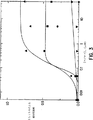
図1は、ELISAによる、試料#193に対する配列番号1(▼)、配列番号2(■)、配列番号3(●)及び配列番号4(▲)の相対反応性を示す。
図2は、ELISAによる、試料#267に対する配列番号1(▼)、配列番号2(■)、配列番号3(●)及び配列番号4(▲)の相対反応性を示す。
図3は、ELISAによる、試料#341に対する配列番号1(▼)、配列番号2(■)、配列番号3(●)及び配列番号4(▲)の相対反応性を示す。
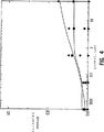
図4は、ELISAによる、試料#655に対する配列番号1(▼)、配列番号2(■)、配列番号3(●)及び配列番号4(▲)の相対反応性を示す。
図5は、ELISAによる、試料Mに対する配列番号2(■)及び配列番号3(●)の相対反応性を示す。
発明の詳細な説明
本発明者は、特定のアミノ酸の置換がHIV−1サブタイプOの存在の検出に重要であるHIV−1サブタイプBのgp41免疫優性領域(IDR)の2つのアミノ酸位置を同定し、これら2つの残基の少なくとも一方の改変をHIV−1サブタイプB特性を有するペプチド配列に組み込んだハイブリッドペプチドを設計・合成した。これらのハイブリッドペプチドは、確認されたHIV−1サブタイプOのテスト試料パネル中に存在する抗HIV−1サブタイプO抗体と反応し得るが、一部の試料は、改変されていないサブタイプB配列(配列番号1)を用いた場合には反応しなかった。これらのペプチドは、配列番号2、配列番号3及び配列番号4として配列表に示されている。これらの配列はHIV−1サブタイプBのgp41の19アミノ酸配列の604位及び610位(該ナンバリングはLAI株、前掲による)の一方又は両方に点変異を有している。
特に断りのない限り、下記の用語は下記の意味を有する。
用語「テスト試料」とは、(検出対象の抗体又は抗原のような)分析物源である個体の1構成成分を指す。これらの成分は当業界では周知である。該テスト試料には、本明細書に記載の方法によりテストし得る生物学的試料、及び全血、血清、血漿、脳脊髄液、尿、リンパ液や、気道、腸管及び尿生殖路の種々の外分泌液、涙、唾液、乳、白血球、骨髄腫などのようなヒト及び動物の体液、並びに細胞培養上清、固定化組織標本及び固定化細胞標本が含まれる。
本明細書に用いられている「分析物」とは、テスト試料中に存在し得る検出対象物質である。分析物は、その天然の特異的結合メンバー(例えば抗体)が存在するか、又はその特異的結合メンバーを生成し得るならいずれの物質であってもよい。従って、分析物はアッセイにおいて1種以上の特異的結合メンバーに結合し得る物質である。「分析物」にはさらに、任意の抗原物質、ハプテン、抗体及びその組み合わせも含まれる。分析物は、特異的結合対のメンバーとして、ビタミンB12を定量する場合には特異的結合対のメンバーとしての固有のタンパク性因子の使用、炭水化物を定量する場合には特異的結合対のメンバーとしてのレクチンの使用のような天然の特異的結合相手(対)を用いて検出することができる。分析物は、タンパク質、ペプチド、アミノ酸、標的ヌクレオチドなどを含み得る。
本発明は特異的結合メンバーを用いるアッセイを提供する。本明細書に用いられている「特異的結合メンバー」とは特異的結合対のメンバーである。即ち、一方の分子が化学的又は物理的手段により第2の分子に特異的に結合する2つの異なる分子である。従って、一般的なイムノアッセイの抗原抗体特異的結合対の他に、他の特異的結合対として、ビオチンとアビジン、炭水化物とレクチン、相補的ヌクレオチド配列、エフェクター分子とレセプター分子、補因子と酵素、酵素阻害剤と酵素などを含み得る。さらに、特異的結合対には、元の特異的結合メンバーの類似体であるメンバー、例えば分析物類似体が含まれ得る。葉酸の定量には葉酸結合タンパク質の免疫反応性特異的結合、又は炭水化物の定量には特異的結合対のメンバーとしてのレクチンを用いる。特異的結合対メンバーには、タンパク質、ペプチド、アミノ酸、標的ヌクレオチドなどが含まれ得る。さらに、特異的結合対には、元の特異的結合メンバーの類似体であるメンバー、例えば分析物類似体が含まれ得る。免疫反応性特異的結合メンバーには、組換えDNA分子により形成されたものを含めた、抗原、抗原のフラグメント、モノクローナル及びポリクローナル抗体、該抗体のフラグメント並びにその複合体が含まれる。本明細書に用いられている用語「ハプテン」とは、抗体には結合し得るが担体タンパク質に結合しない限り抗体の形成を誘発し得ない部分抗原又は非タンパク質結合メンバーを指す。
「指示薬」は、HIVに対する特異的結合メンバーに結合(付着)した外部手段により検出し得る測定可能なシグナルを生成し得る(生成する)「シグナル生成化合物」(標識)を含む。本明細書に用いられている「特異的結合メンバー」とは、特異的結合対のメンバーを意味する。即ち、一方の分子が化学的又は物理的手段により第2の分子に特異的に結合する2つの異なる分子である。指示薬は、HIVに対する特異的結合対の抗体メンバー以外にも、ビオチン又は抗ビオチン、アビジン又はビオチン、炭水化物又はレクチン、相補的ヌクレオチド配列、エフェクター分子又はレセプター分子、酵素補因子又は酵素、酵素阻害剤又は酵素などのようなハプテン−抗ハプテン系を含めたいずれの特異的結合対のメンバーであってもよい。免疫反応性特異的結合メンバーは、抗体、抗原、又はサンドイッチアッセイではHIVに、競合アッセイでは捕獲試薬に、若しくは間接アッセイでは補助特異的結合メンバーに結合し得る抗体/抗原複合体であってよい。
考えられる種々の「シグナル生成化合物」(標識)には、色素原、酵素のような触媒、蛍光及びローダミンのような発光化合物、ジオキセタン、アクリジニウム、フェナントリジニウム及びルミノールのような化学発光化合物、放射性元素並びに直視標識が含まれる。酵素の例としては、アルカリホスファターゼ、西洋ワサビペルオキシダーゼ、β−ガラクトシダーゼなどが挙げられる。どの標識を選択するかは重要ではないが、標識は、それ自体で又は1種又は複数種の追加物質と一緒になってシグナルを生成し得る。
「固相」(「固体支持体」)は当業者には公知であり、反応トレイのウエルの壁、試験管、ポリスチレンビーズ、磁気ビーズ、ニトロセルロース細片、膜、ラテックス粒子のような微粒子、ヒツジ(又は他の動物)の赤血球、デュラサイトなどが含まれる。どの「固相」を用いるかは重要ではなく、当業者が選択し得る。従って、ラテックス粒子、微粒子、磁気又は非磁気ビーズ、膜、プラスチックチューブ、マイクロタイターウェルの壁、ガラス又はシリコンチップ、ヒツジ(又は他の動物)の赤血球及びデュラサイトは全て適当な例である。ペプチドを固相上に固定する適当な方法には、イオン性、疎水性、共有結合性相互作用などが含まれる。本明細書に用いられている「固相」とは、不溶性であるか又は後続反応により不溶性とされ得る任意の物質を指す。固相は、捕獲試薬を誘引して固定化するその固有の能力に基づいて選択し得る。あるいは、固相は、捕獲試薬を誘引して固定化する能力を有する追加の受容体を有していてもよい。追加の受容体には、捕獲試薬自体の電荷又は捕獲試薬に結合した荷電物質と反対の電荷を帯びた荷電物質が含まれ得る。あるいは、受容体分子は、固相上に固定され(固相に結合し)且つ特異的結合反応により捕獲試薬を固定化する能力を有する任意の特異的結合メンバーであってよい。受容体分子は、アッセイ実施前又はアッセイ実施中に捕獲試薬と固相物質とを間接結合させ得る。従って、固相は、プラスチック、誘導体化プラスチック、磁気又は非磁気金属、試験管のガラス又はシリコン表面、マイクロタイターウエル、シート、ビーズ、微粒子、チップ、ヒツジ(又は他の適当な動物)の赤血球、デュラサイト及び当業者には公知の他の構造であってよい。
固相がさらに抗体の検出を可能にするに十分な多孔度及び抗体が結合するのに適当な表面親和性を有する適当な多孔質物質を含み得ることも考慮され、これも本発明の範囲内に包含される。一般的には微孔質構造が好ましいが、水和状態でゲル構造を有する物質を用いてもよい。これらの物質は、フィルム、シート又はプレートのような適当な形態で用いてもよいし、紙、ガラス、プラスチックフイルム又は繊維のような適切な不活性担体上にコーティングするか、該担体に付着若しくは積層させてもよい。
種々の他の固相を利用する他の実施態様も考慮され、これらも本発明の範囲内に包含される。例えば、EP公開第0326100号に対応する同時係属の米国特許出願第150,278号及び米国特許出願第375,029号(EP公開第0406473号)に記載されている、負に帯電したポリマーとの固定化可能な反応複合体を固定化するためのイオン捕獲手順を本発明に従って用い、迅速な液相免疫化学反応を行うことができる。固定化し得る免疫複合体を、負に帯電したポリアニオン/免疫複合体と前もって処理しておいた正に帯電した多孔質マトリックスとのイオン相互作用により反応混合物の残分から分離し、EPO公開第0,273,115号に対応する同時係属の米国特許出願第921,979号に記載の化学発光シグナルの測定に記載のものを含めた既に記載されている種々のシグナル生成系を用いて検出することができる。
また、本発明の方法を、固相が(磁気又は非磁気)微粒子を含む自動化又は半自動化系を含めた微粒子技術を用いる系に使用するように適合させることもできる。そのような系には、それぞれ、公開されたEPO出願、同EP0425633号及びEP0424634号に対応する、係属中の米国特許出願第425,651号及び同第425,643号に記載のものが含まれる。
イムノアッセイに走査プローブ顕微鏡検査法(SPM)を用いるのも本発明のペプチドが容易に適合し得る技術である。走査プローブ顕微鏡検査法、特に原子力顕微鏡検査法では、捕獲相、例えば本明細書に開示されている少なくとも1種のペプチドを固相に付着させ、検出対象の抗体を含む疑いのあるテスト試料を固相と接触させ、走査プローブ顕微鏡を用いて固相表面上に存在し得る抗原/抗体複合体を検出する。通常多くのイムノアッセイ系では抗原/抗体複合体の検出に標識を用いなければならないが、走査トンネル顕微鏡検査法を用いるとその必要がなくなる。そのようなシステムが係属中の米国特許出願第662,147号に記載されている。特異的結合反応のモニターにSPMを用いることは多くの方法で生じ得る。ある方法では、特異的結合相手(本明細書に開示されているペプチドである分析物特異的物質)の一方のメンバーを走査に適当な表面に結合させる。分析物特異的物質は、当業者には公知の方法に従ってプラスチック又は金属表面を有する固相を含む試験片に吸着により付着させる。あるいは、特異的結合相手(分析物特異的物質)を誘導体化プラスチック、金属、シリコン又はガラスの固相を含む試験片に共有結合させる方法を用いてもよい。共有結合法は当業者には公知である。また、1988年1月29日に出願された係属中の米国特許出願第150,278号及び1989年7月7日に出願された同第375,029号に記載の技術及び化学を用いて、高分子電解質相互作用を用いて特異的結合相手を試験片の表面上に固定してもよい。特異的結合メンバーを結合させた後、該表面をさらに血清、タンパク質又は他のブロッキング剤で処理して、非特異的結合を最小限にし得る。さらに該表面を製造現場又は使用箇所で走査してアッセイに対するその適合性を検証し得る。走査プロセスによって試験片の特異的結合特性が変わることはないと考えられる。
アッセイに用いられる試薬は、それぞれが別々の試薬、例えばペプチド若しくはペプチド混合物又はアッセイに用いられる指示薬を含有するバイアル又はビンのような1個以上の容器を含むテストキットの形態で提供し得るものと考えられる。そのようなテストキットには、緩衝液、対照などのような当業者には公知の他の成分が含まれ得る。
イムノアッセイにおける抗原として抗体を産生させる免疫原などを含む、本明細書に詳述されているペプチドを用いるアッセイフォーマットを設計し得る。ヒトテスト試料中の特異的分析物(例えばHIV−1)に対する抗体の存在を検出するアッセイフォーマットの場合、ヒトテスト試料を、本明細書に開示されている少なくとも1種のHIV−1ペプチドで被覆した固相と接触させて、インキュベートする。分析物特異的抗体がテスト試料中に存在すると、該抗体はペプチドとの複合体を形成して固相に固定される。複合体形成後、固相を洗浄して結合しなかった物質及び試薬を除去する。次いで、これらの複合体を指示薬と接触・反応させて、第2の複合体が形成されるような時間及び条件でインキュベートする。ペプチドに対するテスト試料中の抗体の存在は、生成されたシグナルを検出して決定する。生成されたシグナルがカットオフ値以上であれば、テスト試料中に存在する分析物に対する抗体の存在を示す。酵素のような多くの指示薬を用いる場合、存在する抗体の量は生成されるシグナルに比例する。テスト試料のタイプに応じて、テスト試料を適当な緩衝試薬で稀釈し、濃縮するか、又は何ら処理せずに(「ニート」)固相と接触させ得る。例えば、通常、前もって稀釈しておいた血清若しくは血漿試料又は尿のような濃縮標本をアッセイして抗体の存在及び/又は存在する抗体の量を測定するのが好ましい。
さらに、実験中の感染物質のウイルスゲノムの種々の抗原エピトープに対するペプチドを用いることにより、上記のアッセイフォーマットで1種以上のペプチドを用いて特異的感染物質に対する抗体の存在をテストすることができる。従って、特異的ウイルス抗原領域内のエピトープ並びに該ウイルスのゲノム以外の他の抗原領域からのエピトープを含むペプチドを用いて1つのエピトープからのペプチドを用いるものより感度及び恐らく特異性が高められたアッセイを提供するのが好ましい。そのようなアッセイは確認(confirmatory)アッセイとして用い得る。この特定のアッセイフォーマットの場合、既知量のテスト試料と、本明細書に開示されているペプチドの少なくとも1種で被覆された既知量の少なくとも1種の固体支持体とを、ペプチド/抗体複合体の形成に十分な時間及び条件で接触させる。次いで、該複合体と既知量の適切な指示薬とを、反応の生起に適当な時間及び条件で接触させる。シグナルを生成させ、得られたシグナルを陰性テスト試料と比較して、テスト試料中の分析物に対する抗体の存在を決定する。さらに、微粒子のような特定の固相を用いると、アッセイに用いられる各ペプチドが別々の微粒子に結合し、被覆された種々の微粒子が一緒になって、それぞれのアッセイに最適化され得る微粒子混合物が形成され得るとも考えられる。
上記アッセイフォーマットの変形には、一方の分析物に対する抗体の存在を検出するために、本明細書に開示されている合成ペプチド1種以上と、同一又は異なる固相に結合した異なる分析物に特異的な組換えタンパク質又は合成ペプチド(例えば、異なる感染因子の特定の抗原領域に特異的な組換えタンパク質を含む同一又は異なる固相上に被覆されたHIV−1の特定の抗原領域に特異的な、本明細書に開示されている合成ペプチド)を組み込んで、感染性因子のどちらか(又は両方)の存在を検出するものが含まれる。
さらに別のアッセイフォーマットにおいては、抗原性エピトープを含むペプチドは中和アッセイのような競合的アッセイに有用である。中和アッセイを実施するためには、HIV−1の抗原性領域のエピトープを表すペプチドを可溶化し、最終濃度0.5〜50.0μg/mlになるように試料稀釈液と混合する。稀釈するか又は稀釈していない既知量(例えば10μl)のテスト試料を反応ウエルに加え、次いで、例えば400μlの本明細書に開示のペプチドを含む試料稀釈液を加える。所望なら、混合物を約15分〜2時間予備インキュベートしてもよい。次いで本明細書に開示のペプチドで被覆した固相を反応ウエルに加え、約40℃で1時間インキュベートする。洗浄後、既知量の指示薬、例えば結合体用稀釈液中のペルオキシダーゼ標識したヤギ抗ヒトIgG200μlを加え、40℃で約1時間インキュベートする。記載のような酵素結合体を用いる場合には、洗浄後、混合物にOPD基質のような酵素基質を加え、室温で30分間インキュベートする。反応ウエルに1N 硫酸のような停止試薬を加えて反応を停止させる。492nmで吸光度を読み取る。特異的ペプチドに対する抗体を含むテスト試料は、溶液中でのこれらの抗体に対するペプチドの競合的結合により、生成されるシグナルが低減する。競合的結合の割合(%)は、同一稀釈度でペプチド存在下の試料の吸光度値とペプチド不在下に測定した試料の吸光度値とを比較して計算し得る。従って、ペプチド存在下の試料及びペプチド不在下の試料により生成されたシグナルの差が抗体の存在又は不在の決定に用いられる尺度である。
別のアッセイフォーマットでは、本発明のペプチドをイムノブロットアッセイ系に用いることができる。イムノブロットアッセイ系はニトロセルロース固体支持体上に列をなして置かれた精製組換えポリペプチド又は合成ペプチドのパネルを用いる。調製された固体支持体を、テスト試料並びに組換えタンパク質及び/又は合成ペプチド(特異的結合メンバー)に対する捕獲特異的抗体(他の特異的結合メンバー)と接触させて、特異的結合メンバー対を形成する。指示薬と反応させて捕獲された抗体を検出する。結合体特異的反応は、1988年8月2日に出願された米国特許出願第07/227,408号に記載されている装置内の反射光学アセンブリーを用いて定量するのが好ましい。関連米国特許出願第07/227,586号及び同第07/227,590号(どちらも1988年8月2日に出願)、並びに米国特許第5,075,077号(1988年8月2日に出願された米国特許出願第07/227,272号)(いずれも同一所有権者が享受するものであり、本明細書に参照として組み込むものとする)には、イムノブロットアッセイの実施に有用な特定の方法及び装置が詳細に記載されている。要約すれば、ニトロセルロースベースのテストカートリッジを複数の抗原性ペプチドで処理する。各ペプチドはテストカートリッジ上のそれぞれ特定の反応領域内に包含されている。全ての抗原性ポリペプチドをニトロセルロース上に置いた後で、該ニトロセルロース上の過剰な結合部位をブロックする。次いで、テスト試料が適切な抗体を含んでいる場合には各反応ゾーン中の各抗原性ペプチドが該抗体と反応するようにテストカートリッジをテスト試料と接触させる。反応後、テストカートリッジを洗浄し、周知の適当な試薬を用いて抗原抗体反応を同定する。上記にリストした特許及び特許出願明細書に記載されているように、全過程を自動化することができる。イムノブロットアッセイを実施するための方法及び装置に関連するこれらの特許出願明細書は本明細書に参照として組み込むものとする。
本明細書に開示されているペプチドは、テスト試料中の分析物特異的抗原に対する抗体を検出するための以下の様な第1及び第2の固体支持体を用いるアッセイに使用し得る。このアッセイフォーマットでは、テスト試料の第1のアリコートと、分析物に特異的な第1のペプチドで被覆した第1の固体支持体とを、ペプチド/分析物抗体複合体の形成に十分な時間及び条件で接触させる。次いで、これらの複合体をペプチドに特異的な指示薬と接触させる。指示薬から生成されたシグナルを検出して、テスト試料中に存在するペプチドに対する抗体の存在(もし存在すれば)を決定する。この後、テスト試料の第2のアリコートと、第2の抗体として合成ペプチド又は組換えタンパク質で被覆した第2の固体支持体とを組換えタンパク質又は合成ペプチド/第2抗体複合体の形成に十分な時間及び条件下に接触させて、同一分析物の異なる抗原決定基の存在を決定する。該複合体をその抗体に特異的な第2の指示薬と接触させる。指示薬から生成されたシグナルを検出して、テスト試料中の抗体の存在を決定するが、分析物の一方又は両方に対する抗体の存在は、テスト試料中の抗分析物の存在を示している。さらに、固体支持体を同時にテストし得ることも考えられる。
ハプテンを用いてシグナルの生成を増強させ、それによってアッセイの感度を高めることができる。ハプテンの使用は当業界では公知である。アッセイの性能を高めるために、本明細書に開示されているペプチドを用いるアッセイにハプテンを用いることも意図している。
本発明によるテスト試料中の抗体を検出するための別のアッセイ法には、フローサイトメトリー法及び粒子計数法が含まれる。例えば、粒子計数法では、特異的抗体を含む疑いのあるテスト試料のアリコートと、特異的結合対の他方のメンバーとして検出対象抗体に結合し得る本明細書に開示の少なくとも1種のペプチドのような抗体に特異的な捕獲試薬で被覆された微粒子とを混合することにより、特異的結合対の抗体メンバーである分析物を定量する。テスト試料中に抗体が存在する場合、抗体は捕獲試薬で被覆された一部の微粒子に結合して、凝集体を形成する。分析物の濃度は凝集しなかった粒子数に反比例する。例えば、Roseら編,Manual of Clinical Laboratory Immunology,第3版,第8章,43−48ページ,American Society for Microbiology,Washington,D.C.(1986)を参照されたい。
照射された細胞又は粒子からの電子及び光シグナルを検出するフローサイトメトリーにより、細胞の表面特性、量及び細胞のサイズを測定する。例えば、テスト試料中に存在する抗体は本明細書に開示のペプチドに結合し、ペプチドに直接結合するか又は二次反応を介して付加された蛍光染料により検出される。1つの試料から1種以上の分析物を検出し得るように、異なる波長で励起され得る異なる染料を異なる分析物に特異的な1種以上のペプチドと共に用いることができる。蛍光フローサイトメトリーでは、テスト試料中の粒子(典型的には細胞)の懸濁液を、試料中の個々の粒子に1種以上の集光ビームを照射するフローセルを介して輸送する。1個以上の検出器がフローセルを通って流れる標識粒子と光ビームとの相互作用を検出する。一般に、ある種の検出器は蛍光の放出を測定するように設計されているが、他の検出器は散乱強度又はパルスの持続時間を測定する。従って、フローセルを通過する各粒子は、その軸が検出器により測定される放出色、光強度又は他の特性、即ち散乱である特徴スペースにマッピングされ得る。ある場合には、試料中の異なる粒子を特徴スペースのオーバーラップしていない別個の領域にマッピングして、各粒子をその特徴スペース中のマッピングに基づいて分析し得る。フローサイトメトリー分析用のテスト試料を調製するには、オペレーターが手動で一定量のテスト試料を試料管から分析管にピペット装入する。一定量の蛍光色原体で標識した所望のペプチドを加える。次いで、試料/ペプチド混合物を抗体/ペプチド結合が生起し得るに十分な時間及び条件でインキュベートする。必要なら、インキュベーション後に、オペレーターは一定量のRNS溶解物を加えて試料中のRBCを破壊する。溶解後、試料を遠心、洗浄して、溶解ステップからの残留物を除去する。遠心/洗浄ステップは数回繰り返してよい。試料を一定量の固定液に再懸濁し、次いで蛍光フローサイトメトリー装置に通す。流動自動化分析を実施するための方法及び装置は共同所有になる米国特許出願第08/283,379号(本明細書に参照として組み込むものとする)に記載されている。本明細書に記載の方法に中心体を用い、タッグを付けるか標識して、in vitro診断に用い得ることも本発明の範囲内である。細菌、ウイルス、デュラサイトなどを含めた他の細胞又は粒子に、本発明に記載されているPNA又はモルホリノ化合物でタッグを付けるか又は標識して、フローサイトメトリーに用い得ることも本発明の範囲内である。
本発明を以下の実施例により説明するが、該実施例は例示を目的とし、本発明の思想及び範囲を限定するものではない。
実施例
実施例1: ペプチドの合成
全てのペプチドは、FMOC化学、標準サイクル及びDCC−HOBt活性化を用い、ABI Peptide Synthesizer,Model 431Aで合成した。切断及び脱保護条件は以下の通りである:20mlのトリフルオロ酢酸、0.3mlの水、0.2mlのエタンジチオール、0.2mlのチオアニソール及び100mgのフェノールに樹脂を加え、室温で1.5時間攪拌した。次いで、樹脂を吸引濾過し、TFA溶液をエーテルで沈降させ、次いで濾過してペプチドを得た。各ペプチドを水/アセトニトリル/0.1%TFA勾配を用いる逆相分取HPLCにかけて精製し、凍結乾燥した。生成物を質量分光法により確認した。
以下の自動酸化条件を用いてジスルフィド結合を形成した。ペプチドを最少量のDMSO(約10ml)に溶解し、次いで緩衝液(0.1M Tris,pH6.2)を加えて濃度を0.3〜0.8mg/mlとした。ジスルフィド結合が完全に形成されるまで反応混合物をHPLCでモニターし、次いで、水/アセトニトリル/0.1%TFA勾配を用いる逆相分取HPLCにかけ、凍結乾燥した。次いで生成物を質量分光法により確認した。全てのペプチドは2個のシステイン(C)残基間に形成されたジスルフィドループを含んでいた。
実施例2: EIA
A.試料の調達. サブタイプO試料「M」及び「E」は9種の確認されたサブタイプO試料のオリジナルフランスパネルに属するものであった。該試料「M」及び「E」はぞれぞれ試料#7及び#2〔I.Loussert−Ajakaら,The Lancet 343:1393−1394(1994)〕と同一である。試料DUR、FAN及びMAAはフランス政府から調達した他のサブタイプO試料であった。
試料#2901及びHA112はそれぞれ、ドイツ、ミュンヘンのLutz Gurtler博士、ベルリンのHartmut Hampl博士から得た。試料#193、#267、#341及び#655は赤道ギニアから得たものであり、PCRにより真のサブタイプO試料であることが確認されている。
B.EIA. 先ず、合成ペプチドを0.1M モルホリノエタンスルホン酸(MES)緩衝液(pH5.5)に20μMの濃度に溶解した。コーティングステップでは、各ペプチドの20μM溶液(0.1M MES緩衝液中,pH5.5)の種々の稀釈液(0〜5倍)200μlをMicrotiterTM(Dynatech Immunolon 4 ポリスチレン)プレートのウエルに加えた。室温で一晩インキュベートした後、プレートを、TBST(Tris Buffer Saline,0.01M Tris,0.15M NaC1,0.05%Tween−20▲R▼,pH8)中0.5%脱脂粉乳を含む洗浄溶液で洗浄した。ブロッキングステップでは、各ウエルにTBST溶液中10%脱脂粉乳300μlを添加し、次いで室温で1時間インキュベートした。次いで、プレートを洗浄溶液で洗浄し、各ウエルに10%粉乳−TBSTに150倍稀釈した血清/血漿試料150μlを加えた。室温で2時間インキュベートした後、プレートを再び洗浄溶液で洗浄し、次いで、各ウエルに、10%脱脂粉乳−TBST中の結合体〔ヤギ−抗ヒトIgG−西洋ワサビペルオキシダーゼ(HRPO),1mg/ml,Kirkegaard & Perry Laboratory,Inc.,Gaithersburg,MD〕の16,000倍稀釈液100μlを加えた。1時間インキュベートした後、プレートを洗浄溶液で洗浄した。各ウエルに過酸化水素中のo−フェニレンジアミン(OPD)の溶液100μlを加え、10分間インキュベートして発色を得た。1N 硫酸100μlを加えて発色反応を停止し、490nm及び630nmの波長のDynatech MR5000プレート読み取り装置で吸光度を測定した。ウエルのA490−A630の相対強度は、特定のペプチドが特定の血清/血漿試料と反応した効力に比例していた。
実施例3:
SからKへの置換及びTからYへの置換の比較分析
この実施例は、一部のHIV−1サブタイプO試料(#2901、#267及び#655)の検出におけるSからKへの置換(配列番号2)の重要性、一部の他のHIV−1サブタイプO試料(#193、「E」及びHA112)の検出におけるTからYへの置換(配列番号3)の重要性、及び配列番号2及び配列番号3が共に配列番号1より性能が全般的に優れていることを示している。
実施例2に記載のようにアッセイを実施した。ウエルのコーティングに用いたペプチド濃度は20μMであり、血清/血漿試料の稀釈度は、#193、#267、#341、#655、HA112及びDURの場合は150倍、#2901、試料「E」及びFANの場合は450倍、試料MAAの場合は750倍、試料「M」の場合は1,500倍であったことに留意されたい。確認された陰性ヒトHIV試料をこの実験の陰性対照として用いた。
表1、2及び3に記載されている吸光度値はA490−A630として計算した。

SからKへの置換及びTからYへの置換の比較分析
この実施例は、合計11種のサブタイプO試料中10種の検出において、SからK及びTからYへの2置換ペプチド(配列番号4)が配列番号1に比べて著しく性能が優れていることを示している。
実施例2に記載のようにアッセイを実施した。ウエルのコーティングに用いたペプチド濃度は20μMであり、血清/血漿試料の稀釈度は、#193、#267、#341、#655、HA112及びDURの場合は150倍、#2901、試料「E」及びFANの場合は450倍、試料MAAの場合は750倍、試料「M」の場合は1,500倍であったことに留意されたい。実施例3に記載のように、確認された陰性ヒトHIV試料をこの実験の陰性対照として用いた。
表4、5及び6に記載されている数値は吸光度値(A490−A630)であった。
A.実験プロトコル. 実施例1に記載にように調製したそれぞれ100μlの以下の合成ペプチドで16時間4℃で被覆した96ウエルプレートを用いてHIV合成ペプチドの相対免疫反応性を測定した:共通のB(配列番号1)、B/O−7(配列番号2)、B/O−81(配列番号3)及びB/O−2(配列番号4)。ペプチドを以下の濃度で評価した:500μM、50μM、5μM、0.5μM、0.05μM及び0.005μM。これらのペプチドの適用に用いた緩衝液は、100mM モルホリノ−エタンスルホン酸、pH5.5であった。次いで、ペプチドで被覆したウエルを、8mM リン酸ナトリウム、2mM リン酸カリウム、140mM 塩化ナトリウム、10mM 塩化カリウム、0.05%Tween−20、0.1%ウシ血清アルブミン、pH7.4からなる洗浄緩衝液で3回洗浄した。
次いで、ウエルを、リン酸緩衝塩水:8mM リン酸ナトリウム、2mM リン酸カリウム、140mM 塩化ナトリウム、10mM 塩化カリウム、pH7.4中9%(w/v)Carnation▲R▼粉末スキムミルクで1時間室温でブロックした。次いで、ウエルを洗浄緩衝液で3回洗浄した。
ヒト血清試料(#193、#267、#341、#655及び「M」)をPBS中4.5%カーネーションスキムミルク(w/v)で150倍稀釈した。これらの試料100μlをウエル中37℃で1時間インキュベートした。次いで、ウエルを洗浄緩衝液で3回洗浄した。
ヤギ抗ヒトIgGに結合した西洋ワサビペルオキシダー
ゼを用いてHIV抗体−ペプチド抗原複合体を含む抗体陽性試料を検出した。各ウェルに、洗浄緩衝液に1:5000稀釈したHRPO−ヤギ抗ヒトIgG結合体100μlを加え、室温で1時間インキュベートした。次いで、ウエルを洗浄緩衝液で3回洗浄し、Pierceから入手したABTS溶液(2,2′−アジノビス−[3−エチルベンゾチゾリン−6−スルホン酸]ジアンモニウム塩)に露出した後、405nmで吸光度を読み取ってHIV抗体の濃度を予測した。
B.データ分析. 405nmでの吸光度の読み取り値を配列番号4に対して標準化し、ウエルのコーティングに用いたペプチド濃度のlogに対して相対反応性をプロットした。データをMirocal Inc.により記載されたOriginプログラムに見られるようなS字形曲線を描く下記の式に当てはめた:
![]()
C.結果. 図1、図2、図3及び図4は、20μmのペプチド濃度が飽和シグナルの生成に十分であり、従って実施例3及び4における先のデータを立証することを示している。
配列表
配列番号:1
配列の長さ:19
配列の型:アミノ酸
鎖の数:一本鎖
トポロジー:直鎖状
配列の種類:タンパク質
配列
配列の長さ:19
配列の型:アミノ酸
鎖の数:一本鎖
トポロジー:直鎖状
配列の種類:タンパク質
配列
配列の長さ:19
配列の型:アミノ酸
鎖の数:一本鎖
トポロジー:直鎖状
配列の種類:タンパク質
配列
配列の長さ:19
配列の型:アミノ酸
鎖の数:一本鎖
トポロジー:直鎖状
配列の種類:タンパク質
配列
The present invention relates generally to peptides useful for the detection of HIV-1 antibodies, and more specifically, positions 604 and 610 of the gp160 sequence of HIV-1 subtype B (the numbering is described in Myers et al., HIV published below). HIV-1 subtype O antibody detection using the amino acid sequence of HIV-1 gp41 immunodominant region (IDR (immunodominant region)) containing two point mutations (in the LAI strain of -1).
Currently, there are six recognized subtypes of HIV-1 termed A, B, C, D, E, and F (so-called “systematic branches”), which are Myers et al., Human Retroviruses. and AIDS 1993: A Compilation and Analysis of Nucleic Acid and Amino Acid Sequences (Los Alamos National Laboratories, Los Alamos, NM) (1993). Recently, further subtypes G and H have been described. See, for example, Jansens et al., AIDS Research and Human Retroviruses 10: 877 (1994); and Myers et al., Supra. Of particular importance among the new subtypes is the recognition of a highly divergent HIV-1 sequence family termed “O”. HIV-1 subtype O was first described in 1987 because it was found that the subtype had only about 50% sequence identity with other HIV-1 subtypes at the nucleic acid level of the env gene. "O" after the name "outlier". The other subtypes above contain about 75% sequence identity with each other at the nucleic acid level of the env gene. In the earliest reports on the sequence of type O virus, in the phylogenetic tree, SIVCPZGAB is more closely related to other HIV-1 than group O, ie the chimpanzee virus is located between groups M and O It has been shown. I would like advice on whether this needs to be stated accurately. For example, Gurtler et al. Virology 68: 1581-1585 (1994); Vanden Haesevelde et al., J. Biol. Virology 68: 1586-1596 (1994); De Leys et al., J. Biol. Virology 64: 1207-1216 (1990); Deleys et al., US Pat. No. 5,304,466; Gultler et al. P. O. See Publication No. 05991414A2. The sequence of group O is the most branched sequence of HIV-1 among those described to date, and subtype B is the most common subtype of HIV-1.
HIV serology has been characterized by the amino acid sequence of the most expressed viral protein (antigen), particularly those containing the core and envelope. Antigens that are similar in structure and function but different in amino acid sequence elicit antibodies that are similar but different in specificity for the antigen. One example is the difference in gp41 IDR antigens in HIV-1 and HIV-2, and using this difference, individuals exposed to HIV-1 and / or HIV-2 can be serumized in a variety of ways. Can be distinguished scientifically. For example, Hunt et al., AIDS Research and Human Retroviruses 6: 883-898 (1990); Gnaan et al., Science 237: 1346-1349 (1987); Cot et al., AIDS Research and Human Retroviruses 4: 239-241 (198) Et al., U.S. Patent (4656.US.C1). Similarly, HIV-1 group O viruses can be antigenically and serologically distinguished from other HIV-1 subtypes. Lousert-Ajaka et al., The Lancet 343: 1393-1394 (1994); Gurtler et al., J. MoI. Virology 68: 1581-1585 (1994); Vanden Haesevelde et al., J. Biol. Virology 68: 1586-1596 (1994); De Leys et al., J. Biol. Virology 64 (supra); US Pat. No. 5,304,466; Gurtler et al. P. O. See Publication No. 05991414A2.
The ability to detect HIV-1 subtype O has become an important concern in the blood bank community. In one experiment, a commercial assay capable of detecting HIV-1 subtype B was reported to fail to detect 9 sample panels positive for HIV-1 subtype O [I. . Lousert-Ajaka et al., The Lancet 343: 1393-1394 (1994)]. Although the number and area of HIV-1 subtype O infections actually confirmed is limited, this subtype is beginning to spread from Cameroon where the virus originated to neighboring countries such as Equatorial Guinea There is.
It would be beneficial to develop reagents that can be used in assays that detect the presence of HIV-1 subtype O antibodies in a test sample.
Summary of the Invention
The present invention provides a polypeptide having a point mutation at position 604 of the IDR of HIV-1 subtype B. More specifically, the amino acid at position 604 of the polypeptide is lysine (K). The polypeptide is represented by SEQ ID NO: 2. The present invention further provides a polypeptide having a point mutation at position 610 of the HIV-1 subtype B IDR. More specifically, the amino acid at position 610 of the polypeptide is tyrosine (Y). The polypeptide is represented by SEQ ID NO: 3. Also provided are polypeptides having two single point mutations at positions 604 and 610 of the HIV-1 subtype B IDR. The point mutation at position 604 of the polypeptide is lysine (K), and the position at position 610 is tyrosine (Y). The polypeptide is represented by SEQ ID NO: 4. Also provided are the polypeptides of SEQ ID NO: 2, SEQ ID NO: 3 and SEQ ID NO: 4.
The present invention provides an immunoassay for detecting the presence of HIV antibodies in a test sample, the assay comprising a test sample and a solid phase coupled to an HIV-1 polypeptide having a point mutation between positions 593 and 611. And incubating for a time and under conditions sufficient to form a polypeptide / antibody complex; specific for an HIV antibody bound to a signal-generating compound capable of producing a measurable signal with the polypeptide / antibody complex Contacting with an indicator comprising a member of a binding pair and incubating for a time and under conditions sufficient to form a polypeptide / antibody / indicator complex; and detecting a measurable signal to detect the presence of HIV antibody Determining the step. Point mutations exist at position 604 or 610. Alternatively, point mutations exist at both positions 604 and 610. The solid phase is from the well wall of the reaction tray, test tubes, polystyrene beads, magnetic beads, nitrocellulose strips, membranes, microparticles such as latex particles, sheep (or other animal) erythrocytes and duracyte. Selected from the group consisting of The signal generating compound of the indicator is selected from the group consisting of chromogens, enzymes, luminescent compounds, chemiluminescent compounds, radioactive elements and direct-view labels. Preferred as a member of the indicator specific binding pair is anti-human IgG.
The present invention provides an improved immunoassay for detecting an HIV antibody in a test sample, the immunoassay comprising contacting the test sample with an HIV-1 polypeptide and detecting the presence of the antibody. Is to use or utilize a polypeptide having a point mutation between position 593 to position 611 of the HIV-1 gp160 sequence.
Further provided is a diagnostic test kit capable of detecting HIV antibodies, the test kit comprising a container containing a polypeptide having a sequence selected from the group consisting of SEQ ID NO: 2, SEQ ID NO: 3 and SEQ ID NO: 4. .
[Brief description of the drawings]
FIG. 1 shows the relative reactivity of SEQ ID NO: 1 (▼), SEQ ID NO: 2 (■), SEQ ID NO: 3 (●), and SEQ ID NO: 4 (▲) to sample # 193 by ELISA.
FIG. 2 shows the relative reactivity of SEQ ID NO: 1 (▼), SEQ ID NO: 2 (■), SEQ ID NO: 3 (●), and SEQ ID NO: 4 (▲) with respect to sample # 267 by ELISA.
FIG. 3 shows the relative reactivity of SEQ ID NO: 1 (▼), SEQ ID NO: 2 (■), SEQ ID NO: 3 (●), and SEQ ID NO: 4 (▲) with respect to sample # 341 by ELISA.
FIG. 4 shows the relative reactivity of SEQ ID NO: 1 (▼), SEQ ID NO: 2 (■), SEQ ID NO: 3 (●), and SEQ ID NO: 4 (▲) with respect to sample # 655 by ELISA.
FIG. 5 shows the relative reactivity of SEQ ID NO: 2 (■) and SEQ ID NO: 3 (●) to sample M by ELISA.
Detailed Description of the Invention
The inventor has identified two amino acid positions in the gp41 immunodominant region (IDR) of HIV-1 subtype B in which specific amino acid substitutions are important for detecting the presence of HIV-1 subtype O, and these 2 A hybrid peptide was designed and synthesized in which at least one modification of one residue was incorporated into a peptide sequence having HIV-1 subtype B characteristics. These hybrid peptides can react with anti-HIV-1 subtype O antibodies present in a validated HIV-1 subtype O test sample panel, but some samples are not modified subtype B There was no reaction when the sequence (SEQ ID NO: 1) was used. These peptides are shown in the sequence listing as SEQ ID NO: 2, SEQ ID NO: 3 and SEQ ID NO: 4. These sequences have point mutations in one or both of positions 604 and 610 of the 19 amino acid sequence of HIV-1 subtype B gp41 (the numbering is according to the LAI strain, supra).
Unless otherwise noted, the following terms have the following meanings.
The term “test sample” refers to a component of an individual that is an analyte source (such as an antibody or antigen to be detected). These ingredients are well known in the art. The test samples include biological samples that can be tested by the methods described herein, and various exocrine secretions of whole blood, serum, plasma, cerebrospinal fluid, urine, lymph, airways, intestinal tract, and urogenital tract. Human and animal body fluids such as fluids, tears, saliva, milk, leukocytes, myeloma, and the like, as well as cell culture supernatants, immobilized tissue specimens and immobilized cell specimens are included.
As used herein, an “analyte” is a substance to be detected that may be present in a test sample. An analyte may be any substance whose natural specific binding member (eg, antibody) is present or capable of producing that specific binding member. Thus, an analyte is a substance that can bind to one or more specific binding members in an assay. “Analyte” further includes any antigenic substance, hapten, antibody and combinations thereof. The analyte is a member of a specific binding pair, the use of a unique protein factor as a member of a specific binding pair when quantifying vitamin B12, and a member of a specific binding pair when quantifying carbohydrates. Can be detected using natural specific binding partners (pairs) such as the use of lectins. Analytes can include proteins, peptides, amino acids, target nucleotides, and the like.
The present invention provides assays using specific binding members. As used herein, a “specific binding member” is a member of a specific binding pair. That is, two different molecules in which one molecule specifically binds to a second molecule by chemical or physical means. Therefore, in addition to antigen-antibody-specific binding pairs of general immunoassays, other specific binding pairs include biotin and avidin, carbohydrates and lectins, complementary nucleotide sequences, effector molecules and receptor molecules, cofactors and enzymes, enzymes Inhibitors and enzymes may be included. Furthermore, a specific binding pair can include a member that is an analog of the original specific binding member, eg, an analyte analog. For quantification of folic acid, immunoreactive specific binding of folate binding protein, or for quantification of carbohydrate, lectin as a member of a specific binding pair is used. Specific binding pair members can include proteins, peptides, amino acids, target nucleotides, and the like. Furthermore, a specific binding pair can include a member that is an analog of the original specific binding member, eg, an analyte analog. Immunoreactive specific binding members include antigens, fragments of antigens, monoclonal and polyclonal antibodies, fragments of the antibodies and complexes thereof, including those formed by recombinant DNA molecules. As used herein, the term “hapten” refers to a partial antigen or non-protein binding member that can bind to an antibody but cannot induce the formation of the antibody unless bound to a carrier protein.
An “indicator” includes a “signal generating compound” (label) that can generate (generate) a measurable signal that can be detected by external means bound (attached) to a specific binding member for HIV. As used herein, “specific binding member” means a member of a specific binding pair. That is, two different molecules in which one molecule specifically binds to a second molecule by chemical or physical means. In addition to antibody members of specific binding pairs for HIV, the indicator may be biotin or anti-biotin, avidin or biotin, carbohydrate or lectin, complementary nucleotide sequence, effector molecule or receptor molecule, enzyme cofactor or enzyme, enzyme inhibitor or It may be a member of any specific binding pair, including hapten-antihapten systems such as enzymes. The immunoreactive specific binding member may be an antibody / antigen or antibody / antigen complex that can bind to HIV in a sandwich assay, to a capture reagent in a competitive assay, or to an auxiliary specific binding member in an indirect assay.
Various possible “signal generating compounds” (labels) include chromogens, catalysts such as enzymes, luminescent compounds such as fluorescence and rhodamine, chemiluminescent compounds such as dioxetane, acridinium, phenanthridinium and luminol, Radioactive elements as well as direct-view signs are included. Examples of the enzyme include alkaline phosphatase, horseradish peroxidase, β-galactosidase and the like. It does not matter which label is selected, but the label may itself or together with one or more additional substances generate a signal.
“Solid phases” (“solid supports”) are known to those skilled in the art and include fine walls such as reaction well walls, test tubes, polystyrene beads, magnetic beads, nitrocellulose strips, membranes, latex particles, Sheep (or other animal) red blood cells, durasite, etc. are included. Which “solid phase” is used is not critical and can be selected by one skilled in the art. Thus, latex particles, microparticles, magnetic or non-magnetic beads, membranes, plastic tubes, microtiter well walls, glass or silicon chips, sheep (or other animal) erythrocytes and durasite are all suitable examples. Suitable methods for immobilizing the peptide on the solid phase include ionic, hydrophobic, covalent interactions and the like. As used herein, “solid phase” refers to any substance that is insoluble or can be rendered insoluble by subsequent reactions. The solid phase can be selected based on its inherent ability to attract and immobilize the capture reagent. Alternatively, the solid phase may have an additional receptor that has the ability to attract and immobilize the capture reagent. The additional receptor may include a charged substance with a charge opposite to the charge of the capture reagent itself or a charged substance bound to the capture reagent. Alternatively, the receptor molecule may be any specific binding member that is immobilized on (couples to) the solid phase and has the ability to immobilize the capture reagent by a specific binding reaction. The receptor molecule may indirectly bind the capture reagent and the solid phase material before or during the assay. Thus, the solid phase can be plastic, derivatized plastic, magnetic or non-magnetic metal, test tube glass or silicon surface, microtiter wells, sheets, beads, microparticles, chips, sheep (or other suitable animal) red blood cells, It may be durasite and other structures known to those skilled in the art.
It is also contemplated that the solid phase may further comprise a suitable porous material having sufficient porosity to allow detection of the antibody and a suitable surface affinity for the antibody to bind, which is also within the scope of the present invention. Is included. In general, a microporous structure is preferable, but a substance having a gel structure in a hydrated state may be used. These materials may be used in a suitable form such as a film, sheet or plate, coated on or attached to a suitable inert carrier such as paper, glass, plastic film or fiber. It may be laminated.
Other embodiments utilizing a variety of other solid phases are also contemplated and are within the scope of the present invention. For example, with negatively charged polymers described in co-pending U.S. Patent Application No. 150,278 and U.S. Patent Application No. 375,029 (EP Publication No. 0406473) corresponding to EP Publication No. 0326100. An ion capture procedure for immobilizing an immobilizable reaction complex can be used according to the present invention to perform a rapid liquid phase immunochemical reaction. The immobilizable immune complex is separated from the remainder of the reaction mixture by ionic interaction between the negatively charged polyanion / immune complex and the previously treated positively charged porous matrix, Detection using various signal generation systems already described, including those described in the measurement of chemiluminescent signals described in co-pending US patent application 921,979 corresponding to 273,115. it can.
The methods of the invention can also be adapted for use in systems using microparticle technology, including automated or semi-automated systems where the solid phase contains microparticles (magnetic or non-magnetic). Such systems include those described in pending US patent applications 425,651 and 425,643, corresponding to published EPO applications, EP0425633 and EP0424634, respectively. .
The use of scanning probe microscopy (SPM) in immunoassays is a technique that the peptides of the present invention can be easily adapted to. In scanning probe microscopy, particularly atomic force microscopy, a capture phase, such as at least one peptide disclosed herein, is attached to a solid phase and a test sample suspected of containing an antibody to be detected is immobilized. In contact with the phase, the scanning probe microscope is used to detect antigen / antibody complexes that may be present on the solid surface. Usually many immunoassay systems must use a label to detect the antigen / antibody complex, which is no longer necessary using scanning tunneling microscopy. Such a system is described in pending US Patent Application No. 662,147. The use of SPM to monitor specific binding reactions can occur in many ways. In one method, one member of a specific binding partner (an analyte-specific substance that is a peptide disclosed herein) is bound to a surface suitable for scanning. The analyte-specific substance is attached by adsorption to a test strip containing a solid phase having a plastic or metal surface according to methods known to those skilled in the art. Alternatively, a method may be used in which a specific binding partner (analyte-specific substance) is covalently bound to a test strip containing a derivatized plastic, metal, silicon or glass solid phase. Covalent bonding methods are known to those skilled in the art. Also, using the techniques and chemistry described in pending US patent application 150,278 filed on January 29, 1988 and 375,029 filed on July 7, 1989, Polyelectrolyte interactions may be used to immobilize specific binding partners on the surface of the test strip. After binding the specific binding member, the surface can be further treated with serum, protein or other blocking agent to minimize non-specific binding. In addition, the surface can be scanned at the manufacturing site or point of use to verify its suitability for the assay. It is believed that the scanning process does not change the specific binding properties of the specimen.
Reagents used in the assay may be provided in the form of a test kit comprising one or more containers, such as vials or bottles, each containing a separate reagent, eg, a peptide or peptide mixture or an indicator used in the assay. Conceivable. Such test kits can include other components known to those skilled in the art, such as buffers, controls, and the like.
Assay formats can be designed using the peptides detailed herein, including immunogens that produce antibodies as antigens in immunoassays. For assay formats that detect the presence of antibodies to a specific analyte (eg, HIV-1) in a human test sample, the human test sample is coated with at least one HIV-1 peptide disclosed herein. Incubate in contact with the prepared solid phase. When an analyte-specific antibody is present in the test sample, the antibody forms a complex with the peptide and is immobilized on the solid phase. After complex formation, the solid phase is washed to remove unbound material and reagents. These complexes are then contacted and reacted with an indicator and incubated for a time and under conditions such that a second complex is formed. The presence of the antibody in the test sample against the peptide is determined by detecting the signal generated. If the generated signal is greater than or equal to the cut-off value, it indicates the presence of an antibody against the analyte present in the test sample. When using many indicators, such as enzymes, the amount of antibody present is proportional to the signal produced. Depending on the type of test sample, the test sample can be diluted with an appropriate buffer reagent, concentrated, or contacted with the solid phase without any further treatment (“neat”). For example, it is usually preferred to assay a pre-diluted serum or plasma sample or a concentrated specimen such as urine to determine the presence of antibody and / or the amount of antibody present.
Furthermore, by using peptides against various antigenic epitopes of the viral genome of the infectious agent under experiment, one or more peptides can be used in the assay format described above to test for the presence of antibodies to specific infectious agents. Thus, an assay with increased sensitivity and possibly more specificity than using a peptide from one epitope with a peptide comprising an epitope within a specific viral antigen region and an epitope from another antigenic region other than the genome of the virus. Is preferably provided. Such an assay can be used as a confirmatory assay. For this particular assay format, a peptide / antibody complex comprising a known amount of test sample and a known amount of at least one solid support coated with at least one of the peptides disclosed herein. For a period of time and under conditions sufficient to form The complex is then contacted with a known amount of a suitable indicator for a time and under conditions appropriate for the reaction to occur. A signal is generated and the resulting signal is compared to a negative test sample to determine the presence of antibodies to the analyte in the test sample. In addition, using a specific solid phase such as microparticles, each peptide used in the assay binds to a separate microparticle, and the various coated microparticles can be combined together and optimized for each assay. It is also considered that can be formed.
Variations on the above assay format are specific to one or more of the synthetic peptides disclosed herein and to different analytes bound to the same or different solid phase to detect the presence of antibodies to one analyte. Specific recombinant protein or synthetic peptide (eg specific for a specific antigenic region of HIV-1 coated on the same or different solid phase containing a recombinant protein specific for a specific antigenic region of a different infectious agent , Synthetic peptides disclosed herein), and those that detect the presence of either (or both) infectious agents.
In yet another assay format, peptides containing antigenic epitopes are useful in competitive assays such as neutralization assays. To carry out the neutralization assay, peptides representing epitopes of the antigenic region of HIV-1 are solubilized and mixed with sample dilutions to a final concentration of 0.5-50.0 μg / ml. A diluted or undiluted known amount (eg, 10 μl) of test sample is added to the reaction well, followed by, eg, 400 μl of sample dilution containing a peptide disclosed herein. If desired, the mixture may be preincubated for about 15 minutes to 2 hours. The solid phase coated with the peptides disclosed herein is then added to the reaction wells and incubated at about 40 ° C. for 1 hour. After washing, add a known amount of an indicator, eg, 200 μl of peroxidase-labeled goat anti-human IgG in conjugate dilution and incubate at 40 ° C. for about 1 hour. When using the enzyme conjugate as described, after washing, an enzyme substrate such as OPD substrate is added to the mixture and incubated at room temperature for 30 minutes. The reaction is stopped by adding a stop reagent such as 1N sulfuric acid to the reaction well. Read absorbance at 492 nm. Test samples containing antibodies against specific peptides have a reduced signal produced by competitive binding of the peptides to these antibodies in solution. The percentage of competitive binding can be calculated by comparing the absorbance value of the sample in the presence of peptide at the same dilution with the absorbance value of the sample measured in the absence of peptide. Thus, the difference in signal produced by a sample in the presence of peptide and a sample in the absence of peptide is a measure used to determine the presence or absence of antibody.
In another assay format, the peptides of the invention can be used in an immunoblot assay system. The immunoblot assay system uses a panel of purified recombinant or synthetic peptides placed in a row on a nitrocellulose solid support. The prepared solid support is contacted with a test sample and a capture specific antibody (other specific binding member) against a recombinant protein and / or a synthetic peptide (specific binding member) to form a specific binding member pair To do. The captured antibody is detected by reacting with an indicator. The conjugate specific reaction is preferably quantified using a reflective optical assembly in the apparatus described in US patent application Ser. No. 07 / 227,408 filed Aug. 2, 1988. Related U.S. Patent Application Nos. 07 / 227,586 and 07 / 227,590 (both filed on August 2, 1988), and U.S. Patent No. 5,075,077 (August 2, 1988) US patent application Ser. No. 07 / 227,272 filed in U.S.A. (both enjoyed by the same owner and incorporated herein by reference) are useful for performing immunoblotting assays. Specific methods and apparatus are described in detail. In summary, nitrocellulose-based test cartridges are treated with multiple antigenic peptides. Each peptide is contained within a particular reaction area on the test cartridge. After all antigenic polypeptide has been placed on nitrocellulose, excess binding sites on the nitrocellulose are blocked. The test cartridge is then contacted with the test sample so that each antigenic peptide in each reaction zone reacts with the antibody if the test sample contains an appropriate antibody. After the reaction, the test cartridge is washed, and the antigen-antibody reaction is identified using a known appropriate reagent. The entire process can be automated as described in the patents and patent application specifications listed above. These patent applications relating to methods and apparatus for performing immunoblot assays are hereby incorporated by reference.
The peptides disclosed herein can be used in assays using first and second solid supports to detect antibodies against analyte-specific antigens in test samples, such as the following. In this assay format, a first aliquot of the test sample and a first solid support coated with a first peptide specific for the analyte are allowed to have sufficient time and sufficient time to form a peptide / analyte antibody complex. Contact with conditions. These complexes are then contacted with an indicator specific for the peptide. The signal generated from the indicator is detected to determine the presence (if any) of antibodies to the peptides present in the test sample. After this, a second aliquot of the test sample and a second solid support coated with a synthetic peptide or recombinant protein as a second antibody are sufficient to form a recombinant protein or synthetic peptide / second antibody complex. For the presence of different antigenic determinants of the same analyte. The complex is contacted with a second indicator specific for the antibody. A signal generated from the indicator is detected to determine the presence of the antibody in the test sample, but the presence of an antibody against one or both of the analytes indicates the presence of the anti-analyte in the test sample. It is also conceivable that the solid support can be tested simultaneously.
Haptens can be used to enhance signal generation and thereby increase the sensitivity of the assay. The use of haptens is well known in the art. It is also contemplated to use haptens in assays using the peptides disclosed herein to enhance assay performance.
Alternative assays for detecting antibodies in test samples according to the present invention include flow cytometry and particle counting. For example, in particle counting, an aliquot of a test sample suspected of containing a specific antibody and at least one peptide disclosed herein that can bind to the antibody to be detected as the other member of a specific binding pair. Analytes that are antibody members of specific binding pairs are quantified by mixing microparticles coated with a capture reagent specific for a specific antibody. When antibodies are present in the test sample, the antibodies bind to some of the microparticles coated with the capture reagent to form aggregates. The concentration of the analyte is inversely proportional to the number of particles that have not aggregated. For example, edited by Rose et al., Manual of Clinical Laboratory Immunology, 3rd edition, Chapter 8, pages 43-48, American Society for Microbiology, Washington, D. et al. C. (1986).
Cell surface properties, quantity, and cell size are measured by flow cytometry, which detects electron and light signals from irradiated cells or particles. For example, an antibody present in a test sample binds to the peptides disclosed herein and is detected by a fluorescent dye that binds directly to the peptide or added via a secondary reaction. Different dyes that can be excited at different wavelengths can be used with one or more peptides specific for different analytes so that one or more analytes can be detected from one sample. In fluorescence flow cytometry, a suspension of particles (typically cells) in a test sample is transported through a flow cell that irradiates individual particles in the sample with one or more focused beams. One or more detectors detect the interaction between the labeled particles flowing through the flow cell and the light beam. In general, some detectors are designed to measure the emission of fluorescence, while other detectors measure scattering intensity or pulse duration. Thus, each particle passing through the flow cell can be mapped to a feature space whose axis is the emission color, light intensity or other characteristic, ie scattering, measured by the detector. In some cases, different particles in the sample may be mapped to separate non-overlapping regions of the feature space, and each particle may be analyzed based on the mapping in that feature space. To prepare a test sample for flow cytometry analysis, an operator manually pipets a certain amount of test sample from the sample tube into the analysis tube. The desired peptide labeled with a certain amount of fluorescent chromogen is added. The sample / peptide mixture is then incubated for a time and under conditions sufficient for antibody / peptide binding to occur. If necessary, after incubation, the operator adds a certain amount of RNS lysate to destroy the RBC in the sample. After lysis, the sample is centrifuged and washed to remove residues from the lysis step. The centrifugation / washing step may be repeated several times. The sample is resuspended in a fixed amount of fixative and then passed through a fluorescence flow cytometer. A method and apparatus for performing flow automation analysis is described in commonly owned US patent application Ser. No. 08 / 283,379, which is incorporated herein by reference. It is also within the scope of the present invention that centrosomes can be used in the methods described herein and tagged or labeled for use in in vitro diagnostics. It is also within the scope of the present invention that other cells or particles including bacteria, viruses, durasite, etc. can be tagged or labeled with PNA or morpholino compounds described in the present invention and used for flow cytometry. Is within.
The invention is illustrated by the following examples, which are intended to be illustrative and are not intended to limit the spirit and scope of the invention.
Example
Example 1: Synthesis of peptides
All peptides were synthesized with ABI Peptide Synthesizer, Model 431A using FMOC chemistry, standard cycle and DCC-HOBt activation. The cleavage and deprotection conditions are as follows: resin is added to 20 ml trifluoroacetic acid, 0.3 ml water, 0.2 ml ethanedithiol, 0.2 ml thioanisole and 100 mg phenol and 1. Stir for 5 hours. The resin was then suction filtered and the TFA solution was precipitated with ether and then filtered to give the peptide. Each peptide was purified by reverse phase preparative HPLC using a water / acetonitrile / 0.1% TFA gradient and lyophilized. The product was confirmed by mass spectroscopy.
Disulfide bonds were formed using the following autooxidation conditions. The peptide was dissolved in a minimum amount of DMSO (about 10 ml) and then buffered (0.1 M Tris, pH 6.2) was added to a concentration of 0.3-0.8 mg / ml. The reaction mixture was monitored by HPLC until the disulfide bond was completely formed, then subjected to reverse phase preparative HPLC using a water / acetonitrile / 0.1% TFA gradient and lyophilized. The product was then confirmed by mass spectroscopy. All peptides contained a disulfide loop formed between two cysteine (C) residues.
Example 2: EIA
A.Sample procurement. Subtype O samples “M” and “E” belonged to the original French panel of nine identified subtype O samples. The samples “M” and “E” are samples # 7 and # 2 [I. Lousert-Anaka et al., The Lancet 343: 1393-1394 (1994)]. Samples DUR, FAN and MAA were other subtype O samples procured from the French government.
Samples # 2901 and HA112 were obtained from Dr. Lutz Gurtler, Munich, Germany, and Dr. Hartmut Hampl, Berlin, respectively. Samples # 193, # 267, # 341 and # 655 were obtained from Equatorial Guinea and confirmed to be true subtype O samples by PCR.
B.EIA. First, the synthetic peptide was dissolved in a 0.1 M morpholinoethanesulfonic acid (MES) buffer (pH 5.5) at a concentration of 20 μM. In the coating step, 200 μl of various dilutions (0-5 times) of 20 μM solutions of each peptide (in 0.1 M MES buffer, pH 5.5) were added to Microtiter.TM(Dynatech Immunolon 4 polystyrene) was added to the wells of the plate. After overnight incubation at room temperature, the plates were washed with TBST (Tris Buffer Saline, 0.01 M Tris, 0.15 M NaC1, 0.05% Tween-20.▲ R ▼, PH 8) and washed with a washing solution containing 0.5% nonfat dry milk. In the blocking step, 300 μl of 10% nonfat dry milk in TBST solution was added to each well and then incubated for 1 hour at room temperature. The plate was then washed with wash solution and 150 μl of serum / plasma sample diluted 150-fold in 10% milk powder-TBST was added to each well. After incubating for 2 hours at room temperature, the plates were washed again with wash solution, and then each well was conjugated in 10% nonfat dry milk-TBST [goat-anti-human IgG-horseradish peroxidase (HRPO), 1 mg / ml Kirkegaard & Perry Laboratory, Inc. , Gaithersburg, MD], 16,000-fold dilution of 100 μl was added. After incubation for 1 hour, the plate was washed with a wash solution. To each well, 100 μl of a solution of o-phenylenediamine (OPD) in hydrogen peroxide was added and incubated for 10 minutes to obtain color. The color reaction was stopped by adding 100 μl of 1N sulfuric acid, and the absorbance was measured with a Dynatech MR5000 plate reader with wavelengths of 490 nm and 630 nm. Well A490-A630The relative intensity of was proportional to the potency that a particular peptide reacted with a particular serum / plasma sample.
Example 3:
Comparative analysis of S to K substitution and T to Y substitution
This example demonstrates the importance of S to K substitution (SEQ ID NO: 2) in the detection of some HIV-1 subtype O samples (# 2901, # 267 and # 655), some other HIV- Importance of T to Y substitution (SEQ ID NO: 3) in the detection of 1 subtype O sample (# 193, “E” and HA112), and performance of both SEQ ID NO: 2 and SEQ ID NO: 3 is generally better than SEQ ID NO: 1. It shows that it is excellent.
The assay was performed as described in Example 2. The peptide concentration used for coating the wells was 20 μM, and the dilution of the serum / plasma sample was 150 × for # 193, # 267, # 341, # 655, HA112 and DUR, # 2901, sample “E” Note that for FAN and 450, it was 450 times for sample MAA, 750 times for sample MAA, and 1500 times for sample “M”. A confirmed negative human HIV sample was used as a negative control for this experiment.
The absorbance values listed in Tables 1, 2 and 3 are A490-A630As calculated.
Comparative analysis of S to K substitution and T to Y substitution
In this example, the S-to-K and T-to-Y 2-substituted peptide (SEQ ID NO: 4) is significantly superior to SEQ ID NO: 1 in 10 detections in a total of 11 subtype O samples. It is shown that.
The assay was performed as described in Example 2. The peptide concentration used for coating the wells was 20 μM, and the dilution of serum / plasma samples was # 150 for # 193, # 267, # 341, # 655, HA112 and DUR, # 2901, sample “E” Note that for FAN and 450, it was 450 times for sample MAA, 750 times for sample MAA, and 1500 times for sample “M”. As described in Example 3, a confirmed negative human HIV sample was used as a negative control for this experiment.
The numerical values described in Tables 4, 5 and 6 are absorbance values (A490-A630)Met.
A.Experimental protocol. The relative immunoreactivity of HIV synthetic peptides was measured using 96 well plates each coated as described in Example 1 with 100 μl of the following synthetic peptides for 16 hours at 4 ° C .: common B (SEQ ID NO: 1 ), B / O-7 (SEQ ID NO: 2), B / O-81 (SEQ ID NO: 3) and B / O-2 (SEQ ID NO: 4). Peptides were evaluated at the following concentrations: 500 μM, 50 μM, 5 μM, 0.5 μM, 0.05 μM and 0.005 μM. The buffer used for the application of these peptides was 100 mM morpholino-ethanesulfonic acid, pH 5.5. The peptide-coated wells were then washed with a wash buffer consisting of 8 mM sodium phosphate, 2 mM potassium phosphate, 140 mM sodium chloride, 10 mM potassium chloride, 0.05% Tween-20, 0.1% bovine serum albumin, pH 7.4. Washed 3 times with liquid.
The wells were then washed with 9% (w / v) Carnation in phosphate buffered saline: 8 mM sodium phosphate, 2 mM potassium phosphate, 140 mM sodium chloride, 10 mM potassium chloride, pH 7.4.▲ R ▼Blocked with powdered skim milk for 1 hour at room temperature. The wells were then washed 3 times with wash buffer.
Human serum samples (# 193, # 267, # 341, # 655 and “M”) were diluted 150-fold with 4.5% carnation skim milk (w / v) in PBS. 100 μl of these samples were incubated for 1 hour at 37 ° C. in the wells. The wells were then washed 3 times with wash buffer.
Horseradish peroxider conjugated to goat anti-human IgG
The antibody positive sample containing the HIV antibody-peptide antigen complex was detected using To each well, 100 μl of HRPO-goat anti-human IgG conjugate diluted 1: 5000 in wash buffer was added and incubated for 1 hour at room temperature. The wells were then washed three times with wash buffer and exposed to ABTS solution (2,2'-azinobis- [3-ethylbenzothizolin-6-sulfonic acid] diammonium salt) obtained from Pierce before 405 nm. The concentration of HIV antibody was predicted by reading the absorbance.
B.Data analysis. Absorbance readings at 405 nm were normalized to SEQ ID NO: 4 and the relative reactivity was plotted against the log of peptide concentration used to coat the wells. The data was obtained from Mirocal Inc. Was applied to the following equation which draws a sigmoidal curve as found in the Origin program described by:
![]()
C.result. FIGS. 1, 2, 3 and 4 show that a peptide concentration of 20 μm is sufficient for the generation of a saturation signal, thus demonstrating the previous data in Examples 3 and 4.
Sequence listing
SEQ ID NO: 1
Sequence length: 19
Sequence type: amino acid
Number of chains: single chain
Topology: Linear
Sequence type: Protein
Array
Sequence length: 19
Sequence type: amino acid
Number of chains: single chain
Topology: Linear
Sequence type: Protein
Array
Sequence length: 19
Sequence type: amino acid
Number of chains: single chain
Topology: Linear
Sequence type: Protein
Array
Sequence length: 19
Sequence type: amino acid
Number of chains: single chain
Topology: Linear
Sequence type: Protein
Array
Claims (5)
(2)配列番号2で表されるアミノ酸配列の末端に、1若しくは数個のアミノ酸が付加された配列からなるポリペプチドであって、かつHIV−1サブタイプOに対する抗体に特異的に結合することができるポリペプチド。(1) a polypeptide comprising the amino acid sequence represented by SEQ ID NO: 2 below,
(2) A polypeptide comprising a sequence in which one or several amino acids are added to the end of the amino acid sequence represented by SEQ ID NO: 2, and specifically binds to an antibody against HIV-1 subtype O A polypeptide that can.
(2)配列番号3で表されるアミノ酸配列の末端に、1若しくは数個のアミノ酸が付加された配列からなるポリペプチドであって、かつHIV−1サブタイプOに対する抗体に特異的に結合することができるポリペプチド。(1) a polypeptide comprising an amino acid sequence represented by SEQ ID NO: 3 below,
(2) A polypeptide comprising a sequence in which one or several amino acids are added to the end of the amino acid sequence represented by SEQ ID NO: 3, and specifically binds to an antibody against HIV-1 subtype O A polypeptide that can.
(2)配列番号4で表されるアミノ酸配列の末端に、1若しくは数個のアミノ酸が付加された配列からなるポリペプチドであって、かつHIV−1サブタイプOに対する抗体に特異的に結合することができるポリペプチド。(1) a polypeptide comprising an amino acid sequence represented by SEQ ID NO: 4 below,
(2) A polypeptide comprising a sequence in which one or several amino acids are added to the end of the amino acid sequence represented by SEQ ID NO: 4, and specifically binds to an antibody against HIV-1 subtype O A polypeptide that can.
Applications Claiming Priority (3)
| Application Number | Priority Date | Filing Date | Title |
|---|---|---|---|
| US47259795A | 1995-06-07 | 1995-06-07 | |
| US08/472,597 | 1995-06-07 | ||
| PCT/US1996/009655 WO1996040763A2 (en) | 1995-06-07 | 1996-06-07 | Peptides for hiv-1 detection |
Publications (2)
| Publication Number | Publication Date |
|---|---|
| JPH11507635A JPH11507635A (en) | 1999-07-06 |
| JP3889045B2 true JP3889045B2 (en) | 2007-03-07 |
Family
ID=23876162
Family Applications (1)
| Application Number | Title | Priority Date | Filing Date |
|---|---|---|---|
| JP50192997A Expired - Fee Related JP3889045B2 (en) | 1995-06-07 | 1996-06-07 | Peptides for detecting HIV |
Country Status (7)
| Country | Link |
|---|---|
| US (1) | US5800983A (en) |
| EP (1) | EP0832113B1 (en) |
| JP (1) | JP3889045B2 (en) |
| CA (1) | CA2222994A1 (en) |
| DE (1) | DE69632535T2 (en) |
| ES (1) | ES2220982T3 (en) |
| WO (1) | WO1996040763A2 (en) |
Families Citing this family (6)
| Publication number | Priority date | Publication date | Assignee | Title |
|---|---|---|---|---|
| US6074646A (en) * | 1993-10-26 | 2000-06-13 | Board Of Regents, The University Of Texas System | Nondenatured HIV envelope antigens for detecting early HIV-specific antibodies |
| US6682940B2 (en) | 1999-05-04 | 2004-01-27 | Dan A. Pankowsky | Products and methods for single parameter and multiparameter phenotyping of cells |
| US6828157B1 (en) * | 1999-05-04 | 2004-12-07 | Dan A. Pankowsky | Products and methods for single parameter and multiparameter phenotyping of cells |
| AU1090101A (en) * | 1999-10-15 | 2001-04-30 | Peptide Solutions, Inc. | Hiv-1 subtype d peptides |
| WO2003073992A2 (en) * | 2002-02-28 | 2003-09-12 | Board Of Regents, The University Of Texas System | Method and composition for detecting early hiv antibody |
| EP1613964B1 (en) * | 2003-04-11 | 2015-10-21 | The Government of the United States of America, as represented by the Secretary, Department of Health & Human Services | Multiple antigenic peptide assay for detection of hiv or siv type retroviruses |
Family Cites Families (10)
| Publication number | Priority date | Publication date | Assignee | Title |
|---|---|---|---|---|
| EP0657532B1 (en) * | 1988-06-09 | 2003-12-17 | N.V. Innogenetics S.A. | HIV-3 retrovirus strains and their use |
| US5089424A (en) * | 1988-06-14 | 1992-02-18 | Abbott Laboratories | Method and apparatus for heterogeneous chemiluminescence assay |
| US5320808A (en) * | 1988-08-02 | 1994-06-14 | Abbott Laboratories | Reaction cartridge and carousel for biological sample analyzer |
| US5075077A (en) * | 1988-08-02 | 1991-12-24 | Abbott Laboratories | Test card for performing assays |
| JP2994031B2 (en) * | 1988-12-20 | 1999-12-27 | クラリティ テクノロジーズ インコーポレイテッド | Method for determining the presence or amount of HIV-1 or HIV-2 antibody, immunospecific reagent, synthetic peptide, diagnostic kit, method for preparing HIV-1 or HIV-2 antibody, immunogen and antibody |
| ES2066887T3 (en) * | 1989-02-03 | 1995-03-16 | Abbott Lab | MONOCLONAL ANTIBODY TO DIFFERENT HIV-2 SEROPOSITIVE INDIVIDUALS FROM HIV-1. |
| WO1990012852A1 (en) * | 1989-04-14 | 1990-11-01 | Procedes Petroliers Et Petrochimiques | Process for steam-cracking hydrocarbons |
| EP0591914B2 (en) * | 1992-10-06 | 2010-02-10 | Siemens Healthcare Diagnostics Products GmbH | Retrovirus of the HIV-group and its application |
| DE4405810A1 (en) * | 1994-02-23 | 1995-08-24 | Behringwerke Ag | Peptides derived from a retrovirus from the HIV group and their use |
| DE19505262C2 (en) * | 1995-02-16 | 1998-06-18 | Behring Diagnostics Gmbh | Retrovirus from the HIV group and its use |
-
1996
- 1996-06-07 CA CA002222994A patent/CA2222994A1/en not_active Abandoned
- 1996-06-07 JP JP50192997A patent/JP3889045B2/en not_active Expired - Fee Related
- 1996-06-07 ES ES96921412T patent/ES2220982T3/en not_active Expired - Lifetime
- 1996-06-07 EP EP96921412A patent/EP0832113B1/en not_active Expired - Lifetime
- 1996-06-07 DE DE69632535T patent/DE69632535T2/en not_active Expired - Lifetime
- 1996-06-07 WO PCT/US1996/009655 patent/WO1996040763A2/en active IP Right Grant
-
1997
- 1997-04-22 US US08/837,732 patent/US5800983A/en not_active Expired - Lifetime
Also Published As
| Publication number | Publication date |
|---|---|
| DE69632535D1 (en) | 2004-06-24 |
| EP0832113A2 (en) | 1998-04-01 |
| JPH11507635A (en) | 1999-07-06 |
| EP0832113B1 (en) | 2004-05-19 |
| WO1996040763A2 (en) | 1996-12-19 |
| CA2222994A1 (en) | 1996-12-19 |
| ES2220982T3 (en) | 2004-12-16 |
| DE69632535T2 (en) | 2005-06-02 |
| WO1996040763A3 (en) | 1997-02-06 |
| US5800983A (en) | 1998-09-01 |
Similar Documents
| Publication | Publication Date | Title |
|---|---|---|
| US4870003A (en) | Simultaneous enzyme immunoassay for detecting antigen and/or antibody in humans | |
| US5447837A (en) | Multi-immunoassay diagnostic system for antigens or antibodies or both | |
| US5086002A (en) | Erythrocyte agglutination assay | |
| JP3958796B2 (en) | Antigen-specific IgG detection | |
| JPH0312705B2 (en) | ||
| JPH03502241A (en) | Enzyme immunoassay to detect HIV antigen in human serum | |
| JPH01150856A (en) | Simultaneous immunoassay of antigen and antibody | |
| JPH07508102A (en) | assay | |
| EP0644950A1 (en) | Assay for detection of hiv antigen and hiv antibody | |
| JPH10197531A (en) | Immunity dissociation to improve immunochemical measurement of inspection object substance | |
| US6030770A (en) | Increasing the sensitivity in the immunochemical determination of an analyte | |
| JP4130505B2 (en) | Elimination of diagnostic method interference by peptides consisting of D-amino acids | |
| JP3840521B2 (en) | Virus detection method and virus test kit | |
| JP3889045B2 (en) | Peptides for detecting HIV | |
| JP3327488B2 (en) | Method of immunochemical measurement of the subject | |
| WO1991013360A1 (en) | NEW HIV p24 PEPTIDES, DIAGNOSTIC ANTIGENS AND DISCRIMINATIVE IMMUNOASSAY METHOD | |
| JPH08510063A (en) | HIV immunoassay using recombinant protein and synthetic peptide reagents | |
| JP2553159B2 (en) | Hemagglutination assay | |
| JPH08304397A (en) | Detecting method for pathogen infection | |
| WO1995032293A1 (en) | Assay for hiv-1 group o using at least one gp41 peptide | |
| US20070026386A1 (en) | Method for the detection of newly acquired hiv infection | |
| JP2549305B2 (en) | Independent multiplex immunoassay diagnostic system | |
| EP0443880A2 (en) | Assay for equine infectious anemia virus | |
| JP3307637B2 (en) | Denatured carrier protein to improve enzyme-linked antibody immunosorbent assays | |
| JPH08511104A (en) | Immunoassay |
Legal Events
| Date | Code | Title | Description |
|---|---|---|---|
| A131 | Notification of reasons for refusal |
Free format text: JAPANESE INTERMEDIATE CODE: A131 Effective date: 20050726 |
|
| A601 | Written request for extension of time |
Free format text: JAPANESE INTERMEDIATE CODE: A601 Effective date: 20051019 |
|
| A602 | Written permission of extension of time |
Free format text: JAPANESE INTERMEDIATE CODE: A602 Effective date: 20051205 |
|
| A521 | Request for written amendment filed |
Free format text: JAPANESE INTERMEDIATE CODE: A523 Effective date: 20060112 |
|
| A131 | Notification of reasons for refusal |
Free format text: JAPANESE INTERMEDIATE CODE: A131 Effective date: 20060328 |
|
| A601 | Written request for extension of time |
Free format text: JAPANESE INTERMEDIATE CODE: A601 Effective date: 20060626 |
|
| A602 | Written permission of extension of time |
Free format text: JAPANESE INTERMEDIATE CODE: A602 Effective date: 20060814 |
|
| A521 | Request for written amendment filed |
Free format text: JAPANESE INTERMEDIATE CODE: A523 Effective date: 20060927 |
|
| TRDD | Decision of grant or rejection written | ||
| A01 | Written decision to grant a patent or to grant a registration (utility model) |
Free format text: JAPANESE INTERMEDIATE CODE: A01 Effective date: 20061114 |
|
| A61 | First payment of annual fees (during grant procedure) |
Free format text: JAPANESE INTERMEDIATE CODE: A61 Effective date: 20061129 |
|
| R150 | Certificate of patent or registration of utility model |
Free format text: JAPANESE INTERMEDIATE CODE: R150 |
|
| LAPS | Cancellation because of no payment of annual fees |