JP3887154B2 - Monoclonal antibody against human MK - Google Patents
Monoclonal antibody against human MK Download PDFInfo
- Publication number
- JP3887154B2 JP3887154B2 JP2000272199A JP2000272199A JP3887154B2 JP 3887154 B2 JP3887154 B2 JP 3887154B2 JP 2000272199 A JP2000272199 A JP 2000272199A JP 2000272199 A JP2000272199 A JP 2000272199A JP 3887154 B2 JP3887154 B2 JP 3887154B2
- Authority
- JP
- Japan
- Prior art keywords
- monoclonal antibody
- human
- antibody
- mouse
- midkine
- Prior art date
- Legal status (The legal status is an assumption and is not a legal conclusion. Google has not performed a legal analysis and makes no representation as to the accuracy of the status listed.)
- Expired - Fee Related
Links
Images

Description
【0001】
【発明が属する技術分野】
本発明は、ヒトミッドカインに特異的に結合するモノクローナル抗体、およびその製造方法に関する。
【0002】
【従来の技術】
ミッドカイン(midkine:MK)は、胚性腫瘍細胞(EC)のレチノイン酸による分化誘導の過程で一過性に発現する遺伝子の産物として発見された増殖・分化因子で、塩基性アミノ酸とシテインに富む分子量13kDaのポリペプチドである(Kadomatsu, K. et al. : Biochem. Biophys. Res. Commun., 151 : 1312-1318; Tomomura, M. et al. : J. Biol. Chem., 265 : 10765-10770, 1990)。MKはヒトからアフリカツメガエルまで見出されているが、種間でのタンパク質の保存度は高く、ヒトとマウスのMKでは、アミノ酸配列において、87%の相同性がある。MKは、様々な生物活性を有する。すなわち、神経突起の伸長、神経細胞の生存を促進し、神経系の構築に関与すると考えられる。損傷時の組織、多くのヒト癌、そしてアルツハイマー病の老人斑にも発現され、組織の修復、線溶系活性化能、そして病態の関連でも注目される。
このようなヒトMK(単に「MK」と称することもある)の生物活性や疾病の過程でのMKの関与の可能性から、MKを高感度に測定する測定系の開発が望まれている。MKのような抗原を定量的に測定する方法として、酵素免疫測定法(EIA)は最も適切であると考えられる。EIAによる抗原の測定系で、ヘテロジニアス非競合法(サンドイッチ法)は最もよく使用されている測定システムである。この方法は、固相上に不溶化した抗体、測定すべき抗原、酵素標識抗体の3者からなる免疫複合体を固相上に形成させ、固相上の酵素活性を測定して、測定すべき抗原の量を知る方法である。
【0003】
MKを測定するためEIAとして、1−ステップサンドイッチ法が提案されている(特開平10-160735)。この方法は、固相化抗体及び標識抗体共に、異なる動物(ウサギとニワトリ)由来のポリクローナル抗体を用いている。ポリクローナル抗体は高いアビディテイを有するが、免疫原に混在する目的外の生体分子との交差反応が問題となる場合が多く、一定の品質の抗体を無限に供給することはできない。一方、モノクローナル抗体は、抗原の一部の特定のエピトープ部位を認識することから、特異性が高く、一定の品質の抗体を無限に供給することができる。そこで、ヒトMKを測定するための1−ステップサンドイッチ法に用いる抗体の組み合わせを、ポリクローナル抗体−ポリクローナル抗体の組み合わせから、ポリクローナル抗体−モノクローナル抗体、あるいはモノクローナル抗体−モノクローナル抗体などに変更することが望まれている。しかしながら、これまでMKを特異的に認識するモノクローナル抗体の作成が試みられたが、抗ヒトMKモノクローナル抗体は得られていない。
【0004】
【発明が解決しようとする課題】
本発明は、ヒトMKを特異的に認識するモノクローナル抗体を提供することを課題とする。
【0005】
【課題を解決するための手段】
免疫原が動物由来の生物活性物質である場合は、被免疫動物も同様の生物活性をもっており、化学構造には相同性をもつ部分が多く、種特異的構造の部分は少ないこともありうるので、免疫動物の選択は難しい。
MKの場合、上記したように種間でのタンパク質の保存度は高く、ヒトとマウスのMKでは、アミノ酸配列において87%の相同性がある。このようなことから、ヒトMKタンパク質を免疫原として、マウスに免疫した場合、マウスの方で外来の異種タンパク質として認識する程度が低下することが考えられる。実際に、これまで、マウスを免疫動物として用いて抗ヒトMKモノクローナル抗体が作製されたという報告はない。
本発明者らは、MK遺伝子をノックアウトした複数のラインのマウスを免疫動物として用いた。マウスについて性別、週齢を検討し、免疫方法について、免疫量、注射部位(皮下、皮内、筋肉内、静脈内、腹腔内、リンパ節内、フットパッド内)、投与間隔などを検討し、抗体価の高いマウス抗ヒトMKモノクローナル抗体を作製することができた。
【0006】
すなわち、本発明は、ヒトミッドカインと特異的に結合する抗ミッドカイン・モノクローナル抗体を提供する。
また、ミッドカイン遺伝子をノックアウトしたマウスを、ヒトミッドカインまたは該ヒトミッドカインと同様の生物活性を有するヒトミッドカイン断片で免疫し、前記免疫マウスから得られる脾細胞と、ミェローマ細胞とを細胞融合し、モノクローナル抗体産生ハイブリドーマを選択し、選択したハイブリドーマを増殖させ、産生するモノクローナル抗体を採取することを特徴とする、抗ヒトミッドカイン・マウスモノクローナル抗体の製造方法も提供する。
【0007】
【発明の実施の形態】
1.MK遺伝子ノックアウトマウスの作製
相同組換えによる遺伝子破壊の技術は、もはや確立されたものとして、多数の遺伝子に対して試みられるようになってきている。特に、胚性幹細胞(ES細胞)の特定の遺伝子を破壊(ノックアウト)し、その後、その細胞を胚盤胞に注入してキメラマウスを作製する手段は、ねらった遺伝子の突然変異マウスを作製しその機能を探ることができるという意味で非常に有用である。今日では新規の遺伝子を単離したら、まずノックアウトマウスを作製してみることが当然の研究手法となっている。
ヒトMKの遺伝子は既にクローニングされている(Uehara, K. et al.: J. Biochem., 111: 563-567, 1992)。そしてMK遺伝子をノックアウトしたマウスについては、129/Sv系マウスのエクソン2の一部とエクソン3の一部を破壊したノックアウトマウスが既に作製されている(Nakamura , E. et al.: Genes to Cells, 3: 811-822, 1998)。このMK遺伝子ノックアウトマウスは、胎生期に、致死的とはならず、ヘテロ接合体、あるいは野生型に比して、有意に体重が少ない。マウスの系統によって、ES細胞株の樹立が容易な系統と、そうでない系統とがある。129系からはフイーダー細胞がなくても容易に樹立が可能である。
さまざまな遺伝子をノックアウトしたマウス作製に関連する技術に関しては、数多く報告(例えば、Mansor, S.L. et al.: Nature, 336: 348-352, 1988; Joyner, A., ed.: Gene targeting, IRL Press, 1993; 相沢慎一: ジーンターゲッティングES細胞を用いた変異マウスの作成. 洋土社, 1995)がなされており、また、ヒトMK遺伝子は文献公知であるので、当業者であれば、これらの文献を参考にして、容易にMK遺伝子ノックアウトマウスを作製することができる。
【0008】
2.マウス抗ヒトMKモノクローナル抗体の作製
モノクローナル抗体に関しては、1975年にケーラーとミルシュタインにより、その作製方法が報告されており、その後、モノクローナル抗体の作製について数多くの文献や実験書が発行されている。しかしながら、一般的にあらゆる抗原に対して、特異性および親和性が高いモノクローナル抗体を得ることは現在でも確立された技術となってはいない。
【0009】
モノクローナル抗体は、一般的には次のような工程で作製される。
1.抗原の調製
2.抗原による動物の免疫
3.細胞融合
4.モノクローナル抗体のスクリーニング
5.抗体産生細胞の増殖と凍結
本発明の抗ヒトMKモノクローナル抗体作製もこの工程にしたがって作製される。
【0010】
1.免疫原の調製
ヒトMKのcDNAはクローニングされている(特開平5-91880)。免疫原のMKタンパク質は、組み換えMKとして調製することができる。例えば、このcDNAをピキア酵母などで発現させた組み換えMKが報告されている(特開平9-95454)。また、化学合成MKも免疫原として試みることもできる。全長MKは化学合成されている(Inui, T. et al.: J. Peptide Sci., 2: 28-39, 1996)。本発明においては、ヒトMKと生物活性(ヘパリン結合能、神経突起伸長能、線溶系活性化能)が同等のMK断片、例えばNドメインを欠く短縮型MK(Kaname, T. et al.: Biochem. Biophys. Res. Commun., 219: 256-260, 1996)断片を免疫原として試みてもよい。このようなMK断片は、十分な免疫原性をもつように、キャリアタンパク質と共有結合させて用いるとよい。
キャリアタンパク質としては、アルブミン(ウシ血清アルブミン、ヒト血清アルブミン、ウサギ血清アルブミン、卵白アルブミン)、血清グロブリン分画(ウシガンマグロブリン、ウマ血清グロブリン、ヒツジガンマグロブリン)、チログロブリン(ウシチログロブリン、ブタチログロブリン)、ヘモシアニン(KLH)、合成ポリペプチド(ポリ-L-リジン、ポリグルタミン酸)などが挙げられる。
MK断片をキャリアタンパク質に結合する方法としては、(1)システィン残基のSHを利用する方法(MBS法)、(2)アミノ基を利用する方法(ビスイミドエステル法、グルタルアルデヒド法)、(3)フェノール基を利用する方法(ビス-ジアゾベンゼン法)、(4)カルボキシル基を利用する方法(カルボジイミド法)などが挙げられる。
MKと結合するキャリアタンパク質、及び結合方法の組み合わせをいろいろ試み、最も免疫原性の強い組み合わせを選択することが必要である。このような選択は、当業者であればルーチンの実験により容易に行うことができるので、このようにして選択された組み合わせは本発明に包含される。
【0011】
2.MK抗原によるMKノックアウトマウスの免疫
MKノックアウトマウスを免疫する場合、動物種および抗原に対する免疫応答能力が重要なポイントとなる。一般に、免疫に用いる動物種とミェローマ細胞の動物種が同一の場合には、ハイブリドーマが効率よく形成される。特に、マウス由来のミェローマ細胞が全てBALB/cマウスを起源にしているために、免疫動物のMKノックアウトマウスの系統は、BALB/cマウスが適していると考えられる。この場合、脾細胞とミェローマ細胞が共にBALB/c由来であり、得られるハイブリドーマはBALB/cの腹腔内で増殖できるため、特別な処理なしで腹水から、高濃度のモノクローナル抗体を得ることができる。しかしBALB/cマウスからES細胞を樹立することはなかなか難しい。
【0012】
免疫は、多くのタンパク質抗原、多糖類抗原については、1〜100μgの抗原と等量のフロイント完全アジュバントを混合してエマルジョンを作製し、腹腔または皮下に投与する。アジュバントの働きは十分には分かっていないが、少なくとも2つの重要な働きがあると考えられている。1つは、抗原が速やかに処理されるのを防ぐ働きで、いま1つは、非特異的な免疫反応を惹起して目的の抗原に対する免疫反応を起こりやすくする働きである。一般に広く用いられているのは前者で、水溶液とのエマルジョンを作製して使用する。最近では、リポソームや合成サーファクタントなどもアジュバントとして用いられている。後者では、Lipid Aや死菌が用いられている。現在最も用いられている死菌は、Bordetalla pertussisとMycobacterium tuberculosisである。Mycobacterium tuberculosisはフロイント完全アジュバントに加えられている死菌である。その活性部位はムラミルジペプチド(MDP)に存在することが明らかにされており、様々な性状MDPが市販されている。このように、抗原を保持する物質と非特異的に免疫反応を惹起する物質を組み合わせてアジュバント(死菌を含む)として使用する。一般には、初回免疫にはフロイント完全アジュバント、追加免疫には不完全アジュバント(死菌を含まない)が用いられることが多い。アジュバントの最適な選択および組み合わせは当業者であればルーチンの実験により容易に行うことができるので、このようにして選択された組み合わせは本発明に包含される。
初回免疫後2〜3週の間隔で1ないし数回の追加免疫を行う。追加免疫の直前、およびその1週間後、試験的に採血し抗体価を測定する。抗体価の一番上がったマウスを選び追加免疫し実験に供する。
【0013】
以上の免疫方法は、抗原の性状や量によって変わりうるものであり、最適の方法の選択は、当業者であればルーチン的な仕事である。
【0014】
3.細胞融合
細胞融合に関しては基本的には、ミルステタンらの方法(Galfre,G. & Milstein,C.,Methods Enzymol.73:3-46,1981)に準じて行う。モノクローナル抗体作製に用いられる代表的なマウスラインのミェローマ細胞は、P3-X63-Ag8、P3-X63-Ag8U1、X63Ag8.653、SP2/0-Ag14、FO、NSI/1-Ag4-1、NSO/1、FOX-NYであるが、これらの細胞株は全てP3K株由来である。P3Kは、鉱物油で誘発されたBALB/cマウス由来のミェローマ株MOPC21由来の細胞で、P3KのHGPRT遺伝子をもたない細胞株として得られたのがP3とMS-1である。
【0015】
4.モノクローナル抗体のスクリーニング
細胞融合後、培養上清中の抗体の有無を7〜14日後にスクリーニングするに
際しては、MKを固相に結合させたEIAによるのが簡便である。EIAでのスクリーニング後、陽性ウエルの細胞を増殖させてクローニングを行う。
5.抗体産生細胞の増殖と凍結
スクリーニングで検出された陽性ウエルには目的の細胞が含まれているはずである。ところが継代を続けていくと、別の細胞との生存競争に敗れたり、染色体が脱落したりして、目的の細胞がなくなってしまう事態に陥ることがある。このような事態に備えて、クローニングを行う一方で、細胞を凍結しておくことが重要である。
本発明においては、このようにして得られたマウス抗ヒトMKモノクローナル抗体、および該抗体をトリプシン、パパイン、ペプシンなどの酵素により処理して、場合によっては還元して得られるFab、Fab'、F(ab')を含む。
【0016】
本発明によって得られたマウス抗ヒトMKモノクローナル抗体は、1-ステップサンドイッチ法(特開平10-160735)のマウス抗ヒトポリクローナル抗体に代えて用いることもできる。この場合、標識物を付与する抗体としては、IgG画分、さらにはペプシン消化後還元して得られる特異的Fab'を用いることができる。
【0017】
【実施例】
以下、実施例により本発明を説明するが、本発明はこれらの実施例に限定されるものではない。
【0018】
[実施例1] マウス抗ヒトMKの作製
免疫動物は、2ライン(A及びB)のES細胞由来のMKノックアウトマウス(Nakamura , E. et al.: Genes to Cells, 3: 811-822, 1998)のそれぞれについて、雄及び雌を免疫動物として使用した。抗原はヒトMKタンパク質(特開平9-95454)を用いた。ラインおよび抗原投与量は以下のとおり。
・ラインA(雄、3匹)・・・MK100μg/匹
・ラインA(雌、2匹)・・・MK1μg/匹
・ラインB(雄、4匹)・・・MK1μg/匹
・ラインB(雌、5匹)・・・MK10μg/匹
初回、背部皮下数カ所に免疫し、2回目以降は、腹腔内に免疫した。免疫は、約3週間に1回の間隔で行った。すべてのマウスに3回の免疫を施した後、1週間後、各マウスの眼窩より採血を行い、その血清を用いて抗体価の測定をEIAで行った。
ラインBのメスの5匹について、際だった抗体価の上昇が確認された。そこで、これら5匹のMKノックアウトマウスのマウス眼底静脈よりMKを10μg/匹で最終追加免疫した。
それぞれのマウス個体から得られた脾細胞とミエローマ細胞(P3U1)の細胞融合は、基本的には、ミルステインらの方法(Galfre,G. & Milstein,C.,Methods Enzymol.73:3-46,1981)に準じて行った。常法によるクローニングの結果、ある特定のマウス個体1匹に由来して、5つのクローン(クローンA、B、C、D、およびE)が得られた。これらのクローンは凍結保存した。これらのクローンからモノクローナル抗体を常法により調製した。これらの5つの抗体のサブクラスはすべてIgG(κ)であった。
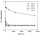
次にこれら各クローンの抗体価(希釈倍率:100、300、900、2700、8100、24300、および72900)を検討した。結果を図2に示す。
【0019】
【発明の効果】
本発明により、ヒトMKに対するモノクローナル抗体が得られた。このマウス抗ヒトMKモノクローナル抗体は、1-ステップサンドイッチ法に有効に適用できる。
【0020】
【図面の簡単な説明】
【図1】 129/Sv系マウスのエクソン2とエクソン3の一部を破壊したノックアウトマウスの変異型染色体を示す図である。
【図2】 クローンA、B、C、D、およびEから得られたモノクローナル抗体の抗体価を示す図である。[0001]
[Technical field to which the invention belongs]
The present invention relates to a monoclonal antibody that specifically binds to human midkine , and a method for producing the same .
[0002]
[Prior art]
Midkine (MK) is a growth / differentiation factor discovered as a product of a gene that is transiently expressed during the process of induction of differentiation of embryonic tumor cells (EC) by retinoic acid. A rich 13 kDa polypeptide (Kadomatsu, K. et al .: Biochem. Biophys. Res. Commun., 151: 1312-1318; Tomomura, M. et al .: J. Biol. Chem., 265: 10765 -10770, 1990). MK has been found from humans to Xenopus, but protein conservation between species is high, and human and mouse MKs have 87% homology in amino acid sequence. MK has various biological activities. That is, it is considered to be involved in the construction of the nervous system by promoting neurite outgrowth and nerve cell survival. It is also expressed in tissues at the time of injury, many human cancers, and senile plaques of Alzheimer's disease, and is also noted in relation to tissue repair, fibrinolytic system activation ability, and pathological conditions.
Development of a measurement system for measuring MK with high sensitivity is desired because of the biological activity of such human MK (sometimes simply referred to as “MK”) and the possibility of involvement of MK in the course of disease. As a method for quantitatively measuring an antigen such as MK, enzyme immunoassay (EIA) is considered to be most appropriate. Heterogeneous non-competitive method (sandwich method) is the most commonly used measurement system for antigen measurement by EIA. In this method, an immunocomplex consisting of an antibody insolubilized on a solid phase, an antigen to be measured, and an enzyme-labeled antibody is formed on the solid phase, and the enzyme activity on the solid phase is measured and measured. This is a method of knowing the amount of antigen.
[0003]
A 1-step sandwich method has been proposed as an EIA for measuring MK (Japanese Patent Laid-Open No. 10-160735). In this method, polyclonal antibodies derived from different animals (rabbit and chicken) are used for both the immobilized antibody and the labeled antibody. Polyclonal antibodies have high avidity, but cross-reaction with unintended biomolecules mixed in the immunogen often becomes a problem, and it is impossible to supply antibodies of a certain quality indefinitely. On the other hand, since a monoclonal antibody recognizes a specific epitope site of a part of an antigen, it has high specificity and can supply an infinite number of antibodies of a certain quality. Therefore, it is desirable to change the antibody combination used in the 1-step sandwich method for measuring human MK from a polyclonal antibody-polyclonal antibody combination to a polyclonal antibody-monoclonal antibody or a monoclonal antibody-monoclonal antibody. ing. However, preparation of a monoclonal antibody that specifically recognizes MK has been attempted so far, but no anti-human MK monoclonal antibody has been obtained.
[0004]
[Problems to be solved by the invention]
An object of the present invention is to provide a monoclonal antibody that specifically recognizes human MK.
[0005]
[Means for Solving the Problems]
If the immunogen is an animal-derived biologically active substance, the immunized animal will have the same biological activity, and there may be many parts with homology in the chemical structure and few parts in the species-specific structure. The selection of immunized animals is difficult.
In the case of MK, as described above, protein conservation between species is high, and human and mouse MK have 87% homology in amino acid sequence. For this reason, when a mouse is immunized with the human MK protein as an immunogen, the degree of recognition as a foreign heterologous protein in the mouse may be reduced. Actually, there has been no report that anti-human MK monoclonal antibodies have been produced using mice as immunized animals.
The present inventors used multiple lines of mice knocked out of the MK gene as immunized animals. Examining the sex and age of mice, examining the immunization method, immunization amount, injection site (subcutaneous, intradermal, intramuscular, intravenous, intraperitoneal, intralymphatic node, intrafootpad), administration interval, etc. A mouse anti-human MK monoclonal antibody having a high antibody titer could be prepared.
[0006]
That is, the present invention provides an anti-midkine monoclonal antibody that specifically binds to human midkine.
In addition, mice knocked out of the midkine gene are immunized with human midkine or a human midkine fragment having the same biological activity as the human midkine, and cell fusion is performed between the spleen cells obtained from the immunized mouse and myeloma cells. The present invention also provides a method for producing an anti-human midkine / mouse monoclonal antibody, comprising selecting a monoclonal antibody-producing hybridoma, proliferating the selected hybridoma, and collecting the produced monoclonal antibody.
[0007]
DETAILED DESCRIPTION OF THE INVENTION
1. Generation of MK gene knockout mice The technology of gene disruption by homologous recombination has already been established and has been attempted for a large number of genes. In particular, a means of disrupting (knocking out) a specific gene of embryonic stem cells (ES cells) and then injecting the cells into a blastocyst to produce a chimeric mouse produces a mutant mouse of the targeted gene. It is very useful in that you can explore its functions. Today, when a new gene is isolated, it is a natural research method to first create a knockout mouse.
The gene for human MK has already been cloned (Uehara, K. et al .: J. Biochem., 111: 563-567, 1992). Regarding mice knocked out of the MK gene, knockout mice in which a part of
There are a number of reports (eg, Mansor, SL et al .: Nature, 336: 348-352, 1988; Joyner, A., ed .: Gene targeting, IRL Press) related to the generation of mice that knocked out various genes. , 1993; Shinichi Aizawa: Generating mutant mice using gene targeting ES cells. Yodosha, 1995) and human MK genes are well known in the art. MK gene knockout mice can be easily prepared with reference to.
[0008]
2. Preparation of mouse anti-human MK monoclonal antibody As for monoclonal antibodies, the production method was reported by Köhler and Milstein in 1975, and many literatures and experimental books have been published on the production of monoclonal antibodies. However, in general, obtaining a monoclonal antibody having high specificity and affinity for any antigen has not yet become an established technique.
[0009]
A monoclonal antibody is generally prepared by the following steps.
1. 1. Preparation of
[0010]
1. Preparation of immunogen The cDNA of human MK has been cloned (JP-A-5-91880). The immunogenic MK protein can be prepared as a recombinant MK. For example, recombinant MK in which this cDNA is expressed in Pichia yeast has been reported (Japanese Patent Laid-Open No. 9-95454). Chemically synthesized MK can also be tried as an immunogen. Full length MK has been chemically synthesized (Inui, T. et al .: J. Peptide Sci., 2: 28-39, 1996). In the present invention, an MK fragment having the same biological activity (heparin binding ability, neurite extension ability, fibrinolytic system activation ability) as human MK, for example, a truncated MK lacking the N domain (Kaname, T. et al .: Biochem Biophys. Res. Commun., 219: 256-260, 1996) Fragments may be attempted as immunogens. Such an MK fragment may be used by covalently binding to a carrier protein so as to have sufficient immunogenicity.
Examples of carrier proteins include albumin (bovine serum albumin, human serum albumin, rabbit serum albumin, ovalbumin), serum globulin fraction (bovine gamma globulin, horse serum globulin, sheep gamma globulin), thyroglobulin (bovine thyroglobulin, butyrothyro Globulin), hemocyanin (KLH), synthetic polypeptides (poly-L-lysine, polyglutamic acid) and the like.
As a method for binding the MK fragment to the carrier protein, (1) a method using SH of cysteine residue (MBS method), (2) a method using amino group (bisimide ester method, glutaraldehyde method), ( 3) A method using a phenol group (bis-diazobenzene method), (4) a method using a carboxyl group (carbodiimide method), and the like.
It is necessary to try various combinations of carrier proteins and binding methods that bind to MK and to select the most immunogenic combination. Since such a selection can be easily performed by a person skilled in the art through routine experiments, the combinations thus selected are included in the present invention.
[0011]
2. Immunization of MK Knockout Mice with MK Antigen When immunizing MK knockout mice, the animal species and the ability to respond to the antigen are important points. Generally, when the animal species used for immunization and the animal species of myeloma cells are the same, a hybridoma is efficiently formed. In particular, since all myeloma cells derived from mice originate from BALB / c mice, it is considered that BALB / c mice are suitable for MK knockout mouse strains of immunized animals. In this case, both spleen cells and myeloma cells are derived from BALB / c, and the resulting hybridoma can grow in the intraperitoneal cavity of BALB / c, so that a high concentration of monoclonal antibody can be obtained from ascites without any special treatment. . However, it is difficult to establish ES cells from BALB / c mice.
[0012]
For immunization, for many protein antigens and polysaccharide antigens, an emulsion is prepared by mixing 1 to 100 μg of an antigen and an equal amount of Freund's complete adjuvant, and then administered intraperitoneally or subcutaneously. Although the action of adjuvants is not fully understood, it is believed that there are at least two important actions. One is a function of preventing the antigen from being processed quickly, and the other is a function of inducing a non-specific immune reaction to easily cause an immune reaction against the target antigen. In general, the former is widely used, and an emulsion with an aqueous solution is prepared and used. Recently, liposomes and synthetic surfactants have also been used as adjuvants. In the latter, Lipid A and killed bacteria are used. Currently used dead bacteria are Bordetalla pertussis and Mycobacterium tuberculosis. Mycobacterium tuberculosis is a dead bacterium added to Freund's complete adjuvant. The active site has been shown to be present in muramyl dipeptide (MDP), and various properties of MDP are commercially available. In this way, a substance that retains an antigen and a substance that non-specifically elicits an immune reaction are combined and used as an adjuvant (including dead bacteria). In general, Freund's complete adjuvant is often used for the initial immunization, and incomplete adjuvant (not including dead bacteria) is often used for the booster immunization. Since the optimal selection and combination of adjuvants can be easily carried out by those skilled in the art through routine experimentation, combinations thus selected are included in the present invention.
One to several booster immunizations are performed at intervals of 2 to 3 weeks after the initial immunization. Immediately before the booster immunization and one week after that, a test blood is collected to measure antibody titer. The mouse with the highest antibody titer is selected and boosted to provide for experiments.
[0013]
The above immunization methods can vary depending on the nature and amount of the antigen, and selection of the optimal method is a routine task for those skilled in the art.
[0014]
3. Cell fusion Cell fusion is basically performed according to the method of Milstetan et al. (Galfre, G. & Milstein, C., Methods Enzymol. 73: 3-46, 1981). Representative mouse line myeloma cells used for monoclonal antibody production are P3-X63-Ag8, P3-X63-Ag8U1, X63Ag8.653, SP2 / 0-Ag14, FO, NSI / 1-Ag4-1, NSO / 1. FOX-NY, but all of these cell lines are derived from the P3K strain. P3K is a cell derived from the myeloma strain MOPC21 derived from mineral oil-induced BALB / c mice, and P3 and MS-1 were obtained as cell lines without the P3K HGPRT gene.
[0015]
4). Monoclonal antibody screening When screening for the presence or absence of antibodies in the culture supernatant after cell fusion 7 to 14 days later, it is convenient to use EIA in which MK is bound to a solid phase. After screening with EIA, cells in positive wells are expanded and cloned.
5). Positive wells detected by growth and freeze screening of antibody-producing cells should contain the cells of interest. However, as the passage continues, the target cell may be lost due to loss of survival competition with other cells or loss of chromosomes. In preparation for this situation, it is important to keep the cells frozen while cloning.
In the present invention, the mouse anti-human MK monoclonal antibody thus obtained, and Fab, Fab ′, F obtained by treating the antibody with an enzyme such as trypsin, papain, pepsin, etc. (ab ') is included.
[0016]
The mouse anti-human MK monoclonal antibody obtained by the present invention can be used in place of the mouse anti-human polyclonal antibody of the 1-step sandwich method (Japanese Patent Laid-Open No. 10-160735). In this case, as an antibody imparting a label, an IgG fraction, or a specific Fab ′ obtained by reduction after pepsin digestion can be used.
[0017]
【Example】
EXAMPLES Hereinafter, although an Example demonstrates this invention, this invention is not limited to these Examples.
[0018]
[Example 1] Production of mouse anti-human MK An immunized animal was an MK knockout mouse derived from two lines (A and B) of ES cells (Nakamura, E. et al .: Genes to Cells, 3: 811-822, 1998). ), Males and females were used as immunized animals. Human MK protein (Japanese Patent Laid-Open No. 9-95454) was used as the antigen. Lines and antigen doses are as follows.
・ Line A (male, 3) ・ ・ ・ MK100μg / animal ・ Line A (female, 2) ... MK1μg / animal ・ Line B (male, 4) ・ MK1μg / animal ・ Line B (female) 5)
A marked increase in antibody titer was observed for 5 females in line B. Therefore, the final booster immunization was performed at 10 μg / mouse from the fundus vein of the mouse of these five MK knockout mice.
Cell fusion of spleen cells and myeloma cells (P3U1) obtained from each mouse individual is basically performed by the method of Milstein et al. (Galfre, G. & Milstein, C., Methods Enzymol. 73: 3-46). , 1981). As a result of usual cloning, five clones (clone A, B, C, D, and E) were obtained from one specific mouse individual. These clones were stored frozen. Monoclonal antibodies were prepared from these clones by conventional methods. All five subclasses of these antibodies were IgG (κ).
Next, the antibody titer (dilution ratio: 100, 300, 900, 2700, 8100, 24300, and 72900) of each of these clones was examined. The results are shown in FIG.
[0019]
【The invention's effect】
According to the present invention, a monoclonal antibody against human MK was obtained. This mouse anti-human MK monoclonal antibody can be effectively applied to the 1-step sandwich method.
[0020]
[Brief description of the drawings]
FIG. 1 is a diagram showing mutant chromosomes of knockout mice in which
FIG. 2 shows the antibody titers of monoclonal antibodies obtained from clones A, B, C, D, and E.
Claims (3)
Priority Applications (1)
| Application Number | Priority Date | Filing Date | Title |
|---|---|---|---|
| JP2000272199A JP3887154B2 (en) | 2000-09-07 | 2000-09-07 | Monoclonal antibody against human MK |
Applications Claiming Priority (1)
| Application Number | Priority Date | Filing Date | Title |
|---|---|---|---|
| JP2000272199A JP3887154B2 (en) | 2000-09-07 | 2000-09-07 | Monoclonal antibody against human MK |
Publications (3)
| Publication Number | Publication Date |
|---|---|
| JP2002085058A JP2002085058A (en) | 2002-03-26 |
| JP2002085058A5 JP2002085058A5 (en) | 2005-07-07 |
| JP3887154B2 true JP3887154B2 (en) | 2007-02-28 |
Family
ID=18758359
Family Applications (1)
| Application Number | Title | Priority Date | Filing Date |
|---|---|---|---|
| JP2000272199A Expired - Fee Related JP3887154B2 (en) | 2000-09-07 | 2000-09-07 | Monoclonal antibody against human MK |
Country Status (1)
| Country | Link |
|---|---|
| JP (1) | JP3887154B2 (en) |
Families Citing this family (6)
| Publication number | Priority date | Publication date | Assignee | Title |
|---|---|---|---|---|
| JP2007297282A (en) * | 2004-08-13 | 2007-11-15 | Cell Signals Inc | Influence caused by midkine when endometriosis appear |
| JP5398987B2 (en) | 2005-11-14 | 2014-01-29 | セルミド リミテッド | Method for treating and preventing diseases based on abnormal function of regulatory T cells |
| US9163081B2 (en) | 2006-11-14 | 2015-10-20 | Medical Therapies Limited | Antibody recognizing C-domain of midkine |
| WO2010074218A1 (en) * | 2008-12-25 | 2010-07-01 | 株式会社イーベック | Anti-human midkine antibody |
| WO2012122590A1 (en) | 2011-03-14 | 2012-09-20 | Cellmid Limited | Antibody recognizing n-domain of midkine |
| US9840552B2 (en) | 2012-07-30 | 2017-12-12 | National University Corporation Nagoya University | Monoclonal antibody against human midkine |
-
2000
- 2000-09-07 JP JP2000272199A patent/JP3887154B2/en not_active Expired - Fee Related
Also Published As
| Publication number | Publication date |
|---|---|
| JP2002085058A (en) | 2002-03-26 |
Similar Documents
| Publication | Publication Date | Title |
|---|---|---|
| US5783185A (en) | Monoclonal antibodies to transforming growth factor-beta and methods of use | |
| US5955317A (en) | Antibodies to β-amyloids or their derivatives and use thereof | |
| US5036003A (en) | Antibodies to VPF | |
| JPH11504635A (en) | Regulation of cholesteryl ester transfer protein (CETP) activity | |
| JP4374316B2 (en) | Antibody to β-amyloid or a derivative thereof and use thereof | |
| JP3887154B2 (en) | Monoclonal antibody against human MK | |
| EP0399429A1 (en) | Anti-human interleukin-6 monoclonal antibody | |
| EP0709455A1 (en) | Monoclonal antibody to human glicentin, hybridoma for producing said antibody and assay method for human glicentin using said antibody | |
| JP2004506603A (en) | Monoclonal antibodies against human LDL receptor, their production and use | |
| JPH0881499A (en) | Hybridoma producing thymosin alpha-1-resistant monoclonal antibody | |
| CA2058041A1 (en) | Anti-igf-ii monoclonal antibody | |
| HU192989B (en) | Process for production of phisiologically active hormone preparatives | |
| JP4059404B2 (en) | Antibodies with activity to stimulate thyroid function | |
| JP3288716B2 (en) | Human adhesion molecule occludin | |
| EP0594879A1 (en) | Cathepsin L-specific monoclonal antibodies, hybridomas for producing the same and a method of using the same | |
| JP2955082B2 (en) | Monoclonal antibody that specifically recognizes the N-terminal part of human calcitonin | |
| WO2004031241A1 (en) | Monoclonal antibody against subtilisin-like proprotein convertase pace4 and utilization thereof | |
| JP3213359B2 (en) | Anti-IGF-II monoclonal antibody | |
| CN111349169B (en) | Monoclonal antibody of Werner syndrome ATP dependent helicase and application thereof | |
| Yokota et al. | Monoclonal antibodies against synthesized short peptides corresponding to human AA amyloid protein | |
| US7078514B1 (en) | Chicken growth hormone releasing hormone receptor | |
| CN111349157B (en) | Monoclonal antibody of cadherin 6 and application thereof | |
| CN111349170B (en) | Monoclonal antibody of immune related GTPase family M (IRGM) and application thereof | |
| CN111349171B (en) | Monoclonal antibody of peroxidase-6 and application thereof | |
| CN110483642B (en) | Monoclonal antibody of enoyl-acyl carrier protein reductase and application thereof |
Legal Events
| Date | Code | Title | Description |
|---|---|---|---|
| A521 | Written amendment |
Free format text: JAPANESE INTERMEDIATE CODE: A523 Effective date: 20001012 |
|
| A711 | Notification of change in applicant |
Free format text: JAPANESE INTERMEDIATE CODE: A711 Effective date: 20011212 |
|
| A521 | Written amendment |
Free format text: JAPANESE INTERMEDIATE CODE: A821 Effective date: 20011212 |
|
| A521 | Written amendment |
Free format text: JAPANESE INTERMEDIATE CODE: A523 Effective date: 20020201 |
|
| A521 | Written amendment |
Free format text: JAPANESE INTERMEDIATE CODE: A523 Effective date: 20020208 |
|
| A521 | Written amendment |
Free format text: JAPANESE INTERMEDIATE CODE: A821 Effective date: 20041105 Free format text: JAPANESE INTERMEDIATE CODE: A523 Effective date: 20041105 |
|
| A621 | Written request for application examination |
Free format text: JAPANESE INTERMEDIATE CODE: A621 Effective date: 20041105 |
|
| RD02 | Notification of acceptance of power of attorney |
Free format text: JAPANESE INTERMEDIATE CODE: A7422 Effective date: 20041105 |
|
| A521 | Written amendment |
Free format text: JAPANESE INTERMEDIATE CODE: A523 Effective date: 20041105 |
|
| RD04 | Notification of resignation of power of attorney |
Free format text: JAPANESE INTERMEDIATE CODE: A7424 Effective date: 20060405 |
|
| A131 | Notification of reasons for refusal |
Free format text: JAPANESE INTERMEDIATE CODE: A131 Effective date: 20060830 |
|
| A521 | Written amendment |
Free format text: JAPANESE INTERMEDIATE CODE: A523 Effective date: 20061024 |
|
| TRDD | Decision of grant or rejection written | ||
| A01 | Written decision to grant a patent or to grant a registration (utility model) |
Free format text: JAPANESE INTERMEDIATE CODE: A01 Effective date: 20061117 |
|
| A61 | First payment of annual fees (during grant procedure) |
Free format text: JAPANESE INTERMEDIATE CODE: A61 Effective date: 20061124 |
|
| R150 | Certificate of patent or registration of utility model |
Free format text: JAPANESE INTERMEDIATE CODE: R150 |
|
| RD02 | Notification of acceptance of power of attorney |
Free format text: JAPANESE INTERMEDIATE CODE: A7422 Effective date: 20071113 |
|
| FPAY | Renewal fee payment (event date is renewal date of database) |
Free format text: PAYMENT UNTIL: 20091201 Year of fee payment: 3 |
|
| RD04 | Notification of resignation of power of attorney |
Free format text: JAPANESE INTERMEDIATE CODE: R3D04 |
|
| FPAY | Renewal fee payment (event date is renewal date of database) |
Free format text: PAYMENT UNTIL: 20091201 Year of fee payment: 3 |
|
| RD02 | Notification of acceptance of power of attorney |
Free format text: JAPANESE INTERMEDIATE CODE: R3D02 |
|
| FPAY | Renewal fee payment (event date is renewal date of database) |
Free format text: PAYMENT UNTIL: 20091201 Year of fee payment: 3 |
|
| S111 | Request for change of ownership or part of ownership |
Free format text: JAPANESE INTERMEDIATE CODE: R313113 |
|
| FPAY | Renewal fee payment (event date is renewal date of database) |
Free format text: PAYMENT UNTIL: 20091201 Year of fee payment: 3 |
|
| R350 | Written notification of registration of transfer |
Free format text: JAPANESE INTERMEDIATE CODE: R350 |
|
| A072 | Dismissal of procedure |
Free format text: JAPANESE INTERMEDIATE CODE: A072 Effective date: 20080401 |
|
| FPAY | Renewal fee payment (event date is renewal date of database) |
Free format text: PAYMENT UNTIL: 20091201 Year of fee payment: 3 |
|
| S202 | Request for registration of non-exclusive licence |
Free format text: JAPANESE INTERMEDIATE CODE: R315201 |
|
| FPAY | Renewal fee payment (event date is renewal date of database) |
Free format text: PAYMENT UNTIL: 20091201 Year of fee payment: 3 |
|
| R350 | Written notification of registration of transfer |
Free format text: JAPANESE INTERMEDIATE CODE: R350 |
|
| FPAY | Renewal fee payment (event date is renewal date of database) |
Free format text: PAYMENT UNTIL: 20091201 Year of fee payment: 3 |
|
| S111 | Request for change of ownership or part of ownership |
Free format text: JAPANESE INTERMEDIATE CODE: R313113 |
|
| FPAY | Renewal fee payment (event date is renewal date of database) |
Free format text: PAYMENT UNTIL: 20091201 Year of fee payment: 3 |
|
| R350 | Written notification of registration of transfer |
Free format text: JAPANESE INTERMEDIATE CODE: R350 |
|
| FPAY | Renewal fee payment (event date is renewal date of database) |
Free format text: PAYMENT UNTIL: 20101201 Year of fee payment: 4 |
|
| FPAY | Renewal fee payment (event date is renewal date of database) |
Free format text: PAYMENT UNTIL: 20111201 Year of fee payment: 5 |
|
| FPAY | Renewal fee payment (event date is renewal date of database) |
Free format text: PAYMENT UNTIL: 20121201 Year of fee payment: 6 |
|
| FPAY | Renewal fee payment (event date is renewal date of database) |
Free format text: PAYMENT UNTIL: 20121201 Year of fee payment: 6 |
|
| FPAY | Renewal fee payment (event date is renewal date of database) |
Free format text: PAYMENT UNTIL: 20131201 Year of fee payment: 7 |
|
| R250 | Receipt of annual fees |
Free format text: JAPANESE INTERMEDIATE CODE: R250 |
|
| R250 | Receipt of annual fees |
Free format text: JAPANESE INTERMEDIATE CODE: R250 |
|
| R250 | Receipt of annual fees |
Free format text: JAPANESE INTERMEDIATE CODE: R250 |
|
| LAPS | Cancellation because of no payment of annual fees |