JP3774518B2 - X-ray CT scanner - Google Patents
X-ray CT scanner Download PDFInfo
- Publication number
- JP3774518B2 JP3774518B2 JP29416596A JP29416596A JP3774518B2 JP 3774518 B2 JP3774518 B2 JP 3774518B2 JP 29416596 A JP29416596 A JP 29416596A JP 29416596 A JP29416596 A JP 29416596A JP 3774518 B2 JP3774518 B2 JP 3774518B2
- Authority
- JP
- Japan
- Prior art keywords
- data
- ray
- detection
- sensitivity distribution
- detection element
- Prior art date
- Legal status (The legal status is an assumption and is not a legal conclusion. Google has not performed a legal analysis and makes no representation as to the accuracy of the status listed.)
- Expired - Fee Related
Links
Images
Landscapes
- Measurement Of Radiation (AREA)
- Apparatus For Radiation Diagnosis (AREA)
Description
【0001】
【発明の属する技術分野】
本発明は、被検体の体軸方向に直交する方向(チャンネル方向)に沿って配列された複数の検出素子を有する検出手段により前記被検体を透過してきたX線を検出し、検出したデータに応じて生成された投影データに基づいて被検体の断層面のCT画像を撮影するX線CTスキャナに係わり、特に、投影データに付加されるサンプリング位置に特徴を有するX線CTスキャナに関する。
【0002】
【従来の技術】
X線CTスキャナにおいて従来から用いられていたタイプとして、ファンビーム(シングルスライス)X線CTスキャナ(シングルスライスCTともいう)がある。
【0003】
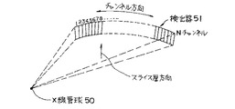
このファンビームX線CTスキャナは、図18に示すように、図示しない被検体(例えば患者)を挟んで対向配置されたX線管球(X線言、X線管)50及び検出器51を有しており、検出器51においては、当該被検体の体軸方向に沿ったスライス厚方向に直交する方向(チャンネル方向)に沿って扇状に検出素子がNチャンネル(例えば約1000チャンネル)並べられており、当該各検出素子は、スライス方向に沿った断面が矩形状に形成されている。
【0004】
検出器51には種々のタイプが使用可能であるが、小型化が可能なシンチレーション検出器が良く用いられている。このシンチレーション検出器は、各検出素子としてシンチレータ及びフォトダイオード等の光センサ(光電変換器)をそれぞれ有し、被検体を透過したX線をシンチレータにより吸収し、その吸収により当該シンチレータで発生した蛍光を光センサによって電気信号に変換して各検出素子毎に出力するようになっている。
【0005】
すなわち、上述したX線CTスキャナによれば、X線源50から被検体のあるスライス面(単にスライスともいう)に対してファン状にX線ビームを照射し、被検体のあるスライス面を透過したX線ビームを検出器51の各検出素子毎に電気信号に変換してX線透過データを収集する。
【0006】
検出器51の各素子毎に収集されたX線透過データは、その素子毎に設けられたデータ収集素子を有するデータ収集装置(DAS)に送られ、その各データ収集素子により増幅処理等が行なわれて投影データ(1回のデータ収集を1ビューという)が収集される。
【0007】
そして、X線源50及び検出器51を一体で被検体の周囲に回転させながらX線源の焦点を介してX線照射を行なって前記データ収集を約1000回程度繰り返すことにより、被検体に対する多方向からの投影データが収集される。
【0008】
収集された多方向からの投影データに対してデータ補間処理を施してパラレルビームの投影データに変換し、その変換投影データにコンボリューションバックプロジェクション等の再構成処理を施して被検体のスライス面の画像が再構成される。
【0009】
X線管50の焦点から照射されるX線ビームは、被検体の体軸方向、すなわち検出器スライス厚方向に広がりを持つ薄いコーンビームとして被検体に対して照射される。このX線ビームのビーム幅を、図19に示すように被検体到達前にプリコリメータ52で(図19(a)参照)、あるいは被検体通過後に検出器入射直前にポストコリメータ53で(図19(b)参照)制御して所望のスライス厚を有するX線ビームを生成し、生成されたX線ビームを検出器51の各検出素子により検出するようになっている。
【0010】
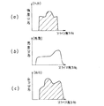
検出器51の各検出素子に入射されたX線ビームは、当該各検出素子を介して検出信号(電気信号)に変換されて出力されるが、その際の入力X線強度と出力電気信号の強度分布(出力分布)の比を各検出素子の感度として下式のように表す(図20(a)〜図20(c)参照、なお、スライス厚方向の分布のみを示し、チャンネル方向は一定とする。以下の図においても同様とする)。
【0011】
【数1】
(感度)=(出力)/(入力) ……(1)
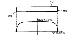
また、検出器の各検出素子では、シンチレータのスライス厚方向の端部(エッジ部)において光吸収が生じるため、図21に示すように、各検出素子51aの感度分布はその素子の端部(エッジ)51bにおいて低下することが知られている。したがって、シングルスライスCTでは、図22に示すように、例えばプリコリメータ52によるX線ビームのコリメートにより検出素子エッジ部分51bを利用せず、検出素子51aの中央及びその近傍部分、すなわち当該検出素子51aの感度分布が略平坦な領域の検出信号のみをデータとして利用している。このため、検出素子に入射する略矩形のX線ビームの強度分布(図23(a)参照)に対応した検出素子の出力分布も、当該検出素子の感度分布(図23(b)参照))にほとんど関係なく矩形となる(図23(c)参照)。
【0012】
すなわち、図23に示すように、各検出素子51aには略矩形のX線ビームが入射され、その入射ビームに対応する矩形の検出信号が出力されることになる。
【0013】
一方、上述した補間処理やコンボリューションバックプロジェクション等の再構成処理においては、収集された投影データを離散化されたサンプリング位置のデータとして用いている。すなわち、各検出素子で検出された信号に基づく投影データは、前記サンプリング位置におけるデータとして用いられることになる。
【0014】
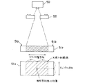
シングルスライスCTにおいては、検出素子における感度分布が略平坦な領域をデータ収集領域としており、X線入射範囲及びその検出素子からの出力信号の強度分布も略矩形であるため、図22に示すように、X線焦点を含む面上に存在する前記X線入射範囲(あるいは検出素子51a自体の矩形形状)の幾何学的な重心位置を上述した投影データのサンプリング位置としている。つまり、ある検出素子により検出された信号に基づく投影データは、その検出素子の幾何学的重心位置が有するデータとして以後の再構成処理等で扱われることになる。
【0015】
一方、このようなシングルスライスX線CTスキャナでは、被検体のある一つのスライス面の画像を得ているため、短時間に広い範囲の画像を撮影することは難しく、医師等から単位時間により高精細(高解像度)且つ広範囲に画像を撮影したいという強い要望が出されていた。
【0016】
この要望に答えるために、近年、マルチスライスX線CTスキャナ(マルチスライスCTともいう)が研究されている。
【0017】
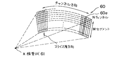
このマルチスライスCTは、シングルスライスCTにおける検出器の検出素子を被検体の体軸方向に対応するスライス厚方向(seg方向)に沿って複数列(複数(M)セグメント)有しており、当該検出器は、全体でNチャンネル×Mセグメントの検出素子を有する2次元検出器60として構成されている(図24参照)。この場合、DASの各素子は、例えば2次元検出器60の各検出素子毎に設けられている。
【0018】
すなわち、マルチスライスCTによれば、円錐状(コーン状)のX線ビームを曝射するX線源61と、上述した2次元検出器60とを有しており、当該円錐状のX線ビーム(有効視野直径FOV)に基づいて被検体を透過したX線を2次元検出器60の各検出素子で検出することにより、当該被検体の多スライス面の投影データを一度に収集するものであり、上述した高精細且つ広範囲な画像収集を可能にするものとして期待されている。
【0019】
このようなマルチスライスCTにおいても、シングルスライスCTと同様に、プリコリメータ、あるいはポストコリメータで照射X線ビームを制御して所望のスライス厚を有するX線ビームを生成し、生成されたX線ビームを検出器の各検出素子により検出して出力するようになっている。
【0020】
例えば図24においてあるチャンネルの検出素子列(なお、セグメント方向のスライスピッチを2mmとし、セグメント数を30とする)60aに着目すれば(図25(a)参照)、上述したポストコリメータの制御によりスライス厚が2mmのデータを例えば10スライス(slice)分出力可能である(図25(b))。また、3つの素子で検出されたデータを束ねて1つのデータとすることにより、スライス厚が6mmのデータを例えば10スライス(slice)分出力可能である(図25(c))。
【0021】
図25(b)及び図25(c)を比較参照すると分かるように、図25(b)においてはスライス厚が2mmの分解能の高いスライスに対応する検出信号が出力され、図25(c)においてはスライス厚が6mmとなり分解能は低いがスライス厚方向に沿って撮影領域が大きい検出信号が出力されるため、上述した高精細且つ広範囲な画像収集が共に実現できる。
【0022】
このようなマルチスライスCTにおいても、シングルスライスCTと同様に1つのデータを構成する検出素子(図25(b)のように1素子が1データに対応する場合もあり、また、図25(c)のように複数素子が1データに対応する場合もある)の形状(不感帯ゾーンを除いたアクティブエリアの形状や素子全体の形状)の幾何学的重心位置(長方形であれば対角線の交点)を当該検出素子で検出された信号に基づく投影データのサンプリング位置としている。なお、図25(c)のように複数素子が1データに対応する場合においては、複数素子全体の形状における幾何学的重心位置がデータサンプリング位置となる。
【0023】
【発明が解決しようとする課題】
上述したように、従来のシングルスライスCT及びマルチスライスCTにおいて、1つのデータを構成する検出素子のデータサンプリング位置は、常に当該検出素子の幾何学的重心位置に定められている。
【0024】
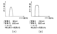
しかしながら、各検出素子の感度分布は、前掲図21に示したようにエッジ部分において感度が低下する分布であり、さらに、その感度分布は、X線管球の管電流や管電圧、当該検出素子に入射される入射X線のエネルギー特性(エネルギー分布)等のX線照射条件やビューレート等のデータ収集条件に対応して変化する(図26(A)及び(B)参照)ため、場合によっては、各検出素子の感度分布に偏りが生じることがあった。
【0025】
特に、マルチスライスCTにおいて複数の検出素子の検出データを束ねて1つのデータとして扱う場合には、束ねられた各データに対応する検出素子の感度分布が不均一となることがあり、全体の感度分布に偏りが生じることがあった。
【0026】
すなわち、図27に示すように、1データ分の検出素子(1個、あるいは複数個)の感度分布が偏りを有する分布である場合、当該検出素子の幾何学的重心位置と実際の感度分布の重心位置とが異なることがある。
【0027】
検出素子の感度分布が平坦である場合においては、実際の感度分布の重心位置は、当該検出素子の幾何学的重心位置に略一致しているため、当該幾何学的重心位置をデータサンプリング位置としても別段問題は無い。
【0028】
しかしながら、上述したように検出素子の幾何学的重心位置が実際の感度分布の重心位置と異なっている場合には、幾何学的重心位置をデータサンプリング位置として補間処理や再構成処理で扱うと、実際の感度分布から見て誤ったサンプリング位置のデータを利用していることに等しくなる。このデータサンプリング位置のズレ(誤差)は、補間処理におけるデータの重み付け等や再構成処理におけるフィルタ処理等において処理精度を低下させることになり、再構成されたCT画像にアーチファクト等の画質劣化を生じさせる原因となっていた。
【0029】
本発明は上述したような事情に鑑みてなされたもので、各投影データに対応する検出器の各検出素子におけるデータサンプリング位置を当該素子の実際の感度分布の重心位置に設定することにより、データサンプリング位置の誤差に起因した画質劣化を抑制し、視認性の良い高画質なCT画像を得ることをその目的とする。
【0032】
【課題を解決するための手段】
前記目的を達成するため本発明のX線CTスキャナでは、X線を曝射するX線管と、被検体の体軸方向及びその体軸方向に直交する方向に沿って2次元的に配列された複数の検出素子を有し、前記X線管から曝射され当該被検体を透過してきたX線を前記各検出素子により検出する2次元検出手段と、この2次元検出手段の各検出素子により検出されたデータに基づいて前記体軸方向に直交する複数のスライス面の投影データをそれぞれ収集するデータ収集手段と、収集された複数の投影データ及びその複数の投影データそれぞれに対応するサンプリング位置データに基づいて少なくとも画像再構成処理を行なって前記複数のスライス面のX線画像を撮影する画像処理手段とを備えたX線CTスキャナにおいて、前記2次元検出手段の各検出素子の内、前記体軸方向に沿った少なくとも1列の検出素子の感度分布を求める手段と、求められた少なくとも1列の検出素子の感度分布に基づいて前記複数の投影データそれぞれに対応する検出素子の感度分布の重心位置データを求める手段とを備え、前記画像処理手段は、前記複数の投影データそれぞれに対応するサンプリング位置データとして当該複数の投影データそれぞれに対応して求められた重心位置データを用いるようにしている。
【0033】
特に、前記複数の投影データそれぞれに対応する検出素子の感度分布の重心位置データは、当該検出素子のX線入射面を含む2次元座標空間を設定した場合の当該検出素子の感度分布の1次モーメント量が最小のときの座標データであり、また、前記検出素子の感度分布を求める手段は、前記2次元検出手段の各検出素子の感度分布を求める手段である。
【0034】
特に、前記2次元検出手段は、前記体軸方向における前記検出素子のピッチを不均等に形成した2次元検出手段であるとともに、前記2次元検出手段の各検出素子を前記体軸方向の素子毎にスライス厚条件に応じて選択して前記データ収集装置の体軸方向に対応するデータ収集素子に接続する検出素子選択手段を備え、当該検出素子選択手段による前記各検出素子の選択内容により前記複数の投影データそれぞれのスライス厚を前記スライス厚条件に応じて設定するように構成している。
【0035】
特に、前記感度分布の重心位置データを求める手段は、前記各検出素子の選択内容に応じたスライス厚を有する複数の投影データそれぞれに対応する検出素子の感度分布の重心位置データを求める手段であり、また、前記感度分布を求める手段は、前記X線管から曝射されたX線を均一強度を有するX線に調整した後で直接前記検出手段の各検出素子に対して直接照射して当該各検出素子の感度分布を求める手段である。
【0036】
特に、前記感度分布を求める手段は、前記X線画像撮影時に設定可能な複数のデータ収集条件に応じて前記検出手段の各検出素子の感度分布を求める手段であり、前記感度分布の重心位置データを求める手段は、前記複数のデータ収集条件に応じて求められた複数の感度分布に基づいて、その感度分布毎に前記複数の投影データそれぞれに対応する検出素子の感度分布の重心位置データを求める手段である。
【0037】
特に、前記感度分布を求める手段は、前記X線画像撮影時に設定可能な複数のX線照射条件に応じて前記検出手段の各検出素子の感度分布を求める手段であり、前記感度分布の重心位置データを求める手段は、前記複数のX線照射条件に応じて求められた複数の感度分布に基づいて、その感度分布毎に前記複数の投影データそれぞれに対応する検出素子の感度分布の重心位置データを求める手段である。
【0038】
前記目的を達成するため、本発明のX線CTスキャナでは、X線を曝射するX線管と、被検体の体軸方向及びその体軸方向に直交する方向に沿って2次元的に配列された検出素子を有し、前記X線管から曝射され当該被検体を透過してきたX線を前記各検出素子により検出する2次元検出手段と、この2次元検出手段の各検出素子により検出されたデータに基づいて複数のスライス面の投影データをそれぞれ収集するデータ収集手段と、収集された複数の投影データ及びその投影データに対応するサンプリング位置データに基づいて少なくとも画像再構成処理を行なって前記複数のスライス面のX線画像を撮影する画像処理手段とを備えたX線CTスキャナにおいて、前記2次元検出手段の各検出素子の内、前記体軸方向に沿った少なくとも1列の検出素子の感度分布に基づいて前記複数の投影データそれぞれに対応する検出素子の感度分布の重心位置データを求める手段を備え、前記画像処理手段は、前記複数の投影データに対応するサンプリング位置データとして当該複数の投影データに対応して求められた重心位置データを用いるようにしたものである。
【0039】
収集された複数の投影データ及びその複数の投影データそれぞれに対応するサンプリング位置データに基づいて、画像処理手段により少なくとも画像再構成処理が行なわれ、前記複数のスライス面のX線画像が撮影されるようになっている。
【0040】
そして、本発明のX線CTスキャナによれば、2次元検出手段の各検出素子の内、体軸方向に沿った少なくとも1列の検出素子の感度分布(例えば全ての検出素子の感度分布)が均一強度を有するX線を検出手段の各検出素子に対して照射すること等の方法により求められ、求められた各検出素子の感度分布に基づいて、複数の投影データそれぞれに対応する検出素子の感度分布の重心位置データが求められる。この複数の投影データそれぞれに対応する検出素子の感度分布の重心位置データは、当該検出素子のX線入射面を含む2次元座標空間を設定した場合の当該検出素子の感度分布の1次モーメント量が最小のときの座標データとして定義されている。
【0041】
このとき、前記画像処理手段により前記サンプリング位置データとして、求められた複数の投影データそれぞれに対応する検出素子の感度分布の重心位置データが用いられているため、当該サンプリング位置データは、仮に複数の投影データそれぞれに対応する検出素子の感度分布に偏りが生じていたとしても、当該感度分布に最適な位置データとなっている。したがって、従来のCTスキャナのような検出素子の感度分布に関係なくサンプリング位置が当該素子の幾何学的重心位置に予め固定されていた場合と比べて、データサンプリング位置の誤差(位置ズレ)が解消されている。
【0042】
特に、本構成によれば、2次元検出手段として、体軸方向における前記検出素子のピッチが不均等に形成された2次元検出手段を用いた場合においては、検出素子選択手段により2次元検出手段の各検出素子が体軸方向の素子毎にスライス厚条件に応じて選択されてデータ収集装置の体軸方向に対応するデータ収集素子に接続されることにより、その各検出素子の選択内容、言い換えれば各検出素子の内の何れの検出素子を束ねてデータ収集素子に接続するかを定める内容により、複数の投影データそれぞれのスライス厚がスライス厚条件に応じて設定される。
【0043】
このとき、本構成では、各検出素子の選択内容に応じたスライス厚を有する複数の投影データそれぞれに対応する検出素子の感度分布の重心位置データが求められている。すなわち、どんなスライス厚条件が設定されたとしても、当該スライス厚を有する複数の投影データそれぞれに対応する検出素子の感度分布の重心位置データが求められていることになる。したがって、スライス厚の選択の仕方(束ね方)に起因して、得られた投影データの対応する検出素子の感度分布に偏りが生じたとしても、データサンプリング位置に誤差(位置ズレ)が生じることが無い。
【0044】
また、本構成によれば、複数の投影データそれぞれに対応する検出素子の感度分布の重心位置データは、X線画像撮影時に設定可能な複数のデータ収集条件毎に求められた検出手段の各検出素子の感度分布に基づいてそれぞれ生成されているため、当該複数のデータ収集条件の内の所定のデータ収集条件が選択されて前記CT画像が撮影された場合、そのデータ収集条件に対応して求められた検出素子の感度分布の重心位置データを用いることにより、データ収集条件に起因した各検出素子の感度分布への影響を抑制することができる。
【0045】
さらに、本構成によれば、複数の投影データそれぞれに対応する検出素子の感度分布の重心位置データは、X線画像撮影時に設定可能な複数のX線照射条件毎に求められた検出手段の各検出素子の感度分布に基づいてそれぞれ生成されているため、当該複数のX線照射条件の内の所定のX線照射条件が選択されて前記CT画像が撮影された場合、そのX線照射条件に対応して求められた検出素子の感度分布の重心位置データを用いることにより、X線照射条件に起因した各検出素子の感度分布への影響を抑制することができる。
【0046】
【発明の実施の形態】
以下、本発明の実施形態を添付図面を参照して説明する。
【0047】
図1は、本実施形態のX線CTスキャナ1の概略構成を示すブロック図である。
【0048】
図1によれば、X線CTスキャナ(CTシステム)1は、被検体(患者)P載置用の寝台2と、被検体Pを挿入して診断を行なうための図示しない診断用開口部を有し、被検体Pをスキャンして投影データの収集を行なうガントリー3と、スキャナ全体の制御を行なうとともに、収集された投影データに基づいて画像再構成処理や再構成画像表示等を行なうシステム部4とを備えている。
【0049】
寝台2は、図示しない寝台駆動部の駆動により被検体Pの体軸方向に沿ってスライド可能になっている。
【0050】
ガントリー3は、その診断用開口部に挿入された被検体Pを挟んで対向配置されたX線管球10及び主検出器11と、ガントリー駆動部12とを備えており、X線管球10と主検出器11は、ガントリー駆動部12の駆動により、ガントリー3の診断用開口内に挿入された被検体Pの体軸方向に平行な中心軸の廻りに一体で回転可能になっている。ガントリー3内のX線管球10と被検体Pとの間には、X線のエネルギー分布を被検体の撮像部位等に応じてHigh(高エネルギー分布)/Low(低エネルギー分布)のどちらか一方に制御するフィルタ10Aと、X線管球10のX線焦点から曝射されフィルタ10Aを介して送られてきたコーン状のX線ビームを整形し、所要の大きさのX線ビームを形成するためのプリコリメータ13が設けられている。また、主検出器11のX線ビーム入射側には、例えば主検出器11の列方向に沿って移動する2枚のX線遮蔽板を有するポストコリメータ14が設けられている。このポストコリメータ14は、スキャン条件(スライス厚条件)に応じて2枚のスリット板の主検出器11の列方向に沿った移動位置を制御することにより被検体Pを透過してきたX線ビームをトリミングして、良好なプロファイルの透過X線ビームを生成するように構成されている。
【0051】
さらに、X線CTスキャナ1は、X線管球10に高電圧を供給する高電圧発生装置15を備えている。この高電圧発生装置15によるX線管球10への高電圧供給は、例えば非接触式のスリップリング機構により行なわれる。
【0052】
主検出器11には、シンチレータ及びフォトダイオード等の光センサ(光電変換器)を有するシンチレーション検出器が用いられている。すなわち、X線管球10から曝射され被検体を透過したX線をシンチレータにより吸収し、その吸収により発生した蛍光を光センサによって電気信号に変換して出力するようになっている。
【0053】
このシンチレーション型主検出器11は、図2に示すように、1chあたり複数seg(本実施形態では20seg)がseg方向(体軸方向、スライス厚方向)に沿って並べられた検出素子列をチャンネル方向(ch方向)に沿って複数ch(本実施形態では16ch)アレイ状に配列した2次元検出器(図2では、16ch×20segの2次元検出器を示している)として構成されている。
【0054】
すなわち、図2では、第1chの20seg分の素子列を11a1 とすると、第1ch:11a1 〜第16ch:11a20の素子列が配置されており、また、第1segの16ch分の素子列を11α1 とすると、第1seg:11α1 〜第20seg:11α20の素子列が配置されていることになる。
【0055】
ここで、このように2次元配列された各検出素子の各素子の位置(アドレス)を(seg,ch)で表すと、例えば(第1seg,第1ch)に位置する素子は、11(1,1)として表され、以下、第1ch11a1 の素子列は、11(2,1)…11(20,1)と表される。また、このようにして、残りのch11a2 〜11a16の素子列は、それぞれ第2ch11a2 →11(1,2)…11(20,2),第3ch11a3 →11(1,3)…11(20,3),・・・・,第15ch11a15→11(1,15)…11(20,15),第16ch11a16→11(1,16)…11(20,16)として表される。
【0056】
なお、各seg間及び各ch間は、例えば金属板等のセパレータ(反射板)11s1 及び11s2 が設けられ、隣接するch間及びseg間のクロストークを無くすように構成されている。
【0057】
そして、主検出器11は、被検体Pを透過して入射されたX線から不均等スライス厚の検出信号を生成し、この不均等スライス厚の検出信号を束ねて同一スライス厚を有する複数のスライス、あるいは異なるスライス厚を有する複数のスライス等、指定されたスライス厚に基づく検出信号として出力するようになっている。
【0058】
本実施形態において上述した不均等スライス厚の検出信号を生成するために、主検出器(2次元検出器)11の各chの素子列11a1 〜11a16の各素子のseg方向のスライス厚(スライスピッチ)は、中央の素子から端部の素子に向けてピッチが広がるように不均等に形成されている。なお、このseg方向に沿って不均等に形成されたスライスピッチのことを不均等ピッチという。
【0059】
ここで、主検出器11の各チャンネル(ch)の素子列1a1 〜11a16の構成を図3に示す。なお、図3は、第1chの素子列11a1 について代表して示している。
【0060】
本実施形態においては、X線CTスキャナ1で得られる最小スライス厚を実現するためのサイズを有する検出素子列を基本セグメントと呼び、本実施形態では最小スライス厚が1mmの場合について説明する。
【0061】
図3によれば、本実施形態の主検出器11の各チャンネルの素子列11a1 〜11a16の構成は、中央に基本セグメント(1mm−seg;1mmスライス厚のセグメント)を8セグメント((図面向かって右側からseg1a1 〜seg1a8 とする)配列し、さらにその外側に2mmセグメント(2mm−seg;2mmスライス厚のセグメント)を片側に2セグメントずつ合計4セグメント配列し(図面向かって右側からseg2a1 〜seg2a4 とする)、さらにその2mmセグメントの外側に、4mmセグメント(4mm−seg;4mmスライス厚のセグメント)を片側に2セグメントずつ合計4セグメント配列する(図面向かって右側からseg4a1 〜seg4a4 とする)。さらに、4mmセグメントの外側に8mmセグメント(8mm−seg;8mmスライス厚のセグメント)を片側に2セグメントずつ合計4セグメント配列している(図面向かって右側からseg8a1 〜seg8a4 とする)。1チャンネルあたり合計20segであり、全体で64mmに対応する。なお、ここに示した寸法は、ガントリー3(X線管球10及び主検出器11)の回転軸中心での値であり、主検出器11における実寸法ではない。
【0062】
そして、主検出器11の各検出素子により検出されたX線透過データは、スイッチ群20を介して例えば各チャンネルの検出素子列11a1 〜11a16それぞれ(20seg)に対して、当該20segより少ない8列分(8スライス分)のデータ収集素子(DAS−1a1 〜DAS−8a1 …DAS−1a16〜DAS−8a16)を有するDAS(データ収集装置)21に送られる。
【0063】
図4は、本実施形態の2次元検出器11,スイッチ群20,及びDAS21の構造を示す斜視図である。図4に示すように、2次元検出器11は、検出素子がアレイ状に並べられており、スイッチ群20は、例えばスイッチ基板上にFET等のスイッチング素子を実装して構成されている。また、DAS21のデータ収集素子は、2次元検出器11の各検出素子と同様にアレイ状に配列されている。
【0064】
DAS21の各データ収集素子(DAS−1a1 〜DAS−8a1 …DAS−1a16〜DAS−8a16)は、送られたX線透過データに対して増幅処理やA/D変換処理等を施して当該被検体Pの8スライス分の投影データを収集するようになっている。
【0065】
ここで、主検出器11の例えば第1チャンネルにおける20segの検出素子列11a1 (seg1a1 〜seg1a8 、seg2a1 〜seg2a4 、seg4a1 〜seg4a4 、seg8a1 〜seg8a4 )と、この第1チャンネルの検出素子列11a1 に対応するDAS21の8列(8スライス)分のデータ収集素子(DAS−1a1 〜DAS−8a1 )を有するDAS(データ収集装置)21とのスイッチ群20による接続関係を図5に示す。なお、図5には、説明を容易にするために、素子列両端のseg8a1 〜seg8a4 と各DAS−1a1 〜DAS−8a1 とを接続するスイッチ群のみを示している。
【0066】
図5によれば、seg8a1 は、スイッチS11を介してDAS−1a1 に接続され、以下、スイッチS12〜S18を介してDAS−2a1 〜DAS−8a1 に接続されている。そして、S1Gを介してGNDに接続されている。同様に、seg8a2 は、スイッチS21〜S2Gを介してDAS−1a1 〜DAS−8a1 及びGNDに接続されている。
【0067】
以下、seg4a1 は、スイッチS31〜S3Gを介してDAS−1a1 〜DAS−8a1 及びGNDに接続され、seg4a2 は、スイッチS41〜S4Gを介し手DAS−1a1 〜DAS−8a1 及びGNDに接続されている。また、seg2a1 は、スイッチS51〜S5Gを介してDAS−1a1 〜DAS−8a1 及びGNDに接続され、seg2a2 は、スイッチS61〜S6Gを介してDAS−1a1 〜DAS−8a1 及びGNDに接続されている。
【0068】
さらに、seg1a1 …seg1a8 は、それぞれスイッチS71〜S7G…スイッチS141 〜S14G を介してDAS−1a1 〜DAS−8a1 及びGNDに接続されている。そして、seg2a3 は、スイッチS151 〜S15G を介してDAS−1a1 〜DAS−8a1 及びGNDに接続され、seg2a4 は、スイッチS161 〜S16G を介してDAS−1a1 〜DAS−8a1 及びGNDに接続されている。また、seg4a3 は、スイッチS171 〜S17G を介してDAS−1a1 〜DAS−8a1 及びGNDに接続され、seg4a4 は、スイッチS181 〜S18G を介してDAS−1a1 〜DAS−8a1 及びGNDに接続されている。
【0069】
そして、seg8a3 は、スイッチS191 〜S19G を介してDAS−1a1 〜DAS−8a1 及びGNDに接続され、seg8a4 (20seg目)は、スイッチS201 〜S20G を介してDAS−1a1 〜DAS−8a1 及びGNDに接続されている。
【0070】
各接続スイッチS11〜S20G には、それぞれシステム部4のホストコントローラ25から図示しない制御信号線が接続されており、この制御信号線を介してホストコントローラ25から送られる制御信号に応じて接続スイッチS11〜S20G は個別にON/OFFし、各セグメントseg1a1 〜seg1a8 、seg2a1 〜seg2a4 、seg4a1 〜seg4a4 、及びseg8a1 〜seg8a4 とDAS−1a1 〜DAS−8a1 及びGNDとの接続/非接続を個別に切り換え制御するようになっている。
【0071】
なお、第2チャンネルの検出素子列11a2 〜第16チャンネルの検出素子列11a16についても、第1チャンネルの検出素子列11a1 と同様に図3に示した構造を有している。また、当該第2チャンネルの検出素子列11a2 〜第16チャンネルの検出素子列11a16は、各接続スイッチを介して対応するDAS−1a2 〜DAS−8a2 …DAS−1a16〜DAS−8a16にそれぞれ接続されており、各接続スイッチは、ホストコントローラ25からの制御信号に応じて各セグメントとDASのデータ収集素子及びGNDとの間で接続/非接続を個別に切り換え制御するようになっている。
【0072】
ここで、同一のスライス厚で8スライスを収集する場合のX線透過データのスイッチ群20による束ね方の一例を図6及び図7に示す。図6及び図7中、網掛け部分が検出されたX線透過データを使用する検出素子の範囲を表し、太線が束ねたX線透過データの切れ目を表している。なお、ここでは、説明を簡単にするために、主検出器11の第1チャンネルの検出素子列11a1 で検出されたX線透過データを束ねてDAS−1a1 〜DAS−8a1 に送る構成についてのみ示しているが、第2チャンネルの検出素子列11a2 〜第16チャンネルの検出素子列11a16とDAS−1a2 …DAS−8a2 〜DAS−1a16…DAS−8a16との間でも同様に行なわれることは言うまでもない。
【0073】
図6(A)は、スライス厚として最小のスライス厚(1mm)で8スライスを収集する場合におけるスイッチ群20によるX線透過データの束ね方を示すものである。
【0074】
すなわち、ホストコントローラ25は、入力されたスライス厚条件(1mm)を含むスキャン条件に基づいてスイッチ群20のスイッチS11 〜S20G をON/OFF制御して、各検出素子列で検出されたX線透過データを束ねる。すなわち、seg1a1 〜seg1a8 とDAS−1a1 〜DAS−8a1 とをそれぞれ接続するスイッチS71、S82、S93、S104 、S115 、S126 、S137 、S148 がONされ、それ以外のスイッチS72〜S7G、S81、S83〜S8G、S91、S92、S94〜S9G、・・・、S141 〜S147 、S14G はそれぞれOFFされる。
【0075】
また、seg8a1 〜seg8a4 とGNDとを接続するスイッチS1G、S2G、S19G 、S20G はそれぞれONされ、以下、seg4a1 〜seg4a4 とGNDとを接続するスイッチS3G、S4G、S17G 、S18G →ON、seg2a1 〜seg2a4 とGNDとを接続するスイッチS5G、S6G、S15G 、S16G →ONされる。さらに、S11〜S18、S21〜S28、S31〜S38、…、S61〜S68、S151 〜S158 、S161 〜S168 、…、S201 〜S208 →OFFされる。
【0076】
この結果、スキャン撮影時に被検体Pを透過したX線は、第1チャンネルの検出素子列11a1 の基本セグメント(seg1a1 〜seg1a8 )を介して電気信号に変換された後、上述したようにON/OFF制御されたスイッチ群20を介して1mm厚の8スライス(1mm−slice×8−slice)のX線透過データとして各DAS−1a1 〜DAS−8a1 に送ることができる。
【0077】
また、図6(B)は、スライス厚として2mmスライス厚で8スライスを収集する場合におけるスイッチ群20によるX線透過データの束ね方を示すものである。
【0078】
すなわち、ホストコントローラ25は、入力されたスライス厚条件(2mm)を含むスキャン条件に基づいてスイッチ群20の各スイッチS11〜S20G をON/OFF制御して、seg2a1 をDAS−1a1 ,seg2a2 をDAS−2a1 に接続し、seg1a1 〜seg1a2 を束ねてDAS−3a1 、seg1a3 〜seg1a4 を束ねてDAS−4a1 、seg1a5 〜seg1a6 を束ねてDAS−5a1 、seg1a7 〜seg1a8 を束ねてDAS−6a1 にそれぞれ接続する。さらに、seg2a3 をDAS−7a1 、seg2a4 をDAS−8a1 に接続し、他のseg4a1 〜seg4a4 ,seg8a1 〜seg8a4 を全てGNDに接続する。
【0079】
この結果、スキャン撮影時に被検体Pを透過したX線は、第1チャンネルの検出素子列11a1 の基本セグメント(seg1a1 〜seg1a8 )及び2mmセグメント(seg2a1 〜seg2a4 )を介して不均等スライス厚の電気信号に変換された後、上述したようにON/OFF制御されたスイッチ群20により束ねられて2mm厚の8スライス(2mm−slice×8−slice)のX線透過データとして各DAS−1a1 〜DAS−8a1 に送ることができる。
【0080】
同様に、図7(A)では、ホストコントローラ25は、各スイッチS11〜S20G をON/OFF制御して、seg4a1 をDAS−1a1 、seg4a2 をDDAS−2a1 、seg2a1 〜seg2a2 →DAS−3a1 、seg1a1 〜seg1a4 →DAS−4a1 、seg1a5 〜seg1a8 →DAS−5a1 、seg2a3 〜seg2a4 →DAS−6a1 、seg4a3 をDAS−7a1 、seg4a4 をDAS−8a1 、seg8a1 〜seg8a4 →GNDにそれぞれ接続する。
【0081】
この結果、スキャン撮影時に被検体Pを透過したX線は、第1チャンネルの検出素子列11a1 の基本セグメント(seg1a1 〜seg1a8 ),2mmセグメント(seg2a1 〜seg2a4 ),及び4mmセグメント(seg4a1 〜seg4a4 )を介して不均等スライス厚の電気信号に変換された後、上述したようにON/OFF制御されたスイッチ群20により束ねられて4mm厚の8スライス(1mm−slice×8−slice)のX線透過データとして各DAS−1a1 〜DAS−8a1 に送ることができる。
【0082】
さらに、図7(B)では、各スイッチS11〜S20G をON/OFF制御して、seg8a1 をDAS−1a1 、seg8a2 をDAS−2a1 、seg4a1 〜seg4a2 →DAS−3a1 、seg2a1 〜seg2a2 及びseg1a1 〜seg1a4 →DAS−4a1 、seg1a5 〜seg1a8 及びseg2a3 〜seg2a4 →DAS−5a1 、seg4a3 〜seg4a4 →DAS−6a1 、seg8a3 をDAS−7a1 、seg8a4 をDAS−8a1 にそれぞれ接続する。
【0083】
この結果、スキャン撮影時に被検体Pを透過したX線は、第1チャンネルの検出素子列11a1 の各セグメント(seg1a1 〜seg1a8 ,seg2a1 〜seg2a4 ,seg4a1 〜seg4a4 ,及びseg8a1 〜seg8a4 )を介して不均等スライス厚の電気信号に変換された後、上述したようにON/OFF制御されたスイッチ群20により束ねられて8mm厚の8スライス(8mm−slice×8−slice)のX線透過データとして各DAS−1a1 〜DAS−8a1 に送ることができる。
【0084】
一方、X線CTスキャナ1のシステム部4は、例えばCPU等を有するコンピュータ回路を搭載したデータ処理装置26を有している。このデータ処理装置26は、DAS21の各データ収集素子により収集された8スライス分の投影データを保持し、上述したガントリー3の回転による多方向から得られた同一スライスの全ての投影データを加算する処理や、後述する記憶装置27に記憶されたサンプリング位置データに基づいて、加算処理により得られた多方向投影データに対して必要に応じて補間処理、補正処理等を施すようになっている。
【0085】
また、システム部4は、データ処理装置26におけるデータ処理や後述する再構成装置28の画像再構成処理に必要なサンプリング位置データ等のデータを記憶する記憶装置27と、データ処理装置26によりデータ処理されて得られた投影データに対して、前記サンプリング位置データに基づいて、例えばコンボリューションバックプロジェクション等の再構成処理を施して、8スライス分の再構成画像データを生成する再構成装置28と、この再構成装置28により生成された再構成画像データを表示する表示装置29と、キーボードや各種スイッチ、マウス等を備え、オペレータOを介してスライス厚、スライス数、ビューレート(1秒あたりのサンプリング数)、管電流値、及び管電圧値等の各種スキャン条件を入力可能な入力装置30と、再構成装置28により生成された再構成画像データを記憶可能な大容量の記憶領域を有する補助記憶装置31とを備えている。
【0086】
そして、X線CTスキャナ1のシステム部4は、CPUを有するコンピュータ回路を搭載したホストコントローラ25を有している。このホストコントローラ25は、高電圧発生装置15に接続されるとともに、バスBを介してガントリー3内の図示しない寝台駆動部、ガントリー駆動部12、ポストコリメータ14、スイッチ群20、及びDAS21にそれぞれ接続されている。
【0087】
また、ホストコントローラ25、データ処理装置26、記憶装置27、再構成装置28、表示装置29、入力装置30、及び補助記憶装置31は、それぞれバスBを介して相互接続され、当該バスBを通じて互いに高速に画像データや制御データ等の受け渡しを行なうことができるように構成されている。
【0088】
上述した記憶装置27に記憶されたサンプリング位置データは、前掲図6及び図7に示した各DAS−1a1 〜DAS−8a1 に出力される複数スライス厚(1mm,2mm,4mm,8mm)のX線透過データそれぞれに応じて予め設定されており、しかも、そのサンプリング位置は、各X線透過データに対応する検出素子の感度分布の重心位置として設定されている。また、前記感度分布は、X線照射条件やデータ収集条件等のスキャン条件に応じて変化するため、当該感度分布の重心位置も各種のスキャン条件に対応するようにそれぞれ設定されている。
【0089】
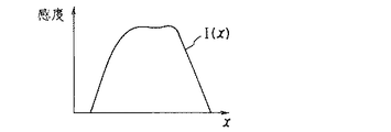
ここで、感度分布の重心位置は次のように定義される。すなわち、ある検出素子のスライス厚方向(x方向)における感度分布I(x)が図8のように表されたとすると、その感度の1次モーメント量Mは、次式のように定義される。
【0090】
【数2】
M=|Σ(I(x)・l(x)| ……(2)
但し、l(x)=x−xc を表す。
【0091】
このとき、感度分布の重心位置(x方向)xc は、上記1次モーメント量Mが最小のとき(min(M))の当該Xcの値で定義される。
【0092】
検出素子はスライス厚方向及びチャンネル方向にそれぞれ所定の長さを有する矩形形状に形成されているため、各検出素子のX線入射面において設定された座標軸(所定の点を原点としてスライス厚方向をx方向、チャンネル方向をy方向として定める)において、下式により1次モーメント量Mの最小値min(M)を求めることにより、そのmin(M)における(xc ,yc )を重心位置と定義することができる。
【0093】
【数3】
ただし、l(x,y)は、(xc ,yc )から(x,y)までの距離を表す。
【0094】
上述したように、データサンプリング位置として感度分布の重心位置を用いるためには、前掲図6〜図7で示された各スライス厚(1mm,2mm,4mm,8mm)のX線透過データ収集において、当該各データに対応する感度分布を測定し、測定された感度分布に応じて重心位置を設定しなければならない。
【0095】
そこで、本構成では、各種のスキャン条件(X線照射条件、データ収集条件)に応じて前記各スライス厚(1mm,2mm,4mm,8mm)のX線透過データに対応する検出素子毎の感度分布を予め測定し、測定された感度分布に基づいて各データ毎のデータサンプリング位置、すなわち感度分布の重心位置を求めている。
【0096】
ここで、感度分布測定処理及び感度分布重心位置(データサンプリング位置)設定処理について説明する。なお、この処理はスキャン撮影前の処理として行なわれる。また、例えば、スキャン条件は、次のように設定されているものとする。
【0097】
データ収集条件
・スライス厚条件(検出素子の束ね方条件):図6〜図7で示された各スライス厚(1mm,2mm,4mm,8mm)。
・ビューレート:1000ビュー/秒or1500ビュー/秒。
X線照射条件
・管電流:30,50,100,150,200,250,or300mA。
・管電圧:80,100,120,130,or140kV。
・エネルギー分布:フィルタ10Aの設定(High or Low)。
【0098】
まず最初に、オペレータOは、例えば入力装置30及びホストコントローラ25を介して高電圧発生装置15やガントリー3を制御して、上述したスキャン条件のある一組(ビューレート:1000ビュー/秒、管電流:30、管電圧:80、フィルタ10Aの設定:LoW)に基づいて主検出器11の各検出素子毎のスキャン撮影を実施する。
【0099】
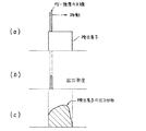
すなわち、ホストコントローラ25の制御に基づいて上記スキャン条件に基づいてX線をX線管球10から曝射する。このとき、プリコリメータ13、あるいはポストコリメータ14の制御により曝射されたX線をコリメートして、均一強度且つ非常に薄い(0.2mm程度)X線(以下、単に均一強度のX線とする)を主検出器11の検出素子の一部に照射する(図9、ステップS1;図10(a)参照)。その結果、当該検出素子には均一な強度のX線が入射される。そして、ホストコントローラ25は、均一強度のX線が一部に入射された検出素子の出力信号を測定する(ステップS2;図10(b)参照)。
【0100】
そして、コリメータの制御によりX線照射位置を少しずつ移動させながら均一強度のX線を該当する検出素子の全領域に照射して各々の領域の出力信号を測定し(図10(c)参照)、この測定された検出素子の全ての出力信号に基づいて、上記(1)式の関係から当該検出素子の感度分布を測定する(ステップS3)。
【0101】
以下、コリメータの制御によりX線照射位置を少しずつ移動させながら全ての検出素子に対してX線を照射して当該全ての検出素子により検出させることにより、各検出素子毎の感度分布を測定する(ステップS4)。
【0102】
例えば主検出器11の第1chの各素子(seg1a1 〜seg1a8 ,seg2a1 〜seg2a4 ,seg4a1 〜seg4a4 ,及びseg8a1 〜seg8a4 )では、図11に示すように感度分布(スライス厚方向に沿った分布)が測定される。なお、他のchの各素子も同様に測定される。
【0103】
なお、コリメータの制御によっても強度分布が均一のX線ビームが得られない場合においては、検出素子に入射する前のX線ビームの強度分布を例えばフィルム等に焼き付けて測定し、検出素子で測定した感度分布を測定されたX線ビームの強度分布で補正して(例えば感度分布を強度分布で割る)、均一化したX線ビームによる感度分布を得ることもできる。
【0104】
そして、上述したステップS1〜ステップS4の処理を上記スキャン条件のあらゆる組合わせ毎に行なうことにより、主検出器11の各検出素子の感度分布を各組合わせ毎に求める(ステップS5)。
【0105】
そして、ホストコントローラ25は、スキャン条件の組合わせ毎に測定された主検出器11の各検出素子の感度分布に基づいて、前掲図6及び図7で示した各スライス厚(1mm,2mm,4mm,8mm)のX線透過データに対応する検出素子の感度分布を抽出し、抽出された感度分布の重心位置を、当該感度の1次モーメント最小値を与える位置((3)式参照)として各組合わせ毎に求めて処理を終了する(ステップS6)。
【0106】
例えば、図7(B)に示した8mm厚の8スライスのX線透過データの内の向かって左側の4スライスのデータは、seg8a4 により検出されたデータ、seg8a3 により検出されたデータ、seg4a3 〜seg4a4 により検出されたデータが束ねられて生成されたデータ、seg2a3 〜seg2a4 及びseg1a5 〜seg1a8 により検出されたデータが束ねられて生成されたデータである。
【0107】
これらのデータに対応する検出素子(seg8a4 、seg8a3 、seg4a3 〜seg4a4 、seg2a3 〜seg2a4 及びseg1a5 〜seg1a8 )の感度分布(スライス厚方向に沿った分布)をそれぞれ図12に示す。
【0108】
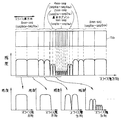
図12によれば、本構成のようなスライス厚方向に不均等に形成された検出器の各データを束ねて例えば同一スライス厚のデータとして出力するような場合では、各データに対応する検出素子(例えば、seg8a4 、seg8a3 、seg4a3 〜seg4a4 、seg2a3 〜seg2a4 及びseg1a5 〜seg1a8 )の感度分布は、データ毎に異なっている。
【0109】
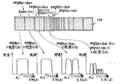
特に、複数segの検出データを束ねて生成されたデータ、例えばseg2a3 〜seg2a4 及びseg1a5 〜seg1a8 の各データを束ねて生成されたデータに対応する感度分布(図13に拡大して示す)は、束ねずに単独のseg8a4 で生成されたデータに対応する感度分布と比べて大きくことなっている。
【0110】
これは、上述した検出素子端部(エッジ)の感度低下の影響をスライス厚の薄い素子程強く受け、厚い素子程弱い影響しか受けないためであり、8mmスライスに対応するseg8a4 の感度分布と比べて、その8mmスライスよりも非常に薄い2mmスライスに対応するseg2a3 〜seg2a4 及び1mmスライスに対応するseg1a5 〜seg1a8 の感度分布は偏りを有していることが分かる。
【0111】
したがって、例えばseg2a3 〜seg2a4 及びseg1a5 〜seg1a8 の各データを束ねて生成されたデータのサンプリング位置を、図13に示すように、当該seg2a3 〜seg2a4 及びseg1a5 〜seg1a8 が形成する矩形形状の幾何学的重心位置に設定したのでは、上述した感度分布の偏りにより誤差が生じ、補間処理や再構成処理等において精度が悪化する恐れがある。
【0112】
しかしながら、本構成では、図12及び図13に示すように、各データに対応する感度分布の重心位置(xc1〜xc4)を(3)式に基づいて求めており、図14に示すように、求められた8mm厚の8スライスのX線透過データそれぞれに対応する感度分布の重心位置を、当該X線透過データのサンプリング位置データに設定しているため、感度分布の偏りに関係なくその感度分布に適したサンプリング位置を求めることができる。
【0113】
このようにして、各スライス厚の(1mm,2mm,4mm,8mm)のX線透過データに対応する検出素子の感度分布の重心位置が当該各データのサンプリング位置データとして各スキャン条件の組合わせ毎に求められる。このサンプリング位置データは、上述した記憶装置27に各スキャン条件の組合わせ及び各スライス厚のX線透過データ毎にそれぞれ記憶される。
【0114】
続いて、通常のスキャン撮影時における全体動作について説明する。
【0115】
ホストコントローラ25は、オペレータOから入力装置30を介して入力されたスライス厚(1mmスライス〜8mmスライスの何れか)やX線照射条件(上述した管電圧値及び管電流値)及びデータ収集条件(上述したビューレート)等のスキャン条件を内部メモリに記憶し、この記憶したスキャン条件に基づいて記憶装置27に記憶されたサンプリング位置データの中から、当該スライス厚、X線照射条件及びデータ収集条件に対応するサンプリング位置データを読み出す(図15、ステップS10)。
【0116】
そして、ホストコントローラ25は、記憶されたスキャン条件(あるいは、マニュアルモードにおいてオペレータOから直接設定されたスキャン条件)に基づいて高電圧発生装置15、図示しない寝台駆動部、ガントリー駆動部12、及びビームトリマ14を介して寝台2の体軸方向への送り量、送り速度、ガントリー3(X線管球10及び主検出器11)の回転速度、回転ピッチ、ビームトリマ14のエッジ位置、及びX線の曝射タイミング等を制御しながら当該高電圧発生装置15、寝台駆動部、ガントリー駆動部12、及びビームトリマ14を駆動させることにより、被検体Pの所望の撮影領域に対して多方向からコーン状のX線ビームを照射してスキャン撮影を実行する。そして、そのスキャン撮影により被検体Pの撮影領域を透過した透過X線は、主検出器11の各検出素子を介してX線透過データとして検出される(ステップS11)。
【0117】
同時に、ホストコントローラ25は、内部メモリに記憶されたスキャン条件(あるいは、マニュアルモードのスキャン条件)に基づいてスイッチ群20の各スイッチの切り換え制御を行なって主検出器11の各検出素子とDAS21との接続状態を切り換えることにより、当該各検出素子で検出されたX線透過データを束ねて、スキャン条件に対応したスライス厚を有する複数スライスのX線透過データを生成する(ステップS12)。
【0118】
そしてホストコントローラ25は、生成された複数スライスのX線透過データをDAS21に送って当該複数スライスの投影データを生成するとともに、生成された各投影データに対して、当該各投影データ(すなわち、この投影データの元になるX線透過データ)に対応するサンプリング位置データを付加し、サンプリング位置データが付加された投影データ、すなわちサンプリング位置に基づく投影データとしてデータ処理装置26に送る(ステップS13)。
【0119】
このとき、各投影データに付加されたサンプリング位置データは、当該投影データに対応する検出素子の感度分布の重心位置となっているため、本構成のようなスライス厚方向に不均等ピッチを有する検出素子を2次元配列して形成された主検出器11を用いた場合であっても、当該各素子の不均等ピッチ構造に起因した感度分布の偏りに関係なく、各データに最適なサンプリング位置を設定することができる。
【0120】
そして、データ収集処理装置26は、サンプリング位置データが付加された複数の投影データに対して、そのサンプリング位置データに基づいて補間処理を必要に応じて行なった後、そのサンプリング位置データ及び投影データに基づいてコンボリューションバックプロジェクション等の画像再構成処理を行なって複数スライスのCT画像をそれぞれ生成する(ステップS14)。
【0121】
そして、ホストコントローラ25は、生成された複数のCT画像を順次表示装置29に表示して処理を終了する(ステップS15)。
【0122】
すなわち、表示装置29に表示されるCT画像は、従来のようなデータサンプリング位置の誤差に起因した画質劣化やアーチファクトが大幅に抑制された視認性の良い高画質なCT画像となっている。
【0123】
また、予め各スキャン条件のあらゆる組合わせに基づいてサンプリング位置データを求めているため、入力装置30から他のスキャン条件が入力されても、そのスキャン条件に対応したサンプリング位置データに基づいてスキャン撮影及びデータ補間処理・再構成処理を行なうことができる。したがって、スライス厚(1mmスライス〜8mmスライス)の変化、X線管球の管電流や管電圧等のX線照射条件、あるいはビューレート等のデータ収集条件の変化に起因した検出素子の感度分布の変化、及びその感度分布の変化により生じるサンプリング位置データの誤差を解消し、当該X線照射条件あるいはデータ収集条件の変化に係わらず常に視認性の良い高画質なCT画像を得ることができる。
【0124】
なお、本実施形態では、全ての組合わせに対して全ての検出素子の感度分布を測定したが、セグメント毎(基本セグメント〜8mmセグメント)に一つの感度分布に近似してサンプリング位置を各セグメント単位で決定することも可能である。また、スキャン条件の一部(例えば管電流)には、当該条件を変化させても感度分布があまり変化しないものもあるため(管電圧が定まれば管電流を変えても感度分布はほとんど変化しない)、このような場合では、その条件がいずれの値であっても同一のサンプリング位置を適用してもよい。さらに、上述したスキャン条件(X線照射条件,データ収集条件等)以外の条件に応じてサンプリング位置を設定してもよい。
【0125】
一方、本実施形態では、感度分布の重心位置をスライス厚方向について示したが、チャンネル方向における感度分布の重心位置についても、(3)式で同様に求められており、スライス厚方向及びチャンネル方向それぞれにおいて最適なサンプリング位置を求めることができる。なお、上述したスライス厚方向のエッジ部分の感度劣化が感度分布の偏りに支配的な場合であれば、スライス厚方向において感度分布の重心位置を求め、チャンネル方向においては、幾何学的重心位置を用いることもできる。
【0126】
ところで、本実施形態においては、各チャンネルあたり1mm〜8mmのセグメントを合計20セグメント配列した主検出器11aで得られたX線透過データを8スライス分のDAS21で投影データとして収集する例について説明したが、本発明はこれに限定されるものではない。
【0127】
例えば、4スライス分のデータ収集素子を有するDAS21' を備えた場合の4セグメント検出器及びその感度分布を図16に示す。
【0128】
4セグメント検出器の例えば第1chの素子列11A1 は、図面向かって左から2個の1mmセグメント(seg1A1 ,seg1A2 ),1個の2mmセグメント(seg2A),及び1個の4mmセグメント(seg4A)がスライス厚方向に沿って配列されている。
【0129】
このように配列された第1chの素子列11A1 の全ての検出素子(seg1A1 ,seg1A2 ,seg2A及びseg4A)を束ねて1つのデータ(8mmスライス)として出力する場合(図16(a))においても、全ての素子(seg1A1 ,seg1A2 ,seg2A及びseg4A)の幾何学的重心位置をサンプリング位置として用いるのではなく、当該全ての素子(seg1A1 ,seg1A2 ,seg2A及びseg4A)の感度分布の重心位置をサンプリング位置データとして用いることにより、感度分布の偏りの影響が無くなり、再構成CT画像の画質を向上させることができる。
【0130】
また、図16(b)は、第1chの素子列11A1の内のseg1A1 ,seg1A2 及びseg2Aを束ねて1つのデータ(4mmスライス)として出力し、seg4Aを(束ねて)1つのデータ(4mmスライス)として出力する場合であるが、この場合においても、seg1A1 ,seg1A2 及びseg2Aの幾何学的重心位置及びseg4Aの幾何学的重心位置をサンプリング位置として用いるのではなく、当該素子(seg1A1 ,seg1A2 及びseg2A)の感度分布の重心位置及び素子(seg4A)の重心位置をそれぞれサンプリング位置データとして用いることにより、感度分布の偏りの影響が無くなり、再構成CT画像の画質を向上させることができる(なお、本構成の場合では、seg4Aの感度分布の重心位置は幾何学的重心位置とは同一位置となっている)。
【0131】
さらに、本実施形態では、主検出器(2次元検出器)の各chの素子列の各素子のスライス厚方向(seg方向)のスライス厚(スライスピッチ)が不均等に形成された2次元検出器を有するX線CTスキャナについて説明したが、本発明はこれに限定されるものではなく、例えば、スライス厚方向のピッチが均等な通常の2次元検出器を有するX線CTスキャナについても適用可能である。
【0132】
特に、スライス厚方向のピッチが均等な2次元検出器の一例として、照射X線を直接電気信号に変換して出力する2次元X線検出器がある。そして、この2次元X線検出器のチャンネル方向に沿って配列された少なくとも1列の素子の内の少なくとも一部の素子の材質がチャンネル方向の沿って配列された他の素子の材質よりも高感度(時間的に反応が敏感)なものを用いることにより、当該スライス厚方向の高分解能化を可能にした2次元X線検出器がある。
【0133】
この2次元X線検出器を図17に示す。図17によれば、2次元X線検出器41は、1chあたり複数seg(本実施形態では5seg)がスライス厚方向(seg方向)に沿って並べられた検出素子列をチャンネル方向(ch方向)に沿って複数ch(本実施形態では5ch)アレイ状に配列した2次元検出器(図2では、5ch×5segの2次元検出器を示している)として構成されている。
【0134】
すなわち、図16に示すように、第1chの5seg分の素子列を41a1 とすると、第1ch:41a1 〜第5ch:41a5 の素子列が配置されており、また、第1segの5ch分の素子列を41α1 とすると、第1seg:41α1 〜第5seg:41α5 の素子列が配置されていることになる。
【0135】
そして、スライス厚方向において中央であり、チャンネル方向に沿って配列された素子列41α3 が高感度な材質で形成され、その他の素子列41α1 ,41α2 、41α4 ,41α5 は、当該高感度材質よりも感度が低い材質(Cs I等)で形成されている。
【0136】
すなわち、高感度材質で形成された中央の素子列41α3 部分に対応するスキャン撮影時には、ビューレートを高く(例えば1000ビュー/秒)、その他の素子列41α1 ,41α2 、41α4 ,41α5 に対応するスキャン撮影時には、ビューレートを低く(例えば30ビュー/秒)設定することにより、ワイドな撮影領域及び所望のスライス面の画像の高分解能化が共に図れるように構成されている。
【0137】
上述した構成において、例えば隣接する素子列41α2 ,41α3 を束ねて1つのデータにした場合では、各素子列41α2 ,41α3 の感度分布は異なるため、当該各素子列41α2 ,41α3 の幾何学的重心位置をサンプリング位置と設定すると、感度分布の重心位置とサンプリング位置にズレが生じ、画質劣化につながる恐れがある。
【0138】
しかしながら、本発明を適用すれば、各素子列41α2 ,41α3 全体の感度分布の重心位置をデータサンプリング位置として設定することができるため、前記データサンプリング位置のズレに起因した画質劣化を大幅に抑制することができる。すなわち、2次元検出器の各素子列の感度分布が異なる場合であっても、各感度分布に応じた最適なデータサンプリング位置を設定し、設定されたデータサンプリング位置に基づいて補間処理や再構成処理が行なわれるため、再構成画像の画質劣化を大幅に抑制し、当該再構成画像の画質を向上させることができる。
【0139】
なお、チャンネル方向に沿って配列された少なくとも1列の内の少なくとも一部の素子の材質が他のスライス厚方向の素子列の材質よりも高感度(時間的に反応が敏感)なものを用いた2次元検出器としては、上述した2次元X線検出器に限らず、通常の2次元X線検出器(例えばシンチレーション型検出器)においても適用可能である。
【0140】
また、その他の2次元検出器や通常の検出器(シングルスライスCT用)を用いたCTスキャナにおいても、例えばスライス厚方向の各検出素子の感度分布にばらつきがある場合では、本発明を適用することにより、感度分布の偏り係わらず当該感度分布に最適なデータサンプリング位置を設定することができる。
【0141】
さらに、本実施形態では、スキャナのコリメータを用いてX線ビームを制御して均一強度のX線ビームを生成し、この均一強度のX線ビームを用いて感度分布を測定するように構成したが、本発明はこれに限定されるものではない。例えば、鉛遮蔽板等のX線遮蔽部材を用いて、測定者(オペレータ)が当該X線遮蔽部材をマニュアルで移動制御して均一強度のX線ビームを生成してもよい。
【0142】
また、工場等で検出器単体として測定された感度分布を下にサンプリング位置を決定し、決定されたサンプリング位置に基づいて図15に示す処理を行なってもよい。
【0143】
【発明の効果】
本発明のX線CTスキャナによれば、X線管から曝射され、被検体を透過してきたX線が、2次元検出手段における例えば被検体の体軸方向及びその体軸方向に直交する方向に沿って2次元的に配列された複数の検出素子により検出される。そしてこの各検出素子により検出されたデータに基づいて、データ収集手段により前記体軸方向に直交する複数のスライス面の投影データがそれぞれ収集される。複数の投影データそれぞれに対応する検出素子の感度分布の重心位置データをデータサンプリング位置として用いることにより、従来のサンプリング位置が検出素子の幾何学的重心位置に固定されていた場合と比べてデータサンプリング位置の誤差(位置ズレ)が解消されている。したがって、データサンプリング位置の誤差に起因したアーチファクト等の画質劣化を抑制し、視認性の良い高画質なCT画像を得ることができる。
【0144】
本発明のX線CTスキャナによれば、体軸方向における前記検出素子のピッチが不均等に形成された2次元検出手段を用いて複数の異なるスライス厚を有する複数のスライス画像を撮影する場合においても、複数のスライス厚を有する複数の投影データそれぞれに対応する検出素子の感度分布の重心位置データを当該スライス厚に対応して求めておき、この重心位置データを各スライス厚条件に対応するデータサンプリング位置として用いているため、スライス厚の選択の仕方(束ね方)に起因して、得られた投影データの対応する検出素子の感度分布に偏りが生じたとしても、データサンプリング位置に誤差(位置ズレ)が生じることが無い。この結果、データサンプリング位置の誤差に起因したアーチファクト等の画質劣化を抑制し、視認性の良い高画質なCT画像を得ることができる。
【0145】
さらに、本発明のX線CTスキャナによれば、複数の投影データそれぞれに対応する検出素子の感度分布の重心位置データをX線画像撮影時に設定可能な複数のデータ収集条件毎に求められた検出手段の各検出素子の感度分布に基づいてそれぞれ求めているため、CT画像撮影時にどんなデータ収集条件が設定されても、設定されたデータ収集条件に対応して求められた検出素子の感度分布の重心位置データをデータサンプリング位置として用いることにより、データ収集条件に起因した各検出素子の感度分布への影響を抑制することができる。
【0146】
そして、本発明のX線CTスキャナによれば、複数の投影データそれぞれに対応する検出素子の感度分布の重心位置データをX線画像撮影時に設定可能な複数のX線照射条件毎に求められた検出手段の各検出素子の感度分布に基づいてそれぞれ求めているため、CT画像撮影時にどんなX線照射条件が設定されても、設定されたX線照射条件に対応して求められた検出素子の感度分布の重心位置データをデータサンプリング位置として用いることにより、X線照射条件に起因した各検出素子の感度分布への影響を抑制することができる。
【図面の簡単な説明】
【図1】本発明の実施形態に係わるX線CTスキャナの概略構成を示すブロック図。
【図2】主検出器(2次元検出器)の構成を示す図。
【図3】最小スライス厚が1mm、全体のセグメント数が20個の場合の検出器列の構成を示す図。
【図4】主検出器、スイッチ群、データ収集装置の概略構成を示す斜視図。
【図5】8スライス分のDASでデータ収集する際のスイッチ群の構成の一例を示す図。
【図6】同一ピッチで8スライスのデータを収集する際のデータの束ね方を示すものであり、(A)は1mm×8スライス、(B)は2mm×8スライスを示す図。
【図7】同一ピッチで8スライスのデータを収集する際のデータの束ね方を示すものであり、(A)は4mm×8スライス、(B)は8mm×8スライスを示す図。
【図8】ある検出素子のスライス厚方向(x方向)における感度分布I(x)を表す図。
【図9】本実施形態の構成における感度分布測定処理及び感度分布重心位置(データサンプリング位置)設定処理を説明するための概略フローチャート。
【図10】(a)は検出素子の一部に入射される均一強度のX線ビームを示す図、(b)は入射されたX線に基づいて当該検出素子で測定された出力信号を示す図、(c)はその検出素子の全ての領域で測定された出力信号分布を示す図。
【図11】第1chの各検出素子のスライス厚方向に沿った感度分布を示す図。
【図12】束ねられた8mm厚の8スライスのX線透過データに対応する各検出素子の感度分布を示す図。
【図13】seg2a3 ,seg2a4 及びseg1a5 〜seg1a8 の各データを束ねて生成されたデータに対応する感度分布を拡大して示す図。
【図14】8mm厚の8スライスのX線透過データそれぞれに対応する感度分布の重心位置を示す図。
【図15】本実施形態の構成における通常のスキャン撮影時の全体動作を説明するための概略フローチャート。
【図16】本実施形態の変形例に係わる4セグメント検出器及びその感度分布を示す図。
【図17】本実施形態の変形例に係わる2次元X線検出器を示す図。
【図18】シングルスライスCTにおけるX線管球及び検出器の構成を示す図。
【図19】(a)はプリコリメータによりX線ビームの制御構成を示す図、(b)はポストコリメータによりX線ビームの制御構成を示す図。
【図20】(a)は検出器に入射されるX線強度分布を示す図、(b)は検出器感度分布(スライス厚方向)を示す図、(c)は検出器から出力される信号強度分布を示す図。
【図21】シンチレータエッジでの感度分布を示す図。
【図22】シングルスライスCTにおいて検出素子に入射されるX線ビームの入射範囲及び検出素子におけるデータサンプリング位置(幾何学的重心位置)を示す図。
【図23】(a)はシングルスライスCTにおける検出器に入射されるX線強度分布を示す図、(b)はシングルスライスCTにおける検出器感度分布(スライス厚方向)を示す図、(c)はシングルスライスCTにおける検出器から出力される信号強度分布を示す図。
【図24】マルチスライスCTにおけるX線管球及び検出器の構成を示す図。
【図25】(a)は従来のマルチスライスのあるチャンネルの検出素子列を示す図であり、(b)は高解像度モードにおける検出素子列の構成を示す図であり、(c)は撮影領域最大モードにおける検出素子列の構成を示す図である。
【図26】X線照射条件やデータ収集条件に対応して変化する感度分布を示す図。
【図27】1データ分の検出素子の感度分布が偏りを有する場合における当該検出素子の幾何学的重心位置と実際の感度分布の重心位置とをそれぞれ示す図。
【符号の説明】
1 X線CTスキャナ
2 寝台
3 ガントリー
4 システム部
10 X線管球
11 主検出器
11a1 、11A1 、41α1 第1チャンネルの検出素子列
12 ガントリー駆動部
13 スリット
14 ビームトリマ
14a、14b X線遮蔽板
15 高電圧発生装置
20 スイッチ群
21 データ収集装置(DAS)
25 ホストコントローラ
26 データ処理装置
27 記憶装置
28 再構成装置
29 表示装置
30 入力装置
31 補助記憶装置
41 2次元X線検出器
41α3 高感度検出素子列
seg1a1 〜seg1a8 、seg2a1 〜seg2a4 、seg4a1 〜seg4a4 、seg8a1 〜seg8a4 検出素子列11a1 の各セグメントDAS−1a1 〜DAS−8a1 検出素子列11a1 に対応するデータ収集素子
S11〜S20G スイッチ群20の各スイッチ[0001]
BACKGROUND OF THE INVENTION
In the present invention, X-rays transmitted through the subject are detected by detection means having a plurality of detection elements arranged along a direction (channel direction) orthogonal to the body axis direction of the subject, and the detected data The present invention relates to an X-ray CT scanner that captures a CT image of a tomographic plane of a subject based on projection data generated accordingly, and more particularly to an X-ray CT scanner characterized by a sampling position added to projection data.
[0002]
[Prior art]
As a type conventionally used in the X-ray CT scanner, there is a fan beam (single slice) X-ray CT scanner (also referred to as single slice CT).
[0003]
As shown in FIG. 18, the fan beam X-ray CT scanner includes an X-ray tube (X-ray tube, X-ray tube) 50 and a
[0004]
Although various types of
[0005]
That is, according to the above-described X-ray CT scanner, an X-ray beam is irradiated from the
[0006]
The X-ray transmission data collected for each element of the
[0007]
Then, X-ray irradiation is performed through the focal point of the X-ray source while the
[0008]
The collected projection data from multiple directions is subjected to data interpolation processing to convert it into parallel beam projection data, and the converted projection data is subjected to reconstruction processing such as convolution back projection to obtain the slice surface of the subject. The image is reconstructed.
[0009]
The X-ray beam irradiated from the focal point of the
[0010]
The X-ray beam incident on each detection element of the
[0011]
[Expression 1]
(Sensitivity) = (Output) / (Input) (1)
In addition, since each detector element of the detector absorbs light at the end (edge part) in the slice thickness direction of the scintillator, as shown in FIG. 21, the sensitivity distribution of each
[0012]
That is, as shown in FIG. 23, a substantially rectangular X-ray beam is incident on each
[0013]
On the other hand, in the reconstruction processing such as the interpolation processing and convolution back projection described above, the collected projection data is used as discretized sampling position data. That is, projection data based on signals detected by the respective detection elements is used as data at the sampling position.
[0014]
In the single-slice CT, an area where the sensitivity distribution in the detection element is substantially flat is used as a data collection area, and the X-ray incident range and the intensity distribution of the output signal from the detection element are also substantially rectangular. In addition, the geometric gravity center position of the X-ray incidence range (or the rectangular shape of the
[0015]
On the other hand, such a single slice X-ray CT scanner obtains an image of one slice plane of a subject, so that it is difficult to capture a wide range of images in a short time, and it is difficult for a doctor to increase the unit time. There was a strong demand to capture images in a fine (high resolution) and wide range.
[0016]
In order to meet this demand, in recent years, a multi-slice X-ray CT scanner (also referred to as multi-slice CT) has been studied.
[0017]
This multi-slice CT has a plurality of rows (multiple (M) segments) of detection elements of the detector in the single slice CT along the slice thickness direction (seg direction) corresponding to the body axis direction of the subject. The detector is configured as a two-
[0018]
That is, according to the multi-slice CT, the X-ray source 61 that exposes a conical (conical) X-ray beam and the two-
[0019]
In such a multi-slice CT, similarly to the single-slice CT, an irradiation X-ray beam is controlled by a pre-collimator or a post-collimator to generate an X-ray beam having a desired slice thickness, and the generated X-ray beam Is detected and output by each detection element of the detector.
[0020]
For example, if attention is paid to a
[0021]
As can be seen by comparing FIGS. 25 (b) and 25 (c), in FIG. 25 (b), a detection signal corresponding to a slice having a high resolution with a slice thickness of 2 mm is output, and in FIG. 25 (c). Since the slice thickness is 6 mm and the resolution is low but a detection signal with a large imaging area is output along the slice thickness direction, the above-described high-definition and wide-range image acquisition can be realized together.
[0022]
In such a multi-slice CT, as in the single-slice CT, one element may correspond to one data as shown in FIG. 25 (c). ) The geometric center of gravity (the intersection of diagonal lines if it is a rectangle) of the shape (the shape of the active area excluding the dead zone and the shape of the entire device) The projection data sampling position is based on the signal detected by the detection element. When a plurality of elements correspond to one data as shown in FIG. 25C, the geometric gravity center position in the shape of the entire plurality of elements becomes the data sampling position.
[0023]
[Problems to be solved by the invention]
As described above, in the conventional single slice CT and multi-slice CT, the data sampling position of the detection element constituting one data is always set to the geometric gravity center position of the detection element.
[0024]
However, the sensitivity distribution of each detection element is a distribution in which the sensitivity decreases at the edge portion as shown in FIG. 21, and the sensitivity distribution includes the tube current and the tube voltage of the X-ray tube, the detection element. Since it changes in accordance with X-ray irradiation conditions such as energy characteristics (energy distribution) of incident X-rays incident on and data collection conditions such as view rate (see FIGS. 26A and 26B), depending on circumstances. In some cases, the sensitivity distribution of each detection element is biased.
[0025]
In particular, when the detection data of a plurality of detection elements are bundled and handled as one data in multi-slice CT, the sensitivity distribution of the detection elements corresponding to each bundled data may be non-uniform, and the overall sensitivity. In some cases, the distribution was biased.
[0026]
That is, as shown in FIG. 27, when the sensitivity distribution of one data detection element (one or a plurality) is biased, the geometric gravity center position of the detection element and the actual sensitivity distribution The position of the center of gravity may be different.
[0027]
When the sensitivity distribution of the detection element is flat, the center of gravity position of the actual sensitivity distribution is substantially coincident with the geometric center of gravity of the detection element. Therefore, the geometric center of gravity position is used as the data sampling position. There is no particular problem.
[0028]
However, when the geometric centroid position of the detection element is different from the centroid position of the actual sensitivity distribution as described above, if the geometric centroid position is handled as a data sampling position by interpolation processing or reconstruction processing, This is equivalent to using data at an incorrect sampling position as seen from the actual sensitivity distribution. This deviation (error) in the data sampling position lowers the processing accuracy in the weighting of data in the interpolation process and the filter process in the reconstruction process, resulting in image quality degradation such as artifacts in the reconstructed CT image. It was a cause.
[0029]
The present invention has been made in view of the circumstances as described above. By setting the data sampling position in each detection element of the detector corresponding to each projection data to the barycentric position of the actual sensitivity distribution of the element, the data The object is to obtain high-quality CT images with good visibility by suppressing image quality degradation due to sampling position errors.
[0032]
[Means for Solving the Problems]
SaidTo achieve the purposeX-ray CT scanner of the present inventionThen, an X-ray tube for exposing X-rays, and a plurality of detection elements arranged two-dimensionally along the body axis direction of the subject and the direction orthogonal to the body axis direction, the X-ray tube Two-dimensional detection means for detecting X-rays emitted from the subject and transmitted through the subject by the detection elements, and orthogonal to the body axis direction based on data detected by the detection elements of the two-dimensional detection means Data collection means for collecting projection data of a plurality of slice planes, and a plurality of the plurality of projection data collected and sampling position data corresponding to each of the plurality of projection data to perform at least image reconstruction processing In an X-ray CT scanner comprising an image processing means for taking an X-ray image of a slice plane of at least one of the detection elements of the two-dimensional detection means along the body axis direction. Means for determining the sensitivity distribution of the output element, and means for determining the gravity center position data of the sensitivity distribution of the detection element corresponding to each of the plurality of projection data based on the obtained sensitivity distribution of the detection elements in at least one row, The image processing means uses centroid position data obtained corresponding to each of the plurality of projection data as sampling position data corresponding to each of the plurality of projection data.
[0033]
In particular, the centroid position data of the sensitivity distribution of the detection element corresponding to each of the plurality of projection data is the primary of the sensitivity distribution of the detection element when a two-dimensional coordinate space including the X-ray incident surface of the detection element is set. The coordinate data when the moment amount is minimum, and the means for obtaining the sensitivity distribution of the detection element is means for obtaining the sensitivity distribution of each detection element of the two-dimensional detection means.
[0034]
In particular, the two-dimensional detection means is a two-dimensional detection means in which the pitch of the detection elements in the body axis direction is formed unevenly, and each detection element of the two-dimensional detection means is set for each element in the body axis direction. Detecting means selecting means connected to a data collecting element corresponding to the body axis direction of the data collecting device and selecting according to the selection content of each detecting element by the detecting element selecting means The slice thickness of each projection data is set according to the slice thickness condition.
[0035]
In particular, the means for obtaining the centroid position data of the sensitivity distribution is a means for obtaining the centroid position data of the sensitivity distribution of the detection element corresponding to each of a plurality of projection data having a slice thickness corresponding to the selection content of each detection element. In addition, the means for obtaining the sensitivity distribution may directly irradiate each detection element of the detection means directly after adjusting the X-rays exposed from the X-ray tube to X-rays having a uniform intensity. This is a means for obtaining the sensitivity distribution of each detection element.
[0036]
In particular, the means for obtaining the sensitivity distribution is means for obtaining the sensitivity distribution of each detection element of the detection means in accordance with a plurality of data collection conditions that can be set at the time of the X-ray imaging, and the barycentric position data of the sensitivity distribution Is based on the plurality of sensitivity distributions determined according to the plurality of data collection conditions, and obtains the gravity center position data of the sensitivity distribution of the detection element corresponding to each of the plurality of projection data for each sensitivity distribution. Means.
[0037]
In particular, the means for obtaining the sensitivity distribution is a means for obtaining the sensitivity distribution of each detection element of the detection means according to a plurality of X-ray irradiation conditions that can be set at the time of the X-ray imaging, and the center of gravity position of the sensitivity distribution. The means for obtaining the data is based on the plurality of sensitivity distributions obtained according to the plurality of X-ray irradiation conditions, and the gravity center position data of the sensitivity distribution of the detection element corresponding to each of the plurality of projection data for each sensitivity distribution It is a means to ask for.
[0038]
To achieve the purpose,X-ray CT scanner of the present inventionIn this case, an X-ray tube that emits X-rays and a detection element that is two-dimensionally arranged along the body axis direction of the subject and the direction orthogonal to the body axis direction are exposed from the X-ray tube. Two-dimensional detection means for detecting X-rays that have been emitted and transmitted through the subject, and projection data of a plurality of slice planes based on data detected by each detection element of the two-dimensional detection means Data collection means for collecting each of them, and image processing for photographing X-ray images of the plurality of slice planes by performing at least image reconstruction processing based on the collected projection data and sampling position data corresponding to the projection data. And a plurality of projections based on a sensitivity distribution of at least one row of detection elements along the body axis direction among the detection elements of the two-dimensional detection means. Means for obtaining the gravity center position data of the sensitivity distribution of the detection element corresponding to each of the data, and the image processing means is obtained corresponding to the plurality of projection data as sampling position data corresponding to the plurality of projection data. The center of gravity position data is used.
[0039]
Based on the collected projection data and sampling position data corresponding to each of the projection data, at least image reconstruction processing is performed by the image processing means, and X-ray images of the plurality of slice planes are captured. It is like that.
[0040]
According to the X-ray CT scanner of the present invention, among the detection elements of the two-dimensional detection means, the sensitivity distribution of at least one row of detection elements along the body axis direction (for example, the sensitivity distribution of all the detection elements) is obtained. The detection element corresponding to each of the plurality of projection data is obtained by a method such as irradiating each detection element of the detection means with X-rays having a uniform intensity, and based on the obtained sensitivity distribution of each detection element. The gravity center position data of the sensitivity distribution is obtained. The gravity center position data of the sensitivity distribution of the detection element corresponding to each of the plurality of projection data is the first moment amount of the sensitivity distribution of the detection element when a two-dimensional coordinate space including the X-ray incident surface of the detection element is set. Is defined as the coordinate data when.
[0041]
At this time, since the centroid position data of the sensitivity distribution of the detection element corresponding to each of the obtained plurality of projection data is used as the sampling position data by the image processing means, the sampling position data is temporarily Even if the sensitivity distribution of the detection element corresponding to each projection data is biased, the position data is optimal for the sensitivity distribution. Therefore, the data sampling position error (positional deviation) is eliminated compared with the case where the sampling position is fixed in advance to the geometric gravity center position of the detection element regardless of the sensitivity distribution of the detection element such as a conventional CT scanner. Has been.
[0042]
In particular, according to this configuration, when the two-dimensional detection means in which the pitch of the detection elements in the body axis direction is formed unevenly is used as the two-dimensional detection means, the two-dimensional detection means is detected by the detection element selection means. Each detection element is selected according to the slice thickness condition for each element in the body axis direction and connected to the data collection element corresponding to the body axis direction of the data collection device, so that the selection contents of each detection element, in other words, For example, the slice thickness of each of the plurality of projection data is set in accordance with the slice thickness condition according to the content that determines which of the detection elements is bundled and connected to the data collection element.
[0043]
At this time, in this configuration, the centroid position data of the sensitivity distribution of the detection element corresponding to each of the plurality of projection data having the slice thickness corresponding to the selection content of each detection element is obtained. That is, no matter what the slice thickness condition is set, the gravity center position data of the sensitivity distribution of the detection element corresponding to each of the plurality of projection data having the slice thickness is obtained. Therefore, even if the sensitivity distribution of the corresponding detection element of the obtained projection data is biased due to the way of selecting (bundling) the slice thickness, an error (position shift) occurs in the data sampling position. There is no.
[0044]
In addition, according to this configuration, the gravity center position data of the sensitivity distribution of the detection element corresponding to each of the plurality of projection data is detected by each of the detection means obtained for each of a plurality of data collection conditions that can be set at the time of X-ray image capturing. Since each is generated based on the sensitivity distribution of the element, when a predetermined data collection condition is selected from among the plurality of data collection conditions and the CT image is taken, it is obtained corresponding to the data collection condition. By using the center-of-gravity position data of the sensitivity distribution of the obtained detection elements, it is possible to suppress the influence on the sensitivity distribution of each detection element due to the data collection conditions.
[0045]
Further, according to this configuration, the gravity center position data of the sensitivity distribution of the detection element corresponding to each of the plurality of projection data is obtained for each of the detection means obtained for each of the plurality of X-ray irradiation conditions that can be set at the time of X-ray image capturing. Since each is generated based on the sensitivity distribution of the detection element, when a predetermined X-ray irradiation condition among the plurality of X-ray irradiation conditions is selected and the CT image is taken, the X-ray irradiation condition is set. By using the center-of-gravity position data of the sensitivity distribution of the detection elements obtained correspondingly, the influence on the sensitivity distribution of each detection element due to the X-ray irradiation condition can be suppressed.
[0046]
DETAILED DESCRIPTION OF THE INVENTION
Embodiments of the present invention will be described below with reference to the accompanying drawings.
[0047]
FIG. 1 is a block diagram showing a schematic configuration of an
[0048]
According to FIG. 1, an X-ray CT scanner (CT system) 1 includes a
[0049]
The
[0050]
The gantry 3 includes an
[0051]
The
[0052]
As the
[0053]
As shown in FIG. 2, this scintillation type
[0054]
In other words, in FIG. 2, if the element array for 20seg of the first channel is 11a1, the element array of the first channel: 11a1 to 16th: 11a20 is arranged, and the element array of 16ch of the first channel is 11α1. Then, the element rows of the first seg: 11α1 to the twentieth seg: 11α20 are arranged.
[0055]
Here, when the position (address) of each of the detection elements arranged two-dimensionally in this way is represented by (seg, ch), for example, the element located at (first seg, first ch) is 11 (1, 1), and hereinafter, the element row of the first ch 11a1 is represented as 11 (2,1)... 11 (20,1). In this way, the remaining ch11a2 to 11a16 are arranged in the second ch11a2 → 11 (1,2) ... 11 (20,2) and the third ch11a3 → 11 (1,3) ... 11 (20,3), respectively. ,..., 15th channel 11a15 → 11 (1,15)... 11 (20,15), 16th channel 11a16 → 11 (1,16)... 11 (20,16).
[0056]
Note that separators (reflecting plates) 11s1 and 11s2 such as metal plates are provided between the segs and between the channels, and are configured to eliminate crosstalk between adjacent channels and segs.
[0057]
The
[0058]
In order to generate the detection signal with the uneven slice thickness described above in the present embodiment, the slice thickness (slice pitch) of each element of the element rows 11a1 to 11a16 of each channel of the main detector (two-dimensional detector) 11 ) Are formed unevenly so that the pitch increases from the central element toward the end element. In addition, the slice pitch formed non-uniformly along the seg direction is referred to as non-uniform pitch.
[0059]
Here, the configuration of the element rows 1a1 to 11a16 of each channel (ch) of the
[0060]
In the present embodiment, a detection element array having a size for realizing the minimum slice thickness obtained by the
[0061]
According to FIG. 3, the element arrays 11a1 to 11a16 of the respective channels of the
[0062]
The X-ray transmission data detected by each detection element of the
[0063]
FIG. 4 is a perspective view showing the structure of the two-
[0064]
Each data collection element (DAS-1a1 to DAS-8a1... DAS-1a16 to DAS-8a16) of the
[0065]
Here, for example, the detection element array 11a1 (seg1a1 to seg1a8, seg2a1 to seg2a4, seg4a1 to seg4a4, seg8a1 to seg8a4) in the first channel of the
[0066]
According to FIG. 5, seg 8a1 is connected to DAS-1a1 through switch S11, and is connected to DAS-2a1 to DAS-8a1 through switches S12 to S18. And it is connected to GND via S1G. Similarly, seg8a2 is connected to DAS-1a1 to DAS-8a1 and GND via switches S21 to S2G.
[0067]
Hereinafter, seg4a1 is connected to DAS-1a1 to DAS-8a1 and GND via switches S31 to S3G, and seg4a2 is connected to hands DAS-1a1 to DAS-8a1 and GND via switches S41 to S4G. Further, seg2a1 is connected to DAS-1a1 to DAS-8a1 and GND through switches S51 to S5G, and seg2a2 is connected to DAS-1a1 to DAS-8a1 and GND through switches S61 to S6G.
[0068]
Further, seg1a1... Seg1a8 are connected to DAS-1a1 to DAS-8a1 and GND via switches S71 to S7G... Switches S141 to S14G, respectively. Seg2a3 is connected to DAS-1a1 to DAS-8a1 and GND via switches S151 to S15G, and seg2a4 is connected to DAS-1a1 to DAS-8a1 and GND via switches S161 to S16G. Seg4a3 is connected to DAS-1a1 to DAS-8a1 and GND via switches S171 to S17G, and seg4a4 is connected to DAS-1a1 to DAS-8a1 and GND via switches S181 to S18G.
[0069]
Seg8a3 is connected to DAS-1a1 to DAS-8a1 and GND via switches S191 to S19G, and seg8a4 (20th segment) is connected to DAS-1a1 to DAS-8a1 and GND via switches S201 to S20G. Has been.
[0070]
A control signal line (not shown) is connected to each of the connection switches S11 to S20G from the
[0071]
The second channel detection element array 11a2 to the sixteenth channel detection element array 11a16 also have the structure shown in FIG. 3 in the same manner as the first channel detection element array 11a1. The detection element array 11a2 of the second channel to the detection element array 11a16 of the 16th channel are connected to corresponding DAS-1a2 to DAS-8a2,..., DAS-1a16 to DAS-8a16, respectively, via connection switches. Each connection switch individually controls connection / disconnection between each segment and the DAS data collection element and GND in accordance with a control signal from the
[0072]
Here, an example of how the X-ray transmission data is bundled by the
[0073]
FIG. 6A shows a method of bundling X-ray transmission data by the
[0074]
That is, the
[0075]
Also, the switches S1G, S2G, S19G, and S20G that connect seg8a1 to seg8a4 and GND are respectively turned on. Hereinafter, the switches S3G, S4G, S17G, and S18G that connect seg4a1 to seg4a4 and GND are turned on, seg2a1 to seg2 Switches S5G, S6G, S15G and S16G for connecting to GND are turned ON. Further, S11 to S18, S21 to S28, S31 to S38,..., S61 to S68, S151 to S158, S161 to S168,.
[0076]
As a result, X-rays that have passed through the subject P during scanning imaging are converted into electrical signals via the basic segments (seg1a1 to seg1a8) of the detection element array 11a1 of the first channel and then turned on / off as described above. It can be sent to each DAS-1a1 to DAS-8a1 as X-ray transmission data of 8 slices (1 mm-slice × 8-slice) having a thickness of 1 mm via the controlled
[0077]
FIG. 6B shows how X-ray transmission data is bundled by the
[0078]
That is, the
[0079]
As a result, the X-rays that have passed through the subject P at the time of scan imaging are transmitted through the basic segments (seg1a1 to seg1a8) and 2 mm segments (seg2a1 to seg2a4) of the first channel detection element array 11a1 After being converted to DAS-1a1 to DAS-, the X-ray transmission data of 8 slices (2 mm-slice × 8-slice) having a thickness of 2 mm are bundled by the
[0080]
Similarly, in FIG. 7A, the
[0081]
As a result, the X-rays that have passed through the subject P during the scan imaging process the basic segment (seg1a1 to seg1a8), 2 mm segment (seg2a1 to seg2a4), and 4 mm segment (seg4a1 to seg4a4) of the first channel detection element array 11a1. After being converted into an electric signal having a non-uniform slice thickness, the X-ray transmission of 8 slices (1 mm-slice × 8-slice) having a thickness of 4 mm is bundled by the
[0082]
Further, in FIG. 7B, the switches S11 to S20G are ON / OFF controlled so that seg8a1 is DAS-1a1, seg8a2 is DAS-2a1, seg4a1 to seg4a2, DAS-3a1, seg2a1 to seg2a2, and seg1a1 to seg1 to seg1 DAS-4a1, seg1a5 to seg1a8 and seg2a3 to seg2a4 → DAS-5a1, seg4a3 to seg4a4 → DAS-6a1, and seg8a3 are connected to DAS-7a1 and seg8a4, respectively.
[0083]
As a result, X-rays that have passed through the subject P during scanning imaging are unequal via the segments (seg1a1 to seg1a8, seg2a1 to seg2a4, seg4a1 to seg4a4, and seg8a1 to seg8a4) of the detection element array 11a1 of the first channel. Each DAS is converted into X-ray transmission data of 8 slices (8 mm-slice × 8-slice) that are bundled by the
[0084]
On the other hand, the
[0085]
The
[0086]
The
[0087]
In addition, the
[0088]
The sampling position data stored in the
[0089]
Here, the barycentric position of the sensitivity distribution is defined as follows. That is, if the sensitivity distribution I (x) in a slice thickness direction (x direction) of a certain detection element is expressed as shown in FIG. 8, the first moment amount M of the sensitivity is defined as the following equation.
[0090]
[Expression 2]
M = | Σ (I (x) · l (x) | (2)
However, l (x) = x−xc.
[0091]
At this time, the gravity center position (x direction) xc of the sensitivity distribution is defined by the value of Xc when the primary moment amount M is minimum (min (M)).
[0092]
Since the detection element is formed in a rectangular shape having a predetermined length in each of the slice thickness direction and the channel direction, the coordinate axis set on the X-ray incident surface of each detection element (the slice thickness direction is determined from a predetermined point as an origin). (the x direction and the channel direction are defined as the y direction), the minimum value min (M) of the primary moment amount M is obtained by the following formula, and (xc, yc) in the min (M) is defined as the barycentric position. be able to.
[0093]
[Equation 3]
However, l (x, y) represents the distance from (xc, yc) to (x, y).
[0094]
As described above, in order to use the barycentric position of the sensitivity distribution as the data sampling position, in the X-ray transmission data collection of each slice thickness (1 mm, 2 mm, 4 mm, 8 mm) shown in FIGS. The sensitivity distribution corresponding to each data must be measured, and the barycentric position must be set according to the measured sensitivity distribution.
[0095]
Therefore, in this configuration, the sensitivity distribution for each detection element corresponding to the X-ray transmission data of each slice thickness (1 mm, 2 mm, 4 mm, 8 mm) according to various scanning conditions (X-ray irradiation conditions, data collection conditions). Is measured in advance, and the data sampling position for each data, that is, the barycentric position of the sensitivity distribution is obtained based on the measured sensitivity distribution.
[0096]
Here, the sensitivity distribution measurement process and the sensitivity distribution barycenter position (data sampling position) setting process will be described. This process is performed as a process before scan photographing. For example, assume that the scan conditions are set as follows.
[0097]
Data collection conditions
Slice thickness conditions (conditions for bundling detection elements): slice thicknesses (1 mm, 2 mm, 4 mm, and 8 mm) shown in FIGS.
View rate: 1000 views / second or 1500 views / second.
X-ray irradiation conditions
Tube current: 30, 50, 100, 150, 200, 250, or 300 mA.
Tube voltage: 80, 100, 120, 130, or 140 kV.
Energy distribution: setting of the filter 10A (High or Low).
[0098]
First, the operator O controls the
[0099]
That is, X-rays are emitted from the
[0100]
Then, the X-ray irradiation position is moved little by little under the control of the collimator, and the X-rays of uniform intensity are irradiated to the entire area of the corresponding detection element, and the output signal of each area is measured (see FIG. 10C). Based on the measured output signals of all the detection elements, the sensitivity distribution of the detection elements is measured from the relationship of the above equation (1) (step S3).
[0101]
Hereinafter, the sensitivity distribution for each detection element is measured by irradiating all the detection elements with X-rays while moving the X-ray irradiation position little by little under the control of the collimator and detecting them by all the detection elements. (Step S4).
[0102]
For example, in each element (seg1a1 to seg1a8, seg2a1 to seg2a4, seg4a1 to seg4a4, and seg8a1 to seg8a4) of the
[0103]
When an X-ray beam with a uniform intensity distribution cannot be obtained even by controlling the collimator, the intensity distribution of the X-ray beam before entering the detection element is measured by baking it on a film, for example, and measured by the detection element. It is also possible to correct the obtained sensitivity distribution with the measured X-ray beam intensity distribution (for example, divide the sensitivity distribution by the intensity distribution) to obtain a uniform X-ray beam sensitivity distribution.
[0104]
And the sensitivity distribution of each detection element of the
[0105]
Then, the
[0106]
For example, among the 8 mm X-ray transmission data of 8 mm thickness shown in FIG. 7B, the data on the left four slices are the data detected by seg8a4, the data detected by seg8a3, seg4a3 to seg4a4 Data generated by bundling the data detected by seg2 and data generated by bundling data detected by seg2a3 to seg2a4 and seg1a5 to seg1a8.
[0107]
FIG. 12 shows sensitivity distributions (distributions along the slice thickness direction) of the detection elements (seg8a4, seg8a3, seg4a3 to seg4a4, seg2a3 to seg2a4 and seg1a5 to seg1a8) corresponding to these data.
[0108]
According to FIG. 12, when the data of the detectors formed unevenly in the slice thickness direction as in this configuration are bundled and output as data of the same slice thickness, for example, the detection element corresponding to each data The sensitivity distributions (for example, seg8a4, seg8a3, seg4a3 to seg4a4, seg2a3 to seg2a4, and seg1a5 to seg1a8) are different for each data.
[0109]
In particular, the sensitivity distribution (expanded in FIG. 13) corresponding to data generated by bundling detection data of a plurality of seg, for example, data generated by bundling each data of seg2a3 to seg2a4 and seg1a5 to seg1a8 is bundled. The sensitivity distribution corresponding to the data generated by the single seg8a4 is greatly different.
[0110]
This is because the sensitivity of the edge of the detection element (edge) is affected more strongly as the slice thickness is thinner, and as the thicker element is less affected, it is compared with the sensitivity distribution of seg8a4 corresponding to the 8 mm slice. Thus, it can be seen that the sensitivity distributions of seg2a3 to seg2a4 corresponding to the 2 mm slice much thinner than the 8 mm slice and seg1a5 to seg1a8 corresponding to the 1 mm slice are biased.
[0111]
Therefore, for example, the sampling position of the data generated by bundling the data of seg2a3 to seg2a4 and seg1a5 to seg1a8 is shown in FIG. 13, and the geometric center of gravity of the rectangular shape formed by the seg2a3 to seg2a4 and seg1a5 to seg1a8 is shown in FIG. If the position is set, an error occurs due to the above-described bias of the sensitivity distribution, and the accuracy may be deteriorated in interpolation processing, reconstruction processing, or the like.
[0112]
However, in this configuration, as shown in FIG. 12 and FIG. 13, the center of gravity position (xc1 to xc4) of the sensitivity distribution corresponding to each data is obtained based on the equation (3), and as shown in FIG. The center of gravity position of the sensitivity distribution corresponding to each of the 8 slices of X-ray transmission data of 8 mm thickness obtained is set in the sampling position data of the X-ray transmission data, so the sensitivity distribution is independent of the sensitivity distribution bias. A sampling position suitable for the above can be obtained.
[0113]
In this way, the barycentric position of the sensitivity distribution of the detection element corresponding to (1 mm, 2 mm, 4 mm, 8 mm) X-ray transmission data of each slice thickness is used as the sampling position data of each data for each combination of each scanning condition. Is required. This sampling position data is stored in the
[0114]
Next, the overall operation during normal scan shooting will be described.
[0115]
The
[0116]
Then, based on the stored scanning conditions (or scanning conditions set directly from the operator O in the manual mode), the
[0117]
At the same time, the
[0118]
Then, the
[0119]
At this time, since the sampling position data added to each projection data is the barycentric position of the sensitivity distribution of the detection element corresponding to the projection data, detection having an unequal pitch in the slice thickness direction as in this configuration. Even when the
[0120]
Then, the data
[0121]
Then, the
[0122]
That is, the CT image displayed on the
[0123]
In addition, since the sampling position data is obtained based on all combinations of the scanning conditions in advance, even if other scanning conditions are input from the
[0124]
In this embodiment, the sensitivity distribution of all detection elements is measured for all combinations. However, the sampling position is approximated to each segment by approximating one sensitivity distribution for each segment (basic segment to 8 mm segment). It is also possible to determine by. Also, some of the scan conditions (eg tube current) may not change much even if the conditions are changed (if the tube voltage is determined, the sensitivity distribution will change little even if the tube current is changed). In such a case, the same sampling position may be applied regardless of the value of the condition. Furthermore, the sampling position may be set according to conditions other than the above-described scan conditions (X-ray irradiation conditions, data collection conditions, etc.).
[0125]
On the other hand, in the present embodiment, the centroid position of the sensitivity distribution is shown in the slice thickness direction. However, the centroid position of the sensitivity distribution in the channel direction is also obtained in the same way using Equation (3). In each case, an optimum sampling position can be obtained. If the sensitivity deterioration of the edge portion in the slice thickness direction described above is dominant in the sensitivity distribution bias, the gravity center position of the sensitivity distribution is obtained in the slice thickness direction, and the geometric gravity center position is obtained in the channel direction. It can also be used.
[0126]
By the way, in the present embodiment, an example in which X-ray transmission data obtained by the
[0127]
For example, FIG. 16 shows a 4-segment detector and a sensitivity distribution thereof when a
[0128]
For example, the element array 11A1 of the first channel of the 4-segment detector is sliced into two 1 mm segments (seg1A1, seg1A2), one 2mm segment (seg2A), and one 4mm segment (seg4A) from the left in the drawing. It is arranged along the thickness direction.
[0129]
Even when all the detection elements (seg1A1, seg1A2, seg2A and seg4A) of the first-channel element array 11A1 arranged in this way are bundled and output as one data (8 mm slice) (FIG. 16A), Rather than using the geometric barycentric positions of all elements (seg1A1, seg1A2, seg2A and seg4A) as sampling positions, the barycentric positions of the sensitivity distributions of all the elements (seg1A1, seg1A2, seg2A and seg4A) are sampled position data. As a result, the influence of the bias of the sensitivity distribution is eliminated, and the image quality of the reconstructed CT image can be improved.
[0130]
In FIG. 16B, the seg1A1, seg1A2 and seg2A in the element array 11A1 of the first channel are bundled and output as one data (4 mm slice), and the seg4A is bundled (one bundle (4 mm slice)). In this case as well, the geometric centroid positions of seg1A1, seg1A2 and seg2A and the geometric centroid position of seg4A are not used as sampling positions, but the elements (seg1A1, seg1A2 and seg2A) By using the centroid position of the sensitivity distribution and the centroid position of the element (seg4A) as sampling position data, the influence of the sensitivity distribution bias is eliminated, and the image quality of the reconstructed CT image can be improved (this configuration) In the case of, the centroid position of the sensitivity distribution of seg4A is geometric And the same center of gravity position).
[0131]
Furthermore, in this embodiment, two-dimensional detection in which the slice thickness (slice pitch) in the slice thickness direction (seg direction) of each element of each ch element row of the main detector (two-dimensional detector) is formed unevenly. However, the present invention is not limited to this. For example, the present invention can also be applied to an X-ray CT scanner having a normal two-dimensional detector having a uniform pitch in the slice thickness direction. It is.
[0132]
In particular, as an example of a two-dimensional detector having a uniform pitch in the slice thickness direction, there is a two-dimensional X-ray detector that converts irradiated X-rays directly into electrical signals and outputs them. The material of at least some of the elements in at least one row arranged along the channel direction of the two-dimensional X-ray detector is higher than the material of other elements arranged along the channel direction. There is a two-dimensional X-ray detector that makes it possible to increase the resolution in the slice thickness direction by using a sensitivity (sensitive in time).
[0133]
This two-dimensional X-ray detector is shown in FIG. According to FIG. 17, the two-
[0134]
That is, as shown in FIG. 16, assuming that the element array for 5seg of the first channel is 41a1, the element array of the first channel: 41a1 to 5th channel: 41a5 is arranged, and the element array for 5ch of the first channel. Is 41α1, the element rows of the first seg: 41α1 to the fifth seg: 41α5 are arranged.
[0135]
The element row 41α3 arranged in the center in the slice thickness direction and arranged in the channel direction is formed of a highly sensitive material, and the other element rows 41α1, 41α2, 41α4, 41α5 are more sensitive than the highly sensitive material. Low material (CsI etc.).
[0136]
That is, at the time of scan photographing corresponding to the central element row 41α3 formed of a high-sensitivity material, the scan rate corresponding to the other element rows 41α1, 41α2, 41α4, and 41α5 is high. In some cases, by setting the view rate to be low (for example, 30 views / second), it is possible to increase the resolution of an image in a wide imaging region and a desired slice plane.
[0137]
In the configuration described above, for example, when adjacent element rows 41α2 and 41α3 are bundled into one data, the sensitivity distributions of the element rows 41α2 and 41α3 are different. Therefore, the geometric gravity center positions of the element rows 41α2 and 41α3 are different. Is set as the sampling position, the center of gravity position of the sensitivity distribution and the sampling position are misaligned, which may lead to image quality degradation.
[0138]
However, if the present invention is applied, the center of gravity position of the sensitivity distribution of the entire element arrays 41α2 and 41α3 can be set as the data sampling position, so that image quality deterioration due to the deviation of the data sampling position is greatly suppressed. be able to. That is, even when the sensitivity distribution of each element array of the two-dimensional detector is different, an optimum data sampling position is set according to each sensitivity distribution, and interpolation processing or reconstruction is performed based on the set data sampling position. Since the processing is performed, it is possible to greatly suppress the image quality deterioration of the reconstructed image and improve the image quality of the reconstructed image.
[0139]
Note that the material of at least some of the elements in at least one row arranged along the channel direction is more sensitive (sensitive to time) than the material of other element rows in the slice thickness direction. The two-dimensional detector is not limited to the two-dimensional X-ray detector described above, but can be applied to a normal two-dimensional X-ray detector (for example, a scintillation type detector).
[0140]
Further, even in a CT scanner using another two-dimensional detector or a normal detector (for single slice CT), for example, when the sensitivity distribution of each detection element in the slice thickness direction varies, the present invention is applied. Thus, it is possible to set an optimal data sampling position for the sensitivity distribution regardless of the sensitivity distribution bias.
[0141]
Furthermore, in the present embodiment, the X-ray beam is controlled using the collimator of the scanner to generate a uniform intensity X-ray beam, and the sensitivity distribution is measured using the uniform intensity X-ray beam. However, the present invention is not limited to this. For example, using a X-ray shielding member such as a lead shielding plate, a measurer (operator) may manually move and control the X-ray shielding member to generate a uniform intensity X-ray beam.
[0142]
Alternatively, the sampling position may be determined based on the sensitivity distribution measured as a single detector in a factory or the like, and the processing shown in FIG. 15 may be performed based on the determined sampling position.
[0143]
【The invention's effect】
According to the X-ray CT scanner of the present invention, the X-rays that have been exposed from the X-ray tube and transmitted through the subject are, for example, the body axis direction of the subject and the direction orthogonal to the body axis direction in the two-dimensional detection means. Are detected by a plurality of detection elements arranged two-dimensionally along the line. Based on the data detected by each of the detection elements, projection data of a plurality of slice planes orthogonal to the body axis direction are collected by the data collection means.Data sampling compared to the case where the conventional sampling position is fixed to the geometric center of gravity of the detection element by using the center of gravity position data of the sensitivity distribution of the detection element corresponding to each of a plurality of projection data as the data sampling position Position error (position shift) has been eliminated. Therefore, it is possible to suppress deterioration in image quality such as artifacts due to an error in the data sampling position and obtain a high-quality CT image with good visibility.
[0144]
The present inventionAccording to the X-ray CT scanner, even when photographing a plurality of slice images having a plurality of different slice thicknesses using a two-dimensional detection means in which the pitch of the detection elements in the body axis direction is formed unevenly, The centroid position data of the sensitivity distribution of the detection element corresponding to each of a plurality of projection data having a plurality of slice thicknesses is obtained corresponding to the slice thickness, and the centroid position data is obtained as a data sampling position corresponding to each slice thickness condition. Therefore, even if the sensitivity distribution of the corresponding detection element in the obtained projection data is biased due to the selection method (bundling method) of slice thickness, an error (positional deviation) occurs in the data sampling position. ) Does not occur. As a result, it is possible to suppress deterioration in image quality such as artifacts due to an error in the data sampling position and obtain a high-quality CT image with good visibility.
[0145]
Further, according to the X-ray CT scanner of the present invention, the detection of the gravity center position data of the sensitivity distribution of the detection element corresponding to each of the plurality of projection data is obtained for each of a plurality of data collection conditions that can be set at the time of X-ray image capturing. Since it is obtained based on the sensitivity distribution of each detection element of the means, no matter what data collection conditions are set at the time of CT image photographing, the sensitivity distribution of the detection elements obtained corresponding to the set data collection conditions By using the gravity center position data as the data sampling position, it is possible to suppress the influence on the sensitivity distribution of each detection element due to the data collection condition.
[0146]
According to the X-ray CT scanner of the present invention, the gravity center position data of the sensitivity distribution of the detection element corresponding to each of the plurality of projection data is obtained for each of a plurality of X-ray irradiation conditions that can be set at the time of X-ray image capturing. Since it is obtained based on the sensitivity distribution of each detection element of the detection means, no matter what X-ray irradiation conditions are set at the time of CT image photographing, the detection element obtained corresponding to the set X-ray irradiation conditions By using the gravity center position data of the sensitivity distribution as the data sampling position, it is possible to suppress the influence on the sensitivity distribution of each detection element due to the X-ray irradiation condition.
[Brief description of the drawings]
FIG. 1 is a block diagram showing a schematic configuration of an X-ray CT scanner according to an embodiment of the present invention.
FIG. 2 is a diagram showing a configuration of a main detector (two-dimensional detector).
FIG. 3 is a diagram showing a configuration of a detector array when the minimum slice thickness is 1 mm and the total number of segments is 20.
FIG. 4 is a perspective view showing a schematic configuration of a main detector, a switch group, and a data collection device.
FIG. 5 is a diagram showing an example of the configuration of a switch group when data is collected using a DAS for 8 slices.
FIGS. 6A and 6B show how to bundle data when collecting data of 8 slices at the same pitch, where FIG. 6A shows 1 mm × 8 slices, and FIG. 6B shows 2 mm × 8 slices.
7A and 7B show how to bundle data when collecting data of 8 slices at the same pitch, FIG. 7A is a diagram showing 4 mm × 8 slices, and FIG. 7B is a diagram showing 8 mm × 8 slices.
FIG. 8 is a diagram illustrating a sensitivity distribution I (x) in a slice thickness direction (x direction) of a detection element.
FIG. 9 is a schematic flowchart for explaining a sensitivity distribution measurement process and a sensitivity distribution barycentric position (data sampling position) setting process in the configuration of the embodiment;
10A is a diagram showing a uniform intensity X-ray beam incident on a part of the detection element, and FIG. 10B is an output signal measured by the detection element based on the incident X-ray. FIG. 4C is a diagram showing an output signal distribution measured in all regions of the detection element.
FIG. 11 is a diagram showing a sensitivity distribution along the slice thickness direction of each detection element of the first channel.
FIG. 12 is a diagram showing the sensitivity distribution of each detection element corresponding to the bundled 8 mm-thick 8-slice X-ray transmission data.
FIG. 13 is an enlarged view showing a sensitivity distribution corresponding to data generated by bundling each data of seg2a3, seg2a4, and seg1a5 to seg1a8.
FIG. 14 is a diagram showing the position of the center of gravity of the sensitivity distribution corresponding to each of 8-slice X-ray transmission data of 8 mm thickness.
FIG. 15 is a schematic flowchart for explaining an overall operation at the time of normal scan photographing in the configuration of the embodiment;
FIG. 16 is a diagram showing a 4-segment detector and its sensitivity distribution according to a modification of the embodiment.
FIG. 17 is a diagram showing a two-dimensional X-ray detector according to a modification of the embodiment.
FIG. 18 is a diagram showing a configuration of an X-ray tube and a detector in single slice CT.
19A is a diagram showing a control configuration of an X-ray beam by a pre-collimator, and FIG. 19B is a diagram showing a control configuration of an X-ray beam by a post-collimator.
20A is a diagram showing an X-ray intensity distribution incident on a detector, FIG. 20B is a diagram showing a detector sensitivity distribution (slice thickness direction), and FIG. 20C is a signal output from the detector. The figure which shows intensity distribution.
FIG. 21 is a diagram showing a sensitivity distribution at a scintillator edge.
FIG. 22 is a diagram showing an incident range of an X-ray beam incident on a detection element in a single slice CT and a data sampling position (geometric centroid position) in the detection element.
23A is a diagram showing an X-ray intensity distribution incident on a detector in a single slice CT, FIG. 23B is a diagram showing a detector sensitivity distribution (slice thickness direction) in the single slice CT, and FIG. 23C. FIG. 4 is a diagram showing a signal intensity distribution output from a detector in single slice CT.
FIG. 24 is a diagram showing a configuration of an X-ray tube and a detector in multi-slice CT.
FIG. 25A is a diagram showing a detection element array of a channel having a conventional multi-slice, FIG. 25B is a diagram showing a configuration of the detection element array in a high resolution mode, and FIG. It is a figure which shows the structure of the detection element row | line | column in the maximum mode.
FIG. 26 is a diagram showing a sensitivity distribution that changes in accordance with X-ray irradiation conditions and data collection conditions.
FIG. 27 is a diagram showing a geometric centroid position of a detection element and a centroid position of an actual sensitivity distribution when the sensitivity distribution of the detection element for one data is biased.
[Explanation of symbols]
1 X-ray CT scanner
2 sleeper
3 Gantry
4 System Department
10 X-ray tube
11 Main detector
11a1, 11A1, 41α1 First channel detection element array
12 Gantry drive
13 Slit
14 Beam trimmer
14a, 14b X-ray shielding plate
15 High voltage generator
20 switches
21 Data collection device (DAS)
25 Host controller
26 Data processing device
27 Storage device
28 Reconstruction device
29 Display device
30 Input device
31 Auxiliary storage device
41 Two-dimensional X-ray detector
41α3 High sensitivity detector array
seg1a1 to seg1a8, seg2a1 to seg2a4, seg4a1 to seg4a4, seg8a1 to seg8a4 The data collection element corresponding to each segment DAS-1a1 to DAS-8a1 detection element array 11a1 of the detection element array 11a1
Each switch of S11 ~
Claims (12)
前記2次元検出手段の各検出素子の内、前記体軸方向に沿った少なくとも1列の検出素子の感度分布を求める手段と、求められた少なくとも1列の検出素子の感度分布に基づいて前記複数の投影データに対応する検出素子の感度分布の重心位置データを求める手段とを備え、
前記画像処理手段は、前記複数の投影データそれぞれに対応するサンプリング位置データとして当該複数の投影データに対応して求められた重心位置データを用いるようにしたことを特徴とするX線CTスキャナ。An X-ray tube that emits X-rays, and a plurality of detection elements that are two-dimensionally arranged along the body axis direction of the subject and the direction orthogonal to the body axis direction, are exposed from the X-ray tube. Two-dimensional detection means for detecting X-rays that have been emitted and transmitted through the subject, and projection data of a plurality of slice planes based on data detected by each detection element of the two-dimensional detection means A data collection means for collecting each of the plurality of projection data and an image for taking X-ray images of the plurality of slice planes by performing at least image reconstruction processing based on the collected projection data and sampling position data corresponding to the projection data In an X-ray CT scanner provided with processing means,
Of the detection elements of the two-dimensional detection means, a means for obtaining a sensitivity distribution of at least one row of detection elements along the body axis direction, and a plurality of the plurality of detection elements based on the obtained sensitivity distribution of the detection elements of at least one row. Means for determining the gravity center position data of the sensitivity distribution of the detection element corresponding to the projection data of
The X-ray CT scanner, wherein the image processing means uses centroid position data obtained corresponding to the plurality of projection data as sampling position data corresponding to each of the plurality of projection data.
前記2次元検出手段の各検出素子を前記体軸方向の素子毎にスライス厚条件に応じて選択して前記データ収集装置の体軸方向に対応するデータ収集素子に接続する検出素子選択手段を備え、当該検出素子選択手段による前記各検出素子の選択内容により前記複数の投影データそれぞれのスライス厚を前記スライス厚条件に応じて設定するように構成した請求項3記載のX線CTスキャナ。The two-dimensional detection means is a two-dimensional detection means in which pitches of the detection elements in the body axis direction are formed unevenly,
Detection element selection means for selecting each detection element of the two-dimensional detection means for each element in the body axis direction according to a slice thickness condition and connecting to the data collection element corresponding to the body axis direction of the data collection device. The X-ray CT scanner according to claim 3 , wherein the slice thickness of each of the plurality of projection data is set according to the slice thickness condition according to the selection content of each detection element by the detection element selection unit.
前記2次元検出手段の各検出素子の内、前記体軸方向に沿った少なくとも1列の検出素子の感度分布に基づいて前記複数の投影データそれぞれに対応する検出素子の感度分布の重心位置データを求める手段を備え、
前記画像処理手段は、前記複数の投影データに対応するサンプリング位置データとして当該複数の投影データに対応して求められた重心位置データを用いるようにしたことを特徴とするX線CTスキャナ。 An X-ray tube that emits X-rays, and a detection element that is two-dimensionally arranged along the body axis direction of the subject and a direction orthogonal to the body axis direction, and is exposed from the X-ray tube Two-dimensional detection means for detecting X-rays transmitted through the subject using the detection elements, and projection data of a plurality of slice planes are collected based on data detected by the detection elements of the two-dimensional detection means. Data collecting means for performing image reconstruction on the basis of a plurality of collected projection data and sampling position data corresponding to the projection data, and photographing X-ray images of the plurality of slice planes. In an X-ray CT scanner equipped with
Based on the sensitivity distribution of at least one row of detection elements along the body axis direction among the detection elements of the two-dimensional detection means, the gravity center position data of the sensitivity distribution of the detection elements corresponding to each of the plurality of projection data is obtained. With the means to seek,
The X-ray CT scanner , wherein the image processing means uses centroid position data obtained corresponding to the plurality of projection data as sampling position data corresponding to the plurality of projection data .
Priority Applications (1)
| Application Number | Priority Date | Filing Date | Title |
|---|---|---|---|
| JP29416596A JP3774518B2 (en) | 1996-11-06 | 1996-11-06 | X-ray CT scanner |
Applications Claiming Priority (1)
| Application Number | Priority Date | Filing Date | Title |
|---|---|---|---|
| JP29416596A JP3774518B2 (en) | 1996-11-06 | 1996-11-06 | X-ray CT scanner |
Publications (2)
| Publication Number | Publication Date |
|---|---|
| JPH10127618A JPH10127618A (en) | 1998-05-19 |
| JP3774518B2 true JP3774518B2 (en) | 2006-05-17 |
Family
ID=17804161
Family Applications (1)
| Application Number | Title | Priority Date | Filing Date |
|---|---|---|---|
| JP29416596A Expired - Fee Related JP3774518B2 (en) | 1996-11-06 | 1996-11-06 | X-ray CT scanner |
Country Status (1)
| Country | Link |
|---|---|
| JP (1) | JP3774518B2 (en) |
Families Citing this family (8)
| Publication number | Priority date | Publication date | Assignee | Title |
|---|---|---|---|---|
| JP2001112747A (en) * | 1999-10-19 | 2001-04-24 | Ge Yokogawa Medical Systems Ltd | X-ray ct apparatus |
| US7054409B2 (en) * | 2002-12-31 | 2006-05-30 | General Electric Company | Volumetric CT system and method utilizing multiple detector panels |
| JP5383005B2 (en) * | 2007-05-08 | 2014-01-08 | キヤノン株式会社 | X-ray CT imaging system |
| JP4630305B2 (en) * | 2007-05-10 | 2011-02-09 | 名古屋電機工業株式会社 | X-ray inspection apparatus, X-ray inspection method, and control program for X-ray inspection apparatus |
| JP5452854B2 (en) * | 2007-09-03 | 2014-03-26 | ジーイー・メディカル・システムズ・グローバル・テクノロジー・カンパニー・エルエルシー | X-ray CT system |
| US8155265B2 (en) * | 2010-07-15 | 2012-04-10 | General Electric Company | Asymmetric de-populated detector for computed tomography and method of making same |
| KR101141051B1 (en) * | 2010-07-27 | 2012-05-03 | 한국전기연구원 | X-ray breast cancer diagnostic system for calibrating the geometry using slit jig and method thereof |
| CN112205991A (en) * | 2020-10-14 | 2021-01-12 | 成都理工大学 | Method for correcting anode heel effect of X-ray machine |
-
1996
- 1996-11-06 JP JP29416596A patent/JP3774518B2/en not_active Expired - Fee Related
Also Published As
| Publication number | Publication date |
|---|---|
| JPH10127618A (en) | 1998-05-19 |
Similar Documents
| Publication | Publication Date | Title |
|---|---|---|
| JP2825450B2 (en) | CT scanner | |
| JP3449561B2 (en) | X-ray CT system | |
| JP4079472B2 (en) | System for determining the position of an X-ray beam in a multi-slice computer tomography system | |
| JP3987676B2 (en) | X-ray measuring device | |
| KR20070011176A (en) | X-ray CT device | |
| EP1071044A2 (en) | Method and apparatus for noise compensation in imaging systems | |
| JP2002200068A (en) | System and method for radiation tomographic imaging | |
| US6280084B1 (en) | Methods and apparatus for indirect high voltage verification in an imaging system | |
| JP4782905B2 (en) | Method and computerized tomography system for monitoring variation between detector cells | |
| US6870898B1 (en) | Computed tomography apparatus with automatic parameter modification to prevent impermissible operating states | |
| JP3527381B2 (en) | X-ray CT system | |
| EP1512376B1 (en) | Radiation tomography apparatus and radiation tomography method thereof | |
| US6325539B1 (en) | Calibration simplification for a computed tomograph system | |
| JP3774518B2 (en) | X-ray CT scanner | |
| US6937697B2 (en) | X-ray data collecting apparatus and X-ray CT apparatus | |
| JP4194128B2 (en) | X-ray CT system | |
| JP4393117B2 (en) | Radiation imaging apparatus and water correction method | |
| JP2006102299A (en) | X-ray dose correcting method and x-ray ct apparatus | |
| JPH08252248A (en) | X-ray CT system | |
| JP4551612B2 (en) | Computed tomography equipment | |
| JP2598037B2 (en) | Tomographic imaging device | |
| JP2006034963A (en) | X-ray detector and computed tomography apparatus | |
| JPH06269443A (en) | X-ray CT system | |
| JP2000296124A (en) | Method and system for radiation tomographic | |
| JPH09215687A (en) | Radiographing method and apparatus and x-ray ct apparatus |
Legal Events
| Date | Code | Title | Description |
|---|---|---|---|
| A977 | Report on retrieval |
Free format text: JAPANESE INTERMEDIATE CODE: A971007 Effective date: 20050317 |
|
| A131 | Notification of reasons for refusal |
Free format text: JAPANESE INTERMEDIATE CODE: A131 Effective date: 20051108 |
|
| A521 | Request for written amendment filed |
Free format text: JAPANESE INTERMEDIATE CODE: A523 Effective date: 20060105 |
|
| TRDD | Decision of grant or rejection written | ||
| A01 | Written decision to grant a patent or to grant a registration (utility model) |
Free format text: JAPANESE INTERMEDIATE CODE: A01 Effective date: 20060214 |
|
| A61 | First payment of annual fees (during grant procedure) |
Free format text: JAPANESE INTERMEDIATE CODE: A61 Effective date: 20060220 |
|
| LAPS | Cancellation because of no payment of annual fees |


