JP3732408B2 - Generation of motif-specific and context-independent antibodies using peptide libraries as antigens - Google Patents
Generation of motif-specific and context-independent antibodies using peptide libraries as antigens Download PDFInfo
- Publication number
- JP3732408B2 JP3732408B2 JP2000569230A JP2000569230A JP3732408B2 JP 3732408 B2 JP3732408 B2 JP 3732408B2 JP 2000569230 A JP2000569230 A JP 2000569230A JP 2000569230 A JP2000569230 A JP 2000569230A JP 3732408 B2 JP3732408 B2 JP 3732408B2
- Authority
- JP
- Japan
- Prior art keywords
- motif
- antibody
- protein
- antibodies
- peptide
- Prior art date
- Legal status (The legal status is an assumption and is not a legal conclusion. Google has not performed a legal analysis and makes no representation as to the accuracy of the status listed.)
- Expired - Lifetime
Links
Images


Classifications
-
- C—CHEMISTRY; METALLURGY
- C07—ORGANIC CHEMISTRY
- C07K—PEPTIDES
- C07K16/00—Immunoglobulins [IGs], e.g. monoclonal or polyclonal antibodies
-
- C—CHEMISTRY; METALLURGY
- C07—ORGANIC CHEMISTRY
- C07K—PEPTIDES
- C07K16/00—Immunoglobulins [IGs], e.g. monoclonal or polyclonal antibodies
- C07K16/44—Immunoglobulins [IGs], e.g. monoclonal or polyclonal antibodies against material not provided for elsewhere, e.g. haptens, metals, DNA, RNA, amino acids
Description
【0001】
発明の背景
本発明は、変化する周囲のアミノ酸またはペプチド配列の状況において、少なくとも1つの固定アミノ酸残基に特異的なモチーフ特異性状況独立性(context−independent)抗体の産生に関する。これらの性質を有する抗体は、種々の形態の細胞調節の特徴づけならびに細胞タンパク質レベルおよびタンパク質修飾のゲノムに及ぶ変化のプロファイリングに有用である。
【0002】
細胞内シグナルカスケードの標的の同定は、細胞増殖、細胞分裂、および細胞死の理解に非常に重要である。プロテインキナーゼカスケードは、細胞表面から核を含む多数の細胞区画およびシナプスなどのより遠い細胞プロセスまで情報を中継する(Karinら、Curr,Opin.Cell.Biol.、6、415〜424、1994)。いくつかのタンパク質リン酸化の標的が同定されているにもかかわらず、ほとんど(特に、細胞増殖および細胞分裂を調節するもの)が未知のままである。例えば、MAPキナーゼカスケードは、細胞増殖調節に重要な役割を果たすことが既知である(Lewisら、Adv.Cancer Res.、74、49〜139、1998、Crowleyら、Cell、77、841〜852、1994)。しかし、一握りの基質以外のMAPキナーゼカスケードの多様な作用を担うタンパク質標的は、ほとんど同定されていない(Fukunaga and Hunter、EMBO、16(8)、1921〜1993、1997、Stukengergら、Curr.Biol.、7、338〜348、1997)。
【0003】
細胞シグナルタンパク質の別の例は、14−3−3タンパク質であり、これは、細胞シグナルにおける正確な役割がすでに同定されているホスホセリン結合タンパク質の系統発生的に保存されたファミリーである(Burbelo and Hall、Curr.Biol.、5(2)、95〜96、1995)。これらのタンパク質は、全脳タンパク質の大部分を占め、ras、raf、bad、cdc25などを含む広範な種々のシグナル分子に結合することが既知である(Yaffeら、Cell、91、961〜971、1997)。最近、14−3−3タンパク質は以下のモチーフRXRSXS*XP(S*は、ホスホセリンであり、Xは任意のアミノ酸を表す)を有するタンパク質のリン酸化部位に特異的に結合することが示されている(Muslinら、Cell、84、889〜897、1996、Yaffeら、前出、1997)。
【0004】
同様に、ヒストンは特定のリジン残基でのアセチル化によって修飾されることが長期にわたり既知である。ヒストン中のリジンのアセチル化は、タンパク質−DNA相互作用を減少させ、転写を受ける領域中のクロマチンを開かせると考えられている(Struho、Genes & Delvelopment、12、599〜606、1998)。最近、転写複合体に会合する他のタンパク質は、リジンでアセチル化されるにもかかわらず、機能の有意性は不明確であることが示されている(Imhofら、Curr.Biol.、7、689〜692、1997、Struhl、前出、1998)。
【0005】
ホスホチロシンに対する抗体は、細胞内シグナル機構の同定および特徴づけに非常に価値があることが証明されている(Rossら、Nature、294、654、1981、Kozmaら、Method.Enzymol.、201、28、1991、White and Backer、Method.Enzymol.、201、65、1991、Kamps、Method.Enzymol.、201、101、1991)。この価値は、以下の2つの性質に由来する:1)タンパク質がチロシンリン酸化かするかどうかを識別する能力および2)広範な種々の異なるタンパク質と反応する能力。これらの性質は、細胞内シグナル経路の追跡および活性化チロシンキナーゼの新規の標的の同定に非常に価値があることが証明されている。
【0006】
理想的には、最も有用なホスホチロシン抗体は、可能な限り一般的であるべきであり、その抗体は、全てのホスホチロシン残基の検出を可能にするために、包埋している配列タンパク質配列のホスホチロシンを独立的に認識(状況独立性)すべきである。ホスホチロシン抗体産生の最も成功したアプローチは、ヘテロ機能性または二重機能性架橋材を使用したキーホールリムペットヘモシアニンへのそれらの遊離アミノ酸を介して結合したホスホチロシンまたはホスホチラミンを利用している(Frackeltonら、Method.Enzymol.、201、79、1991、White and Backer、前出、1991、Wang、Method.Enzymol.、201、53、1991、Kamps、前出、1991)。最近産生したポリクローナルおよびモノクローナルホスホチロシン抗体は、多くの異なるタンパク質を認識するにもかかわらず、この抗体は、しばしば化合物(例えばモノヌクレオチド)を含む他のリン酸塩と交叉反応を示す(Frackeltonら、前出、1991、Kamps、前出、1991)。より重要なことは、この様式で惹起するほとんどのホスホチロシン抗体は、リン酸化アミノ酸だけでなく、ホスホチロシン周囲のアミノ酸配列にも依存して変化する配列反応性を示す。例えば、本発明者らは、ほとんどのホスホチロシン抗体が、JNKの活性化ループ中に見出されるプロリンの前のホスホチロシンを認識しないので、活性化(チロシンリン酸化)JNKと有意に反応しないことを認めている(Tanら、未公開の観察)。変化する反応の理由は、おそらく、ホスホチロシン抗原が変化する周囲のアミノ酸の状況下での免疫系に直接存在しない代わりに人工的な結合を介してKLHキャリアに不適切に結合したハプテンとして存在するという事実による。このアプローチは、ホスホチロシンと十分に反応する抗体を産生する傾向があるが、周囲のアミノ酸が抗原中に存在しないので時折阻害される。
【0007】
他のアプローチでは、免疫原としてタンパク質を含む全細胞ホスホチロシンが使用され、有意に成功している(Glenney、Method.Enzymol.、201、92、1991、Wang、前出)が、得られた抗体特異性の状況依存性が慎重に同定されていないにもかかわらず、この様式で惹起された抗体は、チロシンリン酸化タンパク質の大部分と反応した。チロシンリン酸化タンパク質画分は、50%〜94%の範囲で検出されたと評価されている(Kamps、前出、1991)。
【0008】
ホスホセリンおよびホスホトレオニンについての類似の抗体を産生させるための上記の技術を使用する試みは、一部成功している。現在までのところで産生されている抗体は、ホスホセリンまたはホスホトレオニンとの交叉反応性が限定されており、親和性も低いが、これはおそらくホスホトレオニンと比較してこれらのホスホアミノ酸の免疫原性が不十分であるためである(Heffetzら、Method.Enzymol.、201、44、1991)。状況依存性でかつ低親和性であることは、特に、ホスホチロシン抗体と比較した場合、現在利用可能なホスホセリンおよびホスホトレオニン抗体の利用を制限させている。
【0009】
部位特異性ホスホセリンおよびホスホトレオニン抗体は、Nairnら、1982に最初に記載され、タンパク質のリン酸化研究の非常に有用なツールであることが証明されている(Czernikら、Method.Enzymol.、201、264、1991、Czernikら、Neuroprot.、6、56〜61、1995)。この型の抗体の1つの欠点は、目的の各部位につき異なる抗体を産生する必要があることである。明らかに、状況独立性様式でホスホセリンまたはホスホトレオニンを検出する抗体の開発は、セリン/トレオニンキナーゼカスケードの追跡およびその生物学的応答の規定に使用するのに望ましいであろう。同様に、状況独立性ホスホチロシン抗体の開発により、現在制限されている抗体の利用可能性が克服されるであろう。
【0010】
モチーフ特異性状況独立性抗体はまた、14−3−3作用の新規の標的(すなわち、このモチーフでリン酸化された他のタンパク質)の同定およびこれらの部位をリン酸化するプロテインキナーゼの同定に有用であろう。同様に、アセチル化リジンに反応性を示す抗体は、ヒストンのアセチル化の機能的有意性を研究するための有用なツールとしての役割を果たすであろう。
【0011】
さらに、このような抗体を、1つの抗体を使用して多くの異なるリン酸化基質を認識することができる、薬物スクリーニング用の高処理キナーゼアッセイなどにおけるin vitroでのリン酸化または他の酵素修飾の検出用の一般的な試薬として使用することができる。ホスホチロシン抗体は、現在、選択的で高親和性のチロシンキナーゼインヒビターのスクリーニング用の高処理キナーゼアッセイに使用されている。酵素活性を阻害する化合物または薬物は、リン酸化基質へのホスホチロシン抗体結合の減少によって決定される、それらのキナーゼ活性の阻害能力によって検出される。類似のアッセイを、上記のようにホスホセリン、ホスホトレオニンに対する抗体または他のタンパク質修飾を検出する抗体を用いて薬学的に有用な化合物についてのスクリーニングを設定することができる。
【0012】
状況独立性様式において短いモチーフを検出する抗体はまた、タンパク質レベルおよびタンパク質修飾におけるゲノム中に及ぶ変化のプロファイリングに特に有用である。例えば、特定のタンパク質の薬物処理または過剰発現の結果としてのタンパク質のリン酸化におけるゲノム中に及ぶ変化をプロファイリングするための状況独立性ホスホトレオニン抗体および二次元ゲル電気泳動の使用(Patterson and Garrels、Cell Biology:A Laboratory Handbook、249〜257、1994、Academic Press)により、潜在的な薬物−タンパク質相互作用の同定に確かに有用であることが証明され、これは、過剰発現タンパク質用の新規の下流標的を示唆する。
【0013】
発明の要旨
本発明によれば、周囲のアミノ酸、ペプチド、またはタンパク質配列と独立して特定の短いアミノ酸モチーフを選択的に認識する抗体の産生法が得られる。本方法によれば、修飾された単一のアミノ酸(例えば、リン酸化セリン、リン酸化トレオニン、およびリン酸化チロシンまたはアセチル化リジン)ならびに1つまたは複数のアミノ酸の他の非修飾または修飾モチーフを認識する抗体の産生が可能である。
【0014】
本方法には、特異的で高度に変性したアミノ酸モチーフ(キナーゼコンセンサス配列または他の酵素結合部位において見出されるモチーフ)を認識する高度な状況独立性抗体の産生および精製を含む。さらに、本方法を使用して高度な状況独立性ポリクローナル抗体またはモノクローナル抗体を産生することができる。
【0015】
本発明の方法によって産生された抗体は、実質的に任意の修飾または非修飾タンパク質モチーフに特異的であり得る。例えば、本方法を使用して、ホスホトレオニンのみまたはMAPK、14−3−3、またはcdkコンセンサス部位に見出されるいくつかの固定アミノ酸の状況におけるホスホトレオニンを認識する抗体を産生することができる。これを使用して、他の修飾アミノ酸(例えば、アセチル化リジン)に特異的な抗体を産生するか、状況独立性様式で1つまたは複数のアミノ酸の任意の短いモチーフを検出することができる。
【0016】
さらに、本発明は、このようなタンパク質に保存されているモチーフに対するモチーフ特異性状況独立性抗体の使用によるゲノム中に及ぶ規模での巨大で多様なタンパク質集団のプロファイリング法を提供する。例えば、リン酸化特異性抗体により、薬物処理の結果としてのタンパク質リン酸化の変化についてゲノム中に及ぶプロファイリングが可能である。
【0017】
本発明はまた、酵素の他の基質に共通するモチーフに対して惹起されるモチーフ特異性状況独立性抗体の使用による、既知の酵素の未知の基質の同定法を提供する。
【0018】
基質内の所与のモチーフの酵素修飾の検出用の試薬としてのこのようなモチーフ特異性状況独立性抗体の使用もまた本発明に含まれる。
【0019】
図面の簡単な説明
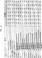
図1aは、特異的ペプチドに対して試験した場合の、実施例1のリン酸化トレオニンペプチドライブラリーに対して産生した親和性精製したポリクローナル抗体の特異性を示す表である。
【0020】
図1bは、種々のホスホペプチドライブラリーに対して試験した場合の、実施例1のホスホトレオニン抗体の特異性を示す表である。
【0021】
図1cは、オカダ酸で処理しているか処理していない細胞由来の細胞抽出物および他のホスホタンパク質に対する、実施例1のホスホトレオニン抗体の反応性を示すウェスタン分析である。
【0022】
図1dは、固定化格子によって示した、実施例1の抗ホスホトレオニン抗体の状況独立性を示す表である。
【0023】
図2aは、実施例IIのリン酸化PXS*Pペプチドライブラリーに対して産生された、親和性精製したポリクローナル抗体の特異性を示す表である。
【0024】
図2bは、オカダ酸で処理しているか処理していない細胞由来の細胞抽出物および他のホスホタンパク質に対する、実施例IIのホスホPXS*P抗体の反応性を示すウェスタン分析である。
【0025】
図3aは、モチーフを欠くホスホペプチドまたは非ホスホペプチドに対して試験した場合の、実施例IIIの親和性精製したポリクローナル14−3−3抗体の反応性の欠失を示す表である。
【0026】
図3bは、GST−BadおよびTPAでトランスフェクトした細胞由来の細胞抽出物に対する実施例IIIのホスホ14−3−3抗体の反応性を示すウェスタン分析を示す図である。
【0027】
図4aは、実施例IVのリン酸化PXT*PXRライブラリーに対して産生されたモノクローナル抗体の特異性を示す表である。
【0028】
図4bは、リン酸化および非リン酸化RBタンパク質に対する実施例IVのCDKコンセンサス部位モノクローナル抗体の反応性を示すウェスタン分析を示す図である。
【0029】
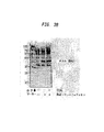
図5aは、アセチル化BSAに対する実施例Vのアセチル化リジン抗体の特異性を示すウェスタン分析を示す図である。
【0030】
図5bは、抗体を非アセチル化ペプチドライブラリーでプレインキュベートした場合のC6細胞抽出物に存在する種々のタンパク質に対する実施例Vのアセチル化リジン抗体の反応性を示すウェスタン分析を示す図である。
【0031】
図5cは、抗体をアセチル化ペプチドライブラリーでプレインキュベートした場合のC6細胞抽出物に存在する種々のタンパク質に対する実施例Vのアセチル化リジン抗体の反応性を示すウェスタン分析を示す図である。
【0032】
図5dは、抗体をアセチル化ペプチドライブラリーでプレインキュベートした場合のコントロールアセチル化BSAに対する実施例Vのアセチル化リジン抗体の反応性を示すウェスタン分析を示す図である。
【0033】
詳細な説明
本発明は、抗原として使用されるペプチドライブラリーにおける任意の個々の配列の濃度が非常に低いので宿主における免疫応答を駆動するのに不十分であるという概念に基づく。したがって、免疫応答を駆動するのに十分に高濃度の抗原決定基のみが各配列ならびにそのペプチドバックボーンに共通の固定残基である。
【0034】
各変性位置に20アミノ酸全てを示すペプチドライブラリーでの宿主の免疫化により、1つまたは複数の固定残基周囲の変化する位置での多くのまたは全てのアミノ酸に対して抗体耐性を得る。このような抗体は、周囲のアミノ酸、ペプチド、またはタンパク質配列の最も広い可能な範囲の状況で抗原決定基と反応する。モチーフの固定残基は、リン酸化または非リン酸化残基などの1つの非修飾または修飾アミノ酸であり得るか、またはコンセンサス認識部位などの複数の非修飾または修飾アミノ酸であり得る。
【0035】
本明細書中で使用される、「抗体」は、Fcフラグメント、Fabフラグメント、キメラフラグメント、または他の抗原特異的抗体フラグメントを含む、ポリクローナルまたはモノクローナル抗体を意味する。
【0036】
本明細書中で使用される、「モチーフ特異性状況独立性抗体」は、変化する周囲のペプチドまたはタンパク質配列の状況において1つまたは複数の固定アミノ酸残基に対して特異的である抗体を意味する。したがって、このような抗体特異性は、抗原が生じる状況に高度に独立している。
【0037】
本明細書中で使用される、「基質」は、酵素によって特異的に認識され、作用するペプチドまたはタンパク質を含む任意の標的分子を意味する。
【0038】
モチーフ特異性状況独立性抗体が本発明によって産生される一般的な方法を、以下に示す。
【0039】
(1)周囲のアミノ酸と独立した特異的標的残基を含む任意のタンパク質またはペプチドと反応するモチーフ特異性抗体を、高度に変性したペプチドライブラリーの合成によって得ることができる。1つの好ましい実施形態では、ライブラリーは、XXXXXXJ*XXXXXXC(式中、X=システインを除く全20アミノ酸およびJ*=修飾(*)アミノ酸(J)(例えば、ホスホトレオニン(T*)またはアセチル化リジン(K*))を含む。特異的標的残基を非修飾し、より短いまたはより長いライブラリーを作製し、周囲のアミノ酸の全てより少ないアミノ酸を変化させることができることが認識される。1つの好ましい実施形態では、ペプチドライブラリーは、約6〜14残基長である。好ましい実施形態では、変化した周囲状況において1つの固定アミノ酸(修飾または非修飾のいずれか)を利用する一方で、他の好ましい実施形態では、いくつかの固定アミノ酸を含むモチーフを利用することができる。同様に、ライブラリーの周囲の配列は、1つを越える位置で同時に変化することができるか、好ましい実施形態におけるように、変性分子あたりたった1つの周囲配列で変化することができ、その結果固定残基以外の全ての位置で完全に変性するライブラリーが作製される。ペプチドライブラリーを、ABIペプチド合成機および変性カップリング反応中の各アミノ酸の混合物を使用した標準的なF−Moc固相ペプチド合成によって合成することができる。
【0040】
固定位置での修飾アミノ酸の組み込みは、他の修飾保護アミノ酸(例えば、脂質(例えば、ファルシニル化、イソプレニル化)で修飾されたアミノ酸または保護O結合もしくはN結合糖(例えば、グリコシル化)、メチル化、リボシル化アミノ酸、ヌクレオチド、ヌクレオチドのポリマー、ヌクレオシド、またはユビキチンなどのアミノ酸、またはアミノ酸アナログ)もまた組込むことができるので、リン酸化またはアセチル化に限定するべきではない。
【0041】
固定位置での非修飾アミノ酸の組込みを、保存モチーフ(例えば、ジンクフィンガーまたは反復アルギニン残基)を模倣するために選択することができる。
【0042】
(2)各変性位置での各アミノ酸の出現を可能な限り等しく産生するために、数ラウンドのアミノ酸組成の変更、合成、およびペプチド配列決定を行う。ペプチドに沿ったいくつのかの異なる位置でアミノ酸配列分析を行って各位置での無作為なアミノ酸出現を評価し、無作為表示を合成の間維持する。各位置での全アミノ酸の等しい分布を達成するために、ラウンド数を変化させることができることが当業者に認識される。
【0043】
(3)好ましくは、キャリアへの共有結合による高度に多様なペプチドライブラリーを、抗原として使用する。好ましい実施形態では、フロイントアジュバント中に乳化させたキーホールリムペットヘモシアニン(KLH)をカップリング剤として使用してカップリングしたペプチドライブラリーを宿主(雌のニュージーランド白ウサギなど)に皮内注射する。免疫応答が得られるまで、完全フロイントアジュバントでの追加免疫注射を行うことができる。抗体力価を、適切な方法(モチーフ特異性ペプチドライブラリーに対するELISA)によって測定する。この様式で惹起した抗血清を、以下に概説のように、粗調製物または精製調製物において使用することができる。
【0044】
(4)最も有望な宿主由来の抗血清を、例えば、protein Aで精製し、J(非修飾)ペプチドライブラリーカラムに吸着させる。好ましい実施形態では、非吸着画分(通過したもの)を、J*カラムに注ぎ、適切なpHで溶出し、透析して、適切な方法(J*およびJを抗原として使用したELISA)によってJ*特異性について試験する。
【0045】
(5)この様式で精製した抗体の親和性は、J*ペプチドライブラリーを認識するが、Jライブラリーとは反応せず、J*と程度の高い特異性を示す。これらの抗体を、さらに、適切な方法(ELISAなど)を用いて標的修飾アミノ酸J*またはJ*ホモログの非修飾形態に対する反応性の欠如について試験することができる。
【0046】
(6)さらに、抗体を、選択したタンパク質修飾酵素インヒビター(タンパク質ホスファターゼインヒビターであるオカダ酸)で処理するか処理しない細胞から調製した細胞抽出物を用いてウェスタンブロッティングまたは別の適切な方法によって試験することができる。タンパク質修飾を増大させる処理により、抗体反応性タンパク質数および反応性の強度が増大する。J*特異的抗体は、コントロール抽出物由来の比較的少数のタンパク質と反応するが、選択したインヒビターとの反応後非常に多数のタンパク質と反応する。抗体は、これらのタンパク質の不活性な非修飾変形との反応性を示さず、これは、J*特異性の程度が高く、多数の異なる修飾された標的を含むタンパク質に対して広範な交叉反応性を示すことが示唆される。
【0047】
(7)状況独立性の程度を、例えば、混合するか個々に試験する個々のJ*ペプチドに対するELISA分析によってより慎重に試験することができる。このような分析により、例えば、J*の後にプロリンが存在する場合、所与のモチーフとの反応性が不十分であるかどうかを示すことができる。
【0048】
(8)好ましい実施形態のように、修飾ペプチドライブラリーの固定格子(immobilized grid)を用いてJ*抗体認識の状況依存性をさらに試験することができる。固定標的残基J*に加えて、J*と比較して異なる位置であるがシステインを除く20種全てのアミノ酸を含むさらなる固定アミノ酸を含むように各々の異なるライブラリーを合成する。各ペプチドライブラリーを、例えば、ELISAウェルの底部に被覆し、J*抗体に暴露する。特定のアミノ酸が特定の位置に存在する場合、格子上の特定のスポットと反応しない抗体(ペプチドライブラリー)は結合しない。この分析は、J*と比較して特定の位置の特定のアミノ酸が結合を許容するか阻害するかを同定する。
【0049】
あるいは、精製抗体をビーズに結合させ、修飾または非修飾ライブラリーに結合させ、非結合配列を洗浄し、結合配列を回収してアミノ酸配列決定し、ライブラリー中の各位置に存在する各アミノ酸の量を同定することができる。この情報は、アミノ酸が各位置で耐性であることを示す。
【0050】
(9)好ましい実施形態の1形態として、適切なキャリア(KLHなど)をJ*ライブラリーへカップリングし、宿主(balbCマウスなど)に注射することでモノクローナル抗体を調製する。J*ペプチド−KLH結合物を、フロイントアジュバント中で乳化し、完全フロイントアジュバントでの追加免疫注射を応答が得られるまで隔週で行うことができる。
【0051】
(10)抗体力価を、適切な方法(J*および非J*ペプチドライブラリーに対するELISAなど)で測定する。高力価応答を示す宿主由来の血清を、固定非J*ペプチドに吸着させ、非吸着画分を、例えばウェスタンブロッティングによって試験する。
【0052】
(11)J*特異的応答を示す宿主由来の脾臓を、骨髄腫細胞と融合し、ハイブリドーマクローンを選択およびスクリーニングする。各クローン由来の上清を、J*ペプチドライブラリーへのその結合能力について最初にスクリーニングする。次に、ポジティブクローンを、非J*ライブラリーに対するその交叉反応性についてスクリーニングする。最も高い程度のJ*特異性を示すクローンを、上記の工程(5)〜(8)に記載のようにさらなる分析用に選択する。
【0053】
(12)上記の(11)由来のモノクローナル抗体の過剰産生を、例えば、腹水を回収し、選択したハイブリドーマを培養するか、宿主生物(E.coliなど)にクローニングすることによって行うことができる。
【0054】
本方法によって産生したモチーフ特異性状況独立性抗体を使用して、酵素の未知の基質を同定することができる。このような抗体を、目的の酵素によって認識されるモチーフ(例えば、コンセンサス部位)に対して最初に作製する。次いで、これらの抗体を使用して、同一のモチーフを含む他の未知の基質の存在についてサンプルをスクリーニングする。本方法によれば、保存基質モチーフを含む種々のカスケードにおける重要な新規の基質の迅速な検出が可能である。例えば、広範な種々のタンパク質を選択的に認識する抗体は、MAPKコンセンサスリン酸化部位でリン酸化された場合のみ、新規のMAPキナーゼ標的の検出に非常に便利である。MAPキナーゼを、細胞培養において過剰発現させ、成長因子によって活性化させ、標的基質タンパク質をリン酸化基質タンパク質を選択的に認識する抗体を用いたウェスタンブロッティングによって同定することができる(Stukenbergら、Curr.Biol.、7、338〜348、1997)。あるいは、MAPKを使用して、in vitroでcDNA発現ライブラリーをリン酸化し、MAPKコンセンサス部位抗体を使用してMAPKリン酸化基質を発現するcDNAクローンを同定することができる(Funkunaga and Hunter、EMBO、16(8)、1921〜1933、1997)。
【0055】
同様に、本発明の方法によって産生した抗体を使用して、既知の基質モチーフを修飾する酵素を同定することができる。修飾(例えば、リン酸化)または非修飾(例えば、ジンクフィンガー)モチーフのいずれかに特異的なこのような抗体を使用して、目的の一定の酵素がそのモチーフを含む基質を修飾したかどうかを検出することができる。この方法により、保存モチーフ(例えば、MAPKコンセンサス部位)を含む既知のクラスの基質に対して作用する重要な新規のタンパク質の迅速な検出が可能になる。
【0056】
本発明のモチーフ特異性状況独立性抗体を、保存モチーフを含む一定の基質の酵素修飾を検出するための薬物スクリーニングなどの高処理アッセイにおけるin vitroでの試薬としても使用することができる。例えば、一定のリン酸化モチーフに特異的な抗体は、そのモチーフに作用する酵素のインヒビターの迅速な検出を可能にする。薬物スクリーニングの場合、単一のモチーフ特異性抗体を使用して多くの多様な配列モチーフで作用する広範な酵素活性をアッセイすることができる。現在、ホスホチロシン抗体は、選択的な高親和性キナーゼインヒビターをスクリーニングするための高処理キナーゼアッセイにおいて使用されている。酵素活性を阻害する化合物または薬物を、リン酸化基質に結合しているホスホチロシン抗体の減少によって同定する、キナーゼ活性の阻害能力によって検出する。ホスホセリン、ホスホトレオニンについて上記のように産生した抗体または他のタンパク質修飾を検出する抗体を用いて薬学的に有用な化合物をスクリーニングするための類似のアッセイを設定することができる。
【0057】
プロテインキナーゼ活性の抗体ベースの検出は、自動化高処理キナーゼアッセイに使用する上で放射性アッセイよりいくつかの利点を有する。第1に、放射性アッセイは、32−Pγ標識ATPのペプチド基質への移動を使用するための自動化が困難である。ホスホペプチドを、ホスホセルロースフィルターを使用して標識ATPから分離し、数回洗浄し、最後に液体シンチレーション法によってリン酸化を停止させる。これらの工程は時間がかかり、自動化が困難である。抗体検出により、自動化および高処理スクリーニングに十分に適切である広く一般に用いられるELISA型アッセイが可能である。
【0058】
第2に、放射性アッセイは、感度を最大にするために高レベルの32−Pの組み込みを確実にするための低レベルのATPを必要とする。キナーゼアッセイにおける低レベルのATPは、プロテインキナーゼの触媒部位においてATP結合と競合する化合物に対するインヒビターの検索が偏る。このようなスクリーニングでは、この結合部位は高度に保存されているためATP結合部位に対して一貫して競合インヒビターが得られるが、これらは選択性が不十分なインヒビターである。
【0059】
現在の高処理キナーゼアッセイは、典型的には、96または386ウェルプレートの下部に固定したビオチン化ペプチド基質を使用し、これはその後所望のプロテインキナーゼ、ATP、および適切なキナーゼ緩衝液とインキュベートされる。キナーゼ活性を、リン酸化ペプチド基質とのみ反応する蛍光標識リン酸特異的抗体を用いて測定する。これらのアッセイは、2つの形式である均質(洗浄工程を含まない)および不均質(洗浄工程を含む)がある。均質な蛍光アッセイは、典型的には、エネルギー受容体(例えば、アロフィコシアニン)に連結するリン酸化ペプチド基質へのランタニド標識ホスホ抗体結合を使用する。ホスホ抗体の結合の際、リン酸化ペプチド基質は、放射シグナルの頻度を変化させ、同時に蛍光共鳴エネルギー転移を可能にするように2つのフロオロフォアを十分に近づかせ、生体分子複合体の存在を示す。異なる化合物を各ウェルに添加し、化合物の基質リン酸化阻害能力を、蛍光エネルギー転移阻害によって同定する。この形式は、一般に放射性アッセイで使用されるシンチレーション近接アッセイ(scintillitation proximity assay)と類似している。他の均質アッセイには、ホスホ抗体のリン酸化基質への結合を測定するための蛍光偏光の使用が含まれる。
【0060】
均質アッセイにおける重要な特徴は、工程数が限られており、自動化が容易なことである。現在、ELISAに基づく多様な不均質キナーゼアッセイも使用されている。これらのアッセイは、典型的には、96または386ウェル形式に固定したリン酸化ペプチド基質に結合する蛍光標識ホスホ抗体を利用する。この場合、非結合抗体を分離するために洗浄工程を必要とする。ウェル中に保持される蛍光標識抗体を、時間分解蛍光を用いて検出する。
【0061】
このような修飾スクリーニングアッセイ用の抗体作製に使用されるモチーフは、修飾または非修飾基質モチーフのいずれかであり得る。非修飾モチーフに対して作製した抗体は、基質がその後酵素によって修飾された場合結合しない。同様に、修飾モチーフに対して作製された抗体は、酵素活性による修飾基質濃度の増加を検出することができる。
【0062】
類似のアプローチを適用して、種々の他の酵素修飾を研究することができ、これには、以下に記載のプロテインキナーゼ活性またはアセチルトランスフェラーゼ活性が含まれるがこれらに限定されない。例えば、このアプローチを使用して、多くの他の型のタンパク質修飾(糖、メチル基、カルボキシル基の付加、種々の脂質の付加、ヌクレオチドもしくはヌクレオチドポリマー、ヌクレオシド、またはユビキチンなどのアミノ酸の付加が含まれるが、これらに限定されない)を認識する抗体を作製することができる。
【0063】
同様に、保存モチーフを含む巨大で多様なタンパク質集団を同時にプロファイリングするために、ゲノム中に及ぶ規模でこのようなモチーフ特異性状況独立性抗体を使用することができる。特異的な2つまたは3つのアミノ酸結合部位(例えば、連続的なアルギニン残基)が(アミノ酸の無作為な分布に基づいて)400または8000残基に1回認められるはずである(1タンパク質あたり約1回または20タンパク質あたり1回のいずれかに相当する(平均タンパク質を400アミノ酸として仮定する))。したがって、起こり得る状況に独立的なこのようなモチーフに特異的な抗体により、非常に多数のタンパク質の迅速なスクリーニングが可能となる。
【0064】
リン酸化特異性抗体は、薬物処理の結果としてのタンパク質のリン酸化またはこのような処理の結果としての特異的遺伝子/タンパク質の過剰発現の変化についてのゲノム中に及ぶプロファイリングが可能である。このような抗体はまた、配列決定したゲノム中の特異的タンパク質発現のプロファイリングを容易にする。
【0065】
例えば、細胞周期依存性プロテインキナーゼcdc2を阻害する薬物開発が考えられる。薬物は、高親和性を有するcdk2阻害が認められているが、他のプロテインキナーゼが阻害されるかどうか、もし阻害されるのであれば、どのプロテインキナーゼが阻害されることになるかについて、化合物の特異性を試験する必要がある。
【0066】
このプロセスにおける初期の工程として、化合物または誘導薬物によって阻害されるリン酸−基質の性質を試験するために、細胞株を薬物で処理して、全細胞タンパク質リン酸化に対する効果をモチーフ特異性ホスホ抗体および一般的なホスホ抗体のパネルを使用して監視することができる。
【0067】
コントロールおよび薬物処理細胞から調製した細胞抽出物由来の全タンパク質を、例えば二次元ゲル(第1次元では等電点電気泳動を行い、第2次元では標準的なSDS−ポリアクリルアミドでの分子量分画を行う)を用いて分画し、ニトロセルロース膜に移し、この場合、キナーゼコンセンサス部位特異的ホスホ抗体を用いたウェスタンブロッティングによって分析することができる。
【0068】
この場合、cdc2コンセンサス部位特異性抗体を用いた全細胞タンパク質の全体的な分析により、全ての潜在的なcdc2部位基質でのリン酸化を阻害する薬物の能力に関する情報が得られるであろう。インヒビターがチロシンキナーゼを阻害するようにも作用するかどうかを同定するために、異なるキナーゼコンセンサス部位に対する抗体を使用するかホスホチロシンに対する抗体を使用して他の非cdc−2基質での阻害パターン(すなわち、特異性の程度)を試験することもできる。
【0069】
現在、哺乳動物細胞についての、二次元ゲルによって視覚化されたタンパク質「スポット」の大部分の同定が未知である。しかし、全てのヒト遺伝子が同定および配列決定され、対応するタンパク質が特徴づけられ、「スポット」同定されているので、本発明によるタンパク質プロファイリングによる分析はより強力な情報源となる。阻害されたタンパク質の同定により、薬物特異性が確認されるだけでなく、阻害されたさらなる「非特異性」タンパク質の同定により可能な副作用も示唆される。二次元ゲルによる多くのタンパク質「スポット」がすでに同定されている酵母などの単純で完全に配列決定されている生物において、理想的な分析を行うことができる。
【0070】
以下に記載の実施例は、本発明の特に好ましい実施形態としてのみ意図され、本明細書に添付の特許請求の範囲に限定した以外は、発明の範囲を限定することを意図しない。本発明は、本明細書中で教示の方法の修正および変形が含まれることが当業者に自明である。
【0071】
上記および下記の引例は、本明細書中で参考として援用される。
【0072】
実施例1
状況独立性ホスホトレオニン抗体
ペプチドライブラリー抗原の合成
リン酸化トレオニン残基を含む任意のタンパク質と反応する(すなわち、周囲のアミノ酸のホスホトレオニンに独立して結合する)ホスホ特異性抗体を、高度に変性させたペプチドライブラリーXXXXXXThr*XXXXXXC(式中、X=システインを除く全20アミノ酸、Thr*=ホスホトレオニン)の合成によって得た。
【0073】
ホスホトレオニンペプチドライブラリーを、ABIペプチド合成機および変性カップリング反応中の各アミノ酸の混合物を用いた標準的なF−Moc固相ペプチド合成によって合成した。変性ペプチドを、0.085mmolのスケールでのFastMocの化学的性質を用いたABIモデル433Aペプチド合成機を用いて合成した(Fieldsら、Pept.Res.、4,、95〜101、1991(本明細書中で参考として援用される))。HBTUアミノ酸活性化を使用したFmoc/NMPの化学的性質(Dourtoglouら、Synthesis、1984、572〜574、1984、Knorrら、Tetra.Let.、30、1927〜1930、1989、Knorrら、Peptides、1988、37〜129、1989、Walter de Gruter & Co.(これらの全てが本明細書中で参考として援用される))を、全てのサイクルで使用した。0.5mmol/gで機能させた、予めロードしたFmoc−Cys(Trt)HMP(p−ヒドロキシメチルフェノキシメチル)ポリスチレン樹脂をペプチドの各変性プールに使用した。ペプチドを、各サイクル間で単一のカップリングを用いて合成したにもかかわらず、カップリング時間は、リン酸化アミノ酸を含む各位置で伸長した。最終Fmocを、合成中に取り除いた。最終的にFmoc基を取り除いた予めロードしたHMPの使用により、切断および脱保護後に遊離アミノ酸およびカルボキシ末端を含むペプチドが得られる。
【0074】
各変性位置で可能な限り等しい各アミノ酸の出現を得るために、数ラウンドのアミノ酸組成の変更、合成、およびペプチド配列決定を行った。所望のペプチドプールは、各「変性」部位で等モルの19アミノ酸(Cys以外の全ての標準的なアミノ酸)を含むことであった。各保護アミノ酸の反応速度が異なるので、簡単に混合した等モル量(それぞれ、全量の約5.26%)では、各位置で等モルのペプチドが得られない。各残基での変性を最大にするために、最初に、各位置で等モルの「混合物」を用いてペプチド合成を行った。フェニルチオカルバミル−アミノ酸分析を行うことにより、各位置での相対的アミノ酸含有量が評価される。各変性位置での各アミノ酸の等しい出現を確実にするために、アミノ酸分析に基づいて、「混合物」中の各アミノ酸のモル量を調整して異なる反応速度を補正した。数ラウンドのペプチド合成後、アミノ酸分析はアミノ酸混合物を至適化する必要があり、それにより変性ペプチドが全て得られた。最適化に到達したアミノ酸混合物は以下である:G(4.6%)、A(5.6%)、V(3.3%)、L(2.5%)、I(4.25%)、S(4.4%)、T(8.4%)、F(2.25%)、Y(6.0%)、W(6.8%)、M(2.9%)、P(2.5%)、D(5.8%)、N(9.5%)、E(6.2%)、Q(9.4%)、K(6.1%)、R(6.4%)、H(3.5%)。
【0075】
側鎖保護基の除去を伴う樹脂からの変性ペプチドの切断は、TFAでの処理と同時に行われる。切断混合物(Perkin Elmer、Emerville、CA、1995)は、以下からなる:0.75gフェノール、0.125ml硫化メチル、0.25ml 1,2−エタンジオール、0.5ml milliQ H2O、0.5mlチオアニソール、10ml TFA。全混合物をペプチド樹脂に添加した(約300mg)。樹脂を窒素で洗い流し、室温で3時間穏やかに撹拌した。次いで、樹脂を濾過して、ペプチドを冷(0℃)メチル−t−ブチルエーテル中に沈殿させた。エーテル画分を、遠心分離して沈殿を回収した。ペプチド沈殿物を、減圧乾燥し、質量分光法によって分析し、HPLCで精製した。
【0076】
ペプチドサンプルを、アセトニトリル/水(50:50、v/v)中に懸濁し、基質として2,4,6−トリヒドロキシアセトフェノン+クエン酸アンモニウムを用いたPerceptive Biosystems(Framingham、MA)MALDI−TOF質量分析計で分析した。予想通り、ペプチド混合物は、均一の生成物を示さなかった。MALDI−TOF分析により、ペプチドプールは変性され、ペプチド質量の平均質量および予想された統計的に正常な曲線を示すことが示された。
【0077】
ペプチドを、Lambda−Max Model 481 Multiwavelength detector、500シリーズポンプ、および自動化勾配制御装置からなるWaters HPLC systemを用いて精製した。Vydacの半調製C18カラムを、逆相精製に使用した。2ml/分の流速で60分間の直線的勾配(10%〜100%、B)を使用した。緩衝液Aは、0.1%TFA/H2O(v/v)からなり、緩衝液Bは、0.1% TFA/60% CH3CN/40% H2O(v/v/v)からなる。214nmで検出した。
【0078】
(質量分光法で示されたように)ペプチドプールが変性していたので、HPLC精製によって均一な生成物が得られるとは期待できなかった。本方法によってペプチド混合物のベースライン分離を行わず、本方法は粗精製/脱塩工程としてのみみなした。質量分光法を行い、質量が理論上の範囲内である全ての画分をプールし凍結乾燥した。
【0079】
ペプチドに沿ったいくつかの異なる位置でのアミノ酸配列分析により、各タンパク質での無作為なアミノ酸が表示され、無作為の出現が合成を通して維持されることが示された。結果は、適切な抗原として作用する非常に多様なペプチドライブラリーの産生を示した。
【0080】
ウサギポリクローナル抗体の産生
異質二重機能性架橋剤(m−マレイミドベンゾイル−N−ヒドロキシスクシンイミドエステル(MBS))を使用して、キャリアタンパク質(KLH)へ結合させるC末端システイン残基を含む全てのペプチドを合成した。使用した結合法は製造者(Pierce)に記載の通りであるが、動物の免疫化および追加免疫用の物質を増加させるためにKLHにカップリングしたペプチドの量は10mgに増加させた。N末端およびKLHリジン残基のε−アミノ基に対する反応後に過剰なMBSを取り除くために、スケールアップにはより大きな脱塩カラム(Bio−Rad 10 DG(Cambridge、MA))の使用が必要であった。
【0081】
ホスホトレオニンペプチドライブラリーを、キーホールリンペットヘモシアニン(KLH)(250μg)に共有結合させ、フロイントアジュバントに乳化し、雌のニュージーランド白ウサギに皮内注射した。完全フロンドアジュバント(200μg)による追加免疫注射を、応答が得られるまで隔週で行った。ホスホトレオニンおよび非ホスホトレオニンペプチドライブラリーを用いたELISAによるホスホペプチド特異性免疫反応性の存在について、ウサギ血清を3週間毎にスクリーニングした。ホスホペプチドに対する抗体の力価が105に達したとき、ウサギを、2週間毎に採血する産生血液スケジュール(production bleed schedule)に従わせる。40mlの高力価の血清が得られたとき、以下に記載のように、ホスホ特異的抗体の精製を開始した。
【0082】
最も有望なウサギ由来の抗血清をprotein Aで精製し、非ホスホThr/Serペプチドライブラリーカラムを通過させた。非吸着画分を、ホスホトレオニンカラムに注ぎ、低pHで溶出し、透析し、ホスホペプチドおよび非ホスホペプチドを用いたELISAによってホスホ特異性について試験した。この様式で親和性精製された抗体は、リン酸化トレオニンペプチドライブラリーを認識するが、非ホスホトレオニン/セリンライブラリーと反応せず、これは、ホスホトレオニンについての特異性の程度が高いことを示している(図1aを参照のこと)。ELISAの結果はまた、抗体はまた18の異なるホスホトレオニンペプチドの混合物と特異的に反応したが、対応するいかなる非ホスホペプチドとも反応しなかったことを示した(図1b)。抗体はまた、ホスホトレオニンについて厳密な選択性を示し、これは、38種の異なるホスホセリンペプチド(図1b)またはホスホチロシンを含むペプチドと反応しないことを示す。
【0083】
本発明者らは、次に、プロテインフォスファターゼ阻害剤であるオカダ酸で処理しているか処理していない細胞由来の細胞抽出物を用いたウェスタンブロッティングによって抗体を試験した。図1cに示すように、ホスホトレオニン抗体は、コントロール抽出物由来の比較的少数のタンパク質と反応したが、オカダ酸で処理後では非常に多数のタンパク質と反応する(図1c、レーン2の高分子量の不鮮明な反応タンパク質を参照のこと)。抗体はまた、それぞれの活性化ループでのトレオニン残基でリン酸化された場合のみ、活性化形態のMAPK(ERK1)およびMKK3と特異的に反応した。抗体は、これらのタンパク質の不活性な非リン酸化形態と反応しなかった(図1c、レーン3〜6)。これらの結果は、ホスホトレオニンの特異性が高いことを示し、これは、多くの異なるトレオニンリン酸化タンパク質およびペプチドに対して広範な交叉反応性を示唆する。
【0084】
状況独立性をより慎重に試験するために、上記の実験で混合した個々のトレオニンリン酸化ペプチドに対してELISA分析を行った。図1aに示すように、ホスホトレオニン抗体は、ホスホトレオニンがプロリンに隣接する場合(例えば、c−MycおよびAPP1ホスホペプチド)以外は、全てのホスホペプチドと良好に反応する(図2b)。これらの結果は、精製ウサギ抗体がホスホ特異性形態で広範な種々のホスホトレオニンと反応するが、リン酸化トレオニンがプロリンが隣接する場合はホスホペプチドとの反応は不十分であることを示す。
【0085】
ホスホトレオニン抗体認識の状況依存性を、ホスホペプチドライブラリーの固定格子を用いてさらに試験した。固定ホスホトレオニンに加えて、ホスホトレオニンと比較して−4、−3、−2、−1、+1、+2、+3の位置にさらなる固定アミノ酸を含むが、システイン以外の全ての20アミノ酸を含む他の位置を有するようにそれぞれ異なるライブラリーを合成した。各ペプチドライブラリーを、ELISAウェルの底部にコートし、ホスホトレオニン抗体に暴露した。格子上の特定のスポット(ペプチドライブラリー)と反応しない抗体は、特定の位置に特定のアミノ酸が存在する場合、結合しない。この分析は、ホスホトレオニンと比較して特定の位置での特定のアミノ酸が結合を許容するか阻害するかどうかを同定する(図1d)。
【0086】
結果により、ホスホトレオニン抗体が−1、−2、−3、−4および+2、+3の位置で全てのアミノ酸を許容し、+1位のプロリン以外の全てのアミノ酸と等しく十分に結合することを確認した(図1d、最初の列を参照のこと)。この結合プロフィールによって規定された反応性は、抗体が−1位でプロリンが隣接するもの以外は全てのホスホトレオニン含有配列に結合することを示す。種々の特異的ホスホトレオニン含有ペプチドを使用したさらなる分析により、これらの結果を確認した。
【0087】
同一のペプチドライブラリー抗原で免疫化したいくつかの他のウサギ由来のホスホトレオニン特異的抗体を、さらに精製して特徴づけた。2つの他のウサギから得た血清から精製した抗体は、ELISAで同定したところ、広範な交叉ホスホトレオニン抗体も産生した。あるウサギでは、ホスホトレオニン後のプロリンを含むペプチドと等しく十分に反応する抗体を産生した。まとめると、これらの結果は、組み合わせペプチドライブラリーを免疫原として使用する場合、ホスホトレオニン応答の広範な状況独立性が得られることを示す。
【0088】
実施例II
プロテインキナーゼコンセンサス部位特異的ホスホ抗体
MAPK−コンセンサス認識部位:PXS*P
MAPKリン酸化の好ましい部位PXS*Pのペプチドライブラリーを、実質的に実施例Iに記載のように合成した(図2a)。等モル量のホスホセリンおよびトレオニンとの混合物に加えて、以下の2つの他の位置のアミノ酸も固定した:−2位のプロリンおよび+1位のプロリン。このライブラリーをKLHにカップリングし、ホスホトレオニンについて記載したようにウサギに注射した。最も有望なウサギ由来のIgGをprotein Aで精製し、非ホスホThr/Serペプチドライブラリーカラムを通過させた。非吸着画分(通過したもの)を、ホスホPXS*Pカラムに注ぎ、低pHで溶出し、透析し、ホスホペプチドおよび非ホスホペプチドを用いたELISAによってホスホ特異性について試験した。
【0089】
この様式で親和性精製した抗体は、リン酸化PXS*Pペプチドライブラリーと強力に反応するが、非ホスホトレオニン/セリンライブラリーとは反応しなかった(図2aを参照のこと)。ELISAの結果はまた、抗体が18の異なるホスホトレオニンペプチドの混合物と特異的に反応するが、対応するいかなる非ホスホペプチドとも反応しないことを示した(図2a)。ホスホ特異性であることに加えて、抗体は−2および+1位のプロリンに選択性を示し、この位置でプロリンを欠くリン酸化ペプチドとは反応しない(図2a)。抗体は、RBおよびcdk4ホスホペプチドと強力に反応するが、+1位にプロリンを欠くMKK3、PKCalpha、またはp70S6ホスホペプチドとは反応しなかった(図2a)。これらの抗体は、−2位にプロリンを欠くいくつかのペプチド(例えば、cdk4ホスホペプチド)と反応し、これは、この位置でプロリンが絶対に必要ではないことを示唆する。
【0090】
タンパク質ホスファターゼインヒビターのオカダ酸で処理したか処理していない細胞から抽出した細胞抽出物を使用したウェスタンブロッティングによって、PXS*P抗体をさらに試験した。RS 4;11細胞由来の細胞抽出物へのPXS*P抗体の結合は、オカダ酸処理後に強力に促進された(図2b、レーン2の高分子量の不鮮明な反応タンパク質を参照のこと)。抗体はまた、MAPキナーゼでin vitroでリン酸化したATF−2と特異的に反応するが、このタンパク質の非リン酸化形態とは反応しない(図2b、レーン3およびレーン4)ので、多くの異なるリン酸化タンパク質およびペプチドとホスホ特異性の程度が高く、交叉反応性が広範であることが示された。
【0091】
PXS*P抗体認識の特異性もまた、ホスホペプチドライブラリーの固定格子を用いて試験した。上記のように、固定ホスホトレオニンまたはホスホセリンに加えて、ホスホトレオニンと相対して−1、+1、+2位に固定したアミノ酸を持つが、他の全ての位置にシステインを除く20種全てのアミノ酸を含むように各々の異なるライブラリーを合成する。
【0092】
PXS*P抗体は、プロリンが−1位に固定された場合ペプチドライブラリーと弱く反応したが、プロリンが−2および+1位に固定された場合ライブラリーと強力に反応した。この結合プロフィールによって定義された反応性は、予想通り、PXS*P抗体がPXS*Pモチーフを含む配列とのみ結合する。抗血清は、S*P(不純物の結果として)に対していくつかの残基反応性をさらに含むが、これは固定S*Pライブラリーを用いたさらなる精製によって取り除くことができる。
【0093】
実施例III
プロテインキナーゼコンセンサス部位特異的ホスホ抗体
14−3−3結合部位:RSXS*XP
14−3−3標的を同定する抗体を、ペプチドライブラリーXXXXRSXS*XPXXXXC(式中、S*はホスホセリンを示し、Xは任意のアミノ酸を表し、Cはシステインである)の合成によって得た。上記の14−3−3ホスホペプチドライブラリーを、実施例Iに記載のように、ABIペプチド合成機および変性カップリング反応中のシステイン以外の各アミノ酸の混合物を用いたF−Moc固相ペプチド合成によって合成した。
【0094】
14−3−3ホスホペプチドライブラリーを、KLHと結合させ、ホスホトレオニンおよびPXS*Pについて上記のようにウサギに注射した。最も有望なウサギ由来の抗血清を、protein Aで精製し、非ホスホ14−3−3ペプチドライブラリーカラムに吸着させた。このカラムを通過したものを、ホスホ14−3−3カラムに注ぎ、低pHで溶出し、透析し、ホスホおよび非ホスホ14−3−3ペプチドライブラリーを用いたELISAによってホスホ特異性について試験した。これらの親和性精製した14−3−3ペプチド抗体は、リン酸化14−3−3ペプチドライブラリーを認識するが、非ホスホ14−3−3ライブラリーは認識しないので、これは、ホスホ14−3−3に対する高度な特異性を示す(図3aを参照のこと)。抗体はまた、14−3−3モチーフ(ホスホ−Bad−Ser136、cdc25−Ser216を含む)を含むいくつかの異なるペプチドと強力に反応し、わずかな変異モチーフを含むホスホ−Bad−Ser112とより弱く反応した。抗体は、対応する非ホスホペプチド(図3a)ともモチーフを含まない他の多くのホスホペプチドとも全く反応しない。
【0095】
ホスホ14−3−3抗体を、GST−Bad融合タンパク質でトランスフェクトしホルボールエステルTPAと処理したか処理していない細胞から調製した細胞抽出物を用いたウェスタンブロッティングによってさらに試験した。抗体は、コントロール抽出物由来の少数のタンパク質と反応した(図3b)。トランスフェクト細胞から調製した抽出物中ではBadが検出されたが、コントロール細胞からは検出されなかった。TPAは、いくつかの高分子量のタンパク質のリン酸化を誘導した(図3bの矢印)が、基底レベルのBadリン酸化が高いので、TPAでのリン酸化の増加を認めるのは困難である。これらの結果は、ホスホ14−3−3抗体がリン酸化Badおよび他のTPA刺激ホスホタンパク質を検出することができることを示す。
【0096】
前記のセリン/トレオニンリン酸化ペプチドライブラリーの格子に対するELISA分析も行った。予想通り、ホスホ14−3−3抗体は、+2位のプロリンが必須である。
【0097】
実施例IV
マウスモノクローナル抗体の産生:CDKコンセンサスリン酸化部位PXT*PXR
PXT*/S*PXR配列は、多数の細胞周期依存性プロテインキナーゼ(cdks)のコンセンサスリン酸化部位を示す。このリン酸化モチーフを認識する抗体は、細胞周期進行の調節に重要な新規のcdk基質の同定に有用であろう。図4aに示すPXT*/S*PXRペプチドを、KLHにカップリングし、BALB/cマウスに注射した。フロイントアジュバントに乳化したホスホペプチド−KLH結合物(50μg)をIP注射した。完全フロイントアジュバント中の追加免疫注射(12.5〜25μg)を、応答が得られるまで3週間毎に行った。抗体力価を、免疫化ホスホペプチドライブラリーに対するELISAによって測定した。高力価応答を示すマウス由来の血清を、固定非ホスホThr/Serペプチドに吸着させ、非吸着画分をウェスタンブロットで試験した(データ示さず)。
【0098】
ホスホ特異的応答を示すマウス由来の脾細胞を、骨髄腫X63Ag8.635細胞に融合し(Kearneyら、J.Immunol.、123、1548〜1550、1979)、約1,100個のハイブリドーマクローンを選択してスクリーニングした。各クローン由来の上清を、免疫化ホスホペプチドライブラリーへの結合能力について最初にスクリーニングし、次に非ホスホペプチドライブラリーに対するその交叉反応性についてスクリーニングした。最も高い程度のホスホ特異性を示す2つの異なるクローンを、さらなる分析用に選択した。クローン6B8および5A9の特異性を、図4aに示したホスホペプチドライブラリーおよびホスホペプチドを用いてさらに特徴づけた。両クローンは、ライブラリーおよび個々のペプチドを含むホスホトレオニンと特異的に反応するが、ペプチド含むホスホセリンとは有意に反応せず、これは、ホスホトレオニン選択クローンが同定されたことを示す。両クローンは、ホスホトレオニンと相対してプロリンが−2および+1位で固定された場合、ペプチドと強力に反応する。T*PおよびPXT*Pライブラリーに対する反応性は、各ライブラリー中の400ペプチド中の1ペプチドおよび20ペプチド中の1ペプチドが固定位置に適切なアミノ酸を有するので、緩やかな特異性を示さない。両クローンは、免疫化ライブラリーに存在する各固定位置に含まれる単一のRBホスホトレオニンペプチドと強力に反応するが、対応する非ホスホペプチドとは有意に反応しない。
【0099】
ウェスタン分析により、培養細胞のオカダ酸処理により両クローン6b8および5A9の結合性が劇的に増加したことが示される(図4b)。クローン6B8はまた、ウェスタンブロッティングによってcdc2リン酸化RBを検出することが示される(図4b)が、非リン酸化RBタンパク質とは反応しない。クローン5A9は、ブダペスト条約の条項に基づき、1998年9月4日にAmerican Type Culture Collectionにアクセッション番号HB−12563で寄託された。
【0100】
実施例V
アセチル化リジン特異的抗体
アセチル化リジンに対して特異的な反応性を示し、非アセチル化リジンに対して反応性を示さない抗体を、以下のアセチル化リジンペプチドライブラリーXXXXXXK*XXXXXXC(式中、K*はアセチル化されており、Xはシステイン以外の任意のアミノ酸を示し、Cはシステインである)の合成によって得た。アセチル化リジンペプチドライブラリーを、上記のように市販の完全に保護されたアセチル化リジンを用いる標準的なF−Moc固相ペプチド合成によって合成した。
【0101】
ペプチドライブラリーをKLHとカップリングし、ウサギに注射した。K*−ペプチド−KLH結合物(250μg)を、上記に記載のように他のホスホペプチドライブラリー用の免疫原として使用した。最も有望なウサギ由来の血清を、protein Aで精製し、非アセチル化リジンペプチドライブラリーカラムに吸着させた。このカラムの通過物を、アセチル化リジンカラムに注ぎ、低pHで溶出し、ELISAによってホスホ特異性について試験した。
【0102】
上記のように親和性精製したアセチル化リジン抗体は、アセチル化リジンペプチドライブラリーを認識するが、非アセチル化ライブラリーは認識せず、これは、ELISAで測定したところ、アセチル化リジンに対する特異性の程度が高いことを示している。この抗体はまた、0.5ngほどのアセチル化ウシ血清アルブミン(BSA)と特異的に反応するが、10μgまでの非アセチル化BSAと全く反応しなかった(図5aを参照のこと)。
【0103】
さらに、抗体を、アニソマイシンで処理するか処理しない細胞から調製した細胞抽出物を用いたウェスタンブロッティングによって試験した。抗体は、C6細胞抽出物中に存在する多数の異なるタンパク質と反応する(図5b)。パネルbおよびパネルcでは、抗体を、1μgの非アセチル化ペプチドライブラリー(図5b)または1μgのアセチル化ペプチドライブラリー(図5c)とプレインキュベートした。非アセチル化ペプチドライブラリーとのプレインキュベーションにより、アセチル化コントロールタンパク質との抗体反応性に対する効果はほとんどなく、細胞抽出物中にバンドが視覚化された(図5c、レーン5〜8)。しかし、アセチル化リジンペプチドライブラリーとの抗体のプレインキュベーションにより、コントロールアセチル化BSAに結合し、細胞抽出物中に存在する多数のタンパク質に結合する抗体を完全に阻害した(図5d、レーン9〜12)。これらの結果は、アセチル化リジンに対する特異性の程度が高いことを示し、抗体が種々の周囲の配列状況におけるアセチル化リジンを含む広範囲の異なるサイズのタンパク質を認識することを示す(図5cおよび図5dのレーン1および2を比較のこと)。
【図面の簡単な説明】
【図1A】 特異的ペプチドに対して試験した場合の、実施例1のリン酸化トレオニンペプチドライブラリーに対して産生した親和性精製したポリクローナル抗体の特異性を示す表である。
【図1B】 種々のホスホペプチドライブラリーに対して試験した場合の、実施例1のホスホトレオニン抗体の特異性を示す表である。
【図1C】 オカダ酸で処理しているか処理していない細胞由来の細胞抽出物および他のホスホタンパク質に対する、実施例1のホスホトレオニン抗体の反応性を示すウェスタン分析である。
【図1D】 固定化格子によって示した、実施例1の抗ホスホトレオニン抗体の状況独立性を示す表である。
【図2A】 実施例IIのリン酸化PXS*Pペプチドライブラリーに対して産生された、親和性精製したポリクローナル抗体の特異性を示す表である。
【図2B】 オカダ酸で処理しているか処理していない細胞由来の細胞抽出物および他のホスホタンパク質に対する、実施例IIのホスホPXS*P抗体の反応性を示すウェスタン分析である。
【図3A】 モチーフを欠くホスホペプチドまたは非ホスホペプチドに対して試験した場合の、実施例IIIの親和性精製したポリクローナル14−3−3抗体の反応性の欠失を示す表である。
【図3B】 GST−BadおよびTPAでトランスフェクトした細胞由来の細胞抽出物に対する実施例IIIのホスホ14−3−3抗体の反応性を示すウェスタン分析を示す図である。
【図4A】 実施例IVのリン酸化PXT*PXRライブラリーに対して産生されたモノクローナル抗体の特異性を示す表である。
【図4B】 リン酸化および非リン酸化RBタンパク質に対する実施例IVのCDKコンセンサス部位モノクローナル抗体の反応性を示すウェスタン分析を示す図である。
【図5】 5Aは、アセチル化BSAに対する実施例Vのアセチル化リジン抗体の特異性を示すウェスタン分析を示す図である。5Bは、抗体を非アセチル化ペプチドライブラリーでプレインキュベートした場合のC6細胞抽出物に存在する種々のタンパク質に対する実施例Vのアセチル化リジン抗体の反応性を示すウェスタン分析を示す図である。5Cは、抗体をアセチル化ペプチドライブラリーでプレインキュベートした場合のC6細胞抽出物に存在する種々のタンパク質に対する実施例Vのアセチル化リジン抗体の反応性を示すウェスタン分析を示す図である。5Dは、抗体をアセチル化ペプチドライブラリーでプレインキュベートした場合のコントロールアセチル化BSAに対する実施例Vのアセチル化リジン抗体の反応性を示すウェスタン分析を示す図である。
【配列表】

Background of the Invention
The present invention relates to the production of context-independent antibodies specific for at least one fixed amino acid residue in the context of changing surrounding amino acid or peptide sequences. Antibodies with these properties are useful for characterizing various forms of cellular regulation and profiling changes across the cell protein level and protein modification genomes.
[0002]
Identification of targets of intracellular signal cascades is very important for understanding cell proliferation, cell division, and cell death. The protein kinase cascade relays information from the cell surface to many cellular compartments including the nucleus and distant cellular processes such as synapses (Karin et al., Curr, Opin. Cell. Biol., 6, 415-424, 1994). Despite the identification of several protein phosphorylation targets, most remain unknown, particularly those that regulate cell proliferation and cell division. For example, the MAP kinase cascade is known to play an important role in cell growth regulation (Lewis et al., Adv. Cancer Res., 74, 49-139, 1998, Crowley et al., Cell, 77, 841-852, 1994). However, few protein targets responsible for the diverse actions of the MAP kinase cascade other than a handful of substrates have been identified (Fukunaga and Hunter, EMBO, 16 (8), 1921-1993, 1997, Stukengerg et al., Curr. Biol. , 7, 338-348, 1997).
[0003]
Another example of a cellular signal protein is the 14-3-3 protein, which is a phylogenetically conserved family of phosphoserine binding proteins whose exact role in cellular signals has already been identified (Burbelo and Hall, Curr. Biol., 5 (2), 95-96, 1995). These proteins make up the majority of whole brain proteins and are known to bind to a wide variety of signal molecules including ras, raf, bad, cdc25, etc. (Yaffe et al., Cell, 91, 961-971, 1997). Recently, 14-3-3 protein has the following motif RXRSXS * XP (S * Is a phosphoserine and X represents any amino acid) has been shown to bind specifically to the phosphorylation site of a protein (Muslin et al., Cell, 84, 889-897, 1996, Yaffe et al., Supra, 1997).
[0004]
Similarly, it has long been known that histones are modified by acetylation at specific lysine residues. Acetylation of lysine in histones is thought to reduce protein-DNA interactions and open chromatin in the region undergoing transcription (Struho, Genes & Development, 12, 599-606, 1998). Recently, other proteins that associate with transcription complexes have been shown to have unclear functional significance despite being acetylated with lysine (Imhof et al., Curr. Biol., 7, 689-692, 1997, Struhl, supra, 1998).
[0005]
Antibodies against phosphotyrosine have proven very valuable in the identification and characterization of intracellular signaling mechanisms (Ross et al., Nature, 294, 654, 1981, Kozma et al., Method. Enzymol., 201, 28, 1991, White and Backer, Method. Enzymol., 201, 65, 1991, Kamps, Method. Enzymol., 201, 101, 1991). This value comes from two properties: 1) the ability to distinguish whether a protein is tyrosine phosphorylated and 2) the ability to react with a wide variety of different proteins. These properties have proven very valuable in tracking intracellular signal pathways and identifying novel targets of activated tyrosine kinases.
[0006]
Ideally, the most useful phosphotyrosine antibody should be as general as possible, and the antibody should be of the embedded sequence protein sequence to allow detection of all phosphotyrosine residues. Phosphotyrosine should be recognized independently (situation independence). The most successful approach to phosphotyrosine antibody production utilizes phosphotyrosine or phosphotyramine linked via their free amino acids to keyhole limpet hemocyanin using heterofunctional or bifunctional crosslinkers (Fracelton) Et al., Method. Enzymol., 201, 79, 1991, White and Backer, supra, 1991, Wang, Method. Enzymol., 201, 53, 1991, Kamps, supra, 1991). Although recently produced polyclonal and monoclonal phosphotyrosine antibodies recognize many different proteins, they often cross-react with other phosphates, including compounds (eg, mononucleotides) (Frackelton et al., Previous 1991, Kamps, supra, 1991). More importantly, most phosphotyrosine antibodies elicited in this manner exhibit sequence reactivity that varies depending not only on phosphorylated amino acids but also on the amino acid sequence surrounding phosphotyrosine. For example, the inventors have observed that most phosphotyrosine antibodies do not significantly react with activated (tyrosine phosphorylated) JNK because they do not recognize phosphotyrosine in front of proline found in the activation loop of JNK. (Tan et al., Undisclosed observations). The reason for the changing reaction is probably that the phosphotyrosine antigen does not exist directly in the immune system under the circumstances of the surrounding amino acids to change, but instead exists as a hapten improperly bound to the KLH carrier via artificial binding It depends on the facts. This approach tends to produce antibodies that react well with phosphotyrosine, but is sometimes inhibited because the surrounding amino acids are not present in the antigen.
[0007]
Other approaches have used whole-cell phosphotyrosine containing proteins as immunogens and have been significantly successful (Glenney, Method. Enzymol., 201, 92, 1991, Wang, supra), but obtained antibody specific Antibodies raised in this manner reacted with the majority of tyrosine phosphorylated proteins, despite the sexual context dependence not being carefully identified. It is estimated that the tyrosine phosphorylated protein fraction was detected in the range of 50% to 94% (Kamps, supra, 1991).
[0008]
Attempts to use the above techniques to produce similar antibodies for phosphoserine and phosphothreonine have been partially successful. Antibodies produced so far have limited cross-reactivity with phosphoserine or phosphothreonine and low affinity, which is probably the immunogenicity of these phosphoamino acids compared to phosphothreonine. This is because it is insufficient (Heffetz et al., Method. Enzymol., 201, 44, 1991). Situation-dependent and low-affinity limits the use of currently available phosphoserine and phosphothreonine antibodies, especially when compared to phosphotyrosine antibodies.
[0009]
Site-specific phosphoserine and phosphothreonine antibodies were first described in Nairn et al., 1982 and have proven to be very useful tools for protein phosphorylation studies (Czernik et al., Method. Enzymol., 201, 264, 1991, Czernik et al., Neuroprot., 6, 56-61, 1995). One disadvantage of this type of antibody is the need to produce a different antibody for each site of interest. Clearly, the development of antibodies that detect phosphoserine or phosphothreonine in a context-independent manner would be desirable for use in tracking the serine / threonine kinase cascade and defining its biological response. Similarly, the development of context-independent phosphotyrosine antibodies will overcome the currently limited availability of antibodies.
[0010]
Motif-specific context-independent antibodies are also useful for identifying novel targets of 14-3-3 action (ie other proteins phosphorylated at this motif) and for identifying protein kinases that phosphorylate these sites Will. Similarly, antibodies reactive to acetylated lysine will serve as a useful tool to study the functional significance of histone acetylation.
[0011]
In addition, such antibodies can be used for in vitro phosphorylation or other enzyme modifications, such as in high-throughput kinase assays for drug screening, where one antibody can be used to recognize many different phosphorylated substrates. It can be used as a general reagent for detection. Phosphotyrosine antibodies are currently used in high-throughput kinase assays for the screening of selective high affinity tyrosine kinase inhibitors. Compounds or drugs that inhibit enzyme activity are detected by their ability to inhibit kinase activity, as determined by a decrease in phosphotyrosine antibody binding to the phosphorylated substrate. Similar assays can be set up for screening for pharmaceutically useful compounds using antibodies against phosphoserine, phosphothreonine or other protein modifications as described above.
[0012]
Antibodies that detect short motifs in a context-independent manner are also particularly useful for profiling changes across the genome at the protein level and protein modification. For example, the use of context-independent phosphothreonine antibodies and two-dimensional gel electrophoresis (Patterson and Garrels, Cell) to profile changes across the genome in protein phosphorylation as a result of drug treatment or overexpression of specific proteins Biology: A Laboratory Handbook, 249-257, 1994, Academic Press) proved to be indeed useful for identifying potential drug-protein interactions, a novel downstream target for overexpressed proteins To suggest.
[0013]
Summary of the Invention
According to the present invention, a method for producing an antibody that selectively recognizes a specific short amino acid motif independently of surrounding amino acid, peptide, or protein sequences is obtained. The method recognizes single amino acids that have been modified (eg, phosphorylated serine, phosphorylated threonine, and phosphorylated tyrosine or acetylated lysine) and other unmodified or modified motifs of one or more amino acids. Antibody can be produced.
[0014]
The methods include the production and purification of highly context-independent antibodies that recognize specific and highly denatured amino acid motifs (motifs found in kinase consensus sequences or other enzyme binding sites). In addition, the method can be used to produce highly context independent polyclonal or monoclonal antibodies.
[0015]
The antibodies produced by the methods of the invention can be specific for virtually any modified or unmodified protein motif. For example, the method can be used to produce antibodies that recognize phosphothreonine in the context of phosphothreonine alone or some fixed amino acids found in MAPK, 14-3-3, or cdk consensus sites. This can be used to produce antibodies specific for other modified amino acids (eg acetylated lysine) or to detect any short motif of one or more amino acids in a context-independent manner.
[0016]
Furthermore, the present invention provides a method for profiling large and diverse protein populations on a genome-wide scale through the use of motif-specific context-independent antibodies to motifs conserved in such proteins. For example, phosphorylation-specific antibodies allow profiling across the genome of changes in protein phosphorylation as a result of drug treatment.
[0017]
The present invention also provides a method for identifying unknown substrates of known enzymes through the use of motif-specific context-independent antibodies raised against motifs common to other substrates of the enzyme.
[0018]
The use of such motif-specific context-independent antibodies as reagents for the detection of enzymatic modification of a given motif within a substrate is also encompassed by the present invention.
[0019]
Brief Description of Drawings
FIG. 1a is a table showing the specificity of affinity purified polyclonal antibodies produced against the phosphorylated threonine peptide library of Example 1 when tested against specific peptides.
[0020]
FIG. 1b is a table showing the specificity of the phosphothreonine antibody of Example 1 when tested against various phosphopeptide libraries.
[0021]
FIG. 1c is a Western analysis showing the reactivity of the phosphothreonine antibody of Example 1 to cell extracts and other phosphoproteins from cells treated or not with okadaic acid.
[0022]
FIG. 1d is a table showing the context independence of the anti-phosphothreonine antibody of Example 1 as indicated by the immobilized lattice.
[0023]
FIG. 2a shows the phosphorylated PXS of Example II * It is a table | surface which shows the specificity of the affinity purified polyclonal antibody produced with respect to P peptide library.
[0024]
FIG. 2b shows the phosphoPXS of Example II against cell extracts and other phosphoproteins from cells treated or not with okadaic acid. * It is a Western analysis which shows the reactivity of P antibody.
[0025]
FIG. 3a is a table showing the lack of reactivity of the affinity purified polyclonal 14-3-3 antibody of Example III when tested against phosphopeptides lacking the motif or non-phosphopeptides.
[0026]
FIG. 3b shows a Western analysis showing the reactivity of the phospho14-3-3 antibody of Example III to cell extracts from cells transfected with GST-Bad and TPA.
[0027]
FIG. 4a shows the phosphorylated PXT of Example IV * It is a table | surface which shows the specificity of the monoclonal antibody produced with respect to the PXR library.
[0028]
FIG. 4b shows a Western analysis showing the reactivity of the CDK consensus site monoclonal antibody of Example IV against phosphorylated and non-phosphorylated RB proteins.
[0029]
FIG. 5a shows a Western analysis showing the specificity of the acetylated lysine antibody of Example V for acetylated BSA.
[0030]
FIG. 5b is a Western analysis showing the reactivity of the acetylated lysine antibody of Example V to various proteins present in C6 cell extracts when the antibody is preincubated with a non-acetylated peptide library.
[0031]
FIG. 5c shows a Western analysis showing the reactivity of the acetylated lysine antibody of Example V to various proteins present in C6 cell extracts when the antibody is pre-incubated with an acetylated peptide library.
[0032]
FIG. 5d shows a Western analysis showing the reactivity of the acetylated lysine antibody of Example V to control acetylated BSA when the antibody is pre-incubated with an acetylated peptide library.
[0033]
Detailed description
The present invention is based on the concept that the concentration of any individual sequence in a peptide library used as an antigen is so low that it is insufficient to drive an immune response in the host. Thus, only sufficiently high concentrations of antigenic determinants to drive the immune response are common residues in each sequence as well as its peptide backbone.
[0034]
Immunization of the host with a peptide library showing all 20 amino acids at each denaturing position provides antibody resistance to many or all amino acids at varying positions around one or more fixed residues. Such antibodies react with antigenic determinants in the widest possible range of circumstances of the surrounding amino acid, peptide, or protein sequence. The motif's fixed residues can be one unmodified or modified amino acid, such as a phosphorylated or non-phosphorylated residue, or multiple unmodified or modified amino acids, such as a consensus recognition site.
[0035]
As used herein, “antibody” means a polyclonal or monoclonal antibody, including Fc fragments, Fab fragments, chimeric fragments, or other antigen-specific antibody fragments.
[0036]
As used herein, “motif-specific context-independent antibody” means an antibody that is specific for one or more fixed amino acid residues in the context of changing surrounding peptide or protein sequences. To do. Thus, such antibody specificity is highly independent of the context in which the antigen is generated.
[0037]
As used herein, “substrate” means any target molecule that includes a peptide or protein that is specifically recognized and acted upon by an enzyme.
[0038]
The general method by which motif-specific context-independent antibodies are produced by the present invention is shown below.
[0039]
(1) Motif-specific antibodies that react with any protein or peptide containing specific target residues independent of surrounding amino acids can be obtained by synthesis of highly denatured peptide libraries. In one preferred embodiment, the library is XXXXXXXJ. * XXXXXXXC (where X = all 20 amino acids except cysteine and J * = Modification ( * ) Amino acid (J) (eg, phosphothreonine (T * ) Or acetylated lysine (K * ))including. It will be appreciated that specific target residues can be unmodified, creating shorter or longer libraries and changing fewer than all of the surrounding amino acids. In one preferred embodiment, the peptide library is about 6-14 residues long. While preferred embodiments utilize one fixed amino acid (either modified or unmodified) in altered ambient situations, other preferred embodiments can utilize motifs that include several fixed amino acids. . Similarly, the sequence surrounding the library can change simultaneously in more than one position, or as in the preferred embodiment, can change with only one surrounding sequence per denaturing molecule, resulting in a fixed A library is generated that is completely denatured at all positions except the residues. Peptide libraries can be synthesized by standard F-Moc solid phase peptide synthesis using an ABI peptide synthesizer and a mixture of each amino acid during the denaturing coupling reaction.
[0040]
Incorporation of a modified amino acid at a fixed position may include other modified protected amino acids (eg, amino acids modified with lipids (eg, farsinylation, isoprenylation) or protected O- or N-linked sugars (eg, glycosylation), methylation , Ribosylated amino acids, nucleotides, nucleotide polymers, nucleosides, or amino acids such as ubiquitin, or amino acid analogs), and should not be limited to phosphorylation or acetylation.
[0041]
Incorporation of unmodified amino acids at fixed positions can be selected to mimic conserved motifs (eg, zinc fingers or repeating arginine residues).
[0042]
(2) In order to produce as much as possible the appearance of each amino acid at each denaturation position, several rounds of amino acid composition change, synthesis, and peptide sequencing are performed. Amino acid sequence analysis is performed at several different positions along the peptide to assess the random amino acid appearance at each position and maintain a random representation during synthesis. One skilled in the art will recognize that the number of rounds can be varied to achieve an equal distribution of all amino acids at each position.
[0043]
(3) Preferably, highly diverse peptide libraries with covalent bonds to carriers are used as antigens. In a preferred embodiment, a peptide library coupled using keyhole limpet hemocyanin (KLH) emulsified in Freund's adjuvant as a coupling agent is injected intradermally into a host (such as a female New Zealand white rabbit). Booster injections with complete Freund's adjuvant can be performed until an immune response is obtained. Antibody titers are measured by an appropriate method (ELISA against motif specific peptide library). Antisera raised in this manner can be used in crude or purified preparations as outlined below.
[0044]
(4) Antisera derived from the most promising host is purified with, for example, protein A and adsorbed onto a J (unmodified) peptide library column. In a preferred embodiment, the non-adsorbed fraction (passed through) is * Pour onto the column, elute at the appropriate pH, dialyze, and use the appropriate method (J * And ELISA using J as the antigen) * Test for specificity.
[0045]
(5) The affinity of antibodies purified in this manner is * Recognizes the peptide library but does not react with the J library. * And a high degree of specificity. These antibodies can be further combined with target-modified amino acids J using appropriate methods (such as ELISA). * Or J * It can be tested for lack of reactivity to the unmodified form of the homolog.
[0046]
(6) Further, antibodies are tested by Western blotting or other suitable methods using cell extracts prepared from cells treated or not treated with a selected protein modifying enzyme inhibitor (a protein phosphatase inhibitor okadaic acid). be able to. Treatments that increase protein modification increase the number of antibody-reactive proteins and the intensity of reactivity. J * Specific antibodies react with a relatively small number of proteins from the control extract, but react with a very large number of proteins after reaction with the selected inhibitor. Antibodies do not show reactivity with inactive unmodified variants of these proteins, which * The degree of specificity is high, suggesting extensive cross-reactivity for proteins containing many different modified targets.
[0047]
(7) The degree of situational independence, eg individual J to be mixed or tested individually * It can be tested more carefully by ELISA analysis on peptides. By such an analysis, for example, J * If proline is present after, it can be shown whether the reactivity with a given motif is insufficient.
[0048]
(8) Using a modified peptide library immobilized grid as in a preferred embodiment, J * The situational dependence of antibody recognition can be further tested. Fixed target residue J * In addition to J * Each different library is synthesized to contain additional fixed amino acids, including all 20 amino acids at different positions but with the exception of cysteine. Each peptide library is coated, for example, on the bottom of an ELISA well, and J * Exposure to antibody. When a specific amino acid is present at a specific position, an antibody (peptide library) that does not react with a specific spot on the lattice does not bind. This analysis is based on J * To identify whether a particular amino acid at a particular position allows or inhibits binding.
[0049]
Alternatively, the purified antibody is bound to beads, bound to a modified or unmodified library, unbound sequences are washed, bound sequences are recovered and amino acid sequenced, and each amino acid present at each position in the library is The amount can be identified. This information indicates that the amino acid is resistant at each position.
[0050]
(9) As one form of a preferred embodiment, an appropriate carrier (such as KLH) is * Monoclonal antibodies are prepared by coupling to a library and injecting into a host (such as a balbC mouse). J * The peptide-KLH conjugate can be emulsified in Freund's adjuvant and booster injections with complete Freund's adjuvant can be performed every other week until a response is obtained.
[0051]
(10) Determine antibody titer by appropriate method (J * And non-J * For example, ELISA for peptide libraries). Serum from a host that exhibits a high titer response is * The peptide is adsorbed and the non-adsorbed fraction is tested, for example by Western blotting.
[0052]
(11) J * A spleen from a host that exhibits a specific response is fused with myeloma cells and hybridoma clones are selected and screened. The supernatant from each clone was * Initial screening for its ability to bind to a peptide library. Next, positive clones are non-J * Screen for its cross-reactivity to the library. Highest degree of J * Clones exhibiting specificity are selected for further analysis as described in steps (5)-(8) above.
[0053]
(12) Overproduction of the monoclonal antibody derived from (11) above can be carried out, for example, by collecting ascites and culturing the selected hybridoma or cloning it into a host organism (such as E. coli).
[0054]
The motif-specific context-independent antibody produced by this method can be used to identify unknown substrates for the enzyme. Such antibodies are first generated against a motif (eg, a consensus site) that is recognized by the enzyme of interest. These antibodies are then used to screen samples for the presence of other unknown substrates that contain the same motif. This method allows for rapid detection of important novel substrates in various cascades containing conserved substrate motifs. For example, antibodies that selectively recognize a wide variety of proteins are very convenient for detecting new MAP kinase targets only when phosphorylated at the MAPK consensus phosphorylation site. MAP kinase can be overexpressed in cell culture, activated by growth factors, and identified by Western blotting using antibodies that selectively recognize the phosphorylated substrate protein (Stukenberg et al., Curr. Biol., 7, 338-348, 1997). Alternatively, MAPK can be used to phosphorylate a cDNA expression library in vitro and MAPK consensus site antibodies can be used to identify cDNA clones that express MAPK phosphorylated substrates (Funkunaga and Hunter, EMBO, 16 (8), 1921- 1933, 1997).
[0055]
Similarly, antibodies produced by the methods of the invention can be used to identify enzymes that modify known substrate motifs. Using such antibodies specific for either modified (eg, phosphorylated) or unmodified (eg, zinc finger) motifs, we can determine whether a particular enzyme of interest has modified the substrate containing that motif Can be detected. This method allows for the rapid detection of important novel proteins that act on a known class of substrates containing conserved motifs (eg, MAPK consensus sites).
[0056]
The motif-specific context-independent antibodies of the present invention can also be used as in vitro reagents in high-throughput assays such as drug screening to detect enzyme modifications of certain substrates containing conserved motifs. For example, an antibody specific for a given phosphorylation motif allows for rapid detection of inhibitors of enzymes that act on that motif. In the case of drug screening, a single motif-specific antibody can be used to assay a wide range of enzyme activities that act on many diverse sequence motifs. Currently, phosphotyrosine antibodies are used in high throughput kinase assays to screen for selective high affinity kinase inhibitors. Compounds or drugs that inhibit enzyme activity are detected by their ability to inhibit kinase activity, identified by a decrease in phosphotyrosine antibody binding to the phosphorylated substrate. Similar assays can be set up to screen for pharmaceutically useful compounds using antibodies produced as described above for phosphoserine, phosphothreonine or antibodies that detect other protein modifications.
[0057]
Antibody-based detection of protein kinase activity has several advantages over radioactive assays for use in automated high-throughput kinase assays. First, radioactive assays are difficult to automate to use the transfer of 32-Pγ labeled ATP to a peptide substrate. The phosphopeptide is separated from the labeled ATP using a phosphocellulose filter, washed several times, and finally phosphorylated by the liquid scintillation method. These processes are time consuming and difficult to automate. Antibody detection allows a widely used ELISA type assay that is well suited for automation and high throughput screening.
[0058]
Second, radioactive assays require low levels of ATP to ensure high levels of 32-P incorporation to maximize sensitivity. Low levels of ATP in kinase assays bias the search for inhibitors for compounds that compete with ATP binding at the catalytic site of protein kinases. In such screens, this binding site is highly conserved and consistently provides competitive inhibitors for the ATP binding site, but these are poorly selective inhibitors.
[0059]
Current high-throughput kinase assays typically use a biotinylated peptide substrate immobilized at the bottom of a 96 or 386 well plate, which is then incubated with the desired protein kinase, ATP, and an appropriate kinase buffer. The Kinase activity is measured using a fluorescently labeled phosphate specific antibody that reacts only with the phosphorylated peptide substrate. These assays are of two formats, homogeneous (without a washing step) and heterogeneous (with a washing step). Homogeneous fluorescence assays typically use lanthanide labeled phosphoantibody binding to a phosphorylated peptide substrate linked to an energy acceptor (eg, allophycocyanin). Upon binding of the phosphoantibody, the phosphorylated peptide substrate changes the frequency of the emitted signal and at the same time brings the two fluorophores close enough to allow fluorescence resonance energy transfer, indicating the presence of a biomolecular complex. Different compounds are added to each well and the compound's ability to inhibit substrate phosphorylation is identified by inhibition of fluorescence energy transfer. This format is similar to the scintillation proximity assay commonly used in radioactive assays. Other homogeneous assays include the use of fluorescence polarization to measure the binding of phosphoantibodies to phosphorylated substrates.
[0060]
An important feature in homogeneous assays is the limited number of steps and ease of automation. Currently, a variety of heterogeneous kinase assays based on ELISA are also used. These assays typically utilize a fluorescently labeled phosphoantibody that binds to a phosphorylated peptide substrate immobilized in a 96 or 386 well format. In this case, a washing step is required to separate unbound antibodies. Fluorescently labeled antibody retained in the well is detected using time-resolved fluorescence.
[0061]
Motifs used in generating antibodies for such modified screening assays can be either modified or unmodified substrate motifs. Antibodies raised against the unmodified motif will not bind if the substrate is subsequently modified by the enzyme. Similarly, antibodies generated against the modified motif can detect an increase in the concentration of the modified substrate due to enzyme activity.
[0062]
A similar approach can be applied to study various other enzyme modifications, including but not limited to the protein kinase activity or acetyltransferase activity described below. For example, using this approach, many other types of protein modifications (including addition of sugars, methyl groups, carboxyl groups, addition of various lipids, nucleotides or nucleotide polymers, nucleosides, or amino acids such as ubiquitin) But not limited thereto) can be produced.
[0063]
Similarly, such motif-specific context-independent antibodies can be used on a scale that spans the genome to simultaneously profile large and diverse protein populations containing conserved motifs. Specific two or three amino acid binding sites (eg, consecutive arginine residues) should be found once per 400 or 8000 residues (based on a random distribution of amino acids) (per protein) This corresponds to either about once or once per 20 proteins (assuming an average protein of 400 amino acids)). Thus, antibodies specific for such motifs that are independent of possible circumstances allow for rapid screening of a large number of proteins.
[0064]
Phosphorylation specific antibodies are capable of profiling across the genome for changes in protein phosphorylation as a result of drug treatment or overexpression of specific genes / proteins as a result of such treatment. Such antibodies also facilitate profiling of specific protein expression in the sequenced genome.
[0065]
For example, drug development that inhibits cell cycle-dependent protein kinase cdc2 can be considered. The drug has been observed to inhibit cdk2 with high affinity, but whether other protein kinases are inhibited, and if so, which protein kinase will be inhibited Need to be tested for specificity.
[0066]
As an initial step in this process, cell lines are treated with drugs to test the properties of phosphate-substrates that are inhibited by compounds or derived drugs, and the effect on whole-cell protein phosphorylation is motif-specific phosphoantibodies And can be monitored using a panel of common phosphoantibodies.
[0067]
Total protein derived from cell extracts prepared from control and drug-treated cells, eg, two-dimensional gel (isoelectric focusing in the first dimension and molecular weight fractionation with standard SDS-polyacrylamide in the second dimension) Can be fractionated and transferred to a nitrocellulose membrane, where it can be analyzed by Western blotting using a kinase consensus site-specific phosphoantibody.
[0068]
In this case, a global analysis of total cellular proteins using cdc2 consensus site-specific antibodies will provide information on the drug's ability to inhibit phosphorylation at all potential cdc2 site substrates. To identify whether the inhibitor also acts to inhibit tyrosine kinases, use an antibody to a different kinase consensus site or use an antibody to phosphotyrosine to inhibit the pattern of inhibition with other non-cdc-2 substrates (ie The degree of specificity) can also be tested.
[0069]
Currently, the identification of the majority of protein “spots” visualized by two-dimensional gels for mammalian cells is unknown. However, analysis by protein profiling according to the present invention is a more powerful source of information since all human genes have been identified and sequenced and the corresponding proteins have been characterized and “spot” identified. The identification of the inhibited protein not only confirms drug specificity, but also suggests possible side effects by identifying additional “non-specific” proteins that are inhibited. An ideal analysis can be performed in simple and fully sequenced organisms, such as yeast, where many protein “spots” from a two-dimensional gel have already been identified.
[0070]
The examples described below are intended only as particularly preferred embodiments of the present invention and are not intended to limit the scope of the invention except as limited to the claims appended hereto. It will be apparent to those skilled in the art that the present invention includes modifications and variations of the methods taught herein.
[0071]
The above and below references are incorporated herein by reference.
[0072]
Example 1
Context-independent phosphothreonine antibody
Synthesis of peptide library antigens
A highly denatured peptide library XXXXXXXThr that reacts with any protein containing a phosphorylated threonine residue (ie, binds independently to the surrounding amino acid phosphothreonine) * XXXXXXC (where X = all 20 amino acids except cysteine, Thr * = Phosphothreonine).
[0073]
The phosphothreonine peptide library was synthesized by standard F-Moc solid phase peptide synthesis using an ABI peptide synthesizer and a mixture of each amino acid during the denaturing coupling reaction. Denatured peptides were synthesized using an ABI model 433A peptide synthesizer using FastMoc chemistry at a 0.085 mmol scale (Fields et al., Pept. Res., 4, 95-101, 1991 (here Incorporated by reference in the book)). Fmoc / NMP chemistry using HBTU amino acid activation (Dourtoglou et al., Synthesis, 1984, 572-574, 1984, Knorr et al., Tetra. Let., 30, 1927-1930, 1989, Knorr et al., Peptides, 1988 37-129, 1989, Walter de Gluter & Co. (all of which are incorporated herein by reference) were used in all cycles. Preloaded Fmoc-Cys (Trt) HMP (p-hydroxymethylphenoxymethyl) polystyrene resin, functioning at 0.5 mmol / g, was used for each modified pool of peptides. Even though the peptide was synthesized with a single coupling between each cycle, the coupling time was extended at each position containing the phosphorylated amino acid. The final Fmoc was removed during the synthesis. The use of preloaded HMP with the final removal of the Fmoc group results in a peptide containing the free amino acid and the carboxy terminus after cleavage and deprotection.
[0074]
In order to obtain the appearance of each amino acid as equal as possible at each denaturing position, several rounds of amino acid composition changes, synthesis, and peptide sequencing were performed. The desired peptide pool was to contain an equimolar 19 amino acids (all standard amino acids except Cys) at each “denaturation” site. Since the reaction rate of each protected amino acid is different, equimolar amounts (approximately 5.26% of the total amount), which are simply mixed, do not yield equimolar peptides at each position. In order to maximize denaturation at each residue, peptide synthesis was first performed using equimolar “mixtures” at each position. By performing a phenylthiocarbamyl-amino acid analysis, the relative amino acid content at each position is evaluated. In order to ensure equal appearance of each amino acid at each denaturation position, based on amino acid analysis, the molar amount of each amino acid in the “mixture” was adjusted to correct for different reaction rates. After several rounds of peptide synthesis, amino acid analysis required optimization of the amino acid mixture, which resulted in all denatured peptides. The amino acid mixture that has reached optimization is: G (4.6%), A (5.6%), V (3.3%), L (2.5%), I (4.25%) ), S (4.4%), T (8.4%), F (2.25%), Y (6.0%), W (6.8%), M (2.9%), P (2.5%), D (5.8%), N (9.5%), E (6.2%), Q (9.4%), K (6.1%), R ( 6.4%), H (3.5%).
[0075]
Cleavage of the modified peptide from the resin with removal of the side chain protecting group is performed simultaneously with the treatment with TFA. The cleavage mixture (Perkin Elmer, Emerville, CA, 1995) consists of: 0.75 g phenol, 0.125 ml methyl sulfide, 0.25
[0076]
Perceptive Biosystems (Framingham, MA) MALDI-TOF mass, suspended in acetonitrile / water (50:50, v / v) and using 2,4,6-trihydroxyacetophenone + ammonium citrate as a substrate Analyzed with an analyzer. As expected, the peptide mixture did not show a homogeneous product. MALDI-TOF analysis showed that the peptide pool was denatured, showing an average mass of peptide mass and the expected statistically normal curve.
[0077]
The peptide was purified using a Waters HPLC system consisting of a Lambda-Max Model 481 Multiwavelength detector, a 500 series pump, and an automated gradient controller. A Vydac semi-prepared C18 column was used for reverse phase purification. A 60 minute linear gradient (10% to 100%, B) at a flow rate of 2 ml / min was used. Buffer A is 0.1% TFA / H 2 O (v / v), buffer B is 0.1% TFA / 60% CH 3 CN / 40% H 2 O (v / v / v). Detection was at 214 nm.
[0078]
Since the peptide pool was denatured (as shown by mass spectroscopy), it could not be expected that a homogeneous product would be obtained by HPLC purification. This method did not result in a baseline separation of the peptide mixture, and this method was considered only as a crude purification / desalting step. Mass spectrometry was performed and all fractions whose mass was in the theoretical range were pooled and lyophilized.
[0079]
Amino acid sequence analysis at several different positions along the peptide displayed random amino acids in each protein, indicating that random appearance was maintained throughout the synthesis. The results showed the production of a very diverse peptide library that acted as a suitable antigen.
[0080]
Production of rabbit polyclonal antibodies
Heterogeneous bifunctional crosslinkers (m-maleimidobenzoyl-N-hydroxysuccinimide ester (MBS)) were used to synthesize all peptides containing a C-terminal cysteine residue that was conjugated to a carrier protein (KLH). The conjugation method used was as described by the manufacturer (Pierce), but the amount of peptide coupled to KLH was increased to 10 mg in order to increase the material for animal immunization and boosting. Scale-up required the use of a larger desalting column (Bio-Rad 10 DG (Cambridge, MA)) to remove excess MBS after reaction to the ε-amino group of the N-terminal and KLH lysine residues. It was.
[0081]
The phosphothreonine peptide library was covalently bound to keyhole limpet hemocyanin (KLH) (250 μg), emulsified in Freund's adjuvant, and injected intradermally into female New Zealand white rabbits. Booster injections with complete Freund's adjuvant (200 μg) were performed every other week until a response was obtained. Rabbit sera were screened every 3 weeks for the presence of phosphopeptide-specific immunoreactivity by ELISA using phosphothreonine and non-phosphothreonine peptide libraries. The antibody titer against the phosphopeptide is 10 5 Rabbits are allowed to follow a production bleed schedule with blood drawn every 2 weeks. When 40 ml of high titer serum was obtained, purification of the phospho-specific antibody was started as described below.
[0082]
Antisera from the most promising rabbit was purified with protein A and passed through a non-phospho Thr / Ser peptide library column. Non-adsorbed fractions were poured onto a phosphothreonine column, eluted at low pH, dialyzed and tested for phospho specificity by ELISA using phosphopeptides and non-phosphopeptides. Antibodies purified affinity in this manner recognize the phosphorylated threonine peptide library, but do not react with the non-phosphothreonine / serine library, indicating a high degree of specificity for phosphothreonine. (See FIG. 1a). The ELISA results also showed that the antibody also reacted specifically with a mixture of 18 different phosphothreonine peptides, but did not react with any corresponding non-phosphopeptide (FIG. 1b). The antibody also shows strict selectivity for phosphothreonine, indicating that it does not react with 38 different phosphoserine peptides (FIG. 1b) or peptides containing phosphotyrosine.
[0083]
We next tested the antibodies by Western blotting using cell extracts from cells treated or not with okadaic acid, a protein phosphatase inhibitor. As shown in FIG. 1c, the phosphothreonine antibody reacted with a relatively small number of proteins from the control extract but reacted with a very large number of proteins after treatment with okadaic acid (FIG. 1c, high molecular weight in lane 2). (See unclear reaction protein). The antibody also specifically reacted with the activated forms of MAPK (ERK1) and MKK3 only when phosphorylated at the threonine residue in the respective activation loop. The antibody did not react with inactive, non-phosphorylated forms of these proteins (FIG. 1c, lanes 3-6). These results indicate that phosphothreonine is highly specific, suggesting extensive cross-reactivity to many different threonine phosphorylated proteins and peptides.
[0084]
To more carefully test the situational independence, an ELISA analysis was performed on the individual threonine phosphorylated peptides mixed in the above experiment. As shown in FIG. 1a, phosphothreonine antibodies react well with all phosphopeptides except when phosphothreonine is adjacent to proline (eg, c-Myc and APP1 phosphopeptides) (FIG. 2b). These results indicate that the purified rabbit antibody reacts with a wide variety of phosphothreonines in a phospho-specific form, but the reaction with phosphopeptides is inadequate when phosphorylated threonine is adjacent to proline.
[0085]
The situation dependence of phosphothreonine antibody recognition was further tested using a fixed lattice of phosphopeptide libraries. In addition to fixed phosphothreonine, other fixed amino acids are included at positions -4, -3, -2, -1, +1, +2, +3 compared to phosphothreonine, but all 20 amino acids except cysteine Different libraries were synthesized to have the positions of Each peptide library was coated on the bottom of an ELISA well and exposed to phosphothreonine antibody. Antibodies that do not react with a specific spot (peptide library) on the lattice will not bind if a specific amino acid is present at a specific position. This analysis identifies whether a particular amino acid at a particular position allows or inhibits binding compared to phosphothreonine (FIG. 1d).
[0086]
Results confirm that the phosphothreonine antibody allows all amino acids at positions -1, -2, -3, -4 and +2, +3 and binds equally well with all amino acids except proline at position +1 (See FIG. 1d, first column). The reactivity defined by this binding profile indicates that the antibody binds to all phosphothreonine-containing sequences except those that are adjacent to proline at position -1. Further analysis using various specific phosphothreonine-containing peptides confirmed these results.
[0087]
Several other rabbit-derived phosphothreonine specific antibodies immunized with the same peptide library antigen were further purified and characterized. Antibodies purified from sera from two other rabbits, as identified by ELISA, also produced a broad range of crossed phosphothreonine antibodies. Some rabbits produced antibodies that reacted equally well with peptides containing proline after phosphothreonine. Taken together, these results indicate that extensive context independence of the phosphothreonine response is obtained when a combinatorial peptide library is used as an immunogen.
[0088]
Example II
Protein kinase consensus site-specific phosphoantibodies
MAPK-consensus recognition site: PXS * P
Preferred site PXS for MAPK phosphorylation * A peptide library of P was synthesized substantially as described in Example I (Figure 2a). In addition to a mixture with equimolar amounts of phosphoserine and threonine, the following two other amino acids were also fixed: proline at position -2 and proline at position +1. This library was coupled to KLH and injected into rabbits as described for phosphothreonine. The most promising rabbit-derived IgG was purified with protein A and passed through a non-phospho Thr / Ser peptide library column. The non-adsorbed fraction (passed through) was converted to phosphoPXS. * Poured onto a P column, eluted at low pH, dialyzed and tested for phospho specificity by ELISA using phosphopeptides and non-phosphopeptides.
[0089]
Antibodies purified affinity in this manner are phosphorylated PXS * Reacts strongly with the P peptide library, but not with the non-phosphothreonine / serine library (see FIG. 2a). The ELISA results also showed that the antibody specifically reacts with a mixture of 18 different phosphothreonine peptides but not any corresponding non-phosphopeptide (FIG. 2a). In addition to being phosphospecific, the antibody is selective for prolines at positions -2 and +1 and does not react with phosphopeptides lacking proline at this position (FIG. 2a). The antibody reacted strongly with RB and cdk4 phosphopeptides but did not react with MKK3, PKCAlpha, or p70S6 phosphopeptides lacking proline at position +1 (FIG. 2a). These antibodies react with some peptides lacking proline at the -2 position (eg cdk4 phosphopeptide), suggesting that proline is not absolutely required at this position.
[0090]
PXS was obtained by Western blotting using cell extracts extracted from cells treated with or without the protein phosphatase inhibitor okadaic acid. * P antibodies were further tested. PXS to cell extract from
[0091]
PXS * The specificity of P antibody recognition was also tested using a fixed lattice of phosphopeptide libraries. As mentioned above, in addition to fixed phosphothreonine or phosphoserine, it has amino acids fixed at positions -1, +1, +2 relative to phosphothreonine, but all 20 amino acids except cysteine are present at all other positions. Synthesize each different library to include.
[0092]
PXS * The P antibody reacted weakly with the peptide library when proline was immobilized at position -1, but reacted strongly with the library when proline was immobilized at positions -2 and +1. The reactivity defined by this binding profile is, as expected, the PXS * P antibody is PXS * Only binds to sequences containing the P motif. Antiserum is S * It further includes some residue reactivity towards P (as a result of impurities), which is fixed S * It can be removed by further purification using the P library.
[0093]
Example III
Protein kinase consensus site-specific phosphoantibodies
14-3-3 binding site: RSXS * XP
An antibody identifying a 14-3-3 target is identified as a peptide library XXXXRSXS * XPXXXC (where S * Represents phosphoserine, X represents any amino acid, and C is cysteine). F-Moc solid phase peptide synthesis using the above 14-3-3 phosphopeptide library, as described in Example I, using an ABI peptide synthesizer and a mixture of amino acids other than cysteine during the denaturation coupling reaction Was synthesized.
[0094]
A 14-3-3 phosphopeptide library was conjugated with KLH to obtain phosphothreonine and PXS. * Rabbits were injected as described above for P. The most promising rabbit-derived antiserum was purified with protein A and adsorbed to a non-phospho 14-3-3 peptide library column. What passed through this column was poured onto a phospho 14-3-3 column, eluted at low pH, dialyzed, and tested for phospho specificity by ELISA using phospho and non-phospho 14-3-3 peptide libraries. . These affinity purified 14-3-3 peptide antibodies recognize the phosphorylated 14-3-3 peptide library, but not the non-phospho 14-3-3 library. It shows a high degree of specificity for 3-3 (see Figure 3a). The antibody also reacts strongly with several different peptides containing the 14-3-3 motif (including phospho-Bad-Ser136, cdc25-Ser216) and is weaker with phospho-Bad-Ser112 containing a few mutant motifs Reacted. The antibody does not react at all with the corresponding non-phosphopeptide (Figure 3a) or with many other phosphopeptides that do not contain the motif.
[0095]
The phospho 14-3-3 antibody was further tested by Western blotting using cell extracts prepared from cells transfected with GST-Bad fusion protein and treated with phorbol ester TPA or untreated. The antibody reacted with a few proteins from the control extract (Figure 3b). Bad was detected in extracts prepared from transfected cells but not in control cells. TPA induced phosphorylation of several high molecular weight proteins (arrows in FIG. 3b), but it is difficult to see increased phosphorylation at TPA due to high basal Bad phosphorylation. These results indicate that the phospho 14-3-3 antibody can detect phosphorylated Bad and other TPA-stimulated phosphoproteins.
[0096]
An ELISA analysis was also performed on the lattice of the serine / threonine phosphorylated peptide library. As expected, the phospho 14-3-3 antibody requires a proline at position +2.
[0097]
Example IV
Production of mouse monoclonal antibody: CDK consensus phosphorylation site PXT * PXR
PXT * / S * The PXR sequence represents a consensus phosphorylation site for a number of cell cycle dependent protein kinases (cdks). Antibodies that recognize this phosphorylation motif will be useful in the identification of novel cdk substrates that are important for the regulation of cell cycle progression. PXT shown in FIG. 4a * / S * PXR peptide was coupled to KLH and injected into BALB / c mice. A phosphopeptide-KLH conjugate (50 μg) emulsified in Freund's adjuvant was injected IP. Booster injections (12.5-25 μg) in complete Freund's adjuvant were given every 3 weeks until a response was obtained. Antibody titers were measured by ELISA against an immunized phosphopeptide library. Sera from mice showing a high titer response were adsorbed to immobilized non-phospho Thr / Ser peptide and the non-adsorbed fraction was examined by Western blot (data not shown).
[0098]
Spleen cells from mice exhibiting a phospho-specific response are fused to myeloma X63Ag8.635 cells (Kearney et al., J. Immunol., 123, 1548-1550, 1979) and approximately 1,100 hybridoma clones are selected. And screened. The supernatant from each clone was first screened for its ability to bind to the immunized phosphopeptide library and then screened for its cross-reactivity to the non-phosphopeptide library. Two different clones showing the highest degree of phospho specificity were selected for further analysis. The specificity of clones 6B8 and 5A9 was further characterized using the phosphopeptide library and phosphopeptide shown in Figure 4a. Both clones react specifically with phosphothreonine containing the library and individual peptides, but not significantly with phosphoserine containing peptides, indicating that phosphothreonine selection clones have been identified. Both clones react strongly with the peptide when proline is fixed at the -2 and +1 positions relative to phosphothreonine. T * P and PXT * The reactivity against the P library does not show moderate specificity because 1 peptide in 400 peptides in each library and 1 peptide in 20 peptides have the appropriate amino acids at the fixed positions. Both clones react strongly with a single RB phosphothreonine peptide contained at each fixed position present in the immunization library, but do not react significantly with the corresponding non-phosphopeptide.
[0099]
Western analysis shows that okadaic acid treatment of cultured cells dramatically increased the binding of both clones 6b8 and 5A9 (FIG. 4b). Clone 6B8 is also shown to detect cdc2 phosphorylated RB by Western blotting (FIG. 4b), but does not react with unphosphorylated RB protein. Clone 5A9 was deposited with the American Type Culture Collection under accession number HB-12563 on September 4, 1998, under the terms of the Budapest Treaty.
[0100]
Example V
Acetylated lysine specific antibody
An antibody that shows specific reactivity with acetylated lysine and does not show reactivity with non-acetylated lysine is obtained by using the following acetylated lysine peptide library XXXXXXXK * XXXXXXC (wherein K * Is acetylated, X represents any amino acid other than cysteine, and C is cysteine). The acetylated lysine peptide library was synthesized by standard F-Moc solid phase peptide synthesis using commercially fully protected acetylated lysine as described above.
[0101]
The peptide library was coupled with KLH and injected into rabbits. K * -Peptide-KLH conjugate (250 [mu] g) was used as an immunogen for other phosphopeptide libraries as described above. Sera from the most promising rabbits were purified with protein A and adsorbed onto a non-acetylated lysine peptide library column. The flow through of this column was poured onto an acetylated lysine column, eluted at low pH and tested for phospho specificity by ELISA.
[0102]
The affinity-purified acetylated lysine antibody as described above recognizes the acetylated lysine peptide library, but does not recognize the non-acetylated library, and this is the specificity for acetylated lysine as measured by ELISA. Indicates a high degree. This antibody also reacts specifically with as little as 0.5 ng of acetylated bovine serum albumin (BSA) but did not react at all with up to 10 μg of non-acetylated BSA (see FIG. 5a).
[0103]
In addition, antibodies were tested by Western blotting using cell extracts prepared from cells treated or not with anisomycin. The antibody reacts with a number of different proteins present in C6 cell extracts (FIG. 5b). In panels b and c, antibodies were preincubated with 1 μg of non-acetylated peptide library (FIG. 5b) or 1 μg of acetylated peptide library (FIG. 5c). Preincubation with the non-acetylated peptide library had little effect on antibody reactivity with the acetylated control protein and bands were visualized in the cell extract (FIG. 5c, lanes 5-8). However, preincubation of the antibody with the acetylated lysine peptide library bound to control acetylated BSA and completely inhibited the antibody binding to numerous proteins present in the cell extract (FIG. 5d, lanes 9-9). 12). These results indicate a high degree of specificity for acetylated lysine and show that the antibody recognizes a wide range of different sized proteins, including acetylated lysine in various surrounding sequence contexts (FIG. 5c and FIG. 5). Compare
[Brief description of the drawings]
FIG. 1A is a table showing the specificity of affinity purified polyclonal antibodies produced against the phosphorylated threonine peptide library of Example 1 when tested against specific peptides.
FIG. 1B is a table showing the specificity of the phosphothreonine antibody of Example 1 when tested against various phosphopeptide libraries.
FIG. 1C is a Western analysis showing the reactivity of the phosphothreonine antibody of Example 1 to cell extracts and other phosphoproteins from cells treated or not with okadaic acid.
FIG. 1D is a table showing the context independence of the anti-phosphothreonine antibody of Example 1 as indicated by an immobilized lattice.
FIG. 2A: Phosphorylated PXS of Example II * It is a table | surface which shows the specificity of the affinity purified polyclonal antibody produced with respect to P peptide library.
FIG. 2B: PhosphoPXS of Example II against cell extracts and other phosphoproteins from cells treated or not with okadaic acid. * It is a Western analysis which shows the reactivity of P antibody.
FIG. 3A is a table showing the lack of reactivity of the affinity purified polyclonal 14-3-3 antibody of Example III when tested against phosphopeptides lacking the motif or non-phosphopeptides.
FIG. 3B shows a Western analysis showing the reactivity of the phospho14-3-3 antibody of Example III to cell extracts from cells transfected with GST-Bad and TPA.
FIG. 4A: Phosphorylated PXT of Example IV * It is a table | surface which shows the specificity of the monoclonal antibody produced with respect to the PXR library.
FIG. 4B shows a Western analysis showing the reactivity of the CDK consensus site monoclonal antibody of Example IV against phosphorylated and non-phosphorylated RB proteins.
FIG. 5A shows a Western analysis showing the specificity of the acetylated lysine antibody of Example V for acetylated BSA. FIG. 5B shows a Western analysis showing the reactivity of the acetylated lysine antibody of Example V against various proteins present in C6 cell extracts when the antibody is preincubated with a non-acetylated peptide library. FIG. 5C shows a Western analysis showing the reactivity of the acetylated lysine antibody of Example V against various proteins present in C6 cell extracts when the antibody is pre-incubated with an acetylated peptide library. FIG. 5D shows a Western analysis showing the reactivity of the acetylated lysine antibody of Example V against control acetylated BSA when the antibody is preincubated with an acetylated peptide library.
[Sequence Listing]
Claims (9)
(a)少なくとも1つのホスホスレオニン、ホスホセリンもしくはホスホチロシンを含む該キナーゼコンセンサス基質モチーフもしくは該蛋白質−蛋白質結合モチーフ及び(ii)該モチーフを取り囲む複数の可変アミノ酸残基、を含むペプチドライブラリーを構築する工程
(b)宿主を該ペプチドライブラリーで免疫化する工程、及び
(c)該宿主から抗血清を回収し、該モチーフを含む複数のペプチドもしくは蛋白質に特異的に結合する抗体を該抗血清から精製する工程、
を含む前記方法。Phosphorylated kinase consensus substrate motifs or proteins are conserved in different proteins - a production method for specifically recognizing antibodies to protein binding motif,
(A) at least one phosphothreonine, the kinase consensus substrate motifs or the protein containing phosphoserine or phosphotyrosine - protein binding motifs and (ii) constructing a peptide library comprising a plurality of variable amino acid residues surrounding the motif (b) the host and the recovery step is immunized with the peptide library, and (c) antisera from said host, antibodies purified from antisera which specifically binds to a plurality of peptides or proteins comprising the motif The process of
Including said method.
Applications Claiming Priority (3)
| Application Number | Priority Date | Filing Date | Title |
|---|---|---|---|
| US09/148,712 | 1998-09-04 | ||
| US09/148,712 US6441140B1 (en) | 1998-09-04 | 1998-09-04 | Production of motif-specific and context-independent antibodies using peptide libraries as antigens |
| PCT/US1999/019597 WO2000014536A1 (en) | 1998-09-04 | 1999-08-26 | Production of motif-specific and context-independent antibodies using peptide libraries as antigens |
Related Child Applications (1)
| Application Number | Title | Priority Date | Filing Date |
|---|---|---|---|
| JP2005260207A Division JP5013693B2 (en) | 1998-09-04 | 2005-09-08 | Generation of motif-specific and context-independent antibodies using peptide libraries as antigens |
Publications (3)
| Publication Number | Publication Date |
|---|---|
| JP2002524092A JP2002524092A (en) | 2002-08-06 |
| JP2002524092A5 JP2002524092A5 (en) | 2005-12-22 |
| JP3732408B2 true JP3732408B2 (en) | 2006-01-05 |
Family
ID=22527002
Family Applications (5)
| Application Number | Title | Priority Date | Filing Date |
|---|---|---|---|
| JP2000569230A Expired - Lifetime JP3732408B2 (en) | 1998-09-04 | 1999-08-26 | Generation of motif-specific and context-independent antibodies using peptide libraries as antigens |
| JP2005260207A Expired - Lifetime JP5013693B2 (en) | 1998-09-04 | 2005-09-08 | Generation of motif-specific and context-independent antibodies using peptide libraries as antigens |
| JP2012058996A Expired - Lifetime JP5648007B2 (en) | 1998-09-04 | 2012-03-15 | Generation of motif-specific and context-independent antibodies using peptide libraries as antigens |
| JP2014036821A Withdrawn JP2014132019A (en) | 1998-09-04 | 2014-02-27 | Production of motif-specific and context-independent antibodies using peptide libraries as antigens |
| JP2016113229A Pending JP2016210783A (en) | 1998-09-04 | 2016-06-07 | Production of motif specific and context independent antibodies using peptide libraries as antigens |
Family Applications After (4)
| Application Number | Title | Priority Date | Filing Date |
|---|---|---|---|
| JP2005260207A Expired - Lifetime JP5013693B2 (en) | 1998-09-04 | 2005-09-08 | Generation of motif-specific and context-independent antibodies using peptide libraries as antigens |
| JP2012058996A Expired - Lifetime JP5648007B2 (en) | 1998-09-04 | 2012-03-15 | Generation of motif-specific and context-independent antibodies using peptide libraries as antigens |
| JP2014036821A Withdrawn JP2014132019A (en) | 1998-09-04 | 2014-02-27 | Production of motif-specific and context-independent antibodies using peptide libraries as antigens |
| JP2016113229A Pending JP2016210783A (en) | 1998-09-04 | 2016-06-07 | Production of motif specific and context independent antibodies using peptide libraries as antigens |
Country Status (6)
| Country | Link |
|---|---|
| US (5) | US6441140B1 (en) |
| EP (3) | EP2325204B1 (en) |
| JP (5) | JP3732408B2 (en) |
| CA (1) | CA2341143C (en) |
| DE (1) | DE69936389T2 (en) |
| WO (1) | WO2000014536A1 (en) |
Families Citing this family (72)
| Publication number | Priority date | Publication date | Assignee | Title |
|---|---|---|---|---|
| US6884597B1 (en) * | 1998-01-20 | 2005-04-26 | Medical & Biological Laboratories, Co., Ltd. | Method for detecting acetyltransferase and deacetylase activities and method for screening inhibitors or enhancers of these enzymes |
| US9085609B2 (en) * | 1998-09-04 | 2015-07-21 | Cell Signaling Technology, Inc. | Production of motif-specific and context-independent antibodies using peptide libraries as antigens |
| WO2003000931A1 (en) | 1998-09-04 | 2003-01-03 | Cell Signaling Technology, Inc. | Immunoaffinity isolation of modified peptides from complex mixtures |
| US7300753B2 (en) * | 1998-09-04 | 2007-11-27 | John Rush | Immunoaffinity isolation of modified peptides from complex mixtures |
| US6441140B1 (en) * | 1998-09-04 | 2002-08-27 | Cell Signaling Technology, Inc. | Production of motif-specific and context-independent antibodies using peptide libraries as antigens |
| US7198896B2 (en) * | 1998-09-04 | 2007-04-03 | Cell Signaling Technology, Inc. | Immunoaffinity isolation of modified peptides from complex mixtures |
| US20120244594A9 (en) * | 1998-09-04 | 2012-09-27 | Cell Signaling Technology, Inc. | Immunoaffinity isolation of modified peptides from complex mixtures |
| US7208581B1 (en) | 1999-11-05 | 2007-04-24 | The Burnham Institute | TRAF family proteins |
| AU2625201A (en) * | 2000-01-03 | 2001-07-16 | Ramot University Authority For Applied Research And Industrial Development Ltd. | Glycogen synthase kinase-3 inhibitors |
| AU2001258093A1 (en) * | 2000-05-04 | 2001-11-12 | Mount Sinai Hospital | Molecules that modulate ubiquitin-dependent proteolysis and methods for identifying same |
| US20020102248A1 (en) * | 2000-09-06 | 2002-08-01 | The Government Of The United States Of America | Modulating response to genotoxic stress |
| CA2410460C (en) * | 2001-03-23 | 2010-06-29 | Japan Science And Technology Corporation | Probes for imaging protein phosphorylation and dephosphorylation and method for detecting and determining protein phosphorylation and dephosphorylation using the same |
| US20110111424A1 (en) * | 2001-06-21 | 2011-05-12 | Cell Signaling Technology, Inc. | Analysis of ubiquitinated polypeptides |
| US7378432B2 (en) | 2001-09-14 | 2008-05-27 | Tel Aviv University Future Technology Development L.P. | Glycogen synthase kinase-3 inhibitors |
| US7166441B2 (en) * | 2002-03-12 | 2007-01-23 | Perseptive Biosystems Inc. | Method and apparatus for the identification and quantification of biomolecules |
| GB0208104D0 (en) * | 2002-04-09 | 2002-05-22 | Univ Dundee | Method |
| JP2006514104A (en) | 2002-12-12 | 2006-04-27 | テル アヴィヴ ユニヴァーシティ フューチャー テクノロジー ディヴェロップメント エル.ピー. | Glycogen synthase kinase-3 inhibitor |
| JP2007527859A (en) * | 2003-06-27 | 2007-10-04 | テル アヴィヴ ユニヴァーシティ フューチャー テクノロジー ディヴェロップメント エル.ピー. | Glycogen synthase kinase-3 inhibitor |
| EP1658506A2 (en) * | 2003-08-18 | 2006-05-24 | Tethys Bioscience, Inc. | Methods for reducing complexity of a sample using small epitope antibodies |
| EP1668369B1 (en) * | 2003-08-20 | 2016-01-06 | ProMIS Neurosciences Inc. | Epitope protection assay and method for detecting protein conformations |
| US20050064507A1 (en) * | 2003-09-11 | 2005-03-24 | James Stephen Shaw | Determining kinase specificity |
| US7282339B2 (en) * | 2003-09-17 | 2007-10-16 | Invitrogen Corporation | Competitive immunoassay |
| US20050244905A1 (en) * | 2004-04-29 | 2005-11-03 | Harry Ischiropoulos | Nitrated fibrinogen as a marker for coronary artery disease and rapid blood clot formation |
| US8852879B2 (en) | 2004-04-29 | 2014-10-07 | The Children's Hospital Of Philadelphia | Materials and methods for the detection of nitrated fibrinogen |
| US7973134B2 (en) * | 2004-07-07 | 2011-07-05 | Cell Signaling Technology, Inc. | Reagents for the detection of protein phosphorylation in anaplastic large cell lymphoma signaling pathways |
| US20070048797A1 (en) * | 2004-08-11 | 2007-03-01 | Xing Su | Composite organic inorganic nanoclusters as carriers and identifiers of tester molecules |
| US7935790B2 (en) * | 2004-10-04 | 2011-05-03 | Cell Singaling Technology, Inc. | Reagents for the detection of protein phosphorylation in T-cell receptor signaling pathways |
| US20070224662A1 (en) * | 2004-12-17 | 2007-09-27 | Jun Luo | Post-translational modification of proteins in cell-free expression systems |
| US7807789B2 (en) * | 2004-12-21 | 2010-10-05 | Cell Signaling Technology, Inc. | Reagents for the detection of protein phosphorylation in EGFR-signaling pathways |
| WO2006106957A1 (en) * | 2005-04-01 | 2006-10-12 | National University Corporation Hokkaido University | Monoclonal antibody specifically recognizing modification site after translation of p53 and kit for assaying modification site containing the same |
| US20090099340A1 (en) * | 2007-10-12 | 2009-04-16 | Cell Signaling Technology, Inc. | Reagents for the detection of protein phosphorylation in carcinoma signaling pathways |
| WO2006117007A1 (en) * | 2005-05-03 | 2006-11-09 | The Antibody Project Aps | Methods for obtaining antigen specific antibodies |
| US20060292642A1 (en) * | 2005-06-24 | 2006-12-28 | Javad Khosravi | Immunoassay of phosphorylated proteins |
| US7906297B2 (en) | 2005-07-20 | 2011-03-15 | Cell Signaling Technology, Inc. | Reagents for the detection of phosphorylated ATR kinase (Ser 428) and uses thereof |
| US20100151495A9 (en) * | 2005-08-31 | 2010-06-17 | Cell Signaling Technolgy, Inc. | Reagents for the detection of protein phosphorylation in carcinoma signaling pathways |
| WO2007027906A2 (en) * | 2005-08-31 | 2007-03-08 | Cell Signaling Technology, Inc. | Reagents for the detection of protein phosphorylation in leukemia signaling pathways |
| WO2007079755A1 (en) | 2006-01-12 | 2007-07-19 | Janus Beierholm Holding Aps | Reimmunization and antibody design |
| EP1996942A1 (en) * | 2006-03-10 | 2008-12-03 | Tethys Bioscience, Inc. | Multiplex protein fractionation |
| US20090298093A1 (en) * | 2006-04-27 | 2009-12-03 | Roberto Polakiewicz | Reagents for the Detection of Protein Phosphorylation in ATM & ATR Kinase Signaling Pathways |
| WO2007124755A1 (en) | 2006-05-02 | 2007-11-08 | The Antibody Project Aps | Method for immunizing an avian species |
| WO2007133689A2 (en) * | 2006-05-12 | 2007-11-22 | Cell Signaling Technology, Inc. | Reagents for the detection of protein acetylation signaling pathways |
| US20150232540A1 (en) | 2006-07-11 | 2015-08-20 | Cell Signaling Technology, Inc. | Analysis of ubiquitnated polypeptides |
| US7939636B2 (en) * | 2006-08-11 | 2011-05-10 | Cell Signaling Technology, Inc. | Reagents for the detection of protein phosphorylation in c-Src signaling pathways |
| US20100092992A1 (en) * | 2006-08-17 | 2010-04-15 | Peter Hornbeck | Lysine acetylation sites |
| US20090258442A1 (en) * | 2006-08-31 | 2009-10-15 | Cell Signaling Technology, Inc. | Reagents for the detection of protein phosphorylation in carcinoma signaling pathways |
| US20090068684A1 (en) * | 2007-03-26 | 2009-03-12 | Cell Signaling Technology, Inc. | Serine and threoninephosphorylation sites |
| US20080238709A1 (en) * | 2007-03-28 | 2008-10-02 | Faramarz Vaziri | One-way communication apparatus with dynamic key generation |
| US7977462B2 (en) | 2007-04-19 | 2011-07-12 | Cell Signaling Technology, Inc. | Tyrosine phosphorylation sites |
| ATE545023T1 (en) | 2007-10-29 | 2012-02-15 | Eisai R&D Man Co Ltd | METHOD FOR PREDICTING THE ABILITY OF A ZEARALENONE ANALOG COMPOUND FOR CANCER TREATMENT |
| US20090220991A1 (en) * | 2008-02-29 | 2009-09-03 | Cell Signaling Technology, Inc. | Reagents for the detection of protein phosphorylation in leukemia signaling pathways |
| EP2304439A4 (en) * | 2008-05-29 | 2012-07-04 | Nuclea Biotechnologies Llc | Anti-phospho-akt antibodies |
| US9868781B2 (en) * | 2008-06-02 | 2018-01-16 | Cornell University | Antibodies for ubiquitinated proteins |
| US7933146B2 (en) * | 2008-10-08 | 2011-04-26 | Seagate Technology Llc | Electronic devices utilizing spin torque transfer to flip magnetic orientation |
| US20110212119A1 (en) * | 2008-10-31 | 2011-09-01 | Proteonova, Inc. | Methods for making hiv vaccines and related compositions |
| US8771693B2 (en) * | 2009-10-27 | 2014-07-08 | Beth Israel Deaconess Medical Center, Inc. | Methods and compositions for the generation and use of conformation-specific antibodies |
| US9453845B2 (en) | 2010-02-01 | 2016-09-27 | Cell Signaling Technology, Inc. | Mass spectroscopy analysis of mutant polypeptides in biological samples |
| CA2794851A1 (en) * | 2010-04-02 | 2011-10-06 | Kalobios Pharmaceuticals, Inc. | Generation of antibodies to an epitope of interest |
| FI20105715A0 (en) * | 2010-06-18 | 2010-06-18 | Helsingin Yliopisto | A polyclonal antibody that binds to acetylated HMGB1 |
| US9249231B2 (en) | 2010-11-05 | 2016-02-02 | Cell Signaling Technology, Inc. | Motif-specific and context-independent antibodies that specifically bind to a cleaved caspase motif |
| US10487114B2 (en) | 2011-04-27 | 2019-11-26 | Beth Israel Deaconess Medical Center, Inc. | Methods for administering peptides for the generation of effective c/s conformation-specific antibodies to a human subject in need thereof |
| EP2755687A4 (en) * | 2011-09-17 | 2015-04-08 | Suzhou Naao New Material Sciences Co Ltd | Antibody recognizing arbitrarily designed epitope of three or more amino acid residues in a peptide and method of generating thereof |
| EP2573565A1 (en) | 2011-09-23 | 2013-03-27 | Gerhard Matthias Kresbach | Immune detection method for common epitopes of two or more analytes in samples of complex compositions, device, and kit for enabling said immune detection method |
| WO2013125221A1 (en) * | 2012-02-21 | 2013-08-29 | 静岡県 | Affinity ligand for antibody purification |
| JP6293409B2 (en) * | 2012-10-04 | 2018-03-14 | 国立大学法人富山大学 | Rapid identification method of antigen-specific rabbit antibody-producing cells and use thereof |
| US9688747B2 (en) | 2013-03-15 | 2017-06-27 | Beth Israel Deaconess Medical Center, Inc. | Methods and compositions for the generation and use of conformation-specific antibodies |
| WO2015157530A2 (en) * | 2014-04-09 | 2015-10-15 | Brandeis University | Enzymatically responsive magnetic particles and their use |
| CA2945263A1 (en) | 2014-04-09 | 2015-10-15 | Christopher Rudd | Use of gsk-3 inhibitors or activators which modulate pd-1 or t-bet expression to modulate t cell immunity |
| EP3137482A1 (en) | 2014-05-02 | 2017-03-08 | MorphoSys AG | Peptide libraries |
| US20210011025A1 (en) | 2015-11-11 | 2021-01-14 | Serimmune Inc. | Methods and compositions for assessing antibody specificities |
| JP2020534796A (en) * | 2017-08-04 | 2020-12-03 | アブカム ピーエルシーAbcam plc | Epitope Post-translational modification State-specific antibody development method and composition for development |
| GB202012722D0 (en) * | 2020-08-14 | 2020-09-30 | Abcam Plc | Validation method |
| CN116444677A (en) * | 2023-06-09 | 2023-07-18 | 中国人民解放军军事科学院军事医学研究院 | FoxM1 protein Y575 phosphorylated polyclonal antibody, preparation method and application thereof |
Family Cites Families (11)
| Publication number | Priority date | Publication date | Assignee | Title |
|---|---|---|---|---|
| JPS61227596A (en) * | 1985-04-01 | 1986-10-09 | Fujisawa Pharmaceut Co Ltd | Antiphosphorylated tyrosine monoclonal antibody and hybridoma producing same |
| EP0544942A1 (en) * | 1991-12-06 | 1993-06-09 | Max-Planck-Gesellschaft zur Förderung der Wissenschaften e.V. | Novel tools for the diagnosis and treatment of Alzheimer disease |
| JPH05244987A (en) * | 1992-02-28 | 1993-09-24 | Unitika Ltd | Anti-sulfated tyrosine antibody, its production and hybridoma capable of producing anti-sulfated tyrosine monoclonal antibody |
| US5532167A (en) * | 1994-01-07 | 1996-07-02 | Beth Israel Hospital | Substrate specificity of protein kinases |
| US5679769A (en) * | 1994-03-15 | 1997-10-21 | Sloan-Kettering Institute For Cancer Research | Synthesis of asparagine-linked glycopeptides on a polymeric solid support |
| US6001580A (en) * | 1995-03-28 | 1999-12-14 | Takeda Chemical Industries, Inc. | Method for assaying ERK2 map kinase |
| US5759787A (en) * | 1996-08-26 | 1998-06-02 | Tularik Inc. | Kinase assay |
| FR2757864B1 (en) | 1996-12-30 | 1999-03-19 | Centre Nat Rech Scient | ANTIBODIES SPECIFICALLY RECOGNIZING A NITROSYLATED PROTEIN, THEIR PREPARATION METHOD, THEIR THERAPEUTIC AND DIAGNOSTIC USE |
| US5965352A (en) * | 1998-05-08 | 1999-10-12 | Rosetta Inpharmatics, Inc. | Methods for identifying pathways of drug action |
| US6441140B1 (en) * | 1998-09-04 | 2002-08-27 | Cell Signaling Technology, Inc. | Production of motif-specific and context-independent antibodies using peptide libraries as antigens |
| US9919577B2 (en) | 2014-11-14 | 2018-03-20 | Hyundai Motor Company | Air-conditioning device for vehicle |
-
1998
- 1998-09-04 US US09/148,712 patent/US6441140B1/en not_active Expired - Lifetime
-
1999
- 1999-08-26 EP EP10012547.5A patent/EP2325204B1/en not_active Expired - Lifetime
- 1999-08-26 CA CA002341143A patent/CA2341143C/en not_active Expired - Lifetime
- 1999-08-26 JP JP2000569230A patent/JP3732408B2/en not_active Expired - Lifetime
- 1999-08-26 WO PCT/US1999/019597 patent/WO2000014536A1/en active IP Right Grant
- 1999-08-26 EP EP07012402A patent/EP1850131B1/en not_active Expired - Lifetime
- 1999-08-26 EP EP99945243A patent/EP1110086B1/en not_active Expired - Lifetime
- 1999-08-26 DE DE69936389T patent/DE69936389T2/en not_active Expired - Lifetime
-
2000
- 2000-03-24 US US09/535,364 patent/US6982318B1/en not_active Expired - Lifetime
-
2001
- 2001-11-13 US US10/014,485 patent/US7259022B2/en not_active Expired - Fee Related
-
2002
- 2002-05-07 US US10/139,841 patent/US7344714B2/en not_active Expired - Lifetime
- 2002-06-18 US US10/174,105 patent/US20030068652A1/en not_active Abandoned
-
2005
- 2005-09-08 JP JP2005260207A patent/JP5013693B2/en not_active Expired - Lifetime
-
2012
- 2012-03-15 JP JP2012058996A patent/JP5648007B2/en not_active Expired - Lifetime
-
2014
- 2014-02-27 JP JP2014036821A patent/JP2014132019A/en not_active Withdrawn
-
2016
- 2016-06-07 JP JP2016113229A patent/JP2016210783A/en active Pending
Also Published As
| Publication number | Publication date |
|---|---|
| EP1110086A1 (en) | 2001-06-27 |
| US20030068652A1 (en) | 2003-04-10 |
| EP1850131B1 (en) | 2013-01-23 |
| JP5648007B2 (en) | 2015-01-07 |
| EP1110086B1 (en) | 2007-06-27 |
| JP5013693B2 (en) | 2012-08-29 |
| US6441140B1 (en) | 2002-08-27 |
| DE69936389T2 (en) | 2008-02-28 |
| US20020168684A1 (en) | 2002-11-14 |
| EP1110086A4 (en) | 2003-08-27 |
| CA2341143C (en) | 2006-11-21 |
| EP2325204B1 (en) | 2014-08-13 |
| JP2012162535A (en) | 2012-08-30 |
| EP2325204A2 (en) | 2011-05-25 |
| JP2006036784A (en) | 2006-02-09 |
| WO2000014536A1 (en) | 2000-03-16 |
| EP1850131A1 (en) | 2007-10-31 |
| US7344714B2 (en) | 2008-03-18 |
| US20020132988A1 (en) | 2002-09-19 |
| US7259022B2 (en) | 2007-08-21 |
| EP2325204A3 (en) | 2011-10-26 |
| JP2002524092A (en) | 2002-08-06 |
| DE69936389D1 (en) | 2007-08-09 |
| JP2014132019A (en) | 2014-07-17 |
| JP2016210783A (en) | 2016-12-15 |
| CA2341143A1 (en) | 2000-03-16 |
| US6982318B1 (en) | 2006-01-03 |
Similar Documents
| Publication | Publication Date | Title |
|---|---|---|
| JP3732408B2 (en) | Generation of motif-specific and context-independent antibodies using peptide libraries as antigens | |
| US20180009884A1 (en) | Production of Motif-Specific and Context-Independent Antibodies Using Peptide Libraries as Antigens | |
| WO2005083113A1 (en) | Protein phosphorylation in c-src signaling pathways | |
| US20110130547A1 (en) | Reagents For The Detection Of Protein Phosphorylation In EGFR Signaling Pathways | |
| US20230033264A1 (en) | Motif-specific and context-independent antibodies that specifically bind to a sumoylated lysine-containing residue | |
| US20090298093A1 (en) | Reagents for the Detection of Protein Phosphorylation in ATM & ATR Kinase Signaling Pathways | |
| US20090263832A1 (en) | Reagents for the Detection of Protein Phosphorylation in Leukemia Signaling Pathways | |
| US20100151495A9 (en) | Reagents for the detection of protein phosphorylation in carcinoma signaling pathways | |
| EP1841882A1 (en) | Protein phosphorylation in egfr-signaling pathways | |
| WO2003107003A1 (en) | Positive identification of phospho-proteins using motif-specific, context-independent antibodies coupled with database searching | |
| AU2002318361B2 (en) | Immunoaffinity isolation of modified peptides from complex mixtures | |
| US7939636B2 (en) | Reagents for the detection of protein phosphorylation in c-Src signaling pathways |
Legal Events
| Date | Code | Title | Description |
|---|---|---|---|
| A521 | Written amendment |
Free format text: JAPANESE INTERMEDIATE CODE: A523 Effective date: 20040610 |
|
| A871 | Explanation of circumstances concerning accelerated examination |
Free format text: JAPANESE INTERMEDIATE CODE: A871 Effective date: 20040610 |
|
| A975 | Report on accelerated examination |
Free format text: JAPANESE INTERMEDIATE CODE: A971005 Effective date: 20040712 |
|
| A131 | Notification of reasons for refusal |
Free format text: JAPANESE INTERMEDIATE CODE: A131 Effective date: 20040803 |
|
| A601 | Written request for extension of time |
Free format text: JAPANESE INTERMEDIATE CODE: A601 Effective date: 20041027 |
|
| A602 | Written permission of extension of time |
Free format text: JAPANESE INTERMEDIATE CODE: A602 Effective date: 20041110 |
|
| A521 | Written amendment |
Free format text: JAPANESE INTERMEDIATE CODE: A523 Effective date: 20050202 |
|
| A131 | Notification of reasons for refusal |
Free format text: JAPANESE INTERMEDIATE CODE: A131 Effective date: 20050308 |
|
| A601 | Written request for extension of time |
Free format text: JAPANESE INTERMEDIATE CODE: A601 Effective date: 20050601 |
|
| A602 | Written permission of extension of time |
Free format text: JAPANESE INTERMEDIATE CODE: A602 Effective date: 20050609 |
|
| A521 | Written amendment |
Free format text: JAPANESE INTERMEDIATE CODE: A523 Effective date: 20050908 |
|
| TRDD | Decision of grant or rejection written | ||
| A01 | Written decision to grant a patent or to grant a registration (utility model) |
Free format text: JAPANESE INTERMEDIATE CODE: A01 Effective date: 20051004 |
|
| A61 | First payment of annual fees (during grant procedure) |
Free format text: JAPANESE INTERMEDIATE CODE: A61 Effective date: 20051012 |
|
| R150 | Certificate of patent or registration of utility model |
Ref document number: 3732408 Country of ref document: JP Free format text: JAPANESE INTERMEDIATE CODE: R150 Free format text: JAPANESE INTERMEDIATE CODE: R150 |
|
| FPAY | Renewal fee payment (event date is renewal date of database) |
Free format text: PAYMENT UNTIL: 20081021 Year of fee payment: 3 |
|
| FPAY | Renewal fee payment (event date is renewal date of database) |
Free format text: PAYMENT UNTIL: 20091021 Year of fee payment: 4 |
|
| R250 | Receipt of annual fees |
Free format text: JAPANESE INTERMEDIATE CODE: R250 |
|
| FPAY | Renewal fee payment (event date is renewal date of database) |
Free format text: PAYMENT UNTIL: 20091021 Year of fee payment: 4 |
|
| FPAY | Renewal fee payment (event date is renewal date of database) |
Free format text: PAYMENT UNTIL: 20101021 Year of fee payment: 5 |
|
| R250 | Receipt of annual fees |
Free format text: JAPANESE INTERMEDIATE CODE: R250 |
|
| FPAY | Renewal fee payment (event date is renewal date of database) |
Free format text: PAYMENT UNTIL: 20101021 Year of fee payment: 5 |
|
| FPAY | Renewal fee payment (event date is renewal date of database) |
Free format text: PAYMENT UNTIL: 20111021 Year of fee payment: 6 |
|
| R250 | Receipt of annual fees |
Free format text: JAPANESE INTERMEDIATE CODE: R250 |
|
| FPAY | Renewal fee payment (event date is renewal date of database) |
Free format text: PAYMENT UNTIL: 20121021 Year of fee payment: 7 |
|
| R250 | Receipt of annual fees |
Free format text: JAPANESE INTERMEDIATE CODE: R250 |
|
| FPAY | Renewal fee payment (event date is renewal date of database) |
Free format text: PAYMENT UNTIL: 20121021 Year of fee payment: 7 |
|
| FPAY | Renewal fee payment (event date is renewal date of database) |
Free format text: PAYMENT UNTIL: 20131021 Year of fee payment: 8 |
|
| R250 | Receipt of annual fees |
Free format text: JAPANESE INTERMEDIATE CODE: R250 |
|
| FPAY | Renewal fee payment (event date is renewal date of database) |
Free format text: PAYMENT UNTIL: 20131021 Year of fee payment: 8 |
|
| R250 | Receipt of annual fees |
Free format text: JAPANESE INTERMEDIATE CODE: R250 |
|
| R250 | Receipt of annual fees |
Free format text: JAPANESE INTERMEDIATE CODE: R250 |
|
| R250 | Receipt of annual fees |
Free format text: JAPANESE INTERMEDIATE CODE: R250 |
|
| R250 | Receipt of annual fees |
Free format text: JAPANESE INTERMEDIATE CODE: R250 |
|
| R250 | Receipt of annual fees |
Free format text: JAPANESE INTERMEDIATE CODE: R250 |
|
| R250 | Receipt of annual fees |
Free format text: JAPANESE INTERMEDIATE CODE: R250 |
|
| EXPY | Cancellation because of completion of term |