JP3607289B2 - Method for coupling ligand inside porous support (PE azlactone) and method of using the same - Google Patents
Method for coupling ligand inside porous support (PE azlactone) and method of using the same Download PDFInfo
- Publication number
- JP3607289B2 JP3607289B2 JP52250394A JP52250394A JP3607289B2 JP 3607289 B2 JP3607289 B2 JP 3607289B2 JP 52250394 A JP52250394 A JP 52250394A JP 52250394 A JP52250394 A JP 52250394A JP 3607289 B2 JP3607289 B2 JP 3607289B2
- Authority
- JP
- Japan
- Prior art keywords
- ligand
- coupling
- porous support
- coupled
- protein
- Prior art date
- Legal status (The legal status is an assumption and is not a legal conclusion. Google has not performed a legal analysis and makes no representation as to the accuracy of the status listed.)
- Expired - Fee Related
Links
Images


Classifications
-
- C—CHEMISTRY; METALLURGY
- C12—BIOCHEMISTRY; BEER; SPIRITS; WINE; VINEGAR; MICROBIOLOGY; ENZYMOLOGY; MUTATION OR GENETIC ENGINEERING
- C12N—MICROORGANISMS OR ENZYMES; COMPOSITIONS THEREOF; PROPAGATING, PRESERVING, OR MAINTAINING MICROORGANISMS; MUTATION OR GENETIC ENGINEERING; CULTURE MEDIA
- C12N9/00—Enzymes; Proenzymes; Compositions thereof; Processes for preparing, activating, inhibiting, separating or purifying enzymes
- C12N9/14—Hydrolases (3)
- C12N9/48—Hydrolases (3) acting on peptide bonds (3.4)
- C12N9/50—Proteinases, e.g. Endopeptidases (3.4.21-3.4.25)
- C12N9/64—Proteinases, e.g. Endopeptidases (3.4.21-3.4.25) derived from animal tissue
- C12N9/6421—Proteinases, e.g. Endopeptidases (3.4.21-3.4.25) derived from animal tissue from mammals
- C12N9/6424—Serine endopeptidases (3.4.21)
- C12N9/6464—Protein C (3.4.21.69)
-
- C—CHEMISTRY; METALLURGY
- C07—ORGANIC CHEMISTRY
- C07K—PEPTIDES
- C07K1/00—General methods for the preparation of peptides, i.e. processes for the organic chemical preparation of peptides or proteins of any length
- C07K1/04—General methods for the preparation of peptides, i.e. processes for the organic chemical preparation of peptides or proteins of any length on carriers
-
- C—CHEMISTRY; METALLURGY
- C07—ORGANIC CHEMISTRY
- C07K—PEPTIDES
- C07K16/00—Immunoglobulins [IGs], e.g. monoclonal or polyclonal antibodies
- C07K16/06—Immunoglobulins [IGs], e.g. monoclonal or polyclonal antibodies from serum
- C07K16/065—Purification, fragmentation
-
- C—CHEMISTRY; METALLURGY
- C07—ORGANIC CHEMISTRY
- C07K—PEPTIDES
- C07K17/00—Carrier-bound or immobilised peptides; Preparation thereof
-
- C—CHEMISTRY; METALLURGY
- C12—BIOCHEMISTRY; BEER; SPIRITS; WINE; VINEGAR; MICROBIOLOGY; ENZYMOLOGY; MUTATION OR GENETIC ENGINEERING
- C12N—MICROORGANISMS OR ENZYMES; COMPOSITIONS THEREOF; PROPAGATING, PRESERVING, OR MAINTAINING MICROORGANISMS; MUTATION OR GENETIC ENGINEERING; CULTURE MEDIA
- C12N11/00—Carrier-bound or immobilised enzymes; Carrier-bound or immobilised microbial cells; Preparation thereof
-
- C—CHEMISTRY; METALLURGY
- C12—BIOCHEMISTRY; BEER; SPIRITS; WINE; VINEGAR; MICROBIOLOGY; ENZYMOLOGY; MUTATION OR GENETIC ENGINEERING
- C12Y—ENZYMES
- C12Y304/00—Hydrolases acting on peptide bonds, i.e. peptidases (3.4)
- C12Y304/21—Serine endopeptidases (3.4.21)
- C12Y304/21069—Protein C activated (3.4.21.69)
Description
発明の分野
本発明は、リガンドを支持体に共有結合で固定化するための改良された方法とその方法から得られた生成物とに関する。
発明の背景
タンパク質などの生物学的に活性のある物質は、支持体表面に共有結合で固定化されると、すなわちリガンドとしてカップリングされると、その利用性が高まる。アフィニティークロマトグラフィーのような分離技法は、カップリングされたリガンドが他の物質混合物の中で標的の生物活性物質と特異的に結合できる能力に基づくものである。アフィニティークロマトグラフィー技法の通常の例として、カップリングされたタンパク質を用いた免疫グロブリンの結合性及びカップリングされた抗体を用いた抗原の結合性が挙げられる。
リガンドカップリングの成功は二つの因子、すなわち固定化量と固定化の質にかかっている。固定化量は、支持体の単位体積当たりの密度として表現されるが、その固定化の質とは無関係にカップリングされたリガンドの量を示すものである。実際には、支持体の高密度領域にカップリングされたタンパク質の大部分は生物学的に不活性である。これは無駄である。
固定化の質は、結合特異的(bound specific)生物活性として表現されるが、リガンドがその生物活性を保持するように支持体上にカップリングされたリガンドの量を示すものである。結合特異的生物活性を最大限に高めることが望まれる。しかしながら、実用性を得るためには十分なリガンド密度が必要である。
リガンドカップリングにおける最適条件は、結合特異的生物活性が最高であるリガンドのカップリング量を最大とする条件であろう。この条件では、支持体上にカップリングされたリガンドのリゲート(ligate)結合性又は官能効率が最適となる。
このように、本出願明細書の目的では、「官能効率」とは許容できるリガンドカップリング量と許容できる結合後特異的生物活性との組合せを意味する。
リガンド候補のほとんどは、生物活性を保持するために必要な特別なコンフォメーションを有する巨大分子である。抗原が抗体と結合する場合、抗原の結合効率の低さは、抗体の表面密度、多孔質支持体に対する抗体結合点が複数あること、共有結合によって強いられる制限された望ましくないコンフォメーション、立体効果及び配向効果が合わさった作用に原因がある。モノクローナル抗体のFab部分を合成抗原で遮蔽(mask)してからその抗体を支持体表面に共有結合で固定化し、その後脱遮蔽した場合に、官能効率が向上する方法について開示しているVelanderらの「The Use of Fab−Masking Antigens to Enhance the Activity of Immobilized Antibodies」[Biotechnology and Bioengineering,Vol.39,1013−1023,1992]を参照されたい。
生物学的に活性な物質をアズラクトン官能性支持体に共有結合で固定化する際にポリアニオン性塩を約0.5Mよりも高い濃度で使用することによって結合特異的生物活性を高める方法が、国際特許出願公開公報WO92/07879号(1992年5月14日)に記載されている。好ましくは、ポリアニオン性塩の他に、共有結合固定化の際に生物活性物質と競争する量のアズラクトンクエンチャーが共有結合固定化の際に添加される。
リガンドカップリング技法分野のこうした利点にも関わらず、高価で貴重な生物活性物質をリガンドとして支持体にカップリングするときの官能効率を最適化するという課題が存在している。
最適な官能効率という問題は未だ解決されていない。例えば、米国特許第4,968,742号明細書(Lewisら)は、カップリング可能な官能性基を導入するための活性化剤でポリマーを誘導体化する方法であって、その誘導体化を、ポリマー上の活性化剤と同じ官能価と反応する遮断剤の存在下で実施することでリガンドの共有結合固定化のためにカップリング可能な官能基の数を制御するといった精巧な逐次方法を利用してリガンドをカップリングさせている。
米国特許第4,839,419号明細書(Kramerら)は、タンパク質を支持体上に吸着させた後にそのタンパク質を支持体に架橋させる方法であって、カップリングのための反応条件を吸着の条件と同じにする方法について記載している。
米国特許第4,775,714号明細書(Hermannら)は、疎水性相互作用と共有結合固定化の工程を含む、生物学的に効率的な化合物を担体に固定化するための二段階プロセスについて記載している。その中の実施例5〜7には、生物学的に効率的な化合物と担体とを含有する反応容器へ濃度0.5M〜3.0Mの無機塩溶液を逐次添加した後、ゆっくりとした反応(適当な攪拌下、40℃で40時間)によって、固定化された生物学的に活性な化合物を得る方法が記載されている。
カップリングされたタンパク質が活性化された多孔質支持体に対してどのように分布しているかを実験的に求めるための検討がなされている。臭化シアンで活性化したSepharose(商標)ビーズにカップリングされた免疫グロブリンの分布は均一であると報告しているStageらのBiochimicaet Biophysica 343,382−391(1974)を参照されたい。さらに、Sepharose(商標)ビーズのCNBr活性化が非常に高い(>50mg/ml)及び/又はカップリング効率が90%よりも高い場合を除いて、フェリチンの均一な分布が認められたと報告しているLaschらのEur. J.Biochem.60,163−167(1975)についても参照されたい。このように、不均一分布は高いカップリング効率からもたらされた。
反応条件の変更を利用して多孔質支持体上へ固定化された酵素の不均一分布について検討がなされており、場合によっては好ましいことが認められているが、他方では、Sepharose(商標)ビーズ上の不均一な抗体固定化が平均抗体密度の増加と共に結合活性が低下する原因となっている可能性があるとの警告もある。TharakanらのJ.Chrom.522,153−162(1990)を参照されたい。
固定化の際に塩濃度及び時間を利用して「反応速度制御」を行うことによりイオン性支持体に対する酵素の分布を制御し且つこの分布を定着するために添加される試薬と化学的にカップリングさせる方法によって、固定化酵素の性能の改良を目的としたものもある。BorcherらのBiotechnology and Bioengineering(26)7,727−736(1984)を参照されたい。また、反応条件を検討することで、タンパク質が支持体にさらにカップリングされないようにすることができる多孔質支持体中の細孔の開口部における制限効果を提案したものもある。ClarkらのB iotechnology and Bioengineering 26(8),892−900(1984)を参照されたい。
一般に、当該技術分野では、高いカップリング能を達成するための条件で反応させることによって結合特異的生物活性を犠牲にすることでカップリング効率が得られるという認識がある。反対に、当該技術分野では、(カップリングされる全リガンド量が低下するため)カップリング効率を低くすることで高い結合特異的生物活性が得られるという認識がある。
これらどちらの場合においても、米国特許第4,775,714号明細書(Hermannら)の実施例5〜7に記載されたものを除いては、反応処理中に反応条件を変更するものはなかった。その場合、塩の逐次添加はリガンド溶液と支持体とを一緒にした後であっても40℃、40時間の反応を開始する前であった。
国際特許出願公開第WO−A−8 907 618号公報は、「ワンポット」系を利用して多糖類の存在下でモノマーを重合させた後にその多糖類のヒドロキシ基を形成されたポリマーに結合させる方法で変性した多糖類支持体のアフィニティーマトリックスの製造方法について記載している。結合工程で用いられる温度は、モノマーの重合に用いられる温度よりも高い。
発明の概要
本発明は、多孔質支持体内表面にカップリングされたリガンドとしての生物学的に活性な物質の官能効率を意外なほど高める迅速な共有結合固定化法に関する。本発明の方法は、リガンドを支持体表面にカップリングさせる前に支持体内部に効果的に分布させるものである。この方法は、反応条件が工程間で変更される2段階カップリング法を採用するものであり、また工程間で固定化剤を添加しないことが好ましい。
結果として、リガンドによって誘導体化された支持体は、標的となる生物学的活性物質との官能効率を最適化する。
この方法の第一段階は、条件又はリガンドと多孔質支持体との他の反応を抑制することで、リガンドが支持体中に入りその内部で拡散する速度を反応速度に対して相対的に高めることである。この方法の第二段階は、リガンドが支持体に迅速に、すなわち約4時間以内にカップリングするようにカップリング条件を高めて、リガンドがカップリングにとって望ましい位置から移動してしまう前にリガンドを支持体にカップリングさせることである。
本発明の方法は、別法ではリガンドの生物活性を阻害又は低下させてしまう表面付近への集中を回避するリガンドカップリングを達成する。本発明の方法で調製された誘導体化支持体表面にカップリングされたリガンドは、従来知られている方法で誘導体化された支持体よりも空間的に著しく均一に分布している。本発明によって得られた誘導体化支持体は、従来既知の方法を採用した場合の約1.25倍〜10倍高い官能効率を達成する。
本発明の特徴は、官能効率を最適化するようなリガンドカップリングのゆるさにある。
本発明の別の特徴は、支持体上にカップリングするためのリガンドのような貴重な又は高価な生物学的活性物質の利用効率を高めることにある。
本発明の利点は、支持体の単位体積当たりカップリングした一定量のリガンドについて固定化された生物学的活性物質により付与される一定の生化学的処理能を達成するために必要な誘導体化支持体の量が劇的に削減されることである。裏返していえば、その利点は、本発明により調製された一定量の誘導体化支持体を使用すると生化学的処理能が劇的に高まることである。いずれにしても、本発明によって生化学的処理能が劇的に改良される。
本発明の別の利点は、誘導体化支持体の官能効率が意外なほど向上したために生化学的処理能の関連コスト(例、緩衝水溶液のような処理流体の出費)が節約される点にある。
本発明の別の利点は、支持体にカップリングされるリガンドの空間的分布がさらに均一になることである。この利点のため、支持体にカップリングされるリガンドの空間密度が低下し、求電子性官能価を支持体表面に存在させて利用することがさらに増大する。この利点はまた、支持体の外部表面で又はその付近でリガンドが過密状態となることなく、支持体にカップリングされるリガンドの平均密度をリガンド量の増加と共に高めることをも可能にする。
こうして、本発明は、多孔質支持体内部にリガンドをカップリングさせる方法であって、多孔質支持体に対するリガンドのカップリング反応を抑制するに十分な条件下でリガンドと多孔質支持体を混合しながらリガンドが多孔質支持体中に入りその内部で拡散する速度を反応速度に対して相対的に高める段階と、条件を変更して多孔質支持体内にリガンドが迅速にカップリングすることを促進する段階とを含む方法を提供するものである。
本発明の別の態様として、この方法によると、多孔質支持体にカップリングされたリガンドへの結合の立体効果によって又は多孔質支持体中への拡散が制限されることによって他の方法では悪影響を受ける、リゲートを結合する官能効率が向上する。この方法の段階は上記の2段階拡散/カップリング法によりリガンドを多孔質支持体にカップリングさせる工程を含む。これにより、支持体の表面にカップリングされたリガンドの空間的分布が、別法では制限された拡散又は立体効果によって影響を受けるリゲートの官能効率、及び空間的に分布したリガンドへのリゲートの結合性を高める結果、リガンドの官能効率が、多孔質支持体中への拡散の制限若しくはリゲート結合の立体効果又はその両方が存在する状況においてカップリングされたリガンドの官能効率よりも高くなる。
本発明はまた、本発明の方法によって製造される誘導体化多孔質支持体をも提供する。
本発明はまた、カップリング効率約80%以上で多孔質支持体にカップリングされたプロテインAを含み且つ結合性免疫グロブリンに対する官能効率が約3.0結合IgG/カップリングされたプロテインAよりも高い誘導体化多孔質支持体をも提供する。膨潤又は水和支持体1ml当たりカップリングされたプロテインAのカップリング密度が約6mg〜約15mgである場合に15%を上回る官能効率の増加が達成できると考えられる。
本発明はまた、約70%よりも高いカップリング効率で多孔質支持体にカップリングされたリゲートタンパク質(例、プロテインC)に対する抗体を含み且つカップリングされた抗リゲートタンパク質(例、プロテインC抗体)に対する結合性リゲートタンパク質(例、プロテインC)に対する官能効率がリゲートタンパク質対抗体モル比2:1において約20%よりも高い誘導体化多孔質支持体をも提供する。膨潤又は水和支持体1ml当たりカップリングされた抗体のカップリング密度が約3mg〜約10mgである場合に15%を上回る官能効率が達成できると考えられる。
本発明はまた、カップリングされたリガンドの量の30%以上が内部表面、すなわち支持体の幾何中心の70%以内に含まれる表面にカップリングされるように多孔質支持体にカップリングされたリガンドを含む誘導体化多孔質支持体をも提供する。球形粒子について表現したならば、内部表面とは、粒子の全半径の70%の半径を有する球体の内部に含まれる表面である。
本発明はまた、多孔質支持体の内部表面にカップリングされるリガンドの透過率が約30%となるように多孔質支持体にカップリングされたリガンドを含む誘導体化多孔質支持体をも提供する。
本発明をさらに認識するため、本発明の実施態様を図面の簡単な説明に続き説明する。
【図面の簡単な説明】
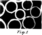
第1図は、支持体の主に外部表面にカップリングされたプロテインAリガンドの分布が一様ではないために望ましくない「ハロ効果」を示している、従来技術の方法で製造された誘導体化多孔質支持体の断面を示す比較用の蛍光顕微鏡写真である。
第2図は、支持体の全面にわたりカップリングされたプロテインAリガンドの分布が一様であるため官能効率が向上していることを示している、本発明に従い製造された誘導体化多孔質支持体の断面を示す蛍光顕微鏡写真である。
第3図は、支持体の主に外部表面にあるプロテインAに結合された免疫グロブリンの分布が一様ではないために官能効率について望ましくない「ハロ効果」を示している、第1図と同じ誘導体化多孔質支持体の断面を示す比較用の蛍光顕微鏡写真である。
第4図は、支持体の全面にわたりカップリングされたプロテインAリガンドに対する免疫グロブリンの結合が一様であるため官能効率が向上していることを示している、第2図と同じ誘導体化多孔質支持体の断面を示す蛍光顕微鏡写真である。
本発明の実施態様
多孔質支持体
本発明において使用が許容される多孔質支持体には、アフィニティークロマトグラフィー技法用に市販されているものが含まれる。多孔質支持体は、天然物であっても合成物であっても、また有機物であっても無機物であっても、多孔質構造を示し且つ水又は水溶液に不溶性であるならばいずれの多孔質固体であってもよい。多孔質構造を有する適当な固体は、平均直径が30ナノメートル以上で且つ0.1cm3/gを上回る孔体積を示す細孔を有する。平均細孔径は、細孔が大きいほど拡散に対して制限が少なくなるので50ナノメートル以上であることが好ましい。細孔を取り囲む表面積が大きくすることにより潜在的能力を高めるため、孔体積は0.5cm3/g以上であることが好ましい。
限定するものではないが、このような多孔質支持体の例として、多糖類、セルロース及びアガロースなどの天然修飾又は合成修飾された天然組成物が挙げられる。市販のアガロースの一例として、スェーデンのUppsalaのPharmacia ABから市販されているSepharose(商標)ビーズが挙げられる。このような多孔質支持体は、リガンドを共有結合固定化するため、臭化シアンやシアノ移動剤などの組成物で活性化する必要がある。このような活性化は、本発明の方法を使用する前に行う必要がある。
限定するものではないが、このような多孔質支持体の別の例として、アクリレート、メタクリレート、アクリルアミド、ビニル芳香族及びビニルアルコールの合成ホモポリマー及びコポリマーが挙げられる。このようなホモポリマー又はコポリマーは、官能価(例、アズラクトン、アルデヒド、等)を有するか、又はリガンドと迅速に直接共有反応して誘導体化支持体を形成させることができる官能価を提供するように修飾されることが望ましい。本発明の方法にはオキシラン又はエポキシド官能価は適していない。というのは、このような基はカップリング反応が迅速には起こらないか又は迅速なカップリングには不利に高いpHを必要とするからである。YarmushらのBiotech Adv.10,413−446(1992)を参照されたい。米国特許第4,775,714号明細書(Hermannら)の実施例5に記載されているように、カップリング反応時間は適当な振盪下、40℃で40時間であった。これは迅速な(すなわち、約4時間以内)カップリング反応とはいえない。
限定するものではないが、このような多孔質支持体の別の例として、多孔質ガラス、シリカ、アルミナ、酸化ジルコニウム及びその他の金属酸化物のような無機粒子が挙げられる。
多孔質支持体は膜、多孔質繊維、ウェブ又はビーズのような粒子であることができる。本発明に有用な多孔質支持体は、リガンドが支持体に共有結合でカップリングできるように反応性の多孔質粒子であることが好ましい。
本発明により使用される前に活性化が必要な上記支持体の他に、中間の活性化工程を必要としない共有結合反応性を示す表面を有する支持体もある。このような直接反応性多孔質支持体が粒子であることが好ましい。
本発明に有用な直接反応性多孔質粒子は、一般に広く2種類のタイプ:化学修飾無機粒子と有機ポリマー粒子、に分けられる。
例えば、無機粒子は、アルミナ、シリカ及びジルコニア;ガラスビーズ、ガラスバブル及び細孔を制御したガラス;等であることができる。これらの粒子は、反応性官能基を含むポリマー(通常は有機)による被覆などの方法によって、或いは反応性官能基を含む適当な試薬(例、アルコキシシランカップリング剤)との反応によって、化学修飾される。
有機粒子は、例えば、適当な反応性官能基を含むモノマーの重合若しくは共重合によって、上記のように粒子支持体を被覆することによって又は別のポリマーを化学修飾して反応性官能基を導入することによって調製された架橋又は非架橋ポリマーであることができる。
有用な粒子のいくつかは市販されているか又は当該技術分野で周知の技法によって調製することができる。それらのリストの一部を以下の表1に見ることができる。
望ましくは、本発明で有用な直接反応性粒子は、球形、規則形又は不規則形であることができる。反応性粒子の寸法は本発明の範囲内で幅広く変動することができ、またある程度は粒子の使用目的に依存する。
一般に、反応性粒子の寸法は平均径で0.1マイクロメートル〜5ミリメートルの範囲をとる。
直接的な共有結合反応であっても、臭化シアンのような組成物で間接的に活性化された場合であっても、本発明の目的に有用な共有結合反応性の官能基は一般に求電子性種として分類することができる。求核種(例、アミン、アルコール又はメルカプタン)と反応すると、付加反応又は置換反応(この場合には副生物分子が放出される)のいずれかによって、共有化学結合が形成する。付加型の反応が好ましい。
有用な直接及び間接の反応性官能基の例と、それらを含む市販の粒子の例を表1に記載する。

本発明ではアズラクトン官能性の反応性粒子が特に好ましいが、これはこのような粒子は表1に示した市販の反応性官能基よりも良好にリガンドと迅速且つ直接に共有結合でカップリングするからである。さらに、このようなアズラクトン官能基はリガンドとの共有結合カップリング前にはかなり安定である。さらに、アズラクトン官能基とリガンドとの共有結合カップリングは副生物分子の置換を全く伴わないので、リガンドとの共有結合カップリング後に複合体製品を精製するという望ましくない工程が回避される。
また、アズラクトン官能基は、プロテインAなどの生物学的活性物質との共有結合カップリング能が高いことが知られている。さらに、プロテインAとの共有結合カップリング能がこのように高いことはまた、カップリングされたリガンドとしてのプロテインAの特異的結合生物活性をも高くする。こうして、本発明においては、アズラクトン官能性の反応性粒子を使用することが特に好ましい。
アズラクトン官能性ポリマー粒子は、例えば、本明細書では参照することにより取り入れることとする米国特許第4,737,560号及び同第4,871,824号明細書に記載されているように、(メト)アクリロイルアミノ酸とその他各種のラジカル重合性コモノマーとを共重合させた後に環化剤を反応させる方法、又は同様に本明細書では参照することにより取り入れることとする欧州特許第0 392 735号公報に記載されているように、アルケニルアズラクトンと他のコモノマーとを共重合させる方法によって製造することができる。また、同様に上記欧州特許第0 392 735号公報に記載されているように、アズラクトン官能ポリマーを有機又は無機粒子表面に溶液コーティングする方法によってアズラクトン官能性粒子を製造することもできる。
アズラクトン官能性の反応性粒子は、米国特許第5,013,795号明細書及び欧州特許第0 392 783号公報に記載されているように、アズラクトングラフトコポリマーから製造することもできる。
アズラクトン官能性粒子の粒径は約0.1〜1,000マイクロメートル、好ましくは0.5〜250マイクロメートルであることができる。乾燥したアズラクトン官能性粒子の平均孔径は約1〜約300ナノメートル、好ましくは5〜約200ナノメートルであることができる。アズラクトン官能性粒子の平均孔体積は粒子1g当たり1.0cm3以上であることができる。径が50〜80マイクロメートルである粒子では、1.2cm3/g以上の孔体積は粒子体積の約60%に当たる孔体積を提供する。同じ粒子において、その表面積は50m2/g以上となる。このように、アズラクトン官能性粒子の内部には、本発明により共有結合固定化に利用できる実質的な表面積が存在する。
本発明に有用な多孔質支持体は、St.Paul,MNのMinnesota Mining and Manufacturing Companyから市販されているEmphaze(商標)多孔質アズラクトン官能性活性化アフィニティークロマトグラフィービーズであることが最も好ましい。
共有結合固定化のためのリガンド
上記のように、多孔質支持体上の反応性官能基は求電子性種であることが望ましい。このため、直接的共有結合固定化のためには、本発明に有用なリガンドは求核性種を含有する。
限定するものではないが、リガンド官能基の例として、第一アミン、第二アミン、アルコール及びメルカプタンが挙げられる。これらの中ではアミン官能性リガンドが特に好ましい。
アダクト複合体の調製に有用なリガンドもまた本発明の範囲内で様々なものを採用することができる。リガンドの選定は、誘導体化された多孔質支持体の所期の末端用途に基づいて行われることが好ましい。
本発明の方法に従いリガンドをカップリングさせた後は、吸着、複合化、触媒又は試薬末端用途など、官能効率の高い生物学的又は化学的相互作用にこれらのリガンドを利用することができる。
誘導体化された多孔質支持体は、吸着剤として、錯生成剤として、触媒として、試薬として、酵素や他のタンパク質を担持する支持体として、そしてクロマトグラフィー製品として有用である。
本発明の好ましい態様では、共有結合固定化に望まれるリガンドは求核性官能基を有する生物学的活性物質又は化合物である。限定するものではないが、生物学的活性物質の例として、生物学的、免疫化学的、生物学的又は薬理的に活性な物質が挙げられる。生物学的活性物質の例として、タンパク質、ペプチド、ポリペプチド、抗体(モノクローナル又はポリクローナル)、抗原性物質、酵素、コファクター、インヒビター、レクチン、ホルモン、レセプター、凝集因子、アミノ酸、ヒストン、ビタミン、薬物、細胞表面マーカー及びこれらと相互作用する物質が挙げられる。
生物学的活性物質のうち、共有結合固定化に望ましいものはタンパク質、酵素及び抗原性物質である。限定するものではないが、タンパク質、酵素及び抗原性物質の例として、天然及び組換えのプロテインA(Prot A)、免疫グロブリン、例えば、ラット(rIg)、ヒト(hIg)、ウシ(bIg)、ウサギ(rIg)及びマウス(mIg)、コンカナバリンA(ConA)、ウシ血清アルブミン(BSA)、チノグロブリン(TG)、アポフェリチン(Af)、リソチーム(Ly)、炭酸脱水酵素(CA)並びに細菌抗原(BA)が挙げられる。限定するものではないが、凝集因子の例として、プロテインC(Prot C)、ヘパリン、フィブリノーゲン及びトロンビンが挙げられる。
カップリングされたタンパク質、酵素及び抗原性物質の使用法については欧州特許第0 392 735号公報に記載されている。凝集因子の使用法には、酵素前駆体を活性タンパク質へ活性化すること、例えば、カップリングされたトロンビンによってプロテインCを活性プロテインCへ活性化することが含まれる。
現在好ましい生物学的活性物質は抗体とProt Aである。
別法として、本発明の誘導体化された多孔質支持体は、カップリングされた酵素を含むことにより該酵素によって認識される物質の化学変換を触媒することができる。また、カップリングされた抗原性物質を含む誘導体化多孔質支持体を利用して、複雑な生物学的流体から対応する抗体をアフィニティー精製することができる。
別の例として、本発明の方法に従い内部及び外部表面にカップリングされたプロテインAを有する多孔質粒子によって、アフィニティー分離プロセスのために免疫グロブリンGのような生物学的活性物質を吸着することができる。別の例では、誘導体化多孔質支持体を用いて、抗体を固定化すること、又は免疫診断若しくはウェスタンブロッティングを行うことができる。
現在好ましいアズラクトン官能基は、アミン、チオール及びアルコールによって求核攻撃をされる。こうして、アズラクトン官能性多孔質支持体における共有結合固定化の候補として、少なくとも一つのアミン、チオール又はアルコール基を表面に有するリガンドが挙げられる。
本発明の方法は、多孔質支持体中への拡散が制限されることによって若しくは密にカップリングされたリガンドへの結合の立体効果によって又はその両方によって別法では影響を受ける大きなリゲートを結合させるのに特に有用である。
限定するものではないが、大きなリゲートとは、細孔の開口部表面においてカップリングされているリガンドに結合されたリゲートを既に有する細孔を横切ることができないようなリゲートとして特徴付けることができる。このため、誘導体化された支持体の内部にカップリングされたリガンドは、リガンドがカップリングされている細孔の開口部表面がリゲートを結合し且つ多孔質支持体内部の誘導体化表面への通路を遮断している場合には、結合の際の利用が不十分であるか恐らくは利用されないままとなる。
また、大きなリゲートとは、新たに別のリゲートの結合を立体効果によって不利に変えてしまうようなリゲートとして特徴付けることもできる。こうして、リゲートの結合は起こっても非効率的に(例えば、カップリングされたリガンドの廃棄が生じるように)起こる可能性がある。そのリガンドがより均一に空間的に分布していたならば、支持体は、より高い結合能及びより高い官能効率の両方を収容することができる。
リゲートの「大きさ」は、多孔質支持体の孔径、カップリングされたリガンドの反応性及び密度、並びにその他の要因の相対的な結果である。限定するものではないが、大きなリゲートの例として、ペプチド、ポリペプチド、アミノ酸及び薬物のような小さな分子を除く、先にリガンド候補として示した生物学的活性物質が挙げられる。
共有結合固定化法
本発明の方法は二つの段階を含む。第一の段階はリガンドを多孔質支持体の表面付近に運搬することである。第二の段階は、そのリガンドを多孔質支持体の表面に迅速に共有結合固定化、すなわち約4時間以内でカップリングさせることである。本発明の利点は、本発明の方法に従いリガンドをカップリングさせることによって達成される優れた官能効率である。
第一の段階は、単一の反応容器において、支持体に対するリガンドのカップリング反応を十分に抑制すると同時に、リガンドが多孔質支持体中に入りその内部に拡散する速度を反応速度に対して相対的に高める条件を採用する。こうして、第一段階はリガンドの拡散速度に対してリガンドのカップリング速度を抑制する。
限定するものではないが、反応容器中でカップリング反応を抑制する条件の例として、pH、イオン強度、温度及びカップリング競争体の組合せの一つ以上において特定の範囲の条件を採用することが挙げられる。カップリングを抑制する条件として現在好ましいものは、リガンドと多孔質支持体がカップリングのために混合される、又はカップリング条件に置けれる、反応溶液のpHの制御及び/又はイオン強度の制御である。pHは約3〜約7の範囲で制御することができる。この範囲のpHでは、リガンド上の求核性基と多孔質支持体表面の求電子性種との反応が最小限に抑えられる拡散条件が得られる。
より好ましくは、拡散時のpHは、求電子性官能基の加水分解に対する安定性はpHの増加と共に高くなるので、約4〜約6とすべきである。
最も好ましくは、求電子性官能基が起こりうる加水分解に対してさらに安定となるため、拡散時のpHは約5とする。
反応溶液のpHが約3〜約7の範囲にある場合、その他の反応条件は固定化技術分野で常用されている条件とすることができる。換言すれば、pHの調整だけで十分にカップリング条件を抑制すると同時にリガンドが多孔質支持体の中に入りその内部で拡散する速度を反応速度に対して相対的に高くすることができる。
ポリアニオン塩(例、硫酸塩、リン酸塩、クエン酸塩、酒石酸塩、等)の存在によるイオン強度は、拡散の際にカップリング条件の抑制を最小限に抑える。拡散時の反応溶液中のポリアニオン塩のモル濃度は約0.01〜約0.4Mであることが好ましい。
カップリング競争体(以下、詳細に記載する)は、官能基との求核反応に対して競争するであろうが、0〜2モル濃度とすることができる。
拡散時の反応溶液の温度は、リガンドのカップリング反応速度を遅らせるように制御する必要がある。好ましくは、その温度は水溶液の凝固点付近から約25℃までとすることができる。
反応溶液は緩衝剤を含むことが普通である。水性媒体用の緩衝剤にはアセテート、ホスフェート、ピロホスフェート、ボレート及びその他当業者には周知の塩が含まれ、こうした緩衝剤の具体例についてはGoodらのBioche mistry,5,(1966)p.467等に記載されている。
水性媒体中での緩衝剤の濃度は、カップリング用に選定された生物学的活性物質の濃度及び反応溶液のイオン強度に影響を及ぼしうる他の任意成分の濃度に依存して、約10mM〜約750mM、望ましくは約50mM〜約200mMの範囲をとることができる。
第一段階の期間は、多孔質支持体の全ての表面付近にリガンドが確実に拡散するよう十分に長くとるべきである。この時間の長さは、用いた多孔質支持体の種類、多孔質支持体の孔径及び孔体積、多孔質支持体の孔体積内を拡散するリガンドの大きさ及びコンフォメーション並びにその他の物理的検討事項によって変わる場合がある。一般に、許容できる拡散を達成するためには5分以上の拡散時間で十分である。少なくとも10分間は拡散を継続させて拡散を向上させることが望ましい。支持体の形状寸法が小さい場合にはほとんどの多孔質支持体において拡散を15分以上続けて確実に拡散を行う。支持体の形状寸法がより大きな場合には、支持体の平均厚さを興味のあるリガンドの水溶液中での拡散率で割った値である特性拡散時間以上に拡散を継続する。
第二の段階は、多孔質支持体の表面にリガンドを迅速且つ確実にカップリングさせるものである。こうして、第二段階での反応条件は第一段階の条件からは急激に変更され、また好ましくは反応溶液へカップリング剤を添加することなく実施される。「カップリング剤」とは、リガンド又は支持体のいずれかと反応してリガンドの支持体へのカップリングを促進する試薬を意味し、カップリング競争体、すなわち多孔質支持体上の反応部位に対して競争する試薬を意味するものではない。
カップリング速度は、カップリング速度定数、リガンド濃度、支持体の単位面積当たりの官能基の反応性、リガンドの支持体中への拡散速度及び温度の関数である。
反応溶液のpHを使用して拡散条件を確立した場合、pHの変更が第二段階を構成する。
反応溶液のpHを求核性リガンドのあるpKaの範囲内のpH、通常は約7〜約10に変更すると、カップリング条件が高められる。この範囲ではリガンドが多孔質支持体へ迅速且つ確実にカップリングされる。
この範囲のpHでは、多孔質支持体表面の求電子性基とリガンド上の求核性基との反応を最大にするカップリング条件が得られる。
反応溶液のカップリングのためのpHを約7.5〜約9.5にし、加水分解又は溶媒との別の反応を減少させることがより好ましい。
最も好ましくは、反応溶液のカップリングのためのpHを約8.5にし、リガンド、特にタンパク質性リガンドの生物学的活性を維持することである。
拡散段階からカップリング段階への反応条件の変更は、pHの変更に限定してもよいし、また他の変更を伴うことも可能である。
pHの変更の他に、又はpHの変更の代わりに、反応溶液のイオン強度を変化させることで、多孔質支持体にカップリングされたリガンドの官能効率を高めることができる。イオン強度の変化量は、リガンドのカップリングを向上させ且つカップリングされたリガンドの官能効率を向上させるに十分な量にすることができる。
拡散段階からカップリング段階へのイオン強度の変化量は約0.5M〜約1.5Mの範囲とすることができる。好ましくは、イオン強度の変化量を約0.6M〜約1.2Mとしてリガンド、特にタンパク質性リガンドの溶解度を維持する。最も好ましくは、カップリング段階における溶液のイオン強度の変化を約1.0〜1.2Mとすることができる。
イオン強度の変化は1種以上のポリアニオン塩を反応溶液へ添加することによって誘発させる。好適なポリアニオン塩が、無機物、有機物共に、国際特許出願公開PCT WO 92/07879号公報及び米国特許第5,200,471号明細書(Colemanら)に記載されている。
好ましいアズラクトン官能性支持体に関するColemanらの説明にもあるように、水性緩衝媒体中で無機ポリアニオン塩を用いてアズラクトン官能性ポリマー支持体へタンパク質を共有結合で固定化すると、無機モノアニオン塩(例えば、NaCl)を用いた場合と比較して、生物学的活性物質の結合特異的生物活性が二倍を上回る。
この固定化効率の向上は非常に迅速且つ簡単に達成される。無機ポリアニオン塩を高濃度で使用しても、周囲温度で達成可能な非常に迅速な共有結合固定化など、アズラクトン官能性ポリマー支持体を使用するその他の貴重な特徴を消失させることはない。
無機ポリアニオン塩の中では、水性媒体中の無機ポリアニオンのモル濃度に対する結合特異的生物活性が増加するので、硫酸塩が望ましい。無機ポリアニオン塩を使用する場合、約pH4〜約pH9に緩衝化された水性媒体中で(金属カオチンによって影響されることのない活性を有する)タンパク質をカップリングする際に使用される現在好ましい塩は、Na2SO4である。硫酸塩がリン酸塩よりも好ましい理由は、アズラクトン官能性ポリマー支持体上に同等密度のリガンドをカップリングさせるのに必要なモル濃度がリン酸塩よりも硫酸塩の方が少ないからである。この利点の証拠についてはColemanらのJ.Chromat ogr.,512(1990)345−363に見ることができる。
好ましくは、有機ポリ酸とその塩は、無機ポリアニオン塩よりも、好ましいアズラクトン官能性ポリマー支持体上にリガンドをさらに生産性よく且つ効率的にカップリングさせることができる。有機ポリアニオン塩は、ほとんどの共有結合固定化が行われるpH7〜pH9のpH範囲において且つ本発明の拡散段階からカップリング段階へのpH変更の好ましい範囲において、無機ポリアニオン塩よりもイオン性が一貫している。このため、有機ポリアニオン塩はポリアニオン1モル当たりのイオン強度が高い。その結果、共有結合固定化に必要な有機塩のモル数は少なくて済む場合が多い。さらに、共有結合固定化に用いられる緩衝化水性媒体において十分な可溶性を示す有機ポリアニオン塩は無機ポリアニオン塩よりも多種多様である。このため、現在のところは有機ポリアニオン塩の方が無機ポリアニオン塩よりも好ましい。
有機ポリ酸候補の中では、二酸、三酸及び四酸又はそれらの塩が望ましい。限定するものではないが、このような酸の例として、マロネート、マレート及びタルトレート系二酸のアルカリ金属塩、シトレート系三酸及びニトリロ三酢酸(NTA)のアルカリ金属塩、並びにエチレンジアミン四酢酸(EDTA)系四酸のアルカリ金属塩が挙げられる。現在好ましい有機ポリアニオン塩はクエン酸ナトリウムである。
pHの変更及び/又はイオン強度の変更の他に、或いはpHの変更及び/又はイオン強度の変更の代わりに、反応溶液へカップリング競争体を添加することにより、多孔質支持体にカップリングされたリガンドの官能効率を高めることもできる。
カップリング競争体は、カップリングされたリガンドの官能効率が高くなるようにリガンドの結合特異的生物活性を向上させるに十分な量で(しかし、カップリングされるリガンドの量を実質的に減少させる量ではない)、添加することができる。
カップリング競争体の種類は、多孔質支持体にカップリングすべきリガンドの性質によって変わることがある。多孔質支持体とリガンドとの(とりわけ、反応溶液のpH、リガンド濃度、温度、イオン強度によって影響を受ける)反応速度論によって、使用するカップリング競争体の量や種類が決まる。
特定の理論に限定するものではないが、カップリング競争体は、これがなければリガンドがカップリングする多孔質支持体表面の反応性部位に対して競争する。反応性部位の数が減少するため、リガンドがそのコンフォメーションを変えてその生物活性を低下又は消失するようなカップリングをする可能性が制限されうる。意外なことに、カップリング競争体は、カップリングのための反応性部位を過剰に排除することなく反応性部位を散在させることによって、カップリングされたリガンドの官能効率を高めるものと考えられる。このことはまた、カップリングされたリガンドをより均一又は有効に分布させる傾向もある。
好適なアズラクトン官能性多孔質支持体を使用する場合、カップリング競争体はアズラクトンクエンチャーとなる。好適なアズラクトンクエンチャーについても国際特許出願公開PCT WO 92/07879号公報及び米国特許第5,200,471号明細書(Colemanら)に記載されている。
限定するものではないが、アズラクトンクエンチャーの例として、エタノールアミン、ウシ血清アルブミン、カゼイン溶解物、ヒドロキシルアミン、エチルアミン、水酸化アンモニウム、グリシン、硫酸アンモニウム、ブチルアミン、グリシンアミド、TRIS、ゼラチン、リソチーム、非脂肪ドライミルク、β−メルカプトエタノール、メルカプトエチルエーテル、ジチオスレイトール、グルタチオン、アルギニン、グアニジン、リジン、ジアミン及びこれらの混合物が挙げられる。これらの非限定例の中には、望まれる固定化とは「無関係な」タンパク質を含むものもある。
本発明の方法の第二段階でpHの変更及び/又はイオン強度の変更と共に添加されるアズラクトンクエンチャーの濃度は、約0.1M〜約10Mの範囲をとることができる。この範囲は約0.5M〜約2Mの範囲にあることが望ましい。アズラクトンクエンチャーとしてエタノールアミンを使用する場合、その濃度は約0.1M〜約1Mとすることができる。アズラクトンクエンチャーとして現在好ましいエタノールアミン濃度は約0.5M〜約1Mである。
pHの変更、イオン強度の変更若しくはカップリング競争体の添加又はこれらの組合せの他に、或いはpHの変更、イオン強度の変更若しくはカップリング競争体の添加又はこれらの組合せの代わりに、反応溶液の温度上昇を利用することにより、多孔質支持体にカップリングされたリガンドの官能効率を高めることもできる。この温度変化量は、反応速度が迅速である限りリガンドのカップリングを促進し、且つカップリングされたリガンドの官能効率を高めるに十分な量とすることができる。
温度上昇は約10℃〜約35℃、好ましくは約20℃〜約30℃とすることができる。というのは、この範囲の温度上昇であればリガンドの生物活性に悪影響を及ぼすことなく支持体に対するリガンドの反応性が高くなるからである。
特定の理論に限定されるものではないが、pHの上昇、イオン強度の増加、温度の上昇又は反応溶液へのカップリング競争体の添加のうちの一つ又は二つ以上によって、リガンドの結合特異的生物活性が保持されるようにリガンドがカップリングする。この保持は、カップリングされたリガンドに結合しようとするリゲートの立体効果又は拡散の制限を最小限に抑えるようにリガンドがより均一又は有効に分布することによる。このため、生物活性のないカップリングされたリガンドの数が最小限に抑えられ且つカップリングされたリガンドの量が最大になることによって、得られたカップリングされたリガンドの官能効率が向上する。
第二段階におけるカップリングは迅速且つ確実である。この段階の所要時間は0.5〜4時間の範囲とすることができる。
カップリング反応は、多孔質支持体上に残存する実質的にすべての反応性部位にカップリングするクエンチャーを過剰に添加して残存反応性部位を消し去ることによって完了する。
多孔質支持体がアズラクトン官能性である場合、クエンチャーとしては先に記載したアズラクトンクエンチャーのいずれのものでも過剰濃度で使用することができる。
特定の理論に限定するものではないが、本発明の方法は、その拡散段階及びカップリング段階の際にティーレモジュラス(Thiele modulus)を制御する利点を利用するものである。
ティーレモジュラスは、拡散速度に対する反応速度の比として無次元項で一般に表され、以下の方程式(1)で表すことができる。
φ=Rp(ka/De)1/2 (1)
上式中、Rpはリガンドの反応性であり、kは一次速度定数であり、aは内部表面積であり、そしてDeはリガンドの有効拡散率である。
ティーレモジュラスは以下の方程式(2)でも表される。
φ=1/2(dp)(Vmaxρ/KmDe)1/2 (2)
上式中、dpは担体の直径であり、Vmaxは活量であり、ρは密度であり、Kmはカップリングの速度論定数であり、そして、Deは有効拡散率である。上記のBorchertらの刊行物を参照されたい。
本発明の第一段階は、カップリング条件を抑制してカップリング速度に対する拡散の相対速度を高める段階である。この第一段階は、第二段階と比較して低いティーレモジュラス(拡散速度が反応速度よりも認知できる程度に高い)を利用する。反対に、第二段階は第一段階よりも高いティーレモジュラス(反応速度が拡散速度よりも認知できる程度に高い)を利用する。
こうして、別の表現をすれば、本発明の方法は、比較的低いティーレモジュラスを使用する条件でより均一に空間分布させるための透過段階に続き、比較的高いティーレモジュラスを使用する条件で迅速にカップリングさせるためのカップリング段階を実施する方法である。
比較的低いティーレモジュラス条件は、低いpH、低いイオン強度反応媒体、低温、カップリング競争体及びこれらの組合せで達成することができる。
比較的高いティーレモジュラス条件は、高いpH、高いイオン強度反応媒体、高温、カップリング競争体及びこれらの組合せで達成することができる。
リガンドの拡散及びカップリングのためのティーレモジュラスに対するこれらの因子の影響は、選ばれたリガンドに依存する。例えば、アズラクトン官能性ビーズにカップリングされたプロテインAの官能効率は、イオン強度の増加、カップリング競争体の添加、これら二つの因子の組合せを利用してカップリング段階を開始するティーレモジュラスの変更によって高めることができる。しかしながら、pHを変更するだけで、多孔質アズラクトン官能性ビーズ上の抗プロテインC抗体の官能効率は向上する。
溶液中のいずれのリガンド濃度においても、第一段階と第二段階との間でティーレモジュラスの差を制御した際の結果の一つは、多孔質支持体内部の表面にある官能基へリガンドをカップリングさせるのに必要な活性化エネルギーの変化である。ティーレモジュラスの低い条件はカップリング反応に必要な活性化エネルギーを上昇させるが、ティーレモジュラスの高い条件はカップリング反応に必要な活性化エネルギーを低下させる。
拡散段階後にカップリング段階を開始するためにティーレモジュラス条件の段階変化を急激に行うことは、リガンドの支持体からの逆拡散を最小限にすることになる。これにより、拡散段階の際に分散された空間分布の大部分が維持される。さらに、多孔質支持体表面にある官能基の反応速度論によって、支持体内部でより均一に空間分布された望ましいリガンドが失われる前にカップリング反応の迅速性が高められる。カップリング反応の迅速性は約4時間未満、好ましくは約1時間未満である。
本発明の有用性
本発明の方法は、カップリングされたリガンドの相当部分を不活性化させてしまう表面での過密状態を回避するように生物学的活性物質を多孔質支持体上に固定化する方法を提供するものである。誘導体化された支持体にカップリングされたリガンドは分子的に散在しており、その官能効率は、表面でより高密度にカップリングされたリガンドよりも有意に高くなる。
第1図と第2図は本発明の利点を直接に比較するものである。
第1図は、従来技術で採用されており以下の比較例6で記載した方法により製造された誘導体化多孔質支持体の断面を示す比較用の蛍光顕微鏡写真である。この方法では、拡散反応後にカップリング反応を行う2段階反応は行われていない。この断面図は、支持体の外部表面にカップリングされた不均一に分布しているリガンドを示している。これは本発明の方法が回避する過密状態を示している。
第2図は、本発明の方法であって以下の実施例7に従い製造された誘導体化多孔質支持体の断面を示す蛍光顕微鏡写真である。この方法では、拡散段階とカップリング段階の間でpHを変化させている。ビーズの多孔質断面全体にわたり、支持体の全表面にリガンドが有意に均一な分布をしてカップリングされていることがわかる。外部表面にカップリングされているリガンドが分子的に散在しており、しかもカップリングされたリガンドが多孔質支持体の全表面にわたり分布しているため、最適なカップリングが達成されている。
第1図と第2図の比較からわかるように、リガンドの30%以上が支持体の幾何中心から70%以内に含まれる内部表面にカップリングされるように、カップリングされたリガンドはより均一に空間分布されている。このように、本発明の方法は、多孔質支持体に対するリガンドのカップリングの空間分布をより均一に制御することができる。
本発明の誘導体化多孔質支持体は、その外部表面へのリガンドのカップリングがまばらになっており且つその内部表面へのリガンドのカップリングが増大されている。
本発明の方法を採用してカップリングされたリガンドの官能効率は、従来法を用いて得られる官能効率の1.1〜10倍程度上昇することができる。このように、意外にも優れた誘導体化支持体が達成される。
第3図は、従来技術で採用されており以下の比較例8で記載した方法により製造された第1図に示した誘導体化多孔質支持体に免疫グロブリンを結合させた状態での断面を示す比較用の蛍光顕微鏡写真である。この断面は、支持体の外部表面にカップリングされたリガンドに結合しているリゲートの一様でない分布を示している。このことは、不十分な官能効率の「ハロ効果」を示す拡散の制限及び立体効果の証拠である。
第4図は、本発明の方法であって以下の実施例9に従い製造された第2図に示した誘導体化多孔質支持体の断面を示す蛍光顕微鏡写真である。この方法では、リゲートがビーズ全体にわたりより均一に結合しており、そして拡散の制限及び立体効果が排除されている。ビーズの多孔質断面全体にわたり、支持体の全表面にカップリングされたリガンドにリゲートが有意に均一な分布をして結合されていることがわかる。多孔質支持体の全表面にわたりカップリングされたリガンドの上にリゲートが分布して結合されているため、最適な官能効率が達成される。
本発明の範囲をさらに理解できるように、以下に実施例を記載する。
実施例
比較例1
この例は、従来技術の1段階法によってプロテインAをEmphaze(商標)Biosupport Medium AB1にカップリングする方法を説明するものである。乾燥Emphaze(商標)ビーズ(Minnesota Mining and Manufacturing Company,St.Paul,MN)をそれぞれ50ミリグラム含有する三つの15mlスクリューキャップ式ポリプロピレンチューブへ天然プロテインA(Fermentech,Ltd.,Edinburgh,UK)を1.2MのNa2SO4、0.1MのNaH2PO4(pH7.5)の中に1.6ミリグラム/ミリリットルで含む溶液2.5ミリリットルを入れて混合することにより乾燥ビーズをすべて湿潤させた。次いで、その反応混合物を室温で全75分間、転倒型回転器で攪拌した。1200×gで10分間遠心分離後、その上澄液を取り出して残存しているプロテインAを比色定量分析法(Pierce Chemical Company,Rockford,ILのBCA法)によって分析した。カップリングしたプロテインAの量とそのカップリング効率を差計算によって決定した。カップリングの結果を表Aに示す。これらのビーズを、pH9.0で約18時間(一晩)かけて3Mエタノールアミン4.0ミリリットルでクエンチした。次いで、三つを一緒にしたビーズを、焼結ガラスフリット漏斗(多孔度D)の上で、リン酸緩衝化塩化ナトリウム水溶液(PBS:0.025MNaH2PO4,0.15M NaCl,pH7.4)、0.2M酢酸ナトリウムpH5.0、0.5M重炭酸ナトリウムpH8.5及びPBSを各液20〜30体積分用いて逐次洗浄した。
次いで、洗浄後のビーズを3×50mmのオムニガラスカラムに充填し、そして試験液として精製ヒトIgG(Sigma Chemical Company,St.Louis,MO)を用いてクロマトグラフィーを行って、免疫グロブリンの結合能について試験した。総量48ミリグラム(0.01M NaH2PO4 pH7.5中1ミリリットル当たり3ミリグラムのものを16ミリリットル)を0.57ミリリットル/分の流速で装填した後、6.8ミリリットルのローディングバッファー、6.8ミリリットルの2M NaCl,0.01M NaH2PO4 pH7.5、4.6ミリリットルのローディングバッファーで洗浄し、そして4.6ミリリットルの0.1Mグリシン,2%酢酸pH2.2で結合IgGを溶離させた。溶離画分を集め、存在するIgGの量を280nmの吸光度で測定した。これらのビーズの結果を表Aに示す。
比較例2
この例は、従来技術に従い比較例1と同様にしてプロテインAをEmphaze(商標)ビーズにカップリングするものであるが、但し、カップリング溶液は、硫酸ナトリウム及びリン酸緩衝液(pH7.5)の他に、カップリング競争体、0.5Mトリス(ヒドロキシメチル)アミノメタン(TRIS)を含有するものとした。
(比較例1に記載したように)カップリング反応とIgG結合能を評価した結果を表Aに示す。
実施例3
この例は、比較例1と直接比較するものとして、プロテインAをEmphaze(商標)ビーズにカップリングする本発明の2段階法を説明するものである。こうして、最終溶液条件は比較例1の条件と同じであるが、第一段階における反応混合物のイオン強度に違いがある。第二段階において、反応混合物のイオン強度を増加させる。15ミリリットルのスクリューキャップ式ポリプロピレンチューブに乾燥Emphaze(商標)ビーズ50ミリグラムを含む三つの試料に、0.5ミリリットルのプロテインA溶液(0.05M NaH2PO4 1ミリリットル当たり8ミリグラム)を室温で加え、そのビーズを15分間水和させた。次いで、第二段階において、2.0ミリリットルの1.5M Na2SO4,0.125M NaH2SO4(pH7.5)を加え、実施例1と同様の攪拌をさらに60分間継続した。比較例1と同様にカップリング反応とIgG結合能を評価し、その結果を表Aに示す。
実施例4
この例は、比較例2と直接比較するものとして、プロテインAをEmphaze(商標)ビーズにカップリングする本発明の2段階法を説明するものである。第二段階において、反応混合物のイオン強度を増加させ、アズラクトンを不活化するカップリング競争体を添加し、そしてpHを上昇させる。この方法は実施例3の方法と似ているが、プロテインAは0.05M酢酸ナトリウム(pH5.0)に含有させ、また第二段階で添加した溶液は硫酸ナトリウム及びリン酸緩衝液(pH7.5)の他に0.5M TRISを含有するものとした。比較例1と同様にプロテインAのカップリング反応とIgG結合能を測定し、その結果を表Aに示す。
比較例5
この例は、プロテインAをEmphaze(商標)ビーズにカップリングする際に、第一段階と第二段階との間でpHだけを変化させた本発明の2段階法を採用したものである。これが比較例である理由は、実施例3及び実施例4とは異なるpHを使用したこと、及び第一段階から第二段階にかけてpHを変化させただけではプロテインAには作用しないことである。この方法は実施例3の方法と似ているが、プロテインAは0.05M酢酸ナトリウム(pH5.0)に含有させ、また第二段階は0.125Mホウ酸溶液(pH9.5)を使用するものとした。比較例1と同様にプロテインAのカップリング反応を測定し、その結果を表Aに示す。
比較例6及び実施例7
これらの例は、比較例6の従来技術による方法と実施例7の本発明による方法との異なるカップリング法から得られたタンパク質の分布を測定するために、蛍光標識されたプロテインAをカップリングしたEmphaze(商標)ビーズの調製を説明するものである。プロテインAをJ.Immunological Methods 50,193−204(1982)に記載のTitusらの方法によってTexas Red(Sigma Chemical Company)と組み合わせて、PBS中の5mg/ml溶液を得た。これを未標識のプロテインAと混合して最終濃度を25mg/mlとした。次いで、この溶液を希釈して、比較例6には比較例1に記載の方法に従い、また実施例7には実施例4に記載の方法に従い、20ミリグラムのビーズと全部で1.0ミリリットルの溶液を用いてEmphaze(商標)ビーズへカップリングした。(ビーズ1ml当たりに正規化した)カップリングしたプロテインAの量及びカップリング効率は、比較例1及び実施例4で未標識プロテインAについて得られた値と本質的に同じであることがわかった。
次いで、これらビーズの試料をJB4−Plus埋封媒体(Polysciences,Warrington,PA)中に流延し、そしてガラスナイフで厚さ約4μmのセクションに区分した。次いで、これらを搭載し、倍率500×で蛍光条件下で検査し、そして写真撮影した。第1図及び第2図は、Emphaze(商標)ビーズへカップリングされたプロテインAの空間分布が顕著に異なることを示している。どちらの調製物も全プロテインA含有量は同じであるため、第1図(比較例6のビーズ)のプロテインAの「ハロ」形分布は、第2図(実施例7のビーズ)のより均一な分布よりもリガンドが著しく狭い領域に濃縮されていることを示唆している。プロテインAは、ビーズの幾何中心から35%以内に含まれるビーズの内部表面に、カップリングされたプロテインAの量の70%以上がカップリングされるように、ビーズにカップリングされている。
比較例8及び実施例9
これらの例は、プロテインAをカップリングさせたEmphaze(商標)ビーズに結合した蛍光標識ヒトIgGの空間分布を測定する方法を説明するものである。比較例1及び実施例4のビーズ約100マイクロリットルのアリコートをFITC標識ヒトIgG(Sigma Chemical社)、0.5ミリリットルのPBS中約1.5ミリグラムと共に室温で一晩インキュベートした。次いで、ビーズ試料をそれぞれ2.0ミリリットルのPBSで五回洗浄し、結合しなかった抗体を除去した。
次いで、これらのビーズを比較例6及び実施例7に記載のように顕微鏡観察用に調製した。これらのビーズの光学顕微鏡写真を第3図及び第4図に見ることができる。ここで、結合したリゲートの分布は第1図及び第2図のカップリングされたリガンドと同様である。このことは、第2図で認められたより均一に分布したプロテインAリガンドが、「ハロ」分布したプロテインA(第1図及び第3図)よりも均一に分布した結合IgGリゲート(第4図)並びに比較例1のビーズの低いIgG結合能よりも高い結合能をもたらすことを示唆している。このことはまた、本発明のカップリング法が、得られる生物学的活性支持体の官能効率を決めることができるリガンドの空間分布に影響を与えられることを確認するものでもある。こうして、この例では、支持体は実質的に球形粒子であり、そしてその粒子の全半径の70%に当たる半径以内に含まれる内部表面にカップリングされたリガンドにリゲートが結合されている。リガンドは、多孔質支持体の内部表面へのカップリングされたリガンドの透過率が約70%になるように多孔質ビーズにカップリングされている。
比較例10
この例は、従来技術の1段階法を採用してプロテインAを臭化シアンで活性化したSepharose(商標)アガロースへカップリングする方法を説明するものである。CNBr−活性化Sepharose(商標)4B(Pharmacia LKB,Biotechnology AB,Uppsala,Sweden)は、製造業者の指示書に従い調製し、ゲル0.6ミリリットルに相当するスラリーのアリコートを15ミリリットルのスクリューキャップ式ポリプロピレンチューブに入れた。上澄みの緩衝液を除去した後、0.5M NaHCO3 pH8.5中その1ミリリットル当たり1.0ミリグラムのプロテインAを含む溶液3.75ミリリットルを加え、そしてその混合物を転倒式回転器で室温で全75分間攪拌した。その後、比較例1と同様にそのスラリーを1200×gで5分間遠心分離し、そしてその上澄液を分離してプロテインAのカップリング量を定量した。結果を表Bに示す。そのゲルを4.0ミリリットルの3Mエタノールアミン(pH9.0)に再懸濁させ、室温で一晩(約18時間)攪拌した。その後、そのゲルをPBS、1M NaCl PBS溶液及びPBSを各8ミリリットル使用して逐次洗浄した。
次いで、これらのビーズを比較例1の方法によってカップリングしたプロテインAの結合能について評価した。結果を表Bに示す。
比較例11
この例は、第一段階から第二段階にかけてpHのみを調整した2段階法を採用して、臭化シアンで活性化したSepharose(商標)へプロテインAをカップリングする方法を説明するものである。比較例10に記載したようにCNBr−活性化Sepharose(商標)4Bのスラリーアリコート(0.6ミリリットル)を調製し、そして1ミリリットル当たり5.0ミリグラムのプロテインAを含む溶液0.75ミリリットル及び0.05M酢酸ナトリウム(pH5.0)と4℃で15分間反応させた。第二段階において3.0ミリリットルの0.625M重炭酸ナトリウム(pH8.5)を添加し、そしてその反応混合物を転倒式回転器で室温でさらに60分間攪拌した。次いで、その混合物を比較例10と同様に処理して、カップリングしたプロテインAの量とその結合能を測定した。結果を表Bに示す。
比較例12
この例は、比較例10に類似した従来技術の1段階法であるが、但しカップリング溶液が1.0M Na2SO4,0.5M NaHCO3(pH8.5)を含むことが異なる方法で、臭化シアンで活性化したSepharose(商標)へプロテインAをカップリングする方法を説明するものである。比較例1に記載したように測定したプロテインAのカップリング結果と結合能を表Bに示す。
実施例13
この例は、実施例11に類似した本発明の2段階法であるが、但し第二段階で用いた溶液が1.25M Na2SO4,0.625M NaHCO3(pH8.5)を含むことが異なる方法により、臭化シアンで活性化したSepharose(商標)へプロテインAをカップリングする方法を説明するものである。比較例1に記載したように測定したプロテインAのカップリング結果と結合能を表Bに示す。
比較例14
この例は、比較例10に類似した1段階法であるが、但しカップリング溶液が1.0M Na2SO4,0.5M NaHCO3,0.4M TRIS(pH8.5)を含むことが異なる方法によって、臭化シアンで活性化したSepharose(商標)へプロテインAをカップリングする方法を説明するものである。比較例1に記載したように測定したプロテインAのカップリング結果と結合能を表Bに示す。
実施例15
この例は、実施例11に類似した本発明の2段階法であるが、但し第二段階で用いた溶液が1.25M Na2SO4,0.625M NaHCO3,0.5M TRIS(pH8.5)を含むことが異なる方法により、臭化シアンで活性化したSepharose(商標)へプロテインAをカップリングする方法を説明するものである。比較例1に記載したように測定したプロテインAのカップリング結果と結合能を表Bに示す。
実施例16
1mgの7D7B10−Mab(American National Red Crossから入手)を125mgのEmphaze(商標)ビーズと共にpH4.0でインキュベートし、その溶液を0.5M Trisの存在下、4℃で10分間ビーズに浸透させた。0.5M Trisによる最初の10分間のインキュベーション後、塩濃度をpH4.0で0.8M Na2SO4に高め、4℃で10分間ビーズに浸透させた。次いで、1N NaOHを数滴加えてpHを9.0に上昇させた。pH9.0における反応を4℃で40分間進行させた。透過/拡散反応の時間は4℃で全体で60分とした。上澄液をピペットで除去した。残存する反応部位を、0.05Mピロリン酸ナトリウム中に1.0Mのエタノールアミンを含む溶液4ml(pH9.3)を用いて室温で30分かけて遮断した。ビーズを沈降させ、そして上澄液をピペットで除去した。そのビーズにさらに4mlの遮断溶液を混合し、室温で60分間インキュベートした。第二遮断段階完了後、カラム体積の4倍量の0.5M NaClでビーズを洗浄し、そしてタンパク質免疫吸着のためにローディングバッファーで平衡化した。アズラクトンに対する7D7B10−Mabのカップリング効率は70%を上回った。pH6.5で3mlの0.125M Tris,0.1M NaCl,25mM EDTAに0.07又は0.7mgの組換えヒトプロテインCを含む溶液をバッチ式で装填し、そして4℃で7D7B10−Mab:アズラクトンビーズを含有する1.0cm×10.0cm(長さ)のガラスクロマトグラフィーカラムの中で線速度1cm/分でカラム溶離した。プロテインCは、4.0mlの0.125M Tris,0.1M NaCl,25mM CaCl2(pH6.5)により線速度1cm/分において免疫吸着体から溶離した。免疫吸着官能効率は約16%であった。この実施例16の結果は、第一段階と第二段階の間のpH調整がカップリングを起こさせなかった比較例5(プロテインA)の結果を比較することができる。
実施例17
10mgの7D7B10−Mabを125mgのEmphaze(商標)ビーズと共にpH4.0でインキュベートし、その溶液を0.5M Trisの存在下、4℃で10分間ビーズに浸透させた。0.5M Trisによる最初の10分間のインキュベーション後、塩濃度をpH4.0で0.8M Na2SO4に高め、4℃で10分間ビーズに浸透させた。次いで、1N NaOHを数滴加えてpHを9.0に上昇させた。pH9.0における反応を4℃で40分間進行させた。透過/拡散反応の時間は4℃で全体で60分とした。上澄液をピペットで除去した。残存する反応部位を、0.05Mピロリン酸ナトリウム中に1.0Mのエタノールアミンを含む溶液4ml(pH9.3)を用いて室温で30分かけて遮断した。ビーズを沈降させ、そして上澄液をピペットで除去した。そのビーズにさらに4mlの遮断溶液を混合し、室温で60分間インキュベートした。第二遮断段階完了後、カラム体積の4倍量の0.5M NaClでビーズを洗浄し、そしてタンパク質免疫吸着用のローディングバッファーで平衡化した。アズラクトンに対する7D7B10−Mabのカップリング効率は70%を上回った。pH6.5で3mlの0.125M Tris,0.1M NaCl,25mM EDTAに0.07又は0.7mgの組換えヒトプロテインCを含む溶液をバッチ式で装填し、そして4℃で7D7B10−Mab:アズラクトンビーズを含有する1.0cm×10.0cm(長さ)のガラスクロマトグラフィーカラムの中で線速度1cm/分でカラム溶離した。プロテインCは、pH6.5において4.0mlの0.125M Tris,0.1M NaCl,25mM CaCl2により線速度1cm/分において免疫吸着体から溶離した。免疫吸着官能効率は約16%であった。
実施例18
1mgの7D7B10−Mabを125mgのEmphaze(商標)ビーズと共に0.5M Trisの存在下、pH4.0で、4℃において10分間インキュベートした。0.5M Trisによる最初の10分間のインキュベーション後、1N NaOHを数滴加えてpHを9.0に上昇させた。pH9.0における反応を4℃で50分間進行させた。透過/拡散反応の時間は4℃で全体で60分とした。上澄液をピペットで除去した。残存する反応部位を、0.05Mピロリン酸ナトリウム中に1.0Mのエタノールアミンを含む溶液4ml(pH9.3)を用いて室温で30分かけて遮断した。ビーズを沈降させ、そして上澄液をピペットで除去した。7D7B10−Mabのカップリング効率は50%を上回った。そのビーズにさらに4mlの遮断溶液を混合し、室温で60分間インキュベートした。第二遮断段階完了後、カラム体積の4倍量の0.5M NaClでビーズを洗浄し、そしてタンパク質免疫吸着用のローディングバッファーで平衡化した。pH6.5で2.0mlの0.125M Tris,0.1M NaCl,25mM EDTAに0.07又は0.7mgの組換えヒトプロテインCを含む溶液をバッチ式で装填し、そして4℃で7D7B10−Mab:アズラクトンビーズを含有する1.0cm×10.0cm(長さ)のガラスクロマトグラフィーカラム中で線速度1cm/分でカラム溶離した。プロテインCは、pH6.5において4.0mlの0.125M Tris,0.1M NaCl,25mM CaCl2により線速度1cm/分において免疫吸着体から溶離した。免疫吸着官能効率は約14%であった。
実施例19
10mgの7D7B10−Mabを125mgのアズラクトンビーズと共に0.5M Trisの存在下、pH4.0で4℃において10分間インキュベートした。0.5M Trisによる最初の10分間のインキュベーション後、1N NaOHを数滴加えてpHを9.0に上昇させた。pH9.0における反応を4℃で50分間進行させた。透過/拡散反応の時間は4℃で全体で60分とした。上澄液をピペットで除去した。残存する反応部位を、0.05Mピロリン酸ナトリウム中に1.0Mのエタノールアミンを含む溶液4ml(pH9.3)を用いて室温で30分かけて遮断した。ビーズを沈降させ、そして上澄液をピペットで除去した。7D7B10−Mabのカップリング効率は50%を上回った。そのビーズにさらに4mlの遮断溶液を混合し、室温で60分間インキュベートした。第二遮断段階完了後、カラム体積の4倍量の0.5M NaClでビーズを洗浄し、そしてタンパク質免疫吸着用のローディングバッファーで平衡化した。pH6.5で2.0mlの0.125M Tris,0.1M NaCl,25mM EDTAに0.07又は0.7mgの組換えヒトプロテインCを含む溶液をバッチ式で装填し、そして4℃で7D7B10−Mab:アズラクトンビーズを含有する1.0cm×10.0cm(長さ)のガラスクロマトグラフィーカラム中で線速度1cm/分でカラム溶離した。プロテインCは、pH6.5において4.0mlの0.125M Tris,0.1M NaCl,25mM CaCl2により線速度1cm/分において免疫吸着体から溶離した。免疫吸着官能効率は約14%であった。
実施例20
20mgの12A8−Mabを350mgのEmphaze(商標)ビーズと共に0.5M Trisの存在下、pH4.0で、4℃において10分間インキュベートした。0.5M Trisによる最初の10分間のインキュベーション後、塩濃度をpH4.0で0.8M Na2SO4に高め、4℃で10分間ビーズに浸透させた。次いで、1N NaOHを数滴加えてpHを9.0に上昇させた。pH9.0における反応を4℃で40分間進行させた。透過/拡散反応の時間は4℃で全体で60分とした。上澄液をピペットで除去した。残存する反応部位を、0.05Mピロリン酸ナトリウム中に1.0Mのエタノールアミンを含む溶液10ml(pH9.3)を用いて室温で30分かけて遮断した。ビーズを沈降させ、そして上澄液をピペットで除去した。12A8−Mabのカップリング効率は70%を上回った。そのビーズにさらに10mlの遮断溶液を混合し、室温で60分間インキュベートした。第二遮断段階完了後、カラム体積の4倍量の0.5M NaClでビーズを洗浄し、そしてタンパク質免疫吸着用のローディングバッファーで平衡化した。pH6.5で30mlの0.125M Tris,0.1M NaCl,25mM EDTAに3.0mgの組換えヒトプロテインCを含む溶液を、12A8−Mab:アズラクトンビーズを含有する1.0cm×10.0cm(長さ)のガラスクロマトグラフィーカラム中に4℃において線速度1cm/分でカラム装填した。プロテインCは、pH10.0で、15.0mlの0.1M NaHCO3,0.15M NaClにより線速度1cm/分において免疫吸着体から溶離した。免疫吸着官能効率は25%であった。
実施例21
750mgの12A8−Mab(American National Red Crossから入手)を24gのアズラクトンビーズと共に0.5M Trisの存在下、pH4.0で、4℃において10分間インキュベートした。0.5M Trisによる最初の10分間のインキュベーション後、塩濃度をpH4.0で0.8M Na2SO4に高め、4℃で10分間処理した。次いで、1N NaOHを数滴加えてpHを9.0に上昇させた。pH9.0における反応を4℃で40分間進行させた。透過/拡散反応の時間は4℃で全体で60分とした。上澄液をピペットで除去した。残存する反応部位を、0.05Mピロリン酸ナトリウム中に1.0Mのエタノールアミンを含む溶液240ml(pH9.3)を用いて室温で30分かけて遮断した。ビーズを沈降させ、そして上澄液をピペットで除去した。12A8−Mabのカップリング効率は70%を上回った。そのビーズにさらに240mlの遮断溶液を混合し、室温で60分間インキュベートした。第二遮断段階完了後、カラム体積の4倍量の0.5M NaClでビーズを洗浄し、そしてタンパク質免疫吸着用のローディングバッファーで平衡化した。pH8.0で600mlの0.125M Tris,0.1M NaCl,15mM MgCl2に125mgの組換えヒトプロテインCを含む溶液を、4℃で12A8−Mab:アズラクトンビーズを含む5.0cm×50.0cm(長さ)のガラスクロマトグラフィーカラム中に1cm/分の線速度でカラム装填した。プロテインCは、pH10.0で、210mlの0.1M NaHCO3,0.15M NaClにより線速度1cm/分において免疫吸着体から溶離した。免疫吸着官能効率は25%であった。
実施態様が同定され例示されたが、以下の請求の範囲とその均等物が本発明の範囲を提供するものである。Field of Invention
The present invention relates to an improved method for covalently immobilizing a ligand to a support and products resulting from the method.
Background of the Invention
Biologically active substances such as proteins become more useful when they are covalently immobilized on the support surface, i.e. coupled as ligands. Separation techniques such as affinity chromatography are based on the ability of the coupled ligand to specifically bind to the target bioactive substance in other substance mixtures. Common examples of affinity chromatography techniques include immunoglobulin binding using coupled proteins and antigen binding using coupled antibodies.
The success of ligand coupling depends on two factors: the amount immobilized and the quality of immobilization. The amount of immobilization is expressed as the density per unit volume of the support, but indicates the amount of ligand coupled regardless of the quality of the immobilization. In practice, most of the proteins coupled to the dense areas of the support are biologically inactive. This is useless.
Immobilization quality, expressed as bound specific biological activity, indicates the amount of ligand coupled on the support such that the ligand retains its biological activity. It is desirable to maximize the binding specific biological activity. However, sufficient ligand density is required to obtain practicality.
Optimal conditions for ligand coupling will be those that maximize the amount of coupling of the ligand with the highest binding specific biological activity. Under these conditions, the ligate binding or functional efficiency of the ligand coupled on the support is optimal.
Thus, for the purposes of this application, “functional efficiency” means a combination of an acceptable amount of ligand coupling and an acceptable post-binding specific biological activity.
Most of the ligand candidates are macromolecules that have the special conformation necessary to retain biological activity. When an antigen binds to an antibody, the low efficiency of antigen binding can be attributed to the surface density of the antibody, the presence of multiple antibody attachment points to the porous support, limited undesirable conformation imposed by covalent bonds, and steric effects. In addition, this is due to the combined action of the orientation effects. Velander et al. Discloses a method for improving the sensory efficiency when the Fab part of a monoclonal antibody is masked with a synthetic antigen, and the antibody is covalently immobilized on the surface of the support and then deshielded. `` The Use of Fab-Masking Antigens to Enhance the Activity of Immobilized Antibodies ''[Biotechnology and Bioengineering,Vol. 39, 1013-1023, 1992].
A method for enhancing binding specific biological activity by using a polyanionic salt at a concentration higher than about 0.5M when covalently immobilizing a biologically active substance to an azlactone-functional support is an international patent. This is described in published application WO92 / 07879 (May 14, 1992). Preferably, in addition to the polyanionic salt, an amount of azlactone quencher that competes with the biologically active substance during covalent immobilization is added during the covalent immobilization.
Despite these advantages in the field of ligand coupling techniques, there is a challenge to optimize the sensory efficiency when coupling expensive and valuable bioactive substances as ligands to a support.
The problem of optimal sensory efficiency has not yet been solved. For example, US Pat. No. 4,968,742 (Lewis et al.) Is a method of derivatizing a polymer with an activator to introduce a functional group capable of coupling, wherein the derivatization is activated on the polymer. Ligation can be achieved using elaborate sequential methods such as controlling the number of functional groups that can be coupled for covalent immobilization of the ligand by performing it in the presence of a blocking agent that reacts with the same functionality as the agent. Coupling.
US Pat. No. 4,839,419 (Kramer et al.) Is a method of adsorbing a protein on a support and then crosslinking the protein to the support, wherein the reaction conditions for the coupling are the same as the conditions for the adsorption. Describes how to do this.
US Pat. No. 4,775,714 (Hermann et al.) Describes a two-step process for immobilizing biologically efficient compounds to a carrier, including steps of hydrophobic interaction and covalent immobilization. Yes. In Examples 5-7, an inorganic salt solution having a concentration of 0.5 M to 3.0 M was sequentially added to a reaction vessel containing a biologically efficient compound and a carrier, followed by a slow reaction (appropriate A method for obtaining an immobilized biologically active compound by vigorous stirring at 40 ° C. for 40 hours is described.
Investigations have been made to experimentally determine how the coupled proteins are distributed over the activated porous support. Stage et al. Report that the distribution of immunoglobulins coupled to cyanogen bromide activated Sepharose ™ beads is uniform.Biochimicaet Biophysica343,382-391 (1974). In addition, reported that a uniform distribution of ferritin was observed, except when Seprrose ™ beads CNBr activation was very high (> 50 mg / ml) and / or the coupling efficiency was higher than 90%. Lasch et al.Eur. J. Biochem.See also 60, 163-167 (1975). Thus, the non-uniform distribution resulted from high coupling efficiency.
The heterogeneous distribution of the enzyme immobilized on the porous support using changes in reaction conditions has been studied and in some cases preferred, but on the other hand, Sepharose ™ beads There is also a warning that the above heterogeneous antibody immobilization may cause the binding activity to decrease with increasing average antibody density. Tharakan et al.J.Chrom.522, 153-162 (1990).
By controlling the reaction rate using the salt concentration and time during immobilization, the enzyme distribution on the ionic support is controlled and chemically coupled with the reagents added to fix this distribution. Some methods aim to improve the performance of the immobilized enzyme depending on the ringing method. Borcher et al.Biotechnology and Bioengineering(26) See 7,727-736 (1984). Others have proposed a limiting effect on the pore openings in the porous support that allows the protein to be further coupled to the support by studying the reaction conditions. Clark et al.B iotechnology and Bioengineering26 (8), 892-900 (1984).
In general, the art recognizes that coupling efficiency can be obtained at the expense of binding specific biological activity by reacting under conditions to achieve high coupling ability. Conversely, the art recognizes that high binding specific biological activity can be achieved by reducing coupling efficiency (because the amount of total ligand coupled is reduced).
In either of these cases, there was nothing that changed the reaction conditions during the reaction process, except those described in Examples 5-7 of US Pat. No. 4,775,714 (Hermann et al.). In that case, the sequential addition of salt was before starting the reaction at 40 ° C. for 40 hours, even after the ligand solution and support were combined.
International Patent Application Publication No. WO-A-8 907 618 uses a “one pot” system to polymerize monomers in the presence of polysaccharides and then attach the hydroxy groups of the polysaccharide to the formed polymer. A method for producing an affinity matrix of a polysaccharide support modified by the method is described. The temperature used in the bonding process is higher than the temperature used for the polymerization of the monomers.
Summary of the Invention
The present invention relates to a rapid covalent immobilization method that surprisingly increases the functional efficiency of biologically active substances as ligands coupled to the surface of a porous support. The method of the present invention effectively distributes the ligand within the support prior to coupling to the support surface. This method employs a two-stage coupling method in which reaction conditions are changed between processes, and it is preferable not to add a fixing agent between processes.
As a result, a support derivatized with a ligand optimizes the sensory efficiency with the target biologically active agent.
The first step of this method is to increase the rate at which the ligand enters the support and diffuses therein relative to the reaction rate by inhibiting conditions or other reactions between the ligand and the porous support. That is. The second step of this method is to increase the coupling conditions so that the ligand is rapidly coupled to the support, i.e., within about 4 hours, so that the ligand is removed from the desired location for coupling. Coupling to a support.
The method of the present invention achieves ligand coupling that avoids concentration near the surface that would otherwise inhibit or reduce the biological activity of the ligand. The ligands coupled to the surface of the derivatized support prepared by the method of the present invention are significantly more evenly distributed in space than the support derivatized by a conventionally known method. The derivatized support obtained by the present invention achieves a sensory efficiency that is about 1.25 times to 10 times higher than when a conventionally known method is employed.
The feature of the present invention is the looseness of the ligand coupling so as to optimize the sensory efficiency.
Another feature of the present invention is to increase the utilization efficiency of valuable or expensive biologically active substances such as ligands for coupling on a support.
An advantage of the present invention is that the derivatization support necessary to achieve a certain biochemical throughput provided by the biologically active substance immobilized for a certain amount of ligand coupled per unit volume of the support. The amount of body is drastically reduced. On the other hand, the advantage is that using a certain amount of derivatized support prepared according to the present invention dramatically increases the biochemical processability. In any event, the biochemical processability is dramatically improved by the present invention.
Another advantage of the present invention is that the sensory efficiency of the derivatized support is unexpectedly improved, thereby saving the associated costs of biochemical processing capabilities (eg, the expense of processing fluids such as aqueous buffer solutions). .
Another advantage of the present invention is that the spatial distribution of the ligand coupled to the support is more uniform. This advantage reduces the spatial density of the ligand coupled to the support, further increasing the utilization of electrophilic functionality present on the support surface. This advantage also allows the average density of the ligands coupled to the support to increase with increasing amount of ligand without causing the ligand to become overcrowded at or near the outer surface of the support.
Thus, the present invention is a method for coupling a ligand inside a porous support, wherein the ligand and the porous support are mixed under conditions sufficient to inhibit the coupling reaction of the ligand to the porous support. While increasing the rate at which the ligand enters the porous support and diffuses therein relative to the reaction rate, and changes the conditions to facilitate rapid coupling of the ligand into the porous support. A method comprising the steps.
As another aspect of the present invention, this method can adversely affect other methods by steric effects of binding to ligands coupled to the porous support or by limiting diffusion into the porous support. The sensory efficiency of binding the ligate is improved. This method step includes coupling the ligand to the porous support by the two-step diffusion / coupling method described above. This allows the spatial distribution of the ligands coupled to the surface of the support to be affected by otherwise limited diffusion or steric effects, and the binding of the ligates to the spatially distributed ligands. As a result, the functional efficiency of the ligand is higher than that of the coupled ligand in the presence of limited diffusion into the porous support and / or ligation steric effects or both.
The present invention also provides a derivatized porous support produced by the method of the present invention.
The present invention also includes protein A coupled to a porous support with a coupling efficiency of about 80% or higher and a functional efficiency for binding immunoglobulin of about 3.0 bound IgG / coupled protein A. A porous support is also provided. It is believed that an increase in sensory efficiency greater than 15% can be achieved when the coupling density of protein A coupled per ml of swollen or hydrated support is from about 6 mg to about 15 mg.
The present invention also includes an antibody against a ligated protein (eg, protein C) coupled to a porous support with a coupling efficiency greater than about 70% and coupled to an anti-ligated protein (eg, protein). It also provides a derivatized porous support that has a functional efficiency for binding ligated proteins (eg, protein C) to antibody (C antibody) greater than about 20% at a ligated protein to antibody molar ratio of 2: 1. It is believed that sensory efficiencies greater than 15% can be achieved when the coupling density of antibody coupled per ml of swollen or hydrated support is from about 3 mg to about 10 mg.
The present invention has also been coupled to a porous support such that 30% or more of the amount of coupled ligand is coupled to an internal surface, ie, a surface that is within 70% of the geometric center of the support. A derivatized porous support comprising the ligand is also provided. Expressed in terms of spherical particles, an internal surface is a surface contained within a sphere having a radius of 70% of the total radius of the particle.
The present invention also provides a derivatized porous support comprising a ligand coupled to the porous support such that the permeability of the ligand coupled to the internal surface of the porous support is about 30%. To do.
For a further recognition of the invention, embodiments of the invention are described following a brief description of the drawings.
[Brief description of the drawings]
FIG. 1 is a derivatization produced by a prior art method showing an undesirable “halo effect” due to the non-uniform distribution of protein A ligand coupled primarily to the external surface of the support. It is a fluorescence microscope photograph for a comparison which shows the cross section of a porous support body.
FIG. 2 shows a derivatized porous support made in accordance with the present invention, showing improved sensory efficiency due to the uniform distribution of protein A ligand coupled across the entire surface of the support. It is a fluorescence-microscope photograph which shows the cross section.
FIG. 3 is the same as FIG. 1, showing an undesirable “halo effect” for sensory efficiency due to the non-uniform distribution of immunoglobulin bound to protein A on the predominantly external surface of the support. It is a fluorescence microscope photograph for a comparison which shows the section of a derivatization porous support.
FIG. 4 shows the same derivatized porous structure as in FIG. 2, showing that the binding of the immunoglobulin to the protein A ligand coupled over the entire surface of the support is uniform, thus improving the sensory efficiency. It is a fluorescence micrograph which shows the cross section of a support body.
Embodiments of the invention
Porous support
Porous supports that are acceptable for use in the present invention include those commercially available for affinity chromatography techniques. The porous support can be any porous material, whether natural or synthetic, organic or inorganic, as long as it exhibits a porous structure and is insoluble in water or aqueous solutions. It may be solid. Suitable solids with a porous structure have an average diameter of 30 nanometers or more and 0.1 cmThreeIt has pores showing a pore volume exceeding / g. The average pore diameter is preferably 50 nanometers or more because the larger the pores, the less the restriction on diffusion. The pore volume is 0.5 cm to increase the potential by increasing the surface area surrounding the pores.Three/ g or more is preferable.
Non-limiting examples of such porous supports include natural modified or synthetically modified natural compositions such as polysaccharides, cellulose and agarose. An example of a commercially available agarose is Sepharose ™ beads, commercially available from Pharmacia AB, Uppsala, Sweden. Such a porous support needs to be activated with a composition such as cyanogen bromide or a cyano transfer agent in order to covalently fix the ligand. Such activation must be done before using the method of the present invention.
Other examples of such porous supports include, but are not limited to, synthetic homopolymers and copolymers of acrylates, methacrylates, acrylamides, vinyl aromatics and vinyl alcohols. Such a homopolymer or copolymer may have functionality (eg, azlactone, aldehyde, etc.) or provide functionality that can be rapidly and directly covalently reacted with a ligand to form a derivatized support. It is desirable to be modified. Oxirane or epoxide functionality is not suitable for the process of the present invention. This is because such groups do not cause the coupling reaction to occur rapidly or require a disadvantageously high pH for rapid coupling. Yarmush et al.Biotech Adv.See 10,413-446 (1992). As described in Example 5 of US Pat. No. 4,775,714 (Hermann et al.), The coupling reaction time was 40 hours at 40 ° C. with appropriate shaking. This is not a rapid (ie, within about 4 hours) coupling reaction.
Other examples of such porous supports include, but are not limited to, inorganic particles such as porous glass, silica, alumina, zirconium oxide and other metal oxides.
The porous support can be particles such as membranes, porous fibers, webs or beads. The porous support useful in the present invention is preferably a reactive porous particle so that the ligand can be covalently coupled to the support.
In addition to the above supports that need to be activated before being used in accordance with the present invention, there are also supports that have a surface that exhibits covalent reactivity that does not require an intermediate activation step. Such a directly reactive porous support is preferably a particle.
Directly reactive porous particles useful in the present invention are generally broadly divided into two types: chemically modified inorganic particles and organic polymer particles.
For example, the inorganic particles can be alumina, silica and zirconia; glass beads, glass bubbles and controlled pore glass; These particles are chemically modified by a method such as coating with a polymer (usually organic) containing a reactive functional group, or by reaction with an appropriate reagent (eg, alkoxysilane coupling agent) containing a reactive functional group. Is done.
The organic particles are introduced, for example, by polymerization or copolymerization of monomers containing suitable reactive functional groups, by coating the particle support as described above, or by chemically modifying another polymer. Can be a crosslinked or non-crosslinked polymer.
Some useful particles are commercially available or can be prepared by techniques well known in the art. Some of those lists can be seen in Table 1 below.
Desirably, the directly reactive particles useful in the present invention can be spherical, regular or irregular. The size of the reactive particles can vary widely within the scope of the present invention and to some extent depends on the intended use of the particles.
In general, the dimensions of the reactive particles range from 0.1 micrometers to 5 millimeters in average diameter.
Whether it is a direct covalent reaction or indirectly activated with a composition such as cyanogen bromide, covalently reactive functional groups useful for the purposes of the present invention are generally sought. It can be classified as an electronic species. Upon reaction with a nucleophilic species (eg, amine, alcohol or mercaptan), a covalent chemical bond is formed by either an addition reaction or a substitution reaction, in which case by-product molecules are released. Addition type reactions are preferred.
Examples of useful direct and indirect reactive functional groups and examples of commercially available particles containing them are listed in Table 1.
Azlactone-functional reactive particles are particularly preferred in the present invention, because such particles are more readily and directly covalently coupled to ligands than the commercially available reactive functional groups shown in Table 1. It is. Furthermore, such azlactone functional groups are fairly stable prior to covalent coupling with a ligand. Furthermore, the undesired step of purifying the complex product after covalent coupling with the ligand is avoided because the covalent coupling between the azlactone functional group and the ligand does not involve any substitution of by-product molecules.
In addition, it is known that the azlactone functional group has a high ability of covalent coupling with biologically active substances such as protein A. Furthermore, this high ability of covalent coupling with protein A also increases the specific binding biological activity of protein A as a coupled ligand. Thus, in the present invention, it is particularly preferable to use azlactone-functional reactive particles.
Azlactone-functional polymer particles may include (meth) acryloyl amino acids and various other types as described, for example, in US Pat. Nos. 4,737,560 and 4,871,824, which are incorporated herein by reference. As described in EP 0 392 735, a method of reacting a cyclizing agent after copolymerization with a radically polymerizable comonomer, or similarly incorporated herein by reference, It can be produced by a method of copolymerizing alkenyl azlactone with another comonomer. Similarly, as described in the above-mentioned European Patent No. 0 392 735, azlactone-functional particles can also be produced by a method of solution-coating azlactone-functional polymer on the surface of organic or inorganic particles.
Azlactone-functional reactive particles can also be made from azlactone graft copolymers as described in US Pat. No. 5,013,795 and EP 0 392 783.
The particle size of the azlactone-functional particles can be about 0.1 to 1,000 micrometers, preferably 0.5 to 250 micrometers. The average pore size of the dried azlactone-functional particles can be about 1 to about 300 nanometers, preferably 5 to about 200 nanometers. The average pore volume of azlactone-functional particles is 1.0 cm per gram of particlesThreeThat can be the end. For particles with a diameter of 50-80 micrometers, 1.2 cmThreeA pore volume above / g provides a pore volume corresponding to about 60% of the particle volume. In the same particle, its surface area is 50m2/ g or more. Thus, there is a substantial surface area available for covalent bond immobilization according to the present invention inside the azlactone-functional particles.
Most preferably, the porous support useful in the present invention is Emphaze ™ porous azlactone-functional activated affinity chromatography beads commercially available from Minnesota Mining and Manufacturing Company of St. Paul, MN.
Ligand for covalent immobilization
As described above, the reactive functional group on the porous support is desirably an electrophilic species. Thus, for direct covalent immobilization, ligands useful in the present invention contain nucleophilic species.
Non-limiting examples of ligand functional groups include primary amines, secondary amines, alcohols and mercaptans. Of these, amine functional ligands are particularly preferred.
Various ligands useful for the preparation of adduct complexes can also be employed within the scope of the present invention. The selection of the ligand is preferably done based on the intended end use of the derivatized porous support.
After coupling the ligands according to the method of the present invention, these ligands can be utilized for functionally efficient biological or chemical interactions such as adsorption, conjugation, catalyst or reagent end use.
Derivatized porous supports are useful as adsorbents, complexing agents, catalysts, reagents, supports carrying enzymes and other proteins, and as chromatographic products.
In a preferred embodiment of the invention, the ligand desired for covalent immobilization is a biologically active substance or compound having a nucleophilic functional group. Non-limiting examples of biologically active substances include biologically, immunochemical, biologically or pharmacologically active substances. Examples of biologically active substances include proteins, peptides, polypeptides, antibodies (monoclonal or polyclonal), antigenic substances, enzymes, cofactors, inhibitors, lectins, hormones, receptors, aggregation factors, amino acids, histones, vitamins, drugs Cell surface markers and substances that interact with these.
Among biologically active substances, those desirable for covalent immobilization are proteins, enzymes and antigenic substances. Non-limiting examples of proteins, enzymes and antigenic substances include natural and recombinant protein A (Prot A), immunoglobulins such as rat (rIg), human (hIg), bovine (bIg), Rabbit (rIg) and mouse (mIg), concanavalin A (ConA), bovine serum albumin (BSA), thyroglobulin (TG), apoferritin (Af), lysozyme (Ly), carbonic anhydrase (CA) and bacterial antigens ( BA). Non-limiting examples of aggregation factors include protein C (Prot C), heparin, fibrinogen and thrombin.
The use of coupled proteins, enzymes and antigenic substances is described in EP 0 392 735. Aggregation factor usage includes activating the enzyme precursor to an active protein, eg, activating protein C to active protein C by coupled thrombin.
Presently preferred biologically active substances are antibodies and Prot A.
Alternatively, the derivatized porous support of the present invention can catalyze the chemical transformation of a substance recognized by the enzyme by including the coupled enzyme. Alternatively, the corresponding antibody can be affinity purified from complex biological fluids using a derivatized porous support containing the coupled antigenic material.
As another example, a biologically active substance such as immunoglobulin G may be adsorbed for an affinity separation process by porous particles having protein A coupled to internal and external surfaces according to the method of the present invention. it can. In another example, the derivatized porous support can be used to immobilize antibodies, or to perform immunodiagnosis or Western blotting.
Currently preferred azlactone functional groups are nucleophilic attacked by amines, thiols and alcohols. Thus, ligands having at least one amine, thiol, or alcohol group on the surface are candidates for covalent bond immobilization in an azlactone-functional porous support.
The method of the present invention binds large ligates that are otherwise affected by limited diffusion into the porous support or by steric effects of binding to tightly coupled ligands, or both. It is particularly useful for.
Without limitation, large ligates can be characterized as ligates that cannot cross a pore that already has a ligate attached to a ligand coupled at the pore opening surface. For this reason, the ligand coupled to the interior of the derivatized support is such that the opening surface of the pore to which the ligand is coupled binds the ligation and the passage to the derivatized surface within the porous support. If it is blocked, it will be used poorly or perhaps not used at the time of binding.
A large ligate can also be characterized as a ligation that undesirably changes the coupling of another ligation due to steric effects. Thus, ligation binding can occur inefficiently (eg, so as to cause disposal of the coupled ligand). If the ligand was more uniformly and spatially distributed, the support can accommodate both higher binding capacity and higher functional efficiency.
The “size” of the ligate is the relative result of the pore size of the porous support, the reactivity and density of the coupled ligand, and other factors. Non-limiting examples of large ligations include biologically active substances that have been previously shown as candidate ligands, except small molecules such as peptides, polypeptides, amino acids and drugs.
Covalent immobilization method
The method of the present invention includes two stages. The first step is to deliver the ligand near the surface of the porous support. The second step is to quickly covalently immobilize the ligand to the surface of the porous support, i.e., couple within about 4 hours. An advantage of the present invention is the superior sensory efficiency achieved by coupling ligands according to the method of the present invention.
The first step is to sufficiently suppress the ligand coupling reaction to the support in a single reaction vessel, while at the same time relative to the reaction rate the rate at which the ligand enters the porous support and diffuses into it. Employing conditions that increase Thus, the first step suppresses the coupling rate of the ligand relative to the diffusion rate of the ligand.
Without limitation, examples of conditions for inhibiting the coupling reaction in the reaction vessel may employ a specific range of conditions in one or more combinations of pH, ionic strength, temperature, and coupling competitors. Can be mentioned. The presently preferred conditions for inhibiting the coupling are the control of the pH of the reaction solution and / or the control of the ionic strength where the ligand and the porous support are mixed for coupling or placed in the coupling conditions. is there. The pH can be controlled in the range of about 3 to about 7. In this range of pH, diffusion conditions are obtained that minimize the reaction of nucleophilic groups on the ligand with electrophilic species on the surface of the porous support.
More preferably, the pH during diffusion should be about 4 to about 6, since the stability of the electrophilic functional group to hydrolysis increases with increasing pH.
Most preferably, the pH upon diffusion is about 5 so that the electrophilic functional group is more stable against possible hydrolysis.
When the pH of the reaction solution is in the range of about 3 to about 7, the other reaction conditions can be those commonly used in the immobilization technical field. In other words, the coupling conditions can be sufficiently suppressed only by adjusting the pH, and at the same time, the rate at which the ligand enters the porous support and diffuses therein can be made relatively high with respect to the reaction rate.
The ionic strength due to the presence of polyanion salts (eg, sulfate, phosphate, citrate, tartrate, etc.) minimizes suppression of coupling conditions during diffusion. The molar concentration of the polyanion salt in the reaction solution during diffusion is preferably about 0.01 to about 0.4M.
Coupling competitors (described in detail below) will compete for nucleophilic reactions with functional groups, but can be 0-2 molar.
It is necessary to control the temperature of the reaction solution during diffusion so as to delay the coupling reaction rate of the ligand. Preferably, the temperature can be from near the freezing point of the aqueous solution to about 25 ° C.
The reaction solution usually contains a buffer. Buffers for aqueous media include acetates, phosphates, pyrophosphates, borates and other salts well known to those skilled in the art, and specific examples of such buffers are from Good et al.Bioche mistry,5, (1966) p.467 and the like.
The concentration of the buffer in the aqueous medium is about 10 mM to depending on the concentration of the biologically active agent selected for coupling and the concentration of other optional components that can affect the ionic strength of the reaction solution. It can range from about 750 mM, desirably from about 50 mM to about 200 mM.
The duration of the first stage should be long enough to ensure that the ligand diffuses near all surfaces of the porous support. This length of time depends on the type of porous support used, the pore size and volume of the porous support, the size and conformation of the ligand diffusing within the porous volume of the porous support, and other physical considerations. May vary depending on the matter. In general, a diffusion time of 5 minutes or more is sufficient to achieve acceptable diffusion. It is desirable to continue diffusion for at least 10 minutes to improve diffusion. When the support has a small geometric dimension, diffusion is ensured by continuing diffusion for 15 minutes or more in most porous supports. If the support geometry is larger, diffusion continues for longer than the characteristic diffusion time, which is the average thickness of the support divided by the diffusivity of the ligand of interest in the aqueous solution.
The second step is to quickly and reliably couple the ligand to the surface of the porous support. Thus, the reaction conditions in the second stage are rapidly changed from those in the first stage, and are preferably carried out without adding a coupling agent to the reaction solution. “Coupling agent” refers to a reagent that reacts with either the ligand or the support to facilitate the coupling of the ligand to the support, and to the coupling competitor, ie the reactive site on the porous support. It does not mean a competitive reagent.
The coupling rate is a function of coupling rate constant, ligand concentration, functional group reactivity per unit area of support, diffusion rate of ligand into the support and temperature.
If diffusion conditions are established using the pH of the reaction solution, the change in pH constitutes the second stage.
Changing the pH of the reaction solution to a pH within the pKa range with the nucleophilic ligand, usually from about 7 to about 10, will increase the coupling conditions. In this range, the ligand is quickly and reliably coupled to the porous support.
In this range of pH, coupling conditions are obtained that maximize the reaction between the electrophilic group on the surface of the porous support and the nucleophilic group on the ligand.
More preferably, the pH for coupling the reaction solution is about 7.5 to about 9.5 to reduce hydrolysis or another reaction with the solvent.
Most preferably, the pH for coupling of the reaction solution is about 8.5 to maintain the biological activity of the ligand, particularly the proteinaceous ligand.
Changing the reaction conditions from the diffusion stage to the coupling stage may be limited to changing the pH or may involve other changes.
In addition to or in place of changing the pH, the functional efficiency of the ligand coupled to the porous support can be increased by changing the ionic strength of the reaction solution. The amount of change in ionic strength can be sufficient to improve the coupling of the ligand and the functional efficiency of the coupled ligand.
The amount of change in ionic strength from the diffusion stage to the coupling stage can range from about 0.5M to about 1.5M. Preferably, the amount of change in ionic strength is about 0.6M to about 1.2M to maintain the solubility of the ligand, particularly the proteinaceous ligand. Most preferably, the change in ionic strength of the solution during the coupling step can be about 1.0-1.2M.
The change in ionic strength is induced by adding one or more polyanion salts to the reaction solution. Suitable polyanion salts, both inorganic and organic, are described in PCT WO 92/07879 and US Pat. No. 5,200,471 (Coleman et al.).
As described by Coleman et al. For preferred azlactone functional supports, when a protein is covalently immobilized to an azlactone functional polymer support using an inorganic polyanion salt in an aqueous buffer medium, an inorganic monoanion salt (e.g., , NaCl), the binding specific biological activity of the biologically active substance is more than doubled.
This increase in immobilization efficiency is achieved very quickly and easily. The use of high concentrations of inorganic polyanion salts does not eliminate other valuable features using azlactone-functional polymer supports, such as the very rapid covalent immobilization that can be achieved at ambient temperatures.
Among inorganic polyanion salts, sulfate is desirable because the binding specific biological activity for the molar concentration of inorganic polyanion in the aqueous medium is increased. When using inorganic polyanion salts, currently preferred salts used in coupling proteins (having activity unaffected by metal chaotics) in aqueous media buffered to about pH 4 to about pH 9 are , Na2SOFourIt is. The reason why sulfate is preferred over phosphate is that the molar concentration required to couple an equivalent density of ligand on the azlactone-functional polymer support is less than sulfate than phosphate. For evidence of this advantage, see Coleman et al.J.Chromat ogr.512 (1990) 345-363.
Preferably, the organic polyacid and its salt can more efficiently and efficiently couple the ligand onto the preferred azlactone-functional polymer support than the inorganic polyanion salt. Organic polyanion salts are more ionic than inorganic polyanion salts in the pH range of pH 7 to pH 9 where most covalent immobilization takes place and in the preferred range of pH change from the diffusion stage to the coupling stage of the present invention. ing. For this reason, the organic polyanion salt has a high ionic strength per mole of polyanion. As a result, the number of moles of organic salt required for covalent bond immobilization is often small. Furthermore, organic polyanion salts that exhibit sufficient solubility in buffered aqueous media used for covalent immobilization are more diverse than inorganic polyanion salts. For this reason, organic polyanion salts are currently preferred over inorganic polyanion salts.
Among organic polyacid candidates, diacids, triacids and tetraacids or salts thereof are desirable. Examples of such acids include, but are not limited to, alkali metal salts of malonate, malate and tartrate diacids, alkali metal salts of citrate triacid and nitrilotriacetic acid (NTA), and ethylenediaminetetraacetic acid ( EDTA) based alkali metal salts of tetraacids. The presently preferred organic polyanion salt is sodium citrate.
In addition to changing the pH and / or changing the ionic strength, or instead of changing the pH and / or changing the ionic strength, it is coupled to the porous support by adding a coupling competitor to the reaction solution. It is also possible to increase the functional efficiency of the ligand.
The coupling competitor is in an amount sufficient to improve the binding specific biological activity of the ligand so as to increase the functional efficiency of the coupled ligand (but substantially reduce the amount of coupled ligand). Amount)) can be added.
The type of coupling competitor may vary depending on the nature of the ligand to be coupled to the porous support. The reaction kinetics between the porous support and the ligand (especially affected by the pH of the reaction solution, ligand concentration, temperature, ionic strength) determine the amount and type of coupling competitor used.
Without being limited to a particular theory, the coupling competitor will compete for reactive sites on the surface of the porous support to which the ligand will otherwise couple. Since the number of reactive sites is reduced, the possibility of coupling such that the ligand changes its conformation to reduce or eliminate its biological activity may be limited. Surprisingly, coupling competitors are believed to increase the functional efficiency of the coupled ligand by interspersing reactive sites without excessively eliminating reactive sites for coupling. This also tends to distribute the coupled ligand more uniformly or effectively.
When a suitable azlactone-functional porous support is used, the coupling competitor is an azlactone quencher. Suitable azlactone quenchers are also described in International Patent Application Publication PCT WO 92/07879 and US Pat. No. 5,200,471 (Coleman et al.).
Examples of azlactone quenchers include, but are not limited to, ethanolamine, bovine serum albumin, casein lysate, hydroxylamine, ethylamine, ammonium hydroxide, glycine, ammonium sulfate, butylamine, glycinamide, TRIS, gelatin, lysozyme, Non-fat dry milk, β-mercaptoethanol, mercaptoethyl ether, dithiothreitol, glutathione, arginine, guanidine, lysine, diamine and mixtures thereof. Some of these non-limiting examples include proteins that are “irrelevant” to the desired immobilization.
The concentration of azlactone quencher added in the second stage of the method of the present invention with a change in pH and / or a change in ionic strength can range from about 0.1M to about 10M. This range is desirably in the range of about 0.5M to about 2M. When ethanolamine is used as the azlactone quencher, its concentration can be about 0.1M to about 1M. Currently preferred ethanolamine concentrations for azlactone quenchers are from about 0.5M to about 1M.
In addition to changing pH, changing ionic strength or adding coupling competitors or combinations thereof, or instead of changing pH, changing ionic strength or adding coupling competitors or combinations thereof, By utilizing the temperature increase, the functional efficiency of the ligand coupled to the porous support can also be increased. This temperature change amount can be set to an amount sufficient to promote the coupling of the ligand as long as the reaction rate is rapid and to increase the functional efficiency of the coupled ligand.
The temperature rise can be about 10 ° C to about 35 ° C, preferably about 20 ° C to about 30 ° C. This is because a temperature increase in this range increases the reactivity of the ligand to the support without adversely affecting the biological activity of the ligand.
Without being limited to any particular theory, the binding specificity of the ligand can be determined by one or more of increasing pH, increasing ionic strength, increasing temperature, or adding a coupling competitor to the reaction solution. The ligand is coupled such that the biological activity is retained. This retention is due to the more uniform or effective distribution of the ligands so as to minimize the steric effects or diffusional limitations of the ligation that attempts to bind to the coupled ligand. Thus, the functional efficiency of the resulting coupled ligand is improved by minimizing the number of coupled ligands without biological activity and maximizing the amount of coupled ligand.
The coupling in the second stage is quick and reliable. The time required for this stage can range from 0.5 to 4 hours.
The coupling reaction is completed by adding an excess of quencher that couples to substantially all reactive sites remaining on the porous support to erase the remaining reactive sites.
When the porous support is azlactone-functional, any of the azlactone quenchers described above can be used in excess concentration as a quencher.
Without being limited to a particular theory, the method of the present invention takes advantage of controlling the Thiele modulus during its diffusion and coupling phases.
The tea modulus is generally expressed in a dimensionless term as the ratio of the reaction rate to the diffusion rate, and can be expressed by the following equation (1).
φ = Rp(Ka/ De)1/2 (1)
In the above formula, RpIs the reactivity of the ligand, k is the first order rate constant, a is the internal surface area, and DeIs the effective diffusivity of the ligand.
The tee modulus is also expressed by the following equation (2).
φ = 1/2 (dp) (Vmaxρ / KmDe)1/2 (2)
Where dpIs the diameter of the carrier, VmaxIs the activity, ρ is the density, and KmIs the kinetic constant of coupling and DeIs the effective diffusion rate. See the above publication by Borchert et al.
The first step of the present invention is a step of increasing the relative speed of diffusion with respect to the coupling speed by suppressing coupling conditions. This first stage utilizes a low teale modulus (diffusion rate is appreciably higher than the reaction rate) compared to the second stage. Conversely, the second stage utilizes a higher teale modulus (the reaction rate is appreciably higher than the diffusion rate) than the first stage.
Thus, to put it another way, the method of the present invention follows the transmission step for more even spatial distribution under conditions using relatively low teale moduli and quickly under conditions using relatively high teale moduli. A method for performing a coupling step for coupling.
Relatively low teale modulus conditions can be achieved at low pH, low ionic strength reaction media, low temperature, coupling competitors and combinations thereof.
Relatively high teale modulus conditions can be achieved at high pH, high ionic strength reaction medium, high temperature, coupling competitors and combinations thereof.
The influence of these factors on the teale modulus for ligand diffusion and coupling depends on the selected ligand. For example, the functional efficiency of protein A coupled to azlactone-functional beads can be attributed to increased ionic strength, the addition of a coupling competitor, and a modification of the teale modulus that initiates the coupling step using a combination of these two factors. Can be enhanced by. However, simply changing the pH improves the functional efficiency of the anti-protein C antibody on the porous azlactone functional beads.
One of the consequences of controlling the difference in tea modulus between the first stage and the second stage at any ligand concentration in solution is that the ligand is attached to the functional group on the surface inside the porous support. This is the change in activation energy required for coupling. Conditions with a low teale modulus increase the activation energy required for the coupling reaction, while conditions with a high teale modulus decrease the activation energy required for the coupling reaction.
Abrupt changes in the tee modulus conditions to initiate the coupling phase after the diffusion phase will minimize the back diffusion of the ligand from the support. This maintains most of the spatial distribution dispersed during the diffusion stage. Furthermore, the reaction kinetics of the functional groups on the surface of the porous support increase the speed of the coupling reaction before losing the desired ligand more evenly spatially distributed within the support. The rapidity of the coupling reaction is less than about 4 hours, preferably less than about 1 hour.
Usefulness of the present invention
The method of the present invention provides a method for immobilizing a biologically active agent on a porous support so as to avoid overcrowding at the surface that would inactivate a substantial portion of the coupled ligand. Is. The ligand coupled to the derivatized support is molecularly interspersed, and its functional efficiency is significantly higher than that of a ligand coupled at a higher density on the surface.
FIGS. 1 and 2 provide a direct comparison of the advantages of the present invention.
FIG. 1 is a comparative fluorescence micrograph showing a cross section of a derivatized porous support that has been employed in the prior art and manufactured by the method described in Comparative Example 6 below. In this method, a two-step reaction in which a coupling reaction is performed after a diffusion reaction is not performed. This cross-sectional view shows the heterogeneously distributed ligand coupled to the outer surface of the support. This indicates an overcrowding condition that the method of the present invention avoids.
FIG. 2 is a fluorescence micrograph showing a cross section of a derivatized porous support produced according to Example 7 below, which is a method of the present invention. In this method, the pH is changed between the diffusion stage and the coupling stage. It can be seen that the ligand is coupled with a significantly uniform distribution across the entire surface of the support throughout the porous cross section of the bead. Optimal coupling is achieved because the ligands coupled to the external surface are molecularly interspersed and the coupled ligands are distributed over the entire surface of the porous support.
As can be seen from a comparison of FIG. 1 and FIG. 2, the coupled ligand is more uniform so that more than 30% of the ligand is coupled to an internal surface within 70% of the geometric center of the support. Is spatially distributed. Thus, the method of the present invention can more uniformly control the spatial distribution of the coupling of the ligand to the porous support.
The derivatized porous support of the present invention has sparse ligand coupling to its outer surface and increased ligand coupling to its inner surface.
The functional efficiency of the ligand coupled using the method of the present invention can be increased by about 1.1 to 10 times the functional efficiency obtained using the conventional method. Thus, a surprisingly excellent derivatized support is achieved.
FIG. 3 shows a cross section in a state in which immunoglobulin is bound to the derivatized porous support shown in FIG. 1 which is employed in the prior art and manufactured by the method described in Comparative Example 8 below. It is a fluorescence microscope photograph for a comparison. This cross section shows an uneven distribution of ligates bound to ligands coupled to the outer surface of the support. This is evidence of diffusional limitations and steric effects that show a “halo effect” with insufficient sensory efficiency.
FIG. 4 is a fluorescence micrograph showing a cross section of the derivatized porous support shown in FIG. 2 which was the method of the present invention and was produced according to Example 9 below. In this way, the ligates are bound more uniformly throughout the bead and diffusion limitations and steric effects are eliminated. It can be seen that the ligate is bound with a significantly uniform distribution to the ligand coupled to the entire surface of the support throughout the porous cross section of the bead. Optimal sensory efficiency is achieved because the ligates are distributed and bound on the ligand coupled across the entire surface of the porous support.
In order that the scope of the present invention may be further understood, the following examples are set forth.
Example
Comparative Example 1
This example illustrates a method for coupling protein A to Emphaze ™ Biosupport Medium AB1 by a prior art one-step method. 1.2M natural protein A (Fermentech, Ltd., Edinburgh, UK) into three 15ml screw cap polypropylene tubes each containing 50 milligrams of dry Emphaze ™ beads (Minnesota Mining and Manufacturing Company, St. Paul, MN) Na2SOFour0.1M NaH2POFourAll dry beads were wetted by adding 2.5 ml of a solution containing 1.6 mg / ml in (pH 7.5) and mixing. The reaction mixture was then stirred on an inversion rotator for a total of 75 minutes at room temperature. After centrifugation at 1200 × g for 10 minutes, the supernatant was removed and the remaining protein A was analyzed by colorimetric analysis (BCA method from Pierce Chemical Company, Rockford, IL). The amount of protein A coupled and its coupling efficiency was determined by difference calculation. The coupling results are shown in Table A. The beads were quenched with 4.0 ml of 3M ethanolamine for about 18 hours (overnight) at pH 9.0. The combined beads are then placed on a sintered glass frit funnel (porosity D) on a phosphate buffered aqueous sodium chloride solution (PBS: 0.025M NaH).2POFour, 0.15M NaCl, pH 7.4), 0.2M sodium acetate pH 5.0, 0.5M sodium bicarbonate pH 8.5 and PBS were sequentially washed using 20-30 volumes of each solution.
The washed beads are then packed into a 3 × 50 mm omniglass column and chromatographed using purified human IgG (Sigma Chemical Company, St. Louis, Mo.) as a test solution to bind immunoglobulins. Were tested. Total amount 48mg (0.01M NaH2POFour 16 ml of 3 milligrams per milliliter in pH 7.5) at a flow rate of 0.57 ml / min, then 6.8 ml of loading buffer, 6.8 ml of 2M NaCl, 0.01M NaH2POFour Washed with pH 7.5, 4.6 ml loading buffer and eluted bound IgG with 4.6 ml 0.1 M glycine, 2% acetic acid pH 2.2. The eluted fractions were collected and the amount of IgG present was measured by absorbance at 280 nm. The results for these beads are shown in Table A.
Comparative Example 2
In this example, protein A is coupled to Emphaze ™ beads in the same manner as in Comparative Example 1 according to the prior art, provided that the coupling solution is sodium sulfate and phosphate buffer (pH 7.5). In addition, a coupling competitor, 0.5M Tris (hydroxymethyl) aminomethane (TRIS) was included.
The results of evaluating the coupling reaction and IgG binding ability (as described in Comparative Example 1) are shown in Table A.
Example 3
This example illustrates the two-step process of the present invention in which protein A is coupled to Emphaze ™ beads as a direct comparison with Comparative Example 1. Thus, the final solution conditions are the same as those of Comparative Example 1, but there is a difference in the ionic strength of the reaction mixture in the first stage. In the second stage, the ionic strength of the reaction mixture is increased. Three samples containing 50 milligrams of dry Emphaze ™ beads in a 15 milliliter screw-capped polypropylene tube were combined with 0.5 milliliters of protein A solution (0.05 M NaH2POFour 8 milligrams per milliliter) was added at room temperature and the beads were hydrated for 15 minutes. Then, in the second stage, 2.0 ml of 1.5M Na2SOFour, 0.125M NaH2SOFour(PH 7.5) was added and the same stirring as in Example 1 was continued for another 60 minutes. The coupling reaction and IgG binding ability were evaluated in the same manner as in Comparative Example 1, and the results are shown in Table A.
Example 4
This example illustrates the two-step process of the present invention in which protein A is coupled to Emphaze ™ beads as a direct comparison with Comparative Example 2. In the second stage, a coupling competitor is added that increases the ionic strength of the reaction mixture, inactivates azlactone, and raises the pH. This method is similar to that of Example 3, except that protein A is contained in 0.05 M sodium acetate (pH 5.0), and the solution added in the second step is sodium sulfate and phosphate buffer (pH 7.5). ) And 0.5M TRIS. The protein A coupling reaction and IgG binding ability were measured in the same manner as in Comparative Example 1, and the results are shown in Table A.
Comparative Example 5
This example employs the two-step method of the present invention in which only the pH is changed between the first and second steps when protein A is coupled to Emphaze ™ beads. The reason why this is a comparative example is that a pH different from that of Example 3 and Example 4 was used, and that the protein A was not affected only by changing the pH from the first stage to the second stage. This method is similar to the method of Example 3, except that protein A is contained in 0.05M sodium acetate (pH 5.0) and the second stage uses 0.125M boric acid solution (pH 9.5). did. The protein A coupling reaction was measured in the same manner as in Comparative Example 1, and the results are shown in Table A.
Comparative Example 6 and Example 7
These examples are for coupling fluorescently labeled protein A to determine the protein distribution obtained from a different coupling method between the prior art method of comparative example 6 and the inventive method of example 7. The preparation of Emphaze ™ beads is described. Protein A was combined with Texas Red (Sigma Chemical Company) by the method of Titus et al. As described in J. Immunological Methods 50,193-204 (1982) to give a 5 mg / ml solution in PBS. This was mixed with unlabeled protein A to a final concentration of 25 mg / ml. This solution was then diluted and 20 milligrams of beads and a total of 1.0 milliliter of solution were prepared according to the method described in Comparative Example 1 for Comparative Example 6 and according to the method described in Example 4 for Example 7. Used to couple to Emphaze ™ beads. The amount of coupled protein A (normalized per ml of beads) and the coupling efficiency were found to be essentially the same as those obtained for unlabeled protein A in Comparative Example 1 and Example 4. .
These bead samples were then cast into JB4-Plus embedding media (Polysciences, Warrington, Pa.) And sectioned with a glass knife into approximately 4 μm thick sections. These were then mounted, examined under fluorescent conditions at 500 × magnification and photographed. FIGS. 1 and 2 show that the spatial distribution of protein A coupled to Emphaze ™ beads is significantly different. Since both preparations have the same total protein A content, the “halo” shape distribution of protein A in FIG. 1 (beads of Comparative Example 6) is more uniform than in FIG. 2 (beads of Example 7). This suggests that the ligand is concentrated in a much narrower region than the normal distribution. Protein A is coupled to the beads such that 70% or more of the amount of protein A coupled is coupled to the inner surface of the beads contained within 35% of the bead's geometric center.
Comparative Example 8 and Example 9
These examples illustrate how to determine the spatial distribution of fluorescently labeled human IgG bound to Emphaze ™ beads coupled with protein A. About 100 microliter aliquots of beads from Comparative Example 1 and Example 4 were incubated overnight at room temperature with about 1.5 milligrams in FITC-labeled human IgG (Sigma Chemical), 0.5 milliliters of PBS. The bead samples were then washed 5 times with 2.0 milliliters of PBS each to remove unbound antibody.
These beads were then prepared for microscopic observation as described in Comparative Example 6 and Example 7. Optical micrographs of these beads can be seen in FIGS. Here, the distribution of coupled ligates is similar to the coupled ligands of FIGS. This is because the more uniformly distributed protein A ligands observed in FIG. 2 are more uniformly distributed than the “halo” distributed protein A (FIGS. 1 and 3) (FIG. 4). It also suggests that the beads of Comparative Example 1 provide a higher binding capacity than the low IgG binding capacity. This also confirms that the coupling method of the present invention can affect the spatial distribution of ligands that can determine the functional efficiency of the resulting biologically active support. Thus, in this example, the support is a substantially spherical particle, and the ligate is bound to a ligand coupled to an internal surface contained within a radius corresponding to 70% of the total radius of the particle. The ligand is coupled to the porous beads such that the permeability of the coupled ligand to the internal surface of the porous support is about 70%.
Comparative Example 10
This example illustrates a method of coupling protein A to cyanogen bromide activated Sepharose ™ agarose using a prior art one-step method. CNBr-activated Sepharose ™ 4B (Pharmacia LKB, Biotechnology AB, Uppsala, Sweden) was prepared according to the manufacturer's instructions, and an aliquot of the slurry equivalent to 0.6 ml of gel was placed in a 15 ml screw-capped polypropylene tube. I put it in. After removing the supernatant buffer, 0.5M NaHCOThree 3.75 milliliters of a solution containing 1.0 milligram protein A per milliliter in pH 8.5 was added, and the mixture was stirred on a tumble rotator at room temperature for a total of 75 minutes. Thereafter, the slurry was centrifuged at 1200 × g for 5 minutes in the same manner as in Comparative Example 1, and the supernatant was separated to determine the amount of protein A coupling. The results are shown in Table B. The gel was resuspended in 4.0 milliliters of 3M ethanolamine (pH 9.0) and stirred overnight (about 18 hours) at room temperature. The gel was then washed sequentially using 8 ml each of PBS, 1M NaCl PBS solution and PBS.
These beads were then evaluated for the binding ability of protein A coupled by the method of Comparative Example 1. The results are shown in Table B.
Comparative Example 11
This example illustrates a method of coupling protein A to Sepharose ™ activated with cyanogen bromide using a two-step process in which only the pH is adjusted from the first stage to the second stage. . A slurry aliquot (0.6 ml) of CNBr-activated Sepharose ™ 4B was prepared as described in Comparative Example 10, and 0.75 ml of a solution containing 5.0 mg protein A per ml and 0.05 M sodium acetate (pH 5. 0) and 4 ° C. for 15 minutes. In the second stage, 3.0 milliliters of 0.625 M sodium bicarbonate (pH 8.5) was added and the reaction mixture was stirred for another 60 minutes at room temperature on an inversion rotator. The mixture was then treated as in Comparative Example 10 to determine the amount of coupled protein A and its binding capacity. The results are shown in Table B.
Comparative Example 12
This example is a prior art one-step process similar to Comparative Example 10 except that the coupling solution is 1.0M Na.2SOFour, 0.5M NaHCOThree1 illustrates a method for coupling protein A to Sepharose ™ activated with cyanogen bromide in a different way, including (pH 8.5). Table B shows the protein A coupling results and binding capacities measured as described in Comparative Example 1.
Example 13
This example is a two-step process of the present invention similar to Example 11, except that the solution used in the second step is 1.25M Na2SOFour, 0.625M NaHCOThree1 illustrates a method for coupling Protein A to cyanogen bromide activated Sepharose ™ by a different method that includes (pH 8.5). Table B shows the protein A coupling results and binding capacities measured as described in Comparative Example 1.
Comparative Example 14
This example is a one-step process similar to Comparative Example 10 except that the coupling solution is 1.0 M Na.2SOFour, 0.5M NaHCOThree, 0.4M TRIS (pH 8.5) is coupled to Protein A to cyanogen bromide activated Sepharose ™ by a different method. Table B shows the protein A coupling results and binding capacities measured as described in Comparative Example 1.
Example 15
This example is a two-step process of the present invention similar to Example 11, except that the solution used in the second step is 1.25M Na2SOFour, 0.625M NaHCOThree, 0.5 M TRIS (pH 8.5) is described in a different way to couple protein A to cyanogen bromide activated Sepharose ™. Table B shows the protein A coupling results and binding capacities measured as described in Comparative Example 1.
Example 16
1 mg of 7D7B10-Mab (obtained from American National Red Cross) was incubated with 125 mg of Emphaze ™ beads at pH 4.0 and the solution was allowed to penetrate the beads for 10 minutes at 4 ° C. in the presence of 0.5 M Tris. After the first 10 min incubation with 0.5M Tris, the salt concentration is 0.8M Na at pH 4.02SOFourThe beads were allowed to penetrate for 10 minutes at 4 ° C. Then a few drops of 1N NaOH was added to raise the pH to 9.0. The reaction at pH 9.0 was allowed to proceed for 40 minutes at 4 ° C. The permeation / diffusion reaction time was 4 minutes at a total temperature of 60 minutes. The supernatant was removed with a pipette. The remaining reaction sites were blocked using 4 ml of a solution containing 1.0 M ethanolamine in 0.05 M sodium pyrophosphate (pH 9.3) at room temperature for 30 minutes. The beads were allowed to settle and the supernatant was removed with a pipette. The beads were further mixed with 4 ml blocking solution and incubated for 60 minutes at room temperature. After completion of the second blocking step, the beads were washed with 4 column volumes of 0.5 M NaCl and equilibrated with loading buffer for protein immunoadsorption. The coupling efficiency of 7D7B10-Mab to azlactone exceeded 70%. A solution containing 0.07 or 0.7 mg of recombinant human protein C in 3 ml of 0.125 M Tris, 0.1 M NaCl, 25 mM EDTA at pH 6.5 is batch loaded and contains 7D7B10-Mab: azlactone beads at 4 ° C. Column elution in a 1.0 cm × 10.0 cm (length) glass chromatography column at a linear velocity of 1 cm / min. Protein C is 4.0ml 0.125M Tris, 0.1M NaCl, 25mM CaCl2(PH 6.5) was eluted from the immunoadsorbent at a linear velocity of 1 cm / min. The immunoadsorption functional efficiency was about 16%. The result of Example 16 can be compared with the result of Comparative Example 5 (Protein A) in which the pH adjustment between the first stage and the second stage did not cause coupling.
Example 17
10 mg of 7D7B10-Mab was incubated with 125 mg of Emphaze ™ beads at pH 4.0 and the solution was allowed to penetrate the beads for 10 minutes at 4 ° C. in the presence of 0.5 M Tris. After the first 10 min incubation with 0.5M Tris, the salt concentration is 0.8M Na at pH 4.02SOFourThe beads were allowed to penetrate for 10 minutes at 4 ° C. Then a few drops of 1N NaOH was added to raise the pH to 9.0. The reaction at pH 9.0 was allowed to proceed for 40 minutes at 4 ° C. The permeation / diffusion reaction time was 4 minutes at a total temperature of 60 minutes. The supernatant was removed with a pipette. The remaining reaction sites were blocked using 4 ml of a solution containing 1.0 M ethanolamine in 0.05 M sodium pyrophosphate (pH 9.3) at room temperature for 30 minutes. The beads were allowed to settle and the supernatant was removed with a pipette. The beads were further mixed with 4 ml blocking solution and incubated for 60 minutes at room temperature. After completion of the second blocking step, the beads were washed with 4 column volumes of 0.5M NaCl and equilibrated with loading buffer for protein immunosorbent. The coupling efficiency of 7D7B10-Mab to azlactone exceeded 70%. A solution containing 0.07 or 0.7 mg of recombinant human protein C in 3 ml of 0.125 M Tris, 0.1 M NaCl, 25 mM EDTA at pH 6.5 is batch loaded and contains 7D7B10-Mab: azlactone beads at 4 ° C. Column elution in a 1.0 cm × 10.0 cm (length) glass chromatography column at a linear velocity of 1 cm / min. Protein C is 4.0 ml of 0.125 M Tris, 0.1 M NaCl, 25 mM CaCl at pH 6.5.2Was eluted from the immunoadsorbent at a linear velocity of 1 cm / min. The immunoadsorption functional efficiency was about 16%.
Example 18
1 mg of 7D7B10-Mab was incubated with 125 mg of Emphaze ™ beads in the presence of 0.5M Tris at pH 4.0 for 10 minutes at 4 ° C. After the first 10 min incubation with 0.5M Tris, a few drops of 1N NaOH was added to raise the pH to 9.0. The reaction at pH 9.0 was allowed to proceed for 50 minutes at 4 ° C. The permeation / diffusion reaction time was 4 minutes at a total temperature of 60 minutes. The supernatant was removed with a pipette. The remaining reaction sites were blocked using 4 ml of a solution containing 1.0 M ethanolamine in 0.05 M sodium pyrophosphate (pH 9.3) at room temperature for 30 minutes. The beads were allowed to settle and the supernatant was removed with a pipette. The coupling efficiency of 7D7B10-Mab exceeded 50%. The beads were further mixed with 4 ml blocking solution and incubated for 60 minutes at room temperature. After completion of the second blocking step, the beads were washed with 4 column volumes of 0.5M NaCl and equilibrated with loading buffer for protein immunosorbent. A solution containing 0.07 or 0.7 mg of recombinant human protein C in 2.0 ml of 0.125 M Tris, 0.1 M NaCl, 25 mM EDTA at pH 6.5 is loaded batchwise, and 7D7B10-Mab: azlactone beads at 4 ° C. Column elution was performed at a linear velocity of 1 cm / min in a 1.0 cm × 10.0 cm (length) glass chromatography column. Protein C is 4.0 ml of 0.125 M Tris, 0.1 M NaCl, 25 mM CaCl at pH 6.5.2Was eluted from the immunoadsorbent at a linear velocity of 1 cm / min. The immunoadsorption functional efficiency was about 14%.
Example 19
10 mg of 7D7B10-Mab was incubated with 125 mg of azlactone beads for 10 minutes at 4 ° C. at pH 4.0 in the presence of 0.5 M Tris. After the first 10 min incubation with 0.5M Tris, a few drops of 1N NaOH was added to raise the pH to 9.0. The reaction at pH 9.0 was allowed to proceed for 50 minutes at 4 ° C. The permeation / diffusion reaction time was 4 minutes at a total temperature of 60 minutes. The supernatant was removed with a pipette. The remaining reaction sites were blocked using 4 ml of a solution containing 1.0 M ethanolamine in 0.05 M sodium pyrophosphate (pH 9.3) at room temperature for 30 minutes. The beads were allowed to settle and the supernatant was removed with a pipette. The coupling efficiency of 7D7B10-Mab exceeded 50%. The beads were further mixed with 4 ml blocking solution and incubated for 60 minutes at room temperature. After completion of the second blocking step, the beads were washed with 4 column volumes of 0.5M NaCl and equilibrated with loading buffer for protein immunosorbent. A solution containing 0.07 or 0.7 mg of recombinant human protein C in 2.0 ml of 0.125 M Tris, 0.1 M NaCl, 25 mM EDTA at pH 6.5 is loaded batchwise, and 7D7B10-Mab: azlactone beads at 4 ° C. Column elution was performed at a linear velocity of 1 cm / min in a 1.0 cm × 10.0 cm (length) glass chromatography column. Protein C is 4.0 ml of 0.125 M Tris, 0.1 M NaCl, 25 mM CaCl at pH 6.5.2Was eluted from the immunoadsorbent at a linear velocity of 1 cm / min. The immunoadsorption functional efficiency was about 14%.
Example 20
20 mg 12A8-Mab was incubated with 350 mg Emphaze ™ beads in the presence of 0.5M Tris at pH 4.0 for 10 minutes at 4 ° C. After the first 10 min incubation with 0.5M Tris, the salt concentration is 0.8M Na at pH 4.02SOFourThe beads were allowed to penetrate for 10 minutes at 4 ° C. Then a few drops of 1N NaOH was added to raise the pH to 9.0. The reaction at pH 9.0 was allowed to proceed for 40 minutes at 4 ° C. The permeation / diffusion reaction time was 4 minutes at a total temperature of 60 minutes. The supernatant was removed with a pipette. The remaining reaction sites were blocked with 10 ml of a solution containing 1.0 M ethanolamine in 0.05 M sodium pyrophosphate (pH 9.3) at room temperature for 30 minutes. The beads were allowed to settle and the supernatant was removed with a pipette. The coupling efficiency of 12A8-Mab exceeded 70%. The beads were further mixed with 10 ml blocking solution and incubated for 60 minutes at room temperature. After completion of the second blocking step, the beads were washed with 4 column volumes of 0.5M NaCl and equilibrated with loading buffer for protein immunosorbent. A solution containing 3.0 mg of recombinant human protein C in 30 ml of 0.125 M Tris, 0.1 M NaCl, 25 mM EDTA at pH 6.5, 1.0 cm × 10.0 cm (length) containing 12A8-Mab: azlactone beads The column was loaded into a glass chromatography column at 4 ° C. with a linear velocity of 1 cm / min. Protein C has a pH of 10.0 and 15.0 ml of 0.1M NaHCOThreeThen, 0.15M NaCl was eluted from the immunoadsorbent at a linear velocity of 1 cm / min. The immunoadsorption functional efficiency was 25%.
Example 21
750 mg of 12A8-Mab (obtained from American National Red Cross) was incubated with 24 g of azlactone beads in the presence of 0.5 M Tris at pH 4.0 for 10 minutes at 4 ° C. After the first 10 min incubation with 0.5M Tris, the salt concentration is 0.8M Na at pH 4.02SOFourAnd treated at 4 ° C. for 10 minutes. Then a few drops of 1N NaOH was added to raise the pH to 9.0. The reaction at pH 9.0 was allowed to proceed for 40 minutes at 4 ° C. The permeation / diffusion reaction time was 4 minutes at a total temperature of 60 minutes. The supernatant was removed with a pipette. The remaining reaction sites were blocked with 240 ml (pH 9.3) of a solution containing 1.0 M ethanolamine in 0.05 M sodium pyrophosphate at room temperature for 30 minutes. The beads were allowed to settle and the supernatant was removed with a pipette. The coupling efficiency of 12A8-Mab exceeded 70%. The beads were further mixed with 240 ml blocking solution and incubated for 60 minutes at room temperature. After completion of the second blocking step, the beads were washed with 4 column volumes of 0.5M NaCl and equilibrated with loading buffer for protein immunosorbent. 600ml 0.125M Tris, 0.1M NaCl, 15mM MgCl at pH 8.02A column containing 125 mg of recombinant human protein C in a 5.0 cm × 50.0 cm (length) glass chromatography column containing 12A8-Mab: azurazole beads at 4 ° C. at a linear velocity of 1 cm / min. did. Protein C has a pH of 10.0 and 210 ml of 0.1 M NaHCOThreeThen, 0.15M NaCl was eluted from the immunoadsorbent at a linear velocity of 1 cm / min. The immunoadsorption functional efficiency was 25%.
While embodiments have been identified and illustrated, the following claims and their equivalents provide the scope of the invention.
Claims (11)
(a)リガンドのカップリングを抑制しながらリガンドを多孔質支持体中に拡散させる段階と、
(b)条件を変更して前記リガンドを前記多孔質支持体へ迅速に共有結合カップリングさせる段階とを含み、
段階(a)におけるリガンドのカップリング速度を段階(b)におけるリガンドのカップリング速度よりも低くし、且つ
段階(a)から段階(b)へ移る際に変更される条件として、pHを段階(a)における範囲3〜7から段階 (b)における範囲7〜10へ上昇させること、イオン濃 度を0.5M〜1.5M増加させること、若しくは温度を10℃〜 35℃上昇させること、又はこれらの組合せを含む前記方法。A method of coupling a ligand inside a porous support,
(A) diffusing the ligand into the porous support while suppressing ligand coupling;
(B) changing the conditions to rapidly covalently couple the ligand to the porous support;
As a condition for changing the coupling rate of the ligand in step (a) to be lower than the coupling rate of the ligand in step (b) and changing from step (a) to step (b), the pH is raising from the scope 3-7 in a) to the range 7-10 in step (b), the increasing 0.5M~1.5M ion concentration or temperature to increase 10 ° C. ~ 35 ° C., or their Said method comprising a combination.
(1)請求の範囲1〜5のいずれか一つに記載の方法の段階(a)及び段階(b)に従い多孔質支持体にリガンドをカップリングさせる段階と、
(2)空間的に分布しているリガンドへリゲートを結合させる段階とを含む前記方法。In ligating methods of binding ligations that are otherwise adversely affected by limited diffusion into the porous support or by steric effects in binding to ligands coupled to the porous support,
(1) coupling a ligand to the porous support according to steps (a) and (b) of the method according to any one of claims 1 to 5 ;
(2) binding the ligate to a spatially distributed ligand.
Applications Claiming Priority (3)
| Application Number | Priority Date | Filing Date | Title |
|---|---|---|---|
| US3864593A | 1993-03-29 | 1993-03-29 | |
| US08/038,645 | 1993-03-29 | ||
| PCT/US1994/003927 WO1994022918A1 (en) | 1993-03-29 | 1994-02-18 | Method of coupling ligands within porous supports (p.e. azlactone) and uses thereof |
Publications (2)
| Publication Number | Publication Date |
|---|---|
| JPH09506067A JPH09506067A (en) | 1997-06-17 |
| JP3607289B2 true JP3607289B2 (en) | 2005-01-05 |
Family
ID=21901089
Family Applications (1)
| Application Number | Title | Priority Date | Filing Date |
|---|---|---|---|
| JP52250394A Expired - Fee Related JP3607289B2 (en) | 1993-03-29 | 1994-02-18 | Method for coupling ligand inside porous support (PE azlactone) and method of using the same |
Country Status (8)
| Country | Link |
|---|---|
| US (1) | US5907016A (en) |
| EP (1) | EP0693085B1 (en) |
| JP (1) | JP3607289B2 (en) |
| KR (1) | KR960701099A (en) |
| CA (1) | CA2158056A1 (en) |
| DE (1) | DE69428891T2 (en) |
| NO (1) | NO953836L (en) |
| WO (1) | WO1994022918A1 (en) |
Families Citing this family (12)
| Publication number | Priority date | Publication date | Assignee | Title |
|---|---|---|---|---|
| GB9425138D0 (en) | 1994-12-12 | 1995-02-08 | Dynal As | Isolation of nucleic acid |
| US6379952B1 (en) | 1999-02-01 | 2002-04-30 | 3M Innovative Properties Company | Method for cell selection utilizing azlactone-functional supports |
| US6787635B2 (en) * | 2001-04-05 | 2004-09-07 | 3M Innovative Properties Company | Solid phase synthesis supports and methods |
| AU2003202026A1 (en) | 2002-01-16 | 2003-09-02 | Dynal Biotech Asa | Method for isolating nucleic acids and protein from a single sample |
| US20030203504A1 (en) * | 2002-04-26 | 2003-10-30 | John Hefti | Diffusion-based system and method for detecting and monitoring activity of biologic and chemical species |
| GB0229287D0 (en) * | 2002-12-16 | 2003-01-22 | Dna Res Innovations Ltd | Polyfunctional reagents |
| WO2006039455A1 (en) * | 2004-10-01 | 2006-04-13 | 3M Innovative Properties Company | Composite filtration article |
| BRPI0518130A (en) * | 2004-10-15 | 2008-10-28 | 3M Innovative Properties Co | pleated filter modules, method of producing a composite depth filter medium, and pleated composite depth filter medium |
| EP1830947A4 (en) * | 2004-12-14 | 2012-04-18 | Ge Healthcare Bio Sciences Ab | Purification of immunoglobulins |
| US8728828B2 (en) | 2004-12-22 | 2014-05-20 | Ge Healthcare Bio-Sciences Ab | Purification of immunoglobulins |
| WO2008026953A1 (en) * | 2006-08-28 | 2008-03-06 | Akademia Medyczna Im. Piastow Slaskich We Wroclawiu | A system and method for the extra-corporeal purification of blood of pathogenic enzymes |
| GB201703116D0 (en) * | 2017-02-27 | 2017-04-12 | Ge Healthcare Bioprocess R&D Ab | A seperation matrix and a method of seperating antibodies |
Family Cites Families (15)
| Publication number | Priority date | Publication date | Assignee | Title |
|---|---|---|---|---|
| DE2966596D1 (en) * | 1978-01-24 | 1984-03-08 | Secr Defence | Improvements in or relating to media for affinity chromatography |
| US4704366A (en) * | 1984-06-22 | 1987-11-03 | Bio-Rad Laboratories, Inc. | Process for binding IgG to protein A |
| US4791069A (en) * | 1984-09-21 | 1988-12-13 | Ortho Diagnostic Systems Inc. | Methods for attaching ligands or anti-ligands to a solid phase |
| CS253971B1 (en) * | 1984-12-11 | 1987-12-17 | Peter Hermann | Production method of high active biological effective compounds immobilized on carrier |
| DE3515252C2 (en) * | 1985-04-27 | 1994-02-24 | Roehm Gmbh | Process for the immobilization of dissolved proteins |
| US4822747A (en) * | 1986-12-09 | 1989-04-18 | Miles Inc. | Polyacrylamide gel particles having hapten moieties bound thereto as immunoassay reagent |
| US4968742A (en) * | 1987-11-09 | 1990-11-06 | Miles Inc. | Preparation of ligand-polymer conjugate having a controlled number of introduced ligands |
| WO1989007618A1 (en) * | 1988-02-11 | 1989-08-24 | Cuno, Incorporated | Affinity matrices of modified polysaccharide supports |
| US5013795A (en) * | 1989-04-10 | 1991-05-07 | Minnesota Mining And Manufacturing Company | Azlactone graft copolymers |
| US5039813A (en) * | 1990-06-29 | 1991-08-13 | Polaroid Corporation | 2-(4-alkenylphenyl)-5-oxazolones and polymers thereof |
| US5200471A (en) * | 1990-11-05 | 1993-04-06 | Minnesota Mining And Manufacturing Company | Biomolecules covalently immobilized with a high bound specific biological activity and method of preparing same |
| US5120833A (en) * | 1991-03-15 | 1992-06-09 | Alexander Kaplan | Method of producing grafts |
| US5219749A (en) * | 1991-10-09 | 1993-06-15 | Institute For Molecular Biology & Biotechnology/Forth | Process for isolating and preparing purified chitin deacetylase |
| US5993935A (en) * | 1991-10-11 | 1999-11-30 | 3M Innovative Properties Company | Covalently reactive particles incorporated in a continous porous matrix |
| ATE154526T1 (en) * | 1992-04-16 | 1997-07-15 | Merck Patent Gmbh | ACTIVATED SUPPORT MATERIALS, THEIR PRODUCTION AND USE |
-
1994
- 1994-02-18 WO PCT/US1994/003927 patent/WO1994022918A1/en active IP Right Grant
- 1994-02-18 EP EP94914106A patent/EP0693085B1/en not_active Expired - Lifetime
- 1994-02-18 JP JP52250394A patent/JP3607289B2/en not_active Expired - Fee Related
- 1994-02-18 KR KR1019950704186A patent/KR960701099A/en not_active Application Discontinuation
- 1994-02-18 DE DE69428891T patent/DE69428891T2/en not_active Expired - Fee Related
- 1994-02-18 CA CA002158056A patent/CA2158056A1/en not_active Abandoned
- 1994-08-29 US US08/296,588 patent/US5907016A/en not_active Expired - Lifetime
-
1995
- 1995-09-28 NO NO953836A patent/NO953836L/en not_active Application Discontinuation
Also Published As
| Publication number | Publication date |
|---|---|
| US5907016A (en) | 1999-05-25 |
| EP0693085A1 (en) | 1996-01-24 |
| DE69428891T2 (en) | 2002-06-27 |
| KR960701099A (en) | 1996-02-24 |
| JPH09506067A (en) | 1997-06-17 |
| CA2158056A1 (en) | 1994-10-13 |
| DE69428891D1 (en) | 2001-12-06 |
| EP0693085B1 (en) | 2001-10-31 |
| NO953836D0 (en) | 1995-09-28 |
| WO1994022918A1 (en) | 1994-10-13 |
| NO953836L (en) | 1995-11-29 |
Similar Documents
| Publication | Publication Date | Title |
|---|---|---|
| CZ252095A3 (en) | Method of attaching a ligand to a porous carrier | |
| US4352884A (en) | Carrier having acrylate copolymer coating for immobilization of bioactive materials | |
| Nisnevitch et al. | The solid phase in affinity chromatography: strategies for antibody attachment | |
| EP0873353B1 (en) | IgG SEPARATION MEDIUM AND PROTEIN A VARIANT | |
| Turkova | Oriented immobilization of biologically active proteins as a tool for revealing protein interactions and function | |
| CA1320718C (en) | Chromatographic material | |
| US5200471A (en) | Biomolecules covalently immobilized with a high bound specific biological activity and method of preparing same | |
| JP3607289B2 (en) | Method for coupling ligand inside porous support (PE azlactone) and method of using the same | |
| US4610962A (en) | Carriers for immobilization of physiologically active substances | |
| US5240602A (en) | Chromatographic material | |
| US5092992A (en) | Polyethyleneimine matrixes for affinity chromatography | |
| Wimalasena et al. | Factors affecting the specific activity of immobilized antibodies and their biologically active fragments | |
| JP3512809B2 (en) | Method for adjusting the density of ligands bound to a carrier and products produced thereby | |
| US5085779A (en) | Polyethyleneimine matrixes for affinity chromatography | |
| US5643731A (en) | Use of pairs of leucine zipper peptides in immunoassay methods | |
| JP2000206115A (en) | Immunoagglutination reaction reagent using antibody fragment | |
| Kim et al. | Immobilization methods for affinity chromatography | |
| JPH0614050B2 (en) | Preparation of test strips for immunological analysis | |
| US5314830A (en) | Immobilized hydrophobically-modified antibodies | |
| Bı́lková et al. | Oriented immobilization of chymotrypsin by use of suitable antibodies coupled to a nonporous solid support | |
| JPH0587520B2 (en) | ||
| JP2807287B2 (en) | Peptides and their uses | |
| CA1332598C (en) | Polyethyleneimine matrixes for affinity chromatography | |
| Margel | Affinity separation with polyaldehyde microsphere beads | |
| Turkova | Affinity chromatography |
Legal Events
| Date | Code | Title | Description |
|---|---|---|---|
| A601 | Written request for extension of time |
Free format text: JAPANESE INTERMEDIATE CODE: A601 Effective date: 20040413 |
|
| A602 | Written permission of extension of time |
Free format text: JAPANESE INTERMEDIATE CODE: A602 Effective date: 20040531 |
|
| A521 | Written amendment |
Free format text: JAPANESE INTERMEDIATE CODE: A523 Effective date: 20040713 |
|
| TRDD | Decision of grant or rejection written | ||
| A01 | Written decision to grant a patent or to grant a registration (utility model) |
Free format text: JAPANESE INTERMEDIATE CODE: A01 Effective date: 20040907 |
|
| A61 | First payment of annual fees (during grant procedure) |
Free format text: JAPANESE INTERMEDIATE CODE: A61 Effective date: 20041007 |
|
| R150 | Certificate of patent or registration of utility model |
Free format text: JAPANESE INTERMEDIATE CODE: R150 |
|
| R154 | Certificate of patent or utility model (reissue) |
Free format text: JAPANESE INTERMEDIATE CODE: R154 |
|
| FPAY | Renewal fee payment (event date is renewal date of database) |
Free format text: PAYMENT UNTIL: 20081015 Year of fee payment: 4 |
|
| FPAY | Renewal fee payment (event date is renewal date of database) |
Free format text: PAYMENT UNTIL: 20091015 Year of fee payment: 5 |
|
| FPAY | Renewal fee payment (event date is renewal date of database) |
Free format text: PAYMENT UNTIL: 20091015 Year of fee payment: 5 |
|
| FPAY | Renewal fee payment (event date is renewal date of database) |
Free format text: PAYMENT UNTIL: 20101015 Year of fee payment: 6 |
|
| LAPS | Cancellation because of no payment of annual fees |