JP3553936B2 - Production of HGF / SF and cell lines useful therefor - Google Patents
Production of HGF / SF and cell lines useful therefor Download PDFInfo
- Publication number
- JP3553936B2 JP3553936B2 JP50817794A JP50817794A JP3553936B2 JP 3553936 B2 JP3553936 B2 JP 3553936B2 JP 50817794 A JP50817794 A JP 50817794A JP 50817794 A JP50817794 A JP 50817794A JP 3553936 B2 JP3553936 B2 JP 3553936B2
- Authority
- JP
- Japan
- Prior art keywords
- met
- hgf
- cells
- human
- antibody
- Prior art date
- Legal status (The legal status is an assumption and is not a legal conclusion. Google has not performed a legal analysis and makes no representation as to the accuracy of the status listed.)
- Expired - Fee Related
Links
Images


Classifications
-
- C—CHEMISTRY; METALLURGY
- C07—ORGANIC CHEMISTRY
- C07K—PEPTIDES
- C07K14/00—Peptides having more than 20 amino acids; Gastrins; Somatostatins; Melanotropins; Derivatives thereof
- C07K14/435—Peptides having more than 20 amino acids; Gastrins; Somatostatins; Melanotropins; Derivatives thereof from animals; from humans
- C07K14/475—Growth factors; Growth regulators
- C07K14/4753—Hepatocyte growth factor; Scatter factor; Tumor cytotoxic factor II
-
- A—HUMAN NECESSITIES
- A61—MEDICAL OR VETERINARY SCIENCE; HYGIENE
- A61P—SPECIFIC THERAPEUTIC ACTIVITY OF CHEMICAL COMPOUNDS OR MEDICINAL PREPARATIONS
- A61P35/00—Antineoplastic agents
-
- A—HUMAN NECESSITIES
- A61—MEDICAL OR VETERINARY SCIENCE; HYGIENE
- A61P—SPECIFIC THERAPEUTIC ACTIVITY OF CHEMICAL COMPOUNDS OR MEDICINAL PREPARATIONS
- A61P35/00—Antineoplastic agents
- A61P35/04—Antineoplastic agents specific for metastasis
-
- A—HUMAN NECESSITIES
- A61—MEDICAL OR VETERINARY SCIENCE; HYGIENE
- A61P—SPECIFIC THERAPEUTIC ACTIVITY OF CHEMICAL COMPOUNDS OR MEDICINAL PREPARATIONS
- A61P43/00—Drugs for specific purposes, not provided for in groups A61P1/00-A61P41/00
-
- C—CHEMISTRY; METALLURGY
- C07—ORGANIC CHEMISTRY
- C07K—PEPTIDES
- C07K14/00—Peptides having more than 20 amino acids; Gastrins; Somatostatins; Melanotropins; Derivatives thereof
- C07K14/435—Peptides having more than 20 amino acids; Gastrins; Somatostatins; Melanotropins; Derivatives thereof from animals; from humans
- C07K14/705—Receptors; Cell surface antigens; Cell surface determinants
- C07K14/71—Receptors; Cell surface antigens; Cell surface determinants for growth factors; for growth regulators
-
- C—CHEMISTRY; METALLURGY
- C07—ORGANIC CHEMISTRY
- C07K—PEPTIDES
- C07K16/00—Immunoglobulins [IGs], e.g. monoclonal or polyclonal antibodies
- C07K16/18—Immunoglobulins [IGs], e.g. monoclonal or polyclonal antibodies against material from animals or humans
- C07K16/22—Immunoglobulins [IGs], e.g. monoclonal or polyclonal antibodies against material from animals or humans against growth factors ; against growth regulators
-
- C—CHEMISTRY; METALLURGY
- C07—ORGANIC CHEMISTRY
- C07K—PEPTIDES
- C07K16/00—Immunoglobulins [IGs], e.g. monoclonal or polyclonal antibodies
- C07K16/18—Immunoglobulins [IGs], e.g. monoclonal or polyclonal antibodies against material from animals or humans
- C07K16/28—Immunoglobulins [IGs], e.g. monoclonal or polyclonal antibodies against material from animals or humans against receptors, cell surface antigens or cell surface determinants
- C07K16/2863—Immunoglobulins [IGs], e.g. monoclonal or polyclonal antibodies against material from animals or humans against receptors, cell surface antigens or cell surface determinants against receptors for growth factors, growth regulators
-
- C—CHEMISTRY; METALLURGY
- C07—ORGANIC CHEMISTRY
- C07K—PEPTIDES
- C07K16/00—Immunoglobulins [IGs], e.g. monoclonal or polyclonal antibodies
- C07K16/18—Immunoglobulins [IGs], e.g. monoclonal or polyclonal antibodies against material from animals or humans
- C07K16/32—Immunoglobulins [IGs], e.g. monoclonal or polyclonal antibodies against material from animals or humans against translation products of oncogenes
-
- A—HUMAN NECESSITIES
- A61—MEDICAL OR VETERINARY SCIENCE; HYGIENE
- A61K—PREPARATIONS FOR MEDICAL, DENTAL OR TOILETRY PURPOSES
- A61K38/00—Medicinal preparations containing peptides
Abstract
Description
発明の背景
肝細胞増殖因子(HGF)は、ラット・肝細胞の有糸分裂生起を刺激する能力に基づいて、ヒトおよびウサギの血漿ならびにラットの血小板から最初に精製された(イー・ゴーダ(E.Gohda)ら,ジャーナル・オブ・クリニカル・インベスティゲイション(J.Clin.Invest)第81巻:414頁(1988年);アール・ザーネガー(R.Zarnegar)およびジー・ミカロプーロス(G.Michalopoulos),キャンサー・リサーチ(Cancer Res.)第19巻:3314頁(1989年);ティー・ナカムラ(T.Nakamura)ら,FEBS・レターズ(FEBS Lett.)第224巻:311頁(1987年))。よって、HGFは、部分的肝切除または肝傷害後の肝臓の再生を促進する液体性因子として作用しうる(イー・エイチ・ゴーダ)(E.H.Gohda)ら,ジャーナル・オブ・クリニカル・インベスティゲイション第81巻:414頁(1988年);ジー・ケイ・ミカロプーロス(G.K.Michalopoulos),FASEB・ジャーナル(FASEB J.)第4巻:176頁(1990年))。同じ因子が、ヒト・繊維芽細胞培養液から精製され、メラノサイトならびに種々の上皮および皮内細胞に作用することが示された(ティー・イガワ)(T.Igawa)ら,BBRC第174巻:831〜838(1991年);エム・カン(M.Kan)ら,BBRC第174巻:331〜337頁(1991年)およびジェイ・エス・ルビン(J.S.Lubin)ら,プロシーディングス・オブ・ナショナル・アカデミー・オブ・サイエンシズ・ユーエスエイ(Proc.Nat'l.Acad.Sci.U.S.A.)第88巻:415頁(1990年))。いくつかの器官におけるHGF発現に関する証拠(ジェイ・エス・ルビン,プロシーディングス・オブ・ナショナル・アカデミー・オブ・サイエンシズ・ユーエスエイ第88巻:415頁(1990年);ケイ・タシロ(K.Tashiro)ら,プロシーディングス・オブ・ナショナル・アカデミー・オブ・サイエンシズ・ユーエスエイ第87巻:3200頁(1990年);アール・ザーネガーら,プロシーディングス・オブ・ナショナル・アカデミー・オブ・サイエンシズ・ユーエスエイ第87巻:1252頁(1990年);ティー・キノシタ(T.Kinoshita)ら,バイオケミ・バイオフィジ・リサーチ・コミュニ(Biochem.Biophys.Res.Comm.)第165巻:1229頁(1989年))とともに、HGFが、細胞形態に対する広いスペクトルを有する増殖についてのパラクリン伝達体として作用しうるということを、これらの知見は示している。HGFの分子クローニングにより、プラスミノーゲンおよび関連セリンプロテアーゼに対する著しい構造上の相同性が明らかとなった(ジェイ・エス・ルビンら,プロシーディングス・オブ・ナショナル・アカデミー・オブ・サイエンシズ・ユーエスエイ第88巻:415頁(1990年);ティー・ナカムラ(T.Nakamura)ら,ネイチャー(Nature)第342巻:440頁(1989年);ケイ・ミヤザワ(K.Miyazawa)ら,バイオフィジ・リサーチ・コミュニ(Biophys.Res.Comm.)第163巻:967頁(1989年))。HGFが、無処理の標的細胞において蛋白のチロシンのすみやかなリン酸化を誘導するという最近の証拠は、チロシンキナーゼ受容体がその分裂シグナルを伝達するということを示唆するものである(ジェイ・エス・ルビンら,プロシーディングス・オブ・ナショナル・アカデミー・オブ・サイエンシズ・ユーエスエイ第88巻:415頁(1990年))。
HGFは、プラスミノーゲン、プロトロンビン、ウロキナーゼ、および組織プラスミノーゲンアクチベータを包含するセリンプロテアーゼの族と構造的に関連している(ジェイ・エス・ルビンら,プロシーディングス・オブ・ナショナル・アカデミー・オブ・サイエンシズ・ユーエスエイ第88巻:415頁(1990年);ティー・ナカムラら,ネイチャー第342巻:440頁(1989年))。例えば、HGFは、特徴的なクリングルドメイン(kringle domain)(パシー(Patthy)ら,FEBS・レターズ第171巻:131〜136頁(1984年))およびセリンプロテアーゼドメイン(ミヤザワら,バイオケミ・バイオフィジ・リサーチ・コミュニ(第163巻:697〜973頁(1989年);ナカムラら,ネイチャー第342巻;440〜443頁(1989年))を有するということにおいて構造的にプラスミノーゲンに類似している。本発明において定義するように、HGFは、プラスミノーゲン様増殖因子(PLGF)と呼ばれる広スペクトルのマイトジェン(米国特許出願第07/582,063号の主題物質である)として以前特徴付けられた増殖因子を包含する。セリンプロテアーゼ族をはじめとするいくつかのプロテアーゼは、おそらく、インスリン受容体のトリプシン活性化と同様の蛋白分解機構により、DNA合成を刺激するであろう(エス・イー・シェールソン(S.E.Shoelson)ら,ジャーナル・オブ・バイオロジカル・ケミストリー(J.Biol.Chem.)第263巻:4852(1988年))。ウロキナーゼのみが、それ自体いかなる既知チロシンキナーゼ受容体とも相同性を有しない特異的な細胞表面受容体と関連があることが見いだされている(エイ・エル・ロルダン(A.L.Roldan)ら,ジ・EMBO・ジャーナル第9巻:467頁(1990年)。
散在因子(scatter factor)(SF)は、もともと、HGFと関連があるが、異なるものであると考えられていた。SFは、運動原性(運動性)と関連しており、さらにHGFは、細胞有糸分裂生起(増殖)に関連していると考えられていた。しかしながら、最近の研究により、実際には、SFおよびHGFは、同じアミノ酸配列、c−met腫瘍原遺伝子によりコードされる蛋白である同じ受容体、および同じ生物学的作用を有する同一蛋白であることが示された(イー・ジェラルディー(E.Gherardi)およびエム・ストーカー(M.Stoker),ネイチャー第346巻:228頁(1990年);ケイ・エム・ワイドナー(K.M.Weidner)ら,PNAS第88巻:7001〜7005頁(1991年);エム・バルガヴァ(M.Bhargava)ら,セル・グロウス・アンド・ディファレンシエイション(Cell Growth & Diff.)第3巻(1):11〜20頁(1992年);ナルディーニ(Naldini)ら,EMBO・ジャーナル第10巻(10):2867〜2878頁(1991年);イー・ジェラルディーおよびエム・ストーク(M.Stoke),キャンサー・セルズ(Cancer Cells)第3巻(6):227〜232頁(1991年)およびグラジアーニ(Graziani)ら,ジャーナル・オブ・バイオロジカル・ケミストリー(USA)第26巻)。
上記文献により取り込まれた米国特許出願第07/642,971号の主題物質は、HGF/SFおよびmet腫瘍原遺伝子蛋白(「Met])よりなる複合体であることが記載されており、HGF/SFの受容体としてMetが同定されている。met腫瘍原遺伝子蛋白はチロシンキナーゼ増殖因子受容体族の1つである。この受容体/リガンドの関係に関する知識により、これらの分子の発現がが重要な役割を果している可能性のある増殖性疾患および腫瘍原性についての研究が容易になる。さらに、met腫瘍原遺伝子受容体HGF/SF複合体の同定により、因子の結合により影響を受ける肝臓組織以外の組織を同定するための手段が提供される。
細胞増殖に対する正の影響は、細胞内(モーゼス(Moses)ら,セル(Cell)第63巻:245〜247頁(1990年))および細胞表面(ハナム(Hannum)ら,ネイチャー第343巻:336〜340頁(1990年);アイゼンベルク(Eisenberg)ら,ネイチャー第343巻:341〜346頁(1990年);カーター(Carter)ら,ネイチャー第344巻:633〜637頁(1990年))の両方で、種々の濃度において妨げられることが、証拠によって示唆されている。
ヒト・HGFの種々の源が同定されており(ナカムラ,ティー,プログレス・イン・グロウス・ファクター・リサーチ(Progress in Growth Factor Research)第3巻:67〜86頁(1992年))、Cos細胞中またはバキュロウイルス宿主系中にトランスフェクションされた場合、遺伝子産物が過剰産生されることが示されている(ナカムラら,ネイチャー第342巻:440〜443頁(1989年);クーパー(Cooper)ら,ジ・EMBO・ジャーナル(The EMBO J.)第5巻(10):2623〜2628頁(1986年))。大量のヒト・HGF/SFを連続的に産生する哺乳動物細胞系は、まだ同定されていない。
発明の概容
したがって、本発明は、HGFおよびSFが同じ蛋白であって、その受容体へのHGF/SFの結合を阻害することにより腫瘍細胞転移を防止することができるという認識を基礎とした転移を阻害する方法を包含する。
より詳細には、HGF/SFがMetと結合するのを妨げるHGF/SF変種、HGF/SF模擬体、またはHGF/SFに対する抗体または抗体フラグメントである物質により、結合が阻害される。同様にHGF/SF−Met結合を妨げるMet変種、Met模擬体およびMetに対する抗体または抗体フラグメントによっても結合が阻害されうる。
そのうえ、大量のHGF/SF源を連続的に産生する必要性の観点において、出願人らは、意外にも、組み換え法により、高レベルのHGF/SFを発現しうるマウス・NIH/3T3細胞系を見いだし、本明細書に記載した。よって、本発明の1の具体例は、以下のステップ:
(a) NIH/3T3細胞を、HGF/SFhu(huはヒト由来)およびMethuをコードしているDNAでトランスフェクションし;
(b) ステップ(a)によりトランスフェクションされた細胞を哺乳動物内に導入し、それにより、1次腫瘍を生じさせ;
(c) 1次腫瘍細胞を外移植し、インビトロで増殖させ;
(d) ステップ(c)により増殖した高レベルのHGF/SFhuおよび高レベルのMethuを発現する細胞を選択し;
(e) ステップ(d)により選択した細胞を哺乳動物内に導入し、それにより、2次腫瘍を生じさせ;
(f) 2次腫瘍細胞を外移植し、インビトロで増殖させ;ついで
(g) ステップ(f)の細胞により産生されたHGF/SFhuを得る
からなるHGF/SFの製造法を提供する:
さらにもう1つの本発明の具体例は、作動可能に連結されたレトロウイルスの末端反復配列由来のプロモーター、ヒト・Metの全コーディングドメインをコードしているDNAおよびポリアデニレーションシグナルからなる第1のDNAベクター、および作動可能に連結されたレトロウイルスの末端反復配列由来のプロモーター、ヒト・HGF/SFをコードしているDNAおよびポリアデニレーションシグナルからなる第2のDNAベクターで同時トランスフェクションされた細胞系に関する。
【図面の簡単な説明】
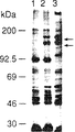
図1は、トランスフェクションされたNIH/3T3細胞におけるmet産物を示す。(A)免疫沈降分析 細胞を[35S]メチオニンよび[35S]システイン(ICN社製、トランスラベル(Translabel))で、5分間、メチオニンおよびシステインを欠くDMEM(ギブコ(Gibco)社製)中で代謝的にラベルし、細胞溶解物をMethu−特異的モノクローナル抗体19S(ファレット,ディー・エル(Faletto,D.L.)ら,オンコジーン(Oncogene)第7巻:149〜1157頁(1991年))(レーン1および2)、またはMetmu(muはネズミ由来)−特異的ペプチド抗体SP260(アイヤー,エイ(Iyer,A.)ら,セル・グロウス・アンド・ディファレンシエイション第1巻:87〜95頁(1990年))(レーン3)のいずれかと免疫沈降させた。プロテインG−セファロース(ギブコ社製)で免疫沈降を完了させ、複合体を、5%β−メルカプトエタノールを含有するSDS試料用緩衝液に可溶化し、7.5%アクリルアミドゲル上で分離した。レーン1は、細胞をpSV2neoのみでトランスフェクションした場合;レーン2は、細胞をmethuでトランスフェクションした場合;レーン3は、細胞をmetmuでトランスフェクションした場合である。(B)パルス・チェイス分析 細胞を[35S]メチオニンよび[35S]システインで45分間代謝的にラベルし(レーン1および3)、ついで、チェイス時間を3時間間とした(レーン2および4)。Metを、Methuに対する19Sモノクローナル抗体(レーン1および3)またはMetmuに対するモノクローナル抗体(レーン2および4)と免疫沈降させ、パネルAに示す電気泳動を行った。レーン1および2は、細胞をMethuでトランスフェクションした場合;レーン3および4は、細胞をMetmuでトランスフェクションした場合である。(C)細胞表面におけるmetのヨウ素化 集密的に近い細胞をペレット化し、ヨードゲン(Iodo−Gen(ピアス(Pierce)社製)の存在下においてNa125Iでラベルした。ラベルされた細胞をPBSで3回洗浄し、RIPA緩衝液で溶解し、19Sモノクローナル抗体(レーン1および2)またはSP260ペプチド抗体(レーン3および4)のいずれかと免疫沈降させた。Methuを発現するNIH/3T3細胞はレーン1および3である。Metmuを発現するNIH/3T3細胞はレーン2および4である。矢印は、p170metおよびp140metの位置を示す。
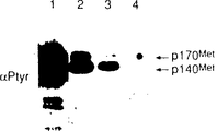
図2は、抗−P−Tyr抗体との精製物の反応性を示す。100mmディッシュ上の集密的に近い細胞を冷TBSで2回洗浄し、溶解用緩衝液(25mM Tris−HCl,pH8.0、100mM NaCl、50mM NaF、1%トリトンX−100、10μg/mlアプロチニン、10μg/mlロイペプチン、1.25mM PMSF、1mMオルトバナジン酸ナトリウム)1ml中で溶解した。Methuに対する抗−C28ペプチド抗体(ゴンザッティ−ハセス,エム(Gonzatti−Haces,M.)ら、プロシーディングス・オブ・ナショナル・アカデミー・オブ・サイエンシズ・ユーエスエイ第85巻:21〜25頁(1988年))(パネルAおよびC)またはMetmuに対するペプチド抗体SP260(アイヤーエイら,セル・グロウス・アンド・ディファレンシエイション第1巻:87〜95頁(1990年))(パネルBおよびD)でもって蛋白1ミリグラムが免疫沈降した。SDS緩衝液に溶解した後、試料を7.5%ゲル上のSDS−PAGEにより分離し、インモビロン−P(Immobilon−P)(ミリポア(Millipore)社製)に移し、ついで、抗−P−Tyr抗体、4G10(モリソン・ディー・ケイ(Morrison,D.K.)ら,セル第58巻:649〜657頁(1989年))(パネルAおよびB)、19Sモノクローナル抗体(パネルC)、またはマウス・metペプチド抗体(パネルD)をプローブとして反応させた。ついで、インモビロンフィルターを125I−プロテインA(ICN社製)とインキュベーションし、オートラジオグラフィーを行った。パナルAおよびCのレーン1〜3には、methuでトランスフェクションした細胞の3本の異なる線があり;レーン4は対照細胞である。パネルBおよびDのレーン1は、metmuでトランスフェクションした細胞である。レーン2はNIH/3T3対照細胞である。
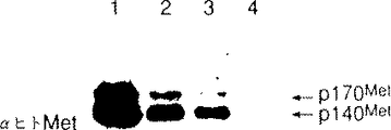
図3は、NIH/3T3細胞におけるMethuおよびHGF/SFhuの性質を示す。腫瘍細胞を外移植し、[35S]メチオニンおよび[35S]システインで6時間代謝的にラベルした。細胞溶解物を、19Sモノクローナル抗体(パネルA)、またはペプチド抗体SP260(パネルB)のいずれかと免疫沈降させた。ついで、「6時間」上清0.25mlを、セントリコン(Centricon)(アミコン(Amicon)社製 10Kカットオフ)で3倍に濃縮し、RIPA緩衝液で体積を0.3mlに調節し、試料を、抗−HGFモノクローナル抗体A3.1.2(パネルC)と免疫沈降させた。レーン1および3は、注入前に同時トランスフェクションされた2種の異なる細胞系からの試料である。レーン2は、レーン1で分析される細胞由来の腫瘍外移植物である。レーン4および5は、レーン3で分析される細胞由来の腫瘍外移植物である。レーン6は、対照NIH/3T3細胞から調製した試料である。矢印は、p170metおよびp140metの位置(パネルAおよびB)ならびに87kDa(前駆体)、69kDa、および34kDaのHGFポリペプチドの位置(パネルC)を示す。
図4は、NIH/3T3細胞腫瘍中のMetキメラ蛋白の性質を示す。細胞を、[35S]メチオニンおよび[35S]システインで6時間代謝的にラベルし、細胞溶解物を、19Sモノクローナル抗体で免疫沈降させた(レーン1〜4)。レーン1は、マウス・N−末端/ヒト・C−末端キメラmetでトランスフェクションした注射していないG418(ギブコ社製)−耐性細胞であり、レーン2および3は、レーン1で分析される細胞由来の腫瘍であり、レーン4は、NIH/3T3対照細胞であり、レーン5は、抗−P−Tyr抗体(4G10)で分析し、ついで、ヒト・抗−C28ペプチド抗体とともに免疫沈降したレーン1〜4と同じ細胞のウエスタンブロットである。
図5は、Metmuの細胞外ドメインがMethuに形質転換能を付与すること(第2行と第3行を比較)、およびHGFhuとともに同時トランスフェクションした場合にMethu細胞外ドメインのみが形質転換能があること(第4行と第5行を比較)を示す。さらに、最後の2行は、Methuに形質転換能を付与する領域としてのNde I−Pbu II部位間のmetmu領域を示す(第6行と第4行を比較)。
図6は、ヒト・Metの保存されたNde I−Pbu II部位(上側)とマウス・Met配列(下側)とのコンピューターによる予測構造での比較を示す。ヒトおよびマウスのMet(Nde I−Pbu II部位の間)のアミノ酸配列をボックス内に示す。アミノ酸配列がより保存的でない領域が強調されている(保存性を、ヒトとマウスの配列の間の線で示す)。Metに形質転換能を付与するcDNAのNde I−Pbu IIセグメント内のより保存性の少ないドメインは、リガンド結合に直接用いられるドメインを反映しているか、あるいは構造的な特性によってリガンド結合またはリガンド結合後の受容体の活性化のいずれかを調節しているかである。それゆえ、出願人らは、これらの配列に対応する合成ペプチドに対するポリクローナル抗体を作った。第1のドメインに対する抗体は、ヒト・配列(α1240)およびマウス・配列(α1241)に対してウサギ中で産生された。第2のドメインに対する抗体は、ヒト・配列(α1242)およびマウス・配列(α1243)に対して産生された。1242および1243のみが、ヒトおよびネズミのc−met蛋白それぞれに対して沈降を生じた。
図7は、ヒト(C28、19S、1242)およびマウス(260、1243)の特異的抗体に対するヒトおよびマウスのMetの反応性の対比を示す(パートA)。パートBは、マウス・met cDNAをNIH/3T3細胞中にトランスフェクションした場合(該細胞はHGF/SFmuを発現する)、該細胞は、ボイデン・チェンバー・アッセイ(Boyden chamber assay)において侵略的であることを示す(アルビーニ(Albini)ら,キャンサー・リサーチ(Cancer Res.)第47巻:3239〜3245頁(1987年))。すなわち、細胞は、マトリゲル(matrigel)でコーティングしたフィルターを越えて移動するのである。metの腫瘍形態(tpr−met)でトランスフェクションした細胞もまた、このアッセイにおいて非常に侵略的である。フィルターを越えて移動する細胞に関しては、それらは、単に運動性があるだけでなく、基礎となる膜成分を分解しうるにちがいない(よって、コラゲナーゼまたはプラスミノゲナーゼのごとき酵素を分泌する)。Methuトランスフェクション体(第2行)は、ヒト・リガンドであるHGF/SFhuの不存在下では侵略的てはないが、HGF産生細胞を培養したならし培地を添加した場合は侵略的である。一方、methuおよびHGF/SFhuの同時トランスフェクション体(第5行)は、このアッセイにおいて侵略的である。この図の第2欄は、上記ドメイン中のヒト・Metに対する1242抗体が、HGF/SFhuを含有するならし培地で同時培養されたmethuトランスフェクション体のフィルター侵入を阻害することを示す。最終欄は、この阻害が、競争的なペプチドの存在下でブロックされることを示す。一方、MethuおよびHGFhuを産生する同時トランスフェクション体の侵略能(第4行)は、1242抗体によってはブロックされず、このことは、内部の、同時に産生されたHGF/SFリガンドによる受容体の細胞内オートクリン活性化を示唆するものである。結局、この図は、1243抗体は、metmuトランスフェクション体によるフィルターの侵略をブロックしなかったことを示す(第3行、第3欄)。
図8は、metmuおよびtpr−metトランスフェクション体が、インビボにおいて腫瘍原性であり(図5)、これらの細胞もまた、いくつかの形式のアッセイにおいては転移的であること(パネルAおよびB)を示すインビボでの転移データを示す。さらに、Methu(パネルA)は、HGFhu cDNAで同時トランスフェクションされないかぎり、インビボにおいて転移的でない。
図9は、実施例2において記載するように、HGF/SFの生産において、NIH/3T3細胞中に同時トランスフェクションされるプラスミドpRS2およびpRS24の構築を示すダイアグラムである。
好ましい具体例の詳細な説明
1の具体例において、本発明は、HGF/SFのMetとの結合を阻害することがらなる腫瘍細胞の転移を防止する方法を包含する。「防止」なる語は、腫瘍の転移を減少または阻止することを意味する。
「転移性」となる腫瘍細胞に関しては、多くの相関のある事柄が起こらなくてはならない。例えば、転移は、癌細胞が血管およびリンパ循環系の壁を透過し、循環系に侵入し、下流毛細血管床またはリンパ節に定着(「付着」)し、循環系を去り(「移動/運動性」)正常な支質組織中に透過していくプロセスを包含する。起こるべきこれらのプロセスのためには、腫瘍細胞が侵略的特性を獲得しなければならない。侵略的特性としては、(1)外部マトリックスに付着する細胞の能力(インテグリンのごとき細胞表面上の受容体による);(2)コラゲナーゼ類およびプラスミノゲナーゼ類のごとき外部マトリックスを突き破る破壊酵素(「メタロプロテアーゼ類」)の誘導;および(3)酵素による破壊によってできた「穴」を通る細胞の移動が挙げられる。細胞により分泌されるメタロプロテアーゼ類のレベルの上昇、細胞の運動原性の増加、および受容体(付着体(インテグリン)に包含される)発現のパターンの変化は、すべて記載されており、いくつかの場合、ヒト・癌腫細胞の転移能と関連がある(エル・エイ・リオッタ(L.A.Liotta),サイエンティフィック・アメリカン(Scientific Ametican)54〜63頁(1992年2月))。
HGF/SFが、これらの事柄のうちの最後の2つ(すなわち、コラゲナーゼ類およびプラスミノゲナーゼ類の分泌ならびに運動原性)における活性レベルを増加させることが知られている。さらに、HGF/SFは、脈管形成性であることが知られており、血管形成は腫瘍細胞の生存にとり重要である(ジェイ・ベーレンス(J.Behrens)ら,ジャーナル・オブ・セル・バイオロジー(J.Cell Biol.)第108巻:2435〜2447頁(1989年);ケイ・エム・ウェイドナー(K.M.Weidner)ら,ジャーナル・オブ・セル・バイオロジー第111巻:2097〜2108頁(1990年))。「運動原性」なる語は、連続した細胞のシートが凝集せず、形態を変化させ、運動性となるプロセスをいう(イー・ジェラルディおよびエム・ストーカー、キャンサー・セルズ第3巻:227〜232頁(1991年))。この現象は「散在化」とも呼ばれる。出願人は、HGF/SFが運動原性を2通りの様式で誘導することを示した。下記実施例1参照。まず、表1に示すように、HGF/SFに対して古典的な散在化応答を示す細胞は、内在性met受容体のチロシンのすみやかなリン酸化を示し、このことは、met受容体を通して散在化が活性化されることを示唆する。

「変種」としては、例えば、met結合ドメインを欠いているかまたは損傷を受けたmet結合ドメインを有するか、もしくは活性化ドメインを欠いているかまたは損傷を受けた活性化ドメインを有するHGF/SF種(しかしHGF/SFの構造的および生物学的特徴を保持している)が挙げられる(ロッカー(Lokker)ら,ジ・EMBO・ジャーナル第11巻(7):2503〜2510頁(1992年)。
本発明の変種とされるHGF/SF種を、慣用的な遺伝子工学的方法により、本発明にしたがって製造することができる。例えば、部位特異的変異を含む方法により、変種を製造することができる。カレント・プロトコールズ・イン・モレキュラー・バイオロジー(Current Protocols in Molecular Biology),8.0.3中「ミュータジェネシス・オブ・クローンド・ディーエヌエイ(Mutagenesis of Cloned DNA)」およびその続きの部分;上記ロッカーらの文献参照。
本発明の変種としては、例えば、HGF/SFと結合する正常なMetに対するアンタゴニストとして作用する細胞外HGF/SF結合ドメインからなる可溶性系形態のMetも挙げられる。かかる変種を、本発明の分野においてよく知られた分子設計法により製造することができる。例えば、フー(Fuh)ら,サイエンス(Science)第256巻:1677〜1680頁(1982年)参照。
本発明の転移阻害性変種は、米国同時係属出願第07/655,502号に開示されたHGF/NK2またはHGF/NK1のごとき天然の変種であってもよい。該出願は文献により取り込まれる。HG/NK2は、N−末端および最初の2個のクリングルドメイン(kringle domain)を含む配列により特徴づけられる別のHGF転写物によりコードされるHGF/SFの切形形態である。HGF/NK1は、N−末端および最初の2個のクリングルドメイン(kringle domain)を含む配列により特徴づけられるHGF/SF転写物によりコードされるHGF/SFのもう1つの切形形態である。
本発明の転移阻害性物質は、HGF/SFまたはMetのためには模擬体であってもよい。模擬体の一例は、抗−イディオタイプ抗体、すなわち、抗原上のエピトープに特異的に結合する抗体で動物を免疫することにより産生される抗体である。抗−イディオタイプ抗体は、第1の抗体の結合部位を認識し、確認する。それゆえ、その結合部位の形態は、第1の抗体の結合部位中に適合するエピトープに非常によく似ている。抗−イディオタイプ抗体が、もとの抗原に似た結合部位を有しているので、それを、もとの抗原に対する受容体との結合のためのリガンドとして用いることができる。ファインバーグ(Fineberg)およびアートル(Ertl),CRC・クリティカル・レビューズ・イン・イミュノロジー(CRC Critical Reviews in Immunology)第7巻:269〜284頁参照。HGF/SFの適当な模擬体を、HGF/SF抗体でスクリーニングしていずれの化合物がそれに結合するのか、あるいはいずれの化合物が分子設計により製造できるのかを調べることができる。モーガン(Morgan)ら,アニュアル・リポーツ・イン・メディシナル・ケミストリー(Annual Reports in Medicinal Chemistry)(アカデミック・プレス(Academic Press),1989年)中「アプローチズ・トゥ・ザ・ディスカバリー・オブ・ザ・ノン−ペプチド・リガンズ・フォー・ペプチド・リセプターズ・アンド・ペプチダーゼス(Approaches to the Discovery of the Non−Peptide LLigands for Peptide Receptors and Peptidases)」参照。
本発明の転移阻害性物質は、HGF/SFのMetとの結合を阻害するHGF/SFまたはMetに対する抗体または抗体フラグメントであってもよい。本発明にいう「抗体」は、抗体全体およびその一部、あるいはそれのみまたは他の部分を伴うものを包含する。抗体としては、ポリクローナル抗体、モノクローナル抗体、および単鎖抗体が挙げられる。抗体フラグメントは、HGF/SFまたはMetと結合するもの、とりわけ、FabおよびF(ab)2フラグメントが挙げられる。本発明の抗体を、動物中で生産することができ、また、当業者によく知られた組換えDNA法により生産することができる。
本発明の1の具体例において、出願人らは、リガンド結合またはリガンド結合の調節に用いるMet細胞外ドメインに対応する合成ペプチドに対するポリクローナル抗体を製造する。図6および図7およびその説明参照。
出願人らは、腫瘍の侵略または転移をブロックする能力を測定するMetに対して指向されたリガンド結合阻害剤についてのインビトロ試験に関するプロトコールを開発した。例えば、出願人らは、metmu(内因的にネズミ・HGF/SFを産生する)およびmetの腫瘍形態であるtpr−metでトランスフェクションした細胞が侵略的であるのと同様に、HGF/SFおよびmethuでトランスフェクションしたNIH/3T3細胞が、ボイデン−チェンバーアッセイにおいて侵略的であることを見いだした。同様に、「ならし培地」、すなわちHGF/SFhuを含有する培地中にある場合には、methuトランスフェクション体は侵略的である。さらに出願人らは、Metの細胞外ドメイン中のペプチド配列に対して産生された「1242」抗体が、腫瘍細胞の侵略能をブロックすることを見いだした。
実際に、実施例2に開示され、図5を参照して議論されるようなキメラなマウス/ヒトに関する実験により、リガンド/受容体結合に包含されるMet受容体およびHGF/SFに関するドメインを明らかにすることが可能になる。図6に示され記載された数学的かつコンピューターによる計算により、マウスおよびヒトにおいて異なっているドメイン中の領域を明らかにすることが可能である。これらの「相違」により、HGF/SFに対する対応における種特異性が調節されている可能性があり、かかる領域に対する抗体を、そのHGF/SF結合を阻害する能力に関して試験することができる。よって、出願人らは、インビボ用途の組成物の開発に適用可能な、腫瘍転移阻害についてのインビトロ試験法を提供した。
作用すべき選択部位への本発明腫瘍転移阻害物質の送達を、ドラッグデリバリー、遺伝子導入、またはそれらのすべての組み合わせといった慣用的方法を用いて行うことができる。ケイ・ジェー・バン・ゼー(K.J.Van Zee)ら,PNAS第89巻:4845〜4849頁(1992年)参照。送達手段としては、炭水化物またはキャリア蛋白と抱合体化;当該分野で認められているいかなる徐放複合体とともに行う投与;微小球またはリポソームのごとき他の送達システムとの組み合わせ;または発現ベクター系に導入しての投与が挙げられる。本発明に適用可能な送達方法の1つとしては、デイビス(Davis)ら,米国特許第4,479,337号の方法により、本発明の腫瘍転移阻害物質をポリエチレングリコールまたはポリプロピレングリコールとカップリングさせて生理学的に活性な免疫原性のない水溶性組成物を製造することが挙げられる。
かかる組成物の投与を、当業者に知られたいかなる方法によっても行うことができる。例えば、組成物を水溶液中に入れ、哺乳動物の循環系内または筋肉内系に注射することができる。適正な用量の決定は、哺乳動物の年令および体重をはじめとするそれぞれのケースに特異的な変数の数に依存し、当業者の技術・知識範囲内の通常の実験を必要とする。
本発明のもう1つの具体例において、HGF/SF経路の人工的な活性化により、本来の生物学的活性を回復、置換、または増強するための治療的方法論に関する根拠が提供される。これらの方法論としては、MetとHGF/SFとの間の結合相互作用を増強するHGF/SFまたはMet変種あるいは模擬体を活性化すべき部位へ送達し、そのことにより、人工的に支持されたHGF/SF相互作用を作り出すことが挙げられる。例えば、MetのHGF/SF結合ドメイン、またはHGF/SFのMet結合ドメイン(あるいはその両方)の部位特異的変異を用いて、他のペアーのメンバーに対するより高い親和性を有するHGF/SF−Metペアーのメンバーを作り出すことができ、よって、損傷組織の成長または再生の加速に影響が及ぶ。同様に、構成的に活性化されたチロシンキナーゼを有するMetの変異をはじめとする慣用的な組換えDNA法を用いて、通常はHGF/SF結合により調節されるMet蛋白のキナーゼ活性を増強または支援することができる。外部から投与されたHGF/SFと組み合わせた組換えDNA法によりMetの自然発現を補う手段によってHGF/SF−Met経路を活性化することも、本発明の範囲内に包含される。
作用すべき選択部位への遺伝子操作されたHGF/SFまたはMet種の送達を、腫瘍転移を阻害する物質の送達に関して上で議論されたような、ドラッグデリバリー、遺伝子導入、またはそれらのすべての組み合わせといった慣用的方法を用いて行うことができる。
本発明の別の具体例において、出願人らは、ヒト・HGF/SFの製造法を提供する。詳細には、出願人らは、驚くべきことに、マウス・繊維芽細胞NIH/3T3細胞が、ヒト・HGF cDNAを含むトランスフェクションされた末端反復配列(LTR)ベクター組み換え体から高レベルのHGF/SFを発現することを見いだした。出願人らは、LTRヒト・c−met腫瘍原遺伝子(cDNAベクター)がヒト・LTR/HGF構築物とともに同時トランスフェクションされ、かつ細胞が2次腫瘍から由来する場合に、最高レベルのHGFがNIH/3T3中に検出されることを見いだした。下記実施例2参照。この細胞系の1の利点は、形質転換細胞が高密度にまで増殖することである。したがって、この細胞系は、非常に高レベルのHGF/SF、すなわちリットルあたりHGF/SF約1mg(mlあたり1250ユニット)を産生する。これに対し、ヒト・表皮ケラチン細胞由来の別の細胞系であるndk細胞は、48時間でリットルあたり約10μgを産生する。ジェイ・シーアダムス(J.C.Adams)ら,サイエンス第98巻:385〜394頁(1991年);イー・エム・ローゼン(E.M.Rosen)ら,BBRC第168巻(3):1082〜1088頁(1990頁)参照。それゆえ、本発明の組換えベクター構築物で同時トランスフェクションしたNIH/3T3細胞から、1mg/mlの収量でHGF/SFを得ることは予期されないことである。
よって、本発明の1の具体例は、以下のステップ:
(a) NIH/3T3細胞を、HGF/SFhuおよびMethuをコードしているDNAでトランスフェクションし;
(b) ステップ(a)によりトランスフェクションされた細胞を哺乳動物内に導入し、それにより、1次腫瘍を生じさせ;
(c) 1次腫瘍細胞を外移植し、インビトロで増殖させ;
(d) ステップ(c)により増殖した高レベルのHGF/SFhuおよび高レベルのMethuを発現する細胞を選択し;
(e) ステップ(d)により選択した細胞を哺乳動物内に導入し、それにより、2次腫瘍を生じさせ;
(f) 2次腫瘍細胞を外移植し、インビトロで増殖させ;ついで
(g) ステップ(f)の細胞により産生されたHGF/SFhuを得る
からなるHGF/SFの製造法に関する。
本発明DNAは、作動可能に連結されたレトロウイルスの末端反復配列由来のプロモーター、ヒト・Metの全コーディングドメインをコードしているDNAおよびポリアデニレーションシグナルからなる第1のDNAベクター、および作動可能に連結されたレトロウイルスの末端反復配列由来のプロター、ヒト・HGF/SFをコードしているDNAおよびポリアデニレーションシグナルからなる第2のDNAベクターよりなる。
本発明DNAベクターの製造を、当業者に知られた種々の方法により行うことができるが、方法を実施例2のcDNAプラスミド構築物および細胞系の議論において例示する。本発明の好ましいDNAプラスミド構築物を図9に示す。
HGF/SFhuおよびMethuLTR−cDNAでトランスフェクションされた細胞を、当該分野でよく知られた方法により、好ましくは、注射により哺乳動物中に導入する。ブレイアー,ディー・ジー(Brair,D.G.)ら,サイエンス第218巻:1122〜1125頁(1982年)参照。本発明の好ましい哺乳動物はヌードマウスである。これらの動物中で5〜10週間増殖した後の1次腫瘍を外移植し、実施例2のヌードマウスアッセイ記載のごとくインビトロ培養で増殖させる。ついで、インビトロ増殖した1次腫瘍細胞を、免疫沈降分析に付してどの細胞が高レベルのHGF/SFhuおよび高レベルのMethuを発現しているのかを確認する。図3およびその説明参照。例えば、外移植された細胞を代謝的にラベルし、ついで、Methuモノクローナル抗体(モノクローナル19S)とともに免疫沈降させ、濃縮し、ついで、HGF/SFhuモノクローナル抗体A3.1.2.とともに免疫沈降させる。高レベルのHGF/SFhuおよびMethuを、出発物質における発現レベルと比較して測定する。したがって、出発物質において観察されるよりも高い発現レベルのものすべてを、この方法の目的に照らし合わせて、「高い」とみなす。ついで、高レベルのHGF/SFhuおよびMethuを発現する細胞を哺乳動物に導入し、増殖する2次腫瘍を外移植し、当該分野でよく知られた方法によりインビトロ増殖させる。ブレイアー・ディー・ジーら,サイエンス第218巻:1122〜1125頁(1982年)参照。
ついで、外移植された2次腫瘍細胞により発現されるHGF/SFhuを、ウェイドナーら,ジャーナル・オブ・セル・バイオロジー第111巻:2097〜2108(1990年)記載のごとく精製する(本発明の適用可能な他の方法は当業者によく知られている)。
さらにもう1つの具体例において、本発明は、met cDNAでトランスフェクションされたNIH/3T3細胞由来のマウスおよびヒトのMetのC−末端イソ体(isoform)に関する。これらのイソ体は翻訳後のプロセッシングにより生じる可能性がもっと高いが、出願人らは、トランスフェクションされたDNAの可能な再配列を排除することはできない。C−末端において切形されたいくつかのmet産物が報告されている(プラト,エム(Prat,M.)ら,モレキュラー・アンド・セルラー・バイオロジー(Mol.Cell.Biol.)第11巻:5954〜5962頁(1991年))が、これらの産物は本発明イソ体(isoform)とは異なる。その理由は、それらがC−末端抗体と反応しないからである。そのうえ、これらのイソ体は、高レベルのmetを発現している細胞においてのみ検出され、そのことは、それらの存在量が少ないことを示すものである。
実施例1.c−met腫瘍原遺伝子は運動原性に関連した受容体である
c−met受容体を発現する多くのヒト・癌細胞系において転移および/または散在化(運動原性)を誘導するHGF/SFの能力を調べた。上記表1に示す細胞系を、1X104〜5X104個/mlとなるよう96ウェル組織培養プレートに撒き、集密的に近くなるまで増殖させた。ウェルを洗浄して血清を除去し、無血清培地にて2日間細胞を飢餓状態とした。その翌日、細胞を、無血清、血清、またはHGF/SF10ng/mlで16〜18時間処理し、ついで、ウェルあたり3H−チミジン1μCi(5μCi/ml)を添加して、さらに6時間置いた。氷冷PBSでウェルから標識を洗い落とすことによりアッセイを終了し、細胞を5%TCAで固定し、ついで、0.25M NaOHでDNAを可溶化した。運動原性インデックスは、血清不存在下におけるチミジンの取り込みとHGFによるチミジンの取り込みとの比である。運動原性アッセイ用に、細胞を、2X10³個/mlとなるように16−ウェルチェンバースライド(ラブテック(LabTek)社製)に撒き、60%集密的になるまで増殖させた。ついで、細胞を洗浄して血清を除去し、HGF/SF添加/無添加無血清培地にて一晩インキュベーションした。スライドを氷冷アセトンで10分間固定し、クリスタルバイオレット染色した(5分間)。「散在化した表現形」における質的な変化が記録された。
c−metの上皮細胞系への導入により「散在化」活性が該細胞に付与されることを示すために、ヒト・met cDNAまたはネズミ・met cDNAのいずれかでリポフェクション(BRL)することにより、C127細胞をトランスフェクションした。表1のように分裂原性および/または動物原性を測定した。この研究結果を上記表2に示す。
実施例2.NIH/3T3細胞系におけるHGF/SF産生
cDNAプラスミド構築物および細胞系 met cDNAプラスミドを、pMEX(オスカム,アール(Pskam,R.)ら,プロシーディングス・オブ・ナショナル・アカデミー・オブ・サイエンシズ・ユーエスエイ第85巻:2964〜2968頁(1988年))の誘導体(ポリリンカー配列なし)であるpMB1中で構築した。該プラスミドは、モロニー・ミュリン・サルコーマ・ウイルス(Molony murine sarcoma virus)(MSV)由来のLTRRプロモーターおよびシミアン・ウイルス40(simian virus 40)のポリアデニレーションシグナルを含んでいる。オーオプンリーディングフレームを含む4.6kbのmethu配列中において、内部の300bpのEcoR IフラグメントをpOKの250bpのEcoR Iフラグメントと置換することによりmethuプラスミドを構築した。パーク,エム(Park,M)ら,プロシーディングス・オブ・ナショナル・アカデミー・オブ・サイエンシズ・ユーエスエイ第84巻:6379〜6383頁(1987年);ロドリゲス(Rodriguez)ら,モレキュラー・アンド・セルラー・バイオロジー第11巻:2962〜2970頁(1991年)参照。metmuプラスミドは、4.6Kbのマウス・metオープンリーディングフレーム全体を含んでいる。アイヤー,エイら,セル・グロウス・アンド・ディファレンシエイション第1巻:87〜95頁(1990年)参照。キメラなヒト/マウスmet構築物を、保存的なPvu II部位(アミノ酸807)を用いて構築した。pMEXのBamH I−Kpn I部位中にヒト・HGF配列の2.3KbのBamH I−Kpn Iフラグメントを挿入することによりHGFhuプラスミドを構築した。ナカムラ,ティーら,ネイチャー第342巻:440〜443頁(1989年);オスカム,アールら,プロシーディングス・オブ・ナショナル・アカデミー・オブ・サイエンシズ・ユーエスエイ第85巻:2964〜2968頁(1988年)参照。NIH/3T3 490細胞を、8%子ウシ血清(ギブコ社製)含有DMEM培地(ギブコ社製)中で増殖させた。
DNAトランスフェクション プラスミドDNA(子ウシ胸腺キャリアDNA8μgを含む水75μl中2μg)を0.67M CaCl275μlと混合することにより、DNAトランスフェクションに関するリン酸カルシウム法(クーパー,シー・エス(Cooper,C.S.)ら,ネイチャー第311巻:29〜33頁(1984年))を行った。連続的に攪拌しながらこの混合物を、0.15mlの溶液H(0.27M NaCl、0.01M KCl、0.0014M Na2HPO4・7H2O、0.012M デキストロース)に滴下した。室温に30分間置いた後、0.01M Hepesバッファーを含む培地1mlを入れた35mmディッシュ上で約70%の集密性を有する細胞に、該混合物を添加した。細胞を37℃で4時間インキュベーションし、ついで、溶液H中の15%グリセロール(v/v)で2分間処理した。G418により選択のために、細胞を再度DMEMおよび8%子ウシ血清中に一晩フィードし、ついで、3個の60mmディッシュに移した。24時間インキュベーションした後、細胞を、400μg/mlのG418(ギブコ社製)を含有する培地に、1週間に2度フィードした。
ノーザン分析 RNAsol(CINNA/BIOTECX社製)を用いてRNAを単離した。RNA20μgを、1%変性的ホルムアミドアガロースゲル上の電気泳動に供し、ついで、ニトロセルロースフィルター(シュライヒャー・アンド・シュール(Schleicher and Schuell)社製)に移した。30%ホルムアミド、6xセイラインクエン酸ナトリウム(SSC)、5xデンハート溶液(Denhardt's solution)、50mMリン酸ナトリウム(pH6.8)および超音波処理したサケ・精子DNA(250μg/ml)中で、ブロットを、32P−標識ランダムプライマーDNAプローブと42℃で15時間ハイブリダイズさせた。ハイブリダーゼーション後、フィルターを、1xSSC(0.1%SDS含有)中、室温で2回洗浄し、ついで、1xSSC(0.1%SDS含有)中、50℃で洗浄した。フィルターを乾燥させて−70℃で1〜3日間、X線フィルムに暴露した。
免疫沈降 集密的に近い細胞を、メチオニンおよびシステイン不含DMEM中で、0.25mCiのトランスラベル(Translabel(ICN社製))(1ml/35mmディッシュ)で4ないし6時間ラベルした。ラベルした細胞を、0.5mlのRIPA緩衝液[1%トリトンX−100、1%デオキシコール酸ナトリウム、0.1%ドデシル硫酸ナトリウム(SDS),0.15M NaCl、0.02M NaPO4,pH7.2、1mM フッ化フェニルメチルスルホニル(PMSF),2mM EDTA、50mM NaF、30mM ピロリン酸ナトリウム]中で溶解させた。等量の放射線カウントを有する清澄化した溶解物を、19Sモノクローナル抗−メチオニン抗体(ファテット,ディー・エル(Faletto,D.L.)ら,オンコジーン(Oncogene)第7巻:1149〜1157頁(1991年))と4℃で一晩免疫沈降させた。免疫沈降物を、プロテインG−セファロース(ギブコ社製)に複合体化させ、ついで、RIPA緩衝液で2回洗浄し、ハイサルト緩衝液(high−salt buffer)(1M NaCl、10mM Tris−HCl,pH7.2、0.5%トリトン)で洗浄した。5%β−メルカプトエタノール含有SDS試料用緩衝液中で煮沸することにより、免疫沈降物を可溶化させた。試料をSDSポリアクリルアミドゲル電気泳動により分析し、ついで、フルオログラフィックエンハンサー(fluorographic enhancer)(アンプリファイ(Amplify)TM,アマシャム(Amersham)社製)で処理し、−70℃において増強スクリーンでフルオログラフィーを得た。
SP260は、ウサギ・抗血清から作られたペプチド抗体であり、metmuのC−末端の21個のアミノ酸に対して指向されている(アイヤー,エイら,セル・グロウス・アンド・ディファレンシエイション第1巻:87〜95頁(1990年))。A.3.1.2は、抗−ヒト組み替え型HGFモノクローナル抗体(IgG,サブクラスG2a)である。
パルスチェイス分析 集密的に近い細胞を、メチオニンおよびシステイン不含DMEM1ml中(各35mmディッシュ)、0.25mCiのトランスラベルで45分間ラベルした。細胞を2回洗浄し、完全培地で3時間チェイスし、ついで、溶解し、免疫沈降分析に供した。
表面ヨウ素化 集密的に近い細胞を、ヨード−ジェン(Iodo−Gen(ピアス(Pierce)社製)の存在下でNa125Iでラベルした。ヨードージェン試薬20μl(クロロホルム中10mg/ml)をI−ドラムバイアル(I−dram vial)の底に入れ、窒素の流れにより乾燥させた。ついで、ヨード−ジェンを10mM EDTAを含有する1M Tris(pH7.5)に溶解し、細胞ペレットに添加した。Na125I(0.5mCi)を添加した10分間反応した。ラベルした細胞をPBSで3回洗浄し、ついで、RIPA緩衝液で溶解し、免疫沈降分析に付した。
ウエスタン分析 100mmディッシュ上の集密的に近い細胞を、冷TBS(10mM Tris,pH8.0、150mM NaCl)で2回洗浄し、1mlの溶解用緩衝液(25mM Tris−HCl,pH8.0、100mM NaCl、50mM NaF、1%トリトンX−100、10μg/mlアプロチニン、10μg/mlロイペプチン、1.25mM PMSF、1mMバナジン酸塩)中で溶解させた。蛋白1mgを、抗−C28抗−Methuポリクローナル抗体(ゴンザッティ−ヘイセス,エム(Gonzatti−Haces,M.),プロシーディングス・オブ・ナショナル・アカデミー・オブ・サイエンシズ・ユーエスエイ第85巻:21〜25頁(1988年))とともに免疫沈降させ、抗−ホスホチロシン(抗−P−Tyr)抗体4G10(モリソン、ディー・ケイ(Morrison,D.K.)ら,セル第58巻:649〜657頁(1989年))、またはヒトもしくはマウスのmet特異的抗体である19Sモノクローナル(ファレット,ディー・エルら,オンコジーン第7巻:1149〜1157頁(1991年))あるいはSP260(アイヤー,エイら,セル・グロウス・アンド・ディファレンシエイション第1巻:87〜95頁(1990年))のいずれかとウエスタン分析を行った。製造者の指示に従い、125I−プロテインA(アマシャム社製)を用いて陽性バンドを検出した。
ヌードマウス腫瘍アッセイ すでに記載されているようにして(ブレイアー,ディー・ジーら,サイエンス第218巻:1122〜1125頁(1982年))、アッセイを行った。トランスフェクションされG418で選択されたNIH/3T3細胞(106個)を2回洗浄し、無血清培地0.1ml中に再懸濁した。細胞を、離乳した胸腺除去ヌードマウス(ハーラン・スプラグ・ダウリー,インク(Harlan Sprague Dawley,Inc.)の背中に注射した。腫瘍形成を、毎週10週目までモニターした。腫瘍が15mmのサイズになった時、腫瘍を外移植し、腫瘍細胞を免疫沈降分析に供した。
軟寒天アッセイ 軟寒天増殖アッセイを、ブレイアら、ウイロロジー(Virology)第95巻:303〜316頁(1979年)の変法により行った。簡単に説明すると、トリプシン処理細胞を、10%子ウシ血清および0.24%精製寒天(DIFCO社製)を含有する8mlのDMEM中、2x10⁵および2x10⁴個となるように懸濁し、10%子ウシ血清および0.27%寒天を含有するDMEMの硬化下層を入れた2系の60mmディッシュに移した。1週間間隔で、10%子ウシ血清および0.27%寒天を含有するDMEM2mlをプレートにフィードした。37℃で3週間インキュベーションした後、顕微鏡でコロニーを数えた。
NIH/3T3細胞におけるMethuおよびMetmuの発現 methu腫瘍原遺伝子cDNAを含んでいるプラスミドを、pSV2neoとともにNIH/3T3細胞に同時トランスフェクションし、免疫沈降分析により、ヒトまたはマウスのMet発現に関してG418耐性細胞をスクリーニングした。これらの分析により、トランスフェクションされたNIH/3T3細胞において、ヒトおよびマウスのp170metおよびp140metが発現されることが示されている(図1Aのそれぞれレーン2および3)。NIH/3T3細胞における同等のレベルのmetmu発現が報告されている(アイヤー,エイら,セル・グロウス・アンド・ディファレンシエイション第1巻:87〜95頁(1990年))。出願人らは、Methuを発現するG418耐性細胞において、外来性のMetmuの発現が見られないかまたは殆ど起こらないことを見いだした。MethuおよびMetmuの適当なプロセッシングについて、パルスチェイスラベリング実験により試験し、これらの研究により、45分のパルスの間に合成されるp170met(図1B,レーン1および3)が、3時間後に、効果的に成熟p410metへとプロセッシングされる(レーン2および4)ことが示された。そのうえ、ヒトおよびマウスのMetは、細胞表面に局在化していた。出願人らは、無処理の細胞をNa125Iでラベルし、その溶解物を免疫沈降して、p140metおよびp170met両形態ともがヨウ素化されたことを示した(図1C)。よって、NIH/3T3細胞において発現されるヒトまたはマウスのMetは、正確にプロセッシングされ、細胞表面に局在化するのである。これらの分析は、p170metが細胞表面に到達することも示す。p170metのヨウ素化物は、溶解細胞からは生じなかった。なぜならば、同じヨウ素化条件において、細胞質性tpr−met腫瘍蛋白が検出されなかったからである(ゴンザッティ−ヘイセス,エム(Gonzatti−Haces,M.),プロシーディングス・オブ・ナショナル・アカデミー・オブ・サイエンシズ・ユーエスエイ第85巻:21〜25頁(1988年))。さらに、methuを過剰産生するヒト・胃癌細胞系であるオカジマ細胞(Okajima cells)において発現されるp170metもまた、表面ヨウ素化によりラベルされるが(データ示さず)、NIH/3T3細胞と比較した場合、ラベルされたp140metに対するp170metの割合は小さかった。
NIH/3T3細胞におけるmetの構造的なチロシンのリン酸化 ノーザンハイブリダイゼーション分析により、出願人らは、NIH/3T3細胞およびmethuまたはmetmuのいずれかでトランスフェクションされた細胞におけるHGF/SF mRNA発現を検出した。出願人らは、同レベルのHGF/SF mRNA発現を観察した。そのことは、G418選択細胞系におけるmethuまたはmetmuの過剰産生は外来性HGF/SF発現に影響しないことを示すものであった。これらの結果もまた、Metがオートクリン様式で活性化されうることを示すものであった。それゆえ、出願人らは、これらの細胞において発現したMethuおよびMetmuが抗−P−Tyr抗体と反応するかどうかを調べた。MethuおよびMetmuを発現する細胞系からの抽出物を、ヒトまたはマウスに特異的なペプチド抗体との免疫沈降に供し、ついで、抗−P−Tyr抗体(図2Aおよび2B)、またはヒト(図2C)あるいはマウス(図2D)に特異的なMet抗体を用いたウエスタン分析を行った。これらの分析は、Methuのp170metおよびp140metならびにMethuが抗−P−Tyr抗体と強く反応したことを示すものである。これは、p170metのチロシンリン酸化物が抗−P−Tyr抗体と反応することを示した最初の例である。1つの細胞系はMethuを非常に高レベルで発現した(図2AおよびCのレーン1)。これは例外である。なぜならば、他のすべてのMethu細胞系が、図2AおよびCのレーン2および3で分析された細胞系に匹敵するレベルの発現を示したからである。さらに、C−末端Met蛋白産物(p85、p75、およびp65)がマウスまたはヒトいずれかのmetを発現する細胞におけるC−末端抗体でもって検出された(図2A〜Dのレーン1)。
NIH/3T3細胞におけるmetの腫瘍原性 出願人らは、図5に示すように、Metmuを発現するがMethuを発現しないNIH/3T3細胞培養物が形質転換されたこと(アイヤー,エイら,セル・グロウス・アンド・ディファレンシエイション第1巻:87〜95頁(1990年))を観察した。このことは、ヌードマウスにおける培養物の腫瘍原性について試験し、ついで、トランスフェクションし、G418選択を行うことによって確認された。metmuを発現するNIH/3T3細胞は、下表3に示すように非常に腫瘍原性があるが、methuを発現する細胞は腫瘍原性が低く、試験した8系のうち1系のみが腫瘍を生成したにすぎなかった。
methuおよびHGF/SFhuで同時トランスフェクションされたNIH/3T3細胞の腫瘍原性 methuでトランスフェクションされたNIH/3T3細胞の低い腫瘍原性に関する1の説明は、外来性のHGF/SFmuによってはmethu受容体活性化により十分なシグナルを得ることができないということである。それゆえ、出願人らは、methuおよびHGF/SFhu両方のcDNAでのトランスフェクションが、オートクリン機構を通して腫瘍原性を増加させるかどうかを調べた。これらの分析により、methuおよびHGF/SFhuで同時トランスフェクションされたNIH/3T3細胞が高い腫瘍原性となることが示されている(表4)。
NIH/3T3細胞におけるキメラなヒト/マウス・metの腫瘍原性 リガンド結合ドメインが腫瘍原性に影響するかどうかを調べるために、出願人らは、キメラなヒト/マウス・met受容体分子を得、ヌードマウスにおけるそれらの腫瘍原性を試験した(図5)。出願人らは、トランスメンブレンをコードしている部位に隣接した外部ドメイン中の保存されたPvu II部位を用いてこれらの組換体を作った。マウス・外部リガンド結合ドメインをヒト・トランスメンブランおよびチロシンキナーゼドメインに連結した場合、該キメラな受容体は、Metmuと同等の腫瘍原活性を示した(図5)。これらの腫瘍の外移植物は、ヒト・チロシンキナーゼドメインに対して指向されたヒト・抗体でもって認識されるキメラなMet蛋白のレベルの増加を示した(図4のレーン2および3)。これらの腫瘍において、metmuの増幅に関する証拠は得られず、キメラな産物は、抗−P−Tyr抗体を用いたエウスタン分析によって認められた(図4のレーン5)。
マウス・Met外部リガンド結合ドメインを伴ったキメラの高い腫瘍原性とは対照的に、ヒト・N−末端/マウス・C−末端キメラは殆ど腫瘍原性がなかった。しかしながら、Methuの場合と同様に、HGF/SFhucDNAでこのキメラを同時トランスフェクションした場合、効果的な腫瘍形成が観察された(図5)。さらに出願人らは、metmu−methuキメラを発現する細胞が、ヌードマウスに注射する前に、形成転換され、metmuでトランスフェクションされた細胞と同様に軟寒天中にコロニーを形成することを示す。methu−metmu細胞は、HGF/SFで同時発現されないかぎり、形質転換された表現形を示さない。出願人らは、Metmu外部リガンド結合ドメインが、腫瘍原性を調べる主要因子であると結論した。
上記発明を、明確および理解の目的で、いくらか詳細に記載したが、本発明は、この開示を読んだ当業者により理解されるであろう。また、本発明の真の範囲から離れることなく、形態および詳細において種々の変更がなされうる。Background of the Invention
Hepatocyte growth factor (HGF) was first purified from human and rabbit plasma and rat platelets based on its ability to stimulate mitogenesis in rat hepatocytes (E. Gohda) Et al., Journal of Clinical Investigation (J. Clin. Invest) 81: 414 (1988); R. Zarnegar and G. Michaelopoulos, Cancer. Research (Cancer Res.) 19: 3314 (1989); T. Nakamura et al., FEBS Letters. 224: 311 (1987)). Thus, HGF may act as a liquid factor to promote liver regeneration after partial hepatectomy or liver injury (EHGohda) et al., Journal of Clinical Investigations. 81: 414 (1988); GKMichalopoulos, FASEB J. 4: 176 (1990). The same factor was purified from human fibroblast cultures and shown to act on melanocytes and various epithelial and intradermal cells (T. Igawa) et al., BBRC Vol. 174: 831. 838 (1991); M. Kan et al., BBRC Volume 174: 331-337 (1991) and JSLubin et al., The Proceedings of National Academy. Of Sciences USA (Proc. Nat'l. Acad. Sci. USA) Vol. 88: 415 (1990)). Evidence for HGF expression in some organs (J. Rubin, Proceedings of National Academy of Sciences USA 88: 415 (1990); K. Tashiro et al. , Proceedings of National Academy of Sciences USA 87: 3200 (1990); HGF, along with T. Kinoshita et al., Biochem. Biophys. Res. Comm., Vol. 165: 1229 (1989)), and T. Kinoshita et al. These findings indicate that it can act as a paracrine mediator for broad-spectrum growth on morphology. Shows. Molecular cloning of HGF revealed significant structural homology to plasminogen and related serine proteases (JS Rubin et al., The Proceedings of National Academy of Sciences USA 88 : 415 (1990); T. Nakamura et al., Nature Vol. 342: 440 (1989); K. Miyazawa et al., Biophys research community (Biophys). .Res.Comm.) 163: 967 (1989)). Recent evidence that HGF induces rapid phosphorylation of protein tyrosine in untreated target cells suggests that the tyrosine kinase receptor transduces its division signal (JS. Rubin et al., Proceedings of National Academy of Sciences USA 88: 415 (1990).
HGF is structurally related to a family of serine proteases including plasminogen, prothrombin, urokinase, and tissue plasminogen activator (JS Rubin et al., The Proceedings of National Academy of Sciences). Sciences USA 88: 415 (1990); T. Nakamura et al., Nature 342: 440 (1989)). For example, HGF is characterized by the characteristic kringle domain (Patthy et al., FEBS Letters 171: 131-136 (1984)) and the serine protease domain (Miyazawa et al., Biochem Biophysics Research). It is structurally similar to plasminogen in having a communis (163: 697-973 (1989); Nakamura et al., Nature 342; 440-443 (1989)). As defined in the present invention, HGF is a growth factor previously characterized as a broad-spectrum mitogen called plasminogen-like growth factor (PLGF), which is the subject of U.S. Patent Application No. 07 / 582,063. Some proteases, including the serine protease family, are responsible for proteolytic mechanisms similar to trypsin activation of the insulin receptor, (SEShoelson et al., Journal of Biological Chemistry, 263: 4852 (1988)). It has been found that it is associated with specific cell surface receptors that have no homology to any known tyrosine kinase receptor (ALRoldan et al., The EMBO Journal, Vol. 9). : 467 pages (1990).
Scatter factor (SF) was originally associated with HGF but was thought to be different. SF was associated with motility (motility), and HGF was thought to be associated with cell mitogenesis (proliferation). However, recent studies indicate that in fact, SF and HGF are the same amino acid sequence, the same receptor that is the protein encoded by the c-met oncogene, and the same protein with the same biological effects. (E. Gherardi and M. Stoker, Nature 346: 228 (1990); KM Weidner et al., PNAS 88. : 7001-7005 (1991); M. Bhargava et al., Cell Growth & Diff., Cell Growth & Diff., 3 (1): 11-20 (1992) Naldini et al., EMBO Journal 10 (10): 2867-2878 (1991); E. Gerardy and M. Stoke, Cancer Cells, eds. 3 (6): 227-232 (1991) Bastiani (Graziani), et al., Journal of Biological Chemistry (USA) Vol. 26).
The subject matter of U.S. Patent Application No. 07 / 642,971, incorporated by reference above, is described to be a complex consisting of HGF / SF and met oncogene protein ("Met]). Met has been identified as a receptor, and the met oncogene protein is a member of the tyrosine kinase growth factor receptor family, and knowledge of this receptor / ligand relationship makes expression of these molecules an important role. Facilitates studies of proliferative diseases and tumorigenicity that may play a role.In addition, the identification of the HGF / SF complex, a met oncogene gene receptor, allows the identification of factors other than liver tissues that are affected by factor binding. Means are provided for identifying tissue.
Positive effects on cell proliferation are described by intracellular (Moses et al., Cell 63: 245-247 (1990)) and cell surface (Hannum et al., Nature 343: 336). 340 (1990); Eisenberg et al., Nature 343: 341-346 (1990); Carter et al., Nature 344: 633-637 (1990)). Evidence suggests that, in both cases, it is hindered at various concentrations.
Various sources of human HGF have been identified (Nakamura, T., Progress in Growth Factor Research, Volume 3: 67-86 (1992)) in Cos cells. Alternatively, gene products have been shown to be overproduced when transfected into a baculovirus host system (Nakamura et al., Nature 342: 440-443 (1989); Cooper et al., The EMBO J., 5 (10): pp. 2623-2628 (1986)). A mammalian cell line that continuously produces large amounts of human HGF / SF has not yet been identified.
Summary of the Invention
Thus, the present invention inhibits metastasis based on the recognition that HGF and SF are the same protein and that inhibition of HGF / SF binding to its receptor can prevent tumor cell metastasis Method.
More specifically, binding is inhibited by substances that are HGF / SF variants, HGF / SF mimetics, or antibodies or antibody fragments against HGF / SF that prevent HGF / SF from binding to Met. Similarly, binding can be inhibited by Met variants, Met mimetics and antibodies or antibody fragments against Met that prevent HGF / SF-Met binding.
Moreover, in view of the need to continuously produce large amounts of HGF / SF sources, the applicants have surprisingly found that the recombinant NIH mouse / NIH / 3T3 cell line is capable of expressing high levels of HGF / SF. And have been described herein. Thus, one embodiment of the present invention comprises the following steps:
(A) NIH / 3T3 cells were transformed into HGF / SF hu ( hu Is of human origin) and Met hu Transfected with DNA encoding
(B) introducing the cells transfected according to step (a) into a mammal, thereby giving rise to a primary tumor;
(C) explanting primary tumor cells and growing in vitro;
(D) High levels of HGF / SF grown by step (c) hu And high level Met hu Selecting cells that express
(E) introducing the cells selected by step (d) into a mammal, thereby giving rise to a secondary tumor;
(F) explanting secondary tumor cells and growing in vitro;
(G) HGF / SF produced by the cells of step (f) hu Get
Provide a method for producing HGF / SF comprising:
Yet another embodiment of the present invention is directed to a first sequence comprising a promoter from an operably linked retroviral terminal repeat, DNA encoding the entire human Met coding domain, and a polyadenylation signal. Cells co-transfected with a DNA vector and a second DNA vector consisting of a promoter derived from an operably linked retroviral terminal repeat, DNA encoding human HGF / SF and a polyadenylation signal About the system.
[Brief description of the drawings]
FIG. 1 shows the met product in transfected NIH / 3T3 cells. (A) Immunoprecipitation analysis 35 S] methionine and [ 35 S] Metabolically labeled with cysteine (ICN, Translabel) in DMEM lacking methionine and cysteine (Gibco) for 5 minutes, and cell lysates were Met hu -Specific
FIG. 2 shows the reactivity of the purified product with an anti-P-Tyr antibody. Nearly confluent cells on a 100 mm dish were washed twice with cold TBS and lysis buffer (25 mM Tris-HCl, pH 8.0, 100 mM NaCl, 50 mM NaF, 1% Triton X-100, 10 μg / ml aprotinin , 10 μg / ml leupeptin, 1.25 mM PMSF, 1 mM sodium orthovanadate). Met hu (Gonzatti-Haces, M., et al., Proceedings of National Academy of Sciences USA 85: 21-25 (1988)). Panels A and C) or
FIG. 3 shows Met in NIH / 3T3 cells. hu And HGF / SF hu Shows the nature of After explanting tumor cells, 35 [S] methionine and [ 35 [S] Cysteine was labeled metabolically for 6 hours. Cell lysates were immunoprecipitated with either the 19S monoclonal antibody (panel A) or the peptide antibody SP260 (panel B). Then, 0.25 ml of the “6 hours” supernatant was concentrated 3 times with Centricon (10 K cut-off from Amicon), the volume was adjusted to 0.3 ml with RIPA buffer, and the sample was purified. -Immunoprecipitated with HGF monoclonal antibody A3.1.2 (panel C).
FIG. 4 shows the properties of the Met chimeric protein in NIH / 3T3 cell tumors. Cells 35 [S] methionine and [ 35 [S] Cysteine was metabolically labeled for 6 hours and cell lysates were immunoprecipitated with the 19S monoclonal antibody (lanes 1-4).
Figure 5 shows Met mu Extracellular domain of Met hu Conferring transforming ability (comparing
FIG. 6 shows a comparison of the conserved Nde I-Pbu II site of human Met (upper) with the mouse Met sequence (lower) in the computer predicted structure. The amino acid sequences of human and mouse Met (between the Nde I-Pbu II sites) are shown in boxes. The regions where the amino acid sequence is less conserved are highlighted (conservation is indicated by the line between the human and mouse sequences). The less conserved domain within the Nde I-Pbu II segment of the cDNA that confers Met transformability reflects the domain used directly for ligand binding, or ligand binding or ligand binding depending on structural properties It regulates any of the subsequent receptor activations. Therefore, Applicants have made polyclonal antibodies against synthetic peptides corresponding to these sequences. Antibodies to the first domain were raised in rabbits against the human sequence (α1240) and the mouse sequence (α1241). Antibodies to the second domain were raised against the human sequence (α1242) and the mouse sequence (α1243). Only 1242 and 1243 resulted in sedimentation for human and murine c-met proteins, respectively.
FIG. 7 shows the reactivity of human and mouse Met against human (C28, 19S, 1242) and mouse (260, 1243) specific antibodies (Part A). Part B shows the case where mouse met cDNA was transfected into NIH / 3T3 cells (the cells were HGF / SF mu ), Indicating that the cells are invasive in the Boyden chamber assay (Albini et al., Cancer Res. 47: 3239-3245). (1987)). That is, cells migrate beyond the matrigel-coated filter. Cells transfected with the tumor form of met (tpr-met) are also very invasive in this assay. For cells migrating across filters, they must be not only motile, but can also degrade the underlying membrane components (and thus secrete enzymes such as collagenase or plasminogenase). Met hu Transfectants (line 2) are human ligand HGF / SF hu Is not invasive in the absence of, but is invasive when HGF-producing cells are conditioned and medium is added. On the other hand, met hu And HGF / SF hu Cotransfectants (line 5) are invasive in this assay. The second column of this figure shows that the 1242 antibody against human Met in the above domain is HGF / SF hu Met co-cultured in conditioned medium containing hu It shows that transfection bodies inhibit filter penetration. The last column indicates that this inhibition is blocked in the presence of a competitive peptide. Meanwhile, Met hu And HGF hu The invasive capacity of co-transfectants producing (line 4) was not blocked by the 1242 antibody, indicating that intracellular autocrine activation of the receptor by internal, co-produced HGF / SF ligands It is suggested. After all, this figure shows that the 1243 antibody is met mu Indicates that the invasion of the filter by the transfectants was not blocked (
Figure 8 shows the met mu And the tpr-met transfectants are tumorigenic in vivo (FIG. 5), and these cells also show metastasis in some types of assays (panels A and B) in vivo. 2 shows the metastasis data. In addition, Met hu (Panel A) is HGF hu It is not metastatic in vivo unless co-transfected with cDNA.
FIG. 9 is a diagram showing the construction of plasmids pRS2 and pRS24 which are co-transfected into NIH / 3T3 cells in the production of HGF / SF as described in Example 2.
Detailed description of preferred embodiments
In one embodiment, the invention encompasses a method of preventing metastasis of a tumor cell that results in inhibiting HGF / SF binding to Met. The term "preventing" refers to reducing or preventing metastasis of a tumor.
For tumor cells to become "metastatic," a number of correlated things must occur. For example, metastasis involves cancer cells penetrating the walls of blood vessels and the lymphatic circulatory system, entering the circulatory system, colonizing downstream capillary beds or lymph nodes ("adhering"), and leaving the circulatory system ("migration / motion" Sex)) encompasses the process of penetrating into normal stromal tissue. For these processes to take place, the tumor cells must acquire invasive properties. Invasive properties include (1) the ability of cells to attach to the external matrix (by receptors on the cell surface, such as integrins); (2) destructive enzymes that penetrate the external matrix, such as collagenases and plasminogenases (" Metalloproteases "); and (3) migration of cells through" holes "created by enzymatic disruption. Increased levels of metalloproteases secreted by cells, increased cell motility, and altered patterns of receptor (included in integrins) expression have all been described, Is associated with the metastatic potential of human carcinoma cells (LALiotta, Scientific Ametican, pp. 54-63 (Feb. 1992)).
HGF / SF is known to increase the level of activity in the last two of these things (ie, secretion of collagenases and plasminogenases and motility). In addition, HGF / SF is known to be angiogenic and angiogenesis is important for tumor cell survival (J. Behrens et al., Journal of Cell Biology. (J. Cell Biol.) 108: 2435-2447 (1989); KM Weidner et al., Journal of Cell Biology 111: 2097-2108 (1990). )). The term "motility" refers to the process by which continuous sheets of cells do not aggregate, change morphology, and become motile (E. Gerardi and M Stalker, Cancer Cells 3: 227- 232 (1991). This phenomenon is also called “scattering”. Applicants have shown that HGF / SF induces motility in two ways. See Example 1 below. First, as shown in Table 1, cells that show a classical sporadic response to HGF / SF show rapid phosphorylation of the endogenous met receptor tyrosine, indicating that Suggests that activation is activated.
"Variants" include, for example, HGF / SF species lacking a met binding domain or having a damaged met binding domain, or lacking an activation domain or having a damaged activation domain ( However, they retain the structural and biological characteristics of HGF / SF (Lokker et al., The EMBO Journal 11 (7): 2503-2510 (1992)).
The HGF / SF species that is a variant of the present invention can be produced according to the present invention by conventional genetic engineering methods. For example, variants can be produced by methods involving site-specific mutations. "Mutagenesis of Cloned DNA" and its continuation in 8.0.3 of Current Protocols in Molecular Biology, 8.0.3; See literature.
Variants of the present invention also include, for example, a soluble form of Met consisting of an extracellular HGF / SF binding domain that acts as an antagonist to normal Met that binds HGF / SF. Such variants can be produced by molecular design methods well known in the art. See, for example, Fuh et al., Science, 256: 1677-1680 (1982).
The metastasis inhibiting variant of the present invention may be a naturally occurring variant such as HGF / NK2 or HGF / NK1 disclosed in U.S. Patent Application Serial No. 07 / 655,502. The application is incorporated by reference. HG / NK2 is a truncated form of HGF / SF encoded by another HGF transcript characterized by a sequence containing the N-terminus and the first two kringle domains. HGF / NK1 is another truncated form of HGF / SF encoded by an HGF / SF transcript characterized by a sequence containing the N-terminus and the first two kringle domains.
The metastasis inhibitor of the present invention may be a mimetic for HGF / SF or Met. One example of a mimetic is an anti-idiotype antibody, ie, an antibody produced by immunizing an animal with an antibody that specifically binds to an epitope on an antigen. The anti-idiotype antibody recognizes and confirms the binding site of the first antibody. Therefore, the form of the binding site is very similar to the epitope that fits in the binding site of the first antibody. Since the anti-idiotype antibody has a binding site similar to the original antigen, it can be used as a ligand for binding to the receptor for the original antigen. See Fineberg and Ertl, CRC Critical Reviews in Immunology, Vol. 7: 269-284. Appropriate mimics of HGF / SF can be screened with HGF / SF antibodies to determine which compounds bind to them or which compounds can be produced by molecular design. Morgan et al., Annual Reports in Medicinal Chemistry (Academic Press, 1989), “Approaches to the Discovery of the Non. -Approaches to the Discovery of the Non-Peptide LLigands for Peptide Receptors and Peptidases.
The metastasis inhibitory substance of the present invention may be an antibody or an antibody fragment against HGF / SF or Met that inhibits the binding of HGF / SF to Met. The “antibody” as referred to in the present invention includes an entire antibody and a part thereof, or an antibody alone or with another part. Antibodies include polyclonal, monoclonal, and single-chain antibodies. Antibody fragments are those that bind HGF / SF or Met, especially Fab and F (ab) 2 Fragments. The antibodies of the present invention can be produced in animals and can be produced by recombinant DNA methods well known to those skilled in the art.
In one embodiment of the invention, Applicants produce polyclonal antibodies against a synthetic peptide corresponding to the Met extracellular domain used for ligand binding or modulation of ligand binding. See FIGS. 6 and 7 and their descriptions.
Applicants have developed a protocol for an in vitro test for ligand binding inhibitors directed against Met that measures the ability to block tumor invasion or metastasis. For example, the Applicants mu (Producing endogenous murine HGF / SF) and cells transfected with tpr-met, the tumor form of met, as well as HGF / SF and met hu NIH / 3T3 cells transfected with were found to be invasive in the Boyden-Chamber assay. Similarly, the "conditioned medium", ie HGF / SF hu When in a medium containing hu Transfectants are invasive. Applicants have further found that the "1242" antibody raised against a peptide sequence in the extracellular domain of Met blocks the ability of tumor cells to invade.
Indeed, experiments on chimeric mice / humans as disclosed in Example 2 and discussed with reference to FIG. 5 reveal domains involved in Met receptor and HGF / SF involved in ligand / receptor binding. It becomes possible to. The mathematical and computational calculations shown and described in FIG. 6 make it possible to reveal regions in the domain that are different in mice and humans. These "differences" may have modulated species specificity in the response to HGF / SF, and antibodies against such regions can be tested for their ability to inhibit HGF / SF binding. Thus, Applicants have provided in vitro assays for tumor metastasis inhibition applicable to the development of compositions for in vivo use.
Delivery of the tumor metastasis inhibitor of the present invention to the selected site to act can be performed using conventional methods such as drug delivery, gene transfer, or any combination thereof. See KJ Van Zee et al., PNAS 89: 4845-4849 (1992). Delivery means include conjugation with a carbohydrate or carrier protein; administration with any art-recognized sustained release complex; combination with other delivery systems such as microspheres or liposomes; or introduction into an expression vector system. Administration. One of the delivery methods applicable to the present invention is to physiologically couple the tumor metastasis inhibitor of the present invention with polyethylene glycol or polypropylene glycol by the method of Davis et al., US Pat. No. 4,479,337. Producing a water-soluble composition without active immunogenicity may be mentioned.
Administration of such compositions can be performed by any method known to those skilled in the art. For example, the composition can be in an aqueous solution and injected into the circulatory or intramuscular system of a mammal. Determining the appropriate dose will depend on the number of variables specific to each case, including the age and weight of the mammal, and will require routine experimentation within the skill and knowledge of those skilled in the art.
In another embodiment of the present invention, artificial activation of the HGF / SF pathway provides a rationale for therapeutic methodologies to restore, replace, or enhance native biological activity. These methodologies include delivering an HGF / SF or Met variant or mimic that enhances the binding interaction between Met and HGF / SF to the site to be activated, thereby providing an artificially supported HGF Creating / SF interactions. For example, HGF / SF-Met pairs with higher affinity for members of other pairs using site-specific mutations of the HGF / SF binding domain of Met, or the Met binding domain of HGF / SF (or both). Members can be created, thereby affecting the accelerated growth or regeneration of damaged tissue. Similarly, using conventional recombinant DNA methods, including mutations of Met with a constitutively activated tyrosine kinase, enhance or enhance the kinase activity of the Met protein, which is normally regulated by HGF / SF binding. I can help. Activating the HGF / SF-Met pathway by means of supplementing spontaneous expression of Met by recombinant DNA methods combined with exogenously administered HGF / SF is also within the scope of the present invention.
Delivery of the engineered HGF / SF or Met species to the selected site (s) to be acted upon by drug delivery, gene transfer, or any combination thereof, as discussed above with respect to delivery of substances that inhibit tumor metastasis Such a conventional method can be used.
In another embodiment of the present invention, applicants provide a method for producing human HGF / SF. In particular, Applicants have surprisingly found that mouse fibroblast NIH / 3T3 cells are capable of producing high levels of HGF / HGF / T from recombinant transfected long terminal repeat (LTR) vector containing human HGF cDNA. It was found to express SF. Applicants show that the highest levels of HGF are NIH / H when the LTR human c-met oncogene (cDNA vector) is co-transfected with the human LTR / HGF construct and the cells are derived from a secondary tumor. It was found to be detected during 3T3. See Example 2 below. One advantage of this cell line is that transformed cells grow to high densities. Thus, this cell line produces very high levels of HGF / SF, ie, about 1 mg of HGF / SF per liter (1250 units per ml). In contrast, ndk cells, another cell line derived from human epidermal keratinocytes, produce about 10 μg per liter in 48 hours. See JCAdams et al., Science 98: 385-394 (1991); EM Rosen et al., BBRC 168 (3): 1082-1088 (1990). . Therefore, it is unexpected to obtain HGF / SF in 1 mg / ml yield from NIH / 3T3 cells co-transfected with the recombinant vector construct of the present invention.
Thus, one embodiment of the present invention comprises the following steps:
(A) NIH / 3T3 cells were transformed into HGF / SF hu And Met hu Transfected with DNA encoding
(B) introducing the cells transfected according to step (a) into a mammal, thereby giving rise to a primary tumor;
(C) explanting primary tumor cells and growing in vitro;
(D) High levels of HGF / SF grown by step (c) hu And high level Met hu Selecting cells that express
(E) introducing the cells selected by step (d) into a mammal, thereby giving rise to a secondary tumor;
(F) explanting secondary tumor cells and growing in vitro;
(G) HGF / SF produced by the cells of step (f) hu Get
And a method for producing HGF / SF.
The DNA of the present invention comprises a first DNA vector comprising a promoter derived from the operably linked retroviral terminal repeat, DNA encoding the entire human Met coding domain, and a polyadenylation signal; And a second DNA vector consisting of a DNA encoding human HGF / SF and a polyadenylation signal.
The production of the DNA vector of the present invention can be performed by various methods known to those skilled in the art, and the method is exemplified in the discussion of the cDNA plasmid constructs and cell lines in Example 2. A preferred DNA plasmid construct of the present invention is shown in FIG.
HGF / SF hu And Met hu Cells transfected with the LTR-cDNA are introduced into a mammal by methods well known in the art, preferably by injection. See Blair, DG, et al., Science 218: 1122-1125 (1982). The preferred mammal of the present invention is a nude mouse. Primary tumors after growth in these animals for 5-10 weeks are explanted and grown in in vitro culture as described in the nude mouse assay of Example 2. The primary tumor cells grown in vitro were then subjected to immunoprecipitation analysis to determine which cells had high levels of HGF / SF. hu And high level Met hu Is confirmed. See FIG. 3 and its description. For example, metabolically labeling explanted cells and then Met hu Immunoprecipitation with a monoclonal antibody (monoclonal 19S), concentration, followed by HGF / SF hu Immunoprecipitate with monoclonal antibody A3.1.2. High level of HGF / SF hu And Met hu Is measured relative to the expression level in the starting material. Therefore, anything with a higher expression level than observed in the starting material is considered "high" for the purposes of this method. Next, high level HGF / SF hu And Met hu Are introduced into a mammal, and a growing secondary tumor is explanted and grown in vitro by methods well known in the art. See Blair DG et al., Science, 218: 1122-1125 (1982).
Next, HGF / SF expressed by the explanted secondary tumor cells hu Is purified as described by Weidner et al., Journal of Cell Biology 111: 2097-2108 (1990) (other methods applicable to the present invention are well known to those skilled in the art). .
In yet another embodiment, the invention relates to C-terminal isoforms of mouse and human Met from NIH / 3T3 cells transfected with met cDNA. Although these isoforms are more likely to result from post-translational processing, Applicants cannot rule out possible rearrangements of the transfected DNA. Several met products truncated at the C-terminus have been reported (Prat, M. et al., Molecular and Cellular Biology, Mol. Cell. Biol. 11: 5954-5962 (1991)), these products differ from the isoforms of the present invention. The reason is that they do not react with the C-terminal antibody. Moreover, these isoforms were only detected in cells expressing high levels of met, indicating their low abundance.
Example 1. The c-met oncogene is a receptor associated with motility
The ability of HGF / SF to induce metastasis and / or sporadicization (motility) in a number of human cancer cell lines expressing the c-met receptor was examined. The cell lines shown in Table 1 above were Four ~ 5X10 Four Cells / ml were seeded on a 96-well tissue culture plate and grown to near confluence. Wells were washed to remove serum and cells were starved for 2 days in serum-free medium. The next day, cells were treated with serum-free, serum, or 10 ng / ml HGF / SF for 16-18 hours, and then Three 1 μCi of H-thymidine (5 μCi / ml) was added and left for another 6 hours. The assay was terminated by washing the label from the wells with ice-cold PBS, cells were fixed with 5% TCA, and the DNA was solubilized with 0.25M NaOH. The motility index is the ratio of thymidine uptake in the absence of serum to thymidine uptake by HGF. For motility assays, cells were grown at 2X10 Three Cells / ml were spread on 16-well chamber slides (LabTek) and grown to 60% confluence. The cells were then washed to remove serum, and incubated overnight in serum-free medium with / without HGF / SF. Slides were fixed in ice cold acetone for 10 minutes and stained with crystal violet (5 minutes). Qualitative changes in "scattered phenotypes" were recorded.
To show that the introduction of c-met into the epithelial cell line imparts "scattering" activity to the cells, by lipofection (BRL) with either human or murine met cDNA, C127 cells were transfected. Mitogenicity and / or zoogenicity were measured as in Table 1. The results of this study are shown in Table 2 above.
Example 2 HGF / SF Production in NIH / 3T3 Cell Line
cDNA Plasmid Constructs and Cell Lines The met cDNA plasmid was prepared using pMEX (Pskam, R., et al., Proceedings of National Academy of Sciences USA 85: 2964--2968 (1988) ) Was constructed in pMB1 which is a derivative (without polylinker sequence). The plasmid contains the LTRR promoter from Moloney murine sarcoma virus (MSV) and the polyadenylation signal of simian virus 40. 4.6kb met including the open reading frame hu By replacing the internal 300 bp EcoRI fragment with the 250 bp EcoRI fragment of pOK in the sequence, hu The plasmid was constructed. Park, M., et al., Proceedings of National Academy of Sciences USA 84: 6379-6383 (1987); Rodriguez et al., Molecular and Cellular Bio. Ryology 11: 2962-2970 (1991). met mu The plasmid contains the entire 4.6 kb mouse met open reading frame. See Ayer, Ay et al., Cell Grouse and Differentiation 1: 87-95 (1990). A chimeric human / mouse met construct was constructed using a conserved Pvu II site (amino acid 807). Inserting the 2.3 kb BamHI-KpnI fragment of the human HGF sequence into the BamHI-KpnI site of pMEX hu The plasmid was constructed. Nakamura, Tee et al., Nature 342: 440-443 (1989); Oscam, Earl et al., The Proceedings of National Academy of Sciences USA 85: 2964-2968 (1988) reference. NIH / 3T3 490 cells were grown in DMEM medium (Gibco) containing 8% calf serum (Gibco).
DNA transfection Plasmid DNA (2 μg in 75 μl water containing 8 μg calf thymus carrier DNA) was added to 0.67
Northern analysis RNA was isolated using RNAsol (CINNA / BIOTECX). 20 μg of RNA was subjected to electrophoresis on a 1% denaturing formamide agarose gel, and then transferred to a nitrocellulose filter (Schleicher and Schuell). Blot in 30% formamide, 6x saline sodium citrate (SSC), 5x Denhardt's solution, 50mM sodium phosphate (pH 6.8) and sonicated salmon / sperm DNA (250μg / ml). , 32 It hybridized with the P-labeled random primer DNA probe at 42 degreeC for 15 hours. After hybridization, the filters were washed twice in 1 × SSC (containing 0.1% SDS) at room temperature, and then in 1 × SSC (containing 0.1% SDS) at 50 ° C. The filters were dried and exposed to X-ray film at -70 ° C for 1-3 days.
Immunoprecipitation Nearly confluent cells were labeled with 0.25 mCi Translabel (Translabel (ICN)) (1 ml / 35 mm dish) for 4-6 hours in methionine and cysteine free DMEM. Label the cells with 0.5 ml of RIPA buffer [1% Triton X-100, 1% sodium deoxycholate, 0.1% sodium dodecyl sulfate (SDS), 0.15 M NaCl, 0.02 M NaPO Four , pH 7.2, 1 mM phenylmethylsulfonyl fluoride (PMSF), 2 mM EDTA, 50 mM NaF, 30 mM sodium pyrophosphate]. Clarified lysates with equivalent radiation counts were purified using a 19S monoclonal anti-methionine antibody (Faletto, DL, et al., Oncogene 7: 1499-1157 (1991)). And immunoprecipitation at 4 ° C. overnight. The immunoprecipitate was complexed with protein G-Sepharose (manufactured by Gibco), washed twice with RIPA buffer, and then mixed with high-salt buffer (high-salt buffer) (1 M NaCl, 10 mM Tris-HCl, pH7 .2, 0.5% Triton). The immunoprecipitate was solubilized by boiling in SDS sample buffer containing 5% β-mercaptoethanol. The sample is analyzed by SDS polyacrylamide gel electrophoresis, and then a fluorographic enhancer (Amplify) TM , Amersham) and fluorography was obtained on an intensifying screen at -70 ° C.
SP260 is a peptide antibody made from rabbit and antiserum, met mu (Ayer, Ay et al., Cell Grouse and Differentiation 1: 87-95 (1990)). A.3.1.2 is an anti-human recombinant HGF monoclonal antibody (IgG, subclass G2a).
Pulse Chase Analysis Nearly confluent cells were labeled with 0.25 mCi translabel in 1 ml of DMEM without methionine and cysteine (each 35 mm dish) for 45 minutes. Cells were washed twice, chased with complete medium for 3 hours, then lysed and subjected to immunoprecipitation analysis.
Surface iodination Cells that are close to confluence were washed with Na-iodine (Iodo-Gen (Pierce)) in the presence of 125 Labeled with I. 20 μl of iodogen reagent (10 mg / ml in chloroform) was placed in the bottom of an I-dram vial and dried with a stream of nitrogen. The iodine was then dissolved in 1 M Tris (pH 7.5) containing 10 mM EDTA and added to the cell pellet. Na 125 The reaction was performed for 10 minutes with the addition of I (0.5 mCi). The labeled cells were washed three times with PBS, then lysed with RIPA buffer and subjected to immunoprecipitation analysis.
Western analysis Closely confluent cells on a 100 mm dish were washed twice with cold TBS (10 mM Tris, pH 8.0, 150 mM NaCl) and 1 ml of lysis buffer (25 mM Tris-HCl, pH 8.0, 100 mM). NaCl, 50 mM NaF, 1% Triton X-100, 10 μg / ml aprotinin, 10 μg / ml leupeptin, 1.25 mM PMSF, 1 mM vanadate). 1 mg of protein, anti-C28 anti-Met hu Immunoprecipitation with a polyclonal antibody (Gonzatti-Haces, M., Proceedings of National Academy of Sciences USA, 85: 21-25 (1988)) Phosphotyrosine (anti-P-Tyr) antibody 4G10 (Morrison, DK et al., Cell 58: 649-657 (1989)), or a human or mouse met-specific antibody. 19S monoclonal (Farrett, D.L., et al., Oncogene, Vol. 7, pp. 1149-1157 (1991)) or SP260 (Ayer, A. et al., Cell Grouse and Differentiation, Vol. 1: 87-95) (1990)) and Western analysis. According to the manufacturer's instructions, 125 Positive bands were detected using I-protein A (Amersham).
Nude Mouse Tumor Assay The assay was performed as previously described (Breaer, DG et al., Science 218: 1122-1125 (1982)). NIH / 3T3 cells transfected and selected with G418 (10 6 Were washed twice and resuspended in 0.1 ml of serum-free medium. Cells were injected into the backs of weaned dethymic nude mice (Harlan Sprague Dawley, Inc.) Tumor formation was monitored weekly until week 10, when tumors became 15 mm in size. At that time, tumors were explanted and tumor cells were subjected to immunoprecipitation analysis.
Soft Agar Assay The soft agar growth assay was performed by a modification of Blair et al., Virology 95: 303-316 (1979). Briefly, trypsinized cells were treated with 2x10 in 8 ml DMEM containing 10% calf serum and 0.24% purified agar (DIFCO). Five And 2x10 Four Individually suspended and transferred to two 60 mm dishes containing a hardened lower layer of DMEM containing 10% calf serum and 0.27% agar. At weekly intervals, plates were fed with 2 ml of DMEM containing 10% calf serum and 0.27% agar. After incubation at 37 ° C. for 3 weeks, colonies were counted under a microscope.
Met in NIH / 3T3 cells hu And Met mu Expression of met hu Plasmids containing the oncogene cDNA were co-transfected into NIH / 3T3 cells with pSV2neo and G418-resistant cells were screened for human or mouse Met expression by immunoprecipitation analysis. These analyzes demonstrate that human and mouse p170 in transfected NIH / 3T3 cells. met And p140 met Is shown to be expressed (
Structural tyrosine phosphorylation of met in NIH / 3T3 cells By Northern hybridization analysis, Applicants found that NIH / 3T3 cells and met hu Or met mu HGF / SF mRNA expression was detected in cells transfected with either of the above. Applicants observed the same level of HGF / SF mRNA expression. That means met in the G418 selection cell line. hu Or met mu Was shown to have no effect on exogenous HGF / SF expression. These results also indicated that Met could be activated in an autocrine fashion. Therefore, Applicants determined that Met expressed in these cells hu And Met mu Was tested for its reactivity with anti-P-Tyr antibodies. Met hu And Met mu Extracts from cell lines expressing E. coli are subjected to immunoprecipitation with human or mouse specific peptide antibodies, followed by anti-P-Tyr antibodies (FIGS. 2A and 2B) or human (FIG. 2C) or mouse Western analysis was performed using a Met antibody specific for (FIG. 2D). These analyzes were performed by Met hu P170 met And p140 met And Met hu Shows that strongly reacted with anti-P-Tyr antibody. This is p170 met Is the first example to show that tyrosine phosphorylate reacts with anti-P-Tyr antibodies. One cell line is Met hu Was expressed at very high levels (
Tumorigenicity of met in NIH / 3T3 cells Applicants noted that, as shown in FIG. mu Expresses Met hu It was observed that a culture of NIH / 3T3 cells that did not express E. coli was transformed (Ayer, Ay et al., Cell Growth and Differentiation 1: 87-95 (1990)). This was confirmed by testing the cultures for tumorigenicity in nude mice, followed by transfection and G418 selection. met mu Are highly tumorigenic, as shown in Table 3 below, hu Were poorly tumorigenic, with only one of the eight lines tested producing a tumor.
met hu And HGF / SF hu Tumorigenicity of NIH / 3T3 cells co-transfected with hu One explanation for the low tumorigenicity of NIH / 3T3 cells transfected with mu Depending on met hu This means that sufficient signal cannot be obtained by receptor activation. Therefore, the applicants met hu And HGF / SF hu It was examined whether transfection with both cDNAs increased tumorigenicity through an autocrine mechanism. From these analyses, met hu And HGF / SF hu It has been shown that NIH / 3T3 cells co-transfected in Table 1 become highly tumorigenic (Table 4).
To investigate whether the chimeric human / mouse met tumorigenicity in NIH / 3T3 cells To determine whether the ligand binding domain affects tumorigenicity, Applicants obtained a chimeric human / mouse met receptor molecule. Tested their tumorigenicity in nude mice (FIG. 5). Applicants have made these recombinants using a conserved Pvu II site in the ectodomain adjacent to the site encoding the transmembrane. When the mouse ectoligand binding domain was linked to the human transmembrane and tyrosine kinase domains, the chimeric receptor was Met mu (Fig. 5). Explants of these tumors showed increased levels of chimeric Met protein recognized by human antibodies directed against the human tyrosine kinase domain (FIG. 4,
In contrast to the high tumorigenicity of the chimera with the mouse Met exoligand binding domain, the human N-terminal / mouse C-terminal chimera was almost non-tumorigenic. However, Met hu HGF / SF hu When this chimera was co-transfected with cDNA, effective tumor formation was observed (FIG. 5). In addition, the applicants met mu −met hu Cells expressing the chimera are transformed and met before injection into nude mice. mu Shows that colonies are formed in soft agar as in the case of the cells transfected. met hu −met mu Cells do not display the transformed phenotype unless co-expressed with HGF / SF. Applicants are Met mu We concluded that the external ligand binding domain was a major factor for examining tumorigenicity.
Although the above invention has been described in some detail for purposes of clarity and understanding, the invention will be understood by one of ordinary skill in the art having read this disclosure. Also, various changes in form and detail may be made without departing from the true scope of the invention.
Claims (3)
(a) NIH/3T3細胞を、HGF/SFhuおよびMethuをコードしているDNAでトランスフェクションし;
(b) ステップ(a)によりトランスフェクションされた細胞を非ヒト哺乳動物内に導入し、それにより、1次腫瘍を生じさせ;
(c) 該1次腫瘍細胞を外移植し、インビトロで増殖させ;
(d) ステップ(c)により増殖した高レベルのHGF/SFhuおよび高レベルのMethuを発現する細胞を選択し;
(e) ステップ(d)により選択した細胞を非ヒト哺乳動物内に導入し、それにより、2次腫瘍を生じさせ;
(f) 該2次腫瘍細胞を外移植し、インビトロで増殖させ;ついで
(g) ステップ(f)の該細胞により産生されたHGF/SFhuを得る
からなるHGF/SFの製造法。The following steps:
(A) transfecting NIH / 3T3 cells with DNA encoding HGF / SF hu and Met hu ;
(B) introducing the cells transfected according to step (a) into a non-human mammal, thereby giving rise to a primary tumor;
(C) explanting said primary tumor cells and growing in vitro;
(D) selecting cells expressing high levels of HGF / SF hu and high levels of Met hu grown according to step (c);
(E) introducing the cells selected by step (d) into a non-human mammal, thereby giving rise to a secondary tumor;
(F) explanting the secondary tumor cells and growing in vitro; and (g) obtaining the HGF / SF hu produced by the cells in step (f).
Applications Claiming Priority (3)
| Application Number | Priority Date | Filing Date | Title |
|---|---|---|---|
| US94606192A | 1992-09-18 | 1992-09-18 | |
| US07/946,061 | 1992-09-18 | ||
| PCT/US1993/008531 WO1994006909A2 (en) | 1992-09-18 | 1993-09-15 | A method of preventing tumor metastasis |
Related Child Applications (1)
| Application Number | Title | Priority Date | Filing Date |
|---|---|---|---|
| JP2003165273A Division JP2004002440A (en) | 1992-09-18 | 2003-06-10 | Neoplasm metastasis-inhibiting composition |
Publications (2)
| Publication Number | Publication Date |
|---|---|
| JPH08504093A JPH08504093A (en) | 1996-05-07 |
| JP3553936B2 true JP3553936B2 (en) | 2004-08-11 |
Family
ID=25483898
Family Applications (2)
| Application Number | Title | Priority Date | Filing Date |
|---|---|---|---|
| JP50817794A Expired - Fee Related JP3553936B2 (en) | 1992-09-18 | 1993-09-15 | Production of HGF / SF and cell lines useful therefor |
| JP2003165273A Pending JP2004002440A (en) | 1992-09-18 | 2003-06-10 | Neoplasm metastasis-inhibiting composition |
Family Applications After (1)
| Application Number | Title | Priority Date | Filing Date |
|---|---|---|---|
| JP2003165273A Pending JP2004002440A (en) | 1992-09-18 | 2003-06-10 | Neoplasm metastasis-inhibiting composition |
Country Status (10)
| Country | Link |
|---|---|
| EP (2) | EP0805203B1 (en) |
| JP (2) | JP3553936B2 (en) |
| AT (2) | ATE368738T1 (en) |
| AU (1) | AU675968B2 (en) |
| CA (1) | CA2144856A1 (en) |
| DE (2) | DE69334159D1 (en) |
| DK (1) | DK0662130T3 (en) |
| ES (1) | ES2112997T3 (en) |
| GR (1) | GR3026165T3 (en) |
| WO (1) | WO1994006909A2 (en) |
Families Citing this family (29)
| Publication number | Priority date | Publication date | Assignee | Title |
|---|---|---|---|---|
| US6344321B1 (en) * | 1990-06-11 | 2002-02-05 | Gilead Sciences, Inc. | Nucleic acid ligands which bind to hepatocyte growth factor/scatter factor (HGF/SF) or its receptor c-met |
| US6566098B1 (en) * | 1990-09-14 | 2003-05-20 | The United States Of America As Represented By The Department Of Health And Human Services | DNA encoding truncated hepatocyte growth factor variants |
| EP0661993B1 (en) * | 1992-09-16 | 1997-05-07 | Genentech, Inc. | Protection against liver damage by hgf |
| US6214344B1 (en) | 1995-06-02 | 2001-04-10 | Genetech, Inc. | Hepatocyte growth factor receptor antagonists and uses thereof |
| US5646036A (en) * | 1995-06-02 | 1997-07-08 | Genentech, Inc. | Nucleic acids encoding hepatocyte growth factor receptor antagonist antibodies |
| US5686292A (en) * | 1995-06-02 | 1997-11-11 | Genentech, Inc. | Hepatocyte growth factor receptor antagonist antibodies and uses thereof |
| US6855685B2 (en) | 1995-10-24 | 2005-02-15 | Toshikazu Nakamura | Anti-cancer agent |
| JP3832674B2 (en) * | 1995-10-24 | 2006-10-11 | 敏一 中村 | Anticancer drug |
| WO1998019696A1 (en) * | 1996-11-05 | 1998-05-14 | Smithkline Beecham Corporation | Hepatocyte growth factor antagonists |
| WO2000025727A2 (en) * | 1998-11-04 | 2000-05-11 | The Government Of The United States Of America, Represented By The Secretary, Department Of Health And Human Services | Use of flumethasone, fluocinolone acetonide fluoromethasone for manufacture of a medicament for the treatment of tumours |
| IL131142A0 (en) * | 1999-07-28 | 2001-01-28 | Rad Ramot | Prognosis of breast cancer and other diseases |
| AU1598801A (en) * | 1999-11-09 | 2001-06-06 | Government Of The United States Of America, As Represented By The Secretary Of The Department Of Health And Human Services, The | HGF-SF monoclonal antibody combinations |
| AU7785401A (en) * | 2000-06-29 | 2002-01-14 | Long Island Jewish Res Inst | Modulators of cellular proliferation and angiogenesis, methods for use and identification thereof |
| EP1592713A2 (en) * | 2003-02-13 | 2005-11-09 | Pharmacia Corporation | Antibodies to c-met for the treatment of cancers |
| SI1648998T1 (en) | 2003-07-18 | 2015-01-30 | Amgen Inc. | Specific binding agents to hepatocyte growth factor |
| HN2004000285A (en) | 2003-08-04 | 2006-04-27 | Pfizer Prod Inc | ANTIBODIES DIRECTED TO c-MET |
| ZA200701656B (en) | 2004-08-05 | 2008-09-25 | Genentech Inc | Humanized anti-cment antagonists |
| PL1863519T3 (en) | 2005-03-31 | 2014-03-31 | Massachusetts Gen Hospital | Modulating hgf/hgfr activity for treating lymphodema |
| ZA200710599B (en) | 2005-04-15 | 2009-08-26 | Genentech Inc | HGF Beta chain variants |
| ATE517126T1 (en) | 2006-03-20 | 2011-08-15 | Seikagaku Kogyo Co Ltd | THERAPEUTIC FOR RHEUMATOID ARTHRITIS |
| BRPI0709917A2 (en) | 2006-03-30 | 2011-07-05 | Novartis Ag | compositions and methods of use for c-met antibodies |
| CN107739407B (en) | 2010-03-10 | 2021-07-06 | 根马布股份公司 | Monoclonal antibodies against c-MEt |
| ES2594493T3 (en) | 2010-11-03 | 2016-12-20 | arGEN-X BV | Anti-c-Met antibodies |
| US9150613B2 (en) | 2011-04-02 | 2015-10-06 | Washington State University | Hepatocyte growth factor mimics as therapeutic agents |
| US20130004484A1 (en) | 2011-06-30 | 2013-01-03 | Genentech, Inc. | Anti-c-met antibody formulations |
| US9926364B2 (en) | 2011-11-03 | 2018-03-27 | Argen-X N.V. | Chimeric human-llama antigens and methods of use |
| US9168300B2 (en) | 2013-03-14 | 2015-10-27 | Oncomed Pharmaceuticals, Inc. | MET-binding agents and uses thereof |
| CN107002119A (en) | 2014-03-24 | 2017-08-01 | 豪夫迈·罗氏有限公司 | Treatment of cancer and the former and associating that HGF is expressed using C MET antagonists |
| BR112017005202A2 (en) | 2014-09-16 | 2017-12-12 | Symphogen As | anti-met antibodies and compositions |
Family Cites Families (6)
| Publication number | Priority date | Publication date | Assignee | Title |
|---|---|---|---|---|
| US5362716A (en) * | 1989-12-27 | 1994-11-08 | The United States Of America As Represented By The Department Of Health And Human Services | Methods for stimulating hematopoietic progenitors using hepatocyte growth factor and lymphokines |
| US5648273A (en) * | 1989-12-27 | 1997-07-15 | The United States Of America, As Represented By The Department Of Health And Human Services | Hepatic growth factor receptor is the MET proto-oncogene |
| GB9003621D0 (en) * | 1990-02-16 | 1990-04-11 | Imp Cancer Res Tech | Protein factor |
| JP2784455B2 (en) * | 1990-05-09 | 1998-08-06 | 敏一 中村 | Liver cirrhosis treatment |
| CA2091700C (en) * | 1990-09-14 | 2002-11-19 | Jeffrey S. Rubin | A non-mitogenic competitive hgf antagonist |
| ATE222603T1 (en) * | 1992-05-18 | 2002-09-15 | Genentech Inc | HEPATOCYT GROWTH FACTOR VARIANT |
-
1993
- 1993-09-15 WO PCT/US1993/008531 patent/WO1994006909A2/en active IP Right Grant
- 1993-09-15 EP EP97201105A patent/EP0805203B1/en not_active Expired - Lifetime
- 1993-09-15 AT AT97201105T patent/ATE368738T1/en not_active IP Right Cessation
- 1993-09-15 JP JP50817794A patent/JP3553936B2/en not_active Expired - Fee Related
- 1993-09-15 DE DE69334159T patent/DE69334159D1/en not_active Expired - Lifetime
- 1993-09-15 CA CA002144856A patent/CA2144856A1/en not_active Abandoned
- 1993-09-15 DE DE69315440T patent/DE69315440T2/en not_active Expired - Fee Related
- 1993-09-15 AU AU48555/93A patent/AU675968B2/en not_active Ceased
- 1993-09-15 EP EP93921471A patent/EP0662130B1/en not_active Expired - Lifetime
- 1993-09-15 DK DK93921471T patent/DK0662130T3/en active
- 1993-09-15 ES ES93921471T patent/ES2112997T3/en not_active Expired - Lifetime
- 1993-09-15 AT AT93921471T patent/ATE160585T1/en not_active IP Right Cessation
-
1998
- 1998-02-18 GR GR980400340T patent/GR3026165T3/en unknown
-
2003
- 2003-06-10 JP JP2003165273A patent/JP2004002440A/en active Pending
Also Published As
| Publication number | Publication date |
|---|---|
| EP0662130B1 (en) | 1997-11-26 |
| EP0805203A1 (en) | 1997-11-05 |
| ATE368738T1 (en) | 2007-08-15 |
| JPH08504093A (en) | 1996-05-07 |
| WO1994006909A3 (en) | 1994-05-26 |
| AU4855593A (en) | 1994-04-12 |
| AU675968B2 (en) | 1997-02-27 |
| JP2004002440A (en) | 2004-01-08 |
| DE69315440D1 (en) | 1998-01-08 |
| CA2144856A1 (en) | 1994-03-31 |
| EP0805203B1 (en) | 2007-08-01 |
| ATE160585T1 (en) | 1997-12-15 |
| ES2112997T3 (en) | 1998-04-16 |
| DE69315440T2 (en) | 1998-07-09 |
| GR3026165T3 (en) | 1998-05-29 |
| EP0662130A1 (en) | 1995-07-12 |
| DE69334159D1 (en) | 2007-09-13 |
| DK0662130T3 (en) | 1998-08-10 |
| WO1994006909A2 (en) | 1994-03-31 |
Similar Documents
| Publication | Publication Date | Title |
|---|---|---|
| JP3553936B2 (en) | Production of HGF / SF and cell lines useful therefor | |
| US5871959A (en) | Method of producing hepatocycte growth factor/scatter factor and related cell lines | |
| Comoglio et al. | Drug development of MET inhibitors: targeting oncogene addiction and expedience | |
| Rosen et al. | Scatter factor expression and regulation in human glial tumors | |
| Di Renzo et al. | Expression of the Met/hepatocyte growth factor receptor in human pancreatic cancer | |
| Lamszus et al. | Scatter factor/hepatocyte growth factor (SF/HGF) content and function in human gliomas | |
| CA2258153C (en) | Hepatocyte growth factor receptor agonists and uses thereof | |
| Cate et al. | Isolation of the bovine and human genes for Müllerian inhibiting substance and expression of the human gene in animal cells | |
| US6214344B1 (en) | Hepatocyte growth factor receptor antagonists and uses thereof | |
| EP1981981B1 (en) | Anti-met monoclonal antibody, fragments and vectors thereof, for the treatment of tumors and corresponding products | |
| US5646036A (en) | Nucleic acids encoding hepatocyte growth factor receptor antagonist antibodies | |
| US7531304B2 (en) | Method for screening FGFR-4 agonists | |
| JPH08502514A (en) | Vascular endothelial growth factor antagonist | |
| CA2548282A1 (en) | Methods and compositions for inhibiting c-met dimerization and activation | |
| ES2213906T3 (en) | 88KDA FACTOR OF GROWTH OF TUMORS AND ANTAGONISTS. | |
| Stellrecht et al. | MET receptor tyrosine kinase as a therapeutic anticancer target | |
| BRPI0512286B1 (en) | CHEMICAL PROTEINS INHIBITORING ANGIOGENESIS AND THE USE | |
| AU2003205716A1 (en) | FGFR agonists | |
| US20090123462A1 (en) | Fgfr agonists | |
| EP0571387B1 (en) | A non-mitogenic competitive hgf antagonist | |
| Son et al. | Blockage of HGF/c-Met system by gene therapy (adenovirus-mediated NK4 gene) suppresses hepatocellular carcinoma in mice | |
| ES2288777T3 (en) | USE OF ANTI-PROLACTIN AGENTS FOR THE TREATMENT OF CANCER. | |
| Accornero et al. | The scatter factor signaling pathways as therapeutic associated target in cancer treatment | |
| AU721568B2 (en) | A method of preventing tumor metastasis | |
| KR20120095365A (en) | Antagonists of dsg2 for treatment of cancer |
Legal Events
| Date | Code | Title | Description |
|---|---|---|---|
| TRDD | Decision of grant or rejection written | ||
| A01 | Written decision to grant a patent or to grant a registration (utility model) |
Free format text: JAPANESE INTERMEDIATE CODE: A01 Effective date: 20040406 |
|
| A61 | First payment of annual fees (during grant procedure) |
Free format text: JAPANESE INTERMEDIATE CODE: A61 Effective date: 20040506 |
|
| R150 | Certificate of patent or registration of utility model |
Free format text: JAPANESE INTERMEDIATE CODE: R150 |
|
| R250 | Receipt of annual fees |
Free format text: JAPANESE INTERMEDIATE CODE: R250 |
|
| LAPS | Cancellation because of no payment of annual fees |