JP3147945U - Medical connection holding device - Google Patents
Medical connection holding device Download PDFInfo
- Publication number
- JP3147945U JP3147945U JP2008007819U JP2008007819U JP3147945U JP 3147945 U JP3147945 U JP 3147945U JP 2008007819 U JP2008007819 U JP 2008007819U JP 2008007819 U JP2008007819 U JP 2008007819U JP 3147945 U JP3147945 U JP 3147945U
- Authority
- JP
- Japan
- Prior art keywords
- endoscope
- medical
- gripping means
- operation unit
- holding device
- Prior art date
- Legal status (The legal status is an assumption and is not a legal conclusion. Google has not performed a legal analysis and makes no representation as to the accuracy of the status listed.)
- Expired - Fee Related
Links
Images


Abstract
【課題】医療行為を円滑に行うことができる医療用連結保持器具を提供する。【解決手段】医療用連結保持器具10は、内視鏡1を把持自在な第1把持手段11と、内視鏡用処置具を把持自在な第2把持手段12とを備えている。また、第1把持手段11と第2把持手段12との間に柔軟連結具13が連結されている。この柔軟連結具13は、第1把持手段11および第2把持手段12のうちの一方を、他方に対して移動自在であるとともに保持自在に構成されている。このことにより、内視鏡1および内視鏡用処置具のうちの一方を他方に対して所望の位置に移動させることができるとともに当該位置に保持することができる。このため、医療行為者は、患部を処置している間に、この医療用連結保持器具10を用いて内視鏡1および内視鏡用処置具のうちの一方を他方に対して保持させて、空いた手を使って他の医療器具などを把持して作業を行うことができる。【選択図】図1Disclosed is a medical connection holding device capable of smoothly performing a medical practice. A medical connection holding device includes a first gripping means that can grip an endoscope and a second gripping means that can grip a treatment instrument for endoscope. A flexible connector 13 is connected between the first gripping means 11 and the second gripping means 12. The flexible connector 13 is configured to be able to move and hold one of the first gripping means 11 and the second gripping means 12 with respect to the other. Accordingly, one of the endoscope 1 and the endoscope treatment tool can be moved to a desired position with respect to the other and can be held at the position. Therefore, the medical practitioner holds one of the endoscope 1 and the endoscope treatment tool with respect to the other using the medical connection holding device 10 while treating the affected part. Using a free hand, it is possible to work by grasping other medical instruments. [Selection] Figure 1
Description
本考案は、導管を有し、患者の体内の患部を撮像する内視鏡と、先端に内視鏡の導管内に挿入可能な処置部が設けられ、当該患部を処置する内視鏡用処置具とを連結保持する医療用連結保持器具に関する。 The present invention includes an endoscope that has a conduit and images an affected part in a patient's body, and a treatment unit that can be inserted into the endoscope's conduit at the distal end to treat the affected part. The present invention relates to a medical connection holding device for connecting and holding a tool.
従来より、導管を有し、患者の体内の患部を撮像する内視鏡と、先端に内視鏡の導管内に挿入可能な処置部が設けられ、当該患部を処置する内視鏡用処置具とを用いて患部を処置する方法が知られている。 2. Description of the Related Art Conventionally, an endoscope has a conduit and images an affected area in a patient's body, and a treatment section that can be inserted into the endoscope's conduit at the distal end to treat the affected area. A method of treating an affected area using the above is known.
このような内視鏡と内視鏡用処置具を用いて患部を処置する場合、まず、内視鏡の導管の先端が患者の体内に挿入され、導管を送り込むことにより導管の先端に設けられた撮像窓部を患部近傍に到達させる。次に、内視鏡の撮像操作部に設けられた挿入口を通って導管内に内視鏡用処置具の処置部が挿入され、さらに送り込むことにより処置部が導管の先端から導出され、患部近傍に到達する。その後、内視鏡の撮像操作部を操作して患部が撮像された画像を見ながら、内視鏡用処置具の処置操作部を操作して患部に対して処置が行われる。 When treating an affected area using such an endoscope and an endoscope treatment tool, first, the distal end of the endoscope conduit is inserted into the patient's body, and the distal end of the conduit is provided by feeding the conduit. The captured imaging window is made to reach the vicinity of the affected area. Next, the treatment portion of the endoscope treatment tool is inserted into the conduit through the insertion port provided in the imaging operation portion of the endoscope, and the treatment portion is led out from the distal end of the conduit by further feeding, and the affected portion Reach nearby. Thereafter, while operating the imaging operation unit of the endoscope and viewing the image in which the affected part is imaged, the treatment operation part of the endoscope treatment tool is operated to perform the treatment on the affected part.
しかしながら、この場合、医療行為者は、一方の手で内視鏡の撮像操作部を把持して操作するとともに、他方の手で内視鏡用処置具の処置操作部を把持して操作しなければならない。このため、患部を処置している間に、内視鏡および内視鏡用処置具以外の他の医療器具などを把持して何らかの作業を行いたい場合には、内視鏡および内視鏡用処置具のうちの少なくとも一つを他の者に預けて把持してもらう必要があり、円滑な医療行為の妨げになっていた。 However, in this case, the medical practitioner must grasp and operate the imaging operation unit of the endoscope with one hand, and grasp and operate the treatment operation unit of the endoscope treatment tool with the other hand. I must. For this reason, when it is desired to hold a medical instrument other than the endoscope and the endoscope treatment tool while performing an operation while the affected part is being treated, At least one of the treatment tools had to be entrusted to another person and held, which hindered smooth medical practice.
本考案は、このような点を考慮してなされたものであり、医療行為を円滑に行うことができる医療用連結保持器具を提供することを目的とする。 The present invention has been made in consideration of such points, and an object of the present invention is to provide a medical connecting and holding device capable of smoothly performing a medical practice.
本考案は、導管を有し、患者の体内の患部を撮像する内視鏡と、先端に内視鏡の導管内に挿入可能な処置部が設けられ、当該患部を処置する内視鏡用処置具とを連結保持する医療用連結保持器具において、内視鏡を把持自在な第1把持手段と、内視鏡用処置具を把持自在な第2把持手段と、第1把持手段と第2把持手段との間に連結された柔軟連結具と、を備え、この柔軟連結具は、第1把持手段および第2把持手段のうちの一方を、他方に対して移動自在であるとともに保持自在に構成されていることを特徴とする医療用連結保持器具である。 The present invention includes an endoscope that has a conduit and images an affected part in a patient's body, and a treatment unit that can be inserted into the endoscope's conduit at the distal end to treat the affected part. In a medical connection holding instrument for connecting and holding a tool, a first gripping means capable of gripping an endoscope, a second gripping means capable of gripping an endoscope treatment tool, a first gripping means and a second gripping And a flexible connector connected between the first and second gripping means. The flexible connector is configured to be movable and holdable with respect to the other one of the first gripping means and the second gripping means. It is the medical connection holding | maintenance instrument characterized by the above-mentioned.
本考案は、柔軟連結具は、柔軟性を有するワイヤ状部材からなることを特徴とする医療用連結保持器具である。 This invention is a medical connection holding | maintenance instrument characterized by a flexible connector consisting of a wire-shaped member which has a softness | flexibility.
本考案は、柔軟連結具は、複数の円筒状継手と、各円筒状継手間に延びる連結ピンとを有し、柔軟連結具の各連結ピンは、その両端に球体を有し、各円筒状継手は、内部に連結ピンの球体が内接する一対の凹部を有していることを特徴とする医療用連結保持器具である。 In the present invention, the flexible connector has a plurality of cylindrical joints and a connection pin extending between the cylindrical joints, and each connection pin of the flexible connector has a sphere at each end thereof, and each cylindrical joint Is a medical connection holding device characterized in that it has a pair of recesses in which the spheres of the connection pins are inscribed.
本考案によれば、患者の体内の患部を撮像する内視鏡が第1把持手段により把持され、当該患部を処置する内視鏡用処置具が、第1把持手段に柔軟連結具を介して連結された第2把持手段により把持される。ここで、柔軟連結具は、第1把持手段および第2把持手段のうちの一方を、他方に対して移動自在であるとともに保持自在に構成されている。このことにより、内視鏡および内視鏡用処置具のうちの一方を他方に対して所望の位置に移動させることができるとともに当該位置に保持することができる。このため、医療行為者は、患部を処置している間に、この医療用連結保持器具を用いて内視鏡および内視鏡用処置具のうちの一方を他方に対して保持させて、空いた手を使って他の医療器具などを把持して作業を行うことができる。この結果、医療行為を円滑に行うことができる。 According to the present invention, an endoscope for imaging an affected area in a patient's body is gripped by the first gripping means, and an endoscope treatment tool for treating the affected area is connected to the first gripping means via a flexible connector. It is gripped by the connected second gripping means. Here, the flexible connector is configured to be able to move and hold one of the first gripping means and the second gripping means with respect to the other. Accordingly, one of the endoscope and the endoscope treatment tool can be moved to a desired position with respect to the other and can be held at the position. For this reason, the medical practitioner holds one of the endoscope and the endoscope treatment tool with respect to the other using the medical connection and holding device while treating the affected part, and is vacant. You can use your hand to hold other medical devices and work. As a result, medical practice can be performed smoothly.
第1の実施の形態
以下、図面を参照して、本考案の実施の形態について説明する。ここで、図1乃至図3は、本考案の第1の実施の形態における医療用連結保持器具を示す図である。このうち図1は、本考案の第1の実施の形態における医療用連結保持器具により連結保持される内視鏡および内視鏡用処置具を示す概略図であり、図2は、本考案の第1の実施の形態における医療用連結保持器具を示す図であり、図3は、本考案の第1の実施の形態における医療用連結保持器具において第1保持手段の向きを変えた状態を示す図である。
First Embodiment Hereinafter, an embodiment of the present invention will be described with reference to the drawings. Here, FIG. 1 thru | or FIG. 3 is a figure which shows the medical connection holding | maintenance instrument in the 1st Embodiment of this invention. Among these, FIG. 1 is a schematic view showing an endoscope and an endoscopic treatment tool that are connected and held by the medical connecting and holding device according to the first embodiment of the present invention, and FIG. It is a figure which shows the medical connection holding | maintenance instrument in 1st Embodiment, FIG. 3 shows the state which changed the direction of the 1st holding means in the medical connection holding | maintenance apparatus in 1st Embodiment of this invention. FIG.
まず、内視鏡1および内視鏡用処置具6について述べる。内視鏡1は、図1に示すように、先端に撮像窓部(図示せず)が設けられた柔軟性を有する導管3と、この導管3に連結され、撮像窓部を所望の位置に移動させて患部を撮像する撮像操作部4と、この撮像操作部4に設けられ、後述する内視鏡用処置具6の鉗子(処置部)7を挿入可能な挿入口5とを有している。また、内視鏡用処置具6は、先端に内視鏡1の導管3および挿入口5に挿入可能に設けられた鉗子7と、この鉗子7に柔軟性を有するコード8を介して連結された処置操作部9とを有している。
First, the endoscope 1 and the endoscope treatment tool 6 will be described. As shown in FIG. 1, the endoscope 1 has a flexible conduit 3 provided with an imaging window (not shown) at the tip, and is connected to the conduit 3 so that the imaging window is positioned at a desired position. An imaging operation unit 4 that moves and images an affected part, and an
次に本考案による医療用連結保持器具10について、図2を用いて説明する。ここで医療用連結保持器具10は、上述した内視鏡1と内視鏡用処置具6とを連結保持するためのものである。
Next, the medical
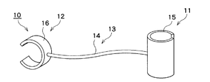
図2に示すように医療用連結保持器具10は、内視鏡1を把持自在な第1把持手段11と、内視鏡用処置具6を把持自在な第2把持手段12と、第1把持手段11と第2把持手段12との間に連結された柔軟連結具13とを備えている。
As shown in FIG. 2, the medical connecting and
このうち、柔軟連結具13は、第1把持手段11および第2把持手段12のうちの一方を、他方に対して移動自在であるとともに保持自在に構成されている。
Among these, the
この柔軟連結具13は、柔軟性を有するワイヤ状部材14からなっている。このワイヤ状部材14に用いる材料としては、内視鏡1の撮像操作部4または内視鏡用処置具6の処置操作部9を保持可能な強度と、内視鏡用処置具6の処置操作部9を内視鏡1の撮像操作部4に対して移動自在な程度の柔軟性とを有し、滅菌処理に使用する薬品に対して耐えることができる材料であればよく、とりわけポリウレタン、ポリアセタール、ポリカーボネート、ポリスチレン、ポリ塩化ビニール、ナイロン、金属材料などを好適に用いることができる。
The
また、第1把持手段11は、断面がC字状に形成され、一対の先端部15aを有する第1ホールド部材15からなっている。同様に第2把持手段12は、断面がC字状に形成された一対の先端部16aを有する第2ホールド部材16からなっている。これら第1ホールド部材15および第2ホールド部材16に用いる材料としては、弾力性を有するとともに滅菌処理に使用する薬品に対して耐薬品性を有する材料であればよく、とりわけポリウレタン、ポリアセタール、ポリカーボネート、ポリスチレン、ポリ塩化ビニール、ナイロン、金属材料などを好適に用いることができる。
The first gripping means 11 includes a
このような医療用連結保持器具10は、予め洗浄されて滅菌処理が施され、その後個々に滅菌包装(図示せず)が施されてディスポーザブルタイプの医療用器具として用いられる。
Such a medical connecting and holding
次に、このような構成からなる本実施の形態の作用について説明する。 Next, the operation of the present embodiment having such a configuration will be described.
医療行為者が内視鏡1と内視鏡用処置具6とを用いて患者の体内の患部に医療行為を行う場合、まず、図1に示すように、内視鏡1の導管3の先端が患者の体内に挿入され、導管3を送り込むことにより導管3の先端に設けられた撮像窓部を患部近傍に到達させる。次に、内視鏡1の挿入口5を通って導管3内に内視鏡用処置具6の鉗子7が挿入され、コード8を送り込むことにより鉗子7が導管3の先端から導出され、鉗子7を患部近傍に到達させる。
When a medical practitioner performs a medical practice on an affected part in a patient's body using the endoscope 1 and the endoscope treatment tool 6, first, as shown in FIG. 1, the distal end of the conduit 3 of the endoscope 1 is used. Is inserted into the body of the patient, and the imaging window provided at the distal end of the conduit 3 is made to reach the vicinity of the affected area by feeding the conduit 3. Next, the
次に、内視鏡1の撮像操作部4が、予め滅菌処理が施された医療用連結保持器具10(図2参照)の第1ホールド部材15に把持されるとともに、内視鏡用処置具6の処置操作部9が第1ホールド部材11にワイヤ状部材14を介して連結された第2ホールド部材16に把持される。この場合、まず、医療用連結保持器具10を滅菌包装から取り出す。次に、第1ホールド部材15の各先端部15aを離間させるようにして、内視鏡1の撮像操作部4を、各先端部15a間を通って第1ホールド部材15内に挿入する。このとき、第1ホールド部材15の各先端部15a間は、内視鏡1の撮像操作部4を挿入する前よりも離間している。このため、第1ホールド部材15が有する弾力性により、第1ホールド部材15が撮像操作部4を把持することができる。同様にして第2ホールド部材16が内視鏡用処置具6の処置操作部9を把持することができる。
Next, the imaging operation unit 4 of the endoscope 1 is held by the
その後、医療行為者は、一方の手で内視鏡1の撮像操作部4を把持して操作し、患部が撮像された画像を見ながら、他方の手で内視鏡用処置具6の処置操作部9を把持して操作し、患部に対して処置が行われる。 Thereafter, the medical practitioner grasps and operates the imaging operation unit 4 of the endoscope 1 with one hand, and treats the endoscopic treatment tool 6 with the other hand while viewing the image in which the affected part is captured. The operation unit 9 is gripped and operated, and a treatment is performed on the affected part.
この間、上述したように、ワイヤ状部材14は内視鏡用処置具6の処置操作部9を内視鏡1の撮像操作部4に対して移動自在な程度の柔軟性を有しているため、内視鏡用処置具6の処置操作部9を内視鏡1の撮像操作部4に対して所望の位置に移動させることができる。この場合、さらに、図3に示すように、第2把持手段12を第1把持手段11に対して90°異なる方向に向けることもできる。このことにより、内視鏡用処置具6の処置操作部9を内視鏡1の撮像操作部4に対して所望の位置に移動させるとともに、必要に応じて所望の向きに向けることもできる。
During this time, as described above, the wire-
また、上述したように、ワイヤ状部材14は、内視鏡1の撮像操作部4または内視鏡用処置具6の処置操作部9を保持可能な強度を有しているため、内視鏡用処置具6の処置操作部9を内視鏡1の撮像操作部4に対してその位置に保持させることができる。このことにより、医療行為者は内視鏡用処置具6の処置操作部9を離して、内視鏡1の撮像操作部4および内視鏡用処置具6の処置操作部9以外の他の医療器具、例えばチューブ、他の鉗子(いずれも図示せず)などを把持することができる。この間、内視鏡用処置具6の処置操作部9は離される前の位置に保持される。
Further, as described above, the wire-
医療行為者は、この他の医療器具を用いた作業を終えると、内視鏡用処置具6の処置操作部9を把持して、患部に対して医療行為を再開することができる。この場合、上述したように、内視鏡用処置具6の処置操作部9は離される前の位置に保持されているため、内視鏡用処置具6の鉗子7が患部に対して位置ずれすることが抑制され、当該患部に対して医療行為を迅速に再開することができる。
When the medical practitioner finishes the work using the other medical instruments, the medical practitioner can grasp the treatment operating unit 9 of the endoscope treatment tool 6 and resume the medical practice on the affected part. In this case, as described above, since the treatment operation unit 9 of the endoscope treatment tool 6 is held at a position before being released, the
このように本実施の形態によれば、患者の体内の患部を撮像する内視鏡1の撮像操作部4が医療用連結保持器具10の第1ホールド部材15により把持され、当該患部を処置する内視鏡用処置具6の処置操作部9が、第1ホールド部材15にワイヤ状部材14を介して連結された第2ホールド部材16により把持される。ここで、ワイヤ状部材14は、第1ホールド部材15および第2ホールド部材16のうちの一方を、他方に対して移動自在であるとともに保持自在に構成されている。このことにより、内視鏡用処置具6の処置操作部9を内視鏡1の撮像操作部4に対して所望の位置に移動させることができるとともに当該位置に保持することができる。このため、医療行為者は、患部を処置している間に、この医療用連結保持器具10を用いて内視鏡用処置具6の処置操作部9を内視鏡1の撮像操作部4に対して保持させて、空いた手を使って他の医療器具などを把持して作業を行うことができる。この結果、医療行為を円滑に行うことができる。
As described above, according to the present embodiment, the imaging operation unit 4 of the endoscope 1 that images the affected part in the patient's body is gripped by the
なお、本実施の形態においては、医療行為者が医療用連結保持器具10を用いて内視鏡用処置具6の処置操作部9を離して内視鏡1の撮像操作部4に対して保持させている例について述べたが、このことに限られることはなく、内視鏡1の撮像操作部4を離して内視鏡用処置具6の処置操作部9に対して保持させるようにしても良い。
In the present embodiment, the medical practitioner uses the medical
第2の実施の形態
次に、図4乃至図7により、本考案の第2の実施の形態における医療用連結保持器具について説明する。ここで図4は、本考案の第2の実施の形態における医療用連結保持器具を示す図であり、図5は、本考案の第2の実施の形態における医療用連結保持器具の他の例を示す図であり、図6は、本考案の第2の実施の形態における医療用連結保持器具のさらなる他の例を示す図であり、図7は、本考案の第2の実施の形態における医療用連結保持器具の柔軟連結具の詳細を示す図である。
Second Embodiment Next, referring to FIGS. 4 to 7, a medical connecting and holding device according to a second embodiment of the present invention will be described. Here, FIG. 4 is a view showing a medical connection holding device according to the second embodiment of the present invention, and FIG. 5 is another example of the medical connection holding device according to the second embodiment of the present invention. FIG. 6 is a diagram showing still another example of the medical connection holding device according to the second embodiment of the present invention, and FIG. 7 is a diagram according to the second embodiment of the present invention. It is a figure which shows the detail of the flexible connector of a medical connection holding | maintenance device.
図4乃至図7に示す第2の実施の形態において、柔軟連結具は、複数の円筒状継手と、各円筒状継手間に延びる連結ピンとを有し、第1把持部材および第2把持部材の構成が異なるのみであり、他の構成は、図1乃至図3に示す第1の実施の形態と略同一である。なお、図4乃至図7において、図1乃至図3に示す第1の実施の形態と同一部分には同一符号を付して詳細な説明は省略する。 In the second embodiment shown in FIGS. 4 to 7, the flexible connector has a plurality of cylindrical joints and a connecting pin extending between the cylindrical joints, and the first gripping member and the second gripping member. Only the configuration is different, and the other configurations are substantially the same as those of the first embodiment shown in FIGS. 1 to 3. 4 to 7, the same parts as those of the first embodiment shown in FIGS. 1 to 3 are denoted by the same reference numerals, and detailed description thereof is omitted.
図4および図7に示すように、柔軟連結具13は、複数の円筒状継手21と、各円筒状継手21間に延びる連結ピン22とを有し、柔軟連結具13の各連結ピン22は、その両端に球体23を有している。また、各円筒状継手21は、内部に連結ピン22の球体23が内接する一対の凹部24を有している。
As shown in FIGS. 4 and 7, the
この円筒状継手21および連結ピン22に用いる材料としては、内視鏡1の撮像操作部4または内視鏡用処置具6の処置操作部9を保持可能な強度と、滅菌処理に使用する薬品に対して耐薬品性を有する材料であればよく、とりわけポリウレタン、ポリアセタール、ポリカーボネート、ポリスチレン、ポリ塩化ビニール、ナイロン、金属材料などを好適に用いることができる。
The material used for the cylindrical joint 21 and the connecting
上述したように、連結ピン22の球体23はこれに対応する円筒状継手21の凹部24に内接して収納されているため、球体23を中心として円筒状継手21の軸線方向と連結ピン22の軸線方向を所望の範囲内で異なる方向に向けることができる。また、連結ピン22の球体23と円筒状継手21の凹部24との間に摩擦力が作用しているため、円筒状継手21と連結ピン22を、ある程度大きな外力が働かない限りその状態で保持することができる。そしてこのような円筒状継手21と連結ピン22は、第1把持手段11と第2把持手段12との間にそれぞれ複数設けられている。このことにより本実施の形態における柔軟連結具13は、内視鏡1または内視鏡用処置具6を保持可能な強度を有するとともに、内視鏡用処置具6の処置操作部9を内視鏡1の撮像操作部4に対して移動自在な程度の柔軟性を有している。このことにより、内視鏡用処置具6の処置操作部9を所望の位置に移動させることができるとともに当該位置に保持させることができる。
As described above, since the
なお、円筒状継手21の個数は、内視鏡用処置具6の処置操作部9を内視鏡1の撮像操作部4に対して所望の範囲にわたって移動可能であれば任意の数とすることができ、例えば図5に示すように、円筒状継手21の個数を減らして、各円筒状継手21の間隔を図4に示す場合よりも広げて構成してもよい。さらに、例えば図6に示すように、円筒状継手21を均等に配置することなく、第1把持手段11および第2把持手段12のうちのいずれか一方の側に円筒状継手21を1つ配置して、他方の側に残りの円筒状継手21の間隔を狭めて配置してもよい。
The number of the
また、図4に示すように、第1把持手段11は、一端に押圧部25aが形成されるとともに他端に当接部25bが形成され、押圧部25aと当接部25bとの間に凹部25cが形成された第1部材25と、一端に押圧部26aが形成されるとともに他端に当接部26bが形成され、押圧部26aと当接部26bとの間に凹部26cが形成された第2部材26とを有している。このうち、第1部材25の押圧部25aに、柔軟連結具13の一端に配置された円筒状継手21の凹部24に内接される球体25d(図7参照)が形成されている。
As shown in FIG. 4, the first
この第1部材25と第2部材26は、押圧部25a、26aと凹部25c、26cとの間に設けられる揺動中心28を中心として互いに揺動自在に連結されている。また、第1部材25と第2部材26との間に、第1部材25の当接部25bと第2部材26の当接部26bとを互いに離間させる方向に第1部材25および第2部材26を付勢するねじりばね27が設けられている。
The
この第1部材25および第2部材26に用いる材料としては、滅菌処理に使用する薬品に対して耐薬品性を有する材料であればよく、とりわけポリウレタン、ポリアセタール、ポリカーボネート、ポリスチレン、ポリ塩化ビニール、ナイロン、金属材料などを好適に用いることができる。
The material used for the
このことにより、第1把持手段11により内視鏡1の撮像操作部4を把持する場合、まず、第1部材25の押圧部25aおよび第2部材26の押圧部26aを把持してねじりばね27の付勢力に対抗して各押圧部25a、26aを互いに近接するように押圧する。このことにより、揺動中心28を中心として第1部材25および第2部材26が回動し、第1部材25の当接部25bと第2部材26の当接部26bとの間に内視鏡1の撮像操作部4が離間される。次に、離間された第1部材25の当接部25bと第2部材26の当接部26bとの間を通って第1部材25の凹部25cと第2部材26の凹部26cとの間に内視鏡1の撮像操作部4が挿入される。その後、第1部材25の押圧部25aおよび第2部材26の押圧部26aを押圧していた力を弱め、第1部材25の凹部25cと第2部材26の凹部26cとを内視鏡1の撮像操作部4の外面に当接させる。このようにして、第1把持手段11により内視鏡1の撮像操作部4を把持することができる。この場合、第1部材25の当接部25bと第2部材26の当接部26bとの間は、内視鏡1の撮像操作部4を挿入する前よりも離間している。このことにより、ねじりばね27の付勢力を受けて、内視鏡1の撮像操作部4が第1部材25の凹部25cと第2部材26の凹部26cにより押圧される。このことにより、第1把持手段11が撮像操作部4を把持することができる。
Accordingly, when the imaging operation unit 4 of the endoscope 1 is gripped by the first
同様にして、第2把持手段12により内視鏡用処置具6の処置操作部9を把持することができる。
Similarly, the treatment operation unit 9 of the endoscope treatment tool 6 can be grasped by the second grasping
このように本実施の形態によれば、患者の体内の患部を撮像する内視鏡1の撮像操作部4が医療用連結保持器具10の第1把持手段11により把持され、当該患部を処置する内視鏡用処置具6の処置操作部9が、第1把持手段11に柔軟連結具15を介して連結された第2把持手段12により把持される。ここで、柔軟連結具13は、第1把持手段11および第2把持手段12のうちの一方を、他方に対して移動自在であるとともに保持自在に構成されている。このことにより、内視鏡用処置具6の処置操作部9を内視鏡1の撮像操作部4に対して所望の位置に移動させることができるとともに当該位置に保持することができる。このため、医療行為者は、患部を処置している間に、この医療用連結保持部10を用いて内視鏡用処置具6の処置操作部9を内視鏡1の撮像操作部4に対して保持させて、空いた手を使って他の医療器具などを把持して作業を行うことができる。この結果、医療行為を円滑に行うことができる。
As described above, according to the present embodiment, the imaging operation unit 4 of the endoscope 1 that images the affected part in the body of the patient is gripped by the first gripping means 11 of the medical
なお本実施の形態においては、柔軟連結具13が、複数の円筒状継手21と、各円筒状継手21間に延びる連結ピン22とを有している例について述べた。しかしながらこのことに限られることはなく、柔軟連結具13が、柔軟性を有するワイヤ状部材14からなっていても良い。
In the present embodiment, the example in which the
また本実施の形態においては、第1把持手段11および第2把持手段12は、第1部材25と第2部材26とねじりばね27とをそれぞれ有している例について述べた。しかしながらこのことに限られることはなく、第1把持手段11および第2把持手段12が断面がC字状に形成された第1ホールド部材15および第2ホールド部材16からそれぞれ構成されていても良い。
Moreover, in this Embodiment, the 1st holding means 11 and the 2nd holding means 12 described the example which has the
1 内視鏡
2 レンズ部
3 導管
4 撮像操作部
5 挿入口
6 内視鏡用処置具
7 鉗子
8 コード
9 処置操作部
10 医療用連結保持器具
11 第1把持手段
12 第2把持手段
13 柔軟連結具
14 ワイヤ状部材
15 第1ホールド部材
15a 先端部
16 第2ホールド部材
21 円筒状継手
22 連結ピン
23 球体
24 凹部
25 第1部材
25a 押圧部
25b 当接部
25c 凹部
25d 球体
26 第2部材
26a 押圧部
26b 当接部
26c 凹部
27 ねじりばね
28 揺動中心
DESCRIPTION OF SYMBOLS 1 Endoscope 2 Lens part 3 Conduit 4
Claims (3)
内視鏡を把持自在な第1把持手段と、
内視鏡用処置具を把持自在な第2把持手段と、
第1把持手段と第2把持手段との間に連結された柔軟連結具と、を備え、
この柔軟連結具は、第1把持手段および第2把持手段のうちの一方を、他方に対して移動自在であるとともに保持自在に構成されていることを特徴とする医療用連結保持器具。 An endoscope that has a conduit and images the affected area inside the patient's body, and a treatment section that can be inserted into the endoscope's conduit at the tip are connected to the endoscope treatment tool that treats the affected area. In the medical holding device for holding,
First gripping means capable of gripping an endoscope;
A second gripping means capable of gripping an endoscope treatment tool;
A flexible coupling connected between the first gripping means and the second gripping means,
This flexible connector is configured to be capable of holding and holding one of the first gripping means and the second gripping means while being movable with respect to the other.
柔軟連結具の各連結ピンは、その両端に球体を有し、
各円筒状継手は、内部に連結ピンの球体が内接する一対の凹部を有していることを特徴とする請求項1に記載の医療用連結保持器具。 The flexible connector has a plurality of cylindrical joints and a connecting pin extending between each cylindrical joint,
Each connecting pin of the flexible connector has a sphere at both ends,
2. The medical connection holding device according to claim 1, wherein each cylindrical joint has a pair of recesses in which a spherical body of a connection pin is inscribed.
Priority Applications (1)
| Application Number | Priority Date | Filing Date | Title |
|---|---|---|---|
| JP2008007819U JP3147945U (en) | 2008-11-07 | 2008-11-07 | Medical connection holding device |
Applications Claiming Priority (1)
| Application Number | Priority Date | Filing Date | Title |
|---|---|---|---|
| JP2008007819U JP3147945U (en) | 2008-11-07 | 2008-11-07 | Medical connection holding device |
Publications (1)
| Publication Number | Publication Date |
|---|---|
| JP3147945U true JP3147945U (en) | 2009-01-29 |
Family
ID=54781541
Family Applications (1)
| Application Number | Title | Priority Date | Filing Date |
|---|---|---|---|
| JP2008007819U Expired - Fee Related JP3147945U (en) | 2008-11-07 | 2008-11-07 | Medical connection holding device |
Country Status (1)
| Country | Link |
|---|---|
| JP (1) | JP3147945U (en) |
-
2008
- 2008-11-07 JP JP2008007819U patent/JP3147945U/en not_active Expired - Fee Related
Similar Documents
| Publication | Publication Date | Title |
|---|---|---|
| ES2884356T3 (en) | Enhanced Flexible Robotic Endoscopy System | |
| JP6242101B2 (en) | Medical device and system | |
| JP6140950B2 (en) | Medical system | |
| EP2198790B1 (en) | Operating mechanism, medical manipulator, and surgical robot system | |
| US9610129B2 (en) | Medical manipulator | |
| JP2001095747A (en) | Electronic endoscope | |
| CN108836483B (en) | Medical treatment tool and surgical system | |
| JPH10503390A (en) | Instruments used for endoscopic surgery | |
| JP2009148859A (en) | Manipulator system and method of controlling manipulator | |
| JP6245877B2 (en) | Operation input device for endoscope treatment tool | |
| JP6234267B2 (en) | Surgical manipulator operating device and surgical manipulator system | |
| JP2009226093A (en) | Operation tool | |
| JP6811676B2 (en) | Drive member, drive mechanism, and manufacturing method of drive mechanism | |
| US8613740B2 (en) | Hand-held manipulator with symmetrical grip | |
| CN109843207B (en) | Computer-assisted teleoperated surgical system and method | |
| JP5859260B2 (en) | forceps | |
| JP3147945U (en) | Medical connection holding device | |
| JP2012040202A (en) | Manipulator | |
| US20200323598A1 (en) | Surgical instrument and surgical robot | |
| JP5792414B2 (en) | Introduction device, endoscope device | |
| JP4407203B2 (en) | Endoscope bending operation device | |
| JP2016034396A (en) | Assisting grip for endoscope | |
| JP6895007B2 (en) | Medical treatment tool | |
| CN115103650A (en) | Surgical robot and control unit for surgical robot | |
| KR100994373B1 (en) | Surgical instrument |
Legal Events
| Date | Code | Title | Description |
|---|---|---|---|
| R150 | Certificate of patent (=grant) or registration of utility model |
Free format text: JAPANESE INTERMEDIATE CODE: R150 |
|
| FPAY | Renewal fee payment (prs date is renewal date of database) |
Free format text: PAYMENT UNTIL: 20120107 Year of fee payment: 3 |
|
| FPAY | Renewal fee payment (prs date is renewal date of database) |
Free format text: PAYMENT UNTIL: 20120107 Year of fee payment: 3 |
|
| S531 | Written request for registration of change of domicile |
Free format text: JAPANESE INTERMEDIATE CODE: R323531 |
|
| FPAY | Renewal fee payment (prs date is renewal date of database) |
Free format text: PAYMENT UNTIL: 20120107 Year of fee payment: 3 |
|
| R350 | Written notification of registration of transfer |
Free format text: JAPANESE INTERMEDIATE CODE: R350 |
|
| FPAY | Renewal fee payment (prs date is renewal date of database) |
Free format text: PAYMENT UNTIL: 20130107 Year of fee payment: 4 |
|
| LAPS | Cancellation because of no payment of annual fees |