JP2024540887A - BAG3 METHODS AND USES FOR THE TREATMENT OF INFLAMMATION - Google Patents
BAG3 METHODS AND USES FOR THE TREATMENT OF INFLAMMATION Download PDFInfo
- Publication number
- JP2024540887A JP2024540887A JP2024523137A JP2024523137A JP2024540887A JP 2024540887 A JP2024540887 A JP 2024540887A JP 2024523137 A JP2024523137 A JP 2024523137A JP 2024523137 A JP2024523137 A JP 2024523137A JP 2024540887 A JP2024540887 A JP 2024540887A
- Authority
- JP
- Japan
- Prior art keywords
- bag3
- formulation
- vector
- mice
- protein
- Prior art date
- Legal status (The legal status is an assumption and is not a legal conclusion. Google has not performed a legal analysis and makes no representation as to the accuracy of the status listed.)
- Pending
Links
Images

Classifications
-
- A—HUMAN NECESSITIES
- A61—MEDICAL OR VETERINARY SCIENCE; HYGIENE
- A61K—PREPARATIONS FOR MEDICAL, DENTAL OR TOILETRY PURPOSES
- A61K38/00—Medicinal preparations containing peptides
- A61K38/16—Peptides having more than 20 amino acids; Gastrins; Somatostatins; Melanotropins; Derivatives thereof
- A61K38/17—Peptides having more than 20 amino acids; Gastrins; Somatostatins; Melanotropins; Derivatives thereof from animals; from humans
- A61K38/1703—Peptides having more than 20 amino acids; Gastrins; Somatostatins; Melanotropins; Derivatives thereof from animals; from humans from vertebrates
- A61K38/1709—Peptides having more than 20 amino acids; Gastrins; Somatostatins; Melanotropins; Derivatives thereof from animals; from humans from vertebrates from mammals
- A61K38/1761—Apoptosis related proteins, e.g. Apoptotic protease-activating factor-1 (APAF-1), Bax, Bax-inhibitory protein(s)(BI; bax-I), Myeloid cell leukemia associated protein (MCL-1), Inhibitor of apoptosis [IAP] or Bcl-2
-
- A—HUMAN NECESSITIES
- A01—AGRICULTURE; FORESTRY; ANIMAL HUSBANDRY; HUNTING; TRAPPING; FISHING
- A01K—ANIMAL HUSBANDRY; AVICULTURE; APICULTURE; PISCICULTURE; FISHING; REARING OR BREEDING ANIMALS, NOT OTHERWISE PROVIDED FOR; NEW BREEDS OF ANIMALS
- A01K67/00—Rearing or breeding animals, not otherwise provided for; New or modified breeds of animals
- A01K67/027—New or modified breeds of vertebrates
- A01K67/0275—Genetically modified vertebrates, e.g. transgenic
-
- A—HUMAN NECESSITIES
- A61—MEDICAL OR VETERINARY SCIENCE; HYGIENE
- A61K—PREPARATIONS FOR MEDICAL, DENTAL OR TOILETRY PURPOSES
- A61K31/00—Medicinal preparations containing organic active ingredients
- A61K31/70—Carbohydrates; Sugars; Derivatives thereof
- A61K31/7088—Compounds having three or more nucleosides or nucleotides
-
- A—HUMAN NECESSITIES
- A61—MEDICAL OR VETERINARY SCIENCE; HYGIENE
- A61K—PREPARATIONS FOR MEDICAL, DENTAL OR TOILETRY PURPOSES
- A61K38/00—Medicinal preparations containing peptides
- A61K38/16—Peptides having more than 20 amino acids; Gastrins; Somatostatins; Melanotropins; Derivatives thereof
- A61K38/17—Peptides having more than 20 amino acids; Gastrins; Somatostatins; Melanotropins; Derivatives thereof from animals; from humans
- A61K38/1703—Peptides having more than 20 amino acids; Gastrins; Somatostatins; Melanotropins; Derivatives thereof from animals; from humans from vertebrates
- A61K38/1709—Peptides having more than 20 amino acids; Gastrins; Somatostatins; Melanotropins; Derivatives thereof from animals; from humans from vertebrates from mammals
-
- A—HUMAN NECESSITIES
- A61—MEDICAL OR VETERINARY SCIENCE; HYGIENE
- A61K—PREPARATIONS FOR MEDICAL, DENTAL OR TOILETRY PURPOSES
- A61K48/00—Medicinal preparations containing genetic material which is inserted into cells of the living body to treat genetic diseases; Gene therapy
- A61K48/005—Medicinal preparations containing genetic material which is inserted into cells of the living body to treat genetic diseases; Gene therapy characterised by an aspect of the 'active' part of the composition delivered, i.e. the nucleic acid delivered
- A61K48/0058—Nucleic acids adapted for tissue specific expression, e.g. having tissue specific promoters as part of a contruct
-
- A—HUMAN NECESSITIES
- A61—MEDICAL OR VETERINARY SCIENCE; HYGIENE
- A61K—PREPARATIONS FOR MEDICAL, DENTAL OR TOILETRY PURPOSES
- A61K48/00—Medicinal preparations containing genetic material which is inserted into cells of the living body to treat genetic diseases; Gene therapy
- A61K48/005—Medicinal preparations containing genetic material which is inserted into cells of the living body to treat genetic diseases; Gene therapy characterised by an aspect of the 'active' part of the composition delivered, i.e. the nucleic acid delivered
- A61K48/0066—Manipulation of the nucleic acid to modify its expression pattern, e.g. enhance its duration of expression, achieved by the presence of particular introns in the delivered nucleic acid
-
- A—HUMAN NECESSITIES
- A61—MEDICAL OR VETERINARY SCIENCE; HYGIENE
- A61K—PREPARATIONS FOR MEDICAL, DENTAL OR TOILETRY PURPOSES
- A61K48/00—Medicinal preparations containing genetic material which is inserted into cells of the living body to treat genetic diseases; Gene therapy
- A61K48/0083—Medicinal preparations containing genetic material which is inserted into cells of the living body to treat genetic diseases; Gene therapy characterised by an aspect of the administration regime
-
- A—HUMAN NECESSITIES
- A61—MEDICAL OR VETERINARY SCIENCE; HYGIENE
- A61P—SPECIFIC THERAPEUTIC ACTIVITY OF CHEMICAL COMPOUNDS OR MEDICINAL PREPARATIONS
- A61P25/00—Drugs for disorders of the nervous system
- A61P25/14—Drugs for disorders of the nervous system for treating abnormal movements, e.g. chorea, dyskinesia
- A61P25/16—Anti-Parkinson drugs
-
- A—HUMAN NECESSITIES
- A61—MEDICAL OR VETERINARY SCIENCE; HYGIENE
- A61P—SPECIFIC THERAPEUTIC ACTIVITY OF CHEMICAL COMPOUNDS OR MEDICINAL PREPARATIONS
- A61P29/00—Non-central analgesic, antipyretic or antiinflammatory agents, e.g. antirheumatic agents; Non-steroidal antiinflammatory drugs [NSAID]
-
- A—HUMAN NECESSITIES
- A61—MEDICAL OR VETERINARY SCIENCE; HYGIENE
- A61P—SPECIFIC THERAPEUTIC ACTIVITY OF CHEMICAL COMPOUNDS OR MEDICINAL PREPARATIONS
- A61P37/00—Drugs for immunological or allergic disorders
- A61P37/02—Immunomodulators
- A61P37/06—Immunosuppressants, e.g. drugs for graft rejection
-
- A—HUMAN NECESSITIES
- A61—MEDICAL OR VETERINARY SCIENCE; HYGIENE
- A61P—SPECIFIC THERAPEUTIC ACTIVITY OF CHEMICAL COMPOUNDS OR MEDICINAL PREPARATIONS
- A61P9/00—Drugs for disorders of the cardiovascular system
-
- C—CHEMISTRY; METALLURGY
- C07—ORGANIC CHEMISTRY
- C07K—PEPTIDES
- C07K14/00—Peptides having more than 20 amino acids; Gastrins; Somatostatins; Melanotropins; Derivatives thereof
- C07K14/435—Peptides having more than 20 amino acids; Gastrins; Somatostatins; Melanotropins; Derivatives thereof from animals; from humans
- C07K14/46—Peptides having more than 20 amino acids; Gastrins; Somatostatins; Melanotropins; Derivatives thereof from animals; from humans from vertebrates
- C07K14/47—Peptides having more than 20 amino acids; Gastrins; Somatostatins; Melanotropins; Derivatives thereof from animals; from humans from vertebrates from mammals
- C07K14/4701—Peptides having more than 20 amino acids; Gastrins; Somatostatins; Melanotropins; Derivatives thereof from animals; from humans from vertebrates from mammals not used
- C07K14/4747—Apoptosis related proteins
-
- C—CHEMISTRY; METALLURGY
- C12—BIOCHEMISTRY; BEER; SPIRITS; WINE; VINEGAR; MICROBIOLOGY; ENZYMOLOGY; MUTATION OR GENETIC ENGINEERING
- C12N—MICROORGANISMS OR ENZYMES; COMPOSITIONS THEREOF; PROPAGATING, PRESERVING, OR MAINTAINING MICROORGANISMS; MUTATION OR GENETIC ENGINEERING; CULTURE MEDIA
- C12N15/00—Mutation or genetic engineering; DNA or RNA concerning genetic engineering, vectors, e.g. plasmids, or their isolation, preparation or purification; Use of hosts therefor
- C12N15/09—Recombinant DNA-technology
- C12N15/63—Introduction of foreign genetic material using vectors; Vectors; Use of hosts therefor; Regulation of expression
- C12N15/79—Vectors or expression systems specially adapted for eukaryotic hosts
- C12N15/85—Vectors or expression systems specially adapted for eukaryotic hosts for animal cells
- C12N15/86—Viral vectors
-
- A—HUMAN NECESSITIES
- A01—AGRICULTURE; FORESTRY; ANIMAL HUSBANDRY; HUNTING; TRAPPING; FISHING
- A01K—ANIMAL HUSBANDRY; AVICULTURE; APICULTURE; PISCICULTURE; FISHING; REARING OR BREEDING ANIMALS, NOT OTHERWISE PROVIDED FOR; NEW BREEDS OF ANIMALS
- A01K2217/00—Genetically modified animals
- A01K2217/07—Animals genetically altered by homologous recombination
- A01K2217/075—Animals genetically altered by homologous recombination inducing loss of function, i.e. knock out
-
- A—HUMAN NECESSITIES
- A01—AGRICULTURE; FORESTRY; ANIMAL HUSBANDRY; HUNTING; TRAPPING; FISHING
- A01K—ANIMAL HUSBANDRY; AVICULTURE; APICULTURE; PISCICULTURE; FISHING; REARING OR BREEDING ANIMALS, NOT OTHERWISE PROVIDED FOR; NEW BREEDS OF ANIMALS
- A01K2217/00—Genetically modified animals
- A01K2217/07—Animals genetically altered by homologous recombination
- A01K2217/075—Animals genetically altered by homologous recombination inducing loss of function, i.e. knock out
- A01K2217/077—Animals genetically altered by homologous recombination inducing loss of function, i.e. knock out heterozygous knock out animals displaying phenotype
-
- A—HUMAN NECESSITIES
- A01—AGRICULTURE; FORESTRY; ANIMAL HUSBANDRY; HUNTING; TRAPPING; FISHING
- A01K—ANIMAL HUSBANDRY; AVICULTURE; APICULTURE; PISCICULTURE; FISHING; REARING OR BREEDING ANIMALS, NOT OTHERWISE PROVIDED FOR; NEW BREEDS OF ANIMALS
- A01K2217/00—Genetically modified animals
- A01K2217/20—Animal model comprising regulated expression system
- A01K2217/206—Animal model comprising tissue-specific expression system, e.g. tissue specific expression of transgene, of Cre recombinase
-
- A—HUMAN NECESSITIES
- A01—AGRICULTURE; FORESTRY; ANIMAL HUSBANDRY; HUNTING; TRAPPING; FISHING
- A01K—ANIMAL HUSBANDRY; AVICULTURE; APICULTURE; PISCICULTURE; FISHING; REARING OR BREEDING ANIMALS, NOT OTHERWISE PROVIDED FOR; NEW BREEDS OF ANIMALS
- A01K2227/00—Animals characterised by species
- A01K2227/10—Mammal
- A01K2227/105—Murine
-
- A—HUMAN NECESSITIES
- A01—AGRICULTURE; FORESTRY; ANIMAL HUSBANDRY; HUNTING; TRAPPING; FISHING
- A01K—ANIMAL HUSBANDRY; AVICULTURE; APICULTURE; PISCICULTURE; FISHING; REARING OR BREEDING ANIMALS, NOT OTHERWISE PROVIDED FOR; NEW BREEDS OF ANIMALS
- A01K2267/00—Animals characterised by purpose
- A01K2267/03—Animal model, e.g. for test or diseases
- A01K2267/035—Animal model for multifactorial diseases
- A01K2267/0375—Animal model for cardiovascular diseases
-
- C—CHEMISTRY; METALLURGY
- C12—BIOCHEMISTRY; BEER; SPIRITS; WINE; VINEGAR; MICROBIOLOGY; ENZYMOLOGY; MUTATION OR GENETIC ENGINEERING
- C12N—MICROORGANISMS OR ENZYMES; COMPOSITIONS THEREOF; PROPAGATING, PRESERVING, OR MAINTAINING MICROORGANISMS; MUTATION OR GENETIC ENGINEERING; CULTURE MEDIA
- C12N2750/00—MICROORGANISMS OR ENZYMES; COMPOSITIONS THEREOF; PROPAGATING, PRESERVING, OR MAINTAINING MICROORGANISMS; MUTATION OR GENETIC ENGINEERING; CULTURE MEDIA ssDNA viruses
- C12N2750/00011—Details
- C12N2750/14011—Parvoviridae
- C12N2750/14111—Dependovirus, e.g. adenoassociated viruses
- C12N2750/14141—Use of virus, viral particle or viral elements as a vector
- C12N2750/14143—Use of virus, viral particle or viral elements as a vector viral genome or elements thereof as genetic vector
Landscapes
- Health & Medical Sciences (AREA)
- Life Sciences & Earth Sciences (AREA)
- Chemical & Material Sciences (AREA)
- General Health & Medical Sciences (AREA)
- Engineering & Computer Science (AREA)
- Medicinal Chemistry (AREA)
- Veterinary Medicine (AREA)
- Animal Behavior & Ethology (AREA)
- Public Health (AREA)
- Pharmacology & Pharmacy (AREA)
- Organic Chemistry (AREA)
- Bioinformatics & Cheminformatics (AREA)
- Genetics & Genomics (AREA)
- Molecular Biology (AREA)
- Epidemiology (AREA)
- Zoology (AREA)
- Biomedical Technology (AREA)
- Biotechnology (AREA)
- Nuclear Medicine, Radiotherapy & Molecular Imaging (AREA)
- General Chemical & Material Sciences (AREA)
- Chemical Kinetics & Catalysis (AREA)
- Biochemistry (AREA)
- Immunology (AREA)
- Gastroenterology & Hepatology (AREA)
- Proteomics, Peptides & Aminoacids (AREA)
- Neurology (AREA)
- Neurosurgery (AREA)
- Marine Sciences & Fisheries (AREA)
- Rheumatology (AREA)
- Pain & Pain Management (AREA)
- Biophysics (AREA)
- Environmental Sciences (AREA)
- Toxicology (AREA)
- Psychology (AREA)
- General Engineering & Computer Science (AREA)
- Wood Science & Technology (AREA)
- Biodiversity & Conservation Biology (AREA)
- Animal Husbandry (AREA)
- Virology (AREA)
- Plant Pathology (AREA)
Abstract
Bag3は、心臓、骨格筋、中枢神経系において、及び多くのがんにおいて主に発現される多機能タンパク質である。BAG3はほんの10年前にクローニングされたものの、調査は、遺伝的バリアント、特にハプロ不全をもたらすものが、重度の左心室機能不全につながり得ることを示しており;しかしながら、関与する全メカニズムは不明確なままである。Bag3の生物学に対する心不全自体の影響を取り除くために、単一アレルノックアウトを持つトランスジェニック(ransgenic)マウスを、心不全の任意の明白な兆しが確かになる前の8~10週齢で調査した。結果は驚くべきものであり且つ有益な情報を与えた。まず、正常な表現型にもかかわらず、若齢Bag3+/-は、代謝及びアポトーシスと関連したタンパク質の変化によって特徴付けられるプロテオームの著しい変化を有することが見出された。この知見と一貫して、ミトコンドリア膜電位を維持する責任を負う重大なタンパク質のレベルの減少が観察された。若齢マウスは、アポトーシスの外因性及び内因性経路の間の均衡からシフトすることも見出された。しかしながら、ストレスの存在及びBag3の非存在下では、均衡から外因性優勢なシステム(切断カスパーゼ8)へのシフトがあった。Bag3によって調節される多種多様の重大な経路は、とりわけストレスの間のより重要な役割、及びこの役割が、タンパク質を偶然に会わせるのではなく、タンパク質が最も有効であり得る場所にタンパク質を保持する細胞内接着剤として働くことを含むことがあることを示唆する。
【選択図】図9
Bag3 is a multifunctional protein that is predominantly expressed in the heart, skeletal muscle, central nervous system, and in many cancers. Although BAG3 was cloned only a decade ago, studies have shown that genetic variants, especially those resulting in haploinsufficiency, can lead to severe left ventricular dysfunction; however, the full mechanisms involved remain unclear. To eliminate the influence of heart failure itself on the biology of Bag3, transgenic mice with a single allele knockout were studied at 8-10 weeks of age, before any obvious signs of heart failure were evident. The results were surprising and informative. First, despite a normal phenotype, young Bag3 +/- were found to have a striking alteration of the proteome characterized by changes in proteins associated with metabolism and apoptosis. Consistent with this finding, a decrease in the levels of critical proteins responsible for maintaining the mitochondrial membrane potential was observed. Young mice were also found to have a shift from the balance between the extrinsic and intrinsic pathways of apoptosis. However, in the presence of stress and in the absence of Bag3, there was a shift from the equilibrium to an extrinsic dominated system (cleaved caspase 8). The wide variety of critical pathways regulated by Bag3 suggests a more important role, especially during stress, and that this role may involve acting as an intracellular glue that holds proteins where they can be most effective, rather than forcing them to meet by chance.
[Selection diagram] Figure 9
Description
[序論]
BAG3は、遍在的に発現されるが、心臓、骨格筋、中枢神経系において、及び多くのがんにおいて最も顕著な多機能タンパク質である(1、2)。遺伝性及び特発性拡張型心筋症(DCM)の両方を有する患者由来のDNAについての複数のゲノムワイド関連調査及び全エクソーム又は全ゲノムシーケンシングは、BAG3の単一アレルの喪失が、疾患の重要な原因であることを示している(3~6)。BAG3のホモ接合型欠失が、出生後早期に致死性であり(7)、一方で単一アレルの機能喪失が、ゼブラフィッシュにおいて(3)、ヒト変異を担持する間葉系幹細胞由来心筋細胞において(8)、点変異及びハプロ不全の両方を有するマウスモデルにおいて(9、10)、並びにハプロ不全を有するヒトにおいて(11、12)、筋細胞機能不全の発症につながることから、このことは動物モデルにおいて裏付けられている。調査は、遺伝子型的に正常である、心不全及び駆出率の低下(HFrEF)を有する患者が、BAG3短縮を有する患者において見られるものに匹敵する、心臓移植時の心室心筋におけるBAG3の低下を有することも示している(11、12)。心臓とは対照的に、がん細胞におけるBAG3の過剰発現は、化学療法抵抗性、並びに転移及び局所侵入の傾向の増加をもたらす(13、14)。先進国における2種の主な疾患である心臓疾患及びがんにおけるBAG3の明白な重要性にもかかわらず、健康及び疾患におけるBAG3の役割の全容は十分に規定されていない。
[Introduction]
BAG3 is a ubiquitously expressed, but multifunctional protein, most prominent in the heart, skeletal muscle, central nervous system, and in many cancers (1, 2). Multiple genome-wide association studies and whole-exome or whole-genome sequencing of DNA from patients with both hereditary and idiopathic dilated cardiomyopathy (DCM) have shown that loss of a single allele of BAG3 is a significant cause of disease (3-6). This is supported in animal models, as homozygous deletion of BAG3 is lethal early after birth (7), while loss of function of a single allele leads to the development of myocyte dysfunction in zebrafish (3), in mesenchymal stem cell-derived cardiomyocytes carrying human mutations (8), in mouse models with both point mutations and haploinsufficiency (9, 10), and in humans with haploinsufficiency (11, 12). Studies have also shown that genotypically normal patients with heart failure and reduced ejection fraction (HFrEF) have reduced BAG3 in ventricular myocardium at the time of heart transplantation comparable to that seen in patients with BAG3 truncation (11, 12). In contrast to the heart, overexpression of BAG3 in cancer cells leads to chemotherapy resistance and increased propensity for metastasis and local invasion (13, 14). Despite the clear importance of BAG3 in heart disease and cancer, two major diseases in developed countries, the full extent of BAG3's role in health and disease has not been fully defined.
BAG3の構造は、BAG3が多種多様の分子及び細胞活性に影響することを可能にする数多くのタンパク質-タンパク質結合ドメインを有する。心臓において、BAG3は、熱ショックタンパク質70(hsp/hsc70)に対するシャペロンとして働くことによってオートファジーを増進する(15)。BAG3は、抗アポトーシスタンパク質Bcl-2と相互作用することによってアポトーシスを阻害し(16)、ベータ-アドレナリン受容体(b-AR)とL型Ca2+チャネルとを連結することによって興奮-収縮連関を向上させ(17)、筋節の完全性を維持する(18)。BAG3の様々な機能は、複数の結合部位及び多様な群の結合パートナーの存在によって促される。例えば、PXXPドメインは、モーター・ダイニン輸送システムの近位端に対する分子アンカーとして働き、一方で2つのイソロイシン-プロリン-バリン(IPV)モチーフは、小型熱ショックタンパク質HspB6及びHspB8に結合し、マクロオートファジーを支持する(1)。 The structure of BAG3 has numerous protein-protein binding domains that enable it to affect a wide variety of molecular and cellular activities. In the heart, BAG3 promotes autophagy by acting as a chaperone for heat shock protein 70 (hsp/hsc70) (15). BAG3 inhibits apoptosis by interacting with the antiapoptotic protein Bcl-2 (16), improves excitation-contraction coupling by linking the beta-adrenergic receptor (b-AR) to L-type Ca2+ channels (17), and maintains sarcomere integrity (18). The diverse functions of BAG3 are facilitated by the presence of multiple binding sites and a diverse set of binding partners. For example, the PXXP domain serves as a molecular anchor for the proximal end of the motor-dynein transport system, while two isoleucine-proline-valine (IPV) motifs bind the small heat shock proteins HspB6 and HspB8, supporting macroautophagy (1).
動物モデルにおける最近の調査は、BAG3における特異的ヘテロ接合型遺伝的バリアントと、固有の細胞又は分子表現型とを関連付け始めている。例えば、E455K機能喪失変異は、BAG3とHsp-70との間の相互作用を妨害し、熱ショックタンパク質の不安定性及びタンパク質恒常性の喪失をもたらす(9)。稀なP209Lバリアントは、バリアント自身とHsp70クライアントとの凝集を引き起こす機能変異のドミナントゲインであり、この凝集はHsp70オートファジーネットワークの失速(stalling)をもたらし、拘束型心筋症につながる(19)。対照的に、BAG3のP209Sバリアントは、2人の患者において、遅発性軸索シャルコー・マリー・トゥースニューロパチーと関連して報告されている(20)。しかしながら、明瞭さの欠如は、マウスの心臓の表現型に対するBAG3P209L変異の効果の結果が不一致であることから、依然として存在する(21)(22)。 Recent investigations in animal models have begun to link specific heterozygous genetic variants in BAG3 with unique cellular or molecular phenotypes. For example, the E455K loss-of-function mutation disrupts the interaction between BAG3 and Hsp-70, resulting in heat shock protein instability and loss of proteostasis (9). The rare P209L variant is a dominant gain of function mutation that causes aggregation of the variant with its own Hsp70 clients, which leads to stalling of the Hsp70 autophagy network and restrictive cardiomyopathy (19). In contrast, the P209S variant of BAG3 has been reported in association with late-onset axonal Charcot-Marie-Tooth neuropathy in two patients (20). However, a lack of clarity still exists, as the effects of the BAG3 P209L mutation on cardiac phenotypes in mice are inconsistent (21)(22).
本明細書に開示されるように、BAG3ハプロ不全の心臓生物学についてのより良い理解を得るために、タンデム質量分析と連動した高圧液体クロマトグラフィー(LC-MS/MS)を使用して、ヘテロ接合型BAG3除去を有する、左心室(LV)機能及び心臓サイズが依然として正常である時点である若齢8~10週齢のマウスにおいて示差的に発現されるタンパク質を特定した。BAG3のヘテロ接合型ノックアウトを有するマウスは、8~10週齢の時点で心エコー検査によって表現型的に正常であるが、18週齢までに左心室機能が著しく変化することが実証されている。 As disclosed herein, to gain a better understanding of the cardiac biology of BAG3 haploinsufficiency, high pressure liquid chromatography coupled with tandem mass spectrometry (LC-MS/MS) was used to identify differentially expressed proteins in young 8-10 week old mice with heterozygous BAG3 ablation, a time point at which left ventricular (LV) function and heart size remain normal. Mice with heterozygous knockout of BAG3 are phenotypically normal by echocardiography at 8-10 weeks of age, but demonstrate markedly altered LV function by 18 weeks of age.
プロテオーム解析は、心臓生物学の2つのエリア:ミトコンドリア機能及びプログラム細胞死又はアポトーシスを明らかにした。8~10週齢BAG3+/-マウスのプロテオームについての評価は、正常な表現型にもかかわらず、代謝及びプログラム細胞死又はアポトーシスと関連したタンパク質の異常を明らかにした。 Proteomic analysis revealed abnormalities in two areas of cardiac biology: mitochondrial function and programmed cell death or apoptosis. Evaluation of the proteome of 8-10 week old BAG3 +/- mice revealed abnormalities in proteins associated with metabolism and programmed cell death or apoptosis, despite a normal phenotype.
本明細書に開示されるように、とりわけ、BCL2関連アサノジーン(Athanogene)3(BAG3)は、心臓における、並びに他の組織及び細胞タイプにおけるアポトーシスの内因性及び外因性経路の両方の重大な構成要素であることがここで発見されている。特に、BAG3は、標準的並びに非標準的経路の両方を通じて、細胞アポトーシスを調節する。 As disclosed herein, inter alia, it has now been discovered that BCL2-associated athanogen 3 (BAG3) is a critical component of both the intrinsic and extrinsic pathways of apoptosis in the heart, as well as in other tissues and cell types. In particular, BAG3 regulates cell apoptosis through both canonical and non-canonical pathways.
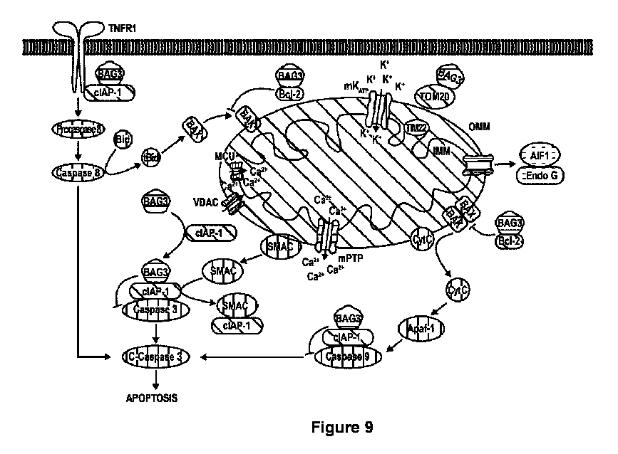
また本明細書に開示されるように、とりわけ、BAG3は、アポトーシスにおける後期ステップ、つまりカスパーゼ3の活性化を修飾することがここで発見されている。BAG3は、アポトーシスタンパク質1/2の阻害剤(cIAP1/2又はcIAP)と直接相互作用する。正常な条件下で、BAG3タンパク質は正常なレベルで存在し、アポトーシスの細胞阻害剤1(CIAP-1)に結合し、カスパーゼ3に結合し且つその活性を阻害するCIAP1の能力を促す。しかしながら、BAG3ハプロ不全の条件下では、ミトコンドリアから漏出する、カスパーゼの第2のミトコンドリア由来活性化因子、すなわちSMAC(直接IAP結合タンパク質(DIABLO)とも称される)につながる膜電位の減少を含めた、ミトコンドリア機能の著しい異常がある。SMACは、次いで細胞質へ移行し、CIAP-1に結合し、それによってカスパーゼ3からCIAP-1を解放し、カスパーゼ3が活性化されるのを可能にする。活性化されたカスパーゼ3は、次いで細胞の主要な構成要素を溶解し得る。したがって、BAG3は、カスパーゼ-3の活性化を低下、阻害、又は減少させ、それによって炎症又は炎症応答を低下させるために使用又は製剤化され得る。
As also disclosed herein, it has now been discovered that, among other things, BAG3 modulates a later step in apoptosis, namely activation of caspase-3. BAG3 interacts directly with inhibitor of
本明細書にさらに開示されるように、BAG3のレベルの減少は、カスパーゼ活性化の内因性及び外因性経路の間の均衡から、カスパーゼ-8の活性化(切断カスパーゼ8)を好むシグナル伝達へのシフトを引き起こすことがここで発見されている。したがって、BAG3は、カスパーゼ-8の活性化を低下、阻害、又は減少させるために使用又は製剤化され得る。 As further disclosed herein, it has now been discovered that reducing the levels of BAG3 causes a shift in the balance between the intrinsic and extrinsic pathways of caspase activation to signaling that favors activation of caspase-8 (cleaved caspase-8). Thus, BAG3 can be used or formulated to reduce, inhibit, or decrease activation of caspase-8.
本明細書に付加的に開示されるように、とりわけ、BAG3は、ミトコンドリアインポート受容体サブユニットTOM22、及びCa2+ユニポーター、ミトコンドリア代謝、並びにミトコンドリア膜電位の発生とそれぞれ直接相互作用することがここで発見されている。 As additionally disclosed herein, inter alia, BAG3 has now been discovered to directly interact with the mitochondrial import receptor subunit TOM22 and the Ca 2+ uniporter, mitochondrial metabolism, and generation of mitochondrial membrane potential, respectively.
本明細書に付加的に開示されるように、とりわけ、BAG3の減少は、ポリ(ADP-リボース)ポリメラーゼ1(PARP1)の増加をもたらすことがここで発見されている。そのようなPARP1の増加は、アルファ-シヌクレインの増加、及びパーキンソン症状の悪化をもたらし得る。したがって、BAG3は、PARP1を低下、阻害、又は減少させるために使用又は製剤化され得る。 As additionally disclosed herein, it has now been discovered that, among other things, a decrease in BAG3 leads to an increase in poly(ADP-ribose) polymerase 1 (PARP1). Such an increase in PARP1 can lead to an increase in alpha-synuclein and an exacerbation of Parkinson's symptoms. Thus, BAG3 can be used or formulated to lower, inhibit, or decrease PARP1.
本発明によれば、BAG3は、TNFシグナル伝達をモジュレートするために使用又は製剤化され得る。特定の実施形態において、BAG3は、TNFシグナル伝達を低下、阻害、又は減少させるために使用又は製剤化され得る。 In accordance with the present invention, BAG3 may be used or formulated to modulate TNF signaling. In certain embodiments, BAG3 may be used or formulated to reduce, inhibit, or decrease TNF signaling.
本発明によれば、BAG3は、炎症をモジュレートするために使用又は製剤化され得る。特定の実施形態において、BAG3は、炎症を低下、阻害、減少、又は治療するために使用又は製剤化され得る。 In accordance with the present invention, BAG3 may be used or formulated to modulate inflammation. In certain embodiments, BAG3 may be used or formulated to reduce, inhibit, decrease, or treat inflammation.
本発明によれば、BAG3は、炎症応答をモジュレートするために使用又は製剤化され得る。特定の実施形態において、BAG3は、炎症応答を低下、阻害、減少、又は治療するために使用又は製剤化され得る。 In accordance with the present invention, BAG3 may be used or formulated to modulate an inflammatory response. In certain embodiments, BAG3 may be used or formulated to reduce, inhibit, decrease, or treat an inflammatory response.
炎症又は炎症応答は、臓器又は組織において等、全身的、領域的、又は局所的であり得る。特定の態様において、低下させるか、阻害するか、減少させるか、又は治療するためにBAG3が使用又は製剤化され得る、炎症又は炎症応答の非限定的な例は、肺系、肺、心血管系、中枢神経系、骨、骨格関節、骨格筋、胃腸系、胃、小腸、大腸、肝臓、腎臓、及び膵臓において生じる。 Inflammation or inflammatory responses can be systemic, regional, or local, such as in an organ or tissue. Non-limiting examples of inflammation or inflammatory responses that BAG3 may be used or formulated to reduce, inhibit, decrease, or treat in certain embodiments occur in the pulmonary system, lungs, cardiovascular system, central nervous system, bone, skeletal joints, skeletal muscle, gastrointestinal system, stomach, small intestine, large intestine, liver, kidneys, and pancreas.
特定の態様において、低下させるか、阻害するか、減少させるか、又は治療するためにBAG3が使用又は製剤化され得る、炎症又は炎症応答の非限定的な例は、慢性炎症性疾患、慢性炎症性脱髄性多発神経炎、一次性免疫性血小板減少症、老人性摂食障害、腸管炎症、炎症性腸疾患、潰瘍性大腸炎、クローン病、ループス、関節リウマチ、慢性心筋炎、Covid19感染後の慢性心筋炎、乾癬、乾癬性関節炎、及び強直性脊椎炎を含む。 In certain embodiments, non-limiting examples of inflammation or inflammatory responses that BAG3 may be used or formulated to reduce, inhibit, decrease, or treat include chronic inflammatory diseases, chronic inflammatory demyelinating polyneuropathy, primary immune thrombocytopenia, geriatric eating disorders, intestinal inflammation, inflammatory bowel disease, ulcerative colitis, Crohn's disease, lupus, rheumatoid arthritis, chronic myocarditis, chronic myocarditis after COVID-19 infection, psoriasis, psoriatic arthritis, and ankylosing spondylitis.
本発明によれば、BAG3は、PARP1レベル、発現、又は活性をモジュレートするために使用又は製剤化され得る。特定の実施形態において、BAG3は、PARP1レベル、発現、又は活性を低下、阻害、又は減少させるために使用又は製剤化され得る。特定の実施形態において、BAG3は、アルファ-シヌクレインの量を低下、阻害、減少、又は安定させるために使用又は製剤化され得る。特定の実施形態において、BAG3は、パーキンソン病の1つ又は複数の症状の悪化又は重症度を低下、阻害、減少、又は減少させるために使用又は製剤化され得る。 In accordance with the present invention, BAG3 may be used or formulated to modulate PARP1 levels, expression, or activity. In certain embodiments, BAG3 may be used or formulated to lower, inhibit, or decrease PARP1 levels, expression, or activity. In certain embodiments, BAG3 may be used or formulated to lower, inhibit, reduce, or stabilize the amount of alpha-synuclein. In certain embodiments, BAG3 may be used or formulated to lower, inhibit, reduce, or decrease the worsening or severity of one or more symptoms of Parkinson's disease.
特定の実施形態において、BAG3コード核酸は、BAG3タンパク質又はその活性BAG3ペプチドを発現する発現ベクターを含む。 In certain embodiments, the BAG3-encoding nucleic acid comprises an expression vector that expresses the BAG3 protein or an active BAG3 peptide thereof.
特定の実施形態において、発現ベクターはプロモーターを含み、プロモーターは、誘導性プロモーター、構成的プロモーター、2シストロン性プロモーター、組織特異的プロモーター、又は心臓特異的プロモーターを含む。 In certain embodiments, the expression vector comprises a promoter, the promoter comprising an inducible promoter, a constitutive promoter, a bicistronic promoter, a tissue-specific promoter, or a cardiac-specific promoter.
特定の実施形態において、発現ベクターは、ウイルスベクター、心臓指向性ベクター、プラスミド、又は酵母ベクターを含む。 In certain embodiments, the expression vector comprises a viral vector, a cardiotropic vector, a plasmid, or a yeast vector.
特定の実施形態において、ウイルス又は心臓指向性ベクターは、アデノウイルスベクター、アデノ随伴ウイルスベクター(AAV)、コクサッキーウイルスベクター、サイトメガロウイルスベクター、エプスタイン・バールウイルスベクター、パルボウイルスベクター、又は肝炎ウイルスベクターを含む。 In certain embodiments, the virus or cardiotropic vector comprises an adenovirus vector, an adeno-associated virus vector (AAV), a coxsackievirus vector, a cytomegalovirus vector, an Epstein-Barr virus vector, a parvovirus vector, or a hepatitis virus vector.
特定の実施形態において、AAVベクターは、AAV1、AAV2、AAV3、AAV4、AAV5、AAV6、AAV7、AAV8、AAV9、AAV10、AAV11、又はAAV12のうちのいずれかと90%以上の配列同一性を有するカプシドタンパク質を含む。 In certain embodiments, the AAV vector comprises a capsid protein having 90% or greater sequence identity to any of AAV1, AAV2, AAV3, AAV4, AAV5, AAV6, AAV7, AAV8, AAV9, AAV10, AAV11, or AAV12.
特定の実施形態において、発現ベクターはシュードタイピングされたウイルスベクターである。 In certain embodiments, the expression vector is a pseudotyped viral vector.
特定の実施形態において、炎症又は炎症応答は、サイトカインによって誘導される又は増加する。 In certain embodiments, the inflammation or inflammatory response is induced or increased by cytokines.
特定の実施形態において、サイトカインは腫瘍壊死因子(TNF)を含む。 In certain embodiments, the cytokine comprises tumor necrosis factor (TNF).
特定の実施形態において、患者は、組織若しくは臓器において正常よりも低いレベルのBAG3を発現する、又は機能的BAG3を検出可能に発現若しくは産生しない。 In certain embodiments, the patient expresses lower than normal levels of BAG3 in tissues or organs, or does not detectably express or produce functional BAG3.
特定の実施形態において、炎症又は炎症応答は、肺系、肺、心血管系、中枢神経系、骨、骨格関節、骨格筋、胃腸系、胃、小腸、大腸、肝臓、腎臓、又は膵臓において生じる。 In certain embodiments, the inflammation or inflammatory response occurs in the pulmonary system, lungs, cardiovascular system, central nervous system, bone, skeletal joints, skeletal muscle, gastrointestinal system, stomach, small intestine, large intestine, liver, kidney, or pancreas.
特定の実施形態において、発現ベクターはプロモーターをさらに含み、プロモーターは、誘導性プロモーター、構成的プロモーター、2シストロン性プロモーター、又は組織特異的プロモーターを任意選択で含む。 In certain embodiments, the expression vector further comprises a promoter, which optionally comprises an inducible promoter, a constitutive promoter, a bicistronic promoter, or a tissue-specific promoter.
特定の実施形態において、プロモーターは、肺系、肺、心血管系、中枢神経系、骨、骨格関節、骨格筋、胃腸系、胃、小腸、大腸、肝臓、腎臓、又は膵臓における発現を与える。 In certain embodiments, the promoter confers expression in the pulmonary system, lung, cardiovascular system, central nervous system, bone, skeletal joints, skeletal muscle, gastrointestinal system, stomach, small intestine, large intestine, liver, kidney, or pancreas.
特定の実施形態において、発現ベクターは、AAV逆位末端反復(ITR)をさらに含む。 In certain embodiments, the expression vector further comprises an AAV inverted terminal repeat (ITR).
特定の実施形態において、発現ベクターは、ポリアデニル化配列及び/又は終止コドンをさらに含む。 In certain embodiments, the expression vector further comprises a polyadenylation sequence and/or a stop codon.
特定の実施形態において、患者又は対象はヒトである。 In certain embodiments, the patient or subject is a human.
特定の実施形態において、患者、対象、又はヒトは、彼らの内在性BAG3ポリヌクレオチド又はポリペプチドに変異を有する。 In certain embodiments, the patient, subject, or human has a mutation in their endogenous BAG3 polynucleotide or polypeptide.
特定の実施形態において、患者、対象、又はヒトは、内在性BAG3ポリヌクレオチド又はポリペプチドの発現又は活性の低下を有する。 In certain embodiments, the patient, subject, or human has reduced expression or activity of an endogenous BAG3 polynucleotide or polypeptide.
特定の実施形態において、ウイルスベクターは、約0.1×10¹²ベクターゲノム(vg)/キログラム単位での患者の重量(vg/kg)~約1.0×1014vg/kgの用量で投与又は製剤化される。 In certain embodiments, viral vectors are administered or formulated at a dose of about 0.1×10 12 vector genomes (vg) per kilogram of patient weight (vg/kg) to about 1.0×10 14 vg/kg.
特定の実施形態において、ウイルスベクターは、約1.0×10¹²vg/kg~約0.5×1014vg/kgの用量で投与又は製剤化される。 In certain embodiments, the viral vector is administered or formulated at a dose of about 1.0×10 12 vg/kg to about 0.5×10 14 vg/kg.
特定の実施形態において、ウイルスベクターは、約3.0×10¹²vg/kg~約1.0×1013vg/kgの用量で投与又は製剤化される。 In certain embodiments, the viral vector is administered or formulated at a dose of about 3.0×10 12 vg/kg to about 1.0×10 13 vg/kg.
特定の実施形態において、ウイルスベクターは、約3.0×10¹²vg/kg~約9.0×1012vg/kgの用量で投与又は製剤化される。 In certain embodiments, the viral vector is administered or formulated at a dose of about 3.0×10 12 vg/kg to about 9.0×10 12 vg/kg.
特定の実施形態において、ウイルスベクターは、約3.0×10¹²vg/kg~約8.0×1012vg/kgの用量で投与又は製剤化される。 In certain embodiments, the viral vector is administered or formulated at a dose of about 3.0×10 12 vg/kg to about 8.0×10 12 vg/kg.
特定の実施形態において、ウイルスベクターは、約3.0×10¹²vg/kg~約5.0×1012vg/kgの用量で投与又は製剤化される。 In certain embodiments, the viral vector is administered or formulated at a dose of about 3.0×10 12 vg/kg to about 5.0×10 12 vg/kg.
ある特定の実施形態についての以下の説明は、本来は単なる例示であり、本発明、その適用又は使用を限定することを意図されるわけでは決してない。本発明の実施形態は、提示される理論上の態様なしで実践されてもよい。さらに、任意の理論上の態様は、本出願人が、提示される理論によって拘束されることを求めないという理解とともに提示される。 The following description of certain embodiments is merely exemplary in nature and is in no way intended to limit the invention, its application, or uses. Embodiments of the invention may be practiced without the theoretical aspects presented. Moreover, any theoretical aspects are presented with the understanding that the applicant does not seek to be bound by the presented theory.
ある特定の実施形態において、炎症に罹患している又は炎症を発症するリスクがある患者を治療する方法又は治療するための製剤は、治療有効量の作用物質を患者に投与するステップを含み、作用物質は、BCL2関連アサノジーン3(BAG3)コード核酸、BAG3タンパク質、又はBAG3ペプチドの発現又は量をモジュレートし、それによって炎症を治療する。 In certain embodiments, a method or formulation for treating a patient suffering from or at risk of developing inflammation comprises administering to the patient a therapeutically effective amount of an agent that modulates the expression or amount of a BCL2-associated aganogen 3 (BAG3)-encoding nucleic acid, a BAG3 protein, or a BAG3 peptide, thereby treating the inflammation.
炎症は、限定されることなく、正常から外れた又は望ましくない炎症応答、自己免疫応答、障害、及び疾患を含む。そのような応答、障害、及び疾患は、抗体若しくは細胞媒介性、又は抗体及び細胞媒介性の組み合わせであることがある。そのような応答は、T細胞又はB細胞応答を含む。 Inflammation includes, but is not limited to, abnormal or unwanted inflammatory responses, autoimmune responses, disorders, and diseases. Such responses, disorders, and diseases may be antibody or cell mediated, or a combination of antibody and cell mediated. Such responses include T cell or B cell responses.
炎症応答とは、急性又は慢性の応答、活性、又は機能を含む、望まれるよりも大きい又は生理学的に正常な応答、活性、若しくは機能よりも大きい、任意の免疫応答、活性、又は機能を指す。そのような炎症応答は、免疫系の望ましくない又は正常から外れて増加した又は不適当な、応答、活性、又は機能として一般的に特徴付けられる。しかしながら、望ましくない炎症応答、機能、又は活性は、正常な応答、機能、又は活性であり得る。ゆえに、正常な炎症又は炎症応答は、正常な炎症又は炎症応答が望ましくないと考えられる限り、たとえ正常から外れたと考えられないとしても、これらの用語の意味に含まれる。異常な(正常から外れた)炎症応答、機能、又は活性は、正常から逸脱する。 An inflammatory response refers to any immune response, activity, or function that is greater than desired or greater than a physiologically normal response, activity, or function, including acute or chronic responses, activities, or functions. Such inflammatory responses are generally characterized as undesirable or aberrant increased or inappropriate responses, activities, or functions of the immune system. However, an undesirable inflammatory response, function, or activity may be a normal response, function, or activity. Thus, normal inflammation or inflammatory responses are included within the meaning of these terms, even if they are not considered to be aberrant, inasmuch as normal inflammation or inflammatory responses are considered undesirable. An abnormal (aberrant) inflammatory response, function, or activity deviates from normal.
炎症及び炎症応答は、体液性、細胞媒介性、又はその組み合わせであり得る、多くの異なる生理学的な有害症状又は合併症によって特徴付けられる。本明細書における実施形態に従って治療され得る炎症、炎症応答、障害、及び疾患は、患者における細胞又は組織/臓器損傷に直接的に又は間接的につながる又は引き起こすものを含むが、それに限定されるわけではない。全身、領域、又は局所レベルで、炎症又は炎症応答は、腫脹、痛み、頭痛、発熱、嘔吐、骨格関節硬直、体液蓄積、可動性の欠如、発疹、発赤、又は他の変色によって特徴付けられ得る。細胞レベルで、炎症は、T細胞活性化及び/又は分化、領域の細胞浸潤、抗体の産生、サイトカイン、リンホカイン、ケモカイン、インターフェロン及びインターロイキン、細胞成長及び成熟因子(例えば、増殖及び分化因子)の産生、細胞蓄積又は移動、並びに細胞、組織、又は臓器損傷のうちの1つ又は複数によって特徴付けられ得る。ゆえに、方法、使用、及び製剤は、炎症又は炎症応答に特徴的な任意のそのような生理学的症状又は細胞応答若しくは生物学的応答の治療及びそれに対する改善効果を含む。 Inflammation and inflammatory responses are characterized by many different adverse physiological symptoms or complications that may be humoral, cell-mediated, or a combination thereof. Inflammation, inflammatory responses, disorders, and diseases that may be treated according to embodiments herein include, but are not limited to, those that directly or indirectly lead to or cause cell or tissue/organ damage in a patient. At the systemic, regional, or local level, inflammation or inflammatory responses may be characterized by swelling, pain, headache, fever, vomiting, skeletal joint stiffness, fluid accumulation, lack of mobility, rash, redness, or other discoloration. At the cellular level, inflammation may be characterized by one or more of T cell activation and/or differentiation, cellular infiltration of the region, production of antibodies, cytokines, lymphokines, chemokines, interferons and interleukins, production of cell growth and maturation factors (e.g., proliferation and differentiation factors), cell accumulation or migration, and cell, tissue, or organ damage. Thus, the methods, uses, and formulations include the treatment and amelioration of any such physiological symptoms or cellular or biological responses characteristic of inflammation or an inflammatory response.
特定の実施形態において、本明細書における実施形態に従った方法、使用、又は製剤は、患者における炎症又は炎症応答を減少、低下、阻害、抑制、制限、又は制御する。付加的な特定の実施形態において、方法、使用、又は製剤は、炎症又は炎症応答の有害症状を減少、低下、阻害、抑制、制限、又は制御する。 In certain embodiments, the methods, uses, or formulations according to the embodiments herein reduce, reduce, inhibit, suppress, limit, or control inflammation or an inflammatory response in a patient. In additional specific embodiments, the methods, uses, or formulations reduce, reduce, inhibit, suppress, limit, or control adverse symptoms of inflammation or an inflammatory response.
BCL2関連アサノジーン3;MFM6;Bcl-2結合タンパク質Bis;CAIR-1;ドッキングタンパク質CAIR-1;BAGファミリー分子シャペロン調節因子3;BAG-3;BCL2結合アサノジーン3;又はBISとしても公知のBcl-2関連アサノジーン-3(BAG3)は、HSP70への結合をHip-1と競合する細胞保護ポリペプチドである。BAG3に対するNCBI参照アミノ酸配列は、アクセッション番号NP_004272.2;公開GI:14043024の下でGenbankに見出され得る。Genbankアクセッション番号NP_004272.2;公開GI:14043024のアミノ酸配列は、本明細書において配列番号1と称される。BAG3に対するNCBI参照核酸配列は、アクセッション番号NM_004281.3 GI:62530382の下でGenbankに見出され得る。Genbankアクセッション番号NM_004281.3 GI:62530382の核酸配列は、配列番号2と称される。他のBAG3アミノ酸配列は、例えば、限定されることなく、095817.3 GI:12643665(配列番号3);EAW49383.1 GI:119569768(配列番号4);EAW49382.1 GI:119569767(配列番号5);及びCAE55998.1 GI:38502170(配列番号6)を含む。本発明のBAG3ポリペプチドは、バリアントが機能性を保持するという条件で、本明細書に記載されるポリペプチドのバリアントであり得る。
Bcl-2-associated athanogene-3 (BAG3), also known as BCL2-associated
本明細書において使用するとき、「作用物質」という用語は、疾患又は他の医学的病状を阻止、改善、又は治療し得る任意の分子、化学的実体、組成物、薬物、治療剤、又は生物学的作用物質を包含することを意図される。該用語は、小分子化合物、アンチセンス試薬、siRNA試薬、抗体、酵素、ペプチド、有機又は無機分子、天然又は合成化合物等を含む。作用物質は、臨床試験中、試験前検査中、又はFDA承認後の任意の段階で、本発明の方法に従ってアッセイされ得る。 As used herein, the term "agent" is intended to encompass any molecule, chemical entity, composition, drug, therapeutic, or biological agent that can prevent, ameliorate, or treat a disease or other medical condition. The term includes small molecule compounds, antisense reagents, siRNA reagents, antibodies, enzymes, peptides, organic or inorganic molecules, natural or synthetic compounds, and the like. Agents may be assayed according to the methods of the present invention during clinical trials, pre-trial testing, or at any stage after FDA approval.
「ポリペプチド」、「タンパク質」、及び「ペプチド」という用語は、本明細書において互換可能に使用される。「ポリヌクレオチド配列」によってコードされる「ポリペプチド」、「タンパク質」、及び「ペプチド」は、天然に存在するタンパク質のような全長天然配列だけでなく、機能的部分配列、修飾形態、又は配列バリアントを、該部分配列、修飾形態、又はバリアントが天然全長タンパク質のある程度の機能性を保持する限り、含む。ポリヌクレオチド配列によってコードされるそのようなポリペプチド、タンパク質、及びペプチドは、治療される患者における内在性タンパク質と同一であり得るが、同一であることを要されるわけではない。 The terms "polypeptide," "protein," and "peptide" are used interchangeably herein. A "polypeptide," "protein," and "peptide" encoded by a "polynucleotide sequence" includes not only a full-length native sequence, such as a naturally occurring protein, but also a functional subsequence, modified form, or sequence variant, so long as the subsequence, modified form, or variant retains some functionality of the native full-length protein. Such polypeptides, proteins, and peptides encoded by a polynucleotide sequence can be, but are not required to be, identical to an endogenous protein in the patient being treated.
「核酸」及び「ポリヌクレオチド」という用語は、デオキシリボ核酸(DNA)及びリボ核酸(RNA)を含む、すべての形態の核酸、オリゴヌクレオチドを指すために本明細書において互換可能に使用される。核酸は、ゲノムDNA、cDNA、及びアンチセンスDNA、並びにスプライシングされた又はスプライシングされていないmRNA、rRNA、tRNA、並びに阻害性DNA又はRNA(RNAi、例えば低分子若しくは短いヘアピン(sh)RNA、マイクロRNA(miRNA)、低分子若しくは短い干渉(si)RNA、トランススプライシングRNA、又はアンチセンスRNA)を含む。 The terms "nucleic acid" and "polynucleotide" are used interchangeably herein to refer to all forms of nucleic acid, oligonucleotides, including deoxyribonucleic acid (DNA) and ribonucleic acid (RNA). Nucleic acids include genomic DNA, cDNA, and antisense DNA, as well as spliced or unspliced mRNA, rRNA, tRNA, and inhibitory DNA or RNA (RNAi, e.g., small or short hairpin (sh)RNA, microRNA (miRNA), small or short interfering (si)RNA, trans-splicing RNA, or antisense RNA).
核酸は、天然に存在する、合成の、及び意図的に修飾された又は変更されたポリヌクレオチドを含む。核酸は、単一、二重、又は三重、線状又は環状であり得、任意の長さのものであり得る。核酸を論じる際、特定のポリヌクレオチドの配列又は構造は、5’から3’方向に配列を提供するという慣習に従って本明細書に記載されることができる。 Nucleic acids include naturally occurring, synthetic, and intentionally modified or altered polynucleotides. Nucleic acids can be single, double, or triplex, linear or circular, and of any length. When discussing nucleic acids, the sequence or structure of a particular polynucleotide can be described herein according to the convention of providing the sequence in the 5' to 3' direction.
「異種」ポリヌクレオチド又は核酸配列とは、細胞へのポリヌクレオチドのベクター媒介性移入/送達の目的で、プラスミド又はベクターに挿入されたポリヌクレオチドを指す。異種核酸配列はウイルス核酸と区別できる、つまりウイルス核酸に対して非天然である。いったん細胞に移入/送達されると、ベクターに含まれた異種核酸配列は発現され得る(例えば、転写され、適当な場合には翻訳される)。あるいは、ベクターに含まれた、細胞における移入/送達された異種ポリヌクレオチドは、発現される必要はない。「異種」という用語は、本明細書において、核酸配列及びポリヌクレオチドに関して常に使用されるわけではないものの、「異種」という修飾語がない場合でも、核酸配列又はポリヌクレオチドへの言及は、省略にもかかわらず、異種核酸配列及びポリヌクレオチドを含むことを意図する。 A "heterologous" polynucleotide or nucleic acid sequence refers to a polynucleotide inserted into a plasmid or vector for the purpose of vector-mediated transfer/delivery of the polynucleotide to a cell. A heterologous nucleic acid sequence is distinct from, i.e., non-native to, the viral nucleic acid. Once transferred/delivered to a cell, a heterologous nucleic acid sequence contained in a vector may be expressed (e.g., transcribed and, where appropriate, translated). Alternatively, a heterologous polynucleotide transferred/delivered in a cell, contained in a vector, need not be expressed. Although the term "heterologous" is not always used herein with respect to nucleic acid sequences and polynucleotides, reference to a nucleic acid sequence or polynucleotide in the absence of the "heterologous" modifier is intended to include heterologous nucleic acid sequences and polynucleotides despite the omission.
本明細書において使用される「発現ベクター」という用語は、転写され得る遺伝子産物の少なくとも一部をコードする核酸配列(例えば、BAG3)を含むベクターを指す。ある場合には、RNA分子は、次いでタンパク質、ポリペプチド、又はペプチドに翻訳される。他の場合には、これらの配列は、例えばアンチセンス分子、siRNA、リボザイム等の産生において、翻訳されない。発現ベクターは様々な制御配列を含むことができ、制御配列とは、特定の宿主生命体における作動的に連結されたコード配列の転写及び場合によっては翻訳に必要な核酸配列を指す。転写及び翻訳を支配する制御配列に加えて、ベクター及び発現ベクターは、他の機能を果たす核酸配列も含むことがある。 As used herein, the term "expression vector" refers to a vector that contains a nucleic acid sequence (e.g., BAG3) that codes for at least a portion of a gene product that can be transcribed. In some cases, the RNA molecule is then translated into a protein, polypeptide, or peptide. In other cases, these sequences are not translated, for example, in the production of antisense molecules, siRNA, ribozymes, etc. Expression vectors can contain a variety of control sequences, which refer to nucleic acid sequences necessary for the transcription, and possibly translation, of an operably linked coding sequence in a particular host organism. In addition to control sequences that govern transcription and translation, vectors and expression vectors can also contain nucleic acid sequences that serve other functions.
本明細書において使用される「プロモーター」とは、典型的には、核酸配列(例えば、BAG3)に近接して位置するDNA配列を指し得る。プロモーターは、典型的には、プロモーターが存在しない場合に発現される量と比較して、発現される核酸配列(例えば、BAG3)の量を増加させる。 As used herein, a "promoter" may refer to a DNA sequence that is typically located in close proximity to a nucleic acid sequence (e.g., BAG3). A promoter typically increases the amount of a nucleic acid sequence (e.g., BAG3) that is expressed compared to the amount that is expressed in the absence of the promoter.
本明細書において使用される「エンハンサー」とは、核酸配列(例えば、BAG3)に近接して位置する配列を指し得る。エンハンサーエレメントは、典型的にはプロモーターエレメントの上流に位置するが、核酸配列(例えば、BAG3)の下流又は中でも機能し、位置し得る。それゆえ、エンハンサーエレメントは、核酸配列(例えば、BAG3)の100塩基対、200塩基対、又は300塩基対以上上流又は下流に位置し得る。エンハンサーエレメントは、典型的には、プロモーターエレメントによって与えられる発現の増加を上回って、核酸配列(例えば、BAG3)の発現を増加させる。 As used herein, "enhancer" may refer to a sequence located proximal to a nucleic acid sequence (e.g., BAG3). Enhancer elements are typically located upstream of a promoter element, but may function and be located downstream or within a nucleic acid sequence (e.g., BAG3). Thus, enhancer elements may be located 100, 200, or 300 or more base pairs upstream or downstream of a nucleic acid sequence (e.g., BAG3). Enhancer elements typically increase expression of a nucleic acid sequence (e.g., BAG3) over and above the increase in expression conferred by a promoter element.
本発明に従った方法において使用され得る発現調節エレメント又は発現制御エレメントの例は、例えば、限定されることなく、サイトメガロウイルス(CMV)最初期プロモーター/エンハンサー、ラウス肉腫ウイルス(RSV)プロモーター/エンハンサー、SV40プロモーター、ジヒドロ葉酸レダクターゼ(DHFR)プロモーター、ニワトリβ-アクチン(CBA)プロモーター、ホスホグリセロールキナーゼ(PGK)プロモーター、及び伸長因子-1アルファ(EF1-アルファ)プロモーターを含む。 Examples of expression regulatory or expression control elements that may be used in the methods according to the present invention include, for example and without limitation, the cytomegalovirus (CMV) immediate early promoter/enhancer, the Rous sarcoma virus (RSV) promoter/enhancer, the SV40 promoter, the dihydrofolate reductase (DHFR) promoter, the chicken β-actin (CBA) promoter, the phosphoglycerol kinase (PGK) promoter, and the elongation factor-1 alpha (EF1-alpha) promoter.
ある特定の実施形態において、本発明の方法及び製剤において使用されることができるウイルスベクターは、例えば、限定されることなく、AAV粒子を含む。ある特定の実施形態において、本発明において使用されることができるウイルスベクターは、例えば、限定されることなく、その組み換え型を含めた、レトロウイルス、アデノウイルス、ヘルパー依存性アデノウイルス、ハイブリッドアデノウイルス、単純ヘルペスウイルス、レンチウイルス、ポックスウイルス、エプスタイン・バールウイルス、ワクシニアウイルス、及びヒトサイトメガロウイルスベクターを含む。 In certain embodiments, viral vectors that can be used in the methods and formulations of the invention include, for example, but are not limited to, AAV particles. In certain embodiments, viral vectors that can be used in the invention include, for example, but are not limited to, retroviruses, adenoviruses, helper-dependent adenoviruses, hybrid adenoviruses, herpes simplex viruses, lentiviruses, poxviruses, Epstein-Barr virus, vaccinia virus, and human cytomegalovirus vectors, including recombinant forms thereof.
組み換えAAV(rAAV)ベクター等、ウイルスベクターの修飾語、並びに組み換えポリヌクレオチド及びポリペプチド等の配列の修飾語としての「組み換え」という用語は、組成が、天然では一般的に存在しない方式で操られている(すなわち、操作されている)ことを意味する。それゆえ、「組み換えウイルスベクター」とは、1種又は複数の異種遺伝子産物又は配列を含むウイルスベクターを指す。 The term "recombinant" as a modifier of viral vectors, such as recombinant AAV (rAAV) vectors, and sequences, such as recombinant polynucleotides and polypeptides, means that a composition has been manipulated (i.e., engineered) in a manner not typically found in nature. Thus, a "recombinant viral vector" refers to a viral vector that includes one or more heterologous gene products or sequences.
多くのウイルスベクターは、パッケージングと関連したサイズ制限を呈することから、異種遺伝子産物又は配列は、典型的には、ウイルスゲノムの1つ又は複数の部分を置き換えることによって導入される。そのようなウイルスは複製欠損になることがあり、ウイルス複製及びカプシド化の間に、欠失した機能(複数可)がトランスで提供されること(すなわち、「ヘルパー」機能)を要する(例えば、AAV rep、AAV cap、ヒトアデノウイルスE4、及びアデノウイルスVA RNA等、複製及び/又はカプシド化に必要な遺伝子産物を担持するヘルパーウイルス又はパッケージング細胞株を使用することによる)。送達される対象となるポリヌクレオチドが、ウイルス粒子の外側に担持される修飾ウイルスベクターも記載されている(例えば、Curiel,D Tら、PNAS 88:8850~8854、1991を参照されたい)。 Because many viral vectors exhibit size limitations associated with packaging, heterologous gene products or sequences are typically introduced by replacing one or more portions of the viral genome. Such viruses can be replication-deficient, requiring the missing function(s) to be provided in trans (i.e., "helper" functions) during viral replication and encapsidation (e.g., by using a helper virus or packaging cell line carrying gene products necessary for replication and/or encapsidation, such as AAV rep, AAV cap, human adenovirus E4, and adenovirus VA RNA). Modified viral vectors in which the polynucleotide of interest to be delivered is carried on the outside of the viral particle have also been described (see, e.g., Curiel, D T et al., PNAS 88:8850-8854, 1991).
組み換えAAVベクターの特定の例は、野生型AAVゲノムに通常存在しない核酸(異種ポリヌクレオチド)がウイルスゲノムに挿入されている場合であろう。その例は、治療用タンパク質をコードする核酸(例えば、遺伝子)又はポリヌクレオチド配列が、5’、3’、及び/又は遺伝子がAAVゲノム内で通常結び付いているイントロン領域の有無で、ベクター内にクローニングされている場合であろう。「組み換え」という用語は、本明細書において、AAVベクター及びポリヌクレオチド等の配列に関して常に使用されるわけではないものの、AAVベクター、ポリヌクレオチド等を含む組み換え形態は、そのような省略にかかわらず、明示的に含まれる。 A specific example of a recombinant AAV vector would be where a nucleic acid (heterologous polynucleotide) not normally present in the wild-type AAV genome has been inserted into the viral genome. An example would be where a nucleic acid (e.g., gene) or polynucleotide sequence encoding a therapeutic protein has been cloned into the vector with or without the 5', 3', and/or intronic regions to which the gene is normally associated in the AAV genome. Although the term "recombinant" is not always used herein with respect to AAV vectors, polynucleotides, and the like sequences, recombinant forms including AAV vectors, polynucleotides, and the like are expressly included, notwithstanding such omission.
「rAAVベクター」は、例えば、分子的方法を用いて野生型AAVゲノムのすべて又は一部を除去し、治療用タンパク質をコードする核酸又はポリヌクレオチド配列等の非天然(異種)核酸で置き換えることによって、AAVの野生型ゲノムから導き出される。典型的には、rAAVベクターでは、AAVゲノムの一方又は両方の逆位末端反復(ITR)配列が保持される。AAVゲノムのすべて又は一部が治療用タンパク質をコードする異種核酸又はポリヌクレオチド配列など、AAVゲノム核酸に対する非天然配列で置き換えられていることから、rAAVはAAVゲノムと区別される。それゆえ、非天然(異種)配列の組み込みが、AAVを、「rAAVベクター」と称され得る「組み換え」AAVベクターとして定義づける。 "rAAV vectors" are derived from the wild-type genome of AAV, for example, by using molecular methods to remove all or a portion of the wild-type AAV genome and replace it with a non-native (heterologous) nucleic acid, such as a nucleic acid or polynucleotide sequence encoding a therapeutic protein. Typically, in rAAV vectors, one or both inverted terminal repeat (ITR) sequences of the AAV genome are retained. rAAV is distinguished from the AAV genome because all or a portion of the AAV genome has been replaced with a non-native sequence to the AAV genomic nucleic acid, such as a heterologous nucleic acid or polynucleotide sequence encoding a therapeutic protein. Thus, the incorporation of the non-native (heterologous) sequence defines the AAV as a "recombinant" AAV vector, which may be referred to as a "rAAV vector."
組み換えAAVベクター配列(又はゲノム)は、エクスビボ、インビトロ、又はインビボにおける細胞の後続の感染(形質導入)のために、パッケージされ得、本明細書において「粒子」と称される。組み換えベクター配列がAAV粒子内にカプシド化又はパッケージされる場合、粒子は、「rAAV」、「rAAV粒子」、及び/又は「rAAVビリオン」とも称され得る。そのようなrAAV、rAAV粒子、及びrAAVビリオンは、ベクターゲノムをカプシド化又はパッケージするタンパク質を含む。特定の例は、AAVの場合、カプシドタンパク質を含む。 Recombinant AAV vector sequences (or genomes) may be packaged for subsequent infection (transduction) of cells ex vivo, in vitro, or in vivo and are referred to herein as "particles." When recombinant vector sequences are encapsidated or packaged within an AAV particle, the particle may also be referred to as a "rAAV," "rAAV particle," and/or "rAAV virion." Such rAAV, rAAV particles, and rAAV virions include proteins that encapsidate or package the vector genome. Particular examples include, in the case of AAV, capsid proteins.
「vg」と略されることがある「ベクターゲノム」とは、最終的にパッケージ又はカプシド化されてrAAV粒子を形成する組み換えプラスミド配列の一部分を指す。組み換えAAVベクターを構築又は製造するために組み換えプラスミドが使用される場合、AAVベクターゲノムは、組み換えプラスミドのベクターゲノム配列に相当しない「プラスミド」の部分を含まない。組み換えプラスミドのこの非ベクターゲノム部分は、増殖及び組み換えAAVベクター産生に必要であるプロセスである、プラスミドのクローニング及び増幅に重要であるが、それ自身はrAAV粒子内にパッケージされない又はカプシド化されない「プラスミド骨格」と称される。ゆえに、「ベクターゲノム」とは、rAAVによってパッケージ又はカプシド化される核酸を指す。 "Vector genome," sometimes abbreviated as "vg," refers to the portion of the recombinant plasmid sequence that is ultimately packaged or encapsidated to form the rAAV particle. When a recombinant plasmid is used to construct or produce a recombinant AAV vector, the AAV vector genome does not include the portion of the "plasmid" that does not correspond to the vector genome sequence of the recombinant plasmid. This non-vector genome portion of the recombinant plasmid is referred to as the "plasmid backbone," which is important for plasmid cloning and amplification, processes that are necessary for propagation and recombinant AAV vector production, but is not itself packaged or encapsidated within the rAAV particle. Thus, "vector genome" refers to the nucleic acid that is packaged or encapsidated by the rAAV.
本明細書において使用するとき、AAVベクターに関する「血清型」という用語は、他のAAV血清型と血清学的に区別できるカプシドを意味する。血清学的独自性は、別のAAVと比較した、1種のAAVに対する抗体の間の交差反応性の欠如に基づいて判定される。交差反応性の差は、通常、カプシドタンパク質配列/抗原決定基の差に起因する(例えば、AAV血清型のVP1、VP2、及び/又はVP3配列差に起因する)。1種のAAVに対する抗体は、カプシドタンパク質配列の相同性に起因して、1種又は複数の他のAAV血清型と交差反応することがある。 As used herein, the term "serotype" with respect to AAV vectors refers to a capsid that is serologically distinguishable from other AAV serotypes. Serological uniqueness is determined based on the lack of cross-reactivity between antibodies to one AAV compared to another AAV. Differences in cross-reactivity are usually due to differences in capsid protein sequences/antigenic determinants (e.g., due to VP1, VP2, and/or VP3 sequence differences of AAV serotypes). Antibodies to one AAV may cross-react with one or more other AAV serotypes due to homology of capsid protein sequences.
伝統的な規定の下で、血清型とは、関心対象のウイルスが、中和活性について、すべての既存の且つ特徴付けされた血清型に特異的な血清に対して検査されており、関心対象のウイルスを中和する抗体が見出されていないことを意味する。より多くの天然に存在するウイルス単離株が発見され及び/又はカプシド変異体が作出されるにつれて、現存する血清型のいずれかとの血清学的な差があってもなくてもよい。ゆえに、新しいウイルス(例えば、AAV)が血清学的な差を有しない場合、この新しいウイルス(例えば、AAV)は、相当する血清型のサブグループ又はバリアントであろう。多くの場合、変異体ウイルスが、血清型の伝統的な規定に従って別の血清型のものであるかどうかを判定するための、中和活性についての血清学検査は、カプシド配列修飾を有する変異体ウイルスに対してまだ実施されていない。したがって、利便性のために及び反復を回避するために、「血清型」という用語は、血清学的に区別できるウイルス(例えば、AAV)、並びに所与の血清型のサブグループ又はバリアントの内にあることがある血清学的に区別できないウイルス(例えば、AAV)の両方を広く指す。 Under the traditional definition, serotype means that the virus of interest has been tested for neutralizing activity against all existing and characterized serotype-specific sera, and no antibodies have been found that neutralize the virus of interest. As more naturally occurring virus isolates are discovered and/or capsid variants are created, there may or may not be serological differences from any of the existing serotypes. Thus, if a new virus (e.g., AAV) does not have serological differences, the new virus (e.g., AAV) will be a subgroup or variant of the corresponding serotype. In many cases, serological testing for neutralizing activity has not yet been performed on mutant viruses with capsid sequence modifications to determine whether the mutant virus is of another serotype according to the traditional definition of serotype. Therefore, for convenience and to avoid repetition, the term "serotype" refers broadly to both serologically distinguishable viruses (e.g., AAV) as well as serologically indistinguishable viruses (e.g., AAV) that may be within a subgroup or variant of a given serotype.
rAAVベクターは、任意のウイルス系統又は血清型を含む。例えば、限定されることなく、rAAVベクターゲノム又は粒子(VP1、VP2、及び/又はVP3等のカプシド)は、例えばAAV-1、-2、-3、-4、-5、-6、-7、-8、-9、-10、-11、-12、AAV3B、又はAAV-2i8等、任意のAAV血清型に基づき得る。そのようなベクターは、同じ系統若しくは血清型(又はサブグループ若しくはバリアント)に基づき得る、又は互いに異なり得る。例えば、限定されることなく、1種の血清型ゲノムに基づくrAAVプラスミド又はベクターゲノム又は粒子(カプシド)は、ベクターをパッケージするカプシドタンパク質の1種又は複数と同一であり得る。加えて、rAAVプラスミド又はベクターゲノムは、ベクターゲノムをパッケージするカプシドタンパク質の1種又は複数と区別できるAAV血清型ゲノムに基づき得、その場合、3種のカプシドタンパク質の少なくとも1種は、異なるAAV血清型、例えばAAV1、AAV2、AAV3、AAV3B、AAV-2i8(AAV2/AAV8キメラ)、AAV4、AAV5、AAV6、AAV7、AAV8、AAV9、AAV10、AAV11、AAV12、又はそのバリアントであり得る。より具体的には、rAAV2ベクターゲノムは、AAV2 ITR、しかし、例えばAAV1、AAV3、AAV3B、AAV4、AAV5、AAV6、AAV7、AAV8、AAV9、AAV10、AAV11、AAV12、AAV-2i8、又はそのバリアント等、異なる血清型由来のカプシドを含み得る。したがって、rAAVベクターは、特定の血清型、並びに「シュードタイプ」とも称され得る「混合」血清型に特徴的な遺伝子/タンパク質配列と同一の遺伝子/タンパク質配列を含む。 rAAV vectors include any viral strain or serotype. For example, and without limitation, an rAAV vector genome or particle (capsid, such as VP1, VP2, and/or VP3) can be based on any AAV serotype, such as AAV-1, -2, -3, -4, -5, -6, -7, -8, -9, -10, -11, -12, AAV3B, or AAV-2i8. Such vectors can be based on the same strain or serotype (or subgroup or variant), or can differ from each other. For example, and without limitation, an rAAV plasmid or vector genome or particle (capsid) based on one serotype genome can be identical in one or more of the capsid proteins that package the vector. In addition, the rAAV plasmid or vector genome can be based on a distinct AAV serotype genome with one or more of the capsid proteins packaging the vector genome, where at least one of the three capsid proteins can be a different AAV serotype, e.g., AAV1, AAV2, AAV3, AAV3B, AAV-2i8 (AAV2/AAV8 chimera), AAV4, AAV5, AAV6, AAV7, AAV8, AAV9, AAV10, AAV11, AAV12, or a variant thereof. More specifically, the rAAV2 vector genome may contain the AAV2 ITRs, but capsids from different serotypes, such as AAV1, AAV3, AAV3B, AAV4, AAV5, AAV6, AAV7, AAV8, AAV9, AAV10, AAV11, AAV12, AAV-2i8, or variants thereof. Thus, the rAAV vector contains gene/protein sequences identical to those characteristic of a particular serotype, as well as "mixed" serotypes, which may also be referred to as "pseudotypes."
ある特定の実施形態において、rAAVベクターは、1種又は複数のAAV1、AAV2、AAV3、AAV3B、AAV-2i8、AAV4、AAV5、AAV6、AAV7、AAV8、AAV9、AAV10、AAV11、又はAAV12カプシドタンパク質(VP1、VP2、及び/又はVP3配列)と少なくとも70%以上(例えば、75%、80%、85%、90%、95%、96%、97%、98%、99%、99.5%等)同一のカプシド配列を含む又はそれからなる。ある特定の実施形態において、rAAVベクターは、1種又は複数のAAV1、AAV2、AAV3、AAV3B、AAV4、AAV5、AAV6、AAV7、AAV8、AAV9、AAV10、AAV11、又はAAV12 ITR(複数可)と少なくとも70%以上(例えば、75%、80%、85%、90%、95%、96%、97%、98%、99%、99.5%等)同一の配列を含む又はそれからなる。 In certain embodiments, the rAAV vector comprises or consists of a capsid sequence that is at least 70% identical (e.g., 75%, 80%, 85%, 90%, 95%, 96%, 97%, 98%, 99%, 99.5%, etc.) to one or more AAV1, AAV2, AAV3, AAV3B, AAV-2i8, AAV4, AAV5, AAV6, AAV7, AAV8, AAV9, AAV10, AAV11, or AAV12 capsid proteins (VP1, VP2, and/or VP3 sequences). In certain embodiments, the rAAV vector comprises or consists of a sequence that is at least 70% identical (e.g., 75%, 80%, 85%, 90%, 95%, 96%, 97%, 98%, 99%, 99.5%, etc.) to one or more AAV1, AAV2, AAV3, AAV3B, AAV4, AAV5, AAV6, AAV7, AAV8, AAV9, AAV10, AAV11, or AAV12 ITR(s).
ある特定の実施形態において、rAAVベクターは、国際公開第2013/158879号(国際出願PCT/US2013/037170)、国際公開第2015/013313号(国際出願PCT/US2014/047670)、及び米国特許出願公開第2013/0059732号(米国出願第13/594,773号)に明記される、例えばそのAAV1、AAV2、AAV3、AAV3B、AAV-2i8、AAV4、AAV5、AAV6、AAV7、AAV8、AAV9、AAV10、AAV11、又はAAV12バリアント(例えば、アミノ酸挿入、付加、置換、及び欠失等、ITR及びカプシドバリアント)を含む。 In certain embodiments, the rAAV vector comprises, for example, an AAV1, AAV2, AAV3, AAV3B, AAV-2i8, AAV4, AAV5, AAV6, AAV7, AAV8, AAV9, AAV10, AAV11, or AAV12 variant thereof (e.g., ITR and capsid variants, including amino acid insertions, additions, substitutions, and deletions) as set forth in WO 2013/158879 (International Application PCT/US2013/037170), WO 2015/013313 (International Application PCT/US2014/047670), and U.S. Patent Application Publication No. 2013/0059732 (U.S. Application Serial No. 13/594,773).
AAV1、AAV2、AAV3、AAV3B、AAV-2i8、AAV4、AAV5、AAV6、AAV7、AAV8、AAV9、AAV10、AAV11、AAV12、並びにバリアント、ハイブリッド、及びキメラ配列等のrAAVは、1種又は複数の機能的AAV ITR配列と隣接した1種又は複数の異種ポリヌクレオチド配列(導入遺伝子)を含むように、当業者に公知である組み換え技法を使用して構築され得る。そのようなAAVベクターは、典型的には、組み換えベクターのレスキュー、複製、及びrAAVベクター粒子内へのパッケージングに必要な少なくとも1種の機能的隣接ITR配列(複数可)を保持する。それゆえ、rAAVベクターゲノムは、複製及びパッケージングのためのシスに要される配列(例えば、機能的ITR配列)を含むであろう。 rAAV, such as AAV1, AAV2, AAV3, AAV3B, AAV-2i8, AAV4, AAV5, AAV6, AAV7, AAV8, AAV9, AAV10, AAV11, AAV12, and variant, hybrid, and chimeric sequences, can be constructed using recombinant techniques known to those of skill in the art to contain one or more heterologous polynucleotide sequences (transgenes) flanked by one or more functional AAV ITR sequences. Such AAV vectors typically retain at least one functional flanking ITR sequence(s) necessary for rescue, replication, and packaging of the recombinant vector into rAAV vector particles. Thus, the rAAV vector genome will contain sequences required in cis for replication and packaging (e.g., functional ITR sequences).
ある特定の実施形態において、本発明において使用されるレンチウイルスは、ヒト免疫不全-1(HIV-1)、ヒト免疫不全-2(HIV-2)、サル免疫不全ウイルス(SIV)、ネコ免疫不全ウイルス(FIV)、ウシ免疫不全ウイルス(BIV)、ジェンブラナ病ウイルス(JDV)、ウマ感染性貧血ウイルス(EIAV)、又はヤギ関節炎脳炎ウイルス(CAEV)であってもよい。レンチウイルスベクターは、インビトロ及びインビボの両方で、非分裂細胞への異種ポリヌクレオチド配列の効率的な送達、組み込み、及び長期発現を提供し得る。様々なレンチウイルスベクターが当技術分野において公知であり、Naldiniら(Proc.Natl.Acad.Sci.USA、93:11382~11388(1996);Science、272:263~267(1996))、Zuffereyら(Nat.Biotechnol.、15:871~875、1997)、Dullら(J Virol.1998 Nov;72(11):8463~71、1998)、米国特許第6,013,516号及び第5,994,136号を参照されたく、そのうちのいずれかは、本発明における使用のための適切なウイルスベクターであることがある。 In certain embodiments, the lentivirus used in the present invention may be human immunodeficiency-1 (HIV-1), human immunodeficiency-2 (HIV-2), simian immunodeficiency virus (SIV), feline immunodeficiency virus (FIV), bovine immunodeficiency virus (BIV), Jembrana disease virus (JDV), equine infectious anemia virus (EIAV), or caprine arthritis encephalitis virus (CAEV). Lentiviral vectors can provide efficient delivery, integration, and long-term expression of heterologous polynucleotide sequences into non-dividing cells both in vitro and in vivo. A variety of lentiviral vectors are known in the art, see Naldini et al. (Proc. Natl. Acad. Sci. USA, 93:11382-11388 (1996); Science, 272:263-267 (1996)), Zufferey et al. (Nat. Biotechnol., 15:871-875, 1997), Dull et al. (J Virol. 1998 Nov; 72(11):8463-71, 1998), U.S. Patent Nos. 6,013,516 and 5,994,136, any of which may be suitable viral vectors for use in the present invention.
組み換えウイルスベクター用量は、任意の適当な用量で製剤化、投与、又は送達され得る。一般的に、用量は、効果を達成するために、患者の重量のキログラムあたり少なくとも1×10⁸以上、例えば1×10⁹、1×10¹⁰、1×10¹¹、1×10¹²、1×10¹³、又は1×10¹⁴以上のベクターゲノム(vg/kg)に及ぶであろう。マウスにおける1×1010~1×1011vg/kg、及び犬における1×1012~1×1013vg/kgの域にあるAAV用量は有効であった。より特に、約1×1011vg/kg~約5×1014vg/kgを包摂する、又は約5×1011vg/kg~約1×1014vg/kgを包摂する、又は約5×1011vg/kg~約5×1013vg/kgを包摂する、又は約5×1011vg/kg~約1×1013vg/kgを包摂する、又は約5×1011vg/kg~約5×1012vg/kgを包摂する、又は約5×1011vg/kg~約1×1012vg/kgを包摂する用量。用量は、約2×1011~約2×1014vg/kgを包摂する用量、特に例えば約2×1012vg/kg、約6×1012vg/kg、又は約2×1013vg/kg等、例えば約5×1014vg/kg又は約5×1014vg/kgよりも少なくてもよい。 Recombinant viral vector doses may be formulated, administered, or delivered at any suitable dose. Generally, doses will range from at least 1×10 8 or more, e.g., 1×10 9 , 1×10 10 , 1×10 11 , 1×10 12 , 1×10 13 , or 1×10 14 or more vector genomes per kilogram of patient weight (vg/kg) to achieve efficacy. AAV doses in the range of 1×10 10 -1×10 11 vg/kg in mice and 1×10 12 -1×10 13 vg/kg in dogs have been effective. More particularly, a dose comprising from about 1x10¹¹ vg/kg to about 5x10¹⁴ vg/kg, or comprising from about 5x10¹¹ vg/kg to about 1x10¹⁴ vg/kg, or comprising from about 5x10¹¹ vg/kg to about 5x10¹³ vg/kg, or comprising from about 5x10¹¹ vg/kg to about 1x10¹³ vg/kg, or comprising from about 5x10¹¹ vg/kg to about 5x10¹² vg/kg, or comprising from about 5x10¹¹ vg/kg to about 1x10¹² vg/kg. Doses may include doses encompassing about 2×10 11 to about 2×10 14 vg/kg, particularly less than about 5×10 14 vg/kg, such as about 2×10 12 vg/kg, about 6×10 12 vg/kg, or about 2× 10 13 vg /kg.
「有効量」、「十分量」、又は「治療有効量」とは、単回又は複数回の投薬で、単独で、又は1種若しくは複数の他の組成物、治療、プロトコール、若しくは治療レジメン作用物質との組み合わせで、任意の時間の継続期間(長期又は短期)の検出可能な応答、任意の測定可能な若しくは検出可能な程度の、又は任意の時間の継続期間(例えば、数分間、数時間、数日間、数ヶ月間、数年間、又は治癒した)の患者における予想される若しくは望まれる転帰又は患者への有益性を提供する量を指す。疾患の進行又は悪化を減少、低下、阻害、抑制、制限、又は制御することは満足な転帰であるものの、治療(例えば、改善すること、又は治療的有益性若しくは向上を提供すること)のための「有効量」又は「十分量」という用量は、典型的には、疾患の1つ、複数、又はすべての有害症状、因果関係、又は合併症、例えば疾患によって引き起こされる又は疾患と関連した1つ又は複数の有害症状、障害、疾病、病理、又は合併症に対する応答を、測定可能な程度まで提供するのに有効である。 An "effective amount," "sufficient amount," or "therapeutically effective amount" refers to an amount that, in single or multiple doses, alone or in combination with one or more other compositions, treatments, protocols, or therapeutic regimen agents, provides a detectable response of any duration of time (long or short term), any measurable or detectable degree, or any duration of time (e.g., minutes, hours, days, months, years, or cured) expected or desired outcome in a patient or benefit to the patient. Although reducing, lowering, inhibiting, suppressing, limiting, or controlling the progression or worsening of a disease is a satisfactory outcome, an "effective amount" or "sufficient amount" dose for treatment (e.g., improving or providing a therapeutic benefit or improvement) is typically effective to provide a measurable degree of response to one, more, or all adverse symptoms, causal effects, or complications of a disease, such as one or more adverse symptoms, disorders, diseases, pathologies, or complications caused by or associated with a disease.
有効量又は十分量は、単一の製剤又は投与で提供され得るがそうである必要はなく、複数回の投与を要してもよく、単独で、又は別の組成物(例えば、作用物質)、治療、プロトコール、若しくは治療レジメンとの組み合わせで投与され得るがそうである必要はない。例えば、量は、患者の必要性、治療される疾患のタイプ、状態、及び重症度、又は治療の副作用(もしあれば)によって示されるように、比例的に増加してもよい。加えて、そのような用量をはるかに超える付加的な用量、量、若しくは継続期間、又は付加的な組成物(例えば、薬物又は作用物質)、治療、プロトコール、若しくは治療レジメンが、所与の患者において有効である又は十分であると考えられるように含まれてよいことから、有効量又は十分量は、第2の組成物(例えば、別の薬物又は作用物質)、治療、プロトコール、又は治療レジメンなしで、単回又は複数回の投薬で与えられた場合に有効である又は十分である必要はない。有効であると考えられる量は、別の治療、治療レジメン、又はプロトコールの使用の低下をもたらす量も含む。 An effective or sufficient amount may, but need not, be provided in a single formulation or administration, may require multiple administrations, and may, but need not, be administered alone or in combination with another composition (e.g., an agent), treatment, protocol, or treatment regimen. For example, the amount may be proportionally increased as indicated by the patient's needs, the type, condition, and severity of the disease being treated, or the side effects of the treatment, if any. In addition, an effective or sufficient amount need not be effective or sufficient when given in a single or multiple doses without a second composition (e.g., another drug or agent), treatment, protocol, or treatment regimen, since additional doses, amounts, or durations far beyond such doses, or additional compositions (e.g., drugs or agents), treatments, protocols, or treatment regimens may be included as deemed effective or sufficient in a given patient. An amount deemed effective also includes an amount that results in a reduced use of another treatment, treatment regimen, or protocol.
有効量又は十分量は、治療されるありとあらゆる患者においても、所与の群又は集団における治療された患者の大多数においても有効である必要はない。有効量又は十分量は、群又は一般的集団ではなく、特定の患者における有効性又は十分性を意味する。そのような方法に典型的であるように、一部の患者は、所与の治療法又は使用に対して、より大きな応答を呈するであろう、又はほとんど若しくは全く応答を呈しないであろう。 An effective or sufficient amount need not be effective in each and every patient treated, nor in the majority of treated patients in a given group or population. An effective or sufficient amount refers to effectiveness or sufficiency in a particular patient, not a group or general population. As is typical of such methods, some patients will exhibit a greater response, or little or no response, to a given treatment or use.
それゆえ、本発明の方法、使用、及び製剤は、患者に、検出可能な若しくは測定可能な有益な効果を、又は炎症若しくは炎症応答における任意の客観的な若しくは主観的な一過性の若しくは一時的な若しくは長期の向上(例えば、治癒)を提供することを含む。ゆえに、患者の病状の漸進的向上、炎症若しくは炎症応答の1つ若しくは複数の関連有害症状若しくは合併症の重症度、頻度、継続期間、若しくは進行の部分的低下、又は炎症若しくは炎症応答の生理学的な、生化学的な、若しくは細胞の兆候若しくは特徴のうちの1つ若しくは複数の阻害、低下、排除、阻止、若しくは反転がある場合、満足な臨床評価項目は達成される。それゆえ、治療的有益性又は向上(「改善する」が同義的に使用される)は、炎症又は炎症応答と関連した任意の又はすべての有害症状又は合併症の完全な除去である必要はないが、炎症又は炎症応答の任意の測定可能な又は検出可能な、客観的に又は主観的に意味のある向上である。例えば、炎症若しくは炎症応答又は関連症状の悪化又は進行を阻害すること(例えば、進行を減速すること、或いは1つ又は複数の症状、合併症、又は生理学的な若しくは心理学的な効果若しくは応答を安定させること)は、たとえほんの数日間、数週間、又は数ヶ月間だとしても、たとえ炎症若しくは炎症応答又は関連有害症状の完全な除去が達成されないとしても、有益な効果であると考えられる。 Thus, the methods, uses, and formulations of the present invention include providing a patient with a detectable or measurable beneficial effect, or any objective or subjective transient or temporary or long-term improvement (e.g., a cure) in inflammation or inflammatory response. Thus, a satisfactory clinical endpoint is achieved if there is a gradual improvement in the patient's condition, a partial reduction in the severity, frequency, duration, or progression of one or more associated adverse symptoms or complications of inflammation or inflammatory response, or an inhibition, reduction, elimination, prevention, or reversal of one or more physiological, biochemical, or cellular signs or characteristics of inflammation or inflammatory response. Thus, a therapeutic benefit or improvement (with "improve" used interchangeably) does not necessarily have to be a complete elimination of any or all adverse symptoms or complications associated with inflammation or inflammatory response, but any measurable or detectable, objectively or subjectively meaningful improvement in inflammation or inflammatory response. For example, inhibiting the worsening or progression of inflammation or an inflammatory response or associated symptoms (e.g., slowing the progression or stabilizing one or more symptoms, complications, or physiological or psychological effects or responses), even if only for a few days, weeks, or months, is considered to be a beneficial effect, even if complete elimination of the inflammation or inflammatory response or associated adverse symptoms is not achieved.
「治療」とは、発症を阻止するか、障害の病理若しくは症状を変更するか、又は障害の進行若しくは悪化を遅延させる意図を有して実施される介入である。したがって、「治療」とは、治療的治療及び予防的又は阻止的手段の両方を指す。「治療」は、緩和ケアとしても指定されることがある。 "Treatment" is an intervention performed with the intent to prevent the onset, alter the pathology or symptoms of a disorder, or slow the progression or deterioration of a disorder. Thus, "treatment" refers to both curative treatment and preventative or preventative measures. "Treatment" may also be designated as palliative care.
「予防」及びその文法的変形は、対象への接触、投与、又はインビボ送達が、病状、障害、又は疾患(或いは関連症状、又は生理学的若しくは心理学的応答)の兆候又は発生に先立つ、本発明に従った方法であって、それにより方法が、病状、障害、若しくは疾患、又は関連症状を有する可能性、感受性、発生、又は頻度を排除、阻止、阻害、減少、又は低下させ得る、方法を意味する。予防のための標的患者は、本明細書に明記されるように、炎症若しくは炎症応答又は関連症状にかかる、或いは以前に診断された炎症若しくは炎症応答又は関連症状の再発の増加したリスク(可能性又は感受性)のあるものであり得る。 "Prevention" and grammatical variations thereof refer to a method according to the present invention in which contact, administration, or in vivo delivery to a subject precedes the onset or occurrence of a condition, disorder, or disease (or associated symptoms, or physiological or psychological response), whereby the method may eliminate, prevent, inhibit, reduce, or decrease the likelihood, susceptibility, occurrence, or frequency of having the condition, disorder, or disease, or associated symptoms. Target patients for prevention may be those at increased risk (likelihood or susceptibility) of inflammation or an inflammatory response or associated symptoms, or of recurrence of previously diagnosed inflammation or an inflammatory response or associated symptoms, as specified herein.
治療を必要とする者は、障害をすでに有する者、並びに障害が阻止されるべき者を含む。したがって、障害又は病状を「治療すること」又は「治療」は、(1)障害又は病状に苦しむ又はかかりやすい傾向があることがあるが、障害又は病状の臨床又は亜臨床症状をまだ経験しない又は呈示しないヒト又は他の哺乳類において発症する状態、障害、又は病状の臨床症状の出現を阻止すること又は遅延させること;(2)障害又は病状を阻害すること、すなわち、障害若しくは病状の発症、又はその再燃(維持治療の場合)、又はその少なくとも1つの臨床若しくは亜臨床症状を食い止めること、低下させること、又は遅延させること;或いは(3)疾患を軽減すること、すなわち、障害若しくは病状、又はその臨床若しくは亜臨床症状の少なくとも1つの退縮を引き起こすこと、を含む。治療されるべき患者に対する有益性は、統計的に有意である、又は患者にとって若しくは医師にとって少なくとも認知可能である。 Those in need of treatment include those who already have the disorder, as well as those in whom the disorder is to be prevented. Thus, "treating" or "treatment" of a disorder or condition includes (1) preventing or delaying the appearance of clinical symptoms of a condition, disorder, or condition that develops in a human or other mammal that may be afflicted with or prone to the disorder or condition, but has not yet experienced or exhibited clinical or subclinical symptoms of the disorder or condition; (2) inhibiting the disorder or condition, i.e., halting, reducing, or delaying the onset of the disorder or condition, or its recurrence (in the case of maintenance treatment), or at least one clinical or subclinical symptom thereof; or (3) relieving the disease, i.e., causing regression of the disorder or condition, or at least one clinical or subclinical symptom thereof. The benefit to the patient to be treated is statistically significant or at least perceptible to the patient or to the physician.
「改善する」という用語は、患者の疾患若しくはその症状、又は根底にある細胞応答の検出可能又は測定可能な向上を意味する。検出可能又は測定可能な向上は、疾患、又は疾患によって引き起こされる若しくは疾患と関連した合併症の発生率、頻度、重症度、進行、若しくは継続期間の主観的な又は客観的な減少、低下、阻害、抑制、限定、又は制御、或いは症状、又は疾患の根底にある原因若しくは因果関係の向上、或いは疾患の反転を含む。 The term "ameliorate" refers to a detectable or measurable improvement in a patient's disease or symptoms thereof, or an underlying cellular response. Detectable or measurable improvement includes a subjective or objective decrease, reduction, inhibition, suppression, limitation, or control of the incidence, frequency, severity, progression, or duration of a disease or a complication caused by or associated with a disease, or an improvement in the symptoms, or the underlying cause or causation of a disease, or a reversal of a disease.
製剤は、日あたり1回若しくは複数回;1日おきに1回;週あたり1回若しくは複数回;月あたり1回若しくは複数回;年あたり1回若しくは複数回;又は、患者の生涯にわたって1~2回投与され得る。当業者であれば、疾患又は障害の重症度、所望の転帰、以前の治療、患者の全般的健康状態及び/又は年齢、並びに存在する他の疾患を含むがそれらに限定されない、ある特定の因子が、患者を治療するために要される投薬量及びタイミングに影響し得ることを解するであろう。さらに、本発明に従った治療有効量を用いた患者の治療は、一連の治療等、単回の治療又は複数回の治療を含み得る。 The formulations may be administered one or more times per day; once every other day; one or more times per week; one or more times per month; one or more times per year; or once or twice over the patient's lifetime. One of ordinary skill in the art will appreciate that certain factors may affect the dosage and timing required to treat a patient, including, but not limited to, the severity of the disease or disorder, the desired outcome, previous treatments, the patient's general health and/or age, and other diseases present. Furthermore, treatment of a patient with a therapeutically effective amount according to the present invention may include a single treatment or multiple treatments, such as a series of treatments.
本発明の製剤、組成物、及び医薬組成物は、活性剤が、意図される治療目的を達成するための有効量で含有される組成物を含む。有効用量を判定することは、当技術分野において公知の技法及び手引きを使用して、並びに本明細書において提供される教示を使用して、技能を有する医師の能力の十分に範囲内にある。 The formulations, compositions, and pharmaceutical compositions of the present invention include compositions in which the active agent is contained in an effective amount to achieve the intended therapeutic purpose. Determining an effective dose is well within the capabilities of a skilled physician using techniques and guidance known in the art, as well as using the teachings provided herein.
医薬組成物等の製剤は、核酸転写及びコードタンパク質の翻訳を可能にするように、患者に送達されることができる。ある特定の実施形態において、医薬組成物等の製剤は、患者におけるBAG3の治療有効量の産生を可能にするための、例えばTNFシグナル伝達をモジュレートするための、十分な遺伝材料を含む。 The formulation, such as a pharmaceutical composition, can be delivered to a patient to allow for transcription of the nucleic acid and translation of the encoded protein. In certain embodiments, the formulation, such as a pharmaceutical composition, includes sufficient genetic material to allow for the production of a therapeutically effective amount of BAG3 in the patient, e.g., to modulate TNF signaling.
「モジュレートする」という用語は、本明細書において具現化された化合物の言及された活性のいずれかが、例えば、増加、増強、増加、アゴナイズ(アゴニストとして作用する)、促進、減少、低下、阻害、抑制、遮断、又はアンタゴナイズされる(アンタゴニストとして作用する)ことを意味する。モジュレートは、その活性をベースライン値よりも下に低下又は減少させ得る、例えば1~5倍、1~10倍、5~10倍、10~20倍、20~30倍、40~50倍等、又は少なくとも1倍、2倍、3倍、5倍、10倍、20倍、50倍、100倍等の低下又は減少。モジュレートは、また、活性をベースライン値よりも上に増加又は増強させ得る、例えば1~5倍、1~10倍、5~10倍、10~20倍、20~30倍、40~50倍等、又は少なくとも1倍、2倍、3倍、5倍、10倍、20倍、50倍、100倍等の増加又は増強。 The term "modulate" means that any of the referenced activities of the compounds embodied herein are, for example, increased, enhanced, increased, agonized (acting as an agonist), promoted, decreased, reduced, inhibited, suppressed, blocked, or antagonized (acting as an antagonist). Modulating may decrease or reduce the activity below baseline levels, for example, 1-5 fold, 1-10 fold, 5-10 fold, 10-20 fold, 20-30 fold, 40-50 fold, etc., or at least 1-fold, 2-fold, 3-fold, 5-fold, 10-fold, 20-fold, 50-fold, 100-fold, etc., decrease or reduction. Modulating can also increase or enhance activity above baseline values, for example, 1-5 fold, 1-10 fold, 5-10 fold, 10-20 fold, 20-30 fold, 40-50 fold, etc., or at least 1 fold, 2 fold, 3 fold, 5 fold, 10 fold, 20 fold, 50 fold, 100 fold, etc., increase or enhancement.
本発明の製剤、組成物、方法、及び使用は、霊長類(例えば、ヒト)及び獣医学適用において使用され得る。それゆえ、適切な患者は、ヒト等の哺乳類、並びに非ヒト哺乳類を含む。「患者」及び「対象」という用語は、動物、典型的には、ヒト、非ヒト霊長類(類人猿、テナガザル、ゴリラ、チンパンジー、オラウータン、マカク)、家庭の動物(犬及び猫)、家畜(ニワトリ及びアヒル等の家禽、馬、牛、山羊、羊、豚)、及び実験動物(マウス、ラット、ウサギ、モルモット)等の哺乳類を指す。ヒト患者は、胎児、新生児、幼児、若年、及び成人対象を含む。患者は、動物疾患モデル、例えばBAG3不全のマウス及び他の動物モデルも含む。 The formulations, compositions, methods, and uses of the present invention may be used in primate (e.g., human) and veterinary applications. Suitable patients therefore include mammals, such as humans, as well as non-human mammals. The terms "patient" and "subject" refer to animals, typically mammals, such as humans, non-human primates (apes, gibbons, gorillas, chimpanzees, orangutans, macaques), domestic animals (dogs and cats), livestock (poultry such as chickens and ducks, horses, cows, goats, sheep, pigs), and laboratory animals (mice, rats, rabbits, guinea pigs). Human patients include fetal, neonatal, infant, juvenile, and adult subjects. Patients also include animal disease models, such as mouse and other animal models of BAG3 deficiency.
組成物及び製剤は無菌であってもよく、方法及び使用は、無菌の組成物及び製剤を用いて実施されてもよい。組成物は、生理食塩水、緩衝生理食塩水、デキストロース、及び水を含むがそれらに限定されない、任意の生体適合性の薬学的キャリアとともに製剤化されてもよい又はそれにおいて投与されてもよい。組成物は、単独で、又は投薬量、投与頻度、及び/若しくは治療効力に影響する他の作用物質との組み合わせで、製剤化されてもよい又は患者に投与されてもよい。 The compositions and formulations may be sterile, and the methods and uses may be carried out using sterile compositions and formulations. The compositions may be formulated with or administered in any biocompatible pharmaceutical carrier, including, but not limited to, saline, buffered saline, dextrose, and water. The compositions may be formulated or administered to a patient alone or in combination with other agents that affect dosage, frequency of administration, and/or therapeutic efficacy.
本発明の製剤、方法、及び使用は、全身的な、領域的な、若しくは局所的な(例えば、特定の領域、組織、臓器、又は細胞への)、又は任意の経路による、例えば注射若しくは注入による、送達及び投与を含む。組成物、製剤、及び医薬組成物のインビボでの投与又は送達は、対流増強送達等の他の送達法が想定されるものの、従来のシリンジを使用した注射を介して一般的に成し遂げられることができる(例えば、米国特許第5,720,720号を参照されたい)。例えば、製剤及び組成物は、皮下に、表皮に、皮内に、髄腔内に、眼窩内に、粘膜内に、腹腔内に、静脈内に、胸膜内に、動脈内に、経口的に、肝内に、又は筋肉内に送達されてもよい。患者の治療を専門とする臨床医であれば、患者の病状及び治療の目的(例えば、TNFシグナル伝達をモジュレートすること、TNFシグナル伝達を低下させること、炎症を治療すること、炎症応答を低下させること等)を含むがそれらに限定されないいくつかの基準に基づいて、投与のための最適な経路を判定することができる。 The formulations, methods, and uses of the present invention include delivery and administration systemically, regionally, or locally (e.g., to a specific area, tissue, organ, or cell), or by any route, such as by injection or infusion. In vivo administration or delivery of compositions, formulations, and pharmaceutical compositions can generally be accomplished via injection using a conventional syringe, although other delivery methods, such as convection-enhanced delivery, are envisioned (see, e.g., U.S. Pat. No. 5,720,720). For example, the formulations and compositions may be delivered subcutaneously, epidermally, intradermally, intrathecally, intraorbitally, intramucosally, intraperitoneally, intravenously, intrapleurally, intraarterially, orally, intrahepatically, or intramuscularly. A clinician skilled in treating a patient can determine the optimal route for administration based on several criteria, including, but not limited to, the patient's medical condition and the goals of treatment (e.g., modulating TNF signaling, reducing TNF signaling, treating inflammation, reducing inflammatory responses, etc.).
また本発明によれば、核酸、ウイルスベクターを含めた発現ベクター、及びウイルス粒子は、リポソーム、ナノ粒子、脂質ナノ粒子、ポリマー、マイクロ粒子、マイクロカプセル、ミセル、又は細胞外小胞とともにカプセル化されてもよい又は複合体形成されてもよい。 Also according to the invention, nucleic acids, expression vectors, including viral vectors, and viral particles may be encapsulated or complexed with liposomes, nanoparticles, lipid nanoparticles, polymers, microparticles, microcapsules, micelles, or extracellular vesicles.
「脂質ナノ粒子」又は「LNP」とは、核酸、ナノスケールの、すなわち約10nm~約1000nm、又は約50~約500nm、又は約75~約127nmの大きさを有するウイルスベクターを含めた発現ベクターの投与又は送達に有用な脂質ベースの小胞を指す。理論によって拘束されることなく、LNPは、核酸、発現ベクター、又は組み換えウイルスベクターに免疫系からの部分的な又は完全な遮蔽を提供すると思われる。遮蔽は、組織又は細胞への核酸、発現ベクター、又はウイルスベクターの送達を可能にし、一方でインビボで核酸、発現ベクター、又はウイルスベクターに対する実質的な免疫応答を誘導することを回避する。遮蔽は、実質的な免疫応答を誘導することなく、反復投与も可能にすることができる。遮蔽は、インビボにおける送達効率、治療効果の継続期間、及び/又は治療効力を向上させることもできる又は増加させることもできる。 "Lipid nanoparticles" or "LNPs" refer to lipid-based vesicles useful for administration or delivery of nucleic acids, expression vectors, including viral vectors, having nanoscale dimensions, i.e., from about 10 nm to about 1000 nm, or from about 50 to about 500 nm, or from about 75 to about 127 nm. Without being bound by theory, LNPs are believed to provide partial or complete shielding to the nucleic acid, expression vector, or recombinant viral vector from the immune system. Shielding allows delivery of the nucleic acid, expression vector, or viral vector to tissues or cells while avoiding inducing a substantial immune response against the nucleic acid, expression vector, or viral vector in vivo. Shielding can also allow for repeated administration without inducing a substantial immune response. Shielding can also improve or increase delivery efficiency, duration of therapeutic effect, and/or therapeutic efficacy in vivo.
AAV表面は、わずかな負の電荷を担持する。そのため、AAVは、LNPが例えばアミノ脂質等のカチオン性脂質を含むのに有益であることがある。例示的なアミノ脂質は、米国特許第9,352,042号、第9,220,683号、第9,186,325号、第9,139,554号、第9,126,966号、第9,018,187号、第8,999,351号、第8,722,082号、第8,642,076号、第8,569,256号、第8,466,122号、及び第7,745,651号、並びに米国特許出願公開第2016/0213785号、第2016/0199485号、第2015/0265708号、第2014/0288146号、第2013/0123338号、第2013/0116307号、第2013/0064894号、第2012/0172411号、及び第2010/0117125号に記載されている。 The AAV surface carries a slight negative charge. Therefore, it may be beneficial for the LNP to include a cationic lipid, such as an amino lipid. Exemplary amino lipids are described in U.S. Pat. Nos. 9,352,042, 9,220,683, 9,186,325, 9,139,554, 9,126,966, 9,018,187, 8,999,351, 8,722,082, 8,642,076, 8,569,256, 8,466,122, and 7,745,651. No. 2016/0213785, No. 2016/0199485, No. 2015/0265708, No. 2014/0288146, No. 2013/0123338, No. 2013/0116307, No. 2013/0064894, No. 2012/0172411, and No. 2010/0117125.
「カチオン性脂質」及び「アミノ脂質」という用語は、1、2、3本、又はそれを上回る数の脂肪酸又は脂肪アルキル鎖、及びpH滴定可能なアミノ基(例えば、アルキルアミノ又はジアルキルアミノ基)を有するそうした脂質及びその塩を含むために、本明細書において互換可能に使用される。カチオン性脂質は、典型的には、カチオン性脂質のpKaよりも下のpHでプロトン化され(すなわち、正に帯電される)、pKaよりも上のpHで実質的に中性である。カチオン性脂質は、滴定可能なカチオン性脂質でもあってもよい。ある特定の実施形態において、カチオン性脂質は、プロトン化可能な第3級アミン(例えば、pH滴定可能な)基;各アルキル鎖が0~3(例えば、0、1、2、又は3)個の二重結合を独立的に有する、C18アルキル鎖;及び頭部基とアルキル鎖との間のエーテル、エステル、又はケタール連結を含む。 The terms "cationic lipid" and "amino lipid" are used interchangeably herein to include such lipids and salts thereof having one, two, three or more fatty acid or fatty alkyl chains and a pH-titratable amino group (e.g., an alkylamino or dialkylamino group). Cationic lipids are typically protonated (i.e., positively charged) at a pH below the pKa of the cationic lipid and are substantially neutral at a pH above the pKa. The cationic lipid may also be a titratable cationic lipid. In certain embodiments, the cationic lipid comprises a protonatable tertiary amine (e.g., pH-titratable) group; C18 alkyl chains, each alkyl chain independently having 0-3 (e.g., 0, 1, 2, or 3) double bonds; and an ether, ester, or ketal linkage between the head group and the alkyl chain.
ある特定の実施形態において、カチオン性脂質は、LNPの約10重量%~脂質ナノ粒子の約85重量%、又はLNPの約50重量%~LNPの約75重量%の量で存在してもよい。 In certain embodiments, the cationic lipid may be present in an amount from about 10% by weight of the LNP to about 85% by weight of the lipid nanoparticle, or from about 50% by weight of the LNP to about 75% by weight of the LNP.
LNPは中性脂質を含み得る。中性脂質は、生理学的pHで非電荷又は中性双性イオン形態のいずれかで存在する任意の脂質種を含んでもよい。そのような脂質は、限定されることなく、ジアシルホスファチジルコリン、ジアシルホスファチジルエタノールアミン、セラミド、スフィンゴミエリン、ジヒドロスフィンゴミエリン、セファリン、及びセレブロシドを含む。中性脂質の選択は、一般的に、とりわけ粒子サイズ及び必須の安定性の考慮によって導かれる。ある特定の実施形態において、中性脂質構成要素は、2種のアシル基(例えば、ジアシルホスファチジルコリン及びジアシルホスファチジルエタノールアミン)を有する脂質であってもよい。 The LNPs may include neutral lipids. Neutral lipids may include any lipid species that exists in either uncharged or neutral zwitterionic form at physiological pH. Such lipids include, but are not limited to, diacylphosphatidylcholine, diacylphosphatidylethanolamine, ceramide, sphingomyelin, dihydrosphingomyelin, cephalin, and cerebrosides. The selection of neutral lipids is generally guided by considerations of particle size and requisite stability, among others. In certain embodiments, the neutral lipid component may be a lipid having two acyl groups (e.g., diacylphosphatidylcholine and diacylphosphatidylethanolamine).
ある特定の実施形態において、中性脂質は、脂質ナノ粒子の約0.1重量%~LNPの約75重量%、又はLNPの約5重量%~LNPの約15重量%の量で存在してもよい。 In certain embodiments, the neutral lipid may be present in an amount from about 0.1% by weight of the lipid nanoparticle to about 75% by weight of the LNP, or from about 5% by weight of the LNP to about 15% by weight of the LNP.
生物学的サンプルは、典型的には、生物学的生命体から獲得される又はそれによって産生される。解析されることができる患者由来の生物学的サンプルの例は、例えば、限定されることなく、全血、血清、血漿等、及びその組み合わせを含む。患者由来の他の生物学的サンプルは、例えば、限定されることなく、脳脊髄液又は単なる脊髄液を含む。生物学的サンプルは、細胞を欠いていてもよい又は細胞を含んでもよい(例えば、赤血球、血小板、及び/又はリンパ球)。 Biological samples are typically obtained from or produced by a biological organism. Examples of biological samples from a patient that can be analyzed include, for example, but are not limited to, whole blood, serum, plasma, and the like, and combinations thereof. Other biological samples from a patient include, for example, but are not limited to, cerebrospinal fluid or simple spinal fluid. Biological samples may be devoid of cells or may contain cells (e.g., red blood cells, platelets, and/or lymphocytes).
本発明は、パッケージ材料及びその中の1種又は複数の構成要素を含むキット等の組成物を提供する。キットは、典型的には、構成要素の説明、又はキットの中の構成要素のインビトロ、インビボ、若しくはエクスビボにおける使用のための使用説明書を含めた、標識又はパッケージ挿入物を含む。キットは、そのような構成要素、例えば核酸、組み換えベクター、ウイルス(例えば、AAV、レンチウイルス)ベクター、又はウイルス粒子の収集物を含有し得る。 The invention provides compositions such as kits that include packaging materials and one or more components therein. Kits typically include a label or package insert that includes a description of the components or instructions for in vitro, in vivo, or ex vivo use of the components in the kit. Kits can contain a collection of such components, such as nucleic acids, recombinant vectors, viral (e.g., AAV, lentiviral) vectors, or viral particles.
キットとは、キットの1種又は複数の構成要素を収納する物理的構造物を指す。パッケージ材料は、構成要素を無菌に維持し得、そのような目的によく使用される材料(例えば、紙、段ボール(corrugated fiber)、ガラス、プラスチック、アルミホイル、アンプル、バイアル、チューブ等)から作製され得る。 A kit refers to a physical structure that houses one or more components of the kit. The packaging materials can maintain the sterility of the components and can be made from materials commonly used for such purposes (e.g., paper, corrugated fiber, glass, plastic, aluminum foil, ampoules, vials, tubes, etc.).
標識又は挿入物は、キットの中の1種又は複数の構成要素、用量、作用のメカニズム、薬物動態、及び薬力学を含めた活性成分(複数可)の臨床薬理学についての識別情報を含み得る。標識又は挿入物は、メーカー、ロット番号、製造場所及び日付、消費期限を識別する情報を含み得る。標識又は挿入物は、メーカー情報、ロット番号、製造場所(manufacturer location)及び日付を識別する情報を含み得る。標識又は挿入物は、キット構成要素を使用することができる疾患に関する情報を含み得る。標識又は挿入物は、方法、使用、又は治療プロトコール若しくは治療レジメンにおいてキット構成要素の1種又は複数を使用するための、臨床医又は患者に対する使用説明書を含み得る。使用説明書は、本明細書に記載される方法、使用、治療プロトコール、又は予防若しくは治療レジームのいずれかを実践するための、投薬量、頻度又は継続期間、及び使用説明書を含み得る。 The label or insert may include identifying information about one or more components in the kit, the dose, the mechanism of action, the clinical pharmacology of the active ingredient(s), including pharmacokinetics, and pharmacodynamics. The label or insert may include information identifying the manufacturer, lot number, manufacturing location and date, expiration date. The label or insert may include information identifying the manufacturer information, lot number, manufacturer location and date. The label or insert may include information regarding the disease for which the kit components can be used. The label or insert may include instructions for the clinician or patient to use one or more of the kit components in a method, use, or treatment protocol or regimen. The instructions may include dosage, frequency or duration, and instructions for practicing any of the methods, uses, treatment protocols, or prophylactic or therapeutic regimes described herein.
標識又は挿入物は、予防的又は治療的有益性等、構成要素が提供することができる任意の有益性に関する情報を含み得る。標識又は挿入物は、特定の組成物を使用することが適当でないであろう事態に関する患者又は臨床医への警告等、潜在的な有害な副作用、合併症、又は反応に関する情報を含み得る。有害な副作用又は合併症は、患者が、組成物と不適合であることがある1種若しくは複数の他の医薬を有する、摂取するであろう、若しくは現在摂取している、又は患者が、組成物と不適合であろう別の治療プロトコール若しくは治療レジメンを有する、受けるであろう、若しくは現在受けている場合にも生じ得、それゆえ、使用説明書は、そのような不適合性に関する情報を含み得る。 The labeling or insert may include information regarding any benefit the component may provide, such as a prophylactic or therapeutic benefit. The labeling or insert may include information regarding potential adverse side effects, complications, or reactions, such as warnings to the patient or clinician regarding situations in which it may not be appropriate to use a particular composition. Adverse side effects or complications may also arise if the patient has, will be taking, or is currently taking one or more other medications that may be incompatible with the composition, or if the patient has, will be undergoing, or is currently undergoing another treatment protocol or regimen that may be incompatible with the composition, and therefore the instructions may include information regarding such incompatibilities.
標識又は挿入物は、構成要素、キット、若しくはパッケージ材料(例えば、ボックス)から分離した若しくはそれに貼り付けられた、又はキット構成要素を含有するアンプル、チューブ、若しくはバイアルに取り付けられた「印刷物」、例えば紙又はボール紙を含む。 Labels or inserts include "printed matter", e.g., paper or cardboard, separate from or affixed to a component, kit, or packaging material (e.g., a box), or attached to an ampoule, tube, or vial containing a kit component.
別様に規定されない限り、本明細書において使用されるすべての技術的及び科学的用語は、本発明が属する技術分野における当業者によって共通に理解されるものと同じ意味を有する。本明細書に記載されるものと同様の又は等価の方法及び材料が、本発明の実践又は検査において使用され得るものの、適切な方法及び材料が本明細書に記載される。 Unless otherwise defined, all technical and scientific terms used herein have the same meaning as commonly understood by one of ordinary skill in the art to which this invention belongs. Although methods and materials similar or equivalent to those described herein can be used in the practice or testing of the present invention, suitable methods and materials are described herein.
本明細書において引用されるすべての特許、特許出願、刊行物、及び他の参考文献、GenBank引用、並びにATCC引用は、参照によりそれら文献の全体として組み込まれる。矛盾する場合、規定を含む本明細書が制御するであろう。 All patents, patent applications, publications, and other references cited herein, GenBank citations, and ATCC citations are incorporated by reference in their entireties. In the case of conflict, the present specification, including any provisions, will control.
本明細書に開示される特質のすべては、任意の組み合わせで組み合わせられてもよい。本明細書に開示される各特質は、同じ、等価の、又は同様の目的を果たす代替的特質によって置き換えられてもよい。ゆえに、別様に明示的に記述されない限り、開示される特質は、等価の又は同様の特質の属の例である。 All of the features disclosed herein may be combined in any combination. Each feature disclosed herein may be replaced by an alternative feature serving the same, equivalent, or similar purpose. Thus, unless expressly stated otherwise, a disclosed feature is an example of a genus of equivalent or similar features.
本明細書において使用するとき、「a」、「an」、及び「the」という単数形形態は、文脈が別様にはっきりと指示しない限り、複数形の指示対象を含む。ゆえに、例えば、「核酸(a nucleic acid)」への言及は、複数のそのような核酸を含み、「ベクター(a vector)」への言及は、複数のそのようなベクターを含み、「ウイルス(a virus)」又は「粒子(particle)」への言及は、複数のそのようなウイルス/粒子を含む。 As used herein, the singular forms "a," "an," and "the" include plural referents unless the context clearly dictates otherwise. Thus, for example, reference to "a nucleic acid" includes a plurality of such nucleic acids, reference to "a vector" includes a plurality of such vectors, and reference to "a virus" or "particle" includes a plurality of such viruses/particles.
本明細書において使用するとき、品目、組成物、製剤、方法、プロセス、システム等についての規定又は記載されるエレメントに関連した「含む(comprising)」、「含む(comprise)」、又は「含まれる(comprised)」という用語、及びその変形は、包摂的又はオープンエンドであることが意図され、付加的なエレメントを容認し、それによって、規定又は記載される品目、組成物、製剤、方法、プロセス、システム等が、そうした指定されるエレメント、又は必要に応じてその等価物を含むこと、及び他のエレメントが含まれ得、規定される品目、組成物、製剤、方法、プロセス、システム等の範囲/規定内に依然として入り得ることを示す。 As used herein, the terms "comprising," "comprise," or "comprised," and variations thereof, in connection with defined or described elements of an item, composition, formulation, method, process, system, etc., are intended to be inclusive or open-ended, allowing for additional elements, thereby indicating that the defined or described item, composition, formulation, method, process, system, etc. includes such specified elements, or equivalents thereof, as appropriate, and that other elements may be included and still fall within the scope/definition of the defined item, composition, formulation, method, process, system, etc.
「約」又は「およそ」という用語は、値がどのように測定又は判定されるか、すなわち測定システムの限界に一部は依存するであろう、当業者によって判定される特定の値に対する許容できる誤差域内を意味する。例えば、「約」とは、当技術分野における実践に従って、1又は1を上回る標準偏差内を意味し得る。あるいは、「約」とは、所与の値内の最高20%、又は最高10%、又は最高5%の域を意味し得る。あるいは、特に生物学的システム又はプロセスに関して、該用語は、所与の値の1桁以内、例えば5倍、4倍、3倍以内、又は2倍以内を意味し得る。特定の値が本出願及び特許請求の範囲に記載される場合、別様に記述されない限り、特定の値に対する許容できる誤差域内を意味する「約」という用語が想定されるべきである。 The term "about" or "approximately" means within an acceptable margin of error for a particular value as determined by one of ordinary skill in the art, which will depend in part on how the value is measured or determined, i.e., the limitations of the measurement system. For example, "about" can mean within one or more standard deviations, in accordance with practice in the art. Alternatively, "about" can mean within a range of up to 20%, or up to 10%, or up to 5% within a given value. Alternatively, particularly with respect to biological systems or processes, the term can mean within an order of magnitude, e.g., within 5-fold, 4-fold, 3-fold, or 2-fold, of a given value. When specific values are described in this application and claims, unless otherwise stated, the term "about" meaning within an acceptable margin of error for the particular value should be assumed.
すべての数値又は数域は、文脈が別様にはっきりと指示しない限り、そのような域内の整数及び値の分数又は域内の整数を含む。ゆえに、例示するために、95%以上の低下への言及は、95%、96%、97%、98%、99%、100%等、並びに95.1%、95.2%、95.3%、95.4%、95.5%等、96.1%、96.2%、96.3%、96.4%、96.5%等、その他を含む。ゆえに、また例示するために、「1~4」等の数域への言及は、2、3、並びに1.1、1.2、1.3、1.4等、その他を含む。例えば、「1~4週間」は、7、8、9、10、11、12、13、14、15、16、17、18、19、20、21、22、23、24、25、26、27、又は28日間を含む。 All numerical values or ranges include integers within such ranges and fractions of values or integers within the range, unless the context clearly dictates otherwise. Thus, by way of example, reference to a reduction of 95% or more includes 95%, 96%, 97%, 98%, 99%, 100%, etc., as well as 95.1%, 95.2%, 95.3%, 95.4%, 95.5%, etc., 96.1%, 96.2%, 96.3%, 96.4%, 96.5%, etc., etc. Thus, and by way of example, reference to a range such as "1 to 4" includes 2, 3, as well as 1.1, 1.2, 1.3, 1.4, etc., etc. For example, "1 to 4 weeks" includes 7, 8, 9, 10, 11, 12, 13, 14, 15, 16, 17, 18, 19, 20, 21, 22, 23, 24, 25, 26, 27, or 28 days.
さらに、「0.01~10」等の数域への言及は、0.011、0.012、0.013等、並びに9.5、9.6、9.7、9.8、9.9等、その他を含む。例えば、約「0.01mg/kg~約10mg/kg」患者体重という投薬量は、0.011mg/kg、0.012mg/kg、0.013mg/kg、0.014mg/kg、0.015mg/kg等、並びに9.5mg/kg、9.6mg/kg、9.7mg/kg、9.8mg/kg、9.9mg/kg等、その他を含む。 Furthermore, references to numerical ranges such as "0.01-10" include 0.011, 0.012, 0.013, etc., as well as 9.5, 9.6, 9.7, 9.8, 9.9, etc. For example, a dosage of about "0.01 mg/kg to about 10 mg/kg" patient weight includes 0.011 mg/kg, 0.012 mg/kg, 0.013 mg/kg, 0.014 mg/kg, 0.015 mg/kg, etc., as well as 9.5 mg/kg, 9.6 mg/kg, 9.7 mg/kg, 9.8 mg/kg, 9.9 mg/kg, etc., etc.
よりも上回る(よりも大きい)又はよりも少ないを有する整数への言及は、それぞれ参照数よりも大きい又は少ない任意の数を含む。ゆえに、例えば、2を上回るへの言及は、3、4、5、6、7、8、9、10、11、12、13、14、15等、その他を含む。例えば、「2回以上」の回数の組み換えウイルスベクター、プロテアーゼ、及び/又はグリコシダーゼの投与は、3、4、5、6、7、8、9、10、11、12、13、14、15回、又はそれを上回る回数を含む。 References to integers with more than (greater than) or less than include any number greater than or less than the referenced number, respectively. Thus, for example, reference to more than 2 includes 3, 4, 5, 6, 7, 8, 9, 10, 11, 12, 13, 14, 15, etc. For example, administration of a recombinant viral vector, protease, and/or glycosidase "two or more times" includes 3, 4, 5, 6, 7, 8, 9, 10, 11, 12, 13, 14, 15, or more times.
さらに、「1~90」等の数域への言及は、1.1、1.2、1.3、1.4、1.5等、並びに81、82、83、84、85等、その他を含む。例えば、「約1分間~約90日間」は、1.1分間、1.2分間、1.3分間、1.4分間、1.5分間等、並びに1日間、2日間、3日間、4日間、5日間…81日間、82日間、83日間、84日間、85日間等、その他を含む。 Additionally, references to number ranges such as "1-90" include 1.1, 1.2, 1.3, 1.4, 1.5, etc., as well as 81, 82, 83, 84, 85, etc. For example, "from about 1 minute to about 90 days" includes 1.1 minutes, 1.2 minutes, 1.3 minutes, 1.4 minutes, 1.5 minutes, etc., as well as 1 day, 2 days, 3 days, 4 days, 5 days...81 days, 82 days, 83 days, 84 days, 85 days, etc.
「任意選択の」又は「任意選択で」とは、その後に記載される状況が生じても生じなくてもよいことを意味し、そのため、その記載は、状況が生じる場合と状況が生じない場合を含む。 "Optional" or "optionally" means that the subsequently described circumstance may or may not occur, and so the description includes cases where the circumstance occurs and cases where the circumstance does not occur.
本発明は、本発明の数多くの実施形態を記載するために肯定的な言語を使用して本明細書に概して開示される。本発明は、組成物若しくは製剤、使用、方法ステップ及び条件、プロトコール、又は手順等、特定の主題が完全に又は一部除かれる実施形態も具体的に含む。例えば、本発明のある特定の実施形態において、組成物及び/又は方法ステップは除かれる。ゆえに、たとえ本発明が、本発明が含まないものという観点から本明細書に概して表現されないとしても、本発明において明示的に除かれない態様は、それにもかかわらず本明細書に開示される。 The present invention is generally disclosed herein using positive language to describe numerous embodiments of the invention. The present invention also specifically includes embodiments in which certain subject matter, such as compositions or formulations, uses, method steps and conditions, protocols, or procedures, are excluded in whole or in part. For example, in certain embodiments of the invention, compositions and/or method steps are excluded. Thus, even if the invention is not generally expressed herein in terms of what the invention does not include, aspects of the invention that are not expressly excluded are nevertheless disclosed herein.
本発明のいくつかの実施形態が記載されている。それにもかかわらず、当業者であれば、本発明の精神及び範囲から逸脱することなく、本発明の様々な変化及び改変を施して、本発明を様々な使用法及び条件に適応させ得る。したがって、以下の実施例は、主張される本発明の範囲を決して限定するのではなく、例示することを意図される。 Several embodiments of the present invention have been described. Nevertheless, those skilled in the art may make various changes and modifications of the present invention to adapt it to various usages and conditions without departing from the spirit and scope of the present invention. Thus, the following examples are intended to illustrate, but in no way limit, the scope of the claimed invention.
実施例1:材料及び方法
動物、動物モデル、外科的手順、及びヒト組織
以前に記載されるように(10)、フロックス(floxed)BAG3を担持するマウス(BAG3フロックス/フロックス)と、α-ミオシン重鎖(α-MHC)を担持するCreマウスとを交配することによって(すべてC57Bl/6バックグランド)、BAG3の単一のアレル又は2つのアレルが欠失されたマウスを作出した。BAG3フロックス/フロックスマウス(BAG3(HEPD0556_7_B06)を、欧州マウス変異体アーカイブ(European Mouse Mutant Archive)(www.infrafrontier.eu)からの移管でトランスジェニックマウスを作出し且つ分配するMRC Harwell(国際マウス表現型コンソーシアム(International Mouse Phenotyping Consortium)のメンバー)から獲得した。Bag3+/-及びBag3-/-マウスの表現型は以前に記載され(10)、不全の及び非不全のヒト心臓のサンプルを、以前に記載されるように(23、24)、ピッツバーグ大学(University of Pittsburgh)及びコロラド大学健康科学キャンパス(University of Colorado Health Sciences Campus)における心臓組織貯蔵機関から獲得した。
Example 1: Materials and Methods Animals, Animal Models, Surgical Procedures, and Human Tissues Mice with a single or two alleles deleted in BAG3 were generated by crossing mice carrying floxed BAG3 ( BAG3floxed/floxed ) with Cre mice carrying α-myosin heavy chain (α-MHC) (all on a C57Bl/6 background) as previously described (10). BAG3 floxed/floxed mice (BAG3 (HEPD0556_7_B06) were obtained from MRC Harwell (a member of the International Mouse Phenotyping Consortium), which generates and distributes transgenic mice with transfer from the European Mouse Mutant Archive (www.infrafrontier.eu). The phenotype of Bag3 +/− and Bag3 −/− mice has been described previously ( 10 ), and samples of failing and non-failing human hearts were obtained from the University of Pittsburgh (University of Pittsburgh, Pittsburgh, USA) as previously described ( 23 , 24 ). Cardiac tissue was obtained from the cardiac tissue repositories at the University of Colorado Health Sciences Campus at Pittsburgh.
質量分析-Bag3+/+(WT)及びBag3+/-マウス
3匹の野生型及び3匹のBag3-KOマウス由来の左心室組織を、9M尿素を含有する溶解緩衝液にてホモジナイズし、次いで短時間超音波処理した。次いで、溶液を10,000RCFで10分間遠心分離し、可溶化されたタンパク質を含有する上清を回収した。タンパク質濃度をBCAアッセイ(Pierce)によって判定した。各サンプルからのおよそ300μgのタンパク質をプロテオミクスに使用した。
Mass spectrometry - Bag3 +/+ (WT) and Bag3 +/- mice Left ventricular tissue from three wild type and three Bag3-KO mice was homogenized in lysis buffer containing 9M urea and then briefly sonicated. The solution was then centrifuged at 10,000 RCF for 10 min and the supernatant containing the solubilized proteins was collected. Protein concentration was determined by BCA assay (Pierce). Approximately 300 μg of protein from each sample was used for proteomics.
質量分析生データをピークスバイオインフォマティクス(Peaks Bioinformatics)ソフトウェアにインポートし、固定した修飾としてカルバミドメチル化システイン及び可変の修飾としてリン酸化を用いて、マウス(Mus musculus)データベースに対して検索した。各サンプルの総イオン電流(TIC)に対して正規化された内蔵型ラベルフリー定量(LFQ)オプションを使用して、データを解析した。この方法によって特定された関心対象のタンパク質を、タンパク質機能及び細胞コンパートメント特徴付けを可能にするDAVIDバイオインフォマティクスプログラム(バージョン6.8)を使用したパスウェイ解析によってさらに調べた。 Mass spectrometry raw data were imported into Peaks Bioinformatics software and searched against a Mus musculus database using carbamidomethylated cysteine as the fixed modification and phosphorylation as the variable modification. Data were analyzed using the built-in label-free quantification (LFQ) option normalized to the total ion current (TIC) of each sample. Proteins of interest identified by this method were further investigated by pathway analysis using the DAVID bioinformatics program (version 6.8), which allows for protein function and cellular compartment characterization.
細胞死に対するTUNEL染色
単離された成体マウス心筋細胞を、ラミニンでコートされた、No.1.5の10mm2直径ガラスカバースリップインサートを有する35mm2ディッシュ(MatTek Corporation、カタログ番号P35G-1.5-10-C)に播種した。細胞を、タイロード(Tyrode)緩衝液中100nMノニルアクリジンオレンジ(Molecular Probes、カタログ番号A1372)で染色する前に、低酸素状態(1時間)及び再酸素化(2時間)に供した。次いで、以前に記載されるように、細胞を共焦点顕微鏡法又は蛍光顕微鏡法によって撮像した。
TUNEL staining for cell death. Isolated adult mouse cardiomyocytes were seeded on laminin-coated 35 mm2 dishes with No. 1.5 10 mm2 diameter glass coverslip inserts (MatTek Corporation, Cat. No. P35G-1.5-10-C). Cells were subjected to hypoxia (1 hr) and reoxygenation (2 hr) before staining with 100 nM nonyl acridine orange (Molecular Probes, Cat. No. A1372) in Tyrode's buffer. Cells were then imaged by confocal or fluorescence microscopy as previously described.
ミトコンドリア膜電位及びミトコンドリア含有量に対するTMRM染色
単離された成体マウス心筋細胞を、TUNEL染色の節に記載されるように播種した。細胞を、タイロード緩衝液中100nMテトラメチルローダミン、メチルエステル、過塩素酸塩(TMRM)(Thermo Fisher Scientific、カタログ番号T668)で染色した。撮像を、共焦点顕微鏡法によって、以前に記載されるように実施した。
TMRM staining for mitochondrial membrane potential and mitochondrial content. Isolated adult mouse cardiomyocytes were seeded as described in the TUNEL staining section. Cells were stained with 100 nM tetramethylrhodamine, methyl ester, perchlorate (TMRM) (Thermo Fisher Scientific, Cat. No. T668) in Tyrode's buffer. Imaging was performed by confocal microscopy as previously described.
ミトコンドリアROS含有量に対するミトソックス染色
単離された成体マウス心筋細胞を、TUNEL染色の節に記載されるように播種した。細胞を、5μMミトソックス(商標)赤色ミトコンドリアスーパーオキシド指示薬(Red Mitochondrial Superoxide Indicator)(Thermo Fisher Scientific、カタログ番号M36008)で染色し、共焦点画像をフィジーイメージJにおいて定量した。
Mitosox staining for mitochondrial ROS content. Isolated adult mouse cardiomyocytes were seeded as described in the TUNEL staining section. Cells were stained with 5 μM Mitosox™ Red Mitochondrial Superoxide Indicator (Thermo Fisher Scientific, Cat. No. M36008) and confocal images were quantified in Fiji Image J.
初代新生児マウス心室心筋細胞(NMVC)の調製
以前に記載されるように(27)、新生児マウス心室心筋細胞を、メーカーの使用説明書に従って、Pierce初代心筋細胞単離キット(Primary Cardiomyocyte Isolation Kit)(カタログ番号88281、Thermo Scientific、Rockford IL)を使用して、1~3日齢FVBマウスから単離した。
Preparation of Primary Neonatal Mouse Ventricular Cardiomyocytes (NMVCs) Neonatal mouse ventricular cardiomyocytes were isolated from 1- to 3-day-old FVB mice using the Pierce Primary Cardiomyocyte Isolation Kit (catalog no. 88281, Thermo Scientific, Rockford IL) according to the manufacturer's instructions, as previously described (27).
成体筋細胞の単離及び培養
成体心臓筋細胞を、Bag3+/+(WT)、Bag3-/-、及びBag3+/-マウスの中隔及びLV遊離壁から単離し、ラミニンコートされたガラスカバースリップに播種し、次いで、Zhouら(28)によって最初に記載されたように扱い、この研究チームのメンバーによって後に公開された改変(29)、(30)を加えた。実験技法の詳細な説明は、以前に記載されている。
Adult myocyte isolation and culture. Adult cardiac myocytes were isolated from the septum and LV free wall of Bag3 +/+ (WT), Bag3 −/− , and Bag3 +/− mice, plated on laminin-coated glass coverslips, and then handled as originally described by Zhou et al. (28), with modifications published later by members of this research team (29), (30). A detailed description of the experimental techniques has been described previously.
Bag3ノックダウン
以前に記載されるように、メーカーの使用説明書に従って、60~70%コンフルエンスのNMVCに、リポフェクタミン(lipofectamine)RNAimax(ThermoFisher)と合わせたBag3特異的siRNAを、リポフェクタミン3000システム(Thermo Scientific、Waltham、MA)を使用してトランスフェクトした。
Bag3 knockdown NMVCs at 60-70% confluence were transfected with Bag3-specific siRNA combined with lipofectamine RNAimax (ThermoFisher) using the Lipofectamine 3000 system (Thermo Scientific, Waltham, MA) according to the manufacturer's instructions as previously described.
低酸素状態/再酸素化(H/R)
以前に記載されるように(31)、NMVCをH/Rに供した。簡潔には、NMVCを加湿5%CO2:95%N2に37℃で16時間曝露し、グルコースフリー培地においてインキュベートした。次いで、細胞を、グルコースを含有する培地において、5%CO2:95%加湿空気を用いて4時間再酸素化した。
Hypoxia/Reoxygenation (H/R)
NMVCs were subjected to H/R as previously described (31). Briefly, NMVCs were exposed to humidified 5% CO2 :95% N2 for 16 h at 37°C and incubated in glucose-free medium. Cells were then reoxygenated with 5% CO2 :95% humidified air in glucose-containing medium for 4 h.
細胞収穫及びタンパク質抽出
培養細胞を1×PBSにおいて洗浄し、哺乳類プロテアーゼ阻害剤カクテルを補給された溶解緩衝液において溶解し、次いでディッシュからかき集めた。細胞をボルテックスし、次いで冷所にて13,000×gで5分間遠心分離した。上清を回収し、タンパク質解析に使用した(27)。
Cell harvest and protein extraction. Cultured cells were washed in 1x PBS and lysed in lysis buffer supplemented with mammalian protease inhibitor cocktail, then scraped off the dishes. Cells were vortexed and then centrifuged at 13,000xg for 5 min in the cold. The supernatant was collected and used for protein analysis (27).
タンパク質単離
心臓を摘出し、左心室を分離し、液体窒素において瞬間凍結し、使用まで-80℃で保存した。膜タンパク質を、バレットブレンダー(Bullet Blender)(Next Advance、Averill Park、NY)を使用して、以前に記載されるように(32)調製した。
Protein Isolation Hearts were removed and the left ventricle was isolated, flash frozen in liquid nitrogen, and stored at −80° C. until use. Membrane proteins were prepared using a Bullet Blender (Next Advance, Averill Park, NY) as previously described (32).
細胞質及びミトコンドリア分画
ミトコンドリア及び細胞質タンパク質を、メーカーの使用説明書に従って、培養細胞に対するミトコンドリア単離キット(Mitochondria Isolation Kit for Cultured Cells)(Thermofisher、#89874)を使用して分離した。単離されたタンパク質を、ブラッドフォード(Braford)アッセイ(Bio-Rad、USA)を使用して定量した。次いで、タンパク質をウェスタンブロット解析によって分離した。
Cytoplasmic and Mitochondrial Fractionation Mitochondrial and cytoplasmic proteins were isolated using the Mitochondria Isolation Kit for Cultured Cells (Thermofisher, #89874) according to the manufacturer's instructions. Isolated proteins were quantified using the Bradford assay (Bio-Rad, USA). Proteins were then resolved by Western blot analysis.
免疫沈降
NVCM又はAC16心筋細胞を、10cmディッシュに播種し、上で記載されるように処理した。細胞を冷却PBSで迅速に洗浄し、ホスフェート阻害剤及びハルト(halt)プロテイナーゼ阻害剤を補給されたIP溶解緩衝液(Thermofisher)に入れ、バレットブレンダーにおいてビーズを用いてホモジナイズした。次いで、タンパク質溶解物を、マグナ(Magna)磁気ビーズ(A/G)(Millipore、Sigma)とともに1時間インキュベートし、溶解物の量を以前に記載されるように定量した。
Immunoprecipitation NVCM or AC16 cardiomyocytes were seeded in 10 cm dishes and treated as described above. Cells were quickly washed with cold PBS, placed in IP lysis buffer (Thermofisher) supplemented with phosphate inhibitors and halt proteinase inhibitors, and homogenized with beads in a bullet blender. Protein lysates were then incubated with Magna magnetic beads (A/G) (Millipore, Sigma) for 1 hour, and the amount of lysate was quantified as previously described.
ウェスタンブロット解析
以前に記載されるように(17)、タンパク質溶解物(90μl)を還元剤(ThermoFisher)と混合し、タンパク質をニューページ(NuPAGE)ゲル(ThermoFisher)を使用して分離し、ウェット式転写システムを使用することによってニトロセルロース膜(LiCor、Lincoln、NE)に転写した。膜を迅速に洗浄し、Licorオデッセイ(Odyssey)ブロッキング緩衝液(LiCor)でブロッキングし、二次抗体とともにインキュベートし、結果として生じた画像をLicorイメージングシステムを用いてキャプチャーした。
Western blot analysis Protein lysates (90 μl) were mixed with reducing agent (ThermoFisher), proteins were separated using NuPAGE gels (ThermoFisher) and transferred to nitrocellulose membranes (LiCor, Lincoln, NE) by using a wet transfer system as previously described (17). The membranes were rapidly washed, blocked with Licor Odyssey blocking buffer (LiCor), incubated with secondary antibodies, and the resulting images were captured using a Licor imaging system.
免疫蛍光染色
新生児又は成体心筋細胞を、PBS中4%パラホルムアルデヒドで15分間固定し、洗浄し;PBS中0.5%トライトン(Triton)X-100で10分間透過処理し、洗浄し;5%牛血清アルブミン(BSA)及び0.1%トライトンX-100を含有するLicorブロッキング緩衝液Sで1時間ブロッキングした;すべて室温。細胞を、ブロッキング溶液に希釈されたウサギ抗関心対象のタンパク質抗体(rabbit anti-protein of interest antibody)とともに4℃で一晩インキュベートした。次いで、細胞をPBSで洗浄し、ブロッキング溶液に希釈された適切な二次抗体及びDAPI(4’,6-ジアミジノ-2-フェニルインドール)とともに室温で45分間インキュベートした。
Immunofluorescence staining Neonatal or adult cardiomyocytes were fixed with 4% paraformaldehyde in PBS for 15 min, washed; permeabilized with 0.5% Triton X-100 in PBS for 10 min, washed; and blocked with Licor Blocking Buffer S containing 5% bovine serum albumin (BSA) and 0.1% Triton X-100 for 1 h; all at room temperature. Cells were incubated overnight at 4° C. with rabbit anti-protein of interest antibody diluted in blocking solution. Cells were then washed with PBS and incubated with appropriate secondary antibody and DAPI (4′,6-diamidino-2-phenylindole) diluted in blocking solution for 45 min at room temperature.
共焦点顕微鏡法
以前に記載されるように(17)、共焦点顕微鏡法を使用して、成体心筋細胞において検出した。簡潔には、NMVCを単離し、ラミニンコートされた4ウェルチャンバースライド(Lab-Tek.、Rochester、NY)に播種した。Bag3を、一次ウサギ抗体(1:200;Proteintech Group Inc、Chicago IL)を使用して特定した(28、29)。総レーザー強度及び光電子増倍管増大をすべての群に対して一定に設定し、設定及びデータを、実験群が分からない2人の独立した観察者によって検証した。最低3枚のカバースリップを各実験群に使用し、少なくとも3枚の細胞画像を各カバースリップから取得した。
Confocal microscopy was used to detect NMVCs in adult cardiomyocytes as previously described (17). Briefly, NMVCs were isolated and seeded onto laminin-coated 4-well chamber slides (Lab-Tek., Rochester, NY). Bag3 was identified using a primary rabbit antibody (1:200; Proteintech Group Inc, Chicago IL) (28, 29). Total laser intensity and photomultiplier tube gain were set constant for all groups, and settings and data were verified by two independent observers blinded to the experimental groups. A minimum of three coverslips were used for each experimental group, and at least three cell images were acquired from each coverslip.
ミトコンドリア膜電位(ΔΨm)及びミトコンドリアCa2+取り込みの測定
以前に記載されるように(35)、測定を行った。簡潔には、WT及びBag3+/-心臓から単離されたLV筋細胞を、21%O2-5%CO2(酸素正常状態)又は1%O2-5%CO2(低酸素状態)のいずれかに30分間曝露し、その後に30分間の再酸素化が続いた(33)。透過処理された筋細胞に、スクシネートを補給した。Fura-FF(0.5μM)を0秒の時点で、及びJC-1(800nM;Molecular Probes)を20秒の時点で添加して、それぞれミトコンドリア外Ca2+及びΔΨmを測定した。蛍光シグナルを、多波長励起及び2波長発光分光蛍光光度計(Delta RAM、Photon Technology International)を用いてモニターした。以前に記載されるように(30)、レシオメトリック色素Fura-FFを較正した。示される時点で、10μM Ca2+パルスを加え、ΔΨm及びミトコンドリア外Ca2+を同時にモニターした。ΔΨmを、JC-1オリゴマー対モノマー形態の蛍光の比として算出した。細胞質Ca2+クリアランス速度を、ミトコンドリアCa2+取り込みを表すと捉えた。
Measurement of mitochondrial membrane potential (ΔΨ m ) and mitochondrial Ca 2+ uptake Measurements were performed as previously described (35). Briefly, LV myocytes isolated from WT and Bag3 +/− hearts were exposed to either 21% O 2 -5% CO 2 (normoxia) or 1% O 2 -5% CO 2 (hypoxia) for 30 min, followed by 30 min of reoxygenation (33). Permeabilized myocytes were supplemented with succinate. Fura-FF (0.5 μM) was added at 0 s and JC-1 (800 nM; Molecular Probes) at 20 s to measure extramitochondrial Ca 2+ and ΔΨ m , respectively. Fluorescence signals were monitored using a multi-wavelength excitation and dual-wavelength emission spectrofluorometer (Delta RAM, Photon Technology International). The ratiometric dye Fura-FF was calibrated as previously described (30). At the indicated time points, a 10 μM Ca2 + pulse was added and ΔΨm and extramitochondrial Ca2 + were monitored simultaneously. ΔΨm was calculated as the ratio of fluorescence of JC-1 oligomeric to monomeric forms. Cytoplasmic Ca2 + clearance rate was taken to represent mitochondrial Ca2 + uptake.
心エコー検査
以前に記載されるように(11)、全体的LV機能を、ビジュアルソニックベボ(VisualSonics Vevo)770イメージングシステム及び707走査ヘッド(Miami、FL)を使用して、軽い鎮静(2%イソフルレン)の後にすべてのマウスにおいて評価した。左心室駆出率(LVEF)を、式EF%=[(LVEDV-LVESV)/LVEDV]×100;式中、LVEDV及びLVESVは、それぞれ左心室拡張末期容積及び左心室収縮末期容積である、を使用して算出した。
Echocardiography Global LV function was assessed in all mice after light sedation (2% isoflurane) using a VisualSonics Vevo 770 imaging system and 707 scan head (Miami, FL) as previously described (11). Left ventricular ejection fraction (LVEF) was calculated using the formula EF% = [(LVEDV - LVESV)/LVEDV] x 100, where LVEDV and LVESV are the left ventricular end-diastolic and end-systolic volumes, respectively.
ミトコンドリアCa2+ユニポーター(MCU)電流(IMCU)の測定
筋細胞を、8~12週齢のWT又はBAG3+/-マウスのLV及び中隔から単離した(29)。次いで、筋細胞にAdv-GFP又はAdv-BAG3(7×106pfu/ml)を感染させ、ミトプラスト単離のための使用の前に24培養した(34)。以前に詳細に記載されるように(35~37)、ミトプラストパッチクランプ記録を30℃で行った。IMCUを、デジデータ(Digidata)1320A取得ボード(pClamp 10.0ソフトウェア;Axon Instruments)を有するコンピューター制御Axon200Bパッチクランプ増幅器を使用して記録した。ミトプラストを生理学的溶液に浸し、GΩシールの形成後、ミトプラストを破裂させ、キャパシタンスを測定した。キャパシタンス補償の後、ミトプラストを0mVに保ち、5mM Caの添加の前及び後の両方で、IMCUを電圧ランプ(-160mV~+80mV、120mV/秒)を用いて誘発した。
Measurement of mitochondrial Ca 2+ uniporter (MCU) current (I MCU ). Myocytes were isolated from the LV and septum of 8- to 12-week-old WT or BAG3 +/- mice (29). Myocytes were then infected with Adv-GFP or Adv-BAG3 (7 × 10 6 pfu/ml) and cultured for 24 h before use for mitoplast isolation (34). Mitoplast patch clamp recordings were performed at 30°C as previously described in detail (35-37). I MCU was recorded using a computer-controlled Axon 200B patch clamp amplifier with a Digidata 1320A acquisition board (pClamp 10.0 software; Axon Instruments). Mitoplasts were immersed in physiological solution and, after formation of a GΩ seal, mitoplasts were ruptured and capacitance was measured. After capacitance compensation, mitoplasts were held at 0 mV and IMCU was evoked with a voltage ramp (-160 mV to +80 mV, 120 mV/sec) both before and after addition of 5 mM Ca.
統計解析
データは、連続的変数に対する平均±SEMとして提示される。ボンフェローニ多重比較調整を有するANOVAを使用して、検討群にわたる差を査定した。ウェスタンブロット解析に関しては、p<0.05のp値を有意と考えた。各実験に対する対照(例えば、Ad-GFP又は酸素正常状態)を1.0に設定した。ローディング標準を含めた、タンパク質レベルの査定に使用された全ブロットを各実験に対して示し、各実験を少なくとも1回再現し、匹敵する結果を達成した。すべてのブロットを、ブロットと同じゲル由来の適当な標準によって正規化した。各図の個々のエレメントは、1つのサンプルにおける個別の生物学的測定を表す。記されない限り、技術的再現は、この調査の結果の節には見られない。各図に対する元のブロットは、要請があれば、検証可能な科学者から入手可能である。ユニポーターの複数の構成要素のレベルの測定の場合、各実験における少ないタンパク質収量が理由で、各実験を5回反復した。
Statistical Analysis Data are presented as mean ± SEM for continuous variables. ANOVA with Bonferroni multiple comparison adjustment was used to assess differences across study groups. For Western blot analysis, a p-value of p<0.05 was considered significant. Controls for each experiment (e.g. Ad-GFP or normoxia) were set to 1.0. All blots used to assess protein levels, including loading standards, are shown for each experiment, and each experiment was replicated at least once with comparable results. All blots were normalized by appropriate standards from the same gel as the blot. Individual elements of each figure represent individual biological measurements in one sample. Unless noted, technical replicates are not found in the results section of this study. The original blots for each figure are available from the verifiable scientist upon request. In the case of measurements of levels of multiple components of the uniporter, each experiment was replicated five times due to the low protein yields in each experiment.
実施例2:結果
マウスにおけるBAG3の1つのアレルの除去は、加齢に関連したLV機能不全をもたらす
Bag3の単一アレルノックアウトを有するマウスは、それらマウスは欠失又は短縮の分子生物学を映し出すため、Bag3の生物学を調査するための理想的なモデルを提供する(8~10、27、38)。図1Aに見られるように、BAG3の1つのアレルが除去された8~10週齢のマウス(Bag3+/-)は、心エコー検査による正常なLV表現型を有する。しかしながら、18週齢までに、Bag3+/-マウスは、LV機能並びにLV拡大の有意な縮小を有する。予想どおり、Bag3のレベルは、すべてのBag3+/-群においておよそ50%一貫して低減した(図1B)。それゆえ、若齢Bag3+/-マウスは、心臓の細胞及び分子生物学に対するBag3欠損の効果を調査するための理想的なモデルを提供した。
Example 2: Results Ablation of one allele of BAG3 in mice leads to age-associated LV dysfunction Mice with a single allele knockout of Bag3 provide an ideal model to investigate Bag3 biology because they mirror the molecular biology of the deletion or truncation (8-10, 27, 38). As seen in Figure 1A, 8-10 week old mice with one allele of BAG3 ablated (Bag3 +/- ) have a normal LV phenotype by echocardiography. However, by 18 weeks of age, Bag3 +/- mice have a significant reduction in LV function as well as LV enlargement. As expected, Bag3 levels were consistently reduced by approximately 50% in all Bag3 +/- groups (Figure 1B). Therefore, young Bag3 +/- mice provided an ideal model to investigate the effects of Bag3 deficiency on cardiac cellular and molecular biology.
マウスにおける心筋細胞に制限されたBag3 KOは、ミトコンドリアタンパク質発現の変更と関連する
心臓におけるBag3発現のおよそ50%の低下は、ヒトにおける心不全と関連し(11、12)、マウスにおける心筋細胞に制限されたBag3ハプロ不全は、ヒトにおける表現型と一貫した進行性左心室機能不全を引き起こす(10)。心臓におけるBag3の減少の影響を特徴付けし、特異的経路が調節不全になることについて洞察を得るために、不偏質量分析(unbiased mass spectrometry)を使用して、年齢を合わせた野生型対照と比較した、心筋細胞特異的Bag3ノックアウトを有する若齢マウスのプロテオームを解析した(9、10)。
Cardiomyocyte-restricted Bag3 KO in mice is associated with altered mitochondrial protein expression Approximately 50% reduction in Bag3 expression in the heart is associated with heart failure in humans (11, 12), and cardiomyocyte-restricted Bag3 haploinsufficiency in mice causes progressive left ventricular dysfunction consistent with the human phenotype (10). To characterize the impact of Bag3 reduction in the heart and gain insight into the specific pathways that are dysregulated, we used unbiased mass spectrometry to analyze the proteomes of young mice with cardiomyocyte-specific Bag3 knockout compared to age-matched wild-type controls (9, 10).
マウス左心室由来のタンパク質をトリプシンプロテアーゼで消化し、結果として生じたペプチドを、タンデム質量分析と連動した高圧液体クロマトグラフィー(LC-MS/MS)に供した。取得されたスペクトルを、次いで、マウスデータベースに対して検索し、総イオン電流によって正規化されたラベルフリー定量(LFQ)を使用して解析した。LFQ解析は、BAG3 KOマウスにおいて有意に変更された発現を有する86種のタンパク質を特定した(WTに対してp<0.05、図2A及び下の表1)。 Proteins from mouse left ventricles were digested with trypsin protease and the resulting peptides were subjected to high pressure liquid chromatography coupled with tandem mass spectrometry (LC-MS/MS). The acquired spectra were then analyzed using label-free quantification (LFQ) searched against a mouse database and normalized by total ion current. LFQ analysis identified 86 proteins with significantly altered expression in BAG3 KO mice (p<0.05 vs. WT, Figure 2A and Table 1 below).
著しくは、これらのタンパク質のパスウェイ解析は、最も大きな割合(36%)が主としてミトコンドリアに局在することを明らかにし(図2B)、ミトコンドリア機能の妨害が、Bag3発現の低下と関連した心臓機能不全の一因となることがあることを示唆した。86種のタンパク質をそれらタンパク質の生物学的機能によって解析した場合、それらの主たる機能の中に、ミトコンドリア代謝及びミトコンドリア依存性アポトーシスを調節することがあった(図2C)。Bag3は、新生児ラット心室心筋細胞におけるミトコンドリア機能と関連付けられており、Bag3ノックダウンは、マイトファジーの低下と関連した(39)。しかしながら、このことは、Bag3を低下させることが、ミトコンドリアタンパク質の発現を大きく変更するということを最初に示すものであり、心臓におけるミトコンドリア媒介性細胞生存経路及び非ミトコンドリア炎症におけるBag3の役割をさらに示す。 Strikingly, pathway analysis of these proteins revealed that the largest proportion (36%) was primarily localized to mitochondria (Figure 2B), suggesting that disruption of mitochondrial function may contribute to cardiac dysfunction associated with reduced Bag3 expression. When 86 proteins were analyzed by their biological functions, among their major functions were regulating mitochondrial metabolism and mitochondria-dependent apoptosis (Figure 2C). Bag3 has been linked to mitochondrial function in neonatal rat ventricular cardiomyocytes, and Bag3 knockdown was associated with reduced mitophagy (39). However, this is the first demonstration that reducing Bag3 significantly alters mitochondrial protein expression, further indicating a role for Bag3 in mitochondria-mediated cell survival pathways and non-mitochondrial inflammation in the heart.
低酸素状態-再酸素化のストレス:BAG3+/-マウスにおけるアポトーシス
Bag3ハプロ不全が、BAG3欠損のマウスモデルにおけるアポトーシスにどのように影響するかを判定するために、成体筋細胞を8~10週齢のWT及びBAG3+/-マウスから収穫し、核DNA(DAPI)、生存ミトコンドリア(NAO)、及び損傷DNA(TMRレッド-TUNEL)についてそれら細胞を染色し、結果として生じた共焦点画像を、一元配置分散分析(図3A)、それに続くボンフェローニ補正を用いたサブグループ解析を用いて解析した(図3B;p<0.0125は統計的に有意である)。ストレスの非存在下では、Bag3+/-マウスにおいてアポトーシスのごくわずかで且つ有意でない(p=0.1130)増加があった。WT筋細胞に、1時間の低酸素状態及び後続の2時間の再酸素化(H/R)のストレスを加えた場合、WT-酸素正常状態細胞と比較した場合に、有意に(p<0.0001)より多くのTUNEL陽性細胞があった;H/R傷害後に予想されるとおり。同様に、H/Rは、Bag3+/--酸素正常状態細胞と比較した場合に、有意に(p<0.0001)より多くのTUNEL陽性Bag3+/-筋細胞をもたらした。より興味深いことに、H/Rは、WT-H/R筋細胞と比較した場合に、Bag3+/-筋細胞において有意に(p=0.0076)より多くのTUNEL陽性細胞をもたらし;BAG3ハプロ不全がH/R傷害を増悪させることを示した。
Hypoxia-Reoxygenation Stress: Apoptosis in BAG3+/- Mice To determine how Bag3 haploinsufficiency affects apoptosis in a mouse model of BAG3 deficiency, adult myocytes were harvested from 8-10 week old WT and BAG3 +/- mice, stained for nuclear DNA (DAPI), viable mitochondria (NAO), and damaged DNA (TMR Red-TUNEL), and the resulting confocal images were analyzed using one-way ANOVA (Figure 3A) followed by subgroup analysis with Bonferroni correction (Figure 3B; p<0.0125 is statistically significant). In the absence of stress, there was a slight and non-significant (p=0.1130) increase in apoptosis in Bag3 +/- mice. When WT myocytes were stressed with 1 hour of hypoxia followed by 2 hours of reoxygenation (H/R), there were significantly (p<0.0001) more TUNEL-positive cells when compared to WT-normoxia cells; as expected after H/R injury. Similarly, H/R resulted in significantly (p<0.0001) more TUNEL-positive Bag3 +/- myocytes when compared to Bag3 +/- -normoxia cells. More interestingly, H/R resulted in significantly (p=0.0076) more TUNEL-positive cells in Bag3 +/- myocytes when compared to WT-H/R myocytes; indicating that BAG3 haploinsufficiency exacerbates H/R injury.
BAG3のホモ接合型喪失は、ミトコンドリア膜電位(ΔΨm)を変更する
調査の別個の群は、共焦点顕微鏡法を使用して、ミトコンドリア面積(サイズ)及び活性酸素種(ROS)のレベルに対するBag3欠失の効果を測定することに着手した。パイロット実験は、ヘテロ接合型BAG3欠失がミトコンドリア活性酸素種に対して効果を有しないことを示唆したため、Bag3-/-マウスから単離された細胞をこれらの実験に使用し、肯定的調査を追跡する計画を立てた。図3Dに見られるように、ホモ接合型欠失はミトコンドリア活性酸素種に対しても効果を有せず、ホモ接合型欠失はミトコンドリア含有量に対しても効果を有しなかった。対照的に、これらの同じ細胞を膜電位のマーカーで染色した場合、WTマウスと比較した場合に、Bag3-/-マウスにおいてミトコンドリア膜電位(ΔΨm)の有意な(p<0.01)減少があった(図3E)。
Homozygous Loss of BAG3 Alters Mitochondrial Membrane Potential (ΔΨ m ) A separate group of studies set out to measure the effect of Bag3 deletion on mitochondrial area (size) and levels of reactive oxygen species (ROS) using confocal microscopy. Because pilot experiments suggested that heterozygous BAG3 deletion had no effect on mitochondrial reactive oxygen species, cells isolated from Bag3 −/− mice were used for these experiments with plans to follow up on positive studies. As can be seen in Figure 3D, homozygous deletion had no effect on mitochondrial reactive oxygen species, nor did homozygous deletion have an effect on mitochondrial content. In contrast, when these same cells were stained with a marker of membrane potential, there was a significant (p<0.01) decrease in mitochondrial membrane potential (ΔΨ m ) in Bag3 −/− mice when compared to WT mice (Figure 3E).
Bag3+/-及びBag3WTマウスにおける関連タンパク質レベルの比較解析
プロテオーム調査の結果を裏付けるために、アポトーシス関連タンパク質に焦点を合わせて、Bag3WT及びBag3+/-マウスの左心室心筋における筋細胞恒常性及びタンパク質恒常性(proteostasis)に重大であるタンパク質のレベルを測定した。アポトーシスの内因性経路は、ミトコンドリアに由来するシグナルによって活性化される。
Comparative analysis of relevant protein levels in Bag3 +/- and Bag3 WT mice To support the results of the proteomic survey, we measured the levels of proteins critical for myocyte homeostasis and proteostasis in the left ventricular myocardium of Bag3 WT and Bag3 +/- mice, focusing on apoptosis-related proteins. The intrinsic pathway of apoptosis is activated by signals derived from mitochondria.
図4Aに見られるように、アポトーシスの主たる実行因子(エフェクター)タンパク質であるカスパーゼ3のレベルの有意な(p<0.01)増加があった。しかしながら、切断カスパーゼ3/総カスパーゼ3タンパク質の比は、WTマウスの心臓から単離されたタンパク質と比較した場合に、Bag3+/-マウス由来の心室心筋において上昇せず、Bag3ハプロ不全が、疾患の早期段階にほんのわずかから最小限のアポトーシスを引き起こすことを示唆した。いかなる理論又は仮説によっても拘束されることを望まないものの、心臓リモデリングがない時点である早期疾患に関してカスパーゼ-3の活性化が観察された一方で、心臓リモデリング及び機能の低減を有する高齢マウスではカスパーゼ3の活性化があることがある。しかしながら、10~12週齢又は18~22週齢のいずれかにおける高齢マウスにおいて、活性化カスパーゼ-3のレベルの有意な増加があったが、切断カスパーゼ3/総カスパーゼ3タンパク質の比は、(WTと比較して)依然として増加せず、高齢と合わせた重度のストレスのみが、マウス心臓におけるカスパーゼ-3媒介性アポトーシスを活性化するであろうことを示唆した(図4B)。 As seen in Figure 4A, there was a significant (p<0.01) increase in the levels of caspase-3, the primary effector protein of apoptosis. However, the ratio of cleaved caspase-3/total caspase-3 protein was not elevated in ventricular myocardium from Bag3 +/- mice when compared to protein isolated from WT mouse hearts, suggesting that Bag3 haploinsufficiency causes only little to minimal apoptosis in the early stages of disease. While not wishing to be bound by any theory or hypothesis, it may be that while caspase-3 activation was observed for early disease, a time point when there is no cardiac remodeling, there may be caspase-3 activation in older mice with reduced cardiac remodeling and function. However, although there was a significant increase in the levels of activated caspase-3 in aged mice at either 10-12 weeks or 18-22 weeks of age, the ratio of cleaved caspase-3/total caspase-3 protein still did not increase (compared to WT), suggesting that only severe stress combined with aging would activate caspase-3-mediated apoptosis in the mouse heart (Figure 4B).
BAG3欠損及びアポトーシスの外因性経路
カスパーゼ8の後続の切断及び活性化を伴う、TNFR1受容体への結合を通じて腫瘍壊死因子-アルファ(TNFa)によって活性化される外因性/ミトコンドリア非依存性経路(40)を、次いで評価した。切断カスパーゼ8は、次いでカスパーゼ3を活性化し得、これはアポトーシスに直接つながり、あるいは、カスパーゼ3は、Bcl-2ファミリーのメンバーであるbidに結合し得、bidは次いでtBidと相互作用し、tBidは、ミトコンドリアに移行した後、シトクロムcの放出を始動する。シトクロムcは、次いでアポトソームに結合し、プロカスパーゼ9の後続の活性化を伴う。
BAG3 Deficiency and the Extrinsic Pathway of Apoptosis The extrinsic/mitochondria-independent pathway (40), activated by tumor necrosis factor-alpha (TNFa) through binding to the TNFR1 receptor with subsequent cleavage and activation of caspase 8, was then evaluated. Cleaved caspase 8 can then activate
TNFαのレベルが、WT同腹対照マウス(36.7 4 2.7;n=5)と比較した場合に、Bag3+/-マウスにおいて有意に高い(58.0±2.7、n=5;p=0.014)ことから、外因性経路はBag3+/-マウスにおいてはっきりと活性化された(図4C)。炎症誘発性サイトカインIL-6のレベルの変化が検出されなかったことから、これらの効果はTNF制限的であった(図4C)。切断カスパーゼ8/総カスパーゼ8の比(0.94±0.09、n=5)が、WTマウスから獲得された組織(0.54±0.03、n=5、図4D)と比較した場合に、Bag3+/-心室心筋において有意に(p<0.003)増加したという知見によって、Bag3+/-マウスにおけるアポトーシスにおける外因性シグナル伝達の役割がさらに支持された。 The extrinsic pathway was clearly activated in Bag3 +/- mice, as levels of TNFα were significantly higher in Bag3 +/- mice (58.0 ± 2.7, n = 5; p = 0.014) when compared with WT littermate control mice ( 36.7 4 2.7; n = 5) (Figure 4C). These effects were TNF-restricted, as no changes in levels of the proinflammatory cytokine IL-6 were detected (Figure 4C). Further support for the role of extrinsic signaling in apoptosis in Bag3 +/- mice was provided by the finding that the ratio of cleaved caspase 8/total caspase 8 (0.94 ± 0.09, n = 5) was significantly (p < 0.003) increased in Bag3 +/- ventricular myocardium when compared with tissue obtained from WT mice (0.54 ± 0.03, n = 5, Figure 4D ).
Bag3+/-心臓が、ADP-リボースをアポトーシス誘導因子(AIF)に移し、ミトコンドリアから核へのAIFの移行をもたらし、そこでAIFはDNA断片化にシグナルを送ることによって細胞死を始動する(41、42)、タンパク質であるポリ(ADP-リボース)ポリメラーゼ-1(PARP-1)の有意な増加を有したという知見によって、Bag3のレベルの減少は細胞炎症の増加と関連することが支持された(図4E)。ゆえに、全体として、BAG3の1つのアレルが欠失されたマウス由来の心筋細胞におけるタンパク質レベルについてのこれらの調査は、標準的TNF受容体シグナル伝達がBag3ハプロ不全の特徴的な特質であること、並びに標準的TNF受容体シグナル伝達がPARP1及びAIFの活性化につながり、この活性化が今度は無菌性炎症及びDNA断片化をもたらすことを示す(41、42)。 That reduced levels of Bag3 are associated with increased cellular inflammation was supported by the finding that Bag3 +/- hearts had a significant increase in poly(ADP-ribose) polymerase-1 (PARP-1), a protein that transfers ADP-ribose to apoptosis-inducing factor (AIF) and results in translocation of AIF from mitochondria to the nucleus, where AIF initiates cell death by signaling DNA fragmentation (41, 42) (Figure 4E). Thus, collectively, these studies of protein levels in cardiomyocytes from mice lacking one allele of BAG3 indicate that canonical TNF receptor signaling is a defining feature of Bag3 haploinsufficiency and that canonical TNF receptor signaling leads to activation of PARP1 and AIF, which in turn leads to sterile inflammation and DNA fragmentation (41, 42).
カスパーゼ、SMAC、及びcIAP-アポトーシスシグナル伝達の制御
アポトーシス及びミトコンドリア恒常性のBag3の調節のメカニズムをより良く理解するために、Bag3プロテオームの一部であるタンパク質を調べた。すでに記されたように、カスパーゼ3は、アポトーシスシグナル伝達カスケードの最終末端に座する実行因子カスパーゼの1種であり、カスパーゼ3の活性は、ミトコンドリアマトリックスからのシトクロムc及びエンドヌクレアーゼGの放出を含めた、ミトコンドリアを伴うシグナルに依存する。それゆえ、カスパーゼ3は、「ミトコンドリア依存性」カスパーゼであると考えられる。ストレスの非存在下において、アポトーシスの細胞阻害剤(cIAP)は、カスパーゼ3(並びに他のすべてのカスパーゼ)上の受容体に結合し、活性部分に切断されるカスパーゼ3の能力を阻害する。しかしながら、ストレス、例えば虚血又は毒素の存在下では、カスパーゼの第2のミトコンドリア由来活性化因子(SMAC)が、ミトコンドリア外膜(OMM)から放出される、(及び他のカスパーゼ)。次いで、カスパーゼ3は切断され、心臓細胞のアポトーシスを生じさせ始め得る。
Caspases, SMAC, and cIAPs - Control of Apoptosis Signaling To better understand the mechanism of Bag3 regulation of apoptosis and mitochondrial homeostasis, we investigated proteins that are part of the Bag3 proteome. As already noted,
若齢Bag3+/-マウスにおけるデータは、切断/プロカスパーゼ3の比は不変でありながら、どのようにBag3+/-マウス由来の成体筋細胞においてアポトーシスをはっきりと増加させ得たのかとの難問を提示した。この明らかな食い違いに対する考え得る解説は、SMACの機能の潜在的変化である。DIABLO遺伝子によって転写されるタンパク質であるSMACは、ミトコンドリア間空間に位置する。初めて、図4Fに見られるように、cIAPは、SMACとではなく、Bag3と及びカスパーゼ-3と共免疫沈降(co-IP)し、Bag3は、SMACと又はカスパーゼ3とではなく、cIAPとco-IPする。Bag3は、高度に相同なXIAP(x連鎖IAP)と共免疫沈降しないことから、Bag3とcIAPとの間の関係性は選択的であった(図4F)。 The data in young Bag3 +/- mice posed the conundrum of how apoptosis could be so clearly increased in adult muscle cells from Bag3 +/- mice, while the ratio of cleaved/procaspase-3 remained unchanged. A possible explanation for this apparent discrepancy is a potential change in the function of SMAC. SMAC, a protein transcribed by the DIABLO gene, is located in the intermitochondrial space. For the first time, as seen in Figure 4F, cIAP co-immunoprecipitates (co-IPs) with Bag3 and caspase-3, but not with SMAC, and Bag3 co-IPs with cIAP, but not with SMAC or caspase-3. The association between Bag3 and cIAP was selective, since Bag3 did not co-immunoprecipitate with the highly homologous XIAP (x-linked IAP) (Figure 4F).
ミトコンドリアの恒常性をモジュレートすることにおける第2の重要なエレメントは、外膜のトランスロカーゼ(TOM)、及びTIMと呼ばれる内膜系のトランスロカーゼと呼ばれるタンパク質インポートシステムである。TOM及びTIMタンパク質は、ミトコンドリアの内及び外への主要エレメントの移行に要される。TOM22(TOM20とともに)は、ミトコンドリアマトリックスにタンパク質の短鎖を輸送するOMMにおける細孔である、外膜の移行因子(TOM)の付属ユニットである。興味深いことに、Bag3は、TOM22タンパク質とも共免疫沈降する(図4F)。 A second key element in modulating mitochondrial homeostasis is a protein import system called the translocase of the outer membrane (TOM) and the translocase of the inner membrane system called TIM. TOM and TIM proteins are required for the translocation of key elements into and out of mitochondria. TOM22 (along with TOM20) is an accessory unit of the translocator of the outer membrane (TOM), a pore in the OMM that transports short chains of proteins into the mitochondrial matrix. Interestingly, Bag3 also co-immunoprecipitates with TOM22 protein (Figure 4F).
Bag3の非存在下で、SMACはOMMにはまり込んでいる
いかなる理論又は作用のメカニズムによっても拘束されることを望まないものの、1つの仮説は、SMACがミトコンドリア膜で立ち往生し、細胞質に再局在化されないことに起因して、カスパーゼ-3がBag3ハプロ不全に関して活性化されないというものであった。この仮説に対処するために、野生型マウス由来の新生児筋細胞を、単離し且つ培養した。次いで、細胞を1)正常な対照条件、2)低酸素状態/再酸素化;3)Bag3に対するsiRNA;又は4)低酸素状態/再酸素化+Bag3 siRNAに曝露した。
In the absence of Bag3, SMAC is stuck in the OMM While not wishing to be bound by any theory or mechanism of action, one hypothesis was that caspase-3 is not activated upon Bag3 haploinsufficiency due to SMAC being stuck at the mitochondrial membrane and not relocalized to the cytoplasm. To address this hypothesis, neonatal myocytes from wild-type mice were isolated and cultured. The cells were then exposed to 1) normal control conditions, 2) hypoxia/reoxygenation; 3) siRNA against Bag3; or 4) hypoxia/reoxygenation + Bag3 siRNA.
図5Aに見られるように、対照培養条件下で、SMACは、細胞質(Cyto)における少量のタンパク質を有して、ミトコンドリア(Mito)において顕著であった。SMAC自身に対するH/Rストレスの添加は、SMACのこの局在を変化させなかった。しかしながら、Bag3の非存在下(siRNA Bag3)では、細胞質において感知可能なSMACはなかった。同様に、Bag3を欠如する細胞(siBag3)をH/Rのストレスに曝露した場合、SMACは細胞質に見られなかった。TOM22はもっぱらミトコンドリア性であるため、TOM22は対照として働いた。4種の調査条件を通して、TOM22はミトコンドリアにとどまり、SMACの移行がBag3を要することを示した。 As seen in Figure 5A, under control culture conditions, SMAC was prominent in the mitochondria (Mito), with a small amount of protein in the cytoplasm (Cyto). Addition of H/R stress to SMAC itself did not change this localization of SMAC. However, in the absence of Bag3 (siRNA Bag3), there was no appreciable SMAC in the cytoplasm. Similarly, when cells lacking Bag3 (siBag3) were exposed to H/R stress, SMAC was not seen in the cytoplasm. TOM22 served as a control, as it is exclusively mitochondrial. Across the four conditions examined, TOM22 remained in the mitochondria, indicating that translocation of SMAC requires Bag3.
BAG3+/-心不全前プロテオーム
Bag3欠失がどのようにアポトーシスに影響を与えるかについてのより広い理解を得るために、心筋不全と関連する他のアポトーシスシグナル伝達タンパク質を査定した。図5A~5Dは、8週齢Bag3+/-マウスにおけるBag3欠損の効果が、疾患の後期段階で見られるLV機能不全の証であるタンパク質レベルの変化とは実質的に異なることを示す。例えば、cIAP、又は炎症及びアポトーシスプロセスに伴う病理学的変化を媒介することに関わるMAPキナーゼである(43)総P39のいずれかの変化は観察されなかった(図5B)。図5A~5Cに見られるように、心不全において上方調節され、複数のアポトーシス誘発性シグナル伝達経路を活性化するJNKにおいて、又はHFrEFにおいて示差的に調節され、ミトコンドリアからSMACを放出することによって一部には細胞に対する非常に同様の効果を有するJunにおいて、変化は観察されなかった。最後に、ホスホロ-ERK1/2及び総ERKの発現を査定したが、BAG3に起因する変化はなかった。
BAG3 +/- Pre-Failure Heart Proteome To gain a broader understanding of how Bag3 deletion impacts apoptosis, other apoptotic signaling proteins associated with myocardial failure were assessed. Figures 5A-5D show that the effect of Bag3 deficiency in 8-week-old Bag3 +/- mice is substantially different from the changes in protein levels that are hallmarks of LV dysfunction seen at later stages of the disease. For example, no changes were observed in either cIAPs or total P39 (Figure 5B), a MAP kinase implicated in mediating the pathological changes associated with inflammatory and apoptotic processes (43). As seen in Figures 5A-5C, no changes were observed in JNK, which is upregulated in the failing heart and activates multiple proapoptotic signaling pathways, or in Jun, which is differentially regulated in HFrEF and has very similar effects on cells in part by releasing SMAC from mitochondria. Finally, expression of phosphoro-ERK1/2 and total ERK was assessed, but no changes were attributed to BAG3.
プロテオームスクリーニングから導き出された1つの観察結果は予想外であったが、Bag3の異常なレベルが、心臓インフラマソームの増加につながるという包括的な仮説と一致した。RNA結合タンパク質のヒト抗原R(HuR:ELAV-1)は、WT対照マウスにおけるレベル(1.07±0.11、n=9、p=0.03)と比較した場合に、Bag3+/-心臓において過剰発現された(1.51±0.16、n=8)(図5D)。HuRノックダウンは炎症応答を弱め、病的心肥大における治療標的であることがあることから(44、45)、Bag3ハプロ不全が、インフラマソームの活性の増加と関連するという観察結果に照らして、このことは驚くべきことではないかもしれない。ゆえに、全体として、Bag3の1つのアレルが欠失されたマウス由来の心筋細胞におけるタンパク質レベルについてのこれらの調査は、標準的TNF受容体シグナル伝達がBag3ハプロ不全の特徴的な特質であること、並びに標準的TNF受容体シグナル伝達が炎症及びDNA断片化につながることを強く示唆する。ミトコンドリアマトリックスへのタンパク質及び他の栄養素の取り込みを調節するタンパク質であるTOM22のレベルの有意な減少もあった(図4E)。 One observation derived from the proteomic screen was unexpected but consistent with the overarching hypothesis that abnormal levels of Bag3 lead to increased cardiac inflammasome activity. The RNA-binding protein human antigen R (HuR:ELAV-1) was overexpressed in Bag3 +/- hearts (1.51 ± 0.16, n = 8) when compared to levels in WT control mice (1.07 ± 0.11, n = 9, p = 0.03) (Figure 5D). This may not be surprising in light of the observation that Bag3 haploinsufficiency is associated with increased inflammasome activity, since HuR knockdown attenuates inflammatory responses and may be a therapeutic target in pathological cardiac hypertrophy (44, 45). Thus, overall, these studies of protein levels in cardiomyocytes from mice lacking one allele of Bag3 strongly suggest that canonical TNF receptor signaling is a defining feature of Bag3 haploinsufficiency and that canonical TNF receptor signaling leads to inflammation and DNA fragmentation. There was also a significant decrease in the levels of TOM22, a protein that regulates the import of proteins and other nutrients into the mitochondrial matrix (Figure 4E).
Bag3ハプロ不全は、異常なミトコンドリアCa2+恒常性を引き起こす
図2A~2C及び表1に見られるように、本明細書に開示されるプロテオーム調査は、Bag3ハプロ不全の主たる効果が、細胞代謝及びエネルギー産生において機能する特異的ミトコンドリアタンパク質の量の変化であることを実証した。具体的には、1つのBAG3アレルのノックアウトは、イソクエン酸デヒドロゲナーゼ、ピルビン酸デヒドロゲナーゼ、及びアルファケトグルタル酸デヒドロゲナーゼを含めた、ミトコンドリア機能と関連した酵素の発現の減少をもたらした。Bag3のハプロ不全は、異常なミトコンドリアCa2+恒常性につながったため、それゆえ、3つの検討手法を取って、Bag3のハプロ不全が、心臓におけるBag3+/-の機能の低減における原因因子であり得るという仮説を評価した。
Bag3 Haploinsufficiency Causes Abnormal Mitochondrial Ca2 + Homeostasis As seen in Figures 2A-2C and Table 1, the proteomic survey disclosed herein demonstrated that the primary effect of Bag3 haploinsufficiency is an alteration in the abundance of specific mitochondrial proteins that function in cellular metabolism and energy production. Specifically, knockout of one BAG3 allele resulted in reduced expression of enzymes associated with mitochondrial function, including isocitrate dehydrogenase, pyruvate dehydrogenase, and alpha-ketoglutarate dehydrogenase. Because haploinsufficiency of Bag3 led to abnormal mitochondrial Ca2 + homeostasis, therefore, a three-pronged approach was taken to evaluate the hypothesis that haploinsufficiency of Bag3 may be a causative factor in the reduced function of Bag3 +/- in the heart.
1)Bag3のハプロ不全は、ミトコンドリア膜電位(MMP)を維持する心臓の能力を妨げる
調査の第1セットにおいて、成体筋細胞をWT及びBag3+/-マウスから抽出した。次いで、筋細胞をH/Rに曝露し、ミトコンドリア膜電位(ΔΨm)をレシオメトリック指示薬JC-1を使用して、ミトコンドリアCa2+取り込みをレシオメトリック色素Fura-FFを使用して評価した。以前に記載されるように(35)、2波長発光分光蛍光光度計を使用して蛍光を測定した。
1) Haploinsufficiency of Bag3 impedes the heart's ability to maintain mitochondrial membrane potential (MMP) In the first set of studies, adult myocytes were extracted from WT and Bag3 +/- mice. Myocytes were then exposed to H/R and mitochondrial membrane potential ( ΔΨm ) was assessed using the ratiometric indicator JC-1 and mitochondrial Ca 2+ uptake using the ratiometric dye Fura-FF. Fluorescence was measured using a dual-wavelength emission spectrofluorometer as previously described (35).
図7A及び7Bに見られるように、予想どおり、H/Rのストレスに曝露されたWT心臓由来の筋細胞と比較した場合に、酸素正常状態WT心臓由来の筋細胞においてΔΨmの有意な(p<0.001)減少があった。H/R後のBag3+/-マウス由来の筋細胞と比較した場合に、Bag3+/-マウス由来の筋細胞においてΔΨmの有意な低下(p<0.001)もあった。しかしながら、H/Rに曝露されたWT筋細胞と比較した場合に、BAG3+/-筋細胞においてΔΨmの付加的な有意な(p<0.05)低下があり、H/Rのストレスが、ミトコンドリア機能に対するBag3ハプロ不全の根底にある効果を悪化させることを示唆した。カルシウム恒常性に対するBag3の1つのアレルの欠失の効果に注目した場合に、同様の現象が観察された(図7C及び7D)。WTマウス由来の成体筋細胞を、Bag3+/-マウスから単離された細胞と比較した場合、WTマウス由来のミトコンドリアと比較したときに、Bag3+/-ミトコンドリアによる[Ca2+]m取り込みの速度(1/τ)の有意な(p<0.001)減少があった。全体として、これらの結果は、Bag3欠失が、とりわけストレスを受けている細胞において、Ca2+恒常性の実質的な変化をもたらすという仮説を支持する。 As seen in Figures 7A and 7B, as expected, there was a significant (p<0.001) decrease in ΔΨ m in myocytes from normoxic WT hearts when compared to myocytes from WT hearts exposed to the stress of H/R. There was also a significant (p<0.001) reduction in ΔΨ m in myocytes from Bag3 +/- mice when compared to myocytes from Bag3 +/- mice after H/R. However, there was an additional significant (p<0.05) reduction in ΔΨ m in BAG3 +/- myocytes when compared to WT myocytes exposed to H/R, suggesting that the stress of H/R exacerbates the underlying effect of Bag3 haploinsufficiency on mitochondrial function. A similar phenomenon was observed when looking at the effect of deletion of one allele of Bag3 on calcium homeostasis (Figures 7C and 7D). When adult muscle cells from WT mice were compared with cells isolated from Bag3 +/− mice, there was a significant (p<0.001) decrease in the rate (1/τ) of [Ca 2+ ] m uptake by Bag3 +/− mitochondria when compared with mitochondria from WT mice. Overall, these results support the hypothesis that Bag3 deletion leads to substantial alterations in Ca 2+ homeostasis, particularly in cells undergoing stress.
2)Bag3のヘテロ接合型欠失は、Ca2+ユニポーターの機能を変更する
ユニポーターは、2種の細孔形成サブユニット(MCU及びMCUb)及び3種の調節サブユニット(MICU1、MICU2、及びEMRE)から構成され、これらは、ミトコンドリア外膜(OMM)の負の電位を一緒に維持する(46)。OMMにわたるこの負の電位は、ミトコンドリアへの貴重な資源の移動に関与する。静止条件下で、MICU1及びMICU2は二量体化し、MCUに対する門番として働く。ミトコンドリアへの細胞質[Ca2+]放出の始動は、Ca2+移動のMICU-2依存的阻害を遮断することによって、タンパク質複合体の立体構造変化を引き起こす。MICU1はチャネルを活性化し、ミトコンドリアへのCa2+輸送を刺激する。EMREはMCU-MICU1複合体を安定させ、MCU-MICU1複合体が今度は、ミトコンドリアに入り得るCa2+のレベルを微調整する。
2) Heterozygous deletion of Bag3 alters the function of the Ca 2+ uniporter The uniporter is composed of two pore-forming subunits (MCU and MCUb) and three regulatory subunits (MICU1, MICU2, and EMRE), which together maintain the negative potential of the outer mitochondrial membrane (OMM) (46). This negative potential across the OMM is responsible for the transfer of valuable resources to mitochondria. Under resting conditions, MICU1 and MICU2 dimerize and act as gatekeepers for the MCU. Triggering of cytoplasmic [Ca 2+ ] release into mitochondria causes a conformational change in the protein complex by blocking MICU-2-dependent inhibition of Ca 2+ movement. MICU1 activates the channel and stimulates Ca 2+ transport into mitochondria. EMRE stabilizes the MCU-MICU1 complex, which in turn fine-tunes the levels of Ca 2+ that can enter mitochondria.
図7E及び7Fに見られるように、Bag3-WTマウスと比較した場合に、Bag3+/-マウスにおいてMICU2のレベルの減少の傾向があったが、この傾向は統計的有意性に達しなかった。しかしながら、WT対照と比較した場合に、Bag3+/-マウスにおいてMICU1のレベルの有意な(p<0.05)減少があり、BAG3ハプロ不全が、ミトコンドリアCa2+恒常性の異常の発症と直接関連し且つその原因となるというさらなる支持を提供した。 As seen in Figures 7E and 7F, there was a trend towards decreased levels of MICU2 in Bag3 +/- mice when compared to Bag3-WT mice, but this trend did not reach statistical significance. However, there was a significant (p<0.05) decrease in the levels of MICU1 in Bag3 +/- mice when compared to WT controls, providing further support that BAG3 haploinsufficiency is directly associated with and contributes to the development of abnormalities in mitochondrial Ca2 + homeostasis.
3)MCU機能は、Bag3のアデノウイルス過剰発現によって正常化される
ミトコンドリアカルシウム取り込みの減少が、MCU活性の低下に起因し、Bag3のハプロ不全に直接関係することを裏付けるために、心臓ミトプラストをWT筋細胞及びBag3+/-筋細胞から単離した(両方とも、対照としてGFPを過剰発現する)。5mM Ca2+の添加の前及び後に、電流(IMCU)を、電圧をクランプされたミトプラストから測定した。これらの測定は、技術的に困難であるが、膜電位、Ca2+及びH+勾配の条件を厳密に制御することによって、ミトコンドリアの異なる群の間のMCU活性の比較を可能にする。示されるように、電圧ランプ(voltage ramp)の間にIMCUを記録した。
3) MCU function is normalized by adenoviral overexpression of Bag3 To confirm that the reduced mitochondrial calcium uptake is due to reduced MCU activity and directly related to haploinsufficiency of Bag3, cardiac mitoplasts were isolated from WT and Bag3 +/- myocytes (both overexpressing GFP as a control). Currents (I MCU ) were measured from voltage-clamped mitoplasts before and after addition of 5 mM Ca 2+ . These measurements are technically challenging, but allow comparison of MCU activity between different populations of mitochondria by tightly controlling the conditions of membrane potential, Ca 2+ and H + gradients. I MCU was recorded during voltage ramps as indicated.
図7G及び7Hに示されるように、WT-GFPミトプラストにおけるピークIMCUは、Bag3+/-GFPミトプラストよりも有意に(p=0.018)高かった(それぞれn=5)。重要なことに、Bag3+/-筋細胞におけるWT Bag3のアデノウイルス媒介性過剰発現は、ピークIMCUを正常に戻した(Bag3+/-と比較した場合、p=0.028)。これらのデータは、MCU発現及び/又は活性を維持するために、BAG3の充分なレベルが必要であることを示す。 As shown in Figures 7G and 7H, peak I MCU in WT-GFP mitoplasts was significantly (p=0.018) higher than Bag3 +/- GFP mitoplasts (n=5 each). Importantly, adenovirus-mediated overexpression of WT Bag3 in Bag3 +/- muscle cells restored peak I MCU to normal (p=0.028 compared to Bag3 +/- ). These data indicate that sufficient levels of BAG3 are required to maintain MCU expression and/or activity.
ヒトプロテオーム及びインフラマソームは、Bag3+/-マウスに見られるものを反映する
図8Aに見られるように、Bag3のレベルは、非不全の対照と比較した場合に、不全のヒト心臓組織のLV心筋においておよそ50%低下する。さらに、ヒト心臓のインフラマソームは、Bag3のハプロ不全を有するマウスにおけるものと同様であるが同一ではないことが見出され:cPARP及び切断カスパーゼ8のレベルは上昇し、一方で切断カスパーゼ3のレベルは増加しない(図8D及び8E)。cPARPの増加も観察された(図8D)。マウスとは対照的に、HuRにおける有意な変化は観察されなかった(図8G)。
Human proteome and inflammasome reflect those seen in Bag3 +/- mice As seen in Figure 8A, levels of Bag3 are reduced by approximately 50% in the LV myocardium of failing human cardiac tissue when compared to non-failing controls. Furthermore, human cardiac inflammasomes were found to be similar, but not identical, to those in mice with haploinsufficiency of Bag3: levels of cPARP and cleaved caspase-8 are elevated, whereas levels of cleaved caspase-3 are not increased (Figures 8D and 8E). An increase in cPARP was also observed (Figure 8D). In contrast to mice, no significant changes in HuR were observed (Figure 8G).
実施例3:考察
ちょうど10年少し前に、Selcen及び同僚は、アミノ酸209位におけるプロリンの代わりにロイシンの置換をもたらす、BAG3における特発性単一ヌクレオチド多型(SNP)が、小児における巨大軸索、重度の骨格筋の乱れ(skeletal muscle dis-array)、及び軽度の心肥大を含む表現型につながることを最初に報告した。後続のゲノムワイド関連調査(GWAS)並びに全エクソーム(WES)及び全ゲノム(WGS)シーケンシングは、様々な心臓表現型、最も著しくはDCMの原因となるとして、BAG3における短縮及びSNPを特定している(3~6)。科学的知識の観点からは時代は新しいものの、Bag3ホモログが植物に見出されているという事実によって証明されるように、BAG3は、生物学的視点からはかなり古い(47)。これまでに、Bag3のゲノム異常は、オートファジーの低減、アポトーシスの増加、異常な興奮-収縮連関、及び異常な筋節機能と、タンパク質及び分子レベルで関連付けられている。しかしながら、Bag3の機能的能力の全範囲が既知であるかどうかは不明であった。
Example 3: Discussion Just over a decade ago, Selcen and colleagues first reported that an idiopathic single nucleotide polymorphism (SNP) in BAG3, resulting in a substitution of leucine for proline at amino acid position 209, leads to phenotypes including giant axons in children, severe skeletal muscle disarray, and mild cardiac hypertrophy. Subsequent genome-wide association studies (GWAS) and whole-exome (WES) and whole-genome (WGS) sequencing have identified truncations and SNPs in BAG3 as responsible for a variety of cardiac phenotypes, most notably DCM (3-6). Although new in terms of scientific knowledge, BAG3 is quite ancient from a biological perspective, as evidenced by the fact that Bag3 homologs have been found in plants (47). Previously, genomic abnormalities in Bag3 have been associated with reduced autophagy, increased apoptosis, abnormal excitation-contraction coupling, and abnormal sarcomeric function at the protein and molecular levels, however, it was unclear whether the full extent of Bag3's functional capabilities was known.
BAG3の単一アレルが除去されたが駆出率の減少をまだ発症していない若齢マウス、及びタンデム質量分析と連動した高圧液体クロマトグラフィー(LC-MS/MS)を用いたプロテオミクスを使用して、Bag3ハプロ不全の以前は認識されていない因果関係を特定した。本明細書に開示されるプロテオミクス調査及び後続の検討に示されるように、BAG3は、アポトーシスの外因性経路を修飾することにおいても役割を果たし、一方で心臓インフラマソームの炎症から活性化まで、及び特にTNFR1シグナル伝達カスケードの調節因子としても働く。初めて本明細書に開示されるように、Bag3は、ミトコンドリアの内への及び外へのCa2+の輸送を支持することにおいて等しく重要な役割を果たすことが示されている。このCa2+の輸送は、トリ-カルボン酸(TCA)サイクルの酵素によって要されるエネルギーを提供する生物学的事象であるCa2+流動に要されるミトコンドリア膜電位を維持する。 Using young mice with a single allele of BAG3 deleted but not yet developing reduced ejection fraction, and proteomics using high pressure liquid chromatography coupled with tandem mass spectrometry (LC-MS/MS), we identified a previously unrecognized causal role for Bag3 haploinsufficiency. As shown in the proteomic survey and subsequent studies disclosed herein, BAG3 also plays a role in modifying the extrinsic pathway of apoptosis, while also acting as a regulator of the inflammation to activation of the cardiac inflammasome and in particular the TNFR1 signaling cascade. As disclosed herein for the first time, Bag3 has been shown to play an equally important role in supporting the transport of Ca 2+ into and out of mitochondria, which maintains the mitochondrial membrane potential required for Ca 2+ flux, a biological event that provides the energy required by enzymes of the tri-carboxylic acid (TCA ) cycle.
細胞が突然死んだ場合に生じる巻き添え的に損傷することなく、Bag3が、切り離すことのできないほどに損傷した又は罹患した細胞小器官及び細胞を組織から除外することを可能にする細胞機構は、複雑であり、複数の調節経路を伴う。アポトーシスと同様に細胞死がプログラムされる場合、機能的細胞小器官としての細胞が究極的に最後を迎える前に内部酵素及び毒性材料が分解されるように、細胞膜はプロセスを通して無傷のままである。10年以上前に報告されたように、2種の標準的経路が心臓におけるアポトーシスを調節する:I型又はII型経路(48)。1型(外因性又はミトコンドリア非依存性)経路は、腫瘍壊死因子受容体-1(TNFR-1)等の死ドメイン受容体の活性化から始まり、実行因子カスパーゼ-7及び-3の活性化で終わる事象のカスケードからなる。対照的に、II型(内因性又はミトコンドリア依存性)経路では、アポトーシスは、シトクロムc及びエンドヌクレアーゼgを含めた、ミトコンドリアからのアポトーシス誘発性シグナルの放出、並びにカスパーゼ-9及び-3の後続の活性化によって活性化される。 The cellular mechanisms that allow Bag3 to exclude irretrievably damaged or diseased organelles and cells from tissues without the collateral damage that occurs when cells suddenly die are complex and involve multiple regulatory pathways. When cell death is programmed, as in apoptosis, the cell membrane remains intact throughout the process so that internal enzymes and toxic materials are degraded before the ultimate end of the cell as a functional organelle. As reported more than a decade ago, two canonical pathways regulate apoptosis in the heart: type I or type II pathways (48). The type 1 (extrinsic or mitochondria-independent) pathway consists of a cascade of events that begins with the activation of death domain receptors, such as tumor necrosis factor receptor-1 (TNFR-1), and ends with the activation of the executioners caspases-7 and -3. In contrast, in the type II (intrinsic or mitochondria-dependent) pathway, apoptosis is activated by the release of apoptogenic signals from mitochondria, including cytochrome c and endonuclease g, and the subsequent activation of caspases-9 and -3.
アポトーシス細胞死は、1種又は複数の抗アポトーシスタンパク質が枯渇した後(一方でBcl-2の過剰発現がこの経路を部分的に弱めるのに十分である)、必ずしも細胞死経路の活性化の直接的結果として生じるのではなく、その代わりに、細胞死につながる持続性TNFシグナル伝達に起因することが提唱された(42)。しかしながら、このモデルが10年以上前に提唱されたとき、(BAG3はごく最近に発見されている)、細胞及び生命体全体へのBAG3の重要性は、完全に不明であった。実際には、BAG3は、Bcl2に結合することによってアポトーシスを調節し、アクチンフィラメントとZ板とを連結すると考えられた。事実、早期の論文は、BAG3のハプロ不全がHF表現型をもたらさないであろうことを誤って示唆した。しかしながら、強力な抗アポトーシスBag3タンパク質の完全相補体がないことは、総カスパーゼ3活性及びカスパーゼ8活性の増加を有して、Bag3ハプロ不全モデルと非常に同様の表現型をもたらしたことが現在公知である。Bag3モデルのハプロ不全に固有であるものは、TNFのレベルの増加、並びに外在性TNFから独立したミトコンドリア膜電位の有害な変化(減少)である。さらに、cIAPがBag3の完全相補体とパートナーになることができないことに起因したcIAPの役割の低減は、BAG3欠損の病理生物学における寄与因子である可能性がある。ゆえに、アポトーシスと生存との間の不均衡は、心筋において重度の因果関係を有する。 It was proposed that apoptotic cell death does not necessarily occur as a direct result of activation of a cell death pathway after one or more anti-apoptotic proteins are depleted (whereas overexpression of Bcl-2 is sufficient to partially attenuate this pathway), but instead results from sustained TNF signaling leading to cell death (42). However, when this model was proposed over a decade ago (BAG3 has only recently been discovered), the importance of BAG3 to the cell and the organism as a whole was completely unclear. In fact, BAG3 was thought to regulate apoptosis by binding to Bcl2, linking actin filaments to the Z-disk. In fact, an early paper erroneously suggested that haploinsufficiency of BAG3 would not result in a HF phenotype. However, it is now known that the absence of a full complement of potent anti-apoptotic Bag3 proteins resulted in a phenotype very similar to the Bag3 haploinsufficiency model, with increased total caspase-3 and caspase-8 activity. Inherent in the haploinsufficiency of the Bag3 model are increased levels of TNF as well as deleterious changes (reductions) in mitochondrial membrane potential independent of exogenous TNF. Furthermore, the reduced role of cIAPs due to their inability to partner with the full complement of Bag3 may be a contributing factor in the pathobiology of BAG3 deficiency. Thus, the imbalance between apoptosis and survival has profound consequences in the myocardium.
アポトーシスの標準的経路において、1型経路は、アポトーシスの阻害剤(IAP)によって下方調節される(49)。対照的に、2型経路は、アポトーシス誘発性(BIM、BID、BAD、BAX/BAK)及び抗アポトーシス(Bcl-2、Bcl-XL、MCL-1)メンバーの両方を含むタンパク質の非常に大きなBcl-2ファミリーの創設メンバーである(50)Bcl-2へのBag3の結合によって阻害される(50)。本明細書に開示されるように、1つのBag3アレルの喪失及び結果として生じるBag3レベルの50%の減少は、切断カスパーゼ3/総カスパーゼ-3の比の変化ではなく、カスパーゼ3の増加と関連した。むしろ、アポトーシスの増加と関連する、カスパーゼ-8及び切断カスパーゼ8の有意な増加、並びに切断カスパーゼ8対総カスパーゼ8の比の増加がある。活性化されると、カスパーゼ8は、死受容体Fas、TNFR1、及びDR3によって誘導されるアポトーシスに関与する。事実、以前の調査において、カスパーゼ-8及びRIPK3の欠失は、正常から外れた細胞死を阻止し、炎症を低下させ、マウス生存期間を引き延ばす(49)。カスパーゼ-8は、がんにおいて大々的に調査されているが、心臓においてはそれほど調査されていない(49、51、52)。しかしながら、これらの調査の結果は、カスパーゼ-8が、解されているよりも、心臓においてより大きな役割を果たすことを示唆する。
In the canonical pathway of apoptosis, the
本明細書における調査からの興味深く且つ新規の観察結果の1つは、cIAPがBag3と共免疫沈降することである。Bag3が、カスパーゼ-9並びにエフェクターカスパーゼ-3及び-7を直接中和する阻害剤であり、多くの異なるタイプの進行がんにおいて豊富に見出されるx連鎖ホモログXIAP(53)とは沈降しないことが見出されたことから、Bag3とcIAPとの間のこの結び付きは選択的である。炎症性腸疾患における及びがんにおける以前の調査は、cIAP及びそれらのアンタゴニストが、自然発生的な及びTNF誘導性の炎症誘発性サイトカイン及びケモカイン産生を調節することを示している(54)。BAG3はcIAPと免疫沈降し、cIAPは、TRAF2/TRAF5、LUBAC、及びRIPK1の重大なジャンクションでTNFR1と共役することが以前に示されていることから、Bag3の存在又は非存在は、TNFシグナル伝達において、ゆえに心筋細胞のインフラマソームにおいて重要な役割を有する可能性がある(55)。 One interesting and novel observation from the studies herein is that cIAPs coimmunoprecipitate with Bag3. This association between Bag3 and cIAPs is selective, as Bag3 was found not to precipitate with its x-linked homolog XIAP (53), a direct neutralizing inhibitor of caspase-9 and the effector caspases-3 and -7, which is found in abundance in many different types of advanced cancers. Previous studies in inflammatory bowel disease and in cancer have shown that cIAPs and their antagonists regulate spontaneous and TNF-induced proinflammatory cytokine and chemokine production (54). Since BAG3 immunoprecipitates with cIAP, and cIAP has previously been shown to couple to TNFR1 at a critical junction of TRAF2/TRAF5, LUBAC, and RIPK1, the presence or absence of Bag3 may have an important role in TNF signaling and thus in the inflammasome in cardiomyocytes (55).
驚くべきことに、Bag3は、カスパーゼ-3又はSMACと共沈降しなかった。SMACが細胞質に移行した場合、SMACはcIAPと競合し、cIAPを押しのけ、カスパーゼ-3を活性化させる。本明細書における調査は、この移行がBag3の非存在下では生じないことを示唆するものの、さらなる調査は、Bag3及びSMACが互いに物理的に結び付かなかったという観察結果をとりわけ考慮して、BAG3がSMAC移行を調節するメカニズムを解明し得る。 Surprisingly, Bag3 did not co-precipitate with caspase-3 or SMAC. When SMAC translocates to the cytoplasm, it competes with cIAP, displacing it and activating caspase-3. Although the studies herein suggest that this translocation does not occur in the absence of Bag3, further studies may elucidate the mechanism by which Bag3 regulates SMAC translocation, especially given the observation that Bag3 and SMAC did not physically associate with each other.
ミトコンドリアによるCa2+取り込みの主要な経路であるミトコンドリアCa2+(mCa2+)ユニポーターは、mCa2+ユニポーターが、トリ-カルボン酸サイクルによるATP産生、特にトリ-カルボン酸サイクルのCa2+調節性酵素の活性に関与することから(56)、心臓において重要な役割を果たす。事実、Bag3+/-マウスのプロテオーム調査は、Bag3が、ピルビン酸デヒドロゲナーゼ、アルファケトグルタル酸デヒドロゲナーゼ、及びイソクエン酸デヒドロゲナーゼを含めた、Ca2+依存性トリ-カルボン酸酵素の増加と関連することを示した(57)。正常を超えるレベルのmCa2+は、作業負荷の増加をもたらし、それによって細胞ストレスを増加させ、一方でmCa2+レベルが低く、ストレスが最小限に抑えられる場合には、反対のことが生じる(58)。MCUは、ミトコンドリアによるCa2+取り込みの主要な経路であり、一方でミトコンドリア透過性遷移孔は、それを通じて過度のCa2+が喪失される部位である。 The mitochondrial Ca 2+ (mCa 2+ ) uniporter, the major pathway for Ca 2+ uptake by mitochondria, plays an important role in the heart because the mCa 2+ uniporter is involved in ATP production by the tri-carboxylic acid cycle, particularly the activity of Ca 2+ -regulated enzymes of the tri-carboxylic acid cycle (56). Indeed, proteomic studies of Bag3 +/- mice showed that Bag3 is associated with an increase in Ca 2+ -dependent tri-carboxylic acid enzymes, including pyruvate dehydrogenase, alpha-ketoglutarate dehydrogenase, and isocitrate dehydrogenase (57). Supranormal levels of mCa 2+ result in increased workload, thereby increasing cellular stress, whereas the opposite occurs when mCa 2+ levels are low and stress is minimized (58). The MCU is the major pathway for Ca 2+ uptake by mitochondria, whereas the mitochondrial permeability transition pore is the site through which excess Ca 2+ is lost.
Bag3+/-心臓におけるMCUレベル及び活性の減少がHFrEF表現型の一因となるということを示唆することは魅力的であろうが、mCa2+の生物学は複雑であり、論議がないわけではない。例えば、ミトコンドリアカルシウムユニポーター阻害は、機能の低下を有する心肥大及び心不全の両方において有益であると言われている(59、60)。対照的に、ゼブラフィッシュ(61)、糖尿病マウス心臓(62)、及び心不全のモルモットモデル(58)における調査は、正常な又は正常を超えるレベルのMCUの復帰が、心臓機能に対する良い効果を有するであろうことを示した。ミトコンドリアの分子及び細胞生物学に関与するメカニズム、特にミトコンドリアがどのようにCa2+恒常性に関わるかも議論されている(58、63)、(64)。論議を考慮すると、正常なレベルのBag3の回復が、ミトコンドリアCa2+取り込みを向上させ、転じて細胞生体エネルギーを増強し、それによって細胞に有益性を提供するという観察結果は、慎重に解釈されるべきである。 Although it would be tempting to suggest that reduced MCU levels and activity in Bag3 +/− hearts contribute to the HFrEF phenotype, mCa 2+ biology is complex and not without controversy. For example, mitochondrial calcium uniporter inhibition has been described as beneficial in both cardiac hypertrophy and heart failure with compromised function (59, 60). In contrast, studies in zebrafish (61), diabetic mouse hearts (62), and guinea pig models of heart failure (58) have shown that restoration of normal or supranormal levels of MCU would have a positive effect on cardiac function. The mechanisms involved in mitochondrial molecular and cellular biology, particularly how mitochondria are involved in Ca 2+ homeostasis, are also under debate (58, 63), (64). In light of the controversy, the observation that restoration of normal levels of Bag3 improves mitochondrial Ca 2+ uptake, which in turn enhances cellular bioenergetics, thereby providing benefits to the cell, should be interpreted with caution.
Bag3の多くのタンパク質-タンパク質結合ドメインを考慮すると、Bag3はマルチタスクの役割に理想的に適している。しかしながら、筋鞘における受容体又は筋節における収縮性エレメント等、細胞の特異的ドメインにある細胞内タンパク質とは異なり、BAG3は遍在性である(図9)。しかしながら、BAG3の特異的な関与及び場所に基づき、BAG3は個別の細胞内微小環境を確立するように見える。例えば、BAG3は、心筋節において収縮性エレメントをZ板に共役させ、筋鞘においてb-アドレナリン受容体をL型Ca2+チャネルに共役させ、ダイニンモータータンパク質を核周囲アグリソームに接続させ、プロテアソームのドメイン内のオートファジーのタンパク質構成成分を共シャペロン(co-chaperone)する。 Given Bag3's many protein-protein binding domains, it is ideally suited for a multitasking role. However, unlike intracellular proteins that are located in specific domains of the cell, such as receptors in the sarcolemma or contractile elements in the sarcomere, BAG3 is ubiquitous (Figure 9). However, based on its specific engagement and location, BAG3 appears to establish distinct intracellular microenvironments. For example, BAG3 couples contractile elements to the Z-disk in the cardiac myometrium, couples b-adrenergic receptors to L-type Ca2 + channels in the sarcolemma, connects dynein motor proteins to perinuclear aggresomes, and co-chaperones protein components of autophagy within the domain of the proteasome.
本明細書に開示される結果は、BAG3が、ミトコンドリアにおいてTOM22とも共位置することを実証する。ゆえに、単に調節的な又はさらに構造的なタンパク質であるのではなく、Bag3は、Bag3がパートナータンパク質と相互作用し得、おそらくパートナータンパク質を共局在化し得る特異的細胞ドメインに、タンパク質を選択的に局在化する普遍的接着剤として働くように見える。一般的ではないものの、このタイプのタンパク質マルチタスクの例はあり:多機能タンパク質4.1Rは良い例である(65)。 The results disclosed herein demonstrate that BAG3 also colocalizes with TOM22 in mitochondria. Thus, rather than being merely a regulatory or even structural protein, Bag3 appears to act as a universal glue that selectively localizes proteins to specific cellular domains where it can interact with and possibly colocalize partner proteins. Although not common, there are examples of this type of protein multitasking: the multifunctional protein 4.1R is a good example (65).
参考文献
1. Behl C. Breaking BAG: The Co-ChaperoneBAG3 in Health and Disease. Trends Pharmacol Sci 2016;37:672-688.
2. De Marco M, Turco MC, Marzullo L. BAG3 inTumor Resistance to Therapy. Trends Cancer 2020;6:985-988.
3. Norton N, Li D, Rieder MJ et al.Genome-wide studies of copy number variation and exome sequencing identify rarevariants in BAG3 as a cause of dilated cardiomyopathy. Am J Hum Genet2011;88:273-82.
4. Shah S, Henry A, Roselli C et al.Genome-wide association and Mendelian randomisation analysis provide insightsinto the pathogenesis of heart failure. Nat Commun 2020;11:163.
5. Choquet H, Thai KK, Jiang C et al.Meta-Analysis of 26 638 Individuals Identifies Two Genetic Loci Associated WithLeft Ventricular Ejection Fraction. Circ Genom Precis Med 2020;13:e002804.
6. Mazzarotto F, Tayal U, Buchan RJ et al.Reevaluating the Genetic Contribution of Monogenic Dilated Cardiomyopathy.Circulation 2020;141:387-398.
7. Homma S, Iwasaki M, Shelton GD, EngvallE, Reed JC, Takayama S. BAG3 deficiency results in fulminant myopathy and earlylethality. Am J Pathol 2006;169:761-73.
8. McDermott-Roe C, Lv W, Maximova T et al.Investigation of a dilated cardiomyopathy-associated variant in BAG3 usinggenome-edited iPSC-derived cardiomyocytes. JCI Insight 2019;4.
9. Fang X, Bogomolovas J, Wu T et al.Loss-of-function mutations in co-chaperone BAG3 destabilize small HSPs andcause cardiomyopathy. J Clin Invest 2017;127:3189-3200.
10. Myers VD, Tomar D, Madesh M et al.Haplo-insufficiency of Bcl2-associated athanogene 3 in mice results inprogressive left ventricular dysfunction, beta-adrenergic insensitivity, andincreased apoptosis. J Cell Physiol 2018;233:6319-6326.
11. Feldman AM, Begay RL, Knezevic T et al.Decreased levels of BAG3 in a family with a rare variant and in idiopathicdilated cardiomyopathy. J Cell Physiol 2014;229:1697-702.
12. Toro R, Perez-Serra A, Campuzano O etal. Familial Dilated Cardiomyopathy Caused by a Novel Frameshift in the BAG3Gene. PLoS One 2016;11:e0158730.
13. Rosati A, Graziano V, De Laurenzi V,Pascale M, Turco MC. BAG3: a multifaceted protein that regulates major cellpathways. Cell Death Dis 2011;2:e141.
14. De Marco M, Basile A, Iorio V et al.Role of BAG3 in cancer progression: A therapeutic opportunity. Semin Cell DevBiol 2018;78:85-92.
15. Sherman MY, Gabai V. The role of Bag3 incell signaling. J Cell Biochem 2021.
16. Hockenbery D, Nunez G, Milliman C,Schreiber RD, Korsmeyer SJ. Bcl-2 is an inner mitochondrial membrane proteinthat blocks programmed cell death. Nature 1990;348:334-6.
17. Feldman AM, Gordon J, Wang J et al. BAG3regulates contractility and Ca(2+) homeostasis in adult mouse ventricularmyocytes. J Mol Cell Cardiol 2016;92:10-20.
18. Martin TG, Tawfik S, Moravec CS, Pak TR,Kirk JA. BAG3 Expression and Sarcomere Localization in the Human Heart areLinked to HSF-1 and are Differentially Affected by Sex and Disease. Am JPhysiol Heart Circ Physiol 2021.
19. Kimura K, Ooms A, Graf-Riesen K et al.Overexpression of human BAG3(P209L) in mice causes restrictive cardiomyopathy.Nat Commun 2021;12:3575.
20. Shy M, Rebelo AP, Feely SM et al.Mutations in BAG3 cause adult-onset Charcot-Marie-Tooth disease. J NeurolNeurosurg Psychiatry 2018;89:313-315.
21. Quintana MT, Parry TL, He J et al.Cardiomyocyte-Specific Human Bcl2-Associated Anthanogene 3 P209L ExpressionInduces Mitochondrial Fragmentation, Bcl2-Associated Anthanogene 3Haploinsufficiency, and Activates p38 Signaling. Am J Pathol2016;186:1989-2007.
22. Fang X, Bogomolovas J, Zhou PS et al.P209L mutation in Bag3 does not cause cardiomyopathy in mice. Am J PhysiolHeart Circ Physiol 2019;316:H392-H399.
23. Bristow MR, Minobe WA, Raynolds MV etal. Reduced beta 1 receptor messenger RNA abundance in the failing human heart.J Clin Invest 1993;92:2737-45.
24. Li YY, Feldman AM, Sun Y, McTiernan CF.Differential expression of tissue inhibitors of metalloproteinases in thefailing human heart. Circulation 1998;98:1728-34.
25. Huang DW, Sherman BT, Tan Q et al. DAVIDBioinformatics Resources: expanded annotation database and novel algorithms tobetter extract biology from large gene lists. Nucleic Acids Res2007;35:W169-75.
26. Huang da W, Sherman BT, Lempicki RA.Systematic and integrative analysis of large gene lists using DAVIDbioinformatics resources. Nat Protoc 2009;4:44-57.
27. Su F, Myers VD, Knezevic T et al.Bcl-2-associated athanogene 3 protects the heart from ischemia/reperfusioninjury. JCI Insight 2016;1:e90931.
28. Zhou YY, Wang SQ, Zhu WZ et al. Cultureand adenoviral infection of adult mouse cardiac myocytes: methods for cellulargenetic physiology. Am J Physiol Heart Circ Physiol 2000;279:H429-36.
29. Song J, Zhang XQ, Wang J et al.Regulation of cardiac myocyte contractility by phospholemman: Na+/Ca2+ exchangeversus Na+ -K+ -ATPase. Am J Physiol Heart Circ Physiol 2008;295:H1615-25.
30. Wang J, Gao E, Rabinowitz J et al.Regulation of in vivo cardiac contractility by phospholemman: role of Na+/Ca2+exchange. Am J Physiol Heart Circ Physiol 2011;300:H859-68.
31. Zhu W, Tilley DG, Myers VD, Coleman RC,Feldman AM. Arginine vasopressin enhances cell survival via a G protein-coupledreceptor kinase 2/beta-arrestin1/extracellular-regulated kinase 1/2-dependentpathway in H9c2 cells. Mol Pharmacol 2013;84:227-35.
32. Tilley DG, Zhu W, Myers VD et al.beta-adrenergic receptor-mediated cardiac contractility is inhibited viavasopressin type 1A-receptor-dependent signaling. Circulation 2014;130:1800-11.
33. Cheung JY, Thompson IG, Bonventre JV.Effects of extracellular calcium removal and anoxia on isolated rat myocytes.Am J Physiol 1982;243:C184-90.
34. Tucker AL, Song J, Zhang XQ et al. Alteredcontractility and [Ca2+]i homeostasis in phospholemman-deficient murinemyocytes: role of Na+/Ca2+ exchange. Am J Physiol Heart Circ Physiol2006;291:H2199-209.
35. Miller BA, Hoffman NE, Merali S et al.TRPM2 channels protect against cardiac ischemia-reperfusion injury: role ofmitochondria. J Biol Chem 2014;289:7615-29.
36. Kirichok Y, Krapivinsky G, Clapham DE.The mitochondrial calcium uniporter is a highly selective ion channel. Nature2004;427:360-4.
37. Hoffman NE, Chandramoorthy HC,Shamugapriya S et al. MICU1 motifs define mitochondrial calcium uniporterbinding and activity. Cell Rep 2013;5:1576-1588.
38. Fang X, Bogomolovas J, Trexler C, ChenJ. The BAG3-dependent and -independent roles of cardiac small heat shockproteins. JCI Insight 2019;4.
39. Tahrir FG, Knezevic T, Gupta MK et al.Evidence for the Role of BAG3 in Mitochondrial Quality Control inCardiomyocytes. J Cell Physiol 2017;232:797-805.
40. Liu G, Zou H, Luo T et al.Caspase-Dependent and Caspase-Independent Pathways Are Involved inCadmium-Induced Apoptosis in Primary Rat Proximal Tubular Cell Culture. PLoSOne 2016;11:e0166823.
41. Mastrocola R, Aragno M, Alloatti G,Collino M, Penna C, Pagliaro P. Metaflammation: Tissue-Specific Alterations ofthe NLRP3 Inflammasome Platform in Metabolic Syndrome. Curr Med Chem2018;25:1294-1310.
42. Mashimo M, Bu X, Aoyama K et al. PARP1inhibition alleviates injury in ARH3-deficient mice and human cells. JCIInsight 2019;4.
43. Romero-Becerra R, Santamans AM,Folgueira C, Sabio G. p38 MAPK Pathway in the Heart: New Insights in Health andDisease. Int J Mol Sci 2020;21.
44. Green LC, Anthony SR, Slone S et al.Human antigen R as a therapeutic target in pathological cardiac hypertrophy.JCI Insight 2019;4.
45. Krishnamurthy P, Lambers E, Verma S etal. Myocardial knockdown of mRNA-stabilizing protein HuR attenuates post-MIinflammatory response and left ventricular dysfunction in IL-10-null mice.FASEB J 2010;24:2484-94.
46. Mammucari C, Gherardi G, Rizzuto R.Structure, Activity Regulation, and Role of the Mitochondrial Calcium Uniporterin Health and Disease. Front Oncol 2017;7:139.
47. Doukhanina EV, Chen S, van der Zalm E,Godzik A, Reed J, Dickman MB. Identification and functional characterization ofthe BAG protein family in Arabidopsis thaliana. J Biol Chem 2006;281:18793-801.
48. Haudek SB, Taffet GE, Schneider MD, MannDL. TNF provokes cardiomyocyte apoptosis and cardiac remodeling throughactivation of multiple cell death pathways. J Clin Invest 2007;117:2692-701.
49. Zhang J, Webster JD, Dugger DL et al.Ubiquitin Ligases cIAP1 and cIAP2 Limit Cell Death to Prevent Inflammation.Cell Rep 2019;27:2679-2689 e3.
50. Siddiqui WA, Ahad A, Ahsan H. Themystery of BCL2 family: Bcl-2 proteins and apoptosis: an update. Arch Toxicol2015;89:289-317.
51. Baumgartner HK, Gerasimenko JV, Thorne Cet al. Caspase-8-mediated apoptosis induced by oxidative stress is independentof the intrinsic pathway and dependent on cathepsins. Am J Physiol GastrointestLiver Physiol 2007;293:G296-307.
52. Kruidering M, Evan GI. Caspase-8 inapoptosis: the beginning of "the end"? IUBMB Life 2000;50:85-90.
53. Obexer P, Ausserlechner MJ. X-linkedinhibitor of apoptosis protein - a critical death resistance regulator andtherapeutic target for personalized cancer therapy. Front Oncol 2014;4:197.
54. Kearney CJ, Sheridan C, Cullen SP et al.Inhibitor of apoptosis proteins (IAPs) and their antagonists regulatespontaneous and tumor necrosis factor (TNF)-induced proinflammatory cytokineand chemokine production. J Biol Chem 2013;288:4878-90.
55. Brenner D, Blaser H, Mak TW. Regulationof tumour necrosis factor signalling: live or let die. Nat Rev Immunol2015;15:362-74.
56. Liu T, O'Rourke B. Regulation ofmitochondrial Ca2+ and its effects on energetics and redox balance in normaland failing heart. J Bioenerg Biomembr 2009;41:127-32.
57. Maack C, Cortassa S, Aon MA, Ganesan AN,Liu T, O'Rourke B. Elevated cytosolic Na+ decreases mitochondrial Ca2+ uptakeduring excitation-contraction coupling and impairs energetic adaptation incardiac myocytes. Circ Res 2006;99:172-82.
58. Liu T, Yang N, Sidor A, O'Rourke B. MCUOverexpression Rescues Inotropy and Reverses Heart Failure by Reducing SRCa(2+) Leak. Circ Res 2021;128:1191-1204.
59. Tang Y, Wu Y. Decreased ATP productionduring mitochondrial calcium uniporter inhibition enhances autophagy andmitophagy to provide cardioprotection in cardiac failure. Int J Cardiol2019;282:67.
60. Yu Z, Chen R, Li M et al. Mitochondrialcalcium uniporter inhibition provides cardioprotection in pressureoverload-induced heart failure through autophagy enhancement. Int J Cardiol2018;271:161-168.
61. Langenbacher AD, Shimizu H, Hsu W et al.Mitochondrial Calcium Uniporter Deficiency in Zebrafish Causes CardiomyopathyWith Arrhythmia. Front Physiol 2020;11:617492.
62. Suarez J, Cividini F, Scott BT et al.Restoring mitochondrial calcium uniporter expression in diabetic mouse heartimproves mitochondrial calcium handling and cardiac function. J Biol Chem2018;293:8182-8195.
63. Garbincius JF, Elrod JW. Is the FailingHeart Starved of Mitochondrial Calcium? Circ Res 2021;128:1205-1207.
64. Pan X, Liu J, Nguyen T et al. Thephysiological role of mitochondrial calcium revealed by mice lacking themitochondrial calcium uniporter. Nat Cell Biol 2013;15:1464-72.
65. Huang SC, Vu LV, Yu FH, Nguyen DT, BenzEJ, Jr. Multifunctional protein 4.1R regulates the asymmetric segregation ofNumb during terminal erythroid maturation. J Biol Chem 2021;297:101051.
References
1. Behl C. Breaking BAG: The Co-ChaperoneBAG3 in Health and Disease. Trends Pharmacol Sci 2016;37:672-688.
2. De Marco M, Turco MC, Marzullo L. BAG3 inTumor Resistance to Therapy. Trends Cancer 2020;6:985-988.
3. Norton N, Li D, Rieder MJ et al.Genome-wide studies of copy number variation and exome sequencing identify rarevariants in BAG3 as a cause of dilated cardiomyopathy. Am J Hum Genet2011;88:273-82.
4. Shah S, Henry A, Roselli C et al.Genome-wide association and Mendelian randomization analysis provide insights into the pathogenesis of heart failure. Nat Commun 2020;11:163.
5. Choquet H, Thai KK, Jiang C et al.Meta-Analysis of 26 638 Individuals Identifies Two Genetic Loci Associated WithLeft Ventricular Ejection Fraction. Circ Genom Precis Med 2020;13:e002804.
6. Mazzarotto F, Tayal U, Buchan RJ et al.Reevaluating the Genetic Contribution of Monogenic Dilated Cardiomyopathy.Circulation 2020;141:387-398.
7. Homma S, Iwasaki M, Shelton GD, EngvallE, Reed JC, Takayama S. BAG3 deficiency results in fulminant myopathy and earlylethality. Am J Pathol 2006;169:761-73.
8. McDermott-Roe C, Lv W, Maximova T et al.Investigation of a dilated cardiomyopathy-associated variant in BAG3 using genome-edited iPSC-derived cardiomyocytes. JCI Insight 2019;4.
9. Fang X, Bogomolovas J, Wu T et al.Loss-of-function mutations in co-chaperone BAG3 destabilize small HSPs andcause cardiomyopathy. J Clin Invest 2017;127:3189-3200.
10. Myers VD, Tomar D, Madesh M et al.Haplo-insufficiency of Bcl2-associated
11. Feldman AM, Begay RL, Knezevic T et al.Decreased levels of BAG3 in a family with a rare variant and in idiopathic dilated cardiomyopathy. J Cell Physiol 2014;229:1697-702.
12. Toro R, Perez-Serra A, Campuzano O etal. Familial Dilated Cardiomyopathy Caused by a Novel Frameshift in the BAG3Gene. PLoS One 2016;11:e0158730.
13. Rosati A, Graziano V, De Laurenzi V,Pascale M, Turco MC. BAG3: a multifaceted protein that regulates major cellpathways. Cell Death Dis 2011;2:e141.
14. De Marco M, Basile A, Iorio V et al.Role of BAG3 in cancer progression: A therapeutic opportunity. Semin Cell DevBiol 2018;78:85-92.
15. Sherman MY, Gabai V. The role of Bag3 incell signaling. J Cell Biochem 2021.
16. Hockenbery D, Nunez G, Milliman C,Schreiber RD, Korsmeyer SJ. Bcl-2 is an inner mitochondrial membrane protein that blocks programmed cell death. Nature 1990;348:334-6.
17. Feldman AM, Gordon J, Wang J et al. BAG3regulates contractility and Ca(2+) homeostasis in adult mouse ventricular myocytes. J Mol Cell Cardiol 2016;92:10-20.
18. Martin TG, Tawfik S, Moravec CS, Pak TR,Kirk JA. BAG3 Expression and Sarcomere Localization in the Human Heart areLinked to HSF-1 and are Differentially Affected by Sex and Disease. Am JPhysiol Heart Circ Physiol 2021.
19. Kimura K, Ooms A, Graf-Riesen K et al.Overexpression of human BAG3(P209L) in mice causes restrictive cardiomyopathy.Nat Commun 2021;12:3575.
20. Shy M, Rebelo AP, Feely SM et al.Mutations in BAG3 cause adult-onset Charcot-Marie-Tooth disease. J NeurolNeurosurg Psychiatry 2018;89:313-315.
21. Quintana MT, Parry TL, He J et al.Cardiomyocyte-Specific Human Bcl2-
22. Fang X, Bogomolovas J, Zhou PS et al.P209L mutation in Bag3 does not cause cardiomyopathy in mice. Am J PhysiolHeart Circ Physiol 2019;316:H392-H399.
23. Bristow MR, Minobe WA, Raynolds MV etal. Reduced
24. Li YY, Feldman AM, Sun Y, McTiernan CF.Differential expression of tissue inhibitors of metalloproteinases in the failing human heart. Circulation 1998;98:1728-34.
25. Huang DW, Sherman BT, Tan Q et al. DAVIDBioinformatics Resources: expanded annotation database and novel algorithms to better extract biology from large gene lists. Nucleic Acids Res2007;35:W169-75.
26. Huang da W, Sherman BT, Lempicki RA.Systematic and integrative analysis of large gene lists using DAVIDbioinformatics resources. Nat Protoc 2009;4:44-57.
27. Su F, Myers VD, Knezevic T et al.Bcl-2-associated
28. Zhou YY, Wang SQ, Zhu WZ et al. Culture and adenoviral infection of adult mouse cardiac myocytes: methods for cellulargenetic physiology. Am J Physiol Heart Circ Physiol 2000;279:H429-36.
29. Song J, Zhang XQ, Wang J et al.Regulation of cardiac myocyte contractility by phospholemman: Na+/Ca2+ exchangeversus Na+ -K+ -ATPase. Am J Physiol Heart Circ Physiol 2008;295:H1615-25.
30. Wang J, Gao E, Rabinowitz J et al.Regulation of in vivo cardiac contractility by phospholemman: role of Na+/Ca2+exchange. Am J Physiol Heart Circ Physiol 2011;300:H859-68.
31. Zhu W, Tilley DG, Myers VD, Coleman RC,Feldman AM. Arginine vasopressin enhances cell survival via a G protein-coupled
32. Tilley DG, Zhu W, Myers VD et al.beta-adrenergic receptor-mediated cardiac contractility is inhibited viavasopressin type 1A-receptor-dependent signaling. Circulation 2014;130:1800-11.
33. Cheung JY, Thompson IG, Bonventre JV.Effects of extracellular calcium removal and anoxia on isolated rat myocytes.Am J Physiol 1982;243:C184-90.
34. Tucker AL, Song J, Zhang XQ et al. Altered contractility and [Ca2+]i homeostasis in phospholemman-deficient murinemyocytes: role of Na+/Ca2+ exchange. Am J Physiol Heart Circ Physiol2006;291:H2199-209.
35. Miller BA, Hoffman NE, Merali S et al.TRPM2 channels protect against cardiac ischemia-reperfusion injury: role of mitochondria. J Biol Chem 2014;289:7615-29.
36. Kirichok Y, Krapivinsky G, Clapham DE. The mitochondrial calcium uniporter is a highly selective ion channel. Nature2004;427:360-4.
37. Hoffman NE, Chandramoorthy HC, Shamugapriya S et al. MICU1 motifs define mitochondrial calcium uniporterbinding and activity. Cell Rep 2013;5:1576-1588.
38. Fang X, Bogomolovas J, Trexler C, ChenJ. The BAG3-dependent and -independent roles of cardiac small heat shock proteins. JCI Insight 2019;4.
39. Tahrir FG, Knezevic T, Gupta MK et al.Evidence for the Role of BAG3 in Mitochondrial Quality Control inCardiomyocytes. J Cell Physiol 2017;232:797-805.
40. Liu G, Zou H, Luo T et al.Caspase-Dependent and Caspase-Independent Pathways Are Involved inCadmium-Induced Apoptosis in Primary Rat Proximal Tubular Cell Culture. PLoSOne 2016;11:e0166823.
41. Mastrocola R, Aragno M, Alloatti G,Collino M, Penna C, Pagliaro P. Metaflammation: Tissue-Specific Alterations of the NLRP3 Inflammasome Platform in Metabolic Syndrome. Curr Med Chem2018;25:1294-1310.
42. Mashimo M, Bu X, Aoyama K et al. PARP1inhibition alleviates injury in ARH3-deficient mice and human cells. JCIInsight 2019;4.
43. Romero-Becerra R, Santamans AM,Folgueira C, Sabio G. p38 MAPK Pathway in the Heart: New Insights in Health andDisease. Int J Mol Sci 2020;21.
44. Green LC, Anthony SR, Slone S et al.Human antigen R as a therapeutic target in pathological cardiac hypertrophy.JCI Insight 2019;4.
45. Krishnamurthy P, Lambers E, Verma S etal. Myocardial knockdown of mRNA-stabilizing protein HuR attenuates post-MIinflammatory response and left ventricular dysfunction in IL-10-null mice.FASEB J 2010;24:2484-94.
46. Mammucari C, Gherardi G, Rizzuto R.Structure, Activity Regulation, and Role of the Mitochondrial Calcium Uniporterin Health and Disease. Front Oncol 2017;7:139.
47. Doukhanina EV, Chen S, van der Zalm E, Godzik A, Reed J, Dickman MB. Identification and functional characterization of the BAG protein family in Arabidopsis thaliana. J Biol Chem 2006;281:18793-801.
48. Haudek SB, Taffet GE, Schneider MD, MannDL. TNF provokes cardiomyocyte apoptosis and cardiac remodeling through activation of multiple cell death pathways. J Clin Invest 2007;117:2692-701.
49. Zhang J, Webster JD, Dugger DL et al.Ubiquitin Ligases cIAP1 and cIAP2 Limit Cell Death to Prevent Inflammation.Cell Rep 2019;27:2679-2689 e3.
50. Siddiqui WA, Ahad A, Ahsan H. Themystery of BCL2 family: Bcl-2 proteins and apoptosis: an update. Arch Toxicol2015;89:289-317.
51. Baumgartner HK, Gerasimenko JV, Thorne Cet al. Caspase-8-mediated apoptosis induced by oxidative stress is independentof the intrinsic pathway and dependent on cathepsins. Am J Physiol GastrointestLiver Physiol 2007;293:G296-307.
52. Kruidering M, Evan GI. Caspase-8 inapoptosis: the beginning of "the end"? IUBMB Life 2000;50:85-90.
53. Obexer P, Ausserlechner MJ. X-linked inhibitor of apoptosis protein - a critical death resistance regulator and therapeutic target for personalized cancer therapy. Front Oncol 2014;4:197.
54. Kearney CJ, Sheridan C, Cullen SP et al.Inhibitor of apoptosis proteins (IAPs) and their antagonists regulatespontaneous and tumor necrosis factor (TNF)-induced proinflammatory cytokineand chemokine production. J Biol Chem 2013;288:4878-90.
55. Brenner D, Blaser H, Mak TW. Regulationof tumour necrosis factor signaling: live or let die. Nat Rev Immunol2015;15:362-74.
56. Liu T, O'Rourke B. Regulation of mitochondrial Ca2+ and its effects on energetics and redox balance in normaland failing heart. J Bioenerg Biomembr 2009;41:127-32.
57. Maack C, Cortassa S, Aon MA, Ganesan AN,Liu T, O'Rourke B. Elevated cytosolic Na+ decreases mitochondrial Ca2+ uptakeduring excitation-contraction coupling and impairs energetic adaptation in cardiac myocytes. Circ Res 2006;99:172-82.
58. Liu T, Yang N, Sidor A, O'Rourke B. MCUOverexpression Rescues Inotropy and Reverses Heart Failure by Reducing SRCa(2+) Leak. Circ Res 2021;128:1191-1204.
59. Tang Y, Wu Y. Decreased ATP productionduring mitochondrial calcium uniporter inhibition enhances autophagy and mitophagy to provide cardioprotection in cardiac failure. Int J Cardiol2019;282:67.
60. Yu Z, Chen R, Li M et al. Mitochondrialcalcium uniporter inhibition provides cardioprotection in pressureoverload-induced heart failure through autophagy enhancement. Int J Cardiol2018;271:161-168.
61. Langenbacher AD, Shimizu H, Hsu W et al.Mitochondrial Calcium Uniporter Deficiency in Zebrafish Causes CardiomyopathyWith Arrhythmia. Front Physiol 2020;11:617492.
62. Suarez J, Cividini F, Scott BT et al.Restoring mitochondrial calcium uniporter expression in diabetic mouse heart improves mitochondrial calcium handling and cardiac function. J Biol Chem2018;293:8182-8195.
63. Garbincius JF, Elrod JW. Is the FailingHeart Starved of Mitochondrial Calcium? Circ Res 2021;128:1205-1207.
64. Pan X, Liu J, Nguyen T et al. The physiological role of mitochondrial calcium revealed by mice lacking themitochondrial calcium uniporter. Nat Cell Biol 2013;15:1464-72.
65. Huang SC, Vu LV, Yu FH, Nguyen DT, BenzEJ, Jr. Multifunctional protein 4.1R regulates the asymmetric segregation of Numb during terminal erythroid maturation. J Biol Chem 2021;297:101051.
[政府支援]
本発明は、国立衛生研究所によって授与された助成金番号:HL091799及びHL123093の下で政府支援により行われた。政府は、本発明におけるある特定の権利を有する。
[Government support]
This invention was made with Government support under Grant Nos. HL091799 and HL123093 awarded by the National Institutes of Health. The Government has certain rights in this invention.
Claims (33)
Applications Claiming Priority (5)
| Application Number | Priority Date | Filing Date | Title |
|---|---|---|---|
| US202163262953P | 2021-10-22 | 2021-10-22 | |
| US63/262,953 | 2021-10-22 | ||
| US202263368765P | 2022-07-18 | 2022-07-18 | |
| US63/368,765 | 2022-07-18 | ||
| PCT/US2022/047479 WO2023069744A1 (en) | 2021-10-22 | 2022-10-21 | Bag3 methods and uses for treatment of inflammation |
Publications (2)
| Publication Number | Publication Date |
|---|---|
| JP2024540887A true JP2024540887A (en) | 2024-11-06 |
| JP2024540887A5 JP2024540887A5 (en) | 2025-09-10 |
Family
ID=86059617
Family Applications (1)
| Application Number | Title | Priority Date | Filing Date |
|---|---|---|---|
| JP2024523137A Pending JP2024540887A (en) | 2021-10-22 | 2022-10-21 | BAG3 METHODS AND USES FOR THE TREATMENT OF INFLAMMATION |
Country Status (10)
| Country | Link |
|---|---|
| US (2) | US20230241162A1 (en) |
| EP (1) | EP4419127A4 (en) |
| JP (1) | JP2024540887A (en) |
| KR (1) | KR20240130680A (en) |
| AU (1) | AU2022370875A1 (en) |
| CA (1) | CA3235431A1 (en) |
| CL (1) | CL2024001238A1 (en) |
| IL (1) | IL312509A (en) |
| MX (1) | MX2024004850A (en) |
| WO (1) | WO2023069744A1 (en) |
Families Citing this family (1)
| Publication number | Priority date | Publication date | Assignee | Title |
|---|---|---|---|---|
| JP2025514489A (en) * | 2022-05-03 | 2025-05-02 | テンプル ユニバーシティー オブ ザ コモンウェルス システム オブ ハイアー エデュケーション | BAG3 and protein quality control in the brain |
Family Cites Families (3)
| Publication number | Priority date | Publication date | Assignee | Title |
|---|---|---|---|---|
| EP1323733A1 (en) * | 2001-12-28 | 2003-07-02 | Arturo Leone | BAG3 nucleotide and protein sequences to be used in research, diagnostics and therapy for cell death-involving diseases |
| CA3228845A1 (en) * | 2014-01-31 | 2015-08-06 | Temple University Of The Commonwealth System Of Higher Education | Bag3 and uses thereof in the treatment of heart failure with reduced ejection fraction |
| SG11202103102WA (en) * | 2018-09-26 | 2021-04-29 | California Inst Of Techn | Adeno-associated virus compositions for targeted gene therapy |
-
2022
- 2022-10-21 WO PCT/US2022/047479 patent/WO2023069744A1/en not_active Ceased
- 2022-10-21 KR KR1020247016733A patent/KR20240130680A/en active Pending
- 2022-10-21 US US17/971,477 patent/US20230241162A1/en active Pending
- 2022-10-21 CA CA3235431A patent/CA3235431A1/en active Pending
- 2022-10-21 IL IL312509A patent/IL312509A/en unknown
- 2022-10-21 US US18/703,672 patent/US20260000789A1/en active Pending
- 2022-10-21 EP EP22884544.2A patent/EP4419127A4/en active Pending
- 2022-10-21 MX MX2024004850A patent/MX2024004850A/en unknown
- 2022-10-21 AU AU2022370875A patent/AU2022370875A1/en active Pending
- 2022-10-21 JP JP2024523137A patent/JP2024540887A/en active Pending
-
2024
- 2024-04-19 CL CL2024001238A patent/CL2024001238A1/en unknown
Also Published As
| Publication number | Publication date |
|---|---|
| MX2024004850A (en) | 2024-09-19 |
| US20230241162A1 (en) | 2023-08-03 |
| AU2022370875A1 (en) | 2024-05-23 |
| EP4419127A1 (en) | 2024-08-28 |
| EP4419127A4 (en) | 2025-11-26 |
| IL312509A (en) | 2024-07-01 |
| KR20240130680A (en) | 2024-08-29 |
| WO2023069744A1 (en) | 2023-04-27 |
| US20260000789A1 (en) | 2026-01-01 |
| CL2024001238A1 (en) | 2025-01-31 |
| CA3235431A1 (en) | 2023-04-27 |
Similar Documents
| Publication | Publication Date | Title |
|---|---|---|
| JP2020028308A (en) | Adeno-associated virus virions with variant capsid and methods of use thereof | |
| KR20150079751A (en) | Compositions and methods for treating proteinopathies | |
| US20220023384A1 (en) | Mybpc3 polypeptides and uses thereof | |
| JP6782701B2 (en) | Methods and Compositions for Treating Brain Diseases | |
| JP2022526823A (en) | Gene therapy for lysosomal disorders | |
| US20150374851A1 (en) | Aav vectors expressing sec10 for treating kidney damage | |
| US20240309398A1 (en) | Enhancers driving expression in motor neurons | |
| WO2022020391A2 (en) | Precision targeted retromer therapeutics for the treatment of neurodegenerative diseases and disorders | |
| EP3781587A1 (en) | Zinc finger protein compositions for modulation of huntingtin (htt) | |
| US20250049958A1 (en) | Gene therapy delivery of parkin mutants having increased activity to treat parkinson's disease | |
| JP7357039B2 (en) | AAV/UPR-plus virus, UPR-plus fusion protein, gene therapy, and uses thereof in the treatment of neurodegenerative diseases, such as Parkinson's disease and Huntington's disease, among others | |
| US20230241162A1 (en) | Bag3 methods and uses for treatment of inflammation | |
| Lin et al. | AAVLINK: A potent DNA-recombination method for large cargo delivery in gene therapy | |
| US20130195866A1 (en) | Methods to inhibit neurodegeneration | |
| US20100016221A1 (en) | Method of degrading protein by chaperone-mediated autophagy | |
| CN118591385A (en) | Methods and uses of BAG3 for treating inflammation | |
| CN106029107A (en) | Compositions and methods for treating cardiovascular diseases using disease-specific promoter | |
| Kumar et al. | Retinal penetrating adeno-associated virus | |
| JP2025535780A (en) | Compositions and methods of using reelin in Alzheimer's disease | |
| JP2021523093A (en) | Compositions and Methods for the Treatment of Dominant Hereditary Catecholamine-Induced Polymorphic Ventricular Tachycardia | |
| US20210052742A1 (en) | Retinal Promoter and Uses Thereof | |
| US20250186546A1 (en) | Compositions and Methods to Prevent, Inhibit or Treat Autoimmune and Vascular Disease | |
| WO2024097602A2 (en) | Gene therapy for lemd2 cardiomyopathy | |
| US7465541B1 (en) | Vitamin D3-responsive sequences located 5′-upstream of p27/kip 1 gene and methods of screening for pharmaceutical agents using the sequences | |
| WO2025192147A1 (en) | COMPOSITION FOR INHIBITING ACCUMULATION OF α-SYNUCLEIN, AND USE THEREFOR |
Legal Events
| Date | Code | Title | Description |
|---|---|---|---|
| A521 | Request for written amendment filed |
Free format text: JAPANESE INTERMEDIATE CODE: A523 Effective date: 20250901 |
|
| A621 | Written request for application examination |
Free format text: JAPANESE INTERMEDIATE CODE: A621 Effective date: 20250901 |