JP2023537229A - Interaction of SARS-CoV-2 protein with host cell molecular and cellular machinery and formulations for treating COVID-19 - Google Patents
Interaction of SARS-CoV-2 protein with host cell molecular and cellular machinery and formulations for treating COVID-19 Download PDFInfo
- Publication number
- JP2023537229A JP2023537229A JP2023503508A JP2023503508A JP2023537229A JP 2023537229 A JP2023537229 A JP 2023537229A JP 2023503508 A JP2023503508 A JP 2023503508A JP 2023503508 A JP2023503508 A JP 2023503508A JP 2023537229 A JP2023537229 A JP 2023537229A
- Authority
- JP
- Japan
- Prior art keywords
- cells
- patient
- enhancement
- treated
- protein
- Prior art date
- Legal status (The legal status is an assumption and is not a legal conclusion. Google has not performed a legal analysis and makes no representation as to the accuracy of the status listed.)
- Pending
Links
- 208000025721 COVID-19 Diseases 0.000 title claims abstract description 53
- 239000000203 mixture Substances 0.000 title abstract description 10
- 230000001413 cellular effect Effects 0.000 title description 15
- 230000003993 interaction Effects 0.000 title description 9
- 108091005774 SARS-CoV-2 proteins Proteins 0.000 title description 7
- 238000009472 formulation Methods 0.000 title 1
- ZTGXAWYVTLUPDT-UHFFFAOYSA-N cannabidiol Natural products OC1=CC(CCCCC)=CC(O)=C1C1C(C(C)=C)CC=C(C)C1 ZTGXAWYVTLUPDT-UHFFFAOYSA-N 0.000 claims abstract description 472
- QHMBSVQNZZTUGM-ZWKOTPCHSA-N cannabidiol Chemical compound OC1=CC(CCCCC)=CC(O)=C1[C@H]1[C@H](C(C)=C)CCC(C)=C1 QHMBSVQNZZTUGM-ZWKOTPCHSA-N 0.000 claims abstract description 342
- QHMBSVQNZZTUGM-UHFFFAOYSA-N Trans-Cannabidiol Natural products OC1=CC(CCCCC)=CC(O)=C1C1C(C(C)=C)CCC(C)=C1 QHMBSVQNZZTUGM-UHFFFAOYSA-N 0.000 claims abstract description 339
- 229950011318 cannabidiol Drugs 0.000 claims abstract description 339
- PCXRACLQFPRCBB-ZWKOTPCHSA-N dihydrocannabidiol Natural products OC1=CC(CCCCC)=CC(O)=C1[C@H]1[C@H](C(C)C)CCC(C)=C1 PCXRACLQFPRCBB-ZWKOTPCHSA-N 0.000 claims abstract description 338
- 230000006907 apoptotic process Effects 0.000 claims abstract description 275
- 102000014150 Interferons Human genes 0.000 claims abstract description 125
- 108010050904 Interferons Proteins 0.000 claims abstract description 125
- 229940079322 interferon Drugs 0.000 claims abstract description 106
- 230000000694 effects Effects 0.000 claims abstract description 85
- 239000008194 pharmaceutical composition Substances 0.000 claims abstract description 63
- 238000000034 method Methods 0.000 claims abstract description 52
- 208000015181 infectious disease Diseases 0.000 claims abstract description 46
- 230000015788 innate immune response Effects 0.000 claims abstract description 46
- 102000054566 Antiviral Restriction Factors Human genes 0.000 claims abstract description 25
- 108700021236 Antiviral Restriction Factors Proteins 0.000 claims abstract description 25
- 230000001939 inductive effect Effects 0.000 claims abstract description 24
- 238000013518 transcription Methods 0.000 claims abstract description 23
- 230000035897 transcription Effects 0.000 claims abstract description 23
- 238000011321 prophylaxis Methods 0.000 claims abstract description 11
- 230000002265 prevention Effects 0.000 claims abstract description 6
- 210000004027 cell Anatomy 0.000 claims description 789
- VBGLYOIFKLUMQG-UHFFFAOYSA-N Cannabinol Chemical compound C1=C(C)C=C2C3=C(O)C=C(CCCCC)C=C3OC(C)(C)C2=C1 VBGLYOIFKLUMQG-UHFFFAOYSA-N 0.000 claims description 136
- 229960003453 cannabinol Drugs 0.000 claims description 134
- 239000003557 cannabinoid Substances 0.000 claims description 66
- 229930003827 cannabinoid Natural products 0.000 claims description 66
- 229940065144 cannabinoids Drugs 0.000 claims description 48
- 241001465754 Metazoa Species 0.000 claims description 45
- 241000700605 Viruses Species 0.000 claims description 44
- 238000011282 treatment Methods 0.000 claims description 44
- WVOLTBSCXRRQFR-DLBZAZTESA-N cannabidiolic acid Chemical compound OC1=C(C(O)=O)C(CCCCC)=CC(O)=C1[C@H]1[C@H](C(C)=C)CCC(C)=C1 WVOLTBSCXRRQFR-DLBZAZTESA-N 0.000 claims description 41
- WVOLTBSCXRRQFR-SJORKVTESA-N Cannabidiolic acid Natural products OC1=C(C(O)=O)C(CCCCC)=CC(O)=C1[C@@H]1[C@@H](C(C)=C)CCC(C)=C1 WVOLTBSCXRRQFR-SJORKVTESA-N 0.000 claims description 40
- 230000006698 induction Effects 0.000 claims description 38
- 230000001965 increasing effect Effects 0.000 claims description 33
- 241000282412 Homo Species 0.000 claims description 20
- 230000005860 defense response to virus Effects 0.000 claims description 20
- 230000035772 mutation Effects 0.000 claims description 18
- 230000000840 anti-viral effect Effects 0.000 claims description 16
- 239000012636 effector Substances 0.000 claims description 15
- 102100035473 2'-5'-oligoadenylate synthase-like protein Human genes 0.000 claims description 14
- 101000597360 Homo sapiens 2'-5'-oligoadenylate synthase-like protein Proteins 0.000 claims description 14
- 230000003834 intracellular effect Effects 0.000 claims description 13
- 208000037265 diseases, disorders, signs and symptoms Diseases 0.000 claims description 12
- 201000010099 disease Diseases 0.000 claims description 11
- 230000003416 augmentation Effects 0.000 claims description 10
- 230000029812 viral genome replication Effects 0.000 claims description 10
- 101150112867 MX1 gene Proteins 0.000 claims description 8
- 230000010076 replication Effects 0.000 claims description 7
- 102100027769 2'-5'-oligoadenylate synthase 1 Human genes 0.000 claims description 6
- 101001008907 Homo sapiens 2'-5'-oligoadenylate synthase 1 Proteins 0.000 claims description 6
- 101001082065 Homo sapiens Interferon-induced protein with tetratricopeptide repeats 1 Proteins 0.000 claims description 6
- 230000015572 biosynthetic process Effects 0.000 claims description 6
- 238000011161 development Methods 0.000 claims description 6
- 101150113359 OAS1 gene Proteins 0.000 claims description 4
- 101150077900 Oasl gene Proteins 0.000 claims description 4
- 230000009471 action Effects 0.000 claims description 4
- 230000002708 enhancing effect Effects 0.000 claims description 4
- 238000009877 rendering Methods 0.000 claims description 3
- 230000001225 therapeutic effect Effects 0.000 claims description 3
- 101150107547 OAS3 gene Proteins 0.000 claims description 2
- 230000000069 prophylactic effect Effects 0.000 claims description 2
- 239000000126 substance Substances 0.000 claims description 2
- 102100027621 2'-5'-oligoadenylate synthase 2 Human genes 0.000 claims 5
- 102100035389 2'-5'-oligoadenylate synthase 3 Human genes 0.000 claims 5
- 101001008910 Homo sapiens 2'-5'-oligoadenylate synthase 2 Proteins 0.000 claims 5
- 101000597332 Homo sapiens 2'-5'-oligoadenylate synthase 3 Proteins 0.000 claims 5
- 210000005260 human cell Anatomy 0.000 claims 5
- 210000004102 animal cell Anatomy 0.000 claims 4
- 230000007236 host immunity Effects 0.000 claims 3
- 238000002703 mutagenesis Methods 0.000 claims 2
- 231100000350 mutagenesis Toxicity 0.000 claims 2
- 101150058616 Oas2 gene Proteins 0.000 claims 1
- 241000124008 Mammalia Species 0.000 abstract description 9
- 239000013612 plasmid Substances 0.000 description 494
- LFQSCWFLJHTTHZ-UHFFFAOYSA-N Ethanol Chemical compound CCO LFQSCWFLJHTTHZ-UHFFFAOYSA-N 0.000 description 341
- 230000014509 gene expression Effects 0.000 description 193
- 239000003981 vehicle Substances 0.000 description 176
- 101710085938 Matrix protein Proteins 0.000 description 157
- 101710127721 Membrane protein Proteins 0.000 description 157
- 239000013598 vector Substances 0.000 description 147
- 101000748061 Acholeplasma phage L2 Uncharacterized 16.1 kDa protein Proteins 0.000 description 139
- 101000748063 Haemophilus phage HP1 (strain HP1c1) Uncharacterized 11.1 kDa protein in rep-hol intergenic region Proteins 0.000 description 139
- 101710198378 Uncharacterized 10.8 kDa protein in cox-rep intergenic region Proteins 0.000 description 133
- 235000019441 ethanol Nutrition 0.000 description 113
- QXACEHWTBCFNSA-SFQUDFHCSA-N cannabigerol Chemical compound CCCCCC1=CC(O)=C(C\C=C(/C)CCC=C(C)C)C(O)=C1 QXACEHWTBCFNSA-SFQUDFHCSA-N 0.000 description 111
- QXACEHWTBCFNSA-UHFFFAOYSA-N cannabigerol Natural products CCCCCC1=CC(O)=C(CC=C(C)CCC=C(C)C)C(O)=C1 QXACEHWTBCFNSA-UHFFFAOYSA-N 0.000 description 111
- 101000947615 Clostridium perfringens Uncharacterized 38.4 kDa protein Proteins 0.000 description 110
- 101000964391 Enterococcus faecalis UPF0145 protein Proteins 0.000 description 110
- 101000790840 Klebsiella pneumoniae Uncharacterized 49.5 kDa protein in cps region Proteins 0.000 description 110
- 108090000623 proteins and genes Proteins 0.000 description 87
- 108010067390 Viral Proteins Proteins 0.000 description 74
- 101710096370 ORF8 protein Proteins 0.000 description 69
- 101710193546 Tegument protein VP16 homolog Proteins 0.000 description 68
- 210000003734 kidney Anatomy 0.000 description 60
- 102000004169 proteins and genes Human genes 0.000 description 58
- 241001678559 COVID-19 virus Species 0.000 description 54
- WOVKYSAHUYNSMH-RRKCRQDMSA-N 5-bromodeoxyuridine Chemical compound C1[C@H](O)[C@@H](CO)O[C@H]1N1C(=O)NC(=O)C(Br)=C1 WOVKYSAHUYNSMH-RRKCRQDMSA-N 0.000 description 46
- WOVKYSAHUYNSMH-UHFFFAOYSA-N BROMODEOXYURIDINE Natural products C1C(O)C(CO)OC1N1C(=O)NC(=O)C(Br)=C1 WOVKYSAHUYNSMH-UHFFFAOYSA-N 0.000 description 45
- 229950004398 broxuridine Drugs 0.000 description 45
- 108010065667 Viral Matrix Proteins Proteins 0.000 description 42
- 238000010348 incorporation Methods 0.000 description 42
- 102100038284 Cytospin-B Human genes 0.000 description 41
- 230000007423 decrease Effects 0.000 description 39
- 230000003612 virological effect Effects 0.000 description 24
- 230000001640 apoptogenic effect Effects 0.000 description 22
- 230000004663 cell proliferation Effects 0.000 description 22
- 229940047124 interferons Drugs 0.000 description 19
- 238000002835 absorbance Methods 0.000 description 18
- 230000002829 reductive effect Effects 0.000 description 16
- 238000003556 assay Methods 0.000 description 15
- 230000009467 reduction Effects 0.000 description 15
- 231100000673 dose–response relationship Toxicity 0.000 description 14
- 230000010261 cell growth Effects 0.000 description 12
- 238000001890 transfection Methods 0.000 description 12
- 241000315672 SARS coronavirus Species 0.000 description 11
- 239000003814 drug Substances 0.000 description 11
- 238000005259 measurement Methods 0.000 description 11
- XJMOSONTPMZWPB-UHFFFAOYSA-M propidium iodide Chemical compound [I-].[I-].C12=CC(N)=CC=C2C2=CC=C(N)C=C2[N+](CCC[N+](C)(CC)CC)=C1C1=CC=CC=C1 XJMOSONTPMZWPB-UHFFFAOYSA-M 0.000 description 11
- 230000001419 dependent effect Effects 0.000 description 10
- 102000004127 Cytokines Human genes 0.000 description 9
- 108090000695 Cytokines Proteins 0.000 description 9
- 108010074328 Interferon-gamma Proteins 0.000 description 9
- 102000008070 Interferon-gamma Human genes 0.000 description 9
- 201000003176 Severe Acute Respiratory Syndrome Diseases 0.000 description 9
- 229960003130 interferon gamma Drugs 0.000 description 9
- 238000004519 manufacturing process Methods 0.000 description 9
- 210000003470 mitochondria Anatomy 0.000 description 9
- 102100020990 Interferon lambda-1 Human genes 0.000 description 8
- 101710099623 Interferon lambda-1 Proteins 0.000 description 8
- 101710198474 Spike protein Proteins 0.000 description 8
- 230000004913 activation Effects 0.000 description 8
- 230000004044 response Effects 0.000 description 8
- 108091032973 (ribonucleotides)n+m Proteins 0.000 description 7
- 108020004414 DNA Proteins 0.000 description 7
- 229940096437 Protein S Drugs 0.000 description 7
- 150000001413 amino acids Chemical class 0.000 description 7
- 230000030833 cell death Effects 0.000 description 7
- 229940079593 drug Drugs 0.000 description 7
- 101710188299 Protein I Proteins 0.000 description 6
- 208000006673 asthma Diseases 0.000 description 6
- HVYWMOMLDIMFJA-DPAQBDIFSA-N cholesterol Chemical compound C1C=C2C[C@@H](O)CC[C@]2(C)[C@@H]2[C@@H]1[C@@H]1CC[C@H]([C@H](C)CCCC(C)C)[C@@]1(C)CC2 HVYWMOMLDIMFJA-DPAQBDIFSA-N 0.000 description 6
- 230000003247 decreasing effect Effects 0.000 description 6
- 238000002474 experimental method Methods 0.000 description 6
- 150000002632 lipids Chemical class 0.000 description 6
- 230000002438 mitochondrial effect Effects 0.000 description 6
- 230000035755 proliferation Effects 0.000 description 6
- 230000007502 viral entry Effects 0.000 description 6
- 102000002227 Interferon Type I Human genes 0.000 description 5
- 108010014726 Interferon Type I Proteins 0.000 description 5
- 102100032965 Myomesin-2 Human genes 0.000 description 5
- 101710144121 Non-structural protein 5 Proteins 0.000 description 5
- 238000004458 analytical method Methods 0.000 description 5
- 230000031016 anaphase Effects 0.000 description 5
- 210000002472 endoplasmic reticulum Anatomy 0.000 description 5
- 230000008569 process Effects 0.000 description 5
- 230000001105 regulatory effect Effects 0.000 description 5
- 101000708563 Acidithiobacillus ferrooxidans Uncharacterized 9.0 kDa protein in mobE 3'region Proteins 0.000 description 4
- 101000765606 Bacillus subtilis (strain 168) FlaA locus uncharacterized protein YlxG Proteins 0.000 description 4
- 101000861181 Cupriavidus necator (strain ATCC 17699 / DSM 428 / KCTC 22496 / NCIMB 10442 / H16 / Stanier 337) Uncharacterized protein H16_B0148 Proteins 0.000 description 4
- 238000002965 ELISA Methods 0.000 description 4
- 102100023727 Mitochondrial antiviral-signaling protein Human genes 0.000 description 4
- 101710142315 Mitochondrial antiviral-signaling protein Proteins 0.000 description 4
- 208000037847 SARS-CoV-2-infection Diseases 0.000 description 4
- 108091023040 Transcription factor Proteins 0.000 description 4
- 102000040945 Transcription factor Human genes 0.000 description 4
- 108700005077 Viral Genes Proteins 0.000 description 4
- 108020000999 Viral RNA Proteins 0.000 description 4
- 230000001464 adherent effect Effects 0.000 description 4
- 230000002411 adverse Effects 0.000 description 4
- 238000003782 apoptosis assay Methods 0.000 description 4
- 230000034994 death Effects 0.000 description 4
- 231100000517 death Toxicity 0.000 description 4
- 230000007123 defense Effects 0.000 description 4
- 230000028993 immune response Effects 0.000 description 4
- 230000002458 infectious effect Effects 0.000 description 4
- 230000007246 mechanism Effects 0.000 description 4
- 239000012528 membrane Substances 0.000 description 4
- 230000003389 potentiating effect Effects 0.000 description 4
- 230000000861 pro-apoptotic effect Effects 0.000 description 4
- 108090000765 processed proteins & peptides Proteins 0.000 description 4
- 238000011160 research Methods 0.000 description 4
- 230000004083 survival effect Effects 0.000 description 4
- 238000012360 testing method Methods 0.000 description 4
- 210000002845 virion Anatomy 0.000 description 4
- 102000040650 (ribonucleotides)n+m Human genes 0.000 description 3
- TZCPCKNHXULUIY-RGULYWFUSA-N 1,2-distearoyl-sn-glycero-3-phosphoserine Chemical compound CCCCCCCCCCCCCCCCCC(=O)OC[C@H](COP(O)(=O)OC[C@H](N)C(O)=O)OC(=O)CCCCCCCCCCCCCCCCC TZCPCKNHXULUIY-RGULYWFUSA-N 0.000 description 3
- 101150028074 2 gene Proteins 0.000 description 3
- 102100024827 Dynamin-1-like protein Human genes 0.000 description 3
- 101710109538 Dynamin-1-like protein Proteins 0.000 description 3
- ZWZWYGMENQVNFU-UHFFFAOYSA-N Glycerophosphorylserin Natural products OC(=O)C(N)COP(O)(=O)OCC(O)CO ZWZWYGMENQVNFU-UHFFFAOYSA-N 0.000 description 3
- 238000010162 Tukey test Methods 0.000 description 3
- 101710100170 Unknown protein Proteins 0.000 description 3
- 208000036142 Viral infection Diseases 0.000 description 3
- 238000000540 analysis of variance Methods 0.000 description 3
- 230000033228 biological regulation Effects 0.000 description 3
- 238000012512 characterization method Methods 0.000 description 3
- 235000012000 cholesterol Nutrition 0.000 description 3
- 230000018109 developmental process Effects 0.000 description 3
- 230000002900 effect on cell Effects 0.000 description 3
- 230000004992 fission Effects 0.000 description 3
- 210000000987 immune system Anatomy 0.000 description 3
- 239000003112 inhibitor Substances 0.000 description 3
- 230000001404 mediated effect Effects 0.000 description 3
- 239000002609 medium Substances 0.000 description 3
- 108020004999 messenger RNA Proteins 0.000 description 3
- 239000002245 particle Substances 0.000 description 3
- 238000012545 processing Methods 0.000 description 3
- 102000005962 receptors Human genes 0.000 description 3
- 108020003175 receptors Proteins 0.000 description 3
- 230000019491 signal transduction Effects 0.000 description 3
- 238000003786 synthesis reaction Methods 0.000 description 3
- 230000005945 translocation Effects 0.000 description 3
- 229960005486 vaccine Drugs 0.000 description 3
- 230000009385 viral infection Effects 0.000 description 3
- 101150084750 1 gene Proteins 0.000 description 2
- 108030002617 2'-5' oligoadenylate synthases Proteins 0.000 description 2
- 101150090724 3 gene Proteins 0.000 description 2
- 102000053723 Angiotensin-converting enzyme 2 Human genes 0.000 description 2
- 108090000975 Angiotensin-converting enzyme 2 Proteins 0.000 description 2
- 241000008904 Betacoronavirus Species 0.000 description 2
- 102000018208 Cannabinoid Receptor Human genes 0.000 description 2
- 108050007331 Cannabinoid receptor Proteins 0.000 description 2
- 241000218236 Cannabis Species 0.000 description 2
- 206010048610 Cardiotoxicity Diseases 0.000 description 2
- 102000019034 Chemokines Human genes 0.000 description 2
- 108010012236 Chemokines Proteins 0.000 description 2
- 102000057710 Coatomer Human genes 0.000 description 2
- 108700022408 Coatomer Proteins 0.000 description 2
- 101710139375 Corneodesmosin Proteins 0.000 description 2
- 102100031673 Corneodesmosin Human genes 0.000 description 2
- 241000711573 Coronaviridae Species 0.000 description 2
- 238000012286 ELISA Assay Methods 0.000 description 2
- 108010043121 Green Fluorescent Proteins Proteins 0.000 description 2
- 102000004144 Green Fluorescent Proteins Human genes 0.000 description 2
- 206010021143 Hypoxia Diseases 0.000 description 2
- 206010061218 Inflammation Diseases 0.000 description 2
- 108090000467 Interferon-beta Proteins 0.000 description 2
- 108010090054 Membrane Glycoproteins Proteins 0.000 description 2
- 102000012750 Membrane Glycoproteins Human genes 0.000 description 2
- 241000699666 Mus <mouse, genus> Species 0.000 description 2
- 108010057466 NF-kappa B Proteins 0.000 description 2
- 102000003945 NF-kappa B Human genes 0.000 description 2
- 101710141454 Nucleoprotein Proteins 0.000 description 2
- 108700026244 Open Reading Frames Proteins 0.000 description 2
- 201000004681 Psoriasis Diseases 0.000 description 2
- 241000700159 Rattus Species 0.000 description 2
- 101000596353 Severe acute respiratory syndrome coronavirus 2 ORF7a protein Proteins 0.000 description 2
- 101000596375 Severe acute respiratory syndrome coronavirus 2 ORF7b protein Proteins 0.000 description 2
- 101000970479 Severe acute respiratory syndrome coronavirus 2 ORF8 protein Proteins 0.000 description 2
- 101000629318 Severe acute respiratory syndrome coronavirus 2 Spike glycoprotein Proteins 0.000 description 2
- 238000009825 accumulation Methods 0.000 description 2
- 229940121363 anti-inflammatory agent Drugs 0.000 description 2
- 239000002260 anti-inflammatory agent Substances 0.000 description 2
- 230000003110 anti-inflammatory effect Effects 0.000 description 2
- 239000003963 antioxidant agent Substances 0.000 description 2
- 230000003078 antioxidant effect Effects 0.000 description 2
- 235000006708 antioxidants Nutrition 0.000 description 2
- 230000002238 attenuated effect Effects 0.000 description 2
- 239000000872 buffer Substances 0.000 description 2
- 231100000259 cardiotoxicity Toxicity 0.000 description 2
- 210000000170 cell membrane Anatomy 0.000 description 2
- 230000002032 cellular defenses Effects 0.000 description 2
- 230000033077 cellular process Effects 0.000 description 2
- 239000003795 chemical substances by application Substances 0.000 description 2
- 230000002860 competitive effect Effects 0.000 description 2
- 150000001875 compounds Chemical class 0.000 description 2
- 210000004351 coronary vessel Anatomy 0.000 description 2
- 239000013078 crystal Substances 0.000 description 2
- 238000003235 crystal violet staining Methods 0.000 description 2
- 230000003013 cytotoxicity Effects 0.000 description 2
- 231100000135 cytotoxicity Toxicity 0.000 description 2
- 150000002009 diols Chemical class 0.000 description 2
- 102100035859 eIF5-mimic protein 2 Human genes 0.000 description 2
- 210000002889 endothelial cell Anatomy 0.000 description 2
- 230000008753 endothelial function Effects 0.000 description 2
- 238000005516 engineering process Methods 0.000 description 2
- -1 etc.) Proteins 0.000 description 2
- 239000012634 fragment Substances 0.000 description 2
- 230000006870 function Effects 0.000 description 2
- 239000005090 green fluorescent protein Substances 0.000 description 2
- 230000012010 growth Effects 0.000 description 2
- 210000002865 immune cell Anatomy 0.000 description 2
- 238000001727 in vivo Methods 0.000 description 2
- 230000004054 inflammatory process Effects 0.000 description 2
- 230000002401 inhibitory effect Effects 0.000 description 2
- 238000011835 investigation Methods 0.000 description 2
- 230000008437 mitochondrial biogenesis Effects 0.000 description 2
- 230000004898 mitochondrial function Effects 0.000 description 2
- 230000006677 mitochondrial metabolism Effects 0.000 description 2
- 210000001616 monocyte Anatomy 0.000 description 2
- 230000017074 necrotic cell death Effects 0.000 description 2
- 230000003472 neutralizing effect Effects 0.000 description 2
- 210000004940 nucleus Anatomy 0.000 description 2
- 230000036542 oxidative stress Effects 0.000 description 2
- 230000008506 pathogenesis Effects 0.000 description 2
- 230000001717 pathogenic effect Effects 0.000 description 2
- 230000001991 pathophysiological effect Effects 0.000 description 2
- 230000037361 pathway Effects 0.000 description 2
- 229920001184 polypeptide Polymers 0.000 description 2
- 102000004196 processed proteins & peptides Human genes 0.000 description 2
- 230000001681 protective effect Effects 0.000 description 2
- 230000004063 proteosomal degradation Effects 0.000 description 2
- 230000005180 public health Effects 0.000 description 2
- 230000035882 stress Effects 0.000 description 2
- 238000006467 substitution reaction Methods 0.000 description 2
- 230000001629 suppression Effects 0.000 description 2
- 230000002195 synergetic effect Effects 0.000 description 2
- 238000007492 two-way ANOVA Methods 0.000 description 2
- 230000010472 type I IFN response Effects 0.000 description 2
- 230000034512 ubiquitination Effects 0.000 description 2
- 238000010798 ubiquitination Methods 0.000 description 2
- 230000007486 viral budding Effects 0.000 description 2
- 230000001018 virulence Effects 0.000 description 2
- ZROLHBHDLIHEMS-HUUCEWRRSA-N (6ar,10ar)-6,6,9-trimethyl-3-propyl-6a,7,8,10a-tetrahydrobenzo[c]chromen-1-ol Chemical compound C1=C(C)CC[C@H]2C(C)(C)OC3=CC(CCC)=CC(O)=C3[C@@H]21 ZROLHBHDLIHEMS-HUUCEWRRSA-N 0.000 description 1
- GTVAUHXUMYENSK-RWSKJCERSA-N 2-[3-[(1r)-3-(3,4-dimethoxyphenyl)-1-[(2s)-1-[(2s)-2-(3,4,5-trimethoxyphenyl)pent-4-enoyl]piperidine-2-carbonyl]oxypropyl]phenoxy]acetic acid Chemical compound C1=C(OC)C(OC)=CC=C1CC[C@H](C=1C=C(OCC(O)=O)C=CC=1)OC(=O)[C@H]1N(C(=O)[C@@H](CC=C)C=2C=C(OC)C(OC)=C(OC)C=2)CCCC1 GTVAUHXUMYENSK-RWSKJCERSA-N 0.000 description 1
- 102000011113 26S Proteasome non-ATPase regulatory subunit 6 Human genes 0.000 description 1
- 108050001316 26S Proteasome non-ATPase regulatory subunit 6 Proteins 0.000 description 1
- 101800000535 3C-like proteinase Proteins 0.000 description 1
- 101800002396 3C-like proteinase nsp5 Proteins 0.000 description 1
- 101100165663 Alternaria brassicicola bsc8 gene Proteins 0.000 description 1
- 108010032595 Antibody Binding Sites Proteins 0.000 description 1
- 208000023275 Autoimmune disease Diseases 0.000 description 1
- 101000765604 Bacillus subtilis (strain 168) FlaA locus 22.9 kDa protein Proteins 0.000 description 1
- 102000009132 CB1 Cannabinoid Receptor Human genes 0.000 description 1
- 108010073366 CB1 Cannabinoid Receptor Proteins 0.000 description 1
- 102000009135 CB2 Cannabinoid Receptor Human genes 0.000 description 1
- 108010073376 CB2 Cannabinoid Receptor Proteins 0.000 description 1
- OYPRJOBELJOOCE-UHFFFAOYSA-N Calcium Chemical compound [Ca] OYPRJOBELJOOCE-UHFFFAOYSA-N 0.000 description 1
- 101000964402 Caldicellulosiruptor saccharolyticus Uncharacterized protein in xynC 3'region Proteins 0.000 description 1
- 208000031229 Cardiomyopathies Diseases 0.000 description 1
- 102000014914 Carrier Proteins Human genes 0.000 description 1
- 102100026550 Caspase-9 Human genes 0.000 description 1
- 108090000566 Caspase-9 Proteins 0.000 description 1
- 108091005462 Cation channels Proteins 0.000 description 1
- 102100038385 Coiled-coil domain-containing protein R3HCC1L Human genes 0.000 description 1
- 241000494545 Cordyline virus 2 Species 0.000 description 1
- 240000005109 Cryptomeria japonica Species 0.000 description 1
- 108020005199 Dehydrogenases Proteins 0.000 description 1
- ZROLHBHDLIHEMS-UHFFFAOYSA-N Delta9 tetrahydrocannabivarin Natural products C1=C(C)CCC2C(C)(C)OC3=CC(CCC)=CC(O)=C3C21 ZROLHBHDLIHEMS-UHFFFAOYSA-N 0.000 description 1
- 201000007547 Dravet syndrome Diseases 0.000 description 1
- 108010036694 Dynamin I Proteins 0.000 description 1
- 102100021236 Dynamin-1 Human genes 0.000 description 1
- 208000000059 Dyspnea Diseases 0.000 description 1
- 206010013975 Dyspnoeas Diseases 0.000 description 1
- 102100021650 ER membrane protein complex subunit 1 Human genes 0.000 description 1
- 101710194962 ER membrane protein complex subunit 1 Proteins 0.000 description 1
- 102100039609 Endoplasmic reticulum lectin 1 Human genes 0.000 description 1
- 101710105302 Endoplasmic reticulum lectin 1 Proteins 0.000 description 1
- 102000004190 Enzymes Human genes 0.000 description 1
- 108090000790 Enzymes Proteins 0.000 description 1
- 102000008857 Ferritin Human genes 0.000 description 1
- 108050000784 Ferritin Proteins 0.000 description 1
- 238000008416 Ferritin Methods 0.000 description 1
- 102000013446 GTP Phosphohydrolases Human genes 0.000 description 1
- 108091006109 GTPases Proteins 0.000 description 1
- WQZGKKKJIJFFOK-GASJEMHNSA-N Glucose Natural products OC[C@H]1OC(O)[C@H](O)[C@@H](O)[C@@H]1O WQZGKKKJIJFFOK-GASJEMHNSA-N 0.000 description 1
- 102100021181 Golgi phosphoprotein 3 Human genes 0.000 description 1
- HVLSXIKZNLPZJJ-TXZCQADKSA-N HA peptide Chemical compound C([C@@H](C(=O)N[C@@H](CC(O)=O)C(=O)N[C@@H](C(C)C)C(=O)N1[C@@H](CCC1)C(=O)N[C@@H](CC(O)=O)C(=O)N[C@@H](CC=1C=CC(O)=CC=1)C(=O)N[C@@H](C)C(O)=O)NC(=O)[C@H]1N(CCC1)C(=O)[C@@H](N)CC=1C=CC(O)=CC=1)C1=CC=C(O)C=C1 HVLSXIKZNLPZJJ-TXZCQADKSA-N 0.000 description 1
- 101000743767 Homo sapiens Coiled-coil domain-containing protein R3HCC1L Proteins 0.000 description 1
- 101000997630 Homo sapiens E3 ubiquitin-protein ligase Itchy homolog Proteins 0.000 description 1
- 101001040734 Homo sapiens Golgi phosphoprotein 3 Proteins 0.000 description 1
- 101001002469 Homo sapiens Interferon lambda-2 Proteins 0.000 description 1
- 206010061598 Immunodeficiency Diseases 0.000 description 1
- 208000029462 Immunodeficiency disease Diseases 0.000 description 1
- 108010032038 Interferon Regulatory Factor-3 Proteins 0.000 description 1
- 102100026720 Interferon beta Human genes 0.000 description 1
- 102100020989 Interferon lambda-2 Human genes 0.000 description 1
- 102100029843 Interferon regulatory factor 3 Human genes 0.000 description 1
- 102000006992 Interferon-alpha Human genes 0.000 description 1
- 108010047761 Interferon-alpha Proteins 0.000 description 1
- 102000003996 Interferon-beta Human genes 0.000 description 1
- 102100031802 Interferon-induced GTP-binding protein Mx1 Human genes 0.000 description 1
- 102100027355 Interferon-induced protein with tetratricopeptide repeats 1 Human genes 0.000 description 1
- 108090000174 Interleukin-10 Proteins 0.000 description 1
- 108090000176 Interleukin-13 Proteins 0.000 description 1
- 108090000978 Interleukin-4 Proteins 0.000 description 1
- 108010002616 Interleukin-5 Proteins 0.000 description 1
- 108090001005 Interleukin-6 Proteins 0.000 description 1
- 230000004163 JAK-STAT signaling pathway Effects 0.000 description 1
- 101000977779 Lymantria dispar multicapsid nuclear polyhedrosis virus Uncharacterized 33.9 kDa protein in PE 3'region Proteins 0.000 description 1
- 206010025327 Lymphopenia Diseases 0.000 description 1
- 241000556720 Manga Species 0.000 description 1
- 102000002274 Matrix Metalloproteinases Human genes 0.000 description 1
- 108010000684 Matrix Metalloproteinases Proteins 0.000 description 1
- 102000006404 Mitochondrial Proteins Human genes 0.000 description 1
- 108010058682 Mitochondrial Proteins Proteins 0.000 description 1
- 102000013379 Mitochondrial Proton-Translocating ATPases Human genes 0.000 description 1
- 108010026155 Mitochondrial Proton-Translocating ATPases Proteins 0.000 description 1
- 102100040273 Mitochondrial glutamate carrier 1 Human genes 0.000 description 1
- 101710180264 Mitochondrial glutamate carrier 1 Proteins 0.000 description 1
- 241000711408 Murine respirovirus Species 0.000 description 1
- 208000009525 Myocarditis Diseases 0.000 description 1
- 208000036572 Myoclonic epilepsy Diseases 0.000 description 1
- 150000001200 N-acyl ethanolamides Chemical class 0.000 description 1
- 101000827630 Narcissus mosaic virus Uncharacterized 10 kDa protein Proteins 0.000 description 1
- 206010028980 Neoplasm Diseases 0.000 description 1
- 208000014060 Niemann-Pick disease Diseases 0.000 description 1
- 108091093105 Nuclear DNA Proteins 0.000 description 1
- 102000007999 Nuclear Proteins Human genes 0.000 description 1
- 108010089610 Nuclear Proteins Proteins 0.000 description 1
- 108090001074 Nucleocapsid Proteins Proteins 0.000 description 1
- 101710087110 ORF6 protein Proteins 0.000 description 1
- 101100226896 Phomopsis amygdali PaMT gene Proteins 0.000 description 1
- 102000012288 Phosphopyruvate Hydratase Human genes 0.000 description 1
- 108010022181 Phosphopyruvate Hydratase Proteins 0.000 description 1
- 108090000708 Proteasome Endopeptidase Complex Proteins 0.000 description 1
- 102000004245 Proteasome Endopeptidase Complex Human genes 0.000 description 1
- 102100021201 Proteasome subunit alpha type-7 Human genes 0.000 description 1
- 101710186664 Proteasome subunit alpha type-7 Proteins 0.000 description 1
- 102000001708 Protein Isoforms Human genes 0.000 description 1
- 108010029485 Protein Isoforms Proteins 0.000 description 1
- 108010076504 Protein Sorting Signals Proteins 0.000 description 1
- 206010038687 Respiratory distress Diseases 0.000 description 1
- 108091027981 Response element Proteins 0.000 description 1
- 241000831652 Salinivibrio sharmensis Species 0.000 description 1
- 101001024637 Severe acute respiratory syndrome coronavirus 2 Nucleoprotein Proteins 0.000 description 1
- 101000979057 Severe acute respiratory syndrome coronavirus 2 ORF6 protein Proteins 0.000 description 1
- 101001086079 Severe acute respiratory syndrome coronavirus 2 Putative ORF3b protein Proteins 0.000 description 1
- 206010073677 Severe myoclonic epilepsy of infancy Diseases 0.000 description 1
- 101710172711 Structural protein Proteins 0.000 description 1
- 210000001744 T-lymphocyte Anatomy 0.000 description 1
- 102000004399 TNF receptor-associated factor 3 Human genes 0.000 description 1
- 108090000922 TNF receptor-associated factor 3 Proteins 0.000 description 1
- 108090000009 TNF receptor-associated factor 6 Proteins 0.000 description 1
- 102000003714 TNF receptor-associated factor 6 Human genes 0.000 description 1
- 102000003565 TRPV2 Human genes 0.000 description 1
- 101150077905 Trpv2 gene Proteins 0.000 description 1
- 101710135104 Uncharacterized protein p6 Proteins 0.000 description 1
- 108010003533 Viral Envelope Proteins Proteins 0.000 description 1
- SIIZPVYVXNXXQG-KGXOGWRBSA-N [(2r,3r,4r,5r)-5-(6-aminopurin-9-yl)-4-[[(3s,4r)-5-(6-aminopurin-9-yl)-3,4-dihydroxyoxolan-2-yl]methoxy-hydroxyphosphoryl]oxy-3-hydroxyoxolan-2-yl]methyl [(2r,4r,5r)-2-(6-aminopurin-9-yl)-4-hydroxy-5-(phosphonooxymethyl)oxolan-3-yl] hydrogen phosphate Polymers C1=NC2=C(N)N=CN=C2N1[C@@H]1O[C@H](COP(O)(=O)OC2[C@@H](O[C@H](COP(O)(O)=O)[C@H]2O)N2C3=NC=NC(N)=C3N=C2)[C@@H](O)[C@H]1OP(O)(=O)OCC([C@@H](O)[C@H]1O)OC1N1C(N=CN=C2N)=C2N=C1 SIIZPVYVXNXXQG-KGXOGWRBSA-N 0.000 description 1
- XLIJUKVKOIMPKW-BTVCFUMJSA-N [O].OC[C@@H](O)[C@@H](O)[C@H](O)[C@@H](O)C=O Chemical compound [O].OC[C@@H](O)[C@@H](O)[C@H](O)[C@@H](O)C=O XLIJUKVKOIMPKW-BTVCFUMJSA-N 0.000 description 1
- 238000011481 absorbance measurement Methods 0.000 description 1
- 239000002253 acid Substances 0.000 description 1
- 230000003213 activating effect Effects 0.000 description 1
- 239000004480 active ingredient Substances 0.000 description 1
- 230000001154 acute effect Effects 0.000 description 1
- 230000000202 analgesic effect Effects 0.000 description 1
- 238000010171 animal model Methods 0.000 description 1
- 239000005557 antagonist Substances 0.000 description 1
- 230000001093 anti-cancer Effects 0.000 description 1
- 230000001773 anti-convulsant effect Effects 0.000 description 1
- 230000000561 anti-psychotic effect Effects 0.000 description 1
- 239000001961 anticonvulsive agent Substances 0.000 description 1
- 229960003965 antiepileptics Drugs 0.000 description 1
- 239000000427 antigen Substances 0.000 description 1
- 108091007433 antigens Proteins 0.000 description 1
- 102000036639 antigens Human genes 0.000 description 1
- 230000007416 antiviral immune response Effects 0.000 description 1
- 239000002249 anxiolytic agent Substances 0.000 description 1
- 230000000949 anxiolytic effect Effects 0.000 description 1
- 230000004900 autophagic degradation Effects 0.000 description 1
- 230000009286 beneficial effect Effects 0.000 description 1
- 230000008275 binding mechanism Effects 0.000 description 1
- 108091008324 binding proteins Proteins 0.000 description 1
- 230000000975 bioactive effect Effects 0.000 description 1
- 230000004071 biological effect Effects 0.000 description 1
- 230000005540 biological transmission Effects 0.000 description 1
- 230000001851 biosynthetic effect Effects 0.000 description 1
- 238000004061 bleaching Methods 0.000 description 1
- 210000004369 blood Anatomy 0.000 description 1
- 239000008280 blood Substances 0.000 description 1
- 239000011575 calcium Substances 0.000 description 1
- 229910052791 calcium Inorganic materials 0.000 description 1
- 230000001201 calcium accumulation Effects 0.000 description 1
- 230000003913 calcium metabolism Effects 0.000 description 1
- 230000000747 cardiac effect Effects 0.000 description 1
- 229940045200 cardioprotective agent Drugs 0.000 description 1
- 239000012659 cardioprotective agent Substances 0.000 description 1
- 239000000969 carrier Substances 0.000 description 1
- 230000015556 catabolic process Effects 0.000 description 1
- 230000003915 cell function Effects 0.000 description 1
- 230000010479 cellular ifn response Effects 0.000 description 1
- 230000007969 cellular immunity Effects 0.000 description 1
- 239000003153 chemical reaction reagent Substances 0.000 description 1
- 239000003638 chemical reducing agent Substances 0.000 description 1
- 238000003776 cleavage reaction Methods 0.000 description 1
- 230000001149 cognitive effect Effects 0.000 description 1
- 206010009887 colitis Diseases 0.000 description 1
- 230000000052 comparative effect Effects 0.000 description 1
- 239000012141 concentrate Substances 0.000 description 1
- 235000008504 concentrate Nutrition 0.000 description 1
- 238000009833 condensation Methods 0.000 description 1
- 230000005494 condensation Effects 0.000 description 1
- 238000012790 confirmation Methods 0.000 description 1
- 239000000470 constituent Substances 0.000 description 1
- 230000008602 contraction Effects 0.000 description 1
- 230000001276 controlling effect Effects 0.000 description 1
- 108700010904 coronavirus proteins Proteins 0.000 description 1
- 244000096108 cunha Species 0.000 description 1
- 230000016396 cytokine production Effects 0.000 description 1
- 206010052015 cytokine release syndrome Diseases 0.000 description 1
- 230000001086 cytosolic effect Effects 0.000 description 1
- 210000001151 cytotoxic T lymphocyte Anatomy 0.000 description 1
- 230000008260 defense mechanism Effects 0.000 description 1
- 238000006731 degradation reaction Methods 0.000 description 1
- 230000003111 delayed effect Effects 0.000 description 1
- 206010012601 diabetes mellitus Diseases 0.000 description 1
- ZGSPNIOCEDOHGS-UHFFFAOYSA-L disodium [3-[2,3-di(octadeca-9,12-dienoyloxy)propoxy-oxidophosphoryl]oxy-2-hydroxypropyl] 2,3-di(octadeca-9,12-dienoyloxy)propyl phosphate Chemical compound [Na+].[Na+].CCCCCC=CCC=CCCCCCCCC(=O)OCC(OC(=O)CCCCCCCC=CCC=CCCCCC)COP([O-])(=O)OCC(O)COP([O-])(=O)OCC(OC(=O)CCCCCCCC=CCC=CCCCCC)COC(=O)CCCCCCCC=CCC=CCCCCC ZGSPNIOCEDOHGS-UHFFFAOYSA-L 0.000 description 1
- SDIXRDNYIMOKSG-UHFFFAOYSA-L disodium methyl arsenate Chemical compound [Na+].[Na+].C[As]([O-])([O-])=O SDIXRDNYIMOKSG-UHFFFAOYSA-L 0.000 description 1
- 208000035475 disorder Diseases 0.000 description 1
- 229960003722 doxycycline Drugs 0.000 description 1
- XQTWDDCIUJNLTR-CVHRZJFOSA-N doxycycline monohydrate Chemical compound O.O=C1C2=C(O)C=CC=C2[C@H](C)[C@@H]2C1=C(O)[C@]1(O)C(=O)C(C(N)=O)=C(O)[C@@H](N(C)C)[C@@H]1[C@H]2O XQTWDDCIUJNLTR-CVHRZJFOSA-N 0.000 description 1
- 238000009509 drug development Methods 0.000 description 1
- 230000004064 dysfunction Effects 0.000 description 1
- 230000027721 electron transport chain Effects 0.000 description 1
- 238000000295 emission spectrum Methods 0.000 description 1
- 239000002621 endocannabinoid Substances 0.000 description 1
- 206010015037 epilepsy Diseases 0.000 description 1
- 230000001037 epileptic effect Effects 0.000 description 1
- 230000032050 esterification Effects 0.000 description 1
- 238000005886 esterification reaction Methods 0.000 description 1
- 229940011871 estrogen Drugs 0.000 description 1
- 239000000262 estrogen Substances 0.000 description 1
- 230000005284 excitation Effects 0.000 description 1
- 238000000695 excitation spectrum Methods 0.000 description 1
- 230000002964 excitative effect Effects 0.000 description 1
- 210000002950 fibroblast Anatomy 0.000 description 1
- 238000013467 fragmentation Methods 0.000 description 1
- 238000006062 fragmentation reaction Methods 0.000 description 1
- 239000008103 glucose Substances 0.000 description 1
- 208000006454 hepatitis Diseases 0.000 description 1
- 231100000283 hepatitis Toxicity 0.000 description 1
- 210000004295 hippocampal neuron Anatomy 0.000 description 1
- 230000005745 host immune response Effects 0.000 description 1
- 230000007062 hydrolysis Effects 0.000 description 1
- 238000006460 hydrolysis reaction Methods 0.000 description 1
- 208000018875 hypoxemia Diseases 0.000 description 1
- 230000002519 immonomodulatory effect Effects 0.000 description 1
- 230000037451 immune surveillance Effects 0.000 description 1
- 230000036039 immunity Effects 0.000 description 1
- 230000007813 immunodeficiency Effects 0.000 description 1
- 230000002163 immunogen Effects 0.000 description 1
- 230000005847 immunogenicity Effects 0.000 description 1
- 238000000338 in vitro Methods 0.000 description 1
- 239000000411 inducer Substances 0.000 description 1
- 208000027866 inflammatory disease Diseases 0.000 description 1
- 230000028709 inflammatory response Effects 0.000 description 1
- 230000005764 inhibitory process Effects 0.000 description 1
- 230000000977 initiatory effect Effects 0.000 description 1
- 230000002452 interceptive effect Effects 0.000 description 1
- 230000010468 interferon response Effects 0.000 description 1
- 229960001388 interferon-beta Drugs 0.000 description 1
- 230000009545 invasion Effects 0.000 description 1
- 208000037906 ischaemic injury Diseases 0.000 description 1
- 231100000518 lethal Toxicity 0.000 description 1
- 230000001665 lethal effect Effects 0.000 description 1
- 230000000670 limiting effect Effects 0.000 description 1
- 238000011866 long-term treatment Methods 0.000 description 1
- 231100001023 lymphopenia Toxicity 0.000 description 1
- 230000005415 magnetization Effects 0.000 description 1
- 239000003550 marker Substances 0.000 description 1
- 239000000463 material Substances 0.000 description 1
- 230000010534 mechanism of action Effects 0.000 description 1
- 210000001806 memory b lymphocyte Anatomy 0.000 description 1
- 230000004060 metabolic process Effects 0.000 description 1
- 229910052751 metal Inorganic materials 0.000 description 1
- 239000002184 metal Substances 0.000 description 1
- 230000002025 microglial effect Effects 0.000 description 1
- 230000005012 migration Effects 0.000 description 1
- 238000013508 migration Methods 0.000 description 1
- 230000010280 mitochondria-mediated cell death Effects 0.000 description 1
- 230000004660 morphological change Effects 0.000 description 1
- 201000006417 multiple sclerosis Diseases 0.000 description 1
- 101150008049 mx gene Proteins 0.000 description 1
- OHDXDNUPVVYWOV-UHFFFAOYSA-N n-methyl-1-(2-naphthalen-1-ylsulfanylphenyl)methanamine Chemical compound CNCC1=CC=CC=C1SC1=CC=CC2=CC=CC=C12 OHDXDNUPVVYWOV-UHFFFAOYSA-N 0.000 description 1
- 210000004897 n-terminal region Anatomy 0.000 description 1
- 230000000324 neuroprotective effect Effects 0.000 description 1
- 210000000440 neutrophil Anatomy 0.000 description 1
- 229930027945 nicotinamide-adenine dinucleotide Natural products 0.000 description 1
- BOPGDPNILDQYTO-NNYOXOHSSA-N nicotinamide-adenine dinucleotide Chemical compound C1=CCC(C(=O)N)=CN1[C@H]1[C@H](O)[C@H](O)[C@@H](COP(O)(=O)OP(O)(=O)OC[C@@H]2[C@H]([C@@H](O)[C@@H](O2)N2C3=NC=NC(N)=C3N=C2)O)O1 BOPGDPNILDQYTO-NNYOXOHSSA-N 0.000 description 1
- 125000000449 nitro group Chemical group [O-][N+](*)=O 0.000 description 1
- 238000010606 normalization Methods 0.000 description 1
- 210000000633 nuclear envelope Anatomy 0.000 description 1
- 230000005937 nuclear translocation Effects 0.000 description 1
- 238000001543 one-way ANOVA Methods 0.000 description 1
- 230000001590 oxidative effect Effects 0.000 description 1
- 244000052769 pathogen Species 0.000 description 1
- 239000002831 pharmacologic agent Substances 0.000 description 1
- 150000003904 phospholipids Chemical class 0.000 description 1
- 230000026731 phosphorylation Effects 0.000 description 1
- 238000006366 phosphorylation reaction Methods 0.000 description 1
- 230000008092 positive effect Effects 0.000 description 1
- 230000034190 positive regulation of NF-kappaB transcription factor activity Effects 0.000 description 1
- 244000144977 poultry Species 0.000 description 1
- 230000005522 programmed cell death Effects 0.000 description 1
- 210000004129 prosencephalon Anatomy 0.000 description 1
- 230000004224 protection Effects 0.000 description 1
- 230000006916 protein interaction Effects 0.000 description 1
- 230000004850 protein–protein interaction Effects 0.000 description 1
- 238000011002 quantification Methods 0.000 description 1
- 239000003642 reactive oxygen metabolite Substances 0.000 description 1
- 230000028503 regulation of lipid metabolic process Effects 0.000 description 1
- 210000003289 regulatory T cell Anatomy 0.000 description 1
- 230000003362 replicative effect Effects 0.000 description 1
- 208000023504 respiratory system disease Diseases 0.000 description 1
- 238000012552 review Methods 0.000 description 1
- 206010039073 rheumatoid arthritis Diseases 0.000 description 1
- 239000000523 sample Substances 0.000 description 1
- 230000007017 scission Effects 0.000 description 1
- 230000028327 secretion Effects 0.000 description 1
- 210000004739 secretory vesicle Anatomy 0.000 description 1
- 230000001568 sexual effect Effects 0.000 description 1
- 230000011664 signaling Effects 0.000 description 1
- 241000894007 species Species 0.000 description 1
- 230000007480 spreading Effects 0.000 description 1
- 238000010186 staining Methods 0.000 description 1
- 238000007447 staining method Methods 0.000 description 1
- 238000007619 statistical method Methods 0.000 description 1
- 230000000638 stimulation Effects 0.000 description 1
- 230000004960 subcellular localization Effects 0.000 description 1
- 208000024891 symptom Diseases 0.000 description 1
- 208000011580 syndromic disease Diseases 0.000 description 1
- 230000008685 targeting Effects 0.000 description 1
- 229940124597 therapeutic agent Drugs 0.000 description 1
- 238000002560 therapeutic procedure Methods 0.000 description 1
- 210000001519 tissue Anatomy 0.000 description 1
- 230000000451 tissue damage Effects 0.000 description 1
- 231100000827 tissue damage Toxicity 0.000 description 1
- 231100000419 toxicity Toxicity 0.000 description 1
- 230000001988 toxicity Effects 0.000 description 1
- 230000001052 transient effect Effects 0.000 description 1
- 230000001960 triggered effect Effects 0.000 description 1
- DCXXMTOCNZCJGO-UHFFFAOYSA-N tristearoylglycerol Chemical compound CCCCCCCCCCCCCCCCCC(=O)OCC(OC(=O)CCCCCCCCCCCCCCCCC)COC(=O)CCCCCCCCCCCCCCCCC DCXXMTOCNZCJGO-UHFFFAOYSA-N 0.000 description 1
- 241000701161 unidentified adenovirus Species 0.000 description 1
- 230000003827 upregulation Effects 0.000 description 1
- 238000009423 ventilation Methods 0.000 description 1
- 230000017613 viral reproduction Effects 0.000 description 1
- 230000000007 visual effect Effects 0.000 description 1
- 239000012855 volatile organic compound Substances 0.000 description 1
Images


Classifications
-
- A—HUMAN NECESSITIES
- A61—MEDICAL OR VETERINARY SCIENCE; HYGIENE
- A61K—PREPARATIONS FOR MEDICAL, DENTAL OR TOILETRY PURPOSES
- A61K31/00—Medicinal preparations containing organic active ingredients
- A61K31/658—Medicinal preparations containing organic active ingredients o-phenolic cannabinoids, e.g. cannabidiol, cannabigerolic acid, cannabichromene or tetrahydrocannabinol
-
- A—HUMAN NECESSITIES
- A61—MEDICAL OR VETERINARY SCIENCE; HYGIENE
- A61K—PREPARATIONS FOR MEDICAL, DENTAL OR TOILETRY PURPOSES
- A61K31/00—Medicinal preparations containing organic active ingredients
- A61K31/045—Hydroxy compounds, e.g. alcohols; Salts thereof, e.g. alcoholates
- A61K31/05—Phenols
-
- A—HUMAN NECESSITIES
- A61—MEDICAL OR VETERINARY SCIENCE; HYGIENE
- A61K—PREPARATIONS FOR MEDICAL, DENTAL OR TOILETRY PURPOSES
- A61K47/00—Medicinal preparations characterised by the non-active ingredients used, e.g. carriers or inert additives; Targeting or modifying agents chemically bound to the active ingredient
- A61K47/30—Macromolecular organic or inorganic compounds, e.g. inorganic polyphosphates
- A61K47/36—Polysaccharides; Derivatives thereof, e.g. gums, starch, alginate, dextrin, hyaluronic acid, chitosan, inulin, agar or pectin
- A61K47/38—Cellulose; Derivatives thereof
-
- A—HUMAN NECESSITIES
- A61—MEDICAL OR VETERINARY SCIENCE; HYGIENE
- A61K—PREPARATIONS FOR MEDICAL, DENTAL OR TOILETRY PURPOSES
- A61K9/00—Medicinal preparations characterised by special physical form
- A61K9/0012—Galenical forms characterised by the site of application
- A61K9/0019—Injectable compositions; Intramuscular, intravenous, arterial, subcutaneous administration; Compositions to be administered through the skin in an invasive manner
-
- A—HUMAN NECESSITIES
- A61—MEDICAL OR VETERINARY SCIENCE; HYGIENE
- A61K—PREPARATIONS FOR MEDICAL, DENTAL OR TOILETRY PURPOSES
- A61K9/00—Medicinal preparations characterised by special physical form
- A61K9/0012—Galenical forms characterised by the site of application
- A61K9/0034—Urogenital system, e.g. vagina, uterus, cervix, penis, scrotum, urethra, bladder; Personal lubricants
-
- A—HUMAN NECESSITIES
- A61—MEDICAL OR VETERINARY SCIENCE; HYGIENE
- A61K—PREPARATIONS FOR MEDICAL, DENTAL OR TOILETRY PURPOSES
- A61K9/00—Medicinal preparations characterised by special physical form
- A61K9/0012—Galenical forms characterised by the site of application
- A61K9/0043—Nose
-
- A—HUMAN NECESSITIES
- A61—MEDICAL OR VETERINARY SCIENCE; HYGIENE
- A61K—PREPARATIONS FOR MEDICAL, DENTAL OR TOILETRY PURPOSES
- A61K9/00—Medicinal preparations characterised by special physical form
- A61K9/0012—Galenical forms characterised by the site of application
- A61K9/0048—Eye, e.g. artificial tears
-
- A—HUMAN NECESSITIES
- A61—MEDICAL OR VETERINARY SCIENCE; HYGIENE
- A61K—PREPARATIONS FOR MEDICAL, DENTAL OR TOILETRY PURPOSES
- A61K9/00—Medicinal preparations characterised by special physical form
- A61K9/0012—Galenical forms characterised by the site of application
- A61K9/0053—Mouth and digestive tract, i.e. intraoral and peroral administration
- A61K9/0056—Mouth soluble or dispersible forms; Suckable, eatable, chewable coherent forms; Forms rapidly disintegrating in the mouth; Lozenges; Lollipops; Bite capsules; Baked products; Baits or other oral forms for animals
-
- A—HUMAN NECESSITIES
- A61—MEDICAL OR VETERINARY SCIENCE; HYGIENE
- A61K—PREPARATIONS FOR MEDICAL, DENTAL OR TOILETRY PURPOSES
- A61K9/00—Medicinal preparations characterised by special physical form
- A61K9/0012—Galenical forms characterised by the site of application
- A61K9/0053—Mouth and digestive tract, i.e. intraoral and peroral administration
- A61K9/006—Oral mucosa, e.g. mucoadhesive forms, sublingual droplets; Buccal patches or films; Buccal sprays
-
- A—HUMAN NECESSITIES
- A61—MEDICAL OR VETERINARY SCIENCE; HYGIENE
- A61K—PREPARATIONS FOR MEDICAL, DENTAL OR TOILETRY PURPOSES
- A61K9/00—Medicinal preparations characterised by special physical form
- A61K9/0087—Galenical forms not covered by A61K9/02 - A61K9/7023
- A61K9/0095—Drinks; Beverages; Syrups; Compositions for reconstitution thereof, e.g. powders or tablets to be dispersed in a glass of water; Veterinary drenches
-
- A—HUMAN NECESSITIES
- A61—MEDICAL OR VETERINARY SCIENCE; HYGIENE
- A61K—PREPARATIONS FOR MEDICAL, DENTAL OR TOILETRY PURPOSES
- A61K9/00—Medicinal preparations characterised by special physical form
- A61K9/02—Suppositories; Bougies; Bases therefor; Ovules
-
- A—HUMAN NECESSITIES
- A61—MEDICAL OR VETERINARY SCIENCE; HYGIENE
- A61K—PREPARATIONS FOR MEDICAL, DENTAL OR TOILETRY PURPOSES
- A61K9/00—Medicinal preparations characterised by special physical form
- A61K9/06—Ointments; Bases therefor; Other semi-solid forms, e.g. creams, sticks, gels
-
- A—HUMAN NECESSITIES
- A61—MEDICAL OR VETERINARY SCIENCE; HYGIENE
- A61K—PREPARATIONS FOR MEDICAL, DENTAL OR TOILETRY PURPOSES
- A61K9/00—Medicinal preparations characterised by special physical form
- A61K9/10—Dispersions; Emulsions
- A61K9/107—Emulsions ; Emulsion preconcentrates; Micelles
-
- A—HUMAN NECESSITIES
- A61—MEDICAL OR VETERINARY SCIENCE; HYGIENE
- A61K—PREPARATIONS FOR MEDICAL, DENTAL OR TOILETRY PURPOSES
- A61K9/00—Medicinal preparations characterised by special physical form
- A61K9/20—Pills, tablets, discs, rods
- A61K9/2004—Excipients; Inactive ingredients
- A61K9/2022—Organic macromolecular compounds
- A61K9/205—Polysaccharides, e.g. alginate, gums; Cyclodextrin
- A61K9/2054—Cellulose; Cellulose derivatives, e.g. hydroxypropyl methylcellulose
-
- A—HUMAN NECESSITIES
- A61—MEDICAL OR VETERINARY SCIENCE; HYGIENE
- A61K—PREPARATIONS FOR MEDICAL, DENTAL OR TOILETRY PURPOSES
- A61K9/00—Medicinal preparations characterised by special physical form
- A61K9/20—Pills, tablets, discs, rods
- A61K9/28—Dragees; Coated pills or tablets, e.g. with film or compression coating
- A61K9/2806—Coating materials
- A61K9/2833—Organic macromolecular compounds
- A61K9/284—Organic macromolecular compounds obtained by reactions only involving carbon-to-carbon unsaturated bonds, e.g. polyvinyl pyrrolidone
- A61K9/2846—Poly(meth)acrylates
-
- A—HUMAN NECESSITIES
- A61—MEDICAL OR VETERINARY SCIENCE; HYGIENE
- A61K—PREPARATIONS FOR MEDICAL, DENTAL OR TOILETRY PURPOSES
- A61K9/00—Medicinal preparations characterised by special physical form
- A61K9/48—Preparations in capsules, e.g. of gelatin, of chocolate
- A61K9/4841—Filling excipients; Inactive ingredients
- A61K9/4866—Organic macromolecular compounds
-
- A—HUMAN NECESSITIES
- A61—MEDICAL OR VETERINARY SCIENCE; HYGIENE
- A61P—SPECIFIC THERAPEUTIC ACTIVITY OF CHEMICAL COMPOUNDS OR MEDICINAL PREPARATIONS
- A61P31/00—Antiinfectives, i.e. antibiotics, antiseptics, chemotherapeutics
- A61P31/12—Antivirals
-
- A—HUMAN NECESSITIES
- A61—MEDICAL OR VETERINARY SCIENCE; HYGIENE
- A61P—SPECIFIC THERAPEUTIC ACTIVITY OF CHEMICAL COMPOUNDS OR MEDICINAL PREPARATIONS
- A61P31/00—Antiinfectives, i.e. antibiotics, antiseptics, chemotherapeutics
- A61P31/12—Antivirals
- A61P31/14—Antivirals for RNA viruses
-
- C—CHEMISTRY; METALLURGY
- C07—ORGANIC CHEMISTRY
- C07K—PEPTIDES
- C07K14/00—Peptides having more than 20 amino acids; Gastrins; Somatostatins; Melanotropins; Derivatives thereof
- C07K14/435—Peptides having more than 20 amino acids; Gastrins; Somatostatins; Melanotropins; Derivatives thereof from animals; from humans
- C07K14/52—Cytokines; Lymphokines; Interferons
- C07K14/555—Interferons [IFN]
Abstract
本発明は、Covid-19感染症を治療するための医薬組成物および方法を提供する。本発明はまた、Covid-19感染症の予防または予防処置のための医薬組成物および方法を提供する。該方法は、治療的に有効な量のカンナビジオールを含む組成物を投与し、次のi)感染した患者細胞が感染後初期にアポトーシスを受け、ii)患者におけるインターフェロン転写を誘導し、iii)患者におけるインターフェロン誘導抗ウイルスエフェクターを誘導することの少なくとも1つの作用によって患者/哺乳類/ヒトの自然免疫の強化/向上を惹起することに関する。The present invention provides pharmaceutical compositions and methods for treating Covid-19 infection. The present invention also provides pharmaceutical compositions and methods for the prevention or prophylactic treatment of Covid-19 infection. The method comprises administering a composition comprising a therapeutically effective amount of cannabidiol to: i) cause infected patient cells to undergo apoptosis early after infection; ii) induce interferon transcription in the patient; and iii) It relates to eliciting enhancement/enhancement of innate immunity in a patient/mammal/human by at least one effect of inducing interferon-induced antiviral effectors in the patient.
Description
本発明は、Covid-19感染症を治療するための医薬組成物および方法を提供する。本発明はまた、Covid-19感染症の予防または予防処置のための医薬組成物および方法を提供する。 The present invention provides pharmaceutical compositions and methods for treating Covid-19 infection. The present invention also provides pharmaceutical compositions and methods for prevention or prophylactic treatment of Covid-19 infection.
Covid-19感染症の治療または予防は非常に困難である。これは、SARS-CoV-2が多くの変異体を有し、一部の変異体が
i)伝達率を増加させ、
ii)毒性が増加させ、
iii)ワクチンの有効性を低下させる
特性を有するためである。
Covid-19 infection is very difficult to treat or prevent. This is because SARS-CoV-2 has many variants and some variants are
i) increase the transmissibility,
ii) increases toxicity,
iii) because it has properties that reduce the effectiveness of the vaccine.
第1の態様では、本発明は、治療上有効な量のカンナビノイドを含む医薬組成物を患者に投与することを含む、Covid-19感染症を治療するための医薬組成物および方法を提供し、このような医薬組成物の該患者への投与は、以下の効果:
i)感染した患者細胞は、感染後初期にアポトーシスを受けること;
ii)患者におけるインターフェロンの転写を誘導すること;
iii)患者におけるインターフェロン誘導抗ウイルスエフェクターを誘導すること
の少なくとも1つによって患者の自然免疫の強化/増強をもたらす。
In a first aspect, the present invention provides pharmaceutical compositions and methods for treating Covid-19 infection comprising administering to a patient a pharmaceutical composition comprising a therapeutically effective amount of a cannabinoid, Administration of such pharmaceutical compositions to the patient has the following effects:
i) infected patient cells undergo apoptosis early after infection;
ii) inducing interferon transcription in the patient;
iii) resulting in enhancement/augmentation of the patient's innate immunity by at least one of inducing interferon-induced antiviral effectors in the patient.
第2の態様では、本発明はまた、治療上有効な量のカンナビノイドを含む哺乳動物/ヒトのような薬学的組成物に投与することを含む、Covid-19感染症の予防または予防のための医薬組成物および方法を提供し、このような医薬組成物の哺乳動物/ヒトへの投与は、以下の効果:
i)患者におけるインターフェロンの転写を誘導すること;
ii)患者におけるインターフェロン誘導抗ウイルスエフェクターを誘導すること
の少なくとも1つによって患者の自然免疫の強化/増強をもたらす。
In a second aspect, the present invention also provides for the prophylaxis or prophylaxis of Covid-19 infection comprising administering to a mammalian/human-like pharmaceutical composition comprising a therapeutically effective amount of a cannabinoid. Pharmaceutical compositions and methods are provided wherein administration of such pharmaceutical compositions to a mammal/human has the following effects:
i) inducing interferon transcription in a patient;
ii) resulting in enhancement/augmentation of the patient's innate immunity by at least one of inducing interferon-induced antiviral effectors in the patient.
第3の態様では、本発明は、治療上有効な量のカンナビノイドを含む医薬組成物を投与することによって、患者におけるSars-Cov-2ウイルスの変異を予防または減少させるための医薬組成物および方法を提供し、感染後に感染患者細胞を初期にアポトーシスさせることによって、患者におけるSars-Cov-2ウイルスの変異を予防または低減するための医薬組成物および方法を提供する。 In a third aspect, the present invention provides pharmaceutical compositions and methods for preventing or reducing Sars-Cov-2 virus mutation in a patient by administering a pharmaceutical composition comprising a therapeutically effective amount of a cannabinoid. and pharmaceutical compositions and methods for preventing or reducing mutation of the Sars-Cov-2 virus in patients by causing early apoptosis of infected patient cells after infection.
第4の態様では、本発明は、感染した哺乳動物/ヒトにおけるCovid-19感染症の予防または改善に使用するための治療有効量のカンナビノイドを含む医薬組成物を投与するための医薬組成物および方法を提供し、この医薬組成物の哺乳動物/ヒトへの投与は、以下の効果:
i)哺乳動物/ヒトにおけるインターフェロンの転写を誘導すること;
iii)哺乳動物/ヒトにおけるインターフェロン誘導抗ウイルスエフェクターを誘導すること
の少なくとも1つによってこのような哺乳動物/ヒトにおける自然免疫の強化/増強をもたらし、このような誘導は、ウイルスの脅威のために細胞がプライミング/調製されることを可能にする最初の細胞のアポトーシスに関連しておらず、そのような細胞は、通常の用量よりもビリオンの感染性用量の増加を含む感染のために調製することができ、細胞は、複製および/または突然変異のためにウイルスに利用できない細胞をレンダリングする感染後に初期にアポトーシスを受ける。
In a fourth aspect, the present invention provides pharmaceutical compositions for administering pharmaceutical compositions comprising a therapeutically effective amount of cannabinoids for use in preventing or ameliorating Covid-19 infection in infected mammals/humans and A method is provided wherein administration of this pharmaceutical composition to a mammal/human has the following effects:
i) inducing interferon transcription in mammals/humans;
iii) effecting enhancement/augmentation of innate immunity in such mammals/humans by at least one of inducing an interferon-induced antiviral effector in such mammals/humans, such induction due to viral threats; not associated with apoptosis of the initial cells that allow the cells to be primed/prepared, such cells being prepared for infection involving an increased infectious dose of virions over the normal dose Cells can undergo apoptosis early after infection rendering the cells unavailable to the virus for replication and/or mutation.
図1-図5は、ブロモデオキシウリジン(BrdU)を積極的に増殖する細胞のDNAに組み込んで定量することによって測定された細胞増殖速度を示す。吸光度値は、370nm (基準波長:約492nm)でのBioTek相乗作用H 1ハイブリッドマルチ-モードマイクロプレートリーダーアッセイによるELISAアッセイにより測定される。 Figures 1-5 show cell proliferation rates measured by incorporating and quantifying bromodeoxyuridine (BrdU) into the DNA of actively growing cells. Absorbance values are measured by ELISA assay with a BioTek synergistic H1 hybrid multi-mode microplate reader assay at 370 nm (reference wavelength: approximately 492 nm).
HEK 293(ヒト胚性腎臓)細胞を96ウェルプレートに播種し、次いで、空の対照ベクター(pCMV-3Tag-3a)またはウイルスOrf8、Orf10またはMタンパク質を発現するベクターを発現するプラスミドでトランスフェクトした。トランスフェクトされていない対照細胞もまた試験されているが、pCMV対照体と有意に異なるものではなかった。
数時間後に細胞をカンナビノイド1μMで処理し、次いで24時間成長させ、BrdU組み込みを検出する比色ELISAを用いて測定した。
本発明者らは、2方向のANOVAを実行した。これを、n=5-6の生物学的複製を用いて、異なる日/週における複数の別個のアッセイにおいて試験した(細胞の別個の継代は異なる生物学的複製と考えられた)。各生物学的複製は、プレート当たり2-6の技術的複製で播種され、それらは各試行において平均化され、その試行中にその生物学的複製についてのn=1を得た。
HEK 293 (human embryonic kidney) cells were seeded in 96-well plates and then transfected with plasmids expressing empty control vector (pCMV-3Tag-3a) or vectors expressing viral Orf8, Orf10 or M protein. . Non-transfected control cells were also tested and were not significantly different from the pCMV control.
Several hours later cells were treated with 1 μM cannabinoids and then grown for 24 hours and measured using a colorimetric ELISA that detects BrdU incorporation.
We performed a two-way ANOVA. This was tested in multiple separate assays on different days/weeks with n=5-6 biological replicates (different passages of cells were considered different biological replicates). Each biological replicate was seeded with 2-6 technical replicates per plate, which were averaged in each run to give n=1 for that biological replicate during that run.
図8Bは、細胞がORF10でトランスフェクトされ、カンナビジオールで処置された対照プラスミドまたはプラスミドのいずれかでトランスフェクトされたときの相対的な細胞数を提供する。この図は、カンナビジオール処理なしのORF10の発現が相対的な細胞数を減少させないことを示しているが、細胞がORF10を発現し、カンナビジオールで処理された場合には、ORF10を発現しているがビークルのみで処理された細胞と比較して、相対的な細胞数が減少することを示している、または対照プラスミドでトランスフェクトされた細胞と比較して、カンナビジオールで処置される。これは、カンナビジオールがこのSARS-CoV-2遺伝子と結合して、ウイルスタンパク質がカンナビジオールに結合したときにのみ見られる相対的な細胞数の減少を引き起こすことを示している。図8Cは、Mタンパク質でトランスフェクトされ、カンナビジオールで処理された対照プラスミドまたはプラスミドのいずれかで細胞をトランスフェクトしたときの相対的な細胞数を提供する この図は、カンナビジオールが単独でトランスフェクトされた細胞と比較して、カンナビジオールが単独でトランスフェクトされた細胞と比較して、タンパク質の発現が相対的な細胞数を減少させることを示し、カンナビジオールのいずれかで、または、カンナビジオールなしで処理されることを示している。しかしながら、Mタンパク質を発現する細胞において、カンナビジオール治療は、相対的な細胞数の減少をさらに増強する。 FIG. 8B provides the relative cell numbers when cells were transfected with ORF10 and either the control plasmid or the plasmid treated with cannabidiol. This figure shows that expression of ORF10 without cannabidiol treatment does not reduce the relative cell number, but when cells express ORF10 and are treated with cannabidiol, they do not express ORF10. A decrease in relative cell numbers compared to cells that are either positive but treated with vehicle alone, or treated with cannabidiol compared to cells transfected with a control plasmid. This indicates that cannabidiol binds to this SARS-CoV-2 gene, causing a relative decrease in cell numbers seen only when viral proteins bind to cannabidiol. Figure 8C provides the relative cell numbers when cells were transfected with either the control plasmid or the plasmid transfected with M protein and treated with cannabidiol. We show that expression of the protein reduces the relative number of cells compared to cells transfected with cannabidiol alone, compared to cells transfected with either cannabidiol or cannabidiol. It is shown to be processed without a diol. However, in cells expressing M protein, cannabidiol treatment further potentiates the relative cell number reduction.
図8D、8E、8Fおよび8Gは、それぞれ、HEK 293細胞数で、カンナビジオール治療を伴う、および非カンナビジオール処理なしに、ORF8、ORF10、およびMタンパク質発現の効果を細胞の相対的な数で提供する。
図8Dは、対照プラスミド(pCMV)を用いた細胞のトランスフェクション後のウェル24h当たりの細胞の相対数に対するカンナビジオールの用量依存性の効果、またはORF8、ORF10、およびMタンパク質(n=3-9) を発現するプラスミドを提供する。
この図は、カンナビジオール治療によるORF8、ORF10、またはMタンパク質の発現が相対的な細胞数を減少させることを示しているが、対照プラスミド(pCMV)のみで細胞をトランスフェクトする場合には、相対的な細胞数は減少しないことを示している。カンナビジオール治療によるORF8、ORF10、またはMタンパク質の発現との相対的な細胞数の急激な低下がある。
Figures 8D, 8E, 8F and 8G show the effect of ORF8, ORF10 and M protein expression on HEK 293 cell numbers, with and without cannabidiol treatment, respectively, on the relative number of cells. provide.
Figure 8D shows the dose-dependent effect of cannabidiol on the relative number of cells per well 24 h after transfection of cells with control plasmid (pCMV) or ORF8, ORF10, and M protein (n=3-9). ) is provided.
This figure shows that expression of ORF8, ORF10, or M protein with cannabidiol treatment reduces the relative cell number, whereas transfecting cells with the control plasmid (pCMV) alone resulted in a relative decrease in cell number. It shows that the number of normal cells does not decrease. There is a sharp decline in cell numbers relative to expression of ORF8, ORF10, or M protein with cannabidiol treatment.
図8E-8Gは、相対的な細胞数に対する2μMカンナビジオールの効果を提供し、n=6-12のデータは、平均値±SEMであり、****P<0.0001である。
図8Eは、細胞がORF8でトランスフェクトされた対照ベクターまたはプラスミドのいずれかでトランスフェクトされ、カンナビジオールで処理されたか、または非カンナビジオールで処置されたときの相対的な細胞数を提供する。
この図は、カンナビジオール処理なしのORF8の発現が相対的な細胞数を減少させないことを示しているが、ORF8を発現している細胞と比較して、または対照ベクターでトランスフェクトされた細胞と比較して、またはカンナビジオールで処置された細胞と比較して、細胞がORF8を発現し、カンナビジオールで処理されると、相対的な細胞数が減少することを示している。これは、カンナビジオールがこのSARS-CoV-2遺伝子と結合して、ウイルスタンパク質がカンナビジオールに結合したときにのみ見られる相対的な細胞数の減少を引き起こすことを示している。
Figures 8E-8G provide the effect of 2 μM cannabidiol on relative cell number, data for n=6-12 are mean±SEM, ****P<0.0001.
FIG. 8E provides the relative cell numbers when cells were transfected with either a control vector or plasmid transfected with ORF8 and treated with cannabidiol or non-cannabidiol.
This figure shows that expression of ORF8 without cannabidiol treatment does not reduce the relative cell numbers, but rather than cells expressing ORF8 or cells transfected with a control vector. In comparison, or compared to cells treated with cannabidiol, cells express ORF8 and are treated with cannabidiol, indicating a decrease in relative cell numbers. This indicates that cannabidiol binds to this SARS-CoV-2 gene, causing a relative decrease in cell numbers seen only when viral proteins bind to cannabidiol.
図8Fは、細胞がORF10でトランスフェクトされた対照ベクターまたはプラスミドのいずれかでトランスフェクトされ、カンナビジオールで処理されたか、または非カンナビジオールで処置されたときの相対的な細胞数を提供する。
この図は、カンナビジオール処理なしのORF10の発現が相対的な細胞数を減少させないことを示しているが、ORF8を発現している細胞と比較して、又は対照プラスミドでトランスフェクトされた細胞と比較して、又はカンナビジオールで処理された細胞と比較して、カンナビジオールで処理されると、相対的な細胞数が減少することを示している。これは、カンナビジオールがこのSARS-CoV-2遺伝子と結合して、ウイルスタンパク質がカンナビジオールに結合したときにのみ見られる相対的な細胞数の減少を引き起こすことを示している。
FIG. 8F provides the relative cell numbers when cells were transfected with either a control vector or plasmid transfected with ORF10 and treated with cannabidiol or non-cannabidiol.
This figure shows that expression of ORF10 without cannabidiol treatment does not reduce the relative number of cells, compared to cells expressing ORF8, or cells transfected with a control plasmid. It shows a relative decrease in cell number when treated with cannabidiol in comparison or compared to cells treated with cannabidiol. This indicates that cannabidiol binds to this SARS-CoV-2 gene, causing a relative decrease in cell numbers seen only when viral proteins bind to cannabidiol.
図8Gは、細胞がMタンパク質でトランスフェクトされた対照ベクターまたはプラスミドのいずれかでトランスフェクトされ、カンナビジオールで処理されたか、または非カンナビジオールで処置されたときの相対的な細胞数を提供する。
この図は、カンナビジオール処理なしでMタンパク質の発現が相対的な細胞数を減少させないことを示しているが、細胞がORF8を発現し、カンナビジオールで処置されたときには、Mタンパク質を発現するがビークルのみで処理された細胞と比較して、または細胞と比較して、相対的な細胞数が減少することを示している。対照ベクターでトランスフェクトし、カンナビジオールで処理した。これは、カンナビジオールがこのSARS-CoV-2遺伝子と結合して、ウイルスタンパク質がカンナビジオールに結合したときにのみ見られる相対的な細胞数の減少を引き起こすことを示している。
FIG. 8G provides relative cell numbers when cells were transfected with either a control vector or plasmid transfected with M protein and treated with cannabidiol or non-cannabidiol. .
This figure shows that expression of M protein does not reduce the relative cell number without cannabidiol treatment, but when cells express ORF8 and are treated with cannabidiol, they express M protein, but do not. Shows a decrease in relative cell numbers compared to cells treated with vehicle alone or compared to cells. Transfected with a control vector and treated with cannabidiol. This indicates that cannabidiol binds to this SARS-CoV-2 gene, causing a relative decrease in cell numbers seen only when viral proteins bind to cannabidiol.
図9Cおよび9Dは、ORF10を発現し、カンナビジオールで処置された対照プラスミドまたはウイルスプラスミドでトランスフェクトされた細胞における初期アポトーシスおよび後期アポトーシスデータをそれぞれ提供する。カンナビジオールは、ORF10でトランスフェクトされ、カンナビジオールで処理された細胞において、カンナビジオールで処理されたが、対照プラスミドだけを発現する細胞よりも有意に大きな程度までアポトーシスを誘導し、それはSARS-CoV-2 ORF10タンパク質と組み合わせて存在する場合、アポトーシスを増強するカンナビジオールの特定の能力を示すが、非ウイルスプラスミドが存在する場合には、そうではないことを示す。 Figures 9C and 9D provide early and late apoptotic data, respectively, in control or viral plasmid transfected cells expressing ORF10 and treated with cannabidiol. Cannabidiol induced apoptosis in cells transfected with ORF10 and treated with cannabidiol to a significantly greater extent than in cells expressing the control plasmid alone but not treated with cannabidiol, which is associated with SARS-CoV -2 indicates a specific ability of cannabidiol to enhance apoptosis when present in combination with the ORF10 protein, but not when a non-viral plasmid is present.
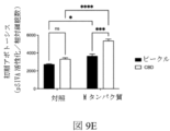
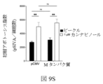
図9Eおよび9Fは、それぞれ、Mタンパク質を発現し、カンナビジオールで処置された対照プラスミドまたはウイルスプラスミドでトランスフェクトされた細胞における初期アポトーシスおよび後期アポトーシスデータを提供する。Mタンパク質でトランスフェクトされ、カンナビジオールで処理された細胞は、同じ条件下で処理されるが、対照プラスミドのみでトランスフェクトされた細胞と比較して、初期および後期アポトーシスの両方を有意に増加させる。Mタンパク質でトランスフェクトされ、カンナビジオールで処理された細胞は、Mタンパク質を発現しているが、ビークルのみで処理された細胞に対しても、初期かつ後期アポトーシスが有意に上昇した。 Figures 9E and 9F provide early and late apoptotic data, respectively, in control or viral plasmid-transfected cells expressing M protein and treated with cannabidiol. Cells transfected with M protein and treated with cannabidiol significantly increase both early and late apoptosis compared to cells treated under the same conditions but transfected with control plasmid alone . Cells transfected with M protein and treated with cannabidiol, which expressed M protein, also had significantly elevated early and late apoptosis relative to cells treated with vehicle alone.
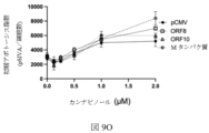
図9Gおよび9Hは、それぞれ、ORF8、ORF10、またはMタンパク質発現の効果を、CBDを用いて、初期および後期アポトーシスの測定に与える。対照プラスミド(pCMV)でトランスフェクトされた細胞におけるカンナビジオールの用量依存効果、またはORF 8、ORF10およびMタンパク質(n=3-9)を発現するプラスミドが示されている。対照プラスミドでトランスフェクトされたカンナビジオール処理細胞は、初期に有意な増加および後期アポトーシスを示さないが、ウイルスタンパク質ORF8、ORF10およびMタンパク質を発現するプラスミドでトランスフェクトされたカンナビジオール処理細胞は、初期アポトーシスおよび後期アポトーシスにおいて有意な増加を示した。
Figures 9G and 9H present the effect of ORF8, ORF10, or M protein expression, respectively, on measurements of early and late apoptosis using CBD. Dose-dependent effects of cannabidiol in cells transfected with control plasmid (pCMV) or
図9Iは、i)対照ベクターを発現する対照プラスミド、およびii)ウイルスタンパク質ORF8を発現するプラスミドでトランスフェクトされ、次に2μMカンナビジオールで処理された、HEK 293(ヒト胚性腎臓)細胞の初期アポトーシスデータを提供する。対照プラスミドでトランスフェクトされたカンナビジオール処理細胞は、初期アポトーシスにおいて有意な増加を示さないが、ウイルスタンパク質ORF8を発現するプラスミドでトランスフェクトされたカンナビジオール処理細胞は、ビークル対照のみで処理されたORF8発現細胞と、そしてカンナビジオールで処理された対照ベクター発現細胞の両方と比べて、初期アポトーシスにおいて有意な増加を示した。 FIG. 9I depicts early stages of HEK 293 (human embryonic kidney) cells transfected with i) a control plasmid expressing a control vector and ii) a plasmid expressing the viral protein ORF8 and then treated with 2 μM cannabidiol. Provides apoptosis data. Cannabidiol-treated cells transfected with a control plasmid show no significant increase in early apoptosis, whereas cannabidiol-treated cells transfected with a plasmid expressing the viral protein ORF8 show no significant increase in ORF8 treated with vehicle control alone. It showed a significant increase in early apoptosis compared to both expressing cells and control vector expressing cells treated with cannabidiol.
図9Jは、i)対照ベクターを発現する対照プラスミド、およびii)ウイルスタンパク質ORF10を発現するプラスミドでトランスフェクトされ、次いで、2μMカンナビジオールで処理された、HEK 293(ヒト胚性腎臓)細胞の初期アポトーシスデータを提供する。対照プラスミドでトランスフェクトされたカンナビジオール処理細胞は、初期アポトーシスにおいて有意な増加を示さないが、ウイルスタンパク質ORF10を発現するプラスミドでトランスフェクトされたカンナビジオール処理細胞は、ビークル対照のみで処理されたORF10発現細胞と、そしてカンナビジオールで処理された対照ベクター発現細胞の両方と比べて、初期アポトーシスにおいて有意な増加を示した。 FIG. 9J depicts early stages of HEK 293 (human embryonic kidney) cells transfected with i) a control plasmid expressing a control vector and ii) a plasmid expressing the viral protein ORF10 and then treated with 2 μM cannabidiol. Provides apoptosis data. Cannabidiol-treated cells transfected with a control plasmid show no significant increase in early apoptosis, whereas cannabidiol-treated cells transfected with a plasmid expressing the viral protein ORF10 show no significant increase in ORF10 treated with vehicle control alone. It showed a significant increase in early apoptosis compared to both expressing cells and control vector expressing cells treated with cannabidiol.
図9Kは、i)対照ベクターを発現する対照プラスミド、およびii)ウイルスMタンパク質を発現するプラスミドでトランスフェクトされ、次いで、2μMカンナビジオールで処理された、HEK 293(ヒト胚性腎臓)細胞の初期アポトーシスデータを提供する。対照プラスミドでトランスフェクトされたカンナビジオール処理細胞は、初期アポトーシスにおいて有意な増加を示さないが、ウイルスMタンパク質を発現するプラスミドでトランスフェクトされたカンナビジオール処理細胞は、ビークル対照のみで処理されたウイルスMタンパク質発現細胞と、カンナビジオールで処理された対照ベクター発現細胞との両方に対して、初期アポトーシスにおいて有意に増加した。 FIG. 9K depicts early stages of HEK 293 (human embryonic kidney) cells transfected with i) a control plasmid expressing a control vector and ii) a plasmid expressing a viral M protein and then treated with 2 μM cannabidiol. Provides apoptosis data. Cannabidiol-treated cells transfected with a control plasmid show no significant increase in early apoptosis, whereas cannabidiol-treated cells transfected with a plasmid expressing the viral M protein show a significant increase in virus treated with vehicle control alone. There was a significant increase in early apoptosis relative to both M protein-expressing cells and control vector-expressing cells treated with cannabidiol.
図9Lは、i)対照ベクターを発現する対照プラスミド、およびii)ウイルスタンパク質ORF8を発現するプラスミドでトランスフェクトされ、2μMカンナビジオールで処理された、HEK 293(ヒト胚性腎臓)細胞の後期アポトーシスデータを提供する。対照プラスミドでトランスフェクトされたカンナビジオール処理細胞は、後期アポトーシスにおいて有意な増加を示さないが、ウイルスタンパク質ORF8を発現するプラスミドでトランスフェクトされたカンナビジオール処理細胞は、ビークル対照のみで処理されたORF8発現細胞に対して、およびカンナビジオールで処理された対照ベクター発現細胞に対して相対的に、後期アポトーシスにおいて有意な増加を示した。 Figure 9L shows late apoptosis data for HEK 293 (human embryonic kidney) cells transfected with i) a control plasmid expressing a control vector and ii) a plasmid expressing the viral protein ORF8 and treated with 2 μM cannabidiol. I will provide a. Cannabidiol-treated cells transfected with a control plasmid show no significant increase in late apoptosis, whereas cannabidiol-treated cells transfected with a plasmid expressing the viral protein ORF8 show no significant increase in ORF8 treated with vehicle control alone. There was a significant increase in late apoptosis relative to expressing cells and to control vector-expressing cells treated with cannabidiol.
図9Mは、i)対照ベクターを発現する対照プラスミド、およびii)ウイルスタンパク質ORF10を発現するプラスミドでトランスフェクトされ、次いで、2μMカンナビジオールで処理された、HEK 293(ヒト胚性腎臓)細胞の後期アポトーシスデータを提供する。対照プラスミドでトランスフェクトされたカンナビジオール処理細胞は、後期アポトーシスにおいて有意な増加を示さないが、ウイルスタンパク質ORF10を発現するプラスミドでトランスフェクトされたカンナビジオール処理細胞は、ビークル対照のみで処理されたORF10発現細胞に対して、およびカンナビジオールで処理された対照ベクター発現細胞に対して相対的に、後期アポトーシスにおいて有意な増加を示した。 FIG. 9M depicts anaphase of HEK 293 (human embryonic kidney) cells transfected with i) a control plasmid expressing a control vector and ii) a plasmid expressing the viral protein ORF10 and then treated with 2 μM cannabidiol. Provides apoptosis data. Cannabidiol-treated cells transfected with a control plasmid show no significant increase in late apoptosis, whereas cannabidiol-treated cells transfected with a plasmid expressing the viral protein ORF10 show no significant increase in ORF10 treated with vehicle control alone. There was a significant increase in late apoptosis relative to expressing cells and to control vector expressing cells treated with cannabidiol.
図9Nは、i)対照ベクターを発現する対照プラスミド、およびii)ウイルスMタンパク質を発現するプラスミドでトランスフェクトされ、次いで、2μMカンナビジオールで処理された、HEK 293(ヒト胚性腎臓)細胞の後期アポトーシスデータを提供する。対照プラスミドでトランスフェクトされたカンナビジオール処理細胞は、後期アポトーシスにおいて有意な増加を示さないが、ウイルスMタンパク質を発現するプラスミドでトランスフェクトされたカンナビジオール処理細胞は、ビークル対照のみで処理されたウイルスMタンパク質発現細胞と、カンナビジオールで処理された対照ベクター発現細胞との両方に対して、後期アポトーシスにおいて有意な増加を示した。 FIG. 9N depicts anaphase of HEK 293 (human embryonic kidney) cells transfected with i) a control plasmid expressing a control vector and ii) a plasmid expressing a viral M protein and then treated with 2 μM cannabidiol. Provides apoptosis data. Cannabidiol-treated cells transfected with a control plasmid show no significant increase in late apoptosis, whereas cannabidiol-treated cells transfected with a plasmid expressing the viral M protein show a significant increase in virus treated with vehicle control alone. It showed a significant increase in late apoptosis relative to both M protein-expressing cells and control vector-expressing cells treated with cannabidiol.
図9O-9Pは、ORF 8、ORF10、またはMタンパク質発現の効果を提供し、カンナビノールが存在せず、初期および後期アポトーシスの測定に影響を与える。対照プラスミド(pCMV)でトランスフェクトされた細胞におけるカンナビノールの用量依存効果、またはORF8、ORF10またはMタンパク質(n=3-6)を発現するプラスミドが示されている。対照プラスミドでトランスフェクトされたカンナビジオール処理細胞は、初期のアポトーシスおよび後期アポトーシスにおいても有意に増加するだけでなく、ウイルスタンパク質ORF8、ORF10およびMタンパク質を発現するプラスミドでトランスフェクトされたカンナビノール処理細胞も有意に増加している。
Figures 9O-9P provide the effects of
図9Q-9Sは、初期アポトーシスの測定における1μMカンナビノールの効果を提供する。データは、平均値±SEM、*P<0.05、**P<0.01、***P<0.01である。 Figures 9Q-9S provide the effect of 1 μM cannabinol in measuring early apoptosis. Data are mean±SEM, *P<0.05, **P<0.01, ***P<0.01.
図9Qは、i)対照ベクターを発現する対照プラスミド、およびii)ウイルスタンパク質ORF8を発現するプラスミドでトランスフェクトされ、次いで、1μMカンナビノールで処理された、HEK 293(ヒト胚性腎臓)細胞の初期アポトーシスデータを提供する。対照プラスミドでトランスフェクトされたカンナビノール処理細胞は、初期アポトーシスの有意な増加を示す。ウイルスタンパク質ORF8を発現するプラスミドでトランスフェクトされたカンナビノール処理細胞も、ビークル対照のみで処理されたORF8発現細胞に対する初期アポトーシスにおいて有意に増加している。 FIG. 9Q depicts early stages of HEK 293 (human embryonic kidney) cells transfected with i) a control plasmid expressing a control vector and ii) a plasmid expressing the viral protein ORF8 and then treated with 1 μM cannabinol. Provides apoptosis data. Cannabinol-treated cells transfected with the control plasmid show a significant increase in early apoptosis. Cannabinol-treated cells transfected with a plasmid expressing the viral protein ORF8 also show a significant increase in early apoptosis relative to ORF8-expressing cells treated with vehicle control alone.
図9Rは、i)対照ベクターを発現する対照プラスミド、およびii) ウイルスタンパク質ORF10を発現するプラスミドでトランスフェクトされ、、次いで、1μMカンナビノールで処理された、HEK 293(ヒト胚性腎臓)細胞の初期アポトーシスデータを提供する。対照プラスミドでトランスフェクトされたカンナビノール処理細胞は、初期アポトーシスの有意な増加を示し、また、ウイルスタンパク質ORF10を発現するプラスミドでトランスフェクトされたカンナビノール処理細胞も、ビークル対照のみで処理されたORF10発現細胞に対して、初期アポトーシスにおいて有意な増加を示した。 FIG. 9R depicts HEK 293 (human embryonic kidney) cells transfected with i) a control plasmid expressing a control vector and ii) a plasmid expressing the viral protein ORF10 and then treated with 1 μM cannabinol. Provides early apoptosis data. Cannabinol-treated cells transfected with a control plasmid showed a significant increase in early apoptosis, and cannabinol-treated cells transfected with a plasmid expressing the viral protein ORF10 also showed a significant increase in ORF10 treated with vehicle control alone. It showed a significant increase in early apoptosis relative to expressing cells.
図9Sは、i)対照ベクターを発現する対照プラスミド、およびii)ウイルスMタンパク質を発現するプラスミドでトランスフェクトされ、次いで、1μMカンナビノールで処理された、HEK 293(ヒト胚性腎臓)細胞の初期アポトーシスデータを提供する。対照プラスミドでトランスフェクトされたカンナビノール処理細胞は、初期アポトーシスの有意な増加を示し、ウイルスMタンパク質を発現するプラスミドでトランスフェクトされたカンナビノール処理細胞も、ビークル対照のみで処理されたウイルスMタンパク質発現細胞と比べて、初期アポトーシスにおいて有意な増加を示した。 FIG. 9S depicts early stages of HEK 293 (human embryonic kidney) cells transfected with i) a control plasmid expressing a control vector and ii) a plasmid expressing a viral M protein and then treated with 1 μM cannabinol. Provides apoptosis data. Cannabinol-treated cells transfected with a control plasmid showed a significant increase in early apoptosis, and cannabinol-treated cells transfected with a plasmid expressing viral M protein also showed a significant increase in viral M protein treated with vehicle control alone. It showed a significant increase in early apoptosis compared to expressing cells.
図9T-9Vは、後期アポトーシスの測定における1μMカンナビノールの効果を提供する。データは、平均値±SEM、*P<0.05、**P<0.01、***P<0.01である。 Figures 9T-9V provide the effect of 1 μM cannabinol in measuring late apoptosis. Data are mean±SEM, *P<0.05, **P<0.01, ***P<0.01.
図9Tは、i)対照ベクターを発現する対照プラスミド、およびii)ウイルスタンパク質ORF8を発現するプラスミドでトランスフェクトされ、次いで、1μMカンナビノールで処理された、HEK 293(ヒト胚性腎臓)細胞の後期アポトーシスデータを提供する。対照プラスミドでトランスフェクトされたカンナビノール処理細胞は、後期アポトーシスの有意な増加を示し、また、ウイルスタンパク質ORF8を発現するプラスミドでトランスフェクトされたカンナビノール処理細胞も、ビークル対照のみで処理されたORF8発現細胞と比べて、後期アポトーシスにおいて有意な増加を示した。 FIG. 9T depicts late stages of HEK 293 (human embryonic kidney) cells transfected with i) a control plasmid expressing a control vector and ii) a plasmid expressing the viral protein ORF8 and then treated with 1 μM cannabinol. Provides apoptosis data. Cannabinol-treated cells transfected with a control plasmid showed a significant increase in late apoptosis, and cannabinol-treated cells transfected with a plasmid expressing the viral protein ORF8 also showed a significant increase in ORF8 treated with vehicle control alone. It showed a significant increase in late apoptosis compared to expressing cells.
図9Uは、i)対照ベクターを発現する対照プラスミド、およびii)ウイルスタンパク質ORF10を発現するプラスミドでトランスフェクトされ、次いで、1μMカンナビノールで処理された、HEK 293(ヒト胚性腎臓)細胞の後期アポトーシスデータを提供する。対照プラスミドでトランスフェクトされたカンナビノール処理細胞は、後期アポトーシスの有意な増加を示し、また、ウイルスタンパク質ORF10を発現するプラスミドでトランスフェクトされたカンナビノール処理細胞も、後期アポトーシスにおいて有意な増加を示した。ビークル対照のみで処理されたORF10発現細胞と、カンナビノールで処理された対照ベクター発現細胞との両方に関連している。 FIG. 9U shows anaphase of HEK 293 (human embryonic kidney) cells transfected with i) a control plasmid expressing a control vector and ii) a plasmid expressing the viral protein ORF10 and then treated with 1 μM cannabinol. Provides apoptosis data. Cannabinol-treated cells transfected with a control plasmid showed a significant increase in late apoptosis, and cannabinol-treated cells transfected with a plasmid expressing the viral protein ORF10 also showed a significant increase in late apoptosis. Ta. Both ORF10-expressing cells treated with vehicle control alone and control vector-expressing cells treated with cannabinol.
図9Vは、i)対照ベクターを発現する対照プラスミド、およびii) ウイルスMタンパク質を発現するプラスミドでトランスフェクトされ、次いで、1μMカンナビノールで処理された、HEK 293(ヒト胚性腎臓)細胞の後期アポトーシスデータを提供する。対照プラスミドでトランスフェクトされたカンナビノール処理細胞は、後期アポトーシスにおいて有意な増加を示し、ウイルスMタンパク質を発現するプラスミドでトランスフェクトされたカンナビノール処理細胞は、後期アポトーシスにおいて有意に増加する。ビークル対照のみで処理されたウイルスMタンパク質発現細胞に対する相対的なウイルスMタンパク質発現細胞 FIG. 9V depicts anaphase of HEK 293 (human embryonic kidney) cells transfected with i) a control plasmid expressing a control vector and ii) a plasmid expressing a viral M protein and then treated with 1 μM cannabinol. Provides apoptosis data. Cannabinol-treated cells transfected with a control plasmid show a significant increase in late apoptosis, and cannabinol-treated cells transfected with a plasmid expressing the viral M protein show a significant increase in late apoptosis. Virus M protein-expressing cells relative to virus M protein-expressing cells treated with vehicle control only
図9Wおよび9Xは、ORF8、ORF10、またはMタンパク質発現の効果を、カンナビノールなしで、初期および後期アポトーシスの測定に提供する。対照プラスミド(pCMV)でトランスフェクトされた細胞におけるカンナビノールの用量依存効果、またはORF8、ORF10またはMタンパク質(n=3-6) を発現するプラスミドが示されている。対照プラスミドでトランスフェクトされたカンナビノール処理された細胞は、初期かつ後期アポトーシスにおいて、何らかの有意な増加を示し、ウイルスタンパク質ORF8、ORF10およびMタンパク質を発現するプラスミドでトランスフェクトされたカンナビノール処理細胞は、初期アポトーシスおよび後期アポトーシスにおいて有意な増加を示した。 Figures 9W and 9X provide the effect of ORF8, ORF10, or M protein expression on measurements of early and late apoptosis without cannabinol. Dose-dependent effects of cannabinol in cells transfected with control plasmid (pCMV) or plasmids expressing ORF8, ORF10 or M protein (n=3-6) are shown. Cannabinol-treated cells transfected with control plasmids showed some significant increase in early and late apoptosis, whereas cannabinol-treated cells transfected with plasmids expressing viral proteins ORF8, ORF10 and M protein , showed a significant increase in early and late apoptosis.
図9Y-9A Aは、初期アポトーシスの測定における2μMカンナビノールの効果を提供する。データは、平均値±SEM、*P<0.05、**P<0.01、***P<0.01である Figures 9Y-9A A provide the effect of 2 μM cannabinol in measuring early apoptosis. Data are mean±SEM, *P<0.05, **P<0.01, ***P<0.01
図9Yは、i)対照ベクターを発現する対照プラスミド、およびii)ウイルスタンパク質ORF8を発現するプラスミドでトランスフェクトされ、次いで、2μMカンナビノールで処理された、HEK 293(ヒト胚性腎臓)細胞の初期アポトーシスデータを提供する。対照プラスミドでトランスフェクトされたカンナビノール処理細胞は、初期アポトーシスの有意な増加を示し、ウイルスタンパク質ORF8を発現するプラスミドでトランスフェクトされたカンナビノール処理細胞は、初期アポトーシスにおいて有意な増加を示した。ビークル対照のみで処理されたORF8発現細胞と、カンナビノールで処理された対照ベクター発現細胞との両方に関連している。 FIG. 9Y depicts early stages of HEK 293 (human embryonic kidney) cells transfected with i) a control plasmid expressing a control vector and ii) a plasmid expressing the viral protein ORF8 and then treated with 2 μM cannabinol. Provides apoptosis data. Cannabinol-treated cells transfected with a control plasmid showed a significant increase in early apoptosis, and cannabinol-treated cells transfected with a plasmid expressing the viral protein ORF8 showed a significant increase in early apoptosis. Both ORF8-expressing cells treated with vehicle control alone and control vector-expressing cells treated with cannabinol.
図9Zは、i)対照ベクターを発現する対照プラスミド、およびii) ウイルスタンパク質ORF10を発現するプラスミドでトランスフェクトされ、次いで、2μMカンナビノールで処理された、HEK 293(ヒト胚性腎臓)細胞の初期アポトーシスデータを提供する。対照プラスミドでトランスフェクトされたカンナビノール処理細胞は、初期アポトーシスの有意な増加を示し、ウイルスタンパク質ORF10を発現するプラスミドでトランスフェクトされたカンナビノール処理細胞は有意である。ビークル対照のみで処理されたORF10発現細胞に対して、初期アポトーシスの増加が増加する。 FIG. 9Z depicts early stages of HEK 293 (human embryonic kidney) cells transfected with i) a control plasmid expressing a control vector and ii) a plasmid expressing the viral protein ORF10 and then treated with 2 μM cannabinol. Provides apoptosis data. Cannabinol-treated cells transfected with a control plasmid show a significant increase in early apoptosis, and cannabinol-treated cells transfected with a plasmid expressing the viral protein ORF10 are significant. There is an increase in early apoptosis relative to ORF10-expressing cells treated with vehicle control only.
図9AAは、i)対照ベクターを発現する対照プラスミド、およびii) ウイルスMタンパク質を発現するプラスミドでトランスフェクトされ、次いで、2μMカンナビノールで処理された、HEK 293(ヒト胚性腎臓)細胞の初期アポトーシスデータを提供する。対照プラスミドでトランスフェクトされたカンナビノール処理細胞は、初期アポトーシスの有意な増加を示し、ウイルスMタンパク質を発現するプラスミドでトランスフェクトされたカンナビノール処理細胞は、初期アポトーシスにおいて有意に増加した。ウイルスMタンパク質発現細胞は、ビークル対照のみで処理されたウイルスMタンパク質発現細胞、およびカンナビノールで処理された対照ベクター発現細胞に対する両方である。 FIG. 9AA shows early stages of HEK 293 (human embryonic kidney) cells transfected with i) a control plasmid expressing a control vector and ii) a plasmid expressing a viral M protein and then treated with 2 μM cannabinol. Provides apoptosis data. Cannabinol-treated cells transfected with a control plasmid showed a significant increase in early apoptosis, and cannabinol-treated cells transfected with a plasmid expressing the viral M protein had a significant increase in early apoptosis. Viral M protein-expressing cells are both to viral M protein-expressing cells treated with vehicle control alone and control vector-expressing cells treated with cannabinol.
図9AB-9ADは、後期アポトーシスの測定において2μMカンナビノールの効果を提供する。データは、平均値±SEM、*P<0.05、**P<0.01、***P<0.01である。 Figures 9AB-9AD provide the effect of 2 μM cannabinol in measuring late apoptosis. Data are mean±SEM, *P<0.05, **P<0.01, ***P<0.01.
図9ABは、i)対照ベクターを発現する対照プラスミド、およびii) ウイルスタンパク質ORF8を発現するプラスミドでトランスフェクトされ、次いで、2μMカンナビノールで処理された、HEK 293(ヒト胚性腎臓)細胞の後期アポトーシスデータを提供する。対照プラスミドでトランスフェクトされたカンナビノール処理細胞は、後期アポトーシスの有意な増加を示し、また、ウイルスタンパク質ORF8を発現するプラスミドでトランスフェクトされたカンナビノール処理細胞も、後期アポトーシスにおいて有意な増加を示した。ビークル対照のみで処理されたORF8発現細胞に対しては、カンナビノールで処理された対照ベクター発現細胞に対しては相対的ではない。 FIG. 9AB. Late stages of HEK 293 (human embryonic kidney) cells transfected with i) a control plasmid expressing a control vector and ii) a plasmid expressing the viral protein ORF8 and then treated with 2 μM cannabinol. Provides apoptosis data. Cannabinol-treated cells transfected with a control plasmid showed a significant increase in late apoptosis, and cannabinol-treated cells transfected with a plasmid expressing the viral protein ORF8 also showed a significant increase in late apoptosis. Ta. ORF8-expressing cells treated with vehicle control only are not relative to control vector-expressing cells treated with cannabinol.
図9ACは、i)対照ベクターを発現する対照プラスミド、およびii) ウイルスタンパク質ORF10を発現するプラスミドでトランスフェクトされ、次いで、2μMカンナビノールで処理された、HEK 293(ヒト胚性腎臓) 細胞の後期アポトーシスデータを提供する。対照プラスミドでトランスフェクトされたカンナビノール処理細胞は、後期アポトーシスの有意な増加を示し、また、ウイルスタンパク質ORF10を発現するプラスミドでトランスフェクトされたカンナビノール処理細胞も、後期アポトーシスにおいて有意な増加を示した。ビークル対照のみで処理されたORF10発現細胞に対しては、カンナビノールで処理された対照ベクター発現細胞に対しては相対的ではない。 FIG. 9AC depicts late stages of HEK 293 (human embryonic kidney) cells transfected with i) a control plasmid expressing a control vector and ii) a plasmid expressing the viral protein ORF10 and then treated with 2 μM cannabinol. Provides apoptosis data. Cannabinol-treated cells transfected with a control plasmid showed a significant increase in late apoptosis, and cannabinol-treated cells transfected with a plasmid expressing the viral protein ORF10 also showed a significant increase in late apoptosis. Ta. ORF10-expressing cells treated with vehicle control only are not relative to control vector-expressing cells treated with cannabinol.
図9ADは、i)対照ベクターを発現する対照プラスミド、およびii) ウイルスMタンパク質を発現するプラスミドでトランスフェクトされ、次いで、2μMカンナビノールで処理された、HEK 293(ヒト胚性腎臓)細胞の後期アポトーシスデータを提供する。対照プラスミドでトランスフェクトされたカンナビノール処理細胞は、後期アポトーシスの有意な増加を示し、ウイルスMタンパク質を発現するプラスミドでトランスフェクトされたカンナビノール処理細胞も、後期アポトーシスにおいて顕著に増加している。ビークル対照のみで処理されたウイルスMタンパク質発現細胞と、カンナビノールで処理された対照ベクター発現細胞との両方に関連している。
図9A E-9AJは、ORF8、ORF10、またはMタンパク質発現の効果を、カンナビジオール酸(CBDA)なしで、初期および後期アポトーシスの測定に提供する。初期のアポトーシス(n=3)の測定における1μM CBDAの効果が示されている。データは、平均値±SEM、*P<0.05、**P<0.01、***P<0.01である
FIG. 9A-D depicts late stages of HEK 293 (human embryonic kidney) cells transfected with i) a control plasmid expressing a control vector and ii) a plasmid expressing a viral M protein and then treated with 2 μM cannabinol. Provides apoptosis data. Cannabinol-treated cells transfected with a control plasmid show a significant increase in late apoptosis, and cannabinol-treated cells transfected with a plasmid expressing the viral M protein also show a significant increase in late apoptosis. Both viral M protein-expressing cells treated with vehicle control alone and control vector-expressing cells treated with cannabinol.
Figure 9A E-9AJ provides the effect of ORF8, ORF10, or M protein expression on measurements of early and late apoptosis without cannabidiolic acid (CBDA). Shown is the effect of 1 μM CBDA in measuring early apoptosis (n=3). Data are mean±SEM, *P<0.05, **P<0.01, ***P<0.01
図9AEは、i)対照ベクターを発現する対照プラスミド、およびii) ウイルスタンパク質ORF8を発現するプラスミドでトランスフェクトされ、次いで、1μM CBDAで処理された、HEK 293(ヒト胚性腎臓)細胞の初期アポトーシスデータを提供する。対照プラスミドでトランスフェクトされたCBDA処理細胞は、初期アポトーシスにおいて有意な増加を示さないが、ウイルスタンパク質ORF8を発現するプラスミドでトランスフェクトされたCBDA処理細胞は、初期アポトーシスにおいて有意に増加する。CBDAで処理された対照ベクター発現細胞に関する。 Figure 9AE shows early apoptosis of HEK 293 (human embryonic kidney) cells transfected with i) a control plasmid expressing a control vector and ii) a plasmid expressing the viral protein ORF8 and then treated with 1 μM CBDA. provide data. CBDA-treated cells transfected with a control plasmid show no significant increase in early apoptosis, whereas CBDA-treated cells transfected with a plasmid expressing the viral protein ORF8 show a significant increase in early apoptosis. For control vector-expressing cells treated with CBDA.
図9AFは、i)対照ベクターを発現する対照プラスミド、およびii) ウイルスタンパク質ORF10を発現するプラスミドでトランスフェクトされ、次いで、1μM CBDAで処理された、HEK 293(ヒト胚性腎臓)細胞の初期アポトーシスデータを提供する。CBDA処理された細胞は、対照ベクターでトランスフェクトされる ウイルスタンパク質ORF10を発現するプラスミドでトランスフェクトされたCBDA処理細胞は、CBDAで処理された対照ベクター発現細胞に対する初期アポトーシスにおいて有意な増加を示した。 Figure 9AF shows early apoptosis of HEK 293 (human embryonic kidney) cells transfected with i) a control plasmid expressing a control vector and ii) a plasmid expressing the viral protein ORF10 and then treated with 1 μM CBDA. provide data. CBDA-treated cells transfected with a control vector CBDA-treated cells transfected with a plasmid expressing the viral protein ORF10 showed a significant increase in early apoptosis relative to control vector-expressing cells treated with CBDA .
図9AGは、i)対照ベクターを発現する対照プラスミド、およびii) ウイルスMタンパク質を発現するプラスミドでトランスフェクトされ、次いで、1μM CBDAで処理された、HEK 293(ヒト胚性腎臓)細胞の初期アポトーシスデータを提供する。CBDA処理細胞 対照ベクターでトランスフェクトされたCBDA処理細胞は、初期アポトーシスにおいて有意な増加を示さないが、ウイルスMタンパク質を発現するプラスミドでトランスフェクトされたCBDA処理細胞は、CBDAで処理された対照ベクター発現細胞に対する初期アポトーシスにおいて有意に増加した。 Figure 9AG shows early apoptosis of HEK 293 (human embryonic kidney) cells transfected with i) a control plasmid expressing a control vector and ii) a plasmid expressing a viral M protein and then treated with 1 μM CBDA. provide data. CBDA-treated cells CBDA-treated cells transfected with a control vector show no significant increase in early apoptosis, whereas CBDA-treated cells transfected with a plasmid expressing the viral M protein show a significant increase in CBDA-treated control vector There was a significant increase in early apoptosis for expressing cells.
図9AHは、i)対照ベクターを発現する対照プラスミド、およびii) ウイルスタンパク質ORF8を発現するプラスミドでトランスフェクトされ、次いで、1μM CBDAで処理された、HEK 293(ヒト胚性腎臓)細胞の後期アポトーシスデータを提供する。対照プラスミドでトランスフェクトされたCBDA処理された細胞は、後期アポトーシスにおいて有意な増加を示さず、ウイルスタンパク質ORF8を発現するプラスミドでトランスフェクトされたCBDA処理された細胞も示さない。ビークル対照のみで処理されたORF8発現細胞、またはCBDAで処理された対照ベクター発現細胞に対するいずれかである。 Figure 9AH shows late apoptosis of HEK 293 (human embryonic kidney) cells transfected with i) a control plasmid expressing a control vector and ii) a plasmid expressing the viral protein ORF8 and then treated with 1 μM CBDA. provide data. CBDA-treated cells transfected with a control plasmid show no significant increase in late apoptosis, nor do CBDA-treated cells transfected with a plasmid expressing the viral protein ORF8. Either against ORF8-expressing cells treated with vehicle control only, or control vector-expressing cells treated with CBDA.
図9AIは、i)対照ベクターを発現する対照プラスミド、およびii) ウイルスタンパク質ORF10を発現するプラスミドでトランスフェクトされ、次いで、1μM CBDAで処理された、HEK 293(ヒト胚性腎臓)細胞の後期アポトーシスデータを提供する。対照ベクターでトランスフェクトされたCBDA処理された細胞は、後期アポトーシスにおいて有意な増加を示さない。ウイルスタンパク質ORF10を発現するプラスミドでトランスフェクトされたCBDA処理細胞は、CBDAで処理された対照ベクター発現細胞に対する後期アポトーシスの有意な増加を示した。 Figure 9AI shows late apoptosis of HEK 293 (human embryonic kidney) cells transfected with i) a control plasmid expressing a control vector and ii) a plasmid expressing the viral protein ORF10 and then treated with 1 μM CBDA. provide data. CBDA-treated cells transfected with control vector show no significant increase in late apoptosis. CBDA-treated cells transfected with a plasmid expressing the viral protein ORF10 showed a significant increase in late apoptosis relative to control vector-expressing cells treated with CBDA.
図9AJは、i)対照ベクターを発現する対照プラスミド、およびii) ウイルスMタンパク質を発現するプラスミドでトランスフェクトされ、次いで、1μM CBDAで処理された、HEK 293(ヒト胚性腎臓)細胞の後期アポトーシスデータを提供する。対照ベクターでトランスフェクトされたCBDA処理された細胞は、後期アポトーシスにおいて有意な増加を示さない。ウイルスMタンパク質を発現するプラスミドでトランスフェクトされたCBDA処理細胞は、CBDAで処理された対照ベクター発現細胞に対する後期アポトーシスにおいて有意な増加を示した。 FIG. 9AJ shows late apoptosis of HEK 293 (human embryonic kidney) cells transfected with i) control plasmid expressing control vector and ii) plasmid expressing viral M protein and then treated with 1 μM CBDA. provide data. CBDA-treated cells transfected with control vector show no significant increase in late apoptosis. CBDA-treated cells transfected with a plasmid expressing the viral M protein showed a significant increase in late apoptosis relative to control vector-expressing cells treated with CBDA.
図9AK-9ALは、ORF8、ORF10、またはMタンパク質発現の効果を提供し、カンナビロール (CBG)なしで、初期および後期アポトーシスの測定に影響を与える。対照プラスミド(pCMV)でトランスフェクトされた細胞、またはORF8、ORF10、またはMタンパク質(n=3-6) を発現するプラスミドにおいて、初期および後期アポトーシスに対するCBGの用量依存効果が示されている。対照プラスミドでトランスフェクトされたCBG処理された細胞は、初期にいくつかの有意な増加および後期アポトーシスを示し、また、ウイルスタンパク質ORF 8、ORF10およびMタンパク質を発現するプラスミドでトランスフェクトされたCBG処理細胞も、初期アポトーシスおよび後期アポトーシスにおいて有意に増加している。 Figure 9AK-9AL provides the effect of ORF8, ORF10, or M protein expression to affect measurements of early and late apoptosis in the absence of cannabinol (CBG). Dose-dependent effects of CBG on early and late apoptosis are shown in cells transfected with control plasmid (pCMV) or plasmids expressing ORF8, ORF10, or M protein (n=3-6). CBG-treated cells transfected with control plasmids showed some significant increases in early and late apoptosis, and CBG-treated cells transfected with plasmids expressing viral proteins ORF8, ORF10 and M protein Cells are also significantly increased in early and late apoptosis.
図9AM-9AOは、初期アポトーシスの測定における1μM CBGの効果を提供する。データは、平均値±SEM、*P<0.05、**P<0.01、***P<0.01である。 Figures 9AM-9AO provide the effect of 1 μM CBG in measuring early apoptosis. Data are mean±SEM, *P<0.05, **P<0.01, ***P<0.01.
図9AMは、i)対照ベクターを発現する対照プラスミド、およびii) ウイルスタンパク質ORF8を発現するプラスミドでトランスフェクトされ、次いで、1μM CBGで処置された、HEK 293(ヒト胚性腎臓) 細胞の初期アポトーシスデータを提供する。対照プラスミドでトランスフェクトされたCBG処理された細胞は、ウイルスタンパク質ORF8を発現するプラスミドでトランスフェクトされた初期アポトーシスおよびCBG治療細胞の有意な増加を示し、また、ウイルスタンパク質ORF8を発現するプラスミドでトランスフェクトされた細胞は、初期アポトーシスにおいても有意に増加している。ORF8発現細胞は、ビークル対照のみで処理される Figure 9AM shows early apoptosis of HEK 293 (human embryonic kidney) cells transfected with i) a control plasmid expressing a control vector and ii) a plasmid expressing the viral protein ORF8 and then treated with 1 μM CBG. provide data. CBG-treated cells transfected with a control plasmid showed a significant increase in early apoptosis and CBG-treated cells transfected with a plasmid expressing the viral protein ORF8, and also CBG-treated cells transfected with a plasmid expressing the viral protein ORF8. The transfected cells also show a significant increase in early apoptosis. ORF8-expressing cells are treated with vehicle control only
図9ANは、i)対照ベクターを発現する対照プラスミド、およびii) ウイルスタンパク質ORF10を発現するプラスミドでトランスフェクトされ、次いで、1μM CBGで処置された、HEK 293(ヒト胚性腎臓)細胞の初期アポトーシスデータを提供する。対照プラスミドでトランスフェクトされたCBG処理された細胞は、有意に増加した初期アポトーシスを示し、また、ウイルスタンパク質ORF10を発現するプラスミドでトランスフェクトされたCBG処理細胞は、初期アポトーシスにおいて有意に増加している。ORF10発現細胞は、ビークル対照のみで処理される。 Figure 9AN depicts early apoptosis of HEK 293 (human embryonic kidney) cells transfected with i) a control plasmid expressing a control vector and ii) a plasmid expressing the viral protein ORF10 and then treated with 1 μM CBG. provide data. CBG-treated cells transfected with a control plasmid showed a significantly increased early apoptosis, and CBG-treated cells transfected with a plasmid expressing the viral protein ORF10 showed a significant increase in early apoptosis. there is ORF10 expressing cells are treated with vehicle control only.
図9AOは、i)対照ベクターを発現する対照プラスミド、およびii) ウイルスMタンパク質を発現するプラスミドでトランスフェクトされ、次いで、1μM CBGで処理された、HEK 293(ヒト胚性腎臓)細胞の初期アポトーシスデータを提供する。対照プラスミドでトランスフェクトされたCBG処理細胞は、ウイルスMに対する初期アポトーシスにおいて有意に増加したウイルスMタンパク質を発現するプラスミドでトランスフェクトされた初期アポトーシスおよびCBG処置細胞の有意な増加を示す。タンパク質発現細胞は、ビークル対照のみで処理される。 FIG. 9AO shows early apoptosis of HEK 293 (human embryonic kidney) cells transfected with i) a control plasmid expressing a control vector and ii) a plasmid expressing a viral M protein and then treated with 1 μM CBG. provide data. CBG-treated cells transfected with the control plasmid show a significant increase in early apoptosis and CBG-treated cells transfected with a plasmid expressing the virus M protein significantly increased in early apoptosis to virus M. Protein-expressing cells are treated with vehicle control only.
図9AP-9ARは、後期アポトーシスの測定における1μM CBGの効果を提供する。データは、平均値±SEM、*P<0.05、**P<0.01、***P<0.01である。 Figures 9AP-9AR provide the effect of 1 μM CBG in measuring late apoptosis. Data are mean±SEM, *P<0.05, **P<0.01, ***P<0.01.
図9APは、i)対照ベクターを発現する対照プラスミド、およびii) ウイルスタンパク質ORF 8を発現するプラスミドでトランスフェクトされ、次いで、1μM CBGで処置された、HEK 293(ヒト胚性腎臓)細胞の後期アポトーシスデータを提供する。対照プラスミドでトランスフェクトされたCBG処理された細胞は、後期アポトーシスにおいて有意な増加を示さない。しかし、ウイルスタンパク質ORF8を発現するプラスミドでトランスフェクトされたCBG処理細胞は、ビークル対照のみで処理されたORF8発現細胞に対する後期アポトーシスの有意な増加を示した。
FIG. 9AP shows anaphase of HEK 293 (human embryonic kidney) cells transfected with i) a control plasmid expressing a control vector and ii) a plasmid expressing the
図9AQは、i)対照ベクターを発現する対照プラスミド、およびii) ウイルスタンパク質ORF10を発現するプラスミドでトランスフェクトされ、次いで、1μM CBGで処置された、HEK 293(ヒト胚性腎臓)細胞の後期アポトーシスデータを提供する。対照プラスミドでトランスフェクトされたCBG処理された細胞は、後期アポトーシスにおいて有意な増加を示し、また、ウイルスタンパク質ORF10を発現するプラスミドでトランスフェクトされたCBG処理細胞は、後期アポトーシスにおいて有意に増加する。ORF10発現細胞は、ビークル対照のみで処理される。 FIG. 9AQ Late apoptosis of HEK 293 (human embryonic kidney) cells transfected with i) a control plasmid expressing a control vector and ii) a plasmid expressing the viral protein ORF10 and then treated with 1 μM CBG. provide data. CBG-treated cells transfected with a control plasmid show a significant increase in late apoptosis, and CBG-treated cells transfected with a plasmid expressing the viral protein ORF10 show a significant increase in late apoptosis. ORF10 expressing cells are treated with vehicle control only.
図9ARは、i)対照ベクターを発現する対照プラスミド、およびii) ウイルスMタンパク質を発現するプラスミドでトランスフェクトされ、次いで、1μM CBGで処理された、HEK 293(ヒト胚性腎臓)細胞の後期アポトーシスデータを提供する。対照プラスミドでトランスフェクトされたCBG処理された細胞は、後期アポトーシスにおいて有意な増加を示さない。しかし、ウイルスMタンパク質を発現するプラスミドでトランスフェクトされたCBG処理細胞は、ビークル対照のみで処理されたウイルスMタンパク質発現細胞に対する後期アポトーシスにおいて有意な増加を示した。 FIG. 9AR shows late apoptosis of HEK 293 (human embryonic kidney) cells transfected with i) control plasmid expressing control vector and ii) plasmid expressing viral M protein and then treated with 1 μM CBG. provide data. CBG-treated cells transfected with the control plasmid show no significant increase in late apoptosis. However, CBG-treated cells transfected with a plasmid expressing viral M protein showed a significant increase in late apoptosis relative to viral M protein-expressing cells treated with vehicle control alone.
図9AS-9ATは、ORF8、ORF10、またはMタンパク質発現の効果を提供し、カンナビロール(CBG)なしで、初期および後期アポトーシスの測定に影響を与える。対照プラスミド(pCMV)でトランスフェクトされた細胞、またはORF8、ORF10、またはMタンパク質(n=3-6)を発現するプラスミドにおいて、初期および後期アポトーシスに対するCBGの用量依存効果が示されている。対照プラスミドでトランスフェクトされたCBG処理された細胞は、初期にいくつかの有意な増加、ならびにウイルスタンパク質ORF8、ORF10およびMタンパク質を発現するプラスミドでトランスフェクトされたCBG処理された細胞は、初期アポトーシスおよび後期アポトーシスにおいて有意な増加を示した。 Figure 9AS-9AT provides the effect of ORF8, ORF10, or M protein expression to affect measurements of early and late apoptosis in the absence of cannabinol (CBG). Dose-dependent effects of CBG on early and late apoptosis are shown in cells transfected with control plasmid (pCMV) or plasmids expressing ORF8, ORF10, or M protein (n=3-6). CBG-treated cells transfected with the control plasmid showed several significant increases in early, as well as CBG-treated cells transfected with plasmids expressing the viral proteins ORF8, ORF10 and M protein showed early apoptosis. and showed a significant increase in late apoptosis.
図9AU-9AWは、初期アポトーシスの測定における2μM CBGの効果を提供する。データは、平均値±SEM、*P<0.05、**P<0.01、***P<0.01である。 Figures 9AU-9AW provide the effect of 2 μM CBG in measuring early apoptosis. Data are mean±SEM, *P<0.05, **P<0.01, ***P<0.01.
図9AUは、i)対照ベクターを発現する対照プラスミド、およびii) ウイルスタンパク質ORF8を発現するプラスミドでトランスフェクトされ、その後、2μM CBGで処置された、HEK 293(ヒト胚性腎臓)細胞の初期アポトーシスデータを提供する。対照プラスミドでトランスフェクトされたCBG処理細胞は、初期アポトーシスにおいて有意な増加を示し、また、ウイルスタンパク質ORF8を発現するプラスミドでトランスフェクトされたCBG処理細胞は、初期アポトーシスにおいて有意な増加を示した。ビークル対照のみで処理されたORF8発現細胞と、CBGで処理された対照ベクター発現細胞との両方に関連している。 Figure 9AU shows early apoptosis of HEK 293 (human embryonic kidney) cells transfected with i) a control plasmid expressing a control vector and ii) a plasmid expressing the viral protein ORF8, followed by treatment with 2 μM CBG. provide data. CBG-treated cells transfected with the control plasmid showed a significant increase in early apoptosis, and CBG-treated cells transfected with a plasmid expressing the viral protein ORF8 showed a significant increase in early apoptosis. Both ORF8-expressing cells treated with vehicle control alone and control vector-expressing cells treated with CBG.
図9AVは、i)対照ベクターを発現する対照プラスミド、およびii) ウイルスタンパク質ORF10を発現するプラスミドでトランスフェクトされ、次いで、2μM CBGで処置された、HEK 293(ヒト胚性腎臓)細胞の初期アポトーシスデータを提供する。対照プラスミドでトランスフェクトされたCBG処理された細胞は、初期アポトーシスにおいて有意な増加を示し、また、ウイルスタンパク質ORF 10を発現するプラスミドでトランスフェクトされたCBG処理細胞は、初期アポトーシスにおいて有意に増加している。ORF10発現細胞は、ビークル対照のみで処理される。
Figure 9AV shows early apoptosis of HEK 293 (human embryonic kidney) cells transfected with i) a control plasmid expressing a control vector and ii) a plasmid expressing the viral protein ORF10 and then treated with 2 μM CBG. provide data. CBG-treated cells transfected with a control plasmid showed a significant increase in early apoptosis, and CBG-treated cells transfected with a plasmid expressing the
図9AWは、i)対照ベクターを発現する対照プラスミド、およびii) ウイルスMタンパク質を発現するプラスミドでトランスフェクトされ、その後、2μM CBGで処置された、HEK 293(ヒト胚性腎臓)細胞の初期アポトーシスデータを提供する。対照プラスミドでトランスフェクトされたCBG処理細胞は、初期アポトーシスにおいて有意な増加を示し、ウイルスMタンパク質を発現するプラスミドでトランスフェクトされたCBG処理細胞は、初期アポトーシスにおいて有意な増加を示した。ウイルスMタンパク質発現細胞は、ビークル対照のみで処理されたウイルスMタンパク質発現細胞と、CBGで処理された対照ベクター発現細胞との両方に関連している。 FIG. 9AW shows early apoptosis of HEK 293 (human embryonic kidney) cells transfected with i) control plasmid expressing control vector and ii) plasmid expressing viral M protein and then treated with 2 μM CBG. provide data. CBG-treated cells transfected with a control plasmid showed a significant increase in early apoptosis, and CBG-treated cells transfected with a plasmid expressing the viral M protein showed a significant increase in early apoptosis. Viral M protein-expressing cells are related to both viral M protein-expressing cells treated with vehicle control alone and control vector-expressing cells treated with CBG.
図9AX-9AZは、後期アポトーシスの測定における2μM CBGの効果を提供する。データは、平均値±SEM、*P<0.05、**P<0.01、***P<0.01である。 Figure 9AX-9AZ provides the effect of 2 μM CBG in measuring late apoptosis. Data are mean±SEM, *P<0.05, **P<0.01, ***P<0.01.
図9AXは、i)対照ベクターを発現する対照プラスミド、およびii) ウイルスタンパク質ORF8を発現するプラスミドでトランスフェクトされ、その後、2μM CBGで処置された、HEK 293(ヒト胚性腎臓)細胞の後期アポトーシスデータを提供する。対照プラスミドでトランスフェクトされたCBG処理された細胞は、後期アポトーシスにおいて有意な増加を示し、また、ウイルスタンパク質ORF8を発現するプラスミドでトランスフェクトされたCBG処理細胞は、後期アポトーシスにおいて有意に増加する。ORF8発現細胞は、ビークル対照のみで処理される。 FIG. 9AX Late apoptosis of HEK 293 (human embryonic kidney) cells transfected with i) a control plasmid expressing a control vector and ii) a plasmid expressing the viral protein ORF8, followed by treatment with 2 μM CBG. provide data. CBG-treated cells transfected with a control plasmid show a significant increase in late apoptosis, and CBG-treated cells transfected with a plasmid expressing the viral protein ORF8 show a significant increase in late apoptosis. ORF8-expressing cells are treated with vehicle control only.
図9AYは、i)対照ベクターを発現する対照プラスミド、およびii) ウイルスタンパク質ORF10を発現するプラスミドでトランスフェクトされ、次いで、2μM CBGで処置された、HEK 293(ヒト胚性腎臓)細胞の後期アポトーシスデータを提供する。対照プラスミドでトランスフェクトされたCBG処理された細胞は、後期アポトーシスにおいて有意な増加を示す。しかし、ウイルスタンパク質ORF10を発現するプラスミドでトランスフェクトされたCBG処理細胞は、ビークル対照のみで処理されたORF10発現細胞に対する後期アポトーシスの有意な増加を示した。 FIG. 9AY Late apoptosis of HEK 293 (human embryonic kidney) cells transfected with i) a control plasmid expressing a control vector and ii) a plasmid expressing the viral protein ORF10 and then treated with 2 μM CBG. provide data. CBG-treated cells transfected with the control plasmid show a significant increase in late apoptosis. However, CBG-treated cells transfected with a plasmid expressing the viral protein ORF10 showed a significant increase in late apoptosis relative to ORF10-expressing cells treated with vehicle control alone.
図9AZは、i)対照ベクターを発現する対照プラスミド、およびii) ウイルスMタンパク質を発現するプラスミドでトランスフェクトされ、その後、2μM CBGで処置された、HEK 293細胞(ヒト胚性腎臓)細胞の後期アポトーシスデータを提供する。対照プラスミドでトランスフェクトされたCBG処理細胞は、後期アポトーシスにおいて有意な増加を示し、ウイルスMタンパク質を発現するプラスミドでトランスフェクトされたCBG処理細胞は、後期アポトーシスにおいて有意な増加を示した。ウイルスMタンパク質発現細胞は、ビークル対照のみで処理されたウイルスMタンパク質発現細胞と、CBGで処理された対照ベクター発現細胞との両方に関連している。 FIG. 9AZ Late stages of HEK 293 cells (human embryonic kidney) cells transfected with i) a control plasmid expressing a control vector and ii) a plasmid expressing a viral M protein and then treated with 2 μM CBG. Provides apoptosis data. CBG-treated cells transfected with the control plasmid showed a significant increase in late apoptosis, and CBG-treated cells transfected with a plasmid expressing the viral M protein showed a significant increase in late apoptosis. Viral M protein-expressing cells are related to both viral M protein-expressing cells treated with vehicle control alone and control vector-expressing cells treated with CBG.
図9BA-9BEは、ORF8、ORF10およびMタンパク質発現の効果を提供し、δ8-テトラヒドロカンナビラシン(d 8-THCV)なしで、初期のアポトーシスおよび後期アポトーシスの測定を提供する Figure 9BA-9BE provides effects of ORF8, ORF10 and M protein expression and provides measurements of early and late apoptosis in the absence of δ8-tetrahydrocannaviracin (d8-THCV).
図9BA-9BCは、初期アポトーシスの測定における1μM d8-THCVの効果を提供する(n=3) データは平均値±SEM、*P<0.05、**P<0.01である Figure 9BA-9BC provides the effect of 1 μM d8-THCV in measuring early apoptosis (n=3) Data are mean±SEM, *P<0.05, **P<0.01
図9BAは、i)対照ベクターを発現する対照プラスミド、およびii) ウイルスタンパク質ORF8を発現するプラスミドでトランスフェクトされ、次いで、1μM d8-THCVで処理された、HEK 293(ヒト胚性腎臓)細胞の初期アポトーシスデータを提供する。対照プラスミドでトランスフェクトされたD8-THCV処理された細胞は、初期アポトーシスにおいて有意な増加を示さない。ウイルスタンパク質ORF8を発現するプラスミドでトランスフェクトされたd 8-THCV処理細胞は、初期アポトーシスおよびd 8-THCVで処理された対照ベクター発現細胞に対する有意な増加を示した。 FIG. 9BA depicts HEK 293 (human embryonic kidney) cells transfected with i) a control plasmid expressing a control vector and ii) a plasmid expressing the viral protein ORF8 and then treated with 1 μM d8-THCV. Provides early apoptosis data. D8-THCV treated cells transfected with control plasmid show no significant increase in early apoptosis. d8-THCV-treated cells transfected with a plasmid expressing the viral protein ORF8 showed early apoptosis and a significant increase over control vector-expressing cells treated with d8-THCV.
図9BBは、i)対照ベクターを発現する対照プラスミド、およびii) ウイルスタンパク質ORF10を発現するプラスミドでトランスフェクトされ、次いで、1μM d8-THCVで処理された、HEK 293(ヒト胚性腎臓)細胞の初期アポトーシスデータを提供する。対照プラスミドでトランスフェクトされたd 8-THCV処理細胞は、初期アポトーシスの有意な増加を示さないが、ウイルスタンパク質ORF10を発現するプラスミドでトランスフェクトされたd 8-THCV処理細胞は、ビークル対照のみで処理されたORF10発現細胞に対して、およびd 8-THCVで処理された対照ベクター発現細胞に対して、初期アポトーシスにおいて有意な増加を示した。 FIG. 9BB depicts HEK 293 (human embryonic kidney) cells transfected with i) a control plasmid expressing a control vector and ii) a plasmid expressing the viral protein ORF10 and then treated with 1 μM d8-THCV. Provides early apoptosis data. d8-THCV-treated cells transfected with a control plasmid show no significant increase in early apoptosis, whereas d8-THCV-treated cells transfected with a plasmid expressing the viral protein ORF10 show no significant increase in d8-THCV-treated cells in the vehicle control alone. There was a significant increase in early apoptosis relative to treated ORF10-expressing cells and relative to control vector-expressing cells treated with d8-THCV.
図9BCは、i)対照ベクターを発現する対照プラスミド、およびii) ウイルスMタンパク質を発現するプラスミドでトランスフェクトされ、次いで、1μM d8-THCVで処理された、HEK 293(ヒト胚性腎臓)細胞の初期アポトーシスデータを提供する。対照プラスミドでトランスフェクトされたD8-THCV処理細胞は、初期アポトーシスにおいて有意な増加を示さないが、ウイルスMタンパク質を発現するプラスミドでトランスフェクトされたd 8-THCV処理細胞は、初期アポトーシスにおいて有意な増加を示した。ビークル対照のみで処理されたウイルスMタンパク質発現細胞と、d 8-THCVで処理された対照ベクター発現細胞との両方に関連している。 FIG. 9BC depicts HEK 293 (human embryonic kidney) cells transfected with i) a control plasmid expressing a control vector and ii) a plasmid expressing a viral M protein and then treated with 1 μM d8-THCV. Provides early apoptosis data. D8-THCV-treated cells transfected with a control plasmid show no significant increase in early apoptosis, whereas d8-THCV-treated cells transfected with a plasmid expressing the viral M protein show a significant increase in early apoptosis. showed an increase. Both viral M protein-expressing cells treated with vehicle control alone and control vector-expressing cells treated with d8-THCV.
図9BD-9BFは、後期アポトーシスの測定における1μM d8-THCVの効果を提供する。データは、平均値±SEM、*P<0.05、**P<0.01、***P<0.01である。 Figures 9BD-9BF provide the effect of 1 μM d8-THCV in measuring late apoptosis. Data are mean±SEM, *P<0.05, **P<0.01, ***P<0.01.
図9BDは、i)対照ベクターを発現する対照プラスミド、およびii) ウイルスタンパク質ORF8を発現するプラスミドでトランスフェクトされ、次いで、1μM d8-THCVで処理された、HEK 293(ヒト胚性腎臓)細胞の後期アポトーシスデータを提供する。対照ベクターでトランスフェクトされたD8-THCV処理された細胞は、後期アポトーシスにおいて有意な増加を示し、また、ウイルスタンパク質ORF8を発現するプラスミドでトランスフェクトされたd 8-THCV処理細胞も、後期アポトーシスの有意な増加を示した。ビークル対照のみで処理されたORF8発現細胞には、ORF8発現細胞が存在する。 FIG. 9BD depicts HEK 293 (human embryonic kidney) cells transfected with i) a control plasmid expressing a control vector and ii) a plasmid expressing the viral protein ORF8 and then treated with 1 μM d8-THCV. Provides late apoptotic data. D8-THCV-treated cells transfected with the control vector showed a significant increase in late apoptosis, and d8-THCV-treated cells transfected with a plasmid expressing the viral protein ORF8 also showed a significant increase in late apoptosis. showed a significant increase. ORF8-expressing cells are present in ORF8-expressing cells treated with vehicle control only.
図9BEは、i)対照ベクターを発現する対照プラスミド、およびii) ウイルスタンパク質ORF10を発現するプラスミドでトランスフェクトされ、次いで、1μM d8-THCVで処理された、HEK 293(ヒト胚性腎臓)細胞の後期アポトーシスデータを提供する。対照ベクターでトランスフェクトされたd 8-THCV処理細胞は、後期アポトーシスの有意な増加を示しているが、ウイルスタンパク質ORF10を発現するプラスミドでトランスフェクトされたd 8-THCV処理細胞は、ビークル対照のみで処理されたORF10発現細胞に対して、またはd 8-THCVで処理された対照ベクター発現細胞に対しても有意に増加しない。 FIG. 9BE depicts HEK 293 (human embryonic kidney) cells transfected with i) a control plasmid expressing a control vector and ii) a plasmid expressing the viral protein ORF10 and then treated with 1 μM d8-THCV. Provides late apoptotic data. d8-THCV-treated cells transfected with the control vector show a significant increase in late apoptosis, whereas d8-THCV-treated cells transfected with a plasmid expressing the viral protein ORF10 show a significant increase in d8-THCV-treated cells compared to vehicle control only. nor to control vector-expressing cells treated with d8-THCV.
図9BFは、i)対照ベクターを発現する対照プラスミド、およびii) ウイルスMタンパク質を発現するプラスミドでトランスフェクトされ、次いで、1μM d8-THCVで処理された、HEK 293(ヒト胚性腎臓) 細胞の後期アポトーシスデータを提供する。対照ベクターでトランスフェクトされたD8-THCV処理された細胞は、後期アポトーシスにおいて有意な増加を示す。ウイルスMタンパク質を発現するプラスミドでトランスフェクトされたd 8-THCV処理細胞は、ビークル対照のみで処理されたウイルスMタンパク質発現細胞に対する後期アポトーシスの有意な増加を示した。 FIG. 9BF depicts HEK 293 (human embryonic kidney) cells transfected with i) a control plasmid expressing a control vector and ii) a plasmid expressing a viral M protein and then treated with 1 μM d8-THCV. Provides late apoptotic data. D8-THCV treated cells transfected with control vector show a significant increase in late apoptosis. d8-THCV-treated cells transfected with a plasmid expressing viral M protein showed a significant increase in late apoptosis relative to viral M protein-expressing cells treated with vehicle control alone.
図10Aは、ORF8または対照ベクターを発現する細胞をビークル対照と比較してカンナビジオールで処置したときに産生されるインターフェロンラムダ1mRNAレベルを提供する。ORF8を発現するが、CBDで処理されていない細胞では、インターフェロンラムダ1レベルは、空ベクトル対照ベクターのみを発現する細胞に対して有意に上昇しなかった。このことは、細胞がしばしばSARS-CoV-2に対する不適切な抗ウイルス応答を有するという問題を強調する。
ORF8を発現する細胞では、CBDが、ORF8に対するこの抗ウイルス応答を増強することを示す、ビークル単独での24時間の間のインターフェロンλ1の発現を有意に増加させる。
FIG. 10A provides
In ORF8-expressing cells, CBD significantly increases interferon λ1 expression during 24 hours with vehicle alone, indicating that this antiviral response to ORF8 is enhanced.
図10Dおよび10Eは、カンナビジオールがMタンパク質を発現する細胞にてINF-λ1およびINF-λ2/3の両方を誘導したことを提供し、このことはCBDがこのSARS-CoV-2タンパク質に対するインターフェロン応答を増強し、自然細胞内抗ウイルス応答のこの態様を増強することを示す。
図10F-10Kは、ORF8、ORF10、およびMタンパク質の効果を、CBDなしで、I型INFの遺伝子発現に提供する。データは平均値±SEMである。
Figures 10D and 10E provide that cannabidiol induced both INF-λ1 and INF-λ2/3 in cells expressing M protein, indicating that CBD is an interferon to this SARS-CoV-2 protein. It is shown to enhance this aspect of the natural intracellular antiviral response.
Figures 10F-10K provide the effects of ORF8, ORF10 and M protein on gene expression of type I INFs without CBD. Data are mean±SEM.
図10Fは、対照プラスミド(pCMV)でトランスフェクトされた細胞、またはORF8を発現し、ビークル対照(0.1%エタノール) または2μM CBDで処理されたプラスミドにおけるINFαの発現を提供する。
図10Gは、対照プラスミド(pCMV)でトランスフェクトされた細胞、またはORF10を発現し、ビークル対照(0.1%エタノール) または2μM CBDで処理されたプラスミドにおけるINFαの発現を提供する。
FIG. 10F provides expression of INFα in cells transfected with control plasmid (pCMV) or plasmid expressing ORF8 and treated with vehicle control (0.1% ethanol) or 2 μM CBD.
FIG. 10G provides expression of INFα in cells transfected with control plasmid (pCMV) or plasmid expressing ORF10 and treated with vehicle control (0.1% ethanol) or 2 μM CBD.
図10Hは、対照ベクター(pCMV)でトランスフェクトされた細胞におけるINFαの発現、またはMタンパク質を発現し、ビークル対照(0.1%エタノール) または2μM CBDデータで処理されたプラスミドが、平均値±SEMである。
図10Iは、対照プラスミド(pCMV)でトランスフェクトされた細胞におけるINFβの発現、またはORF8を発現し、ビークル対照(0.1%エタノール) または2μM CBDで処理されたプラスミドを提供する。
FIG. 10H shows INFα expression in cells transfected with control vector (pCMV), or M protein, and plasmid treated with vehicle control (0.1% ethanol) or 2 μM CBD data, mean ± SEM. be.
FIG. 10I provides expression of INFβ in cells transfected with control plasmid (pCMV), or plasmid expressing ORF8 and treated with vehicle control (0.1% ethanol) or 2 μM CBD.
図10Jは、対照プラスミド(pCMV)でトランスフェクトされた細胞におけるINFβの発現、またはORF10を発現し、ビークル対照(0.1%エタノール) または2μM CBDで処理されたプラスミドを提供する。
図10Kは、対照ベクター(pCMV)でトランスフェクトされた細胞、またはMタンパク質を発現し、ビークル対照(0.1%エタノール) または2μM CBDで処理されたプラスミドにおけるINFβの発現を提供する。
FIG. 10J provides expression of INFβ in cells transfected with control plasmid (pCMV), or plasmids expressing ORF10 and treated with vehicle control (0.1% ethanol) or 2 μM CBD.
FIG. 10K provides expression of INFβ in cells transfected with control vector (pCMV) or plasmids expressing M protein and treated with vehicle control (0.1% ethanol) or 2 μM CBD.
図10L-10Tは、ORF8、ORF10およびMタンパク質の効果を、CBDを用いて、タイプIIおよびIII INFの遺伝子発現に提供する。
図10Lは、対照プラスミド(pCMV)またはORF8でトランスフェクトされ、ビークル対照(0.1%エタノール) または2μM CBD (n=5) で処理された細胞におけるINFγの発現を提供する。
Figures 10L-10T provide the effects of ORF8, ORF10 and M protein on type II and III INF gene expression with CBD.
FIG. 10L provides INFγ expression in cells transfected with control plasmid (pCMV) or ORF8 and treated with vehicle control (0.1% ethanol) or 2 μM CBD (n=5).
図10Mは、対照プラスミド(pCMV)またはORF10でトランスフェクトされ、ビークル対照(0.1%エタノール) または2μM CBD (n=5) で処理された細胞におけるINFγの発現を提供する。
図10Nは、対照プラスミド(pCMV)またはMタンパク質でトランスフェクトされ、ビークル対照(0.1%エタノール) または2μM CBD (n=5) で処理された細胞におけるINFγの発現を提供する。
FIG. 10M provides expression of INFγ in cells transfected with control plasmid (pCMV) or ORF10 and treated with vehicle control (0.1% ethanol) or 2 μM CBD (n=5).
FIG. 10N provides INFγ expression in cells transfected with control plasmid (pCMV) or M protein and treated with vehicle control (0.1% ethanol) or 2 μM CBD (n=5).
図10Oは、対照プラスミド(pCMV)またはORF8でトランスフェクトされ、ビークル対照(0.1%エタノール)または2μM CBD (n=5)で処理された細胞におけるINFλ1の発現を提供する。
図10Pは、対照プラスミド(pCMV)またはORF10でトランスフェクトされ、ビークル対照(0.1%エタノール)または2μM CBD (n=5)で処理された細胞におけるINFλ1の発現を提供する。
図10Qは、対照プラスミド(pCMV)またはMタンパク質でトランスフェクトされ、ビークル対照(0.1%エタノール)または2μM CBD (n=5)で処理された細胞におけるINFλ1の発現を提供する。
FIG. 10O provides expression of INFλ1 in cells transfected with control plasmid (pCMV) or ORF8 and treated with vehicle control (0.1% ethanol) or 2 μM CBD (n=5).
FIG. 10P provides expression of INFλ1 in cells transfected with control plasmid (pCMV) or ORF10 and treated with vehicle control (0.1% ethanol) or 2 μM CBD (n=5).
FIG. 10Q provides expression of INFλ1 in cells transfected with control plasmid (pCMV) or M protein and treated with vehicle control (0.1% ethanol) or 2 μM CBD (n=5).
図10Rは、対照プラスミド(pCMV)またはORF8でトランスフェクトされ、ビークル対照(0.1%エタノール)または2μM CBD (n=5)で処理された細胞におけるINFλ2/3の発現を提供する。
図10Sは、対照プラスミド(pCMV)またはORF10でトランスフェクトされ、ビークル対照(0.1%エタノール)または2μM CBD (n=5)で処理された細胞におけるINFλ2/3の発現を提供する。
図10Tは、対照プラスミド(pCMV)またはMタンパク質でトランスフェクトされ、ビークル対照(0.1%エタノール) または2μM CBD (n=5) で処理された細胞におけるINFλ2/3の発現を提供する。
FIG. 10R provides INFλ2/3 expression in cells transfected with control plasmid (pCMV) or ORF8 and treated with vehicle control (0.1% ethanol) or 2 μM CBD (n=5).
FIG. 10S provides INFλ2/3 expression in cells transfected with control plasmid (pCMV) or ORF10 and treated with vehicle control (0.1% ethanol) or 2 μM CBD (n=5).
FIG. 10T provides INFλ2/3 expression in cells transfected with control plasmid (pCMV) or M protein and treated with vehicle control (0.1% ethanol) or 2 μM CBD (n=5).
図10U-10Zは、タイプI INFの遺伝子発現において、ORF8、ORF10およびMタンパク質の効果を提供し、1uMカンナビノールを使用しない。データは平均値±SEM、n=3である。
図10Uは、対照プラスミド(pCMV)でトランスフェクトされた細胞におけるINFαの発現、またはORF8を発現し、ビークル対照(0.1%エタノール) または1μMカンナビノールで24時間処理されたプラスミドの発現を提供する。
図10Vは、対照プラスミド(pCMV)でトランスフェクトされた細胞におけるINFαの発現、またはORF10を発現し、ビークル対照(0.1%エタノール) または1μMカンナビノールで24時間処理されたプラスミドの発現を提供する。
Figures 10U-10Z provide the effect of ORF8, ORF10 and M protein on type I INF gene expression without 1 uM cannabinol. Data are mean±SEM, n=3.
FIG. 10U provides expression of INFα in cells transfected with control plasmid (pCMV), or plasmid expressing ORF8 and treated with vehicle control (0.1% ethanol) or 1 μM cannabinol for 24 hours.
FIG. 10V provides expression of INFα in cells transfected with control plasmid (pCMV), or plasmids expressing ORF10 and treated with vehicle control (0.1% ethanol) or 1 μM cannabinol for 24 hours.
図10Wは、対照プラスミド(pCMV)でトランスフェクトされた細胞におけるINFαの発現、またはMタンパク質を発現し、ビークル対照(0.1%エタノール) または1μMカンナビノールで24時間処理されたプラスミドの発現を提供する。
図10Xは、対照プラスミド(pCMV)でトランスフェクトされた細胞におけるINFβの発現、またはORF8を発現し、ビークル対照(0.1%エタノール) または1μMカンナビノールで24時間処理されたプラスミドの発現を提供する。
図10Yは、対照プラスミド(pCMV)でトランスフェクトされた細胞におけるINFβの発現、またはORF10を発現し、ビークル対照(0.1%エタノール) または1μMカンナビノールで24時間処理されたプラスミドの発現を提供する。
FIG. 10W provides expression of INFα in cells transfected with control plasmid (pCMV), or plasmid expressing M protein and treated with vehicle control (0.1% ethanol) or 1 μM cannabinol for 24 hours. .
FIG. 10X provides expression of INFβ in cells transfected with control plasmid (pCMV), or plasmid expressing ORF8 and treated with vehicle control (0.1% ethanol) or 1 μM cannabinol for 24 hours.
FIG. 10Y provides expression of INFβ in cells transfected with control plasmid (pCMV), or plasmids expressing ORF10 and treated with vehicle control (0.1% ethanol) or 1 μM cannabinol for 24 hours.
図10Zは、対照プラスミド(pCMV)でトランスフェクトされた細胞におけるINFβの発現、またはMタンパク質を発現し、ビークル対照(0.1%エタノール) または1μMカンナビノールで24時間処理されたプラスミドの発現を提供する。
図10AA-図10AIは、Type II&III IFNの遺伝子発現において、ORF8、ORF10およびMタンパク質の効果を、1uMカンナビノールを用いて、かつ、1uMカンナビノールなしで提供する。データは平均値±SEM、n=3である。
図10AAは、対照プラスミド(pCMV)でトランスフェクトされた細胞、またはORF8を発現し、ビークル対照(0.1%エタノール) または1μMカンナビノールで24時間処理されたプラスミドでトランスフェクトされた細胞におけるINFγの発現を提供する。
FIG. 10Z provides expression of INFβ in cells transfected with control plasmid (pCMV), or plasmids expressing M protein and treated with vehicle control (0.1% ethanol) or 1 μM cannabinol for 24 hours. .
Figures 10AA-10AI provide the effects of ORF8, ORF10 and M protein on gene expression of Type II & III IFNs with and without 1 uM cannabinol. Data are mean±SEM, n=3.
FIG. 10AA, Expression of INFγ in cells transfected with control plasmid (pCMV) or plasmid expressing ORF8 and treated with vehicle control (0.1% ethanol) or 1 μM cannabinol for 24 h. I will provide a.
図10ABは、対照プラスミド(pCMV)でトランスフェクトされた細胞、またはORF10を発現し、ビークル対照(0.1%エタノール) または1μMカンナビノールで24時間処理されたプラスミドでトランスフェクトされた細胞におけるINFγの発現を提供する。
図10ACは、対照ベクター(pCMV)でトランスフェクトされた細胞、またはMタンパク質を発現し、ビークル対照(0.1%エタノール) または1μMカンナビノールで24時間処理されたプラスミドでトランスフェクトされた細胞におけるINFγの発現を提供する。
図10ADは、対照プラスミド(pCMV)でトランスフェクトされた細胞内のINFλ1の発現、またはORF8を発現し、ビークル対照(0.1%エタノール)または1μMカンナビノールで24時間処理されたプラスミドにおいて、INFλ1の発現を提供する。
FIG. 10AB. Expression of INFγ in cells transfected with control plasmid (pCMV) or plasmid expressing ORF10 and treated with vehicle control (0.1% ethanol) or 1 μM cannabinol for 24 h. I will provide a.
FIG. 10AC depicts INFγ in cells transfected with control vector (pCMV) or plasmid expressing M protein and treated with vehicle control (0.1% ethanol) or 1 μM cannabinol for 24 h. provide expression.
FIG. 10A-D shows INFλ1 expression in cells transfected with control plasmid (pCMV) or ORF8-expressing plasmid and treated with vehicle control (0.1% ethanol) or 1 μM cannabinol for 24 h. I will provide a.
図10AEは、対照プラスミド(pCMV)またはORF10を発現し、ビークル対照(0.1%エタノール)または1μMカンナビノールで24時間処理されたプラスミドでトランスフェクトされた細胞におけるINFλ1の発現を提供する。
図10AFは、対照プラスミド(pCMV)またはMタンパク質を発現し、ビークル対照(0.1%エタノール)または1μMカンナビノールで24時間処理されたプラスミドでトランスフェクトされた細胞におけるINFλ1の発現を提供する。
図10AGは、対照プラスミド(pCMV)またはORF8を発現し、ビークル対照(0.1%エタノール)または1μMカンナビノールで24時間処理されたプラスミドでトランスフェクトされた細胞におけるINFλ2-3の発現を提供する。
FIG. 10AE provides expression of INFλ1 in cells transfected with plasmid expressing control plasmid (pCMV) or ORF10 and treated with vehicle control (0.1% ethanol) or 1 μM cannabinol for 24 hours.
FIG. 10AF provides expression of INFλ1 in cells transfected with control plasmid (pCMV) or plasmid expressing M protein and treated with vehicle control (0.1% ethanol) or 1 μM cannabinol for 24 hours.
FIG. 10AG provides expression of INFλ2-3 in cells transfected with plasmid expressing control plasmid (pCMV) or ORF8 and treated with vehicle control (0.1% ethanol) or 1 μM cannabinol for 24 hours.
図10AHは、対照プラスミド(pCMV)またはORF10を発現するプラスミドでトランスフェクトされ、ビークル対照(0.1%エタノール)または1μMカンナビノールで24時間処理された細胞におけるINFλ2-3の発現を提供する。
図10AIは、対照プラスミド(pCMV)またはMタンパク質を発現し、ビークル対照(0.1%エタノール)または1μMカンナビノールで24時間処理されたプラスミドでトランスフェクトされた細胞におけるINFλ2-3の発現を提供する。
図10AJ-10AOは、タイプI INFの遺伝子発現において、カンナビロール (CBG)を用いて、および、カンナビロール (CBG)なしで、ORF8、ORF10、またはMタンパク質の効果を提供する。データは平均値±SEM、n=3である。
FIG. 10AH provides expression of INFλ2-3 in cells transfected with control plasmid (pCMV) or plasmid expressing ORF10 and treated with vehicle control (0.1% ethanol) or 1 μM cannabinol for 24 hours.
FIG. 10AI provides expression of INFλ2-3 in cells transfected with control plasmid (pCMV) or plasmid expressing M protein and treated with vehicle control (0.1% ethanol) or 1 μM cannabinol for 24 hours.
Figures 10AJ-10AO provide the effects of ORF8, ORF10, or M protein with and without cannabinol (CBG) on gene expression of type I INF. Data are means±SEM, n=3.
図10AJは、対照プラスミド(pCMV)またはORF8を発現し、ビークル対照(0.1%エタノール)または1μM CBGで24時間処理されたプラスミドでトランスフェクトされた細胞におけるINFαの発現を提供する。
図10AKは、対照プラスミド(pCMV)またはORF10を発現し、ビークル対照(0.1%エタノール) または1μM CBGで24時間処理されたプラスミドでトランスフェクトされた細胞におけるINFαの発現を提供する。
図10ALは、対照プラスミド(pCMV)またはMタンパク質を発現し、ビークル対照(0.1%エタノール) または1μM CBGで24時間処理されたプラスミドでトランスフェクトされた細胞におけるINFαの発現を提供する。
FIG. 10AJ provides expression of INFα in cells transfected with plasmid expressing control plasmid (pCMV) or ORF8 and treated with vehicle control (0.1% ethanol) or 1 μM CBG for 24 hours.
FIG. 10AK provides expression of INFα in cells transfected with plasmid expressing control plasmid (pCMV) or ORF10 and treated with vehicle control (0.1% ethanol) or 1 μM CBG for 24 hours.
FIG. 10AL provides expression of INFα in cells transfected with control plasmid (pCMV) or plasmid expressing M protein and treated with vehicle control (0.1% ethanol) or 1 μM CBG for 24 hours.
図10AMは、対照プラスミド(pCMV)またはORF8を発現し、ビークル対照(0.1%エタノール)または1μM CBGで24時間処理されたプラスミドでトランスフェクトされた細胞におけるINFβの発現を提供する。
図10AN は、対照プラスミド(pCMV)またはORF10を発現し、ビークル対照(0.1%エタノール)または1μM CBGで24時間処理されたプラスミドでトランスフェクトされた細胞におけるINFβの発現を提供する。
図10AOは、対照プラスミド(pCMV)またはMタンパク質を発現し、ビークル対照(0.1%エタノール) または1μM CBGで24時間処理されたプラスミドでトランスフェクトされた細胞におけるINFβの発現を提供する。
FIG. 10AM provides expression of INFβ in cells transfected with plasmid expressing control plasmid (pCMV) or ORF8 and treated with vehicle control (0.1% ethanol) or 1 μM CBG for 24 hours.
FIG. 10AN provides expression of INFβ in cells transfected with plasmid expressing control plasmid (pCMV) or ORF10 and treated with vehicle control (0.1% ethanol) or 1 μM CBG for 24 hours.
FIG. 10AO provides expression of INFβ in cells transfected with control plasmid (pCMV) or plasmid expressing M protein and treated with vehicle control (0.1% ethanol) or 1 μM CBG for 24 hours.
図10AP-10AXは、Type II&III IFNの遺伝子発現において、カンナビロール (CBG)なしで、かつ、カンナビロール (CBG)なしで、ORF8、ORF10およびMタンパク質の効果を提供する。データは平均値±SEM、n=3である。
図10APは、対照プラスミド(pCMV)またはORF8を発現し、ビークル対照(0.1%エタノール)または1μM CBGで24時間処理されたプラスミドでトランスフェクトされた細胞におけるINFγの発現を提供する。
図10AQは、対照プラスミド(pCMV)またはORF10を発現し、ビークル対照(0.1%エタノール) または1μM CBGで24時間処理されたプラスミドでトランスフェクトされた細胞におけるINFγの発現を提供する。
Figures 10AP-10AX provide the effects of ORF8, ORF10 and M protein without cannabinol (CBG) and without cannabinol (CBG) on gene expression of Type II & III IFNs. Data are means±SEM, n=3.
FIG. 10AP provides expression of INFγ in cells transfected with plasmid expressing control plasmid (pCMV) or ORF8 and treated with vehicle control (0.1% ethanol) or 1 μM CBG for 24 hours.
FIG. 10AQ provides expression of INFγ in cells transfected with plasmid expressing control plasmid (pCMV) or ORF10 and treated with vehicle control (0.1% ethanol) or 1 μM CBG for 24 hours.
図10ARは、対照プラスミド(pCMV)またはMタンパク質を発現し、ビークル対照(0.1%エタノール)または1μM CBGで24時間処理されたプラスミドでトランスフェクトされた細胞におけるINFγの発現を提供する。
図10ASは、対照プラスミド(pCMV)またはORF8を発現し、ビークル対照(0.1%エタノール) または1μM CBGで24時間処理されたプラスミドでトランスフェクトされた細胞におけるINFλ1の発現を提供する。
図10ATは、対照プラスミド(pCMV)またはORF10を発現し、ビークル対照(0.1%エタノール) または1μM CBGで24時間処理されたプラスミドでトランスフェクトされた細胞におけるINFλ1の発現を提供する。
FIG. 10AR provides expression of INFγ in cells transfected with control plasmid (pCMV) or plasmid expressing M protein and treated with vehicle control (0.1% ethanol) or 1 μM CBG for 24 hours.
FIG. 10AS provides expression of INFλ1 in cells transfected with plasmid expressing control plasmid (pCMV) or ORF8 and treated with vehicle control (0.1% ethanol) or 1 μM CBG for 24 hours.
FIG. 10AT provides expression of INFλ1 in cells transfected with plasmid expressing control plasmid (pCMV) or ORF10 and treated with vehicle control (0.1% ethanol) or 1 μM CBG for 24 hours.
図10AUは、対照プラスミド(pCMV)またはMタンパク質を発現し、ビークル対照(0.1%エタノール) または1μM CBGで24時間処理されたプラスミドでトランスフェクトされた細胞におけるINFλ1の発現を提供する。
図10AVは、対照プラスミド(pCMV)またはORF8を発現するプラスミドでトランスフェクトされ、ビークル対照(0.1%エタノール) または1μM CBGで24時間処理された細胞におけるINFλ2-3の発現を提供する。
図10AWは、対照プラスミド(pCMV)またはORF10を発現し、ビークル対照(0.1%エタノール) または1μM CBGで24時間処理されたプラスミドでトランスフェクトされた細胞におけるINFλ2-3の発現を提供する。
図10AXは、対照プラスミド(pCMV)またはMタンパク質を発現し、ビークル対照(0.1%エタノール)または1μM CBGで24時間処理されたプラスミドでトランスフェクトされた細胞におけるINFλ2-3の発現を提供する。
FIG. 10AU provides expression of INFλ1 in cells transfected with control plasmid (pCMV) or plasmid expressing M protein and treated with vehicle control (0.1% ethanol) or 1 μM CBG for 24 hours.
FIG. 10AV provides expression of INFλ2-3 in cells transfected with control plasmid (pCMV) or plasmid expressing ORF8 and treated with vehicle control (0.1% ethanol) or 1 μM CBG for 24 hours.
FIG. 10AW provides expression of INFλ2-3 in cells transfected with plasmid expressing control plasmid (pCMV) or ORF10 and treated with vehicle control (0.1% ethanol) or 1 μM CBG for 24 hours.
FIG. 10AX provides expression of INFλ2-3 in cells transfected with control plasmid (pCMV) or plasmid expressing M protein and treated with vehicle control (0.1% ethanol) or 1 μM CBG for 24 hours.
図11C-11Eは、14時間後のMxの遺伝子発現に対して、ORF8、ORF10、またはMタンパク質の効果をCBDを用いてCBDなしで提供する。データは平均値±SEMである。
図11Cは、対照プラスミド(pCMV)、ORF8でトランスフェクトされ、ビークル対照(0.1%エタノール)または2μM CBDで14時間処理された細胞におけるMxの発現を提供する(n=5)。
図11Dは、対照プラスミド(pCMV)、ORF10でトランスフェクトされ、ビークル対照(0.1%エタノール)または2μM CBDで14時間処理された細胞におけるMxの発現を提供する(n=5)。
Figures 11C-11E provide the effects of ORF8, ORF10, or M protein with and without CBD on Mx gene expression after 14 hours. Data are mean±SEM.
FIG. 11C provides Mx expression in cells transfected with control plasmid (pCMV), ORF8 and treated with vehicle control (0.1% ethanol) or 2 μM CBD for 14 h (n=5).
FIG. 11D provides Mx expression in cells transfected with control plasmid (pCMV), ORF10 and treated with vehicle control (0.1% ethanol) or 2 μM CBD for 14 h (n=5).
図11Eは、対照プラスミド(pCMV)、Mタンパク質でトランスフェクトされ、ビークル対照(0.1%エタノール) または2μM CBDで14h (n=5) で処理された細胞におけるMxの発現を提供する。
図11Fは、対照プラスミド(pCMV)、ORF8でトランスフェクトされ、ビークル対照(0.1%エタノール)または1μM CBDで24時間処理された細胞におけるMxの発現を提供する(n=5)。
図11Gは、対照プラスミド(pCMV)、ORF10でトランスフェクトされ、ビークル対照(0.1%エタノール)または1μM CBDで24時間処理された細胞におけるMxの発現を提供する(n=5)。
FIG. 11E provides Mx expression in cells transfected with control plasmid (pCMV), M protein and treated with vehicle control (0.1% ethanol) or 2 μM CBD for 14 h (n=5).
FIG. 11F provides Mx expression in cells transfected with control plasmid (pCMV), ORF8 and treated with vehicle control (0.1% ethanol) or 1 μM CBD for 24 hours (n=5).
FIG. 11G provides Mx expression in cells transfected with control plasmid (pCMV), ORF10 and treated with vehicle control (0.1% ethanol) or 1 μM CBD for 24 hours (n=5).
図11Hは、対照プラスミド(pCMV)、Mタンパク質でトランスフェクトされ、ビークル対照(0.1%エタノール) または1μM CBDで48h (n=5) で処理された細胞におけるMxの発現を提供する。
図11Iおよび11Jは、11Cおよび11D (それらは繰り返される) と同じである。
FIG. 11H provides Mx expression in cells transfected with control plasmid (pCMV), M protein and treated with vehicle control (0.1% ethanol) or 1 μM CBD for 48 h (n=5).
Figures 11I and 11J are the same as 11C and 11D (they are repeated).
図12Cは、対照プラスミド(pCMV)、ORF10でトランスフェクトされ、ビークル対照(0.1%エタノール)または2μM CBDで14h (n=5)で処理された細胞におけるIFITの発現を提供する。
図12Dは、対照プラスミド(pCMV)、Mタンパク質でトランスフェクトされ、ビークル対照(0.1%エタノール) または2μM CBDで14h (n=5) で処理された細胞におけるIFITの発現を提供する。
図12Eは、対照プラスミド(pCMV)、ORF8でトランスフェクトされ、ビークル対照(0.1%エタノール)または1μM CBDで48h (n=5)で処理された細胞におけるIFITの発現を提供する。
FIG. 12C provides expression of IFIT in cells transfected with control plasmid (pCMV), ORF10 and treated with vehicle control (0.1% ethanol) or 2 μM CBD for 14 h (n=5).
FIG. 12D provides expression of IFIT in cells transfected with control plasmid (pCMV), M protein and treated with vehicle control (0.1% ethanol) or 2 μM CBD for 14 h (n=5).
FIG. 12E provides expression of IFIT in cells transfected with control plasmid (pCMV), ORF8 and treated with vehicle control (0.1% ethanol) or 1 μM CBD for 48 h (n=5).
図12Fは、対照プラスミド(pCMV)、ORF10でトランスフェクトされ、ビークル対照(0.1%エタノール) または48h (n=5) の1μM CBDで処理された細胞におけるIFITの発現を提供する。
図12Gは、対照プラスミド(pCMV)、Mタンパク質でトランスフェクトされ、ビークル対照(0.1%エタノール)または1μM CBDで48h (n=5)で処理された細胞におけるIFITの発現を提供する。
FIG. 12F provides expression of IFIT in cells transfected with control plasmid (pCMV), ORF10 and treated with vehicle control (0.1% ethanol) or 1 μM CBD for 48 h (n=5).
FIG. 12G provides expression of IFIT in cells transfected with control plasmid (pCMV), M protein and treated with vehicle control (0.1% ethanol) or 1 μM CBD for 48 h (n=5).
図13D-13Fは、OAS1の遺伝子発現に対して、ORF8、ORF10およびMタンパク質の効果をCBDを用いずに提供する。データは平均値±SEMである。*P<0.05、***P<0.001、****P<0.0001。
図13Dは、対照プラスミド(pCMV)、ORF8でトランスフェクトされ、ビークル対照(0.1%エタノール)または2μM CBDで14時間処理された細胞におけるOAS 1の発現を提供する(n=5)。
図13Eは、対照プラスミド(pCMV)、ORF10でトランスフェクトされ、ビークル対照(0.1%エタノール) または2μM CBDで14時間処理された細胞におけるOAS 1の発現を提供する(n=5)。
Figures 13D-13F provide the effects of ORF8, ORF10 and M protein on OAS1 gene expression without CBD. Data are mean±SEM. *P<0.05, ***P<0.001, ****P<0.0001.
FIG. 13D provides
FIG. 13E provides
図13Fは、対照プラスミド(pCMV)、Mタンパク質でトランスフェクトされ、ビークル対照(0.1%エタノール) または2μM CBDで14時間処理された細胞におけるOAS 1の発現を提供する(n=5)。
図13G-13Iは、OAS1の遺伝子発現において、ORF8、ORF10、またはMタンパク質の効果を、1uMカンナビノールを用いて、かつ、1uMカンナビノールなしで提供する。データは平均値±SEMである。*P<0.05、***P<0.001、****P<0.0001。
図13Gは、対照プラスミド(pCMV)、ORF8でトランスフェクトされ、ビークル対照(0.1%エタノール)または24h (n=3)の1μMカンナビノールで処理された細胞におけるOAS 1の発現を提供する。
FIG. 13F provides
Figures 13G-13I provide the effects of ORF8, ORF10, or M protein on OAS1 gene expression with and without 1 uM cannabinol. Data are mean±SEM. *P<0.05, ***P<0.001, ****P<0.0001.
FIG. 13G provides
図13Hは、対照プラスミド(pCMV)、ORF10でトランスフェクトされ、ビークル対照(0.1%エタノール) または24h (n=3) の 1μMカンナビノールで処理された細胞におけるOAS 1の発現を提供する。
図13Iは、対照プラスミド(pCMV)、Mタンパク質でトランスフェクトされ、ビークル対照(0.1%エタノール) または24h (n=3) の1μMカンナビノールで処理された細胞におけるOAS 1の発現を提供する。
図13J-13Lは、OAS 1の遺伝子発現において、ORF8、ORF10、またはMタンパク質の効果を提供し、1uMカンナビロール(CBG)を使用しない。データは平均値±SEMである。*P<0.05、***P<0.001、****P<0.0001。
FIG. 13H provides
FIG. 13I provides
Figures 13J-13L provide the effect of ORF8, ORF10, or M protein on
図13Jは、対照プラスミド(pCMV)、ORF8でトランスフェクトされ、ビークル対照(0.1%エタノール) または1μM CBGで24時間処理された細胞におけるOAS 1の発現を提供する(n=3)。
図13Kは、対照プラスミド(pCMV)、ORF10でトランスフェクトされ、ビークル対照(0.1%エタノール) または1μM CBGで24時間処理された細胞におけるOAS 1の発現を提供する(n=3)。
図13Lは、対照プラスミド(pCMV)、Mタンパク質でトランスフェクトされ、ビークル対照(0.1%エタノール) または1μM CBGで24時間処理された細胞におけるOAS1の発現を提供する(n=3)。
FIG. 13J provides
FIG. 13K provides
FIG. 13L provides OAS1 expression in cells transfected with control plasmid (pCMV), M protein and treated with vehicle control (0.1% ethanol) or 1 μM CBG for 24 hours (n=3).
図14Cは、対照プラスミド(pCMV)またはMタンパク質でトランスフェクトされた細胞におけるOAS 2の発現を提供し、ビークル対照(0.1%エタノール) または14h (n=5) の2μM CBDで処理される。
図14D-14Fは、OAS 2の遺伝子発現において、ORF8、ORF10、またはMタンパク質の効果を、1uMカンナビノールを用いて1uMカンナビノールなしで提供する。データは平均値±SEMである。*P<0.05、***P<0.001、****P<0.0001。
図14Dは、対照プラスミド(pCMV)、ORF8でトランスフェクトされ、ビークル対照(0.1%エタノール)または24h (n=3)の1μMカンナビノールで処理された細胞におけるOAS 2の発現を提供する。
FIG. 14C provides
Figures 14D-14F provide the effects of ORF8, ORF10, or M protein on
FIG. 14D provides
図14Eは、対照プラスミド(pCMV)、ORF10でトランスフェクトされ、ビークル対照(0.1%エタノール) または24h (n=3) の1μMカンナビノールで処理された細胞におけるOAS 2の発現を提供する。
図14Fは、対照プラスミド(pCMV)、Mタンパク質でトランスフェクトされ、ビークル対照(0.1%エタノール) または24h (n=3) の1μMカンナビノールで処理された細胞におけるOAS 2の発現を提供する。
図14G-14Iは、OAS 2の遺伝子発現において、ORF8、ORF10、またはMタンパク質の効果を提供し、1uMカンナビロール (CBG)を使用しない。データは平均値±SEMである。*P<0.05、***P<0.001、****P<0.0001。
FIG. 14E provides
FIG. 14F provides
Figures 14G-14I provide the effect of ORF8, ORF10, or M protein on
図14Gは、対照プラスミド(pCMV)、ORF8でトランスフェクトされ、ビークル対照(0.1%エタノール) または1μM CBGで24時間処理された細胞におけるOAS 2の発現を提供する(n=3)。
図14Hは、対照プラスミド(pCMV)、ORF10でトランスフェクトされ、ビークル対照(0.1%エタノール) または1μM CBGで24時間処理された細胞におけるOAS 2の発現を提供する(n=3)。
図14Iは、対照プラスミド(pCMV)、Mタンパク質でトランスフェクトされ、ビークル対照(0.1%エタノール) または1μM CBGで24時間処理された細胞におけるOAS 2の発現を提供する(n=3)。
FIG. 14G provides
FIG. 14H provides
FIG. 14I provides
図15Cは、対照プラスミド(pCMV)またはMタンパク質でトランスフェクトされた細胞におけるOAS 3の発現を提供し、ビークル対照(0.1%エタノール) または14h (n=5) の2μM CBDで処理される。
図15D-15Fは、OAS 3の遺伝子発現において、ORF8、ORF10、またはMタンパク質の効果を、1uMカンナビノールを用いて、かつ、1uMカンナビノールなしで提供する。データは平均値±SEMである。*P<0.05、***P<0.001、****P<0.0001。
図15Dは、対照プラスミド(pCMV)、ORF8でトランスフェクトされ、ビークル対照(0.1%エタノール)または24h (n=3)の1μMカンナビノールで処理された細胞におけるOAS 3の発現を提供する。
FIG. 15C provides expression of
Figures 15D-15F provide the effects of ORF8, ORF10, or M protein on
FIG. 15D provides
図15Eは、対照プラスミド(pCMV)、ORF10でトランスフェクトされ、ビークル対照(0.1%エタノール) または24h (n=3) の1μMカンナビノールで処理された細胞におけるOAS 3の発現を提供する。
図15Fは、対照プラスミド(pCMV)、Mタンパク質でトランスフェクトされ、ビークル対照(0.1%エタノール) または24h (n=3) の1μMカンナビノールで処理された細胞におけるOAS 3の発現を提供する。
図15G-15Iは、OAS 3の遺伝子発現において、ORF8、ORF10、またはMタンパク質の効果を提供し、1uMカンナビロール (CBG)を使用しない。データは平均値±SEMである。*P<0.05、***P<0.001、****P<0.0001。
FIG. 15E provides
FIG. 15F provides
Figures 15G-15I provide the effect of ORF8, ORF10, or M protein on
図15Gは、対照プラスミド(pCMV)、ORF8でトランスフェクトされ、ビークル対照(0.1%エタノール) または1μM CBGで24時間処理された細胞におけるOAS 3の発現を提供する(n=3)。
図15Hは、対照プラスミド(pCMV)、ORF10-でトランスフェクトされ、ビークル対照(0.1%エタノール) または1μM CBGで24時間処理された細胞におけるOAS 3の発現を提供する(n=3)。
図15Iは、対照プラスミド(pCMV)、Mタンパク質でトランスフェクトされ、ビークル対照(0.1%エタノール) または1μM CBGで24時間処理された細胞におけるOAS 3の発現を提供する(n=3)。
FIG. 15G provides
FIG. 15H provides
FIG. 15I provides
図16Cは、対照プラスミド(pCMV)Mタンパク質でトランスフェクトされた細胞におけるOASLの発現を提供し、14h (n=5) のビークル対照(0.1%エタノール)または2μM CBDで処理される。
図16D-16Fは、OASLの遺伝子発現において、ORF8、ORF10、またはMタンパク質の効果を、1uMカンナビノールを用いて1uMカンナビノールなしで提供する。データは平均値±SEMである。*P<0.05、***P<0.001、****P<0.0001。
図16Dは、対照プラスミド(pCMV)、ORF8でトランスフェクトされ、ビークル対照(0.1%エタノール) または24h (n=3) の1μMカンナビノールで処理された細胞におけるOASLの発現を提供する。
FIG. 16C provides expression of OASL in cells transfected with control plasmid (pCMV) M protein and treated with vehicle control (0.1% ethanol) or 2 μM CBD for 14 h (n=5).
Figures 16D-16F provide the effects of ORF8, ORF10, or M protein on OASL gene expression with and without 1 uM cannabinol. Data are mean±SEM. *P<0.05, ***P<0.001, ****P<0.0001.
FIG. 16D provides expression of OASL in cells transfected with control plasmid (pCMV), ORF8 and treated with vehicle control (0.1% ethanol) or 1 μM cannabinol for 24 h (n=3).
図16Eは、対照プラスミド(pCMV)、ORF10でトランスフェクトされ、ビークル対照(0.1%エタノール) または24h (n=3) の1μMカンナビノールで処理された細胞におけるOASLの発現を提供する。
図16Fは、対照プラスミド(pCMV)、Mタンパク質でトランスフェクトされ、ビークル対照(0.1%エタノール) または24h (n=3) の1μMカンナビノールで処理された細胞におけるOASLの発現を提供する。
FIG. 16E provides expression of OASL in cells transfected with control plasmid (pCMV), ORF10 and treated with vehicle control (0.1% ethanol) or 1 μM cannabinol for 24 h (n=3).
FIG. 16F provides expression of OASL in cells transfected with control plasmid (pCMV), M protein and treated with vehicle control (0.1% ethanol) or 1 μM cannabinol for 24 h (n=3).
図16G-16Iは、OASLの遺伝子発現において、ORF8、ORF10、またはMタンパク質の効果を提供し、1uMカンナビロール (CBG)を使用しない。データは平均値±SEMである。*P<0.05、***P<0.001、****P<0.0001。
図16Gは、対照プラスミド(pCMV)、ORF8でトランスフェクトされ、ビークル対照(0.1%エタノール) または1μM CBGで24時間処理された細胞におけるOASLの発現を提供する(n=3)。
図16Hは、対照プラスミド(pCMV)、ORF10でトランスフェクトされ、ビークル対照(0.1%エタノール) または1μM CBGで24時間処理された細胞におけるOASLの発現を提供する(n=3)。
図16Iは、対照プラスミド(pCMV)、Mタンパク質でトランスフェクトされ、ビークル対照(0.1%エタノール) または1μM CBGで24時間処理された細胞におけるOASLの発現を提供する(n=3)。
Figures 16G-16I provide the effect of ORF8, ORF10, or M protein on OASL gene expression without 1uM cannabinolol (CBG). Data are mean±SEM. *P<0.05, ***P<0.001, ****P<0.0001.
FIG. 16G provides expression of OASL in cells transfected with control plasmid (pCMV), ORF8 and treated with vehicle control (0.1% ethanol) or 1 μM CBG for 24 hours (n=3).
FIG. 16H provides expression of OASL in cells transfected with control plasmid (pCMV), ORF10 and treated with vehicle control (0.1% ethanol) or 1 μM CBG for 24 hours (n=3).
FIG. 16I provides expression of OASL in cells transfected with control plasmid (pCMV), M protein and treated with vehicle control (0.1% ethanol) or 1 μM CBG for 24 hours (n=3).
(背景技術)
ウイルスタンパク質は、通常、宿主獲得免疫応答と干渉して重要な役割を果たすが、ウイルス複製および拡散を停止することを意味する感染細胞内に直接媒介される抗ウイルス不自然免疫応答と直接干渉することもできる。2019年新型コロナウイルス(2019-nCoV、SARS-CoV-2) 感染に起因するコロナウイルス病2019(COVID-19) のパンデミックは、国際関心 (PHEIC)の公衆衛生緊急度となっている SARS-CoV-2は、ヒトにおける高病原性であり、世界に測定可能な公共の健康課題を有する。
(Background technology)
Viral proteins normally play an important role by interfering with the host-acquired immune response, but they directly interfere with the anti-viral unnatural immune response mediated directly within infected cells, meant to halt viral replication and spread. can also The coronavirus disease 2019 (COVID-19) pandemic caused by the 2019 novel coronavirus (2019-nCoV, SARS-CoV-2) infection has become a public health emergency of international concern (PHEIC) SARS-CoV -2 is highly pathogenic in humans and poses a measurable public health challenge worldwide.
SARS CoV-2は、ヒトにおける呼吸器疾患、SARS CoVをも引き起こす先の株に関連する。SARS CoVの事前特徴付けは、SARS CoV 2ゲノムを容易に復号することを有する。
SARS CoV-2ゲノムのゲノム産物は、下ケース文字、italics (例えば、ORF10) において指定され、一方、ウイルス遺伝子は、上部ケース文字(例えば、ORF10) において指定される。
2019年新型コロナウイルス(2019-nCoVまたはSARS-CoV-2) 感染によって引き起こされるコロナウイルス疾患2019(COVID-19) のパンデミックは、世界に127百万人以上の個体に感染することによって、およびMarch 29、2021のように約3百万未満のdeathsを発生させることによって生成され、ケースの数および死亡の両方を伴う。いくつかのコロナウイルスタンパク質は、宿主の自然免疫を調節する際に重要な役割を果たしていることが報告されているが、SARS-CoV-2については少数の研究が行われている。
SARS CoV-2 is related to a previous strain that also causes respiratory disease in humans, SARS CoV. Pre-characterization of SARS CoV has facile decoding of the
Genomic products of the SARS CoV-2 genome are designated in lower case letters, italics (eg, ORF10), while viral genes are designated in upper case letters (eg, ORF10).
The coronavirus disease 2019 (COVID-19) pandemic, caused by the 2019 novel coronavirus (2019-nCoV or SARS-CoV-2) infection, has spread by infecting more than 127 million individuals worldwide and by March 29, 2021 by generating less than about 3 million deaths, with both the number of cases and deaths. Although several coronavirus proteins have been reported to play important roles in regulating host innate immunity, few studies have been performed on SARS-CoV-2.
SARS-CoV-2がSARS-CoVとほぼ80%のゲノムを共有することにより、Lu、Rら、Zhou、Pら、Xu、Jらによるいくつかの独立した研究研究が検討されている。SARS-CoV-2のほとんどすべてのコードされたタンパク質がSARS-CoVタンパク質に相同であることをさらに提供する。
(SARS)-CoVは、2002-3国際SARSアウトブレイクの病因物質として同定された。Chong-Shan Shiらは、Journal of Immunology (2014) の公表された論文にて、ヒト疾患を引き起こすためにSARSがどのように免疫応答を回避するかについてのインサイト研究を提供する。
With SARS-CoV-2 sharing nearly 80% genome with SARS-CoV, several independent research studies by Lu, R et al., Zhou, P et al., Xu, J et al. It further provides that almost all encoded proteins of SARS-CoV-2 are homologous to SARS-CoV proteins.
(SARS)-CoV was identified as the etiologic agent of the 2002-3 international SARS outbreak. Chong-Shan Shi et al., in a paper published in the Journal of Immunology (2014), provide an insightful study of how SARS evades immune responses to cause human disease.
Shiによれば、オープンリーディングフレーム-9b (ORF-9b) として指定されるSARS-CoVによってコードされるタンパク質は、以下のように複数の役割を果たす。
1. それはミトコンドリアに局在化し、ダイナミン様タンパク質 (DR1) のユビキチン化及びプロテアソーム分解をトリガすることによってミトコンドリア伸長を引き起こす;ミトコンドリア核分裂に関与する宿主タンパク質;
2. それはミトコンドリアに作用し、ミトコンドリア関連アダプター分子vizを標的とする。ポリ(C)結合タンパク質2(PCBP 2) およびHECTドメインE 3リガーゼAIP 4を超えて、MAVS、TRAF 3およびTRAF 6の分解をトリガすることによるミトコンドリア抗ウイルスシグナル伝達タンパク質(MAVS)(MAVS)。これは、宿主細胞インターフェロン応答を厳しく制限する。
According to Shi, the protein encoded by SARS-CoV, designated as open reading frame-9b (ORF-9b), serves multiple roles as follows.
1. It localizes to mitochondria and causes mitochondrial elongation by triggering ubiquitination and proteasomal degradation of dynamin-like protein (DR1); a host protein involved in mitochondrial fission;
2. It acts on mitochondria and targets the mitochondria-associated adapter molecule viz. Mitochondrial antiviral signaling protein (MAVS) by triggering the degradation of MAVS, TRAF3 and TRAF6 over poly(C) binding protein 2 (PCBP2) and HECT domain E3 ligase AIP4 (MAVS). This severely limits the host cell interferon response.
3. 一過性ORF-9bの発現は、細胞内のオートファジーの強い誘導に導かれる。Shiは以下のように報告される
これらの結果は、SARS-CoV ORF-9bが宿主細胞のミトコンドリアおよびミトコンドリア機能を操作して宿主の自然免疫を回避するのを助けることを示している。この研究は、SARS-CoV感染の病因に重要な手がかりを有しておらず、小さなオープンリーディングフレームが細胞内で原因となり得るHAVOCを示している。SARS-COV-2 vizの全てのウイルスタンパク質 NSP 1、NSP 2、NSP 3、NSP 4、NSP 5、NSP 6、NSP 7、NSP 8、NSP 9、NSP 10、NSP 11、NSP 12、NSP 13、NSP 14、NSP 15、NSP 16、Sタンパク質、ORF7a、ORF7b、ORF8、Nタンパク質、ORF10は、Covid-19(Gordon、D。E。et al、2020) を治療するための新たな治療薬の開発のために広く研究されている。
3. Transient ORF-9b expression leads to strong induction of intracellular autophagy. Shi reported that these results indicate that SARS-CoV ORF-9b manipulates host cell mitochondria and mitochondrial function to help evade host innate immunity. This study holds no significant clues to the pathogenesis of SARS-CoV infection and indicates HAVOCs in which small open reading frames can be responsible intracellularly. All viral proteins of SARS-COV-2
米国ウイルス研究286(2020)198074号において、Jin-Yan Li et al (2020) は、SARS-CoV-2のウイルスタンパク質をスクリーニングした。 In US Virus Research No. 286 (2020) 198074, Jin-Yan Li et al (2020) screened viral proteins of SARS-CoV-2.
Li et al (2020) は、ウイルス感染をトリガする次のメカニズムを報告する。
i)ウイルス感染時のi型IFN (IFN-α/β)発現を刺激するために、IRF-3およびNF-κBなどのいくつかの転写因子のインターフェロンプロモーターへの結合
ii)インターフェロンの分泌およびその受容体との結合
iii)JAK/STAT経路の開始およびインターフェロンとその受容体との結合時にIFN応答性転写因子の核移行因子を誘導すること;
iv)インターフェロン刺激応答要素(ISNE)を含む遺伝子をそれらのプロモーターに活性化することにより、抗ウイルス状態(Catanzaro et al、2020)を確立するIFN刺激遺伝子(ISGs)のセットの発現をもたらす。
Li et al (2020) report the following mechanisms that trigger viral infection.
i) binding of several transcription factors such as IRF-3 and NF-κB to interferon promoters to stimulate type i IFN (IFN-α/β) expression upon viral infection; ii) interferon secretion and its binding to the receptor iii) inducing the nuclear translocation factor of the IFN-responsive transcription factor upon initiation of the JAK/STAT pathway and binding of interferon to its receptor;
iv) Activating genes containing interferon-stimulated response elements (ISNEs) into their promoters results in the expression of a set of IFN-stimulated genes (ISGs) that establish an antiviral state (Catanzaro et al, 2020).
Liは、この強力な選択的環境に応答して、多様なファミリーから多くのウイルス、ポックスウイルス、イン。エンザラウイルス、アビオウイルス、およびコロナウイルス(CoV)を含み、抗ウイルス性Iインターフェロンの誘導を回避するための発展型の複数の受動的および能動的な機構を有し、それらは、細胞内のウイルス複製(Vokら、2020)のための細胞内資源を最適化することができる Liらは、ウイルスORF 6、ORF8およびヌクレオカプシドタンパク質がI型インターフェロンシグナル伝達経路の潜在的阻害剤であることを見出した。全ての3種のタンパク質は、I型インターフェロン(IFN-β)およびNF-κB応答性プロモーターの強力な阻害を示した さらに、Liによる検査により、これらのタンパク質は、センダイウイルス感染後のインターフェロン刺激応答要素(IRE)を阻害することができ、一方、ORF6およびORF8タンパク質のみがインターフェロンβで処置した後のISREを阻害することができたことが明らかになった。SARS-CoV-2ORF 6、ORF8、NおよびORF3bは強力なインターフェロンアンタゴニストであり、SARS-CoV-2感染の初期段階では、IFNの遅延放出は、宿主の抗ウイルス応答を阻害し、次いで有益なウイルス複製を阻害するであろう これに続いて、急速に増加したサイトカインおよびケモカインが、好中球および単球などの非免疫細胞を誘引し、結果として、組織損傷を引き起こす過剰な免疫不全をもたらす。 In response to this powerfully selective environment, Li induced many viruses from diverse families, poxviruses, ins. Including enzaraviruses, avioviruses, and coronaviruses (CoVs), which have evolved multiple passive and active mechanisms to evade the induction of antiviral I interferon, they are linked to intracellular Able to optimize intracellular resources for viral replication (Vok et al., 2020) Li et al. found viral ORF 6, ORF8 and nucleocapsid proteins to be potential inhibitors of the type I interferon signaling pathway Ta. All three proteins showed potent inhibition of type I interferon (IFN-β)- and NF-κB-responsive promoters.Furthermore, examination by Li showed that these proteins were associated with interferon-stimulated responses after infection with Sendai virus. (IRE), whereas only ORF6 and ORF8 proteins were able to inhibit ISRE after treatment with interferon-β. SARS-CoV-2 ORF 6, ORF8, N and ORF3b are potent interferon antagonists, and during the early stages of SARS-CoV-2 infection, the delayed release of IFN inhibits the host's antiviral response, and then the beneficial virus. Following this, rapidly increasing cytokines and chemokines attract non-immune cells such as neutrophils and monocytes, resulting in excessive immunodeficiency causing tissue damage.
Koyamaらは、Koyama et al、2020によって物品を指し、Koyamaは、ORF10、SARS-CoV-2ゲノムからの短38残基ペプチドはないことを見出した。
NCBIリポジトリにおける他のタンパク質と相同であり、また、ORF10がNCBIリポジトリ内に比較タンパク質を有していないため、PCRベースの戦略よりも迅速に感染を区別するために使用することができる種類タンパク質のうちの1つであるが、このタンパク質のさらなる特徴付けが強く求められる。
Referred to by Koyama et al., 2020, Koyama found that there is no ORF10, a short 38-residue peptide from the SARS-CoV-2 genome.
A class of proteins that are homologous to other proteins in the NCBI repository and can be used to distinguish infections more rapidly than PCR-based strategies, since ORF10 does not have a comparable protein in the NCBI repository. Although one of them, further characterization of this protein is strongly desired.
エストロゲン。orgで入手可能な紙は、ORF10が未知のタンパク質であること、すなわち、日付まで存在する生物において既知のタンパク質と相同性を持たない未知のタンパク質であることを重視して強調されたパンデミック性の新型コロナウイルス2019-nCoVの競合的性質の中への分子内視である。さらに、全ての102019-nCoVタンパク質の中で、ORF 10が最も高い数の免疫原性のCTLエピトープの中に存在することが観察された、免疫学的インフォマティクス研究を実施した。(細胞傷害性Tリンパ球)
estrogen. The paper, available at www.org, emphasizes that ORF10 is an unknown protein, i.e., an unknown protein that has no homology to known proteins in organisms existing to date. A molecular endoscopic view into the competitive nature of the novel coronavirus 2019-nCoV. In addition, an immunoinformatics study was performed in which
ORF10をパンデミック新規コロナウイルス2019-nCoVの競合的性質に連結しながら、以下のような状態が得られる。
免疫インフォマティクス研究により、10個の2019-nCoVタンパク質の中で、ORF10が最も高い数の免疫原性の中で提示されていることが観察された。これらのエピトープは、HTLエピトープを有するクラスターの一部であり、ORF10において高いエピトープ保存度が存在することを示唆する。生物全体にわたるタンパク質配列の保存は見られず、構造的および機能的な照準を得るための構造をモデル化および導出する公知の構造テンプレートは存在しない その配列は全く保存されていないか、または構造であるため、免疫系に新規タンパク質として提示されてもよい。さらに、人体は、他の微生物に対して生成された任意のメモリBおよびT細胞を標的OR 10に利用することができず、この病原体を戦闘させることができない場合があり、そのデッドに寄与する。
While linking ORF10 to the competitive nature of the pandemic novel coronavirus 2019-nCoV, the following conditions are obtained.
An immunoinformatics study observed that among the ten 2019-nCoV proteins, ORF10 presented among the highest number of immunogenicities. These epitopes are part of a cluster with HTL epitopes, suggesting that there is a high degree of epitope conservation in ORF10. There is no conservation of protein sequences across organisms and there are no known structural templates from which to model and derive structures for structural and functional targeting As such, it may be presented to the immune system as a novel protein. Additionally, the human body may not be able to utilize any memory B and T cells generated against other microbes to target OR10 and may not be able to combat this pathogen, contributing to its dead. .
さらに、ORF8タンパク質は、SARS CoVゲノム (Xu、J et al、ウイルス2020、12、244) 中の他のタンパク質と相同ではない他のタンパク質であるが、他の関連ウイルス (Tang、X et al National Science Review 2020、7、1012-1023) によってコードされるタンパク質に対して非常に低い相同性を示している。SARS CoV-2 ORF8タンパク質は、近年のI型インターフェロンシグナル伝達経路の潜在的阻害剤であることを見出し、宿主自然免疫の抗ウイルス応答のためのキー成分であることを見出したためである。 In addition, the ORF8 protein is another protein that is not homologous to other proteins in the SARS CoV genome (Xu, J et al, Virus 2020, 12, 244), but other related viruses (Tang, X et al National It shows very low homology to the protein encoded by Science Review 2020, 7, 1012-1023). This is because the SARS CoV-2 ORF8 protein was recently found to be a potential inhibitor of the type I interferon signaling pathway and a key component for the antiviral response of host innate immunity.
orf8(Accession Y_009724396。1、UniProt ID P0DTC 8。NS 8_SARS 2) の遺伝子は、SARS CoV-2ゲノムの3'末端にコードされる。それは121アミノ酸長のタンパク質をもたらし、N末端領域はaa 15で切断部位を識別する予測信号ペプチドを形成する(標的P-2。0予測)。予測されたサブ細胞局在 (乾癬を使用している)(乾癬を使用している)は細胞外(55.6%)である。
The gene for orf8 (Accession Y_009724396.1,
しかしながら、ORF8と潜在的に相互作用する80の細胞タンパク質を超えると (www。ebi。ac/uk/interact/interactors/id:P 0DTC 8) が同定されている。これらは、代謝に関与するミトコンドリアタンパク質、ならびにカルジオリピンおよび脂質合成(例えば、ミトコンドリアグルタミン酸担体1、ミトコンドリアATP合成酵素サブユニットα、β、α三官能性タンパク質、および種々の脱水酵素およびエノラーゼ) Golgiタンパク質(例えば、Coatomerサブユニット^/^/、(c)など) 小胞体(ER)タンパク質(例えば、Coatomerサブユニット^/、(c)など)、小胞体(ER)タンパク質を含む ERレクチン1、ER膜タンパク質複合体サブユニット1等)、プロテアソームタンパク質(例えば、26Sプロテアソーム非ATPase調節サブユニット6、プロテアソームサブユニットα型7)、核タンパク質(例えば、EIA 3A、RB 2等)等が挙げられる。
However, over 80 cellular proteins that potentially interact with ORF8 (www.ebi.ac/uk/interact/interactors/id:P 0DTC 8) have been identified. These include mitochondrial proteins involved in metabolism, as well as cardiolipin and lipid synthesis (e.g.,
ORF10タンパク質は、日付まで存在する生物において既知のタンパク質と相同性を有しない未知のタンパク質であり、SARS-CoV-2との固有の関連性により、関心のある候補でもある
ORF10(受託Y P_0097255.1、UniProt ID A0A 0a 663DJA 2*) は、PSORT IIにより細胞質(56。5%確率) である可能性があるが、ミトコンドリア (21。7%) 核(13%)、分泌系小胞関連(4.3%)またはER-関連(4.3%)であると予測される。このウイルスタンパク質は、小であり、38アミノ酸のみであり、予測されたN末端膜貫通らせんにわたるアミノ酸5-19を有する。
The ORF10 protein is an unknown protein with no homology to known proteins in organisms existing to date and is also an interesting candidate due to its unique association with SARS-CoV-2.
ORF10 (Accession Y P_0097255.1, UniProt ID
IntActデータベースからのタンパク質相互作用データ(https://www。ebi。ac。uk/intacc/interactors/id:A 0A 663DJA 2*)は、30個の潜在的なアクターのみを示す。しかしながら、いくつかの一般的であることは注目すべきである。
Protein interaction data from the IntAct database (https://www.ebi.ac.uk/intacc/interactors/id: A
ミトコンドリア、Gogi、および小胞体タンパク質を含む、ORF8タンパク質とORF10タンパク質との間のアクター
膜糖タンパク質(Mタンパク質、受託Y_009724393.1) は、すべてのベータコロナウイルスにわたって高度に保存されているが、SARS CoV-2ウイルスにおいていくつかの配列変異体を有することが見出されたが、それまでに同定された少なくとも7アミノ酸置換を有する(M。Biani et al、BioMed Research International Vol 2020記事ID 4389089)。Mタンパク質は、ウイルスの侵入、複製、および宿主細胞内の粒子アセンブリ、ならびにウイルスの出芽のために重要であり得る 相互作用研究からのデータはまた、Mタンパク質がミトコンドリア代謝(https://doi。org/10.1038/s 41586-020-2286-9)および追加の細胞プロセスと干渉し得ることを示唆している。
The actor membrane glycoprotein (M protein, accession Y_009724393.1) between ORF8 and ORF10 proteins, which includes mitochondria, Gogi, and endoplasmic reticulum proteins, is highly conserved across all betacoronaviruses, but not SARS. It was found to have several sequence variants in the CoV-2 virus, with at least seven amino acid substitutions identified so far (M. Biani et al, BioMed Research International Vol 2020 article ID 4389089). The M protein may be important for viral entry, replication, and particle assembly within the host cell, as well as viral budding. Data from interaction studies also indicate that the M protein is involved in mitochondrial metabolism (https://doi. org/10.1038/s 41586-020-2286-9) and suggest that it may interfere with additional cellular processes.
SARS CoV-2ゲノムには、多数の非構造タンパク質が存在する。非構造タンパク質5(NSP 5)は、ポリペプチド(受託番号YP_009725295.1) およびORF10 ab (ポリペプチド受託番号YP_009724389。1)を産生するオープンリーディングフレーム1a (or f1a)においてエンコードされ、これはさらに、NSP 5を含む非構造タンパク質を生成するために処理される 最近の相互作用研究は、SARS CoV-2ゲノムのメインプロテアーゼであるNSP 5が、ミトコンドリアを標的とするタンパク質の能力に影響を及ぼし、酸化ストレスを引き起こす可能性があり、これは未だ実験的に示されていないが、抗酸化剤薬物によって治療的に標的化され得る、タンパク質-タンパク質相互作用に基づいて提案されている。 There are numerous nonstructural proteins in the SARS CoV-2 genome. Nonstructural protein 5 (NSP 5) is encoded in open reading frame 1a (or f1a) producing polypeptide (accession number YP_009725295.1) and ORF10 ab (polypeptide accession number YP_009724389.1), which further comprises Processed to generate nonstructural proteins containing NSP5 Recent interaction studies show that NSP5, the main protease of the SARS CoV-2 genome, influences the protein's ability to target mitochondria and is oxidized. It may cause stress, which has not yet been shown experimentally, but has been proposed based on protein-protein interactions that could be therapeutically targeted by antioxidant drugs.
SARS CoV-2に関する基本的な知識の欠如は、この疾患を治療するための新規治療薬の開発のための制限因子である。SARS-CoV-2は、SARS-CoV (Catanzaro 2020) とほぼ80%のゲノムを共有するように観察されたが、これらの2つのウイルス (2) 、ORF8タンパク質およびORF10タンパク質の間の感染性、宿主相互作用および病原性に差異があることから、SARS CoV-2ゲノム中の他の既知の個々のタンパク質の中でも、Mタンパク質およびNSP 5と同様に重要な関心がある。 The lack of basic knowledge about SARS CoV-2 is a limiting factor for the development of novel therapeutics to treat this disease. SARS-CoV-2 was observed to share nearly 80% genome with SARS-CoV (Catanzaro 2020), but the infectivity between these two viruses (2), ORF8 and ORF10 proteins, Among other known individual proteins in the SARS CoV-2 genome, M protein and NSP5 are of similar interest due to differences in host interactions and virulence.
最近では、強力な抗酸化作用及び抗炎症作用を有するマリーの非精神活性成分であるカンナビジオール(CBD)の関心は指数関数的に上昇する。
CBDは、転写因子を含む種々の細胞タンパク質の転座を調節することが見出された。CBD曝露は、迅速に増加したTRPV 2タンパク質発現を急速に増加させ、BV-2細胞 (Saia Hassan 2014) の細胞表面への移行を促進する。
Recently, interest in cannabidiol (CBD), a non-psychoactive component of marie with potent antioxidant and anti-inflammatory properties, has risen exponentially.
CBD was found to regulate the translocation of various cellular proteins, including transcription factors. CBD exposure rapidly increases TRPV2 protein expression and promotes translocation to the cell surface of BV-2 cells (Saia Hassan 2014).
Chong-Shan Shiらは、オープンリーディングフレーム-9b (ORF-9b) として指定されるSARS-CoVによってコードされるタンパク質がミトコンドリアに局在化し、ダイナミン様タンパク質 (DR 1) のユビキチン化およびプロテアソーム分解をトリガすることによってミトコンドリア伸長を引き起こす、ミトコンドリア核分裂に関与する宿主タンパク質 (Shi et al 2014) を提供する。CBDは、鉄過負荷細胞 (da。Siva VK et al 2014) で減少したダイナミン1のレベルをレスキューすることが見出されている。
Chong-Shan Shi et al. show that a protein encoded by SARS-CoV, designated as open reading frame-9b (ORF-9b), localizes to mitochondria and initiates ubiquitination and proteasomal degradation of dynamin-like protein (DR 1). Provides host proteins involved in mitochondrial fission (Shi et al 2014) that trigger mitochondrial elongation. CBD has been found to rescue decreased levels of
Enkui Haoらは、ドキシサイクリン誘発性心毒性および心機能障害に対するCBDの防御効果を報告する。
(i)酸化的およびニトロ的ストレスを減衰させること、(ii)ミトコンドリア機能を改善すること、(iii)ミトコンドリア生物形成を増強すること、(iv)細胞死を減少させ、MMPsの発現を減少させ、および(v)心筋炎症を減少させることを含む。
CBDは、転写因子(Huang Yet al、2019)および膜カチオンチャネル(Hassan Set al、2014)を含む様々な細胞タンパク質の転座を調節することが見出された。
Enkui Hao et al. report protective effects of CBD against doxycycline-induced cardiotoxicity and cardiac dysfunction.
(i) attenuate oxidative and nitro stress, (ii) improve mitochondrial function, (iii) enhance mitochondrial biogenesis, (iv) reduce cell death and reduce expression of MMPs. and (v) reducing myocardial inflammation.
CBD was found to regulate the translocation of various cellular proteins, including transcription factors (Huang Yet al, 2019) and membrane cation channels (Hassan Set al, 2014).
CBDは、ミトコンドリアカルシウム代謝、ミトコンドリア媒介アポトーシス、ミトコンドリアフェリチン調節、電子輸送鎖、およびミトコンドリア生物形成および核分裂 (da Siva VK、2018;Hao Eet al、2015;McKalip RJ et al、2006;Ryan Det al、2009およびValvassori SS et al、2013) の変調において機能することが見出されている。
本発明者らは、アデノウイルス(未公開データ)に対するインハウス作業において、感染細胞におけるより低い複合I活性を見出した。しかしながら、ラット増加複合体I、II、III、およびIV活性を増加させたラットのCBD治療は、ミトコンドリア内のカルシウムの蓄積が増強されるため、カルシウム感受性脱水素酵素の活性を増大させ、酸化的リン酸化のためのNADHの利用可能性を促進することが示されている(Valvassori SS et al、2013)。
CBD is involved in mitochondrial calcium metabolism, mitochondria-mediated apoptosis, mitochondrial ferritin regulation, electron transport chain, and mitochondrial biogenesis and nuclear fission (da Siva VK, 2018; Hao E et al, 2015; McKalip RJ et al, 2006; Ryan Det al, 2009). and Valvassori SS et al, 2013).
We found lower combined I activity in infected cells in our in-house work against adenovirus (unpublished data). However, CBD treatment of rats, which increased rat increased complex I, II, III, and IV activity, increased the activity of calcium-sensitive dehydrogenases and oxidative stress due to enhanced intramitochondrial calcium accumulation. It has been shown to promote NADH availability for phosphorylation (Valvassori SS et al, 2013).
様々な研究者は、CBDが、主に、アポトーシス促進効果の誘導の証拠に基づいて、多数の癌の治療のために有意な約束を示すことを示している (Jeong Set al、2019;Jeong S、Yun HK et al、2019;Sultan AS et al、2018)。本発明者らは、住宅内作業において、代謝的に調節された細胞において、CBDが細胞死を減少させ、正常な細胞(未公開データ)に影響を及ぼさないことを見出した。同様のことは、Othh aet al、2016、およびSoinas Met al、2012によっても観察される 細胞型は、応答を決定する因子であってもよい。低酸素性虚血障害のin vivoモデルでは、酸素-グルコース除去されたマウス前脳組織は、100μM CBD (Cassram aet al、2010)によって約50%に減衰されたカスパーゼ9活性化において5倍増加を有し、一方、5μM CBDも、酸素-グルコース除去された培養HT 22海馬ニューロンにおけるアポトーシスおよび酸化ストレスを有意に減衰させた(Sun Set al、2017)。
Various researchers have shown that CBD shows significant promise for the treatment of a number of cancers, primarily based on evidence of induction of pro-apoptotic effects (Jeong Set al, 2019; Jeong S. , Yun HK et al, 2019; Sultan AS et al, 2018). We found that CBD reduced cell death in metabolically regulated cells and had no effect on normal cells (unpublished data) in a home work. Similar observations are also made by Othha et al, 2016, and Soinas Metal, 2012. Cell type may be a factor determining response. In an in vivo model of hypoxic-ischemic injury, oxygen-glucose-deprived mouse forebrain tissue exhibited a 5-fold increase in caspase-9 activation that was attenuated to approximately 50% by 100 μM CBD (Cassram et al, 2010). while 5 μM CBD also significantly attenuated apoptosis and oxidative stress in oxygen-glucose-depleted
CBDまたは他のカンナビノイドが、ウイルスタンパク質の潜在的なアポトーシス効果を減衰させることができるかどうかは、直接調査を必要とする。
CBDによる脂質代謝の調節上で報告されたデータが見出されたが、CBDは、ニーマンピック病を有する患者から培養された細胞におけるスフィンゴミエリンの加水分解を刺激するために報告され、それは、蓄積に起因する症状の緩和に役立つ可能性があることを示唆する (Burstein Set al、1984)。
Whether CBD or other cannabinoids can attenuate the potential apoptotic effects of viral proteins requires direct investigation.
Although reported data on the regulation of lipid metabolism by CBD were found, CBD was reported to stimulate the hydrolysis of sphingomyelin in cells cultured from patients with Niemann-Pick disease, which is associated with accumulation (Burstein Set al, 1984).
約40年前に発行された紙中のbは、トリアシルグリセロールまたはリン脂質合成に影響を与えることなく、培養されたヒト線維芽細胞におけるカンナビジオールおよび他のカンナビノイドの用量依存的に抑制されたコレステロールエステル化が認められた (コーicelli JA、et al 1981)。
培養マウスミクログリア細胞のc CBD処理はまた、膜脂質ラフト (Rimmerman Net al、2012) におけるN-アシルエタノールアミン(N-AE) の特定種の蓄積を変更する。微量成分であるが、N-AEは高生理活性である。膜結合タンパク質の結合部位、およびシグナル伝達複合体のアセンブリのための"足場部位"として、脂質ラフトは、生理学的および病態生理学的調節のための細胞における重要な部位である。
In a paper published nearly 40 years ago, b dose-dependent suppression of cannabidiol and other cannabinoids in cultured human fibroblasts without affecting triacylglycerol or phospholipid synthesis Cholesterol esterification has been observed (Coicelli JA, et al 1981).
cCBD treatment of cultured mouse microglial cells also alters specific species accumulation of N-acylethanolamine (N-AE) in membrane lipid rafts (Rimmerman Net al, 2012). Although a minor component, N-AE is highly bioactive. As binding sites for membrane-bound proteins and "scaffolding sites" for the assembly of signaling complexes, lipid rafts are important sites in cells for physiological and pathophysiological regulation.
脂質ラフトの調節はまた、COVID-19。において特定の重要性を有することができ、SARS CoV-2のスパイクタンパク質を結合して細胞の侵入および感染を開始するACE 2受容体は、コレステロールに富む脂質ドメイン (Lu Yet al、2008) 内に位置する。
スフィンゴミエリンおよび遊離(すなわち、エステル化されていない)コレステロールは両方とも脂質ラフトの有意な成分であるので、これらの報告は、これらの膜サブドメインの調節におけるCBDの潜在的役割を示す。
Regulation of lipid rafts is also associated with COVID-19. The ACE2 receptor, which can be of particular importance in and binds the spike protein of SARS CoV-2 to initiate cell entry and infection, is located within a cholesterol-rich lipid domain (Lu Yet al, 2008). To position.
Since both sphingomyelin and free (ie, unesterified) cholesterol are significant components of lipid rafts, these reports indicate a potential role for CBD in regulating these membrane subdomains.
カンナビジオールは、大麻植物の主カンナビノイド構成要素である。それは、非常に弱く、CB 1およびCB 2受容体に結合する。カンナビジオールは、精神活性または認知効果を誘発せず、ヒトにおいて副作用なしに十分に許容され、したがって、推定治療標的となる。結合された状態では、カンナビジオール薬物エピダイレックスは、2つのてんかん障害:ドラベ症候群およびLENNO/gasteaaut症候群の治療のための2018年の食品および薬物投与によって承認された。
カンナビジオールは、2-[1R、6R)-3-メチル-6-(1-メチルエテニル)-2-シクロヘキセン-1-イル]-5-ペンチル-1、3-ベンゼンジオールとして化学的に指定される。化学構造は以下の通りである。
米国特許第6410588号は、炎症性疾患を治療するためのカンナビジオールの使用を開示している。
Cannabidiol is the major cannabinoid constituent of the cannabis plant. It binds very weakly to CB1 and CB2 receptors. Cannabidiol induces no psychoactive or cognitive effects and is well tolerated in humans without side effects, thus making it a putative therapeutic target. In a combined state, the cannabidiol drug Epidiolex was approved by Food and Drug Administration in 2018 for the treatment of two epileptic disorders: Dravet syndrome and LENNO/gasteaut syndrome.
Cannabidiol is chemically designated as 2-[1R,6R)-3-methyl-6-(1-methylethenyl)-2-cyclohexen-1-yl]-5-pentyl-1,3-benzenediol . Its chemical structure is shown below.
US Pat. No. 6,410,588 discloses the use of cannabidiol to treat inflammatory diseases.
国際公開WO 2001095899A 2は、カンナビジオール誘導体、および鎮痛、抗不安、抗痙攣、神経保護、抗精神病および抗癌活性を有する抗炎症剤であるカンナビジオール誘導体を含む医薬組成物に関する
カンナビジオールは、抗てんかん薬として承認される (Barnes、2006;Deinsky et al、2017)。カンナビジオールは、有害な心臓毒性を欠いており、糖尿病/高グルコース誘導性有害心筋症(Cunha et al、1980;Izzo、Borreli、Capasso、Di Marzo&Mechour、2009;Rajesh et al、2010)を改善する。
WO 2001095899A2 relates to cannabidiol derivatives and pharmaceutical compositions comprising cannabidiol derivatives which are anti-inflammatory agents with analgesic, anxiolytic, anticonvulsant, neuroprotective, antipsychotic and anticancer activity Approved as an epilepsy drug (Barnes, 2006; Deinsky et al, 2017). Cannabidiol is devoid of adverse cardiotoxicity and ameliorates diabetic/high glucose-induced adverse cardiomyopathy (Cunha et al, 1980; Izzo, Borreli, Capasso, Di Marzo & Mechour, 2009; Rajesh et al, 2010).
カンナビジオールは、rajメッシュMらによるヒト冠動脈内皮細胞 (HCAECs)の内皮機能及び完全性を保護するのに有効であることが示されている。
・ミトコンドリアによる反応性酸素種の産生
・NF-κB活性化
・単球の経皮的移動
・HCAECsにおける単球-内皮接着
Cannabidiol has been shown to be effective in protecting endothelial function and integrity of human coronary artery endothelial cells (HCAECs) by rajmesh M et al.
・Production of reactive oxygen species by mitochondria ・NF-κB activation ・Transcutaneous migration of monocytes ・Monocyte-endothelial adhesion in HCAECs
Nagkattiらは、以下のように提供する。
・カンナビノイド、大麻の有効成分、および内因性カンナビノイドは、カンナビノイド受容体1および2(CB 1およびCB 2) として知られる特定のカンナビノイド受容体の活性化を通じてそれらの効果を媒介する。
・カンナビノイドシステムは、免疫調節特性を通して免疫系を調節することに関与するためにin vivoおよびin vitroの両方で示されている。
・カンナビノイドは、炎症反応を抑制し、続いて疾患症状を減衰させる。カンナビノイドのこの特性は、活性化免疫細胞におけるアポトーシス誘導、炎症部位におけるサイトカインおよびケモカインの抑制、ならびにFoxP 3制御性T細胞のupreg形成などの複数の経路を介して媒介される。
・カンナビノイドは、多発性硬化症、関節リウマチ、大腸炎および肝炎などの自己免疫疾患のいくつかの実験モデルで試験されており、複数の抗炎症性経路の誘導により宿主を病因から保護することが示されている。
Nagkatti et al. provide:
• Cannabinoids, active ingredients of cannabis, and endocannabinoids mediate their effects through activation of specific cannabinoid receptors known as
• The cannabinoid system has been shown both in vivo and in vitro to be involved in regulating the immune system through its immunomodulatory properties.
• Cannabinoids suppress the inflammatory response and subsequently attenuate disease symptoms. This property of cannabinoids is mediated through multiple pathways such as apoptosis induction in activated immune cells, suppression of cytokines and chemokines at sites of inflammation, and upregulation of FoxP3 regulatory T cells.
Cannabinoids have been tested in several experimental models of autoimmune diseases such as multiple sclerosis, rheumatoid arthritis, colitis and hepatitis, and can protect the host from pathogenesis by inducing multiple anti-inflammatory pathways. It is shown.
Vuoloらは、喘息の動物モデルにおけるカンナビジオール治療の役割を実証している。すべての6サイトカインのレベルは喘息vizに関係している。TNF^、IL-6、IL-4、IL-13、IL-10、およびIL-5は、制御動物、喘息誘発動物、およびカンナビジオールで処置された喘息誘発動物において決定された。誘導喘息は、全ての6サイトカインを増加させているが、CBDで処理された動物群では、全てのサイトカインのレベルが有意に低下している カンナビジオールのこの作用は、喘息だけでなく、サイトカインの上昇が報告される他の条件下で非常に重要である。Huang、C。et alによる最近の研究は、呼吸困難、低酸素血症および急性呼吸促迫に加えて、リンパ球減少およびサイトカイン放出症候群もまた、SARS-CoV-2感染症の患者において重要な臨床的特徴であることを示している。したがって、カンナビジオールはまた、サイトカインを低減する能力によるCovid-19の処置としても提案される Vuolo et al. demonstrate the role of cannabidiol treatment in an animal model of asthma. Levels of all 6 cytokines are associated with asthma viz. TNF^, IL-6, IL-4, IL-13, IL-10, and IL-5 were determined in control, asthma-induced and cannabidiol-treated asthma-induced animals. Although induced asthma increased all 6 cytokines, levels of all cytokines were significantly reduced in the CBD-treated group of animals It is of great importance under other conditions where elevation is reported. Huang, C. A recent study by et al showed that in addition to dyspnea, hypoxemia and acute respiratory distress, lymphopenia and cytokine release syndrome are also important clinical features in patients with SARS-CoV-2 infection. It is shown that. Cannabidiol is therefore also proposed as a treatment for Covid-19 due to its ability to reduce cytokines
さらに、非常に最近のZhou Sirii et al (Zhou Sirii et al、2021)状態は、以下のような状態である。
「OAS 1レベルのsd増加は、還元COVID-19死又は換気(オッズ比(OR)=0.54、P=7×10⁻⁸)、入院(OR=0.61、P=8×10⁻⁸)及び磁化率(OR=0.78、P=8×10⁻⁶)に関連付けられたことを見出した。504個体におけるOAS 1レベルを測定することにより、非感染状態におけるより高い血漿OAS 1レベルが、低減されたCOVID-19感受性および重症度と関連付けられたことが見出された さらなる分析は、ヨーロッパの個体におけるOAS 1の致死的アイソフォームがこの保護をもたらすことを示唆する。したがって、MRおよび症例制御研究からの証拠は、COVID-19の有害な結果におけるOAS 1の保護役割をサポートする。OAS 1レベルを増加させる利用可能な薬理学的薬剤は、薬物開発のために優先され得る。代表的なものである。」
Furthermore, a very recent Zhou Sirii et al (Zhou Sirii et al, 2021) state is the following state:
"
用語の定義
カンナビジオールおよびCBDの用語は、同義的に使用される
カンナビノイドは、天然、半合成、生合成、合成、またはそれらの組み合わせのうちの1つまたは複数である。カンナビノイドとは、カンナビジオール (CBD)、カンナビロール(CBG)、カンナビジオール酸(CBA)、カンナビノール(CBN)およびΔ8 THCVを含むがこれらに限定されないカンナビノイドを意味する。
DEFINITIONS OF TERMS The terms cannabidiol and CBD are used interchangeably Cannabinoids are one or more of natural, semisynthetic, biosynthetic, synthetic, or combinations thereof. By cannabinoids is meant cannabinoids including, but not limited to, cannabidiol (CBD), cannabinol (CBG), cannabidiolic acid (CBA), cannabinol (CBN) and Δ8 THCV.
インターフェロンλ2/3およびインターフェロンλ2-3は、同義的に使用される。
インターフェロン誘導抗ウイルスエフェクター、インターフェロン刺激遺伝子およびインターフェロン刺激遺伝子が同義的に使用される。
初期アポトーシスという用語は、アポトーシスの段階として初期アポトーシスをカバーする。
感染後のアポトーシスは、細胞が感染後にアポトーシスを受ける時点を示す。これは、感染後に初期に起こる限り、初期および後期アポトーシスの両方をカバーする。本発明において、細胞は24時間アポトーシスを受け、これは、カンナビジオールが感染後初期にアポトーシスを引き起こすことを示し、一部の細胞は初期アポトーシスであり得、いくつかの細胞はアポトーシスの後期であり得ることに留意されたい。
Interferon λ2/3 and interferon λ2-3 are used interchangeably.
Interferon-induced antiviral effectors, interferon-stimulated genes and interferon-stimulated genes are used interchangeably.
The term early apoptosis covers early apoptosis as a stage of apoptosis.
Post-infection apoptosis indicates the time point at which cells undergo apoptosis after infection. This covers both early and late apoptosis, as long as it occurs early after infection. In the present invention, cells underwent apoptosis for 24 hours, indicating that cannabidiol induces apoptosis early after infection, some cells may be early apoptotic, some cells may be late apoptotic. Note that we get
患者は、感染している任意の動物またはヒトをカバーする。患者は、症候性および無症候性の患者/担体を含む。
動物は、ペットおよび哺乳動物などの任意の他の動物を含み、また、商業的またはドーム的に上昇する鳥などの家禽動物を含む。
A patient covers any animal or human that is infected. Patients include symptomatic and asymptomatic patients/carriers.
Animals include pets and any other animals such as mammals, and also include poultry animals such as commercial or dome-raising birds.
材料の調達
C末端に3×DYKDDDKタグ (Ex-CoV 229-M 14) でタグ付けされたORF8タンパク質(YP_009724396。1)、C末端に3xヘマグルチニンタグ(Ex-CoV 231-M 07)でタグ付けされたORF10タンパク質(YP_009725255.1)、及びC末端に緑色蛍光タンパク質(Ex-CoV 225-M 03)で標識されたMタンパク質(YP_009724393.1)を発現したプラスミドをGeneCopoeia (Rock、MD、U。S。A)から調達した。[選択図]図1
Procurement of materials
ORF8 protein (YP_009724396.1) tagged with 3x DYKDDDK tag (Ex-CoV 229-M 14) at C-terminus, ORF10 tagged with 3x hemagglutinin tag (Ex-CoV 231-M 07) at C-terminus Plasmids that expressed the protein (YP_009725255.1), and the M protein (YP_009724393.1) labeled with green fluorescent protein (Ex-CoV 225-M03) at the C-terminus were obtained from GeneCopoeia (Rock, MD, U.S.A.). ). [Selection] Fig. 1
対照プラスミドは、pCMV-3Tag-3A (pCMV)であり、Agilent Technologies、Santa Clara、CA、USAから調達された。
CBD (#ISO 60156-1) および他のカンナビノイドをスギレーン。Labs (Burlington、ON、Canada) から購入した 。
The control plasmid was pCMV-3Tag-3A (pCMV) and was procured from Agilent Technologies, Santa Clara, CA, USA.
Sugi Lane CBD (#ISO 60156-1) and other cannabinoids. It was purchased from Labs (Burlington, ON, Canada).
カンナビジオールは、ヒト冠動脈内皮細胞(HCAECs)における内皮機能および完全性を保護するのに有効であることが以前に報告されている。カンナビジオールは、誘発された喘息におけるサイトカインが低減されている。カンナビジオールは、抗炎症剤、サイトカインの阻害剤、LQTを減少させる剤、および心保護剤などの複数の役割を果たす 全てのカンナビノイドは安全であると考えられる。カンナビジオールを用いた長期の治療は安全であると考えられている。 Cannabidiol has previously been reported to be effective in protecting endothelial function and integrity in human coronary artery endothelial cells (HCAECs). Cannabidiol has reduced cytokines in induced asthma. Cannabidiol plays multiple roles as an anti-inflammatory agent, inhibitor of cytokines, LQT-reducing agent, and cardioprotective agent All cannabinoids are considered safe. Long-term treatment with cannabidiol is considered safe.
SARS-COV-2 viz の全てのウイルスタンパク質。NSP 1、NSP 2、NSP 3、NSP 4、NSP 5、NSP 6、NSP 7、NSP 8、NSP 9、NSP 10、NSP 11、NSP 12、NSP 13、NSP 14、NSP 15、NSP 16、Sタンパク質、ORF7a、ORF7b、ORF 8、Nタンパク質、ORF10は、Covid-19(Gordon、D。E。et al、2020) を治療するための新たな治療薬の開発のために広く研究されている。
All viral proteins of SARS-COV-2 viz.
ウイルスタンパク質の細胞性および機能を理解することは、COVID-19に対する新しい治療および戦略的介入を試験することを可能にし、本発明者らは、ORF 8、ORF10、Mタンパク質およびNSP 5の作用メカニズムを解明するための作業を開始した。これらの新規タンパク質、ORF8およびORF10は、実験的に完全に特徴付けられておらず、細胞内のそれらの機能は、以前の作業から推測できない SARS CoV-2の形態のMタンパク質およびNSP 5の機能は理解されにくい。実際には、SARS CoV-2ゲノムを構成するタンパク質については、他の公知のウイルスタンパク質と比較して全ての差異を含むため、ほとんど知られていない。SARS CoV-2ゲノム中のこれらの新規タンパク質の細胞機能および病態生理学的役割の知識は、治療介入のための潜在的な新しい標的を提供することが期待される さらに、この作業は、これらのウイルスタンパク質の作用と干渉し得る特定の化合物で開始され、パンデミックを戦うためにマンガに有用であることが証明される。これらの化合物は、これらのウイルスタンパク質によって引き起こされる特に逆細胞摂動を生じる可能性があるものである。 Understanding the cellularity and function of viral proteins will allow us to test new therapeutics and strategic interventions against COVID-19, and we explored the mechanism of action of ORF8, ORF10, M protein and NSP5 started work to clarify These novel proteins, ORF8 and ORF10, have not been fully characterized experimentally and their functions in cells cannot be inferred from previous work. is difficult to understand. In fact, little is known about the proteins that make up the SARS CoV-2 genome, as they contain all the differences compared to other known viral proteins. Knowledge of the cellular functions and pathophysiological roles of these novel proteins in the SARS CoV-2 genome is expected to provide potential new targets for therapeutic intervention. Starting with specific compounds that can interfere with the action of proteins, manga will prove useful in fighting the pandemic. These compounds are particularly likely to cause adverse cellular perturbations caused by these viral proteins.
July 17、2021について、参拝者は、世界的にCovid-19の190、80868の場合、および4、099、310の死亡を報告する。
ウイルスによる初期感染は、重度の疾患(推定感染線量は1000ビリオン)を引き起こさない。病気のみは、ウイルスが細胞に入り、細胞機械をhiジャッキして複製し、1000sまたは数百万の新しいビリオンを形成するときにのみ発生する。
SARS-CoV-2は、多くの変異体を有しているが、i)増加した透過率によって特定の重要性がある
ii)毒性が増加した
iii)ワクチンの有効性を低下させる
On July 17, 2021, worshipers report 190,80868 cases of Covid-19 and 4,099,310 deaths globally.
Initial infection with the virus does not cause severe disease (estimated infectious dose of 1000 virions). Disease only occurs when a virus enters a cell, hi-jacks the cellular machinery and replicates, forming thousands or millions of new virions.
SARS-CoV-2 has many variants, i) of particular importance due to increased penetrance, ii) increased virulence, and iii) reduced vaccine efficacy.
SARS-CoV-2の顕著な変異体は、クラスタ5、系統B。1。1。7、系統B。1。207、系統B。1。317、系統B。1。318、系統B。1。351、系統B。1。429/C。20C、系統B。1。525系統P。1、系統P 3を含む。
A prominent variant of SARS-CoV-2 is
SARS-CoV-2ウイルスゲノム中の異なる遺伝子に変異が生じ得る。しかしながら、スパイクタンパク質に生じる変異体は、特に関連する。このウイルスエンベロープ表面タンパク質のアミノ酸配列を改変する変異は、その構造を変化させることができる。これは、ACE 2との相互作用を改善することによって伝送速度を増加させることができる(Passlla、S、et al、2021) しかしながら、これらの突然変異はまた、感染、ワクチン接種、または治療源[Azarevic、I、et al、2021]の適用後に産生される抗体を中和することから回避することができる。抗体は、スパイクタンパク質の一部分などの抗原上のエピトープと抗体内のパラトープとの間の特異的相互作用に依存する"キーおよびロック"結合機構に依存する。エピトープが十分に変化する場合、抗体認識および相互作用が失われる。SARS-CoV-2のための最も一般的なワクチンは、SARS-CoV-2スパイクタンパク質に対する抗体の産生において、いずれも、2019[CertTi、G、et al、2021]を循環していた元の野生型歪みおよび配列に基づいて、SARS-CoV-2スパイクタンパク質に対する抗体の産生をもたらす しかしながら、関心のある変異体(VOC)は、突然変異したスパイクタンパク質を含み、初期スパイクタンパク質の配列に対して生成された抗体を中和することは、新たなVOC [Kemp、SA、et al、2020]に対して有効ではないという証拠がある。 Mutations can occur in different genes in the SARS-CoV-2 viral genome. However, mutations that occur in the spike protein are of particular relevance. Mutations that alter the amino acid sequence of this viral envelope surface protein can alter its structure. This can increase transmission rate by improving interaction with ACE2 (Passlla, S, et al, 2021). Azarevic, I, et al, 2021] can be avoided from neutralizing antibodies produced after application. Antibodies rely on a "key and lock" binding mechanism that relies on specific interactions between an epitope on the antigen, such as part of the spike protein, and a paratope within the antibody. If the epitope is altered sufficiently, antibody recognition and interaction is lost. The most common vaccines for SARS-CoV-2, both in producing antibodies against the SARS-CoV-2 spike protein, are the original wild Type distortion and sequence-based results in the production of antibodies against the SARS-CoV-2 spike protein. There is evidence that neutralizing antibodies that have been detected are not effective against emerging VOCs [Kemp, SA, et al, 2020].
本発明の組成物および方法は、SARS-CoV-2の任意の特定の歪みに限定されず、これらの組成物および方法は、すべての既知および報告された株および将来の突然変異に対して有効である。ほとんどの突然変異は、1つ以上のスパイクタンパク質における突然変異を伴う。SARS-CoV-2治療および予防の特許に記載されているメカニズムは、スパイクタンパク質とは無関係に起こるものであり、inナメートの細胞内免疫の増強に基づく したがって、スパイクタンパク質の突然変異に起因するバリアントを含む野生型ウイルスまたは新規変異体と同等にウェルを作用させることが期待されるであろう。 The compositions and methods of the invention are not limited to any particular strain of SARS-CoV-2, these compositions and methods are effective against all known and reported strains and future mutations. is. Most mutations involve mutations in one or more spike proteins. The mechanism described in the patent for SARS-CoV-2 treatment and prevention occurs independently of the spike protein and is based on the enhancement of intracellular immunity in namate. Therefore, variants resulting from mutations in the spike protein It would be expected to make the wells perform equally well with wild-type virus or novel mutants containing .
ウイルスを攻撃する機構は多く存在する。ほとんどの方法は、ウイルス複製のみを阻害することに着目する。ウイルスが最初に複製された後に広がることを防止するための1つの独特の方法が、感染した宿主機械をウイルスに利用できないようにする 感染細胞の自然免疫応答が感染細胞死を引き起こす初期アポトーシスを引き起こす場合、これは宿主の感染した細胞機械を断片化し、i)複製を防止し、ii)身体全体に広がることができ、過剰な感染を引き起こす新しい感染性ビリオンの形成を阻害するであろう。 There are many mechanisms by which viruses can be attacked. Most methods focus on inhibiting viral replication only. One unique way to prevent a virus from spreading after initial replication is to render the infected host machinery unavailable to the virus Innate immune response of infected cells triggers early apoptosis that causes infected cell death In some cases, this would fragment the infected cellular machinery of the host, i) prevent replication, and ii) inhibit the formation of new infectious virions that could spread throughout the body and cause excessive infection.
COVID-19の1つの問題は、自然免疫応答がしばしば不十分であることである。ウイルスエントリは、インターフェロンをトリガするが、インターフェロンが十分な量でトリガされない場合には、このような不適切なインターフェロンの誘導が問題となる。研究者は、薬剤処理細胞中のウイルスRNA数とウイルスRNA含有量とを比較することによって、薬剤による治療後のウイルスRNAの数の減少を測定することなどの1つ以上の手段を通してウイルス複製の予防を研究してきた ビークル処理された制御。他の研究では、薬剤で処理され、感染された細胞は、スパイクタンパク質のために染色され、スパイクタンパク質を発現する細胞の%がプロットされる。 One problem with COVID-19 is that innate immune responses are often inadequate. Viral entry triggers interferon, but such inappropriate induction of interferon is problematic if interferon is not triggered in sufficient amounts. Researchers may measure the reduction in viral RNA numbers after treatment with drugs by comparing viral RNA counts and viral RNA content in drug-treated cells. We have studied prophylactic vehicle-treated controls. In other studies, drug-treated infected cells are stained for spike protein and the % of cells expressing spike protein are plotted.
本発明者らは、3つのウイルスタンパク質vizに着目した。ORF8、ORF10、Mタンパク質
ORF8は、宿主の免疫応答を妨害するために提案されているアクセサリータンパク質である。ORF8は、ウイルス内で分散可能であるという点で一意である。
複製であるが、宿主細胞の免疫監視を回避する独特の役割を有し、すなわち、ウェイウイルス内の役割を有することは、宿主細胞の免疫を回避する役割を有する。
The inventors focused on three viral proteins viz. ORF8, ORF10, M protein
ORF8 is an accessory protein that has been proposed to interfere with host immune responses. ORF8 is unique in that it can be distributed within the virus.
Although replicating, it has a unique role in evading host cell immune surveillance, ie having a role within the Wei virus has a role in evading host cell immunity.
Koyamaらは、Koyama et al、2020によって物品を指し、ここで、Koyamaは、ORF10、SARS-CoV-2ゲノムからの短38残基ペプチドは、NCBIリポジトリにおける他のタンパク質と相同ではなく、また、ORF10がNCBIリポジトリ内に比較タンパク質を有さないため、PCRベースの戦略よりも迅速に感染を区別するために使用することができる種類タンパク質のうちの1つであるが、このタンパク質のさらなる特徴付けが強く求められる。 Koyama et al. refer to the article by Koyama et al, 2020, where Koyama states that ORF10, a short 38-residue peptide from the SARS-CoV-2 genome, is not homologous to other proteins in the NCBI repository, and Further characterization of this protein, which is one of a class of proteins that can be used to distinguish infections more rapidly than PCR-based strategies, since ORF10 does not have a comparable protein in the NCBI repository. is strongly required.
膜糖タンパク質(Mタンパク質、受託YP_009724393.1) は、すべてのベータコロナウイルスにわたって高度に保存されるが、SARS CoV-2ウイルスにおいていくつかの配列変異体を有することが見出された構造タンパク質であり、それまでに同定された少なくとも7アミノ酸置換 (M。Biani et al、BioMed Research International Vol 2020記事ID 4389089)を有することが見出された。Mタンパク質は、ウイルスの侵入、複製、および宿主細胞内の粒子アセンブリ、ならびにウイルスの出芽のために重要であり得る 相互作用研究からのデータはまた、Mタンパク質がミトコンドリア代謝(https://doi。org/10.1038/s 41586-020-2286-9)および追加の細胞プロセスと干渉し得ることを示唆している。 Membrane glycoprotein (M protein, accession YP_009724393.1) is a structural protein highly conserved across all betacoronaviruses but found to have several sequence variants in the SARS CoV-2 virus. was found to have at least seven previously identified amino acid substitutions (M. Biani et al, BioMed Research International Vol 2020 article ID 4389089). The M protein may be important for viral entry, replication, and particle assembly within the host cell, as well as viral budding. Data from interaction studies also indicate that the M protein is involved in mitochondrial metabolism (https://doi. org/10.1038/s 41586-020-2286-9) and suggest that it may interfere with additional cellular processes.
初期に出願された同時係属中のアプリケーションIN 202021030633において、発明者らは、CBDまたは他のカンナビノイドが、ウイルスタンパク質の潜在的なアポトーシス効果を減衰させることができ、直接的な調査が行われない限り、質問に応答することができないと結論付けた。まず、ウイルスタンパク質がアポトーシス促進作用を示すか否かを確立し、次いで、カンナビノイドがタンパク質の効果を変化させるかどうかを調べる必要がある。 In an earlier filed co-pending application IN 202021030633, the inventors found that CBD or other cannabinoids can attenuate the potential apoptotic effects of viral proteins, unless direct investigations are performed. , concluded that the question could not be answered. First, it is necessary to establish whether viral proteins exhibit pro-apoptotic effects, and then to examine whether cannabinoids alter the effects of proteins.
宿主細胞がウイルスに感染したとき、それらは、ウイルス複製を阻止するために、RNA処理を阻止するインターフェロンソンである。ウイルス"ハイジャック"は、RNA処理を必要とする自己のコピーを作製するための細胞機械を作製する。インターフェロンは、ウイルス粒子が細胞に入ると非常に早い応答として作られ、細胞内のRNA媒介プロセスをシャットダウンする。これにより、ウイルスの複製が停止される しかしながら、このインターフェロンの作用はまた、細胞の分割から細胞を停止させ、それらをアポトーシスさせることができる。しかしながら、細胞タンパク質が連続し続けることを可能にする一方で、細胞の大部分はウイルスタンパク質を選択的にブロックする。 When host cells are infected with viruses, they are interferonsons that block RNA processing to block viral replication. Viral "hijacks" create cellular machinery to make copies of themselves that require RNA processing. Interferons are produced as a very rapid response when virus particles enter cells, shutting down RNA-mediated processes within the cell. This halts viral replication. However, the action of this interferon can also arrest cells from dividing and cause them to undergo apoptosis. However, the majority of cells selectively block viral proteins while allowing cellular proteins to continue running.
したがって、細胞の初期細胞内抗ウイルス防御因子を増強することができる因子、特に、宿主細胞が、回復型I、IIまたはIIIインターフェロンシグナル伝達経路のようなウイルスエントリのすぐに発射することができる因子を見出すことは必須である。 Therefore, factors that can enhance the cell's initial intracellular antiviral defenses, particularly factors that allow the host cell to fire immediately of viral entry, such as the restored type I, II or III interferon signaling pathways. It is imperative to find
本発明は、i)Covid-19感染症を治療するための医薬組成物および方法、2) Covid-19感染症の予防または予防のための医薬組成物および方法、3) 患者および4におけるSars-Cov-2ウイルスの変異を予防または低減するための医薬組成物を投与するための医薬組成物および方法、ならびに治療的に有効な量のカンナビジオールを含む医薬組成物および方法を提供する 感染させるべき哺乳動物/ヒトにおけるCovid-19感染症の予防または改善に使用する。 The present invention provides: i) pharmaceutical compositions and methods for treating Covid-19 infection; 2) pharmaceutical compositions and methods for preventing or preventing Covid-19 infection; Provided are pharmaceutical compositions and methods for administering pharmaceutical compositions to prevent or reduce mutation of the Cov-2 virus, as well as pharmaceutical compositions and methods comprising a therapeutically effective amount of cannabidiol to infect. For use in preventing or ameliorating Covid-19 infection in mammals/humans.
本発明の組成物を投与する組成物および方法は、以下の効果の少なくとも1つによる患者/ヒトの自然免疫の強化/増強を生成する。
a) 患者におけるインターフェロンの転写を誘導すること
b) 患者におけるインターフェロン誘導抗ウイルスエフェクターの誘導
The compositions and methods of administering the compositions of the invention produce an enhancement/augmentation of the patient's/human's innate immunity by at least one of the following effects.
a) inducing transcription of interferon in a patient
b) Induction of interferon-induced antiviral effectors in patients
さらに、Covid-19の患者において、本発明の組成物を投与する組成物および方法は、感染した患者の細胞が感染後初期にアポトーシスを受けるにつれて、患者の自然免疫の強化/増強を生成することができる。前述のように、感染後の初期アポトーシスは、初期アポトーシスおよび後期アポトーシスの両方を含む。 Furthermore, in patients with Covid-19, the compositions and methods of administering the compositions of the present invention may produce an enhancement/augmentation of the patient's innate immunity as the infected patient's cells undergo apoptosis early after infection. can be done. As mentioned above, early apoptosis after infection includes both early and late apoptosis.
本発明の組成物は、予防的/治療的に有効な量の1つ以上のカンナビノイドを有する。好ましいカンナビノイドは、カンナビジオール(CBD)、カンナビロール(CBG)、カンナビジオール酸(CBA)、カンナビノール(CBN)およびΔ8-THCVから1つ以上である。
本発明は、96または24のウェルプレートに播種され、空の対照ベクター(pCMV-3Tag-3a)またはウイルスOrf8、Orf10またはMタンパク質を発現するベクターを発現するプラスミドでトランスフェクトされたHEK 293細胞を使用して実施された多数の実験を含む。
Compositions of the invention have a prophylactically/therapeutically effective amount of one or more cannabinoids. Preferred cannabinoids are one or more of cannabidiol (CBD), cannabinol (CBG), cannabidiolic acid (CBA), cannabinol (CBN) and Δ8-THCV.
The present invention tested HEK 293 cells seeded in 96 or 24 well plates and transfected with plasmids expressing an empty control vector (pCMV-3Tag-3a) or vectors expressing viral Orf8, Orf10 or M protein. Includes a number of experiments performed using
HEK 293(ヒト胚性腎臓)細胞を、様々なウイルスタンパク質でトランスフェクトするために選択した。HEK 293を96ウェルプレートに播種し、次いで、空の対照ベクター(pCMV-3Tag-3a)またはウイルスOrf8、Orf10またはMタンパク質を発現するベクターを発現するプラスミドでトランスフェクトした。数時間後に細胞をカンナビノイド1μMで処理し、次いで24時間成長させ、BrdU組み込みを検出する比色ELISAを用いて測定した。
HEK 293細胞を、製造者の指示に従って、96または24ウェルプレートのいずれかで1×10⁴個/ウェルあたり1×104個の密度で播種し、JETPRIME (ポリプラストランスフェクション、ニューヨーク、NY、USA) を使用して24時間後にトランスフェクトした。簡潔に言えば、96ウェルプレートでのトランスフェクションのために、0.1μgのプラスミドDNAおよび0.25μLの突堤試薬を5μLの緩衝液と混合し、室温で10分間インキュベートした。24ウェルプレートでのトランスフェクションのために、0.5μgのプラスミドDNAおよび1 25 50μLの緩衝液と混合し、室温で10分間インキュベートした。培養液を培地で100μL (96ウェルプレート用)または500μL (24ウェルプレート用)に希釈し、混合物を細胞の培地に置換した。トランスフェクション後、約2-3時間、細胞をCBDまたはビークル(0.1%エタノール)のいずれかで24時間処理した。
HEK 293 (human embryonic kidney) cells were chosen for transfection with various viral proteins. HEK 293 were seeded in 96-well plates and then transfected with plasmids expressing an empty control vector (pCMV-3Tag-3a) or vectors expressing viral Orf8, Orf10 or M proteins. Several hours later cells were treated with 1 μM cannabinoids and then grown for 24 hours and measured using a colorimetric ELISA that detects BrdU incorporation.
HEK 293 cells were seeded at a density of 1 x 10⁴ per well in either 96- or 24-well plates according to the manufacturer's instructions, followed by JETPRIME (Polyplus Transfection, New York, NY, USA). was used to transfect 24 hours later. Briefly, for transfection in 96-well plates, 0.1 μg of plasmid DNA and 0.25 μL of jetty reagent were mixed with 5 μL of buffer and incubated for 10 minutes at room temperature. For transfection in 24-well plates, 0.5 μg plasmid DNA and 1 25 50 μL buffer were mixed and incubated for 10 minutes at room temperature. The culture was diluted with medium to 100 μL (for 96-well plates) or 500 μL (for 24-well plates) and the mixture was replaced with cell medium. Approximately 2-3 hours after transfection, cells were treated with either CBD or vehicle (0.1% ethanol) for 24 hours.
統計解析
全ての実験について、複数回の比較のためのTukeyのpost-hoc-testを伴う分散(ANOVA)の二方向解析を行った。図示のデータは平均値±SEMであり、n値は生物複製の数であり、これは技術的複製の平均である。
Statistical Analysis For all experiments, a two-way analysis of variance (ANOVA) with Tukey's post-hoc-test for multiple comparisons was performed. Data shown are mean±SEM, n-value is the number of biological replicates, which is the average of technical replicates.
本発明者らが検討した研究は、以下の実験に着目した。
1. BrdU組み込みによる細胞増殖研究
これは、対照ベクターを発現する対照ベクターでトランスフェクトされた細胞、ならびにウイルスタンパク質を発現するプラスミドでトランスフェクトされた細胞において、核BrdU組み込みにおける1つ以上のカンナビノイドの効果を研究することを含む。
The studies examined by the present inventors focused on the following experiments.
1. Cell Proliferation Study by BrdU Incorporation This is a study of one or more cannabinoids in nuclear BrdU incorporation in cells transfected with a control vector expressing a control vector, as well as in cells transfected with a plasmid expressing a viral protein. Including studying effects.
2. 結晶バイオレット染色法による相対的な細胞数の定量は、マルチウェルクラスタ料理に付着した細胞の相対密度に関する定量的な情報を得るのに有用な単純なアッセイである。付着した細胞は生細胞であり、洗い流された細胞は死細胞である。このアッセイにおける色素、結晶バイオレット、ステイン細胞。このアッセイでは、3種類の細胞が定量化される。
i)制御ベクトルでトランスフェクトされた細胞
ii)ウイルスタンパク質を発現するプラスミドでトランスフェクトされ、ビークルで処理された細胞
iii)ウイルスタンパク質を発現するプラスミドでトランスフェクトされ、1つ以上のカンナビノイドで処理された細胞
2. Quantitation of relative cell numbers by crystal violet staining is a simple assay useful for obtaining quantitative information about the relative density of attached cells in multiwell cluster dishes. The attached cells are live cells and the washed away cells are dead cells. Dye, crystalline violet, stained cells in this assay. Three types of cells are quantified in this assay.
i) cells transfected with control vectors ii) cells transfected with plasmids expressing viral proteins and treated with vehicle iii) cells transfected with plasmids expressing viral proteins and treated with one or more cannabinoids cells
相対的な細胞数を定量化するための詳細なプロセスは、実施例2に記載されている。
制御ベクトルでトランスフェクトされた細胞は、数を減少させないものとする。残りの2種類/組のセルのうち、1つのみが数の減少を示す場合、結論を引くことができる。
A detailed process for quantifying relative cell numbers is described in Example 2.
Cells transfected with control vectors should not be reduced in number. A conclusion can be drawn if only one of the remaining two types/sets of cells shows a decrease in number.
3. アポトーシスアッセイ
細胞数の差は、細胞増殖の変化、または細胞死(すなわち、アポトーシスまたは壊死)、またはその両方から生じる可能性がある。最初に、細胞増殖およびアポトーシスの両方は、いずれかのビークルまたは1μM CBDで処理された細胞を使用してアッセイされ、細胞増殖に有意な効果は認められないが、アポトーシス指標の増加が60%を超えることが見出された。したがって、発明者らは、アポトーシスについて鋭意検討した アポトーシス(pSIVA)の初期マーカーの活性化に対する1つ以上のカンナビノイドの用量依存効果(pSIVA)、およびアポトーシス(PI)の後期マーカーの組み込みは、ORF8、ORF10、およびMタンパク質を発現する細胞、および対照プラスミドのみでトランスフェクトされた細胞において研究された。
アポトーシスアッセイの詳細なプロセスは、実施例3の下に提供される。
3. Apoptosis Assay Differences in cell numbers can result from changes in cell proliferation, or cell death (ie, apoptosis or necrosis), or both. Initially, both cell proliferation and apoptosis were assayed using cells treated with either vehicle or 1 μM CBD, showing no significant effect on cell proliferation, but an increase in the apoptotic index of 60%. was found to exceed Therefore, we focused on apoptosis. The dose-dependent effect of one or more cannabinoids (pSIVA) on the activation of early markers of apoptosis (pSIVA), and the incorporation of late markers of apoptosis (PI), was associated with ORF8, It was studied in cells expressing ORF10, and M protein, and in cells transfected with control plasmid alone.
A detailed process for the apoptosis assay is provided under Example 3.
4. INF及びISG mRNA発現
感染(Lee AJおよびAshkar AA)2018に続く異なる時点で誘導されるインターフェロン(IFN)は、innate、細胞内、抗ウイルス宿主防御(Durbin RK et al、2013)を調節するのを助ける、非共沸生物学的効果を有する誘導性サイトカインのファミリーである。インターフェロンのクラスが異なる I型(α;αおよびβ;β)IFNは、増殖を遅くし、細胞の生存を調節する傾向があり、II型(γ、γ)IFNも細胞の生存および増殖を調節し、III型(λ;λ)IFNは細胞アポトーシスを誘導し、タイプIまたはIIよりも多くなる。IFN、特にλ型インターフェロンの不適切な誘導は、SARS-CoV-2感染の因子として同定されており、より過酷な疾患(Andreakos EおよびTsiodas S、2020)につながる IFNλファミリーは、粘膜表面での抗ウイルス免疫応答の重要な誘導剤であり(Ye Let al、2019)、より大きなIFNλ誘導を有する人々は、より少ないウイルス炎症を有する傾向があり、疾患(Andreakos EおよびTsiodre S、2020)を発症していない可能性がある。
4. INF and ISG mRNA expression Interferon (IFN) induced at different time points following infection (Lee AJ and Ashkar AA) 2018 regulates innate, intracellular, antiviral host defense (Durbin RK et al, 2013) A family of inducible cytokines with non-azeotropic biological effects that help to Different classes of interferons, type I (α; α and β; β) IFNs, tend to slow proliferation and regulate cell survival, and type II (γ, γ) IFNs also regulate cell survival and proliferation. However, type III (λ;λ) IFNs induce cell apoptosis and are more prevalent than type I or II. Inappropriate induction of IFNs, especially λ-type interferons, has been identified as a factor in SARS-CoV-2 infection, leading to more severe disease (Andreakos E and Tsiodas S, 2020). A key inducer of antiviral immune responses (Ye Let al, 2019), people with greater IFNλ induction tend to have less viral inflammation and develop disease (Andreakos E and Tsiodre S, 2020) may not have.
本発明において、インターフェロン(INF)およびインターフェロン刺激遺伝子(ISG)の増強された誘導である先に仮定された仮説上での発明者らは、ウイルスタンパク質を発現する細胞において観察され、1つ以上のカンナビノイドで処置された増強されたアポトーシスの役割を果たすことができる。初期および後期アポトーシスの研究の両方において、様々なカンナビノイドは、ウイルスタンパク質の存在下で有意なアポトーシスを示した このような研究では、インターフェロンの有意な誘導が認められない場合、これらのインターフェロンは、アポトーシスにおいては役割を有さず、ある種のインターフェロンの誘導が有意に増強された場合には、アポトーシスにおけるそれらの役割を明確に示唆する。
特定の関心のあるウイルスタンパク質の存在下で、2'-5'-オリゴアデニル酸合成酵 (OAS)ファミリーメンバーのような特定のインターフェロン誘導遺伝子の増強に対する1つ以上のカンナビノイドの効果。様々な研究者(キャスタレーションJ et al、1998;キャスタレーションJC et al、1999;Flodstrom-Tullberg Mら、2005;石材VM et al、2021) は、これらの酵素vizの予め確立された役割を有する。OASファミリーメンバーは、ウイルス関連アポトーシスの強力なメディエーターとしてのOASファミリーメンバーである したがって、OASファミリーメンバーの増強は、アポトーシスにおいてそれらの役割を確立する。
In the present invention, the hypothetical inventors previously hypothesized that enhanced induction of interferon (INF) and interferon-stimulated genes (ISG) was observed in cells expressing viral proteins, and one or more May play a role in enhanced apoptosis treated with cannabinoids. In both early and late apoptosis studies, various cannabinoids showed significant apoptosis in the presence of viral proteins. had no role in cytotoxicity, and the induction of certain interferons was significantly enhanced, clearly suggesting their role in apoptosis.
Effects of one or more cannabinoids on enhancing specific interferon-inducible genes, such as 2′-5′-oligoadenylate synthase (OAS) family members, in the presence of specific viral proteins of interest. Various researchers (Castellation J et al, 1998; Castellation JC et al, 1999; Flodstrom-Tullberg M et al, 2005; Stone VM et al, 2021) have pre-established roles for these enzymes viz. . OAS Family Members Are Potent Mediators of Virus-Associated Apoptosis Thus, enhancement of OAS family members establishes their role in apoptosis.
INF遺伝子の発現およびISGの発現
発明者らは、INFおよびISGの増強された誘導が、ウイルスタンパク質を発現する細胞において観察され、1つ以上のカンナビノイドで処理された細胞において観察される増強されたアポトーシスの役割を果たすことを仮定した。これは、インターフェロンおよびインターフェロン刺激遺伝子の増強を試験する理由の1つであった。
INF Gene Expression and ISG Expression We found that enhanced induction of INF and ISG was observed in cells expressing viral proteins and in cells treated with one or more cannabinoids. hypothesized to play an apoptotic role. This was one of the reasons for testing enhancement of interferons and interferon-stimulated genes.
インターフェロン(IFN)は、innate、細胞内、抗ウイルス宿主防御を調節するのに役立つことが確立されている。I型(α;αおよびβ;β)IFNは増殖を遅くし、細胞生存を調節する傾向があり、Type II (γ、γ)IFNも細胞生存および増殖を調節し、Type III (λ;λ)IFNは細胞アポトーシスを誘導する。ESTMは、年間2020年、およびアンカクEおよびタシオンドンSである IFN、特にλ型インターフェロンの不適切な誘導が、SARS-CoV-2感染の因子として同定されており、より過酷な疾患につながることが示されている。ラムダ型INFは、SARS-CoV-2複製および拡散を制御する際のそれらのより大きな効力を示す証拠の結果としてCOVID-19に有意な関心がある(Stancher ML et al、2020) アポトーシスを誘導するダウンストリーム。エフェクターとして作用するIII型INF刺激遺伝子(ISG)の中でも、2'-5'-オリゴアデニル酸合成酵素(OAS)ファミリーメンバー(Liu MQ et al、2012、Palma-Ocampo HK et al、2015)であるため、これらのメンバーの増強に関する研究も本研究に組み込まれる。この研究に含まれる他の下流エフェクターは、Mx 1およびIFIT遺伝子である。
インターフェロンおよびエフェクター遺伝子発現のレベルを測定するための方法が、実施例4の下に提供される。
It has been established that interferons (IFNs) help regulate innate, intracellular, and antiviral host defenses. Type I (α; α and β; β) IFNs tend to slow proliferation and regulate cell survival, Type II (γ, γ) IFNs also regulate cell survival and proliferation, Type III (λ; ) IFN induces cell apoptosis. ESTM will be released in 2020, and Ankaku E and Tashiondong S. Inappropriate induction of IFNs, especially λ-type interferon, has been identified as a factor in SARS-CoV-2 infection and may lead to more severe disease. It is shown. Lambda-type INFs are of significant interest in COVID-19 as a result of evidence pointing to their greater potency in controlling SARS-CoV-2 replication and spread (Stancher ML et al, 2020) to induce apoptosis downstream. Among the type III INF-stimulated genes (ISGs) that act as effectors are 2′-5′-oligoadenylate synthase (OAS) family members (Liu MQ et al, 2012, Palma-Ocampo HK et al, 2015) Therefore, studies on augmentation of these members will also be incorporated into this study. Other downstream effectors included in this study are the Mx1 and IFIT genes.
Methods for measuring levels of interferon and effector gene expression are provided under Example 4.
BrdU組み込みによる細胞増殖
BrdUは、分割細胞の核内に組み込まれるので、細胞増殖の相対的な測定を提供することができる。しかしながら、この尺度は、存在する細胞の数に依存するため、BrdU組み込みの測定は、データが存在して測定されている相対的な細胞数に正規化された後の細胞増殖速度の変化を示すと解釈され得る したがって、BrdU組み込みの減少は、細胞増殖速度が低いことを意味するか、または細胞増殖速度が異なることを意味することができるが、測定される細胞がより少ないことを意味することができる。
Cell proliferation by BrdU incorporation
BrdU integrates into the nucleus of dividing cells and thus can provide a relative measure of cell proliferation. However, since this measure is dependent on the number of cells present, measurements of BrdU incorporation show changes in cell growth rates after the data are normalized to the relative number of cells present and being measured. Thus, a decrease in BrdU incorporation could mean a lower cell growth rate, or a different cell growth rate, but fewer cells being measured. can be done.
ウイルスタンパク質ORF8、ORF10、およびMタンパク質の効果は、HEK 293(ヒト胚性腎臓)細胞へのBrdU組み込みで決定され、さらに、カンナビノイドを用いてトランスフェクトされた細胞の処置がウイルスタンパク質の任意の観察効果を逆転させるかどうかをチェックするために実施される。
驚くべきことに、ウイルスタンパク質は、HEK 293(ヒト胚性腎臓)細胞のBrdU組み込みに多くの影響を与えなかったことが観察された。有意な衝撃は観察されなかったが、HEK 293(ヒト胚性腎臓)細胞のBrdU取り込み速度のわずかな低下は、対照ベクターを発現する対照プラスミドが使用された還元よりも減少が高い全てのウイルスタンパク質で観察された HEK 293(ヒト胚性腎臓)細胞については、対照ベクターを発現する対照プラスミドであっても、異物であるが、対照プラスミドによるBrdU組み込みの有意な低下は認められなかった。
The effects of the viral proteins ORF8, ORF10, and M protein were determined upon BrdU incorporation into HEK 293 (human embryonic kidney) cells, and furthermore, treatment of transfected cells with cannabinoids was responsible for any observation of viral proteins. Conducted to check if it reverses the effect.
Surprisingly, it was observed that viral proteins did not significantly affect BrdU incorporation in HEK 293 (human embryonic kidney) cells. Although no significant impact was observed, a slight decrease in the rate of BrdU incorporation in HEK 293 (human embryonic kidney) cells was higher than the reduction in which the control plasmid expressing the control vector was used for all viral proteins. For HEK 293 (human embryonic kidney) cells observed in , no significant reduction in BrdU incorporation by the control plasmid was observed, even with the control plasmid expressing the control vector, although foreign.
さらに驚くべきことに、1つ以上のカンナビノイドで処理された場合にSARS-CoV-2の様々なウイルスタンパク質でトランスフェクトされたHEK 293(ヒト胚性腎臓)細胞において、これらの細胞のBrdU取り込み速度がシャープで有意に低下したことが観察された。この効果は、試験された全てのウイルス遺伝子に対して共通であった。本発明者らは、2-Way ANOVAを実行した これは、n=5-6の生物学的複製(細胞の別個の継代が異なる生物学的複製と考えられた)を有する、異なる日/週における複数の別個のアッセイにおいて試験されている。各生物学的複製は、プレート当たり2-6の技術的複製で播種され、それらは各試行において平均化されて、その試行中にその生物学的複製のためにn=1を与える。なお、これらのデータは、各ウェルに存在していた相対的な細胞数に正規化されなかった 細胞を積極的に増殖させるDNAにブロモデオキシウリジン(BrdU)を組み込み、定量することにより、DNAへのBrdU組み込みレベルを測定した。吸光度値は、370nm (基準波長:約492nm)でのBioTek相乗作用H 1ハイブリッドマルチ-モードマイクロプレートリーダーアッセイによるELISAアッセイにより測定される。図1-図5は、実施された試験の結果を提供する。[図6]図6 More surprisingly, in HEK 293 (human embryonic kidney) cells transfected with various viral proteins of SARS-CoV-2 when treated with one or more cannabinoids, the BrdU uptake rate of these cells was sharp and significantly reduced. This effect was common to all viral genes tested. We performed a 2-way ANOVA, which had n=5-6 biological replicates (separate passages of cells were considered different biological replicates)/ It has been tested in multiple separate assays over the course of a week. Each biological replicate was seeded with 2-6 technical replicates per plate, which were averaged in each run to give n=1 for that biological replicate during that run. It should be noted that these data were not normalized to the relative number of cells present in each well. BrdU incorporation levels were measured. Absorbance values are measured by ELISA assay with a BioTek synergistic H1 hybrid multi-mode microplate reader assay at 370 nm (reference wavelength: approximately 492 nm). Figures 1-5 provide the results of the tests performed. [Figure 6] Figure 6
全ての図からのデータを、レディ比較のために組み合わせる。吸光度は未処理制御として表され、吸光度値は正規化される。
吸光度値は、細胞の各ウェル内の細胞の核内に取り込まれたBrdUの平均量を反映し、これらの細胞のDNAは、アッセイにおいて測定されるブロモデオキシウリジンが組み込まれている。空の制御ベクトル(pCMV-3Tag-3a)を発現するプラスミドで細胞をトランスフェクトしたときの吸光度を制御とする。pCMV-3Tag-3aは、3個のFLAGタグからなる非常に小さなタンパク質をタンデムに発現する制御ベクトルである(アミノ酸配列はDYKDDDDKDYKDDKDYKDDKである 全ての吸光度値は、制御値と比較されるべきである。制御値からの有意な偏向は、BrdU組み込みの減少または増強のいずれかを反映すべきである。細胞増殖の違いを反映してもよいし、細胞のアポトーシスの増強を示す細胞数の違いを反映してもよく、それはウェル当たりの細胞数を減少させるであろう。
Data from all figures are combined for ready comparison. Absorbance is expressed as untreated control and absorbance values are normalized.
Absorbance values reflect the average amount of BrdU incorporated into the nuclei of cells within each well of cells whose DNA incorporates bromodeoxyuridine, which is measured in the assay. Control is the absorbance when cells are transfected with a plasmid expressing an empty control vector (pCMV-3Tag-3a). pCMV-3Tag-3a is a control vector that expresses in tandem a very small protein consisting of 3 FLAG tags (amino acid sequence is DYKDDDDKDYKDDKDYKDDK). All absorbance values should be compared with control values. Significant deviations from control values should reflect either decreased or enhanced BrdU incorporation, may reflect differences in cell proliferation, or may reflect differences in cell numbers indicating enhanced apoptosis of cells. may be used, which will reduce the number of cells per well.
ウイルス感染細胞は、異なるウイルスタンパク質を発現するプラスミドで細胞をトランスフェクトすることによってシミュレートされる。トランスフェクションされた細胞は、BrdU組み込みを検出する比色ELISAを使用して、それらがアッセイされる前にウイルスタンパク質発現の時間を可能にするために24時間成長される。ウイルスタンパク質のみが存在しないことを示す、またはBrdU組み込みに対する非常に小さな阻害効果のみを示す、有意な偏向はなく、わずかな還元が吸光度の値で観察される。
ウイルス感染細胞に対するカンナビジオールの効果は、異なるウイルスタンパク質を発現するプラスミドでトランスフェクトされた細胞をカンナビジオールで処理することによってシミュレートされる。トランスフェクションされた細胞は、BrdU組み込みを検出する比色ELISAを使用して、それらがアッセイされる前にウイルスタンパク質発現の時間を可能にするために24時間成長される。驚くべきことに、カンナビノイドで処置した後、吸光度の有意な低下が観察される。
Virus-infected cells are simulated by transfecting cells with plasmids expressing different viral proteins. Transfected cells are grown for 24 hours to allow time for viral protein expression before they are assayed using a colorimetric ELISA that detects BrdU incorporation. Little reduction is observed in absorbance values with no significant bias, indicating the absence of viral proteins alone or only a very small inhibitory effect on BrdU incorporation.
The effect of cannabidiol on virus-infected cells is simulated by treating cells transfected with plasmids expressing different viral proteins with cannabidiol. Transfected cells are grown for 24 hours to allow time for viral protein expression before they are assayed using a colorimetric ELISA that detects BrdU incorporation. Surprisingly, a significant decrease in absorbance is observed after treatment with cannabinoids.
空の対照ベクターを発現するプラスミドで細胞をトランスフェクトしたときの吸光度が最大であることが観察されるので、この吸光度値は100%または100に正規化され、他のすべての値が100%に対してプロットされる。
図3に示すように、ORF8タンパク質を発現し、CBDで処理された細胞において、平均BrdU取り込みは、ORF8タンパク質を発現しているがカンナビノイドで処置されていない細胞より37.28%低い。
図4に示すように、ORF10を発現し、カンナビジオールで処理された細胞において、平均細胞BrdUの取り込みは、ORF10を発現しているがカンナビノイドで処置されていない細胞に比べて30.44%低い。
図5に示すように、Mタンパク質を発現し、CBDで処理された細胞において、平均細胞BrdUの取り込みは、Mタンパク質を発現しているがカンナビノイドで処置されていない細胞より37.28%低い。
This absorbance value was normalized to 100% or 100, and all other values to 100%, as the maximum absorbance was observed when the cells were transfected with the plasmid expressing the empty control vector. plotted against
As shown in Figure 3, in cells expressing ORF8 protein and treated with CBD, the average BrdU incorporation is 37.28% lower than cells expressing ORF8 protein but not treated with cannabinoids.
As shown in FIG. 4, in cells expressing ORF10 and treated with cannabidiol, mean cellular BrdU uptake is 30.44% lower than in cells expressing ORF10 but not treated with cannabinoids.
As shown in Figure 5, in cells expressing M protein and treated with CBD, mean cellular BrdU uptake was 37.28% lower than in cells expressing M protein but not treated with cannabinoids.
したがって、SARS-CoV-2の全ての3つのウイルスタンパク質でトランスフェクトされたHEK 293(ヒト胚性腎臓)細胞のカンナビジオール影響BrdU取り込みレベルは、それぞれの場合に有意な減少がある。驚くべきことに、未処理細胞におけるカンナビジオール、ならびに対照ベクターを発現する対照プラスミドでトランスフェクトされた細胞は、細胞増殖を減少させない。これは、感染細胞における細胞数または細胞増殖のいずれかに影響を与えるカンナビジオールの処理可能性の指標である。 Thus, the cannabidiol-affected BrdU incorporation levels of HEK 293 (human embryonic kidney) cells transfected with all three viral proteins of SARS-CoV-2 are significantly reduced in each case. Surprisingly, cannabidiol in untreated cells as well as cells transfected with a control plasmid expressing a control vector do not reduce cell proliferation. This is an indication of the processability of cannabidiol to affect either cell number or cell proliferation in infected cells.
カンナビジオールで処置した後のBrdU取り込みにおける吸光度/還元の減少は、少数のものを反映している。
1. インターフェロンは、ウイルスの進入に対する応答として生成される。しかしながら、インターフェロンは細胞増殖を停止し、アポトーシスを増加させ、これは細胞数を減少させ、またBrdU取り込みを減少させるであろう。そのため、インターフェロンが発生しても、吸光度が低下する可能性がある。したがって、吸光度の低下は、インターフェロンの増強された生成、およびこれらのSARS-CoV-2遺伝子に対する不自然な細胞内応答の増加を反映し得る。
2. BrdUの組み込みを減少させること、したがって、吸光度はまた、細胞のアポトーシスを増加させるためであり得る。吸光度の低下が、増加した細胞アポトーシスによる低い細胞数に起因する場合、それはまた、ウイルス遺伝子への自然細胞防御の活性化を反映する。トランスフェクトされた細胞(ウイルス遺伝子でトランスフェクトされた細胞)は、プログラムされた細胞死を受けることが示されている。このプロセスは、電位を有する。
感染した宿主細胞は、健康な細胞の背後に残るアポトーシスを選択的に受けることができる。したがって、感染細胞の宿主機械が破壊され、または断片化され、ウイルスが複製または突然変異のための場所を有さず、新たな変異体の生成が抑制される。
3. インターフェロンも生産され、トランスフェクトされた細胞もアポトーシスを受けることが可能である
The decrease in absorbance/reduction in BrdU incorporation after treatment with cannabidiol reflects a minority.
1. Interferon is produced as a response to viral entry. However, interferon will stop cell proliferation and increase apoptosis, which will reduce cell number and reduce BrdU incorporation. Therefore, even if interferon is generated, the absorbance may decrease. Therefore, a decrease in absorbance may reflect enhanced production of interferon and increased unnatural intracellular responses to these SARS-CoV-2 genes.
2. Decreasing the incorporation of BrdU and thus absorbance may also be due to increasing apoptosis of cells. If the decrease in absorbance is due to low cell numbers due to increased cell apoptosis, it also reflects activation of innate cell defenses to viral genes. Transfected cells (cells transfected with viral genes) have been shown to undergo programmed cell death. This process has an electric potential.
Infected host cells can selectively undergo apoptosis that leaves healthy cells behind. Thus, the infected cell's host machinery is destroyed or fragmented, the virus has no place to replicate or mutate, and the generation of new mutants is suppressed.
3. Interferon is also produced and transfected cells can also undergo apoptosis
それぞれの場合、カンナビジオールで処置した後の細胞防御の活性化が明らかである。近年、Banerjeeらは、二本鎖RNA (dsRNA)の生成に先立って、細胞(NSP 1、NSP 8、NSP 9、およびNSP 16)によるRNA処理を阻害するSARS-CoV-2のウイルスタンパク質の全てが、ウイルスライフサイクルの最初の段階で産生されることを報告した。dsRNAは、宿主免疫センサによって検出され、I型インターフェロン応答がトリガされる これは、インターフェロンの転写を停止することができるSARS-CoV-2ウイルスタンパク質が、I型インターフェロン応答をトリガするイベントよりも先に形成されることを意味する。したがって、機構が依然としてインターフェロンを生じさせることができない限り、SARS CoV-2タンパク質によるインターフェロン産生停止の面では、ウイルス感染に対する細胞防御は活性化されない。
In each case, activation of cell defenses after treatment with cannabidiol is evident. Banerjee et al. recently reported that all SARS-CoV-2 viral proteins inhibit RNA processing by cells (
本研究は、このような初期防御機構を提供し、ここで、カンナビジオールの存在による細胞は、ウイルスエントリにおけるウイルスタンパク質発現に迅速に干渉するか、または細胞防御の結果として感染細胞のアポトーシスを引き起こすかのいずれかである。
BrdU組み込みの低下を反映した図1-6からのデータは、細胞番号に正規化されなかった。これは、対照プラスミドでトランスフェクトされた細胞と、異なるウイルスタンパク質でトランスフェクトされ、カンナビジオールで処理されたプラスミドとでトランスフェクトされた細胞が、実際には細胞増殖速度の低下ではなく、アポトーシスの増加による細胞数の減少に起因していない場合のBrdU組み込みの減少を意味することができる カンナビジオールによる治療によって細胞増殖が減少しているかどうかを確認するために、細胞増殖データは細胞数に正規化されるべきである。図7A、7Bおよび7Cは、BrdU取り込み/細胞増殖を提供し、ここで、BrdUの相対的組み込みの尺度は、それぞれ、ORF8、ORF10およびMタンパク質でトランスフェクトされた細胞についての相対的な細胞数に対して正規化される。
The present study provides such an early defense mechanism, where cells due to the presence of cannabidiol rapidly interfere with viral protein expression upon viral entry or cause apoptosis of infected cells as a result of cellular defense. Either
Data from Figures 1-6, which reflect the reduction in BrdU incorporation, were not normalized to cell number. This suggests that cells transfected with control plasmids and plasmids transfected with different viral proteins and treated with cannabidiol were actually affected by apoptosis rather than decreased cell growth rate. Cell proliferation data were normalized to cell number to determine if cell proliferation was reduced by treatment with cannabidiol. should be Figures 7A, 7B and 7C provide BrdU incorporation/cell proliferation, where a measure of the relative incorporation of BrdU is the relative cell number for cells transfected with ORF8, ORF10 and M protein, respectively. normalized to
カンナビジオールで処置した。それはまた、対照ベクターでトランスフェクトされ、カンナビジオールで処置された細胞も提供する。対照プラスミドまたはウイルスタンパク質でトランスフェクトされた細胞がカンナビジオールで処置される場合、細胞数に正規化されると、BrdUの組込/細胞増殖速度に有意差はない、すなわち、細胞増殖速度の低下はないことに留意されたい これは、対照ベクターでトランスフェクトされた細胞、またはカンナビジオールを用いてウイルスタンパク質を発現するプラスミドを治療することは、細胞増殖速度を低下させないが、BrdU取り込みレベルを低下させたが、より初期に観察されたことを意味する。より初期に観察されたBrdU組み込み低減の理由を見出すことがさらに必要である。これは、結晶バイオレット染色アッセイによって行うことができ、これはウェル内に存在する接着細胞の数の相対的な測定を提供する。 treated with cannabidiol. It also provides cells transfected with a control vector and treated with cannabidiol. When cells transfected with control plasmids or viral proteins are treated with cannabidiol, there is no significant difference in BrdU incorporation/cell growth rate when normalized to cell number, i.e. a decrease in cell growth rate It should be noted that treating cells transfected with control vectors, or plasmids expressing viral proteins with cannabidiol, did not reduce cell growth rates, but reduced BrdU incorporation levels. This implies that it was observed earlier. It is further necessary to find the reason for the earlier observed reduction in BrdU incorporation. This can be done by a crystal violet staining assay, which provides a relative measure of the number of adherent cells present within the wells.
結晶バイオレット染色法による相対的な細胞数の定量化は、それぞれ、結晶バイオレットによって細胞が染色され、したがって相対的な細胞数を提供する結晶バイオレットアッセイを提供する。図8A Dは、細胞がORF8を発現する対照ベクターまたはプラスミドのいずれかでトランスフェクトされ、カンナビジオールで処置されたときの相対的な細胞数を提供する 図8B Eは、細胞がORF10を発現する対照ベクターまたはプラスミドのいずれかでトランスフェクトされ、カンナビジオールで処置されたときの相対的な細胞数を提供する。図7Fは、細胞がMタンパク質を発現する対照ベクターまたはプラスミドのいずれかでトランスフェクトされ、カンナビジオールで処置されたときの相対的な細胞数を提供する。 Quantification of relative cell numbers by crystalline violet staining methods each provides a crystalline violet assay in which cells are stained by crystalline violet and thus provide relative cell numbers. Figure 8A D provides relative cell numbers when cells were transfected with either a control vector or plasmid expressing ORF8 and treated with cannabidiol Figure 8B E shows cells expressing ORF10 Relative cell numbers when transfected with either control vector or plasmid and treated with cannabidiol are provided. FIG. 7F provides relative cell numbers when cells were transfected with either a control vector or plasmid expressing M protein and treated with cannabidiol.
図8A、8B、8Cは、それぞれ、接着性細胞が結晶バイオレットによって染色されるアッセイを提供し、したがって、ウェル当たりの相対的な細胞数の尺度を提供する。これらの図は、細胞のみが対照プラスミドを発現する場合に、カンナビジオールが細胞当たりの相対的な数に有意に影響を及ぼさないことを示している。図8A 7Dは、細胞がORF8でトランスフェクトされた対照ベクターまたはプラスミドのいずれかでトランスフェクトされ、カンナビジオールで処理されたか、または非カンナビジオールで処置されたときの相対的な細胞数を提供する この図は、カンナビジオール処理なしのORF8の発現が相対的な細胞数を減少させないことを示しているが、ORF8を発現する細胞と比較して、対照プラスミドでトランスフェクトされ、カンナビジオールで処理された細胞と比較して、ORF8を発現する細胞とカンナビジオールで処理された場合の相対的な細胞数が減少することを示している これは、カンナビジオールがこのSARS-CoV-2遺伝子と結合して、ウイルスタンパク質がカンナビジオールに結合したときにのみ見られる相対的な細胞数の減少を引き起こすことを示している。 Figures 8A, 8B, 8C each provide assays in which adherent cells are stained by crystalline violet, thus providing a measure of relative cell numbers per well. These figures show that cannabidiol does not significantly affect the relative numbers per cell when only the cells express the control plasmid. FIG. 8A 7D provides relative cell numbers when cells were transfected with either ORF8-transfected control vector or plasmid and treated with cannabidiol or non-cannabidiol. This figure shows that expression of ORF8 without cannabidiol treatment does not reduce the relative number of cells, but compared to cells expressing ORF8, transfected with a control plasmid and treated with cannabidiol. This indicates that cannabidiol binds to this SARS-CoV-2 gene, indicating a relative decrease in the number of cells expressing ORF8 and treated with cannabidiol compared to cells treated with this SARS-CoV-2 gene. showed that viral proteins caused a relative decrease in cell numbers seen only when they bound to cannabidiol.
図8Bは、細胞がORF10でトランスフェクトされ、カンナビジオールで処置された対照プラスミドまたはプラスミドのいずれかでトランスフェクトされたときの相対的な細胞数を提供する。この図は、カンナビジオール処理なしのORF10の発現が相対的な細胞数を減少させないことを示しているが、細胞がORF10を発現し、カンナビジオールで処理された場合には、ORF10を発現しているがビークルのみで処理された細胞と比較して、相対的な細胞数が減少することを示している 、または対照プラスミドでトランスフェクトされた細胞と比較して、カンナビジオールで処置される。これは、カンナビジオールがこのSARS-CoV-2遺伝子と結合して、ウイルスタンパク質がカンナビジオールに結合したときにのみ見られる相対的な細胞数の減少を引き起こすことを示している。図8Cは、Mタンパク質でトランスフェクトされ、カンナビジオールで処理された対照プラスミドまたはプラスミドのいずれかで細胞をトランスフェクトしたときの相対的な細胞数を提供する この図は、カンナビジオールが単独でトランスフェクトされた細胞と比較して、カンナビジオールが単独でトランスフェクトされた細胞と比較して、タンパク質の発現が相対的な細胞数を減少させることを示し、カンナビジオールのいずれかで、または、カンナビジオールなしで処理されることを示している。しかしながら、Mタンパク質を発現する細胞において、カンナビジオール治療は、相対的な細胞数の減少をさらに増強する。 FIG. 8B provides the relative cell numbers when cells were transfected with ORF10 and either the control plasmid or the plasmid treated with cannabidiol. This figure shows that expression of ORF10 without cannabidiol treatment does not reduce the relative cell number, but when cells express ORF10 and are treated with cannabidiol, they do not express ORF10. show a decrease in relative cell numbers compared to cells treated with vehicle alone, or treated with cannabidiol compared to cells transfected with a control plasmid. This indicates that cannabidiol binds to this SARS-CoV-2 gene, causing a relative decrease in cell numbers seen only when viral proteins bind to cannabidiol. Figure 8C provides the relative cell numbers when cells were transfected with either the control plasmid or the plasmid transfected with M protein and treated with cannabidiol. We show that expression of the protein reduces the relative number of cells compared to cells transfected with cannabidiol alone, compared to cells transfected with either cannabidiol or cannabidiol. It is shown to be processed without a diol. However, in cells expressing M protein, cannabidiol treatment further potentiates the relative cell number reduction.
用量応答曲線は、対照ベクター(pCMV)でトランスフェクトされた細胞、またはウイルスタンパク質を発現するプラスミドをビヒクル(0.1%EtOH)またはCBDの濃度を増加させることによって生成した。試験された濃度の範囲は、ヒト薬物動態学的研究(Chan JZおよびDuncan RE、2021)において観察される薬理学的に達成可能な血液濃度に基づく SARS-CoV-2タンパク質を発現する細胞では、対照プラスミドのみでトランスフェクトされた細胞ではないが、相対的な細胞数の用量依存性減少が観察された(Fig8D)。各濃度における相対的な細胞数に対する全体的な効果は、試験された異なるウイルスタンパク質間で非常に類似した。ウイルスタンパク質を発現するプラスミドでトランスフェクトされた細胞を2μM CBDで処置した場合、相対細胞数の減少は60%よりも大きい 特異的な分析は、対照ベクターにトランスフェクトされた細胞またはウイルスタンパク質を発現するプラスミドを比較し、最大効果を示したいずれかのビークルまたは2μM CBDで処理される(Fig8E、8F、8G)。2μM CBDを用いた細胞の治療は、単独での治療と比較して、24hにおける対照プラスミドトランスフェクション細胞の相対数に有意に影響を及ぼさなかった ORF8、ORF10またはMタンパク質を発現し、単独で処理された細胞において、ウェルあたりの細胞の相対数は、対照ベクターでトランスフェクトされたウェル内の細胞の数と有意に異なることはなかった。しかしながら、ORF8、ORF10、またはMタンパク質を発現し、2μM CBDで処理された細胞では、ウェルあたりの相対的な細胞数が有意に減少した。 Dose-response curves were generated from cells transfected with control vector (pCMV) or plasmids expressing viral proteins with increasing concentrations of vehicle (0.1% EtOH) or CBD. The concentration range tested is based on pharmacologically achievable blood concentrations observed in human pharmacokinetic studies (Chan JZ and Duncan RE, 2021). A dose-dependent decrease in relative cell numbers was observed, but not in cells transfected with control plasmid alone (Fig 8D). The overall effect on relative cell numbers at each concentration was very similar between the different viral proteins tested. When cells transfected with plasmids expressing viral proteins were treated with 2 μM CBD, the relative cell number reduction was greater than 60% Specific analysis included cells transfected with control vector or expressing viral proteins Plasmids were compared and treated with either vehicle or 2 μM CBD that showed maximal effect (Figs 8E, 8F, 8G). Treatment of cells with 2 μM CBD did not significantly affect the relative number of control plasmid-transfected cells at 24 h compared to treatment alone expressing ORF8, ORF10 or M protein and treated alone In cells transfected with transfected cells, the relative number of cells per well was not significantly different from the number of cells in wells transfected with the control vector. However, cells expressing ORF8, ORF10, or M protein and treated with 2 μM CBD significantly reduced the relative number of cells per well.
結晶バイオレットアッセイは、各ウイルスタンパク質でトランスフェクトされ、カンナビジオールで処理された細胞について、相対的な細胞数が有意に減少することを提供する。このシグナルは、カンナビジオールで処理された細胞のアポトーシスにおいて、アポトーシス研究を必要とする。 The crystal violet assay provides a significant reduction in relative cell numbers for cells transfected with each viral protein and treated with cannabidiol. This signal requires apoptosis studies in cannabidiol-treated cells.
細胞がウイルスタンパク質でトランスフェクトされ、細胞がウイルスタンパク質でトランスフェクトされ、カンナビジオールで処置された場合の相対的な細胞数の減少が増加した細胞アポトーシスに起因する場合、ウイルスタンパク質によるトランスフェクション後のカンナビジオールでのトランスフェクション後の細胞BrdUの取り込みにおけるi)細胞のアポトーシスが増加し、ii)相対的な細胞数が減少するためであるかどうかの理由を見出した 本研究では、驚くべきことに、カンナビジオールは、対照プラスミド(空プラスミド)でトランスフェクトされた細胞に有意な影響を及ぼさないが、ウイルスOrf8、Orf10またはMタンパク質を発現するプラスミドでトランスフェクトされた細胞に対して特異的かつ有意な効果を及ぼすことが見出された。この研究は、Covid-19のカンナビジオールの使用のためのいくつかの手段を展開している。 After transfection with viral proteins, the relative decrease in cell numbers when cells were transfected with viral proteins and treated with cannabidiol was due to increased cell apoptosis We found the reason for whether the incorporation of cellular BrdU after transfection with cannabidiol was due to i) increased apoptosis of the cells and ii) a decrease in the relative cell numbers. , cannabidiol has no significant effect on cells transfected with a control plasmid (empty plasmid), but is specific and significant on cells transfected with plasmids expressing viral Orf8, Orf10 or M protein. was found to have a significant effect. This study develops several avenues for the use of cannabidiol for Covid-19.
第1に、これは、カンナビジオールが感染していない細胞と感染した細胞とを区別して区別することができることを反映している。
第2に、ウイルスタンパク質でトランスフェクトされているが、カンナビジオールで未処理の細胞は、制御値から有意な偏向/減少を示さなかったため、インターフェロンが産生されず、またはそのような細胞にアポトーシスが誘導されないことが可能である。
ウイルスプラスミドのみは、細胞の増殖のわずかな減少(または、細胞死を増加させる可能性がある)のみを引き起こすように見える。
First, this reflects the ability of cannabidiol to differentiate and distinguish between uninfected and infected cells.
Second, cells transfected with viral proteins but untreated with cannabidiol showed no significant deviation/decrease from control values, indicating that no interferon was produced or apoptosis occurred in such cells. It is possible not to be induced.
The viral plasmid alone appears to cause only a slight reduction in cell proliferation (or possibly increased cell death).
初期アポトーシス研究
アポトーシス研究
アポトーシス細胞死は、細胞収縮、血漿膜漂白、細胞剥離、ホスファチジルセリンの外部化、核縮合および最終的にDNA断片化(テイラー、R Cet al、2008、Henry、C M、2013)を含む細胞アーキテクチャの定型的および形態的変化を特徴とする高度に調節されたプロセスである。
初期アポトーシスでは、ホスファチジルセリン濃度が細胞外に上昇する。PCOAはホスファチジルセリンに結合する初期アポトーシスのマーカーであり、アポトーシスが始まると細胞の外側に集中して上昇し、結合後に蛍光を発する。細胞は、この相互作用が起こるために、まだ透過性である必要はない。
Early Apoptotic Studies Apoptotic Studies Apoptotic cell death is characterized by cell contraction, plasma membrane bleaching, cell detachment, phosphatidylserine externalization, nuclear condensation and finally DNA fragmentation (Taylor, R C et al, 2008; Henry, CM, 2013). is a highly regulated process characterized by routine and morphological changes in cellular architecture, including
In early apoptosis, phosphatidylserine concentration increases extracellularly. PCOA is a marker of early apoptosis that binds to phosphatidylserine. When apoptosis begins, PCOA concentrates on the outside of the cell and rises, and emits fluorescence after binding. Cells do not yet have to be permeabilized for this interaction to occur.
後期アポトーシスでは、DNAに結合して蛍光を発生させるヨウ化プロピジウム(PI)が使用される。PIは、アポトーシスの後期にあるときにのみ細胞に入ることができ、ここで、細胞および核膜は透過性になってフラグメント化され、これはPIが細胞内に入ることを可能にする。この蛍光は、異なる励起/発光スペクトルでpSIVA及びPIを検出するプレートリーダーで読み取られるが、両者は存在し得るが、別々に読み取られる。 Late apoptosis uses propidium iodide (PI), which binds to DNA and produces fluorescence. PI can enter cells only when in the late stages of apoptosis, where the cell and nuclear membranes become permeabilized and fragmented, allowing PI to enter the cell. This fluorescence is read on a plate reader that detects pSIVA and PI with different excitation/emission spectra, although both may be present, read separately.
ステップは以下の通りである
1. HEK 293(ヒト胚性腎臓)細胞が研究のために選択される。細胞を96ウェルプレートにウェルあたり104細胞の密度で播種し、次いで60-70%の合流点に増殖させた。
2. 次いで、細胞は、制御ベクトルvizを表す対照ベクターでトランスフェクトされる。PCMV-3Tag-3a、またはORF8、ORF10、またはM-タンパク質を発現するプラスミドでトランスフェクトされた形質転換体。これらのトランスフェクションは、複製で行われる。
3. 一方の面をCBDカンナビノイドで処理し、一方の面をエタノール(0.01%v/v最終濃度)で処理する。
3. pORF8およびpORF10プラスミドは、ORF8 (3xFLAGタグでタグ付けされているので、pCMV-3-タグ-3aは本質的に完全な制御である)またはORF10 (ヘムチンでタグ付けされている)を発現し、一方、Mタンパク質は緑色蛍光タンパク質でタグ付けされる。したがって、pCMV-3タグ-3aは、少量の外来DNAであって、ウイルスではない小さな異物転写物を発現する対照プラスミドを表す。
4. トランスフェクション/処置の24時間後、細胞を、アポトーシスの2つのマーカーを培地vizに添加することによって、アポトーシスの相対速度について試験した。PCOA (初期アポトーシスのための)およびヨウ化プロピジウム(後期アポトーシスのため)。
The steps are as follows
1. HEK 293 (human embryonic kidney) cells are selected for study. Cells were seeded in 96-well plates at a density of 104 cells per well and then grown to 60-70% confluence.
2. Cells are then transfected with a control vector representing the control vector viz. Transformants transfected with PCMV-3Tag-3a, or plasmids expressing ORF8, ORF10, or M-protein. These transfections are replicated.
3. Treat one side with CBD cannabinoid and one side with ethanol (0.01% v/v final concentration).
3. pORF8 and pORF10 plasmids express ORF8 (tagged with 3xFLAG tag, so pCMV-3-tag-3a is essentially a perfect control) or ORF10 (tagged with hemtin) while the M protein is tagged with green fluorescent protein. Thus, pCMV-3tag-3a represents a control plasmid that expresses a small amount of foreign DNA, a small foreign transcript that is not viral.
4. Twenty-four hours after transfection/treatment, cells were tested for relative rate of apoptosis by adding two markers of apoptosis to medium viz. PCOA (for early apoptosis) and propidium iodide (for late apoptosis).
蛍光測定値は、24時間のアポトーシスの初期段階または後期段階のいずれかにあるウェル中の細胞の割合の相対的な測定値を与える。
実験は、一定の数の細胞で開始される。しかしながら、実験が24時間にわたって行われる場合、実験が行われる。細胞アポトーシスにより、細胞が脱離し、断片化される。初期および後期アポトーシスマーカーは、接着性細胞を読み取り、したがって、アポトーシスアッセイが完了したときにウェル内の細胞の相対的な数の尺度をとる必要がある。
ウェルあたりの細胞の密度は、結晶バイオレットのような細胞染色を伴うアッセイ後の細胞を染色することによって推定される。次いで、細胞から結晶紫色が溶出され、ウェルあたりの吸光度が測定される。吸光度が高いほど、細胞数が多くなる。次のステップは、これらの蛍光値をクリスタルバイオレット吸光度測定によって分割することによって、アポトーシスの測定値(すなわち、pSIVAの全蛍光およびPIに対する全蛍光)を相対的な細胞数に正規化することである。
Fluorescence measurements provide a relative measure of the percentage of cells in a well that are either in early or late stages of apoptosis over 24 hours.
Experiments are started with a fixed number of cells. However, if the experiment is run over a 24 hour period, the experiment is done. Cell apoptosis causes cells to detach and fragment. Early and late apoptotic markers read adherent cells and thus should be taken as a measure of the relative number of cells in a well when the apoptosis assay is completed.
The density of cells per well is estimated by staining the cells after the assay with a cell stain such as crystalline violet. The crystalline violet is then eluted from the cells and the absorbance per well is measured. The higher the absorbance, the higher the cell number. The next step is to normalize apoptosis measurements (i.e., total fluorescence of pSIVA and total fluorescence of PI) to relative cell number by dividing these fluorescence values by crystal violet absorbance measurements. .
図9Aおよび9Bは、i)対照ベクターを発現する対照プラスミド、およびii) ウイルスタンパク質ORF8を発現するプラスミドでトランスフェクトされ、次いでカンナビジオールで処置された、HEK 293(ヒト胚性腎臓)細胞の初期および後期アポトーシスデータを個々に提供する。対照プラスミドでトランスフェクトされたカンナビジオール処理された細胞は、後期アポトーシスだけでなく、ウイルスを発現するプラスミドでトランスフェクトされたカンナビジオール処理細胞も、初期に有意な増加を示さない。
タンパク質ORF8は、ビークル対照のみで処理されたORF8発現細胞に対して、およびカンナビジオールで処理された対照ベクター発現細胞に対して、初期アポトーシスおよび後期アポトーシスの有意な増加を示し、カンナビジオールは、ORF8に対する細胞プロアポトーシス性抗ウイルス応答を増強することを示し、これは、ORF8を発現する細胞に特異的である。
Figures 9A and 9B show early stages of HEK 293 (human embryonic kidney) cells transfected with i) a control plasmid expressing a control vector and ii) a plasmid expressing the viral protein ORF8 and then treated with cannabidiol. and late apoptotic data are provided individually. Cannabidiol-treated cells transfected with the control plasmid show no significant increase in late apoptosis, nor in early cannabidiol-treated cells transfected with the virus-expressing plasmid.
The protein ORF8 showed a significant increase in early and late apoptosis relative to ORF8-expressing cells treated with vehicle control alone and relative to control vector-expressing cells treated with cannabidiol; , which is specific for cells expressing ORF8.
図9Cおよび9Dは、i)対照ベクターを発現する対照プラスミド、およびii) ウイルスタンパク質ORF10を発現するプラスミドでトランスフェクトされ、次いでカンナビジオールで処置された、HEK 293(ヒト胚性腎臓)細胞の初期および後期アポトーシスデータを個々に提供する。対照プラスミドでトランスフェクトされたカンナビジオール処理された細胞は、初期アポトーシスおよび後期アポトーシスにおいて有意な増加を示さないが、ウイルスタンパク質ORF10を発現するプラスミドでトランスフェクトされたカンナビジオール処理細胞は、カンナビジオールで処理された対照ベクター発現細胞に対する初期アポトーシスおよび後期アポトーシスにおいて有意に増加し、カンナビジオールが、ORF10に対する細胞プロアポトーシス性抗ウイルス応答を増強することを示す。 Figures 9C and 9D show early stages of HEK 293 (human embryonic kidney) cells transfected with i) a control plasmid expressing a control vector and ii) a plasmid expressing the viral protein ORF10 and then treated with cannabidiol. and late apoptotic data are provided individually. Cannabidiol-treated cells transfected with a control plasmid show no significant increase in early and late apoptosis, whereas cannabidiol-treated cells transfected with a plasmid expressing the viral protein ORF10 show no significant increase in cannabidiol-treated cells. There was a significant increase in early and late apoptotic versus treated control vector-expressing cells, indicating that cannabidiol enhances cellular pro-apoptotic antiviral responses to ORF10.
図9Eおよび9Fは、それぞれi)対照ベクターを発現する対照プラスミド、およびii) ウイルスMタンパク質を発現するプラスミドでトランスフェクトされ、次いでカンナビジオールで処置された、HEK 293(ヒト胚性腎臓)細胞の初期および後期アポトーシスデータを個々に提供する。対照プラスミドでトランスフェクトされたカンナビジオール処理細胞は、初期アポトーシスおよび後期アポトーシスにおいて有意な増加を示さないが、ウイルスMタンパク質を発現するプラスミドでトランスフェクトされたカンナビジオール処理細胞は、ビークル対照のみで処理された細胞を発現するウイルスMタンパク質と、カンナビジオールで処理された対照ベクター発現細胞との両方に対して有意な増加を示した ここで、カンナビジオールは、Mタンパク質に対する細胞プロアポトーシス性抗ウイルス応答を増強することを示し、これは、Mタンパク質を発現する細胞に特異的である。 Figures 9E and 9F show HEK 293 (human embryonic kidney) cells transfected with i) a control plasmid expressing a control vector and ii) a plasmid expressing the viral M protein, respectively, and then treated with cannabidiol. Early and late apoptotic data are provided separately. Cannabidiol-treated cells transfected with the control plasmid show no significant increase in early and late apoptosis, whereas cannabidiol-treated cells transfected with a plasmid expressing the viral M protein were treated with the vehicle control alone. viral M protein expressing cells treated with cannabidiol and control vector-expressing cells treated with cannabidiol, where cannabidiol stimulates the cellular pro-apoptotic antiviral response to the M protein. , which is specific for cells expressing M protein.
細胞数の差は、細胞増殖の変化、または細胞死(すなわち、アポトーシスまたは壊死)、またはその両方から生じる可能性がある。最初に、細胞増殖およびアポトーシスの両方は、いずれかのビークルまたは1μM CBDで処理された細胞を使用してアッセイされ、細胞増殖に有意な効果は認められないが、アポトーシス指標の増加が60%を超えることが見出された。したがって、発明者らは、アポトーシスについて鋭意検討した。アポトーシス(pSIVA)の初期マーカーの活性化に対するCBDの用量依存効果、およびアポトーシス(PI)の後期マーカーの組み込みは、ORF8、ORF10、およびMタンパク質を発現する細胞において明らかであったが、これは、対照プラスミドのみでトランスフェクトされた細胞において観察されなかった(図9G、H)。対照ベクターまたはウイルスタンパク質を発現するプラスミドでトランスフェクトされた細胞を比較し、いずれかのビークルまたは2μM CBD (Fig 9I-N)で処理された細胞を比較する具体的な分析は、いくつかの重要な効果を実証する。最初に、この分析は、試験された最高濃度においてもCBDのみが対照細胞におけるアポトーシスを有意に増加させないことを示す。さらに、ウイルスタンパク質ORF8、ORF10、またはMタンパク質の単独の発現のみが、制御細胞に対する初期または後期アポトーシスのいずれかを有意に増加させず、細胞がこれらのウイルスRNAまたはタンパク質の存在を検出および応答する能力の乏しいことを示すことを実証している。これは、アポトーシスを引き起こす細胞におけるウイルスタンパク質およびカンナビジオールの組み合わせのみであることを示す。したがって、健康な細胞(ウイルスタンパク質を含まない細胞)は、アポトーシスを受けないであろう。しかしながら、ORF8を発現する細胞では、初期かつ後期のアポトーシス指数は、両方とも、2μM CBDで処理された細胞内の6倍を超えて、ビークル処置細胞単独での指標と比較して増加した(図9I、9L)。ORF10を発現する細胞において(図9J、9M)、初期および後期アポトーシスは、それぞれ、約4.7-約4.0倍に増加し、一方、これらのそれぞれの増加は、Mタンパク質(図9K、9N)を発現する細胞において約5.6-約4.7倍であった。 Differences in cell numbers can result from changes in cell proliferation, or cell death (ie, apoptosis or necrosis), or both. Initially, both cell proliferation and apoptosis were assayed using cells treated with either vehicle or 1 μM CBD, showing no significant effect on cell proliferation, but an increase in the apoptotic index of 60%. was found to exceed Therefore, the inventors intensively studied apoptosis. A dose-dependent effect of CBD on activation of early markers of apoptosis (pSIVA) and incorporation of late markers of apoptosis (PI) was evident in cells expressing ORF8, ORF10, and M protein, which It was not observed in cells transfected with control plasmid alone (Fig. 9G,H). Specific analyzes comparing cells transfected with control vectors or plasmids expressing viral proteins and cells treated with either vehicle or 2 µM CBD (Fig 9I-N) are of several importance. demonstrating a positive effect. First, this analysis shows that CBD alone does not significantly increase apoptosis in control cells, even at the highest concentrations tested. Moreover, expression of the viral proteins ORF8, ORF10, or M protein alone did not significantly increase either early or late apoptosis on control cells, which detect and respond to the presence of these viral RNAs or proteins. Demonstrates demonstrating poor ability. This indicates that it is only the combination of viral proteins and cannabidiol in cells that causes apoptosis. Therefore, healthy cells (cells that do not contain viral proteins) will not undergo apoptosis. However, in cells expressing ORF8, both early and late apoptotic indices were increased more than 6-fold in cells treated with 2 μM CBD compared to indices in vehicle-treated cells alone (Fig. 9I, 9L). In cells expressing ORF10 (FIGS. 9J, 9M), early and late apoptosis were increased by about 4.7- to about 4.0-fold, respectively, while these respective increases were associated with expressing M protein (FIGS. 9K, 9N). It was about 5.6- to about 4.7-fold in cells that
他のカンナビノイド、カンナビノールが同様の方法で試験される。アポトーシス(pSIVA)の初期マーカーの活性化に対するカンナビノールの用量依存効果、およびアポトーシス(PI)の後期マーカーの組み込みは、ORF8、ORF10、およびMタンパク質を発現する細胞に提供された(図9O、9P)。対照ベクターでトランスフェクトされた細胞またはウイルスタンパク質を発現するプラスミドを比較する特異的な分析、およびいずれかのビークルまたは1μMカンナビノール(図9Q-9V)で処理される。1μMカンナビノールがウイルスタンパク質のみから生じるレベルを上回るアポトーシスを増強するように提供されたが、この増強は2μMカンナビノール(図9Y-9AD)で有意である。カンナビノールの線量応答曲線(図9Wおよび9X)は、再度、同様に再生される。(図9O、9P)。 Another cannabinoid, cannabinol, is tested in a similar manner. A dose-dependent effect of cannabinol on activation of early markers of apoptosis (pSIVA) and incorporation of late markers of apoptosis (PI) was provided in cells expressing ORF8, ORF10, and M protein (Fig. 9O, 9P). ). Specific assays comparing cells transfected with a control vector or plasmids expressing viral proteins and treated with either vehicle or 1 μM cannabinol (FIGS. 9Q-9V). Although 1 μM cannabinol was provided to enhance apoptosis over levels resulting from viral proteins alone, this enhancement is significant at 2 μM cannabinol (FIGS. 9Y-9AD). The cannabinol dose-response curves (FIGS. 9W and 9X) are again similarly reproduced. (Figures 9O, 9P).
試験される他のカンナビノイドはカンナビジオール酸である。アポトーシス(pSIVA)の初期マーカーの活性化に対するカンナビジオール酸の効果、およびアポトーシス(PI)の後期マーカーの組み込みは、ORF8、ORF10、およびMタンパク質(図9AE-9AJ)を発現する細胞に提供された。特定の分析は、対照ベクターまたはプラスミドを発現するプラスミドでトランスフェクトされた細胞を比較し、ビークルまたは1μMカンナビノール(図9AE-9AJ)で処理された細胞を比較する。しかしながら、同じプラスミド(すなわち、pCMVまたはウイルスプラスミド)を発現する基間の有意差が非常に少ないことを提供したが、ほとんどの場合、ウイルスタンパク質を発現する細胞への1uM CBDAの添加は、pCMVを発現し、CBDAで処置された細胞と比較して、非常に大きいレベルの初期または後期アポトーシスをもたらした。 Another cannabinoid tested is cannabidiolic acid. Effects of cannabidiolic acid on activation of early markers of apoptosis (pSIVA) and incorporation of late markers of apoptosis (PI) were provided to cells expressing ORF8, ORF10, and M protein (Figures 9AE-9AJ). . Certain analyzes compare cells transfected with control vectors or plasmids expressing plasmids and compare cells treated with vehicle or 1 μM cannabinol (FIGS. 9AE-9AJ). However, in most cases, addition of 1 uM CBDA to cells expressing viral proteins reduced pCMV, although there were very few significant differences between groups expressing the same plasmid (i.e., pCMV or viral plasmid). expressed, resulting in much greater levels of early or late apoptosis compared to CBDA-treated cells.
初期および後期アポトーシス効果のために試験された1つ以上のカンナビノイドは、カンナビロール(CBG)である。図9Aおよび9AT (9AKおよび9ALの複製)は、対照ベクター(pCMV)またはウイルスタンパク質をコードするプラスミドのいずれかを発現する細胞におけるカンナビロールの初期および後期アポトーシスの効果を示す。これらの図のすべては、0μM CBG (ビークルのみ)から2μM CBGまでの用量応答である。それぞれの用量で分析された3個または6個のサンプルが存在する 図9AM-9AO (初期アポトーシス)および図9AP-9AR (後期アポトーシス)に提供される棒グラフは、24個の砂時計1μM CBGについて、ビークルおよび1μM CBGのいずれかで処理された細胞についての比較を提供し、24個の砂時計1μM CBGは、ウイルスタンパク質だけではなく、制御レベルだけではないレベルのアポトーシスを増強する。これらの図からのデータは、カンナビロールは、ウイルスタンパク質が存在するときにアポトーシスを誘導することによって有用な治療を提供することができることを示す。2μM CBGが使用される場合、図9AU-9AWに示されるように、CBG処置はORF8およびM-タンパク質を発現する細胞における初期アポトーシスを増強し、図9AX-9AZに示されるように、Mタンパク質を発現する細胞における後期アポトーシスを増強する。
One or more cannabinoids tested for early and late apoptotic effects is cannabinol (CBG). Figures 9A and 9AT (9AK and 9AL replication) show the early and late apoptotic effects of cannabinol in cells expressing either a control vector (pCMV) or plasmids encoding viral proteins. All of these figures are dose responses from 0 μM CBG (vehicle only) to 2 μM CBG. There were 3 or 6 samples analyzed at each dose. A comparison is provided for cells treated with either 24 Hourglass and 1 μM CBG, 24
さらに別のカンナビノイドviz。Δ8-テトラヒドロカンナビラシン(d 8-THCV)も、アポトーシス研究に使用される。1μM d8-THCVの初期および後期アポトーシスの測定における効果(n=3)は、図9BA-9BC (初期アポトーシス)および図9BD-9BF (後期アポトーシス)において提供される。全ての場合において、ウイルスタンパク質を発現する細胞への1uM d8-THCVの添加は、pCMVを発現し、d 8-THCVで処置された細胞と比較して、非常に大きいレベルの初期アポトーシスをもたらした。
後期アポトーシスは、ビークルのみで処理された同一の細胞に対してpCMVまたはORF8またはM-タンパク質のいずれかを発現する細胞においてd 8-THCVによって有意に増強されたが、制御ベクトルと比較してウイルスタンパク質を発現する細胞についてはその効果は大きくなかった。
Yet another cannabinoid viz. Δ8-Tetrahydrocannaviracin (d8-THCV) is also used in apoptosis studies. The effect of 1 μM d8-THCV on measuring early and late apoptosis (n=3) is provided in Figures 9BA-9BC (early apoptosis) and Figures 9BD-9BF (late apoptosis). In all cases, addition of 1 uM d8-THCV to cells expressing viral proteins resulted in significantly greater levels of early apoptosis compared to cells expressing pCMV and treated with d8-THCV. .
Late apoptosis was significantly enhanced by d8-THCV in cells expressing either pCMV or ORF8 or M-protein versus the same cells treated with vehicle alone, but viral compared with control vector. The effect was modest for cells expressing the protein.
試験されたカンナビノイドの大部分は、ウイルスタンパク質の存在下で初期アポトーシスまたは後期アポトーシスまたはその両方を増強する能力を示した。カンナビジオールは最も効果的であり、次にカンナビロールであった。それにもかかわらず、全てのカンナビノイドは、ウイルスタンパク質でトランスフェクトされたHEK 293細胞をカンナビノイドで治療すると、アポトーシスを増強する能力を示した。これにより、感染性Covid疾患の治療に特に有用な薬剤が提供される。 Most of the cannabinoids tested demonstrated the ability to enhance early or late apoptosis or both in the presence of viral proteins. Cannabidiol was the most effective, followed by cannabinol. Nevertheless, all cannabinoids demonstrated the ability to enhance apoptosis upon cannabinoid treatment of HEK 293 cells transfected with viral proteins. This provides a particularly useful agent for the treatment of infectious Covid disease.
インターフェロンおよびインターフェロン刺激遺伝子/抗ウイルスエフェクター-ORF8タンパク質の刺激
カンナビノイドの効果がウイルスタンパク質でトランスフェクトされた細胞内でのインターフェロンの産生にも起因するかどうかを調査するステップを含む。これは、ウイルスタンパク質を用いたトランスフェクションおよびカンナビノイドで処置したときのインターフェロンおよびその下流のエフェクターの産生をチェックする。
Stimulation of Interferons and Interferon Stimulated Gene/Antiviral Effector-ORF8 Proteins Involves investigating whether the effects of cannabinoids are also due to the production of interferons in cells transfected with viral proteins. This checks the production of interferon and its downstream effectors upon transfection with viral proteins and treatment with cannabinoids.
CBDおよびインターフェロン
この研究は、ウイルスORF8を発現し、カンナビジオールで処置されたプラスミドでトランスフェクトされた細胞のインターフェロン-lada-1レベルを推定することに関する。レベルはまた、対照ベクターを発現し、カンナビジオールで処置された対照プラスミドでトランスフェクトされた細胞において推定される。
CBD and Interferon This study involved estimating interferon-lada-1 levels in plasmid-transfected cells expressing the viral ORF8 and treated with cannabidiol. Levels are also estimated in cells transfected with the control plasmid expressing the control vector and treated with cannabidiol.
図10Aは、ORF8または対照プラスミドを発現する細胞をカンナビジオールで処置したときに産生されるインターフェロンラムダ1mRNAレベルを提供する。それはまた、ORF8を発現する細胞間のインターフェロンラムダ1レベルの産生の比較を提供するが、CBDで処理されておらず、カンナビジオールで処理されていない処理された細胞を制御する。
ORF8を発現するがCBDで処理されていない細胞では、インターフェロンラムダ1遺伝子発現は制御処理された細胞に対して有意に上昇しなかった。これは、以下のようにすることができる。
細胞がしばしばSARS-CoV-2に対する不適切な抗ウイルス応答を有するという問題を強調表示する。
FIG. 10A provides
In cells expressing ORF8 but not treated with CBD,
Highlights the problem that cells often have inadequate antiviral responses to SARS-CoV-2.
ORF8を発現し、CBDで処置された細胞において、CBDは、24時間でインターフェロンλ1の発現を有意に増加させ、これは、CBDがORF8に対するこの抗ウイルス応答を増強することを示す。しかしながら、CBDは、CBDがSARS-Cov-2遺伝子に対する抗ウイルス応答を特異的に増強することを示す、対照プラスミドでトランスフェクトされた細胞におけるINFλ1発現に有意に影響を与えなかった。
さらに、インターフェロンγのレベルも、対照ベクターおよびウイルスタンパク質を発現するプラスミドでトランスフェクトされた細胞において研究されている。図10Bに提供されるように、ORF8または対照プラスミドを発現する細胞がカンナビジオールで処置されるときに産生されるインターフェロンγレベルを提供する。それは、ORF8を発現する細胞間のインターフェロンγレベルの産生の比較を提供するが、CBDで処理されず、カンナビジオールで処理されていない制御処理された細胞を制御する。
In cells expressing ORF8 and treated with CBD, CBD significantly increased interferon λ1 expression at 24 hours, indicating that CBD potentiates this antiviral response to ORF8. However, CBD did not significantly affect INFλ1 expression in control plasmid-transfected cells, indicating that CBD specifically enhances antiviral responses to the SARS-Cov-2 gene.
In addition, levels of interferon gamma have also been studied in cells transfected with control vectors and plasmids expressing viral proteins. As provided in FIG. 10B, interferon-γ levels produced when cells expressing ORF8 or control plasmids are treated with cannabidiol. It provides a comparison of the production of interferon-γ levels between cells expressing ORF8, but control treated cells not treated with CBD and not treated with cannabidiol.
CBDは、制御およびORF8発現細胞の両方においてINF-γの発現を増強するが、ORF8発現細胞においてこの発現に対してより大きな効果を有していた。
図10Cに提供されるように、ORF10または対照プラスミドを発現する細胞がカンナビジオールで処置されるときに産生されるインターフェロンγレベルを提供する。それは、ORF 10を発現する細胞間のインターフェロンγレベルの産生の比較を提供するが、CBDで処理されず、カンナビジオールで処理されていない制御処理された細胞を制御する。
図10Cは、ORF10を発現し、CBDで処置された細胞において、CBDは、細胞による自然抗ウイルス応答の増強の指標であるインターフェロンγの発現を有意に増加させることを提供する。インターフェロンγの発現は、対照ベクターでトランスフェクトされたカンナビジオール処理細胞においても見られるが、SARS-CoV-2遺伝子ORF10でトランスフェクトされたカンナビジオール処理細胞に比べて少ない程度である。
CBD enhanced the expression of INF-γ in both control and ORF8-expressing cells, but had a greater effect on this expression in ORF8-expressing cells.
As provided in FIG. 10C, interferon-γ levels produced when cells expressing ORF10 or control plasmids are treated with cannabidiol. It provides a comparison of the production of interferon-γ levels between
FIG. 10C provides that in cells expressing ORF10 and treated with CBD, CBD significantly increased expression of interferon-γ, an indication of the enhancement of the innate antiviral response by the cells. Expression of interferon-γ is also seen in cannabidiol-treated cells transfected with the control vector, but to a lesser extent compared to cannabidiol-treated cells transfected with the SARS-CoV-2 gene ORF10.
図10Dおよび10Eは、それぞれ、Mタンパク質または対照プラスミドを発現する細胞をカンナビジオールで処置したときに生じるINF-λ1およびINF-λ2/3レベルを提供する。それらはまた、Mタンパク質を発現する細胞間のINF-λ1およびINF-λ2/3レベルの比較を提供するが、CBDで処理されておらず、カンナビジオールで処理されていない処理された細胞を制御する。
図10Dおよび10Eは、CBDが、このSARS-CoV-2タンパク質に対するインターフェロン応答を増強し、かつ、innate細胞内抗ウイルス応答のこの態様を増強することを示す、Mタンパク質を発現し、CBDで処置された細胞におけるINF-λ1およびINF-λ2/3の両方に誘導されるカンナビジオールを提供することを提供する。
Figures 10D and 10E provide INF-λ1 and INF-λ2/3 levels, respectively, that result when cells expressing M protein or control plasmids are treated with cannabidiol. They also provide a comparison of INF-λ1 and INF-λ2/3 levels between cells expressing M protein, but not CBD-treated, but not cannabidiol-treated cells. do.
Figures 10D and 10E show that CBD enhances the interferon response to this SARS-CoV-2 protein and enhances this aspect of the innate intracellular antiviral response. providing cannabidiol that is induced by both INF-λ1 and INF-λ2/3 in cells that have undergone cytotoxicity.
この知見は、immenseアプリケーションを有している。カンナビジオールは、24時間だけインターフェロンλ1のレベルを有意に上昇させることによって、ウイルスタンパク質によるトランスフェクションの際に自然免疫応答を確実に増強する。
図10F-10Kは、ORF8、ORF10、およびMタンパク質の効果を、CBDなしで、I型INFの遺伝子発現に提供する。データは平均値±SEMである。
図10F-10Hは、それぞれ、対照プラスミド(pCMV)でトランスフェクトされた細胞、またはORF8、ORF10、Mタンパク質を発現し、ビークル対照(0.1%エタノール)または2μM CBDで処理されたプラスミドにおいてINFαの発現を提供する。
This finding has immense application. Cannabidiol significantly enhances the innate immune response upon transfection with viral proteins by significantly increasing interferon λ1 levels for only 24 hours.
Figures 10F-10K provide the effects of ORF8, ORF10 and M protein on gene expression of type I INFs without CBD. Data are mean±SEM.
Figures 10F-10H show INFα expression in cells transfected with control plasmid (pCMV) or plasmids expressing ORF8, ORF10, M proteins and treated with vehicle control (0.1% ethanol) or 2 μM CBD, respectively. I will provide a.
図10I-10Kは、それぞれ、対照プラスミド(pCMV)でトランスフェクトされた細胞、またはORF 8、ORF10、Mタンパク質を発現し、ビークル対照(0.1%エタノール)または2μM CBDで処理されたプラスミドにおいてINFβの発現を提供する。
タイプI INFs (INFα及びINFβ) の発現は、ORF8、ORF10又はMタンパク質によって、2μM CBD (Fig3A-F) のいずれか又は2μM CBD (Fig3A-F)によって有意に改変されなかった(図1)10 F-10K)。
図10L-10Tは、ORF8、ORF10およびMタンパク質の効果を、CBDを用いて、タイプIIおよびIII INFの遺伝子発現に提供する。
図10L、10M及び10Nは、それぞれ、対照ベクター(pCMV)及びORF8、対照プラスミド(pCMV)及びORF10及び対照プラスミド(pCMV)及びMタンパク質でトランスフェクトされ、ビークル対照(0.1%エタノール)又は2μM CBD (n=5)で処理された細胞におけるINFγの発現を提供する。
Figures 10I-10K depict INFβ in cells transfected with control plasmid (pCMV) or
Expression of type I INFs (INFα and INFβ) was not significantly altered by ORF8, ORF10 or M protein, either by 2 μM CBD (Fig3A-F) or by 2 μM CBD (Fig3A-F) (Fig. 1). F-10K).
Figures 10L-10T provide the effects of ORF8, ORF10 and M protein on type II and III INF gene expression with CBD.
Figures 10L, 10M and 10N were transfected with control vector (pCMV) and ORF8, control plasmid (pCMV) and ORF10 and control plasmid (pCMV) and M protein, respectively, vehicle control (0.1% ethanol) or 2 µM CBD ( provides the expression of INFγ in cells treated with n=5).
図10Mは、対照プラスミド(pCMV)、ORF10でトランスフェクトされ、ビークル対照(0.1%エタノール) または2μM CBD (n=5) で処理された細胞におけるINFγの発現を提供する。
図10Nは、対照ベクター(pCMV)、Mタンパク質でトランスフェクトされ、ビークル対照(0.1%エタノール)または2μM CBD (n=5)で処理された細胞におけるINFγの発現を提供する。
図10O、10P、10Qは、対照プラスミド(pCMV)およびORF8、対照プラスミド(pCMV)およびORF10、対照プラスミド(pCMV)およびMタンパク質でトランスフェクトされ、ビークル対照(0.1%エタノール)または2μM CBD (n=5)で処理された細胞におけるINFλ1の発現をそれぞれ提供する。
FIG. 10M provides expression of INFγ in cells transfected with control plasmid (pCMV), ORF10 and treated with vehicle control (0.1% ethanol) or 2 μM CBD (n=5).
FIG. 10N provides INFγ expression in cells transfected with control vector (pCMV), M protein and treated with vehicle control (0.1% ethanol) or 2 μM CBD (n=5).
Figures 10O, 10P, 10Q transfected with control plasmid (pCMV) and ORF8, control plasmid (pCMV) and ORF10, control plasmid (pCMV) and M protein, vehicle control (0.1% ethanol) or 2 µM CBD (n = 5) provides the expression of INFλ1 in cells treated in 5), respectively.
図10R、10S、10Tは、それぞれ、対照プラスミド(pCMV)およびORF8、対照プラスミド(pCMV)およびORF10、対照プラスミド(pCMV)およびMタンパク質でトランスフェクトされ、ビークル対照(0.1%エタノール)または2μM CBD (n=5)で処理された細胞におけるINFλ2/3の発現を提供する。
ウイルスタンパク質の存在は、II型INF (INFγ)およびIII型INFs (INFλ1およびINFλ2/3)の遺伝子発現を有意に増加させ、これをさらに2μM CBD (Fig。10L-10T)によって増強した。
Figures 10R, 10S, 10T were transfected with control plasmid (pCMV) and ORF8, control plasmid (pCMV) and ORF10, control plasmid (pCMV) and M protein, respectively, vehicle control (0.1% ethanol) or 2 µM CBD ( INFλ2/3 expression in cells treated with n=5) is provided.
The presence of viral proteins significantly increased gene expression of type II INFs (INFγ) and type III INFs (INFλ1 and INFλ2/3), which was further enhanced by 2 μM CBD (Fig. 10L-10T).
CBDおよびインターフェロン増強
相対的な細胞数およびアポトーシスの測定は、2μM CBDの影響を有意に受けなかったが、この治療は、pCMV-対照細胞におけるINFγの発現の約5倍の増加に起因する、pCMV-対照細胞と比較して、ビークルのみで処理された細胞(Figs。10L-10N)を制御する。同様に、2μM CBDで3倍(Figs。10O-10Q)、7倍(Figs。10R-10T)で処理したpCMVトランスフェクト制御細胞において、INFλ1、INFλ2/3を増加させた。
CBD and Interferon Potentiation Relative cell numbers and apoptosis measurements were not significantly affected by 2 μM CBD, whereas this treatment resulted in an approximately 5-fold increase in INFγ expression in pCMV-control cells, pCMV -Control cells treated with vehicle only (Figs. 10L-10N) compared to control cells. Similarly, INFλ1, INFλ2/3 were increased in pCMV-transfected control cells treated 3-fold (Figs. 10O-10Q), 7-fold (Figs. 10R-10T) with 2 μM CBD.
カンナビノールおよびインターフェロン
図10U-10Wは、それぞれ、対照プラスミド(pCMV)でトランスフェクトされた細胞におけるINFαの発現、またはORF10を発現するプラスミド、対照プラスミド(pCMV)、またはORF10を発現するプラスミド、対照プラスミド(pCMV)、またはMタンパク質を発現し、ビークル対照(0.1%エタノール)または1μMカンナビノールで24時間処理されたプラスミドを発現するプラスミドをそれぞれ提供する。
Cannabinol and interferon Figures 10U-10W show the expression of INFα in cells transfected with control plasmid (pCMV), or plasmid expressing ORF10, control plasmid (pCMV), or plasmid expressing ORF10, control plasmid, respectively. (pCMV), or plasmids expressing M protein and vehicle control (0.1% ethanol) or plasmids treated with 1 μM cannabinol for 24 h are provided, respectively.
図10X-10Zは、それぞれ、対照プラスミド(pCMV)でトランスフェクトされた細胞におけるINFβの発現、またはORF10を発現するプラスミド、対照プラスミド(pCMV)、またはORF10を発現するプラスミド、対照プラスミド(pCMV)、またはMタンパク質を発現し、ビークル対照(0.1%エタノール)または1μMカンナビノールで24時間処理されたプラスミドを発現するプラスミドをそれぞれ提供する。
図10U-10Zは、IFNαおよびIFNβ遺伝子発現レベルが、カンナビノールおよびSARS-CoV-2タンパク質の組み合わせによる増加したアポトーシスを仲介するのに有意な役割を果たしていないことを示す1μMカンナビノールでの処理後に有意な役割を果たしていないことを示す。
図10AA-図10AIは、Type II&III IFNの遺伝子発現において、ORF8、ORF10およびMタンパク質の効果を、1uMカンナビノールを用いて、かつ、1uMカンナビノールなしで提供する。データは平均値±SEM、n=3である。
Figures 10X-10Z show the expression of INFβ in cells transfected with control plasmid (pCMV), or plasmid expressing ORF10, control plasmid (pCMV), or plasmid expressing ORF10, control plasmid (pCMV), respectively. Or plasmids expressing M protein and vehicle control (0.1% ethanol) or plasmids treated with 1 μM cannabinol for 24 h are provided, respectively.
Figures 10U-10Z show that IFNα and IFNβ gene expression levels do not play a significant role in mediating increased apoptosis by the combination of cannabinol and SARS-CoV-2 protein after treatment with 1 μM cannabinol. indicates that it does not play a significant role.
Figures 10AA-10AI provide the effects of ORF8, ORF10 and M protein on gene expression of Type II & III IFNs with and without 1 uM cannabinol. Data are mean±SEM, n=3.
図10AA-10ACは、対照プラスミド(pCMV)でトランスフェクトされた細胞におけるINFγの発現、またはORF10を発現するプラスミド、対照プラスミド(pCMV)、またはORF10を発現するプラスミド、対照プラスミド(pCMV)、またはMタンパク質を発現し、ビークル対照(0.1%エタノール)または1μMカンナビノールで24時間処理されたプラスミドを発現するプラスミドを提供する。
図10AD-10AGは、対照プラスミド(pCMV)でトランスフェクトされた細胞におけるINFλ1の発現、またはORF10を発現するプラスミド、対照プラスミド(pCMV)、またはORF10を発現するプラスミド、対照プラスミド(pCMV)、またはMタンパク質を発現し、ビークル対照(0.1%エタノール)または1μMカンナビノールで24時間処理されたプラスミドにおいて、INFλ1の発現を提供する。
Figures 10AA-10AC show expression of INFγ in cells transfected with control plasmid (pCMV), ORF10-expressing plasmid, control plasmid (pCMV), or ORF10-expressing plasmid, control plasmid (pCMV), or M Plasmids expressing proteins are provided, vehicle control (0.1% ethanol) or plasmids treated with 1 μM cannabinol for 24 hours.
FIG. 10AD-10AG shows expression of INFλ1 in cells transfected with control plasmid (pCMV), or plasmid expressing ORF10, control plasmid (pCMV), or plasmid expressing ORF10, control plasmid (pCMV), or M Expression of INFλ1 is provided in plasmids expressing protein and treated with vehicle control (0.1% ethanol) or 1 μM cannabinol for 24 hours.
図10AH-10AIは、対照プラスミド(pCMV)、またはORF8を発現するプラスミド、対照プラスミド(pCMV)、またはORF10を発現するプラスミド、対照プラスミド(pCMV)、またはMタンパク質を発現するプラスミドでトランスフェクトされ、ビークル対照(0.1%エタノール)または1μMカンナビノールで24時間処理された細胞でのINF λ2/3の発現を提供する。
カンナビノールおよびインターフェロンの増強
遺伝子発現データを、1uMカンナビノールで24時間処理した細胞で分析した。カンナビノールは、ORF8、ORF10、またはM-タンパク質を発現する細胞においてIFNλ1の誘導を有意に増強するが、これらのタンパク質を発現する細胞と比較して、またはビークルのみで処理されるか、またはpCMVでトランスフェクトされた対照細胞と比較して、ビヒクルまたはカンナビノールで処理されたものと比較される。ORF8を発現する細胞におけるIFNλ2/3についても同様の効果が見られた。
Figure 10AH-10AI were transfected with a control plasmid (pCMV), or a plasmid expressing ORF8, a control plasmid (pCMV), or a plasmid expressing ORF10, a control plasmid (pCMV), or a plasmid expressing M protein, Expression of INF λ2/3 in cells treated with vehicle control (0.1% ethanol) or 1 μM cannabinol for 24 hours is provided.
Cannabinol and Interferon Potentiation Gene expression data were analyzed in cells treated with 1 uM cannabinol for 24 hours. Cannabinol significantly enhances the induction of IFNλ1 in cells expressing ORF8, ORF10, or M-protein, compared to cells expressing these proteins, or treated with vehicle alone, or pCMV compared to control cells transfected with vehicle or treated with cannabinol. A similar effect was seen with IFNλ2/3 in cells expressing ORF8.
カンナビロールおよびインターフェロン
図10AJ-10AOは、Type IINFの発現を提供する。対照プラスミド(pCMV)でトランスフェクトされた細胞、またはORF8を発現するプラスミド、対照プラスミド(pCMV)、またはORF10を発現するプラスミド、対照プラスミド(pCMV)、またはMタンパク質を発現し、ビークル対照(0.1%エタノール)または1μMカンナビロール(CBG)で24時間処理されたプラスミド。データは平均値±SEM、n=3である。
図10AJ-10ALは、対照プラスミド(pCMV)でトランスフェクトされた細胞におけるINFαの発現、またはORF10を発現するプラスミド、対照プラスミド(pCMV)、またはORF10を発現するプラスミド、対照プラスミド(pCMV)、またはMタンパク質を発現し、ビークル対照(0.1%エタノール)または1μM CBGで24時間処理されたプラスミドを発現する 図10AM-10AOは、対照プラスミド(pCMV)でトランスフェクトされた細胞におけるINFβの発現、またはORF10を発現するプラスミド、対照プラスミド(pCMV)、またはORF10を発現するプラスミド、対照プラスミド(pCMV)、またはMタンパク質を発現し、ビークル対照(0.1%エタノール)または1μM CBGで24時間処理されたプラスミドを発現するプラスミドを提供する。図10AP-10AXは、Type II &III IFNの発現を提供する。データは平均値±SEM、n=3である。
Cannabinol and Interferon Figures 10AJ-10AO provide the expression of Type IINF. Cells transfected with a control plasmid (pCMV), or a plasmid expressing ORF8, a control plasmid (pCMV), or a plasmid expressing ORF10, a control plasmid (pCMV), or expressing M protein, and a vehicle control (0.1% ethanol) or 1 μM cannabinol (CBG) for 24 h. Data are mean±SEM, n=3.
Figure 10AJ-10AL shows expression of INFα in cells transfected with control plasmid (pCMV), or plasmid expressing ORF10, control plasmid (pCMV), or plasmid expressing ORF10, control plasmid (pCMV), or M Expressing protein and expressing plasmids treated with vehicle control (0.1% ethanol) or 1 μM CBG for 24 h. Expressing plasmid, control plasmid (pCMV), or plasmid expressing ORF10, control plasmid (pCMV), or expressing M-protein and vehicle control (0.1% ethanol) or plasmid treated with 1 µM CBG for 24 h Provide plasmids. Figures 10AP-10AX provide expression of Type II & III IFNs. Data are mean±SEM, n=3.
図10AP-10ARは、対照プラスミド(pCMV)でトランスフェクトされた細胞におけるINFγの発現、またはORF8、対照プラスミド(pCMV)、またはそれらを発現するプラスミドを提供する ORF10を発現するプラスミド、対照プラスミド(pCMV)、またはMタンパク質を発現し、ビークル対照(0.1%エタノール)または1μM CBGで24時間処理されたプラスミド Figure 10AP-10AR provides expression of INFγ in cells transfected with control plasmid (pCMV), ORF8, control plasmid (pCMV), or plasmids expressing them ORF10, control plasmid (pCMV). ), or plasmids expressing M protein and treated with vehicle control (0.1% ethanol) or 1 μM CBG for 24 h
図10AS-10AUは、対照プラスミド(pCMV)でトランスフェクトされた細胞におけるINFλ1の発現、またはORF10を発現するプラスミド、対照プラスミド(pCMV)、またはORF10を発現するプラスミド、対照プラスミド(pCMV)、またはMタンパク質を発現し、ビークル対照(0.1%エタノール)または1μM CBGで24時間処理されたプラスミドにおいて、INFλ1の発現を提供する。
図10AV-10AXは、対照プラスミド(pCMV)でトランスフェクトされた細胞におけるINFλ2/3の発現、またはORF10を発現するプラスミド、対照プラスミド(pCMV)、またはORF10を発現するプラスミド、対照プラスミド(pCMV)、またはMタンパク質を発現し、ビークル対照(0.1%エタノール)または1μM CBGで24時間処理されたプラスミドを提供する。
FIG. 10AS-10AU shows expression of INFλ1 in cells transfected with control plasmid (pCMV) or plasmid expressing ORF10, control plasmid (pCMV), or plasmid expressing ORF10, control plasmid (pCMV), or M Expression of INFλ1 is provided in plasmid expressing protein and treated with vehicle control (0.1% ethanol) or 1 μM CBG for 24 hours.
FIG. 10AV-10AX shows expression of INFλ2/3 in cells transfected with control plasmid (pCMV), or plasmid expressing ORF10, control plasmid (pCMV), or plasmid expressing ORF10, control plasmid (pCMV), Or provide plasmids expressing M protein and treated with vehicle control (0.1% ethanol) or 1 μM CBG for 24 h.
カンナビロールおよびインターフェロン増強
IFNαおよびIFNβ遺伝子発現レベルは、これらのI型インターフェロンが、CBGおよびSARS-CoV-2タンパク質の組み合わせによる増加したアポトーシスを仲介する上で有意な役割を果たしていないことを示すカンナビロールで処置した後に有意に異なるものではなかった 遺伝子発現データを1μM CBGで24時間処理した細胞で分析した。CBGは、Mタンパク質を発現する細胞と比較して、Mタンパク質を発現する細胞においてIFNγの誘導を有意に増強するが、ビークルのみで処理されるか、またはpCMVでトランスフェクトされた対照細胞と比較して、ビークルまたはCBGで処理される。これは、IFN-λ1およびORF8、ORF10およびMタンパク質についても起こる。これは、これらの場合、CBGが抗ウイルス不自然免疫応答を高めたことを意味する。
Cannabinol and interferon enhancement
IFNα and IFNβ gene expression levels were significant after treatment with cannabinolol indicating that these type I interferons did not play a significant role in mediating increased apoptosis by the combination of CBG and SARS-CoV-2 proteins Gene expression data were analyzed in cells treated with 1 μM CBG for 24 h. CBG significantly enhances the induction of IFNγ in cells expressing M protein compared to cells expressing M protein, but compared to control cells treated with vehicle alone or transfected with pCMV and processed by vehicle or CBG. This also occurs for IFN-λ1 and ORF8, ORF10 and M protein. This means that in these cases CBG enhanced the antiviral unnatural immune response.
カンナビノイドおよびインターフェロン刺激遺伝子(ISG)
インターフェロンのレベルの上昇は本質的に励起所見である。ウイルス侵入に対する応答として人体内のインターフェロン上昇は、インターフェロン刺激抗ウイルスエフェクターとも呼ばれるインターフェロン刺激遺伝子を刺激する。これらの遺伝子が人体に見出される場合、体の増強された免疫応答の確認および健康な個体が感染と戦うことができるより良好な状態であり、患者は、状況が悪化しないためCovid-19感染を扱うことができる
Cannabinoid and interferon-stimulated genes (ISG)
Elevated levels of interferon are essentially excitatory findings. Interferon elevation in the human body in response to viral invasion stimulates interferon-stimulated genes, also called interferon-stimulated antiviral effectors. If these genes are found in the human body, confirmation of the body's enhanced immune response and better condition for a healthy individual to fight the infection, the patient will be more susceptible to Covid-19 infection because the situation will not worsen. can handle
このような下流エフェクター遺伝子に作用している間、発明者らは、ウイルスタンパク質、特にORF8でトランスフェクトされた細胞において有意に上昇し、1つ以上のカンナビノイドで処理されたこのようなエフェクター遺伝子を横切ってきた While acting on such downstream effector genes, we found that viral proteins, particularly ORF8, were significantly elevated in cells transfected with one or more cannabinoids, and such effector genes treated with one or more cannabinoids. have crossed
CBDおよびMx1(ダイナミン様GTPアーゼマイソウイルス耐性タンパク質1)
図11Aに提供されるように、図11Aに提供されるように、Mx 1は、ORF8タンパク質でトランスフェクトされた細胞がカンナビジオールで処置されたとき、より高度に発現され、このSARS-CoV-2タンパク質と組み合わせたカンナビジオールがこの抗ウイルス応答を増強することを強調する。ORF8タンパク質でトランスフェクトした細胞をカンナビジオールで治療した場合、hMx 1が高発現する図11Fにおいても同様の効果が認められる。図の両方において、1つ以上の可視効果がある。図11A及び11Fは、対照ベクターでトランスフェクトされた細胞が、カンナビジオールで処理された場合に、Mx 1又はhMxに有意な上昇を発現しない場合である。図11Bでは、対照ベクターおよびMタンパク質の両方でトランスフェクトされ、カンナビジオールで処理された細胞は、Mx 1の増強された発現を示した カンナビジオールは、Mタンパク質でトランスフェクトされた細胞においてMx 1遺伝子発現を誘導し、カンナビジオールで処理された細胞より有意に大きい程度にカンナビジオールで処理されるが、対照プラスミドのみを発現する。
CBD and Mx1 (dynamin-like GTPase mysovirus resistance protein 1)
As provided in Figure 11A,
図11C、11D及び11Eは、それぞれ、対照ベクター(pCMV)及びORF8でトランスフェクトされた細胞におけるMxの発現、対照プラスミド(pCMV)及びORF10、並びに、14h (n=5) のビークル対照(0.1%エタノール)又は2μm CBDで処理されたMタンパク質を提供する。
図11F、11G、11Hは、それぞれ、対照プラスミド(pCMV)およびORF8、対照プラスミド(pCMV)およびORF10および対照プラスミド(pCMV)およびMタンパク質でトランスフェクトされた細胞におけるMxの発現を提供し、24hまたは48h (n=5)のビークル対照(0.1%エタノール)または1μm CBDで処理される。
Figures 11C, 11D and 11E show Mx expression in cells transfected with control vector (pCMV) and ORF8, control plasmid (pCMV) and ORF10, respectively, and vehicle control (0.1%) at 14h (n=5). ethanol) or 2 μm CBD-treated M protein is provided.
Figures 11F, 11G, 11H provide the expression of Mx in cells transfected with control plasmid (pCMV) and ORF8, control plasmid (pCMV) and ORF10 and control plasmid (pCMV) and M protein, respectively, for 24 h or Treated with vehicle control (0.1% ethanol) or 1 μm CBD for 48 h (n=5).
CBDおよびIFIT
図12Aは、カンナビジオールが、Mタンパク質または対照ベクターでトランスフェクトされた細胞においてIFIT1の発現を有意に増加させることを提供し、したがって、抗ウイルス防御を発射する能力を増強するために、自然細胞性免疫系をプライムするのを助けることができる。
図12B-12Dは、それぞれ、14時間におけるIFITの遺伝子発現に対して、ORF8、ORF10、またはMタンパク質の効果をCBDを用いずに提供する。データは平均値±SEMである。
図12E-12Gは、それぞれ、対照プラスミド(pCMV)およびORF8、対照プラスミド(pCMV)およびORF10、対照プラスミド(pCMV)およびMタンパク質でトランスフェクトされ、ビークル対照(0.1%エタノール)または48h (n=5)の1μm CBDで処理された細胞におけるIFITの発現をそれぞれ提供する。全ての3回の実験において、ORF8、ORF10およびMタンパク質のいずれかでトランスフェクトされ、48時間でCBDで処置された細胞において、IFITの発現が増強される。
CBD and IFIT
FIG. 12A provides that cannabidiol significantly increases the expression of IFIT1 in cells transfected with M protein or control vector, thus enhancing the ability of natural cells to fire antiviral defenses. can help prime the sexual immune system.
Figures 12B-12D provide the effects of ORF8, ORF10, or M protein without CBD on IFIT gene expression at 14 hours, respectively. Data are mean±SEM.
Figures 12E-12G are transfected with control plasmid (pCMV) and ORF8, control plasmid (pCMV) and ORF10, control plasmid (pCMV) and M protein, respectively, vehicle control (0.1% ethanol) or 48 h (n=5). ) of IFIT in cells treated with 1 μm CBD, respectively. In all three experiments, expression of IFIT is enhanced in cells transfected with either ORF8, ORF10 and M protein and treated with CBD for 48 hours.
CBDおよびOAS 1:
図13Aは、OAS 1(オリゴアデニル酸合成1) 遺伝子の発現が、ORF8タンパク質でトランスフェクトされ、カンナビジオールで処理された細胞において、他のすべての基および治療に対して非常に有意な増加を提供する。
図13Bは、ORF10を発現する対照ベクターまたはプラスミドでトランスフェクトされ、カンナビジオールで処置された細胞におけるOAS 1の発現を提供する。カンナビジオールを用いた治療は、ORF10を導入した細胞内のOAS 1の誘導を、単独で(すなわち、カンナビジオールなし)で処理するために、OAS 1の誘導を有意に増加させる。
図13Cは、対照ベクターまたはMタンパク質のいずれかでトランスフェクトされ、カンナビジオールで処理された細胞が、それぞれのビークル処理された細胞と比較してOAS 1遺伝子の有意に大きい発現を示したことを提供する。
CBD and OAS 1:
FIG. 13A shows that expression of the OAS 1 (oligoadenylate synthesis 1) gene is highly significantly increased in cells transfected with ORF8 protein and treated with cannabidiol relative to all other groups and treatments. provide.
FIG. 13B provides
FIG. 13C shows that cannabidiol-treated cells transfected with either control vector or M protein showed significantly greater expression of the
図13Aに示すように、OAS 1(オリゴアデニル酸合成酵素1)遺伝子の発現の大幅な増加は、ORF8タンパク質でトランスフェクトされ、カンナビジオールで処置された細胞において見出される。ORF8発現細胞におけるCBD増強OAS 1が、対照ベクターでトランスフェクトされた細胞におけるCBD増強OAS 1発現がその程度よりも有意に高い程度であった。この知見は、Covid-19感染症を治療する際のカンナビジオールの役割を確認し、非常に面白みがあり、また、励起していることが確認された。
したがって、OAS 1遺伝子のレベルの有意な向上は、薬剤開発における治療薬としてカンナビジオールを選択するための確認である。
As shown in FIG. 13A, a significant increase in OAS 1 (oligoadenylate synthase 1) gene expression is found in cells transfected with ORF8 protein and treated with cannabidiol. The degree of CBD-enhanced
A significant increase in the level of the
より明らかに、インターフェロン刺激遺伝子は、ORF8のみを用いたトランスフェクションの際にはUPREGであるとは認められず、ORF8およびカンナビジオールのみであるが、インターフェロンγは、カンナビジオールを添加しなくても、ORF8を発現するプラスミドを用いて細胞をトランスフェクションすることによって有意に促進されるが、ORF8およびカンナビジオールのみであるとは認められない。ORF8は、宿主の免疫応答を妨害するために提案されているアクセサリータンパク質である。宿主の免疫応答に干渉するタンパク質は、現在のケースORF8が発現され、依然としてOAS1が有意な量で製造されているため、カンナビジオールの存在下で何ら影響を及ぼすことができないであろう。 More clearly, interferon-stimulated genes were not found to be UPREG upon transfection with ORF8 alone, only ORF8 and cannabidiol, whereas interferon-gamma was induced even without the addition of cannabidiol. , is significantly enhanced by transfecting cells with a plasmid expressing ORF8, but not ORF8 and cannabidiol alone. ORF8 is an accessory protein that has been proposed to interfere with host immune responses. Proteins that interfere with the host's immune response would not be able to have any effect in the presence of cannabidiol, as in the present case ORF8 is expressed and still OAS1 is manufactured in significant amounts.
特に、ORF8タンパク質を発現するプラスミドを用いた細胞のトランスフェクションをカンナビジオールで処置することなく実施すると、OAS1遺伝子発現は、対照プラスミドでトランスフェクトされたビークル処理細胞のレベルに比べて有意に上昇しない。これは、カンナビジオールの不在下でウイルスタンパク質に曝露された個体は、OAS 1などのインターフェロンおよびインターフェロン誘導抗ウイルスエフェクターを有意な量で産生することができないことを示す また、カンナビジオールのいずれかが少ない効果を有するか、または制御トランスフェクション細胞に影響を及ぼさないという事実は、安全性の高いマージンを示す。 Notably, transfection of cells with a plasmid expressing ORF8 protein without treatment with cannabidiol does not significantly increase OAS1 gene expression compared to levels in vehicle-treated cells transfected with control plasmids. . This indicates that individuals exposed to viral proteins in the absence of cannabidiol are unable to produce significant amounts of interferons and interferon-induced antiviral effectors such as OAS1. The fact that they have little or no effect on control transfected cells indicates a high margin of safety.
図13A-13Cに提示されるデータはまた、ウイルスタンパク質ORF8、ORF10およびMタンパク質の非存在下でのカンナビジオールもまた、OAS 1(図はウイルスに曝露されていない健常な個体によってカンナビジオールが消費された場合、インターフェロン転写およびインターフェロン誘導抗ウイルスエフェクターを誘導することができることを意味し、ウイルス脅威に応答するためによりよく調製されることを意味する このOAS 1発現は、ウイルスタンパク質を導入してCovid-19に対する個人のより良好な戦闘を可能にするときに、10倍超、20倍超、30倍超を増加させることができる。
図13D-13Fは、対照プラスミド(pCMV)およびORF8、対照プラスミド(pCMV)およびORF10、対照プラスミド(pCMV)およびMタンパク質でトランスフェクトされ、ビークル対照(0.1%エタノール)または2μm CBDで14時間処理された細胞におけるOAS 1の発現を提供する(n=5)。
The data presented in Figures 13A-13C also show that cannabidiol in the absence of the viral proteins ORF8, ORF10 and M protein was also significantly affected by OAS 1 (Figure shows cannabidiol consumption by healthy individuals not exposed to virus). This
Figures 13D-13F were transfected with control plasmid (pCMV) and ORF8, control plasmid (pCMV) and ORF10, control plasmid (pCMV) and M protein and treated with vehicle control (0.1% ethanol) or 2 μm CBD for 14 hours. provides expression of
カンナビノールおよびOAS 1遺伝子発現
図13G-図13Iは、それぞれ、対照プラスミド(pCMV)およびORF8でトランスフェクトされた細胞におけるOAS 1の発現、対照プラスミド(pCMV)およびORF10、ならびに24h (n=3) のビークル対照(0.1%エタノール)または1μmカンナビノールで処理された対照ベクター(pCMV)およびMタンパク質をそれぞれ提供する。
対照プラスミドでトランスフェクトされ、カンナビノールで処置された細胞におけるOAS 1の発現は有意に増強されないことが分かる。
Cannabinol and
It can be seen that the expression of
カンナビロールおよびOAS 1遺伝子発現
図13J、13kおよび13Lは、それぞれ、対照プラスミド(pCMV)およびORF8、対照プラスミド(pCMV)およびORF10、対照プラスミド(pCMV)およびMタンパク質でトランスフェクトされ、ビークル対照(0.1%エタノール)または1μm CBGで24時間処理された細胞におけるOAS 1の発現を提供する(n=3)。
これらの分析において、1μM CBGは、ウイルスタンパク質と組み合わされたときにOASファミリータンパク質の遺伝子発現を増強するが、制御レベル、またはウイルスタンパク質を発現する細胞に見られるレベルであるが、1μM CBGでは治療されない。
対照プラスミドでトランスフェクトされ、カンナビロールで処置された細胞におけるOAS 1の発現が増強されることが分かる。これは、カンナビロールの予防的作用に有用であり得るか、感染しているがまだ感染していないそれらを保護する。
Cannabinol and
In these analyses, 1 μM CBG enhanced gene expression of OAS family proteins when combined with viral proteins, but at control levels, or levels found in cells expressing viral proteins, whereas 1 μM CBG did not treat not.
It can be seen that the expression of
カンナビジオール及びOAS 2遺伝子発現
図14A-14Cは、対照プラスミド(pCMV)およびORF8、対照プラスミド(pCMV)およびORF10、対照プラスミド(pCMV)およびMタンパク質でトランスフェクトされた細胞におけるOAS2遺伝子の発現をそれぞれ提供し、ビークル対照(0.1%エタノール)または2μm CBDで14時間処理される(n=5)。
これらの図から、ウイルスタンパク質でトランスフェクトされ、カンナビジオールで処置された細胞は、OAS1の生産をより向上させることが分かる。
Cannabidiol and OAS2 Gene Expression Figures 14A-14C show the expression of the OAS2 gene in cells transfected with control plasmid (pCMV) and ORF8, control plasmid (pCMV) and ORF10, control plasmid (pCMV) and M protein, respectively. Provided and treated with vehicle control (0.1% ethanol) or 2 μm CBD for 14 h (n=5).
These figures show that cells transfected with viral proteins and treated with cannabidiol have a higher production of OAS1.
カンナビノールおよびOAS2遺伝子発現
図14D-14Fは、対照プラスミド(pCMV)およびORF8、対照プラスミド(pCMV)およびORF10、対照プラスミド(pCMV)およびMタンパク質でトランスフェクトされた細胞におけるOAS2遺伝子の発現をそれぞれ提供し、ビークル対照(0.1%エタノール)または2μm CBDで14時間処理される(n=5)。
対照プラスミドでトランスフェクトされ、カンナビノールで処置された細胞におけるOAS 2の発現が増強されることが分かる。これは、カンナビノールの予防的作用に有用であり得るか、SARS-CoV-2に曝露されるがまだ感染していないそれらを保護する。
Cannabinol and OAS2 Gene Expression Figures 14D-14F provide OAS2 gene expression in cells transfected with control plasmid (pCMV) and ORF8, control plasmid (pCMV) and ORF10, control plasmid (pCMV) and M protein, respectively. and treated with vehicle control (0.1% ethanol) or 2 μm CBD for 14 h (n=5).
It can be seen that the expression of OAS2 is enhanced in cells transfected with the control plasmid and treated with cannabinol. This could be useful for the prophylactic action of cannabinol or protect those exposed to SARS-CoV-2 but not yet infected.
カンナビロールおよびOAS 2遺伝子発現
図14G-14Iは、対照プラスミド(pCMV)およびORF8、対照プラスミド(pCMV)およびORF10、対照プラスミド(pCMV)およびMタンパク質でトランスフェクトされた細胞におけるOAS2遺伝子の発現をそれぞれ提供し、24h (n=3) のビークル対照(0.1%エタノール)または1μmカンナビロールで処理される。
これらの図から、ウイルスタンパク質でトランスフェクトされ、カンナビロールで処置された細胞は、OAS 2の生産をより向上させることが分かる。
対照プラスミドでトランスフェクトされ、カンナビジオールで処置された細胞におけるOAS 2の発現が増強されることが分かる。これは、カンナビロールの予防的作用に有用であり得るか、SARS-CoV-2に曝露されるがまだ感染していないそれらを保護する。
Cannabinol and
These figures show that cells transfected with viral proteins and treated with cannabinol produce more enhanced OAS2 production.
It can be seen that the expression of OAS2 is enhanced in cells transfected with the control plasmid and treated with cannabidiol. This could be useful for the prophylactic action of cannabinol or protect those exposed to SARS-CoV-2 but not yet infected.
カンナビジオール及びOAS 3遺伝子発現
図15A-15Cは、対照プラスミド(pCMV)およびORF8、対照プラスミド(pCMV)およびORF10、対照プラスミド(pCMV)およびMタンパク質でトランスフェクトされた細胞におけるOAS 3遺伝子の発現をそれぞれ提供し、ビークル対照(0.1%エタノール)または2μmカンナビジオールで14h (n=5)で処理される。
これらの図から、ウイルスタンパク質でトランスフェクトされ、カンナビジオールで処置された細胞は、OAS 3の生産をより向上させることが分かる。
Cannabidiol and
These figures show that cells transfected with viral proteins and treated with cannabidiol have a higher production of OAS3.
カンナビノールおよびOAS 3遺伝子発現
図15D-15Fは、対照プラスミド(pCMV)およびORF8、対照プラスミド(pCMV)およびORF10、対照プラスミド(pCMV)およびMタンパク質でトランスフェクトされた細胞におけるOAS 3遺伝子の発現をそれぞれ提供し、24h (n=3) のビークル対照(0.1%エタノール)または1μmカンナビノールで処理される。
Cannabinol and
カンナビロールおよびOAS 3遺伝子発現
図15G-15Iは、対照プラスミド(pCMV)およびORF8、対照プラスミド(pCMV)およびORF10、対照プラスミド(pCMV)およびMタンパク質でトランスフェクトされた細胞におけるOAS 3遺伝子の発現をそれぞれ提供し、24h (n=3) のビークル対照(0.1%エタノール)または1μmカンナビロールで処理される。
これらの図から分かるように、カンナビロールは、ウイルスタンパク質でトランスフェクトされた細胞においてOAS 3遺伝子を増強する。
Cannabinol and
As can be seen from these figures, cannabinol enhances the
カンナビジオールおよびOASL遺伝子発現
図16A-16Cは、対照プラスミド(pCMV)およびORF8、対照プラスミド(pCMV)およびORF10、対照プラスミド(pCMV)およびMタンパク質でトランスフェクトされた細胞におけるOASL 遺伝子の発現をそれぞれ提供し、ビークル対照(0.1%エタノール) または2μmカンナビジオールで14h (n=5) で処理される。
これらの図から、ウイルスタンパク質でトランスフェクトされ、カンナビジオールで処置された細胞は、OASLの生産をより向上させることが分かる。
Cannabidiol and OASL Gene Expression Figures 16A-16C provide OASL gene expression in cells transfected with control plasmid (pCMV) and ORF8, control plasmid (pCMV) and ORF10, control plasmid (pCMV) and M protein, respectively. and treated with vehicle control (0.1% ethanol) or 2 μm cannabidiol for 14 h (n=5).
These figures show that cells transfected with viral proteins and treated with cannabidiol produce more OASL.
カンナビノールおよびOASL遺伝子発現
図16D-16Fは、対照プラスミド(pCMV)およびORF8、対照プラスミド(pCMV)およびORF10、対照プラスミド(pCMV)およびMタンパク質でトランスフェクトされた細胞におけるOASL遺伝子の発現をそれぞれ提供し、24h (n=3) のビークル対照(0.1%エタノール)または1μmカンナビノールで処理される。
これらの図から、ウイルスタンパク質でトランスフェクトされ、カンナビノールで処理された細胞は、OASLの生産をより向上させることが分かる。
Cannabinol and OASL Gene Expression Figures 16D-16F provide the expression of the OASL gene in cells transfected with control plasmid (pCMV) and ORF8, control plasmid (pCMV) and ORF10, control plasmid (pCMV) and M protein, respectively. and treated with vehicle control (0.1% ethanol) or 1 μm cannabinol for 24 h (n=3).
These figures show that cells transfected with viral proteins and treated with cannabinol produce more OASL.
カンナビロールおよびOASL遺伝子発現
図16Gは、対照プラスミド(pCMV)およびORF8でトランスフェクトされた細胞におけるOASL遺伝子の発現をそれぞれ提供し、24h (n=3) のビークル対照(0.1%エタノール)または1μmカンナビノールで処理される。
これらの図から、ウイルスタンパク質でトランスフェクトされ、大麻ロールで処理された細胞は、OASLの生産をより向上させることが分かる。
以下に提供される表1-5は、対照プラスミドでトランスフェクトされた細胞上の様々なカンナビノイドの作用、ならびにウイルスタンパク質およびそのような効果の含意を要約する。
i)Covid-19感染症を治療する
ii)Covid-19感染症の予防
iii)Sars-Cov-2ウイルスの変異を防止する
iv) Covid-19感染症から感染した哺乳動物およびヒトを保護する
Cannabinol and OASL Gene Expression Figure 16G provides the expression of the OASL gene in cells transfected with control plasmid (pCMV) and ORF8, respectively, followed by vehicle control (0.1% ethanol) or 1 μm cannabinol for 24 h (n=3). treated with nol.
These figures show that cells transfected with viral proteins and treated with cannabis rolls produce more OASL.
Tables 1-5, provided below, summarize the effects of various cannabinoids on cells transfected with control plasmids, as well as viral proteins and implications of such effects.
i) treat Covid-19 infection ii) prevent Covid-19 infection iii) prevent Sars-Cov-2 virus mutation
iv) protect infected mammals and humans from Covid-19 infection


表2-カンナビジオール酸 (CBDA)


表3-カンナビゲロール(CBG)


表4-カンナビノール


表5-Δ8-テトラヒドロ-カンナビベリン (Δ8-THCV)


1μMカンナビジオールの結果
1) インターフェロンおよびインターフェロン刺激抗ウイルスエフェクター-ORF10タンパク質の刺激
図13に示すように、ORF10を発現する細胞において、CBDは、免疫増強の指標であるインターフェロンγの発現を有意に増加させる。インターフェロンγの発現は、対照ベクターでトランスフェクトされたカンナビジオール処理細胞においても見られる。ウイルスタンパク質の非存在下でのこの発現は、ウイルスタンパク質ORF10の存在下で3-4倍を増加させる。したがって、カンナビジオールは、ORF10を発現する細胞における自然免疫応答を有意に増強する。CBDは、ビークルのみで処理された細胞と比較して、OAS 10に応答してOAS 1の誘導を有意に増強する。ORF10+CBDに応答したOAS 1誘導は、CBDプラス対照ベクターに対する誘導よりも低い。それにもかかわらず、CBDは、いずれかのプラスミドでトランスフェクトされた細胞におけるこの抗ウイルス応答を増強した。
1 μM cannabidiol results
1) Interferon and interferon-stimulated antiviral effector-stimulation of ORF10 protein As shown in Figure 13, in cells expressing ORF10, CBD significantly increases the expression of interferon gamma, an indicator of immune enhancement. Expression of interferon gamma is also seen in cannabidiol-treated cells transfected with control vector. This expression in the absence of viral proteins increases 3-4 fold in the presence of viral protein ORF10. Thus, cannabidiol significantly enhances the innate immune response in cells expressing ORF10. CBD significantly enhances the induction of OAS1 in response to OAS10 compared to vehicle-only treated cells.
2) インターフェロンの刺激およびインターフェロン刺激の抗ウイルスエフェクター-Mタンパク質
カンナビジオールは、M-タンパク質を発現する細胞においてINF-λ1およびINF-λ2/3の両方に誘導され、カンナビジオールがこのSARS-CoV-2タンパク質に対するインターフェロン応答を増強し、自然免疫応答を増強することを示す。カンナビジオールは、対照ベクターのみでトランスフェクトされた細胞におけるINF-λ1の誘導、またはインターフェロンλ2/3を引き起こさなかった。
Mx 1は、他のインターフェロン誘導抗ウイルスエフェクターである。Mタンパク質でトランスフェクトされ、カンナビジオールで処理された細胞は、対照ベクターでトランスフェクトされ、カンナビジオールで処置された細胞よりもMx 1の発現が高い。これは、ウイルス遺伝子の発現の際に、CBD"霊長類"細胞がウイルス脅威に応答する準備ができる可能性を示す CBD治療は、対照ベクターを過剰発現する細胞と比較してMタンパク質を発現する細胞におけるMx 1の発現を増強し、このウイルス遺伝子の存在下で増強された抗ウイルス応答を示す。
2) Interferon-stimulated and antiviral effectors of interferon-stimulated M-
さらに1回の関心のある研究では、対照ベクターまたはMタンパク質のいずれかでトランスフェクトされ、カンナビジオールで処理された細胞は、ビークル処置制御細胞に対するOAS 1遺伝子の発現において有意な上昇を示した。
In addition, in one study of interest, cannabidiol-treated cells transfected with either control vector or M protein showed a significant increase in
カンナビジオールは、ウイルスタンパク質が存在しない場合であっても、インターフェロンおよびインターフェロン誘導抗ウイルスエフェクターを増強する。これは、CBDが自然免疫応答のこの態様をプライムするのを助けることができる予防薬としてカンナビジオールを選択するのに強い理由である。IFIT1(テトラペプチド反復を有するインターフェロン誘導タンパク質)は、別のインターフェロン誘導抗ウイルスエフェクターである。図19に示すように、対照ベクターおよびMタンパク質の両方でトランスフェクトされ、カンナビジオールで処理された細胞は、単独で処理された細胞に対するIFIT1の上昇発現を示した。この増強は、対照ベクターを発現する細胞に比べてMタンパク質を発現する細胞において低下したが、CBDが自然免疫応答のこの態様をプライムするのを助けることができることを示す有意な増強であった。 Cannabidiol enhances interferon and interferon-induced antiviral effectors even in the absence of viral proteins. This is a strong reason to choose cannabidiol as a prophylactic agent that can help CBD prime this aspect of the innate immune response. IFIT1, an interferon-induced protein with tetrapeptide repeats, is another interferon-induced antiviral effector. As shown in Figure 19, cells transfected with both control vector and M protein and treated with cannabidiol showed elevated expression of IFIT1 relative to cells treated alone. Although this enhancement was reduced in cells expressing M protein compared to cells expressing control vector, it was a significant enhancement indicating that CBD can help prime this aspect of the innate immune response.
Zhou Siriiら (Zhou Sirii et al、2021) によって報告されているように、Covid-19を治療するために、OAS 1レベルを増加させる利用可能な薬理学的薬剤は、薬物開発のために優先され得る。試験された3つのウイルスタンパク質のうち、2つは、Covid-19療法のためのカンナビジオールを選択するための強い理由を示している。
驚くべきことに、本発明者らは、Covid-19の予防および治療におけるカンナビジオールの複数の役割を強化する様々な事実を横切ってきたが、これらの役割は以下のように要約される。
As reported by Zhou Sirii et al. (Zhou Sirii et al, 2021), available pharmacological agents that increase OAS1 levels are a priority for drug development to treat Covid-19. obtain. Of the three viral proteins tested, two present strong reasons for choosing cannabidiol for Covid-19 therapy.
Surprisingly, the inventors have traversed a variety of facts that reinforce the multiple roles of cannabidiol in Covid-19 prevention and treatment, which are summarized below.
3) アポトーシス中の役割
1. CBDは、初期かつ後期のアポトーシス誘導を増強し、細胞をウイルス遺伝子でトランスフェクトした後、24時間後に、CBDは、細胞が初期感染を達成するのを助けることができることを示唆する。感染した宿主細胞のアポトーシスおよび宿主機械は、ウイルス複製および突然変異のために利用できない。ウイルス転写産物が細胞内に出現した後のアポトーシスの誘導は、感染に対する高い保護性である。ウイルスが細胞に入った後、"ハイジャック"の細胞機械が新しいウイルスの産生を開始する。これは、広範囲の感染をもたらすものであり、また、変異をウイルスゲノムに導入することができる時間(すなわち、ウイルスゲノムの複製中) であり、結果として、新たな変異体が出現する。しかしながら、アポトーシスは、細胞小器官の断片化を引き起こし、最終的には細胞である。感染後に初期に起こると、感染した細胞が新しいウイルスを作ることを防ぐことができる。これは、感染していない可能性がある人からウイルスおよび感染細胞を初期に排除することになるであろう。
3) Role during apoptosis
1. CBD enhanced early and late apoptosis induction, suggesting that 24 hours after transfecting cells with viral genes, CBD could help cells achieve early infection. Apoptosis of infected host cells and host machinery are unavailable for viral replication and mutation. Induction of apoptosis after viral transcripts appear intracellularly is highly protective against infection. After the virus enters the cell, the "hijacking" cellular machinery begins producing new virus. This is what leads to widespread infection and is also the time when mutations can be introduced into the viral genome (ie, during replication of the viral genome), resulting in the emergence of new mutants. However, apoptosis causes fragmentation of organelles and ultimately cells. When it occurs early after infection, it can prevent infected cells from making new viruses. This would result in early elimination of virus and infected cells from potentially uninfected persons.
2. ORF8を発現する細胞における初期アポトーシスは、この非常にタンパク質が宿主の免疫応答を妨害するために提案されているので、非常に重要である。ORF8は、ウイルス複製に分散可能であるという点で一意であるが、宿主細胞の免疫監視を回避する独特の役割を有しており、すなわち、ウェイウイルス内の役割を有することは、宿主細胞の免疫を回避する役割を有する。 2. Early apoptosis in cells expressing ORF8 is of great importance, as this very protein has been proposed to interfere with the host's immune response. ORF8 is unique in that it is capable of dispersing in viral replication, but has a unique role in evading immune surveillance of host cells, i. It has a role to evade immunity.
3. CBDは、CBDの安全性が高いことを示し、CBDとSARS-CoV-2ウイルスタンパク質の組み合わせの効果が特異的であることを示す、ビークル単独での制御トランスフェクション細胞における初期または後期アポトーシスを増加させない。 3. CBD increases early or late apoptosis in control-transfected cells with vehicle alone, demonstrating the high safety of CBD and demonstrating that the combined effect of CBD and SARS-CoV-2 viral proteins is specific don't let
4. インターフェロンソンの誘導は、免疫細胞を必要としないinnate、細胞内、抗ウイルス宿主防御をもたらす。異なるタイプのインターフェロンソンが存在する。タイプ1(αおよびβ) は、増殖を遅くする傾向があり、細胞の生存を調節する傾向がある。タイプII (γ)はまた、細胞の生存と増殖の両方を調節する傾向がある。III型インターフェロン(すなわち、ラムダ型インターフェロン)は、タイプiまたはIIよりはるかに、アポトーシスに向かって細胞を強制する傾向がある。タイプIインターフェロンソンにはあまり変化は認められないが、CBD治療の結果としてタイプIIおよびタイプIIIインターフェロンのいくつかの有意な増加が注目されている。CBD再生二重役割。ウイルスタンパク質でトランスフェクトされた細胞内で、および対照プラスミドでトランスフェクトされ、カンナビジオールで処置された細胞において、発現されたインターフェロンで表される。したがって、CBDは、ウイルスがない場合であっても、ウイルス脅威のための宿主を調製することが分かる。 Four. Induction of interferonson results in an innate, intracellular, antiviral host defense that does not require immune cells. There are different types of interferonson. Type 1 (α and β) tend to slow growth and regulate cell survival. Type II (γ) also tends to regulate both cell survival and proliferation. Type III interferons (ie, lambda-type interferons) are much more prone to force cells toward apoptosis than type i or II. Some significant increases in type II and type III interferons have been noted as a result of CBD treatment, although type I interferons have seen little change. CBD regeneration dual role. Represented interferon expressed in cells transfected with viral proteins and in cells transfected with control plasmid and treated with cannabidiol. CBD therefore proves to prepare a host for viral threats, even in the absence of viruses.
5. カンナビジオールでの治療中に発現されるインターフェロン誘導抗ウイルスエフェクターのいくつかは、Mx 1、IFIT 1およびOAS 1を含む。 Five. Some of the interferon-induced antiviral effectors expressed during treatment with cannabidiol include Mx1, IFIT1 and OAS1.
6. IFITは、"四フッ化ペプチドを用いたインターフェロン誘導タンパク質"に対して短い。それは、署名メチル化配列を欠いているRNAに結合し(外来(およびおそらくウイルス)起源を示す)、それらの翻訳を阻害するので、ウイルスmRNAがタンパク質に翻訳されるのを助けるように機能する非自然細胞機構である それはまた、自然免疫シグナル伝達およびアポトーシスを調節することによって、宿主の抗ウイルス応答の調節に対するそれらの寄与を拡大するために、他の細胞タンパク質と相互作用する。したがって、IFITを誘導するのは、ウイルス複製を遅くするのを助けるべきである。CBD強化IFIT 1転写は、制御トランスフェクション細胞において、およびM-タンパク質を発現する細胞において転写される。
6. IFIT is short for "interferon-inducible protein using tetrafluoropeptide". It binds to RNAs lacking signature methylation sequences (indicating a foreign (and possibly viral) origin) and inhibits their translation, thus functioning to help viral mRNAs be translated into proteins. Being a natural cellular mechanism, it also interacts with other cellular proteins to extend their contribution to regulation of the host's antiviral response by regulating innate immune signaling and apoptosis. Therefore, inducing IFIT should help slow viral replication. CBD-
7. MX 1(ダイナミン様GTPase myxoウイルス抵抗タンパク質1)は、インターフェロン刺激遺伝子である。この遺伝子は、IFNタイプiおよび/またはタイプIII (すなわちINFλ)によって誘導され得る。Mx 1は、ウイルスRNAの転写を阻害する。Bizottoジューラら(bizゾット。ジューラら、2020)は、SARS CoV-2感染においてウイルス負荷が増加するにつれて、MX 1レベルが増加することを報告する。Mx 1転写は、CBDとORF8またはCBDとm-タンパク質との組み合わせによって増強される。https://pubed。Ncbi。nlm。nih。gov/32989429/
7. MX1 (dynamin-like GTPase myxovirus resistance protein 1) is an interferon-stimulated gene. This gene can be induced by IFN type i and/or type III (ie INFλ).
8. OAS1は、RNaseLを活性化することによってウイルスのRNA分解を誘導することができるインターフェロン刺激遺伝子のファミリーであるオリゴアデニル酸合成酵素(OAS)のためのものである
Zhouらは、より高いレベルのOAS 1がヨーロッパ。アペストリーの人々のNeandera SNPに関連していることを報告しており、より高いレベルは、COVID-19死、換気、入院または感受性のリスクを低減する
試験された3つのタンパク質のうち、ORF8およびMタンパク質の2つのタンパク質でトランスフェクトされ、カンナビジオールで処置された細胞は、OAS 1遺伝子の発現が有意に増加した。これは、Covid-19を治療するためのカンナビジオールを確認した候補とする
8. OAS1 is for oligoadenylate synthase (OAS), a family of interferon-stimulated genes that can induce viral RNA degradation by activating RNaseL.
Zhou et al. suggested that a
生成されると、OAS 1は、エンドリボヌクレアーゼL (リボースL)を活性化し、これは、ウイルス及び細胞の両方を含む全ての細胞RNAを分解する。これは、本発明の場合に明らかなアポトーシスをもたらす。この効果は、細胞の生存を可能にする抗ウイルスまたは複製阻害効果だけよりもはるかに大きく、はるかに有意である OAS 1転写物レベルは、対照プラスミド、またはORF8、ORF10、またはM-タンパク質を発現する細胞におけるCBDでの処置、およびビークル対照処置に対する処置によって有意に増強された。これは、ウイルスの存在に応答してアポトーシスを有意に増強すること、またはウイルス感染のために調製されるべき細胞に有意に増強することが予想され、ウイルス感染に対するより迅速な抗ウイルス、アポトーシス応答を可能にする。
Once produced,
OAS1遺伝子の増強誘導は、SARS-CoV-2に対する劇的な保護と関連しており(及び、より高い発現を有する人々は、病的ではない可能性が低い)。したがって、カンナビジオールは、それが宿主細胞の免疫応答を増強する複数の経路を有する。ウイルス脅威のための宿主細胞を調製し、予防薬として作用することができる CBDで処理された制御トランスフェクション細胞におけるOAS 1発現およびINF-γのわずかな増加は、実際にアポトーシスを増加させることなく、CBD"霊長類"細胞がウイルス脅威に応答する準備ができる可能性を示す。一方、ウイルスタンパク質でトランスフェクトされた細胞がカンナビジオールで処置されるときに、細胞の免疫応答を増強するために、インターフェロンおよびインターフェロン誘導エフェクター遺伝子の顕著な増加が見出された カンナビジオールは、ORF8およびMタンパク質でトランスフェクトされた細胞において初期および後期アポトーシスを引き起こす。アポトーシスがカンナビジオールに起因するかどうか、またはアポトーシスに向かって細胞を強制する傾向があるIII型インターフェロン(すなわち、ラムダ型インターフェロン)の増加したレベルを通るかどうかは、感染した宿主細胞をウイルスに利用できず、複製および突然変異させる。 Enhanced induction of the OAS1 gene is associated with dramatic protection against SARS-CoV-2 (and people with higher expression are less likely to be morbid). Thus, cannabidiol has multiple pathways by which it enhances host cell immune responses. A small increase in OAS1 expression and INF-γ in control-transfected cells treated with CBD, which can prepare host cells for viral threats and act as a prophylactic agent, without actually increasing apoptosis , suggesting that CBD "primate" cells may be primed to respond to viral threats. On the other hand, when cells transfected with viral proteins were treated with cannabidiol, a marked increase in interferon and interferon-induced effector genes was found to enhance the cellular immune response. and causes early and late apoptosis in cells transfected with M protein. Whether apoptosis is due to cannabidiol, or through increased levels of type III interferons (i.e., lambda-type interferons), which tend to force the cells toward apoptosis, depends on the availability of infected host cells to the virus. unable to replicate and mutate.
カンナビジオールはまた、ワクチン接種後および個体における完全免疫応答の前にウイルス粒子の透過の機会を減少させることによって制限されないが、突然変異を防止することによってウイルス遺伝子プールの拡張を防止することによって、限定されないが、COVID-19についての既存の免疫化戦略の結果を改善することができる。
実際、免疫がワクチン接種から獲得された後であっても、SARS-CoV-2を契約している個体は、突然変異が生じたときに、新規の変異体を依然として生成することができる。
ウイルス複製中に、細胞感染の間にウイルス複製が起こるため、有効かつ完全な体液性の取得(適応性)の免疫応答の活性化が起こる。この活性化は、時間を要することができ、したがって、ワクチン接種された人であっても、ウイルスを依然として広げることができ、この期間中に突然変異体を産生することができる。ウイルス遺伝子に曝露された細胞におけるアポトーシスを増強することによって、カンナビジオールは、ウイルス複製を防止することができ、したがって、新規SARS-CoV-2変異体の形成を防止することができる。
Cannabidiol is also limited not only by reducing the chances of viral particle penetration after vaccination and before a full immune response in an individual, but by preventing expansion of the viral gene pool by preventing mutation. Without limitation, it can improve the outcome of existing immunization strategies for COVID-19.
Indeed, even after immunity is acquired from vaccination, individuals contracting SARS-CoV-2 can still generate novel variants when mutations occur.
During viral replication, activation of an effective and complete humoral acquisition (adaptive) immune response occurs as viral replication occurs during cell infection. This activation can take time, so even vaccinated people can still spread the virus and produce mutants during this period. By enhancing apoptosis in cells exposed to viral genes, cannabidiol can prevent viral replication and thus prevent the formation of novel SARS-CoV-2 variants.
カンナビジオールはまた、旅行者、必須の作業者および他の高いリスクの個体の予防に限定されないが、ホスト内のウイルスの拡散および他人への送信を潜在的に制御するための候補であり得る。突然変異を防止するための電位は、特に、変異を増加させることができる新たな地理に非常在株を導入する可能性がある旅行者にとって有意である。
カンナビジオールはまた、てんかんの希少な形態の年齢1年として患者における小児用の調節承認を有する。したがって、Sars-CoV-2および他のウイルスの無症候性の担体および/またはリザーバであり得る子供での使用の可能性は、予防のためにアンダーミン化され得ず、コミュニティの広がりおよび増加した変異および突然変異の機会を減少させることができない カンナビジオールはまた、カンナビジオールまたはSars-CoV-2および他のウイルスの補助/併用療法としての単処理として、新規な新生児にも投与することができる。
Cannabidiol may also be a candidate for potentially controlling the spread of the virus within a host and transmission to others, although not limited to the prevention of travelers, essential workers and other high-risk individuals. The potential for preventing mutations is particularly significant for travelers who may introduce non-resident strains into new geographies where mutations can increase.
Cannabidiol also has pediatric regulatory approval in
一実施形態では、本発明は、Sars-Cov-2ウイルスに起因するCovid-19感染症の治療に使用するための治療有効量のカンナビノイドを含む医薬組成物であって、Covid-19を患う患者への該医薬組成物の投与は、以下の効果の少なくとも1つによる患者の自然免疫の強化/増強をもたらす:
i)感染した患者細胞は、感染後初期にアポトーシスを受ける
ii)患者におけるインターフェロンの転写を誘導すること
iii)患者におけるインターフェロン誘導抗ウイルスエフェクターの誘導
In one embodiment, the present invention provides a pharmaceutical composition comprising a therapeutically effective amount of cannabinoids for use in treating Covid-19 infection caused by the Sars-Cov-2 virus, comprising: Administration of the pharmaceutical composition to the patient results in enhancement/augmentation of the patient's innate immunity by at least one of the following effects:
i) infected patient cells undergo apoptosis early after infection ii) induction of interferon transcription in patients iii) induction of interferon-induced antiviral effectors in patients
別の実施形態では、本発明は、Covid-19の予防または予防処置に使用するための治療的有効/予防的有効量のカンナビノイドを含む医薬組成物であって、該医薬組成物を動物/ヒトに投与することによって、以下の効果のうちの少なくとも1つによるそのような動物/ヒトにおける自然免疫の強化/増強をもたらす、医薬組成物を提供する:
i)感染した動物またはヒト細胞が感染後初期にアポトーシスを受ける
ii)動物/ヒトにおけるインターフェロンの転写を誘導すること
iii)動物/ヒトにおけるインターフェロン誘導抗ウイルスエフェクターの誘導
In another embodiment, the present invention is a pharmaceutical composition comprising a therapeutically/prophylactically effective amount of a cannabinoid for use in the prophylaxis or prophylactic treatment of Covid-19, wherein the pharmaceutical composition is administered to an animal/human. provides a pharmaceutical composition that, when administered to, results in enhancement/enhancement of innate immunity in such animals/humans by at least one of the following effects:
i) infected animal or human cells undergo apoptosis early after infection ii) induction of interferon transcription in animals/humans iii) induction of interferon-induced antiviral effectors in animals/humans
1つの実施形態では、本発明は、Sars-Cov-2ウイルスに起因するCovid-19感染症のための動物/ヒト/患者の治療または予防または予防のための医薬組成物であって、該医薬組成物を該動物/ヒト/患者に投与することにより、以下の効果の少なくとも1つによる動物/ヒト/患者の自然免疫の強化/増強をもたらし、
i)感染した細胞は、感染後初期にアポトーシスを受ける
ii)患者におけるインターフェロンの転写を誘導すること
iii)患者におけるインターフェロン誘導抗ウイルスエフェクターの誘導
そして、上記の効果のうちの1つ以上は、以下の
i)ウイルスを部分的または完全にクリアする、
ii)感染の発症を防止し、疾患を引き起こすために必要とされる感染力価を上げる
iii)ウイルス複製を防止するため、変異体(変異体)形成を防止する
の少なくとも1つを引き起こす。
In one embodiment, the present invention provides a pharmaceutical composition for the treatment or prophylaxis or prophylaxis of animals/humans/patients for Covid-19 infection caused by the Sars-Cov-2 virus, said medicament administration of the composition to said animal/human/patient results in enhancement/enhancement of the animal/human/patient's innate immunity by at least one of the following effects:
i) infected cells undergo apoptosis early after infection; ii) induction of interferon transcription in the patient; iii) induction of interferon-induced antiviral effectors in the patient; i) partially or completely clear the virus,
ii) prevent the onset of infection and raise the infectious titer required to cause disease; iii) prevent viral replication and prevent mutation (mutant) formation.
1つ以上の実施形態は、患者におけるSars-Cov-2ウイルスの突然変異を予防または低減するための治療有効量の1種以上のカンナビノイドを含む医薬組成物を提供する。
医薬組成物を、Covid-19感染症に罹患している前記患者に投与することを含み、前記医薬組成物の前記患者への投与は、以下の効果の少なくとも1つによる、そのような動物/ヒトにおける自然免疫の強化/増強をもたらし、
i)感染した動物またはヒト細胞が感染後初期にアポトーシスを受ける
ii)動物/ヒトにおけるインターフェロンの転写を誘導すること
iii)動物/ヒトにおけるインターフェロン誘導抗ウイルスエフェクターの誘導
の少なくとも1つを引き起こす。
One or more embodiments provide a pharmaceutical composition comprising a therapeutically effective amount of one or more cannabinoids for preventing or reducing Sars-Cov-2 virus mutation in a patient.
administering a pharmaceutical composition to said patient suffering from a Covid-19 infection, wherein administering said pharmaceutical composition to said patient is affected by at least one of the following effects: result in enhancement/augmentation of innate immunity in humans,
causing at least one of: i) the infected animal or human cells undergo apoptosis early after infection, ii) inducing the transcription of interferon in the animal/human, and iii) the induction of interferon-induced antiviral effectors in the animal/human.
本発明のさらに別の実施形態は、前記動物/ヒトに前記医薬組成物を投与することによって、前記動物/ヒトがCovid-19感染症に感染し得る、Covid-19感染症のための動物またはヒトをよりよく調製するための、治療的に有効な量の1つ以上のカンナビノイドを含む医薬組成物を提供する 以下の効果の少なくとも1つによる、動物/ヒトの自然免疫増強の増強
i)感染した細胞は、感染後初期にアポトーシスを受ける
ii)患者におけるインターフェロンの転写を誘導すること
iii)患者におけるインターフェロン誘導抗ウイルスエフェクターの誘導
の少なくとも1つを引き起こす。
Yet another embodiment of the present invention provides an animal or animal for Covid-19 infection, wherein said animal/human can become infected with Covid-19 infection by administering said pharmaceutical composition to said animal/human. A pharmaceutical composition comprising a therapeutically effective amount of one or more cannabinoids is provided to better prepare humans for enhanced innate immunity enhancement in animals/humans by at least one of the following effects: i) infection The infected cells undergo apoptosis early after infection, ii) induce transcription of interferon in the patient, and iii) induce at least one interferon-induced antiviral effector in the patient.
本発明の1つの他の実施形態は、患者におけるSars-Cov-2ウイルスに起因するCovid-19感染症を治療する方法を覆い、当該方法は、治療上有効な量のカンナビノイドを含む医薬組成物を患者に投与することを含み、当該医薬組成物の当該患者への投与は、以下の効果の少なくとも1つによる患者の自然免疫の強化/増強をもたらす:
i)感染した患者細胞は、感染後初期にアポトーシスを受ける
ii)患者におけるインターフェロンの転写を誘導すること
iii)患者におけるインターフェロン誘導抗ウイルスエフェクターの誘導
One other embodiment of the invention covers a method of treating Covid-19 infection caused by the Sars-Cov-2 virus in a patient, the method comprising a pharmaceutical composition comprising a therapeutically effective amount of a cannabinoid to the patient, wherein administration of the pharmaceutical composition to the patient results in enhancement/augmentation of the patient's innate immunity by at least one of the following effects:
i) infected patient cells undergo apoptosis early after infection ii) induction of interferon transcription in patients iii) induction of interferon-induced antiviral effectors in patients
本発明の1つ以上の方法は、Sars-Cov-2ウイルスに起因するCovid-19感染症の予防または予防方法であって、該方法は、治療上有効/予防的に有効な量のカンナビノイドを含む医薬組成物を動物/ヒトに投与することを含み、該医薬組成物の投与は、以下の効果の少なくとも1つによるこのような動物/ヒトにおける自然免疫の強化/増強をもたらす:
i)感染した動物またはヒト細胞が感染後初期にアポトーシスを受ける
ii)動物/ヒトにおけるインターフェロンの転写を誘導すること
iii)動物/ヒトにおけるインターフェロン誘導抗ウイルスエフェクターの誘導
One or more methods of the present invention are methods of preventing or preventing Covid-19 infection caused by the Sars-Cov-2 virus, comprising administering a therapeutically/prophylactically effective amount of a cannabinoid to administering to an animal/human a pharmaceutical composition comprising, administration of said pharmaceutical composition resulting in enhancement/augmentation of innate immunity in such animal/human by at least one of the following effects:
i) infected animal or human cells undergo apoptosis early after infection ii) induction of interferon transcription in animals/humans iii) induction of interferon-induced antiviral effectors in animals/humans
さらに別の実施形態は、Sars-Cov-2ウイルスに起因するCovid-19感染症の予防または予防処置の方法または予防または予防方法を提供し、当該方法は、前記動物/ヒト/患者に投与することを含み、前記動物/ヒト/患者への前記医薬組成物の投与は、強化/増強をもたらすカンナビノイドの予防的/治療的有効量を含む医薬組成物を提供する 以下の効果の少なくとも1つによる、動物/ヒト/患者の自然免疫
i)感染した細胞は、感染後初期にアポトーシスを受ける
ii)患者におけるインターフェロンの転写を誘導すること
iii)患者におけるインターフェロン誘導抗ウイルスエフェクターの誘導
そして、上記の効果のうちの1つ以上は、ウイルスを部分的または完全にクリアする、以下のi)の少なくとも1つを引き起こす
ii)感染の発症を防止し、疾患を引き起こすために必要とされる感染力価を上げる
iii)ウイルス複製を防止するため、変異体(変異体)形成を防止する
Yet another embodiment provides a method of prophylactic or prophylactic treatment or prophylactic or prophylactic treatment of Covid-19 infection caused by the Sars-Cov-2 virus, which method is administered to said animal/human/patient and administration of said pharmaceutical composition to said animal/human/patient provides a pharmaceutical composition comprising a prophylactically/therapeutically effective amount of a cannabinoid that provides enhancement/potentiation by at least one of the following effects: , animal/human/patient innate immunity, i) infected cells undergo apoptosis early after infection, ii) induction of interferon transcription in patients, iii) induction of interferon-induced antiviral effectors in patients, and the above effects. one or more of which partially or completely clears the virus, causes at least one of i) below, ii) prevents the development of infection and reduces the infectious titer required to cause disease iii) prevent mutagenesis (mutant) formation to prevent viral replication
結果
2μMカンナビジオールの結果
1. 相対的な細胞数
用量応答曲線は、対照ベクター(pCMV)でトランスフェクトされた細胞、またはウイルスタンパク質を発現するプラスミドをビヒクル(0.1%EtOH)またはCBDの濃度を増加させることによって生成した。試験された濃度の範囲は、ヒト薬物動態研究(50)において観察される薬理学的に達成可能な血液濃度に基づく SARS-CoV-2タンパク質を発現する細胞では、対照プラスミドのみでトランスフェクトされた細胞ではないが、相対的な細胞数の用量依存性減少が観察された(Fig8D)。各濃度における相対的な細胞数に対する全体的な効果は、試験された異なるウイルスタンパク質間で非常に類似した。具体的な分析は、対照ベクターにトランスフェクトされた細胞またはウイルスタンパク質を発現するプラスミドを比較し、最大の効果を示した、いずれかのビークルまたは2μM CBDで処理されたものを比較することが示されている(図 8 E、8E、8G)。2μM CBDを用いた細胞の治療は、単独での治療と比較して、24hにおける対照プラスミドトランスフェクション細胞の相対数に有意に影響を及ぼさなかった。ORF8、ORF10またはMタンパク質を発現し、単独で処理された細胞において、ウェルあたりの細胞の相対数は、対照ベクターでトランスフェクトされたウェル内の細胞の数と有意に異なることはなかった しかしながら、ORF8、ORF10、またはMタンパク質を発現し、2μM CBDで処理された細胞では、ウェルあたりの相対的な細胞数が有意に減少した。
result
2 μM cannabidiol results
1. Relative Cell Numbers Dose-response curves were generated by increasing concentrations of vehicle (0.1% EtOH) or CBD in cells transfected with control vector (pCMV) or plasmids expressing viral proteins. The range of concentrations tested was based on pharmacologically achievable blood concentrations observed in human pharmacokinetic studies (50). Cells expressing SARS-CoV-2 protein were transfected with control plasmid alone. A dose-dependent decrease in the relative number of cells, but not cells, was observed (Fig 8D). The overall effect on relative cell number at each concentration was very similar between the different viral proteins tested. Specific analyzes were shown to compare cells transfected with a control vector or plasmids expressing viral proteins, and those treated with either vehicle or 2 μM CBD, which showed the greatest effect. (Fig. 8E, 8E, 8G). Treatment of cells with 2 μM CBD did not significantly affect the relative number of control plasmid-transfected cells at 24 h compared to treatment alone. In cells expressing ORF8, ORF10 or M protein and treated alone, the relative number of cells per well was not significantly different from the number of cells in wells transfected with control vector. Relative cell numbers per well were significantly reduced in cells expressing ORF8, ORF10, or M protein and treated with 2 μM CBD.
2. 初期アポトーシス
細胞数の差は、細胞増殖の変化、または細胞死(すなわち、アポトーシスまたは壊死)、またはその両方から生じる可能性がある。本発明者らは、最初に、いずれかのビークルまたは1μM CBDで処理された細胞を使用して細胞増殖およびアポトーシスの両方を測定し、アポトーシス指標の>60%増加を見出したが、細胞増殖(不図示)に有意な効果は認められない。したがって、発明者らは、本発明者らは、アポトーシスについて鋭意検討した。
2. Early Apoptosis Differences in cell numbers can result from changes in cell proliferation, or cell death (ie, apoptosis or necrosis), or both. We first measured both cell proliferation and apoptosis using cells treated with either vehicle or 1 μM CBD and found a >60% increase in the apoptotic index, although cell proliferation ( (not shown) has no significant effect. Therefore, the inventors have focused on apoptosis.
アポトーシス(pSIVA)の初期マーカーの活性化に対するCBDの用量依存効果、およびアポトーシス(PI)の後期マーカーの組み込みは、ORF8、ORF10、およびMタンパク質を発現する細胞において明らかであるが、これは、対照プラスミドのみでトランスフェクトされた細胞において観察されなかった(図9G、9H)。対照ベクターでトランスフェクトされた細胞またはウイルスタンパク質を発現するプラスミドを比較する特異的な分析、およびいずれかのビークルまたは2μM CBDで処理される(図 9 I-9N)は、いくつかの重要な効果を実証する。最初に、この分析は、試験された最高濃度においてもCBDのみが対照細胞におけるアポトーシスを有意に増加させないことを示す。さらに、ウイルスタンパク質ORF8、0RF10、またはMタンパク質の単独の発現のみが、制御細胞に対する初期または後期アポトーシスのいずれかを有意に増加させず、細胞がこれらのウイルスRNAまたはタンパク質の存在を検出および応答する能力の乏しいことを示すことを実証している しかしながら、ORF8を発現する細胞では、初期かつ後期のアポトーシス指数は、両方とも、ビークル単独での指標と比較して2μM CBDで処理された細胞において6倍を超えて増加した(Fig9I、9L)。ORF10(Fig9J、9M)を発現する細胞において、初期および後期アポトーシスは、それぞれ、約4.7-約4.0倍に増加し、一方、これらのそれぞれの増加は、Mタンパク質(Fig 9K、9N)を発現する細胞において約5.6-約4.7倍であった。 A dose-dependent effect of CBD on the activation of early markers of apoptosis (pSIVA) and the incorporation of late markers of apoptosis (PI) is evident in cells expressing ORF8, ORF10, and M protein, although this is not the control. It was not observed in cells transfected with plasmid alone (Fig. 9G, 9H). Specific analyzes comparing cells transfected with control vectors or plasmids expressing viral proteins and treated with either vehicle or 2 µM CBD (Figure 9I-9N) showed several significant effects. to demonstrate. First, this analysis shows that CBD alone does not significantly increase apoptosis in control cells, even at the highest concentrations tested. Moreover, expression of the viral proteins ORF8, ORF10, or M protein alone did not significantly increase either early or late apoptosis on control cells, which detect and respond to the presence of these viral RNAs or proteins. However, in ORF8-expressing cells, both the early and late apoptotic indices were reduced to 6 in cells treated with 2 μM CBD compared to those of vehicle alone. more than doubled (Figs 9I, 9L). In cells expressing ORF10 (Figs 9J, 9M), early and late apoptosis are increased ~4.7-~4.0-fold, respectively, while their respective increases are associated with expressing M protein (Figs 9K, 9N). about 5.6- to about 4.7-fold in cells.
3. INF遺伝子の発現
Type IINFs (INFα及びINfβ) の発現は、ORF8、ORF10又はMタンパク質によって、2μM CBD (図10F-10K) のいずれか又は2μM CBD (図10F-10K)によって有意に改変されなかった。しかしながら、ウイルスタンパク質の存在は、II型INF (INFγおよびIII型INFs (INFu 1およびINFo 2/3)の遺伝子発現を有意に増加させ、これはさらに2μM CBD (図11C-11K)によって増強された 相対的な細胞数およびアポトーシスの測定は、2μM CBDの影響を有意に受けなかったが、この治療は、pCMV-対照細胞におけるINFγの発現の約5倍の増加に起因する、pCMV-対照細胞と比較して、ビークルのみで処理された細胞を制御する(図11C-11E)。同様に、2μM CBDで3倍(Fig11F-11H)および7倍で処理したpCMVトランスフェクト制御細胞において、INFλ 1およびINFλ 2/3を増加させた(図1) 11 I-11K)。CBDが存在しない場合、ORF8、ORF10、またはMタンパク質を有する細胞のトランスフェクションは、ビークル処理された対照細胞に対するINFγの発現を有意16-29倍増加させたが、この効果は、2μM CBDでの処置によって増強され、さらに他の1。5-3.3倍(図11F-11H)によるINFガンマ発現を増加させた しかしながら、ORF 8(CBDの非存在下)でトランスフェクトされた細胞は、INF 1またはINF 2/3のより高い発現を有していなかったが、これらの遺伝子は、細胞をORF10(9。6倍および2。4倍)またはMタンパク質(両遺伝子について4.1倍)でトランスフェクトしたとき、CBD共処理なしに誘導されたが、これらの遺伝子はCBD共処理なしに誘導された。2μM CBDを用いた細胞の治療は、ORF8によってINFo 1の誘導をさらに増強するが、INFo 2/3ではない 逆に、2μM CBDは、ORF10またはMタンパク質をトランスフェクトしたときに発生したINFo 1およびINFo 2/3の両方の誘導をさらに3.8-11。2倍で強く増強した
3. INF gene expression
Expression of Type IINFs (INFα and INfβ) was not significantly altered by ORF8, ORF10 or M protein, either by 2 μM CBD (FIGS. 10F-10K) or by 2 μM CBD (FIGS. 10F-10K). However, the presence of viral proteins significantly increased gene expression of type II INFs (INFγ and type III INFs (
4. ISGの発現
ISG IFITおよびMxの発現は、2μM CBDで処置することによって、またはSARS-CoV-2タンパク質ORF8、ORF10およびMタンパク質の発現によって、単独で、または組み合わせて(Fig。12B-12G)、有意に改変されなかった。しかしながら、OASファミリー遺伝子を分析したときに、いくつかの興味のある効果が観察された。驚くべきことに、ORF 8、ORF 10、およびMタンパク質の発現は、CBDの非存在下でpCMVでトランスフェクトされた細胞に対するOAS 1、OAS 2またはOAS 3の発現を有意に誘導しなかった(図15A-15I)。これは、これらの細胞が、OASファミリーを含む自然免疫系活性化によって、これらのウイルスタンパク質に認識および応答する能力に乏しいことを示す。OASLのみが、ORF 8(7。8倍)、ORF10(4.87倍)、およびMタンパク質(18によって有意に誘導された)のみであった 84 。CBD (Fig。16A-16C)が存在しない場合には、CBD(FG。16A-16C)の非存在下で。対照プラスミドでトランスフェクトされた細胞に2μM CBDを加えたとき、OAS 2、OAS 3およびOASLの発現が有意に増加する(5.7-7。8倍)。ORF8、ORF10、またはMタンパク質でトランスフェクトされた細胞への2μM CBDの添加、対応するビークル処理された細胞に対するすべてのOASファミリー遺伝子の発現を増強し、CBDによる追加の誘導が3.1-22。9倍である。
4. Expression of ISGs
Expression of ISG IFIT and Mx was significantly altered by treatment with 2 μM CBD or by expression of SARS-CoV-2 proteins ORF8, ORF10 and M protein, either alone or in combination (Fig. 12B-12G). it wasn't. However, some interesting effects were observed when the OAS family genes were analyzed. Surprisingly,
医薬組成物:
適切な用量/予防的/治療的有効量のカンナビジオール(CBD)は、体重1-4000mg/kgで0.00001mg/kgである。適切な用量/予防的/治療的有効量のカンナビジオールはまた、0.00001-1000mg/kgの体重または0.00001-500mg/kgの体重であってもよい。好ましい用量/好ましい予防的/治療的有効量のカンナビジオールは、0.00001-100mg/kgの体重または0であり得る 00001 -10mg/kgの体重である。
用量は、ヒトまたは動物患者の健康の性質および状態に依存する。それはまた、いかなるものであっても、年齢およびコモノマーに依存するであろう。さらに、用量は、例えば、経口または非経口または局所用の組成物の種類に依存する。
以下の医薬製剤/組成物は、本発明のより良好な理解のために記載されており、本発明の範囲をいかなる方法でも限定するものではない。
Pharmaceutical composition:
A suitable dose/prophylactically/therapeutically effective amount of cannabidiol (CBD) is 0.00001 mg/kg at 1-4000 mg/kg body weight. A suitable dose/prophylactically/therapeutically effective amount of cannabidiol may also be 0.00001-1000 mg/kg body weight or 0.00001-500 mg/kg body weight. A preferred dose/preferred prophylactic/therapeutically effective amount of cannabidiol is 0.00001-100 mg/kg body weight or 00001-10 mg/kg body weight, which can be zero.
The dose depends on the nature and state of health of the human or animal patient. It will also depend on whatever age and comonomer. Further, the dosage depends on the type of composition, for example oral or parenteral or topical.
The following pharmaceutical formulations/compositions are described for a better understanding of the invention and are not intended to limit the scope of the invention in any way.
適切な経口剤形は、錠剤-舌下、頬側、発泡性、チュアブル性、トロチ、ロザル、分散性粉末または顆粒およびドラバ;カプセル、溶液、懸濁液、シロップ、ロゼンジ、薬用ガム、頬側ゲルまたはパッチを含むが、これらに限定されない。錠剤は、当技術分野でよく知られている圧縮または成形技術を使用して製造することができる 他の剤形はまた、3次元(3D)または4D印刷によって、また、炭素グラフェンが担持されたナノ粒子および微小粒子によって調製することもできる。ゼラチンまたは非ゼラチンカプセルは、当技術分野で周知の技術を使用して、液体、固体、および半固体充填材料を封入することができる硬質または軟カプセルシェルとして配合することができる Suitable oral dosage forms include tablets-sublingual, buccal, effervescent, chewable, lozenges, rosal, dispersible powders or granules and drava; capsules, solutions, suspensions, syrups, lozenges, medicated gums, buccal. Including but not limited to gels or patches. Tablets can be manufactured using compression or molding techniques well known in the art Other dosage forms have also been produced by three-dimensional (3D) or 4D printing, also carbon graphene supported It can also be prepared with nanoparticles and microparticles. Gelatin or non-gelatin capsules can be formulated as hard or soft capsule shells capable of enclosing liquid, solid, and semi-solid fill materials using techniques well known in the art.
以下の実施例は、カンナビジオール(CBD)の種々の医薬組成物を提供する。
経口噴霧製剤は、カンナビジオール(CBD)を包含し、各濃度は0.00001mg-200mg/mlであり、希釈剤などの賦形剤を有する。マンニトールは、10-15mg/mlの範囲であり、甘味料は、5-10mg/mlのスクラロース、ラズベリーとしての香料、5-10mg/mlのイチゴ、5-10mg/mlのイチゴ、0.1-0.5mg/mlの塩化ナトリウムおよびプロピレングリコールなどのトーン性調節因子、ならびにベース溶媒または担体としての精製水を用いて0.1-0.5mg/mlである。製剤の比重は1。01-1。5g/mlとすることができる。
さらに、上記経口噴霧は、1-200mg/mlの範囲の濃度で、界面活性剤可溶化剤およびプルロニックF 127またはポロキサマー407などのゲル化剤を包含してもよい。この製剤は、摂氏10度未満の温度で液体であり、摂氏30度を超える温度範囲でゲル化を開始する。それは、滅菌されていない非発熱性溶液である。再構成される場合のpH範囲は、5-9であることが好ましく、6。5-7。5であるべきである 特定の噴霧ノズルを有する適切な噴霧容器を使用して、舌部vizの下方への噴霧を容易にすることができる。舌下又は口腔内又は鼻腔内への舌下又は中にある。スプレーはまた、微細化またはナノサイズの懸濁液の形態であってもよい。鼻噴霧製剤は、甘味料および香料を欠いているであろう。それにより、体温でゲルがゲル化し、粘膜ライニングを通して薬物の浸透を促進する可能性のあるより長い滞留時間を容易にする この薬物送達モードは、胃の過酷な酸性条件をバイパスするとともに、肝臓の破壊もバイパスし、それにより、生体利用可能性を増大させる可能性がある。製剤の比重は、1.01-1.7g/mlとすることができる。
The following examples provide various pharmaceutical compositions of cannabidiol (CBD).
Oral spray formulations include cannabidiol (CBD) at concentrations of 0.00001 mg-200 mg/ml with excipients such as diluents. Mannitol ranges from 10-15 mg/ml, sweeteners 5-10 mg/ml sucralose, flavor as raspberry, 5-10 mg/ml strawberry, 5-10 mg/ml strawberry, 0.1-0.5 mg 0.1-0.5 mg/ml with tone modifiers such as sodium chloride and propylene glycol at 0.1-0.5 mg/ml and purified water as base solvent or carrier. The specific gravity of the formulation can be 1.01-1.5 g/ml.
Additionally, the oral spray may include a surfactant solubilizer and a gelling agent such as Pluronic F 127 or Poloxamer 407 at concentrations ranging from 1-200 mg/ml. The formulation is liquid at temperatures below 10 degrees Celsius and begins to gel at temperatures above 30 degrees Celsius. It is a non-sterile, non-pyrogenic solution. The pH range when reconstituted is preferably 5-9 and should be 6.5-7.5. Downward spraying can be facilitated. Under the tongue or in the mouth or into the nasal cavity. Sprays may also be in the form of finely divided or nanosized suspensions. Nasal spray formulations will lack sweeteners and flavors. This allows the gel to gel at body temperature, facilitating longer residence times that may facilitate drug penetration through the mucosal lining. It may also bypass destruction, thereby increasing bioavailability. The specific gravity of the formulation can be 1.01-1.7 g/ml.
注射製剤は、カンナビジオール(CBD)を含有し、0.00001mg-200mg/mlの濃度で、エチルアルコール20%/mlおよびプロピレングリコール40%/mlなどの可溶化剤と、40%/ml-40%/mlの水とを含む。溶液は等張性であり、塩化ナトリウムなどの張度調整塩を使用することができる。5-9のpH範囲は、適切な緩衝剤を用いて調整することができ、好ましくは6.5-7.5であるべきであり、それは無菌の非発熱性溶液である 前記注射製剤は、溶液の形態であってもよいし、微細化されたものであっても、ナノサイズの分散液であってもよい。当該製剤はまた、医療器具、計量または計量されていない、および/または肺-vizへの薬物送達のための経鼻投与のための噴霧化によって、吸入を介して、またはそれを伴わずに投与され得る。肺。前記製剤はまた、口腔内液滴として、または適切な医療装置を使用して口腔スプレーとして投与することもできる 当該製剤は、舌下滴として舌下経路を介して、または適切な医療デバイスを使用して舌下スプレーとして投与することができる。無菌注射製剤の別の変異体は、凍結乾燥注射であってもよい。この注入はまた、クエン酸ナトリウム二水和物およびクエン酸無水物を含んでもよく、最後に、白色として黄色凍結乾燥粉末またはプラグとしてもよい。溶液は、1-2mLの防腐剤を含まない無菌塩化ナトリウム注入(0)のみで調製されるべきである 9 注入のためのパーセントまたは防腐剤を含まない無菌水。再構成された溶液は、明確であり、わずかに黄色であり、本質的に可視粒子を含まない。製剤の比重は、1。01-1。7g/mlとすることができる。液滴の粒径は、5ミクロン-500ミクロンの範囲とすることができる。
The injectable formulation contains cannabidiol (CBD) at a concentration of 0.00001mg-200mg/ml with solubilizers such as
吸入または肺カプセルは、0.00001mg-50mg/カプセルのカンナビジオール (CBD)濃度を有し、ステアリン酸マグネシウム[吸入グレード]またはラクトース[吸入グレード]などの賦形剤を有する。製剤のコア重量は、25-500mg/カプセルの範囲であり得る。
エアロゾルまたは肺送達システムは、0.00001mg-100mg/作動のカンナビジオール(CBD)濃度を有し、推進剤ガス、プロピレングリコール、水、界面活性剤、抗フォームエマルションおよび凍結防止賦形剤などの賦形剤を有する。液滴の粒径は、5ミクロン-500ミクロンの範囲とすることができる。
Inhalation or lung capsules have a cannabidiol (CBD) concentration of 0.00001 mg-50 mg/capsule with excipients such as magnesium stearate [inhalation grade] or lactose [inhalation grade]. The core weight of the formulation may range from 25-500 mg/capsule.
Aerosol or pulmonary delivery systems have a cannabidiol (CBD) concentration of 0.00001 mg-100 mg/actuation and excipients such as propellant gases, propylene glycol, water, surfactants, anti-foam emulsions and anti-freeze excipients. have an agent. The droplet size can range from 5 microns to 500 microns.
舌下錠剤は、カンナビジオール(CBD)を有し、0.00001mgから50mg/錠剤の濃度で、希釈剤などの賦形剤を有する。ラクトース一水和物またはマンニトールは、10-30mg/錠剤である。
10-15mg/錠剤からのデンプンまたはクロスポビドンなどの崩壊剤;5-10mg/タブレットからの微結晶セルロースなどの充填剤、および0.5-1mg/錠剤からステアリン酸マグネシウムなどの潤滑剤。それは、さらに、リン酸二水素カリウムなどの塩化ナトリウムまたは緩衝剤などの味調節またはマスキング剤の5-10mg/錠剤を含んでいてもよいし、5-10mg/錠剤であってもよい。製剤のコア重量は、50-80mg/錠剤であることができる。
経口分散性錠剤(ODT)は、0.00001mg-100mg/錠剤の濃度でカンナビジオール(CBD)を有し、希釈剤などの賦形剤を有する。10-15mg/錠剤のラクトース一水和物またはマンニトール、10-15mg/錠剤からのデンプンまたはクロスポビドンなどの崩壊剤、5-10mg/タブレットからの微結晶セルロースなどの充填剤、および0.5-1mg/錠剤からのステアリン酸マグネシウムなどの潤滑剤。製剤のコア重量は、50-80mg/錠剤であることができる。
Sublingual tablets have cannabidiol (CBD) in concentrations from 0.00001 mg to 50 mg/tablet with excipients such as diluents. Lactose monohydrate or mannitol is 10-30 mg/tablet.
Disintegrants such as starch or crospovidone from 10-15 mg/tablet; fillers such as microcrystalline cellulose from 5-10 mg/tablet, and lubricants such as magnesium stearate from 0.5-1 mg/tablet. It may further contain 5-10 mg/tablet of a taste-modifying or masking agent such as sodium chloride such as potassium dihydrogen phosphate or a buffering agent, or may be 5-10 mg/tablet. The core weight of the formulation can be 50-80 mg/tablet.
Orally dispersible tablets (ODT) have cannabidiol (CBD) in a concentration of 0.00001 mg-100 mg/tablet and have excipients such as diluents. 10-15 mg/tablet of lactose monohydrate or mannitol, 10-15 mg/tablet of a disintegrant such as starch or crospovidone, 5-10 mg/tablet of a filler such as microcrystalline cellulose, and 0.5-1 mg/tablet of a filler such as microcrystalline cellulose. A lubricant such as magnesium stearate from tablets. The core weight of the formulation can be 50-80 mg/tablet.
バッカル錠剤は、0.00001mg-100mg/錠剤の濃度でカンナビジオール(CBD)を有し、ポリマーvizなどの賦形剤を有する。アクリル酸およびC 10-C 30アルキルアクリレートのポリマーは、10-15mg/タブレットからのカルボポール934、または35-40mg/タブレットからのヒドロキシプロピルメチルセルロース(HPMC)K 4M、10-15mg/錠剤からマンニトール(直接圧縮可能)などの充填剤、および0からステアリン酸マグネシウムなどの潤滑剤で例示されるアリルペンタエリスリトールによって架橋されたアクリル酸およびC 10-C 30アルキルアクリレートの重合体 5 -1mg/錠剤。製剤のコア重量は、50-80mg/錠剤であることができる。 Buccal tablets have cannabidiol (CBD) at a concentration of 0.00001 mg-100 mg/tablet with excipients such as polymer viz. Polymers of acrylic acid and C10-C30 alkyl acrylates, Carbopol 934 from 10-15 mg/tablet, or Hydroxypropyl methylcellulose (HPMC) K 4M from 35-40 mg/tablet, Mannitol from 10-15 mg/tablet ( Polymers of acrylic acid and C10-C30 alkyl acrylate crosslinked by fillers such as 0 to allylpentaerythritol exemplified by fillers such as direct compressible) and lubricants such as magnesium stearate 5-1 mg/tablet. The core weight of the formulation can be 50-80 mg/tablet.
遅延放出錠剤は、0.00001mg-200mg/錠剤の濃度でカンナビジオール(CBD)を有し、マンニトール、微結晶セルロース(MCC PH 102)、リン酸三ナトリウム、ヒドロキシプロピルメチルセルロース(HPMC 5cps)、ヒドロキシプロピルメチルセルロース(HPMC 15cps)およびクロスポビドン、コロイド状二酸化ケイ素、錠剤コアとしてステアリン酸マグネシウムを、適切な溶媒系vizを用いてエチルセルロースを包含するシールコーティング組成物でコーティングした 水性、非水性、好ましくは非水性(Iso-プロピルアルコールおよびジクロロメタン)の水性、非水性、好ましくは非水性(Iso-プロピルアルコールおよびジクロロメタン)は、最終的に水性胃耐コーティング組成物vizでコーティングされた錠剤コア上の4-5重量%のゲインになる。Eudragit L100-55、クエン酸トリエチル、乳白剤および着色剤は、錠剤コアの26-30%の総重量利得になる。製剤のコア重量は、50-1200mg/錠剤とすることができる。
Delayed release tablets have cannabidiol (CBD) at a concentration of 0.00001mg-200mg/tablet, mannitol, microcrystalline cellulose (MCC PH 102), trisodium phosphate, hydroxypropylmethylcellulose (HPMC 5cps), hydroxypropylmethylcellulose (
徐放性錠剤は、0.00001mg-200mg/錠剤の濃度でカンナビジオール(CBD)を有し、フィラーvizなどの賦形剤を有する。微結晶セルロース(MCC PH 101);ポリマーVIZヒドロキシプロピルメチルセルロース(HPMC K100M)及びヒドロキシプロピルメチルセルロース(HPMC K15M);結合剤viz。ポビドン(PVP K29/32)および潤滑剤viz。ステアリン酸マグネシウム Sustained release tablets have cannabidiol (CBD) in concentrations of 0.00001 mg-200 mg/tablet and have excipients such as filler viz. microcrystalline cellulose (MCC PH 101); polymer VIZ hydroxypropylmethylcellulose (HPMC K100M) and hydroxypropylmethylcellulose (HPMC K15M); binder viz. Povidone (PVP K29/32) and lubricant viz. Magnesium stearate
フィルムコーティング組成物でコーティングされた錠剤コアは、適切な溶媒系vizを使用してコーティングされる。水性または非水性、好ましくは非水性(Iso-プロピルアルコールおよびジクロロメタン)は、錠剤コア上の2-3重量%の重量利得である。製剤のコア重量は、50-1200mg/錠剤とすることができる。
発泡錠は、0.00001mg-200mg/錠剤の濃度でカンナビジオール(CBD)を有し、クエン酸、重炭酸ナトリウム、クエン酸カリウム、マンニトール、アスパルテーム、イチゴ香料、緩衝剤、安息香酸ナトリウムおよびポリエチレングリコール6000などの賦形剤を有し、製剤のコア重量は、50-2000mg/錠剤であり得る。
Tablet cores coated with a film coating composition are coated using a suitable solvent system viz. Aqueous or non-aqueous, preferably non-aqueous (Iso-propyl alcohol and dichloromethane) is a weight gain of 2-3% by weight on the tablet core. The core weight of the formulation can be 50-1200 mg/tablet.
Effervescent tablets have cannabidiol (CBD) at a concentration of 0.00001mg-200mg/tablet, along with citric acid, sodium bicarbonate, potassium citrate, mannitol, aspartame, strawberry flavor, buffering agents, sodium benzoate and
浸透制御型放出経口送達システム(OROS)錠剤は、0.00001mg-200mg/錠剤の濃度でカンナビジオール(CBD)を有し、ソルビタンモノラウレートおよび塩化ナトリウム、微結晶セルロース(MCC PH 102)、ポリマーvizヒドロキシプロピルメチルセルロース(HPMC K100M)およびヒドロキシプロピルメチルセルロース(HPMC K15M)などの賦形剤と、コロイド状二酸化ケイ素と、錠剤コアとしてステアリン酸マグネシウムと、錠剤コアへのフィルムコートと、2。5-3の重量利得とを有する 0 %w/wを錠剤コアに非水性媒体および機能性コートに使用して、Iso-プロピルアルコール中のセルロースアセテート非水性分散液を錠剤コアの25-30%w/wの重量利得に最終的にレーザー穿孔して、150-250ミクロンのオリフィスを有する錠剤を穿孔する。製剤のコア重量は、50-1000mg/錠剤とすることができる。 Osmotic Controlled Release Oral Delivery System (OROS) tablets have cannabidiol (CBD) in concentrations of 0.00001mg-200mg/tablet, sorbitan monolaurate and sodium chloride, microcrystalline cellulose (MCC PH 102), polymer viz Excipients such as hydroxypropylmethylcellulose (HPMC K100M) and hydroxypropylmethylcellulose (HPMC K15M), colloidal silicon dioxide, magnesium stearate as the tablet core, and a film coat on the tablet core, 2. of 5-3. A non-aqueous dispersion of cellulose acetate in iso-propyl alcohol was added to 25-30% w/w of the tablet core using 0% w/w on the tablet core for the non-aqueous medium and functional coat. Final laser drilling to weight gain drills tablets with orifices of 150-250 microns. The core weight of the formulation can be 50-1000 mg/tablet.
カプセルは、0.00001mg-200mg/カプセルの濃度でカンナビジオール(CBD)を有し、微結晶セルロース(MCC PH 105)、コロイド状二酸化ケイ素、およびコアとしてステアリン酸マグネシウムを有する賦形剤を有し、硬質ゼラチンカプセルに包含される。製剤のコア重量は、30-2055mg/カプセルの範囲であり得る。
圧縮されたlozenesまたはChwsまたはLollipopは、0.00001mg-200mg/単位の濃度でカンナビジオール(CBD)を有し、エトキシル化水素化ヒマシ油ポリオキサリル35ヒマシ油(Cremophore EL/Kolliphor EL)、デキストリン酸、ポリエチレングリコール6000、微結晶セルロースなどの賦形剤を有する。
(MCC 102)ポビドン(PVP K29/32)およびFD&CイエローNo 6およびステアリン酸マグネシウムをコアとして使用する。製剤のコア重量は、100-3000mg/ユニットの範囲であり得る。
The capsules have cannabidiol (CBD) at a concentration of 0.00001mg-200mg/capsule, with excipients having microcrystalline cellulose (MCC PH 105), colloidal silicon dioxide, and magnesium stearate as the core, Contained in a hard gelatin capsule. The core weight of the formulation may range from 30-2055 mg/capsule.
Compressed lozenes or Chws or Lollipop have cannabidiol (CBD) at a concentration of 0.00001 mg-200 mg/unit, ethoxylated hydrogenated castor oil polyoxalyl 35 castor oil (Cremophore EL/Kolliphor EL), dextrin acid, It has excipients such as
(MCC 102) Povidone (PVP K29/32) and FD&C Yellow No 6 and magnesium stearate are used as cores. The core weight of the formulation may range from 100-3000 mg/unit.
ソフトゲルカプセルは、0.00001mg-200mg/カプセルの濃度でカンナビジオール(CBD)を有し、プロピレングリコール、ポリエチレングリコ-400、ポリビニルピロリドンK 29/32、ブチル化ヒドロキシトルエンおよびエタノール-水ブレンドなどの賦形剤を、不透明な軟ゼラチンカプセルに充填されたコア材料として有する。製剤のコア重量は、100-800mg/カプセルの範囲であり得る。
急速溶解膜-経口および/舌下は、0.00001mg-200mg/単位の濃度でカンナビジオール(CBD)を有し、プルラン、ソルビトール、ポリソルベート80、スクラロース、モノアンモニウムグリチルリチン酸塩およびペパーミント香料などの賦形剤を有する。製剤のコア重量は、50-800mg/ユニットの範囲であり得る。
Oro-バッカル粘液接着フィルム-経口または舌下は、0.00001mg-200mg/単位の濃度でカンナビジオール(CBD)を有し、ヒドロキシプロピルセルロース、ヒドロキシエチルセルロースおよびナトリウムカルボキシメチルセルロースなどの賦形剤、ポリオキサリル35ヒマシ油(Cremophore EL/Kolliphor EL)、安息香酸ナトリウム、パラオキシ安息香酸メチル、パラオキシ安息香酸プロピル、クエン酸ナトリウムおよびサッカリンナトリウムを有する。製剤のコア重量は、50-80mg/ユニットの範囲であり得る。
Softgel capsules have cannabidiol (CBD) at a concentration of 0.00001mg-200mg/capsule and excipients such as propylene glycol, polyethylene glyco-400, polyvinylpyrrolidone K 29/32, butylated hydroxytoluene and ethanol-water blends. The formulation has as core material filled into an opaque soft gelatin capsule. The core weight of the formulation may range from 100-800 mg/capsule.
Fast Dissolving Films - Oral and/or Sublingual have Cannabidiol (CBD) at a concentration of 0.00001mg-200mg/unit and excipients such as Pullulan, Sorbitol,
Oro - Buccal Mucoadhesive Film - Oral or Sublingual has cannabidiol (CBD) at a concentration of 0.00001mg-200mg/unit with excipients such as hydroxypropylcellulose, hydroxyethylcellulose and sodium carboxymethylcellulose, polyoxalyl 35 It has castor oil (Cremophore EL/Kolliphor EL), sodium benzoate, methyl parahydroxybenzoate, propyl parahydroxybenzoate, sodium citrate and sodium saccharin. The core weight of the formulation may range from 50-80 mg/unit.
経口エマルジョンは、0.00001mg-200mg/gの濃度でカンナビジオール(CBD)を有し、ポリオキソミル35ヒマシ油(Cremophore EL/Kolliphor EL)、サッカリンナトリウム、カラメル、着色剤、ペパーミント油、コーン油、スクロースおよび水などの賦形剤を有する。製剤の比重は、0.5-1。5g/mlの間であり得る。
膣ゲルは、0.00001mg-200mg/gの濃度でカンナビジオール(CBD)を有し、ポリオキソミル35ヒマシ油(Cremophore EL/Kolliphor EL)、アスコルビン酸、グリセリン、またはプロピレングリコール、ヒドロキシプロピル、メチルセルロース(HPMC E50)、クエン酸三ナトリウム二水和物および水などの賦形剤を有する。製剤の比重は、1.01-1.8g/mlの間であり得る。
The oral emulsion has cannabidiol (CBD) at a concentration of 0.00001 mg-200 mg/g and contains polyoxomil 35 castor oil (Cremophore EL/Kolliphor EL), sodium saccharin, caramel, colorant, peppermint oil, corn oil, sucrose and water. with excipients such as The specific gravity of the formulation can be between 0.5-1.5 g/ml.
The vaginal gel has cannabidiol (CBD) at a concentration of 0.00001 mg-200 mg/g, polyoxomil 35 castor oil (Cremophore EL/Kolliphor EL), ascorbic acid, glycerin, or propylene glycol, hydroxypropyl, methylcellulose (HPMC E50 ), trisodium citrate dihydrate and water. The specific gravity of the formulation can be between 1.01-1.8 g/ml.
点眼剤は、0.00001mg-200mg/mlの濃度でカンナビジオール(CBD)を有し、ポリソルベート20/80、塩化ベンザルコニウム、二酸二ナトリウム、カルボキシメチルセルロースナトリウム(Na CMC)、クエン酸一水和物、水酸化ナトリウム、塩酸および水などの賦形剤を有する。最終溶液は滅菌されている。製剤の比重は、1.01-1.8g/mlの間であり得る。
座薬製剤は、0.00001mg-200mg/gの濃度でカンナビジオール(CBD)を有し、硬質脂肪、界面活性剤、および以下の不活性成分:ブチル化ヒドロキシアニソール、ブチル化ヒドロキシトルエン、エデト酸、グリセリン、ポリエチレングリコール3350、ポリエチレングリコール8000、精製水および塩化ナトリウムなどの賦形剤を有する。製剤のコア重量は、200-3000mg/ユニットの範囲であり得る。
The eye drops have cannabidiol (CBD) at a concentration of 0.00001mg-200mg/ml,
The suppository formulation has cannabidiol (CBD) at a concentration of 0.00001 mg-200 mg/g, hard fat, surfactants, and the following inactive ingredients: butylated hydroxyanisole, butylated hydroxytoluene, edetic acid, glycerin. , polyethylene glycol 3350,
[実施例]
実施例1:細胞増殖速度を測定するためのプロセス
ブロモデオキシウリジンの取り込み速度は、ブロモデオキシウリジン(BrdU)を積極的に増殖する細胞のDNAに組み込んで定量することにより測定した。吸光度値は、370nm (基準波長:約492nm)でのBioTek相乗作用H 1ハイブリッドマルチ-モードマイクロプレートリーダーアッセイによるELISAアッセイにより測定される。図1-図5は、実施された試験の結果を提供する。しかしながら、細胞増殖レベルは、データが細胞数に正規化されるとのみ推論され得ることに留意されたい。図6は、レディ比較のためのすべての図からのデータを結合する。吸光度は、未処理制御として表される。図7では、データは、相対的なセル番号に正規化される。
[Example]
Example 1: Process for Measuring Cell Proliferation Rates Bromodeoxyuridine uptake rates were measured by incorporating and quantifying bromodeoxyuridine (BrdU) into the DNA of actively growing cells. Absorbance values are measured by ELISA assay with a BioTek synergistic H1 hybrid multi-mode microplate reader assay at 370 nm (reference wavelength: approximately 492 nm). Figures 1-5 provide the results of the tests performed. Note, however, that cell proliferation levels can only be inferred when the data are normalized to cell number. Figure 6 combines data from all figures for ready comparison. Absorbance is expressed as untreated control. In Figure 7, the data are normalized to relative cell number.
実施例2:結晶バイオレット染色
バイオレット染色
相対的な細胞数は、Duncan、RE、et al [Duncan、RE、et al、2004] によって前述したように、結晶バイオレット染色法を用いて定量した。簡潔には、HEK 293細胞を96ウェルプレートに播種し(1×10⁴細胞)、 24時間後にそれぞれのプラスミドでトランスフェクトした後、CBDまたはビークル24hのいずれかでトランスフェクション後、数時間処理した。細胞を1xリン酸緩衝生理食塩水(PBS)で穏やかに洗浄し、10%メタノール10%酢酸(v/v)の混合物で固定し、結晶バイオレット(Fisher Scientific;#AA 2193214)で染色し、次いで、595nmでBioTek相乗作用H 1ハイブリッドマルチモードマイクロプレートリーダーを使用して試料の吸光度を測定するために洗浄し、溶出した。
Example 2: Crystal Violet Staining Violet Staining Relative cell numbers were quantified using the crystal violet staining method as previously described by Duncan, RE, et al [Duncan, RE, et al, 2004]. Briefly, HEK 293 cells were seeded in 96-well plates (1×10 4 cells) and transfected with the respective plasmids 24 h later, followed by treatment with either CBD or vehicle 24 h post-transfection for several hours. . Cells were gently washed with 1x phosphate-buffered saline (PBS), fixed with a mixture of 10% methanol and 10% acetic acid (v/v), stained with crystal violet (Fisher Scientific; #AA 2193214), and then stained. , washed and eluted samples were measured for absorbance using a BioTek Synergy H1 hybrid multimode microplate reader at 595 nm.
実施例3:アポトーシスアッセイ
初期および後期アポトーシス細胞は、製造者の指示に従って、運動アポトーシスキット(#ab 129817、Abcam、Toron、Ontario、Canada)を使用して検出された。簡潔に言えば、細胞を96ウェルプレートに播種し(1×10⁴細胞)、24時間後にトランスフェクトし、CBDまたはビークルのいずれかで24時間処理し、その後、初期/進行中のアポトーシスを検出するPolarity Sensitive Indicator & Apoptosis (pSIVATM)および後期アポトーシスにある細胞を検出するヨウ化プロピジウム(PI)で標識された。生細胞を37℃に維持し、蛍光を469/525nmで記録し、PIの検出のためにpSIVAおよび531/647nmの検出を行った。結果を指標として、525nm/相対細胞数におけるpSIVA吸光度として算出された初期アポトーシス指数、および647nm/相対細胞数でのPI吸光度として計算された後期アポトーシス指数を示す。
Example 3: Apoptosis Assay Early and late apoptotic cells were detected using the kinetic apoptosis kit (#ab 129817, Abcam, Toron, Ontario, Canada) according to the manufacturer's instructions. Briefly, cells were seeded in 96-well plates (1×10 4 cells), transfected 24 hours later and treated with either CBD or vehicle for 24 hours, after which early/ongoing apoptosis was detected. were labeled with Polarity Sensitive Indicator & Apoptosis (pSIVATM) and propidium iodide (PI) to detect cells in late apoptosis. Live cells were maintained at 37° C. and fluorescence was recorded at 469/525 nm with detection of pSIVA and 531/647 nm for detection of PI. Results are indexed for early apoptotic index calculated as pSIVA absorbance at 525 nm/relative cell number and late apoptotic index calculated as PI absorbance at 647 nm/relative cell number.
実施例4:インターフェロンおよびエフェクター遺伝子発現のレベルを測定するための方法
INF及びISG mRNA発現
(M'Hiri Iet al、2020)以前に説明したように、qPCR分析を行った。細胞を24ウエルプレートで増殖させ、pCMV-3Tag-3Aのいずれかでトランスフェクトし、またはORF8、ORF10、またはMタンパク質を発現するプラスミドでトランスフェクトし、次いで、製造者(Invitrogen、Watham、MA)によって記載されるように、2^M CBDまたは14h Total RNAを用いて一晩14時間の全RNAを単離した RNAサンプルの定量は、ナノドロップ2000分光光度計(Thermo Fisher、Watham、MA)を使用して実施され、2μgのRNAは、製造者のプロトコル(Invitrogen、Watham、MA)に従って、SuperScript II逆転写酵素を使用してオリゴ(dT)プライミングを介して合成酵素cDNAに使用された。リアルタイムPCRアッセイのために、cDNAを1:4に希釈し、1μlを、9μlのPerfeCTa SYBR (登録商標)Greenスーパーミックス(Quanta Bio、Beverly、MA)、0とマスターミックスに加えた 5 標的遺伝子の前方およびリバースプライマー(25μM)(以下のリスト参照)、および3μlのddH 20.全遺伝子のサイクル条件は、1サイクル95℃で2分間、続いて49サイクル95℃で10秒間、60℃で20秒間の相対発現を、グリセロアルデヒド3-リン酸デヒドロゲナーゼ(Gapdh)に規格化されたCt値を用いて、
Ct法を用いて計算した
Example 4: Methods for Measuring Levels of Interferon and Effector Gene Expression
INF and ISG mRNA expression
(M'Hiri I et al, 2020) qPCR analysis was performed as previously described. Cells were grown in 24-well plates, transfected with either pCMV-3Tag-3A, or with plasmids expressing ORF8, ORF10, or M protein, then manufactured by the manufacturer (Invitrogen, Watham, MA). Overnight 14 h total RNA was isolated using 2^M CBD or 14 h Total RNA as described by Quantification of RNA samples using a
calculated using the Ct method
表6:プライマー配列


[参照]
Lu R、Zhao X、Li J、et alゲノム特徴化および2019新型コロナウイルスのエピデミ科:ウイルス由来および受容体結合のための意味である。ランセット(Lodon、England)。2020Feb;395(10224):565-574.DOI:10.1016/s 0140-6736(20)30251-8。2020Mar;579(7798):270-273.doi:10.1038/s 41586-020-2012-7。epub 2020Feb 3
Xu J、Zhao S、Teng T、et alシステマティック比較は、2つの動物対ヒト送信ヒトコロナウイルス:SARS-CoV-2およびSARS-CoVの体系的比較である。ウイルス
2020 12(2) :244.公開2020Feb 22。doi:10.3390/v 12020244
Shi CS、Qi HY、ベルヌーイC、Huang NN、Abu-Asab M、Shaphamer JH、et al SARS-コロナウイルスオープンリーディングフレーム-9bは、ミトコンドリアを標的とすることによる自然免疫を抑制し、MASS/TRAF 3/TRAF 6信号は、信号性を標的とする。免疫log。2014;193(6) :3080-9のジャーナル
Jin-Yan Li et al-Jin-Yan Li、Ce-Heng Lio、Qiong Wang、Yong-Jun Tan、Rui Luo、Ye Qiu、Xing-Yi Ge、Rui Luo、Ye Qiu、Xing-Yi Ge、Rui Luo、ORF8、およびSARS-CoV-2のヌクレオカプシドタンパク質は、I型インターフェロンシグナル伝達経路、ウイルス研究、ボリューム286、2020、Khaila-Khail RA、Sadar M、Ozaslan Mゲノム特徴付けを、新規SARS-CoV-2[プリントの先にオンラインで公開された2020Apr 16] に阻害する。遺伝子Rep
2020 19:100682。Doi:10.1016/j。genta。2020.100682
T。Koyama、D。Plt、L。Paridan、COVID-19ゲノムの変異解析、Bull。世界保健器。 (2020)
Catanzaro、M、Fagiani、F、Rachi、M、Cosini、E、Govoni、S、Lani、C、2020.標的。起泡性皮膚炎。5(1) 、84
Sema Misha-Mishan、Sema (2020) :ORF10:Pandemic Newコロナウイルス2019-nCoVの競合的性質。Cherxiv。プレプリント。https://doi。org/10.26434/化学吸着。12118839。v 3
Xu、J。et al、ウイルス2020、12、244
Tang、X。et al。National Science Review 2020、7、1012-1023
M. Bianchi et al, BioMed Research International Vol 2020 Article ID 4389089 Hassan S, Eldeeb K, Millns PJ, Bennett AJ, Alexander SP, Kendall DA. Cannabidiol enhances microglial phagocytosis via transient receptor potential (TRP) channel activation. Br J Pharmacol.2014;171(9):2426-39.
da Silva VK, de Freitas BS, da Silva Dornelles A, Nery LR, Falavigna L, Ferreira RD, et al. Cannabidiol normalizes caspase 3, synaptophysin, and mitochondrial fission protein DNM1L expression levels in rats with brain iron overload: implications for neuroprotection. Molecular neurobiology.2014;49(1):222-33. Huang Y, Wan T, Pang N, Zhou Y, Jiang X, Li B, et al.Cannabidiol protects livers against nonalcoholic steatohepatitis induced by high-fat high cholesterol diet via regulating NF-κB and NLRP3 inflammasome pathway. J Cell Physiol.
2019 ;234(11):21224-34.
Hassan S, Eldeeb K, Millns PJ, Bennett AJ, Alexander SP, Kendall DA. Cannabidiol enhances microglial phagocytosis via transient receptor potential (TRP) channel activation. Br J Pharmacol.2014;171(9):2426-39.
da Silva VK, de Freitas BS, Dornelles VC, Kist LW, Bogo MR, Silva MC, et al. Novel insights into mitochondrial molecular targets of iron-induced neurodegeneration: Reversal by cannabidiol. Brain Res Bull.2018;139:1-8.
Hao E, Mukhopadhyay P, Cao Z, Erdelyi K, Holovac E, Liaudet L, et al. Cannabidiol Protects against Doxorubicin-Induced Cardiomyopathy by Modulating Mitochondrial Function and Biogenesis. Mol Med.2015; 21:38-45. McKallip RJ, Jia W, Schlomer J, Warren JW, Nagarkatti PS, Nagarkatti M. Cannabidiol-induced apoptosis in human leukemia cells: A novel role of
p 22phxおよびNox 4の発現調節におけるカンナビジオール。Mol。Pharmacol。
2006 、70(3) :897-908
Ryan D、乾燥Sdale AJ、Label C、Pertwee RG、めっきBカンナビジオール標的ミトコンドリアは、細胞内Ca 2+レベルを調節するためにミトコンドリアを標的とする。J。Neurosci。2009;29(7) :2053-63
Valvassori SS、Bavaresco DV、Scini G、Varela RB、Streck EL、Chagas MH、et al急性および慢性投与のカンナビジオールは、ラット脳におけるミトコンドリア複合体およびクレアチンキナーゼ活性を増加させる。Braz J。Psys。2013;35(4) :380-6。Jeong S、Jo MJ、Yun HK、Kim DY、Kim BR、Kim JL、et alカンナビジオールは胃癌におけるXIAP/Smacの調節を介してアポトーシスを促進する。細胞死Dis
2019 、10(11) :846
Jeong S、Yun HK、Jeong YA、Jo MJ、Kang SH、Kim JL、et alカンナビジオール誘導アポトーシスは、ヒト結腸直腸癌細胞におけるNoxaの活性化によって媒介される。癌レター。2019;447:12-23
スルtan AS、Marie MA、Shwita SA新規メカニズムは、乳癌細胞株におけるカンナビジオール誘導アポトーシスの新規メカニズムである。呼吸。2018;41:34-41
Olet HA、マーカOvics A、Szabo-Papp J、Szabo PT、Stott C、Zouboris CC、et al差効果は、ヒトのセボサイト機能上の選択された非精神病性フィトカンナビノイドの差分有効性は、乾燥/脂漏性皮膚およびニキビ治療におけるそれらの導入を意味する。Exp皮膚。2016;25(9) :701-7
Soinas M、Masssi P、Candimo AR、Cattano MG、Cammarta R、Bartoini D、et alカンナビジオールは、複数の機構による血管新生を阻害する。Br。J。Pharmacol
2012 、167(6) :1218-31
カブリオA、Toon MR、Fernandez-Ruiz J、Romero J、Martinz-Orgado J。マウスにおける新生児低酸素性虚血性脳損傷のin vitroモデルにおけるカンナビジオールの神経保護作用は、CB (2) およびアデノシン受容体によって媒介される。Neurobiology of diagra。2010;37(2) :434-40
太陽S、Hu F、Wu J、Zhang Sカンナビジオールは、ミトコンドリア生物エネルギーを増強し、海馬ニューロンにおけるペントース-リン酸経路を介してグルコース代謝を調節することによってOGD/R誘発損傷を減衰させる。酸化還元Bill。2017;11:577-85.Burstein S、ハンターSA、Renzulli L刺激は、ニーマンピック患者からの線維芽細胞におけるカンナビジオールによるスフィンゴミエリンの加水分解の刺激である。生化学的および生物学的研究通信部1984;121(1) :168-73
コーicelli JA、Gilman SR、Krom BA、Kottke BA。カンナビノイドは、培養されたヒト細胞におけるコレステロールエステルの形成を阻害する。動脈硬化症
1981 ;1(6) :449-54
Rimmerman N、Bradshw HB、Kozela E、Levely R、Juknat a、Vogel Z比較は、カベオリン-1、Br JPharmacro。2012;165(8) :2436-49を欠いているミクログリア細胞株における脂質ラフトへのエンドカンナビノイドの脂質ラフトへの区画化である
Lu Yet al 2008-45、lu Y、Liu DX、Tam JP。脂質ラフトは、Vero E6細胞へのSARS-CoVエントリーに関与する。生化学的および生物学的研究通信。2008;369(2) :344-9
Duncan RE、El-Soluhey A、アーチェリーMC。メバロン酸は、インビボでヒト癌細胞に由来する腫瘍の成長を促進し、増強されたサイクリン依存性キナーゼ-2活性を有するインビトロで増殖を刺激する。生物化学のジャーナル
2004 、279(32) :33079-84
Duncan RE、Lau D、El-Soluhey A、アーチェリーMC。ゲラニオールおよびβ-イオノンは、HMG-CoAレダクターゼ活性に影響されないMCF-7乳癌細胞における増殖、細胞周期進行およびサイクリン依存性キナーゼ2活性を阻害する。生化学的薬物LOG。2004;68(9) :1739-47
Gordon、DE、Jang、GM、ブールOadou、M。et al ASARS-CoV-2タンパク質相互作用マップは、薬物レパージングの標的を明らかにしている。Nature 583、459-468(2020) 。https://doi。org/10.1038/S 41586-020-2286-9
Bizotto J、S Anchis P、Abled M、Lage-ビッカースS、ラサラR、Toro A、Olszevici S、Saba A、Cacardo F、Vazquez E、Cotignola J、Gueron GSARS-CoV-2
感染は、COVID-19患者における感染増強MX 1抗ウイルスエフェクターである。ISciene。2020Oct 23;23(10) :101585
Zhou S、Butler-Lariorte G、Nakshi T、Morison DR、Afilazner M、Afirison N、Zhao K、Pietzner M、Kenry N、Zhao K、Braunasiabogham E、Henry D、Knochi N、Rafasaba L、Guzman C、Xue X、Taselio L、Guzman C、Xue X、Taselios C、Vulesman C、Xue X、Taselios C、Vulesevic N、Cherson C、Tablistorm M、グリーンウッドCMT、Zeberg H、Langenberg C、Thysell E、Polak M、Mooser V、Forgtta V、Kaufmann DE、Richards JB Neandera OAS 1アイソフォームは、COVID-19感受性および重症度に対してヨーロッパアナセストリーの個体を保護する。Nat Meda 2021Feb 25、duncan、RE、et al、ゲラニオール及びβ-イオノンは、HMG-CoA還元酵素活性に影響されないMCF-7乳癌細胞における増殖、細胞周期進行、及びサイクリン依存性キナーゼ2活性を阻害する。Biochem Pharmacol、2004
68 (9)p。1739-47
M'Hiri I、対角酸化物De Silva KH、Duncan RE。リゾホスファチジン酸受容体とオートタキシンとを、白色および褐色脂肪組織剥離物中の短期的な空腹時の相対的な発現および調節により調節する。Lipids。2020;55(3) :279-84
Nagkatti P、Pandey R、Rida SA、Hegde VL、Nagkatti Mカンナビノイドは、新規な抗炎症薬である。将来のMed Chem 2009;1(7) :1333-1349、doi:10.4155/FMC 0993
Passlla、S、et al、SARS-CoV-2B。1。617インド。バリアント:より高い透過速度Ω。J Med Virol、2021。Lazarevic、I、et al、免疫回避、SARS-CoV-2発現変異体の免疫回避の原因となる静電ポテンシャル変化:SARS-CoV-2の出現変異体:Far。ウイルス、2021。13(7)
CertTi、G、et al、2個の強力なSARS-CoV-2中和抗体による出現B。1。351およびB。1。1。7変異体の調節のための構造ベース。構造、2021。29(7) :p。655-663.e 4
Kemp、SA、et al、中和抗体は、スパイク媒介SARS-CoV-2適合である。MedRxiv、2020
[reference]
Lu R, Zhao X, Li J, et al Genomic characterization and epidemiology of the 2019 novel coronavirus: implications for virus origin and receptor binding. Lancet (Lodon, England). 2020 Feb;395(10224):565-574. DOI:10.1016/s 0140-6736(20)30251-8.
Xu J, Zhao S, Teng T, et al Systematic comparison of two animal-versus-human transmitted human coronaviruses: SARS-CoV-2 and SARS-CoV. virus
2020 12(2): 244. Published 2020
Shi CS, Qi HY, Bernoulli C, Huang NN, Abu-Asab M, Shaphamer JH, et al SARS-coronavirus open reading frame-9b suppresses innate immunity by targeting mitochondria and MASS/
Jin-Yan Li et al-Jin-Yan Li, Ce-Heng Lio, Qiong Wang, Yong-Jun Tan, Rui Luo, Ye Qiu, Xing-Yi Ge, Rui Luo, Ye Qiu, Xing-Yi Ge, Rui Luo, ORF8, and the nucleocapsid proteins of SARS-CoV-2, the type I interferon signaling pathway, Virology Research, Volume 286, 2020, Khaila-Khail RA, Sadar M, Ozaslan M Genome characterization of novel SARS-CoV-2[ published online prior to print 2020Apr 16]. Gene Rep
2020 19:100682. Doi: 10.1016/j. genta. 2020.100682
T. Koyama, D. Plt, L. Paridan, Mutational analysis of the COVID-19 genome, Bull. World Health Instrument. (2020)
Catanzaro, M, Fagiani, F, Rachi, M, Cosini, E, Govoni, S, Lani, C, 2020. Target. effervescent dermatitis. 5(1), 84
Sema Misha-Mishan, Sema (2020): ORF10: Competitive nature of Pandemic New coronavirus 2019-nCoV. Cherxiv. preprint. https://doi. org/10.26434/Chemisorption. 12118839. v3
Xu, J. et al, Virus 2020, 12, 244
Tang, X. et al. National Science Review 2020, 7, 1012-1023
M. Bianchi et al, BioMed Research International Vol 2020 Article ID 4389089 Hassan S, Eldeeb K, Millns PJ, Bennett AJ, Alexander SP, Kendall DA. Cannabidiol enhances microglial phagocytosis via transient receptor potential (TRP) channel activation. Br J Pharmacol. 2014;171(9):2426-39.
da Silva VK, de Freitas BS, da Silva Dornelles A, Nery LR, Falavigna L, Ferreira RD, et al. Cannabidiol normalizes
2019;234(11):21224-34.
Hassan S, Eldeeb K, Millns PJ, Bennett AJ, Alexander SP, Kendall DA. Cannabidiol enhances microglial phagocytosis via transient receptor potential (TRP) channel activation. Br J Pharmacol.2014;171(9):2426-39.
da Silva VK, de Freitas BS, Dornelles VC, Kist LW, Bogo MR, Silva MC, et al. Novel insights into mitochondrial molecular targets of iron-induced neurodegeneration: Reversal by cannabidiol. Brain Res Bull.2018;139:1-8 .
Hao E, Mukhopadhyay P, Cao Z, Erdelyi K, Holovac E, Liaudet L, et al. Cannabidiol Protects against Doxorubicin-Induced Cardiomyopathy by Modulating Mitochondrial Function and Biogenesis. Mol Med.2015; 21:38-45. W, Schlomer J, Warren JW, Nagarkatti PS, Nagarkatti M. Cannabidiol-induced apoptosis in human leukemia cells: A novel role of
Cannabidiol in p22phx and Nox4 expression regulation. Mol. Pharmacol.
2006, 70(3):897-908
Ryan D, Dry Sdale AJ, Label C, Pertwee RG, Plating B. Cannabidiol-targeted mitochondria targets mitochondria to regulate intracellular Ca2+ levels. J. Neurosci. 2009;29(7):2053-63
Valvassori SS, Bavaresco DV, Scini G, Varela RB, Streck EL, Chagas MH, et al Acute and chronic administration of cannabidiol increases mitochondrial complex and creatine kinase activity in rat brain. Braz J. Psys. 2013;35(4):380-6. Jeong S, Jo MJ, Yun HK, Kim DY, Kim BR, Kim JL, et al Cannabidiol promotes apoptosis through regulation of XIAP/Smac in gastric cancer. cell death Dis
2019, 10(11): 846
Jeong S, Yun HK, Jeong YA, Jo MJ, Kang SH, Kim JL, et al Cannabidiol-induced apoptosis is mediated by Noxa activation in human colorectal cancer cells. cancer letter. 2019; 447: 12-23
Sultan AS, Marie MA, Shwita SA novel mechanism of cannabidiol-induced apoptosis in breast cancer cell lines. breathing. 2018;41:34-41
Olet HA, Marker Ovics A, Szabo-Papp J, Szabo PT, Stott C, Zouboris CC, et al. their introduction in the treatment of leaky skin and acne. Exp skin. 2016;25(9):701-7
Soinas M, Masssi P, Candimo AR, Cattano MG, Cammarta R, Bartoini D, et al Cannabidiol inhibits angiogenesis by multiple mechanisms. Br. J. Pharmacol
2012, 167(6): 1218-31
Cabrio A, Toon MR, Fernandez-Ruiz J, Romero J, Martinz-Orgado J. The neuroprotective effects of cannabidiol in an in vitro model of neonatal hypoxic-ischemic brain injury in mice are mediated by CB (2) and adenosine receptors. Neurobiology of diagrams. 2010;37(2):434-40
Sun S, Hu F, Wu J, Zhang S Cannabidiol attenuates OGD/R-induced damage by enhancing mitochondrial bioenergetics and regulating glucose metabolism via the pentose-phosphate pathway in hippocampal neurons. Redox Bill. 2017; 11: 577-85. Burstein S, Hunter SA, Renzulli L Stimulation of sphingomyelin hydrolysis by cannabidiol in fibroblasts from Niemann-Pick patients. Biochemical and Biological Research Correspondence 1984;121(1):168-73
Cor iceli JA, Gilman SR, Krom BA, Kottke BA. Cannabinoids inhibit the formation of cholesterol esters in cultured human cells. arteriosclerosis
1981;1(6):449-54
Rimmerman N, Bradshw HB, Kozela E, Levely R, Juknat a, Vogel Z Comparison is caveolin-1, Br JPharmacro. 2012; 165(8):Compartmentalization of endocannabinoids into lipid rafts in a microglial cell line lacking 2436-49.
Lu Yet al 2008-45, lu Y, Liu DX, Tam JP. Lipid rafts are involved in SARS-CoV entry into Vero E6 cells. Biochemical and biological research communications. 2008;369(2):344-9
Duncan RE, El-Soluhey A, Archery MC. Mevalonate promotes the growth of tumors derived from human cancer cells in vivo and stimulates proliferation in vitro with enhanced cyclin-dependent kinase-2 activity. journal of biochemistry
2004, 279(32):33079-84
Duncan RE, Lau D, El-Soluhey A, Archery MC. Geraniol and β-ionone inhibit proliferation, cell cycle progression and cyclin-
Gordon, DE, Jang, GM, Boule Oadou, M. et al ASARS-CoV-2 protein interaction map reveals targets for drug repurging. Nature 583, 459-468 (2020). https://doi. org/10.1038/S 41586-020-2286-9
Bizotto J, S Anchis P, Abled M, Lage-Vickers S, Lasala R, Toro A, Olszevici S, Saba A, Cacardo F, Vazquez E, Cotignola J, Gueron GSARS-CoV-2
Infection is an infection-enhancing MX1 antiviral effector in COVID-19 patients. I Sciene. 2020 Oct 23; 23(10): 101585
Zhou S, Butler-Lariorte G, Nakshi T, Morison DR, Afilazner M, Afirison N, Zhao K, Pietzner M, Kenry N, Zhao K, Braunasiabogham E, Henry D, Knochi N, Rafasaba L, Guzman C, Xue X, Taselio L, Guzman C, Xue X, Taselios C, Vulesman C, Xue X, Taselios C, Vulesevic N, Cherson C, Tablistorm M, Greenwood CMT, Zeberg H, Langenberg C, Thysell E, Polak M, Mooser V, Forgtta V, Kaufmann DE, Richards
68 (9) p. 1739-47
M'Hiri I, Diagonal oxide De Silva KH, Duncan RE. Lysophosphatidic acid receptors and autotaxin are regulated by relative short-term fasting expression and regulation in white and brown adipose tissue exfoliants. Lipids. 2020;55(3):279-84
Nagkatti P, Pandey R, Rida SA, Hegde VL, Nagkatti M Cannabinoids are novel anti-inflammatory drugs. Future Med Chem 2009;1(7):1333-1349, doi:10.4155/FMC 0993
Passlla, S, et al, SARS-CoV-2B. 1.617 India. Variant: higher permeation rate Ω. J Med Virol, 2021. Lazarevic, I, et al, Immune evasion, Electrostatic potential changes responsible for immune evasion of SARS-CoV-2 expressing mutants: emerging mutants of SARS-CoV-2: Far. Virus, 2021. 13(7)
CertTi, G, et al, Emergence B with two potent SARS-CoV-2 neutralizing antibodies. 1.351 and B. 1.1.7 Structural basis for regulation of mutants. Structure, 2021. 29(7): p. 655-663.
Kemp, SA, et al, Neutralizing antibody is spike-mediated SARS-CoV-2 adaptation. MedRxiv, 2020
Claims (38)
i)感染した患者細胞が感染後初期にアポトーシスを受けること;
ii)患者におけるインターフェロンの転写を誘導すること;
iii)患者におけるインターフェロン誘導抗ウイルスエフェクターを誘導すること
に起因して該患者の自然免疫の強化/増強をもたらす、医薬組成物。 A pharmaceutical composition comprising a therapeutically effective amount of a cannabinoid for use in treating Covid-19 infection caused by the Sars-Cov-2 virus, said pharmaceutical composition for said patient suffering from Covid-19 Administration of a substance has at least one of the following effects:
i) that infected patient cells undergo apoptosis early after infection;
ii) inducing interferon transcription in the patient;
iii) A pharmaceutical composition resulting in an enhancement/augmentation of the patient's innate immunity due to the induction of interferon-induced antiviral effectors in the patient.
i)ウイルスを部分的または完全にクリアすること;
ii)感染の発症を防止し、疾患を引き起こすために必要とされる感染力価を上げること;
iii)ウイルス複製を防止し、それで変異(変種)形成を防止すること
の1つをもたらす、請求項3に記載の医薬組成物。 Early or late apoptosis can affect:
i) partially or completely clear the virus;
ii) preventing the development of infection and increasing the titer of infection required to cause disease;
3. The pharmaceutical composition according to claim 3, which effects one of iii) preventing viral replication and thus preventing mutagenesis.
i)感染した動物またはヒト細胞が感染後初期にアポトーシスを受けること;
ii)動物/ヒトにおけるインターフェロンの転写を誘導すること;
iii)動物/ヒトにおけるインターフェロン誘導抗ウイルスエフェクターを誘導すること
に起因してかかる動物/ヒトでの自然免疫の強化/増強をもたらす、医薬組成物。 A pharmaceutical composition comprising a therapeutically/prophylactically effective amount of a cannabinoid for use in prophylactic treatment prevention of Covid-19, wherein administration of said pharmaceutical composition to an animal/human comprises at least one of: effect:
i) that infected animal or human cells undergo apoptosis early after infection;
ii) inducing interferon transcription in animals/humans;
iii) A pharmaceutical composition resulting in enhancement/enhancement of innate immunity in such animals/humans due to induction of interferon-induced antiviral effectors in such animals/humans.
i)感染した患者細胞が感染後初期にアポトーシスを受けること;
ii)患者におけるインターフェロンの転写を誘導すること;
iii)患者におけるインターフェロン誘導抗ウイルスエフェクターを誘導すること;
に起因して該動物/ヒト/患者の自然免疫の強化/増強をもたらし、
ここで、上記した1または複数の効果が、以下の少なくとも1つの作用:
i)ウイルスを部分的または完全にクリアすること;
ii)感染の発症を防止し、疾患を引き起こすために必要とされる感染力価を上げること;
iii)ウイルス複製を防止し、それで変異(変種)形成を防止すること
をもたらす、医薬組成物。 A pharmaceutical composition for treating or preventing or prophylactic treatment of an animal/human/patient from Covid-19 infection caused by Sars-Cov-2 virus, wherein said pharmaceutical composition is administered to said animal/human/patient Administration has at least one of the following effects:
i) that infected patient cells undergo apoptosis early after infection;
ii) inducing interferon transcription in the patient;
iii) inducing interferon-induced antiviral effectors in the patient;
resulting in enhancement/augmentation of the animal/human/patient's innate immunity due to
wherein one or more of the above effects are combined with at least one of the following actions:
i) partially or completely clear the virus;
ii) preventing the development of infection and increasing the titer of infection required to cause disease;
iii) A pharmaceutical composition which results in preventing viral replication and thus preventing mutation (mutagenesis) formation.
i)感染した動物またはヒト細胞が感染後初期にアポトーシスを受けること;
ii)動物/ヒトにおけるインターフェロンの転写を誘導すること;
iii)動物/ヒトにおけるインターフェロン誘導抗ウイルスエフェクターを誘導すること;
に起因してかかる動物/ヒトの自然免疫の強化/増強をもたらす、医薬組成物。 A pharmaceutical composition comprising a therapeutically effective amount of one or more cannabinoids administered to a patient suffering from Covid-19 infection to prevent or reduce mutation of the Sars-Cov-2 virus in the patient. wherein administering the pharmaceutical composition to the patient has at least one of the following effects:
i) that infected animal or human cells undergo apoptosis early after infection;
ii) inducing interferon transcription in animals/humans;
iii) inducing interferon-induced antiviral effectors in animals/humans;
A pharmaceutical composition that provides enhancement/enhancement of such animal/human innate immunity due to
i)感染細胞が感染後初期にアポトーシスを受けること;
ii)患者におけるインターフェロンの転写を誘導すること;
iii)患者におけるインターフェロン誘導抗ウイルスエフェクターを誘導すること
に起因して該動物/ヒトの自然免疫の強化/増強をもたらす、医薬組成物。 A pharmaceutical composition comprising a therapeutically effective amount of one or more cannabinoids to better prepare an animal or human against Covid-19 infection, said animal/human being the animal of said pharmaceutical composition / infecting a Covid-19 infection in full force by administration to a human, wherein administration of said pharmaceutical composition to said animal / human has at least one of the following effects:
i) that infected cells undergo apoptosis early after infection;
ii) inducing interferon transcription in the patient;
iii) A pharmaceutical composition resulting in enhancement/enhancement of innate immunity of said animal/human due to induction of interferon-induced antiviral effectors in said patient.
i)感染した患者細胞が感染後初期にアポトーシスを受けること;
ii)患者におけるインターフェロンの転写を誘導すること;
iii)患者におけるインターフェロン誘導抗ウイルスエフェクターを誘導すること
に起因して該患者の自然免疫の強化/増強をもたらす、方法。 A method of treating Covid-19 infection caused by the Sars-Cov-2 virus in a patient, comprising administering to the patient a pharmaceutical composition comprising a therapeutically effective amount of a cannabinoid, Administration to the patient has at least one of the following effects:
i) that infected patient cells undergo apoptosis early after infection;
ii) inducing interferon transcription in the patient;
iii) A method resulting in an enhancement/augmentation of the patient's innate immunity due to the induction of interferon-induced antiviral effectors in the patient.
i)感染した動物またはヒト細胞が感染後初期にアポトーシスを受けること;
ii)動物/ヒトにおけるインターフェロンの転写を誘導すること;
iii)動物/ヒトにおけるインターフェロン誘導抗ウイルスエフェクターを誘導すること
に起因してかかる動物/ヒトにて自然免疫の強化/増強をもたらす、方法。 A method of prophylactic or prophylactic treatment of Covid-19 infection caused by the Sars-Cov-2 virus comprising administering to an animal/human a pharmaceutical composition comprising a therapeutically/prophylactically effective amount of a cannabinoid. , wherein administration of said pharmaceutical composition has at least one of the following effects:
i) that infected animal or human cells undergo apoptosis early after infection;
ii) inducing interferon transcription in animals/humans;
iii) A method resulting in enhanced/enhanced innate immunity in an animal/human due to the induction of interferon-induced antiviral effectors in such animal/human.
i)感染した動物またはヒト細胞が感染後初期にアポトーシスを受けること;
ii)動物/ヒトにおけるインターフェロンの転写を誘導すること;
iii)動物/ヒトにおけるインターフェロン誘導抗ウイルスエフェクターを誘導すること
に起因してかかる動物/ヒトの自然免疫の強化/増強をもたらす、方法。 A method of better preparing an animal or human against Covid-19 infection by administering to said animal/human a pharmaceutical composition comprising a therapeutically effective amount of one or more cannabinoids. , said animal/human is infected with Covid-19 infection in a medley of ways, wherein administration of said pharmaceutical composition has at least one of the following effects:
i) that infected animal or human cells undergo apoptosis early after infection;
ii) inducing interferon transcription in animals/humans;
iii) A method resulting in enhancement/enhancement of innate immunity of such animals/humans due to induction of interferon-induced antiviral effectors in such animals/humans.
i)感染した細胞が感染後初期にアポトーシスを受けること;
ii)患者におけるインターフェロンの転写を誘導すること;
iii)患者におけるインターフェロン誘導抗ウイルスエフェクターを誘導すること;
に起因して該動物/ヒト/患者の自然免疫の強化/増強をもたらし、
ここで、上記した1または複数の効果が、以下の少なくとも1つの作用:
i)ウイルスを部分的または完全にクリアすること;
ii)感染の発症を防止し、疾患を引き起こすために必要とされる感染力価を上げること;
iii)ウイルス複製を防止し、それで変異(変種)形成を防止すること
をもたらす、方法。 A method of treating or prophylactically or prophylactically treating a Covid-19 infection caused by the Sars-Cov-2 virus in a prophylactically/therapeutically effective amount in said animal/human/patient wherein administering said pharmaceutical composition to said animal/human/patient has at least one effect of:
i) that infected cells undergo apoptosis early after infection;
ii) inducing interferon transcription in the patient;
iii) inducing interferon-induced antiviral effectors in the patient;
resulting in enhancement/augmentation of the animal/human/patient's innate immunity due to
wherein one or more of the above effects are combined with at least one of the following actions:
i) partially or completely clear the virus;
ii) preventing the development of infection and increasing the titer of infection required to cause disease;
iii) A method resulting in preventing viral replication and thus preventing mutation (variant) formation.
Applications Claiming Priority (7)
| Application Number | Priority Date | Filing Date | Title |
|---|---|---|---|
| IN202021030633 | 2020-07-18 | ||
| IN202021030633 | 2020-07-18 | ||
| IN202021054151 | 2020-12-12 | ||
| IN202021054151 | 2020-12-12 | ||
| PCT/IN2021/050325 WO2021199078A2 (en) | 2020-03-29 | 2021-03-30 | Interaction of sars-cov-2 proteins with molecular and cellular mechanisms of host cells and formulations to treat covid-19. |
| INPCT/IN2021/050325 | 2021-03-30 | ||
| PCT/IN2021/050699 WO2022018754A1 (en) | 2020-07-18 | 2021-07-19 | Interaction of sars-cov-2 proteins with molecular and cellular mechanisms of host cells and formulations to treat covid-19. |
Publications (1)
| Publication Number | Publication Date |
|---|---|
| JP2023537229A true JP2023537229A (en) | 2023-08-31 |
Family
ID=79728594
Family Applications (1)
| Application Number | Title | Priority Date | Filing Date |
|---|---|---|---|
| JP2023503508A Pending JP2023537229A (en) | 2020-07-18 | 2021-07-19 | Interaction of SARS-CoV-2 protein with host cell molecular and cellular machinery and formulations for treating COVID-19 |
Country Status (7)
| Country | Link |
|---|---|
| US (1) | US20240024337A1 (en) |
| EP (1) | EP4181917A1 (en) |
| JP (1) | JP2023537229A (en) |
| AU (1) | AU2021313476A1 (en) |
| CA (2) | CA3179022A1 (en) |
| IL (1) | IL300014A (en) |
| WO (1) | WO2022018754A1 (en) |
Families Citing this family (1)
| Publication number | Priority date | Publication date | Assignee | Title |
|---|---|---|---|---|
| WO2023195029A2 (en) * | 2022-04-07 | 2023-10-12 | Akseera Pharma Corp. | A method of use for cannabidiol in the prevention and treatment of interferon-responsive conditions |
Family Cites Families (1)
| Publication number | Priority date | Publication date | Assignee | Title |
|---|---|---|---|---|
| MX2011009724A (en) * | 2009-03-19 | 2011-10-14 | Merck Sharp & Dohme | RNA INTERFERENCE MEDIATED INHIBITION OF BTB AND CNC HOMOLOGY 1, BASIC LEUCINE ZIPPER TRANSCRIPTION FACTOR 1 (BACH 1) GENE EXPRESSION USING SHORT INTERFERING NUCLEIC ACID (siNA) SEQUENCE LISTING. |
-
2021
- 2021-03-30 CA CA3179022A patent/CA3179022A1/en active Pending
- 2021-07-19 WO PCT/IN2021/050699 patent/WO2022018754A1/en unknown
- 2021-07-19 US US18/016,762 patent/US20240024337A1/en active Pending
- 2021-07-19 IL IL300014A patent/IL300014A/en unknown
- 2021-07-19 AU AU2021313476A patent/AU2021313476A1/en active Pending
- 2021-07-19 EP EP21845882.6A patent/EP4181917A1/en active Pending
- 2021-07-19 JP JP2023503508A patent/JP2023537229A/en active Pending
- 2021-07-19 CA CA3189802A patent/CA3189802A1/en active Pending
Also Published As
| Publication number | Publication date |
|---|---|
| CA3189802A1 (en) | 2022-01-27 |
| EP4181917A1 (en) | 2023-05-24 |
| IL300014A (en) | 2023-03-01 |
| WO2022018754A1 (en) | 2022-01-27 |
| CA3179022A1 (en) | 2021-10-07 |
| US20240024337A1 (en) | 2024-01-25 |
| AU2021313476A1 (en) | 2023-03-16 |
Similar Documents
| Publication | Publication Date | Title |
|---|---|---|
| Dos Santos | Natural history of COVID-19 and current knowledge on treatment therapeutic options | |
| Pooladanda et al. | The current understanding and potential therapeutic options to combat COVID-19 | |
| Xue et al. | The PERK arm of the unfolded protein response negatively regulates transmissible gastroenteritis virus replication by suppressing protein translation and promoting type I interferon production | |
| Lee et al. | Autophagic machinery activated by dengue virus enhances virus replication | |
| JP2013500265A (en) | MTOR kinase inhibitor used as an antiviral agent | |
| Choudhury et al. | Chemotherapy vs. Immunotherapy in combating nCOVID19: An update | |
| CN113679726A (en) | Application of salvia miltiorrhiza extract and quinone compounds in resisting coronavirus | |
| Meunier et al. | A photoactivable natural product with broad antiviral activity against enveloped viruses, including highly pathogenic coronaviruses | |
| Li et al. | A vimentin-targeting oral compound with host-directed antiviral and anti-inflammatory actions addresses multiple features of COVID-19 and related diseases | |
| Singh et al. | Chloroquine: a potential drug in the COVID-19 scenario | |
| WO2022257936A1 (en) | Method and composition for preventing and treating covid-19 and long covid | |
| US10765664B2 (en) | Treatment of infectious diseases | |
| JP2023537229A (en) | Interaction of SARS-CoV-2 protein with host cell molecular and cellular machinery and formulations for treating COVID-19 | |
| Dinda et al. | An overview of anti-SARS-CoV-2 and anti-inflammatory potential of baicalein and its metabolite baicalin: insights into molecular mechanisms | |
| WO2021199078A2 (en) | Interaction of sars-cov-2 proteins with molecular and cellular mechanisms of host cells and formulations to treat covid-19. | |
| CN115666572A (en) | Pharmaceutical composition for preventing or treating epidemic RNA virus infection | |
| Joseph et al. | State-of-the-art nanotechnology-based drug delivery strategies to combat covid-19 | |
| Soriano-Torres et al. | Lithium salts as a treatment for COVID-19: pre-clinical outcomes | |
| CA3179317A1 (en) | Methods for the prophylaxis and treatment of covid and covid-19 | |
| Belouzard et al. | Large scale screening discovers clofoctol as an inhibitor of SARS-CoV-2 replication that reduces COVID-19-like pathology | |
| US20220323402A1 (en) | Treatment, amelioration or prevention of a viral infection | |
| Chih-Ching et al. | Green tea polyphenol catechins inhibit coronavirus replication and potentiate the adaptive immunity and autophagy-dependent protective mechanism to improve acute lung injury in mice | |
| Lai et al. | N-acylethanolamine acid amide hydrolase is a novel target for drugs against SARS-CoV-2 and Zika virus | |
| CN114404439B (en) | Blocking agent for inhibiting different types of porcine reproductive and respiratory syndrome virus infection | |
| Khales et al. | Amlodipine and Diltiazem Significantly Repress Human Rotavirus Infection In Vitro |