JP2023528362A - Methods and compositions for treating retinal diseases and conditions - Google Patents
Methods and compositions for treating retinal diseases and conditions Download PDFInfo
- Publication number
- JP2023528362A JP2023528362A JP2022572671A JP2022572671A JP2023528362A JP 2023528362 A JP2023528362 A JP 2023528362A JP 2022572671 A JP2022572671 A JP 2022572671A JP 2022572671 A JP2022572671 A JP 2022572671A JP 2023528362 A JP2023528362 A JP 2023528362A
- Authority
- JP
- Japan
- Prior art keywords
- cells
- rpe
- retinal
- cell
- retina
- Prior art date
- Legal status (The legal status is an assumption and is not a legal conclusion. Google has not performed a legal analysis and makes no representation as to the accuracy of the status listed.)
- Pending
Links
Images


Classifications
-
- A—HUMAN NECESSITIES
- A61—MEDICAL OR VETERINARY SCIENCE; HYGIENE
- A61K—PREPARATIONS FOR MEDICAL, DENTAL OR TOILETRY PURPOSES
- A61K35/00—Medicinal preparations containing materials or reaction products thereof with undetermined constitution
- A61K35/12—Materials from mammals; Compositions comprising non-specified tissues or cells; Compositions comprising non-embryonic stem cells; Genetically modified cells
- A61K35/30—Nerves; Brain; Eyes; Corneal cells; Cerebrospinal fluid; Neuronal stem cells; Neuronal precursor cells; Glial cells; Oligodendrocytes; Schwann cells; Astroglia; Astrocytes; Choroid plexus; Spinal cord tissue
-
- A—HUMAN NECESSITIES
- A61—MEDICAL OR VETERINARY SCIENCE; HYGIENE
- A61K—PREPARATIONS FOR MEDICAL, DENTAL OR TOILETRY PURPOSES
- A61K9/00—Medicinal preparations characterised by special physical form
- A61K9/0012—Galenical forms characterised by the site of application
- A61K9/0048—Eye, e.g. artificial tears
-
- A—HUMAN NECESSITIES
- A61—MEDICAL OR VETERINARY SCIENCE; HYGIENE
- A61P—SPECIFIC THERAPEUTIC ACTIVITY OF CHEMICAL COMPOUNDS OR MEDICINAL PREPARATIONS
- A61P27/00—Drugs for disorders of the senses
- A61P27/02—Ophthalmic agents
-
- A—HUMAN NECESSITIES
- A61—MEDICAL OR VETERINARY SCIENCE; HYGIENE
- A61P—SPECIFIC THERAPEUTIC ACTIVITY OF CHEMICAL COMPOUNDS OR MEDICINAL PREPARATIONS
- A61P9/00—Drugs for disorders of the cardiovascular system
- A61P9/10—Drugs for disorders of the cardiovascular system for treating ischaemic or atherosclerotic diseases, e.g. antianginal drugs, coronary vasodilators, drugs for myocardial infarction, retinopathy, cerebrovascula insufficiency, renal arteriosclerosis
Landscapes
- Health & Medical Sciences (AREA)
- Life Sciences & Earth Sciences (AREA)
- Engineering & Computer Science (AREA)
- Chemical & Material Sciences (AREA)
- Medicinal Chemistry (AREA)
- Pharmacology & Pharmacy (AREA)
- Animal Behavior & Ethology (AREA)
- General Health & Medical Sciences (AREA)
- Public Health (AREA)
- Veterinary Medicine (AREA)
- Bioinformatics & Cheminformatics (AREA)
- Ophthalmology & Optometry (AREA)
- Epidemiology (AREA)
- Developmental Biology & Embryology (AREA)
- Cell Biology (AREA)
- Biomedical Technology (AREA)
- Organic Chemistry (AREA)
- Chemical Kinetics & Catalysis (AREA)
- General Chemical & Material Sciences (AREA)
- Nuclear Medicine, Radiotherapy & Molecular Imaging (AREA)
- Heart & Thoracic Surgery (AREA)
- Cardiology (AREA)
- Vascular Medicine (AREA)
- Urology & Nephrology (AREA)
- Neurology (AREA)
- Neurosurgery (AREA)
- Biotechnology (AREA)
- Immunology (AREA)
- Virology (AREA)
- Zoology (AREA)
- Medicines Containing Material From Animals Or Micro-Organisms (AREA)
- Medicines That Contain Protein Lipid Enzymes And Other Medicines (AREA)
- Pharmaceuticals Containing Other Organic And Inorganic Compounds (AREA)
- Medicinal Preparation (AREA)
- Micro-Organisms Or Cultivation Processes Thereof (AREA)
Abstract

黄斑変性などの網膜症状を含む眼の疾患及び病気を処置するための方法、組成物、及び装置が本明細書で提供される。

Provided herein are methods, compositions, and devices for treating ocular diseases and conditions, including retinal conditions such as macular degeneration.
Description
関連出願の相互参照
本出願は、2020年5月25日に出願された米国仮特許出願第63/029,669号;2020年6月8日出願の米国仮出願第63/036,327号;2020年10月27日に出願された米国仮出願第63/106,339号;2021年4月30日に出願された米国仮特許出願第63/182,684号の利益を主張し、それらは、その全体があらゆる目的のために参照により本明細書に組み込まれる。
CROSS-REFERENCE TO RELATED APPLICATIONS This application is filed May 25, 2020, U.S. Provisional Application No. 63/029,669; U.S. Provisional Application No. 63/036,327, filed June 8, 2020; U.S. Provisional Application No. 63/106,339, filed October 27, 2020; U.S. Provisional Application No. 63/182,684, filed April 30, 2021, which , which is incorporated herein by reference in its entirety for all purposes.
背景
本開示は、一般に、網膜疾患を処置する分野に関し、より詳細には、ヒト胚性幹細胞由来網膜色素上皮(RPE)細胞組成物を使用して網膜疾患を処置することに関する。
BACKGROUND The present disclosure relates generally to the field of treating retinal diseases, and more particularly to treating retinal diseases using human embryonic stem cell-derived retinal pigment epithelial (RPE) cell compositions.
RPE細胞の機能不全、変性及び喪失は、AMD、ベスト病及び網膜色素変性症(RP)のサブタイプなどの網膜疾患の顕著な特徴である。AMDは、西洋世界における視覚障害の主な原因である。75歳を超える人々の中で、25~30%が加齢黄斑変性(AMD)に罹患しており、進行性の中心視野の喪失により患者の6~8%が失明に至る。AMDは、加齢、煙及び補体多型などの複数の病因的危険因子が関与しており、その病態生理学的根本原因は、RPE加齢、酸化ストレス、パラ炎症、ブルッフ膜加齢及び脈絡膜虚血として要約することができ、これらは個々に、又は集合的に、網膜の健康の代謝悪化を引き起こす。網膜変性は、主に、細かい視覚的詳細、色知覚、顔認識、読書、及び運転を担う網膜の中心部分である黄斑が関与している。AMDには、ウェット型AMDとドライ型AMDの2つの形態がある。ドライ型AMDは、2つのタイプのうちの一般的な方であり、症例の約85~90%を占める。ウェット型AMDは、2つのタイプのうちの一般的でない方であり、症例の約10~15%を占める。AMDのドライ型は、RPEの過形成及びRPEの下又は代謝最終産物からなるブルッフ膜内のドルーゼン沈着物の形成によって開始される。この疾患は、黄斑の広い領域にわたるRPE細胞及び光受容体の変性を伴う地図状萎縮(GA)の進行期に徐々に進行し、中心視野の喪失を引き起こし得る。加えて、変性RPEは、血液網膜関門(BRB)に影響を及ぼし、これは、内側及び外側のバリアから構成されるものである。外側BRBは、脈絡膜から網膜下腔への溶質及び栄養素を調節する、ブルッフ膜とともに網膜色素上皮細胞層に形成されるバリアを指す。外側BRBは、特に体内で最も高い酸素代謝活性が行われる黄斑領域内で、光受容体の解剖学的及び機能的完全性を維持するのに不可欠な役割を果たす。hRPE細胞療法の主な目的は、喪失又は損傷した宿主RPEを置き換え、機能的で活性かつ生存可能なRPEを送達して光受容体を支持することである。 Dysfunction, degeneration and loss of RPE cells are hallmarks of retinal diseases such as AMD, Best's disease and subtypes of retinitis pigmentosa (RP). AMD is the leading cause of visual impairment in the Western world. Among people over the age of 75, age-related macular degeneration (AMD) affects 25-30%, and progressive central vision loss leads to blindness in 6-8% of patients. AMD is associated with multiple etiological risk factors such as aging, smoke and complement polymorphisms, and its pathophysiological underlying causes are RPE aging, oxidative stress, para-inflammation, aging of Bruch's membrane and choroidal Summarized as ischemia, these individually or collectively cause metabolic deterioration of retinal health. Retinal degeneration primarily involves the macula, the central portion of the retina responsible for fine visual detail, color perception, face recognition, reading, and driving. There are two forms of AMD: wet AMD and dry AMD. Dry AMD is the more common of the two types and accounts for about 85-90% of cases. Wet AMD is the less common of the two types and accounts for about 10-15% of cases. The dry form of AMD is initiated by hyperplasia of the RPE and the formation of drusen deposits under the RPE or within Bruch's membrane, which consist of metabolic end-products. The disease progresses gradually to an advanced stage of geographic atrophy (GA) with degeneration of RPE cells and photoreceptors over large areas of the macula, which can lead to loss of central vision. In addition, degenerative RPE affects the blood-retinal barrier (BRB), which is composed of inner and outer barriers. The outer BRB refers to the barrier formed in the retinal pigment epithelial cell layer with Bruch's membrane that regulates solutes and nutrients from the choroid to the subretinal space. The outer BRB plays an essential role in maintaining the anatomical and functional integrity of photoreceptors, especially within the macular region where the highest oxygen metabolic activity occurs in the body. The primary goal of hRPE cell therapy is to replace lost or damaged host RPE and deliver functional, active and viable RPE to support photoreceptors.
この疾患の病因には、機能的に相互に関連する4つの組織、すなわち網膜色素上皮(RPE)、ブルッフ膜、脈絡毛細管板、及び光受容体の異常が関与している。しかしながら、RPE細胞機能の障害は、臨床的に関連するAMD変化をもたらす分子経路における初期の重要な事象である。 The pathogenesis of this disease involves abnormalities in four functionally interrelated tissues: the retinal pigment epithelium (RPE), Bruch's membrane, the choriocapillaris, and the photoreceptors. However, impairment of RPE cell function is an early and important event in the molecular pathways leading to clinically relevant AMD changes.
ドライ型加齢黄斑変性(AMD)は、先進国における成人の失明の主な原因である。ウェット型AMDのほぼすべての症例は、ドライ型AMDとして始まる。ドライ型AMDは、典型的には両眼に発症する。現在、ドライ型AMDの患者に利用可能な米国食品医薬品局(FDA)又は欧州医薬品庁(EMA)が承認した処置選択肢はない。予防的手段としては、ビタミン/ミネラルのサプリメントが挙げられる。これらは、ウェット型AMDを発症するリスクを低下させるが、地図状萎縮の進行の発症には影響しない。 Dry age-related macular degeneration (AMD) is the leading cause of blindness in adults in developed countries. Almost all cases of wet AMD begin as dry AMD. Dry AMD typically affects both eyes. There are currently no US Food and Drug Administration (FDA) or European Medicines Agency (EMA) approved treatment options available for patients with dry AMD. Preventive measures include vitamin/mineral supplements. They reduce the risk of developing wet AMD, but do not affect the development of progressive geographic atrophy.
概要
本明細書の実施形態は、一般に、黄斑変性などの網膜症状を含む眼の疾患及び病気を処置するための方法、組成物、及び装置に関する。
Overview Embodiments herein generally relate to methods, compositions, and devices for treating ocular diseases and conditions, including retinal conditions such as macular degeneration.
一態様では、本開示は、網膜の疾患又は障害を処置又はその進行を遅らせる方法であって、それを必要とする対象に細胞治療剤を投与することを含み、細胞治療剤が網膜色素上皮(RPE)細胞を含み、RPE細胞が対象の網膜の解剖学的構造又は機能性を回復させる、方法を提供する。 In one aspect, the present disclosure provides a method of treating or slowing progression of a retinal disease or disorder, comprising administering a cell therapeutic agent to a subject in need thereof, wherein the cell therapeutic agent is a retinal pigment epithelium ( RPE) cells, wherein the RPE cells restore retinal anatomy or functionality in a subject.
いくつかの実施形態では、RPE細胞は、多能性細胞に由来する。いくつかの実施形態では、RPE細胞は、ヒトRPE細胞である。いくつかの実施形態では、RPE細胞は、ヒト胚(hESC)細胞株に由来する。 In some embodiments, RPE cells are derived from pluripotent cells. In some embodiments, the RPE cells are human RPE cells. In some embodiments, the RPE cells are derived from a human embryonic (hESC) cell line.
いくつかの実施形態では、RPE細胞は、高濃度のアクチビンA、形質転換増殖因子ベータ(TGF-b)ファミリーメンバー及びニコチンアミドを補充した低酸素(5%)培養下で、その後、通常の酸素(20%)培養に切り替えてRPE集団を濃縮して得られたものである。 In some embodiments, RPE cells are grown in hypoxic (5%) culture supplemented with high concentrations of activin A, transforming growth factor beta (TGF-b) family members and nicotinamide, followed by normal oxygen. (20%) were obtained by switching to culture and enriching the RPE population.
いくつかの実施形態では、RPE細胞は、約2000ng/ml/日~約4000ng/ml/日の濃度でPEDFを分泌する。 In some embodiments, RPE cells secrete PEDF at a concentration of about 2000 ng/ml/day to about 4000 ng/ml/day.
いくつかの実施形態では、細胞治療剤は、患者の萎縮性網膜の領域に投与されるか、又は萎縮性網膜の領域に隣接して投与される。 In some embodiments, the cell therapeutic agent is administered to an area of atrophic retina of the patient or administered adjacent to an area of atrophic retina.
いくつかの実施形態では、細胞治療剤は、約50,000細胞~約1,000,000細胞の用量で投与される。いくつかの実施形態では、細胞治療剤は、約100,000細胞~約750,000細胞の用量で投与される。いくつかの実施形態では、細胞治療剤は、約200,000細胞~約500,000細胞の用量で投与される。 In some embodiments, the cell therapeutic agent is administered at a dose of about 50,000 cells to about 1,000,000 cells. In some embodiments, the cell therapeutic agent is administered at a dose of about 100,000 cells to about 750,000 cells. In some embodiments, the cell therapeutic agent is administered at a dose of about 200,000 cells to about 500,000 cells.
いくつかの実施形態では、細胞治療剤の投与は、対象の萎縮性網膜における萎縮領域を減少させる。 In some embodiments, administration of the cell therapeutic agent reduces the atrophic area in the subject's atrophic retina.
いくつかの実施形態では、細胞治療剤の投与は、網膜の1つ又は複数の網膜層を回復させる。 In some embodiments, administration of the cell therapy restores one or more retinal layers of the retina.
いくつかの実施形態では、細胞治療剤の投与は、網膜における光受容体の機能性を回復させる。 In some embodiments, administration of the cell therapeutic agent restores photoreceptor functionality in the retina.
いくつかの実施形態では、細胞治療剤の投与は、網膜の外顆粒層(ONL)を回復させる。 In some embodiments, administration of the cell therapeutic agent restores the outer nuclear layer (ONL) of the retina.
いくつかの実施形態では、細胞治療剤の投与は、網膜のエリプソイドゾーン(EZ)を回復させる。 In some embodiments, administration of the cell therapy agent restores the ellipsoid zone (EZ) of the retina.
いくつかの実施形態では、細胞治療剤の投与は、網膜の中心窩を回復させる。 In some embodiments, administration of the cell therapy restores the retinal fovea.
いくつかの実施形態では、細胞治療剤の投与は、網膜の血液網膜関門(BRB)を回復させる。 In some embodiments, administration of the cell therapeutic agent restores the blood-retinal barrier (BRB) of the retina.
いくつかの実施形態では、細胞治療剤の投与は、網膜の細胞外マトリックス(ECM)をリモデリングする。 In some embodiments, administration of the cell therapeutic agent remodels the extracellular matrix (ECM) of the retina.
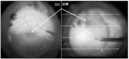
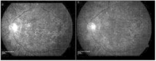
いくつかの実施形態では、網膜の解剖学的構造又は機能性の回復は、地図状萎縮の成長の低下、視力の改善、読み取り速度の改善、網膜構造の改善、ドルーゼンの減少、又は細胞の安定な生着のうちの1つ又は複数を評価することによって決定される。 In some embodiments, restoration of retinal anatomy or functionality is reduced growth of geographic atrophy, improved visual acuity, improved reading speed, improved retinal structure, decreased drusen, or stabilized cells. determined by assessing one or more of the following engraftments.
いくつかの実施形態では、改善は、微小視野測定によって測定される。 In some embodiments, improvement is measured by microperimetry.
いくつかの実施形態では、対象の視力が処置によって改善され、改善された視力が、GA病変(複数可)の総面積の変化;単眼読み取り速度の変化;機能的読み取り独立指数(FRII)複合スコアの変化;正常輝度最高矯正視力スコア(NL-BCVA)の変化;低輝度最高矯正視力スコア(LL-BCVA)の変化;低輝度不足(LLD)の変化;単眼限界印刷サイズの変化;National Eye Institute Visual Functioning Questionnaire 25項目バージョン(NEI VFQ-25)遠方活動サブスケールスコアの変化;暗点の数の変化;黄斑感度の変化;及びAPL-2の全身血漿濃度の変化のうちの1つ又は複数によって評価される。 In some embodiments, the subject's visual acuity is improved by treatment, and the improved visual acuity is measured by: change in total area of GA lesion(s); change in monocular reading speed; functional reading independence index (FRII) composite score change in normal-luminance best-corrected visual acuity score (NL-BCVA); change in low-luminance best-corrected visual acuity score (LL-BCVA); change in low-luminance deficiency (LLD); change in monocular limit print size; National Eye Institute Visual Functioning Questionnaire 25-item version (NEI VFQ-25) by one or more of: change in distance activity subscale score; change in number of scotoma; change in macular sensitivity; and change in systemic plasma concentration of APL-2 evaluated.
いくつかの実施形態では、本方法は、移植された細胞の拒絶の遅延性の炎症を最小限にするか、又は全く生じさせない。 In some embodiments, the method produces minimal or no delayed inflammation of the rejection of transplanted cells.
いくつかの実施形態では、投与することは、網膜の領域又は網膜に隣接する領域にRPE細胞を送達することを含む。いくつかの実施形態では、送達することは、網膜の領域又は網膜に隣接する領域にRPE細胞を移植することを含む。 In some embodiments, administering comprises delivering RPE cells to an area of the retina or an area adjacent to the retina. In some embodiments, delivering comprises transplanting RPE cells to an area of the retina or an area adjacent to the retina.
いくつかの実施形態では、処置することは、RPE細胞の多能性分泌効果を含む。 In some embodiments, treating comprises pluripotent secretory effects of RPE cells.
いくつかの実施形態では、対象は、ドライ型AMD、網膜色素変性症、アッシャー症候群、卵黄様黄斑症、シュタルガルト病、網膜剥離、網膜異形成、網膜萎縮、網膜症、黄斑ジストロフィー、錐体ジストロフィー、錐体-桿体ジストロフィー、蜂巣状網膜ジストロフィー(Malattia Leventinese)、ドインハニカム型ジストロフィー、ソースビー型ジストロフィー、パターン/蝶型ジストロフィー、Best病、ノースカロライナ型ジストロフィー、中心性輪紋状脈絡膜ジストロフィー、網膜色素線条症、毒性黄斑症、病的近視、網膜色素変性、及び黄斑変性から選択される網膜疾患症状に罹患している。 In some embodiments, the subject has dry AMD, retinitis pigmentosa, Usher syndrome, vitelliform maculopathy, Stargardt disease, retinal detachment, retinal dysplasia, retinal atrophy, retinopathy, macular dystrophy, cone dystrophy, Cone-Rod Dystrophy, Malattia Leventinese, Doyne Honeycomb Dystrophy, Sauceby Dystrophy, Pattern/Butterfly Dystrophy, Best's Disease, North Carolina Dystrophy, Central Ringiform Choroidal Dystrophy, Retinal Pigment Lines Suffering from a retinal disease condition selected from striatum, toxic maculopathy, pathologic myopia, retinitis pigmentosa, and macular degeneration.
いくつかの実施形態では、細胞治療剤は、送達装置を用いて投与される。 In some embodiments, cell therapy agents are administered using a delivery device.
いくつかの実施形態では、細胞治療剤は、送達装置を用いて網膜の地図状萎縮部に投与されるか、又はそれに隣接して投与される。 In some embodiments, the cell therapy agent is administered to or adjacent to the geographic atrophy of the retina using a delivery device.
いくつかの実施形態では、送達装置は、ニードル、キャピラリー及びチップを備える。いくつかの実施形態では、送達装置は、約0.63mmの外径及び約0.53mmの内径を有するニードルと、約0.5mmの外径及び約0.25mmの内径を有するキャピラリーと、約0.12mmの外径及び約0.07mmの内径を有するチップとを備える。 In some embodiments the delivery device comprises a needle, capillary and tip. In some embodiments, the delivery device comprises a needle having an outer diameter of about 0.63 mm and an inner diameter of about 0.53 mm, a capillary having an outer diameter of about 0.5 mm and an inner diameter of about 0.25 mm, and about a tip having an outer diameter of 0.12 mm and an inner diameter of about 0.07 mm.
別の態様では、本開示は、本明細書に記載の方法のいずれかで使用するための、送達装置を提供する。 In another aspect, the disclosure provides a delivery device for use in any of the methods described herein.
いくつかの実施形態では、送達装置は、ニードル、キャピラリー及びチップを備える。 In some embodiments the delivery device comprises a needle, capillary and tip.
いくつかの実施形態では、装置は、約0.63mmの外径及び約0.53mmの内径を有するニードルと、約0.5mmの外径及び約0.25mmの内径を有するキャピラリーと、約0.12mmの外径及び約0.07mmの内径を有するチップとを備える。 In some embodiments, the device comprises a needle having an outer diameter of about 0.63 mm and an inner diameter of about 0.53 mm, a capillary having an outer diameter of about 0.5 mm and an inner diameter of about 0.25 mm, and a a tip having an outer diameter of 0.12 mm and an inner diameter of about 0.07 mm.
さらに別の態様では、本開示は、本開示による対象の網膜の解剖学的構造又は機能性を回復させるための細胞治療剤を含む組成物を提供する。 In yet another aspect, the disclosure provides a composition comprising a cell therapeutic agent for restoring retinal anatomy or functionality in a subject according to the disclosure.
網膜色素上皮(RPE)は、視細胞外節(POS)と脈絡膜血管系との間のブルッフ膜上に位置する神経上皮由来の色素細胞の単層である。RPE単層は、光受容体の機能及び健全性にとって重要である。網膜色素上皮(RPE)細胞の機能不全、損傷、及び喪失は、加齢黄斑変性(AMD)、ベスト病を含む遺伝性黄斑変性(初期発症型の卵黄様黄斑ジストロフィー)、及び網膜色素変性(RP)のサブタイプなどの特定の眼疾患及び障害の顕著な特徴である。このような疾患に罹患した者の網膜へのRPEの移植は、RPEが変性した網膜疾患における細胞置換療法として使用することができる。 The retinal pigment epithelium (RPE) is a monolayer of neuroepithelial-derived pigment cells located on Bruch's membrane between the photoreceptor outer segment (POS) and the choroidal vasculature. The RPE monolayer is critical to photoreceptor function and integrity. Dysfunction, damage, and loss of retinal pigment epithelial (RPE) cells are associated with age-related macular degeneration (AMD), hereditary macular degeneration including Best's disease (early onset vitelliform macular dystrophy), and retinitis pigmentosa (RPE). ) are hallmarks of certain ocular diseases and disorders, such as subtypes of Implantation of RPE into the retina of persons afflicted with such diseases can be used as a cell replacement therapy in retinal diseases in which the RPE is degenerative.
ヒト多能性幹細胞は、移植のためのRPE細胞の供給源として有意な利点を提供する。それらの多能性発生能は、それらの真の機能的RPE細胞への分化を可能にし、無限の自己再生の可能性を考慮すると、それらはRPE細胞の無限の供給源として役立ち得る。実際、ヒト胚性幹細胞(hESC)及びヒト人工多能性幹細胞(iPSC)がインビトロでRPE細胞に分化し、網膜変性を減弱させ、網膜下移植後の視覚機能を維持し得ることが実証されている。したがって、hESCは、細胞療法のためのRPE細胞の産生のための無制限の供給源であり得る。 Human pluripotent stem cells offer significant advantages as a source of RPE cells for transplantation. Their pluripotent developmental potential allows them to differentiate into true functional RPE cells, and given their limitless self-renewal potential, they can serve as a limitless source of RPE cells. Indeed, it has been demonstrated that human embryonic stem cells (hESCs) and human induced pluripotent stem cells (iPSCs) can differentiate into RPE cells in vitro, attenuate retinal degeneration, and preserve visual function after subretinal transplantation. there is Therefore, hESCs can be a limitless source for the production of RPE cells for cell therapy.
しかしながら、ほとんどの細胞ベースの処置は、通常、体内への直接投与に適合しない低温溶液中で凍結保存され、臨床使用のための実用的な問題を引き起こす。細胞は、解凍後数時間以内に移植されるべきであり、さもなければ生存率及び品質を失い始める可能性がある。さらに、細胞は、臨床現場、病院又は他の処置施設に近接していなくてもよい認定施設での投与前に調製されなければならない。最後に、最終製剤の調製は細胞療法製造プロセスの一部であると考えられるため、各対象の処置用量は資格のある技術者によって放出されなければならない。 However, most cell-based treatments are usually cryopreserved in cryogenic solutions that are not compatible with direct administration into the body, creating practical problems for clinical use. Cells should be transplanted within hours after thawing or they may begin to lose viability and quality. Additionally, the cells must be prepared prior to administration at an accredited facility that may not be in close proximity to clinical sites, hospitals, or other treatment facilities. Finally, since the preparation of the final formulation is considered part of the cell therapy manufacturing process, each subject's treatment dose must be delivered by a qualified technician.
本開示は、再生医療及びRPE細胞療法の分野におけるこれら及び他の欠点に対処する。本開示は、様々な方法、装置及び組成物に関するデータをさらに提供する。 The present disclosure addresses these and other shortcomings in the field of regenerative medicine and RPE cell therapy. The disclosure further provides data regarding various methods, devices and compositions.
本発明の実施形態のための教示、方法、組成物、装置及びノウハウは、2019年7月4日に公開された「RETINAL PIGMENT EPITHELIUM CELL COMPOSITIONS」と題する国際公開第2019/130061号;2018年9月20日に公開された「METHODS FOR MEASURING THERAPEUTIC EFFECTS OF RETINAL DISEASE THERAPIES」と題する国際公開第2018/170494号;及び、2017年2月2日に公開された「LARGE SCALE PRODUCTION OF RETINAL PIGMENT EPITHELIAL CELLS」と題する国際公開第2017/017686号に見出される。その各々は、単独又は互いに組み合わせて、その方法、装置及び器具、組成物のすべてについてその全体が参照により本明細書に組み込まれる。 The teachings, methods, compositions, apparatus and know-how for embodiments of the present invention can be found in WO 2019/130061 entitled RETINAL PIGMENT EPITHELIUM CELL COMPOSITIONS, published July 4, 2019; International Publication No. WO 2018/170494 entitled "METHODS FOR MEASURING THERAPEUTIC EFFECTS OF RETINAL DISEASE THERAPIES" published on 20 February 2017; TINAL PIGMENT EPITHELIAL CELLS" WO 2017/017686 entitled. Each of its methods, devices and instruments, compositions, each alone or in combination with each other, is hereby incorporated by reference in its entirety.
図は、驚くべきかつ予想外の結果の様々な例示及び例を提供する。実施形態は、議論され、記載され、又はデータが図に提示される評価及びアッセイのいずれかを含むことができる様々な方法に関する。 The figures provide various illustrations and examples of surprising and unexpected results. Embodiments relate to various methods that can include any of the evaluations and assays discussed, described, or for which data are presented in figures.
詳細な説明
本明細書の実施形態は、一般に、黄斑変性などの網膜症状を含む眼の疾患及び病気を処置するための方法、組成物、及び装置に関する。
DETAILED DESCRIPTION Embodiments herein generally relate to methods, compositions, and devices for treating ocular diseases and conditions, including retinal conditions such as macular degeneration.
いくつかの実施形態では、組成物、方法及び装置は、同種異系(「既製」)の製品候補を利用することができる。例えば、これは、材料が個々の患者ではなく細胞株に由来し、大規模生産を促進し、患者特異的な処置よりも生産コストが低いことを意味し得る。 In some embodiments, the compositions, methods and devices can utilize allogeneic (“off-the-shelf”) product candidates. For example, this may mean that the material is derived from cell lines rather than individual patients, facilitating large-scale production and lower production costs than patient-specific treatments.
方法、装置、組成物などは、添付の図面に記載されたものを含むことができる。 Methods, apparatus, compositions, etc. can include those depicted in the accompanying drawings.
この説明を読んだ後、様々な代替の実施形態及び代替の適用において本開示を実施する方法が当業者に明らかになるであろう。ただし、本発明の様々な実施形態のすべてについてここでは説明しない。ここに提示される実施形態は、一例としてのみ提示され限定でないことが理解されよう。したがって、様々な代替の実施形態のこの詳細な説明は、本明細書に記載の本開示の範囲又は幅を限定すると解釈されるべきではない。 After reading this description, it will become apparent to one skilled in the art how to implement the present disclosure in various alternative embodiments and alternative applications. However, not all of the various embodiments of the invention are described here. It will be appreciated that the embodiments presented herein are presented by way of example only and are not limiting. Therefore, this detailed description of various alternative embodiments should not be construed as limiting the scope or breadth of the disclosures described herein.
本技術を開示及び説明する前に、以下に記載される態様は、特定の組成物、そのような組成物を調製する方法、又はその使用に限定されず、したがって当然のことながら変化し得ることを理解されたい。本明細書で使用される専門用語が特定の態様を説明することのみを目的としており、限定するようには意図されていないことも理解されたい。 Before disclosing and describing the present technology, it should be noted that the aspects described below are not limited to particular compositions, methods of preparing such compositions, or uses thereof, as such may, of course, vary. Please understand. It is also to be understood that the terminology used herein is for the purpose of describing particular aspects only and is not intended to be limiting.
読者の便宜のためだけに様々なセクションに分けられた詳細な説明及び任意のセクションに見られる開示は、別のセクションのものと組み合わされてもよい。タイトル又はサブタイトルが、読者の便宜のために本明細書で使用され得、本開示の範囲に影響を及ぼすことを意図しない。 The detailed description, which is divided into various sections solely for the convenience of the reader, and disclosure found in any section may be combined with that in any other section. Titles or subtitles may be used herein for the convenience of the reader and are not intended to affect the scope of the disclosure.
定義
「処置すること(treating)」、又は「処置(treatment)」という用語は、傷害、疾患、病理又は症状の治療又は改善におけるなんらかの成功の兆候を指し、あらゆる客観的又は主観的パラメータ、例えば症候の軽減、緩和、減弱、又は傷害、病理、若しくは症状を患者にとってより許容可能にすること、衰退又は衰弱の速度の低下、衰退の最終点での衰弱の重度の低下、患者の身体的又は精神的健康の改善などを含む。症候の処置又は改善は、客観的又は主観的パラメータに基づくことができる。身体検査、精神神経学的検査、及び/又は精神医学的評価の結果を含む。「処置」という用語及びその活用形は、傷害、病理、症状又は疾患の予防を含み得る。複数の実施形態において、処置することは予防することである。複数の実施形態において、処置することは予防することを含まない。本明細書で使用される「処置すること」又は「処置」はまた、(当技術分野でよく理解されているように)、臨床結果を含む、対象の症状において有益な又は所望の結果を得るための任意のアプローチを広く含む。有益な又は所望の臨床結果には、限定するものではないが、1つ又は複数の症候又は症状の緩和又は改善、疾患の程度の縮小、疾患の状態の安定化(すなわち、悪化しない)、疾患の伝播又は広がりの予防、疾患の進行の遅延又は鈍化、疾患状態の改善又は緩和、疾患の再発の減少、及び部分的であろうと全体的であろうと、検出可能であろうと検出不能であろうと、寛解が含まれ得る。換言すれば、本明細書で使用される「処置」は、疾患の任意の治癒、改善、又は予防を含む。処置は、疾患の発生を防止;疾患の広がりを抑制;疾患の症候を軽減;疾患の根本原因を完全に若しくは部分的に除去;疾患の持続期間を短縮;又はこれらの組み合わせを行い得る。
DEFINITIONS The term “treating” or “treatment” refers to any indication of success in curing or ameliorating an injury, disease, pathology or condition, and any objective or subjective parameter such as symptom reducing, mitigating, diminishing, or making the injury, pathology, or condition more tolerable to the patient; slowing the rate of decline or decline; reducing the severity of weakness at the end point of decline; including improved physical health. Treatment or amelioration of symptoms can be based on objective or subjective parameters. Includes results of physical examination, neuropsychiatric examination, and/or psychiatric evaluation. The term "treatment" and its conjugations may include prevention of injury, pathology, condition or disease. In some embodiments, treating is prophylactic. In some embodiments, treating does not include preventing. As used herein, "treating" or "treatment" also (as well understood in the art) includes obtaining beneficial or desired results in the condition of interest, including clinical results. broadly includes any approach to Beneficial or desired clinical results include, but are not limited to, alleviation or amelioration of one or more symptoms or symptoms, reduction in extent of disease, stabilization of disease state (i.e., not worsening), prevention of transmission or spread of disease, delay or slowing of disease progression, amelioration or alleviation of disease state, reduction of disease recurrence, and whether partial or total, detectable or undetectable , may include remission. In other words, "treatment" as used herein includes any cure, amelioration or prevention of disease. Treatment may prevent the occurrence of the disease; inhibit the spread of the disease; alleviate the symptoms of the disease; completely or partially eliminate the underlying cause of the disease; shorten the duration of the disease;
本明細書で使用される「処置すること」及び「処置」は、予防的処置を含む。処置方法は、治療有効量の活性薬剤を対象に投与することを含む。投与工程は、単回投与からなってもよく、又は一連の投与を含んでもよい。処置期間の長さは、症状の重症度、患者の年齢、活性薬剤の濃度、処置に使用される組成物の活性、又はそれらの組み合わせなどの様々な要因に依存する。処置又は予防に使用される薬剤の有効投与量は、特定の処置又は予防計画の経過にわたって増加又は減少し得ることも理解されよう。投与量の変化は、当技術分野で公知の標準的な診断アッセイによって生じ、明らかになり得る。場合によっては、慢性投与が必要とされ得る。例えば、組成物は、患者を処置するのに十分な量及び期間で対象に投与される。複数の実施形態において、処置すること、又は処置は、予防的処置ではない。 As used herein, "treating" and "treatment" include prophylactic treatment. Treatment methods include administering to a subject a therapeutically effective amount of an active agent. The administering step may consist of a single dose or may include a series of doses. The length of the treatment period will depend on a variety of factors such as severity of symptoms, age of the patient, concentration of active agent, activity of the composition used for treatment, or a combination thereof. It will also be appreciated that the effective dosage of an agent used for treatment or prevention may increase or decrease over the course of a particular treatment or prevention regimen. Changes in dosage may result and become apparent by standard diagnostic assays known in the art. Chronic administration may be required in some cases. For example, the composition is administered to the subject in an amount and for a period of time sufficient to treat the patient. In some embodiments, treating or treatment is not prophylactic treatment.
「予防する」という用語は、患者における疾患症候の発生の減少を指す。上記のように、予防は完全(検出可能な症候なし)であってもよく、又は、処置なしで生じる得るものよりも少ない症候が観察されるような部分的であってもよい。 The term "prevent" refers to reducing the occurrence of disease symptoms in a patient. As noted above, prevention may be complete (no detectable symptoms) or partial such that fewer symptoms are observed than would otherwise occur without treatment.
「患者」又は「それを必要とする対照」とは、本明細書で提供されるような医薬組成物の投与によって処置することができる疾患又は症状に苦しんでいるか、又は罹患しやすい生物を意味する。非限定的な例としては、ヒト、その他の哺乳動物、ウシ、ラット、マウス、イヌ、サル、ヤギ、ヒツジ、ウシ、シカ、及びその他の非哺乳動物が挙げられる。いくつかの実施形態では、患者はヒトである。 "Patient" or "control in need thereof" means an organism afflicted with or susceptible to a disease or condition that can be treated by administration of a pharmaceutical composition as provided herein. do. Non-limiting examples include humans, other mammals, cows, rats, mice, dogs, monkeys, goats, sheep, cows, deer, and other non-mammals. In some embodiments, the patient is human.
「有効量」は、組成物の不存在と比較して、組成物が述べられた目的を達成するのに十分な量である(例えば、それが投与される効果を達成するか、疾患を処置するか、酵素活性を低下させるか、酵素活性を増加させるか、シグナル伝達経路を減少させるか、又は疾患若しくは症状の1つ又は複数の症候を減少させる)。「有効量」の例は、疾患の1つ又は複数の症候の処置、予防、又は軽減に寄与するのに十分な量であり、「治療有効量」とも呼ばれ得る。1つ又は複数の症候の「減少」(及びこの句の文法的等価物)は、症候(複数可)の重症度又は頻度の減少、又は症候(複数可)の排除を意味する。薬物(例えば、本明細書中に記載される細胞)の「予防有効量」は、対象に投与された場合に、意図された予防効果、例えば、損傷、疾患、病理若しくは症状の発症(又は再発)を予防若しくは遅延させるか、又は、損傷、疾患、病理若しくは症状、又はそれらの症候の発症(又は再発)の可能性を低下させる薬物の量である。完全な予防効果は、必ずしも1用量の投与によって生じるわけではなく、一連の用量の投与後にのみ生じることができる。したがって、予防有効量を1回又は複数回の投与で投与することができる。本明細書で使用される「活性低下量」は、アンタゴニストの非存在下と比較して酵素の活性を低下させるのに必要なアンタゴニストの量を指す。本明細書で使用される「機能破壊量」は、アンタゴニストの非存在下と比較して、酵素又はタンパク質の機能を破壊するのに必要なアンタゴニストの量を指す。正確な量は、処置の目的に依存し、公知の技術(例えば、Lieberman,Pharmaceutical Dosage Forms(vols.1-3,1992);Lloyd,The Art,Science and Technology of Pharmaceutical Compounding(1999);Pickar,Dosage Calculations(1999);及びRemington:The Science and Practice of Pharmacy,20th Edition,2003,Gennaro,Ed.,Lippincott,Williams&Wilkinsを参照)を用いて当業者によって確認可能であろう。 An "effective amount" is an amount sufficient, relative to the absence of the composition, for the composition to achieve its stated purpose (e.g., achieve the effect for which it is administered or treat a disease). reduce enzymatic activity, increase enzymatic activity, reduce signal transduction pathways, or reduce one or more symptoms of a disease or condition). An example of an "effective amount" is an amount sufficient to contribute to the treatment, prevention, or alleviation of one or more symptoms of a disease, and can also be referred to as a "therapeutically effective amount." A "reduction" of one or more symptoms (and grammatical equivalents of this phrase) means a reduction in the severity or frequency of the symptom(s) or the elimination of the symptom(s). A "prophylactically effective amount" of a drug (e.g., a cell described herein) has an intended prophylactic effect, e.g., the onset (or recurrence) of an injury, disease, pathology or condition when administered to a subject. ) or reduce the likelihood of onset (or recurrence) of an injury, disease, pathology or condition, or symptoms thereof. A full prophylactic effect does not necessarily occur by administration of one dose, but can occur only after administration of a series of doses. Accordingly, a prophylactically effective amount can be administered in one or more administrations. As used herein, an "activity-reducing amount" refers to the amount of antagonist required to reduce the activity of an enzyme compared to the absence of antagonist. As used herein, "function-disrupting amount" refers to the amount of antagonist required to disrupt the function of an enzyme or protein compared to the absence of antagonist. The exact amount depends on the purpose of treatment and can be determined according to known techniques (eg, Lieberman, Pharmaceutical Dosage Forms (vols. 1-3, 1992); Lloyd, The Art, Science and Technology of Pharmaceutical Compounding (19 99); Dosage Calculations (1999); and Remington: The Science and Practice of Pharmacy, 20th Edition, 2003, Gennaro, ED. See T, WILLIAMS & WILKINS).
本明細書に記載の任意の組成物について、治療有効量は、細胞培養アッセイから最初に決定することができる。目標濃度は、本明細書に記載又は当技術分野で公知の方法を使用して測定された、本明細書に記載の方法を達成することができる活性組成物(複数可)(例えば、細胞濃度又は細胞数)の濃度である。 For any composition described herein, the therapeutically effective dose can be initially determined from cell culture assays. The target concentration is the active composition(s) capable of achieving the methods described herein (e.g., cell concentration or cell number).
当技術分野で周知のように、ヒトで使用するための治療有効量は、動物モデルから決定することもできる。例えば、ヒトに対する用量は、動物において有効であることが分かっている濃度を達成するように製剤化することができる。ヒトにおける投与量は、上記のように、組成物の有効性を監視し、投与量を上方又は下方に調整することによって調整することができる。上記の方法及び他の方法に基づいてヒトにおいて最大の有効性を達成するために用量を調整することは、十分に当業者の能力の範囲内である。 A therapeutically effective amount for use in humans can also be determined from animal models, as is well known in the art. For example, a dose for humans can be formulated to achieve concentrations found to be effective in animals. The dosage in humans can be adjusted by monitoring the efficacy of the composition and adjusting the dosage upwards or downwards, as described above. Adjusting dosages to achieve maximal efficacy in humans based on the methods described above and other methods is well within the capability of those skilled in the art.
本明細書で使用される「治療有効量」という用語は、上記の障害を改善するのに十分な治療薬の量を指す。例えば、所与のパラメータについて、治療有効量は、少なくとも5%、10%、15%、20%、25%、40%、50%、60%、75%、80%、90%、又は少なくとも100%の増加又は減少を示す。治療有効性は、「-倍」の増加又は減少として表すこともできる。例えば、治療有効量は、対照に対して少なくとも1.2倍、1.5倍、2倍、5倍又はそれ以上の効果を有することができる。 As used herein, the term "therapeutically effective amount" refers to that amount of therapeutic agent sufficient to ameliorate the disorders described above. For example, for a given parameter, a therapeutically effective amount is at least 5%, 10%, 15%, 20%, 25%, 40%, 50%, 60%, 75%, 80%, 90%, or at least 100% % increase or decrease. Therapeutic efficacy can also be expressed as a "-fold" increase or decrease. For example, a therapeutically effective amount can have an effect of at least 1.2-fold, 1.5-fold, 2-fold, 5-fold or more over a control.
投与量は、患者及び採用される組成物の要件に応じて変化してもよい。本開示の文脈において、患者に投与される用量は、時間の経過とともに患者において有益な治療反応をもたらすのに十分であるべきである。また、投与量の大きさは、副作用の有無、性質、程度によっても決定される。特定の状況に対する適切な投与量の決定は、医療従事者のスキルの範囲内である。一般に、処置は、組成物の最適用量未満の、より少ない用量で開始される。その後、状況下で最適な効果が得られるまで、投与量を少しずつ増やす。投与量及び間隔は、処置されている特定の臨床的適応症に有効な投与された組成物のレベルを提供するために個別に調整することができる。これにより、個人の疾患状態の重症度に見合った治療法を提供することができる。 Dosages may vary according to the requirements of the patient and the composition employed. In the context of the present disclosure, the dose administered to a patient should be sufficient to produce a beneficial therapeutic response in the patient over time. In addition, the size of the dosage is also determined by the presence/absence, nature, and degree of side effects. Determination of the proper dosage for a particular situation is within the skill of medical practitioners. Generally, treatment is initiated with smaller dosages which are less than the optimum dose of the composition. Thereafter, the dosage is increased by small increments until the optimum effect under the circumstances is reached. Dosage amounts and intervals may be adjusted individually to provide levels of the administered composition effective for the particular clinical indication being treated. This allows the provision of treatments that are commensurate with the severity of an individual's disease state.
「共投与」は、本明細書中に記載される組成物が、1つ又は複数の追加の治療の投与と同時に、投与の直前に、又は投与の直後に投与されることを意味する。本明細書で提供される組成物は、患者に対して、単独で投与することができ、又は共投与することができる。共投与とは、組成物を個別に、又は組み合わせて(複数の組成物)同時投与又は連続投与することを含むことを意味する。したがって、調製物は、必要に応じて、(例えば、代謝分解を減少させるために)他の活性物質と組み合わせることもできる。 "Co-administration" means that the compositions described herein are administered concurrently with, immediately prior to, or immediately following administration of one or more additional therapies. The compositions provided herein can be administered alone or can be co-administered to the patient. Co-administration is meant to include simultaneous or sequential administration of the compositions individually or in combination (multiple compositions). Thus, the preparations can also be combined with other active substances (eg, to reduce metabolic degradation), if desired.
「対照」又は「対照実験」は、その単純な通常の意味に従って使用され、実験の手順、試薬、又は変数の省略を除いて、実験の対象又は試薬が並行実験のように扱われる実験を指す。場合によっては、対照は、実験効果を評価する際の比較の基準として使用される。いくつかの実施形態では、対照は、本明細書(実施形態及び例を含む)に記載の組成物の非存在下でのタンパク質の活性の尺度である。 "Control" or "control experiment" is used according to its simple and ordinary meaning and refers to an experiment in which the subject or reagents of the experiment are treated like a parallel experiment, except for the omission of experimental procedures, reagents, or variables. . In some cases, controls are used as a basis for comparison in assessing experimental effects. In some embodiments, a control is a measure of protein activity in the absence of a composition described herein (including embodiments and examples).
「薬学的に許容され得る賦形剤」及び「薬学的に許容され得る担体」は、対象への活性薬剤の投与及び対象による吸収を補助し、患者に有意な有害毒性作用を引き起こさずに本開示の組成物に含めることができる物質を指す。薬学的に許容され得る賦形剤の非限定的な例としては、水、NaCl、通常の生理食塩水、乳酸加リンガー液、通常のスクロース、通常のグルコース、結合剤、充填剤、崩壊剤、潤滑剤、コーティング、甘味料、香味剤、塩溶液(リンガー液など)、アルコール、油類、ゼラチン、ラクトース、アミロース又はデンプンなどの炭水化物、脂肪酸エステル、ヒドロキシメチルセルロース、ポリビニルピロリジン及び着色剤などが挙げられる。そのような調製物は、滅菌し、必要に応じて、潤滑剤、保存剤、安定剤、湿潤剤、乳化剤、浸透圧に影響を及ぼすための塩、緩衝剤、着色剤、及び/又は本開示の組成物と有害に反応しない芳香族物質などの補助剤と混合することができる。当業者は、他の医薬賦形剤が本開示において有用であることを認識するであろう。 "Pharmaceutically acceptable excipients" and "pharmaceutically acceptable carriers" are those that aid administration to, and absorption by, a subject of an active agent without causing significant adverse toxic effects to the patient. It refers to substances that can be included in the disclosed compositions. Non-limiting examples of pharmaceutically acceptable excipients include water, NaCl, normal saline, lactated Ringer's solution, normal sucrose, normal glucose, binders, fillers, disintegrants, Lubricants, coatings, sweeteners, flavoring agents, salt solutions (such as Ringer's solution), alcohols, oils, carbohydrates such as gelatin, lactose, amylose or starch, fatty acid esters, hydroxymethylcellulose, polyvinylpyrrolidine and coloring agents. . Such preparations are sterilized and optionally contain lubricants, preservatives, stabilizers, wetting agents, emulsifiers, salts for influencing osmotic pressure, buffers, coloring agents and/or the present disclosure. can be mixed with adjuvants such as aromatic substances that do not deleteriously react with the composition of Those skilled in the art will recognize that other pharmaceutical excipients are useful in the present disclosure.
本明細書で使用される「細胞」は、そのゲノムDNAを保存又は複製するのに十分な代謝又は他の機能を実行する細胞を指す。細胞は、例えば、無傷の膜の存在、特定の色素による染色、子孫を産生する能力、又は配偶子の場合、第2の配偶子と組み合わせて生存可能な子孫を産生する能力を含む、当技術分野で周知の方法によって同定することができる。細胞は、原核細胞及び真核細胞を含み得る。原核細胞には、細菌が含まれるが、これに限定されない。真核細胞には、酵母細胞並びに植物及び動物由来の細胞、例えば哺乳動物、昆虫(例えばスポドプテラ(spodoptera))及びヒト細胞が含まれるが、これらに限定されない。細胞は、天然に非接着性であるか、又は例えばトリプシン処理によって表面に接着しないように処理されている場合に有用であり得る。 As used herein, "cell" refers to a cell that performs sufficient metabolic or other functions to store or replicate its genomic DNA. Cells are characterized by, for example, the presence of intact membranes, staining with a particular dye, the ability to produce progeny, or, in the case of gametes, the ability to combine with a second gamete to produce viable progeny. It can be identified by methods well known in the art. Cells can include prokaryotic and eukaryotic cells. Prokaryotic cells include, but are not limited to bacteria. Eukaryotic cells include, but are not limited to, yeast cells and cells of plant and animal origin, such as mammalian, insect (eg, spodoptera) and human cells. Cells may be useful if they are naturally non-adherent or have been treated to not adhere to surfaces, such as by trypsinization.
本明細書中で使用される場合、「幹細胞」とは、特定の特殊化された機能を有する他の細胞型(例えば、完全に分化した細胞)に分化するように誘導されるまで、培地中で長期間にわたって未分化状態(例えば、多能性又は多能性幹細胞)に留まることができる細胞のことを指す。複数の実施形態において、「幹細胞」には、胚性幹細胞(ESC)、人工多能性幹細胞(iPSC)、成体幹細胞、間葉系幹細胞及び造血幹細胞が含まれる。複数の実施形態において、RPE細胞は、多能性幹細胞(例えば、ESC又はiPSC)から作製される。 As used herein, a “stem cell” is a Cells that can remain in an undifferentiated state (eg, pluripotent or pluripotent stem cells) for long periods of time. In embodiments, "stem cells" include embryonic stem cells (ESC), induced pluripotent stem cells (iPSC), adult stem cells, mesenchymal stem cells and hematopoietic stem cells. In embodiments, RPE cells are generated from pluripotent stem cells (eg, ESCs or iPSCs).
本明細書で使用される場合、「人工多能性幹細胞」又は「iPSC」は、体細胞の遺伝子操作によって、例えば、Oct-3/4、Sox2、c-Myc及びKLF4などの転写因子を用いた線維芽細胞、肝細胞、胃上皮細胞などの体細胞のレトロウイルス形質導入によって、体細胞から生成することができる細胞である[Yamanaka S,Cell Stem Cell.2007,1(1):39-49;Aoi T,et al.,Generation of Pluripotent Stem Cells from Adult Mouse Liver and Stomach Cells.Science.2008 Feb 14.(Epub ahead of print);IH Park,Zhao R,West JA,et al.Reprogramming of human somatic cells to pluripotency with defined factors.Nature 2008;451:141-146;K Takahashi,Tanabe K,Ohnuki M,et al.Induction of pluripotent stem cells from adult human fibroblasts by defined factors.Cell 2007;131:861-872]。他の胚様幹細胞は、レシピエント細胞が有糸分裂で停止している場合、卵母細胞への核移植、胚性幹細胞との融合、又は接合体への核移植によって作製され得る。さらに、iPSCは、非組み込み法を使用して、例えば、小分子又はRNAを使用することによって生成され得る。
As used herein, “induced pluripotent stem cells” or “iPSCs” are defined by genetic engineering of somatic cells to use, for example, transcription factors such as Oct-3/4, Sox2, c-Myc and KLF4. Cells that can be generated from somatic cells by retroviral transduction of somatic cells such as fibroblasts, hepatocytes, gastric epithelial cells [Yamanaka S, Cell Stem Cell. 2007, 1(1):39-49; Aoi T, et al. , Generation of Pluripotent Stem Cells from Adult Mouse Livers and Stomach Cells. Science. 2008
「胚性幹細胞」という用語は、3つすべての胚性胚葉(すなわち、内胚葉、外胚葉及び中胚葉)の細胞に分化することができる、又は未分化状態のままであることができる胚性細胞を指す。「胚性幹細胞」という語句は、妊娠後に形成された胚組織(例えば、胚盤胞)から胚の着床前に得られる細胞(すなわち、着床前胚盤胞)、着床後/原腸形成前段階の胚盤胞から得られる拡張胚盤胞細胞(EBC)(国際公開第2006/040763号参照)、及び妊娠中の任意の時点、好ましくは妊娠10週前に胎児の生殖組織から得られる胚性生殖(EG)細胞を含む。複数の実施形態において、胚性幹細胞は、周知の細胞培養方法を用いて得られる。例えば、ヒト胚性幹細胞は、ヒト胚盤胞から単離することができる。 The term "embryonic stem cells" refers to embryonic stem cells that can differentiate into cells of all three embryonic germ layers (i.e., endoderm, ectoderm and mesoderm) or remain undifferentiated. refers to cells. The phrase "embryonic stem cells" refers to cells obtained prior to implantation of the embryo (i.e., pre-implantation blastocyst) from embryonic tissue formed after pregnancy (e.g., blastocyst), post-implantation/gastrula Expanded blastocyst cells (EBC) obtained from preformal stage blastocysts (see WO 2006/040763) and from fetal reproductive tissue at any time during pregnancy, preferably before 10 weeks of gestation. contains embryonic germ (EG) cells that are In embodiments, embryonic stem cells are obtained using well-known cell culture methods. For example, human embryonic stem cells can be isolated from human blastocysts.
市販の幹細胞もまた、本開示の態様及び実施形態において使用され得ることが理解される。ヒトES細胞は、NIHヒト胚性幹細胞登録(www.grants.nih.govstem_cells/)又は他のhESC登録から購入することができる。市販の胚性幹細胞株の非限定的な例は、HAD-C 102、ESI、BGO 1、BG02、BG03、BG04、CY12、CY30、CY92、CY1O、TE03、TE32、CHB-4、CHB-5、CHB-6、CHB-8、CHB-9、CHB-10、CHB-11、CHB-12、HUES 1、HUES 2、HUES 3、HUES 4、HUES 5、HUES 6、HUES 7、HUES 8、HUES 9、HUES 10、HUES 11、HUES 12、HUES 13、HUES 14、HUES 15、HUES 16、HUES 17、HUES 18、HUES 19、HUES 20、HUES 21、HUES 22、HUES 23、HUES 24、HUES 25、HUES 26、HUES 27、HUES 28、CyT49、RUES3、WAO 1、UCSF4、NYUES 1、NYUES2、NYUES3、NYUES4、NYUESS、NYUES6、NYUES7、UCLA 1、UCLA 2、UCLA 3、WA077(H7)、WA09(H9)、WA 13(H13)、WA14(H14)、HUES 62、HUES 63、HUES 64、CT I、CT2、CT3、CT4、MA135、Eneavour-2、WIBR 1、WIBR2、WIBR3、WIBR4、WIBRS、WIBR6、HUES 45、Shef 3、Shef 6、BINhem19、BJNhem20、SAGO 1、SAOO1である。 It is understood that commercially available stem cells may also be used in aspects and embodiments of the present disclosure. Human ES cells can be purchased from the NIH Human Embryonic Stem Cell Registry (www.grants.nih.govstem_cells/) or other hESC registry. Non-limiting examples of commercially available embryonic stem cell lines include HAD-C 102, ESI, BGO 1, BG02, BG03, BG04, CY12, CY30, CY92, CY1O, TE03, TE32, CHB-4, CHB-5, CHB-6, CHB-8, CHB-9, CHB-10, CHB-11, CHB-12, HUES 1, HUES 2, HUES 3, HUES 4, HUES 5, HUES 6, HUES 7, HUES 8, HUES 9 , HUES 10, HUES 11, HUES 12, HUES 13, HUES 14, HUES 15, HUES 16, HUES 17, HUES 18, HUES 19, HUES 20, HUES 21, HUES 22, HUES 23, HUES 24, HUES 25, HUES 26, HUES 27, HUES 28, CYT49, RUES3, WAO 1, UCSF4, NYUES2, NYUES3, NYUES3, NYUES3, NYUESS4, NYUES6, NYUES6, UCLA 2, UCLA 2, UCLA 3, W A077 (H7), WA09 (H9) , WA 13 (H13), WA14 (H14), HUES 62, HUES 63, HUES 64, CT I, CT2, CT3, CT4, MA135, Eneavour-2, WIBR 1, WIBR2, WIBR3, WIBR4, WIBRS, WIBR6, HUES 45, Shef 3, Shef 6, BINhem 19, BJNhem 20, SAGO 1, SAOOl.
「網膜の色素層」としても知られる「網膜色素上皮」又は「RPE」という用語は、網膜の外側の細胞の色素層を指す。RPE層は、ブルッフ膜(脈絡膜内縁)と光受容体との間に位置する。RPEは、網膜に栄養素を供給するための中間体であり、網膜の発達、光の吸収、成長因子の分泌、及び眼の免疫応答の媒介を含む多くの機能を補助する。RPEの機能不全は、網膜色素変性、糖尿病性網膜症、西ナイルウイルス、及び黄斑変性を含む症状において視力喪失又は失明をもたらし得る。 The term "retinal pigment epithelium" or "RPE", also known as "retinal pigment layer", refers to the pigment layer of cells on the outer surface of the retina. The RPE layer is located between Bruch's membrane (inner choroid) and the photoreceptors. The RPE is an intermediate for supplying nutrients to the retina and assists in many functions including retinal development, light absorption, growth factor secretion, and mediation of the ocular immune response. Dysfunction of the RPE can lead to vision loss or blindness in conditions including retinitis pigmentosa, diabetic retinopathy, West Nile virus, and macular degeneration.
「疾患」又は「症状」という用語は、本明細書で提供される組成物又は方法で処置することができる患者又は対象の状態又は健康状態を指す。加齢黄斑変性すなわちAMDは、網膜中心部の進行性慢性疾患であり、世界中で視力喪失の主な原因である。ほとんどの視力喪失は、2つのプロセス:新生血管(「ウェット型」AMD及び地図状萎縮(GA、「ドライ型」)のうちの1つに起因して疾患の後期段階で起こる。GAでは、網膜色素上皮、脈絡毛細管板及び光受容体の進行性萎縮が起こる。AMDのドライ型はより一般的であるが(全症例の85~90%)、「ウェット」型に進行することがあり、未処置のまま放置すると、急速かつ重度の視力喪失をもたらす。AMDの推定有病率は、米国及び他の先進国では2,000人に1人である。この有病率は、一般人口における高齢者の割合とともに増加すると予想される。疾患の危険因子には、環境因子と遺伝因子の両方が含まれる。この疾患の病因には、機能的に相互に関連する4つの組織、すなわち網膜色素上皮(RPE)、ブルッフ膜、脈絡毛細管板及び光受容体の異常が関与している。しかしながら、RPE細胞機能の障害は、臨床的に関連するAMD変化をもたらす分子経路における初期の重要な事象である。現在、ドライ型AMDに対する承認された処置はない。予防的手段としては、ビタミン/ミネラルのサプリメントが挙げられる。これらは、ウェット型AMDを発症するリスクを低下させるが、地図状萎縮(GA)の進行の発症には影響しない。 The terms "disease" or "condition" refer to a condition or health condition in a patient or subject that can be treated with the compositions or methods provided herein. Age-related macular degeneration, or AMD, is a progressive, chronic disease of the central retina and the leading cause of vision loss worldwide. Most vision loss occurs in the late stages of the disease due to one of two processes: neovascularization (“wet” AMD and geographic atrophy (GA, “dry”). Progressive atrophy of the pigment epithelium, choriocapillaris, and photoreceptors occurs.The dry form of AMD is more common (85-90% of all cases), although it may progress to the 'wet' form, which may progress to lesser If left untreated, it results in rapid and severe vision loss.AMD has an estimated prevalence of 1 in 2,000 in the United States and other developed countries. Risk factors for the disease include both environmental and genetic factors.The pathogenesis of the disease involves four functionally interrelated tissues: the retinal pigment epithelium; (RPE), Bruch's membrane, choriocapillaris, and photoreceptors are involved.However, impairment of RPE cell function is an early key event in the molecular pathway leading to clinically relevant AMD changes. There is currently no approved treatment for dry AMD.Preventative measures include vitamin/mineral supplements.These reduce the risk of developing wet AMD, but geographic atrophy (GA) does not affect the onset of progression of
本明細書で提供される方法に従って処置の効果を測定し得る疾患の非限定的なリストには、網膜色素変性症、レーバー先天性黒内障、遺伝性又は後天性黄斑変性、加齢黄斑変性(AMD)、地図状萎縮(GA)、ベスト病、網膜剥離、脳回転状萎縮、コロイデレミア、パターンジストロフィー並びにRPEの他のジストロフィー、シュタルガルト病、光線性、レーザー性、炎症性、感染性、放射線性、新生血管性又は外傷性のいずれか1つによって生じる損傷によるRPE及び網膜損傷、網膜異形成、網膜萎縮、網膜症、黄斑ジストロフィー、錐体ジストロフィー、錐体-桿体ジストロフィー、蜂巣状網膜ジストロフィー(Malattia Leventinese)、ドインハニカム型ジストロフィー、ソースビー型ジストロフィー、パターン/蝶型ジストロフィー、Best病、ノースカロライナ型ジストロフィー、中心性輪紋状脈絡膜ジストロフィー、網膜色素線条症、毒性黄斑症、病的近視、網膜色素変性、及び黄斑変性が含まれる。複数の実施形態において、疾患は、ドライ型AMDである。複数の実施形態において、疾患は、GAである。 A non-limiting list of diseases for which efficacy of treatment may be measured according to the methods provided herein include retinitis pigmentosa, Leber congenital amaurosis, hereditary or acquired macular degeneration, age-related macular degeneration (AMD) ), geographic atrophy (GA), Best's disease, retinal detachment, cerebral rotational atrophy, choroideremia, pattern dystrophy and other dystrophies of RPE, Stargardt disease, actinic, laser, inflammatory, infectious, radiogenic, neoplastic RPE and retinal damage due to damage caused by either vascular or traumatic, retinal dysplasia, retinal atrophy, retinopathy, macular dystrophy, cone dystrophy, cone-rod dystrophy, honeycombing retinal dystrophy (Malattia Leventinese) ), Doyne honeycomb dystrophy, Sorsby dystrophy, pattern/butterfly dystrophy, Best disease, North Carolina dystrophy, central ring-shaped choroidal dystrophy, retinitis pigmentosa, toxic maculopathy, pathological myopia, retinitis pigmentosa , and macular degeneration. In some embodiments, the disease is dry AMD. In some embodiments, the disease is GA.
萎縮型加齢黄斑変性(AMD)又は進行性ドライ型AMDとしても知られる「地図状萎縮」又は「GA」又は「萎縮性網膜」は、経時的に視覚機能の損失をもたらし得る網膜(光受容体、網膜色素上皮、脈絡毛細管板)の進行性及び不可逆性の損失をもたらし得る加齢黄斑変性の進行した形態である。 “Geographic atrophy” or “GA” or “atrophic retina,” also known as dry age-related macular degeneration (AMD) or progressive dry AMD, is a retinal (photoreceptor It is an advanced form of age-related macular degeneration that can lead to progressive and irreversible loss of the body, retinal pigment epithelium, choriocapillaris).
いくつかの実施形態では、RPE欠損は、高齢、喫煙、不健康な体重、抗酸化物質の低摂取、又は心血管障害のうちの1つ又は複数に起因し得る。他の実施形態では、RPE欠損は先天性異常に起因し得る。文脈が許す限り互換的に使用され得る「網膜色素上皮細胞」、「RPE細胞」、「RPE」は、例えば、機能的に、エピジェネティックに、又は発現プロファイルによって、網膜の色素上皮細胞層を形成する天然RPE細胞と似ている(例えば、眼内への移植、投与又は送達の際に、それらは天然RPE細胞の機能活性と同様の機能活性を示す)細胞型の細胞を指す。 In some embodiments, RPE deficiency may result from one or more of advanced age, smoking, unhealthy weight, low intake of antioxidants, or cardiovascular disease. In other embodiments, RPE deficiency may result from congenital abnormalities. "Retinal pigment epithelial cell", "RPE cell", "RPE", which may be used interchangeably as the context permits, e.g., functionally, epigenetically, or by expression profile, form the pigment epithelial cell layer of the retina. Refers to cells of a cell type that resemble native RPE cells (eg, upon implantation, administration or delivery into the eye, they exhibit functional activity similar to that of native RPE cells).
本明細書で使用される場合、「OpRegen」という用語は、系統制限ヒトRPE細胞株を指す。RPE細胞は、アクチビンA、形質転換増殖因子ベータ(TGF-b)ファミリー及びニコチンアミドを補充してRPE集団を濃縮した分化培地下で誘導される。OpRegenは、眼用平衡塩類溶液(BSS Plus)中に、又はCryoStor(登録商標)5内のすぐに投与できる(ready to administer:RTA)解凍注入(thaw and inject:TAI)製剤として製剤化された単一細胞懸濁液である。
As used herein, the term "OpRegen" refers to a lineage restricted human RPE cell line. RPE cells are induced in differentiation medium supplemented with activin A, transforming growth factor beta (TGF-b) family and nicotinamide to enrich the RPE population. OpRegen was formulated in ophthalmic balanced salt solution (BSS Plus) or as a ready to administrator (RTA) thaw and inject (TAI) formulation in the
処置方法
本明細書の実施形態は、一般に、黄斑変性などの網膜症状を含む眼の疾患及び病気を処置するための方法、組成物、及び装置に関する。
Methods of Treatment Embodiments herein generally relate to methods, compositions, and devices for treating ocular diseases and conditions, including retinal conditions such as macular degeneration.
したがって、一態様では、本明細書に記載、説明又は例示される網膜疾患又は障害を処置又はその進行を遅らせる方法が提供される。 Accordingly, in one aspect, a method of treating or slowing progression of a retinal disease or disorder described, illustrated or exemplified herein is provided.
いくつかの実施形態によれば、網膜疾患を処置すること、又はその進行を遅らせることは、視力の再生を評価するための微小視野測定によって実証することができる。微小視野測定は、視覚感度領域の高解像度マッピングで視覚機能を測定又は評価するために使用できるツールの1つである。微小視野測定は、網膜上のこの特定の視覚領域又は視力障害の位置を特定することを可能にし、解剖学的変化と臨床的変化との間で、これらの2つの重要なパラメータ(解剖学的欠陥と視覚障害)の間の良好な相関を伴って、「ギャップを乗り越える」ことができる。 According to some embodiments, treating or slowing progression of retinal disease can be demonstrated by microperimetry to assess vision regeneration. Microperimetry is one of the tools that can be used to measure or assess visual function with high-resolution mapping of areas of visual sensitivity. Microperimetry makes it possible to localize this particular visual area or vision defect on the retina, and between anatomical and clinical changes these two important parameters (anatomical It is possible to "cross the gap" with good correlation between defects and visual impairments).
他の実施形態によれば、微小視野測定で評価される視力の再生は、RPE細胞の投与が、ベースライン微小視野測定評価と比較して改善された微小視野測定評価を含むことを実証することを含む。他の実施形態によれば、微小視野測定で評価される視力の再生は、RPE細胞の投与が、ベースライン及び他眼/未処置眼と比較して保存された微小視野測定評価を含むことを実証することを含む。 According to another embodiment, visual acuity regeneration as assessed by microperimetry demonstrates that administration of RPE cells comprises improved microperimetry assessments compared to baseline microperimetry assessments. including. According to other embodiments, visual acuity regeneration assessed by microperimetry comprises administration of RPE cells compared to baseline and fellow/untreated eyes preserved microperimetry assessment. Including demonstrating.
特定の実施形態によれば、網膜疾患を処置又はその進行を遅らせることは、RPE細胞の投与の1年後での、ベースライン又は他眼と比較して約5%~約20%のGA病変成長率の低下を含む。複数の実施形態において、網膜疾患を処置又はその進行を遅らせることは、投与の1年後での、ベースライン又は他眼と比較して約5%~約50%のGA病変成長率の低下を含む。複数の実施形態において、網膜疾患を処置又はその進行を遅らせることは、投与の1年後での、ベースライン又は他眼と比較して約5%~約25%のGA病変成長率の低下を含む。複数の実施形態において、網膜疾患を処置又はその進行を遅らせることは、投与の1年後での、ベースライン又は他眼と比較して約5%~約100%のGA病変成長率の低下を含む。複数の実施形態において、網膜疾患を処置又はその進行を遅らせることは、投与の1年後での、ベースライン又は他眼と比較して約5%~約10%のGA病変成長率の低下を含む。量は、エンドポイントを含む、列挙された範囲内の任意の値又は部分範囲であってよい。 According to certain embodiments, treating or slowing the progression of retinal disease is about 5% to about 20% GA lesions compared to baseline or other eyes one year after administration of RPE cells. Including slow growth. In embodiments, treating or slowing progression of retinal disease results in a reduction in GA lesion growth rate of about 5% to about 50% compared to baseline or other eyes at 1 year post-administration. include. In embodiments, treating or slowing progression of retinal disease results in a reduction in GA lesion growth rate of about 5% to about 25% compared to baseline or other eyes at 1 year post-administration. include. In embodiments, treating or slowing progression of retinal disease results in a reduction in GA lesion growth rate of from about 5% to about 100% compared to baseline or other eyes at 1 year after administration. include. In embodiments, treating or slowing the progression of retinal disease results in a reduction in GA lesion growth rate of about 5% to about 10% after one year of administration compared to baseline or other eyes. include. The amount can be any value or subrange within the recited range, including the endpoint.
いくつかの実施形態によれば、網膜疾患を処置又はその進行を遅らせることは、安定な最高矯正視力(BCVA);低輝度試験性能の低下がないこと;又は微小視野測定感度の低下がないこと;又は、読み出し速度の低下がないことの、1つ又は複数を含む。複数の実施形態において、比較は、年齢が一致し、性別が一致する対照との比較である。複数の実施形態において、比較はベースラインとの比較である。複数の実施形態において、比較は他眼との比較である。複数の実施形態において、比較は、約1週間~約5年の期間で行われる。複数の実施形態において、比較は、約1ヶ月で行われる。複数の実施形態において、比較は、約3ヶ月で行われる。複数の実施形態において、比較は、約6ヶ月で行われる。複数の実施形態において、比較は、約1年で行われる。期間は、エンドポイントを含む、列挙された範囲内の任意の値又は部分範囲であってよい。 According to some embodiments, treating or slowing progression of retinal disease is associated with stable best-corrected visual acuity (BCVA); no loss of low-brightness test performance; or no loss of microperimetry sensitivity. or no degradation in read speed. In some embodiments, the comparison is to age-matched, sex-matched controls. In some embodiments, the comparison is to baseline. In some embodiments, the comparison is to another eye. In embodiments, the comparison is made over a period of about 1 week to about 5 years. In some embodiments, the comparison is made in about one month. In some embodiments, the comparison is made at about 3 months. In some embodiments, the comparison is made at about 6 months. In some embodiments, the comparison is made at about one year. The time period can be any value or subrange within the recited range, including the endpoints.
いくつかの実施形態によれば、約25,000~約1,000,000個のRPE細胞を活性物質として含む、網膜の疾患又は障害を処置又はその進行を遅らせるための医薬組成物が提示される。複数の実施形態では、組成物は、約50,000~約500,000個のRPE細胞を含む。複数の実施形態では、組成物は、約100,000~約500,000個のRPE細胞を含む。複数の実施形態では、組成物は、約250,000~約500,000個のRPE細胞を含む。複数の実施形態では、組成物は、約50,000~約400,000個のRPE細胞を含む。複数の実施形態では、組成物は、約50,000~約300,000個のRPE細胞を含む。複数の実施形態では、組成物は、約50,000~約250,000個のRPE細胞を含む。複数の実施形態では、組成物は、約50,000~約200,000個のRPE細胞を含む。量は、エンドポイントを含む、列挙された範囲内の任意の値又は部分範囲であってよい。 According to some embodiments, a pharmaceutical composition is provided for treating or slowing progression of a retinal disease or disorder comprising from about 25,000 to about 1,000,000 RPE cells as an active agent. be. In embodiments, the composition comprises from about 50,000 to about 500,000 RPE cells. In embodiments, the composition comprises from about 100,000 to about 500,000 RPE cells. In embodiments, the composition comprises from about 250,000 to about 500,000 RPE cells. In embodiments, the composition comprises from about 50,000 to about 400,000 RPE cells. In embodiments, the composition comprises from about 50,000 to about 300,000 RPE cells. In embodiments, the composition comprises from about 50,000 to about 250,000 RPE cells. In embodiments, the composition comprises from about 50,000 to about 200,000 RPE cells. The amount can be any value or subrange within the recited range, including the endpoint.
いくつかの実施形態では、本方法は、それを必要とする対象に細胞治療剤を投与することを含み、細胞治療剤は網膜疾患の網膜構造を回復させることができる。 In some embodiments, the method comprises administering a cell therapeutic agent to a subject in need thereof, wherein the cell therapeutic agent can restore retinal structures of the retinal disease.
細胞治療剤
いくつかの態様では、本開示は、多能性細胞に由来する網膜色素上皮(RPE)細胞を含む細胞治療剤である。そのような細胞治療剤には、OpRegenが含まれるが、これに限定されることは意図されていない。
Cell Therapeutic Agents In some aspects, the present disclosure is a cytotherapeutic agent comprising retinal pigment epithelial (RPE) cells derived from pluripotent cells. Such cytotherapeutic agents include, but are not intended to be limited to, OpRegen.
いくつかの実施形態によれば、RPE細胞は、成熟RPE細胞の少なくとも1、2、3、4又は5つのマーカーを発現する。いくつかの実施形態によれば、RPE細胞は、成熟RPE細胞の少なくとも2個~少なくとも10個又は少なくとも2個~少なくとも30個のマーカーを発現する。そのようなマーカーとしては、CRALBP、RPE65、PEDF、PMEL17、ベストロフィン1及びチロシナーゼが挙げられるが、これらに限定されない。任意に、RPE細胞はまた、RPE前駆細胞のマーカー(例えば、MITF)を発現し得る。他の実施形態では、RPE細胞はPAX-6を発現する。他の実施形態では、RPE細胞は、Rx、OTX2又はSIX3を含むがこれらに限定されない網膜前駆細胞の少なくとも1つのマーカーを発現する。任意に、RPE細胞は、SIX6及び/又はLHX2を発現し得る。
According to some embodiments, the RPE cells express at least 1, 2, 3, 4 or 5 markers of mature RPE cells. According to some embodiments, the RPE cells express at least 2 to at least 10 or at least 2 to at least 30 markers of mature RPE cells. Such markers include, but are not limited to, CRALBP, RPE65, PEDF, PMEL17,
いくつかの実施形態によれば、RPE細胞はOpRegen(登録商標)細胞である。 According to some embodiments, the RPE cells are OpRegen® cells.
本明細書で使用される場合、「成熟RPE細胞のマーカー」という語句は、非RPE細胞又は未成熟RPE細胞と比較して成熟RPE細胞で上昇している(例えば、少なくとも2倍、少なくとも5倍、少なくとも10倍)抗原(例えば、タンパク質)を指す。 As used herein, the phrase "a marker for mature RPE cells" is elevated in mature RPE cells compared to non-RPE cells or immature RPE cells (e.g., at least 2-fold, at least 5-fold , at least 10-fold) refers to an antigen (eg, protein).
本明細書で使用される場合、「RPE前駆細胞のマーカー」という語句は、非RPE細胞と比較したとき、RPE前駆細胞において上昇している(例えば、少なくとも2倍、少なくとも5倍、少なくとも10倍)抗原(例えば、タンパク質)のことを指す。 As used herein, the phrase "marker for RPE progenitor cells" is elevated in RPE progenitor cells when compared to non-RPE cells (e.g., at least 2-fold, at least 5-fold, at least 10-fold ) refers to an antigen (eg, protein).
他の実施形態によれば、RPE細胞は、網膜の色素上皮細胞層を形成する天然RPE細胞の形態と同様の形態を有する。例えば、細胞は着色していてもよく、特徴的な多角形の形状を有していてもよい。 According to other embodiments, the RPE cells have a morphology similar to that of native RPE cells that form the pigmented epithelial cell layer of the retina. For example, the cells may be colored and have a characteristic polygonal shape.
いくつかの実施形態によれば、RPE細胞は、多能性幹細胞(例えば、ESC又はiPSC)から作製される。 According to some embodiments, RPE cells are generated from pluripotent stem cells (eg, ESCs or iPSCs).
人工多能性幹細胞(iPSC)は、体細胞の遺伝子操作によって、例えば、Oct-3/4、Sox2、c-Myc及びKLF4などの転写因子を用いた線維芽細胞、肝細胞、胃上皮細胞などの体細胞のレトロウイルス形質導入によって、体細胞から作製することができる[Yamanaka S,Cell Stem Cell.2007,1(1):39-49;Aoi T,et al.,Generation of Pluripotent Stem Cells from Adult Mouse Liver and Stomach Cells.Science.2008 Feb 14.(Epub ahead of print);IH Park,Zhao R,West JA,et al.Reprogramming of human somatic cells to pluripotency with defined factors.Nature 2008;451:141-146;K Takahashi,Tanabe K,Ohnuki M,et al.Induction of pluripotent stem cells from adult human fibroblasts by defined factors.Cell 2007;131:861-872]。他の胚様幹細胞は、レシピエント細胞が有糸分裂で停止している場合、卵母細胞への核移植、胚性幹細胞との融合、又は接合体への核移植によって作製され得る。さらに、iPSCは、非組み込み法を使用して、例えば、小分子又はRNAを使用することによって生成され得る。
Induced pluripotent stem cells (iPSCs) are produced by genetic manipulation of somatic cells, such as fibroblasts, hepatocytes, gastric epithelial cells, etc., using transcription factors such as Oct-3/4, Sox2, c-Myc and KLF4. can be generated from somatic cells by retroviral transduction of somatic cells [Yamanaka S, Cell Stem Cell. 2007, 1(1):39-49; Aoi T, et al. , Generation of Pluripotent Stem Cells from Adult Mouse Livers and Stomach Cells. Science. 2008
ヒト胚性幹細胞は、ヒト胚盤胞から単離することができる。ヒト胚盤胞は、典型的には、ヒトのインビボ着床前胚又はインビトロ受精(IVF)胚から得られる。あるいは、単一細胞のヒト胚を胚盤胞期まで拡大することができる。ヒトES細胞を単離するために、透明帯を胚盤胞から除去し、内部細胞塊(ICM)を、栄養外胚葉細胞を溶解し、穏やかなピペッティングによって無傷のICMから除去する手順によって単離する。その後、ICMを、その増殖を可能にする適切な培地を含有する組織培養フラスコに置床する。9~15日後、ICM由来の増殖物を機械的解離又は酵素的分解のいずれかによって塊に解離させ、次いで、細胞を新鮮な組織培養培地に再播種する。未分化形態を示すコロニーをマイクロピペットによって個々に選択し、機械的に塊に解離させ、再播種する。次いで、得られたES細胞を4~7日毎に日常的に分割する。ヒトES細胞の調製方法のさらなる詳細については、Reubinoff et al.Nat Biotechnol 2000,May:18(5):559;Thomson et al.,[U.S.Patent No.5,843,780;Science 282:1145,1998;Curr.Top.Dev.Biol.38:133,1998;Proc.Natl.Acad.Sci.USA 92:7844,1995];Bongso et al.,[Hum Reprod 4:706,1989];及びGardner et al.,[Fertil.Steril.69:84,1998]を参照のこと。 Human embryonic stem cells can be isolated from human blastocysts. Human blastocysts are typically obtained from human in vivo preimplantation or in vitro fertilized (IVF) embryos. Alternatively, single-cell human embryos can be expanded to the blastocyst stage. To isolate human ES cells, the zona pellucida is removed from the blastocyst and the inner cell mass (ICM) is isolated by a procedure that lyses the trophectoderm cells and removes them from the intact ICM by gentle pipetting. release. The ICM is then plated into tissue culture flasks containing appropriate medium to allow its growth. After 9-15 days, ICM-derived outgrowths are dissociated into clumps by either mechanical dissociation or enzymatic dissociation, and the cells are then replated in fresh tissue culture medium. Colonies exhibiting undifferentiated morphology are individually selected by micropipette, mechanically dissociated into clumps and replated. The resulting ES cells are then routinely split every 4-7 days. For further details on how to prepare human ES cells, see Reubinoff et al. Nat Biotechnol 2000, May: 18(5):559; Thomson et al. , [U. S. Patent no. 5,843,780; Science 282:1145, 1998; Curr. Top. Dev. Biol. 38:133, 1998; Proc. Natl. Acad. Sci. USA 92:7844, 1995]; Bongso et al. , [Hum Reprod 4:706, 1989]; and Gardner et al. , [Fertil. Steril. 69:84, 1998].
さらに、ES細胞は、マウス(Mills and Bradley,2001)、ゴールデンハムスター[Doetschman et al.,1988,Dev Biol.127:224-7]、ラット[Iannaccone et al.,1994,Dev Biol.163:288-92]、ウサギ[Giles et al.1993,Mol Reprod Dev.36:130-8;Graves&Moreadith,1993,Mol Reprod Dev.1993,30 36:424-33]、いくつかの家畜種[Notarianni et al.,1991,J Reprod Fertil Suppl.43:255-60;Wheeler 1994,Reprod Fertil Dev.6:563-8;Mitalipova et al.,2001,Cloning.3:59-67]及び非ヒト霊長類種(アカゲザル及びマーモセット)[TThomson et al.,1995,Proc Natl Acad Sci U S A.92:7844-8;Thomson et al.,1996,Biol Reprod.55:254-9]を含む他の種から得ることができる。 Furthermore, ES cells have been used in mice (Mills and Bradley, 2001), golden hamsters [Doetschman et al. , 1988, Dev Biol. 127:224-7], rats [Iannaccone et al. , 1994, Dev Biol. 163:288-92], rabbits [Giles et al. 1993, Mol Reprod Dev. 36:130-8; Graves & Moreadith, 1993, Mol Reprod Dev. 1993, 30 36:424-33], some livestock species [Notarianni et al. , 1991, J Reprod Fertil Suppl. 43:255-60; Wheeler 1994, Reprod Fertil Dev. 6:563-8; Mitalipova et al. , 2001, Cloning. 3:59-67] and non-human primate species (rhesus monkeys and marmosets) [TThomson et al. , 1995, Proc Natl Acad Sci USA. 92:7844-8; Thomson et al. , 1996, Biol Reprod. 55:254-9].
拡張胚盤胞細胞(EBC)は、原腸形成前の段階で、受精後少なくとも9日の胚盤胞から得ることができる。胚盤胞を培養する前に、透明帯を消化して[例えば、Tyrodeの酸性溶液による方法(Sigma Aldrich,St Louis,MO,USA)]、内部の細胞塊を露出させる。次いで、胚盤胞を、標準的な胚性幹細胞培養法を使用して、インビトロで受精後少なくとも9日、最大14日間(すなわち、原腸形成事象の前に)、全胚として培養する。 Expanded blastocyst cells (EBC) can be obtained from blastocysts that are at least 9 days post fertilization at the pre-gastrulation stage. Prior to culturing the blastocyst, the zona pellucida is digested [eg, Tyrode's acid solution method (Sigma Aldrich, St Louis, MO, USA)] to expose the inner cell mass. Blastocysts are then cultured as whole embryos in vitro for at least 9 days post-fertilization and up to 14 days (ie, prior to the gastrulation event) using standard embryonic stem cell culture methods.
ES細胞を調製するための別の方法は、Chung et al.,Cell Stem Cell,Volume 2,Issue 2,113-117(2008年2月7日)に記載されている。この方法は、体外受精プロセス中に胚から単一細胞を除去することを含む。このプロセスで胚は破壊されない。
Another method for preparing ES cells is described by Chung et al. , Cell Stem Cell,
EG(胚性生殖)細胞は、当業者に公知の実験技術を使用して、妊娠約8~11週の胎児(ヒト胎児の場合)から得られた始原生殖細胞から調製される。生殖隆起は解離され、小さな部分に切断され、その後、機械的解離によって細胞に解離される。次いで、EG細胞を、適切な培地を含む組織培養フラスコ中で増殖させる。細胞は、EG細胞と一致する細胞形態が観察されるまで(典型的には7~30日又は1~4継代後)、培地を毎日交換して培養される。ヒトEG細胞を調製する方法に関するさらなる詳細については、Shamblott et al.,[Proc.Natl.Acad.Sci.USA 95:13726,1998]及び米国特許第6,090,622号を参照のこと。 EG (Embryonic Germ) cells are prepared from primordial germ cells obtained from fetuses (in the case of human fetuses) of approximately 8-11 weeks of gestation using laboratory techniques known to those skilled in the art. The genital ridge is dissociated, cut into small pieces, and then dissociated into cells by mechanical dissociation. EG cells are then grown in tissue culture flasks containing appropriate media. Cells are cultured with daily changes of medium until cell morphology consistent with EG cells is observed (typically after 7-30 days or 1-4 passages). For further details on how to prepare human EG cells, see Shamblott et al. , [Proc. Natl. Acad. Sci. USA 95:13726, 1998] and US Patent No. 6,090,622.
ES細胞を調製するさらに別の方法は、単為生殖によるものである。このプロセスでも胚は破壊されない。 Yet another method of preparing ES cells is by parthenogenesis. This process also does not destroy the embryo.
ES培養法は、幹細胞増殖に必要な因子を分泌すると同時にそれらの分化を阻害するフィーダー細胞層の使用を含み得る。培養は、典型的には、固体表面、例えばゼラチン又はビメンチンでコーティングされた表面で行われる。例示的なフィーダー層としては、ヒト胚性線維芽細胞、成体ファロピウス上皮細胞、初代マウス胚性線維芽細胞(PMEF)、マウス胚性線維芽細胞(MEF)、マウス胎児線維芽細胞(MFF)、ヒト胚性線維芽細胞(HEF)、ヒト胚性幹細胞の分化から得られるヒト線維芽細胞、ヒト胎児筋細胞(HFM)、ヒト胎児皮膚細胞(HFS)、ヒト成体皮膚細胞、ヒト包皮線維芽細胞(HFF)、ヒト臍帯線維芽細胞、臍帯又は胎盤から得られるヒト細胞、及びヒト骨髄間質細胞(hMSC)が挙げられる。成長因子を培地に添加して、ESCを未分化状態に維持することができる。そのような成長因子としては、bFGF及び/又はTGFが挙げられる。別の実施形態では、薬剤を培地に添加して、hESCをナイーブ未分化状態に維持することができる-例えば、Kalkan et al.,2014,Phil.Trans.R.Soc.B,369:20130540を参照のこと。 ES culture methods may involve the use of feeder cell layers that secrete factors necessary for stem cell proliferation while inhibiting their differentiation. Culturing is typically performed on a solid surface, such as a surface coated with gelatin or vimentin. Exemplary feeder layers include human embryonic fibroblasts, adult fallopian epithelial cells, primary mouse embryonic fibroblasts (PMEF), mouse embryonic fibroblasts (MEF), mouse embryonic fibroblasts (MFF), Human Embryonic Fibroblasts (HEF), Human Fibroblasts Obtained from Differentiation of Human Embryonic Stem Cells, Human Fetal Muscle Cells (HFM), Human Fetal Skin Cells (HFS), Human Adult Skin Cells, Human Foreskin Fibroblasts (HFF), human umbilical cord fibroblasts, human cells obtained from umbilical cord or placenta, and human bone marrow stromal cells (hMSCs). Growth factors can be added to the medium to maintain ESCs in an undifferentiated state. Such growth factors include bFGF and/or TGF. In another embodiment, agents can be added to the medium to maintain hESCs in a naive undifferentiated state--eg, Kalkan et al. , 2014, Phil. Trans. R. Soc. B, 369:20130540.
ヒト臍帯線維芽細胞は、ヒト血清(例えば20%)及びグルタミンを補充したダルベッコ改変イーグル培地(例えば、DMEM、SH30081.01、Hyclone)中で増殖させ得る。好ましくは、ヒト臍帯細胞は照射される。これは、当技術分野で公知の方法(例えば、Gamma cell,220 Exel,MDS Nordion 3,500-7500rads)を使用して行うことができる。十分な細胞が得られると、それらは凍結され得る(例えば、凍結保存)。ESCの増殖のために、ヒト臍帯線維芽細胞を、典型的には、約20%のヒト血清(及びグルタミン)を補充したDMEM(例えば、SH30081.01、Hyclone)中に約25,000~100,000細胞/cm2の濃度で、ゼラチン(例えば組換えヒトゼラチン(RhG 100-001、繊維素)又はヒトビトロネクチン又はラミニン521(Bio lamina)などの接着性基質で任意にコーティングした固体表面(例えば、T75又はT175フラスコ)に播種する。hESCは、典型的には、支持培地(例えば、ヒト血清アルブミンを含むNUTRISTEM(登録商標)又はNUT(+))中で1~4日後にフィーダー細胞の上にプレーティングされる。bFGF及びTGFI3などのESCの分化を防ぐために、培地に追加の因子を添加してもよい。十分な量のhESCが得られると、細胞を機械的に破壊することができる(例えば、滅菌チップ又は使い捨て滅菌幹細胞ツールを使用することによって;14602 Swemed)。あるいは、細胞を酵素処理(例えば、コラゲナーゼA、又はTrypLE Select)によって除去することができる。このプロセスは、必要な量のhESCに達するために数回繰り返され得る。いくつかの実施形態によれば、1回目の増殖後、TrypLE Selectを使用してhESCを除去し、2回目の増殖後、コラゲナーゼAを使用してhESCを除去する。
Human umbilical cord fibroblasts can be grown in Dulbecco's Modified Eagle's Medium (eg DMEM, SH30081.01, Hyclone) supplemented with human serum (eg 20%) and glutamine. Preferably, the human umbilical cord cells are irradiated. This can be done using methods known in the art (eg Gamma cell, 220 Excel,
ESCは、分化工程の前にフィーダー上で増殖させることができる。フィーダー層ベースの培地の非限定的な例は、本明細書で上記に記載されている。増殖は、典型的には、少なくとも2日間、3日間、4日間、5日間、6日間、7日間、8日間、9日間又は10日間行われる。増殖は、少なくとも1継代、少なくとも2継代、少なくとも3継代、少なくとも4継代、少なくとも5継代、少なくとも6継代、少なくとも7継代、少なくとも8継代、少なくとも9継代又は少なくとも10継代にわたって行われる。いくつかの実施形態では、増殖は、少なくとも2継代~少なくとも20継代行われる。他の実施形態では、増殖は、少なくとも2継代~少なくとも40継代行われる。増殖後、分化剤を用いて多能性幹細胞(例えばESC)を指向性分化に供する。 ESCs can be grown on feeders prior to the differentiation step. Non-limiting examples of feeder layer-based media are described herein above. Growth is typically carried out for at least 2 days, 3 days, 4 days, 5 days, 6 days, 7 days, 8 days, 9 days or 10 days. Propagation is for at least 1 passage, at least 2 passages, at least 3 passages, at least 4 passages, at least 5 passages, at least 6 passages, at least 7 passages, at least 8 passages, at least 9 passages or at least 10 passages. carried out over passages. In some embodiments, the expansion is for at least 2 passages to at least 20 passages. In other embodiments, the expansion is for at least 2 passages to at least 40 passages. After expansion, a differentiating agent is used to subject the pluripotent stem cells (eg, ESCs) to directed differentiation.
フィーダー細胞を含まない系もES細胞培養において使用されており、そのような系は、フィーダー細胞層の代替物として、血清代替物、サイトカイン及び成長因子(IL6及び可溶性IL6受容体キメラを含む)が補充されたマトリックスを利用する。幹細胞は、培養培地、例えばLonza L7系、mTeSR、StemPro、XFKSR、E8、NUTRISTEM(登録商標))の存在下で、細胞外マトリックス(例えば、MATRIGELR(商標)、ラミニン又はビトロネクチン)などの固体表面上で増殖させることができる。フィーダー細胞と幹細胞の同時増殖を必要とし、混合細胞集団をもたらし得るフィーダーベースの培養とは異なり、フィーダーフリー系で増殖させた幹細胞は表面から容易に分離される。幹細胞の増殖に用いられる培養培地には、MEF馴化培地、bFGF等の、分化を効果的に阻害し、その増殖を促進する因子が含まれる。 Feeder cell-free systems have also been used in ES cell culture, such systems using serum replacement, cytokines and growth factors (including IL6 and soluble IL6 receptor chimeras) as feeder cell layer replacements. Make use of supplemented matrices. Stem cells are grown on a solid surface such as an extracellular matrix (e.g. MATRIGELR™, laminin or vitronectin) in the presence of a culture medium such as Lonza L7 system, mTeSR, StemPro, XFKSR, E8, NUTRISTEM®. can be propagated in Stem cells grown in a feeder-free system are easily separated from the surface, unlike feeder-based cultures that require simultaneous growth of feeder and stem cells and can result in mixed cell populations. The culture media used to expand stem cells contain factors that effectively inhibit differentiation and promote their proliferation, such as MEF conditioned medium, bFGF.
いくつかの実施形態では、増殖後、多能性ESCは、接着性表面上で(スフェロイド又はエンビロイド体の中間生成なしに)指向性分化に供される。例えば、参照によりその全体が本明細書に組み込まれる国際特許出願公開第WO2017/072763号を参照されたい。 In some embodiments, after expansion, pluripotent ESCs are subjected to directed differentiation (without intermediate generation of spheroids or enviroid bodies) on adherent surfaces. See, for example, International Patent Application Publication No. WO2017/072763, which is incorporated herein by reference in its entirety.
したがって、本開示の一態様によれば、接着性表面上で指向性分化に供される細胞の少なくとも50%、55%、60%、65%、70%、75%、80%、85%、90%、91%、92%、93%、94%、95%、96%、97%、98%、99%又は100%が未分化ESCであり、多能性のマーカーを発現する。例えば、細胞の少なくとも50%、55%、60%、65%、70%、75%、80%、85%、90%、91%、92%、93%、94%、95%、96%、97%、98%、99%又は100%がOct4±TRA-1-60+である。未分化ESCは、NANOG、Rex-1、アルカリホスファターゼ、Sox2、TDGF-ベータ、SSEA-3、SSEA-4及び/又はTRA-1-81などの他の多能性マーカーを発現し得る。 Thus, according to one aspect of the present disclosure, at least 50%, 55%, 60%, 65%, 70%, 75%, 80%, 85% of the cells subjected to directed differentiation on the adherent surface, 90%, 91%, 92%, 93%, 94%, 95%, 96%, 97%, 98%, 99% or 100% are undifferentiated ESCs and express markers of pluripotency. For example, at least 50%, 55%, 60%, 65%, 70%, 75%, 80%, 85%, 90%, 91%, 92%, 93%, 94%, 95%, 96% of the cells, 97%, 98%, 99% or 100% are Oct4±TRA-1-60+. Undifferentiated ESCs may express other pluripotency markers such as NANOG, Rex-1, alkaline phosphatase, Sox2, TDGF-beta, SSEA-3, SSEA-4 and/or TRA-1-81.
1つの例示的な分化プロトコルでは、非分化胚性幹細胞を、第1の分化剤を使用して接着性表面上でRPE細胞系統に分化させ、次いで、形質転換増殖因子B(TGFB)スーパーファミリーのメンバー(例えば、TGF1、TGF2及びTGF3サブタイプ、並びに、アクチビン(例えば、アクチビンA、アクチビンB、及びアクチビンAB)、nodal、抗ミュラー管ホルモン(AMH)、いくつかの骨形成タンパク質(BMP)、例えば、BMP2、BMP3、BMP4、BMP5、BMP6及びBMP7、並びに成長因子及び分化因子(GDF)を含む相同リガンド)を使用してRPE細胞にさらに分化させる。具体的な実施形態によれば、形質転換増殖因子B(TGFB)スーパーファミリーのメンバーは、アクチビンA(例えば20~200ng/ml、例えば100~180ng/ml)である。 In one exemplary differentiation protocol, undifferentiated embryonic stem cells are differentiated into the RPE cell lineage on an adherent surface using a first differentiating agent, followed by transforming growth factor B (TGFB) superfamily. members (e.g., TGF1, TGF2 and TGF3 subtypes, and activins (e.g., activin A, activin B, and activin AB), nodal, anti-Mullerian hormone (AMH), some bone morphogenetic proteins (BMPs), e.g. , BMP2, BMP3, BMP4, BMP5, BMP6 and BMP7, and growth and differentiation factors (GDF)) are used to further differentiate into RPE cells. According to a specific embodiment, the member of the transforming growth factor B (TGFB) superfamily is activin A (eg 20-200 ng/ml, eg 100-180 ng/ml).
いくつかの実施形態によれば、第1の分化剤は、約1~100mM、5~50mM、5~20mM、例えば10mMの濃度で使用されるニコチンアミド(NA)である。他の実施形態によれば、第1の分化剤は3-アミノベンズミンである。 According to some embodiments, the first differentiating agent is nicotinamide (NA) used at a concentration of about 1-100 mM, 5-50 mM, 5-20 mM, such as 10 mM. According to another embodiment, the first differentiating agent is 3-aminobenzmine.
「ナイアシンアミド」としても知られているNAは、ベータ細胞機能を保存及び改善すると考えられているビタミンB3(ナイアシン)のアミド誘導体形態である。NAは、化学式C6H6N20を有する。NAは、成長及び食物のエネルギーへの変換に不可欠であり、関節炎の処置及び糖尿病の処置と予防に使用されている。 NA, also known as "niacinamide", is an amide derivative form of vitamin B3 (niacin) thought to preserve and improve beta-cell function. NA has the chemical formula C6H6N20. NA is essential for growth and the conversion of food into energy and is used in the treatment of arthritis and in the treatment and prevention of diabetes.
いくつかの実施形態によれば、ニコチンアミドは、ニコチンアミド誘導体又はニコチンアミド模倣物である。本明細書で使用される「ニコチンアミド(NA)の誘導体」という用語は、天然NAの化学修飾誘導体である化合物を意味する。一実施形態では、化学修飾は、アミド部分の窒素又は酸素原子を介した、基本的なNA構造のピリジン環の(環の炭素又は窒素メンバーを介した)置換であり得る。置換されている場合、1つ又は複数の水素原子が置換基で置換されていてもよく、及び/又は置換基がN原子に結合して4価の正に帯電した窒素を形成していてもよい。したがって、本発明のニコチンアミドは、置換又は非置換のニコチンアミドを含む。別の実施形態では、化学修飾は、例えばNAのチオベンズアミド類似体を形成するための、単一の基の欠失又は置換であり得、これらはすべて有機化学に精通した者によって理解される通りである。本発明の文脈における誘導体には、NAのヌクレオシド誘導体(例えばニコチンアミドアデニン)も含まれる。NAの様々な誘導体が記載されており、いくつかはまた、PDE4酵素の阻害活性に関連して(国際公開第03/068233号;国際公開第02/060875号;GB2327675A)、又はVEGF受容体チロシンキナーゼ阻害剤として(WOO 1/55114)記載されている。例えば、4-アリール-ニコチンアミド誘導体を調製する方法(国際公開第05/014549号)。他の例示的なニコチンアミド誘導体は、WOO 1/55114及びEP2128244に開示されている。
According to some embodiments, the nicotinamide is a nicotinamide derivative or nicotinamide mimetic. As used herein, the term "derivatives of nicotinamide (NA)" refers to compounds that are chemically modified derivatives of natural NA. In one embodiment, the chemical modification may be substitution of the pyridine ring of the basic NA structure (via a carbon or nitrogen member of the ring) via a nitrogen or oxygen atom of the amide moiety. When substituted, one or more of the hydrogen atoms may be replaced with a substituent and/or the substituent may be attached to the N atom to form a tetravalent positively charged nitrogen. good. Accordingly, nicotinamides of the present invention include substituted or unsubstituted nicotinamides. In another embodiment, the chemical modification may be the deletion or substitution of a single group, for example to form a thiobenzamide analogue of NA, all of which are understood by those skilled in organic chemistry. is. Derivatives in the context of the present invention also include nucleoside derivatives of NA (eg nicotinamide adenine). Various derivatives of NA have been described, some also associated with inhibitory activity of the PDE4 enzyme (WO 03/068233; WO 02/060875; GB2327675A) or the VEGF receptor tyrosine It is described (
ニコチンアミド模倣物には、ニコチンアミドの修飾形態、及び多能性細胞からのRPE細胞の分化及び成熟におけるニコチンアミドの効果を再現するニコチンアミドの化学的類似体が含まれる。例示的なニコチンアミド模倣物には、安息香酸、3-アミノ安息香酸、及び6-アミノニコチンアミドが含まれる。ニコチンアミド模倣物として作用し得る別のクラスの化合物は、ポリ(ADP-リボース)ポリメラーゼ(PARP)の阻害剤である。例示的なPARP阻害剤には、3-アミノベンズアミド、イニパリブ(BSI 201)、オラパリブ(AZD-2281)、ルカパリブ(AG014699、PF-01367338)、ベリパリブ(ABT-888)、CEP 9722、MK 4827及びBMN-673が含まれる。 Nicotinamide mimetics include modified forms of nicotinamide and chemical analogues of nicotinamide that mimic the effects of nicotinamide on the differentiation and maturation of RPE cells from pluripotent cells. Exemplary nicotinamide mimetics include benzoic acid, 3-aminobenzoic acid, and 6-aminonicotinamide. Another class of compounds that can act as nicotinamide mimetics are inhibitors of poly(ADP-ribose) polymerase (PARP). Exemplary PARP inhibitors include 3-aminobenzamide, iniparib (BSI 201), olaparib (AZD-2281), rucaparib (AG014699, PF-01367338), veliparib (ABT-888), CEP 9722, MK 4827 and BMN. -673 is included.
さらなる企図される分化剤としては、例えばノギン、Wntのアンタゴニスト(Dkkl又はIWR1e)、nodalアンタゴニスト(Lefty-A)、レチノイン酸、タウリン、GSK3b阻害剤(CHIR99021)及びノッチ阻害剤(DAPT)が挙げられる。 Further contemplated differentiation agents include, for example, Noggin, Wnt antagonists (Dkkl or IWR1e), nodal antagonists (Lefty-A), retinoic acid, taurine, GSK3b inhibitors (CHIR99021) and Notch inhibitors (DAPT). .
特定の実施形態によれば、分化は以下のように行われる。(a)第1の分化剤(例えばニコチンアミド)を含む培地中でのESCの培養;(b)TGFBスーパーファミリーのメンバー(例えばアクチビンA)及び第1の分化剤(例えばニコチンアミド)を含む培地中での工程a)から得られた細胞の培養。 According to certain embodiments, differentiation is performed as follows. (a) culture of ESCs in medium containing a first differentiating agent (e.g. nicotinamide); (b) medium containing a member of the TGFB superfamily (e.g. activin A) and a first differentiating agent (e.g. nicotinamide) Cultivation of the cells obtained from step a) in.
工程(a)は、TGFI3スーパーファミリーのメンバー(例えばアクチビンA)の非存在下で行われ得る。 Step (a) may be performed in the absence of a member of the TGFI3 superfamily (eg activin A).
いくつかの実施形態では、工程(a)における培地は、TGFI3スーパーファミリーのメンバーを完全に欠いている。他の実施形態では、培地中のTGFI3スーパーファミリーメンバーのレベルは、20ng/ml未満、10ng/ml、1ng/ml又はさらには0.1ng/ml未満である。 In some embodiments, the medium in step (a) is completely devoid of members of the TGFI3 superfamily. In other embodiments, the level of TGFI3 superfamily member in the medium is less than 20 ng/ml, 10 ng/ml, 1 ng/ml or even less than 0.1 ng/ml.
上記のプロトコルは、工程(b)で得られた細胞を、第1の分化剤(例えばニコチンアミド)を含むが、TGFI3スーパーファミリーのメンバー(例えばアクチビンA)を含まない培地で培養することによって継続され得る。この工程は、本明細書では工程(b*)と呼ばれる。 The above protocol is continued by culturing the cells obtained in step (b) in medium containing the first differentiating agent (e.g. nicotinamide) but not a member of the TGFI3 superfamily (e.g. activin A). can be This step is referred to herein as step (b*).
上記のプロトコルを、追加の実施形態を用いてさらに詳細に説明する。工程(a):十分な量のESCが得られたら、分化プロセスを開始する。細胞を細胞培養物から取り出し(例えば、コラゲナーゼA、ディスパーゼ、TrypLE select、EDTAを使用することによって)、ニコチンアミドの存在下(及びアクチビンAの非存在下)で非接着性基質(例えば、Hydrocellなどの細胞培養プレート又はアガロース被覆培養皿、又はペトリ細菌学的皿)上に播種し得る。ニコチンアミドの例示的な濃度は、0.01~100mM、0.1~100mM、0.1~50mM、5~50mM、5~20mM、及び10mMである。細胞が非接着性基質(例えば、細胞培養プレート)上に播種されると、細胞培養物は、細胞懸濁物、好ましくは懸濁培養物中の浮遊性クラスター、すなわちヒト胚性幹細胞(hESC)に由来する細胞の凝集物と称され得る。細胞クラスターは、いずれの基質(例えば、培養プレート、担体)にも接着しない。浮遊性幹細胞の供給源は、国際公開第06/070370号に以前に記載されており、その全体が参照により本明細書に組み込まれる。この段階は、最低1日間、より好ましくは2日間、3日間、1週間、又はさらには14日間行われ得る。好ましくは、細胞は、例えば0.01~100mM、0.1~100mM、0.1~50mM、5~50mM、5~20mM、例えば10mMのニコチンアミドと一緒に(及びアクチビンAの非存在下で)懸濁液中で3週間を超えて培養されない。一実施形態では、細胞は、例えば0.01~100mM、0.1~100mM、0.1~50mM、5~50mM、5~20mM、例えば10mMのニコチンアミドと一緒に(及びアクチビンAの非存在下で)懸濁液中で6~8日間培養される。 The above protocol is explained in more detail with additional embodiments. Step (a): Once a sufficient amount of ESCs is obtained, the differentiation process is initiated. Cells are removed from cell culture (e.g. by using collagenase A, dispase, TrypLE select, EDTA) and treated with a non-adherent substrate (e.g. Hydrocell, etc.) in the presence of nicotinamide (and in the absence of activin A). cell culture plates or agarose-coated culture dishes, or Petri bacteriological dishes). Exemplary concentrations of nicotinamide are 0.01-100 mM, 0.1-100 mM, 0.1-50 mM, 5-50 mM, 5-20 mM, and 10 mM. When the cells are seeded on a non-adherent substrate (e.g., a cell culture plate), the cell culture develops into a cell suspension, preferably free-floating clusters in suspension culture, i.e. human embryonic stem cells (hESCs). may be referred to as aggregates of cells derived from Cell clusters do not adhere to any substrate (eg culture plate, carrier). Sources of suspension stem cells were previously described in WO 06/070370, which is incorporated herein by reference in its entirety. This step may be performed for a minimum of 1 day, more preferably 2 days, 3 days, 1 week or even 14 days. Preferably, the cells are treated with nicotinamide, such as 0.01-100 mM, 0.1-100 mM, 0.1-50 mM, 5-50 mM, 5-20 mM, such as 10 mM nicotinamide (and in the absence of activin A). ) not cultured in suspension for more than 3 weeks. In one embodiment, the cells are for example 0.01-100 mM, 0.1-100 mM, 0.1-50 mM, 5-50 mM, 5-20 mM, such as 10 mM nicotinamide (and the absence of activin A). below) are cultured in suspension for 6-8 days.
いくつかの実施形態によれば、細胞が非接着性基質、例えば細胞培養プレート上で培養される場合、大気酸素条件は20%である。しかしながら、大気酸素パーセントが約20%、15%、10%、9%、8%、7%、6%未満、又はさらには約5%未満(例えば、1%~20%、1%~10%又は0~5%)であるような、大気酸素条件の操作も考えられる。他の実施形態によれば、細胞は、最初に通常の大気酸素条件下で非接着性基質上で培養され、次いで通常の大気酸素条件よりも低く下げられる。 According to some embodiments, atmospheric oxygen conditions are 20% when cells are cultured on non-adherent substrates, such as cell culture plates. However, if the percent atmospheric oxygen is less than about 20%, 15%, 10%, 9%, 8%, 7%, 6%, or even less than about 5% (eg, 1%-20%, 1%-10%) or 0-5%) is also contemplated. According to other embodiments, the cells are first cultured on a non-adherent substrate under normal atmospheric oxygen conditions and then lowered below normal atmospheric oxygen conditions.
非接着性細胞培養プレートとしては、Nunc社製(例えば、Hydrocellカタログ番号174912)等が挙げられる。 Examples of non-adhesive cell culture plates include those manufactured by Nunc (for example, Hydrocell Catalog No. 174912).
典型的には、クラスターは、少なくとも約50~500,000、50~100,000、50~50,000、50~10,000、50~5000、又は50~1000個の細胞を含む。一実施形態によれば、クラスター内の細胞は、層に編成されず、不規則な形状を形成する。1つの実施形態において、クラスターは、多能性胚性幹細胞を実質的に欠いている。別の実施形態では、クラスターは、少量の多能性胚性幹細胞(例えば、タンパク質レベルでOCT4及びTRA-1-60を共発現する5%以下又は3%以下(例えば0.01~2.7%)の細胞)を含む。典型的には、クラスターは、ニコチンアミドの影響下で部分的に分化した細胞を含む。そのような細胞は、PAX6、Rax、Six3及び/又はCHX10などの神経及び網膜の前駆体マーカーを主に発現する。 Typically, a cluster comprises at least about 50-500,000, 50-100,000, 50-50,000, 50-10,000, 50-5000, or 50-1000 cells. According to one embodiment, the cells within the clusters are not organized in layers and form irregular shapes. In one embodiment, the cluster is substantially devoid of pluripotent embryonic stem cells. In another embodiment, the cluster comprises a small number of pluripotent embryonic stem cells (eg, 5% or less or 3% or less (eg, 0.01-2.7%) co-expressing OCT4 and TRA-1-60 at the protein level. %) cells). Typically, clusters contain partially differentiated cells under the influence of nicotinamide. Such cells predominantly express neural and retinal progenitor markers such as PAX6, Rax, Six3 and/or CHX10.
クラスターは、当技術分野で公知の酵素的又は非酵素的(例えば、機械的)方法を使用して解離させることができる。いくつかの実施形態によれば、細胞は、もはやクラスター(例えば、2~100,000個の細胞、2~50,000個の細胞、2~10,000個の細胞、2~5000個の細胞、2~1000個の細胞、2~500個の細胞、2~100個の細胞、2~50個の細胞の凝集物又は塊)にならないように解離される。特定の実施形態によれば、細胞は単一細胞懸濁液中にある。 Clusters can be dissociated using enzymatic or non-enzymatic (eg, mechanical) methods known in the art. According to some embodiments, the cells are no longer in clusters (eg, 2-100,000 cells, 2-50,000 cells, 2-10,000 cells, 2-5000 cells). , 2-1000 cells, 2-500 cells, 2-100 cells, 2-50 cells, aggregates or clumps). According to certain embodiments, the cells are in single cell suspension.
次いで、細胞(例えば解離細胞)を、接着性基質上に播種し、例えば0.01~100mM、0.1~100mM、0.1~50mM、5~50mM、5~20mM、及び例えば10mMのニコチンアミドの存在下(及びアクチビンAの非存在下)で培養することができる。濃度は、エンドポイントを含む、列挙された範囲内の任意の値又は部分範囲であってよい。この段階は、最低1日間、より好ましくは2日間、3日間、1週間、又はさらには14日間行われ得る。好ましくは、細胞は、ニコチンアミドの存在下(及びアクチビンの非存在下)で3週間を超えて培養されない。例示的な実施形態では、この段階は6~7日間行われる。 Cells (eg, dissociated cells) are then seeded onto the adherent substrate with, eg, 0.01-100 mM, 0.1-100 mM, 0.1-50 mM, 5-50 mM, 5-20 mM, and such as 10 mM nicotine. It can be cultured in the presence of amide (and in the absence of activin A). The concentration can be any value or subrange within the recited range, including the endpoint. This step may be performed for a minimum of 1 day, more preferably 2 days, 3 days, 1 week or even 14 days. Preferably, the cells are not cultured in the presence of nicotinamide (and in the absence of activin) for more than 3 weeks. In an exemplary embodiment, this stage is performed for 6-7 days.
他の実施形態によれば、細胞が接着性基質、例えばラミニン上で培養される場合、大気酸素条件は20%である。それらは、大気酸素百分率が約20%、15%、10%未満、より好ましくは約9%未満、約8%未満、約7%未満、約6%未満、より好ましくは約5%(例えば、1%~20%、1%~10%又は0~5%)であるように操作され得る。量は、エンドポイントを含む、列挙された範囲内の任意の値又は部分範囲であってよい。 According to another embodiment, the atmospheric oxygen conditions are 20% when the cells are cultured on an adhesive substrate, such as laminin. They have an atmospheric oxygen percentage of less than about 20%, 15%, 10%, more preferably less than about 9%, less than about 8%, less than about 7%, less than about 6%, more preferably less than about 5% (e.g. 1%-20%, 1%-10% or 0-5%). The amount can be any value or subrange within the recited range, including the endpoint.
いくつかの実施形態によれば、細胞は、最初に通常の大気酸素条件下で接着性基質上で培養され、その後、酸素は通常の大気酸素条件よりも低く下げられる。 According to some embodiments, the cells are first cultured on the adherent substrate under normal atmospheric oxygen conditions, after which the oxygen is lowered below normal atmospheric oxygen conditions.
接着性基質又は物質混合物の例には、フィブロネクチン、ラミニン、ポリD-リジン、コラーゲン及びゼラチンが含まれ得るが、これらに限定されない。 Examples of adhesive matrices or substance mixtures can include, but are not limited to, fibronectin, laminin, poly-D-lysine, collagen and gelatin.
工程(b):指向性分化の第1段階の後(工程a;すなわち、ニコチンアミド(例えば0.01~100mM、0.1~100mM、0.1~50mM、5~50mM、5~20mM、例えば10mM)の存在下での培養では、次いで、部分的に分化した細胞は、アクチビンA(例えば0.01~1000ng/ml、0.1~200ng/ml、1~200ng/ml-例えば140ng/ml、150ng/ml、160ng/ml又は180ng/ml)の存在下での培養によって接着性基質上でのさらなる分化段階に供され得る。したがって、アクチビンAは、0.1pM~10nM、10pM~10nM、0.1nM~10nM、1nM~10nM、例えば5.4nMの最終モル濃度で添加され得る。濃度は、エンドポイントを含む、列挙された範囲内の任意の値又は部分範囲であってよい。 Step (b): after the first stage of directed differentiation (step a; i.e. nicotinamide (eg 0.01-100 mM, 0.1-100 mM, 0.1-50 mM, 5-50 mM, 5-20 mM, in the presence of activin A (eg 0.01-1000 ng/ml, 0.1-200 ng/ml, 1-200 ng/ml--eg 140 ng/ml). ml, 150 ng/ml, 160 ng/ml or 180 ng/ml) can be subjected to further differentiation steps on adherent substrates, thus activin A is 0.1 pM-10 nM, 10 pM-10 nM , 0.1 nM to 10 nM, 1 nM to 10 nM, eg, 5.4 nM The concentration can be any value or subrange within the recited range, including the endpoint.
ニコチンアミドもこの段階で添加することができる(例えば0.01~100mM、0.1~100mM、0.1~50mM、5~50mM、5~20mM、例えば10mM)。濃度は、エンドポイントを含む、列挙された範囲内の任意の値又は部分範囲であってよい。この段階は、1日~10週間、3日~10週間、1週間~10週間、1週間~8週間、1週間~4週間、例えば少なくとも1日、少なくとも2日、少なくとも3日、少なくとも5日、少なくとも1週間、少なくとも9日、少なくとも10日、少なくとも2週間、少なくとも3週間、少なくとも4週間、少なくとも5週間、少なくとも6週間、少なくとも7週間、少なくとも8週間、少なくとも9週間、少なくとも10週間行われ得る。期間は、エンドポイントを含む、列挙された範囲内の任意の値又は部分範囲であってよい。 Nicotinamide can also be added at this stage (eg 0.01-100 mM, 0.1-100 mM, 0.1-50 mM, 5-50 mM, 5-20 mM, eg 10 mM). The concentration can be any value or subrange within the recited range, including the endpoint. This stage may range from 1 day to 10 weeks, 3 days to 10 weeks, 1 week to 10 weeks, 1 week to 8 weeks, 1 week to 4 weeks, such as at least 1 day, at least 2 days, at least 3 days, at least 5 days. , at least 1 week, at least 9 days, at least 10 days, at least 2 weeks, at least 3 weeks, at least 4 weeks, at least 5 weeks, at least 6 weeks, at least 7 weeks, at least 8 weeks, at least 9 weeks, at least 10 weeks obtain. The time period can be any value or subrange within the recited range, including the endpoints.
いくつかの実施形態によれば、この段階は、約8日~約2週間行われる。この分化段階は、本明細書で上記に詳述したように、低又は通常の大気酸素条件で行われ得る。 According to some embodiments, this step is performed for about 8 days to about 2 weeks. This differentiation step can be performed in low or normal atmospheric oxygen conditions, as detailed hereinabove.
工程(b*):指向性分化の第2段階の後(すなわち、接着性基質上でのニコチンアミド及びアクチビンAの存在下での培養;工程(b)、さらなる分化細胞は、任意に、アクチビンAの非存在下で、ニコチンアミド(例えば、0.01~100mM、0.1~100mM、0.1mM~50mM、5mM~50mM、5mM~20mM、例えば10mM)の存在下での接着性基質培養での後続の分化段階に供される。濃度は、エンドポイントを含む、列挙された範囲内の任意の値又は部分範囲であってよい。この段階は、少なくとも1日、2日、5日、少なくとも1週間、少なくとも2週間、少なくとも3週間又はさらには4週間行われてもよい。この分化段階はまた、本明細書で上記に詳述したように、低又は通常の大気酸素条件で行われ得る。 Step (b*): After the second stage of directed differentiation (i.e. culture in the presence of nicotinamide and activin A on an adhesive substrate; step (b), further differentiated cells optionally undergo activin Adherent substrate culture in the absence of A and in the presence of nicotinamide (eg, 0.01-100 mM, 0.1-100 mM, 0.1-50 mM, 5-50 mM, 5-20 mM, such as 10 mM) The concentration can be any value or subrange within the recited range, including the endpoint.This step comprises at least 1 day, 2 days, 5 days, It may be carried out for at least 1 week, at least 2 weeks, at least 3 weeks or even 4 weeks This differentiation step may also be carried out in low or normal atmospheric oxygen conditions, as detailed hereinabove. obtain.
ESCが分化される基本培地は、インビトロでの細胞増殖を支持するための当技術分野で公知の任意の公知の細胞培養培地であり、典型的には、塩類、糖類、アミノ酸及び培養物中の細胞を生存可能な状態に維持するために必要な任意の他の栄養素を含む規定の基本溶液を含む培地である。特定の実施形態によれば、基本培地は馴化培地ではない。本発明に従って利用され得る市販の基本培地の非限定的な例には、NUTRISTEM(登録商標)(ESC分化のためのbFGF及びTGFなし、ESC増殖のためのbFGF及びTGFあり)、NEUROBASAL(商標)、KO-DMEM、DMEM、DMEM/F12、CELLGRO(商標)幹細胞増殖培地又はX-VIVO(商標)が含まれる。基本培地には、細胞培養を扱う当技術分野で公知の様々な薬剤を補充することができる。以下は、本開示に従って使用される培養物に含まれ得る様々なサプリメントへの非限定的な言及である:血清又は血清代替物含有培地、例えば、限定されないが、ノックアウト血清代替物(KOSR)、NUTRIDOMA-CS、TCH(商標)、N2、N2誘導体若しくはB27又は組み合わせ;細胞外マトリックス(ECM)成分、例えば、限定されないが、フィブロネクチン、ラミニン、コラーゲン及びゼラチン。ECMはその後、成長因子のTGFI3スーパーファミリーの1つ又は複数のメンバー;限定されないが、ペニシリン及びストレプトマイシンなどの抗菌剤;及び非必須アミノ酸(NEAA)、培養中のSCの生存を促進するのに役割を果たすことが知られているニューロトロフィン、例えば、限定されないが、BDNF、NT3、NT4を担持するために使用され得る。 The basal medium in which the ESCs are differentiated is any known cell culture medium known in the art for supporting cell growth in vitro and typically includes salts, sugars, amino acids and A medium containing a defined base solution containing any other nutrients necessary to maintain the cells in a viable state. According to certain embodiments, the basal medium is not a conditioned medium. Non-limiting examples of commercially available basal media that may be utilized in accordance with the present invention include NUTRISTEM® (without bFGF and TGF for ESC differentiation, with bFGF and TGF for ESC proliferation), NEUROBASAL™ , KO-DMEM, DMEM, DMEM/F12, CELLGRO™ Stem Cell Growth Medium or X-VIVO™. The basal medium can be supplemented with various agents known in the art dealing with cell culture. The following are non-limiting references to various supplements that may be included in cultures used in accordance with the present disclosure: serum or serum replacement containing media, such as, but not limited to, Knockout Serum Replacement (KOSR); NUTRIDOMA-CS, TCH™, N2, N2 derivatives or B27 or combinations; extracellular matrix (ECM) components such as, but not limited to fibronectin, laminin, collagen and gelatin. The ECM then acts on one or more members of the TGFI3 superfamily of growth factors; antibacterial agents, including but not limited to penicillin and streptomycin; and non-essential amino acids (NEAA), which play a role in promoting survival of SCs in culture. It can be used to carry neurotrophins known to serve as, but not limited to, BDNF, NT3, NT4.
いくつかの実施形態によれば、ESCを分化させるために使用される培地は、NUTRISTEM(登録商標)培地(Biological Industries,06-5102-01-1A)である。 According to some embodiments, the medium used to differentiate ESCs is NUTRISTEM® medium (Biological Industries, 06-5102-01-1A).
いくつかの実施形態によれば、ESCの分化及び増殖は、異種が存在しない条件下で行われる。他の実施形態によれば、増殖/成長培地は、異種混入物を実質的に含まない、すなわち、血清、動物由来成長因子及びアルブミンなどの動物由来成分を含まない。したがって、これらの実施形態によれば、培養は異種混入物の非存在下で行われる。異種を含まない条件下でESCを培養するための他の方法は、米国特許出願第20130196369号に提供されており、その内容は参照によりその全体が本明細書に組み込まれる。 According to some embodiments, ESC differentiation and expansion is performed under xeno-free conditions. According to other embodiments, the proliferation/growth medium is substantially free of xenogenic contaminants, ie, free of animal-derived components such as serum, animal-derived growth factors and albumin. Thus, according to these embodiments, culturing is performed in the absence of xenogenic contaminants. Other methods for culturing ESCs under xeno-free conditions are provided in US Patent Application No. 20130196369, the contents of which are incorporated herein by reference in their entirety.
RPE細胞を含む調製物は、適正製造基準(GMP)(例えば、調製物はGMPに準拠している)及び/又は現在の適正組織基準(GTP)(例えば、調製物はGTPに準拠していてもよい)に従って調製され得る。 Preparations comprising RPE cells may be prepared according to Good Manufacturing Practices (GMP) (e.g., preparations are GMP compliant) and/or current Good Tissue Practices (GTP) (e.g., preparations are GTP compliant). may be prepared according to
分化工程の間、胚性幹細胞は、それらの分化状態についてモニターされ得る。細胞分化は、分化を示すことが知られている細胞又は組織特異的マーカーの検査時に決定することができる。 During the differentiation process, embryonic stem cells can be monitored for their differentiation state. Cellular differentiation can be determined upon examination of cell- or tissue-specific markers known to indicate differentiation.
組織/細胞特異的マーカーは、当技術分野で周知の免疫学的技術[Thomson JA et al.,(1998).Science 282:1145-7]を使用して検出することができる。例としては、膜結合又は細胞内マーカーについてのフローサイトメトリー、細胞外及び細胞内マーカーについての免疫組織化学、並びに分泌された分子マーカーについての酵素免疫アッセイが挙げられるが、これらに限定されない。 Tissue/cell specific markers can be obtained using immunological techniques well known in the art [Thomson JA et al. , (1998). Science 282:1145-7]. Examples include, but are not limited to, flow cytometry for membrane bound or intracellular markers, immunohistochemistry for extracellular and intracellular markers, and enzyme immunoassay for secreted molecular markers.
本明細書において上記で記載される分化段階の後、色素細胞及び非色素細胞の両方を含む混合細胞集団が得られ得る。この態様によれば、混合細胞集団の細胞がプレートから除去される。いくつかの実施形態では、これは酵素的に行われる(例えば、トリプシン(TrypLE Select)を使用する;例えば、参照によりその全体が本明細書に組み込まれる国際特許出願公開第WO2017/021973号を参照されたい)。本発明のこの態様によれば、培養物から除去される(及びその後増殖される)細胞の少なくとも10%、20%、30%、少なくとも40%、少なくとも50%、少なくとも60%、少なくとも70%が非色素細胞である。他の実施形態では、これは、例えばセルスクレーパーを使用して機械的に行われる。さらに他の実施形態では、これは化学的に(例えば、EDTAによって)行われる。酵素処理と化学処理の組み合わせも考えられる。例えば、EDTA及び酵素処理を使用することができる。さらに、培養物から取り出される(及びその後に増殖される)細胞の少なくとも10%、20%又はさらには30%が色素細胞であり得る。 After the differentiation steps described herein above, a mixed cell population containing both pigmented and non-pigmented cells can be obtained. According to this aspect, the cells of the mixed cell population are removed from the plate. In some embodiments, this is done enzymatically (e.g., using Trypsin (TrypLE Select); see, e.g., International Patent Application Publication No. WO2017/021973, which is incorporated herein by reference in its entirety. want to be). According to this aspect of the invention, at least 10%, 20%, 30%, at least 40%, at least 50%, at least 60%, at least 70% of the cells removed from the culture (and subsequently expanded) are Non-pigmented cells. In other embodiments, this is done mechanically, for example using a cell scraper. In still other embodiments, this is done chemically (eg, by EDTA). A combination of enzymatic and chemical treatments is also conceivable. For example, EDTA and enzymatic treatment can be used. Additionally, at least 10%, 20% or even 30% of the cells removed from the culture (and subsequently expanded) may be melanocytes.
本開示の一態様によれば、培養物中の全細胞の少なくとも50%、60%、70%、80%、90%、95%、100%が除去され、その後、増殖される。 According to one aspect of the disclosure, at least 50%, 60%, 70%, 80%, 90%, 95%, 100% of all cells in the culture are removed and then expanded.
細胞の混合集団の増殖は、細胞外マトリックス、例えばゼラチン、コラーゲンI、コラーゲンIV、ラミニン(例えばラミニン521)、フィブロネクチン及びポリ-D-リジン上で行われ得る。増殖のために、細胞を無血清KOM、血清含有培地(例えば、20%ヒト血清を含むDMEM)又はNUTRISTEM(登録商標)培地(06-5102-01-1A、Biological Industries)中で培養することができる。これらの培養条件下で、適切な条件下で継代した後、色素細胞対非色素細胞の比は、精製RPE細胞の集団が得られるように増加する。そのような細胞は、RPE細胞の特徴的な多角形形態及び色素沈着を示す。 Growth of mixed populations of cells can be performed on extracellular matrices such as gelatin, collagen I, collagen IV, laminin (eg laminin 521), fibronectin and poly-D-lysine. For expansion, cells can be cultured in serum-free KOM, serum-containing medium (eg, DMEM with 20% human serum), or NUTRISTEM® medium (06-5102-01-1A, Biological Industries). can. Under these culture conditions, after passaging under appropriate conditions, the ratio of pigmented to non-pigmented cells is increased so as to obtain a population of purified RPE cells. Such cells exhibit the characteristic polygonal morphology and pigmentation of RPE cells.
一実施形態では、増殖は、ニコチンアミドの存在下(例えば0.01~100mM、0.1~100mM、0.1~50mM、5~50mM、5~20mM、例えば10mM)及びアクチビンAの非存在下で行われる。濃度は、エンドポイントを含む、列挙された範囲内の任意の値又は部分範囲であってよい。 In one embodiment, proliferation is in the presence of nicotinamide (eg, 0.01-100 mM, 0.1-100 mM, 0.1-50 mM, 5-50 mM, 5-20 mM, such as 10 mM) and the absence of activin A. done below. The concentration can be any value or subrange within the recited range, including the endpoint.
細胞の混合集団は、懸濁液中(マイクロキャリアの有無にかかわらず)又は単層中で増殖させることができる。単層培養又は懸濁培養における細胞の混合集団の増殖は、当業者に周知の方法によって、バイオリアクタ又はマルチ/ハイパースタックにおける大規模増殖に変更することができる。 Mixed populations of cells can be grown in suspension (with or without microcarriers) or in monolayers. Growth of mixed populations of cells in monolayer or suspension cultures can be converted to large-scale growth in bioreactors or multi/hyperstacks by methods well known to those skilled in the art.
いくつかの実施形態によれば、増殖期は、少なくとも1~20週間、少なくとも2週間、少なくとも3週間、少なくとも4週間、少なくとも5週間、少なくとも6週間、少なくとも7週間、少なくとも8週間、少なくとも9週間、又はさらに10週間行われる。好ましくは、増殖期は、1週間~10週間、より好ましくは2週間~10週間、より好ましくは3週間~10週間、より好ましくは4週間~10週間、又は4週間~8週間行われる。期間は、エンドポイントを含む、列挙された範囲内の任意の値又は部分範囲であってよい。 According to some embodiments, the growth phase is at least 1-20 weeks, at least 2 weeks, at least 3 weeks, at least 4 weeks, at least 5 weeks, at least 6 weeks, at least 7 weeks, at least 8 weeks, at least 9 weeks. , or for an additional 10 weeks. Preferably, the growth phase is carried out for 1 to 10 weeks, more preferably 2 to 10 weeks, more preferably 3 to 10 weeks, more preferably 4 to 10 weeks, or 4 to 8 weeks. The time period can be any value or subrange within the recited range, including the endpoints.
さらに他の実施形態によれば、細胞の混合集団は、増殖期に少なくとも1回、増殖期に少なくとも2回、増殖期に少なくとも3回、増殖期に少なくとも4回、増殖期に少なくとも5回、又は増殖期に少なくとも6回継代される。 According to yet other embodiments, the mixed population of cells has undergone at least one growth phase, at least two growth phases, at least three growth phases, at least four growth phases, at least five growth phases, or passaged at least 6 times during the growth phase.
細胞を酵素的に収集する場合、8継代超、9継代超、さらには10継代超(例えば11~15継代)増殖を継続することが可能である。総細胞倍加数は、30超、例えば31、32、33、34又はそれ以上に増加させることができる。(参照によりその全体が本明細書に組み込まれる国際公開第2017/021973号を参照されたい)。 If the cells are harvested enzymatically, it is possible to continue expansion for more than 8 passages, more than 9 passages, or even more than 10 passages (eg, 11-15 passages). Total cell doublings can be increased to greater than 30, such as 31, 32, 33, 34 or more. (See WO2017/021973, which is incorporated herein by reference in its entirety).
本明細書に記載の方法に従って生成されたRPE細胞の集団は、いくつかの異なるパラメータに従って特徴付けられ得る。したがって、例えば、得られたRPE細胞は、形状が多角形であり、着色していてもよい。 A population of RPE cells generated according to the methods described herein can be characterized according to several different parameters. Thus, for example, the RPE cells obtained may be polygonal in shape and colored.
本明細書中に開示される細胞集団及び細胞組成物は、一般に、未分化ヒト胚性幹細胞を欠いていることが理解されるであろう。いくつかの実施形態によれば、例えばFACSによって測定して、1:250,000未満の細胞がOct4+TRA-1-60+細胞である。細胞はまた、PCRによって測定して、GDF3又はTDGFの発現が(5,000倍を超えて)下方制御され得る。この態様のRPE細胞は、胚性幹細胞マーカーを実質的に発現しない。前記1つ又は複数の胚性幹細胞マーカーは、OCT-4、NANOG、Rex-1、アルカリホスファターゼ、Sox2、TDGF-ベータ、SSEA-3、SSEA-4、TRA-1-60及び/又はTRA-1-81を含み得る。 It will be appreciated that the cell populations and cell compositions disclosed herein generally lack undifferentiated human embryonic stem cells. According to some embodiments, less than 1:250,000 cells are Oct4+TRA-1-60+ cells, eg, as measured by FACS. Cells may also have downregulated (more than 5,000-fold) expression of GDF3 or TDGF as measured by PCR. The RPE cells of this aspect do not substantially express embryonic stem cell markers. The one or more embryonic stem cell markers are OCT-4, NANOG, Rex-1, alkaline phosphatase, Sox2, TDGF-beta, SSEA-3, SSEA-4, TRA-1-60 and/or TRA-1 -81 may be included.
治療用RPE細胞調製物は、実質的に精製されてよく、非RPE細胞に関して、少なくとも約75%、80%、85%、90%、91%、92%、93%、94%、95%、96%、97%、98%、99%、又は100%のRPE細胞を含む。RPE細胞調製物は、非RPE細胞を本質的に含まなくてもよく、又はRPE細胞からなってもよい。例えば、RPE細胞の実質的に精製された調製物は、約25%、20%、15%、10%、9%、8%、7%、6%、5%、4%、3%、2%又は1%未満の非RPE細胞型を含み得る。例えば、RPE細胞調製物は、約25%、20%、15%、10%、9%、8%、7%、6%、5%、4%、3%、2%、1%、0.9%、0.8%、0.7%、0.6%、0.5%、0.4%、0.3%、0.2%、0.1%、0.09%、0.08%、0.07%、0.06%、0.05%、0.04%、0.03%、0.02%、0.01%、0.009%、0.008%、0.007%、0.006%、0.005%、0.004%、0.003%、0.002%、0.001%、0.0009%、0.0008%、0.0007%、0.0006%、0.0005%、0.0004%、0.0003%、0.0002%、又は0.0001%未満の非RPE細胞を含み得る。 The therapeutic RPE cell preparation may be substantially purified, with respect to non-RPE cells, at least about 75%, 80%, 85%, 90%, 91%, 92%, 93%, 94%, 95%, 96%, 97%, 98%, 99%, or 100% RPE cells. The RPE cell preparation may be essentially free of non-RPE cells or may consist of RPE cells. For example, substantially purified preparations of RPE cells are about 25%, 20%, 15%, 10%, 9%, 8%, 7%, 6%, 5%, 4%, 3%, 2 % or less than 1% non-RPE cell types. For example, RPE cell preparations are about 25%, 20%, 15%, 10%, 9%, 8%, 7%, 6%, 5%, 4%, 3%, 2%, 1%, 0. 9%, 0.8%, 0.7%, 0.6%, 0.5%, 0.4%, 0.3%, 0.2%, 0.1%, 0.09%, 0.9% 0.08%, 0.07%, 0.06%, 0.05%, 0.04%, 0.03%, 0.02%, 0.01%, 0.009%, 0.008%, 0.08% 0.007%, 0.006%, 0.005%, 0.004%, 0.003%, 0.002%, 0.001%, 0.0009%, 0.0008%, 0.0007%, 0.007% 0006%, 0.0005%, 0.0004%, 0.0003%, 0.0002%, or less than 0.0001% non-RPE cells.
RPE細胞調製物は、非RPE細胞及び他の成熟レベルのRPE細胞の両方に関して実質的に純粋であり得る。調製物は、非RPE細胞に関して実質的に精製され、成熟RPE細胞について濃縮され得る。例えば、成熟RPE細胞が濃縮されたRPE細胞調製物では、RPE細胞の少なくとも約30%、40%、45%、50%、55%、60%、65%、70%、75%、80%、85%、90%、91%、92%、93%、94%、95%、96%、97%、98%、99%、99%、又は100%が成熟RPE細胞である。調製物は、非RPE細胞に関して実質的に精製され、成熟RPE細胞ではなく分化RPE細胞について濃縮され得る。例えば、RPE細胞の少なくとも約30%、40%、45%、50%、55%、60%、65%、70%、75%、80%、85%、90%、91%、92%、93%、94%、95%、96%、97%、98%、99%、又は100%が、成熟RPE細胞ではなく分化RPE細胞であり得る。 RPE cell preparations can be substantially pure with respect to both non-RPE cells and other mature levels of RPE cells. The preparation may be substantially purified for non-RPE cells and enriched for mature RPE cells. For example, in RPE cell preparations enriched for mature RPE cells, at least about 30%, 40%, 45%, 50%, 55%, 60%, 65%, 70%, 75%, 80%, 85%, 90%, 91%, 92%, 93%, 94%, 95%, 96%, 97%, 98%, 99%, 99%, or 100% are mature RPE cells. The preparation may be substantially purified for non-RPE cells and enriched for differentiated RPE cells but not mature RPE cells. For example, at least about 30%, 40%, 45%, 50%, 55%, 60%, 65%, 70%, 75%, 80%, 85%, 90%, 91%, 92%, 93% of the RPE cells %, 94%, 95%, 96%, 97%, 98%, 99%, or 100% may be differentiated RPE cells rather than mature RPE cells.
本明細書中に記載される調製物は、細菌、ウイルス又は真菌の汚染又は感染(HIV1、HIV2、HBV、HCV、HAV、CMV、HTLV1、HTLV2、パルボウイルスB19、エプスタイン・バーウイルス又はヘルペスウイルス1及び2、SV40、HHVS、6、7、8、CMV、ポリオーマウイルス、HPV、エンテロウイルスの存在が含まれるが、これらに限定されない)を実質的に含まなくてもよい。本明細書に記載の調製物は、マイコプラズマの汚染又は感染を実質的に含まなくてもよい。 The preparations described herein may contain bacterial, viral or fungal contamination or infection (HIV1, HIV2, HBV, HCV, HAV, CMV, HTLV1, HTLV2, Parvovirus B19, Epstein-Barr virus or Herpesvirus 1). and 2, the presence of SV40, HHVS, 6, 7, 8, CMV, polyomavirus, HPV, enterovirus). The preparations described herein may be substantially free of mycoplasma contamination or infection.
本明細書に開示される細胞集団を特徴付ける別の方法は、マーカー発現によるものである。したがって、例えば、免疫染色によって測定して、細胞の少なくとも80%、85%、90%、95%又は100%がベストロフィン1を発現し得る。1つの実施形態によれば、細胞の80~100%がベストロフィン1を発現する。
Another method of characterizing the cell populations disclosed herein is by marker expression. Thus, for example, at least 80%, 85%, 90%, 95% or 100% of the cells may express
他の実施形態によれば、免疫染色によって測定して、細胞の少なくとも80%、85%、87%、89%、90%、95%、97%又は100%が小眼球症関連転写因子(MITF)を発現する。例えば、細胞の80~100%がMITFを発現する。 According to other embodiments, at least 80%, 85%, 87%, 89%, 90%, 95%, 97% or 100% of the cells are microphthalmia-associated transcription factor (MITF ). For example, 80-100% of cells express MITF.
他の実施形態によれば、免疫染色によって測定して、細胞の少なくとも80%、85%、87%、89%、90%、95%、97%又は100%が小眼球症関連転写因子(MITF)及びベストロフィン1の両方を発現する。例えば、細胞の80~100%がMITF及びベストロフィン1を共発現する。 According to other embodiments, at least 80%, 85%, 87%, 89%, 90%, 95%, 97% or 100% of the cells are microphthalmia-associated transcription factor (MITF ) and bestrophin-1. For example, 80-100% of cells co-express MITF and bestrophin-1.
他の実施形態によれば、免疫染色によって測定して、細胞の少なくとも80%、85%、87%、89%、90%、95%、97%又は100%が小眼球症関連転写因子(MITF)及びZ0-1の両方を発現する。例えば、細胞の80~100%がMITF及びZ0-1を共発現する。 According to other embodiments, at least 80%, 85%, 87%, 89%, 90%, 95%, 97% or 100% of the cells are microphthalmia-associated transcription factor (MITF ) and Z0-1. For example, 80-100% of cells co-express MITF and Z0-1.
他の実施形態によれば、免疫染色によって測定して、細胞の少なくとも80%、85%、87%、89%、90%、95%、97%又は100%がZ0-1及びベストロフィン1の両方を発現する。 According to other embodiments, at least 80%, 85%, 87%, 89%, 90%, 95%, 97% or 100% of the cells express ZO-1 and bestrophin-1 as determined by immunostaining. express both.
例えば、細胞の80~100%がZ0-1及びベストロフィン1を共発現する。 For example, 80-100% of cells co-express Z0-1 and bestrophin-1.
別の実施形態によれば、免疫染色又はFACSによって測定して、細胞の少なくとも50%、60%、70%、80%、85%、87%、89%、90%、95%、97%又は100%がペアードボックス遺伝子6(PAX-6)を発現する。例えば、細胞の少なくとも50%~100%がペアードボックス遺伝子6(PAX-6)を発現する。 According to another embodiment, at least 50%, 60%, 70%, 80%, 85%, 87%, 89%, 90%, 95%, 97% or 100% express the paired box gene 6 (PAX-6). For example, at least 50%-100% of cells express paired box gene 6 (PAX-6).
別の実施形態によれば、免疫染色によって測定して、細胞の少なくとも80%、85%、87%、89%、90%、95%、97%又は100%が細胞性レチナールアルデヒド結合タンパク質(CRALBP)を発現する。例えば、細胞の80~100%がCRALBPを発現する。 According to another embodiment, at least 80%, 85%, 87%, 89%, 90%, 95%, 97% or 100% of the cells contain cellular retinaldehyde binding protein (CRALBP) as measured by immunostaining ). For example, 80-100% of cells express CRALBP.
別の実施形態によれば、免疫染色によって測定したて、細胞の少なくとも80%、85%、87%、89%、90%、95%、97%又は100%が細胞メラノサイト系統特異的抗原GP100(PMEL17)を発現する。例えば、細胞の約80~100%がPMEL17を発現する。 According to another embodiment, at least 80%, 85%, 87%, 89%, 90%, 95%, 97% or 100% of the cells, as determined by immunostaining, contain the cell melanocyte lineage-specific antigen GP100 ( expresses PMEL17). For example, about 80-100% of cells express PMEL17.
RPE細胞は、最終分化を示すマーカー、例えばベストロフィン1、CRALBP及び/又はRPE65を共発現し得る。一実施形態によれば、得られたRPE細胞集団の細胞の少なくとも95%、少なくとも96%、少なくとも97%、少なくとも98%、少なくとも99%、少なくとも100%、又はさらには約50%~100%が、プレメラノソームタンパク質(PMEL17)と細胞性レチナールアルデヒド結合タンパク質(CRALBP)の両方を共発現する。
RPE cells may co-express markers indicative of terminal differentiation such as
特定の実施形態によれば、細胞は、PMEL17(SwissProt No.P40967)と、細胞性レチナールアルデヒド結合タンパク質(CRALBP;SwissProt No.P12271)、レシチンレチノールアシルトランスフェラーゼ(LRAT;SwissProt No.095327)及び性決定領域Yボックス9(SOX 9;P48436)からなる群から選択される少なくとも1つのポリペプチドとを共発現する。
According to a particular embodiment, the cells are fermented with PMEL17 (SwissProt No. P40967), cellular retinaldehyde binding protein (CRALBP; SwissProt No. P12271), lecithin retinol acyltransferase (LRAT; SwissProt No. 095327) and sex-determining co-expressed with at least one polypeptide selected from the group consisting of region Y box 9 (
特定の実施形態によれば、集団の細胞の少なくとも80%が、検出可能なレベルのPMEL17及び上述のポリペプチドの1つ(例えば、CRALBP)を発現し、より好ましくは集団の細胞の少なくとも85%が、検出可能なレベルのPMEL17及び上述のポリペプチドの1つ(例えば、CRALBP)を発現し、より好ましくは集団の細胞の少なくとも90%が、検出可能なレベルのPMEL17及び上述のポリペプチドの1つ(例えば、CRALBP)を発現し、より好ましくは集団の細胞の少なくとも95%が、検出可能なレベルのPMEL17及び上述のポリペプチドの1つ(例えば、CRALBP)を発現し、より好ましくは集団の細胞の100%が、検出可能なレベルのPMEL17及び上述のポリペプチドの1つ(例えば、FACS)を発現する。 According to a particular embodiment, at least 80% of the cells of the population express detectable levels of PMEL17 and one of the above-described polypeptides (e.g., CRALBP), more preferably at least 85% of the cells of the population express detectable levels of PMEL17 and one of the aforementioned polypeptides (e.g., CRALBP), and more preferably at least 90% of the cells of the population express detectable levels of PMEL17 and one of the aforementioned polypeptides (e.g., CRALBP), more preferably at least 95% of the cells of the population express detectable levels of PMEL17 and one of the above-described polypeptides (e.g., CRALBP), more preferably of the population 100% of the cells express detectable levels of PMEL17 and one of the polypeptides described above (eg, FACS).
別の実施形態によれば、CRALBP及び上記ポリペプチドの1つ(例えばPMEL17)の共発現(例えば、平均蛍光強度によって測定される)のレベルは、未分化ESCと比較して、少なくとも2倍、より好ましくは少なくとも3倍、より好ましくは少なくとも4倍、さらにより好ましくは少なくとも5倍、少なくとも10倍、少なくとも20倍、少なくとも30倍、少なくとも40倍、少なくとも50倍増加する。 According to another embodiment, the level of co-expression (e.g., as measured by mean fluorescence intensity) of CRALBP and one of the above polypeptides (e.g., PMEL17) is at least 2-fold compared to undifferentiated ESCs; More preferably at least 3-fold, more preferably at least 4-fold, even more preferably at least 5-fold, at least 10-fold, at least 20-fold, at least 30-fold, at least 40-fold, at least 50-fold.
一実施形態では、RPEは最終分化しており、一般にPax6を発現しない。別の実施形態では、RPE細胞は最終分化しており、一般にPax6を発現する。 In one embodiment, the RPE is terminally differentiated and generally does not express Pax6. In another embodiment, the RPE cells are terminally differentiated and generally express Pax6.
本明細書に記載のRPE細胞はまた、移植後に機能的RPE細胞として作用し得、ここで、RPE細胞は、移植細胞を受けている患者の神経感覚網膜と脈絡膜との間に単層を形成し得る。RPE細胞はまた、隣接する光受容体に栄養素を供給し、食作用によって脱落した視細胞外節を処分し得る。 The RPE cells described herein can also act as functional RPE cells after transplantation, where the RPE cells form a monolayer between the neurosensory retina and the choroid of patients receiving transplanted cells. can. RPE cells may also supply nutrients to adjacent photoreceptors and dispose of shed photoreceptor outer segments by phagocytosis.
一実施形態によれば、単層中の細胞の経上皮電気抵抗は、100オームより大きい。 According to one embodiment, the transepithelial electrical resistance of cells in the monolayer is greater than 100 ohms.
好ましくは、細胞の経上皮電気抵抗は、150、200、250、300、300、400、500、600、700、800オームより大きく、又はさらには900オームより大きい。抵抗は、エンドポイントを含む、列挙された範囲内の任意の値又は部分範囲であってよい。 Preferably, the transepithelial electrical resistance of the cells is greater than 150, 200, 250, 300, 300, 400, 500, 600, 700, 800 ohms, or even greater than 900 ohms. The resistance can be any value or subrange within the recited range, including the endpoint.
経上皮電気抵抗(TEER)を測定するための装置は当技術分野で公知であり、例えば、EVOM2上皮容積計(World Precision Instruments)が挙げられる。 Devices for measuring transepithelial electrical resistance (TEER) are known in the art and include, for example, the EVOM2 Epithelial Volometer (World Precision Instruments).
増殖期の後、RPE細胞を含む細胞集団が得られ、その少なくとも70%、71%、72%、73%、74%、75%、76%、77%、78%、79%、80%、81%、82%、83%、84%、85%、86%、87%、88%、89%、90%、91%、92%、93%、94%、95%、96%、97%、98%、99%又はさらに100%がCRALBP+PMEL1 7+である。
After the proliferation phase, a cell population comprising RPE cells is obtained, of which at least 70%, 71%, 72%, 73%, 74%, 75%, 76%, 77%, 78%, 79%, 80%, 81%, 82%, 83%, 84%, 85%, 86%, 87%, 88%, 89%, 90%, 91%, 92%, 93%, 94%, 95%, 96%, 97% , 98%, 99% or even 100% are CRALBP+
RPE細胞の誘導が大きな利益をもたらすことは、当業者には十分に理解されよう。それらは、それらの生存、再生及び機能を促進するための新しい薬物の開発のためのインビトロモデルとして使用され得る。RPE細胞は、RPE細胞に対して毒性又は再生効果を有する化合物のハイスループットスクリーニングに役立ち得る。それらは、光受容体細胞の発達、分化、維持、生存及び機能にとって重要な機構、新しい遺伝子、可溶性又は膜結合因子を明らかにするために使用され得る。 Those skilled in the art will appreciate that derivation of RPE cells is of great benefit. They can be used as in vitro models for the development of new drugs to promote their survival, regeneration and function. RPE cells can be useful for high-throughput screening of compounds that have toxic or regenerative effects on RPE cells. They can be used to reveal mechanisms, novel genes, soluble or membrane-bound factors that are important for photoreceptor cell development, differentiation, maintenance, survival and function.
本明細書に記載のRPE細胞はまた、網膜変性及び他の変性障害における機能不全又は変性RPE細胞の移植、補充及び支持のためのRPE細胞の無制限の供給源として役立ち得る。さらに、遺伝子改変RPE細胞は、移植後に眼及び網膜に遺伝子を運搬及び発現するためのベクターとして機能し得る。 The RPE cells described herein can also serve as an unlimited source of RPE cells for transplantation, replacement and support of dysfunctional or degenerating RPE cells in retinal degeneration and other degenerative disorders. Additionally, genetically modified RPE cells can serve as vectors to deliver and express genes to the eye and retina after transplantation.
特定の実施形態では、RPE細胞組成物は、以下の方法に従って生成され得る。(1)ヒト血清アルブミン(HSA)を含むNUT+で2週間、CWプレート中のhUCF上でhESCを培養すること、(2)HSAを含むNUT+で4~5週間(又は所望の量の細胞まで)、CWプレート中のhUCF上でhESCを増殖させるために機械的に継代すること、(3)HSAを含むNUT+でさらに1週間、6cmプレート中のhUCF上でhESCコロニーを(例えば、コラゲナーゼを使用して)増殖させ続けること、(4)ニコチンアミド(NIC)を含むNUT-において、約5枚の6cmプレートから1 HydroCellに約1週間、コロニーを移すことによってスフェロイド体(SB)を調製すること、(5)NICを含むNUT-において、約1週間、6ウェルプレートの2~3ウェルにSBを移すことによって、Lam511上のSBの平坦化を実施してもよい、(6)NIC及びアクチビンを含むNUT-において、Lam511上で接着性細胞を約1~2週間培養し、NICを含むNUT-で培地を置換し、1~3週間培養し、(7)酵素、例えばTrypLE Selectなどを使用して色素細胞を濃縮し、(8)20%ヒト血清及びNUT-において、約2~9週間(培地を置換する)、フラスコ中でゼラチン上でRPE細胞を増殖させ、(9)RPE細胞を回収する。 In certain embodiments, RPE cell compositions can be produced according to the following methods. (1) Culture hESCs on hUCF in CW plates with NUT+ with human serum albumin (HSA) for 2 weeks, (2) NUT+ with HSA for 4-5 weeks (or to desired amount of cells). (3) pass hESC colonies on hUCF in 6 cm plates for an additional week in NUT+ containing HSA (e.g., using collagenase); (4) preparing spheroid bodies (SB) by transferring colonies from about 5 6 cm plates to 1 HydroCell in NUT- containing nicotinamide (NIC) for about 1 week. , (5) in NUT- containing NIC, flattening of SBs on Lam511 may be performed by transferring SBs to 2-3 wells of a 6-well plate for about 1 week, (6) NIC and activin In NUT- containing, adherent cells are cultured on Lam511 for about 1-2 weeks, the medium is replaced with NUT- containing NIC, cultured for 1-3 weeks, (7) an enzyme such as TrypLE Select is used (8) grow the RPE cells on gelatin in flasks in 20% human serum and NUT- for about 2-9 weeks (media replacement); to recover.
RPE細胞の増殖集団の回収は、当技術分野で公知の方法を使用して(例えば、トリプシンなどの酵素を使用するか、EDTAなどを使用して化学的に)行われ得る。いくつかの実施形態では、PBS又はBSS plusなどの適切な溶液を使用してRPE細胞を洗浄することができる。他の実施形態では、凍結保存のためのRPE細胞組成物の製剤化及び融解直後の対象への投与の前に、RPE細胞をフィルターにかけることができる。いくつかの実施形態では、濾過後細胞の生存率パーセントは、少なくとも約75%、80%、85%、90%、91%、92%、93%、94%、95%、96%、97%、98%、99%又は100%である。いくつかの実施形態では、中和溶液中に約0~約8時間保存した濾過後の細胞の生存率パーセントは、少なくとも約75%、80%、85%、90%、91%、92%、93%、94%、95%、96%、97%、98%、99%又は100%である。 Recovery of the expanded population of RPE cells can be performed using methods known in the art (eg, using enzymes such as trypsin, or chemically, such as using EDTA). In some embodiments, a suitable solution such as PBS or BSS plus can be used to wash the RPE cells. In other embodiments, the RPE cells can be filtered prior to formulation of the RPE cell composition for cryopreservation and administration to the subject immediately after thawing. In some embodiments, the percent post-filtration cell viability is at least about 75%, 80%, 85%, 90%, 91%, 92%, 93%, 94%, 95%, 96%, 97% , 98%, 99% or 100%. In some embodiments, the percent viability of cells after filtration stored in the neutralizing solution for about 0 to about 8 hours is at least about 75%, 80%, 85%, 90%, 91%, 92%, 93%, 94%, 95%, 96%, 97%, 98%, 99% or 100%.
さらなる実施形態では、中和培地に約0~約8時間保存し、続いて凍結保存培地に約0~約8時間保存した濾過後の細胞の生存率パーセントは、少なくとも約75%、80%、85%、90%、91%、92%、93%、94%、95%、96%、97%、98%、99%、又は100%である。他の実施形態では、中和培地に約0~約8時間保存し、続いて凍結保存培地に約0~約8時間保存した濾過後の細胞の回収パーセントは、少なくとも約70%、75%、80%、85%、90%、91%、92%、93%、94%、95%、96%、97%、98%、99%、又は100%である。 In further embodiments, the percent viability of cells after filtration stored in neutralizing medium for about 0 to about 8 hours followed by storage in cryopreservation medium for about 0 to about 8 hours is at least about 75%, 80%, 85%, 90%, 91%, 92%, 93%, 94%, 95%, 96%, 97%, 98%, 99%, or 100%. In other embodiments, the percent recovery of cells after filtration after storage in neutralizing medium for about 0 to about 8 hours followed by storage in cryopreservation medium for about 0 to about 8 hours is at least about 70%, 75%, 80%, 85%, 90%, 91%, 92%, 93%, 94%, 95%, 96%, 97%, 98%, 99%, or 100%.
さらに他の実施形態では、中和培地に約0~約8時間保存し、続いて凍結保存培地に約0~約8時間保存し、凍結保存組成物を解凍した後の、濾過後の細胞の生存率パーセントは、少なくとも約75%、80%、85%、90%、91%、92%、93%、94%、95%、96%、97%、98%、99%、又は100%である。さらに他の実施形態では、中和培地に約0~約8時間保存し、続いて凍結保存培地に約0~約8時間保存し、凍結保存組成物を解凍した後の、濾過後の細胞の回収パーセントは、少なくとも約70%、75%、80%、85%、90%、91%、92%、93%、94%、95%、96%、97%、98%、99%、又は100%である。 In still other embodiments, after storage in neutralizing medium for about 0 to about 8 hours, followed by storage in cryopreservation medium for about 0 to about 8 hours, and thawing the cryopreserved composition, the filtered cells are The percent survival is at least about 75%, 80%, 85%, 90%, 91%, 92%, 93%, 94%, 95%, 96%, 97%, 98%, 99%, or 100%. be. In still other embodiments, after storage in neutralizing medium for about 0 to about 8 hours, followed by storage in cryopreservation medium for about 0 to about 8 hours, and thawing the cryopreserved composition, the filtered cells are Percent recovery is at least about 70%, 75%, 80%, 85%, 90%, 91%, 92%, 93%, 94%, 95%, 96%, 97%, 98%, 99%, or 100% %.
いくつかの実施形態では、中和培地に約0~約8時間保存し、続いて凍結保存培地に約0~約8時間保存し、凍結保存組成物を解凍した後の、濾過後のRPE細胞は、約1,500ng/ml/日~約4,500ng/ml/日、約2,000ng/ml/日~約3,000ng/ml/日のPEDFを分泌することができる。濃度は、エンドポイントを含む、列挙された範囲内の任意の値又は部分範囲であってよい。他の実施形態では、中和培地に約0~約8時間保存し、続いて凍結保存培地に約0~約8時間保存し、凍結保存組成物を解凍した後の、濾過後のRPE細胞は、14日間で少なくとも約1.2x106~5 x106、又は約2.5x x106~約4 x10⁶個の細胞に増殖させることができる。 In some embodiments, the filtered RPE cells after storage in neutralizing medium for about 0 to about 8 hours followed by storage in cryopreservation medium for about 0 to about 8 hours and thawing the cryopreserved composition can secrete PEDF from about 1,500 ng/ml/day to about 4,500 ng/ml/day, from about 2,000 ng/ml/day to about 3,000 ng/ml/day. The concentration can be any value or subrange within the recited range, including the endpoint. In other embodiments, after storage in neutralizing medium for about 0 to about 8 hours, followed by storage in cryopreservation medium for about 0 to about 8 hours, and thawing the cryopreserved composition, the filtered RPE cells are , to at least about 1.2×10 6 to 5×10 6 , or about 2.5××10 6 to about 4×10 6 cells in 14 days.
いくつかの実施形態では、中和培地中に室温で約0~約8時間保存した濾過後のRPE細胞の生存率パーセントは、少なくとも約70%、75%、80%、85%、90%、91%、92%、93%、94%、95%、96%、97%、98%、99%又は100%である。いくつかの実施形態では、凍結保存培地中に室温で約0~約8時間保存した濾過後のRPE細胞の生存率パーセントは、少なくとも約70%、75%、80%、85%、90%、91%、92%、93%、94%、95%、96%、97%、98%、99%又は100%である。さらなる実施形態では、中和溶液に室温で約0~約8時間保存し、続いて凍結保存培地に室温で約0~約8時間保存した濾過後の細胞の生存率パーセントは、少なくとも約75%、80%、85%、90%、91%、92%、93%、94%、95%、96%、97%、98%、99%、又は100%である。なおさらなる実施形態では、中和溶液に室温で約0~約8時間保存し、続いて凍結保存培地に室温で約0~約8時間保存した濾過後の細胞の回収パーセントは、少なくとも約75%、80%、85%、90%、91%、92%、93%、94%、95%、96%、97%、98%、99%、100%、105%、110%、115%、120%、125%、130%、140%、150%である。 In some embodiments, the percent viability of RPE cells after filtration stored in neutralizing medium at room temperature for about 0 to about 8 hours is at least about 70%, 75%, 80%, 85%, 90%, 91%, 92%, 93%, 94%, 95%, 96%, 97%, 98%, 99% or 100%. In some embodiments, the percent viability of post-filtration RPE cells stored in cryopreservation medium at room temperature for about 0 to about 8 hours is at least about 70%, 75%, 80%, 85%, 90%, 91%, 92%, 93%, 94%, 95%, 96%, 97%, 98%, 99% or 100%. In a further embodiment, the percent viability of cells after filtration of storage in neutralizing solution for about 0 to about 8 hours at room temperature followed by storage in cryopreservation medium for about 0 to about 8 hours at room temperature is at least about 75%. , 80%, 85%, 90%, 91%, 92%, 93%, 94%, 95%, 96%, 97%, 98%, 99%, or 100%. In still further embodiments, the percent recovery of cells after filtration after storage in neutralizing solution for about 0 to about 8 hours at room temperature followed by storage in cryopreservation medium for about 0 to about 8 hours at room temperature is at least about 75%. , 80%, 85%, 90%, 91%, 92%, 93%, 94%, 95%, 96%, 97%, 98%, 99%, 100%, 105%, 110%, 115%, 120 %, 125%, 130%, 140% and 150%.
回収後、RPE細胞の増殖集団を特定の治療用量(例えば、細胞数)で製剤化し、診療所への輸送のために凍結保存することができる。次いで、すぐに投与できる(RTA)RPE細胞療法組成物は、さらなる処理をせずに解凍後に直接投与することができる。凍結保存に適した培地の例としては、90%ヒト血清/10% DMSO、培地3 10%(CS10)、培地2 5%(CS5)及び培地1 2%(CS2)、幹細胞バンカー、PRIME XV°FREEZIS、HYPOTHERMASOL(登録商標)、トレハロースなどが挙げられるが、これらに限定されない。
After harvesting, the expanded population of RPE cells can be formulated at a specific therapeutic dose (eg, cell number) and cryopreserved for transport to the clinic. The ready-to-administer (RTA) RPE cell therapy composition can then be administered directly after thawing without further processing. Examples of media suitable for cryopreservation include 90% Human Serum/10% DMSO,
解凍後すぐに投与できる(RTA)用途に適した凍結保存培地に製剤化されたRPE細胞は、アデノシン、デキストラン40、ラクトビオン酸、HEPES(N-(2-ヒドロキシエチル)ピペラジンN’-(2-エタンスルホン酸))、水酸化ナトリウム、L-グルタチオン、塩化カリウム、重炭酸カリウム、リン酸カリウム、デキストロース、スクロース、マンニトール、塩化カルシウム、塩化マグネシウム、水酸化カリウム、水酸化ナトリウム、ジメチルスルホキシド(DMSO)、及び水に懸濁したRPE細胞を含み得る。この凍結保存培地の一例は、商品名CRYOSTOR(登録商標)で市販されており、BioLife Solutions,Inc.によって製造されている。
RPE cells formulated in cryopreservation media suitable for ready-to-thaw (RTA) applications were loaded with adenosine,
さらなる実施形態では、凍結保存培地は、プリンヌクレオシド(例えば、アデノシン)、分岐グルカン(例えば、デキストラン40)、両性イオン性有機化学緩衝剤(例えば、HEPES(N-(2-ヒドロキシエチル)ピペラジンEN’-(2Eエタンスルホン酸)))、及び細胞が耐え得る極性非プロトン性溶媒(例えば、ジメチルスルホキシド(DMSO)を含む。なおさらなる実施形態では、プリンヌクレオシド、分岐グルカン、緩衝剤、及び極性非プロトン性溶媒の1つ又は複数は、一般に、米国FDAによって安全であると認識されている。 In further embodiments, the cryopreservation medium comprises purine nucleosides (eg, adenosine), branched glucans (eg, dextran 40), zwitterionic organic chemical buffers (eg, HEPES (N-(2-hydroxyethyl)piperazine EN' -(2E ethanesulfonic acid))), and a polar aprotic solvent that the cell can tolerate, such as dimethylsulfoxide (DMSO). In still further embodiments, purine nucleosides, branched glucans, buffers, and polar aprotic solvents. One or more of the toxic solvents are generally recognized as safe by the US FDA.
いくつかの実施形態では、凍結保存培地は、糖酸(例えば、ラクトビオン酸)、1又は複数の塩基(例えば、水酸化ナトリウム、水酸化カリウム)、酸化防止剤(例えば、L-グルタチオン)、1又は複数のハロゲン化物塩(例えば、塩化カリウム、塩化ナトリウム、塩化マグネシウム)、塩基性塩(例えば、重炭酸カリウム)、リン酸塩(例えば、リン酸カリウム、リン酸ナトリウム、リン酸カリウム)、1又は複数の糖(例えば、デキストロース、スクロース)、糖アルコール(例えば、マンニトール)及び水のうちの1つ又は複数をさらに含む。 In some embodiments, the cryopreservation medium comprises a sugar acid (eg, lactobionic acid), one or more bases (eg, sodium hydroxide, potassium hydroxide), an antioxidant (eg, L-glutathione), 1 or multiple halide salts (e.g. potassium chloride, sodium chloride, magnesium chloride), basic salts (e.g. potassium bicarbonate), phosphates (e.g. potassium phosphate, sodium phosphate, potassium phosphate), 1 or further comprising one or more of sugars (eg, dextrose, sucrose), sugar alcohols (eg, mannitol) and water.
他の実施形態では、糖酸、塩基、ハロゲン化物塩、塩基性塩、酸化防止剤、リン酸塩、糖、糖アルコールの1つ又は複数は、一般に、米国FDAによって安全であると認識されている。 In other embodiments, one or more of sugar acids, bases, halide salts, basic salts, antioxidants, phosphates, sugars, sugar alcohols are generally recognized as safe by the US FDA. there is
DMSOは、凍結保存プロセス中に細胞を死滅させ得る氷結晶の形成を防止するための凍結保護剤として使用することができる。いくつかの実施形態では、凍結保存可能なRPE細胞療法組成物は、約0.1%~約2%のDMSO(v/v)を含む。いくつかの実施形態では、RTA RPE細胞療法組成物は、約1%~約20%のDMSOを含む。いくつかの実施形態では、RTA RPE細胞療法組成物は、約2%のDMSOを含む。いくつかの実施形態では、RTA RPE細胞療法組成物は、約5%のDMSOを含む。 DMSO can be used as a cryoprotectant to prevent the formation of ice crystals that can kill cells during the cryopreservation process. In some embodiments, the cryopreservable RPE cell therapy composition comprises about 0.1% to about 2% DMSO (v/v). In some embodiments, the RTA RPE cell therapy composition comprises about 1% to about 20% DMSO. In some embodiments, the RTA RPE cell therapy composition comprises about 2% DMSO. In some embodiments, the RTA RPE cell therapy composition comprises about 5% DMSO.
いくつかの実施形態では、解凍後すぐに投与できる用途に適した凍結保存培地に製剤化されたRPE細胞療法は、DMSOを含まない凍結保存培地に懸濁したRPE細胞を含み得る。例えば、RTA RPE細胞療法組成物は、Trolox、Na+、K+、Ca2+、Mg2+、cl-、H2P04-HEPES、ラクトビオン酸、スクロース、マンニトール、グルコース、デキストラン-40、アデノシン、DMSOを含まないグルタチオン(ジメチルスルホキシド、(CH3)2SO)又は任意の他の双極性非プロトン性溶媒に懸濁したRPE細胞を含み得る。この凍結保存培地の一例は、HYPOTHERMOSOL(登録商標)又はHYPOTHERMOSOL(登録商標)-FRSの商品名で市販されており、これもBioLife Solutions,Inc.によって製造されている。他の実施形態では、解凍後すぐに投与できる用途に適した凍結保存培地に製剤化されたRPE細胞組成物は、トレハロースに懸濁したRPE細胞を含み得る。 In some embodiments, an RPE cell therapy formulated in cryopreservation medium suitable for use that can be administered immediately after thawing can comprise RPE cells suspended in DMSO-free cryopreservation medium. For example, RTA RPE cell therapy compositions include Trolox, Na+, K+, Ca2+, Mg2+, cl-, H2P04-HEPES, lactobionic acid, sucrose, mannitol, glucose, dextran-40, adenosine, DMSO-free glutathione (dimethylsulfoxide , (CH3)2SO) or any other bipolar aprotic solvent. An example of this cryopreservation medium is commercially available under the trade name HYPOTHERMOSOL® or HYPOTHERMOSOL®-FRS, also available from BioLife Solutions, Inc. Manufactured by In other embodiments, an RPE cell composition formulated in a cryopreservation medium suitable for use that can be administered immediately after thawing can comprise RPE cells suspended in trehalose.
本開示に従って製剤化されたRTA RPE細胞療法は、対象の眼への注射の前に最終用量製剤を調製するためのGMP設備の使用を必要としない。本明細書に記載のRTA RPE細胞療法製剤は、臨床現場に直接出荷することができる最終用量製剤を含む非毒性凍結溶液中で凍結保存することができる。必要に応じて、製剤を解凍し、中間調製工程を実施する必要なく対象の眼に投与することができる。 RTA RPE cell therapy formulated according to the present disclosure does not require the use of GMP facilities to prepare the final dose formulation prior to injection into a subject's eye. The RTA RPE cell therapy formulations described herein can be cryopreserved in non-toxic freezing solutions with final dose formulations that can be shipped directly to clinical sites. If desired, the formulation can be thawed and administered to the subject's eye without the need to perform intermediate preparation steps.
いくつかの実施形態では、RPE細胞組成物は凍結保存され、約-4°C~約-200°Cの温度で保存され得る。いくつかの実施形態では、RPE細胞組成物は凍結保存され、約-20°C~約-200°Cの温度で保存され得る。いくつかの実施形態では、RPE細胞組成物は凍結保存され、約-70°C~約-196°Cの温度で保存され得る。いくつかの実施形態では、凍結保存に適した温度又は凍結保存温度は、約-4°C~約-200°Cの温度、又は約-20°C~約-200°C、-70°C~約-196°Cの温度を含む。 In some embodiments, RPE cell compositions can be cryopreserved and stored at temperatures from about -4°C to about -200°C. In some embodiments, RPE cell compositions can be cryopreserved and stored at temperatures from about -20°C to about -200°C. In some embodiments, RPE cell compositions can be cryopreserved and stored at temperatures from about -70°C to about -196°C. In some embodiments, the temperature suitable for cryopreservation or cryopreservation is a temperature of about -4°C to about -200°C, or about -20°C to about -200°C, -70°C to about -196°C.
いくつかの実施形態では、RTA RPE細胞療法組成物は、約1、2、3、4、5、6、7、8、9、10、11、12、13、14、15、16、17、18、19、20、21、22、23、24、25、26、27、28、29、30、又は31日間凍結保存され得る。他の実施形態では、RPE細胞は、約1.5~48ヶ月間凍結保存され得る。他の実施形態では、RTA RPE細胞療法組成物は、生存率又は細胞回収率を低下させることなく、約1~約48ヶ月間凍結保存され得る。いくつかの実施形態では、RTA RPE細胞療法組成物は、安定性を維持しながら、2~8°Cで少なくとも約38時間保存され得る。 In some embodiments, the RTA RPE cell therapy composition comprises about 1, 2, 3, 4, 5, 6, 7, 8, 9, 10, 11, 12, 13, 14, 15, 16, 17, It can be cryopreserved for 18, 19, 20, 21, 22, 23, 24, 25, 26, 27, 28, 29, 30, or 31 days. In other embodiments, RPE cells can be cryopreserved for about 1.5-48 months. In other embodiments, RTA RPE cell therapy compositions can be cryopreserved for about 1 to about 48 months without loss of viability or cell recovery. In some embodiments, the RTA RPE cell therapy composition can be stored at 2-8°C for at least about 38 hours while maintaining stability.
いくつかの実施形態では、RTA RPE細胞療法組成物は、生存率、細胞回収率又は効力を低下させることなく、8,000マイルにわたって凍結されて出荷され得る。 In some embodiments, RTA RPE cell therapy compositions can be shipped frozen over 8,000 miles without loss of viability, cell recovery or efficacy.
RPE細胞は、例えば、Idelson M,Alper R,Obolensky A et al.(Directed differentiation of human embryonic stem cells into functional retinal pigment epithelium cells.Cell Stem Cell 2009;5:396-408)の方法に従って、又はParul Choudhary et al,(「Directing Differentiation of Pluripotent Stem Cells Toward Retinal Pigment Epithelium Lineage」、Stem Cells Translational Medicine,2016)、又は国際公開第2008129554号に従って産生することができ、これらはすべて、その全体が参照により本明細書に組み込まれる。 RPE cells are described, for example, in Idelson M, Alper R, Obolensky A et al. (Directed differentiation of human embryonic stem cells into functional retinal pigment epithelium cells. Cell Stem Cell 2009; 5:396-408) or according to Parul Choud hary et al, ("Directing Differentiation of Pluripotent Stem Cells Toward Retinal Pigment Epithelium Lineage" , Stem Cells Translational Medicine, 2016), or WO2008129554, all of which are hereby incorporated by reference in their entireties.
RTA RPE細胞療法組成物は、RPEの生着、組み込み、生存、効力などを支持する追加の因子を任意に含み得る。いくつかの実施形態では、RTA RPE細胞療法組成物は、本明細書に記載のRPE細胞調製物の機能の活性化剤を含む。いくつかの実施形態では、RTA RPE細胞療法組成物は、ニコチンアミドを含む。いくつかの実施形態では、RTA RPE細胞療法組成物は、約0.01~100mM、0.1~100mM、0.1~50mM、5~50mM、5~20mM、例えば10mMの濃度でニコチンアミドを含む。他の実施形態では、RTA RPE細胞療法組成物は、レチノイン酸を含む。いくつかの実施形態では、RTA RPE細胞療法組成物は、約0.01~100mM、0.1~100mM、0.1~50mM、5~50mM、5~20mM、例えば10mMの濃度でレチノイン酸を含む。濃度は、エンドポイントを含む、列挙された範囲内の任意の値又は部分範囲であってよい。 RTA RPE cell therapy compositions may optionally include additional factors that support RPE engraftment, integration, survival, efficacy, and the like. In some embodiments, the RTA RPE cell therapy composition comprises an activator of the function of the RPE cell preparations described herein. In some embodiments, the RTA RPE cell therapy composition comprises nicotinamide. In some embodiments, the RTA RPE cell therapy composition contains nicotinamide at a concentration of about 0.01-100 mM, 0.1-100 mM, 0.1-50 mM, 5-50 mM, 5-20 mM, such as 10 mM. include. In other embodiments, the RTA RPE cell therapy composition comprises retinoic acid. In some embodiments, the RTA RPE cell therapy composition comprises retinoic acid at a concentration of about 0.01-100 mM, 0.1-100 mM, 0.1-50 mM, 5-50 mM, 5-20 mM, such as 10 mM. include. The concentration can be any value or subrange within the recited range, including the endpoint.
いくつかの実施形態では、RTA RPE細胞療法組成物は、本明細書に記載されるものなどのRPE細胞調製物のブランチの膜への接着を増加させることが示されている様々なインテグリンの活性化剤を含むように製剤化され得る。例えば、いくつかの実施形態では、RTA RPE細胞療法組成物は、約5μM~1,000μMの濃度で細胞外マンガン(Mn2+)を含む。他の実施形態では、RTA RPE細胞療法組成物は、配座特異的モノクローナル抗体TS2/16を含む。 In some embodiments, the RTA RPE cell therapy composition is characterized by the activity of various integrins that have been shown to increase adhesion of branches of RPE cell preparations, such as those described herein, to membranes. agent. For example, in some embodiments, the RTA RPE cell therapy composition comprises extracellular manganese (Mn2+) at a concentration of about 5 μM to 1,000 μM. In other embodiments, the RTA RPE cell therapy composition comprises the conformation-specific monoclonal antibody TS2/16.
他の実施形態では、RTA RPE細胞療法組成物はまた、RPE細胞免疫調節活性の活性化剤を含むように製剤化され得る。 In other embodiments, RTA RPE cell therapy compositions can also be formulated to include an activator of RPE cell immunomodulatory activity.
いくつかの実施形態では、RTA RPE細胞療法組成物は、ROCK阻害剤を含み得る。 In some embodiments, an RTA RPE cell therapy composition may include a ROCK inhibitor.
いくつかの実施形態では、RTA RPE細胞療法組成物は、フリーラジカルの捕捉、pH緩衝、癌/浸透圧支持、及びイオン濃度バランスの維持によって凍結及び融解プロセス中の分子細胞ストレスを減少させる成分を含む培地に製剤化され得る。 In some embodiments, the RTA RPE cell therapy compositions contain components that reduce molecular cellular stress during the freezing and thawing process by scavenging free radicals, pH buffering, cancer/osmotic support, and maintaining ionic concentration balance. can be formulated in a medium containing
いくつかの実施形態では、解凍後すぐに投与できる用途に適した凍結保存培地に製剤化されたRPE細胞療法は、1つ又は複数の免疫抑制化合物を含み得る。特定の実施形態では、解凍後すぐに投与できる用途に適した凍結保存培地に製剤化されたRPE細胞療法は、1つ又は複数の免疫抑制化合物の徐放のために製剤化された1つ又は複数の免疫抑制化合物を含み得る。本明細書中に記載される製剤とともに使用するための免疫抑制化合物は、以下のクラスの免疫抑制薬に属し得る:グルココルチコイド、細胞増殖抑制薬(例えば、アルキル化剤又は代謝拮抗剤)、抗体(ポリクローナル又はモノクローナル)、イムノフィリンに作用する薬物(例えば、シクロスポリン、タクロリムス又はシロリムス)。さらなる薬物には、インターフェロン、オピオイド、TNF結合タンパク質、ミコフェノール酸及び低分子生物学的薬剤が含まれる。免疫抑制薬の例としては、間葉系幹細胞、抗リンパ球グロブリン(ALG)ポリクローナル抗体、抗胸腺細胞グロブリン(ATG)ポリクローナル抗体、アザチオプリン、BAS 1L1 X 1MAB(登録商標)(抗IL-2Ra受容体抗体)、シクロスポリン(シクロスポリンA)、DACLIZUMAB(登録商標)(抗IL-2Ra受容体抗体)、エベロリムス、ミコフェノール酸、RITUXUMAB(登録商標)(抗CD20抗体)、シロリムス、タクロリムス、タクロリムス及び/又はミコフェノール酸モフェチルが挙げられる。 In some embodiments, RPE cell therapies formulated in cryopreservation media suitable for use that can be administered immediately after thawing may comprise one or more immunosuppressive compounds. In certain embodiments, the RPE cell therapy formulated in ready-to-administer cryopreservation media upon thawing comprises one or more immunosuppressive compounds formulated for sustained release of one or more immunosuppressive compounds. Multiple immunosuppressive compounds may be included. Immunosuppressive compounds for use with the formulations described herein may belong to the following classes of immunosuppressants: glucocorticoids, cytostatics (e.g. alkylating agents or antimetabolites), antibodies. (polyclonal or monoclonal), drugs that act on immunophilins (eg, cyclosporine, tacrolimus or sirolimus). Additional drugs include interferons, opioids, TNF binding proteins, mycophenolic acid and small molecule biologics. Examples of immunosuppressants include mesenchymal stem cells, anti-lymphocyte globulin (ALG) polyclonal antibodies, anti-thymocyte globulin (ATG) polyclonal antibodies, azathioprine, BAS 1L1 X 1MAB® (anti-IL-2Ra receptor antibody), cyclosporin (cyclosporin A), DACLIZUMAB® (anti-IL-2Ra receptor antibody), everolimus, mycophenolic acid, RITUXUMAB® (anti-CD20 antibody), sirolimus, tacrolimus, tacrolimus and/or myco phenolic acid mofetil.
本開示内で想定されるRPE細胞を作製するためのさらなる方法は、PCT/US2018/023030(WO2018/170494)に記載されており、その内容は参照によりその全体が本明細書に組み込まれる。 Additional methods for making RPE cells contemplated within this disclosure are described in PCT/US2018/023030 (WO2018/170494), the contents of which are incorporated herein by reference in their entirety.
本開示内で想定される「解凍注入」製剤を作製するためのさらなる方法は、PCT/IB2018/001579(WO2019/130061)に記載されており、その内容は参照によりその全体が本明細書に組み込まれる。 Additional methods for making "thawed infusion" formulations contemplated within this disclosure are described in PCT/IB2018/001579 (WO2019/130061), the contents of which are hereby incorporated by reference in their entirety. be
特定の実施形態では、RPE細胞療法は、約100,000細胞/ml~約1,000,000細胞/mlの細胞濃度で製剤化され得る。特定の実施形態では、RPE細胞療法は、約1,000,000細胞/ml、約2,000,000細胞/ml、約3,000,000細胞/ml、約4,000,000細胞/ml、約5,000,000細胞/ml、6,000,000細胞/ml、7,000,000細胞/ml、8,000,000細胞/ml、約9,000,000細胞/ml、約10,000,000細胞/ml、約11,000,000細胞/ml、約12,000,000細胞/ml、13,000,000細胞/ml、14,000,000細胞/ml、15,000,000細胞/ml、16,000,000細胞/ml、約17,000,000細胞/ml、約18,000,000細胞/ml、約19,000,000細胞/ml、又は約20,000,000細胞/mlの細胞濃度で製剤化され得る。細胞は、エンドポイントを含む、列挙された範囲内の任意の値又は部分範囲であってよい。 In certain embodiments, RPE cell therapy can be formulated at a cell concentration of about 100,000 cells/ml to about 1,000,000 cells/ml. In certain embodiments, the RPE cell therapy is about 1,000,000 cells/ml, about 2,000,000 cells/ml, about 3,000,000 cells/ml, about 4,000,000 cells/ml , about 5,000,000 cells/ml, 6,000,000 cells/ml, 7,000,000 cells/ml, 8,000,000 cells/ml, about 9,000,000 cells/ml, about 10 ,000,000 cells/ml, about 11,000,000 cells/ml, about 12,000,000 cells/ml, 13,000,000 cells/ml, 14,000,000 cells/ml, 15,000, 000 cells/ml, 16,000,000 cells/ml, about 17,000,000 cells/ml, about 18,000,000 cells/ml, about 19,000,000 cells/ml, or about 20,000, It can be formulated at a cell concentration of 000 cells/ml. Cells can be any value or subrange within the recited range, including the endpoint.
いくつかの実施形態では、RPE細胞は、治療的又は薬学的に許容され得る担体又は生体適合性培地で投与される。いくつかの実施形態では、対象に投与されるRPE製剤の体積は、約10μl~約50μl、約20μl~約70μl、約20μl~約100μl、約25μl~約100μl、約100μl~約150μl、又は約10μl~約200μlである。特定の実施形態では、10μl~200μlのRPE製剤の2つ以上の用量を投与することができる。特定の実施形態では、一定量のRPE製剤を対象の眼の網膜下腔に投与する。特定の実施形態では、網膜下送達方法は、経硝子体又は上脈絡膜であり得る。いくつかの実施形態では、一部の対象について、経硝子体又は上脈絡膜網膜下送達方法を使用してERMのインシデントを減少させることができる。いくつかの実施形態では、一定量のRPE製剤を対象の眼に注射することができる。 In some embodiments, RPE cells are administered in a therapeutically or pharmaceutically acceptable carrier or biocompatible medium. In some embodiments, the volume of RPE formulation administered to the subject is about 10 μl to about 50 μl, about 20 μl to about 70 μl, about 20 μl to about 100 μl, about 25 μl to about 100 μl, about 100 μl to about 150 μl, or about 10 μl to about 200 μl. In certain embodiments, two or more doses of 10 μl to 200 μl of RPE formulation can be administered. In certain embodiments, an amount of RPE formulation is administered to the subretinal space of the subject's eye. In certain embodiments, the subretinal delivery method can be transvitreal or suprachoroidal. In some embodiments, transvitreal or suprachoroidal subretinal delivery methods can be used to reduce ERM incidents for some subjects. In some embodiments, an amount of RPE formulation can be injected into a subject's eye.
いくつかの実施形態では、細胞治療剤のRPE細胞はヒトRPE細胞である。 In some embodiments, the RPE cells of the cell therapy agent are human RPE cells.
いくつかの実施形態では、RPE細胞は、OpRegen(登録商標)細胞である。OpRegenはヒト胚(hESC)細胞株に由来するRPE細胞株であり、高濃度のアクチビンA、形質転換増殖因子ベータ(TGF-b)ファミリー及びニコチンアミドを補充した低酸素(5%)培養下で、その後、通常の酸素(20%)培養に切り替えてRPE集団を濃縮したものである。アクチビンAは、硬い基材又は硬質の基材上のRPE細胞の生存を改善するが、軟質の基材上では改善しない。このように、OpRegenは、ネイティブRPE細胞と比較してさらなる生物学的能力を獲得し、ブルッフ膜が変性して硬くなるか又は肥厚するGA設定などの過酷な微小環境での生存を増強した。OpRegen細胞によって分泌される120+を超える同定されたタンパク質の中で、色素上皮由来因子(PEDF)、血小板由来成長因子(PDGF)、血管内皮成長因子(VEGF)、ベストロフィン、アンギオゲニン、CRLABP、TIMP-2、TIMP-1、IL-6、PMEL-1(メロノソーム)、インテグリン、TNF-α及び補体保護タンパク質が高レベルの分泌タンパク質としてトップである。その効力を、基礎PEDF/VEGF比及び21日目の頂端VEGF/PEDF比(これらの両方が>1であった)によって試験した。注目すべきことに、高い酸素レベルはPEDF分泌を増加させる。懸濁処方のOpRegenは、依然として2~8°Cで24時間PEDFを生成することができ、これはその堅牢性を示している。
In some embodiments, the RPE cells are OpRegen® cells. OpRegen is an RPE cell line derived from a human embryonic (hESC) cell line and grown in hypoxic (5%) culture supplemented with high concentrations of activin A, transforming growth factor beta (TGF-b) family and nicotinamide. , then switched to normal oxygen (20%) culture to enrich the RPE population. Activin A improves survival of RPE cells on hard or rigid substrates, but not on soft substrates. Thus, OpRegen gained additional biological potency compared to native RPE cells and enhanced survival in harsh microenvironments such as the GA setting, where Bruch's membrane denatures and stiffens or thickens. Among the over 120+ identified proteins secreted by OpRegen cells are pigment epithelium-derived factor (PEDF), platelet-derived growth factor (PDGF), vascular endothelial growth factor (VEGF), bestrophin, angiogenin, CRLABP, TIMP -2, TIMP-1, IL-6, PMEL-1 (melonosomes), integrins, TNF-α and complement protective proteins are top secreted proteins with high levels. Its efficacy was tested by the basal PEDF/VEGF ratio and the apical VEGF/PEDF ratio on
OpRegenは、2000~4000ng/ml/日で非常に高いレベルのPEDFを分泌し、これは、PEDFが、AMD適応症にとって関心のあるBRBに対するRPEにおける抗酸化的役割を有するので、その高い治療効力を説明することができる。PEDFは、インビボでRPE及びミュラーグリアによって分泌される50kDaのタンパク質である;これはまた、おそらく加齢及び酸化ストレスによって乱されるミトコンドリア動態を回復させることによって、光受容体に対する神経保護機能を実証する。PEDFは、H202によって誘発されるRPE透過性変化を防止し、酸化ストレスに対するRPEのバリア機能を維持することができた。PEDFはまた、マスター因子NF-KappaBとの相互作用を介した内因性抗炎症因子である。PEDFは細胞外マトリックス(コラーゲン及びプロテオグリカン)に結合し、TGF-ベータの阻害を介して糖尿病性網膜症及びウェット型AMDの抗線維症において役割を果たす。一部には、PEDF分泌は、ドルーゼンを有する/有さない患者におけるフルオレセイン血管造影(FA)の改善、及び移植後2~4週間という早期に見られるGA病変内のECMリモデリング又は瘢痕減弱の可能性のある徴候を伴うOCT画像化によって証明されるように、OpRegen処置された対象における所見を裏付ける。 OpRegen secretes very high levels of PEDF at 2000-4000 ng/ml/day, which indicates its high therapeutic efficacy, as PEDF has an antioxidant role in RPE against BRB, which is of interest for AMD indications. can be explained. PEDF is a 50 kDa protein secreted by RPE and Müller glia in vivo; it also demonstrates a neuroprotective function for photoreceptors, possibly by restoring mitochondrial dynamics disturbed by aging and oxidative stress. do. PEDF was able to prevent H202-induced RPE permeability changes and maintain the barrier function of RPE against oxidative stress. PEDF is also an endogenous anti-inflammatory factor through interaction with the master factor NF-KappaB. PEDF binds to the extracellular matrix (collagen and proteoglycans) and plays a role in anti-fibrosis in diabetic retinopathy and wet AMD through inhibition of TGF-beta. In part, PEDF secretion is associated with improved fluorescein angiography (FA) in patients with/without drusen and ECM remodeling or scarring within GA lesions seen as early as 2-4 weeks after transplantation. Supports the findings in OpRegen-treated subjects as evidenced by OCT imaging with possible indications.
本開示の範囲内での使用に適したRPE細胞は、本明細書に記載のRPE細胞に限定されない。任意の市販の、又は他の方法で入手可能なRPE細胞を使用することができる。 RPE cells suitable for use within the present disclosure are not limited to the RPE cells described herein. Any commercially available or otherwise available RPE cells can be used.
いくつかの実施形態では、本明細書に記載の細胞治療剤は、網膜疾患の網膜構造を回復させることができる。 In some embodiments, the cell therapeutic agents described herein can restore retinal architecture in retinal diseases.
患者の網膜の解剖学的構造の回復は、「回復(restoration)」及び「回復させること(restoring)」と交換可能に使用することができ、年齢が一致し、性別が一致する対照、ベースライン、又は他眼と比較した患者の正常な構造の回復又は再生;罹患領域におけるエリプソイドゾーン(EZ)の変化によって決定される正常な解剖学的構造の領域の回復、OCTによって証明されるRPE生着、及び網膜の厚さの改善;網膜色素上皮(RPE)の再生の回復又は誘導;罹患領域におけるエリプソイドゾーン(EZ)の変化によって決定される正常な解剖学的構造の領域の回復、OCTによって証明されるRPE生着、及び網膜の厚さの改善;視力回復;萎縮性網膜の萎縮領域を減少させる;網膜の1つ又は複数の網膜層を回復させること;網膜の光受容体を回復させること;網膜の外顆粒層(ONL)を回復させること;網膜のエリプソイドゾーン(EZ)を回復させること;網膜の中心窩を回復させること;網膜の血液網膜関門(BRB)を回復させること;及び網膜の細胞外マトリックス(ECM)を回復させることを意味する。 Restoration of a patient's retinal anatomy can be used interchangeably with "restoration" and "restoring", age-matched, sex-matched controls, baseline , or restoration or regeneration of the patient's normal structure compared to other eyes; restoration of areas of normal anatomy as determined by changes in the ellipsoid zone (EZ) in the affected area, RPE birth evidenced by OCT and improvement in retinal thickness; restoration or induction of regeneration of the retinal pigment epithelium (RPE); restoration of areas of normal anatomy as determined by changes in the ellipsoid zone (EZ) in the affected area, OCT Improves RPE engraftment and retinal thickness as evidenced by; restores visual acuity; reduces atrophic areas of atrophic retina; restores one or more retinal layers of the retina; restoring the retinal outer nuclear layer (ONL); restoring the retinal ellipsoid zone (EZ); restoring the retinal fovea; restoring the retinal blood-retinal barrier (BRB) and restore the extracellular matrix (ECM) of the retina.
患者の網膜の機能を回復すること又は再生することは、網膜層がそれらの正常な構造に回復させること、並びに光吸収、上皮輸送、視細胞外節(POS)膜の食作用、及びPEDF及び光受容体などの因子の分泌などの活性を行うRPE細胞が機能的に活性であり、光伝達を行うことができ、それによって機能的視力を可能にすることを意味する。 Restoring or regenerating the function of a patient's retina involves restoring the retinal layers to their normal structure and reducing light absorption, epithelial transport, photoreceptor outer segment (POS) membrane phagocytosis, and PEDF and It means that RPE cells, which carry out activities such as secretion of factors such as photoreceptors, are functionally active and capable of light transduction, thereby enabling functional vision.
「再生(recovery)」及び「再生する(recover)」及び「再生する(recovers)」及び「再生すること(recovering)」は、エリプソイドゾーンの再生;正常なアーキテクチャの回復による再生;年齢が一致し、性別が一致する対照、ベースライン又は他眼と比較して;以下を含む1つ又は複数がより組織化されつつあるという主観的評価:外境界膜、筋様体領域(光受容体の内側セグメント)、エリプソイドゾーン(IS/OS接合部)、光受容体の外側セグメント、ドルーゼンの喪失、及び網状偽ドルーゼンの消失;限定されないが、外境界膜、筋様体領域(光受容体の内側セグメント)、エリプソイドゾーン(IS/OS接合部)、及び光受容体の外側セグメントの1つ又は複数を含む、網膜の基本的な基礎層の1つ又は複数がより組織化されつつあるという主観的評価;RPE細胞の投与部位の近く又は投与部位における網膜の部位が、ベースライン微小視野測定評価と比較して改善された微小視野測定評価を含むことを実証すること;EZ-RPEの厚さ、面積、又は体積測定値の1つ又は複数の改善を含むエリプソイドゾーンの再生;EZ-RPE中心窩平均厚さの改善;EZ-RPE中心窩厚みの改善;EZ-RPE中心亜領域体積の改善;色素上皮及び網膜の厚さの再生;網膜の基本的な基礎層の組織化;網膜の12~14層のうちの2~6層の組織化を意味するために交換可能に使用され得る。 "Recovery" and "recover" and "recovers" and "recovering" refer to regeneration of the ellipsoid zone; regeneration by restoration of normal architecture; compared to matched, gender-matched controls, baseline or other eyes; subjective assessment of becoming more organized in one or more of the following including: the outer limiting membrane, the myoid area (photoreceptor inner segment), ellipsoid zone (IS/OS junction), outer segment of photoreceptors, loss of drusen, and loss of reticular pseudodrusen; inner segment), ellipsoid zone (IS/OS junction), and one or more of the outer segments of the photoreceptors are becoming more organized. Subjective assessment; demonstrating that areas of the retina near or at the site of administration of RPE cells contain improved microperimetry assessments compared to baseline microperimetry assessments; thickness of EZ-RPE Ellipsoid zone regeneration including improvement in one or more of depth, area, or volume measurements; EZ-RPE foveal mean thickness improvement; EZ-RPE foveal thickness improvement; EZ-RPE central subregion volume regeneration of pigment epithelium and retinal thickness; organization of the basic basal layers of the retina; organization of 2-6 of the 12-14 layers of the retina; used interchangeably to mean obtain.
処置及び投薬量
対象に投与され得る生細胞の数は、典型的には、用量あたり少なくとも約50,000~約5x106個である。いくつかの実施形態では、細胞治療剤は、少なくとも約50,000個の生細胞を含む。いくつかの実施形態では、細胞治療剤は、少なくとも約100,000個の生細胞を含む。いくつかの実施形態では、細胞治療剤は、少なくとも約150,000個の生細胞を含む。いくつかの実施形態では、細胞治療剤は、少なくとも約200,000個の生細胞を含む。いくつかの実施形態では、細胞治療剤は、少なくとも約250,000個の生細胞を含む。いくつかの実施形態では、細胞治療剤は、少なくとも約300,000個の生細胞を含む。いくつかの実施形態では、細胞治療剤は、少なくとも約350,000個の生細胞を含む。いくつかの実施形態では、細胞治療剤は、少なくとも約400,000個の生細胞を含む。いくつかの実施形態では、細胞治療剤は、少なくとも約450,000個の生細胞を含む。いくつかの実施形態では、細胞治療剤は、少なくとも約500,000個の生細胞を含む。いくつかの実施形態において、細胞治療剤は、少なくとも約600,000個、少なくとも約700,000個、少なくとも約800,000個、少なくとも約900,000個、少なくとも約1,000,000個、少なくとも約2,000,000個、少なくとも約3,000,000個、少なくとも約4,000,000個、少なくとも約5,000,000個、少なくとも約6,000,000個、少なくとも約7,000,000個、少なくとも約8,000,000個、少なくとも約9,000,000個、少なくとも約10,000,000個、少なくとも約11,000,000、又は少なくとも約12,000,000個の生存細胞を含む。いくつかの実施形態では、細胞治療剤は、50,000~100,000個の生細胞を含む。いくつかの実施形態では、細胞治療剤は、100,000~200,000個の生細胞を含む。いくつかの実施形態では、細胞治療剤は、200,000~300,000個の生細胞を含む。いくつかの実施形態では、細胞治療剤は、300,000~400,000個の生細胞を含む。いくつかの実施形態では、細胞治療剤は、400,000~500,000個の生細胞を含む。いくつかの実施形態では、細胞治療剤は、500,000~1,000,000個の生細胞を含む。いくつかの実施形態では、細胞治療剤は、1,000,000~2,000,000個の生細胞を含む。いくつかの実施形態では、細胞治療剤は、2,000,000~3,000,000個の生細胞を含む。いくつかの実施形態では、細胞治療剤は、3,000,000~4,000,000個の生細胞を含む。いくつかの実施形態では、細胞治療剤は、4,000,000~5,000,000個の生細胞を含む。いくつかの実施形態では、細胞治療剤は、5,000,000~6,000,000個の生細胞を含む。いくつかの実施形態では、細胞治療剤は、6,000,000~7,000,000個の生細胞を含む。いくつかの実施形態では、細胞治療剤は、7,000,000~8,000,000個の生細胞を含む。いくつかの実施形態では、細胞治療剤は、8,000,000~9,000,000個の生細胞を含む。いくつかの実施形態では、細胞治療剤は、9,000,000~10,000,000個の生細胞を含む。いくつかの実施形態では、細胞治療剤は、10,000,000~11,000,000個の生細胞を含む。いくつかの実施形態では、細胞治療剤は、11,000,000~12,000,000個の生細胞を含む。特定の実施形態では、細胞治療剤は、50,000~1,000,000個の細胞の用量で投与される。特定の実施形態では、細胞治療剤は、100,000~750,000個の細胞の用量で投与される。特定の実施形態では、細胞治療剤は、200,000~500,000個の細胞の用量で投与される。本明細書に列挙される値又は範囲の各々は、エンドポイントを含む、それらの間の任意の値又は部分範囲を含んでもよい。
Treatment and Dosage The number of viable cells that can be administered to a subject is typically at least about 50,000 to about 5×10 6 per dose. In some embodiments, the cell therapeutic agent comprises at least about 50,000 viable cells. In some embodiments, the cell therapeutic agent comprises at least about 100,000 viable cells. In some embodiments, the cell therapeutic agent comprises at least about 150,000 viable cells. In some embodiments, the cell therapeutic agent comprises at least about 200,000 viable cells. In some embodiments, the cell therapeutic agent comprises at least about 250,000 viable cells. In some embodiments, the cell therapeutic agent comprises at least about 300,000 viable cells. In some embodiments, the cell therapeutic agent comprises at least about 350,000 viable cells. In some embodiments, the cell therapeutic agent comprises at least about 400,000 viable cells. In some embodiments, the cell therapeutic agent comprises at least about 450,000 viable cells. In some embodiments, the cell therapeutic agent comprises at least about 500,000 viable cells. In some embodiments, the cell therapeutic agents are at least about 600,000, at least about 700,000, at least about 800,000, at least about 900,000, at least about 1,000,000, at least about about 2,000,000, at least about 3,000,000, at least about 4,000,000, at least about 5,000,000, at least about 6,000,000, at least about 7,000, 000, at least about 8,000,000, at least about 9,000,000, at least about 10,000,000, at least about 11,000,000, or at least about 12,000,000 viable cells including. In some embodiments, the cell therapeutic agent comprises 50,000-100,000 viable cells. In some embodiments, the cell therapeutic agent comprises 100,000-200,000 viable cells. In some embodiments, the cell therapeutic agent comprises 200,000-300,000 viable cells. In some embodiments, the cell therapeutic agent comprises 300,000-400,000 viable cells. In some embodiments, the cell therapeutic agent comprises 400,000-500,000 viable cells. In some embodiments, the cell therapeutic agent comprises 500,000-1,000,000 viable cells. In some embodiments, the cell therapeutic agent comprises 1,000,000 to 2,000,000 viable cells. In some embodiments, the cell therapeutic agent comprises 2,000,000 to 3,000,000 viable cells. In some embodiments, the cell therapeutic agent comprises 3,000,000 to 4,000,000 viable cells. In some embodiments, the cell therapy agent comprises 4,000,000 to 5,000,000 viable cells. In some embodiments, the cell therapeutic agent comprises 5,000,000 to 6,000,000 viable cells. In some embodiments, the cell therapeutic agent comprises between 6,000,000 and 7,000,000 viable cells. In some embodiments, the cell therapeutic agent comprises between 7,000,000 and 8,000,000 viable cells. In some embodiments, the cell therapeutic agent comprises 8,000,000 to 9,000,000 viable cells. In some embodiments, the cell therapeutic agent comprises 9,000,000 to 10,000,000 viable cells. In some embodiments, the cell therapeutic agent comprises 10,000,000 to 11,000,000 viable cells. In some embodiments, the cell therapeutic agent comprises 11,000,000 to 12,000,000 viable cells. In certain embodiments, the cell therapeutic agent is administered at a dose of 50,000-1,000,000 cells. In certain embodiments, the cell therapeutic agent is administered at a dose of 100,000-750,000 cells. In certain embodiments, the cell therapeutic agent is administered at a dose of 200,000-500,000 cells. Each value or range recited herein may include any value or subrange therebetween, including the endpoints.
いくつかの実施形態では、対象に投与されるRTA RPE製剤の体積は、約50μl~約100μl、約25μl~約100μl、約100μl~約150ul、又は約10μl~約200μlである。特定の実施形態では、10μl~200μlのRTA RPE製剤の2回用量を投与することができる。本明細書に列挙される値又は範囲の各々は、エンドポイントを含む、それらの間の任意の値又は部分範囲を含んでもよい。 In some embodiments, the volume of RTA RPE formulation administered to the subject is about 50 μl to about 100 μl, about 25 μl to about 100 μl, about 100 μl to about 150 ul, or about 10 μl to about 200 μl. In certain embodiments, two doses of 10 μl to 200 μl of RTA RPE formulation can be administered. Each value or range recited herein may include any value or subrange therebetween, including the endpoints.
特定の実施形態では、一定量のRTA RPE製剤を対象の眼の網膜下腔に投与する。特定の実施形態では、網膜下送達方法は、経硝子体又は上脈絡膜であり得る。いくつかの実施形態では、一定量のRTA RPE製剤を対象の眼に注射することができる。 In certain embodiments, an amount of RTA RPE formulation is administered to the subretinal space of the subject's eye. In certain embodiments, the subretinal delivery method can be transvitreal or suprachoroidal. In some embodiments, an amount of RTA RPE formulation can be injected into the subject's eye.
特定の実施形態では、RTA RPE治療用細胞組成物は、約100,000細胞/ml~約1,000,000細胞/mlの細胞濃度で製剤化され得る。特定の実施形態では、RTA RPE細胞療法は、約1,000,000細胞/ml、約2,000,000細胞/ml、約3,000,000細胞/ml、約4,000,000細胞/ml、約5,000,000細胞/ml、6,000,000細胞/ml、7,000,000細胞/ml、8,000,000細胞/ml、約9,000,000細胞/ml、約10,000,000細胞/ml、約11,000,000細胞/ml、約12,000,000細胞/ml、13,000,000細胞/ml、14,000,000細胞/ml、15,000,000細胞/ml、16,000,000細胞/ml、約17,000,000細胞/ml、約18,000,000細胞/ml、約19,000,000細胞/ml、又は約20,000,000細胞/mlの細胞濃度で製剤化され得る。本明細書に列挙される値又は範囲の各々は、エンドポイントを含む、それらの間の任意の値又は部分範囲を含んでもよい。 In certain embodiments, the RTA RPE therapeutic cell composition can be formulated at a cell concentration of about 100,000 cells/ml to about 1,000,000 cells/ml. In certain embodiments, the RTA RPE cell therapy is about 1,000,000 cells/ml, about 2,000,000 cells/ml, about 3,000,000 cells/ml, about 4,000,000 cells/ml ml, about 5,000,000 cells/ml, 6,000,000 cells/ml, 7,000,000 cells/ml, 8,000,000 cells/ml, about 9,000,000 cells/ml, about 10,000,000 cells/ml, about 11,000,000 cells/ml, about 12,000,000 cells/ml, 13,000,000 cells/ml, 14,000,000 cells/ml, 15,000 ,000 cells/ml, 16,000,000 cells/ml, about 17,000,000 cells/ml, about 18,000,000 cells/ml, about 19,000,000 cells/ml, or about 20,000 ,000 cells/ml. Each value or range recited herein may include any value or subrange therebetween, including the endpoints.
複数の実施形態において、本方法は、RPE細胞を対象の眼に投与することを含む。複数の実施形態において、本方法は、対象の眼の網膜下腔にRPE細胞を投与することを含む。複数の実施形態において、本方法は、RPE細胞を対象の眼の硝子体空間、網膜内外、網膜周辺又は脈絡膜内に投与することを含む。複数の実施形態において、本方法は、GA病変上にRPE細胞を投与することを含む。複数の実施形態において、本方法は、対象の眼におけるGAを標的化することを含む。複数の実施形態において、本方法が、GAを持ち上げることによってRPE細胞を投与することを含む。複数の実施形態において、本方法は、GA病変付近の周囲の健康な組織にRPC細胞を投与することを含む。複数の実施形態において、RPE細胞は、単層として投与される。いくつかの実施形態では、細胞組成物が注入される。 In embodiments, the method comprises administering RPE cells to the eye of the subject. In embodiments, the method comprises administering RPE cells to the subretinal space of the subject's eye. In embodiments, the method comprises administering RPE cells into the vitreous space, transretinal, periretinal, or intrachoroidal space of the subject's eye. In embodiments, the method comprises administering RPE cells onto the GA lesion. In embodiments, the method includes targeting GA in the subject's eye. In embodiments, the method comprises administering RPE cells by lifting GA. In embodiments, the method includes administering RPC cells to surrounding healthy tissue near the GA lesion. In embodiments, RPE cells are administered as a monolayer. In some embodiments, the cell composition is injected.
本明細書に記載されるように生成されたRPE細胞は、対象の眼又は他の場所(例えば、脳内)の様々な標的部位に移植され得る。1つの実施形態によれば、RPE細胞の移植は、RPEの正常な解剖学的位置(視細胞外節と脈絡膜との間)である眼の網膜下腔に対するものである。さらに、細胞の遊走能及び/又は正のパラクリン作用に依存して、硝子体空間、網膜内外、網膜周辺及び脈絡膜内を含むがこれらに限定されない追加の眼区画への移植が考慮され得る。 RPE cells generated as described herein can be implanted into various target sites in the eye or elsewhere (eg, in the brain) of a subject. According to one embodiment, the transplantation of RPE cells is into the subretinal space of the eye, which is the normal anatomical location of the RPE (between the photoreceptor outer segment and the choroid). Additionally, depending on the migratory capacity and/or positive paracrine effects of the cells, transplantation into additional ocular compartments may be considered, including but not limited to the vitreous space, intra- and extra-retinal, peri-retinal and intra-choroidal.
移植は、当技術分野で公知の様々な技術によって行われ得る。RPE移植を行うための方法は、例えば、米国特許第5,962,027号、第6,045,791号及び第5,941,250号、並びにEye Graefes Arch Clin Exp Opthalmol March 1997;235(3):149-58;Biochem Biophys Res Commun Feb.24,2000;268(3):842-6;Opthalmic Surg February 1991;22(2):102-8に記載されている。角膜移植を行うための方法は、例えば、米国特許第5,755,785号及びEye 1995;9(Pt 6 Su):6-12;Curr Opin Opthalmol August 1992;3(4):473-81;Ophthalmic Surg Lasers April 1998;29(4):305-8;Ophthalmology April 2000;107(4):719-24;及びJpn J Ophthalmol November-December 1999;43(6):502-8に記載されている。主にパラクリン効果を利用する場合、半透過性容器又は生分解性細胞外マトリックス内に封入された眼に細胞を送達及び維持することもでき、これはまた、宿主免疫系への細胞の曝露を減少させる(Neurotech USA CNTF delivery system;PNAS March 7,2006 vol.103(10)3896-3901)。
Transplantation can be performed by various techniques known in the art. Methods for performing RPE transplantation are described, for example, in U.S. Pat. ): 149-58; Biochem Biophys Res Commun Feb. 24, 2000; 268(3):842-6; Opthalmic Surg February 1991; 22(2):102-8. Methods for performing corneal transplants are described, for example, in US Pat. No. 5,755,785 and Eye 1995;9(
いくつかの実施形態では、細胞治療剤は、萎縮性網膜に隣接して埋め込まれる。 In some embodiments, the cell therapeutic agent is implanted adjacent to the atrophic retina.
複数の実施形態において、細胞治療剤は、GAに隣接して投与される。複数の実施形態において、細胞治療剤は、GAに投与される。複数の実施形態において、細胞治療剤は、投与後にGAの少なくとも約20%を覆う。複数の実施形態において、細胞治療剤は、投与後にGAの少なくとも約30%を覆う。複数の実施形態において、細胞治療剤は、投与後にGAの少なくとも約40%を覆う。複数の実施形態において、細胞治療剤は、投与後にGAの少なくとも約50%を覆う。複数の実施形態において、細胞治療剤は、投与後にGAの少なくとも約60%を覆う。複数の実施形態において、細胞治療剤は、投与後にGAの少なくとも約70%を覆う。複数の実施形態において、細胞治療剤は、投与後にGAの少なくとも約75%を覆う。複数の実施形態において、細胞治療剤は、投与後にGAの少なくとも約80%を覆う。複数の実施形態において、細胞治療剤は、投与後にGAの少なくとも約85%を覆う。複数の実施形態において、細胞治療剤は、投与後にGAの少なくとも約90%を覆う。複数の実施形態において、細胞治療剤は、投与後にGAの少なくとも約95%を覆う。複数の実施形態において、細胞治療剤は、投与後にGAの少なくとも約96%を覆う。複数の実施形態において、細胞治療剤は、投与後にGAの少なくとも約97%を覆う。複数の実施形態において、細胞治療剤は、投与後にGAの少なくとも約98%を覆う。複数の実施形態において、細胞治療剤は、投与後にGAの少なくとも約99%を覆う。複数の実施形態において、細胞治療剤は、投与後にGAの約100%を覆う。 In embodiments, the cell therapeutic agent is administered adjacent to the GA. In embodiments, the cell therapy agent is administered to GA. In embodiments, the cell therapeutic agent covers at least about 20% of the GA after administration. In embodiments, the cell therapeutic agent covers at least about 30% of the GA after administration. In embodiments, the cell therapeutic agent covers at least about 40% of the GA after administration. In embodiments, the cell therapeutic agent covers at least about 50% of the GA after administration. In embodiments, the cell therapeutic agent covers at least about 60% of the GA after administration. In embodiments, the cell therapeutic agent covers at least about 70% of the GA after administration. In embodiments, the cell therapeutic agent covers at least about 75% of the GA after administration. In embodiments, the cell therapeutic agent covers at least about 80% of the GA after administration. In embodiments, the cell therapeutic agent covers at least about 85% of the GA after administration. In embodiments, the cell therapeutic agent covers at least about 90% of the GA after administration. In embodiments, the cell therapeutic agent covers at least about 95% of the GA after administration. In embodiments, the cell therapeutic agent covers at least about 96% of the GA after administration. In embodiments, the cell therapeutic agent covers at least about 97% of the GA after administration. In embodiments, the cell therapeutic agent covers at least about 98% of the GA after administration. In embodiments, the cell therapeutic agent covers at least about 99% of the GA after administration. In embodiments, the cell therapy agent covers about 100% of the GA after administration.
1つの実施形態によれば、移植は、部分平面硝子体切除手術を介して行われ、その後、小さい網膜開口を介して網膜下腔に細胞が送達されるか、又は直接注射によって行われる。 According to one embodiment, implantation is performed via partial planar vitrectomy surgery, followed by delivery of cells to the subretinal space through a small retinal opening or by direct injection.
特定の実施形態では、投与は、硝子体切除術と、それに続く小さな網膜切開術によるカニューレを介した黄斑領域の網膜下腔へのRTA治療用細胞組成物の送達を含み得る。細胞用量に応じて、50~100μLの総体積の細胞懸濁液を、GA拡大の潜在的リスクがある領域に埋め込むことができる。 In certain embodiments, administration may include delivery of the RTA therapeutic cell composition to the subretinal space of the macular region via a cannula through a vitrectomy followed by a small retinotomy. Depending on the cell dose, a total volume of 50-100 μL of cell suspension can be implanted in areas at potential risk for GA expansion.
いくつかの実施形態では、RTA治療用細胞組成物が、存在する場合にはGAの領域とより良好に保存された中心窩網膜外及びRPE層との間の境界に沿って、黄斑領域に形成された網膜下腔に、硝子体切除後に小さな網膜切開術を通して送達される単一の外科的処置が行われる。蓋検鏡の配置後、標準的な3ポート硝子体切除術を実施することができる。これは、23G又は25G注入カニューレ及び2つの23G又は25/23Gポート(トロカール)の配置を含み得る。その後、コア硝子体切除術を、23G又は25Gの器具を用いて行い、その後、後部硝子体顔面を剥離することができる。RTA治療用細胞組成物は、後極内の所定の部位の網膜下腔に、好ましくは、存在する場合、GAの境界の近くに依然として比較的保存されている領域の網膜を貫通して注射され得る。 In some embodiments, the RTA therapeutic cell composition forms in the macular region along the border between the region of the GA, if present, and the better preserved extrafoveolar retinal and RPE layers. A single surgical procedure delivered through a small retinotomy after vitrectomy is performed on the subretinal space. After placement of the opercular speculum, a standard 3-port vitrectomy can be performed. This may involve placement of a 23G or 25G infusion cannula and two 23G or 25/23G ports (trocars). A core vitrectomy can then be performed with a 23G or 25G instrument, followed by a posterior vitreofacial peel. The RTA therapeutic cell composition is injected into the subretinal space at a predetermined site within the posterior pole, preferably through the retina in an area, if present, still relatively preserved near the border of the GA. obtain.
いくつかの実施形態では、細胞組成物は上脈絡膜注射によって投与される。 In some embodiments, the cell composition is administered by suprachoroidal injection.
RPE細胞は、様々な形態で移植され得る。例えば、RPE細胞は、マトリックスとともに単一細胞懸濁液の形態で標的部位に導入され得るか、又はマトリックス若しくは膜、細胞外マトリックス若しくは基質、例えば生分解性ポリマー若しくは組み合わせに接着され得る。RPE細胞はまた、マトリックス又は足場上に印刷され得る。RPE細胞はまた、光受容体などの他の網膜細胞と一緒に移植(同時移植)され得る。処置の有効性は、視覚及び眼の機能及び構造の様々な尺度によって評価され得、これには、とりわけ、最高矯正視力(BCVA)、暗及び明順応状態、全視野、多焦点、焦点又はパターン網膜電図検査5 ERG)での視野測定又は微小視野測定によって測定される光に対する網膜感度、コントラスト感度、読み取り速度、色覚、臨床生体顕微鏡検査、眼底撮影法、光干渉断層法(OCT)、眼底自発蛍光(FAF)、赤外線及び多色イメージング、フルオレセイン又はICG血管造影、養子光学、並びに視覚機能及び眼構造を評価するために使用される追加の手段が含まれる。 RPE cells can be implanted in a variety of forms. For example, RPE cells can be introduced to the target site in the form of a single cell suspension with a matrix, or adhered to a matrix or membrane, extracellular matrix or substrate, such as a biodegradable polymer or combination. RPE cells can also be printed onto matrices or scaffolds. RPE cells can also be transplanted (co-implanted) together with other retinal cells such as photoreceptors. Efficacy of treatment can be assessed by various measures of vision and ocular function and structure, including, among others, best corrected visual acuity (BCVA), dark and photopic conditions, full field, multifocal, focal or pattern Retinal sensitivity to light measured by perimetry or microperimetry with electroretinography (ERG), contrast sensitivity, reading speed, color vision, clinical biomicroscopy, fundus photography, optical coherence tomography (OCT), fundus Included are autofluorescence (FAF), infrared and multicolor imaging, fluorescein or ICG angiography, adoptive optics, and additional tools used to assess visual function and ocular structures.
対象は、RPE細胞の投与前又は投与と同時にコルチコステロイド、例えばプレドニゾロン又はメチルプレドニゾロン、プレドフォルテを投与され得る。別の実施形態によれば、対象は、RPE細胞の投与前又は投与と同時にコルチコステロイド、例えばプレドニゾロン又はメチルプレドニゾロン、プレドフォルテを投与されない。 Subjects may be administered corticosteroids, such as prednisolone or methylprednisolone, predforte, prior to or concurrently with administration of RPE cells. According to another embodiment, the subject is not administered a corticosteroid, eg, prednisolone or methylprednisolone, predforte, prior to or concurrently with administration of RPE cells.
免疫抑制薬は、処置前、処置と同時に及び/又は処置後に対象に投与され得る。免疫抑制薬は、以下のクラスに属し得る:グルココルチコイド、細胞増殖抑制薬(例えば、アルキル化剤又は代謝拮抗剤)、抗体(ポリクローナル又はモノクローナル)、イムノフィリンに作用する薬物(例えば、シクロスポリン、タクロリムス又はシロリムス)。さらなる薬物には、インターフェロン、オピオイド、TNF結合タンパク質、ミコフェノール酸及び低分子生物学的薬剤が含まれる。免疫抑制薬の例としては、間葉系幹細胞、抗リンパ球グロブリン(ALG)ポリクローナル抗体、抗胸腺細胞グロブリン(ATG)ポリクローナル抗体、アザチオプリン、BAS 1L1 X 1MABO(抗IL-2Ra受容体抗体)、シクロスポリン(シクロスポリンA)、DACLIZUMAB(登録商標)(抗IL-2Ra受容体抗体)、エベロリムス、ミコフェノール酸、RITUX 1MABO(抗CD20抗体)、シロリムス、タクロリムス、タクロリムス及び/又はミコフェノール酸モフェチルが挙げられる。 Immunosuppressive drugs can be administered to a subject before, concurrently with, and/or after treatment. Immunosuppressants can belong to the following classes: glucocorticoids, cytostatics (e.g. alkylating agents or antimetabolites), antibodies (polyclonal or monoclonal), drugs acting on immunophilins (e.g. cyclosporine, tacrolimus). or sirolimus). Additional drugs include interferons, opioids, TNF binding proteins, mycophenolic acid and small molecule biologics. Examples of immunosuppressants include mesenchymal stem cells, anti-lymphocyte globulin (ALG) polyclonal antibody, anti-thymocyte globulin (ATG) polyclonal antibody, azathioprine, BAS 1L1 X 1MABO (anti-IL-2Ra receptor antibody), cyclosporine (cyclosporine A), DACLIZUMAB® (anti-IL-2Ra receptor antibody), everolimus, mycophenolic acid, RITUX 1MABO (anti-CD20 antibody), sirolimus, tacrolimus, tacrolimus and/or mycophenolate mofetil.
免疫抑制薬は、例えば、局所、眼内、網膜内、又は全身に対象に投与され得る。免疫抑制薬は、これらの方法の1つ又は複数で同時に投与されてもよく、又は送達方法は、互い違いの方法で使用されてもよい。 Immunosuppressive drugs can be administered to a subject, for example, topically, intraocularly, intraretinally, or systemically. Immunosuppressive drugs may be administered simultaneously in one or more of these methods, or the delivery methods may be used in a staggered manner.
あるいは、RTA RPE細胞療法組成物は、免疫抑制薬を使用せずに投与され得る。 Alternatively, the RTA RPE cell therapy composition can be administered without immunosuppressive drugs.
抗生物質が、処置前、処置と同時に及び/又は処置後に対象に投与され得る。抗生物質の例としては、オフロックス、ゲンタマイシン、クロラムフェニコール、トブレックス、ビガモックス、又は眼での使用が認可された任意の他の局所抗生物質製剤が挙げられる。 Antibiotics can be administered to the subject before, concurrently with, and/or after treatment. Examples of antibiotics include Ofrox, Gentamicin, Chloramphenicol, Tobrex, Vigamox, or any other topical antibiotic formulation approved for use in the eye.
いくつかの実施形態では、細胞組成物は、投与後に炎症を引き起こさない。いくつかの実施形態では、炎症は、炎症に関連する細胞の存在を特徴とし得る。 In some embodiments, the cell composition does not cause inflammation after administration. In some embodiments, inflammation may be characterized by the presence of inflammation-associated cells.
いくつかの実施形態において、回復は萎縮面積の減少をもたらす。処置後の指定された時間に、眼底自発蛍光(FAF)を使用して、特に病変の縁の周りの任意の過蛍光を検出することができ、萎縮領域のサイズを測定することができる。病変の全体的な大きさの減少に加えて、病変の周囲の過蛍光辺縁部の大きさの減少又は消失を使用して、処置が疾患の進行を減速又は停止させていることを示すことができる。病変の処置された半分と病変の処置されていない半分との間の過蛍光の差を測定し、処置の有効性を決定するために使用することができる。したがって、同じ眼を処置対象及び対照対象として使用することができる。 In some embodiments, recovery results in a decrease in atrophy area. At specified times after treatment, fundus autofluorescence (FAF) can be used to detect any hyperfluorescence, especially around the edges of the lesion, and to measure the size of the atrophic region. In addition to reducing the overall size of the lesion, the reduction or disappearance of the size of the hyperfluorescent margin surrounding the lesion should be used to indicate that the treatment is slowing or halting disease progression. can be done. The difference in hyperfluorescence between the treated half of the lesion and the untreated half of the lesion can be measured and used to determine the efficacy of treatment. Therefore, the same eye can be used as a treatment subject and a control subject.
いくつかの実施形態において、回復は萎縮面積の減少をもたらす。本明細書で使用される場合、「減少する(decrease)」、「低下する(reduce)」、「減少(reduction)」、「最小(minimal)」、「低い(low)」又は「より低い(lower)」という用語は、例えば対照と比較して、基底レベル未満の減少を指す。「増加する(increase)」、「高い(high)」、「より高い(higher)」、「最大(maximal)」、「上昇する(elevate)」又は「上昇(elevation)」という用語は、例えば対照と比較して、基底レベルを超える増加を指す。増加、上昇、減少、又は低下は、対照又は標準レベルと比較して、1%、2%、3%、4%、5%、6%、7%、8%、9%、10%、11%、12%、13%、14%、15%、16%、17%、18%、19%、20%、21%、22%、23%、24%、25%、26%、27%、28%、29%、30%、31%、32%、33%、34%、35%、36%、37%、38%、39%、40%、41%、42%、43%、44%、45%、46%、47%、48%、49%、50%、51%、52%、53%、54%、55%、56%、57%、58%、59%、60%、61%、62%、63%、64%、65%、66%、67%、68%、69%、70%、71%、72%、73%、74%、75%、76%、77%、78%、79%、80%、81%、82%、83%、84%、85%、86%、87%、88%、89%、90%、91%、92%、93%、94%、95%、96%、97%、98%、99%、又は100%であり得る。本明細書に列挙される値又は範囲の各々は、エンドポイントを含む、それらの間の任意の値又は部分範囲を含んでもよい。 In some embodiments, recovery results in a decrease in atrophy area. As used herein, "decrease", "reduce", "reduction", "minimal", "low" or "lower The term "lower" refers to a decrease below basal levels, eg compared to a control. The terms "increase", "high", "higher", "maximal", "elevate" or "elevation" are Refers to an increase above basal levels compared to . An increase, increase, decrease or decrease is 1%, 2%, 3%, 4%, 5%, 6%, 7%, 8%, 9%, 10%, 11% compared to a control or standard level %, 12%, 13%, 14%, 15%, 16%, 17%, 18%, 19%, 20%, 21%, 22%, 23%, 24%, 25%, 26%, 27%, 28%, 29%, 30%, 31%, 32%, 33%, 34%, 35%, 36%, 37%, 38%, 39%, 40%, 41%, 42%, 43%, 44% , 45%, 46%, 47%, 48%, 49%, 50%, 51%, 52%, 53%, 54%, 55%, 56%, 57%, 58%, 59%, 60%, 61% %, 62%, 63%, 64%, 65%, 66%, 67%, 68%, 69%, 70%, 71%, 72%, 73%, 74%, 75%, 76%, 77%, 78%, 79%, 80%, 81%, 82%, 83%, 84%, 85%, 86%, 87%, 88%, 89%, 90%, 91%, 92%, 93%, 94% , 95%, 96%, 97%, 98%, 99%, or 100%. Each value or range recited herein may include any value or subrange therebetween, including the endpoints.
特定の実施形態では、処置は網膜層の回復をもたらす。別の実施形態では、眼底自発蛍光の二次元画像を使用する処置効果評価は、光干渉断層法(OCT)を使用して増強される。OCTは、三次元高解像度画像を生成するために使用することができ、特に網膜疾患の処置を受けている対象において、網膜層の構造評価のための重要な断面情報を提供することができる。OCTを使用して、網膜障害の処置が施される前後に網膜の層のプロファイル画像を得ることができる。健康な眼では、網膜組織の個々の層は明確なバンドとして見ることができる。逆に、例えばAMD又はGAによって引き起こされる特徴的な欠陥は、RPE及び光受容体層における劣化の明確に画定された領域として見ることができる。GAを有する多くの眼において、OCT画像は、ブランチ膜と外網状層との間に発達し得るくさび形低反射構造を示し得る。そのような構造の同定及び監視は、AMD及びGAを有する患者における網膜層の生存率を維持することを目的とする治療の臨床試験において重要である光受容体層のOCT境界を定義するのに有用であり得る。 In certain embodiments, the treatment results in restoration of retinal layers. In another embodiment, treatment efficacy assessment using two-dimensional images of fundus autofluorescence is enhanced using optical coherence tomography (OCT). OCT can be used to generate three-dimensional high-resolution images and can provide important cross-sectional information for structural assessment of retinal layers, especially in subjects undergoing treatment for retinal disease. OCT can be used to obtain profile images of the layers of the retina before and after treatment for retinopathy is administered. In a healthy eye, individual layers of retinal tissue can be seen as distinct bands. Conversely, characteristic defects caused by AMD or GA, for example, can be seen as well-defined areas of deterioration in the RPE and photoreceptor layers. In many eyes with GA, OCT images may show wedge-shaped hyporeflective structures that may develop between the branch membrane and the outer plexiform layer. Identification and monitoring of such structures is important in defining the OCT boundaries of the photoreceptor layer, which is important in clinical trials of treatments aimed at preserving retinal layer viability in patients with AMD and GA. can be useful.
OCTにおける網膜層のセグメント化を眼底自発蛍光の代謝マッピングと組み合わせることによって、機能的変化に関連する形態学的変化をより明確に見ることができる。特殊なソフトウェアを使用して、FAF画像に見られる病変領域を定量化し、経時的に追跡することができる。病変を覆うRPE再生の領域を含む処置効果も同定することができ、RPEの回復は網膜の厚さを測定することによって定量化することができる。 By combining retinal layer segmentation in OCT with metabolic mapping of fundus autofluorescence, morphological changes associated with functional changes can be seen more clearly. Specialized software can be used to quantify the lesion area seen in the FAF images and track it over time. Treatment effects, including areas of RPE regeneration over the lesion, can also be identified, and RPE restoration can be quantified by measuring retinal thickness.
いくつかの実施形態では、処置は、光受容体の回復をもたらす。RPE細胞は、栄養素、水及びイオン輸送、光吸収、脱落した視細胞外節(POS)の食作用、全トランス型レチナールの11シス型レチナールへの再異性化(これは視覚サイクル、免疫調節、必須因子の分泌及び血液網膜関門の形成にとって重要である)を含む、光受容体生存にとって重要な多くの過程に関与している。RPE単層は、PRと脈絡毛細管(CC)との間の極性化した代謝ゲートキーパとして作用する。RPEは、先端から基底側への構造的及び機能的極性を有する。頂端側では、RPE細胞は、POSとの直接接触を可能にする複数の絨毛を形成し、脈絡毛細管からPRにグルコース及びビタミンAなどの分子を輸送する。基底側では、RPE細胞は、CO2、乳酸塩及び水などの代謝産物を脈絡膜毛細血管に輸送し、血液網膜関門を生成する脈絡膜からRPEを分離する下地の基底ブルッフ膜(BM)を生成する。側壁では、隣接するRPE細胞が密着結合を形成する。バリア機能を使用して、細胞間に形成されたタイトジャンクションを測定することによって、RPE細胞培養物の効力を決定することができる。RPEタイトジャンクションは、RPE単層を横切るイオン及び水の傍細胞移動を制限し、RPE輸送体の正しい頂端-基底分布を維持する。本明細書に開示されるRPE細胞組成物は、100Ωを超える経上皮電気抵抗(TEER)を生成する能力によって決定されるバリア機能を示す。 In some embodiments, the treatment results in restoration of photoreceptors. RPE cells are involved in nutrient, water and ion transport, light absorption, phagocytosis of shed photoreceptor outer segments (POS), reisomerization of all-trans-retinal to 11-cis-retinal (which is involved in the visual cycle, immune regulation, It is involved in many processes important for photoreceptor survival, including secretion of essential factors and formation of the blood-retinal barrier). The RPE monolayer acts as a polarized metabolic gatekeeper between the PR and choriocapillaris (CC). The RPE has an apical-to-basolateral structural and functional polarity. On the apical side, RPE cells form multiple villi that allow direct contact with the POS and transport molecules such as glucose and vitamin A from the choriocapillaris to the PR. On the basal side, RPE cells transport metabolites such as CO2, lactate and water to the choroidal capillaries, creating the underlying basal Bruch's membrane (BM) that separates the RPE from the choroid that creates the blood-retinal barrier. On the sidewalls, adjacent RPE cells form tight junctions. Barrier function can be used to determine the efficacy of RPE cell cultures by measuring tight junctions formed between cells. RPE tight junctions restrict the paracellular movement of ions and water across the RPE monolayer and maintain the correct apical-basal distribution of RPE transporters. The RPE cell compositions disclosed herein exhibit barrier function as determined by their ability to generate transepithelial electrical resistance (TEER) greater than 100Ω.
さらに、RPE細胞は、毛様体毛細血管内皮及び光受容体の構造的完全性を維持するのを助ける様々な神経栄養因子、例えば線維芽細胞増殖因子(bFGF及びaFGF)、毛様体神経栄養因子(CNTF)、色素上皮由来因子(PEDF)、脳由来神経栄養因子(BDNF)、血管内皮増殖因子(VEGF)などを分泌する。RPE細胞はまた、眼の免疫特権特性を確立するのに重要な形質転換増殖因子(TGF)-βなどの抗炎症性サイトカインを分泌する。本明細書中に記載されるRTA治療用細胞組成物において使用されるRPE細胞は、神経栄養因子を分泌することができる。本明細書に開示されるRPE細胞組成物はまた、それぞれRPE増殖及び血管形成を増強させる分極PEDF及びVEGF分泌を示す。 In addition, RPE cells support various neurotrophic factors, such as fibroblast growth factors (bFGF and aFGF), ciliary neurotrophic factors, which help maintain the structural integrity of the ciliary capillary endothelium and photoreceptors. It secretes factors such as CNTF, pigment epithelium-derived factor (PEDF), brain-derived neurotrophic factor (BDNF), and vascular endothelial growth factor (VEGF). RPE cells also secrete anti-inflammatory cytokines such as transforming growth factor (TGF)-β, which is important in establishing immune privileged properties of the eye. The RPE cells used in the RTA therapeutic cell compositions described herein can secrete neurotrophic factors. The RPE cell compositions disclosed herein also exhibit polarized PEDF and VEGF secretion that enhance RPE proliferation and angiogenesis, respectively.
特定の実施形態では、RPE細胞移植片は、移植された後にこれらの因子を分泌することによって網膜組織を変性させるための長期持続性栄養支持を提供する。この栄養支持は、網膜の分解及び視力喪失を減弱させるように作用し得る一部の対象である。栄養因子は、細胞生存及び分化促進剤として知られている。栄養因子及び栄養因子ファミリーの例としては、ニューロトロフィン、毛様体神経栄養因子/白血病阻害因子(CNTF/LIF)ファミリー、肝細胞成長因子/散乱因子ファミリー、インスリン様成長因子(IGF)ファミリー及びグリア細胞株由来神経栄養因子(GDNF)ファミリーが挙げられるが、これらに限定されない。本明細書に記載のRPE細胞は、投与又は網膜移植の直後に栄養因子の分泌を開始し得る。さらに、細胞がレシピエント細胞間に組み込まれ、対象の細胞とのシナプス接触を確立すると、神経保護支持の安定した流れが始まり得る。 In certain embodiments, RPE cell implants provide long-lasting trophic support for degenerating retinal tissue by secreting these factors after being implanted. This nutritional support is one target that may act to attenuate retinal degradation and vision loss. Trophic factors are known as cell survival and differentiation promoters. Examples of trophic factors and trophic factor families include neurotrophins, ciliary neurotrophic factor/leukemia inhibitory factor (CNTF/LIF) family, hepatocyte growth factor/scatter factor family, insulin-like growth factor (IGF) family and Examples include, but are not limited to, the glial cell line-derived neurotrophic factor (GDNF) family. The RPE cells described herein can begin secreting trophic factors shortly after administration or retinal transplantation. Furthermore, a steady stream of neuroprotective support can begin once the cells integrate between recipient cells and establish synaptic contacts with the cells of interest.
いくつかの実施形態では、RPE細胞の処置/投与は、J.Cell.Mol.Med.Vol 17,No 7,2013 pp.833-843(その全体が参照により本明細書に組み込まれる)によって記載されているように、RPE細胞の多能性分泌効果をもたらす。
In some embodiments, the treatment/administration of RPE cells is as described in J. Am. Cell. Mol. Med.
いくつかの実施形態では、処置は、外顆粒層(ONL)の回復をもたらし得る。ONL(又は外顆粒の層又は外顆粒層)は、眼の光検出部分である脊椎動物網膜の層の1つである。内顆粒層と同様に、外顆粒層は楕円形核体のいくつかの層を含んでおり、それらは、次の層の杆状体及び錐状体とそれぞれ接続されているため杆状体顆粒及び錐状体顆粒と名付けられている2種類である。 In some embodiments, treatment may result in restoration of the outer nuclear layer (ONL). The ONL (or outer nuclear layer or outer nuclear layer) is one of the layers of the vertebrate retina that is the light-sensing portion of the eye. Similar to the inner nuclear layer, the outer nuclear layer contains several layers of ellipsoidal nuclei, which are connected with the rods and cones of the next layer, respectively, thus rod granules. and two types named cone granules.
球状の杆状体顆粒は、はるかに多数であり、層全体にわたって異なるレベルに配置される。それらの核は特有の横縞模様の外観を呈し、各細胞のいずれかの末端から延長されるのは微細突起である。外側プロセスは、杆状体及び錐状体の層の単一の杆状体と連続している;内端は、拡大された末端の外網状層内にあり、杆状体の双極細胞の外突起が分裂するタフト内に埋め込まれている。その過程で、それは多数の静脈瘤を呈する。 Spherical rod granules are much more numerous and arranged at different levels throughout the layer. Their nuclei have a characteristic striped appearance, and extending from either end of each cell are microprojections. The outer process is continuous with a single rod of layers of rods and cones; The protrusions are embedded within the dividing tufts. In the process, it presents with numerous varicose veins.
茎様錐状体顆粒は、杆状体顆粒よりも数が少ないが、外境界膜の近くに配置され、それを通って杆状体及び錐状体の層の錐状体と連続している。それらは交差剥離を示さないが、細胞をほぼ完全に満たす梨状核を含有する。顆粒の内端から厚い突起が外網状層に入り、そこから錐体拡大又は足板に拡大し、そこから多数の微細なフィブリルが放出され、円錐双極の外突起と接触する。 Stalk-like cone granules, which are fewer in number than rod granules, are located close to the outer limiting membrane through which they are continuous with the cones of the rod and cone layer. . They show no cross detachment but contain a piriform nucleus that almost completely fills the cell. From the inner end of the granule, thick processes enter the outer plexiform layer, from which they extend into cone extensions or footplates, from which numerous fine fibrils are released and contact the outer processes of the conical bipolar.
いくつかの実施形態では、処置は、本明細書の他の箇所に記載されるように、エリプソイドゾーンの回復をもたらし得る。 In some embodiments, treatment may result in restoration of the ellipsoid zone, as described elsewhere herein.
いくつかの実施形態では、処置は、網膜の中心窩の回復をもたらし得る。 In some embodiments, the treatment may result in restoration of the fovea of the retina.
いくつかの実施形態では、処置は、本明細書の他の箇所に記載されるように、血液網膜関門(BRB)の回復又は修復をもたらし得る。 In some embodiments, treatment may result in restoration or repair of the blood-retinal barrier (BRB), as described elsewhere herein.
いくつかの実施形態では、回復は、細胞外マトリックス(ECM)のリモデリングをもたらし得る。ECMは、周囲の細胞に構造的及び生化学的支持を提供するコラーゲン、酵素、糖タンパク質及びヒドロキシアパタイトなどの細胞外高分子及びミネラルからなる三次元ネットワークである。多細胞性は異なる多細胞系統において独立して進化したので、ECMの組成は多細胞構造間で異なるが、細胞接着、細胞間コミュニケーション及び分化は、ECMの共通の機能である。 In some embodiments, restoration may result in extracellular matrix (ECM) remodeling. The ECM is a three-dimensional network of extracellular macromolecules and minerals such as collagen, enzymes, glycoproteins and hydroxyapatite that provide structural and biochemical support to the surrounding cells. Since multicellularity evolved independently in different multicellular lineages, ECM composition differs between multicellular structures, but cell adhesion, cell-to-cell communication and differentiation are common functions of the ECM.
動物細胞外マトリックスは、間質マトリックス及び基底膜を含む。間質性マトリックスが様々な動物細胞の間に(すなわち、細胞間空間に)存在する。多糖類及び繊維状タンパク質のゲルは間質腔を満たし、ECMにかかるストレスに対する圧縮緩衝液として作用する。基底膜は、様々な上皮細胞が上に存在するECMのシート状の沈着物である。動物における結合組織の各タイプは、ECMのタイプを有する:コラーゲン線維及び骨ミネラルは骨組織のECMを含み、網状線維及び基質は疎性結合組織のECMを含み、血漿は血液のECMである。 Animal extracellular matrix includes stromal matrix and basement membrane. Interstitial matrix exists between various animal cells (ie, in the intercellular space). A gel of polysaccharides and fibrous proteins fills the interstitial space and acts as a compression buffer against stress on the ECM. The basement membrane is a sheet-like deposit of ECM overlaid by various epithelial cells. Each type of connective tissue in animals has a type of ECM: collagen fibers and bone mineral comprise the ECM of bone tissue, reticular fibers and matrix comprise the ECM of loose connective tissue, and plasma is the ECM of blood.
いくつかの実施形態では、回復は、地図状萎縮の成長の低下、視力の改善、読み取り速度の改善、網膜構造の改善、ドルーゼン(RPE細胞によって除去される廃棄物)の減少、又は細胞の安定な生着のうちの1つ又は複数を含む。 In some embodiments, recovery is reduced growth of geographic atrophy, improved vision, improved reading speed, improved retinal architecture, reduced drusen (waste products removed by RPE cells), or cell stabilization. engraftment.
複数の実施形態において、回復には、地理的萎縮の成長の減少が含まれる。複数の実施形態において、地図状萎縮の成長を減少させることは、地図状萎縮の大きさを減少させること、例えば、萎縮の総面積を減少させることを含む。複数の実施形態において、地理的萎縮の成長を低下させることは、萎縮性病変の成長を低下させることを含む。複数の実施形態において、萎縮性病変は、孤立している(原発性GAとは無関係である)。複数の実施形態において、地理的萎縮の成長を低下させることは、地理的萎縮の成長率を低下させることを含む。複数の実施形態において、低下は、対照(例えば、予想される成長若しくは成長率、過去の成長若しくは成長率、未処置の眼における成長若しくは成長率、同様の疾患若しくは障害を有する対象についての平均成長若しくは成長率、又は同等な対象における成長若しくは成長率など)と比較される。 In some embodiments, recovery includes reducing the growth of geographic atrophy. In embodiments, reducing the growth of geographic atrophy comprises reducing the size of geographic atrophy, eg, reducing the total area of atrophy. In embodiments, reducing the growth of geographic atrophy comprises reducing the growth of atrophic lesions. In embodiments, the atrophic lesion is solitary (unrelated to primary GA). In embodiments, reducing the growth of geographic atrophy comprises reducing the growth rate of geographic atrophy. In some embodiments, the reduction is in controls (e.g., expected growth or growth rate, past growth or growth rate, growth or growth rate in untreated eyes, average growth for subjects with a similar disease or disorder). or growth rate, or growth or rate of growth in comparable subjects, etc.).
複数の実施形態において、地理的萎縮の成長は、対照の約98%未満である。複数の実施形態において、地理的萎縮の成長は、対照の約95%未満である。複数の実施形態において、地理的萎縮の成長は、対照の約90%未満である。複数の実施形態において、地理的萎縮の成長は、対照の約85%未満である。複数の実施形態において、地理的萎縮の成長は、対照の約80%未満である。複数の実施形態において、地理的萎縮の成長は、対照の約75%未満である。複数の実施形態において、地理的萎縮の成長は、対照の約70%未満である。複数の実施形態において、地理的萎縮の成長は、対照の約65%未満である。複数の実施形態において、地理的萎縮の成長は、対照の約60%未満である。複数の実施形態において、地理的萎縮の成長は、対照の約50%未満である。複数の実施形態において、地理的萎縮の成長は、対照の約40%未満である。複数の実施形態において、地理的萎縮の成長は、対照の約30%未満である。複数の実施形態において、地理的萎縮の成長は、対照の約25%未満である。複数の実施形態において、地理的萎縮の成長は、対照の約20%未満である。複数の実施形態において、地理的萎縮の成長は、対照の約10%未満である。複数の実施形態において、地理的萎縮の成長は、対照の約1%~約99%である。複数の実施形態において、地理的萎縮の成長は、対照の約10%~約90%である。値は、エンドポイントを含む、列挙された範囲内の任意の値又は部分範囲であり得る。 In embodiments, the growth of geographic atrophy is less than about 98% of controls. In embodiments, the growth of geographic atrophy is less than about 95% of controls. In embodiments, the growth of geographic atrophy is less than about 90% of controls. In embodiments, the growth of geographic atrophy is less than about 85% of controls. In embodiments, the growth of geographic atrophy is less than about 80% of controls. In embodiments, the growth of geographic atrophy is less than about 75% of controls. In embodiments, the growth of geographic atrophy is less than about 70% of controls. In embodiments, the growth of geographic atrophy is less than about 65% of controls. In embodiments, the growth of geographic atrophy is less than about 60% of controls. In embodiments, the growth of geographic atrophy is less than about 50% of controls. In embodiments, the growth of geographic atrophy is less than about 40% of controls. In embodiments, the growth of geographic atrophy is less than about 30% of controls. In embodiments, the growth of geographic atrophy is less than about 25% of controls. In embodiments, the growth of geographic atrophy is less than about 20% of controls. In embodiments, the growth of geographic atrophy is less than about 10% of controls. In embodiments, the growth of geographic atrophy is from about 1% to about 99% of control. In embodiments, the growth of geographic atrophy is from about 10% to about 90% of control. The value can be any value or subrange within the recited range, including the endpoints.
複数の実施形態において、回復には、視力の改善が含まれる。複数の実施形態において、視力の改善は、処置前(ベースライン)の視力などの対照に対する改善を含む。複数の実施形態において、「改善」には、予想されたよりも低い視力の喪失、例えば、対照よりも低い視力の喪失、未処置の眼よりも低い視力の喪失、過去の喪失率よりも低い視力の喪失、同様の疾患又は障害を有する対照についての平均喪失率よりも低い視力の喪失などが含まれる。複数の実施形態において、視力の改善には、総合視力の改善が含まれる。複数の実施形態において、視力の改善には、色覚の改善が含まれる。複数の実施形態において、視力の改善には、周辺視野の改善が含まれる。複数の実施形態において、視力の改善には、遠見視力の改善が含まれる。複数の実施形態において、視力の改善には、視覚特有の社会的機能の改善が含まれる。複数の実施形態において、視力の改善には、視覚特有の精神衛生の改善が含まれる。複数の実施形態において、視力の改善には、視覚特有の依存性の改善が含まれる。 In some embodiments, ameliorating includes improving vision. In embodiments, improvement in visual acuity includes improvement relative to a control, such as pre-treatment (baseline) visual acuity. In embodiments, "improvement" includes less visual loss than expected, e.g., less visual loss than controls, less visual loss than untreated eyes, less visual loss than historical loss rates. loss of visual acuity below the average rate of loss for controls with similar diseases or disorders. In embodiments, improving visual acuity includes improving overall visual acuity. In some embodiments, improving vision includes improving color vision. In embodiments, improving vision includes improving peripheral vision. In embodiments, improving visual acuity includes improving distance vision. In embodiments, improving vision includes improving vision-specific social functioning. In embodiments, improving vision includes improving vision-specific mental health. In embodiments, improving visual acuity includes improving vision-specific dependencies.
複数の実施形態において、視力の改善は、対照と比較して少なくとも5%改善される。複数の実施形態において、視力の改善は、対照と比較して少なくとも10%改善される。複数の実施形態において、視力の改善は、対照と比較して少なくとも20%改善される。複数の実施形態において、視力の改善は、対照と比較して少なくとも25%改善される。複数の実施形態において、視力の改善は、対照と比較して少なくとも30%改善される。複数の実施形態において、視力の改善は、対照と比較して少なくとも40%改善される。複数の実施形態において、視力の改善は、対照と比較して少なくとも50%改善される。複数の実施形態において、視力の改善は、対照と比較して少なくとも60%改善される。複数の実施形態において、視力の改善は、対照と比較して少なくとも70%改善される。複数の実施形態において、視力の改善は、対照と比較して少なくとも80%、90%、100%又はそれ以上改善される。複数の実施形態において、視力は、対照と比較して約5%~約500%改善される。複数の実施形態において、視力は、対照と比較して約5%~約250%改善される。複数の実施形態において、視力は、対照と比較して約5%~約100%改善される。改善は、エンドポイントを含む、列挙された範囲内の任意の値又は部分範囲であり得る。 In embodiments, the improvement in visual acuity is improved by at least 5% compared to controls. In embodiments, the improvement in visual acuity is improved by at least 10% compared to controls. In embodiments, the improvement in visual acuity is improved by at least 20% compared to controls. In embodiments, the improvement in visual acuity is improved by at least 25% compared to controls. In embodiments, the improvement in visual acuity is improved by at least 30% compared to controls. In embodiments, the improvement in visual acuity is improved by at least 40% compared to controls. In embodiments, the improvement in visual acuity is improved by at least 50% compared to controls. In embodiments, the improvement in visual acuity is improved by at least 60% compared to controls. In embodiments, the improvement in visual acuity is improved by at least 70% compared to controls. In embodiments, the improvement in visual acuity is at least 80%, 90%, 100% or more improvement compared to controls. In embodiments, visual acuity is improved from about 5% to about 500% compared to controls. In embodiments, visual acuity is improved from about 5% to about 250% compared to controls. In embodiments, visual acuity is improved from about 5% to about 100% compared to controls. The improvement can be any value or subrange within the recited range, including the endpoint.
複数の実施形態において、回復は、読み取り速度の改善を含む。複数の実施形態において、読み取り速度の改善は、処置前(ベースライン)の読み取り速度などの対照に対する改善を含む。複数の実施形態において、「改善」には、予想されたよりも低い読み取り速度の喪失、例えば、対照よりも低い読み取り速度の喪失、例えば、未処置の眼よりも低い読み取り速度の喪失、過去の喪失率よりも低い読み取り速度の喪失、同様の疾患又は障害を有する対照についての平均喪失率よりも低い読み取り速度の喪失、同等の対象についての喪失率よりも低い読み取り速度の喪失などが含まれる。 In embodiments, recovery includes improving read speed. In embodiments, the improvement in read speed includes an improvement over a control, such as a pre-treatment (baseline) read speed. In embodiments, "improvement" includes loss of read rate lower than expected, e.g., loss of read rate lower than control, e.g., loss of read rate lower than untreated eye, loss of past reading rate loss below average rate, reading rate loss below average rate for controls with similar disease or disorder, reading rate loss below rate for comparable subjects, and the like.
複数の実施形態において、読み取り速度の改善は、対照と比較して少なくとも5%改善される。複数の実施形態において、読み取り速度の改善は、対照と比較して少なくとも10%改善される。複数の実施形態において、読み取り速度の改善は、対照と比較して少なくとも20%改善される。複数の実施形態において、読み取り速度の改善は、対照と比較して少なくとも25%改善される。複数の実施形態において、読み取り速度の改善は、対照と比較して少なくとも30%改善される。複数の実施形態において、読み取り速度の改善は、対照と比較して少なくとも40%改善される。複数の実施形態において、読み取り速度の改善は、対照と比較して少なくとも50%改善される。複数の実施形態において、読み取り速度の改善は、対照と比較して少なくとも60%改善される。複数の実施形態において、読み取り速度の改善は、対照と比較して少なくとも70%改善される。複数の実施形態において、読み取り速度の改善は、対照と比較して少なくとも80%、90%、100%又はそれ以上改善される。複数の実施形態において、読み取り速度は、対照と比較して約5%~約500%改善される。複数の実施形態において、読み取り速度は、対照と比較して約5%~約250%改善される。複数の実施形態において、読み取り速度は、対照と比較して約5%~約100%改善される。改善は、エンドポイントを含む、列挙された範囲内の任意の値又は部分範囲であり得る。 In embodiments, the improvement in read speed is improved by at least 5% compared to the control. In embodiments, the improvement in read speed is improved by at least 10% compared to the control. In embodiments, the improvement in read speed is improved by at least 20% compared to the control. In embodiments, the improvement in read speed is improved by at least 25% compared to the control. In embodiments, the improvement in read speed is improved by at least 30% compared to the control. In embodiments, the improvement in read speed is improved by at least 40% compared to the control. In embodiments, the improvement in read speed is improved by at least 50% compared to the control. In embodiments, the improvement in read speed is improved by at least 60% compared to the control. In embodiments, the improvement in read speed is improved by at least 70% compared to the control. In embodiments, the read speed improvement is at least 80%, 90%, 100% or more improvement compared to the control. In embodiments, read speed is improved from about 5% to about 500% compared to controls. In embodiments, read speed is improved from about 5% to about 250% compared to controls. In embodiments, read speed is improved from about 5% to about 100% compared to controls. The improvement can be any value or subrange within the recited range, including the endpoint.
複数の実施形態において、回復させることは、網膜の1つ又は複数の領域の厚さを増加させること、厚さの損失を防ぐこと、又は厚さの損失率を低下させることを含む。複数の実施形態において、回復させることは、網膜の1つ又は複数の領域の面積を増加させること、面積の損失を防ぐこと、又は面積の損失率を低下させることを含む。複数の実施形態において、回復させることは、網膜の1つ又は複数の領域の体積を増加させること、体積の損失を防ぐこと、又は体積の損失率を低下させることを含む。複数の実施形態において、網膜の領域は、萎縮領域の近傍を含む。複数の実施形態において、網膜の領域は、全網膜、中心窩中心、中心窩、中心萎縮又は病変、末梢萎縮又は病変、多発性病変、RPE、外境界膜(ELM)、外顆粒層(ONL)、外網状層(OPL)、内顆粒層(INL)、内網状層(IPL)、神経節細胞層(GCL)、網膜神経線維層(RNFL)、内境界膜(ILM)、エリプソイドゾーン(EZ)、PRの内側/外側セグメント(IS/OS)のうちの1つ又は複数であり得る。 In embodiments, restoring comprises increasing thickness, preventing thickness loss, or reducing the rate of thickness loss in one or more regions of the retina. In embodiments, restoring comprises increasing the area of one or more regions of the retina, preventing area loss, or reducing the rate of area loss. In embodiments, restoring comprises increasing volume, preventing volume loss, or reducing the rate of volume loss of one or more regions of the retina. In some embodiments, the region of the retina includes the vicinity of the atrophic region. In embodiments, the region of the retina is whole retina, central fovea, fovea, central atrophy or lesion, peripheral atrophy or lesion, multiple lesions, RPE, outer limiting membrane (ELM), outer nuclear layer (ONL) , outer plexiform layer (OPL), inner nuclear layer (INL), inner plexiform layer (IPL), ganglion cell layer (GCL), retinal nerve fiber layer (RNFL), inner limiting membrane (ILM), ellipsoid zone (EZ ), inner/outer segment (IS/OS) of PR.
複数の実施形態において、網膜の領域の厚さ、面積又は体積は、対照と比較して少なくとも5%改善される。複数の実施形態において、網膜の領域の厚さ、面積又は体積は、対照と比較して少なくとも10%改善される。複数の実施形態において、網膜の領域の厚さ、面積又は体積は、対照と比較して少なくとも20%改善される。複数の実施形態において、網膜の領域の厚さ、面積又は体積は、対照と比較して少なくとも25%改善される。複数の実施形態において、網膜の領域の厚さ、面積又は体積は、対照と比較して少なくとも30%改善される。複数の実施形態において、網膜の領域の厚さ、面積又は体積は、対照と比較して少なくとも40%改善される。複数の実施形態において、網膜の領域の厚さ、面積又は体積は、対照と比較して少なくとも50%改善される。複数の実施形態において、網膜の領域の厚さ、面積又は体積は、対照と比較して少なくとも60%改善される。複数の実施形態において、網膜の領域の厚さ、面積又は体積は、対照と比較して少なくとも70%改善される。複数の実施形態において、網膜の領域の厚さ、面積又は体積は、対照と比較して少なくとも80%、90%、100%又はそれ以上改善される。複数の実施形態において、網膜の領域の厚さ、面積又は体積は、対照と比較して約5%~約500%改善される。複数の実施形態において、網膜の領域の厚さ、面積又は体積は、対照と比較して約5%~約250%改善される。複数の実施形態において、網膜の領域の厚さ、面積又は体積は、対照と比較して約5%~約100%改善される。改善は、エンドポイントを含む、列挙された範囲内の任意の値又は部分範囲であり得る。 In embodiments, the thickness, area or volume of the retinal region is improved by at least 5% compared to controls. In embodiments, the thickness, area or volume of the retinal region is improved by at least 10% compared to controls. In embodiments, the thickness, area or volume of the retinal region is improved by at least 20% compared to controls. In embodiments, the thickness, area or volume of the retinal region is improved by at least 25% compared to controls. In embodiments, the thickness, area or volume of the retinal region is improved by at least 30% compared to controls. In embodiments, the thickness, area or volume of the retinal region is improved by at least 40% compared to controls. In embodiments, the thickness, area or volume of the retinal region is improved by at least 50% compared to controls. In embodiments, the thickness, area or volume of the retinal region is improved by at least 60% compared to controls. In embodiments, the thickness, area or volume of the retinal region is improved by at least 70% compared to controls. In embodiments, the thickness, area or volume of the retinal region is improved by at least 80%, 90%, 100% or more compared to controls. In embodiments, the thickness, area or volume of the retinal region is improved from about 5% to about 500% compared to controls. In embodiments, the thickness, area or volume of the retinal region is improved from about 5% to about 250% compared to controls. In embodiments, the thickness, area or volume of the retinal region is improved from about 5% to about 100% compared to controls. The improvement can be any value or subrange within the recited range, including the endpoint.
特定の実施形態では、網膜疾患を処置又はその進行を遅らせる、その停滞を維持する、又は逆転させることは、微小視野測定で評価した視力の再生によって実証される。微小視野測定は、眼底関連視野測定と呼ばれることもあるが、いくつかの技術のうちの1つを使用して、物体や光源を注視する能力を失った人々の網膜の特定の部分で知覚される光の量の「網膜感度マップ」を作成する視野検査の一種である。微小視野測定で評価された視力の再生は、ベースライン、年齢が一致し、性別が一致する対照、又は対象の他眼と比較した、微小視野測定での網膜感度と網膜の解剖学的変化/欠陥との間の相関を含む。特定の実施形態では、網膜疾患を処置又はその進行の遅らせる、その停滞を維持する、又は逆転させることは、微小視野測定で評価される視力の再生によって実証され、スペクトル領域光干渉断層法(SD-OCT)で見られる解剖学的網膜変化又は萎縮領域と、黄斑完全性評価(MAIA)微小視野測定での網膜感度損失との相関がある。Invest Ophthalmol Vis Sci.2017 May 1;58(6):BIO291-BIO299.doi:10.1167/iovs.17-21834,「Correlation Between Macular Integrity Assessment and Optical Coherence Tomography Imaging of Ellipsoid Zone in Macular Telangiectasia Type 2」;Mukherjee D.et al.を参照のこと(その全体が参照により本明細書に組み込まれる)。
In certain embodiments, treating or slowing progression of, maintaining stasis of, or reversing retinal disease is demonstrated by visual acuity regeneration as assessed by microperimetry. Microperimetry, sometimes called fundus-related perimetry, is perceived by specific parts of the retina in people who have lost the ability to gaze at objects or light sources using one of several techniques. It is a type of visual field test that creates a “retinal sensitivity map” of the amount of light that passes through the eye. Visual acuity regeneration as assessed by microperimetry was measured by microperimetry retinal sensitivity and retinal anatomic changes/ Includes correlations between defects. In certain embodiments, treating or slowing progression of, maintaining stasis of, or reversing retinal disease is demonstrated by visual acuity regeneration as assessed by microperimetry, spectral domain optical coherence tomography (SD - There is a correlation between anatomic retinal changes or areas of atrophy seen on OCT) and retinal sensitivity loss on macular integrity assessment (MAIA) microperimetry. Invest Ophthalmol Vis Sci. 2017 May 1;58(6):BIO291-BIO299. doi: 10.1167/iovs. 17-21834, "Correlation Between Macular Integrity Assessment and Optical Coherence Tomography Imaging of Ellipsoid Zone in
他の実施形態では、マップを、年齢が一致し、性別が一致する対照、対象のベースライン又は対象の他眼と比較することによって、網膜疾患の処置又はその進行を遅らせる、その停滞を維持する、又は逆転させることを実証するために、エリプソイドゾーンのトポグラフィーマップ、例えば直交トポグラフィー(正面)マップを、OCTボリュームスキャン、例えばHeidelberg Spectralis OCTボリュームスキャン(15×10°の領域、30μmのBスキャン間隔)又はZeiss Cirrus HD-OCT 4000 512×128キューブスキャンから生成した。EZの組織化と網膜感度との間には相関関係がある。RPE細胞の投与後、EZゾーンが組織化し、網膜感度が改善する。例えば、Retina,2018 Jan;38 Suppl 1:S27-S32.「Correlation Of Structural And Functional Outcome Measures In A Phase One Trial Of Ciliary Neurotrophic Factor In Type 2 Idiopathic Macular Telangiectasia,」Sallo FB,et al.を参照のこと(その全体が参照により組み込まれる)。
In other embodiments, the map is compared to an age-matched, gender-matched control, a subject's baseline, or a subject's other eye to treat or slow progression of, or maintain stasis of, retinal disease. , or to demonstrate reversal, a topography map of the ellipsoid zone, e.g., an orthogonal topography (frontal) map, is subjected to an OCT volume scan, e.g., a Heidelberg Spectralis OCT volume scan (15×10° area, 30 μm B scan intervals) or generated from Zeiss Cirrus HD-OCT 4000 512×128 cube scans. There is a correlation between EZ organization and retinal sensitivity. After administration of RPE cells, the EZ zone is organized and retinal sensitivity is improved. For example, Retina, 2018 Jan;38 Suppl 1:S27-S32. "Correlation Of Structural And Functional Outcome Measures In A Phase One Trial Of Ciliary Neurotrophic Factor In
特定の実施形態では、網膜疾患を処置又はその進行を遅らせる、その停滞を維持する、又は逆転させることは、年齢が一致し、性別が一致する対照、投与前後の対象のベースライン又は他眼と比較して、OCT-Aによって実証される。 In certain embodiments, treating or slowing progression of, maintaining stasis of, or reversing a retinal disease is associated with an age-matched, sex-matched control, subject baseline or other eye before and after dosing. In comparison, demonstrated by OCT-A.
例えば、スペクトルドメイン(SD)-OCT及びOCT-Aイメージングを使用し、例えばOCT EZマッピングを使用してSD-OCTデータを分析して、黄斑キューブ全体のEZ-網膜色素上皮(RPE)複合体の線形、面積及び体積測定値を得る。OCT-A網膜毛細血管密度は、例えば、Optovue Avantiスプリットスペクトル振幅無相関血管造影アルゴリズムを使用して測定することができる。EZ-RPEパラメータを、年齢が一致し、性別が一致する対照、対象のベースライン又は他眼と比較する。 For example, using spectral domain (SD)-OCT and OCT-A imaging, and analyzing the SD-OCT data using, for example, OCT EZ mapping, the EZ-retinal pigment epithelium (RPE) complex of the entire macular cube Obtain linear, area and volume measurements. OCT-A retinal capillary density can be measured, for example, using the Optovue Avanti split spectrum amplitude decorrelation angiography algorithm. EZ-RPE parameters are compared to age-matched, sex-matched controls, subject's baseline or other eyes.
一実施形態では、投与後、EZ-RPE中心窩平均厚さが改善し、EZ-RPE中心窩厚さが改善し、EZ-RPE中心亜領域体積が改善する。EZ-RPEの厚さ、面積、及び体積は、処置応答を測定するための視力の改善と相関している。これらの測定値のそれぞれは、視力と逆相関する。例えばInvest Ophthalmol Vis Sci.2017 Jul 1;58(9):3683-3689,「OCT Angiography and Ellipsoid Zone Mapping of Macular Telangiectasia Type 2 From the AVATAR Study,」Runkle AP.,et alに概説されている方法を参照されたい(その全体が参照により組み込まれる)。
In one embodiment, EZ-RPE foveal mean thickness is improved, EZ-RPE foveal thickness is improved, and EZ-RPE central subregion volume is improved following administration. EZ-RPE thickness, area, and volume correlate with visual acuity improvement to measure treatment response. Each of these measurements is inversely correlated with visual acuity. For example, Invest Ophthalmol Vis Sci. 2017
一実施形態では、再生は、例えば、外境界膜、筋様体領域(光受容体の内側セグメント)、エリプソイドゾーン(IS/OS接合部)、光受容体の外側セグメント、ドルーゼンの喪失、及び網状偽ドルーゼンの消失を含む1つ又は複数がより組織化されつつあるという主観的評価である。再生はまた、網膜の基本的な基礎層の1つ又は複数がより組織化されつつあるという主観的評価を含むことができる。本明細書で使用される場合、より組織化される網膜の基本的な基礎層は、外境界膜、筋様体領域(光受容体の内側セグメント)、エリプソイドゾーン(IS/OS接合部)、及び光受容体の外側セグメントの1つ又は複数を含む。 In one embodiment, regeneration includes, for example, the outer limiting membrane, myoid region (inner segment of photoreceptors), ellipsoid zone (IS/OS junction), outer segment of photoreceptors, loss of drusen, and Subjective evaluation of one or more becoming more organized including disappearance of reticular pseudodrusen. Regeneration can also include a subjective assessment that one or more of the basic basal layers of the retina are becoming more organized. As used herein, the basic underlying layers of the more organized retina are the outer limiting membrane, the myoid region (the inner segment of the photoreceptors), the ellipsoid zone (the IS/OS junction) , and one or more of the outer segments of the photoreceptor.
一実施形態では、エリプソイドゾーンの分析は、年齢が一致し、性別が一致する対照、ベースライン又は他眼と比較したEZ体積の減少によるEZの組織化を実証する。別の実施形態では、EZ体積の減少は、少なくとも2%又は少なくとも5%又は少なくとも7%又は少なくとも10%、又は1~5%、又は1~10%、又は1~50%、又は10~50%を含む。別の実施形態では、EZの組織化は、例えば、EZの構造の体積の減少によって実証され、例えば、ベースラインと2ヶ月目及び3ヶ月目との比較を参照されたい。例えば、EZの体積は、少なくとも2%、少なくとも5%、少なくとも10%減少する。本明細書に列挙される値又は範囲の各々は、エンドポイントを含む、それらの間の任意の値又は部分範囲を含んでもよい。
In one embodiment, analysis of the ellipsoid zone demonstrates organization of the EZ by reduction in EZ volume compared to age-matched, sex-matched controls, baseline or fellow eyes. In another embodiment, the EZ volume reduction is at least 2%, or at least 5%, or at least 7%, or at least 10%, or 1-5%, or 1-10%, or 1-50%, or 10-50 %including. In another embodiment, organization of the EZ is demonstrated, for example, by a decrease in the volume of structures in the EZ, see, for example, comparison of baseline to
一実施形態では、再生は、EZ-RPE中心窩平均厚さの改善、EZ-RPE中心窩厚さの改善、及びEZ-RPE中心亜領域体積の改善の1つ又は複数を含む。EZ-RPEの厚さ、面積、及び体積は、処置応答を測定するための視力の改善と相関している。これらの測定値のそれぞれは、視力と逆相関する。 In one embodiment, regeneration comprises one or more of improving EZ-RPE foveal mean thickness, improving EZ-RPE foveal thickness, and improving EZ-RPE central subregional volume. EZ-RPE thickness, area, and volume correlate with visual acuity improvement to measure treatment response. Each of these measurements is inversely correlated with visual acuity.
いくつかの実施形態では、改善又は回復は、微小視野測定によって測定される。 In some embodiments, improvement or recovery is measured by microfield measurements.
微小視野測定では、網膜の特定の領域が光点で刺激され、対象はボタンを押して刺激の知覚を確認する。機能的及び非機能的領域の識別に加えて、刺激強度を変化させて、網膜の特定の領域の相対感度も識別することができる。眼底は、赤外線カメラを介して監視することができ、視野の感度は、眼底写真にマッピングされ、他の技術で得られた画像と比較することができる。 In microperimetry, a specific area of the retina is stimulated with a spot of light and the subject presses a button to confirm the perception of the stimulus. In addition to identifying functional and non-functional areas, varying stimulus intensity can also identify the relative sensitivity of specific areas of the retina. The fundus can be monitored via an infrared camera and the sensitivity of the visual field can be mapped to fundus photographs and compared with images obtained with other techniques.
特定の実施形態では、網膜疾患を処置又はその進行を遅らせる、その停滞を維持する、又は逆転させることは、微小視野測定で評価される視力の再生によって実証され、微小視野測定で評価される視力の再生は、ベースライン、年齢が一致し、性別が一致する対照、又は対象の他眼と比較して、微小視野測定での網膜感度と網膜の解剖学的変化/欠陥との間の相関を含む。特定の実施形態では、網膜疾患を処置又はその進行の遅らせること、その停滞を維持すること、又は逆転させることは、微小視野測定で評価される視力の再生によって実証され、スペクトル領域光干渉断層法(SD-OCT)で見られる解剖学的網膜変化又は萎縮領域と、黄斑完全性評価(MAIA)微小視野測定での網膜感度損失との相関がある。Invest Ophthalmol Vis Sci.2017 May 1;58(6):BIO291-BIO299.doi:10.1167/iovs.17-21834,「Correlation Between Macular Integrity Assessment and Optical Coherence Tomography Imaging of Ellipsoid Zone in Macular Telangiectasia Type 2」;Mukherjee D.et al.を参照のこと(その全体が参照により本明細書に組み込まれる)。
In certain embodiments, treating or slowing progression of, maintaining stasis of, or reversing a retinal disease is demonstrated by regeneration of visual acuity as assessed by microperimetry and visual acuity as assessed by microperimetry. Regeneration of retinal cells correlates between retinal sensitivity in microperimetry and retinal anatomic changes/defects compared to baseline, age-matched, sex-matched controls, or subject fellow eyes. include. In certain embodiments, treating or slowing progression, maintaining stasis, or reversing a retinal disease is demonstrated by visual acuity regeneration as assessed by microperimetry, spectral-domain optical coherence tomography There is a correlation between anatomic retinal changes or areas of atrophy seen on (SD-OCT) and retinal sensitivity loss on macular integrity assessment (MAIA) microperimetry. Invest Ophthalmol Vis Sci. 2017 May 1;58(6):BIO291-BIO299. doi: 10.1167/iovs. 17-21834, "Correlation Between Macular Integrity Assessment and Optical Coherence Tomography Imaging of Ellipsoid Zone in
RPE細胞は、様々な形態で移植され得る。例えば、RPE細胞は、マトリックスとともに単一細胞懸濁液の形態で標的部位に導入され得るか、又はマトリックス若しくは膜、細胞外マトリックス若しくは基質、例えば生分解性ポリマー若しくは組み合わせに接着され得る。RPE細胞はまた、マトリックス又は足場上に印刷され得る。RPE細胞はまた、光受容体などの他の網膜細胞と一緒に移植(同時移植)され得る。処置の有効性は、視覚及び眼の機能及び構造の様々な尺度によって評価され得、これには、とりわけ、最高矯正視力(BCVA)、暗及び明順応状態、全視野、多焦点、焦点又はパターン網膜電図検査5 ERG)での視野測定又は微小視野測定によって測定される光に対する網膜感度、コントラスト感度、読み取り速度、色覚、臨床生体顕微鏡検査、眼底撮影法、光干渉断層法(OCT)、眼底自発蛍光(FAF)、赤外線及び多色イメージング、フルオレセイン又はICG血管造影、養子光学、並びに視覚機能及び眼構造を評価するために使用される追加の手段が含まれる。 RPE cells can be implanted in a variety of forms. For example, RPE cells can be introduced to the target site in the form of a single cell suspension with a matrix, or adhered to a matrix or membrane, extracellular matrix or substrate, such as a biodegradable polymer or combination. RPE cells can also be printed onto matrices or scaffolds. RPE cells can also be transplanted (co-implanted) together with other retinal cells such as photoreceptors. Efficacy of treatment can be assessed by various measures of vision and ocular function and structure, including, among others, best corrected visual acuity (BCVA), dark and photopic conditions, full field, multifocal, focal or pattern Retinal sensitivity to light measured by perimetry or microperimetry with electroretinography (ERG), contrast sensitivity, reading speed, color vision, clinical biomicroscopy, fundus photography, optical coherence tomography (OCT), fundus Included are autofluorescence (FAF), infrared and multicolor imaging, fluorescein or ICG angiography, adoptive optics, and additional tools used to assess visual function and ocular structures.
いくつかの実施形態では、細胞治療剤は、送達装置を使用して網膜下腔に埋め込まれる。いくつかの実施形態では、送達装置は、ニードル、キャピラリー及びチップを備える。複数の実施形態において、送達装置は、約0.63mmの外径及び約0.53mmの内径を有するニードルと、約0.5mmの外径及び約0.25mmの内径を有するキャピラリーと、約0.12mmの外径及び約0.07mmの内径を有するチップとを備える。 In some embodiments, the cell therapy agent is implanted into the subretinal space using a delivery device. In some embodiments the delivery device comprises a needle, capillary and tip. In embodiments, the delivery device comprises a needle having an outer diameter of about 0.63 mm and an inner diameter of about 0.53 mm, a capillary having an outer diameter of about 0.5 mm and an inner diameter of about 0.25 mm, and a a tip having an outer diameter of 0.12 mm and an inner diameter of about 0.07 mm.
別の態様では、本明細書に記載、説明又は例示される網膜疾患又は障害の進行を評価する方法が提供される。 In another aspect, methods of assessing progression of a retinal disease or disorder described, illustrated or exemplified herein are provided.
一態様では、本明細書に記載、説明又は例示される細胞治療薬を製造する方法が提供される。 In one aspect, methods of making a cell therapeutic agent described, illustrated or exemplified herein are provided.
一態様では、本明細書に記載、説明又は図示された評価尺度に従って視覚を評価及び改善する方法が提供される。複数の実施形態において、評価は、以下のうちの1つ又は複数である:地図状萎縮の成長の低下、視力、読み取り速度、網膜構造、ドルーゼンの減少、又は細胞の安定した生着。複数の実施形態において、評価は、地図状萎縮の成長の低下である。複数の実施形態において、評価は、視力である。複数の実施形態において、評価は、読み取り速度である。複数の実施形態において、評価は、網膜構造である。複数の実施形態において、評価は、ドルーゼンの減少である。複数の実施形態において、評価は、細胞の安定な生着である。 In one aspect, methods of assessing and improving vision according to rating scales described, illustrated or illustrated herein are provided. In embodiments, the assessment is one or more of: reduced growth of geographic atrophy, visual acuity, reading speed, retinal structure, reduced drusen, or stable engraftment of cells. In some embodiments, the assessment is a reduction in the growth of geographic atrophy. In some embodiments, the assessment is visual acuity. In some embodiments, the evaluation is reading speed. In some embodiments, the assessment is retinal structure. In some embodiments, the assessment is drusen reduction. In embodiments, the assessment is stable engraftment of cells.
本明細書で提供される方法について、複数の実施形態において、本方法は、移植された細胞の拒絶の遅延性の炎症を最小限にするか、又は全く生じさせない。複数の実施形態において、本方法は、移植された細胞の拒絶を最小限に抑える。複数の実施形態において、本方法は、移植された細胞の拒絶の遅延性の炎症をもたらす。 In embodiments of the methods provided herein, the methods minimize or do not cause delayed inflammation of the rejection of transplanted cells. In embodiments, the method minimizes rejection of transplanted cells. In embodiments, the method results in delayed inflammation of the rejection of the transplanted cells.
本明細書で提供される方法について、複数の実施形態において、本方法は、本明細書に記載、説明又は例示される患者集団、患者特性又は患者人口統計を含む。複数の実施形態において、本方法は、本明細書に記載、説明又は例示される患者集団を含む。複数の実施形態において、本方法は、本明細書に記載、説明又は例示される患者特性を含む。複数の実施形態において、本方法は、本明細書に記載、説明又は例示される患者人口統計を含む。 For the methods provided herein, in several embodiments, the method comprises a patient population, patient characteristic or patient demographic as described, illustrated or exemplified herein. In embodiments, the method comprises a patient population as described, illustrated or exemplified herein. In embodiments, the method includes patient characteristics described, illustrated or exemplified herein. In embodiments, the method includes patient demographics as described, illustrated or illustrated herein.
いくつかの実施形態では、本方法は、本明細書に記載、説明又は例示される患者(対象)、患者集団、患者特性、又は患者人口統計を選択することをさらに含み得る。いくつかの実施形態では、患者集団は、網膜疾患起源又はRPE損傷、機能不全若しくは様々な病態からの喪失に関連する網膜疾患に罹患している。いくつかの実施形態では、患者集団は、ドライ型AMD、網膜色素変性症、アッシャー症候群、卵黄様黄斑症、シュタルガルト病、網膜剥離、網膜異形成、網膜萎縮、網膜症、黄斑ジストロフィー、錐体ジストロフィー、錐体-桿体ジストロフィー、蜂巣状網膜ジストロフィー(Malattia Leventinese)、ドインハニカム型ジストロフィー、ソースビー型ジストロフィー、パターン/蝶型ジストロフィー、Best病、ノースカロライナ型ジストロフィー、中心性輪紋状脈絡膜ジストロフィー、網膜色素線条症、毒性黄斑症、病的近視、網膜色素変性、及び黄斑変性からなる群から選択される網膜疾患症状に罹患している。複数の実施形態において、AMDに罹患している患者が選択される。複数の実施形態において、患者は、ドライ型AMDに罹患している。複数の実施形態において、患者は、ウェット型AMDに罹患している。 In some embodiments, the method may further comprise selecting a patient (subject), patient population, patient characteristic, or patient demographic as described, described or exemplified herein. In some embodiments, the patient population suffers from a retinal disease associated with retinal disease origin or loss from RPE damage, dysfunction or various pathological conditions. In some embodiments, the patient population is dry AMD, retinitis pigmentosa, Usher syndrome, vitelliform maculopathy, Stargardt disease, retinal detachment, retinal dysplasia, retinal atrophy, retinopathy, macular dystrophy, cone dystrophy , cone-rod dystrophy, honeycomb retinal dystrophy (Malattia Leventinese), Doyne honeycomb dystrophy, sauceby dystrophy, pattern/butterfly dystrophy, Best disease, North Carolina dystrophy, central ring-shaped choroidal dystrophy, retinal pigment Suffering from a retinal disease symptom selected from the group consisting of striatum, toxic maculopathy, pathologic myopia, retinitis pigmentosa, and macular degeneration. In embodiments, a patient with AMD is selected. In embodiments, the patient has dry AMD. In embodiments, the patient has wet AMD.
上述の疾患に加えて、記載される方法に従って処置の効果を測定し得る疾患の非限定的なリストには、レーバー先天性黒内障、遺伝性又は後天性黄斑変性、加齢黄斑変性(AMD)、地図状萎縮(GA)、ベスト病、網膜剥離、脳回転状萎縮、コロイデレミア、パターンジストロフィー、並びに光、レーザー、炎症、感染、放射線、血管新生又は外傷性損傷のいずれか1つによって引き起こされる損傷によるRPE、RPE及び網膜損傷の他のジストロフィーも含まれる。特定の実施形態によれば、疾患はドライ型AMDである。別の実施形態によれば、疾患はGAである。 In addition to the diseases mentioned above, a non-limiting list of diseases for which the effect of treatment can be measured according to the methods described include Leber Congenital Amaurosis, Hereditary or Acquired Macular Degeneration, Age-Related Macular Degeneration (AMD), Geographic atrophy (GA), Best disease, retinal detachment, cerebral rotational atrophy, choroideremia, pattern dystrophy, and damage caused by any one of light, laser, inflammation, infection, radiation, angiogenesis or traumatic injury Also included are other dystrophies of RPE, RPE and retinal damage. According to certain embodiments, the disease is dry AMD. According to another embodiment, the disease is GA.
複数の実施形態において、本方法は、ドライ型AMDを有する患者を選択することを含む。複数の実施形態において、本方法は、進行したドライ型AMDを有する患者を選択することを含む。複数の実施形態において、本方法は、GAを有するドライ型AMDを有する患者を選択することを含む。複数の実施形態において、本方法は、GAを有する進行したドライ型AMDを有する患者を選択することを含む。複数の実施形態において、本方法は、最高矯正視力(BCVA)が20/200以下の患者を選択することを含む。複数の実施形態において、本方法は、最高矯正視力(BCVA)が20/63~20/250の患者を選択することを含む。複数の実施形態において、本方法は、最高矯正視力(BCVA)が20/250よりも良い患者を選択することを含む。複数の実施形態において、本方法は、最高矯正視力(BCVA)が20/100よりも良い患者を選択することを含む。複数の実施形態において、本方法は、最高矯正視力(BCVA)が20/63よりも良い患者を選択することを含む。複数の実施形態において、本方法は、黄斑領域を含む中心GAを有する患者を選択することを含む。複数の実施形態において、本方法は、黄斑領域を含まずに中心GAを有する患者を選択することを含む。複数の実施形態において、本方法は、末梢GAを有する患者を選択することを含む。複数の実施形態において、本方法は、中心及び末梢GAを有する患者を選択することを含む。複数の実施形態において、本方法は、GAサイズが約0.2mm2以上の患者を選択することを含む。 In embodiments, the method includes selecting a patient with dry AMD. In embodiments, the method includes selecting a patient with advanced dry AMD. In embodiments, the method includes selecting a patient with dry AMD with GA. In embodiments, the method includes selecting a patient with advanced dry AMD with GA. In embodiments, the method includes selecting patients with best corrected visual acuity (BCVA) of 20/200 or less. In embodiments, the method includes selecting patients with best corrected visual acuity (BCVA) between 20/63 and 20/250. In embodiments, the method includes selecting patients with best corrected visual acuity (BCVA) better than 20/250. In embodiments, the method includes selecting patients with best corrected visual acuity (BCVA) better than 20/100. In embodiments, the method includes selecting patients with best corrected visual acuity (BCVA) better than 20/63. In embodiments, the method includes selecting a patient with a central GA that includes the macular region. In embodiments, the method includes selecting a patient with a central GA without a macular region. In embodiments, the method includes selecting a patient with peripheral GA. In embodiments, the method includes selecting patients with central and peripheral GA. In embodiments, the method includes selecting patients with a GA size of about 0.2 mm 2 or greater.
本明細書に記載の所見は、本発明の教示によるRPE細胞移植が網膜病変又は変性に罹患している患者の網膜細胞を置換又は救済することができる独特の視点を支持する。重要なことに、原発性萎縮病変から離れた、不完全RPE及び外側網膜萎縮(iRORA)の周辺領域において、OpRegen移植後の広範な消失の例が開示されている(例えば、図21を参照のこと)。 The findings described herein support the unique perspective that RPE cell transplantation according to the teachings of the present invention can replace or rescue retinal cells in patients suffering from retinal lesions or degeneration. Importantly, examples of extensive effacement after OpRegen implantation have been disclosed in peripheral regions of incomplete RPE and lateral retinal atrophy (iRORA), remote from the primary atrophic lesion (see, e.g., FIG. 21). matter).
装置
本明細書で提供される方法について、実施形態では、方法は、本明細書に記載、提示、又は記載されるデバイス又は装置を含む。
Apparatus For the methods provided herein, in embodiments, the method includes a device or apparatus described, presented, or described herein.
一態様では、本明細書に記載、説明又は例示される方法、装置及び組成物に使用するための装置及び/又は組成物が提供される。 In one aspect, devices and/or compositions are provided for use in the methods, devices and compositions described, illustrated or exemplified herein.
いくつかの実施形態では、本開示は、本明細書に記載の方法のいずれかで使用するための、送達装置を提供する。 In some embodiments, the present disclosure provides delivery devices for use in any of the methods described herein.
いくつかの実施形態では、装置は、ニードル、キャピラリー及びチップを備える。いくつかの実施形態では、装置は、約0.63mmの外径及び約0.53mmの内径を有するニードルと、約0.5mmの外径及び約0.25mmの内径を有するキャピラリーと、約0.12mmの外径及び約0.07mmの内径を有するチップとを備える。 In some embodiments the device comprises a needle, capillary and tip. In some embodiments, the device comprises a needle having an outer diameter of about 0.63 mm and an inner diameter of about 0.53 mm, a capillary having an outer diameter of about 0.5 mm and an inner diameter of about 0.25 mm, and a a tip having an outer diameter of 0.12 mm and an inner diameter of about 0.07 mm.
複数の実施形態において、組成物、方法及び装置は、同種異系(「既製」)の製品候補を利用することができる。例えば、これは、材料が個々の患者ではなく細胞株に由来し、大規模生産を促進し、患者特異的な処置よりも生産コストが低いことを意味し得る In embodiments, the compositions, methods and apparatus can utilize allogeneic (“off-the-shelf”) product candidates. For example, this may mean that the material is derived from cell lines rather than individual patients, facilitating large-scale production and lower production costs than patient-specific treatments.
方法、装置、組成物などは、参照により本明細書に組み込まれる添付の図面に記載されたものを含むことができる。 Methods, apparatus, compositions, etc. can include those illustrated in the accompanying drawings, which are incorporated herein by reference.
実施例1:24名の患者のOpRegenの第1/2a相臨床試験からの中間結果
OpRegenは、確立された多能性細胞株に由来し、GAを有する進行したドライ型AMD患者の網膜下に移植されたヒト網膜色素上皮細胞の単回注射の第1/2相非盲検用量漸増安全性及び有効性試験で評価された。この試験は、24人の患者を4つのコホートに登録した。最初の3つのコホートは、疾患の進行期を有する対象を登録した。最初の3つのコホートの12人の対象はすべて、法定盲目であり、最高矯正視力(BCVA)は20/200以下であり、GAが進行していた(サイズは約17mm2)。第4のコホートには、コホート1~3と比較して疾患の初期段階に提示された12人の対象が登録され、より良好な視力(20/63~20/250の視力)及びより小さいGA面積(最大11mm2)を有していた。コホート4にはまた、OpRegenの新しい「解凍及び注射」(TAI)製剤で処置された対象が含まれ、これは現場に直接出荷され、解凍したらすぐに使用することができ、用量調製施設を使用しなければならない複雑性及び物流を排除する。コホート4の最初の3人の対象は以前の製剤で処置され、コホート4の最後の9人の対象は「TAI」製剤で処置された。この試験の主な目的は、処置によって発現した有害事象の発生率及び頻度によって評価されるOpRegenの安全性及び忍容性を評価することであった。副次的目的は、主要な臨床的関連性の様々な方法によって測定された眼科的パラメータの変化を評価することによって、OpRegen処置の予備的有効性を評価することであった。さらなる目的は、ジャイロスコープSDSを使用したOpRegenの送達の安全性の評価を含む。
Example 1: Interim results from a
コホート4で処置された12人の対象は、より良好なベースライン視力及びより小さな領域の地図状萎縮(GA)を有していた。コホート1~3では、ベースライン時に法定盲目であった対象では、進行性GAのために予想通り視力(VA)低下が生じた。コホート4では、GAの面積が小さく、ベースラインの最高矯正視力(BCVA)が高い、改善又は持続したBCVAが、最後の来院時に11/12(92%)の対象で観察された(-7~+19のETDRS文字の範囲)。OpRegenは、免疫抑制(COVID又は他の健康状態)がより少ない2人の対象を含めて、すべての処置対象(N=24)で忍容性が高かった。急性又は遅延性の炎症及び持続的な眼内圧上昇(IOP)は観察されなかった。すべての対象が少なくとも1つの有害事象(AE)を報告したが、AEの大部分は軽度であった(87%)。眼関連障害系のAE(n=165事象)には以下が含まれる:経毛様体扁平部硝子体切除術(PPV)処置対象(n=17対象;54.7歳F/U)ではn=136、Orbit SDS処置対象(n=7対象;6.9歳F/U)ではn=29。持続的な網膜下色素沈着は、OpRegenの複数年の耐久性を示唆した。ドルーゼンの減少、光受容体及びRPE層の回復、処置領域におけるGA進行の局所的な遅延、ETDRSスコア及び読み取り速度によるより良好な視力、及び改善されたNEI視覚機能アンケート(VFQ-25)スコア(National Eye Institute Visual Functioning Questionnaire-25(NEI VFQ-25)バージョン2000-面接者管理形式)を含む、解剖学的構造及び機能の改善がいくつかの対象で観察され続けている。処置後の外科的介入は、以下を含む4例(4名の対象における5つの事象)で発生した:3つの網膜上膜(ERM)を外科的に剥離し(ERMは17人の対象のうち15人で観察され、ほとんどは臨床的に有意ではなかった)、PPV網膜切開術によって細胞を受けた17人の対象のうち2人で網膜剥離(RD)が観察され、処置応答性の脈絡膜血管新生(CNV)が3人のOrbit SDS処置対象で観察され、全員が承認された抗VEGFの単回投与を受けた。OpRegen TAI製剤を7名のOrbit SDS処置対象及び2名のPPV処置対象に投与した。網膜下液のゆっくりとした再吸収が、続発症を伴わずに、4名のOrbit SDS/TAI処置対象において認められた。臨床的利益の評価は進行中であり、標準的なFAF測定に加えて詳細なOCT分析を利用している。対象の長期追跡調査は進行中である。
Twelve subjects treated in
最小有効用量及び持続期間の免疫抑制療法を投与するための継続的な試みの一部として、免疫抑制は、コホート4の対象において約3ヶ月の周術期の間にのみ利用された。注目すべきことに、タクロリムスを含まず、ミコフェノール酸モフェチルのみを含むベースラインで修正免疫抑制レジメンを受けた一人のOpRegen患者は、移植後4.5ヶ月でOpRegen細胞の急性又は遅延性の炎症又は拒絶の徴候を示さなかった。1人の患者は処置直後にCOVIDと診断され、すべての免疫抑制を停止し、患者が無症候性になった時点で再開した。この2人目の患者も同様に、術後4.5ヶ月でOpRegen細胞の急性又は遅延性の炎症又は拒絶の徴候を示さなかった。上記の軽減されたレジメン以外に、免疫抑制剤は予定通り、典型的には術後90日以内に中止され、OpRegenによる急性又は遅延性の拒絶又は炎症の症例は報告されなかった。
As part of an ongoing effort to administer the lowest effective dose and duration of immunosuppressive therapy, immunosuppression was utilized only during the approximately 3-month perioperative period in
9人の対象をOpRegenの新しい「解凍及び注射」(TAI)製剤で処置し、7人をGyroscope Orbit(商標)Subretinal Delivery System(Orbit SDS)を使用して処置した。処置後3ヶ月(図1)及び9ヶ月(図2)の、処置眼のGA内の色素沈着領域の代表的なFP画像を示す。色素沈着領域は、GA内のRPE細胞の存在の証拠である。
Nine subjects were treated with OpRegen's new “thaw and inject” (TAI) formulation and seven were treated using the Gyroscope Orbit™ Subretinal Delivery System (Orbit SDS). Representative FP images of pigmented areas within the GA of treated
全体として、コホート4の対象の処置眼の11/12(92%)は、移植後4.5ヶ月~3年超でベースライン視力以上であった。最高矯正視力(BCVA)の改善は、糖尿病性網膜症早期処置研究(ETDRS)チャートで最大+19文字に達した。対照的に、対象の未処置眼の11/12(92%)は、同じ時点でベースラインエントリー値未満であった。新たに報告されたデータの中で、最近処置されたコホート4対象の3人(50%)が、少なくとも4.5ヶ月の最後の予定された評価で+7~+16文字の範囲のBCVAの顕著な改善を示した。2名のさらなるコホート4の対象は、ベースライン値から2文字の増加を経験した。1人の患者は、ベースラインより7文字下であった。以前に報告された一部の対象における網膜の構造的改善及びドルーゼン密度の減少は継続している。OpRegen RPE細胞の永続的な生着の証拠は、最も早期に処置された対象において5年超に及んだ。他眼と比較して処置された方がGAの進行が遅い傾向が続いた。全体として、OpRegenは良好に忍容され、予想外の有害事象又は重篤な有害事象はなかった。
Overall, 11/12 (92%) of treated eyes of
以下の表1、表2及び表3のデータは、コホート4の5人の対象(14、15、13、16及び17)の記録値の変化をまとめたものである。視覚カテゴリについては、5人の対象全員が改善を認めた。視覚カテゴリについて組み合わせた5人の対象すべての記録値の平均変化は18%であった。 The data in Tables 1, 2 and 3 below summarize the change in recorded values for the 5 subjects in Cohort 4 (14, 15, 13, 16 and 17). For the visual category, all 5 subjects noted improvement. The mean change in recorded values for all five subjects combined for the visual category was 18%.
網膜回復の証拠及び確認されたGA増殖の病歴(9ヶ月で最初に報告された)を有するコホート4の対象は、23ヶ月目に、ベースライン時よりも小さいGAの面積を維持していた。この対象はまた、処置後9ヶ月~23ヶ月でBCVAのさらなる改善を経験したが、未処置眼は視力のさらなる低下を経験している。
Subjects in
コホート4の経時的な(1ヶ月~24ヶ月)視力の個々の変化を図3(ベースラインからのETDRS文字の数の変化によって測定)及び図8(読み取り速度によって測定)に示す。視力の平均変化(ベースラインからのETDRS文字の数の変化によって測定)を図5に示す。処置した眼におけるGAのサイズの平均変化を図4に示す。
Individual changes in visual acuity over time (1 month to 24 months) for
個々の対象のデータを図6及び図7A~図7Cに示す。 Data for individual subjects are shown in Figures 6 and 7A-7C.
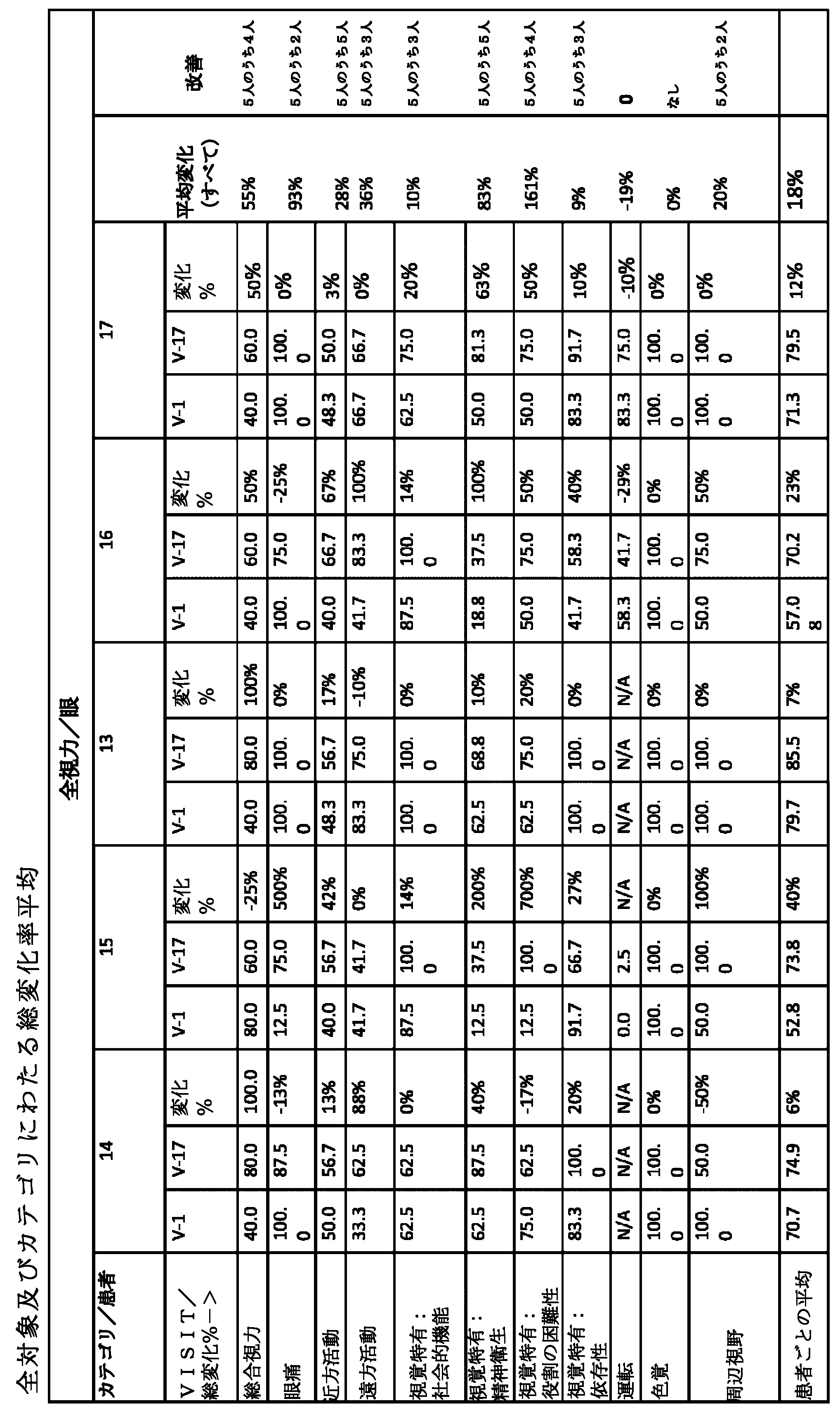


すべての質問を含む空白のアンケート(National Eye Institute Visual Functioningアンケート25(VFQ-25)バージョン2000-面接者管理形式)は、参照により本明細書に組み込まれる。アンケートは、スクリーニング訪問11、訪問17、訪問18、訪問19、訪問20、訪問21及び訪問22で投与された。表4に示す項目を平均すると、VFQ-25サブスケールが生成された。
A blank questionnaire containing all questions (National Eye Institute Visual Functioning Questionnaire 25 (VFQ-25) Version 2000—Interviewer Managed Format) is incorporated herein by reference. Questionnaires were administered at

臨床試験データからの観察には、生活の質の改善、読み取り速度の改善、及び微小視野測定の改善が含まれる。 Observations from clinical trial data include improved quality of life, improved reading speed, and improved microperimetry.
実施例2:GAを伴うドライ型AMDを有する対象における網膜回復
網膜回復は、本明細書中で使用される細胞が、GA境界を測定するために使用される一般的な画像化技術であるFAFのもとで自家蛍光しないので、観察することが困難である。IRによる測定は、萎縮境界を評価する方法として受け入れられていない。高分解能OCTは、GA病変境界及び網膜の微細層を測定するためのFAFの代替法である。このようにOCTを使用することは、それ自体の制限を伴うより遅い手動プロセスであるが、ケーキの層のように、網膜内の個々の細胞型を区別する能力を提供する(例:ONL、OPL、RPE)。図9、図12~図14、図16、図18~図22、図26~図28及び図30は、ベースライン及び処置後の萎縮領域のいくつかの断面及び「空中」の視点を示す。
Example 2: Retinal Recovery in Subjects with Dry AMD with GA Retinal recovery is a common imaging technique in which the cells used herein are used to measure GA boundaries, FAF It is difficult to observe because it does not autofluoresce under light. Measurement by IR is not an accepted method of assessing atrophy boundaries. High-resolution OCT is an alternative to FAF for measuring GA lesion borders and retinal microlayers. Using OCT in this way is a slower manual process with its own limitations, but like layers of a cake, it offers the ability to distinguish individual cell types within the retina (e.g. ONL, OPL, RPE). Figures 9, 12-14, 16, 18-22, 26-28 and 30 show several cross-sectional and "aerial" views of the atrophied region after baseline and treatment.
対象14は、処置後9ヶ月及び23ヶ月でOPL、ONL、ELM、RPE及び外側網膜の再生/回復の解剖学的改善を有していた(図9~15)。同様に、対象21は、1ヶ月目にGA境界の減少並びに解剖学的改善及びELMの回復(図16及び図17)、並びに以前の(原発性GAから分離された)萎縮領域のほぼ完全な回復を有し、欠損層の再生及び萎縮性病変の「消失」を有していた(図18)。処置後2及び3ヶ月で改善が見られた(図18~図22)。GAへのRPE送達は、処置手順中並びに処置後2ヶ月及び3ヶ月に対象14で観察された(図31)。
微小視野測定。図15は、回復領域も機能的であり得るという予備的証拠を示す(単に組織を見ることは組織が活性であることを意味しない)。微小視野測定は、視覚に使用される領域を「マッピング」するために、網膜上にピンポイント光をフラッシュすることを含む。微小視野測定データは収集が困難であるため、少数の時点で少数の対象にしか存在しない。しかしながら、それらは、患者14が回復領域で視覚能力を有するという少なくともいくつかの証拠を提供する。
Microfield measurement. Figure 15 shows preliminary evidence that the restored area may also be functional (just looking at the tissue does not mean the tissue is active). Microperimetry involves flashing a pinpoint light onto the retina to "map" the area used for vision. Microperimetry data are difficult to collect and are therefore only available for a small number of subjects at a small number of time points. However, they provide at least some evidence that the
対象22は、未処置眼と比較して処置眼の視力及びGAサイズの改善を示した(図23)。対象22における処置後3ヶ月での色素沈着は、RPE細胞の存在を示した(図24)。IR画像化によって測定されるGAサイズは、3ヶ月でのGAの境界の減少を実証し(図25)、OCT測定(図26~30)も同様である。
対象14を35ヶ月間追跡した。別個の組織層は23ヶ月で検出可能であったが、9ヶ月では存在しなかった。観察期間を通して、及び萎縮の全(周辺)領域にわたってこの現象の例が多数あった。出願人らは、処置前の年の患者のGA成長率を測定し、未処置の成長率に基づく患者のGAサイズの外挿を可能にした。GAは3年間ベースラインと比較して変化しないままであり、これは疾患の自然経過(すなわち、状況は次第に悪化する)を考慮すると、これは予想されないことであった。患者の処置された眼は最近になってようやくベースラインを下回ったが、患者がもはや視力のために使用していない反対側の眼よりもはるかに良好なままである。対象14はオリジナルの症例であり、効果の持続性を示す。
対象21の新たな所見。同様の観察が、2.5ヶ月という早期に異なる患者において検出された。分析は外側網膜領域のみで行った。ベースラインは、予想される場所でELM、EZの喪失を伴う予想されるGA/cRORAを示した。3週間後、ELM/EZの明らかな部分的再形成を含む有意な外側網膜変化が観察された。EZのびまん性肥厚及び非晶質の高反射網膜下物質が存在した。6週間で、いくつかのEZ変化が持続したが、EZの消失も生じた。RPE/ブルッフの肥厚も観察された。
対象22の新たな所見。対象22は、自身の処置経験を「生活変化」と呼んだ女性である。GAの周りの様々な位置におけるELMの新しい材料及び拡張、並びに主要領域に接続されていないGAのいくつかの小さな領域又は「島」が特定された。3ヶ月までに、これらの島は処置後に消失しており、早期の介入がドライ型AMDにおいてより良好な臨床転帰をもたらすという主張を裏付けている。患者22をOrbit SDSを用いて処置した。
ベースラインは、多焦点サテライトを伴う中心GA/cRORAを示した。EZ/ELM/過剰透過の予想される損失は、RPEを通して観察された。4週目に、大きな網膜下液貯留を伴う黄斑円孔形成があった。RPE表面上の多数の沈着物がIR及びOCTで同定された。6週目に、残留網膜下液及び新しい物質がRPEの表面上に明らかになった。PEDが、非常に高反射性の内部物質(可能性のある1型CNV)により明らかであった。3ヶ月までに、すべての網膜下液が消散し、網膜下物質が残存し、大きな中心網膜下沈着物が現れた。新しい上網膜内液があった。4ヶ月までに、ELMの伸長が多くの場所で認められた。網膜下物質が増加した。眼底写真上の網膜出血は、流体の領域及びブルッフを通る1型CNVの可能性のある芽の領域に対応し得る。FAFではRPE喪失の全体的な拡大があったが、色素沈着の増加及び定義された萎縮の境界へのELMの拡大があった。
Baseline showed central GA/cRORA with multifocal satellites. The expected loss of EZ/ELM/excess permeation was observed through the RPE. At 4 weeks, there was macular hole formation with large subretinal fluid retention. A number of deposits on the RPE surface were identified by IR and OCT. At 6 weeks, residual subretinal fluid and new material were evident on the surface of the RPE. PED was evident with a very highly reflective internal material (
概要
対象14、21及び22では、移植細胞が回復した症例がGAの大部分を占めていた。細胞配置は、Orbit評価に重要な意味を有するこれらの結果を達成するために重要であると思われる。対象14(GAの完全なカバレッジを有する患者)における回復を見た後、外科医は、最終的な7人の対象においてGAにわたって細胞を送達するためにより大きな努力をした。最後の4人のOrbit対象では、高度に訓練された外科医の手に委ねられたにもかかわらず、1人だけがGAにわたって細胞を首尾よく堆積させた。対照的に、PPVにアクセスした処置の両方がこれを首尾よく達成することができた(PPVはこの点に関してより柔軟である)。第3の症例(Pt.#22)では、Orbitを使用して、完全なカバレッジを完了した同じ外科医によって部分的なカバレッジが達成された。
Overview In subjects 14, 21 and 22, the majority of GA cases were those in which the transplanted cells recovered. Cell placement appears to be important to achieve these results, which have important implications for Orbit evaluation. After seeing recovery in subject 14 (a patient with complete coverage of the GA), surgeons made greater efforts to deliver cells across the GA in the final 7 subjects. Of the last four Orbit subjects, only one successfully deposited cells across the GA, despite being placed in the hands of a highly trained surgeon. In contrast, both PPV-accessed treatments were able to achieve this successfully (PPV is more flexible in this regard). In a third case (Pt. #22), partial coverage was achieved using Orbit by the same surgeon who completed full coverage.
回復はこの時点で臨床転帰と完全に相関していなかったが、いくつかの興味深いつながりが引き出され得る。しかし、AMDを処置するための任意の他のアプローチで以前に回復が観察されていないことを考えると、機能回復が起こる場合に機能再生の動態を予測するのを助ける先行例はない。 Although recovery was not perfectly correlated with clinical outcome at this time point, some interesting connections can be drawn. However, given that recovery has not been previously observed with any other approach to treat AMD, there is no precedent to help predict the dynamics of functional regeneration should it occur.
実施例3:加齢黄斑変性(AMD)のドライ型及びウェット型についての重要な調節エンドポイント
予想される有効性エンドポイントは以下の通りである。主要有効性エンドポイントFAFに基づく試験対象の眼におけるGA病変(複数可)の総面積(mm2)のベースラインから12ヶ月目までの変化。
Example 3: Key Modulatory Endpoints for Dry and Wet Forms of Age-Related Macular Degeneration (AMD) Expected efficacy endpoints are as follows. Change from baseline to
主要な副次的有効性エンドポイント。1)24ヶ月目(選択された国)のMinnesota Reading(MNRead)又はRadner Reading Chartsによって評価された、単眼読み取り速度(試験対象の眼)のベースラインからの変化。2)24ヶ月目の、機能的読み取り独立指数(FRII)複合スコアのベースラインからの変化。3)ETDRSチャートによって評価した24ヶ月目の正常輝度最高矯正視力スコア(NL-BCVA)のベースラインからの変化。4)ETDRSチャートによって評価した12ヶ月目及び24ヶ月目の低輝度最高矯正視力スコア(LL-BCVA)のベースラインからの変化。5)12ヶ月目及び24ヶ月目の低輝度不足(LLD)のベースラインからの変化。6)FAF(選択部位)によって評価した場合の試験対象の眼におけるGA病変(複数可)の総面積(mm2)の各計画された評価でのベースラインからの変化。7)MNRead又はRadner Reading Chartsによって評価された、12ヶ月目及び24ヶ月目(選択された国)の単眼限界印刷サイズ(試験対象の眼)のベースラインからの変化。8)12ヶ月目及び24ヶ月目のNational Eye Institute Visual Functioning Questionnaire 25アイテムバージョン(NEI VFQ-25)遠方活動サブスケールスコアのベースラインからの変化。9)黄斑機能的応答の評価のために薄明視微小視野測定によって評価される暗点の数(Oaks試験のみ)。10)黄斑機能的応答の評価のための薄明視微小視野測定によって評価される黄斑感度の変化。11)経時的なAPL-2の全身血漿濃度。
Primary secondary efficacy endpoint. 1) Change from baseline in monocular reading speed (study eye) as assessed by Minnesota Reading (MNRead) or Radner Reading Charts at 24 months (selected countries). 2) Change from baseline in Functional Reading Independent Index (FRII) composite score at 24 months. 3) Change from baseline in normal-luminance best-corrected visual acuity score (NL-BCVA) at 24 months as assessed by ETDRS chart. 4) Change from baseline in low-luminance best-corrected visual acuity score (LL-BCVA) at 12 and 24 months assessed by ETDRS chart. 5) Change from baseline in low luminance deficiency (LLD) at 12 and 24 months. 6) Change from baseline at each planned assessment in total area (mm 2 ) of GA lesion(s) in study eye as assessed by FAF (selected site). 7) Change from baseline in monocular critical print size (study eye) at 12 months and 24 months (selected countries) as assessed by MNRead or Radner Reading Charts. 8) Change from baseline in the National Eye Institute
安全性エンドポイント。1)眼及び全身の処置によって発現した有害事象の発生率及び重症度。2)APL-2に対する抗治療抗体の発生率。3)試験対象の眼における新しい活性CNVの発生率。 Safety endpoint. 1) Incidence and severity of ocular and systemic treatment-emergent adverse events. 2) Incidence of anti-therapeutic antibodies to APL-2. 3) Incidence of new active CNVs in the eyes tested.
ドライ型AMD試験における重要な副次的エンドポイントのいくつかに関する詳細は以下の通りであった。48週目での、薄明視顕微測定によって評価される絶対暗点の数のベースラインからの変化[時間枠:ベースライン、48週目]。暗点は、黄斑を中心とし、試験した範囲内の網膜感度の欠如を報告した微小視野測定検査の試験点であり、最大68点がこの範囲内で試験された。より高い結果は、絶対暗点の拡大及びより高い数の絶対暗点を示す。薄明視微小視野測定評価を、試験対象の眼のみの拡張後に実施し、データを中央読み取りセンターに転送した。データは、試験の早期終了のため、96週目ではなく48週目まで収集した。ベースラインからの正の変化は、絶対的な暗点の数の増加(より多くの網膜感度の欠如);疾患の悪化を示す。
Details regarding some of the important secondary endpoints in the dry AMD trial were as follows. Change from baseline in the number of absolute scotoma assessed by mesopic microscopic measurements at 48 weeks [Timeframe: Baseline, Week 48]. The scotoma was a microperimetry test point centered on the macula and reported a lack of retinal sensitivity within the range tested, with a maximum of 68 points tested within this range. A higher result indicates an expansion of absolute scotoma and a higher number of absolute scotoma. Mesopic microperimetry evaluations were performed after dilation of the test eye only, and data were transferred to a central reading center. Data were collected through
48週目での、薄明視微小視野測定によって評価される平均黄斑感度のベースラインからの変化[時間枠:ベースライン、48週目]。薄明視微小視野測定を使用して黄斑感度を評価し、評価を試験対象の眼のみの拡張後に実施し、データを中央読み取りセンターに転送した。ベースラインからの負の変化は、平均黄斑感度の減少;疾患の悪化を示す。データは、試験の早期終了のため、96週目ではなく48週目まで収集した。
Change from baseline in mean macular sensitivity assessed by mesopic microfield measurements at 48 weeks [Timeframe: Baseline, Week 48]. Mesopic microfield measurements were used to assess macular sensitivity, assessments were performed after dilation of the test eye only, and data were transferred to a central reading center. A negative change from baseline indicates a decrease in mean macular sensitivity; worsening disease. Data were collected through
48週目の糖尿病性網膜症早期処置研究(ETDRS)チャートによって評価した最高矯正視力(BCVA)スコアのベースラインからの変化[時間枠:ベースライン、48週目]。BCVAスコアは、4メートル(m)の開始距離で評価したETDRS視力チャート上で正しく読み取られた文字の数に基づいた。BCVAスコア試験を、眼を拡張する前に行った。BCVAスコアは、試験対象の眼において0~100文字の範囲である。アイチャート上で正しく読み取られた文字の数が少ないほど、視覚(又は視力)は悪くなる。ベースラインからの負の変化は、視力の低下;疾患の悪化を示す。データは、試験の早期終了のため、96週目ではなく48週目まで収集した。
Change from Baseline in Best Corrected Visual Acuity (BCVA) Score as assessed by the Diabetic Retinopathy Early Treatment Study (ETDRS) Chart at Week 48 [Time Frame: Baseline, Week 48]. The BCVA score was based on the number of letters read correctly on the ETDRS visual acuity chart evaluated at a starting distance of 4 meters (m). A BCVA score test was performed prior to eye dilation. BCVA scores range from 0 to 100 letters in the eye tested. The lower the number of letters read correctly on the eye chart, the worse the vision (or visual acuity). A negative change from baseline indicates decreased visual acuity; worsening disease. Data were collected through
48週目での、BCVAスコアにおいてベースラインから15文字未満の喪失を有する参加者のパーセンテージ[時間枠:48週目]。ベースラインからの15文字未満の喪失を、4メートル(m)の開始距離でETDRSチャートによって評価した。BCVAは、アイチャートを使用して測定し、正しく読み取られた文字の数(0~100文字の範囲)として報告した。アイチャート上で正しく読み取られた文字の数が少ないほど、視覚(又は視力)は悪くなる。データは、試験の早期終了のため、96週目ではなく48週目まで収集した。
Percentage of participants with <15 letter loss from baseline in BCVA score at Week 48 [Timeframe: Week 48]. A loss of less than 15 letters from baseline was assessed by the ETDRS chart at a starting distance of 4 meters (m). BCVA was measured using an eye chart and reported as the number of characters read correctly (ranging from 0 to 100 characters). The lower the number of letters read correctly on the eye chart, the worse the vision (or visual acuity). Data were collected through
48週目での、低輝度条件下でのETDRSチャートによって評価した低輝度視力(LLVA)のベースラインからの変化[時間枠:ベースライン、48週目]。LLVAは、その眼の最高矯正に2.0-log単位の中立密度フィルターをかけ、参加者に通常照射されたETDRSチャートを読み取らせることによって測定した。評価は、眼を拡張する前に行った。LLVAスコアは、試験対象の眼において0~100文字の範囲である。アイチャート上で正しく読み取られた文字の数が少ないほど、視覚(又は視力)は悪くなる。データは、試験の早期終了のため、96週目ではなく48週目まで収集した。
Change from baseline in low-luminance visual acuity (LLVA) assessed by ETDRS chart under low-luminance conditions at Week 48 [time frame: Baseline, Week 48]. LLVA was measured by applying a 2.0-log unit neutral density filter to the eye's best correction and having participants read a normally illuminated ETDRS chart. Evaluations were made prior to eye dilation. LLVA scores range from 0 to 100 letters in the eye tested. The lower the number of letters read correctly on the eye chart, the worse the vision (or visual acuity). Data were collected through
48週目での、LLVAスコアにおいてベースラインから15文字未満の喪失を有する参加者のパーセンテージ[時間枠:48週目]。ベースラインからの15文字未満の喪失を、4mの開始距離でETDRSチャートによって評価した。データは、試験の早期終了のため、96週目ではなく48週目まで収集した。
Percentage of participants with a loss of <15 letters from baseline in the LLVA score at Week 48 [Timeframe: Week 48]. A loss of less than 15 letters from baseline was assessed by the ETDRS chart at a starting distance of 4 m. Data were collected through
48週目での、Minnesota低視力読み取り試験(MNRead)チャート又はRadner読み取りチャートによって評価した両眼読み取り速度のベースラインからの変化[時間枠:ベースライン、48週目]。MNRead視力カードは、正常及び低視力参加者の読み取り視力及び読み取り速度を測定するのに適した連続テキスト読み取り視力カードであった。MNRead視力カードは、等しい数の文字を有する単一の単純な文からなっていた。ストップウォッチを使用して、10分の1秒までの時間を記録した。視覚のために読み取ることができなかった、又は試行されなかった文は、時間が0、エラーが10として記録されるべきである。Radner Reading Cardは、読み取り速度、読み取り視力、及び限界印刷サイズの測定に適していた。読み取り時間が20秒を超えたとき、又は参加者が重大なエラーを起こしたときに読み取り試験を停止した。ベースラインからの負の変化は、両眼読み取り速度の減少;疾患の悪化を示す。データは、試験の早期終了のため、96週目ではなく48週目まで収集した。
Change from baseline in binocular reading speed as assessed by the Minnesota Low Vision Reading Test (MNRead) chart or the Radner reading chart at Week 48 [time frame: Baseline, Week 48]. The MNRead visual acuity card was a continuous text reading visual acuity card suitable for measuring reading acuity and reading speed in normal and low vision participants. The MNRead visual acuity card consisted of a single simple sentence with an equal number of letters. A stopwatch was used to record times to the nearest tenth of a second. Sentences that could not be read visually or were not attempted should be scored with a time of 0 and an error of 10. The Radner Reading Card was suitable for measuring reading speed, reading visual acuity, and critical print size. The reading test was stopped when the reading time exceeded 20 seconds or when the participant made a serious error. A negative change from baseline indicates decreased binocular reading speed; worsening disease. Data were collected through
48週目での、MNReadチャート又はRadner読み取りチャートによって評価した単眼最大読み取り速度のベースラインからの変化[時間枠:ベースライン、48週目]。MNRead視力カードは、正常及び低視力参加者の読み取り視力及び読み取り速度を測定するのに適した連続テキスト読み取り視力カードであった。MNRead視力カードは、等しい数の文字を有する単一の単純な文からなっていた。ストップウォッチを使用して、10分の1秒までの時間を記録した。視覚のために読み取ることができなかった、又は試行されなかった文は、時間が0、エラーが10として記録されるべきである。Radner Reading Cardは、読み取り速度、読み取り視力、及び限界印刷サイズの測定に適していた。読み取り時間が20秒を超えたとき、又は参加者が重大なエラーを起こしたときに読み取り試験を停止した。ベースラインからの負の変化は、単眼読み取り速度の減少;疾患の悪化を示す。データは、試験の早期終了のため、96週目ではなく48週目まで収集した。
Change from baseline in maximal monocular reading speed assessed by MNRead chart or Radner reading chart at Week 48 [time frame: Baseline, Week 48]. The MNRead visual acuity card was a continuous text reading visual acuity card suitable for measuring reading acuity and reading speed in normal and low vision participants. The MNRead visual acuity card consisted of a single simple sentence with an equal number of letters. A stopwatch was used to record times to the nearest tenth of a second. Sentences that could not be read visually or were not attempted should be scored with a time of 0 and an error of 10. The Radner Reading Card was suitable for measuring reading speed, reading visual acuity, and critical print size. The reading test was stopped when the reading time exceeded 20 seconds or when the participant made a serious error. A negative change from baseline indicates a decrease in monocular reading speed; worsening disease. Data were collected through
48週目のnational eye institute visual functioning questionnaire 25項目(NEI VFQ-25)バージョン複合スコアのベースラインからの変化[時間枠:ベースライン、48週目]。NEI-VFQ-25アンケートは、25の項目を含み、それに基づいて全体的な複合VFQスコア及び12のサブスケールが導出された:近方活動、遠方活動、全般的な健康、総合視力、眼痛、視覚特有の社会的機能、視覚特有の精神衛生、視覚特有の役割の困難、視覚特有の依存、運転、色覚及び周辺視野。各質問に対する応答は0~100スコアに変換された。各サブスケール、合計スコア=スコアに寄与する項目の平均。各サブスケール及び総スコア、スコア範囲:0~100、より高いスコアはより良好な機能を表す。ベースラインからの負の変化は、視覚機能の低下;疾患の悪化を示す。データは、試験の早期終了のため、96週目ではなく48週目まで収集した。
Change from baseline in the national eye institute visual functioning questionnaire 25-item (NEI VFQ-25) version composite score at Week 48 [time frame: Baseline, Week 48]. The NEI-VFQ-25 questionnaire contained 25 items, from which an overall composite VFQ score and 12 subscales were derived: near activity, distance activity, general health, global visual acuity, eye pain. , vision-specific social functioning, vision-specific mental health, vision-specific role difficulties, vision-specific dependence, driving, color vision and peripheral vision. Responses to each question were converted to a 0-100 score. For each subscale, total score = mean of items contributing to score. Each subscale and total score, score range: 0-100, with higher scores representing better function. A negative change from baseline indicates decreased visual function; worsening disease. Data were collected through
48週目での、NEI VFQ-25近方活動サブスケールスコアのベースラインからの変化[時間枠:ベースライン、週]。NEI-VFQ-25アンケートには25の項目が含まれ、それに基づいて近方活動が測定された。近方活動は、新聞の普通の印刷物を読むこと、近見視力を必要とする仕事又は趣味を行うこと、又は混雑した棚で何かを見つけることと定義される。各質問に対する応答は0~100スコアに変換された。サブスケール=スコアに寄与する項目の平均。このサブスケールでは、スコア範囲は0~100であり、より高いスコアはより良好な機能を表す。ベースラインからの負の変化は、近方視覚活動の低下;疾患の悪化を示す。データは、試験の早期終了のため、96週目ではなく48週目まで収集した。
Change from Baseline in NEI VFQ-25 Proximal Activity Subscale Scores at Week 48 [Timeframe: Baseline, Weeks]. The NEI-VFQ-25 questionnaire contained 25 items on which near activity was measured. Near activities are defined as reading the plain print of a newspaper, doing a job or hobby that requires near vision, or finding something on a crowded shelf. Responses to each question were converted to a 0-100 score. Subscale = mean of items contributing to score. On this subscale, scores range from 0 to 100, with higher scores representing better function. A negative change from baseline indicates decreased near visual activity; worsening disease. Data were collected through
48週目での、NEI VFQ-25遠方活動サブスケールスコアのベースラインからの変化[時間枠:ベースライン、48週目]。NEI-VFQ-25アンケートには25の項目が含まれ、それに基づいて遠方活動が測定された。遠方活動は、道路標識又は店舗名を読むこと、及び階段、段差、又は縁石を降りることとして定義される。各質問に対する応答は0~100スコアに変換された。サブスケール=スコアに寄与する項目の平均。このサブスケールでは、スコア範囲は0~100であり、より高いスコアはより良好な機能を表す。ベースラインからの負の変化は、遠方視覚活動の低下;疾患の悪化を示す。データは、試験の早期終了のため、96週目ではなく48週目まで収集した。
Change from Baseline in NEI VFQ-25 Distant Activity Subscale Scores at Week 48 [Timeframe: Baseline, Week 48]. The NEI-VFQ-25 questionnaire contained 25 items on which distance activity was measured. Distant activities are defined as reading street signs or store names and descending stairs, steps, or curbs. Responses to each question were converted to a 0-100 score. Subscale = mean of items contributing to score. On this subscale, scores range from 0 to 100, with higher scores representing better function. A negative change from baseline indicates decreased distance visual activity; worsening disease. Data were collected through
48週目の平均機能的読み取り独立(FRI)指数のベースラインからの変化[時間枠:ベースライン、48週目]。FRIは、GA AMD参加者に最も関連する機能的読み取り活動に関する7項目を有する面接者管理アンケートであった。それは、1つの総合指数スコアを有する。過去7日間に行われた各FRI指数読み取り活動について、参加者は、視覚補助、活動の調整、又は別の参加者からの支援を必要とする程度について尋ねられた。平均FRI指数スコアは1~4の範囲であり、スコアが高いほど独立性が高いことを示す。ベースラインからの負の変化は、FRIの低下;疾患の悪化を示す。データは、試験の早期終了のため、96週目ではなく48週目まで収集した。
Change from Baseline in Mean Functional Reading Independent (FRI) Index at Week 48 [time frame: Baseline, Week 48]. The FRI was an interviewer-administered questionnaire with 7 items regarding the functional reading activities most relevant to GA AMD participants. It has one composite index score. For each FRI index reading activity performed in the past 7 days, participants were asked about the extent to which they needed visual assistance, coordination of the activity, or assistance from another participant. Average FRI index scores range from 1 to 4, with higher scores indicating greater independence. A negative change from baseline indicates a decrease in FRI; worsening disease. Data were collected through
実施例4:厚さ及び面積を測定するためのSD-OCTイメージング
網膜の異なる層の厚さ、面積及び体積を処置した眼において決定した。SD-OCT画像を、Spectralis(Spectralis;Heidelberg Engineering,Inc.,Heidelberg,Germany)、中心窩を中心とする20×20度の視野内の512×49の等間隔のBスキャンからなる黄斑体積を用いてキャプチャーした。すべてのBスキャンの網膜層を、3D-OCTOR(Doheny Eye Instituteで開発)を使用して厚さ及び面積の測定のために手動でセグメント化した。具体的には、外顆粒層、視細胞内節(ミオイドゾーン)、視細胞外節(エリプソイドゾーン)、及びRPE+ドルーゼン複合体を、黄斑容積内のすべてのBスキャンを使用して手動でセグメント化した。
Example 4: SD-OCT imaging to measure thickness and area The thickness, area and volume of different layers of the retina were determined in treated eyes. SD-OCT images were obtained using Spectralis (Spectralis; Heidelberg Engineering, Inc., Heidelberg, Germany), a macular volume consisting of 512×49 equally spaced B-scans within a 20×20 degree field centered on the fovea. captured. Retinal layers of all B-scans were manually segmented for thickness and area measurements using 3D-OCTOR (developed at the Doheny Eye Institute). Specifically, the outer nuclear layer, photoreceptor inner segment (myoid zone), photoreceptor outer segment (ellipsoid zone), and RPE + drusen complex were manually segmented using all B-scans within the macular volume. bottom.
例示のBスキャンを図33A~図33Cに示す。Bスキャン(図33A)を境界に基づいて層に分割し(図33B)、層の厚さ及び面積を決定した(図33C)。厚さマップは、全網膜、ONL、視細胞外節、RPE+ドルーゼン複合体(図34、それぞれ左から右)、及び視細胞内節の厚さを示す。個々の対象の例示的な厚さマップを図35~図52に示す。結果を表5~表10に示す。 Exemplary B-scans are shown in FIGS. 33A-33C. B-scans (Fig. 33A) were divided into layers based on boundaries (Fig. 33B) and layer thicknesses and areas were determined (Fig. 33C). Thickness maps show total retina, ONL, photoreceptor outer segment, RPE+drusen complex (FIG. 34, left to right, respectively), and photoreceptor inner segment thickness. Exemplary thickness maps for individual subjects are shown in FIGS. 35-52. The results are shown in Tables 5-10.


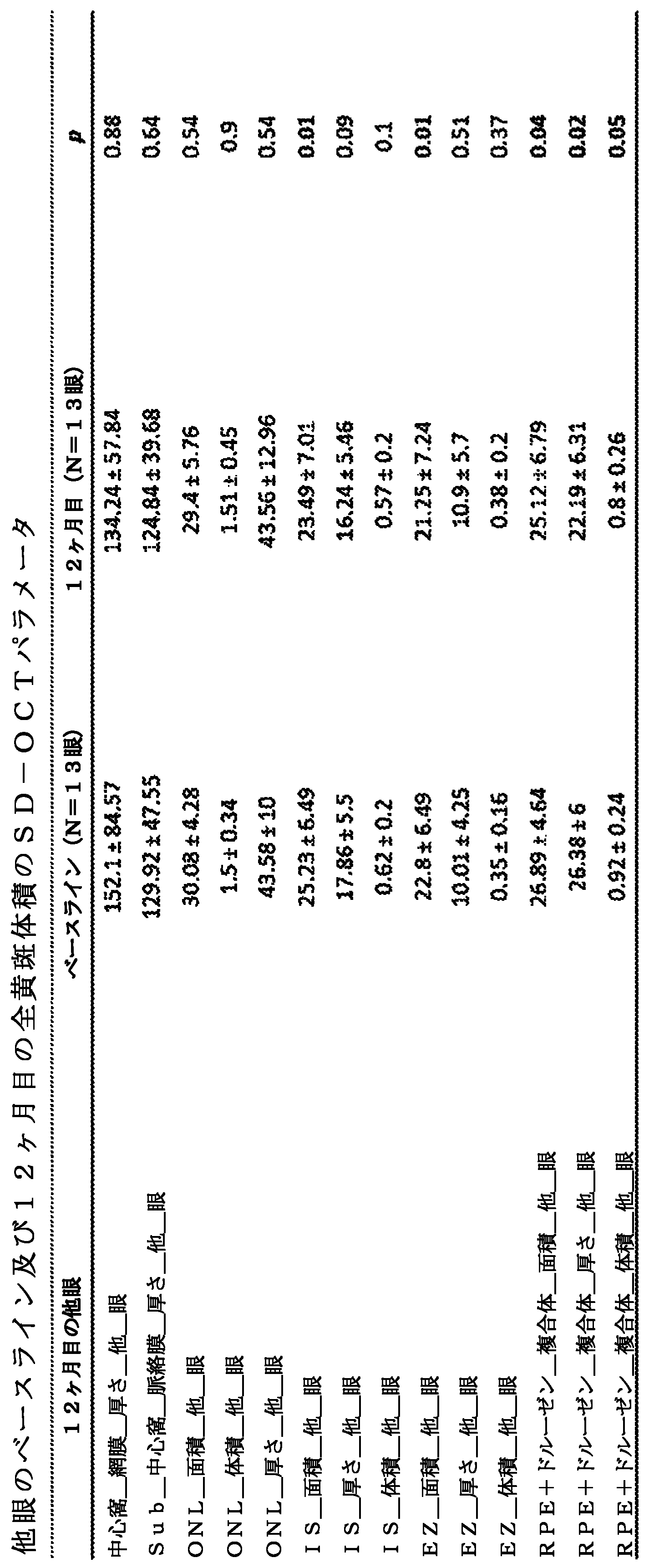
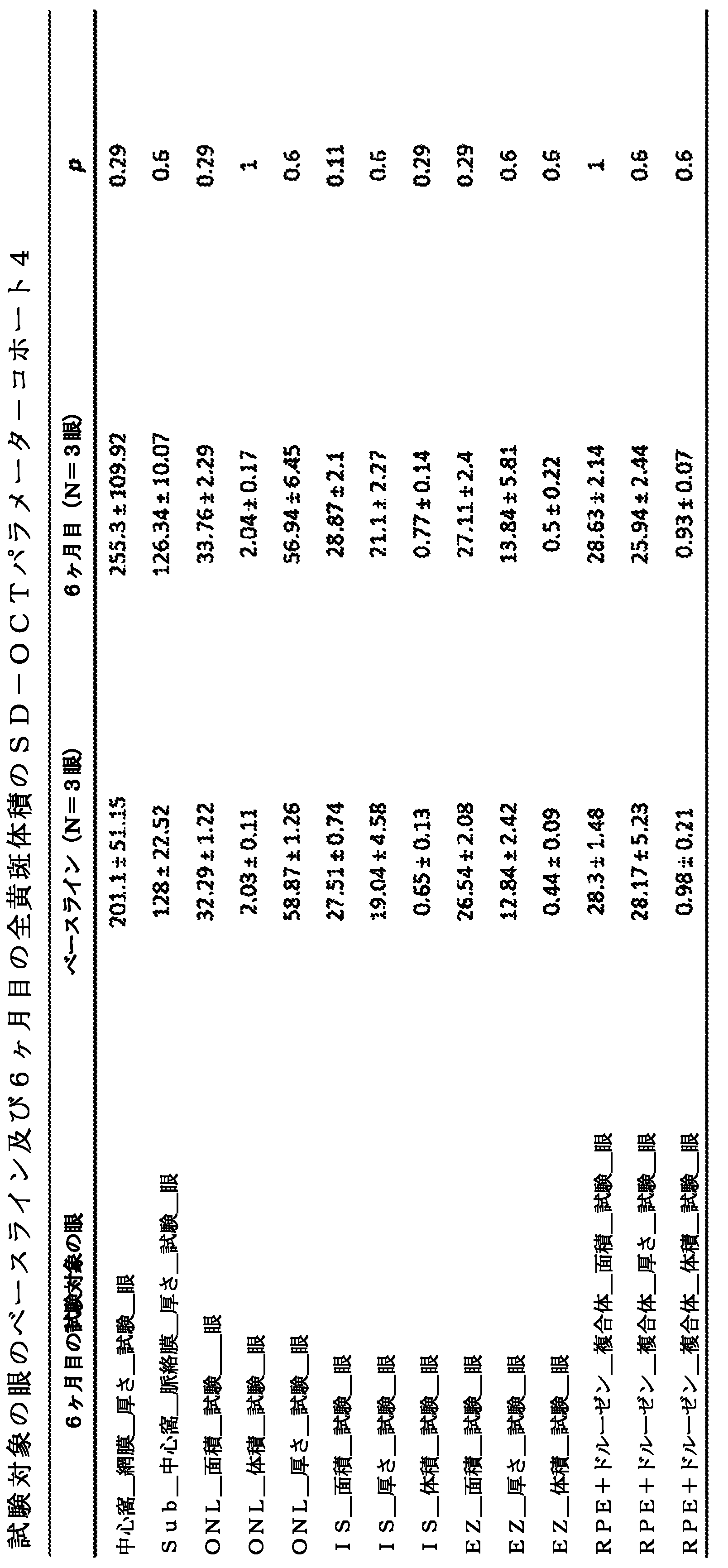


実施例5:
RPE処置は血液網膜関門の回復をもたらす
RPEは非常に高いレベルのPEDFを分泌し(2000~4000ng/ml/日のOpRegenレベルについて測定した)、これは治療効力に寄与する。PEDFは、インビボでRPE及びミュラーグリアによって分泌される50kDaのタンパク質であり、抗血管新生作用、老化及び酸化ストレスによって乱されたミトコンドリア動態を回復させることによる光受容体の神経保護機能、(マスター因子NF-KappaBとの相互作用による)抗炎症活性、細胞外マトリックス(コラーゲン及びプロテオグリカン)への結合による抗線維化活性などの有益な機能を有している。OpRegen処置された対象では、これは、ドルーゼンを有する/有さない患者におけるフルオレセイン血管造影(FA)の改善、及び移植後2~4週間という早期に見られるGA病変内のECMリモデリング又は瘢痕減弱の可能性のある徴候を伴うOCT画像化によって証明される。
Example 5:
RPE Treatment Leads to Restoration of the Blood-Retinal Barrier RPE secretes very high levels of PEDF (measured for OpRegen levels of 2000-4000 ng/ml/day), which contributes to therapeutic efficacy. PEDF is a 50-kDa protein secreted by RPE and Müller glia in vivo and has anti-angiogenic effects, neuroprotective function of photoreceptors by restoring mitochondrial dynamics disturbed by aging and oxidative stress, (master factor It has beneficial functions such as anti-inflammatory activity through interaction with NF-KappaB), anti-fibrotic activity through binding to the extracellular matrix (collagen and proteoglycans). In OpRegen-treated subjects, this is associated with improved fluorescein angiography (FA) in patients with/without drusen, and ECM remodeling or diminished scarring within GA lesions seen as early as 2-4 weeks after transplantation. evidenced by OCT imaging with possible signs of
対象8におけるベースラインFA検査は、大量のフルオレセイン色素が硝子体腔に漏出することを示し、それにより脈絡膜フラッシュ及び動脈相中の血管灌流の視認性が遮断され、血液網膜関門の破壊及びパラ炎症が眼内に予め存在することが示唆された(図53)。移植後22ヶ月で、FA検査は、明らかな脈絡膜及び網膜血管灌流を示し、硝子体腔に漏出する色素はなく、OpRegenがおそらく複数の作用機序によって(例えばPEDFの様々な対象を介する)破壊されたBRBの完全性を回復したことを示している。図54A~図54Dは、OpRegen細胞療法によるBRBの回復又は修復の3つのさらなる例を提供する。
Baseline FA examination in
対象8は、後部網膜全体に広がる広範なドルーゼンを有する患者の典型例である。図55は、ドルーゼンの消失が、上部の移植片領域から始まり(左上)、術後8ヶ月で残った小さな細長いバンドを除いて、下方へ移動してほぼ後部全体を清浄化したことを示す(上、左から2番目、大きな円)。OCTイメージングの特徴は、ベースライン(右上及び右下)と比較して5.5ヶ月(上、右から2番目)及び8ヶ月(下、右から2番目)でカラー眼底写真と一致しており;subRPEドルーゼンは有意に減少又は消失した。宿主網膜組織は減弱しているようであり、これは、高レベルのPEDFの存在によってもたらされる生物学的効果に部分的に、場合によってはそれに起因して、ECMリモデリングの可能性を示唆している。
11ヶ月で、対象8では、大きなドルーゼンが消失した後、移植片が宿主網膜をリモデリングし続けた(図56A~図56C)。FAは、有意に減少した染色を示したが(ドルーゼン)、網膜血管構造をぼやかすベールのような膜を有するようであった。眼底検査の22ヶ月で、網膜組織は、おそらくその抗炎症効果、又はPEDFがマトリックス外代謝回転を調節する役割を有するECM洗浄のために、ベースラインのものと比較してより鮮明に見える。
At 11 months, in
図57は、初期段階、中期段階及び後期段階からのFA検査の時間経過を提供しており、網膜の健康の有意な改善、全体にわたる血管灌流のより良好な視認性、及び炎症の減少を実証する。網膜組織は非常にきれいに見える;このFAパターンは、他の治療様式では以前に報告されていない。これは、OpRegenの治療効果に非常に独特である。 FIG. 57 provides a time course of FA examination from early, middle and late stages, demonstrating significant improvement in retinal health, better visibility of vascular perfusion throughout, and reduced inflammation. do. Retinal tissue appears very clean; this FA pattern has not been previously reported with other treatment modalities. This is very unique to the therapeutic effects of OpRegen.
本明細書で提供されるすべての参考文献(すべての非特許文献、特許、及び特許公報を含む)は、その全体が参照により本明細書に組み込まれる。 All references (including all non-patent literature, patents, and patent publications) provided herein are hereby incorporated by reference in their entirety.
Claims (34)
Applications Claiming Priority (9)
| Application Number | Priority Date | Filing Date | Title |
|---|---|---|---|
| US202063029669P | 2020-05-25 | 2020-05-25 | |
| US63/029,669 | 2020-05-25 | ||
| US202063036327P | 2020-06-08 | 2020-06-08 | |
| US63/036,327 | 2020-06-08 | ||
| US202063106339P | 2020-10-27 | 2020-10-27 | |
| US63/106,339 | 2020-10-27 | ||
| US202163182684P | 2021-04-30 | 2021-04-30 | |
| US63/182,684 | 2021-04-30 | ||
| PCT/US2021/034114 WO2021242788A1 (en) | 2020-05-25 | 2021-05-25 | Methods and compositions for treating retinal diseases and conditions |
Publications (2)
| Publication Number | Publication Date |
|---|---|
| JP2023528362A true JP2023528362A (en) | 2023-07-04 |
| JPWO2021242788A5 JPWO2021242788A5 (en) | 2024-05-23 |
Family
ID=78722790
Family Applications (1)
| Application Number | Title | Priority Date | Filing Date |
|---|---|---|---|
| JP2022572671A Pending JP2023528362A (en) | 2020-05-25 | 2021-05-25 | Methods and compositions for treating retinal diseases and conditions |
Country Status (8)
| Country | Link |
|---|---|
| US (1) | US20230086868A1 (en) |
| EP (1) | EP4157219A4 (en) |
| JP (1) | JP2023528362A (en) |
| CN (1) | CN115811980A (en) |
| AU (1) | AU2021280260A1 (en) |
| CA (1) | CA3184790A1 (en) |
| IL (1) | IL298569A (en) |
| WO (1) | WO2021242788A1 (en) |
Families Citing this family (3)
| Publication number | Priority date | Publication date | Assignee | Title |
|---|---|---|---|---|
| WO2018170494A1 (en) * | 2017-03-16 | 2018-09-20 | Bio Time, Inc. | Methods for measuring therapeutic effects of retinal disease therapies |
| CN117957311A (en) | 2021-07-28 | 2024-04-30 | 谱系细胞疗法公司 | Expansion of retinal pigment epithelial cells |
| CN114561356B (en) * | 2022-03-09 | 2022-09-13 | 北京呈诺医学科技有限公司 | Method for influencing cytokine secretion and application |
Citations (1)
| Publication number | Priority date | Publication date | Assignee | Title |
|---|---|---|---|---|
| WO2019130061A2 (en) * | 2017-12-29 | 2019-07-04 | Cell Cure Neurosciences Ltd. | Retinal pigment epithelium cell compositions |
Family Cites Families (5)
| Publication number | Priority date | Publication date | Assignee | Title |
|---|---|---|---|---|
| CN103555654B (en) * | 2007-04-18 | 2016-04-20 | 哈达锡特医学研究服务及发展有限公司 | Stem cell-derived retinal pigment epithelium |
| CA2781149A1 (en) * | 2009-11-17 | 2011-05-26 | Advanced Cell Technology, Inc. | Methods of producing human rpe cells and pharmaceutical preparations of human rpe cells |
| IL253244B2 (en) * | 2014-12-30 | 2023-09-01 | Cell Cure Neurosciences Ltd | Methods of treating retinal diseases |
| WO2018170494A1 (en) * | 2017-03-16 | 2018-09-20 | Bio Time, Inc. | Methods for measuring therapeutic effects of retinal disease therapies |
| JP2020530839A (en) * | 2017-07-31 | 2020-10-29 | バイオタイム インコーポレイテッド | Compositions and Methods for Recovering or Preventing Visual Loss Caused by Disease or Traumatic Injury |
-
2021
- 2021-05-25 WO PCT/US2021/034114 patent/WO2021242788A1/en not_active Ceased
- 2021-05-25 JP JP2022572671A patent/JP2023528362A/en active Pending
- 2021-05-25 AU AU2021280260A patent/AU2021280260A1/en active Pending
- 2021-05-25 IL IL298569A patent/IL298569A/en unknown
- 2021-05-25 CN CN202180038361.3A patent/CN115811980A/en active Pending
- 2021-05-25 EP EP21813012.8A patent/EP4157219A4/en active Pending
- 2021-05-25 CA CA3184790A patent/CA3184790A1/en active Pending
-
2022
- 2022-11-23 US US18/058,425 patent/US20230086868A1/en active Pending
Patent Citations (1)
| Publication number | Priority date | Publication date | Assignee | Title |
|---|---|---|---|---|
| WO2019130061A2 (en) * | 2017-12-29 | 2019-07-04 | Cell Cure Neurosciences Ltd. | Retinal pigment epithelium cell compositions |
Non-Patent Citations (2)
| Title |
|---|
| INVESTIGATIVE OPHTHALMOLOGY & VISUAL SCIENCE, vol. Vol.59, 77, JPN6025022747, 2018, ISSN: 0005611934 * |
| TRANSLATIONAL VISION SCIENCE & TECHNOLOGY, vol. Vol.6, No.3, Article 17, JPN6025022746, 2017, ISSN: 0005611935 * |
Also Published As
| Publication number | Publication date |
|---|---|
| AU2021280260A1 (en) | 2023-02-02 |
| WO2021242788A1 (en) | 2021-12-02 |
| IL298569A (en) | 2023-01-01 |
| US20230086868A1 (en) | 2023-03-23 |
| CA3184790A1 (en) | 2021-12-02 |
| EP4157219A4 (en) | 2024-07-10 |
| EP4157219A1 (en) | 2023-04-05 |
| CN115811980A (en) | 2023-03-17 |
Similar Documents
| Publication | Publication Date | Title |
|---|---|---|
| US20230028133A1 (en) | Methods of treating retinal diseases | |
| JP7498664B2 (en) | Retinal pigment epithelial cell composition | |
| JP2023089154A (en) | Methods for measuring therapeutic efficacy of retinal disease treatments | |
| US20230086868A1 (en) | Methods and compositions for treating retinal diseases and conditions | |
| US20240299625A1 (en) | Methods and compositions for treating retinal diseases and conditions | |
| CN117479835A (en) | Methods and compositions for treating retinal diseases and conditions | |
| HK40081746A (en) | Methods and compositions for treating retinal diseases and conditions | |
| HK40099184A (en) | Methods and compositions for treating retinal diseases and conditions | |
| HK40103528A (en) | Methods for measuring therapeutic effects of retinal disease therapies | |
| HK40027162A (en) | Methods for measuring therapeutic effects of retinal disease therapies | |
| HK40066083A (en) | Methods of treating retinal diseases |
Legal Events
| Date | Code | Title | Description |
|---|---|---|---|
| RD04 | Notification of resignation of power of attorney |
Free format text: JAPANESE INTERMEDIATE CODE: A7424 Effective date: 20240328 |
|
| A521 | Request for written amendment filed |
Free format text: JAPANESE INTERMEDIATE CODE: A523 Effective date: 20240515 |
|
| A621 | Written request for application examination |
Free format text: JAPANESE INTERMEDIATE CODE: A621 Effective date: 20240515 |
|
| A131 | Notification of reasons for refusal |
Free format text: JAPANESE INTERMEDIATE CODE: A131 Effective date: 20250605 |
|
| A521 | Request for written amendment filed |
Free format text: JAPANESE INTERMEDIATE CODE: A523 Effective date: 20250902 |
|
| A02 | Decision of refusal |
Free format text: JAPANESE INTERMEDIATE CODE: A02 Effective date: 20251201 |